Up-to-Date Overview of the Use of Natural Ingredients ... - MDPI
29
Citation: Resende, D.I.S.P.; Jesus, A.; Sousa Lobo, J.M.; Sousa, E.; Cruz, M.T.; Cidade, H.; Almeida, I.F. Up-to-Date Overview of the Use of Natural Ingredients in Sunscreens. Pharmaceuticals 2022, 15, 372. https://doi.org/10.3390/ ph15030372 Academic Editor: Daniela De Vita Received: 16 February 2022 Accepted: 14 March 2022 Published: 18 March 2022 Publisher’s Note: MDPI stays neutral with regard to jurisdictional claims in published maps and institutional affil- iations. Copyright: © 2022 by the authors. Licensee MDPI, Basel, Switzerland. This article is an open access article distributed under the terms and conditions of the Creative Commons Attribution (CC BY) license (https:// creativecommons.org/licenses/by/ 4.0/). pharmaceuticals Article Up-to-Date Overview of the Use of Natural Ingredients in Sunscreens Diana I. S. P. Resende 1,2 , Ana Jesus 3,4 , José M. Sousa Lobo 3,4 , Emília Sousa 1,2, * , Maria T. Cruz 5,6 , Honorina Cidade 1,2 and Isabel F. Almeida 3,4, * 1 CIIMAR—Centro Interdisciplinar de Investigação Marinha e Ambiental, 4450-208 Matosinhos, Portugal; [email protected] (D.I.S.P.R.); [email protected] (H.C.) 2 Laboratório de Química Orgânica e Farmacêutica, Departamento de Ciências Químicas, Faculdade de Farmácia, Universidade do Porto, 4050-313 Porto, Portugal 3 Associate Laboratory i4HB—Institute for Health and Bioeconomy, Faculty of Pharmacy, University of Porto, 4050-313 Porto, Portugal; [email protected] (A.J.); [email protected] (J.M.S.L.) 4 UCIBIO—Applied Molecular Biosciences Unit, MedTech, Laboratory of Pharmaceutical Technology, Department of Drug Sciences, Faculty of Pharmacy, University of Porto, 4050-313 Porto, Portugal 5 Faculty of Pharmacy, University of Coimbra, 3004-531 Coimbra, Portugal; [email protected] 6 Center for Neuroscience and Cell Biology, 3004-504 Coimbra, Portugal * Correspondence: [email protected] (E.S.); [email protected] (I.F.A.); Tel.: +351-220-428-689 (E.S.); +351-220-428-621 (I.F.A.) Abstract: The photoprotective skincare segment is in high demand to meet consumer concerns on UV-induced skin damage, with a recent trend towards sunscreen alternatives with a natural origin. In this study, the use of natural ingredients, either from terrestrial or marine origin, in a panel of 444 sunscreen commercial formulations (2021) was analyzed. Ingredients from terrestrial organisms represent the large majority found in the analyzed sunscreen formulations (48%), whereas marine ingredients are present only in 13% of the analyzed products. A deeper analysis regarding the most prevalent families of ingredients from terrestrial and marine organisms used as top ingredients is also presented, as well as their mechanisms of action. This study provides an up-to-date overview of the sunscreen market regarding the use of natural ingredients, which is of relevance for scientists involved in the development of new sunscreens to identify opportunities for innovation. Keywords: natural ingredients; sunscreens; preparations; market 1. Introduction Naturally occurring ultraviolet radiation (UVR) is divided into three regions, classified according to their wavelength, called UVA (315–400 nm), UVB (280–315 nm), and UVC (100–280 nm)[1]. The ozone layer acts as a filter for UVR, absorbing all UVC and 90% of UVB when sunlight passes through the atmosphere. However, some UVB and UVA radiation is not filtered by the atmosphere, reaching the Earth and the sea, and causing a harmful effect on both terrestrial and aquatic organisms [2]. Excessive solar exposure is visually characterized by the swelling and redness of the affected area, well-known as sunburn or solar erythema. Nonetheless, the deleterious effects of excessive exposure are beyond what is seen, since UVR interferes in biological and metabolic processes, triggering a cascade of reactions that cause skin photodamage, photoaging, and photocarcinogenesis [3]. These skin damage events are provoked by UVR, which alters DNA, cellular antioxidant balance, signal transduction pathways, immune system, and the extracellular matrix [4]. UVB radiation directly affects the DNA by inducing apoptosis or errors in the DNA replication, activating inflammatory processes, photo-immunosuppression, melanogenesis, and skin cancer [5–7]. On the other hand, DNA damage, due to overproduction of reactive oxygen species (ROS), cross-linking of collagen and elastin fibers, premature skin aging, dryness, wrinkles, hyperpigmentation, and skin sensitization are some of the consequences Pharmaceuticals 2022, 15, 372. https://doi.org/10.3390/ph15030372 https://www.mdpi.com/journal/pharmaceuticals
-
Upload
khangminh22 -
Category
Documents
-
view
1 -
download
0
Transcript of Up-to-Date Overview of the Use of Natural Ingredients ... - MDPI

�����������������
Citation: Resende, D.I.S.P.; Jesus, A.;
Sousa Lobo, J.M.; Sousa, E.; Cruz,
M.T.; Cidade, H.; Almeida, I.F.
Up-to-Date Overview of the Use of
Natural Ingredients in Sunscreens.
Pharmaceuticals 2022, 15, 372.
https://doi.org/10.3390/
ph15030372
Academic Editor: Daniela De Vita
Received: 16 February 2022
Accepted: 14 March 2022
Published: 18 March 2022
Publisher’s Note: MDPI stays neutral
with regard to jurisdictional claims in
published maps and institutional affil-
iations.
Copyright: © 2022 by the authors.
Licensee MDPI, Basel, Switzerland.
This article is an open access article
distributed under the terms and
conditions of the Creative Commons
Attribution (CC BY) license (https://
creativecommons.org/licenses/by/
4.0/).
pharmaceuticals
Article
Up-to-Date Overview of the Use of Natural Ingredientsin SunscreensDiana I. S. P. Resende 1,2 , Ana Jesus 3,4 , José M. Sousa Lobo 3,4 , Emília Sousa 1,2,* , Maria T. Cruz 5,6 ,Honorina Cidade 1,2 and Isabel F. Almeida 3,4,*
1 CIIMAR—Centro Interdisciplinar de Investigação Marinha e Ambiental, 4450-208 Matosinhos, Portugal;[email protected] (D.I.S.P.R.); [email protected] (H.C.)
2 Laboratório de Química Orgânica e Farmacêutica, Departamento de Ciências Químicas, Faculdade deFarmácia, Universidade do Porto, 4050-313 Porto, Portugal
3 Associate Laboratory i4HB—Institute for Health and Bioeconomy, Faculty of Pharmacy, University of Porto,4050-313 Porto, Portugal; [email protected] (A.J.); [email protected] (J.M.S.L.)
4 UCIBIO—Applied Molecular Biosciences Unit, MedTech, Laboratory of Pharmaceutical Technology,Department of Drug Sciences, Faculty of Pharmacy, University of Porto, 4050-313 Porto, Portugal
5 Faculty of Pharmacy, University of Coimbra, 3004-531 Coimbra, Portugal; [email protected] Center for Neuroscience and Cell Biology, 3004-504 Coimbra, Portugal* Correspondence: [email protected] (E.S.); [email protected] (I.F.A.); Tel.: +351-220-428-689 (E.S.);
+351-220-428-621 (I.F.A.)
Abstract: The photoprotective skincare segment is in high demand to meet consumer concerns onUV-induced skin damage, with a recent trend towards sunscreen alternatives with a natural origin.In this study, the use of natural ingredients, either from terrestrial or marine origin, in a panel of444 sunscreen commercial formulations (2021) was analyzed. Ingredients from terrestrial organismsrepresent the large majority found in the analyzed sunscreen formulations (48%), whereas marineingredients are present only in 13% of the analyzed products. A deeper analysis regarding the mostprevalent families of ingredients from terrestrial and marine organisms used as top ingredients isalso presented, as well as their mechanisms of action. This study provides an up-to-date overview ofthe sunscreen market regarding the use of natural ingredients, which is of relevance for scientistsinvolved in the development of new sunscreens to identify opportunities for innovation.
Keywords: natural ingredients; sunscreens; preparations; market
1. Introduction
Naturally occurring ultraviolet radiation (UVR) is divided into three regions, classifiedaccording to their wavelength, called UVA (315–400 nm), UVB (280–315 nm), and UVC(100–280 nm) [1]. The ozone layer acts as a filter for UVR, absorbing all UVC and 90%of UVB when sunlight passes through the atmosphere. However, some UVB and UVAradiation is not filtered by the atmosphere, reaching the Earth and the sea, and causinga harmful effect on both terrestrial and aquatic organisms [2]. Excessive solar exposureis visually characterized by the swelling and redness of the affected area, well-known assunburn or solar erythema. Nonetheless, the deleterious effects of excessive exposure arebeyond what is seen, since UVR interferes in biological and metabolic processes, triggering acascade of reactions that cause skin photodamage, photoaging, and photocarcinogenesis [3].These skin damage events are provoked by UVR, which alters DNA, cellular antioxidantbalance, signal transduction pathways, immune system, and the extracellular matrix [4].UVB radiation directly affects the DNA by inducing apoptosis or errors in the DNAreplication, activating inflammatory processes, photo-immunosuppression, melanogenesis,and skin cancer [5–7]. On the other hand, DNA damage, due to overproduction of reactiveoxygen species (ROS), cross-linking of collagen and elastin fibers, premature skin aging,dryness, wrinkles, hyperpigmentation, and skin sensitization are some of the consequences
Pharmaceuticals 2022, 15, 372. https://doi.org/10.3390/ph15030372 https://www.mdpi.com/journal/pharmaceuticals

Pharmaceuticals 2022, 15, 372 2 of 29
of excessive sunlight exposure associated with UVA radiation [5,8,9]. DNA damage is themain event that occurs at a cellular level, as a result of UVR exposure. Depending on thewavelength and energy profile of the radiation, the kind of lesions produced might beattributed to direct or oxidative DNA damage [10]. Some of these lesions can evade theendogenous DNA repair mechanisms, thus persisting and even accumulating with chronicexposure, contributing to skin photodamage [10].
Ancient evidence from paintings suggests that clothing covering the body, veils andlarge brim hats were used by ancient Greeks to protect themselves from solar exposure, andthat umbrellas existed in ancient Egypt, Mesopotamia, China and India [11]. More recently,at the turn of the century, various plant extracts were used in folk medicine as sunscreens.One of the most effective was a chestnut extract from which aesculin was derived (1911).Later, several chemicals were introduced as UV filters, such as 2-naphthol-6,8-disulfonicacid salts (which were quite effective in both the UVB and UVA region) (1922), tannic acid(1925), benzyl salicylate (1931), para-aminobenzoic acid derivatives and 2-phenylimidazolederivatives (1942), anthranilic acid (1950), various cinnamates (1954), chloroquine (1962),benzophenones (1965) and many more since then [11]. A list containing the approvedchemicals and inorganic filters that can be used in sunscreen formulations was publishedby the US Food and Drug Administration and the European Community [11].
Currently, general photoprotection measures include, among others, wearing protec-tive clothing, sunglasses and a hat, seeking shade and avoiding sun exposure during peaksunlight hours, and the crucial relevance of using sunscreen [1]. Although no sunscreenis effective in reducing total UVR exposure, they are of paramount importance to mini-mize solar erythema, cutaneous immunosuppression, carcinogenesis, and skin aging [12].The choice of the right sunscreen can be challenging and confusing for consumers due tothe awareness of the product’s origin (either natural or synthetic) and whether they areeco-friendly and eco-sustainable. Over recent years, evidence suggesting that syntheticUVR filters may cause damage to the marine environment has emerged, eventually leadingto the adoption of restrictive measures by some countries, namely to ban the sale anddistribution of sunscreens containing those ingredients in certain locations (Hawaii, KeyWest, U.S. Virgin Islands, Palau, parts of Mexico, and the Caribbean islands) [2]. Whileoxybenzone has been shown to confer ecotoxicities that lead to coral reef bleaching [13,14],other UV chemical filters have been found in diverse marine organisms [14,15]. Hence, theresearch and development of an eco-friendly alternative is essential and might eventuallylead to the reduction of the consumer’s concerns, increasing the use of sunscreens.
A recent demand for alternatives of natural origin by industries towards new consumer-oriented cosmetic formulations has led to a deeper investigation of natural sources forsunscreen application [16]. Studies reported that the addition of natural ingredients tosunscreens can increase their photoprotective properties through their antioxidant effectsand the regulation of UV-induced skin inflammation, barrier impairment, and aging [16,17].Since oxidative stress is induced by UVA through ROS, skin exposure to this radiationleads to oxidative DNA lesions [17]. The use of topical and systemic antioxidants has beenexplored as a means to deal with UVR-induced oxidative stress and UVA, in particular,reducing the damage caused by ROS, impeding or lessening tissue damage, and promotingrepair after UVR exposure [17]. Some of these ingredients are used as extracts or come fromplant extracts (tea extracts, lutein, flavonoids, fern extract, pycnogenol, and lycopene) andhave been reported to protect skin against various UVR-induced damage endpoints [17].
Apart from the natural sources from terrestrial organisms, marine biodiversity rep-resents an underexploited source of a wide range of naturally occurring UVR screeningcompounds, which can be used for cosmeceutical applications as eco-friendly and saferalternatives to synthetic UV filters [2,16,18,19]. Examples reporting algae-containing pho-toprotective substances (mycosporine-like amino acids (MAAs), scytonemin, sulfatedpolysaccharides, carotenoids, and polyphenols) [18–23] are undoubtedly the most common;however, photoprotective properties have also been described for other marine organismslike microorganisms [24], artemia [25–29], and plankton [30–38].

Pharmaceuticals 2022, 15, 372 3 of 29
The aim of this work is, therefore, to analyze the use of natural ingredients in sun-screens marketed in 2021 in Europe (represented in this work by the Portuguese pharmacymarket), corresponding exclusively to multinational brands.
2. Results and Discussion2.1. Overview of the Use of Natural Ingredients in Sunscreens from Terrestrial and Marine Sources
The preliminary analysis of the presence of natural ingredients in all of the studied444 sunscreens, in a total of 43 brands, indicates that 211 (48%) contain ingredients from terres-trial organisms while marine ingredients are present in 57 (13%) of the studied formulations(Figure 1). Interestingly, only 29 (7%) of the 444 analyzed sunscreen formulations containedboth terrestrial and marine ingredients and 176 (40%) did not include the referred ingredients.
Pharmaceuticals 2022, 15, x FOR PEER REVIEW 3 of 30
and safer alternatives to synthetic UV filters [2,16,18,19]. Examples reporting algae-containing photoprotective substances (mycosporine-like amino acids (MAAs), scytonemin, sulfated polysaccharides, carotenoids, and polyphenols) [18–23] are undoubtedly the most common; however, photoprotective properties have also been described for other marine organisms like microorganisms [24], artemia [25–29], and plankton [30–38].
The aim of this work is, therefore, to analyze the use of natural ingredients in sunscreens marketed in 2021 in Europe (represented in this work by the Portuguese pharmacy market), corresponding exclusively to multinational brands.
2. Results and Discussion 2.1. Overview of the Use of Natural Ingredients in Sunscreens from Terrestrial and Marine Sources
The preliminary analysis of the presence of natural ingredients in all of the studied 444 sunscreens, in a total of 43 brands, indicates that 211 (48%) contain ingredients from terrestrial organisms while marine ingredients are present in 57 (13%) of the studied formulations (Figure 1). Interestingly, only 29 (7%) of the 444 analyzed sunscreen formulations contained both terrestrial and marine ingredients and 176 (40%) did not include the referred ingredients.
Figure 1. Analysis of the presence of natural ingredients from terrestrial and marine sources in the studied 444 sunscreens.
According to the International Nomenclature of Cosmetic Ingredients (INCI) [39] and the glossary of common ingredient names for use on labels of cosmetic products, the classification of botanical ingredients usually requires the genus and species of the plant and comprises all products that have not undergone chemical modification including extracts, juices, waters, distillates, powders, oils, waxes, soaps, tars, gums, unsaponifiables, and resins [39]. A further distinction consists in the inclusion of the part(s) of the plant from which the material is derived [39].
Herein, the information presented in the cosmetic product label was compiled and categorized according to the family of the specified ingredient (Figure 2). It is interesting to notice that Fabaceae, within the order Fabales, appears as the most relevant source of natural ingredients from terrestrial and marine sources in the analyzed sunscreens, followed by Asteraceae (Figure 2). The Fabaceae or Leguminosae, commonly known as the legume, pea, or bean family, is a large, economically and medicinally important family of flowering plants [40]. The Asteraceae family is one of the largest angiosperm families and the plants are characterized by the presence of numerous clustered inflorescences, which have the appearance of a single compound flower. It is estimated that this family represents around 10% of all flowered species, with a great biodiversity, covering all environments on the planet, except Antarctica [41].
Figure 1. Analysis of the presence of natural ingredients from terrestrial and marine sources in thestudied 444 sunscreens.
According to the International Nomenclature of Cosmetic Ingredients (INCI) [39]and the glossary of common ingredient names for use on labels of cosmetic products,the classification of botanical ingredients usually requires the genus and species of theplant and comprises all products that have not undergone chemical modification includingextracts, juices, waters, distillates, powders, oils, waxes, soaps, tars, gums, unsaponifiables,and resins [39]. A further distinction consists in the inclusion of the part(s) of the plantfrom which the material is derived [39].
Herein, the information presented in the cosmetic product label was compiled andcategorized according to the family of the specified ingredient (Figure 2). It is interesting tonotice that Fabaceae, within the order Fabales, appears as the most relevant source of naturalingredients from terrestrial and marine sources in the analyzed sunscreens, followed byAsteraceae (Figure 2). The Fabaceae or Leguminosae, commonly known as the legume,pea, or bean family, is a large, economically and medicinally important family of floweringplants [40]. The Asteraceae family is one of the largest angiosperm families and the plantsare characterized by the presence of numerous clustered inflorescences, which have theappearance of a single compound flower. It is estimated that this family represents around10% of all flowered species, with a great biodiversity, covering all environments on theplanet, except Antarctica [41].
Regarding marine ingredients, a more detailed analysis on the origin of these in-gredients revealed that several types of marine organisms (algae, crustaceans, plankton,microorganisms) are used as ingredients in those formulations (Figure 3). Algae wereundoubtedly the most used marine ingredient, probably due to their biodiversity, easycultivation, and growth modulation. Brown, red, and green macroalgae account for approx-imately 59%, 40%, and less than 1%, respectively, of the total macroalgae cultivated in theworld [42]. It is interesting to notice that the wider availability of brown and red algae istranslated to their use as ingredients amongst the 444 studied sunscreen formulations, withbrown algae (Laminaria ochroleuca and Ascophyllum nodosum) representing the top two usedalgae, followed by red algae (Asparagopsis armata). The latest developments concerningnew methods for the production of marine organisms include more sustainable practices

Pharmaceuticals 2022, 15, 372 4 of 29
and allow the production/cultivation of other marine organisms besides algae (such as fish,sponges, corals, mollusks, echinoderms, Artemia, plankton, and microorganisms) at largerscales. However, their high potential was not translated into the sunscreen formulationscontaining the referred ingredients that were commercialized in 2021 in the Portuguesemarket and contained the referred ingredients. These ingredients were included in only 5%of the studied formulations, and comprised crustaceans, represented by Artemia, and Ther-mus thermophilus, Pseudo alteromonas, and Spirulina platensis representing microorganisms.
Pharmaceuticals 2022, 15, x FOR PEER REVIEW 4 of 30
Figure 2. Detailed analysis on the family of natural ingredients from terrestrial sources present in the studied 444 sunscreens.
Regarding marine ingredients, a more detailed analysis on the origin of these ingre-dients revealed that several types of marine organisms (algae, crustaceans, plankton, mi-croorganisms) are used as ingredients in those formulations (Figure 3). Algae were un-doubtedly the most used marine ingredient, probably due to their biodiversity, easy cul-tivation, and growth modulation. Brown, red, and green macroalgae account for approx-imately 59%, 40%, and less than 1%, respectively, of the total macroalgae cultivated in the world [42]. It is interesting to notice that the wider availability of brown and red algae is translated to their use as ingredients amongst the 444 studied sunscreen formulations, with brown algae (Laminaria ochroleuca and Ascophyllum nodosum) representing the top two used algae, followed by red algae (Asparagopsis armata). The latest developments concern-ing new methods for the production of marine organisms include more sustainable prac-tices and allow the production/cultivation of other marine organisms besides algae (such as fish, sponges, corals, mollusks, echinoderms, Artemia, plankton, and microorganisms) at larger scales. However, their high potential was not translated into the sunscreen for-mulations containing the referred ingredients that were commercialized in 2021 in the Portuguese market and contained the referred ingredients. These ingredients were in-cluded in only 5% of the studied formulations, and comprised crustaceans, represented by Artemia, and Thermus thermophilus, Pseudo alteromonas, and Spirulina platensis represent-ing microorganisms.
Figure 3. Detailed analysis on the origin of marine ingredients present in the studied 444 sunscreens.
Figure 2. Detailed analysis on the family of natural ingredients from terrestrial sources present in thestudied 444 sunscreens.
Pharmaceuticals 2022, 15, x FOR PEER REVIEW 4 of 30
Figure 2. Detailed analysis on the family of natural ingredients from terrestrial sources present in the studied 444 sunscreens.
Regarding marine ingredients, a more detailed analysis on the origin of these ingre-dients revealed that several types of marine organisms (algae, crustaceans, plankton, mi-croorganisms) are used as ingredients in those formulations (Figure 3). Algae were un-doubtedly the most used marine ingredient, probably due to their biodiversity, easy cul-tivation, and growth modulation. Brown, red, and green macroalgae account for approx-imately 59%, 40%, and less than 1%, respectively, of the total macroalgae cultivated in the world [42]. It is interesting to notice that the wider availability of brown and red algae is translated to their use as ingredients amongst the 444 studied sunscreen formulations, with brown algae (Laminaria ochroleuca and Ascophyllum nodosum) representing the top two used algae, followed by red algae (Asparagopsis armata). The latest developments concern-ing new methods for the production of marine organisms include more sustainable prac-tices and allow the production/cultivation of other marine organisms besides algae (such as fish, sponges, corals, mollusks, echinoderms, Artemia, plankton, and microorganisms) at larger scales. However, their high potential was not translated into the sunscreen for-mulations containing the referred ingredients that were commercialized in 2021 in the Portuguese market and contained the referred ingredients. These ingredients were in-cluded in only 5% of the studied formulations, and comprised crustaceans, represented by Artemia, and Thermus thermophilus, Pseudo alteromonas, and Spirulina platensis represent-ing microorganisms.
Figure 3. Detailed analysis on the origin of marine ingredients present in the studied 444 sunscreens. Figure 3. Detailed analysis on the origin of marine ingredients present in the studied 444 sunscreens.
To further explore the photoprotective properties of the natural ingredients includedin the analyzed sunscreen formulations, the natural terrestrial and marine species withgreater prevalence were selected (Figure 4). Since only nine marine ingredients were found,the top nine were considered, while for terrestrial natural ingredients, the top 10 wereconsidered. The scientific and marketing evidence for antioxidant and photoprotectiveactivities of these natural ingredients was compiled and will be discussed below.
There has been an enormous rise in the cosmetic market with products having adual activity of anti-aging and sun protection. Very recently, two studies developed by usdescribed the trends in the use of marine ingredients [43] and botanicals [44] in anti-agingcosmetics. Some of these ingredients are also present in the sunscreen products presentedherein. Therefore, they were analyzed and their use in these formulations is occasionallyrelated to their anti-aging properties instead of their photoprotective activity.

Pharmaceuticals 2022, 15, 372 5 of 29
Pharmaceuticals 2022, 15, x FOR PEER REVIEW 5 of 30
To further explore the photoprotective properties of the natural ingredients included in the analyzed sunscreen formulations, the natural terrestrial and marine species with greater prevalence were selected (Figure 4). Since only nine marine ingredients were found, the top nine were considered, while for terrestrial natural ingredients, the top 10 were considered. The scientific and marketing evidence for antioxidant and photoprotec-tive activities of these natural ingredients was compiled and will be discussed below.
There has been an enormous rise in the cosmetic market with products having a dual activity of anti-aging and sun protection. Very recently, two studies developed by us de-scribed the trends in the use of marine ingredients [43] and botanicals [44] in anti-aging cosmetics. Some of these ingredients are also present in the sunscreen products presented herein. Therefore, they were analyzed and their use in these formulations is occasionally related to their anti-aging properties instead of their photoprotective activity.
A final analysis should be made regarding the use of “non-UV filter” agents, such as extracts and other antioxidants that have been reported to add protection against expo-sure to UVR in sunscreens [17]. However, since all of the analyzed sunscreen formulations included one or more types of organic or inorganic filters none of the used extracts exclu-sively had the function of replacing the filter in the analyzed sunscreen formulations, therefore, there are no studies focused on protection against exposure to UVR.
Figure 4. Top natural terrestrial and marine ingredients included in the composition of the analyzed sunscreen products and their relative usage in a total of 444 analyzed formulations.
Figure 4. Top natural terrestrial and marine ingredients included in the composition of the analyzedsunscreen products and their relative usage in a total of 444 analyzed formulations.
A final analysis should be made regarding the use of “non-UV filter” agents, such asextracts and other antioxidants that have been reported to add protection against exposureto UVR in sunscreens [17]. However, since all of the analyzed sunscreen formulationsincluded one or more types of organic or inorganic filters none of the used extracts ex-clusively had the function of replacing the filter in the analyzed sunscreen formulations,therefore, there are no studies focused on protection against exposure to UVR.
2.2. Scientific Evidence Supporting the Efficacy of the Top 10 Natural Ingredients from Terrestrialand Marine Sources Used in Sunscreens2.2.1. Natural Ingredients from Terrestrial Organisms
Due to the variety of preparations for some botanical species, corresponding to theextraction of different parts of the plant, a categorization was performed regarding theidentified natural ingredients. Differences in the plant origin, or which part of the plant orextraction method was used can lead to very diverse ingredients. Additionally, in somecases, the information found in the products’ composition list is incomplete, and for thesecases, the ingredients were classified as undefined. Hence, the information presentedin the cosmetic product label was compiled regarding each botanical preparation andthen categorized according to the botanical species. Due to the large extension of the listof ingredients, only the top 10 botanical ingredients found in INCI lists from analyzedsunscreen products and their relative usage were analyzed regarding the scientific evidenceof their photoprotective properties (Table 1).

Pharmaceuticals 2022, 15, 372 6 of 29
Table 1. Usage frequency of natural cosmetic ingredients from terrestrial sources—the top 10 species—and respective INCI and description.
Species Usage (%) INCI Description
H. annuus (34)
H. annuus seed oil 34 (7.7%) H. annuus seed oilH. Annuus Seed Oil is the oil expressed
from the seeds of the Sunflower,H. annuus L., Compositae
Glycine max (33)
G. max oil 30 (6.8%) Glycine soja oil
G. Soja Oil is the oil obtained from thesoybean, G. soja, Leguminosae, by extraction
or expression. It consists essentially oftriglycerides of oleic, linoleic and
saturated acids
G. max seed extract 3 (0.7%) G. soja seed extract G. Soja Seed is an extract of the Soybean,G. soja, Leguminosae
V. paradoxa (26)
V. paradoxa butter 16 (3.6%) Butyrospermum parkii butterB.Parkii Seedcake Extract is the extract of the
seedcake of the Shea Tree,B. parkii, Sapotaceae
V. paradoxa butter extract 8 (1.8%) B. parkii butter extract B. Parkii Butter is the fat obtained from thefruit of the Shea Tree, B. parkii, Sapotaceae
V. paradoxa butter seedcake extract 2 (0.5%) B. parkii butter seedcake extract B. Parkii Butter Extract is an extract obtainedfrom the Shea Tree, B. parkii, Sapotaceae
P. gratissima (22)
P. gratissima fruit extract 11 (2.5%) P. gratissima fruit extract P. Gratissima Fruit Extract is an extract of thefruit of the Avocado, P. gratissima, Lauraceae
P. gratissima oil 11 (2.5%) P. gratissima oil
P. Gratissima Oil is the fixed oil obtained bypressing the dehydrated sliced flesh of theavocado pear, P. gratissima, Lauraceae. Itconsists principally of the glycerides of
fatty acids
G. inflata (21)
G. inflata root extract 21 (4.7%) G. inflata root extract G. Inflata Root Extract is an extract of theroots of G. inflata, Leguminosae
T. parthenium (16)
T. parthenium extract 7 (1.6%) Chrysanthemum parthenium extractC. Parthenium Extract is an extract of the
herb of the feverfew,C. parthenium, Asteraceae
T. parthenium flower extract 9 (2.0%) C. parthenium flower extractC. Parthenium Flower Extract is an extract of
the flowers the feverfew,C. parthenium, Asteraceae
S. baicalensis (16)
S. baicalensis extract 8 (1.8%) S. baicalensis extract S. Baicalensis Extract is the extract of thewhole plant, S. baicalensis, Lamiaceae
S. baicalensis root extract 8 (1.8%) S. baicalensis root extract S. Baicalensis Root Extract is an extract of theroots of the S. baicalensis, Lamiaceae
A. barbadensis (15)
A. barbadensis leaf extract 2 (0.5%) A. barbadensis leaf extractA. Barbadensis Leaf Extract is an extract of
the leaves of the aloe,A. barbadensis, Liliaceae
A. barbadensis leaf juice 5 (1.1%) A. barbadensis leaf juiceA. Barbadensis Leaf Juice is the juice
expressed from the leaves of the aloe,A. barbadensis, Liliaceae
A. barbadensis leaf juice powder 6 (1.4%) A. barbadensis leaf juice powderA. Barbadensis Leaf Juice Powder is the
powder obtained from the dried juice leavesof the aloe, A. barbadensis, Liliaceae

Pharmaceuticals 2022, 15, 372 7 of 29
Table 1. Cont.
Species Usage (%) INCI Description
A. barbadensis (15)
A. barbadensis leaf water 2 (0.5%) A. barbadensis leaf water
A. Barbadensis Leaf Water is an aqueoussolution of the steam distillate obtained from
the leaves of the aloe,A. barbadensis, Liliaceae
C. nucifera (14)
C. nucifera oil 14 (3.2%) C. nucifera oilC. Nucifera Oil is the fixed oil obtained by
expression of the kernels of the seeds of theCoconut, C. nucifera L., Palmaceae
P. leucotomos (13)
P. leucotomos leaf extract 13 (2.9%) P. leucotomos leaf extract P. Leucotomos Leaf Extract is an extract of theleaves of P. leucotomos, Polypodiaceae
H. annuusH. annuus (Linné, 1753) (Asteraceae), also known as sunflower due to its rotation
around the sun, is native to North America and can reach typical heights of 3 m [45]. Besidestheir decorative uses, sunflowers are cultivated in crops mainly for their edible fruits (alsocalled sunflower seeds, which represent about 50% of the flower’s content) and edible oil.Since these crops are vulnerable to different atmospheric conditions, sunflowers are oftencultivated in temperate climates to guarantee that their seeds are obtained in similar yieldswith no alterations in oil and fatty acid content [46,47]. Considering the worldwide top3 oilseed crops (sunflower, soybean, and rapeseed), sunflower (H. annuus) is referred toas being one of the most profitable and providing the highest quality end products [47].Sunflower seeds and sunflower seed oil are characterized by their high content in aminoacids, tocopherols (namely α-tocopherol), flavonoids (apigenin, luteolin, quercetin, andkaempferol), phenolic acids (caffeic, caffeoylquinic, coumaric, gallic, ferulic, and sinapic),and fatty acids (eicosanoid, lauric, linoleic, linolenic, oleic, palmitic, stearic) [47–49]. Severalmedicinal applications, such as antimicrobial, antioxidant, anti-inflammatory activities,and wound-healing properties, were attributed to the use of seeds and the seed-extractedoils from sunflowers [48,50]. Additionally, these extracts also exhibited antioxidant andphotoprotective activities that were associated with the presence of tocopherols, flavonoids,and phenolic acids [51,52]. Interestingly, the sunflower’s head, a by-product of sunflowers,was also reported to exhibit antioxidant activity [50].
Naturally-occurring α-tocopherol (Figure 5), or vitamin E, is a fat-soluble antioxidantvitamin that confers protection against free radical damage, especially from ROS. Consideringthe excessive production of ROS derived from extrinsic factors, such as pollution, smoke,and mostly from UVR, this vitamin could play an essential role as a photo-preventive agent.Accordingly, a recent in vitro study performed by Tamara and colleagues investigated thephotoprotective properties of sunflower seed oil, combined with two UV filters, oxybenzoneand octyl methoxycinnamate in a cosmetic formulation [51]. The SPF value found for theformulation with 1% of sunflower seed oil, 2% of oxybenzone, and 5% of octyl methoxycin-namate was 26.03, almost 5 units higher than the cream only with the UV filters at the samepercentage (SPF = 21.26) [51]. However, increasing the percentage of sunflower seed oil to5% (SPF = 26.84) and 10% (SPF = 28.88) did not boost the SPF value as expected. For thedevelopment of a photoprotective formulation, sunflower seed oil at 5% is considered anappropriate amount to provide a good SPF value. Recently, polyphenols from H. annuus wereintroduced, leading to the cosmetic ingredient HELIOXINE® being classified as a quencher ofphoto-induced free radical damage, by maintaining the antioxidants/ROS species equilibrium,and consequently acting as a natural preventive agent against UVR damage [53].

Pharmaceuticals 2022, 15, 372 8 of 29
Pharmaceuticals 2022, 15, x FOR PEER REVIEW 8 of 30
UV filters at the same percentage (SPF = 21.26) [51]. However, increasing the percentage of sunflower seed oil to 5% (SPF = 26.84) and 10% (SPF = 28.88) did not boost the SPF value as expected. For the development of a photoprotective formulation, sunflower seed oil at 5% is considered an appropriate amount to provide a good SPF value. Recently, polyphe-nols from H. annuus were introduced, leading to the cosmetic ingredient HELIOXINE® being classified as a quencher of photo-induced free radical damage, by maintaining the antioxidants/ROS species equilibrium, and consequently acting as a natural preventive agent against UVR damage [53].
Figure 5. Chemical structure of vitamin E present in Helianthus annuus extract with skin anti-aging effects.
G. max G. max (L. Merr, 1917) oil is obtained after extraction and purification of G. max, a
Fabaceae family’s soybean, native from China, Japan, Korea, and Russia [54]. G. max oil has a high content of triglycerides, such as linoleic, oleic and linolenic acids, saturated fatty acids, and flavonoids, namely isoflavones (daidzein and genistein), flavones (apig-enin and luteolin), and flavonols (kaempferol and quercetin) (Figure 6) [44]. Flavonoids and phenolic acids have been widely reported due to their potent antioxidant action, as well as good absorption of UVR [55]. Quercetin is one of the natural flavonoids present in G. max oil and seed extracts that possess several biological activities, highlighting the strong antioxidant potential and ability to protect against UVB radiation and prevent UV-photoinduced damage in the skin [56]. Additionally, the seed extract of G. max is present in different cosmetics products, namely shampoo, styling products, sunscreens and anti-aging products, also due to its anti-inflammatory and antioxidant activities, and sun pro-tection characteristics [57]. G. max oil also contributes to skin regeneration, stabilization, smoothing, and wetting properties required in cosmetic products [58–61].
Figure 6. Chemical structure of compounds present in G. max extract with skin anti-aging effects.
V. Paradoxa Formerly known as B. parkii, V. paradoxa (C.F. Gaertn, 1807), also commonly known
as Sheatree butter, is a native from central Africa, and it is rich in triglycerides (oleic, stea-ric, palmitic, linoleic, and arachidic) [62]. This tree from the Sapotaceae family reaches up to 22 m in length, and a good quality shea fruit only appears after 15 years of cultivation,
Figure 5. Chemical structure of vitamin E present in Helianthus annuus extract with skin anti-aging effects.
G. maxG. max (L. Merr, 1917) oil is obtained after extraction and purification of G. max, a
Fabaceae family’s soybean, native from China, Japan, Korea, and Russia [54]. G. max oilhas a high content of triglycerides, such as linoleic, oleic and linolenic acids, saturatedfatty acids, and flavonoids, namely isoflavones (daidzein and genistein), flavones (apigeninand luteolin), and flavonols (kaempferol and quercetin) (Figure 6) [44]. Flavonoids andphenolic acids have been widely reported due to their potent antioxidant action, as wellas good absorption of UVR [55]. Quercetin is one of the natural flavonoids present inG. max oil and seed extracts that possess several biological activities, highlighting thestrong antioxidant potential and ability to protect against UVB radiation and preventUV-photoinduced damage in the skin [56]. Additionally, the seed extract of G. max ispresent in different cosmetics products, namely shampoo, styling products, sunscreens andanti-aging products, also due to its anti-inflammatory and antioxidant activities, and sunprotection characteristics [57]. G. max oil also contributes to skin regeneration, stabilization,smoothing, and wetting properties required in cosmetic products [58–61].
Pharmaceuticals 2022, 15, x FOR PEER REVIEW 8 of 30
UV filters at the same percentage (SPF = 21.26) [51]. However, increasing the percentage of sunflower seed oil to 5% (SPF = 26.84) and 10% (SPF = 28.88) did not boost the SPF value as expected. For the development of a photoprotective formulation, sunflower seed oil at 5% is considered an appropriate amount to provide a good SPF value. Recently, polyphe-nols from H. annuus were introduced, leading to the cosmetic ingredient HELIOXINE® being classified as a quencher of photo-induced free radical damage, by maintaining the antioxidants/ROS species equilibrium, and consequently acting as a natural preventive agent against UVR damage [53].
Figure 5. Chemical structure of vitamin E present in Helianthus annuus extract with skin anti-aging effects.
G. max G. max (L. Merr, 1917) oil is obtained after extraction and purification of G. max, a
Fabaceae family’s soybean, native from China, Japan, Korea, and Russia [54]. G. max oil has a high content of triglycerides, such as linoleic, oleic and linolenic acids, saturated fatty acids, and flavonoids, namely isoflavones (daidzein and genistein), flavones (apig-enin and luteolin), and flavonols (kaempferol and quercetin) (Figure 6) [44]. Flavonoids and phenolic acids have been widely reported due to their potent antioxidant action, as well as good absorption of UVR [55]. Quercetin is one of the natural flavonoids present in G. max oil and seed extracts that possess several biological activities, highlighting the strong antioxidant potential and ability to protect against UVB radiation and prevent UV-photoinduced damage in the skin [56]. Additionally, the seed extract of G. max is present in different cosmetics products, namely shampoo, styling products, sunscreens and anti-aging products, also due to its anti-inflammatory and antioxidant activities, and sun pro-tection characteristics [57]. G. max oil also contributes to skin regeneration, stabilization, smoothing, and wetting properties required in cosmetic products [58–61].
Figure 6. Chemical structure of compounds present in G. max extract with skin anti-aging effects.
V. Paradoxa Formerly known as B. parkii, V. paradoxa (C.F. Gaertn, 1807), also commonly known
as Sheatree butter, is a native from central Africa, and it is rich in triglycerides (oleic, stea-ric, palmitic, linoleic, and arachidic) [62]. This tree from the Sapotaceae family reaches up to 22 m in length, and a good quality shea fruit only appears after 15 years of cultivation,
Figure 6. Chemical structure of compounds present in G. max extract with skin anti-aging effects.
V. ParadoxaFormerly known as B. parkii, V. paradoxa (C.F. Gaertn, 1807), also commonly known as
Sheatree butter, is a native from central Africa, and it is rich in triglycerides (oleic, stearic,palmitic, linoleic, and arachidic) [62]. This tree from the Sapotaceae family reaches up to22 m in length, and a good quality shea fruit only appears after 15 years of cultivation, beingthe fruit’s maximum fat content present only after 30 years of growth [62]. Saponifiablematter, with oleic and stearic acids as major constituents, represents about 85–90% of thetotal composition, and 10% corresponds to unsaponifiable matter, such as catechins, triter-penes, tocopherol, phenols, and sterols [62,63]. Shea butter and oil were reported for theirantioxidant, anti-inflammatory, UV-filtering and anti-photoaging activities [62–64]. Theshea butter is generally used for nutritional purposes, such as in chocolate confection [62],and for cosmetic uses as a moisturizer and skin barrier-protective agent [65]. It was recentlyproved that shea butter improves collagen production [44] and the combination of theunsaponifiable compounds contributes to the absorption of UVR [66,67].

Pharmaceuticals 2022, 15, 372 9 of 29
Two studies published in 2020 confirmed that V. paradoxa exhibits photoprotectiveactivity and a photostabilization effect on the physical properties of a cosmetic formulation,such as colour, texture, melting point, and in vitro sunscreen effectiveness [68,69]. The stud-ies showed an increase of in vitro SPF value when shea butter was added to a formulationcontaining ethylhexyl methoxycinnamate (EHMC) and titanium oxide (TiO2), an organicand an inorganic UV filter, respectively. Additionally, results showed the chemical stabiliza-tion of EHMC [68], a UVB filter commonly used in sunscreen formulations, known to bephotounstable [70]. Two concentrations of this botanical butter (10% and 15%) were testedin a base lipstick sunscreen containing 10% of TiO2 and 7.5% of EHMC. The formulationswere tested in healthy volunteers, with different phototypes, and using a solar simulator,mimicking real conditions of UV-exposure, with obtained SPF values of 28.7 (10% of V.paradoxa) and 39.1 (15% of V. paradoxa) [68]. The photostabilization of EHMC conferred bysheatree butter was also confirmed by high-performance liquid chromatography (HPLC).The initial concentration of EHMC found in the formulations with 10% and 15% of V.paradoxa were 73 µg/mL and 77 µg/mL, respectively. After UV irradiation, a decrease of10% (65 µg/mL) and 2% (75 µg/mL) in EHMC concentration of the formulations with 10%and 15% of V. paradoxa, respectively, was observed [68], allowing one to ascertain that theshea butter natural ingredient, when combined with this organic UVB filter, diminishedthe EHMC degradation, augmenting the formulation stability to UVR effects. Additionally,shea butter ameliorates the texture of skin and makes it softer, possesses wound-healingcharacteristics, helps in skin tissue regeneration, and acts as a UV-absorber agent due to thepresence of cinnamate esters of triterpene alcohol, known as UVB radiation absorbers [64].
P. gratissimaP. gratissima (Miller, 1768) is mainly known for its extract, oil, flowers and fruit (avocado),
and it is classified as a member of the Lauraceae family [71]. This tree can reach 12–15 min length. It originates from Mexico, but is now planted in countries with tropical and sub-tropical climates [72]. P. gratissima oil and fruit are mainly used for culinary uses, namelysnacks, desserts, and salads, where avocado oil is considered one of the most expensive amongall the edible oils [72]. The percentage of fatty acids in avocado varies significantly dependingon the cultivation area, the climate, and the degree of the fruit’s maturation [71].
P. gratissima extracts include fatty acids (approximately 75%), namely oleic, palmitic,caproic, linoleic, margaric, 9-hydroxyoctadecanoic, and stearic (oleic and palmitic acidsrepresent more than 50% of the total fatty acid content). Other constituents include vi-tamins, mineral potassium, phytosterols, and carotenoids [72,73]. Three flavonoids withpotential therapeutic applications, namely antioxidant, antitumor, antimicrobial, and anti-inflammatory activities, were identified in the flowers and seeds of P. gratissima: 3-O-trans-p-coumaroylkaempferol, quercetin 3-O-rhamnoside, and isorhamnetin 3-O-d-glucoside(Figure 7) [74]. P. gratissima oil extract is widely used in different cosmetic products,cleansing products, hair conditioners, lipsticks, sunscreen agents, and moisturizers [75].Until now, only one study was published demonstrating the photoprotective action of P.gratissima extract oil sterols against the UVB-induced damage through the inhibition ofpro-inflammatory mediators [76].
Pharmaceuticals 2022, 15, x FOR PEER REVIEW 10 of 30
Figure 7. Chemical structure of compounds present in P. gratissima extract with skin anti-aging ef-fects.
G. inflata Native from central Asia, G. inflata (Batalin, 1891), also known as Chinese licorice,
belongs to the Fabaceae family, and its use dates back to traditional Chinese medicine [77]. Flavonoid compounds were identified by HPLC in G. inflata root extract, namely licochal-cone A-E, echinatin, and isoliquiritigenin (Figure 8) [78,79]. These nature-derived chal-cones were reported to exhibit antioxidant and anti-inflammatory activities, inhibiting the phosphorylation of transcription factor nuclear kappa B (NF-κB) [78,80]. Besides the strong content in flavonoids, G. inflata is also constituted by triterpenoids, such as apio-glycyrrhizin, araboglycyrrhizin, glycyrrhizin, glycyrrhetinic acid, licoricidin, and inflasaponins, and coumarin derivatives, including liqcoumarin, glabrocoumarone A and B, hemarin, umbelliferone, glycyrin, and glycocoumarin (Figure 8) [77,79]. Chinese lico-rice has also been reported for its diverse biological activities, namely antioxidant, antivi-ral, antimicrobial, and anti-inflammatory activities [79–81]. In addition, this is one of the most common extracts used in sunscreens [82]. Evidence of the anti-aging properties of these extracts, through the inhibition of enzymes involved in collagen degradation, espe-cially tyrosinase and elastase enzymes, was also reported [83,84]. Licoricidin, one of the triterpenoids found in G. inflata root extract, was tested in human fibroblasts and revealed interesting activity as a preventive agent of UVA-induced photoaging (Figure 8) [85,86].
Figure 7. Chemical structure of compounds present in P. gratissima extract with skin anti-aging effects.

Pharmaceuticals 2022, 15, 372 10 of 29
G. inflataNative from central Asia, G. inflata (Batalin, 1891), also known as Chinese licorice,
belongs to the Fabaceae family, and its use dates back to traditional Chinese medicine [77].Flavonoid compounds were identified by HPLC in G. inflata root extract, namely licochal-cone A-E, echinatin, and isoliquiritigenin (Figure 8) [78,79]. These nature-derived chalconeswere reported to exhibit antioxidant and anti-inflammatory activities, inhibiting the phos-phorylation of transcription factor nuclear kappa B (NF-κB) [78,80]. Besides the strongcontent in flavonoids, G. inflata is also constituted by triterpenoids, such as apioglycyrrhizin,araboglycyrrhizin, glycyrrhizin, glycyrrhetinic acid, licoricidin, and inflasaponins, andcoumarin derivatives, including liqcoumarin, glabrocoumarone A and B, hemarin, um-belliferone, glycyrin, and glycocoumarin (Figure 8) [77,79]. Chinese licorice has also beenreported for its diverse biological activities, namely antioxidant, antiviral, antimicrobial,and anti-inflammatory activities [79–81]. In addition, this is one of the most commonextracts used in sunscreens [82]. Evidence of the anti-aging properties of these extracts,through the inhibition of enzymes involved in collagen degradation, especially tyrosinaseand elastase enzymes, was also reported [83,84]. Licoricidin, one of the triterpenoids foundin G. inflata root extract, was tested in human fibroblasts and revealed interesting activityas a preventive agent of UVA-induced photoaging (Figure 8) [85,86].
T. partheniumT. parthenium, with synonym C. parthenium (feverfew) (Schultz_Bip., 1844), is a com-
mon oriental flower from the Asteraceae family, classified as a small plant that does notgrow more than 50 cm [87]. Native to limiting countries within Europe and Asia, nowa-days it is widely dispersed around the world [87]. T. parthenium is mainly used as anornament plant, in traditional medicine, and nutrition, especially its flower-head, leaves,and stem [88]. Feverfew was already reported as possessing several biological activities,namely, antibacterial, anti-inflammatory, antioxidant, antiviral activities, and photoprotec-tive, neuroprotective, analgesic, antipyretic, and immunomodulatory properties [86,88].Essential oils (oxygenated monoterpenes, monoterpene hydrocarbons, and oxygenatedsesquiterpenes) [88], terpenes-lactone compounds (chrysartemins A and B) [89], sterols(campesterol and sitosterol (80%), stigmasterol (15%), and stanols, fucosterol, and isofucos-terol (5%)) [90], phenolic acids (gentisic, caffeic, and chlorogenic acids) [91], and flavonoids(luteonin, quercetin, apigenin, and flavonol 6-hydroxykaempferol-3,7,4’-trimethyl ether)(Figure 6) [91,92] are the major constituents of T. parthenium extracts (Figure 9).
Polyphenolic compounds, such as phenolic acids and flavonoids, are well-knownfor their antioxidant [91] and anti-inflammatory [92] activities. Phenolic acids (gentisic,caffeic, and chlorogenic acids) and flavonoids (luteolin), were found in a concentra-tion inferior to 0.2 µg/g plant extract, except quercetin (Figure 6) (27.61 ± 0.39 µg/gplant extract) and apigenin (9.71± 0.18 µg/g plant extract) [91]. The evaluation of the2,2-diphenyl-1-picrylhydrazyl (DPPH) scavenging activity of T. parthenium extract al-lowed researchers to verify that this product showed a weak antioxidant activity (IC50value of 149.76 ± 6.23 µg/mL), when compared to quercetin used as a positive control(IC50 = 5.47 µg/mL) [91].

Pharmaceuticals 2022, 15, 372 11 of 29Pharmaceuticals 2022, 15, x FOR PEER REVIEW 11 of 30
Figure 8. Chemical structure of compounds present in G. inflata extracts with skin anti-aging ef-fect.
T. parthenium T. parthenium, with synonym C. parthenium (feverfew) (Schultz_Bip., 1844), is a com-
mon oriental flower from the Asteraceae family, classified as a small plant that does not grow more than 50 cm [87]. Native to limiting countries within Europe and Asia, nowa-days it is widely dispersed around the world [87]. T. parthenium is mainly used as an or-nament plant, in traditional medicine, and nutrition, especially its flower-head, leaves,
Figure 8. Chemical structure of compounds present in G. inflata extracts with skin anti-aging effect.

Pharmaceuticals 2022, 15, 372 12 of 29
Pharmaceuticals 2022, 15, x FOR PEER REVIEW 12 of 30
and stem [88]. Feverfew was already reported as possessing several biological activities, namely, antibacterial, anti-inflammatory, antioxidant, antiviral activities, and photopro-tective, neuroprotective, analgesic, antipyretic, and immunomodulatory properties [86,88]. Essential oils (oxygenated monoterpenes, monoterpene hydrocarbons, and oxy-genated sesquiterpenes) [88], terpenes-lactone compounds (chrysartemins A and B) [89], sterols (campesterol and sitosterol (80%), stigmasterol (15%), and stanols, fucosterol, and isofucosterol (5%)) [90], phenolic acids (gentisic, caffeic, and chlorogenic acids) [91], and flavonoids (luteonin, quercetin, apigenin, and flavonol 6-hydroxykaempferol-3,7,4’-tri-methyl ether) (Figure 6) [91,92] are the major constituents of T. parthenium extracts (Figure 9).
Polyphenolic compounds, such as phenolic acids and flavonoids, are well-known for their antioxidant [91] and anti-inflammatory [92] activities. Phenolic acids (gentisic, caf-feic, and chlorogenic acids) and flavonoids (luteolin), were found in a concentration infe-rior to 0.2 µg/g plant extract, except quercetin (Figure 6) (27.61 ± 0.39 µg/g plant extract) and apigenin (9.71± 0.18 µg/g plant extract) [91]. The evaluation of the 2,2-diphenyl-1-picrylhydrazyl (DPPH) scavenging activity of T. parthenium extract allowed researchers to verify that this product showed a weak antioxidant activity (IC50 value of 149.76 ± 6.23 µg/mL), when compared to quercetin used as a positive control (IC50 = 5.47 µg/mL) [91].
Some Tanacetum species extracts revealed potential to act as a preventive agent of UVB-induced skin damage [93]. T. parthenium extract showed maximum absorption in the UVB region of the UV spectrum along with a reduction of UVB-induced expression of metalloproteinases (MMPs) involved in the photo-induced inflammatory processes, and the maintenance of the antioxidant/ROS species levels in the skin, revealing an ability to prevent and treat photo-induced skin damage [93], sound evidence of the photoprotective potential of this botanical species.
Figure 9. Chemical structure of compounds present in T. parthenium extract with skin anti-aging effects. Figure 9. Chemical structure of compounds present in T. parthenium extract with skin anti-aging effects.
Some Tanacetum species extracts revealed potential to act as a preventive agent ofUVB-induced skin damage [93]. T. parthenium extract showed maximum absorption inthe UVB region of the UV spectrum along with a reduction of UVB-induced expression ofmetalloproteinases (MMPs) involved in the photo-induced inflammatory processes, andthe maintenance of the antioxidant/ROS species levels in the skin, revealing an ability toprevent and treat photo-induced skin damage [93], sound evidence of the photoprotectivepotential of this botanical species.
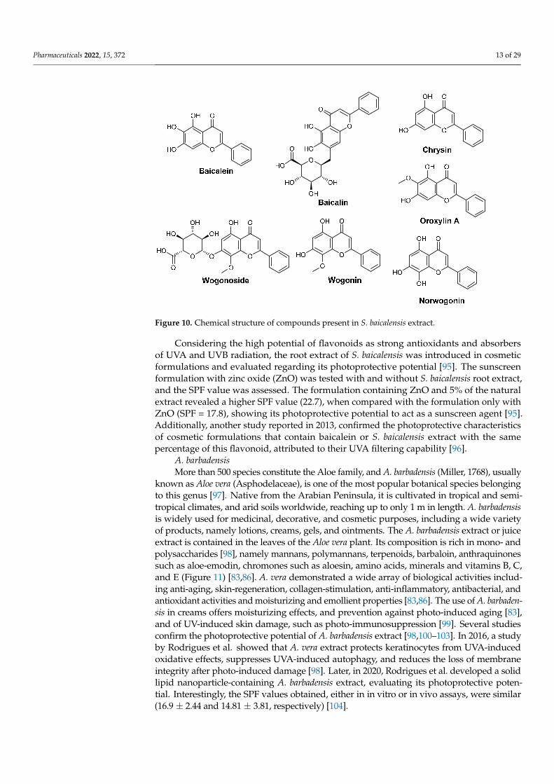
S. baicalensisS. baicalensis (Georgi, 1775) (Laminaceae), also known as Chinese skullcap, is a flower-
ing plant native to Asian countries. It has been used for two thousand years for medicinalpurposes due to its diverse array of biological activities. S. baicalensis possess clinical appli-cations such as antioxidant, antitumor, antiviral and antibacterial activities, and hepato- andneuroprotective effects [94]. Its high content in flavonoids, more than 30 metabolites, madethis extract a source of several active molecules. Chinese skullcap extract also includesamino acids, essential oils, sterols, and phenolic compounds. All of the constituents conferthe colour to the flowers (pigmentation) and photoprotection against UV-induced dam-age [94]. The major flavones present in the S. baicalensis root extract are baicalin, baicalein,wogonoside, wogonin, norwogonin, oroxylin A, and chrysin (Figure 10) [94].
Pharmaceuticals 2022, 15, 372 13 of 29
Pharmaceuticals 2022, 15, x FOR PEER REVIEW 13 of 30
S. baicalensis S. baicalensis (Georgi, 1775) (Laminaceae), also known as Chinese skullcap, is a flow-
ering plant native to Asian countries. It has been used for two thousand years for medici-nal purposes due to its diverse array of biological activities. S. baicalensis possess clinical applications such as antioxidant, antitumor, antiviral and antibacterial activities, and hepato- and neuroprotective effects [94]. Its high content in flavonoids, more than 30 me-tabolites, made this extract a source of several active molecules. Chinese skullcap extract also includes amino acids, essential oils, sterols, and phenolic compounds. All of the con-stituents confer the colour to the flowers (pigmentation) and photoprotection against UV-induced damage [94]. The major flavones present in the S. baicalensis root extract are bai-calin, baicalein, wogonoside, wogonin, norwogonin, oroxylin A, and chrysin (Figure 10) [94].
Considering the high potential of flavonoids as strong antioxidants and absorbers of UVA and UVB radiation, the root extract of S. baicalensis was introduced in cosmetic for-mulations and evaluated regarding its photoprotective potential [95]. The sunscreen for-mulation with zinc oxide (ZnO) was tested with and without S. baicalensis root extract, and the SPF value was assessed. The formulation containing ZnO and 5% of the natural extract revealed a higher SPF value (22.7), when compared with the formulation only with ZnO (SPF = 17.8), showing its photoprotective potential to act as a sunscreen agent [95]. Additionally, another study reported in 2013, confirmed the photoprotective characteris-tics of cosmetic formulations that contain baicalein or S. baicalensis extract with the same percentage of this flavonoid, attributed to their UVA filtering capability [96].
Figure 10. Chemical structure of compounds present in S. baicalensis extract.
A. barbadensis More than 500 species constitute the Aloe family, and A. barbadensis (Miller, 1768),
usually known as Aloe vera (Asphodelaceae), is one of the most popular botanical species belonging to this genus [97]. Native from the Arabian Peninsula, it is cultivated in tropical and semi-tropical climates, and arid soils worldwide, reaching up to only 1 m in length. A. barbadensis is widely used for medicinal, decorative, and cosmetic purposes, including a wide variety of products, namely lotions, creams, gels, and ointments. The A. barbadensis extract or juice extract is contained in the leaves of the Aloe vera plant. Its composition is rich in mono- and polysaccharides [98], namely mannans, polymannans, terpenoids, bar-baloin, anthraquinones such as aloe-emodin, chromones such as aloesin, amino acids,
Figure 10. Chemical structure of compounds present in S. baicalensis extract.
Considering the high potential of flavonoids as strong antioxidants and absorbersof UVA and UVB radiation, the root extract of S. baicalensis was introduced in cosmeticformulations and evaluated regarding its photoprotective potential [95]. The sunscreenformulation with zinc oxide (ZnO) was tested with and without S. baicalensis root extract,and the SPF value was assessed. The formulation containing ZnO and 5% of the naturalextract revealed a higher SPF value (22.7), when compared with the formulation only withZnO (SPF = 17.8), showing its photoprotective potential to act as a sunscreen agent [95].Additionally, another study reported in 2013, confirmed the photoprotective characteristicsof cosmetic formulations that contain baicalein or S. baicalensis extract with the samepercentage of this flavonoid, attributed to their UVA filtering capability [96].
A. barbadensisMore than 500 species constitute the Aloe family, and A. barbadensis (Miller, 1768), usually
known as Aloe vera (Asphodelaceae), is one of the most popular botanical species belongingto this genus [97]. Native from the Arabian Peninsula, it is cultivated in tropical and semi-tropical climates, and arid soils worldwide, reaching up to only 1 m in length. A. barbadensisis widely used for medicinal, decorative, and cosmetic purposes, including a wide varietyof products, namely lotions, creams, gels, and ointments. The A. barbadensis extract or juiceextract is contained in the leaves of the Aloe vera plant. Its composition is rich in mono- andpolysaccharides [98], namely mannans, polymannans, terpenoids, barbaloin, anthraquinonessuch as aloe-emodin, chromones such as aloesin, amino acids, minerals and vitamins B, C,and E (Figure 11) [83,86]. A. vera demonstrated a wide array of biological activities includ-ing anti-aging, skin-regeneration, collagen-stimulation, anti-inflammatory, antibacterial, andantioxidant activities and moisturizing and emollient properties [83,86]. The use of A. barbaden-sis in creams offers moisturizing effects, and prevention against photo-induced aging [83],and of UV-induced skin damage, such as photo-immunosuppression [99]. Several studiesconfirm the photoprotective potential of A. barbadensis extract [98,100–103]. In 2016, a studyby Rodrigues et al. showed that A. vera extract protects keratinocytes from UVA-inducedoxidative effects, suppresses UVA-induced autophagy, and reduces the loss of membraneintegrity after photo-induced damage [98]. Later, in 2020, Rodrigues et al. developed a solidlipid nanoparticle-containing A. barbadensis extract, evaluating its photoprotective poten-tial. Interestingly, the SPF values obtained, either in in vitro or in vivo assays, were similar(16.9 ± 2.44 and 14.81 ± 3.81, respectively) [104].

Pharmaceuticals 2022, 15, 372 14 of 29
Pharmaceuticals 2022, 15, x FOR PEER REVIEW 14 of 30
minerals and vitamins B, C, and E (Figure 11) [83,86]. A. vera demonstrated a wide array of biological activities including anti-aging, skin-regeneration, collagen-stimulation, anti-inflammatory, antibacterial, and antioxidant activities and moisturizing and emollient properties [83,86]. The use of A. barbadensis in creams offers moisturizing effects, and pre-vention against photo-induced aging [83], and of UV-induced skin damage, such as photo-immunosuppression [99]. Several studies confirm the photoprotective potential of A. barbadensis extract [98,100–103]. In 2016, a study by Rodrigues et al. showed that A. vera extract protects keratinocytes from UVA-induced oxidative effects, suppresses UVA-in-duced autophagy, and reduces the loss of membrane integrity after photo-induced dam-age [98]. Later, in 2020, Rodrigues et al. developed a solid lipid nanoparticle-containing A. barbadensis extract, evaluating its photoprotective potential. Interestingly, the SPF val-ues obtained, either in in vitro or in vivo assays, were similar (16.9 ± 2.44 and 14.81 ± 3.81, respectively) [104].
Figure 11. Chemical structure of compounds present in A. barbadensis extract with skin anti-aging effects.
C. nucifera C. nucifera (Linnaeus, 1753) or coconut tree belongs to the Arecaceae family, well-
characterized for its palm leaves, and is usually found in coastal areas associated with tropical regions, reaching up to 30 m in length [105]. Its juice or water is appreciated by tourists when they visit tropical and subtropical areas, and its origin is related to Pacific Islands in southeast Asia, such as the Philippines, Malaysia, and Indonesia [105]. The fruit is rich in fatty acids (caprylic, capric, lauric, myristic, palmitic, and oleic acids that repre-sent almost 90% of its content), carbohydrates, proteins, and a small percentage of vita-mins and minerals [105–107]. C. nucifera oil possesses some interesting activities such as antioxidant and antimicrobial activities, prevention of cardiac and Alzheimer’s diseases, due to high density of lipids that contribute to good cholesterol, and could be used to maintain the health of teeth, hair, and skin; for these reasons, it is widely used in cosmetic products [105,107,108].
At least three studies have reported the photoprotective potential of coconut oil [109–111]. In 2010, Kaur and Safar evaluated the in vitro SPF value of plant-derived oils typi-cally applied in cosmetic products, where coconut oil showed an in vitro SPF value of 7.119. When compared to the other 13 vegetable oils, coconut oil presented one of the highest SPF values [109]. In 2017, Widiyati investigated the photoprotective action of this oil in a cosmetic cream, which only revealed absorption in the UVC region of the UV spec-trum and insignificant absorption of UVA and UVB radiation. However, the combination of coconut oil with UV filters that absorb in the UVA and UVB regions, such as titanium oxide (TiO2) and benzophenone-3, was demonstrated to be favorable, having the cream that contained benzophenone-3 (UVA filter) and coconut oil with protective action against UVB and UVA radiation [110]. Oliveira et al. quantified the phenolic composition of C. nucifera extract and evaluated its antioxidant and photoprotective activities, either isolated or incorporated in a formulation [111]. Quercetin (Figure 6), (+)-catechin, vanillic and caf-feic acids, and (−)-epicatechin were detected and quantified by HPLC-MS analysis of the extract. All of the phenolic compounds were previously reported to exhibit antioxidant activity [111,112]. Regarding the photoprotective activity of the cream containing the C.
Figure 11. Chemical structure of compounds present in A. barbadensis extract with skin anti-aging effects.
C. nuciferaC. nucifera (Linnaeus, 1753) or coconut tree belongs to the Arecaceae family, well-
characterized for its palm leaves, and is usually found in coastal areas associated with tropicalregions, reaching up to 30 m in length [105]. Its juice or water is appreciated by tourists whenthey visit tropical and subtropical areas, and its origin is related to Pacific Islands in southeastAsia, such as the Philippines, Malaysia, and Indonesia [105]. The fruit is rich in fatty acids(caprylic, capric, lauric, myristic, palmitic, and oleic acids that represent almost 90% of itscontent), carbohydrates, proteins, and a small percentage of vitamins and minerals [105–107].C. nucifera oil possesses some interesting activities such as antioxidant and antimicrobialactivities, prevention of cardiac and Alzheimer’s diseases, due to high density of lipids thatcontribute to good cholesterol, and could be used to maintain the health of teeth, hair, andskin; for these reasons, it is widely used in cosmetic products [105,107,108].
At least three studies have reported the photoprotective potential of coconut oil [109–111].In 2010, Kaur and Safar evaluated the in vitro SPF value of plant-derived oils typicallyapplied in cosmetic products, where coconut oil showed an in vitro SPF value of 7.119.When compared to the other 13 vegetable oils, coconut oil presented one of the highestSPF values [109]. In 2017, Widiyati investigated the photoprotective action of this oil in acosmetic cream, which only revealed absorption in the UVC region of the UV spectrumand insignificant absorption of UVA and UVB radiation. However, the combination ofcoconut oil with UV filters that absorb in the UVA and UVB regions, such as titanium oxide(TiO2) and benzophenone-3, was demonstrated to be favorable, having the cream thatcontained benzophenone-3 (UVA filter) and coconut oil with protective action against UVBand UVA radiation [110]. Oliveira et al. quantified the phenolic composition of C. nuciferaextract and evaluated its antioxidant and photoprotective activities, either isolated orincorporated in a formulation [111]. Quercetin (Figure 6), (+)-catechin, vanillic and caffeicacids, and (−)-epicatechin were detected and quantified by HPLC-MS analysis of theextract. All of the phenolic compounds were previously reported to exhibit antioxidantactivity [111,112]. Regarding the photoprotective activity of the cream containing theC. nucifera extract and the cream containing quercetin, the SPF values varied between5.0–14.09 and 1.44–15.04, respectively. This study highlighted the potential of this naturalproduct in cosmetic formulations, as an extract or with only one ingredient, to be used as asustainable alternative to be incorporated in cosmetic formulations [111].
P. leucotomosP. leucotomos (Linnaeus, 1753), a native from South America, belongs to the Polypo-
diaceae family, and is classified as a tropical fern [113,114]. Several studies reported itsmedicinal applications in diminishing UV-induced damage and inflammatory and oxida-tive processes, as well as its use in some skin disorders, namely solar erythema, melasma,and atopic dermatitis [113–116], and contributing to fight skin photo-aging [83]. P. leucoto-mos extract is mainly constituted by phenolic compounds, namely phenolic acids (benzoic,cinnamic, caffeic, ferulic, coumaric, among others acids) and monosaccharides [117]. Pheno-lic acids offer antioxidant and photoprotective activities, and also contribute to the decreaseof UV-induced inflammation and oxidative damage [83]. González and Pathak (1996)demonstrated the protective effect of this botanical extract against UVA- and UVB-induceddamage, by decreasing the formation of ROS, lipid peroxidation and photosensitization,

Pharmaceuticals 2022, 15, 372 15 of 29
following either topical application or oral administration [118]. P. leucotomos extract wasalso reported to inhibit tumour necrosis factor alpha (TNF-α), and the production of cellularnitric oxide (NO) through the inhibition of nitric oxide synthase (iNOS), in conditions thatmimic the solar exposure-induced damage [119], and for protecting epidermal keratinocytesand dermal fibroblasts exposed to UV radiation [120].
2.2.2. Marine Ingredients
L. ochroleucaL. ochroleuca (Bachelot de la Pylaie, 1824) is a yellow-brown digitate kelp that reaches
up to about 1.5 m in length. Widely consumed in Asian countries, this edible alga ismainly valued in Europe through its extracts, which include alginates, bioactives, and pig-ments [121]. While alginates are usually commercialized to be used as thickening, gelling,and stabilizing agents in food, cosmetic and pharmaceutical industries, this alga also con-tains other bioactives, such as fucoidans that reduce the expression of the pro-inflammatorycytokines and have antioxidant, antimicrobial, and antitumor properties [122], and phenoliccompounds to which are usually associated antioxidant properties [20,123,124]. An exam-ple of phenolic compounds identified in extracts of L. ochroleuca is the linear phlorethols,which may have ortho-, meta- or para-oriented biphenyl ether bridges or combinations suchas triphlorethol C and acetylated tetraphlorethols A and B (Figure 12) [125,126].
Pharmaceuticals 2022, 15, x FOR PEER REVIEW 16 of 30
Figure 12. Structures of triphlorethol C and tetraphlorethols A and B extracted from L. ochroleuca.
Plankton The planktonic compartment of marine resources, composed of zooplankton, phyto-
plankton, bacteria, and viruses, represents 95% of marine biomass [127]. Although the potential is largely unexplored, planktonic organisms offer already immense opportuni-ties: new resources for medicine, cosmetics, and food, renewable energy, and long-term solutions to mitigate climate change [127]. Regarding its usage in cosmetics, plankton is included in skincare compositions to improve the health and physical appearance of skin; however, there is a paucity of evidence regarding the antioxidant and photoprotective properties of plankton.
Planktonic species are exposed to solar UVR, which penetrates the water column [33]. These organisms, upon different environmental stress factors (e.g., seasonal changes in UV levels and temperature), have evolved a variety of response mechanisms to prevent or repair damage from UVR [33]. Besides regulating their position in the water column, they also have the tendency to accumulate or synthesize sunscreens (e.g., carotenoids and MAAs), or repair DNA damage (e.g., photoenzymatic repair and nucleotide excision re-pair) [33,38]. Specific examples of MAAs isolated from Alexandrium excavatum, a red-tide dinoflagellate, are represented in Figure 13 [34].
Mycosporine-glycine
O
OCH3
NHHO HO
COOH
Palythine
NH
OCH3
NHHO HO
COOH
Asterina-330
N
OCH3
NHHO HO
COOH
OH
Shinorine
N
OCH3
NHHO HO
COOH
O
OH
Porphyra-334
N
OCH3
NHHO HO
COOH
CO2H
H3C
OH
(Z) and (E)-Palythenic Acid
N
OCH3
NHHO HO
COOH
CO2H(Z)
CH3
Usujirene
N
OCH3
NHHO HO
COOH
CH3
Palythene
N
OCH3
NHHO HO
COOH
H3C
Figure 13. Mycosporine-like amino acids isolated from A. excavatum [34].
The research on the photoprotective potential of MAAs has mostly been focused on the species in which MAA compounds are produced or found [31,37,38,128]. There has been surprisingly little work carried out in skin models to demonstrate their potential for human use [129,130]. However, preliminary data strongly suggest that MAAs have the
Figure 12. Structures of triphlorethol C and tetraphlorethols A and B extracted from L. ochroleuca.
The study of the seasonal and yearly variations in phenolic contents and associatedcosmetic activities of seven brown marine macroalgae, including L. ochroleuca, revealedthat the phlorotannin content of the studied algae was high regardless of the season, andantioxidant and photoprotective activities were similar to those of commercial molecules(butylated hydroxyanisole (BHA) and vitamin C) [20]. Concerning L. ochroleuca in particular,no statistically significant variation was observed (p > 0.05) between the seasons (autumn,winter, and spring), although seaweeds harvested in autumn appeared to have the highestradical scavenging activity in the DPPH test and antioxidant activity on the ferric reducingantioxidant power (FRAP) assay [20]. The phlorotannin enriched fraction of L. ochroleucaalso revealed potential photoprotective activities since the photoprotective factors weregreater than 1 (between 1.24 and 2.15 for sun protection factor (SPF) and between 1.19 and1.96 for UVA protection factor (PF-UVA)), with no seasonal differences [20].
PlanktonThe planktonic compartment of marine resources, composed of zooplankton, phy-
toplankton, bacteria, and viruses, represents 95% of marine biomass [127]. Although thepotential is largely unexplored, planktonic organisms offer already immense opportuni-ties: new resources for medicine, cosmetics, and food, renewable energy, and long-termsolutions to mitigate climate change [127]. Regarding its usage in cosmetics, plankton isincluded in skincare compositions to improve the health and physical appearance of skin;however, there is a paucity of evidence regarding the antioxidant and photoprotectiveproperties of plankton.
Planktonic species are exposed to solar UVR, which penetrates the water column [33].These organisms, upon different environmental stress factors (e.g., seasonal changes in

Pharmaceuticals 2022, 15, 372 16 of 29
UV levels and temperature), have evolved a variety of response mechanisms to preventor repair damage from UVR [33]. Besides regulating their position in the water column,they also have the tendency to accumulate or synthesize sunscreens (e.g., carotenoidsand MAAs), or repair DNA damage (e.g., photoenzymatic repair and nucleotide excisionrepair) [33,38]. Specific examples of MAAs isolated from Alexandrium excavatum, a red-tidedinoflagellate, are represented in Figure 13 [34].
Pharmaceuticals 2022, 15, x FOR PEER REVIEW 16 of 30
Figure 12. Structures of triphlorethol C and tetraphlorethols A and B extracted from L. ochroleuca.
Plankton The planktonic compartment of marine resources, composed of zooplankton, phyto-
plankton, bacteria, and viruses, represents 95% of marine biomass [127]. Although the potential is largely unexplored, planktonic organisms offer already immense opportuni-ties: new resources for medicine, cosmetics, and food, renewable energy, and long-term solutions to mitigate climate change [127]. Regarding its usage in cosmetics, plankton is included in skincare compositions to improve the health and physical appearance of skin; however, there is a paucity of evidence regarding the antioxidant and photoprotective properties of plankton.
Planktonic species are exposed to solar UVR, which penetrates the water column [33]. These organisms, upon different environmental stress factors (e.g., seasonal changes in UV levels and temperature), have evolved a variety of response mechanisms to prevent or repair damage from UVR [33]. Besides regulating their position in the water column, they also have the tendency to accumulate or synthesize sunscreens (e.g., carotenoids and MAAs), or repair DNA damage (e.g., photoenzymatic repair and nucleotide excision re-pair) [33,38]. Specific examples of MAAs isolated from Alexandrium excavatum, a red-tide dinoflagellate, are represented in Figure 13 [34].
Mycosporine-glycine
O
OCH3
NHHO HO
COOH
Palythine
NH
OCH3
NHHO HO
COOH
Asterina-330
N
OCH3
NHHO HO
COOH
OH
Shinorine
N
OCH3
NHHO HO
COOH
O
OH
Porphyra-334
N
OCH3
NHHO HO
COOH
CO2H
H3C
OH
(Z) and (E)-Palythenic Acid
N
OCH3
NHHO HO
COOH
CO2H(Z)
CH3
Usujirene
N
OCH3
NHHO HO
COOH
CH3
Palythene
N
OCH3
NHHO HO
COOH
H3C
Figure 13. Mycosporine-like amino acids isolated from A. excavatum [34].
The research on the photoprotective potential of MAAs has mostly been focused on the species in which MAA compounds are produced or found [31,37,38,128]. There has been surprisingly little work carried out in skin models to demonstrate their potential for human use [129,130]. However, preliminary data strongly suggest that MAAs have the
Figure 13. Mycosporine-like amino acids isolated from A. excavatum [34].
The research on the photoprotective potential of MAAs has mostly been focused onthe species in which MAA compounds are produced or found [31,37,38,128]. There hasbeen surprisingly little work carried out in skin models to demonstrate their potentialfor human use [129,130]. However, preliminary data strongly suggest that MAAs havethe potential for the protection of human skin from a diverse range of adverse effectsof solar UVR, including UVR-induced toxicity, DNA damage/erythema, inflammation,photoaging [129], and the damage induced by free radicals produced by UVR exposurethrough their antioxidant activity [131,132].
In another example, the antioxidant properties of aqueous extracts from the zoo-plankton dinophycean flagellates Gymnodinium impudicum and Alexandrium affine and theraphidophycean flagellate Chattonella ovata were examined [132]. All of the extracts directlyscavenged superoxide and the superoxide scavenging potential of any of the extracts wascomparable to that of L-ascorbic acid. As for hydroxyl radical scavenging, the Fenton reac-tion and the method of ultraviolet radiation to hydrogen peroxide were used as hydroxylradical generation systems [132]. Although a reduction of the level of hydroxyl radicalsin both of the systems for all the extracts was noticed, the fact that the levels of phenoliccompounds exhibited a correlation with the antioxidant activities of the extracts indicatesthe presence of substances other than phenolic compounds that might be responsible forthe activities.
Although the regular use of sunscreen has demonstrated a protective effect towardsthe development of skin cancer, newer options to help repair damaged DNA may havean important role in reducing the incidence of chronic sun exposure-related photoagingand non-melanoma skin cancer [10]. Plankton extract, which contains the DNA repairenzyme photolyase, is one novel ingredient that is currently being incorporated intosunscreens to complement intrinsic DNA repair, thus expanding the photoprotectiveabilities of sunscreens to photo repair [10]. This enzyme is capable of repairing cyclobutanedimers formed as a result of DNA UV irradiation [10].

Pharmaceuticals 2022, 15, 372 17 of 29
T. thermophillus FermentProbiotics have also attracted great attention towards the development of natural non-
toxic antioxidants due to their capacity to decrease the risk of accumulation of ROS and topotentially be used to reduce oxidative stress [133]. Their in vitro and in vivo antioxidantcapacity can be attributed mainly to the presence of specific antioxidant enzymes, therelease of different antioxidant compounds acting mainly as free-radical scavengers such asglutathione, the production of some extracellular polysaccharide biomolecules [134,135], oreven the exhibition of metal chelating activity [136]. Additionally, probiotic microorganismsare known to have beneficial properties such as soothing inflammation, balancing skinmicroflora, and inhibiting the growth of harmful bacteria [137].
Although currently there is a paucity of data on the antioxidant activity of T. thermophillusferment, a study in the field of compositions (either topical or food) revealed that thesecan benefit from the use of a probiotic/antioxidant composition with improved antioxidantcapabilities [137]. In particular, an example of a topical composition containing 10% ofa probiotic blend (≈70% α-glucan oligosaccharide, ≈19% polyminia sonchifolia root juice,≈10% maltodextrin, ≈1% lactobacillus), 60% of an antioxidant blend (≈95% T. thermophillusferment, ≈5% glycerin), 20% of methylsilanol hydroxyproline aspartate, and 10% of Triticummonococcum [wheat] seed extract, acted synergistically as an antioxidant in sufficient amountto inhibit signalling pathways that contribute to skin inflammation [137]. Additionally, specificenzymes from T. thermophillus can be used on damaged skin exposed to UV radiation andheat to catalyze the scavenging of free radicals caused by UV damage [138].
To provide a solution to prevent and/or treat damage associated with sun-derivedinfrared A (IRA) radiation of the skin and its appendages, a fermentation medium of T.thermophilus was developed by the cosmetic industry [139,140] and marketed as an anti-agingingredient used in cosmetic compositions for protecting the cellular structures from damagecaused not only by UV but also IR radiation. In the study that accompanied the developmentof this product, it was shown that it can limit the increase of the formation of pro-inflammatorymediators (PGE2, IL-6 and IL-8) and reactive oxygen species (ROS) by IRA [139]. Additionally,this product improves epidermis quality (stimulation of the synthesis of involucrin, ceramides,and/or intercellular lipids) and can limit the decrease in the synthesis of macromoleculesstructuring the extracellular matrix [collagen-1, fibrilin-1, and/or HSP-47 (heat shock protein)],caused by the alteration of the dermal integrity by IRA [139].
Artemia ExtractBrine shrimp (Artemia salina, Linnaeus, 1758) is a simple marine invertebrate of about
1 mm in size. This animal is routinely used as live feed for marine larval fish since itsfreeze-dried cysts can last for several years and can be hatched into larvae without specialequipment. Additionally, A. salina is widely used in the brine shrimp lethality assay(BSLA), a simple and inexpensive bioassay used for the preliminary assessment of toxicity.Extracts of A. salina have been largely explored for their anti-aging properties, which werepreviously reviewed by us [43]. Only a few in vivo studies were performed aiming toassess the efficacy of Artemia extracts as photoprotective agents. The use of a cosmeticproduct for the regeneration and stimulation of skin cells based on maritime components,comprised of (i) a product of an enzymatic extraction process of A. salina (phosphorylatednucleotides—diguanosine-tetraphosphate as the main component); (ii) D-myo-inositol-1,4,5-triphosphate; and (iii) glucan (Figure 14) (a:b:c ratio: 1:0.1–50:0.1–30) with additionalcosmetic active ingredients and supporting materials, resulted in a synergetic effect againstaggressive environmental influences by naturally strengthening the immune system of theskin, stimulating skin regeneration and, concomitantly, providing UV protection with theassistance of improved keratin barriers [25].

Pharmaceuticals 2022, 15, 372 18 of 29
Pharmaceuticals 2022, 15, x FOR PEER REVIEW 18 of 30
synthesis of involucrin, ceramides, and/or intercellular lipids) and can limit the decrease in the synthesis of macromolecules structuring the extracellular matrix [collagen-1, fi-brilin-1, and/or HSP-47 (heat shock protein)], caused by the alteration of the dermal integ-rity by IRA [139].
Artemia Extract Brine shrimp (Artemia salina, Linnaeus, 1758) is a simple marine invertebrate of about
1 mm in size. This animal is routinely used as live feed for marine larval fish since its freeze-dried cysts can last for several years and can be hatched into larvae without special equipment. Additionally, A. salina is widely used in the brine shrimp lethality assay (BSLA), a simple and inexpensive bioassay used for the preliminary assessment of tox-icity. Extracts of A. salina have been largely explored for their anti-aging properties, which were previously reviewed by us [43]. Only a few in vivo studies were performed aiming to assess the efficacy of Artemia extracts as photoprotective agents. The use of a cosmetic product for the regeneration and stimulation of skin cells based on maritime components, comprised of (i) a product of an enzymatic extraction process of A. salina (phosphorylated nucleotides—diguanosine-tetraphosphate as the main component); (ii) D-myo-inositol-1,4,5-triphosphate; and (iii) glucan (Figure 14) (a:b:c ratio: 1:0.1–50:0.1–30) with additional cosmetic active ingredients and supporting materials, resulted in a synergetic effect against aggressive environmental influences by naturally strengthening the immune sys-tem of the skin, stimulating skin regeneration and, concomitantly, providing UV protec-tion with the assistance of improved keratin barriers [25].
Figure 14. Maritime components of the tested cosmetic product containing A. salina extract.
Hsp70, a heat shock protein, has protective effects against UV, apoptosis, and ische-mia, through the inhibition of aggregation and assistance in the refolding of denatured proteins, and is recommended for wound healing and anti-aging purposes [141]. Studies on both cultured human epidermal cells and ex vivo skin showed that induction or ad-ministration of Hsp70 before stress significantly diminished UV-related morphological changes and sunburn cell number [26–28,142]. As an example, a study on the influence of heat shock proteins in the protection of cells from various types of stress has shown that treatment of human skin with Artemia extract induces Hsp70 in skin cells in a stress-free manner, enhancing aged skin defense from UV damage. The analysis of the punch
Figure 14. Maritime components of the tested cosmetic product containing A. salina extract.
Hsp70, a heat shock protein, has protective effects against UV, apoptosis, and ischemia,through the inhibition of aggregation and assistance in the refolding of denatured proteins,and is recommended for wound healing and anti-aging purposes [141]. Studies on bothcultured human epidermal cells and ex vivo skin showed that induction or administrationof Hsp70 before stress significantly diminished UV-related morphological changes andsunburn cell number [26–28,142]. As an example, a study on the influence of heat shockproteins in the protection of cells from various types of stress has shown that treatmentof human skin with Artemia extract induces Hsp70 in skin cells in a stress-free manner,enhancing aged skin defense from UV damage. The analysis of the punch biopsies from10 volunteers showed a decrease in UV damage signs when compared to the untreatedaged skin, confirming the fragility of aged skin towards UV stress and highlighting anew application of Artemia extract in the protection of aged skin [26]. Other similarstudies involving the administration of Artemia extract were also reported with similarresults [27,28,142].
N. gaditanaN. gaditana (L.M. Lubián, 1982) are mixotrophic, non-flagellated algae belonging to the
class Eustigmatophyceae [143]. Several species belonging to the Nannochloropsis genus aresources of eicosapentaenoic acid (EPA), which is of nutritional importance [143]. Anotherhighly interesting aspect is that N. gaditana produces lipids such as triacylglycerol (TAG),which is of special interest for human consumption since it has a composition of fattyacids similar to many vegetable oils [144]. Although the photoprotection strategies of thealga N. gaditana are well documented in the literature [145,146], only a limited numberof studies have reported the beneficial role of this microalga in cosmetic formulationsaimed to protect skin from UVR or aging factors. UVR (photoaging), oxidative stress(environmental pollution), and to a lesser extent, daily habits (smoking, poor nutrition) arethe main extrinsic (external) factors responsible for the acceleration and extent of aging.Various strategies have been developed that can act synergistically with sunscreen filtersand provide increased photoprotection in cosmetic formulations via various mechanisms.One of these strategies consists of the addition of active substances to cosmetic formulationsthat are commonly used for daily protection and care during low-sunshine periods. Acombination of a N. gaditana extract and a propolis extract was tested as an active componentin cosmetic formulations for dermal and in particular anti-aging use [147]. The protectiverole against the aging of human skin cells was confirmed by an increase in the ability of

Pharmaceuticals 2022, 15, 372 19 of 29
skin cells to produce collagen and elastin, by an increase in the expression of transcriptsof fibrillin in skin cells, enhancing skin elasticity since this protein offers the scaffoldfor the proper conformation of elastin [147]. Another study to evaluate the effects of aN. gaditana extract against oxidative stress in human primary fibroblasts was performedto investigate the potential applications of this natural ingredient in cosmetics [143]. N.gaditana extract exhibited skin protection properties by mediating oxidative responsesand apoptosis (including SOD1, GPX1, BID), positively regulating genes involved in skintexture and hydration (including AQP3, Col6A1, FBN1). Additionally, N. gaditana extractmodulated the expression of genes involved in skin irritation, DNA damage and aging(including IL1R, PCNA, FOXO3).
A. nodosumA. nodosum [(Linnaeus) Le Jolis, 1863] is a brown seaweed that belongs to the taxonomic
order of Fucales. The characteristic brown color presented by these species results fromthe dominance of the pigment fucoxanthin, which masks the other pigments (chlorophylla and c, β-carotene, and other carotenoids). Some of the most important constituents ofthis brown algae are phlorotannins, phloroglucinol-based polyphenols that are responsiblefor the well-known antioxidant activity exhibited by A. nodosum [20,148–158]. This brownalga has been highlighted in previous studies as a very promising ingredient due to itsantioxidant properties, both in formulations for anti-aging [43] and sensitive skin [159]. Re-garding photoprotection studies, some examples include a study of the seasonal and yearlyvariations in phenolic contents and associated cosmetic activities of seven brown marinemacroalgae, the five studied Fucales (including A. nodosum) presented higher activities thanLaminariales (IC50 = 0.010–0.353 mg·mL−1 (DPPH assay) and EC50 = 0.001–0.004 mg·mL−1
(FRAP assay)) with no significant differences between seasons or years for the differentFucales in either assay [20]. Concerning the FRAP assay, activities of fractions obtainedfrom A. nodosum (spring 2016) were significantly higher than the vitamin C (p = 0.039) [20].A. nodosum also revealed photoprotective activities since the photoprotective factors of thephlorotannin enriched fractions were greater than 1 (between 1.46 and 2.71 for SPF; 1.28and 1.93 for PF-UVA), with the summer fraction showing the highest activity [20].
The photoprotective potential against UVB radiation of extracts obtained from 21commercial macroalgae, including A. nodosum, was evaluated in vivo using the zebrafishembryo as a whole model organism [22]. A. nodosum showed an intermediate photoprotec-tive capacity since 54% of the embryos showed a low level of malformations, but normalembryos were present in 8.3%. The authors hypothesized that phlorotannins are respon-sible for the “in vivo” photoprotection, supporting A. nodosum’s potential application inphotoaging prevention [22].
Spirulina platensisSpirulina, also known as Arthrospira platensis (Gomont, 1892), is an edible cyanobacte-
ria (usually incorrectly classified as blue-green algae) that contains high levels of proteins,vitamins, fatty acids, and minerals and therefore is used as food, feed, and agriculturalsupplement [160]. Although numerous biological properties, such as antioxidant, anti-cancer, antimicrobial, and hepatoprotective effects, have been attributed to spirulina, thephotoprotective effect of this alga was only recently explored [160,161]. Spirulina was alsopointed to as a top ingredient to be included in anti-aging formulations, attributed mainlyto its content of sulfated polysaccharides [43].
A recent study demonstrated for the first time that an extract of spirulina has a reverseeffect on UV-induced photodamage such as loss of cell viability, cellular senescence, DNAdamage, and collagen destruction in dermal fibroblasts [160]. The extract promoted G2/Mcell cycle arrest and strongly inhibited UVB-induced cytotoxicity, senescence-associatedβ-galactosidase expression, and cyclobutane pyrimidine dimer formation in normal humandermal fibroblasts [160]. Additionally, the extract inhibited UVB-induced extracellularmatrix degradation by downregulating matrix metalloproteinase 1 (MMP-1) and MMP-3expression [160]. Another study explored the ability of spirulina to absorb the UVB spectraby determining its SPF, and by addressing its biological protective effects against UVB-

Pharmaceuticals 2022, 15, 372 20 of 29
irradiated human skin fibroblast cultures [161]. The photoprotective activity of spirulinawas attributed to the presence of high amounts of phenolic compounds, producing a highdegree of absorbance and yielding an SPF value of 30.39 [161]. No cytotoxicity was observedon UVB irradiation of human skin fibroblasts and the induction of the pro-inflammatorymediators IL-6 and IL-8 triggered by UVB irradiation was dramatically decreased aftertreatment with spirulina [161].
A. armataA. armata (Harvey, 1855) is a red seaweed that can be found on European coasts and
in the Northeast Atlantic [159]. Besides photosynthetic pigments, their main bioactiveingredients include agar, sulfated polysaccharides (carrageenans and porphyrans), andMAAs [159]. Two different studies aiming to screen selected red algae for the productionof UV-screen substances, such as MAAs, identified shinorine at the highest concentration(about 80–85%), followed by palythine (17–20%) (Figure 13) and finally asterina-330 pre-senting only traces [23,162]. These low-molecular-weight water-soluble molecules presentgreat potential as photoprotective and antioxidant ingredients since they can absorb UVradiation and disperse the absorbed energy as heat without generating reactive oxygenspecies (ROS) [159].
2.2.3. Mechanisms of Action
There are several biological mechanisms of action by which these active ingredientscould have beneficial effects; to disclose the molecular targets of the ingredients it isimportant to highlight the biological and cellular mechanisms associated with the UV-induced damage. The studied ingredients protect skin from a diverse range of adverseeffects of solar UVR, including UVR-induced DNA damage/erythema, inflammation,photoaging, and the damage of free radicals produced by UVR exposure. It is noteworthythat all the biological mechanisms are strongly interconnected such as the UV-inducedoxidative stress [163], associated with the excessive production of reactive species, namelyROS and RNS, which will contribute to triggering the expression of metalloproteinases [164].Additionally, the activation of transcription factors such as NF-kB and the production of pro-inflammatory cytokines/interleukins are usually triggered by solar radiation, thus evokinginflammatory and immune responses [165,166]. Direct or indirect/oxidative damage inbiomolecules, including cellular and mitochondrial DNA damage [167], crosslinking ofextracellular matrix fibers, such as collagen, elastin, and fibronectin [168], oxidation ofthe skin lipids [169], and formation of reactive and toxic advanced glycation products(AGEs) in glycation process [170] are other harmful events of the solar-induced stressoxidative process. Dysregulation of melanogenesis, namely the activation of tyrosinaseenzyme [171], and senescent events, including telomere shortening [172], may occur. Thedual antioxidant-photoprotective activity of natural-derived compounds present in UV-photoprotective formulations, particularly botanical and marine ingredients, could beconsidered the gold pot for the development of novel and upgraded skincare products
2.3. Highlights
In this study, the prevalence of natural ingredients in the composition of sunscreenproducts was assessed. The following highlights illustrate a resume of the most importantfacts that could be drawn from this study:
• Two hundred and eleven (48%) of the 444 analyzed sunscreen formulations comprisedingredients from terrestrial plants, while marine ingredients were present in 57 (13%)of the studied formulations.
• The highest prevalence of ingredients from terrestrial plants might be attributedto their biodiversity, easy cultivation, and growth modulation, making them moreattractive to the cosmetic industry.
• Twenty-nine (7%) of the total formulations contained both natural ingredients fromterrestrial and marine environments, showing a possible synergism of metaboliteaction towards activity, without loss of the desired properties.

Pharmaceuticals 2022, 15, 372 21 of 29
• The most used ingredients from terrestrial sources were obtained from the speciesH. annuus, G. max, V. paradoxa, P. gratissima, and G. inflata, all of which have beenstudied as photoprotective agents.
• The antioxidant and photoprotective activities of these extracts were mainly attributedto the presence of tocopherols, flavonoids, and phenolic acids.
• Algae, due to easy cultivation allied with the development of newer and improvedtechniques for their cultivation, constitute the most used marine ingredient, which istranslated in the number of studies and scientific evidence available in the literature.
• Other marine ingredients such as Artemia or microorganisms were less used, probablydue to the scarce scientific evidence of their photoprotective properties to back uptheir use in sunscreen formulations.
• The photoprotective effects of the analyzed marine ingredients are mainly attributedto MAAs. These low-molecular-weight water-soluble molecules proved to have anti-inflammatory effects by suppressing the expression of pro-inflammatory mediators inresponse to UV irradiation, therefore constituting a natural promising UV-absorbingalternative to common sunscreens.
3. Materials and Methods3.1. Data Collection
The composition of a pool of skincare facial cosmetic products from multinationalmanufacturers, marketed in Portuguese parapharmacies and pharmacies was collectedin 2021, to access the most used active marine and botanical ingredients in sunscreens.Sunscreen products with a labeled SPF were included in the study. All the informationavailable in the product’s label was collected, along with the information available on themanufacturers’ websites.
3.2. Data Analysis
The natural ingredients contained in sunscreens were listed according to the Interna-tional Nomenclature of Cosmetic Ingredients (INCI). Afterward, the data were analyzedconcerning the following parameters:
3.2.1. Natural Ingredients Use
The number of sunscreen products containing ingredients from terrestrial and/ormarine sources was evaluated and expressed in percentage.
3.2.2. Top Natural Ingredients Used in Sunscreens
The top ingredients from terrestrial and/or marine sources were identified from INCIlists and ranked in descending order of occurrence to disclose the top.
3.2.3. Scientific Evidence Supporting the Efficacy of Natural Ingredients in Sunscreens
The efficacy data for each ingredient from terrestrial and/or marine sources weresearched on the online databases PubMed, Scopus, Cochrane, KOSMET, and SciFinder®
[Chemical Abstracts Service (CAS)]. Due to the lack of studies regarding the applicabilityof active ingredients in sunscreens, a broader search was performed using the keywords“INCI name” OR “synonyms”, when applicable.
4. Conclusions
Over the last years, the search for valid alternatives of natural origin to complement theuse of synthetic chemicals has been a growing trend in the cosmetic sector. As consumersbecome increasingly aware of the potential toxicity of synthetic cosmetic ingredients, prod-ucts formulated with natural ingredients are increasingly reaching the consumer market.To date, an interesting number of ingredients from plants and marine organisms withphotoprotective properties have already been incorporated in sunscreens that are currentlyon the market. Although some advances have been put towards the research and the use

Pharmaceuticals 2022, 15, 372 22 of 29
of ingredients from terrestrial plants in this type of formulation, the number of marineingredients is still very minimal when compared to the botanical, especially consideringthe vastness of the sea and great potential for breakthroughs. Efforts should be deviatedtowards new strategies to increase the profitability of the extraction process, isolationand characterization of products from natural resources, and further investigation of theirphotoprotective potential and the safety profile regarding their application in the skin.However, some limitations are expected to avert the full exploitation of natural resources—the low concentrations of the active ingredient in the plant/organism responsible for theproduction of the ingredient, the variability on the environment (habitat, season of harvest-ing, and environmental conditions), which can cause different outcomes regarding theirbiosynthesis, and the fact that the mass cultivation of some species might endanger somealready threatened ecosystems. The solution lies in finding sustainable ways to producebioactive metabolites, paired with the optimization of growing and harvesting conditionsto obtain enough high-valued compounds to be used as cosmetic ingredients.
Another important aspect lies with the fact that the efficacy and safety of some syn-thetic UV filters are hindered by their low photostability, and damage to marine ecosystems.The discovery that some of the chemicals found in sunscreens and other cosmetic productsthreaten the health of coral reefs entails a need for constant product development and refor-mulation. The photoprotective effects of natural ingredients are mainly exerted throughtheir antioxidant effects, and through the regulation of UV-induced skin inflammation, bar-rier impairment, and aging. Even though natural ingredients cannot replace conventionalUV filters, they can complement the photoprotective effectiveness afforded by organic andinorganic UV filters.
In conclusion, the use of natural ingredients in sunscreen formulations presents agreat potential for the cosmetic industry, with a noticeable growing tendency over recentyears. Nevertheless, the understanding of the photoprotection mechanisms used by naturalorganisms to bear sun overexposure should be further explored and extended to ensurethe effectiveness and safety of the ingredients used for cosmetic application. This studyprovides an original and updated overview on the use of ingredients from plants andmarine organisms in a panel of sunscreen formulations commercialized in the Portuguesemarket in 2021 and provides researchers and the cosmetic industry a perspective of thetrends in the use of natural ingredients to develop new and improved formulations toprevent the deleterious effects of UVR.
Author Contributions: Conceptualization: I.F.A.; Data analysis: D.I.S.P.R. and A.J.; Writing—originaldraft preparation and final manuscript: D.I.S.P.R. and A.J.; Supervision: J.M.S.L., H.C. and M.T.C.;Writing—review and editing: I.F.A., E.S., H.C. and M.T.C. All authors have read and agreed to thepublished version of the manuscript.
Funding: This work is financed by national funds from FCT—Fundação para a Ciência e a Tecnologia,I.P., in the scope of the project UIDP/04378/2020 and UIDB/04378/2020 of the Research Uniton Applied Molecular Biosciences—UCIBIO and the project LA/P/0140/2020 of the AssociateLaboratory Institute for Health and Bioeconomy—i4HB. This research was also supported by nationalfunds through FCT (Foundation for Science and Technology) within the scope of UIDB/04423/2020,UIDP/04423/2020 (Group of Natural Products and Medicinal Chemistry—CIIMAR), as well asa structured program of R&D&I ATLANTIDA (NORTE-01-0145-FEDER-000040), supported byNORTE2020, through ERDF, and CHIRALBIO ACTIVE-PI-3RL-IINFACTS-2019. A.J. acknowledgesfor her PhD fellowship with the reference UI/BD/151319/2021.
Institutional Review Board Statement: Not applicable.
Informed Consent Statement: Not applicable.
Data Availability Statement: Not applicable.
Acknowledgments: To Inês Augusto for Data collection.
Conflicts of Interest: The authors declare no conflict of interest.

Pharmaceuticals 2022, 15, 372 23 of 29
References1. Radiation: Ultraviolet (UV) Radiation. Available online: https://www.who.int/news-room/q-a-detail/radiation-ultraviolet-(uv)
(accessed on 30 August 2021).2. Milito, A.; Castellano, I.; Damiani, E. From Sea to Skin: Is There a Future for Natural Photoprotectants? Mar. Drugs 2021, 19, 379.
[CrossRef] [PubMed]3. Rittié, L.; Fisher, G.J. Natural and Sun-Induced Aging of Human Skin. Cold Spring Harb. Perspect. Med. 2015, 5, a015370. [CrossRef]4. Bosch, R.; Philips, N.; Suárez-Pérez, J.A.; Juarranz, A.; Devmurari, A.; Chalensouk-Khaosaat, J.; González, S. Mechanisms
of Photoaging and Cutaneous Photocarcinogenesis, and Photoprotective Strategies with Phytochemicals. Antioxidants 2015,4, 248–268. [CrossRef]
5. Orazio, J.; Jarrett, S.; Amaro-Ortiz, A.; Scott, T. UV Radiation and the Skin. Int. J. Mol. Sci. 2013, 14, 12222–12248. [CrossRef]6. Martinez, R.M.; Pinho-Ribeiro, F.A.; Steffen, V.S.; Caviglione, C.V.; Vignoli, J.A.; Barbosa, D.S.; Baracat, M.M.; Georgetti, S.R.;
Verri, W.A.; Casagrande, R. Naringenin Inhibits UVB Irradiation-Induced Inflammation and Oxidative Stress in the Skin ofHairless Mice. J. Nat. Prod. 2015, 78, 1647–1655. [CrossRef] [PubMed]
7. Duteil, L.; Cardot-Leccia, N.; Queille-Roussel, C.; Maubert, Y.; Harmelin, Y.; Boukari, F.; Ambrosetti, D.; Lacour, J.-P.; Passeron, T.Differences in visible light-induced pigmentation according to wavelengths: A clinical and histological study in comparison withUVB exposure. Pigment Cell Melanoma Res. 2014, 27, 822–826. [CrossRef] [PubMed]
8. de Oliveira, C.A.; Peres, D.D.A.; Rugno, C.M.; Kojima, M.; Pinto, C.; Consiglieri, V.O.; Kaneko, T.M.; Rosado, C.; Mota, J.;Velasco, M.V.R.; et al. Functional photostability and cutaneous compatibility of bioactive UVA sun care products. J. Photochem.Photobiol. B 2015, 148, 154–159. [CrossRef]
9. Wolber, R.; Schlenz, K.; Wakamatsu, K.; Smuda, C.; Nakanishi, Y.; Hearing, V.J.; Ito, S. Pigmentation effects of solar simulatedradiation as compared with UVA and UVB radiation. Pigment Cell Melanoma Res. 2008, 21, 487–491. [CrossRef]
10. Leccia, M.-T.; Lebbe, C.; Claudel, J.-P.; Narda, M.; Basset-Seguin, N. New Vision in Photoprotection and Photorepair. Dermatol.Ther. 2019, 9, 103–115. [CrossRef]
11. Urbach, F. The historical aspects of sunscreens. J. Photochem. Photobiol. B Biol. 2001, 64, 99–104. [CrossRef]12. Bens, G. Sunscreens. In Sunlight, Vitamin D and Skin Cancer; Springer: New York, NY, USA, 2014; pp. 429–463. [CrossRef]13. Lee, S.-H.; Xiong, J.-Q.; Ru, S.; Patil, S.M.; Kurade, M.B.; Govindwar, S.P.; Oh, S.-E.; Jeon, B.-H. Toxicity of benzophenone-3 and its
biodegradation in a freshwater microalga Scenedesmus obliquus. J. Hazard. Mater. 2020, 389, 122149. [CrossRef]14. Schneider, S.L.; Lim, H.W. Review of environmental effects of oxybenzone and other sunscreen active ingredients. J. Am. Acad.
Dermatol. 2019, 80, 266–271. [CrossRef] [PubMed]15. de Miranda, L.L.R.; Harvey, K.E.; Ahmed, A.; Harvey, S.C. UV-filter pollution: Current concerns and future prospects. Environ.
Monit. Assess. 2021, 193, 840. [CrossRef] [PubMed]16. He, H.; Li, A.; Li, S.; Tang, J.; Li, L.; Xiong, L. Natural components in sunscreens: Topical formulations with sun protection factor
(SPF). Biomed. Pharmacother. 2021, 134, 111161. [CrossRef] [PubMed]17. Matsui, M.S.; Hsia, A.; Miller, J.D.; Hanneman, K.; Scull, H.; Cooper, K.D.; Baron, E. Non-Sunscreen Photoprotection: Antioxidants
Add Value to a Sunscreen. J. Investig. Dermatol. Symp. Proc. 2009, 14, 56–59. [CrossRef]18. Pangestuti, R.; Siahaan, E.A.; Kim, S.-K. Photoprotective Substances Derived from Marine Algae. Mar. Drugs 2018, 16, 399.
[CrossRef] [PubMed]19. Amador-Castro, F.; Rodriguez-Martinez, V.; Carrillo-Nieves, D. Robust natural ultraviolet filters from marine ecosystems for the
formulation of environmental friendlier bio-sunscreens. Sci. Total Environ. 2020, 749, 141576. [CrossRef]20. Gager, L.; Connan, S.; Molla, M.; Couteau, C.; Arbona, J.-F.; Coiffard, L.; Cerantola, S.; Stiger-Pouvreau, V. Active phlorotannins
from seven brown seaweeds commercially harvested in Brittany (France) detected by 1H NMR and in vitro assays: Temporalvariation and potential valorization in cosmetic applications. J. Appl. Phycol. 2020, 32, 2375–2386. [CrossRef]
21. Debacq-Chainiaux, F.; Borlon, C.; De Hertogh, B.; Remacle, J.; Morvan, P.Y.; Vallee, R.; Toussaint, O. Identification of potentialanti-photoageing algal compounds using an in-vitro model of photoageing. J. Pharm. Pharmacol. 2006, 58, 1577–1583. [CrossRef]
22. Guinea, M.; Franco, V.; Araujo-Bazán, L.; Rodríguez-Martín, I.; González, S. In vivo UVB-photoprotective activity of extracts fromcommercial marine macroalgae. Food Chem. Toxicol. 2012, 50, 1109–1117. [CrossRef]
23. Lalegerie, F.; Lajili, S.; Bedoux, G.; Taupin, L.; Stiger-Pouvreau, V.; Connan, S. Photo-protective compounds in red macroalgaefrom Brittany: Considerable diversity in mycosporine-like amino acids (MAAs). Mar. Environ. Res. 2019, 147, 37–48. [CrossRef][PubMed]
24. Shanuja, S.K.; Iswarya, S.; Sridevi, J.; Gnanamani, A. Exploring the UVB-protective efficacy of melanin precursor extracted frommarine imperfect fungus: Featuring characterization and application studies under in vitro conditions. Int. Microbiol. 2018,21, 59–71. [CrossRef] [PubMed]
25. Zastrow, L.; Domloge, N.; Golz-Berner, K. Cosmetic Product Based on Artemia Salina Extracts for Regenerating and StimulatingSkin Cells. WO1999038483A1, 5 August 1999.
26. Cucumel, K.; Farra, C.D.; Domloge, N. Artemia Extract ‘Compensates’ for Age-Related Decrease of Hsp70 in Skin. J. Investig.Dermatol. 2002, 119, 207–350. [CrossRef]
27. Cucumel, K.; Botto, J.; Bauza, E.; Farra, C.D.; Roetto, R.; Domloge, N. Artemia extract induces Hsp70 in human cells and enhancescell protection from stress. J. Investig. Dermatol. 2001, 117, 456–489. [CrossRef]

Pharmaceuticals 2022, 15, 372 24 of 29
28. Domloge, N.; Bauza, E.; Cucumel, K.; Peyronel, D.; Farra, C.D. Artemia extract toward more extensive sun protection. Cosmet.Toiletries 2002, 2002, 69–78.
29. Wang, L.; Meng, L.; Ye, S.; Chen, F.; Xie, S.; Wang, X. A Photoprotection Repairing Composition, Preparation Method andApplication, and Skin Care Product Thereof [Machine Translation]. CN112535657A, 23 March 2021.
30. Patil, J.S.; Rodrigues, R.V.; Paul, P.; Sathish, K.; Rafi, M.; Anil, A.C. Benthic dinoflagellate blooms in tropical intertidal rock pools:Elucidation of photoprotection mechanisms. Mar. Biol. 2017, 164, 1–19. [CrossRef]
31. Moisan, T.A.; Goes, J.; Neale, P.J. Chapter 4—Mycosporine-like amino acids in phytoplankton: Biochemistry, physiology andoptics. In Marine Phytoplankton; Kersey, W.T., Munger, S.P., Eds.; Nova Science Publishers: New York, NY, USA, 2009; pp. 119–143.
32. Laspoumaderes, C.; Bastidas Navarro, M.; Souza, M.S.; Modenutti, B.; Balseiro, E. Effect of ultraviolet radiation on clearance rateof planktonic copepods with different photoprotective strategies. Int. Rev. Hydrobiol. 2019, 104, 34–44. [CrossRef]
33. Sonntag, B.; Sommaruga, R. Effectiveness of photoprotective strategies in three mixotrophic planktonic ciliate species. Diversity2020, 12, 252. [CrossRef]
34. Carreto, J.I.; Carignan, M.O.; Daleo, G.; Marco, S.G.D. Occurrence of mycosporine-like amino acids in the red-tide dinoflagellateAlexandrium excavatum: UV-photoprotective compounds? J. Plankton Res. 1990, 12, 909–921. [CrossRef]
35. Bashevkin, S.M.; Morgan, S.G.; Christy, J.H. Photoprotective benefits of pigmentation in the transparent plankton community: Acomparative species experimental test. Ecology 2019, 100, e02680. [CrossRef] [PubMed]
36. Hessen, D.O.; Soerensen, K. Photoprotective pigmentation in alpine zooplankton populations. Aqua Fenn. 1990, 20, 165–170.37. Karentz, D.; McEuen, F.S.; Land, M.C.; Dunlap, W.C. Survey of mycosporine-like amino acid compounds in Antarctic marine
organisms: Potential protection from ultraviolet exposure. Mar. Biol. 1991, 108, 157–166. [CrossRef]38. Hylander, S.; Jephson, T. UV protective compounds transferred from a marine dinoflagellate to its copepod predator. J. Exp. Mar.
Biol. Ecol. 2010, 389, 38–44. [CrossRef]39. Nikitakis, J.; Lange, B. International Cosmetic Ingredient Dictionary and Handbook; Personal Care Products Council: Washington, DC,
USA, 2016; Volume 1.40. Kuete, V.; Viertel, K.; Efferth, T. 18-Antiproliferative Potential of African Medicinal Plants. In Medicinal Plant Research in Africa;
Kuete, V., Ed.; Elsevier: Oxford, UK, 2013; pp. 711–724.41. Darqui, F.S.; Radonic, L.M.; Beracochea, V.C.; Hopp, H.E.; López Bilbao, M. Peculiarities of the Transformation of Asteraceae
Family Species: The Cases of Sunflower and Lettuce. Front. Plant Sci. 2021, 12, 767459. [CrossRef] [PubMed]42. Christaki, E.; Bonos, E.; Giannenas, I.; Florou-Paneri, P. Functional properties of carotenoids originating from algae. J. Sci. Food
Agric. 2013, 93, 5–11. [CrossRef]43. Resende, D.I.S.P.; Ferreira, M.; Magalhães, C.; Sousa Lobo, J.M.; Sousa, E.; Almeida, I.F. Trends in the use of marine ingredients in
anti-aging cosmetics. Algal Res. 2021, 55, 102273. [CrossRef]44. Ferreira, M.S.; Magalhães, M.C.; Oliveira, R.; Sousa-Lobo, J.M.; Almeida, I.F. Trends in the Use of Botanicals in Anti-Aging
Cosmetics. Molecules 2021, 26, 3584. [CrossRef]45. Sánchez-Muniz, F.J.; Bastida, S.; Benedí, J. Sunflower Oil. In Encyclopedia of Food and Health; Caballero, B., Finglas, P.M.,
Toldrá, F., Eds.; Academic Press: Oxford, UK, 2016; pp. 217–226. [CrossRef]46. Pal, D. Sunflower (Helianthus annuus L.) Seeds in Health and Nutrition. In Nuts and Seeds in Health and Disease Prevention;
Academic Press: Cambridge, MA, USA, 2011; pp. 1097–1105. [CrossRef]47. Adeleke, B.S.; Babalola, O.O. Oilseed crop sunflower (Helianthus annuus) as a source of food: Nutritional and health benefits.
Food Sci. Nutr. 2020, 8, 4666–4684. [CrossRef]48. Guo, S.; Ge, Y.; Na Jom, K. A review of phytochemistry, metabolite changes, and medicinal uses of the common sunflower seed
and sprouts (Helianthus annuus L.). Chem. Cent. J. 2017, 11, 95. [CrossRef]49. Salas, J.J.; Bootello, M.A.; Martínez-Force, E.; Venegas Calerón, M.; Garcés, R. High stearic sunflower oil: Latest advances and
applications. OCL 2021, 28, 35. [CrossRef]50. Mehmood, A.; Zhao, L.; Ishaq, M.; Safdar, B.; Wang, C.; Nadeem, M. Optimization of total phenolic contents, antioxidant, and
in-vitro xanthine oxidase inhibitory activity of sunflower head. CYTA J. Food 2018, 16, 957–964. [CrossRef]51. Tamara, N.; Angkasa, C.; Ginting, C.L.; Chiuman, L.; Lister, I.N.E. Determination Sunscreen Potential Based on Sunflower Seed
Oil (Helianthus annuus) in Cream Preparation with Combination of Oxybenzone and Octyl Methoxycinnamate by in Vitro Method.Am. Sci. Res. J. Eng. Technol. Sci. 2019, 60, 33–39.
52. Karamac, M.; Kosinska, A.; Estrella, I.; Hernandez, T.; Duenas, M. Antioxidant activity of phenolic compounds identified insunflower seeds. Eur. Food Res. Technol. 2012, 235, 221–230. [CrossRef]
53. HELIOXINE®. Sunflowers for a Photoprotection Strategy. Available online: https://www.silab.fr/produit-27-helioxine_usa.html(accessed on 21 September 2021).
54. Wang, K.-J.; Li, X.-H.; Zhang, J.-J.; Chen, H.; Zhang, Z.-L.; Yu, G.-D. Natural introgression from cultivated soybean (Glycine max)into wild soybean (Glycine soja) with the implications for origin of populations of semi-wild type and for biosafety of wild speciesin China. Genet. Resour. Crop Evol. 2010, 57, 747–761. [CrossRef]
55. Carvalho, D.O.; Curto, A.F.; Guido, L.F. Determination of Phenolic Content in Different Barley Varieties and CorrespondingMalts by Liquid Chromatography-diode Array Detection-Electrospray Ionization Tandem Mass Spectrometry. Antioxidants 2015,4, 563–576. [CrossRef] [PubMed]

Pharmaceuticals 2022, 15, 372 25 of 29
56. An, Y.Y.; Qun, Z.Y.; Yuan, L. Effects of quercetin and enhanced UV-B radiation on the soybean (Glycine max) leaves. Acta Physiol.Plant. 2006, 28, 49–57. [CrossRef]
57. Soybean Extract—Supplied by Carruba. Available online: https://cosmetics.specialchem.com/product/i-carrubba-soybean-extract (accessed on 9 October 2021).
58. Chen, Q.; Yuan, X.; Zhang, C.; Yan, N.; Jing, C.; Wang, X.; Shi, J. Comparison of Phenolic and Flavonoid Compound Profiles andAntioxidant and α-Glucosidase Inhibition Properties of Cultivated Soybean (Glycine max) and Wild Soybean (Glycine soja). Plants2021, 10, 813. [CrossRef]
59. Jing, C.; Yuan, Y.; Tang, Q.; Zou, P.; Li, Y.; Zhang, C. Extraction optimization, preliminary characterization and antioxidantactivities of polysaccharides from Glycine soja. Int. J. Biol. Macromol. 2017, 103, 1207–1216. [CrossRef]
60. Machado, A.C.R.; Monteiro, A.C.; Mochi, D.A. Selection of anti-wetting agents and photoprotectants compatible with a soybeanweed control bioagent. Afr. J. Microbiol. Res. 2016, 10, 1829–1836. [CrossRef]
61. Tsoyi, K.; Park, H.B.; Kim, Y.M.; Chung, J.I.; Shin, S.C.; Shim, H.J.; Lee, W.S.; Seo, H.G.; Lee, J.H.; Chang, K.C.; et al. ProtectiveEffect of Anthocyanins from Black Soybean Seed Coats on UVB-Induced Apoptotic Cell Death In Vitro and In Vivo. J. Agric. FoodChem. 2008, 56, 10600–10605. [CrossRef]
62. Honfo, F.G.; Akissoe, N.; Linnemann, A.R.; Soumanou, M.; Van Boekel, M.A.J.S. Nutritional Composition of Shea Products andChemical Properties of Shea Butter: A Review. Crit. Rev. Food Sci. Nutr. 2014, 54, 673–686. [CrossRef] [PubMed]
63. Butyrospermum Parkii (Shea) Butter. Available online: https://cosmeticsinfo.org/ingredient/butyrospermum-parkii-shea-butter(accessed on 22 September 2021).
64. Maanikuu, P.M.I.; Peker, K. Medicinal and Nutricional Benefits from the Shea-T- (Vitellaria paradoxa). J. Biol. Agric. Healthc. 2017,7, 51–57.
65. Fowler, J.; Sivelberg, N. Active Naturals Have a Key Role in Atopic Dermatitis. Semin. Cutan. Med. Surg. 2008, 27, 8–10. [CrossRef]66. Maranz, S.; Wiesman, Z.; Garti, N. Phenolic constituents of Shea (Vitellaria paradoxa) kernels. J. Agric. Food Chem. 2003,
51, 6268–6273. [CrossRef] [PubMed]67. Morocho-Jacome, A.L.; Freire, T.B.; de Oliveira, A.C.; Velasco, M.V.R.; Baby, A.R.; de, A.T.S.; Rosado, C. In Vivo SPF from
multifunctional sunscreen systems developed with natural compounds—A review. J. Cosmet. Dermatol. 2021, 20, 729–737.[CrossRef] [PubMed]
68. Sarruf, F.D.; Sauce, R.; Candido, T.M.; Oliveira, C.A.; Velasco, M.V.R.; Baby, A.R.; Rosado, C. Butyrospermum parkii butter increasedthe photostability and in vivo SPF of a molded sunscreen system. J. Cosmet. Dermatol. 2020, 19, 3296–3301. [CrossRef] [PubMed]
69. Sarruf, F.D.; Candido, T.M.; de Oliveira, C.A.; Bou-Chacra, N.A.; Robles, V.M.V.; Baby, A.R. Influence of Shea (Butyrospermumparkii) butter, TiO2 and ethylhexyl methoxycinnamate on physical parameters and in vitro photoprotective efficacy. J. Cosmet.Dermatol. 2020, 19, 2076–2085. [CrossRef]
70. Tarras-Wahlberg, N.; Stenhagen, G.; Larko, O.; Rosen, A.; Wennberg, A.M.; Wennerstrom, O. Changes in ultraviolet absorption ofsunscreens after ultraviolet irradiation. J. Investig. Dermatol. 1999, 113, 547–553. [CrossRef]
71. Persea Gratissima (Avocado) Oil. Available online: https://cosmeticsinfo.org/ingredient/persea-gratissima-avocado-oil-0(accessed on 23 September 2021).
72. Dreher, M.L.; Davenport, A.J. Hass avocado composition and potential health effects. Crit. Rev. Food Sci. Nutr. 2013, 53, 738–750.[CrossRef]
73. Orhan, I.; Sener, B. Fatty Acid Content of Selected Seed Oils. J. Herb. Pharmacother. 2002, 2, 29–33. [CrossRef]74. Kruthiventi, A.K.; Krishnaswamy, N.R. Constituents of the flowers of Persea gratissima. Fitoterapia 2000, 71, 94–96. [CrossRef]75. Special Chem—Cosmetic Ingredients—Avocado Oil. Available online: https://cosmetics.specialchem.com/product/i-oqema-
avocado-oil (accessed on 23 September 2021).76. Joseph, L.B.; Koukouras, K. Persea Gratissima (Avocado) Sterols decrease UVB induced proinflammatory mediators. J. Cosmet.
Sci. 2002, 53, 309–311. [PubMed]77. Xie, J.; Zhang, Y.; Wang, W. HPLC analysis of glycyrrhizin and licochalcone A in Glycyrrhiza inflata from Xinjiang (China). Chem.
Nat. Compd. 2010, 46, 148–151. [CrossRef]78. Furusawa, J.; Funakoshi-Tago, M.; Mashino, T.; Tago, K.; Inoue, H.; Sonoda, Y.; Kasahara, T. Glycyrrhiza inflata-derived chalcones,
Licochalcone A, Licochalcone B and Licochalcone D, inhibit phosphorylation of NF-kappaB p65 in LPS signaling pathway. Int.Immunopharmacol. 2009, 9, 499–507. [CrossRef] [PubMed]
79. European Medicines Agency—Committee on Herbal Medicinal Products. Assessment report on Glycyrrhiza glabra L. and/orGlycyrrhiza inflata Bat. and/or Glycyrrhiza uralensis Fisch., Radix; European Medicines Agency—Committee on Herbal MedicinalProducts: London, UK, 2013.
80. Fu, Y.; Chen, J.; Li, Y.-J.; Zheng, Y.-F.; Li, P. Antioxidant and anti-inflammatory activities of six flavonoids separated from licorice.Food Chem. 2013, 141, 1063–1071. [CrossRef] [PubMed]
81. Okada, K.; Tamura, Y.; Yamamoto, M.; Inoue, Y.; Takagaki, R.; Takahashi, K.; Demizu, S.; Kajiyama, K.; Hiraga, Y.; Kinoshita, T.Identification of antimicrobial and antioxidant constituents from licorice of Russian and Xinjiang origin. Chem. Pharm. Bull. 1989,37, 2528–2530. [CrossRef] [PubMed]
82. Monico, G.; Leo, M.; Ma, B.; Johal, R.S.; Ma, T.; Sivamani, R.K. The use of botanical products and vitamins in sunscreens. Dermatol.Online J. 2015, 21. [CrossRef]
Pharmaceuticals 2022, 15, 372 26 of 29
83. Naser, W. The Cosmetic Effects of Various Natural Biofunctional Ingredients against Skin Aging: A Review. Int. J. Appl. Pharm.2021, 13, 10–18. [CrossRef]
84. Castangia, I.; Caddeo, C.; Manca, M.L.; Casu, L.; Latorre, A.C.; Diez-Sales, O.; Ruiz-Sauri, A.; Bacchetta, G.; Fadda, A.M.; Manconi,M. Delivery of liquorice extract by liposomes and hyalurosomes to protect the skin against oxidative stress injuries. Carbohydr.Polym. 2015, 134, 657–663. [CrossRef]
85. Saeedi, M.; Morteza-Semmani, K.; Ghoreishi, M.-R. The treatment of atopic dermatitis with licorice gel. J. Dermatol. Treat. 2008,14, 153–157. [CrossRef]
86. Nguyen, J.K.; Masub, N.; Jagdeo, J. Bioactive ingredients in Korean cosmeceuticals: Trends and research evidence. J. Cosmet.Dermatol. 2020, 19, 1555–1569. [CrossRef] [PubMed]
87. Pareek, A.; Suthar, M.; Rathore, G.S.; Bansal, V. Feverfew (Tanacetum parthenium L.): A systematic review. Pharmacogn. Rev. 2011,5, 103–110. [CrossRef] [PubMed]
88. Grzeszczuk, M.; Grzeszczuk, M.; Wesołowska, A. Composition of essential oils and some antioxidants in flowers of threeChrysanthemum cultivars. J. Elem. 2018, 23, 1331–1342. [CrossRef]
89. Romo, J.; de Vivar, A.R.; Trevino, R.; Joseph-Nathan, P.; Díaz, E. Constituents of Artemisia and Crysanthemum species—Thestructures of Chrysartemins A and B. Phytochemistry 1970, 9, 1615–1621. [CrossRef]
90. Whkomirski, B.; Dubielecka, B. Sterol content as a similarity of different organs of two varietas of Chrysanthemum parthenium.Phytochemistry 1996, 42, 1603–1604. [CrossRef]
91. Hangaru, D.; Benedec, D.; Vlase, L.; Popica, I.; Bele, C.; Raita, O.; Gheldiu, A.-M.; Mihali, C.V.; Tarmure, V. Polyphenolic contentand antioxidant activity of Chrysanthemum Parthenium extract. Farmacia 2016, 64, 498–501.
92. Williams, C.A.; Hoult, J.R.S.; Harbone, J.B.; Greenham, J.; Eagles, J. A biologically active lipophilic flavonol from TanacetumParthenium. Phytochemistry 1995, 38, 267–270. [CrossRef]
93. Sun, S.; Jiang, P.; Su, W.; Xiang, Y.; Li, J.; Zeng, L.; Yang, S. Wild chrysanthemum extract prevents UVB radiation-induced acutecell death and photoaging. Cytotechnology 2016, 68, 229–240. [CrossRef]
94. Zhao, Q.; Chen, X.Y.; Martin, C. Scutellaria baicalensis, the golden herb from the garden of Chinese medicinal plants. Sci. Bull.2016, 61, 1391–1398. [CrossRef]
95. Seok, J.K.; Kwak, J.Y.; Choi, G.W.; An, S.M.; Kwak, J.H.; Seo, H.H.; Suh, H.J.; Boo, Y.C. Scutellaria radix Extract as a Natural UVProtectant for Human Skin. Phytother. Res. 2016, 30, 374–379. [CrossRef]
96. Jouy, C.; Ruiz, L.; Pygmalion, M.-J. Photoprotectant Compositions Comprising Baicalin. FR2977490A1, 11 January 2013.97. Salehi, B.; Albayrak, S.; Antolak, H.; Kregiel, D.; Pawlikowska, E.; Sharifi-Rad, M.; Uprety, Y.; Tsouh Fokou, P.V.; Yousef, Z.;
Amiruddin Zakaria, Z.; et al. Aloe Genus Plants: From Farm to Food Applications and Phytopharmacotherapy. Int. J. Mol. Sci.2018, 19, 2843. [CrossRef]
98. Rodrigues, D.; Viotto, A.C.; Checchia, R.; Gomide, A.; Severino, D.; Itri, R.; Baptista, M.S.; Martins, W.K. Mechanism of Aloe Veraextract protection against UVA: Shelter of lysosomal membrane avoids photodamage. Photochem. Photobiol. Sci. 2016, 15, 334–350.[CrossRef] [PubMed]
99. Fox, C. From aloe vera gel to stabilized vitamin C. Cosmet. Toiletries 2003, 118, 22–28.100. Park, H.M.; Moon, E.; Lee, S.; Kim, S.Y.; Do, S.-G.; Kim, J.; Liu, K.-H.; Lee, C.H. Topical application of baby-and adult-aloe on
ultraviolet B irradiated mouse skin with metabolite profiling. Metabolomics 2015, 11, 1219–1230. [CrossRef]101. Yurin, V.O.; Kim, Y.A.; Muzafarov, E.N. Structural changes in lipid membranes and collagen irradiated with UV light and the
protective effect of plant extracts. Biofizika 2004, 49, 666–673.102. Lee, H.; Choi, W.; Ro, H.; Kim, G.; Lee, H. Skin Antiaging Effects of the Fermented Outer Layers of Leaf Skin of Aloe barbadensis
Miller Associated with the Enhancement of Mitochondrial Activities of UVb-Irradiated Human Skin Fibroblasts. Appl. Sci. 2021,11, 5660. [CrossRef]
103. Strickland, F.M.; Pelley, R.P. Plant saccharides and the prevention of sun-induced skin cancer. Med. Aromat. Plants Ind. Profiles2004, 38, 265–310.
104. Rodrigues, L.R.; Jose, J. Exploring the photo protective potential of solid lipid nanoparticle-based sunscreen cream containingAloe vera. Environ. Sci. Pollut. Res. 2020, 27, 20876–20888. [CrossRef] [PubMed]
105. Savva, S.C.; Kafatos, A. Vegetable Oils: Dietary Importance. In Encyclopedia of Food and Health; Elsevier: Amsterdam, TheNetherlands, 2016; pp. 365–372. [CrossRef]
106. Zhang, Y.; Duan, K.; Wang, W.; Chen, W.; Zhao, S. Antioxidant activities of de-fat coconut endocarp peptide. Zhongguo LiangyouXuebao 2014, 29, 65–68, 73.
107. Asghar, M.T.; Yusof, Y.A.; Mokhtar, M.N.; Ya’acob, M.E.; Mohd. Ghazali, H.; Chang, L.S.; Manaf, Y.N. Coconut (Cocos nucifera L.)sap as a potential source of sugar: Antioxidant and nutritional properties. Food Sci. Nutr. 2020, 8, 1777–1787. [CrossRef]
108. Chakraborty, M.; Mitra, A. The antioxidant and antimicrobial properties of the methanolic extract from Cocos nucifera mesocarp.Food Chem. 2007, 107, 994–999. [CrossRef]
109. Kaur, C.D.; Saraf, S. In vitro sun protection factor determination of herbal oils used in cosmetics. Pharmacogn. Res. 2010, 2, 22–25.[CrossRef]
110. Widiyati, E. Determination of ultraviolet filter activity on coconut oil cosmetic cream. In 4th International Conference on Research,Implementation, and Education of Mathematics and Science (4TH ICRIEMS): Research and Education for Developing Scientific Attitude inSciences And Mathematics; AIP Publishing: Melville, NY, USA, 2017. [CrossRef]

Pharmaceuticals 2022, 15, 372 27 of 29
111. Oliveira, M.B.S.; Valentim, I.B.; Santos, T.R.; Xavier, J.A.; Ferro, J.N.S.; Barreto, E.O.; Santana, A.E.G.; Melo, L.V.; Bottoli, C.B.G.;Goulart, M.O.F. Photoprotective and antiglycation activities of non-toxic Cocos nucifera Linn. (Arecaceae) husk fiber ethanol extractand its phenol chemical composition. Ind. Crops Prod. 2021, 162, 113246. [CrossRef]
112. Muritala, H.F.; Akolade, J.O.; Akande, S.A.; Abdulazeez, A.T.; Aladodo, R.A.; Bello, A.B. Antioxidant and alpha-amylaseinhibitory potentials of Cocos nucifera husk. Food Sci. Nutr. 2018, 6, 1676–1683. [CrossRef] [PubMed]
113. Segars, K.; Mccarver, V.; Miller, R.A. Dermatologic Applications of Polypodium leucotomos: A Literature Review. J. Clin. Aesthet.Dermatol. 2021, 14, 50–60. [PubMed]
114. Bhatia, N. Polypodium leucotomos: A potential new photoprotective agent. Am. J. Clin. Dermatol. 2015, 16, 73–79. [CrossRef][PubMed]
115. Berman, B.; Ellis, C.; Elmets, C. Polypodium leucotomos—An overview of basic investigative findings. J. Drugs Dermatol. 2016,15, 224–228.
116. Rabinovich, L.; Kazlouskaya, V. Herbal sun protection agents: Human studies. Clin. Dermatol. 2018, 36, 369–375. [CrossRef]117. El-Haj, N.; Goldstein, N. Sun protection in a pill: The photoprotective properties of Polypodium leucotomos extract. Int. J.
Dermatol. 2015, 54, 362–366. [CrossRef]118. Gonzalez, S.; Pathak, M.A. Inhibition of ultraviolet-induced formation of reactive oxygen species, lipid peroxidation, erythema
and skin photosensitization by Polypodium leucotomos. Photodermatol. Photoimmunol. Photomed. 1996, 12, 45–56. [CrossRef]119. Janczyk, A.; Garcia-Lopez, M.A.; Fernandez-Penas, P.; Alonso-Lebrero, J.L.; Benedicto, I.; Lopez-Cabrera, M.; Gonzalez, S.
A Polypodium leucotomos extract inhibits solar-simulated radiation-induced TNF-alpha and iNOS expression, transcriptionalactivation and apoptosis. Exp. Dermatol. 2007, 16, 823–829. [CrossRef]
120. Gonzalez, S.; Lucena, S.R.; Delgado, P.; Juarranz, A. Comparison of several hydrophilic extracts of Polypodium leucotomos revealsdifferent antioxidant moieties and photoprotective effects in vitro. J. Med. Plants Res. 2018, 12, 336–345. [CrossRef]
121. Fernandes, F.; Barbosa, M.; Oliveira, A.P.; Azevedo, I.C.; Sousa-Pinto, I.; Valentão, P.; Andrade, P.B. The pigments of kelps(Ochrophyta) as part of the flexible response to highly variable marine environments. J. Appl. Phycol. 2016, 28, 3689–3696.[CrossRef]
122. Florez, N.; Gonzalez-Munoz, M.J.; Ribeiro, D.; Fernandes, E.; Dominguez, H.; Freitas, M. Algae Polysaccharides’ ChemicalCharacterization and their Role in the Inflammatory Process. Curr. Med. Chem. 2017, 24, 149–175. [CrossRef] [PubMed]
123. Fradinho, P.; Flórez-Fernández, N.; Sousa, I.; Raymundo, A.; Domínguez, H.; Torres, M.D. Environmentally friendly processing ofLaminaria ochroleuca for soft food applications with bioactive properties. J. Appl. Phycol. 2020, 32, 1455–1465. [CrossRef]
124. Otero, P.; López-Martínez, M.I.; García-Risco, M.R. Application of pressurized liquid extraction (PLE) to obtain bioactive fattyacids and phenols from Laminaria ochroleuca collected in Galicia (NW Spain). J. Pharm. Biomed. Anal. 2019, 164, 86–92. [CrossRef][PubMed]
125. Koch, M.; Glombitza, K.-W.; Eckhard, G. Phlorotannins of phaeophycea Laminaria ochroleuca. Phytochemistry 1980, 19, 1821–1823.[CrossRef]
126. Vo, T.S.E.A. Brown Algal Polyphenol and Its Pharmaceutical Properties. In Marine-Derived Biomaterials for Tissue EngineeringApplications; Choi, A., Ben-Nissan, B., Eds.; Springer: Berlin, Germany, 2019; pp. 223–243.
127. Abida, H.; Ruchaud, S.; Rios, L.; Humeau, A.; Probert, I.; De Vargas, C.; Bach, S.; Bowler, C. Bioprospecting Marine Plankton. Mar.Drugs 2013, 11, 4594–4611. [CrossRef]
128. Hylander, S. Mycosporine-Like Amino Acids (MAAs) in Zooplankton. Mar. Drugs 2020, 18, 72. [CrossRef]129. Karl, P.L.; Paul, F.L.; Antony, R.Y. Mycosporine-Like Amino Acids for Skin Photoprotection. Curr. Med. Chem. 2018, 25, 5512–5527.
[CrossRef]130. Rosic, N.N. Mycosporine-Like Amino Acids: Making the Foundation for Organic Personalised Sunscreens. Mar. Drugs 2019,
17, 638. [CrossRef]131. Brekhman, I.I.; Golotin, V.G.; Gonenko, V.A.; Molokova, L.P.; Khasina, E.I. Antioxidant activity of neuston and plankton extracts
from vostok bay, sea of Japan. Comp. Biochem. Physiol. B 1981, 70, 381–383. [CrossRef]132. Niwano, Y.; Sato, E.; Kohno, M.; Matsuyama, Y.; Kim, D.; Oda, T. Antioxidant Properties of Aqueous Extracts from Red Tide
Plankton Cultures. Biosci. Biotechnol. Biochem. 2007, 71, 1145–1153. [CrossRef]133. Riane, K.; Ouled-Haddar, H.; Alyane, M.; Sifour, M.; Alyane, M.; Espinosa, C.; Angeles, E.M. Assessment of Streptococcus salivarius
sp. thermophiles Antioxidant Efficiency and its Role in Reducing Paracetamol Hepatotoxicity. Iran J. Biotechnol. 2019, 17, e2061.[CrossRef]
134. Afify, A.; Romeilah, R.; Sultan, S.; Hussein, M. Antioxidant activity and biological evaluations of probiotic bacteria strains. Int. J.Acad. Res. 2012, 4, 131–139. [CrossRef]
135. Spyropoulos, B.G.; Misiakos, E.P.; Fotiadis, C.; Stoidis, C.N. Antioxidant properties of probiotics and their protective effects in thepathogenesis of radiation-induced Enteritis and Colitis. Dig. Dis. Sci. 2011, 56, 285–294. [CrossRef] [PubMed]
136. Amaretti, A.; di Nunzio, M.; Pompei, A.; Raimondi, S.; Rossi, M.; Bordoni, A. Antioxidant properties of potentially probioticbacteria: In vitro and in vivo activities. Appl. Microbiol. Biotechnol. 2013, 97, 809–817. [CrossRef] [PubMed]
137. Reinhart, G.; Traudt, M. Probiotic/Antioxidant Synergistic Blend Applicable in Skin Inflammation. WO2013137899A1, 19September 2013.
138. Kim, S.-K.; Ravichandran, Y.D.; Khan, S.B.; Kim, Y.T. Prospective of the cosmeceuticals derived from marine organisms. Biotechnol.Bioprocess Eng. 2008, 13, 511–523. [CrossRef]

Pharmaceuticals 2022, 15, 372 28 of 29
139. Mondon, P.; Ringenbach, C.; Marchand, T.; Ursulet, C. Thermus Thermophilus Fermentation Medium for Use in Prevention andTreatment of Infra-Red Related Skin Damages. EP2986305B1, 22 November 2017.
140. Venuceane™. Available online: https://cosmetics.specialchem.com/product/i-croda-venuceane (accessed on 23 August 2021).141. Gorouhi, F.; Maibach, H.I. Topical Peptides and Proteins for Aging Skin. In Textbook of Aging Skin; Farage, M.A., Miller, K.W.,
Maibach, H.I., Eds.; Springer: Berlin/Heidelberg, Germany, 2016; pp. 1–33. [CrossRef]142. Bauza, E.; Farra, C.D.; Domloge, N. Hsp70 induction by Artemia extract exhibits anti-inflammatory effect and down-regulates
IL-1 and IL-8 synthesis in human hacat cells. J. Investig. Dermatol. 2001, 117, 415.143. Letsiou, S.; Kalliampakou, K.; Gardikis, K.; Mantecon, L.; Infante, C.; Chatzikonstantinou, M.; Labrou, N.E.; Flemetakis, E. Skin
protective effects of Nannochloropsis gaditana extract on H2O2-stressed human dermal fibroblasts. Front. Mar. Sci. 2017, 4, 221.[CrossRef]
144. Mourelle, M.L.; Gómez, C.P.; Legido, J.L. The Potential Use of Marine Microalgae and Cyanobacteria in Cosmetics and Thalas-sotherapy. Cosmetics 2017, 4, 46. [CrossRef]
145. Chukhutsina, V.U.; Fristedt, R.; Morosinotto, T.; Croce, R. Photoprotection strategies of the alga Nannochloropsis gaditana. Biochim.Biophys. Acta Bioenerg. 2017, 1858, 544–552. [CrossRef] [PubMed]
146. Carbonera, D.; Agostini, A.; Di Valentin, M.; Gerotto, C.; Basso, S.; Giacometti, G.M.; Morosinotto, T. Photoprotective sitesin the violaxanthin-chlorophyll a binding Protein (VCP) from Nannochloropsis gaditana. Biochim. Biophys. Acta Bioenerg. 2014,1837, 1235–1246. [CrossRef] [PubMed]
147. Koutsianas, N.; Letsiou, S.; Gardikis, K.; Dragani, P. A Combination of a Nannochloropsis Gaditana Extract and a Propolis Extract asActive Component in Cosmetic Formulations for Dermal Use. WO2017216588A1, 21 December 2017.
148. Sellimi, S.; Maalej, H.; Rekik, D.M.; Benslima, A.; Ksouda, G.; Hamdi, M.; Sahnoun, Z.; Li, S.; Nasri, M.; Hajji, M. Antioxidant,antibacterial and in vivo wound healing properties of laminaran purified from Cystoseira barbata seaweed. Int. J. Biol. Macromol.2018, 119, 633–644. [CrossRef] [PubMed]
149. Agregan, R.; Munekata, P.E.S.; Franco, D.; Carballo, J.; Barba, F.J.; Lorenzo, J.M. Antioxidant potential of extracts obtained frommacro- (Ascophyllum nodosum, Fucus vesiculosus and Bifurcaria bifurcata) and micro-algae (Chlorella vulgaris and Spirulina platensis)assisted by ultrasound. Medicines 2018, 5, 33. [CrossRef] [PubMed]
150. Sineiro Torres, J.; Sanchez Guerrero, M.; Nunez Garcia, M.J. Antioxidant Extracts from Brown Macroalgae. ES2441469A1,4 February 2014.
151. Peinado, I.; Giron, J.; Koutsidis, G.; Ames, J.M. Chemical composition, antioxidant activity and sensory evaluation of five differentspecies of brown edible seaweeds. Food Res. Int. 2014, 66, 36–44. [CrossRef]
152. Blanco-Pascual, N.; Montero, M.P.; Gomez-Guillen, M.C. Antioxidant film development from unrefined extracts of brownseaweeds Laminaria digitata and Ascophyllum nodosum. Food Hydrocoll. 2014, 37, 100–110. [CrossRef]
153. Abu, R.; Jiang, Z.; Ueno, M.; Okimura, T.; Yamaguchi, K.; Oda, T. In vitro antioxidant activities of sulfated polysaccharideascophyllan isolated from Ascophyllum nodosum. Int. J. Biol. Macromol. 2013, 59, 305–312. [CrossRef]
154. Dutot, M.; Fagon, R.; Hemon, M.; Rat, P. Antioxidant, Anti-inflammatory, and Anti-senescence Activities of a Phlorotannin-RichNatural Extract from Brown Seaweed Ascophyllum nodosum. Appl. Biochem. Biotechnol. 2012, 167, 2234–2240. [CrossRef]
155. Bourges, C.; Pauquai, T. Antioxidant Composition Containing Extracts of Nettles, Ascophyllum, and Acerola, and Vitamin C.FR2964875A1, 23 March 2012.
156. Jimenez, J.T.; O’Connell, S.; Lyons, H.; Bradley, B.; Hall, M. Antioxidant, antimicrobial, and tyrosinase inhibition activities ofacetone extract of Ascophyllum nodosum. Chem. Pap. 2010, 64, 434–442. [CrossRef]
157. Connan, S.; Deslandes, E.; Gall, E.A. Influence of day-night and tidal cycles on phenol content and antioxidant capacity in threetemperate intertidal brown seaweeds. J. Exp. Mar. Biol. Ecol. 2007, 349, 359–369. [CrossRef]
158. Le Tutour, B.; Benslimane, F.; Gouleau, M.P.; Gouygou, J.P.; Saadan, B.; Quemeneur, F. Antioxidant and pro-oxidant activitiesof the brown algae, Laminaria digitata, Himanthalia elongata, Fucus vesiculosus, Fucus serratus and Ascophyllum nodosum. J. Appl.Phycol. 1998, 10, 121–129. [CrossRef]
159. Ferreira, M.S.; Resende, D.I.S.P.; Lobo, J.M.S.; Sousa, E.; Almeida, I.F. Marine Ingredients for Sensitive Skin: Market Overview.Mar. Drugs 2021, 19, 464. [CrossRef] [PubMed]
160. Lee, J.-J.; Kim, K.B.; Heo, J.; Cho, D.-H.; Kim, H.-S.; Han, S.H.; Ahn, K.J.; An, I.-S.; An, S.; Bae, S. Protective effect ofArthrospira platensis extracts against ultraviolet B-induced cellular senescence through inhibition of DNA damage and ma-trix metalloproteinase-1 expression in human dermal fibroblasts. J. Photochem. Photobiol. B: Biol. 2017, 173, 196–203. [CrossRef]
161. Mapoung, S.; Arjsri, P.; Thippraphan, P.; Semmarath, W.; Yodkeeree, S.; Chiewchanvit, S.; Piyamongkol, W.; Limtrakul, P.Photochemoprotective effects of Spirulina platensis extract against UVB irradiated human skin fibroblasts. S. Afr. J. Bot. 2020,130, 198–207. [CrossRef]
162. Figueroa, F.L.; Bueno, A.; Korbee, N.; Santos, R.; Mata, L.; Schuenhoff, A. Accumulation of Mycosporine-like Amino Acids inAsparagopsis armata Grown in Tanks with Fishpond Effluents of Gilthead Sea Bream, Sparus aurata. J. World Aquac. Soc. 2008,39, 692–699. [CrossRef]
163. Nishigori, C.; Hattori, Y.; Arima, Y.; Miyachi, Y. Photoaging and oxidative stress. Exp. Dermatol. 2003, 12, 18–21. [CrossRef][PubMed]
164. Brenneisen, P.; Sies, H.; Scharffetter-Kochanek, K. Ultraviolet-B Irradiation and Matrix Metalloproteinases. Ann. N. Y. Acad. Sci.2002, 973, 31–43. [CrossRef]

Pharmaceuticals 2022, 15, 372 29 of 29
165. Bennett, M.F.; Robinson, M.K.; Baron, E.D.; Cooper, K.D. Skin Immune Systems and Inflammation: Protector of the Skin orPromoter of Aging? J. Investig. Dermatol. Symp. Proc. 2008, 13, 15–19. [CrossRef]
166. Ullrich, S.E. Mechanisms underlying UV-induced immune suppression. Mutat Res. 2005, 571, 185–205. [CrossRef] [PubMed]167. Yarosh, D.B. DNA Damage and Repair in Skin Aging. In Textbook of Aging Skin; Farage, M.A., Miller, K.W., Maibach, H.I., Eds.;
Springer: Berlin/Heidelberg, Germany, 2010; pp. 323–331. [CrossRef]168. Widmer, R.; Ziaja, I.; Grune, T. Protein oxidation and degradation during aging: Role in skin aging and neurodegeneration. Free
Radic. Res. 2006, 40, 1259–1268. [CrossRef]169. Niki, E. Lipid oxidation in the skin. Free Rad. Res. 2015, 49, 827–834. [CrossRef] [PubMed]170. Farrar, M.D. Advanced glycation end products in skin ageing and photoageing: What are the implications for epidermal function?
Exp. Dermatol. 2016, 25, 947–948. [CrossRef] [PubMed]171. Park, H.Y.; Kosmadaki, M.; Yaar, M.; Gilchrest, B.A. Cellular mechanisms regulating human melanogenesis. Cell. Mol. Life Sci.
2009, 66, 1493–1506. [CrossRef] [PubMed]172. Fitsiou, E.; Pulido, T.; Campisi, J.; Alimirah, F.; Demaria, M. Cellular Senescence and the Senescence-Associated Secretory
Phenotype as Drivers of Skin Photoaging. J. Investig. Dermatol. 2021, 141, 1119–1126. [CrossRef]