Natural Ingredients Common in the Trás-os-Montes ... - MDPI
24
Molecules 2021, 26, 5255. https://doi.org/10.3390/molecules26175255 www.mdpi.com/journal/molecules Review Natural Ingredients Common in the Trás-os-Montes Region (Portugal) for Use in the Cosmetic Industry: A Review about Chemical Composition and Antigenotoxic Properties Sara Gonçalves and Isabel Gaivão * Department of Genetics and Biotechnology and CECAV, University of Trás-os-Montes and Alto Douro, 5000-801 Vila Real, Portugal; [email protected] * Correspondence: [email protected]; Tel.: +351-91-050-0469 Abstract: The natural cosmetics market has grown since consumers became aware of the concept of natural-based ingredients. A significant number of cosmetics have an ecological impact on the en- vironment and carry noxious and chemically potent substances. Thus, the use of natural and organic cosmetics becomes increasingly important since it is clear that topical treatment with cosmeceuticals can help improve skin rejuvenation. A substantial investigation into the benefits that fruits and plants can bring to health is required. Studies have shown that antigenotoxic properties are linked to anti-aging properties. Several studies have shown potential antigenotoxicity in natural ingredi- ents such as Almonds (Prunus dulcis), Elderberry (Sambucus nigra), Olives (Olea europaea), and Grapes (Vitis vinifera). This review presents an overview of research conducted on these natural ingredients, the most common in the Northeast of Portugal. This region of Portugal possesses the most organic farmers, and ingredients are easily obtained. The Northeast of Portugal also has cli- matic, topographic, and pedological differences that contribute to agricultural diversity. Keywords: almonds; antigenotoxic; cosmetics; elderberry; genotoxicity; grapes; natural ingredients; olives 1. Introduction Aging is a physiological process that affects all structures of the organism, with the particularity of each organ and tissue having its own rhythm of aging [1]. It is character- ized by the progressive inability to maintain vital functions, it is harmful, and it is consid- ered to be the final stage of human development, ending with death. Oxidative stress is believed to be a significant factor for speeding up the process of aging [2]. Although sev- eral theories have been proposed to justify aging, it is a multifactorial process. It involves the cumulative effects of extrinsic influences and an intrinsic molecular program of cellu- lar aging. According to stochastic theories of aging, free radical atoms are unstable due to the loss of electrons and are continuously produced by body metabolism. Damage occurs when they react violently with other molecules in the cell. They can affect cell mainte- nance and repair if the damage occurs on DNA. So, we grow old because of cumulative damage to our body cells from external and internal sources [1]. The aging of the skin is primarily associated with the intrinsic genome. However, diet, lifestyle, drug and alcohol history, and environmental exposures are other factors that influence skin aging by affect- ing DNA [3]. On the other hand, deterministic theories maintain that aging is a direct consequence of a genetic program; the genome and molecular structure are a type of mo- lecular clock [1]. There are many ways in which we are exposed to toxic substances: through the air we breathe, the food we eat, the water we drink, the clothes we wear, cosmetics, radiation Citation: Gonçalves, S.; Gaivão, I. Natural Ingredients Common in the Trás-os-Montes Region (Portugal) for Use in the Cosmetic Industry: A Review about Chemical Composition and Antigenotoxic Properties. Molecules 2021, 26, 5255. https://doi.org/10.3390/ molecules26175255 Academic Editor: René Csuk Received: 19 July 2021 Accepted: 25 August 2021 Published: 30 August 2021 Publisher’s Note: MDPI stays neu- tral with regard to jurisdictional claims in published maps and institu- tional affiliations. Copyright: © 2021 by the authors. Li- censee MDPI, Basel, Switzerland. This article is an open access article distributed under the terms and con- ditions of the Creative Commons At- tribution (CC BY) license (http://crea- tivecommons.org/licenses/by/4.0/).
-
Upload
khangminh22 -
Category
Documents
-
view
0 -
download
0
Transcript of Natural Ingredients Common in the Trás-os-Montes ... - MDPI

Molecules 2021, 26, 5255. https://doi.org/10.3390/molecules26175255 www.mdpi.com/journal/molecules
Review
Natural Ingredients Common in the Trás-os-Montes Region (Portugal) for Use in the Cosmetic Industry: A Review about Chemical Composition and Antigenotoxic Properties Sara Gonçalves and Isabel Gaivão *
Department of Genetics and Biotechnology and CECAV, University of Trás-os-Montes and Alto Douro, 5000-801 Vila Real, Portugal; [email protected] * Correspondence: [email protected]; Tel.: +351-91-050-0469
Abstract: The natural cosmetics market has grown since consumers became aware of the concept of natural-based ingredients. A significant number of cosmetics have an ecological impact on the en-vironment and carry noxious and chemically potent substances. Thus, the use of natural and organic cosmetics becomes increasingly important since it is clear that topical treatment with cosmeceuticals can help improve skin rejuvenation. A substantial investigation into the benefits that fruits and plants can bring to health is required. Studies have shown that antigenotoxic properties are linked to anti-aging properties. Several studies have shown potential antigenotoxicity in natural ingredi-ents such as Almonds (Prunus dulcis), Elderberry (Sambucus nigra), Olives (Olea europaea), and Grapes (Vitis vinifera). This review presents an overview of research conducted on these natural ingredients, the most common in the Northeast of Portugal. This region of Portugal possesses the most organic farmers, and ingredients are easily obtained. The Northeast of Portugal also has cli-matic, topographic, and pedological differences that contribute to agricultural diversity.
Keywords: almonds; antigenotoxic; cosmetics; elderberry; genotoxicity; grapes; natural ingredients; olives
1. Introduction Aging is a physiological process that affects all structures of the organism, with the
particularity of each organ and tissue having its own rhythm of aging [1]. It is character-ized by the progressive inability to maintain vital functions, it is harmful, and it is consid-ered to be the final stage of human development, ending with death. Oxidative stress is believed to be a significant factor for speeding up the process of aging [2]. Although sev-eral theories have been proposed to justify aging, it is a multifactorial process. It involves the cumulative effects of extrinsic influences and an intrinsic molecular program of cellu-lar aging. According to stochastic theories of aging, free radical atoms are unstable due to the loss of electrons and are continuously produced by body metabolism. Damage occurs when they react violently with other molecules in the cell. They can affect cell mainte-nance and repair if the damage occurs on DNA. So, we grow old because of cumulative damage to our body cells from external and internal sources [1]. The aging of the skin is primarily associated with the intrinsic genome. However, diet, lifestyle, drug and alcohol history, and environmental exposures are other factors that influence skin aging by affect-ing DNA [3]. On the other hand, deterministic theories maintain that aging is a direct consequence of a genetic program; the genome and molecular structure are a type of mo-lecular clock [1].
There are many ways in which we are exposed to toxic substances: through the air we breathe, the food we eat, the water we drink, the clothes we wear, cosmetics, radiation
Citation: Gonçalves, S.; Gaivão, I.
Natural Ingredients Common in the
Trás-os-Montes Region (Portugal)
for Use in the Cosmetic Industry: A
Review about Chemical
Composition and Antigenotoxic
Properties. Molecules 2021, 26, 5255.
https://doi.org/10.3390/
molecules26175255
Academic Editor: René Csuk
Received: 19 July 2021
Accepted: 25 August 2021
Published: 30 August 2021
Publisher’s Note: MDPI stays neu-
tral with regard to jurisdictional
claims in published maps and institu-
tional affiliations.
Copyright: © 2021 by the authors. Li-
censee MDPI, Basel, Switzerland.
This article is an open access article
distributed under the terms and con-
ditions of the Creative Commons At-
tribution (CC BY) license (http://crea-
tivecommons.org/licenses/by/4.0/).

Molecules 2021, 26, 5255 2 of 24
exposure, which also has harmful effects. Toxic substance exposure is much more prob-lematic today than it would have been in the past. The environmental repercussions in-clude DNA damage, and this genome instability leads to diseases such as cancer, degen-erative diseases, infertility, diseases associated with aging [4], among many other issues. A healthy lifestyle can reduce these issues, including consuming substances that protect the genome by several mechanisms reducing DNA damage. Genotoxicological studies are fundamental for knowing the hazards to genome and health, and antigenotoxicological studies are the answer to minimize genome instability.
The ability of different agents to produce damage to genetic material is called geno-toxicity. The agents capable of causing genetic toxicity are classified into three categories, according to their origin: physical, which includes ionizing and electromagnetic radiation, temperature, and ultraviolet light; chemical, consisting of heavy metals, pesticides, aro-matic hydrocarbons, alkylating agents, acridine, acrylamide, aliphatic epoxides, organic solvents, asbestos particles, food additives and xenobiotics; and biological, such as para-sites, bacteria, plants, viruses and fungi [4,5].
The term cosmetics, as defined by the current European regulation on cosmetics, refers to a product applied to the body to beautify, cleanse, or improve the appearance and en-hance attractive features [6]. Included in the definition of cosmetics are soaps, shampoos, toothpaste, cleansing and moisturizing creams for regular care, color cosmetics, hair color-ants, and styling agents, fragrance products, and ultraviolet light (UV light) screening prep-arations [7]. Although conventional, natural and organic cosmetics have the same definition, they differ in their specificities. The formulation of conventional cosmetics does not need to contain certified natural and organic ingredients [8]. Natural cosmetics is a product that must have at least one ingredient “derived from” some natural substance, extracted directly from a plant or mineral rather than being synthesized. Natural cosmetics can contain per-centages of organic ingredients. However, a natural product is not necessarily an organic product [9]. An organic cosmetic must contain at least 95% of certified organic ingredients in its composition. These raw materials are obtained through certified crops and extraction. They must be biodegradable and preserve the most natural chemical characterization. The remaining 5% of the formulation may be composed of water, natural raw materials from agriculture, or non-certified allowed extractive for organic formulation. [7–9].
Modern-day cosmetics increasingly include noxious and chemically potent sub-stances in modern days and have an ecological impact on the environment [10]. As aware-ness of this grows, people tend to buy organic and natural products more frequently. An-alysts maintain an optimistic long-term outlook on the global natural and organic per-sonal care products market. It is a market that has grown and amounts to €288.9 million in 2021. The market is expected to grow annually by 9.24 % (2021–2025) [11–13]. A total of 80 % of French women buy or have already bought natural and organic beauty products [14]. About 50 % of French consumers decided to buy organic cosmetics after realizing the ecological impact of non-organic products [15,16]. The organic cosmetic market grew by 5.9% in Germany, reaching 1.26 billion EUR in 2018 [17].
Thus, the use of natural and organic cosmetics becomes increasingly essential. A study performed by the Danish Council THINK Chemicals found 65 chemicals of concern in 39 products. The major results of this study are listed in Table 1 [18].

Molecules 2021, 26, 5255 3 of 24
Table 1. Major chemicals found, their uses, and problematic properties.
Chemical Uses Problematic Properties Comments Reference
Iodopropynyl butylcarbamate
Preservative Allergenic
The Cosmetic Ingredient Review concluded that IPBC was safe as a cosmetic ingredient at concentrations less than or equal to 0.1%, but IPBC should not be used in products in-
tended to be aerosolized because of the negative effect on the lungs.
[19]
Butylparaben
A preservative used in per-sonal care products
Endocrine disruptor
The available data for butylparaben show strong evidence that this compound has estrogenic effects in vivo in Utero-
trophic assays performed in immature females. One in vivo study has shown adverse effects on sperm counts fol-lowing perinatal exposure, while there are conflicting re-
sults on the influence of butylparaben on sperm count/quality following exposure of young male rats. Bu-
tylparaben causes vitellogenin induction in fish.
[20]
Resorcinol
Numerous uses, including
rubber and resins, in cos-
metics, pharmaceuti-cals and hair
dye
Endocrine disruptor
Resorcinol has been shown to affect thyroid function as well as estrogen and glucose metabolism.
According to human case reports, resorcinol exerts anti-thyroid functions. Resorcinol’s long-term administration to permeable (damaged) skin can cause myxoedema (re-
duced thyroid function). Cessation of exposure causes the myxoedema to disappear. In the human study investigat-ing dermal uptake in healthy individuals, the dermal bar-
rier avoids uptake of resorcinol.
[20]
Ethylhexyl Methox-
ycinnamate (OMC)
UV-filter Endocrine disruptor
There is strong evidence that OMC can affect the endo-crine system in vivo. Slight but significant increases in
uterine weights have been seen in both intact immature and adult ovariectomized rats. In a 2-generation study, a
significant decrease in sperm cell number was seen. In contrast, another reproductive study has shown develop-mental OMC exposure to cause several adverse reproduc-tive effects in offspring, including reduced reproductive organ weights, reduced reproductive hormone levels, re-duced sperm counts, and neurobehavioural effects. OMC can also interfere with the hypothalamo-pituitary-thyroid
axis in vivo, as many studies have shown reduced levels of thyroxine in the blood. OMC affects the transcription of
genes involved in hormonal pathways, including vitello-genin, in most fish studies.
[20]
Glyoxal Preservative
Antimicrobial Mutagenic
As for the in vitro studies, Glyoxal is reported to be a mu-tagen in renaturation assays, unscheduled DNA synthesis
(UDS) assays, the Ames assay, the Escherichia coli SOS chromotest, the Bacillus subtilis liquid rec-assay, the rat
hepatocyte primary DNA repair test (single strand breaks found, but no DNA cross-linking), sister chromatid ex-
change assays, Chinese hamster ovary (CHO) and Chinese hamster V79 chromosome aberration assays, the
CHO/HGPRT gene mutation assay (only with metabolic activation), the mouse lymphoma L5178y/TK+/- system, and in vivo in the rat, where UDS and increased alkaline
[21]

Molecules 2021, 26, 5255 4 of 24
elution of DNA were seen in glandular stomach tissue and single strand breaks in liver tissue DNA (not seen in kid-ney, spleen, pancreas, and lung). It was negative in the C3H/I0T1/2 cell transformation assay and the in vivo
mouse micronucleus assay. For the in vivo studies, Glyoxal was mutagenic in most as-says. Glyoxal inhibited the effect of DMN in a short-term
oral study in rats.
Propylparaben Preservative Potential en-docrine dis-
ruptor
Propylparaben is associated with estrogenic and anti-androgen activity, affecting sperm function and prenatal development, among others. The substance has been de-
tected in biomonitoring studies and human urine and milk.
[22,23]
Zinc Pyrithione Preservative
Antimicrobial CRM Cate-
gory 1B
Zinc pyrithione, which has been classified as a category 1B carcinogen, is now prohibited for use in cosmetic prod-
ucts. [24]
Consumers exposed themself to these chemicals, perhaps daily. Since the genotoxic agents are present in many cosmetics, a substantial investigation into the benefits that plants and fruits can bring to health is required since it is clear that topical treatment with cosmeceuticals can help improve skin rejuvenation [25]. Identifying products with anti-genotoxic effects is among the most promising research areas in recent years since they might protect against DNA damage and its consequences. Studies have shown that anti-genotoxic properties are linked to anti-aging properties [4,26], and these properties are essential to revert genotoxic effects. Therefore, this review presents an overview of re-search conducted on natural ingredients common in the Northeast of Portugal with an antigenotoxic effect.
2. Characterization of Trás-os-Montes Region Trás-os-Montes is limited to the west by the province of Minho, to the south by the
Douro, to the east by the Douro River, and to the north by Spain. The climate is sub-Atlantic/continental and Mediterranean, represented by “Terra Fria de Planalto”, “Terra Fria de Montanha”, “Terra Fria de Alta Montanha”, “Terra de Transição” and “Terra Quente”. The climate is influenced in the west by the humidity from the Atlantic, in the east by the cold and dryness from the continent, and in the south by the heat [27]. The ecological zoning of Trás-os-Montes highlights the domains: Atlantic (50%), Iberic (26%) and Mediterranean (24%) and four agrotypes, “Granito e Xisto,” “Meia Encosta Nordes-tina,” “Terra Fria Transmontana” and “Terra Quente Transmontana” [28]. Physiography is dominated by mountain and sub-mountain hypsometry, from 450–700 m (natural limit of vine culture) to 1500 m [29]. The dominant soils are of granitic origin and the like, as-sociated with schist [30]. In the Valleys of Vila Pouca de Aguiar, Chaves, Vila Real, and Boticas (<700 m), there is a greater variety of crops, such as vines, olive trees, fruit trees, wheat, potatoes, rye, maize, and permanent pastures [27]. This review will focus espe-cially on Elderberry, Almonds, Olives, and Grapes.
Elderberry is commonly found in the north, especially in the Varosa Valley, which be-cause of the surrounding mountains, produces a microclimate favorable for the develop-ment of this species [31,32]. The almond tree is one the most widely planted tree crops in the Trás-os-Montes area, occupying an area of 19,206 hectares [33]. The most common varieties are “Parada”, “Casanova”, “Verdeal” and “Pegarinhos”[34]. Trás-os-Montes is the second Portuguese olive growing region, currently representing between 12 and 15% of the national production of olive oil [35]. The more important varieties are “Cobrançosa”, “Madural” and “Verdeal” [36]. Portugal has a wine-growing area of 1/4 to 1/5 of the surface of the significant wine-growing countries in Europe. Of the 343 grape varieties listed, about 230 varieties are

Molecules 2021, 26, 5255 5 of 24
considered indigenous to Portugal or the Iberian Peninsula, reflecting the vast and unique Portuguese viticultural genetics. Trás-os-Montes area has 40 indigenous varieties in cultiva-tion [37]. As such, natural ingredients are easily obtained in this area [38]. It is also a region with the most organic farmers, and the climatic, topographic, and pedological differences predispose this region for agricultural diversity [16].
3. Natural Ingredients from Northeast of Portugal 3.1. Elderberry (Sambucus nigra L.)
Overview: The Elderberry is a species of the Caprifoliaceae family. It grows in most parts of Europe, North Africa, West Asia, and the USA [39]. The flower blossoms generally in May or June [40]. The fruit matures over six to eight weeks from July to September [41].
Flowers and berries have a large spectrum of applications such as cosmetics, for skin and eye lotions; in the kitchen, for liquors, wine, jelly, and chutneys; in the food industry, as a natural dye; and in medicine, for cold, flu and phlegm [42]. Various studies suggest that Elderberry helps to reduce enhanced production of inflammatory mediators [43], has bene-ficial effects on blood pressure [44], diabetes, and obesity [45,46], has antiviral, antibacterial, and antifungal activity [47], protects against UV radiation [48] and has laxative and diuretic activity [49]. Tree leaves and barks present diuretic and healing properties. They should only be used for external use since they cause poisoning due to cyanogenic glycosides, the most common being sambunigrin and prunasin. They also contains m-hydroxysubstituted glycosides, such as zierin and holocalin. These compounds are toxic and life-threatening because they can be hydrolyzed, resulting in the release of cyanide [46,50].
3.1.1. Chemical Composition Chemical composition varies depending on the cultivar, development stage, ripening,
and season. As for carbohydrates, elderberry berries contain 7.86–11.50% of total sugar and 2.8–8.55% of reducing sugar. The main sugars are glucose (33.33–50.23 g/kg FW) and fruc-tose (33.99–52.25 g/kg FW). Sucrose is also found in small amounts (0.47–1.68 g/kg FW) [51]. Other carbohydrates were found such as dietary fibre, in particular, pectin (0.1593%), pectin acid (0.2299%), protopectin (0.0409%), Ca-pectate (1.53%), and cellulose (1.65%) [52]. As for proteins, they are present in berries (2.7–2.9%), flowers (2.5%), and leaves (3.3%). Thus pro-tein includes sixteen amino acids, nine of which are essential (9% in flowers and 11.5% in leaves) [51]. Glutamic acid (0.311 g/100 mL), aspartic acid (0.303 g/100 ml) and alanine (0.238 g/100 mL) were reported as the dominant amino acids, whereas cysteine (0.008 g/100 mL), methionine (0.025 g/100 ml) and histidine (0.062 g/100 mL) where less abundant [52]. Fats are accumulated mostly in elderberry seeds (22.4%, with 75.15% of polyunsaturated fatty acids and 14.21% of monounsaturated fatty acids) and seed flour (15.9%, with 21.54% of polyunsaturated fatty acids and 4.21% of monounsaturated fatty acids) [51]. Organic acids represent 1.0–1.3% of the berry content. Four organic acids were detected in elderberry fruit. The most dominant was citric acid (3.08–4.81 g/kg FW), followed by malic acid (0.97–1.31 g/kg FW) and smaller concentrations of shikimic (0.14–0.93 g/kg FW) and fumaric acid (0.10–0.29 g/kg FW) [53]. Minerals are located both in berries and flowers and represent 0.90–1.55% of the fruit mass. They include K (391.33 mg/100 g), P (54.00 mg/100 g), Ca (28.06 mg/100 g), Na (2.17 mg/100 g), Mg (25.99 mg/100 g), Fe (1.86 mg/100 g), Zn (0.36 mg/100 g), Mn (0.27 mg/100 g) and Cu (0.14 mg/100 g) [52]. Elderberry fruit and flowers also include essential oils (0.01%), consisting of approximately 53 compounds in berries and 58 in flowers [51]. Vitamin C and cellulose are also found in elderberry fruit in concentrations of 34.10 mg/100 g and 1.65 mg/100 g, respectively [52].
Elderberry fruit has components with high biological activity, primarily polyphenols and anthocyanins. The major polyphenols in elderberry fruit are chlorogenic acid, neo-chlorogenic acid, crypto-chlorogenic acid, quercetin, quercetin-3-rutinoside (rutin), quer-cetin-3-glucoside (isoquercitrin), kaempferol-3-rutinoside, kaempferol-3-glu-coside (astragalin), isorhamnetin-3-rutinoside and isorhamnetin-3-glucoside [51]. The primary

Molecules 2021, 26, 5255 6 of 24
flavonol in this plant is rutin, while isoquercitrin and astragalin occur in elderberries in smaller amounts [54]. Elderberry also have quercetins in their composition: quercetin (2.70–4.50 mg CGE/100 g FW), quercetin 3-rutinoside (35.88–50.04 mg CGE/100 g FW) and quercetin 3-glucoside (6.38–26.52 mg CGE/100 g FW) [53]. As for anthocyanins, five were found in elderberry fruit: cyanidin 3-sambubioside-5-glucoside (19.52–53.49 mg CGE/100 g), cyanidin 3,5-diglucoside (7.41–23.29 mg CGE/100 g)), cyanidin 3-sambubioside (270.8–630.8 mg CGE/100 g), cyanidin 3-glucoside (221.4–586.4 mg CGE/100 g) and cyanidin 3-rutinoside (1.49–9.63 mg CGE/100 g) [53]. Elderberry fruit contains small amounts of tan-nins. These are procyanidins, like epicatechin (88.4% of total tannins) and catechin (11.6% of total tannins) and their thiol derivatives [51].
Elderberry fruit also has high antioxidant activity. According to Duymuş et al. [55], elderberry extract shows antiradical activity (towards DPPH), having an IC50 value (con-centration needed to scavenge 50% of free radicals) of 123 µg/mL. Espín et al. [56] demon-strated lower radical scavenger capacity towards DPPH than other studied sources of an-thocyanins and natural and synthetic antioxidants. Obied et al. [57] also investigated the antioxidant potential of Elderberry, with results indicating that elderberry fruit can pro-tect colon cells against harmful effects of oxidative stress. Elderberry berries have a vibrant chemical composition. According to Imenšek et al. [58], the consumption of 100 g of ber-ries could cover 13% of the recommended daily intake of calcium for women and men. The cellulose content is vital for lowering the risk of developing type II diabetes and heart diseases. Elderberry has antioxidant and anticancer activity thanks to the high content of vitamin C and anthocyanins. It has a protective influence in many chronic degenerative diseases due to the high protein level and seven essential amino acids [52]. Therefore, using fruits, flowers and leaves may constitute a potential protective agent against growth and unfavorable effects of oxidative stress in the human body [54,59].
Elderberry flowers contain much higher amounts of phenolic compounds than the fruits and leaves [54]. The main phenolic compounds found are chlorogenic acid, neo-chlorogenic acid, cryptochlorogenic acid, 3- and 5-feruloyl quinic acid, and dicaffeoylquinic acid. As for flavonols, glycosides of quercetin, kaempferol, and isorham-netin are present in elderflowers [60]. Another group present in elderflowers is flavanols, including catechin, epicatechin and procyanidin trimer, and flavanones [61]. They also possess higher antioxidant activity than berries and leaves. A study performed by Kołodziej and Drożdżal showed that the time to reduce DPPH concentration by 50% was 23–75 s for flowers and fruit 91–133 s. A study performed by Imenšek et al. [58] analyzed the macro and micronutrients in various parts of the Elderberry plant. Elderberry leaves have the highest calcium content (1.38 ± 0.04%) and significantly higher magnesium con-tent (0.73 ± 0.05%) than other plant parts. Phosphorus was also found but in significantly lower proportions (0.30 ± 0.02%). As for micronutrients, iron (115 ± 4 mg/kg DW), manga-nese (67.0 ± 4.6 mg/kg DW), zinc (39.7 ± 2.0 mg/kg DW), and strontium (35.0 ± 2.2 mg/kg DW) were detected.
3.1.2. Potential Application in the Cosmetic Industry Elderberry possesses ingredients favorable for cosmetic formulation, such as antho-
cyanins, which that can reduce oxidative stress by scavenging free radicals, making them a potential anti-aging agent [62]. Elderberry flowers are potential sources of active sub-stances, which in contrast to the antioxidants commonly added to cosmetics, are not sub-jected to degradation under the influence of ultraviolet radiation and exhibit a high bio-logical activity [48,63]. Limonene and linalool, monoterpenic compounds, are often em-ployed in perfumes, creams, and soaps [64]. Cellulose enhances hydration’s physical and structural properties and the skins’ oil holding capacity [65].
Relevant studies of Elderberry: Only two studies have been developed since 2012, showing that Elderberry has no mutagenic effect and a high in vitro activity (Table 2).
Table 2. Relevant studies of Elderberry.

Molecules 2021, 26, 5255 7 of 24
Year Main Objective Type of Study
Assay Em-ployed Material Conclusion Reference
2012
Evaluate possible cyto-toxic and genotoxic ef-fects of anthocyanin-
rich Elderberry concen-trate
In situ Allium cepa (mi-totic index) Test
Sambucus nigra fruit extract powder
S. nigra fruit extract pow-der has a very high in
vitro antioxidant activity and no mutagenic effects
at low concentrations
[66]
2015
Detection and isolation of lectins from S. nigra flowers, and investiga-tion of some of their bi-ological properties with an emphasis on genetic
effects
In vitro
hprt locus test (Chinese ham-ster cells) Disk
diffusion method
Lectins from S. nigra
Shows a link of antimuta-genic/mutagenic and pro-tective effects of S. nigra flower lectins with the
regulation of repair func-tions on the molecular
level
[67]
3.2. Olives (Olea europaea L.) Overview: Olives are the fruit of the Olive tree, a species of the Oleaceae family. The
olive tree is one of the most ancient cultivated fruit trees, and the use of Olives has been ascertained in the late Stone Age at the Kfar Samirin site in Israel [68]. In the Graeco-Roman civilization, olive oil and wine were closely associated due to the similarities in their trans-formation process and economic importance. They were used not only in daily life but also in trade, religious rites, and art. Since prehistoric times, the olive tree has been of significant cultural importance in that region and still has symbolic and religious significance today [69]. Olive trees are usually distributed in the coastal areas of the eastern Mediterranean basin, the contiguous coastal areas of south-eastern Europe, northern Iran at the south end of the Caspian Sea, western Asia, and northern Africa [70]. The best olive oil should be acidic [71], from the first cold pressure, preferably from of organic farming [36].
3.2.1. Chemical Composition The main constituents of the olive flesh are water (60–75%) and lipids (10–25%) [72].
Olives have a lower sugar content (2–5%). The main sugars in the flesh are glucose, fol-lowed by fructose, galactose, and mannitol [72,73]. The protein content of the fresh pulp is relatively low (1–3%), and its amino acid composition does not differ significantly [72]. The fruit also contains flavonoids, mainly luteolin, apigenin, quercetin-3-rutinoside (ru-tin), and anthocyanins. Some of these compounds can be found in olive oil and may con-tribute to its antioxidant properties [74]. Significant components of crude fiber (1–4%) are cellulose, lignin, and hemicellulose. Olives are considered a rich source of phenolic com-pounds (1–3%) with a wide array of biological activities with significant nutritional, phys-iological, and pharmaceutical effects on human health. [75]. The primary polyphenols found in raw olives are oleuropein, verbascoside, ligstroside, salidroside, rutin, luteolin-7-glucoside, cyanidin-3-glucoside, and cyanidin-3-rutinoside [72]. Ash varies from 0.611%, with the significant elements being K, Ca, P, Na, Mg, and S [76]. Organic acids are found in concentrations between 0.5% and 1%, which are malic and citric acids, the pri-mary acids in raw olives [72].
Olive leaves are a source of bioactive compounds, particularly polyphenols and con-sist of simple phenols, flavonoids, and secoiridoids [77,78]. The concentration levels of main phenolic compounds in olive leaves are as follows: for secoiridoids, oleuropein agly-cone (14.8 × 10³ mg/kg), oleuropein glucoside (6600 mg/kg), demethyloleuropein (2300 mg/kg), oleuropein (6.97 × 10³–441 × 103 mg/kg), ligstroside (12,400 mg/kg), oleuroside (2010–7000 mg/kg), methoxyoleuropein (870–2190 mg/kg), oleoside (10,800 mg/kg), and secologanoside (7300 mg/kg); for flavonoids, and in the flavones group there is present luteolin (10.1–5600 mg/kg), luteolin glucoside (507–10,500 mg/kg), luteolin diglucoside (0.0–121.4 mg/kg), luteolin rutinoside (67–2700 mg/kg), apigenin (1–480 mg/kg), apigenin

Molecules 2021, 26, 5255 8 of 24
glucoside ( 12–680 mg/kg), apigenin diglucoside (90–480 mg/kg), apigenin rutinoside (7.3–1130 mg/kg), diosmetin (1–37 mg/kg), and chrysoeriol-7-Oglucoside (580–840 mg/kg); as for flavonols, rutin (13.8–3500 mg/kg), quercetin rutinoside (654–1210 mg/kg), and quer-cetrin (1–129 mg/kg); as for flavan-3-ols, catechin (0.8–64.2 mg/kg); for simple phenols, tyrosol (90–660 mg/kg), tyrosol glucoside (860–1280 mg/kg), hydroxytyrosol (30.8–11,400 mg/kg), and hydroxytyrsol glucoside (340–790 mg/kg); for phenoli aldehyde, vanillin (1.3–8.2 mg/kg); for phenoli acids, vanillic acid (12.8–110.1 mg/kg), caffeic acid ( 1.60 mg/kg), gallic acid (7.4–55.8 mg/kg), cinnamic acid (5.4–44.5 mg/kg), hydroxycinnamic acid (5040–32.69 × 10³ mg/kg), syringic acid (174–447 mg/kg), ferulic acid (7–91.4 mg/kg), verbascoside (29 × 103 mg/kg), isoverbascoside (17,200 mg/kg), p-hydroxybenzoic acid (0.6–23.8 mg/kg), cholorogenic acid (3.4–3.8 mg/kg), protocatechuic acid (2.3–61.0 mg/kg), and hydroxyphenylacetic acid (14.7–45.7 mg/kg). Other compound are also present such as elenolic acid (99.6–662.927 mg/kg), elenolic acid glucoside (5600 mg/kg), and elenolic acid diglucoside (270–1370 mg/kg) [78,79]. Olive leaves have antibacterial properties and can be used to treat wounds [80]. They are also diuretic and, as such, used to relieve joint pain, and gout [81]. They are also febrifuges and hypotensive [82,83], as well as having have anti-diabetic properties, helping to lower blood sugar [84]. Olives have widely been exploited as a functional food [62,85] with various biophenols [57].
According to the “Autoridade de Segurança Alimentar e Económica” (Food and Eco-nomic Safety Authority) in Portugal, the standard grades of olive oil currently available on the market are extra virgin olive oil, virgin olive oil, and “azeite lampante” [86]. The standard grades are based on the free acidity or degree of processing of the oil. The free acidity for extra virgin olive oil is ≤0.8 %, virgin olive oil ≤2.0 %, and “azeite lampante” ≥2.0%. Olive oil has minerals, such as calcium (1 mg/100 g), iron (0.56 mg/100 g), potas-sium (1 mg/100 g), and sodium (2 mg/100 g); vitamins, namely vitamin E (14.35 mg/100 g) and vitamin K (60.20 µg/100 g); and lipids, saturated fatty acids (15.40 g/100 g), monoun-saturated fatty acids (69.20 g/100 g), and polyunsaturated fatty acids (9.07 g/100 g) [73,87]. Olive oil has diverse fatty acids, namely myristic, palmitic, palmitoleic, heptadecanoic, heptadecenoic, stearic, oleic, linoleic, linolenic, arachidic, eicosenoic, behenic, and lig-noceric acids [79]. One of the major hydrocarbons present in olive oil is squalene. Squalene appears to be critical for reducing free radical oxidative damage to the skin and has been used as a moisturizing or emollient agent in cosmetic preparation [88].
Olive oil is rich in molecules with antioxidant and anti-inflammatory functions, such as ω-3 polyunsaturated fatty acids, ω-9 monounsaturated fatty acids, and phenolic com-pounds [89]. Olive oil is recommended in the diet of pregnant women as it favors the healthy development of the brain and nervous system of the baby before and after birth [90,91]. It also allows better bone mineralization [92]. Olive oil prevents the accumulation of fats in the liver, lowers blood pressure, prevents atherosclerosis, and prevents throm-bosis [93–96]. Olive oil can benefit another group of dementia-related pathologies called tauopathies [97]. It could also prevent diseases related to oxidative damage, such as coro-nary heart disease, stroke, and several types of cancer [98]. Olive oil could also have an anti-aging effect. Olive oil is widely used in soaps and massage oils [99]. It is an excellent vehicle for macerating aromatic plants and flowers for therapeutic and culinary uses.
3.2.2. Potential Application in the Cosmetic Industry Virgin olive oil provides a safe and stable emulsion delivery system [100]. The anti-
oxidant activity of olives makes them a candidate for moderating the effects of the aging process on the skin by limiting biochemical consequences of oxidation [101] due to its high squalene content and β-sitosterol, and richness in oleic acid (a skin softener). As such, virgin olive oil is ideal for directly protecting the skin [102]. Oleuropein is used in cosmet-ics due to its antioxidant, antiviral, antimicrobial, anti-inflammatory, skin protecting, and anti-aging properties [65]. Fatty acids increase hydration, softness and elasticity and act as a protective barrier [65,103].

Molecules 2021, 26, 5255 9 of 24
Relevant studies of Olives: Several studies have been developed since 2005, showing that Olives have an antigenotoxic effect, antioxidant potential, protect cells from induced DNA damage, and minimize cytotoxicity (Table 3).
Table 3. Relevant studies of Olives.
Year Main Objective Type of Study
Assay Em-ployed Materials Conclusion Reference
2018
Investigate the geno-toxic and protective ef-
fects of fullerene C60 and Virgin olive oil
in rats induced by cad-mium chloride.
In vivo
Chromosomal aberrations in bone marrow cells from rats (Rattus norvegi-
cus)
Virgin olive oil mixed with fullerene C60
Virgin olive oil is described as a po-tential antioxidant and protects cells
from induced DNA damage by cad-mium exposure
[104]
2019
Monitor the possible hazardous effects of
hexavalent chromium on rats and the possible
effects of extra-virgin olive oil against hexa-valent chromium in-
duced toxicity by stud-ying the cellular altera-tion, DNA damage, im-mune function altera-tions, and histopatho-logical changes of rat
spleen
In vivo
Micronucleus assay and Esti-
mation of serum 8-hydroxy-2-de-oxyguanosine level in blood samples from
rats (Rattus norvegicus)
Extra virgin olive oil obtained directly from olives, purchased from
a local market
Extra-virgin olive oil succeeded in mini-mizing cytotoxicity
and genotoxicity
[105]
3.3. Almonds (Prunus dulcis) Overview: The Almond tree is a species of the Rosaceae family. It is the oldest nut crop
of southwest Asia, and from that region it has diffused to other regions and continents [106]. Hippocrates was the first to discuss the use of almonds for cold and other phleg-matic disorders [107]. Due to the successive Greek, Roman, and Arab invasions, almond cultivation spread in a narrow horizontal band westward through the Mediterranean to Spain [106]. Almonds can be eaten as dried fruit and are also used for pastry and liquors. The almond shell is used for biofuel [108].
3.3.1. Chemical Composition Almonds have a composition of 4.70 g/100 g of water, 21.22 g/100 g of protein, 3.89
g/100 g of total sugar, 12.30 g/100 g of dietary fibers, and 49.42 g/100 g of total fat; this represents 46–64% of fat, composed of monounsaturated fat, such as oleic acid (58.1–71.3%), polyunsaturated fat, linoleic acid (15.7–29.9%) and saturated fatty acids, such as palmitic (5.9–7.5%), stearic (0.1–2.4%), arachidic (0.07–0.10%), and myristic (0.02–0.05%) acids [109]. As for proteins, their levels can vary from 10% to 35%. Amino acids have a residual content in the ripe kernel (< 200 mg/100 g), such as lysine and arginine [110]. Carbohydrates are present in almond kernels (2–12%). 90% of the total sugars are sucrose and raffinose, whereas the remaining 10% are dietary fibers, mainly cellulose, hemicellu-loses, and lignin [111]. Almond kernel is considered a good source of minerals. The major elements are K, P, Ca, and Mg, while the minor elements are Fe, Na, Cu, Zn, and Mn [112]. Tocopherol is also present in the almond kernel. A study showed that the content of α-tocopherol could vary from 139 to 355 mg/kg [113], representing 97.3% of total content,

Molecules 2021, 26, 5255 10 of 24
whereas γ- tocopherol represents 2.8% [114]. As for phytosterols, an average value of total phytosterol amount in almond kernel ranges from 1100 to 2800 mg/kg, β-sitosterol being the principal component [115]. Phenolic compounds are also found in the almond kernel, catechin’s main phenolic compound, followed by chlorogenic acid and naringenin[110]. Almond skin is also rich in phenolic compounds, such as (+)-catechin, (−)-epicatechin, naringenin-7-O-glucoside, kaempferol-3-O-rutinoside, isorhamnetin-3-O-rutinoside, iso-rhamnetin-3-O-glucoside, and naringenin. The flavonoids present are isorhamnetin, iso-rhamnetin-3-O-glucoside, kaempferol, quercetin-3-galactoside, catechin, kaempferol-3-O-glucoside, kaempferol-3-O-galactoside, and quercetin [110,116]. Also, some alcohols (1-butanol, 1-hetanol, 1-hexanol, 1-nonanol, 1-octanol, 1-pentanol, 1,2-propanediol, 2-ethyl-1-hexano, 2-heptanone, 2-methyl-1-propanol, 2-phenylethanol, 3-methyl-1-butanol, 3-me-thyl-2-buten-1-ol, 3-methyl-3-buten-1-ol, and benzyl alcohol), acids (acetic and hexanoic acids), pyrazine (2-methylpyrazine), terpene (α-pinene and limonene), lactone (butyrolac-tone), alkane (toluene), and aldehydes (benzaldehyde, heptanal, hexanal, nonanal, oc-tanal, and pentanal) have been described being present in raw almonds [117,118]. Squa-lene is also present in almonds (95.0 mg/kg) [114]. The consumption of Almonds has been linked to reduced risk of chronic diseases such as coronary heart disease and type 2 dia-betes and weight maintenance and control [109]. Almonds are a source of energy and macronutrients and are high in fibers. Almonds are also rich in chemically related phy-tosterols, which help maintain healthy blood cholesterol levels [109].
3.3.2. Potential Application in the Cosmetic Industry Sweet almond oil is used in the cosmetic industry, especially in dry skin creams, anti-
wrinkle and anti-aging products. It enhances the glow and fairness of the skin. It is used in over 280 cosmetic formulations in concentrations up to 50% [119]. When mixed with white wine and honey, it can be applied to urticaria and wound healing [120]. Almond oil is one of the most popular oils used in aromatherapy and massage therapy since it is suit-able for any skin type. Since it contains large amounts of vitamins E and K, it helps skin regeneration and elasticity [121].
Relevant studies of Almonds: Table 4 shows the results of antigenotoxic studies of Almonds.
Table 4. Relevant studies of Almonds.
Year Main Objective Type of Study
Assay Em-ployed
Materials Conclusion Reference
2006
Study to test the effi-cacy of two different doses of Almonds in reducing oxidative
damage and oxidative stress among smokers
In vivo
Alkaline Comet Assay with en-zyme (endonu-clease III) in hu-
man lympho-cytes
Almond pow-der
Almond consumption is pro-tective against oxidative
stress and DNA damage among smokers
[122]
2011 Study potential geno-
toxicity of almond skins
In vivo
Micronucleus test and mam-malian bone
marrow chro-mosome aberra-
tion tests in Swiss Albino
Mice (Mus mus-culus)
Almond skins that were
turned into a brown powder
Almond skins are not geno-toxic [123]
In vitro Bacterial re-
verse mutation

Molecules 2021, 26, 5255 11 of 24
assay with Sal-monella typhi-
murium
2011
Study the effects on cell growth, cell
cycle modulation as well as the geno-
toxic/anti-genotoxic potential of the fer-
mentation supernatant of colon adenocarci-
noma cells
In vitro
Alkaline Comet assay with en-zyme (Fpg) in human colon adenocarci-
noma cell line HT29
Purchased al-monds di-
gested in an in vitro simula-
tion of the hu-man gastroin-testinal pas-
sage
Fermented nuts are consid-ered to be not genotoxic in
HT29 cells [124]
2015
Investigate possible beneficial or harmful
effects of Ultra high-pressure homogenization tech-nology application on physical, nutritional,
and bio-functional properties of almond milk
In vitro
Alkaline Comet assay with en-zyme (endonu-clease III) in hu-man colon ade-
nocarcinoma cell line Caco-2
Almond milk samples pro-
duced from al-mond powder
No significant differences between raw and Ultra high-
pressure homogenization-treated almond milk indicat-
ing that Ultra high-pressure homoge-nization-treatment did not af-fect the genotoxic potential of
almond milk
[125]
2017
Investigate both the chemopreventive ef-
fects of fermented raw Almonds
In vitro
Alkaline Comet assay (Basic as-say) in human colon adenoma cell line LT97
In vitro diges-tion and fer-mentation of roasted al-
monds
The genotoxic potential of fer-mentation supernatants of roasted Almonds can be
excluded even for the highest roasting temperature
[126]
3.4. Grapes (Vitis vinifera) Overview: Grapes are the fruit of the Winegrape, a species of the Vitaceae family. It is
one of the most consumed fruits globally, mostly in juice and wine, but some are destined for fresh consumption or dried into raisins [127,128]. The archaeological record suggests that cultivation of the domesticated grape, Vitis vinifera subsp. vinifera began 6,000–8,000 years ago in the Near East from its wild progenitor, Vitis vinifera subsp. sylvestris [128]. After pruning in January, clusters are formed in the spring, and it is during the summer that the Grapes gain color, aroma, and taste. Between September and October, when the Grapes are already ripe, that is when their weight, color, and acidity present the ideal conditions for wine production, the harvests take place [129].
3.4.1. Chemical Composition Grape skin is rich in flavonoids (myricetin-glucuronide, myricetin-glucoside, quer-
cetin-glucuronide, quercetin-glucoside, and kaempferol-glucoside) and anthocyanins (such as delphinidin-monoglucoside, cyanidin-monoglucoside, petunidin-monogluco-side, among others), with a total amount of 4863 mg/kg and 1670 mg/kg, respectively [130]. Phenolic acids are also present in the seeds, skin, and pulp. The ones found are gallic acid, protocatechuic acid, catechin, epicatechin, procyanidin, epigallocatechin, resvera-trol, chlorogenic acid, coumaric acid, caffeic acid, and rutin. Organic acids and sugars are also present in seeds, skin, and pulp, the major ones being tartaric, malic, and citric acids, glucose, and fructose [131].
Grapeseeds contain 8–20% of oil and are a by-product of the winemaking process, and their oil content is traditionally extracted using either mechanical techniques or an organic solvent. Cold-pressing is a method of oil extraction involving no heat or chemical

Molecules 2021, 26, 5255 12 of 24
treatment and may retain more health-beneficial components [132–134]. The total amount of polyphenols extracted from grapeseed oil by the cold-pressing method is about 2.9 mg/kg. They are a rich source of catechins and procyanidins [135]. Grapeseed oil contains many phenolic compounds, including flavonoids, carotenoids, phenolic acids, tannins, and stilbenes. However, the polyphenols content in grape seed oil is low (0.013–0.019% of total phenolic compounds) [136]. They also contain palmitic, 11-eicosenoic, oleic, linoleic, α-linolenic, and arachidic acids [137]. Flavan-3-ols are also present in the seed, namely catechin (67.71 ± 11.37 mg/100 g) and epicatechin (57.93 ± 22.12 mg/100 g) [130]. Trans-resveratrol is also present in grapes (0.62 ± 0.24 mg/kg). Benzene compounds can also be found in grapes, the most abundant being benzyl alcohol (540–1140 µg/kg of grape) and homovanillic alcohol (380–960 µg/kg of grape). Grapeseed oil has a great vitamin E con-tent, ranging from 1 to 53 mg per 100 g of oil [138]. Vitamin E contributes to has a high antioxidant activity and neuroprotective and antitumoral properties [139,140]. For this reason, grape seed oils have been suggested to delay the aging process and prevent some chronic diseases. Grapeseed oil has antioxidant, anti-inflammatory, antimicrobial, and an-titumoral properties [132,136,139,141]. It has also been used as edible oil.
In winemaking, a large amount of grape pomace is produced that is rich in polyphe-nolics and highly beneficial for human health, as phenols are helpful for skin ultraviolet (UV) protection. Incomplete extraction of compounds such as polyphenols during wine-making leads to about 70% of the initial active substances remaining in grape pomace waste, 20–30% in peels, and 60–70% in seeds [142–144]. Some groups of polyphenols pre-sent in V. vinifera L. grape pomace are flavanols (flavan-3-ols), flavonols, anthocyanins, condensed tannins, and proanthocyanidins [145].
3.4.2. Potential Application in the Cosmetic Industry Grapes can be used in cosmetics as follows: bud extract, flower extract, fruit extract,
fruit powder, fruit water, juice, juice extract, leaf extract, leaf oil, leaf/seed skin extract, leaf water, leaf water, leaf wax, root extract, seed, seed extract, seed powder, shoot extract, skin extract, skin powder, vine extract, and vine sap. The seed extract is used in 463 cos-metic formulations, fruit extract in 219 cosmetic formulations, and leaf extract is used in 78 cosmetic formulations [146]. Grape seeds contain fiber (40%), oil (16%), protein (11%), sugars, and minerals being a rich source of proanthocyanidins. Proanthocyanidins are po-tent antioxidants and have free radical scavenger activities [147]. The various components of grapes make them an excellent ingredient to be added to cosmetic formulations. Resveratrol’s proven ability to penetrate the skin barrier and anti-aging activity makes it an excellent complement for cosmetic formulation. It can also stimulate fibroblasts’ pro-liferation and increase the concentration of collagen III [148]. Phenolic acids and flavo-noids, such as ferulic acid, caffeic acid, gallic acid, and proanthocyanidins, are efficient protectors by reducing oxidative stress and may be essential in cosmetic surgery formu-lation for post-sun skin care [149]. Grapes also provide phenolic components, like antho-cyanins, gallic acid, catechin, epicatechin, conjugated flavonoids, oleic, linoleic, and lino-lenic acids, counteracting symptoms of epidermal aging and delaying the process of pho-toaging [150,151]. Currently, to optimize sun protection and photostability, sunscreens use natural antioxidant composition. Scientific evidence has shown the benefits of poly-phenols’ topical and oral use from some plant species against UV radiation, including Vitis vinifera [120,121,146,152].Relevant studies of Grapes: Table 5 shows relevant studies de-veloped on Grapes since 1999.

Molecules 2021, 26, 5255 13 of 24
Table 5. Relevant studies of Grapes.
Year Main Objective Type of Study
Assay Em-ployed Materials Conclusion Reference
2002
Study the influence of a great variety of food
components, i.e., fruits, vegetables, spices, and
beverages of plant origin, on
the genotoxicity of aro-matic amines
In vitro
Alkaline Comet Assay (basic as-say) in rat (Rat-tus norvegicus)
commercial cell line
Red and white wine and grape juice
Genotoxicity was strongly
reduced in a dose-related manner by
red Grapes
[153]
2004
Examine the antigeno-toxic and protective
effects of a procyanidin extract from grape seed
In vivo
Alkaline Comet Assay (basic as-say) in the rat
(Rattus norvegi-cus) commercial
Fao cells
Procyanidin extract obtained from grape
seeds
A complex mixture of wine polyphe-
nols protected against some
types of chemically induced oxidative DNA damage in
the rat
[154]
2009
Study the genotoxicity of a commercial grape
seeds proanthocya-nidin extract alone and its antigenotoxic effects
on Doxorubicin-in-duced somatic muta-
tion and recombination in the wing spot test of Drosophila melano-
gaster
In vivo
Somatic muta-tion and recom-bination test (D
melanogaster)
Purchased grape seeds proanthocyanidins
(Vittis®)
Grape seed proan-thocyanidins were
not genotoxic [155]
2011
Assessment of the health-protecting prop-erties of the skin, seeds, and pulp of Vitis vinif-
era fruit
In vitro
Somatic muta-tion and recom-bination test in Drosophila mela-
nogaster
Skin, seeds, and pulp from grapes
Procyanidins are antigenotoxic, and flavonoids prevent the damage caused
by mutagens in DNA
[156]
2014
Evaluate the antimuta-genic and antigenotoxic potential of grape juice concentrate in rodent
organs exposed to cad-mium chloride intoxi-
cation
In vivo
Micronucleus test in the bone
marrow and liver tissue from rats and Alka-line Comet As-
say (basic assay) in peripheral
blood and liver cells from rats (Rattus norvegi-
cus)
Grape juice
Shows a significant reduction in
DNA damage. Grape juice concen-
trate was able to protect liver cells
against the oxidative stress by
H2O2
[157]

Molecules 2021, 26, 5255 14 of 24
2016
Evaluate the effect of unfermented grape juice on the levels of
genomic damage in Chronic kid-
ney disease patients under dialysis by ana-lyzing markers such as
genomic/oxidative DNA
damage and chromo-some damage
In vivo
Alkaline Comet Assay with en-zyme (Fpg) and Micronucleus Test in human lymphocytes
Purchased unfer-mented grape juice
Significant de-creases in the un-derlying levels of
oxidative DNA damage was
obtained
[158]
2017
Investigate the chemi-cal composition of dif-
ferent parts of strawberry grape
In vivo SOS chromotest (Escherichia coli)
Peel, seed, leaf, and stalk of strawberry
grape
Seed and stalk extracts together with the leaves of the grape showed high antigenotoxic
activity
[159]
4. Discussion Micronucleus, Comet Assay, and SMART (in Drosophila melanogaster) are widely used
tests for genotoxicity of potential substances and detect DNA damage evidenced by strand breaks in various organisms and tissues. Excessive generation of free radicals may result in DNA damage. At least two major human problems, aging, and carcinogenesis, involve DNA damage. The possible protective effect of these natural ingredients against oxidative DNA damage should be investigated. We are exposed to noxious substances daily. Table 6 presents different assays used in genotoxicological studies, as well as the advantages and disadvantages.
Table 6. Assays used in genotoxicological studies.
Assay Characteristics Advantages Disadvantages
Comet Assay
Sensitive and rapid method for DNA strand break detection in in-
dividual cells. Used in genotoxicological studies to determine oxidative DNA dam-
age that has been implicated in several health conditions (in com-bination with certain bacterial en-zymes), to show protective effects
of different dietary factors in chemopreventive studies, to deter-
mine sequence or gene-specific damage and repair (in combina-tion with fluorescence in situ hy-bridization) as well as of possible
diagnostic use [160,161].
Identify DNA damage at the sin-gle-cell level
Sensitivity for detecting low lev-els of DNA damage using a
small number of cells per sample (<10,000)
Eukaryote single cell population can be used both in vitro and in
vivo Ease of application
Low cost Short time needed to perform
the assay [161,162]
Not a reliable biomarker for gen-otoxic effects in DNA damage
The detected DNA damage does not correspond to fixed muta-
tions [161,163–165]
Somatic Muta-tion and Recom-
bination Test (SMART)
In vivo genotoxicity assays are performed on Drosophila melano-
gaster.
Easy to perform Inexpensive
Shows high sensitivity, specific-ity, and accuracy
Time-consuming Delay in the development of the
larvae [168,169]
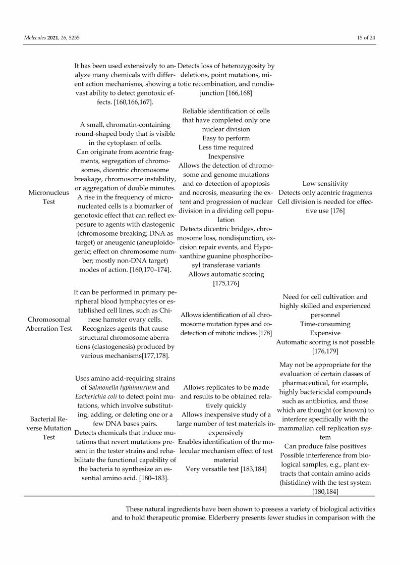
Molecules 2021, 26, 5255 15 of 24
It has been used extensively to an-alyze many chemicals with differ-ent action mechanisms, showing a vast ability to detect genotoxic ef-
fects. [160,166,167].
Detects loss of heterozygosity by deletions, point mutations, mi-
totic recombination, and nondis-junction [166,168]
Micronucleus Test
A small, chromatin-containing round-shaped body that is visible
in the cytoplasm of cells. Can originate from acentric frag-
ments, segregation of chromo-somes, dicentric chromosome
breakage, chromosome instability, or aggregation of double minutes. A rise in the frequency of micro-nucleated cells is a biomarker of
genotoxic effect that can reflect ex-posure to agents with clastogenic (chromosome breaking; DNA as
target) or aneugenic (aneuploido-genic; effect on chromosome num-
ber; mostly non-DNA target) modes of action. [160,170–174].
Reliable identification of cells that have completed only one
nuclear division Easy to perform
Less time required Inexpensive
Allows the detection of chromo-some and genome mutations and co-detection of apoptosis
and necrosis, measuring the ex-tent and progression of nuclear division in a dividing cell popu-
lation Detects dicentric bridges, chro-
mosome loss, nondisjunction, ex-cision repair events, and Hypo-xanthine guanine phosphoribo-
syl transferase variants Allows automatic scoring
[175,176]
Low sensitivity Detects only acentric fragments Cell division is needed for effec-
tive use [176]
Chromosomal Aberration Test
It can be performed in primary pe-ripheral blood lymphocytes or es-tablished cell lines, such as Chi-
nese hamster ovary cells. Recognizes agents that cause
structural chromosome aberra-tions (clastogenesis) produced by
various mechanisms[177,178].
Allows identification of all chro-mosome mutation types and co-detection of mitotic indices [178]
Need for cell cultivation and highly skilled and experienced
personnel Time-consuming
Expensive Automatic scoring is not possible
[176,179]
Bacterial Re-verse Mutation
Test
Uses amino acid-requiring strains of Salmonella typhimurium and
Escherichia coli to detect point mu-tations, which involve substitut-ing, adding, or deleting one or a
few DNA bases pairs. Detects chemicals that induce mu-tations that revert mutations pre-sent in the tester strains and reha-bilitate the functional capability of
the bacteria to synthesize an es-sential amino acid. [180–183].
Allows replicates to be made and results to be obtained rela-
tively quickly Allows inexpensive study of a
large number of test materials in-expensively
Enables identification of the mo-lecular mechanism effect of test
material Very versatile test [183,184]
May not be appropriate for the evaluation of certain classes of pharmaceutical, for example,
highly bactericidal compounds such as antibiotics, and those
which are thought (or known) to interfere specifically with the
mammalian cell replication sys-tem
Can produce false positives Possible interference from bio-logical samples, e.g., plant ex-tracts that contain amino acids (histidine) with the test system
[180,184]
These natural ingredients have been shown to possess a variety of biological activities and to hold therapeutic promise. Elderberry presents fewer studies in comparison with the

Molecules 2021, 26, 5255 16 of 24
other natural ingredients. The difficulty of accessing such natural ingredients in some coun-tries and the fact that it is a wild tree can be some of the reasons for the lack of studies on this tree. On the other hand, grapes and olives have various in vivo and in vitro studies. Also, some of their studies have been performed in D. melanogaster and human lymphocytes. D. melanogaster is one of the preferred organisms for toxicological research. In recent dec-ades, it has been used as a model to elucidate human diseases and also for toxicological studies [185–187]. The use of D. melanogaster in experimental studies met the standard of the European Centre for the Validation of Alternative Methods (ECVAM): reduction, refine-ment, and replacement (3Rs) of laboratory animal usage. D. melanogaster as a model raises few ethical concerns [188]. The short life cycle, the distinct developmental stages, the avail-ability of various tools and reagents, known genome sequence, and the physiological simi-larity of D. melanogaster with humans (namely on dietary input, xenobiotic metabolizing system, antioxidant enzymes, and DNA repair systems) make them an excellent in vivo model organism to rapidly test toxicity in the whole organism and elucidate the molecular mechanisms underlying the toxicity [160]. In biomedical sciences, experimental evidence has implicated oxidative stress in the pathophysiology of several disease conditions [189]. However, the precise role of oxidative stress in the pathology of diseases is far from being known. Therefore, the study of oxidative stress in animal models is of particular importance at present. D. melanogaster has been used to assess oxidative stress and antioxidant markers [190,191]. They have been used in genotoxicity and antigenotoxicity studies. In vivo tests for detecting somatic or, germinative mutations are especially valuable.
Human lymphocytes are used as surrogate tissue, as they are easily obtained, are available in large numbers, do not require cell culture, are diploids, and are almost all in the same phase of the cell cycle. Although the Comet Assay is well accepted among the scientific community, there are issues regarding standardization among laboratories [192]. Therefore, new methods for DNA damage assessment would be beneficial to im-prove research on DNA damage repair and antigenotoxicity.
5. Conclusions The present review synthesizes the most accurate evidence of the antigenotoxic ca-
pacity of some natural ingredients common in the Northeast of Portugal. Almonds, Grapes, Olives, and Elderberry proved to have an antigenotoxic effect. Natural occurring antigenotoxicity in natural ingredients could strongly counteract genome instability.
Even though these ingredients are already being used in cosmetics, the lack of anti-genotoxicological studies makes it crucial to investigate further how to incorporate them in cosmetics to benefit human health. Studies have shown that plants, fruits, and vegeta-bles with antigenotoxic properties show promising results for the cosmetic industry [193–195]. Additional investigation can be carried out, namely, evaluating the cosmetic prop-erties of the natural ingredients towards promoting DNA integrity. Using Comet Assay and SMART, evaluating genoprotection, longevity, and prolificacy of the natural ingredi-ents in D. melanogaster could reveal exciting results.
Author Contributions: Both authors have investigated and written the present article. All authors have read and agreed to the published version of the manuscript.
Funding: This work was supported by the project UIDB/CVT/00772/2020 funded by the Fundação para a Ciência e Tecnologia (FCT).
Institutional Review Board Statement: Not applicable.
Informed Consent Statement: Not applicable.
Data Availability Statement: Data available in a publicly accessible repository.
Acknowledgments: The authors would also like to thank Malcolm Purves (professional translator) for the manuscript proofreading and editing.
Conflicts of Interest: The authors declare no conflict of interest.

Molecules 2021, 26, 5255 17 of 24
References 1. Pathath, A.W. Theories of Aging. Int. J. Indian Psychol. 2017, 4, 15–22, doi:10.25215/0403.142. 2. Sram, R.J.; Binkova, B.; Rossner, P. Vitamin C for DNA damage prevention. Mutat. Res. Fundam. Mol. Mech. Mutagen. 2012, 733,
39–49, doi:10.1016/j.mrfmmm.2011.12.001. 3. Graf, J. Anti-Aging Skin Care Ingredient Technologies. In Cosmetic Dermatology; Springer: Berlin/Heidelberg, Germany, 2005;
pp. 17–28. 4. Izquierdo-Vega, J.; Morales-González, J.; SánchezGutiérrez, M.; Betanzos-Cabrera, G.; Sosa-Delgado, S.; Sumaya-Martínez, M.;
Morales-González, Á.; Paniagua-Pérez, R.; Madrigal-Bujaidar, E.; Madrigal-Santillán, E. Evidence of Some Natural Products with Antigenotoxic Effects. Part 1: Fruits and Polysaccharides. Nutrients 2017, 9, 102, doi:10.3390/nu9020102.
5. Nagarathna, P.K.M.; Wesley, M.; Reddy, P.; Reena, K. Review on genotoxicity, its molecular mechanisms and prevention. Int. J. Pharm. Sci. Rev. Res. 2013, 22, 236–243.
6. Singh, S.K. Handbook on Cosmetics (Processes, Formulae with Testing Methods); Asia Pacific Business Press Inc.: Delhi, India, 2010. 7. EUR-Lex—31976L0768—EN. Council Directive 76/768/EEC of 27 July 1976 on the Approximation of the Laws of the Member
States Relating to Cosmetic Products. 1997. Available online: https://eur-lex.europa.eu/legal-con-tent/EN/TXT/HTML/?uri=CELEX:31976L0768&from=EN (accessed on 9 November 2020).
8. Romero, V.; Khury, E.; Aiello, L.M.; Leonardi, G.R. Differences between organic and natural cosmetics: Clarifying literature for prescribers. Surg. Cosmet. Dermatol. 2018, 10, 188–193, doi:10.5935/scd1984-8773.20181031087.
9. Fonseca-Santos, B.; Corrêa, M.; Chorilli, M. Sustainability, natural and organic cosmetics: Consumer, products, efficacy, toxico-logical and regulatory considerations. Braz. J. Pharm. Sci. 2015, 51, 17–26, doi:10.1590/S1984-82502015000100002.
10. Okereke, J.N.; Udebuani, A.C.; Ezeji, E.U.; Obasi, K.O.; Nnoli, M.C. Possible Health Implications Associated with Cosmetics: A Review. SJPH Sci. J. Public Health 2015, 3, 58–63, doi:10.11648/j.sjph.s.2015030501.21.
11. Natural Cosmetics: Sales Value France. Statista 2015. Available online: https://www.statista.com/statistics/671090/natural-or-ganic-cosmetics-sales-value-france/(accessed on 2 November 2020).
12. Global Market Value for Natural/Organic Cosmetics and Personal Care in 2018–2027. Statista n.d. Available online: https://www.statista.com/statistics/673641/global-market-value-for-natural-cosmetics/(accessed on 27 March 2021).
13. Natural Cosmetics—Germany | Statista Market Forecast. Statista 2021. Available online: https://www.statista.com/out-look/cmo/beauty-personal-care/cosmetics/natural-cosmetics/germany (accessed on 27 March 2021).
14. Natural and Organic Cosmetics: Share of Female Users France 2016. Statista 2016. Available online: https://www.sta-tista.com/statistics/671247/natural-organic-cosmetics-share-women-france/(accessed on 2 November 2020).
15. Organic Cosmetics: Decisive Moments for Consumption France 2016. Statista 2016. Available online: https://www.sta-tista.com/statistics/671320/key-moments-consumer-organic-cosmetics-france/(accessed on 2 November 2020).
16. Gonçalves, S.; Gaivão, I. Evaluation of Cosmetic Properties of Natural Ingredients in the Trás-Os-Montes Area: A PhD Project. Sci. Posters 2021, doi:10.14293/S2199-1006.1.SOR-.PPLZ4CW.v1.
17. Germany: Natural and Organic Cosmetics Gained More Than a Million New Customers in 2018. Premium Beauty News 2019. Available online: https://www.premiumbeautynews.com/en/germany-natural-and-organic,14771 (accessed on 27 March 2021).
18. Jørgensen, C. Cosmetics Worldwide—Same Contents? A Comparative Study; The Danish Consumer Council THINK Chemicals: Copenhagen, Denmark, 2020.
19. Jensen, L.M.M. Chemical of the Month: Iodopropynyl Butylcarbamate (IPBC). AllergyCertified 2017. Available online: https://allergycertified.com/blog/2017/11/22/iodopropynyl-butylcarbamate/(accessed 8 August 2021).
20. Hass, U.; Christiansen, S.; Petersen, M.A.; Boberg, J.; Andersson, A.-M.; Skakkebæk, N.E.; Bay, K.; Holbeck, H.; Lund Kinnberg, K.; Bjerregaard, P. Evaluation of 22 SIN List 2.0 Substances According to the Danish Proposal on Criteria for Endocrine Disrupters; Danish Centre on Endocrine Disrupters: Odense, Denmark, 2012.
21. Becker, L.C. Amended Safety Assessment of Glyoxal as Used in Cosmetics; Consumer Federation of America: Washington, DC, USA, 2017. 22. Darbre, P.D.; Harvey, P.W. Parabens can enable hallmarks and characteristics of cancer in human breast epithelial cells: A
review of the literature with reference to new exposure data and regulatory status: Parabens and breast cancer. J. Appl. Toxicol. 2014, 34, 925–938, doi:10.1002/jat.3027.
23. Berger, K.P.; Kogut, K.R.; Bradman, A.; She, J.; Gavin, Q.; Zahedi, R.; Parra, K.L.; Harley, K.G. Personal care product use as a predictor of urinary concentrations of certain phthalates, parabens, and phenols in the HERMOSA study. J. Exp. Sci. Environ. Epidemiol. 2019, 29, 21–32, doi:10.1038/s41370-017-0003-z.
24. European Commission to Ban More CMRs in Cosmetics n.d. Available online: https://chemicalwatch.com/208245/european-commission-to-ban-more-cmrs-in-cosmetics (accessed on 9 August 2021).
25. McCook, J.P. Topical Products for the Aging Face. Clin. Plast. Surg. 2016, 43, 597–604, doi:10.1016/j.cps.2016.03.005. 26. Boran, R. Investigations of anti-aging potential of Hypericum origanifolium Willd. for skincare formulations. Ind. Crop. Prod. 2018,
118, 290–295, doi:10.1016/j.indcrop.2018.03.058. 27. Pires, J.; Pinto, P.; Moreira, N. Lameiros de Trás-Os-Montes. Perspectivas de Futuro Para Estas Pastagens de Montanha; Insituto Politéc-
nico de Bragança: Bragança, Portugal, 1994; ISBN 972-745-025-3. 28. Rosário, M.C. O Sistema Agrário de Trás-Os-Montes e a modernidade sustentável. Gest. Desenvolv. 2004, 12, 237–257. 29. Ribeiro, J. Caracterização genérica da região vinhateira do Alto Douro. In Douro—Estudos & Documentos, 2nd ed.; Universidade
de Trás-os-Montes e Alto Douro: Vila Real, Portugal, 2000; Volume 5, pp. 11–29.

Molecules 2021, 26, 5255 18 of 24
30. Cardoso, J.; Bessa, M.; Marado, M. Carta dos Solos (III,I); Comissão Nacional do Ambiente, Instituto Hidrográfico de Lisboa: Lisboa, Portugal, 1978.
31. Braga, F.G.; Carvalho, L.M.; Carvalho, M.J.; Guedes-Pinto, H.; Torres-Pereira, J.M.; Neto, M.F.; Monteiro, A. Variation of the Anthocyanin Content in Sambucus nigra L. Populations Growing in Portugal. J. Herbs Spices Med. Plants 2002, 9, 289–295, doi:10.1300/J044v09n04_05.
32. Trindade, C.; Valdiviesso, T.; de Brás Oliveira, P. Caraterização Morfológica das Inflorescências de Variedades de Sambucus nigra L.; Universidade de Trás-os-Montes e Alto Douros: Vila Real, Portugal, 2019.
33. Centro Nacional de Competências dos Frutos Secos. Amêndoa. Estudi de Produção e Comercialização nas Terras de Trás-Os-Mntes; CNCFS: Bragança, Portugal, 2020.
34. Cordeiro, V.; Monteiro, A. Almond Growing in Trás-Os-Montes Region (Portugal). Acta Hortic. 2002, 161–165, doi:10.17660/ActaHortic.2002.591.22.
35. Portal do INE 2013. Available online: https://www.ine.pt/xportal/xmain?xpid=INE&xpgid=ine_indicadores&indOcor-rCod=0000708&xlang=pt&contexto=bd&selTab=tab2 (accessed on 16 November 2020).
36. de Figueiredo, T.; Almeida, A.; Araújo, J. Edaphic characteristics of olive-tree areas in the Trás-Os-Montes Region (Portugal): A map-based approach. Acta Hortic. 2002, 586, 151–154.
37. Sousa, M.; Pererira, C.; Guerra, J.; Abade, E. Caracterização de Castas Cultivadas na Região Vitivinícola de Trás-Os-Montes, Sub regiões de Chaves, Planalto Mirandês e Valpaços; Coleção uma Agricultura com Norte; Ministério da Agricultura, do Desenvolvimento Rural e das Pescas: Lisboa, Portugal, 2007; p. 40.
38. Gonçalves, S.; Gaivão, I. Assessment of antigenotoxic properties in natural ingredients common in the Trás-os-Montes region: A Phd project. ScienceOpen 2021, doi:10.13140/RG.2.2.11406.28482.
39. Viapiana, A.; Wesolowski, M. The Phenolic Contents and Antioxidant Activities of Infusions of Sambucus nigra L. Plant Foods Hum. Nutr. 2017, 72, 82–87, doi:10.1007/s11130-016-0594-x.
40. Atkinson, M.D.; Atkinson, E. Sambucus nigra L.: Sambucus nigra. J. Ecol. 2002, 90, 895–923, doi:10.1046/j.1365-2745.2002.00698.x. 41. Charlebois, D.; Byers, P.L.; Finn, C.E.; Thomas, A.L. Elderberry: Botany, Horticulture, Potential. In Horticultural Reviews; Janick,
J., Ed.; John Wiley & Sons, Inc.: Hoboken, NJ, USA, 2010; Volume 37, pp. 213–280, doi:10.1002/9780470543672.ch4. 42. Mabey, R. The Complete New Herbal. A Practical Guide to Herbal Living; Gaia Books Ltd.: London, UK, 1988; ISBN 978-0-14-012682-2. 43. Olejnik, A.; Kowalska, K.; Olkowicz, M.; Rychlik, J.; Juzwa, W.; Myszka, K.; Dembczyński, R.; Białas, W. Anti-inflammatory
effects of gastrointestinal digested Sambucus nigra L. fruit extract analysed in co-cultured intestinal epithelial cells and lipopol-ysaccharide-stimulated macrophages. J. Funct. Foods 2015, 19, 649–660, doi:10.1016/j.jff.2015.09.064.
44. Ciocoiu, M.; Badescu, L.; Badulescu, O.; Badescu, M. Intervention of Sambucus nigra Polyphenolic Extract in Experimental Ar-terial Hypertension. Int. J. Med. Health Sci. 2012, 6, 80–83, doi:10.5281/ZENODO.1055287.
45. Salvador, Â.; Król, E.; Lemos, V.; Santos, S.; Bento, F.; Costa, C.; Almeida, A.; Szczepankiewicz, D.; Kulczyński, B.; Krejpcio, Z.; et al. Effect of Elderberry (Sambucus nigra L.) Extract Supplementation in STZ-Induced Diabetic Rats Fed with a High-Fat Diet. IJMS Int. J. Mol. Sci. 2016, 18, 13, doi:10.3390/ijms18010013.
46. Bhattacharya, S.; Christensen, K.B.; Olsen, L.C.B.; Christensen, L.P.; Grevsen, K.; Færgeman, N.J.; Kristiansen, K.; Young, J.F.; Oksbjerg, N. Bioactive Components from Flowers of Sambucus nigra L. Increase Glucose Uptake in Primary Porcine Myotube Cultures and Reduce Fat Accumulation in Caenorhabditis elegans. J. Agric. Food Chem. 2013, 61, 11033–11040, doi:10.1021/jf402838a.
47. Pilot Clinical Study on a Proprietary Elderberry Extract : Efficacy in Addressing Influenza Symptoms n.d. Available online:/pa-per/Pilot-Clinical-Study-on-a-Proprietary-Elderberry-%3A/367d1c92716b6be462f26dbfe6c223863dc78464 (accessed on 16 De-cember 2020).
48. Jarzycka, A.; Lewińska, A.; Gancarz, R.; Wilk, K.A. Assessment of extracts of Helichrysum arenarium, Crataegus monogyna, Sambucus nigra in photoprotective UVA and UVB; photostability in cosmetic emulsions. J. Photochem. Photobiol. B 2013, 128, 50–57, doi:10.1016/j.jphotobiol.2013.07.029.
49. Picon, P.D.; Picon, R.V.; Costa, A.F.; Sander, G.B.; Amaral, K.M.; Aboy, A.L.; Henriques, A.T. Randomized clinical trial of a phytotherapic compound containing Pimpinella anisum, Foeniculum vulgare, Sambucus nigra, and Cassia augustifolia for chronic constipation. BMC Complement. Altern. Med. 2010, 10, 17, doi:10.1186/1472-6882-10-17.
50. Neto, M. Sabugueiro—Potencialidades. 2007. Available online: http://www.drapn.min-agricultura.pt/drapn/con-teudos/cen_documentos/outros/Sabugueiro_Potencialidades.pdf (accessed on 7 June 2021).
51. Młynarczyk, K.; Walkowiak-Tomczak, D.; Lysiak, G. Bioactive properties of Sambucus nigra L. as a functional ingredient for food and pharmaceutical industry. J. Funct. Foods 2018, 40, 377–390.
52. Vulic, J.; Vracar, L.; Sumic, Z. Chemical characteristics of cultivated elderberry fruit. Acta Period. Technol. 2008, 85–90, doi:10.2298/APT0839085V.
53. Veberic, R.; Jakopic, J.; Stampar, F.; Schmitzer, V. European elderberry (Sambucus nigra L.) rich in sugars, organic acids, antho-cyanins and selected polyphenols. Food Chem. 2009, 114, 511–515, doi:10.1016/j.foodchem.2008.09.080.
54. Dawidowicz, A.L.; Wianowska, D.; Baraniak, B. The antioxidant properties of alcoholic extracts from Sambucus nigra L. (antiox-idant properties of extracts). LWT Food Sci. Technol. 2006, 39, 308–315, doi:10.1016/j.lwt.2005.01.005.
55. Duymuş, H.G.; Göger, F.; Başer, K.H.C. In vitro antioxidant properties and anthocyanin compositions of elderberry extracts. Food Chem. 2014, 155, 112–119, doi:10.1016/j.foodchem.2014.01.028.
56. Espín, J.C.; Soler-Rivas, C.; Wichers, H.J.; García-Viguera, C. Anthocyanin-Based Natural Colorants: A New Source of Antiradi-cal Activity for Foodstuff. J. Agric. Food Chem. 2000, 48, 1588–1592, doi:10.1021/jf9911390.

Molecules 2021, 26, 5255 19 of 24
57. Obied, H.K.; Allen, M.S.; Bedgood, D.R.; Prenzler, P.D.; Robards, K.; Stockmann, R. Bioactivity and Analysis of Biophenols Recovered from Olive Mill Waste. J. Agric. Food Chem. 2005, 53, 823–837, doi:10.1021/jf048569x.
58. Imenšek, N.; Sem, V.; Kolar, M.; Ivančič, A.; Kristl, J. The Distribution of Minerals in Crucial Plant Parts of Various Elderberry (Sambucus spp.) Interspecific Hybrids. Plants 2021, 10, 653, doi:10.3390/plants10040653.
59. Sidor, A.; Gramza-Michałowska, A. Advanced research on the antioxidant and health benefit of elderberry (Sambucus nigra) in food—A review. J. Funct. Foods 2015, 18, 941–958, doi:10.1016/j.jff.2014.07.012.
60. Christensen, L.P.; Kaack, K.; Fretté, X.C. Selection of elderberry (Sambucus nigra L.) genotypes best suited for the preparation of elder-flower extracts rich in flavonoids and phenolic acids. Eur. Food Res. Technol. 2008, 227, 293–305, doi:10.1007/s00217-007-0723-8.
61. Mikulic-Petkovsek, M.; Samoticha, J.; Eler, K.; Stampar, F.; Veberic, R. Traditional Elderflower Beverages: A Rich Source of Phenolic Compounds with High Antioxidant Activity. J. Agric. Food Chem. 2015, 63, 1477–1487, doi:10.1021/jf506005b.
62. Galanakis, C.M. Olive fruit dietary fiber: Components, recovery and applications. Trends Food Sci. Technol. 2011, 22, 175–184, doi:10.1016/j.tifs.2010.12.006.
63. Chen, L.; Hu, J.Y.; Wang, S.Q. The role of antioxidants in photoprotection: A critical review. J. Am. Acad. Dermatol. 2012, 67, 1013–1024, doi:10.1016/j.jaad.2012.02.009.
64. Salvador, Â.C.; Silvestre, A.J.D.; Rocha, S.M. Comprehensive Insight into the Elderflowers and Elderberries (Sambucus nigra L.) Mono and Sesquiterpenic Metabolites: Factors that Modulate Their Composition. In Secondary Metabolites—Sources and Applica-tions; Vijayakumar, R., Raja, S.S.S., Eds.; InTech: London, UK, 2018; doi:10.5772/intechopen.77291.
65. Rodrigues, F.; Pimentel, F.B.; Oliveira, M.B.P.P. Olive by-products: Challenge application in cosmetic industry. Ind. Crop. Prod. 2015, 70, 116–124, doi:10.1016/j.indcrop.2015.03.027.
66. Bratu, M.; Doroftei, E.; Negreanu-Pirjol, T.; Hostina, C.; Porta, S. Determination of Antioxidant Activity and Toxicity of Sambu-cus nigra Fruit Extract Using Alternative Methods. Food Technol. Biotechnol. 2012, 50, 177–182.
67. Karpova, I.S.; Lylo, V.V.; Macewicz, L.L.; Kotsarenko, K.V.; Palchykovska, L.G.; Ruban, T.O.; Lukash, L.L. Lectins of Sambucus nigra as Biologically Active and DNA-Protective Substances. Acta Hortic. 2015, 93–102, doi:10.17660/ActaHortic.2015.1061.9.
68. Gaetano, L.; Calabrese, G.; Perrino, E. The Origin and Distribution of the Olive Tree and Olive Crop; CIHEAM-Mediterranean Ag-ronomic Institute of Bari: Valenzano, Italy, 2012; pp. 1–4.
69. Newton, C.; Terral, J.-F.; Ivorra, S. The Egyptian olive (Olea europaea subsp. europaea ) in the later first millennium BC: Origins and history using the morphometric analysis of olive stones. Antiquity 2006, 80, 405–414, doi:10.1017/S0003598 × 00093716.
70. Ryan, D.; Robards, K. Critical Review. Phenolic compounds in olives. Analyst 1998, 123, 31R–44R, doi:10.1039/a708920a. 71. Grossi, M.; Lecce, G.D.; Toschi, T.G.; Ricco, B. Fast and Accurate Determination of Olive Oil Acidity by Electrochemical Imped-
ance Spectroscopy. IEEE Sens. J. 2014, 14, 2947–2954, doi:10.1109/JSEN.2014.2321323. 72. Montaño, A.; Sánchez, A.H.; López-López, A.; de Castro, A.; Rejano, L. Chemical Composition of Fermented Green Olives. In
Olives and Olive Oil in Health and Disease Prevention; Elsevier: Oxford, UK, 2010; pp. 291–297, doi:10.1016/B978-0-12-374420-3.00031-0.
73. Guo, Z.; Jia, X.; Zheng, Z.; Lu, X.; Zheng, Y.; Zheng, B.; Xiao, J. Chemical composition and nutritional function of olive (Olea europaea L.): A review. Phytochem. Rev. 2018, 17, 1091–1110, doi:10.1007/s11101-017-9526-0.
74. Cirilli, M.; Bellincontro, A.; Urbani, S.; Servili, M.; Esposto, S.; Mencarelli, F.; Muleo, R. On-field monitoring of fruit ripening evolution and quality parameters in olive mutants using a portable NIR-AOTF device. Food Chem. 2016, 199, 96–104, doi:10.1016/j.foodchem.2015.11.129.
75. Elbir, M.; Es-Safi, N.E.; Amhoud, A.; Mbarki, M. Characterization of phenolic compounds in olive stones of three moroccan varieties. Maderas Cienc. Tecnol. 2015, 17, 479–492, doi:10.4067/S0718-221 × 2015005000043.
76. Fernandez-Bolanos, J.; Fernandez Diez, M.J.; Rivas Moreno, M.; Gil Serrano, A.; Perez Romero, T. Sugars and polyois in green olives. III. Quantitative determination by gas-liquid chromatography. Grasas Aceites 1983, 34, 168–71.
77. Jilani, H.; Cilla, A.; Barberá, R.; Hamdi, M. Improved bioaccessibility and antioxidant capacity of olive leaf (Olea europaea L.) polyphenols through biosorption on Saccharomyces cerevisiae. Ind. Crop. Prod. 2016, 84, 131–138, doi:10.1016/j.indcrop.2016.02.002.
78. Talhaoui, N.; Taamalli, A.; Gómez-Caravaca, A.M.; Fernández-Gutiérrez, A.; Segura-Carretero, A. Phenolic compounds in olive leaves: Analytical determination, biotic and abiotic influence, and health benefits. Food Res. Int. 2015, 77, 92–108, doi:10.1016/j.foodres.2015.09.011.
79. Gouvinhas, I.; Machado, N.; Sobreira, C.; Domínguez-Perles, R.; Gomes, S.; Rosa, E.; Barros, A. Critical Review on the Signifi-cance of Olive Phytochemicals in Plant Physiology and Human Health. Molecules 2017, 22, 1986, doi:10.3390/molecules22111986.
80. Ahmed, A.M.; Rabii, N.S.; Garbaj, A.M.; Abolghait, S.K. Antibacterial effect of olive (Olea europaea L.) leaves extract in raw peeled undeveined shrimp (Penaeus semisulcatus). Int. J. Vet. Sci. Med. 2014, 2, 53–56, doi:10.1016/j.ijvsm.2014.04.002.
81. Al Okbi, S.; Hassan, Z.; El Mazar, M.; Ammar, N.; Abou Elkassem, L.; El Bakry, H. Diuretic activity of Olive (Olea europaea L.). Planta Med. 2011, 77, PM62, doi:10.1055/s-0031-1282820.
82. Khan, M.; Panchal, S.; Vyas, N.; Butani, A.; Kumar, V. Olea europaea: A phyto-pharmacological review. Pharmacogn. Rev. 2006, 1, 114–118.
83. Khayyal, M.; El-Ghazaly, M.; Abdallah, D.; Nassar, N.; Okpanyi, S.; Kreuter, M.-H. Blood Pressure Lowering Effect of an Olive Leaf Extract {Olea europaed) in L-NAME Induced Hypertension in Rats. Arzneimittelforschung 2011, 52, 797–802, doi:10.1055/s-0031-1299970.
84. Mousa, H.M.; Farahna, M.; Ismail, M.S.; Al-Hassan, A.A.; Ammar, A.S.; Abdel-Salam, A.M. Anti-Diabetic Effect of Olive Leaves Extract in Alloxan-Diabetic Rats. J. Agric. Vet. Sci. 2014, 7, 181–196, doi:10.12816/0009472.

Molecules 2021, 26, 5255 20 of 24
85. Erbay, Z.; Icier, F. The Importance and Potential Uses of Olive Leaves. Food Rev. Int. 2010, 26, 319–334, doi:10.1080/87559129.2010.496021.
86. Autoridade de Segurança Alimentar e Económica Azeites e sua Classificação. Available online: https://www.asae.gov.pt/news-letter2/asaenews-n-108-julho-2017/azeites-e-sua-classificacao-.aspx (accessed on 7 June 2021).
87. FoodData Central n.d. Available online: https://fdc.nal.usda.gov/fdc-app.html#/food-details/748608/nutrients (accessed on 7 June 2021).
88. Huang, Z.-R.; Lin, Y.-K.; Fang, J.-Y. Biological and Pharmacological Activities of Squalene and Related Compounds: Potential Uses in Cosmetic Dermatology. Molecules 2009, 14, 540–554, doi:10.3390/molecules14010540.
89. Scoditti, E.; Capurso, C.; Capurso, A.; Massaro, M. Vascular effects of the Mediterranean diet—Part II: Role of omega-3 fatty acids and olive oil polyphenols. Vascul. Pharmacol. 2014, 63, 127–134, doi:10.1016/j.vph.2014.07.001.
90. Trapani, G.; Vagliano, L.; Giribaldi, M.; Cavallarin, L.; Coscia, A. Olive oil: Maternal and pediatric health. J. Pediatr. Neonatal Individ. Med. 2017, 6, e060133, doi:10.7363/060133.
91. Coletta, J.M.; Bell, S.J.; Roman, A.S. Omega-3 Fatty Acids and Pregnancy. Rev. Obstet. Gynecol. 2010, 3, 163–171. 92. Roncero-Martín, R.; Aliaga Vera, I.; Moreno-Corral, L.; Moran, J.; Lavado-Garcia, J.; Pedrera-Zamorano, J.; Pedrera-Canal, M.
Olive Oil Consumption and Bone Microarchitecture in Spanish Women. Nutrients 2018, 10, 968, doi:10.3390/nu10080968. 93. Widmer, R.J.; Freund, M.A.; Flammer, A.J.; Sexton, J.; Lennon, R.; Romani, A.; Mulinacci, N.; Vinceri, F.F.; Lerman, L.O.; Lerman,
A. Beneficial effects of polyphenol-rich olive oil in patients with early atherosclerosis. Eur. J. Nutr. 2013, 52, 1223–1231, doi:10.1007/s00394-012-0433-2.
94. Assy, N.; Nassar, F.; Nasser, G.; Grosovski, M. Olive oil consumption and non-alcoholic fatty liver disease. WJG World J. Gastro-enterol. 2009, 15, 1809–1815, doi:10.3748/wjg.15.1809.
95. Yubero-Serrano, E.M.; Lopez-Moreno, J.; Gomez-Delgado, F.; Lopez-Miranda, J. Extra virgin olive oil: More than a healthy fat. Eur. J. Clin. Nutr. 2019, 72, 8–17, doi:10.1038/s41430-018-0304-x.
96. Brzosko, S.; Curtis, A.D.; Murzilli, S.; Gaetano, G.D.; Donati, M.B.; Iacoviello, L. Effect of extra virgin olive oil on experimental thrombosis and primary hemostasis in rats. Nutr. Metab. Cardiovasc. Dis. NMCD 2002, 12, 337–342.
97. Lauretti, E.; Nenov, M.; Dincer, O.; Iuliano, L.; Praticò, D. Extra virgin olive oil improves synaptic activity, short-term plasticity, memory, and neuropathology in a tauopathy model. Aging Cell 2020, 19, e13076, doi:10.1111/acel.13076.
98. Condelli, N.; Caruso, M.C.; Galgano, F.; Russo, D.; Milella, L.; Favati, F. Prediction of the antioxidant activity of extra virgin olive oils produced in the Mediterranean area. Food Chem. 2015, 177, 233–239, doi:10.1016/j.foodchem.2015.01.001.
99. Genders, R. Natural Beauty—The Practical Guide to Wild Flower Cosmetics; Helmut Lingen: Köln, Germany, 1987. 100. Smaoui, S.; Hlima, H.B.; Jarraya, R.; Kamoun, N.G.; Ellouze, R.; Damak, M. Cosmetic emulsion from virgin olive oil: Formula-
tion and bio-physical evaluation. Afr. J. Biotechnol. 2012, 11, 9664–9671, doi:10.5897/AJB12.163. 101. Angerhofer, C.K.; Maes, D.; Giacomoni, P.U. The Use of Natural Compounds and Botanicals in the Development of Anti-Aging Skin
Care Products. In Skin Aging Handbook; Elsevier: Norwich, NY, USA, 2009; pp. 205–263, doi:10.1016/B978-0-8155-1584-5.50014-4. 102. Viola, P.; Viola, M. Virgin olive oil as a fundamental nutritional component and skin protector. Clin. Dermatol. 2009, 27, 159–165,
doi:10.1016/j.clindermatol.2008.01.008. 103. Lin, M.-H.; Khnykin, D. Fatty acid transporters in skin development, function and disease. Biochim. Biophys. Acta BBA—Mol.
Cell Biol. Lipids 2014, 1841, 362–368, doi:10.1016/j.bbalip.2013.09.016. 104. Aly, F.M.; Kotb, A.M.; Haridy, M.A.M.; Hammad, S. Impacts of fullerene C60 and virgin olive oil on cadmium-induced geno-
toxicity in rats. Sci. Total Environ. 2018, 630, 750–756, doi:10.1016/j.scitotenv.2018.02.205. 105. Khalil, S.; Awad, A.; Elewa, Y. Antidotal impact of extra virgin olive oil against genotoxicity, cytotoxicity and immunotoxicity
induced by hexavalent chromium in rat. Int. J. Vet. Sci. Med. 2013, 1, 65–73, doi:10.1016/j.ijvsm.2013.10.001. 106. Ladizinsky, G. On the Origin of Almond. Genet. Resour. Crop Evol. 1999, 46, 143–147, doi:10.1023/A:1008690409554. 107. Albala, K. Almonds Along the Silk Road: The Exchange and Adaptation of Ideas from West to East. Petits Propos Culin. 2009,
88, 19–34. 108. Offeman, R.D.; Holtman, K.M.; Covello, K.M.; Orts, W.J. Almond hulls as a biofuels feedstock: Variations in carbohydrates by
variety and location in California. Ind. Crop. Prod. 2014, 54, 109–114, doi:10.1016/j.indcrop.2014.01.010. 109. Richardson, D.P.; Astrup, A.; Cocaul, A.; Ellis, P. The nutritional and health benefits of almonds: A healthy food choice. Food
Sci. Technol. Bull. Funct. Foods 2009, 6, 41–50, doi:10.1616/1476-2137.15765. 110. Beltrán Sanahuja, A.; Maestre Pérez, S.E.; Grané Teruel, N.; Valdés García, A.; Prats Moya, M.S. Variability of Chemical Profile
in Almonds (Prunus dulcis) of Different Cultivars and Origins. Foods 2021, 10, 153, doi:10.3390/foods10010153. 111. Socias, R.; Kodad, O.; Alonso, J.M.; Gradziel, T.M. Almond Quality: A Breeding Perspective. In Horticultural Reviews; Janick, J.,
Ed.; John Wiley & Sons, Inc.: Hoboken, NJ, USA, 2008; pp. 197–238, doi:10.1002/9780470380147.ch3. 112. Roncero, J.M.; Álvarez-Ortí, M.; Pardo-Giménez, A.; Rabadán, A.; Pardo, J.E. Review about Non-Lipid Components and Minor
Fat-Soluble Bioactive Compounds of Almond Kernel. Foods 2020, 9, 1646, doi:10.3390/foods9111646. 113. Kodad, O.; Alonso, J.M.; Espiau, M.T.; Estopañán, G.; Juan, T.; Rafel Socias i Company. Chemometric Characterization of Al-
mond Germplasm: Compositional Aspects Involved in Quality and Breeding. J. Am. Soc. Hortic. Sci. 2011, 136, 273–281, doi:10.21273/JASHS.136.4.273.
114. Fernandes, G.D.; Gómez-Coca, R.B.; Pérez-Camino, M.d.C.; Moreda, W.; Barrera-Arellano, D. Chemical Characterization of Major and Minor Compounds of Nut Oils: Almond, Hazelnut, and Pecan Nut. J. Chem. 2017, 2017, 2609549, doi:10.1155/2017/2609549.

Molecules 2021, 26, 5255 21 of 24
115. Fernández-Cuesta, Á.; Kodad, O.; Rafel Socias i Company; Velasco, L. Phytosterol Variability in Almond Germplasm. J. Am. Soc. Hortic. Sci. 2012, 137, 343–348, doi:10.21273/JASHS.137.5.343.
116. Garrido, I.; Monagas, M.; Gómez-Cordovés, C.; Bartolomé, B. Polyphenols and Antioxidant Properties of Almond Skins: Influ-ence of Industrial Processing. J. Food Sci. 2008, 73, C106–C115, doi:10.1111/j.1750-3841.2007.00637.x.
117. Xiao, L.; Lee, J.; Zhang, G.; Ebeler, S.E.; Wickramasinghe, N.; Seiber, J.; Mitchell, A.E. HS-SPME GC/MS characterization of volatiles in raw and dry-roasted almonds (Prunus dulcis). Food Chem. 2014, 151, 31–39, doi:10.1016/j.foodchem.2013.11.052.
118. Franklin, L.M.; Mitchell, A.E. Review of the Sensory and Chemical Characteristics of Almond (Prunus dulcis) Flavor. J. Agric. Food Chem. 2019, 67, 2743–2753, doi:10.1021/acs.jafc.8b06606.
119. 4 Final Report on the Safety Assessment of Sweet Almond Oil and Almond Meal. J. Am. Coll. Toxicol. 1983, 2, 85–99, doi:10.3109/10915818309140716.
120. Deuschle, V.C.K.N.; Deuschle, R.A.N.; Bortoluzzi, M.R.; Athayde, M.L. Physical chemistry evaluation of stability, spreadability, in vitro antioxidant, and photo-protective capacities of topical formulations containing Calendula officinalis L. leaf extract. Braz. J. Pharm. Sci. 2015, 51, 63–75, doi:10.1590/S1984-82502015000100007.
121. Ngoc, L.T.N.; Tran, V.V.; Moon, J.Y.; Chae, M.; Park, D.; Lee, Y.C. Recent Trends of Sunscreen Cosmetic: An Update Review. Cosmetics 2019, 6, 64, doi:10.3390/cosmetics6040064.
122. Jia, X.; Li, N.; Zhang, W.; Zhang, X.; Lapsley, K.; Huang, G.; Blumberg, J.; Ma, G.; Chen, J. A Pilot Study on the Effects of Almond Consumption on DNA Damage and Oxidative Stress in Smokers. Nutr. Cancer 2006, 54, 179–183, doi:10.1207/s15327914nc5402_4.
123. Zhang, X.; Xiang, Q.; Cui, W.; Jia, X.; Li, N. Evaluation of the in vitro and in vivo Genotoxicity of Almond Skins. Biomed. Environ. Sci. 2011, 24, 415–421, doi:10.3967/0895-3988.2011.04.013.
124. Lux, S.; Scharlau, D.; Schlörmann, W.; Birringer, M.; Glei, M. In vitro fermented nuts exhibit chemopreventive effects in HT29 colon cancer cells. Br. J. Nutr. 2012, 108, 1177–1186, doi:10.1017/S0007114511006647.
125. Briviba, K.; Gräf, V.; Walz, E.; Guamis, B.; Butz, P. Ultra high pressure homogenization of almond milk: Physico-chemical and physiological effects. Food Chem. 2016, 192, 82–89, doi:10.1016/j.foodchem.2015.06.063.
126. Schlörmann, W.; Fischer, S.; Saupe, C.; Dinc, T.; Lorkowski, S.; Glei, M. Influence of roasting on the chemopreventive potential of in vitro fermented almonds in LT97 colon adenoma cells. Int. J. Food Sci. Nutr. 2018, 69, 52–63, doi:10.1080/09637486.2017.1334140.
127. Martin, M.E.; Grao-Cruces, E.; Millan-Linares, M.C.; Montserrat-de la Paz, S. Grape (Vitis vinifera L.) Seed Oil: A Functional Food from the Winemaking Industry. Foods 2020, 9, 1360, doi:10.3390/foods9101360.
128. Myles, S.; Boyko, A.R.; Owens, C.L.; Brown, P.J.; Grassi, F.; Aradhya, M.K.; Prins, B.; Reynolds, A.; Chia, J.-M.; Ware, D.; et al. Genetic structure and domestication history of the grape. Proc. Natl. Acad. Sci. USA 2011, 108, 3530–3535, doi:10.1073/pnas.1009363108.
129. Vindimas em Portugal: Uma Tradição que Perdura. Clube de Vinhos 2010. Available online: https://clubedevinhos.com/arti-gos/vindimas-portugal-tradicao-que-perdura (accessed on 15 December 2020).
130. De Rosso, M.; Panighel, A.; Carraro, R.; Padoan, E.; Favaro, A.; Dalla Vedova, A.; Flamini, R. Chemical Characterization and Enological Potential of Raboso Varieties by Study of Secondary Grape Metabolites. J. Agric. Food Chem. 2010, 58, 11364–11371, doi:10.1021/jf102551f.
131. Mota, A.; Pinto, J.; Fartouce, I.; Correia, M.J.; Costa, R.; Carvalho, R.; Aires, A.; Oliveira, A.A. Chemical profile and antioxidant potential of four table grape (Vitis vinifera) cultivars grown in Douro region, Portugal. Ciênc. e Téc. Vitiviníc. 2018, 33, 125–135, doi:10.1051/ctv/20183302125.
132. Shinagawa, F.B.; de Santana, F.C.; Torres, L.R.O.; Mancini-Filho, J. Grape seed oil: A potential functional food? Food Sci. Technol. 2015, 35, 399–406, doi:10.1590/1678-457X.6826.
133. Zielinski, E. The Healing Power of Essential Oils, 1st ed.; Harmony Books: New York, NY, USA, 2020. 134. Ramos, S. Os Segredos da Aromaterapia: Um Guia Completo de Óleos Essenciais Para a sua Saúde, Beleza e Bem-Estar, 2nd ed.; Manu-
scrito: Lisboa, Portugal, 2019. 135. Cavaliere, C.; Foglia, P.; Gubbiotti, R.; Sacchetti, P.; Samperi, R.; Laganà, A. Rapid-resolution liquid chromatography/mass spec-
trometry for determination and quantitation of polyphenols in grape berries. Rapid Commun. Mass Spectrom. 2008, 22, 3089–3099, doi:10.1002/rcm.3705.
136. Garavaglia, J.; Markoski, M.M.; Oliveira, A.; Marcadenti, A. Grape Seed Oil Compounds: Biological and Chemical Actions for Health. Nutr. Metab. Insights 2016, 9, NMI-S32910, doi:10.4137/NMI.S32910.
137. Ullah, A. Physiochemical properties and fatty acid profile of grapes available in Peshawar city. PAB Pure Appl. Biol. 2018, 7, 248–254, doi:10.19045/bspab.2018.70030.
138. Assumpção, C.F.; Nunes, I.L.; Mendonça, T.A.; Bortolin, R.C.; Jablonski, A.; Flôres, S.H.; de Oliveira Rios, A. Bioactive Com-pounds and Stability of Organic and Conventional Vitis labrusca Grape Seed Oils. J. Am. Oil Chem. Soc. 2016, 93, 115–124, doi:10.1007/s11746-015-2742-0.
139. Fernandes, L.; Casal, S.; Cruz, R.; Pereira, J.A.; Ramalhosa, E. Seed oils of ten traditional Portuguese grape varieties with inter-esting chemical and antioxidant properties. Food Res. Int. 2013, 50, 161–166, doi:10.1016/j.foodres.2012.09.039.
140. Shinagawa, F.B.; de Santana, F.C.; Mancini-Filho, J. Efeito do óleo de semente de uva prensado a frio nos marcadores bioquími-cos e perfil inflamatório de ratos. Rev. Nutr. 2015, 28, 65–76, doi:10.1590/1415-52732015000100006.
141. Huang, S.; Yang, N.; Liu, Y.; Gao, J.; Huang, T.; Hu, L.; Zhao, J.; Li, Y.; Li, C.; Zhang, X. Grape seed proanthocyanidins inhibit colon cancer-induced angiogenesis through suppressing the expression of VEGF and Angl. Int. J. Mol. Med. 2012, 30, 1410–1416, doi:10.3892/ijmm.2012.1147.

Molecules 2021, 26, 5255 22 of 24
142. Ky, I.; Teissedre, P.-L. Characterisation of Mediterranean Grape Pomace Seed and Skin Extracts: Polyphenolic Content and Antioxidant Activity. Molecules 2015, 20, 2190–2207, doi:10.3390/molecules20022190.
143. Monrad, J.K.; Howard, L.R.; King, J.W.; Srinivas, K.; Mauromoustakos, A. Subcritical Solvent Extraction of Anthocyanins from Dried Red Grape Pomace. J. Agric. Food Chem. 2010, 58, 2862–2868, doi:10.1021/jf904087n.
144. Dwyer, K.; Hosseinian, F.; Rod, M. The Market Potential of Grape Waste Alternatives. JFR J. Food Res. 2014, 3, 91, doi:10.5539/jfr.v3n2p91.
145. Hübner, A.A.; Sarruf, F.D.; Oliveira, C.A.; Neto, A.V.; Fischer, D.C.H.; Kato, E.T.M.; Lourenço, F.R.; Baby, A.R.; Bacchi, E.M. Safety and Photoprotective Efficacy of a Sunscreen System Based on Grape Pomace (Vitis vinifera L.) Phenolics from Winemak-ing. Pharmaceutics 2020, 12, 1148, doi:10.3390/pharmaceutics12121148.
146. Fiume, M.M.; Bergfeld, W.F.; Belsito, D.V.; Hill, R.A.; Klaassen, C.D.; Liebler, D.C.; Marks, J.G.; Shank, R.C.; Slaga, T.J.; Snyder, P.W.; et al. Safety Assessment of Vitis vinifera (Grape)-Derived Ingredients as Used in Cosmetics. Int. J. Toxicol. 2014, 33, 48S–83S, doi:10.1177/1091581814545247.
147. de Campos, L.M.A.S.; Leimann, F.V.; Pedrosa, R.C.; Ferreira, S.R.S. Free radical scavenging of grape pomace extracts from Cab-ernet sauvingnon (Vitis vinifera). Bioresour. Technol. 2008, 99, 8413–8420, doi:10.1016/j.biortech.2008.02.058.
148. Ratz-Łyko, A.; Arct, J. Resveratrol as an active ingredient for cosmetic and dermatological applications: A review. J. Cosmet. Laser Ther. 2019, 21, 84–90, doi:10.1080/14764172.2018.1469767.
149. Saewan, N.; Jimtaisong, A. Natural products as photoprotection. J. Cosmet. Dermatol. 2015, 14, 47–63, doi:10.1111/jocd.12123. 150. Saraf, S.; Kaur, C. Phytoconstituents as photoprotective novel cosmetic formulations. Pharmacogn. Rev. 2010, 4, 1–11,
doi:10.4103/0973-7847.65319. 151. Lorencini, M.; Brohem, C.A.; Dieamant, G.C.; Zanchin, N.I.T.; Maibach, H.I. Active ingredients against human epidermal aging.
Ageing Res. Rev. 2014, 15, 100–115, doi:10.1016/j.arr.2014.03.002. 152. Radice, M.; Manfredini, S.; Ziosi, P.; Dissette, V.; Buso, P.; Fallacara, A.; Vertuani, S. Herbal extracts, lichens and biomolecules
as natural photo-protection alternatives to synthetic UV filters. A systematic review. Fitoterapia 2016, 114, 144–162, doi:10.1016/j.fitote.2016.09.003.
153. Edenharder, R.; Sager, J.W.; Glatt, H.; Muckel, E.; Platt, K.L. Protection by beverages, fruits, vegetables, herbs, and flavonoids against genotoxicity of 2-acetylaminofluorene and 2-amino-1-methyl-6-phenylimidazo[4,5-b]pyridine (PhIP) in metabolically competent V79 cells. Mutat. Res. Genet. Toxicol. Environ. Mutagen. 2002, 521, 57–72, doi:10.1016/S1383-5718(02)00212-7.
154. Llópiz, N.; Puiggròs, F.; Céspedes, E.; Arola, L.; Ardévol, A.; Bladé, C.; Salvadó, M.J. Antigenotoxic Effect of Grape Seed Procy-anidin Extract in Fao Cells Submitted to Oxidative Stress. J. Agric. Food Chem. 2004, 52, 1083–1087, doi:10.1021/jf0350313.
155. de Rezende, A.A.A.; Graf, U.; da Rosa Guterres, Z.; Kerr, W.E.; Spanó, M.A. Protective effects of proanthocyanidins of grape (Vitis vinifera L.) seeds on DNA damage induced by Doxorubicin in somatic cells of Drosophila melanogaster. Food Chem. Toxicol. 2009, 47, 1466–1472, doi:10.1016/j.fct.2009.03.031.
156. Anter, J.; de Abreu-Abreu, N.; Fernández-Bedmar, Z.; Villatoro-Pulido, M.; Alonso-Moraga, Á.; Muñoz-Serrano, A. Targets of Red Grapes: Oxidative Damage of DNA and Leukaemia Cells. Nat. Prod. Commun. 2011, 6, 59–64.
157. de Moura, C.F.G.; Ribeiro, F.A.P.; de Jesus, G.P.P.; da Silva, V.H.P.; Oshima, C.T.F.; Gollücke, A.P.B.; Aguiar, O.; Ribeiro, D.A. Antimutagenic and antigenotoxic potential of grape juice concentrate in blood and liver of rats exposed to cadmium. Environ. Sci. Pollut. Res. 2014, 21, 13118–13126, doi:10.1007/s11356-014-3257-1.
158. Corredor, Z.; Rodríguez-Ribera, L.; Coll, E.; Montañés, R.; Diaz, J.M.; Ballarin, J.; Marcos, R.; Pastor, S. Unfermented grape juice reduce genomic damage on patients undergoing hemodialysis. Food Chem. Toxicol. 2016, 92, 1–7, doi:10.1016/j.fct.2016.03.016.
159. D’Abrosca, B.; Lavorgna, M.; Scognamiglio, M.; Russo, C.; Graziani, V.; Piscitelli, C.; Fiorentino, A.; Isidori, M. 2D-NMR inves-tigation and in vitro evaluation of antioxidant, antigenotoxic and estrogenic/antiestrogenic activities of strawberry grape. Food Chem. Toxicol. 2017, 105, 52–60, doi:10.1016/j.fct.2017.03.050.
160. Sierra, L.M.; Gaivão, I. (Eds.) The Control of Hydrophobic Compound Exposure in In Vitro Tests for Genotoxicity. In Genotoxi-city and DNA Repair; Springer: New York, NY, USA, 2014; doi:10.1007/978-1-4939-1068-7_4.
161. Liao, W.; McNutt, M.A.; Zhu, W.-G. The comet assay: A sensitive method for detecting DNA damage in individual cells. Methods 2009, 48, 46–53, doi:10.1016/j.ymeth.2009.02.016.
162. Kumaravel, T.S.; Vilhar, B.; Faux, S.P.; Jha, A.N. Comet Assay measurements: A perspective. Cell Biol. Toxicol. 2009, 25, 53–64, doi:10.1007/s10565-007-9043-9.
163. Singh, N.P.; McCoy, M.T.; Tice, R.R.; Schneider, E.L. A simple technique for quantitation of low levels of DNA damage in individual cells. Exp. Cell Res. 1988, 175, 184–191, doi:10.1016/0014-4827(88)90265-0.
164. Hartwig, A. Sensitive analysis of oxidative DNA damage in mammalian cells: Use of the bacterial Fpg protein in combination with alkaline unwinding. Toxicol. Lett. 1996, 88, 85–90, doi:10.1016/0378-4274(96)03722-8.
165. Fahim, M.; Ahmed, A.; Hussain, S. Single cell gel electrophoresis and its applications in different fields. MOJ Bioequiv Availab 2017, 4, 256–259, doi:10.15406/mojbb.2017.04.00066.
166. Gaivão, I.; Comendador, M.A. The w/w+ somatic mutation and recombination test (SMART) of Drosophila melanogaster for detecting reactive oxygen species: Characterization of 6 strains. Mutat. Res. Environ. Mutagen. Relat. Subj. 1996, 360, 145–151, doi:10.1016/0165-1161(96)00003-9.
167. Ferreira, J.; Marques, A.; Abreu, H.; Pereira, R.; Rego, A.; Pacheco, M.; Gaivão, I. Red seaweeds Porphyra umbilicalis and Grateloupia turuturu display antigenotoxic and longevity-promoting potential in Drosophila melanogaster. Eur. J. Phycol. 2019, 54, 519–530, doi:10.1080/09670262.2019.1623926.

Molecules 2021, 26, 5255 23 of 24
168. Graf, U.; van Schaik, N. Improved high bioactivation cross for the wing somatic mutation and recombination test in Drosophila melanogaster. Mutat. Res. Environ. Mutagen. Relat. Subj. 1992, 271, 59–67, doi:10.1016/0165-1161(92)90032-H.
169. Han, S.-J. High-throughput, in vivo genotoxicity testing: An automated readout system for the Somatic Mutation and Recom-bination Test (SMART). PLoS ONE 2015, 10, e0121287, doi:10.6084/M9.FIGSHARE.1299073.
170. Fenech, M.; Morley, A.A. Measurement of micronuclei in lymphocytes. Mutat. Res. Environ. Mutagen. Relat. Subj. 1985, 147, 29–36, doi:10.1016/0165-1161(85)90015-9.
171. Sommer, S.; Buraczewska, I.; Kruszewski, M. Micronucleus Assay: The State of Art, and Future Directions. IJMS Int. J. Mol. Sci. 2020, 21, 1534, doi:10.3390/ijms21041534.
172. Terradas, M.; Martín, M.; Tusell, L.; Genescà, A. Genetic activities in micronuclei: Is the DNA entrapped in micronuclei lost for the cell? Mutat. Res. Rev. Mutat. Res. 2010, 705, 60–67, doi:10.1016/j.mrrev.2010.03.004.
173. D’Costa, A.; Praveen Kumar, M.K.; Shyama, S.K. Genotoxicity assays. In Advances in Biological Science Research; University of California Press; Elsevier: Oakland, CA, USA, 2019; pp. 291–301, doi:10.1016/B978-0-12-817497-5.00018-5.
174. Emam, A.N.; Girgis, E.; Khalil, W.K.B.; Mohamed, M.B. Toxicity of Plasmonic Nanomaterials and Their Hybrid Nanocomposites. In Advances in Molecular Toxicology; Elsevier: London, UK, 2014; Volume 8, pp. 173–202, doi:10.1016/B978-0-444-63406-1.00005-2.
175. Fenech, M. The advantages and disadvantages of the cytokinesis-block micronucleus method. Mutat. Res. Genet. Toxicol. Environ. Mutagen. 1997, 392, 11–18, doi:10.1016/S0165-1218(97)00041-4.
176. Scientific Committee on Emerging and Newly Identified Health Risks. Health Effects of Exposure to EMF; Health & Consumer Protection DG: Brussels, Belgium, 2009.
177. Registre, M.; Proudlock, R. The In Vitro Chromosome Aberration Test. In Genetic Toxicology Testing; Elsevier: London, UK, 2016; pp. 207–267, doi:10.1016/B978-0-12-800764-8.00007-0.
178. Chromosome Aberration Test In Vitro. Eurofins Scientific n.d. Available online: https://www.eurofins.de/biopharma-product-testing-en/validated-standard-testing-methods/chromosome-aberration-test/(accessed on 24 May 2021).
179. Kirkland, D.J. Chromosomal aberration tests in vitro : Problems with protocol design and interpretation of results. Mutagenesis 1992, 7, 95–106, doi:10.1093/mutage/7.2.95.
180. Gatehouse, D.; Haworth, S.; Cebula, T.; Gocke, E.; Kier, L.; Matsushima, T.; Melcion, C.; Nohmi, T.; Ohta, T.; Venitt, S.; et al. Recommendations for the performance of bacterial mutation assays. Mutat. Res. Environ. Mutagen. Relat. Subj. 1994, 312, 217–233, doi:10.1016/0165-1161(94)90037-X.
181. Ames, B.N.; McCann, J.; Yamasaki, E. Methods for detecting carcinogens and mutagens with the salmonella/mammalian-mi-crosome mutagenicity test. Mutat. Res. Environ. Mutagen. Relat. Subj. 1975, 31, 347–363, doi:10.1016/0165-1161(75)90046-1.
182. Center for Food Safety and Applied Nutrition; Office of Food Additive Safety. Redbook 2000: IV.C.1.a. Bacterial Reverse Mutation Test; US Food and Drug Administration: College Park, MD, USA, 2019. Available online: https://www.fda.gov/regulatory-in-formation/search-fda-guidance-documents/redbook-2000-ivc1a-bacterial-reverse-mutation-test (accessed on 25 May 2021).
183. Pillco, A.; de la Peña, E. Ames Test (Bacterial Reverse Mutation Test): Why, When, and How to Use. In Genotoxicity and DNA Repair; Sierra, L.M., Gaivão, I., Eds.; Springer: New York, NY, USA, 2014; pp. 3–22, doi:10.1007/978-1-4939-1068-7_1.
184. Mortelmans, K.; Zeiger, E. The Ames Salmonella/microsome mutagenicity assay. Mutat. Res. Fundam. Mol. Mech. Mutagen. 2000, 455, 29–60, doi:10.1016/S0027-5107(00)00064-6.
185. Rand, M.D. Drosophotoxicology: The growing potential for Drosophila in neurotoxicology. Neurotoxicol. Teratol. 2010, 32, 74–83, doi:10.1016/j.ntt.2009.06.004.
186. Rand, M.D.; Lowe, J.A.; Mahapatra, C.T. Drosophila CYP6g1 and its human homolog CYP3A4 confer tolerance to methylmer-cury during development. Toxicology 2012, 300, 75–82, doi:10.1016/j.tox.2012.06.001.
187. Sudati, J.H.; Vieira, F.A.; Pavin, S.S.; Dias, G.R.M.; Seeger, R.L.; Golombieski, R.; Athayde, M.L.; Soares, F.A.; Rocha, J.B.T.; Barbosa, N.V. Valeriana officinalis attenuates the rotenone-induced toxicity in Drosophila melanogaster. NeuroToxicol. 2013, 37, 118–126, doi:10.1016/j.neuro.2013.04.006.
188. Abolaji, A.; Kamdem, J.P.; Farombi, O.; Rocha, J.B. Drosophila melanogaster as a Promising Model Organism in Toxicological Studies: A Mini Review. Arch. Basic Appl. Med. 2013, 1, 33–38.
189. Piper, M.D.W.; Skorupa, D.; Partridge, L. Diet, metabolism and lifespan in Drosophila. Exp. Gerontol. 2005, 40, 857–862, doi:10.1016/j.exger.2005.06.013.
190. Lushchak, O.V.; Rovenko, B.M.; Gospodaryov, D.V.; Lushchak, V.I. Drosophila melanogaster larvae fed by glucose and fructose demonstrate difference in oxidative stress markers and antioxidant enzymes of adult flies. Comp. Biochem. Physiol. Part A Mol. Integr. Physiol. 2011, 160, 27–34, doi:10.1016/j.cbpa.2011.04.019.
191. Sheshadri, D.; Shetty, N.; Shivanandappa, T.; Ramesh, S.R. Oxidative stress and longevity: Evidence from a long-lived strain of Drosophila melanogaster. Drosoph. Inf. Serv. 2014, 97, 137–139.
192. Møller, P.; Möller, L.; Godschalk, R.W.L.; Jones, G.D.D. Assessment and reduction of comet assay variation in relation to DNA damage: Studies from the European Comet Assay Validation Group. Mutagenesis 2010, 25, 109–111, doi:10.1093/mutage/gep067.
193. López-Romero, D.; Izquierdo-Vega, J.; Morales-González, J.; Madrigal-Bujaidar, E.; Chamorro-Cevallos, G.; Sánchez-Gutiérrez, M.; Betanzos-Cabrera, G.; Alvarez-Gonzalez, I.; Morales-González, Á.; Madrigal-Santillán, E. Evidence of Some Natural Prod-ucts with Antigenotoxic Effects. Part 2: Plants, Vegetables, and Natural Resin. Nutrients 2018, 10, 1954, doi:10.3390/nu10121954.

Molecules 2021, 26, 5255 24 of 24
194. Melo, K.M.; Fascineli, M.L.; Milhomem-Paixão, S.S.R.; Grisolia, C.K.; Santos, A.S.; Salgado, H.L.C.; Muehlmann, L.A.; Azevedo, R.B.; Pieczarka, J.C.; Nagamachi, C.Y. Evaluation of the Genotoxic and Antigenotoxic Effects of Andiroba (Carapa guianensis Aublet) Oil and Nanoemulsion on Swiss Mice. J. Nanomater. 2018, 2018, 4706057, doi:10.1155/2018/4706057.
195. Cruz, M.; Antunes, P.; Paulo, L.; Ferreira, A.M.; Cunha, A.; Almeida-Aguiar, C.; Oliveira, R. Antioxidant and dual dose-dependent antigenotoxic and genotoxic properties of an ethanol extract of propolis. RSC Adv. 2016, 6, 49806–49816, doi:10.1039/C6RA04856K.