
Untitled - Unical
172
-
Upload
khangminh22 -
Category
Documents
-
view
0 -
download
0
Transcript of Untitled - Unical
Index
Summay ........................................................................................................................................................................................................................................ I
Summary [Italian] ............................................................................................................................................................................................. III
Introduction ....................................................................................................................................................................................................................... 1
Hormonal Risk And Protective Factors for Breast Cancer .......................................................... 1
Endocrine disruptors in Breast Cancer ......................................................................................................................... 5
Endocrine Therapy: Estrogens and Antiestrogens ................................................................................... 9
Tamoxifen Mode of Action in Breast Cancer .................................................................................................. 10
Tamoxifen Use in Advanced Breast Cancer ...................................................................................................... 11
Estrogen Receptordependent and Estrogen Receptorindependent
pathways for tamoxifeninduced apoptosis .................................................................................................... 13
Hormone induced activation of cellsurface receptors:
signaling from plasma membrane to nucleus .............................................................................................. 16
The membrane receptor GPR30 ........................................................................................................................................... 23
Activation Protein 1 (AP1) ......................................................................................................................................................... 29
Results ...................................................................................................................................................................................................................................... 36
Discussion .......................................................................................................................................................................................................................... 47
References ......................................................................................................................................................................................................................... 54
Publications ................................................................................................................................................................................................................... 69
Summary
I
Estrogens are pleiotropic hormones that regulate the growth and differentiation of
many tissues. By acting as mitogens they also promote the development of breast
and ovarian tumors. The biological effects of estrogens are classically mediated by
the estrogen receptor (ER)s α and β which function as hormoneinducible
transcription factors binding to the estrogenresponsive element (ERE) located
within the promoter region of target genes. Many studies have identified
membraneassociated estrogen signals which may alter gene expression
independently of the nuclear ERs. Previously, we suggested that evaluating the
levels of G proteincoupled receptor 30 (GPR30) in combination with those of a set
of GPR30 target genes might be more informative to assess the outcome in certain
types of cancer. We recently assessed that GPR30 triggers proliferative stimuli of
several natural compounds (phytoestrogens) as well as synthetic compounds
(xenoestrogens) in a variety of estrogensensitive cancer cells. Moreover, we
demonstrated that 4hydroxytamoxifen (OHT) like estrogens is able to induce cell
proliferation through GPR30 by induction of cFos and other target genes. On the
other hand, we have also examined, in specific cellular contexts, the molecular
mechanisms of OHTinduced apoptosis. In this regard, our studies were focused on
the activation of cJun, a major component of the AP1 transcription factor, which
represents a paradigm for the transcriptional response to stress. Transactivation of
cJun is regulated by JunNterminal kinases (JNKs) through phosphorylation at
serine 63 and 73 (S63/S73), as well as at threonine 91 and 93 (T91/T93). We show
that following a short exposure to the DNAdamaging compound etoposide, cJun
phosphorylation is restricted to S63/S73. In contrast, JNKdependent
phosphorylation of T91/T93 requires continuous exposure to the drug and is
impaired by caffeine treatment or alanine substitution of the adjacent threonine 95
Summary
II
(T95). Hence, our study suggests that cJun may sense the strength of genotoxic
stress, in apoptosis cell death, through DNAdamage dependent phosphorylation of
T95, which in turn augments cJun transactivation by JNKs. Finally, we analysed
whether cJun, the major nuclear target of JNK, has a role in OHTinduced apoptosis
of SkBr3 breast cancer cells. We show that before DNA fragmentation and caspase
3/7 activation, cytotoxic concentrations of OHT induce JNKdependent
phosphorylation of cJun at JNK sites earlier shown to regulate cJunmediated
apoptosis. In addition, OHT induced ERKdependent expression of cFos and
transactivation of an AP1responsive promoter. In particular, the ectopic
expression of dominantnegative constructs blocking either AP1 activity or cJun N
terminal phosphorylation prevented DNA fragmentation after OHT treatment.
Furthermore, both cFos expression and cJun Nterminal phosphorylation preceded
OHTdependent activation of caspase 37 in different types of tamoxifensensitive
cancer cells, but not in OHTresistant LNCaP prostate cancer cells. Taken together,
our results indicate that the cJun/cFos AP1 complex has a proapoptotic role in
OHTtreated cancer cells and suggest that pharmacological boosts of cJun
activation may be useful in a combination therapy setting to sensitize cancer cells
to tamoxifenmediated cell death.
Our data contribute to better understand the molecular mechanisms involved in
biological effects elicited in different cancer cell types by estrogens and
antiestrogens, providing new insights for the comprehension of apoptotic
mechanisms induced by antiestrogenic therapy in cancer.
Summary [Italian]
III
Gli estrogeni sono ormoni pleitropici che regolano la crescita e la differenziazione di
molti tessuti. Agendo da mitogeni sono inoltre in grado di promuovere lo sviluppo di
tumori estrogenosensibili come il tumore mammario ed ovarico. Gli effetti biologici
degli estrogeni sono mediati dal Recettore Estrogenico (ER) α e β, che agendo da
fattori di trascrizione, legano le sequenze responsive agli estrogeni (ERE) presenti
nelle regioni promoter dei geni target. Numerosi studi hanno identificato segnali
estrogenici associati alla membrana che modificherebbero l’espressione genica
indipendentemente da ERs nucleari. Negli ultimi anni, attraverso lo studio dei livelli
di espressione di un recettore accoppiato a proteine G (GPR30) e dei geni target da
esso regolati, il nostro gruppo di lavoro ha potuto fornire ulteriori importanti
informazioni riguardo alcuni tipi di tumore.
Recentemente, abbiamo dimostrato che GPR30 media gli effetti proliferativi di
alcuni composti naturali (fitoestrogeni), così come di composti sintetici
(xenoestrogeni) in diverse linee cellulari tumorali sensibili all’azione degli
estrogeni. Successivamente abbiamo dimostrato che il 4hydroxytamoxifen (OHT),
al pari degli estrogeni, è capace di indurre proliferazione cellulare attraverso
GPR30. Abbiamo inoltre valutato, in specifici contesti cellulari, i meccanismi
molecolari dell’apoptosi indotta da OHT. A tal proposito, abbiamo focalizzato i
nostri studi sull’attivazione di cJun, il principale componente del fattore di
trascrizione AP1. La transattivazione di cJun, fondamentale nella risposta
trascrizionale allo stress cellulare, è regolata dalla fosforilazione da parte di una
classe di chinasi denominate JunNterminal kinases (JNKs) delle serine 63 e 73
(S63/73), così come delle treonine 91 e 93 (T91/T93) situate nella regione N
terminale di cJun. Abbiamo dimostrato che dopo una breve esposizione all’
etoposide, composto in grado di danneggiare il DNA, la fosforilazione di cJun è
Summary [Italian]
IV
ristretta alle S63/73. Al contrario, la fosforilazione di T91/T93 richiede esposizioni
prolungate al trattamento ed è abolita dall’esposizione alla caffeina o dalla
sostituzione del sito di fosforilazione adiacente, treonina 95 (T95), con un’alanina.
Pertanto, i nostri studi indicano che cJun potrebbe comportarsi da sensore di
intensità dello stress genotossico capace di innescare la morte cellulare per
apoptosi, attraverso la fosforilazione di T95 in seguito a danno al DNA. Infine,
abbiamo valutato il ruolo di cJun, principale target delle JNK, nell’apoptosi indotta
da OHT in cellule di tumore mammario SkBr3. Abbiamo dimostrato che prima della
frammentazione del DNA e dell’attivazione delle caspasi 3/7, concentrazioni
citotossiche di OHT sono in grado di indurre la fosforilazione di cJun a livello dei
siti target delle JNK, coinvolte, come dimostrato in precedenza, nella regolazione
dell’apoptosi mediata da cJun. Inoltre, OHT è in grado di aumentare l’espressione
di cFos e la transattivazione di AP1 in maniera dipendente dalle ERK. In
particolare, l’espressione ectopica di costrutti in grado di bloccare sia l’attività di
AP1 che la fosforilazione di specifici siti a livello Nterminale di cJun, è stata in
grado di prevenire il danno al DNA dopo trattamento con OHT. Inoltre, l’aumento
dei livelli proteici di cFos e della fosforilazione di cJun indotti da OHT, precedono
l’attivazione delle caspasi 37 in diversi tipi di cellule tumorali sensibili al
trattamento con OHT, ma non nella linea cellulare di tumore prostatico LNCaP,
resistente a OHT. I nostri risultati dimostrano che il complesso AP1, formato da c
Jun e cFos, svolge un ruolo proapoptotico nelle cellule tumorali trattate con OHT e
individuano l’attivazione di cJun come bersaglio farmacologico da utilizzare in una
terapia combinata che sensibilizzi le cellule tumorali alla morte cellulare indotta
dal tamoxifene.

Summary [Italian]
V
I nostri dati contribuiscono ad una migliore conoscenza dei meccanismi molecolari
coinvolti negli effetti biologici esercitati dagli estrogeni e dagli antiestrogeni in
diverse tipologie di cellule tumorali suggerendo, nuovi campi di studio per la
comprensione del meccanismo apoptotico indotto dagli antiestrogeni nella terapia
antitumorale attualmente in uso.

INTRODUCTION
Introduction
1
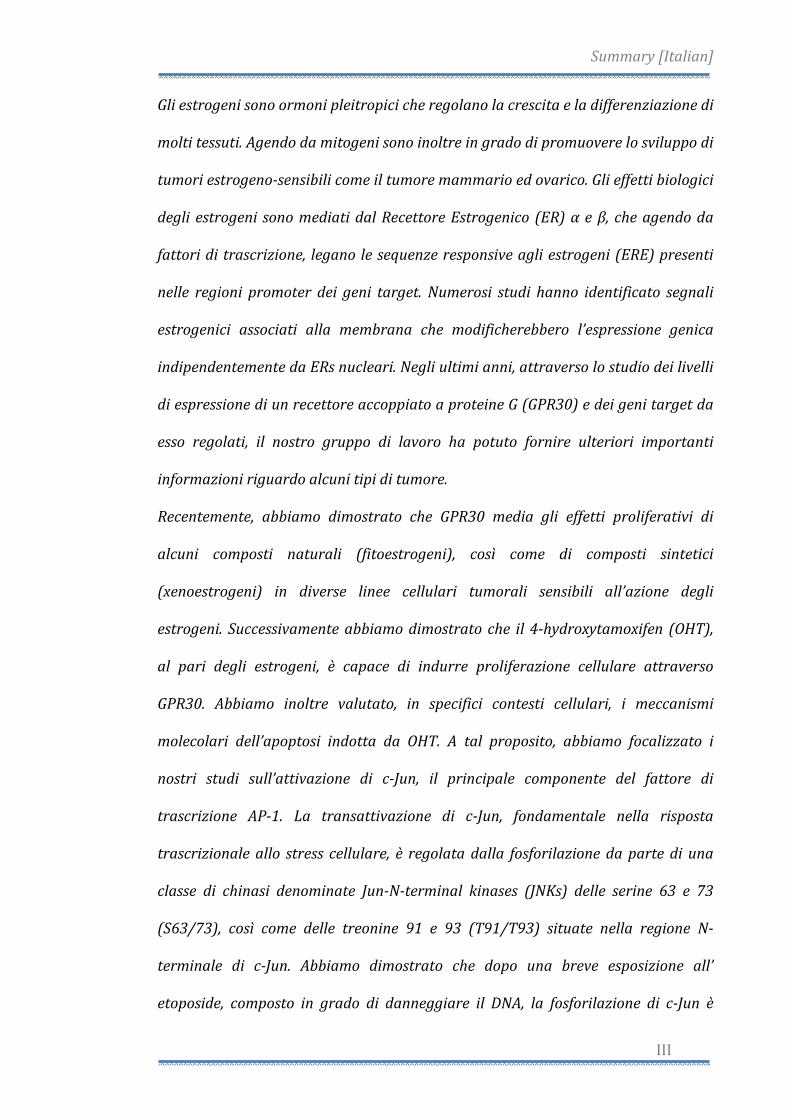
Hormonal Risk And Protective Factors for Breast Cancer
Worldwide, breast cancer is by far the most common cancer amongst women,
with an incidence rate more than twice that of colorectal cancer and cervical
cancer and about three times that of lung cancer. However breast cancer
mortality worldwide is only 25% greater than that of lung cancer in women
(World Health Organization International Agency for Research on Cancer, 2003).
In 2004, breast cancer caused 519,000 deaths worldwide (7% of cancer deaths;
almost 1% of all deaths) (World Health Organization, 2006).
The number of cases worldwide has significantly increased since the 1970s, a
phenomenon partly blamed on modern lifestyles in the Western world
(Laurance, 2006) (Fig. 1).
Fig. 1. Agestandardised death rates from Breast cancer by country (per 100,000 habitants)
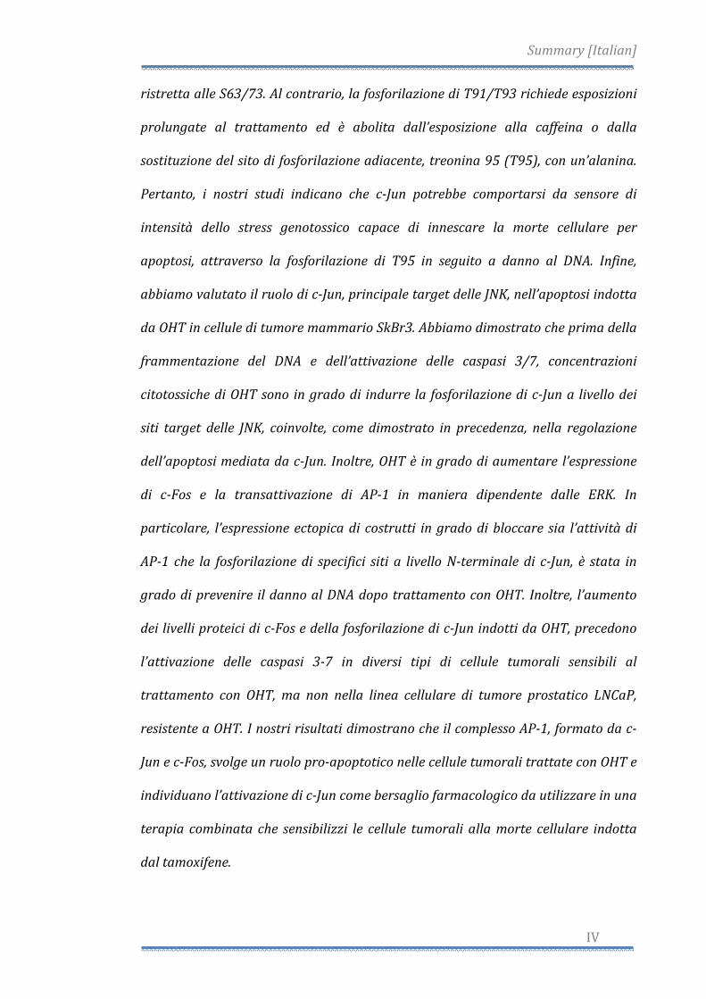
Breast cancer is the most common cancer in women and is estimated to have
accounted for 182,460 new cancer diagnoses and 40,480 deaths in 2008 (Jemal
et al., 2008). The incidence is highest in highly industrialized countries like North
America, Northern Europe, and Australia, where age‐adjusted rates are 75‐92
per 100,000 women (standardized to year 2000 world population), and lowest
in Asia and Africa, where incidence is less than 22 per 100,000 (Parkin et al.,
Introduction
2
2001). Ovarian cancer is the fourth leading cause of tumor death in Western
countries representing the most fatal gynecologic malignancy with the overall 5‐
year survival rate about 10% to 20% (Boete et al., 1993) and is also estimated to
have accounted for 21,650 new cases and 15,520 deaths in 2008 (Jemal et al.,
2008) (Fig. 2).
Fig. 2. Ten Leading Cancer Types for the Estimated New Cancer Cases and Deaths, by Sex, United
States, 2008.(Jemal et al., 2008).
There has been an evident decline in breast cancer mortality since 1997, most
likely the result of therapy with tamoxifen and perhaps other forms of
chemotherapy (McKeanCowdin et al., 2000). Existing evidence regarding the
hormonal etiology of breast cancer supports the hypothesis that estrogen is the
primary stimulant for breast cell proliferation (Henderson and Feigelson, 2000).
Introduction
3
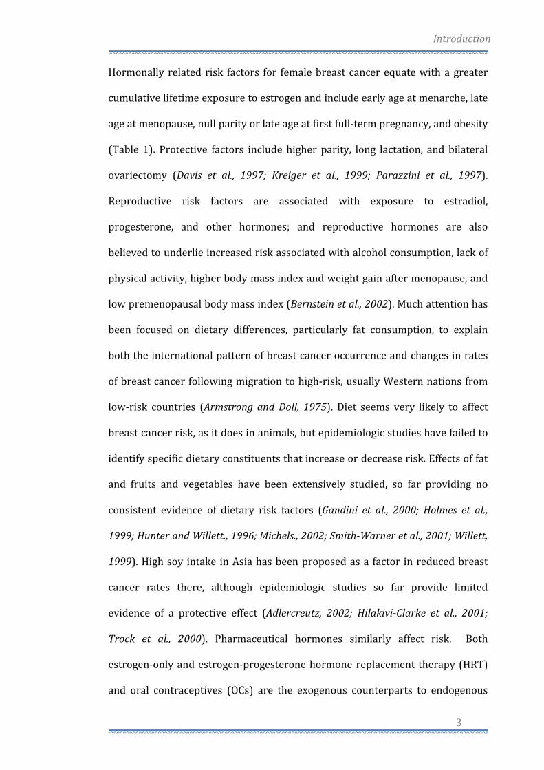
Hormonally related risk factors for female breast cancer equate with a greater
cumulative lifetime exposure to estrogen and include early age at menarche, late
age at menopause, null parity or late age at first full‐term pregnancy, and obesity
(Table 1). Protective factors include higher parity, long lactation, and bilateral
ovariectomy (Davis et al., 1997; Kreiger et al., 1999; Parazzini et al., 1997).
Reproductive risk factors are associated with exposure to estradiol,
progesterone, and other hormones; and reproductive hormones are also
believed to underlie increased risk associated with alcohol consumption, lack of
physical activity, higher body mass index and weight gain after menopause, and
low premenopausal body mass index (Bernstein et al., 2002). Much attention has
been focused on dietary differences, particularly fat consumption, to explain
both the international pattern of breast cancer occurrence and changes in rates
of breast cancer following migration to high‐risk, usually Western nations from
low‐risk countries (Armstrong and Doll, 1975). Diet seems very likely to affect
breast cancer risk, as it does in animals, but epidemiologic studies have failed to
identify specific dietary constituents that increase or decrease risk. Effects of fat
and fruits and vegetables have been extensively studied, so far providing no
consistent evidence of dietary risk factors (Gandini et al., 2000; Holmes et al.,
1999; Hunter and Willett., 1996; Michels., 2002; SmithWarner et al., 2001; Willett,
1999). High soy intake in Asia has been proposed as a factor in reduced breast
cancer rates there, although epidemiologic studies so far provide limited
evidence of a protective effect (Adlercreutz, 2002; HilakiviClarke et al., 2001;
Trock et al., 2000). Pharmaceutical hormones similarly affect risk. Both
estrogen‐only and estrogen‐progesterone hormone replacement therapy (HRT)
and oral contraceptives (OCs) are the exogenous counterparts to endogenous
Introduction
4
hormonal exposures experienced by women and therefore are of concern as
potential contributors to breast cancer risk. Other more common allelic
variations in estrogen metabolism genes (e.g. CYP17, CYP19, HSD17B1) in breast
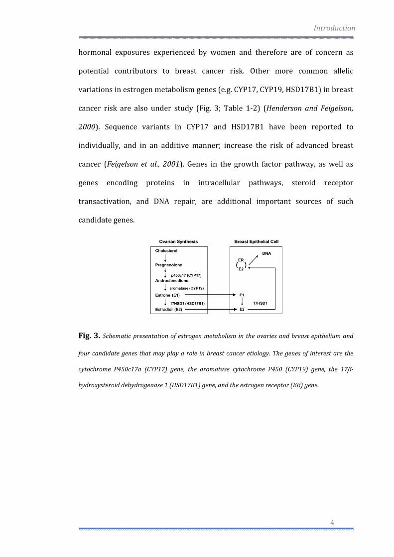
cancer risk are also under study (Fig. 3; Table 1‐2) (Henderson and Feigelson,
2000). Sequence variants in CYP17 and HSD17B1 have been reported to
individually, and in an additive manner; increase the risk of advanced breast
cancer (Feigelson et al., 2001). Genes in the growth factor pathway, as well as
genes encoding proteins in intracellular pathways, steroid receptor
transactivation, and DNA repair, are additional important sources of such
candidate genes.
Fig. 3. Schematic presentation of estrogen metabolism in the ovaries and breast epithelium and
four candidate genes that may play a role in breast cancer etiology. The genes of interest are the
cytochrome P450c17a (CYP17) gene, the aromatase cytochrome P450 (CYP19) gene, the 17β
hydroxysteroid dehydrogenase 1 (HSD17B1) gene, and the estrogen receptor (ER) gene.

Introduction
5
Endocrine disruptors in Breast Cancer
A class of hormonally active chemicals, referred to as endocrine disruptors, may
affect breast cancer primarily at the phase of tumor promotion. They may also
affect mammary gland development and responsiveness to other carcinogens. It
has been suggested that exposure to endocrine disruptors, including chemicals
that mimic estrogens or xenoestrogens, might play an important role in breast
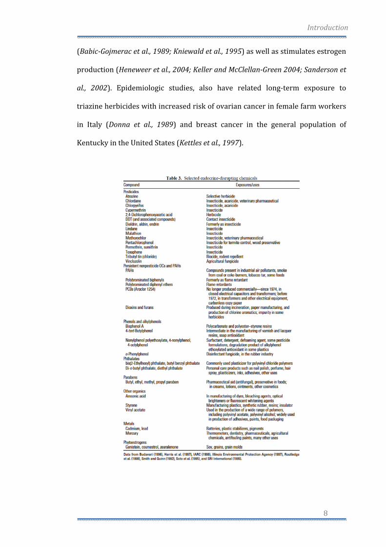
cancer risk (Davis et al., 1993). More than 500 chemicals have been found to be
weakly estrogenic in various assays, including many chemicals in common use,
such as constituents of detergents, pesticides, and plastics (Jobling et al., 1998;
Nishihara et al., 2000; Soto et al., 1995) (Table 3). In a variety of short term in
vitro assays, many of these chemicals are able to bind the ER, initiate
transcription of estrogen‐regulated genes, and stimulate breast cancer cells in

Introduction
6
vitro to proliferate (Korach and McLachlan, 1995; Shelby et al., 1996; Soto et al.,
1995). Short‐term in vivo assays, such as increase in uterine weight in rodents,
are also used to demonstrate estrogenic activity (O'Connor et al., 1996).
Furthermore, the effects of these compounds have been frequently observed in
wildlife in a more natural context. Sexual disruption of wild fish has been
reported in rivers receiving wastewater effluent from industries containing
mixtures of endogenous and pharmaceutical estrogens and industrial chemical
endocrine disruptors (Jobling et al., 1998). The identification of estrogenic
compounds in the environment has raised significant issues regarding the
relevance of the potential adverse health effects (Rudel, 1997). Some researchers
maintain that the potency of many of these endocrine‐disrupting pollutants is
typically much lower than the potency of endogenous estrogens, and so their
effects will likely be insignificant (Safe, 1995). Others are concerned about the
exposure to endocrine‐disrupting chemicals levels of endogenous hormones are
very low, such as in utero or during prepubertal, or postmenopausal time
periods. Furthermore, it must be taken into consideration that additive effects of
low‐level estrogenic pollutants can act together even when each individual
component of the mixture is present below a threshold for effect (Silva et al.,
2002). Finally, the in vivo estrogenic effects of a range of compounds
demonstrate that estrogenic compounds can exhibit different mechanisms and
effects (Gould et al., 1998; Rudel, 1997). This diversity is attributed, at least in
part, to the fact that the shape of the ER ligand (either E2 or an endocrine
disruptor) affects the binding of the ER‐ligand complex to DNA sequences and
subsequent gene expression. Current research into selective estrogen response
modifiers (SERMs, like tamoxifen) for menopause and breast cancer prevention

Introduction
7
is a result of this phenomenon (Emmen and Korach, 2001). Endocrine disruptors
can also act indirectly, for example, by up‐ or down‐regulating the enzymes that
metabolize endogenous estrogens or by affecting synthesis of endogenous
hormones (NRC, 1999). Although research in this area focuses on measuring
circulating serum or urinary levels of endogenous hormones, it is important to
note that human breast tissue, both normal and tumour tissue can metabolize
hormones and create its own local hormonal environment independent of
circulating levels (Adams, 1991; Adams et al., 1992). Thus, effects of chemicals on
the local hormone environment in the breast may be more relevant than effects
on circulating hormone levels. Estrogens can enhance the development of breast
cancer by stimulating cell proliferation rate and thereby increasing the number
of errors occurring during DNA replication (epigenetic effects), as well as by
causing DNA damage via their genotoxic metabolites produced during oxidation
reactions (genotoxic effects) (Gadducci et al., 2005).
Synthetic estrogenic compounds, called xenoestrogens, environmental estrogens
or disruptors, include a variety of pesticides, polychlorinated biphenyls and
plasticizers and are almost ubiquitous in our society (Starek, 2003; Jacobs and
Lewis, 2002). Atrazine, belongs to the 2‐chloro‐s‐triazine family of herbicides and
is the most common pesticide contaminant of groundwater and surface water
(Fenelon and Moore, 1998; Kolpin et al., 1998; Miller et al., 2000) (Tab. 3).
Atrazine is able to interfere with androgen‐ and estrogen‐mediated processes
(Cooper et al., 1999, 2000, 2007; Cummings et al., 2000; Friedmann, 2002;
Narotsky et al., 2001; Stoker et al., 2000). This action occurs without direct
agonism or antagonism of the ER or Androgen Receptor (AR) (Roberge et al.,
2004). Previous studies have shown that atrazine reduces androgen synthesis
Introduction
8
(BabicGojmerac et al., 1989; Kniewald et al., 1995) as well as stimulates estrogen
production (Heneweer et al., 2004; Keller and McClellanGreen 2004; Sanderson et
al., 2002). Epidemiologic studies, also have related long‐term exposure to
triazine herbicides with increased risk of ovarian cancer in female farm workers
in Italy (Donna et al., 1989) and breast cancer in the general population of
Kentucky in the United States (Kettles et al., 1997).
Introduction
9
Endocrine Therapy: Estrogens and Antiestrogens
It was first shown over 100 years ago (Powles, 2002) that approximately one‐
third of premenopausal women with advanced breast cancer will respond to
oophorectomy or removal of the ovaries. Since advances in the understanding of
reproductive endocrinology and steroid biochemistry permitted the
development of specific strategies to restrict the availability of estrogen, the
hormone was widely believed to be responsible for the development of breast
carcinoma (Henderson et al., 1988). As early as 1936, researchers predicted that a
therapeutic agent might be found that could block the stimulatory effects of
estrogen in breast tissue (Lacassagne, 1936). The first non‐steroidal
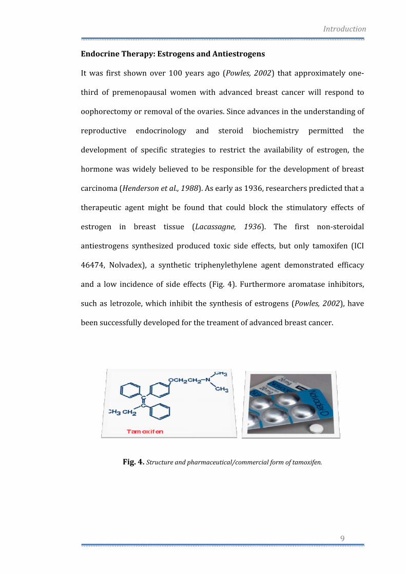
antiestrogens synthesized produced toxic side effects, but only tamoxifen (ICI
46474, Nolvadex), a synthetic triphenylethylene agent demonstrated efficacy
and a low incidence of side effects (Fig. 4). Furthermore aromatase inhibitors,
such as letrozole, which inhibit the synthesis of estrogens (Powles, 2002), have
been successfully developed for the treament of advanced breast cancer.
Fig. 4. Structure and pharmaceutical/commercial form of tamoxifen.
Introduction
10
Tamoxifen Mode of Action in Breast Cancer
Tamoxifen is a competitive inhibitor of estradiol binding to the ER and can
reversibly prevent estrogen‐stimulated growth in vitro. Similarly, tamoxifen will
prevent estrogen‐stimulated growth of ER‐positive breast cancer cells
transplanted into immune deficient (athymic) mice (Nilsen et al., 2000).
Estrogens are believed to modulate cell growth by causing an increase in
stimulatory growth factors (e.g. TGF‐α) and a decrease in inhibitory growth
factors (e.g. TGF‐β) (Dickson and Lippman, 1987). These growth factors are
thought to influence the cell cycle by interaction with their respective membrane
receptors. The regulatory mechanism functions as an autocrine loop, however
also paracrine (cell‐cell) influences of growth factors (e.g. IGF‐1) can play a role
in modulating the replication of epithelial cells. Antiestrogens interfere with the
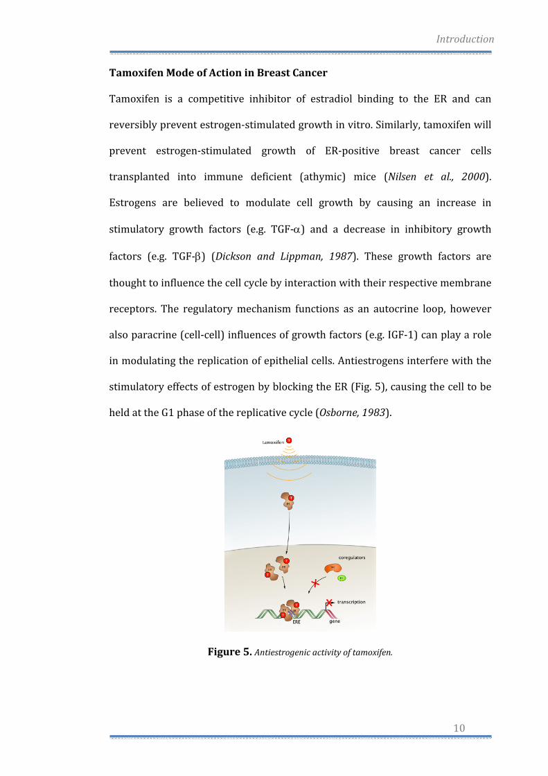
stimulatory effects of estrogen by blocking the ER (Fig. 5), causing the cell to be
held at the G1 phase of the replicative cycle (Osborne, 1983).
Figure 5. Antiestrogenic activity of tamoxifen.
Introduction
11
Initially, experimental studies indicated that tamoxifen acted as an anti‐
estrogenic competititor by preferentially binding to the ER and denying access to
estrogen. Since then it has become apparent that tamoxifen can initiate both
estrogenic and antiestrogenic events (Powles, 2002) by binding to the ER.
Tamoxifen binds to the LBD pocket in the same way as other ligands, such as
estradiol to form transcriptional complexes to activate or switch off genome
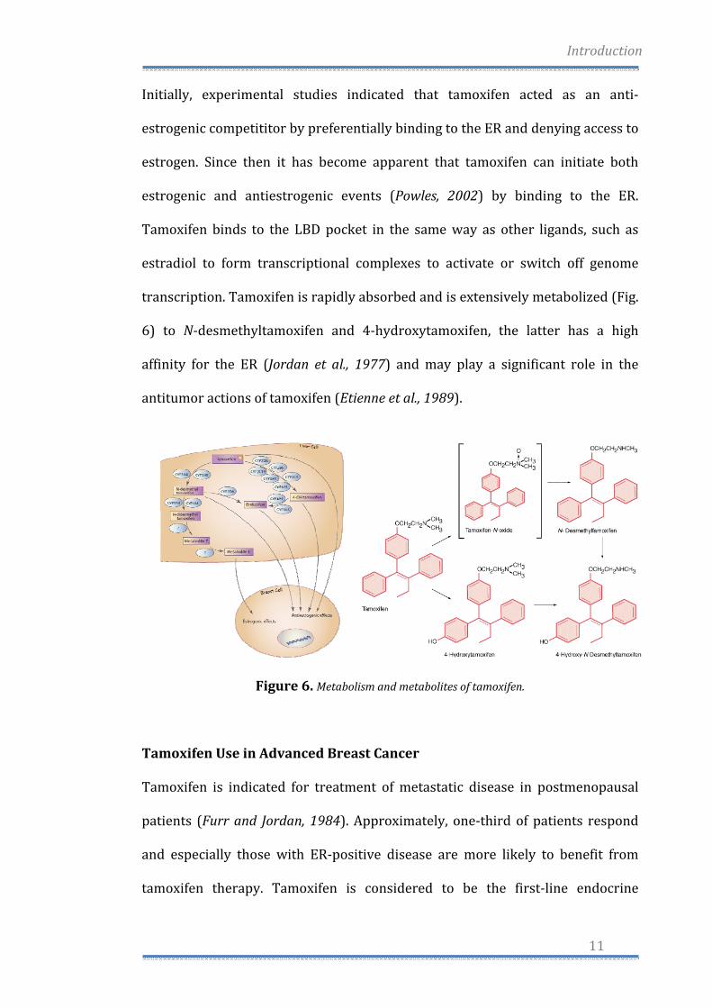
transcription. Tamoxifen is rapidly absorbed and is extensively metabolized (Fig.
6) to N‐desmethyltamoxifen and 4‐hydroxytamoxifen, the latter has a high
affinity for the ER (Jordan et al., 1977) and may play a significant role in the
antitumor actions of tamoxifen (Etienne et al., 1989).
Figure 6. Metabolism and metabolites of tamoxifen.
Tamoxifen Use in Advanced Breast Cancer
Tamoxifen is indicated for treatment of metastatic disease in postmenopausal
patients (Furr and Jordan, 1984). Approximately, one‐third of patients respond
and especially those with ER‐positive disease are more likely to benefit from
tamoxifen therapy. Tamoxifen is considered to be the first‐line endocrine
Introduction
12
therapy due to reduced incidence of side effects and time to progression. The
third generation peripheral aromatase inhibitors, such as letrozole are powerful
inhibitors of estrogen synthesis and produce undetectable levels of circulating
estrogen (Powles, 2002), although side effects are more serious since long‐term
medication might give rise to adverse effects (Smith et al., 1981). Tamoxifen is
also effective in the treatment of advanced disease in premenopausal women;
however, serious concerns have been raised about the biologic consequences of
long‐term tamoxifen treatment for women with breast cancer. One concern in
premenopausal women is the rise in circulating estrogen levels (Smith et al.,
1981). Although tamoxifen has an appropriate level of estrogenic activity in some
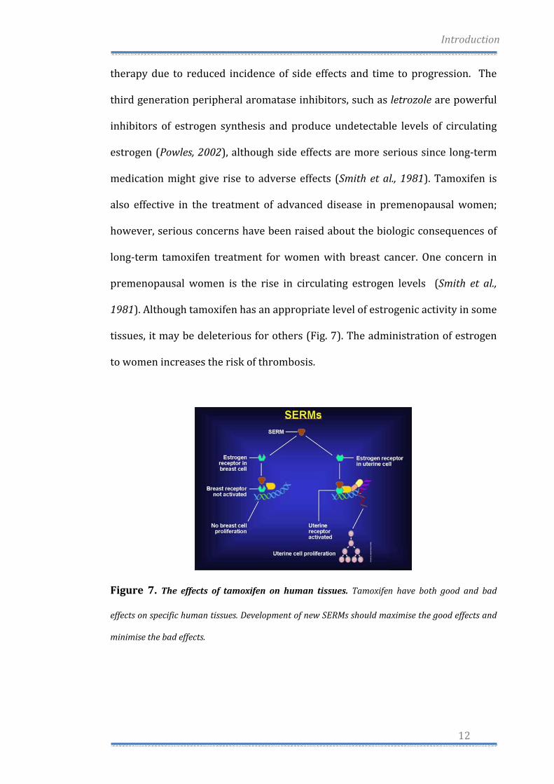
tissues, it may be deleterious for others (Fig. 7). The administration of estrogen
to women increases the risk of thrombosis.
Figure 7. The effects of tamoxifen on human tissues. Tamoxifen have both good and bad
effects on specific human tissues. Development of new SERMs should maximise the good effects and
minimise the bad effects.
Introduction
13
Tamoxifen is known to produce some estrogen‐like effects in postmenopausal
women (Furr and Jordan, 1984) with increased endometrial thickening,
hyperplasia, and fibroids following prolonged tamoxifen therapy (Kedar, 1994).
Endometrial carcinoma has been reported to occur in patients receiving
adjuvant tamoxifen therapy (Killackey, 1985). There is a modest increase in
endometrial carcinoma associated with tamoxifen, but only in postmenopausal
women. Premenopausal women are not at risk for an increase in endometrial
cancer (Bernstein et al., 1999).
Estrogen Receptordependent and Estrogen Receptorindependent
Pathways for tamoxifeninduced apoptosis
Estrogens, acting through estrogen receptors (ERs), regulate the growth and
differentiation of cells of the reproductive system. Binding of 17β‐estradiol (E2)
to the ER induces a conformational change that enables the ER to recruit
transcriptional co‐activators and to induce expression of estrogen‐regulated
genes. Several estrogen‐inducible genes, including c‐myc, TGF‐β, and cathepsin D,
are implicated in malignant transformation or tumor metastases (Dubik et al.,
1992; Bates et al., 1988; Rochefort et al., 1990; Spyratos et al., 1989). Tamoxifen
and its active metabolite, 4‐hydroxytamoxifen (OHT), are nonsteroidal selective
estrogen receptor modulators (SERMs) that compete with E2 and other
estrogens for binding to the ER. Structural studies and chromatin
immunoprecipitations show that OHT∙ER induces an ER conformation that does
not recruit coactivators to target genes and in many cell and promoter contexts
recruits co‐repressors (Shang et al., 2000; Shang and Brown, 2002). The
therapeutic effectiveness of tamoxifen in treatment of hormone‐dependent
Introduction
14
cancers and in preventing breast cancer in high risk women is thought to arise
primarily from its ability to compete with estrogens for binding to the ER. It is
thought that tamoxifen∙ER and OHT∙ER are unable to effectively activate
transcription of genes important for the growth and development of estrogen‐
dependent tumors. However, several often‐conflicting studies show that
tamoxifen and OHT can actively induce programmed cell death of cancer cells
(Mandlekar and Kong, 2001). The mechanism(s) by which tamoxifen and OHT
induce programmed cell death have been quite controversial, with even the
identity of the toxic agents in dispute. One group reported that high
concentrations of tamoxifen, but not of OHT, induce cell death (Dietze et al.,
2001). Others indicated that both tamoxifen and OHT induce cell death
(Mandlekar et al., 2000). Although a recent report was consistent with a role for
ER in OHT‐induced apoptosis (Zhang and Shapiro, 2000), other workers suggest
a number of different mechanisms for tamoxifen‐induced apoptosis. The effects
of tamoxifen might be mediated through an ER‐independent increase in reactive
oxygen species, resulting in caspase activation (Mandlekar et al., 2000a;
Mandlekar et al., 2000b), or through an influx of extracellular calcium (Kim et al.,
1999; Zhang et al., 2000). In addition, effects of tamoxifen on the levels of
proteins important in cell growth including protein kinase C (O’Brian et al., 1986;
Gundimeda et al., 1996), TGF‐β (Chen et al., 1996; Colletta et al., 1990), and c‐Myc
(Kang et al., 1996; Leng et al., 2000) have been reported. Resolution of the role of
ER in tamoxifen‐ and OHT‐induced apoptosis is complicated by the fact that
available ER‐positive and ER‐negative breast cancer cell lines are derived from
independent tumors. Many compounds that are known to induce programmed
cell death (PCD) work via pathways that involve mitochondria. The presence of
Introduction
15
an apoptotic stimulus triggers a rapid increase in mitochondrial permeability,
leading to mitochondrial dysfunction. One of the causes of the mitochondrial
permeability transition is the translocation of the proapoptotic Bax protein from
the cytosol to the mitochondria, where it forms selective channels in the outer
mitochondrial membrane and facilitates the release into the cytosol of
cytochrome c (Shimizu et al., 1999; Shiraishi et al., 2001). In the classic apoptotic
pathway, this cytosolic cytochrome c forms a complex with procaspase 9 and
Apaf‐1 called the apoptosome, which leads to the ATP‐dependent cleavage and
activation of pro‐caspase 9, the initiator caspase in mitochondrial apoptosis.
Activation of pro‐caspase 9 results in activation of downstream executioner
caspases, such as caspase 3 (Leist and Jaattela, 2001; Strasser et al., 2000;
Mattson, 2000). Moreover, OHT is able to induce two independent pathways of
PCD. An ER‐independent pathway kills ER‐negative HeLa cells, requires 10–20
μM tamoxifen or OHT. In contrast, submicromolar amounts of tamoxifen and
OHT trigger cell death only in ER‐positive HeLa cells (Obrero et al., 2002). This
effect is blocked by pre‐treatment with E2, RAL, and ICI 182,780, demonstrating
that binding of tamoxifen and OHT to the ER is required for this pathway of PCD.
The ER‐dependent and ER‐independent pathways both trigger a mitochondrial
permeability transition and share other features of mitochondrial apoptosis,
such as translocation of the proapoptotic Bax protein from the cytosol into the
mitochondria and the release of cytochrome c into the cytosol (Obrero et al.,
2002). However, in contrast to ER‐independent PCD, which displays typical
apoptotic markers such as PARP cleavage, chromatin condensation, and DNA
laddering, a different cell morphology, as well as the absence of those markers,
indicates that the ER‐dependent pathway does not involve caspase activation.
Introduction
16
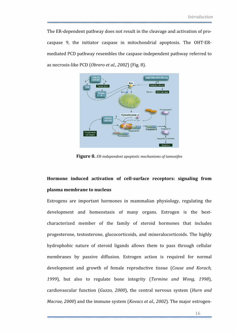
The ER‐dependent pathway does not result in the cleavage and activation of pro‐
caspase 9, the initiator caspase in mitochondrial apoptosis. The OHT∙ER‐
mediated PCD pathway resembles the caspase‐independent pathway referred to
as necrosis‐like PCD (Obrero et al., 2002) (Fig. 8).
Figure 8. ERindependent apoptotic mechanisms of tamoxifen
Hormone induced activation of cellsurface receptors: signaling from
plasma membrane to nucleus
Estrogens are important hormones in mammalian physiology, regulating the
development and homeostasis of many organs. Estrogen is the best‐
characterized member of the family of steroid hormones that includes
progesterone, testosterone, glucocorticoids, and mineralocorticoids. The highly
hydrophobic nature of steroid ligands allows them to pass through cellular
membranes by passive diffusion. Estrogen action is required for normal
development and growth of female reproductive tissue (Couse and Korach,
1999), but also to regulate bone integrity (Termine and Wong, 1998),
cardiovascular function (Guzzo, 2000), the central nervous system (Hurn and
Macrae, 2000) and the immune system (Kovacs et al., 2002). The major estrogen‐
Introduction
17
producing organ is the ovary but recent studies have revealed the synthesis of
estrogen at multiple discrete sites where it may have highly localized effects
(Baquedano et al., 2007). Plasma concentrations of estrogen in women are
commonly in the 1 nM range, although the normal concentration in breast tissue
of postmenopausal women has been reported to be 10‐20‐fold higher than
serum concentration, suggesting local production or concentration of the
hormone (Geisler, 2003). The biological effects of estrogens are mediated by a
specific nuclear receptor (ER) that recognizes and binds the hormone,
transmitting this information to downstream effectors. The first described ER,
ERα, was characterized in 1973 on the basis of specific binding activity in rat
uterus/vagina extracts (Jensen and Desombre, 1973). Its DNA sequence was
determined in 1986 (Greene et al., 1986) and the first crystal structure of an ER
ligand‐binding domain was described in 1997 (Brzozowski et al., 1997). A second
related ER, ΕRβ was identified in 1996 (Kuiper et al., 1997). The ERs are coded
from two separate genes: ERα is located at chromosomal locus 6q25.1 (Menasce
et al., 1993), and encodes a 66kDa protein of 595 amino acids, whereas ERβ is
found at position 14q22‐24 (Enmark et al., 1997) encoding a 54kDa protein of
485 aminoacids. As for the other members of the steroid/thyroid hormone
superfamily of nuclear receptors, ERα and ERβ are composed of three
independent but interacting functional domains: the NH2‐terminal or A/B
domain, the C or DNA‐binding domain, and the D/E/F or ligand‐binding domain
(Nilsson et al., 2001).
Binding of a ligand to ER triggers conformational changes in the receptor and
this leads to changes in the rate of transcription of estrogen‐regulated genes.
These events include receptor dimerization, receptor‐DNA interaction,
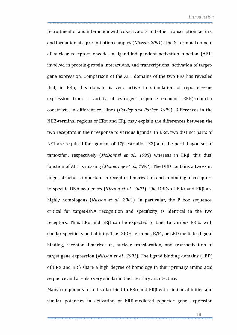
Introduction
18
recruitment of and interaction with co‐activators and other transcription factors,
and formation of a pre‐initiation complex (Nilsson, 2001). The N‐terminal domain
of nuclear receptors encodes a ligand‐independent activation function (AF1)
involved in protein‐protein interactions, and transcriptional activation of target‐
gene expression. Comparison of the AF1 domains of the two ERs has revealed
that, in ERα, this domain is very active in stimulation of reporter‐gene
expression from a variety of estrogen response element (ERE)‐reporter
constructs, in different cell lines (Cowley and Parker, 1999). Differences in the
NH2‐terminal regions of ERα and ERβ may explain the differences between the
two receptors in their response to various ligands. In ERα, two distinct parts of
AF1 are required for agonism of 17β‐estradiol (E2) and the partial agonism of
tamoxifen, respectively (McDonnel et al., 1995) whereas in ERβ, this dual
function of AF1 is missing (McInerney et al., 1998). The DBD contains a two‐zinc
finger structure, important in receptor dimerization and in binding of receptors
to specific DNA sequences (Nilsson et al., 2001). The DBDs of ERα and ERβ are
highly homologous (Nilsson et al., 2001). In particular, the P box sequence,
critical for target‐DNA recognition and specificity, is identical in the two
receptors. Thus ERα and ERβ can be expected to bind to various EREs with
similar specificity and affinity. The COOH‐terminal, E/F‐, or LBD mediates ligand
binding, receptor dimerization, nuclear translocation, and transactivation of
target gene expression (Nilsson et al., 2001). The ligand binding domains (LBD)
of ERα and ERβ share a high degree of homology in their primary amino acid
sequence and are also very similar in their tertiary architecture.
Many compounds tested so far bind to ERα and ERβ with similar affinities and
similar potencies in activation of ERE‐mediated reporter gene expression
Introduction
19
(Kuiper et al., 1998). ERβ shares considerable homology in the DNA binding
region (97%) with ERα, while this homology is markedly lower (55%) in the
LBD, but the trans‐activation mode of action of both ERs, is similar (Petterson et
al., 1997) (Fig. 9). In the absence of its cognate ligand, ERs are recovered in the
cytosolic fraction of target cell homogenates in inactive untransformed hetero‐
oligomeric complexes which contain one steroid‐binding subunit and a non
steroid, non‐DNA‐binding component, identified as a heat shock protein (hsp90).
An important physiologic role for hsp90 is that of maintaining the receptor in a
non functional state: interaction of hsp90 and LBD of the receptor, would
interfere with several LBD and DNA binding domain (DBD) functions, resulting
in the repression of the transcriptional activity of ER (Picard, 1990 and 2002;
Pratt and Toft, 2003). Another essential characteristic of hsp90 is to mediate
receptor trafficking from the cytoplasmatic fraction to the nucleus, through a
microtubule dependent mechanism (Pratt, 1990).
Fig. 9. ERα and ERβ functional domains.
Introduction
20
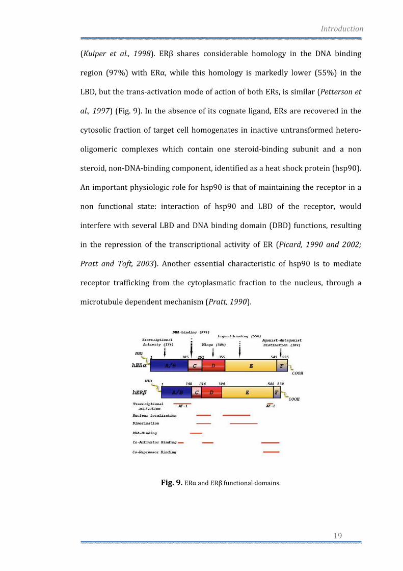
Both ERs are widely distributed throughout the body, displaying distinct but
overlapping expression patterns in a variety of tissues (Petterson and Gustafsson,
2001). ERα is expressed primarily in the uterus, liver, kidney and heart, whereas
ERβ is expressed principally in the ovary, prostate, lung, gastrointestinal tract,
bladder and hematopoietic and central nervous systems. ERs are, however, co‐
expressed in a number of tissues including the mammary gland, epididymis,
thyroid, adrenal, bone and certain regions of the brain (Matthews and Gustafsson,
2003). Cellular responses to estrogens are often divided into two broad
categories: Genomic and Non‐Genomic Responses (Fig. 10).
Fig. 10. Genomic and nongenomic actions of estrogens
Introduction
21
Genomic responses are characterized by gene transcription changes and occur in
the time frame of hours to days, while non‐genomic responses are generally
rapid signaling events. Classical ERs, mediate their primary effects at the
genomic level, but in recent years, it has become clear that not all effects of
estrogens and compounds with estrogen‐like activity can be explained by the
classic genomic mechanism. Breast and ovarian cancer are common in western
countries: environmental factors may play an essential role in hormone‐
dependent tumor etiology. In fact, estrogenic activity can be found in a large
variety of natural and man‐made compounds. Phytoestrogens are natural
substances derived from sources such as plants or fungi: they are typically
flavonoids or isoflavonoids. For example the phytoestrogens Genistein and
Quercetin, copiously present in soyabeans, vegetables and fruits, exert
estrogenic activity through direct binding and activation of ERα and ERβ ,
influencing breast cancer cell proliferation in a dose‐dependent manner
(Maggiolini et al., 2004). In addition, the structure–activity relationships of
resveratrol (RSV), which is classified as a phytoestrogen due to its ability to
interact with estrogen receptors, and the analogs 4,49‐dihydroxystilbene (4,49‐
DHS), 3,5‐dihydroxystilbene (3,5‐DHS), 3,49‐dihydroxystilbene (3,49‐DHS), 4‐
hydroxystilbene (4‐HS) have been assessed using as model systems the ERα‐
positive and negative MCF7 and SkBr3SkBr3 breast cancer cells, respectively. In
binding assays and transfection experiments RSV and the analogs showed the
following order of agonism for ERα: 3,49‐DHS > 4,49‐DHS > 4‐HS > RSV, while
3,5‐DHS did not elicit any ligand properties. Hence, subtle changes in the
structure of the RSV derivatives may be responsible for the different ERα‐

Introduction
22
mediated biological responses observed in estrogen‐sensitive cancer cells
(Lappano et al., 2009).
In addition, the growth of estrogen‐dependent tumors may also have an
important non‐genomic component (Singleton et al., 2003). It has been shown
that estrogens act rapidly by activating membrane receptors coupled to G
proteins (GPCRs) (Kelly and Levin, 2001; Acconcia et al., 2004; Li et al., 2003;
Razandi et al., 2003). These receptors are able to mediate estrogen function
including transcriptional signaling as well as non‐genomic or rapid signaling
(Govind and Thampan, 2003). Some reports described estrogen binding sites on
the intracellular membrane (Evans and Muldoon, 1991), other reports suggest
that palmitoylation (Acconcia et al., 2004; Li et al., 2003) or phosphorylation
(Balasenthil et al., 2004) may transfer ERs to the cytoplasmic face of the plasma
membrane. Also adaptor proteins, such as Shc (Evinger and Levin, 2005) and
NMAR, (Boonyaratanakornkit and Edwards, 2004) can recruit ERα to the plasma
membrane. Classical steroids receptors, bind DNA after ligand stimulation, but
they can also act in the presence or absence of ligand (Lu et al., 2006),
independently of direct DNA binding to scaffold transcription factors, like AP‐1
(Barkhem et al., 2004; Kushner et al., 2000), or induce the activation of kinases,
like MAPKs, phosphatidylinositol 3‐kinase (PI3K), Src or lead to phosphorylation
and transcriptional events through transcription factors like Elk‐1 (Duan et al.,
2001) and serum response factor (SRF) (Duan et al., 2002). Therefore, in addition
to transcriptional regulation estrogens can also mediate cellular effects including
the generation of the second messengers like Ca2+, cAMP and NO, as well as
activation of receptor tyrosine kinases, EGFR and IGF‐1R and protein/lipid
Introduction
23
kinases (Hall et al., 2001; Ho and Liao, 2002; Kelly and Levin, 2001; Levin, 2001
and 2002; Razandi et al., 2003).
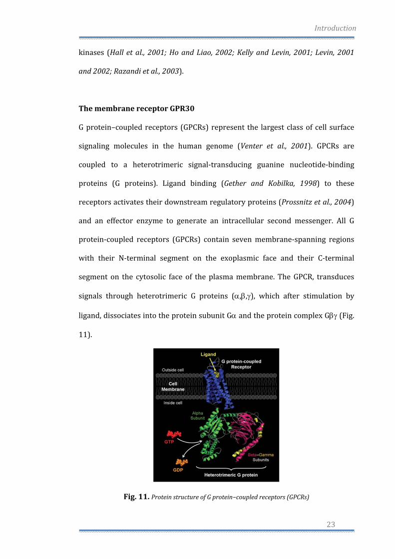
The membrane receptor GPR30
G protein–coupled receptors (GPCRs) represent the largest class of cell surface
signaling molecules in the human genome (Venter et al., 2001). GPCRs are
coupled to a heterotrimeric signal‐transducing guanine nucleotide‐binding
proteins (G proteins). Ligand binding (Gether and Kobilka, 1998) to these
receptors activates their downstream regulatory proteins (Prossnitz et al., 2004)
and an effector enzyme to generate an intracellular second messenger. All G
protein‐coupled receptors (GPCRs) contain seven membrane‐spanning regions
with their N‐terminal segment on the exoplasmic face and their C‐terminal
segment on the cytosolic face of the plasma membrane. The GPCR, transduces
signals through heterotrimeric G proteins (α,β,γ), which after stimulation by
ligand, dissociates into the protein subunit Gα and the protein complex Gβγ (Fig.
11).
Fig. 11. Protein structure of G protein−coupled receptors (GPCRs)
Introduction
24
One such receptor, GPR30, was cloned by different groups using highly disparate
approaches in the late 1990 (Carmeci et al., 1997; O'Dowd et al., 1998; Owman et
al., 1996; Takada et al., 1997). It was not until 2000 that a possible function for
this GPR30 was identified from experiments demonstrating MAP kinase
(ERK1/2) activation by estrogen, as well as the pure ER antagonists ICI 182,780
and tamoxifen, which mimics estrogen function in certain tissues but acts as an
antagonist in other tissues and are collectively known as SERMs. Responses were
demonstrated in breast cancer cell lines expressing GPR30 but not in cell lines
lacking GPR30 (Filardo et al., 2000). Signaling in response to estrogen could be
restored in the latter cell lines by expressing GPR30. They found that estrogen‐
dependent signaling acted through a pertussis toxin‐sensitive pathway:
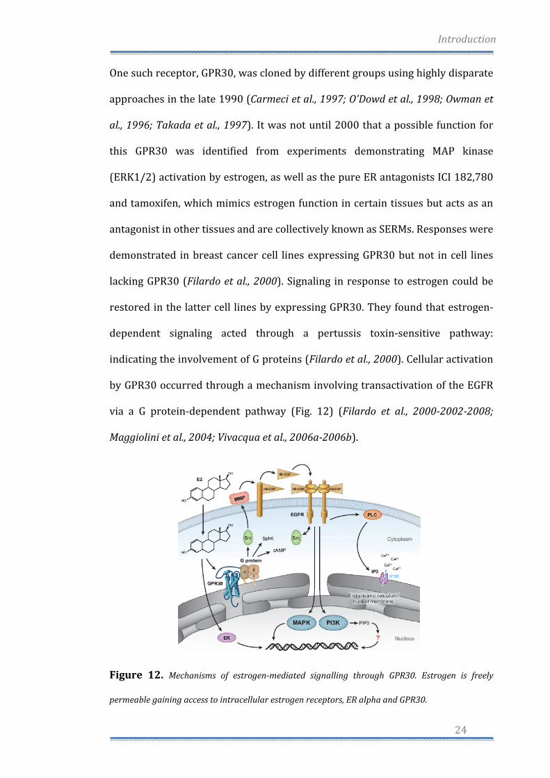
indicating the involvement of G proteins (Filardo et al., 2000). Cellular activation
by GPR30 occurred through a mechanism involving transactivation of the EGFR
via a G protein‐dependent pathway (Fig. 12) (Filardo et al., 200020022008;
Maggiolini et al., 2004; Vivacqua et al., 2006a2006b).
Figure 12. Mechanisms of estrogenmediated signalling through GPR30. Estrogen is freely
permeable gaining access to intracellular estrogen receptors, ER alpha and GPR30.
Introduction
25
At that time such transactivation pathways from GPCRs to EGFR were still a
relatively new concept yet were known to involve metalloproteinase cleavage of
proheparin‐binding (‐bound) epidermal growth factor–like growth factor (pro‐
HB‐EGF) (Daub et al., 1996; Prenzel et al., 1999). A follow‐up report described
GPR30‐mediated elevation of cAMP by estrogen as a mechanism to restore EGF
activated ERK1/2 to basal levels through protein kinase A (PKA)‐dependent
inhibition of Raf‐1 activity (Filardo et al., 2002). Furthermore, GPR30‐mediated
up‐regulation of nerve growth factor production in macrophages by induction of
c‐fos expression has also been demonstrated (Kanda and Watanabe, 2003a). The
up‐regulation of c‐fos by estrogen and phytoestrogens has also been shown in
breast cancer cells (Maggiolini et al., 2004). The majority of GPCRs are expressed
in the plasma membrane, but some GPCRs may be functionally expressed at
intracellular sites (Gobeil et al., 2006). This is particularly true of GPCRs with
lipophilic ligands. Where is GPR30 localized? This question is still open, because
using subcellular markers, one team showed that GPR30 is expressed in an
intracellular compartment, the endoplasmic reticulum but also in the Golgi
apparatus and nuclear membrane. In addition, they were unable to detect
transfected or endogenously expressed GPR30 on the plasma membrane
(Revankar et al., 2005; Revankar et al., 2007). Recently, other two teams reported
expression of GPR30 in the plasma membrane (Thomas et al., 2005; Funakoshi et
al., 2006). The proposed role of GPR30 in cellular estrogen responsiveness was,
until recently, based on the correlation of receptor expression with estrogen‐
mediated signaling (Filardo et al., 2000; Kanda and Watanabe, 2003a; Kanda and
Watanabe, 2003b; Kanda and Watanabe, 2004; Ylikomi et al., 2004). The affinity
of E2 for GPR30 was demonstrated using tritiated estrogen fluorescent E2
Introduction
26
derivates (Revankar et al., 2005; Revankar et al., 2007; Thomas et al., 2005). The
ER antagonists ICI 182,780 and tamoxifen, were also shown to bind GPR30
(Thomas et al., 2005), which is consistent with previous studies showing that
these same compounds were agonists for GPR30 (Filardo et al., 2000).
Furthermore it was demonstrated that tamoxifen activates PI3K through GPR30
but not ERα, suggesting a possible involvement in tamoxifen‐resistant breast
cancers and/or the increased incidence and severity of endometrial cancers in
women treated with tamoxifen. GPR30 has been demonstrated to mediate the
proliferative effects of both estrogen and tamoxifen in endometrial cancer cells
(Vivacqua et al., 2006b). Whether E2 acts on the EGFR/ERK transduction
pathway only through GPR30 binding or also through ERα binding is less clear,
since E2 binds to both receptors although with different affinity. The selective
GPR30 ligand G‐1 permitted the evaluation of GPR30 to mediate proliferative
effects in ovarian cancer cells expressing both ERα and GPR30. Hence, it was
demonstrated that a cross‐talk between ERα and GPR30 could mediate
proliferative effects induced by E2 and G‐1 in ovarian cancer cells (Albanito et al.,
2007). Moreover, it was evaluated whether GPR30 could also be implicated in the
growth effects induced by the pesticide Atrazine in ovarian cancer cells and also
in this case we found that GPR30 and ERα are both involved in this response
(Albanito et al., 2008a). Furthermore, it was found that EGF modulates GPR30
expression through the MAPK pathway (Albanito et al., 2008b).
The steroid hormone estrogen can signal through several receptors and
pathways. Although the transcriptional responses mediated by the nuclear ERs
have been extensively characterized, the changes in gene expression elicited by
signalling through the membrane‐associated ER GPR30 have not been studied. In
Introduction
27
ER‐negative human breast cancer cells the activation of GPR30 signaling by
estrogen or by 4‐hydroxytamoxifen (OHT), an ER antagonist but GPR30 agonist,
induces a transcription factor network, which resembles that induced by serum
in fibroblasts (Pandey et al., 2009). The most strongly induced gene, CTGF,
appears to be a target of these transcription factors. The secreted factor
connective tissue growth factor (CTGF) not only contributes to promote
proliferation but also mediates the GPR30‐induced stimulation of cell migration
(Pandey et al., 2009). In these studies we provided a framework for
understanding the physiological and pathological functions of GPR30. As the
activation of GPR30 by OHT also induces CTGF in fibroblasts from breast tumor
biopsies, these pathways may be involved in promoting aggressive behavior of
breast tumors in response to endogenous estrogens or to OHT being used for
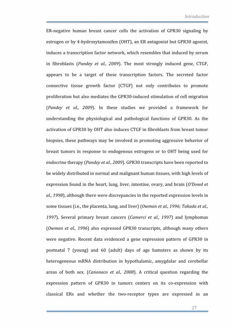
endocrine therapy (Pandey et al., 2009). GPR30 transcripts have been reported to
be widely distributed in normal and malignant human tissues, with high levels of
expression found in the heart, lung, liver, intestine, ovary, and brain (O’Dowd et
al., 1998), although there were discrepancies in the reported expression levels in
some tissues (i.e., the placenta, lung, and liver) (Owman et al., 1996; Takada et al.,
1997). Several primary breast cancers (Camerci et al., 1997) and lymphomas
(Owman et al., 1996) also expressed GPR30 transcripts, although many others
were negative. Recent data evidenced a gene expression pattern of GPR30 in
postnatal 7 (young) and 60 (adult) days of age hamsters as shown by its
heterogeneous mRNA distribution in hypothalamic, amygdalar and cerebellar
areas of both sex. (Canonaco et al., 2008). A critical question regarding the
expression pattern of GPR30 in tumors centers on its co‐expression with
classical ERs and whether the two‐receptor types are expressed in an
Introduction
28
overlapping or an exclusive pattern. That MCF‐7 breast cancer cells express all
three‐estrogen receptors (ERα, ERβ, and GPR30) whereas SkBr3 breast cancer
cells express only GPR30 suggested that all combinations of receptor expression
patterns would likely be possible. Approximately two‐thirds of all breast
carcinomas express ERα. Whereas in these patients ERα antagonists such as
tamoxifen and raloxifene have represented front‐line endocrine therapy,
aromatase inhibitors are now expanding in use. Nevertheless, approximately
25% of patients with ER‐positive breast carcinomas do not respond to tamoxifen
therapy (EBCTCG, 2005). An analysis of 321 cases of primary breast cancer
showed that approximately 60% of the breast tumor cases expressed levels of
GPR30 similar to that normal breast cancer, while 40% of the breast cancer cases
expressed low or undetectable levels of GPR30 protein. Codependency for
GPR30 and ER was observed, as roughly 40% of the cases co‐expressed each
receptor type. Twenty percent of the tumors were doubly negative, failing to
express GPR30 and ER, with the remaining 40% expressing either one receptor
or the other. Interestingly, half of the 122 ER‐negative tumors, scored positively
for GPR30, possibly suggesting that an ER‐negative tumor that retains GPR30
may remain estrogen responsive by signaling through EGFRs (Fig. 14) (Filardo et
al., 2008). Furthermore, the recent identification of the first GPR30‐selective
ligand G‐1 (Bologa et al., 2007) has provided new opportunities to further
differentiate between the functions of the ER family member and GPR30 in
mediating the multifaceted mechanisms of estrogen action (Prossnitz and
Maggiolini, 2009; Maggiolini and Picard, 2009).
Introduction
29
Figure 14. Coexpression of GPR30 and ER in primary human breast tumors (Filardo et al.,
2008).
Activation Protein 1 (AP1)
The AP‐1 transcription factor participates in the control of cellular responses to
stimuli that regulate proliferation, differentiation, immune responses, cell death
and the response to genotoxic agents or stress (Angel and Karin, 1991). AP‐1 is
composed of Jun family members (c‐Jun, JunB and JunD) that can form either
homo‐ or hetero‐dimers among themselves. Jun proteins also dimerize with Fos
family members (c‐fos, fosB, Fra1 and Fra2) (Curran and Franza, 1988) and with
members of the Activating Transcription Factor (ATF) family of proteins (Karin,
1994) These proteins are characterized by a highly charged, basic DBD,
immediately adjacent to an amphipathic dimerization domain, referred to as the
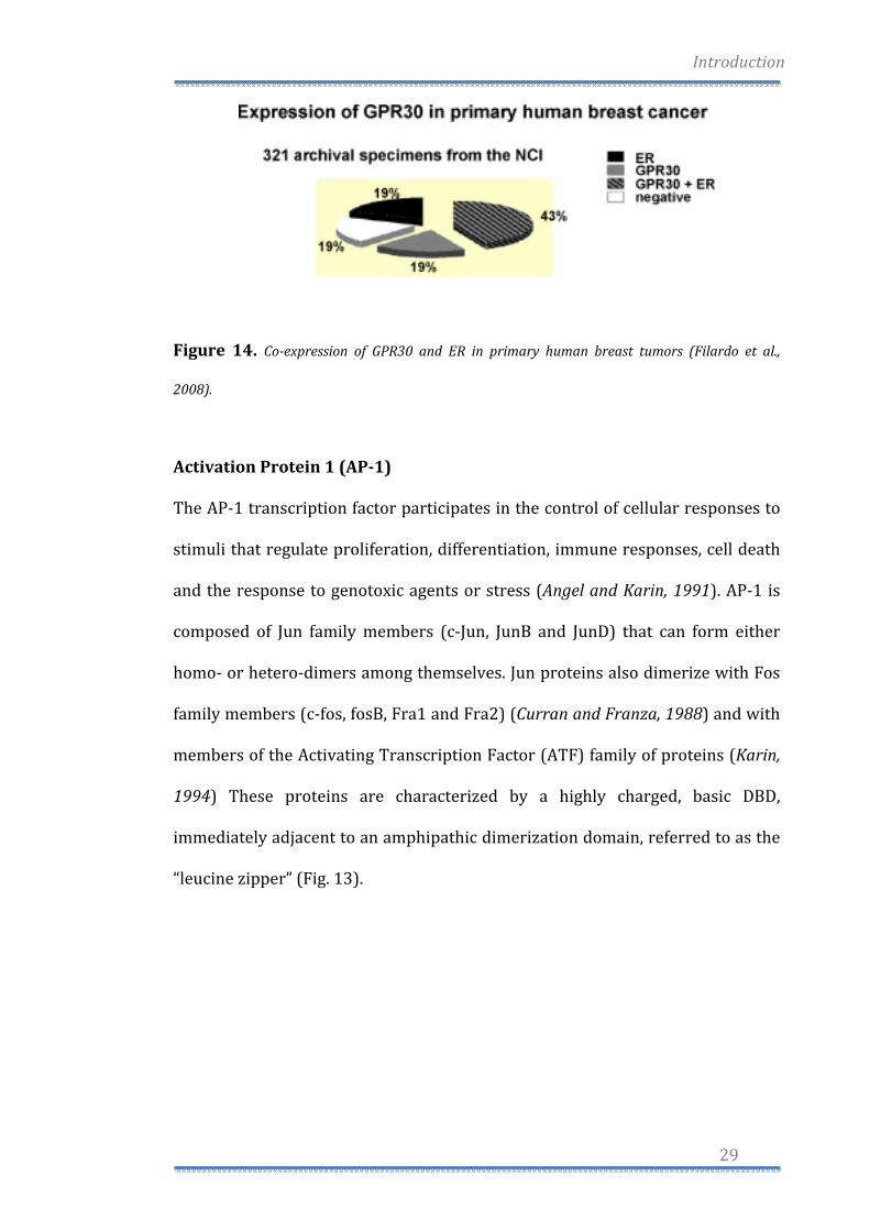
“leucine zipper” (Fig. 13).
Introduction
30
Figure 13. The JunFos heterodimer. The bZIP domains of Jun and Fos form an Xshaped ahelical
structure, which binds to the palindromic AP1 site (TGAGTCA). The bZIP domain of Jun is shown in
blue and the bZIP domain of Fos in red. The DNA backbone is shown in yellow. The Jun and Fos
proteins exhibit several domains, including the bZIP domain (leucine zipper plus basic domain),
transactivation domains and docking sites for several kinases, such as JNK or ERK. These kinases
phosphorylate two serine and threonine residues and thereby modulate the activity of both proteins.
JNK specifically phosphorylates serine residues within the transactivation domain of Jun at position
63 and 73 and thereby regulates its transactivation activity. Mutation of serine to alanine generates
a Jun mutant (JunAA) that cannot be activated by JNKs. Jun is also phosphorylated by casein kinase
II, GSK3ß and ERK, which is not depicted in this scheme. ERK phosphorylates threonine residues at
positions 325 and 331 and a serine residue at position 374 of Fos. Additionally, a Fosrelated kinase
phosphorylates a threonine residue at position 232 of Fos.
The composition of the subunit is determined by the nature of the extracellular
stimulus and the MAPK signaling pathway that is activated: the expression and
activity of c‐Jun and c‐Fos are tightly regulated by members of the mitogen‐
activated protein kinase (MAPK) family, including c‐Jun N‐terminal kinases
(JNKs), extracellular signal‐regulated protein kinase 5 (ERK5), and p38MAPK
kinases and by acting on transcription factors of the TCF family such as Elk‐1,
Introduction
31
and can cause induction of the c‐fos gene. Upon stimulation, the regulation of AP‐
1 activity occurs by activating the transcription of these genes as well as by
phosphorylation of existing Jun and Fos proteins at specific serine and threonine
sites (Vinciguerra et al., 2008; Shaulian and Karin, 2001). AP‐1 activity is
regulated by a broad range of physiological and pathological stimuli, including
cytokines, growth factors, stress signals and infections, as well as by oncogenic
stimuli (Karin and Shaulian, 2001; Shaulian and Karin, 2001). The proto‐
oncogene c‐fos plays a relevant role in the regulation of normal cell growth,
differentiation, and cellular transforming processes (Curran and Franza, 1988).
In particular, c‐fos is classified as a prototypical “immediate early gene” since its
expression is rapidly induced by numerous extracellular stimuli, including
hormones and mitogens (Weisz and Bresciani, 1993; Ginty et al., 1994; Hill and
Treisman, 1995; Bonapace et al., 1996).
The transcription of c‐fos is regulated by different cis‐elements present in the
promoter region of the gene: the Ca2+‐cAMP response element (Ca++/CRE); or the
serum response element (SRE; the sis‐inducible element (SIE)) (Sheng et al.,
1991; Renquin, 2001) (see Fig. 13). Serum or growth factors rapidly induce the
expression of c‐fos through Ras‐MAPK and the SRF (serum responsive factor),
and also through the ternary complex factor composed of Elk‐1, Sap‐1 and Sap‐2
(SRF accessory proteins) which then regulate the expression of target genes
involved in cell proliferation (Hill and Treisman, 1995). Many studies have also
demonstrated that E2 is able to induce the expression of c‐fos in breast cancer
cells through ERα (Weisz and Bresciani, 1993; Bonapace et al., 1996). The
promoter region of c‐fos contains an imperfect ERE palidrome sequence, which is
unable to transactivate c‐fos, however ERα interacts with the Sp1 region at GC
Introduction
32
rich sites downstream from the imperfect ERE palindrome (Duan et al., 1998).
Later it was also demonstrated that E2 is able to activate a non‐genomic pathway
independently of ERα, which may involve Elk‐1 phosphorylation in breast cancer
(Duan et al., 2001). This was also observed in endometrial cancer cells, whereby
c‐fos was induced by E2 and tamoxifen through ERK1/2 activation and
involvement of SRE (Singleton et al., 2003).
c‐Jun is the name of a gene and protein which, in combination with c‐Fos, forms
the AP‐1 early response transcription factor (see Fig. 13). It was first identified
as the Fos‐binding protein p39 and only later rediscovered as the product of the
c‐Jun gene. It is activated through double phosphorylation by the JNK pathway
but has also a phosphorylation‐independent function. c‐Jun knockout is lethal,
but transgenic animals with a mutated c‐Jun that cannot be phosphorylated
(termed c‐JunAA) can survive. This gene is the putative transforming gene of
avian sarcoma virus 17. It encodes a protein, which is highly similar to the viral
protein, and which interacts directly with specific target DNA sequences to
regulate gene expression. This gene is intronless and is mapped to 1p32‐p31, a
chromosomal region involved in both translocations and deletions in human
malignancies.
Genetic and biochemical studies have shown that the biological activity of c‐Jun
mainly relays on JNK‐dependent phosphorylation of its N‐terminal domain at
S63/S73 (Eferl and Wagner, 2003; Shaulian and Karin, 2001). Phosphorylation of
S63/S73 is required for c‐Jun‐mediated cellular transformation, tumor
progression and cancer development (Behrens et al., 2000; Nateri et al., 2005;
Smeal et al., 1991). Abrogation of S63/S73 phosphorylation reduces fibroblast
survival in response to UV radiation (Wisdom et al., 1999) and leads to

Introduction
33
chemoresistance acquisition in various tumor cells to DNA‐damaging drugs
(Potapova et al., 2001). These studies suggest that when phosphorylated at
S63/S73, c‐Jun augments DNA repair, as also evidenced by the accumulation of
spontaneous DNA damage occurring in presence of the genetic ablation of c‐Jun
(MacLaren et al., 2004). Moreover, several studies have shown that c‐Jun
phosphorylation at S63/S73 is required for neuronal apoptosis (Dunn et al.,
2002; Raivich and Behrens, 2006; Shaulian & Karin, 2001). Subsequent
investigations have revealed that c‐Jun is phosphorylated by a variety of stress
and pro‐inflammatory signals on a second group of MAPK sites at T91/T93
(Morton et al., 2003; Papavassiliou et al., 1995) involved in c‐Jun transactivation
(Weiss et al., 2003). In this context, genetic analyses have shown that both groups
of MAPK sites require JNK expression in order to be phosphorylated in response
to stress or pro‐inflammatory stimuli (Morton et al., 2003). Conversely, in
absence of JNK expression, EGF can induce phosphorylation at S63/S73 but not
at T91/T93 (Morton et al., 2003), indicating that T91/T93 phosphorylation is
specifically up regulated by stress and pro‐inflammatory signals. In line,
multisite phosphorylation of c‐Jun on all four MAPK sites has been detected
during chronic hypoxia (Laderoute et al., 2002), neuronal apoptosis by tropic
factor deprivation (Hongisto et al., 2003) and UV‐induced apoptosis of different
types of mammalian cells (Hamdi et al., 2005; Ui et al., 1998). Altogether, these
studies suggest that different physiological contexts may elicit distinct patterns
of c‐Jun N‐terminal phosphorylation, which in turn correlate with diverse
biological output of c‐Jun activation.
Hence, to assess this hypothesis recent studies have investigated whether
different grades of genotoxic stress could induce distinct N‐terminal
Introduction
34
phosphorylation of c‐Jun. As an experimental model system, a differential
exposure of human embryonic kidney 293 (HEK‐293) cells to etoposide
(Vinciguerra et al., 2008), a DNA toposoimerase II inhibitor responsible for both
double and single DNA strand breaks were used (Cliby et al., 2002; Costanzo et al.,
2003). By damaging DNA, etoposide, a DNA toposoimerase II inhibitor
responsible for both double and single DNA strand breaks (Clibyet al., 2002;
Costanzo et al., 2003), triggers the cellular response to genotoxic stress
consisting in a complex network of signaling pathways designed to induce the
onset of cell cycle delay and DNA repair (Sancar et al., 2004). Activation of IP3
kinase family members ATM and ATR is essential for initiating DNA damage
pathways in response to almost all genotoxic stress including etoposide. Upon
genotoxic stress, ATM and ATR activate a network of damage‐response leading
to activation of the checkpoint kinases, Chk1 and Chk2, and DNA repair proteins
(Sancar et al., 2004). If DNA damage persists and unrepaired DNA accumulates,
the same network may lead to the induction of apoptotic programs (Roos and
Kaina, 2006). Etoposide‐mediated activation of the DNA damage response can be
abrogated by caffeine (Clifford et al., 2003; Costanzo et al., 2003), an inhibitor of
ATM and ATR kinases (Sarkaria et al., 1999), able to reverse cell cycle checkpoint
function in response to a wide number of genotoxic stress signals (reviewed in
Kaufmann et al., 2003). Hence, c‐Jun is promptly phosphorylated on S63/S73
following a short exposure to etoposide. In contrast, a continuous exposure to
the drug induces c‐Jun phosphorylation at T91/T93, an event that is caffeine‐
sensitive and linked to an intact threonine 95 (T95). Furthermore, the stress‐
induced biological activity of c‐Jun is impaired by alanine substitution of T95,
Introduction
35
pointing to T95 as a novel regulatory site integrating the strength of genotoxic
stress with the output of c‐Jun activity (Vinciguerra et al., 2008).
In the present thesis, the molecular mechanisms involved in OHT‐induced
apoptosis were evaluated using as a model system the ER‐negative SkBr3 breast
cancer cells. In particular, the study demonstrates (i) the specific
phosphorylation of c‐Jun and (ii) the up‐regulation of c‐Fos as separate ways
contributing to AP‐1‐mediated apoptosis in response to OHT exposure. The work
provides new insights in order to sensitize cancer cells to tamoxifen‐induced cell
death.

RESULTS
Results
36
Oncogene (2009). Received 30 April 2009; Revised 22 September 2009; Accepted 19 October 2009;
Published online 23 November 2009.
cJun activation is required for 4hydroxytamoxifeninduced
cell death in breast cancer cells
Antonio Madeo, Maria Vinciguerra, Rosamaria Lappano, Mario Galgani, Anna
Gasperi‐Campani, Marcello Maggiolini and Anna Maria Musti
Introduction 37
JNKspecific phosphorylation of cJun precedes apoptosis
in OHTtreated SkBr3 cells 41
OHT induces cFos expression in SkBr3 cells 42
OHT transactivates an AP1responsive promoter 43
Abrogation of either cJun phosphorylation or AP1
transactivation protects SkBr3 cells from OHTinduced apoptosis 43
OHT induces cJun activation and cFos expression
in different cancer cell types 45
Results
37
Introduction
Tamoxifen is largely used in the first‐line treatment of estrogen receptor (ER)‐
positive breast cancer (Love, 1989). By binding to ER, tamoxifen antagonizes
estrogen activity by recruiting co‐repressors of transcription to ER target genes
(Smith et al., 1997). However, in vitro studies have shown that at micromolar
concentrations tamoxifen exerts anticancer activity by inducing apoptosis of
tumor cells regardless of ER expression (Mandlekar and Kong, 2001). Apoptotic
cell death is characterized by the activation of a cascade of intracellular cysteine
proteases culminating with the activation of downstream caspases 3, 6 and 7.
These effector‐caspases execute the terminal phases of apoptosis by cleaving a
variety of substrates including those triggering DNA fragmentation (Porter and
Janicke, 1999). Several transduction pathways have been shown to mediate
tamoxifen‐induced apoptosis, including activation of PKC, intracellular Ca2+ and
TGF‐β (Mandlekar and Kong, 2001). It has been shown that tamoxifen‐
dependent elevation of intracellular Ca2+ triggers apoptosis of human
hepatoblastoma HepG2 cancer cells through the accumulation of reactive
oxygen species (ROS) obtained by non‐phagocytic activation of NADPH oxidase
(Lee et al., 2000). ROS‐induced apoptosis requires the participation of further
cell death signaling pathways, including the JNK family of stress‐responsive
MAPKs (Shen et al., 1996). In particular, it has been shown that prevention of
ROS accumulation by antioxidants inhibits both JNK activation and apoptosis in
tamoxifen‐treated cancer cells (Mandlekar et al., 2000b; Mabuchi et al., 2004).
These studies pointed to ROS‐activated JNK as a crucial effector of tamoxifen‐
induced apoptosis. However, downstream targets of the JNK pathway involved
in tamoxifen‐induced apoptosis have not been identified. It has been largely
Results
38
shown that c‐Jun represents the principal transcription factor mediating gene
regulation by JNK (Davis, 2000; Dunn et al., 2002). JNK induces c‐Jun
transactivation by phosphorylating the c‐Jun N‐terminal domain at serines
63/73 and threonines 91/93 (Bannister et al., 1995; Papavassiliou et al., 1995;
Morton et al., 2003; Weiss et al., 2003; Nateri et al., 2005; Vinciguerra et al., 2008).
By forming stable homodimeric or heterodimeric complexes with Fos or ATF
family members, c‐Jun constitutes the AP‐1 transcription factor. Both genetic
and biochemical studies indicate that AP‐1 is involved in different cellular
processes including proliferation, differentiation and apoptosis (Shaulian and
Karin, 2001; Eferl and Wagner, 2003; Hess et al., 2004). However, several studies
have shown that the functional output of AP‐1 depends on the levels of
expression and activation of the individual AP‐1 members that make up the AP‐
1 dimers, as well as the type of stimulus and the intracellular environment (Eferl
and Wagner, 2003; Hess et al., 2004). It has been widely shown that the JNK c‐Jun
pathway contributes to stress‐induced apoptosis in different cell types, such as
neurons, fibroblasts or DNA‐damaged cancer cells (Behrens et al., 1999; Dunn et
al., 2002; Raivich, 2008). In contrast, c‐Jun phosphorylation at serine 63 and 73
(S63/S73) is also important for cell cycle progression or intestinal tumor
progression (Eferl and Wagner, 2003; Wada et al., 2004; Nateri et al., 2005),
suggesting that additional phosphorylation of c‐Jun at threonine 91 and 93
(T91/T93) might be critical for c‐Jun pro‐apoptotic functions. In line with this
suggestion, we have recently shown that phosphorylation of c‐Jun at all four JNK
sites is crucial for c‐Jun pro‐apoptotic function in response to DNA damage
(Vinciguerra et al., 2008). Furthermore, JNK‐specific phosphorylation of c‐Jun at
T91/T93, but not at S63/S73, requires a priming phosphorylation at T95 by a
Results
39
yet to be identified stress‐induced kinase (Vinciguerra et al., 2008). These
observations suggest that depending on the type of stimulus c‐Jun may or may
not be phosphorylated at all four terminal sites by JNK and correspondingly it
may result in different cellular consequences. Therefore, the analysis of N‐
terminal phosphorylation of c‐Jun at both S63/73 and T91/T93 is essential for
associating c‐Jun activation to either prosurvival or pro‐apoptotic pathways. To
date, c‐Jun phosphorylation in response to tamoxifen has been analysed only in
tamoxifen‐resistant MCF‐7‐derived xenografts and limited to the S63 site (Schiff
et al., 2000).
c‐Fos, the main heterodimeric partner of c‐Jun, has also been shown to be
involved in both proliferative and apoptotic pathways (Hess et al., 2004). c‐Fos
expression can be induced by a variety of stimuli, each acting on one or more
multiple cis‐elements contained within the promoter of c‐Fos (Treisman, 1995).
In this regard, it has been reported that the serum‐responsive element recruits
ERK‐activated Elk‐1 as well as the serum responsive factor accessory proteins 1
and 2 to the c‐Fos promoter sequence (Price et al., 1995). Accordingly, in our
previous studies both estrogen and 4‐hydroxytamoxifen (OHT) induced c‐Fos
expression by ERK‐dependent activation of Elk‐1 (Maggiolini et al., 2004;
Vivacqua et al., 2006ab). These studies, as well as other reports (Reviewed in
Prossnitz and Maggiolini, 2009), indicate that the aforementioned stimulations
may occur through the GPR30 in conjunction with activated EGFR and relayed
on a pertussis toxinsensitive pathway, confirming the involvement of Gi/o
heterotrimeric G proteins. In this study, we have assessed the role of the c‐
Jun/c‐Fos AP‐1 complex in OHT‐induced apoptosis. To this aim, we have
examined JNK‐dependent phosphorylation of c‐Jun at both S73 and T91/T93

Results
40
sites as well as the induction of both c‐Fos expression and AP‐1 transactivation
in response to OHT. Our results indicate that both JNK‐dependent activation of
c‐Jun and ERK‐mediated expression of c‐Fos are crucial for OHT‐induced
apoptosis in ER‐negative breast cancer cells.
Results
41
JNKspecific phosphorylation of cJun precedes apoptosis in OHTtreated
SkBr3 cells
Micromolar concentrations of OHT have been shown to induce apoptotic cell
death in ER‐negative breast cancer cells through caspase 3 and JNK pathways
(Mandlekar et al., 2000a). We used the ER‐negative breast cancer‐derived cell
line SkBr3 as a model system to analyse the role of JNK‐activated c‐Jun in the
OHT‐dependent apoptosis. First, we examined whether micromolar
concentrations of OHT, which were earlier reported to be cytotoxic in SkBr3
cells (Basu et al., 2004), could induce hallmarks of apoptotic cell death such as
DNA fragmentation and caspase 3/7 activation. As shown by flow cytometric
analysis, DNA fragmentation was induced by 10 μM OHT and was further
increased by 25 μM OHT, whereas neither concentration had any influence on
cell cycle phases (Figures 1a and b). The effect of 10 μM OHT on DNA
fragmentation was further confirmed by TdT‐mediated dUTP nick end labeling
(TUNEL) staining and cell viability assay (Figures 1c and d). Furthermore, OHT
induced caspase 3/7 activities (Figure 1e), suggesting that DNA fragmentation
results from apoptosis. Next, we analysed whether JNK‐activated c‐Jun precedes
OHT‐dependent apoptosis in SkBr3 cells. To this end, we examined the ability of
OHT to induce c‐Jun N‐terminal phosphorylation at either (S73, a hallmark of c‐
Jun activation by JNK (Dunn et al., 2002), or at T91/T93, which are stringently
correlated with c‐Jun pro‐apoptotic activity (Vinciguerra et al., 2008).
Phosphorylation of both S73 and T91/T93 sites were induced following a 2 h
exposure to OHT and no longer detected in presence of the JNK‐specific
inhibitor SP 600125 (SP) (Figures 2a and b), indicating that JNK‐specific
activation of c‐Jun precedes OHT‐induced apoptosis. In contrast, neither the
Results
42
EGFR inhibitor AG 1478 (AG), nor the MEK/ERK inhibitor PD 98059 (PD) and
the ER pure antiestrogen ICI 182 780 (ICI) prevented c‐Jun phosphorylation by
OHT. As OHT has been shown to induce JNK activation through generation of
oxidative stress (Lee et al., 2000; Mandlekar and Kong, 2001; Mabuchi et al.,
2004), we next analysed the effect of the antioxidant N‐acetyl‐L‐cysteine (NAC)
on the JNK/c‐Jun pathway. As shown in Figure 2c, both activation of JNK and c‐
Jun phosphorylation at T91/T93 sites were prevented by NAC treatment. In line
with previous studies, the exposure to NAC prevented also DNA fragmentation
by OHT (Figure 2d). These results indicate that c‐Jun pro‐apoptotic
phosphorylation by OHT is triggered by oxidative stress and is associated with
DNA fragmentation.
OHT induces cFos expression in SkBr3 cells
In addition to c‐Jun activation, we analysed whether OHT induces c‐Fos
expression in SkBr3 cells, at both messenger RNA and protein levels. As shown
by either reverse transcription‐PCR analysis or western blots, the expression of
c‐Fos was rapidly induced by OHT (Figures 3a and b). In agreement with our
previous studies performed in different cancer cell types (Vivacqua et al., 2006a
b), c‐Fos induction by OHT was sensitive to AG and PD but not to SP or ICI
(Figure 3b). It is noteworthy that the effect of OHT on c‐Fos expression was also
abrogated silencing GPR30 expression (Figures 3c and d), which we have shown
earlier to mediate c‐Fos induction by estrogens through ERK activation
(Maggiolini et al., 2004; Vivacqua et al., 2006ab). Accordingly, OHT‐dependent
activation of ERK was sensitive to either PD or abrogation of GPR30 expression
(Figures 3e and f). In contrast, NAC treatment had no effect on either ERK
Results
43
activation or c‐Fos expression, indicating that OHT induces the ERK/c‐Fos
pathway independently from its ability to trigger oxidative stress
(Supplementary Figure 1). Taken together, these results suggest that OHT
induces c‐Fos expression through the GPR30–EGFR–ERK transduction pathway
in SkBr3 cells.
OHT transactivates an AP1responsive promoter
Next, we evaluated whether OHT‐dependent induction of c‐Jun N‐terminal
phosphorylation and c‐Fos expression could lead to AP‐1 transactivation. To
this end, we tested the ability of OHT to activate the AP‐1‐responsive
collagenase promoter fused to a luciferase reporter gene (AP‐1‐luc). As shown
in Figure 4a, OHT induced AP‐1‐luc transactivation, which was sensitive to SP,
AG or PD, but not to ICI. Furthermore, only AG and PD inhibited the
transactivation of the c‐Fos promoter by OHT (Figure 4b), hence corroborating
the specific role exerted by ERK activation in OHT‐induced c‐Fos expression.
Overall, these findings suggest that both JNK‐specific activation of c‐Jun and
ERK‐dependent induction of c‐Fos expression contribute to AP‐1 activation by
OHT.
Abrogation of either cJun phosphorylation or AP1 transactivation
protects SkBr3 cells from OHTinduced apoptosis
We then analysed the specific involvement of the c‐Jun/c‐Fos AP‐1 complex in
OHT‐induced apoptosis. To this aim, we performed TUNEL assays in OHT‐
treated cells ectopically expressing a truncated form of c‐Fos (A‐Fos), which was
shown earlier to efficiently knockdown c‐Jun/c‐Fos AP‐1 heterodimers (Olive et
Results
44
al., 1997). As shown in Figures 5a–f, approximately 60% of SkBr3 cells
transfected with an empty vector resulted positive for TUNEL staining after
exposure to OHT. In contrast, only 11% of A‐Fos‐transfected SkBr3 cells were
TUNEL positive (Figures 5g–l), indicating that the c‐Jun/c‐Fos AP‐1 complex is
involved in OHT‐induced apoptosis. As shown in Figure 5s, the constitutive
expression of c‐Fos slightly decreased the percentage of TUNEL‐positive cells
(Figures 5m–r), suggesting that c‐Fos alone cannot sensitize SkBr3 cells to OHT‐
induced apoptosis. To further analyse the direct role of c‐Jun activation in OHT‐
mediated apoptosis, we ectopically expressed in SkBr3 cells either a HA‐tagged
c‐Jun‐wt protein or a HA‐tagged c‐Jun mutant bearing alanine substitutions of
S63/S73 and T91/T93 JNK sites (c‐Jun‐PanAla), which was shown to impair c‐
Jun activation as well as c‐Jun‐mediated apoptosis (Papavassiliou et al., 1995;
Watson et al., 1998; Dunn et al., 2002). Expression of HA‐tagged c‐Jun‐PanAla
protected SkBr3 cells from OHT‐induced apoptosis, as only 6% of HA‐positive
cells were also positive for TUNEL staining, while 58% of HA‐negative cells
were TUNEL positive (Figures 6i–p). In contrast, ectopic expression of c‐Jun‐wt
had no effect on DNA fragmentation (Figures 6a–h), indicating the abrogation of
JNK‐specific phosphorylation of c‐Jun is essential to protect SkBr3 cells from
OHT‐induced apoptosis. Furthermore, a c‐Jun mutant that cannot be specifically
phosphorylated at the T91/T93 JNK sites (Vinciguerra et al., 2008) was as
efficient as c‐Jun‐PanAla to protect SkBr3 cells from OHT‐dependent apoptosis
(Figures 6q–x), showing that c‐Jun N‐terminal phosphorylation at T91/T93 site
has a pivotal role in OHT‐dependent apoptosis.
Results
45
OHT induces cJun activation and cFos expression in different cancer cell
types
To validate the role of AP‐1 activation in OHT‐induced apoptosis, we examined
whether OHT couples the induction of both c‐Fos expression and c‐Jun
phosphorylation to caspase 3/7 activation in different cancer cell types. As
shown in Figures 7a and b, OHT stimulated both c‐Fos expression and c‐Jun
phosphorylation in ER‐negative BT20 and ER‐positive MCF‐7 breast cancer
cells, HepG2 hepatocarcinoma cells, LoVo colorectal adenocarcinoma cells and
SCLC‐R1 lung cancer cells. As observed for SkBr3 cells (Figure 3), OHT‐
dependent induction of c‐Fos was specifically abolished by AG and PD (Figure
7a), as well as by silencing GPR30 expression (Supplementary Figure 2).
Furthermore, in all cell lines OHT‐stimulated c‐Jun N‐terminal phosphorylation
in a JNK‐specific manner, because only SP abrogated c‐Jun phosphorylation
(Figure 7b). Similarly, OHT induced caspase3/7 activity (Figure 7c) and DNA
fragmentation (Supplementary Figure 3) in all cancer cell types, except for the
OHT‐resistant LNCaP prostate cancer cells. Remarkably, OHT also failed to
induce either c‐Fos expression or c‐Jun phosphorylation in LNCaP cells (Figures
7d and e), corroborating the functional link between AP‐1 activation and OHT‐
induced apoptosis. As our results indicate that activation of the JNK/c‐Jun
pathway by OHT is triggered by oxidative stress, we asked whether the
resistance of LNCaP cells to OHT‐induced apoptosis correlated with a deficiency
in ROS production and consequential lack of activation of the JNK/c‐Jun
pathway. To this end, we examined the ability of OHT to generate ROS in either
SkBr3 or LNCaP cells by DCFH fluorescence (Shen et al., 1996). As show in
Figure 8, OHT increased the basal level of ROS in SkBr3 cells, but not in LNCaP

Results
46
cells, suggesting that the failure to generate ROS may contribute to protect
LNCaP cells from OHT‐dependent apoptosis.

DISCUSSION
Discussion
47
The effects of estrogens are versatile and fundamental throughout the body.
Estrogens exert diverse biological effects in animals and humans, and many of
these effects result from a direct interaction of estrogen with an intracellular
receptor that activates the expression of genes encoding proteins with important
biological functions (Kumar et al., 1987; Evans, 1988; Weisz et al., 1990; Parker,
1991). Although some of the earliest effects of estrogen were shown to be rapid in
nature, the majority of studies following the discovery of the classical ER focused
on long‐term transcriptional activities of the hormone. Recently, the rapid
signaling events mediated by E2 have been the object of increasing interest. The
transcriptional mechanisms of ER function through ERE sequences have been
investigated, but gene regulation by estrogen at promoters lacking EREs require
further investigation (Carroll and Brown, 2006). E2 is able to start
phosphorylation events through MAPKs and PI3Ks thus modulating gene
expression via alternate promoter sites such as serum response elements
through the actions of Elk‐1 and SRF (Duan et al., 2001; Duan et al. 2002). The
activation of these pathways has been typically associated with growth factor
receptors and GPCRs. With the recent identification of GPR30, as an estrogen‐
responsive GPCR, our understanding of the mechanisms of estrogen action has
been broadened. GPR30 activates rapid kinase signaling pathways and also
mediates transcriptional regulation of genes previously associated with estrogen
action through ERs. GPR30 protein expression had been observed in many cancer
cell lines: MCF‐7 and SKBR3 breast cancer (Carmerci et al., 1997; Filardo et al.,
2000; Thomas et al., 2005; Revankar et al., 2005), HEC1A (Vivacqua et al., 2006b)
and HEC50 (Revankar et al., 2005) endometrial cancer, JEG choriocarcinoma
(Revankar et al., 2005), BG‐1 ovarian cancer, and thyroid carcinoma (Vivacqua et
Discussion
48
al., 2006a) as well as in some brain areas of the hamster (Canonaco et al., 2008).
Our previous reports demonstrated estrogen‐mediated cell proliferation to be
dependent on GPR30 (Maggiolini et al., 2004; Vivacqua et al., 2006ab). Our data
suggested that the c‐Fos/AP‐1 complex is implicated in tumor progression
elicited by estrogens, phytoestrogens, xenoestrogens and antiestrogens through
GPR30, in different cancer cell types (Albanito et al., 2007; Albanito et al.,
2008a;.Pandey et al., 2009; Albanito et al., 2008b)
Furthermore, we studied the role of c‐Jun activation in cell death induced by 4‐
hydroxytamoxifen (OHT) in breast cancer cells and in diverse cancer cell types.
Accumulating evidence suggests that tamoxifen‐induced apoptosis may consist in
the generation of oxidative stress and subsequent activation of the JNK family of
stress‐responsive MAPKs (Lee et al., 2000; Mandlekar and Kong, 2001; Mabuchi et
al., 2004). c‐Jun mediates proapoptotic functions of JNK in different types of cells
including neurons, fibroblasts and cancer cells (Dunn et al., 2002; Hess et al., 2004;
Raivich, 2008). However, c‐Jun does not mediate all paradigms of JNK‐dependent
apoptosis and its pro‐apoptotic function depends on the type of stimulus as well
as on the heterodimeric partner forming the AP‐1 transcription factor (Hess et al.,
2004; Raivich et al., 2006). In this study, we analysed whether JNK‐dependent
activation of c‐Jun has a role in OHT‐induced cell death of ER‐negative SkBr3
breast cancer cells. To this end, we first established by fluorescence‐activated cell
sorting (FACS) analyses the range of micromolar OHT concentrations required to
induce DNA fragmentation, as also confirmed by TUNEL and cell viability assays.
Importantly, we found that OHT triggers caspase 3/7 activity, suggesting that
DNA fragmentation by OHT is a secondary consequence of apoptosis rather than
necrosis. Moreover, OHT had no effects on cell cycle progression, excluding the
Discussion
49
possibility that OHT‐dependent cytotoxicity was due to cell growth arrest. Next,
we analysed the ability of OHT to induce JNK‐dependent phosphorylation of c‐Jun
at the S73 site, which is considered a hallmark of c‐Jun activation in several
paradigms of stress signals (Dunn et al., 2002). We detected both JNK activation
and c‐Jun phosphorylation at S73 on a 2‐h exposure to OHT. Remarkably, OHT
also induced phosphorylation of c‐Jun at T91/T93 sites, which was shown to be
crucial for c‐Jun‐mediated cell death in response to either ultraviolet or etoposide
exposure (Vinciguerra et al., 2008). Moreover, both S73 and T91/T93
phosphorylations by OHT were specifically sensitive to SP, indicating that JNK‐
specific activation of c‐Jun precedes apoptosis in SkBr3 cells. Interestingly, OHT‐
induced c‐Jun N‐terminal phosphorylation without alteration of c‐Jun expression,
suggesting that activation of c‐Jun by OHT is limited to pre‐existing c‐Jun
proteins. Accordingly, our (unpublished observations) and other studies (Nephew
et al., 1993) have indicate that c‐Jun expression is not regulated by OHT
treatment. Importantly, DNA degradation as well as JNK activation and N‐
terminal phosphorylation of c‐Jun, were abrogated by antioxidant treatments.
These results suggest that generation of oxidative stress, presumably by NADPH
activation (Lee et al., 2000), could be the initial OHT‐dependent signal leading to
activation of the JNK/c‐Jun pathway and thereafter to apoptosis. We then asked
whether c‐Jun activation by OHT was coupled to induction of c‐Fos expression.
Transactivation assays of the c‐Fos promoter combined with the analysis of
steady‐state levels of both c‐Fos protein and messenger RNA indicated that OHT
leads to transcriptional induction of c‐Fos expression. Interestingly, both the
transactivation of the c‐Fos promoter and the induction of c‐Fos protein levels
were sensitive to AG or PD, but not to SP or ICI, indicating that OHT induces c‐Fos
Discussion
50
expression independently of its ability to induce JNK activity but rather in an
EGFR/ERK‐mediated manner. Accordingly, OHT‐dependent activation of ERK
was abrogated by PD but not by SP, confirming that JNK activity is not required
for the signaling pathway upstream c‐Fos expression. Moreover, both c‐Fos
expression and ERK activation by OHT were sensitive to GPR30 silencing, in
accordance with our previous studies performed in different cancer cells
(Maggiolini et al., 2004; Vivacqua et al., 2006ab; Albanito et al., 2007). It is
noteworthy that the ERK/c‐Fos pathway was not altered by antioxidant
treatment, thus corroborating the fact that OHT initiates two distinct signaling
pathways converging to activation of the c‐Jun/c‐Fos AP‐1 complex. Induction of
c‐Fos expression coupled to c‐Jun N‐terminal phosphorylation is the most
frequent encountered paradigm of AP‐1 activation in mammalian cells. Indeed,
we found that OHT‐induced AP‐1 transactivation in an ERK and JNK‐dependent
manner, suggesting that the heterodimeric c‐Jun/c‐Fos AP‐1 complex functions in
OHT‐induced apoptosis of cancer cells. Alternatively, because of the crucial role
of AP‐1 in a variety of cellular events, activation of c‐Jun/c‐Fos AP‐1 complex by
OHT may reflect a pro‐survival response.
To evaluate the specific involvement of c‐Jun/c‐Fos in OHT‐dependent apoptosis,
we abrogated c‐Jun/c‐Fos AP‐1 activity in SkBr3 cells by two independent
experimental approaches: (i) ectopic expression of A‐Fos, a truncated form of c‐
Fos that has been shown to specifically knock‐down c‐Jun/c‐Fos AP‐1
heterodimers (Olive et al., 1997); and (ii) ectopic expression of c‐Jun‐PanAla, a c‐
Jun mutant bearing alanine substitution of all four JNK phosphorylation sites
S63/S73 and T91/T93, which has been shown to act as a c‐Jun dominant negative
in several cell lines (Papavassiliou et al., 1995; Watson et al., 1998; Dunn et al.,
Discussion
51
2002). A‐Fos overexpression protected cells from OHT‐induced apoptosis,
indicating that c‐Jun/c‐Fos heterodimers are directly involved in apoptosis. This
finding was further corroborated by the evidence that AP‐1 transactivation by
OHT was dependent on both ERK and JNK activity. Similarly, overexpression of c‐
Jun‐PanAla resulted in the protection of SkBr3 cells from OHT‐induced apoptosis.
As Jun‐wt had no effect on DNA degradation, the protective effect of c‐Jun‐PanAla
presumably results from its ability to act as dominant‐negative, indicating that
JNK‐activated c‐Jun is necessary to trigger OHT‐induced apoptosis. Moreover, a c‐
Jun mutant that specifically cannot be phosphorylated at the T91/T93 JNK sites
was as efficient as c‐Jun‐PanAla in protecting SkBr3 cells from OHT‐dependent
apoptosis, suggesting that phosphorylation of c‐Jun at S63/S73 is not sufficient to
trigger c‐Jun‐dependent apoptosis. It is noteworthy that ectopic expression of c‐
Jun could not significantly increase the number of TUNEL‐positive cells,
indicating that activation of pre‐existing c‐Jun proteins by OHT is sufficient to
trigger apoptosis. Our data show that OHT induces both ERK/c‐Fos and JNK/c‐
Jun pathways in various OHT‐sensitive cancer cells, pointing to c‐Jun/c‐Fos AP‐1
activation as a general mechanism mediating OHT‐dependent apoptosis, rather
than a peculiar response of SkBr3 cells. Accordingly, OHT‐induced caspase 3/7
activity and DNA degradation in all cancer cell types used, except in OHT‐
resistant LNCaP prostate cancer cells. It is noteworthy that OHT failed to induce
both c‐Fos expression and c‐Jun N‐terminal phosphorylation in LNCaP cells,
validating that the up‐regulation of c‐Jun/c‐Fos AP‐1 by cytotoxic concentrations
of OHT is linked to caspase 3/7 activation and in turn to cell death. How c‐
Jun/AP‐1 drives cell death in OHT‐treated cancer cells remains to be elucidated.
The JNK/c‐Jun pathway has been shown to regulate the release of cytochrome c
Discussion
52
from mitochondria (Whitfield et al., 2001). In line with this observation, active c‐
Jun is required for the expression of genes that increase mitochondrial outer
membrane permeability, such as Bim and Dp5, which are members of the
proapoptotic BH3‐only subfamily of Bcl‐2 proteins (Ham et al., 2005). A crucial
role of mitochondria has been also shown in OHT‐induced apoptosis, as it can
elicit the release of mitochondrial cytochrome c altering the expression of Bcl‐2
proteins (Mandlekar and Kong, 2001; Kallio et al., 2005; Lagadec et al., 2008).
Therefore, downregulation of the antiapoptotic Bcl‐2 protein together with
upregulation of one or more proapototic Bcl‐2 proteins may constitute a critical
mechanism promoting mitochondrial dysfunction by OHT. Presumably, the
JNK/c‐Jun pathway may contribute to this intrinsic proapoptotic mechanism by
upregulating one or more proapototic BH3‐only proteins. In contrast, as c‐
Jun/AP‐1 controls tumor necrosis factor‐α expression (Liu et al., 2000; Pope et al.,
2000), we cannot exclude a putative role of c‐Jun in a cross talk between the
death receptor and mitochondrial pathways.
Our previous studies have shown that one micromolar OHT induces cell
proliferation in thyroid and endometrial cancer cells by activating the
EGFR/GPR30/ERK pathway and in turn c‐Fos expression (Vivacqua et al., 2006a
b), whereas it has no effects on the JNK/c‐Jun pathway (Madeo A, unpublished
observations). These observations together with this study lead to the hypothesis
that the functional output of c‐Fos induction by OHT may depend on whether or
not there is concomitant c‐Jun activation by JNK signaling. As antioxidant
pathways are often enhanced in cancer cells (Friesen et al., 2004; Young et al.,
2004; Recktenwald et al., 2008; Ballatori et al., 2009), the resistance of certain
cancer cells to tamoxifen may depend on their ability to prevent ROS
Discussion
53
accumulation and in turn JNK‐specific activation of c‐Jun. In line with this
hypothesis we found that OHT‐induced ROS accumulation in SkBr3 cells but not
in LNCaP cells (Madeo et al., 2009).
In conclusion, our data have contributed to better understand the activation of
estrogenic pathways by a variety of natural and synthetic compounds in
biological effects elicited in different cancer cell types, providing new insights for
the knowledge of the molecular mechanisms implicated in cancer progression
but also in programmed cell death induced by antiestrogens. In particular, we
have demonstrated the role of the c‐Jun/c‐Fos AP‐1 complex in OHT‐induced
apoptosis providing that pharmacological boosts of c‐Jun N‐terminal
phosphorylation may be useful to sensitize cancer cells to OHT‐mediated cell
death.

REFERENCES

References
54
Acconcia F, Ascenzi P, Fabozzi G, Visca P, Marino M. 2004. S‐palmitoylation modulates human estrogen receptor alpha functions. Biochem Biophys Res Commun 316 878‐883
Adams JB, Vrahimis R, Phillips N. 1992. Regulation of estrogen sulfotransferase by estrogen in
MCF‐7 human mammary cancer cells. Breast Cancer Res Treat 22 157‐161 Adams JB. 1991. Enzymatic regulation of estradiol‐17β‐concentrations in human breast cancer
cells. Breast Cancer Res Treat 20 145‐154 Adlercreutz H. 2002. Phytoestrogens and breast cancer. J Steroid Biochem Mol Biol 83 113‐118 Albanito L, Lappano R, Madeo A, Chimento A, Prossnitz ER, Cappello AR, Dolce V, Abonante S,
Pezzi V, Maggiolini M. 2008a. G‐protein‐coupled receptor 30 and estrogen receptor‐alpha are involved in the proliferative effects induced by atrazine in ovarian cancer cells. Environ Health Perspect. 116(12) 1648‐55
Albanito L, Sisci D, Aquila S, Brunelli E, Vivacqua A, Madeo A, et al. 2008b. EGF induces GPR30
expression in estrogen receptor‐negative breast cancer cells. Endocrinology 149(8) 3799‐3808
Albanito L, Madeo A, Lappano R, Vivacqua A, Rago V, Carpino A, et al. 2007. G Protein‐Coupled
Receptor 30 (GPR30) Mediates Gene Expression Changes and Growth Response to 17beta‐Estradiol and Selective GPR30 Ligand G‐1 in Ovarian Cancer Cells. Cancer Res 67 1859‐1866
Alden CL. 2000. Safety assessment for non‐genotoxic rodent carcinogens: curves, low‐dose
extrapolations, and mechanisms in carcinogenesis. Hum Exp Toxicol 19(10) 557‐60; discussion 571‐2
Angel P and Karin M. 1991. The role of Jun, Fos and the AP‐1 complex in cell proliferation and
transformation. Biochim Biophys Acta 2‐3 129‐57 Appel MJ, nan Garderen‐Hoetmer A, Woutersen RA. 1990. Azaserine‐induced pancreatic
carcinogenesis in rats: promotion by a diet rich in saturated fat and inhibition by a standard laboratory chow. Cancer Lett 55(3) 239‐48
Armstrong B, Doll R. 1975. Environmental factors and cancer incidence and mortality in different
countries, with special reference to dietary practices. Int J Cancer 15(4) 617‐31 Babic‐Gojmerac T, Kniewald Z, Kniewald J. 1989. Testosterone metabolism in neuroendocrine
organs in male rats under atrazine and deethylatrazine influence. J Steroid Biochem 33 141‐146
Balasenthil S, Barnes CJ, Rayala SK, Kumar R. 2004. Estrogen receptor activation at serine 305 is
sufficient to upregulate cyclin D1 in breast cancer cells. FEBS 567 243‐247 Ballatori N, Krance SM, Notenboom S, Shi S, Tieu K, Hammond CL. 2009. Glutathione
dysregulation and the etiology and progression of human diseases. Biol Chem 390 191‐214 Bannister AJ, Oehler T, Wilhelm D, Angel P, Kouzarides T. 1995. Stimulation of c‐Jun activity by
CBP: c‐Jun residues Ser63/73 are required for CBP induced stimulation in vivo and CBP binding in vitro. Oncogene 11 2509‐2514
Baquedano MS, Saraco N, Berensztein E, Pepe C, Bianchini M, Levy E, Goñi J, Rivarola MA,
Belgorosky A. 2007. Identification and developmental changes of aromatase and estrogen receptor expression in prepubertal and pubertal human adrenal tissues. J Clin Endocrinol Metab 92 2215‐2222
Barkhem T, Nilsson S, Gustafsson J. 2004. Molecular mechanisms, physiological consequences and
pharmacological implications of ER alpha action. Am J pharmacogenomics 4 19‐28
References
55
Barret JM, Ernould AP, Ferry G, Genton A, Boutin JA. 1993. Integrated system for the screening of
the specificity of protein kinase inhibitors. Biochem Pharmacol 46(3) 439‐48 Basu S, Ma R, Boyle PJ, Mikulla B, Bradley M, Smith B et al. 2004. Apoptosis of human carcinoma
cells in the presence of potential anti‐cancer drugs: III. Treatment of Colo‐205 and SKBR3 cells with: cis ‐platin, tamoxifen, melphalan, betulinic acid, L‐PDMP, L‐PPMP, and GD3 ganglioside. Glycoconj J 20 563‐577
Bates SE, Davidson NE, Valverius EM, Freter CE, Dickson RB, Tam JP, Kudlow JE, Lippman ME,
Salomon DS. 1988. Expression of transforming growth factor alpha and its messenger ribonucleic acid in human breast cancer: its regulation by estrogen and its possible functional significance. Mol Endocrinol 2(6) 543‐55
Behrens A, Jochum W, Sibilia M, Wagner EF. 2000. Oncogenic transformation by ras and fos is
mediated by c‐Jun N‐terminal phosphorylation. Oncogene 19(22) 2657‐2663 Behrens A, Sibilia M, Wagner EF. 1999. Amino‐terminal phosphorylation of c‐Jun regulates stress‐
induced apoptosis and cellular proliferation. Nat Genet 21 326‐329 Bernstein L, Deapen D, Cerhan JR, et al. 1999. Tamoxifen therapy for breast cancer and
endometrial cancer risk. J Natl Cancer Inst 91 1654‐62 Bernstein L. 2002. Epidemiology of endocrine‐related risk factors for breast cancer. J Mammary
Gland Biol Neoplasia 7 3‐15 Boete MP, Hurteau J, Rodriguez GC, et al. 1993. The biology of ovarian cancer. Curr. Opin Oncol 5
900‐7 Bologa CG, Revankar CM, Young SM, et al. 2006. Virtual and biomolecular screening converge on a
selective agonist for GPR30. Nat Chem Biol 4 207‐12 Bonapace IM, Addeo R, Altucci L, Cicatiello L, Bifulco M, Laezza C, Salzano S, Sica V, Bresciani F,
Weisz A. 1996. 17β‐Estradiol overcomes a G1 block induced by HMG‐CoA reductase inhibitors and fosters cell cycle progression without inducing ERK‐1 and ‐2 MAP kinases activation. Oncogene 12 753–763
Boonyaratanakornkit V and Edwards DP. 2004. Receptor mechanisms of rapid extranuclear
signaling initiated by steroid hormones. Essays Biochem 40 105‐120 Brzozowski AM, Pike AC, Dauter Z, Hubbard RE, Bonn T, et al. 1997. Molecular basis of agonism
and antagonism in the oestrogen receptor. Nature 389 753‐758 Canonaco M, Giusi G, Madeo A, Facciolo RM, Lappano R, Canonaco A, Maggiolini M. 2008. A
sexually dimorphic distribution pattern of the novel estrogen receptor G‐protein‐coupled receptor 30 in some brain areas of the hamster. J Endocrinol 196(1) 131‐8
Carmeci C, Thompson DA, Ring HZ, Francke U, Weigel RJ. 1997. Identification of a gene (GPR30)
with homology to the G‐protein‐coupled receptor superfamily associated with estrogen receptor expression in breast cancer. Genomics3 607‐17
Chan TW, Pollak M, Huyunh H. 2001 Inhibition of insulin‐like growth factor signaling pathways in
mammary gland by pure antiestrogen ICI 182,780. Clin Cancer Res 7 2545‐2554 Chen H, Tritton TR, Kenny N, Absher M, Chiu JF. 1996. Tamoxifen induces TGF‐beta 1 activity and
apoptosis of human MCF‐7 breast cancer cells in vitro. J Cell Biochem 61(1) 9‐17
References
56
Cliby WA, Lewis KA, Lilly KK, Kaufmann SH. 2002. S phase andG2arrests induced by topoisomerase I poisons are dependent on ATR kinase function. J Biol Chem 277(2) 1599‐1606
Clifford B, Beljin M, Stark GR, Taylor WR. 2003. G2 arrest in response to topoisomerase II
inhibitors: The role of p53. Cancer Res 63(14) 4074‐4081 Colletta AA, Wakefield LM, Howell FV, van Roozendaal KE, Danielpour D, Ebbs SR, Sporn MB,
Baum M. 1990. Anti‐oestrogens induce the secretion of active transforming growth factor beta from human fetal fibroblasts. Br J Cancer 62(3) 405‐9
Cooper RL, Laws SC, Das PC, Narotsky MG, Goldman JM, Tyrey EL, et al. 2007. Atrazine and
reproductive function: mode and mechanism of action studies. Birth Defects Research 80 98‐112
Cooper RL, Stoker TE, McElroy WK. 1999. Atrazine (ATR) disrupts hypothalamic catecholamines
and pituitary function. Toxicologist 42 60‐66 Cooper RL, Stoker TE, Tyrey L, Goldman JM, McElroy WK. 2000. Atrazine disrupts the
hypothalamic control of pituitary ovarian function. Toxicol Sci 53 297‐307 Costanzo V, Shechter D, Lupardus PJ, Cimprich KA, Gottesman M, Gautier J. 2003. AnATR‐ and
Cdc7‐dependent DNA damage checkpoint that inhibits initiation of DNA replication. Mol Cell 11(1) 203‐213
Couse JF and Korach KS. 1999. Estrogen Receptor null mice: what have we learned and where will
they lead us? Endocr Rev 3 358‐417 Cowley SM and Parker MG. 1999. A comparison of transcriptional activation by ER alpha and ER
beta. J Steroid Biochem Mol Biol 69 165–175 Cummings A, Rhodes B, Cooper R. 2000. Effect of atrazine on implantation and early pregnancy in
4 strains of rats. Toxicol Sci 58 135‐143 Curran T and Franza Jr Br. 1988. Foss and Jun: the AP1 connection. Cell 55 395‐397 Daub H, Weiss FU, Wallasch C, Ullrich A. 1996. Role of transactivation of the EGF receptor in
signalling by G‐protein‐coupled receptors. Nature 6565 557‐60 Davis DL, Bradlow HL, Wolff M, Woodruff T, Howl DG, Anton‐Culver H. 1993. Medical hypothesis:
xenoestrogens as preventable causes of breast cancer. Environ Health Perspect 101 372–377 Davis RJ. 2000. Signal transduction by the JNK group of MAP kinases. Cell 103 239‐252 Dickson RB and Lippman ME. 1987. Estrogenic regulation of growth and polypeptide growth
factor secretion in human breast carcinoma. Endocr Rev 8 29‐43 Dietze EC, Caldwell LE, Grupin SL, Mancini M, Seewaldt VL. 2001. Tamoxifen but not 4‐
hydroxytamoxifen initiates apoptosis in p53(‐) normal human mammary epithelial cells by inducing mitochondrial depolarization. J Biol Chem 276(7) 5384‐94
Donna A, Crosignani P, Robutti F, Betta PG, Bocca R, Mariani N, et al. 1989. Triazine herbicides and
ovarian epithelial neoplasms. Scand J Work Environ Health 15 47‐53 Duan R, Xie W, Li X, McDougal A, Safe S. 2002. Estrogen regulation of c‐fos gene expression
through PI3K dependent activation of SRF in MCF7 breast cancer cells. Biochem Biophys Res Commun 294 384‐394
References
57
Duan R, Xie W, Burghrdt RC, Safe S. 2001. ER‐mediated activation of the serum response element in MCF7 cells through MAPK‐dependent phosphorylation of Elk‐1. JBC 276 11590‐11598
Duan R, Porter W, Safe S. 1998. Estrogen‐induced c‐fos protooncogene expression in MCF‐7
human breast cancer cells: role of estrogen receptor Sp1 complex formation. Endocrinology 139 1981‐1990
Dubik D and Shiu RP. 1992. Mechanism of estrogen activation of c‐myc oncogene expression.
Oncogene 7(8) 1587‐94 Dunn C, Wiltshire C, MacLaren A, Gillespie DA. 2002. Molecular mechanism and biological
functions of c‐Jun N‐terminal kinase signalling via the c‐Jun transcription factor. Cell Signal 14(7) 585‐593
Early Breast Cancer Trialists’ Collaborative Group (EBCTCG). 2005. Effects of chemotherapy and
hormonal therapy for early breast cancer on recurrence and 15‐year survival: an overview of the randomised trials. Lancet 365 1687‐717
Eferl R and Wagner EF. 2003. AP‐1: A double‐edged sword in tumorigenesis. Nat Rev Cancer
3(11) 859‐868 Emmen JM and Korach KS. 2001. Developing animal models for analyzing SERM activity. Ann NY
Acad Sci 949 36‐43 Enmark E, Pelto‐huikko M, Grandien K, Lagercrantz S, Lagercrantz J, Fried G, Nordenskjold M,
Gustafsson JA. 1997. Human estrogen receptor beta gene structure, chromosomal localization, and expression pattern. J Clin Endocrinol Metab 82 4258‐4265
Etienne MC, Milano G, Fischel JL, et al. 1989. Tamoxifen metabolism: pharmacokinetics and in
vitro study. Br J Cancer 60 30‐5 Evans AC, Muldoon TG. 1991. Characterization of estrogen‐binding sites associated with the
endoplasmic reticulum of rat uterus. Steroids 56 59‐65 Evans RM. 1988. The steroid and thyroid hormone receptor superfamily. Science 240 889‐895 Evinger AJ, Levin ER. 2005. Requirements for ER alpha membrane localization and function.
Steroids 70 361‐363 Feigelson HS, Calle EE, Robertson AS, Wingo PA, Thun MJ. 2001. Alcohol consumption increases
the risk of fatal breast cancer (United States). Cancer Causes & Control 12 895‐902 Fenelon J and Moore R. 1998. Transport of agrochemicals to ground and surface waters in a small
central Indiana watershed. J Environ Qual 27 884‐894 Filardo EJ, Quinn JA, Sabo E. 2008. Association of the membrane estrogen receptor, GPR30, with
breast tumor metastasis and transactivation of the epidermal growth factor receptor. Steroids 9‐10 870‐3
Filardo EJ, Quinn JA, Frackelton AR, Bland KI. 2002. Estrogen action via the G protein‐coupled
receptor, GPR30: stimulation of adenylyl cyclase and cAMP‐mediated attenuation of the epidermal growth factor receptor‐to‐MAPK signaling axis. Mol Endocrinol 16 70‐84
Filardo EJ, Quinn JA, Bland KI, Frackelton AR Jr. 2000. Estrogen‐induced activation of Erk‐1 and
Erk‐2 requires the G protein‐coupled receptor homolog, GPR30, and occurs via trans‐activation of the epidermal growth factor receptor through release of HB‐EGF. Mol Endocrinol 10 1649‐60
References
58
Friedmann A. 2002. Atrazine inhibition of testosterone production in rat males following peripubertal exposure. Reprod Toxicol 16 275‐279
Friesen C, Kiess Y, Debatin KM. 2004. A critical role of glutathione in determining apoptosis
sensitivity and resistance in leukemia cells. Cell Death Differ 11(Suppl 1) S73‐S85 Funakoshi T, Yanai A, Shinoda K, Kawano MM, Mizukami Y. 2006. G‐proteincoupled receptor 30 is
an estrogen receptor in the plasma membrane. Biochem Biophys Res Commun 346 904‐910 Furr BJA and Jordan VC. 1984. The pharmacology and clinical uses of tamoxifen. Pharmacol Ther
25 127‐205 Gadducci A, Biglia N, Sismondi P, Genazzani AR. 2005. Breast cancer and sex steroids: critical
review of epidemiological, experimental and clinical investigations on etiopathogenesis, chemoprevention and endocrine treatment of breast cancer. Gynecol Endocrinol. 20(6) 343‐60
Gandini S, Merzenich H, Robertson C, Boyle P. 2000. Metaanalysis of studies on breast cancer risk
and diet: the role of fruit and vegetable consumption and the intake of associated micronutrients. Eur J Cancer 36 636‐46
Geisler J. 2003. Breast cancer tissue estrogens and their manipulation with aromatase inhibitors
and activators. J Steroid Biochem Mol Biol 86 245‐253 Gether U and Kobilka BK. 1998. G protein‐coupled receptors. II. Mechanism of agonist activation. J
Biol Chem 29 17979‐82 Ginty DD, Bonni A, Greenberg ME. 1994. Nerve growth factor activates a Ras‐dependent protein
kinase that stimulates c‐fos transcription via phosphorylation of CREB. Cell 5 713‐25 Gobeil F, Fortier A, Zhu T, Bossolasco M, Leduc M, Grandbois M, Heveker N, Bkaily G, Chemtob S,
Barbaz D. 2006. G‐protein‐coupled receptors signaling at the cell nucleus: an emerging paradigm. Can J Physiol Pharmacol 84 287‐297
Gould JC, Leonard LS, Maness SC, Wagner BL, Conner K, Zacharewshi T, et al. 1998. Bisphenol A
interacts with estrogen receptor alpha in a distinct manner from estradiol. Mol Cell Endocrinol 142 203‐214
Govind AP and Thampan RV. 2003. Membrane associated estrogen receptors and related
proteins: localization at the plasma membrane and the endoplasmic reticulum. Moll Cell Biochem 253 233‐240
Greene GL., Gilna P., Waterfield M., Baker A.,Hort Y., Shine J. 1986. Sequence and expression of
human estrogen receptor complementary DNA. Science 231 1150‐1154 Gundimeda U, Chen ZH, Gopalakrishna R. 1996. Tamoxifen modulates protein kinase C via
oxidative stress in estrogen receptor‐negative breast cancer cells. J Biol Chem 271(23) 13504‐14
Guzzo JA. 2000. Selective estrogen receptor modulators: a new age of estrogens in cardiovascular
disease? Clin. Cardiol. 23 15‐17 Hall JM, Course JF, Korach KS. 2001. The multifaceted mechanism of estradiol and estrogen
receptor signalling. J Biol Chem 276 36869‐72 Ham J, Towers E, Gilley J, Terzano S, Randall R. 2005. BH3‐only proteins: key regulators of
neuronal apoptosis. Cell Death Differ 12 1015‐1020

References
59
Hamdi M, Kool J, Cornelissen‐Steijger P, Carlotti F, Popeijus HE, van der Burgt C, et al. 2005. DNA damage in transcribed genes induces apoptosis via the JNK pathway and the JNK‐phosphatase MKP‐1. Oncogene 24(48) 7135‐7144
Henderson BE and Feigelson HS. 2000. Hormonal carcinogenesis. Carcinogenesis 21(3) 427‐33 Henderson BE, Ross R, Bernstein L. 1988. Estrogens as a cause of human cancer: the Richard,
Hilda Rosenthal Foundation Award Lecture. Cancer Res 48 246‐53 Henderson BE, Ross RK, Pike MC. 1993. Hormonal chemoprevention of cancer in women. Science
259(5095) 633‐8 Heneweer M, van den Berg M, Sanderson J. 2004. A comparison of human H295R and rat R2C cell
lines as in vitro screening tools for effects on aromatase. Toxicol Lett 146 183‐194 Hess J, Angel P, Schorpp‐Kistner M. 2004. AP‐1 subunits: quarrel and harmony among siblings. J
Cell Sci 117 5965‐5973 Hilakivi‐Clarke L, Cho E, deAssis S, Olivo S, Ealley E, Bouker KB, et al. 2001. Maternal and
prepubertal diet, mammary development and breast cancer risk. J Nutr 131 154S‐157S Hill CS and Treisman R. 1995. Differential activation of c‐fos promoter elements by serum,
lysophosphatidic acid, G proteins and polypeptide growth factors. EMBO J 20 5037‐47 Ho KJ and Liao JK. 2002. Non‐nuclear actions of estrogen. Arterioscler Thromb Vasc Biol 22 1952‐
1961 Holmes MD, Hunter DJ, Colditz GA, Stampfer MJ, Hankinson SE, Speizer FE, et al. 1999. Association
of dietary intake of fat and fatty acids with risk of breast cancer. JAMA 281 914‐920 Hongisto V, Smeds N, Brecht S, Herdegen T, Courtney MJ, Coffey ET. 2003. Lithium blocks the c‐
Jun stress response and protects neurons via its action on glycogen synthase kinase 3. Mol Cell Biol 23(17) 6027‐6036
Hunter DJ and Willett WC. 1996. Nutrition and breast cancer. Cancer Causes Control 7(1) 56‐68 Hurn PD and Macrae IM. Estrogen as a neuroprotectant in stroke. 2000. J Cereb Blood Flow Metab.
4 631‐52 Jacobs MN and Lewis DF. 2002. Steroid hormone receptor and dietary ligands: a selected review.
Proc Nutr Soc 61 105‐122 Jemal A, Siegel R, Ward E, Hao Y, Xu J, Murray T, Thun MJ. 2008. Cancer Statistic. CA Cancer J Clin
58 71‐96 Jensen EV and DeSombre ER. 1973.Estrogen‐receptor interaction. Science 182 126‐134 Jobling S, Nolan M, Tyler CR, Brighty G, Sumpter JP. 1998. Widespread sexual disruption in wild
fish. Environ Sci Technol 32 2498‐2506 Jordan VC, Collins MM, Rowsby L, Prestwich G. 1977. A monohydroxylated metabolite of
tamoxifen with potent antioestrogenic activity. J Endocrinol 75 305‐16 Kallio A, Zheng A, Dahllund J, Heiskanen KM, Harkonen P. 2005. Role of mitochondria in
tamoxifen‐induced rapid death of MCF‐7 breast cancer cells. Apoptosis 10 1395‐1410 Kanda N and Watanabe S. 2004. 17beta‐estradiol stimulates the growth of human keratinocytes
by inducing cyclin D2 expression. J Invest Dermatol 2 319‐28

References
60
Kanda N and Watanabe S. 2003a. 17beta‐estradiol enhances the production of nerve growth factor in THP‐1‐derived macrophages or peripheral blood monocyte‐derived macrophages. J Invest Dermatol 47 71‐80
Kanda N, Watanabe S. 2003b. 17beta‐estradiol inhibits oxidative stress‐induced apoptosis in
keratinocytes by promoting Bcl‐2 expression. J Invest Dermatol 6 1500‐9 Kang Y, Cortina R, Perry RR. 1996. Role of c‐myc in tamoxifen‐induced apoptosis estrogen‐
independent breast cancer cells. J Natl Cancer Inst 88(5) 279‐84 Karin M and Shaulian E. 2001. AP‐1: linking hydrogen peroxide and oxidative stress to the control
of cell proliferation and death. IUBMB Life 1‐2 17‐24 Karin M. 1994. Signal transduction from the cell surface to the nucleus through the
phosphorylation of transcription factors. Curr Opin Cell Biol 6 415‐424 Kaufmann WK, Heffernan TP, Beaulieu LM, Doherty S, Frank AR, Zhou Y, et al. 2003. Caffeine and
human DNA metabolism: The magic and the mystery. Mutat Res 532 (1‐2) 85‐102 Kedar RP, Bourne TH, Powles TJ, et al. 1994. Effects of tamoxifen on uterus and ovaries of
postmenopausal women in a randomized breast cancer prevention trial. Lancet 343 1318‐21 Keller J and McClellan‐Green P. 2004. Effects of organochlorine compounds on cytochrome P450
aromatase activity in an immortal sea turtle cell line. Mar Environ Res 58 347‐351 Kelly MJ and Levin ER. 2001. Rapid actions of plasma membrane estrogen receptors. Trends
Endocrinol Metab 12 152‐156 Kettles MK, Browning SR, Prince TS, Horstman SW. 1997.Triazine herbicide exposure and breast
cancer incidence: an ecologic study of Kentucky counties. Environ Health Perspect 105:1222‐1227
Killackey MA, Hakes TB, Pierce VK. 1985. Endometrial adenocarcinoma in breast cancer patients
receiving tamoxifen. Cancer Treat Rep 69 237‐8 Kim JA, Kang YS, Jung MW, Lee SH, Lee YS. 1999. Involvement of Ca2+ influx in the mechanism of
tamoxifen‐induced apoptosis in HepG2 human hepatoblastoma cells. Cancer Lett 147(1‐2) 115‐23
Klaunig JE, Kamendulis LM, Xu Y. 2000. Epigenetic mechanisms of chemical carcinogenesis. Hum
Exp Toxicol 19(10) 543‐55 Kniewald J, Osredecki V, Gojmerac T, Zechner V, Kniewald Z. 1995. Effect of s‐triazine compounds
on testosterone metabolism in the rat prostate. J Appl Toxicol 15 215‐218 Kolpin D, Barbash J, Gilliom R. 1998. Occurence of pesticides in shallow groundwater of the United
States: initial results from the National Water‐Quality Assessment Program. Environ Sci Technol 32 558‐566
Korach KS and McLachlan JA. 1995. Techniques for detection of estrogenicity. Environ Health
Perspect 103:5‐8 Kovacs EJ, Messingham KA, Gregory MS. 2002. Estrogen regulation of immune responses after
injury. Mol. Cell. Endocrinol. 193 129‐135 Kreiger N, Sloan M, Cotterchio M, Kirsch V. 1999. The risk of breast cancer following reproductive
surgery. Eur J Cancer 35 97‐101
References
61
Kuiper, GG, Lemmen, JG, Carlsson B, Corton JC, Safe SH, Van Der Saag PT, Van Der Burg B, Gustafsson JA. 1998. Interaction of estrogenic chemicals and phytoestrogens with estrogen receptor. Endocrinology 139 4252‐4263
Kuiper GG, Carlsson B, Grandien K, Enmark E, Haggblad J, Nilsson S, Gustafsson GA.
1997.Comparison of the ligand binding specifity and transcription tissue distribution of ERα and β. Endocrinology. 138 863‐70
Kumar V, Green,S, Stack G, Berry,M, Jin JR and Chambon P. 1987. Functional domains of the
human estrogen receptor. Cell 51 941‐951 Kushner PJ, Agard DA, Greene GL, Scanlan TS, Shiau AK. 2000. ER pathways to AP1. Steroid
Biochem Mol Biol 74 311‐317 Lacassagne A. 1936. Hormonal pathogenesis of adenocarcinoma of the breast. Am J Cancer 27
217‐25 Laderoute KR, Calaoagan JM, Gustafson‐Brown C, Knapp AM, Li GC, Mendonca HL, et al. 2002. The
response of c‐jun/ AP‐1 to chronic hypoxia is hypoxia‐inducible factor 1 alpha dependent. Mol Cell Biol 22(8) 2515‐2523
Lagadec C, Adriaenssens E, Toillon RA, Chopin V, Romon R, Van Coppenolle F, et al. 2008.
Tamoxifen and TRAIL synergistically induce apoptosis in breast cancer cells. Oncogene 27 1472‐1477
Lappano R, Rosano C, Madeo A, Albanito L, Plastina P, Gabriele B, Forti L, Stivala LA, Iacopetta D,
Dolce V, Andò S, Pezzi V, Maggiolini M. 2009. Structure‐activity relationships of resveratrol and derivatives in breast cancer cells. Mol Nutr Food Res 53(7) 845‐58
Laurance J. 2006. "Breast cancer cases rise 80% since Seventies" Lee YS, Kang YS, Lee SH, Kim JA. 2000. Role of NAD(P)H oxidase in the tamoxifen‐induced
generation of reactive oxygen species and apoptosis in HepG2 human hepatoblastoma cells. Cell Death Differ 7 925‐932
Leist M and Jäättelä M. 2001. Four deaths and a funeral: from caspases to alternative mechanisms.
Nat Rev Mol Cell Biol 2(8) 589‐98 Leng Y, Gu ZP, Cao L. 2000. Apoptosis induced by droloxifene and c‐myc, bax and bcl‐2 mRNA
expression in cultured luteal cells of rats. Eur J Pharmacol 409(2) 123‐31 Levin ER. 2002. Cellular functions of plasma membrane estrogen receptors. Steroids 67 471–475 Levin ER. 2001. Cell localization, physiology, and nongenomic actions of estrogen receptors. J Appl
Physiol 91 1860‐1867 Li L, Haynes MP, Bender JR. 2003. Plasma membrane localization and function of the estrogen
receptor alpha variant (ER46) in human endothelial cells. Proc Natl Acad Sci 100 4807‐48‐12 Liu H, Sidiropoulos P, Song G, Pagliari LJ, Birrer MJ, Stein B et al. 2000. TNF‐alpha gene expression
in macrophages: regulation by NF‐kappa B is independent of c‐Jun or C/EBP beta. J Immunol 164 4277‐4285
Love RR. 1989. Tamoxifen therapy in primary breast cancer: biology, efficacy, and side effects. J
Clin Oncol 7 803‐815 Lu T, Achari Y., Sciore P., Hart DA. 2006. ER alpha regulates matrix metalloproteinase‐13
promoter activity primarily through the AP‐1 transcriptional regulatory site. Biochim Biphys Acta 1762 719‐731
References
62
Mabuchi S, Ohmichi M, Kimura A, Ikebuchi Y, Hisamoto K, Arimoto‐Ishida E, et al. 2004. Tamoxifen inhibits cell proliferation via mitogen‐activated protein kinase cascades in human ovarian cancer cell lines in a manner not dependent on the expression of estrogen receptor or the sensitivity to cisplatin. Endocrinology 145 1302‐1313
MacLaren A, Black EJ, Clark W, Gillespie DA. 2004. c‐Jun‐deficient cells undergo premature
senescence as a result of spontaneous DNA damage accumulation. Mol Cell Biol 24(20) 9006‐9018
Madeo A, Vinciguerra M, Lappano R, Galgani M, Gasperi‐Campani A, Maggiolini M and Musti AM.
2009. c‐Jun activation is required for 4‐hydroxytamoxifen‐induced cell death in breast cancer cells. Oncogene (Epub ahead of print)
Maggiolini M and Picard D. 2009. The unfolding stories of GPR30, a new membrane bound
estrogen receptor. Endocrinology Maggiolini M, Vivacqua A, Fasanella G, Recchia AG, Sisci D, Pezzi V, Montanaro D, Musti AM, Picard
D, Andò S. 2004. The G protein‐coupled receptor GPR30 mediates c‐fos up‐regulation by 17beta‐estradiol and phytoestrogens in breast cancer cells. Journal of Biological Chemistry 279 27008‐27016
Mandlekar S and Kong AN. 2001. Mechanisms of tamoxifen‐induced apoptosis. Apoptosis 6 469‐
477 Mandlekar S, Hebbar V, Christov K, Kong AN. 2000a. Pharmacodynamics of tamoxifen and its 4‐
hydroxy and N‐desmethyl metabolites: activation of caspases and induction of apoptosis in rat mammary tumors and in human breast cancer cell lines. Cancer Res 60(23) 6601‐6
Mandlekar S, Yu R, Tan TH, Kong AN. 2000b. Activation of caspase‐3 and c‐Jun NH2‐terminal
kinase‐1 signaling pathways in tamoxifen‐induced apoptosis of human breast cancer cells. Cancer Res 60(21) 5995‐6000
Matthews and Gustafsson JA. 2003. Estrogen signaling: a subtle balance between ER alpha and ER
beta. Mol. Interv 3 281‐292 Mattson MP. 2000. Apoptosis in neurodegenerative disorders. Nat Rev Mol Cell Biol 1(2) 120‐9 McDonnel DP, Clemm DL, Herman T, Goldman ME, and Pike JW. 1995. Analysis of estrogen
receptor function in vitro reveals three distinct classes of antiestrogens. Mol Endocrinol 9 659‐669
McInerney EM, Weis KE, Sun J, Mosselman S, Katzenellenbogen BS. 1998. Transcription activation
by the human estrogen receptor subtype beta (ER beta) studied with ER beta and ER alpha receptor chimeras. Endocrinology 139 4513‐4522
McKean‐Cowdin R, Feigelson HS, Ross RK, Pike MC, Henderson BE. 2000. Declining cancer rates in
the 1990s. J Clin Oncol 18(11) 2258‐68 Menasce LP, White GR, Harrison CJ, Boyle JM. 1993. Localization of the estrogen receptor locus
(ESR) to chromosome 6q25.1 by FISH and a simple post‐FISH banding technique. Genomics. 17:263‐265
Michels K. 2002. The contribution of the environment (especially diet) to breast cancer risk.
Breast Cancer Res 4 58‐61 Miller S, Sweet C, Depinto J, Hornbuckle K. 2000. Atrazine and nutrients in precipitation: results
from the Lake Michiganmass balance study. Environ Sci Technol 34 55‐61
References
63
Morton S, Davis RJ, McLaren A, Cohen P. 2003. A reinvestigation of the multisite phosphorylation of the transcription factor c‐Jun. EMBO J 22(15) 3876‐3886
Narotsky M, Best DS, Guidici DL, Cooper RL. 2001. Strain comparisons of atrazine‐induced
pregnancy loss in the rat. Reprod Toxicol 15 61‐69 Nateri AS, Spencer‐Dene B, Behrens A. 2005. Interaction of phosphorylated c‐Jun with TCF4
regulates intestinal cancer development. Nature 437(7056) 281‐285 Nephew KP, Polek TC, Akcali KC, Khan SA. 1993. The antiestrogen tamoxifen induces c‐fos and
jun‐B, but not c‐jun or jun‐D, protooncogenes in the rat uterus. Endocrinology 133 419‐422 Nilsen J, Mor G, Naftolin F. 2000. Estrogen‐regulated developmental neuronal apoptosis is
determined by estrogen receptor subtype and the Fas/Fas ligand system. J Neurobiol 43 64‐78 Nilsson S, Mäkelä S, Treuter E, Tujague M, Thomsen J, Andersson G, Enmark E, Pettersson K,
Warner M, Gustafsson JA. 2001. Mechanism of estrogen action. Physiol Rev 81 1535‐1565 Nishihara T, Nishikawa J, Kanayama T, Dakeyama F, Saito K, Imagawa M, et al. 2000. Estrogenic
activities of 517 chemicals by yeast two‐hybrid assay. J Health Science 46 282‐298 NRC (National Research Council). 1999. Hormonally Active Agents in the Environment.
Washington, DC:National Academy Press O’Connor JC, Cook JC, Craven SC, Pelt CSV, Obourn JD. 1996. An in vivo battery for identifying
endocrine modulators that are estrogenic or dopamine regulators. Fundam Appl Toxicol 33 182‐195
O’Dowd BF, Nguyen T, Marchese A, Cheng R, Lynch KR, et al. 1998. Discovery of three novel G‐
protein‐coupled receptor genes. Genomics 47 310‐13 Obrero M, Yu DV and Shapiro DJ. 2002. Estrogen receptor‐dependent and estrogen receptor‐
independent pathways for Tamoxifen and 4‐Hydroxytamoxifen‐induced Programmed Cell Death. The Journal of Biological Chemistry 277 45695–45703
O'Brian CA, Liskamp RM, Solomon DH, Weinstein IB. 1986. Triphenylethylenes: a new class of
protein kinase C inhibitors. J Natl Cancer Inst 76(6) 1243‐6 Olive M, Krylov D, Echlin DR, Gardner K, Taparowsky E, Vinson C. 1997. A dominant negative to
activation protein‐1 (AP1) that abolishes DNA binding and inhibits oncogenesis. J Biol Chem 272 18586‐18594
Osborne CK, Boldt DH, Clark GM, Trent JM. 1983. Effects of tamoxifen on human breast cancer cell
kinetics: accumulation of cells in early G1 phase. Cancer Res 43 3583‐5 Owman C, Blay P, Nilsson C, Lolait SJ. 1996. Cloning of human cDNA encoding a novel heptahelix
receptor expressed in Burkitt’s lymphoma and widely distributed in brain and peripheral tissues. Biochem Biophys Res Commun 228 285‐92
Pandey DP, Lappano R, Albanito L, Madeo A, Maggiolini M, Picard D. 2009. Estrogenic GPR30
signalling induces proliferation and migration of breast cancer cells through CTGF. EMBO J 28(5) 523‐32
Papavassiliou AG, Treier M, Bohmann D. 1995. Intramolecular signal transduction in c‐Jun. EMBO
J 14(9) 2014‐2019 Parazzini F, Braga C, Vecchi CL, Negri E, Acerboni S, Franceschi S. 1997. Hysterectomy,
oophorectomy in premenopause, and risk of breast cancer. Obstet Gynecol 90 453‐456

References
64
Parker MG (ed.). 1991. Nuclear Hormone Receptors. Academic Press, London Parkin DM, Bray FI, Devesa SS. 2001. Cancer burden in the year 2000. The global picture. Eur J
Cancer 37 S4‐S66 Petterson and Gustafsson JA. 2001. Role of estrogen receptor beta in estrogen action. Annu Rev
Physiol 63 165‐192 Petterson K, GrandienK, Kuiper GG, Gustafsson JA. 1997. Mouse estrogen receptor beta forms
estrogen response element‐binding heterodimers with estrogen receptor alpha. Mol. Endocrinol 11 1486‐1496
Picard D, Khursheed B, Garabedian MJ, Fortin MG, Lindquist S, Yamamoto KR. 1990. Reduced
levels of hsp90 compromise steroid receptor action in vivo. Nature. 348:166‐168 Picard D. 2002. Heat‐shock protein 90, a chaperone for folding and regulation. Cell. Mol. Life. Sci.
59:1640‐1648 Pitot HC and Dragan YP. 1991. Facts and theories concerning the mechanisms of carcinogenesis.
FASEB J 5(9) 2280‐6 Pitot HC, Knost JA, Mahoney MR, Kugler J, Krook JE, Hatfield AK, Sargent DJ, Goldberg RM. 2000. A
North Central Cancer Treatment Group Phase II trial of 9‐aminocamptothecin in previously untreated patients with measurable metastatic colorectal carcinoma. Cancer 89(8) 1699‐705
Pitot HC. 1993. The molecular biology of carcinogenesis. Cancer 72(3 Suppl) 962‐70 Pope R, Mungre S, Liu H, Thimmapaya B. 2000. Regulation of TNF‐alpha expression in normal
macrophages: the role of C/EBPbeta. Cytokine 12 1171‐1181 Porter AG and Janicke RU. 1999. Emerging roles of caspase‐3 in apoptosis. Cell Death Differ 6 99‐
104 Potapova O, Basu S, Mercola D, Holbrook NJ. 2001. Protective role for c‐Jun in the cellular
response to DNA damage. J Biol Chem 276(30) 28546‐28553 Powles. 2002. Nature Rev: Beatson GT. On the treatment of inoperable cases of carcinoma of the
mma: suggestions for a new method of treatment with illustrative cases. Lancet 1986 2 104‐107
Pratt WB and Toft DO. 2003 Regulation of signaling protein function and trafficking bu the
hsp90/hsp70‐based chaperone machinery. Exp Biol Med 228 111‐133 Pratt WB. 1990. Interaction of hsp90 with steroid receptor “organizing some diverse observation
and presenting the newest concepts”. Mol Cell Endocrinol 74 C69‐C79 Prenzel N, Zwick E, Daub H, Leserer M, Abraham R, Wallasch C, Ullrich A. 1999. EGF receptor
transactivation by G‐protein‐coupled receptors requires metalloproteinase cleavage of proHB‐EGF. Nature 6764 884‐8
Price MA, Rogers AE, Treisman R. 1995. Comparative analysis of the ternary complex factors Elk‐
1, SAP‐1a and SAP‐2 (ERP/NET). Embo J 14 2589‐2601 Prossnitz ER and Maggiolini M. 2009. Mechanisms of estrogen signaling and gene expression via
GPR30.Molecular and Cellular Endocrinology Prossnitz ER. 2004. Novel roles for arrestins in the post‐endocytic trafficking of G protein‐coupled
receptors. Life Sci 8 893‐9
References
65
Raivich G. 2008. c‐Jun expression, activation and function in neural cell death, inflammation and repair. J Neurochem 107 898‐906
Raivich G and Behrens A. 2006. Role of the AP‐1 transcription factor c‐Jun in developing, adult and
injured brain. Prog Neurobiol 78(6) 347‐363 Razandi M, Pedram A, Park ST, Levi ER. 2003. Proximal events in signaling by plasma membrane
estrogen receptors. JBC 278 2701‐2712 Recktenwald CV, Kellner R, Lichtenfels R, Seliger B. 2008. Altered detoxification status and
increased resistance to oxidative stress by K‐ras transformation. Cancer Res 68 10086‐10093 Revankar CM, Mitchell HD, Field AS, Burai R, Corona C, Ramesh C, Sklar LA, Arterburn JB,
Prossnitz ER. 2007. Synthetic estrogen derivatives demonstrate the functionality of intracellular GPR30. ACS Chem Biol 2 536‐544
Revankar CM, Cimino DF, Sklar LA, Arterburn JB, Prossnitz ER. 2005. A transmembrane
intracellular estrogen receptor mediates rapid cell signaling. Science 307 1625‐1630 Roberge M, Hakk H, Larsen G. 2004. Atrazine is a competitive inhibitor of phosphodiesterase but
does not affect the estrogen receptor. Toxicol Lett 154 61‐68 Rochefort H, Capony F, Garcia M. 1990. Cathepsin D in breast cancer: from molecular and cellular
biology to clinical applications. Cancer Cells 2(12):383‐8 Roos WP and Kaina B. 2006. DNA damage‐induced cell death by apoptosis. Trends Mol Med 12(9)
440‐450 Rossi R, Zatelli MC, Valentini A, Cavazzini P, Fallo F, del Senno L, degli Uberti EC. 1998 Evidence
for androgen receptor gene expression and growth inhibitory effect of dihydrotestosterone on human adrenocortical cells. J Endocrinol 159 373‐380
Rudel R. 1997. Predicting health effects of exposures to compounds with estrogenic activity:
methodological issues. Environ Health Perspect 105 655‐663 Safe SH. 1995. Environmental and dietary estrogens and human health: is there a problem?
Environ Health Perspect 103 346‐351 Sancar A, Lindsey‐Boltz LA, Unsal‐Kacmaz K, Linn S. 2004. Molecular mechanisms of mammalian
DNA repair and the DNA damage checkpoints. Annu Rev Biochem 73 3985 Sanderson JT, Boerma J, Lansbergen G, Van den Berg M. 2002. Induction and inhibition of
aromatase (CYP19) activity by various classes of pesticides in H295R human adrenocortical carcinoma cells. Toxicol Appl Pharmacol 182 44‐54
Sarkaria JN, Busby EC, Tibbetts RS, Roos P, Taya Y, Karnitz LM, et al. 1999. Inhibition of ATM and
ATR kinase activities by the radiosensitizing agent, caffeine. Cancer Res 59(17) 4375‐4382 Shang Y and Brown M. 2002. Molecular determinants for the tissue specificity of SERMs. Science.
295(5564) 2465‐8 Shang Y, Hu X, DiRenzo J, Lazar MA, Brown M. 2000. Cofactor dynamics and sufficiency in estrogen
receptor‐regulated transcription. Cell 2000 103(6) 843‐52 Shaulian E and Karin M. 2001. AP‐1 in cell proliferation and survival. Oncogene 19 2390‐400 Shelby MD, Newbold RR, Tully DB, Chae K, Davis VL. 1996. Assessing environmental chemicals for
estrogenicity using a combination of in vitro and in vivo assays. Environ Health Perspect 104 1296‐1300
References
66
Shen HM, Shi CY, Shen Y, Ong CN. 1996. Detection of elevated reactive oxygen species level in cultured rat hepatocytes treated with aflatoxin B1. Free Radic Biol Med 21 139‐146
Shimizu S, Narita M, Tsujimoto Y. 1999. Bcl‐2 family proteins regulate the release of apoptogenic
cytochrome c by the mitochondrial channel VDAC. Nature 399(6735) 483‐7 Shiraishi J, Tatsumi T, Keira N, Akashi K, Mano A, Yamanaka S, Matoba S, Asayama J, Yaoi T,
Fushiki S, Fliss H, Nakagawa M. 2001. Important role of energy‐dependent mitochondrial pathways in cultured rat cardiac myocyte apoptosis. Am J Physiol Heart Circ Physiol 281(4) H1637‐47
Silva E, Rajapakse N, Kortenkamp A. 2002. Something from “nothing”‐eight weak estrogenic
chemicals combined at concentrations below NOECs produce significant mixture effects. Environ Sci Technol 36 1751‐1756
Singleton DW, Feng Y, Burd CJ Khan SA. 2003. Nongenomic activity and subsequent c‐fos
induction by estrogen receptor ligands are not sufficient to promote deoxyribonucleic acid synthesis in human endometrial adenocarcinoma cells. Endocrinology 144 121‐128
Smeal T, Binetruy B, Mercola DA, Birrer M, Karin M. 1991. Oncogenic and transcriptional
cooperation with Ha‐Ras requires phosphorylation of c‐Jun on serines 63 and 73. Nature 354(6353) 494 496
Smith CL, Nawaz Z, O’Malley BW. 1997. Coactivator and corepressor regulation of the
agonist/antagonist activity of the mixed antiestrogen, 4‐hydroxytamoxifen. Mol Endocrinol 11 657‐666
Smith IE, Harris AL, Morgan M, et al. 1981. Tamoxifen versus aminoglutethimide in advanced
breast carcinoma: a randomized crossover trial. Br Med J 283 1432‐4 Smith‐Warner S, Spiegelman D, Yaun S, Adami H, Beeson W, van den Brandt PA, et al. 2001. Intake
of fruits and vegetables and risk of breast cancer: a pooled analysis of cohort studies. JAMA 285 769‐776
Soto AM, Sonnenschein C, Chung KL, Fernandez MF, Olea N, Serrano FO. 1995. The E‐SCREEN
assay as a tool to identify estrogens: an update on estrogenic environmental pollutants. Environ Health Perspect 103 113‐122
Spyratos F, Maudelonde T, Brouillet JP, Brunet M, Defrenne A, Andrieu C, Hacene K, Desplaces A,
Rouëssé J, Rochefort H. 1989. Cathepsin D: an independent prognostic factor for metastasis of breast cancer. Lancet. 11;2(8672) 1115‐8
Starek A. 2003. Estrogens organochlorine xenoestrogens and breast cancer risk. Int. J. Occup. Med.
Environ. Health 16 113‐124 Stewart BW and Kleihues P. 2003. World Cancer Report. IARC Press Stoker TE, Laws S, Guidici D, Cooper R. 2000. The effect of atrazine on puberty in male Wistar rats:
an evaluation in the protocol for the 75 assessment of pubertal development and thyroid function. Toxicol Sci 58 50‐59
Strasser A, O'Connor L, Dixit VM. 2000. Apoptosis signaling. Annu Rev Biochem 69 217‐45 Takada Y, Kato C, Kondo S, Korenaga R, Ando J. 1997. Cloning of cDNAs encoding G protein‐
coupled receptor expressed in human endothelial cells exposed to fluid shear stress. Biochem Biophys Res Commun 240 737‐41
Termine JD and Wong M. 1998. Post‐menopausal women and hosteoporosis: available choices for
maintenance of skelatal health. Maturitas 30 241‐245

References
67
Thomas P, Pang Y, Filardo EJ, Dong J. 2005. Identity of an estrogen membrane receptor coupled a G protein in human breast cancer cells. Endocrinology 146 624‐632
Treisman R. 1995. Journey to the surface of the cell: Fos regulation and the SRE. Embo J 14 4905‐
4913 Trock B, Butler LW, Clarke R, Hilakivi‐Clarke L. 2000. Metaanalysis of soy intake and breast cancer
risk. In: Third International Symposium on the Role of Soy in Preventing and Treating Chronic Disease. J Nutr 130 653S‐680S
Ui M, Sonobe MH, Ito T, Murakami M, Okazaki S, Takada M, et al. 1998. Biochemical and functional
analysis of highly phosphorylated forms of c‐Jun protein. FEBS Lett 429(3) 289‐294 Venter JC, Adams MD, Myers EW, Li PW, Mural RJ, Sutton GG et al. 2001. The sequence of the
human genome. Science 291(5507) 1304‐51 Vivacqua A, Bonofiglio D, Albanito L, Madeo A, Rago V, Carpino A, et al. 2006a. 17‐beta‐Estradiol,
genistein, and 4‐hydroxytamoxifen induce the proliferation of thyroid cancer cells through the G protein coupled‐receptor GPR30. Mol Pharmacol 70 1414‐23
Vivacqua A, Bonofiglio D, Recchia AG, Musti AM, Picard D, Andò S, et al. 2006b. The G protein‐
coupled receptor GPR30 mediates the proliferative effects induced by 17beta‐estradiol and hydroxytamoxifen in endometrial cancer cells. Mol Endocrinol 20 631‐646
Wada T, Joza N, Cheng HY, Sasaki T, Kozieradzki I, Bachmaier K, et al. 2004. MKK7 couples stress
signalling to G2/M cell‐cycle progression and cellular senescence. Nat Cell Biol 6 215‐226 Watson A, Eilers A, Lallemand D, Kyriakis J, Rubin LL, Ham J. 1998. Phosphorylation of c‐Jun is
necessary for apoptosis induced by survival signal withdrawal in cerebellar granule neurons.J Neurosci 18 751‐762
Weiss C, Schneider S, Wagner EF, Zhang X, Seto E, Bohmann D. 2003. JNK phosphorylation relieves
HDAC3‐dependent suppression of the transcriptional activity of c‐Jun. EMBO J 22(14) 3686‐3695
Weisz A and Bresciani F. 1993. Estrogen regulation of proto‐oncogenes coding for nuclear
proteins. Crit Rev Oncog 4361‐88 Weisz A, Cicatiello L, Persicot E, Scalona M and Bresciani F. 1990. Estrogen stimulates
transcription of c‐jun protooncogene. Mol Endocrinol 4 1041‐1050 Whitfield J, Neame SJ, Paquet L, Bernard O, Ham J. 2001. Dominant‐negative c‐Jun promotes
neuronal survival by reducing BIM expression and inhibiting mitochondrial cytochrome c release. Neuron 29 629‐643
Willett WC. 1999. Dietary fat and breast cancer. Toxicol Sci 52 127‐146 Wisdom R, Johnson RS, Moore C. 1999. c‐Jun regulates cell cycle progression and apoptosis by
distinct mechanisms. EMBO J 18(1) 188‐197 World Health Organization International Agency for Research on Cancer. 2003. "World Cancer
Report" World Health Organization. 2006. "Fact sheet No. 297: Cancer" Ylikomi T, Vienonen A, Ahola TM. 2004. G protein‐coupled receptor 30 down‐regulates cofactor
expression and interferes with the transcriptional activity of glucocorticoid. Eur J Biochem 21 4159‐68

References
68
Young TW, Mei FC, Yang G, Thompson‐Lanza JA, Liu J, Cheng X. 2004. Activation of antioxidant pathways in ras‐mediated oncogenic transformation of human surface ovarian epithelial cells revealed by functional proteomics and mass spectrometry. Cancer Res 64 4577‐4584
Zhang CC and Shapiro DJ. 2000. Activation of the p38 mitogen‐activated protein kinase pathway
by estrogen or by 4‐hydroxytamoxifen is coupled to estrogen receptor‐induced apoptosis. J Biol Chem 275(1) 479‐86
Zhang W, Couldwell WT, Song H, Takano T, Lin JH, Nedergaard M. 2000. Tamoxifen‐induced
enhancement of calcium signaling in glioma and MCF‐7 breast cancer cells. Cancer Res 60(19) 5395‐400

PUBLICATIONS
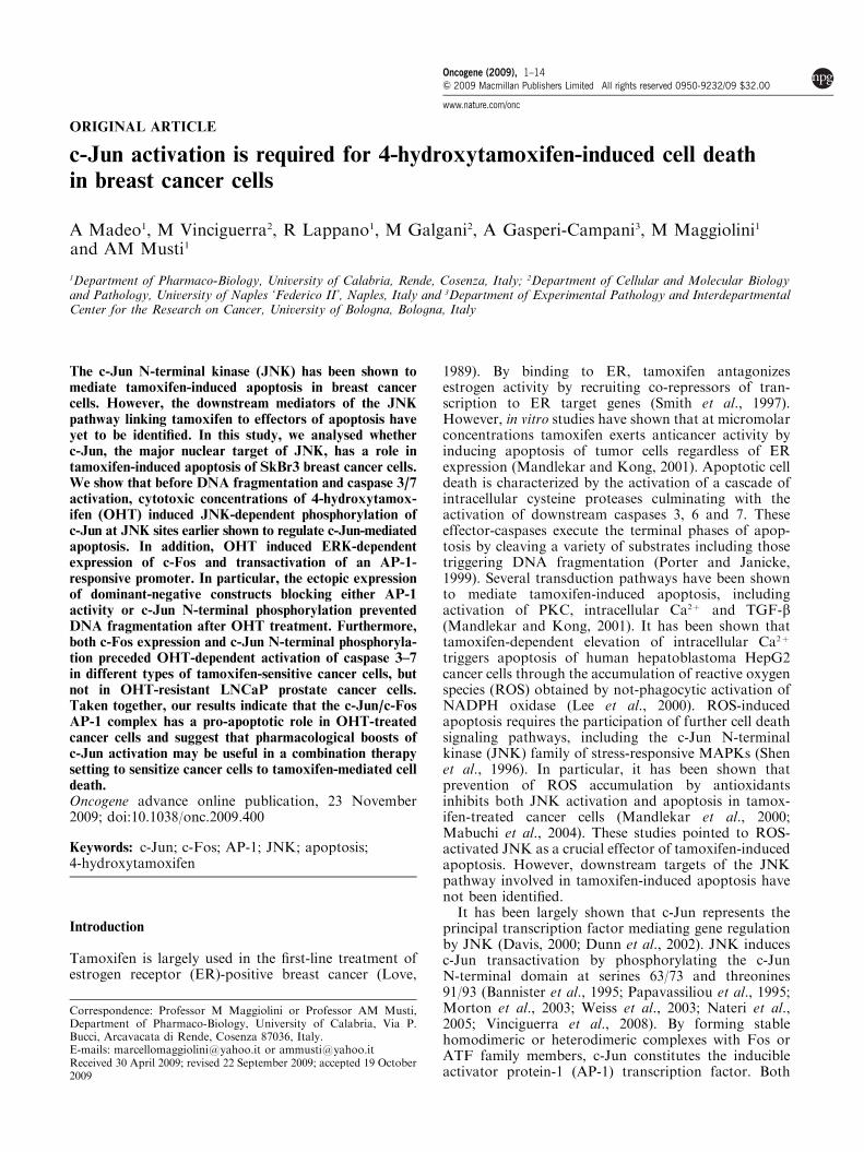
ORIGINAL ARTICLE
c-Jun activation is required for 4-hydroxytamoxifen-induced cell death
in breast cancer cells
A Madeo1, M Vinciguerra2, R Lappano1, M Galgani2, A Gasperi-Campani3, M Maggiolini1
and AM Musti1
1Department of Pharmaco-Biology, University of Calabria, Rende, Cosenza, Italy; 2Department of Cellular and Molecular Biologyand Pathology, University of Naples ‘Federico II’, Naples, Italy and 3Department of Experimental Pathology and InterdepartmentalCenter for the Research on Cancer, University of Bologna, Bologna, Italy
The c-Jun N-terminal kinase (JNK) has been shown tomediate tamoxifen-induced apoptosis in breast cancercells. However, the downstream mediators of the JNKpathway linking tamoxifen to effectors of apoptosis haveyet to be identified. In this study, we analysed whetherc-Jun, the major nuclear target of JNK, has a role intamoxifen-induced apoptosis of SkBr3 breast cancer cells.We show that before DNA fragmentation and caspase 3/7activation, cytotoxic concentrations of 4-hydroxytamox-ifen (OHT) induced JNK-dependent phosphorylation ofc-Jun at JNK sites earlier shown to regulate c-Jun-mediatedapoptosis. In addition, OHT induced ERK-dependentexpression of c-Fos and transactivation of an AP-1-responsive promoter. In particular, the ectopic expressionof dominant-negative constructs blocking either AP-1activity or c-Jun N-terminal phosphorylation preventedDNA fragmentation after OHT treatment. Furthermore,both c-Fos expression and c-Jun N-terminal phosphoryla-tion preceded OHT-dependent activation of caspase 3–7in different types of tamoxifen-sensitive cancer cells, butnot in OHT-resistant LNCaP prostate cancer cells.Taken together, our results indicate that the c-Jun/c-FosAP-1 complex has a pro-apoptotic role in OHT-treatedcancer cells and suggest that pharmacological boosts ofc-Jun activation may be useful in a combination therapysetting to sensitize cancer cells to tamoxifen-mediated celldeath.Oncogene advance online publication, 23 November2009; doi:10.1038/onc.2009.400
Keywords: c-Jun; c-Fos; AP-1; JNK; apoptosis;4-hydroxytamoxifen
Introduction
Tamoxifen is largely used in the first-line treatment ofestrogen receptor (ER)-positive breast cancer (Love,
1989). By binding to ER, tamoxifen antagonizesestrogen activity by recruiting co-repressors of tran-scription to ER target genes (Smith et al., 1997).However, in vitro studies have shown that at micromolarconcentrations tamoxifen exerts anticancer activity byinducing apoptosis of tumor cells regardless of ERexpression (Mandlekar and Kong, 2001). Apoptotic celldeath is characterized by the activation of a cascade ofintracellular cysteine proteases culminating with theactivation of downstream caspases 3, 6 and 7. Theseeffector-caspases execute the terminal phases of apop-tosis by cleaving a variety of substrates including thosetriggering DNA fragmentation (Porter and Janicke,1999). Several transduction pathways have been shownto mediate tamoxifen-induced apoptosis, includingactivation of PKC, intracellular Ca2þ and TGF-b(Mandlekar and Kong, 2001). It has been shown thattamoxifen-dependent elevation of intracellular Ca2þ
triggers apoptosis of human hepatoblastoma HepG2cancer cells through the accumulation of reactive oxygenspecies (ROS) obtained by not-phagocytic activation ofNADPH oxidase (Lee et al., 2000). ROS-inducedapoptosis requires the participation of further cell deathsignaling pathways, including the c-Jun N-terminalkinase (JNK) family of stress-responsive MAPKs (Shenet al., 1996). In particular, it has been shown thatprevention of ROS accumulation by antioxidantsinhibits both JNK activation and apoptosis in tamox-ifen-treated cancer cells (Mandlekar et al., 2000;Mabuchi et al., 2004). These studies pointed to ROS-activated JNK as a crucial effector of tamoxifen-inducedapoptosis. However, downstream targets of the JNKpathway involved in tamoxifen-induced apoptosis havenot been identified.
It has been largely shown that c-Jun represents theprincipal transcription factor mediating gene regulationby JNK (Davis, 2000; Dunn et al., 2002). JNK inducesc-Jun transactivation by phosphorylating the c-JunN-terminal domain at serines 63/73 and threonines91/93 (Bannister et al., 1995; Papavassiliou et al., 1995;Morton et al., 2003; Weiss et al., 2003; Nateri et al.,2005; Vinciguerra et al., 2008). By forming stablehomodimeric or heterodimeric complexes with Fos orATF family members, c-Jun constitutes the inducibleactivator protein-1 (AP-1) transcription factor. Both
Received 30 April 2009; revised 22 September 2009; accepted 19 October2009
Correspondence: Professor M Maggiolini or Professor AM Musti,Department of Pharmaco-Biology, University of Calabria, Via P.Bucci, Arcavacata di Rende, Cosenza 87036, Italy.E-mails: [email protected] or [email protected]
Oncogene (2009), 1–14& 2009 Macmillan Publishers Limited All rights reserved 0950-9232/09 $32.00
www.nature.com/onc
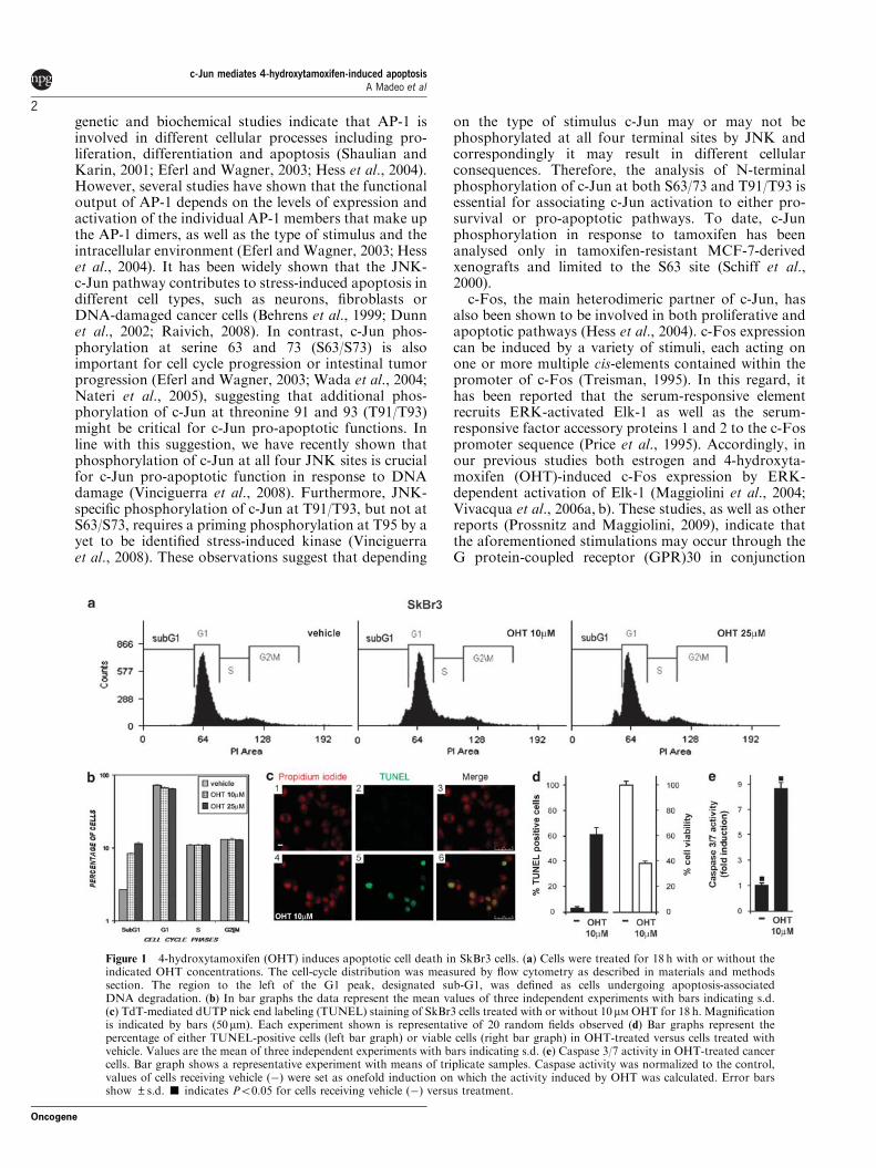
genetic and biochemical studies indicate that AP-1 isinvolved in different cellular processes including pro-liferation, differentiation and apoptosis (Shaulian andKarin, 2001; Eferl and Wagner, 2003; Hess et al., 2004).However, several studies have shown that the functionaloutput of AP-1 depends on the levels of expression andactivation of the individual AP-1 members that make upthe AP-1 dimers, as well as the type of stimulus and theintracellular environment (Eferl and Wagner, 2003; Hesset al., 2004). It has been widely shown that the JNK-c-Jun pathway contributes to stress-induced apoptosis indifferent cell types, such as neurons, fibroblasts orDNA-damaged cancer cells (Behrens et al., 1999; Dunnet al., 2002; Raivich, 2008). In contrast, c-Jun phos-phorylation at serine 63 and 73 (S63/S73) is alsoimportant for cell cycle progression or intestinal tumorprogression (Eferl and Wagner, 2003; Wada et al., 2004;Nateri et al., 2005), suggesting that additional phos-phorylation of c-Jun at threonine 91 and 93 (T91/T93)might be critical for c-Jun pro-apoptotic functions. Inline with this suggestion, we have recently shown thatphosphorylation of c-Jun at all four JNK sites is crucialfor c-Jun pro-apoptotic function in response to DNAdamage (Vinciguerra et al., 2008). Furthermore, JNK-specific phosphorylation of c-Jun at T91/T93, but not atS63/S73, requires a priming phosphorylation at T95 by ayet to be identified stress-induced kinase (Vinciguerraet al., 2008). These observations suggest that depending
on the type of stimulus c-Jun may or may not bephosphorylated at all four terminal sites by JNK andcorrespondingly it may result in different cellularconsequences. Therefore, the analysis of N-terminalphosphorylation of c-Jun at both S63/73 and T91/T93 isessential for associating c-Jun activation to either pro-survival or pro-apoptotic pathways. To date, c-Junphosphorylation in response to tamoxifen has beenanalysed only in tamoxifen-resistant MCF-7-derivedxenografts and limited to the S63 site (Schiff et al.,2000).
c-Fos, the main heterodimeric partner of c-Jun, hasalso been shown to be involved in both proliferative andapoptotic pathways (Hess et al., 2004). c-Fos expressioncan be induced by a variety of stimuli, each acting onone or more multiple cis-elements contained within thepromoter of c-Fos (Treisman, 1995). In this regard, ithas been reported that the serum-responsive elementrecruits ERK-activated Elk-1 as well as the serum-responsive factor accessory proteins 1 and 2 to the c-Fospromoter sequence (Price et al., 1995). Accordingly, inour previous studies both estrogen and 4-hydroxyta-moxifen (OHT)-induced c-Fos expression by ERK-dependent activation of Elk-1 (Maggiolini et al., 2004;Vivacqua et al., 2006a, b). These studies, as well as otherreports (Prossnitz and Maggiolini, 2009), indicate thatthe aforementioned stimulations may occur through theG protein-coupled receptor (GPR)30 in conjunction
Figure 1 4-hydroxytamoxifen (OHT) induces apoptotic cell death in SkBr3 cells. (a) Cells were treated for 18 h with or without theindicated OHT concentrations. The cell-cycle distribution was measured by flow cytometry as described in materials and methodssection. The region to the left of the G1 peak, designated sub-G1, was defined as cells undergoing apoptosis-associatedDNA degradation. (b) In bar graphs the data represent the mean values of three independent experiments with bars indicating s.d.(c) TdT-mediated dUTP nick end labeling (TUNEL) staining of SkBr3 cells treated with or without 10mM OHT for 18 h. Magnificationis indicated by bars (50mm). Each experiment shown is representative of 20 random fields observed (d) Bar graphs represent thepercentage of either TUNEL-positive cells (left bar graph) or viable cells (right bar graph) in OHT-treated versus cells treated withvehicle. Values are the mean of three independent experiments with bars indicating s.d. (e) Caspase 3/7 activity in OHT-treated cancercells. Bar graph shows a representative experiment with means of triplicate samples. Caspase activity was normalized to the control,values of cells receiving vehicle (�) were set as onefold induction on which the activity induced by OHT was calculated. Error barsshow ±s.d. ’ indicates Po0.05 for cells receiving vehicle (�) versus treatment.
c-Jun mediates 4-hydroxytamoxifen-induced apoptosisA Madeo et al
2
Oncogene
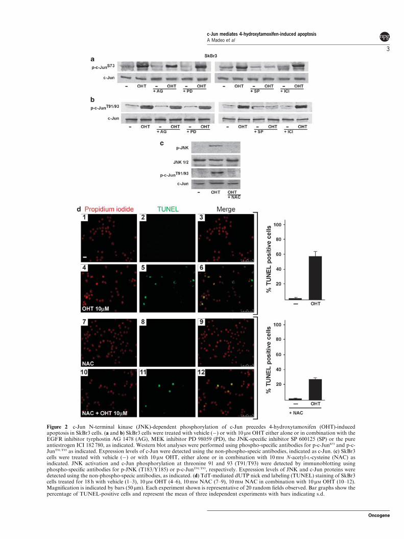
Figure 2 c-Jun N-terminal kinase (JNK)-dependent phosphorylation of c-Jun precedes 4-hydroxytamoxifen (OHT)-inducedapoptosis in SkBr3 cells. (a and b) SkBr3 cells were treated with vehicle (�) or with 10 mM OHT either alone or in combination with theEGFR inhibitor tyrphostin AG 1478 (AG), MEK inhibitor PD 98059 (PD), the JNK-specific inhibitor SP 600125 (SP) or the pureantiestrogen ICI 182 780, as indicated. Western blot analyses were performed using phospho-specific antibodies for p-c-JunS73 and p-c-JunT91/T93 as indicated. Expression levels of c-Jun were detected using the non-phospho-specic antibodies, indicated as c-Jun. (c) SkBr3cells were treated with vehicle (�) or with 10 mM OHT, either alone or in combination with 10mM N-acetyl-L-cysteine (NAC) asindicated. JNK activation and c-Jun phosphorylation at threonine 91 and 93 (T91/T93) were detected by immunoblotting usingphospho-specific antibodies for p-JNK (T183/Y185) or p-c-JunT91/T93, respectively. Expression levels of JNK and c-Jun proteins weredetected using the non-phospho-specic antibodies, as indicated. (d) TdT-mediated dUTP nick end labeling (TUNEL) staining of SkBr3cells treated for 18 h with vehicle (1–3), 10 mM OHT (4–6), 10mM NAC (7–9), 10mM NAC in combination with 10mM OHT (10–12).Magnification is indicated by bars (50mm). Each experiment shown is representative of 20 random fields observed. Bar graphs show thepercentage of TUNEL-positive cells and represent the mean of three independent experiments with bars indicating s.d.
c-Jun mediates 4-hydroxytamoxifen-induced apoptosisA Madeo et al
3
Oncogene
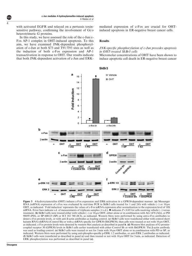
with activated EGFR and relayed on a pertussis toxin-sensitive pathway, confirming the involvement of Gi/oheterotrimeric G proteins.
In this study, we have assessed the role of the c-Jun/c-Fos AP-1 complex in OHT-induced apoptosis. To thisaim, we have examined JNK-dependent phosphoryl-ation of c-Jun at both S73 and T91/T93 sites as well asthe induction of both c-Fos expression and AP-1transactivation in response to OHT. Our results indicatethat both JNK-dependent activation of c-Jun and ERK-
mediated expression of c-Fos are crucial for OHT-induced apoptosis in ER-negative breast cancer cells.
Results
JNK-specific phosphorylation of c-Jun precedes apoptosisin OHT-treated SkBr3 cellsMicromolar concentrations of OHT have been shown toinduce apoptotic cell death in ER-negative breast cancer
Figure 3 4-hydroxytamoxifen (OHT) induces c-Fos expression and ERK-activation in a GPR30-dependent manner. (a) MessengerRNA (mRNA) expression of c-Fos was evaluated by real-time PCR in SkBr3 cells treated for 1 and 24 h with vehicle (�) or 10 mMOHT, as indicated. ‘Fold induction’ represents the values of c-Fos mRNA expression after normalization to the expression level of 18SmRNA. Error bars indicate s.d. of measurements of triplicate samples (±s.d.). � indicates Po0.05 for cells receiving vehicle (�) versustreatment. (b) SkBr3 cells were treated either with vehicle (�) or 10mM OHT, either alone or in combination with AG 1478 (AG), or PD98059 (PD), or SP 600125 (SP) or ICI 182 780 (ICI), as indicated. Western blots were performed by using anti-c-Fos antibodies todetect c-Fos protein levels, or with anti b-actin antibodies as loading control. (c) SkBr3 cells were transfected either with control shorthairpin RNA (shRNA) (Control Sh) or with a shRNA specific for GPR30 (ShGPR30), then cells were treated or not with 10 mM OHT,as indicated. c-Fos protein levels were detected by western blot analysis as described in panel b. (d) Western blot analyses of G protein-coupled receptor 30 (GPR30) levels in SkBr3 cells earlier transfected with either Control Sh or with ShGPR30. The b-actin antibodywas used as loading control. (e) SkBr3 cells were treated or not for 5min with 10mM OHT alone or in combination with PD or SP, asindicated. Western blots were performed by using anti-phospho-specific p-ERK 1/2 antibodies, or anti-ERK 2 antibodies as indicated.(f) SkBr3 cells were transfected as described in panel (c) and then treated or not with 10mM OHT for 5min, as indicated. Detection ofERK phosphorylation was performed as described in panel (e).
c-Jun mediates 4-hydroxytamoxifen-induced apoptosisA Madeo et al
4
Oncogene
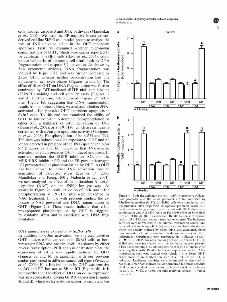
cells through caspase 3 and JNK pathways (Mandlekaret al., 2000). We used the ER-negative breast cancer-derived cell line SkBr3 as a model system to analyse therole of JNK-activated c-Jun in the OHT-dependentapoptosis. First, we examined whether micromolarconcentrations of OHT, which were earlier reported tobe cytotoxic in SkBr3 cells (Basu et al., 2004), couldinduce hallmarks of apoptotic cell death such as DNAfragmentation and caspase 3/7 activation. As shown byflow cytometric analysis, DNA fragmentation wasinduced by 10 mM OHT and was further increased by25 mM OHT, whereas neither concentration had anyinfluence on cell cycle phases (Figures 1a and b). Theeffect of 10 mM OHT on DNA fragmentation was furtherconfirmed by TdT-mediated dUTP nick end labeling(TUNEL) staining and cell viability assay (Figures 1cand d). Furthermore, OHT-induced caspase 3/7 activ-ities (Figure 1e), suggesting that DNA fragmentationresults from apoptosis. Next, we analysed whether JNK-activated c-Jun precedes OHT-dependent apoptosis inSkBr3 cells. To this end, we examined the ability ofOHT to induce c-Jun N-terminal phosphorylation ateither S73, a hallmark of c-Jun activation by JNK(Dunn et al., 2002), or at T91/T93, which are stringentlycorrelated with c-Jun pro-apoptotic activity (Vinciguer-ra et al., 2008). Phosphorylation of both S73 and T91/T93 sites was induced on a 2 h exposure to OHT and nolonger detected in presence of the JNK-specific inhibitorSP (Figures 2a and b), indicating that JNK-specificactivation of c-Jun precedes OHT-induced apoptosis. Incontrast, neither the EGFR inhibitor AG, nor theMEK/ERK inhibitor PD and the ER pure antiestrogenICI prevented c-Jun phosphorylation by OHT. As OHThas been shown to induce JNK activation throughgeneration of oxidative stress (Lee et al., 2000;Mandlekar and Kong, 2001; Mabuchi et al., 2004),we next analysed the effect of the antioxidant N-acetyl-L-cysteine (NAC) on the JNK/c-Jun pathway. Asshown in Figure 2c, both activation of JNK and c-Junphosphorylation at T91/T93 sites were prevented byNAC treatment. In line with previous studies, the ex-posure to NAC prevented also DNA fragmentation byOHT (Figure 2d). These results indicate that c-Junpro-apoptotic phosphorylation by OHT is triggeredby oxidative stress and is associated with DNA frag-mentation.
OHT induces c-Fos expression in SkBr3 cellsIn addition to c-Jun activation, we analysed whetherOHT induces c-Fos expression in SkBr3 cells, at bothmessenger RNA and protein levels. As shown by eitherreverse transcription–PCR analysis or western blots, theexpression of c-Fos was rapidly induced by OHT(Figures 3a and b). In agreement with our previousstudies performed in different cancer cell types (Vivacquaet al., 2006a, b), c-Fos induction by OHT was sensitiveto AG and PD but not to SP or ICI (Figure 3b). It isnoteworthy that the effect of OHT on c-Fos expressionwas also abrogated silencing GPR30 expression (Figures3c and d), which we have shown earlier to mediate c-Fos
Figure 4 Both the activator protein-1 (AP-1)-responsive collage-nase promoter and the c-Fos promoter are transactivated by4-hydroxytamoxifen (OHT). (a) SkBr3 cells were transfected withthe proximal AP-1-responsive collagenase promoter fused to aluciferase reporter gene and treated or not with OHT alone or incombination with AG 1478 (AG), or PD 98059 (PD), or SP 600125(SP) or ICI 182 780 (ICI), as indicated. Renilla luciferase expressionvector (pRL-TK) was used as a transfection control. The luciferaseactivities were normalized to the internal transfection control andvalues of cells receiving vehicle (�) were set as onefold induction onwhich the activity induced by 10 mM OHT was calculated. Errorbars indicate s.d. of normalized luciferase activities of threeindependent experiments each performed in triplicates (±s.d.).J, �, &, Po0.05, for cells receiving vehicle (�) versus OHT. (b)SkBr3 cells were transfected with the luciferase reporter plasmidc-Fos-luc containing a 2.2-kb long upstream region of human c-fosgene together with Renilla luciferase expression vector. Aftertransfection, cells were treated with vehicle (�) or 10 mM OHT,either alone or in combination with AG, PD, SP or ICI, asindicated. Luciferase activities were determined as described inpanel (a). Error bars indicate s.d. of normalized luciferase activitiesof three independent experiments each performed in triplicates(±s.d.). J, �, &, Po0.05, for cells receiving vehicle (�) versustreatment.
c-Jun mediates 4-hydroxytamoxifen-induced apoptosisA Madeo et al
5
Oncogene
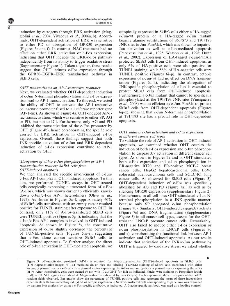
induction by estrogens through ERK activation (Mag-giolini et al., 2004; Vivacqua et al., 2006a, b). Accord-ingly, OHT-dependent activation of ERK was sensitiveto either PD or abrogation of GPR30 expression(Figures 3e and f). In contrast, NAC treatment had noeffect on either ERK activation or c-Fos expression,indicating that OHT induces the ERK/c-Fos pathwayindependently from its ability to trigger oxidative stress(Supplementary Figure 1). Taken together, these resultssuggest that OHT induces c-Fos expression throughthe GPR30–EGFR–ERK transduction pathway inSkBr3 cells.
OHT transactivates an AP-1-responsive promoterNext, we evaluated whether OHT-dependent inductionof c-Jun N-terminal phosphorylation and c-Fos expres-sion lead to AP-1 transactivation. To this end, we testedthe ability of OHT to activate the AP-1-responsivecollagenase promoter fused to a luciferase reporter gene(AP-1-luc). As shown in Figure 4a, OHT-induced AP-1-luc transactivation, which was sensitive to either SP, AGor PD, but not to ICI. Furthermore, only AG and PDinhibited the transactivation of the c-Fos promoter byOHT (Figure 4b), hence corroborating the specific roleexerted by ERK activation in OHT-induced c-Fosexpression. Overall, these findings suggest that bothJNK-specific activation of c-Jun and ERK-dependentinduction of c-Fos expression contribute to AP-1activation by OHT.
Abrogation of either c-Jun phosphorylation or AP-1transactivation protects SkBr3 cells fromOHT-induced apoptosisWe then analysed the specific involvement of c-Jun/c-Fos AP-1 complex in OHT-induced apoptosis. To thisaim, we performed TUNEL assays in OHT-treatedcells ectopically expressing a truncated form of c-Fos(A-Fos), which was shown earlier to efficiently knock-down c-Jun/c-Fos AP-1 heterodimers (Olive et al.,1997). As shown in Figures 5a–f, approximately 60%of SkBr3 cells transfected with an empty vector resultedpositive for TUNEL staining after exposure to OHT. Incontrast, only 11% of A-Fos-transfected SkBr3 cellswere TUNEL positive (Figures 5g–l), indicating that thec-Jun/c-Fos AP-1 complex is involved in OHT-inducedapoptosis. As shown in Figure 5s, the constitutiveexpression of c-Fos slightly decreased the percentageof TUNEL-positive cells (Figures 5m–r), suggestingthat c-Fos alone cannot sensitize SkBr3 cells toOHT-induced apoptosis. To further analyse the directrole of c-Jun activation in OHT-mediated apoptosis, we
ectopically expressed in SkBr3 cells either a HA-taggedc-Jun-wt protein or a HA-tagged c-Jun mutantbearing alanine substitutions of S63/S73 and T91/T93JNK sites (c-Jun-PanAla), which was shown to impair c-Jun activation as well as c-Jun-mediated apoptosis(Papavassiliou et al., 1995; Watson et al., 1998; Dunnet al., 2002). Expression of HA-tagged c-Jun-PanAlaprotected SkBr3 cells from OHT-induced apoptosis, asonly 6% of HA-positive cells were also positive forTUNEL staining, while 58% of HA-negative cells wereTUNEL positive (Figures 6i–p). In contrast, ectopicexpression of c-Jun-wt had no effect on DNA fragmen-tation (Figures 6a–h), indicating the abrogation ofJNK-specific phosphorylation of c-Jun is essential toprotect SkBr3 cells from OHT-induced apoptosis.Furthermore, a c-Jun mutant that cannot be specificallyphosphorylated at the T91/T93 JNK sites (Vinciguerraet al., 2008) was as efficient as c-Jun-PanAla to protectSkBr3 cells from OHT-dependent apoptosis (Figures6q–x), showing that c-Jun N-terminal phosphorylationat T91/T93 site has a pivotal role in OHT-dependentapoptosis.
OHT induces c-Jun activation and c-Fos expressionin different cancer cell typesTo validate the role of AP-1 activation in OHT-inducedapoptosis, we examined whether OHT couples theinduction of both c-Fos expression and c-Jun phosphor-ylation to caspase 3/7 activation in different cancer celltypes. As shown in Figures 7a and b, OHT stimulatedboth c-Fos expression and c-Jun phosphorylation inER-negative BT20 and ER-positive MCF-7 breastcancer cells, HepG2 hepatocarcinoma cells, LoVocolorectal adenocarcinoma cells and SCLC-R1 lungcancer cells. As observed for SkBr3 cells (Figure 3),OHT-dependent induction of c-Fos was specificallyabolished by AG and PD (Figure 7a), as well as bysilencing GPR30 expression (Supplementary Figure 2).Furthermore, in all cell lines OHT-stimulated c-Jun N-terminal phosphorylation in a JNK-specific manner,because only SP abrogated c-Jun phosphorylation(Figure 7b). Similarly, OHT-induced caspase3/7 activity(Figure 7c) and DNA fragmentation (SupplementaryFigure 3) in all cancer cell types, except for the OHT-resistant LNCaP prostate cancer cells. Remarkably,OHT also failed to induce either c-Fos expression orc-Jun phosphorylation in LNCaP cells (Figures 7dand e), corroborating the functional link between AP-1activation and OHT-induced apoptosis. As our resultsindicate that activation of the JNK/c-Jun pathway byOHT is triggered by oxidative stress, we asked whether
Figure 5 c-Fos/activator protein-1 (AP-1) is required for 4-hydroxytamoxifen (OHT)-induced apoptosis in SkBr3 cells.(a–r) Representative images of TdT-mediated dUTP nick end labeling (TUNEL) staining of SkBr3 cells transfected with eitheran empty plasmid vector (a–f) or with a plasmid vector expressing the A-Fos mutant (g–l), or a plasmid vector expressing the c-Fos(m–r). After transfection, cells were treated or not with 10 mM OHT for 18 h as indicated. Nuclei were staining by Propidium iodide(red), or TUNEL (green) as indicated. Magnification is indicated by bars (50 mm). Each experiment shown is representative of 20random fields observed. Bar graphs show the percentage of TUNEL-positive cells and represent the mean of three independentexperiments with bars indicating s.d. (s) c-Fos ectopic expression in SkBr3-transfected cells corresponding to panel m-r was examinedby western blot analysis by using a c-Fos-specific antibody, as indicated. A b-actin-specific antibody was used as a loading control.
c-Jun mediates 4-hydroxytamoxifen-induced apoptosisA Madeo et al
6
Oncogene
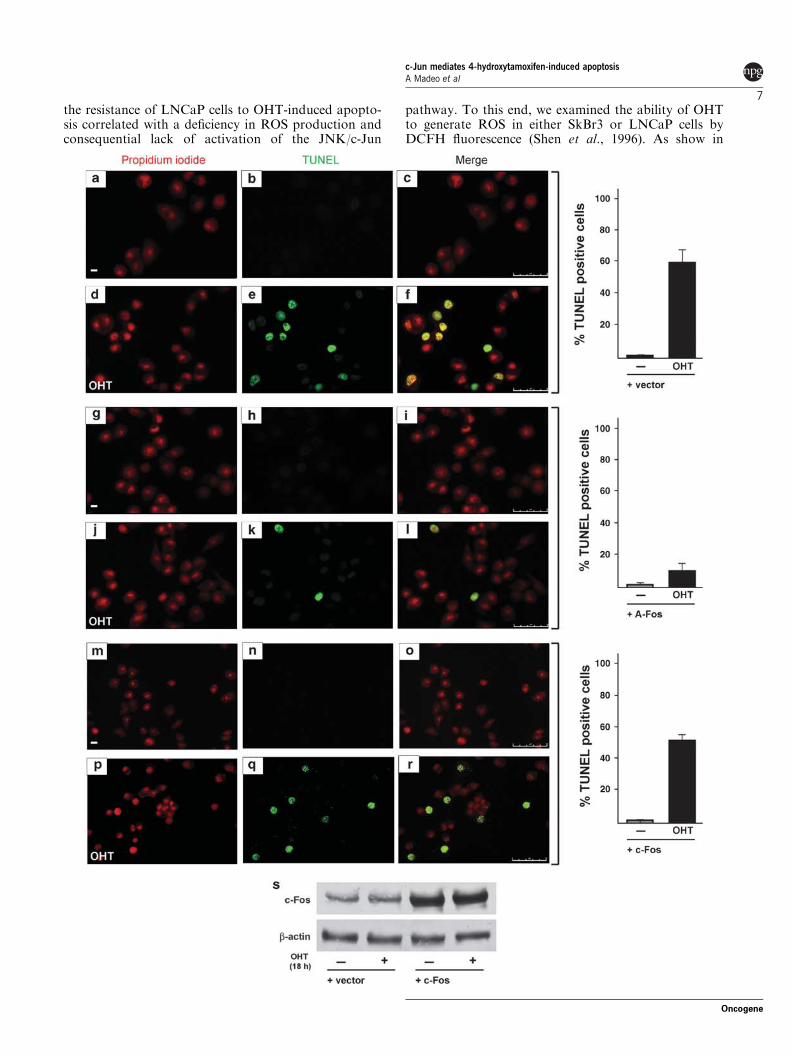
the resistance of LNCaP cells to OHT-induced apopto-sis correlated with a deficiency in ROS production andconsequential lack of activation of the JNK/c-Jun
pathway. To this end, we examined the ability of OHTto generate ROS in either SkBr3 or LNCaP cells byDCFH fluorescence (Shen et al., 1996). As show in
c-Jun mediates 4-hydroxytamoxifen-induced apoptosisA Madeo et al
7
Oncogene
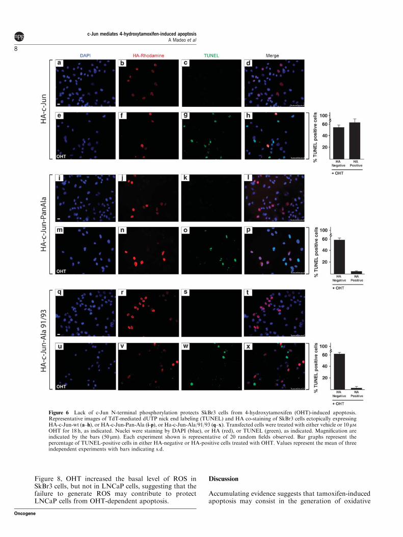
Figure 8, OHT increased the basal level of ROS inSkBr3 cells, but not in LNCaP cells, suggesting that thefailure to generate ROS may contribute to protectLNCaP cells from OHT-dependent apoptosis.
Discussion
Accumulating evidence suggests that tamoxifen-inducedapoptosis may consist in the generation of oxidative
Figure 6 Lack of c-Jun N-terminal phosphorylation protects SkBr3 cells from 4-hydroxytamoxifen (OHT)-induced apoptosis.Representative images of TdT-mediated dUTP nick end labeling (TUNEL) and HA co-staining of SkBr3 cells ectopically expressingHA-c-Jun-wt (a–h), or HA-c-Jun-Pan-Ala (i-p), or Ha-c-Jun-Ala/91/93 (q–x). Transfected cells were treated with either vehicle or 10 mMOHT for 18 h, as indicated. Nuclei were staining by DAPI (blue), or HA (red), or TUNEL (green), as indicated. Magnification areindicated by the bars (50 mm). Each experiment shown is representative of 20 random fields observed. Bar graphs represent thepercentage of TUNEL-positive cells in either HA-negative or HA-positive cells treated with OHT. Values represent the mean of threeindependent experiments with bars indicating s.d.
c-Jun mediates 4-hydroxytamoxifen-induced apoptosisA Madeo et al
8
Oncogene
stress and subsequent activation of the JNK family ofstress-responsive MAPKs (Lee et al., 2000; Mandlekarand Kong, 2001; Mabuchi et al., 2004). c-Jun mediatesproapoptotic functions of JNK in different types of cellsincluding neurons, fibroblasts and cancer cells (Dunnet al., 2002; Hess et al., 2004; Raivich, 2008). However,c-Jun does not mediate all paradigms of JNK-dependentapoptosis and its pro-apoptotic function depends on thetype of stimulus as well as on the heterodimeric partnerforming the AP-1 transcription factor (Hess et al., 2004;Raivich and Behrens, 2006). In this study, we analysedwhether JNK-dependent activation of c-Jun has a rolein OHT-induced cell death of ER-negative SkBr3 breastcancer cells. To this end, we first established byfluorescence-activated cell sorting analyses the range ofmicromolar OHT concentrations required to induceDNA fragmentation (Figure 1), as also confirmed byTUNEL and cell viability assays (Figure 1). Impor-tantly, we found that OHT triggers caspase 3/7 activity(Figure 1), suggesting that DNA fragmentation byOHT is a secondary consequence of apoptosis ratherthan necrosis. Moreover, OHT had no effects oncell cycle progression (Figure 1), excluding the possibi-lity that OHT-dependent cytotoxicity was due tocell growth arrest.
Next, we analysed the ability of OHT to induce JNK-dependent phosphorylation of c-Jun at the S73 site,which is considered a hallmark of c-Jun activation inseveral paradigms of stress signals (Dunn et al., 2002).We detected both JNK activation (Figure 2) and c-Junphosphorylation at S73 on a 2 h exposure to OHT(Figure 2). Remarkably, OHT also induced phosphor-ylation of c-Jun at T91/T93 sites (Figure 2), which wasshown to be crucial for c-Jun-mediated cell death inresponse to either ultraviolet or etoposide (Vinciguerraet al., 2008). Moreover, both S73 and T91/T93 phos-phorylations by OHT were specifically sensitive to SP,indicating that JNK-specific activation of c-Jun precedesapoptosis in SkBr3 cells. Interestingly, OHT-inducedc-Jun N-terminal phosphorylation without alteration ofc-Jun expression (Figures 2 and 7), suggesting thatactivation of c-Jun by OHT is limited to pre-existingc-Jun proteins. Accordingly, our (unpublished observa-tions) and other studies (Nephew et al., 1993), haveindicate that c-Jun expression is not regulated by OHTtreatment. Importantly, DNA degradation as well asJNK activation and N-terminal phosphorylation of c-Jun,were abrogated by antioxidant treatments (Figure 2).These results suggest that generation of oxidative stress,presumably by NADPH activation (Lee et al., 2000),could be the initial OHT-dependent signal leading toactivation of the JNK/c-Jun pathway and thereafter toapoptosis.
We then asked whether c-Jun activation by OHT wascoupled to induction of c-Fos expression. Transactiva-tion assays of the c-Fos promoter (Figure 4) combinedwith the analysis of steady-state levels of both c-Fosprotein and messenger RNA (Figure 3) indicated thatOHT leads to transcriptional induction of c-Fosexpression. Interestingly, both the transactivation ofthe c-Fos promoter and the induction of c-Fos protein
levels were sensitive to AG or PD, but not to SP or ICI,indicating that OHT induces c-Fos expression indepen-dently of its ability to induce JNK activity but rather inan EGFR/ERK-mediated manner. Accordingly, OHT-dependent activation of ERK was abrogated by PD butnot by SP (Figure 3), confirming that JNK activity is notrequired for the signaling pathway upstream c-Fosexpression. Moreover, both c-Fos expression (Figure 3)and ERK activation (Figure 3) by OHT were sensitive toGPR30 silencing, in accordance with our previousstudies performed in different cancer cells (Maggioliniet al., 2004; Vivacqua et al., 2006a, b; Albanito et al.,2007). It is noteworthy that the ERK/c-Fos pathwaywas not altered by antioxidant treatment (Supplemen-tary Figure 1), thus corroborating the fact that OHTinitiates two distinct signaling pathways converging toactivation of the c-Jun/c-Fos AP-1 complex.
Induction of c-Fos expression coupled to c-JunN-terminal phosphorylation is the most frequentencountered paradigm of AP-1 activation in mammaliancells. Indeed, we found that OHT-induced AP-1 transac-tivation in an ERK and JNK-dependent manner(Figure 4), suggesting that the heterodimeric c-Jun/c-Fos AP-1 complex functions in OHT-induced apoptosisof cancer cells. Alternatively, because of the crucial roleof AP-1 in a variety of cellular events, activation ofc-Jun/c-Fos AP-1 complex by OHT may reflect a pro-survival response. To evaluate the specific involvement ofc-Jun/c-Fos in OHT-dependent apoptosis, we abrogatedc-Jun/c-Fos AP-1 activity in SkBr3 cells by twoindependent experimental approaches: (i) ectopic expres-sion of A-Fos, a truncated form of c-Fos that has beenshown to specifically knock-down c-Jun/c-Fos AP-1 hete-rodimers (Olive et al., 1997); and (ii) ectopic expressionof c-Jun-PanAla, a c-Jun mutant bearing alaninesubstitution of all four JNK phosphorylation sitesS63/S73 and T91/T93, which has been shown to act asa c-Jun dominant negative in several cell lines (Papa-vassiliou et al., 1995; Watson et al., 1998; Dunn et al.,2002). A-Fos overexpression protected cells from OHT-induced apoptosis, indicating that c-Jun/c-Fos hetero-dimers are directly involved in apoptosis. This findingwas further corroborated by the evidence that AP-1transactivation by OHT was dependent on both ERKand JNK activity (Figure 4). Similarly, overexpressionof c-Jun-PanAla resulted in the protection of SkBr3 cellsfrom OHT-induced apoptosis (Figure 6). As Jun-wt hadno effect on DNA degradation (Figure 6), the protectiveeffect of c-Jun-PanAla presumably results from itsability to act as dominant-negative, indicating thatJNK-activated c-Jun is necessary to trigger OHT-induced apoptosis. Moreover, a c-Jun mutant thatspecifically cannot be phosphorylated at the T91/T93JNK sites was as efficient as c-Jun-PanAla in protectingSkBr3 cells from OHT-dependent apoptosis, suggestingthat phosphorylation of c-Jun at S63/S73 is notsufficient to trigger c-Jun-dependent apoptosis. It isnoteworthy that ectopic expression of c-Jun could notsignificantly increase the number of TUNEL-positivecells (Figure 6), indicating that activation of pre-existingc-Jun proteins by OHT is sufficient to trigger apoptosis.
c-Jun mediates 4-hydroxytamoxifen-induced apoptosisA Madeo et al
9
Oncogene
Our data show that OHT induces both ERK/c-Fosand JNK/c-Jun pathways in various OHT-sensitivecancer cells (Figure 7), pointing to c-Jun/c-Fos AP-1activation as a general mechanism mediating OHT-dependent apoptosis, rather than a peculiar response ofSkBr3 cells. Accordingly, OHT-induced caspase 3/7activity and DNA degradation in all cancer cell typesused, except in OHT-resistant LNCaP prostate cancercells (Figure 7). It is noteworthy that OHT failed toinduce both c-Fos expression and c-Jun N-terminalphosphorylation in LNCaP cells (Figure 7), validatingthat the upregulation of c-Jun/c-Fos AP-1 by cytotoxicconcentrations of OHT is linked to caspase 3/7activation and in turn to cell death.
How c-Jun/AP-1 drives cell death in OHT-treatedcancer cells remains to be elucidated. The JNK/c-Junpathway has been shown to regulate the release ofcytochrome c from mitochondria (Whitfield et al., 2001).In line with this observation, active c-Jun is required forthe expression of genes that increase mitochondrialouter membrane permeability, such as Bim and Dp5,which are members of the proapoptotic BH3-onlysubfamily of Bcl-2 proteins (Ham et al., 2005). A crucialrole of mitochondria has been also shown in OHT-induced apoptosis, as it can elicit the release ofmitochondrial cytochrome c altering the expression ofBcl-2 proteins (Mandlekar and Kong, 2001; Kallioet al., 2005; Lagadec et al., 2008). Therefore, down-regulation of the antiapoptotic Bcl-2 protein togetherwith upregulation of one or more proapototic Bcl-2proteins may constitute a critical mechanism promotingmitochondrial dysfunction by OHT. Presumably, theJNK/c-Jun pathway may contribute to this intrinsicproapoptotic mechanism by upregulating one or moreproapototic BH3-only proteins. In contrast, as c-Jun/AP-1 controls tumor necrosis factor-a expression(Liu et al., 2000; Pope et al., 2000), we cannot excludea putative role of c-Jun in a cross talk between the deathreceptor and mitochondrial pathways.
Our previous studies have shown that one micromolarOHT induces cell proliferation in thyroid and endome-trial cancer cells by activating the EGFR/GPR30/ERKpathway and in turn c-Fos expression (Vivacqua et al.,2006a, 2006b), whereas it has no effects on the JNK/c-Jun pathway (Madeo A, unpublished observations).These observations together with this study lead to thehypothesis that the functional output of c-Fos induction
by OHT may depend on whether or not there is aconcomitant c-Jun activation by JNK signaling. Asantioxidant pathways are often enhanced in cancer cells(Friesen et al., 2004; Young et al., 2004; Recktenwaldet al., 2008; Ballatori et al., 2009), the resistance ofcertain cancer cells to tamoxifen may depend on theirability to prevent ROS accumulation and in turn JNK-specific activation of c-Jun. In line with this hypothesiswe found that OHT induced ROS accumulation inSkBr3 cells but not in LNCaP cells (Figure 8). Hence,pharmacological boosts of c-Jun N-terminal phosphor-ylation may be useful to sensitize cancer cells toOHT-mediated cell death.
Materials and methods
Cell cultureSkBr3 breast cancer cells were maintained in RPMI 1640without phenol red supplemented with 10% fetal bovine serum(FBS). MCF-7 and BT20 breast cancer cells were culturedrespectively in DMEM-F12 or MEM with phenol redsupplemented with 10% FBS. HePG2 hepatocarcinoma cells,LoVo colorectal adenocarcinoma cells and LNCaP prostatecancer cells were maintained in RPMI 1640 with phenol redand supplemented with 10% FBS. SCLC-R1 lung cancercells were cultured in RPMI 1640 with phenol red, one� non-essential amino acids, one� sodium pyruvate and 10%FBS. Cells were switched to medium without serum the daybefore experiments for immunoblots and reverse transcrip-tion–PCR.
Drugs and chemicals4-hydroxytamoxifen, PD 98059 (PD), SP 600125 (SP), NACwere purchased from Sigma-Aldrich Corporation (Milan,Italy). ICI 182 780 (ICI) was obtained from Tocris Chemicals(Bristol, UK), AG 1478 (AG) was purchased from BiomolResearch Laboratories, Inc. (Plymouth Meeting, PA, USA).All compounds were solubilized in dimethyl sulfoxide(DMSO), except for OHT that was dissolved in ethanol(EtOH) and N-acetyl-L-cysteine (NAC) that was dissolved inH2O. For TUNEL assays and immunofluorescent detectionexperiments propidium iodide and DAPI were purchased fromSigma-Aldrich Corporation.
PlasmidsThe short hairpin RNA construct to knock-down theexpression of GPR30 and the unrelated short hairpin RNAcontrol construct have been described earlier (Albanito et al.,
Figure 7 Inducton of c-Jun phosphorylation and c-Fos expression by 4-hydroxytamoxifen (OHT) is associated with caspase 3/7activity in different cancer cell types. (a) BT-20, HepG2, LoVo, SCLC-R1 and MCF-7 cells were treated with vehicle (�) or 10 mMOHT for 2 h, either alone or in combination with AG 1478 (AG), or PD 98059 (PD), or SP 600125 (SP) or ICI 182 780 (ICI), asindicated. Western blots were performed by using anti-c-Fos antibodies to detect c-Fos protein levels, or with anti b-actin antibodiesas loading control. (b) c-Jun phosphorylation (p-c-JunS73) in the various cell lines and treatments described in panel (a). Western blotanalyses were performed as described in Figure 2a. (c) Caspase 3/7 activity in OHT-treated cancer cell lines. Bar graph shows arepresentative experiment with means of triplicate samples. Caspase activity was normalized to the control and values of cellsreceiving vehicle (�) were set as onefold induction on which the activity induced by OHT was calculated. Error bars show ±s.d. J,�, &, ’, ., Po0.05, for cells receiving vehicle (�) versus OHT. (d) LNCaP cells were treated either with vehicle or 10 mM OHT for2 h, then c-Fos expression was detected by western blot by using anti-c-Fos antibodies, or b-actin antibodies as a loading control.(e) LNCaP cells were treated with vehicle or 10mM OHT for 2 h, c-Jun N-terminal kinase (JNK) activation and c-Jun phosphorylationat threonine 91 and 93 (T91/T93) and serine 73 (S73) were detected by western blot analysis using phospho-specific antibodies forp-JNK (T183/Y185) or for p-c-JunT91/T93, or p-c-JunS73, respectively. Expression levels of JNK and c-Jun proteins were detected usingthe non-phospho-specic antibodies, as indicated.
c-Jun mediates 4-hydroxytamoxifen-induced apoptosisA Madeo et al
10
Oncogene
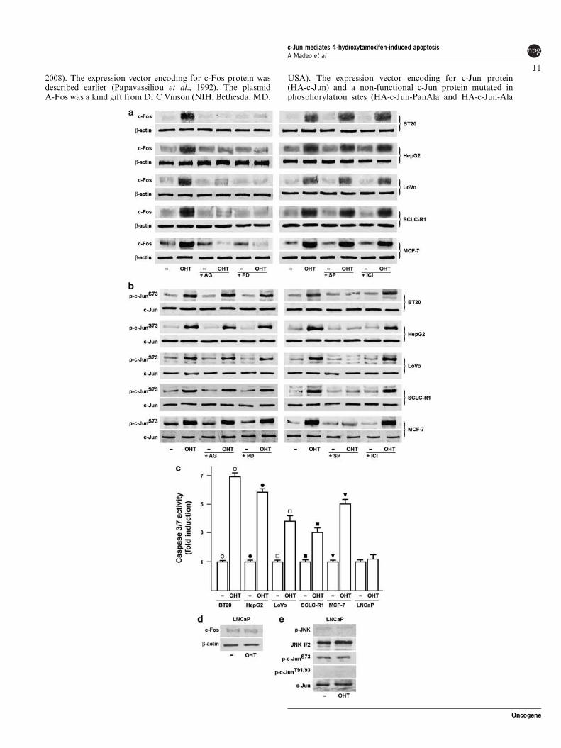
2008). The expression vector encoding for c-Fos protein wasdescribed earlier (Papavassiliou et al., 1992). The plasmidA-Fos was a kind gift from Dr C Vinson (NIH, Bethesda, MD,
USA). The expression vector encoding for c-Jun protein(HA-c-Jun) and a non-functional c-Jun protein mutated inphosphorylation sites (HA-c-Jun-PanAla and HA-c-Jun-Ala
c-Jun mediates 4-hydroxytamoxifen-induced apoptosisA Madeo et al
11
Oncogene
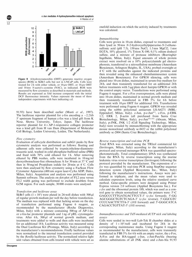
91/93) have been described earlier (Musti et al., 1997).The luciferase reporter plasmid for c-fos encoding a �2.2 kb50 upstream fragment of human c-fos was a kind gift from KNose, Showa University, Tokyo, Japan. The luciferasereporter plasmid for 4�AP-1-responsive collagen promoterwas a kind gift from H van Dam (Department of MolecularCell Biology, Leiden University, Leiden, The Netherlands).
Flow cytometryEvaluation of cell-cycle distribution and subG1 peaks by flowcytometric analysis was performed as follows: floating andadherent cells were collected by trypsin/ethylene-diaminete-traacetic acid, washed in cold phosphate-buffered saline (PBS)and fixed with 70% cold ethanol for 1 h. After removingethanol by PBS washes, cells were incubated in 10mg/mldeoxyribonuclease-free ribonuclease A for 30min at 37 1C andthen in 50mg/ml Propidium iodide for 20min at 4 1C. Cellswere then analysed by flow cytometry using a FacScan FlowCytometer Apparatus (488 nm argon laser-CyAn ADP, Dako,Milan, Italy). Acquisition and analysis was performed usingSummit software. The analysis on dot-plot of FL2 area versusFL2 width gating was performed to exclude doublets fromG2M region. For each sample, 30 000 events were analysed.
Transfection and luciferase assaySkBr3 cells (1� 105) were plated in 24-well dishes with 500 mlof regular growth medium per well the day before transfection.The medium was replaced with that lacking serum on the dayof transfection performed using Fugene 6 reagent, asrecommended by the manufacturer (Roche Diagnostics,Milan, Italy) with a mixture containing 500 ng of AP-1-lucor c-fos-luc promoter plasmids and 1 ng of pRL–cytomegalo-virus. After 6 h, 500 ml of normal growth medium, andtreatments were added to wells and then cells were incubatedfor an additional 18 h. Luciferase activity was measured usingthe Dual Luciferase Kit (Promega, Milan, Italy) according tothe manufacturer’s recommendations. Firefly luciferase valueswere normalized to the internal transfection control providedby the Renilla luciferase activity. The normalized relative lightunit values obtained from cells treated with vehicle were set as
onefold induction on which the activity induced by treatmentswas calculated.
ImmunoblottingCells were grown in 10-cm dishes, exposed to treatments andthen lysed in 50mM N-2-hydroxyethylpiperazine-N-2-ethane-sulfonic acid (pH 7.5), 150mM NaCl, 1.5mM MgCl2, 1mM
EGTA, 10% glycerol, 1% Triton X-100, 0,1% odium dodecylsulfate, and a mixture of protease inhibitors (BoeringherIngelheim, Milan, Italy). Equal amounts of whole proteinextract were resolved on a 10% polyacrylamide gel electro-phoresis, transferred to a nitrocellulose membrane (AmershamBiosciences, Arlington Heights, IL, USA), probed overnight at4 1C with the antibodies against the indicated proteins andthen revealed using the enhanced chemiluminescence system(Amersham Biosciences). For GPR30 silencing, cells wereplated into 10-cm dishes, maintained in serum-free medium for24 h, and then transiently transfected for an additional 24 hbefore treatments with 5 mg/plate short hairpin GPR30 or withthe control empty vector. Transfections were performed usingFugene 6 reagent. For c-Fos overexpression, cells were platedinto 10-cm dishes, maintained in normal growth medium andthen transiently transfected in FBS 5% for 6 h beforetreatment with 10 mM OHT for additional 18 h. Transfectionswere performed using Fugene 6 reagent. GPR30 was revealedusing the rabbit polyclonal antiserum LS-A4271 (MBL-Eppendorf, Milan, Italy). c-Jun, p-c-JunS73, c-Fos, p-ERK1/2, ERK 2, b-actin (all purchased from Santa CruzBiotechnology, Milan, Italy), p-c-JunT91/T93 (Abcam, Milan,Italy), p-JNK, JNK 1/2 (Cell Signaling Technology, Milan,Italy) were revealed by immunoblotting with the appropriatemouse monoclonal antibody sc-8432 or the rabbit polyclonalantibody sc-2004 (Santa Cruz Biotechnology).
Reverse transcription and real-time PCRTotal RNA was extracted using the TRIzol commercial kit(Invitrogen, Milan, Italy) according to the manufacturer’sprotocol and reversed messenger RNA was quantified spectro-photometrically. Total complementary DNA was synthesizedfrom the RNA by reverse transcription using the murineleukemia virus reverse transcriptase (Invitrogen) following theprotocol provided by the manufacturer. The expression of c-fos was quantified by real-time PCR using StepOne sequencedetection system (Applied Biosystems Inc., Milan, Italy)following the manufacturer’s instructions. Assays were per-formed in triplicate, and the mean values were used tocalculate expression levels, using the relative standard curvemethod. Gene-specific primers were designed using PrimerExpress version 2.0 software (Applied Biosystems Inc.). Forc-fos and the ribosomal protein 18S, which was used as a con-trol gene to obtain normalized values, the primers were: 50-CGAGCCCTTTGATGACTTCCT-30 (c-fos forward); 50-GGAGCGGGCTGTCTCAGA-30 (c-fos reverse); 50-GGCGTCCCCCAACTTCTTA-30 (18S forward) and 50-GGGCATCACAGACCTGTTAT-30 (18S reverse).
Immunofluorescence and TdT-mediated dUTP nick end labelingassaysCells were seeded in two-well Lab-Tek II chamber slides at adensity of 1� 105/well and incubated for 24 h in thecorresponding maintenance media. Using Fugene 6 reagentas recommended by the manufacturer, cells were transientlytransfected in FBS 5% for 6 h with an empty vector, the c-Fos,A-Fos and the HA-tagged c-Jun, c-Jun-PanAla (bearingalanine substitutions of all JNK sites) and c-Jun-Ala 91/93
Figure 8 4-hydroxytamoxifen (OHT) generates reactive oxygenspecies (ROS) in SkBr3 cells but not in LNCaP cells. Cells weretreated for 2 h with either vehicle, or 10mM OHT, or 10 mM OHTand 10mM N-acetyl-L-cysteine (NAC), as indicated. ROS weremeasured by flow cytometry as described in materials and methods.Results are expressed as fold increase compared with the initialDCF fluorescence intensity. Values represent the mean of threeindependent experiments with bars indicating s.d.
c-Jun mediates 4-hydroxytamoxifen-induced apoptosisA Madeo et al
12
Oncogene
(c-Jun mutant in T91/T93) expression vectors. Cells were thentreated for 18 h with vehicle or 10mM OHT.For immunofluorescence staining, cultured SkBr3 cells were
fixed in 4% paraformaldehyde, permeabilized with 0.1%TWEEN three times for 5min, then were blocked for 1 h atroom temperature with PBS containing 15% normal donkeyserum (Santa Cruz Biotechnology, DBA, Milan, Italy), 0.1%Triton X-100 and 0.05% TWEEN. After incubations with aprimary antibody against HA (HA-probe F-7 purchased fromSanta Cruz Biotechnology, DBA, Milan, Italy) in PBScontaining 15% normal donkey serum, 0.1% Triton X-100and 0.05% TWEEN, slides were washed three times with PBSand incubated with the donkey secondary antibody againstmouse IgG conjugated to rhodamine (1:300) (Santa CruzBiotechnology, DBA).For the detection of DNA fragmentation at the cellular
level, cells were stained using DeadEnd Fluorometric TUNELSystem (Promega) following the manufacturer’s instructions.Nuclei of cells were stained with propidium iodide for theTUNEL assay or DAPI for immunofluorescent detection incombined TUNEL assay. Leica AF6000 Advanced Fluore-scence Imaging System supported by quantification andimage processing software Leica Application Suite AdvancedFluorescence (Leica Microsystems CMS, GbH Mannheim,Germany) were used for experiment evaluation.
Determination of caspase 3/7Caspase 3/7 activation was determined using the Caspase-Glo3/7 Assay (Promega) following the protocol provided by themanufacturer. Cells were plated in regular growth medium.Cancer cells were treated in FBS 5% with 10 mM OHT for 18 hand then incubated with Caspase-Glo 3/7 Reagent for 3 h.Thereafter, the caspase activity was determined by lumin-ometer (Lumat LB 9507 Berthold, Pforzheim, Germany).
Dichlorofluorescein assayDCFH–DA was dissolved in dimethyl sulfoxide as stocksolution and kept frozen in 20 1C. For loading the cells withDCFH, DCFH–DA from stock solution was mixed withloading medium (99% RPMI 1640 and 1% FBS) to a finalconcentration of 100M. Adherent cells were collected by
trypsin/ethylene-diaminetetraacetic acid, washed for two timesin cold PBS and then incubated in 1-ml solution containingPBS and DCF for 30min at 37 1C in the dark. After removingDCF by PBS wash, cells were incubated in 1ml PBS solution,in ice, and then analysed by flow cytometry using a FacScanFlow Cytometer Apparatus (Dako). Acquisition and analysiswas performed using SummitTM software. For each sample,30 000 events were analysed.
Cell viabilityFor cell viability assay, cells were seeded (2� 104) in 24-wellplates in normal growth medium supplemented with 10% FBS.Cells were washed extensively once then had attached andtreated with 10mM OHT for 18 h in the medium supplementedwith 5% FBS. Then, cells were trypsinized and the numbersof Trypan blue excluding viable cells were counted in ahaemocytometer (Strober, 2001).
Statistical analysisStatistical analysis was carried out using analysis of variancefollowed by Newman–Keuls’ testing to determine differencesin means. Po0.05 was considered as statistically significant.
Conflict of interest
The authors declare no conflict of interest.
Acknowledgements
We thank Dr Lidia Albanito for continuous helpful experi-mental assistance. We thank Dr Anna Grazia Recchia for theEnglish revision of the paper. We are grateful to C Vinson, KNose and H van Dam for gifts of reagents. Funding for thisproject was provided by the Associazione Italiana Ricerca sulCancro (AIRC), Ministero dell’Universita‘ e Ricerca, RegioneCalabria.
References
Albanito L, Madeo A, Lappano R, Vivacqua A, Rago V, Carpino Aet al. (2007). G protein-coupled receptor 30 (GPR30) mediates geneexpression changes and growth response to 17beta-estradiol andselective GPR30 ligand G-1 in ovarian cancer cells. Cancer Res 67:1859–1866.
Albanito L, Sisci D, Aquila S, Brunelli E, Vivacqua A, Madeo A et al.(2008). Epidermal growth factor induces G protein-coupled receptor30 expression in estrogen receptor-negative breast cancer cells.Endocrinology 149: 3799–3808.
Ballatori N, Krance SM, Notenboom S, Shi S, Tieu K, Hammond CL.(2009). Glutathione dysregulation and the etiology and progressionof human diseases. Biol Chem 390: 191–214.
Bannister AJ, Oehler T, Wilhelm D, Angel P, Kouzarides T. (1995).Stimulation of c-Jun activity by CBP: c-Jun residues Ser63/73 arerequired for CBP induced stimulation in vivo and CBP bindingin vitro. Oncogene 11: 2509–2514.
Basu S, Ma R, Boyle PJ, Mikulla B, Bradley M, Smith B et al. (2004).Apoptosis of human carcinoma cells in the presence of potentialanti-cancer drugs: III. Treatment of Colo-205 and SKBR3 cellswith: cis -platin, tamoxifen, melphalan, betulinic acid, L-PDMP,L-PPMP, and GD3 ganglioside. Glycoconj J 20: 563–577.
Behrens A, Sibilia M, Wagner EF (1999). Amino-terminal phosphor-ylation of c-Jun regulates stress-induced apoptosis and cellularproliferation. Nat Genet 21: 326–329.
Davis RJ. (2000). Signal transduction by the JNK group of MAPkinases. Cell 103: 239–252.
Dunn C, Wiltshire C, MacLaren A, Gillespie DA. (2002). Molecularmechanism and biological functions of c-Jun N-terminal kinasesignalling via the c-Jun transcription factor. Cell Signal 14: 585–593.
Eferl R, Wagner EF. (2003). AP-1: a double-edged sword intumorigenesis. Nat Rev Cancer 3: 859–868.
Friesen C, Kiess Y, Debatin KM. (2004). A critical role of glutathionein determining apoptosis sensitivity and resistance in leukemia cells.Cell Death Differ 11(Suppl 1): S73–S85.
Ham J, Towers E, Gilley J, Terzano S, Randall R. (2005). BH3-onlyproteins: key regulators of neuronal apoptosis. Cell Death Differ 12:1015–1020.
Hess J, Angel P, Schorpp-Kistner M. (2004). AP-1 subunits: quarreland harmony among siblings. J Cell Sci 117: 5965–5973.
Kallio A, Zheng A, Dahllund J, Heiskanen KM, Harkonen P. (2005).Role of mitochondria in tamoxifen-induced rapid death of MCF-7breast cancer cells. Apoptosis 10: 1395–1410.
c-Jun mediates 4-hydroxytamoxifen-induced apoptosisA Madeo et al
13
Oncogene
Lagadec C, Adriaenssens E, Toillon RA, Chopin V, Romon R, VanCoppenolle F et al. (2008). Tamoxifen and TRAIL synergisticallyinduce apoptosis in breast cancer cells. Oncogene 27: 1472–1477.
Lee YS, Kang YS, Lee SH, Kim JA. (2000). Role of NAD(P)H oxidasein the tamoxifen-induced generation of reactive oxygen species andapoptosis in HepG2 human hepatoblastoma cells. Cell Death Differ
7: 925–932.Liu H, Sidiropoulos P, Song G, Pagliari LJ, Birrer MJ, Stein B et al.
(2000). TNF-alpha gene expression in macrophages: regulation byNF-kappa B is independent of c-Jun or C/EBP beta. J Immunol 164:4277–4285.
Love RR. (1989). Tamoxifen therapy in primary breast cancer:biology, efficacy, and side effects. J Clin Oncol 7: 803–815.
Mabuchi S, Ohmichi M, Kimura A, Ikebuchi Y, Hisamoto K,Arimoto-Ishida E et al. (2004). Tamoxifen inhibits cell proliferationvia mitogen-activated protein kinase cascades in human ovariancancer cell lines in a manner not dependent on the expression ofestrogen receptor or the sensitivity to cisplatin. Endocrinology 145:1302–1313.
Maggiolini M, Vivacqua A, Fasanella G, Recchia AG, Sisci D, Pezzi Vet al. (2004). The G protein-coupled receptor GPR30 mediates c-fosup-regulation by 17beta-estradiol and phytoestrogens in breastcancer cells. J Biol Chem 279: 27008–27016.
Mandlekar S, Kong AN. (2001). Mechanisms of tamoxifen-inducedapoptosis. Apoptosis 6: 469–477.
Mandlekar S, Yu R, Tan TH, Kong AN. (2000). Activation ofcaspase-3 and c-Jun NH2-terminal kinase-1 signaling pathways intamoxifen-induced apoptosis of human breast cancer cells. CancerRes 60: 5995–6000.
Morton S, Davis RJ, McLaren A, Cohen P. (2003). A reinvestigationof the multisite phosphorylation of the transcription factor c-Jun.Embo J 22: 3876–3886.
Musti AM, Treier M, Bohmann D. (1997). Reduced ubiquitin-dependent degradation of c-Jun after phosphorylation by MAPkinases. Science 275: 400–402.
Nateri AS, Spencer-Dene B, Behrens A. (2005). Interaction ofphosphorylated c-Jun with TCF4 regulates intestinal cancerdevelopment. Nature 437: 281–285.
Nephew KP, Polek TC, Akcali KC, Khan SA. (1993). The antiestro-gen tamoxifen induces c-fos and jun-B, but not c-jun or jun-D,protooncogenes in the rat uterus. Endocrinology 133: 419–422.
Olive M, Krylov D, Echlin DR, Gardner K, Taparowsky E, Vinson C.(1997). A dominant negative to activation protein-1 (AP1) thatabolishes DNA binding and inhibits oncogenesis. J Biol Chem 272:18586–18594.
Papavassiliou AG, Treier M, Bohmann D. (1995). Intramolecularsignal transduction in c-Jun. Embo J 14: 2014–2019.
Papavassiliou AG, Treier M, Chavrier C, Bohmann D. (1992).Targeted degradation of c-Fos, but not v-Fos, by a phosphoryla-tion-dependent signal on c-Jun. Science 258: 1941–1944.
Pope R, Mungre S, Liu H, Thimmapaya B. (2000). Regulationof TNF-alpha expression in normal macrophages: the role ofC/EBPbeta. Cytokine 12: 1171–1181.
Porter AG, Janicke RU. (1999). Emerging roles of caspase-3 inapoptosis. Cell Death Differ 6: 99–104.
Price MA, Rogers AE, Treisman R. (1995). Comparative analysis ofthe ternary complex factors Elk-1, SAP-1a and SAP-2 (ERP/NET).Embo J 14: 2589–2601.
Prossnitz ER, Maggiolini M. (2009). Mechanisms of estrogen signalingand gene expression via GPR30. Mol Cell Endocrinol 308: 32–38.
Raivich G. (2008). c-Jun expression, activation and function in neuralcell death, inflammation and repair. J Neurochem 107: 898–906.
Raivich G, Behrens A. (2006). Role of the AP-1 transcription factorc-Jun in developing, adult and injured brain. Prog Neurobiol 78:347–363.
Recktenwald CV, Kellner R, Lichtenfels R, Seliger B. (2008). Altereddetoxification status and increased resistance to oxidative stress byK-ras transformation. Cancer Res 68: 10086–10093.
Schiff R, Reddy P, Ahotupa M, Coronado-Heinsohn E, Grim M,Hilsenbeck SG et al. (2000). Oxidative stress and AP-1 activity intamoxifen-resistant breast tumors in vivo. J Natl Cancer Inst 92:1926–1934.
Shaulian E, Karin M. (2001). AP-1 in cell proliferation and survival.Oncogene 20: 2390–2400.
Shen HM, Shi CY, Shen Y, Ong CN. (1996). Detection of elevatedreactive oxygen species level in cultured rat hepatocytes treated withaflatoxin B1. Free Radic Biol Med 21: 139–146.
Smith CL, Nawaz Z, O’Malley BW. (1997). Coactivatorand corepressor regulation of the agonist/antagonist activity ofthe mixed antiestrogen, 4-hydroxytamoxifen. Mol Endocrinol 11:657–666.
Strober W. (2001). Trypan blue exclusion test of cell viability. Curr
Protoc Immunol; Appendix 3: A.3B.1-2, Supplement 21.Treisman R. (1995). Journey to the surface of the cell: Fos regulation
and the SRE. Embo J 14: 4905–4913.Vinciguerra M, Esposito I, Salzano S, Madeo A, Nagel G, Maggiolini
M et al. (2008). Negative charged threonine 95 of c-Jun is essentialfor c-Jun N-terminal kinase-dependent phosphorylation ofthreonine 91/93 and stress-induced c-Jun biological activity.Int J Biochem Cell Biol 40: 307–316.
Vivacqua A, Bonofiglio D, Albanito L, Madeo A, Rago V,Carpino A et al. (2006a). 17beta-estradiol, genistein, and 4-hydroxytamoxifen induce the proliferation of thyroid cancer cellsthrough the g protein-coupled receptor GPR30. Mol Pharmacol 70:1414–1423.
Vivacqua A, Bonofiglio D, Recchia AG, Musti AM, Picard D,Ando S et al. (2006b). The G protein-coupled receptor GPR30mediates the proliferative effects induced by 17beta-estradiol andhydroxytamoxifen in endometrial cancer cells. Mol Endocrinol 20:631–646.
Wada T, Joza N, Cheng HY, Sasaki T, Kozieradzki I, Bachmaier Ket al. (2004). MKK7 couples stress signalling to G2/M cell-cycleprogression and cellular senescence. Nat Cell Biol 6: 215–226.
Watson A, Eilers A, Lallemand D, Kyriakis J, Rubin LL, Ham J.(1998). Phosphorylation of c-Jun is necessary for apoptosis inducedby survival signal withdrawal in cerebellar granule neurons.J Neurosci 18: 751–762.
Weiss C, Schneider S, Wagner EF, Zhang X, Seto E, Bohmann D.(2003). JNK phosphorylation relieves HDAC3-dependent sup-pression of the transcriptional activity of c-Jun. Embo J 22:3686–3695.
Whitfield J, Neame SJ, Paquet L, Bernard O, Ham J. (2001).Dominant-negative c-Jun promotes neuronal survival by reducingBIM expression and inhibiting mitochondrial cytochrome c release.Neuron 29: 629–643.
Young TW, Mei FC, Yang G, Thompson-Lanza JA, Liu J, Cheng X.(2004). Activation of antioxidant pathways in ras-mediatedoncogenic transformation of human surface ovarian epithelial cellsrevealed by functional proteomics and mass spectrometry. Cancer
Res 64: 4577–4584.
Supplementary Information accompanies the paper on the Oncogene website (http://www.nature.com/onc)
c-Jun mediates 4-hydroxytamoxifen-induced apoptosisA Madeo et al
14
Oncogene
The International Journal of Biochemistry & Cell Biology 40 (2008) 307–316
Available online at www.sciencedirect.com
Negative charged threonine 95 of c-Jun is essential for c-JunN-terminal kinase-dependent phosphorylation of threonine
91/93 and stress-induced c-Jun biological activity
Maria Vinciguerra a,b, Ilaria Esposito b, Salvatore Salzano b, Antonio Madeo a,Georg Nagel c,1, Marcello Maggiolini a, Adriana Gallo b, Anna Maria Musti a,b,∗
a Dipartimento Farmaco-biologico, Universita’ della Calabria, 87036 Rende (CS), Italyb Dipartimento di Biologia e Patologia Molecolare e Cellulare, IEOS del CNR, Universita Federico II, Napoli, Italy
c
Max Planck Institute for Biophysics, D-60438 Frankfurt, GermanyReceived 5 July 2007; received in revised form 3 August 2007; accepted 6 August 2007Available online 10 August 2007
Abstract
Activation of c-Jun, a major component of the AP-1 transcription factor, represents a paradigm for transcriptional responseto stress. Transactivation of c-Jun is regulated by Jun-N-terminal kinases (JNKs) through phosphorylation at serine 63 and 73(S63/S73), as well as at threonine 91 and 93 (T91/T93). How these two groups of phosphoacceptor sites respond to different gradesof genotoxic stress and whether DNA-damage pathways influence the extent of their JNK-dependent phosphorylations remain tobe elucidated. Here, we show that following a short exposure to the DNA-damaging compound etoposide, c-Jun phosphorylationis restricted to S63/S73. In contrast, JNK-dependent phosphorylation of T91/T93 requires continuous exposure to the drug and isimpaired by caffeine treatment or alanine substitution of the adjacent threonine 95 (T95). Conversely, c-Jun mutations switchingthe T95/Q96 site into a canonical site for mitogen activated protein kinase (MAPK) phosphorylation (T95/P96) rescues T91/T93phosphorylation in presence of caffeine, suggesting that a preceding phosphorylation at T95 exposes T91/T93 to JNK-dependentphosphorylation. Moreover, we show that alanine substitution at T95 impairs c-Jun transactivation and c-Jun-mediated cell death,indicating that negatively charged T95 is a general constraint for c-Jun activation. Hence, our study suggests that c-Jun may sense the
strength of genotoxic stress through DNA-damage dependent phosphorylation of T95, which in turn augments c-Jun transactivationby JNKs.© 2007 Elsevier Ltd. All rights reserved.Keywords: c-Jun; JNK; DNA damage; Phosphorylation; Etoposide
Abbreviations: AP-1, activator protein 1; MAPK, mitogenic activated protein kinase; EGF, epidermal growth factor; IP3, inositol-1,4,5-trisphosphate; PIKK, phosphatidylinositol kinase-related protein kinase; ATM, ataxia telangiectasia mutated; ATR, ataxia telangiectasia andRad3-related; Chk1, checkpoint kinase 1; Chkk2, checkpoint kinase 2; HA, hemagglutinin; MKK7, MAPK kinase 7; MAPKK, MAPK kinase∗ Corresponding author at: Dipartimento Farmaco-biologico, Universita’ della Calabria, 87036 Rende (CS), Italy. Tel.: +39 0984493076;
fax: +39 0984493271.E-mail addresses: [email protected] (M. Vinciguerra), [email protected] (I. Esposito), [email protected] (S. Salzano),
[email protected] (A. Madeo), [email protected] (G. Nagel), [email protected] (M. Maggiolini),[email protected] (A. Gallo), [email protected] (A.M. Musti).
1 Present address: Lehrstuhl fur Botanik I-Molekulare Pflanzenphysiologie und Biophysik, Julius-Sachs-Institut fur Biowissenschaften,Universitat Wurzburg, Julius-von-Sachs-Platz 2-3, 97082 Wurzburg, Germany.
1357-2725/$ – see front matter © 2007 Elsevier Ltd. All rights reserved.doi:10.1016/j.biocel.2007.08.001
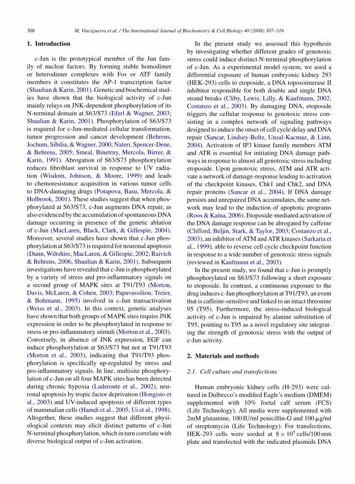
al of Bi
308 M. Vinciguerra et al. / The International Journ1. Introduction
c-Jun is the prototypical member of the Jun fam-ily of nuclear factors. By forming stable homodimeror heterodimer complexes with Fos or ATF familymembers it constitutes the AP-1 transcription factor(Shaulian & Karin, 2001). Genetic and biochemical stud-ies have shown that the biological activity of c-Junmainly relays on JNK-dependent phosphorylation of itsN-terminal domain at S63/S73 (Eferl & Wagner, 2003;Shaulian & Karin, 2001). Phosphorylation of S63/S73is required for c-Jun-mediated cellular transformation,tumor progression and cancer development (Behrens,Jochum, Sibilia, & Wagner, 2000; Nateri, Spencer-Dene,& Behrens, 2005; Smeal, Binetruy, Mercola, Birrer, &Karin, 1991). Abrogation of S63/S73 phosphorylationreduces fibroblast survival in response to UV radia-tion (Wisdom, Johnson, & Moore, 1999) and leadsto chemoresistance acquisition in various tumor cellsto DNA-damaging drugs (Potapova, Basu, Mercola, &Holbrook, 2001). These studies suggest that when phos-phorylated at S63/S73, c-Jun augments DNA repair, asalso evidenced by the accumulation of spontaneous DNAdamage occurring in presence of the genetic ablationof c-Jun (MacLaren, Black, Clark, & Gillespie, 2004).Moreover, several studies have shown that c-Jun phos-phorylation at S63/S73 is required for neuronal apoptosis(Dunn, Wiltshire, MacLaren, & Gillespie, 2002; Raivich& Behrens, 2006; Shaulian & Karin, 2001). Subsequentinvestigations have revealed that c-Jun is phosphorylatedby a variety of stress and pro-inflammatory signals ona second group of MAPK sites at T91/T93 (Morton,Davis, McLaren, & Cohen, 2003; Papavassiliou, Treier,& Bohmann, 1995) involved in c-Jun transactivation(Weiss et al., 2003). In this context, genetic analyseshave shown that both groups of MAPK sites require JNKexpression in order to be phosphorylated in response tostress or pro-inflammatory stimuli (Morton et al., 2003).Conversely, in absence of JNK expression, EGF caninduce phosphorylation at S63/S73 but not at T91/T93(Morton et al., 2003), indicating that T91/T93 phos-phorylation is specifically up-regulated by stress andpro-inflammatory signals. In line, multisite phosphory-lation of c-Jun on all four MAPK sites has been detectedduring chronic hypoxia (Laderoute et al., 2002), neu-ronal apoptosis by tropic factor deprivation (Hongisto etal., 2003) and UV-induced apoptosis of different typesof mammalian cells (Hamdi et al., 2005; Ui et al., 1998).
Altogether, these studies suggest that different physi-ological contexts may elicit distinct patterns of c-JunN-terminal phosphorylation, which in turn correlate withdiverse biological output of c-Jun activation.ochemistry & Cell Biology 40 (2008) 307–316
In the present study we assessed this hypothesisby investigating whether different grades of genotoxicstress could induce distinct N-terminal phosphorylationof c-Jun. As a experimental model system, we used adifferential exposure of human embryonic kidney 293(HEK-293) cells to etoposide, a DNA toposoimerase IIinhibitor responsible for both double and single DNAstrand breaks (Cliby, Lewis, Lilly, & Kaufmann, 2002;Costanzo et al., 2003). By damaging DNA, etoposidetriggers the cellular response to genotoxic stress con-sisting in a complex network of signaling pathwaysdesigned to induce the onset of cell cycle delay and DNArepair (Sancar, Lindsey-Boltz, Unsal-Kacmaz, & Linn,2004). Activation of IP3 kinase family members ATMand ATR is essential for initiating DNA damage path-ways in response to almost all genotoxic stress includingetoposide. Upon genotoxic stress, ATM and ATR acti-vate a network of damage-response leading to activationof the checkpoint kinases, Chk1 and Chk2, and DNArepair proteins (Sancar et al., 2004). If DNA damagepersists and unrepaired DNA accumulates, the same net-work may lead to the induction of apoptotic programs(Roos & Kaina, 2006). Etoposide-mediated activation ofthe DNA damage response can be abrogated by caffeine(Clifford, Beljin, Stark, & Taylor, 2003; Costanzo et al.,2003), an inhibitor of ATM and ATR kinases (Sarkaria etal., 1999), able to reverse cell cycle checkpoint functionin response to a wide number of genotoxic stress signals(reviewed in Kaufmann et al., 2003).
In the present study, we found that c-Jun is promptlyphosphorylated on S63/S73 following a short exposureto etoposide. In contrast, a continuous exposure to thedrug induces c-Jun phosphorylation at T91/T93, an eventthat is caffeine-sensitive and linked to an intact threonine95 (T95). Furthermore, the stress-induced biologicalactivity of c-Jun is impaired by alanine substitution ofT95, pointing to T95 as a novel regulatory site integrat-ing the strength of genotoxic stress with the output ofc-Jun activity.
2. Materials and methods
2.1. Cell culture and transfections
Human embryonic kidney cells (H-293) were cul-tured in Dulbecco’s modified Eagle’s medium (DMEM)supplemented with 10% foetal calf serum (FCS)(Life Technology). All media were supplemented with
2mM glutamine, 100 IU/ml penicillin-G and 100 �g/mlof streptomycin (Life Technology). For transfections,HEK-293 cells were seeded at 8 × 10⁵ cells/100 mmplate and transfected with the indicated plasmids DNA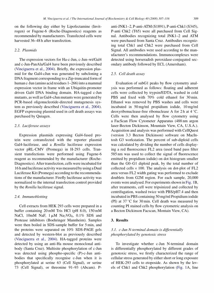
al of Bi
orrh
2
a(mDhedmPtEp
2
tGvsrD1Ltnb
2
bNPwta(dbwbp7
M. Vinciguerra et al. / The International Journ
n the following day either by Lipofectamine (Invit-ogen) or Fugene-6 (Roche-Diagnostics) reagents asecommended by manufacturers. Transfected cells werearvested 36–48 h after transfection.
.2. Plasmids
The expression vectors for Ha-c-Jun, c-Jun–wt/Gal4nd c-Jun-PanAla/Gal4 have been previously describedVinciguerra et al., 2004). Briefly, the expression plas-id for the Gal4-cJun was generated by subcloning aNA fragment corresponding to a Zip-truncated form ofuman c-Jun (amino acid residues 1–266) into a mammalxpression vector in frame with an Ubiquitin-promoterriven Gal4 DNA binding domain. HA-tagged c-Junutants, as well as Gal4-cJun mutants were generated byCR-based oligonucleotide-directed mutagenesis sys-
em as previously described (Vinciguerra et al., 2004).GFP expressing plasmid used in cell death assays wasurchased by Quiagen.
.3. Luciferase assays
Expression plasmids expressing Gal4-fused pro-ein were cotransfected with the reporter plasmidal4-luciferase, and a Renilla luciferase expressionector pRL-CMV (Promega) in H-293 cells. Tran-ient transfections were performed using Fugene-6eagent as recommended by the manufacturer (Roche-iagnostics). After transfection, cells were incubated for6 h and luciferase activity was measured by using a Dualuciferase Kit (Promega) according to the recommenda-
ions of the manufacturer. Firefly luciferase activity wasormalized to the internal transfection control providedy the Renilla luciferase signal.
.4. Immunoblotting
Cell extracts from HEK 293 cells were prepared in auffer containing 20 mM Tris HCl (pH 8.0), 150 mMaCl, 10mM NaF, 1 �M Na3VO4, 0.1% SDS androtease inhibitors (Boehringer Mannheim). Samplesere then boiled in SDS-sample buffer for 5 min, and
he proteins were separated on 10% SDS-PAGE gelsnd detected by western-blot as previously describedVinciguerra et al., 2004). HA-tagged proteins wereetected by using an anti-Ha mouse monoclonal anti-ody (Santa Cruz). Multisite phosphorylation of c-Jun
as detected using phospho-specific (P)-c-Jun anti-odies that specifically recognise c-Jun when it ishosphorylated at serine 63 (Cell Signal), or serine3 (Cell Signal), or threonine 91–93 (Abcam). P-ochemistry & Cell Biology 40 (2008) 307–316 309
anti-JNK1–2, P-anti-ATM (S1891), P-anti-Chk1 (S345),P-anti Chk2 (T65) were all purchased from Cell Sig-nal. Antibodies recognizing total JNK1–2 and ATMwere purchased from Santa Cruz. Antibodies recogniz-ing total Chk1 and Chk2 were purchased from CellSignal. All antibodies were used according to the man-ufacturer’s recommendations. Immunocomplexes weredetected using horseradish peroxidase-conjugated sec-ondary antibody followed by ECL (Amersham).
2.5. Cell death assay
Evaluation of subG1 peaks by flow cytometry anal-ysis was performed as follows: floating and adherentcells were collected by trypsin/EDTA, washed in coldPBS and fixed with 70% cold ethanol for 30 min.Ethanol was removed by PBS washes and cells wereincubated in 50 mg/ml propidium iodide, 10 mg/mldeoxyribonuclease-free ribonuclease A for 2 h at 4 ◦C.Cells were then analysed by flow cytometry usinga FacScan Flow Cytometer Apparatus (488 nm argonlaser-Becton Dickinson, Mountain View, CA, U.S.A.).Acquisition and analysis was performed with CellQuest(version 3.3 Becton Dickinson) software on MacIn-tosh G3 workstation. The percent of sub-diploid cellswas calculated by dividing the number of cells display-ing a red fluorescence FL2 area (used band pass filter585 nm was used to collect the signal of fluorescenceemitted by propidium iodide) on dot histogram smallerthan the G0–G1 diploid peak, by the total number ofcollected cells × 100. The analysis on dot-plot of FL2area versus FL2 width gating was performed to excludedoublets from G2M region. For each sample, 20,000events were analysed. For experiments shown in Fig. 1B,after treatments, cell were tripsinized and collected bycentrifugation, washed twice with PBS/pH7.4 and thenincubated in PBS containing 50 mg/ml Propidium iodide(PI) at 37 ◦C for 30 min. Cell death was measured bycounting PI stained cells by flow cytometric analysis ona Becton Dickinson Facscan, Montain View, CA).
3. Results
3.1. c-Jun N-terminal domain is differentiallyphosphorylated by genotoxic stress
To investigate whether c-Jun N-terminal domainis differentially phosphorylated by different grades of
genotoxic stress, we firstly characterized the range ofcellular stress generated by either short or long exposureof HEK-293 cells to etoposide. As shown by the lev-els of Chk1 and Chk2 phosphorylation (Fig. 1A, line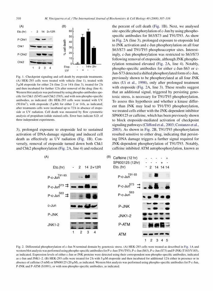
310 M. Vinciguerra et al. / The International Journal of Bi
Fig. 1. Checkpoint signaling and cell death by etoposide treatments.(A) HEK-293 cells were treated with vehicle (line 1), treated with5 �M etoposide for either 2 h (line 2) or 14 h (line 3), treated for 2 hand then incubated for further 12 h after removal of the drug (line 4).Western blot analysis was performed by using phospho-antibodies spe-cific for Chk1 (S345) and Chk2 (T65), and with non-phospho-specificantibodies, as indicated. (B) HEK-293 cells were treated with UV(50 J/m2), with etoposide (5 �M) for either 2 or 14 h, as indicated;after treatments cells were incubated up to 72 h in absence of etopo-
side or UV radiation. Cell death was measured by flow cytometricanalysis of propidium iodide stained cells. Error bars indicate S.D. ofthree independent experiments.3), prolonged exposure to etoposide led to sustained
activation of DNA-damage signaling and induced celldeath as effectively as UV radiation (Fig. 1B). Con-versely, removal of etoposide turned down both Chk1and Chk2 phosphorylation (Fig. 2A, line 4) and reducedFig. 2. Differential phosphorylation of c-Jun N-terminal domain by genotoxiwestern blot analysis was performed using phospho-specific antibodies for P-c-as indicated. Expression levels of either c-Jun or JNK proteins were detectedas c-Jun and JNK1–2. (B) HEK-293 cells were treated for 2 h with 5 �M etoabsence of caffeine (5 mM) or SP600125 (20 �M), as indicated. Western blot aP-JNK and P-ATM (S1891), or with non-phospho-specific antibodies, as indi
ochemistry & Cell Biology 40 (2008) 307–316
the percent of cell death (Fig. 1B). Next, we analysedsite-specific phosphorylation of c-Jun by using phospho-specific antibodies for S63/S73 and T91/T93. As showin Fig. 2A (line 3), prolonged exposure to etoposide ledto JNK activation and c-Jun phosphorylation on all fourS63/S73 and T91/T93 phosphoacceptor sites. Interest-ingly, c-Jun phosphorylation was restricted to S63/S73following removal of etoposide, although JNK phospho-rylation remained elevated (Fig. 2A, line 4). Notably,phospho-specific antibodies for either c-Jun-S63 or c-Jun-S73 detected a shifted phosphorylated form of c-Jun,previously shown to be phosphorylated at all four JNKsites (Ui et al., 1998), only after prolonged treatmentwith etoposide (Fig. 2A, line 3). These results suggestthat an additional signal, triggered by persisting geno-toxic stress, is necessary for T91/T93 phosphorylation.To assess this hypothesis and whether a kinase differ-ent than JNK may lead to T91/T93 phosphorylation,we treated cells either with the JNK-dependent inhibitorSP600125 or caffeine, which has been previously shownto block etoposide-mediated activation of checkpointsignaling pathways (Clifford et al., 2003; Costanzo et al.,2003). As shown in Fig. 2B, T91/T93 phosphorylation
resulted sensitive to either drug, indicating that persist-ing DNA damage triggers a further signal required forJNK-dependent phosphorylation of T91/T93. Notably,caffeine inhibited ATM autophosphorylation, known asc stress. (A) HEK-293 cells were treated as described in Fig. 1A andJun (T91/T93), P-c-Jun (S63), P-c-Jun (S73) and P-JNK (T183/Y185),using their correspondent non-phospho-specific antibodies, indicatedposide and then incubated for additional 12 h either in presence or innalysis was performed using phospho-specific antibodies for P-c-Jun,cated.
al of Bi
aw(prptf
Jiaspetwp
3p
spaoCm
Fawa
M. Vinciguerra et al. / The International Journ
hallmark of its activation (Bakkenist & Kastan, 2003),ithout affecting either JNK or S73 phosphorylation
Fig. 2B), thus excluding that the inhibition of T91/T93hosphorylation results from JNK inactivation. Theseesults suggest that JNK activity is essential for c-Junhosphorylation at all four sites, however a further signalriggered by active DNA-damage pathways is necessaryor T91/T93 phosphorylation.
It is worth to note that although SP600125 inhibitsNK activity by blocking ATP binding, we found thatt also inhibited JNK phosphorylation (Fig. 2B). Suchpeculiar finding has been previously shown by other
tudies reporting negative effects of SP600125 on JNKhosphorylation by DNA-damaging drugs or LPS (Leet al., 2006; Yang et al., 2007). Presumably, in vivoreatment of SP600125 may activate secondary path-ays leading to unbalanced regulation of JNK specifichosphatases.
.2. T95 is required for DNA-damage dependenthosphorylation of T91/T93
Based on the observations shown above, we con-idered that caffeine might interfere with T91/T93hosphorylation by inhibiting an intramolecular mech-
nism involving c-Jun phosphorylation at one, or more,f the three ATM/ATR consensus sites (Kim, Lim,anman, & Kastan, 1999) present within the c-Jun pri-ary structure (Fig. 3A). To assess this hypothesis, weig. 3. Abrogation of T95 affects JNK-dependent phosphorylation of T91/T9re in “Helvetica bold”. (B and C) HEK-293 cells were transfected with HA-c-ere treated with etoposide (5 �M) for 16 h and then harvested for western an
ntibodies. The expression levels of HA-c-Jun proteins were normalized usin
ochemistry & Cell Biology 40 (2008) 307–316 311
generated HA-tagged c-Jun mutant proteins bearing sin-gle alanine substitutions at specific sites (c-Jun/A95,or c-Jun/A249, or c-Jun/A325), or at all three sites (c-Jun/3xAQ). HA-Jun proteins were transiently expressedin HEK-293 cells and c-Jun phosphorylation was anal-ysed using phospho-specific antibodies as describedin the Materials and Methods section. As shown inFig. 3B, etoposide-dependent phosphorylation of c-Junat T91/T93 was strongly reduced exclusively in c-Jun/3xAQ and c-Jun/A95 mutants. Conversely, the ratioof S73 phosphorylation to T91/T93 phosphorylation wasappreciably higher in c-Jun/3xAQ or c-Jun/A95 com-pared to c-Jun-wt (or c-JunA249 and c-JunA325). Theseresults indicate that etoposide-dependent phosphoryla-tion of c-Jun on T91/T93 requires an intact T95, unlessanti-phospho T91/T93 antibodies (P-cJunT91/T93) cross-react predominantly with phosphorylated T95 ratherthan phosphorylated T91/T93. To evaluate this possibil-ity we examined the ability of P-cJunT91/T93 antibodiesto immunoreact with a c-Jun mutant bearing an ala-nine substitution at T91/T93 (c-JunA91–93), or a c-Junmutant containing alanine substitution at all MAPK sitesS63/S73 and T91/T93–95 (c-Jun-pan ala). As shown inFig. 3C, P-cJunT91/T93 immunoreacted only with c-Jun-wt, while the anti-phospho S73 antibody recognized both
3. (A) Primary sequence of human c-Jun. ATM/ATR consensus-sitesJun-wt or HA-c-Jun mutants, as indicated. After 24 h, transfected cellsalysis of c-Jun phosphorylation using the indicated phospho-specific
g HA antibodies.
c-Jun-wt and c-Jun-A91–93. These observations implythat the P-cJunT91/T93 antibody is predominantly reactivetowards phosphorylated T91/T93. However, we cannotexclude the possibility that T95 is not phosphorylated in
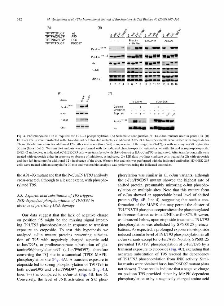
312 M. Vinciguerra et al. / The International Journal of Biochemistry & Cell Biology 40 (2008) 307–316
Fig. 4. Phosphorylated T95 is required for T91–93 phosphorylation. (A) Schematic configuration of HA-c-Jun mutants used in panel (B). (B)HEK-293 cells were transfected with HA-c-Jun-wt or HA-c-Jun mutants, as indicated. After 24 h, transfected cells were treated with etoposide for2 h and then left in culture for additional 12 h either in absence (lines 5–8) or in presence of the drug (lines 9–12), or with anisomycin (500 ng/ml) for30 min (lines 13–16). Western blot analysis was performed with the indicated phospho-specific antibodies, or with HA and non-phospho-specificJNK1–2 antibodies, as indicated. (C) HEK-293 cells were transfected with HA-c-Jun-wt or HA-c-JunD95, as indicated. After transfection, cells were
ndicatedtern blois was p
treated with etoposide either in presence or absence of inhibitors, as iand then left in culture for additional 12 h in absence of the drug. Wescells were treated with anisomycin for 30 min and western blot analys
the A91–93 mutant and that the P-cJunT91/T93 antibodycross-reacted, although to a lesser extent, with phospho-rylated T95.
3.3. Aspartic acid substitution of T95 triggersJNK-dependent phosphorylation of T91/T93 inabsence of persisting DNA damage
Our data suggest that the lack of negative chargeon position 95 might be the missing signal impair-ing T91/T93 phosphorylation in response to transientexposure to etoposide. To test this hypothesis weanalysed c-Jun mutant proteins presenting substitu-tion of T95 with negatively charged aspartic acid(c-Jun/D95), or proline/aspartate substitution of glu-tamine96/phenylalanine97 (c-Jun/P96D97), thereforeconverting the TQ site in a canonical (TPD) MAPK-phosphorylation site (Fig. 4A). A transient exposure to
etoposide led to strong phosphorylation of T91/T93 inboth c-Jun/D95 and c-Jun/P96D97 proteins (Fig. 4B,lines 7–8) as compared to c-Jun-wt (Fig. 4B, line 5).Conversely, the level of JNK activation or S73 phos-. 2 + 12R (last two lines) indicate cells treated for 2 h with etoposidet analysis was performed with the indicated antibodies. (D) HEK-293erformed using the indicated antibodies.
phorylation was similar in all c-Jun variants, althoughthe c-Jun/P96D97 mutant showed the highest rate ofshifted protein, presumably mirroring c-Jun phospho-rylation on multiple sites. Note that this mutant formof c-Jun showed an appreciable basal level of shiftedprotein (Fig. 4B, line 4), suggesting that such a con-formation of the MAPK site may permit the cluster ofT91/T93/T5 phosphoacceptor sites to be phosphorylatedin absence of stress-activated JNKs, as for S73. However,as discussed below, upon etoposide treatment, T91/T93phosphorylation was abolished by SP600125 preincu-bations. As expected, a prolonged exposure to etoposideinduced a similar level of T91/T93 phosphorylation in allc-Jun variants except for c-Jun/A95. Notably, SP600125prevented T91/T93 phosphorylation of c-Jun/D95 by atransient exposure to etoposide (Fig. 4C), excluding thataspartate substitution of T95 rescued the dependencyof T91/T93 phosphorylation from JNK activity. Simi-
lar results were obtained for c-Jun/P96D97 mutant (datanot shown). These results indicate that a negative chargeon position T95 provided either by MAPK-dependentphosphorylation or by a negatively charged amino acid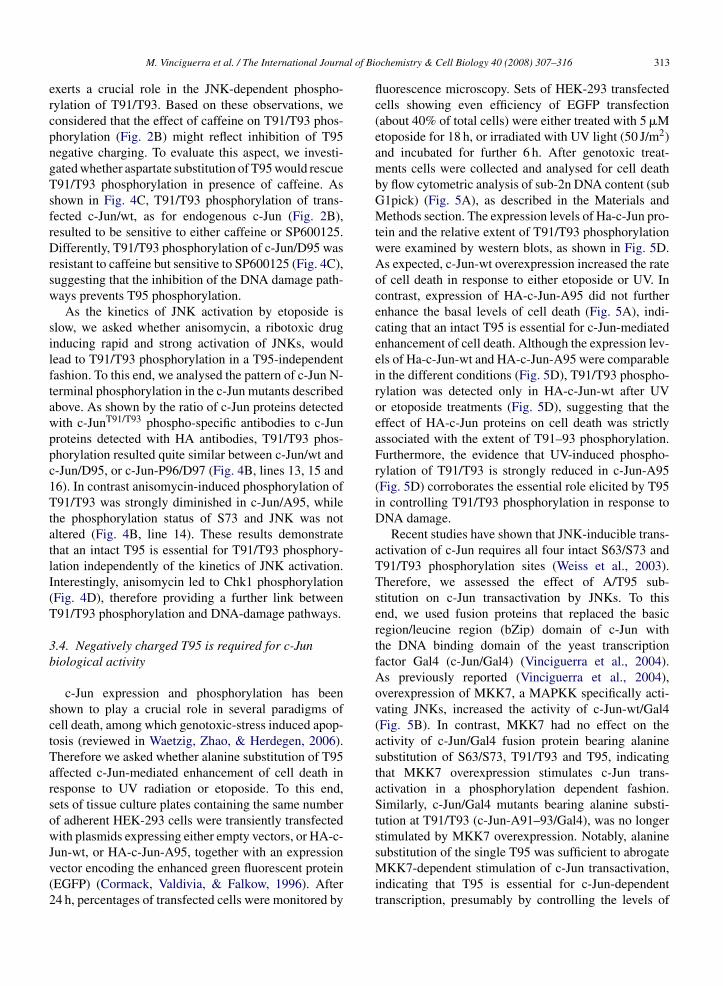
al of Bi
ercpngTsfrDrsw
silftawppc1TtatlI(T
3b
sctTarsowJv(2
M. Vinciguerra et al. / The International Journ
xerts a crucial role in the JNK-dependent phospho-ylation of T91/T93. Based on these observations, weonsidered that the effect of caffeine on T91/T93 phos-horylation (Fig. 2B) might reflect inhibition of T95egative charging. To evaluate this aspect, we investi-ated whether aspartate substitution of T95 would rescue91/T93 phosphorylation in presence of caffeine. Ashown in Fig. 4C, T91/T93 phosphorylation of trans-ected c-Jun/wt, as for endogenous c-Jun (Fig. 2B),esulted to be sensitive to either caffeine or SP600125.ifferently, T91/T93 phosphorylation of c-Jun/D95 was
esistant to caffeine but sensitive to SP600125 (Fig. 4C),uggesting that the inhibition of the DNA damage path-ays prevents T95 phosphorylation.As the kinetics of JNK activation by etoposide is
low, we asked whether anisomycin, a ribotoxic drugnducing rapid and strong activation of JNKs, wouldead to T91/T93 phosphorylation in a T95-independentashion. To this end, we analysed the pattern of c-Jun N-erminal phosphorylation in the c-Jun mutants describedbove. As shown by the ratio of c-Jun proteins detectedith c-JunT91/T93 phospho-specific antibodies to c-Junroteins detected with HA antibodies, T91/T93 phos-horylation resulted quite similar between c-Jun/wt and-Jun/D95, or c-Jun-P96/D97 (Fig. 4B, lines 13, 15 and6). In contrast anisomycin-induced phosphorylation of91/T93 was strongly diminished in c-Jun/A95, while
he phosphorylation status of S73 and JNK was notltered (Fig. 4B, line 14). These results demonstratehat an intact T95 is essential for T91/T93 phosphory-ation independently of the kinetics of JNK activation.nterestingly, anisomycin led to Chk1 phosphorylationFig. 4D), therefore providing a further link between91/T93 phosphorylation and DNA-damage pathways.
.4. Negatively charged T95 is required for c-Juniological activity
c-Jun expression and phosphorylation has beenhown to play a crucial role in several paradigms ofell death, among which genotoxic-stress induced apop-osis (reviewed in Waetzig, Zhao, & Herdegen, 2006).herefore we asked whether alanine substitution of T95ffected c-Jun-mediated enhancement of cell death inesponse to UV radiation or etoposide. To this end,ets of tissue culture plates containing the same numberf adherent HEK-293 cells were transiently transfectedith plasmids expressing either empty vectors, or HA-c-
un-wt, or HA-c-Jun-A95, together with an expressionector encoding the enhanced green fluorescent proteinEGFP) (Cormack, Valdivia, & Falkow, 1996). After4 h, percentages of transfected cells were monitored by
ochemistry & Cell Biology 40 (2008) 307–316 313
fluorescence microscopy. Sets of HEK-293 transfectedcells showing even efficiency of EGFP transfection(about 40% of total cells) were either treated with 5 �Metoposide for 18 h, or irradiated with UV light (50 J/m2)and incubated for further 6 h. After genotoxic treat-ments cells were collected and analysed for cell deathby flow cytometric analysis of sub-2n DNA content (subG1pick) (Fig. 5A), as described in the Materials andMethods section. The expression levels of Ha-c-Jun pro-tein and the relative extent of T91/T93 phosphorylationwere examined by western blots, as shown in Fig. 5D.As expected, c-Jun-wt overexpression increased the rateof cell death in response to either etoposide or UV. Incontrast, expression of HA-c-Jun-A95 did not furtherenhance the basal levels of cell death (Fig. 5A), indi-cating that an intact T95 is essential for c-Jun-mediatedenhancement of cell death. Although the expression lev-els of Ha-c-Jun-wt and HA-c-Jun-A95 were comparablein the different conditions (Fig. 5D), T91/T93 phospho-rylation was detected only in HA-c-Jun-wt after UVor etoposide treatments (Fig. 5D), suggesting that theeffect of HA-c-Jun proteins on cell death was strictlyassociated with the extent of T91–93 phosphorylation.Furthermore, the evidence that UV-induced phospho-rylation of T91/T93 is strongly reduced in c-Jun-A95(Fig. 5D) corroborates the essential role elicited by T95in controlling T91/T93 phosphorylation in response toDNA damage.
Recent studies have shown that JNK-inducible trans-activation of c-Jun requires all four intact S63/S73 andT91/T93 phosphorylation sites (Weiss et al., 2003).Therefore, we assessed the effect of A/T95 sub-stitution on c-Jun transactivation by JNKs. To thisend, we used fusion proteins that replaced the basicregion/leucine region (bZip) domain of c-Jun withthe DNA binding domain of the yeast transcriptionfactor Gal4 (c-Jun/Gal4) (Vinciguerra et al., 2004).As previously reported (Vinciguerra et al., 2004),overexpression of MKK7, a MAPKK specifically acti-vating JNKs, increased the activity of c-Jun-wt/Gal4(Fig. 5B). In contrast, MKK7 had no effect on theactivity of c-Jun/Gal4 fusion protein bearing alaninesubstitution of S63/S73, T91/T93 and T95, indicatingthat MKK7 overexpression stimulates c-Jun trans-activation in a phosphorylation dependent fashion.Similarly, c-Jun/Gal4 mutants bearing alanine substi-tution at T91/T93 (c-Jun-A91–93/Gal4), was no longerstimulated by MKK7 overexpression. Notably, alanine
substitution of the single T95 was sufficient to abrogateMKK7-dependent stimulation of c-Jun transactivation,indicating that T95 is essential for c-Jun-dependenttranscription, presumably by controlling the levels of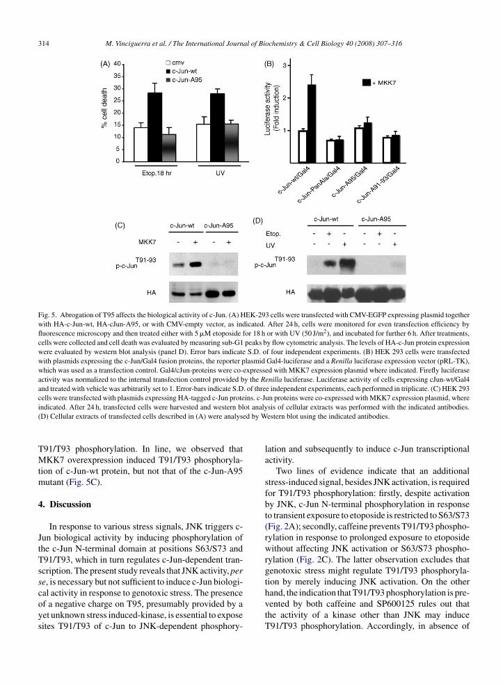
314 M. Vinciguerra et al. / The International Journal of Biochemistry & Cell Biology 40 (2008) 307–316
Fig. 5. Abrogation of T95 affects the biological activity of c-Jun. (A) HEK-293 cells were transfected with CMV-EGFP expressing plasmid togetherwith HA-c-Jun-wt, HA-cJun-A95, or with CMV-empty vector, as indicated. After 24 h, cells were monitored for even transfection efficiency byfluorescence microscopy and then treated either with 5 �M etoposide for 18 h or with UV (50 J/m2), and incubated for further 6 h. After treatments,cells were collected and cell death was evaluated by measuring sub-G1 peaks by flow cytometric analysis. The levels of HA-c-Jun protein expressionwere evaluated by western blot analysis (panel D). Error bars indicate S.D. of four independent experiments. (B) HEK 293 cells were transfectedwith plasmids expressing the c-Jun/Gal4 fusion proteins, the reporter plasmid Gal4-luciferase and a Renilla luciferase expression vector (pRL-TK),which was used as a transfection control. Gal4/cJun-proteins were co-expressed with MKK7 expression plasmid where indicated. Firefly luciferaseactivity was normalized to the internal transfection control provided by the Renilla luciferase. Luciferase activity of cells expressing cJun-wt/Gal4and treated with vehicle was arbitrarily set to 1. Error-bars indicate S.D. of three independent experiments, each performed in triplicate. (C) HEK 293cells were transfected with plasmids expressing HA-tagged c-Jun proteins. c-Jun proteins were co-expressed with MKK7 expression plasmid, where
ot analyed by W
indicated. After 24 h, transfected cells were harvested and western bl(D) Cellular extracts of transfected cells described in (A) were analys
T91/T93 phosphorylation. In line, we observed thatMKK7 overexpression induced T91/T93 phosphoryla-tion of c-Jun-wt protein, but not that of the c-Jun-A95mutant (Fig. 5C).
4. Discussion
In response to various stress signals, JNK triggers c-Jun biological activity by inducing phosphorylation ofthe c-Jun N-terminal domain at positions S63/S73 andT91/T93, which in turn regulates c-Jun-dependent tran-scription. The present study reveals that JNK activity, perse, is necessary but not sufficient to induce c-Jun biologi-
cal activity in response to genotoxic stress. The presenceof a negative charge on T95, presumably provided by ayet unknown stress induced-kinase, is essential to exposesites T91/T93 of c-Jun to JNK-dependent phosphory-sis of cellular extracts was performed with the indicated antibodies.estern blot using the indicated antibodies.
lation and subsequently to induce c-Jun transcriptionalactivity.
Two lines of evidence indicate that an additionalstress-induced signal, besides JNK activation, is requiredfor T91/T93 phosphorylation: firstly, despite activationby JNK, c-Jun N-terminal phosphorylation in responseto transient exposure to etoposide is restricted to S63/S73(Fig. 2A); secondly, caffeine prevents T91/T93 phospho-rylation in response to prolonged exposure to etoposidewithout affecting JNK activation or S63/S73 phospho-rylation (Fig. 2C). The latter observation excludes thatgenotoxic stress might regulate T91/T93 phosphoryla-tion by merely inducing JNK activation. On the other
hand, the indication that T91/T93 phosphorylation is pre-vented by both caffeine and SP600125 rules out thatthe activity of a kinase other than JNK may induceT91/T93 phosphorylation. Accordingly, in absence of
al of Bi
Je2dr
ttmtwaopodasTtpanmrarssrtc
uddateTmi
htua(tTb(
M. Vinciguerra et al. / The International Journ
NK expression, T91/T93 are not phosphorylated byither stress or pro-inflammatory stimuli (Morton et al.,003). However, whether T91/T93 phosphorylation isirectly driven by JNKs, or by a JNK-regulated kinase,emains to be determined.
Interestingly, the inhibitory effect of A/T95 substitu-ion on T91/T93 phosphorylation (Figs. 3 and 4) suggestshat a negative charge on T95 might represent the putative
odification required for JNK-dependent phosphoryla-ion of T91/T93. In accordance with this hypothesis,e show that T95 replacement with negatively charged
spartic acid leads to JNK-dependent phosphorylationf T91/T93 in presence of caffeine, or in absence ofersisting DNA damage (Fig. 4). Since negative chargef threonine/serine are mostly provided by kinase-ependent phosphorylation, our study points to T95s a putative DNA damage dependent-phosphorylationite essential for JNK-dependent phosphorylation of91/T93. In line with this hypothesis, replacement of
he TQ acceptor site with a canonical MAPK phos-horylation site leads to T91/T93 phosphorylation inbsence of persisting DNA damage, excluding that theegative charged aspartic acid substitution of T95 mightimic a structural modification other than phospho-
ylation (Fig. 4B). Furthermore, the observation thatlanine substitution of T95 prevents T91/T93 phospho-ylation in response to various types of genotoxic stress,uch as anisomycin and UV radiation (Figs. 4B, 5D),uggests that phosphorylation of T95 is a general pre-equisite for stress-induced activation of c-Jun, ratherhan specific to etoposide-mediated phosphorylation of-Jun.
A wide number of studies have pointed top-regulation of c-Jun expression coupled to JNK-ependent phosphorylation of the c-Jun N-terminalomain as a crucial mechanism leading to stress-inducedpoptosis (Waetzig et al., 2006). Remarkably, we foundhat c-Jun-A95 is not able to enhance either UV ortoposide-induced cell death (Fig. 5A), pointing to95 phosphorylation as an essential post-transcriptionalodification required for the induction of c-Jun biolog-
cal activity.c-Jun mostly acts by forming either homodimers or
eterodimers with other members of the AP-1 family ofranscription factors. Accordingly, most mutations mod-lating c-Jun biological output affect its transcriptionalctivity. Phosphorylation of both groups of MAPK sitesS63/S73 and T91/T93) is required for signal-induced
ranscriptional activity of c-Jun (Weiss et al., 2003).he evidence that alanine substitution of T95 affectsoth c-Jun transactivation and T91/T93 phosphorylationFig. 5C) suggests that an uncharged alanine at positionochemistry & Cell Biology 40 (2008) 307–316 315
T95 affects c-Jun-mediated apoptosis by impairing itstransactivation potential.
Depending on the environmental and genetic back-ground, activation of c-Jun by JNKs can elicit quitediverse biological programs including cell survival orapoptosis. Presumably, according to the physiologicalcondition, active c-Jun may influence different cellu-lar responses by shifting the balance of the expressionpattern of distinct AP-1 responsive genes. The presentfindings suggest that c-Jun may perceive the strengthof genotoxic stress through phosphorylation of T95which in turn allowing JNK-dependent phosphorylationof T91/T93, drives c-Jun transcriptional activity and theexpression pattern of distinct c-Jun target genes.
Acknowledgements
We thank Dr. R.J. Davis for MKK7 expressing plas-mid. We are most grateful to Prof. V.E. Avvedimentoand Prof. E. Bamberg for their support and VEA forhis continuous helpful scientific discussions. We thankDoris Ollig and Rita Cerillo for technical assistance. Wealso thank Dirk Bohmann and Anna Grazia Recchia fortheir helpful comments on the manuscript. This work wassupported by Associazione Italiana Ricerca sul Cancro(AIRC).
References
Bakkenist, C. J., & Kastan, M. B. (2003). DNA damage activates ATMthrough intermolecular autophosphorylation and dimer dissocia-tion. Nature, 421(6922), 499–506.
Behrens, A., Jochum, W., Sibilia, M., & Wagner, E. F. (2000). Onco-genic transformation by ras and fos is mediated by c-Jun N-terminalphosphorylation. Oncogene, 19(22), 2657–2663.
Cliby, W. A., Lewis, K. A., Lilly, K. K., & Kaufmann, S. H. (2002). Sphase and G2 arrests induced by topoisomerase I poisons are depen-dent on ATR kinase function. J Biol Chem, 277(2), 1599–1606.
Clifford, B., Beljin, M., Stark, G. R., & Taylor, W. R. (2003). G2 arrestin response to topoisomerase II inhibitors: The role of p53. CancerRes, 63(14), 4074–4081.
Cormack, B. P., Valdivia, R. H., & Falkow, S. (1996). FACS-optimizedmutants of the green fluorescent protein (GFP). Gene, 173(1 SpecNo), 33–38.
Costanzo, V., Shechter, D., Lupardus, P. J., Cimprich, K. A., Gottes-man, M., & Gautier, J. (2003). An ATR- and Cdc7-dependent DNAdamage checkpoint that inhibits initiation of DNA replication. MolCell, 11(1), 203–213.
Davis, R. J. (2000). Signal transduction by the JNK group of MAPkinases. Cell, 103(2), 239–252.
Dunn, C., Wiltshire, C., MacLaren, A., & Gillespie, D. A. (2002).
Molecular mechanism and biological functions of c-Jun N-terminalkinase signalling via the c-Jun transcription factor. Cell Signal,14(7), 585–593.Eferl, R., & Wagner, E. F. (2003). AP-1: A double-edged sword intumorigenesis. Nat Rev Cancer, 3(11), 859–868.
al of Bi
316 M. Vinciguerra et al. / The International JournHamdi, M., Kool, J., Cornelissen-Steijger, P., Carlotti, F., Popeijus,H. E., van der Burgt, C., et al. (2005). DNA damage in tran-scribed genes induces apoptosis via the JNK pathway and theJNK-phosphatase MKP-1. Oncogene, 24(48), 7135–7144.
Hongisto, V., Smeds, N., Brecht, S., Herdegen, T., Courtney, M. J., &Coffey, E. T. (2003). Lithium blocks the c-Jun stress response andprotects neurons via its action on glycogen synthase kinase 3. MolCell Biol, 23(17), 6027–6036.
Kaufmann, W. K., Heffernan, T. P., Beaulieu, L. M., Doherty, S.,Frank, A. R., Zhou, Y., et al. (2003). Caffeine and human DNAmetabolism: The magic and the mystery. Mutat Res, 532(1–2),85–102.
Kim, S. T., Lim, D. S., Canman, C. E., & Kastan, M. B. (1999).Substrate specificities and identification of putative substrates ofATM kinase family members. J Biol Chem, 274(53), 37538–37543.
Laderoute, K. R., Calaoagan, J. M., Gustafson-Brown, C., Knapp, A.M., Li, G. C., Mendonca, H. L., et al. (2002). The response of c-jun/AP-1 to chronic hypoxia is hypoxia-inducible factor 1 alphadependent. Mol Cell Biol, 22(8), 2515–2523.
MacLaren, A., Black, E. J., Clark, W., & Gillespie, D. A. (2004). c-Jun-deficient cells undergo premature senescence as a result ofspontaneous DNA damage accumulation. Mol Cell Biol, 24(20),9006–9018.
Morton, S., Davis, R. J., McLaren, A., & Cohen, P. (2003). A reinves-tigation of the multisite phosphorylation of the transcription factorc-Jun. EMBO J, 22(15), 3876–3886.
Nateri, A. S., Spencer-Dene, B., & Behrens, A. (2005). Interactionof phosphorylated c-Jun with TCF4 regulates intestinal cancerdevelopment. Nature, 437(7056), 281–285.
Papavassiliou, A. G., Treier, M., & Bohmann, D. (1995). Intramolec-
ular signal transduction in c-Jun. EMBO J, 14(9), 2014–2019.Potapova, O., Basu, S., Mercola, D., & Holbrook, N. J. (2001). Protec-tive role for c-Jun in the cellular response to DNA damage. J BiolChem, 276(30), 28546–28553.
ochemistry & Cell Biology 40 (2008) 307–316
Raivich, G., & Behrens, A. (2006). Role of the AP-1 transcription factorc-Jun in developing, adult and injured brain. Prog Neurobiol, 78(6),347–363.
Roos, W. P., & Kaina, B. (2006). DNA damage-induced cell death byapoptosis. Trends Mol Med, 12(9), 440–450.
Sancar, A., Lindsey-Boltz, L. A., Unsal-Kacmaz, K., & Linn, S. (2004).Molecular mechanisms of mammalian DNA repair and the DNAdamage checkpoints. Annu Rev Biochem, 73, 39–85.
Sarkaria, J. N., Busby, E. C., Tibbetts, R. S., Roos, P., Taya, Y., Karnitz,L. M., et al. (1999). Inhibition of ATM and ATR kinase activi-ties by the radiosensitizing agent, caffeine. Cancer Res, 59(17),4375–4382.
Shaulian, E., & Karin, M. (2001). AP-1 in cell proliferation and sur-vival. Oncogene, 20(19), 2390–2400.
Smeal, T., Binetruy, B., Mercola, D. A., Birrer, M., & Karin, M. (1991).Oncogenic and transcriptional cooperation with Ha-Ras requiresphosphorylation of c-Jun on serines 63 and 73. Nature, 354(6353),494–496.
Ui, M., Sonobe, M. H., Ito, T., Murakami, M., Okazaki, S., Takada, M.,et al. (1998). Biochemical and functional analysis of highly phos-phorylated forms of c-Jun protein. FEBS Lett, 429(3), 289–294.
Vinciguerra, M., Vivacqua, A., Fasanella, G., Gallo, A., Cuozzo, C.,Morano, A., et al. (2004). Differential phosphorylation of c-Jun andJunD in response to the epidermal growth factor is determined bythe structure of MAPK targeting sequences. J Biol Chem, 279(10),9634–9641.
Waetzig, V., Zhao, Y., & Herdegen, T. (2006). The bright side of JNKs-Multitalented mediators in neuronal sprouting, brain developmentand nerve fiber regeneration. Prog Neurobiol, 80(2), 84–97.
Weiss, C., Schneider, S., Wagner, E. F., Zhang, X., Seto, E., &Bohmann, D. (2003). JNK phosphorylation relieves HDAC3-
dependent suppression of the transcriptional activity of c-Jun.EMBO J, 22(14), 3686–3695.Wisdom, R., Johnson, R. S., & Moore, C. (1999). c-Jun regulates cellcycle progression and apoptosis by distinct mechanisms. EMBO J,18(1), 188–197.
Estrogenic GPR30 signalling induces proliferationand migration of breast cancer cells through CTGF
Deo Prakash Pandey1,3, RosamariaLappano2,3, Lidia Albanito2, AntonioMadeo2, Marcello Maggiolini2,4 andDidier Picard1,4,*1Departement de Biologie Cellulaire, Sciences III, Universite de Geneve,Geneve, Switzerland and 2Department of Pharmaco-Biology, Universityof Calabria, Rende, Italy
The steroid hormone oestrogen can signal through several
receptors and pathways. Although the transcriptional re-
sponses mediated by the nuclear oestrogen receptors (ER)
have been extensively characterized, the changes in gene
expression elicited by signalling through the membrane-
associated ER GPR30 have not been studied. We show here
for ER-negative human breast cancer cells that the activa-
tion of GPR30 signalling by oestrogen or by hydroxy-
tamoxifen (OHT), an ER antagonist but GPR30 agonist,
induces a transcription factor network, which resembles
that induced by serum in fibroblasts. The most strongly
induced gene, CTGF, appears to be a target of these
transcription factors. We found that the secreted factor
connective tissue growth factor (CTGF) not only contri-
butes to promote proliferation but also mediates the
GPR30-induced stimulation of cell migration. These
results provide a framework for understanding the phy-
siological and pathological functions of GPR30. As the
activation of GPR30 by OHT also induces CTGF in fibro-
blasts from breast tumour biopsies, these pathways may
be involved in promoting aggressive behaviour of breast
tumours in response to endogenous oestrogens or to OHT
being used for endocrine therapy.
The EMBO Journal (2009) 28, 523–532. doi:10.1038/
emboj.2008.304; Published online 15 January 2009
Subject Categories: signal transduction; molecular biology of
disease
Keywords: oestrogen; G-protein coupled receptor; micro-
array; signal transduction; tamoxifen resistance
Introduction
The steroid hormone oestrogen binds and activates the
oestrogen receptors (ER) a and b, two members of the
nuclear receptor superfamily. Activated ERs regulate the
transcription of target genes by binding either directly to
specific DNA sequences or by tethering to other DNA-
bound transcription factors. ERs have been extensively stu-
died at the molecular, cellular, physiological and pathological
levels (reviewed by Dahlman-Wright et al, 2006; Deroo
and Korach, 2006; Heldring et al, 2007). Tamoxifen and its
hydroxylated active form hydroxytamoxifen (OHT) are syn-
thetic ER ligands that compete with the physiological oestro-
gen 17b-estradiol (E2) for binding. Depending on promoter,
cell and signalling context, OHT functions either as a partial
agonist or as a partial antagonist. The latter mode has led to
its use for endocrine therapy of ERa-positive breast tumours,
the proliferation of which can be stimulated by E2 (reviewed
by Jordan, 2004).
The early discovery of Filardo et al (2000) that the pre-
sence of the completely unrelated transmembrane receptor
GPR30 can mediate oestrogen responsiveness of ER-negative
breast cancer cells came as a big surprise. GPR30 was later
shown to be a genuine ER (Revankar et al, 2005; Thomas
et al, 2005). In addition to E2, OHT also functions as a
GPR30 agonist (Revankar et al, 2005; Vivacqua et al,
2006a, b). The GPR30 signalling pathway has been studied
in a variety of cell lines. GPR30 couples to a trimeric G
protein, stimulating the cAMP pathway most likely through
a Gas (Thomas et al, 2005) and Src through Gbg (Filardo,
2002). Subsequently, Src promotes the shedding of heparin-
binding EGF-like growth factor and activation of the EGF
receptor (Filardo et al, 2000). This in turn activates a whole
series of intracellular signalling events, most notably the
activation of mitogen-activated protein kinases (MAPK)
Erk1/2, PI3 kinase and phospholipase C (reviewed by
Prossnitz et al, 2008). Further cellular responses lie down-
stream of these signals, including the activation of the gene
FOS (Maggiolini et al, 2004).
It is unlikely that the activation of FOS can account for all
of the biological effects of GPR30 signalling that have been
reported. For example, E2 is able to stimulate the proliferation
of breast, thyroid and ovarian carcinomas through GPR30
(Vivacqua et al, 2006b; Albanito et al, 2007, 2008). This effect
is clearly independent of ERs and can also be observed with a
GPR30-specific ligand, but how GPR30 signalling stimulates
proliferation remains unclear. Although the genomic effects
of ERa have been extensively studied, and in particular in
breast cancer cells (see Carroll and Brown, 2006; Dudek
and Picard, 2008 and references therein), the global changes
in gene expression triggered by GPR30 signalling are not
known. Unlike a transcription factor such as ERa, GPR30
would have to effect these changes indirectly. Nevertheless,
GPR30-mediated changes in gene expression patterns have to
be considered a specific output of this signal-transduction
pathway. Here, we report the transcriptional consequences of
GPR30 signalling in human breast cancer cells. The most
strongly induced gene suggested a new function of GPR30
signalling in cell migration and proved to be functionally
relevant for our understanding of the biological effects of
GPR30 signalling.Received: 18 June 2008; accepted: 22 December 2008; publishedonline: 15 January 2009
*Corresponding author. Departement de Biologie Cellulaire, Sciences III,Universite de Geneve, 30 quai Ernest-Ansermet, 1211 Geneve 4,Switzerland. Tel.: þ 41 22 379 6813; Fax: þ 41 22 379 6928;E-mail: [email protected] authors contributed equally to this work4Joint senior authors
The EMBO Journal (2009) 28, 523–532 | & 2009 European Molecular Biology Organization | All Rights Reserved 0261-4189/09
www.embojournal.org
&2009 European Molecular Biology Organization The EMBO Journal VOL 28 | NO 5 | 2009
EMBO
THE
EMBOJOURNAL
THE
EMBOJOURNAL
523
Results
Gene expression profiling of GPR30 signalling
To determine the changes in gene expression that GPR30
signalling elicits, we chose human SKBr3 breast cancer cells
as our model system. These cells lack both ERa and ERb but
express GPR30 and display GPR30 signalling (Filardo et al,
2000; Maggiolini et al, 2004). Despite the absence of other
known ERs in SKBr3 cells, we knocked down GPR30 expres-
sion with an antisense strategy (Revankar et al, 2005;
Vivacqua et al, 2006b) (Supplementary Figure 1A) to ascer-
tain that any observed ligand-induced changes in gene ex-
pression are mediated by GPR30. Serum-deprived cells were
treated for only 1 h with E2 or OHT to capture the primary
responses. It should be pointed out here, that the OHT
concentration (10 mM) used for induction is comparable to
the micromolar OHTconcentrations that are reached in breast
tissue of patients undergoing tamoxifen therapy (Kisanga
et al, 2004).
The mRNA levels of a total of 175 genes were induced
by at least 1.3-fold by one of the treatments by comparison
with uninduced control cells (Supplementary Figure 1B). At
this point, we decided that we would only consider those
genes as potential GPR30 target genes that fulfilled the
following stringent criteria: at least 1.3-fold induction by
both E2 and OHT, and at least a 1.3-fold reduction of the
OHT response by antisense-mediated GPR30 knockdown.
These criteria defined 36 genes as GPR30 target genes
(Figure 1; Supplementary Table 1). In total, 19 of these 36
genes were induced by more than two-fold by OHT. Within
the short time frame of the treatment, no gene was signifi-
cantly repressed according to the same criteria (data not
shown).
We then undertook a Q-PCR experiment with the same
RNA samples for a panel of genes to validate the microarray
results and to obtain more quantitative data. Qualitatively,
GPR30-mediated induction could be confirmed for all of
them, although, not surprisingly, larger quantitative differ-
ences were obtained by Q-PCR (Figure 2). The gene encoding
the connective tissue growth factor (CTGF, also known as
CCN2) proved to be induced 15- to 16-fold by OHT and E2. It
is a technical limitation of microarray analyses that some
genes with a relatively modest induction fall through the
cracks. This is the case, for example, for JUN. Our short list of
36 genes contains the genes FOS and FOSB (Supplementary
Table 1). These encode components of the heterodimeric
transcription factor AP1. Surprisingly, our gene list contains
none of the genes, such as JUN, that encode heterodimeric
partner proteins of Fos proteins. Although JUN did not pass
the third stringent criterion (reduction by at least 1.3-fold in
the GPR30 knockdown sample) in the microarray analysis, it
easily passed all criteria for a GPR30 target gene in the Q-PCR
experiment, including a two-fold induction by both ligands
(Figure 2). We therefore include JUN as a GPR30 target gene
and consider it very likely that there are other false negatives
in the microarray data.
CTGF is a GPR30 target gene
CTGF is by far the gene most strongly induced by E2 or OHT.
We performed an immunoblot analysis to determine whether
the dramatic induction seen at the mRNA level leads to
increased CTGF protein expression in SKBr3 cells.
Figure 3A shows that this is the case and that this increase
can be blunted by an shRNA-mediated knock down of GPR30.
The requirement for GPR30 and the specificity of the GPR30
knockdown are further emphasized by the fact that the co-
transfection of an shRNA-resistant version of GPR30 (‘GPR30
rescue’) restores the response. The increase at the protein
level might seem modest, but note that only cell-associated
proteins, and not proteins already released into the medium,
were immunoblotted. We further explored the generality of
this response with other cell lines and the GPR30-specific
ligand G-1 (Bologa et al, 2006) (see Figure 3B and C). CTGF is
induced by OHT in the human breast cancer cell lines MCF7
and BT-20, which are ERa positive and negative, respectively.
Contro
l
OHTE2
OHT, GPR30
KD
CTGFEGR1FOSDUSP1MT1XATF3MT1FMT1AMT2AMT1GFOSBRASD1RGS16TNFPPP1R15ACYR61EDN1ZFP36DDIT4TUFT1LOC441019DDIT3PTGER4DUSP5DUSP4EDN2NFKBIABHLHB2CEBPDBM956082RND1NR4A2IER2ERRFI1TNFAIP3KIAA1754
0.1 1.0 10
Figure 1 Colour-coded map of hierarchically clustered gene expres-sion profiles. For each gene and condition, the colour indicates theratio of the values obtained for the treated and untreated samples(as listed in Supplementary Table 1). GPR30 KD, GPR30 knock-down.
GPR30 signalling stimulates migrationDP Pandey et al
The EMBO Journal VOL 28 | NO 5 | 2009 &2009 European Molecular Biology Organization524
The induction is seen both at the mRNA and protein levels,
and it is not elicited by the antioestrogen ICI 1820780 (ICI)
(Figure 3B), at least not in MCF7 cells under our experimental
conditions. Importantly, the OHT induction of CTGF in MCF7
cells is independent of ERa as it can still be observed when
ERa is knocked down (Supplementary Figure 2). The time
course experiment confirms the activation of the CTGF and
FOS genes by OHT, and shows an identical activation by G-1.
Induction at the mRNA level is transient in that it is clearly
observed after 1 h but has subsided 3 h later. Note that the
microarray analysis was performed with RNA from cells
treated for 1 h.
Mediators of the transcriptional response
to GPR30 signalling
The GPR30-mediated activation of target genes must be
indirect. Previous analyses had indicated that GPR30 leads
to the activation of Erk1/2 (Filardo et al, 2000; Maggiolini
et al, 2004). MAPK can activate transcription factors such as
the serum response factor (SRF) and members of the ETS
family by direct phosphorylation (see for example, Posern
and Treisman, 2006; Gutierrez-Hartmann et al, 2007).
Moreover, it has been pointed out that the increase in
cAMP elicited by GPR30 signalling could be expected to
activate CREB (Prossnitz et al, 2008). These factors in turn
activate the expression of the second tier of transcription
factors such as c-Fos, FosB, c-Jun, EGR1, ATF3, C/EBPd and
NR4A2 (Nurr1). In addition to FOS, which we already knew
to be activated by GPR30 signalling (Maggiolini et al, 2004),
the genes for the aforementioned second tier transcription
factors are all in our list of GPR30-induced genes (comple-
mented with JUN from the Q-PCR experiment).
As a first step towards elucidating the signalling and trans-
cription factor network that might underlie the transcrip-
tional response to GPR30 signalling, we downloaded 5 kb of
upstream sequences (relative to the start sites of transcrip-
tion) for 34 of the 36 target genes of Supplementary Table 1.
We scanned them for the presence of common sequence
motifs and compared those with the known DNA-binding
sequences of the TRANSFAC database to identify the corre-
sponding transcription factors. The results of these analyses
are displayed in Figure 4A for CTGF and for the complete set
of target genes in Supplementary Table 2. SRF is by far the
most over-represented transcription factor with EGR2, CREB
and ATF among the runners up. Overall, the results of this
bioinformatic analysis are entirely compatible with the afore-
mentioned activation scheme.
Binding sites for AP1, of which c-Fos can be a component,
are also highly represented, although not over-represented by
more than two-fold, in promoters of GPR30 target genes (data
not shown). For CTGF (Figure 4A), whose upstream se-
quences contain AP1 sites, we experimentally verified the
role of c-Fos. The expression of a dominant-negative variant
of c-Fos in SKBr3 cells abolishes the induction of CTGF by
OHT or E2 (Figure 4B). To assess whether 50 flanking
sequences of the CTGF gene would be sufficient to mediate
the GPR30 response, we used a reporter gene containing a
2 kb CTGF promoter fragment upstream of the luciferase-
coding region (Chaqour et al, 2006; see Figure 4A). Upon
transfection into SKBr3 cells, this reporter gene could be
induced more than two-fold with E2 in a GPR30-dependent
manner (Figure 4C). The response to OHT, which appears to
E2OHTOHT, GPR30 KD
Fol
d in
duct
ion
20
18
16
14
12
10
8
6
4
2
0
CTGFEGR1
ATF3
FOS
DUSP1
RASD1
RGS16TNF
PPP1R15
A
CYR61
DDIT3
NR4A2
JUN
Figure 2 Q-PCR validation of a subset of GPR30-regulated genes.‘Fold induction’ denotes the ratio of the values obtained for thetreated and untreated samples. Error bars indicate standard devia-tions of measurements of triplicate samples. GPR30 KD, GPR30knockdown.
MCF7
– OHTR
T–P
CR
B
C
RPLP0
CTGF CTGF
β-Actin
BT-20
– OHT
CTGF
FOS
RPLP0
SKBr3
– 1 4 8
+OHT
MCF7
– ICI
– 1 4 8 (h)(h)
+G1
A
CTGF
β-Actin
– OHT – OHT
GPR30 shRNAControlshRNA
– OHT
GPR30rescue
OHT
Figure 3 Induction of CTGF mRNA and protein in a variety ofbreast cancer cell lines. (A) Immunoblot analysis of CTGF expressedby SKBr3 cells. Cells were transfected with shRNA constructs andthe GPR30 rescue vector, and treated with OHT as indicated.(B) Immunoblots of semiquantitative RT–PCR products (‘RT–PCR’) and CTGF protein (rightmost panel). (C) Time course ofCTGF and FOS induction; semiquantitative RT–PCR analysis ofSKBr3 cells treated with OHT or G1. b-Actin and the RPLP0 mRNAserved as internal standards for the immunoblot and RT–PCRexperiments, respectively.
GPR30 signalling stimulates migrationDP Pandey et al
&2009 European Molecular Biology Organization The EMBO Journal VOL 28 | NO 5 | 2009 525
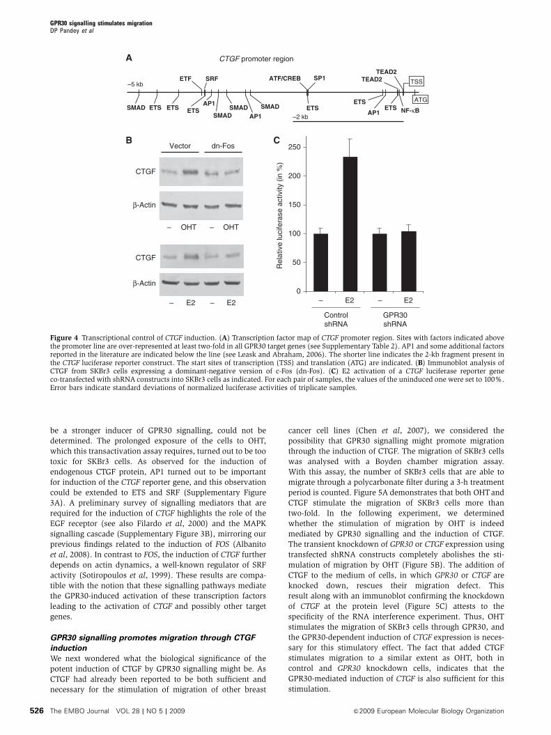
be a stronger inducer of GPR30 signalling, could not be
determined. The prolonged exposure of the cells to OHT,
which this transactivation assay requires, turned out to be too
toxic for SKBr3 cells. As observed for the induction of
endogenous CTGF protein, AP1 turned out to be important
for induction of the CTGF reporter gene, and this observation
could be extended to ETS and SRF (Supplementary Figure
3A). A preliminary survey of signalling mediators that are
required for the induction of CTGF highlights the role of the
EGF receptor (see also Filardo et al, 2000) and the MAPK
signalling cascade (Supplementary Figure 3B), mirroring our
previous findings related to the induction of FOS (Albanito
et al, 2008). In contrast to FOS, the induction of CTGF further
depends on actin dynamics, a well-known regulator of SRF
activity (Sotiropoulos et al, 1999). These results are compa-
tible with the notion that these signalling pathways mediate
the GPR30-induced activation of these transcription factors
leading to the activation of CTGF and possibly other target
genes.
GPR30 signalling promotes migration through CTGF
induction
We next wondered what the biological significance of the
potent induction of CTGF by GPR30 signalling might be. As
CTGF had already been reported to be both sufficient and
necessary for the stimulation of migration of other breast
cancer cell lines (Chen et al, 2007), we considered the
possibility that GPR30 signalling might promote migration
through the induction of CTGF. The migration of SKBr3 cells
was analysed with a Boyden chamber migration assay.
With this assay, the number of SKBr3 cells that are able to
migrate through a polycarbonate filter during a 3-h treatment
period is counted. Figure 5A demonstrates that both OHTand
CTGF stimulate the migration of SKBr3 cells more than
two-fold. In the following experiment, we determined
whether the stimulation of migration by OHT is indeed
mediated by GPR30 signalling and the induction of CTGF.
The transient knockdown of GPR30 or CTGF expression using
transfected shRNA constructs completely abolishes the sti-
mulation of migration by OHT (Figure 5B). The addition of
CTGF to the medium of cells, in which GPR30 or CTGF are
knocked down, rescues their migration defect. This
result along with an immunoblot confirming the knockdown
of CTGF at the protein level (Figure 5C) attests to the
specificity of the RNA interference experiment. Thus, OHT
stimulates the migration of SKBr3 cells through GPR30, and
the GPR30-dependent induction of CTGF expression is neces-
sary for this stimulatory effect. The fact that added CTGF
stimulates migration to a similar extent as OHT, both in
control and GPR30 knockdown cells, indicates that the
GPR30-mediated induction of CTGF is also sufficient for this
stimulation.
Rel
ativ
e lu
cife
rase
act
ivity
(in
%)
0
50
100
150
200
250
– E2 – E2
GPR30shRNA
ControlshRNA
β-Actin
CTGF
Vector dn-Fos
OHT OHT– –
β-Actin
CTGF
E2 E2– –
ETF SRF ATF/CREB SP1 TEAD2
ATGSMAD ETS ETS ETS
SMADSMAD SMAD ETS
ETSETS
AP1
AP1AP1
–2 kb
–5 kb
NF-κB
CTGF promoter region
TEAD2
TSS
Figure 4 Transcriptional control of CTGF induction. (A) Transcription factor map of CTGF promoter region. Sites with factors indicated abovethe promoter line are over-represented at least two-fold in all GPR30 target genes (see Supplementary Table 2). AP1 and some additional factorsreported in the literature are indicated below the line (see Leask and Abraham, 2006). The shorter line indicates the 2-kb fragment present inthe CTGF luciferase reporter construct. The start sites of transcription (TSS) and translation (ATG) are indicated. (B) Immunoblot analysis ofCTGF from SKBr3 cells expressing a dominant-negative version of c-Fos (dn-Fos). (C) E2 activation of a CTGF luciferase reporter geneco-transfected with shRNA constructs into SKBr3 cells as indicated. For each pair of samples, the values of the uninduced one were set to 100%.Error bars indicate standard deviations of normalized luciferase activities of triplicate samples.
GPR30 signalling stimulates migrationDP Pandey et al
The EMBO Journal VOL 28 | NO 5 | 2009 &2009 European Molecular Biology Organization526
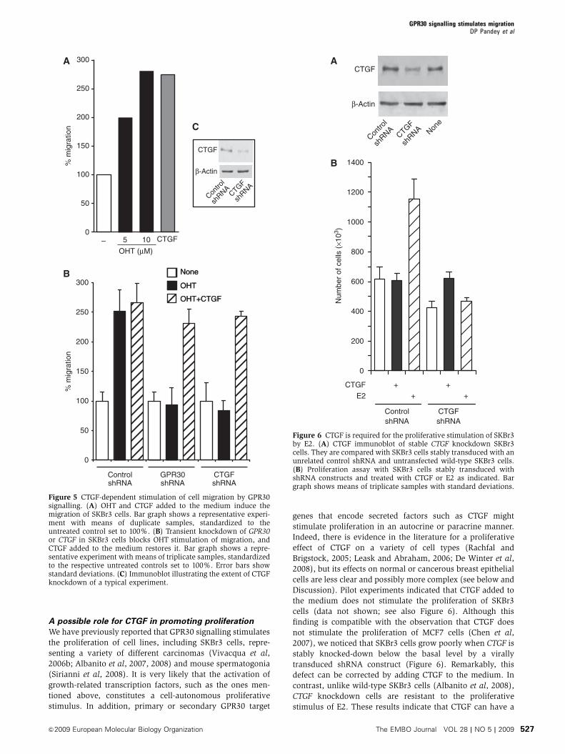
A possible role for CTGF in promoting proliferation
We have previously reported that GPR30 signalling stimulates
the proliferation of cell lines, including SKBr3 cells, repre-
senting a variety of different carcinomas (Vivacqua et al,
2006b; Albanito et al, 2007, 2008) and mouse spermatogonia
(Sirianni et al, 2008). It is very likely that the activation of
growth-related transcription factors, such as the ones men-
tioned above, constitutes a cell-autonomous proliferative
stimulus. In addition, primary or secondary GPR30 target
genes that encode secreted factors such as CTGF might
stimulate proliferation in an autocrine or paracrine manner.
Indeed, there is evidence in the literature for a proliferative
effect of CTGF on a variety of cell types (Rachfal and
Brigstock, 2005; Leask and Abraham, 2006; De Winter et al,
2008), but its effects on normal or cancerous breast epithelial
cells are less clear and possibly more complex (see below and
Discussion). Pilot experiments indicated that CTGF added to
the medium does not stimulate the proliferation of SKBr3
cells (data not shown; see also Figure 6). Although this
finding is compatible with the observation that CTGF does
not stimulate the proliferation of MCF7 cells (Chen et al,
2007), we noticed that SKBr3 cells grow poorly when CTGF is
stably knocked-down below the basal level by a virally
transduced shRNA construct (Figure 6). Remarkably, this
defect can be corrected by adding CTGF to the medium. In
contrast, unlike wild-type SKBr3 cells (Albanito et al, 2008),
CTGF knockdown cells are resistant to the proliferative
stimulus of E2. These results indicate that CTGF can have a
0
50
100
150
200
250
300
–OHT (μM)
CTGF5 10
% m
igra
tion
CTGF
β-Actin
None
OHT
OHT+CTGF
Contro
l
shRNA
CTGF
shRNA
None
OHT
OHT+CTGF
GPR30shRNA
ControlshRNA
CTGFshRNA
0
50
100
150
200
250
300
% m
igra
tion
Figure 5 CTGF-dependent stimulation of cell migration by GPR30signalling. (A) OHT and CTGF added to the medium induce themigration of SKBr3 cells. Bar graph shows a representative experi-ment with means of duplicate samples, standardized to theuntreated control set to 100%. (B) Transient knockdown of GPR30or CTGF in SKBr3 cells blocks OHT stimulation of migration, andCTGF added to the medium restores it. Bar graph shows a repre-sentative experiment with means of triplicate samples, standardizedto the respective untreated controls set to 100%. Error bars showstandard deviations. (C) Immunoblot illustrating the extent of CTGFknockdown of a typical experiment.
0
200
400
600
800
Num
ber
of c
ells
(×1
03 )
CTGF
ControlshRNA
CTGFshRNA
+ +
1000
1200
1400
E2 + +
CTGF
CTGF
shRNA
β-Actin
Contro
l
shRNA Non
e
Figure 6 CTGF is required for the proliferative stimulation of SKBr3by E2. (A) CTGF immunoblot of stable CTGF knockdown SKBr3cells. They are compared with SKBr3 cells stably transduced with anunrelated control shRNA and untransfected wild-type SKBr3 cells.(B) Proliferation assay with SKBr3 cells stably transduced withshRNA constructs and treated with CTGF or E2 as indicated. Bargraph shows means of triplicate samples with standard deviations.
GPR30 signalling stimulates migrationDP Pandey et al
&2009 European Molecular Biology Organization The EMBO Journal VOL 28 | NO 5 | 2009 527

proliferative effect on such breast carcinoma cells under
certain conditions, and that CTGF is required for the proli-
ferative response to E2.
Contribution to GPR30 signalling from the stroma
Given that secreted factors regulating proliferation and mi-
gration need not only be made by the breast carcinoma cells
themselves, we examined the expression and signalling of
GPR30 in fibroblasts obtained from biopsies of primary breast
tumours. GPR30 is clearly expressed in these fibroblasts,
and its activation by OHT stimulates the expression of
CTGF (Figure 7).
Discussion
In the process of characterizing the transcriptional response
to GPR30 signalling, we have found a set of target genes that
contribute to mediating the proliferative stimulus of GPR30
activators for carcinoma cell lines. Moreover, the prominent
induction of CTGF by both E2 and OHT led us to the discovery
that GPR30 signalling stimulates cell migration through
CTGF. As these responses may occur in breast cancer cells
independently of their ERa status as well as in the surround-
ing stromal cells, these findings may have important implica-
tions for our understanding of endocrine resistance in breast
cancer.
GPR30 signalling activates a transcription factor
network
Our microarray analysis indicated that GPR30 signalling
triggers the activation of a network of transcription factors.
A first tier, including SRF, members of the ETS family and
CREB, is directly activated through post-translational mod-
ifications by kinases that are activated by GPR30 signalling.
These transcription factors are the ones that transcriptionally
activate the expression of a second tier of transcription
factors (for example, Fos and Jun proteins, EGR1, ATF3,
C/EBPd and NR4A2). The response is most likely further
amplified by GPR30-stimulated post-translational modifica-
tions of these proteins and positive feedback loops between
many of these transcription factors. GPR30 itself may be part
of a positive reinforcement loop as we have found that its
expression is induced through MAPK and c-Fos in response to
the growth factor EGF (Albanito et al, 2008). It remains to be
seen whether these mechanisms, possibly involving other
transcription factors mentioned above, also operate in
response to GRP30 signalling, which incidentally depends
on the EGF receptor (Filardo et al, 2000).
Considering the GPR30 signalling pathway, it is not sur-
prising that the GPR30 response resembles the well-
characterized transcriptional ‘immediate early response’ of
fibroblasts to serum (Iyer et al, 1999). The strong induction of
CTGF is consistent with the following: (i) CTGF is known to
be activated by a panel of different extracellular signals and
its promoter contains binding sites for transcription factors of
the ‘immediate early response’, notably SRF, ETS, ATF, AP1,
and TEAD2/ETF (Leask and Abraham, 2006; Cooper et al,
2007; Figure 4A); (ii) the inhibitory effect of a dominant-
negative Fos points to a role for AP1, and additional pre-
liminary functional results suggest that ETS and SRF are also
required; and (iii) the activation of MAPK and actin dynamics
are essential for the activation of both the full complement of
these transcription factors and CTGF.
GPR30 expression and signalling in carcinomas
Activation of GPR30 by oestrogens elicits proliferative
responses of breast and other carcinomas (Vivacqua et al,
2006b; Albanito et al, 2007, 2008). Although GPR30 is also
expressed in normal breast epithelium, a survey of 321
primary breast tumours showed that 60% maintain GPR30
expression, including half of all ER-negative tumours
(Filardo et al, 2006, 2008). Unlike ERa, the expression of
which correlates with good prognosis, GPR30 expression was
found to correlate very strongly with tumour size, HER-2
expression and distant metastasis. Remarkably, another study
on breast cancer failed to find a similar correlation (Kuo et al,
2007), but methodological differences and a smaller cohort
compared with the aforementioned studies call for more
investigations. In the meantime, it is noteworthy that
GPR30 expression was also associated with poor prognosis
in a survey of endometrial carcinomas (Smith et al, 2007).
Even in GPR30-positive cells, GPR30 is not expressed abun-
dantly at the mRNA and protein levels (data not shown).
Rather than monitoring only the expression of GPR30 itself, it
might be more informative to combine it with measurements
of the expression of a set of GPR30 target genes. Hence, our
results highlight a set of GPR30 target genes that might both
provide a signature for GPR30 signalling and a mechanistic
underpinning of the aforementioned phenomena.
GPR30 target genes and cancer
Our experiments with SKBr3 cells demonstrate that CTGF is
necessary for the stimulation of proliferation and migration
by GPR30 signalling. Although CTGF is sufficient to stimulate
migration, steady-state levels of CTGF might normally be
sufficient to sustain a basal level of proliferation. The stimu-
lation of proliferation by GPR30 signalling might arise from
the combined induction of CTGF with multiple other GPR30
target genes. How the highly transient activation of GPR30
target genes that we have observed at the mRNA level is
CTGF
β-Actin
– OHT – OHT
ControlshRNA
GPR30shRNA
GPR30
Contro
l
shRNA
GPR30
shRNA
β-Actin
Figure 7 GPR30 expression and signalling in fibroblasts frombreast tumours. The data shown are from a representative experi-ment of three experiments performed with samples from differentpatients. (A) Immunoblot analysis showing expression and knockdown of GPR30 in transfected fibroblasts. (B) Immunoblot analysisof CTGF expressed by fibroblasts. Cells were transfected withindicated shRNA constructs and treated with OHT as indicated.
GPR30 signalling stimulates migrationDP Pandey et al
The EMBO Journal VOL 28 | NO 5 | 2009 &2009 European Molecular Biology Organization528
converted into this long-term response remains to be further
analysed. Perhaps not a single, but repeated pulses of GPR30
signalling may be sufficient to produce relatively persistent
higher levels of key proteins such as CTGF.
CTGF defines the cystein knot family of proteins along with
Cyr61 (also known as CCN1) and Nov. Note that CYR61 is
also induced by GPR30 signalling in SKBr3 cells, albeit much
less than CTGF (Figures 1 and 2; Supplementary Table 1).
Cyr61 and CTGF bind integrins and heparan sulphate-con-
taining proteoglycans. CTGF also binds receptors or co-recep-
tors for other signalling molecules such as Wnt, and CCN
proteins even bind growth factors and cytokines themselves.
These findings have led to the notion that CCN proteins
exert their biological functions by modifying the action of
other signals and the interactions with the extracellular
matrix (reviewed by Rachfal and Brigstock, 2005; Leask and
Abraham, 2006; De Winter et al, 2008; Holbourn et al, 2008).
This may explain why the effects of CCN proteins are
exquisitely dependent on cell type and experimental condi-
tions, and why they may at times be seemingly contradictory.
For example, low concentrations of CTGF can promote angio-
genesis, but high concentrations inhibit the angiogenic effects
of VEGF (Inoki et al, 2002). The evidence for an implication
of CTGF in breast cancer is equally confusing. Although we
have observed stimulatory effects of CTGF with SKBr3 cells,
CTGF was shown to mediate TGFb-induced apoptosis of
MCF7 cells (Hishikawa et al, 1999). Nevertheless, CTGF
was found to be part of a gene expression signature of
osteolytic metastatic variants of the ER-negative breast cancer
cell line MDA-MB-231, and to contribute to metastasis upon
overexpression (Kang et al, 2003). A recent study confirmed
this association and notably demonstrated that the overex-
pression of the genome organizer protein SATB1, which is
overexpressed by aggressive breast cancer cells, induces
CTGF expression (Han et al, 2008). In contrast, in a survey
of 122 human breast tumours, Cyr61 and CTGF were found to
be oppositely correlated with poor outcome, being high and
low, respectively (Jiang et al, 2004). Elevated Cyr61 and CTGF
levels have been reported to be characteristic of a number of
other carcinomas (Bleau et al, 2005; Rachfal and Brigstock,
2005; Deng et al, 2007; Liu et al, 2008; Mullis et al, 2008).
Whether the ERa status of breast tumours and cell lines may
influence the effects of CCN proteins and account for some of
the apparent discrepancies remains to be investigated.
There is yet another set of GPR30 target genes, the expres-
sion of which may contribute to breast cancer progression. Of
the 11 most strongly induced genes, 5 are metallothionein
genes. These include four members of the MT1 gene cluster
and one, MT2A, from another chromosome. Interestingly,
high expression of metallothioneins in breast tumours corre-
lates with poor outcome, perhaps owing to their protective
and proliferative functions (Bay et al, 2006).
Relevance to tamoxifen resistance of breast cancer
and to physiology
Tamoxifen is used clinically for the endocrine treatment of
ERa-positive breast cancer. How and why many of these
tumours ultimately become resistant to this therapy remain
poorly understood (reviewed by Herynk and Fuqua, 2007;
Riggins et al, 2007). On the basis of our findings, we would
suggest that GPR30 continues to mediate stimulatory oestro-
gen signals for proliferation and migration even in tumours
that lose ERa expression. Indeed, GPR30 may become a risk
factor in patients that are treated with tamoxifen as OHT is a
potent GPR30 agonist. Thus, for a subset of breast tumours as
well as other carcinomas, perhaps independently of ERastatus, activation of GPR30 might actually promote poor
outcome by stimulating tissue invasion and remodelling
through CTGF. GPR30 antagonists, which are not yet avail-
able, could be of great therapeutic value. As our results with
primary fibroblasts indicate, the stroma could contribute to
relaying stimulatory signals of oestrogens or OHT to breast
carcinoma cells, again independently of ERa status. It has
been increasingly recognized that there is a decisive interplay
between cancer cells and stroma (Bhowmick and Moses,
2005; Finak et al, 2008). Future studies should therefore
include the evaluation of GPR30 levels in the cancer-asso-
ciated stroma.
Although our transcriptome analysis was carried out with
a breast cancer cell line, its results, which we have partially
confirmed with primary fibroblasts, may be of heuristic
value for understanding other biological GPR30 functions.
The induction of a network of transcription factors and target
genes such as CTGF, CYR61 and metallothioneins may be
relevant to the wide range of GPR30 functions currently being
unravelled in thymic atrophy (Wang et al, 2008a), mechan-
ical hyperalgesia (Kuhn et al, 2008), liver injury (Hsieh et al,
2007), in the hypothalamus (Qiu et al, 2006; Brailoiu et al,
2007; Canonaco et al, 2008), for primordial follicle formation
(Wang et al, 2008b), and for the proliferation of urothelial
cells (Teng et al, 2008) and osteoblasts (Teplyuk et al, 2008).
Materials and methods
Cell cultureSKBr3 breast cancer cells were maintained in RPMI-1640 withoutphenol red supplemented with 10% fetal bovine serum (FBS).MCF7 and BT-20 breast cancer cells were cultured in Dulbecco’smodified Eagle’s medium and MEM, respectively, supplementedwith 10% FBS. At 24 h prior to induction experiments, MCF7 andBT-20 cells were switched to a medium without serum and withoutphenol red. Lentiviruses were produced by transient co-transfectionof lentiviral shRNA constructs with the VSV-G envelope plasmidpMD2G and the packaging plasmid psPAX2 (gifts from Dr D Trono;see http://tronolab.epfl.ch) into 293Tcells. SKBr3 cells with a stableknockdown of CTGF were generated by lentiviral transduction of anshRNA construct (see below). In parallel, we obtained cells stablytransduced with an unrelated control shRNA. To this end, cells wereconsecutively infected twice overnight, washed twice and subjectedto a selection with 3mg/ml puromycin for at least 24 h to eliminatethe small fraction of non-infected cells.
Generation of primary fibroblast cells from breast cancertissuesBreast cancer specimens were collected from primary tumours ofpatients who signed informed consent and underwent surgerybefore any pharmacological treatment. Following tumour excision,small pieces (1–2 mm diameter) were placed in a digestion solution(400 IU collagenase, 100 IU hyalurodinase and 20% FBS in Hank’sbalanced salt solution; all components from Sigma) containingantibiotics and antimycotics (Sigma) and incubated at 371C with5% CO2 for 5–6 h. After centrifugation at 90 g for 2 min, thesupernatant containing fibroblast cells was centrifuged at 500 g for8 min, then resuspended and cultured in RPMI-1640 mediumsupplemented with 20% FBS, antibiotics and antimycotics.
PlasmidsThe GPR30 knockdown for the microarray analysis was carried outwith the plasmid GPR30/AS (Revankar et al, 2005) using the relatedempty expression vector pRK5 (Schall et al, 1990) as a negativecontrol. The CTGF luciferase reporter plasmid p(�1999/þ 36)-luc
GPR30 signalling stimulates migrationDP Pandey et al
&2009 European Molecular Biology Organization The EMBO Journal VOL 28 | NO 5 | 2009 529
(Chaqour et al, 2006), which is based on the backbone of vectorpGL3-basic (Promega), was a gift from Dr B Chaqour. The plasmidA-FOS, which encodes a c-Fos mutant that heterodimerizes withc-Fos dimerization partners but does not allow DNA binding(Gerdes et al, 2006), was obtained from Dr C Vinson. We havepreviously reported the characteristics and the evaluation of anshRNA construct to knock down the expression of GPR30 and of anunrelated shRNA control construct (Albanito et al, 2008). TheshRNA construct for CTGF was obtained from the same supplier(Open Biosystems; www.Biocat.de). It has clone IDTRCN0000061950 and is based on the same lentiviral expressionvector pLKO.1 as the other shRNA constructs. The targeting strandgenerated from the CTGF shRNA construct is TAGTACAGCGATTCAAAGATG. The expression vector for Flag-tagged human GPR30has been described (Albanito et al, 2008). It was used to generatethe GPR30 rescue vector containing silent mutations in the shRNA-targeted sequence: codons 293–297 were changed to CCG TGT AAACAA AGT (changes underlined).
Microarray analysisSKBr3 cells were transiently transfected either with the emptyexpression vector pRK5 or with the plasmid GPR30/AS using theFuGENE 6 transfection reagent (Roche) at 3ml/mg DNA (5mg DNAper 10 cm dish) in a medium with only 1% FBS. At 36 h aftertransfection, cells were switched to serum-free medium for 12 hbefore induction with 100 nM E2 or 10 mM OHT for 1 h. Total RNAfrom triplicate samples was extracted with Trizol (Invitrogen)according to the manufacturer’s instructions. RNA quality wasassessed with a BioAnalyzer (Bio-Rad). All RNA samples werestored at �801C until required for further processing. Expressionprofiles were determined with HumanWG-6 v2.0 BeadChip micro-arrays (Illumina) at the genomics platform of the University ofGeneva. The data have been submitted to GEO (access codeGSE11567).
Promoter sequence analysisSequence retrieval: 5000 nucleotide sequences from upstream of thetranscription start sites of the 34 genes in Supplementary Table 1with a RefSeq accession number were retrieved from the UCSCgenome browser (http://hgdownload.cse.ucsc.edu/goldenPath/hg18/bigZips/upstream5000.fa.gz). Similarly, a large set of promo-ter sequences, to be used as a background/control sequences, wasretrieved from http://ani.embl.de/trawler. Transcription factoridentification: TRANSFAC Professional 11.4 was downloaded fromBiobase (http://www.biobase-international.com) and installed lo-cally on a computer running Ubuntu. The set of all vertebratetranscription factor-binding sites was retrieved from matrix.dat inTRANSFAC. For these factors, a number of profiles were generatedwith matrix similarity ranging from 0.8 to 1.00 and core similarityranging from 0.8 to 1.00. The profile minFP to minimize falsepositives was generated from MATCH on the website. MATCH wasused locally to identify transcription factor-binding sites in the set ofpromoter sequences with the profile minFP. Thereafter, F-MATCHwas used with a cutoff P-value of 0.001 to identify over-representedtranscription factor-binding sites. These transcription factor-bindingsites were sorted by their relative over-representation in the targetsequence set. To retrieve the predicted sites for 5000 bp of CTGFupstream sequences, the profile with 0.95/0.95 matrix/coresimilarity was used.
PCR analysesQuantitative real-time PCR analysis was performed for the triplicateRNA samples used for the microarray experiment. To identify
appropriate control genes for standardization, the analysis wasperformed with three candidates that had proved useful in manyother gene expression analyses (TFRC, ALAS1 and TBP). As TFRCand ALAS1 varied the least across all sample, all measurementswere normalized to the geometric mean of these two control genes.Semiquantitative RT–PCR was carried out as described (Maggioliniet al, 1999) with total RNA from cells induced for 1 h. FOS, CTGFand the internal control RPLP0 (also known as 36B4) cDNAs yieldedbands of 420, 392 and 408 bp with 20, 20 and 10 PCR cycles,respectively. The primers are listed in Supplementary Table 3.
ImmunoblottingTotal protein extracts were prepared from cells after a 2-h induction.For inhibition experiments with shRNAs or the dominant-negativec-Fos variant, cells were transiently transfected in a mediumwithout serum and induced 24 h later. CTGF, b-actin and GPR30were revealed by immunoblotting with the goat polyclonalantiserum sc-14939, the mouse monoclonal antibody sc-8432(Santa Cruz Biotechnology) and the rabbit polyclonal antiserumLS-A1183 (MBL-Eppendorf), respectively.
Reporter gene assayThe CTGF luciferase construct was co-transfected with a Renillaluciferase expression plasmid as an internal transfection controlinto SKBr3 cells as described above for the microarray analysis.Induction with 100 nM E2 was carried out overnight. Luciferaseactivities of triplicate samples were measured with the Dual-Luciferase Reporter Assay system (Promega).
Migration and proliferation assaysMigration assays were performed with SKBr3 cells in triplicateusing Boyden chambers (Costar Transwell, 8 mm polycarbonatemembrane). For knockdown experiments, cells were transfectedwith shRNA constructs directed against GPR30 or CTGF or with anunrelated shRNA construct (3 mg DNA with FuGENE 6 at a 3ml to1mg DNA ratio in 60 mm plates) in standard growth medium. After24 h, they were seeded in the upper chambers. OHT or CTGF wasadded to the medium without serum in the bottom wells. After 3 h,cells on the bottom side of the membrane were fixed and counted.Human CTGF was purchased from MBL and added at 100 ng/ml.For proliferation assays, SKBr3 cells with a stable CTGF (or control)knockdown were cultured in a medium supplemented with 5%charcoal-treated FBS. Where indicated, CTGF was added each day.Cells were counted at day 6.
Supplementary dataSupplementary data are available at The EMBO Journal Online(http://www.embojournal.org).
Acknowledgements
We are grateful to Drs B Chaqour, E Prossnitz, H Sato, D Trono andC Vinson for gifts of reagents, Dr R Treisman for the SRF shRNAsequence and to Dr K Strub for a critical reading of the paper. We areindebted to Dr S Abonante and Dr F Romeo for providing the breastcancer specimens. We thank the team of the genomics platform ofthe NCCR Frontiers-in-Genetics at the University of Geneva for helpwith microarrays. Funding for this project was provided by theAssociazione Italiana Ricerca sul Cancro (AIRC) and Ministerodell’Universita e Ricerca to MM, the Fondation Medic, the SwissNational Science Foundation and the Canton de Geneve to DP.
References
Albanito L, Madeo A, Lappano R, Vivacqua A, Rago V, Carpino A,Oprea TI, Prossnitz ER, Musti AM, Ando S, Maggiolini M(2007) G protein-coupled receptor 30 (GPR30) mediates geneexpression changes and growth response to 17b-estradiol andselective GPR30 ligand G-1 in ovarian cancer cells. Cancer Res 67:1859–1866
Albanito L, Sisci D, Aquila S, Brunelli E, Vivacqua A, Madeo A,Lappano R, Pandey DP, Picard D, Mauro L, Ando S, Maggiolini M
(2008) EGF induces GPR30 expression in estrogen receptor-negative breast cancer cells. Endocrinology 149: 3799–3808
Bay BH, Jin R, Huang J, Tan PH (2006) Metallothionein as a prognosticbiomarker in breast cancer. Exp Biol Med 231: 1516–1521
Bhowmick NA, Moses HL (2005) Tumor–stroma interactions. CurrOpin Genet Dev 15: 97–101
Bleau AM, Planque N, Perbal B (2005) CCN proteins and cancer:two to tango. Front Biosci 10: 998–1009
GPR30 signalling stimulates migrationDP Pandey et al
The EMBO Journal VOL 28 | NO 5 | 2009 &2009 European Molecular Biology Organization530

Bologa CG, Revankar CM, Young SM, Edwards BS, Arterburn JB,Kiselyov AS, Parker MA, Tkachenko SE, Savchuck NP, Sklar LA,Oprea TI, Prossnitz ER (2006) Virtual and biomolecularscreening converge on a selective agonist for GPR30. Nat ChemBiol 2: 207–212
Brailoiu E, Dun SL, Brailoiu GC, Mizuo K, Sklar LA, Oprea TI,Prossnitz ER, Dun NJ (2007) Distribution and characterization ofestrogen receptor G protein-coupled receptor 30 in the rat centralnervous system. J Endocrinol 193: 311–321
Canonaco M, Giusi G, Madeo A, Facciolo RM, Lappano R,Canonaco A, Maggiolini M (2008) A sexually dimorphic distribu-tion pattern of the novel estrogen receptor G-protein-coupledreceptor 30 in some brain areas of the hamster. J Endocrinol196: 131–138
Carroll JS, Brown M (2006) Estrogen receptor target gene: anevolving concept. Mol Endocrinol 20: 1707–1714
Chaqour B, Yang R, Sha Q (2006) Mechanical stretch modulates thepromoter activity of the profibrotic factor CCN2 through in-creased actin polymerization and NF-kappaB activation. J BiolChem 281: 20608–20622
Chen P-S, Wang M-Y, Wu S-N, Su J-L, Hong C-C, Chuang S-E, ChenM-W, Hua K-T, Wu Y-L, Cha S-T, Babu MS, Chen C-N, Lee P-H,Chang K-J, Kuo M-L (2007) CTGF enhances the motility of breastcancer cells via an integrin-avb3-ERK1/2-dependent S100A4-up-regulated pathway. J Cell Sci 120: 2053–2065
Cooper SJ, Trinklein ND, Nguyen L, Myers RM (2007) Serumresponse factor binding sites differ in three human cell types.Genome Res 17: 136–144
Dahlman-Wright K, Cavailles V, Fuqua SA, Jordan VC,Katzenellenbogen JA, Korach KS, Maggi A, Muramatsu M,Parker MG, Gustafsson JA (2006) International Union ofPharmacology. LXIV. Estrogen receptors. Pharmacol Rev 58:773–781
De Winter P, Leoni P, Abraham D (2008) Connective tissue growthfactor: structure–function relationships of a mosaic, multifunc-tional protein. Growth Factors 26: 80–91
Deng YZ, Chen PP, Wang Y, Yin D, Koeffler HP, Li B, Tong XJ, Xie D(2007) Connective tissue growth factor is overexpressed in eso-phageal squamous cell carcinoma and promotes tumorigenicitythrough beta-catenin-T-cell factor/Lef signaling. J Biol Chem 282:36571–36581
Deroo BJ, Korach KS (2006) Estrogen receptors and human disease.J Clin Invest 116: 561–570
Dudek P, Picard D (2008) Genomics of signaling crosstalk ofestrogen receptor a in breast cancer cells. PLoS ONE 3: e1859
Filardo EJ (2002) Epidermal growth factor receptor (EGFR) trans-activation by estrogen via the G-protein-coupled receptor, GPR30:a novel signaling pathway with potential significance for breastcancer. J Steroid Biochem Mol Biol 80: 231–238
Filardo EJ, Graeber CT, Quinn JA, Resnick MB, Giri D, DeLellis RA,Steinhoff MM, Sabo E (2006) Distribution of GPR30, a sevenmembrane-spanning estrogen receptor, in primary breast cancerand its association with clinicopathologic determinants of tumorprogression. Clin Cancer Res 12: 6359–6366
Filardo EJ, Quinn JA, Bland KI, Frackelton Jr AR (2000) Estrogen-induced activation of Erk-1 and Erk-2 requires the G protein-coupled receptor homolog, GPR30, and occurs via trans-activa-tion of the epidermal growth factor receptor through release ofHB-EGF. Mol Endocrinol 14: 1649–1660
Filardo EJ, Quinn JA, Sabo E (2008) Association of the membraneestrogen receptor, GPR30, with breast tumor metastasis andtransactivation of the epidermal growth factor receptor. Steroids73: 870–873
Finak G, Bertos N, Pepin F, Sadekova S, Souleimanova M, Zhao H,Chen H, Omeroglu G, Meterissian S, Omeroglu A, Hallett M, ParkM (2008) Stromal gene expression predicts clinical outcome inbreast cancer. Nat Med 14: 518–527
Gerdes MJ, Myakishev M, Frost NA, Rishi V, Moitra J, Acharya A,Levy MR, Park S-w, Glick A, Yuspa SH, Vinson C (2006) Activatorprotein-1 activity regulates epithelial tumor cell identity. CancerRes 66: 7578–7588
Gutierrez-Hartmann A, Duval DL, Bradford AP (2007) ETS tran-scription factors in endocrine systems. Trends Endocrinol Metab18: 150–158
Han HJ, Russo J, Kohwi Y, Kohwi-Shigematsu T (2008) SATB1reprogrammes gene expression to promote breast tumour growthand metastasis. Nature 452: 187–193
Heldring N, Pike A, Andersson S, Matthews J, Cheng G, Hartman J,Tujague M, Strom A, Treuter E, Warner M, Gustafsson JA (2007)Estrogen receptors: how do they signal and what are their targets.Physiol Rev 87: 905–931
Herynk MH, Fuqua SA (2007) Estrogen receptors in resistance tohormone therapy. Adv Exp Med Biol 608: 130–143
Hishikawa K, Oemar BS, Tanner FC, Nakaki T, Luscher TF, Fujii T(1999) Connective tissue growth factor induces apoptosisin human breast cancer cell line MCF-7. J Biol Chem 274:37461–37466
Holbourn KP, Acharya KR, Perbal B (2008) The CCN family ofproteins: structure–function relationships. Trends Biochem Sci 33:461–473
Hsieh YC, Yu HP, Frink M, Suzuki T, Choudhry MA, Schwacha MG,Chaudry IH (2007) G protein-coupled receptor 30-dependentprotein kinase A pathway is critical in nongenomic effects ofestrogen in attenuating liver injury after trauma-hemorrhage.Am J Pathol 170: 1210–1218
Inoki I, Shiomi T, Hashimoto G, Enomoto H, Nakamura H,Makino K, Ikeda E, Takata S, Kobayashi K, Okada Y (2002)Connective tissue growth factor binds vascular endothelialgrowth factor (VEGF) and inhibits VEGF-induced angiogenesis.FASEB J 16: 219–221
Iyer VR, Eisen MB, Ross DT, Schuler G, Moore T, Lee JC, Trent JM,Staudt LM, Hudson Jr J, Boguski MS, Lashkari D, Shalon D,Botstein D, Brown PO (1999) The transcriptional program in theresponse of human fibroblasts to serum. Science 283: 83–87
Jiang WG, Watkins G, Fodstad O, Douglas-Jones A, Mokbel K,Mansel RE (2004) Differential expression of the CCN familymembers Cyr61, CTGF and Nov in human breast cancer. EndocrRelat Cancer 11: 781–791
Jordan VC (2004) Selective estrogen receptor modulation: conceptand consequences in cancer. Cancer Cell 5: 207–213
Kang Y, Siegel PM, Shu W, Drobnjak M, Kakonen SM, Cordon-CardoC, Guise TA, Massague J (2003) A multigenic program mediatingbreast cancer metastasis to bone. Cancer Cell 3: 537–549
Kisanga ER, Gjerde J, Guerrieri-Gonzaga A, Pigatto F, Pesci-Feltri A,Robertson C, Serrano D, Pelosi G, Decensi A, Lien EA (2004)Tamoxifen and metabolite concentrations in serum and breastcancer tissue during three dose regimens in a randomized pre-operative trial. Clin Cancer Res 10: 2336–2343
Kuhn J, Dina OA, Goswami C, Suckow V, Levine JD, Hucho T (2008)GPR30 estrogen receptor agonists induce mechanical hyperalge-sia in the rat. Eur J Neurosci 27: 1700–1709
Kuo WH, Chang LY, Liu DL, Hwa HL, Lin JJ, Lee PH, Chen CN, LienHC, Yuan RH, Shun CT, Chang KJ, Hsieh FJ (2007) The interac-tions between GPR30 and the major biomarkers in infiltratingductal carcinoma of the breast in an Asian population. TaiwanJ Obstet Gynecol 46: 135–145
Leask A, Abraham DJ (2006) All in the CCN family: essentialmatricellular signaling modulators emerge from the bunker.J Cell Sci 119: 4803–4810
Liu LY, Han YC, Wu SH, Lv ZH (2008) Expression of connectivetissue growth factor in tumor tissues is an independent predictorof poor prognosis in patients with gastric cancer. WorldJ Gastroenterol 14: 2110–2114
Maggiolini M, Donze O, Picard D (1999) A non-radioactive methodfor inexpensive quantitative RT–PCR. Biol Chem 380: 695–697
Maggiolini M, Vivacqua A, Fasanella G, Recchia AG, Sisci D, PezziV, Montanaro D, Musti AM, Picard D, Ando S (2004) The Gprotein-coupled receptor GPR30 mediates c-fos up-regulation by17b-estradiol and phytoestrogens in breast cancer cells. J BiolChem 279: 27009–27016
Mullis TC, Tang X, Chong KT (2008) Expression of connective tissuegrowth factor (CTGF/CCN2) in head and neck squamous cellcarcinoma. J Clin Pathol 61: 606–610
Posern G, Treisman R (2006) Actin’ together: serum response factor,its cofactors and the link to signal transduction. Trends Cell Biol16: 588–596
Prossnitz ER, Arterburn JB, Smith HO, Oprea TI, Sklar LA,Hathaway HJ (2008) Estrogen signaling through the transmem-brane G protein-coupled receptor GPR30. Annu Rev Physiol 70:165–190
Qiu J, Bosch MA, Tobias SC, Krust A, Graham SM, Murphy SJ, KorachKS, Chambon P, Scanlan TS, Ronnekleiv OK, Kelly MJ (2006) A G-protein-coupled estrogen receptor is involved in hypothalamiccontrol of energy homeostasis. J Neurosci 26: 5649–5655
GPR30 signalling stimulates migrationDP Pandey et al
&2009 European Molecular Biology Organization The EMBO Journal VOL 28 | NO 5 | 2009 531
Rachfal AW, Brigstock DR (2005) Structural and functional proper-ties of CCN proteins. Vitam Horm 70: 69–103
Revankar CM, Cimino DF, Sklar LA, Arterburn JB, Prossnitz ER(2005) A transmembrane intracellular estrogen receptor mediatesrapid cell signaling. Science 307: 1625–1630
Riggins RB, Schrecengost RS, Guerrero MS, Bouton AH (2007)Pathways to tamoxifen resistance. Cancer Lett 256: 1–24
Schall TJ, Lewis M, Koller KJ, Lee A, Rice GC, Wong GH, GatanagaT, Granger GA, Lentz R, Raab H, Kohr WJ, Goeddel DV (1990)Molecular cloning and expression of a receptor for human tumornecrosis factor. Cell 61: 361–370
Sirianni R, Chimento A, Ruggiero C, De Luca A, Lappano R, Ando S,Maggiolini M, Pezzi V (2008) The novel estrogen receptor GPR30mediates the proliferative effects induced by 17b-estradiolon mouse spermatogonial GC-1 cell line. Endocrinology 149:5043–5051
Smith HO, Leslie KK, Singh M, Qualls CR, Revankar CM, Joste NE,Prossnitz ER (2007) GPR30: a novel indicator of poor survival forendometrial carcinoma. Am J Obstet Gynecol 196: 386 e381–389
Sotiropoulos A, Gineitis D, Copeland J, Treisman R (1999) Signal-regulated activation of serum response factor is mediated bychanges in actin dynamics. Cell 98: 159–169
Teng J, Wang ZY, Prossnitz ER, Bjorling DE (2008) The G protein-coupled receptor GPR30 inhibits human urothelial cell prolifera-tion. Endocrinology 149: 4024–4034
Teplyuk NM, Galindo M, Teplyuk VI, Pratap J, Young DW, LapointeD, Javed A, Stein JL, Lian JB, Stein GS, van Wijnen AJ (2008)Runx2 regulates G protein-coupled signaling pathways to controlgrowth of osteoblast progenitors. J Biol Chem 283: 27585–27597
Thomas P, Pang Y, Filardo EJ, Dong J (2005) Identity of an estrogenmembrane receptor coupled to a G protein in human breastcancer cells. Endocrinology 146: 624–632
Vivacqua A, Bonofiglio D, Albanito L, Madeo A, Rago V, Carpino A,Musti AM, Picard D, Ando S, Maggiolini M (2006a) 17b-Estradiol,genistein and 4-hydroxytamoxifen induce the proliferation ofthyroid cancer cells through the G protein-coupled receptorGPR30. Mol Pharmacol 70: 1414–1423
Vivacqua A, Bonofiglio D, Recchia AG, Musti AM, Picard D, Ando S,Maggiolini M (2006b) The G protein-coupled receptor GPR30mediates the proliferative effects induced by 17b-estradiol andhydroxytamoxifen in endometrial cancer cells. Mol Endocrinol 20:631–646
Wang C, Dehghani B, Magrisso IJ, Rick EA, Bonhomme E, Cody DB,Elenich LA, Subramanian S, Murphy SJ, Kelly MJ, Rosenbaum JS,Vandenbark AA, Offner H (2008a) GPR30 contributes to estrogen-induced thymic atrophy. Mol Endocrinol 22: 636–648
Wang C, Prossnitz ER, Roy SK (2008b) G protein-coupledreceptor 30 expression is required for estrogen stimulation ofprimordial follicle formation in the hamster ovary. Endocrinology149: 4452–4461
GPR30 signalling stimulates migrationDP Pandey et al
The EMBO Journal VOL 28 | NO 5 | 2009 &2009 European Molecular Biology Organization532
131
A sexually dimorphic distribution p
attern of the novel estrogenreceptor G-protein-coupled receptor 30 in some brain areasof the hamsterMarcello Canonaco1, Giuseppina Giusi1, Antonio Madeo2, Rosa Maria Facciolo1, Rosamaria Lappano2,
Alessia Canonaco3 and Marcello Maggiolini2
1Comparative Neuroanatomy and Cytology Laboratory and 2Pharmaco-Biology Department, University of Calabria, 87030 Arcavacata di Rende, Cosenza, Italy3Dermatology Department, ‘La Sapienza’ University of Rome, Piazza Aldo Moro, 00166 Rome, Italy
(Correspondence should be addressed to M Canonaco; Email: [email protected])
Abstract
The isolation of the G-protein-coupled receptor 30 (GPR30),
an orphan membrane receptor unrelated to nuclear estrogen
receptors (ERs), has become a key factor towards the unraveling
of rapid estrogen action. This membrane receptor together with
cellular signaling intermediaries, i.e., extracellular signal-
dependent kinases 1 and 2, may promote neuronal proliferation
anddifferentiation activities. In thepresent study, an evident gene
expression pattern of GPR30 characterized postnatal 7 (young)
and 60 (adult) days of age hamsters as shownby its heterogeneous
mRNA distribution in hypothalamic, amygdalar and cerebellar
areas of both sexes. In particular, most of the brain areas
considered in the adult hamster plus only the amygdala and
Journal of Endocrinology (2008) 196, 131–1380022–0795/08/0196–131 q 2008 Society for Endocrinology Printed in Great
cerebellum of young animals behaved in a sexually dimorphic
fashion.This similar patternwas also detected for theERa andb,as shown by the latter receptor prevailing in young and adult
females, while the former predominated in young females. Even
for the two kinases, a sexually dimorphic distribution was
featured above all for younghamsters.Overall, the findings of the
present studyestablished a distinct expressionpattern of thenovel
ER (GPR30) that may operate differently in some brain areas of
thehamster and thismayprovide interesting insights regarding its
probable neuroprotective role during the execution of some
hibernating states, which are typical of our rodent model.
Journal of Endocrinology (2008) 196, 131–138
Introduction
A plethora of evidence has demonstrated that estrogens are
involved in many physiological processes in mammals,
including developmental features, cellular homeostasis and
neurobiological activities (Kow et al. 2005, Simpson et al.
2005) as well as pathological conditions (Maggiolini et al.
2004). Recently, in addition to the classical genomic
mechanisms of action of intracellular estrogen receptor
(ER) a and b, some studies have begun to emphasize the
importance of a rapid non-genomic steroid action by the
binding of 17b-estradiol (E2) to a G-protein-coupled receptor
30 named GPR30. Although it is often straightforward to link
the physiological effects of E2 to a genomic model,
considerable controversy still exists on its ability to elicit
transcriptional responses independently of the classical nuclear
receptor isoforms. On the basis of such observations, growing
attention is beginning to be focused on some major health
conditions such as tumor formations in which estrogen-
dependent activities in the absence of ERs seem to
predominately operate through GPR30 (Filardo & Thomas
2005). At the brain level, an elevated number of estrogen-
dependent neuronal actions in areas such as hypothalamus
(HTH), which is noted for its estrogen-enriched properties
(McEwen 1991), seem to underlie GPR30 as a key factor for
rapid cerebral estrogen actions (Canonaco et al. 2002).
Recently, studies have pointed to the specific activity of
GPR30 being tightly linked to some cellular signaling
intermediaries, such as extracellular signal-related kinase
(ERK) 1/2, since estrogens are able to induce their activities
even in the absence of ERs (Sweatt 2004) and the inhibition
of these factors by the rapid signaling cascades seems to
decrease the potentiation of neuronal transcriptional activities
(Vasudevan et al. 2005). Moreover, the aforementioned
signaling intermediaries that seem to overlap the expression
pattern of GPR30 throughout the brain have been reported
to regulate neuronal proliferation, differentiation and
postsynaptic processing. In this context, estrogen-induced
neuronal signals seem to facilitate early ERK-dependent
migration of brain elements and namely neurons and glia
(Zsarnovszky & Belcher 2004).
On the basis of these neuronal characteristics, it was our
intention toevaluate the sexuallydimorphicdistributionpatternof
GPR30 in some brain areas of young postnatal day 7 (PND7) and
60-day-old (PND60, adults) golden hamster (Mesocricetus auratus),
ahibernating rodent. In addition, this patternwas correlated to that
DOI: 10.1677/JOE-07-0392Britain Online version via http://www.endocrinology-journals.org
M CANONACO and others . GPR30 in the hamster brain132
of both ERa and ERb as well as ERK1/2 considering the
protective roles exerted by these factors against excitotoxicity
events in cortical and hippocampal neurons (Nilsen&Diaz 2003).
Overall, the resulting sexually dimorphic distribution pattern of
GPR30 expressing neurons could nicely represent a starting point
regarding its role not only on estrogen functions in the absence of
ERs, but also on ischemic-like events that are evoked in
hibernators during arousal (Canonaco et al. 2005).
Materials and Methods
Animals and dissections of brain regions
Sexuallymature female (nZ6) andmale (nZ6) golden hamsters
(M. auratus) at PND60 (adults) plus female (nZ10) and male
(nZ10) pups at PND7 were purchased from Charles River
(Como, Italy). They were housed in the stabularium of the
Cellular Biology Department (University of Calabria, Cosenza,
Italy) and maintained at a 14 h light:10 h darkness schedule.
Animal maintenance and experimental procedures were in
accordance with the UFAW Handbook on the Care and
Management of Laboratory Animals. Efforts were made to
minimize animal suffering and reduce the number of specimens
used. The brain areas, such as amygdala (AMY), hypothalamus
(HTH), thalamus (TH), and cerebellum (Cb), of both sexes at
PND60, and at PND7 ages were dissected from the brain of
anesthetized decapitated hamsters. For this part, a dissecting
microscope was used in order to carefully isolate and remove
these specific brain areas that by placing the brain dorsal side up
onfilter papermoistenedwith ice-coldRNAase-freePBS itwas
possible to view them. All operations were handled with
sterilized surgical instruments that were cleaned with RNAa-
seZap to assure no contamination of these brain areas. The Cb
was the first area to be removed because bymaking a coronal cut
along the mesencephalic portion behind the inferior colliculi, it
was possible to easily remove this rhombencephalic area
according to its anatomical position reported in the stereotaxic
atlas of Paxinos & Watson (1982). Maintaining the brain in the
same position and using this atlas, two coronal cuts were made
just behind the rostral and caudal limits of the diencephalic area
to assure the removal of HTH and TH, while the AMY was
removed by making cuts along the lateral portions of both right
and left hemispheres of the rostral–caudal block. Once these
areas were removed, they were quickly frozen atK80 8C.
Selection of primers
Primers used for RT-PCR were designed on the basis of
rat gene sequences: ERa (accession AF181077) forward
5 0-CGCGCCTACGAGTTCAAC-3 0 and reverse 5 0-CTC-
TTAAAGAAAGCCTTGCAGCC-3 0; ERb (accession
AB076607) forward 5 0-CTATGCAGAACCTCAAAA-
GAGT CC-3 0 and reverse 5 0-TTCGTGGCTGGACAGA-
TATAATC-3 0; GPR30 (accession DQ237895) forward
5 0-GTGGCCGACTCCCTGA TCG-3 0 and reverse
Journal of Endocrinology (2008) 196, 131–138
5 0-CGG GCATGGTGCTTGGTGC-3 0; ERK1 (accession
S46779) forward 5 0-CCTCCAATCTGCTTATCA AC-3 0
and reverse 5 0-GGGCCTTCATGTTAATGATA-3 0; ERK2
(accession M64300) forward 5 0-TGGTACAGAGCTCCA-
GAAAT-3 0 and reverse 5 0-GAGCCTGTTCAACTTCA-
ATC-3 0; the housekeeping gene 36B4 forward 5 0-CTCA
ACATCTCCCCCTTCTC-3 0 and reverse 5 0-CAAA-
TCCCATATCCTCGTCC-3 0.
RNA extraction and RT-PCR
Total RNA was extracted from the above-mentioned
dissected brain areas of every animal using Triazol reagent as
suggested by the manufacturer (Invitrogen). The purity and
the integrity of RNA were checked by gel electrophoresis
before carrying out any analytical procedure. To determine
even low gene expression, PCR was performed for 40 cycles
using the above primers. The cycles were for ERa/b: 94 8Cfor 1 min, 60 8C for 1 min, and 72 8C for 1 min, for GPR30:
94 8C for 1 min, 55 8C for 1 min, and 72 8C for 1 min, for
ERK1/2: 94 8C for 1 min, 55 8C for 1 min, and 72 8C for
1 min, for the housekeeping gene 36B4: 94 8C for 1 min,
58 8C for 1 min, and 72 8C for 1 min, as illustrated in all
figure panels of representative analyses. To check for the
presence of DNA contamination, a RT-PCR was performed
using 1 mg total RNA without Moloney murine leukemia
virus reverse transcriptase (negative control). The PCR
products were analyzed on 1.5% agarose gel and stained
with ethidium bromide. At first, all fragments of DNA
amplified were cloned and sequenced to verify their exact
corresponding sequences, which showed a high homology
(O80%) between the rat sequence and the hamster for all
genes considered in this study (data not shown).
Statistical analysis
DNA quantity in each lane was analyzed by scanning
densitometry and optical density (OD; GS.E.M.) were
evaluated via computer-assisted image analyzer system
(National Institutes of Health-Scion Image 2.0). The different
expression levels were compared between the two animal
sexes of the same PND age using one-way ANOVA plus post
hoc Student’s t-test, *P!0.05 (moderately significant);
**P!0.01 (significant); ***P!0.001 (very robust).
Results
Distribution of ERa/b and GPR30
The application of primers for ERa/b and GPR30 supplied
an age-dependent sexually dimorphic pattern of their gene
expression. In particular, the notable OD for both ERs
(Fig. 1a–d) when compared with housekeeping gene (Fig. 1e)
allowed us to show that an early (PND7) moderately
significant (P!0.05) expression capacity characterized both
www.endocrinology-journals.org
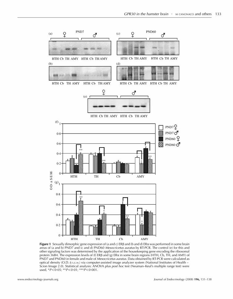
Figure 1 Sexually dimorphic gene expression of (a and c) ERb and (b and d) ERa was performed in some brainareas of (a and b) PND7 and (c and d) PND60 Mesocricetus auratus by RT-PCR. The control (e) for this andother signaling factors was determined by the application of the housekeeping gene encoding the ribosomalprotein 36B4. The expression levels of (f) ERb and (g) ERa in some brain regions (HTH, Cb, TH, and AMY) ofPND7 and PND60 in female and male of Mesocricetus auratus. Data obtained by RT-PCR were calculated asoptical density (O.D.GS.E.M.) via computer-assisted image analyzer system (National Institutes of Health –Scion Image 2.0). Statistical analysis: ANOVA plus post hoc test (Neuman–Keul’s multiple range test) wereused, *P!0.05; **P!0.01; ***P!0.001.
GPR30 in the hamster brain . M CANONACO and others 133
www.endocrinology-journals.org Journal of Endocrinology (2008) 196, 131–138
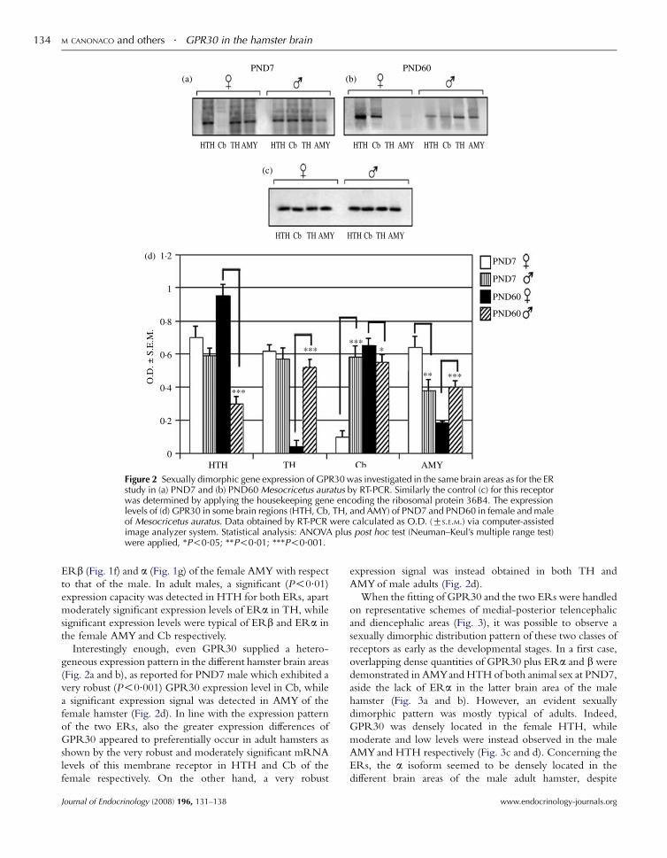
Figure 2 Sexually dimorphic gene expression of GPR30 was investigated in the same brain areas as for the ERstudy in (a) PND7 and (b) PND60 Mesocricetus auratus by RT-PCR. Similarly the control (c) for this receptorwas determined by applying the housekeeping gene encoding the ribosomal protein 36B4. The expressionlevels of (d) GPR30 in some brain regions (HTH, Cb, TH, and AMY) of PND7 and PND60 in female and maleof Mesocricetus auratus. Data obtained by RT-PCR were calculated as O.D. (GS.E.M.) via computer-assistedimage analyzer system. Statistical analysis: ANOVA plus post hoc test (Neuman–Keul’s multiple range test)were applied, *P!0.05; **P!0.01; ***P!0.001.
M CANONACO and others . GPR30 in the hamster brain134
ERb (Fig. 1f) and a (Fig. 1g) of the female AMY with respect
to that of the male. In adult males, a significant (P!0.01)expression capacity was detected in HTH for both ERs, apart
moderately significant expression levels of ERa in TH, while
significant expression levels were typical of ERb and ERa in
the female AMY and Cb respectively.
Interestingly enough, even GPR30 supplied a hetero-
geneous expression pattern in the different hamster brain areas
(Fig. 2a and b), as reported for PND7 male which exhibited a
very robust (P!0.001) GPR30 expression level in Cb, while
a significant expression signal was detected in AMY of the
female hamster (Fig. 2d). In line with the expression pattern
of the two ERs, also the greater expression differences of
GPR30 appeared to preferentially occur in adult hamsters as
shown by the very robust and moderately significant mRNA
levels of this membrane receptor in HTH and Cb of the
female respectively. On the other hand, a very robust
Journal of Endocrinology (2008) 196, 131–138
expression signal was instead obtained in both TH and
AMYof male adults (Fig. 2d).
When the fitting of GPR30 and the two ERs were handled
on representative schemes of medial-posterior telencephalic
and diencephalic areas (Fig. 3), it was possible to observe a
sexually dimorphic distribution pattern of these two classes of
receptors as early as the developmental stages. In a first case,
overlapping dense quantities of GPR30 plus ERa and b were
demonstrated in AMYand HTH of both animal sex at PND7,
aside the lack of ERa in the latter brain area of the male
hamster (Fig. 3a and b). However, an evident sexually
dimorphic pattern was mostly typical of adults. Indeed,
GPR30 was densely located in the female HTH, while
moderate and low levels were instead observed in the male
AMYand HTH respectively (Fig. 3c and d). Concerning the
ERs, the a isoform seemed to be densely located in the
different brain areas of the male adult hamster, despite
www.endocrinology-journals.org
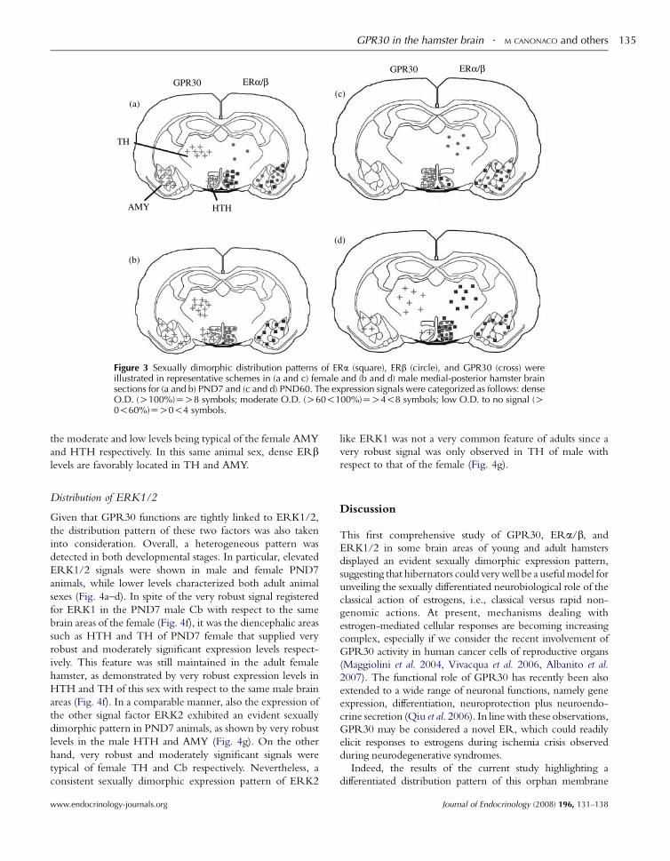
Figure 3 Sexually dimorphic distribution patterns of ERa (square), ERb (circle), and GPR30 (cross) wereillustrated in representative schemes in (a and c) female and (b and d) male medial-posterior hamster brainsections for (a and b) PND7 and (c and d) PND60. The expression signals were categorized as follows: denseO.D. (O100%)ZO8 symbols; moderate O.D. (O60!100%)ZO4!8 symbols; low O.D. to no signal (O0!60%)ZO0!4 symbols.
GPR30 in the hamster brain . M CANONACO and others 135
the moderate and low levels being typical of the female AMY
and HTH respectively. In this same animal sex, dense ERblevels are favorably located in TH and AMY.
Distribution of ERK1/2
Given that GPR30 functions are tightly linked to ERK1/2,
the distribution pattern of these two factors was also taken
into consideration. Overall, a heterogeneous pattern was
detected in both developmental stages. In particular, elevated
ERK1/2 signals were shown in male and female PND7
animals, while lower levels characterized both adult animal
sexes (Fig. 4a–d). In spite of the very robust signal registered
for ERK1 in the PND7 male Cb with respect to the same
brain areas of the female (Fig. 4f), it was the diencephalic areas
such as HTH and TH of PND7 female that supplied very
robust and moderately significant expression levels respect-
ively. This feature was still maintained in the adult female
hamster, as demonstrated by very robust expression levels in
HTH and TH of this sex with respect to the same male brain
areas (Fig. 4f). In a comparable manner, also the expression of
the other signal factor ERK2 exhibited an evident sexually
dimorphic pattern in PND7 animals, as shown by very robust
levels in the male HTH and AMY (Fig. 4g). On the other
hand, very robust and moderately significant signals were
typical of female TH and Cb respectively. Nevertheless, a
consistent sexually dimorphic expression pattern of ERK2
www.endocrinology-journals.org
like ERK1 was not a very common feature of adults since a
very robust signal was only observed in TH of male with
respect to that of the female (Fig. 4g).
Discussion
This first comprehensive study of GPR30, ERa/b, and
ERK1/2 in some brain areas of young and adult hamsters
displayed an evident sexually dimorphic expression pattern,
suggesting that hibernators could verywell be a usefulmodel for
unveiling the sexually differentiated neurobiological role of the
classical action of estrogens, i.e., classical versus rapid non-
genomic actions. At present, mechanisms dealing with
estrogen-mediated cellular responses are becoming increasing
complex, especially if we consider the recent involvement of
GPR30 activity in human cancer cells of reproductive organs
(Maggiolini et al. 2004, Vivacqua et al. 2006, Albanito et al.
2007). The functional role of GPR30 has recently been also
extended to a wide range of neuronal functions, namely gene
expression, differentiation, neuroprotection plus neuroendo-
crine secretion (Qiu et al. 2006). In linewith these observations,
GPR30 may be considered a novel ER, which could readily
elicit responses to estrogens during ischemia crisis observed
during neurodegenerative syndromes.
Indeed, the results of the current study highlighting a
differentiated distribution pattern of this orphan membrane
Journal of Endocrinology (2008) 196, 131–138
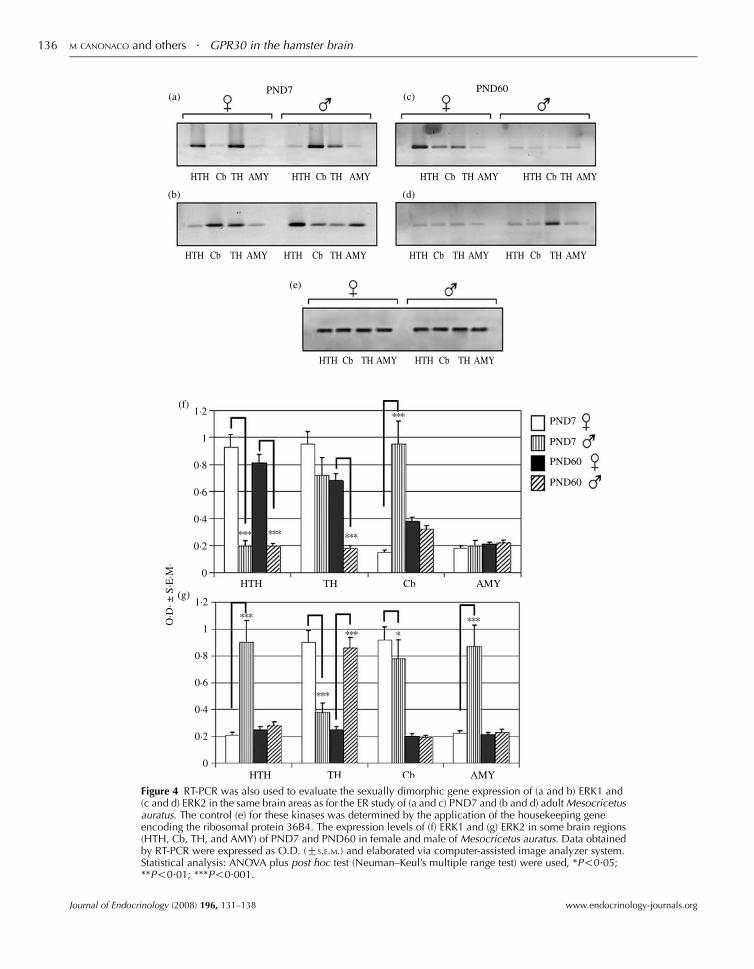
Figure 4 RT-PCR was also used to evaluate the sexually dimorphic gene expression of (a and b) ERK1 and(c and d) ERK2 in the same brain areas as for the ER study of (a and c) PND7 and (b and d) adult Mesocricetusauratus. The control (e) for these kinases was determined by the application of the housekeeping geneencoding the ribosomal protein 36B4. The expression levels of (f) ERK1 and (g) ERK2 in some brain regions(HTH, Cb, TH, and AMY) of PND7 and PND60 in female and male of Mesocricetus auratus. Data obtainedby RT-PCR were expressed as O.D. (GS.E.M.) and elaborated via computer-assisted image analyzer system.Statistical analysis: ANOVA plus post hoc test (Neuman–Keul’s multiple range test) were used, *P!0.05;**P!0.01; ***P!0.001.
M CANONACO and others . GPR30 in the hamster brain136
Journal of Endocrinology (2008) 196, 131–138 www.endocrinology-journals.org
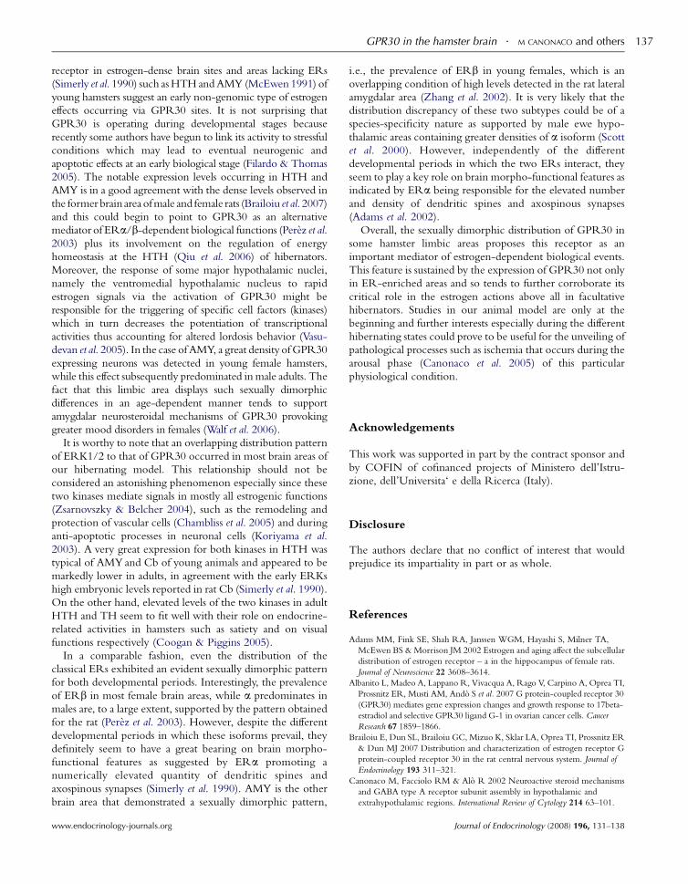
GPR30 in the hamster brain . M CANONACO and others 137
receptor in estrogen-dense brain sites and areas lacking ERs
(Simerly et al. 1990) such asHTH andAMY (McEwen 1991) of
young hamsters suggest an early non-genomic type of estrogen
effects occurring via GPR30 sites. It is not surprising that
GPR30 is operating during developmental stages because
recently some authors have begun to link its activity to stressful
conditions which may lead to eventual neurogenic and
apoptotic effects at an early biological stage (Filardo & Thomas
2005). The notable expression levels occurring in HTH and
AMY is in a good agreement with the dense levels observed in
the former brain area ofmale and female rats (Brailoiu et al. 2007)
and this could begin to point to GPR30 as an alternative
mediator of ERa/b-dependent biological functions (Perez et al.2003) plus its involvement on the regulation of energy
homeostasis at the HTH (Qiu et al. 2006) of hibernators.
Moreover, the response of some major hypothalamic nuclei,
namely the ventromedial hypothalamic nucleus to rapid
estrogen signals via the activation of GPR30 might be
responsible for the triggering of specific cell factors (kinases)
which in turn decreases the potentiation of transcriptional
activities thus accounting for altered lordosis behavior (Vasu-
devan et al. 2005). In the case of AMY, a great density ofGPR30
expressing neurons was detected in young female hamsters,
while this effect subsequently predominated inmale adults. The
fact that this limbic area displays such sexually dimorphic
differences in an age-dependent manner tends to support
amygdalar neurosteroidal mechanisms of GPR30 provoking
greater mood disorders in females (Walf et al. 2006).
It is worthy to note that an overlapping distribution pattern
of ERK1/2 to that of GPR30 occurred in most brain areas of
our hibernating model. This relationship should not be
considered an astonishing phenomenon especially since these
two kinases mediate signals in mostly all estrogenic functions
(Zsarnovszky & Belcher 2004), such as the remodeling and
protection of vascular cells (Chambliss et al. 2005) and during
anti-apoptotic processes in neuronal cells (Koriyama et al.
2003). A very great expression for both kinases in HTH was
typical of AMYand Cb of young animals and appeared to be
markedly lower in adults, in agreement with the early ERKs
high embryonic levels reported in rat Cb (Simerly et al. 1990).
On the other hand, elevated levels of the two kinases in adult
HTH and TH seem to fit well with their role on endocrine-
related activities in hamsters such as satiety and on visual
functions respectively (Coogan & Piggins 2005).
In a comparable fashion, even the distribution of the
classical ERs exhibited an evident sexually dimorphic pattern
for both developmental periods. Interestingly, the prevalence
of ERb in most female brain areas, while a predominates in
males are, to a large extent, supported by the pattern obtained
for the rat (Perez et al. 2003). However, despite the different
developmental periods in which these isoforms prevail, they
definitely seem to have a great bearing on brain morpho-
functional features as suggested by ERa promoting a
numerically elevated quantity of dendritic spines and
axospinous synapses (Simerly et al. 1990). AMY is the other
brain area that demonstrated a sexually dimorphic pattern,
www.endocrinology-journals.org
i.e., the prevalence of ERb in young females, which is an
overlapping condition of high levels detected in the rat lateral
amygdalar area (Zhang et al. 2002). It is very likely that the
distribution discrepancy of these two subtypes could be of a
species-specificity nature as supported by male ewe hypo-
thalamic areas containing greater densities of a isoform (Scott
et al. 2000). However, independently of the different
developmental periods in which the two ERs interact, they
seem to play a key role on brain morpho-functional features as
indicated by ERa being responsible for the elevated number
and density of dendritic spines and axospinous synapses
(Adams et al. 2002).
Overall, the sexually dimorphic distribution of GPR30 in
some hamster limbic areas proposes this receptor as an
important mediator of estrogen-dependent biological events.
This feature is sustained by the expression of GPR30 not only
in ER-enriched areas and so tends to further corroborate its
critical role in the estrogen actions above all in facultative
hibernators. Studies in our animal model are only at the
beginning and further interests especially during the different
hibernating states could prove to be useful for the unveiling of
pathological processes such as ischemia that occurs during the
arousal phase (Canonaco et al. 2005) of this particular
physiological condition.
Acknowledgements
This work was supported in part by the contract sponsor and
by COFIN of cofinanced projects of Ministero dell’Istru-
zione, dell’Universita‘ e della Ricerca (Italy).
Disclosure
The authors declare that no conflict of interest that would
prejudice its impartiality in part or as whole.
References
Adams MM, Fink SE, Shah RA, Janssen WGM, Hayashi S, Milner TA,
McEwen BS &Morrison JM 2002 Estrogen and aging affect the subcellular
distribution of estrogen receptor – a in the hippocampus of female rats.
Journal of Neuroscience 22 3608–3614.
Albanito L, Madeo A, Lappano R, Vivacqua A, Rago V, Carpino A, Oprea TI,
Prossnitz ER, Musti AM, Ando S et al. 2007 G protein-coupled receptor 30
(GPR30) mediates gene expression changes and growth response to 17beta-
estradiol and selective GPR30 ligand G-1 in ovarian cancer cells. Cancer
Research 67 1859–1866.
Brailoiu E, Dun SL, Brailoiu GC, Mizuo K, Sklar LA, Oprea TI, Prossnitz ER
& Dun MJ 2007 Distribution and characterization of estrogen receptor G
protein-coupled receptor 30 in the rat central nervous system. Journal of
Endocrinology 193 311–321.
Canonaco M, Facciolo RM & Alo R 2002 Neuroactive steroid mechanisms
and GABA type A receptor subunit assembly in hypothalamic and
extrahypothalamic regions. International Review of Cytology 214 63–101.
Journal of Endocrinology (2008) 196, 131–138

M CANONACO and others . GPR30 in the hamster brain138
Canonaco M, Madeo M, Alo R, Giusi G, Granata T, Carelli A, Canonaco A
& Facciolo RM 2005 The histaminergic signaling system exerts a protective
role against neurodegenerative-induced processes in the hamster. Journal of
Pharmacology and Experimental Therapeutics 315 188–195.
Chambliss KL, Simon L, Yuhanna IS, Mineo C & Shaul PW 2005 Dissecting
the basis of nongenomic activation of endothelial nitric oxide synthase by
estradiol: Role of ERalpha domains with known nuclear functions.
Molecular Endocrinology 19 277–289.
Coogan AN & Piggins HD 2005 Dark pulse suppression of p-ERK and c-Fos
in the hamster suprachiasmatic nuclei. European Journal of Neuroscience 22
158–168.
Filardo EJ & Thomas P 2005 GPR30: a seven-transmembrane spanning
estrogen receptor that triggers EGF release. Trends in Endocrinology and
Metabolism 16 362–367.
Koriyama Y, Chiba K & Mohri T 2003 Propentofylline protects b-amyloid
protein-induced apoptosis in cultured rat hippocampal neurons. European
Journal of Pharmacology 458 235–241.
Kow LM, Easton A & Pfaff DW 2005 Acute estrogen potentiates excitatory
responses of neurons in rat hypothalamic ventromedial nucleus. Brain
Research 1043 124–131.
Maggiolini M, Vivacqua A, Fasanella G, Recchia AG, Sisci D, Pezzi V,
Montanaro D, Musti AM, Picard D & Ando S 2004 The G protein-coupled
receptor GPR30 mediates c-fos up-regulation by 17b-Estradiol andphytoestrogens in breast cancer cells. Journal of Biological Chemistry 279
27008–27016.
McEwen B 1991 Non-genomic and genomic effects of steroids on neural
activity. Trends in Pharmacological Sciences 12 141–146.
Nilsen J & Diaz BR 2003 Mechanism of estrogen-mediated neuroprotection:
regulation of mitochondrial calcium and Bcl-2 expression. PNAS 100
2842–2847.
Paxinos G &Watson C 1982 The Rat Brain in Stereotaxic Coordinates., New
York: Academic Press.
Perez SE, Chen EY &Mufson EJ 2003 Distribution of estrogen receptor alpha
and beta immunoreactive profiles in the postnatal rat brain. Developmental
Brain Research 145 117–139.
Qiu J, Bosch MA, Tobias SC, Krust A, Graham SM, Murphy SJ, Korach KS,
Chambon P, Scanlan TS, Rønnekleiv OK et al. 2006 A G-protein coupled
estrogen receptor is involved in hypothalamic control of energy
homeostasis. Journal of Neuroscience 26 5649–5655.
Journal of Endocrinology (2008) 196, 131–138
Scott CJ, Tilbrook AJ, Simmons DM, Rawson JA, Chu S, Fuller PJ, Ing NH &
Clark IJ 2000 The distribution of cells containing estrogen receptor-(ER) and
ER messenger ribonucleic acid in the preoptic area and hypothalamus of the
sheep: scomparison of males and females. Endocrinology 141 291–2962.
Simerly RB, Chang C, Muramatsu M & Swanson LW 1990 Distribution of
androgen and estrogen receptor mRNA-containing cells in the rat brain: an
in situ hybridization story. Journal of Comparative Neurology 294 76–95.
Simpson ER,MissoM,Hewitt KN,Hill RA, BoonWC, JonesME, Kovacic A,
Zhou J & Clyne CD 2005 Estrogen – the good, the bad, and the unexpected.
Endocrine Reviews 26 322–330.
Sweatt JD 2004 Mitogen-activated protein kinases in synaptic plasticity and
memory. Current Opinion in Neurobiology 14 311–317.
UFAW Handbook on the Care and Management of Laboratory Animals,
Universities Federation for Animal Welfare at The Old School, Brewhouse
Hill, Wheathampstead, Herts AL4 8AN, UK.
Vasudevan N, Kow LM & Pfaff D 2005 Integration of steroid hormone
initiated membrane membrane action to genomic function in the brain.
Steroids 70 388–396.
Vivacqua A, Bonofiglio D, Recchia AG, Musti AM, Picard D, Ando S &
Maggiolini M 2006 The G protein-coupled receptor GPR30 mediates the
proliferative effects induced by 17b-estradiol and hydroxytamoxifen in
endometrial cancer cells. Molecular Endocrinology 20 631–646.
Walf AA, Sumida K & Frye CA 2006 Inhibiting 5a-reductase in the amygdala
attenuates antianxiety and antidepressive behavior of naturally receptive and
hormone-primed ovariectomized rats. Psychopharmacology 186 302–311.
Zhang JQ, Cai WQ, Zhou DS & Su BY 2002 Distribution and differences of
estrogen receptor beta immunoreactivity in the brain of adult male and
female rats. Brain Research 935 73–80.
Zsarnovszky A & Belcher SM 2004 Spatial, temporal, and cellular distribution
of the activated extracellular signal regulated kinases 1 and 2 in the
developing and mature rat cerebellum. Developmental Brain Research 150
199–209.
Received in final form 4 October 2007Accepted 24 October 2007Made available online as an Accepted Preprint24 October 2007
www.endocrinology-journals.org
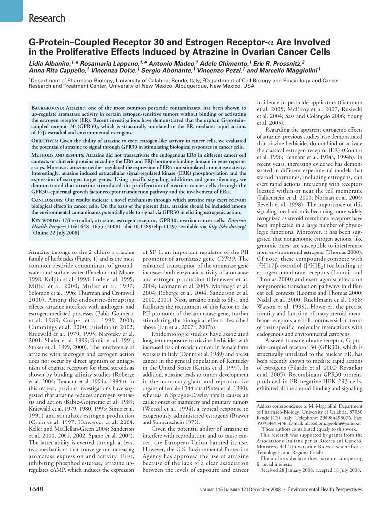
1648 VOLUME 116 | NUMBER 12 | December 2008 • Environmental Health Perspectives
Research
Atrazine belongs to the 2-chloro-s-triazinefamily of herbicides (Figure 1) and is the mostcommon pesticide contaminant of ground-water and surface water (Fenelon and Moore1998; Kolpin et al. 1998; Lode et al. 1995;Miller et al. 2000; Müller et al. 1997;Solomon et al. 1996; Thurman and Cromwell2000). Among the endocrine-disruptingeffects, atrazine interferes with androgen- andestrogen-mediated processes (Babic-Gojmeracet al. 1989; Cooper et al. 1999, 2000;Cummings et al. 2000; Friedmann 2002;Kniewald et al. 1979, 1995; Narotsky et al.2001; Shafer et al. 1999; Simic et al. 1991;Stoker et al. 1999, 2000). The interference ofatrazine with androgen and estrogen actiondoes not occur by direct agonism or antago-nism of cognate receptors for these steroids asshown by binding affinity studies (Robergeet al. 2004; Tennant et al. 1994a, 1994b). Inthis respect, previous investigations have sug-gested that atrazine reduces androgen synthe-sis and action (Babic-Gojmerac et al. 1989;Kniewald et al. 1979, 1980, 1995; Simic et al.1991) and stimulates estrogen production(Crain et al. 1997; Heneweer et al. 2004;Keller and McClellan-Green 2004; Sandersonet al. 2000, 2001, 2002; Spano et al. 2004).The latter ability is exerted through at leasttwo mechanisms that converge on increasingaromatase expression and activity. First,inhibiting phosphodiesterase, atrazine up-regulates cAMP, which induces the expression
of SF-1, an important regulator of the PIIpromoter of aromatase gene CYP19. Theenhanced transcription of the aromatase geneincreases both enzymatic activity of aromataseand estrogen production (Heneweer et al.2004; Lehmann et al. 2005; Morinaga et al.2004; Roberge et al. 2004; Sanderson et al.2000, 2001). Next, atrazine binds to SF-1 andfacilitates the recruitment of this factor to thePII promoter of the aromatase gene, furtherstimulating the biological effects describedabove (Fan et al. 2007a, 2007b).
Epidemiologic studies have associatedlong-term exposure to triazine herbicides withincreased risk of ovarian cancer in female farmworkers in Italy (Donna et al. 1989) and breastcancer in the general population of Kentuckyin the United States (Kettles et al. 1997). Inaddition, atrazine leads to tumor developmentin the mammary gland and reproductiveorgans of female F344 rats (Pintér et al. 1990),whereas in Sprague-Dawley rats it causes anearlier onset of mammary and pituitary tumors(Wetzel et al. 1994), a typical response toexogenously administered estrogens (Brawerand Sonnenschein 1975).
Given the potential ability of atrazine tointerfere with reproduction and to cause can-cer, the European Union banned its use.However, the U.S. Environmental ProtectionAgency has approved the use of atrazinebecause of the lack of a clear associationbetween the levels of exposure and cancer
incidence in pesticide applicators (Gammonet al. 2005; McElroy et al. 2007; Rusieckiet al. 2004; Sass and Colangelo 2006; Younget al. 2005).
Regarding the apparent estrogenic effectsof atrazine, previous studies have demonstratedthat triazine herbicides do not bind or activatethe classical estrogen receptor (ER) (Connoret al. 1996; Tennant et al. 1994a, 1994b). Inrecent years, increasing evidence has demon-strated in different experimental models thatsteroid hormones, including estrogens, canexert rapid actions interacting with receptorslocated within or near the cell membrane(Falkenstein et al. 2000; Norman et al. 2004;Revelli et al. 1998). The importance of thissignaling mechanism is becoming more widelyrecognized as steroid membrane receptors havebeen implicated in a large number of physio-logic functions. Moreover, it has been sug-gested that nongenomic estrogen actions, likegenomic ones, are susceptible to interferencefrom environmental estrogens (Thomas 2000).Of note, these compounds compete with[3H]17β-estradiol ([3H]E2) for binding toestrogen membrane receptors (Loomis andThomas 2000) and exert agonist effects onnongenomic transduction pathways in differ-ent cell contexts (Loomis and Thomas 2000;Nadal et al. 2000; Ruehlmann et al. 1988;Watson et al. 1999). However, the preciseidentity and function of many steroid mem-brane receptors are still controversial in termsof their specific molecular interactions withendogenous and environmental estrogens.
A seven-transmembrane receptor, G-pro-tein–coupled receptor 30 (GPR30), which isstructurally unrelated to the nuclear ER, hasbeen recently shown to mediate rapid actionsof estrogens (Filardo et al. 2002; Revankaret al. 2005). Recombinant GPR30 protein,produced in ER-negative HEK-293 cells,exhibited all the steroid binding and signaling
Address correspondence to M. Maggiolini, Departmentof Pharmaco-Biology, University of Calabria, 87030Rende (CS), Italy. Telephone: 390984493076. Fax:390984493458. E-mail: [email protected]
*These authors contributed equally to this work.This research was supported by grants from the
Associazione Italiana per la Ricerca sul Cancro,Ministero dell’Università e Ricerca Scientifica eTecnologica, and Regione Calabria.
The authors declare they have no competingfinancial interests.
Received 28 January 2008; accepted 18 July 2008.
G-Protein–Coupled Receptor 30 and Estrogen Receptor-α Are Involved in the Proliferative Effects Induced by Atrazine in Ovarian Cancer CellsLidia Albanito,1,* Rosamaria Lappano,1,* Antonio Madeo,1 Adele Chimento,1 Eric R. Prossnitz,2
Anna Rita Cappello,1 Vincenza Dolce,1 Sergio Abonante,1 Vincenzo Pezzi,1 and Marcello Maggiolini1
1Department of Pharmaco-Biology, University of Calabria, Rende, Italy; 2Department of Cell Biology and Physiology and CancerResearch and Treatment Center, University of New Mexico, Albuquerque, New Mexico, USA
BACKGROUND: Atrazine, one of the most common pesticide contaminants, has been shown toup-regulate aromatase activity in certain estrogen-sensitive tumors without binding or activatingthe estrogen receptor (ER). Recent investigations have demonstrated that the orphan G-protein–coupled receptor 30 (GPR30), which is structurally unrelated to the ER, mediates rapid actionsof 17β-estradiol and environmental estrogens.
OBJECTIVES: Given the ability of atrazine to exert estrogen-like activity in cancer cells, we evaluatedthe potential of atrazine to signal through GPR30 in stimulating biological responses in cancer cells.
METHODS AND RESULTS: Atrazine did not transactivate the endogenous ERα in different cancer cellcontexts or chimeric proteins encoding the ERα and ERβ hormone-binding domain in gene reporterassays. Moreover, atrazine neither regulated the expression of ERα nor stimulated aromatase activity.Interestingly, atrazine induced extracellular signal-regulated kinase (ERK) phosphorylation and theexpression of estrogen target genes. Using specific signaling inhibitors and gene silencing, wedemonstrated that atrazine stimulated the proliferation of ovarian cancer cells through theGPR30–epidermal growth factor receptor transduction pathway and the involvement of ERα.
CONCLUSIONS: Our results indicate a novel mechanism through which atrazine may exert relevantbiological effects in cancer cells. On the basis of the present data, atrazine should be included amongthe environmental contaminants potentially able to signal via GPR30 in eliciting estrogenic action.
KEY WORDS: 17β-estradiol, atrazine, estrogen receptor, GPR30, ovarian cancer cells. EnvironHealth Perspect 116:1648–1655 (2008). doi:10.1289/ehp.11297 available via http://dx.doi.org/[Online 22 July 2008]
characteristics of a functional estrogen mem-brane receptor (Thomas et al. 2005; Thomasand Dong 2006). Our studies and others havealso demonstrated that GPR30 mediates therapid response to E2 in a variety of estrogen-responsive cancer cells by activating the epi-dermal growth factor receptor (EGFR)–mitogen-activated protein kinase (MAPK)transduction pathway (Albanito et al. 2007;Bologa et al. 2006; Filardo et al. 2000;Maggiolini et al. 2004; Revankar et al. 2005;Thomas et al. 2005; Vivacqua et al. 2006a,2006b).
In the present study, for the first time wehave demonstrated that atrazine stimulatesgene expression and growth effects in estrogen-sensitive ovarian cancer cells through GPR30and the involvement of ERα. Moreover, weshow that GPR30 mediates the stimulatoryeffects of atrazine in ER-negative SkBr3 breastcancer cells.
Materials and Methods
Reagents. We purchased atrazine [2-chloro-4-(ethylamine)-6-(isopropylamine)-s-triazine],17β-estradiol (E2), N-[2-(p-bromocinnamyl-amino)ethyl]-5-isoquinolinesulfonamidedihydrochloride (H89), wortmannin (WM),and PD98059 (PD) from Sigma-Aldrich(Milan, Italy); AG1478 (AG) from BiomolResearch Laboratories (DBA, Milan, Italy);ICI 182,780 (ICI) from Tocris Chemicals(Bristol, UK); and GF109203X (GFX) fromCalbiochem (VWR International, Milan,Italy). All compounds were solubilized indimethyl sulfoxide (DMSO), except E2 andPD, which were dissolved in ethanol.
Cell culture. Human BG-1 and 2008 ovar-ian cancer cells as well as human Ishikawaendometrial cancer cells were maintained inphenol red–free Dulbecco’s modified Eaglemedium (DMEM) supplemented with 10%fetal bovine serum (FBS). H295R adrenal carci-noma cells were cultured in DMEM/F12 1:1supplemented with 1% ITS Liquid MediaSupplement (Sigma-Aldrich), 10% calf serum,and antibiotics. Human MCF-7 breast cancercells were maintained in DMEM with phenolred supplemented with 10% FBS, and humanSkBr3 breast cancer cells were maintained inphenol red–free RPMI 1640 supplementedwith 10% FBS. Cells were switched to mediumwithout serum the day before experimentsfor immunoblots and reverse transcription-polymerase chain reaction (RT-PCR).
Plasmids. Firefly luciferase reporter plas-mids used were XETL for ERα (Bunone et al.1996) and GK1 for yeast transcription factorGal4 fusion proteins (Webb et al. 1998).XETL contains the estrogen response element(ERE) from the Xenopus vitellogenin A2 gene(nucleotides –334 to –289), the herpes simplexvirus thymidine kinase promoter region(nucleotides –109 to +52), the firefly luciferase
coding sequence, and the SV40 splice andpolyadenylation sites from plasmid pSV232A/L-AA5. Gal4 chimeras Gal-ERα and Gal-ERβwere expressed from plasmids GAL93.ER(G)and GAL.ERβ, respectively. They were con-structed by transferring the coding sequencesfor the hormone-binding domain (HBD) ofERα (amino acids 282–595) from HEG0(Bunone et al. 1996), and for the ERβ HBD(C-terminal 287 amino acids) from plasmidpCMV5-hERβ into the mammalian expres-sion vector pSCTEVGal93 (Seipel et al. 1992).We used the Renilla luciferase expressionvector pRL-TK (Promega, Milan, Italy) as atransfection standard.
Transfection and luciferase assays. BG-1,MCF-7, Ishikawa, and SkBr3 cells (1 × 10⁵)were plated into 24-well dishes with500 µL/well DMEM (BG-1, MCF-7, andIshikawa cells) or RPMI 1640 (SkBr3 cells)containing 10% FBS the day before transfec-tion. We replaced the medium with phenolred–free DMEM or RPMI 1640, both sup-plemented with 1% charcoal-stripped (CS)FBS, on the day of transfection. Transfectionswere performed using FuGENE 6 Reagent asrecommended by the manufacturer (RocheDiagnostics, Mannheim, Germany) with amixture containing 0.3 µg of reporter plas-mid, 1 ng pRL-TK, and 0.1 µg effector plas-mid where applicable. After 5–6 hr, themedium was replaced again with serum-freeDMEM lacking phenol red and supple-mented with 1% CS-FBS; ligands were addedat this point, and cells were incubated for16–18 hr. We measured luciferase activitywith the Dual Luciferase Kit (Promega)according to the manufacturer’s recommen-dations. Firefly luciferase values were normal-ized to the internal transfection controlprovided by Renilla luciferase activity. Thenormalized relative light unit values obtainedfrom cells treated with vehicle were set as1-fold induction, from which the activityinduced by treatments was calculated.
RT-PCR. Using semiquantitative RT-PCRas described previously (Maggiolini et al.1999), we evaluated gene expression for ERα[GenBank accession no. NM 000125 (NationalCenter for Biotechnology Information 2008)],c-fos (NM 005252), progesterone receptor(PR; NM 000926), pS2 (NM 003225),cathepsin D (NM 001909), cyclin A (NM001237), cyclin D1 (NM 053056), cyclin E(NM 001238), and the acid phosphoproteinP0 (36B4) (NM 001002) used as a controlgene. We used the primers 5´-AATTCA-GATAATCGACGCCAG-3´ (ERα forward)and 5´-GTGTTTCAACATTCTCCCTC-CTC-3´ (ERα reverse); 5´-AGAAAAGGA-GAATCCGAAGGGAAA-3´ (c-fos forward)and 5´-ATGATGCTGGGACAGGAAG-TC-3´ (c-fos reverse); 5´-ACACCTTGC-CTGAAGTTTCG-3´ (PR forward) and
5´-CTGTCCTTTTCTGGGGGACT-3´(PR reverse); 5´-TTCTATCCTAATAC-CATCGACG-3´ (pS2 forward) and5´-TTTGAGTAGTCAAAGTCAGAGC-3´(pS2 reverse); 5´-AACAACAGGGTGGGCTTC-3´ (cathepsin D forward), and5´-ATGCACGAAACAGATCTGTGCT-3´(cathepsin D reverse); 5´-GCCATTAGTT-TACCTGGACCCAGA-3´ (cyclin A for-ward) and 5´-CACTGACATGGAAGACAGGAACCT-3´ (cyclin A reverse); 5´-TCTAA-GATGAAGGAGACCATC-3´, (cyclin D1forward) and 5´-GCGGTAGTAGGACAGGAAGTTGTT-3´ (cyclin D1 reverse);5´-CCTGACTATTGTGTCCTGGC-3´(cyclin E forward) and 5´-CCCGCT-GCTCTGCTTCTTAC-3´ (cyclin E reverse);and 5´-CTCAACATCTCCCCCTTCTC-3´(36B4 forward) and 5´-CAAATCCCA-TATCCTCGTCC-3´ (36B4 reverse) to yieldproducts of 345, 420, 196, 210, 303, 354,354, 488, and 408 bp, respectively, with20 PCR cycles for ERα, c-fos, PR, pS2, cathep-sin D, cyclin A, and cyclin E and 15 PCRcycles for both cyclin D1 and 36B4.
Western blotting. Cells were grown in10-cm dishes, exposed to ligands, and thenlysed in 500 µL of 50 mmol/L NaCl, 1.5mmol/L MgCl2, 1 mmol/L EGTA, 10%glycerol, 1% Triton X-100, 1% sodium dode-cyl sulfate (SDS), and a mixture of proteaseinhibitors containing 1 mmol/L aprotinin,20 mmol/L phenylmethylsulfonyl fluoride, and200 mmol/L sodium orthovanadate. We thendiluted samples 10 times and determined pro-tein concentration using Bradford reagentaccording to the manufacturer’s recommenda-tions (Sigma-Aldrich). Equal amounts of wholeprotein extract were resolved on a 10% SDS-polyacrylamide gel and transferred to a nitro-cellulose membrane (Amersham Biosciences,Milan, Italy). Membranes were probedovernight at 4°C with the antibody againstERα (F-10), c-fos (H-125), β-actin (C-2),phosphorylated extracellular signal-regulatedkinase 1/2 (ERK1/2; E-4), and ERK2 (C-14),all purchased from Santa Cruz Biotechnology,DBA (Milan, Italy), and human P450 aro-matase (MCA 2077S; Serotec, Milan, Italy),and then revealed using the ECL WesternBlotting Analysis System (GE Healthcare,Milan, Italy).
ER binding assay. BG-1 cells were strippedof any estrogen by keeping them in mediumwithout serum for 2 days. Cells were incubated
Atrazine action in ovarian cancer cells
Environmental Health Perspectives • VOLUME 116 | NUMBER 12 | December 2008 1649
Figure 1. Structures of E2 and atrazine.
OHCI
HO
H3C NH NH
H3C CH
3
NN
N
E2
Atrazine
with 1 nM [2,4,6,7-3H]E2 (89 Ci/mmol;Amersham Biosciences) and increasing concen-trations of nonlabeled E2 or atrazine for 1 hr at37°C in a humidified atmosphere of 95%air/5% CO2. After removal of the medium,cells were washed with ice-cold phosphate-buffered saline/0.1% methylcellulose twice,harvested by scraping and centrifugation, andlysed with 100% ethanol, 500 µL/60-mmdish, for 10 min at room temperature (Lee andGorski 1996). We measured the radioactivityof extracts by liquid scintillation counting.
Aromatase assay. In subconfluent BG-1 orH295R cells, we measured aromatase activityin the cell culture medium by tritiated waterrelease using 0.5 µM [1β-3H(N)]androst-4-ene-3,17-dione (25.3 Ci/mmol; DuPontNEN, Boston, MA, USA) as a substrate(Lephart and Simpson 1991). The cells weretreated in a six-well dish in culture medium inthe presence of atrazine or DMSO for 40 hrand then incubated with [1β-3H(N)]androst-4-ene-3,17-dione. Incubations were per-formed at 37°C for 6 hr under a 95%/5%
air/CO2 atmosphere. The results were calcu-lated as picomoles per hour, normalized tomilligrams of protein (pmol/hr per 1 mg pro-tein), and expressed as percentages ofuntreated cells (100%).
GPR30 and ERα silencing experiments.Cells were plated onto 10-cm dishes, main-tained in antibiotic-free medium for 24 hr,and then transfected for additional 24 hrbefore treatments with a mixture containingOpti-MEM, 8 µL/well LipofectAMINE 2000(Invitrogen, Milan, Italy), and 0.5 µg/wellvector or short hairpin GPR30 (shGPR30)(Albanito et al. 2008), control small inter-fering RNA (siRNA), or ERα siRNA (Sigma-Aldrich).
Proliferation assay. For the quantitativeproliferation assay, we seeded 10,000 cells in24-well plates in regular growth medium. Cellswere washed once they had attached and thenincubated in medium containing 2.5%CS-FBS with the indicated treatments.Medium was renewed every 2 days (with treat-ments), and cells were trypsinized and countedin a hemocytometer on day 6. The day beforetreatments, 200 ng/L of the indicated shorthairpin RNA was transfected using FuGENE 6Reagent as recommended by the manufacturer,and then renewed every 2 days before counting.
Statistical analysis. Statistical analysis wasperformed using analysis of variance followedby Newman-Keuls testing to determine dif-ferences in means. p-Values < 0.05 are consid-ered statistically significant.
Results
Atrazine does not activate ERα in cancer cells.Based on the evidence that atrazine producesearly onset and increased incidence of estro-gen-sensitive tumors in different experimentalmodels (Cooper et al. 2007), we first evaluatedwhether atrazine could activate a transientlytransfected ER reporter gene in estrogen-sensi-tive ovarian (BG-1), breast (MCF-7), andendometrial (Ishikawa) cancer cells. Exposureto 100 nM E2 induced a strong ERα transacti-vation that was absent in the presence of10 µM of the ER antagonist ICI in all cellcontexts evaluated (Figure 2A–C). In contrast,treatments with 1 µM atrazine and even con-centrations ranging from 1 nM to 10 µM(data not shown) failed to stimulate luciferaseexpression or to block that observed uponaddition of E2 (Figure 2A–C). Moreover,atrazine did not activate an expression vectorencoding ERα transiently transfected in ER-negative SkBr3 breast cancer cells (Figure 2D).To confirm that atrazine is not an ERα ago-nist and to examine whether ERβ couldrespond to atrazine, we turned to a completelyheterologous system. Chimeric proteins con-sisting of the DNA binding domain of theyeast transcription factor Gal4 and the ERα orERβ HBD transiently transfected in SkBr3
Albanito et al.
1650 VOLUME 116 | NUMBER 12 | December 2008 • Environmental Health Perspectives
Figure 2. ERα transactivation in BG-1 (A), MCF-7 (B), and Ishikawa (C) cells transfected with the ERluciferase reporter plasmid XETL (ERE-luc) and treated with 100 nmol/L E2 or 1 μmol/L atrazine (Atr), withand without 10 μmol/L ER antagonist ICI. Luciferase activities were normalized to the internal transfectioncontrol, and values of cells receiving vehicle (–) were set as 1-fold induction, from which the activityinduced by treatments was calculated. (D–F) SkBr3 cells were transfected with ER luciferase reportergene XETL and ERα expression plasmid (D) and with Gal4 reporter gene (GK1) and the Gal4 fusion proteinsencoding the HBD of ERα (GalERα; E) and or ERβ (GalERβ; F) and treated with 100 nmol/L E2 or 1 μmol/Latrazine, with and without 10 μmol/L ICI. Values shown are mean ± SD of three independent experimentsperformed in triplicate. *p < 0.05 compared with vehicle.
40
30
20
10
1
– E2
Atr ICI E2 +
ICI
E2 +
Atr
ERE-luc
Fire
fly/
Re
nil
la l
uc
ife
rase
ac
tivi
ty (
fold
in
du
cti
on
) 40
30
20
10
1
– E2
Atr ICI E2 +
ICI
E2 +
AtrERE-luc
Fire
fly/
Re
nil
la l
uc
ife
rase
ac
tivi
ty (
fold
in
du
cti
on
) 40
30
20
10
1
– E2
Atr ICI E2 +
ICI
E2 +
Atr
ERE-luc
Fire
fly/
Re
nil
la l
uc
ife
rase
ac
tivi
ty (
fold
in
du
cti
on
)
– E2
Atr ICI E2 +
ICI
E2 +
Atr
ERE-luc + ERα
40
30
20
10
1
Fire
fly/
Re
nil
la l
uc
ife
rase
ac
tivi
ty (
fold
in
du
cti
on
) 40
30
20
10
1
Fire
fly/
Re
nil
la l
uc
ife
rase
ac
tivi
ty (
fold
in
du
cti
on
)
– E2
Atr ICI E2 +
ICI
E2 +
Atr
GK1 + GalERα
40
30
20
10
1
Fire
fly/
Re
nil
la l
uc
ife
rase
ac
tivi
ty (
fold
in
du
cti
on
)
– E2
Atr ICI E2 +
ICI
E2 +
Atr
GK1 + GalERβ
A B C
D E F
BG-1 MCF-7 ISHIKAWA
SkBr3 SkBr3 SkBr3
*
**
*
* *
*
**
*
* *
*
**
*
**
Figure 3. mRNA expression and binding of ERα in BG-1 cells treated for 24 hr with vehicle (–), 100 nmol/LE2, or 1 μmol/L atrazine (Atr). (A) mRNA expression of ERα was evaluated by semiquantitative RT-PCR; thevalues of housekeeping gene 36B4 were determined as a control. (B) Immunoblot of ERα from BG-1 cells,with 100 nmol β-actin serving as a loading control. Results in (A) and (B) are representative of three inde-pendent experiments. (C) ERα binding assay using increasing concentrations of atrazine.
100
75
50
25
Sp
ec
ific
bin
din
g (
%)
Competitor (M)–10 –9 –8 –7 –6 –5
– E2
Atr
ERα
36B4
– E2
Atr
β-Actin
ERα
A
B
E2
Atr
C
Protein
mRNA
cells were strongly activated by E2 but notupon atrazine treatment (Figure 2E,F), furthercorroborating the aforementioned results.
Atrazine neither regulates ERα expressionnor competes with estrogen binding to ERα.Considering that the down-regulation of ERαinduced by an agonist has been considered anadditional hallmark of receptor activation(Santagati et al. 1997), we further investigatedwhether atrazine could modulate ERα expres-sion in BG-1 cells, which lack ERβ (data notshown), and express a receptor expression pat-tern similar to that found in primary ovariantumors (Bardin et al. 2004; Geisinger et al.1989). As shown in Figure 3A,B, 100 nM E2down-regulated ERα at both mRNA andprotein levels, whereas 1 µM atrazine did notproduce any modulatory effect. In agreementwith these results and those obtained in trans-fection experiments, atrazine showed no bind-ing capacity for ERα (Figure 3C), as previouslyreported (Cooper et al. 2007). Altogether, ourfindings rule out that the estrogen action ofatrazine occurs through binding and directactivation of ERα.
Aromatase activity is not induced byatrazine. Given that atrazine is able to up-regulate aromatase expression and function indifferent cell contexts (Cooper et al. 2007;Fan et al. 2007a, 2007b; Roberge et al. 2004;Sanderson et al. 2000, 2001), we then deter-mined aromatase activity by tritiated water
release assays in BG-1 cells. As shown inFigure 4, 1 µM atrazine did not stimulate aro-matase activity, which in contrast wasstrongly induced in human H295R adreno-corticocarcinoma cells previously used as amodel system to assess aromatase catalyticactivity (Heneweer et al. 2004; Sandersonet al. 2001). In addition, the low aromataseprotein expression detected in BG-1 cells didnot increase upon exposure to 1 µM atrazine(data not shown). Hence, atrazine is neitheran ERα activator nor an aromatase regulatorin estrogen-sensitive ovarian cancer cells.
ERK phosphorylation is stimulated byatrazine. In recent years, numerous reportshave demonstrated that estrogens and xeno-estrogens can generate rapid signaling via sec-ond messenger systems such as Ca2+, cAMP,nitric oxide, and G-proteins, which in turnleads to activation of different downstreamkinases (Bulayeva and Watson 2004; Watsonet al. 2007).
To evaluate whether the potential estro-genic activity of atrazine is exerted through arapid cellular response, we investigated itsability to produce ERK phosphorylation inBG-1 cells. Interestingly, atrazine stimulated
ERK phosphorylation, although a higher con-centration and prolonged time period wererequired to trigger this biochemical responsecompared with E2 (Figures 5A,B, 6A). ERKactivation was also delayed in the presence of1 µM atrazine compared with 100 nM E2 in2008 ovarian cancer cells (Figure 6D), whichpresent a receptor expression similar to that ofBG-1 cells (Safei et al. 2005). To determinethe transduction pathways involved in ERKactivation by atrazine, cells were exposed to100 nM E2 and 1 µM atrazine along withspecific inhibitors widely used to pinpoint themechanisms contributing to ERK phosphory-lation (Bulayeva and Watson 2004). Of note,the ER antagonist ICI, the EGFR inhibitorAG and the ERK inhibitor PD preventedERK activation induced by both E2 andatrazine, whereas GFX, H89, and WM,inhibitors of protein kinase C (PKC), proteinkinase A (PKA), and phosphoinositide3-kinase (PI3K), respectively, did not(Figure 6B,C,E,F). Considering that in a pre-vious study ICI was able to trigger ERK phos-phorylation (Filardo et al. 2000), we exposedSkBr3 cells to increasing concentrations ofICI. We observed no ERK activation after
Atrazine action in ovarian cancer cells
Environmental Health Perspectives • VOLUME 116 | NUMBER 12 | December 2008 1651
Figure 4. Aromatase activity assessed by tritiatedwater release in BG-1 and H295R cells treated withvehicle (–) or 1 μmol/L atrazine (Atr). Results areexpressed as percentages of untreated cells(100%). Values are mean ± SD of three independentexperiments, each performed in triplicate. *p < 0.05 compared with vehicle.
200
150
100
50
– –Atr Atr
BG-1 H295R
Aro
ma
tase
ac
tivi
ty (
%)
Figure 5. ERK1/2 phosphorylation (pERK1/2) in BG-1cells exposed to increasing concentrations of E2 oratrazine (Atr) for 20 min.
– –9 –8 –7 –6 [M]
– –9 –8 –7 –6 [M]
E2
Atr
pERK1/2
ERK2
pERK1/2
ERK2
A
B
Figure 6. BG-1 (A–C) and 2008 (D–F) cells treated with vehicle (–) or 100 nmol/L E2 with or without 1 μmol/Latrazine (Atr) for 5, 10, 20, or 30 min (A,D), or for 20 min with vehicle E2 (B and E), or 1 μmol Atr in combina-tion with 10 μmol/L ICI, AG, PD, GFX, H89, or WM, inhibitors of ER, EGFR, MEK (MAP/ERK kinase), PKC (pro-tein kinase C), PKA (protein kinase A), and PI3K (phosphoinositide 3-kinase), respectively. pERK1/2,phosphorylated ERK 1/2.
–
Atr exposure (min)
pERK1/2
ERK2
A
B
pERK1/2
5 10 20 30 5 10 20 30
E2 exposure (min)
ERK2
pERK1/2
ERK2
pERK1/2
ERK2
pERK1/2
ERK2
ERK2
pERK1/2
– E2
– E2
– E2
– E2
ICI AG PD
– E2
– E2
– E2
– E2
GFX H89 WM
– Atr – Atr – Atr – Atr – Atr – Atr – Atr – Atr
ICI AG PD GFX H89 WM
–
Atr exposure (min)
5 10 20 30 5 10 20 30
E2 exposure (min)
pERK1/2
ERK2
pERK1/2
ERK2
– E2
– E2
– E2
– E2
ICI AG PD
– E2
– E2
– E2
– E2
GFX H89 WM
pERK1/2
ERK2
pERK1/2
ERK2
– Atr – Atr – Atr – Atr – Atr – Atr – Atr – Atr
ICI AG PD GFX H89 WM
C
D
E
F
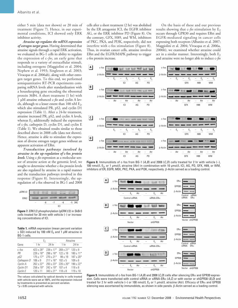
either 5 min (data not shown) or 20 min oftreatment (Figure 7). Hence, in our experi-mental conditions, ICI showed only ERKinhibitor activity.
Atrazine up-regulates the mRNA expressionof estrogen target genes. Having determined thatatrazine signals through a rapid ERK activation,we evaluated in BG-1 cells its ability to regulatethe expression of c-fos, an early gene thatresponds to a variety of extracellular stimuli,including estrogens (Maggiolini et al. 2004;Nephew et al. 1993; Singleton et al. 2003;Vivacqua et al. 2006ab), along with other estro-gen target genes. To this end, we performedsemiquantitative RT-PCR experiments com-paring mRNA levels after standardization witha housekeeping gene encoding the ribosomalprotein 36B4. A short treatment (1 hr) with1 µM atrazine enhanced c-fos and cyclin A lev-els, although to a lesser extent than 100 nM E2,which also stimulated PR, pS2, and cyclin D1expression (Table 1). After a 24-hr treatment,atrazine increased PR, pS2, and cyclin A levels,whereas E2 additionally induced the expressionof c-fos, cathepsin D, cyclin D1, and cyclin E(Table 1). We obtained results similar to thosedescribed above in 2008 cells (data not shown).Hence, atrazine is able to stimulate the expres-sion of diverse estrogen target genes without anapparent activation of ERα.
Transduction pathways involved byatrazine in the up-regulation of c-fos proteinlevels. Using c-fos expression as a molecular sen-sor of atrazine action at the genomic level, wesought to determine whether c-fos protein levelsare also regulated by atrazine in a rapid mannerand the transduction pathways involved in thisresponse (Figure 8). Interestingly, the up-regulation of c-fos observed in BG-1 and 2008
cells after a short treatment (2 hr) was abolishedby the ER antagonist ICI, the EGFR inhibitorAG, or the ERK inhibitor PD (Figure 8). Onthe contrary, GFX, H89, and WM, inhibitorsof PKC, PKA, and PI3K, respectively, did notinterfere with c-fos stimulation (Figure 8).Thus, in ovarian cancer cells, atrazine involvesERα and the EGFR/MAPK pathway to triggerc-fos protein increase.
On the basis of these and our previousresults showing that c-fos stimulation by E2occurs through GPR30 and requires ERα andEGFR-mediated signaling in cancer cellsexpressing both receptors (Albanito et al. 2007;Maggiolini et al. 2004; Vivacqua et al. 2006a,2006b), we examined whether atrazine couldact in a similar manner. Interestingly, both E2and atrazine were no longer able to induce c-fos
Albanito et al.
1652 VOLUME 116 | NUMBER 12 | December 2008 • Environmental Health Perspectives
Figure 9. Immunoblots of c-fos from BG-1 (A,B) and 2008 (C,D) cells after silencing ERα and GPR30 expres-sion. Cells were transfected with control siRNA or siRNA-ERα (A,C) or with vector or shGPR30 (B,D) andtreated for 2 hr with vehicle (–) or 100 nmol/L E2 or 1 μmol/L atrazine (Atr). Efficacy of ERα and GPR30silencing was ascertained by immunoblots, as shown in side panels. β-Actin served as a loading control.
Figure 8. Immunoblots of c-fos from BG-1 (A,B) and 2008 (C,D) cells treated for 2 hr with vehicle (–),100 nmol/L E2, or 1 μmol/L atrazine (Atr) in combination with 10 μmol/L ICI, AG, PD, GFX, H89, or WM,inhibitors of ER, EGFR, MEK, PKC, PKA, and PI3K, respectively. β-Actin served as a loading control.
c-fos
β-Actin
– E2
– E2
– E2
– E2
GFX H89 WM
– Atr – Atr – Atr – Atr – Atr – Atr – Atr – Atr
ICI AG PD GFX H89 WM
C
c-fos
β-Actin
– E2
– E2
– E2
– E2
ICI AG PD
c-fos
β-Actin
c-fos
β-Actin
c-fos
β-Actin
– E2
– E2
– E2
– E2
GFX H89 WM
– Atr – Atr – Atr – Atr – Atr – Atr – Atr – Atr
ICI AG PD GFX H89 WM
c-fos
β-Actin
– E2
– E2
– E2
– E2
ICI AG PD
c-fos
β-Actin
c-fos
β-Actin
D
B
A
Figure 7. ERK1/2 phosphorylation (pERK1/2) in SkBr3cells treated for 20 min with vehicle (–) or increas-ing concentrations of ICI.
– –8 –7 –6 –5 [M]
ICI
pERK1/2
ERK2
Table 1. mRNA expression (mean percent variation± SD) induced by 100 nM E2 and 1 μM atrazine inBG-1 cells.
E2 Atrazine
Gene 1 hr 24 hr 1 hr 24 hr
c-fos 423 ± 28* 239 ± 17* 269 ± 21* 120 ± 9PR 228 ± 18* 298 ± 18* 122 ± 18 180 ± 11*pS2 175 ± 17* 270 ± 21* 99 ± 19 187 ± 20*Cathepsin D 106 ± 9 217 ± 16* 102 ± 5 109 ± 6Cyclin A 262 ± 22* 293 ± 23* 220 ± 20* 190 ± 22*Cyclin D1 258 ± 19* 242 ± 19* 107 ± 4 118 ± 8Cyclin E 120 ± 11 343 ± 21* 118 ± 8 119 ± 10
The values calculated by optical density in cells treatedwith vehicle were set at 100%, and the expression inducedby treatments is presented as percent variation. *p < 0.05 compared with vehicle.
c-fos
β-Actin
Atr– E2
B
A
c-fos
β-Actin
c-fos
β-Actin
c-fos
β-Actin
Atr– E2
Control-siRNA siRNA-ERα
Vector shGPR30
Atr– E2
Atr– E2
Atr– E2
Atr– E2
Control-siRNA siRNA-ERα
Vector shGPR30
Atr– E2
Atr– E2
ERα
GPR30
β-Actin
ERα
GPR30
β-Actin
ERα
GPR30
β-Actin
ERα
GPR30
β-Actin
siRNAERα
ControlsiRNA
shGPR30Vector
siRNAERα
ControlsiRNA
shGPR30Vector
C
D
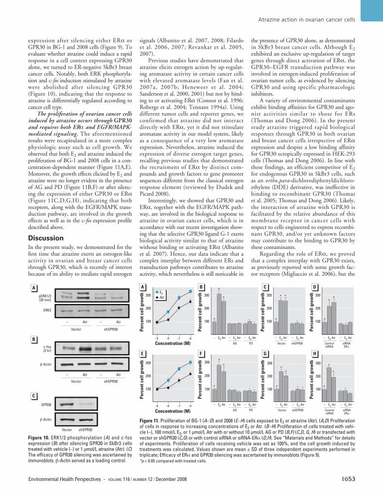
expression after silencing either ERα orGPR30 in BG-1 and 2008 cells (Figure 9). Toevaluate whether atrazine could induce a rapidresponse in a cell context expressing GPR30alone, we turned to ER-negative SkBr3 breastcancer cells. Notably, both ERK phosphoryla-tion and c-fos induction stimulated by atrazinewere abolished after silencing GPR30(Figure 10), indicating that the response toatrazine is differentially regulated according tocancer cell type.
The proliferation of ovarian cancer cellsinduced by atrazine occurs through GPR30and requires both ERα and EGFR/MAPK-mediated signaling. The aforementionedresults were recapitulated in a more complexphysiologic assay such as cell growth. Weobserved that both E2 and atrazine induced theproliferation of BG-1 and 2008 cells in a con-centration-dependent manner (Figure 11A,E).Moreover, the growth effects elicited by E2 andatrazine were no longer evident in the presenceof AG and PD (Figure 11B,F) or after silenc-ing the expression of either GPR30 or ERα(Figure 11C,D,G,H), indicating that bothreceptors, along with the EGFR/MAPK trans-duction pathway, are involved in the growtheffects as well as in the c-fos expression profiledescribed above.
Discussion
In the present study, we demonstrated for thefirst time that atrazine exerts an estrogen-likeactivity in ovarian and breast cancer cellsthrough GPR30, which is recently of interestbecause of its ability to mediate rapid estrogen
signals (Albanito et al. 2007, 2008; Filardoet al. 2006, 2007; Revankar et al. 2005,2007).
Previous studies have demonstrated thatatrazine elicits estrogen action by up-regulat-ing aromatase activity in certain cancer cellswith elevated aromatase levels (Fan et al.2007a, 2007b; Heneweer et al. 2004;Sanderson et al. 2000, 2001) but not by bind-ing to or activating ERα (Connor et al. 1996;Roberge et al. 2004; Tennant 1994a). Usingdifferent tumor cells and reporter genes, weconfirmed that atrazine did not interactdirectly with ERα, yet it did not stimulatearomatase activity in our model system, likelyas a consequence of a very low aromataseexpression. Nevertheless, atrazine induced theexpression of diverse estrogen target genes,recalling previous studies that demonstratedthe recruitment of ERα by distinct com-pounds and growth factors to gene promotersequences different from the classical estrogenresponse element (reviewed by Dudek andPicard 2008).
Interestingly, we showed that GPR30 andERα, together with the EGFR/MAPK path-way, are involved in the biological response toatrazine in ovarian cancer cells, which is inaccordance with our recent investigation show-ing that the selective GPR30 ligand G-1 exertsbiological activity similar to that of atrazinewithout binding or activating ERα (Albanitoet al. 2007). Hence, our data indicate that acomplex interplay between different ERs andtransduction pathways contributes to atrazineactivity, which nevertheless is still noticeable in
the presence of GPR30 alone, as demonstratedin SkBr3 breast cancer cells. Although E2exhibited an exclusive up-regulation of targetgenes through direct activation of ERα, theGPR30–EGFR transduction pathway wasinvolved in estrogen-induced proliferation ofovarian tumor cells, as evidenced by silencingGPR30 and using specific pharmacologicinhibitors.
A variety of environmental contaminantsexhibit binding affinities for GPR30 and ago-nist activities similar to those for ERs(Thomas and Dong 2006). In the presentstudy atrazine triggered rapid biologicalresponses through GPR30 in both ovarianand breast cancer cells irrespective of ERαexpression and despite a low binding affinityfor GPR30 ectopically expressed in HEK-293cells (Thomas and Dong 2006). In line withthese findings, an efficient competitor of E2for endogenous GPR30 in SkBr3 cells, suchas an ortho,para-dichlorodiphenyldichloro-ethylene (DDE) derivative, was ineffective inbinding to recombinant GPR30 (Thomaset al. 2005; Thomas and Dong 2006). Likely,the interaction of atrazine with GPR30 isfacilitated by the relative abundance of thismembrane receptor in cancer cells withrespect to cells engineered to express recombi-nant GPR30, and/or yet unknown factorsmay contribute to the binding to GPR30 bythese contaminants.
Regarding the role of ERα, we provedthat a complex interplay with GPR30 exists,as previously reported with some growth fac-tor receptors (Migliaccio et al. 2006), but the
Atrazine action in ovarian cancer cells
Environmental Health Perspectives • VOLUME 116 | NUMBER 12 | December 2008 1653
Figure 10. ERK1/2 phosphorylation (A) and c-fosexpression (B) after silencing GPR30 in SkBr3 cellstreated with vehicle (–) or 1 μmol/L atrazine (Atr). (C)The efficacy of GPR30 silencing was ascertained byimmunoblots. β-Actin served as a loading control.
pERK1/2(20 min)
ERK2
Vector
GPR30
β-Actin
c-fos(2 hr)
β-Actin
shGPR30
Vector shGPR30
Vector shGPR30
– Atr – Atr
– Atr – Atr
A
B
C
Figure 11. Proliferation of BG-1 (A–D) and 2008 (E–H) cells exposed to E2 or atrazine (Atr). (A,D) Proliferationof cells in response to increasing concentrations of E2 or Atr. (B–H) Proliferation of cells treated with vehi-cle (–), 100 nmol/L E2, or 1 μmol/L Atr with or without 10 μmol/L AG or PD (B,F) (C,D, G, H) or transfected withvector or shGPR30 (C,G) or with control siRNA or siRNA-ERα (D,H). See “Materials and Methods” for detailsof experiments. Proliferation of cells receiving vehicle was set as 100%, and the cell growth induced bytreatments was calculated. Values shown are mean ± SD of three independent experiments performed intriplicate; Efficacy of ERα and GPR30 silencing was ascertained by immunoblots (Figure 9). *p < 0.05 compared with treated cells.
Concentration (M)
Concentration (M)
A B C
300
200
100
–9 –8 –7 –6
300
200
100
– E2
Atr – E2
Atr – E2
Atr
300
200
100
– E2
Atr – E2
Atr
AG PD Vector shGPR30
300
200
100
– E2
Atr – E2
Atr
ControlsiRNA
siRNAERα
Pe
rce
nt
ce
ll g
row
th
300
200
100
Pe
rce
nt
ce
ll g
row
th
–9 –8 –7 –6
300
200
100
300
200
100
300
200
100
– E2
Atr – E2
Atr – E2
Atr – E2
Atr – E2
Atr
AG PD Vector shGPR30
– E2
Atr – E2
Atr
ControlsiRNA
siRNAERα
E2
Atr
Pe
rce
nt
ce
ll g
row
thP
erc
en
t c
ell
gro
wth
Pe
rce
nt
ce
ll g
row
thP
erc
en
t c
ell
gro
wth
Pe
rce
nt
ce
ll g
row
thP
erc
en
t c
ell
gro
wth
D
E F G H
*
*
*
*
* *
*
**
*
*
*
**
*
*
*
*
molecular mechanisms involved remain to beelucidated. Our study and previous investiga-tions indicate that environmental estrogensexert pleiotropic actions by directly bindingto ERα as well as through GPR30–EGFRsignaling, which can engage ERα dependingon the receptor expression pattern present indifferent cell types. This mode of action ofxenoestrogens fits well with the resultsobtained after silencing GPR30 or ERαexpression in ovarian cancer cells, becausesilencing each gene prevented the growthresponse to atrazine.
Our data recall the results of previousstudies showing that xenoestrogens mimicrapid estrogen action in several animal and cellmodels (Bulayeva and Watson 2004; Loomisand Thomas 2000; Nadal et al. 2000;Ruehlmann et al. 1988; Watson et al. 1999,2007). Particularly, in GH3/B6/F10 pituitarytumor cells, diverse xenoestrogens inducedERK phosphorylation with a temporally dis-tinct activation pattern compared with E2(Bulayeva and Watson 2004). In the latterstudy, on the basis of the inhibitory activityexerted by ICI, the authors hypothesized thatan ER localized to the plasma membranecould mediate the ERK phosphorylationresponse by xenoestrogens, depending on theirdifferent ER binding affinities. Moreover, theauthors suggested that the signaling cascadesleading to ERK activation may involve thenature of membrane ERs and their ability tointeract with various signaling partners(Bulayeva and Watson 2004). Interestingly,our findings have provided evidence that ERαmay be involved by xenoestrogens without adirect binding activity and produce relevantresponses such as ERK phosphorylation, geneexpression, and cell growth.
A subset of estrogen-sensitive cell tumorscan proliferate independently from ER expres-sion (i.e., ER-negative cells). In this condition,well represented by SkBr3 breast cancer cells,GPR30–EGFR signaling may still allow forenvironmental estrogen activity as we haveshown in the present study as well as in a pre-vious study (Maggiolini et al. 2004). Hence,multiple transduction pathways triggeredsimultaneously at the membrane level, as wellas within each cell type, may contribute to thenature and magnitude of biological responsesto distinct estrogenic compounds. These con-sequently should be examined individually fortheir complex mechanistic and functional out-comes that result from interaction with a dif-ferent repertoire of receptor proteins.
Atrazine, a potent endocrine disruptor, isthe most common pesticide contaminant ofgroundwater and surface water. Here, we haveprovided novel insight regarding the potentialrole of GPR30 in mediating the action ofatrazine in endocrine-related diseases, such asestrogen-sensitive tumors.
REFERENCES
Albanito L, Madeo A, Lappano R, Vivacqua A, Rago V,
Carpino A, et al. 2007. G protein-coupled receptor 30
(GPR30) mediates gene expression changes and growth
response to 17beta-estradiol and selective GPR30 ligand
G-1 in ovarian cancer cells. Cancer Res 67:1859–1866.
Albanito L, Sisci D, Aquila S, Brunelli E, Vivacqua A, Madeo A,
et al. 2008. Epidermal growth factor induces G protein-cou-
pled receptor expression in estrogen receptor-negative
breast cancer cells. Endocrinology 149:3799–3808.
Babic-Gojmerac T, Kniewald Z, Kniewald J. 1989. Testosterone
metabolism in neuroendocrine organs in male rats under
atrazine and deethylatrazine influence. J Steroid Biochem
33:141–146.
Bardin A, Hoffman P, Boulle N, Katsaros D, Vignon F, Pujol P,
et al. 2004. Involvement of estrogen receptor beta in ovarian
carcinogenesis. Cancer Res 64:5861–5869.
Bologa CG, Revankar CM, Young SM, Edwards BS, Arterburn JB,
Kiselyov AS, et al. 2006. Virtual and biomolecular screening
converge on a selective agonist for GPR30. Nat Chem Biol
4:207–212.
Brawer JR, Sonnenschein C. 1975. Cytopathological effects of
estradiol on the arcuate nucleus of the female rat. A possible
mechanism for pituitary tumorigenesis. Am J Anat 144:57–88.
Bulayeva NN, Watson CS. 2004. Xenoestrogen-induced ERK-1
and ERK-2 activation via multiple membrane-initiated sig-
naling pathways. Environ Health Perspect 15:1481–1487.
Bunone G, Briand PA, Miksicek RJ, Picard D. 1996. Activation
of the unliganded estrogen receptor by EGF involves the
MAP kinase pathway and direct phosphorylation. EMBO J
15:2174–2183.
Connor K, Howell J, Chen I, Liu H, Berhane K, Sciarretta C,
et al. 1996. Failure of chloro-S-triazine-derived compounds
to induce estrogen receptor-mediated responses in vivo
and in vitro. Fundam Appl Toxicol 30:93–101.
Cooper RL, Laws SC, Das PC, Narotsky MG, Goldman JM,
Tyrey EL, et al. 2007. Atrazine and reproductive function:
mode and mechanism of action studies. Birth Defects Res
80:98–112.
Cooper RL, Stoker TE, McElroy WK. 1999. Atrazine (ATR) disrupts
hypothalamic catecholamines and pituitary function.
Toxicologist 42:60–66.
Cooper RL, Stoker TE, Tyrey L, Goldman JM, McElroy WK. 2000.
Atrazine disrupts the hypothalamic control of pituitary
ovarian function. Toxicol Sci 53:297–307.
Crain D, Guillette LJ, Rooney AA, Pickford D. 1997. Alterations
in steroidogenesis in alligators (Alligator mississippiensis)
exposed naturally and experimentally to environmental
contaminants. Environ Health Perspect 105:528–533.
Cummings A, Rhodes B, Cooper R. 2000. Effect of atrazine on
implantation and early pregnancy in 4 strains of rats. Toxicol
Sci 58:135–143.
Donna A, Crosignani P, Robutti F, Betta PG, Bocca R, Mariani N,
et al. 1989. Triazine herbicides and ovarian epithelial neo-
plasms. Scand J Work Environ Health 15:47–53.
Dudek P, Picard D. 2008. Genomics of signaling crosstalk of
estrogen receptor α in breast cancer cells. PLoS ONE
3:1–11.
Falkenstein EHC, Tillmann HC, Christ M, Feuring M, Wehling M.
2000. Multiple actions of steroid hormones a focus on
rapid, nongenomic effects. Pharmacol Rev 52:513–556.
Fan W, Yanase T, Morinaga H, Gondo S, Okabe T, Nomura M,
et al. 2007a. Atrazine-induced aromatase expression is SF-1
dependent: implications for endocrine disruption in wildlife
and reproductive cancers in humans. Environ Health
Perspect 115:720–727.
Fan W, Yanase T, Morinaga H, Gondo S, Okabe T, Nomura M,
et al. 2007b. Herbicide atrazine activates SF-1 by direct
affinity and concomitant co-activators recruitments to
induce aromatase expression via promoter II. Biochem
Biophys Res Commun 355:1012–1018.
Fenelon J, Moore R. 1998. Transport of agrochemicals to
ground and surface waters in a small central Indiana
watershed. J Environ Qual 27:884–894.
Filardo EJ, Graeber CT, Quinn JA, Resnick MB, Giri D, DeLellis
RA, et al. 2006. Distribution of GPR30, a seven membrane-
spanning estrogen receptor, in primary breast cancer and
its association with clinicopathologic determinants of tumor
progression. Clin Cancer Res 12:6359–6366.
Filardo EJ, Quinn JA, Bland KI, Frackelton AR. 2000. Estrogen-
induced activation of Erk-1 and Erk-2 requires the G protein-
coupled receptor homolog, GPR30, and occurs via
trans-activation of the epidermal growth factor receptor
through release of HB-EGF. Mol Endocrinol 14:1649–1660.
Filardo EJ, Quinn JA, Frackelton AR, Bland KI. 2002. Estrogen
action via the G protein-coupled receptor, GPR30: stimula-
tion of adenylyl cyclase and cAMP-mediated attenuation of
the epidermal growth factor receptor-to-MAPK signaling
axis. Mol Endocrinol 16:70–84.
Filardo EJ, Quinn JA, Pang C, Graeber CT, Shaw S, Dong J, et al.
2007. Activation of the novel estrogen receptor G protein
coupled receptor 30 (GPR30) at the plasma membrane.
Endocrinology 148:3236–3245.
Friedmann A. 2002. Atrazine inhibition of testosterone produc-
tion in rat males following peripubertal exposure. Reprod
Toxicol 16:275–279.
Gammon DW, Aldous CN, Carr WC Jr, Sanborn JR, Pfeifer KF.
2005. A risk assessment of atrazine use in California: human
health and ecological aspects. Pest Manag Sci 61:331–355.
Geisinger KR, Kute TE, Pettenati MJ, Welander CE, Dennard Y,
Collins LA, et al. 1989. Characterization of a human ovarian
carcinoma cell line with estrogen and progesterone
receptors. Cancer 63:280–288.
Heneweer M, van den Berg M, Sanderson J. 2004. A comparison
of human H295R and rat R2C cell lines as in vitro screening
tools for effects on aromatase. Toxicol Lett 146:183–194.
Keller J, McClellan-Green P. 2004. Effects of organochlorine
compounds on cytochrome P450 aromatase activity in an
immortal sea turtle cell line. Mar Environ Res 58:347–351.
Kettles MK, Browning SR, Prince TS, Horstman SW. 1997.
Triazine herbicide exposure and breast cancer incidence:
an ecologic study of Kentucky counties. Environ Health
Perspect 105:1222–1227.
Kniewald J, Mildner P, Kniewald Z. 1979. Effects ofs-triazine
herbicides on 5α-dihydrotestosterone receptor complex
formation, 5α-reductase and 3α-hydroxysteroid dehydro-
genase activity at the anterior pituitary level. J Steroid
Biochem 11:833–838.
Kniewald J, Mildner P, Kniewald Z. 1980. Effects of s-triazine
herbicides on 5α-dihydrotestosterone receptor complex
formation in the hypothalamus and ventral prostate. In:
Pharmacological Modulation of Steroid Action (Genazzani E,
DiCarlo F, Mainwaring WIP, eds). New York:Raven Press,
159–169.
Kniewald J, Osredecki V, Gojmerac T, Zechner V, Kniewald Z.
1995. Effect of s-triazine compounds on testosterone
metabolism in the rat prostate. J Appl Toxicol 15:215–218.
Kolpin D, Barbash J, Gilliom R. 1998. Occurence of pesticides in
shallow groundwater of the United States: initial results
from the National Water-Quality Assessment Program.
Environ Sci Technol 32:558–566.
Lee YJ, Gorski J. 1996. Estrogen-induced transcription of the prog-
esterone receptor gene does not parallel estrogen receptor
occupancy. Proc Natl Acad Sci USA 93:15180–15184.
Lehmann TP, Biernacka-Lukanty JM, Saraco N, Langlois D,
Li JY, Trzeciak WH. 2005. Temporal pattern of the induc-
tion of SF-1 gene expression by the signal transduction
pathway involving 3´,5´-cyclic adenosine monophosphate.
Acta Biochim Pol 52:485–491.
Lephart ED, Simpson ER. 1991. Assay of aromatase activity.
Methods Enzymol 206:477–483.
Lode O, Eklo O, Holen B, Svensen A, Johnsen A. 1995. Pesticides
in precipitation in Norway. Sci Total Environ 160:421–431.
Loomis AK, Thomas P. 2000. Effects of estrogens and xeno-
estrogens on androgen production by Atlantic croaker
testes in vitro: evidence for a nongenomic action mediated
by an estrogen membrane receptor. Biol Reprod
62:995–1004.
Maggiolini M, Donzé O, Picard D. 1999. A non-radioactive
method for inexpensive quantitative RT-PCR. Biol Chem
380:695–700.
Maggiolini M, Vivacqua A, Fasanella G, Recchia AG, Sisci D,
Pezzi V, et al. 2004. The G protein-coupled receptor GPR30
mediates c-fos up-regulation by 17-beta estradiol and
phytoestrogens in breast cancer cells. J Biol Chem
279:27008–27016.
McElroy JA, Gangnon RE, Newcomb PA, Kanarek MS,
Anderson HA, Brook JV, et al. 2007. Risk of breast cancer
for women living in rural areas from adult exposure to
atrazine from well water in Wisconsin. J Expo Sci Environ
Epidemiol 17:207–214.
Migliaccio A, Castoria G, Di Domenico M, Ciociola A,
Lombardi M, De Falco A, et al. 2006. Crosstalk between
EGFR and extranuclear steroid receptors. Ann NY Acad Sci
1089:194–200.
Miller S, Sweet C, Depinto J, Hornbuckle K. 2000. Atrazine and
Albanito et al.
1654 VOLUME 116 | NUMBER 12 | December 2008 • Environmental Health Perspectives
nutrients in precipitation: results from the Lake Michigan
mass balance study. Environ Sci Technol 34:55–61.
Morinaga H, Yanase T, Nomura M, Okabe T, Goto K, Harada N,
et al. 2004. A benzimidazole fungicide, benomyl, and its
metabolite, carbendazim, induce aromatase activity in a
human ovarian granulose-like tumor cell line (KGN).
Endocrinology 145:1860–1869.
Müller S, Berg M, Ulrich M, Schwarzenbach RP. 1997. Atrazine
and its primary metabolites in Swiss lakes: input charac-
teristics and long-term behavior in the water column.
Environ Sci Technol 31:2104–2113.
Nadal A, Ropero AB, Laribi O, Maillet M, Fuentes E, Soria B.
2000. Nongenomic actions of estrogens and xenoestrogens
by binding at a plasma membrane receptor unrelated to
estrogen receptor alpha and estrogen receptor beta. Proc
Natl Acad Sci USA 97:11603–11608.
Narotsky M, Best DS, Guidici DL, Cooper RL. 2001. Strain com-
parisons of atrazine-induced pregnancy loss in the rat.
Reprod Toxicol 15:61–69.
National Center for Biotechnology Information. 2008. Searching
GenBank. Available: http://www.ncbi.nlm.nih.gov/Genbank/
GenbankSearch.html [accessed 21 October 2008].
Nephew KP, Polek TC, Akcali KC, Khan SA. 1993. The anti-
estrogen tamoxifen induces c-fos and c-jun, but not c-jun
or Jun-D, protooncogenes in the rat uterus. Endocrinology
133:419–422.
Norman AW, Mizwicki MT, Norman DP. 2004. Steroid-hormone
rapid actions, membrane receptors and a conformational
ensemble model. Nat Rev Drug Discov 3:27–41.
Pintér A, Török G, Börzsönyi M, Surján A, Csík M, Kelecsényi Z,
et al. 1990. Long-term carcinogenicity bioassay of the herbi-
cide atrazine in F344 rats. Neoplasma 37:533–544.
Revankar CM, Cimino DF, Sklar LA, Arterburn JB, Prossnitz ER.
2005. A transmembrane intracellular estrogen receptor
mediates rapid cell signalling. Science 307:1625–1630.
Revankar CM, Mitchell HD, Field AS, Burai R, Corona C,
Ramesh C, et al. 2007. Synthetic estrogen derivatives
demonstrate the functionally of intracellular GPR30. ACS
Chem Biol 2:536–544.
Revelli A, Massobrio M, Tesarik J. 1998. Nongenomic actions
of steroid hormones in reproductive tissues. Endocr Rev
19:3–17.
Roberge M, Hakk H, Larsen G. 2004. Atrazine is a competitive
inhibitor of phosphodiesterase but does not affect the
estrogen receptor. Toxicol Lett 154:61–68.
Ruehlmann DO, Steinert JR, Valverde MA, Jacob R, Mann GE.
1988. Environmental estrogenic pollutants induce acute
vascular relaxation by inhibiting C-type Ca2+ channels in
smooth muscle cells. FASEB J 12:613–619.
Rusiecki JA, De Roos A, Lee WJ, Dosemeci M, Lubin JH,
Hoppin JA, et al. 2004. Cancer incidence among pesticide
applicators exposed to atrazine in the Agricultural Health
Study. J Natl Cancer Inst 96:1375–1382.
Safei R, Katano K, Larson BJ, Samimi G, Holzer AK,
Naerdemann W, et al. 2005. Intracellular localization and
trafficking of fluorescein-labeled cisplatin in human ovar-
ian carcinoma cells. Clin Cancer Res 11:756–767.
Sanderson JT, Boerma J, Lansbergen G, Van den Berg M. 2002.
Induction and inhibition of aromatase (CYP19) activity by
various classes of pesticides in H295R human adrenocorti-
cal carcinoma cells. Toxicol Appl Pharmacol 182:44–54.
Sanderson JT, Letcher RJ, Heneweer M, Giesy JP, Van den
Berg M. 2001. Effects of chloro-s-triazine herbicides and
metabolites on aromatase activity in various human cell
lines and on vitellogenin production in male carp hepato-
cytes. Environ Health Perspect 109:1027–1031.
Sanderson JT, Seinen W, Giesy JP, van den Berg M. 2000.
2-Chloro-triazine herbicides induce aromatase (CYP19)
activity in H295R human adrenocortical carcinoma cells: a
novel mechanism for estrogenicity? Toxicol Sci 54:121–127.
Santagati S, Gianazza E, Agrati P, Vegeto E, Patrone C, Pollio G,
et al. 1997. Oligonucleotide squelching reveals the mecha-
nism of estrogen receptor autologous down-regulation.
Mol Endocrinol 11:938–949.
Sass JB, Colangelo A. 2006. European Union bans atrazine,
while the United States negotiates continued use. Int J
Occup Environ Health 12:260–267.
Seipel K, Georgiev O, Schaffner W. 1992. Different activation
domains stimulate transcription from remote (‘enhancer’)
and proximal (‘promoter’) positions. EMBO J 11:4961–4968.
Shafer TJ, Ward TR, Meacham CA, Cooper RL. 1999. Effects of
the cholorotriazine herbicide, cyanizine on GABAA recep-
tors in cortical tissue from rat brain. Toxicology 142:57–68.
Simic B, Kniewald Z, Davies J, Kniewald J. 1991. Reversibility
of inhibitory effect of atrazine and lindane on 5-dihydro-
testosterone receptor complex formation in rat prostate.
Bull Environ Contam Toxicol 46:92–99.
Singleton DW, Feng Y, Burd CJ, Khan SA. 2003. Nongenomic
activity and subsequent c-fos induction by estrogen
receptor ligands are not sufficient to promote deoxy-
ribonucleic acid synthesis in human endometrial adeno-
carcinoma cells. Endocrinology 144:121–128.
Solomon K, Baker D, Richards R, Dixon K, Klaine S, LaPoint T,
et al. 1996. Ecological risk assessment of atrazine in North
American surface waters. Environ Toxicol Chem 15:31–76.
Spano L, Tyler C, van Aerle R, Devos P, Mandiki S, Silvestre F,
et al. 2004. Effects of atrazine on sex steroid dynamics,
plasma vitellogenin concentration and gonad development
in adult goldfish (Carassius auratus). Aquat Toxicol
66:369–379.
Stoker TE, Laws S, Guidici D, Cooper R. 2000. The effect of
atrazine on puberty in male Wistar rats: an evaluation in
the protocol for the assessment of pubertal development
and thyroid function. Toxicol Sci 58:50–59.
Stoker TE, Robinette CL, Cooper RL. 1999. Maternal exposure to
atrazine during lactation suppresses suckling-induced pro-
lactin release and results in prostatitis in the adult offspring.
Toxicol Sci 52:68–79.
Tennant MK, Hill DS, Eldridge JC, Wetzel LT, Breckenridge CB,
Stevens JT. 1994a. Chloro-s-triazine antagonism of estro-
gen action: limited interaction with estrogen receptor
binding. J Toxicol Environ Health 43:197–211.
Tennant MK, Hill DS, Eldridge JC, Wetzel LT, Breckenridge CB,
Stevens JT. 1994b. Possible antiestrogenic properties of
chloro-s-triazines in rat uterus. J Toxicol Environ Health
43:183–196.
Thomas P. 2000. Chemical interference with the genomic and
nongenomic actions of steroids in fishes: role of receptor
binding. Mar Environ Res 50:127–134.
Thomas P, Dong J. 2006. Binding and activation of the seven-
transmembrane estrogen receptor GPR30 by environmen-
tal estrogens: a potential novel mechanism of endocrine
disruption. J Steroid Biochem Mol Biol 102:175–179.
Thomas P, Pang Y, Filardo EJ, Dong J. 2005. Identity of an
estrogen membrane receptor coupled to a G-protein in
human breast cancer cells. Endocrinology 146:624–632.
Thurman E, Cromwell A. 2000. Atmospheric transport, deposi-
tion, and fate of triazine herbicides and their metabolites
in pristine areas at Isle Royale National Park. Environ Sci
Technol 34:3079–3085.
Vivacqua A, Bonofiglio D, Albanito L, Madeo A, Rago V,
Carpino A, et al. 2006a. 17-beta-Estradiol, genistein, and 4-
hydroxytamoxifen induce the proliferation of thyroid can-
cer cells through the G protein coupled-receptor GPR30.
Mol Pharmacol 70:1414–1423.
Vivacqua A, Bonofiglio D, Recchia AG, Musti AM, Picard D,
Andò S, et al. 2006b. The G protein-coupled receptor
GPR30 mediates the proliferative effects induced by
17beta-estradiol and hydroxytamoxifen in endometrial
cancer cells. Mol Endocrinol 20:631–646.
Watson CS, Alyea RA, Jeng YJ, Kochukov MY. 2007.
Nongenomic actions of low concentration estrogens and
xenoestrogens on multiple tissues. Mol Cell Endocrinol
274:1–7.
Watson CS, Campbell CH, Gametchu B. 1999. Membrane
estrogen receptors on rat pituitary tumor cells: immuno-
identification and response to oestradiol and xeno-
estrogens. Exp Physiol 84:1013–1018.
Webb P, Nguyen P, Shinsako J, Anderson C, Feng W,
Nguyen MP, et al. 1998. Estrogen receptor activation func-
tion 1 works by binding p160 coactivator proteins. Mol
Endocrinol 12:1605–1618.
Wetzel LT, Luempert LG, Breckenridge CB, Tisdel MO,
Stevens JT, Thakur AK, et al. 1994. Chronic effects of
atrazine on estrus and mammary tumor formation in
female Sprague-Dawley and Fischer 344 rats. J Toxicol
Environ Health 43:169–182.
Young HA, Mills PK, Riordan DG, Cress RD. 2005. Triazine herbi-
cides and epithelial ovarian cancer risk in central California.
J Occup Environ Med 47:1148–1156.
Atrazine action in ovarian cancer cells
Environmental Health Perspectives • VOLUME 116 | NUMBER 12 | December 2008 1655
Epidermal Growth Factor Induces G Protein-CoupledReceptor 30 Expression in Estrogen Receptor-NegativeBreast Cancer Cells
Lidia Albanito, Diego Sisci, Saveria Aquila, Elvira Brunelli, Adele Vivacqua, Antonio Madeo,Rosamaria Lappano, Deo Prakash Pandey, Didier Picard, Loredana Mauro, Sebastiano Ando,and Marcello Maggiolini
Departments of Pharmaco-Biology (L.A., D.S., S.Aq., A.V., A.M., R.L., M.M.), Ecology (E.B.), and Cell Biology (L.M., S.An.),University of Calabria, 87030 Rende, Italy; and Department of Cell Biology (D.P.P., D.P.), University of Geneve, 1211Geneve 4, Switzerland
Different cellular receptors mediate the biological effects in-duced by estrogens. In addition to the classical nuclear es-trogen receptors (ERs)-� and -�, estrogen also signals throughthe seven-transmembrane G-protein-coupled receptor (GPR)-30. Using as a model system SkBr3 and BT20 breast cancercells lacking the classical ER, the regulation of GPR30 expres-sion by 17�-estradiol, the selective GPR30 ligand G-1, IGF-I,and epidermal growth factor (EGF) was evaluated. Transienttransfections with an expression plasmid encoding a short5�-flanking sequence of the GPR30 gene revealed that an ac-tivator protein-1 site located within this region is required forthe activating potential exhibited only by EGF. Accordingly,EGF up-regulated GPR30 protein levels, which accumulatedpredominantly in the intracellular compartment. The stimu-latory role elicited by EGF on GPR30 expression was trig-
gered through rapid ERK phosphorylation and c-fos induc-tion, which was strongly recruited to the activator protein-1site found in the short 5�-flanking sequence of the GPR30gene. Of note, EGF activating the EGF receptor-MAPKtransduction pathway stimulated a regulatory loop thatsubsequently engaged estrogen through GPR30 to boost theproliferation of SkBr3 and BT20 breast tumor cells. Theup-regulation of GPR30 by ligand-activated EGF receptor-MAPK signaling provides new insight into the well-knownestrogen and EGF cross talk, which, as largely reported,contributes to breast cancer progression. On the basis ofour results, the action of EGF may include the up-regula-tion of GPR30 in facilitating a stimulatory role of estrogen,even in ER-negative breast tumor cells. (Endocrinology 149:3799–3808, 2008)
GIVEN THE ARRAY of extracellular cues to which theyare exposed, cells have developed complex machinery
for the reception and interpretation of external stimuli. Mul-tiple intracellular signaling pathways are activated by thesesignals, which are then translated into changes of cellularfunctions. A common theme in the arrangement of thesepathways is the integration and cross talk between contig-uous cascades to fine-tune biological outcomes as diverse ascell proliferation, differentiation, and migration. The trans-activation of receptor tyrosine kinases by G protein-coupledreceptors (GPCRs) is a nice example of communication andcooperation between different signaling networks. In thisregard, agonist binding to GPCRs results in transactivationof the epidermal growth factor (EGF) receptor (EGFR) and
activation of the ERK/MAPK cascade in a variety of cellularcontexts. Traditionally, the EGF network has been viewed asa direct orchestrator of cell replication under physiologicaland pathological conditions; nevertheless, the involvementof cross-signaling with steroid hormones such as estrogenshas largely been demonstrated, particularly in the regulationof normal mammary development and breast cancer pro-gression (1). Indeed, aberrant expression and activation ofEGFR is frequently observed in estrogen-sensitive tumorslike breast and ovary, in which it correlates with a poorerpatient prognosis (2, 3). In addition, up-regulation of EGFRsignaling is thought to be an important mechanism, confer-ring antiestrogen resistance of breast cancer resulting in thefailure of endocrine therapy (4).
Several lines of evidence have suggested that the interac-tion of EGFR with estrogen signaling can occur at differentlevels. The major estrogen, 17�-estradiol (E2), primarilyacts through cognate nuclear receptors [estrogen receptors(ERs)], leading to regulation of gene expression, which hastraditionally been deemed as genotropic estrogen activity.Many E2-responsive genes are indeed key signaling mole-cules that participate in EGFR signaling (1). Alternatively, acell membrane-associated form of ER has been reported tocouple with and activate various G proteins, thereby trig-gering nongenotropic effects through the transactivation ofthe EGFR (1, 5). More recently our and other studies haveshown that an orphan GPCR, named GPR30, is able to me-
First Published Online May 8, 2008Abbreviations: AG, AG1478; AP1, activator protein-1; ChIP, chroma-
tin immunoprecipitation; DN, dominant negative; E2, 17�-estradiol;EGF, epidermal growth factor; EGFR, EGF receptor; ER, estrogen re-ceptor; FBS, fetal bovine serum; G-1, 1-[4-(-6-bromobenzol[1, 3]diodo-5-yl)-3a,4,5,9b-tetrahidro-3H-cyclopenta[c]quinolin-8yl]ethanone;GPCR, G protein-coupled receptor; LY, LY 294,002; MTT, dimethylthia-zoldiphenyltetra-zoliumbromide; PD, PD98059; PI3K, phosphatidylino-sitol 3-kinase; PKA, protein kinase A; PP2, 4-amino-5-(4-chlorophenyl)-7-(t-butyl)pyrazolo[3,4-d] pyrimidine; sh, short hairpin; SP1, specificityprotein-1.Endocrinology is published monthly by The Endocrine Society (http://www.endo-society.org), the foremost professional society serving theendocrine community.
0013-7227/08/$15.00/0 Endocrinology 149(8):3799–3808Printed in U.S.A. Copyright © 2008 by The Endocrine Society
doi: 10.1210/en.2008-0117
3799
at Biblioteca Area Tecnico-Sci on January 23, 2009 endo.endojournals.orgDownloaded from
diate rapid E2-dependent signals prompting major biologicalresponses such as gene expression and cancer cell prolifer-ation (6–11). Interestingly, it has been shown that GPR30 isinvolved in the EGFR transactivation by E2 (12) as exhibitedby other GPCR ligands (13–17). In addition, different studiesincluding our own have demonstrated that E2 and the mixedER agonist/antagonist 4-hydroxytamoxifen can signalthrough GPR30 to activate the EGFR-MAPK cascade, even incancer cells lacking ERs (8, 18).
From all these studies, it is possible to assume that E2 caninitiate rapid MAPK signaling in an ER-dependent and ER-independent manner. First, E2 can bind a membrane ER,quite similar or identical with the nuclear receptor, and sub-sequently activate G proteins; second, E2 can directly activateGPCR at the membrane/intracellular level (see below) in anER-independent manner, thereby signaling to G protein ac-tivation. However, because GPR30 was found to be localizedclose to the endoplasmic reticulum (6), whether this intra-cellular receptor coupled with G proteins can directly trans-activate EGFR as well as its physiological function(s) remainsto be fully understood. ER inhibition has proven to be aneffective means of blocking the growth of breast tumorsexpressing ER, and this modality of treatment still remainsthe standard endocrine therapy for ER� tumors. Althoughthere is general concordance between ER expression andresponsiveness to ER antagonism, as indicated by greaterdisease-free survival at 5-yr follow-up for postmenopausalpatients with ER� tumors receiving tamoxifen (19), roughlyone in four patients do not respond to tamoxifen therapyfrom the onset, and after a few years in most patients, treat-ment with this antiestrogen produces agonist effects.
A variety of explanations have been offered to account forunresponsiveness to ER antagonism, including: 1) intratu-moral heterogeneity in ER expression, 2) evolution of mutantERs with reduced affinity for ER antagonists, 3) drug resis-tance, 4) partial receptor antagonism, and 5) the presence orabsence of trans-acting factors that influence ER functional-ity. These interpretations have prompted strategies betterdesigned to assess ER activity and have served as the ratio-nale for the discovery and use of new endocrine agents withmore complete ER antagonist activity. In this concern, theexistence of an alternative ER, such as GPR30, which is po-tentially stimulated by ER antagonists, may provide a furtherpossible explanation for the 4-hydroxytamoxifen failure. Todate, studies in animal and cell models have long indicatedthat estrogens manifest physiologic actions and biochemicaleffects inconsistent with its classical genomic mechanism ofaction (20). For instance, estrogen induces EGF-like activityin female reproductive tissue (21, 22) and likewise activatesbiochemical signals typically associated with the EGFR trans-duction pathway (23, 24). In this regard, it should be notedthat GPR30 can act independently from ERs in triggeringestrogen-dependent EGFR action. Indeed, GPR30 may playan important role in breast cancer biology because it providesa mechanism through which estrogens promote EGF-likeeffects. According to this model, ER-negative breast tumorsalso may remain estrogen responsive through GPR30. Thisconcept should be taken into account, particularly in thosepatients receiving endocrine therapy because OHT behaves
similarly to estradiol, being capable to elicit EGFR activationin breast cancer cells (9, 10, 18).
Given that GPR30 involves the EGFR pathway in medi-ating the estrogen signals, in the present study, we evaluatedthe regulation of GPR30 expression and demonstrate for thefirst time that EGF is able to induce GPR30 protein levels thataccumulate in the intracellular compartment. Consequently,EGF generates a regulatory loop engaging E2 to boost theproliferation of ER-negative breast cancer cells.
Materials and MethodsReagents
E2, EGF, IGF-I, H89, LY 294,002 (LY), and PD98059 (PD) were pur-chased from Sigma-Aldrich Corp. (Milan, Italy). AG1478 (AG) was pur-chased from Biomol Research Laboratories, Inc. (DBA, Milan, Italy), and4-amino-5-(4-chlorophenyl)-7-(t-butyl)pyrazolo[3,4-d] pyrimidine (PP2)was obtained from Calbiochem (VWR International, Milan, Italy). 1-[4-(-6-bromobenzol[1, 3]diodo-5-yl)-3a,4,5,9b-tetrahidro-3H-cyclopenta[c-]quinolin-8yl]ethanone (G-1) was purchased from Merck KGaA (Frank-furt, Germany). All compounds were solubilized in dimethylsulfoxide,except E2 and PD, which were dissolved in ethanol.
Cell culture
SkBr3 breast cancer cells were maintained in RPMI 1640 withoutphenol red supplemented with 10% fetal bovine serum (FBS). BT20breast cancer cells and HEK-293 embryonal kidney cells were culturedin MEM and DMEM, respectively, with phenol red supplementedwith 10% FBS. Cells were switched to medium without serum the daybefore experiments for immunoblots, RT-PCR, and confocal micros-copy assessment.
Plasmids
To generate the luciferase expression vector for the GPR30 –5�flanking region (GPR30), a 641-bp fragment next to the 5�-flankingregion of the GPR30 gene was amplified by PCR using the followingprimer pairs: 5�-AACACTGGCTTTCCCTTCCTATCT-3� (forward) and5�-CTTGAAGTGAGCCTGGCATTTGTC-3� (reverse) from genomic DNA,which was extracted from SkBr3 cells by Trizol reagent as suggested by themanufacturer (Invitrogen, Milan, Italy). PCR primer pairs were selectedanalyzing the 5�-flanking region of GPR30 gene in chromosome 7, location7p22.3. The PCR amplification was performed using 1.25 U GoTaq DNApolymerase according to the manufacturer’s instructions (Promega, Milan,Italy). PCR conditions were 5 min at 95 C followed by 1 min at 94 C, 1 minat 58 C, and 1 min at 72 C for 30 cycles. The fragment was then inserted inthe pCR 2.1 plasmid using the TA cloning kit (Invitrogen), sequenced, andcut with HindIII and XhoI. The insert was cloned in the pGL3 basic vector(Promega). Analyses of GPR30–5� flanking region revealed an activatorprotein-1 (AP1; �471 to �477) and an specificity protein-1 (SP1; �133 to�138) consensus binding sites. Mutations from position �471 to �477 inthe GPR30–5� flanking sequence corresponding to an AP1 motif and fromposition �133 to �138 corresponding to the SP1 binding site (Fig. 2A) weregenerated using QuikChange XL site-directed mutagenesis kit (Stratagene,Milan, Italy).
The following pairs of primers were used to generate the AP1 andSp1mutants: GPR30AP1mut (forward), 5�-CCCTGCCTGTGGGAGACGC-CCACGTCCAGCCTCC-3� and (reverse) 5�-GGAGGCTGGACGTG-GGCGTCTCCCACAGGCAGGG-3�; GPR30SP1mut (forward), 5�-GG-ACGAGCACGCGGAGATCACTCGCCTCCACGG-3� and (reverse)5�-CCGTGGAGGCGAGGTGATCTCCGCGTGCTCGTCC-3�. All plas-mids were sequenced before use. Plasmid 3x-FLAG-hGPR30 was con-structed using the HindIII/BamHI sites in pCMV10.3x-ratFLAG (25).hGPR30 was amplified with primers CCCCAAGCTTatggatgtgacttcccaagand CAGCGGATCCctacacggcactgctgaac (restriction sites are underlined).Reference plasmid Prl-3x-FLAG is a gift from K. Strub (Department of CellBiology, University of Geneve, Geneve, Switzerland), and it expresses anunrelated 26-kDa protein. Short hairpin (sh)RNA constructs against hu-man GPR30 were bought from Openbiosystems (Biocat.de, Heidelberg,Germany) with catalog no. RHS4533-M001505. The targeting strands gen-
3800 Endocrinology, August 2008, 149(8):3799–3808 Albanito et al. • EGF Induces GPR30 Expression
at Biblioteca Area Tecnico-Sci on January 23, 2009 endo.endojournals.orgDownloaded from
erated from the shRNA vectors sh1, sh2, sh3, sh4, and unrelated controlare complementary to the following sequences, respectively: CGAGTTA-AAGAGGAGAAGGAA, CTCCCTCATTGAGGTGTTCAA, CGCTCCCT-GCAAGCAGTCTTT, GCAGTACGTGATCGGCCTGTT, and CGAC-ATGAAACCGTCCATGTT.
To evaluate the effectiveness of the different shRNA constructs, HEK-293 cells were seeded at about 50% confluency in 6-cm plates. Six to 8 hlater, cells were transfected using the calcium-phosphate method with1 �g of 3x-FLAG-hGPR30, 10 �g of shRNA construct, and 2 �g ofPrl-3x-FLAG. Prl-3xFLAG was used as a transfection control. Fortyhours after transfection, cells were harvested and lysed with 20 mmTris-HCl (pH 8), 100 mm NaCl, 10% glycerol, 0.1% Nonidet P-40, 1 mmmonovanadate, 1 mm dithiothreitol, and protease inhibitors. DNA wassheared by several passages through a 25-gauge needle. Lysates werecleared by centrifugation, and protein concentrations were determinedby the Bradford method. Thirty micrograms of lysates were subjected toWestern blot analysis with the FLAG antibody M2 (Sigma, LausanneSwitzerland). With a 74% knockdown of 3x-FLAG-hGPR30 expressionshRNA construct, sh3 showed the highest efficacy. Hereafter sh3 isreferred to as shGPR30. The dominant-negative (DN)/c-fos plasmid, agift from C. Vinson (National Institutes of Health, Bethesda, MD), con-sists of an acidic amphipathic protein sequence appended onto the Nterminus of the fos leucine zipper, replacing the normal basic regioncritical for DNA binding. The reporter plasmid for 4xAP1-responsivecollagen promoter, a gift from H. van Dam (Department of MolecularCell Biology, Leiden University, Leiden, The Netherlands), contains fourAP1 binding sequences (TGAC/GTCA) inserted into a luciferase con-struct with the minimal promoter sequences from the albumin gene. TheGPR30 expression vector was kindly provided by R. Weigel (Depart-ment of Surgery, Thomas Jefferson University, Philadelphia, PA) (8).
Transfection and luciferase assays
SkBr3 and BT20 cells (1 � 105) were plated into 24-well dishes with500 �l of regular growth medium per well the day before transfection.The medium was replaced with that lacking serum on the day of trans-fection performed using Fugene 6 reagent as recommended by themanufacturer (Roche Diagnostics, Milan, Italy) with a mixture contain-ing 300 ng of GPR30 expression vector and 3 ng of pRL-TK. After 5 h,the serum-free medium containing the indicated treatments was re-newed, and then cells were incubated for 18 h. Luciferase activity wasmeasured with the dual luciferase kit (Promega) according to the man-ufacturer’s recommendations. Firefly luciferase values were normalizedto the internal transfection control provided by the Renilla luciferaseactivity. The normalized relative light unit values obtained from cellstreated with vehicle were set as 1-fold induction upon which the activityinduced by treatments was calculated.
Western blotting
SkBr3 cells were grown in 10-cm dishes, exposed to ligands, and thenlysed in 500 �l of 50 mmol/liter NaCl; 1.5 mmol/liter MgCl2; 1 mmol/liter EGTA; 10% glycerol; 1% Triton X-100; 1% sodium dodecyl sulfate;a mixture of protease inhibitors containing 1 mmol/liter aprotinin, 20mmol/liter phenylmethylsulfonyl fluoride, and 200 mmol/liter sodiumorthovanadate. Protein concentration was determined using Bradfordreagent according to the manufacturer recommendations (Sigma-Al-drich). Equal amounts of whole protein extract were resolved on a 10%sodium dodecyl sulfate-polyacrylamide gel; transferred to a nitrocellu-lose membrane (Amersham Biosciences, Milan, Italy); probed overnightat 4 C with the antibody against GPR30 (MBL-Eppendorf, Milan, Italy),c-fos, �-actin, phosphorylated ERK1/2, and ERK2 (all purchased fromSanta Cruz Biotechnology, DBA, Milan, Italy); and then revealed usingthe ECL Western blotting analysis system (Amersham Biosciences).
RT-PCR
SkBr3 cells were grown in 10-cm dishes in regular growth mediumand then switched to medium lacking serum for 24 h. Thereafter treat-ments were added for 1 h, and cells were processed for mRNA extractionusing the Trizol reagent (Invitrogen) according to the manufacturer’sprotocol. The mRNA expression was evaluated by semiquantitativeRT-PCR as previously described (26). For GPR30 and the acid phos-
phoprotein P0 (36B4), which was used as a control gene, the primerswere 5�-CTGGGGAGTTTCCTGCTGA-3� (GPR30 forward) and 5�-GCT-TGGGAAGTCACACCAT-3� (GPR30 reverse) and 5�-CTCAACATCTC-CCCCTTCTC-3� (36B4 forward) and 5�-CAAATCCCATATCCTCGTCC-3�(36B4 reverse) to yield products, respectively, of 155 and 408 bp, with 15PCR cycles for both genes.
Confocal microscopy
Fifty percent confluent cultured SkBr3 and HEK-293 cells grown oncoverslips were serum deprived for 24 h and then treated for 2 h with50 ng/ml EGF and 10 �m AG and PD as indicated. Then cells were fixedin 4% paraformaldehyde, permeabilized with 0.2% Triton X-100, washedthree times with PBS, and incubated for 1 h with 2 mg/ml primaryantibody against GPR30. After incubation with the GPR30 antibody, theslides were washed three times with PBS and incubated with 1 mg/mlrhodamine-conjugated donkey antirabbit IgG (Calbiochem). HEK-293cells were also stained with propidium iodide. The cellular expressionand localization of GPR30 was evaluated by confocal microscope with�1000 magnification. The optical sections were taken at the centralplane. The specificity of the detection was verified by neutralizing theGPR30 antibody with the antigen peptide, which was produced by theTNT quick coupled transcription/translation systems (Promega).
Chromatin immunoprecipitation (ChIP)
Cells grown in 10-cm plates were shifted for 24 h to medium lackingserum and then treated for 2 h with vehicle or 50 ng/ml EGF. ChIP assaywas performed as previously described (27). The immuno-cleared chro-matin was precipitated with anti-c-fos antibody or nonspecific IgG (SantaCruz Biotechnology, DBA). A 4-�l volume of each sample was used astemplate to amplify by PCR two fragments located next the GPR30–5�flanking region: one fragment of 261 bp containing the AP1 site andthe second fragment of 364 bp (from �937 to �1301) not containingthe AP1 site. The primer pairs used to amplify the first fragment were:5�-CGTGCCCATACCTTCATTGCTTCC-3� (forward) and 5�-CCTG-GCCGGGTGTCTGTAG-3� (reverse), whereas the primer pairs usedto amplify the second fragment were: 5�-CCGTGGCCCGCTGCATA-GAGAAC-3� (forward) and 5�-GAGAGGGAGAAGTGGGCTGTC-3�(reverse). The PCR conditions were 45 sec at 94 C, 40 sec at 58 C, and90 sec at 72 C. The amplification products obtained in 25 cycles wereanalyzed in a 2% agarose gel and visualized by ethidium bromidestaining. Three microliters of the initial preparations of soluble chro-matin were amplified to control input DNA before precipitation.
Cell proliferation assay
Cells (10,000) were seeded in 24-well plates in regular growth me-dium. Cells were washed once they had attached and then incubated inmedium containing 2.5% charcoal-stripped FBS with the indicated treat-ments; medium was renewed every 2 d (with treatments), and cellgrowth was monitored by dimethylthiazoldiphenyltetra-zoliumbro-mide (MTT) assay according to the manufacturer’s protocol (Sigma). Aconcentration of 200 ng/liter of the shGPR30 or DN/c-fos was trans-fected using Fugene 6 reagent (Roche Diagnostics) as recommended bythe manufacturer the day before treatments and then renewed every 2 dbefore MTT assay.
Statistical analysis
Statistical analysis was done using ANOVA followed by Newman-Keuls’ testing to determine differences in means. P � 0.05 was consid-ered as statistically significant.
ResultsEGF transactivates the 5� flanking region of GPR30through an AP1 site in ER-negative breast cancer cells
In our previous studies (8–11), we demonstrated thatGPR30 mediates the stimulatory effects elicited by E2 andother agonists, including the selective GPR30 ligand G-1, ina variety of tumor cells expressing or lacking ERs. The GPR30
Albanito et al. • EGF Induces GPR30 Expression Endocrinology, August 2008, 149(8):3799–3808 3801
at Biblioteca Area Tecnico-Sci on January 23, 2009 endo.endojournals.orgDownloaded from
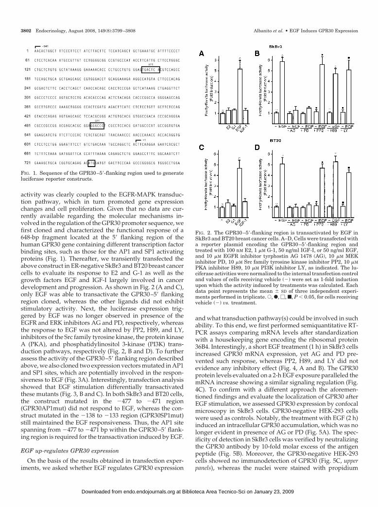
activity was clearly coupled to the EGFR-MAPK transduc-tion pathway, which in turn promoted gene expressionchanges and cell proliferation. Given that no data are cur-rently available regarding the molecular mechanisms in-volved in the regulation of the GPR30 promoter sequence, wefirst cloned and characterized the functional response of a648-bp fragment located at the 5� flanking region of thehuman GPR30 gene containing different transcription factorbinding sites, such as those for the AP1 and SP1 activatingproteins (Fig. 1). Thereafter, we transiently transfected theabove construct in ER-negative SkBr3 and BT20 breast cancercells to evaluate its response to E2 and G-1 as well as thegrowth factors EGF and IGF-I largely involved in cancerdevelopment and progression. As shown in Fig. 2 (A and C),only EGF was able to transactivate the GPR30–5� flankingregion cloned, whereas the other ligands did not exhibitstimulatory activity. Next, the luciferase expression trig-gered by EGF was no longer observed in presence of theEGFR and ERK inhibitors AG and PD, respectively, whereasthe response to EGF was not altered by PP2, H89, and LY,inhibitors of the Src family tyrosine kinase, the protein kinaseA (PKA), and phosphatidylinositol 3-kinase (PI3K) trans-duction pathways, respectively (Fig. 2, B and D). To furtherassess the activity of the GPR30–5� flanking region describedabove, we also cloned two expression vectors mutated in AP1and SP1 sites, which are potentially involved in the respon-siveness to EGF (Fig. 3A). Interestingly, transfection analysisshowed that EGF stimulation differentially transactivatedthese mutants (Fig. 3, B and C). In both SkBr3 and BT20 cells,the construct mutated in the �477 to �471 region(GPR30AP1mut) did not respond to EGF, whereas the con-struct mutated in the �138 to �133 region (GPR30SP1mut)still maintained the EGF responsiveness. Thus, the AP1 sitespanning from �477 to �471 bp within the GPR30–5� flank-ing region is required for the transactivation induced by EGF.
EGF up-regulates GPR30 expression
On the basis of the results obtained in transfection exper-iments, we asked whether EGF regulates GPR30 expression
and what transduction pathway(s) could be involved in suchability. To this end, we first performed semiquantitative RT-PCR assays comparing mRNA levels after standardizationwith a housekeeping gene encoding the ribosomal protein36B4. Interestingly, a short EGF treatment (1 h) in SkBr3 cellsincreased GPR30 mRNA expression, yet AG and PD pre-vented such response, whereas PP2, H89, and LY did notevidence any inhibitory effect (Fig. 4, A and B). The GPR30protein levels evaluated on a 2-h EGF exposure paralleled themRNA increase showing a similar signaling regulation (Fig.4C). To confirm with a different approach the aforemen-tioned findings and evaluate the localization of GPR30 afterEGF stimulation, we assessed GPR30 expression by confocalmicroscopy in SkBr3 cells. GPR30-negative HEK-293 cellswere used as controls. Notably, the treatment with EGF (2 h)induced an intracellular GPR30 accumulation, which was nolonger evident in presence of AG or PD (Fig. 5A). The spec-ificity of detection in SkBr3 cells was verified by neutralizingthe GPR30 antibody by 10-fold molar excess of the antigenpeptide (Fig. 5B). Moreover, the GPR30-negative HEK-293cells showed no immunodetection of GPR30 (Fig. 5C, upperpanels), whereas the nuclei were stained with propidium
FIG. 1. Sequence of the GPR30–5�-flanking region used to generateluciferase reporter constructs.
FIG. 2. The GPR30–5�-flanking region is transactivated by EGF inSkBr3 and BT20 breast cancer cells. A–D, Cells were transfected witha reporter plasmid encoding the GPR30–5�-flanking region andtreated with 100 nM E2, 1 �M G-1, 50 ng/ml IGF-I, or 50 ng/ml EGF,and 10 �M EGFR inhibitor tyrphostin AG 1478 (AG), 10 �M MEKinhibitor PD, 10 �M Src family tyrosine kinase inhibitor PP2, 10 �MPKA inhibitor H89, 10 �M PI3K inhibitor LY, as indicated. The lu-ciferase activities were normalized to the internal transfection controland values of cells receiving vehicle (�) were set as 1-fold inductionupon which the activity induced by treatments was calculated. Eachdata point represents the mean � SD of three independent experi-ments performed in triplicate. E, F, �, f, P � 0.05, for cells receivingvehicle (�) vs. treatment.
3802 Endocrinology, August 2008, 149(8):3799–3808 Albanito et al. • EGF Induces GPR30 Expression
at Biblioteca Area Tecnico-Sci on January 23, 2009 endo.endojournals.orgDownloaded from
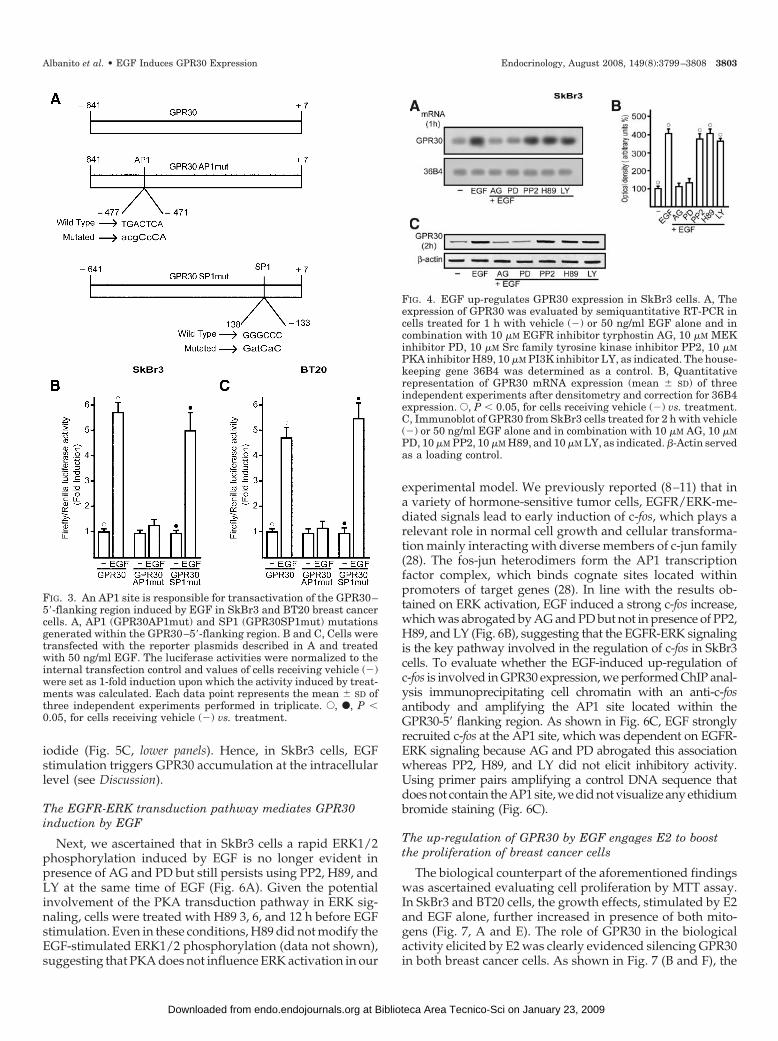
iodide (Fig. 5C, lower panels). Hence, in SkBr3 cells, EGFstimulation triggers GPR30 accumulation at the intracellularlevel (see Discussion).
The EGFR-ERK transduction pathway mediates GPR30induction by EGF
Next, we ascertained that in SkBr3 cells a rapid ERK1/2phosphorylation induced by EGF is no longer evident inpresence of AG and PD but still persists using PP2, H89, andLY at the same time of EGF (Fig. 6A). Given the potentialinvolvement of the PKA transduction pathway in ERK sig-naling, cells were treated with H89 3, 6, and 12 h before EGFstimulation. Even in these conditions, H89 did not modify theEGF-stimulated ERK1/2 phosphorylation (data not shown),suggesting that PKA does not influence ERK activation in our
experimental model. We previously reported (8–11) that ina variety of hormone-sensitive tumor cells, EGFR/ERK-me-diated signals lead to early induction of c-fos, which plays arelevant role in normal cell growth and cellular transforma-tion mainly interacting with diverse members of c-jun family(28). The fos-jun heterodimers form the AP1 transcriptionfactor complex, which binds cognate sites located withinpromoters of target genes (28). In line with the results ob-tained on ERK activation, EGF induced a strong c-fos increase,which was abrogated by AG and PD but not in presence of PP2,H89, and LY (Fig. 6B), suggesting that the EGFR-ERK signalingis the key pathway involved in the regulation of c-fos in SkBr3cells. To evaluate whether the EGF-induced up-regulation ofc-fos is involved in GPR30 expression, we performed ChIP anal-ysis immunoprecipitating cell chromatin with an anti-c-fosantibody and amplifying the AP1 site located within theGPR30-5� flanking region. As shown in Fig. 6C, EGF stronglyrecruited c-fos at the AP1 site, which was dependent on EGFR-ERK signaling because AG and PD abrogated this associationwhereas PP2, H89, and LY did not elicit inhibitory activity.Using primer pairs amplifying a control DNA sequence thatdoes not contain the AP1 site, we did not visualize any ethidiumbromide staining (Fig. 6C).
The up-regulation of GPR30 by EGF engages E2 to boostthe proliferation of breast cancer cells
The biological counterpart of the aforementioned findingswas ascertained evaluating cell proliferation by MTT assay.In SkBr3 and BT20 cells, the growth effects, stimulated by E2and EGF alone, further increased in presence of both mito-gens (Fig. 7, A and E). The role of GPR30 in the biologicalactivity elicited by E2 was clearly evidenced silencing GPR30in both breast cancer cells. As shown in Fig. 7 (B and F), the
FIG. 3. An AP1 site is responsible for transactivation of the GPR30–5�-flanking region induced by EGF in SkBr3 and BT20 breast cancercells. A, AP1 (GPR30AP1mut) and SP1 (GPR30SP1mut) mutationsgenerated within the GPR30–5�-flanking region. B and C, Cells weretransfected with the reporter plasmids described in A and treatedwith 50 ng/ml EGF. The luciferase activities were normalized to theinternal transfection control and values of cells receiving vehicle (�)were set as 1-fold induction upon which the activity induced by treat-ments was calculated. Each data point represents the mean � SD ofthree independent experiments performed in triplicate. E, F, P �0.05, for cells receiving vehicle (�) vs. treatment.
FIG. 4. EGF up-regulates GPR30 expression in SkBr3 cells. A, Theexpression of GPR30 was evaluated by semiquantitative RT-PCR incells treated for 1 h with vehicle (�) or 50 ng/ml EGF alone and incombination with 10 �M EGFR inhibitor tyrphostin AG, 10 �M MEKinhibitor PD, 10 �M Src family tyrosine kinase inhibitor PP2, 10 �MPKA inhibitor H89, 10 �M PI3K inhibitor LY, as indicated. The house-keeping gene 36B4 was determined as a control. B, Quantitativerepresentation of GPR30 mRNA expression (mean � SD) of threeindependent experiments after densitometry and correction for 36B4expression. E, P � 0.05, for cells receiving vehicle (�) vs. treatment.C, Immunoblot of GPR30 from SkBr3 cells treated for 2 h with vehicle(�) or 50 ng/ml EGF alone and in combination with 10 �M AG, 10 �MPD, 10 �M PP2, 10 �M H89, and 10 �M LY, as indicated. �-Actin servedas a loading control.
Albanito et al. • EGF Induces GPR30 Expression Endocrinology, August 2008, 149(8):3799–3808 3803
at Biblioteca Area Tecnico-Sci on January 23, 2009 endo.endojournals.orgDownloaded from
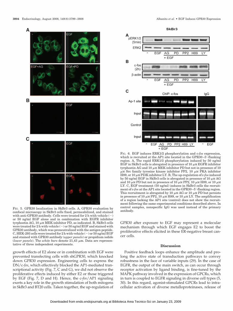
growth effects of E2 alone or in combination with EGF wereprevented transfecting cells with shGPR30, which knockeddown GPR30 expression. Engineering cells to express theDN/c-fos, which effectively blocked the AP1-mediated tran-scriptional activity (Fig. 7, C and G), we did not observe theproliferative effects induced by either E2 or those triggeredby EGF (Fig. 7, D and H). Hence, the c-fos/AP1 signalingexerts a key role in the growth stimulation of both mitogensin SkBr3 and BT20 cells. Taken together, the up-regulation of
GPR30 after exposure to EGF may represent a molecularmechanism through which EGF engages E2 to boost theproliferative effects elicited in these ER-negative breast can-cer cells.
Discussion
Positive feedback loops enhance the amplitude and pro-long the active state of transduction pathways to conveyrobustness in the face of variable inputs (29). In the case ofEGFR, the output of the main switch, as can occur throughreceptor activation by ligand binding, is fine-tuned by theMAPK pathway involved in the expression of GPCRs, whichin turn is coupled to EGFR signaling in diverse cell types (5,30). In this regard, agonist-stimulated GPCRs lead to intra-cellular activation of diverse metalloproteinases, release of
FIG. 5. GPR30 localization in SkBr3 cells. A, GPR30 evaluation byconfocal microscopy in SkBr3 cells fixed, permeabilized, and stainedwith anti-GPR30 antibody. Cells were treated for 2 h with vehicle (�)or 50 ng/ml EGF alone and in combination with EGFR inhibitortyrphostin AG, 10 �M MEK inhibitor PD, as indicated. B, SkBr3 cellswere treated for 2 h with vehicle (�) or 50 ng/ml EGF and stained withGPR30 antibody, which was preneutralized with the antigen peptide.C, HEK-293 cells were treated for 2 h with vehicle (�) or 50 ng/ml EGFand stained with GPR30 antibody (upper panels) or propidium iodide(lower panels). The white bars denote 21,43 �m. Data are represen-tative of three independent experiments.
FIG. 6. EGF induces ERK1/2 phosphorylation and c-fos expression,which is recruited at the AP1 site located in the GPR30–5�-flankingregion. A, The rapid ERK1/2 phosphorylation induced by 50 ng/mlEGF in SkBr3 cells is abrogated in presence of 10 �M EGFR inhibitortyrphostin AG and 10 �M MEK inhibitor PD but not in presence of 10�M Src family tyrosine kinase inhibitor PP2, 10 �M PKA inhibitorH89, or 10 �M PI3K inhibitor LY. B, The up-regulation of c-fos inducedby 50 ng/ml EGF in SkBr3 cells is abrogated in presence of 10 �M AGand 10 �M PD but not in presence of 10 �M PP2, 10 �M H89, or 10 �MLY. C, EGF treatment (50 ng/ml) induces in SkBr3 cells the recruit-ment of c-fos at the AP1 site located in the GPR30–5�-flanking region.This recruitment is abrogated by 10 �M AG or 10 �M PD but persistsin presence of 10 �M PP2, 10 �M H89, or 10 �M LY. The amplificationof a region lacking the AP1 site (control) does not show the recruit-ment following the same experimental conditions described above. Incontrol samples, nonspecific IgG was used instead of the primaryantibody.
3804 Endocrinology, August 2008, 149(8):3799–3808 Albanito et al. • EGF Induces GPR30 Expression
at Biblioteca Area Tecnico-Sci on January 23, 2009 endo.endojournals.orgDownloaded from
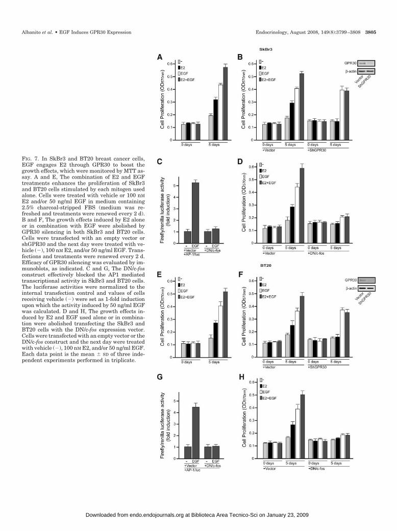
FIG. 7. In SkBr3 and BT20 breast cancer cells,EGF engages E2 through GPR30 to boost thegrowth effects, which were monitored by MTT as-say. A and E, The combination of E2 and EGFtreatments enhances the proliferation of SkBr3and BT20 cells stimulated by each mitogen usedalone. Cells were treated with vehicle or 100 nME2 and/or 50 ng/ml EGF in medium containing2.5% charcoal-stripped FBS (medium was re-freshed and treatments were renewed every 2 d).B and F, The growth effects induced by E2 aloneor in combination with EGF were abolished byGPR30 silencing in both SkBr3 and BT20 cells.Cells were transfected with an empty vector orshGPR30 and the next day were treated with ve-hicle (�), 100 nM E2, and/or 50 ng/ml EGF. Trans-fections and treatments were renewed every 2 d.Efficacy of GPR30 silencing was evaluated by im-munoblots, as indicated. C and G, The DN/c-fosconstruct effectively blocked the AP1 mediatedtranscriptional activity in SkBr3 and BT20 cells.The luciferase activities were normalized to theinternal transfection control and values of cellsreceiving vehicle (�) were set as 1-fold inductionupon which the activity induced by 50 ng/ml EGFwas calculated. D and H, The growth effects in-duced by E2 and EGF used alone or in combina-tion were abolished transfecting the SkBr3 andBT20 cells with the DN/c-fos expression vector.Cells were transfected with an empty vector or theDN/c-fos construct and the next day were treatedwith vehicle (�), 100 nM E2, and/or 50 ng/ml EGF.Each data point is the mean � SD of three inde-pendent experiments performed in triplicate.
Albanito et al. • EGF Induces GPR30 Expression Endocrinology, August 2008, 149(8):3799–3808 3805
at Biblioteca Area Tecnico-Sci on January 23, 2009 endo.endojournals.orgDownloaded from
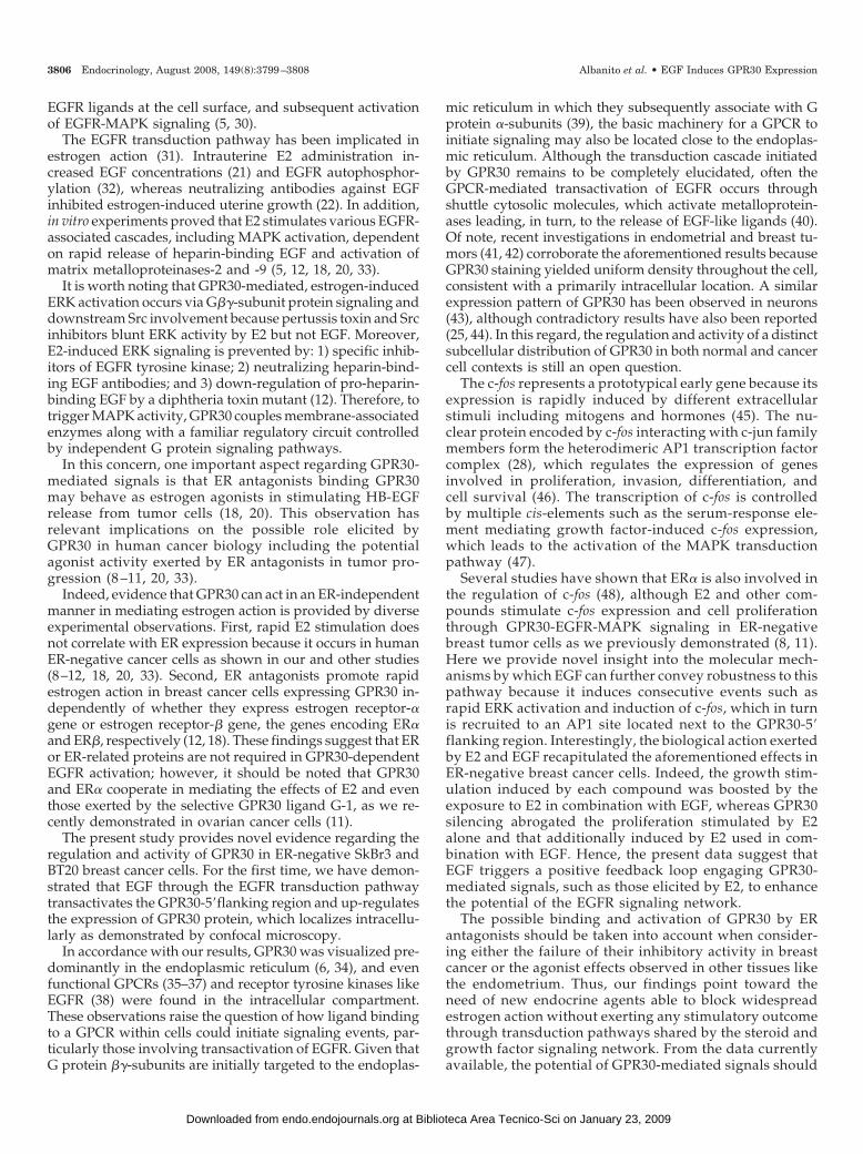
EGFR ligands at the cell surface, and subsequent activationof EGFR-MAPK signaling (5, 30).
The EGFR transduction pathway has been implicated inestrogen action (31). Intrauterine E2 administration in-creased EGF concentrations (21) and EGFR autophosphor-ylation (32), whereas neutralizing antibodies against EGFinhibited estrogen-induced uterine growth (22). In addition,in vitro experiments proved that E2 stimulates various EGFR-associated cascades, including MAPK activation, dependenton rapid release of heparin-binding EGF and activation ofmatrix metalloproteinases-2 and -9 (5, 12, 18, 20, 33).
It is worth noting that GPR30-mediated, estrogen-inducedERK activation occurs via G��-subunit protein signaling anddownstream Src involvement because pertussis toxin and Srcinhibitors blunt ERK activity by E2 but not EGF. Moreover,E2-induced ERK signaling is prevented by: 1) specific inhib-itors of EGFR tyrosine kinase; 2) neutralizing heparin-bind-ing EGF antibodies; and 3) down-regulation of pro-heparin-binding EGF by a diphtheria toxin mutant (12). Therefore, totrigger MAPK activity, GPR30 couples membrane-associatedenzymes along with a familiar regulatory circuit controlledby independent G protein signaling pathways.
In this concern, one important aspect regarding GPR30-mediated signals is that ER antagonists binding GPR30may behave as estrogen agonists in stimulating HB-EGFrelease from tumor cells (18, 20). This observation hasrelevant implications on the possible role elicited byGPR30 in human cancer biology including the potentialagonist activity exerted by ER antagonists in tumor pro-gression (8 –11, 20, 33).
Indeed, evidence that GPR30 can act in an ER-independentmanner in mediating estrogen action is provided by diverseexperimental observations. First, rapid E2 stimulation doesnot correlate with ER expression because it occurs in humanER-negative cancer cells as shown in our and other studies(8–12, 18, 20, 33). Second, ER antagonists promote rapidestrogen action in breast cancer cells expressing GPR30 in-dependently of whether they express estrogen receptor-�gene or estrogen receptor-� gene, the genes encoding ER�and ER�, respectively (12, 18). These findings suggest that ERor ER-related proteins are not required in GPR30-dependentEGFR activation; however, it should be noted that GPR30and ER� cooperate in mediating the effects of E2 and eventhose exerted by the selective GPR30 ligand G-1, as we re-cently demonstrated in ovarian cancer cells (11).
The present study provides novel evidence regarding theregulation and activity of GPR30 in ER-negative SkBr3 andBT20 breast cancer cells. For the first time, we have demon-strated that EGF through the EGFR transduction pathwaytransactivates the GPR30-5�flanking region and up-regulatesthe expression of GPR30 protein, which localizes intracellu-larly as demonstrated by confocal microscopy.
In accordance with our results, GPR30 was visualized pre-dominantly in the endoplasmic reticulum (6, 34), and evenfunctional GPCRs (35–37) and receptor tyrosine kinases likeEGFR (38) were found in the intracellular compartment.These observations raise the question of how ligand bindingto a GPCR within cells could initiate signaling events, par-ticularly those involving transactivation of EGFR. Given thatG protein ��-subunits are initially targeted to the endoplas-
mic reticulum in which they subsequently associate with Gprotein �-subunits (39), the basic machinery for a GPCR toinitiate signaling may also be located close to the endoplas-mic reticulum. Although the transduction cascade initiatedby GPR30 remains to be completely elucidated, often theGPCR-mediated transactivation of EGFR occurs throughshuttle cytosolic molecules, which activate metalloprotein-ases leading, in turn, to the release of EGF-like ligands (40).Of note, recent investigations in endometrial and breast tu-mors (41, 42) corroborate the aforementioned results becauseGPR30 staining yielded uniform density throughout the cell,consistent with a primarily intracellular location. A similarexpression pattern of GPR30 has been observed in neurons(43), although contradictory results have also been reported(25, 44). In this regard, the regulation and activity of a distinctsubcellular distribution of GPR30 in both normal and cancercell contexts is still an open question.
The c-fos represents a prototypical early gene because itsexpression is rapidly induced by different extracellularstimuli including mitogens and hormones (45). The nu-clear protein encoded by c-fos interacting with c-jun familymembers form the heterodimeric AP1 transcription factorcomplex (28), which regulates the expression of genesinvolved in proliferation, invasion, differentiation, andcell survival (46). The transcription of c-fos is controlledby multiple cis-elements such as the serum-response ele-ment mediating growth factor-induced c-fos expression,which leads to the activation of the MAPK transductionpathway (47).
Several studies have shown that ER� is also involved inthe regulation of c-fos (48), although E2 and other com-pounds stimulate c-fos expression and cell proliferationthrough GPR30-EGFR-MAPK signaling in ER-negativebreast tumor cells as we previously demonstrated (8, 11).Here we provide novel insight into the molecular mech-anisms by which EGF can further convey robustness to thispathway because it induces consecutive events such asrapid ERK activation and induction of c-fos, which in turnis recruited to an AP1 site located next to the GPR30-5�flanking region. Interestingly, the biological action exertedby E2 and EGF recapitulated the aforementioned effects inER-negative breast cancer cells. Indeed, the growth stim-ulation induced by each compound was boosted by theexposure to E2 in combination with EGF, whereas GPR30silencing abrogated the proliferation stimulated by E2alone and that additionally induced by E2 used in com-bination with EGF. Hence, the present data suggest thatEGF triggers a positive feedback loop engaging GPR30-mediated signals, such as those elicited by E2, to enhancethe potential of the EGFR signaling network.
The possible binding and activation of GPR30 by ERantagonists should be taken into account when consider-ing either the failure of their inhibitory activity in breastcancer or the agonist effects observed in other tissues likethe endometrium. Thus, our findings point toward theneed of new endocrine agents able to block widespreadestrogen action without exerting any stimulatory outcomethrough transduction pathways shared by the steroid andgrowth factor signaling network. From the data currentlyavailable, the potential of GPR30-mediated signals should
3806 Endocrinology, August 2008, 149(8):3799–3808 Albanito et al. • EGF Induces GPR30 Expression
at Biblioteca Area Tecnico-Sci on January 23, 2009 endo.endojournals.orgDownloaded from
be considered in estrogen-sensitive tumors to discoverinnovative antiestrogens. GPR30 overexpression was re-cently associated with lower survival rates in endometrialcancer patients (41) and higher risk of developing meta-static disease in patients with breast tumor (42). Therefore,the expression levels of GPR30 may characterize not onlyestrogen sensitivity and the potential response to endo-crine pharmacological intervention in these tumors butcould also be predictive of biologically aggressive pheno-types consistent with adverse outcome and survival. Inaddition, the up-regulation of GPR30 expression by theligand-activated EGFR further extends our knowledge re-garding the cross talk between EGF and E2 signaling inbreast cancer progression. Likewise, our results indicatethat the action of EGF may include the up-regulation ofGPR30 in facilitating a stimulatory role of estrogen evenin ER-negative breast tumor cells.
Acknowledgments
Received January 25, 2008. Accepted April 29, 2008.Address all correspondence and requests for reprints to: Professor
Marcello Maggiolini, Department Pharmaco-Biology, University ofCalabria, 87030 Rende (Cosenze), Italy. E-mail: [email protected].
This work was supported by grants from the Associazione Italianaper la Ricerca sul Cancro and Ministero dell’Universita e Ricerca Sci-entifica e Tecnologica and Regione Calabria.
Disclosure Statement: The authors have nothing to disclose.
References
1. Levin ER 2003 Bidirectional signaling between the estrogen receptor and theepidermal growth factor receptor. Mol Endocrinol 17:309–317
2. Keen JC, Davidson NE 2003 The biology of breast carcinoma. Cancer97:825– 833
3. Roskoski Jr R 2004 The ErbB/HER receptor protein-tyrosine kinases andcancer. Biochem Biophys Res Commun 319:1–11
4. Ali S, Coombes RC 2002 Endocrine-responsive breast cancer and strategies forcombating resistance. Nat Rev Cancer 2:101–112
5. Razandi M, Pedram A, Park ST, Levin ER 2003 Proximal events in signalingby plasma membrane estrogen receptor. J Biol Chem 278:2701–2712
6. Revankar CM, Cimino DF, Sklar LA, Arterburn JB, Prossnitz ER 2005 Atransmembrane intracellular estrogen receptor mediates rapid cell signaling.Science 307:1625–1630
7. Thomas P, Pang Y, Filardo EJ, Dong J 2005 Identity of an estrogen membranereceptor coupled a G protein in human breast cancer cells. Endocrinology146:624–632
8. Maggiolini M, Vivacqua A, Fasanella G, Recchia AG, Sisci D, Pezzi V,Montanaro D, Musti AM, Picard D, Ando S 2004 The G protein-coupledreceptor GPR30 mediates c-fos up-regulation by 17-�-estradiol and phytoestro-gens in breast cancer cells. J Biol Chem 279:27008–27016
9. Vivacqua A, Bonofiglio D, Recchia AG, Musti AM, Picard D, Ando S,Maggiolini M 2006 The G protein-coupled receptor GPR30 mediates the pro-liferative effects induced by 17-�-estradiol and hydroxytamoxifen in endo-metrial cancer cells. Mol Endocrinol 20:631–646
10. Vivacqua A, Bonofiglio D, Albanito L, Madeo A, Rago V, Carpino A, MustiAM, Picard D, Ando S, Maggiolini M 2006 17-�-Estradiol, genistein and4-hydroxytamoxifen induce the proliferation of thyroid cancer cells throughthe G protein-coupled receptor GPR30. Mol Pharmacol 70:1414–1423
11. Albanito L, Madeo A, Lappano R, Vivacqua A, Rago V, Carpino A, OpreaTI, Prossnitz ER, Musti AM, Ando S, Maggiolini M 2007 G-protein coupledreceptor 30 (GPR30) mediates gene expression changes and growth responseto 17-�-estradiol and selective GPR30 ligand G-1 in ovarian cancer cells. CancerRes 67:1859–1866
12. Filardo EJ, Quinn JA, Bland KI, Frackelton Jr AR 2000 Estrogen-inducedactivation of Erk-1 and Erk-2 requires the G protein-coupled receptor homolog,GPR30 and occurs via trans-activation of the epidermal growth factor receptorthrough release of HB-EGF. Mol Endocrinol 14:1649–1660
13. Prenzel N, Zwick E, Daub H, Leserer M, Abraham R, Wallasch C, Ullrich A1999 EGF receptor transactivation by G-protein-coupled receptors requiresmetalloproteinase cleavage of proHB-EGF. Nature 402:884–888
14. Gschwind A, Zwick E, Prenzel N, Leserer M, Ullrich A 2001 Cell commu-
nication networks: epidermal growth factor receptor transactivation as theparadigm for interreceptor signal transmission. Oncogene 20:1594–1600
15. Hla T, Lee MJ, Ancellin N, Paik JH, Kluk MJ 2001 Lysophospholipids-receptor revelations. Science 294:1875–1878
16. Tanimoto T, Lungu AO, Berk BC 2004 Sphingosine 1-phosphate transacti-vates the platelet-derived growth factor � receptor and epidermal growthfactor receptor in vascular smooth muscle cells. Circ Res 94:1050–1058
17. Greco S, Muscella A, Elia MG, Salvatore P, Storelli C, Mazzotta A, MancaC, Marsigliante S 2003 Angiotensin II activates extracellular signal regulatedkinases via protein kinase C and epidermal growth factor receptor in breastcancer cells. J Cell Physiol 196:370–377
18. Filardo EJ, Quinn JA, Frackelton Jr AR, Bland KI 2002 Estrogen action via theG protein-coupled receptor, GPR30: stimulation of adenylyl cyclase andcAMP-mediated attenuation of the epidermal growth factor receptor-to-MAPK signaling axis. Mol Endocrinol 16:70–84
19. Early Breast Cancer Trialist’ Collaborative Group 2005 Effects of chemother-apy and hormonal therapy for early breast cancer on recurrence and 15-yearssurvival: an overview of the randomized trials. Lancet 365:1687–1700
20. Filardo EJ 2002 Epidermal Growth Factor (EGFR) transactivation by es-trogen via the G-protein-coupled receptor, GPR30: a novel signalling path-way with potential significance for breast cancer. J Steroid Biochem Mol Biol80:231–238
21. DiAugustine RP, Petrusz P, Bell GI, Brown CF, Korach KS, McLachlan JA,Teng CT 1988 Influence of estrogens on mouse uterine epidermal growthfactor precursor protein and messenger ribonucleic acid. Endocrinology 122:2355–2363
22. Nelson KG, Takahashi T, Bossert NL, Walmer DK, McLachlan JA 1991Epidermal growth factor replaces estrogen in the stimulation of female genital-tract growth and differentiation. Proc Natl Acad Sci USA 88:21–25
23. Migliaccio A, Di Domenico M, Castoria G, de Falco A, Bontempo P, Nola E,Auricchio F 1996 Tyrosine kinase/p21ras/MAPK kinase pathway activationby estradiol-receptor complex in MCF-7 cells. EMBO J 15:1292–1300
24. Martin MB, Franke TF, Stoica GE, Chambon P, Katzenellenbogen BS, StoicaBA, McLemore MS, Olivo SE, Stoica A 2000 A role for AKT in mediating theestrogenic functions of epidermal growth factor and insuline-like growthfactor I. Endocrinology 141:4503–4511
25. Funakoshi T, Yanai A, Shinoda K, Kawano MM, Mizukami Y 2006 G protein-coupled receptor 30 is an estrogen receptor in the plasma membrane. BiochemBiophys Res Commun 346:904–910
26. Maggiolini M, Donze O, Picard D 1999 A nonradioactive method for inex-pensive quantitative RT-PCR. J Biol Chem 380:695–697
27. Morelli C, Garofalo C, Sisci D, del Rincon S, Cascio S, Tu X, VecchioneA, Sauter ER, Miller Jr WH, Surmacz E 2004 Nuclear insulin receptorsubstrate 1 interacts with estrogen receptor � at ERE promoters. Oncogene23:7517–7526
28. Curran T, Franza BR J 1988 Fos and Jun: the AP1 connection. Cell 55:395–39729. Freeman M 2000 Feedback control of intercellular signalling in development.
Nature 408:313–31930. Hsieh M, Conti M 2005 G-protein-coupled receptor signaling and the EGF
network in endocrine systems. Trends Endocrinol Metab 16:320–32631. Cunha GR, Wiesen JF, Werb Z, Young P, Hom YK, Cooke PS, Lubahn DB
2000 Paracrine mechanisms of mouse mammary ductal growth. Adv Exp MedBiol 480:93–97
32. Mukku VR, Stancel GM 1985 Regulation of epidermal growth factor receptorby estrogen. J Biol Chem 260:9820–9824
33. Filardo EJ, Thomas P 2005 GPR30: a seven-transmembrane-spanning estrogenreceptor that triggers EGF release. Trends Endocrinol Metab 16:362–370
34. Revankar CM, Mitchell HD, Field AS, Burai R, Corona C, Ramesh C, SklarLA, Arterburn JB, Prossnitz ER 2007 Synthetic estrogen derivatives demon-strate the functionality of intracellular GPR30. ACS Chem Biol 2:536–544
35. Gobeil F, Fortier A, Zhu T, Bossolasco M, Leduc M, Grandbois M, HevekerN, Bkaily G, Chemtob S, Barbaz D 2006 G-protein-coupled receptors signal-ling at the cell nucleus: an emerging paradigm. Can J Physiol Pharmacol84:287–297
36. Marrache AM, Gobeil F, Zhu T, Chemtob S 2005 Intracellular signaling oflipid mediators via cognate nuclear G-protein-coupled receptors. Endothelium12:63–72
37. Zhu T, Gobeil F, Vazquez-Tello A, Leduc M, Rihakova L, Bossolasco M,Bkaily G, Peri K, Varma DR, Orvoine R, Chemtob S 2006 Intracrine signalingthrough lipid mediators and their cognate nuclear G-protein-coupled recep-tors: a paradigm based on PGE2, PAF and LPA1 receptors. Can J PhysiolPharmacol 84:377–391
38. Bjornstrom L, Sjorberg M 2005 Mechanisms of estrogen receptor signaling:convergence of genomic and nongenomic actions on target genes. Mol Endo-crinol 19:833–842
39. Levin ER 2005 Integration of the extranuclear and nuclear actions of estrogen.Mol Endocrinol 19:1951–1959
40. Hasbi A, O’Dowd BF, George SR 2005 A G protein-coupled receptor forestrogen: the end of the search? Mol Interv 5:158–161
41. Smith HO, Leslie KK, Singh M, Qualls CR, Revankar CM, Joste NE,Prossnitz ER 2007 GPR30: a novel indicator of poor survival for endometrialcarcinoma. Am J Obstet Gynecol 196:386.e1–386.e9, 386.e9–386.e11
Albanito et al. • EGF Induces GPR30 Expression Endocrinology, August 2008, 149(8):3799–3808 3807
at Biblioteca Area Tecnico-Sci on January 23, 2009 endo.endojournals.orgDownloaded from

42. Filardo EJ, Graeber CT, Quinn JA, Resnick MB, Giri D, DeLellis RA,Steinhoff MM, Sabo E 2006 Distribution of GPR30, a seven membrane-spanning estrogen receptor, in primary breast cancer and its associationwith clinicopathologic determinants of tumor progression. Clin Cancer Res12:6359 – 6366
43. Brailoiu E, Dun SL, Brailoiu GC, Mizuo K, Sklar LA, Oprea TI, Prossnitz ER,Dun NJ 2007 Distribution and characterization of estrogen receptor G protein-coupled receptor 30 in the rat central nervous system. J Endocrinol 193:311–321
44. Filardo E, Quinn J, Pang Y, Graeber C, Shaw S, Dong J, Thomas P 2007Activation of the novel estrogen receptor (GPR30) at the plasma membrane.Endocrinology 148:3236–3245
45. Hill CS, Treisman R 1995 Differential activation of c-fos promoter elements byserum, lysophosphatidic acid, G proteins and polypeptide growth factors.EMBO J 14:5037–5047
46. Shaulian E, Karin M 2002 AP-1 as a regulator of cell life and death. Nat CellBiol 4:E131–E136
47. Karin M, Hunter T 1995 Transcriptional control by protein phosphorylation:signal transmission from the cell surface to the nucleus. Curr Biol 5:747–757
48. Bonapace IM, Addeo R, Altucci L, Cicatiello L, Bifulco M, Laezza C, SalzanoS, Sica V, Bresciani F, Weisz A 1996 17�-Estradiol overcomes a G1 blockinduced by HMG-CoA reductase inhibitors and fosters cell cycle progressionwithout inducing ERK-1 and -2 MAP kinases activation. Oncogene 12:753–763
Endocrinology is published monthly by The Endocrine Society (http://www.endo-society.org), the foremost professional society serving theendocrine community.
3808 Endocrinology, August 2008, 149(8):3799–3808 Albanito et al. • EGF Induces GPR30 Expression
at Biblioteca Area Tecnico-Sci on January 23, 2009 endo.endojournals.orgDownloaded from
845Mol. Nutr. Food Res. 2009, 53, 845 –858 DOI 10.1002/mnfr.200800331
Research Article
Structure–activity relationships of resveratrol andderivatives in breast cancer cells
Rosamaria Lappano1, Camillo Rosano2, Antonio Madeo1, Lidia Albanito1, PierluigiPlastina3, Bartolo Gabriele3, Luca Forti4, Lucia Anna Stivala5, Domenico Iacopetta1,Vincenza Dolce1, Sebastiano And�6, Vincenzo Pezzi1 and Marcello Maggiolini1
1 Dipartimento Farmaco-Biologico, Universit� della Calabria, Rende (CS), Italy2 Dipartimento di Bioinformatica e Proteomica Strutturale, Istituto Nazionale per la Ricerca sul Cancro
(IST), Genova, Italy3 Dipartimento di Scienze Farmaceutiche, Universit� della Calabria, Rende (CS), Italy4 Dipartimento di Chimica, Universit� di Modena e Reggio Emilia, Modena, Italy5 Dipartimento di Medicina Sperimentale, Universit� di Pavia, Pavia, Italy6 Dipartimento di Biologia Cellulare, Universit� Della Calabria, Rende (CS), Italia
Resveratrol (RSV) is classified as a phytoestrogen due to its ability to interact with estrogen receptors(ERs). We assessed structure–activity relationships of RSV and the analogs 4,49-dihydroxystilbene(4,49-DHS), 3,5-dihydroxystilbene (3,5-DHS), 3,49-dihydroxystilbene (3,49-DHS), 4-hydroxystilbene(4-HS) using as model systems the ERa-positive and negative MCF7 and SkBr3 breast cancer cells,respectively. In binding assays and transfection experiments RSV and the analogs showed the follow-ing order of agonism for ERa: 3,49-DHS A 4,49-DHS A 4-HS A RSV, while 3,5-DHS did not elicit anyligand properties. Computational docking analysis and real-time PCR revealed for each analog a dis-tinct ERa binding orientation and estrogen target gene expression profile. Interestingly, the aforemen-tioned order of ligand activity was confirmed in proliferation assays which also showed the lack ofgrowth stimulation by 3,5-DHS. Our data suggest that subtle changes in the structure of the RSVderivatives examined may be responsible for the different ERa-mediated biological responsesobserved in estrogen-sensitive cancer cells.
Keywords: Breast cancer / Docking analysis / Estrogen receptor / Hydroxystilbenes / Resveratrol /
Received: July 25, 2008; revised: October 14, 2008; accepted: October 18, 2008
1 Introduction
Breast cancer is the most frequent tumor and the majorcause of death among women in the United States [1]. Theproliferation of many breast tumor cells is stimulated by thebinding of 17b-estradiol (E2) to the estrogen receptor (ER)isoforms, ERa and ERb, which belong to the nuclear recep-tor super-family. The transcriptional activity of the ERs ismediated by both an N-terminal ligand-independent activa-tion function (AF-1) and a ligand-dependent activationfunction (AF-2) located in the ligand binding domain
(LBD) [2]. The LBD contains a molecular switch helix–helix 12-which regulates the communication betweenligand- and coactivator-binding sites [2, 3]. In ERa, as inother steroid hormone receptors, the ligand-binding pocketis a compact ellipsoid cavity, closely resembling the surfaceof the steroid ligands [4]. In the presence of agonist ligands,helix 12 is stabilized in a conformation that allows it toform one side of the coactivator-binding site and to com-pletely encapsulate the ligand in the pocket. Antagonistligands, such as the selective ER modulator (SERM)tamoxifen, resemble agonist ligands but contain an addi-tional extended group [2–4]. The bulky side chain ontamoxifen protrudes between helices 3 and 11, and physi-cally obstructs helix 12 from adopting the agonist confor-mation, thus preventing coactivator recruitment to the LBD[3]. The binding of E2 to the LBD of ERa induces confor-mational changes allowing receptor–chromatin interactionand the transcriptional regulation of target genes [5, 6]. Alarge body of data indicates that dietary factors play a sig-
Correspondence: Professor Marcello Maggiolini, Department Phar-maco-Biology, University of Calabria, 87030 Rende (CS), ItalyE-mail: [email protected]: +390-984493458
Abbreviations: 4-HS, 4-hydroxystilbene; CS, charcoal-stripped;DHS, dihydroxystilbene; E2, 17b-estradiol; ER, estrogen receptor;ICI, ICI 182,780; LBD, ligand binding domain; PR, progesterone re-ceptor; RSV, resveratrol
i 2009 WILEY-VCH Verlag GmbH & Co. KGaA, Weinheim www.mnf-journal.com
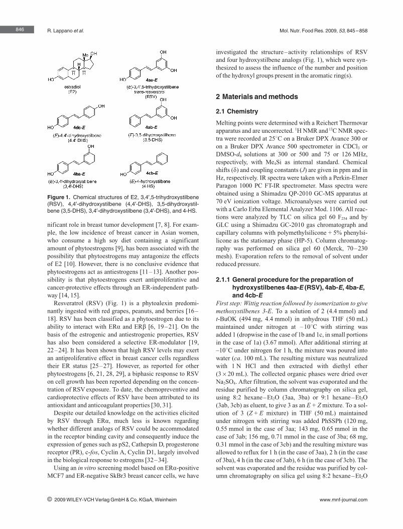
R. Lappano et al. Mol. Nutr. Food Res. 2009, 53, 845 –858
nificant role in breast tumor development [7, 8]. For exam-ple, the low incidence of breast cancer in Asian women,who consume a high soy diet containing a significantamount of phytoestrogens [9], has been associated with thepossibility that phytoestrogens may antagonize the effectsof E2 [10]. However, there is no conclusive evidence thatphytoestrogens act as antiestrogens [11–13]. Another pos-sibility is that phytoestrogens exert antiproliferative andcancer-protective effects through an ER-independent path-way [14, 15].
Resveratrol (RSV) (Fig. 1) is a phytoalexin predomi-nantly ingested with red grapes, peanuts, and berries [16–18]. RSV has been classified as a phytoestrogen due to itsability to interact with ERa and ERb [6, 19–21]. On thebasis of the estrogenic and antiestrogenic properties, RSVhas also been considered a selective ER-modulator [19,22–24]. It has been shown that high RSV levels may exertan antiproliferative effect in breast cancer cells regardlesstheir ER status [25–27]. However, as reported for otherphytoestrogens [6, 21, 28, 29], a biphasic response to RSVon cell growth has been reported depending on the concen-tration of RSV exposure. To date, the chemopreventive andcardioprotective effects of RSV have been attributed to itsantioxidant and anticoagulant properties [30, 31].
Despite our detailed knowledge on the activities elicitedby RSV through ERa, much less is known regardingwhether different analogs of RSV could be accommodatedin the receptor binding cavity and consequently induce theexpression of genes such as pS2, Cathepsin D, progesteronereceptor (PR), c-fos, Cyclin A, Cyclin D1, largely involvedin the biological response to estrogens [32–34].
Using an in vitro screening model based on ERa-positiveMCF7 and ER-negative SkBr3 breast cancer cells, we have
investigated the structure–activity relationships of RSVand four hydroxystilbene analogs (Fig. 1), which were syn-thesized to assess the influence of the number and positionof the hydroxyl groups present in the aromatic ring(s).
2 Materials and methods
2.1 Chemistry
Melting points were determined with a Reichert Thermovarapparatus and are uncorrected. 1H NMR and 13C NMR spec-tra were recorded at 258C on a Bruker DPX Avance 300 oron a Bruker DPX Avance 500 spectrometer in CDCl3 orDMSO-d6 solutions at 300 or 500 and 75 or 126 MHz,respectively, with Me4Si as internal standard. Chemicalshifts (d) and coupling constants (J) are given in ppm and inHz, respectively. IR spectra were taken with a Perkin-ElmerParagon 1000 PC FT-IR spectrometer. Mass spectra wereobtained using a Shimadzu QP-2010 GC-MS apparatus at70 eV ionization voltage. Microanalyses were carried outwith a Carlo Erba Elemental Analyzer Mod. 1106. All reac-tions were analyzed by TLC on silica gel 60 F254 and byGLC using a Shimadzu GC-2010 gas chromatograph andcapillary columns with polymethylsilicone + 5% phenylsi-licone as the stationary phase (HP-5). Column chromatog-raphy was performed on silica gel 60 (Merck, 70–230mesh). Evaporation refers to the removal of solvent underreduced pressure.
2.1.1 General procedure for the preparation ofhydroxystilbenes 4aa-E (RSV), 4ab-E, 4ba-E,and 4cb-E
First step: Wittig reaction followed by isomerization to givemethoxystilbenes 3-E. To a solution of 2 (4.4 mmol) andt-BuOK (494 mg, 4.4 mmol) in anhydrous THF (50 mL)maintained under nitrogen at –108C with stirring wasadded 1 (dropwise in the case of 1b and 1c, in small portionsin the case of 1a) (3.67 mmol). After additional stirring at–108C under nitrogen for 1 h, the mixture was poured intowater (ca. 100 mL). The resulting mixture was neutralizedwith 1 N HCl and then extracted with diethyl ether(3620 mL). The collected organic phases were dried overNa2SO4. After filtration, the solvent was evaporated and theresidue purified by column chromatography on silica gel,using 8:2 hexane–Et2O (3aa, 3ba) or 9:1 hexane–Et2O(3ab, 3cb) as eluent, to give 3 as an E + Z mixture. To a sol-ution of 3 (Z + E mixture) in THF (50 mL) maintainedunder nitrogen with stirring was added PhSSPh (120 mg,0.55 mmol in the case of 3aa; 143 mg, 0.65 mmol in thecase of 3ab; 156 mg, 0.71 mmol in the case of 3ba; 68 mg,0.31 mmol in the case of 3cb) and the resulting mixture wasallowed to reflux for 1 h (in the case of 3aa), 2 h (in the caseof 3ba), 4 h (in the case of 3ab), 6 h (in the case of 3cb). Thesolvent was evaporated and the residue was purified by col-umn chromatography on silica gel using 8:2 hexane–Et2O
846
i 2009 WILEY-VCH Verlag GmbH & Co. KGaA, Weinheim www.mnf-journal.com
Figure 1. Chemical structures of E2, 3,49,5-trihydroxystilbene(RSV), 4,49-dihydroxystilbene (4,49-DHS), 3,5-dihydroxystil-bene (3,5-DHS), 3,49-dihydroxystilbene (3,49-DHS), and 4-HS.
Mol. Nutr. Food Res. 2009, 53, 845 –858
(3aa, 3ba) or 9:1 hexane–Et2O (3ab, 3cb) as eluent, to givepure isomers 3-E. The yields obtained in each case (basedon starting 1) together with the characterization data forcompounds 3-E are given below.
Second step: Deprotection of methoxystilbenes 3-E togive hydroxystilbenes 4-E. To a solution of 3-E (1.43 mmol)in anhydrous CH2Cl2 (30 mL) maintained under nitrogen at–208C with stirring was added BBr3 dropwise (2.51 g,10.0 mmol in the case of 3aa-E, 3ab-E, and 3ba-E; 1.07 g,4.27 mmol in the case of 3cb-E). The mixture, maintainedunder nitrogen with stirring, was allowed to warm up toroom temperature (ca. 4–5 h), and then it was poured intowater (ca. 100 mL) and extracted with AcOEt (3620 mL).The collected organic phases were washed with brine(20 mL) and then dried over Na2SO4. After filtration, thesolvent was evaporated and the residue purified by columnchromatography on silica gel using 6:4 hexane–acetone(4aa-E), 7:3 hexane–AcOEt (4ab-E, 4ba-E), 8:2 hexane–AcOEt (4cb-E) as eluent, to give pure 4-E. The yieldsobtained in each case (based on 3-E) together with the char-acterization data for compounds 4-E are given below.
2.1.2 Preparation of (E)-4,49-dihydroxystilbene4cd-E (stilbestrol)
A mixture of 4-hydroxybenzaldehyde 1d9 (1.04 g,8.52 mmol), 4-hydroxyphenylacetic acid 2c9 (1.96 g,12.88 mmol), Ac2O (15 mL), and Et3N (3 mL) was heatedat 1808C under nitrogen for 6 h. After cooling to room tem-perature, 5% HCl (30 mL) and AcOEt (30 mL) were addedto the mixture. Phases were separated, and the aqueous layerwas extracted with AcOEt (3630 mL). The collectedorganic phases were washed with brine (50 mL) and driedover MgSO4. After filtration, the solvent was removedunder reduced pressure to obtain diacetylated a-(p-hydrox-yphenyl)-p-hydroxycinnamic acid, which was then deacety-lated at room temperature with K2CO3 in MeOH. Afterremoval of the solvent under reduced pressure, the residuewas diluted with 5% HCl (30 mL) and AcOEt (30 mL).Phases were separated, and the aqueous layer was extractedwith AcOEt (3630 mL). The collected organic phaseswere washed with brine (50 mL) and dried over MgSO4.After filtration, the solvent was removed under reducedpressure to obtain crude a-(p-hydroxyphenyl)-p-hydroxy-cinnamic acid. Quinoline (12 mL) and CuCrO3 (500 mg)were added to crude a-(p-hydroxyphenyl)-p-hydroxycin-namic acid (2.5 g), and the reaction flask was heated at2408C under nitrogen for 5 h. After cooling, the mixturewas filtered through celite and washed with AcOEt(2630 mL). The filtrate was washed with 5% HCl(29630 mL) and dried over MgSO4. After filtration, the sol-vent was removed under reduced pressure. The black oilthus obtained was purified by chromatography on silica gelusing petroleum ether–AcOEt from 8:2 to 6:4 as the eluent,to give pure (E)-4,49-dihydroxystilbene 4dc-E as an off-white solid (430 mg, 24% based on starting 1d9).
(E)-3,49,5-Trimethoxystilbene (3aa-E). Yield: 735 mg,starting from 610 mg of 3,5-dimethoxybenzaldehyde(74%). Colorless solid, mp 568C, lit. [35] 55–568C. IR(KBr): m = 3074 (w), 2991 (m), 2934 (m), 2834 (m), 1592(s), 1511 (m), 1460 (m), 1279 (m), 1251 (m), 1071 (m),1033 (m), 955 (m), 859 (m), 773 (m) cm – 1; 1H NMR(500 MHz, CDCl3): d = 7.42–7.38 (m, 2 H, H-29 + H-69),7.01 (distorted d, J = 16.5, 1 H, CH=CH), 6.87 (distorted d,J = 16.5, 1 H, CH=CH), 6.87–6.83 (m, 2 H, H-39 + H-59),6.63 (d, J = 2.2, 2 H, H-2 + H-6), 6.36 (t, J = 2.2, 1 H, H-4),3.77 (s, 6 H, 2 OCH3), 3.75 (s, 3 H, OCH3); 13C NMR(126 MHz, CDCl3): d = 161.0, 159.4, 139.7, 129.9, 128.7,127.8, 126.5, 114.1, 104.3, 99.6, 55.25, 55.20; GC-MS:m/z = 270 (100) [M+], 269 (17), 255 (6), 239 (14), 227 (6),224 (10), 212 (8), 197 (6), 196 (10), 195 (9), 181 (7), 169(8), 165 (8), 153 (11), 152 (14), 141 (8), 115 (8); anal. calcdfor C17H18O3 (270.32): C, 75.53; H, 6.71; found C, 75.62; H,6.69.
(E)-3,5-Dimethoxystilbene (3ab-E). Yield: 764 mg, start-ing from 610 mg of 3,5-dimethoxybenzaldehyde (87%).Colorless solid, mp 568C, lit. [35] 558C. IR (KBr): m = 3074(w), 3027 (w), 3001 (w), 2967 (m), 2941 (w), 2842 (w),1592 (m), 1515 (s), 1464 (m), 1419 (m), 1313 (w), 1268 (s),1226 (m), 1155 (m), 1140 (m), 1027 (m), 959 (m), 861 (w),749 (m) cm – 1; 1H NMR (500 MHz, CDCl3): d = 7.49–7.46(m, 2 H, H-29+ H-69), 7.35–7.30 (m, 2 H, H-39 + H-59),7.26–7.21 (m, 1 H, H-49), 7.06 (distorted d, J = 16.5, 1 H,CH=CH), 7.01 (distorted d, J = 16.5, 1 H, CH=CH), 6.66(d, J = 2.2, 2 H, H-2 + H-6), 6.38 (t, J = 2.2, 1 H, H-4), 3.78(s, 6 H, 2 OCH3); 13C NMR (126 MHz, CDCl3): d = 161.0,139.3, 137.1, 129.2, 128.7, 127.7, 126.6, 104.6, 100.0, 55.3;GC-MS: m/z = 240 (100) [M+], 239 (37), 225 (11), 224 (9),209 (19), 208 (11), 194 (14), 178 (9), 166 (10), 165 (34),153 (12), 152 (13); anal. calcd for C16H16O2 (240.30): C,79.97; H, 6.71; found C, 80.03; H, 7.70.
(E)-3,49-Dimethoxystilbene (3ba-E). Yield: 847 mg,starting from 500 mg of 3-methoxybenzaldehyde (96%).Colorless solid, mp 109–1108C, lit. [35] 108–1098C. IR(KBr): m = 3074 (w), 3027 (w), 3001 (w), 2967 (m), 2941(w), 2842 (w), 1592 (s), 1515 (s), 1464 (m), 1419 (w), 1312(w), 1268 (s), 1226 (m), 1155 (w), 1140 (m), 1027 (m), 959(m), 861 (w), 810 (m), 749 (m) cm – 1; 1H NMR (500 MHz,CDCl3): d = 7.43–7.39 (m, 2 H, H-29 + H-69), 7.25–7.21(m, 1 H, H-5), 7.08–6.99 (m, 3 H, H-2 + H-6 + CH=CH),6.92 (distorted d, J = 16.5, 1 H, CH=CH), 6.88–6.84 (m,2 H, H-39 + H-59), 6.77 (dd, J = 8.2, 2.2, 1 H, H-4), 3.79 (s,3 H, OCH3), 3.76 (s, 3 H, OCH3); 13C NMR (126 MHz,CDCl3): d = 159.9, 159.3, 139.1, 130.0, 129.6, 128.5,127.8, 126.5, 119.0, 114.1, 112.9, 111.5, 55.23, 55.16; GC-MS: m/z = 240 (100) [M+], 239 (10), 210 (7), 209 (16), 197(9), 194 (10), 182 (13), 166 (11), 165 (30), 153 (15), 152(12); anal. calcd for C16H16O2 (240.30): C, 79.97; H, 6.71;found C, 79.89; H, 6.73.
(E)-4-Methoxystilbene (3cb-E). Yield: 623 mg, startingfrom 500 mg of 4-methoxybenzaldehyde (81%). Colorless
847
i 2009 WILEY-VCH Verlag GmbH & Co. KGaA, Weinheim www.mnf-journal.com
R. Lappano et al. Mol. Nutr. Food Res. 2009, 53, 845 –858
solid, mp 134–1368C, lit. [35] 134–134.58C. IR (KBr):m = 3023 (w), 3004 (w), 2965 (w), 2838 (w), 1603 (m),1513 (s), 1447 (m), 1297 (w), 1252 (s), 1180 (m), 1031 (s),968 (m), 860 (w), 813 (m), 750 (s), 689 (m) cm – 1; 1H NMR(500 MHz, CDCl3): d = 7.48–7.45 (m, 2 H, H-29 + H-69),7.44–7.41 (m, 2 H, H-2 + H-6), 7.34–7.30 (m, 2 H, H-39 + H-59), 7.23–7.19 (m, 1 H, H-49), 7.05 (distorted d,J = 16.5, 1 H, CH=CH), 6.95 (distorted d, J = 16.5, 1 H,CH=CH), 6.89–6.84 (m, 2 H, H-3 + H-5), 3.78 (s, 3 H,OCH3); 13C NMR (126 MHz, CDCl3): d = 159.3, 137.7,130.1, 128.6, 128.2, 127.7, 127.2, 126.6, 126.3, 114.1, 55.3;GC-MS: m/z = 210 (100) [M+], 209 (17), 195 (20), 194 (7),179 (13), 178 (8), 177 (6), 167 (39), 166 (17), 165 (53), 152(29), 139 (10), 115 (11), 89 (9), 76 (5), 63 (12); anal. calcdfor C15H14O (210.27): C, 85.68; H, 6.71; found C, 85.59; H,6.72.
(E)-3,49,5-Trihydroxystilbene (4aa-E, trans-RSV). Yield:293 mg, starting from 386 mg of (E)-3,49,5-trimethoxystil-bene (90%). Colorless solid, mp 255–2578C, lit. [35] 256–2578C. IR (KBr): m = 3289 (s, br), 1603 (m), 1588 (s), 1510(m), 1456 (w), 1324 (w), 1252 (m), 1154 (m), 965 (m), 830(m) cm – 1; 1H NMR (500 MHz, DMSO-d6): d = 9.57 (s, br,1 H, OH), 9.23 (s, br, 2 H, 2 OH), 7.43–7.39 (m, 2 H, H-29 + H-69), 6.96 (distorted d, J = 16.5, 1 H, CH=CH), 6.84(distorted d, J = 16.5, 1 H, CH=CH), 6.80–6.76 (m, 2 H, H-39 + H-59), 6.43 (d, J = 2.2, 2 H, H-2 + H-6), 6.16 (t, J = 2.2,1 H, H-4); 13C NMR (126 MHz, DMSO-d6): d = 158.4,157.1, 139.2, 128.0, 127.81, 127.77, 125.6, 115.5, 104.3,101.7; MS (direct injection): m/z = 228 (100) [M+], 227(18), 213 (4), 211 (10), 210 (5), 199 (5), 182 (4), 181 (14),165 (4), 157 (4), 152 (4); anal. calcd for C14H12O3 (228.24):C, 73.67; H, 5.30; found C, 73.75; H, 5.28.
(E)-3,5-Dihydroxystilbene (4ab-E). Yield: 242 mg, start-ing from 343 mg of (E)-3,5-dimethoxystilbene (80%). Col-orless solid, mp 154–1558C, lit. [35] 157–1588C. IR(KBr): m = 3289 (s), 1617 (m), 1599 (s), 1588 (s), 1501 (w),1472 (m), 1358 (w), 1252 (m), 1169 (m), 1148 (m), 1008(m), 963 (m), 840 (w), 689 (m) cm – 1; 1H NMR (300 MHz,DMSO-d6): d = 9.34 (s, 2 H, 2 OH), 7.62–7.56 (m, 2 H, H-29 + H-69), 7.41–7.32 (m, 2 H, H-39 + H-59), 7.30–7.22 (m,1 H, H-49), 7.15–7.03 (AB system, J = 16.9, 2 H, CH=CH),6.53 (d, J = 2.1, 2 H, H-2 + H-6), 6.26 (t, J = 2.1, 1 H, H-4);13C NMR (75 MHz, DMSO-d6): d = 158.6, 138.8, 137.1,129.1, 128.6, 127.9, 127.4, 126.4, 104.9, 102.6; MS (directinjection): m/z = 212 (100) [M+], 211 (36), 197 (10), 195(7), 194 (6), 193 (5), 183 (4), 181 (2), 165 (12), 153 (2), 141(2); anal. calcd for C14H12O2 (212.24): C, 79.22; H, 5.70;found C, 79.34; H, 5.68.
(E)-3,49-Dihydroxystilbene (4ba-E). Yield: 197 mg, start-ing from 343 mg of (E)-3,49-dimethoxystilbene (65%). Col-orless solid, mp 216–2188C, lit. [35] 216–2188C. IR(KBr): m = 3290 (s), 1616 (m), 1598 (s), 1588 (s), 1501 (w),1472 (m), 1358 (w), 1252 (m), 1169 (m), 1149 (m), 1008(m), 963 (m), 840 (w), 689 (m) cm – 1; 1H NMR (300 MHz,DMSO-d6): d = 9.58 (s, br, 1 H, OH), 9.39 (s, br, 1 H, OH),
7.45–7.38 (m, 2 H, H-29 + H-69), 7.18–6.88 (m, 5 H,H-2 + H-5 + H-6 + CH=CH), 6.80–6.74 (m, 2 H,H-39 + H-59), 6.64 (dd, J = 8.1, 2.1, 1 H, H-4); 13C NMR(75 MHz, DMSO-d6): d = 157.6, 157.3, 138.9, 129.4,128.3, 127.7, 125.5, 117.1, 115.6, 114.3, 112.7; MS (directinjection): m/z = 212 (100) [M+], 211 (25), 197 (10), 195(7), 195 (13), 194 (8), 193 (11), 183 (10), 181 (11), 177 (8),167 (6), 166 (7), 165 (33), 153 (8), 63 (5); anal. calcd forC14H12O2 (212.24): C, 79.22; H, 5.70; found C, 79.15; H,5.72.
(E)-4-Hydroxystilbene (4cb-E). Yield: 168.2 mg, startingfrom 300 mg of (E)-4-methoxystilbene (60%). Colorlesssolid, mp 196–1978C, lit. [35] 195–1968C. IR (KBr):m = 3409 (s, br), 1633 (w), 1455 (w), 1372 (m), 1327 (w),1207 (w), 1075 (m), 929 (m), 903 (m) cm – 1; 1H NMR(300 MHz, DMSO-d6): d = 9.61 (s, br, 1 H, OH), 7.57–7.51 (m, 2 H, H-29 + H-69), 7.47–7.40 (m, 2 H, H-2 + H-6),7.38–7.31 (m, 2 H, H-39 + H-59), 7.26–7.18 (m, 1 H, H-49),7.16 (distorted d, J = 16.4, 1 H, CH=CH), 7.02 (distorted d,J = 16.4, 1 H, CH=CH), 6.82–6.75 (m, 2 H, H-3 + H-5);13C NMR (75 MHz, DMSO-d6): d = 157.3, 137.6, 128.5,128.1, 127.8, 126.9, 126.0, 125.2, 115.6; GC-MS:m/z = 196 (100) [M+], 195 (37), 181 (19), 179 (11), 178(12), 177 (28), 176 (7), 167 (20), 166 (8), 165 (25), 152(16), 115 (6), 89 (9), 77 (7), 76 (8), 63 (7); anal. calcd forC14H12O (196.24): C, 85.68; H, 6.16; found C, 85.76; H,6.15.
(E)-4,49-Dihydroxystilbene (4dc-E). Yield: 430 mg, start-ing from 1.04 g of 4-hydroxybenzaldehyde (24%). Off-white solid, mp 297–3008C, lit. [36] 291–2928C. 1H NMR(300 MHz, DMSO-d6): d (ppm) = 9.41 (s, 2 H, 2 OH), 7.33(BB9 multiplet, 4 H, H-2 + H-6 + H-29 + H-69), 6.88 (s, 2 H,HC=CH), 6.72 (AA9 multiplet, 4 H, H-3 + H-5 + H-39 + H-59); 13C NMR (75 MHz, DMSO-d6): d (ppm) = 157.2,129.1, 127.8, 125.7, 115.9; GC-MS: m/z = 212 (100) [M+],197 (10), 165 (22), 141 (2), 115 (4), 77 (6); anal. calcd forC14H12O2 (212.26): C, 79.22; H, 5.70; found: C, 79.29; H,5.78.
2.2 Biological assays and molecular modeling
2.2.1 ReagentsE2 was purchased from Sigma–Aldrich and ICI 182,780(ICI) was obtained from Tocris Chemicals.
2.2.2 PlasmidsFirefly luciferase reporter plasmids used were XETL [37]for ERa and GK1 [38] for the Gal4 fusion proteins. XETLcontains the estrogen response element (ERE) from theXenopus vitellogenin A2 gene (nucleotides –334 to –289),the herpes simplex virus thymidine kinase promoter region(nucleotides –109 to + 52), the firefly luciferase codingsequence, and the SV40 splice and polyadenylation sitesfrom plasmid pSV232A/L-AA5. Gal4 chimeras Gal-ERa,Gal-ER(G521R), Gal-ER(L525A), and Gal-ER(M543A/
848
i 2009 WILEY-VCH Verlag GmbH & Co. KGaA, Weinheim www.mnf-journal.com
Mol. Nutr. Food Res. 2009, 53, 845 –858
L544A) were expressed from plasmids GAL93.ER(G),GAL93.ER(G521R), GAL93.ER(L525A), and GAL93.ER(M543A/L544A), respectively. They were constructed bytransferring the coding sequences for the hormone bindingdomain (HBD) of ERa (amino acids 282–595) from HEG0[37], pCMVhERG521R [39], pCMVhERL525A [39], anda PCR270 mutagenized intermediate with the point muta-tions M543A-L544A, respectively, into the mammalianexpression vector pSCTEVGal93 [40]. The Renilla renifor-mis luciferase expression vector pRL-CMV (Promega) wasused as a transfection standard.
2.2.3 Cell cultureHuman breast cancer MCF7 cells were maintained inDMEM with phenol red supplemented with 10% FBS. ER-negative SkBr3 human breast cancer cells were maintainedin RPMI 1640 without phenol red supplemented with 10%FBS. MCF7 cells to be processed for immunoblot and RT-PCR assays were switched to medium without serum andphenol red 24 h before treatments.
2.2.4 ER binding assayMCF-7 cells were stripped of any estrogen by keeping themin medium without serum for 2 days. Cells were incubatedwith 1 nM [2,4,6,7-3H]E2 (89 Ci/mmol; Amersham Bio-science) and increasing concentrations of nonlabeled E2,RSV or each derivative for 1 h at 378C in a humidifiedatmosphere of 95% air/5% CO2. After removal of themedium, cells were washed with ice-cold PBS/0.1% meth-ylcellulose twice, harvested by scraping and centrifugation,and lysed with 100% ethanol, 500 lL per 60 mm dish, for10 min at room temperature [41]. The radioactivity ofextracts was measured by liquid scintillation counting.
2.2.5 Transfections and luciferase assaysCells were transferred into 24-well plates with 500 lL ofregular growth medium/well the day before transfection.MCF7 cell medium was replaced with DMEM supple-mented with 1% charcoal-stripped (CS) FBS lacking phe-nol red and serum on the day of transfection, which was per-formed using the Fugene6 Reagent as recommended by themanufacturer (Roche Diagnostics, Mannheim, Germany)with a mixture containing 0.2 lg of reporter plasmid and1 ng of pRL-CMV. After 5–6 h the medium was replacedagain with serum-free DMEM lacking phenol red and sup-plemented with 1% CS-FBS, ligands were added at thispoint and cells were incubated for 16–18 h. SkBr3 cellmedium was replaced with RPMI without phenol red andserum on the day of transfection, which was performedusing the Fugene6 Reagent with a mixture containing0.5 lg of reporter plasmid, 1 ng of pRL-CMV, and 0.1 lg ofeffector plasmid where applicable. After 5–6 h ligandswere added and cells were incubated for 16–18 h. Lucifer-ase activity was then measured with the Dual Luciferase kit
(Promega) according to the manufacturer's recommenda-tions. Firefly luciferase activity was normalized to the inter-nal transfection control provided by the Renilla luciferaseactivity.
2.2.6 Reverse transcription and real-time PCRTotal RNA was extracted using Trizol commercial kit (Invi-trogen, Milan, Italy) according to the manufacturer's proto-col. RNA was quantified spectrophotometrically, and itsquality was checked by electrophoresis through agarosegels stained with ethidium bromide. Only samples that werenot degraded and showed clear 18S and 28S bands underUV light were used for RT-PCR. Total cDNA was synthe-sized from the RNA by reverse transcription using the mur-ine leukemia virus reverse transcriptase (Invitrogen) fol-lowing the protocol provided by the manufacturer. Theexpression of selected genes was quantified by real-timePCR using Step One (TM) sequence detection system(Applied Biosystems, Milano, Italy), following the manu-facturer's instructions. Gene-specific primers weredesigned using Primer Express version 2.0 software(Applied Biosystems). Assays were performed in triplicate,and the mean values were used to calculate expression lev-els, using the relative standard curve method. For pS2,Cathepsin D, PR, c-fos, Cyclin A, Cyclin D1, and the ribo-somal protein 18S, which was used as a control gene toobtain normalized values, the primers were: 59-GCCC-CCCGTGAAAGAC-3k (pS2 forward) and 59-CGTCGAA-ACAGCAGCCCTTA-39 (pS2 reverse); 59-CTGGATC-CACCACAAGTACAACA-39 (Cathepsin D forward),59-CGAGCCATAGTGGATGTCAAAC-39 (Cathepsin Dreverse); 59-GAGTTGTGAGAGCACTGGATGCT-39 (PRforward) and 59-CAACTGTATGTCTTGACCTGGTGAA-39 (PR reverse); 59-CGAGCCCTTTGATGACTTCCT-39(c-fos forward), 59-GGAGCGGGCTGTCTCAGA-39 (c-fosreverse); 59-TGCACCCCTTAAGGATCTTCCT-39 (CyclinA forward), 59-GTGAACGCAGGCTGTTTACTGT-39(Cyclin A reverse); 59-GTCTGTGCATTTCTGGTTGCA-39 (Cyclin D1 forward), 59-GCTGGAAACATGCCGGTTA-39 (Cyclin D1 reverse); and 59-GGCGTCCCCCAACT-TCTTA-39 (18S forward); 59-GGGCATCACAGACCTGT-TATT-39 (18S reverse).
2.2.7 ImmunoblottingMCF7 cells were grown in 10 cm dishes and exposed toligands for 18 h before lysis in 500 lL of 50 mM HEPES,pH 7.5, 150 mM NaCl, 1.5 mM MgCl2, 1 mM EGTA, 10%glycerol, 1% Triton X-100, a mixture of protease inhibitors(Aprotinin, PMSF), and Na-orthovanadate. Equal amountsof total protein were resolved on a 10% SDS-polyacryl-amide gel. Proteins were transferred to a nitrocellulosemembrane, probed with the antibodies F-10 against ERa
and b-actin (Santa Cruz Biotechnology), and revealed usingthe ECL Western Blotting Detection Reagents (GE Health-care, Amersham).
849
i 2009 WILEY-VCH Verlag GmbH & Co. KGaA, Weinheim www.mnf-journal.com
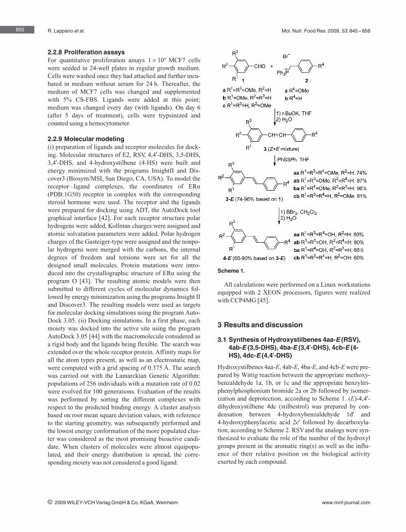
R. Lappano et al. Mol. Nutr. Food Res. 2009, 53, 845 –858
2.2.8 Proliferation assaysFor quantitative proliferation assays 16104 MCF7 cellswere seeded in 24-well plates in regular growth medium.Cells were washed once they had attached and further incu-bated in medium without serum for 24 h. Thereafter, themedium of MCF7 cells was changed and supplementedwith 5% CS-FBS. Ligands were added at this point;medium was changed every day (with ligands). On day 6(after 5 days of treatment), cells were trypsinized andcounted using a hemocytometer.
2.2.9 Molecular modeling(i) preparation of ligands and receptor molecules for dock-ing. Molecular structures of E2, RSV, 4,49-DHS, 3,5-DHS,3,49-DHS, and 4-hydroxystilbene (4-HS) were built andenergy minimized with the programs InsightII and Dis-cover3 (Biosym/MSI, San Diego, CA, USA). To model thereceptor– ligand complexes, the coordinates of ERa
(PDB:1G50) receptor in complex with the correspondingsteroid hormone were used. The receptor and the ligandswere prepared for docking using ADT, the AutoDock toolgraphical interface [42]. For each receptor structure polarhydrogens were added, Kollman charges were assigned andatomic solvatation parameters were added. Polar hydrogencharges of the Gasteiger-type were assigned and the nonpo-lar hydrogens were merged with the carbons, the internaldegrees of freedom and torsions were set for all thedesigned small molecules. Protein mutations were intro-duced into the crystallographic structure of ERa using theprogram O [43]. The resulting atomic models were thensubmitted to different cycles of molecular dynamics fol-lowed by energy minimization using the programs Insight IIand Discover3. The resulting models were used as targetsfor molecular docking simulations using the program Auto-Dock 3.05. (ii) Docking simulations. In a first phase, eachmoiety was docked into the active site using the programAutoDock 3.05 [44] with the macromolecule considered asa rigid body and the ligands being flexible. The search wasextended over the whole receptor protein. Affinity maps forall the atom types present, as well as an electrostatic map,were computed with a grid spacing of 0.375 �. The searchwas carried out with the Lamarckian Genetic Algorithm:populations of 256 individuals with a mutation rate of 0.02were evolved for 100 generations. Evaluation of the resultswas performed by sorting the different complexes withrespect to the predicted binding energy. A cluster analysisbased on root mean square deviation values, with referenceto the starting geometry, was subsequently performed andthe lowest energy conformation of the more populated clus-ter was considered as the most promising bioactive candi-date. When clusters of molecules were almost equipopu-lated, and their energy distribution is spread, the corre-sponding moiety was not considered a good ligand.
All calculations were performed on a Linux workstationsequipped with 2 XEON processors, figures were realizedwith CCP4MG [45].
3 Results and discussion
3.1 Synthesis of Hydroxystilbenes 4aa-E (RSV),4ab-E (3,5-DHS), 4ba-E (3,49-DHS), 4cb-E (4-HS), 4dc-E (4,49-DHS)
Hydroxystilbenes 4aa-E, 4ab-E, 4ba-E, and 4cb-E were pre-pared by Wittig reaction between the appropriate methoxy-benzaldehyde 1a, 1b, or 1c and the appropriate benzyltri-phenylphosphonium bromide 2a or 2b followed by isomer-ization and deprotection, according to Scheme 1. (E)-4,49-dihydroxystilbene 4dc (stilbestrol) was prepared by con-densation between 4-hydroxybenzaldehyde 1d9 and4-hydroxyphenylacetic acid 2c9 followed by decarboxyla-tion, according to Scheme 2. RSVand the analogs were syn-thesized to evaluate the role of the number of the hydroxylgroups present in the aromatic ring(s) as well as the influ-ence of their relative position on the biological activityexerted by each compound.
850
i 2009 WILEY-VCH Verlag GmbH & Co. KGaA, Weinheim www.mnf-journal.com
Scheme 1.
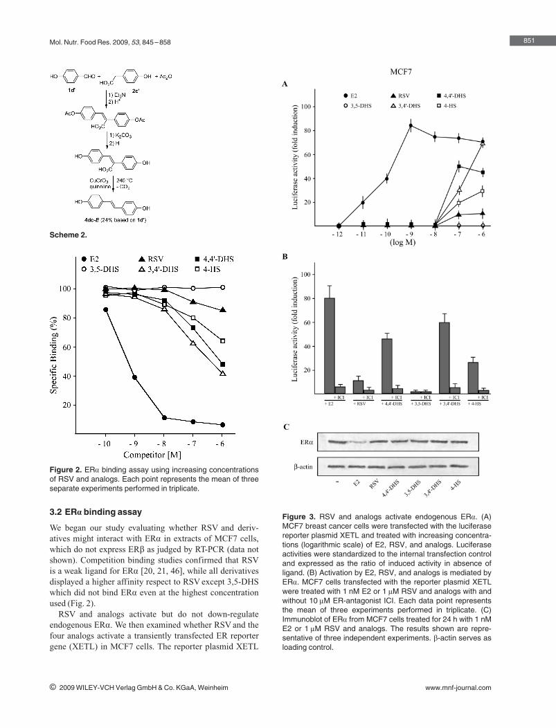
Mol. Nutr. Food Res. 2009, 53, 845 –858
3.2 ERa binding assay
We began our study evaluating whether RSV and deriv-atives might interact with ERa in extracts of MCF7 cells,which do not express ERb as judged by RT-PCR (data notshown). Competition binding studies confirmed that RSVis a weak ligand for ERa [20, 21, 46], while all derivativesdisplayed a higher affinity respect to RSV except 3,5-DHSwhich did not bind ERa even at the highest concentrationused (Fig. 2).
RSV and analogs activate but do not down-regulateendogenous ERa. We then examined whether RSV and thefour analogs activate a transiently transfected ER reportergene (XETL) in MCF7 cells. The reporter plasmid XETL
851
i 2009 WILEY-VCH Verlag GmbH & Co. KGaA, Weinheim www.mnf-journal.com
Scheme 2.
Figure 2. ERa binding assay using increasing concentrationsof RSV and analogs. Each point represents the mean of threeseparate experiments performed in triplicate.
Figure 3. RSV and analogs activate endogenous ERa. (A)MCF7 breast cancer cells were transfected with the luciferasereporter plasmid XETL and treated with increasing concentra-tions (logarithmic scale) of E2, RSV, and analogs. Luciferaseactivities were standardized to the internal transfection controland expressed as the ratio of induced activity in absence ofligand. (B) Activation by E2, RSV, and analogs is mediated byERa. MCF7 cells transfected with the reporter plasmid XETLwere treated with 1 nM E2 or 1 lM RSV and analogs with andwithout 10 lM ER-antagonist ICI. Each data point representsthe mean of three experiments performed in triplicate. (C)Immunoblot of ERa from MCF7 cells treated for 24 h with 1 nME2 or 1 lM RSV and analogs. The results shown are repre-sentative of three independent experiments. b-actin serves asloading control.
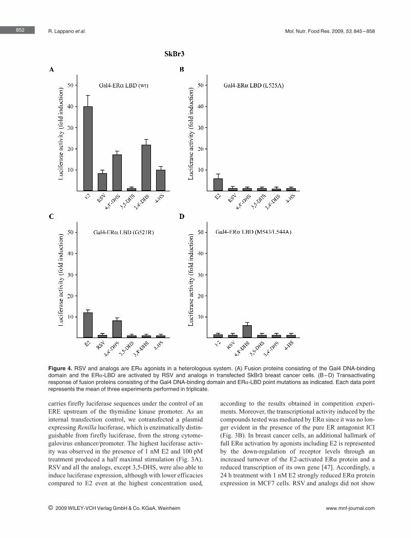
R. Lappano et al. Mol. Nutr. Food Res. 2009, 53, 845 –858
carries firefly luciferase sequences under the control of anERE upstream of the thymidine kinase promoter. As aninternal transfection control, we cotransfected a plasmidexpressing Renilla luciferase, which is enzimatically distin-guishable from firefly luciferase, from the strong cytome-galovirus enhancer/promoter. The highest luciferase activ-ity was observed in the presence of 1 nM E2 and 100 pMtreatment produced a half maximal stimulation (Fig. 3A).RSV and all the analogs, except 3,5-DHS, were also able toinduce luciferase expression, although with lower efficaciescompared to E2 even at the highest concentration used,
according to the results obtained in competition experi-ments. Moreover, the transcriptional activity induced by thecompounds tested was mediated by ERa since it was no lon-ger evident in the presence of the pure ER antagonist ICI(Fig. 3B). In breast cancer cells, an additional hallmark offull ERa activation by agonists including E2 is representedby the down-regulation of receptor levels through anincreased turnover of the E2-activated ERa protein and areduced transcription of its own gene [47]. Accordingly, a24 h treatment with 1 nM E2 strongly reduced ERa proteinexpression in MCF7 cells. RSV and analogs did not show
852
i 2009 WILEY-VCH Verlag GmbH & Co. KGaA, Weinheim www.mnf-journal.com
Figure 4. RSV and analogs are ERa agonists in a heterologous system. (A) Fusion proteins consisting of the Gal4 DNA-bindingdomain and the ERa-LBD are activated by RSV and analogs in transfected SkBr3 breast cancer cells. (B–D) Transactivatingresponse of fusion proteins consisting of the Gal4 DNA-binding domain and ERa-LBD point mutations as indicated. Each data pointrepresents the mean of three experiments performed in triplicate.
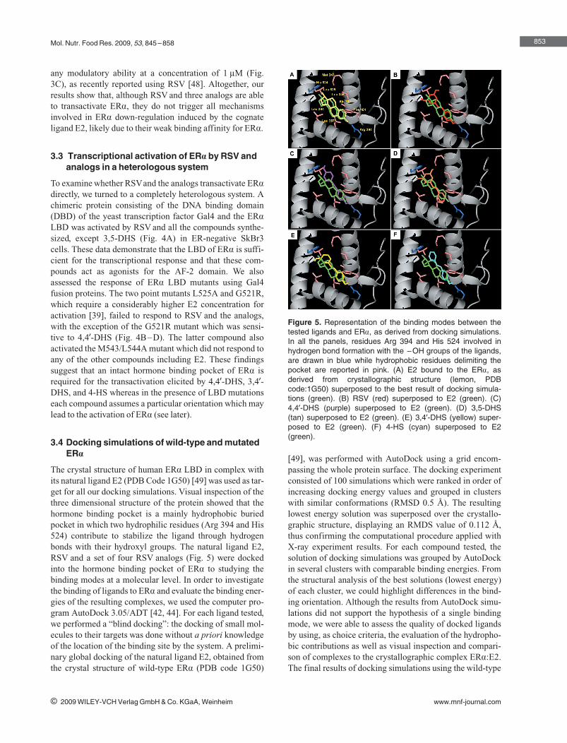
Mol. Nutr. Food Res. 2009, 53, 845 –858
any modulatory ability at a concentration of 1 lM (Fig.3C), as recently reported using RSV [48]. Altogether, ourresults show that, although RSV and three analogs are ableto transactivate ERa, they do not trigger all mechanismsinvolved in ERa down-regulation induced by the cognateligand E2, likely due to their weak binding affinity for ERa.
3.3 Transcriptional activation of ERa by RSV andanalogs in a heterologous system
To examine whether RSVand the analogs transactivate ERa
directly, we turned to a completely heterologous system. Achimeric protein consisting of the DNA binding domain(DBD) of the yeast transcription factor Gal4 and the ERa
LBD was activated by RSV and all the compounds synthe-sized, except 3,5-DHS (Fig. 4A) in ER-negative SkBr3cells. These data demonstrate that the LBD of ERa is suffi-cient for the transcriptional response and that these com-pounds act as agonists for the AF-2 domain. We alsoassessed the response of ERa LBD mutants using Gal4fusion proteins. The two point mutants L525A and G521R,which require a considerably higher E2 concentration foractivation [39], failed to respond to RSV and the analogs,with the exception of the G521R mutant which was sensi-tive to 4,49-DHS (Fig. 4B–D). The latter compound alsoactivated the M543/L544A mutant which did not respond toany of the other compounds including E2. These findingssuggest that an intact hormone binding pocket of ERa isrequired for the transactivation elicited by 4,49-DHS, 3,49-DHS, and 4-HS whereas in the presence of LBD mutationseach compound assumes a particular orientation which maylead to the activation of ERa (see later).
3.4 Docking simulations of wild-type and mutatedERa
The crystal structure of human ERa LBD in complex withits natural ligand E2 (PDB Code 1G50) [49] was used as tar-get for all our docking simulations. Visual inspection of thethree dimensional structure of the protein showed that thehormone binding pocket is a mainly hydrophobic buriedpocket in which two hydrophilic residues (Arg 394 and His524) contribute to stabilize the ligand through hydrogenbonds with their hydroxyl groups. The natural ligand E2,RSV and a set of four RSV analogs (Fig. 5) were dockedinto the hormone binding pocket of ERa to studying thebinding modes at a molecular level. In order to investigatethe binding of ligands to ERa and evaluate the binding ener-gies of the resulting complexes, we used the computer pro-gram AutoDock 3.05/ADT [42, 44]. For each ligand tested,we performed a “blind docking”: the docking of small mol-ecules to their targets was done without a priori knowledgeof the location of the binding site by the system. A prelimi-nary global docking of the natural ligand E2, obtained fromthe crystal structure of wild-type ERa (PDB code 1G50)
[49], was performed with AutoDock using a grid encom-passing the whole protein surface. The docking experimentconsisted of 100 simulations which were ranked in order ofincreasing docking energy values and grouped in clusterswith similar conformations (RMSD 0.5 �). The resultinglowest energy solution was superposed over the crystallo-graphic structure, displaying an RMDS value of 0.112 �,thus confirming the computational procedure applied withX-ray experiment results. For each compound tested, thesolution of docking simulations was grouped by AutoDockin several clusters with comparable binding energies. Fromthe structural analysis of the best solutions (lowest energy)of each cluster, we could highlight differences in the bind-ing orientation. Although the results from AutoDock simu-lations did not support the hypothesis of a single bindingmode, we were able to assess the quality of docked ligandsby using, as choice criteria, the evaluation of the hydropho-bic contributions as well as visual inspection and compari-son of complexes to the crystallographic complex ERa:E2.The final results of docking simulations using the wild-type
853
i 2009 WILEY-VCH Verlag GmbH & Co. KGaA, Weinheim www.mnf-journal.com
Figure 5. Representation of the binding modes between thetested ligands and ERa, as derived from docking simulations.In all the panels, residues Arg 394 and His 524 involved inhydrogen bond formation with the –OH groups of the ligands,are drawn in blue while hydrophobic residues delimiting thepocket are reported in pink. (A) E2 bound to the ERa, asderived from crystallographic structure (lemon, PDBcode:1G50) superposed to the best result of docking simula-tions (green). (B) RSV (red) superposed to E2 (green). (C)4,49-DHS (purple) superposed to E2 (green). (D) 3,5-DHS(tan) superposed to E2 (green). (E) 3,49-DHS (yellow) super-posed to E2 (green). (F) 4-HS (cyan) superposed to E2(green).
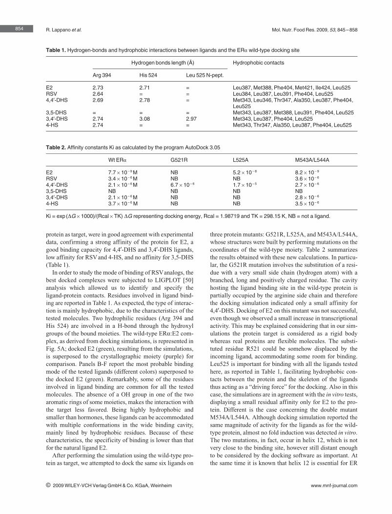
R. Lappano et al. Mol. Nutr. Food Res. 2009, 53, 845 –858
protein as target, were in good agreement with experimentaldata, confirming a strong affinity of the protein for E2, agood binding capacity for 4,49-DHS and 3,49-DHS ligands,low affinity for RSVand 4-HS, and no affinity for 3,5-DHS(Table 1).
In order to study the mode of binding of RSVanalogs, thebest docked complexes were subjected to LIGPLOT [50]analysis which allowed us to identify and specify theligand-protein contacts. Residues involved in ligand bind-ing are reported in Table 1. As expected, the type of interac-tion is mainly hydrophobic, due to the characteristics of thetested molecules. Two hydrophilic residues (Arg 394 andHis 524) are involved in a H-bond through the hydroxylgroups of the bound moieties. The wild-type ERa:E2 com-plex, as derived from docking simulations, is represented inFig. 5A; docked E2 (green), resulting from the simulations,is superposed to the crystallographic moiety (purple) forcomparison. Panels B-F report the most probable bindingmode of the tested ligands (different colors) superposed tothe docked E2 (green). Remarkably, some of the residuesinvolved in ligand binding are common for all the testedmolecules. The absence of a OH group in one of the twoaromatic rings of some moieties, makes the interaction withthe target less favored. Being highly hydrophobic andsmaller than hormones, these ligands can be accommodatedwith multiple conformations in the wide binding cavity,mainly lined by hydrophobic residues. Because of thesecharacteristics, the specificity of binding is lower than thatfor the natural ligand E2.
After performing the simulation using the wild-type pro-tein as target, we attempted to dock the same six ligands on
three protein mutants: G521R, L525A, and M543A/L544A,whose structures were built by performing mutations on thecoordinates of the wild-type moiety. Table 2 summarizesthe results obtained with these new calculations. In particu-lar, the G521R mutation involves the substitution of a resi-due with a very small side chain (hydrogen atom) with abranched, long and positively charged residue. The cavityhosting the ligand binding site in the wild-type protein ispartially occupied by the arginine side chain and thereforethe docking simulation indicated only a small affinity for4,49-DHS. Docking of E2 on this mutant was not successful,even though we observed a small increase in transcriptionalactivity. This may be explained considering that in our sim-ulations the protein target is considered as a rigid bodywhereas real proteins are flexible molecules. The substi-tuted residue R521 could be somehow displaced by theincoming ligand, accommodating some room for binding.Leu525 is important for binding with all the ligands testedhere, as reported in Table 1, facilitating hydrophobic con-tacts between the protein and the skeleton of the ligandsthus acting as a “driving force” for the docking. Also in thiscase, the simulations are in agreement with the in vitro tests,displaying a small residual affinity only for E2 to the pro-tein. Different is the case concerning the double mutantM534A/L544A. Although docking simulation reported thesame magnitude of activity for the ligands as for the wild-type protein, almost no fold induction was detected in vitro.The two mutations, in fact, occur in helix 12, which is notvery close to the binding site, however still distant enoughto be considered by the docking software as important. Atthe same time it is known that helix 12 is essential for ER
854
i 2009 WILEY-VCH Verlag GmbH & Co. KGaA, Weinheim www.mnf-journal.com
Table 1. Hydrogen-bonds and hydrophobic interactions between ligands and the ERa wild-type docking site
Hydrogen bonds length (�) Hydrophobic contacts
Arg 394 His 524 Leu 525 N-pept.
E2 2.73 2.71 = Leu387, Met388, Phe404, Met421, Ile424, Leu525RSV 2.64 = = Leu384, Leu387, Leu391, Phe404, Leu5254,49-DHS 2.69 2.78 = Met343, Leu346, Thr347, Ala350, Leu387, Phe404,
Leu5253,5-DHS = = = Met343, Leu387, Met388, Leu391, Phe404, Leu5253,49-DHS 2.74 3.08 2.97 Met343, Leu387, Phe404, Leu5254-HS 2.74 = = Met343, Thr347, Ala350, Leu387, Phe404, Leu525
Table 2. Affinity constants Ki as calculated by the program AutoDock 3.05
Wt ERa G521R L525A M543A/L544A
E2 7.7610 – 9 M NB 5.2610 – 8 8.2610 – 9
RSV 3.4610 – 6 M NB NB 3.6610 – 6
4,49-DHS 2.1610 – 6 M 6.7610 – 6 1.7610 – 5 2.7610 – 6
3,5-DHS NB NB NB NB3,49-DHS 2.1610 – 6 M NB NB 2.8610 – 6
4-HS 3.7610 – 6 M NB NB 3.5610 – 6
Ki = exp(DG61000)/(Rcal6TK) DG representing docking energy, Rcal = 1.98719 and TK = 298.15 K, NB = not a ligand.
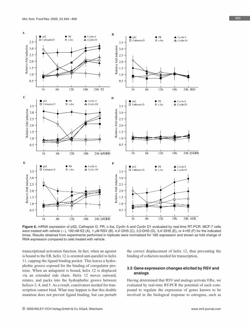
Mol. Nutr. Food Res. 2009, 53, 845 –858
transcriptional activation function. In fact, when an agonistis bound to the ER, helix 12 is oriented anti-parallel to helix11, capping the ligand binding pocket. This leaves a hydro-phobic groove exposed for the binding of coregulator pro-teins. When an antagonist is bound, helix 12 is displacedvia an extended side chain. Helix 12 moves outward,rotates, and packs into the hydrophobic groove betweenhelices 3, 4, and 5. As a result, coactivators needed for tran-scription cannot bind. What may happen is that this doublemutation does not prevent ligand binding, but can perturb
the correct displacement of helix 12, thus preventing thebinding of cofactors needed for transcription.
3.5 Gene expression changes elicited by RSV andanalogs
Having determined that RSV and analogs activate ERa, weevaluated by real-time RT-PCR the potential of each com-pound to regulate the expression of genes known to beinvolved in the biological response to estrogens, such as
855
i 2009 WILEY-VCH Verlag GmbH & Co. KGaA, Weinheim www.mnf-journal.com
Figure 6. mRNA expression of pS2, Cathepsin D, PR, c-fos, Cyclin A and Cyclin D1 evaluated by real-time RT-PCR. MCF-7 cellswere treated with vehicle (– ), 100 nM E2 (A), 1 lM RSV (B), 4,49-DHS (C), 3,5-DHS (D), 3,49-DHS (E), or 4-HS (F) for the indicatedtimes. Results obtained from experiments performed in triplicate were normalized for 18S expression and shown as fold change ofRNA expression compared to cells treated with vehicle.
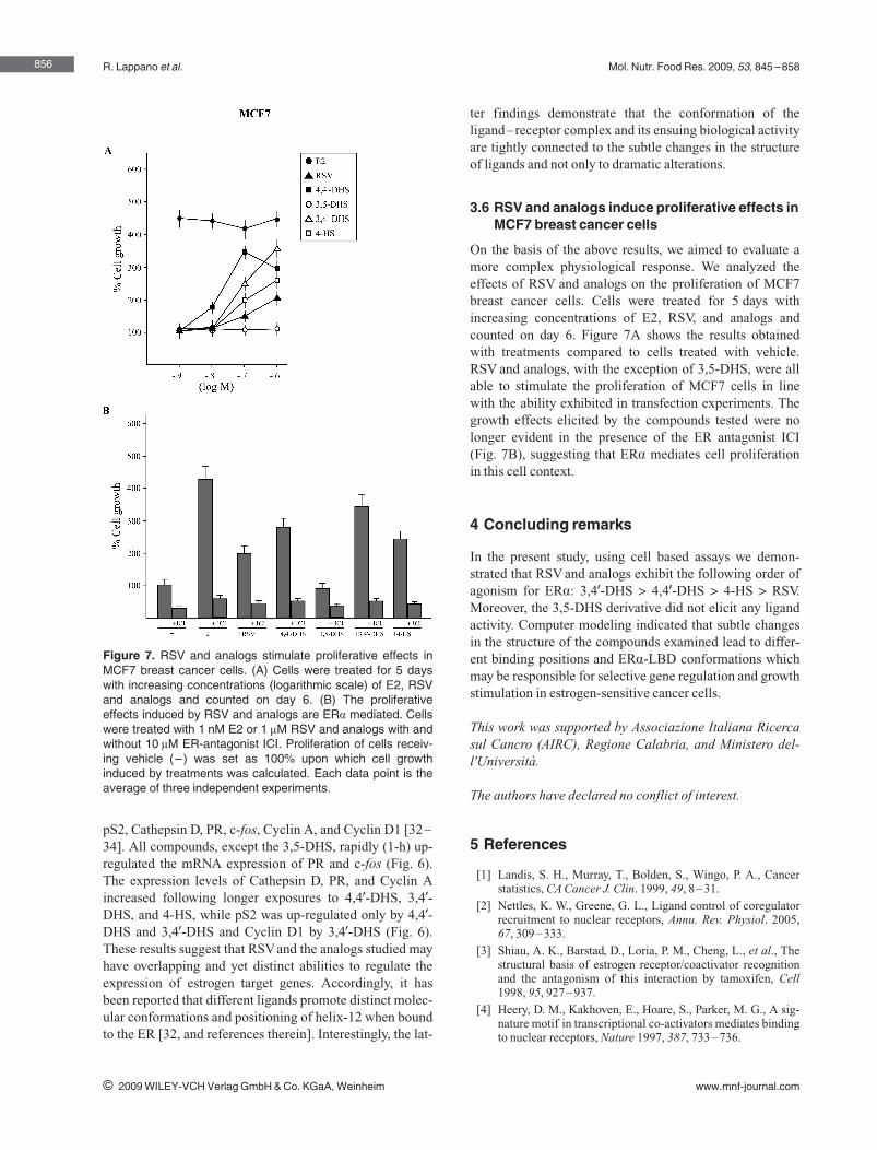
R. Lappano et al. Mol. Nutr. Food Res. 2009, 53, 845 –858
pS2, Cathepsin D, PR, c-fos, Cyclin A, and Cyclin D1 [32–34]. All compounds, except the 3,5-DHS, rapidly (1-h) up-regulated the mRNA expression of PR and c-fos (Fig. 6).The expression levels of Cathepsin D, PR, and Cyclin Aincreased following longer exposures to 4,49-DHS, 3,49-DHS, and 4-HS, while pS2 was up-regulated only by 4,49-DHS and 3,49-DHS and Cyclin D1 by 3,49-DHS (Fig. 6).These results suggest that RSVand the analogs studied mayhave overlapping and yet distinct abilities to regulate theexpression of estrogen target genes. Accordingly, it hasbeen reported that different ligands promote distinct molec-ular conformations and positioning of helix-12 when boundto the ER [32, and references therein]. Interestingly, the lat-
ter findings demonstrate that the conformation of theligand–receptor complex and its ensuing biological activityare tightly connected to the subtle changes in the structureof ligands and not only to dramatic alterations.
3.6 RSV and analogs induce proliferative effects inMCF7 breast cancer cells
On the basis of the above results, we aimed to evaluate amore complex physiological response. We analyzed theeffects of RSV and analogs on the proliferation of MCF7breast cancer cells. Cells were treated for 5 days withincreasing concentrations of E2, RSV, and analogs andcounted on day 6. Figure 7A shows the results obtainedwith treatments compared to cells treated with vehicle.RSV and analogs, with the exception of 3,5-DHS, were allable to stimulate the proliferation of MCF7 cells in linewith the ability exhibited in transfection experiments. Thegrowth effects elicited by the compounds tested were nolonger evident in the presence of the ER antagonist ICI(Fig. 7B), suggesting that ERa mediates cell proliferationin this cell context.
4 Concluding remarks
In the present study, using cell based assays we demon-strated that RSV and analogs exhibit the following order ofagonism for ERa: 3,49-DHS A 4,49-DHS A 4-HS A RSV.Moreover, the 3,5-DHS derivative did not elicit any ligandactivity. Computer modeling indicated that subtle changesin the structure of the compounds examined lead to differ-ent binding positions and ERa-LBD conformations whichmay be responsible for selective gene regulation and growthstimulation in estrogen-sensitive cancer cells.
This work was supported by Associazione Italiana Ricercasul Cancro (AIRC), Regione Calabria, and Ministero del-l'Universit�.
The authors have declared no conflict of interest.
5 References
[1] Landis, S. H., Murray, T., Bolden, S., Wingo, P. A., Cancerstatistics, CA Cancer J. Clin. 1999, 49, 8–31.
[2] Nettles, K. W., Greene, G. L., Ligand control of coregulatorrecruitment to nuclear receptors, Annu. Rev. Physiol. 2005,67, 309–333.
[3] Shiau, A. K., Barstad, D., Loria, P. M., Cheng, L., et al., Thestructural basis of estrogen receptor/coactivator recognitionand the antagonism of this interaction by tamoxifen, Cell1998, 95, 927–937.
[4] Heery, D. M., Kakhoven, E., Hoare, S., Parker, M. G., A sig-nature motif in transcriptional co-activators mediates bindingto nuclear receptors, Nature 1997, 387, 733–736.
856
i 2009 WILEY-VCH Verlag GmbH & Co. KGaA, Weinheim www.mnf-journal.com
Figure 7. RSV and analogs stimulate proliferative effects inMCF7 breast cancer cells. (A) Cells were treated for 5 dayswith increasing concentrations (logarithmic scale) of E2, RSVand analogs and counted on day 6. (B) The proliferativeeffects induced by RSV and analogs are ERa mediated. Cellswere treated with 1 nM E2 or 1 lM RSV and analogs with andwithout 10 lM ER-antagonist ICI. Proliferation of cells receiv-ing vehicle (– ) was set as 100% upon which cell growthinduced by treatments was calculated. Each data point is theaverage of three independent experiments.
Mol. Nutr. Food Res. 2009, 53, 845 –858
[5] Jensen, E. V., Steroid hormone antagonists. Summary andfuture challenges, Ann. N. Y. Acad. Sci. 1995, 761, 1–4.
[6] Maggiolini, M., Bonofiglio, D., Marsico, S., Panno, M. L., etal., Estrogen receptor alpha mediates the proliferative but notthe cytotoxic dose-dependent effects of two major phytoes-trogens on human breast cancer cells, Mol. Pharmacol. 2001,60, 595–602.
[7] Hill, M. J., Nutrition and human cancer, Ann. N. Y. Acad. Sci.1997, 833, 68–78.
[8] Sinha, R., Potter, J. D., Diet, nutrition, and genetic suscepti-bility, Cancer Epidemiol. Biomarkers Prev. 1997, 6, 647 –649.
[9] Messina, M. J., Persky, V., Setchell, K. D., Barnes, S., Soyintake and cancer risk: A review of the in vitro and in vivodata, Nutr. Cancer 1994, 21, 113–131.
[10] Makela, S. I., Pylkkanen, L. H., Santti, R. S., Adlercreutz, H.,Dietary soybean may be antiestrogenic in male mice, J. Nutr.1995, 125, 437 –445.
[11] Markiewicz, L., Garey, J., Adlercreutz, H., Gurpide, E., Invitro bioassays of non-steroidal phytoestrogens, J. SteroidBiochem. Mol. Biol. 1993, 45, 399 –405.
[12] Sathyamoorthy, N., Wang, T. T., Phang, J. M., Stimulation ofpS2 expression by diet-derived compounds, Cancer Res.1994, 54, 957–961.
[13] Stahl, S., Chun, T. Y., Gray, W. G., Phytoestrogens act asestrogen agonists in an estrogen-responsive pituitary cell line,Toxicol. Appl. Pharmacol. 1998, 152, 41–48.
[14] Le Bail, J. C., Champavier, Y. M., Chulia, A. J., Habrioux, G.,Effects of phytoestrogens on aromatase, 3b and 17b-hydrox-ysteroid dehydrogenase activities and human breast cancercells, Life Sci. 2000, 66, 1281–1291.
[15] Zava, D. T., Duwe, G., Estrogenic and antiproliferative prop-erties of genistein and other flavonoids in human breast can-cer cells in vitro, Nutr. Cancer 1997, 27, 31–40.
[16] Burns, J., Yokota, T., Ashihara, H., Lean, M. E., Crozier, A.,Plant foods and herbal sources of resveratrol, J. Agric. FoodChem. 2002, 50, 3337–3340.
[17] Burkon, A., Somoza, V., Quantification of free and protein-bound trans-resveratrol metabolites and identification oftrans-resveratrol-C/O-conjugated diglucuronides – Twonovel resveratrol metabolites in human plasma, Mol. Nutr.Food Res. 2008, 52, 549–57.
[18] Vitaglione, P., Sforza, S., Galaverna, G., Ghidini, C., et al.,Bioavailability of trans-resveratrol from red wine in humans,Mol. Nutr. Food Res. 2005, 49, 495–504.
[19] Bhat, K. P. L., Pezzuto, J. M., Resveratrol exhibits cytostaticand antiestrogenic properties with human endometrial adeno-carcinoma (Ishikawa) cells, Cancer Res. 2001, 61, 6137 –6144.
[20] Bowers, J. L., Tyulmenkov, V. V., Jernigan, S. C., Klinge, C.M., Resveratrol acts as a mixed agonist/antagonist for estro-gen receptors a and b, Endocrinology 2000, 141, 3657 –3667.
[21] Gehm, B. D., McAndrews, J. M., Chien, P. Y., Jameson, J. L.,Resveratrol, a polyphenolic compound found in grapes andwine, is an agonist for the estrogen receptor, Proc. Natl.Acad. Sci. USA 1997, 94, 14138 –14143.
[22] Levenson, A. S., Jordan, V. C., Millennium Review 2000:Selective oestrogen receptor modulation: Molecular pharma-cology for the millennium, Eur. J. Cancer 1999, 35, 1628–1639.
[23] Mizutani, K., Ikeda, K., Kawai, Y., Yamori, Y., Resveratrolattenuates ovariectomy-induced hypertension and bone lossin stroke-prone spontaneously hypertensive rats, J. Nutr. Sci.Vitaminol. 2000, 46, 78–83.
[24] Wu, J. M., Wang, Z. R., Hsieh, T. C., Bruder, J. L., et al.,Mechanism of cardioprotection by resveratrol, a phenolicantioxidant present in red wine, Int. J. Mol. Med. 2001, 8, 3–17.
[25] Hope, C., Planutis, K., Planutiene, M., Moyer, M. P., et al.,Low concentrations of resveratrol inhibit Wnt signal through-put in colon-derived cells: Implications for colon cancer pre-vention, Mol. Nutr. Food Res. 2008, 52, 52–61.
[26] Mgbonyebi, O. P., Russo, J., Russo, I. H., Antiproliferativeeffect of synthetic resveratrol on human breast epithelialcells, Int. J. Oncol. 1998, 12, 865–869.
[27] Tang, F. Y., Su, Y. C., Chen, N. C., Hsieh, H. S., Chen, K. S.,Resveratrol inhibits migration and invasion of human breast-cancer cells, Mol. Nutr. Food Res. 2008, 52, 683–691.
[28] Hsieh, T. C., Burfeind, P., Laud, K., Backer, J. M., et al., Cellcycle effects and control of gene expression by resveratrol inhuman breast carcinoma cell lines with different metastaticpotentials, Int. J. Oncol. 1999, 15, 245 –252.
[29] Nakagawa, H., Kiyozuka, Y., Uemura, Y., Senzaki, H., et al.,Resveratrol inhibits human breast cancer cell growth andmay mitigate the effect of linoleic acid, a potent breast cancercell stimulator, J. Cancer Res. Clin. Oncol. 2001, 127, 258 –264.
[30] Fr�mont, L., Biological effects of resveratrol, Life Sci. 2000,66, 663–673.
[31] Le Corre, L., Chalabi, N., Delort, L., Bignon, Y. J., Bernard-Gallon, D. J., Resveratrol and breast cancer chemoprevention:Molecular mechanisms, Mol. Nutr. Food Res. 2005, 49, 462 –471.
[32] Brooks, S. C., Skafar, D. F., From ligand structure to biologi-cal activity: Modified estratrienes and their estrogenic andantiestrogenic effects in MCF-7 cells, Steroids 2004, 69,401–418.
[33] O'lone, R., Frith, M. C., Karlsson, E. K., Hansen, U.,Genomic targets of nuclear estrogen receptors, Mol. Endocri-nol. 2004, 18, 1859 –1875.
[34] Sanchez, R., Nguyen, D., Rocha, W., White, J. H., Mader, S.,Diversity in the mechanisms of gene regulation by estrogenreceptors, Bioessays 2002, 24, 244 –54.
[35] Ali, M. A., Kondo, K., Tsuda, Y., Synthesis and nematocidalactivity of hydroxystilbenes, Chem. Pharm. Bull. 1992, 40,1130–1136.
[36] Schulz, T. P., Hubbard, T. F., Jin, L., Fischer, T. H., Nicholas,D. D., Phytochemistry 1990, 29, 1501–1507.
[37] Bunone, G., Briand, P. A., Miksicek, R. J., Picard, D., Activa-tion of the unliganded estrogen receptor by EGF involves theMAP kinase pathway and direct phosphorylation, EMBO J.1996, 15, 2174 –2183.
[38] Webb, P., Nguyen, P., Shinsako, J., Anderson, C., et al., Estro-gen receptor activation function 1 works by binding p160coactivator proteins, Mol. Endocrinol. 1998, 12, 1605–1618.
[39] Ekena, K., Weis, K. E., Katzenellenbogen, J. A., Katzenel-lenbogen, B. S., Identification of amino acids in the hormonebinding domain of the human estrogen receptor important inestrogen binding, J. Biol. Chem. 1996, 271, 20053 –20059.
[40] Seipel, K., Georgiev, O., Schaffner, W., Different activationdomains stimulate transcription from remote (“enhancer”)and proximal (“promoter”) positions, EMBO J. 1992, 11,4961–4968.
857
i 2009 WILEY-VCH Verlag GmbH & Co. KGaA, Weinheim www.mnf-journal.com
R. Lappano et al. Mol. Nutr. Food Res. 2009, 53, 845 –858
[41] Lee, Y. J., Gorski, J., Estrogen-induced transcription of theprogesterone receptor gene does not parallel estrogen recep-tor occupancy, Proc. Natl. Acad. Sci. USA 1996, 93, 15180 –15184.
[42] Sanner, M. F., Duncan, B. S., Carillo, C. J., Olson, A. J., Inte-grating computation and visualization for biomolecular anal-ysis: An example using Python and AVS, Pac. Symp. Biocom-put. 1999, 401 –412.
[43] Jones, T. A., Zou, J. Y., Cowan, S. W., Kjeldgaard, M.,Improved methods for building protein models in electrondensity maps and the location of errors in these models, ActaCrystallogr. A 1991, 47, 110 –119.
[44] Morris, G. M., Goodsell, D. S., Halliday, R. S., Huey, R., etal., Automated docking using a lamarkian genetic algorithmand empirical binding free energy function, J. Computat.Chem. 1998, 19, 1639–1662.
[45] Potterton, L., McNicholas, S., Krissinel, E., Gruber, J., et al.,Developments in the CCP4 molecular-graphics project, ActaCrystallogr. D Biol. Crystallogr. 2004, 60, 2288 –2294.
[46] Ashby, J., Tinwell, H., Pennie, W., Brooks, A. N., et al., Par-tial and weak oestrogenicity of the red wine constituentresveratrol: Consideration of its superagonist activity inMCF-7 cells and its suggested cardiovascular protectiveeffects, J. Appl. Toxicol. 1999, 19, 39–45.
[47] Nawaz, Z., Lonard, D. M., Dennis, A. P., Smith, C. L., O'Mal-ley, B. W., Proteasome-dependent degradation of the humanestrogen receptor, Proc. Natl. Acad. Sci. USA 1999, 96,1858–1862.
[48] Wu, F., Safe, S., Differential activation of wild-type estrogenreceptor alpha and C-terminal deletion mutants by estrogens,antiestrogens and xenoestrogens in breast cancer cells, J. Ste-roid Biochem. Mol. Biol. 2007, 103, 1–9.
[49] Eiler, S., Gangloff, M., Duclaud, S., Moras, D., Ruff, M.,Overexpression, purification, and crystal structure of nativeER alpha LBD, Protein Expr. Purif. 2001, 22, 165–173.
[50] Wallace, A. C., Lawskowski, R. A., Thornton, J. M., LIG-PLOT: A program to generate schematic diagrams of protein-ligands interactions, Prot. Eng. 1995, 8, 127–134.
858
i 2009 WILEY-VCH Verlag GmbH & Co. KGaA, Weinheim www.mnf-journal.com
G Protein–Coupled Receptor 30 (GPR30) Mediates Gene Expression
Changes and Growth Response to 17B-Estradiol and Selective
GPR30 Ligand G-1 in Ovarian Cancer Cells
Lidia Albanito,1Antonio Madeo,
1Rosamaria Lappano,
1Adele Vivacqua,
1Vittoria Rago,
2
Amalia Carpino,2Tudor I. Oprea,
3Eric R. Prossnitz,
4,5Anna Maria Musti,
1
Sebastiano Ando,2and Marcello Maggiolini
1
Departments of 1Pharmaco-Biology and 2Cell Biology, University of Calabria, Rende (Cosenza), Italy and 3Division of Biocomputing,4Department of Cell Biology and Physiology, and 5Cancer Research and Treatment Center, University of New Mexico,Albuquerque, New Mexico
Abstract
Estrogens play a crucial role in the development of ovariantumors; however, the signal transduction pathways involvedin hormone action are still poorly defined. The orphan Gprotein–coupled receptor 30 (GPR30) mediates the non-genomic signaling of 17B-estradiol (E2) in a variety ofestrogen-sensitive cancer cells through activation of theepidermal growth factor receptor (EGFR) pathway. Whetherestrogen receptor A (ERA) also contributes to GPR30/EGFR signaling is less understood. Here, we show that, inERA-positive BG-1 ovarian cancer cells, both E2 and theGPR30-selective ligand G-1 induced c-fos expression andestrogen-responsive element (ERE)-independent activity ofa c-fos reporter gene, whereas only E2 stimulated an ERE-responsive reporter gene, indicating that GPR30 signalingdoes not activate ERA-mediated transcription. Similarly, bothligands up-regulated cyclin D1, cyclin E, and cyclin A, whereasonly E2 enhanced progesterone receptor expression. More-over, both GPR30 and ERA expression are required for c-fosstimulation and extracellular signal-regulated kinase (ERK)activation in response to either E2 or G-1. Inhibition of theEGFR transduction pathway inhibited c-fos stimulation andERK activation by either ligand, suggesting that in ovariancancer cells GPR30/EGFR signaling relays on ERA expression.Interestingly, we show that both GPR30 and ERA expressionalong with active EGFR signaling are required for E2-stimulated and G-1–stimulated proliferation of ovarian cancercells. Because G-1 was able to induce both c-fos expressionand proliferation in the ERA-negative/GPR30-positive SKBR3breast cancer cells, the requirement for ERA expression inGPR30/EGFR signaling may depend on the specific cellularcontext of different tumor types. [Cancer Res 2007;67(4):1859–66]
Introduction
Ovarian cancer is the fourth leading cause of tumor death inWestern countries and represents the most fatal gynecologicmalignancy because the overall 5-year survival rate is only 10%to 20% (1, 2). Consequently, the identification of moleculartargets would be useful to define pharmacologic interventions
toward an improved outcome of patients with ovariancarcinoma.
Estrogens are major regulators of growth and differentiation innormal ovaries and also play an important role in the progressionof ovarian cancer (reviewed in ref. 3 and references therein).Likewise, a marked proliferative response to estrogens was shownin ovarian surface epithelial cells representing the site of 90% ofmalignancies (4) and an increased risk of ovarian tumor wasobserved in postmenopausal women receiving estrogen replace-ment therapy (5–7). In line with the aforementioned observations,antiestrogenic treatments repressed the growth of ovariancarcinoma both in vitro and in vivo (8–11), and the aromataseinhibitor letrozole, which depletes the bioavailability of estrogensat tissue levels, showed clinical benefit in a subgroup of ovariancancer patients (12, 13).
The biological effects of estrogens are classically mediated by theestrogen receptor (ER) a and ERh, which function as hormone-inducible transcription factors binding to the estrogen-responsiveelement (ERE) located within the promoter region of target genes(14). In the normal ovary, the levels of ERh are high andpredominate over ERa, whereas an opposite pattern characterizesthe development of ovarian cancer (reviewed in ref. 15 andreferences therein), which often expresses ERa levels similar tothose found in breast carcinoma (16). It has been largely reportedthat ERa mediates the mitogenic action of estrogens in breastcancer by inducing a variety of genes involved in cell proliferation.In this respect, the estrogen-regulated proteins, such as pS2,progesterone receptor (PR), c-fos , and different cyclins (17, 18),have been identified as useful prognostic markers for predicting theresponsiveness to antiestrogen therapy (16–18). As it concerns theovary, the estrogen-driven growth of epithelial carcinoma tissueshas been linked to ERa-mediated regulation of a plethora of genesinvolved in crucial cell function as recently shown by usingmicroarray technology (19). Moreover, it should be taken intoaccount that the levels of 17h-estradiol (E2) usually present atpicomolar-nanomolar concentrations reach micromolar levels inthe dominant follicle selected to ovulate (20–23). Consequently, theovarian surface epithelium surrounding the ovary is exposed toelevated E2 levels that have been largely overlooked.
A large body of evidence has identified different mechanismswhereby membrane-initiated signaling involving growth factorreceptor or membrane ERs mimics and/or contributes to thefunction of nuclear ERs (reviewed in refs. 24, 25 and referencestherein). Recent studies, including our own (26–32), have shown thatthe G protein–coupled receptor (GPCR), named GPR30, mediatesthe nongenomic signaling of E2 in a variety of estrogen-responsive
Requests for reprints: Marcello Maggiolini, Department of Pharmaco-Biology,University of Calabria, 87030 Rende (Cosenza), Italy. Phone: 39-09-84493076; Fax: 39-09-84493271; E-mail: [email protected].
I2007 American Association for Cancer Research.doi:10.1158/0008-5472.CAN-06-2909
www.aacrjournals.org 1859 Cancer Res 2007; 67: (4). February 15, 2007
Research Article
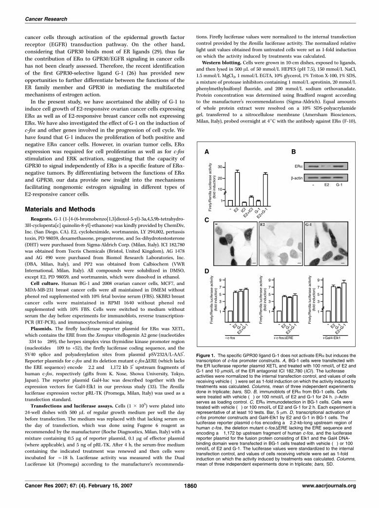
cancer cells through activation of the epidermal growth factorreceptor (EGFR) transduction pathway. On the other hand,considering that GPR30 binds most of ER ligands (29), thus farthe contribution of ERa to GPR30/EGFR signaling in cancer cellshas not been clearly assessed. Therefore, the recent identificationof the first GPR30-selective ligand G-1 (26) has provided newopportunities to further differentiate between the functions of theER family member and GPR30 in mediating the multifacetedmechanisms of estrogen action.
In the present study, we have ascertained the ability of G-1 toinduce cell growth of E2-responsive ovarian cancer cells expressingERa as well as of E2-responsive breast cancer cells not expressingERa. We have also investigated the effect of G-1 on the induction ofc-fos and other genes involved in the progression of cell cycle. Wehave found that G-1 induces the proliferation of both positive andnegative ERa cancer cells. However, in ovarian tumor cells, ERaexpression was required for cell proliferation as well as for c-fosstimulation and ERK activation, suggesting that the capacity ofGPR30 to signal independently of ERa is a specific feature of ERa-negative tumors. By differentiating between the functions of ERaand GPR30, our data provide new insight into the mechanismsfacilitating nongenomic estrogen signaling in different types ofE2-responsive cancer cells.
Materials and Methods
Reagents. G-1 (1-[4-(6-bromobenzo[1,3]dioxol-5-yl)-3a,4,5,9b-tetrahydro-
3H-cyclopenta[c] quinolin-8-yl]-ethanone) was kindly provided by ChemDiv,Inc. (San Diego, CA). E2, cycloheximide, wortmannin, LY 294,002, pertussistoxin, PD 98059, dexamethasone, progesterone, and 5a-dihydrotestosterone(DHT) were purchased from Sigma-Aldrich Corp. (Milan, Italy). ICI 182,780was obtained from Tocris Chemicals (Bristol, United Kingdom), AG 1478
and AG 490 were purchased from Biomol Research Laboratories, Inc.(DBA, Milan, Italy), and PP2 was obtained from Calbiochem (VWRInternational, Milan, Italy). All compounds were solubilized in DMSO,except E2, PD 98059, and wortmannin, which were dissolved in ethanol.
Cell culture. Human BG-1 and 2008 ovarian cancer cells, MCF7, and
MDA-MB-231 breast cancer cells were all maintained in DMEM withoutphenol red supplemented with 10% fetal bovine serum (FBS). SKBR3 breast
cancer cells were maintained in RPMI 1640 without phenol red
supplemented with 10% FBS. Cells were switched to medium without
serum the day before experiments for immunoblots, reverse transcription-PCR (RT-PCR), and immunocytochemical staining.
Plasmids. The firefly luciferase reporter plasmid for ERa was XETL,which contains the ERE from the Xenopus vitellogenin A2 gene (nucleotides�334 to �289), the herpes simplex virus thymidine kinase promoter region(nucleotides �109 to +52), the firefly luciferase coding sequence, and theSV40 splice and polyadenylation sites from plasmid pSV232A/L-AA5¶.Reporter plasmids for c-fos and its deletion mutant c-fosDERE (which lacksthe ERE sequence) encode �2.2 and �1,172 kb 5¶ upstream fragments ofhuman c-fos , respectively (gifts from K. Nose, Showa University, Tokyo,Japan). The reporter plasmid Gal4-luc was described together with theexpression vectors for Gal4-Elk1 in our previous study (33). The Renillaluciferase expression vector pRL-TK (Promega, Milan, Italy) was used as atransfection standard.
Transfections and luciferase assays. Cells (1 � 105) were plated into
24-well dishes with 500 AL of regular growth medium per well the day
before transfection. The medium was replaced with that lacking serum onthe day of transfection, which was done using Fugene 6 reagent as
recommended by the manufacturer (Roche Diagnostics, Milan, Italy) with a
mixture containing 0.5 Ag of reporter plasmid, 0.1 Ag of effector plasmid
(where applicable), and 5 ng of pRL-TK. After 4 h, the serum-free mediumcontaining the indicated treatment was renewed and then cells were
incubated for f18 h. Luciferase activity was measured with the Dual
Luciferase kit (Promega) according to the manufacturer’s recommenda-
tions. Firefly luciferase values were normalized to the internal transfectioncontrol provided by the Renilla luciferase activity. The normalized relative
light unit values obtained from untreated cells were set as 1-fold induction
on which the activity induced by treatments was calculated.
Western blotting. Cells were grown in 10-cm dishes, exposed to ligands,
and then lysed in 500 AL of 50 mmol/L HEPES (pH 7.5), 150 mmol/L NaCl,
1.5 mmol/L MgCl2, 1 mmol/L EGTA, 10% glycerol, 1% Triton X-100, 1% SDS,
a mixture of protease inhibitors containing 1 mmol/L aprotinin, 20 mmol/L
phenylmethylsulfonyl fluoride, and 200 mmol/L sodium orthovanadate.
Protein concentration was determined using Bradford reagent according
to the manufacturer’s recommendations (Sigma-Aldrich). Equal amounts
of whole protein extract were resolved on a 10% SDS-polyacrylamide
gel, transferred to a nitrocellulose membrane (Amersham Biosciences,
Milan, Italy), probed overnight at 4jC with the antibody against ERa (F-10),
Figure 1. The specific GPR30 ligand G-1 does not activate ERa but induces thetranscription of c-fos promoter constructs. A, BG-1 cells were transfected withthe ER luciferase reporter plasmid XETL and treated with 100 nmol/L of E2 andG-1 and 10 Amol/L of the ER antagonist ICI 182,780 (ICI ). The luciferaseactivities were normalized to the internal transfection control, and values of cellsreceiving vehicle (�) were set as 1-fold induction on which the activity induced bytreatments was calculated. Columns, mean of three independent experimentsdone in triplicate; bars, SD. B, immunoblots of ERa from BG-1 cells. Cellswere treated with vehicle (�) or 100 nmol/L of E2 and G-1 for 24 h. h-Actinserves as loading control. C, ERa immunodetection in BG-1 cells. Cells weretreated with vehicle (�) or 100 nmol/L of E2 and G-1 for 2 h. Each experiment isrepresentative of at least 10 tests. Bar, 5 Am. D, transcriptional activation ofc-fos promoter constructs and Gal4-Elk1 by E2 and G-1 in BG-1 cells. Theluciferase reporter plasmid c-fos encoding a �2.2-kb-long upstream region ofhuman c-fos , the deletion mutant c-fosDERE lacking the ERE sequence andencoding a �1,172 bp upstream fragment of human c-fos , and the luciferasereporter plasmid for the fusion protein consisting of Elk1 and the Gal4 DNA-binding domain were transfected in BG-1 cells treated with vehicle (�) or 100nmol/L of E2 and G-1. The luciferase values were standardized to the internaltransfection control, and values of cells receiving vehicle were set as 1-foldinduction on which the activity induced by treatments was calculated. Columns,mean of three independent experiments done in triplicate; bars, SD.
Cancer Research
Cancer Res 2007; 67: (4). February 15, 2007 1860 www.aacrjournals.org
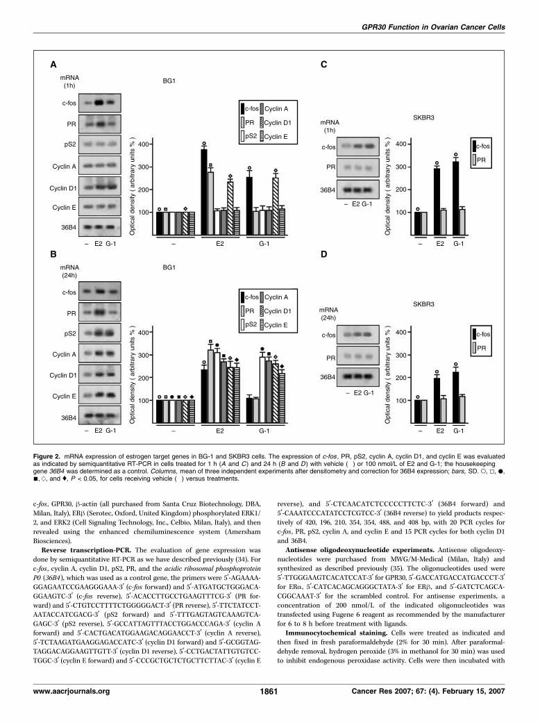
c-fos , GPR30, h-actin (all purchased from Santa Cruz Biotechnology, DBA,
Milan, Italy), ERh (Serotec, Oxford, United Kingdom) phosphorylated ERK1/
2, and ERK2 (Cell Signaling Technology, Inc., Celbio, Milan, Italy), and then
revealed using the enhanced chemiluminescence system (Amersham
Biosciences).Reverse transcription-PCR. The evaluation of gene expression was
done by semiquantitative RT-PCR as we have described previously (34). For
c-fos , cyclin A, cyclin D1, pS2, PR, and the acidic ribosomal phosphoprotein
P0 (36B4), which was used as a control gene, the primers were 5¶-AGAAAA-GGAGAATCCGAAGGGAAA-3¶ (c-fos forward) and 5¶-ATGATGCTGGGACA-
GGAAGTC-3¶ (c-fos reverse), 5¶-ACACCTTGCCTGAAGTTTCG-3¶ (PR for-
ward) and 5¶-CTGTCCTTTTCTGGGGGACT-3¶ (PR reverse), 5¶-TTCTATCCT-AATACCATCGACG-3¶ (pS2 forward) and 5¶-TTTGAGTAGTCAAAGTCA-
GAGC-3¶ (pS2 reverse), 5¶-GCCATTAGTTTACCTGGACCCAGA-3¶ (cyclin A
forward) and 5¶-CACTGACATGGAAGACAGGAACCT-3¶ (cyclin A reverse),
5¶-TCTAAGATGAAGGAGACCATC-3¶ (cyclin D1 forward) and 5¶-GCGGTAG-TAGGACAGGAAGTTGTT-3¶ (cyclin D1 reverse), 5¶-CCTGACTATTGTGTCC-
TGGC-3¶ (cyclin E forward) and 5¶-CCCGCTGCTCTGCTTCTTAC-3¶ (cyclin E
reverse), and 5¶-CTCAACATCTCCCCCTTCTC-3¶ (36B4 forward) and
5¶-CAAATCCCATATCCTCGTCC-3¶ (36B4 reverse) to yield products respec-
tively of 420, 196, 210, 354, 354, 488, and 408 bp, with 20 PCR cycles for
c-fos , PR, pS2, cyclin A, and cyclin E and 15 PCR cycles for both cyclin D1and 36B4.
Antisense oligodeoxynucleotide experiments. Antisense oligodeoxy-
nucleotides were purchased from MWG/M-Medical (Milan, Italy) and
synthesized as described previously (35). The oligonucleotides used were
5¶-TTGGGAAGTCACATCCAT-3¶ for GPR30, 5¶-GACCATGACCATGACCCT-3¶for ERa, 5¶-CATCACAGCAGGGCTATA-3¶ for ERh, and 5¶-GATCTCAGCA-
CGGCAAAT-3¶ for the scrambled control. For antisense experiments, a
concentration of 200 nmol/L of the indicated oligonucleotides was
transfected using Fugene 6 reagent as recommended by the manufacturer
for 6 to 8 h before treatment with ligands.Immunocytochemical staining. Cells were treated as indicated and
then fixed in fresh paraformaldehyde (2% for 30 min). After paraformal-
dehyde removal, hydrogen peroxide (3% in methanol for 30 min) was used
to inhibit endogenous peroxidase activity. Cells were then incubated with
Figure 2. mRNA expression of estrogen target genes in BG-1 and SKBR3 cells. The expression of c-fos , PR, pS2, cyclin A, cyclin D1, and cyclin E was evaluatedas indicated by semiquantitative RT-PCR in cells treated for 1 h (A and C ) and 24 h (B and D ) with vehicle (�) or 100 nmol/L of E2 and G-1; the housekeepinggene 36B4 was determined as a control. Columns, mean of three independent experiments after densitometry and correction for 36B4 expression; bars, SD. o, 5, .,n, w , and y, P < 0.05, for cells receiving vehicle (�) versus treatments.
GPR30 Function in Ovarian Cancer Cells
www.aacrjournals.org 1861 Cancer Res 2007; 67: (4). February 15, 2007
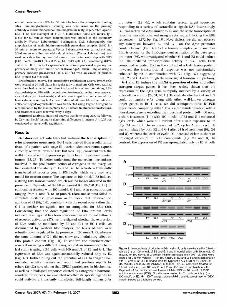
normal horse serum (10% for 30 min) to block the nonspecific binding
sites. Immunocytochemical staining was done using as the primary
antibody a mouse monoclonal immunoglobulin G (IgG) generated against
ERa (F-10; 1:50 overnight at 4jC). A biotinylated horse anti-mouse IgG
(1:600 for 60 min at room temperature) was applied as the secondary
antibody (Vector Laboratories, Burlingame, CA). Subsequently, the
amplification of avidin-biotin-horseradish peroxidase complex (1:100 for
30 min at room temperature; Vector Laboratories) was carried out and
3,3¶-diaminobenzidine tetrachloride dihydrate (Vector Laboratories) was
used as a detection system. Cells were rinsed after each step with TBS
[0.05 mol/L Tris-HCl plus 0.15 mol/L NaCl (pH 7.6)] containing 0.05%
Triton X-100. In control experiments, cells were processed replacing the
primary antibody with mouse serum (Dako S.p.A., Milan, Italy) or using a
primary antibody preabsorbed (48 h at 4jC) with an excess of purified
ERa protein (M-Medical).
Proliferation assays. For quantitative proliferation assays, 10,000 cells
were seeded in 24-well plates in regular growth medium. Cells were washedonce they had attached and then incubated in medium containing 2.5%
charcoal-stripped FBS with the indicated treatments; medium was renewed
every 2 days (with treatments) and cells were trypsinized and counted in a
hemocytometer on day 6. A concentration of 200 nmol/L of the indicatedantisense oligodeoxynucleotides was transfected using Fugene 6 reagent as
recommended by the manufacturer for 6 h before treatments, transfections,
and treatments were renewed every 2 days.Statistical analysis. Statistical analysis was done using ANOVA followed
by Newman-Keuls’ testing to determine differences in means. P < 0.05 was
considered as statistically significant.
Results
G-1 does not activate ERA but induces the transcription ofc-fos promoter constructs. BG-1 cells derived from a solid tumortissue of a patient with stage III ovarian adenocarcinoma expressclinically relevant levels of ERa but lack ERh, consistent with thewell-known receptor expression patterns found in primary ovariantumors (15, 36). To better understand the molecular mechanismsinvolved in the proliferative action of estrogens in the ovary, wefirst evaluated the ability of E2 and G-1 to activate a transientlytransfected ER reporter gene in BG-1 cells, which were used as amodel for ovarian cancer. The exposure to 100 nmol/L E2 induceda strong ERa transactivation, which was no longer observed in thepresence of 10 Amol/L of the ER antagonist ICI 182,780 (Fig. 1A). Incontrast, treatments with 100 nmol/L G-1 and even concentrationsranging from 1 nmol/L to 10 Amol/L (data not shown) failed tostimulate luciferase expression or to block that observed onaddition of E2 (Fig. 1A), consistent with the recent observation thatG-1 is neither an agonist nor an antagonist for ERa (26).Considering that the down-regulation of ERa protein levelsinduced by an agonist has been considered an additional hallmarkof receptor activation (37), we investigated whether the expressionof ERa could be modulated by E2 and G-1 in BG-1 cells. Asdocumented by Western blot analysis, the levels of ERa wererobustly down-regulated in the presence of 100 nmol/L E2, whereasthe same amount of G-1 did not show any modulatory effect onERa protein content (Fig. 1B). To confirm the aforementionedobservation using a different assay, we did an immunocytochem-ical study treating BG-1 cells with 100 nmol/L of E2 and G-1. Theexpression of ERa was again substantially reduced only by E2(Fig. 1C), further ruling out the potential of G-1 to trigger ERa-mediated activity. Because our report and previous reports byothers (26–32) have shown that GPR30 participates in biochemicalas well as in biological responses elicited by estrogens in hormone-sensitive tumor cells, we evaluated whether its specific ligand G-1could activate a transiently transfected full-length human c-fos
promoter (�2.2 kb), which contains several target sequencesresponding to a variety of extracellular signals (38). Interestingly,G-1 transactivated c-fos similar to E2 and the same transcriptionalresponse was still observed using a c-fos mutant lacking the EREsequences (�1,172 bp; Fig. 1D). Nevertheless, we did not observeany synergism between E2 and G-1 on the c-fos promoterconstructs used (Fig. 1D). As the ternary complex factor memberElk1 is crucial for the ERK-dependent activation of the c-fos genepromoter (39), we investigated whether G-1 and E2 could inducethe Elk1-mediated transcriptional activity in BG-1 cells. Eachcompound activated Elk1 in the context of a Gal4 fusion protein;however, the transcriptional response was not substantiallyenhanced by E2 in combination with G-1 (Fig. 1D), suggestingthat E2 and G-1 act through the same signal transduction pathway.G-1 and E2 induce the mRNA expression of c-fos and other
estrogen target genes. It has been widely shown that theexpression of the c-fos gene is rapidly induced by a variety ofextracellular stimuli (27, 31, 40, 41). To evaluate whether G-1 and E2could up-regulate c-fos along with other well-known estrogentarget genes in BG-1 cells, we did semiquantitative RT-PCRexperiments comparing mRNA levels after standardization with ahousekeeping gene encoding the ribosomal protein 36B4. Of note,a short treatment (1 h) with 100 nmol/L of E2 and G-1 enhancedc-fos levels, which were still evident after a 24-h exposure to E2(Fig. 2A and B). The expression of pS2, cyclin A, and cyclin Ewas stimulated by both E2 and G-1 after 24 h of treatment (Fig. 2Aand B), whereas the levels of cyclin D1 increased either at short orprolonged exposure to both compounds (Fig. 2A and B). Incontrast, the expression of PR was up-regulated only by E2 at both
Figure 3. Immunoblots of c-fos from BG-1 cells. A, cells were treated for 2 h withvehicle (�) or 100 nmol/L of E2 and G-1 and in combination with 10 Amol/L ICI182,780 or 100 ng/mL of G protein inhibitor pertussis toxin (PT ). B, cells weretreated for 2 h with vehicle (�) or 100 nmol/L of E2 and G-1 and in combinationwith 10 Amol/L of EGFR kinase inhibitor tyrphostin AG 1478 or 10 Amol/L ofMAPK/ERK kinase (MEK) inhibitor PD 98059 (PD ). C, cells were treated for2 h with vehicle (�) or 100 nmol/L of E2 and G-1 and in combination with10 Amol/L of Src family tyrosine kinase inhibitor PP2 or 10 Amol/L of PI3Kinhibitor wortmannin (WM ). D, cells were treated for 2 h with vehicle (�) or100 nmol/L of E2, G-1, DHT, progesterone (PRG ), and dexamethasone (DEX ).h-Actin serves as a loading control.
Cancer Research
Cancer Res 2007; 67: (4). February 15, 2007 1862 www.aacrjournals.org
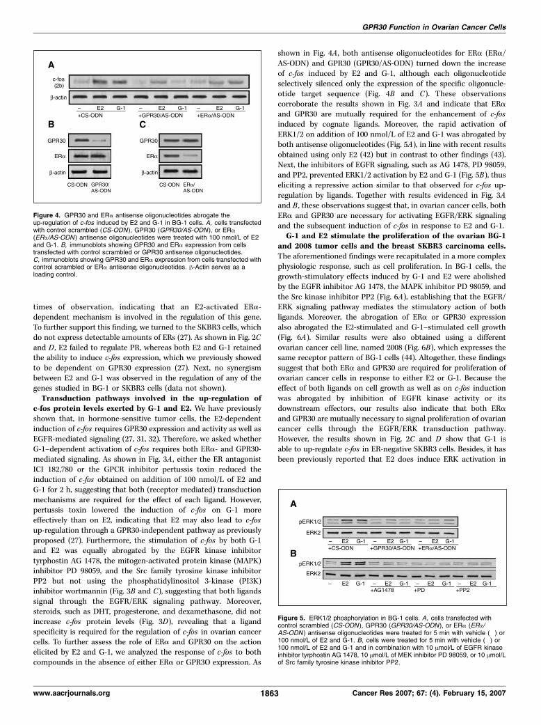
times of observation, indicating that an E2-activated ERa-dependent mechanism is involved in the regulation of this gene.To further support this finding, we turned to the SKBR3 cells, whichdo not express detectable amounts of ERs (27). As shown in Fig. 2Cand D , E2 failed to regulate PR, whereas both E2 and G-1 retainedthe ability to induce c-fos expression, which we previously showedto be dependent on GPR30 expression (27). Next, no synergismbetween E2 and G-1 was observed in the regulation of any of thegenes studied in BG-1 or SKBR3 cells (data not shown).Transduction pathways involved in the up-regulation of
c-fos protein levels exerted by G-1 and E2. We have previouslyshown that, in hormone-sensitive tumor cells, the E2-dependentinduction of c-fos requires GPR30 expression and activity as well asEGFR-mediated signaling (27, 31, 32). Therefore, we asked whetherG-1–dependent activation of c-fos requires both ERa- and GPR30-mediated signaling. As shown in Fig. 3A , either the ER antagonistICI 182,780 or the GPCR inhibitor pertussis toxin reduced theinduction of c-fos obtained on addition of 100 nmol/L of E2 andG-1 for 2 h, suggesting that both (receptor mediated) transductionmechanisms are required for the effect of each ligand. However,pertussis toxin lowered the induction of c-fos on G-1 moreeffectively than on E2, indicating that E2 may also lead to c-fosup-regulation through a GPR30-independent pathway as previouslyproposed (27). Furthermore, the stimulation of c-fos by both G-1and E2 was equally abrogated by the EGFR kinase inhibitortyrphostin AG 1478, the mitogen-activated protein kinase (MAPK)inhibitor PD 98059, and the Src family tyrosine kinase inhibitorPP2 but not using the phosphatidylinositol 3-kinase (PI3K)inhibitor wortmannin (Fig. 3B and C), suggesting that both ligandssignal through the EGFR/ERK signaling pathway. Moreover,steroids, such as DHT, progesterone, and dexamethasone, did notincrease c-fos protein levels (Fig. 3D), revealing that a ligandspecificity is required for the regulation of c-fos in ovarian cancercells. To further assess the role of ERa and GPR30 on the actionelicited by E2 and G-1, we analyzed the response of c-fos to bothcompounds in the absence of either ERa or GPR3O expression. As
shown in Fig. 4A , both antisense oligonucleotides for ERa (ERa/AS-ODN) and GPR30 (GPR30/AS-ODN) turned down the increaseof c-fos induced by E2 and G-1, although each oligonucleotideselectively silenced only the expression of the specific oligonucle-otide target sequence (Fig. 4B and C). These observationscorroborate the results shown in Fig. 3A and indicate that ERaand GPR30 are mutually required for the enhancement of c-fosinduced by cognate ligands. Moreover, the rapid activation ofERK1/2 on addition of 100 nmol/L of E2 and G-1 was abrogated byboth antisense oligonucleotides (Fig. 5A), in line with recent resultsobtained using only E2 (42) but in contrast to other findings (43).Next, the inhibitors of EGFR signaling, such as AG 1478, PD 98059,and PP2, prevented ERK1/2 activation by E2 and G-1 (Fig. 5B), thuseliciting a repressive action similar to that observed for c-fos up-regulation by ligands. Together with results evidenced in Fig. 3Aand B , these observations suggest that, in ovarian cancer cells, bothERa and GPR30 are necessary for activating EGFR/ERK signalingand the subsequent induction of c-fos in response to E2 and G-1.G-1 and E2 stimulate the proliferation of the ovarian BG-1
and 2008 tumor cells and the breast SKBR3 carcinoma cells.The aforementioned findings were recapitulated in a more complexphysiologic response, such as cell proliferation. In BG-1 cells, thegrowth-stimulatory effects induced by G-1 and E2 were abolishedby the EGFR inhibitor AG 1478, the MAPK inhibitor PD 98059, andthe Src kinase inhibitor PP2 (Fig. 6A), establishing that the EGFR/ERK signaling pathway mediates the stimulatory action of bothligands. Moreover, the abrogation of ERa or GPR30 expressionalso abrogated the E2-stimulated and G-1–stimulated cell growth(Fig. 6A). Similar results were also obtained using a differentovarian cancer cell line, named 2008 (Fig. 6B), which expresses thesame receptor pattern of BG-1 cells (44). Altogether, these findingssuggest that both ERa and GPR30 are required for proliferation ofovarian cancer cells in response to either E2 or G-1. Because theeffect of both ligands on cell growth as well as on c-fos inductionwas abrogated by inhibition of EGFR kinase activity or itsdownstream effectors, our results also indicate that both ERaand GPR30 are mutually necessary to signal proliferation of ovariancancer cells through the EGFR/ERK transduction pathway.However, the results shown in Fig. 2C and D show that G-1 isable to up-regulate c-fos in ER-negative SKBR3 cells. Besides, it hasbeen previously reported that E2 does induce ERK activation in
Figure 5. ERK1/2 phosphorylation in BG-1 cells. A, cells transfected withcontrol scrambled (CS-ODN ), GPR30 (GPR30/AS-ODN ), or ERa (ERa/AS-ODN ) antisense oligonucleotides were treated for 5 min with vehicle (�) or100 nmol/L of E2 and G-1. B, cells were treated for 5 min with vehicle (�) or100 nmol/L of E2 and G-1 and in combination with 10 Amol/L of EGFR kinaseinhibitor tyrphostin AG 1478, 10 Amol/L of MEK inhibitor PD 98059, or 10 Amol/Lof Src family tyrosine kinase inhibitor PP2.
Figure 4. GPR30 and ERa antisense oligonucleotides abrogate theup-regulation of c-fos induced by E2 and G-1 in BG-1 cells. A, cells transfectedwith control scrambled (CS-ODN), GPR30 (GPR30/AS-ODN ), or ERa(ERa/AS-ODN ) antisense oligonucleotides were treated with 100 nmol/L of E2and G-1. B, immunoblots showing GPR30 and ERa expression from cellstransfected with control scrambled or GPR30 antisense oligonucleotides.C, immunoblots showing GPR30 and ERa expression from cells transfected withcontrol scrambled or ERa antisense oligonucleotides. h-Actin serves as aloading control.
GPR30 Function in Ovarian Cancer Cells
www.aacrjournals.org 1863 Cancer Res 2007; 67: (4). February 15, 2007
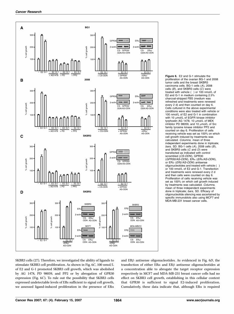
SKBR3 cells (27). Therefore, we investigated the ability of ligands tostimulate SKBR3 cell proliferation. As shown in Fig. 6C , 100 nmol/Lof E2 and G-1 promoted SKBR3 cell growth, which was abolishedby AG 1478, PD 98059, and PP2 or by abrogation of GPR30expression (Fig. 6C). To rule out the possibility that SKBR3 cellsexpressed undetectable levels of ERs sufficient to signal cell growth,we assessed ligand-induced proliferation in the presence of ERa
and ERh antisense oligonucleotides. As evidenced in Fig. 6D , thetransfection of either ERa and ERh antisense oligonucleotides ata concentration able to abrogate the target receptor expressionrespectively in MCF7 and MDA-MB-231 breast cancer cells had noeffect on SKBR3 cell growth, establishing in this cellular contextthat GPR30 is sufficient to signal E2-induced proliferation.Cumulatively, these data indicate that, although ERa is required
Figure 6. E2 and G-1 stimulate theproliferation of the ovarian BG-1 and 2008tumor cells and the breast SKBR3carcinoma cells. BG-1 cells (A), 2008cells (B ), and SKBR3 cells (C ) weretreated with vehicle (�) or 100 nmol/L ofE2 and G-1 in medium containing 2.5%charcoal-stripped FBS (medium wasrefreshed and treatments were renewedevery 2 d) and then counted on day 6.Cells cultured in the above experimentalconditions were also treated with vehicle or100 nmol/L of E2 and G-1 in combinationwith 10 Amol/L of EGFR kinase inhibitortyrphostin AG 1478, 10 Amol/L of MEKinhibitor PD 98059, and 10 Amol/L of Srcfamily tyrosine kinase inhibitor PP2 andcounted on day 6. Proliferation of cellsreceiving vehicle was set as 100% on whichcell growth induced by treatments wascalculated. Columns, mean of threeindependent experiments done in triplicate;bars, SD. BG-1 cells (A), 2008 cells (B ),and SKBR3 cells (C and D ) weretransfected as indicated with controlscrambled (CS-ODN ), GPR30(GPR30/AS-ODN ), ERa (ERa/AS-ODN ),or ERh (ERb/AS-ODN ) antisenseoligonucleotides and treated with vehicle (�)or 100 nmol/L of E2 and G-1. Transfectionand treatments were renewed every 2 dand then cells were counted on day 6.Proliferation of cells receiving vehicle wasset as 100% on which cell growth inducedby treatments was calculated. Columns,mean of three independent experimentsdone in triplicate; bars, SD. Efficacy ofoligonucleotide silencing was ascertained byspecific immunoblots also using MCF7 andMDA-MB-231 breast cancer cells.
Cancer Research
Cancer Res 2007; 67: (4). February 15, 2007 1864 www.aacrjournals.org
for the G-1/GPR30 signaling pathway in ovarian cancer cells,GPR30 may induce cell growth independently of ERa expressiondepending on the tumor type.
Discussion
Ovarian surface epithelial cells, which represent the site of 90%of malignancies, show a striking proliferative response toestrogens (4). As it concerns ERa, its expression in ovary tumorshas been associated with an increased rate of cell proliferationand a less favorable prognosis (45) contrary to that reported inbreast cancer (46).
A wide number of studies have established that estrogens drivecancer cell growth not only by activating the transcriptionalfunction of ERs but also by initiating nongenomic EGFR-dependentsignaling pathways consisting in either ERK or AKT activation(14, 24). Recent studies have shown that, in breast cancer cells (27)as well as in endometrial cancer cells (31) and even in thyroidcarcinoma cells (32), the nongenomic signaling triggered by E2relays on expression and activity of GPR30, which in turn activatesthe EGFR signaling pathway (30). Whether E2 acts on the EGFR/ERK transduction pathway only through GPR30 binding or alsothrough ERa binding is less clear because E2 binds to bothreceptors although with different affinity (28, 29). In the presentarticle, by using either E2 or the selective GPR30 ligand G-1, wehave determined the specificity of each signaling receptor inmediating E2 responsiveness of ovarian cancer cells. First, we showthat both ligands induce the expression of c-fos , pS2, and cyclins A,D1, and E, whereas only E2 modulates ERa-dependent transcrip-tion and PR expression (Figs. 1 and 2). In addition, both ligandsstimulate the proliferation of two different ovarian cancer cell linesin an EGFR-dependent fashion, suggesting that, as in breast cancercells, GPR30 is part of the E2 nongenomic signaling pathway.Furthermore, two lines of evidence indicate that both ERa andGPR30 are mutually required for E2 and G-1 pleiotropic effects: (a)c-fos up-regulation by each ligand is sensitive to both ICI 182,780and pertussis toxin (Fig. 3) and (b) abrogation of ERa and GPR30expression by specific antisense oligonucleotides abolishes c-fosstimulation as well as ERK activation (Figs. 4 and 5) and cellproliferation (Fig. 6) induced by both ligands. Because the effect ofE2 and G-1 on c-fos promoter activity or on Elk1 transactivation isnot synergic (Fig. 1), we conclude that GPR30 and ERa act on thesame signaling pathway respectively upstream and downstream ofEGFR. In line with this model, it has been shown that GPR30 in
response to E2 induces the release of surface-bound proHB-EGF(30), whereas ERa tyrosine phosphorylation by EGFR activationplays a key role in the steroid receptor interaction with Src andthe regulation of Src-associated ERK kinase activity, which in turnstimulates a mitogenic signaling network known to be engaged bygrowth factors (reviewed in ref. 47 and references therein). Besides,it is well known that the EGFR-mediated transcriptional activationof unliganded ERa can also occur through the critical serineresidue at position 118, which is the major phosphorylation siteresulting from activation of the MAPK pathway (48–50).
It will be certainly interesting to define to what extent the cross-talk between ERa and GPR30 may influence the development ofestrogen-sensitive tumors and/or the failure of endocrine thera-peutic agents.
On the other hand, in the present and a previous study (27), wehave shown that, in the ERa-negative SKBR3 breast cancer cells,GPR30 is able to elicit ERK activation and c-fos induction throughEGFR signaling pathway. Interestingly, we now also show thatboth E2 and G-1 are able to induce SKBR3 cell proliferation, whichrelays on the activity of EGFR and its downstream effectorsregardless of transfection of antisense oligonucleotides abrogatingERa and ERh expression. In contrast, a GPR30 antisense oligo-nucleotide abolished both E2-mediated or G-1–mediated prolifer-ation of SKBR3 cells, suggesting that the relevance of a functionalinteraction between ERa and GPR30 depends on the specificcellular context and type of tumor. It remains as an intriguing openquestion if and how other endogenous factors cross-interact withGPR30 in mediating E2-dependent proliferation of ERa-negativecancer cells.
In conclusion, the present study provides new insight toward thedesign of pharmacologic molecules targeting crucial metaboliccascades and genes directly involved in cell proliferation of ovariancancer cells. Furthermore, it represents the first example of how theselective GPR30 ligand G-1 can provide a useful experimentalmodel to screen for estrogen-like properties exerted throughGPR30 in estrogen-sensitive tumors.
Acknowledgments
Received 8/9/2006; revised 10/23/2006; accepted 12/10/2006.Grant support: Associazione Italiana per la Ricerca sul Cancro, Ministero
dell’Universita e Ricerca Scientifica e Tecnologica, and Regione Calabria.The costs of publication of this article were defrayed in part by the payment of page
charges. This article must therefore be hereby marked advertisement in accordancewith 18 U.S.C. Section 1734 solely to indicate this fact.
References1. Greenlee RT, Murray T, Bolden S, Wingo PA. Cancerstatistics, 2000. CA Cancer J Clin 2000;50:7–33.2. Boente MP, Hurteau J, Rodriguez GC, Bast RC, Jr.,Berchuck A. The biology of ovarian cancer. Curr OpinOncol 1993;5:900–7.3. O’Donnell AJM, Macleod KG, Burns DJ, Smyth JF,Langdon SP. Estrogen receptor a mediates gene expres-sion changes and growth response in ovarian cancer cellsexposed to estrogen. Endocr Relat Cancer 2005;12:851–66.4. Bai W, Oliveros-Saunders B, Wang Q, Acevedo-DuncanME, Nicosia SV. Estrogen stimulation of ovarian surfaceepithelial cell proliferation. In Vitro Cell Dev Biol Anim2000;36:657–66.5. Rodrigez C, Patel AV, Calle EE, Jacob EJ, Thun MJ.Estrogen replacement therapy and ovarian cancer
mortality in a large prospective study of US women.JAMA 2001;285:1460–5.6. Riman T, Dickman PW, Nilsson S, et al. Hormonereplacement therapy and the risk of invasive epithelialovarian cancer in Swedish women. J Natl Cancer Inst2002;94:497–504.7. Lacey JV, Jr., Mink PJ, Lubin JH, et al. Menopausalhormone replacement therapy and risk of ovariancancer. JAMA 2002;288:334–41.8. Langdon SP, Hawkes MM, Lawrie SS, et al. Oestrogenreceptor expression and the effects of oestrogen andtamoxifen on the growth of human ovarian carcinomacell lines. Br J Cancer 1990;62:213–6.9. Langdon SP, Crew AJ, Ritchie AA, et al. Growthinhibition of oestrogen receptor-positive human ovariancarcinoma by anti-oestrogens in vitro and in a xenograftmodel. Eur J Cancer 1994;30A:682–6.
10. Hatch KD, Beccham JB, Blessing JA, Creasman WT.Responsiveness of patients with relapsed ovarian cancerto tamoxifen. Cancer 1991;68:269–71.11. Algren JD, Ellison NM, Gottlieb RJ, et al. Hormonalpalliation of chemoresistant ovarian cancer: threeconsecutive phase II trials of the Mid-Atlantic OncologyProgram. J Clin Oncol 1993;10:1957–68.12. Bowman A, Gabra H, Langdon SP, et al. CA125response is associated with estrogen receptor expres-sion in a phase II trial of letrozole in ovarian cancer:identification of an endocrine-sensitive subgroup. ClinCancer Res 2002;8:2233–9.13. Papadimitriou CA, Markaki S, Siapkaras J, et al.Hormonal therapy with letrozole for relapsed epithelialovarian cancer. Long-term results of a phase II study.Oncology 2004;66:112–27.14. Hall JM, Course JF, Korach KS. The multifaceted
GPR30 Function in Ovarian Cancer Cells
www.aacrjournals.org 1865 Cancer Res 2007; 67: (4). February 15, 2007
mechanism of estradiol and estrogen receptor signal-ling. J Biol Chem 2001;276:36869–72.15. Bardin A, Hoffman P, Boulle N, et al. Involvement ofestrogen receptor h in ovarian carcinogenesis. CancerRes 2004;64:5861–9.16. Jordan VC, Morrow M. Tamoxifen, raloxifene, andthe prevention of breast cancer. Endocr Rev 1999;20:253–78.17. Rio MC, Bellocq JP, Gairard B, et al. Specificexpression of the pS2 gene in subclasses of breastcancers in comparison with expression of the estrogenand progesterone receptors and the oncogene ERBB2.Proc Natl Acad Sci U S A 1987;84:9243–7.18. Rochefort H. Estrogen-induced genes in breastcancer, and their medical importance. Bull Acad NatlMed 1999;183:955–68.19. Hall JM, Korach KS. Stromal cell-derived factor 1, anovel target of estrogen receptor action, mediates themitogenic effects of estradiol in ovarian and breastcancer cells. Mol Endocrinol 2003;17:792–803.20. Chaffin CL, Hess DL, Stouffer RL. Dynamics ofperiovulatory steroidogenesis in the rhesus monkeyfollicle after ovarian stimulation. Hum Reprod 1999;14:642–9.21. Belin F, Goudet G, Duchamp G, Gerard N. Intra-follicular concentrations of steroids and steroidogenicenzymes in relation to follicular development in themare. Biol Reprod 2000;62:1335–43.22. Fujiara T, Lambert-Messerlian G, Sidis Y, et al.Analysis of follicular fluid hormone concentrationsand granulosa cell mRNA levels for the inhibin-activin-follistatin system: relation to oocyte and embryocharacteristics. Fertil Steril 2000;74:348–55.23. Gougeon A. Dynamics of human follicular growth:morphologic, dynamic, and functional aspects. In: LeungPCK, Adasci EY, editors. The ovary. 2nd ed. New York:Elsevier Academic Press; 2004. p. 25–38.24. Levin ER. Integration of the extranuclear andnuclear actions of estrogen. Mol Endocrinol 2005;19:1951–9.25. Deroo BJ, Korach KS. Estrogen receptors and humandisease. J Clin Invest 2006;116:561–70.26. Bologa CG, Revankar CM, Young SM, et al. Virtualand biomolecular screening converge on a selectiveagonist for GPR30. Nat Chem Biol 2006;4:207–12.27. Maggiolini M, Vivacqua A, Fasanella G, et al. The Gprotein-coupled receptor GPR30 mediates c-fos up-
regulation by 17h-estradiol and phytoestrogens inbreast cancer cells. J Biol Chem 2004;279:27008–16.28. Revankar CM, Cimino DF, Sklar LA, Arterburn JB,Prossnitz ER. A transmembrane intracellular estrogenreceptor mediates rapid cell signalling. Science 2005;307:1625–30.29. Thomas P, Pang Y, Filardo EJ, Dong J. Identity of anestrogen membrane receptor coupled to a G proteinhuman breast cancer cells. Endocrinology 2005;146:624–32.30. Filardo EJ, Quinn JA, Bland KI, Frackelton AR, Jr.Estrogen-induced activation of Erk-1 and Erk-2 requiresthe G protein-coupled receptor homolog, GPR30, andoccurs via trans -activation of the epidermal growthfactor receptor through release of HB-EGF. MolEndocrinol 2000;10:1649–60.31. Vivacqua A, Bonofiglio D, Recchia AG, et al. The Gprotein-coupled receptor GPR30 mediates the prolifer-ative effects induced by 17h-estradiol and hydroxyta-moxifen in endometrial cancer cells. Mol Endocrinol2006;20:631–46.32. Vivacqua A, Bonofiglio D, Albanito L, et al. 17h-Estradiol, genistein, and 4-hydroxytamoxifen induce theproliferation of thyroid cancer cells through the Gprotein coupled-receptor GPR30. Mol Pharmacol 2006;70:1414–23.33. Gallo A, Cuozzo C, Esposito I, et al. Menin uncouplesElk-1, JunD, and c-Jun phosphorylation from MAPkinase activation. Oncogene 2002;21:6434–45.34. Maggiolini M, Donze O, Picard D. A non-radioactivemethod for inexpensive quantitative RT-PCR. J BiolChem 1999;380:695–7.35. Kanda N, Watanabe S. 17h-Estradiol inhibits oxida-tive stress-induced apoptosis in keratinocytes bypromoting Bcl-2 expression. J Invest Dermatol 2003;121:771–80.36. Geisinger KR, Kute TE, Pettenati MJ, et al. Charac-terization of a human ovarian carcinoma cell line withestrogen and progesterone receptors. Cancer 1989;63:280–8.37. Santagati S, Gianazza E, Agrati P, et al. Oligonucle-otide squelching reveals the mechanism of estrogenreceptor autologous down-regulation. Mol Endocrinol1997;11:938–49.38. Hill CS, Treisman R. Differential activation of c-fospromoter elements by serum, lysophosphatidic acid, Gproteins, and polypeptide growth factors. EMBO J 1995;14:5037–47.
39. Karin M. Signal transduction from the cell surfaceto the nucleus through the phosphorylation oftranscription factors. Curr Opin Cell Biol 1994;6:415–24.40. Singleton DW, Feng Y, Burd CJ, Khan SA. Non-genomic activity and subsequent c-fos induction byestrogen receptor ligands are not sufficient topromote deoxyribonucleic acid synthesis in humanendometrial adenocarcinoma cells. Endocrinology2003;144:121–8.41. Nephew KP, Polek TC, Akcali KC, Khan SA. Theantiestrogen tamoxifen induces c-fos and jun-B, but notc-jun or jun-D, protooncogenes in the rat uterus.Endocrinology 1993;133:419–22.42. Sukocheva O, Wadham C, Holmes A, et al. Estrogentransactivates EGFR via the sphingosine 1-phosphatereceptor Edg-3: the role of sphingosine kinase-1. J CellBiol 2006;173:301–10.43. Pedram A, Razandi M, Levin ER. Nature of functionalestrogen receptors at the plasma membrane. MolEndocrinol 2006;9:1996–2009.44. Safei R, Katano K, Larson BJ, et al. Intracellularlocalization and trafficking of fluorescein-labeled cis-platin in human ovarian carcinoma cells. Clin CancerRes 2005;11:756–67.45. Munstedt K, Steen J, Knauf AG, Buch T, von Georgi R,Franke FE. Steroid hormone receptors and long termsurvival in invasive ovarian cancer. Cancer 2000;89:1783–91.46. Lopez-Otin C, Diamandis EP. Breast and prostatecancer: an analysis of common epidemiological, genetic,and biochemical features. Endocr Rev 1998;19:365–96.47. Migliaccio A, Di Domenico M, Castoria G, et al.Steroid receptor regulation of epidermal growth factorsignaling through Src in breast and prostate cancercells: steroid antagonist action. Cancer Res 2005;65:10585–93.48. Kato S, Endoh H, Masuhiro Y, et al. Activation ofthe estrogen receptor through phosphorylation bymitogen-activated protein kinase. Science 1995;270:1491–4.49. Bunone G, Briand PA, Miksicek RJ, Picard D.Activation of the unliganded estrogen receptor by EGFinvolves the MAP kinase pathway and direct phosphor-ylation. EMBO J 1996;15:2174–83.50. Lannigan D. Estrogen receptor phosphorylation.Steroids 2003;68:1–9.
Cancer Research
Cancer Res 2007; 67: (4). February 15, 2007 1866 www.aacrjournals.org
17�-Estradiol, Genistein, and 4-Hydroxytamoxifen Induce theProliferation of Thyroid Cancer Cells through theG Protein-Coupled Receptor GPR30
Adele Vivacqua, Daniela Bonofiglio, Lidia Albanito, Antonio Madeo, Vittoria Rago,Amalia Carpino, Anna Maria Musti, Didier Picard, Sebastiano Ando, andMarcello MaggioliniDepartments of Pharmaco-Biology (A.V., D.B., L.A., A.M., A.M.M., M.M.) and Cellular Biology (V.R., A.C., S.A.), University ofCalabria, Rende, Italy; and Department of Cellular Biology (D.P.), University of Geneva, Geneva, Switzerland
Received May 5, 2006; accepted July 11, 2006
ABSTRACTThe higher incidence of thyroid carcinoma (TC) in women dur-ing reproductive years compared with men and the increasedrisk associated with the therapeutic use of estrogens havesuggested a pathogenetic role exerted by these steroids in thedevelopment of TC. In the present study, we evaluated thepotential of 17�-estradiol (E2), genistein (G), and 4-hydroxyta-moxifen (OHT) to regulate the expression of diverse estrogentarget genes and the proliferation of human WRO, FRO, andARO thyroid carcinoma cells, which were used as a modelsystem. We have ascertained that ARO cells are devoid ofestrogen receptors (ERs), whereas both WRO and FRO cellsexpress a single variant of ER� that was neither transactivated,modulated, nor translocated into the nucleus upon treatment
with ligands. However, E2, G, and OHT were able either toinduce the transcriptional activity of c-fos promoter constructs,including those lacking the estrogen-responsive elements, or toincrease c-fos, cyclin A, and D1 expression. It is noteworthythat we have demonstrated that the G protein-coupled receptor30 (GPR30) and the mitogen-activated protein kinase (MAPK)pathway mediate both the up-regulation of c-fos and thegrowth response to E2, G, and OHT in TC cells studied, be-cause these stimulatory effects were prevented by silencingGPR30 and using the MEK inhibitor 2�-amino-3�-methoxyfla-vone (PD 98059). Our findings provide new insight into themolecular mechanisms through which estrogens may inducethe progression of TC.
Thyroid carcinoma (TC) is the most common malignancyaffecting the endocrine glands (Jemal et al., 2004). The con-spicuous increased incidence during recent years as observedin the decade between 1994 and 2004 in the United States(�80%) (Jemal et al., 2004) has prompted investigators tobetter define the molecular mechanisms involved in TC de-
velopment. Several lines of evidence have documented thatmutations of genes such as BRAF, RET, and RAS coding foreffectors along the MAPK pathway exert a crucial role inthyroid transformation (Melillo et al., 2005). However, theconsistent demographic observation that TC is 3 times morefrequent in women than in men from the onset of pubertyuntil menopause when this ratio declines progressively (Hen-derson et al., 1982) still remains to be elucidated. The gen-der-dependent difference observed worldwide (Waterhouse etal., 1982) and the increased risk in women taking estrogensfor gynecological disorders (Ron et al., 1987; Persson et al.,1996) have suggested that these steroids may influence the
This research was supported by grants from the Associazione Italiana per laRicerca sul Cancro, Ministero dell’Universita e Ricerca Scientifica e Tecno-logica, the Regione Calabria and Swiss National Science Foundation, Krebsfor-schung Schweiz, and the Canton the Geneve (to D.P.).
Article, publication date, and citation information can be found athttp://molpharm.aspetjournals.org.
doi:10.1124/mol.106.026344.
ABBREVIATIONS: TC, thyroid carcinoma; GPR30, G protein couplet receptor 30; G, genistein; OHT, 4-hydroxytamoxifen; DEX, dexamethasone;PRG, progesterone; PR, progesterone receptor; PD 98059 and PD, 2�-amino-3�-methoxyflavone; ER, estrogen receptor; E2, 17�-estradiol; ERE,estrogen response element; MAPK, mitogen-activated protein kinase; AF, activation function; GPCR, G protein-coupled receptor; Cx, cyclohex-imide; WM, wortmannin; PT, pertussis toxin; DHT, 5�-dihydrotestosterone; ICI 182,780 and ICI, faslodex; AG 1478, 4-(3�-chloroanilino)-6,7-dimethoxy-quinazoline; AG 490, �-cyano-(3,4-dihydroxy)-N-benzylcinnamide; H-89, N-[2-(4-bromocinnamylamino)ethyl]-5-isoquinoline; PP2,4-amino-5-(4-chlorophenyl)-7-(t-butyl)pyrazolo[3,4,d]pyrimidine; FBS, fetal bovine serum; PCR, polymerase chain reaction; RT-PCR, reverse-transcriptase polymerase chain reaction; bp, base pair(s); kb, kilobase(s); ERK, extracellular signal-regulated kinase; MEK, mitogen-activatedprotein kinase kinase; PI3K, phosphoinositide-3 kinase; EGFR, epidermal growth factor receptor; AP-1, activator protein 1; JAK, Janus tyrosinekinase; AS-ODN, antisense oligodeoxynucleotide; LY 294,002, 2-(4-morpholinyl)-8-phenyl-4H-1-benzopyran-4-one.
0026-895X/06/7004-1414–1423$20.00MOLECULAR PHARMACOLOGY Vol. 70, No. 4Copyright © 2006 The American Society for Pharmacology and Experimental Therapeutics 26344/3140426Mol Pharmacol 70:1414–1423, 2006 Printed in U.S.A.
1414
at BIB
LIOT
EC
A A
RE
A T
EC
NIC
O S
CIE
NT
IFIC
A on O
ctober 30, 2008 m
olpharm.aspetjournals.org
Dow
nloaded from
progression of TC, as largely demonstrated for breast andendometrial carcinoma (Pike et al., 2004). On the other hand,the ability of 17�-estradiol (E2) to elicit proliferative effectsin human thyroid cancer cells (Manole et al., 2001; Lee et al.,2005) and in FRTL-5 rat thyroid cells (Furlanetto et al.,1999) and the expression of estrogen receptors (ERs) in nor-mal and neoplastic thyroid tissues (Yane et al., 1994; DallaValle et al., 1998) have provided further evidence on thepotential role exerted by E2 in the progression of TC.
The classic mechanism of E2 action involves the binding toER� and ER� that contain two main transcription activationfunctions (AFs): the N-terminal AF1, and the C-terminalAF2, which is associated with the ligand binding domainresponsible for hormone-dependent transactivation (Tora etal., 1989). Both ERs after ligand activation and nuclear lo-calization interact with the estrogen response elements(EREs) located within the regulatory region of target genes(Kumar and Chambon, 1988). It is interesting that previousstudies, including our own (Maggiolini et al., 2004; Revankaret al., 2005; Thomas et al., 2005; Vivacqua et al., 2006), havedemonstrated that estrogens are also able to activate a Gprotein-coupled receptor (GPCR) named GPR30, which me-diates the transcription of genes like c-fos involved in thecycle progression of different tumor cells. c-fos is rapidly andtransiently induced by different extracellular stimuli, suchas mitogens and hormones (Hill and Treisman, 1995). Thetranscription of c-fos is regulated by multiple cis-elements,including the serum-response element, which recruits theternary complex factors Elk-1 and the serum response factoraccessory proteins 1 and 2 (Treisman, 1995). Moreover, E2signaling may trigger a nongenomic ER� pathway, leading toMAPK-dependent phosphorylation and binding of Elk-1 tothe serum-response element sequence (Duan et al., 2001). Asit concerns the imperfect palindromic ERE sequence con-tained within the c-fos promoter, binding to ER� alone is notsufficient for transactivation but instead requires the recep-tor interaction with a downstream Sp1 site (Duan et al.,1998).
In the present study, we evaluated the potential of E2,genistein (G), and 4-hydroxytamoxifen (OHT) to regulate theexpression of diverse estrogen target genes and the prolifer-ation of human follicular WRO and both anaplastic FRO andARO thyroid cancer cells used as a model system. Our datademonstrate for the first time to our knowledge that aGPR30-dependent mechanism is involved in both gene tran-scription and growth effects elicited by E2, G, and OHT inthyroid cancer cells.
Materials and MethodsReagents. E2, G, OHT, cycloheximide (Cx), wortmannin (WM),
LY 294,002, pertussis toxin (PT), PD 98059 (PD), dexamethasone(DEX), progesterone (PRG), and 5�-dihydrotestosterone (DHT) werepurchased from Sigma-Aldrich Corporation (Milan, Italy). ICI182,780 (ICI) was obtained from Tocris Chemicals (Bristol, UK), AG1478 and AG 490 were purchased from Biomol Research Laborato-ries, Inc. (Plymouth Meeting, PA), and H-89 and PP2 were obtainedfrom Calbiochem (VWR International, Milan, Italy). All compoundswere solubilized in dimethyl sulfoxide, except for E2, OHT, PD, andWM, which were dissolved in ethanol.
Cell Culture. Human follicular WRO and anaplastic FRO andARO thyroid tumor cells (gifts from F. Arturi and A. Belfiore, Uni-versity of Magna Grecia, Catanzaro, Italy), and HeLa cells were
maintained in Dulbecco’s modified Eagle’s medium without phenolred supplemented with 10% fetal bovine serum (FBS) and Glutamax(Invitrogen, Milan, Italy). Cells were switched to medium withoutserum the day before RT-PCR, immunoblots, and the evaluation ofERK1/2 phosphorylation.
Plasmids. The firefly luciferase reporter plasmid for ERs wasXETL, which contains the ERE from the Xenopus laevis vitellogeninA2 gene (nucleotides �334 to �289), the herpes simplex virus thy-midine kinase promoter region (nucleotides �109 to �52), the fireflyluciferase coding sequence, and the SV40 splice and polyadenylationsites from plasmid pSV232A/L-AA5�. Reporter plasmids for c-fos andits deletion mutant c-fos�ERE (which lacks the ERE sequence) en-code �2.2 kb and �1.172 kb 5� upstream fragments of human c-fos,respectively (gifts from K. Nose, Showa University, Tokyo, Japan).The reporter plasmid Gal4-luc was described together with the ex-pression vectors for Gal4-Elk1 in our previous study (Gallo et al.,2002). The plasmid HEG0 was used to express ER�. The plasmidsencoding antisense GPR30 (GPR30/AS) and dominant-negativeERK2 (DN/ERK) were kindly provided by E. R. Prossnitz (Universityof New Mexico, Albuquerque, NM) and M. Cobb (University of Texas,Dallas, TX), respectively. The Renilla reniformis luciferase expres-sion vector pRL-TK (Promega, Milan, Italy) was used as a transfec-tion standard.
Transfections and Luciferase Assays. WRO and FRO cells(1 � 105) were plated into 24-well dishes with 500 �l of regulargrowth medium per well the day before transfection. The mediumwas replaced with that lacking serum on the day of transfection,which was performed using FuGENE 6 reagent as recommended bythe manufacturer (Roche Diagnostics, Milan, Italy) with a mixturecontaining 0.5 �g of reporter plasmid, 0.1 �g of effector plasmidwhere applicable, and 5 ng of pRL-TK. After 4 h, the serum-freemedium containing the indicated treatment was renewed, and thencells were incubated for approximately 12 h. Luciferase activity wasmeasured with the Dual Luciferase Kit (Promega) according to themanufacturer’s recommendations. Firefly luciferase values werenormalized to the internal transfection control provided by the R.reniformis luciferase activity. The normalized relative light unitvalues obtained from untreated cells were set as 100% activity, uponwhich the activity induced by treatments was calculated.
Western Blotting. Cells were grown in 10-cm dishes, exposed toligands, and then lysed in 500 �l of 50 mM HEPES, pH 7.5, 150 mMNaCl, 1.5 mM MgCl2, 1 mM EGTA, 10% glycerol, 1% Triton X-100,1% SDS, and a mixture of protease inhibitors containing 1 mMaprotinin, 20 mM phenylmethylsulfonyl fluoride, and 200 mM so-dium orthovanadate. Protein concentration was determined by Brad-ford reagent according to the manufacturer’s recommendations(Sigma). Equal amounts of whole-protein extract were resolved on a10% SDS-polyacrylamide gel, transferred to a nitrocellulose mem-brane (GE Healthcare, Milan, Italy), probed overnight at 4°C withthe antibodies against the C- and N-terminal domains of ER� (F-10and D-12, respectively), c-fos, GPR30, progesterone receptor (PR),�-actin (all purchased from Santa Cruz Biotechnology, DBA, Milan,Italy), ER� (Serotec, Milan, Italy), pERK1/2, and ERK2 (both fromCell Signaling Technology, Inc., Celbio, Milan, Italy), and then re-vealed using the ECL system (GE Healthcare). Five micrograms ofDN/ERK and GPR30/AS expression plasmid was transfected usingFuGENE 6 reagent as recommended by the manufacturer for 24 hbefore treatments.
RT-PCR. The evaluation of gene expression was performed bysemiquantitative RT-PCR as described previously (Maggiolini et al.,1999). For ER�, c-fos, cyclin A, cyclin D1, pS2, PR, and the acidicribosomal phosphoprotein P0 (36B4), which was used as a controlgene, the primers were the following: 5�-GGAGACATGAGAGCT-GCCA-3� (ER� forward) and 5�-CCAGCAGCATGTCGAAGATC-3�(ER� reverse); 5�-AGAAAAGGAGAATCCGAAGGGAAA-3� (c-fos for-ward) and 5�-ATGATGCTGGGACAGGAAGTC-3� (c-fos reverse); 5�-GCCATTAGTTTACCTGGACCCAGA-3� (cyclin A forward) and 5�-C-ACTGACATGGAAGACAGGAACCT-3� (cyclin A reverse); 5�-TCTA-
GPR30 Function in Thyroid Cancer Cells 1415
at BIB
LIOT
EC
A A
RE
A T
EC
NIC
O S
CIE
NT
IFIC
A on O
ctober 30, 2008 m
olpharm.aspetjournals.org
Dow
nloaded from
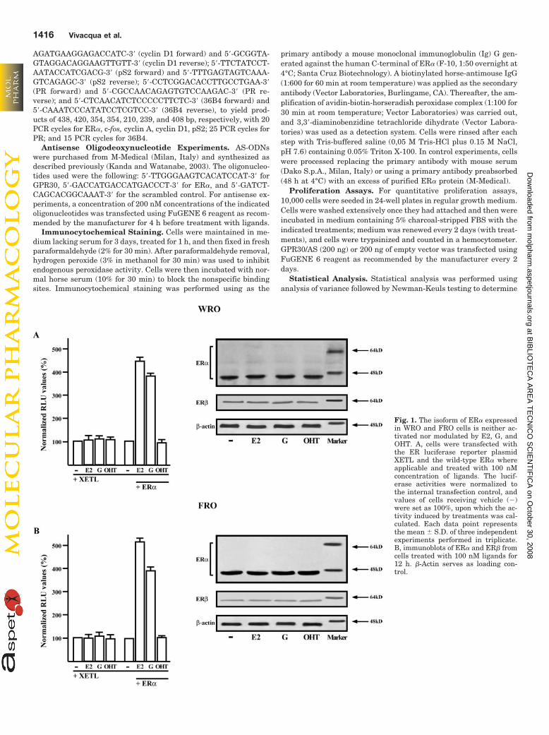
AGATGAAGGAGACCATC-3� (cyclin D1 forward) and 5�-GCGGTA-GTAGGACAGGAAGTTGTT-3� (cyclin D1 reverse); 5�-TTCTATCCT-AATACCATCGACG-3� (pS2 forward) and 5�-TTTGAGTAGTCAAA-GTCAGAGC-3� (pS2 reverse); 5�-CCTCGGACACCTTGCCTGAA-3�(PR forward) and 5�-CGCCAACAGAGTGTCCAAGAC-3� (PR re-verse); and 5�-CTCAACATCTCCCCCTTCTC-3� (36B4 forward) and5�-CAAATCCCATATCCTCGTCC-3� (36B4 reverse), to yield prod-ucts of 438, 420, 354, 354, 210, 239, and 408 bp, respectively, with 20PCR cycles for ER�, c-fos, cyclin A, cyclin D1, pS2; 25 PCR cycles forPR; and 15 PCR cycles for 36B4.
Antisense Oligodeoxynucleotide Experiments. AS-ODNswere purchased from M-Medical (Milan, Italy) and synthesized asdescribed previously (Kanda and Watanabe, 2003). The oligonucleo-tides used were the following: 5�-TTGGGAAGTCACATCCAT-3� forGPR30, 5�-GACCATGACCATGACCCT-3� for ER�, and 5�-GATCT-CAGCACGGCAAAT-3� for the scrambled control. For antisense ex-periments, a concentration of 200 nM concentrations of the indicatedoligonucleotides was transfected using FuGENE 6 reagent as recom-mended by the manufacturer for 4 h before treatment with ligands.
Immunocytochemical Staining. Cells were maintained in me-dium lacking serum for 3 days, treated for 1 h, and then fixed in freshparaformaldehyde (2% for 30 min). After paraformaldehyde removal,hydrogen peroxide (3% in methanol for 30 min) was used to inhibitendogenous peroxidase activity. Cells were then incubated with nor-mal horse serum (10% for 30 min) to block the nonspecific bindingsites. Immunocytochemical staining was performed using as the
primary antibody a mouse monoclonal immunoglobulin (Ig) G gen-erated against the human C-terminal of ER� (F-10, 1:50 overnight at4°C; Santa Cruz Biotechnology). A biotinylated horse-antimouse IgG(1:600 for 60 min at room temperature) was applied as the secondaryantibody (Vector Laboratories, Burlingame, CA). Thereafter, the am-plification of avidin-biotin-horseradish peroxidase complex (1:100 for30 min at room temperature; Vector Laboratories) was carried out,and 3,3�-diaminobenzidine tetrachloride dihydrate (Vector Labora-tories) was used as a detection system. Cells were rinsed after eachstep with Tris-buffered saline (0,05 M Tris-HCl plus 0.15 M NaCl,pH 7.6) containing 0.05% Triton X-100. In control experiments, cellswere processed replacing the primary antibody with mouse serum(Dako S.p.A., Milan, Italy) or using a primary antibody preabsorbed(48 h at 4°C) with an excess of purified ER� protein (M-Medical).
Proliferation Assays. For quantitative proliferation assays,10,000 cells were seeded in 24-well plates in regular growth medium.Cells were washed extensively once they had attached and then wereincubated in medium containing 5% charcoal-stripped FBS with theindicated treatments; medium was renewed every 2 days (with treat-ments), and cells were trypsinized and counted in a hemocytometer.GPR30/AS (200 ng) or 200 ng of empty vector was transfected usingFuGENE 6 reagent as recommended by the manufacturer every 2days.
Statistical Analysis. Statistical analysis was performed usinganalysis of variance followed by Newman-Keuls testing to determine
Fig. 1. The isoform of ER� expressedin WRO and FRO cells is neither ac-tivated nor modulated by E2, G, andOHT. A, cells were transfected withthe ER luciferase reporter plasmidXETL and the wild-type ER� whereapplicable and treated with 100 nMconcentration of ligands. The lucif-erase activities were normalized tothe internal transfection control, andvalues of cells receiving vehicle (�)were set as 100%, upon which the ac-tivity induced by treatments was cal-culated. Each data point representsthe mean � S.D. of three independentexperiments performed in triplicate.B, immunoblots of ER� and ER� fromcells treated with 100 nM ligands for12 h. �-Actin serves as loading con-trol.
1416 Vivacqua et al.
at BIB
LIOT
EC
A A
RE
A T
EC
NIC
O S
CIE
NT
IFIC
A on O
ctober 30, 2008 m
olpharm.aspetjournals.org
Dow
nloaded from
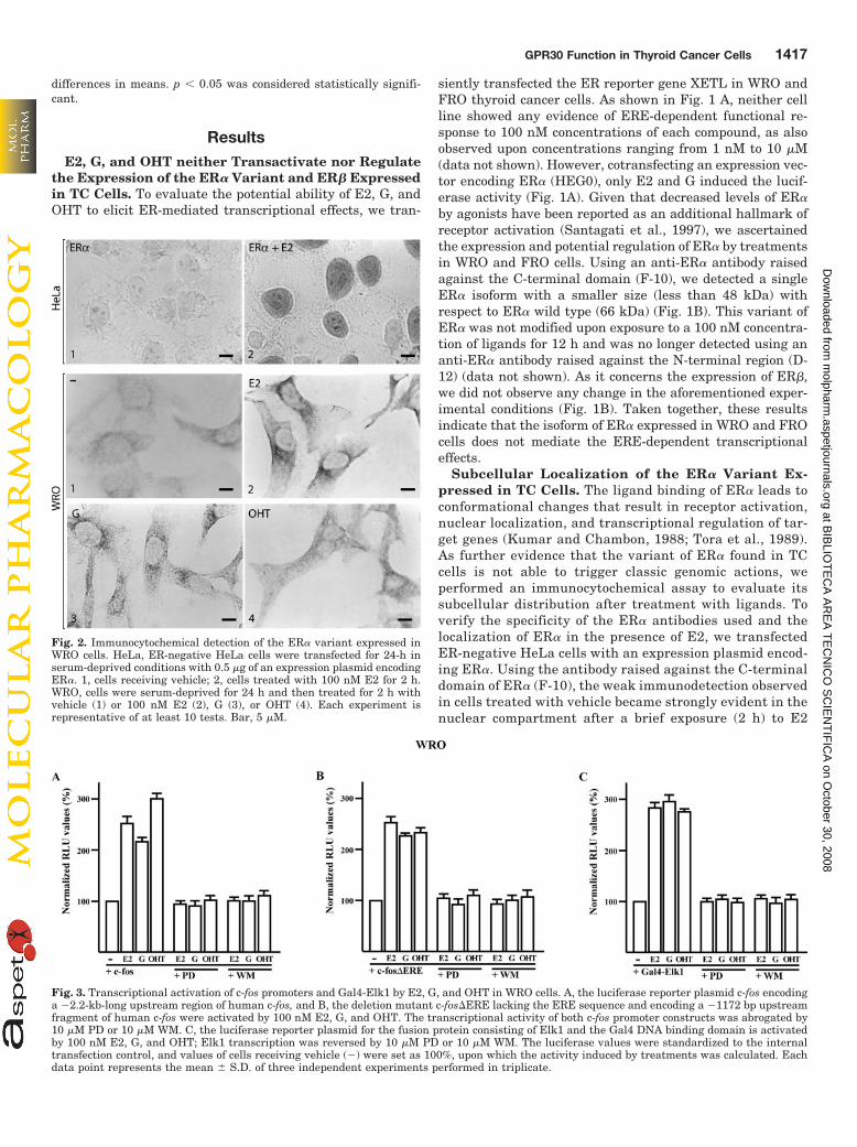
differences in means. p � 0.05 was considered statistically signifi-cant.
ResultsE2, G, and OHT neither Transactivate nor Regulate
the Expression of the ER� Variant and ER� Expressedin TC Cells. To evaluate the potential ability of E2, G, andOHT to elicit ER-mediated transcriptional effects, we tran-
siently transfected the ER reporter gene XETL in WRO andFRO thyroid cancer cells. As shown in Fig. 1 A, neither cellline showed any evidence of ERE-dependent functional re-sponse to 100 nM concentrations of each compound, as alsoobserved upon concentrations ranging from 1 nM to 10 �M(data not shown). However, cotransfecting an expression vec-tor encoding ER� (HEG0), only E2 and G induced the lucif-erase activity (Fig. 1A). Given that decreased levels of ER�by agonists have been reported as an additional hallmark ofreceptor activation (Santagati et al., 1997), we ascertainedthe expression and potential regulation of ER� by treatmentsin WRO and FRO cells. Using an anti-ER� antibody raisedagainst the C-terminal domain (F-10), we detected a singleER� isoform with a smaller size (less than 48 kDa) withrespect to ER� wild type (66 kDa) (Fig. 1B). This variant ofER� was not modified upon exposure to a 100 nM concentra-tion of ligands for 12 h and was no longer detected using ananti-ER� antibody raised against the N-terminal region (D-12) (data not shown). As it concerns the expression of ER�,we did not observe any change in the aforementioned exper-imental conditions (Fig. 1B). Taken together, these resultsindicate that the isoform of ER� expressed in WRO and FROcells does not mediate the ERE-dependent transcriptionaleffects.
Subcellular Localization of the ER� Variant Ex-pressed in TC Cells. The ligand binding of ER� leads toconformational changes that result in receptor activation,nuclear localization, and transcriptional regulation of tar-get genes (Kumar and Chambon, 1988; Tora et al., 1989).As further evidence that the variant of ER� found in TCcells is not able to trigger classic genomic actions, weperformed an immunocytochemical assay to evaluate itssubcellular distribution after treatment with ligands. Toverify the specificity of the ER� antibodies used and thelocalization of ER� in the presence of E2, we transfectedER-negative HeLa cells with an expression plasmid encod-ing ER�. Using the antibody raised against the C-terminaldomain of ER� (F-10), the weak immunodetection observedin cells treated with vehicle became strongly evident in thenuclear compartment after a brief exposure (2 h) to E2
Fig. 2. Immunocytochemical detection of the ER� variant expressed inWRO cells. HeLa, ER-negative HeLa cells were transfected for 24-h inserum-deprived conditions with 0.5 �g of an expression plasmid encodingER�. 1, cells receiving vehicle; 2, cells treated with 100 nM E2 for 2 h.WRO, cells were serum-deprived for 24 h and then treated for 2 h withvehicle (1) or 100 nM E2 (2), G (3), or OHT (4). Each experiment isrepresentative of at least 10 tests. Bar, 5 �M.
Fig. 3. Transcriptional activation of c-fos promoters and Gal4-Elk1 by E2, G, and OHT in WRO cells. A, the luciferase reporter plasmid c-fos encodinga �2.2-kb-long upstream region of human c-fos, and B, the deletion mutant c-fos�ERE lacking the ERE sequence and encoding a �1172 bp upstreamfragment of human c-fos were activated by 100 nM E2, G, and OHT. The transcriptional activity of both c-fos promoter constructs was abrogated by10 �M PD or 10 �M WM. C, the luciferase reporter plasmid for the fusion protein consisting of Elk1 and the Gal4 DNA binding domain is activatedby 100 nM E2, G, and OHT; Elk1 transcription was reversed by 10 �M PD or 10 �M WM. The luciferase values were standardized to the internaltransfection control, and values of cells receiving vehicle (�) were set as 100%, upon which the activity induced by treatments was calculated. Eachdata point represents the mean � S.D. of three independent experiments performed in triplicate.
GPR30 Function in Thyroid Cancer Cells 1417
at BIB
LIOT
EC
A A
RE
A T
EC
NIC
O S
CIE
NT
IFIC
A on O
ctober 30, 2008 m
olpharm.aspetjournals.org
Dow
nloaded from
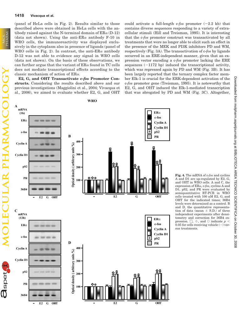
(panel of HeLa cells in Fig. 2). Results similar to thosedescribed above were obtained in HeLa cells with the an-tibody raised against the N-terminal domain of ER� (D-12)(data not shown). Using the anti-ER� antibody F-10 inWRO cells, the immunoreactivity was displayed exclu-sively in the cytoplasm also in presence of ligands (panel ofWRO cells in Fig. 2). In contrast, the anti-ER� antibodyD-12 was not able to evidence any signal in WRO cells(data not shown). On the basis of these observations, wecan further argue that the variant of ER� found in TC cellsdoes not mediate transcriptional effects according to theclassic mechanism of action of ER�.
E2, G, and OHT Transactivate c-fos Promoter Con-structs. Considering the results described above and ourprevious investigations (Maggiolini et al., 2004; Vivacqua etal., 2006), we aimed to evaluate whether E2, G, and OHT
could activate a full-length c-fos promoter (�2.2 kb) thatcontains diverse sequences responding to a variety of extra-cellular stimuli (Hill and Treisman, 1995). It is interestingthat the c-fos promoter construct was transactivated by alltreatments that were no longer able to elicit such an effect inthe presence of the MEK and PI3K inhibitors PD and WM,respectively (Fig. 3A). The transactivation of c-fos by ligandsoccurred in an ERE-independent manner, given that an ex-pression vector encoding a c-fos promoter lacking the EREsequences (�1172 bp) induced the transcriptional activity,which was repressed again by PD and WM (Fig. 3B). It hasbeen largely reported that the ternary complex factor mem-ber Elk-1 is crucial for the ERK-dependent activation of thec-fos promoter gene (Treisman, 1995). It is noteworthy thatE2, G, and OHT induced the Elk-1-mediated transcriptionthat was abrogated by PD and WM (Fig. 3C). Altogether,
Fig. 4. The mRNA of c-fos and cyclinsA and D1 are up-regulated by E2, G,and OHT in WRO cells. A and C, theexpression of ER�, c-fos, cyclins A andD1, pS2, and PR were evaluated bysemiquantitative RT-PCR in WROcells treated with 100 nM E2, G, andOHT for the indicated times; 36B4levels were determined as a control. Band D, the quantitative representa-tion of data (mean � S.D.) of threeindependent experiments after densi-tometry and correction for 36B4 ex-pression. �, �, and E indicate p �0.05 for cells receiving vehicle (�) ver-sus treatments.
1418 Vivacqua et al.
at BIB
LIOT
EC
A A
RE
A T
EC
NIC
O S
CIE
NT
IFIC
A on O
ctober 30, 2008 m
olpharm.aspetjournals.org
Dow
nloaded from
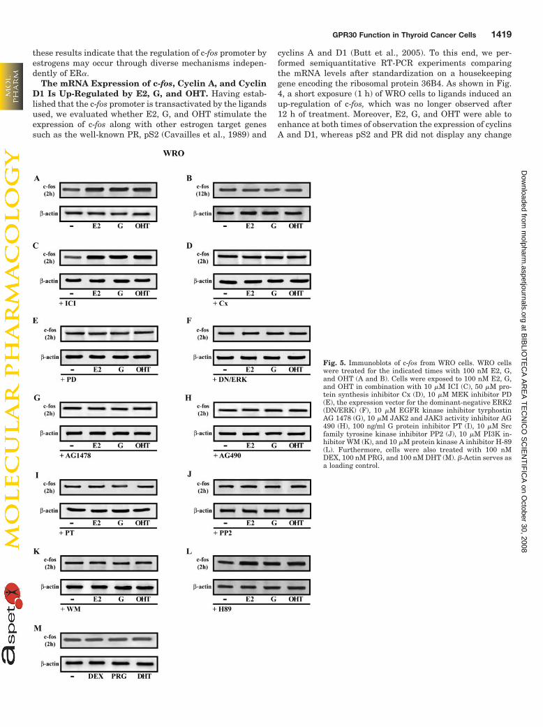
these results indicate that the regulation of c-fos promoter byestrogens may occur through diverse mechanisms indepen-dently of ER�.
The mRNA Expression of c-fos, Cyclin A, and CyclinD1 Is Up-Regulated by E2, G, and OHT. Having estab-lished that the c-fos promoter is transactivated by the ligandsused, we evaluated whether E2, G, and OHT stimulate theexpression of c-fos along with other estrogen target genessuch as the well-known PR, pS2 (Cavailles et al., 1989) and
cyclins A and D1 (Butt et al., 2005). To this end, we per-formed semiquantitative RT-PCR experiments comparingthe mRNA levels after standardization on a housekeepinggene encoding the ribosomal protein 36B4. As shown in Fig.4, a short exposure (1 h) of WRO cells to ligands induced anup-regulation of c-fos, which was no longer observed after12 h of treatment. Moreover, E2, G, and OHT were able toenhance at both times of observation the expression of cyclinsA and D1, whereas pS2 and PR did not display any change
Fig. 5. Immunoblots of c-fos from WRO cells. WRO cellswere treated for the indicated times with 100 nM E2, G,and OHT (A and B). Cells were exposed to 100 nM E2, G,and OHT in combination with 10 �M ICI (C), 50 �M pro-tein synthesis inhibitor Cx (D), 10 �M MEK inhibitor PD(E), the expression vector for the dominant-negative ERK2(DN/ERK) (F), 10 �M EGFR kinase inhibitor tyrphostinAG 1478 (G), 10 �M JAK2 and JAK3 activity inhibitor AG490 (H), 100 ng/ml G protein inhibitor PT (I), 10 �M Srcfamily tyrosine kinase inhibitor PP2 (J), 10 �M PI3K in-hibitor WM (K), and 10 �M protein kinase A inhibitor H-89(L). Furthermore, cells were also treated with 100 nMDEX, 100 nM PRG, and 100 nM DHT (M). �-Actin serves asa loading control.
GPR30 Function in Thyroid Cancer Cells 1419
at BIB
LIOT
EC
A A
RE
A T
EC
NIC
O S
CIE
NT
IFIC
A on O
ctober 30, 2008 m
olpharm.aspetjournals.org
Dow
nloaded from
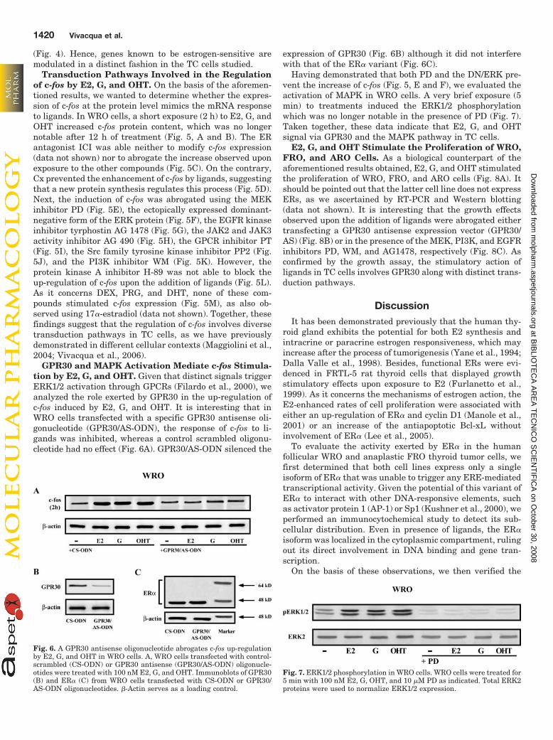
(Fig. 4). Hence, genes known to be estrogen-sensitive aremodulated in a distinct fashion in the TC cells studied.
Transduction Pathways Involved in the Regulationof c-fos by E2, G, and OHT. On the basis of the aforemen-tioned results, we wanted to determine whether the expres-sion of c-fos at the protein level mimics the mRNA responseto ligands. In WRO cells, a short exposure (2 h) to E2, G, andOHT increased c-fos protein content, which was no longernotable after 12 h of treatment (Fig. 5, A and B). The ERantagonist ICI was able neither to modify c-fos expression(data not shown) nor to abrogate the increase observed uponexposure to the other compounds (Fig. 5C). On the contrary,Cx prevented the enhancement of c-fos by ligands, suggestingthat a new protein synthesis regulates this process (Fig. 5D).Next, the induction of c-fos was abrogated using the MEKinhibitor PD (Fig. 5E), the ectopically expressed dominant-negative form of the ERK protein (Fig. 5F), the EGFR kinaseinhibitor tyrphostin AG 1478 (Fig. 5G), the JAK2 and JAK3activity inhibitor AG 490 (Fig. 5H), the GPCR inhibitor PT(Fig. 5I), the Src family tyrosine kinase inhibitor PP2 (Fig.5J), and the PI3K inhibitor WM (Fig. 5K). However, theprotein kinase A inhibitor H-89 was not able to block theup-regulation of c-fos upon the addition of ligands (Fig. 5L).As it concerns DEX, PRG, and DHT, none of these com-pounds stimulated c-fos expression (Fig. 5M), as also ob-served using 17�-estradiol (data not shown). Together, thesefindings suggest that the regulation of c-fos involves diversetransduction pathways in TC cells, as we have previouslydemonstrated in different cellular contexts (Maggiolini et al.,2004; Vivacqua et al., 2006).
GPR30 and MAPK Activation Mediate c-fos Stimula-tion by E2, G, and OHT. Given that distinct signals triggerERK1/2 activation through GPCRs (Filardo et al., 2000), weanalyzed the role exerted by GPR30 in the up-regulation ofc-fos induced by E2, G, and OHT. It is interesting that inWRO cells transfected with a specific GPR30 antisense oli-gonucleotide (GPR30/AS-ODN), the response of c-fos to li-gands was inhibited, whereas a control scrambled oligonu-cleotide had no effect (Fig. 6A). GPR30/AS-ODN silenced the
expression of GPR30 (Fig. 6B) although it did not interferewith that of the ER� variant (Fig. 6C).
Having demonstrated that both PD and the DN/ERK pre-vent the increase of c-fos (Fig. 5, E and F), we evaluated theactivation of MAPK in WRO cells. A very brief exposure (5min) to treatments induced the ERK1/2 phosphorylationwhich was no longer notable in the presence of PD (Fig. 7).Taken together, these data indicate that E2, G, and OHTsignal via GPR30 and the MAPK pathway in TC cells.
E2, G, and OHT Stimulate the Proliferation of WRO,FRO, and ARO Cells. As a biological counterpart of theaforementioned results obtained, E2, G, and OHT stimulatedthe proliferation of WRO, FRO, and ARO cells (Fig. 8A). Itshould be pointed out that the latter cell line does not expressERs, as we ascertained by RT-PCR and Western blotting(data not shown). It is interesting that the growth effectsobserved upon the addition of ligands were abrogated eithertransfecting a GPR30 antisense expression vector (GPR30/AS) (Fig. 8B) or in the presence of the MEK, PI3K, and EGFRinhibitors PD, WM, and AG1478, respectively (Fig. 8C). Asconfirmed by the growth assay, the stimulatory action ofligands in TC cells involves GPR30 along with distinct trans-duction pathways.
DiscussionIt has been demonstrated previously that the human thy-
roid gland exhibits the potential for both E2 synthesis andintracrine or paracrine estrogen responsiveness, which mayincrease after the process of tumorigenesis (Yane et al., 1994;Dalla Valle et al., 1998). Besides, functional ERs were evi-denced in FRTL-5 rat thyroid cells that displayed growthstimulatory effects upon exposure to E2 (Furlanetto et al.,1999). As it concerns the mechanisms of estrogen action, theE2-enhanced rates of cell proliferation were associated witheither an up-regulation of ER� and cyclin D1 (Manole et al.,2001) or an increase of the antiapoptotic Bcl-xL withoutinvolvement of ER� (Lee et al., 2005).
To evaluate the activity exerted by ER� in the humanfollicular WRO and anaplastic FRO thyroid tumor cells, wefirst determined that both cell lines express only a singleisoform of ER� that was unable to trigger any ERE-mediatedtranscriptional activity. Given the potential of this variant ofER� to interact with other DNA-responsive elements, suchas activator protein 1 (AP-1) or Sp1 (Kushner et al., 2000), weperformed an immunocytochemical study to detect its sub-cellular distribution. Even in presence of ligands, the ER�isoform was localized in the cytoplasmic compartment, rulingout its direct involvement in DNA binding and gene tran-scription.
On the basis of these observations, we then verified the
Fig. 6. A GPR30 antisense oligonucleotide abrogates c-fos up-regulationby E2, G, and OHT in WRO cells. A, WRO cells transfected with control-scrambled (CS-ODN) or GPR30 antisense (GPR30/AS-ODN) oligonucle-otides were treated with 100 nM E2, G, and OHT. Immunoblots of GPR30(B) and ER� (C) from WRO cells transfected with CS-ODN or GPR30/AS-ODN oligonucleotides. �-Actin serves as a loading control.
Fig. 7. ERK1/2 phosphorylation in WRO cells. WRO cells were treated for5 min with 100 nM E2, G, OHT, and 10 �M PD as indicated. Total ERK2proteins were used to normalize ERK1/2 expression.
1420 Vivacqua et al.
at BIB
LIOT
EC
A A
RE
A T
EC
NIC
O S
CIE
NT
IFIC
A on O
ctober 30, 2008 m
olpharm.aspetjournals.org
Dow
nloaded from
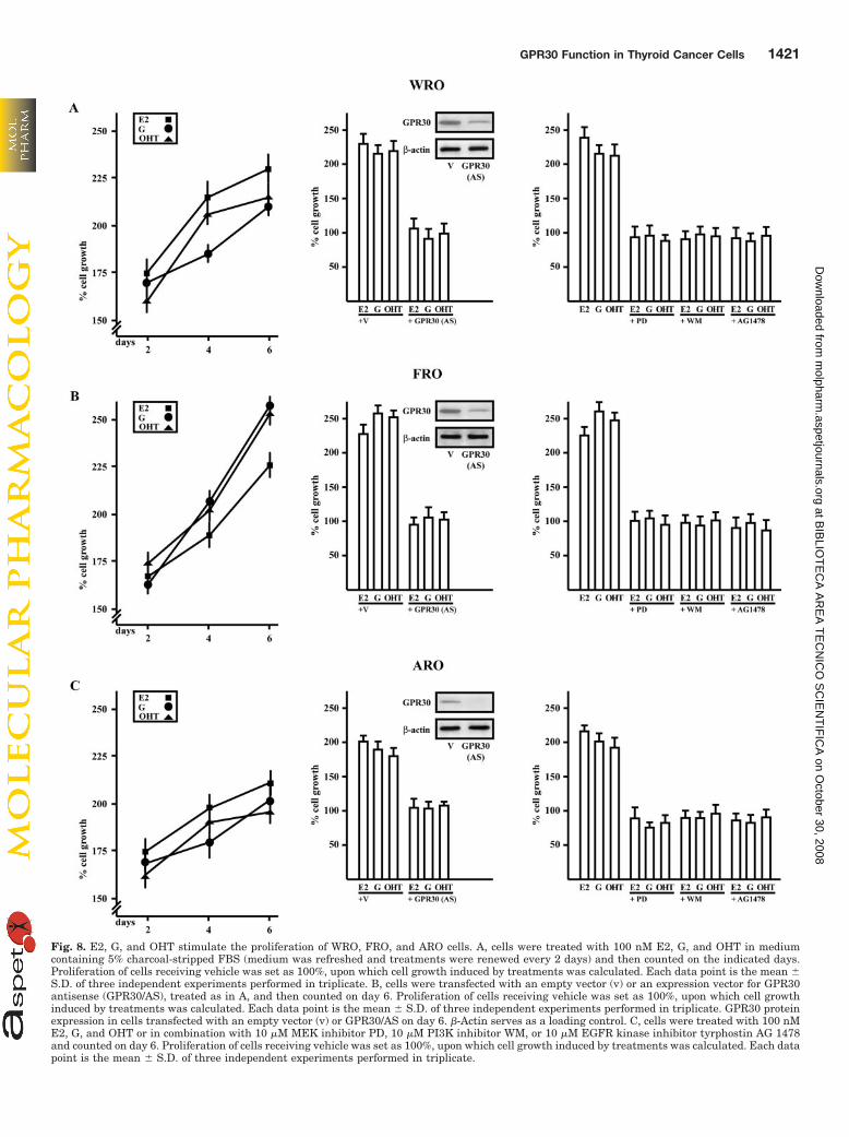
Fig. 8. E2, G, and OHT stimulate the proliferation of WRO, FRO, and ARO cells. A, cells were treated with 100 nM E2, G, and OHT in mediumcontaining 5% charcoal-stripped FBS (medium was refreshed and treatments were renewed every 2 days) and then counted on the indicated days.Proliferation of cells receiving vehicle was set as 100%, upon which cell growth induced by treatments was calculated. Each data point is the mean �S.D. of three independent experiments performed in triplicate. B, cells were transfected with an empty vector (v) or an expression vector for GPR30antisense (GPR30/AS), treated as in A, and then counted on day 6. Proliferation of cells receiving vehicle was set as 100%, upon which cell growthinduced by treatments was calculated. Each data point is the mean � S.D. of three independent experiments performed in triplicate. GPR30 proteinexpression in cells transfected with an empty vector (v) or GPR30/AS on day 6. �-Actin serves as a loading control. C, cells were treated with 100 nME2, G, and OHT or in combination with 10 �M MEK inhibitor PD, 10 �M PI3K inhibitor WM, or 10 �M EGFR kinase inhibitor tyrphostin AG 1478and counted on day 6. Proliferation of cells receiving vehicle was set as 100%, upon which cell growth induced by treatments was calculated. Each datapoint is the mean � S.D. of three independent experiments performed in triplicate.
GPR30 Function in Thyroid Cancer Cells 1421
at BIB
LIOT
EC
A A
RE
A T
EC
NIC
O S
CIE
NT
IFIC
A on O
ctober 30, 2008 m
olpharm.aspetjournals.org
Dow
nloaded from
ability of E2, G, and OHT to regulate other genes known to beestrogen-sensitive. It is interesting that all of the compoundsinduced the expression of c-fos and cyclins A and D1, whereaspS2 and PR did not show alterations. This result would notbe surprising because the complex transcription machineryrequires a cell-specific recruitment of a plethora of cofactorsand the displacement of corepressors on the promoter sites ofdistinct genes, as documented previously (Cosma, 2002). Therapid c-fos response to ligands along with their ability totransactivate an expression vector encoding the promoter ofc-fos lacking ERE sequences prompted us to identify thetransduction pathways involved in these stimulatory effectselicited by E2, G, and OHT in TC cells. We have shown for thefirst time that the expression of genes involved in cycle pro-gression and the proliferation of thyroid tumor cells are me-diated by GPR30 and the MAPK pathway, which was con-firmed by the growth assay performed in ER-negative AROcells. Furthermore, we have demonstrated that a complexinterplay among distinct intracellular pathways contributesto these biological features, because specific inhibitors ofEGFR, Src, and PI3K signaling were each able to abrogatec-fos induction and the proliferative effects obtained upon theaddition of ligands. It is worth noting that our findings are inagreement with previous data suggesting the existence of afunctional network among diverse metabolic cascades, whichmay allow an identical molecular cross-talk in different cel-lular contexts (Castoria et al., 2001; Kraus et al., 2003).Besides, the present results recall our recent data regardingthe involvement of the aforementioned transduction path-ways in gene transcription and proliferation of breast(SKBR3) and endometrial (HEC1A) cancer cells completelydevoid of ERs or expressing an ER� variant, respectively(Maggiolini et al., 2004; Vivacqua et al., 2006).
Fos family proteins, including c-fos, interact with membersof the Jun family to form heterodimers that bind to the AP-1sites located within the regulatory region of genes critical forcell cycle progression (Hill and Treisman, 1995). In this con-text, it has been suggested that c-fos through AP-1 acts as atranscriptional regulator of the cyclin D1 gene, linking mito-genic signaling to cycle re-entry in many cell types (Albaneseet al., 1999). Moreover, in a recent study (Sunters et al.,2004), c-fos overexpression resulted in cyclin D1 increase andcyclin A-dependent accelerated progression of the cell cycle.In line with these findings, we have associated the responseof c-fos to E2, G, and OHT with the transcription regulationof cyclins A and D1 in TC cells. As it concerns the mecha-nisms by which diverse extracellular stimulations initiatethe rapid transcription of mitogenic genes, the MAPK cas-cade was recognized to exert a crucial role triggering therapid up-regulation of c-fos (Whitmarsh et al., 1995) and aseries of phosphorylation events that further increase itstranscriptional potential and the biological outcome (Murphyet al., 2002).
The above evidence has provided a nice scenario for ourdata regarding the effects triggered by ligands in thyroidtumor cells: 1) the rapid MAPK activation; 2) the induction ofc-fos and cyclins A and D1; and 3) the growth response to E2,G, and OHT. Moreover, using specific inhibitors, we havedocumented that GPR30 and diverse transduction pathwaysmediate these ligand-induced stimulations. Our findingshave also demonstrated that ER� is not required for thecomplex biological activity exerted by treatments.
The well-established mode of action of estrogens, such asbinding to ERs which in turn activate gene transcriptionthrough an interaction with ERE and other DNA sequences(Levin, 2005), should be extended to GPR30, which mediatesestrogenic signals in breast, endometrial, and thyroid tumorcells, as we have ascertained in the present and previousstudies (Maggiolini et al., 2004; Vivacqua et al., 2006). Ourdata suggest novel biological targets that could be taken intoaccount for pharmacological interventions with molecularinhibitors in TC. Given the stimulatory effects elicited by E2in neoplastic thyroid cells as evidenced by our and otherreports (Yane et al., 1994; Dalla Valle et al., 1998; Manole etal., 2001; Lee et al., 2005), medications containing estrogensor compounds able to mimic the estrogen activity should becarefully considered at least in women who had TC or aresuspected of occult thyroid malignancy.
ReferencesAlbanese C, D’Amico M, Reutens AT, Fu M, Watanabe G, Lee RJ, Kitsis RN,
Henglein B, Avantaggiati M, Somasundaram B, et al. (1999) Activation of thecyclin D1 gene by the E1A-associated protein p300 through AP-1 inhibits cellularapoptosis. J Biol Chem 270:34186–34195.
Butt AJ, McNeil CM, Musgrove EA, and Sutherland RL (2005) Downstream targetsof growth factor and oestrogen signalling and endocrine resistance: the potentialroles of c-Myc, cyclin D1 and cyclin E. Endocr Relat Cancer 12 (Suppl 1):S47–S59.
Castoria G, Migliaccio A, Bilancio A, Di Domenico M, De Falco A, Lombardi M,Fiorentino R, Varricchio L, Barone MV, and Auricchio F (2001) PI3-kinase inconcert with Src promotes the S-phase entry of oestradiol-stimulated MCF-7 cells.EMBO (Eur Mol Biol Organ) J 20:6050–6059.
Cavailles V, Garcia M, and Rochefort H (1989) Regulation of cathepsin-D and pS2gene expression by growth factors in MCF7 human breast cancer cells. MolEndocrinol 3:552–558.
Cosma MP (2002) Ordered recruitment: gene-specific mechanism of transcriptionactivation. Mol Cell 10:227–236.
Dalla Valle L, Ramina A, Vianello S, Fassina A, Belvedere P, and Colombo L (1998)Potential for estrogen synthesis and action in human normal and neoplasticthyroid tissues. J Clin Endocrinol Metab 83:3702–3709.
Duan R, Porter W, and Safe S (1998) Estrogen-induced c-fos protooncogene expres-sion in MCF-7 human breast cancer cells: role of estrogen receptor Sp1 complexformation. Endocrinology 139:1981–1990.
Duan R, Xie W, Burghardt RC, and Safe S (2001) Estrogen receptor-mediatedactivation of the serum response element in MCF-7 cells through MAPK-dependent phosphorylation of Elk-1. J Biol Chem 276:11590–11598.
Filardo EJ, Quinn JA, Bland KI, and Frackelton AR Jr (2000) Estrogen-inducedactivation of Erk-1 and Erk-2 requires the G protein-coupled receptor homolog,GPR30, and occurs via trans-activation of the epidermal growth factor receptorthrough release of HB-EGF. Mol Endocrinol 10:1649–1660.
Furlanetto TW, Nguyen LQ, and Jameson JL (1999) Estradiol increases proliferationand down-regulates the sodium/iodide symporter gene in FRTL-5 cells. Endocri-nology 140:5705–5711.
Gallo A, Cuozzo C, Esposito I, Maggiolini M, Bonofiglio D, Vivacqua A, GarriamoneM, Weiss C, Bohmann D, and Musti AM (2002) Menin uncouples Elk-1, JunD andc-Jun phosphorylation from MAP kinase activation. Oncogene 21:6434–6445.
Henderson BE, Ross RK, Pike MC, and Casagrande JT (1982) Endogenous hormonesas a major factor in human cancer. Cancer Res 42:3232–3239.
Hill CS and Treisman R (1995) Differential activation of c-fos promoter elements byserum, lysophosphatidic acid, G proteins and polypeptide growth factors. EMBO(Eur Mol Biol Organ) J 14:5037–5047.
Jemal A, Tiwari RC, Murray T, Ghafoor A, Samuels A, Ward E, Feuer EJ, and ThunMJ (2004) American Cancer Society Cancer Statistics. CA Cancer J Clin 54:8–29.
Kanda N and Watanabe S (2003) 17beta-estradiol inhibits oxidative stress-inducedapoptosis in keratinocytes by promoting Bcl-2 expression. J Investig Dermatol121:771–780.
Kraus S, Benard O, Naor Z, and Seger R (2003) c-Src is activated by the epidermalgrowth factor receptor in a pathway that mediates JNK and ERK activation bygonadotropin-releasing hormone in COS7 cells. J Biol Chem 278:32618–32630.
Kumar V and Chambon P (1988) The estrogen receptor binds tightly to its responsiveelement as a ligand-induced homodimer. Cell 55:145–156.
Kushner PJ, Agard D, Feng WJ, Lopez G, Schiau A, Uht R, Webb P, and Greene G(2000) Oestrogen receptor function at classical and alternative response elements.Novartis Found Symp 230:20–26.
Lee ML, Chen GG, Vlantis AC, Tse GM, Leung BC, and van Hasselt CA (2005)Induction of thyroid papillary carcinoma cell proliferation by estrogen is associatedwith an altered expression of Bcl-xL. Cancer J 11:113–121.
Levin ER (2005) Integration of the extranuclear and nuclear actions of estrogen. MolEndocrinol 19:1951–1959.
Maggiolini M, Donze O, and Picard D (1999) A non-radioactive method for inexpen-sive quantitative RT-PCR. Biol Chem 380:695–697.
Maggiolini M, Vivacqua A, Fasanella G, Recchia AG, Sisci D, Pezzi V, Montanaro D,Musti AM, Picard D, and Ando S (2004) The G protein-coupled receptor GPR30mediates c-fos up-regulation by 17�-estradiol and phytoestrogens in breast cancercells. J Biol Chem 279:27008–27016.
1422 Vivacqua et al.
at BIB
LIOT
EC
A A
RE
A T
EC
NIC
O S
CIE
NT
IFIC
A on O
ctober 30, 2008 m
olpharm.aspetjournals.org
Dow
nloaded from
Manole D, Schildknecht B, Gosnell B, Adams E, and Derwahl M (2001) Estrogenpromotes growth of human thyroid tumor cells by different molecular mechanisms.J Clin Endocrinol Metab 86:1072–1077.
Melillo RM, Castellone MD, Guarino V, De Falco V, Cirafici AM, Salvatore G,Caiazzo F, Basolo F, Giannini R, Kruhoffer M, et al. (2005) The RET/PTC-RAS-BRAF linear signaling cascade mediates the motile and mitogenic phenotype ofthyroid cancer cells. J Clin Investig 115:1068–1081.
Murphy LO, Smith S, Chen RH, Fingar DC, and Blenis J (2002) Molecular inter-pretation of ERK signal duration by immediate early gene products. Nat Cell Biol4:556–564.
Persson I, Yuen J, Bergkvist L, and Schairer C (1996) Cancer incidence and mor-tality in women receiving estrogen and estrogen-progestin replacement therapylong term follow-up of a Swedish cohort. Int J Cancer 67:327–332.
Pike MC, Pearce CL, and Wu AH (2004) Prevention of cancers of the breast,endometrium and ovary. Oncogene 23:6379–6391.
Revankar CM, Cimino DF, Sklar LA, Arterburn JB, and Prossnitz ER (2005) Atransmembrane intracellular estrogen receptor mediates rapid cell signaling. Sci-ence (Wash DC) 307:1625–1630.
Ron E, Kleinerman RA, Boice JD Jr, Livolsi VA, Flannery JT, and Fraumeni JF Jr(1987) A population-based case-control study of thyroid cancer. J Natl Cancer Inst79:1–12.
Santagati S, Gianazza E, Agrati P, Vegeto E, Patrone C, Pollio G, and Maggi A (1997)Oligonucleotide squelching reveals the mechanism of estrogen receptor autologousdown-regulation. Mol Endocrinol 11:938–949.
Sunters A, Thomas DP, Yeudall WA, and Grigoriadis AE (2004) Accelerated cell cycleprogression in osteoblasts overexpressing the c-fos proto-oncogene: induction ofcyclin A and enhanced CDK2 activity. J Biol Chem 279:9882–9891.
Thomas P, Pang Y, Filardo EJ, and Dong J (2005) Identity of an estrogen membranereceptor coupled to a G protein in human breast cancer cells. Endocrinology146:624–632.
Tora L, White J, Brou C, Tasset D, Webster N, Scheer E, and Chambon P (1989) Thehuman estrogen receptor has two independent nonacidic transcriptional activationfunctions. Cell 59:477–487.
Treisman R (1995) Journey to the surface of the cell: Fos regulation and the SRE.EMBO (Eur Mol Biol Organ) J 14:4905–4913.
Vivacqua A, Bonofiglio D, Recchia AG, Musti AM, Picard D, Ando S, and MaggioliniM (2006) The G protein-coupled receptor GPR30 mediates the proliferative effectsinduced by 17beta-estradiol and hydroxytamoxifen in endometrial cancer cells.Mol Endocrinol 20:631–646.
Waterhouse J, Muir C, Shanmugaratnam K, and Powel J (1982) Cancer Incidence inFive Continents, pp 185–198, IARC, Lyons, France.
Whitmarsh AJ, Shore P, Sharrocks AD, and Davis RJ (1995) Integration of MAPkinase signal transduction pathways at the serum response element. Science(Wash DC) 269:403–407.
Yane K, Kitahori Y, Konishi N, Okaichi K, Ohnishi T, Miyahara H, Matsunaga T,Lin JC, and Hiasa Y (1994) Expression of the estrogen receptor in human thyroidneoplasms. Cancer Lett 84:59–66.
Address correspondence to: Dr. Marcello Maggiolini, Department of Phar-maco-Biology, University of Calabria, 87030 Rende (CS), Italy. E-mail:[email protected]
GPR30 Function in Thyroid Cancer Cells 1423
at BIB
LIOT
EC
A A
RE
A T
EC
NIC
O S
CIE
NT
IFIC
A on O
ctober 30, 2008 m
olpharm.aspetjournals.org
Dow
nloaded from