Untitled - Learning & Decision-making Lab
174
-
Upload
khangminh22 -
Category
Documents
-
view
3 -
download
0
Transcript of Untitled - Learning & Decision-making Lab


Fronto-striatal mechanisms of attentional control
Martine van Schouwenburg

The studies presented in this thesis were carried out at the Donders Institute for Brain, Cognition and Behaviour, Centre for Cognitive Neuroimaging, Radboud University Nijmegen Medical Centre, Nijmegen, the Netherlands, with financial support from the Netherlands Organization for Scientific Research (NWO VIDI-grant 452-08-009) to prof. dr. Roshan Cools.
Printed by Ipskamp Drukkers, Enschede
ISBN 978-94-91027-39-0
©2012 Martine van Schouwenburg

Fronto-striatal mechanisms of attentional control
Proefschrift
ter verkrijging van de graad van doctor aan de Radboud Universiteit Nijmegenop gezag van de rector magnificus prof. mr. S.C.J.J. Kortmann,
volgens besluit van het college van decanenin het openbaar te verdedigen op donderdag 8 november 2012
om 13.00 uur precies
door
Martine Rinske van Schouwenburggeboren op 21 april 1982te Meyrin (Zwitserland)

PromotorenProf. dr. R. CoolsProf. dr. J.K. Buitelaar
CopromotorDr. H.E.M. den Ouden
ManuscriptcommissieProf. dr. M. UllspergerProf. dr. C.J. FiebachProf. dr. K.R. Ridderinkhof (Universiteit van Amsterdam)


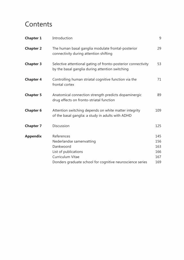
Contents
Chapter 1 Introduction 9
Chapter 2 The human basal ganglia modulate frontal-posterior 29 connectivity during attention shifting
Chapter 3 Selective attentional gating of fronto-posterior connectivity 53 by the basal ganglia during attention switching
Chapter 4 Controlling human striatal cognitive function via the 71 frontal cortex
Chapter 5 Anatomical connection strength predicts dopaminergic 89 drug effects on fronto-striatal function
Chapter 6 Attention switching depends on white matter integrity 109 of the basal ganglia: a study in adults with ADHD
Chapter 7 Discussion 125
Appendix References 145 Nederlandse samenvatting 156 Dankwoord 163 List of publications 166 Curriculum Vitae 167 Donders graduate school for cognitive neuroscience series 169


1 Introduction

10
Chapter 1

11
Introduction
Introduction
We are constantly surrounded by an overwhelming amount of information. This information needs to be filtered such that we can focus on relevant information and ignore distracting, irrelevant information. For example, imagine you are trying to read this thesis while you are in a busy café. You are most likely to succeed if you are able to ignore things like the music from the radio, people walking past your table, and the TV screen on the wall showing the local news. Luckily for you the human brain turns out to be fairly good at this. It can enhance processing of relevant information and suppress processing of irrelevant information. However, from your everyday experience it might seem quite difficult to concentrate on one thing at a time and not get distracted by other things. This is because our brain cannot simply ignore all information that is irrelevant to our current goal. It needs to scan all incoming information for potential dangers or otherwise important information. Imagine you are in the café again, and now, while you are reading this thesis, the fire alarm goes off. Even though this is not relevant to your current goal (reading this thesis) it is quite important that you do not ignore this distracter. Actually, you have to update your goal based on this novel information; you have to stop reading and get out of the café as quickly as possible.
It seems contradictory, that on the one hand your brain needs to filter out information that is not relevant to the current goal, yet on the other hand it needs to process this information to prevent us from missing information that turns out to be relevant after all. Adaptive behaviour requires an optimal balance between these two processes of ‘cognitive stability’ and ‘cognitive flexibility’. This ability is commonly referred to as cognitive control. Achieving optimal cognitive control is not straightforward, as evidenced by neuropsychiatric disorders in which this balance goes awry. For example, Parkinson’s disease patients can get ‘overfocused’ and get stuck in their behaviour. On the other hand, patients with attention deficit hyperactivity disorder (ADHD) or schizophrenia are constantly distracted by information that is not relevant to the current goal.
The aim of the research presented in this thesis is to increase our understanding of these complex cognitive control processes. Previous studies highlighted the importance of two regions, the prefrontal cortex and the basal ganglia (Cools and Robbins, 2004). I aim to elucidate how these regions interact to establish cognitive control. In addition, I investigate the role of the neurotransmitter dopamine in these processes. Finally, I assess whether cognitive control processes are altered in adults diagnosed with ADHD. A short description of the different methods used in this thesis is provided in Box 1. A more extensive description of the methodology can be found in the respective chapters.
1

12
Chapter 1
Box 1 Methods
Functional magnetic resonance imagingFunctional magnetic resonance imaging (fMRI) is a neuroimaging technique that allows the mapping of human brain function. It is a non-invasive technique with whole-brain coverage and a relatively high spatial resolution. fMRI makes use of the different magnetic properties associated with oxygen-rich and oxygen-poor blood. Brain regions that are active will consume more oxygen than brain regions that are not active. To replenish the consumed oxygen, more oxygen-rich blood will flow to those regions that are active. This is what we measure with fMRI: blood oxygen level-dependent signal, or BOLD signal. The BOLD response is a good proxy for neural activity (Huettel et al., 2009). However, one disadvantage is that the BOLD response is delayed and sluggish. As a consequence fMRI has a limited temporal resolution.
Generally, we study differences in BOLD signal between different conditions. For example in my research, I have contrasted BOLD signal during trials on which subjects switched their attention with BOLD signal during trials on which subjects did not switch their attention. This allowed me to find out which brain regions are more active during one condition compared to another condition.
Connectivity analyses of fMRI dataAnother way to analyse fMRI data is to investigate the interaction between brain regions. Functional connectivity can be assessed with psychophysiological interaction (PPI) analysis. It works under the assumption that the degree to which the BOLD signal in one area can be predicted, based on BOLD signal in another, corresponds to the contribution of the second region to the first region. The PPI then tests whether this contribution changes over experimental conditions (Friston et al., 1997). In chapter 5 I used PPI analyses to test effects of a dopaminergic manipulation on functional connectivity between the basal ganglia and the prefrontal cortex.
The interaction between brain regions can also be investigated by assessing effective connectivity, which tests the direct (causal) influences of one neural element onto another. In chapter 2 and chapter 3 of this thesis I used dynamic causal modelling (DCM) to compare different models of interaction between the prefrontal cortex and the basal ganglia. DCM is a model of effective connectivity that allows assessment of interactions between brain regions based on the measured BOLD responses. More specifically, it allows one to test mechanistic hypotheses about interactions between neuronal populations, and their modulation by experimental conditions, and, more recently, even by other neuronal populations (Stephan et al., 2008, 2010).

13
Introduction
1
Transcranial magnetic stimulationfMRI analysis can show us which brain regions are active when a particular task is performed. However, this does not mean that this region is necessary to perform that task. One method which does allow us to make such inferences about brain function is transcranial magnetic stimulation (TMS) (Robertson et al., 2003). TMS uses electromagnetic induction to evoke small electrical currents at the surface of the cortex. Using this technique it is possible to temporarily inhibit or activate a certain brain region. If this brain region is indeed crucial for a particular function than inhibition of that brain region will disturb that function. For example inhibiting Broca’s area, a brain region involved in speech, perturbs the subject’s ability to speak. In the research described in chapter 4 I used TMS in combination with fMRI to assess the effect of cortical stimulation on basal ganglia signal (O’Shea et al., 2007a) to test whether the frontal cortex might affect cognitive flexibility by modulating basal ganglia function.
Diffusion tensor imagingBrain regions are connected with each other via white matter tracts and the strength of these structural connections can be assessed in vivo with diffusion tensor imaging (DTI). DTI makes use of the Brownian motion of water molecules; they can diffuse freely in some areas, such as the cerebral spinal fluid, but diffusion is restricted in tissue. In particular, in white matter, water molecules are more likely to move along fibre tracts than perpendicular to the direction of axons, providing directional information of neuronal tracts (Johansen-Berg and Rushworth, 2009). The fractional anisotropy (FA) is a quantitative measure for such directional dependency. Local FA values rely on several microstructural properties of white matter tissue, such as the level of axon myelination, intact axonal membranes and fibre density (Beaulieu, 2002). This suggests that higher FA values reflect more efficient neuronal communication. Diffusion-weighted images can also be used for tractography to reconstruct white matter tracts. In chapter 5 and chapter 6 I made used of DTI to measure individual differences in anatomical white matter tracts connecting the prefrontal cortex and the basal ganglia.
Pharmacological neuroimaging Pharmacological neuroimaging allows us to assess the involvement of certain neurotransmitters in neurocognitive functions. Neuroimaging data can be acquired after intake of psychopharmacological agents. Task-related activity on drug is then compared with task-related activity after placebo intake (Honey and Bullmore, 2004). In addition, one can assess the effect of psychopharmacological agents on functional connectivity between brain regions. In the study described in chapter 5 subjects were scanned once on the dopamine receptor agonist bromocriptine and once on placebo while performing an attention switching paradigm to assess the role of dopamine in cognitive flexibility.

14
Chapter 1
The prefrontal cortex and cognitive stability
The cognitive control processes necessary for adaptive behaviour have been associated most commonly with the anterior part of the brain, the prefrontal cortex. Specifically, the prefrontal cortex has been reliably implicated in the active on-line maintenance of goal-relevant representations, an ability that is commonly referred to as working memory (Baddeley, 1986). However, the prefrontal cortex has also been implicated in selective attention and filtering of distracters (Everling et al., 2002). How do these concepts of attention and working memory relate to each other and what is their importance for cognitive control? Although intuitively ‘attention’ and ‘working memory’ might seem entirely different concepts, they are closely related, and they perhaps even overlap (Awh and Jonides, 2001). For example, the on-line maintenance of goal-relevant representations is necessary to guide attention towards these representations (de Fockert et al., 2001). Conversely, to keep an item in working memory, attention needs to be directed towards (the internal representation of) this item. Furthermore, like attention, working memory is vulnerable to distraction. Imagine that you have to remember a phone number over a short period of time. One strategy is to rehearse the number in your mind. This requires you to direct your attention towards the internal representation of the phone number, while trying to avoid distracting thoughts and shut yourself off from external inputs. However, if someone were to ask you a question, you might get distracted and forget the number. Thus, cognitive stability relies heavily on efficient working memory.
The importance of the prefrontal cortex for working memory was first demonstrated by Jacobsen (1937). He showed that monkeys with frontal lobe lesions were impaired on the delayed response task, a task that is often used to test working memory capacity. In this task, subjects are presented with stimuli that they have to remember over a short delay period. After this delay, a response has to be made according to the remembered stimuli, for example, indicate whether a target stimulus is one of the remembered stimuli. Subsequent research showed that the working memory deficit introduced by frontal lobe lesion was reversed when monkeys were tested in the dark, suggesting that it reflected increased vulnerability to visual distraction (Malmo, 1942). Consistent with a role for the prefrontal cortex in distracter-resistance were findings from studies in patients with prefrontal cortex lesions, revealing increased distractibility by irrelevant sensory input (Chao and Knight, 1995).
What might be the mechanism by which the prefrontal cortex contributes to cognitive stability? Different aspects of our environment are processed by different brain regions. For example in the visual domain, processing of colour, motion, faces and houses are associated with separate regions in the posterior part of the brain, where the visual cortex resides. Those regions that process goal-relevant aspects of the environment exhibit higher levels of activity compared to other regions. Critically,

15
Introduction
1
attention to current goal-relevant representations is thought to result from the influence of excitatory top-down signals in the prefrontal cortex, which biases the competition among brain regions in posterior cortex, by increasing the activity of brain regions processing goal-relevant representations (Miller and Cohen, 2001). This hypothesis is supported by functional magnetic resonance imaging (fMRI; Box 1) studies in humans, such as that by Gazzaley et al. (2007). In this study, subjects were presented with a series of four sequentially presented stimuli: two faces and two scenes. They were asked to remember either the faces or the scenes. BOLD responses in the parahippocampal place area (PPA), known to process scenes (Epstein and Kanwisher, 1998), were increased when subjects attended to the scenes. In addition, consistent with the hypothesis that the prefrontal cortex modulates processing in the posterior cortex, connectivity between the prefrontal cortex and the PPA was significantly enhanced during the encoding of scenes (Gazzaley et al., 2007). Conversely, when subjects were attending to faces an increase in activity was observed in the fusiform face area (FFA), a region involved in the processing of faces (Kanwisher et al., 1997). This was accompanied by an increase in prefrontal-FFA connectivity. Interestingly, such functional fronto-posterior connectivity could be diminished by presenting a distracter (Yoon et al., 2006). Moreover, perturbation of prefrontal cortex function with transcranial magnetic stimulation (TMS; Box 1) resulted in diminished modulation of activity in posterior visual cortex during a similar working memory task requiring selective attention (Zanto et al., 2011).
Thus, the prefrontal cortex may support focusing of attention by increasing the activity of brain regions that process goal-relevant representations, thereby rendering these representations resistant to disruption by distracting, goal-irrelevant information.
The basal ganglia and cognitive flexibility
Recent evidence suggests that the subcortical basal ganglia also support cognitive control processes. In particular, the basal ganglia seem to be important for the updating of cognitive programs. The first evidence for a role of the basal ganglia in the switching of attention came from animal studies. These studies showed that selective lesions in the basal ganglia caused a deficit in tasks that require the updating of current goal representations (Oberg and Divac, 1975; Taghzouti et al., 1985). The basal ganglia also support cognitive flexibility in humans. For example, BOLD signals in the basal ganglia increase during task-switching, attentional set-shifting and reversal learning, which are all processes that require the flexible updating of current goal representations (Rogers et al., 2000; Cools et al., 2002a, 2004; Leber et al., 2008).
Evidence that the basal ganglia are not just activated, but in fact necessary for cognitive switching in humans comes from studies with Parkinson’s disease patients.

16
Chapter 1
This neurodegenerative disease is characterized by basal ganglia dysfunction. In addition to the prominent motor deficits, patients show deficits in cognitive flexibility (Cools et al., 1984; Downes et al., 1989; Owen et al., 1992, Cools et al., 2001a). For example in a study by Cools et al. (2001b), Parkinson’s disease patients performed a paradigm in which they had to switch between two task instructions. Depending on the colour of the stimulus-window they had to name the letter or the digit displayed on the screen. The goal-relevant stimulus (for example a letter) could be paired with a goal-irrelevant stimulus (a digit) or with a neutral stimulus. Patients were impaired on trials on which they had to switch between tasks. This was especially true if on those trials the relevant stimulus was paired with an irrelevant stimulus, and thus required the suppression of a previously relevant goal. These findings suggest that the basal ganglia are important for selection mechanisms involved in cognitive flexibility, i.e. disengaging from the current goal and engaging in a new goal (Cools et al., 2001b). However, the exact mechanism by which the basal ganglia control attention switching is still unclear.
Several models of basal ganglia function have been proposed. Interestingly, most of these models are based on the anatomical configuration of this region, which I will therefore discuss first. The basal ganglia are a group of interconnected nuclei that are situated deep in the human brain. The input nuclei of the basal ganglia consist of the caudate nucleus and putamen, collectively referred to as the striatum (Figure 1). They receive excitatory input from almost the entire cerebral cortex. The primary output nuclei of the basal ganglia, the internal segment of the globus pallidus (GPi) and the substantia nigra pars reticulata (SNr), send continuous inhibitory output to the cortex, via the thalamus. Information that arrives at the striatum can be passed on, via inhibitory connections, either directly to the output nuclei, or via the external segment of the globus pallidus (GPe) and/or subthalamic nucleus (STN). Depending on the route, activation of the striatum will lead to disinhibition of the cortex (via the direct or ‘Go’ pathway), or further inhibition of the cortex (via the GPe, the so-called indirect or ‘NoGo’ pathway) (Albin et al., 1989; DeLong, 1990).
The strong inhibitory output of the basal ganglia has provided the basis for classical models of basal ganglia function, emphasizing the role of the basal ganglia in inhibition of goal-irrelevant representations (Hikosaka and Wurtz, 1989). Other models suggest that selection is one of the key functions of the basal ganglia (Redgrave et al., 1999a; Gurney et al., 2001). For example, the basal ganglia might facilitate the selection of goal-relevant representations by lowering a decision threshold (Lo and Wang, 2006; Forstmann et al., 2008a). A third group of models suggest that the basal ganglia uses both of these mechanisms to selectively gate the desired representation (Mink, 1996; Hazy et al., 2007). This last group of models has been inspired by the anatomy of fronto-striato-thalamic circuits, which are organized in a topographic and functionally selective manner, such that different parts of the cortex project to

17
Introduction
1
Figure 1. Simplified representation of the functional organization of the basal ganglia. The striatum (putamen and caudate nucleus) receives input from the cortex and via the thalamus projects back to the cortex. Two alternative internal routes in the basal ganglia have either an excitatory effect (via the direct pathway) or an inhibitory effect (via the indirect pathway) on the cortex. GPe = globus pallidus pars externa, GPi = globus pallidus pars interna, STN = subthalamic nucleus, SNr = substantia nigra pars reticulata.
Figure 2. The frontal cortex and basal ganglia are connected via a number of functionally and anatomically rather segregated loops. Examples of such loops are the motor loop (red), the cognitive loop (green) and the limbic loop (blue). Figure reproduced with permission from Lancet Neurology, (Rodriguez-Oroz et al., 2009). GPe = globus pallidus pars externa, GPi = globus pallidus pars interna, STN = subthalamic nucleus.

18
Chapter 1
different parts of the striatum (Alexander et al., 1986) (Figure 2). For example, the motor cortex and putamen are connected within the ‘motor loop’, while the ‘cognitive loop’ runs through the dorsolateral prefrontal cortex and anterior parts of the caudate. In addition, within these functionally distinct loops, a further distinction can be made. For instance, the hand area of the motor cortex is connected to specific parts of the putamen, while the foot area of the motor cortex is connected to other parts of the putamen. This large number of topographically organized loops allows for selective disinhibition and inhibition of the appropriate pathways. With respect to this selective gating model of the basal ganglia, most research has been done on the motor loop. In 1996, Mink proposed that when a movement is initiated in the motor cortex, the basal ganglia act to release inhibition of the desired motor pathway, while further inhibiting the competing motor pathways (Mink, 1996). More specifically, the area of the motor cortex representing the desired movement will be disinhibited via the direct pathway, while other areas of the motor cortex will be further inhibited via the indirect pathway.
Interaction between the prefrontal cortex and basal ganglia
So far, I have discussed relatively separate lines of research implicating the prefrontal cortex and the basal ganglia in cognitive control. Thus as described above, the prefrontal cortex is involved in focusing attention on task-relevant information through the online maintenance of task-relevant information. In addition, the basal ganglia seem to be particularly important when task-relevant information needs to be updated. However, given the strong anatomical connections between the prefrontal cortex and basal ganglia it is unlikely that they contribute independently to cognitive processes (Middleton and Strick, 2000). Indeed, similar to basal ganglia damage, prefrontal cortex damage can impair cognitive flexibility (Milner, 1963; Owen et al., 1993; Rogers, 1998; Frank et al., 2001; Aron et al., 2004). Furthermore, in addition to the prefrontal cortex, the basal ganglia have been associated with working memory (Levy et al., 1997; McNab and Klingberg, 2008). Based on these findings it has been suggested that the prefrontal cortex and basal ganglia might interact to establish the delicate balance between cognitive stability and cognitive flexibility (Frank et al., 2001; Hazy et al., 2007; Cools and D’Esposito, 2011). However, the exact mechanism by which these two regions interact remains to be elucidated. It has been proposed that the role of the basal ganglia in the selective gating of motor action programs extends to the selective gating of attention and cognitive programs (Divac et al., 1967; Cools et al., 1984; Frank et al., 2001; Frank, 2005). For example, it has been suggested that the basal ganglia selectively allow sensory information to enter the prefrontal cortex. They might ‘open the gate’ to support the updating of goal-relevant representations or ‘close the gate’ to prevent distracting information from interfering with the maintained

19
Introduction
1
representations (Frank et al., 2001). Empirical evidence for this input gating was found, showing that the basal ganglia gate sensory information to the premotor cortex (den Ouden et al., 2010). Similarly, the basal ganglia might select which, among the present prefrontal cortex goal representations, guides current behaviour (Frank and Badre, 2012). In other words, although multiple goals can be kept in working memory, only one goal can be pursued at each moment in time. The basal ganglia might ensure that only representations relevant to the current goal can influence attention and action selection. Thus, according to this output gating hypothesis, the basal ganglia might guide attention by enhancing processing of goal-relevant representations, while suppressing processing of goal-irrelevant representations (Frank, 2011).
In chapter 2, chapter 3, chapter 4 and chapter 6 of this thesis I aimed to further explore the interaction between the prefrontal cortex and the basal ganglia.
Dopaminergic modulation of cognitive control
The prefrontal cortex and the basal ganglia are strongly innervated by dopaminergic midbrain neurons. Indeed, the neurotransmitter dopamine plays a key role in cognitive control processes. Dopamine neurons reside in the midbrain, in the ventral tegmental area (VTA) and substantia nigra pars compacta (SNc). They project to virtually the whole brain. This widespread innervation is in line with the role of dopamine as a neuromodulator. Unlike classic neurotransmission, which facilitates chemical wiring from one presynaptic neuron to one postsynaptic neuron, neuromodulation facilitates effects on a group of neurons (Stahl, 2008). By definition, neuromodulators do not induce spiking of the targeted neurons, but rather potentiate or attenuate responses evoked by classical neurotransmitters (i.e. glutamate and GABA) (Seamans and Yang, 2004).
The first studies linking dopamine to cognitive functions focused on the effects of dopamine in the prefrontal cortex. Brozoski et al (1979) provided the first empirical support for a role of dopamine in working memory by showing that dopamine depletion in the prefrontal cortex of monkeys impaired delayed response task performance almost to the same degree as did complete ablation of the prefrontal cortex. Delayed response task performance was also impaired by injection of a dopamine receptor antagonist (which blocks dopamine receptors) into the monkey prefrontal cortex (Sawaguchi et al., 1990a), while administration of dopamine and dopamine receptor agonists (which simulate the effect of endogenous dopamine on its receptors) to prefrontal cortex neurons enhanced delayed response task performance (Sawaguchi et al., 1990b; Sawaguchi and Goldman-Rakic, 1991). Results from human studies are consistent with a role for dopamine in working memory, showing, for example, that administration of dopamine receptor agonists and antagonists, respectively, improved

20
Chapter 1
and impaired performance on a working memory task (Luciana et al., 1992; Mehta et al., 1999). The importance of the prefrontal cortex in this dopaminergic modulation of working memory is supported by several fMRI studies that show a modulation by dopaminergic drugs of BOLD signal during working memory tasks in the prefrontal cortex (Mattay et al., 1996, 2000; Mehta et al., 2000; Cools et al., 2002b, 2007b; Willson et al., 2004; Gibbs and D’Esposito, 2005a, 2005b, 2006).
Only recently have studies started to investigate the effects of dopamine on basal ganglia signals associated with cognitive control. They suggest that dopamine receptor stimulation in the basal ganglia modulates cognitive flexibility rather than cognitive stability. For example, task-switching is impaired by acute administration of the dopamine receptor antagonist sulpiride, which blocks primarily D2 receptors that are most abundant in the basal ganglia (Mehta et al., 1999; van Holstein et al., 2011). More direct evidence for the importance of dopamine in the basal ganglia comes from pharmacological neuroimaging work. For example, Cools et al. (2007a) have shown that the effects of dopaminergic medication withdrawal during switch trials of a probabilistic reversal learning paradigm in Parkinson’s disease patients were restricted to BOLD signal in the basal ganglia, and did not extend to BOLD signal in the prefrontal cortex. Similar selective effects were observed in young healthy volunteers after administration of methylphenidate, which blocks the dopamine transporter thereby increasing dopamine levels (Dodds et al., 2008). Like dopaminergic medication (the dopamine precursor levodopa and dopamine receptor agonists) in Parkinson’s disease patients, methylphenidate reduced BOLD signal in the basal ganglia, but not in the prefrontal cortex during switch trials of the probabilistic reversal learning paradigm. Finally, recent genetic imaging data, showed that the modulation of task-switching costs by incentive motivation depended on genetic variation in the dopamine transporter (Aarts et al., 2010). In this study the authors investigated a common polymorphism in the dopamine transporter gene (DAT1, SLC6A3), which is thought to affect dopamine transmission primarily in the basal ganglia. Results revealed that carriers of the 9-repeat allele, associated with high dopamine levels in the basal ganglia, exhibited greater decreases of switch-costs when they anticipated being rewarded for correct performance, compared to 10-allele homozygotes. Critically, this modulatory effect on task-switching was accompanied by significant modulation of BOLD signal in the basal ganglia.
These different lines of research suggest that the effects of dopamine might depend on its site of action. To directly test this hypothesis Cools et al. (2007b) designed a study that enabled the researchers to assess drug effects on signals associated with cognitive stability and cognitive flexibility in one paradigm. They showed that bromocriptine modulated basal ganglia signals during cognitive switching, but prefrontal cortex signals during distracter-resistance in a delay period. These data accord well with the idea that the same dopaminergic drug might modulate distinct cognitive functions,

21
Introduction
1
i.e. task-switching and distracter-resistance, by acting on dissociable brain regions, i.e. the basal ganglia and the prefrontal cortex respectively (Cools and D’Esposito, 2011).
Taken together, dopamine acting in the prefrontal cortex and basal ganglia might have opposite effects on cognitive control. In addition, it has been demonstrated that dopamine levels in the prefrontal cortex and the basal ganglia are neurochemically reciprocal, such that dopamine increases in the prefrontal cortex lead to dopamine decreases in the basal ganglia and vice versa (Pycock et al., 1980; Akil et al., 2003). Based on these findings a model was proposed suggesting a reciprocal relationship between cognitive stability and cognitive flexibility (Cools and D’Esposito, 2011). According to this model, manipulations that increase cognitive focusing might impair cognitive flexibility (Bilder et al., 2004; Durstewitz and Seamans, 2008; Cools and D’Esposito, 2011). Observations in marmosets with striatal dopamine lesions (Crofts et al., 2001) and patients with Parkinson’s disease (Cools et al., 2010) are in support of such a reciprocal relationship. Thus, non-human primates with striatal dopamine lesions and patients with Parkinson’s disease are impaired on set-shifting tasks that require cognitive flexibility (Cools et al., 1984; Owen et al., 1992; Cools, 2006), while actually showing enhanced cognitive stability, i.e. increased distracter resistance (Crofts et al., 2001; Cools et al., 2010). In chapter 6 of this thesis I tested this model of reciprocity in patients with ADHD.
Dopaminergic effects on fronto-striatal interaction
Apart from direct effects of dopamine on the basal ganglia and prefrontal cortex, dopamine might also modulate the interaction between these regions (Honey et al., 2003; Krugel et al., 2009; Stelzel et al., 2010; Wallace et al., 2011). As described above, dopamine acts as a neuromodulator to potentiate or attenuate responses evoked by classical neurotransmitters (i.e. glutamate and GABA) (Stahl, 2008). In the striatum, dopaminergic receptors are located in the proximity of synapses from fronto-striatal glutamatergic neurons. As such, dopamine can influence the excitability of striatal neurons in response to input from the prefrontal cortex (Moss and Bolam, 2010), thereby modulating information flow through fronto-striatal-thalamic circuits. For instance, it was shown that injection of a dopaminergic agent into the basal ganglia of the rat modulated striatal responses elicited by stimulation of the frontal cortex (Goto and Grace, 2005). The role of dopamine in modulating fronto-striatal interaction was confirmed in human studies. One of the first studies to show such effects used deep brain electrodes in combination with EEG to test coherence between oscillatory activity measured in the basal ganglia (with electrodes) and the cortex (with EEG) (Williams, 2002). The authors tested patients with Parkinson’s disease on and off their dopaminergic medication to show that fronto-striatal interaction was modulated

22
Chapter 1
by dopaminergic medication. Importantly, these results were replicated in healthy individuals using pharmacological neuroimaging. For example, temporarily lowering dopamine levels in healthy volunteers impaired fronto-striatal interaction during an attention switching task (Nagano-Saito et al., 2008). Furthermore, genetic variance in dopamine D2 receptor expression could predict individual variance in switch-related fronto-striatal connectivity (Stelzel et al., 2010).
In sum, dopamine acts on the basal ganglia and prefrontal cortex to regulate cognitive functions associated with these regions. In addition, dopamine can modulate functional fronto-striatal interaction during tasks that require cognitive control.
Individual differences in the effects of dopamine
As outlined above, there is clear evidence for the involvement of dopamine in cognitive control processes. However, one major challenge in dopaminergic drug development is that drug effects are highly variable across individuals (Cools and Robbins, 2004; Cools and D’Esposito, 2011). Dopaminergic drugs might improve cognitive function in one patient, yet impair cognitive function in another. Evidently, the precise relationship between dopamine and cognitive control is complex and non-linear. Indeed, an ‘inverted U’-shaped relationship exists between dopamine receptor stimulation and cognitive function, with too little as well as excessive dopamine levels impairing performance (Williams and Goldman-Rakic, 1995; Zahrt et al., 1997; Arnsten, 1998). Evidence for such an ‘inverted U’-shaped relationship between dopamine and cognitive functions comes, for example, from studies in monkeys showing that low doses of a dopamine receptor agonist improve working memory performance, while higher doses have a detrimental effect (Cai and Arnsten, 1997). Dose-dependent effects of dopaminergic drugs have also been found on prefrontal cortex activity in humans (Tipper et al., 2005).
Dopaminergic drug effects might depend on baseline dopamine levels, such that individuals with suboptimal baseline dopamine levels benefit from additional dopamine receptor stimulation, while individuals with already optimal baseline dopamine might be detrimentally overdosed by the same increase in dopamine receptor stimulation. Indeed, it has been shown that individual differences in dopaminergic drug effects can be explained by a range of measures that reflect individual differences in baseline dopamine levels. For example, several studies showed that dopaminergic drugs had opposite effects on cognitive performance as a function of working memory capacity (Kimberg et al., 1997; Mattay et al., 2000; Mehta et al., 2000; Gibbs and D’Esposito, 2005a), which was shown to correlate with baseline dopamine synthesis capacity (Cools et al., 2008; Landau et al., 2009). Furthermore, evidence for baseline-dependency comes from studies which have made use of common genetic polymorphisms in dopamine

23
Introduction
1
genes to predict dopaminergic drug effects (Mattay et al., 2003; Cohen et al., 2007). For example, amphetamine was shown to improve performance on a working memory task and to reduce prefrontal BOLD responses specifically in carriers of the Val allele of the COMT Val108/158Met genetic polymorphism, which is associated with low baseline dopamine levels in the prefrontal cortex. By contrast, the same drug impaired performance and increased BOLD signal in subjects who were homozygous for the Met allele, which is associated with high baseline dopamine levels in the prefrontal cortex (Mattay et al., 2003).
Similar to drug effects on BOLD signals in the prefrontal cortex, drug effects on BOLD signals in the basal ganglia are highly variable between subjects. For example, in a recent study by Cools et al (2007b), bromocriptine improved cognitive switching and potentiated BOLD signals in the basal ganglia, but only in subjects who scored highly on a self-report measure of trait impulsivity. The enhancing effects of bromocriptine on cognitive switching and associated basal ganglia signals were restricted to high-impulsive subjects, while low-impulsive subjects exhibited, if anything, the opposite effect. This observation concurs with findings from another recent study showing that the effects of methylphenidate on probabilistic reversal learning were predicted by trait impulsivity, such that high-impulsive subjects benefited most from the drug (Clatworthy et al., 2009). Greater cognitive benefits of dopamine-enhancing drugs in high-impulsive subjects are consistent with methylphenidate’s beneficial effects on cognition in ADHD and might reflect suboptimal baseline levels of dopamine transmission in high-impulsive subjects (Dalley et al., 2007; Oswald et al., 2007; Buckholtz et al., 2010). More direct evidence for a relationship between baseline levels of dopamine in the basal ganglia and bromocriptine’s effects on flexible updating comes from a recent study, in which neurochemical PET imaging was combined with psychopharmacology (Cools et al., 2009). In this study, subjects underwent a PET scan with the radiotracer 6-[18F]fluoro-L-m-tyrosine (FMT), a substrate for dopamine synthesis, with uptake of the tracer reflecting the degree to which dopamine is synthesized in the basal ganglia. Results revealed that the effects of bromocriptine on reversal learning could be predicted from baseline levels of dopamine synthesis capacity in the basal ganglia. Bromocriptine improved reversal learning in subjects with low baseline synthesis capacity, but impaired it in subjects with high baseline synthesis capacity. This large variability in drug effects is a challenge for psychiatric treatment and drug research. In chapter 5 of this thesis, I explored a novel way of predicting individual differences in dopaminergic drug effects.

24
Chapter 1
Outline of this thesis
In the research presented in this thesis I investigated the neural mechanisms of cognitive control. One way to study cognitive control is by using attention switching paradigms. Most traditional switching paradigms explicitly cue subjects when to switch their attention. However, in real life situations attention switching usually happens when we are distracted by something in a currently unattended stream of information. I developed a new paradigm in which a switch in attention is triggered in exactly such a bottom-up fashion. In this paradigm, subjects focus their attention on one dimension of a series of two-dimensional stimuli, but switch attention when stimuli of the other dimension change.
The research in chapter 2, chapter 3, chapter 4 and chapter 6 focused primarily on the question how the prefrontal cortex and basal ganglia interact to implement a switch in attention. Because of the bottom-up nature of the attention switches in the novel paradigm, subjects sometimes failed to detect changes in the unattended dimension, and therefore failed to switch attention. This allowed me to assess differential responses to environmental changes that cause a switch in attention versus those that remain unnoticed in chapter 2. In addition, it allowed me to test the output gating model of the basal ganglia as described above. Hence, the basal ganglia might implement a switch in attention by gating prefrontal signals. Specifically, I assessed whether prefrontal top-down signals to posterior visual cortex are modulated by the basal ganglia during a switch in attention, using a combination of fMRI and dynamic causal modelling (DCM; Box 1).
In chapter 3 I focused exclusively on those trials on which an environmental change caused a switch in attention. Here I aimed to assess the mechanism underlying selective gating by the basal ganglia. Again using both fMRI and DCM I assessed whether the basal ganglia ensure selective gating by enhancement of the newly attended information, suppression of the previously attended information, or a combination of these processes. This allowed me to dissociate between three proposed models of basal ganglia function as described above.
In chapter 4 I tested whether the basal ganglia are under frontal control. If this is indeed the case, then frontal stimulation should affect striatal function. I used TMS to stimulate the frontal cortex and scanned subjects before and after this stimulation while they performed a switching paradigm. I predicted that frontal stimulation would modulate striatal BOLD signal, and that this effect would be functionally selective. Put differently, I predicted that frontal stimulation would modulate striatal BOLD signal only during conditions that depend on the basal ganglia.
In chapter 6 I investigated the interaction between the prefrontal cortex and basal ganglia by looking at structural connectivity, rather than functional connectivity, between these regions. Indeed, the prefrontal cortex and basal ganglia are strongly

25
Introduction
1
connected via anatomical white tracts (Alexander et al., 1986). I reasoned that if cognitive flexibility indeed relies on fronto-striatal interaction, then sound fronto-striatal infrastructure is a prerequisite for optimal cognitive flexibility. Hence, individual differences in the ability to flexibly adjust behaviour might depend on individual differences in the strength of white matter fibres connecting the prefrontal cortex and basal ganglia. As an index of local white matter integrity, I calculated fractional anisotropy values from diffusion tensor weighted images (DTI; Box 1). Next, fractional anisotropy values were correlated with individual measures of behavioural performance on our attention switching paradigm.
Together, these chapters will increase our knowledge on how the prefrontal cortex and the basal ganglia interact to control behaviour in our constantly changing environment. These findings might lead future research in neuropsychiatry on disorders associated with fronto-striatal dysfunction and cognitive control deficits.
The research presented in chapter 4, chapter 5 and chapter 6 has more direct implications for neuropsychiatry. In chapter 5 I focused on the link between dopamine and cognitive flexibility. Currently, drug research and psychiatric treatment are challenged by the large variability in dopaminergic drug effects. The same drug might enhance cognitive function in one person, but impair it in another person. Hence, a better of knowledge of the complex relationship between dopamine and cognition is crucial to improve dopaminergic drug treatment. In chapter 5 I examined whether individual differences in the effect of dopaminergic drugs can be predicted as a function of white matter connectivity between the prefrontal cortex and the basal ganglia. As described above, one way by which dopamine might affect cognitive functions is by acting on fronto-striatal glutamatergic synapses, thereby modulating information flow through fronto-striato-thalamic loops. I reasoned that dopaminergic function is thus constrained by the existing anatomical fronto-striatal infrastructure. From each subject we acquired a diffusion weighted scan to measure white matter connectivity. Subsequently, subjects were scanned on and off the dopamine agonist bromocriptine. I hypothesized that individual differences in drug effects on switch-related basal ganglia BOLD signal as well as drug effects on fronto-striatal functional connectivity could be predicted by individual differences in white matter connectivity.
Another challenge in neuropsychiatry is the fact that dopaminergic drugs can elicit adverse effects. This might be a consequence of the wide spread effects of dopamine. Thus systemic administration of dopamine might affect a number of cognitive functions, by acting on several brain regions. For example in Parkinson’s disease, dopaminergic drugs are indicated to alleviate motor rigidity by acting on the dopamine-depleted dorsal striatum. However, these same drugs might detrimentally overdose ventral parts of the striatum leading to impulsive behaviour and aberrant reward processing (Swainson et al., 2000; Cools et al., 2001a, 2003; Cools, 2006). In chapter 4 of this thesis, I exploited a TMS protocol that was previously shown to modulate dopamine levels

26
Chapter 1
selectively in subregions of the basal ganglia. If we find, as predicted, that TMS over the frontal cortex modulates striatal signals in a functionally selective manner, then this protocol might be a potential tool to modulate one cognitive function, without affecting other cognitive functions.
In chapter 6 I aimed to assess performance on our novel attention switching task in patients with ADHD. This disorder is associated with deficits in attention focusing and high distractibility (DSM IV, 1994; Carter et al., 1995; Jonkman et al., 1999; Hervey et al., 2004). Given the reciprocal nature between cognitive stability and cognitive flexibility described earlier, I hypothesized that the impairment in focusing of attention in ADHD might be accompanied by a paradoxical cognitive benefit in a context that requires attention switching. Moreover, I aimed to assess whether such an attention switching benefit was accompanied by changes in white matter connectivity. Indeed, previous studies have shown altered functional and structural connectivity in ADHD (Konrad and Eickhoff, 2010). If attention switching performance can be predicted from white matter this might be informative for neuropsychiatry. Neuropsychiatric treatment is currently based on DSM diagnosis without taking into account individual differences between patients. For example ADHD is associated with both attentional deficits and hyperactivity, but the severity of symptoms in these separate domains varies greatly between patients. Thus, individual neurocognitive assessment of attentional deficits might improve neuropsychiatric treatment.
Finally, in chapter 7, I will present a summary and interpretation of the findings described in this thesis.

27
Introduction
1


Martine van SchouwenburgHanneke den Ouden
Roshan Cools
The human basal ganglia modulate frontal-posterior connectivity during attention shifting2

30
Chapter 2
Abstract
Current models of flexible cognitive control emphasize the role of the prefrontal cortex. This region has been shown to control attention by biasing information processing in favour of task-relevant representations. However, the prefrontal cortex does not act in isolation. We used functional magnetic resonance imaging combined with nonlinear dynamic causal modelling to demonstrate that the basal ganglia play a role in modulating the top-down influence of the prefrontal cortex on visual processing in humans. Specifically, our results reveal that connectivity between the prefrontal cortex and stimulus-specific visual association areas depends on activity in the basal ganglia, elicited by salient events leading to shifts in attention. These data integrate disparate literatures on top-down control by the prefrontal cortex and selective gating by the basal ganglia and highlight the importance of the basal ganglia for high-level cognitive control.
Based on: van Schouwenburg, M.R., den Ouden, H.E.M., Cools, R. (2010). The human basal ganglia modulate frontal-posterior connectivity during attention shifting. Journal of Neuroscience 30(29): 9910-9918

31
Role of the basal ganglia in attention shifting
2
Introduction
The limited processing capacity of our brain requires us to select relevant information for further processing and filter out irrelevant information from our complex environment. According to the biased competition model, this selection is biased by salience (bottom-up processing) as well as behavioural relevance (top-down processing) (Bundesen, 1990; Desimone and Duncan, 1995). Active maintenance of goal-relevant representations in the prefrontal cortex allows top-down biasing of attention by modulation of visual processing in posterior cortical regions (Miller and Cohen, 2001; Wallis et al., 2001; Gazzaley et al., 2007). To facilitate flexibility of attention in response to changes in the environment, these goal-relevant representations need to be updated constantly (Rougier et al., 2005).
The prefrontal cortex does not act in isolation but rather interacts with other regions, such as the basal ganglia to bias attentional flexibility. However, their respective contributions are unclear. The basal ganglia have long been implicated in the control of movement, and the anatomy of the basal ganglia is perfectly suited to selectively gate a desired motor plan to the motor cortex while simultaneously inhibiting competing motor plans (Mink, 1996). Computational modelling work has suggested that the role of the basal ganglia in selective gating is not limited to motor processes but extends to cognitive functions. For instance, it has been proposed that goal-relevant representations in prefrontal cortex are updated only when the basal ganglia ‘open the gate’ for cortical processing (Braver and Cohen, 2000; Frank et al., 2001). This hypothesis is in line with empirical evidence from functional imaging and patient studies revealing a role for the basal ganglia in attention switching (Cools et al., 2004; Leber et al., 2008). For example, patients with focal lesions in the basal ganglia (Cools et al., 2006) as well as patients with Parkinson’s disease, characterized by basal ganglia dysfunction, exhibit attention switching deficits (Cools et al., 2001a, 2001b, 2003).
In this study, we aimed to elucidate the mechanism by which the basal ganglia control attention switching by integrating the hitherto segregated literatures on the role of the prefrontal cortex in top-down biasing of attention and the role of the basal ganglia in selective gating, using fMRI. In contrast to traditional attention switching paradigms (e.g. reversal learning, task switching, and set shifting), we used an attention switching paradigm in which subjects did not switch their attention based on an explicit, top-down cue. Rather, the need to shift attention was signalled by a bottom-up cue consisting of a change in stimuli. We hypothesize that attention switching under such salience-driven conditions is mediated by modulatory influences of the basal ganglia on interactions between the prefrontal cortex and stimulus-specific visual regions in the posterior cortex. To test this hypothesis, we used dynamic causal modelling (DCM),

32
Chapter 2
a generic Bayesian framework for inferring effective connectivity from neuroimaging data (Friston et al., 2003). Specifically, we used a nonlinear extension to DCM (Stephan et al., 2008; den Ouden et al., 2010) that allowed us to investigate modulatory influences of activity in the basal ganglia on the effective connectivity between prefrontal cortex and posterior visual regions.
Materials and methods
Subjects Twenty healthy right-handed volunteers participated in this study, which was approved by the local ethics committee. Exclusion criteria were claustrophobia, neurological or cardiovascular diseases, psychiatric disorders, regular use of medication or marihuana, use of psychotropic drugs, heavy smoking, or metal parts in the body. All subjects gave written informed consent and were compensated for participation. Two subjects were excluded from additional analysis because of abnormal performance on the task (see below). Accordingly, data are reported from 18 subjects (seven male; age 22.4 ± 0.6 years [mean ± SEM]).
Paradigm A novel attention switching paradigm was used in which subjects switched attention when they detected a change in the stimulus exemplars of an unattended dimension of two-dimensional stimuli. Subjects were presented with a series of stimulus pairs, i.e. two images presented side by side, each consisting of an overlapping face exemplar and scene exemplar (Figure 1A). At the beginning of each block, subjects were instructed to select one of the two dimensions (faces or scenes), focus on this dimension, and ignore the other dimension. Within the chosen dimension, subjects then selected one of the two exemplars by making a left (left index finger) or right (right index finger) response, depending on the location of the exemplar of their choice. This self-chosen exemplar was then set as the correct stimulus. Subjects were instructed to continue selecting the correct stimulus on subsequent trials. We used a design similar to that used by Hampshire and Owen (2006), in which stimulus pairs were presented twice within each trial. The combination of face and scene was reversed on the second presentation (F2S1 and F1S2) relative to the first (F1S1 and F2S2). This enabled us to identify the attended stimulus (Figure 1). At the end of each trial, feedback was presented. Feedback was positive (a green ‘smiley’ face) only if the subject selected the correct stimulus twice within the trial. If subjects selected the pattern that did not contain the correct exemplar or did not respond within a personalized cut-off time, then negative feedback (a red ‘sad’ face) was presented. Thus, a trial consisted of two

33
Role of the basal ganglia in attention shifting
2
successive responses followed by feedback, and subjects were explicitly instructed to always respond to the same exemplar within each trial. After a variable number of correct trials (that is, 2-5 positive feedback events, or 4-10 correct responses) stimuli of the ignored dimension were replaced with novel exemplars. Subjects were instructed to shift their attention to this other dimension and to choose one of the two novel exemplars, whenever they detected a change. On trials on which novel exemplars were introduced (novel trials), subjects either detected the change and switched to one of the novel exemplars (novel switch trials [Figure 1C]) or they failed to detect the novel exemplars and kept responding to the previously correct exemplar (novel non-switch trials [Figure 1D]). If they failed to detect the change, negative feedback was presented, usually leading subjects to switch on the subsequent trial. Trials on which no novel stimuli were introduced are defined as repeat trials (Figure 1B).
In the main experiment, subjects were presented with, on average 355 ± 15 trials (mean ± SEM), of which 86 were novel trials. The trials were distributed across four blocks, separated by 23 s breaks. The sequence of the presented faces and scenes was randomized across subjects. For details on the exact timing of the paradigm, we refer to the supplementary materials. The paradigm was programmed using Presentation software (Neurobehavioural Systems).
Figure 1. The attention switching paradigm used in this study required subjects to select one stimulus exemplar (left versus right) within one dimension (faces versus scenes) on every trial. A) Each trial consisted of two consecutive responses followed by feedback. Red boxes indicate a possible response sequence. B-D show two consecutive trials with responses defining the three different trial types. For clarification, the stimuli are displayed schematically (F1, face 1; S1, scene 1; F2, face 2; S2, scene 2). B) In this example, the subject is attending to F1 on the first trial (attended stimuli are displayed in italic). On the next trial, no novel stimuli are introduced and the subject keeps attending to F1. The second trial is thus defined as a repeat trial. C) On a novel switch trial, novel stimuli of the unattended dimension, in this case scenes, are introduced (S3 and S4). The subject detects this change and switches attention to one of two novel stimuli (here S3). D) Alternatively the subject can fail to detect the novel stimuli and keep responding to the previously relevant stimulus exemplar, in this case F1. The subject will then receive negative feedback and the second trial is defined as a novel non-switch trial.

34
Chapter 2
LocalizerAfter completion of the main experiment, subjects performed an one-back task using alternating blocks of face stimuli and scene stimuli to localize the stimulus-specific visual association cortices (i.e. fusiform face area [FFA] [Kanwisher et al., 1997] and parahippocampal place area [PPA] [Epstein and Kanwisher, 1998]), in every subject individually. Subjects were presented with 16 s blocks of 20 face stimuli, 20 scene stimuli (each presented for 300 ms, intertrial interval of 500 ms), and rest periods (seven blocks of each type) and were instructed to press buttons with their left and right index finger whenever they noticed an immediate (one-back) repeat of a stimulus. Acquisition and preprocessing of fMRI data was performed as for the main experiment, and the statistical analysis was conducted using the normalized and smoothed images. In the general linear model (GLM), we included three regressors of interest (scene blocks, face blocks, and rest blocks), and the six realignment parameters as regressors of no interest. The blocks were modelled at the onset of the first stimulus presentation, with a duration of 16 s and convolved with a canonical hemodynamic response. Our contrasts of interest were (1) faces versus scenes and (2) scenes versus faces.
Behavioural analysisThe switch likelihood was calculated as the percentage of switches on novel trials. The primary reaction time (RT) data analyses focused on three trial types of interest: novel switch trials, novel non-switch trials, and repeat trials. Excluded from these primary RT analyses were the first trial of each block, all trials on which subjects received negative feedback (except for the novel non-switch trials, which by definition resulted in negative feedback), and the trials following negative feedback. Median rather than mean RTs were reported to minimize the influence of outliers.
Planned contrasts were assessed using paired sample t-tests. The statistical threshold was set at p < 0.05 (two-tailed). All results are reported as mean ± SEM unless stated otherwise.
fMRI data acquisitionWhole-brain imaging was performed on a 3T MR scanner (Magnetom Trio Tim; Siemens Medical Systems). Functional data were obtained using a gradient-echo echo-planar scanning sequence with blood oxygenation level-dependent (BOLD) contrast (30 axial-oblique slices; repetition time, 1990 ms; echo time, 30 ms; voxel size, 3.5x3.5x3.0 mm; interslice gap, 0.5 mm; field of view, 224 mm; flip angle, 80°). Visual stimuli were projected on a screen and were viewed through a mirror attached to the head coil. In addition, a high-resolution T1-weighted magnetization-prepared rapid-acquisition gradient echo anatomical scan was obtained from each subject (192 sagittal slices; repetition time, 2300 ms; echo time, 3.03 ms; voxel size, 1.0x1.0x1.0 mm; field of view, 256 mm).

35
Role of the basal ganglia in attention shifting
2
fMRI analysisUnivariate data analysis was performed using SPM5 software (Statistical Parametric Mapping; Wellcome Trust Centre for Cognitive Neuroimaging, London, UK). For the DCM analysis, SPM8 software was used. The first four functional scans of each dataset were discarded to avoid T1 equilibrium effects. Anatomical images were spatially coregistered to the mean of the functional images and normalized using a unified segmentation approach. Preprocessing procedures of functional images included within-subject realignment, spatial normalization using the same transformation matrix as estimated from the anatomical images, and spatial smoothing using a Gaussian kernel of 6 mm full width at half maximum. These preprocessed images were used for all analyses.
In a GLM (model A), we included three regressors of interest: novel switch trials, novel non-switch trials, and repeat trials. In addition, we modelled trials following non-switch trials, on which subjects switched their attention based on feedback (regressor 4), all error trials, missed trials and trials after an error or after a missed trial (regressor 5), and the six realignment parameters (regressors 6-11) as regressors of no interest. All paradigm-related regressors were modelled as delta functions at the onset of the first stimulus pair presentation within a trial and were convolved with a canonical hemodynamic response function including time derivatives. Time series were high-pass filtered (128 s).
We focused on the following four contrasts: (1) novel switch versus repeat, (2) novel switch versus novel non-switch, (3) novel non-switch versus repeat, and (4) novel (both switch and non-switch) versus repeat. Contrasts from the first (subject-specific) level were used in a second-level random-effects analysis to test for consistent effects across subjects.
To investigate any stimulus-specific effects in the PPA and FFA, we specified a second GLM (model B) in which novel switch, novel non-switch, and repeat trials were separated according to whether subjects were attending to faces or scenes. The following trial types were categorized as trials on which subjects attended to faces (vice versa for scenes): (1) novel switch trials on which subjects switched attention to a face, (2) novel non-switch trials on which subjects failed to detect a novel scene, and (3) repeat trials on which subjects attended to a face. This additional separation of trial types led to a reduction in the number of trials per trial type. For statistical analysis, we included only those subjects with at least 10 trials per trial type in each comparison (for additional details, see supplementary materials).
We report the results of a random-effects analysis, with inferences drawn at the cluster level, familywise error corrected for multiple comparisons (p < 0.05) over the volumes of interest (VOIs). The height threshold at the voxel level was set at p < 0.001 uncorrected for multiple comparisons. Large activation clusters from the insula often blended into clusters in the basal ganglia and prefrontal cortex as a result of

36
Chapter 2
smoothing and intersubject differences in anatomy. Therefore, we also report the second or third largest peak voxel if the maximum peak voxel in a VOI was at the border with the insula.
Volumes of interestVOIs in the basal ganglia, the prefrontal cortex, and the primary visual cortex (V1) were defined using the Automated Anatomical Labelling (AAL) interface (Tzourio-Mazoyer et al., 2002). The VOI of V1 was defined as the calcarine sulcus. The VOI of the basal ganglia included the caudate nucleus, the putamen, and the pallidum. VOI analyses of the prefrontal cortex focused on the (right) inferior frontal gyrus pars opercularis, based on previous results indicating that the right inferior frontal gyrus plays an important role in the deliberate and selective focusing of attention on currently relevant information (Gazzaley et al., 2004; Hampshire et al., 2007, 2009; Petrides and Pandya, 2009). For example, the right inferior frontal gyrus, but not the middle frontal gyrus, was shown recently to rapidly tune to selectively respond to current targets, becoming less responsive to those same objects when the task demands change (Hampshire et al., 2009). It might be noted that the pattern of BOLD responses in the inferior frontal gyrus reported in Figure 5B does not differ qualitatively from that in other regions of the prefrontal cortex.
Because of large variation in the localization of the FFA and PPA, these were individually defined using an independent localizer task as described above. To define the FFA and PPA VOIs, we used a combination of functional and anatomical constraints. Within the anatomical masks of fusiform gyrus (FFA) and parahippocampal and lingual gyri (PPA) (defined using the AAL interface), the voxel with the highest t-value was determined in the faces versus scenes and scenes versus faces contrasts, respectively, for every subject separately. Voxels that (1) were within the anatomical masks, (2) were within a sphere (radius of 3 mm) around the peak voxel, and (3) exceeded a statistical threshold of p < 0.05 (uncorrected) were included in the subject’s FFA and PPA VOIs.
Inferences were drawn based on the whole-brain or VOI analysis, corrected for multiple comparisons at the cluster level. For illustration purposes, we also plotted the weights for the different trial types for each VOI (extracted using MarsBaR [Brett et al., 2002]). For the basal ganglia, the inferior frontal gyrus, and V1, weights were extracted from the peak voxel at the group level from the novel switch versus repeat contrast. To show the stimulus-specific effects, weights were extracted from the supplementary GLM (model B, with separate regressors for attention to faces and attention to scenes) from the individually defined FFA and PPA VOIs and averaged over the whole VOI.
Dynamic causal modellingDCM is a hypothesis-driven model of neural dynamics that uses a bilinear or nonlinear state equation to characterize an experimentally perturbed cognitive system (Friston

37
Role of the basal ganglia in attention shifting
2
et al., 2003). The original bilinear implementation allows one to estimate effective connectivity between areas as well as modulations of these connections by external parameters. Recently, a nonlinear extension was introduced that allows one to test modulation of effective connectivity between two areas by activity in a third area. We used this nonlinear DCM to test our hypothesis, based on the GLM results and previous findings that top-down influences from the prefrontal cortex to posterior visual regions were modulated by activity in the basal ganglia. More specifically, we tested whether the increased activity in the basal ganglia that accompanied novel switch trials modulated connectivity between the inferior frontal gyrus and the FFA/PPA.
For a given model, nonlinear DCM models the hidden neural dynamics of a system of interacting brain regions. Using a nonlinear state equation, neural state changes are governed by four sets of parameters: (1) direct input parameters that model how brain regions respond to external stimuli, known as the ‘driving inputs’, (2) fixed effective connectivity parameters that reflect the coupling between modelled regions in the absence of input, the ‘endogenous or intrinsic connections’, (3) changes of these connections induced by experimental conditions, or the ‘modulatory inputs’, and (4) modulation of intrinsic connections by the neural activity of one of the modelled regions. This model of neural dynamics is combined with a hemodynamic model that describes the transformation of neural activity into a BOLD response. More details about DCM can be found in previous studies (Friston et al., 2003; Penny et al., 2004a; Stephan et al., 2008, 2010).
The posterior probabilities of the parameters from the neural as well as the hemodynamic model are estimated from the measured BOLD data using a Bayesian inversion scheme that rests on an expectation-maximization algorithm (Friston et al., 2003). The posterior distributions of the estimated parameters can then be used to test hypotheses about connection strengths, context-dependent connectivity changes, or the effect of activity in one region on coupling strength between two other regions. In addition, several models can be compared (e.g. including or excluding a particular connection) to test which estimated model optimally describes the measured BOLD responses, using Bayesian model selection (BMS) (as described below).
DCM specificationBased on our GLM results, we constructed a nonlinear DCM including the right basal ganglia, the inferior frontal gyrus, the PPA, and the FFA (Figure 2). We compared several alternative models, all of which included connections from the inferior frontal gyrus to the FFA and the PPA. In addition to this basic architecture, models could include (1) reciprocal connections between the FFA and the PPA to model mutual interaction between these regions, (2) a connection from the inferior frontal gyrus to the basal ganglia and (3) a connection from the basal ganglia to the inferior frontal

38
Chapter 2
gyrus to test functional contributions of known recurrent loops between these regions (Alexander et al., 1986), and (4) modulation of the connections from the inferior frontal gyrus to the FFA and the PPA by basal ganglia activity to test our hypothesis of interest. Connections from the basal ganglia to the FFA and PPA were not included based on the fact that our GLM results could not be accounted for by direct effects of basal ganglia activity on signal in the FFA and PPA. Varying these model features in a factorial manner resulted in a model space of 16 models (Figure 2). Note that comparing DCMs with these connections is not equivalent to testing whether these connections do or do not exist anatomically but rather whether these connections play a functional role in the process modelled.
Attention to faces and attention to scenes were modelled as input to the FFA and PPA, respectively. In our paradigm, the need to switch attention between faces and scenes was signalled by novelty. Novelty responses were larger in the inferior frontal gyrus then in the basal ganglia (see Figure 5). Accordingly, we modelled novelty as input in the inferior frontal gyrus and switching as input to the basal ganglia.
Following the notation in previous DCM publications (Friston et al., 2003; Stephan et al., 2008), the hidden neural dynamics of the areas x1-n in the tested models are described by the following equation:
Here, x is the state vector, with each state variable representing the population activity in one region of the model, within total n regions (n = 4: FFA, PPA, basal ganglia, inferior frontal gyrus). t is continuous time, and thus dx/dt is the change in activity in areas x over time t. The A-matrix represents the endogenous connection strengths between the modelled regions x, u are the experimentally controlled inputs (attention to faces, attention to scenes, switching, novelty). As can be seen in Figure 2, these external inputs to the system only directly enter into the different areas, the weight of which is represented by the C-matrix, i.e. there are no external modulatory inputs, hence the absence of the B-matrix in this equation. Finally, the D(j) matrices encode how connection strengths are modulated or gated by activity in area j (for details, see Stephan et al., 2008).
Time series extractionBecause the exact locations of activation maxima varied across participants, we used subject-specific anatomical and functional constraints for selection of regional time series (cf. Stephan et al., 2007). For the basal ganglia, we determined the individual peak voxel that (1) exceeded a threshold of p < 0.05 (uncorrected) in the novel switch versus novel non-switch contrast, (2) was within the anatomical VOI of the basal
Cux
n
j
(j)DjxAdtdx +⎟
⎟
⎠
⎞⎜⎜
⎝
⎛∑=
+=1

39
Role of the basal ganglia in attention shifting
2
ganglia, and (3) was within 12 mm of the group maximum in the novel switch versus novel non-switch contrast. To summarize the regional time series, we computed the first eigenvector across all suprathreshold voxels (p < 0.05 uncorrected) within 3 mm of this peak voxel. For the inferior frontal gyrus, we determined the individual peak voxel that (1) exceeded a threshold of p < 0.05 (uncorrected) in the novel versus repeat contrast and (2) was within 6 mm of the group maximum in the novel versus repeat contrast. We then again computed the first eigenvector across all suprathreshold voxels within 3 mm of this peak voxel. For the FFA and PPA, we computed the first eigenvector across all voxels in the individual VOIs.
We were able to extract time series for all four areas in 16 of 18 participants. We could not obtain a basal ganglia time series in two participants because of failure to meet the anatomical and functional criteria above. Given that the complete models could not be specified, these participants were excluded from the DCM analysis.
Bayesian model selectionBMS provides a principled foundation for comparing competing models of different complexity (Penny et al., 2004b). We used the negative free energy approximation to the log model evidence (cf. Friston and Stephan, 2007; Stephan et al., 2007b) to compare models at the group level, using random-effects BMS (Stephan et al., 2009). This method is considerably more robust than either the conventional fixed-effects analysis using the group Bayes factor (Stephan et al., 2007b) or frequentist
Figure 2. Dynamic causal modelling was used to investigate the modulation of connections between the inferior frontal gyrus (IFG) and FFA/PPA by switch-related activity in the basal ganglia (BG). A) The basic architecture of the model included connections from the IFG to the FFA and PPA (black) and the following inputs: novelty to the IFG, switch to the BG, attention to faces to the FFA, and attention to scenes to the PPA. B) We tested 16 alternative models that could include connections from IFG to BG (orange), from BG to IFG (blue), reciprocal connection between FFA and PPA (red), and modulation of IFG to FFA/PPA connectivity by the BG (green). Dark gray boxes indicate that this connection was included in the model. The best model (16) included all connections.

40
Chapter 2
tests applied to model evidences, especially in the presence of outliers (Stephan et al., 2009). It uses variational Bayes to infer the posterior density of the models per se. One can then derive the exceedance probability XPk, i.e. the probability that a particular model k is more likely than any other model considered, given the group data.
Note that the model evidence is defined with respect to one particular dataset and that it is therefore not possible to compare models with different numbers of nodes.
Results
Behavioural resultsThere was large individual variability in terms of the likelihood of switching when novel stimuli were introduced, ranging from 31 to 94% (mean ± SEM, 65 ± 4%). Two subjects with a switch likelihood above 90% were excluded from additional analysis because of insufficient numbers of novel non-switch trials.
Subjects were significantly slower on novel switch trials compared with novel non-switch trials (t17 = 6.0, p < 0.001) and compared with repeat trials (t17 = 7.5, p < 0.001) (RTs: novel switch, 1118 ± 71 ms; novel non-switch, 817 ± 54 ms; repeat, 678 ± 37 ms). Conversely, there was no significant difference in RT between novel non-switch trials and repeat trials (t17 = 1.2, p < 0.3). Thus, subjects’ performance did not differ between trials in which they continued responding to the same stimulus, independent of whether novel stimuli were introduced in the other stimulus dimension.
On average, subjects made 9.0 ± 1.4% errors on repeat trials. Subjects did not respond within the cutoff time (see supplementary materials) on 1.8 ± 0.3% of repeat trials and on 2.5 ± 0.5% of novel trials. Importantly, the number of errors did not correlate with switch likelihood (r18 = -0.01, p = 1.0), indicating that the individual differences in switch likelihood could not be explained by individual differences in the general level of attention, arousal, or motivation.
fMRI resultsIn line with previous findings showing a role for the basal ganglia in switching, we found that BOLD signal in the basal ganglia was significantly higher during novel switch trials than during repeat trials (Figure 3A, Table 1) (see also Figure 5A). This effect was centred on the ventral striatopallidum. Furthermore, there was a significant correlation between BOLD signal in the ventral striatopallidum during switching to a novel stimulus and the behavioural measure of switch likelihood across subjects (Figure 4). This finding strengthens previous observations that the basal ganglia are involved in cognitive switching and extends their role in cue-based switches to salience-driven attentional switches that are not driven by instruction cues. Novel switch-related responses were also found in the inferior frontal gyrus, V1, the FFA, and

41
Role of the basal ganglia in attention shifting
2
the PPA (Figures 3A, 5A-E, Table 1). Interestingly, the basal ganglia and the inferior frontal gyrus showed an increase
in BOLD response not only when a novel stimulus caused the subjects to switch their attention but also when a novel stimulus was introduced but not detected. In other words, the basal ganglia and the inferior frontal gyrus responded to novelty, regardless of whether this novelty elicited an attentional switch (Figures 3B, 5A, B, Table 1). Conversely, posterior visual regions (V1, FFA, and PPA) showed no increase in BOLD response for novelty per se (novel non-switch - repeat) (Figures 3B,5C-E) but were particularly sensitive to switching as evidenced by the large increase in BOLD signal for the contrast novel switch - novel non-switch (Figures 3C, 5C-E, Table 1). This latter contrast also showed an increase in BOLD responses in the basal ganglia, further strengthening the role of the basal ganglia in switching (Figures 3C, 5A, Table 1).
Supplementary GLM analyses (model B) (see materials and methods and supplementary materials) revealed stimulus-specific effects in the FFA and PPA, such
Figure 3. BOLD responses from a whole-brain analysis. Bars indicate t-values, and figures are thresholded for a t-value of 3.65, corresponding to a p-value of 0.001 uncorrected for multiple comparisons. A) Contrasting novel switch trials with repeat trials showed increased responses in the basal ganglia, anterior cingulate cortex, inferior frontal gyrus, midbrain, parietal cortex, and posterior visual regions. B) When comparing novel non-switch trials with repeat trials, the basal ganglia and fronto-parietal regions also showed an increase in BOLD responses, but this effect was not observed in posterior visual regions. C) Contrasting novel switch trials with novel non-switch trials showed increased responses in posterior visual regions and the basal ganglia.

42
Chapter 2
Figure 4. Individual differences in behaviour could be explained by BOLD signal in the basal ganglia. More specifically, the level of BOLD signal on novel switch trials in the basal ganglia correlated negatively with the switch likelihood (left basal ganglia: r18 = -0.54, p < 0.05; right basal ganglia: r18 = -0.48, p < 0.05). Beta-values were extracted from the group peak voxel from the novel switch versus repeat contrast in the right basal ganglia (MNI coordinates [10 10 0]).
Figure 5. To illustrate the pattern of responses in our VOIs we extracted the beta values for each subject from the group peak voxels (MNI coordinates [x y z]) from the novel switch versus repeat contrast. Here we display the mean beta values ± SEM across subjects. Novel stimuli increased BOLD responses in A) the basal ganglia [10 10 0] and B) the inferior frontal gyrus [48 10 28] even when they were not detected. Supplementary repeated measures ANOVA revealed a significant interaction between region (basal ganglia versus inferior frontal gyrus) and novelty (novel switch + novel non-switch versus repeat) (F1,1 = 10.2, p < 0.01), suggesting that the inferior frontal gyrus is particularly important for processing novel information. In contrast, in C) the primary visual cortex [14 -80 8] BOLD responses increased only when the novel information elicited an attention switch. Beta values for D) the FFA and E) the PPA were extracted from the individual localizer-defined VOIs using the supplementary GLM (model B). These areas showed stimulus-specific effects, such that the BOLD response in the FFA increased when an attention switch was elicited by a novel face, but not a novel scene, whereas the reverse effect was found in the PPA.

43
Role of the basal ganglia in attention shifting
2
that BOLD responses increased significantly in the FFA on novel switch trials, only when the novel stimulus was a face and not when it was a scene (Figure 5D). The opposite pattern was obtained in the PPA (Figure 5E) (for details, see supplementary materials). Again, effects were restricted to novel switch trials and did not extend to novel non-switch trials. This indicates that novel stimuli did not cause an overall increase in BOLD signal in posterior visual regions but that the signal was specifically upregulated in stimulus-specific areas and only when they elicited an attentional switch.
In summary, BOLD responses increased on novel switch trials in the basal ganglia, the inferior frontal gyrus, and V1 and in a stimulus-specific manner in the FFA and PPA. In addition, novel stimuli were processed by the basal ganglia and the inferior frontal gyrus, even when these stimuli did not trigger an attention switch. In contrast, it was only when visual information triggered flexible switching in attention that BOLD responses also increased in the primary visual cortex (V1) and stimulus-specific visual association cortices (FFA and PPA).
As outlined in the Introduction, the basal ganglia might control salience-driven attention switching by gating the influence of the inferior frontal gyrus to posterior visual regions. Thus, the attentional bias from the prefrontal cortex on processing in posterior visual regions might be updated only when the basal ganglia open the gate in response to novel stimuli. The present results are consistent with this proposal. To test directly the hypothesis that salience-driven attention switching is mediated by ventral striatopallidum activity on coupling between the inferior frontal gyrus and the FFA and PPA, we used nonlinear DCM, a generic Bayesian framework for inferring hidden neuronal states from measurements of brain activity.
Nonlinear DCMOur hypothesis that ventral striatopallidum activity modulates frontal-posterior coupling required the assessment of second-order modulatory effects on connectivity. Based on the GLM results, we constructed a nonlinear DCM including the ventral striatopallidum, the inferior frontal gyrus, the PPA, and the FFA. We constructed 16 alternative models and compared them at the group level. Each model included connections from the inferior frontal gyrus to the FFA and the PPA (Figure 2, black). In addition to this basic architecture, the following connections were systematically included: (1) reciprocal connections between the FFA and the PPA (red), (2) connection from the inferior frontal gyrus to the ventral striatopallidum (orange), (3) connection from the ventral striatopallidum to the inferior frontal gyrus (blue), and (4) modulation of the connection from the inferior frontal gyrus to the FFA and the PPA by ventral striatopallidum activity (green), which was driven by switching. We hypothesized that the switch signal originates in the ventral striatopallidum, which subsequently facilitates the inferior frontal gyrus-driven biasing of posterior visual regions processing in favour of novel stimuli.

44
Chapter 2
Table 1. Coordinates of local maxima within volumes of interest
Local maxima Cluster statistics
Region Hemisphere x y z T-value
Novel switch > repeatBasal ganglia L -10 8 -2 11.40
(insula) L 26 18 -8 8.85
R 10 10 0 8.77
Inferior frontal gyrus L -46 14 20 8.71
(insula) L 50 18 2 8.78
(insula) L 50 14 20 8.47
R 48 10 28 7.47
V1 L -8 -76 6 7.98
R 14 -80 8 7.88
FFA (VOI) L 5.69
R 5.97
PPA (VOI) L 5.10
R 6.69
Novel switch > novel non-switchBasal ganglia L -18 -2 18 5.67
R 18 4 -6 6.53
Inferior frontal gyrus L -54 6 18 5.63
R 62 18 16 5.27
V1 L 2 -94 4 8.90
R 16 -88 10 6.49
FFA (VOI) L 5.53
R 3.90
PPA (VOI) L 4.55
R 4.66
Novel non-switch > repeatBasal ganglia L -10 4 8 4.15
R 10 8 2 5.01
Inferior frontal gyrus L -48 16 32 7.20
R 42 8 30 6.71
V1 L no suprathreshold clusters
R no suprathreshold clusters
FFA (VOI) L not significant 1.39
R not significant 1.45
PPA (VOI) L not significant 1.75
R not significant 1.63
Clusters were significant at p < 0.05 cluster-level corrected for a priori regions of interest. SPM maps were thresholded at p < 0.001 uncorrected. FFA and PPA statistics were done on mean beta weights, extracted from the individual localizer-defined VOIs. All reported coordinates are in MNI space.
45
Role of the basal ganglia in attention shifting
2
The best model (Figure 2, model 16) included reciprocal connections between the PPA and the FFA, reciprocal connections between the inferior frontal gyrus and the ventral striatopallidum, and, critically, modulation by ventral striatopallidum activity of connectivity between the inferior frontal gyrus and the FFA and the PPA. The exceedance probability for this model was XP = 0.83, surpassing the exceedance probabilities of all other models (which ranged from 0.002 to 0.1) (for details, see Table S1, supplementary material). Using model space partitioning, we could directly compare all models with and without the critical modulatory influence from the ventral striatopallidum (Stephan et al., 2009). This comparison revealed an exceedance probability of 0.95 in favour of the set of models including this modulatory connection. Having determined the optimal model (Figure 2, model 16), we then tested whether, in this model, the modulation of the frontal-posterior coupling by ventral striatopallidum activity was consistently different from zero across subjects. Indeed, the parameter estimates reflecting gating effects of ventral striatopallidum activity on frontal-posterior connections were consistently positive and significant across subjects (effect on IFG to FFA: d = 0.54 ± 0.15 [mean ± SEM], t15 = 3.55, p = 0.003; effect on IFG to PPA: d = 0.62 ± 0.19, t15 = 3.11, p = 0.007). Thus, switch-related activity in the basal ganglia significantly modulated the strength of connections from the inferior frontal gyrus to stimulus-specific visual cortices.
Discussion
The basal ganglia have been implicated in attentional flexibility. Existing evidence indicates that the basal ganglia are activated during the performance of set-shifting, reversal learning, and task-switching paradigms (Rogers et al., 2000; Cools et al., 2002a, 2004; Leber et al., 2008) and that lesions in this region impair the ability to flexibly switch attention in response to changes in the environment (Cools et al., 2006). However, the mechanism by which the basal ganglia control attentional flexibility is unclear. Here we investigate a potential mechanism using a new attention switching paradigm in which subjects flexibly switched attention only when they detected a change in the unattended dimension of two-dimensional stimuli. The results demonstrate that BOLD responses in the basal ganglia, in particular in the ventral parts of the striatum and pallidum (ventral striatopallidum), as well as in the prefrontal cortex were increased when novel stimuli triggered switches in attention. Strikingly, the BOLD signal in these regions also increased during novel stimuli that did not elicit flexible attention switching. In contrast, the primary visual cortex and stimulus-specific visual association cortices responded only when those novel stimuli elicited switches in attention.
The finding that the main effects of stimulus in V1, FFA, and PPA were driven

46
Chapter 2
by attention rather than by novelty per se is consistent with many previous studies, reporting similar attentional gain effects in posterior visual regions (Moran and Desimone, 1985). The absence of signal in V1, FFA, and PPA during the novel non-switch trials relative to the repeat trials is particularly striking and suggests that BOLD in these regions might be driven by top-down signals to a greater extent than by bottom-up signals (Maier et al., 2008). It is precisely the combination of, on the one hand, absence of signal in posterior visual regions, and, on the other hand, presence of signal in the basal ganglia that led us to test the hypothesis that the basal ganglia might control attentional flexibility by modulating the processing of visual information in posterior visual regions. Given extensive connections between the basal ganglia and the inferior parts of the prefrontal cortex (Alexander et al., 1986) and known attentional influences from the prefrontal cortex on the FFA/PPA, we hypothesized that such an influence would most likely occur via modulation of inferior prefrontal inputs to posterior regions.
This hypothesis concurs with the basic architecture of current action selection and centre surround models of the basal ganglia (Hikosaka and Wurtz, 1989; Redgrave et al., 1999a; Nambu et al., 2002), which highlight their role in gating task-relevant cortical programs via the focal release of extensive inhibition mediated by connections between the output nuclei of the basal ganglia and the thalamus. This gating function of the basal ganglia in the motor domain has been suggested to extend to the domain of attention, selection of eye movement, and the selective updating of task-relevant representations in the prefrontal cortex (Braver and Cohen, 2000; Frank et al., 2001; Dodds et al., 2009).
Nonlinear DCM enabled us to test the hypothesis that the basal ganglia function as a gate to modulate top-down attentional biasing by the prefrontal cortex on processing in stimulus-specific posterior visual areas. Consistent with this prediction, we found that our data were best explained by a model that included a modulatory influence of the basal ganglia on connectivity between the prefrontal cortex and stimulus-specific visual regions.
The present finding that attentional flexibility is mediated by influences from basal ganglia activity on frontal-posterior coupling was obtained in the context of a paradigm that required switching in response to the introduction of novel exemplars of an unattended stimulus dimension. We hypothesize that the degree of salience of the novel stimuli determined whether they were detected or not. Only changes that reached a certain salience threshold caused a switch in attention. In other words, the stimulus changes on novel non-switch trials were not salient enough to trigger an attention switch, but changes on novel switch trials were. This hypothesis is reminiscent of a mechanism suggested for action selection, in which evidence for a certain action accumulates until a threshold is reached, on which the action is executed (Lo and Wang, 2006; Yang and Shadlen, 2007; Forstmann et al., 2008a). The basal ganglia

47
Role of the basal ganglia in attention shifting
2
have been implicated in this process, and, based on the literature and our results, we suggest here that the basal ganglia might play similar roles in the domain of attention and action.
Although the basal ganglia are often associated with the processing of reward (Schultz, 2007), several studies have revealed a more general role for the basal ganglia in the processing of salient information. For example, several fMRI studies have shown increased BOLD responses in the basal ganglia in response to novel or surprising nonreward stimuli (Bunzeck and Duzel, 2006; Wittmann et al., 2008; den Ouden et al., 2010). Specifically, Zink et al. (2003, 2006) have found that BOLD signal increased in the basal ganglia in proportion to the degree to which an unexpected novel sound interfered with an ongoing task. These data suggest that the salience of a stimulus is reflected in the BOLD responses in the basal ganglia. Indeed we found the BOLD signal in parts of the basal ganglia to gradually increase over different trial types (Figure 5A), such that novel switch trials showed an increase in BOLD signal compared with novel non-switch trials, which in turn showed an increase in BOLD signal compared with repeat trials. Thus, novel stimuli that were not detected caused an increase in BOLD response in the basal ganglia. However, they did not affect ongoing behaviour in terms of RTs, nor in terms of BOLD responses in the posterior visual regions. We suggest that, although novel stimuli elicited a response in the basal ganglia, the evidence on non-switch trials was not sufficient to trigger attention switching, i.e. did not reach the salience threshold. Note that the present study did not enable us to disentangle whether salience was driven by exogenous (e.g. stimulus properties) or endogenous (e.g. intrinsic brain states) factors.
Our finding of a between-subject correlation of switch-related BOLD signal in the basal ganglia and behavioural switch likelihood can be reconciled with this hypothesis. Low BOLD signal in the basal ganglia during switching was accompanied by high switch likelihood, whereas subjects who showed relatively high BOLD signal during switching were less likely to detect the novel stimulus. At first, a negative correlation may seem counterintuitive, but the critical observation is that this is a between-subject correlation. Although on average the BOLD signal was higher on novel switch trials than on novel non-switch trials, here we look at individual differences in the height of the switch-related BOLD signal. The observed negative correlation to individual behavioural differences could be explained by the following hypothesis: if attention switching occurs when neural activity in the basal ganglia reaches a particular salience threshold, then in subjects with a low salience threshold, a switch will be caused even by a moderately salient stimulus. In these subjects, the average of neural activity across switch trials will be lower than in subjects with a high salience threshold. Salience should be manipulated parametrically in future study to test this hypothesis.
One mechanism by which salient stimuli might influence the selective gating of attention is the regulation of basal ganglia activity by the neuromodulator dopamine,

48
Chapter 2
which is released in the basal ganglia during salient events (Redgrave and Gurney, 2006; Schultz, 2007). This hypothesis is in line with suggestions that short-latency dopamine signals mediate the switching of attention to unexpected, behaviourally relevant stimuli (Redgrave et al., 1999a, 1999b) and concurs with pharmacological functional imaging studies showing that dopaminergic manipulations modulate BOLD signals in the basal ganglia (Cools, 2006; Dodds et al., 2008) and its connectivity with the prefrontal cortex during attention switching (Nagano-Saito et al., 2008). In chapter 5 of this thesis we will test this hypothesis by assessing whether BOLD responses in the basal ganglia during the performance of the present paradigm are modulated by administration of dopaminergic drugs.
The finding that responses within the basal ganglia and prefrontal cortex were centred on their ventral inferior parts concurs with proposals that a ventral attentional network is involved when reorienting to behaviourally relevant stimuli, especially when they are salient or unexpected, whereas a dorsal attentional network is involved when selecting stimuli in a more goal-directed manner (Corbetta and Shulman, 2002). The present finding does not imply that all forms of attention switching are mediated by the basal ganglia. Indeed, there is evidence that different forms of switching are subserved by distinct cortical and subcortical mechanisms (Cools et al., 2004, 2006; Kehagia et al., 2009).
In summary, we combined the use of a new attention switching paradigm with fMRI and DCM to test a hypothesized mechanism by which the basal ganglia might control attentional flexibility. Our results integrate two hitherto disparate literatures on the role of the prefrontal cortex in top-down biasing of attention and the role of the basal ganglia in selective gating by demonstrating that salience-driven attention switching is accompanied by modulatory influences of activity in the basal ganglia on connectivity between the prefrontal cortex and stimulus-specific visual association cortex.

49
Role of the basal ganglia in attention shifting
2
Supplementary materials
Timing details paradigmThe different trial types of the paradigm are described in the methods section of the main text and Figure 1. Here we describe the timing details of the paradigm (see also Figure S1). After a 500 ms inter-trial interval, subjects were presented with the first stimulus-pairs. Upon responding to the first presentation, the stimulus-pairs were removed from the screen and reappeared after a 1000 ms interval. Alternatively, if subjects did not respond within a personalized cut-off time (see next paragraph), stimulus-pairs were removed from the screen, subjects were presented with the words ‘too late’ for 500 ms and stimulus-pairs reappeared after 500 ms. After responding to (or after missing) the second stimulus-pairs, there was a jittered interval (0-4500 ms) followed by 750 ms of feedback.
Before the main experiment, subjects completed a self-paced practice block (consisting of on average 128 trials, including 29 novel trials) outside the scanner. We calculated the mean reaction time (RT) on novel switch trials, which we used as a personalized cut-off time in the main experiment to put emphasis on a fast response. This way subjects were forced to concentrate on the relevant dimension. To prevent subjects from missing too many novel trials, this cut-off time was increased (by 10%) during the experiment if subjects failed to respond in time on more than 10% of novel trials. In order to further ensure sufficient numbers of novel switch and novel non-switch trials, subjects occasionally received additional feedback after a block. If they switched on more than 70% or less than 30% of novel trials, then they were instructed to respectively concentrate on the currently relevant dimension and to respond as fast as possible, or to try to detect a change more often. This occurred on average 1.3 ± SEM 0.3 times per subject.
Figure S1. Stimuli were presented after an inter-trial interval of 500 ms. Upon responding stimuli were removed from the screen and after a 1000 ms interval, stimuli reappeared. After a jittered interval (0-4500 ms) feedback was presented for 750 ms.

50
Chapter 2
Stimulus-specific BOLD responses in FFA and PPAIn addition to our main general linear model (GLM), in which we compared the novel switch, novel non-switch and repeat trials, we built a supplementary GLM (model B) to test for stimulus-specific effects in the fusiform face area (FFA) and parahippocampal place area (PPA). In this model we further separated the novel switch, novel non-switch and repeat trials into trials on which subjects attended to faces versus scenes. The following trial types were categorized as trials on which subjects attended to faces (vice versa for scenes): (1) novel switch trials on which subjects switched attention to a face, (2) novel non-switch trials on which subjects failed to detect a novel scene and (3) repeat trials on which subjects attended to a face. This further separation of trial types led to a reduction of the number of trials per trial type. For the statistical analysis we included only those subjects who had at least 10 trials per trial type in each comparison.
Results are presented in Figure 5 in the main text. As predicted there was a main effect of faces in the FFA (n = 9; left F1,8 = 19.0, p < 0.01; right F1,8 = 12.6, p < 0.01) and a main effect of scenes in the PPA (n = 9; left F1,8 = 25.2, p < 0.01; right F1,8 = 41.6, p < 0.001). In addition, we found that there was significantly larger BOLD responses in the FFA when subjects attended to faces versus scenes on repeat trials (n = 18; left t17 = 4.8,p < 0.001; right t17 = 3.6, p < 0.01), and vice versa for the PPA (n = 18; left t17 = -4.9, p < 0.001; right t17 = -4.4, p < 0.001), confirming that subjects attended to one of the dimensions more than to the other during repeat trials. BOLD responses in the FFA increased when subjects switched to a novel face (switch novel face versus non-switch novel face; n = 12; left t11 = 4.0, p < 0.01; right t11 =1.9, p < 0.09), but not when they switched to a novel scene (switch novel scene versus non-switch novel scene; n = 12; left t11 = 0.5, p < 0.7; right t11 = 1.5, p < 0.2). The opposite pattern was observed in the PPA (switch novel scene versus non-switch novel scene; n = 12; left t11 = 5.6, p < 0.001; right t11 = 10.0, p < 0.001; switch novel face versus non-switch novel face; n = 12; left t11 = -0.4, p < 0.8; right t11 = 0.03, p < 1.0).

51
Role of the basal ganglia in attention shifting
2
Bayesian model selectionTo assess which model described our data best we used random effects Bayesian model selection, where we compared models based on their exceedance probability XPk, i.e. the probability that a particular model k is more likely than any other model considered, given the group data. Table S1 shows the exceedance probabilities for all tested models, showing that model 16 is clearly the winning model. This model included a modulation by basal ganglia activity of the fronto-posterior visual connections (c.f. Figure 2).
Table S1. BMS results
ModelDirichlet
parameter Exceedance probability
1 1.0406 0.0021
2 1.0487 0.0021
3 1.4806 0.0053
4 1.6952 0.0075
5 1.1702 0.0028
6 1.2161 0.0032
7 1.7095 0.0076
8 4.1404 0.1073
9 1.097 0.0023
10 1.1171 0.0026
11 1.5382 0.0057
12 1.7142 0.0077
13 1.0264 0.0021
14 1.0372 0.0021
15 1.9047 0.0102
16 8.0639 0.8295


Martine van SchouwenburgHanneke den Ouden
Roshan Cools
Selective attentional gating of fronto-posterior connectivity by the basal ganglia during attention switching3

54
Chapter 3
Abstract
Both the prefrontal cortex and the basal ganglia have been associated with cognitive flexibility. The prefrontal cortex enhances processing of task-relevant representations by exerting top-down control over posterior cortical areas. However, controversy exists about the exact role of the basal ganglia in cognitive flexibility. While some theories highlight basal ganglia-driven inhibition of task-irrelevant processing, other theories suggest that the basal ganglia contribute to enhancement of task-relevant processing. We combined functional neuroimaging with dynamic causal modelling to show that the basal ganglia subserve both these functions. This finding is in line with selective gating models of the basal ganglia and increases our understanding of the role of the basal ganglia in cognition.

55
Selective attentional gating by the basal ganglia
3
Introduction
Our constantly changing environment requires us to continuously update our goals and associated task-relevant representations. Both the prefrontal cortex and basal ganglia have been implicated in such switches in attention (Monchi et al., 2001; Robbins, 2007; Nagano-Saito et al., 2008; Kehagia et al., 2010; Cools, 2011). Indeed the anatomical arrangement of fronto-striatal circuitry seems optimized for selecting task-relevant representations (Redgrave et al., 1999a). The prefrontal cortex exerts a top-down bias towards processing of task-relevant representations, thereby potentiating the maintenance of representations and increasing their resistance to distracters (Miller and Cohen, 2001; Gazzaley et al., 2007). Conversely, the basal ganglia might be particularly important when task-relevant representations need to be updated (Perry and Zeki, 2000; Rogers et al., 2000; Cools et al., 2002, 2004; Leber et al., 2008).
However, the exact mechanism by which the basal ganglia contribute to attention switching remains unclear. It has been suggested that the basal ganglia selectively gate prefrontal representations, such that they select which prefrontal cortex representation guides behaviour (Hazy et al., 2007; Frank and Badre, 2011). In recent neuroimaging studies we found evidence for such a gating mechanism by the basal ganglia (den Ouden et al., 2010; van Schouwenburg et al., 2010 [chapter 2]). Using dynamic causal modelling (DCM), we showed that the basal ganglia increased connectivity between the prefrontal cortex and visual cortex during attention switching (van Schouwenburg et al., 2010 [chapter 2]). Here we aim to extend these findings by investigating the mechanisms underlying such selective gating by the basal ganglia.
The prefrontal cortex and basal ganglia are strongly connected via a set of functionally segregated loops connecting the cortex with the basal ganglia, and via the thalamus back to the cortex (Alexander et al., 1986). The inhibitory basal ganglia-thalamic connections provided the basis for classical models of basal ganglia function, emphasizing their role in the inhibition of task-irrelevant representations (Hikosaka and Wurtz, 1989; Alexander and Crutcher, 1990; Nambu et al., 2002; Aron, 2007). Other models highlight their importance in the selection of task-relevant representations (Redgrave et al., 1999; Gurney et al., 2001). For example, it has been suggested that the basal ganglia can lower a decision threshold thus facilitating the selection of task-relevant representations (Lo and Wang, 2006; Forstmann et al., 2008). A third group of models suggest that desired representations are selectively gated by a combination of these mechanisms. These suggest that the basal ganglia select the appropriate representation by focally releasing inhibition of the desired representation, while further inhibiting task-irrelevant representations (Mink, 1996; Hazy et al., 2007).
To further elucidate the role of the basal ganglia in attention switching, we employed a spatial attention switching paradigm in combination with fMRI. This enabled us to compare BOLD signals in task-relevant visual cortical areas with BOLD signals in

56
Chapter 3
task-irrelevant visual cortical areas. We predicted that switch-related BOLD signals in visual cortex are modulated in a spatially selective manner. Using DCM we assessed whether these predicted spatially selective effects are controlled by the basal ganglia through selective attentional gating of prefrontal top-down connections. Specifically, we assessed whether the basal ganglia increased fronto-posterior connectivity with the newly attended hemifield and/or decreased fronto-posterior connectivity with the now irrelevant hemifield.
Materials and methods
SubjectsData are reported from 17 subjects (4 men, mean age 20.5, standard error of the mean [SEM] 0.5). Thirty-three subjects were screened on an initial intake session. Only subjects who performed well on the task (accuracy > 65% on repeat trials) and were able to maintain fixation during the task (as assessed by visual inspection of eyetracking data) were invited for the fMRI session. Of the 21 subjects who were scanned, two subjects were excluded due to excessive head movement in the scanner (>2x voxel size), one subject was excluded because the experiment had to be ended prematurely and one subject was excluded because of low accuracy on repeat trials (<50%). One subject who completed 90% of the experiment was included for all analyses.
Exclusion criteria were claustrophobia, neurological diseases, cardiovascular diseases, psychiatric disorders, regular use of medication or soft drugs, use of hard drugs, heavy smoking, excessive alcohol consumption or metal parts in the body. All subjects had normal or corrected to normal vision, and were right-handed. They all gave written informed consent and were compensated for participation. The study was approved by the local ethics committee.
ParadigmA spatial attention switching paradigm was employed in which subjects switched attention when they detected a change in a stimulus at an unattended location. Subjects were instructed to fixate on a centrally presented fixation cross, and to covertly attend to a stimulus either on the left or right side of the fixation cross. Stimuli consisted of a pattern of moving dots that could move in one of four directions (left, right, up, down). On each trial subjects had to indicate the direction of the moving dots on the attended side by pressing one of four buttons (left: right index finger, right: right middle finger, up: left middle finger, down: right index finger). The start of a new ‘repeat trial’ was indicated by a change in the direction of motion at the attended side. At the unattended side random noise was presented on repeat trials. After a variable number of correct responses a ‘switch trial’ was presented on which a change

57
Selective attentional gating by the basal ganglia
3
in the direction of motion at the attended side was accompanied by an initiation of motion at the unattended side (Figure 1). Switch trials required subjects to switch their attention (covertly) to the other side. On switch trials the direction of motion at the attended side was always incongruent with that at the unattended side enabling us to identify the attended stimulus. On trials after a switch trial no change in direction occurred at the now attended side and motion at the now unattended side changed to random noise. These trials did not require a response. Note that trials were not separated by an intertrial interval and that no feedback was presented to the subjects.
The stimuli consisted of 600 dots that were replotted at each time frame (at 60 Hz). A subset of the dots moved coherently in one direction while the other dots were replotted randomly on each time frame. Coherence of the dots varied between 30% and 75% with steps of 5%. The time between the onset of subsequent repeat trials (trial duration) varied between 1.3 s and 6.7 s (average trial duration 3.5 s, SEM 0.02 s) and was randomized across trials. To decrease predictability of trial onset, shorter trial durations were more frequent than longer ones (according to a Poisson
Repeat
Repeat
Repeat
Switch
Post-switch
Figure 1. Attention switching paradigm. Subjects were instructed to covertly attend to the left or right visual hemifield. On each trial (repeat trials) they had to discriminate the direction of a moving dot pattern at the attended side, while ignoring the unattended side (random noise). On switch trials a moving dot pattern at the unattended side triggered a switch in attention. Subjects then continued performing the task at the opposite visual hemifield. Post-switch trials (on which no response was required) were excluded from the analyses.

58
Chapter 3
distribution) and the same trial duration was not repeated more than twice in a row. In addition, the coherence on repeat trials was not repeated more than twice in a row. Responses were collected for the whole trial duration. A switch trial was presented after 3 to 8 consecutive correct responses on repeat trials. The required number of correct responses was randomized and low numbers were more frequent than high numbers according to a Poisson distribution. Furthermore, the required number of correct responses was not repeated more than twice consecutively. A total of 100 switch trials were presented, 10 of each coherence level. The same coherence was not presented on two consecutive switch trials. Because subjects responded more slowly on switch trials compared with repeat trials the trial duration for switch trials was increased (between 2.6 and 6.7 s, average trial duration 3.3 ± 0.01 s) to prevent subjects from missing too many switch trials. The experiment was divided in 5 blocks with breaks in between. Subjects were presented with an average of 553.4 repeat trials (± 21 trials). The paradigm was programmed using PsychToolbox in Matlab.
Behavioural analysisBehavioural analysis focused on reaction time analyses. The difference between reaction times on switch trials and repeat trials was assessed using a paired sample t-test. The statistical threshold was set at p < 0.05 (two-tailed). Mean reaction times ± SEM across subjects are reported.
fMRI data acquisitionWhole-brain imaging was performed on a 3 Tesla MR scanner (Magnetrom Trio Tim, Siemens Medical Systems, Erlangen, Germany). Functional data were obtained using a multi-echo gradient T2*-weighted echo-planar (ME-EPI) scanning sequence (Poser et al., 2006) with blood oxygen level-dependent (BOLD) contrast (38 axial-oblique slices, repetition time, 2.32 s; echo-times, 9.0, 19.3, 30.0 and 40.0 ms; in plane resolution, 3.3x3.3 mm; slice thickness, 2.5 mm; distance factor, 0.17; field of view, 211 mm; flip angle, 90º). Visual stimuli were projected on a screen and were viewed through a mirror attached to the head coil. In addition, a high-resolution T1-weighted magnetization-prepared rapid-acquisition gradient echo anatomical scan was obtained from each subject (192 sagittal slices; repetition time, 2.3 s; echo time, 3.03 ms; voxel size 1.0 x 1.0 x 1.0 mm; field of view 256 mm).
fMRI data analysisMass-univariate data analysis was performed using SPM8 software (Statistical Parametric Mapping; Wellcome Trust Centre for Cognitive Neuroimaging, London, UK). Anatomical images were spatially coregistered to the mean of the functional images and normalized using a unified segmentation approach. For the functional data realignment parameters were estimated for the images acquired at the first echo-time

59
Selective attentional gating by the basal ganglia
3
and subsequently applied to images resulting from the three other echoes. The echo-images were combined by applying a PAID-weight algorithm assessing the signal-to-noise ratio as described by Poser et al. (2006). Thirty volumes, acquired before each instrumental session, were used as input for this algorithm. Further preprocessing procedures of functional images included slice timing correction, spatial normalization using the same transformation matrix as estimated from the anatomical images and spatial smoothing using a Gaussian kernel of 6 mm full width at half maximum. These preprocessed images were used for all analyses.
In a general linear model (GLM) we included four regressors of interest. Switch trials were divided across two regressors, one for switching attention from the left to the right visual hemifield and one for switching attention from the right to the left visual hemifield. Similarly, the repeat trials were divided across two regressors, one for repeat trials on which subjects attended the left visual hemifield and one for repeat trials on which subjects attended the right visual hemifield. In addition, we modelled all error trials, missed trials, trials after an error or missed trial, and the first trial after a switch trials (on which no response was required) using a regressor of no interest. In addition, the six realignment parameters were modelled as regressors of no interest. All paradigm-related regressors were modelled as delta functions at the onset of the trial and were convolved with a canonical hemodynamic response function including time derivatives. Time series were high-pass filtered (128s).
Parameter estimates for the regressors of interest, derived from the mean least-squares fit of the model to the data, were estimated at the (subject-specific) first-level and were used in a second level random effects analysis to assess consistent effects across subjects.
Regions of interestIn line with our previous study and our hypotheses outlined in the introduction, we focused on four regions of interest (ROI). ROIs of the basal ganglia and prefrontal cortex were defined using the Automated Anatomical Labelling (AAL) interface (Tzourio-Mazoyer et al., 2002). The ROI of the basal ganglia included the caudate nucleus, the putamen, and the pallidum. ROI analyses of the prefrontal cortex focused on the inferior frontal gyrus pars opercularis, based on our previous study (van Schouwenburg et al., 2010 [chapter 2]), and based on results indicating that this region plays an important role in the deliberate and selective focusing of attention on currently relevant information (Gazzaley et al., 2004; Hampshire et al., 2009; Petrides and Pandya, 2009). ROIs of the visual cortex were defined separately for the left and right hemisphere and included V1, V2, V3, V4 and V5 according to the Jülich probabilistic atlas (Eickhoff et al., 2007). Moving dot stimuli have previously been shown to activate the motion-sensitive V5/MT region (Zeki et al., 1991; Rees et al., 2000). The main contrast switch versus repeat activated large portions of the visual

60
Chapter 3
cortex including peak voxels near coordinates that have previously been reported to coincide with human V5/MT (Kayser et al., 2010) (left [-42 -74 8], t = 12.59; right [46 -66 4], t = 12.42) (Figure 3). Overlap with human V5/MT was less clear for the contrasts comparing switch directions (switch to left trials versus switch to right trials and vice versa); clusters were more medial and more inferior than previously reported (Figure 4, Table 1). Because the primary goal of this study was to elucidate the mechanism underlying spatially selective effects in posterior visual cortex, we decided to focus analyses on visual regions that showed such spatially selective effects. Definition of ROIs and ROI data extraction were done using MarsBaR (Brett et al., 2002).
Inferences were drawn at the cluster level, corrected for multiple comparisons in our small search volumes (ROIs) (psvc < 0.05). The height threshold at the voxel level was set at p < 0.001 uncorrected for multiple comparisons.
Figures were displayed using MRIcroN (Rorden et al., 2007). SPMs were superimposed on a skull-stripped template in MNI space.
Dynamic Causal Modelling (DCM)We used nonlinear DCM (Stephan et al., 2008; van Schouwenburg et al., 2010 [chapter 2]) to test our hypothesis that top-down influences from the inferior frontal gyrus to the visual cortex were modulated by the basal ganglia in a selective manner. More specifically, we aimed to assess whether the basal ganglia increased connectivity between the inferior frontal gyrus and the visual cortex that processes the newly attended hemifield and/or decreased connectivity between the inferior frontal gyrus and the visual cortex that processes the now irrelevant hemifield.
Nonlinear DCM models the hidden neural dynamics of a system of interacting brain regions. Using a nonlinear state equation, neural state changes are governed by four sets of parameters: (1) direct input parameters that model how brain regions respond to external stimuli, known as the ‘driving inputs’, (2) fixed effective connectivity parameters that reflect the coupling between modelled regions in the absence of input, the ‘fixed connections’, (3) changes of these connections induced by experimental conditions, or the ‘modulatory inputs’, and (4) modulation of fixed connections by the neural activity of one of the modelled regions. This model of neural dynamics is combined with a hemodynamic model that describes the transformation of neural activity into a BOLD response. Details on DCM can be found elsewhere (Friston et al., 2003; Stephan et al., 2008).
The posterior probabilities of the parameters from the neural as well as the hemodynamic model are estimated from the measured BOLD data using a Bayesian inversion scheme, implemented in DCM10 (Friston et al., 2003). The posterior distributions of the estimated parameters can then be used to test hypotheses about connection strengths, context-dependent connectivity changes or the effect of activity in one region on coupling strength between two other regions.

61
Selective attentional gating by the basal ganglia
3
DCM specificationWe constructed a nonlinear DCM, which was based on the model in our previous study, including the right basal ganglia, the right inferior frontal gyrus, and the left and right visual cortex (Figure 2) (van Schouwenburg et al., 2010 [chapter 2]). The models included top-down connections from the inferior frontal gyrus to the visual cortex nodes. These two top-down connections were modulated by basal ganglia activity. Attention to the left and the right visual hemifield were modelled as input to the right and left visual cortex respectively and attention switching was modelled as input to the inferior frontal gyrus.
In the current implementation of DCM, it is not possible for a modulatory connection to be modulated itself by an external input. Therefore, it is not possible to assess the modulatory influence of basal ganglia activity on fronto-posterior connections during attention switching from left to right versus switching from right to left. In order to test spatially selective gating by the basal ganglia, we included the same basal ganglia voxels twice in the model, but created two timeseries, each excluding variance related to right or left lateralised trials, respectively. In other words, one timeseries included only task-related variance of right-lateralised events (i.e. repeat and switch trials on which subjects were attending/switched to the right hemifield), and excluded all task-related variance of left-lateralised events, and vice versa for the other timeseries. Thus, in each basal ganglia node only the variance related to trials in which attention was
BG
left visual
IFG
attention right attention left
switch to left
switch
BG
switch to right right visual
BG
left visual
IFG
attention right attention left
switch to left
switch
BG
switch to right right visual
BG
left visual
IFG
attention right attention left
switch to left
switch
BG
switch to right right visual
Model 1 Model 2 Model 3
Figure 2. Tested dynamic causal models of basal ganglia function in top-down attention. The right inferior frontal gyrus, right basal ganglia (2x) and left and right visual cortex nodes were included. Top-down connections from the inferior frontal gyrus were modulated by basal ganglia activity during ‘switch to left’ and ‘switch to right’ trials. Three alternative models of basal ganglia selective gating were tested. Basal ganglia activity could modulate fronto-posterior connectivity on the visual cortex ipsilateral (model 1) or contralateral (model 2) to the side attention was being switched to, or both (model 3). Model 1 assesses excitatory gating of fronto-posterior connections to the visual hemisphere that processes the newly attended visual hemifield. Model 2 assesses inhibitory gating of fronto-posterior connections to the visual hemisphere that processes the now unattended visual hemifield. In model 3 embodies both these effects. These three models of basal ganglia selective gating were constructed with and without reciprocal connections between the left and right visual cortex (dashed lines). Thus, the final model space included six models.

62
Chapter 3
directed to one particular visual hemifield was present.To assess selective gating by the basal ganglia three types of models were
constructed (Figure 2). The first model set tested for modulation of frontal connections to task-relevant visual cortex by the basal ganglia. This effectively tests for an excitatory gating of fronto-posterior connections to the visual hemisphere that processes the newly attended visual hemifield. The second model set tested for the modulation of frontal connections to the task-irrelevant, now unattended, visual hemisphere by the basal ganglia. This effectively tests for an inhibitory gating of fronto-posterior connections to the visual hemisphere that processes the now unattended visual hemifield. In the third model, both task-relevant and task-irrelevant modulatory influences of the basal ganglia were included.
An additional mechanism that can lead to differences in processing of task-relevant and task-irrelevant representations is mutual lateral inhibition of visual areas. Enhanced processing of the task-relevant visual hemifield would lead to enhanced suppression of the task-irrelevant visual hemifield. To test for such effects, the above described models were constructed with and without reciprocal connections between the left and right visual cortex. Thus, the final model space included six models.
Time series extractionFor each node, regional time series were summarized by computing the first eigenvector across all voxels within 3 mm of the peak voxel at the group level. For the basal ganglia and the inferior frontal gyrus, peak voxels were selected based on the switch versus repeat contrast. We selected the peak voxel within the basal ganglia [18 4 4] and inferior frontal gyrus [52 12 24] ROIs. For the left and right visual cortex, peak voxels were selected based on the switch to left versus switch to right contrast within the right visual cortex ROI [22 -80 -10] and the switch to right versus switch to left contrast within the left visual cortex ROI [-30 -76 -12]. All timeseries were mean-centred and variance explained by motion regressors and other regressors of no interest (i.e. error trials) was removed. Additionally, for each of the two basal ganglia nodes, variance explained by task regressors associated either with ‘attention left’ (i.e. repeat left and switch to left) or ‘attention right’ (i.e. repeat right and switch to right) was removed as described above. This resulted in two basal ganglia timeseries with lateralised task-related variance.
Bayesian model selectionBayesian model selection (BMS) provides a principled foundation for comparing competing models of different complexity (Penny et al., 2004b). We used the negative free energy approximation to the log model evidence (Friston and Stephan, 2007; Stephan et al., 2007b) to compare models at the group level, using random-effects BMS. One can then derive the exceedance probability XPk, i.e. the probability that

63
Selective attentional gating by the basal ganglia
3
a particular model k is more likely than any other model considered, given the group data. To test for evidence for the presence or absence of reciprocal visual cortex connections, we separated the model space into families of models that in- or excluded these reciprocal connections. To test for evidence of excitatory and/or inhibitory gating by the basal ganglia, we separated the model space into 3 families grouped by the presence/absence of each of these connections (Figure 2).
Bayesian model averaging and parameter inferenceWe then looked at the parameters of the models in the winning family. When it was not possible based on the model evidence, to distinguish between families, we used Bayesian model averaging on models in the winning families (Penny et al., 2010). Bayesian model averaging calculates an average parameter estimate for each connection and subject across a set of models, weighted by the posterior probability of each model. This procedure enables inference about model parameters while accounting for differences in model evidence. Our hypothesis was that selective gating by the basal ganglia can explain spatially selective effects in visual cortex. To test this hypothesis, we assessed the significance of parameter estimates of the modulatory influence of basal ganglia response on fronto-posterior connections in a 2x2 repeated-measures ANOVA with the factors ‘switch direction’ (switch to left trials versus switch to right trials) and ‘hemisphere’ (left visual cortex versus right visual cortex). Post-hoc t-tests were performed to determine the direction of the interaction.
Results
Behavioural resultsReaction time analyses revealed that subjects responded significantly more slowly on switch trials (1329.7 ± 37 ms) compared with repeat trials (940.6 ± 28.5 ms) (t16 = 18.7, p < 0.0005). This is in line with previous findings (van Schouwenburg et al., 2010 [chapter 2]).
fMRI resultsWhole-brain analysis of the attention switching contrast revealed two clusters covering large parts of the brain (cluster-corrected for multiple comparisons) (Figure 3A). As predicted, the basal ganglia showed a significantly increased response when subjects covertly switched their attention between the left and right visual hemifields (Figure 3, Table 1). Two clusters (MNI coordinates [-16 6 -2], t = 7.28, psvc < 0.0005 and [18 4 4], t = 6.71, psvc = 0.001) were very close to clusters we found in previous studies which involved attention switching between different dimensions of the same stimulus, rather than spatial attention switching (van Schouwenburg et al., 2010 [chapter 2])

64
Chapter 3
(van Schouwenburg, unpublished data [chapter 5]). In line with our previous study we also found significant clusters in the inferior frontal gyrus and visual cortex (Figure 3, Table 1).
To test whether the visual cortex responded in a spatially selective manner, we compared trials on which subjects switched attention from the left to the right visual hemifield (switch to right) with trials on which subjects switched attention from the right to the left visual hemifield (switch to left). The right visual cortex showed increased BOLD signal for ‘switch to left’ compared with ‘switch to right’ ([22 -80 -10], t = 10.36, psvc < 0.0005), while the opposite contrast showed an increase in the left visual
0
5
10
15
0.0
0.1
0.2
0.3
0.4
0.5
0.6
0.7
0.00.20.40.60.81.01.21.41.61.82.0
Basal ganglia Inferior frontal gyrus
Switch to left Switch to right Repeat left Repeat right
A B
Para
met
er e
stim
ates
Figure 3. Task network for attention switching. A) Main effect on BOLD signal for the attention switching contrast (switch trials versus repeat trials). Bar indicates t-values, and figure is thresholded for a t-value of 3.68, corresponding to a p-value of 0.001 uncorrected for multiple comparisons. B) Graphs show the pattern of activation for the four different trial types in the basal ganglia and inferior frontal gyrus. Plotted data were extracted from the peak voxels from the contrast of interest, as described in the subsection ‘time series extraction’ in the methods section.
0
5
10
15
0
5
10
15
L R
1.0
0.5
0.0
0.5
1.0
1.5
2.0
2.5
1.0
0.5
0.0
0.5
1.0
1.5
2.0Visual cortex - left Visual cortex - right
Switch to left
Para
met
er e
stim
ates
Switch to right Repeat left Repeat right
A B
Figure 4. Spatially selective effects in the visual cortex. A) Switching to the left visual hemifield compared with switching to the right visual hemifield increased BOLD signal in the right visual cortex (red). The opposite contrast showed increased BOLD signal in the left visual cortex (blue). Bar indicates t-values, and figure is thresholded for a t-value of 3.68, corresponding to a p-value of 0.001 uncorrected for multiple comparisons. B) Graphs show the pattern of activation for the four different trial types in the left and right visual cortex. Plotted data were extracted from the peak voxels from the contrast of interest, as described in the subsection ‘time series extraction’ in the methods section.

65
Selective attentional gating by the basal ganglia
3
cortex ([-30 -76 -12], t = 7.05, psvc < 0.0005) (Figure 4, Table 1). Thus consistent with our predictions, processing was increased for the newly attended visual hemifield (in the contralateral visual hemisphere) compared with the now irrelevant visual hemifield (in the ipsilateral visual hemisphere).
DCM resultsNext we asked whether these spatially selective effects in the visual cortex were accompanied by selective modulation of fronto-posterior connectivity by the basal ganglia. We constructed six alternative models, in a 2 x 3 factorial design, with (1) present or absent reciprocal connections between left and right visual cortex and (2) modulation by the basal ganglia of frontal-posterior connections to task-relevant and/or task irrelevant visual cortex (Figure 2). Results from the family-wise model comparison show that the models with the reciprocal visual cortex connections outperformed the models without these connections (XP = [0.99; 0.01]). Family-wise model comparison did not confirm one model of basal ganglia gating to outperform the other models (XP = [0.35; 0.32; 0.33]). Therefore, all models that included the reciprocal connections were included in the Bayesian model average to draw inferences about the basal ganglia modulatory influence.
Crucially, in the averaged model, the modulatory influence of the basal ganglia on fronto-posterior connections showed a significant interaction between ‘switch
Local maximum Statistics
Region Clustersize x y z T-value
Switch > repeatBasal ganglia 314 30 18 0 10.35
88 12 -6 16 8.97
760 -16 6 -2 7.28
135 18 4 4 6.71
Inferior frontal gyrus 1135 52 12 24 11.70
298 -44 4 28 7.62
Visual cortex 6926 -24 -68 -8 14.83
67 18 -66 28 7.75
Switch to left > switch to rightRight visual cortex 846 22 -80 -10 10.36
Switch to right > switch to leftLeft visual cortex 973 -30 -76 -12 7.05
Clusters are reported that are significant at the cluster level, corrected for multiple comparisons across small volumes of interest. Voxel threshold was set at p=0.001 uncorrected for multiple comparisons.
Table 1. Main effects of task

66
Chapter 3
direction’ (switch to left trials versus switch to right trials) and ‘hemisphere’ (left visual cortex versus right visual cortex) (F1,16 = 14.4, p = 0.002) (Figure 5).
Post-hoc t-tests revealed that the basal ganglia increased connectivity between the inferior frontal gyrus and the visual cortex that processed the newly attended hemifield (right visual cortex on ‘switch to left’ trials: t16 = 3.1, p = 0.007, left visual cortex on ‘switch to right’ trials: t16 = 3.5, p = 0.003). Conversely, the basal ganglia decreased connectivity between the inferior frontal gyrus and visual cortex that processed the now unattended hemifield (left visual cortex on ‘switch to left’ trials: t16 = -2.3, p = 0.033, right visual cortex on ‘switch to right’ trials: t16 = -2.6, p = 0.021). In short, basal ganglia activity enhanced prefrontal influence on the newly task-relevant visual cortex while it suppressed prefrontal influence on the task-irrelevant visual cortex during attention switching.
Discussion
Attentional processes are supported by the prefrontal cortex and basal ganglia. However, while the role of the prefrontal cortex in attention is quite well established, the unique contribution of the basal ganglia to attention has remained unclear. Recently, it has been suggested that the basal ganglia act as a selective gate (Hazy et
left visual corte
B
BG
left visual
IFG
attention right attention left
switch to left
switch
BG
switch to right right visual
-++
-
0.04
0.03
0.02
0.01
0.00
0.01
0.02
0.03
0.04
switch to right switch to left
right visual corte
Mod
lula
tor
effe
ct o
f the
as
al g
angl
iaIn
hiito
rci
tato
r
Figure 5. Results from Bayesian model averaging. A) The average model showed that the basal ganglia both suppress previously attended visual information and enhance the newly attended visual information, via modulation of frontal top-down connections. B) In line with this model, the basal ganglia inhibited connection strength with the left visual cortex when subjects switched attention to the left visual hemifield, but enhanced connection strength with the left visual cortex when subjects switched attention to the right visual hemifield. The opposite pattern was observed in the right visual cortex.

67
Selective attentional gating by the basal ganglia
3
al., 2007; Frank and Badre, 2012). According to this account they select which, among multiple maintained prefrontal cortex goal representations, guides current behaviour (Frank and Badre, 2012).
In this study we aimed to assess the mechanism underlying such selective gating during attention switching. Using Bayesian model averaging, we found that switch-related basal ganglia signal enhanced fronto-posterior connectivity with parts of the visual cortex that process the newly attended visual information, while decreasing fronto-posterior connectivity with parts of the visual cortex that process visual information that is no longer relevant. This suggests that basal ganglia function can be described by a model in which selective gating is achieved by a combination of enhanced task-relevant processing and suppressed task-irrelevant processing. This is in line with current anatomical and computational models of the basal ganglia (Mink, 1996; Frank, 2011). According to these models the anatomy of the basal ganglia is perfectly suited to simultaneously perform these seemingly contradicting operations. The primary output nuclei of the basal ganglia, the internal segment of the globus pallidus (GPi) and the substantia nigra pars reticulata (SNr), send continuous inhibitory output to the cortex, via the thalamus. Information that arrives at the input nuclei of the basal ganglia (caudate nucleus and putamen) is passed on via inhibitory connections either directly to the output nuclei, or via the external segment of the globus pallidus (GPe). Depending on the route, activation of the striatum will lead to disinhibition of the cortex (via the direct or ‘Go’ pathway), or further inhibition of the cortex (via the GPe, the so-called indirect or ‘NoGo’ pathway). The large number of topographically organized loops allows for selective disinhibition and inhibition of the appropriate pathways.
This model of basal ganglia function was first proposed in the context of motor control (Mink, 1996). However, recent theorizing suggests that a similar mechanism applies to attentional control (Hazy et al., 2007). In previous studies we found evidence for such a gating mechanism in the human basal ganglia using dynamic causal modelling (den Ouden et al., 2010; van Schouwenburg et al., 2010 [chapter 2]). Critically, however, we did not assess, in those previous studies whether this gating was selective. For example, one of these studies showed that the basal ganglia modulated fronto-posterior connections to the fusiform face area (FFA) and parahippocampal place area (PPA) when subjects switched attention between faces and scenes. In addition, BOLD signal increased in the FFA, but not PPA, when subjects switched attention towards a novel face. The opposite pattern was found in the PPA (van Schouwenburg et al., 2010 [chapter 2]). This finding, together with the spatially selective effects in visual cortex found here, suggests that basal ganglia gating acts in a selective manner. However, those findings did not disentangle three potential mechanisms of this selective gating, i.e. inhibition of irrelevant information, enhancement of relevant information or a combination of both. Here we extend our previous findings by showing that

68
Chapter 3
the basal ganglia ensure attentional gating of prefrontal representations by allowing cortical processing of task-relevant representations, while inhibiting task-irrelevant representations.
Several previous studies have found a modulation of functional signals in task-relevant and task-irrelevant regions as a function of attention (Gazzaley et al., 2005; Polk et al., 2008; King et al., 2010). These have shown that signals are decreased in unattended sensory regions, but increased in attended sensory regions. This attentional modulation has generally been thought to originate from the prefrontal cortex, which increases processing in attended sensory regions and by virtue of mutual suppression inhibits unattended sensory regions (Desimone and Duncan, 1995). We demonstrate that the basal ganglia play a key role in these top-down attentional processes.
In the context of task switching, several studies using univariate fMRI analyses have failed to find evidence for suppression of previously relevant sensory processing (Wylie et al., 2006; Yeung et al., 2006). This might be due to a general increase in attention on switch trials causing enhanced processing in both the task-relevant and task-irrelevant regions. Interestingly, a correlation was found between activity in task-irrelevant regions and switch-cost, suggesting that the failure to suppress the previously relevant information causes the response slowing that is associated with task switching (Yeung et al., 2006). Here we provide evidence that switching indeed involves inhibition of previously attended sensory information as well as enhancement of newly attended sensory information.
Our finding of increased BOLD signal in the basal ganglia when subjects switched attention is in line with previous findings implicating the basal ganglia in attention switching. Most studies linking the basal ganglia to attention switching have been done in the context of reversal learning (Rogers et al., 2000; Cools et al., 2002a) or task switching paradigms (Leber et al., 2008; Kehagia et al., 2010). They suggest that the basal ganglia are particularly important when switching attention between stimuli and associated stimulus-response mappings (Cools et al., 2004). Our study and other studies in both monkeys (Boussaoud and Kermadi, 1997) and humans (Gitelman et al., 1999; Perry and Zeki, 2000) indicate that the basal ganglia are not only involved in object-based attention switching but also in switching attention between different spatial locations. The hypothesized role of the basal ganglia in spatial attention generally concurs with the observation that focal lesions in the basal ganglia can evoke spatial neglect (Karnath et al., 2002).

69
Selective attentional gating by the basal ganglia
3


Martine van Schouwenburg Jacinta O’Shea
Rogier MarsMatthew Rushworth
Roshan Cools
Controlling human striatal cognitive function via the frontal cortex4

72
Chapter 4
Abstract
Cognitive flexibility is known to depend on the striatum. However the striatum does not act in isolation to bias cognitive flexibility. In particular, cognitive flexibility also implicates the frontal cortex. Here we tested the hypothesis that the human frontal cortex controls cognitive flexibility by regulating striatal function via topographically specific fronto-striatal connections. To this end, we exploited a repetitive transcranial magnetic stimulation (TMS) protocol over frontal cortex that is known to increase dopamine release in the striatum. This intervention was combined with functional magnetic resonance imaging to determine the functional and topographic specificity of its consequences at the whole brain level. Participants were scanned both before and after off-line TMS while performing a cognitive switching task that is known to depend on a specific striatal sub-structure, the putamen. Frontal stimulation perturbed task-specific functional signals in the putamen, while reducing fronto-striatal functional connectivity. There were no such effects of TMS over the medial parietal cortex. These data strengthen the hypothesis that cognitive flexibility involves topographic frontal control of striatal function.
Based on: van Schouwenburg, M.R.*, O’Shea, J.*, Mars, R.B., Rushworth, M., Cools, R. (2012). Controlling human striatal cognitive function via the frontal cortex. Journal of Neuroscience 32(16): 5631-5637 *These authors contributed equally to this work

73
Cortical control over striatal cognitive function
4
Introduction
The human striatum is increasingly recognized to be important for higher cognitive functions, in particular ‘cognitive flexibility’ - the ability to update behavioural goals in response to changing contextual demands (Cools et al. 2004, 2006). However, the striatum does not function alone, but interacts with the frontal cortex. This is consistent with the fact that these two regions are strongly interconnected via functionally and anatomically relatively segregated topographic loops (Alexander et al., 1986). Here we aimed to assess whether cognitive flexibility, and associated striatal functional signals, are controlled by the frontal cortex.
To this end, we used an offline repetitive TMS protocol known to increase dopamine release in the striatum. Using [11C]raclopride positron emission tomography (PET), Strafella and colleagues (2001, 2003) showed that cortical stimulation altered striatal dopamine release, in a manner restricted by cortico-striatal circuit structure. Stimulation over primary motor cortex increased dopamine release in anatomically connected regions of the putamen (Strafella et al., 2003), while dorsolateral prefrontal cortex stimulation increased dopamine release focally in the caudate nucleus (Strafella et al., 2001). This TMS-induced dopamine release was observed while subjects were at rest, in the absence of any psychological task.
The functional importance of striatal dopamine for cognitive flexibility is supported by psychopharmacological and fMRI studies, which have revealed that cognitive switching and associated striatal activity (Rogers et al., 2000; Leber et al., 2008) are sensitive to dopaminergic drug administration (Mehta et al., 2004; Cools et al., 2007b) and polymorphisms in dopamine genes (Aarts et al., 2010; Stelzel et al., 2010). Furthermore, dopaminergic manipulations modulate functional connectivity between the striatum and frontal cortex (Nagano-Saito et al., 2008; Wallace et al., 2011).
Previous work suggests that the putamen is critical for cognitive switching between concrete stimulus exemplars, but not between abstract rules that have no direct instantiation in the motor or sensory domain. When healthy volunteers switched between concrete stimuli, but not abstract rules, fMRI signal in the putamen was increased (Cools et al., 2004). Further, patients with focal putamen lesions were selectively impaired during stimulus switching but not rule switching (Cools et al., 2006).
Here we aimed to test the hypothesis that cognitive flexibility involves topographic frontal control of striatal function. If the frontal cortex has a causal role in cognitive flexibility by controlling striatal function, then the functional impact of frontal stimulation should be particularly pronounced when subjects are in a cognitive state that depends critically on putamen signalling (O’Shea et al., 2007a). Subjects performed a cognitive switching task during fMRI (Cools et al., 2004), both before and after TMS. We predicted that TMS over primary motor cortex, but not medial parietal cortex,

74
Chapter 4
would alter functional signal in the putamen, specifically, when subjects switched between stimuli (but not between abstract rules). If the expected change in putamen functional signal is indeed a direct consequence of motor cortex stimulation, then this should be reflected in a TMS-induced change in task-specific functional connectivity between these regions.
Materials and methods
SubjectsTwenty-nine right-handed healthy volunteers participated in this study. Data from one subject were excluded because the scan had to be aborted during the critical period immediately after the TMS. Fourteen subjects received TMS over the left primary motor cortex (M1) (9 female, mean age: 24.4, SD 3.1) and fourteen subjects received TMS over the medial parietal cortex (POz, 60% of the vertex-inion distance) (8 female, mean age: 23.1, SD 3.0). One subject in the control group mistakenly received TMS over PPOz (30% of the vertex-inion distance). Analyses performed with and without this subject yielded the same results.
The study was approved by the Central Oxford Research Ethics Committee (COREC, 07/Q1606/1) and was conducted in accordance with the Declaration of Helsinki. Exclusion criteria were personal or family history of neurological or psychiatric disorder, cardiovascular disease, regular use of medication or recreational drugs, heavy smoking, claustrophobia or metal parts in the body. All subjects gave written informed consent and were compensated for their participation.
ProcedureSubjects were invited to spend on average 4 hours at the University of Oxford Centre for Clinical Magnetic Resonance Research at the Radcliffe Hospital. After extensive practice on the experimental paradigm, they underwent two fMRI scans, one pre-TMS scan and one post-TMS scan, in counterbalanced order (M1 group: 7 subjects received TMS first; control group: 7 subjects received TMS first) (Figure 1A). The average delay between the last TMS pulse and the first experimental trial of the post-TMS fMRI scan was 3 min 44 s for the M1 group (SEM 8.5 s; range 3 min 14 s – 5 min) and 3 min 38 s for the control group (SEM 7.6 s; range 2 min 29 s – 4 min 20 s). For subjects who received TMS first, the minimum (washout) delay between the end of TMS and the start of the second (baseline, so-called ‘pre-TMS’ scan) fMRI scan was 70 min. During both fMRI scans, subjects performed four runs of the behavioural paradigm (described below), which lasted approximately 30 min. For one subject in the control group only two runs were obtained during the pre-TMS session and so data analysis was performed on these two sessions.

75
Cortical control over striatal cognitive function
4
Behavioural paradigmOn each trial, the same two abstract coloured patterns were presented simultaneously (left-right location randomized) (Figure 1B), and subjects were required to choose one of the two patterns on each trial. Responses were made according to one of two response rules using the index and middle finger of the right hand on a button box. The patterns were presented within and at the same time as either blue or yellow stimulus windows. If the windows were yellow, subjects were required to choose the same stimulus as on the previous trial (i.e. the target stimulus remained the same). If the windows were blue, subjects were required to choose the pattern that they did not choose on the previous trial (i.e. they switched responding from target stimulus A to target stimulus B). The design allowed us to separate four trial types: (1) trials on
Stimulus-switch
Rule-switch
Stimulus/rule-switch
Non-switch
B Task design
A Study design
TMS over M1 fMRI fMRI
no TMS fMRI fMRI
TMS over MPC fMRI fMRI
no TMS fMRI fMRI
TMS over M1
no TMS
TMS over MPC
no TMS
M1 group
Control group
Figure 1. A) We used a between-subjects design such that one group of subjects (n=14) received TMS over the primary motor cortex, and one group of subjects (n=14) received TMS over the medial parietal cortex. Subjects were scanned before and after TMS. Importantly, the order of the ‘pre-TMS’ and ‘post-TMS’ scans was counterbalanced within groups. B) On each trial the same two abstract patterns were presented within a pair of coloured stimulus cue windows. The yellow (here solid) stimulus windows cued participants to choose the same pattern as on the previous trial (match rule), while the blue (here dotted) stimulus windows cued participants to respond to the other pattern (non-match rule). This design allowed us to separate four trial types: (1) trials on which both the task rule and the target stimulus were the same as on the previous trial (non-switch trials); (2) trials on which the task rule remained the same but the target stimulus switched (stimulus-switch trials); (3) trials on which the target stimulus remained the same but the task rule was different from the previous trial (rule-switch trials); and (4) trials on which both the task rule and the target stimulus were different from the previous trial (stimulus/rule-switch trials). The white arrows indicate the correct response (not shown to subjects).

76
Chapter 4
which both the task rule and the target stimulus were the same as on the previous trial (i.e. yellow trials after yellow trials [non-switch trials]); (2) trials on which the task rule remained the same but the target stimulus switched (i.e. blue trials after blue trials [stimulus-switch trials]); (3) trials on which the target stimulus remained the same but the task rule switched (i.e. yellow trials after blue trials [rule-switch trials]); and (4) trials on which both the task rule and the target stimulus were different from the previous trial (i.e. blue trials after yellow trials [stimulus/rule-switch trials]) (Figure 1B). Each subject performed four runs of 114 trials (6.3 min per run), and stimuli were presented in a pseudorandom fixed order so that: (1) rule switching was unpredictable (the probability of a rule-switch was 0.5 on each trial); (2) the number of stimulus repetition and stimulus switching trials was matched within each block; (3) response repetition was approximately matched across the four trial types. Stimuli and cue windows were presented for 2000 ms or until a response was made. If a response was not made within 2000 ms, a ‘too late’ message was presented. Feedback, consisting of a green smiley face for correct responses, or a red sad face for incorrect responses, was presented immediately after the response. The feedback faces were presented centrally between the two stimuli for 500 ms, during which the stimuli also remained on the screen. After feedback, the stimuli were removed, and the face was replaced by a fixation cross for a variable interval so that the overall inter-stimulus interval was 3.32 ms, enabling desynchronization from the repetition time (of 1600 ms) and sufficient sampling across the hemodynamic response function.
Upon arrival each subject performed four practice blocks to ensure subjects understood the task and to minimize test-retest effects during the two following experimental sessions. The task was programmed in Microsoft (Seattle, WA) Visual Basic 6.0, and stimuli were presented using a beamer and projected onto a mirror in the MR scanner.
Transcranial Magnetic StimulationTMS was delivered via a biphasic Magstim SuperRapid machine (Magstim Company, Carmarthenshire, Wales, UK) through a 70 mm figure-of-eight coil held tangential to the skull and fixed in position using a mechanical arm. Stimulation intensity was determined for each individual with reference to the hand motor ‘hotspot’, the optimal scalp position overlying the left primary motor cortex (M1) at which the lowest intensity single-pulse TMS evoked a just-noticeable twitch from the relaxed first dorsal interosseous muscle of the right hand. Stimulation was applied at 90% of the resting motor threshold, defined as the lowest TMS intensity to elicit motor evoked potentials of ~ 50 µV amplitude on five out of ten consecutive trials. The resting motor threshold was measured for each individual on a different day prior to the fMRI session. In the same session we confirmed for each individual that a train of the repetitive stimulation protocol at this subthreshold intensity did not elicit motor evoked potentials. Mean

77
Cortical control over striatal cognitive function
4
stimulation intensities were 48.3% (SD ± 6.6) of maximum stimulator output for our cortical area of interest, M1, and 50% (SD ± 11.4) for the control region, medial parietal cortex.
Medial parietal cortex (60% of the vertex-to-inion distance, area ‘POz’ according to the International 10-20 electrode system) was chosen as a control region. In common with other TMS/fMRI studies (e.g. O’Shea et al. 2007a), we selected a cortical region that is not a critical node in the functional network controlling the function of interest, cognitive flexibility. Hence, the data for this region control for any general, non-specific effects of repetitive brain stimulation, and for non-specific connectional spread of stimulation from cortex to striatum. Over M1, the TMS coil was oriented posterior-anterior at ~45˚ from the mid-sagittal axis, inducing latero-medial current flow in the brain. Over medial parietal cortex, the coil was oriented perpendicular to the floor.
Motor evoked potentials were recorded using silver chloride surface electrodes in a belly-tendon montage. Electromyographic responses were sampled, amplified and filtered using a CED 1902 amplifier, a CED 1401 analog-to-digital converter, and a Pentium 4 computer running Signal (version 2.14) software (Cambridge Electronic Design Ltd.). The sampling rate was 5 kHz and signals were notch filtered at 50 Hz and band-pass filtered between 10 and 1000 Hz.
The repetitive TMS protocol was identical to that previously shown to induce focal dopamine release in the striatum (Strafella et al., 2001, 2003, 2005). Three blocks of TMS were delivered 10 minutes apart. Each block consisted of fifteen 10-pulse trains of 1 s duration (i.e. 10 Hz) with an inter-train interval of 10 s. Stimulation was performed in the MRI control room, immediately adjacent to the scanner room.
Image acquisitionWhole-brain imaging was performed on a 3 Tesla MR scanner (Magnetom Trio TIM, Siemens Medical Systems, Erlangen, Germany). Four runs of 250 T2*-weighted echoplanar images were obtained using a gradient-echo echo-planar scanning sequence (25 axial-oblique slices, repetition time = 1.6 s, echo time = 28 ms, slice thickness = 4 mm, interslice gap = 1 mm, descending slice acquisition, field of view = 224 mm, flip angle = 80º). Visual stimuli were projected on a screen and were viewed through a mirror attached to the head coil. In addition, a high-resolution T1-weighted MP-RAGE anatomical scan was obtained from each subject (192 sagittal slices, repetition time = 2300 ms, echo time = 3.03 ms, voxel size = 1.0 x 1.0 x 1.0 mm, field of view = 256 mm).
Image analysisUnivariate data analysis was performed using SPM5 (Statistical Parametric Mapping; Wellcome Trust Centre for Cognitive NeuroImaging, London, UK). The first 12 functional

78
Chapter 4
scans of each dataset were discarded to avoid T1 equilibrium effects. The first task trial began immediately after that. Anatomical images were spatially co-registered to the mean of the functional images and normalized using a unified segmentation approach. Pre-processing procedures for functional images included within-subject realignment, spike removal, spatial normalization using the same transformation matrix as estimated from the anatomical images, and spatial smoothing using a Gaussian kernel of 10 mm full width at half maximum.
In a general linear model we included four regressors of interest: (1) non-switch trials, (2) stimulus-switch trials, (3) rule-switch trials, and (4) combined stimulus/rule-switch trials. The first trial in each block, error trials (including omissions and premature responses), and trials immediately after such error trials were not included in the model. The six realignment parameters were modelled as regressors of no interest. All paradigm-related regressors were modelled as delta functions at the onset of the stimulus (which co-occurred with the onset of the cue) and were convolved with a canonical hemodynamic response function including time derivatives. Time series were high-pass filtered (128 s). The parameter estimate, derived from the mean least-squares fit of the model to the data, reflects the strength of covariance between the data and the canonical response function for a given condition.
We predicted that TMS would modulate blood oxygenation level-dependent (BOLD) signal on trials requiring switching between stimuli. Hence, we defined a ‘stimulus switch’ contrast which was used for all analyses. This ‘stimulus switch’ condition was defined as the contrast between (stimulus-switch and stimulus/rule switch trials) versus (rule-switch and non-switch trials). Contrast images for ‘stimulus switching’ were calculated separately at the subject-level for each fMRI session (pre- and post-TMS). Next, these contrast images were tested in a random effects second-level factorial design with the factors TMS Time (pre- versus post-TMS) and TMS Site (M1 versus medial parietal cortex). This allowed us to assess all of the following within a single statistical model: (1) main effect of Task (‘stimulus switching network’); (2) Task × TMS Site × TMS Time interaction; (3) Task × TMS Time interactions separately for each of the two TMS sites.
The main effect of Task, collapsed across TMS Site and TMS Time conditions, was tested and displayed at a threshold of p < 0.05 family-wise error (FWE) corrected for the whole brain (pFWE).
We predicted that M1 TMS would modulate activity in the putamen in a task-specific manner. Since stimulation was delivered over the left M1, we expected the effect to be particularly strong in the left putamen. To investigate this hypothesis, we generated a functionally defined putamen volume of interest (VOI), based on an a priori expected pattern of putamen activity in the main task condition (BOLD increase during stimulus switching, data collapsed across subjects, TMS Site and TMS Time). The VOI was centred on an activation cluster in the left anterior putamen (MNI

79
Cortical control over striatal cognitive function
4
coordinates peak [-20 6 2], cluster size 412 voxels). VOI definition and data extraction were done using MarsBaR (Brett et al., 2002).
TMS effects in the putamen were assessed at the cluster level, corrected for multiple comparisons in our small search volume (VOI) (psvc < 0.05). The height threshold at the voxel level was set at p < 0.001 uncorrected for multiple comparisons.
In additional analyses we assessed TMS effects in all task-activated regions using MarsBaR. P-values were divided by the number of regions tested to correct for multiple comparisons.
Figures were displayed using MRIcroN (Rorden et al., 2007). Statistical parametric maps were superimposed on a skull-stripped template in standard MNI space.
Psychophysiological interaction analysisFunctional connectivity was assessed using psychophysiological interaction (PPI) analysis (Friston et al., 1997). PPI works under the assumption that the degree to which the BOLD signal in one area can predict the BOLD signal in another, corresponds to the degree of influence that the first region has on the second region. In order words, it tests whether region A shows higher or lower connectivity with region B, during condition C, compared with condition D. The PPI analysis was used to test whether this relationship was changed by TMS. Timeseries were extracted for each individual participant from a seed voxel in the left putamen that showed significant BOLD signal for the stimulus switching contrast (i.e. main effect of Task at p < 0.05 FWE corrected for the whole brain). Because the exact locations of activation maxima varied across subjects, we localized the peak voxel in the putamen for each individual according to the constraints that it: (1) exceeded a threshold of p < 0.05 (uncorrected) for the specified contrast, and (2) was within 10 mm of the group maximum (MNI coordinates [-20 6 2]) for the stimulus switching contrast. For datasets in which no significant voxels were found using these constraints (4 of the 14 pre-TMS sessions and 6 of the 14 post-TMS sessions), the threshold was lowered to p < 0.5 (uncorrected). Once the peak voxel was located for each individual subject, timeseries data were averaged across a 3 mm spherical VOI centred on that voxel. More specifically, regional timeseries were summarized by computing the first eigenvector across all supra-threshold voxels (p < 0.05 or p < 0.5 uncorrected) within 3 mm of this peak voxel. The deconvolved timeseries were then multiplied by a vector coding for the experimental condition of interest (stimulus switching) to obtain the PPI regressor. On the subject level, we included the PPI regressor in a GLM, together with regressors modelling the experimental conditions and the extracted timeseries. This allowed us to assess functional connectivity between the seed and all other voxels in the brain over and above shared functional activation and task-independent correlations in BOLD signal between the seed and other regions. These regressors were convolved with a canonical hemodynamic response function and high-pass filtered (128 s). In addition, the six

80
Chapter 4
realignment parameters were modelled. The PPI analysis was performed separately for each fMRI session (pre- and post-TMS). The PPI maps from the pre- and post-TMS sessions were brought to the second level in a paired sample t-test. We predicted that TMS would change functional connectivity between the putamen and the stimulated left M1. For the M1 hand area, we defined a 6 mm spherical VOI around the MNI coordinates [-32 -21 69] based on our previous work (O’Shea et al., 2007b).
TMS effects were assessed at the cluster level, corrected for multiple comparisons in our small search volume (VOI) (psvc < 0.05) or the whole brain (p < 0.05). The height threshold at the voxel level was set at p < 0.005 uncorrected for multiple comparisons. Note that although this voxel threshold is quite liberal, statistical inferences were done after correction for multiple comparisons in our volume of interest.
Behavioural analysisThe first trial in each block, incorrect trials, trials on which subjects did not respond within the maximum of 2000 ms (omissions), premature responses (<300 ms), and trials after errors and omissions (to avoid a potential bias across trial types in the reaction time data owing to differential rates of ‘post-error slowing’, [Rabbitt, 1966]) were excluded from reaction time analyses. All 28 subjects performed well on the task, and individual percentage errors and omissions did not differ between the two experimental groups (M1: mean 8.2%, medial parietal cortex: mean 7.7%). Data were analyzed using repeated-measures ANOVA (SPSS, Chicago, IL) with Greenhouse-Geisser correction where appropriate. In line with the fMRI analyses, we carried out repeated measures ANOVA with one between-subject factor (TMS Site: M1 versus medial parietal cortex) and two within-subject factors (Task: [stimulus-switch and stimulus/rule switch trials] versus [rule-switch and non-switch trials] and TMS: pre-TMS versus post-TMS) and tested for a 3-way interaction.
Results
Main effect of stimulus switchingBased on previous work (Cools et al., 2004, 2006), we focused hypothesis-driven analyses on trials that required switching between stimuli (i.e. stimulus-switch and stimulus/rule-switch trials versus rule-switch and non-switch trials). First, to identify the network activated by stimulus switching, we assessed the main effect across the whole group (data pooled across pre/post-TMS conditions and TMS site). Consistent with our prior study (Cools et al., 2004), BOLD signal was increased in the anterior putamen, when subjects switched between stimuli. Regions that showed a similar increase during stimulus switching included the supplementary motor area, inferior frontal cortex, thalamus, inferior parietal cortex and visual regions (Figure 2, Table 1).

81
Cortical control over striatal cognitive function
4
4
8
12
15
0
-12 -2 8 18
3828 5848
L R
Figure 2. Task network for stimulus switching. Main effect on BOLD signal during trial types that required switching between stimuli (stimulus-switch and stimulus/rule-switch) relative to trial types that did not require switching between stimuli (rule-switch and non-switch), with data pooled across TMS Time (pre- versus post-TMS) and TMS Site (M1 versus medial parietal cortex). A cluster in the left putamen was defined as a volume of interest (indicated by arrows). Bar indicates t-values, and figure is thresholded for a t-value of 5.22, corresponding to a p-value of 0.05 FWE corrected for multiple comparisons.
Local maximum Cluster statistics M1 TMS effect
Region Clustersize x y z T-value p-valuePutamen left 412 -20 6 2 7.49 0.004
Putamen right 97 22 14 -6 6.09 0.009
Premotor left/ACC 2802 -8 12 46 11.31 n.s.
Occipital cortex 6400 -6 -84 2 11.00 0.026
Premotor right 173 26 -2 48 7.35 n.s.
Parietal cortex left 1383 -28 -50 42 7.05 n.s.
Thalamus right 51 12 -4 -8 6.09 n.s.
Thalamus left 22 -16 -16 0 5.85 n.s.
Parietal cortex right 16 26 -50 36 5.68 n.s.
Occipital cortex left 19 -38 -58 -14 5.67 n.s.
Inferior frontal cortex left 7 -40 26 24 5.56 n.s.
Occipital cortex right 2 22 -86 22 5.30 n.s.
Thalamus/Pallidum right 1 14 4 -10 5.26 n.s.
Clusters showing a main effect of stimulus switching at 0.05 FWE corrected. The last column shows effects of M1 TMS, as revealed by a supplementary VOI analysis for each of these clusters. The p-values for these supplementary analyses are corrected for multiple comparisons. N.s. = non-significant.
Table 1. Main effect of stimulus switching

82
Chapter 4
Effect of TMS on task-specific BOLD signal in the putamen To test the hypothesis that TMS over the left M1, but not the medial parietal cortex, would modulate BOLD signal in the left putamen, we focused analyses on the cluster in the left putamen that showed a main effect of stimulus switching (Figure 2). In this region, we found, as predicted, a significant 3-way interaction between Task (stimulus-switch and stimulus/rule-switch versus rule-switch and non-switch), TMS Time (pre- versus post-TMS) and TMS Site (M1 versus medial parietal cortex) (cluster 1: peak voxel [-18 8 2], F = 14.09, psvc = 0.020 and cluster 2: peak voxel [-16 8 -4], F = 12.53, psvc = 0.034) (Figure 3A). This confirmed that TMS induced a significantly different effect on functional activity in the anterior putamen depending on where the stimulation was applied. To further explore this interaction, we assessed Task × TMS Time interactions separately in each TMS Site group. As predicted, we found that M1 TMS significantly changed putamen BOLD signal (cluster 1: peak voxel [-16 8 -6], F = 28.14, psvc < 0.0005, cluster 2: peak voxel [-20 0 10], F = 12.72, psvc = 0.032 and cluster 3: peak voxel [-24 2 10], F = 12.17, psvc = 0.038), but TMS at the control site did not (Figure 3B, C).
Effect of TMS on task-specific BOLD signal in other regionsTo test the regional selectivity of the M1 TMS effect we assessed TMS Time x Task interactions in all clusters that showed a main effect of Task (Figure 2, Table 1). After correction for multiple comparisons, we found an effect of TMS in three regions. One of these regions was the left putamen, as described above. In addition we found an effect in the right putamen and in an occipital cluster (Table 1).
Psychophysiological interactions: TMS reduced task-specific connectivity between the motor cortex and the anterior putamen Next we assessed whether the effect of M1 TMS on switch-related BOLD signal in the anterior putamen was accompanied by changes in functional connectivity between M1 and the anterior putamen. Functional connectivity was assessed via psychophysiological interaction (PPI) analyses, contrasting pre-TMS versus post-TMS data from the left putamen seed region with the stimulus switching contrast as the task regressor. Random effects analysis with multiple comparison correction (SVC in the a priori defined M1; whole-brain correction elsewhere) revealed that M1 TMS reduced switch-related connectivity between the left putamen and left M1, adjacent to the motor hand knob, which was the target for TMS (Yousry et al., 1997) (peak voxel [-32 -26 66], t = 3.20, psvc = 0.031) (Figure 4). No other effects were found at the whole brain level. The exact same analysis was carried out on the data from the control group who received TMS over the medial parietal cortex. No effects were found. Hence, TMS over M1 reduced switch-related functional connectivity between the putamen and M1.

83
Cortical control over striatal cognitive function
4
Effect of TMS on behaviourThe overall pattern of task performance replicated our previous studies (Cools et al., 2004, 2006) and so is not reported in detail here. Just as for fMRI signal, we expected that TMS would alter task performance, and predicted a 3-way interaction of Task × TMS Site × TMS Time on error rates or reaction times. There was no effect of TMS on reaction time. In the error rate data, the 3-way interaction did not reach significance
2
4
8
10
0
6
A B
-0.8
-0.6
-0.4
-0.2
0
0.2
0.4
0.6
0.8
Motor cortex Medial parietal cortexTMS
effec
t on
stria
tal B
OLD
resp
onse
3-way interaction of TMS Site x TMS Time x Task
Effect of M1 TMS on stimulus switch activity
M1 TMS decreased striatalstimulus switch activity
C
L R
Figure 3. Stimulation-induced task-specific reduction in striatal activity during stimulus switching. A) The statistical parametric maps were masked by the main effect of stimulus switching (thresholded at p<0.001 uncorrected). The 3-way interaction of TMS Site (M1 versus medial parietal cortex) × TMS Time (pre- versus post-TMS) × Task revealed a significant effect in the putamen. TMS had a different effect on BOLD signal in the putamen during stimulus switching depending on whether stimulation was applied to M1 or medial parietal cortex. B) This interaction was driven by a significant effect of M1 TMS, and no significant effect of TMS over the medial parietal cortex (not displayed). Bar indicates t-values and figures are thresholded for a t-value of 3.25 corresponding to a p-value of 0.001 uncorrected for multiple comparisons. C) Bar graphs represent parameter estimates extracted from the peak voxel of the 3-way interaction (MNI coordinates [-18 8 2]) (cluster shown in panel A) shows that TMS over M1, but not the medial parietal cortex, decreased BOLD signal in the anterior putamen during stimulus switching.
2
4
8
10
0
6
L R
Figure 4. TMS reduced switch-related functional connectivity between the motor cortex and putamen. TMS over left M1 reduced functional connectivity between the left putamen and left M1. Bar indicates t-values and figure is thresholded for a t-value of 3.01 corresponding to a p-value of 0.005 uncorrected for multiple comparisons.

84
Chapter 4
(p = 0.1). Rather, analyses revealed a generalized reduction in performance accuracy specifically after M1 stimulation, irrespective of trial type. Error rate analyses showed a significant interaction between TMS Site and TMS Time (F1,26 = 6.84, p = 0.015). Separate analyses by group revealed that accuracy changed only after M1 stimulation (effect of TMS Time: F1,13 = 8.76, p = 0.011). After M1 TMS, there was a generalized increase in error rates that was not specific to any trial type (no Task × TMS Time interaction: F1,13 = 2.89, p = 0.1). There was no main effect or interaction when TMS was applied over the medial parietal cortex. We also tested for a significant correlation between the behavioural and neural effects of TMS, but there was no relationship that survived correction for multiple comparisons.
Discussion
This study confirmed the hypothesis that striatal functional signals associated with cognitive switching are under topographic frontal cortical control. Frontal but not medial parietal cortex TMS attenuated cognitive switch-related signal selectively in the putamen. The effect was anatomically constrained: a focal reduction in putamen activity was accompanied by weakened cortico-striatal functional connectivity. Hence, the induced signal changes were specific to the anatomical loop connecting the cortical stimulation target and its topographically connected striatal sub-structure. In addition, the TMS effect was functionally specific, expressed only on trials that are known to depend critically on the putamen. TMS suppressed BOLD signal only on trials that required subjects to switch between concrete stimuli, with no effect on trials where subjects had to switch between abstract rules. Hence, frontal interference disrupted fronto-striatal connectivity in a functionally and topographically specific way.
These findings extend the work of Strafella and colleagues in an important way. Those authors observed that frontal stimulation induced focal striatal dopamine release. However, the functional consequences were not addressed. Here, for the first time, we demonstrate the functional consequences of this intervention by using a task designed to specifically assay putamen-dependent cognitive functioning. Frontal TMS perturbed putamen signal, selectively on trials in which subjects were required to switch between concrete stimuli. This task has been previously shown to activate the putamen (Cools et al., 2004) and to be impaired by focal putamen lesions (Cools et al., 2004, 2006). As predicted, stimulation had no effect on brain activity during trials in which subjects had to switch between abstract rules, a task that is not striatum-dependent. Hence, the effect of TMS varied as a function of participants’ cognitive state, arising only on trials that imposed a cognitive demand for which the putamen is functionally specialized. Hence, the stimulation effects were not a simple consequence

85
Cortical control over striatal cognitive function
4
of passive connectional spread - rather their trial-by-trial expression was cognitive state-dependent.
The TMS effects were also anatomically specific to the stimulation site: no such effects were observed when stimulation was applied over the medial parietal cortex. Hence, the results cannot be explained by some general, non-specific effect of cortical stimulation. The region in the putamen that was modulated by TMS is remarkably close to the region found in the previous neurochemical study (Strafella et al., 2003), and is known to receive anatomical projections from the hand area of M1 (Takada et al., 1998). To test whether the changes in putamen BOLD signal were indeed a direct effect of M1 stimulation, we carried out functional connectivity analysis. In support of this hypothesis, stimulation weakened functional interaction between the anterior putamen and the stimulated M1 in a task-specific manner.
The topographic specificity of the TMS-induced functional changes concurs with and extends the focal neurochemical findings of Strafella and colleagues (Strafella et al., 2001, 2003). In those studies, focal increases in striatal dopamine varied by cortical stimulation site: M1 TMS selectively affected the putamen, while dorsolateral prefrontal cortex TMS affected the caudate nucleus. Hence, dopamine release was altered specifically in the sub-region of the striatum known to receive most of its projections from the frontal area that was stimulated. The topographical specificity of those neurochemical data, combined with the present pattern of functional activation changes, indicate that the connectional spread of stimulation was constrained by the known anatomical pattern of relatively segregated fronto-striatal-thalamic loops (Alexander et al., 1986; Kelly and Strick, 2004).
To determine the regional specificity of the observed TMS effects, supplementary analysis was conducted on functionally defined VOIs derived from the main task contrast. In addition to the predicted effect in the left putamen, M1 TMS also modulated BOLD signal significantly in the right putamen and the occipital cortex (Table 1). Such distributed effects of cortical stimulation are a typical finding in the TMS-fMRI literature (e.g. O’Shea et al., 2007a; Sack et al., 2007; Ruff et al., 2008). Indeed, given that TMS attenuated functional signal in the putamen, a structure known to have a key role in stimulus switching, it would be surprising if there were no accompanying changes in the activity of functionally interconnected regions within the stimulus switching network (Figure 2). The right putamen effect likely reflects inter-hemispheric connections, either at the level of the putamen or the motor cortex (Künzle, 1975). The effect on occipital cortex might reflect indirect downstream consequences of the perturbation of striatal BOLD signal. Consistent with this, in a previous study we showed that activity in the basal ganglia can influence visual processing by modulating fronto-posterior connections (van Schouwenburg et al., 2010 [chapter 2], chapter 3). Importantly, there were no observed effects in somatosensory regions, such as posterior thalamus or primary somatosensory cortex, ruling out a role for somatosensory feedback in driving

86
Chapter 4
the observed results. In a previous [11C]raclopride PET study, continuous theta burst stimulation (cTBS)
to left dorsolateral prefrontal cortex was shown to reduce striatal dopamine release and impair performance on a Wisconsin Card Sort task involving set-shifting between higher-order abstract rules (Ko et al., 2008). Notably, unlike the effects of the current TMS protocol, the impact of cTBS in that study was neither topographically specific nor restricted to a specific sub-region of the striatum. Reductions in dopamine release were observed in both the caudate nucleus and putamen. In addition, that study did not include a task control, so the functional specificity of the effects could not be determined. The difference in topographical specificity between the results of Ko et al. and the present findings might reflect differences in the TMS protocol (cTBS versus 10 Hz TMS), imaging duration (60 min acquisition time for PET), or cognitive state. In any case, the present study is the first to demonstrate the feasibility of using cortical TMS to modulate subcortical function in a topographically specific manner. The question of whether the current TMS protocol applied to dorsolateral prefrontal cortex would selectively perturb rule switching but not stimulus switching functions should be addressed in future work.
The exact same TMS protocol as used in the current study was previously shown to increase dopamine release in the putamen (Strafella et al., 2001, 2003, 2005). The idea that the present functional effects of TMS may be dopamine-dependent concurs with evidence that cognitive switching is sensitive to dopaminergic drug manipulations and polymorphisms in dopamine genes (Cools et al., 2001a, 2003; Mehta et al., 2004; Aarts et al., 2010; Stelzel et al., 2010). Hence, we hypothesize that the observed TMS effects on functional striatal signal are caused by modulation of striatal dopamine transmission. This remains to be tested, however, since the dopamine findings were observed at rest, in the absence of any psychological task, while the present functional results were shown to be cognitive state-dependent. This causal dopamine hypothesis could be directly tested in future experiments by assessing whether the TMS-induced functional effects are blocked following pre-treatment with sulpiride, a dopamine receptor antagonist that blocks striatal dopamine transmission (van Holstein et al., 2011). The finding that M1 TMS decreased BOLD signal in the putamen might at first seem surprising, given that this protocol is known to increase dopamine release, at least when subjects are at rest. However, it is established that there is an optimal level of dopamine transmission for cognitive function, with either too much or too little dopamine impairing cognitive performance (Arnsten, 1998; Cools and Robbins, 2004; Cools and D’Esposito, 2011). Accordingly, one might speculate that the observed decrease in putamen signal reflects a detrimental ‘overdose’-like effect, caused by a TMS-induced abnormal increase in dopamine release. Contrary to predictions, however, this task-specific perturbation of functional signals was not accompanied by a task-specific behavioural interference effect. Rather, TMS degraded accuracy across

87
Cortical control over striatal cognitive function
4
all trial types. This may reflect an impact of TMS on the directly stimulated motor and adjacent premotor cortices, which play important roles in arbitrary stimulus-response selection and execution (O’Shea et al. 2007a, b).
The topographic specificity of the current results suggests that it is possible to manipulate cognitive functions associated within distinct cortico-striatal circuits by means of non-invasive transcranial stimulation. The present study targeted the putamen-motor cortical loop and provides the first proof-of-principle demonstration. It is an empirical question as to whether other loops can be modulated as effectively. If so, then this approach could have interesting therapeutic potential. For example, in Parkinson’s disease, where patients suffer motor and cognitive deficits caused by dopamine loss in the basal ganglia, dopamine agonists can restore motor and some forms of cognitive control. However, these drugs are systemic and lack specificity, such that improvements in some functions are accompanied by impairment of other functions associated with other cortico-striatal loops that are overdosed by dopaminergic drugs (Cools, 2006). Hence, a non-invasive intervention, such as the current TMS protocol, is of in-principle theoretical interest, since it demonstrates the feasibility of intervening selectively to alter functioning within a specific cortico-striatal circuit without unwanted side effects in adjacent loops.
In summary, the present study confirmed the hypothesis that striatal functional signals associated with cognitive flexibility are under topographic frontal cortical control. Cortical TMS can be used to manipulate subcortical cognitive functions in a functionally and topographically specific manner.


Martine van Schouwenburg Marcel Zwiers
Marieke van der SchaafDirk Geurts
Arnt SchellekensJan Buitelaar
Robbert Jan VerkesRoshan Cools
Anatomical connection strength predicts dopaminergic drug effects on fronto-striatal function5

90
Chapter 5
Abstract
The neurotransmitter dopamine plays a key role in cognitive functions that are associated with fronto-striatal circuitry and has been implicated in many neuropsychiatric disorders. However there is large variability in the direction and extent of dopaminergic drug effects across individuals. Here we combined human psychopharmacology, functional neuroimaging, functional connectivity analyses and structural connectivity analyses to establish a link between dopaminergic drug effects on fronto-striatal function and fronto-striatal anatomy. We demonstrate that stimulation of dopamine receptors with the receptor agonist bromocriptine alters functional signals associated with attention switching in the basal ganglia. Crucially, individual differences in the drug’s effect on these signals could be predicted from individual differences in fronto-striato-thalamic white matter tracts, as indexed by diffusion tensor imaging. Anatomical fronto-striatal connectivity also predicted drug effects on switch-related functional connectivity between the basal ganglia and the prefrontal cortex. These data reinforce the link between dopamine, cognition and the basal ganglia, and have implications for the individual tailoring of dopaminergic drug therapy based on anatomical fronto-striatal connection strength.

91
Role of dopamine in attention
5
Introduction
Dopamine is implicated in many neuropsychiatric disorders, such as Parkinson’s disease, schizophrenia, attention deficit hyperactivity disorder and addiction. These disorders are invariably accompanied by cognitive and attention deficits. Therefore, a better understanding of the mechanisms by which dopamine affects cognition and attention is crucial. However, one major challenge for dopaminergic drug research is that drug effects vary greatly across individuals (Cools and Robbins, 2004; Cools and D’Esposito, 2011). While dopaminergic drugs improve cognitive function in some individuals, they can impair cognitive function in others. Indeed accumulating evidence indicates that the isolation of dopaminergic drug effects requires us to take into account such individual variability (Kimberg et al., 1997; Mehta and Riedel, 2006; Cools et al., 2007b, 2009; Wallace et al., 2011).
Dopamine acts primarily as a neuromodulator, potentiating or attenuating synaptic transmission of classical neurotransmitters (Seamans and Yang, 2004). At the level of the striatum, dopaminergic neurons and glutamatergic cortico-striatal and thalamo-striatal neurons converge onto dendritic spines of medium spiny neurons (Moss and Bolam, 2010). This arrangement enables dopamine to influence cortico-striatal and thalamo-striatal neurotransmission. Thus, dopamine’s primary effect is to modulate the flow of cortical and thalamic information through the basal ganglia. One important implication of these observations is that dopamine’s functional effects must be constrained by existing anatomical infrastructure. Specifically, dopaminergic drug effects must depend on individual differences in fronto-striatal and thalamo-striatal connections. Here we investigated whether individual differences in dopaminergic drug effects on human cognitive processing could be predicted based on individual differences in underlying fronto-striato-thalamic anatomy. We used pharmacological fMRI to assess the effects of the dopamine receptor agonist bromocriptine on fronto-striatal activity during attention switching, as a function of anatomical connectivity as measured with diffusion tensor imaging. We anticipated that individual differences in dopaminergic drug effects on fronto-striatal function would depend on fronto-striato-thalamic anatomical connectivity.
This approach also enabled us to assess the pervasive, but untested hypothesis that dopamine can alter prefrontal function indirectly by acting on the basal ganglia, and by modulating information flow through fronto-striato-thalamo-frontal circuitry (Hazy et al., 2007). So far, most studies have emphasized the role of the prefrontal cortex in dopamine’s effects on cognition and attention (Brozoski et al., 1979; Robbins, 2000; Seamans and Yang, 2004; Arnsten, 2011). They demonstrate that dopamine can act on the prefrontal cortex to regulate attentional control (Noudoost and Moore, 2011). However, accumulating evidence indicates that the prefrontal cortex does not act alone to control attention, but rather interacts with the basal ganglia (Cools et

92
Chapter 5
al., 2004, 2006; Hazy et al., 2007; van Schouwenburg et al., 2010 [chapter 2], chapter 3, chapter 4). Dopamine receptors are particularly abundant in the basal ganglia; accordingly, dopamine might also act on the basal ganglia to modulate attentional control, for example by modification of information flow via anatomical fronto-striato-thalamo-frontal circuits (Alexander et al., 1986). This hypothesis is supported by a number of studies. First, pharmacological fMRI studies in humans have shown that dopamine modulates functional signals in the basal ganglia during a range of paradigms that require attention switching, such as task-switching (Aarts et al., 2010), updating of working memory (Cools et al., 2007b) and reversal learning (Cools et al., 2007a; Dodds et al., 2008). Second, dopamine has been shown to modulate fronto-striatal functional connectivity in humans as well as in animals (Bamford et al., 2004; Nagano-Saito et al., 2008; Wallace et al., 2011). Third, attentional control is disturbed in Parkinson’s disease, a disorder that is characterized by relatively selective dopamine depletion in the basal ganglia (Cools et al., 2001b, 2010).
The present study contributes to existing literature by investigating directly the link between dopaminergic drug effects on the basal ganglia, anatomical fronto-striatal connectivity and functional fronto-striatal connectivity. We predicted that dopamine manipulation would selectively modulate functional signals in the basal ganglia. Crucially, these dopaminergic drug effects were anticipated to be accompanied by dopaminergic drug effects on functional fronto-striatal connectivity, in an anatomy-dependent manner.
Materials and methods
SubjectsTwenty-eight right-handed healthy volunteers participated in this study. Four subjects were excluded due to excessive movement in the scanner (sudden spiky movements of more than two times the voxel size, e.g. translation > 6 mm), or data acquisition problems. One subject had less than 10 switch trials and therefore was excluded. Finally, one subject was excluded because of image pre-processing problems. Accordingly, data are reported from 22 subjects (11 males, mean age 21.3, standard error of the mean [SEM] 0.4).
During an initial intake session, participants were screened by a medical doctor and a research nurse. This screening included a Mini-International Neuropsychiatric Interview (Sheehan et al., 1998) to exclude (a history of) psychiatric diseases. Furthermore, an anamnesis and physical examination (including measurements for weight, pulse rate, blood pressure and an electrocardiogram) were completed to exclude (a history of) medical illness, (a history of) substance abuse or a family history of psychiatric diseases. Self-report questionnaires and neuropsychological tests were

93
Role of dopamine in attention
5
administered to assess personality traits, IQ and baseline working memory capacity. All scores were within normal range. Finally, the attention switching paradigm was practised in the MRI scanner, while we obtained a structural scan and a diffusion-weighted scan.
All subjects gave written informed consent and were compensated for participation. The study was approved by the local ethics committee (committee for the protection of human subjects of the Arnhem/Nijmegen region; CMO protocol number 2008/078).
Pharmacological design and proceduresSubjects were tested on two occasions, once on placebo and once after intake of a single oral dose of the dopamine receptor agonist bromocriptine (Parlodel®, Novartis, 1.25 mg). The order of administration was randomized according to a counterbalanced, placebo-controlled and double-blind design (11 subjects received bromocriptine first, 11 subjects received placebo first). All doses were administered in opaque, gelatine capsules. Dose selection was based on previous and similar studies, which had revealed good tolerance (Gibbs and D’Esposito, 2005b; Cools et al., 2007b). Mean time to maximal plasma concentration of bromocriptine is about 2.5 hours with a plasma half-life of about 7 hours (Deleu et al., 2002). Accordingly, time of testing (110 minutes after drug intake) coincided with the time-window of maximal drug effects represented by a combination of plasma kinetics and physiological effects. Subjects were instructed to abstain from alcohol 24 hours before drug intake and were not allowed to smoke or drink any caffeinated drinks on the day of testing.
A well-known side effect of bromocriptine is hypotension. To monitor our subjects and to assess drug effects, we measured heart rate and blood pressure, on both sessions, before drug intake, 45 minutes after drug intake, 165 minutes after drug intake and 240 minutes after drug intake. In one subject we did not measure heart rate on one of the time points. Drug effects on heart rate, diastolic and systolic blood pressure were tested with a repeated measures ANOVA with the factors drug (2) and time (4).
Secretion of the hormone prolactin is inhibited by dopamine D2 receptor stimulation in a dose-dependent manner. To measure the level of prolactin in blood plasma we drew blood twice during each of the sessions, once before drug intake, and once 165 minutes after bromocriptine intake. Plasma prolactin levels were determined by an electrochemiluminescence immunoassay on a Modular E170 Analyzer (Roche Diagnostics) by Professor Fred Sweep and Rob van den Berg at the Laboratory for Endocrinology of the Radboud University Nijmegen Medical Centre. From two subjects we failed to draw blood on one of the time points. These subjects are excluded from prolactin analyses. Drug effects on prolactin levels were tested with a repeated measures ANOVA with the factors drug (2) and time (2).
On each session, participants completed the following questionnaires: the State

94
Chapter 5
Anxiety Inventory (Spielberger et al., 1970; van der Ploeg et al., 1980), the Barratt Impulsiveness Scale, the Behavioural Inhibition/Behavioural Activation Scale (BIS/BAS) (Carver and White, 1994) and the Positive and Negative Affect Scale. Background neuropsychological tests assessed at the end of each session day included the digit span test, a paper and pencil block completion and number cancellation test and letter fluency test. Paired t-tests were performed to assess drug effects on these neuropsychological tests and questionnaires.
Furthermore, to assess drug effects on subjective mood ratings, subjects completed the Bond and Lader visual analogue scales before drug intake, 165 minutes after drug intake and 240 minutes after drug intake, on each session. Effects of drug on the mood rating scales were measured with repeated measures ANOVA with the factors drug (2) and time (3) for each mood scale separately.
ParadigmAn attention switching paradigm was employed in which subjects switched attention when they detected a change in the stimulus exemplars of an unattended category of face/scene stimuli (van Schouwenburg et al., 2010 [chapter 2]). Subjects were presented with a series of stimulus-pairs, each consisting of a superimposed face exemplar and scene exemplar (Figure 1A). Subjects were instructed to select one of four exemplars by making a left (left index finger) or right (right index finger) response, depending on the location of the exemplar of their choice. This self-chosen exemplar was then set as the correct stimulus and subjects were instructed to continue selecting that stimulus on subsequent trials. Stimulus-pairs were presented twice within each trial and the combination of face and scene was reversed on the second presentation relative to the first, enabling us to identify the attended stimulus (Figure 1). Feedback was presented after each trial, and was positive only if the subject selected the correct stimulus twice within the trial. If subjects selected the pattern that did not contain the correct exemplar or did not respond within a personalized cut-off time, then negative feedback was presented.
After a variable number of correct trials, exemplars of the ignored category were replaced with novel exemplars. Subjects were instructed to switch attention to this other category, and to choose one of the two novel exemplars, as soon as they detected a change. Trials on which novel exemplars were introduced, and on which subjects detected the change and switched to one of the novel exemplars where classified as switch trials (Figure 1C). On some trials subjects failed to detect the novel exemplars and kept responding to the previously correct exemplar (non-switch trials). In this case negative feedback was presented, usually leading subjects to switch on the subsequent trial. Trials on which no novel stimuli were introduced were defined as repeat trials (Figure 1B). Four subjects had less that 10 non-switch trials on one of two sessions. Therefore we focused all analyses on switch and repeat trials.

95
Role of dopamine in attention
5Subjects were presented with an average of 349 trials (SEM 5), on which novel
exemplars were introduced on 82 trials. The sequence of the faces and scenes presented was randomized across subjects but was constant within subjects across the two sessions. For more details on the paradigm see (van Schouwenburg et al., 2010 [chapter 2]).
The paradigm was programmed using Presentation software (Neurobehavioural systems, Albany, USA).
Behavioural analysisBehavioural analysis focused on the switch likelihood, which was calculated as the percentage of immediate switches in response to a novel stimulus, and reaction time analyses. Excluded from these reaction time analyses were the first trial of each block, all trials on which subjects received negative feedback and trials following negative feedback. Median rather than mean reaction times were calculated to minimize the influence of outliers. Mean reaction times ± SEM across subjects are reported. Planned contrasts were assessed using repeated-measures ANOVA’s or paired sample t-tests. The statistical threshold was set at p < 0.05 (two-tailed).
F1S2
e eat ovel switch
F1S1 F2S2
F2S1 F1S2
F2S2 F1S1
F2S1
A B
F1S3
F1S1 F2S2
F2S1 F1S2
F2S3 F1S4
F2S4
C
Figure 1. Subjects were instructed to select one stimulus exemplar (left versus right) within one category (faces versus scenes) and to keep selecting that same exemplar for a number of trials, until novel exemplars were introduced in the unattended category, requiring a switch in attention. A) Stimuli consisted of superimposed exemplars of a face and a scene. Each trial consisted of two consecutive responses followed by feedback. Red boxes indicate a possible response sequence. B–C) Two consecutive trials constituting our two trial types of interest. The stimuli are displayed schematically for illustrative purposes (F1, face 1; S1, scene 1; F2, face 2; S2, scene 2). Attended stimuli are displayed in italic font. B) Repeat trial: on the first trial, the subject attends to F1. On the next trial, no novel stimuli are introduced and the subject keeps attending to F1. The second trial is thus defined as a repeat trial. C) Switch trial: The subject attends to F1 on the first trial. On the second trial, novel stimuli of the unattended category, in this case scenes, are introduced (S3 and S4). The subject detects this change and switches attention to one of two novel stimuli (here S3).

96
Chapter 5
fMRI data acquisitionWhole-brain imaging was performed on a 3 Tesla MR scanner (Magnetom Trio Tim, Siemens Medical Systems, Erlangen, Germany). Functional data were obtained using a gradient-echo echo-planar scanning sequence with blood oxygenation level-dependent (BOLD) contrast (30 axial-oblique slices, repetition time = 1990 ms, echo time = 30 ms, voxel size = 3.5 x 3.5 x 3.0 mm, inter slice gap = 0.5 mm, field of view = 224 mm, flip angle = 80°). Visual stimuli were projected on a screen and were viewed through a mirror attached to the head coil. In addition, a high-resolution T1-weighted MP-RAGE anatomical scan was obtained from each subject (192 sagittal slices, repetition time = 2300 ms, echo time = 3.03 ms, voxel size = 1.0 x 1.0 x 1.0 mm, field of view = 256 mm). The diffusion tensor images were acquired using a twice-refocused spin-echo echo-planar imaging sequence (64 slices interleaved acquisition mode, repetition time = 8600 ms, echo time = 89 ms, voxel size = 2.2 x 2.2 x 2.2 mm, field of view = 220 mm). For each slice, seven images without diffusion weighting (b = 0), and 61 images with diffusion weighting (b = 1000 s/mm2) applied along non-colinear directions were assembled.
fMRI data analysisMass-univariate data analysis was performed using SPM5 software (Statistical Parametric Mapping; Wellcome Trust Centre for Cognitive Neuroimaging, London, UK). The first four functional scans of each dataset were discarded to avoid T1 equilibrium effects. Anatomical images were spatially coregistered to the mean of the functional images and normalized using a unified segmentation approach. Preprocessing procedures of functional images included within-subject realignment, spatial normalization using the same transformation matrix as estimated from the anatomical images and spatial smoothing using a Gaussian kernel of 6 mm full width at half maximum. These procedures were applied to the images for each session separately and these preprocessed images were then used for all further analyses.
In a general linear model we included two regressors of interest: switch and repeat trials. In addition we modelled non-switch trials (regressor 3), trials following non-switch trials (regressor 4), all error trials, missed trials and trials after an error or after a missed trial (regressor 5), and the six realignment parameters (regressors 6 to 11) as regressors of no interest. All paradigm-related regressors were modelled as delta functions at the onset of the first stimulus-pair presentation within a trial and were convolved with a canonical hemodynamic response function including time derivatives. Time series were high-pass filtered (128 s).
Parameter estimates for the regressors of interest, derived from the mean least-squares fit of the model to the data, were estimated at the first-level, for each session separately, and then used in a second level 2x2 factorial design with the within-subject factors trial type (switch and repeat) and drug (placebo and bromocriptine).

97
Role of dopamine in attention
5
To assess drug effects in the basal ganglia we defined a volume of interest (VOI) as a 6 mm sphere around the peak voxel for the switch versus repeat contrast averaged across drug conditions (main effect of task) within the left (MNI coordinates [-12 2 0]) and right [14 2 0] basal ganglia (defined as the caudate, putamen and pallidum, according to the Automated Anatomical Labelling interface [Tzourio-Mazoyer et al., 2002]). The left and right VOI were then combined. Definition of VOI’s and VOI data extraction were done using MarsBaR (Brett et al., 2002). Note that the number of trials was equal in both drug conditions. This balanced design allows us to define a VOI based on the main effect of task without introducing a bias towards finding a drug effect (Kriegeskorte et al., 2010).
The main effects of task across drug conditions were tested and displayed at a threshold of p < 0.05 familywise error (FWE) corrected for the whole brain (pFWE).
Drug effects were assessed at the cluster level, corrected for multiple comparisons across our VOI in the basal ganglia (defined functionally, as described above) (psvc < 0.05). For this analysis the height threshold at the voxel level was set at p < 0.001 uncorrected for multiple comparisons. In addition, exploratory analyses were performed across the whole brain (pFWE < 0.05).
Figures were displayed using MRIcroN (Rorden et al., 2007). SPMs were superimposed on a skull-stripped template in MNI space, unless indicated otherwise. In Table 1 peak voxels for the different contrasts were localized using the SPM Anatomy Toolbox (Eickhoff et al., 2007).
Diffusion tensor imaging analysisTo determine anatomical connectivity we acquired diffusion tensor images (DTI). Raw DTI data were preprocessed using in house software (Zwiers, 2010). The DTI images were realigned and eddy-current corrected by residual error minimization of the diffusion tensor model (Andersson and Skare, 2002). Susceptibility induced echo-planar imaging distortions were corrected by warping the images to the distortion-free T1 reference image (Studholme et al., 2000) using an in-house developed implementation (Visser et al., 2010).
Diffusion tensors were then robustly estimated using our artefact-insensitive compute algorithm (Zwiers, 2010). Mean diffusivity (MD) and fractional anisotropy (FA) measures were computed from the diffusion tensor eigenvalues. FA and MD maps were normalized to the T1 ICBM-template (MNI space) using the unified segmentation parameters of the structural image, and spatially smoothed using a Gaussian kernel of 8 mm full width at half maximum.
The resulting FA and MD maps were then tested using a second level one-sample t-test, with the drug effect on basal ganglia BOLD signal for the switch versus repeat contrast as a covariate. This drug effect was calculated for each subject separately by subtracting the average switch-related BOLD signal across the basal ganglia VOI on

98
Chapter 5
placebo from the average switch-related BOLD signal across the basal ganglia VOI on bromocriptine. MD and FA results were masked by a brain mask and FA results by a threshold mask of FA > 0.2.
Correlations between FA/MD values and drug effects on basal ganglia BOLD signal were assessed at the cluster level, corrected for multiple comparisons across the functionally defined VOI in the basal ganglia (psvc < 0.05). The height threshold at the voxel level was set at p < 0.001 uncorrected for multiple comparisons. In addition, exploratory analyses were performed across the whole brain (pFWE < 0.05).
Fibre trackingThe FA region showing a significant correlation with the drug effect on basal ganglia BOLD signal was then used for probabilistic diffusion tractography to identify white matter tracts connecting with this location. More specifically, we defined a VOI as a 6 mm sphere around the peak voxel of the correlation (MNI coordinates [18 6 0]). For each subject this VOI was brought back into native space, using the inverse of the computed normalization parameters. FMRIB’s Diffusion Toolbox (part of FMRIB’s Software Library [FSL]) was used to build up distributions on diffusion parameters at each voxel, allowing for crossing fibres (using ‘bedpostx’) (Behrens et al., 2007), and subsequent probabilistic tracking from the VOI to all other voxels in the brain (using ‘probtrackx’ with standard settings). Significant voxels were determined with a one-sample t-test in SPM at a threshold of pFWE < 0.05 and a contiguous voxel cluster threshold k > 5.
PPI analysisFunctional connectivity was assessed using psychophysiological interaction (PPI) analysis (Friston et al., 1997). PPI works under the assumption that the degree to which the BOLD signal in one area can be predicted, based on BOLD signal in another, corresponds to the contribution of the second region to the first region. The PPI then tests whether this contribution changes over experimental conditions. In other words, it tests whether region A shows higher or lower connectivity with region B, during condition C, compared to condition D. Timeseries were extracted from a seed voxel in the basal ganglia that showed an increase in BOLD signal during switching (main effect of task), for each subject individually. Because the exact locations of activation maxima varied across subjects, we determined the individual peak voxels in the basal ganglia, using the constraints that it (1) exceeded a threshold of p < 0.05 (uncorrected) in the switch versus repeat contrast, and (2) was within 6 mm of the group maximum (MNI coordinates [16 0 -2]) of the drug effect on the switch versus repeat contrast. To summarize the regional time series, we computed the first eigenvector across all supra-threshold voxels (p < 0.05 uncorrected) within 3 mm of this peak voxel. The timeseries were then multiplied by a vector coding for the experimental conditions

99
Role of dopamine in attention
5
(switch versus repeat) to obtain the PPI. On the subject level, we included the PPI as a regressor of interest in a general
linear model. The experimental conditions and the extracted timeseries were modelled as additional regressors, in order to assess the PPI estimates in the brain over and above shared functional activation and task-independent correlations in BOLD signal between the seed and other regions. This approach ensures that any obtained PPI results are independent of univariate results. These regressors were convolved with a canonical hemodynamic response function and high-pass filtered (128 s). In addition, the six realignment parameters were modelled. The PPI analysis was performed for each session separately.
Next, the difference between PPI maps on the drug and placebo session was calculated for each subject. These difference maps were then brought to the second level in a one-sample t-test, with FA values as a covariate. These FA values were extracted from the region showing a significant correlation with the drug effect on switch-related BOLD signal in the basal ganglia (as shown in Figure 4A). We would like to emphasize here that data derived from (1) the univariate analyses, (2) the PPI analysis and (3) the DTI images are all independent of each other, and therefore circular analysis is not an issue here.
As outlined in the introduction, our functional connectivity analyses aimed to reveal that drug effects on the basal ganglia during attention switching were accompanied by drug effects on functional connectivity between the basal ganglia and regions of the prefrontal cortex that are recruited by the attention switching task. To this end, we focused our functional connectivity analyses on a prefrontal region which we know, based on our previous study with this task (van Schouwenburg et al., 2010 [chapter 2]), to be involved; the right inferior frontal gyrus. Selection of this region is further justified based on other studies suggesting that the right inferior frontal gyrus plays an important role in the selective focusing of attention on currently relevant information (Gazzaley et al., 2004; Hampshire et al., 2007; Petrides and Pandya, 2009). We combined subregions of the right inferior frontal gyrus according to the Automated Anatomical Labelling interface (Tzourio-Mazoyer et al., 2002) to obtain an anatomical VOI.
Drug effects were assessed at the cluster level, corrected for multiple comparisons across the VOI (psvc < 0.05). The height threshold at the voxel level was set at p < 0.001 uncorrected for multiple comparisons. In addition, exploratory analyses were performed across the whole brain (pFWE < 0.05).
VBM analysisWhite matter volume and grey matter volume are highly (negatively) correlated. To ensure that our findings reflect selectively anatomical connections, i.e. white matter, rather than grey matter volume, we applied a threshold mask in our FA correlation analyses excluding all voxels that showed an FA value lower than 0.2. This approach

100
Chapter 5
corresponds to applying a grey matter mask. Furthermore we conducted a voxel-based morphometry analysis. The VBM5.1 toolbox in SPM5 (http://dbm.neuro.uni-jena.de/vbm) was used to segment and normalize the anatomical image of each subject (Ashburner and Friston, 2000). Normalized modulated grey matter tissue probability maps were smoothed using a Gaussian kernel of 8 mm full width at half maximum and tested in a second level one-sample t-test using the drug effect on switch-related BOLD signal extracted from our functional basal ganglia VOI as a covariate. We assessed positive correlations between grey matter volume and drug effects on switch-related BOLD signal.
Results
Neural responses during attention switchingConsistent with our previous study (van Schouwenburg et al., 2010 [chapter 2]), attention switching increased BOLD signal in a network of regions. These included the basal ganglia, inferior frontal gyrus, thalamus, insula, hippocampus, anterior cingulate cortex/supplementary motor area, inferior and superior parietal cortex, visual association cortex and midbrain (Figure 2, Table 1). The switch-related increase in basal ganglia BOLD signal was centred on ventral parts of the striatopallidum as shown previously (van Schouwenburg et al., 2010 [chapter 2]). This is in line with findings that this region responds to salient events (Zink et al., 2003) as well as proposals that attention switching relies predominantly on a ventral attention network that disrupts ongoing activity in response to unexpected, salient events (Corbetta and Shulman, 2002).
Dopaminergic drug effects during attention switchingTo investigate effects of dopamine on this fronto-striatal network associated with attention switching we compared BOLD signal after administration of the dopamine D2 receptor agonist bromocriptine with BOLD signal after placebo. Based on prior work and the distribution of D2 receptors in the brain, we expected bromocriptine to act on the basal ganglia. Consistent with this prediction, bromocriptine increased BOLD signal selectively in the basal ganglia during the switch trials relative to the repeat trials (t = 3.91, psvc = 0.009) (Figure 3). No other effects were observed at our statistical threshold. This result supports previous findings that show the basal ganglia as the site of action of dopaminergic drug effects on attention switching (Cools et al., 2007b; Dodds et al., 2008). However, large individual differences were observed in the degree to which bromocriptine modulated BOLD signal in this region during switching. While bromocriptine increased BOLD signal in some subjects, it decreased BOLD signal in others (Figure 3B).

101
Role of dopamine in attention
5
Correlations between drug effects and white matter structureNext we assessed whether these drug effects were associated with individual differences in the underlying white matter structure. To this end, we extracted BOLD signal from our functional basal ganglia VOI for each subject and calculated the difference between the bromocriptine session and the placebo session. These values were entered as a covariate in a second level general linear model to assess associations with whole brain fractional anisotropy (FA) values, a measure of white matter integrity. This revealed a significant association between the drug effect on basal ganglia BOLD
4
8
12
15
0
Figure 2. Statistical parametric map of the main effect of attention switching, across drug sessions (unmasked). Bar indicates t-values, and figures are thresholded for a t-value of 5.10, corresponding to a p-value of 0.05 FWE corrected for multiple comparisons.
Local maximum Cluster statistics
Region Clustersize x y z T-valueInsula 755 -32 22 0 17.40
Supplementary motor area/Anterior cingulate cortex 2590 -4 18 44 17.13
Inferior frontal gyrus 1571 -42 6 30 15.49
Insula/Inferior frontal gyrus 3355 32 26 2 14.43
Midbrain/Thalamus/Basal ganglia 2425 -6 -28 -6 12.25
Middle/Superior frontal gyrus 861 -24 0 54 11.92
Inferior/Superior parietal lobule 1881 34 -46 46 11.73
Inferior/Superior parietal lobule 2497 -46 -40 42 11.35
Inferior temporal gyrus 72 -44 -58 -8 9.30
Inferior temporal gyrus/Fusiform gyrus 351 30 -34 -20 7.32
Posterior cingulate cortex 62 -4 -28 28 6.96
Fusiform gyrus 30 -28 -54 -10 6.29
Middle frontal gyrus 6 -32 52 24 5.65
Calcarine gyrus 7 -12 -76 8 5.65
Precuneus 9 22 -56 22 5.41
Clusters showing a main effect of task at 0.05 FWE corrected and a contiguous voxel cluster threshold k > 5.
Table 1. Main effect of attention switching

102
Chapter 5
signal and FA values in a region located in the anterior limb of the capsula interna (t = 4.86, psvc = 0.014) (Figure 4A). This region was immediately adjacent to the region where bromocriptine exerted its effect (Figure 4C). The association was negative, such that bromocriptine enhanced BOLD signal in the basal ganglia of subjects with low local white matter integrity, while decreasing BOLD signal in the basal ganglia of subjects with high local white matter integrity (Figure 4B). There were no other effects at our statistical threshold.
Probabilistic diffusion tractographyTo identify the white matter tracts connecting with the region in which the drug-FA association was found, we used the cluster found in the capsula interna as a seed region for probabilistic diffusion tractography (Behrens et al., 2007). Using a threshold of pFWE < 0.05, white matter fibres were revealed selectively in a fronto-striato-thalamic tract, running from the basal ganglia to the inferior frontal gyrus and from the basal ganglia to the thalamus, extending into the midbrain (Figure 5A).
Drug effects on functional connectivityWe predicted that dopamine would also alter functional connectivity between the prefrontal cortex and basal ganglia. Thus next we investigated whether drug effects on functional (switch-related) connectivity between the basal ganglia and right inferior frontal gyrus also depended on white matter integrity.
Functional connectivity was assessed using psychophysiological interaction (PPI) analyses (Friston et al., 1997), using the basal ganglia VOI as a seed region and the
2
4
8
10
0
6
-1.0
-0.5
0.0
0.5
1.0
1.5
rug
effe
ct s
witc
hre
late
d B
in B
G
u ects
A B
Figure 3. A) Whole-brain statistical parametric map of drug effects on switch-related BOLD signal. The statistical parametric map is masked by the main effect of attention switching (thresholded at p < 0.001 uncorrected). Bar indicates t-values of the drug effect (bromocriptine – placebo), and figure is thresholded for a t-value of 3.19, corresponding to a p-value of 0.001 uncorrected for multiple comparisons. B) Shown are individual differences in drug effects on switch-related BOLD signal from the basal ganglia, extracted from the cluster displayed in A.

103
Role of dopamine in attention
5
Figure 5. A-B) Whole-brain statistical parametric map of white matter tracts generated by probabilistic tractography (red/yellow). Bar indicate t-values, and figure is thresholded for a t-value of 6.63, corresponding to a p-value of 0.05 FWE corrected for multiple comparisons. B) Whole-brain statistical parametric map (unmasked) of the association between fractional anisotropy and drug effect on functional connectivity from the basal ganglia, as assessed by PPI (blue/green). Bar indicates t-values, and figure is thresholded for a t-value of 3.55, corresponding to a p-value of 0.001 uncorrected for multiple comparisons.
2
1
F fr
o c
orre
late
d cl
uste
r
rug effect switch related B in BG
A
B C
0.30
0.35
0.40
0.45
0.50
-1.5 -1.0 -0.5 0.0 0.5 1.0 1.5
Figure 4. A) Whole-brain statistical parametric map (unmasked) of association between fractional anisotropy and drug effect on switch-related basal ganglia (BG) BOLD signal, superimposed on the mean fractional anisotropy image from all participants. Bar indicates t-values, and figure is thresholded for a t-value of 3.55, corresponding to a p-value of 0.001 uncorrected for multiple comparisons. B) Data were extracted from the correlated cluster and plotted for illustration purposes. C) Overlap between drug effect on switch-related basal ganglia BOLD signal (green) and its association with fractional anisotropy (red).

104
Chapter 5
switch versus repeat contrast as a task regressor. In line with our predictions, whole-brain analyses revealed that FA values associated with the drug effect on the basal ganglia were also associated with the drug effect on functional connectivity between this region and the right inferior frontal gyrus (cluster 1: MNI coordinates [48 24 22], t = 6.17, psvc = 0.010 and cluster 2: MNI coordinates [48 42 -2], t = 4.88, psvc = 0.003). No other regions were revealed by this analysis. One of these frontal clusters (cluster 2) was localized in remarkably close proximity to the endpoint of the anatomical fronto-striato-thalamic tract revealed by our tractography analyses (Figure 5B). Thus, consistent with our prediction, dopaminergic drug effects on the basal ganglia during attention switching were accompanied by dopaminergic drug effects on functional fronto-striatal connectivity, in an anatomy-dependent manner.
Additional analysesA number of additional analyses were performed to exclude possible confounding factors. First, white matter volume is often anti-correlated with grey matter volume. This might be especially true for the basal ganglia, where the capsula interna is embedded between the caudate and putamen. Therefore, we aimed to exclude the possibility that our white matter correlations were driven by differences in grey matter. To this end, we performed a voxel-based morphometry analysis, assessing whether switch-related BOLD signal in the basal ganglia could also be predicted from individual differences in grey matter volume. This was not the case (no supra-threshold effects within the basal ganglia even at p < 0.05 uncorrected for multiple comparisons). In addition, no other brain regions showed such a correlation at the threshold of 0.001 uncorrected for multiple comparisons.
Second, to strengthen our conclusion that our effects are driven by directional (axonal) organization rather than overall cell density, we repeated our correlational analysis with mean diffusivity (MD) maps. While the FA values represent the orientation-dependence of water diffusion, which is directional in white matter fibres, the MD reflects overall diffusion and depends on cell density. No associations were found between the drug effect on basal ganglia BOLD signal during attention switching and MD within our basal ganglia VOI, indicating that our results cannot be explained in terms of differences in cell density.
Behavioural analysesBehavioural analysis focused on reaction times and switch likelihood, defined as the percentage of immediate switches in response to a novel stimulus.
Reaction times were significantly slower on switch trials compared with repeat trials (main effect of trial type, F1,21 = 107.0, p < 0.0001). There was no main effect of drug (F1,21 = 1.4, p = 0.2) or an interaction between drug and trial type (F1,21 = 1.1, p = 0.3) (reaction times placebo: switch, 1030 ± 53; repeat, 746 ± 36) (reaction times

105
Role of dopamine in attention
5
bromocriptine: switch, 1098 ± 73; repeat, 774 ± 44).Bromocriptine also had no effect on switch likelihood. Mean switch likelihood
was not significantly different between the placebo session (67.2 ± 4.1%) and the bromocriptine session (64.7 ± 3.9%) (t1,21 = 1.2, p = 0.3), also not when individual differences in FA were taken into account. There were no brain-behaviour associations.
Drug effects on physiological measurementsRepeated measures ANOVA showed a significant main effect of drug on the prolactin levels (F1,19 = 9.6, p = 0.006), systolic (F1,21 = 34.5, p < 0.0005), and diastolic blood pressure (F1,21 = 11.8, p = 0.002), indicating that bromocriptine bound to dopamine receptors (Johns et al., 1984; Fitzgerald and Dinan, 2008).
Drug effects on neuropsychological tests and mood ratingsDrug effects on questionnaires and neuropsychological test were assessed. The only significant effect was found on subscales of the Behavioural Activation Scale (BAS) (Carver and White, 1994), such that bromocriptine increased the score on the BAS Drive subscale (t1,21 = 2.1, p = 0.045), but decreased score on the BAS Fun subscale (t1,21 = -2.6, p = 0.018). However, there were no significant correlations between the drug effects on these scales and our critical measures, (1) drug effect on basal ganglia BOLD signal and (2) FA values, indicating that drug effects on these scales cannot account for our findings. Bromocriptine had no effect on mood ratings (Bond and Lader, 1974).
Discussion
Dopaminergic drug effects vary greatly between individuals such that the same drug can exert effects in opposite directions. Our study establishes an important new link between dopaminergic drug effects and white matter integrity of anatomical fronto-striato-thalamic connections. More specifically, we found that the effect of bromocriptine on functional signals in the basal ganglia and fronto-striatal connectivity could be predicted based on anatomical fronto-striato-thalamic connectivity. Bromocriptine had diametrically opposite effects in subjects with high and low white matter tract integrity, as indexed by fractional anisotropy values measured with diffusion tensor imaging. Fractional anisotropy relies on several microstructural properties of white matter tissue, such as the level of axon myelination, intact axonal membranes, fibre density and fibre diameter (Beaulieu, 2002). This suggests that bromocriptine had opposite effects as a function of neuronal communication efficiency, in line with the observation that dopamine acts as a neuromodulator (Moss and Bolam, 2010).
Our approach resembles that used previously to link anatomical connectivity with individual differences in behaviour (Tuch et al., 2005; Forstmann et al., 2008b),

106
Chapter 5
and functional connectivity (Boorman et al., 2007; Neubert et al., 2010). For example, Forstmann et al. (2008b) have shown that individual differences in response inhibition performance depended on fractional anisotropy in the inferior frontal gyrus. However, to our knowledge, no previous work has revealed associations between neurochemical (drug) effects and anatomical connectivity. This finding should have important implications for neuropsychiatric drug treatment. For example, taking into account white matter integrity might contribute to individual tailoring and thus optimization of drug treatment strategies in dopamine-related neuropsychiatric disorders.
The finding that bromocriptine had diametrically opposite effects as a function of anatomy is reminiscent of the inverted-U-shaped relationship between dopamine and cognitive function (Kimberg et al., 1997; Cools et al., 2007b). Subjects with relatively low baseline dopamine levels benefit from dopaminergic drugs, while subjects with already optimized dopamine levels can be impaired by the same dopaminergic drugs (Cools et al., 2009; Cools and D’Esposito, 2011). However, so far individual differences in dopaminergic drug effects have not been linked to brain structure. Of course it is not possible to determine the direction of causality between brain structure and function and accordingly, this study does not allow us to exclude the role of other factors, such as genetic predisposition, in the observed individual differences in anatomy and drug responsiveness. An important aim for future work is to assess how these baseline neurochemical, genetic and anatomical factors interact to determine drug efficacy.
The drug-FA association, revealed by whole-brain analysis, was remarkably regionally selective. Indeed the effect was restricted to a region in the capsula interna transversing the basal ganglia, immediately adjacent to the region that was modulated by drug. Probabilistic tractography revealed that this white matter region projected to the inferior frontal gyrus, and the thalamus, extending into the midbrain. Furthermore, individual differences in white matter integrity predicted drug effects on functional connectivity between the basal ganglia and an inferior frontal cluster that was located right next to the inferior frontal extension of these tracts. These findings are in line with suggestions that dopamine acts on the basal ganglia to alter information flow through anatomical fronto-striatal-thalamic circuits.
Although most existing work has emphasized that dopamine modulates the prefrontal cortex to affect cognition, others have shown previously that dopamine can also modulate cognitive BOLD signal in the basal ganglia (Owen et al., 1998; Lewis et al., 2005; Cools et al., 2007a, 2007b; Dodds et al., 2009; Jocham et al., 2009; Aarts et al., 2010) and fronto-striatal connectivity (Nagano-Saito et al., 2008; Krugel et al., 2009; Stelzel et al., 2010; Wallace et al., 2011). The present work extends this previous work by establishing a link between dopaminergic drug effects on the basal ganglia and dopaminergic drug effects on functional fronto-striatal connectivity. Importantly, this link is further substantiated by the finding that it seems mediated by anatomical connectivity between the basal ganglia and the prefrontal cortex.

107
Role of dopamine in attention
5
The present study investigated the effect of bromocriptine on a task that required a switch in attention in response to novel stimuli. This process presumably involves separate subcomponents, such as detection of a novel stimulus, inhibition of a previous stimulus-response association, and selection of a novel stimulus. Here we did not aim to isolate the specific subcomponent process that was affected by the drug, but rather we aimed to test drug effects on an ecologically valid model of attention switching. Nevertheless, it is notable that the regions within the functional network that were affected by bromocriptine, in particular the inferior frontal gyrus and the basal ganglia, have previously been associated with inhibition (Aron et al., 2007; Duann et al., 2009; Zandbelt and Vink, 2010). Similarly, the thalamus has been implicated in the inhibition of ongoing behaviour and action selection. More specifically, Ding et al. (2010) have proposed that salient signals that are detected by the thalamus are transmitted to the striatum via thalamostriatal axonal connections, where they elicit a characteristic firing pattern that is optimized for halting ongoing (motor and cognitive) programs. Their study revealed that activation of thalamostriatal axons mimicked the response to salient stimuli in the sense that it induced burst firing of striatal interneurons, which in turn triggered prolonged enhancement of postsynaptic responsiveness of striatopallidal neurons. Critically, the effect of stimulating the thalamus on striatal interneurons depended on D2 receptor stimulation. Accordingly, they proposed that effects of D2 receptor agents on attention switching to salient events might depend on axonal connections that enable thalamic gating of striatopallidal signals (Ding et al., 2010). The findings of the present study concur with this hypothesis. Thus one mechanism by which dopamine D2 receptor stimulation might modulate attention switching to salient events is by affecting thalamic signals to the striatum and subsequent flow through fronto-striatal circuitry.
One caveat of the present study is that we did not find any drug effects on the behavioural measures of our task. This was unexpected, but might be accounted for by masking of a subtle behavioural effect by noise induced by the scanner environment. Perhaps our measure of behaviour was not sufficiently sensitive, and thus masked by such non-specific effects. Nevertheless, the lack of behavioural effect does ensure that the observed neural drug effects are not confounded by drug-related differences in performance. Thus, the present study establishes a strong link between dopamine’s effects on the basal ganglia, functional fronto-striatal connectivity during attention switching and fronto-striato-thalamic anatomy. In the next chapter (chapter 6) we aimed to extend this link between fronto-striatal anatomy and dopaminergic drug effects on fronto-striatal function to behaviour.


Martine van SchouwenburgMarten Onnink
Niels ter HuurneCornelis Kan
Martine HoogmanBarbara Franke
Jan BuitelaarRoshan Cools
Attention switching depends on white matter integrity of the basal ganglia: a study in adults with ADHD6

110
Chapter 6
Abstract
Attention deficit hyperactivity disorder (ADHD) is characterized by cognitive deficits, such as deficient focusing of attention. These deficits have been associated most commonly with abnormal functioning of the prefrontal cortex. We assessed whether ADHD is also accompanied by altered cognitive function thought to implicate the basal ganglia, such as attention switching. To this end, an attention switching paradigm was employed, which was previously shown to increase BOLD signal in the basal ganglia. Intriguingly, in that study, switch-related basal ganglia BOLD signal was found to depend on individual differences in white matter integrity of the basal ganglia. Here we demonstrate that white matter integrity of the basal ganglia also predicts behavioural performance on our attention switching task. In addition, patients with more inattentive symptoms exhibited greater attention switching deficits. However, there was no overall difference in task performance between patients with ADHD and controls. Our findings highlight the crucial role of basal ganglia white matter integrity for attention switching. In addition, the data also incidentally demonstrate that a dimensional approach to attentional difficulty in psychiatry has greater neurocognitive validity than does a categorical approach based on (ADHD) diagnosis.

111
Structural connectivity predicts attention switching behaviour
6
Introduction
The ability to focus attention is crucial in daily life. It allows us to allocate our limited processing capacity to the processing of goal-relevant representations rather than to that of irrelevant distracters (Desimone and Duncan, 1995). However, our constantly changing environment also demands frequent updating of these representations, accompanied by a switch in attention towards newly relevant representations. Adaptive behaviour requires a delicate balance between focusing of attention on the one hand and switching attention on the other hand. In fact, models of cognitive control have proposed a reciprocal relationship between these processes, such that manipulations that increase cognitive focusing might impair cognitive flexibility (Bilder et al., 2004; Durstewitz and Seamans, 2008; Cools and D’Esposito, 2011). Observations in marmosets with striatal dopamine lesions (Crofts et al., 2001) and patients with Parkinson’s disease (Cools et al., 2010) are in support of such a reciprocal relationship. Thus, non-human primates with striatal dopamine lesions and patients with Parkinson’s disease are impaired on set-shifting tasks that require cognitive flexibility (Cools et al., 1984; Owen et al., 1992; Cools, 2006), while actually showing enhanced cognitive focusing, i.e. increased distracter resistance (Crofts et al., 2001; Cools et al., 2010). We hypothesized that the opposite pattern might be observed in patients with attention hyperactivity disorder (ADHD). ADHD patients are known to have problems with focusing of attention and are more easily distracted than healthy individuals (DSM IV; American Psychiatric Association, 1994; Carter et al., 1995; Jonkman et al., 1999; Hervey et al., 2004). Here we assess whether ADHD is accompanied by a paradoxical cognitive benefit in a context that requires attention switching rather than focusing.
At first sight, this hypothesis might seem counterintuitive given that a number of studies have shown performance deficits in ADHD patients on set-shifting (Boonstra et al., 2005, 2010) and task switching paradigms (McLean et al., 2004; King et al., 2007). However, these tasks usually require subjects to switch attention based on a top-down instruction cue. Here we employed an attention switching paradigm that required subjects to switch attention in response to a change in stimuli in an unattended dimension. We predicted that our task, which triggers a switch in attention in a bottom-up fashion, might be a more ecologically valid model of attention switching in daily life. Indeed, a previous study demonstrated that distracters can actually improve performance in ADHD patients when this distracter is presented in an unattended dimension (van Mourik et al., 2007).
The cognitive deficits observed in ADHD have most commonly been associated with the prefrontal cortex (Arnsten, 2006). The prefrontal cortex is thought to focus attention towards goal-relevant representations through the top-down biasing of processing in sensory regions (Miller and Cohen, 2001; Gazzaley et al., 2007). However, recent studies suggest that the prefrontal cortex does not act in isolation, but rather

112
Chapter 6
interacts with the subcortical basal ganglia to support cognitive control. In particular, the basal ganglia have been associated with switching of attention. Thus, fMRI studies demonstrate that the basal ganglia are activated during the performance of task switching, reversal learning and set-shifting paradigms (Rogers et al., 2000; Cools et al., 2002a, 2004; Leber et al., 2008). In addition, lesions to this region impair the ability to flexibly switch attention in response to changes in the environment (Cools et al., 2006).
Based on this evidence, we hypothesized that the predicted attention switching benefit in ADHD might depend on basal ganglia integrity. Indeed, ADHD has been associated with altered basal ganglia function, in addition to prefrontal dysfunction. For example, patients with ADHD show reduced striatal activation during sustained attention (Cubillo and Rubia, 2010) and cognitive switching (Cubillo et al., 2010) compared with controls. Moreover, ADHD has been associated with altered gray (Seidman et al., 2011) and white matter volumes (Ashtari et al., 2005) in and around the basal ganglia. Thus, we predicted that attention switching performance in ADHD would be a function of individual differences in basal ganglia integrity.
To test this hypothesis, we employed an attention switching paradigm that was previously shown to reliably recruit the basal ganglia during a switch of attention (van Schouwenburg et al., 2010 [chapter 2]). Furthermore, we also showed using this paradigm that functional signals in the basal ganglia depended on individual differences in local white matter integrity of the basal ganglia, as indexed by fractional anisotropy (FA), measured with diffusion tensor imaging (DTI) (chapter 5). Thus, individual differences in switch-related BOLD signal were predicted based on white matter integrity in the basal ganglia. We reasoned that if basal ganglia function is indeed crucial for attention switching, then task performance should also depend on white matter integrity in the basal ganglia. However, our previous study did not allow us to assess a direct relationship between basal ganglia white matter integrity and task performance on our attention switching task. The narrow distribution of task performance prevented us from testing this relationship in our previous experiment, which included only healthy participants. Here, we anticipated that the inclusion of subjects diagnosed with ADHD would lead to a broader range in task performance. In line with our previous study, we acquired DTI to calculate local FA values, which were used as an index of local white matter integrity.
In summary, based on current models about functional reciprocity between attention focusing and attention switching (Bilder et al., 2004; Durstewitz and Seamans, 2008; Cools and D’Esposito, 2011), we predicted that ADHD patients might perform paradoxically better than controls on our attention switching paradigm. Furthermore, we predicted that individual differences in task performance can be explained by individual differences in white matter integrity of the basal ganglia.

113
Structural connectivity predicts attention switching behaviour
6
Materials and methods
SubjectsNineteen healthy volunteers and 19 volunteers diagnosed with ADHD were recruited from an existing database (Dutch cohort of the International Multicenter persistent ADHD CollaboraTion [IMpACT] [Hoogman et al., 2011]). None of these volunteers had (co-morbid) psychiatric or neurological disorders at the time of testing. All subjects gave written informed consent and were compensated for participation. The study was approved by the local ethics committee (committee for the protection of human subjects of the Arnhem/Nijmegen region; CMO protocol number 2009/260.
All 38 subjects performed the attention switching paradigm as described below. Subjects were asked to complete the ADHD DSM-IV-TR Rating Scale (ADRS) at home and to bring it with them on the day of testing (Kooij et al., 2005). This self-report questionnaire was used to assess inattentive symptoms and hyperactivity symptoms. ADRS data were missing from two subjects (one control subject and one ADHD patient). Structural MRI and diffusion tensor images were missing from five additional subjects (three control subjects and two ADHD patients). Accordingly, results are reported from 31 subjects. These 31 subjects included 15 control subjects (9 men), and 16 ADHD patients (7 men). There were no significant differences between the ADHD and control group with respect to gender (X2 = 1.57, p = 0.21), age (t29 = -0.40, p = 0.69), or IQ (t29 = -0.03, p = 0.97) (Table 1). Four of the ADHD patients were medication-naive and three had taken medication in the past, but were off medication at the time of the experiment. The remaining nine ADHD patients took regular medication, but withdrew from medication approximately 24 hours prior to the experiment.
ParadigmAn attention switching paradigm was employed in which subjects switched attention when they detected a change in the stimulus exemplars of an unattended category of face/scene stimuli (van Schouwenburg et al., 2010 [chapter 2]). Subjects were presented with a series of stimulus-pairs, each consisting of a superimposed face and
ADHD ControlMean SEM Mean SEM
Age 32.5 1.7 31.6 1.4
IQa 11.6 0.6 11.5 0.7
Inattentive symptoms 5.7 0.7 0.8 0.4
Hyperactivity/Impulsivity symptoms 3.9 0.6 0.9 0.2
Table 1. Demographics of ADHD patients and healthy controls
a) Scores represent the average of the standard scores for the block design and vocabulary assessments of the Wechsler Adult Intelligence Scale-III.

114
Chapter 6
scene exemplar (Figure 1A). Subjects were instructed to select one of four exemplars by making a left or right (left/right index finger) response, depending on the location of the exemplar of their choice. This self-chosen exemplar was then set as the correct stimulus and subjects were instructed to continue selecting that stimulus on subsequent trials. Stimulus-pairs were presented twice within each trial and subjects were instructed to select the same stimulus on both presentations within a trial. The specific pairing of the superimposed face and scene exemplars was opposite on the second presentation relative to the first presentation (e.g. if face 1 overlapped scene 1 on the first presentation, then face 1 overlapped scene 2 on the second presentation), enabling us to identify which stimulus exemplar was selected by the subject. Feedback was presented after each trial, and was positive only if the subject selected the correct stimulus twice within the trial.
After a variable number of correct trials, exemplars of the ignored category were replaced with novel exemplars. Subjects were instructed to switch attention to this other category, and to choose one of the two novel exemplars, as soon as they detected a change. Trials on which novel exemplars were introduced, and on which subjects detected the change and switched to one of the novel exemplars where classified as novel switch trials (Figure 1C). On some trials subjects failed to detect the novel exemplars and continued to respond to the previously correct exemplar (novel non-switch trials) (Figure 1D). In this case negative feedback was presented, usually leading subjects to switch on the subsequent trial. Trials on which no novel stimuli
Figure 1. The attention switching paradigm used in this study required subjects to select one stimulus exemplar (left versus right) within one dimension (faces versus scenes) on every trial. A) Each trial consisted of two consecutive responses followed by feedback. Red boxes indicate a possible response sequence. B-D show two consecutive trials with responses defining the three different trial types. For clarification, the stimuli are displayed schematically (F1, face 1; S1, scene 1; F2, face 2; S2, scene 2). B) In this example, the subject is attending to F1 on the first trial (attended stimuli are displayed in italic). On the next trial, no novel stimuli are introduced and the subject keeps attending to F1. The second trial is thus defined as a repeat trial. C) On a novel switch trial, novel stimuli of the unattended dimension, in this case scenes, are introduced (S3 and S4). The subject detects this change and switches attention to one of two novel stimuli (here S3). D) Alternatively the subject can fail to detect the novel stimuli and continue to respond to the previously relevant stimulus exemplar, in this case F1. The subject will then receive negative feedback and the second trial is defined as a novel non-switch trial.

115
Structural connectivity predicts attention switching behaviour
6
were introduced were defined as repeat trials (Figure 1B). Subjects performed a practice block before the start of the main experiment,
consisting of on average 140.2 trials (± 4.5 [SEM]). During the main experiment, subjects were presented with an average of 405.0 repeat trials (± 11.5) (control: 401.0 ± 14.6, ADHD: 408.7 ± 18.0), and novel exemplars were introduced on 82 trials. The sequence of the faces and scenes presented was randomized across subjects but were matched between groups. The timing of the paradigm was slightly adjusted compared to our previous study. Time between presentation of the first and second stimulus was reduced to 500 ms (previously 1000 ms) and feedback was given immediately after the second response (previously jittered between 0 and 4500 ms to allow for desynchronization of trials necessary for fMRI analyses). These adjustments reduced the duration of the experiment with approximately 15 minutes (total current duration: 25-30 minutes).
Behavioural analysisBehavioural analysis focused on the switch likelihood, which was calculated as the percentage of immediate switches in response to a novel stimulus. To assess between-group differences an independent sample t-test was performed. In addition, we assessed correlations between switch likelihood and ADRS scores using Pearson’s correlation analyses. For this analysis the five subjects who did complete the ADRS, but for whom MR data were missing were included.
Furthermore, median reaction times were calculated for our three trial types of interest (novel switch, novel non-switch and repeat). Excluded from these reaction time analyses were the first trial of each block, all trials on which subjects received negative feedback and trials following negative feedback. Median rather than mean reaction times were calculated to minimize the influence of outliers. Four subjects were excluded for this RT analysis due to a small number of (i.e. less than 10) novel switch or novel non-switch trials (3 controls and 1 ADHD). RT data were analyzed with repeated-measures ANOVA with trial type (novel switch, novel non-switch, repeat) as a within-subject factor and group (ADHD, control) as a between-subject factor. Results are reported as the mean ± SEM across subjects. The statistical threshold was set at p < 0.05 (two-tailed).
MRI data acquisitionWhole-brain imaging was performed with a 1.5 Tesla MR scanner (Magnetom Avanto, Siemens Medical Systems, Erlangen, Germany) at the time of inclusion in the IMpACT study (0-3.5 years prior to the current experiment) (Hoogman et al., 2011). A high-resolution T1-weighted MP-RAGE anatomical scan was obtained from each subject (176 sagittal slices, repetition time = 2730 ms, echo time = 2.95 ms, voxel size = 1.0 x 1.0 x 1.0 mm, field of view = 256 mm). In addition, diffusion tensor images were

116
Chapter 6
acquired using a twice-refocused spin-echo echo-planar imaging sequence. Eighteen subjects (8 controls, 10 ADHD) were scanned with the following protocol: 56 slices interleaved acquisition mode, repetition time = 6700 ms, echo time = 85 ms, voxel size = 2.5 x 2.5 x 2.5 mm, field of view = 220 mm. For each slice, four images without diffusion weighting (b=0), and 30 images with diffusion weighting (b=1000 s/mm2) applied along non-colinear directions were assembled. The remaining 13 subjects (7 controls, 5 ADHD) were scanned with a different protocol (64 slices interleaved acquisition mode, repetition time = 10200 ms, echo time = 95 ms, voxel size = 2.5 x 2.5 x 2.5 mm, field of view = 320 mm). For each slice, four images without diffusion weighting (b=0), and 30 images with diffusion weighting (b=900 s/mm2) applied along non-colinear directions were assembled. We corrected for possible variance introduced by using different protocols where appropriate by including DTI protocol as a covariate.
Diffusion tensor imaging analysisRaw DTI data were preprocessed using in house software (Zwiers, 2010). The DTI images were realigned and eddy-current corrected by residual error minimization of the diffusion tensor model (Andersson and Skare, 2002). Susceptibility induced echo-planar imaging distortions were corrected by warping the images to the distortion-free T1 reference image (Studholme et al., 2000) using an in-house developed implementation (Visser et al., 2010).
Diffusion tensors were then robustly estimated using an artefact-insensitive compute algorithm (Zwiers, 2010). Fractional anisotropy (FA) measures were computed from the diffusion tensor eigenvalues. FA maps were normalized to the T1 ICBM-template (MNI space) using the unified segmentation parameters of the structural image, and spatially smoothed using a Gaussian kernel of 8 mm full width at half maximum.
To investigate the question whether white matter integrity predicts attention switching, we submitted the resulting FA maps to a second level one-sample t-test, with switch likelihood as a covariate of interest and DTI scanning protocol as a covariate of non-interest. FA results were masked by a whole brain mask and a threshold mask of FA > 0.2. Previously we had shown that individual differences in basal ganglia BOLD signal during attention switching depended on FA values in the basal ganglia (chapter 5). This prior work provided the basis for our current hypothesis that FA values in this region might also predict individual differences in attention switching in behavioural terms. Accordingly, we defined our FA volume of interest (VOI) based on this previous study (chapter 5). Specifically, we defined our FA VOI as a 4 mm sphere around the peak coordinates [18 6 0] found previously in the right basal ganglia and mirrored this to obtain an FA VOI in the left basal ganglia [-18 6 0]. These two were then combined into one FA VOI, containing a cluster in the left and right basal ganglia (we had no a

117
Structural connectivity predicts attention switching behaviour
6
priori hypothesis on hemisphere selective effects). Definition of VOI’s and VOI data extraction were done using MarsBaR (Brett et al., 2002).
Correlations between FA values and switch likelihood were assessed at the voxel level, corrected for multiple comparisons across our VOI in the basal ganglia (psvc < 0.05). In addition, FA values were extracted from our VOI and averaged across voxels to assess and to plot the correlation between FA value and switch likelihood. Note that the VOI was defined a priori based on an independent study and therefore is not biased in any way towards finding a significant correlation between switch likelihood and the extracted FA data. Additional exploratory analyses were performed across the whole brain (pFWE < 0.05).
Fibre trackingThe FA region showing a significant correlation with switch likelihood was then used for probabilistic diffusion tractography to identify white matter tracts connecting with this location. More specifically, we defined a VOI as a 4 mm sphere around the peak voxels of the correlations (MNI coordinates [-18 2 0] and [20 4 2]). For each subject this VOI was brought back into native space, using the inverse of the computed normalization parameters. FMRIB’s Diffusion Toolbox (part of FMRIB’s Software Library [FSL]) was used to build up distributions on diffusion parameters at each voxel, allowing for crossing fibres (using ‘bedpostx’) (Behrens et al., 2007), and subsequent probabilistic tracking from the VOI to all other voxels in the brain (using ‘probtrackx’ with standard settings). To eliminate spurious connections, tractography in individual subjects was thresholded to include only voxels through which at least 50 samples had passed (out of 5000). These individual tracts were then binarized and summed across subjects to produce group probability maps. In these maps, each voxel value represents the number of subjects in whom the pathway passes through that voxel. Results were thresholded to display only those paths that were present in at least 25% of the subjects (8 out of 31).
Results
BehaviourADHD patients exhibited significantly more inattentive symptoms (t29 = 6.23, p < 0.0005) as well as hyperactive symptoms (t29 = 4.76, p < 0.0005) compared with controls. In contrast to our predictions switch likelihood was not significantly different between the control subjects and subjects diagnosed with ADHD (t29 = 0.27, p = 0.79) (control 49.1 ± 6.3, ADHD 47.0 ± 4.9). In addition, RT analyses revealed that there was also no significant interaction between trial type and group (trial type x group interaction: F1,26 = 1.18, p = 0.32). These results indicate ADHD patients were not improved or impaired

118
Chapter 6
relative to controls in terms of attention switching. Critically however, there was a significant correlation between switch likelihood and
self-reported inattentive symptoms in ADHD subjects (r16 = -0.52, p = 0.027) (Figure 2). More inattentive symptoms were associated with greater attention switching deficits in ADHD patients. In contrast, there was no such correlation with hyperactivity symptoms (r16 = -0.08, p = 0.74). Furthermore, no correlations were found in the control group (inattentive symptoms: r16 = 0.13, p = 0.62, hyperactive symptoms: r16 = 0.12, p = 0.65), which is not surprising because there was little to no variability in ADRS scores in this control group (Figure 2).
Brain-behaviour correlationTo assess the hypothesis that attention switching performance could be predicted from white matter in/around the basal ganglia, we correlated FA values with individual differences in switch likelihood. Consistent with our prediction we found a significant correlation in the basal ganglia (cluster 1: [-18 2 0], t = 3.65, psvc = 0.009, cluster 2: [20 4 2], t = 3.34, psvc = 0.018) across subjects (controls and ADHD) (Figure 3). Clusters were centred on the left and right pallidum. Note that the effect was regionally selective, as exploratory analyses (pFWE < 0.05) revealed no other clusters. The correlation was positive, such that high local FA values were associated with a high switch likelihood. Averaged data extracted from the basal ganglia VOI confirmed these findings as they showed a significant between-subjects correlation with switch likelihood (r28 = 0.475, p = 0.008, corrected for DTI protocol) (Figure 3).
0
10
20
30
40
50
60
70
80
0
100
0 2 4 6 8 10
control
witc
h li
elih
ood
Inattentive s to s
Figure 2. Correlation between inattentive symptoms as scored on the ADRS and switch likelihood. A greater number of inattentive symptoms was associated with lower switch likelihood, thus impaired attention switching, but only in ADHD patients.

119
Structural connectivity predicts attention switching behaviour
6
Probabilistic diffusion tractographyTo identify the white matter tracts connecting with the region in which the switch likelihood-FA association was found, we used the cluster found in the basal ganglia as a seed region for probabilistic diffusion tractography (Behrens et al., 2007). White matter fibres ran from the basal ganglia to the frontal cortex and from the basal ganglia to the thalamus, extending into the midbrain (Figure 4), replicating our prior findings (chapter 5).
0
10
20
30
40
50
60
70
80
0
100
0.225 0.275 0.325 0.375
witc
h li
elih
ood
Mean F in BG I
B
3
2
1
Figure 3. A) Whole-brain (unmasked) statistical parametric map of the association between fractional anisotropy and switch likelihood, superimposed on the mean fractional anisotropy image from all participants. Bar indicates t-values and figure is thresholded at a threshold of t = 2.8, corresponding to a p-value of 0.005 uncorrected, for illustration purposes. B) Mean FA values were extracted from our VOI in the left and right basal ganglia. The scatter plot shows there is a positive correlation between FA values in the basal ganglia and performance on the attention switching task.
31
8
Figure 4. Group probability map of tracts generated by probabilistic tractography from FA clusters correlating with switch likelihood. Bar indicates the number of subjects containing the path and the map is thresholded such that only tracts that were found in at least 25% of the subjects (8 out of 31) are included.

120
Chapter 6
Discussion
Cognitive flexibility has traditionally been associated with the prefrontal cortex (Milner, 1963; Owen et al., 1993; Rougier et al., 2005). However, recent studies suggest that it does not act in isolation, but interacts with the subcortical basal ganglia to control attention switching (Hazy et al., 2007; van Schouwenburg et al., 2010 [chapter 2]). We demonstrate that attention switching performance can be predicted based on individual differences in white matter integrity in/around the basal ganglia. Our data strengthen previous studies that have shown increased activity in the basal ganglia during attention switching (Rogers et al., 2000; Cools et al., 2002a, 2004; Leber et al., 2008). In addition, the results concur with demonstrations that striatal lesions in humans and non-human primates impair attention switching performance (Crofts et al., 2001; Cools et al., 2006). Here we extend these findings by linking attention switching performance to white matter integrity in the intact basal ganglia.
Our approach resembles that used previously to link anatomical connectivity with individual differences in behaviour (Tuch et al., 2005; Forstmann et al., 2008b). More specifically, we calculated FA values based on diffusion tensor images as an index of local white matter strength. Previous studies revealed that fractional anisotropy relies on several microstructural properties of white matter tissue, such as the level of axon myelination, intact axonal membranes, fibre density and fibre diameter (Beaulieu, 2002). This suggests that performance on our attention switching task might be associated with the level of neuronal communication within the basal ganglia, and/or between the basal ganglia and other brain regions.
The prefrontal cortex might be one such region. The basal ganglia and prefrontal cortex are strongly anatomically connected via fronto-striato-thalamic loops (Alexander et al., 1986) and have been suggested to interact during attention switching (Hazy et al., 2007). Indeed, we have previously demonstrated that the basal ganglia act as a gate to selectively guide prefrontal representations during a switch in attention (Hazy et al., 2007; van Schouwenburg et al., 2010 [chapter 2]; Frank, 2011). In line with the proposed role of fronto-striatal interaction in attention switching, probabilistic tractography from the basal ganglia region showing the FA-switch likelihood correlation, revealed a fronto-striato-thalamic network. This same network was found in our previous study (chapter 5). In that study we showed that (drug effects on) switch-related BOLD signal in the basal ganglia could be predicted based on FA values in the exact same white matter region as found here. In addition, (drug effects on) functional (switch-related) fronto-striatal connectivity was predicted by the same FA values. However, the small distribution of task performance in that study prevented us from demonstrating correlations between task performance and white matter strength. Here we confirm the hypothesis that white matter integrity of the basal ganglia, likely representing the degree to which it is connected to other brain regions, is necessary for optimal

121
Structural connectivity predicts attention switching behaviour
6
attention switching performance.In contrast to our predictions we did not find any differences in task performance
between ADHD patients and controls. We had hypothesized that a balance between attention focusing and attention switching might have shifted in ADHD patients, rendering them more vulnerable to distraction (lower cognitive stability), but perhaps more flexible on our task (higher cognitive flexibility) (see also Cools and D’Esposito, 2011). This balance shift should then be accompanied by improved task performance on our attention switching paradigm. If anything, we found the opposite. Although ADHD patients were neither improved nor impaired on our attention switching task compared with controls, we did find a significant correlation between switch likelihood and inattentive symptoms within the ADHD group. More specifically, ADHD patients with more inattentive symptoms performed worse on the attention switching paradigm. This correlation was not found for the hyperactive symptoms. Hence, in line with previous studies, inattention was associated with impaired rather than improved cognitive flexibility (McLean et al., 2004; Boonstra et al., 2005, 2010; King et al., 2007). How can these findings be reconciled with a reciprocal relationship between cognitive stability and cognitive flexibility? Theories on reciprocity in cognitive control processes are primarily based on the opposite effects of dopamine in the prefrontal cortex and basal ganglia. Thus, dopamine is thought to act on the prefrontal cortex to increase focusing of attention (Sawaguchi and Goldman-Rakic, 1991; Durstewitz and Seamans, 2008), while acting on the basal ganglia to improve the ability to flexibly switch attention (Cools et al., 2003; Frank, 2005). Moreover, these two systems are neurochemically reciprocal, such that dopamine increases in the prefrontal cortex lead to dopamine decreases in the basal ganglia and vice versa (Pycock et al., 1980; Akil et al., 2003). However, it should be noted that the reciprocal relationship between a frontal dopamine system (associated with cognitive stability) and a subcortical dopamine system (associated with cognitive flexibility) has been proposed in the context of changing task demands in healthy individuals. In fact, it might be exactly this reciprocity that is compromised in the diseased brain (Cools and D’Esposito, 2011). Indeed several studies have reported decreased fronto-striatal connectivity in ADHD patients (Cubillo and Rubia, 2010; Konrad and Eickhoff, 2010; Cubillo et al., 2011). Thus deficient structural and functional connectivity between the prefrontal cortex and basal ganglia in ADHD might disturb both cognitive functions associated with attention focusing and attention switching (Cubillo and Rubia, 2010; Konrad and Eickhoff, 2010; Cubillo et al., 2011). Interestingly, one study showed that fronto-striatal connectivity was normalized after treatment with methylphenidate, a substance that elevates dopamine levels (Rubia et al., 2009). Thus optimal dopamine levels might be necessary to be able to dynamically adjust the balance between cognitive stability and cognitive flexibility.
A correlation between task performance and inattentive symptoms was not found

122
Chapter 6
in the control subjects. The failure to find such a correlation in the control group might be due to a lack of validity of the questionnaire used to assess inattention in controls. Indeed control subjects on average scored very low on the inattentive symptoms scale: most had a score of zero. The questionnaire was developed for clinical purposes and might be less appropriate for use in healthy individuals. Thus, questions are formulated to assess whether inattentive symptoms form a problem in daily life. Moreover, subjects were informed that they participated in an ADHD study, which might have biased the control subjects to report low inattentive symptoms. In a follow-up study we could use an alternative questionnaire (for example the Cognitive Failure Questionnaire [Broadbent et al., 1982; Wallace et al., 2002], or the Attention-Related Cognitive Errors Scale [Smilek et al., 2010]) to score distractibility and inattentive symptoms to assess whether there is a correlation between switch likelihood and inattentive symptoms in healthy controls as well.
In conclusion, we have shown that attention switching depends on white matter integrity in/around the basal ganglia. This finding supports the idea that the basal ganglia, and perhaps their interaction with other brain regions, are involved in attention switching. In addition, we demonstrated that inattentive symptoms in ADHD were associated with performance deficits on the attention switching task, suggesting that this task might be a good neurocognitive measure to assess inattentive symptoms. Currently, diagnosis in psychiatry is mainly based on self-report measures, rather than underlying biological mechanisms. Indeed, the present categorical approach has proven to be challenging due to the heterogeneity of symptoms within psychiatric disorders and resemblance of symptoms across disorders (Robbins et al., 2012). Moreover, cognitive traits such as inattentiveness might be present on a continuum in the population with patient groups being on one end of such a continuum (Chen et al., 2008; Hoogman et al., 2012). Thus, assessing cognitive function with sophisticated behavioural paradigms and investigating neurobiological traits might allow for better neuropsychiatric treatment. Our data are in support for such a dimensional approach in psychiatry.

123
Structural connectivity predicts attention switching behaviour
6


Discussion7

126
Chapter 7

127
Discussion
7
The limited processing capacity of our brain requires us to select relevant information for further processing and to filter out irrelevant information from our complex environment. In addition, when the environment changes, we need to be flexible, update our goals, and reallocate our attention to newly relevant information. In this thesis I aimed to increase our understanding of brain functions associated with these processes. In this chapter I will present a summary of my findings and discuss these findings in the context of the current literature.
A number of my studies point towards the importance of interaction between the prefrontal cortex and basal ganglia for cognitive control. This will be discussed in the first section. Next I will discuss, based on my findings and previous computational modelling, how the prefrontal cortex and basal ganglia might interact. The research presented in this thesis provides evidence for the selective gating model of the basal ganglia. In the third section I will discuss our attention switching paradigm in more detail and speculate on a role for the basal ganglia in salience detection. Then the role of dopamine in attention switching will be discussed. We demonstrate that dopamine acts on the basal ganglia to modulate functional signals associated with attention switching, in line with previous studies. Importantly, we also found a novel method to predict individual differences in dopaminergic drug effects. Next, I would like to speculate about a potential model of salience-driven attention switching based on my findings and the present literature. In the final section I will discuss the implications of my findings for neuropsychiatry.
The importance of fronto-striatal interaction for cognitive control
Both the prefrontal cortex and the basal ganglia have been implicated in cognitive control processes. More specifically, previous studies have shown a role for the prefrontal cortex in the online maintenance of goal-relevant representations and top-down biasing of processing towards these representations (Jacobsen, 1937; Baddeley, 1986; Miller and Cohen, 2001). In addition, the basal ganglia are involved in the updating of these goal-relevant representations (Rogers et al., 2000; Cools et al., 2002a, 2004; Leber et al., 2008). In this thesis I aimed to integrate the separate literatures on top-down control by the prefrontal cortex and updating by the basal ganglia, and highlight the importance of interaction between these regions to guide behaviour in our constantly changing environment. Indeed, much evidence supports a role for fronto-striatal interaction in cognitive control. First, the prefrontal cortex and the basal ganglia are connected anatomically via a number of fronto-striato-thalamic loops (Alexander et al., 1986). More direct evidence comes from animals studies that have demonstrated that lesioning these fronto-striatal anatomical connections

128
Chapter 7
disrupted performance on a cognitive control task in rats (Dunnett and Meldrum, 2005). In addition, patients studies have shown that both damage to the prefrontal cortex or the basal ganglia can result in cognitive control deficits (Milner, 1963; Divac et al., 1967; Owen et al., 1992, 1993).
The research in this thesis supports the hypothesis that the interaction between the prefrontal cortex and basal ganglia is crucial for guiding behaviour in our constantly changing environment. First, in chapter 2 and chapter 3, we tested subjects on novel attention switching paradigms that required subjects to focus attention on one dimension of bidimensional stimuli, but to switch attention to the other dimension when a change in that unattended dimension was detected. On those trials on which subjects switched attention we found an increase in BOLD signal in the prefrontal cortex and basal ganglia, consistent with previous fMRI studies implicating these regions in cognitive control (Rogers et al., 2000; Cools et al., 2002a; Buchsbaum et al., 2005; Derrfuss et al., 2005; Leber et al., 2008). Importantly, we assessed whether the prefrontal cortex and basal ganglia interact to employ a switch in attention. Using DCM we demonstrated that our data indeed were best explained by a model in which the basal ganglia and prefrontal cortex interact during attention switches (chapter 2). We showed that the basal ganglia implement a switch in attention by modulating prefrontal top-down signals. More specifically, the basal ganglia modulated connectivity between the prefrontal cortex and posterior visual cortex when subjects switched their attention between visual hemifields (chapter 3) or different dimensions within the same stimulus (i.e. faces and scenes) (chapter 2). Hence, the basal ganglia and prefrontal cortex interact to update behaviour in response to novel stimuli.
Second, in chapter 4 we showed that the basal ganglia are under frontal control. In that study we used TMS to modulate the frontal cortex. We reasoned that if the frontal cortex and the striatum interact to support cognitive flexibility, then frontal stimulation should alter striatal signals, in a functionally selective manner. This is exactly what we found; stimulation of the frontal cortex modulated signals in the basal ganglia, but only when subjects switched attention between concrete stimulus exemplars, a form of cognitive flexibility that was previously shown to depend on the basal ganglia. On the other hand, TMS had no effect on basal ganglia signal when subjects switched between abstract rules that was shown to rely on other parts of the brain (Cools et al., 2004, 2006). Importantly, TMS over a control region in parietal cortex did not affect striatal signals. Thus our results cannot be explained by some general, non-specific effect of cortical stimulation. The effects of frontal TMS on basal ganglia signal were accompanied by an effect on switch-related functional connectivity between the stimulated region and the basal ganglia. These findings are in support of the hypothesis that the prefrontal cortex and basal ganglia interact during attention switching. More specifically, they suggest that the prefrontal cortex might control attention switching by modulating basal ganglia function.

129
Discussion
7
Third, in chapter 6 we focused analyses on anatomical interactions, rather than functional interactions, and the association with cognitive flexibility. Indeed, the prefrontal cortex and the basal ganglia are strongly connected via anatomical loops (Alexander et al., 1986). As an index of white matter strength, we calculated fractional anisotropy values based on diffusion tensor images. We demonstrated that individual differences in task performance on our attention switching task can be predicted based on local white matter integrity in the basal ganglia, such that higher white matter integrity was associated with better task performance. Interestingly, probabilistic fibre tracking from this region revealed that this region was connected to the prefrontal cortex. These findings suggest that a direct connection between the basal ganglia and prefrontal cortex is involved in attention switching. Consistently, impaired fronto-striatal interaction has been suggested to underlie cognitive control deficits such as observed in ADHD (Konrad and Eickhoff, 2010; Cubillo et al., 2011).
In summary, we found that the prefrontal cortex and the basal ganglia interact during a switch in attention, such that the basal ganglia can modulate prefrontal top-down signals (chapter 2 and chapter 3). In turn, the basal ganglia are under frontal control during attention switching (chapter 4). And finally, white matter strength in the basal ganglia, possibly reflecting the level of fronto-striatal communication, predicts individual variance in the ability to flexibly adjust behaviour (chapter 6). Together, these data highlight the importance of interaction between these regions to guide behaviour in our constantly changing environment. These findings are in line with previous studies that implicated fronto-striatal interaction in cognitive control processes in the healthy human brain (Liston et al., 2006; Chang et al., 2007; Nagano-Saito et al., 2008).
The selective gating model of the basal ganglia
What might be the mechanism by which the prefrontal cortex and basal ganglia interact? The frontal cortex and basal ganglia are connected via a number of functionally segregated loops (Alexander et al., 1986). Studies on fronto-striatal interaction have focused primarily on the motor circuit, which connects the primary motor cortex with posterior parts of the putamen and the ventral lateral and ventral anterior nuclei of the thalamus. A selective gating model was proposed that suggests that the basal ganglia can enhance activity in a desired motor pathway (via the Go pathway), and simultaneously suppress activity in competing motor pathways (via the NoGo pathway) (Mink, 1996; Nambu et al., 2002) (for more details see the Introduction of this thesis). This selective gating by the basal ganglia might extend to the cognitive domain (Divac et al., 1967; Cools et al., 1984; Redgrave et al., 1999a; Frank et al., 2001; Hazy et al., 2007). Hence, the basal ganglia might perform a similar gating function

130
Chapter 7
within the cognitive loop, connecting the dorsolateral prefrontal cortex with anterior parts of the caudate nucleus and the mediodorsal and ventral anterior nuclei of the thalamus. As highlighted in the Introduction of this thesis, the prefrontal cortex maintains online representations of goals and goal-relevant sensory stimuli. The basal ganglia might select which, among these prefrontal cortex goal representations, guides current behaviour (Frank and Badre, 2012). In other words, although multiple goals can be kept in working memory, only one goal can be pursued at each moment in time. The basal ganglia ensure that only representations relevant to the current goal can influence attention and action selection.
In chapter 2 we found the first evidence for this selective gating model of the basal ganglia in humans. The paradigm employed in this study required subjects to attend to one dimension of overlapping face/scene stimuli, but to switch attention to the other dimension when novel exemplars were introduced in the unattended dimension. On some of these ‘novel’ trials subjects failed to detect the novel exemplars. Reaction times on those trials did not differ from trials on which no novel stimulus was introduced, while reaction times were significantly increased on trials on which a novel stimulus triggered a switch in attention. Thus, ‘novel switch’ trials were accompanied by a switch cost, a typical observation in the task switching literature, while no effect was observed on ‘novel non-switch’ trials. In line with these behavioural effects, BOLD responses increased in the primary visual cortex and stimulus-specific visual association cortices only on those trials on which novel stimuli elicited switches in attention. Novel stimuli that triggered a switch attention also increased BOLD responses in the prefrontal cortex and basal ganglia. But strikingly, in these regions the BOLD signal also increased during novel stimuli that did not elicit flexible attention switching. Hence, all novel stimuli were processed in the prefrontal cortex and basal ganglia, but only some of these novel stimuli caused a switch in attention. In line with the model of basal ganglia function, described above, we assessed whether the basal ganglia might control attention switching by selectively gating top-down signals in response to novel stimuli. Indeed, our data were best described by a model in which the prefrontal cortex processes novel stimuli, while the basal ganglia implement a switch in response to (some of) these novel stimuli. Thus, the basal ganglia allowed top-down gating of prefrontal signals towards visual cortex only on switch trials. This suggests that the basal ganglia can control attention by modulation of prefrontal top-down signals in line with the selective gating model.
In chapter 3, we aimed to extend these findings by testing the underlying mechanisms of such selective gating by the basal ganglia. In keeping with the selective gating model of motor function, the model proposed by Hazy and colleagues (Hazy et al., 2007; Frank and Badre, 2012) suggests that the basal ganglia enhance goal-relevant representations (via the Go pathway), and suppress goal-irrelevant representations (via the NoGo pathway). This hypothesis was tested in chapter 3. In

131
Discussion
7
that study, subjects had to switch attention between visual hemifields. Again we used DCM to test the interaction between the prefrontal cortex, basal ganglia and posterior visual cortex. When subjects switched attention between visual hemifields, the basal ganglia increased connectivity between the prefrontal cortex and the visual cortex that processed the newly attended hemifield. Conversely, they decreased connectivity between the prefrontal cortex and the visual cortex that processed the now unattended hemifields.
In conclusion, we showed that the prefrontal cortex and the basal ganglia interact to control attention. More specifically, the basal ganglia gate prefrontal top-down signals to enhance goal-relevant processing and inhibit goal-irrelevant processing. Interestingly, in chapter 4 we found that frontal TMS, in addition to modulation of striatal signal, modulated signals in posterior visual cortex. Although further research is needed, it is tempting to speculate that the (indirect) modulation of basal ganglia signal affected top-down processing in the visual cortex.
In the first two sections of this final chapter I have focused on the importance of fronto-striatal interaction in cognitive control. This does not mean that these regions do not perform independent functions. In fact, in the above described models the specialized functions of the prefrontal cortex and basal ganglia are just as essential to the model as their interaction (Frank et al., 2001; chapter 2; chapter 3). Previous studies showed that the prefrontal cortex exerts top-down control over posterior visual cortex, highlighting its importance in cognitive stability (Yoon et al., 2006; Gazzaley et al., 2007). Moreover, studies have suggested a role for the basal ganglia in cognitive flexibility (Rogers et al., 2000; Cools et al., 2002a). I made use of this knowledge to test an ecologically valid model of cognitive control. Thus, I used a paradigm that required subjects to focus attention, but at the same time monitor the environment for novel information that requires updating of the current goal. It is exactly this balance between focusing of attention on the one hand and the ability to flexibly switch attention on the other hand that is so important in daily life, and that I propose depends on fronto-striatal interaction.
The basal ganglia: a salience detector?
One key feature of the paradigms used in this thesis is the need for switching attention in response to a salient change in stimuli. As mentioned above, we aimed to test an ecologically valid model. In classical task switching paradigms a switch in attention is generally signalled by an explicit, top-down cue. However, we reasoned that in daily life a switch in attention is more likely to be triggered by stimuli in an unattended stream of information. For example, when you are in a meeting you probably attend to the speaker. However, if in the middle of the meeting someone enters the room you

132
Chapter 7
might redirect your attention to the door. Thus even though you were not attending to the door in the first place, it attracts your attention when something changes unexpectedly. Consequently, we developed a novel paradigm in which a switch in attention was triggered by a change in an unattended stimulus dimension (chapter 2) or visual hemifield (chapter 3).
Such stimulus-driven attention switches have been proposed to be under control of a ventral attention network, that includes the basal ganglia (Corbetta and Shulman, 2002; Shulman et al., 2009). A dorsal fronto-parietal attention network has been associated with focusing attention on stimuli in a goal-directed (top-down) manner. Ongoing activity in this dorsal network might be disrupted by the ventral attention network to redirect attention to a salient stimulus (Corbetta and Shulman, 2002). In keeping with this theory we found in chapter 2 that the basal ganglia modulate prefrontal top-down signals during a stimulus-driven attention switch. Although all novel stimuli increased BOLD responses in the prefrontal cortex and basal ganglia, only some novel stimuli caused a switch in attention. It was only on those switch trials that the basal ganglia allowed top-down gating of prefrontal signals towards visual cortex. These DCM analyses thus suggest that the basal ganglia play a crucial role in determining whether or not a novel stimulus will be accompanied by a switch in attention. The question remains though why some novel stimuli trigger a switch in attention, but others do not.
I speculate that the level of salience might underlie the ability of a stimulus to attract attention. Thus, unattended stimuli might need to reach a certain level of salience in order to attract attention and high salient stimuli might have a higher likelihood of attracting attention than low salient stimuli. Individual differences in this proposed ‘salience threshold’ might then reflect individual differences in the balance between cognitive stability and cognitive flexibility. Put differently, individuals with a high salience threshold are less likely to be distracted by low salient distracters and are thus more focused. The height of this threshold is likely not static and predetermined, but might vary also within individuals. Thus, at one moment you might be more easily distracted than at other times. Furthermore, the height of this salience threshold might be under the control of the prefrontal cortex and dopamine, which will be discussed below.
Our DCM analyses suggest that the basal ganglia control switches in attention and thus might determine which stimuli cause a switch in attention. Following the reasoning described above, one mechanism by which the basal ganglia might regulate attention switching is based on stimulus salience. Indeed, some evidence implicates the basal ganglia in salience processing. For example, the basal ganglia have been shown to respond to salient stimuli, including rewarding, surprising and novel stimuli (Zink et al., 2003, 2006; Bunzeck and Duzel, 2006; Wittmann et al., 2008; den Ouden et al., 2010).

133
Discussion
7
One way to test if basal ganglia signal indeed represents stimulus salience is by experimentally manipulating the salience of stimuli in a parametric fashion. One such attempt was made in chapter 3 in which patterns of moving dots were used as stimuli. Subjects had to respond to the direction of coherently moving dots against a background of noise. To manipulate salience, the number of coherently moving dots was varied between trials. However, in contrast to our findings in chapter 2, in this experiment subjects almost never failed to detect the novel stimulus (i.e. a change in motion direction in the unattended visual hemifield). In other words, the novel stimuli were always associated with a switch in attention, which prevented us from investigating the effects of salience of unattended stimuli, independent of attention switching. Perhaps the high switch likelihood in this experiment can be explained by the nature of the novel stimuli, which consisted of moving stimuli in the periphery of the visual field. From a evolutionary perspective, a strong and automatic reorienting of attention to such stimuli might be beneficial (i.e. to check for potential predators or preys). Future research is needed to test the speculated association between attention switching, stimulus salience and the basal ganglia.
The role of dopamine in attention switching
The neuromodulator dopamine plays a key role in cognitive functions that are associated with fronto-striatal circuitry and has been implicated in many neuropsychiatric disorders (Cools and Robbins, 2004; Arnsten, 2011). More specifically, dopamine is thought to act on the prefrontal cortex to increase the stability of goal-relevant representations and hence, focusing of attention (Brozoski et al., 1979; Sawaguchi et al., 1990b; Sawaguchi and Goldman-Rakic, 1991; Durstewitz et al., 2000; Noudoost and Moore, 2011). Conversely, dopamine is thought to act on the basal ganglia to improve the ability to flexibly switch attention (Bilder et al., 2004; Frank, 2005; Cools et al., 2007a). Importantly, dopamine can also regulate cognitive control by modulating the interaction between the prefrontal cortex and basal ganglia (Honey et al., 2003; Nagano-Saito et al., 2008; Wallace et al., 2011). It acts at the level of the striatum, where dopaminergic receptors are located near fronto-striatal glutamatergic synapses, to modulate information flow through the fronto-striato-thalamic loops (Moss and Bolam, 2010).
One major challenge for dopaminergic drug research is that drug effects vary greatly across individuals (Cools and Robbins, 2004; Cools and D’Esposito, 2011). Given the effects of dopamine on fronto-striato-thalamic information flow, we reasoned that dopamine’s effects must be constrained by existing fronto-striato-thalamic anatomical connections. Specifically, dopaminergic drug effects might depend on individual differences in fronto-striatal and thalamo-striatal connections. To test this

134
Chapter 7
hypothesis, we conducted a pharmacological fMRI study, as described in chapter 5, using the D2 receptor agonist bromocriptine. In line with previous studies (Cools et al., 2007b; Dodds et al., 2008) we found that our dopaminergic manipulation modulated BOLD signal in the basal ganglia during attention switching. As hypothesized, we found that individual differences in dopaminergic drug effects could be predicted from individual differences in fronto-striato-thalamic white matter tracts (chapter 5). Furthermore, anatomical fronto-striatal connectivity also predicted drug effects on switch-related functional connectivity between the basal ganglia and the prefrontal cortex. This suggests that dopaminergic drugs might have acted on D2 receptors in the basal ganglia to modulate fronto-striatal interaction and attention switching. Note that bromocriptine has some affinity for the D1 receptor (albeit 50 times lower than that for the D2 receptor), thus we cannot rule out the possibility that bromocriptine exerted its effects via D1 receptor stimulation.
In accordance with the bottom-up nature of our task, dopamine has also been implicated in salience detection. Rewarding and non-rewarding salient stimuli are accompanied by a phasic burst of dopamine in the basal ganglia (Schultz et al., 1997; Horvitz, 2000), while aversive stimuli are accompanied by a phasic dip in dopamine release (Schultz, 2007). Dopaminergic bursts are thought to promote reinforcement learning mechanisms, such that behaviour just preceding or coinciding with the dopamine burst is likely to be repeated in a similar context (Redgrave and Gurney, 2006; Schultz, 2007). Thus dopamine bursts might promote synaptic plasticity and be involved in learning stimulus-response-reward associations. The reinforcing properties of dopamine might result from complex dopaminergic effects on the Go and NoGo pathway of the basal ganglia (Frank, 2005; Gerfen and Surmeier, 2011). In short, dopamine acts on D1 receptors, which are primarily expressed in the Go pathway to increase activity, while it acts on D2 receptors, which are primarily expressed in the NoGo pathway to decrease activity. Hence a phasic increase in dopamine will temporarily increase the excitatory output from the Go pathway, and decrease the inhibitory output from the NoGo pathway, effectively lowering the threshold for a response to be made. Likewise, dopamine bursts might promote the updating of task-relevant representations in the prefrontal cortex by acting on cognitive fronto-striato-thalamic loops (Frank, 2005). If the phasic dopamine response is accompanied by cortical glutamatergic input (for example during a movement) this will lead to a strengthening of cortico-striatal synaptic strength (long-term potentiation) in the Go pathway and a weakening of cortico-striatal synaptic strength (long-term depression) in the NoGo pathway. As a consequence, the particular cortico-striatal pathway is biased towards activating the Go pathway, and so the movement that was associated with the phasic dopamine burst is reinforced (Gerfen and Surmeier, 2011).

135
Discussion
7
It is easy to imagine why a rewarding stimulus-response association should be reinforced. However, the picture becomes more complicated when we consider salient non-rewarding stimuli (such as novel stimuli). A novel stimulus can be linked to reward, punishment, or not be linked to any behaviourally relevant outcome and thus reinforcement is not always appropriate. Recent studies have suggested that dopamine neurons respond to salient stimuli with a short-latency ‘alerting’ response that precedes the reward-related reinforcing dopamine signal (Bromberg-Martin et al., 2010; Redgrave et al., 2011). Hence, a stimulus might evoke an initial, value-independent dopamine response, followed by a later reinforcing signal that depends on reward value. The initial dopamine burst is thought to promote switching of attention towards the stimulus to allow careful investigation of its potential importance. At the level of the striatum, salient stimuli evoke a characteristic burst-pause firing pattern in striatal interneurons and this pattern is disrupted following lesioning of dopaminergic neurons innervating the striatum (Aosaki et al., 1994). However, it is also disrupted following lesioning of the intralaminar thalamic nucleus (Matsumoto et al., 2001). According to this framework, thalamo-striatal signals and dopaminergic signals need to converge on the striatum in order to elicit the burst-pause response, which has recently been associated with cessation of ongoing behaviour in response to a salient stimulus. More specifically, Ding et al. (2010) demonstrated that in vitro activation of thalamo-striatal axons mimicked the response to salient stimuli in the sense that it induced a characteristic burst-pause firing pattern in striatal interneurons. In turn, this burst-pause pattern triggered a transient suppression of cortical input to both the Go and NoGo pathway, followed by a more prolonged facilitation of the responsiveness of the NoGo pathway. Critically, in line with in vivo observations, the pause in interneuron firing depended on D2 receptor stimulation (Matsumoto et al., 2001; Ding et al., 2010). Thus, thalamo-striatal signals and nigro-striatal dopamine signals might interact in the striatum to inhibit ongoing behaviour through facilitation of the NoGo pathway and thereby promote a switch in attention. Our findings in chapter 5 of this thesis are in line with this hypothesis. We showed that switch-related striatal BOLD signal was modulated by the dopamine D2 receptor agonist bromocriptine as a function of white matter strength in a fronto-striato-thalamic network. Thus, one mechanism by which bromocriptine might have modulated switch-related signals is by affecting thalamic signals to the striatum and subsequent flow through fronto-striatal circuitry.
Taken together, the initial salience-induced burst in dopamine might be important for stopping ongoing behaviour, which might be accompanied by an automatic reorienting of attention towards the potentially important stimulus. This process might depend on D2 receptor stimulation as described above (Matsumoto et al., 2001; Ding et al., 2010). Next, the stimulus can be processed and evaluated more extensively and if the stimulus is rewarding it will initiate a second burst of dopamine. As described above, this reward-based dopamine burst might act on the Go and NoGo pathway, via

136
Chapter 7
D1 and D2 receptors respectively, to lower the threshold that allows for the updating of action plans and goal-relevant representations and reinforcement of stimulus-response associations.
In the previous section I suggested that individual variability in cognitive flexibility might be the consequence of individual differences in a salience threshold, which could be sensitive to dopamine. How would this work? Dopamine levels in the basal ganglia depend not only on phasic bursts in response to behaviourally relevant stimuli. In addition, dopamine is released in a tonic manner providing a low background tone of dopamine receptor stimulation (Grace, 1991). One interesting albeit speculative hypothesis is that the height of these tonic dopamine levels might form a basis for variance in a salience threshold. Thus, if tonic dopamine levels are high, less phasic dopamine might be needed to reach a critical threshold for neuronal firing. However, if tonic levels are low, a larger phasic dopamine response is needed to reach this threshold. Indeed, it has been proposed that genetic differences in tonic and phasic dopamine levels might cause individual differences in the balance between cognitive flexibility and cognitive stability (Bilder et al., 2004). Interestingly, tonic dopamine can be released in response to prefrontal glutamatergic signals (Grace, 1991). Hence, the height of such a salience threshold, and the balance between cognitive stability and cognitive flexibility might be under the control of the prefrontal cortex.
Stopping or switching?
What are we actually measuring with our task? The above reviewed literature suggests that two separate biological signals (i.e. the ‘alerting’ and ‘reward-related’ dopamine signal) might constitute a switch in attention. Indeed, in our paradigm, a switch of attention might entail both ‘the stopping of ongoing behaviour’ as well as ‘refocusing attention on a novel stimulus’. While the latter process might be important both for cognitive stability and cognitive flexibility, the first of these two processes might be especially important for cognitive flexibility. Thus, if we are not able to stop ongoing behaviour we will perseverate and become cognitively inflexible. One task that measures stopping of ongoing behaviour in response to a salient stimulus is the stop-signal paradigm. In this paradigm, subjects have to make a response when signalled by a cue. However, this cue is sometimes followed by a stop-signal, indicating that the planned response should be stopped (Logan and Cowan, 1984). Performance on this task depends on the basal ganglia and ventral parts of the inferior frontal gyrus/insula among other regions (Aron et al., 2007; Duann et al., 2009; Zandbelt and Vink, 2010). Thus, patients with Parkinson’s disease, associated with basal ganglia dysfunction, and patients with inferior frontal gyrus lesions have difficulties in inhibiting their responses (Aron et al., 2004; Gauggel et al., 2004). Notably, in our studies these same regions

137
Discussion
7
showed an increase in BOLD signal in when subjects switched attention based on novel stimuli (chapter 2, chapter 3, chapter 5).
Interestingly, the ability to inhibit an initiated response has also been linked to dopamine (Colzato et al., 2009; Cummins et al., 2011) and recent studies suggest that the effects of dopamine on response inhibition rely on D2 receptor stimulation (Hamidovic et al., 2009). Moreover, impulsive personality is associated with a reduction in D2/D3 receptor binding (Dalley et al., 2007; Buckholtz et al., 2010) as well as impaired response inhibition (Logan et al., 1997). A direct link between D2 receptor functioning, impulsivity and performance on the stop-signal task was recently established in humans (Hamidovic et al., 2009). Furthermore, a study in rats demonstrated that D2 receptor stimulation in the dorsal striatum promotes stopping in the stop-signal task (Eagle et al., 2011). Consistent with these findings, we showed in chapter 5 that a D2 receptor agonist modulated BOLD signals in the striatum as a function of a fronto-striato-thalamic white matter network that connected the thalamus and basal ganglia to ventral parts of the inferior frontal gyrus. Thus, rather than modulating a ‘switching’ process, bromocriptine might have modulated a network associated with stopping of ongoing behaviour. Note that although this network has been associated most often with stopping of motor responses, its role likely extends to the inhibition of action plans and task sets (Verbruggen and Aron, 2010).
In contrast, the region in the prefrontal cortex selected for the DCM analysis in chapter 2 and chapter 3 was much more dorsal and overlapped with a region that has previously been associated with top-down focusing of attention (Gazzaley et al., 2007). Thus in those analyses we might have focused primarily on the process of ‘refocusing attention on a novel stimulus’.
Based on the above reviewed literature and the results presented in this thesis, I would like to propose a model of salience-driven attention switching. Imagine you are walking in the woods with a friend, having a lively discussion about politics. Even though you are engaged in the conversation, you will probably get distracted and turn your head if you suddenly see something moving in the corner of your eye. It is these kinds of salient, unexpected stimuli that are thought to elicit a short-latency ‘alerting’ dopamine signal via the midbrain. This signal would inhibit ongoing processing by acting on D2 receptors in the basal ganglia and causes an automatic reorienting response. It might act as a ‘circuit breaker’ to reset the system and allow you to investigate this potentially dangerous stimulus. This is reminiscent of the theory, described above, of a salience-driven ventral attention network that disturbs the goal-directed dorsal attention network in response to a salient stimulus (Corbetta and Shulman, 2002). Thus a network including the ventral inferior frontal gyrus/insula and the basal ganglia might be involved in stopping ongoing processing (Figure 1, left).

138
Chapter 7
After you have redirected your attention to the moving object you can fully process the object and identify it. Now the stimulus is thought to evoke a second dopamine signal reflecting the rewarding (or punishing) value of the stimulus. This value-dependent dopamine signal acts both on D1 and D2 receptors to promote the updating of goals and the selection of the novel behaviour if appropriate. Thus if you see a deer you will probably continue what you were doing, i.e. walk on and discuss politics with your friend. However, if the moving object turns out to be bear, you might want to update your goal and adapt your behaviour accordingly, i.e. stop talking so you won’t get noticed and retreat slowly. The updating of goals and redirecting of attention to novel goal-relevant representations might rely on a more dorsal part of the inferior frontal gyrus and the basal ganglia (Figure 1, right). The proposed model suggests that ‘stopping’ is essential for ‘switching’. In other words, if you cannot stop, you cannot switch.
Implications for neuropsychiatry
In this last section I would like to discuss the implications of my research for psychiatry. In the research presented in chapter 6 of this thesis I assessed attention switching
performance in patients with ADHD. ADHD is accompanied by problems with focusing
dIFG
vIFG
BG
Thal
Midbrain
Visual
-Initial ‘alerting’ dopamine burst-D2-selective-Stopping of ongoing processing-Reorienting
dIFG
vIFG
BG
Thal
Midbrain
Visual
-Value-dependent dopamine burst-D1 and D2-Updating of goal-Refocusing attention on new goal-relevant representations
Figure 1. Model of salience-driven attention switching. A salient stimulus might evoke an initial burst of dopamine that promotes cessation of ongoing processing and reorienting of attention. Next, a second dopamine burst signals the reward value of the stimulus and allows for updating of goals if appropriate. BG = basal ganglia, dIFG = dorsal inferior frontal gyrus, vIFG = ventral inferior frontal gyrus, Thal = Thalamus

139
Discussion
7
of attention and high distractibility (DSM IV; American Psychiatric Association, 1994; Carter et al., 1995; Jonkman et al., 1999; Hervey et al., 2004). Given the reciprocal relationship between cognitive stability and cognitive flexibility (Cools and D’Esposito, 2011), as described in the Introduction of this thesis, we predicted that this cognitive stability deficit in ADHD might we accompanied by a paradoxical improvement in cognitive flexibility. Moreover, we hypothesized that altered performance on our attention switching task might be a function of changes in basal ganglia integrity. This hypothesis was based on literature highlighting the importance of the basal ganglia in attention switching (Rogers et al., 2000; Cools et al., 2002a, 2006; Leber et al., 2008) and previous studies showing altered gray (Seidman et al., 2011) and white matter volumes (Ashtari et al., 2005) in and around the basal ganglia in ADHD patients.
In contrast to our predictions we did not find any differences in task performance between ADHD patients and controls. If anything, we found the opposite pattern. Thus, inattentive symptoms in ADHD were associated with attention switching performance deficits (chapter 6). This finding suggests that the reciprocal relationship between cognitive stability and cognitive flexibility that was proposed in healthy individuals might be compromised in the diseased brain. Interestingly, brain-behaviour correlations showed that attention switching performance was associated with basal ganglia white matter integrity. Importantly, this correlation was found across all subjects, regardless of diagnosis. Thus, cognitive deficits might have greater neurocognitive validity than does disease diagnosis. Indeed, the current categorical approach in neuropsychiatry, in which diagnosis is based on self-report measures, rather than objective neurobiological markers, has proven challenging due to the heterogeneity of symptoms within psychiatric disorders and resemblance of symptoms across disorders (Robbins et al., 2012). Our findings in chapter 6 suggest that a dimensional approach in neuropsychiatry might be more valid than the current categorical approach. Thus, assessing cognitive deficits with sophisticated cognitive paradigms and investigating neurobiological traits might allow for a better neuropsychiatric treatment.
The large variability in individual responses to neurochemical drugs provides another challenge in neuropsychiatric treatment and pharmacological research. As a result of the inverted-U-shaped relationship between dopamine and cognitive function, dopaminergic drug effects depend on baseline dopamine levels (Williams and Goldman-Rakic, 1995; Zahrt et al., 1997; Arnsten, 1998). Consequently, individuals with low baseline dopamine levels might benefit from a drug, while individuals with already optimal dopamine levels are detrimentally overdosed by the same drug (Cools and Robbins, 2004; Cools and D’Esposito, 2011). A better understanding of the complex relationship between dopamine and cognition, and of the factors that mediate this relationship and thus predict dopaminergic drug efficacy, are crucial for improving dopaminergic drug treatment. To our knowledge, the present work is the first to reveal an association between neurochemical (drug) effects and anatomical connectivity. In

140
Chapter 7
chapter 5 we showed that dopaminergic drugs modulated switch-related BOLD signal in the basal ganglia. However, in line with previous studies, large individual differences were observed in the degree to which bromocriptine modulated switch-related BOLD signal. Interestingly, we found that individual differences in dopaminergic drug effects could be predicted by individual differences in fronto-striato-thalamic white matter tracts. Thus, dopaminergic drugs had diametrically opposite effects in subjects with high and low white matter tract integrity. Anatomical fronto-striatal connectivity also predicted drug effects on switch-related functional connectivity between the basal ganglia and the prefrontal cortex. This finding should have important implications for neuropsychiatric drug treatment. For example, taking into account white matter integrity might contribute to individual tailoring and thus optimization of drug treatment strategies in dopamine-related neuropsychiatric disorders.
The use of neurochemical agents in human pharmacological research and pharmacotherapy raises another problem. Receptor agonists and antagonists used as drugs have different affinity and efficacy for receptors compared to endogenous neurotransmitters (Seeman and van Tol, 1994; Kvernmo et al., 2006). For example, the dopamine agonist bromocriptine has a similar affinity for the D2 receptor compared to endogenous dopamine, but a much lower affinity for the D1 receptor (Seeman and van Tol, 1994). In addition, it might bind to receptors of other neurotransmitter systems as well, for example serotonergic receptors (Kvernmo et al., 2006). Moreover, systemic administration of drugs affects function in the whole brain (as a function of receptor distribution). Thus, dopaminergic drugs can ameliorate cognitive function by acting on one brain region, but at the same time have a detrimental effect on other cognitive functions by acting on other brain regions (Swainson et al., 2000; Cools et al., 2003, 2007b). One interestingly solution to these problems might be the use of TMS. In chapter 4, we employed a TMS protocol that was previously shown to release dopamine in the striatum (Strafella et al., 2001, 2003, 2005). Using [11C]raclopride PET, Strafella and colleagues showed that cortical stimulation altered striatal dopamine release, in a manner restricted by cortico-striatal circuit structure. Stimulation over primary motor cortex increased dopamine release in anatomically connected regions of the putamen (Strafella et al., 2003), while dorsolateral prefrontal cortex stimulation increased dopamine release focally in the caudate nucleus (Strafella et al., 2001). These spatially selective effects render this TMS protocol of therapeutic interest; in contrast to systemic drugs it allows altering dopamine in one specific brain region. Thus, we showed that TMS modulated BOLD signal in the basal ganglia, associated with switching between stimuli, but not in the prefrontal cortex when subjects switched between abstract rules. This approach might also circumvent problems with receptor specificity because endogenous dopamine is released. Hence, TMS might be a potential tool to modulate dopamine levels for example in Parkinson’s disease, which is associated with dopamine loss in selective regions of the basal ganglia in early stages of the disease.

141
Discussion
7
Conclusions
The research in this thesis investigated neural mechanisms of cognitive control. My findings highlight the importance of the basal ganglia and their interaction with the prefrontal cortex for cognitive control. Specifically, I showed that the basal ganglia might control attention by selectively gating prefrontal representations to posterior visual cortex. In turn, fronto-striatal connections allow frontal control over the basal ganglia. My data further suggests that dopamine affects cognitive processing by modulating information flow through this fronto-striatal system. In sum, the work in this thesis increases our knowledge of the complex relationship between the prefrontal cortex, the basal ganglia and dopamine and how they interact to guide behaviour in our constantly changing environment. These findings might lead to improvement of neuropsychiatric treatment in disorders associated with cognitive control deficits.


AppendixA

144
Appendix

145
References
A
References
Aarts E, Roelofs A, Franke B, Rijpkema M, Fernandez G, Helmich RC, Cools R (2010) Striatal dopamine mediates the interface between motivational and cognitive control in humans: evidence from genetic imaging. Neuropsychopharmacology 35:1943–1951.
Akil M, Kolachana BS, Rothmond DA, Hyde TM, Weinberger DR, Kleinman JE (2003) Catechol-O-methyltransferase genotype and dopamine regulation in the human brain. J Neurosci 23:2008–2013.
Albin RL, Young AB, Penney JB (1989) The functional anatomy of basal ganglia disorders. Trends Neurosci 12:366–375.
Alexander GE, Crutcher MD (1990) Functional architecture of basal ganglia circuits: neural substrates of parallel processing. Trends Neurosci 13:266–271.
Alexander GE, DeLong MR, Strick PL (1986) Parallel organization of functionally segregated circuits linking basal ganglia and cortex. Annu Rev Neurosci 9:357–381.
American Psychiatric Association (1994) Diagnostic and Statistical Manual of Mental Disorders (DSM)Andersson JL, Skare S (2002) A model-based method for retrospective correction of geometric
distortions in diffusion-weighted EPI. Neuroimage 16:177–199.Aosaki T, Graybiel AM, Kimura M (1994) Effect of the nigrostriatal dopamine system on acquired neural
responses in the striatum of behaving monkeys. Science 265:412–415.Arnsten AF (2011) Catecholamine Influences on Dorsolateral Prefrontal Cortical Networks. Biol
Psychiatry 69:e89–e99.Arnsten AFT (1998) Catecholamine modulation of prefrontal cortical cognitive function. Trends Cogn
Sci 2:436–447.Arnsten AFT (2006) Fundamentals of attention-deficit/hyperactivity disorder: circuits and pathways. J
Clin Psychiat 67 Suppl 8:7–12.Aron AR (2007) The neural basis of inhibition in cognitive control. Neuroscientist 13:214–228.Aron AR, Behrens TE, Smith S, Frank MJ, Poldrack RA (2007) Triangulating a cognitive control network
using diffusion-weighted magnetic resonance imaging (MRI) and functional MRI. J Neurosci 27:3743–3752.
Aron AR, Monsell S, Sahakian BJ, Robbins TW (2004) A componential analysis of task-switching deficits associated with lesions of left and right frontal cortex. Brain 127:1561–1573.
Ashburner J, Friston KJ (2000) Voxel-based morphometry--the methods. Neuroimage 11:805–821.Ashtari M, Kumra S, Bhaskar SL, Clarke T, Thaden E, Cervellione KL, Rhinewine J, Kane JM, Adesman
A, Milanaik R, Maytal J, Diamond A, Szeszko P, Ardekani BA (2005) Attention-deficit/hyperactivity disorder: a preliminary diffusion tensor imaging study. Biol Psychiatry 57:448–455.
Awh E, Jonides J (2001) Overlapping mechanisms of attention and spatial working memory. Trends Cogn Sci 5:119–126.
Baddeley AD (1986) Working Memory. Oxford: Clarendon Press.Bamford NS, Robinson S, Palmiter RD, Joyce JA, Moore C, Meshul CK (2004) Dopamine modulates
release from corticostriatal terminals. J Neurosci 24:9541–9552.Beaulieu C (2002) The basis of anisotropic water diffusion in the nervous system - a technical review.
NMR Biomed 15:435–455.Behrens TE, Berg HJ, Jbabdi S, Rushworth MF, Woolrich MW (2007) Probabilistic diffusion tractography
with multiple fibre orientations: What can we gain? Neuroimage 34:144–155.Bilder RM, Volavka J, Lachman HM, Grace AA (2004) The catechol-O-methyltransferase polymorphism:
Relations to the tonic-phasic dopamine hypothesis and neuropsychiatric phenotypes. Neuropsychopharmacology 29:1943–1961.
Bond A, Lader M (1974) Use of Analog Scales in Rating Subjective Feelings. Br J Med Psychol 47:211–218.
Boonstra AM, Kooij JJS, Oosterlaan J, Sergeant JA, Buitelaar JK (2010) To act or not to act, that’s the problem: primarily inhibition difficulties in adult ADHD. Neuropsychology 24:209–221.
Boonstra AM, Oosterlaan J, Sergeant JA, Buitelaar JK (2005) Executive functioning in adult ADHD: a meta-analytic review. Psychol Med 35:1097–1108.

146
Appendix
Boorman ED, O’Shea J, Sebastian C, Rushworth MFS, Johansen-Berg H (2007) Individual differences in white-matter microstructure reflect variation in functional connectivity during choice. Curr Biol 17:1426–1431.
Boussaoud D, Kermadi I (1997) The primate striatum: neuronal activity in relation to spatial attention versus motor preparation. Eur J Neurosci 9:2152–2168.
Braver TS, Cohen JD (2000) On the control of control: the role of dopamine in regulating prefrontal function and working memory. In: Attention and Performance XVIII; Control of cognitive processes (Monsell S, Driver J, eds), pp.713–737.
Brett M, Anton JL, Valabregue R, Poline JB (2002) Region of interest analysis using an SPM toolbox [abstract] Presented at the 8th International Conference on Functional Mapping of the Human Brain, June 2-6, 2002, Sendai, Japan.
Broadbent DE, Cooper PF, FitzGerald P, Parkes KR (1982) The Cognitive Failures Questionnaire (CFQ) and its correlates. Br J Clin Psychol 21:1–16.
Bromberg-Martin ES, Matsumoto M, Hikosaka O (2010) Dopamine in motivational control: rewarding, aversive, and alerting. Neuron 68:815–834.
Brozoski TJ, Brown RM, Rosvold HE, Goldman PS (1979) Cognitive deficit caused by regional depletion of dopamine in prefrontal cortex of rhesus monkey. Science 205:929–932.
Buchsbaum BR, Greer S, Chang W-L, Berman KF (2005) Meta-analysis of neuroimaging studies of the Wisconsin card-sorting task and component processes. Hum Brain Mapp 25:35–45.
Buckholtz J, Treadway M, Cowan R (2010) Dopaminergic network differences in human impulsivity. Science 329:11–14.
Bundesen C (1990) A Theory of Visual-Attention. Psychological Review 97:523–547.Bunzeck N, Duzel E (2006) Absolute coding of stimulus novelty in the human substantia nigra/VTA.
Neuron 51:369–379.Cai JX, Arnsten AF (1997) Dose-dependent effects of the dopamine D1 receptor agonists A77636 or
SKF81297 on spatial working memory in aged monkeys. J Pharmacol Exp Ther 283:183–189.Carter CS, Krener P, Chaderjian M, Northcutt C, Wolfe V (1995) Abnormal processing of irrelevant
information in attention deficit hyperactivity disorder. Psychiatry Res 56:59–70.Carver CS, White TL (1994) Behavioral-Inhibition, Behavioral Activation, and Affective Responses to
Impending Reward and Punishment - the Bis Bas Scales. J Pers Soc Psychol 67:319–333.Chang C, Crottaz-Herbette S, Menon V (2007) Temporal dynamics of basal ganglia response and
connectivity during verbal working memory. Neuroimage 34:1253–1269.Chao LL, Knight RT (1995) Human prefrontal lesions increase distractibility to irrelevant sensory inputs.
Neuroreport 6:1605–1610.Chen W et al. (2008) DSM-IV combined type ADHD shows familial association with sibling trait scores:
a sampling strategy for QTL linkage. Am J Med Genet B Neuropsychiatr Genet 147B:1450–1460.Clatworthy PL, Lewis SJG, Brichard L, Hong YT, Izquierdo D, Clark L, Cools R, Aigbirhio FI, Baron J-C,
Fryer TD, Robbins TW (2009) Dopamine release in dissociable striatal subregions predicts the different effects of oral methylphenidate on reversal learning and spatial working memory. J Neurosci 29:4690–4696.
Cohen MX, Krohn-Grimberghe A, Elger CE, Weber B (2007) Dopamine gene predicts the brain’s response to dopaminergic drug. Eur J Neurosci 26:3652–3660.
Colzato LS, van den Wildenberg WPM, van Wouwe NC, Pannebakker MM, Hommel B (2009) Dopamine and inhibitory action control: evidence from spontaneous eye blink rates. Exp Brain Res 196:467–474.
Cools AR, van den Bercken JH, Horstink MW, van Spaendonck KP, Berger HJ (1984) Cognitive and motor shifting aptitude disorder in Parkinson’s disease. J Neurol Neurosurg Psychiatry 47:443–453.
Cools R (2006) Dopaminergic modulation of cognitive function-implications for L-DOPA treatment in Parkinson’s disease. Neurosci Biobehav Rev 30:1–23.
Cools R (2011) Dopaminergic control of the striatum for high-level cognition. Curr Opin Neurobiol 21:402–407.
Cools R, Barker RA, Sahakian BJ, Robbins TW (2001a) Enhanced or impaired cognitive function in Parkinson’s disease as a function of dopaminergic medication and task demands. Cereb Cortex 11:1136–1143.
Cools R, Barker RA, Sahakian BJ, Robbins TW (2001b) Mechanisms of cognitive set flexibility in Parkinson’s disease. Brain 124:2503–2512.

147
References
A
Cools R, Barker RA, Sahakian BJ, Robbins TW (2003) L-Dopa medication remediates cognitive inflexibility, but increases impulsivity in patients with Parkinson’s disease. Neuropsychologia 41:1431–1441.
Cools R, Clark L, Owen AM, Robbins TW (2002a) Defining the neural mechanisms of probabilistic reversal learning using event-related functional magnetic resonance imaging. J Neurosci 22:4563–4567.
Cools R, Clark L, Robbins TW (2004) Differential responses in human striatum and prefrontal cortex to changes in object and rule relevance. J Neurosci 24:1129–1135.
Cools R, D’Esposito M (2011) Inverted-u-shaped dopamine actions on human working memory and cognitive control. Biol Psychiatry 69:e113–e125.
Cools R, Frank MJ, Gibbs SE, Miyakawa A, Jagust W, D’Esposito M (2009) Striatal dopamine predicts outcome-specific reversal learning and its sensitivity to dopaminergic drug administration. J Neurosci 29:1538–1543.
Cools R, Gibbs SE, Miyakawa A, Jagust W, D’Esposito M (2008) Working memory capacity predicts dopamine synthesis capacity in the human striatum. J Neurosci 28:1208–1212.
Cools R, Ivry RB, D’Esposito M (2006) The human striatum is necessary for responding to changes in stimulus relevance. J Cogn Neurosci 18:1973–1983.
Cools R, Lewis SJ, Clark L, Barker RA, Robbins TW (2007a) L-DOPA disrupts activity in the nucleus accumbens during reversal learning in Parkinson’s disease. Neuropsychopharmacology 32:180–189.
Cools R, Miyakawa A, Sheridan M, D’Esposito M (2010) Enhanced frontal function in Parkinson’s disease. Brain 133:225–233.
Cools R, Robbins TW (2004) Chemistry of the adaptive mind. Philos Transact A Math Phys Eng Sci 362:2871–2888.
Cools R, Sheridan M, Jacobs E, D’Esposito M (2007b) Impulsive personality predicts dopamine-dependent changes in frontostriatal activity during component processes of working memory. J Neurosci 27:5506–5514.
Cools R, Stefanova E, Barker RA, Robbins TW, Owen AM (2002b) Dopaminergic modulation of high-level cognition in Parkinson’s disease: the role of the prefrontal cortex revealed by PET. Brain 125:584–594.
Corbetta M, Shulman GL (2002) Control of goal-directed and stimulus-driven attention in the brain. Nat Rev Neurosci 3:201–215.
Crofts HS, Dalley JW, Collins P, Van Denderen JCM, Everitt BJ, Robbins TW, Roberts AC (2001) Differential effects of 6-OHDA lesions of the frontal cortex and caudate nucleus on the ability to acquire an attentional set. Cereb Cortex 11:1015–1026.
Cubillo A, Halari R, Ecker C, Giampietro V, Taylor E, Rubia K (2010) Reduced activation and inter-regional functional connectivity of fronto-striatal networks in adults with childhood Attention-Deficit Hyperactivity Disorder (ADHD) and persisting symptoms during tasks of motor inhibition and cognitive switching. J Psychiatr Res 44:629–639.
Cubillo A, Halari R, Smith A, Taylor E, Rubia K (2011) A review of fronto-striatal and fronto-cortical brain abnormalities in children and adults with Attention Deficit Hyperactivity Disorder (ADHD) and new evidence for dysfunction in adults with ADHD during motivation and attention. Cortex 48:215–194.
Cubillo A, Rubia K (2010) Structural and functional brain imaging in adult attention-deficit/hyperactivity disorder. Expert Rev Neurother 10:603–620.
Cummins TDR, Hawi Z, Hocking J, Strudwick M, Hester R, Garavan H, Wagner J, Chambers CD, Bellgrove MA (2011) Dopamine transporter genotype predicts behavioural and neural measures of response inhibition. Mol Psychiatry:1–7.
Dalley JW, Fryer TD, Brichard L, Robinson ESJ, Theobald DEH, Lääne K, Peña Y, Murphy ER, Shah Y, Probst K, Abakumova I, Aigbirhio FI, Richards HK, Hong Y, Baron J-C, Everitt BJ, Robbins TW (2007) Nucleus accumbens D2/3 receptors predict trait impulsivity and cocaine reinforcement. Science 315:1267–1270.
DeLong MR (1990) Primate models of movement disorders of basal ganglia origin. Trends in Neurosciences 13:281–285.
Deleu D, Northway MG, Hanssens Y (2002) Clinical pharmacokinetic and pharmacodynamic properties of drugs used in the treatment of Parkinson’s disease. Clin Pharmacokinet 41:261–309.
Derrfuss J, Brass M, Neumann J, von Cramon DY (2005) Involvement of the inferior frontal junction in cognitive control: meta-analyses of switching and Stroop studies. Hum Brain Mapp 25:22–34.

148
Appendix
Desimone R, Duncan J (1995) Neural mechanisms of selective visual attention. Annu Rev Neurosci 18:193–222.
Ding JB, Guzman JN, Peterson JD, Goldberg JA, Surmeier DJ (2010) Thalamic gating of corticostriatal signaling by cholinergic interneurons. Neuron 67:294–307.
Divac I, Rosvold HE, Szwarcbart MK (1967) Behavioral effects of selective ablation of the caudate nucleus. Journal of comparative and physiological psychology 63:184–190.
Dodds CM, Clark L, Dove A, Regenthal R, Baumann F, Bullmore E, Robbins TW, Muller U (2009) The dopamine D2 receptor antagonist sulpiride modulates striatal BOLD signal during the manipulation of information in working memory. Psychopharmacology (Berl) 207:35–45.
Dodds CM, Muller U, Clark L, van Loon A, Cools R, Robbins TW (2008) Methylphenidate has differential effects on blood oxygenation level-dependent signal related to cognitive subprocesses of reversal learning. J Neurosci 28:5976–5982.
Downes J, Roberts A, Sahakian B, Evenden J, Morris R, Robbins T (1989) Impaired extra-dimensional shift performance in medicated and unmedicated Parkinson’s disease: evidence for a specific attentional dysfunction. Neuropsychologia 27:1329–1343.
Duann JR, Ide JS, Luo X, Li CS (2009) Functional connectivity delineates distinct roles of the inferior frontal cortex and presupplementary motor area in stop signal inhibition. J Neurosci 29:10171–10179.
Dunnett S, Meldrum A (2005) Frontal-striatal disconnection disrupts cognitive performance of the frontal-type in the rat. Neuroscience 135:1055–1065.
Durstewitz D, Seamans JK (2008) The dual-state theory of prefrontal cortex dopamine function with relevance to catechol-o-methyltransferase genotypes and schizophrenia. Biol Psychiatry 64:739–749.
Durstewitz D, Seamans JK, Sejnowski TJ (2000) Dopamine-mediated stabilization of delay-period activity in a network model of prefrontal cortex. J Neurophysiol 83:1733–1750.
Eagle DM, Wong JCK, Allan ME, Mar AC, Theobald DE, Robbins TW (2011) Contrasting roles for dopamine D1 and D2 receptor subtypes in the dorsomedial striatum but not the nucleus accumbens core during behavioral inhibition in the stop-signal task in rats. J Neurosci 31:7349–7356.
Eickhoff SB, Paus T, Caspers S, Grosbras MH, Evans AC, Zilles K, Amunts K (2007) Assignment of functional activations to probabilistic cytoarchitectonic areas revisited. Neuroimage 36:511–521.
Epstein R, Kanwisher N (1998) A cortical representation of the local visual environment. Nature 392:598–601.
Everling S, Tinsley CJ, Gaffan D, Duncan J (2002) Filtering of neural signals by focused attention in the monkey prefrontal cortex. Nat Neurosci 5:671–676.
Fitzgerald P, Dinan TG (2008) Prolactin and dopamine: what is the connection? A review article. J Psychopharmacol 22:12–19.
de Fockert JW, Rees G, Frith CD, Lavie N (2001) The role of working memory in visual selective attention. Science 291:1803–1806.
Forstmann BU, Dutilh G, Brown S, Neumann J, von Cramon DY, Ridderinkhof KR, Wagenmakers EJ (2008a) Striatum and pre-SMA facilitate decision-making under time pressure. Proc Natl Acad Sci USA 105:17538–17542.
Forstmann BU, Jahfari S, Scholte HS, Wolfensteller U, van den Wildenberg WP, Ridderinkhof KR (2008b) Function and structure of the right inferior frontal cortex predict individual differences in response inhibition: a model-based approach. J Neurosci 28:9790–9796.
Frank MJ (2005) Dynamic dopamine modulation in the basal ganglia: a neurocomputational account of cognitive deficits in medicated and nonmedicated Parkinsonism. J Cogn Neurosci 17:51–72.
Frank MJ (2011) Computational models of motivated action selection in corticostriatal circuits. Curr Opin Neurobiol 21:1–6.
Frank MJ, Badre D (2012) Mechanisms of Hierarchical Reinforcement Learning in Corticostriatal Circuits 1: Computational Analysis. Cereb Cortex 22:509–526.
Frank MJ, Loughry B, O’Reilly RC (2001) Interactions between frontal cortex and basal ganglia in working memory: a computational model. Cogn Affect Behav Neurosci 1:137–160.
Friston KJ, Buechel C, Fink GR, Morris J, Rolls E, Dolan RJ (1997) Psychophysiological and modulatory interactions in neuroimaging. Neuroimage 6:218–229.
Friston KJ, Harrison L, Penny W (2003) Dynamic causal modelling. Neuroimage 19:1273–1302.Friston KJ, Stephan KE (2007) Free-energy and the brain. Synthese 159:417–458.

149
References
A
Gauggel S, Rieger M, Feghoff T-A (2004) Inhibition of ongoing responses in patients with Parkinson’s disease. J Neurol Neurosurg Psychiatry 75:539–544.
Gazzaley A, Cooney JW, McEvoy K, Knight RT, D’Esposito M (2005) Top-down enhancement and suppression of the magnitude and speed of neural activity. J Cogn Neurosci 17:507–517.
Gazzaley A, Rissman J, Cooney J, Rutman A, Seibert T, Clapp W, D’Esposito M (2007) Functional interactions between prefrontal and visual association cortex contribute to top-down modulation of visual processing. Cereb Cortex 17 Suppl 1:i125–i135.
Gazzaley A, Rissman J, D’Esposito M (2004) Functional connectivity during working memory maintenance. Cogn Affect Behav Neurosci 4:580–599.
Gerfen CR, Surmeier DJ (2011) Modulation of striatal projection systems by dopamine. Annu Rev Neurosci 34:441–466.
Gibbs SE, D’Esposito M (2005a) Individual capacity differences predict working memory performance and prefrontal activity following dopamine receptor stimulation. Cogn Affect Behav Neurosci 5:212–221.
Gibbs SE, D’Esposito M (2006) A functional magnetic resonance imaging study of the effects of pergolide, a dopamine receptor agonist, on component processes of working memory. Neuroscience 139:359–371.
Gibbs SEB, D’Esposito M (2005b) A functional MRI study of the effects of bromocriptine, a dopamine receptor agonist, on component processes of working memory. Psychopharmacology (Berl) 180:644–653.
Gitelman DR, Nobre AC, Parrish TB, LaBar KS, Kim YH, Meyer JR, Mesulam M (1999) A large-scale distributed network for covert spatial attention: further anatomical delineation based on stringent behavioural and cognitive controls. Brain 122 ( Pt 6:1093–1106.
Goto Y, Grace AA (2005) Dopaminergic modulation of limbic and cortical drive of nucleus accumbens in goal-directed behavior. Nat Neurosci 8:805–812.
Grace A (1991) Phasic versus tonic dopamine release and the modulation of dopamine system responsivity: a hypothesis for the etiology of schizophrenia. Neuroscience 41.
Gurney K, Prescott TJ, Redgrave P (2001) A computational model of action selection in the basal ganglia. I. A new functional anatomy. Biol Cybern 84:401–410.
Hamidovic A, Dlugos A, Skol A, Palmer AA, de Wit H (2009) Evaluation of genetic variability in the dopamine receptor D2 in relation to behavioral inhibition and impulsivity/sensation seeking: an exploratory study with d-amphetamine in healthy participants. Exp Clin Psychopharmacol 17:374–383.
Hampshire A, Duncan J, Owen AM (2007) Selective tuning of the blood oxygenation level-dependent response during simple target detection dissociates human frontoparietal subregions. J Neurosci 27:6219–6223.
Hampshire A, Owen AM (2006) Fractionating attentional control using event-related fMRI. Cereb Cortex 16:1679–1689.
Hampshire A, Thompson R, Duncan J, Owen AM (2009) Selective tuning of the right inferior frontal gyrus during target detection. Cogn Affect Behav Neurosci 9:103–112.
Hazy TE, Frank MJ, O’Reilly RC (2007) Towards an executive without a homunculus: computational models of the prefrontal cortex/basal ganglia system. Philos Trans R Soc Lond B Biol Sci 362:1601–1613.
Hervey AS, Epstein JN, Curry JF (2004) Neuropsychology of adults with attention-deficit/hyperactivity disorder: a meta-analytic review. Neuropsychology 18:485–503.
Hikosaka O, Wurtz RH (1989) The basal ganglia. Rev Oculomot Res 3:257–281.van Holstein M, Aarts E, van der Schaaf ME, Geurts DEM, Verkes RJ, Franke B, van Schouwenburg
MR, Cools R (2011) Human cognitive flexibility depends on dopamine D2 receptor signaling. Psychopharmacology (Berl) 218:567–578.
Honey G, Bullmore E (2004) Human pharmacological MRI. Trends Pharmacol Sci 25:366–374.Honey GD, Suckling J, Zelaya F, Long C, Routledge C, Jackson S, Ng V, Fletcher PC, Williams SC, Brown
J, Bullmore ET (2003) Dopaminergic drug effects on physiological connectivity in a human cortico-striato-thalamic system. Brain 126:1767–1781.
Hoogman M, Aarts E, Zwiers M, Slaats-Willemse D, Naber M, Onnink M, Cools R, Kan C, Buitelaar J, Franke B (2011) Nitric Oxide Synthase Genotype Modulation of Impulsivity and Ventral Striatal Activity in Adult ADHD Patients and Healthy Comparison Subjects. Am J Psychiatry 168:1099–1106.

150
Appendix
Hoogman M, Rijpkema M, Janss L, Brunner H, Fernandez G, Buitelaar J, Franke B, Arias-Vasquez A (2012) Current Self-Reported Symptoms of Attention Deficit/Hyperactivity Disorder Are Associated with Total Brain Volume in Healthy Adults. PLoS ONE 7:e31273.
Horvitz J (2000) Mesolimbocortical and nigrostriatal dopamine responses to salient non-reward events. Neuroscience 96:651–656.
Huettel SA, Song AW, McCarthy G (2009) Functional Magnetic Resonance Imaging, Second. Sinauer Associates.
Jacobsen C (1937) Studies of cerebral function in primates. IV. The effects of frontal lobe lesions on the delayed alternation habit in monkeys. J Comp Psychol:1–60.
Jocham G, Klein TA, Neumann J, von Cramon DY, Reuter M, Ullsperger M (2009) Dopamine DRD2 polymorphism alters reversal learning and associated neural activity. J Neurosci 29:3695–3704.
Johansen-Berg H, Rushworth MF (2009) Using diffusion imaging to study human connectional anatomy. Annu Rev Neurosci 32:75–94.
Johns DW, Ayers CR, Carey RM (1984) The dopamine agonist bromocriptine induces hypotension by venous and arteriolar dilation. J Cardiovasc Pharmacol 6:582–587.
Jonkman LM, Kemner C, Verbaten MN, Van Engeland H, Kenemans JL, Camfferman G, Buitelaar JK, Koelega HS (1999) Perceptual and response interference in children with attention-deficit hyperactivity disorder, and the effects of methylphenidate. Psychophysiology 36:419–429.
Kanwisher N, McDermott J, Chun MM (1997) The fusiform face area: a module in human extrastriate cortex specialized for face perception. J Neurosci 17:4302–4311.
Karnath HO, Himmelbach M, Rorden C (2002) The subcortical anatomy of human spatial neglect: putamen, caudate nucleus and pulvinar. Brain 125:350–360.
Kayser AS, Erickson DT, Buchsbaum BR, D’Esposito M (2010) Neural representations of relevant and irrelevant features in perceptual decision making. J Neurosci 30:15778–15789.
Kehagia AA, Murray GK, Robbins TW (2010) Learning and cognitive flexibility: frontostriatal function and monoaminergic modulation. Curr Opin Neurobiol 20:199–204.
Kehagia AA, Cools R, Barker RA, Robbins TW (2009) Switching between abstract rules reflects disease severity but not dopaminergic status in Parkinson’s disease. Neuropsychologia 47:1117–1127.
Kelly RM, Strick PL (2004) Macro-architecture of basal ganglia loops with the cerebral cortex: use of rabies virus to reveal multisynaptic circuits. Prog Brain Res 143:449–459.
Kimberg DY, Desposito M, Farah MJ (1997) Effects of bromocriptine on human subjects depend on working memory capacity. Neuroreport 8:3581–3585.
King JA, Colla M, Brass M, Heuser I, von Cramon D (2007) Inefficient cognitive control in adult ADHD: evidence from trial-by-trial Stroop test and cued task switching performance. Behav Brain Funct 3:42.
King JA, Korb FM, von Cramon DY, Ullsperger M (2010) Post-error behavioral adjustments are facilitated by activation and suppression of task-relevant and task-irrelevant information processing. J Neurosci 30:12759–12769.
Ko JH, Monchi O, Ptito A, Bloomfield P, Houle S, Strafella AP (2008) Theta burst stimulation-induced inhibition of dorsolateral prefrontal cortex reveals hemispheric asymmetry in striatal dopamine release during a set-shifting task: a TMS-[(11)C]raclopride PET study. Eur J Neurosci 28:2147–2155.
Konrad K, Eickhoff SB (2010) Is the ADHD brain wired differently? A review on structural and functional connectivity in attention deficit hyperactivity disorder. Hum Brain Mapp 31:904–916.
Kooij JJS, Buitelaar JK, van den Oord EJ, Furer JW, Rijnders CAT, Hodiamont PPG (2005) Internal and external validity of attention-deficit hyperactivity disorder in a population-based sample of adults. Psychol Med 35:817–827.
Kriegeskorte N, Lindquist MA, Nichols TE, Poldrack RA, Vul E (2010) Everything you never wanted to know about circular analysis, but were afraid to ask. J Cereb Blood Flow Metab 30:1551–1557.
Krugel LK, Biele G, Mohr PN, Li SC, Heekeren HR (2009) Genetic variation in dopaminergic neuromodulation influences the ability to rapidly and flexibly adapt decisions. Proc Natl Acad Sci U S A 106:17951–17956.
Kvernmo T, Hartter S, Burger E (2006) A review of the receptor-binding and pharmacokinetic properties of dopamine agonists. Clin Ther 28:1065–1078.
Künzle H (1975) Bilateral projections from precentral motor cortex to the putamen and other parts of the basal ganglia. An autoradiographic study in Macaca fascicularis. Brain Res 88:195–209.

151
References
A
Landau SM, Lal R, O’Neil JP, Baker S, Jagust WJ (2009) Striatal dopamine and working memory. Cereb Cortex 19:445–454.
Leber AB, Turk-Browne NB, Chun MM (2008) Neural predictors of moment-to-moment fluctuations in cognitive flexibility. Proc Natl Acad Sci USA 105:13592–13597.
Levy R, Friedman HR, Davachi L, Goldman-Rakic PS (1997) Differential activation of the caudate nucleus in primates performing spatial and nonspatial working memory tasks. J Neurosci 17:3870–3882.
Lewis SJ, Slabosz A, Robbins TW, Barker RA, Owen AM (2005) Dopaminergic basis for deficits in working memory but not attentional set-shifting in Parkinson’s disease. Neuropsychologia 43:823–832.
Liston C, Watts R, Tottenham N, Davidson MC, Niogi S, Ulug AM, Casey BJ (2006) Frontostriatal microstructure modulates efficient recruitment of cognitive control. Cereb Cortex 16:553–560.
Lo CC, Wang XJ (2006) Cortico-basal ganglia circuit mechanism for a decision threshold in reaction time tasks. Nat Neurosci 9:956–963.
Logan GD, Cowan WB (1984) On the ability to inhibit thought and action: A theory of an act of control. Psychol Rev 91:295–327.
Logan GD, Schachar RJ, Tannock R (1997) Impulsivity and Inhibitory Control. Psychol Sci 8:60–64.Luciana M, Depue RA, Arbisi P, Leon A (1992) Facilitation of Working Memory in Humans by A D2-
Dopamine Receptor Agonist. J Cogn Neurosci 4:58–68.Maier A, Wilke M, Aura C, Zhu C, Ye FQ, Leopold DA (2008) Divergence of fMRI and neural signals in V1
during perceptual suppression in the awake monkey. Nat Neurosci 11:1193–1200.Malmo RB (1942) Interference factors in delayed response in monkeys after removal of frontal lobes.
J Neurophysiol 5:295–308.Matsumoto N, Minamimoto T, Graybiel AM, Kimura M (2001) Neurons in the thalamic CM-Pf
complex supply striatal neurons with information about behaviorally significant sensory events. J Neurophysiol 85:960–976.
Mattay VS, Berman KF, Ostrem JL, Esposito G, Van Horn JD, Bigelow LB, Weinberger DR (1996) Dextroamphetamine enhances “neural network-specific” physiological signals: a positron-emission tomography rCBF study. J Neurosci 16:4816–4822.
Mattay VS, Callicott JH, Bertolino A, Heaton I, Frank JA, Coppola R, Berman KF, Goldberg TE, Weinberger DR (2000) Effects of dextroamphetamine on cognitive performance and cortical activation. Neuroimage 12:268–275.
Mattay VS, Goldberg TE, Fera F, Hariri AR, Tessitore A, Egan MF, Kolachana B, Callicott JH, Weinberger DR (2003) Catechol O-methyltransferase val158-met genotype and individual variation in the brain response to amphetamine. Proc Natl Acad Sci USA 100:6186–6191.
McLean A, Dowson J, Toone B, Young S, Bazanis E, Robbins TW, Sahakian BJ (2004) Characteristic neurocognitive profile associated with adult attention-deficit/hyperactivity disorder. Psychol Med 34:681–692.
McNab F, Klingberg T (2008) Prefrontal cortex and basal ganglia control access to working memory. Nat Neurosci 11:103–107.
Mehta MA, Manes FF, Magnolfi G, Sahakian BJ, Robbins TW (2004) Impaired set-shifting and dissociable effects on tests of spatial working memory following the dopamine D2 receptor antagonist sulpiride in human volunteers. Psychopharmacology (Berl) 176:331–342.
Mehta MA, Owen AM, Sahakian BJ, Mavaddat N, Pickard JD, Robbins TW (2000) Methylphenidate enhances working memory by modulating discrete frontal and parietal lobe regions in the human brain. J Neurosci 20:RC65.
Mehta MA, Riedel WJ (2006) Dopaminergic enhancement of cognitive function. Curr Pharm Des 12:2487–2500.
Mehta MA, Sahakian BJ, McKenna PJ, Robbins TW (1999) Systemic sulpiride in young adult volunteers simulates the profile of cognitive deficits in Parkinson’s disease. Psychopharmacology (Berl) 146:162–174.
Middleton FA, Strick PL (2000) Basal ganglia output and cognition: evidence from anatomical, behavioral, and clinical studies. Brain Cogn 42:183–200.
Miller EK, Cohen JD (2001) An integrative theory of prefrontal cortex function. Annu Rev Neurosci 24:167–202.
Milner B (1963) Effects of different brain lesions on card sorting: The role of the frontal lobes. Arch Neurol 9:90–100.

152
Appendix
Mink JW (1996) The basal ganglia: focused selection and inhibition of competing motor programs. Prog Neurobiol 50:381–425.
Monchi O, Petrides M, Petre V, Worsley K, Dagher A (2001) Wisconsin Card Sorting revisited: distinct neural circuits participating in different stages of the task identified by event-related functional magnetic resonance imaging. J Neurosci 21:7733–7741.
Moran J, Desimone R (1985) Selective attention gates visual processing in the extrastriate cortex. Science 229:782–784.
Moss J, Bolam JP (2010) The relationship between dopaminergic axons and glutamatergic synapses in the striatum: structural considerations. In: Dopamine handbook (Iversen, L., Iversen, SD., Bunnett, SB., Bjorkland A, ed), pp.49–59. New York: Oxford University Press.
van Mourik R, Oosterlaan J, Heslenfeld DJ, Konig CE, Sergeant JA (2007) When distraction is not distracting: a behavioral and ERP study on distraction in ADHD. Clin Neurophysiol 118:1855–1865.
Nagano-Saito A, Leyton M, Monchi O, Goldberg YK, He Y, Dagher A (2008) Dopamine depletion impairs frontostriatal functional connectivity during a set-shifting task. J Neurosci 28:3697–3706.
Nambu A, Tokuno H, Takada M (2002) Functional significance of the cortico-subthalamo-pallidal “hyperdirect” pathway. Neurosci Res 43:111–117.
Neubert FX, Mars RB, Buch ER, Olivier E, Rushworth MF (2010) Cortical and subcortical interactions during action reprogramming and their related white matter pathways. Proc Natl Acad Sci USA 107:13240–13245.
Noudoost B, Moore T (2011) Control of visual cortical signals by prefrontal dopamine. Nature 474:372–375.
Oberg RG, Divac I (1975) Dissociative effects of selective lesions in the caudate nucleus of cats and rats. Acta neurobiologiae experimentalis 35:647–659.
Oswald LM, Wong DF, Zhou Y, Kumar A, Brasic J, Alexander M, Ye W, Kuwabara H, Hilton J, Wand GS (2007) Impulsivity and chronic stress are associated with amphetamine-induced striatal dopamine release. Neuroimage 36:153–166.
den Ouden HE, Daunizeau J, Roiser J, Friston KJ, Stephan KE (2010) Striatal prediction error modulates cortical coupling. J Neurosci 30:3210–3219.
Owen A, Doyon J, Dagher A, Sadikot A (1998) Abnormal basal ganglia outflow in Parkinson’s disease identified with PET. Implications for higher cortical functions. Brain 121:949–965.
Owen AM, James M, Leigh PN, Summers BA, Marsden CD, Quinn NP, Lange KW, Robbins TW (1992) Fronto-striatal cognitive deficits at different stages of Parkinson’s disease. Brain 115:1727–1751.
Owen AM, Roberts AC, Hodges JR, Robbins TW (1993) Contrasting mechanisms of impaired attentional set-shifting in patients with frontal lobe damage or Parkinson’s disease. Brain 116:1159–1175.
O’Shea J, Johansen-Berg H, Trief D, Göbel S, Rushworth MFS (2007a) Functionally specific reorganization in human premotor cortex. Neuron 54:479–490.
O’Shea J, Sebastian C, Boorman ED, Johansen-Berg H, Rushworth MFS (2007b) Functional specificity of human premotor-motor cortical interactions during action selection. Eur J Neurosci 26:2085–2095.
Penny WD, Stephan KE, Daunizeau J, Rosa MJ, Friston KJ, Schofield TM, Leff AP (2010) Comparing families of dynamic causal models. PLoS Comput Biol 6:e1000709.
Penny WD, Stephan KE, Mechelli A, Friston KJ (2004a) Modelling functional integration: a comparison of structural equation and dynamic causal models. Neuroimage 23 Suppl 1:S264–S274.
Penny WD, Stephan KE, Mechelli A, Friston KJ (2004b) Comparing dynamic causal models. Neuroimage 22:1157–1172.
Perry RJ, Zeki S (2000) The neurology of saccades and covert shifts in spatial attention: an event-related fMRI study. Brain 123:2273–2288.
Petrides M, Pandya DN (2009) Distinct parietal and temporal pathways to the homologues of Broca’s area in the monkey. PLoS Biol 7:e1000170.
van der Ploeg H, Defares P, Spielberger C (1980) Handleiding bij de Zelfbeoordeling Vragenlijst, ZBV: Een Nederlandse bewerking van de Spielberger State-Trait-Anxiety-Inventory, STAI-DY. In Lisse: Swets en Zeitlinger.
Polk T, Drake RM, Jonides JJ, Smith MR, Smith EE (2008) Attention enhances the neural processing of relevant features and suppresses the processing of irrelevant features in humans: a functional magnetic resonance imaging study of the Stroop task. J Neurosci 28:13786–13792.

153
References
A
Poser BA, Versluis MJ, Hoogduin JM, Norris DG (2006) BOLD contrast sensitivity enhancement and artifact reduction with multiecho EPI: parallel-acquired inhomogeneity-desensitized fMRI. Magn Reson Med 55:1227–1235.
Pycock CJ, Kerwin RW, Carter CJ (1980) Effect of lesion of cortical dopamine terminals on subcortical dopamine receptors in rats. Nature 286:74–76.
Rabbitt PMA (1966) Errors and error correction in choice-response tasks. J Exp Psychol 71:264–272.Redgrave P, Gurney K (2006) The short-latency dopamine signal: a role in discovering novel actions?
Nat Rev Neurosci 7:967–975.Redgrave P, Prescott TJ, Gurney K (1999a) The basal ganglia: a vertebrate solution to the selection
problem? Neuroscience 89:1009–1023.Redgrave P, Prescott TJ, Gurney K (1999b) Is the short-latency dopamine response too short to signal
reward error? Trends Neurosci 22:146–151.Redgrave P, Vautrelle N, Reynolds JNJ (2011) Functional properties of the basal ganglia’s re-entrant
loop architecture: selection and reinforcement. Neuroscience 198:138–151.Rees G, Friston K, Koch C (2000) A direct quantitative relationship between the functional properties of
human and macaque V5. Nat Neurosci 3:716–723.Robbins TW (2000) Chemical neuromodulation of frontal-executive functions in humans and other
animals. Exp Brain Res 133:130–138.Robbins TW (2007) Shifting and stopping: fronto-striatal substrates, neurochemical modulation and
clinical implications. Philos Trans R Soc Lond B Biol Sci 362:917–932.Robbins TW, Gillan CM, Smith DG, de Wit S, Ersche KD (2012) Neurocognitive endophenotypes of
impulsivity and compulsivity: towards dimensional psychiatry. Trends Cogn Sci 16:81–91.Robertson EM, Théoret H, Pascual-Leone A (2003) Studies in cognition: the problems solved and
created by transcranial magnetic stimulation. J Cogn Neurosci 15:948–960.Rodriguez-Oroz MC, Jahanshahi M, Krack P, Litvan I, Macias R, Bezard E, Obeso J a (2009) Initial clinical
manifestations of Parkinson’s disease: features and pathophysiological mechanisms. Lancet Neurol 8:1128–1139.
Rogers R (1998) Dissociating executive mechanisms of task control following frontal lobe damage and Parkinson’s disease. Brain 121:815–842.
Rogers RD, Andrews TC, Grasby PM, Brooks DJ, Robbins TW (2000) Contrasting cortical and subcortical activations produced by attentional-set shifting and reversal learning in humans. J Cogn Neurosci 12:142–162.
Rorden C, Karnath HO, Bonilha L (2007) Improving lesion-symptom mapping. J Cogn Neurosci 19:1081–1088.
Rougier NP, Noelle DC, Braver TS, Cohen JD, O’Reilly RC (2005) Prefrontal cortex and flexible cognitive control: rules without symbols. Proc Natl Acad Sci USA 102:7338–7343.
Rubia K, Halari R, Cubillo A, Mohammad A-M, Brammer M, Taylor E (2009) Methylphenidate normalises activation and functional connectivity deficits in attention and motivation networks in medication-naïve children with ADHD during a rewarded continuous performance task. Neuropharmacology 57:640–652.
Ruff CC, Bestmann S, Blankenburg F, Bjoertomt O, Josephs O, Weiskopf N, Deichmann R, Driver J (2008) Distinct causal influences of parietal versus frontal areas on human visual cortex: evidence from concurrent TMS–fMRI. Cereb Cortex 18:817–827.
Sack A, Kohler A, Bestmann S, Linden D, Dechent P, Goebel R, Baudewig J (2007) Imaging the brain activity changes underlying impaired visuospatial judgments: Simultaneous fMRI, TMS, and behavioral studies. Cereb Cortex 17:2841–2852.
Sawaguchi T, Goldman-Rakic PS (1991) D1 dopamine receptors in prefrontal cortex: involvement in working memory. Science 251:947–950.
Sawaguchi T, Matsumura M, Kubota K (1990a) Effects of dopamine antagonists on neuronal activity related to a delayed response task in monkey prefrontal cortex. J Neurophysiol 63:1401–1412.
Sawaguchi T, Matsumura M, Kubota K (1990b) Catecholaminergic effects on neuronal activity related to a delayed response task in monkey prefrontal cortex. J Neurophysiol 63:1385–1400.
van Schouwenburg MR, den Ouden HEM, Cools R (2010) The human basal ganglia modulate frontal-posterior connectivity during attention shifting. J Neurosci 30:9910–9918.
Schultz W (2007) Behavioral dopamine signals. Trends Neurosci 30:203–210.

154
Appendix
Schultz W, Dayan P, Montague PR (1997) A neural substrate of prediction and reward. Science 275:1593–1599.
Seamans JK, Yang CR (2004) The principal features and mechanisms of dopamine modulation in the prefrontal cortex. Prog Neurobiol 74:1–58.
Seeman P, van Tol HM (1994) Dopamine receptor pharmacology. TiPS 15:264–270.Seidman LJ, Biederman J, Liang L, Valera EM, Monuteaux MC, Brown A, Kaiser J, Spencer T, Faraone
SV, Makris N (2011) Gray matter alterations in adults with attention-deficit/hyperactivity disorder identified by voxel based morphometry. Biol Psychiatry 69:857–866.
Sheehan DV, Lecrubier Y, Sheehan KH, Amorim P, Janavs J, Weiller E, Hergueta T, Baker R, Dunbar GC (1998) The Mini-International Neuropsychiatric Interview (M.I.N.I.): the development and validation of a structured diagnostic psychiatric interview for DSM-IV and ICD-10. J Clin Psychiatry 59 Suppl 2:22–57.
Shulman GL, Astafiev SV, Franke D, Pope DL, Snyder AZ, McAvoy MP, Corbetta M (2009) Interaction of stimulus-driven reorienting and expectation in ventral and dorsal frontoparietal and basal ganglia-cortical networks. J Neurosci 29:4392–4407.
Smilek D, Carriere JSA, Cheyne JA (2010) Failures of sustained attention in life, lab, and brain: ecological validity of the SART. Neuropsychologia 48:2564–2570.
Spielberger CD, Gorusch RL, Lushene RE (1970) State-trait anxiety inventory. In Palo Alto, CA: Consulting Psychologists Press.
Stahl SM (2008) Stahl’s Essential Psychopharmacology: Neuroscientific Basis and Clinical Applications, 3rd ed. Cambridge: Cambridge University Press.
Stelzel C, Basten U, Montag C, Reuter M, Fiebach CJ (2010) Frontostriatal involvement in task switching depends on genetic differences in d2 receptor density. J Neurosci 30:14205–14212.
Stephan KE, Harrison LM, Kiebel SJ, David O, Penny WD, Friston KJ (2007a) Dynamic causal models of neural system dynamics:current state and future extensions. J Biosci 32:129–144.
Stephan KE, Kasper L, Harrison LM, Daunizeau J, den Ouden HE, Breakspear M, Friston KJ (2008) Nonlinear dynamic causal models for fMRI. Neuroimage 42:649–662.
Stephan KE, Marshall JC, Penny WD, Friston KJ, Fink GR (2007b) Interhemispheric integration of visual processing during task-driven lateralization. J Neurosci 27:3512–3522.
Stephan KE, Penny WD, Daunizeau J, Moran RJ, Friston KJ (2009) Bayesian model selection for group studies. Neuroimage 46:1004–1017.
Stephan KE, Penny WD, Moran RJ, den Ouden HE, Daunizeau J, Friston KJ (2010) Ten simple rules for dynamic causal modeling. Neuroimage 49:3099–3109.
Strafella AP, Ko JH, Grant J, Fraraccio M, Monchi O (2005) Corticostriatal functional interactions in Parkinson’s disease: a rTMS/[C-11]raclopride PET study. Eur J Neurosci 22:2946–2952.
Strafella AP, Paus T, Barrett J, Dagher A (2001) Repetitive transcranial magnetic stimulation of the human prefrontal cortex induces dopamine release in the caudate nucleus. J Neurosci 21:RC157.
Strafella AP, Paus T, Fraraccio M, Dagher A (2003) Striatal dopamine release induced by repetitive transcranial magnetic stimulation of the human motor cortex. Brain 126:2609–2615.
Studholme C, Constable RT, Duncan JS (2000) Accurate alignment of functional EPI data to anatomical MRI using a physics-based distortion model. IEEE Trans Med Imaging 19:1115–1127.
Swainson R, Rogers RD, Sahakian BJ, Summers BA, Polkey CE, Robbins TW (2000) Probabilistic learning and reversal deficits in patients with Parkinson’s disease or frontal or temporal lobe lesions: possible adverse effects of dopaminergic medication. Neuropsychologia 38:596–612.
Taghzouti K, Louilot A, Herman JP, Le Moal M, Simon H (1985) Alternation behavior, spatial discrimination, and reversal disturbances following 6-hydroxydopamine lesions in the nucleus accumbens of the rat. Behavioral and neural biology 44:354–363.
Takada M, Tokuno H, Nambu A, Inase M (1998) Corticostriatal projections from the somatic motor areas of the frontal cortex in the macaque monkey: segregation versus overlap of input zones from the primary motor cortex, the supplementary motor area, and the premotor cortex. Exp Brain Res 120:114–128.
Tipper CM, Cairo TA, Woodward TS, Phillips AG, Liddle PF, Ngan ET (2005) Processing efficiency of a verbal working memory system is modulated by amphetamine: an fMRI investigation. Psychopharmacology (Berl) 180:634–643.

155
References
A
Tuch DS, Salat DH, Wisco JJ, Zaleta AK, Hevelone ND, Rosas HD (2005) Choice reaction time performance correlates with diffusion anisotropy in white matter pathways supporting visuospatial attention. Proc Natl Acad Sci USA 102:12212–12217.
Tzourio-Mazoyer N, Landeau B, Papathanassiou D, Crivello F, Etard O, Delcroix N, Mazoyer B, Joliot M (2002) Automated anatomical labeling of activations in SPM using a macroscopic anatomical parcellation of the MNI MRI single-subject brain. Neuroimage 15:273–289.
Verbruggen F, Aron A (2010) Theta burst stimulation dissociates attention and action updating in human inferior frontal cortex. Proc Natl Acad Sci USA.
Visser E, Qin S, Zwiers M (2010) Echo-planar imaging distortion correction by constrained nonlinear coregistration improves group fMRI. Presented at the Joint Annual Meeting ISMRM-ESMRMB, May 1-7, 2010, Stockholm, Sweden. poster #3459.
Wallace DL, Vytlacil JJ, Nomura EM, Gibbs SE, D’Esposito M (2011) The dopamine agonist bromocriptine differentially affects fronto-striatal functional connectivity during working memory. Front Hum Neurosci 5:32.
Wallace J, Kass S, Stanny C (2002) The cognitive failures questionnaire revisited: Dimensions and correlates. J Gen Psychol 129:238–256.
Wallis JD, Anderson KC, Miller EK (2001) Single neurons in prefrontal cortex encode abstract rules. Nature 411:953–956.
Williams D (2002) Dopamine-dependent changes in the functional connectivity between basal ganglia and cerebral cortex in humans. Brain 125:1558–1569.
Williams GV, Goldman-Rakic PS (1995) Modulation of memory fields by dopamine D1 receptors in prefrontal cortex. Nature 376:572–575.
Willson MC, Wilman AH, Bell EC, Asghar SJ, Silverstone PH (2004) Dextroamphetamine causes a change in regional brain activity in vivo during cognitive tasks: a functional magnetic resonance imaging study of blood oxygen level-dependent response. Biol Psychiatry 56:284–291.
Wittmann BC, Daw ND, Seymour B, Dolan RJ (2008) Striatal activity underlies novelty-based choice in humans. Neuron 58:967–973.
Wylie GR, Javitt DC, Foxe JJ (2006) Jumping the gun: is effective preparation contingent upon anticipatory activation in task-relevant neural circuitry? Cereb Cortex 16:394–404.
Yang T, Shadlen MN (2007) Probabilistic reasoning by neurons. Nature 447:1075–1080.Yeung N, Nystrom LE, Aronson JA, Cohen JD (2006) Between-task competition and cognitive control
in task switching. J Neurosci 26:1429–1438.Yoon JH, Curtis CE, D’Esposito M (2006) Differential effects of distraction during working memory
on delay-period activity in the prefrontal cortex and the visual association cortex. Neuroimage 29:1117–1126.
Yousry TA, Schmid UD, Alkadhi H, Schmidt D, Peraud A, Buettner A, Winkler P (1997) Localization of the motor hand area to a knob on the precentral gyrus. A new landmark. Brain 120:141–157.
Zahrt J, Taylor JR, Mathew RG, Arnsten AF (1997) Supranormal stimulation of D1 dopamine receptors in the rodent prefrontal cortex impairs spatial working memory performance. J Neurosci 17:8528–8535.
Zandbelt BB, Vink M (2010) On the role of the striatum in response inhibition. PLoS One 5:e13848.Zanto TP, Rubens MT, Thangavel A, Gazzaley A (2011) Causal role of the prefrontal cortex in top-down
modulation of visual processing and working memory. Nat Neurosci 14:656–661.Zeki S, Kennard C, Watson J, Lueck C (1991) Cortex of Functional Specialization in Human Visual. J
Neurosci 11:641–649.Zink CF, Pagnoni G, Chappelow J, Martin-Skurski M, Berns GS (2006) Human striatal activation reflects
degree of stimulus saliency. Neuroimage 29:977–983.Zink CF, Pagnoni G, Martin ME, Dhamala M, Berns GS (2003) Human striatal response to salient
nonrewarding stimuli. J Neurosci 23:8092–8097.Zwiers MP (2010) Patching cardiac and head motion artefacts in diffusion-weighted images.
Neuroimage 53:565–575.

156
Appendix
Begrippenlijst Nederlandse samenvatting
cognitieve controle - verzamelterm voor mentale processen die nodig zijn voor doelgericht gedrag; een belangrijk aspect van doelgericht gedrag is een goede balans bewaren tussen focussen op je huidige doel aan de ene kant, en in staat zijn om je doelen aan te passen wanneer nodig
functionele MRI - een techniek waarmee (indirect) hersenactiviteit kan worden gemeten
prefrontale cortex - een hersengebied voorin de hersenen dat betrokken is bij het focussen van aandacht
visuele cortex - een hersengebied achterin de hersenen dat visuele informatie verwerkt
fusiform face area - een onderdeel van de visuele cortex dat betrokken is bij het verwerken van visuele informatie van gezichten
functionele connectiviteit - een maat voor de hoeveelheid communicatie tussen hersengebieden
basale ganglia - een groep van kernen die betrokken zijn bij het switchen van aandacht
structurele connectiviteit - een maat voor de sterkte van anatomische hersenverbindingen
anatomische verbindingen - witte stof banen die hersengebieden met elkaar verbinden
transcraniële magnetische stimulatie - een techniek waarmee het mogelijk is om hersengebieden tijdelijk te activeren of te remmen

157
Nederlandse samenvatting
Nederlandse samenvatting
Cognitieve controleIn het dagelijks leven worden we constant omringd door een overvloed aan informatie, waarbij lang niet alle informatie relevant is. Stel je eens voor dat je dit proefschrift leest in een druk café. Om je heen zitten mensen te praten, lopen langs je tafeltje, en de muziek staat aan. Als je de inhoud van dit proefschrift daadwerkelijk wilt begrijpen zal je je moeten afsluiten voor die informatie, die in feite niet relevant is op dit moment. Gelukkig zijn je hersenen in staat om inkomende signalen te filteren voor relevantie, zodat je je aandacht kan focussen op informatie die er toe doet voor je huidige doel en je niet afgeleid wordt door andere dingen. Maar tegelijkertijd moeten je hersenen ervoor zorgen dat je geen potentieel belangrijke informatie mist. Stel bijvoorbeeld dat het brandalarm afgaat in het café. Ondanks dat dit niet van belang is voor je huidige doel (dit proefschrift lezen), is het belangrijk dat je deze informatie wel verwerkt. Sterker nog, je moet je doel bijstellen; je zal moeten stoppen met lezen en het café zo snel mogelijk verlaten.
Aan de ene kant moeten je hersenen dus irrelevante informatie onderdrukken, maar tegelijkertijd moeten ze die verwerken om er zeker van te zijn dat je geen belangrijke informatie mist. Onze hersenen zullen dus een optimale balans moeten vinden tussen het focussen van aandacht aan de ene kant, en het flexibel switchen van aandacht aan de andere kant. Dat dit niet makkelijk is blijkt uit het feit dat juist deze balans verstoord is bij veel psychiatrische aandoeningen. Patiënten met de ziekte van Parkinson kunnen bijvoorbeeld te gefocust zijn en hebben daardoor soms moeite om hun gedrag aan te passen wanneer de situatie dat vereist. Anderzijds hebben patiënten met ADHD of schizofrenie juist moeite om zich te concentreren op één ding tegelijk en zijn snel afgeleid.
Het onderzoek in dit proefschrift is er op gericht om deze complexe processen, ook wel cognitieve controle genoemd, beter te begrijpen. Hiervoor heb ik onder andere gebruik gemaakt van functionele MRI (fMRI), een techniek waarmee (indirect) hersenactiviteit kan worden gemeten. Zo is het mogelijk om te onderzoeken welke hersengebieden betrokken zijn bij het focussen en switchen van aandacht. In de volgende paragraaf zal ik eerst beschrijven wat er reeds bekend was over de betrokkenheid van bepaalde hersengebieden bij cognitieve controle.
Hersengebieden betrokken bij cognitieve controleEerdere onderzoeken naar cognitieve controle hebben het belang van twee hersengebieden aangetoond. De prefrontale cortex, voorin de hersenen, blijkt vooral belangrijk voor het focussen van aandacht (Figuur 1). Dit hersengebied heeft toegang tot informatie met betrekking tot ons huidige doel wat het mogelijk maakt om te focussen op informatie die relevant is voor ons huidige doel. Het is aangetoond dat de
A

158
Appendix
prefrontale cortex de mate van activiteit in de visuele cortex kan reguleren, zodat de activiteit wordt verhoogd in gebieden van de visuele cortex die relevante informatie verwerken. Als gevolg hiervan wordt relevante informatie in sterkere mate verwerkt dan irrelevante informatie. Met behulp van fMRI is bijvoorbeeld aangetoond dat wanneer mensen hun aandacht focussen op gezichten de activiteit selectief verhoogd wordt in de fusiform face area (FFA), een gebied dat betrokken is bij het verwerken van visuele informatie van gezichten. Daarnaast bleek ook de functionele connectiviteit tussen de FFA en de prefrontale cortex toegenomen wat suggereert dat deze twee gebieden meer met elkaar communiceren op het moment dat de aandacht op gezichten wordt gericht.
Het tweede hersengebied waarvan eerder onderzoek heeft aangetoond dat het betrokken is bij cognitieve controle is een groep van kernen, diep in de hersenen, die samen de basale ganglia worden genoemd (Figuur 1). De basale ganglia blijken vooral van belang voor het switchen van aandacht. Echter het precieze mechanisme waarbij de basale ganglia bijdragen aan cognitieve controle is niet duidelijk. Een recente theorie suggereert dat mogelijk de interactie tussen de prefrontale cortex en de basale ganglia van groot belang is voor cognitieve controle. De sterke anatomische verbindingen tussen deze twee hersengebieden en de unieke configuratie van deze verbindingen zou het mogelijk maken voor de basale ganglia om aandacht te reguleren in samenwerking met de prefrontale cortex.
Figuur 1. Overzicht van hersengebieden betrokken bij cognitieve controle.

159
Nederlandse samenvatting
Interactie tussen de prefrontale cortex en de basale gangliaIn dit proefschrift heb ik de interactie tussen de prefrontale cortex en de basale ganglia onderzocht en getest of deze interactie inderdaad van belang is voor cognitieve controle. De interactie tussen hersengebieden kan op verschillende manieren onderzocht worden. Zo kan er een onderscheid worden gemaakt tussen functionele connectiviteit en structurele connectiviteit tussen gebieden. Waar structurele connectiviteit wordt gezien als een maat voor de sterkte van anatomische verbindingen tussen twee gebieden, wordt functionele connectiviteit gezien als een maat voor de hoeveelheid communicatie tussen twee gebieden. Met andere woorden, als je de mate van structurele connectiviteit vergelijkt met de breedte van een snelweg (2-baans of 4-baans), dan kan je de mate van functionele connectiviteit vergelijken met het aantal auto’s dat daadwerkelijk heen en weer rijdt.
Ten eerste zal ik een onderzoek beschrijven waarbij ik de interactie tussen de basale ganglia en de prefrontale cortex heb onderzocht door te kijken naar de structurele connectiviteit tussen deze gebieden (hoofdstuk 6). Voor dit onderzoek hebben proefpersonen een taak gedaan waarbij ze hun aandacht moesten switchen tussen foto’s van gezichten en foto’s van landschappen. Dit ging de een makkelijker af dan de ander. Van deze individuele verschillen heb ik gebruik gemaakt en gekeken of ze verklaard konden worden door individuele verschillen in structurele connectiviteit tussen de prefrontale cortex en de basale ganglia. Uit mijn onderzoek blijkt dat proefpersonen met een sterke structurele connectiviteit tussen de basale ganglia en de prefrontale cortex relatief goed zijn in aandacht switchen, terwijl proefpersonen met een zwakke structurele connectiviteit meer moeite hebben met aandacht switchen. Oftewel, de sterkte van de connectiviteit tussen de basale ganglia en de prefrontale cortex kon het gedrag van de proefpersonen voorspellen. Dit resultaat lijkt inderdaad te bewijzen dat de interactie tussen de basale ganglia en de prefrontale cortex een rol speelt in cognitieve controle processen.
Het belang van de interactie tussen de basale ganglia en de prefrontale cortex voor cognitieve controle werd nog verder versterkt door de resultaten van de volgende twee onderzoeken (hoofdstuk 2 en hoofdstuk 3). In deze onderzoeken heb ik gekeken naar functionele connectiviteit tussen de prefrontale cortex en basale ganglia. Net als in eerdere studies vond ook ik dat de prefrontale cortex verbonden was met visuele gebieden die betrokken zijn bij het verwerken van gezichten en landschappen (hoofdstuk 2). Wat ik ook heb aangetoond, was dat deze verbindingen werden versterkt door de basale ganglia, juist op de momenten dat de proefpersonen hun aandacht switchten. Dit onderzoek toont daarmee aan dat de basale ganglia helpen bij het switchen van aandacht door verbindingen van de prefrontale cortex naar de visuele cortex te moduleren.
In een vervolgonderzoek heb ik gekeken op welke manier de basale ganglia de verbindingen van de prefrontale cortex naar de visuele cortex precies moduleren
A

160
Appendix
(hoofdstuk 3). Op het moment dat je je aandacht switcht moet aandacht los gemaakt worden van het ene type informatie (bijvoorbeeld gezichten) en vervolgens gefocust worden op andere informatie (bijvoorbeeld landschappen). Mijn onderzoek heeft aangetoond dat de basale ganglia beide processen kunnen reguleren door modulatie van de verbindingen van de prefrontale cortex naar de visuele hersengebieden. De basale ganglia kunnen dus de connectiviteit tussen de prefrontale cortex en bepaalde delen van de visuele cortex onderdrukken zodat aandacht los gelaten wordt van informatie die niet meer relevant is. Daarnaast kunnen ze de connectiviteit tussen de prefrontale cortex en de andere delen van de visuele cortex versterken zodat aandacht gefocust kan worden op nieuwe relevante informatie. Uit dit onderzoek blijkt dat de basale ganglia (via de prefrontale cortex - visuele cortex verbindingen) aandacht reguleren door zowel het onderdrukken van aandacht voor irrelevante informatie als het versterken van aandacht voor relevante informatie.
In een vierde onderzoek heb ik gekeken of er een causaal verband is tussen de prefrontale cortex - basale ganglia interactie en cognitieve controle. Hierbij heb ik gebruik gemaakt van een techniek genaamd transcraniële magnetische stimulatie (TMS). Met deze techniek is het mogelijk om een bepaald hersengebied tijdelijk te remmen of juist te activeren. In mijn onderzoek heb ik de prefrontale cortex door middel van TMS geactiveerd en vervolgens met fMRI gekeken wat dit voor effect had op activiteit in de basale ganglia. In de scanner hebben de proefpersonen wederom een taak uitgevoerd waarbij ze hun aandacht moesten switchen. Stimulatie van de prefrontale cortex zorgde voor een verandering van activiteit in de basale ganglia op het moment dat proefpersonen hun aandacht switchten. Dit onderzoek laat zien dat het mogelijk is om basale ganglia activiteit te veranderen door middel van prefrontale cortex stimulatie, en toont onomstotelijk aan dat de interactie tussen de prefrontale cortex en de basale ganglia van belang is voor het reguleren van cognitieve controle processen.
Concluderend tonen de studies in de hoofdstukken 2, 3, 4 en 6 van dit proefschrift voor het eerst het belang aan van de interactie tussen de prefrontale cortex en de basale ganglia voor cognitieve controle in mensen. De basale ganglia kunnen aandacht beïnvloeden door verbindingen van de prefrontale cortex naar de visuele cortex te moduleren (hoofdstuk 2 en hoofdstuk 3). Daarnaast kan de prefrontale cortex de activiteit van de basale ganglia veranderen (hoofdstuk 4). Het bestaan van dergelijke complexe interacties betekent dat de functie van de prefrontale cortex en de basale ganglia eigenlijk niet los van elkaar gezien kunnen worden. Naar mijn mening zal hersenonderzoek zich daarom in de toekomst meer moeten focussen op de functie van netwerken in de hersenen in plaats van de functie van individuele gebieden.

161
Nederlandse samenvatting
De rol van dopamine in cognitieve controleDe neurotransmitter dopamine wordt geproduceerd door cellen in de hersenstam die projecteren naar zowel de prefrontale cortex als de basale ganglia (Figuur 1). Het is dan ook niet verrassend dat eerdere studies hebben aangetoond dat dopamine een belangrijke rol speelt in cognitieve controle. Maar de relatie tussen dopamine en cognitieve controle is complex en niet rechtlijnig. Dezelfde hoeveelheid dopamine kan in de ene persoon cognitieve functies verbeteren, maar deze juist verslechteren in een ander. Dit is een groot probleem voor de behandeling van stoornissen die dopaminerge medicatie vereisen, omdat de effecten van dopamine moeilijk te voorspellen zijn. In de studie beschreven in hoofdstuk 5 heb ik een nieuwe methode getest om de effecten van dopamine te voorspellen. Op basis van eerdere studies in dieren verwachtte ik dat de effecten van dopamine mogelijk af zouden hangen van structurele connectiviteit. Dit bleek inderdaad het geval: individuele verschillen in structurele connectiviteit tussen de prefrontale cortex en de basale ganglia voorspelden individuele verschillen in de effecten van dopamine.
Implicaties voor neuropsychiatrieDe resultaten van mijn onderzoek zullen mogelijk in de toekomst bijdragen aan het verbeteren van behandeling in de psychiatrie. Met name mijn bevinding van de relatie tussen de effecten van dopamine en structurele connectiviteit zou kunnen bijdragen aan het maken van een model waarmee de effecten van dopamine voor elk individu op voorhand voorspeld kunnen worden. Op basis van een dergelijk model zou er sneller de juiste soort en dosis medicijnen kunnen worden voorgeschreven. Dit verlaagt de kans op bijwerkingen bij de patiënten en scheelt tijd ten opzichte van de huidige ‘trial and error’ methode.
Een andere interessante bevinding die potentieel klinisch relevant is, is het feit dat de activiteit van de basale ganglia gemoduleerd kan worden door middel van stimulatie van de prefrontale cortex. Een probleem bij de huidige behandeling van patiënten met medicatie is de aanwezigheid van bijwerkingen. Dit wordt veroorzaakt doordat veel medicijnen op grote delen van de hersenen en zelfs in de rest van het lichaam werken. Het zou ideaal zijn als we een methode zouden hebben die specifiek in een aangedaan hersengebied zou kunnen worden toegepast. De door ons gebruikte TMS manipulatie maakt het mogelijk om de functie van één gebied of netwerk te veranderen, hetgeen de kans op bijwerkingen door effecten van het medicijn op andere hersengebieden verkleint.
A

162
Appendix

163
Dankwoord
Dankwoord
Dit proefschrift was er niet geweest zonder de bijdrage van een hoop mensen. Als eerste natuurlijk Roshan. Bedankt dat je mij de kans hebt gegeven om een
PhD project bij jou te doen en dat je vertrouwen had in mijn kunnen ondanks mijn iets minder voor de hand liggende achtergrond. Je hebt me razendsnel wegwijs gemaakt in de wereld van fMRI en psychologie en niet te vergeten, in de stad Nijmegen. Ik bewaar leuke herinneringen aan de zomeravond waarop we samen naar de Ooypolder en de Bisonbaai zijn gefietst. De eerste paar maanden dat ik bij je werkte hielden we labmeetings met z’n viertjes op jouw kamer. Wat is er veel veranderd sindsdien! Bedankt voor alles wat je me geleerd hebt. Dankzij jouw uitstekende begeleiding en enthousiasme zijn de afgelopen vier jaar voorbij gevlogen. Ik ben blij en trots dat je hoogleraar bent geworden en dat je nu mijn promotor kunt zijn.
Beste Jan, bedankt voor je hulp met de bromocriptine studie en de ADHD studie. Jouw feedback vanuit een psychiatrisch perspectief was erg nuttig.
Beste copromoter, lieve Hanneke, het is een genoegen om met je samen te werken. Ik denk dat Roshan je ongeveer op je eerste werkdag bij me heeft langs gestuurd om samen naar mijn data te kijken en er een DCM analyse op los te laten. Jij hebt me een hoop geleerd over DCM en jouw kritische blik op artikelen (en beursaanvragen) is voor mij van grote waarde geweest. Natuurlijk zal ik ook onze gezellige etentjes niet snel vergeten, en de lekkere pompoensoep die je gemaakt had op de dag dat ik verhuisde.
Of course I would also like to thank all the other members (and former members) of the Cognitive Control lab. I keep fond memories of the BBQ and pancake dinner at Dirk’s place, the wonderful evening at which we celebrated Sinterklaas and Dirk and Hanneke showed up as Sinterklaas and Zwarte Piet and not to forget the FENS conference this summer in Barcelona. We also had a lot of fun together during the preparation for Roshan her inaugural lecture. I think we only grew closer together during the last couple of months during which Roshan was on maternity leave. Thanks to everyone for the interesting discussions and inspiring conversations we had over the years. Marieke, fijn dat we samen het Donders konden ontdekken en dat we de frustraties, die onze farmacologische studies soms met zich mee brachten, konden delen. Verena, bedankt voor de gezellige etentjes in het niet-zo-gezellige ziekenhuis als we weer eens tot laat door moesten werken of als ik ging hockeyen. Esther, jouw motivatie en enthousiasme voor je werk zijn een voorbeeld voor iedereen. Ik vind het leuk dat we contact hebben gehouden tijdens je tijd in Berkeley en dat je nu weer terug op het Donders bent. En ik ben natuurlijk superblij dat je mijn paranimf wilt zijn!
My time at the Donders would not have been so much fun without such wonderful colleagues! Mirre, Inge, Rick, Lindsey, Lennart, Rene, Marlieke, Andre, Pieter, Cathelijne, Peter(s), Sasha, Miriam, Mirjam, Arjen, Lotte, Carline, Marloes and everyone I happened to forget, thank you for the great time and the ‘gezellige’ Friday Afternoon Drinks!
A

164
Appendix
I would like to thank everyone I collaborated with over the past few years, in particular, Marcel, Jörn, Mathilde, Ole, Niels, Robbert Jan, Jacinta and Ali. Thank you for the fruitful collaborations and insightful discussions.
In de afgelopen jaren heb ik kamer 0.84 met meerdere mensen mogen delen. Kirsten, wij hebben eigenlijk een hele tijd met z’n tweetjes op de kamer gezeten. Bedankt voor de leuke tijd, nuttige discussies en gezellige etentjes en verjaardagen bij jou in de tuin. Stephen, jouw passies voor van alles en nog wat zijn prachtig om te zien. Maarten en Diana, gezellig dat jullie nu ook bij ons op de kamer zijn gekomen. Hoe meer zielen, hoe meer vreugd (en hoe minder werk ;)).
Tildie, Paul, Sandra, Nicole, Marek, Arthur, Erik, Lucia, Sander en al het andere personeel van de TG en administratie; jullie zijn degene die het centrum draaiende houden, en ons wetenschappers een hoop werk uit handen nemen. Bedankt daarvoor!
Mijn tijd op het Donders is onlosmakelijk verbonden met mijn tijd in Nijmegen. Marjolein, de eerste maanden dat ik in Nijmegen woonde was jij eigenlijk de enige die ik kende buiten mijn werk. Bedankt voor de gezellige tijd in ons knusse huisje! Quirine, ik ben blij dat je ook naar Nijmegen bent gekomen. Tijdens het koken konden we altijd even lekker tegen elkaar klagen over onze werkdag om vervolgens gezellig te eten. Heel veel succes met de laatste loodjes van je PhD! En natuurlijk Union Dames 8; wat heb ik een geluk gehad om in zo’n leuk team terecht te komen! Bedankt voor de gezelligheid in en buiten het veld. Gerbrig, Lian, Sofia, Lieke, Manon, Ilse, Nathalie, Inge en Anne-Lucia, bedankt voor alle culinaire hoogstandjes op de dinsdagavonden en de memorabele potjes 30-seconds!
Mijn vriendinnetjes van farmacie, Unitas, huize De Zilveren Lasso en Exseptioneel, bedankt voor de gezellige borrels en etentjes die voor de nodige ontspanning zorgde in het weekend.
Lieve pap, mam, jullie hebben de basis gelegd voor mijn carrière als onderzoeker. Pap, jij hebt me geleerd om altijd kritisch te denken en niks voor waar aan te nemen. Mam, hoofdschuddend keek jij toe wanneer papa aan het experimenteren was voor zijn nieuwste uitvinding en bracht me bij hoe ik het iets gecontroleerder aan kon pakken. Bedankt voor jullie steun door de jaren heen.
Lieve Inge en Lieke, wat zou ik zonder jullie moeten! Hoeveel uren ik de afgelopen vier jaar wel niet met jullie aan de telefoon heb gehangen om advies te vragen of gewoon even mijn verhaal te doen. Ik ben dankbaar dat ik twee zusjes heb die me zo goed begrijpen en bij wie ik altijd terecht kan. Inge, fijn dat je mijn paranimf wilt zijn, en ik vind het een eer dat ik jouw paranimf mag zijn. :)
Lieve Noud, bedankt dat je er altijd voor me bent en voor de rust en ontspanning die je me brengt. Tijdens de weekendjes in Amsterdam was ik letterlijk en figuurlijk even helemaal weg van mijn werk. Ik vind het heerlijk om nu met je samen te wonen en ik kijk uit naar alle avonturen die we nog samen gaan beleven in de toekomst!

165
Dankwoord
A

166
Appendix
List of publications
Winkel, J., van Maanen, L., van der Schaaf, M.E., van Schouwenburg, M.R., Cools, R., Forstmann, B.U. (2012). Bromocriptine does not alter speed-accuracy tradeoff. Frontiers in Decision Neuroscience 6:126
van Schouwenburg, M.R.*, O’Shea, J.*, Mars, R.B., Rushworth, M.F.S., Cools, R. (2012). Controlling human striatal cognitive function via the frontal cortex. Journal of Neuroscience 32(16): 5631-5637 * These authors contributed equally to this work
van Holstein M., Aarts E., van der Schaaf M.E., Geurts D.E., Verkes R.J., Franke B., van Schouwenburg M.R., Cools R. (2011). Human cognitive flexibility depends on dopamine D2 receptor signalling. Psychopharmacology 218(3): 567-78
van Schouwenburg, M.R., den Ouden, H.E.M., Cools,R. (2010). The human basal ganglia modulate frontal-posterior connectivity during attention shifting. Journal of Neuroscience 30(29): 9910-9918
van Schouwenburg, M.R., Aarts, E., Cools, R. (2010). Dopaminergic modulation of cognitive control: distinct roles for the prefrontal cortex and the basal ganglia. Current Pharmaceutical Design 16(18): 2026-2032
Stroh, T., van Schouwenburg, M.R., Beaudet, A., Tannenbaum, G.S. (2009). Subcellular dynamics of somatostatin receptor subtype 1 in the rat arcuate nucleus: receptor localization and synaptic connectivity vary in parallel with the ultradian rhythm of growth hormone secretion. Journal of Neuroscience 29(25):8198-205
van Schouwenburg, M.R., Zwiers, M.P., van der Schaaf, M.E., Geurts, D.E.M., Schellekens, A.F.A., Buitelaar, J.K., Verkes, R.J., Cools, R. Anatomical connection strength predicts dopaminergic drug effects on fronto-striatal function. Submitted
van Schouwenburg, M.R., den Ouden, H.E.M., Cools, R. Selective attentional gating of fronto-posterior connectivity by the basal ganglia during attention switching. Submitted
van der Schaaf, M.E., van Schouwenburg, M.R., Geurts, D.E., Schellekens, A.F.A., Buitelaar, J., Verkes, R.J., Cools, R. Establishing the dopamine-dependency of human striatal signals during reward and punishment reversal learning. Submitted
Mazaheri, A., van Schouwenburg, M.R., Dimitrijevic, A., Denys, D., Cools, R., Jensen, O. Modality-specific alpha modulation serves to functionally inhibit distracting input. In preparation
van Schouwenburg, M.R., Onnink, M., ter Huurne, N., Kan, C., Hoogman, M., Franke, B., Buitelaar, J., Cools, R. Attention switching depends on white matter integrity of the basal ganglia: a study in adults with ADHD. In preparation

167
About the author
A
Curriculum Vitae
Martine van Schouwenburg was born on April 21th 1982 in Meyrin, Switzerland. She finished her secondary education in 2000 at the Carmel College Salland, Raalte. In 2007 she graduated cum laude in medical pharmaceutical science at the University of Groningen. During her studies, she performed a scientific internship at the Montreal Neurological Institute (Montreal, Canada) in the lab of dr. Thomas Stroh. She investigated the subcellular localization of somatostatin receptors using immunohistochemistry and electron microscopy. In 2008, Martine started her PhD at the Donders Institute for Brain, Cognition and Behaviour and the Department of Psychiatry of the Radboud University Nijmegen Medical Centre under the supervision of prof. dr. Roshan Cools and prof. dr. Jan Buitelaar. Her research focused on the role of the prefrontal cortex and the basal ganglia in the control of attention. The results of her PhD research are described in this thesis. She is currently working as a postdoctoral researcher at the Donders Institute with prof. dr. Roshan Cools.

168
Appendix

169
Donders series
A
Donders Graduate School for Cognitive Neuroscience Series
1. van Aalderen-Smeets, S.I. (2007). Neural dynamics of visual selection. Maastricht University, Maastricht, the Netherlands.
2. Schoffelen, J.M. (2007). Neuronal communication through coherence in the human motor system. Radboud University Nijmegen, Nijmegen, the Netherlands.
3. de Lange, F.P. (2008). Neural mechanisms of motor imagery. Radboud University Nijmegen, Nijmegen, the Netherlands.
4. Grol, M.J. (2008). Parieto-frontal circuitry in visuomotor control. Utrecht University, Utrecht, the Netherlands.
5. Bauer, M. (2008). Functional roles of rhythmic neuronal activity in the human visual and somatosensory system. Radboud University Nijmegen, Nijmegen, the Netherlands.
6. Mazaheri, A. (2008). The Influence of Ongoing Oscillatory Brain Activity on Evoked Responses and Behaviour. Radboud University Nijmegen, Nijmegen, the Netherlands.
7. Hooijmans, C.R. (2008). Impact of nutritional lipids and vascular factors in Alzheimer’s Disease. Radboud University Nijmegen, Nijmegen, the Netherlands.
8. Gaszner, B. (2008). Plastic responses to stress by the rodent urocortinergic Edinger-Westphal nucleus. Radboud University Nijmegen, Nijmegen, the Netherlands.
9. Willems, R.M. (2009). Neural reflections of meaning in gesture, language and action. Radboud University Nijmegen, Nijmegen, the Netherlands.
10. van Pelt, S. (2009). Dynamic neural representations of human visuomotor space. Radboud University Nijmegen, Nijmegen, the Netherlands.
11. Lommertzen, J. (2009). Visuomotor coupling at different levels of complexity. Radboud University Nijmegen, Nijmegen, the Netherlands.
12. Poljac, E. (2009). Dynamics of cognitive control in task switching: Looking beyond the switch cost. Radboud University Nijmegen, Nijmegen, the Netherlands.
13. Poser, B.A. (2009). Techniques for BOLD and blood volume weighted fMRI. Radboud University Nijmegen, Nijmegen, the Netherlands.
14. Baggio, G. (2009). Semantics and the electrophysiology of meaning. Tense, aspect, event structure. Radboud University Nijmegen, Nijmegen, the Netherlands.
15. van Wingen, G.A. (2009). Biological determinants of amygdala functioning. Radboud University Nijmegen Medical Centre, Nijmegen, the Netherlands.
16. Bakker, M. (2009). Supraspinal control of walking: lessons from motor imagery. Radboud University Nijmegen Medical Centre, Nijmegen, the Netherlands.
17. Aarts, E. (2009). Resisting temptation: the role of the anterior cingulate cortex in adjusting cognitive control. Radboud University Nijmegen, Nijmegen, the Netherlands.
18. Prinz, S. (2009). Waterbath stunning of chickens – Effects of electrical parameters on the electroencephalogram and physical reflexes of broilers. Radboud University Nijmegen, Nijmegen, the Netherlands.
19. Knippenberg, J.M.J. (2009). The N150 of the Auditory Evoked Potential from the rat amygdala: In search for its functional significance. Radboud University Nijmegen, Nijmegen, the Netherlands.
20. Dumont, G.J.H. (2009). Cognitive and physiological effects of 3,4-methylenedioxymethamphetamine (MDMA or ’ecstasy’) in combination with alcohol or cannabis in humans. Radboud University Nijmegen, Nijmegen, the Netherlands.
21. Pijnacker, J. (2010). Defeasible inference in autism: a behavioral and electrophysiogical approach. Radboud University Nijmegen, Nijmegen, the Netherlands.
22. de Vrijer, M. (2010). Multisensory integration in spatial orientation. Radboud University Nijmegen, Nijmegen, the Netherlands.
23. Vergeer, M. (2010). Perceptual visibility and appearance: Effects of color and form. Radboud University Nijmegen, Nijmegen, the Netherlands.
24. Levy, J. (2010). In Cerebro Unveiling Unconscious Mechanisms during Reading. Radboud University Nijmegen, Nijmegen, the Netherlands.
25. Treder, M. S. (2010). Symmetry in (inter)action. Radboud University Nijmegen, Nijmegen, the Netherlands.

170
Appendix
26. Horlings C.G.C. (2010). A Weak balance; balance and falls in patients with neuromuscular disorders. Radboud University Nijmegen, Nijmegen, the Netherlands.
27. Snaphaan, L.J.A.E. (2010). Epidemiology of post-stroke behavioural consequences. Radboud University Nijmegen Medical Centre, Nijmegen, the Netherlands.
28. Dado – Van Beek, H.E.A. (2010). The regulation of cerebral perfusion in patients with Alzheimer’s disease. Radboud University Nijmegen Medical Centre, Nijmegen, the Netherlands.
29. Derks, N.M. (2010). The role of the non-preganglionic Edinger-Westphal nucleus in sex-dependent stress adaptation in rodents. Radboud University Nijmegen, Nijmegen, the Netherlands.
30. Wyczesany, M. (2010). Covariation of mood and brain activity. Integration of subjective self-report data with quantitative EEG measures. Radboud University Nijmegen, Nijmegen, the Netherlands.
31. Beurze S.M. (2010). Cortical mechanisms for reach planning. Radboud University Nijmegen, Nijmegen, the Netherlands.
32. van Dijk, J.P. (2010). On the Number of Motor Units. Radboud University Nijmegen, Nijmegen, the Netherlands.
33. Lapatki, B.G. (2010). The Facial Musculature – Characterization at a Motor Unit Level. Radboud University Nijmegen, Nijmegen, the Netherlands.
34. Kok, P. (2010). Word Order and Verb Inflection in Agrammatic Sentence Production. Radboud University Nijmegen, Nijmegen, the Netherlands.
35. van Elk, M. (2010). Action semantics: Functional and neural dynamics. Radboud University Nijmegen, Nijmegen, the Netherlands.
36. Majdandzic, J. (2010). Cerebral mechanisms of processing action goals in self and others. Radboud University Nijmegen, Nijmegen, the Netherlands.
37. Snijders, T.M. (2010). More than words – neural and genetic dynamics of syntactic unification. Radboud University Nijmegen, Nijmegen, the Netherlands.
38. Grootens, K.P. (2010). Cognitive dysfunction and effects of antipsychotics in schizophrenia and borderline personality disorder. Radboud University Nijmegen Medical Centre, Nijmegen, the Netherlands.
39. Nieuwenhuis, I.L.C. (2010). Memory consolidation: A process of integration – Converging evidence from MEG, fMRI and behavior. Radboud University Nijmegen Medical Centre, Nijmegen, the Netherlands.
40. Menenti, L.M.E. (2010). The right language: differential hemispheric contributions to language production and comprehension in context. Radboud University Nijmegen, Nijmegen, the Netherlands.
41. van Dijk, H.P. (2010). The state of the brain, how alpha oscillations shape behaviour and event related responses. Radboud University Nijmegen, Nijmegen, the Netherlands.
42. Meulenbroek, O.V. (2010). Neural correlates of episodic memory in healthy aging and Alzheimer’s disease. Radboud University Nijmegen, Nijmegen, the Netherlands.
43. Oude Nijhuis, L.B. (2010). Modulation of human balance reactions. Radboud University Nijmegen, Nijmegen, the Netherlands.
44. Qin, S. (2010). Adaptive memory: imaging medial temporal and prefrontal memory systems. Radboud University Nijmegen, Nijmegen, the Netherlands.
45. Timmer, N.M. (2011). The interaction of heparan sulfate proteoglycans with the amyloid protein. Radboud University Nijmegen, Nijmegen, the Netherlands.
46. Crajé, C. (2011). (A)typical motor planning and motor imagery. Radboud University Nijmegen, Nijmegen, the Netherlands.
47. van Grootel, T.J. (2011). On the role of eye and head position in spatial localisation behaviour. Radboud University Nijmegen, Nijmegen, the Netherlands.
48. Lamers, M.J.M. (2011). Levels of selective attention in action planning. Radboud University Nijmegen, Nijmegen, the Netherlands.
49. Van der Werf, J. (2011). Cortical oscillatory activity in human visuomotor integration. Radboud University Nijmegen, Nijmegen, the Netherlands.
50. Scheeringa, R. (2011). On the relation between oscillatory EEG activity and the BOLD signal. Radboud University Nijmegen, Nijmegen, the Netherlands.
51. Bögels, S. (2011). The role of prosody in language comprehension: when prosodic breaks and pitch accents come into play. Radboud University Nijmegen, Nijmegen, the Netherlands.

171
Donders series
A
52. Ossewaarde, L. (2011). The mood cycle: hormonal influences on the female brain. Radboud University Nijmegen, Nijmegen, the Netherlands.
53. Kuribara, M. (2011). Environment-induced activation and growth of pituitary melanotrope cells of Xenopus laevis. Radboud University Nijmegen, Nijmegen, the Netherlands.
54. Helmich, R.C.G. (2011). Cerebral reorganization in Parkinson’s disease. Radboud University Nijmegen, Nijmegen, the Netherlands.
55. Boelen, D. (2011). Order out of chaos? Assessment and treatment of executive disorders in brain-injured patients. Radboud University Nijmegen, Nijmegen, the Netherlands.
56. Koopmans, P.J. (2011). fMRI of cortical layers. Radboud University Nijmegen, Nijmegen, the Netherlands.
57. van der Linden, M.H. (2011). Experience-based cortical plasticity in object category representation. Radboud University Nijmegen, Nijmegen, the Netherlands.
58. Kleine, B.U. (2011). Motor unit discharges - Physiological and diagnostic studies in ALS. Radboud University Nijmegen Medical Centre, Nijmegen, the Netherlands.
59. Paulus, M. (2011). Development of action perception: Neurocognitive mechanisms underlying children’s processing of others’ actions. Radboud University Nijmegen, Nijmegen, the Netherlands.
60. Tieleman, A.A. (2011). Myotonic dystrophy type 2. A newly diagnosed disease in the Netherlands. Radboud University Nijmegen Medical Centre, Nijmegen, the Netherlands.
61. van Leeuwen, T.M. (2011). ‘How one can see what is not there’: Neural mechanisms of grapheme-colour synaesthesia. Radboud University Nijmegen, Nijmegen, the Netherlands.
62. van Tilborg, I.A.D.A. (2011). Procedural learning in cognitively impaired patients and its application in clinical practice. Radboud University Nijmegen, Nijmegen, the Netherlands.
63. Bruinsma, I.B. (2011). Amyloidogenic proteins in Alzheimer’s disease and Parkinson’s disease: interaction with chaperones and inflammation. Radboud University Nijmegen, Nijmegen, the Netherlands.
64. Voermans, N. (2011). Neuromuscular features of Ehlers-Danlos syndrome and Marfan syndrome; expanding the phenotype of inherited connective tissue disorders and investigating the role of the extracellular matrix in muscle. Radboud University Nijmegen Medical Centre, Nijmegen, the Netherlands.
65. Reelick, M. (2011). One step at a time. Disentangling the complexity of preventing falls in frail older persons. Radboud University Nijmegen Medical Centre, Nijmegen, the Netherlands.
66. Buur, P.F. (2011). Imaging in motion. Applications of multi-echo fMRI. Radboud University Nijmegen, Nijmegen, the Netherlands.
67. Schaefer, R.S. (2011). Measuring the mind’s ear: EEG of music imagery. Radboud University Nijmegen, Nijmegen, the Netherlands.
68. Xu, L. (2011). The non-preganglionic Edinger-Westphal nucleus: an integration center for energy balance and stress adaptation. Radboud University Nijmegen, Nijmegen, the Netherlands.
69. Schellekens, A.F.A. (2011). Gene-environment interaction and intermediate phenotypes in alcohol dependence. Radboud University Nijmegen, Nijmegen, the Netherlands.
70. van Marle, H.J.F. (2011). The amygdala on alert: A neuroimaging investigation into amygdala function during acute stress and its aftermath. Radboud University Nijmegen, Nijmegen, the Netherlands.
71. De Laat, K.F. (2011). Motor performance in individuals with cerebral small vessel disease: an MRI study. Radboud University Nijmegen Medical Centre, Nijmegen, the Netherlands.
72. Mädebach, A. (2011). Lexical access in speaking: Studies on lexical selection and cascading activation. Radboud University Nijmegen, Nijmegen, the Netherlands.
73. Poelmans, G.J.V. (2011). Genes and protein networks for neurodevelopmental disorders. Radboud University Nijmegen, Nijmegen, the Netherlands.
74. van Norden, A.G.W. (2011). Cognitive function in elderly individuals with cerebral small vessel disease. An MRI study. Radboud University Nijmegen Medical Centre, Nijmegen, the Netherlands.
75. Jansen, E.J.R. (2011). New insights into V-ATPase functioning: the role of its accessory subunit Ac45 and a novel brain-specific Ac45 paralog. Radboud University Nijmegen, Nijmegen, the Netherlands.
76. Haaxma, C.A. (2011). New perspectives on preclinical and early stage Parkinson’s disease. Radboud University Nijmegen Medical Centre, Nijmegen, the Netherlands.
77. Haegens, S. (2012). On the functional role of oscillatory neuronal activity in the somatosensory system. Radboud University Nijmegen, Nijmegen, the Netherlands.

172
Appendix
78. van Barneveld, D.C.P.B.M. (2012). Integration of exteroceptive and interoceptive cues in spatial localization. Radboud University Nijmegen, Nijmegen, the Netherlands.
79. Spies, P.E. (2012). The reflection of Alzheimer disease in CSF. Radboud University Nijmegen Medical Centre, Nijmegen, the Netherlands.
80. Helle, M. (2012). Artery-specific perfusion measurements in the cerebral vasculature by magnetic resonance imaging. Radboud University Nijmegen, Nijmegen, the Netherlands.
81. Egetemeir, J. (2012). Neural correlates of real-life joint action. Radboud University Nijmegen, Nijmegen, the Netherlands.
82. Janssen, L. (2012). Planning and execution of (bi)manual grasping. Radboud University Nijmegen, Nijmegen, the Netherlands.
83. Vermeer, S. (2012). Clinical and genetic characterisation of Autosomal Recessive Cerebellar Ataxias. Radboud University Nijmegen Medical Centre, Nijmegen, the Netherlands.
84. Vrins, S. (2012). Shaping object boundaries: contextual effects in infants and adults. Radboud University Nijmegen, Nijmegen, the Netherlands.
85. Weber, K.M. (2012). The language learning brain: Evidence from second language and bilingual studies of syntactic processing. Radboud University Nijmegen, Nijmegen, the Netherlands.
86. Verhagen, L. (2012). How to grasp a ripe tomato. Utrecht University, Utrecht, the Netherlands.87. Nonkes, L.J.P. (2012). Serotonin transporter gene variance causes individual differences in rat
behaviour: for better and for worse. Radboud University Nijmegen Medical Centre, Nijmegen, the Netherlands.
88. Joosten-Weyn Banningh, L.W.A. (2012). Learning to live with Mild Cognitive Impairment: development and evaluation of a psychological intervention for patients with Mild Cognitive Impairment and their significant others. Radboud University Nijmegen Medical Centre, Nijmegen, the Netherlands.
89. Xiang, HD. (2012). The language networks of the brain. Radboud University Nijmegen, Nijmegen, the Netherlands
90. Snijders, A.H. (2012). Tackling freezing of gait in Parkinson’s disease. Radboud University Nijmegen Medical Centre, Nijmegen, the Netherlands.
91. Rouwette, T.P.H. (2012). Neuropathic Pain and the Brain - Differential involvement of corticotropin-releasing factor and urocortin 1 in acute and chronic pain processing. Radboud University Nijmegen Medical Centre, Nijmegen, the Netherlands.
92. van de Meerendonk, N. (2012). States of indecision in the brain: Electrophysiological and hemodynamic reflections of monitoring in visual language perception. Radboud University Nijmegen, Nijmegen, the Netherlands.
93. Sterrenburg, A. (2012). The stress response of forebrain and midbrain regions: neuropeptides, sex-specificity and epigenetics. Radboud University Nijmegen, Nijmegen, The Netherlands.
94. Uithol, S. (2012). Representing Action and Intention. Radboud University Nijmegen, Nijmegen, The Netherlands.
95. van Dam, W.O. (2012). On the specificity and flexibility of embodied lexical-semantic representations. Radboud University Nijmegen, Nijmegen, The Netherlands.
96. Slats, D. (2012). CSF biomarkers of Alzheimer’s disease; serial sampling analysis and the study of circadian rhythmicity. Radboud University Nijmegen Medical Centre, Nijmegen, the Netherlands.
97. van Nuenen, B.F.L. (2012). Cerebral reorganization in premotor parkinsonism. Radboud University Nijmegen Medical Centre, Nijmegen, the Netherlands.
98. van Schouwenburg, M.R. (2012) Fronto-striatal mechanisms of attentional control. Radboud University Nijmegen, Nijmegen, The Netherlands.

173
Donders series
A