Untitled - All About Nurse
206
-
Upload
khangminh22 -
Category
Documents
-
view
0 -
download
0
Transcript of Untitled - All About Nurse


© World Health Organization 2016
2nd EditionAll rights reserved. Requests for publications, or for permission to reproduce or translate WHOpublications, whether for sale or for noncommercial distribution, can be obtained from Publishingand Sales, World Health Organization, Regional Office for South-East Asia, IndraprasthaEstate, Mahatma Gandhi Marg, New Delhi-110 002, India (fax: +91-11-23370197; e-mail: publications@ searo.who.int).
The designations employed and the presentation of the material in this publication do not implythe expression of any opinion whatsoever on the part of the World Health Organization concerningthe legal status of any country, territory, city or area or of its authorities, or concerning thedelimitation of its frontiers or boundaries. Dotted lines on maps represent approximate borderlines for which there may not yet be full agreement.
The mention of specific companies or of certain manufacturers’ products does not imply that theyare endorsed or recommended by the World Health Organization in preference to others of asimilar nature that are not mentioned. Errors and omissions excepted, the names of proprietaryproducts are distinguished by initial capital letters.
All reasonable precautions have been taken by the World Health Organization to verify theinformation contained in this publication. However, the published material is being distributedwithout warranty of any kind, either expressed or implied. The responsibility for the interpretationand use of the material lies with the reader. In no event shall the World Health Organization beliable for damages arising from its use.
This publication contains the collective views of an international group of experts and does notnecessarily represent the decisions or the stated policy of the World Health Organization.
Printed in India
WHO Library Cataloguing-in-Publication data Guidelines for the management of snake-bites, 2nd edition. 1. Snake Bites – education – epidemiology – prevention and control – therapy. 2. Public Health.3. Venoms – therapy.4. Guidelines. ISBN 978-92-9022-530-0 (NLM classification: WD 410)
3CONTENTS
i. Foreword 7
ii. Acknowledgement 8
iii. Preface 9
1 Executive summary 11
2 Prevention 17 Essentials 19
2.1 Reducing the risk of snakebites 19
2.2 Implementing preventive strategies for community education 22
2.3 Conclusion 23
3 Venomous snakes of the South-East Asia Region, 25 their venoms and pathophysiology of human envenoming Essentials 27
3.1 The venom apparatus
3.2 Classification of venomous snakes 29 3.2.1 Medically important species in South-East Asia Region countries 30 3.2.2 Other medically important venomous snakes 63 3.2.3 Non-venomous snakes 64
3.3 Identification of venomous snakes 67
3.4 Snake venoms 67 3.4.1 Venom composition 67 3.4.2 Quantity of venom injected at a bite, “dry bites” 69 3.4.3 Variations in venom composition within 69 individual species of snakes
3.5 Pathophysiology of human envenoming 69
4 Epidemiology of snakebites in South-East Asia Region countries 73 Essentials 75
4.1 Introduction 77
4.2 Determinants of snakebite incidence and severity of envenoming 78
4.3 Epidemiological characteristics of snakebite victims 79
4.4 Circumstances of snakebites 79
4.5 Snakebite as an occupational disease 79
4.6 Death from snakebite 80 4.6.1 Factors contributing to fatal snakebite 80 4.6.2 Time between snakebite and death 80
4.7 Snakebite in different South-East Asia Region countries 80
Contents

GUIDELINES FOR THE MANAGEMENT OF SNAKEBITES
4
5 Clinical aspects of snakebites 89Essentials 91
5.1 When venom has not been injected 94
5.2 When venom has been injected 94 5.2.1 Early symptoms and signs 94 5.2.2 Clinical patterns of envenoming by snakes in South East Asia 95 5.2.3 Local symptoms and signs in the bitten part 95 5.2.4 Generalized (systemic) symptoms and signs 96
5.3 Clinical syndromes of snakebite in South-East Asia 101
5.4 Long-term complications (sequelae) of snakebite 104
5.5 Symptoms and signs of seasnake envenoming 103
5.6 Symptoms and signs of cobra-spit ophthalmia 104 (eye injuries from spitting cobras)
6 Management of snakebites in South-East Asia 105 Essentials 107 6.1 Stages of management 115
6.2 First-aid treatment 115
6.2.1 Principles of first-aid 115 6.2.2 The danger of respiratory paralysis and shock 115 6.2.3 Recommended first-aid methods 116
6.3 Transport to hospital 117
6.4 Medical treatment in the dispensary or hospital 118 6.4.1 Rapid primary clinical assessment and resuscitation 118 6.4.2 Detailed clinical assessment 119 6.4.2.1 History 119 6.4.2.2 Physical examination 120 6.4.2.3 Neurotoxic envenoming: Bulbar and respiratory paralysis 121 6.4.2.4 Generalized rhabdomyolysis 123 6.4.2.5 Examination of pregnant women 123
6.5 Species diagnosis 123
6.6 Investigations/laboratory tests 123 6.6.1 20 minute whole blood clotting test (20WBCT) 123 6.6.2 Other tests of blood coagulation 126 6.6.3 Other laboratory tests 126 6.6.4 Other investigations 127
6.7 Antivenom treatment 128 6.7.1 What is antivenom? 128 6.7.2 Indications for antivenom treatment 129 6.7.3 Inappropriate use of antivenom 130 6.7.4 How long after the bite can antivenom be expected to be effective? 130 6.7.5 Antivenom reactions 130 6.7.5.1 Prediction of antivenom reactions 132 6.7.5.2 Contraindications to antivenom: 132 Prophylaxis in high risk patients

5CONTENTS
6.7.5.3 Prevention of antivenom reactions 132 6.7.5.4 Treatment of antivenom reactions 134 6.7.6 Supply, Selection, storage and shelf-life of antivenom 135 6.7.7 Administration of antivenom 136 6.7.8 Dosage of antivenom 138 6.7.9 Recurrence of systemic envenoming 142 6.7.10 Criteria for repeating the initial dose of antivenom 142
6.8 Conservative treatment when no antivenom is available 143
6.9 Supportive/ancillary treatment 144 6.9.1 Treatment of neurotoxic envenoming 144 6.9.2 Practical guide to airway management and respiratory support 144 6.9.3 Trial of anticholinesterase 152 6.9.4 Treatment of hypotension and shock 154 6.9.5 Treatment of oliguria and acute kidney injury 156 6.9.5.1 Oliguric phase of renal failure 156 6.9.5.2 Bleeding/ blood clotting disturbances in 159 patients with acute kidney injury 6.9.5.3 Prevention of renal damage in patients with myoglobinuria 159 or haemoglobinuria 6.9.5.4 Diuretic phase of renal failure 160 6.9.5.5 Renal recovery phase 160 6.9.5.6 Persisting renal dysfunction 160
6.10 Haemostatic disturbances 160 6.10.1 Dangers of venipuncture in patients with haemostatic abnormalities 160
6.11 Treatment of the bitten part 161 6.11.1 Wound management 161 6.11.2 Bacterial infections 161 6.11.3 Compartment syndromes and fasciotomy 161
6.12 Rehabilitation 163
6.13 Discharge advice and assessment 163
6.14 Management of cobra spit ophthalmia 164
6.15 Management of snakebites at different levels of the health service 165
6.16 Summary of current evidence for treatment of snakebite envenoming 168
7 References and further reading 169
8 Annexes 185Annex 1 Algorithm 1: Antivenom treatment of snakebite cases 187Annex 2 Algorithm 2: Differentiating major Asian snake species by clinical syndrome 188Annex 3 Antivenoms for treating bites by South-East Asian snakes 191Annex 4 Pressure-immobilization and pressure pad 195Annex 5 Measurement of central venous pressure 197Annex 6 Measurement of intracompartmental pressure 198Annex 7 List of Contributors 199


7FOREWORD
Foreword
Snakebites are well known medical emergencies and a cause of hospital admission in many countries. The true scale of this problem however is unknown because of
inadequate reporting in almost every part of the world. The South-East Asia Region is one of the world’s most affected regions, due to its high population density, widespread agriculture activities, presence of numerous venomous snakes and lack of necessary community awareness to address the problem.
Regardless of the increasing knowledge about the composition of snake venom and its mode of action, and a sound
understanding of clinical features of envenoming, management of snakebites remains a challenge in the Region. Appropriate and timely use of anti-snake venom reduces morbidity and mortality. Although in the last few years production of anti-venom has increased, it is still well below the current estimated annual requirement of 10 million vials. In addition, available anti-venoms do not cover all the important venomous snakes of the Region. Mechanisms need to be developed to ensure timely access and availability of anti-venom to all needy persons. Public-private partnerships, intercountry support, involvement of national, regional, and global agencies are vital to effectively meet the challenge.
The WHO Regional Office for South-East Asia had developed and published Guidelines for the Management of Snakebites as a special issue of the South East Asian Journal of Tropical Medicine and Public Health in 1999 followed by publication of the guidelines as independent regional document in 2011. Considering the new technical advances in the field, the guidelines have been revised with the help of regional and global experts. The geographical area specifically covered by this publication includes the South-East Asia Region of WHO. The venomous snake fauna of the South East Asia Region is rich and diverse. It varies within and between countries. Countries such as Malaysia, Singapore, Cambodia, Lao People’s Democratic Republic, Republic of Korea, Philippines, Pakistan and Afghanistan may share many of the same medically-important species of snakes that occur in the South-East Asia Region and may find these guidelines useful. This publication aims to pass on a digest of available knowledge about all clinical aspects of snake-bite to medically trained personnel.
I am confident that these revised guidelines will help Member States to improve the management of snakebites in the peripheral health services and thereby reduce the morbidity and mortality due to snakebites.
Dr Poonam Khetrapal SinghRegional Director

GUIDELINES FOR THE MANAGEMENT OF SNAKEBITES
8
AcknowledgementsProf David Warrell, Emeritus Professor of Tropical Medicine, University of Oxford, UK, wrote the draft of the guidelines. These were finalized through a meeting of experts held at SEARO, New Delhi, in June 2016 and revised by Dr Warrell. The list of experts who contributed can be seen at Annex 7. Contributions of all the experts and people
who helped in the production of this document are sincerely acknowledged.

9PREFACE
PrefaceGeographical coverageThe geographical area specifically covered by this publication includes the South-East Asia Region of WHO including eleven Member States (Bangladesh, Bhutan, Democratic People’s Republic of Korea, Maldives, Myanmar, Nepal, India, Indonesia, Sri Lanka, Thailand, Timor-Leste ). Countries such as Malaysia, Singapore, Cambodia, Lao People’s Democratic Republic, Republic of Korea, Philippines, Pakistan and Afghanistan may share many of the same medically important species of snakes that occur in the South-East Asia Region. It is interesting to note that snakes inhabiting the Indonesian islands East of Wallace’s line (West Papua and Maluku Islands) are part of the Australasian elapid fauna, differing from those West of this line.
Antivenoms are essential drugsThe only specific antidotes to snake venoms are immunoglobulin antivenoms (WHO, 2010), which are recognized as essential drugs. see http://www.who.int/medicines/publications/essentialmedicines/EML_2015_FINAL_amended_NOV2015.pdf?ua=1.
Target readershipThis publication aims to pass on a digest of available knowledge about all clinical aspects of snakebites to medically trained personnel. The guidelines are intended for medical doctors, nurses, paramedics, dispensers and community health workers who have the responsibility of treating victims of snakebites, and also for medical and nursing students and postgraduates as an educational resource. They aim to provide sufficient practical information to allow medically trained personnel to assess and treat patients with snakebites at all the different levels of the health service.
Levels of evidenceRecommendations are based largely on observational studies (“O” see below), expert opinion (“E”) and, in some cases, comparative trials (“T”), but in only one case on formal systematic reviews (“S”).
Symbols for the evidence used as the basis of each recommendation (in order of level of evidence) are:
S formal systematic reviews, such as Cochrane Reviews of which there is only one in the field of snakebites. These include more than one randomized controlled trial;
T comparative trials without formal systematic review;
O observational studies (e.g. surveillance or pharmacological data);
E expert opinion/consensus.

GUIDELINES FOR THE MANAGEMENT OF SNAKEBITES
10

VENOMOUS SNAKES OF THE SEArO rEGION, THEIr VENOMS, AND THE PATHOPHYSIOLOGY...
11
EXECUTIVE SUMMARY

GUIDELINES FOR THE MANAGEMENT OF SNAKEBITES
12

13EXECUTIVE SUMMARY
Executive SummaryEpidemiologyThe venomous snake fauna of the South-East Asia Region is rich and diverse. It varies within and between countries. Widely distributed species of major medical importance, such as Russell’s vipers, show geographical intra-species variation in their venom composition. In many countries, snakebite is an important medical emergency and cause of hospital admission, demanding urgent attention by adequately trained medical staff. It results in the death or chronic disability of tens of thousands of active younger people, especially those involved in farming and plantation work. The true scale of mortality and acute and chronic morbidity from snakebite is only just beginning to be recognized, based on large well-designed community-based studies (Mohapatra et al., 2011; Rahman et al., 2010; Ediriweera et al, 2016). Persisting or permanent physical and psychological disability in snakebite survivors may confer social stigma. The full burden of human suffering remains uncertain because of inadequate reporting in almost every part of the Region. To remedy this deficiency, it is strongly recommended that snakebite be made a specific notifiable disease in all countries in the South-East Asia Region (E).
Snakebite is predominantly a rural problem that has important implications for the nutrition and economy of the countries
1where it occurs commonly, since it is largely an occupational disease of food-producers such as farmers, plantation workers, herdsmen, and fishermen, and also of wild life park rangers, military personnel, snake restaurant workers, snake handlers and collectors of snake skins. It is recommended that snakebite should be formally recognized by the International Labour Office as an important occupational disease in the South-East Asia Region (E).
PreventionDetailed consideration of community education on venomous snakes and snakebite is outside the scope of this publication. However, it is clear that this is a most powerful tool and essential component in any campaign to address the public health challenge posed by snakebite. It is strongly recommended as the method most likely to succeed in reducing the risk of snakebites (E).
Health system strengthening, education and trainingSome ministries of health in the Region have begun to organise training of doctors and other medical workers in the clinical management of snakebite patients. However, medical personnel throughout the Region would benefit from more formal instruction on all aspects of this subject. This should include the

GUIDELINES FOR THE MANAGEMENT OF SNAKEBITES
14
identification of medically important species of snake, clinical diagnosis and the appropriate use of antivenoms and ancillary treatments. It is recommended that education and training in the prevention and management of snakebite should be included in the curriculum of medical and nursing schools and should be addressed specifically through the organization of special training courses and other educational events based on nationally agreed guidelines (E). These measures should be supported by improvements in hospital accommodation of snake-bitten patients (ideally in specialized units staffed by specially trained teams), basic laboratory and diagnostic facilities, supply, deployment and conservation/storage of antivenoms, ambulance services and general community awareness of the problem and its potential solutions.
ManagementFirst-aid: most of the familiar methods for first-aid treatment of snakebite, both western and “traditional/herbal”, have been found to result in more harm (risk) than good (benefit) and should be firmly discouraged. However, in many communities, traditional therapists and their practices are respected and it is important to initiate a dialogue with these practitioners, perhaps through anthropologists, to encourage their understanding and cooperation in the timely referral of envenomed patients to
medical care at the hospital or dispensary. Recommended first-aid methods emphasise reassurance, application of a pressure-pad over the bite wound, immobilization of the bitten limb and transport of the patient to a place where they can receive medical care without delay (O).
Diagnosis of the species of snake responsible for the bite is important for optimal clinical management. This may be achieved through expert identification of the dead snake or a (mobile-phone) image of it, or by inference from the resulting “clinical syndrome” of envenoming. It is recommended that syndromic approaches and algorithms for diagnosing the species responsible for snakebites be developed in different parts of the Region (E).
Treatment: antivenom (species-specific hyperimmune immunoglobulin), a life-saving, WHO-recognized, essential medicine, is the only effective antidote for envenoming. However, there are challenges surrounding its design, production, distribution, conservation, safety, initial dosage and effectiveness. It is recommended that a critical appraisal of all aspects of antivenom production should be carried out in South-East Asia Region countries. Although antivenom is an essential element of the treatment of systemic envenoming, it may be insufficient on its own to save the patient’s life. Antivenoms are generally expensive and in short supply. It is recommended that antivenom should be used in all patients with signs of systemic and/or severe local envenoming in whom the benefits of treatment are judged to exceed the risks of antivenom reactions (E). It should not be used in the absence of evidence of envenoming. Skin/conjunctival hypersensitivity testing does not predict early or late antivenom
Most of the familiar methods for first-aid
treatment of snakebite, both western and
“traditional/herbal”, have been found
to result in more harm (risk) than good
(benefit) and should be firmly discouraged.

15EXECUTIVE SUMMARY
reactions and it is recommended that these tests should not be carried out (T). Prophylactic subcutaneous adrenaline has proved effective in reducing the frequency and severity of early antivenom reactions and its routine use is, therefore, recommended unless the risk of reactions associated with a particular antivenom is low (less than a few %) (T). It is recommended that whenever possible antivenom should be given by slow intravenous injection or infusion (O). Adrenaline should always be available in readiness at the bedside in case of an early anaphylactic antivenom reaction. When no appropriate specific antivenom is available, judicious conservative treatment can in many cases save the life of the patient. In the case of neurotoxic envenoming with bulbar and respiratory paralysis, antivenom alone cannot be relied upon to prevent early death from asphyxiation. Artificial ventilation is essential in such cases. In the case of acute kidney injury associated particularly with envenoming by Russell’s vipers, hump-nosed pit-vipers and sea snakes, conservative management and, in some cases renal replacement therapy (dialysis), is an effective supportive treatment. It is recommended that fasciotomy should never be carried out in snakebite patients unless or until
haemostatic abnormalities have been corrected, clinical features of an intra-compartmental syndrome are present and a consistently raised intra-compartmental pressure has been confirmed by direct measurement (E). Before discharge from the medical facility, patients should be given explanation, reassurance, and advice about avoiding future bites, and the opportunity for follow-up and further rehabilitation/counselling in case of late antivenom reactions and persistent physical or psychological sequelae.
Research: there have been fewer proper clinical studies of snakebite than of almost any other tropical disease, despite its causing more deaths and chronic disability in the Region than all the other so-called Neglected Tropical Diseases combined. It is recommended that governments, academic institutions, pharmaceutical, agricultural and other industries and other funding bodies, should actively encourage, sponsor and invest in properly designed clinical studies of all aspects of snakebite, especially community-based, nation-wide epidemiological studies, correlation of clinical syndromes with envenoming by defined species, and antivenom dose-finding studies (E).

GUIDELINES FOR THE MANAGEMENT OF SNAKEBITES
16

VENOMOUS SNAKES OF THE SEArO rEGION, THEIr VENOMS, AND THE PATHOPHYSIOLOGY...
17
Prevention

GUIDELINES FOR THE MANAGEMENT OF SNAKEBITES
18

19PREVENTION
PreventionEssentials:Snakebites are environmental, occupational and climatic hazards, predominantly of rural areas. Bites are usually inflicted on lower legs, ankles and feet of agricultural workers and their families. Know your local snakes, their favourite habitats, and times of day and seasons when they are most active. Never handle, threaten or attack snakes. Do not attract snakes to homes by keeping livestock indoors or leaving food unprotected, encouraging rodents. Sleep under a well-tucked–in mosquito net, ideally on a raised bed. Clear rubbish and undergrowth from around the house. Always use a light and prod with a stick when walking outside at night, visiting the latrine or relieving yourself in the open. Solid shoes or boots are recommended especially during agricultural activities. Fishermen should avoid touching sea snakes caught in their nets.
Community education reduces the risk of snakebites. Involve all community workers, traditional healers and villagers. Distribute leaflets, banners and posters. Reformat these SEARO recommendations for national or local use as guidelines, training modules, leaflets, video clips or posters, displayed in hospital and clinic waiting areas and disseminate them via radio, TV and social networks.
22.1 Reducing the risk of snakebites Snakebite is an environmental, occupational and climatic hazard in rural and urban areas of many South-East Asian countries where most bites are inflicted on the lower legs, ankles and feet of agricultural workers and their families. Attention to the following recommendations for community education will reduce the risk of bites. Snakes have adapted to a wide range of habitats and prey species. All snakes are predatory carnivores, none is vegetarian although some eat birds’ eggs. Since snakes are preyed upon by other animals, they tend to be secretive and have evolved many survival strategies, including camouflage, making them difficult to see. By understanding something about snakes’ habits, simple precautions can be adopted to reduce the chance of encounters, and consequently bites. Know your local snakes, the sorts of places where they prefer to live and hide, the times of year and times of day and night, and the kinds of weather when they are most likely to be active. Many species are mainly nocturnal (night hunters) (e.g. kraits) while other species (e.g. Australasian brown snakes) are mainly diurnal (daylight hunters). Be particularly vigilant about the risk of snakebites after rains, during flooding, at harvest time and at night and when

GUIDELINES FOR THE MANAGEMENT OF SNAKEBITES
20
walking to and from the fields before dawn and after dusk. Snakes prefer not to confront large animals such as humans, and so avoid cornering them and give them every opportunity to escape.
Inside the house, where snakes may enter in search of food or to find hiding places, do not keep livestock, especially chickens, as they are potential prey for larger snakes and they may attract rodents upon which many species of snakes will prey. Store food in rodent-proof containers. Regularly check houses for snakes and, if possible, avoid types of house construction that will provide snakes with hiding places (e.g. thatched roofs with open eaves, mud and straw walls with large cracks and cavities, large unsealed spaces beneath floorboards). In South Asia, almost all krait (Bungarus) bites are inflicted on people sleeping in their homes, usually on the floor but sometimes even in beds and under pillows
(e.g. in the Sundarbans). Ideally, avoid sleeping unprotected on the ground, but if you do choose, or are forced, to sleep on the ground, or are able to sleep on a raised bed, use an insecticide-impregnated mosquito net that is well tucked-in under the mattress or sleeping mat. This will protect against mosquitoes and other biting insects, centipedes, scorpions, and snakes (Chappuis et al., 2007). (Fig 001) No chemical has yet been discovered that is effectively repellent to snakes without being so toxic as to threaten the life of children and domestic animals.
In the farm yard, compound, or garden: Try not to provide hiding places for snakes. Clear away termite mounds, heaps of rubbish, building materials etc. from near the house. Do not have tree branches touching the house. Keep grass short or clear the ground around your house and clear underneath low bushes so that snakes cannot hide close to the
Figure 001: Protection from sleeping under a mosquito net (H Bawaskar)
Prevention is the mother of cureHimmatroo Salube Bawaskar, Parag Himmatroo Bawaskar, Pramodini Himmatroo Bawaskar, bawasker Hospital and Clinical Research Center, Mahed Raigad, Maharashtra 402301, India (HSB, PrHB); and Topiwala National Medical College and BYL NAIR Hospital, Mumbai, India (PaHB)
Many people in India live in villages where mosquitoes, scorpions, and snakes flourish. To help preven malaria, dengue fever, chikungunya, Japanese encephalities, and lympatic filarias is caused by mosquitoes, as well as life-threatening envenoming from krait bite poisoning and scorpion stings, we prepared a poster to promote the health advantage of sleeping in bed or cot and using mosquito nets. Our poster uses local vernacular language communicate this message and was distributed and displayed at primary health centres, rural hospitals, panchayat offices, and temples in the villages in our area.

21PREVENTION
house. Keep your granary away from the house (it may attract rodents that snakes will hunt). Water sources, reservoirs, ponds and puddles may also attract prey animals such as frogs and toads. Listen to wild and domestic animals, especially birds, as they mob snakes and warn of a snake nearby. Use a light when you walk outside the house, visit the latrine at night, or relieve yourself in the open.
In the countryside: firewood collection at night is a high risk activity. Watch where you walk. The value of wearing protective clothing is widely accepted, but there are many practical difficulties, including cost, habit, inconvenience and discomfort in a tropical climate as well as cultural and superstitious objections making this seem impracticable. However, in Myanmar, a specially developed light-weight boots proved acceptable to farmers for use during the high risk rice harvesting season, although these would be impracticable during rice planting when the fields are inundated (Tun Pe et al., 2002). Rather than walking bare-footed or wearing sandals, use proper shoes or boots and long trousers, especially when walking in the dark or in undergrowth. This advice may not be immediately acceptable in some communities because of cost, cultural attitudes, comfort and convenience. However, use of protective clothing (boots and gloves) should be promoted as the most obvious means of reducing occupational risk of snakebite. Step on to rocks or logs rather than straight over them – snakes may be sunning themselves on the sides. Do not put hands into holes or nests or any hidden places where snakes might be resting. Young boys often do this while hunting for rodents. Use a light (torch, flashlight or lamp) when walking at night especially after heavy rains and prod the ground ahead of you with a stick. Lamps placed at strategic locations such as at
the entrance to the house, in passages between houses, in front of the latrine outside the house or within the courtyard are valuable, provided that they are positioned so as to avoid casting shadows over corners and niches where snakes may be concealed. Be careful when handling dead or apparently dead snakes – even an accidental scratch from the fang of a snake’s severed head may inject venom. Snake restaurants pose a threat of bites to staff and customers. Many snakebites occur during ploughing, planting and harvesting and in the rainy season. Rain may wash snakes and debris into gutters at the edges of roads, and flush burrowing species out of their burrows, so be careful when walking on roads after heavy rain especially after dark.
On the road: Drivers or cyclists should never intentionally run over snakes on the road. The snake may not be instantly killed and may lie injured and pose a risk to pedestrians. The snake may also be injured and trapped under the vehicle, from where it will crawl out once the vehicle has stopped or has been parked in the house compound or garage.
In rivers, estuaries and the sea: To prevent sea snakebites, fishermen should avoid touching sea snakes caught in nets
Lamps placed at strategic locations such as
at the entrance to the house, in passages
between houses, in front of the latrine
outside the house or within the courtyard
are valuable, provided that they are
positioned so as to avoid casting shadows
over corners and niches where snakes may
be concealed.

GUIDELINES FOR THE MANAGEMENT OF SNAKEBITES
22
and on lines. The head and tail are not easily distinguishable. Sea snakes are air-breathing and are therefore drowned if caught in drift or trawl nets, but, unlike fish, may survive if laid on the beach. There is a risk of bites to bathers and those washing clothes in the muddy waters of estuaries, river mouths and some coastlines.
General: Avoid snakes as far as possible, including those displayed by snake charmers, who are frequently bitten. Displays by performers such as Austin Stevens and the late Steve Irwin on TV and social media have encouraged people to risk pursuing, attacking and handling wild snakes. This should be actively discouraged. Never handle, threaten or attack a snake and never intentionally trap or corner a snake in an enclosed space. In many parts of the Region there are dedicated snake catchers who will remove snakes found in houses and gardens. For example, in New Delhi, “if you find a snake hissing inside your house, just don’t panic or cause it any harm. All you need to do is dial a helpline (9871963535) and dedicated snake trappers will be at your doorstep to help you” Wildlife SOS (WSOS). Keep young children away from areas known to be snake-infested. In occupations that carry a high risk of snakebite, such as rice farming and fish farming, employers might be held responsible for providing protective clothing (boots). In Myanmar, farmers can take out special low-cost insurance to cover them specifically against snakebite, and India’s National Health Assurance Mission envisages free treatment for snakebite.
2.2 Implementing preventive strategies for community educationCommunity education has been proved to be an effective strategy for preventing snakebites. In the Terai region of Nepal, a group of villages was targeted for snakebite awareness sessions, involving political leaders, female health volunteers, community health workers, social workers, traditional healers and other villagers. Many leaflets, banners and posters were distributed. As a result, snakebite incidence decreased from 502 bites to 315 bites/100 000 population in targeted villages (relative risk reduction 0.373 (95% CI = 0.245–0.48) but it remained constant in the control untargeted villages (Sharma et al., 2013). A similar scheme in West Bengal, India involved the training of 800 health workers (Vishal Santra, Simultala Conservationists). Such programmes should be repeated every year, using well-designed posters with colour photographs of the medically significant snakes of the Region combined with clear recommendations for preventing snakebite. In Indonesia, since 2013, medical staff have collaborated regularly with herpetologists in learning about snake identification and snakebite management, while in West Papua, church and other religious organizations have an important role in education campaigns on snakebite. Designers of Information, Education and Communication (IEC) activities for preventing snakebites, should bear in mind local infrastructure, demography and topography. An example is given in Figure 01 from Duncan Hospital, Raxaul, Bihar, India (Longkumer et al., 2016).

23PREVENTION
Recommendation for health care workers Rationale
1 Use the months of April and May to• Procure the necessary stocks of ASV• Train health care workers in snakebiteprevention and first aid
80% of bites occur during the months of June-September so preparedness and prevention need particular attention in the time leading up to this “epidemic” season.
2 Envenomation should not be excluded by the absence of fang marks.
3.8% of people without fang marks were envenomed. Krait bites, in particular, can be hard to visualise, even a short time after the bite.
3 Consider snakebite in the differential diagnosis of unexplained altered sensorium, alteration to speech and swallowing, and abdominal pain, especially during the rainy season.
Patients can present with symptoms of envenomation, without any history of being bitten by a snake.
4 Bites by an unknown predator need to be taken seriously and observed for signs of envenomation.
14% of people, who were unable to identify what predator bit them were envenomed.
5 Don’t rely on the ability of patients or relatives to identify snakes.
Identification of snake species is poor, although it is better for cobras.
6 Antivenom should only be given to patients showing symptoms of envenomation
Envenomation of patients only occurred in 63.6% of the cases where cobras were brought. To give anti venom without symptoms of envenomation exposes people to the risk of adverse reactions and is costly, especially in a setting where demand exceeds supplies.
7 Consider the production of a bivalent ASV for regions of north India and Nepal.
Saw scaled and Russell's vipers are not present in this region and a bivalent anti venom is likely to cause less adverse reactions.
Recommendation for Community Education Rationale
1 A large part of the public health education needs to be directed to children and young people to both genders.
The 10-19 year old age group is the peak age interval for bites. Numbers of males and females bitten are almost equal.
2 Encourage the use of footwear and long pants/trousers.
67% of bites occur on the feet and legs.
3 The use of a stick to sacare away snakes, prior to working in an area wth ones hands.
This would decrease the number of bites where hands are put into snake micro habitats without prior visualisation of the area.
4 Improved lighting using:• Torches when walking outside• Lighting in and around houses.
40% of bites occur between 1700-2200 hours. 59.2% of bites occur in and around the house. Lighting will enable better visualisation of snakes.
5 Provision of toilets and education regarding their use.
8% of bites occurred when people were going to the fields for the purpose of open defecation.
6 Encourage people to sleep on a bed and under a well tucked-in mosquito net.
10% of people were bitten while they were sleeping. Sleeping on the ground, inceases your risk of envenomation, sixfold. Sleeping under a mosquito net, decreases your risk of envenomation, six fold.
7 Provision of buffer zones between fields and housing areas.
59.2% of bites occur in and around the house. Snakes are attracted to the rodents who come for grain being grown. Keeping these distances separate may help in decreasing the encroachment of snakes in housing areas.
8 Make sleeping areas separate from food storage, preparation and sonsumption areas.
The presence of rodents in food related areas is prone to attract snakes. If people sleep in places away from areas of the house connected with food, theis may decrease the risk of people connecting with snakes.
Recommendations for health care workers
Recommendations for Community Education
Figure 01: Community Education: recommendations for health-care workers and community education from Duncan Hospital, Raxaul, Bihar, India (see Longkumer et al., 2016)

GUIDELINES FOR THE MANAGEMENT OF SNAKEBITES
24
The above recommendations for preventing snakebite could be reformatted for national or local use as guidelines, training modules, leaflets, video clips and posters (Fig 001) that can be displayed on the walls of hospital and clinic waiting areas for the attention of patients and their families. At the village level, role-playing, drama and puppets have been used successfully to portray snakebite scenarios. Media such as radio and TV can be used for health promotion and advantage can be taken of FM radio phone-ins to publicise the problem. Increasingly, young people and advertisers use social networking (YouTube, Twitter) to communicate information and mobile phone messaging might also be employed.
Religious organizations and charities such as Rotary and Lions Clubs might be persuaded to promote snakebite awareness. It is especially valuable to win the support of high profile media figures such as sporting heroes, film stars, pop stars and politicians. Education departments should incorporate appropriate first-aid for snakebite in school textbooks.
2.3 Conclusion Raising community awareness about prevention of snakebites is the most effective strategy for reducing snakebite morbidity and mortality. The current burden of snakebite morbidity can also be mitigated by providing region-specific guidelines and training protocols for effective case management. Support materials for health-care providers at all levels of the health service could help to disseminate appropriate scientific knowledge about snakebite management. Ensuring an efficient supply-chain for antivenom and other supplies can reduce preventable deaths from snakebite.
Raising community awareness about
prevention of snakebites is the most
effective strategy for reducing snakebite
morbidity and mortality.

Venomous snakes of the South-East Asia Region, their venoms and the pathophysiology of human envenoming


27Venomous snakes of the south-east asia Region, theiR Venoms and the pathophysiology of human enVenoming
Essentials:The venom apparatus: venom glands of Elapidae and Viperidae are situated behind the eye, surrounded by compressor muscles. The venom duct opens at the base of the fang, conducting venom to its tip through a groove or canal. In Viperidae, but not Elapidae or Colubridae, fangs are mounted on a rotatable maxilla, making them erectile. In Colubridae, venom secreted by Duvernoy’s (supralabial) glands tracks down grooves in posterior maxillary fangs. Spitting cobras squeeze venom forward from tips of their fangs in a fine spray directed towards the eyes of an aggressor.
Classification/taxonomy: there are three families of venomous snakes in South East Asia, Elapidae, Viperidae and Colubridae.
Elapidae are relatively long, thin, usually uniformly-coloured snakes with large smooth symmetrical scales on the top of the head. Cobras, raise the front part of their body off the ground and spread a hood. Venomous sea-snakes have flattened paddle-like tails. The
3most important species medically are cobras (Naja), kraits (Bungarus), death adders, taipan, black and brown snakes (Acanthophis, Oxyuranus, Pseudechis, Pseudonaja) and sea-snakes.
Viperidae (vipers) are divided into typical vipers (Viperinae) and pit-vipers (Crotalinae) that have a heat-sensitive loreal pit organ situated between nostril and eye. Vipers are relatively short, thick-bodied, short-tailed snakes with many small rough scales on the top of the head and characteristic patterns of coloured markings on their backs. The most important species are: Russell’s vipers (Daboia), saw-scaled vipers (Echis), Malayan pit viper (Calloselasma), mamushis (Gloydius), hump-nosed pit- vipers (Hypnale), Chinese habu (Protobothrops mucrosquamatus), and green pit vipers (Trimeresurus).
Among Colubridae, red-necked keelback (Rhabdophis subminiatus) and yamakagashi (R. tigrinus) are dangerous. More than a dozen other species can cause mild local envenoming (e.g. cat snakes - Boiga).
Venomous snakes of the South-East Asia Region, their venoms and the pathophysiology of human envenoming

GUIDELINES FOR THE MANAGEMENT OF SNAKEBITES
28
Many bites are inflicted by non-venomous or minimally-venomous species of medical importance because they may be mistaken for venomous species, resulting in unnecessary, expensive, risky and wasteful antivenom treatment. Large pythons (Boidae), occasionally attack and even swallow humans.
Identification: posters and picture cards, with vernacular names are useful. Expert identification of the snake responsible, or of a photo image, is recommended.
Snake venoms 90% of dry weight comprises >100 different proteins: enzymes, non-enzymatic polypeptide toxins, and non-toxic proteins. Enzymes are digestive hydrolases, hyaluronidase (spreading factor), yellow L-amino acid oxidases, phospholipases A2, and peptidases. Snake venom metalloproteases (SVMPs) damage basement membranes, causing endothelial cell damage and spontaneous systemic bleeding. Procoagulant enzymes are thrombin-like, splitting fibrinogen, or activators of factors V, X, prothrombin and other clotting factors, causing DIC, consumption coagulopathy and incoagulable blood. Phospholipases A2 damage mitochondria, red blood cells, leucocytes, platelets, peripheral nerve endings, skeletal muscle, vascular endothelium, and other membranes, producing presynaptic neurotoxic activity, cardiotoxicity, myotoxicity, necrosis, hypotension, haemolysis, anti-coagulation,
haemorrhage, plasma leakage (oedema-formation) and autopharmacological release of histamine and other autacoids.
Polypeptide postsynaptic (α) neurotoxins bind to acetylcholine receptors at the motor endplate. Presynaptic (β) neurotoxins are phospholipases that damage nerve endings irreparably.
Composition and antigenicity of snake venoms varies greatly between and within species, as snakes mature, seasonally, between sexes, and throughout the geographical range. Thus, envenoming by a particular species in one part of its geographical range may not be responsive to an antivenom raised against venom from this same species in another area.
“Dry bites”: about 50% of venomous snakebites do not result in envenoming.
Pathophysiology of human envenomingSwelling and bruising result from venom-induced increased vascular permeability and ischaemia caused by thrombosis, tight tourniquets applied as first-aid, or swollen muscle within tight fascial compartments. Hypotension and shock often results from hypovolaemia caused by leakage of plasma or blood into the bitten limb and elsewhere, vasodilatation and myocardial damage. Oligopeptides (ACE inhibitors and BPPs) and vasodilating autacoids cause early transient hypotension. Procoagulant enzymes cause defibrinogenation, DIC and consumption coagulopathy. Venom phospholipases are anticoagulant. Platelet activation/inhibition and sequestration causes thrombocytopenia. Spontaneous systemic bleeding is attributable to Zn metalloproteases haemorrhagins. Complement activation affects platelets, blood coagulation and other humoral mediator. With elapid and some colubroid venoms the alternative pathway is triggered by
Composition and antigenicity of snake
venoms varies greatly between and within
species, as snakes mature, seasonally,
between sexes, and throughout the
geographical range.

29Venomous snakes of the south-east asia Region, theiR Venoms and the pathophysiology of human enVenoming
“cobra venom factor”, while viperid venoms activate the classical pathway. Neurotoxic polypeptides and PLA2s cause paralysis by blocking transmission at neuromuscular junctions, but do not cross the blood-brain-barrier; there are no direct CNS effects. Descending paralysis affects bulbar and respiratory muscle causing fatal upper airway obstruction, aspiration, or respiratory paralysis. Anticholinesterase drugs (e.g. neostigmine) prolong activity of ACh at neuromuscular junctions, improving paralytic symptoms when postsynaptic neurotoxins are involved. PLA2 myotoxins and metalloproteases in venoms of sea snakes, terrestrial Australasian elapids and some kraits and Russell’s vipers cause generalized rhabdomyolysis, myoglobinaemia, myoglobinuria and acute kidney injury. Acute kidney injury is associated with histopathological evidence of acute tubular necrosis, proliferative glomerulonephritis, bilateral renal cortical necrosis, acute interstitial nephritis, toxic mesangiolysis with platelet agglutination, fibrin deposition and ischaemic changes. Causes include prolonged hypotension, hypovolaemia, DIC, direct nephrotoxicity, haemoglobinuria, myoglobinuria, and hyperkalaemia. Antivenom can cause immune-complex-mediated kidney injury. DIC may result in deposition of fibrin on vascular endothelium that has been activated by metalloproteases, producing microangiopathic haemolysis and thrombotic microangiopathy, resembling haemolytic uraemic syndrome (HUS) and thrombotic thrombocytopenic purpura (TTP), but ADAMTS 13 levels are not depleted. Generalized increase in capillary permeability in Russell’s viper envenoming is attributable to metalloproteases that damage vascular endothelium.
3.1 The venom apparatus(Fig 1) (Gans and Gans 1978; Junghanss and Bodio 1995; Weinstein et al. 2009)
The ability to inject venom into prey animals by means of cannulated, modified teeth evolved over 140 million years ago in bird-like dinosaurs and later in snakes (Gong et al., 2010). The venom glands of Elapidae and Viperidae are situated behind the eye, surrounded by compressor muscles (Fig 1). The venom duct opens within the sheath at the base of the fang and venom is conducted to its tip through a groove or canal, as through a hypodermic needle. In Elapidae, the (proteroglyph) fangs are mounted on a relatively fixed maxilla at the front of the mouth (Fig. 2a). In Viperidae, the (solenoglyph) fangs are mounted on a rotatable maxilla so that they can be folded flat against the roof of the mouth (Fig. 2b). In Colubridae (used here in the broad sense, including some subfamilies such as Natricinae, considered by some taxonomists to be separate families), venom secreted by Duvernoy’s (supralabial) glands tracks down grooves in the anterior
Figure 1: Venom apparatus of an eastern Russell’s viper (Daboia siamensis) (Copyright DA Warrell)

GUIDELINES FOR THE MANAGEMENT OF SNAKEBITES
30
surfaces of (opisthoglyph) fangs at the posterior end of the maxilla (Fig 2c) (Weinstein et al., 2011) . Fangs allow the snake to introduced venom deep into the tissues of its natural prey. If a human is bitten, venom is usually injected subcutaneously or intramuscularly. Spitting cobras can squeeze the venom out of the tips of their fangs producing a fine spray directed towards the eyes of an aggressor.
3.2 Classification of venomous snakes 3.2.1 Medically important species in South-East Asia Region countries (Gopalakrishnakone and Chou 1990; Williams et al., 2009; WHO 2010)
There are three families of venomous snakes in South-East Asia, Elapidae, Viperidae and Colubridae.
Figure 2a: Short, permanently erect, front fangs of a typical elapid (Sri Lankan cobra - Naja naja) (Copyright DA Warrell)
Figure 2c: Rear fangs of a dangerously venomous Colubrid snake, the red-necked keelback (Rhabdophis subminiatus) ) (Copyright DA Warrell)
Figure 2b: Long, hinged, front fangs of a typical viper (Thailand Russell’s viper Daboia russelii) Copyright DA Warrell)

31Venomous snakes of the south-east asia Region, theiR Venoms and the pathophysiology of human enVenoming
Elapidae: have relatively short fixed front (proteroglyph) fangs (Fig 2a). This family includes cobras, king cobra, kraits, coral snakes, Australasian snakes and sea snakes. Elapidae are relatively long, thin, uniformly-coloured snakes with large smooth symmetrical scales (plates) on the top (dorsum) of the head (Fig 3 and Fig 9). There is no loreal scale between the pre-ocular and nasal
Figure 3: Absent loreal scale in Elapid snakes: above - Rat snake Ptyas mucosus – non-venomous family Colubridae, showing 3 loreal scales (red arrows) between pre-ocular (p) and nasal (n) scales (Copyright Mark O’Shea); below - Sind krait Bungarus sindanus – venomous family Elapidae, showing no loreal scale between pre-ocular (p) and nasal (n) scales (Copyright Rom Whitaker)
scales (Fig 3). Some, notably cobras, raise the front part of their body off the ground and spread and flatten the neck to form a hood (Figs 4-9). Several species of cobra can spit their venom for one metre or more towards the eyes of perceived enemies. Venomous sea snakes have flattened paddle-like tails and their ventral (belly) scales are greatly reduced in size or lost (Figs 20-24).

GUIDELINES FOR THE MANAGEMENT OF SNAKEBITES
32
Some important examples of the Elapidae inhabiting South-East Asia Region countries (References to reports or reviews of bites by these species are given in parenthesis):
• Cobras (genus Naja):
• Common spectacled (Indian) cobraN. naja (Fig 4) (Theakston et al.,1990)
• North Indian or Oxus cobra N. oxiana(Fig 5)
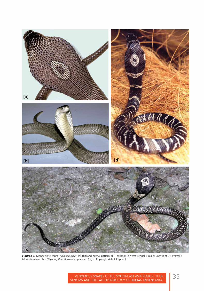
• Monocellate cobra N. kaouthia(Fig 6a-c) (Reid 1964; Warrell 1986;Viravan et al., 1992)
• Andaman cobra Naja sagittifera(Fig 6d)
• Spitting cobras: N. siamensis (Fig 7)(Warrell 1986; Wüster et al., 1997),
• N. sumatrana (Fig 8), N. sputatrix,N. mandalayensis etc
• King cobra: Ophiophagus hannah(Fig 9) (Tin-Myint et al., 1991)
• Kraits (genus Bungarus):
• Common krait B. caeruleus (Fig 10)(Theakston et al., 1990; Ariaratnamet al., 2009)
• Malayan krait B. candidus (Fig11) (Warrell et al., 1983; Kiem-Xuan-Trinh et al., 2010)
• Ceylon krait B. ceylonicus (Fig 12)(de Silva et al., 1993)
• Banded krait B. fasciatus (Fig 13)
• Red-headed krait B. flaviceps(Fig 14)
• Lesser black krait B. lividus (Kuch etal., 2011)
• Chinese krait B. multicinctus (Fig 15)
(Tun- Pe et al., 1997; Ha-Tran-Hung et al., 2010)
• Greater black krait B. niger (Fig 16)(Faiz et al., 2010)
• Sind krait B. sindanus (Pillai et al.,2012; in press) (Fig 17)
• Wall’s krait B. walli
• Spotted coral snake Calliophismaculiceps (Fig 18)
• MacClelland’s coral snakeSinomicrurus maclellandi(Kramer 1977)
• Australasian elapids inhabitingEastern Indonesia (Maluku and WestPapua):
• Death adders (Genus Acanthophis):A. rugosus (Fig 19a) (Lalloo et al.,1996)
• New Guinea small-eyed snakeMicropechis ikaheka (Fig 19b)(Warrell et al., 1996)
• Papuan Taipan Oxyuranus scutellatuscanni (Fig 20) (Lalloo et al., 1995)
• Papuan black snake Pseudechispapuanus (Fig 21) (Lalloo et al.,1994)
• Brown snakes (Genus Pseudonaja)(Fig 22)
• Sea snakes (Reid 1975, 1979;Reid and Lim 1957; Warrell1994): important species includeEnhydrina schistosa (Hydrophisschistosus) (Fig 23) (Kularatne etal., 2014), Hydrophis sp. (Fig 24)(Amararasekara et al., 1994; Wattand Theakston 1985), Lapemis curtus(Fig 25), Pelamis platura (Fig 26)and Laticauda colubrina (Fig 27).

33Venomous snakes of the south-east asia Region, theiR Venoms and the pathophysiology of human enVenoming
Figures 4: Common spectacled cobra (Naja naja): (a) Sri Lanka;(b) Pune; (c) Kolkata; (d) Bardia, Nepal (a-c Copyright DA Warrell; d Copyright Mark O’Shea)
[a]
[c]
[b]
[d]

GUIDELINES FOR THE MANAGEMENT OF SNAKEBITES
34
Figure 5: North Indian or Oxus cobra (Naja oxiana) (Copyright DA Warrell)
35Venomous snakes of the south-east asia Region, theiR Venoms and the pathophysiology of human enVenoming
Figures 6: Monocellate cobra (Naja kaouthia): (a) Thailand nuchal pattern; (b) Thailand; (c) West Bengal (Fig a-c: Copyright DA Warrell); (d) Andamans cobra (Naja sagittifera) juvenile specimen (Fig d: Copyright Ashok Captain)
[d]
[a]
[b]
[c]

GUIDELINES FOR THE MANAGEMENT OF SNAKEBITES
36
Figures 7: Indo-Chinese spitting cobra (Naja siamensis) Thailand: (a) Black and white specimen with ill-defined spectacle marking on hood; (b, c) Brown-coloured specimen showing spectacle marking on hood (Copyright DA Warrell)
[c]
[b]
[a]

37Venomous snakes of the south-east asia Region, theiR Venoms and the pathophysiology of human enVenoming
Figures 8: Sumatran spitting cobra (Naja sumatrana): (a) black phase Singapore (b) golden phase Thailand (Copyright DA Warrell)
[b]
[a]

GUIDELINES FOR THE MANAGEMENT OF SNAKEBITES
38
Figures 9: King cobra or hamadryad (Ophiophagus hannah) (Copyright DA Warrell)(a) The famous king cobra dance in Yangon, Myanmar; (b) Specimen from Trang, southern Thailand more than 3.5 metres in total length; (c, d) Dorsal and lateral views of head of Thai specimens showing the two large occipital scales which distinguish this species from cobras (Naja); (e) Pune, India (Copyright DA Warrell)
[a] [b]
[d]
[c] [e]
39Venomous snakes of the south-east asia Region, theiR Venoms and the pathophysiology of human enVenoming
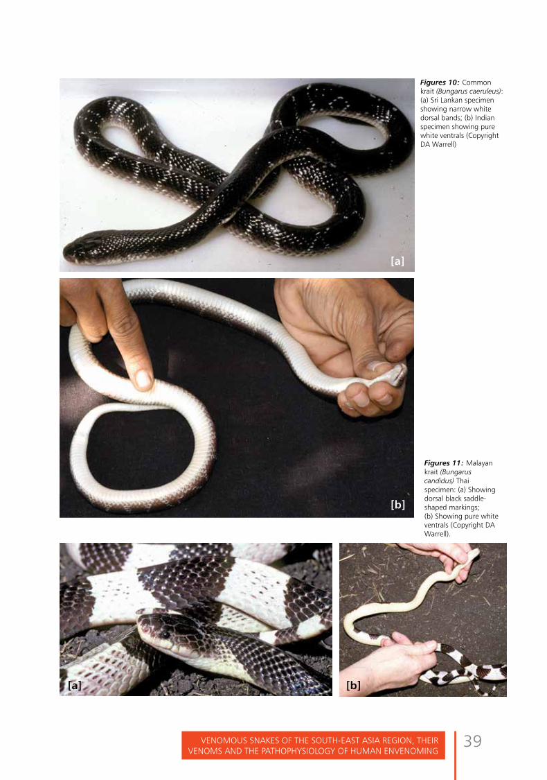
Figures 10: Common krait (Bungarus caeruleus): (a) Sri Lankan specimen showing narrow white dorsal bands; (b) Indian specimen showing pure white ventrals (Copyright DA Warrell)
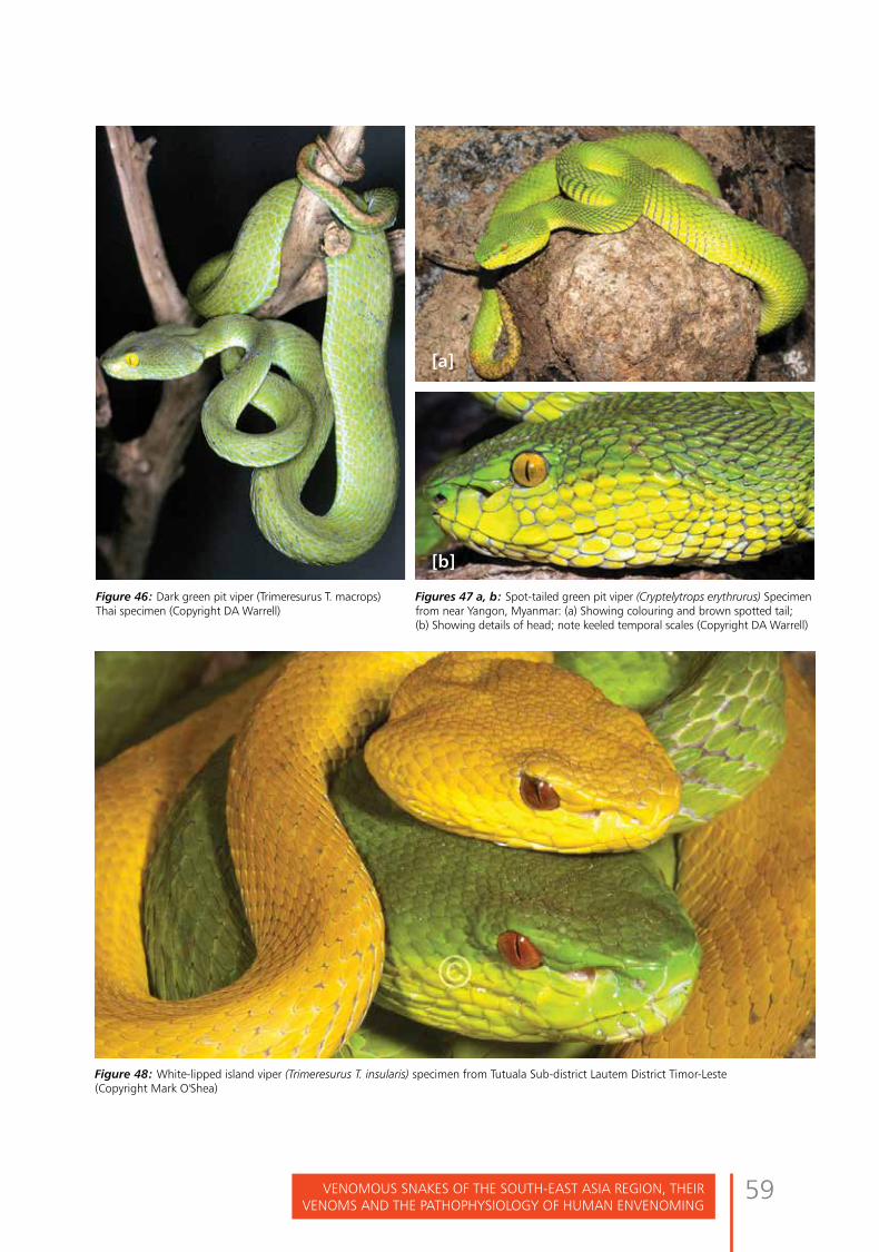
Figures 11: Malayan krait (Bungarus candidus) Thai specimen: (a) Showing dorsal black saddle-shaped markings; (b) Showing pure white ventrals (Copyright DA Warrell).
[a]
[b]
[a] [b]

GUIDELINES FOR THE MANAGEMENT OF SNAKEBITES
40
Figures 12: Ceylon krait (Bungarus ceylonicus): (a) showing incomplete white bands (spots); (b) head; (c) showing white bands and dark ventral scales. Perideniya, Sri Lanka (Copyright DA Warrell)
[c]
[a]
[b]

41Venomous snakes of the south-east asia Region, theiR Venoms and the pathophysiology of human enVenoming
Figures 13: Banded krait (Bungarus fasciatus) Thai specimens: (a) Showing black and yellow bands; (b) Showing circumferential black bands and blunt-tipped tail (Copyright DA Warrell)
Figure 14: Red-headed krait (Bungarus flaviceps) Thailand (Copyright DA Warrell)
[a] [b]

GUIDELINES FOR THE MANAGEMENT OF SNAKEBITES
42
Figure 15: Chinese krait (Bungarus multicinctus) (Copyright DA Warrell)
Figure 16: Greater black krait (Bungarus niger) Nepal (Copyright F. Tillack)
Figures 17: Sind krait (Bungarus sindanus): (a) specimen from Bikaner (Copyright Rom Whitaker); (b) head of specimen from Maharashtra, India (Copyright DA Warrell)
[a]
[b]

43Venomous snakes of the south-east asia Region, theiR Venoms and the pathophysiology of human enVenoming
Figure 18: Spotted coral snake (Calliophis maculiceps) Thai specimen (Copyright DA Warrell)
Figures 19: Death adders (Acanthophis rugosus): (a) Specimen from West Irian, Indonesia; (b) Specimen from Seram, Indonesia
[b][a]

GUIDELINES FOR THE MANAGEMENT OF SNAKEBITES
44
Figure 19c: New Guinea small-eyed snake (Micropechis ikaheka). Specimen from Arso, West Irian, Indonesia 1.69m in total length responsible for a case of envenoming (see Warrell et al., 1996).; (Copyright DA Warrell)
Figure 20: Papuan taipan (Oxuyuranus scutellatus) SaiBai Island, Torres Strait Islands (Copyright DA Warrell)

45Venomous snakes of the south-east asia Region, theiR Venoms and the pathophysiology of human enVenoming
Figure 21: Papuan black snake (Pseudechis papuanus) SaiBai Island, Torres Strait Islands (Copyright DA Warrell)
Figure 22: Eastern brown snake (Pseudechis textilis) Australia (Copyright DA Warrell)
Figure 23: Beaked sea snake (Enhydrina schistose or Hydrophis schistosus) Bunapas Mission, Ramu River, Papua New Guinea (Copyright DA Warrell)
Figure 24a: Blue spotted sea snake (Hydrophis cyanocinctus) Thailand (Copyright DA Warrell)

GUIDELINES FOR THE MANAGEMENT OF SNAKEBITES
46
Figure 24b: Banded sea snake (Hydrophis fasciatus atriceps) Thailand (Copyright DA Warrell)
Figure 24c: Flattened paddle-like tail of sea snakes: Hydrophis cyanocinctus (above); Lapemis curtus (below) Thailand (Copyright DA Warrell)
Figure 25: Hardwick’s sea snake (Lapemis curtus) showing tiny fangs (Copyright DA Warrell)

47Venomous snakes of the south-east asia Region, theiR Venoms and the pathophysiology of human enVenoming
Figure 26: Yellow-bellied sea snake (Pelamis platurus) Watamu, Kenya (Copyright Royjan Taylor)
Figures 27: Sea krait (Laticauda colubrina) Madang, Papua New Guinea: (a) Showing blue and banded pattern and amphibious behaviour; (b) Showing fangs (Copyright DA Warrell)
[a] [b]

GUIDELINES FOR THE MANAGEMENT OF SNAKEBITES
48
Viperidae have relatively long fangs (solenoglyph) which are normally folded flat against the upper jaw but, when the snake strikes, are erected (Fig 2b). There are two subfamilies, typical vipers (Viperinae) and pit-vipers (Crotalinae). The Crotalinae possess a special infra-red heat-sensing organ, the loreal pit organ,
Figure 28: Head of a typical pit viper - dark green pit viper (Trimeresurus T. macrops) showing the pit organ situated between the nostril and the eye (red arrow) (Copyright DA Warrell)
to detect their warm-blooded prey. This is situated between the nostril and the eye (Fig. 28). Viperidae are relatively short, thick-bodied snakes with many small rough scales on the top (dorsum) of the head and characteristic patterns of coloured markings on the dorsal surface of the body (Fig 29).
Pit Viper (Trimeresurus T. Macrops)
Showing heat sensitive pit organ (red arrow)

49Venomous snakes of the south-east asia Region, theiR Venoms and the pathophysiology of human enVenoming
Figures 29: Western Russell’s viper (Daboia russelii): (a) Specimen from Sri Lanka; (b, c) Specimens from Tamil Nadu, southern India; (d, e) specimens from Pune, India (Copyright DA Warrell)
[a]
[d] [e]
[b]
[c]

GUIDELINES FOR THE MANAGEMENT OF SNAKEBITES
50
Some important examples of the Viperidae inhabiting South-East Asia Region countries (References to reports of reviews of bites by these species are given in parenthesis):
Typical vipers (subfamily Viperinae): Russell’s vipers, Western, Daboia russelii (Fig 29 a-e) (Phillips et al., 1988; Warrell 1989; Gawarammana et al., 2009); and Eastern, D. siamensis (Fig 30) (Myint-Lwin et al., 1985; Tun-Pe et al., 1987; Than-Than et al., 1987; Than-Than et al., 1988; Warrell 1989; Than- Than et al., 1989; Thein-Than et al., 1991; Tin-Nu-Swe et al., 1993; Belt et al., 1997)
Saw-scaled or carpet vipers Echis c. carinatus (Fig 31) (Bhat 1974; Warrell and Arnett 1976) and E. c. sochureki (Fig 31c) (Kochar et al., 2007).
Levantine or blunt-nosed viper Macrovipera lebetina (Fig 32) (Sharma et al., 2008)
Pit vipers (subfamily Crotalinae):
Malayan pit viper Calloselasma rhodostoma (Fig 33) (Reid et al., 1963a; Reid et al., 1963b;
Reid 1968; Warrell et al., 1986)
Mount Kinabalu pit viper Garthia chaseni (Fig 34) (Haile 1963; Warrell 1995)
Mamushis (Genus Gloydius): G. brevicaudus (Fig 35) (Warrell 1995)
Hump-nosed pit viper Hypnale hypnale (Fig 36) (de Silva et al., 1994; Joseph et al., 2007;
Ariaratnam et al., 2008; Premawardhena et al, 1998) and other Hypnale species (Maduwage et al., 2011).
Chinese habu Protobothrops mucrosquamatus (Fig 37) (Warrell 1995)
Green pit vipers, bamboo vipers, palm vipers and habus (formerly all genus Trimeresurus)
Indian bamboo viper Trimeresurus
(Craspedocephalus) gramineus (Fig 38)
Malabar rock pit viper Trimeresurus (Crapedocephalus) malabaricus (Fig 39) (Gowda et al., 2006)
Palm viper Trimeresurus (Craspedocephalus) puniceus (Fig 40)
Sri Lankan viper Trimeresurus (Craspedocephalus) trigonocephalus (Fig 41) (Warrell 1995)
Hagen’s pit viper Trimeresurus (Parias) hageni (Fig 42)
Pope’s pit viper Trimeresurus (Popeia) popeiorum (Fig 43)
Chinese bamboo viper Trimeresurus (Viridovipera) stejnegeri (Fig 44) (Warrell 1995)
White-lipped green pit viper Trimeresurus (Trimeresurus) albolabris (Fig 45) (Hutton et al., 1990; Rojnuckarin et al., 1998)
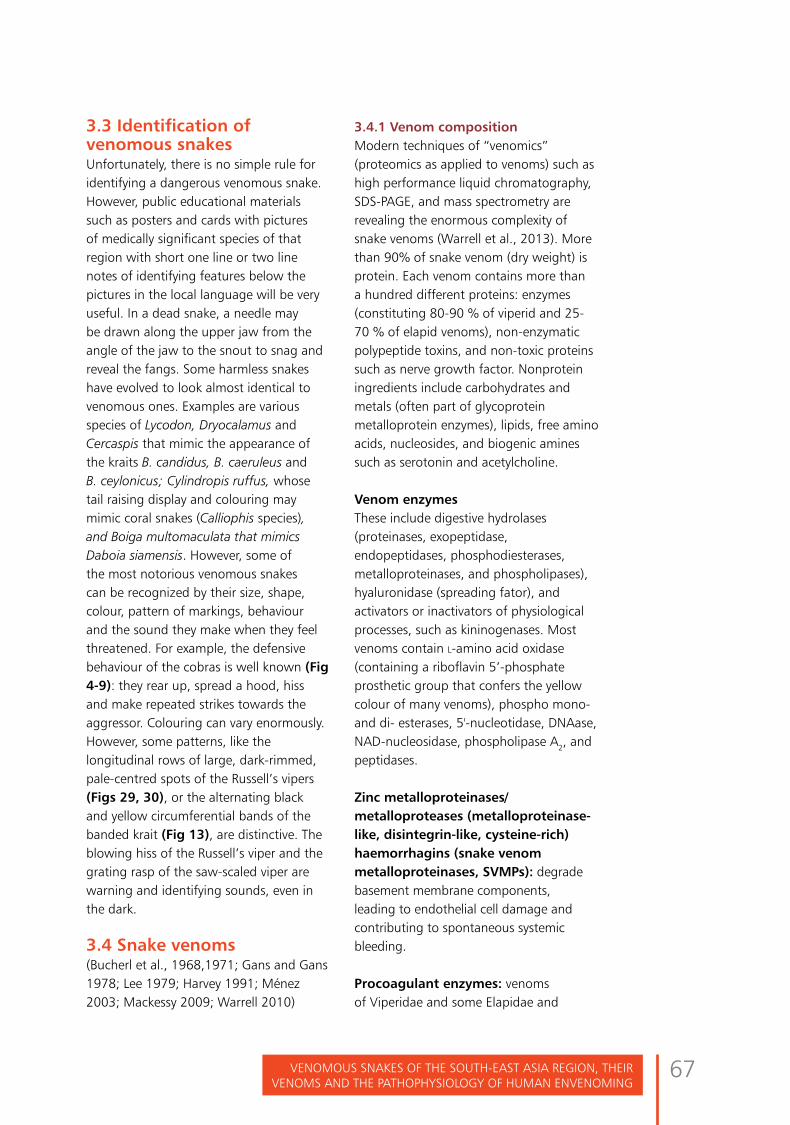
Dark green pit viper Trimeresurus (Trimeresurus) macrops (Fig 28, 46) (Hutton et al., 1990; Warrell 1990b)
Spot-tailed green pit viper Trimeresurus (Trimeresurus) erythrurus (Fig 47) (Warrell 1995)
White-lipped island viper Trimeresurus (Trimeresurus) insularis (Fig 48)
Kanchanburi pit viper Trimeresurus (Trimeresurus) kanburiensis (Warrell et al., 1992)
Mangrove pit viper Trimeresurus (Trimeresurus) purpureomaculatus (Fig 49) (Warrell 1995)
Northern white-lipped pit viper Trimeresurus (Trimeresurus) septentrionalis (Whitaker)
Beautiful pit viper Trimeresurus (Trimeresurus) venustus (Fig 50)
Wagler’s (temple) pit viper Tropidolaemus wagleri (Fig 51a) (Reid 1968)
Banded temple viper Tropidolaemus subannulatus (Fig 51b)

51Venomous snakes of the south-east asia Region, theiR Venoms and the pathophysiology of human enVenoming
Figures 30: Eastern Russell’s viper (Daboia siamensis): (a) Specimen from Myanmar; (b) Specimen from Thailand; (c) Specimen from East Java, Indonesia; (d) Specimen from Flores, Indonesia (Copyright DA Warrell)
Figures 31: Saw-scaled viper (Echis carinatus): (a) Echis carinatus carinatus Specimen from southern India; (b) Echis carinatus carinatus Specimen from Sri Lanka; (c) Echis carinatus sochureki Specimen from Chhattagharh Rajisthan (Copyright DA Warrell)
[a]
[c]
[b]
[d]
[a]
[c]
[b]

GUIDELINES FOR THE MANAGEMENT OF SNAKEBITES
52
Figure 32: Levantine viper (Macrovipera lebetina) Specimen from Cyprus (Copyright DA Warrell)
Figures 33: Malayan pit viper (Calloselasma rhodostoma) Thai specimens: (a) Showing characteristic posture and triangular dorsal markings; (b) Showing supralabial markings (Copyright DA Warrell)
Figure 34: Mount Kinabalu pit viper (Garthia chaseni) (Copyright Prof RS Thorpe)
Figure 35: Mamushi or Fu-she (Gloydius brevicaudus) from China (Copyright DA Warrell)
[b][a]

53Venomous snakes of the south-east asia Region, theiR Venoms and the pathophysiology of human enVenoming
Figures 36 a-d: Hump-nosed viper (Hynpale hypnale) (Copyright DA Warrell)a) Specimen from Sri Lanka; (b) Specimen from Sri Lanka showing long fangs; (c) Specimen from south western India;(d) Specimen from south western India showing upturned snout
Figure 37: Chinese habu (Protobothrops mucrosquamatus) Specimen from China (Copyright DA Warrell)
[a]
[c]
[b]
[d]

GUIDELINES FOR THE MANAGEMENT OF SNAKEBITES
54
Figure 38: Indian bamboo viper (Trimeresurus Craspedocephalus gramineus) (Copyright DA Warrell)
[a]

55Venomous snakes of the south-east asia Region, theiR Venoms and the pathophysiology of human enVenoming
Figures 39 a, b: Malabar rock pit viper (Trimeresurus Craspedocephalus malabaricus) (Copyright DA Warrell)
Figure 40: Palm viper (Trimeresurus Craspedocephalus puniceus) Specimen from Cilacap, West Java, Indonesia (Copyright DA Warrell)
[b]

GUIDELINES FOR THE MANAGEMENT OF SNAKEBITES
56
Figure 41: Sri Lankan pit viper (Trimeresurus Craspedocephalus trigonocephalus) (Copyright DA Warrell

57Venomous snakes of the south-east asia Region, theiR Venoms and the pathophysiology of human enVenoming
Figure 42: Hagen’s pit viper (Trimeresurus Parias hageni) Trang, Thailand (Copyright DA Warrell)
Figure 43: Pope’s pit viper (Trimeresurus Popeia popeiorum) Thailand (Copyright DA Warrell)

GUIDELINES FOR THE MANAGEMENT OF SNAKEBITES
58
Figure 44: Chinese bamboo viper (Viridovipera stejnegeri) Specimen from China (Copyright DA Warrell)
Figures 45 a,b: White-lipped green pit viper (Cryptelytrops albolabris) Thai specimen: (a) Showing colouring and distinctive brown-topped tail; (b) Showing details of the head: note smooth temporal scales (Copyright DA Warrell)
[a] [b]
59Venomous snakes of the south-east asia Region, theiR Venoms and the pathophysiology of human enVenoming
Figure 48: White-lipped island viper (Trimeresurus T. insularis) specimen from Tutuala Sub-district Lautem District Timor-Leste (Copyright Mark O'Shea)
[a]
[b]
Figures 47 a, b: Spot-tailed green pit viper (Cryptelytrops erythrurus) Specimen from near Yangon, Myanmar: (a) Showing colouring and brown spotted tail; (b) Showing details of head; note keeled temporal scales (Copyright DA Warrell)
Figure 46: Dark green pit viper (Trimeresurus T. macrops) Thai specimen (Copyright DA Warrell)

GUIDELINES FOR THE MANAGEMENT OF SNAKEBITES
60
Figures 49 a,b: Mangrove pit viper (Trimeresurus T. purpureomaculatus): (a) Specimen from Kanchanburi, Thailand; (b) Specimen from upper Myanmar (Copyright DA Warrell)
Figure 50: Beautiful pit viper (Cryptelytrops venustus) specimen from Thung Song, Thailand (Copyright DA Warrell)
[a] [b]

61Venomous snakes of the south-east asia Region, theiR Venoms and the pathophysiology of human enVenoming
Figure 51a: Wagler’s pit (temple) viper (Tropidolaemus wagleri) specimens in the snake temple, Penang, Malaysia (Copyright DA Warrell)
Figure 51b: Banded temple viper (Tropidolaemus semiannulatus) Borneo

GUIDELINES FOR THE MANAGEMENT OF SNAKEBITES
62
3.2.2 Other medically important venomous snakesSeveral species of medically important Colubridae (sensu lato) have been identified in the South-East Asia Region. The red-necked keelback Rhabdophis subminiatus (Fig 2c and 52a) and yamakagashi R. tigrinus (Fig 52b) (Warrell 1995) can cause severe life-threatening anti-haemostatic disturbances and acute kidney injury (Fig 52c, 52d). A case of systemic envenoming by the Sri Lankan keelback (Balanophis ceylonicus) with similar features has also been reported
Figure 52a: Red-necked keelback (Rhabdophis subminiatus) Thai specimen (Copyright DA Warrell)
Figure 52b: Yamakagashi (Rhabdophis tigrinus) (Copyright S Mishima The Snake 1974 6-1 Courtesy of Dr Y Sawai)

63Venomous snakes of the south-east asia Region, theiR Venoms and the pathophysiology of human enVenoming
(Fernando et al 2015). More than a dozen other species have proved capable of causing local envenoming [e.g. mangrove snake (Boiga dendrophila) (Weinstein et al., 2014) Warrell, 1995) and other cat snakes (Boiga species); long-nosed whip snake (Ahaetulla nasuta) (Fig 52e); dog-faced water snake (Cerberus rhynchops); and water snakes/paddy field snakes (Enhydris species) (Fig 52f and 53c). Undoubtedly more will be described as the concept of “non-venomous” is further reviewed (Weinstein et al., 2011) Figure 52c: Extensive bruising from envenoming by R. tigrinus in Japan
Figure 52d: Intracerebral haemorrhage (Courtesy of the late Dr Y Sawai)
Figure 52e: Long-nosed whip snake (Ahaetulla nasuta) Sri Lanka (Copyright DA Warrell)
Figure 52f: Paddy field snake (Enhydris plumbea)
CT scanning of the head: Hemorrhage in the left temporal and occipital perforative hemorrhage into the ventricle.

GUIDELINES FOR THE MANAGEMENT OF SNAKEBITES
64
3.2.3 Non-venomous snakesMany species of non-venomous or only trivially-venomous species are responsible for bites, especially those that are aggressive, irritable or prone to strike at humans who approach to closely or that commonly inhabit urban and rural gardens and compounds.
Apart from the taxa mentioned above, these include paradise or flying snakes (Chrysopelea species) (Fig 52g), striped keelbacks (Amphiesma species) (Fig 52h), kukri snakes (Oligodon species) (Fig 52i), checkered keelbacks or Asian water snake (Xenochrophis species) (Fig 52j), wolf snakes (Lycodon or Dinodon species) (Fig 52k), bridle snakes (Dryocalamus) (Fig 52m) and rat snakes (Ptyas, Elaphe, Coelognathus, Goniosoma etc.) (Fig 52n). Their medical importance is that they may be mistaken for venomous species (notably the krait mimics Lycodon and Dryocalamus), resulting in unnecessary and wasteful antivenom treatment (Viravan et al., 1992; Ariaratnam et al., 2009).
Large pythons (Boidae), notably the reticulated python Python reticularis in Indonesia, have been reported to attack and even ingest humans, usually inebriated farmers (Fig 52o).Figure 52g: Paradise or flying snakes (Chrysopelea ornata)
(Copyright Mark O’Shea)
Figure 52h: Striped keelbacks (Amphiesma stolatum) (Copyright Mark O’Shea)Figure 52i: Kukri snakes (Oligodon cyclurus) (Copyright DA Warrell)

65Venomous snakes of the south-east asia Region, theiR Venoms and the pathophysiology of human enVenoming
Figure 52j: Chequered keelback (Xenochrophis piscator) brought by bite victim Chittagong Bangladesh
Figure 52k: Wolf snakes (Lycodon aulicus) (Copyright DA Warrell)
Figure 52l: Yellow-necked wolf snake (Lycodon flavicollis) biting (Copyright DA Warrell)
Figure 52m: Bridle snake (Dryocalamus davisoni) (Copyright DA Warrell)

GUIDELINES FOR THE MANAGEMENT OF SNAKEBITES
66
Figure 52o: Reticulated python (Python reticularis) containing the body of a farmer it had swallowed at Palu, Sulawesi, Indonesia (Copyright Excel Sawuwu)
Figure 52n: Rat snake (Ptyas mucosus) (Copyright Marl O'Shea)
67Venomous snakes of the south-east asia Region, theiR Venoms and the pathophysiology of human enVenoming
3.3 Identification of venomous snakesUnfortunately, there is no simple rule for identifying a dangerous venomous snake. However, public educational materials such as posters and cards with pictures of medically significant species of that region with short one line or two line notes of identifying features below the pictures in the local language will be very useful. In a dead snake, a needle may be drawn along the upper jaw from the angle of the jaw to the snout to snag and reveal the fangs. Some harmless snakes have evolved to look almost identical to venomous ones. Examples are various species of Lycodon, Dryocalamus and Cercaspis that mimic the appearance of the kraits B. candidus, B. caeruleus and B. ceylonicus; Cylindropis ruffus, whose tail raising display and colouring may mimic coral snakes (Calliophis species), and Boiga multomaculata that mimics Daboia siamensis. However, some of the most notorious venomous snakes can be recognized by their size, shape, colour, pattern of markings, behaviour and the sound they make when they feel threatened. For example, the defensive behaviour of the cobras is well known (Fig 4-9): they rear up, spread a hood, hiss and make repeated strikes towards the aggressor. Colouring can vary enormously. However, some patterns, like the longitudinal rows of large, dark-rimmed, pale-centred spots of the Russell’s vipers (Figs 29, 30), or the alternating black and yellow circumferential bands of the banded krait (Fig 13), are distinctive. The blowing hiss of the Russell’s viper and the grating rasp of the saw-scaled viper are warning and identifying sounds, even in the dark.
3.4 Snake venoms(Bucherl et al., 1968,1971; Gans and Gans 1978; Lee 1979; Harvey 1991; Ménez 2003; Mackessy 2009; Warrell 2010)
3.4.1 Venom compositionModern techniques of “venomics” (proteomics as applied to venoms) such as high performance liquid chromatography, SDS-PAGE, and mass spectrometry are revealing the enormous complexity of snake venoms (Warrell et al., 2013). More than 90% of snake venom (dry weight) is protein. Each venom contains more than a hundred different proteins: enzymes (constituting 80-90 % of viperid and 25-70 % of elapid venoms), non-enzymatic polypeptide toxins, and non-toxic proteins such as nerve growth factor. Nonprotein ingredients include carbohydrates and metals (often part of glycoprotein metalloprotein enzymes), lipids, free amino acids, nucleosides, and biogenic amines such as serotonin and acetylcholine.
Venom enzymes These include digestive hydrolases (proteinases, exopeptidase, endopeptidases, phosphodiesterases, metalloproteinases, and phospholipases), hyaluronidase (spreading fator), and activators or inactivators of physiological processes, such as kininogenases. Most venoms contain l-amino acid oxidase (containing a riboflavin 5’-phosphate prosthetic group that confers the yellow colour of many venoms), phospho mono- and di- esterases, 5'-nucleotidase, DNAase, NAD-nucleosidase, phospholipase A2, and peptidases.
Zinc metalloproteinases/metalloproteases (metalloproteinase-like, disintegrin-like, cysteine-rich) haemorrhagins (snake venom metalloproteinases, SVMPs): degrade basement membrane components, leading to endothelial cell damage and contributing to spontaneous systemic bleeding.
Procoagulant enzymes: venoms of Viperidae and some Elapidae and

GUIDELINES FOR THE MANAGEMENT OF SNAKEBITES
68
Colubridae contain serine proteases and other procoagulant enzymes that are thrombin-like or activate factors V, X, prothrombin and other clotting factors. These enzymes stimulate blood clotting with formation of fibrin in the blood stream. Paradoxically, this process results in incoagulable blood because most of the fibrin clot is broken down immediately by the body’s own plasmin fibrinolytic system. Sometimes within 30 minutes of the bite, the levels of clotting factors have been so depleted that the blood will not clot (“consumption coagulopathy”). Some venoms contain multiple anti-haemostatic factors. For example, Russell’s viper venom contains toxins that activate factors II (prothrombin), V, X, IX and XIII, fibrinolysis and protein C, and cause platelet aggregation, anticoagulation and haemorrhage.
Phospholipases A2 (lecithinase): are most widespread and extensively studied of all venom enzymes. They damages mitochondria, red blood cells, leucocytes, platelets, peripheral nerve endings, skeletal muscle, vascular endothelium, and other membranes, producing presynaptic neurotoxic activity, cardiotoxicity, myotoxicity, necrosis, hypotension, haemolysis, haemorrhage, plasma leakage (oedema-induction), opiate-like sedative effects and autopharmacological release of histamine and other autacoids. They are anti-coagulant, either by hydrolysing plasma or platelet membrane phospholipids, or by interacting with different coagulation factors.
Acetylcholinesterases: although found in most elapid venoms, may cause fasciculation.
Hyaluronidase: promotes the spread of venom through tissues by increasing permeability but can also contribute to tissue damage.
Proteolytic enzymes (metalloproteinases, endopeptidases or hydrolases) and polypetide cytotoxins (“cardiotoxins”): increase vascular permeability causing oedema, blistering, bruising and necrosis at the site of the bite.
Venom polypeptide toxins (“neurotoxins”)Postsynaptic (α) neurotoxins such as α-bungarotoxin and cobrotoxin, consist of 60-62 or 66- 74 amino acids. They bind to acetylcholine receptors at the motor endplate. Presynaptic (β) neurotoxins such as β-bungarotoxin, and taipoxin, contain 120-140 amino-acids and a phospholipase A subunit. These release acetylcholine at the nerve endings at neuromuscular junctions and then damage the endings, preventing further release of transmitter.
Variation in venom composition within speciesThe composition of snake venoms, and hence their antigenicities in inducing specific neutralizing antibodies during antivenom manufacture, varies greatly between different species, but also within an individual species, as the snake matures (ontogenic variation), seasonally, between sexes, and throughout the geographical range (Warrell, 1997; Mackessy et al., 2003). The two important implications of venom variation are 1- envenoming by juvenile and adult snakes may cause qualitatively different clinical effects and 2- envenoming by a snake in one part of its geographical range may not be neutralized
Envenoming by juvenile and adult
snakes may cause qualitatively different
clinical effects

69Venomous snakes of the south-east asia Region, theiR Venoms and the pathophysiology of human enVenoming
by an antivenom raised using venom from another part of the range.
3.4.2 Quantity of venom injected at a bite, “dry bites”This is very variable, depending on the species and size of the snake, the mechanical efficiency of the bite, whether one or two fangs penetrated the skin and whether there were repeated strikes. The average dry weight of venom injected at a strike is approximately 60 mg in N. naja, 13 mg in E. carinatus and 63 mg in D. russelii. In the case of D. siamensis in Myanmar, the total yield of desiccated venom extracted by milking ranged from 21-268 mg (127 +/- 13 mg, mean +/- 1SE) in adults (mean total length 111 +/- 1.8 cm) and 8-79 mg (45 +/- 7 mg) in juveniles (mean total length 79 +/- 2.8 cm). Adults inject 45% of the venom glands' content in the first bite (Tun-Pe and Khin-Aung Cho, 1986). Either because of mechanical inefficiency or the snake’s control of venom discharge, a proportion of bites by venomous snakes does not result in the injection of sufficient venom to cause clinical effects. About 50% of bites by Malayan pit vipers and Russell’s vipers, 30% of bites by cobras and 5-10% of bites by saw-scaled vipers do not result in any symptoms or signs of envenoming. Snakes do not exhaust their store of venom, even after several strikes, and they are no less venomous after eating their prey (Tun-Pe et al., 1991).
3.4.3 Variations in venom composition within individual species of snakesAlthough large snakes tend to inject more venom than smaller specimens of the same species, the venom of smaller, younger vipers may be richer in some dangerous components, such as those affecting haemostasis because venom composition varies (ontogenically) with the snake’s age and hence size (Tun-Pe et al., 1995a).
Recommendations: Bites by small snakes should not be ignored or dismissed. They should be taken just as seriously as bites by large snakes of the same species (O). Design and manufacture of antivenoms should take account of geographical and ontogenic variation in venom composition within individual species (O).
3.5 Pathophysiology of human envenomingLocal envenomingSwelling and bruising result from increased vascular permeability attributable to venom endopeptidases, metalloproteinase hemorrhagins, membrane-damaging polypeptide toxins, phospholipases, and endogenous autacoids released by the venom, such as histamine, 5-HT, and kinins. Local tissue necrosis results from the direct action of myotoxins and cytotoxins, and ischemia caused by thrombosis; compression of blood vessels by first-aid methods such as tight tourniquets; or by swollen muscle within a tight fascial compartment. Myotoxins damage the muscle cell plasma membrane directly. Most are PLA2s, either enzymatically active (aspartate-49) or enzymatically inactive (lysine-49). Cobra cardiotoxins are low-molecular weight polypeptides with cytotoxic action.
Hypotension and shockAfter viper bites, leakage of plasma or blood into the bitten limb and elsewhere, or massive gastrointestinal haemorrhage, may cause hypovolaemia. Vasodilation, especially of splanchnic vessels, and a direct effect on the myocardium may contribute to hypotension. Profound hypotension

GUIDELINES FOR THE MANAGEMENT OF SNAKEBITES
70
is part of the autopharmacological syndrome that occurs within minutes of bites by D. siamensis, D. russelii, and Australasian elapids, attributable to oligopeptides (ACE inhibitors and BPPs) and vasodilating autacoids. In some cases, direct myocardial effects of venom may be suggested by electrocardiographic (ECG) changes and autopsy findings of epicardial or endocardial haemorrhages and histopathological evidence of cardiac myonecrosis.
Bleeding and blood clotting disturbancesSnake venoms affect haemostasis in several ways. Procoagulant enzymes activate intravascular coagulation, producing consumption coagulopathy and incoagulable blood. Procoagulants of Colubridae, Australasian Elapidae, Echis, and Daboia species activate prothrombin, whereas those in venoms of Daboia russelii and D. siamensis also activate factors V and X. Thrombin-like enzymes in pit-viper venoms have a direct action on fibrinogen. Some venoms cause defibrinogenation by activating the endogenous fibrinolytic (plasmin) system. Anticoagulant activity is attributable to venom phospholipases. Platelet activation or inhibition results in thrombocytopenia in victims of Trimeresurus and Viridovipera species, Calloselasma rhodostoma, Deinagkistrodon acutus, and Daboia siamensis. Potentially lethal spontaneous systemic bleeding is attributable venom haemorrhagins (Zn metalloproteases).
Complement Activation Elapid and some colubroid venoms activate complement via the alternative pathway (“cobra venom factor” is the snake’s C3b), whereas some viperid venoms activate the classic pathway. Complement activation affects platelets, the blood coagulation system, and other humoral mediators.
NeurotoxicityNeurotoxic polypeptides and PLA2s of snake venoms cause paralysis by blocking transmission at the neuromuscular junction. Patients with paralysis of the bulbar muscles may die of upper airway obstruction or aspiration, but the most common mode of death after neurotoxic envenoming is respiratory paralysis. By prolonging activity of ACh at neuromuscular junctions, anticholinesterase drugs may improve paralytic symptoms in patients bitten by snakes with neurotoxins that are predominantly postsynaptic in their action (e.g., cobras and Australasian death adders [genus Acanthophis]). Some patients bitten by elapids or vipers are drowsy in the absence of respiratory or circulatory failure. This is unlikely to be an effect of neurotoxic polypeptides, which do not cross the blood-brain barrier.
MyotoxicityPLA2 myotoxins and metalloproteinases are principally responsible. They are present in venoms of most species of sea snakes, many terrestrial Australasian elapids, some species of krait (Bungarus), and Viperidae, such as the Sri Lankan Russell’s viper (D. russelii). Release into the bloodstream of myoglobin, muscle enzymes, uric acid, potassium, and other muscle constituents is an effect in humans of presynaptic neurotoxins. Patients may die of bulbar and respiratory muscle weakness, acute hyperkalaemia, or acute kidney injury.
Acute kidney injuryA wide range of renal histological changes has been described after snakebite. Acute tubular necrosis is the most common, but proliferative glomerulonephritis, interstitial nephritis, toxic mesangiolysis with platelet agglutination, fibrin deposition, ischaemic changes, and distal tubular damage (“lower nephron nephrosis”), suggesting direct venom nephrotoxicity attributable

71Venomous snakes of the south-east asia Region, theiR Venoms and the pathophysiology of human enVenoming
to venom PLA2 and metalloproteases , and bilateral renal cortical necrosis with subsequent calcification are also reported. Antivenom can cause immune-complex-mediated kidney injury. Acute tubular necrosis may result from prolonged hypotension and hypovolaemia, DIC, direct toxic effect of the venom on the renal tubules, haemoglobinuria, myoglobinuria, and hyperkalaemia. Russell’s viper venom produces hypotension, DIC, direct nephrotoxicity, and, in Sri Lanka and India, intravascular haemolysis and rhabdomyolysis.In Burmese patients envenomed by Russell’s vipers (D. siamensis), high urinary concentrations of β2-microglobulin, retinal binding protein, and N-acetyl glucosaminidase suggested failure of proximal tubular reabsorption and tubular damage. High plasma concentrations of active renin suggested that renal ischaemia with activation of the renin-angiotensin system was involved in development of renal failure. A massive but transient capillary and glomerular leak of albumin
was an early sign of oliguric renal failure. Snake venom–induced DIC may result in deposition of fibrin on vascular endothelium that has been activated by, for example, metalloproteinases, producing microangiopathic haemolysis. Although the clinical picture is reminiscent of haemolytic uraemic syndrome (HUS) and thrombotic thrombocytopenic purpura (TTP), there is no evidence of depleted ADAMTS-13 levels and, therefore no justification for cryosupernatant plasmapheresis (Ho et al., 2010).
Generalized increase in capillary permeabilityVenoms of some Viperidae, such as D. russelii and D. siamensis, can cause a generalized increase in vascular permeability, resulting in pulmonary oedema, serous effusions, conjunctival, periorbital, facial and retinal oedema, bilateral parotid enlargement, albuminuria, and haemoconcentration. The likely cause is metalloproteases that damage vascular endothelium.

GUIDELINES FOR THE MANAGEMENT OF SNAKEBITES
72

73EpidEmiology of snakEbitE in south-East asia REgion countRiEs
Epidemiology of snakebite in South-East Asia Region countries

GUIDELINES FOR THE MANAGEMENT OF SNAKEBITES
74

75EpidEmiology of snakEbitE in south-East asia REgion countRiEs
Epidemiology of snakebite in South-East Asia Region countriesEssentials:Most published data, based on hospital returns, are incomplete because many patients are treated by traditional healers. However, three large, well-designed, national, community-based studies from Bangladesh, India and Sri Lanka have produced reliable estimates. Data are inadequately reported and so snakebite should be made a notifiable disease in all South-East Asia Region countries. Death certification should use International Classification of Diseases code T63.0 (Toxic effect of contact with venomous animals – snake venom and sea-snake venom) (ICD-10 Version:2015 http://apps.who.int/classifications/icd10/browse/2015/en#/T63.0)
Incidence of snakebite varies diurnally and seasonally. It is highest during agricultural activities and seasonal rains. Most bites are inflicted on the feet and ankles of bare-footed agricultural workers who tread on snakes inadvertently while walking in the dark or working in fields and plantations. Snake species differ in their inclination to strike when disturbed. Notoriously “irritable” species include Russell’s and saw-scaled vipers. Cobras and kraits enter human dwellings. Kraits bite people who are asleep on the ground at night. On average, about 50% of bites by
4venomous snake cause no envenoming (“dry bites”), a figure ranging 5-80% with different species.
Snakebite epidemics follow flooding, cyclones and invasion of snakes’ habitats for road building, irrigation schemes and logging. These activities cause long term changes in climate and ecology and encourage influx of human settlers.
Males are more often bitten than females. Peak incidence is in children and young adults. Pregnant women and their fetuses are at increased risk of dying. Snakebite is an occupational disease of farmers, plantation workers, herders, hunters, fishermen, fish farmers, snake restaurant workers and snake charmers. Factors contributing to fatal snakebite include problems with choice and dosage of antivenom, delay from visiting traditional healers, transportation difficulties, death in transit, airway obstruction, failure to attempt assisted ventilation or problems in carrying it out, failure to treat hypovolaemia, complicating infections, failure to observe deterioration in hospital. Hours usually elapse between bite and neurotoxic deaths from elapid envenoming and several days or longer, from viper envenoming.

GUIDELINES FOR THE MANAGEMENT OF SNAKEBITES
76
Snakebite in different South-East Asia Region countries Bangladesh: medically important species include B. caeruleus, B. niger, B. walli; N. kaouthia, N. naja; D. russelii and T. (T.) erythrurus.
In 2009, 9000 people were questioned in about 4000 randomly selected households throughout all 6 administrative divisions. 589,919 snakebites and 6,041 deaths per year were estimated in rural areas (case-fatality 1%).
Green pit vipers cause many bites, some morbidity but few deaths. D. russelii is restricted to western and southern areas. B. niger, previously unknown in Bangladesh, has caused deaths. N. kaouthia causes most cobra bites.
Bhutan: medically-important species include cobras, kraits and pit-vipers. No recent data on bites.
Democratic People’s Republic of Korea: medically-important species include adders (Vipera berus and V. sachalinensis), several species of mamushi (genus Gloydius) and Rhabdophis tigrinus. No recent data on bites.
India: medically important species include N. naja, N. kaouthia; B. caeruleus; D. russelii, E. carinatus; Hypnale hypnale, Trimeresurus. Registrar General of India’s “Million Death Study” assigned causes of all deaths in about 7000 randomly chosen sample areas, each with a population of about 1000 throughout the whole country. Verbal autopsy (questioning bereaved relatives and neighbours about the circumstances of the deceased’s death), proved reliable for an event as distinctive, dramatic and memorable as snakebite fatality. Results were independent of hospital underreporting and were nationally representative.
Direct estimate of deaths attributable to snakebite in 2005 was 46 000 (99%CI 41 000-51 000), (1 snakebite death for every 2 HIV/AIDS deaths). Snakebites caused 0.5% of all deaths, 3% in 5-14 year-olds. 97% died in rural areas, only 23% in health facilities. The highest numbers of deaths were in Uttar Pradesh (8,700), Andhra Pradesh (5,200), and Bihar (4,500).
Indonesia: medically important species throughout the >18 000 islands include B. candidus, N. sputatrix, N. sumatrana, C. rhodostoma , T. (T.) albolabris; D. siamensis and, in West Papua and Maluku, Acanthophis laevis. Hundreds of bites each year are treated in Sumatra, Java, East Kalimantan, Sulawesi, Wetar and West Papua.
Maldives: there is one species of sea snake but no terrestrial venomous snake. No recent data on bites.
Myanmar: medically important species include B. magnimaculatus, B. multicinctus; N. kaouthia, N. mandalayensis; Trimeresurus (T.) albolabris, T. (T.) erythrurus; D. siamensisIn 2014, 15,080 bites with 305 deaths were reported to the Ministry of Health (case-fatality 2.02%). D. siamensis causes 90% of bites.
Nepal: medically important species include B. caeruleus, B. niger, B. walli; N. naja, N. kaouthia; D. russelii and Ovophis monticola. In the Eastern Terai, there were 162 snakebite deaths/100 000/year. Only 20% of deaths occurred in hospitals. Most were attributed to krait. National totals of between 1000 bites and 200 deaths and 20 000 bites and 1000 deaths/year have been suggested. In 2000, the Ministry of Health reported 480 bites with 22 deaths, while 4,078 bites with 81 deaths were recorded at 10 hospitals in eastern Nepal (case-fatality 3-58%).

77EpidEmiology of snakEbitE in south-East asia REgion countRiEs
Sri Lanka: medically important species include B. caeruleus; N. naja; D. russelii, Echis carinatus and Hypnale hypnale. Bites are caused by D. russelii, (30%), H. hypnale (22%), N. naja (17%) and B. caeruleus. A recent community-based, countrywide survey of snakebite included nearly 1% of the country’s population. The incidence of bites, envenoming and mortality from snakebite was found to be 398 (95% CI: 356–441), 151 (130–173) and 2.3 (0.2– 4.4)/100 000/year, respectively, amounting to more than 80 000 bites, 30 000 envenomings and 400 deaths in the country each year.
Thailand: medically important species include B. candidus; N. kaouthia, N. siamensis; C. rhodostoma; Trimeresurus (T.) albolabris and D. siamensis. Incidence of bites has declined from 9,071 bites (14.5/100 000) in 2002 to 457 (0.7/100 000) in 2015. Mortality has declined from an average of 178/year in the 1950s to fewer than 2/year since 2008 (case-fatality <0.5%). Calloselasma rhodostoma causes 38% of attributable bites, Trimeresurus (T.) albolabris and T. (T). macrops 30 %, Naja kaouthia and N. siamensis 23% and Daboia siamensis 2%. B. candidus causes as many fatalities as the far better recognized Calloselasma rhodostoma).
Timor-Leste: T. (T.) insularis causes bites and a few fatalities.
4.1 IntroductionIt is generally recognized that the epidemiology of snakebite in the South-East Asia Region has not been adequately studied and that the published data, based almost exclusively on hospital returns to ministries of health, are likely to be unreliable and therefore misleading. One reason is that many snakebite victims are treated not in hospitals but by traditional healers (Warrell, 1992). In the past half century, only three attempts have
been made to assess global snakebite mortality. In 1954, Swaroop and Grab of the Statistical Studies Section, WHO, estimated that among half a million snakebites and between 30 000 and 40 000 snakebite deaths each year in the world as a whole, with between 25 000 and 35 000 deaths in Asia. Their analysis was based on registration of deaths in different countries, but they recognized the following deficiencies in this method:
1. “Available statistical data are known tobe unreliable and, at best, can serve toprovide only an approximate and highlyconservative estimate of the relativemagnitude of the snakebite problem.”
2. “The chance of snakebite deaths beingmissed are perhaps even greater thanfor deaths occurring from several othercauses.”
3. “The recorded figures of snakebitedeaths may therefore be regarded asunderestimates of the total fatalityfrom this cause, the degree ofunderrecording varying from place toplace.”
In 1998, Chippaux published an appraisal of the global situation, again based mainly on hospital records or health authority statistics, quoting 114 publications. He speculated that the total number of snakebites each year might exceed 5 million with a snakebite mortality of 125 000 each year in the world as a whole, including 4 million snakebites, 2 million
The total number of snakebites each year
might exceed 5 million with a snakebite
mortality of 125 000 each year in the world
as a whole. (Chippaux,1998)

GUIDELINES FOR THE MANAGEMENT OF SNAKEBITES
78
snakebite envenomings, and 100 000 snakebite deaths each year in Asia.
In 2008 Kasturiratne et al estimated 237 379–1 184 550 envenomings with 15 385 - 57 636 deaths in the Asia-Pacific region (South 14 112-33 666 – rate 0.912- 2.175/100 000/year; East 462-4 829 – rate 0.033- 0.347/100 000/year). Their most conservative estimate of the highest number of deaths due to snake bite was 14 000 in South Asia. Various studies suggest that bites with envenoming constitute 12-50% of the total number of bites in Asia and 18-30% in India and Pakistan.
A fundamental problem throughout much of the Region is that snakebite treatment has remained in the domain of traditional, herbal or ayurvedic practitioners, so that the majority of snakebite victims are not seen or recorded in western-style hospitals or dispensaries. For example, in Wat Promlok, Nakorn Srithamarat, Thailand, one “moor glang baan” (traditional therapist) treated 72-393 snakebite victims each year between 1985 and 2002.
In the Terai of Nepal, a community-based study established the high fatality rate of 161/100 000/year, attributable mainly to krait bites (Sharma et al., 2004). Few other community studies have been attempted (Hati et al 1992). In some countries, such as Sri Lanka, there has, over the last two decades, been a dramatic shift in patients’ preference for treatment from ayurvedic to western medicine.
The true scale of mortality from snakebite is just beginning to be revealed, thanks to three large, well-designed, community-based studies that have recently been published from India (Mohapatra et al., 2011), Bangladesh (Rahman et al., 2010) and Sri Lanka (Ediriweera et al, 2016).
The continuing inadequacies in the reporting of snakebite incidence, morbidity and mortality, prompts the following recommendation.
Recommendation: To remedy the deficiency in reliable snakebite data, it is strongly recommended that snakebites should be made a specific notifiable disease in all countries in the South-East Asia Region and that death certification should use the specific International Classification of Diseases code T63.0 (Toxic effect of contact with venomous animals – snake venom and sea-snake venom) (ICD-10 Version:2015 http://apps.who.int/classifications/icd10/browse/2015/en#/T63.0 (E)(T63.0) Snake venom
4.2 Determinants of snakebite incidence and severity of envenomingThe incidence of snakebites depends critically on the frequency of contact between snakes and humans. Except at times of flooding, snakes are elusive and reclusive and so contact with humans is likely only when humans move into the snakes’ favoured habitat (rice fields in the case of Russell’s vipers and cobras; rubber and coffee plantations in the case of Malayan pit vipers) or when nocturnally active snakes are trodden upon by people walking along paths in the dark. Seasonal peaks of snakebite incidence are usually associated with increases in agricultural activity or seasonal rains, perhaps coinciding with unusual movements and activity by the snakes. Different species of snake vary in their willingness to strike

79EpidEmiology of snakEbitE in south-East asia REgion countRiEs
when disturbed. Typically “irritable” species are Russell’s vipers (Daboia russelii and D. siamensis) and saw-scaled vipers (Echis). Bites may be inflicted in the home by peri-domestic species such as cobras (Naja) which may live in roof spaces or under the floor and by kraits (Bungarus) which enter human dwellings at night in search of their prey and may bite people who move in their sleep. Bites in which a venomous snake’s fangs pierce the skin but no envenoming results are known as “dry bites”. The incidence of dry bites varies with the species, from 5-80%, average about 50% (Reid et al., 1963a; de Rezende et al., 1998; Silveira and Nishioka, 1995). The explanation for dry bites is either mechanical inefficiency of the venom apparatus striking at an unnatural angle or through clothing, or perhaps voluntary defensive retention (metering) of venom by the snake.
Snakebite epidemics follow flooding (India, Bangladesh, Myanmar and Indonesia), cyclones and invasion of snakes’ habitats for road building, irrigation schemes (e.g. Mahaweli Ganga Irrigation Project in Sri Lanka) and logging. These activities cause long-term changes in climate and ecology that may encourage influx of venomous snakes and human settlers into deforested areas that have been developed for farming and plantations. There was no immediate increase in snakebites in Myanmar after Cyclone Nargis, but there was in the aftermath 9-12 months later.
4.3 Epidemiological characteristics of snakebite victimsMales are more often bitten than females, except where the work force is predominantly female (e.g. tea and coffee picking). The peak age for bites is in children and young adults. There is some evidence that peak case fatality
is in young children and the elderly. In pregnant women, snakebite carries definite but largely unquantified risks to mother (perhaps 4-5% case-fatality) and fetus (perhaps 20% case-fatality), mainly from bleeding and stillbirth/abortion (Seneviratne at al, 2002; Langley, 2010). Most snakebites are inflicted on the feet and ankles of agricultural workers.
4.4 Circumstances of snakebitesMost snakebites happen when the snake is trodden on, either in the dark or in undergrowth, by someone who is bare-footed or wearing only sandals. The snake may be picked up, unintentionally in a handful of foliage or intentionally by someone who is unwise or boastful. Some bites occur when the snake (usually a krait) comes in to the home at night in search ofits prey (other snakes, lizards, frogs, mice) and someone sleeping on the floor rolls over onto the snake in their sleep. Not all snakebites happen in rural areas. For example, in some large cities, such as Jammu in India, people who sleep in small huts (jhuggies) are bitten by kraits during the night and wake with paralysis (Saini et al., 1986). In parts of India, Hindu pilgrims travel bare-footed though overgrown and mountainous areas in the dark of early morning to reach a holy place and are at increased risk of snakebites (e.g. Sabarimala in the Periyar Tiger Reserve, Kerala, India). Sea snakebites usually occur when fishermen pick the animals out of their hand nets or when people are bathing or washing clothes in estauaries.
4.5 Snakebite as an occupational disease In South-East Asia Region countries, the risk of snakebite is strongly associated with occupations: farming (rice), plantation work (rubber, coffee), herding, hunting, fishing and fish farming,
GUIDELINES FOR THE MANAGEMENT OF SNAKEBITES
80
catching and handling snakes for food (in snake restaurants), displaying and performing with snakes (snake charmers), manufacturing leather (especially sea snakes), and in the preparation of traditional (Chinese) medicines.
SNAKEBITE: an occupational disease in South-East Asia
• Farmers (rice, etc.,)
• Plantation workers (rubber, coffee, cacao, oil palm etc.)
• Herdsmen
• Hunters
• Snake handlers (snake charmers and in snake restaurants and traditional Chinese
• pharmacies)
• Fishermen and fish farmers
• Sea-snake catchers (for sea-snake skins, leather)
4.6 Death from snakebite4.6.1 Factors contributing to fatal snakebiteFew attempts have been made to examine the factors responsible for death in cases of bites by identified species of snakes. In a study of 46 cases of identified snakebite in Thailand, the three species causing most deaths were Malayan krait (Bungarus candidus), Malayan pit viper
Inadequacies in ambulance transport
from rural locations to properly
equipped hospitals is an important
cause of death.
(Calloselasma rhodostoma) and cobras (Naja species) (Looareesuwan et al., 1988). Factors identified as contributing to a fatal outcome included problems with antivenom use (inadequate dose or use of a monospecific antivenom for a bite by a different species), delayed hospital treatment resulting from prolonged visits to traditional healers and problems with transportation, death on the way to hospital, inadequate artificial ventilation or failure to attempt such treatment, failure to treat hypovolaemia in shocked patients, airway obstruction, complicating infections, and failure to observe patients closely after they had been admitted to hospital. Inadequacies in ambulance transport from rural locations to properly equipped hospitals is an important cause of death (Gimkala et al., 2016). In India, the majority of deaths occur outside a hospital facility (Mohapatra et al. 2011). Peripheral hospitals often have inadequate medical and nursing staffing. It is very important that relatives be effectively deployed to monitor snakebite victims, particularly those with neurotoxic envenoming, to detect progression of weakness and early signs of respiratory paralysis.
4.6.2 Time between snakebite and deathAlthough very rapid death after snakebite has, rarely, been reported (e.g. reputedly “a few minutes” after a bite by the king cobra Ophiophagus hannah), it is clear from studies of large series of snakebite deaths that many hours usually elapse between bite and death in the case of elapid envenoming, and several days, or even longer, in the case of viper envenoming (Reid 1968; Warrell 1995).
4.7 Snakebite in different South-East Asia Region countries For each South-East Asia Region country, some information about the estimated

81EpidEmiology of snakEbitE in south-East asia REgion countRiEs
incidence of snakebite is reviewed below, based on published and unpublished reports. The most important snake species from a medical point of view are given in the boxes, according to the following definitions (WHO, 2010):
CATEGORY 1: Highest Medical Importance - highly venomous snakes that are common or widespread and cause numerous snakebites, resulting in high levels of morbidity, disability or mortality.
CATEGORY 2: Secondary Medical Importance - highly venomous snakes capable of causing morbidity, disability or death, but for which (a) exact epidemiological or clinical data are lacking or (b) are less frequently implicated because of their behaviour, habitat preferences or occurrence in areas remote from large human populations.
Bangladesh: In 2003, about 170 000 households were surveyed in 12 randomly selected districts. A national total was estimated of 15 372 bites and 1709 deaths (Case Fatality 11%). The risk in rural areas was 10.54 times that in urban areas. 94% victims lived under poor socioeconomic conditions and 61% sought immediate treatment from traditional healers (Hossain et al., 2016). In 2009, a survey, funded by the government and World Bank, questioned 9000 people questioned in about 4000 randomly selected households throughout all 6 administrative divisions. Results suggested an incidence of 623.4 (95% CI 513.4–789.2) snakebites per 100 000 person-years. There were an estimated 710 159 episodes of snakebites occur annually in rural Bangladesh. An estimated 589 919 individuals are bitten by snakes and 6041 die from snakebites every year in rural (Case Fatality 1%) (Rahman et al., 2010). This is a much higher incidence than formerly had been imagined. The
large disparities between the results of these two surveys must be explained by their design and assumptions made in calculating the national estimates.
In 1988-1989, a study had discovered records of 764 bites and 168 deaths (22% case fatality). 34% were cobra (Naja naja, N. kaouthia) bites, carrying a 40% case fatality. A total of 8000 bites per year across Bangladesh was estimated (Sarkar et al., 1999). A postal survey suggested 4.3 bites/100 000/year, rising to 7 per 100 000 in areas like Chittagong Division, with an overall case fatality of 20% (Huq et al., 1995). Forty-five percent of victims are said to be farmers and 23% housewives. Most patients are treated by traditional healers (“ozhas”) and 20% of fatal cases receive no conventional medical treatment (Ali Reza Khan. Indo-Asian News Service, Dhaka October 12, 2001). In one five-year study of 336 cases of snakebite at Mymensingh Medical College Hospital, 70% of cases were aged 11-30 years and 75% were males (Bhuiyan WHO New Delhi 1981, unpublished). During the severe flooding of July-August 2007, there were 76 cases of snakebite with 13 deaths.
Only 3% of bite victims attend hospital or seek help from a trained doctor, 6% seek assistance from village doctors and most of the rest used traditional healers “Ojhas”. About 75 % of snakebite victims receive some kind of treatment within two hours of being bitten. Peak snakebite incidence is during May-October. It was highest in Barisal (2667/100 000/year) and lowest in Sylhet (321/100 000/year). In Dhaka, incidence was 440/100 000/year. A study carried out in 4 tertiary referral hospitals estimated the economic cost of snakebite to the victims’ families. The total expenditure related to snakebite was mean US$124 (4 – 2294) with a mean income loss of US$93. Cost of a venomous snakebite was US$231, 7 times higher
GUIDELINES FOR THE MANAGEMENT OF SNAKEBITES
82
than for a non-venomous snakebite. Treatment imposes a major economic burden on affected families, especially in venomous snakebite cases (Hasan et al., 2012).
Green pit vipers Trimeresurus (Trimeresurus) erythrurus cause many bites and some morbidity but few if any fatalities. Russell’s Viper (Daboia russelii) appears to be rare. It is restricted to western and southern parts of the country. There are recent reports of envenoming in the Rajshahi area (Ghose and Faiz, 2015; Ghose et al., 2015). Kraits are responsible for many bites and fatalities. The importance of the greater black krait (Bungarus niger), a species previously unreported from Bangladesh, has only recently emerged. Based on their frequencies among proven krait bites in Bangladesh and their geographical distribution, it is estimated that Wall’s krait (Bungarus walli) causes about 40% of all krait bites in the country, greater black krait (B. niger) and common krait (B. caeruleus) about 28% each, and banded (B. fasciatus) and lesser black kraits (B. lividus) very few. In Bangladesh, B. lividus is known only from the northwest, B. walli and B. caeruleus are absent from southeastern Bangladesh, but B. fasciatus and B. niger occur throughout the country. Monocled cobras Naja kaouthia occur throughout the country and cause most cobra bites. They are the only cobras found in southeastern Bangladesh (including Chittagong, Cox’s Bazar and Chittagong Hill Tract Districts). Spectacled cobras Naja naja occur around Dhaka and to the west and north of the capital. King cobras (Ophiophagus hannah) occur wherever relatively undisturbed bamboo stands and forests remain in Bangladesh, but have not caused documented bites in recent years.
Cat 1: Elapidae: Bungarus caeruleus, Bungarus niger, Bungarus walli; Naja kaouthia; Viperidae: Daboia russelii, Trimeresurus (T.) erythrurus
Cat 2: Elapidae: Bungarus fasciatus, Bungarus lividus; Naja naja; Ophiophagus hannah; Viperidae: Trimeresurus (T.) purpureomaculatus, Trimeresurus (T.) septentrionalis
Bhutan: In 2000, 2085 bites and stings were reported. Five elapid species have been reported from lowland regions of Bhutan (less than 500 metres above mean sea level): cobra (Naja naja), king cobra (Ophiophagus hannah) and two species of krait (Bungarus niger and B. fasciatus). Other venomous species such as N. kaouthia, Sinomicrurus macclellandi, Daboia russelii (“Bhutan Hills” according to MA Smith 1943), and several pit vipers may well occur there as well. There is no published information on snakebites in Bhutan but there are said to be many bites causing local pain and swelling and there were a minimum of 2 fatalities in one year. Antivenom is imported from India (200 vials each year). No recent data on bites.
Cat 1: Elapidae: Bungarus niger; Naja naja
Cat 2: Elapidae: Bungarus fasciatus; Ophiophagus hannah; Viperidae: Trimeresurus (T.) erythrurus; Daboia russelii
Democratic People’s Republic of Korea: Two species of adders

83EpidEmiology of snakEbitE in south-East asia REgion countRiEs
(Vipera berus and V. sachalinensis), several species of mamushi (genus Gloydius) and the yamakagashi (Rhabdophis tigrinus) occur in North Korea but there is no information on snakebites.
Published data are restricted to the Republic of (South) Korea (eg Soh et al., 1978; Sawai,1993). No recent data on bites.
Cat 1: Gloydius brevicaudus
Cat 2: Gloydius intermedius; Gloydius ussuriensis; Vipera berus
India: the numbers of snakebite fatalities in India has long been controversial. The Government of India’s web-site reported an average of only 1350 deaths/year between 2003 and 2008, whereas Kastururatne et al., 2008 estimated about 11 000 deaths/year. Estimates as low as 61 507 bites and 1124 deaths in 2006 and 76 948 bites and 1359 deaths in 2007 and as high as 50 000 deaths each year have been published. The Registrar General of India’s “Million Death Study”, 2001-2003, assigned causes of all deaths, using verbal autopsy, in about 7000 randomly chosen sample areas, each with a population of about 1000 throughout the whole country. Verbal autopsy (questioning bereaved relatives and neighbours about the circumstances of the deceased’s death), proved reliable for an event as distinctive, dramatic and memorable as snakebite fatality (Mohapatra et al., 2011). The results were independent of hospital underreporting and were nationally representative. The direct estimate of deaths attributable to snakebite in 2005 was 46 000 (99%CI 41 000-51 000), or 1 snakebite death for every 2 HIV/AIDS deaths. This figure was more than 20 times higher than Government of India’s
The direct estimate of deaths attributable
to snakebite in India in 2005 was 46 000
(99%CI 41 000-51 000), or 1 snakebite
death for every 2 HIV/AIDS deaths.
figure from health facilities and more than 4 times higher than WHO-sponsored guesstimate (Kasturiratne et al, 2008). Snakebites caused 0.5% of all deaths, but 3% in the age group 5-14 years. The proportion of victims dying in rural areas was 97%. Only 23% had died in a health facility. The highest number of deaths were in Uttar Pradesh (8700), Andhra Pradesh (5200), and Bihar (4500).
Previous studies had included a field survey in randomly selected villages in Barddhaman (Burdwan) District, West Bengal that suggested that among the total population of nearly 5 million people, nearly 8000 were bitten and 800 killed by snakes each year, an average incidence of 16.4 deaths/100 000/year (Hati et al., 1992). In Maharashtra State, between 1974 and 78, there was an average of 1224 deaths/year (2.43 deaths/100 000/year). “The big four” medically important species had been considered to be Naja naja, Bungarus caeruleus, Daboia russelii and Echis carinatus but other species have now been proved important in particular areas, such as Naja oxiana (north), N. kaouthia (north east), Hypnale hypnale (south-west coast and Western Ghats (Joseph et al., 2007)), Echis carinatus sochureki (Rajisthan) (Kochar et al., 2007) and Trimeresurus (Craspedocephalus) malabaricus (Hassan District, Mysore, Karnataka). Bites by non-venomous species such as checkered keelback/Asiatic water snake (Xenochrophis piscator) are common and may cause confusion among medical staff and lead to inappropriate

GUIDELINES FOR THE MANAGEMENT OF SNAKEBITES
84
antivenom treatment. There is variation in the pattern of syndromes across India, with predominance of haemotoxic viper bites in south India and neurotoxic elapid bites in north India. Syndrome-species correlation studies in Tamil Nadu suggest the validity of the main syndromes in identifying the four main venomous snakes in this geographical region: haemotoxicity without acute kidney injury (Echis carinatus); haemotoxicity and neurotoxicity with or without renal failure (Daboia russelii); neurotoxicity with local swelling (Naja naja); and neurotoxicity without local swelling (Bungarus caeruleus).
Cat 1: Elapidae: Bungarus caeruleus; Naja kaouthia (east), Naja naja (throughout); Viperidae: Daboia russelii; Echis carinatus; Hypnale hypnale (south-west)
Cat 2: Elapidae: Bungarus fasciatus, Bungarus niger, Bungarus sindanus, Bungarus walli; Naja oxiana (north west), Naja sagittifera (Andaman Islands); Ophiophagus hannah (south, north-east, Andaman Islands); Viperidae: Trimeresurus (T.) albolabris (northeast); Trimeresurus (T.) erythrurus (northeast); Trimeresurus (T.) purpureomaculatus (east); Trimeresurus (Craspedocephalus) malabaricus (south-west), Trimeresurus (Craspedocephalus) gramineus (south India, Andaman & Nicobar Islands), Macrovipera lebetina (north)
Indonesia: Although fewer than 20 snakebite deaths are registered each year in this vast archipelago of more than 18 000 islands, several thousand deaths are suspected to occur. Species responsible for most bites include Trimeresurus
(Trimeresurus) albolabris, Bungarus candidus (Kuch and Mebs 2007), spitting cobras (Naja sumatrana and N. sputatrix), Calloselasma rhodostoma (Java, Madura), Daboia siamensis (East Java, Komodo, Flores and Lomblen) and death adders (Acanthophis spp.)(West Irian). The national antivenom producer BioFarma manufactures a trivalent antivenom against Naja sputatrix, Bungarus fasciatus and Calloselasma rhodostoma. No cases of B. fasciatus bites are known but deaths from B. candidus (Java), D. siamensis (Java, Flores, Komodo) and Acanthophis (West Irian) have been reported.
Sumatra: in Bengkulu, 2-4 case of snakebite /week; Java (West): in Semarang 1-3 case/week, in Serang, 5-8 case /week, in Madiun 1-3 case/week, in Jogjarkarta 5-6 case/week; Java East: in Surabaya and Sidoarjo 2-5 case/week, in Bondowoso, 148 cases of snakebite were treated between March 2015 and May 2016 (15 months): Trimeresurus (T.) insularis - 85 cases, Bungarus sp – 5, Naja sp -15, Calleselasma rhodostoma – 2, non-venomous snakes - 5, unidentified snakes – 36; Lombok: 5-8 case/week; East Kalimantan, Borneo: in Samarinda 1-4 case/week; Sulawesi: in Palu 1-2 case/week; Weta: 5-8 case/week; West Papua: in Timika 1-3 case/week (Dr Tri Maharani, personal communication).
Indonesia (Sumatra, Java, Borneo, Sulawesi & Lesser Sunda Islands but West of Wallace’s line i.e. excluding West Papua and Maluku Islands):
Cat 1: Elapidae: Bungarus candidus (Sumatra, Java, east to Bali); Naja sputatrix (Java & Lesser Sunda Islands), Naja sumatrana (Sumatra & Borneo);

85EpidEmiology of snakEbitE in south-East asia REgion countRiEs
Viperidae: Calloselasma rhodostoma (Java, Madura); Trimeresurus (T.) albolabris (Java, Sumatra, Borneo); Daboia siamensis (formerly D. s. limitis and D.s . sublimitis)
Cat 2: Elapidae: Bungarus fasciatus, Bungarus flaviceps (Sumatra & Borneo); Calliophis bivirgatus; Ophiophagus hannah (Sumatra, Borneo & Java); Viperidae: Trimeresurus (T.) insularis (Java, Bali, Komodo, Wetar, Bangka, Sumatra, Sulawesi), Trimeresurus (T.) purpureomaculatus (Sumatra)
Indonesia (East of Wallace’s line, i.e. West Papua and Maluku):
Cat 1: Elapidae: Acanthophis laevis
Cat 2: Elapidae: Acanthophis rugosus; Micropechis ikaheka; Oxyuranus scutellatus; Pseudechis papuanus, Pseudechis rossignolii; Pseudonaja textilis
Maldives: Only one species of sea snake (Pelamis platurus) and two species of harmless land snakes (Lycodon aulicus or L. capucinus and Typhlops brahminus) occur. There have been no reports of bites. A living specimen of Naja kaouthia was found in the wild. It had presumably been imported in cargo from South Asia. No recent data on bites.
Myanmar: In the 1930s the annual snakebite mortality reported in Burma exceeded 2000 (15.4/100 000/year). Thirty years later it was still estimated to exceed 1000 (3.3/100 000/year). Russell’s viper (Daboia siamensis) bite was once the 5th
and is now the 12th leading cause of death in this country. In 1991, there were 14 000 bites with 1000 deaths and in 1997, 8000 bites with 500 deaths. From 2005 until 2008, 8994-11172 bites were reported annually with 748-794 deaths. In 2012, there were 13 867 snakebites in the whole country with 285 deaths (case-fatality 2.06%). In 2014, there were 15 080 bites with 305 deaths (case-fatality 2.02%). Russell’s vipers (Daboia siamensis) cause 90% of bites. Other important species are cobras (Naja kaouthia and N. mandalayensis), kraits (Bungarus spp.) and green pit vipers [Trimeresurus (T.) erythrurus]. In the recent past, antivenom has been imported from Thailand and India to supplement national production. However, the latest annual production by Myanmar Pharmaceutical Factory (Insein) is 73 000 vials of Russell’s viper and 7000 vials of Cobra antivenom, with a predicted production of 100 000 vials of antivenom in 2016-2017 financial year that will fulfil national requirements.
Cat 1: Elapidae: Bungarus magnimaculatus, Bungarus multicinctus; Naja kaouthia, Naja mandalayensis; Viperidae: Trimeresurus (T.) albolabris, Trimeresurus (T.) erythrurus; Daboia siamensis
Cat 2: Elapidae: Bungarus candidus; Ophiophagus hannah; Viperidae: Calloselasma rhodostoma (southern Peninsula); Trimeresurus (T.) purpureomaculatus; Ovophis monticola, Protobothrops kaulbacki, Protobothrops mucrosquamatus
Nepal: Sixteen species of known or potential medical importance occur in Nepal (Sharma et al, 2013). The highest

GUIDELINES FOR THE MANAGEMENT OF SNAKEBITES
86
recorded focal incidence, for Nepal or for the whole world, was 162 snakebite deaths/100 000/year, determined in the Eastern Terai (Sharma et al., 2004). In this study, only 20% of the deaths occurred in hospitals. Increased risk of fatality was associated with being bitten inside the house while resting between 2400 and 0060 hr, suggesting bites by the common krait (Bungarus caeruleus) (see below). Other risk factors were an initial visit to a traditional healer and delayed transport to hospital. Medically important species include Naja naja, Bungarus caeruleus, B. walli and Daboia russelii. In the Kathmandu valley and foothills of the Himalaya, Ovophis monticola and Trimeresurus pit-vipers cause some bites. Russell’s viper (D. russelii) causes bites in a few parts of the Terai (e.g. Nawalparasi). In the country as a whole, 1000 bites and 200 deaths have been estimated but one survey suggested 20 000 bites and 1000 deaths/year (Bhetwal et al., 1998). In 2000, the Ministry of Health reported 480 bites with 22 deaths, while 4078 bites with 81 deaths were recorded at 10 hospitals in eastern Nepal. The case-fatality varied from 3 to 58% in different hospitals (Sharma, 2003).
Cat 1: Elapidae: Bungarus caeruleus, Bungarus niger; Naja naja; Naja kaouthia Viperidae: Daboia russelii
Cat 2: Elapidae: Elapidae: Bungarus bungaroides, Bungarus
According to the Epidemiology Unit, Ministry
of Health, reported snakebite numbers
increased from 12 175 per year in 1991 to
peak at 37 244 in 2002 and 36 861 in 2005.
fasciatus; Bungarus lividus; Bungarus walli; Ophiophagus hannah; Hemibungarus (Sinomicrurus) macclellandii Viperidae: Trimeresurus (T.) septentrionalis; Cryptelytrops Trimeresurus (T.) albolabris; Trimeresurus (T.) erythrurus; Trimeresurus (Crapedocephalus) gramineus; Gloydius himalayanus; Ovophis monticola; Himalayophis tibetanus; Protobothrops jerdonii; Trimeresurus (Viridovipera) stejnegeri; Trimeresurus (Viridovipera) yunnanensis
Sri Lanka: According to the Epidemiology Unit, Ministry of Health, reported snakebite numbers increased from 12 175 per year in 1991 to peak at 37 244 in 2002 and 36 861 in 2005. Fatalities peaked at 194 in 2000 and there were 134 in 2005. There are currently 30 000 – 35 000 bites and 100-150 deaths each year. In hospitals, case fatality decreased from 3.5% in 1985 to 0.2% in 2006. However, these data are based on hospital returns which are likely to miss at least 5% of deaths. Comparison of hospital data with death certifications in Monaragala District during a 5-year period (1999-2003) revealed a 63% underestimate by hospital records of the true number of snakebite deaths (Fox et al., 2006), partly explained by the fact that 36% of snakebite victims did not seek or achieve hospital treatment. In Kandy district, snakebite fatality based on death certification was 2/100 000/year during the period 1967-1987, amounting to about 0.5% of all deaths. Bites are caused by Daboia russelii, (30%), hump-nosed viper (Hypnale hypnale) (22%), Naja naja (17%) and kraits (mainly Bungarus caeruleus but a few B. ceylonicus ) (15%).

87EpidEmiology of snakEbitE in south-East asia REgion countRiEs
A recent community based, countrywide survey of snakebite included nearly 1% of the country’s population. The incidence of bites, envenoming and mortality from snakebite was found to be 398 (95% CI: 356–441), 151 (130–173) and 2.3 (0.2– 4.4)/100 000/year, respectively. This amounts to more than 80 000 bites, 30 000 envenomings and 400 deaths in the country each year (Ediriweera et al, 2016).
Cat 1: Elapidae: Bungarus caeruleus; Naja naja; Viperidae: Daboia russelii; Hypnale hypnale
Cat 2: Elapidae: Bungarus ceylonicus; Viperidae: Echis carinatus; Hypnale nepa, Hypnale walli; Trimeresurus trigonocephalus
Thailand: Improved surveillance explained the reporting of increasing numbers of snakebite cases from an average of 2316/year in the 1950s to 9071 (14.5/100 000) in 2002 and 8299 (13.25/100 000) in 2006. Cases have subsequently declined from 7835 (12.35/100 000) in 2008 to 5077 (7.9/100 000) in 2012, 1962 (3.06/100 000) in 2013, 1219 (1.88/100 000) in 2014 and 457 (0.7/100 000) in 2015. Mortality has declined from an average of 178/year in the 1950s to fewer than 2/year since 2008. Over the last 5 years, both incidence and case fatality have declined to 500– 5000 bites/year (1-10/100 000/year) with an admirably low case fatality less than 0.05%. Calloselasma rhodostoma causes 38% of attributable bites, Trimeresurus (T.) albolabris and T. (T). macrops 30 %, Naja kaouthia and N. siamensis 23% and Daboia siamensis 2%. However, Bungarus candidus causes as many fatalities as the far better recognized Calloselasma rhodostoma) (Looareesuwan et al., 1988).
Cat 1: Elapidae: Bungarus candidus; Naja kaouthia, Naja siamensis; Viperidae: Calloselasma rhodostoma; Trimeresurus (T.) albolabris; Daboia siamensis
Cat 2: Elapidae: Bungarus fasciatus, Bungarus flaviceps; Naja sumatrana; Ophiophagus hannah; Viperidae: Trimeresurus (T.) macrops, Trimeresurus (T. ) purpureomaculatus
Timor-Leste: the white-lipped island viper Trimeresurus (T.) insularis is very common on the main island of Timor, on the small Ataúro Island to the north and on at least 18 other islands in Indonesia. On Timor L’Este, it does cause bites including a few fatalities. The spitting cobra (Naja sputatrix) is still unconfirmed but suspected and ophthalmic injuries from spitting have been rumoured (http://www.markoshea.info/timor.php) No recent data on bites.
Cat 1: Viperidae: Trimeresurus (T.) insularis
Cat 2: Elapidae: Naja sputatrix (unconfirmed)
Summary: There is great diversity of venomous snake species in the Region. There are in excess of one million envenomings and 75 000 deaths/year in the Region. Important species include Naja naja, N. kaouthia, N. oxiana, Bungarus caeruleus, B. multicinctus, Daboia russelii, D. siamensis, Echis carinatus, Calloselasma rhodostoma, Hypnale hypnale, Trimeresurus (T.) albolabris and Trimeresurus (Craspedocephalus) gramineus. (O)

GUIDELINES FOR THE MANAGEMENT OF SNAKEBITES
88

89CliniCal effeCts of snakebite
Clinical effects of snakebite

GUIDELINES FOR THE MANAGEMENT OF SNAKEBITES
90

91CliniCal effeCts of snakebite
Clinical effects of snakebiteEssentials:In patients with suspected snakebite there may be 1-Puncture mark(s) only, caused by “dry bite” from venomous snake, bite by non-venomous snake, bite/scratch by another animal (lizard, fish, rodent, spider etc.), or puncture by sharp vegetation; 2-Local pain/swelling at bite site that may be transient, persistent or complicated by necrosis or infection leading to permanent disability; 3-Local and/or systemic envenoming affecting organs and tissues distant from bite site that may be transient, persistent, life-threatening or permanently debilitating; 4-Signs of extreme anxiety prompted by the frightening experience: hyperventilation, acroparaesthesiae, tetany, dizziness/syncope, vasovagal shock with profound bradycardia, diarrhoea and vomiting, agitation, irrational behaviour, hypertension, tachycardia, sweating, trembling that may mislead medical staff and lead to persistent psychological morbidity; 5-Effects of first-aid and other pre-hospital treatments such as pain, swelling and congestion from tight tourniquet and effects of herbal and other traditional remedies.
Summary of symptoms and signsGeneral: fear and foreboding are very common
Local envenoming: increasing local pain at the site of the bite (krait bites
5usually painless), local swelling spreading proximally, tender, painful swelling of regional lymph nodes draining bite site. Other signs: fang mark, persistent local bleeding, bruising, lymphangitis, inflammation (swelling, redness, heat), blistering (blebs, bullae, vesicles), infection, abscess formation, necrosis.
Systemic envenoming: nausea, vomiting, malaise, abdominal pain, weakness, drowsiness, prostration.
Cardiovascular (Viperidae): visual disturbances, dizziness, faintness, collapse, shock, hypotension, cardiac arrhythmias, myocardial damage. Generalized increase in capillary permeability: facial, periorbital, conjunctival oedema (chemosis), bilateral parotid enlargement, pleural and pericardial effusions, pulmonary oedema, massive albuminuria, haemoconcentration. Bleeding and clotting disorders (Viperidae): local traumatic bleeding from recent and partly-healed wounds and venepuncture sites; spontaneous systemic bleeding (gums, epistaxis, haematemesis, meningism from subarachnoid haemorrhage, lateralising signs and/or coma from cerebral haemorrhage/thrombosis), haemoptysis, haematemesis, rectal bleeding or melaena, haematuria, vaginal bleeding, subconjunctival haemorrhages, skin petechiae, purpura, discoid haemorrhages, ecchymoses.
GUIDELINES FOR THE MANAGEMENT OF SNAKEBITES
92
Neurological (Elapidae, Viperidae eg Russell’s viper D. russelii, Gloydius species): bilateral ptosis, external ophthalmoplegia, descending paralysis progressing to generalized flaccid paralysis. Generalized rhabdomyolysis: muscles stiff, tender, painful on passive stretching, trismus, dark brown urine. Acute kidney injury: loin (lower back) pain, haematuria, haemoglobinuria, myoglobinuria, oliguria/anuria, uraemia (acidotic breathing, hiccups, nausea, pleuritic chest pain, encephalopathy).
Acute pituitary insufficiency (Russell’s viper): acute - shock, hypoglycaemia; chronic - weakness, loss of secondary sexual hair, loss of libido, amenorrhoea, testicular atrophy, hypothyroidism etc.
Clinical syndromes of snakebite in South-East Asia: although there may be overlap of clinical features caused by venoms of different species of snake, especially as knowledge advances, a “syndromic approach” is useful, especially when the snake has not been identified and only monospecific antivenoms are available.
SYNDROME 1: Local envenoming (swelling etc) with bleeding/clotting disturbances = Viperidae (all species)
SYNDROME 2: Local envenoming (swelling etc) with bleeding/clotting disturbances, shock or acute kidney injury = Russell’s viper; with conjunctival oedema (chemosis) and acute pituitary insufficiency = Russell’s viper, Myanmar and South India with bilateral ptosis, external ophthalmoplegia, facial paralysis etc. and dark brown urine = Russell’s viper, Sri Lanka and South India
SYNDROME 3: Local envenoming (swelling etc.) with paralysis = cobra or king cobra
SYNDROME 4: Paralysis with minimal or no local envenoming: Bitten on land while sleeping on the ground with/without abdominal pain = krait
Bitten in the sea, estuary and some freshwater lakes = sea snake
Bitten in Indonesia Maluku or West Papua with/without bleeding/clotting disturbance = Australasian elapid
SYNDROME 5: Paralysis with dark brown urine and acute kidney injury:Bitten on land (with bleeding/clotting disturbance) = Russell’s viper, Sri Lanka or South India
Bitten on land while sleeping indoors = krait (B. niger, B. candidus, B. multicinctus), Bangladesh, Thailand
Bitten in the sea, estuary and some freshwater lakes (no bleeding/clotting disturbances) = sea snake
Long term complications: at the bite site - tissue loss, amputation, chronic ulceration (risk of malignant change), infection, osteomyelitis, arthritis, arthrodesis, contracture and hypertrophic or keloid scars cause permanent physical disability. Chronic kidney disease, chronic panhypopituitarism, chronic neurological deficits after strokes. Chronic psychological morbidity (depression, anxiety, impaired function, post-traumatic stress disorder, unexplained residual physical disability).
Cobra-spit ophthalmia: there is immediate intense pain, profuse watering with whitish discharge, congested conjunctivae and spasm and swelling of eyelids.

93CliniCal effeCts of snakebite
Victims of snakebite may suffer any or all of the following:1. No physical effects other than the fang/
tooth puncture. This is explained by bites by non-venomous snakes, animals other than snakes (e.g. lizards, fish, rodents, spiders see Fig 53), by being impaled on a thorn or other sharp object, or “dry bites” by venomous
snakes in which no venom was injected. Patients who develop no or negligible local swelling and no systemic envenoming are identified by clinical evaluation. They can be managed at the most peripheral level of the health service without referral to higher centres (Harris et al., 2010).
Figures 53: Animal bite marks: (a) venomous European adder (Vipera berus); (b) venomous Western Russell’s viper (Daboia russelii) (fatal case); (c) non-venomous back-fanged rice paddy or plumbeous water snake (Enhydris plumbea); (d) rat; (e) Brazilian wandering spider (Phoneutria nigriventer); (f) catfish (Siluridae) (Copyright DA Warrell)
[a]
[c]
[e] [f]
[d]
[b]

GUIDELINES FOR THE MANAGEMENT OF SNAKEBITES
94
2. Local envenoming confined to the partof the body that has been bitten. Theseeffects may be transient, resolving inhours or a few days; persistent forweeks; or debilitating, sometimespermanently, due to locally necroticeffects of venom and complicatinginfections.
3. Systemic envenoming involving organsand tissues distant from the part ofthe body that has been bitten. Theseeffects may be transient, persistent,life threatening and debilitating,sometimes permanently.
4. Effects of anxiety prompted by thefrightening experience of being bitten(real or imagined) and by exaggeratedbeliefs about the potency and speedof action of snake venoms. Thesesymptoms can be misleading formedical personnel. They may persistcausing psychological morbidity(Williams et al., 2011).
5. Effects of first-aid and other pre-hospital treatments that may causemisleading clinical features. These maybe debilitating and rarely even life-threatening. (Harris et al., 2010)
5.1 When venom has not been injectedSome people who are bitten by snakes or suspect or imagine that they have been bitten, may develop quite striking symptoms and signs, even when no venom has been injected. This results from an understandable fear, and foreboding about the consequences of a real venomous bite. Anxious people may over breathe so that they develop pins and needles (paraesthesiae) of the extremities, stiffness or tetany of their hands and feet and dizziness. Others may develop vasovagal shock after the bite or suspected bite - faintness and collapse with profound
slowing of the heart. Others may become highly agitated and irrational and may develop a wide range of misleading symptoms. Blood pressure and pulse rate may increase and there may be sweating and trembling. Others may vomit and have diarrhoea.
Another source of symptoms and signs not caused by snake venom is first aid and traditional treatments (Harris et al., 2010). Constricting bands or tourniquets may cause pain, swelling and congestion that suggest local envenoming. Ingested herbal remedies may cause vomiting. Instillation of irritant plant juices into the eyes may cause conjunctivitis. Forcible insufflation of oils into the respiratory tract may lead to aspiration pneumonia, bronchospasm, ruptured ear drums and pneumothorax. Incisions, cauterisation, immersion in scalding liquid and heating over a fire can result in devastating injuries.
5.2 When venom has been injected5.2.1 Early symptoms and signsFollowing the immediate pain of mechanical penetration of the skin by the snake’s fangs, and the fear associated with such a terrifying experience as being bitten by a snake, there may be increasing local pain (burning, bursting, throbbing) at the site of the bite, local swelling that gradually extends proximally up the bitten limb and tender, painful enlargement of the regional lymph nodes draining the site of the bite (in the groin - femoral or inguinal, following bites in the lower limb; at the elbow – epitrochlear - or in the axilla following bites in the upper limb). However, bites by kraits, sea snakes and Philippine cobras may be virtually painless and may cause negligible local swelling. Someone who is sleeping may not even wake up when bitten by a krait and there may be no detectable fang marks or signs of local envenoming.

95CliniCal effeCts of snakebite
5.2.2 Clinical patterns of envenoming by snakes in South East AsiaSymptoms and signs vary according to the species of snake responsible for the bite and the amount of venom injected. Sometimes the identity of the biting snake can be confirmed by examining the dead snake or an image of the snake sent by mobile ‘phone; it may be strongly suspected from the patient’s description or the circumstances of the bite or from knowledge of the clinical effects of the venom of that species. This information will enable the doctor to choose an appropriate antivenom, anticipate the likely complications and therefore take appropriate action. If the biting species is unknown, the patient should be observed closely to allow recognition of the emerging pattern of symptoms, signs and results of laboratory tests (“the clinical syndrome”), together with other evidence,
which may suggest which species was responsible.
5.2.3 Local symptoms and signs in the bitten part
• fang marks (Fig 54a)
• local pain
• local bleeding (Fig 54b)
• bruising (Fig 54c)
• spreading local swelling (Fig 55)
• lymphangitis
• lymph node enlargement
• inflammation (swelling, redness, heat)
• blistering (Fig 54c, 54d, 54e)
• local infection, abscess formation (Fig 56)
• necrosis (Fig 57)
Figures 54: Local signs of envenoming: (a) Fang marks 2.5 cm apart inflicted by a large Russell’s viper in Sri Lanka; (b) Persistent local bleeding from fang marks 40 minutes after a bite by a Malayan pit viper; (c) Swelling, blistering and bruising following a bite by a Malayan pit viper; (d) Blistering with early necrosis at the site of a monocellate cobra bite; (e) blistering and early tissue necrosis following a bite by an Indo-Chinese spitting cobra (Naja siamensis) in south Viet Nam. (Copyright DA Warrell)
[a] [b]
[c][e][d]

GUIDELINES FOR THE MANAGEMENT OF SNAKEBITES
96
Figure 55: Local swelling after a saw-scaled viper (Echis ocellatus) bite on the hand rapidly extending proximally (Copyright DA Warrell)
Figure 56: Local abscess at the site of a Western Russell’s viper (Daboia russelii) bite in Sri Lanka (Copyright DA Warrell)
Figures 57: Local necrosis that required surgical debridement: (a) after Malayan pit viper (Calloselasma rhodostoma) bite in Thailand; (b) after spectacled cobra (Naja naja) bite in Sri Lanka (Copyright DA Warrell)
[a]
[b]
5.2.4 Generalized (systemic) symptoms and signs
GeneralFear, anxiety, nausea, vomiting, malaise, abdominal pain, weakness, drowsiness, prostration.
Cardiovascular (Viperidae)Visual disturbances, dizziness, faintness, collapse, shock, hypotension, cardiac arrhythmias, myocardial damage (reduced ejection fraction).
Generalized increase in capillary permeability (“capillary leak syndrome”) (Russell’s vipers: D. siamensis in Myanmar (Myint-Lwin et al., 1985) and D. russelii in India; Facial and conjunctival oedema (chemosis) (Fig 58), bilateral parotid enlargement (Fig 59 a, b) (Chakraborty and Bhattachcharjee, 2010; Deepak et al., 2013), pleural and pericardial effusions, pulmonary oedema, massive albuminuria, haemoconcentration.
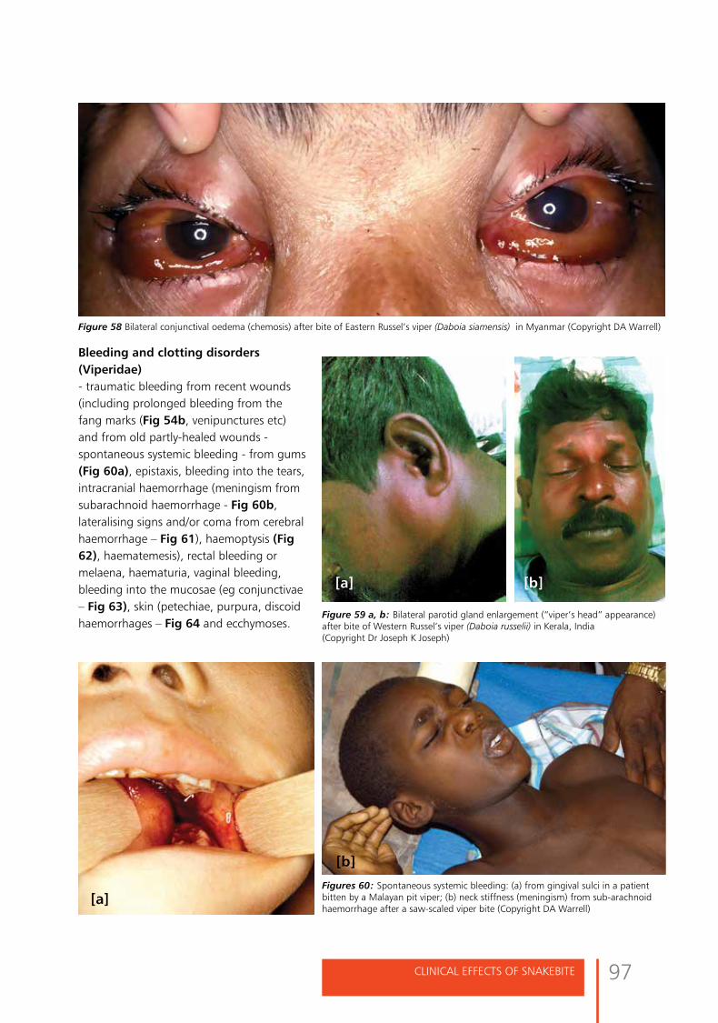
97CliniCal effeCts of snakebite
[A]
Bleeding and clotting disorders (Viperidae)- traumatic bleeding from recent wounds (including prolonged bleeding from the fang marks (Fig 54b, venipunctures etc) and from old partly-healed wounds - spontaneous systemic bleeding - from gums (Fig 60a), epistaxis, bleeding into the tears, intracranial haemorrhage (meningism from subarachnoid haemorrhage - Fig 60b, lateralising signs and/or coma from cerebral haemorrhage – Fig 61), haemoptysis (Fig 62), haematemesis), rectal bleeding or melaena, haematuria, vaginal bleeding, bleeding into the mucosae (eg conjunctivae – Fig 63), skin (petechiae, purpura, discoidhaemorrhages – Fig 64 and ecchymoses.
Figure 58 Bilateral conjunctival oedema (chemosis) after bite of Eastern Russel’s viper (Daboia siamensis) in Myanmar (Copyright DA Warrell)
Figure 59 a, b: Bilateral parotid gland enlargement (“viper’s head” appearance) after bite of Western Russel’s viper (Daboia russelii) in Kerala, India (Copyright Dr Joseph K Joseph)
Figures 60: Spontaneous systemic bleeding: (a) from gingival sulci in a patient bitten by a Malayan pit viper; (b) neck stiffness (meningism) from sub-arachnoid haemorrhage after a saw-scaled viper bite (Copyright DA Warrell)
[a]
[a] [b]
[b]

GUIDELINES FOR THE MANAGEMENT OF SNAKEBITES
98
Figures 61: Strokes after Russell’s viper bites: (a) fatal cerebral haemorrhage in Myanmar (Copyright Dr U Hla Mon); (b) intra cerebral haemorrhages in Maharashtra, India; (c) ischaemic/thrombotic stroke in Maharashtra, India (b, c, Copyright Dr Suvarna Patil)
[a] [b] [c]
Figure 62: Haemoptysis from a tuberculous lung cavity in a patient bitten by a Malayan pit viper (Copyright DA Warrell)
Figure 63: Subconjunctival haemorrhages in a patient bitten by a Burmese Russell’s viper (Copyright DA Warrell)
Figure 64: Cutaneous signs: (a) lymphangitic lines extending towards regional lymph nodes (b) Cutaneous discoid haemorrhages in a patient bitten by a Malayan pit viper in Viet Nam (Copyright DA Warrell)

99CliniCal effeCts of snakebite
[a]
Cerebral arterial thrombosis (Russell’s vipers Daboia russelii and D. siamensis)Thrombotic strokes, confirmed by angiography or imaging, are increasingly recognized after envenoming by D. russelii in India (Fig 61c), Sri Lanka (Gawarammana et al., 2009) and Taiwan.
Neurological (Elapidae, Viperidae eg Russell’s viper D. russelii, Gloydius species)Drowsiness, paraesthesiae, abnormalities of taste and smell, “heavy” eyelids, ptosis (Fig 65a,b), external ophthalmoplegia (Fig 66),
Figures 65a,b: Bilateral ptosis: (a) in a patient bitten by a common krait in Sri Lanka; (b) in a patient bitten by a Russell’s viper in Sri Lankan (Copyright DA Warrell)
Figure 66: External ophthalmoplegia in a patient bitten by a Russell’s viper in Sri Lanka. The patient is attempting to look to his right. The eyes must be held open becauseof the bilateral ptosis (Copyright DA Warrell)
[b]
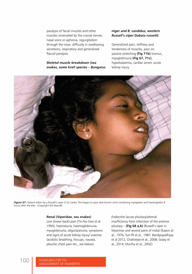
GUIDELINES FOR THE MANAGEMENT OF SNAKEBITES
100
paralysis of facial muscles and other muscles innervated by the cranial nerves, nasal voice or aphonia, regurgitation through the nose, difficulty in swallowing secretions, respiratory and generalized flaccid paralysis.
Skeletal muscle breakdown (sea snakes, some krait species – Bungarus
Figure 67: Patient bitten by a Russell’s viper in Sri Lanka. She began to pass dark brown urine containing myoglobin and haemoglobin 8 hours after the bite. (Copyright DA Warrell)
niger and B. candidus, western Russell’s viper Daboia russelii)
Generalized pain, stiffness and tenderness of muscles, pain on passive stretching (Fig 71b) trismus, myoglobinuria (Fig 67, 71c), hyperkalaemia, cardiac arrest, acute kidney injury.
Renal (Viperidae, sea snakes)Loin (lower back) pain (Tin-Nu-Swe et al. 1993), haematuria, haemoglobinuria, myoglobinuria, oliguria/anuria, symptoms and signs of acute kidney injury/ uraemia (acidotic breathing, hiccups, nausea, pleuritic chest pain etc., see below).
Endocrine (acute pituitary/adrenal insufficiency from infarction of the anterior pituitary – (Fig 68 a,b) (Russell’s viper in Myanmar and several parts of India) (Eapen at al., 1976; Tun-Pe et al., 1987; Bandyopadhyay et al 2012; Chatterjee et al., 2008; Golay et al., 2014; Murthy et al., 2002)

101CliniCal effeCts of snakebite
Figures 68: Haemorrhagic infarction of the anterior pituitary resulting in Sheehan’s-like syndrome (pan-hypopituitarism) after Russell’s viper bite in Myanmar: (a) appearances at the base of the brain at autopsy in a patient who died acutely after the bite (Copyright Dr U Hla Mon); (b) Patient presenting with symptoms and signs of panhypopituitarism three years after severe envenoming by Russell’s viper. There is loss of secondary sexual hair and testicular atrophy (Copyright DA Warrell)
Acute phase: shock, hypoglycaemiaChronic phase (months to years after the bite): weakness, loss of secondary sexual hair, loss of libido, amenorrhoea, testicular atrophy, hypothyroidism etc (Fig 68b).
Hyponatraemia has been observed in victims of krait bites in the area of Hanoi and around Ho Chi Minh City in Viet Nam (Bungarus nr. multicinctus/candidus) (Hung and Höjer 2009; Kiem-Xuan-Trinh et al., 2010; Höjer et al., 2010), implying natriuretic hormone like activity in the venom.
5.3 Clinical syndromes of snakebite in South-East AsiaLimitations of syndromic approach: The more carefully the clinical effects of snakebites are studied, the more it is realised that the range of activities of a particular venom is very wide. For
example, some elapid venoms, such as those of Asian cobras, can cause severe local envenoming (Fig 54d, 54e, 57b, 66), formerly thought to be an effect only of viper venoms. In Sri Lanka and India, Russell’s viper venom causes paralytic signs (ptosis etc.) (Figs 65b, 66, 67), suggesting elapid neurotoxicity, and muscle pains and dark brown urine (Fig 67), suggesting sea-snake rhabdomyolysis. Envenoming by the greater black krait (B. niger) can cause generalized rhabdomyolysis leading to acute kidney injury (Faiz et al., 2010). Although there may be considerable overlap of clinical features caused by venoms of different species of snake, a “syndromic approach” may still be useful, especially when the snake has not been identified and only monospecific antivenoms are available (see Annexes 1 and 2) (Pathmeswaran et al, 2006; Ariaratnam et al., 2009).
[a] [b]

GUIDELINES FOR THE MANAGEMENT OF SNAKEBITES
102
SYNDROME 1: Local envenoming (swelling etc) with bleeding/clotting disturbances = Viperidae (all species)
SYNDROME 2: Local envenoming (swelling etc) with bleeding/clotting disturbances, shock or acute kidney injury = Russell’s viper with conjunctival oedema (chemosis) and acute pituitary insufficiency = Russell’s viper, Myanmar and South India with ptosis, external ophthalmoplegia, facial paralysis etc. and dark brown urine = Russell’s viper, Sri Lanka and South India (?Myanmar)
SYNDROME 3: Local envenoming (swelling etc) with paralysis = cobra or king cobra
SYNDROME 4: Paralysis with minimal or no local envenoming: Bitten on land while sleeping on the ground with/without abdominal pain = krait
Bitten in the sea, estuary and some freshwater lakes = sea snake
Bitten in Indonesia Maluku or West Papua with/without bleeding/clotting disturbance = Australasian elapid
SYNDROME 5: Paralysis with dark brown urine and acute kidney injury:
Bitten on land (with bleeding/clotting disturbance) = Russell’s viper, Sri Lanka or South India
Bitten on land while sleeping indoors = krait (B. niger, B. candidus, B. multicinctus), Bangladesh, Thailand
Bitten in the sea, estuary and some freshwater lakes (no bleeding/clotting disturbances) = sea-snake
5.4 Long term complications (sequelae) of snakebiteAt the site of the bite, loss of tissue may result from sloughing or surgical débridement of necrotic areas or amputation: chronic ulceration, infection, osteomyelitis or arthritis may persist causing severe physical disability (Fig 70a). Malignant transformation may occur in skin ulcers after a number of years (Marjolin’s ulcer) (Fig 70b).
Chronic kidney disease (renal failure) may occur after bilateral cortical necrosis (Russell’s viper and hump-nosed pit viper bites) (Ariaratnam et al., 2008b); and
Figures 70: Chronic physical handicap resulting from necrotic envenoming by Malayan pit vipers: (a) Deformity and dysfunction after a snakebite and subsequent necrosis of the calf; (b) Squamous cell carcinoma arising at the site of a chronic skin ulcer with osteomyelitis 8 years after the bite (Copyright DA Warrell)
[a] [b]

103CliniCal effeCts of snakebite
chronic panhypopituitarism or diabetes insipidus after Russell’s viper (Daboia russelii and D. siamensis) bites in India (Golay et al., 2014) and Sri Lanka and, most commonly, in Myanmar, (Fig 68b). Chronic neurological deficit is seen in patients who survive intracranial haemorrhages and thromboses (Viperidae).
Abnormalities of electrophysiological tests can persist for over 12 months following elapid bites in Sri Lanka (Bell et al, 2010).
Delayed psychological morbidity has been reported one to four years after snakebite envenoming in Sri Lanka. Depression and anxiety, impaired functioning, post-traumatic stress disorder and unexplained residual physical disability were reported (Williams et al, 2011). Chronic musculoskeletal disabilities have been reported following snake envenoming in Sri Lanka. The disabilities involved mostly the lower limbs. They ranged from swelling, muscle wasting, stiff joints, reduced muscle power, impaired balance, fixed deformities, chronic non-healing
ulcer, and persisting lump to amputations. (Jayawardene et al., submitted for publication). Unexplained residual symptoms have also been reported following snakebite in Tamil Nadu, India (Vaiyapuri et al, 2013).
5.5 Symptoms and signs of sea-snake envenoming(Reid, 1979; Warrell, 1994)Envenoming by sea snakes (Hydrophiinae) and sea kraits (Laticaudinae): the bite is usually painless and may not be noticed by the wader or swimmer. Fangs and other teeth may be left in the wound.
Figures 71: Sea-snake bite in North-west Malaysia: (a) Ptosis, facial paralysis and trismus; (b) Generalised myalgia making passive movement of the limbs extremely painful; (c) myoglobinuria (Copyright the late H Alistair Reid)
[a] [b]
[c]

GUIDELINES FOR THE MANAGEMENT OF SNAKEBITES
104
There is minimal or no local swelling and involvement of local lymph nodes is unusual. Generalized rhabdomyolysis is the dominant effect of envenoming by these snakes although patients without this feature have been described. Early symptoms include headache, a thick feeling of the tongue, thirst, sweating and vomiting. Generalized aching, stiffness and tenderness of the muscles becomes noticeable between 30 minutes and 3½ hours after the bite. Trismus is common (Fig 71a). Passive stretching of the muscles is painful (Fig 71b). Later, there is progressive flaccid paralysis starting with ptosis, as in other neurotoxic envenomings. The patient remains
conscious until the respiratory muscles are sufficiently affected to cause respiratory failure. Myoglobinaemia and myoglobinuria develop 3–8 hours after the bite (Fig 71c). These are suspected when the serum/plasma appears brownish and the urine dark reddish brown (‘Coca-Cola-coloured’). Bedside ‘sticks’ tests will appear positive for haemoglobin/blood in urine containing myoglobin. Myoglobin and potassium released from damaged skeletal muscles may contribute to acute kidney injury, while hyperkalaemia developing within 6–12 hours of the bite may precipitate cardiac arrest.
5.6 Symptoms and signs of cobra-spit ophthalmia(eye injuries from spitting cobras) (Fig 72)If the “spat” venom enters the eyes, there is immediate and persistent intense burning, stinging pain, followed by profuse watering of the eyes with production of whitish discharge, congested conjunctivae, spasm and swelling of the eyelids, photophobia, clouding of vision and temporary blindness. Corneal ulceration, permanent corneal scarring and secondary endophthalmitis are recognized complications of African spitting cobra venom but have not been described in Asia.
Figure 72: Bilateral conjunctivitis in a patient who had venom spat into both eyes by an Indo-Chinese spitting cobra
(Naja siamensis) (Copyright DA Warrell)
If the “spat” venom enters the eyes,
there is immediate and persistent
intense burning, stinging pain, followed
by profuse watering of the eyes with
production of whitish discharge, congested
conjunctivae, spasm and swelling of the
eyelids, photophobia, clouding of vision
and temporary blindness.

105ManageMent of snakebites in south-east asia
Management of snakebites in South-East Asia

GUIDELINES FOR THE MANAGEMENT OF SNAKEBITES
106

107ManageMent of snakebites in south-east asia
Management of snakebites in South-East AsiaEssentials:First-aid should be carried out immediately after the bite, by the victim or others present. Most traditional methods are useless and harmful. To prevent deaths from respiratory paralysis or shock before victims reach medical care, delay venom spread with pressure-pad or pressure-bandage plus immobilization, accelerate transport to hospital (improve ambulance services), and train accompanying medical workers in airway management, assisted ventilation and resuscitation. The snake is valuable evidence, but should not be pursued, killed or handled. Close-up mobile ‘phone images ’ are useful.
Recommended first-aid: reassurance, immobilization of the whole patient, especially their bitten limb, accelerated transport to medical care (summoned by emergency telephone helpline) ideally in recovery position. Unless neurotoxic elapid bite can be excluded, apply pressure-pad (simpler, more practicable than pressure-bandage immobilization). Never use, recommend or condone tight (arterial) tourniquets.
Medical treatment in dispensary or hospitalRapid clinical assessment accompanies urgent resuscitation of shocked or
6asphyxiated patients. Clear airway, give oxygen, establish intravenous access, monitor vital signs. Beware of removing tight tourniquets before antivenom and resuscitation facilities are available.
Key questions: “Where (in which part of your body) were you bitten? Point to the place”; “When were you bitten and what were you doing then?”; “Where is the snake that bit you?” or “What did it look like; did anyone take a picture?”; “How are you feeling now?”
Examination of the bitten part: extent of tenderness/swelling, lymph nodes draining bitten limb, early signs of necrosis (blistering, demarcated altered pigmentation, putrefaction odour. General: blood pressure (postural drop indicates hypovolaemia), gingival sulci, nose, skin and mucosae for evidence of bleeding, chemosis, abdominal tenderness, loin pain and tenderness, meningism, lateralising neurological signs (asymmetrical pupils), impaired consciousness, bilateral ptosis, diplopia, external ophthalmoplegia, mouth opening/closing, trismus, tongue protrusion, facial muscles, gag reflex neck flexors “broken neck sign”, swallowing, “paradoxical respiration”, fasciculations or myokymia,

GUIDELINES FOR THE MANAGEMENT OF SNAKEBITES
108
measure ventilatory capacity. If adequately ventilated, totally paralysed patient are fully conscious and can communicate by flexing a digit. Conventional tests of brain death misleading. Generalised tender, painful muscles and dark brown urine suggest rhabdomyolysis. Pregnant women: suspect antepartum or postpartum haemorrhage, vaginal bleeding, premature labour, fetal distress, intra-uterine fetal death, abortion/stillbirth.
Species diagnosis: expert identification of dead snake or a mobile ‘phone photo image is useful. Otherwise, infer species from the patient’s description and circumstances of bite (e.g. nocturnal bite while sleeping on ground suggests krait) and clinical syndrome.
Investigations/laboratory tests: 20 minute whole blood clotting test (20WBCT) is a simple, informative bedside test requiring only a new, clean, dry, ordinary glass tube, bottle, vial or syringe. Positive (non-clotting) result indicates severe consumption coagulopathy and need for immediate antivenom treatment. False positive (non-clotting) 20WBCT results from use of plastic, polystyrene or polypropylene rather than ordinary glass, or glass cleaned with detergent, soap or washing fluid that destroy surface-activation of blood coagulation. If new glass tubes not available, re-use ordinary glass vessels (e.g. antibiotic bottles), washed with “normal 0.9% saline” for intravenous infusion, without detergent or other cleaning agent, dried in hot air.
Other more sensitive laboratory tests of blood coagulation: International Normalized Ratio (INR) based on prothrombin time (PT) (> or =1.2 is abnormal), activated partial thromboplastin time (aPPT), fibrin(ogen) related antigens (fibrin degradation products -FDP) or D-dimer. Point-of-care (bedside) devices for
measuring INR and D-dimer unreliable in snakebite victims.
Other laboratory tests: haemoglobin concentration/haematocrit, thrombocytopenia, neutrophil leucocytosis, fragmented red cells (“helmet cell”, schistocytes) signifying microangiopathic haemolysis. Observe spontaneously sedimented plasma for haemoglobinaemia/myoglobinaemia.
Biochemical abnormalities: plasma creatinine, urea/blood urea nitrogen and potassium concentrations raised in acute kidney injury (Russell’s viper, hump-nosed pit-viper, sea-snake envenoming). Elevated aminotransferases and muscle enzymes (creatine kinase, aldolase etc.) indicate local and generalized muscle damage (sea snakes, some kraits, some Australasian Elapidae and Russell’s viper bites). Hyponatraemia associated with krait bites.
Urine examination: dipsticks test for blood, haemoglobin or myoglobin and proteinuria. Microscopy to detect erythrocytes and red cell casts, indicating glomerular bleeding, eosinophilia suggesting acute interstitial nephritis.
Other investigations: chest radiography for detecting pulmonary oedema, haemorrhages, infarcts, pleural effusions, secondary bronchopneumonia; ultrasound for assessing local envenoming, deep vein thrombosis, pleural and pericardial effusion and bleeding; echocardiography for myocardial dysfunction; CT and MRI imaging for intracranial and spinal haemorrhages and infarcts and osteomyelitis at the bite site; ECG for arrhythmias, myocardial damage, evidence of hyperkalaemia.
Antivenom treatmentAntivenom (= antivenin, anti-snakebite serum, ASV etc.) is the only specific

109ManageMent of snakebites in south-east asia
antidote to snake venom. A most important decision in managing snakebite victims is whether or not they need antivenom. It is immunoglobulin [usually pepsin-refined F(ab’)2 fragments of whole IgG] purified from plasma of horses or sheep hyperimmunised with venoms of one (to make mono-specific antivenom) or more (to make poly-specific antivenom) snake species, medically the most important species in a particular geographical area. Paraspecific neutralization of venoms of closely related species is possible but not certain. Indian “polyvalent anti-snake venom serum” (ASV) is raised against venoms of their “big four” species: spectacled cobra (N. naja); common krait (B. caeruleus), Russell’s viper (D. russelii), saw-scaled viper (E. carinatus), but other species, now recognized as being important, are not covered. Thai Red Cross Society manufactures “neuro polyvalent snake antivenin” covering 4 elapids and “haemato polyvalent snake antivenin” covering 3 vipers.
Design, quantity, quality, stability, safety and dosage guidelines of antivenoms require review and improvement.
Antivenom treatment is indicated if/when patients with proven/suspected snakebite develop one or more of the following signs. Systemic envenoming: haemostatic abnormalities [spontaneous systemic bleeding, coagulopathy (+ve non-clotting 20WBCT, INR >1.2, or prothrombin time >4-5 seconds longer than control), or thrombocytopenia; neurotoxicity (bilateral ptosis, external ophthalmoplegia, paralysis etc.); cardiovascular abnormalities (hypotension, shock, cardiac arrhythmia, abnormal ECG); Acute kidney injury (oliguria/anuria, rising blood creatinine/urea); haemoglobin-/myoglobin-uria (dark brown/black urine, +ve urine dipsticks, other evidence of intravascular
haemolysis/generalized rhabdomyolysis); supporting laboratory evidence. Local envenoming: local swelling involving more than half bitten limb (in absence of tourniquet) within 48 hr of the bite; swelling after bites on digits; rapid extension of swelling beyond wrist/ankle within few hours of bites on hand/foot); enlarged tender lymph node draining bitten limb
Antivenom should be given only when benefits exceeds risks. Since it is expensive and in limited supply, it must not be used indiscriminately. It should be given as soon as it is indicated and as long as anti-haemostatic abnormalities persist, even two weeks after the bite. It is unlikely to prevent/limit local tissue damage unless given within a few hours of the bite.
Antivenom reactions: many patients develop early (within a few hours) or late (after 5 days or more) reactions. Depending on type of antivenom and dose, the incidence may be as high as 81% (43% severe) of early anaphylactic or pyrogenic reactions or as low as 3.5%. IgE-mediated Type I hypersensitivity after previous exposure to equine serum is uncommon.
Early anaphylactic reactions (1 - 180 minutes after starting antivenom) can have all classic features of anaphylaxis from urticaria to life-threatening shock, bronchospasm and angio-oedema. Most are attributable to complement activation by IgG aggregates/residual Fc fragments,
A most important decision in managing
snakebite victims is whether or not they
need antivenom.

GUIDELINES FOR THE MANAGEMENT OF SNAKEBITES
110
or stimulation of mast cells/basophils by antivenom proteins.
Pyrogenic (endotoxin) reactions (within 1-2 hours) involve rigors, fever (risk of febrile convulsions in children), vasodilatation, hypotension and a fall in blood pressure. Attributable to pyrogen contamination during manufacture.
Late (serum sickness-type) reactions (1-12, mean 7, days after treatment) involve fever, nausea, vomiting, diarrhoea, itching, recurrent urticaria, arthralgia, myalgia, lymphadenopathy, periarticular swellings, mononeuritis multiplex, proteinuria, immune complex nephritis, rarely encephalopathy.
Prediction: skin and conjunctival “hypersensitivity” tests, often recommended in “package inserts” predict acquired IgE-mediated Type I hypersensitivity to horse/sheep proteins, but not large majority of early (anaphylactic) or late (serum sickness type) antivenom reactions. Their use is strongly discouraged.
Prevention: A powerful, well-designed study in Sri Lanka showed that adrenaline (0.25 ml/mg of 0.1% solution subcutaneously) given before antivenom was started reduced severe reactions by 43% (95% CI 25–67) at 1 h and by 38% (95% CI 26–49) up to and including 48 h after antivenom administration; hydrocortisone (200 mg intravenously) and promethazine (25 mg intravenously) were ineffective. Hydrocortisone negated benefit of adrenaline. Dilution and slow infusion (10-120 minutes) does not alter risk of reactions.
At the earliest sign of early anaphylactic antivenom reaction, temporarily suspend antivenom administration and give
intramuscular injection of 0.1% adrenaline, initial adult dose 0.5 ml/mg (0.01 mg/kg body weight for children). Since severe, life-threatening anaphylaxis can evolve rapidly, adrenaline must be available at the bed-side and should be given at the first sign of anaphylaxis (e.g. when itching, tachycardia, restlessness develop and a few spots of urticaria appear). Repeat adrenaline every 5-10 minutes if reaction persists or worsens. Adrenaline safe in pregnancy. In case of asthmatic symptoms, given salbutamol bronchodilator. After adrenaline, give intravenous antihistamine anti-H1 blocker (e.g. chlorphenamine maleate, adults 10 mg, children 0.2 mg/kg intravenously) and hydrocortisone (adults 100 mg, children 2 mg/kg body weight). Anaphylactic shock unresponsive to adrenaline is treated by laying patient supine, legs elevated, and giving intravenous volume replacement (0.9% saline, adults 1-2 litres rapidly). Consider intravenous adrenaline infusion (adults 1mg/1.0 ml of 0.1% solution in 250 ml IV fluid) infused at 15–60 drops/min using micro-dropper burette chamber, or dopamine. Unresponsive bronchospasm or angioedema is treated with optimal nebulised/inhaled and/or parenteral bronchodilator and oxygen.
Pyrogenic reactions are treated by physical cooling (undressing, tepid sponging, fanning) and giving antipyretic paracetamol. Intravenous fluids should be given to correct hypovolaemia. Patients with features of anaphylaxis should be given adrenaline.
After recovery from early reactions, cautiously resume and complete administration of the dose of antivenom.
Late (serum sickness) reactions respond to 5-day course of oral antihistamine or, failing that, 5-day course of prednisolone.
111ManageMent of snakebites in south-east asia
Supply, selection, storage and shelf-life of antivenom
Polyvalent (polyspecific) antivenoms are preferred in many countries because of problems with specific diagnosis. Antivenom should be given only if its stated range of specificity and paraspecific neutralization includes the species known or suspected to have been responsible for the bite. Lyophilised antivenoms (shelf life about 5 years) are stored <25ºC, liquid antivenoms (shelf life 2-3 years) at 2-8 ºC. Ideally, antivenoms should be used before stated expiry dates, antivenoms retain useful activity for months or even years after these dates. In patients with severe envenoming, recently expired antivenoms may be considered if there is no alternative.
Intravenous administration is mandatory, either by slow “push” injection (maximum 2ml/minute) or by intravenous infusion, diluted in 5 ml of isotonic fluid/kg body weight, over 30-60 minutes. Intramuscularly administered antivenom is poorly bioavailable. This route should be considered only in the absence of anyone capable of giving an intravenous injection.
Initial dose of antivenom:Guidance on average initial dose of a particular antivenom, appropriate for envenoming of different severities, in patients bitten by different species based on clinical trials is rarely available. Usually, dosages quoted on manufacturers’ package inserts are based on laboratory mouse LD50s and ED50s which are unreliable clinically. In practice, choice of initial dose is usually empirical. Regimens based on a higher initial (loading) dose are more logical than low doses repeated over several days.
Antivenom failure (ineffectiveness) may be due to batch-to-batch variations in potency.
Geographical intra-species variation, in venom composition and antigenicity, explains why antivenoms raised against venom of a species (e.g. Russell’s viper) from one area (e.g. Tamil Nadu, India) may not be effective in patients envenomed by the same species elsewhere in its geographical range (e.g. Kerala, Maharashtra). Regional polyspecific antivenoms may not cover all medically important snakes (e.g. N. kaouthia, H. hypnale, T. (Craspedocephalus) malabaricus, T (T.) erythrurus in parts of India).
Response to antivenom includes rapid decrease in general malaise (?placebo effect), cessation of spontaneous systemic bleeding (in 15-30 minutes), restoration of blood coagulability (in 3-9 hours), normalisation of blood pressure in shocked patients (in 30-60 minutes) and sinus rhythm, reversal of post-synaptic type neurotoxicity (after cobra bite) (after 30 minutes), cessation of active haemolysis and rhabdomyolysis (within a few hours).
After an initial response to antivenom, signs of systemic envenoming may recur within 24-48 hours. Improved perfusion of the bite site following correction of shock/hypovolaemia increases venom absorption from bite site “depot” and redistribution of venom from tissues to vascular space at a time when therapeutic antivenom has been eliminated.
After the first dose of antivenom, the initial dose should be repeated 6 hours later if blood remains incoagulable, or 1 hour later if spontaneous systemic bleeding continues, or neurotoxic or cardiovascular signs persist or deteriorate. However, further doses of antivenom have no proven value in paralysed patients who are being ventilated.
When no appropriate antivenom is available, patients must be treated

GUIDELINES FOR THE MANAGEMENT OF SNAKEBITES
112
conservatively. Respiratory paralysis: try anticholinesterases, but without delaying assisted ventilation. Haemostatic abnormalities: rest in bed to avoid trauma, transfuse cryoprecipitate, fresh frozen plasma, fresh whole blood and platelets. Shock, myocardial damage: correct hypovolaemia and consider vasopressor drugs. In hypotension with bradycardia, try atropine. Acute kidney injury: conservative treatment or renal replacement therapy. Rhabdomyolysis: correct hypovolaemia with intravenous fluid, acidosis with sodium bicarbonate. Severe local envenoming: surgical intervention, prophylactic broad spectrum antimibiotics.
Supportive/ancillary treatmentNeurotoxic envenomingIndications for intubation: imminent respiratory arrest (respiratory distress, breathing absent/inadequate); neck muscle weakness with shallow respiration or paradoxical breathing; secretions pooling in pharynx with loss of gag/cough reflexes; upper airway obstruction with stridor (secondary to anaphylaxis angioedema); oxygen saturation <90% (equivalent to Pa02 <60 mmHg) despite high flow oxygen; respiratory acidosis (hypoxia PaO2 < 60 mm Hg with PaCO2 > 45 mm Hg)
Insert cuffed endotracheal tube using laryngoscope under sedation,or laryngeal mask airway or i-gel supraglottic airway.
For acute airway obstruction (e.g. angioedema) perform cricothyroidotomy.
Resuscitation: if patient is unresponsive, summon assistanceby ‘phone, open and maintain airway using “head tilt - chin lift” or “jaw thrust” manoeuvres to improve air flow by dislodging tongue and reopening upper airway. Remove foreign material from upper airway (suction or forceps, not finger) and insert oropharyngeal (Guedel) airway. Administer oxygen by any available means and assess breathing. If breathing is adequate, lay victim in recovery position (important because of venom-induced vomiting, bleeding and hypersalivation with loss of airway-protective reflexes), but check breathing every 2 minutes.
If breathing is inadequate or not discernible, respiratory rate low, respirations shallow, patient taking agonal (gasping) breaths, central cyanosis, finger oximeter oxygen saturation <90%, end-tidal PCO2 high or increasing - assisted ventilation is required.
Techniques: If no health facilities are immediately available - Expired Air Resuscitation
In health care centre - Non-invasive ventilation using bag-mask/bag-mask-valve ventilation
In hospital - Invasive Ventilation (intubation and mechanical ventilation) for Type I respiratory failure (primary failure of oxygenation as in pulmonary oedema in Russell’s viper envenoming) and Type II respiratory failure (primary ventilatory failure due to respiratory muscle paralysis or obstruction as in elapid envenoming).
Indications for invasive ventilation: patients who, despite receiving oxygen by mask or nasal catheters remain tachpnoeic (respiratory rate > 25/min), have
if patient is unresponsive, summon
assistanceby ‘phone, open and maintain
airway using “head tilt - chin lift” or “jaw
thrust” manoeuvres to improve air flow
by dislodging tongue and reopening
upper airway.

113ManageMent of snakebites in south-east asia
paradoxical respirations and deteriorating ventilatory capacity, hypoxia, respiratory acidosis. Sedate them with fentanyl and midazolam, or morphine and lorazepam. Ensure adequate hydration, nutrition, physiotherapy, regular turning to prevent pressure areas and lung collapse, eye care and safe weaning from ventilation.
Trial of anticholinesterase: after making baseline observations, give atropine sulphate intravenously followed by intramuscular neostigmine (or, ideally, slow intravenous injection of short-acting edrophonium chloride – Tensilon, but rarely available), and observe over next 10-60 minutes for disappearance of ptosis or improved ventilatory capacity. Treat patients who respond convincingly with repeated neostigmine and atropine.
Hypotension and shockPrimary causes are anaphylaxis, vasodilatation, cardiotoxicity, hypovolaemia; secondary causes are antivenom reactions, respiratory failure, acute pituitary adrenal insufficiency, septicaemia
Measure postural change in blood pressure or passive leg raising test to determine fluid responsiveness. Control fluid therapy using jugular/central venous pressure, respiratory rate, detection of crepitations. Selective vasoconstrictor such as dopamine may be used.
Acute pituitary/adrenal insufficiency may occur in victims of Russell’s viper bites (Myanmar, India, Sri Lanka). Hydrocortisone is life-saving in these cases.
Oliguria and acute kidney injury (AKI) Impending AKI signalled by abrupt reduction in kidney function over 48 hours: decreasing/no urine output, increased/increasing serum creatinine concentration, clinical “uraemia syndrome”
(nausea, vomiting, acidotic breathing, hiccups, fetor, drowsiness, confusion, coma, flapping tremor, muscle twitching, convulsions, pericardial friction rub, signs of fluid overload). Patients with any of these features should be monitored for other clinical signs of “uraemic syndrome”, pulse rate, postural blood pressure, height of jugular venous pulse, respiratory rate, temperature, auscultation of lung bases for crepitations, fluid balance chart and/or daily weight.
Most patients with AKI become oliguric (urine output < 400 ml/day or < 30 ml (children < 0.5ml/kg bodyweight /hour). Conservative management may tide the patient over, avoiding the need for renal replacement therapy (dialysis).
In patients with intravascular volume depletion: establish intravenous access, give cautious fluid challenge (adult 250-500 ml of isotonic saline over one hour). Stop challenge immediately if pulmonary oedema develops. If the urine output does not improve, perform furosemide stress/challenge test, if that fails to increase urine output, switch to conservative management.
Conservative management of AKI: no further diuretics, restrict fluid intake to previous day’s output plus “insensible losses” (500-1000 ml/day) and refer to renal unit. Diet bland, high calorie, normal protein, low potassium, low in salt.
Measure serum/plasma urea, creatinine and electrolytes daily until renal failure is resolving. Examine ECG for evidence of hyperkalaemia, for which emergency treatments include calcium gluconate, β2 agonist aerosol inhaler, 50% dextrose with insulin and sodium bicarbonate, followed by a low potassium diet. Severe acidosis is corrected with sodium bicarbonate.

GUIDELINES FOR THE MANAGEMENT OF SNAKEBITES
114
Renal replacement therapy/dialysis Patient with clinical uraemia (encephalopathy, pericarditis etc.), fluid overload not responding to diuretics, hyperkalaemia or hyperkalaemic ECG changes, symptomatic acidosis, raised plasma/serum creatinine and/or urea should be transferred to a health facility where they can receive renal replacement therapy.
In patients with AKI associated with thrombotic microangiopathy, there is no evidence for plasmapheresis using cryosupernatant. In patients with myoglobinuria or haemoglobinuria, correct hypovolaemia, maintain urine output 200-300 ml/hour, correct severe acidosis with bicarbonate, promote alkalinise diuresis, continuing until CK level <5000 U.
Diuretic phase of AKI: some patients develop persistent polyuria of 5-10 litres/24 hours and may become volume and electrolyte depleted.
Chronic kidney disease: oliguria and dialysis-dependence for more than 4 weeks demands referral to nephrologist to consider kidney biopsy.
Haemostatic disturbances: usually corrected by antivenom, but recurrent coagulopathy is possible. Exceptionally, in patients already treated with antivenom but who have persistent severe bleeding or require urgent surgery, restoration of coagulability can be accelerated with blood products.
Treatment of the bitten part:Nurse painful, swollen limb in the most comfortable position, avoiding excessively elevation. Leave blisters, aspirate abscesses and culture pus. Necrotic tissue demands early surgical débridement and split-skin grafting. Primary and
secondary infections involve aerobic and anaerobic bacteria from the snake’s oral flora, patient’s skin and asterile incisions. Tetanus toxoid booster and prompt antibiotic treatment of necrotic wounds is recommended.
Compartmental syndromes are often misdiagnosed and fasciotomies performed unnecessarily. Since classical signs of compartment syndrome are difficult to assess in snake-bitten limbs, direct measurement of intracompartmental pressure is mandatory and fasciotomy must not be attempted before haemostatic disturbances have been corrected by antivenom.
Rehabilitation: conventional physiotherapy accelerates functional recovery of the bitten limb, but is often forgotten.
Discharge assessment: discussion, encouragement, follow-up, warning about late serum sickness-type reactions and advice about reducing risk of future snakebites are important.
Management of cobra spit ophthalmia: first aid is irrigation of affected eyes and other mucous membranes with liberal quantities of water; medical treatment involves analgesia, exclusion of corneal abrasions and application of prophylactic topical antibiotics.
Management of snakebites at different levels of the health service: all levels of the health service can contribute to the management of patients with suspected snakebite. In rural areas, where snakebites are most common, transfer to a hospital may not be feasible, and so a lower level of health facility must cope with these medical emergencies. Training of doctors, nurses, dispensers, health assistants and paramedics in

115ManageMent of snakebites in south-east asia
diagnosis (including snake identification), early management and indications for and practicalities of administering antivenom is essential.
Snakebite treatment in developing countries is challenging, creating many management problems. It is, therefore, of utmost importance to train doctors and other health- care workers in the effective care of snakebite patients.
6.1 Stages of managementThe following steps or stages are often involved:
Management of snakebite
• First-aid treatment
• Transport to hospital
• Rapid clinical assessment andresuscitation
• Detailed clinical assessment andspecies diagnosis
• Investigations/laboratory tests
• Antivenom treatment
• Observing the response toantivenom
• Deciding whether further dose(s)of antivenom are needed
• Supportive/ancillary treatment
• Treatment of the bitten part
• Rehabilitation
• Treatment of chroniccomplications
• Advising how to avoid future bites
6.2 First-aid treatment6.2.1 Principles of first-aidFirst-aid treatment is carried out immediately or very soon after the bite, before the patient reaches a dispensary or hospital. It can be performed by the snakebite victim himself/herself or by anyone else who is present and able.
Unfortunately, most of the traditional, popular, available and affordable first aid methods have proved to be useless or even frankly dangerous. These methods include: making local incisions or pricks/punctures (“tattooing”) at the site of the bite or in the bitten limb, attempts to suck the venom out of the wound, use of (black) snake stones, tying tight bands (tourniquets) around the limb, electric shock, topical instillation or application of chemicals, herbs or ice packs. Local people may have great confidence in traditional (herbal) treatments, but they must not be allowed to delay medical treatment or to do harm.
Aims of first aid
• reassure the snakebite victim
• attempt to delay systemicabsorption of venom
• preserve life and preventcomplications before the patientcan receive medical care at adispensary or hospital
• control distressing or dangerousearly symptoms of envenoming
• arrange the transport of thepatient to a place where theycan receive medical care
ABOVE ALL, AIM TO DO NOHARM!
6.2.2 The danger of respiratory paralysis and shockThe greatest fear is that a snakebite victim might develop fatal respiratory paralysis or shock before reaching a place where they may be resuscitated (Looareesuwan et al., 1988). This risk may be reduced by speeding up transport to hospital, by improving free ambulance services (Gimkala et al., 2016) or by recruiting village-based motor cyclist volunteers who transport the victim propped

GUIDELINES FOR THE MANAGEMENT OF SNAKEBITES
116
upright between the driver in front and a supporting pillion passenger behind. This has proved effective in villages in the Nepal Terai (Sharma et al., 2013) (Figure 73). Medical workers can be trained in airway management and assisted ventilation (see below). The special danger of rapidly developing paralytic envenoming after bites by some elapid snakes has prompted the use of pressure-bandage immobilization (Sutherland et al., 1979) and pressure-pad immobilization (Anker 1982; Tun-Pe et al.,1995b )(ANNEX 4). Pressure bandage immobilization requires equipment (long elasticated bandages and splints) (Canale et al., 2009; Currie et al., 2008 ) and skill.
As far as the snake is concerned - do not attempt to kill it as this may be dangerous.However, if the snake has already been killed, it should be taken to the dispensary or hospital with the patient in case it can be identified. However, do not handle the snake with your bare
Figure 73: Evacuation of a snakebite victim showing early signs of paralysis by a village-based motorcycle volunteer in the Terai region of Nepal. The victim is supported between the driver and a pillion passenger (Copyright Dr Sanjib Sharma) [CAUTION - If this method of transport has to be employed, because of local conditions and the terrain, ideally helmets should be worn and the legs of the passengers protected from exhaust burns]
hands as even a severed head can bite! Several close-up mobile ‘phone images of the snake should be taken if possible to allow expert identification.
MOST TRADITIONAL FIRST-AID METHODS SHOULD BE DISCOURAGED:THEY DO MORE HARM THAN GOOD!
6.2.3 Recommended first-aid methods• Reassure the victim who may be
very anxious. Reassurance will drive away their fear and excitement, slow the patient’s heart rate and reduce the spread of venom. Grounds for reassurance include the possibility of a “dry bite” even if the snake was venomous, the usually slow evolution of severe envenoming allowing time for treatment, and the effectiveness of modern medical management of snakebite.
• Immobilize the whole of the patient’s body by laying him/her down in a comfortable and safe position, ideally in the recovery position (lying prone on the left side in case vomiting threatens to result in aspiration), and immobilize the bitten limb with a splint or sling. Any movement or muscular contraction, even undressing or walking, will increase absorption and spread of venom by squeezing veins and lymphatics.
• Unless the possibility of an elapid bite can confidently be excluded, apply pressure-pad immobilization (See ANNEX 4), or, if the necessary equipment and skills are available, pressure-bandage immobilization. In Myanmar, the pressure-pad method has proved effective in reducing spread of venom in victims of Russell’s viper bite (Tun Pe et al., 1995b). Pressure-bandage immobilization has not become widely used in this Region,

117ManageMent of snakebites in south-east asia
because provision of the necessary equipment (long, wide elasticated bandages), training and skills required to apply it safely and reliably have proved impossible to achieve. The pressure-pad immobilization method is preferred and recommended as being simpler and more practicable (O).
• Avoid any interference with the bite wound (incisions, rubbing, vigorous cleaning, massage, application of herbs or chemicals) as this may introduce infection, increase absorption of the venom and increase local bleeding.
CAUTION Delay the release of tight bands, bandages and ligatures: if the patient has already applied these very popular methods of first-aid, they should not be released until the patient is under medical care in hospital, medical staff and resuscitation facilities are available and antivenom treatment has been started (Watt et al., 1988) see Caution below.
Tight (arterial) tourniquets must never be recommended or condoned! Traditional tight (arterial) tourniquets. If applied tightly around the upper part of the limb, these bands, bandages or ligatures are extremely painful as the limb becomes ischaemic and are very dangerous if left in place for long periods. Many gangrenous limbs have resulted! (Fig 74)
Figure 74: Gangrenous limb caused by a tight tourniquet after a bite by Malayan pit viper (Copyright DA Warrell)
6.3 Transport to hospitalEmergency Helpline NumbersLinkage with an emergency helpline number (e.g. in India 108 see http://www.emri.in/) can speed up the transport of a patient to a higher referral centre when emergency treatment is required. This will decrease delays in accessing emergency care and reduce mortality. Information about helpline numbers could be widely disseminated.
The patient must be transported to a place where they can receive medical care (dispensary or hospital) as quickly, but as safely and comfortably as possible. Any movement, but especially movement of the bitten limb, must be reduced to an absolute minimum to avoid increasing the systemic absorption of venom. Any muscular contraction will increase spread of venom from the site of the bite in veins and lymphatics. Where a conventional motor vehicle ambulance is not available or feasible, stretcher, bicycle, motorbike (Patel and Ekkiswata, 2010) - by recruiting village-based motorbike owners/cyclists (Sharma et al., 2013) (Fig 73), cart, horse, train or boat may have to be considered, or the patient can be carried (e.g. using the “fireman’s lift” method). If possible, patients should be placed in the recovery position during transit, in case they vomit.

GUIDELINES FOR THE MANAGEMENT OF SNAKEBITES
118
6.4 Treatment in the dispensary or hospital (Warrell 1990; 1995)
Medical management of envenomed patients in dispensary or hospital
• Rapid primary clinical assessment and resuscitation
• Detailed clinical history, physical examination and species diagnosis
• Simple test of blood coagulability [20-minute-whole-blood-clotting-test (20WBCT)]
• Antivenom: indications, initial and repeated dosage, response, reactions
• Treatment of organ and system failures
• Treatment of the bitten limb
• Rehabilitation, restoration of function
• Advice at discharge from hospital and follow-up
• Preventive health education towards reducing risk of future bites
6.4.1 Rapid primary clinical assessment and resuscitationSnakebite is a medical emergency: history, symptoms and signs must be obtained rapidly so that appropriate, urgent and life-saving treatment can be given
Cardiopulmonary resuscitation may be needed, including administration of oxygen and establishment of intravenous access.
Rapid primary clinical assessment and resuscitation: ABCDE approach
Airway
Breathing (respiratory movements)
Circulation (arterial pulse)
Disability of the nervous system (level of consciousness)
Exposure and environmental control (protect from cold, risk of drowning etc)
Airway patency, respiratory movements, arterial pulse and level of consciousness must be checked immediately. Record the vital signs.
The Glasgow Coma Scale cannot be used to assess the level of consciousness of patients paralysed by neurotoxic venoms (see below).
Clinical situations in which snakebite victims might require urgent resuscitation: a) Profound hypotension and shock:
resulting from direct cardiovascular effects of the venom or secondary effects, such as hypovolaemia, release of inflammatory vasoactive mediators, haemorrhagic shock or rarely primary anaphylaxis induced by the venom itself.
b) Terminal respiratory failure: from progressive neurotoxic envenoming that has led to paralysis of the respiratory muscles; or simple airway obstruction.
c) Respiratory distress: from generalized increase in capillary permeability (Russell’s viper bite victims)

119ManageMent of snakebites in south-east asia
d) Sudden deterioration: or rapiddevelopment of severe systemicenvenoming after releasing a tighttourniquet or compression bandage(see Caution above).
e) Cardiac arrest: precipitated byhyperkalaemia resulting from skeletalmuscle breakdown (rhabdomyolysis)after bites by sea snakes, certain kraitsand Russell’s vipers.
f) Late results of severe envenoming:in someone arriving days/weeks after thebite: with severe bleeding/blood clottingdisturbances, acute kidney injury orsepticaemia complicating local necrosis.
6.4.2 Detailed clinical assessment and species diagnosis
6.4.2.1 HistoryA precise history of the circumstances of the bite and the progression of local and systemic symptoms and signs is very important.
Four useful initial questions:1. “Where (in what part of your body) were
you bitten? Point to the place” Observeany local signs – fang marks, swelling,bruising, persistent bleeding, pre-hospital traditional treatment.
2. “When were you bitten and what wereyou doing when were you bitten?” Ifthe bite was very recent, there may nothave been time for signs of envenomingto develop. If the patient was bitten atnight while asleep, a krait was probablyimplicated; if in a paddy field, a cobra orRussell’s viper; if while tending fruit trees,a green pit viper; if while swimming orwading in water a cobra (fresh water) orsea snake (sea or estuary).
3. “Where is the snake that bit you?”or “What did it look like; did anyone
take a picture?” If the snake has been killed and brought, or an adequate photo image is available (e.g. taken by mobile ‘phone at the scene of the bite), its correct identification can be very helpful. If it is obviously a harmless species (or not a snake at all!), the patient can be quickly reassured and discharged from hospital. If it was killed and left at home, send someone to fetch it, in whatever condition!
4. “How are you feeling now?”
Have any symptoms of envenoming developed?
A common early symptom of systemic envenoming is vomiting and, after bites by some species, fainting and collapsing (sometimes causing injury) with transient unconsciousness and features of anaphylaxis (angioedema etc.). Patients who become defibrinogenated or thrombocytopenic may begin to bleed from old, partially-healed wounds as well as bleeding persistently from the fang marks. The patient should be asked how much urine they have passed since the bite and whether it was a normal colour or very dark (implying haemoglobin/myoglobin –uria). Patients who complain of sleepiness, drooping eyelids or blurred or double vision may have neurotoxic envenoming. An important early symptom of sea snake envenoming that may develop as soon as 30 minutes after the bite is generalized
A common early symptom of systemic
envenoming is vomiting and, after bites
by some species, fainting and collapsing
(sometimes causing injury) with transient
unconsciousness and features of anaphylaxis
(angioedema etc.)

GUIDELINES FOR THE MANAGEMENT OF SNAKEBITES
120
pain, tenderness and stiffness of muscles and trismus.
Early clues that a patient has severe envenoming:
• Snake identified as a very dangerous one or a large specimen
• Widely spaced fang puncture marks or evidence of multiple strikes
• Rapid early extension of local swelling from the site of the bite
• Early tender enlargement of local lymph nodes, indicating spread of venom in the
• lymphatic system
• Early systemic symptoms: collapse (hypotension, shock), nausea, vomiting, diarrhoea, severe headache, “heaviness” of the eyelids, inappropriate (pathological) drowsiness or early ptosis/ophthalmoplegia
• Early spontaneous systemic bleeding
• No urine passed since the bite
• Passage of dark brown/black urine
6.4.2.2 Physical examinationThis should start with careful assessment of the site of the bite and signs of local envenoming.
Examination of the bitten part: The extent of swelling, which is usually also the extent of tenderness to palpation (start proximally and squeeze gently while watching the patient’s expression), should be recorded. Lymph nodes draining the limb should be palpated and overlying ecchymoses and lymphangitic
lines noted. A bitten limb may be tensely oedematous, cold, immobile, painful on passive movement and with impalpable arterial pulses. These appearances may suggest intravascular thrombosis, which is exceptionally rare after snakebite, or a compartmental syndrome, which is uncommon. If possible, intracompartmental pressure should be measured (see Annex 5) and the blood flow and patency of arteries and veins assessed (e.g. by doppler ultrasound). Early signs of necrosis may include blistering, demarcated darkening (easily confused with bruising) (Fig 54 d, e, Fig 69) or paleness of the skin, loss of sensation and a smell of putrefaction (rotting flesh).
General examination: Measure the blood pressure (sitting up and lying to detect a postural drop indicative of hypovolaemia – see 14 Management of hypotension and shock) and heart rate. Examine the skin and mucous membranes for evidence of petechiae, purpura, discoid haemorrhages (Fig 64b) and, ecchymoses, the conjunctivae, for haemorrhages (Fig 63) and chemosis (Fig 58), and the optic fundi for retinal haemorrhages. Examine the gingival sulci thoroughly, using a torch and spatula/tongue depressor, as these may show the earliest evidence of spontaneous systemic bleeding (Fig 60a), a very valuable sign. Examine the nose for epistaxis. Abdominal tenderness may suggest gastrointestinal or retroperitoneal bleeding. Loin (low back) pain and tenderness suggest acute renal ischaemia (Russell’s viper bites). Subarachnoid haemorrhage is suggested by neck stiffness (meningism) (Fig 60b). Intracranial haemorrhage is suggested by lateralising neurological signs, asymmetrical pupils, convulsions or impaired consciousness (in the absence of respiratory or circulatory failure).

121ManageMent of snakebites in south-east asia
5.4.2.3 Neurotoxic envenoming: Bulbar and respiratory paralysisTo exclude early neurotoxic envenoming, ask the patient to look up and observe whether the upper lids retract fully (Fig 75). Ask about diplopia and test eye movements for evidence of early external ophthalmoplegia (Fig 66). Check the size and reaction of the pupils. Ask the patient to open their mouth wide and protrude their tongue; early restriction in mouth opening may indicate trismus (sea snake envenoming) (Fig 71a) or, more often, paralysis of pterygoid muscles (Fig 76).
Check other muscles innervated by the cranial nerves (facial muscles, tongue, gag reflex etc.).
The muscles flexing the neck may be paralysed, giving the “broken neck sign” (Fig 77).
Figure 75: Examination for ptosis in a patient with neurotoxic envenoming by a Papuan taipan. This is usually the earliest sign of neurotoxic envenoming (Copyright DA Warrell)
Figure 76: Examination for ability to open the mouth and protrude the tongue in a patient with neurotoxic envenoming from the Malayan krait (Copyright DA Warrell)
Figure 77: Broken neck sign in a child envenomed by a krait in Sri Lanka (Copyright DA Warrell)
Can the patient swallow or are secretions accumulating in the pharynx, an early sign of bulbar paralysis? Ask the patient to take deep breaths in and out. “Paradoxical respiration”
(abdomen expands rather than the chest on attempted inspiration) indicates that the diaphragm is still contracting but that the intercostal muscles and accessory muscles of inspiration are

GUIDELINES FOR THE MANAGEMENT OF SNAKEBITES
122
paralysed. Where possible, serial objective measurements of ventilatory capacity are very useful. Use a peak flow metre, spirometer (to measure FEV1 and FVC) or ask the patient to blow into the tube of a sphygmomanometer (mercury or aneroid) to record the maximum expiratory pressure (mmHg) or use the single-breath counting test (SBC). SBC measures how far the patient can count at two numbers per second in a normal speaking voice after taking a maximal inhalation (Ali et al., 2011). A finger oximeter will detect decreasing arterial oxygen saturation. Remember that, provided their lungs are adequately ventilated, patients with
Figure 78: Generalized flaccid paralysis in a patient envenomed by a common krait in Sri Lanka. The patient was fully conscious and was able to communicate by flexing his index fingers (Copyright DA Warrell)
profound generalized flaccid paralysis from neurotoxic envenoming are fully conscious (Fig 78).
Lifting their paralysed upper eyelids allows them to see their surroundings which they find very reassuring. If asked, they may still be able to flex a finger or toe, allowing simple communication. However, because their eyes are closed and they do not move or speak, they are commonly assumed to be unconscious or even dead. Conventional tests of brain death can prove misleading (Dayal et al., 2014) and there are media reports of a child with snakebite paralysis who was about to be placed on a funeral pyre before she was seen to be breathing and of snakebite victims being rescued when they were about to be buried alive.
Test the tone and power of limb muscles and the superficial and deep tendon reflexes. Look for lateralising signs suggesting intracranial haemorrhage or thrombosis. Observe involuntary movements such as fasciculations/myokymia (as in anticholinesterase overdose or organo-phosphate poisoning) and writhing choreo-athetotic movements suggesting hypoglycaemia (Russell’s viper envenoming).
Do not assume that snake-bitten patients are unconscious or even irreversibly “brain dead” just because their eyes are closed, they are unresponsive to painful stimuli, are areflexic, or have fixed dilated pupils. They may merely be paralysed! They may be severely paralysed and lack motor responses or spontaneous eye movements mimicking coma (locked-in syndrome). Check pulse, heart sounds and, if possible, ECG.

123ManageMent of snakebites in south-east asia
6.4.2.4 Generalized rhabdomyolysisIn victims of envenoming by sea snakes, some species of kraits (B. niger and B. candidus), some Australasian elapids and Russell’s vipers in Sri Lanka and South India, muscles, especially of the neck, trunk and proximal part of the limbs, may become tender and painful on active or passive movement (Fig 71b) and later may become paralysed. In sea snakebite envenoming, there is pseudotrismus that can be overcome by sustained pressure on the lower jaw. Myoglobinuria may be evident 3 hours after the bite (Figs 67, 71c).
6.4.2.5 Examination of pregnant womenPotential complications of envenoming in pregnancy include antepartum and postpartum hemorrhage indicated by vaginal bleeding, premature labour, abortion/stillbirth, fetal distress or intra-uterine fetal death. If possible, uterine contractions and fetal heart rate should be monitored continuously. Fetal distress may be signalled by fetal bradycardia, tachycardia, or late deceleration after each uterine contraction. If there is vaginal bleeding or the need for imminent surgery, correction of antihaemostatic abnormalities after antivenom treatment should be accelerated using blood products. Lactating women should be encouraged to continue breastfeeding.
6.5 Species diagnosisIf the dead snake has been brought, it can be identified, but this requires skill and even experienced medical personnel may mistake harmless mimics for venomous snake, or they may confuse different venomous species (Viravan et al., 1992; Ariaratnam et al., 2009). As a result, the patient may be given antivenom unnecessarily, as in the case of hump-nosed pit viper (Hypnale hypnale) bites mistaken for saw-scaled viper (Echis
carinatus) bites in SW India (Joseph et al., 2007); or mistaken for Russell’s viper (Daboia russelii) bites in Sri Lanka, since available polyvalent antivenoms do not cover the venom of this species. An image of the dead snake may be taken and then easily sent by mobile ‘phone, “What’s App”, or internet to a local, national or international snake expert or Poisons Information Centre for definitive identification. Otherwise, the species responsible must be inferred indirectly from the patient’s description of the snake, circumstances of the bite (e.g. nocturnal bites by kraits in people sleeping on the ground, Ariaratnam et al., 2008), and the clinical syndrome of symptoms and signs (see above and Annexes 1 and 2). This was particularly important in countries where only monospecific antivenoms are available.
6.6 Investigations/ laboratory tests6.6.1 20 minute whole blood clotting test(20WBCT) (Warrell et a., 1977; Sano-Martins et al., 1994) (Fig 79)This very useful and informative bedside test requires very little skill and only one piece of
Figure 79: 20 minute whole blood clotting test in a patient still bleeding from incisions made at the site of the bite. The blood is icoagulable indicating venom-induced consumption coagulopathy (Copyright DA Warrell)

GUIDELINES FOR THE MANAGEMENT OF SNAKEBITES
124
apparatus - a new, clean, dry, ordinary glass vessel (tube, bottle or syringe).
20 minute whole blood clotting test (20WBCT)• Place 2 mls of freshly sampled
venous blood in a small, new, dry, glass vessel
• Leave undisturbed for 20 minutes at ambient temperature
• Tip the vessel once• If the blood is still liquid
(unclotted) and runs out, the patient has hypofibrinogenaemia (“incoagulable blood”) as a result of venom-induced consumption coagulopathy
• In the South-East Asia Region, incoagulable blood is diagnostic of a viper bite and rules out an elapid bite
• Warning! If the vessel used for the test is not made of ordinary glass, or if it has been cleaned with detergent, its wall may not stimulate clotting of the blood sample (surface activation of factor XII – Hageman factor) and test will be invalid
• If there is any doubt, repeat the test in duplicate, including a healthy “control” (blood from a healthy person such as a relative)
Problems with the 20WBCTFalse positive results: a “false positive” (i.e. non-clotting) 20WBCT in a patient who is not envenomed and has normal blood coagulation, results from the use of a tube, bottle, syringe or other vessel that is made of plastic, polystyrene, polypropylene rather than ordinary glass, or a glass vessel that has been cleaned with detergent, soap or washing fluid or is wet or contaminated, or in the case of some glass syringes, has a lubricant that is anticoagulant. In
these cases, surface activation of blood coagulation by the glass surface, mediated by Hageman Factor XII is lacking or has been destroyed by cleaning with the result that blood coagulation is not activated and the blood will not clot.
A major difficulty is that many hospitals/dispensaries in countries where snakebites-are common, cannot afford to provide a new, unwashed, un-recycled bottle, tube, syringe or other vessel for each test and ordinary glass tubes/vessels may be difficult to purchase in this age of plastic. However, the commonly used recycled glass antibiotic bottles can be made suitable and reliable, provided that they are cleaned by washing with “normal 0.9% saline” for intravenous infusion, without any added detergent of other cleansing agent, followed by hot air drying.
Variants of the 20WBCT, such as the 30 minute WBCT, “2,3,5 syringe test” used in Myanmar, or the capillary tube test have not been standardised or validated and are susceptible to the same risks of false positivity outlined above.
False negative results: a “false negative” (i.e. clotting) 20WBCT may occur in patients with milder degrees of coagulopathy. The 20WBCT is less sensitive to mild depletion of fibrinogen and other clotting factors, in the early stages of evolving snake venom induced DIC and consumption coagulopathy, than laboratory tests such as prothrombin time (PT), activated partial thromboplastin time (aPPT), and fibrinogen assay. The 20WBCT becomes positive (i.e. non-clotting) at plasma fibrinogen concentrations below 0.5g/L (Sano-Martins et al., 1994). A study of 20WBCT in victims of taipan bites (Oxyuranus scutellatus) in Papua New Guinea found that a positive 20WBCT (i.e. non-clotting) was associated with a median PT of 120.0s (IQR 24.4-200s),
125ManageMent of snakebites in south-east asia
median aPTT of 132 sec (IQR=69.1-180s) and median fibrinogen concentration of 0.01 g/L (IQR=0.01-0.18 g/L). A positive (i.e. non-clotting) 20WBCT had a positive predictive value of 89.7%, negative predictive value of 93.5%, sensitivity of 92.9% and specificity of 90.6% for fibrinogen concentrations of <0.5 g/L.(Paiva et al., 2015). However, a study in Sri Lanka that is open to many criticisms found that in some patients bitten by Russell’s vipers, whose 20WBCTs were negative (i.e. clotting) on admission to hospital, blood samples taken up to one hour later, frozen and flown to Australia for testing, showed prolonged prothrombin times [International Normalized Ratio (INR) >1.5 see below]. This was not a comparison of 20WBCT and INR in blood sample taken at the same time. In every case, the INR sample was taken later than the 20WBCT sample, by which time the process of venom-induced DIC and consumption coagulopathy in the envenomed patient would have evolved. The authors considered that the use of the 20WBCT was associated with an unacceptable level of false negative (clotting) results and that it consequently delayed antivenom treatment. However, if blood clots in the 20WBCT, the sample must contain sufficient clotting factors, albeit at reduced levels, to make this possible, indicating that haemostasis is maintained. However, no evidence was adduced that this false negative result was in any way deleterious to the patients whose antivenom treatment was delayed until their 20WBCTs became positive (i.e. non-clotting) some hours later (Isbister et al., 2013). The authors admitted that prothrombin times could not be performed in Sri Lankan hospitals and, unfortunately, they were unable to suggest any practicable alternative to the 20WBCT for the many provincial and rural hospitals in tropical developing countries where snakebites are common.
Recommendation: in the absence of an alternative simple bed-side test of blood coagulability available in hospitals in the developing world, the 20WBCT should continue to be used. However, every effort should be made to eliminate false positive (non-clotting) results by ensuring that ordinary glass is used, that recycled glass vessels are not cleaned with detergents or other cleansing fluids and that a normal control blood is used for comparison in cases where the 20WBCT result is inconsistent with the patient’s clinical condition. Accepting that the 20WBCT may remain negative (clotting) in patients with evolving venom-induced DIC, the test should be repeated frequently and antivenom treatment should not be delayed if there is other evidence of anti-haemostatic disturbances (e.g. spontaneous systemic bleeding distant from the bite site)
Every effort should be made to eliminate
false positive (non-clotting) results by
ensuring that ordinary glass is used, that
recycled glass vessels are not cleaned with
detergents or other cleansing fluids and
that a normal control blood is used for
comparison in cases where the 20WBCT
result is inconsistent with the patient’s
clinical condition.
GUIDELINES FOR THE MANAGEMENT OF SNAKEBITES
126
6.6.2 Other tests of blood coagulationMore sensitive laboratory tests that are rapid and relatively simple to perform are plasma prothrombin time (PT) or activated partial thromboplastin time (aPPT) and measurement of fibrin(ogen) related antigens, also known as fibrin degradation products (FDP) or fibrin split products (FSP), by agglutination of sensitized latex particles or of D-dimer (cross-linked fibrin fragments) by assays using monoclonal antibodies that detect an epitope that is present in the factor XIIIa–crosslinked fragment D domain of fibrin. The International Normalized Ratio (INR) is the patient’s PT divided by the laboratory control PT (normal range 0.8 - 1.2). An abnormal INR result, indicating coagulopathy, is 1.2 or above. Point of care (bed-side) devices for measuring INR and D-dimer are expensive and are unreliable in snakebite victims (Cubitt et al., 2013). Measurement of plasma concentrations of fibrinogen and other individual clotting factors are more demanding in time and laboratory skills.
Thrombo-elastography (TEG) and thromboelastometry (TEM, ROTEG, ROTEM) have been suggested as a simple bed-side method for assessing coagulopathy in snakebite victims but the equipment is expensive. Analysis of the TEG tracing provides measures of time to initiation of clotting, speed of clot formation and clot strength that reflect coagulation factor activity, fibrinolysis and platelet function. In a study of envenomed children in South Africa, TEG predicted severe bleeding diathesis in 50% of patients with 94% sensitivity (Hadley et al., 1999). It has also been used in envenomed dogs (Armentano et al., 2014).
6.6.3 Other laboratory testsHaemoglobin concentration/
haematocrit: a transient increase indicates haemoconcentration resulting from a generalized increase in capillary permeability (e.g. in Russell’s viper bite).
More often, there is a decrease reflecting blood loss or, in the case of Indian, Thai and Sri Lankan Russell’s viper bite, intravascular haemolysis.
Platelet count: this may be decreased in victims of envenoming by vipers and Australasian elapids.
White blood cell count: an early neutrophil leucocytosis is evidence of systemic envenoming by any species. Lymphopenia has been described in patients envenomed by Australasian Elapidae.
Blood film: fragmented red cells (“helmet cell”, schistocytes) are seen when there is microangiopathic haemolysis or thrombotic microangiopathy (TMA). Haemolytic uraemic syndrome (HUS) consists of persistent, profound thrombocytopenia, microangiopathic haemolytic anaemia (fragmented red cells, “helmet cells”, or schistocytes) and acute kidney injury. It is associated with envenoming by Russell’s vipers, hump-nosed pit vipers and Australian Elapidae. The pathophysiology of TMA is unknown, but in other diseases, such as thrombotic thrombocytopenic purpura (TPP), it is thought to be due to deficiency of the metalloproteinase ADAMTS 13 which cleaves von Willebrand multimers. These multimers initiate platelet activation and formation of microthrombi which is the key factor in the development of acute kidney injury. In an Australian tiger snake (Notechis scutatus) bite victim with TMA, ADAMTS 13 levels were normal before and after plasmapheresis with cryosupernatant (Ho et al., 2010).

127ManageMent of snakebites in south-east asia
Plasma/serum in blood samples allowed to sediment spontaneously (without centrifugation) may be stained pinkish or brownish if there is gross haemoglobinaemia or myoglobinaemia.
Biochemical abnormalities: plasma creatinine, urea/blood urea nitrogen and potassium concentrations are raised in the acute kidney injury of Russell’s viper, hump-nosed pit-viper and sea-snake envenoming. Aminotransferases and muscle enzymes (creatine kinase, aldolase etc.) will be elevated if there is severe local damage or, particularly, if there is generalized muscle damage (sea-snakes, some kraits, some Australasian Elapidae and Sri Lankan, Indian, Bangladeshi and Myanmar Russell’s viper bites). Mild hepatic dysfunction is reflected in slight increases in other serum enzymes. Bilirubin is elevated following massive extravasation of blood. Early hyperkalaemia may be seen following extensive rhabdomyolysis in sea snakebites. Bicarbonate will be low in metabolic acidosis (e.g. acute kidney injury). Hyponatraemia is reported in victims of krait bites in northern Viet Nam (Bungarus near candidus).
Arterial blood gases and pH may show evidence of respiratory failure (neurotoxic envenoming) and acidaemia (respiratory or metabolic acidosis).
Warning: arterial puncture is contraindicated in patients with haemostatic abnormalities (Viperidae and some Australasian Elapidae)
Desaturation: arterial oxygen saturation can be assessed non-invasively in patients with respiratory failure or shock using a finger pulse oximeter. Remember that an oxygen saturation below 90% is equivalent
to an arterial PO2 of less than 60 mmHg (8.8 kPa).
Urine examination: the urine should be tested by dipsticks for blood or haemoglobin or myoglobin. Standard dipsticks do not distinguish blood, haemoglobin and myoglobin. Haemoglobin and myoglobin can be separated by immunoassays but there is no easy or reliable test. Microscopy will confirm whether there are erythrocytes in the urine.
Red cell casts indicate glomerular bleeding. Massive proteinuria is an early sign of the generalized increase in capillary permeability in Russell’s viper envenoming and an early indicator of acute kidney injury. Urine eosinophilia suggests acute interstitial nephritis, but this can be confirmed only by renal biopsy (Priyamvada et al., 2016).
6.6.4 Other investigationsRadiography: chest radiography is useful for detecting pulmonary oedema (e.g. after bites by Vipera and Daboia species), pulmonary haemorrhages and infarcts, pleural effusions, and secondary bronchopneumonia.
Ultrasound: ultrasonography was found to be useful for assessing local envenoming, including deep vein thrombosis in a case of Russell’s viper bite in Pondicherry, India, and for detecting pleural and pericardial effusion and bleeding into serous cavities.
Echocardiography has proved useful in detecting reduced left ventricular ejection fraction in hypotensive and shocked patients envenomed by Russell’s vipers in Sri Lanka..
Imaging: CT and MRI imaging is increasingly available in the Region. It has detected haemorrhages and ischaemic

GUIDELINES FOR THE MANAGEMENT OF SNAKEBITES
128
infarcts in the brain (subarachnoid, subdural, cerebral, cerebellar, brainstem) (Figs 61b, 61c, 52d), spinal cord, peritoneum and elsewhere in Russell’s viper victims in India, Sri Lanka and Myanmar. Cerebral imaging has shown pituitary shrinkage in cases of chronic panhypopituitarism and unexplained haemorrhagic and demyelinating leucoencephalopathies after Russell’s viper bites in India. Imaging can show oedema and haemorrhage in fascial compartment muscles and the degree of osteomyletis and soft tissue changes in chronic snakebite ulcers that have undergone malignant change to squamous cell carcinomas (Marjolin’s ulcers).
Electrocardiography: ECG abnormalities reported in snakebite victims include tachyarrhythmias, sinus bradycardia, ST-T wave changes, varying degrees of atrioventricular block, and evidence of hyperkalaemia. Shock may induce myocardial ischaemia or infarction in patients with diseased coronary arteries.
6.7 Antivenom treatment
Antivenom is the only specific antidote to snake venom.A most important decision in the management of a snakebite victim is whether or not to give antivenom.
6.7.1 What is antivenom?(WHO 2010)Antivenom treatment for cobra bites was introduced by Albert Calmette at the Institut Pasteur in Saigon, Viet Nam in the 1890s (Bon and Goyffon 1996). Antivenom is immunoglobulin [usually pepsin-refined F(ab’)2 fragment of whole IgG] purified from the plasma of a horse, mule or donkey (equine) or sheep (ovine)
that has been immunised with the venoms of one or more species of snake. “Specific” antivenom, implies that the antivenom has been raised against the venom of the snake that has bitten the patient and that it can therefore be expected to contain specific antibody that will neutralize that particular venom and perhaps the venoms of closely related species (paraspecific neutralization). Monovalent (monospecific) antivenom neutralizes the venom of only one species of snake. Polyvalent (polyspecific) antivenom neutralizes the venoms of several different species of snakes, usually the most important species, from a medical point of view, in a particular geographical area.
For example, the Indian antivenom manufacturers’ “polyvalent anti-snake venom serum” is raised in horses, using the venoms of the four most important venomous snakes in India (Indian cobra, Naja naja; Indian krait, Bungarus caeruleus; Russell’s viper, Daboia russelii; saw-scaled viper, Echis carinatus), although the validity of the concept of “the big four” is increasingly challenged by the discovery that other species are also important in certain regions [e.g. Echis carinatus sochureki in Rajasthan (Kochar et al., 2007); H. hypnale in SW India (Joseph et al., 2007); Trimeresurus (Craspedocephalus) malabaricus in southern India; Trimeresurus (Peltopelor) macrolepis (ref) in hilly regions of Tamil Nadu and Kerala; Trimeresurus (T.) erythrurus in Assam and Sikkim; Naja kaouthia in North east India; Bungarus sindanus in W and NW India (Pillai et al., 2012); B. walli and possibly B. niger in NE India (Faiz et al., 2010)] (Whitaker and Martin 2014; Whitaker 2015). Antibodies raised against the venom of one species may have cross-neutralizing activity against other venoms, usually from closely related species. This is known as paraspecific activity. For example, the manufacturers of Haffkine polyvalent anti-snake venom
129ManageMent of snakebites in south-east asia
serum claim that this antivenom also neutralizes venoms of two Trimeresurus species.
The Thai Red Cross Society now manufactures two polyvalent antivenoms to cover the venoms of neurotoxic Elapidae (Naja kaouthia, O. hannah, Bungarus candidus, B. fasciatus) and haematotoxic Viperidae (Daboia russelii, Calloselasma rhodostoma, Trimeresurus (T.) albolabris).
In Indonesia, Biofarma produces a polyvalent antivenom to cover venoms of neurotoxic Naja sputatix, Bungarus fasciatus (that rarely, if ever, bites humans) and Calloselasma rhodostoma, but, unaccountably, ignores important species such as Bungarus candidus, Daboia siamensis, Trimeresurus species and all the Eastern Indonesian Australasian Elapidae.
There is an urgent need to improve the design (species cover), quantity, and quality of antivenoms produced in South-East Asia Region countries, in the interests of reducing snakebite mortality and morbidity.
6.7.2 Indications for antivenom treatment (see also Annexes 1 and 2)
Antivenom should be given only to patients in whom its benefits are considered likely to exceed its risks. Since antivenom is relatively costly and often in limited supply, it should not be used indiscriminately. The risk of reactions should always be taken into consideration.
Indications for antivenomAntivenom treatment is recommended if and when a patient with proven or suspected snakebite develops one or more of the following signs:
Systemic envenoming• Haemostatic abnormalities:
spontaneous systemic bleeding distant from the bite site (clinical), coagulopathy [+ve (non-clotting) 20WBCT or other laboratory tests such as INR >1.2 or patient’s prothrombin time >4-5 seconds longer than laboratory control value] or thrombocytopenia [<100 x 109/litre, or <100 000/cu mm, or (India) < 1.0 lakh per microlitre of blood)] (laboratory)
• Neurotoxic signs: ptosis, external ophthalmoplegia, paralysis etc (clinical)
• Cardiovascular abnormalities: hypotension, shock, cardiac arrhythmia (clinical), abnormal ECG
• Acute kidney injury (renal failure): oliguria/anuria (clinical), rising blood creatinine/ urea (laboratory)
• Haemoglobin-/myoglobin-uria: dark brown urine (clinical), urine dipsticks, other evidence of intravascular haemolysis or generalized rhabdomyolysis (generalized muscle aches, pains, tenderness, pain on passive stretching (hyperkalaemia) (clinical, laboratory)
• Supporting laboratory evidence of systemic envenoming (see above) Local envenoming
There is an urgent need to improve the
design (species cover), quantity, and quality
of antivenoms produced in South-East Asia
Region countries, in the interests of reducing
snakebite mortality and morbidity.

GUIDELINES FOR THE MANAGEMENT OF SNAKEBITES
130
• Local swelling involving more than half of the bitten limb (in the absence of a tourniquet) within 48 hr of the bite Swelling after bites on the digits (toes and especially fingers)
• Rapid extension of swelling (for example beyond the wrist or ankle within a few hours of bites on the hands or feet)
• Development of an enlarged tender lymph node draining the bitten limb
6.7.3 Inappropriate use of antivenomIn some parts of the world, a small standard dose of antivenom is given routinely to any patient claiming to have been bitten by a snake, irrespective of symptoms or signs of envenoming. Sometimes the local community are so frightened of snakebite that they compel the doctor to give antivenom against medical judgement. Even doctors themselves may administer antivenom to patients envenomed by snakes against which the antivenom is known to be ineffective (Seneviratne et al, 2000).
Inappropriate use of antivenom should be strongly discouraged as they expose patients who may not need treatment to the risks of antivenom reactions; they also waste valuable and scarce stocks of antivenom.
6.7.4 How long after the bite can antivenom be expected to be effective?Antivenom treatment should be given as soon as it is indicated. It may reverse systemic envenoming even when this has persisted for several days or, in the case of haemostatic abnormalities, for two or more weeks. It is, therefore, appropriate to
give antivenom for as long as evidence of the coagulopathy persists. Whether antivenom can prevent local necrosis remains controversial, but there is some clinical evidence that, to be effective in this situation, it must be given within the first few hours after the bite (Warrell et al., 1976; Tilbury 1982).
6.7.5 Antivenom reactionsA substantial proportion of patients develop reactions, either early (within a few hours) or late (5 days or more) after being given antivenom. In Sri Lanka, Indian polyvalent antivenoms incur reactions in as many as 81% of recipients and severe reactions in as many as 43% (Ariaratnam et al. 2001, de Silva et al, 2016). In India, these antivenoms cause reactions in 5.6 to 56% of recipients, 10-15% of which are moderate to severe (Srimannarayana et al., 2004; Deshpande et al., 2013; Cherian et al., 2013; Menon et al., 2016a). Other antivenoms have much lower reported reaction rates. In Thailand, among 254 patients receiving Thai Red Cross (TRC) antivenoms during 1997–2006, early reactions occurred in 9 (3.5%) including 3 (1.2%) with hypotension. Most patients were being treated for Trimeresurus (84%) and Naja (13%) bites, in whom the incidence of reactions was 2.3% and 12.5% respectively, reflecting the difference in dose (3 and 10 vials respectively) (Thiansookon and Rojnukarin, 2008). In Australia, use of CSL antivenoms incurred early anaphylactic reactions in 25% and severe raections in 5% of recipients (Isbister et al., 2008). The risk of antivenom reactions is dose-related, except in rare cases in which there has been sensitisation (IgE-mediated Type I hypersensitivity) by previous exposure to animal serum, for example to equine antivenom, tetanus-immune globulin or rabies-immune globulin.

131ManageMent of snakebites in south-east asia
1. Early anaphylactic reactions: usually within minutes and up to 180 minutes after starting antivenom, the patient begins to itch (often over the scalp) and develops urticaria (Fig 80 a,b), dry cough, fever, nausea, vomiting, abdominal colic, diarrhoea and tachycardia. A minority of these patients may develop severe life-threatening anaphylaxis: hypotension, bronchospasm and angio-oedema (Fig 80c).
Fatal reactions have probably been under-reported as death after snakebite is usually attributed to the venom and patients may not be monitored carefully after treatment.
In most cases, these reactions are not truly “allergic”. They are not IgE-mediated type Ihypersensitivity reactions to horse or sheep proteins as there is no evidence of specific IgE, either by skin testing or radioallergosorbent tests (RAST). Complement activation by IgG aggregates or residual Fc fragments or direct stimulation of mast cells or
Figures 80: Early anaphylactic reaction to antivenom: (a, b) urticaria and pruritus of the trunk and face; (c) life-threatening angioedema of face tongue, gums, throat, neck and upper airway (Copyright DA Warrell)
basophils by antivenom protein are more likely mechanisms for these reactions.
2. Pyrogenic (endotoxin) reactions: usually develop 1-2 hours after treatment. Symptoms include shaking chills (rigors), fever, vasodilatation and a fall in blood pressure. Febrile convulsions may be precipitated in children. These reactions are caused by pyrogen contamination during the manufacturing process. They are commonly reported.
3. Late (serum sickness type) reactions: develop 1-12 (mean 7) days after treatment. Clinical features include fever, nausea, vomiting, diarrhoea, itching, recurrent urticaria, arthralgia, myalgia, lymphadenopathy, periarticular swellings, mononeuritis multiplex, proteinuria with immune complex nephritis and rarely encephalopathy. Patients who suffer
[a]
[b]
[c]

GUIDELINES FOR THE MANAGEMENT OF SNAKEBITES
132
early reactions that are treated with antihistamines and corticosteroid are less likely to develop late reactions.
6.7.5.1 Prediction of antivenom reactions
Skin and conjunctival “hypersensitivity” tests can only detect IgE mediated Type I hypersensitivity to horse or sheep proteins. However, the large majority of early (anaphylactic) or late (serum sickness type) antivenom reactions result from direct dose-related complement activation rather than from non-dose-related IgE-mediated hypersensitivity, and so these tests are not predictive. Since they may delay treatment and can in themselves be sensitising, these tests should not be used.
6.7.5.2 Contraindications to antivenom: Prophylaxis of high risk patientsThere is no absolute contraindication to antivenom treatment, but patients who have reacted to horse (equine) or sheep (ovine) serum in the past (for example after treatment with equine anti-tetanus serum, equine anti-rabies serum or equine or ovine antivenom) and those with a strong history of atopic diseases (especially severe asthma) are at high risk of severe reactions and should therefore be given antivenom only if they have signs of systemic envenoming. Subcutaneous epinephrine
(adrenaline) *adult dose 0.25mg of 0.1% solution is given just before antivenom treatment is started (see below), followed by an intravenous anti-H1antihistamines such as chlorphenamine. In asthmatic patients, prophylactic use of an inhaled adrenergic β2 agonist such as salbutamol may prevent bronchospasm.
6.7.5.3 Prevention of antivenom reactionsOne of only two systematic reviews carried out in the field of snakebite treatment (Nuchpraryoon and Garner, 2000) concluded that routine prophylactic adrenaline for antivenom known to have high adverse event rates seemed sensible, based on only one trial (Premawardhena et al., 1999) and that antihistamine appeared to be of no obvious benefit, again based on one trial (Fan et al., 1999). Since then, more definitive data have been published.
1. Prophylactic drugs (adrenaline, antihistamine anti-H1 blockers, corticosteroids)Adrenaline (epinephrine) is the most effective treatment for anaphylactic reactions, by reducing bronchospasm and capillary permeability. However, the risks of adrenaline make it less attractive for prophylaxis (Rusznak and Peebles, 2002). Premawardhena et al. (1999) used premedication with subcutaneous adrenaline in 105 snakebite victims and found a reduction from 43% to 11% (p=0.04) in the incidence of acute adverse reactions compared to placebo. No adverse reactions to the adrenaline were observed in this
study. However, a subsequent fatality (Dassanayake et al., 2002) raised concerns about intracranial bleeding (a known complication of systemic envenoming following bites by vipers and Australian elapid snakes), hypertension and arrhythmias if adrenaline prophylaxis
Adrenaline (epinephrine) is the most
effective treatment for anaphylactic
reactions, by reducing bronchospasm and
capillary permeability.
133ManageMent of snakebites in south-east asia
were to be used routinely especially in children, pregnant women and in patients with heart disease who had been excluded from the trial. Gawarammana et al. (2004) tested parallel pre-antivenom infusion of placebo, hydrocortisone alone, or hydrocortisone plus chlorphenamine in 52 patients. Reactions were reduced from approximately 80% in the first two groups to 52% in the premedicated group though the results did not achieve statistical significance and the study was underpowered. A randomized placebo-controlled trial of 101 patients in Brazil by Fan et al. (1999) showed that premedication with intramuscular promethazine had no significant effect on the high rate (68%) of anaphylactic reactions to antivenom. A review of 10 years of experience with various premedication regimens in Papua New Guinea (Williams et al., 2007) further illustrates the heterogeneity and lack of standardization of snakebite victim care in developing countries where most venomous snakebites occur but suggested efficacy of some prophylactic regimes, as did the study of Caron et al., 2009 in Ecuador.
A large and well-designed study was carried out in Sri Lanka. In total, 1,007 patients were randomized, using a 2 x 2 x 2 factorial design, in a double-blind, placebo-controlled trial of adrenaline (0.25 ml of a 0.1% solution subcutaneously), promethazine (25 mg intravenously), and hydrocortisone (200 mg intravenously), each alone and in all possible combinations. The interventions, or matching placebo, were given immediately before infusion of antivenom. Patients were monitored for mild, moderate, or severe adverse reactions for at least 96 h. (de Silva et al., 2011). Compared with placebo, adrenaline significantly reduced severe
reactions to antivenom by 43% (95% CI 25–67) at 1 h and by 38% (95% CI 26–49) up to and including 48 h after antivenom administration; hydrocortisone and promethazine did not. Adding hydrocortisone negated the benefit of adrenaline. The plasma half-life of adrenaline is about 2-3 minutes. However, when given by subcutaneous or intramuscular injection, local vasoconstriction may delay absorption so that the effects may last longer than the half-life suggests.
2. Speed and dilution of intravenous antivenom administrationThe in vitro anti-complementary activity of several commercial antivenoms led Sutherland (1977) and others to advocate dilution and slow infusion of antivenom (Reid, 1980; WHO, 1981). However, in a small randomized study, Malasit et al. (1986) found no difference in rate or severity of reactions in patients given diluted antivenom over 30 minutes compared to those in whom intravenous push injection over 10 minutes was used. In Ecuador, Caron et al., 2009 found a strikingly lower incidence of reactions in a group of patients premedicated with intravenous hydrocortisone and diphenhydramine and given antivenom by infusion over 60 minutes compared to a group of historical controls who had been given no prophylaxis and in whom antivenom had been injected intravenously over 10 minutes. In Sri Lanka, 104 patients were randomly allocated to receive antivenom by intravenous infusion over 20 minutes and 94 by infusion over 2 hours. There was no difference in the incidence of early severe anaphylactic reactions in the two groups (32% vs. 35%; difference 3%; 95% CI: -10% to +17%; p = 0.65). The frequency of mild/moderate reactions was also similar (Isbister et al., 2012).

GUIDELINES FOR THE MANAGEMENT OF SNAKEBITES
134
6.7.5.4 Treatment of antivenom reactionsEarly anaphylactic antivenom reactions: Epinephrine (adrenaline) is given intramuscularly (ideally into the upper lateral thigh) in an initial dose of 0.5 mg for adults, 0.01 mg/kg body weight for children. Because severe, life-threatening anaphylaxis can evolve so rapidly, epinephrine (adrenaline) should be given at the very first sign of a reaction, even when only a few spots of urticaria have appeared or at the start of itching, tachycardia or restlessness. The dose can be repeated every 5-10 minutes if the reaction persists or the symptoms become worse. Adrenaline has proved safe in pregnant women, whereas anaphylaxis can induce abortion.
Additional treatment: After epinephrine (adrenaline), patients with bronchospasm should be given an inhaled short-acting β2 agonist bronchodilator such as salbutamol or terbutaline ideally by oxygen-driven nebuliser) and an antihistamine anti-H1 blocker can be given, such as chlorphenamine maleate (adults 10 mg, children 0.2 mg/kg by intravenous injection over a few minutes). Intravenous hydrocortisone (adults 100 mg, children 2 mg/kg body weight) can be given, but it is unlikely to act for several hours. Corticosteroids do not reduce the risk of recurrence (biphasic) anaphylaxis (Grunau et al., 2015).
Anaphylaxis unresponsive to intramuscular epinephrine (adrenaline): a few patients may not respond to single or repeated doses of intramuscular epinephrine (adrenaline).
Patients who remain shocked and hypotensive should be laid supine with their legs elevated and given intravenous volume replacement with 0.9% saline (1-2 litres rapidly in an adult). Intravenous epinephrine (adrenaline) infusion should be considered [adult dose 1mg (1.0 ml) of 0.1% solution in 250 ml 5% dextrose or 0.9% saline - i.e. 4 μ (micro) g/ml concentration) - infused at 1–4 μ (micro) g/minute (15–60 drops/min usin a microdropper burette chamber), increasing to maximum 10 μ (micro) g/min] and, in patients who remain hypotensive, a vasopressor agent such as dopamine [dose 400mg in 500ml 5% dextrose or 0.9% saline infused at 2–5 μ (micro) g/kg/min].
Patients who remain dyspnoeic, with bronchospasm or angioedema, should be propped up at 45 degrees and given supplemental oxygen by any available route together with optimal nebulised/inhaled and/or parenteral bronchodilator (β2 agonist) (Kemp and Kemp 2014).
Recommendation: based on the results of a powerful and well-designed trial in Sri Lanka, routine use of prophylactic adrenaline is recommended before antivenom treatment, except in those older patients in whom there is evidence or suspicion of underlying cerebrovascular disease and when the particular antivenom in use has a proven low incidence of reactions (<5%). The adult dose of epinephrine (adrenaline) is 0.25ml of 0.1% solution (0.25 mg) by sub-cutaneous injection (children 0.005 ml/kg body weight of 0.1% solution) (T).
Use of histamine anti-H1 and anti-H2 blockers, corticosteroid, and the rate of intravenous infusion of antivenom (between 10 and 120 minutes), do not affect the incidence or severity of early antivenom reactions (T,O).

135ManageMent of snakebites in south-east asia
Pyrogenic reactions: the patient must be cooled physically (remove clothing, tepid sponging with fanning) and given an antipyretic (e.g. paracetamol by mouth or suppository). Intravenous fluids should be given to correct hypovolaemia. Patients who also exhibit features of anaphylaxis should be given adrenaline as well (see above).
At the earliest sign of a reaction:
• Antivenomadministrationmust be temporarily suspended
• Epinephrine(adrenaline)(0.1% solution, 1 in 1 000, 1 mg/ml) given by intramuscular injection is the effective treatment for early anaphylactic antivenom reactions
Completion of administration of antivenom dose:After the patient has recovered from the early anaphylactic or pyrogenic reaction, the indications for antivenom therapy should be critically re-examined. If antivenom is still indicated, intravenous administration should be cautiously resumed until the total dose has been given.
Treatment of late (serum sickness) reactions: Late (serum sickness) reactions may respond to a 5-day course of oral antihistamine. Patients who fail to respond within 24-48 hours should be given a 5-day course of prednisolone.
Doses: Chlorphenamine: adults 2 mg six hourly, children 0.25 mg/kg /day in divided doses.
Prednisolone: adults 5 mg six hourly, children 0.7 mg/kg/day in divided doses for 5-7 Days
6.7.6 Supply, selection, storage and shelf-life of antivenom Supply: it is important to determine how much antivenom remains unused at the end of each year in different parts of the country, to allow the calculation of antivenom requirements, and the efficient deployment of adequate stocks of this scarce resource to areas where it is most needed. Adequate buffer stocks are mandatory in areas where natural disasters, such as the annual floods in Bangladesh, cause epidemics of snakebite. These provisions will reduce preventable deaths. Adequate training of medical personnel in the rational use of antivenom is important to reduce mortality and morbidity and minimise wastage.
Selection: Polyvalent (polyspecific) antivenoms are preferred in many countries because of the difficulty in identifying species responsible for bites. They can be just as effective as monovalent (monospecific) ones. An antivenom should be given only if its stated range of specificity and paraspecific neutralization includes the species known or suspected to have been responsible for the bite. If the biting species is known, the ideal treatment may be with a monovalent (monospecific) antivenom, as this may be less expensive and may involve administration of a lower dose of antivenom protein than with a polyvalent (polyspecific) antivenom. However, immunisation of a horse or sheep with venoms of several related species of snakes (e.g. Viperidae) may produce an enhanced antibody response to common antigens, making the resulting polyvalent antivenom more rather than less potent than a monovalent antivenom (Raweerith and Ratanabanangkoon, 2005; WHO, 2010).

GUIDELINES FOR THE MANAGEMENT OF SNAKEBITES
136
Storage and shelf-life: to retain their full potency within the limits of stated expiry dates, lyophilised antivenoms (shelf life about 5 years) should be stored at below 25ºC and liquid antivenoms (shelf life 2-3 years) should be stored at 2-8 ºC and not frozen. Ideally, antivenoms should be used before the stated expiry dates but, provided that they have been properly stored, they can be expected to retain useful activity for months or even years after these dates (WHO, 1981; O’Leary et al., 2009). TRC antivenoms retain their potency after 5 years even when stored temperatures of 25-50 °C (Prof Sumana Komvilai, personal communication).
In patients with severe envenoming, recently expired antivenoms may be used if there is no alternative. However, liquid antivenoms that have become opaque or which contain visible particles should not be used as precipitation of protein indicates loss of activity and an increased risk of reactions.
6.7.7 Administration of antivenom
• Epinephrine (adrenaline) should always be available at the bed-side, ideally drawn up in readiness, before antivenom is administered.
• Antivenomshouldbegivenby the intravenous (iv) route whenever possible, either by slow iv “push” injection (maximum 2 ml/minute) or by iv infusion diluted in about 5 ml of isotonic fluid per kg body weight over about 30-60 minutes.
Freeze-dried (lyophilised) antivenoms are reconstituted, usually with 10 ml of sterile water for injection per ampoule. If the freeze-dried protein is difficult to dissolve, it may have been denatured by a faulty freeze-drying technique during manufacture (WHO, 2010).
Two methods of administration are recommended:
1. Intravenous “push” injection: reconstituted freeze-dried antivenom or neat liquid antivenom is given by slow intravenous injection (not more than 2 ml/minute). This method has the advantage that the doctor, nurse, or dispenser giving the antivenom must remain with the patient during the time when some early reactions may develop. It is also economical, saving the use of intravenous fluids, giving sets, cannulae etc.
2. Intravenous infusion: reconstituted freeze-dried or neat liquid antivenom is diluted in about 5 ml of isotonic fluid per kg body weight (i.e. about 250 ml of isotonic saline or 5% dextrose in the case of an adult patient) and is infused at a constant rate over a period of about 30-60 minutes.
Patients must be closely observed for at least one hour after starting intravenous antivenom administration, so that early anaphylactic antivenom reactions can be detected and treated early with epinephrine (adrenaline).
Local administration of antivenom at the site of the bite is not recommended! Although this route may seem rational, it should not be used as it is

137ManageMent of snakebites in south-east asia
extremely painful, may increase intracompartmental pressure and proved to be much less effective than by the intravenous route (O).
Intramuscular injection of antivenom: Antivenoms are large molecules, [F(ab’)2 fragments or sometimes whole IgG] which, after intramuscular injection, are absorbed slowly via lymphatics. Bioavailability is poor, especially after intragluteal injection, and blood levels of antivenom never reach those achieved rapidly by intravenous administration (Fig 81) Theakston RDG, Warrell DA unpublished). Other disadvantages are the pain of injection of large volumes of antivenom and the risk of haematoma formation in patients with haemostatic abnormalities.
The only situations in which intramuscular administration might be considered is in the absence of anyone capable of giving an intravenous injection:
1. at a peripheral first aid station, before a patient with obvious envenoming is put in an ambulance for a journey to hospital that may last several hours (Win-Aung et al., 1996);
2. on an expedition exploring a remote area very far from medical care;
3. when intravenous access has proved impossible.
Although the risk of antivenom reactions is less with intramuscular than intravenous administration, epinephrine (adrenaline) must be readily available
Antivenom must never be given by the intramuscular route if it could be given intravenously.
Figure 81: Comparison of intravenous and intramuscular administration of an Fab fragment antivenom. Antivenom administered intramuscularly appeared slowly in the bloodstream (dashed line) compared to that given by the intravenous route (solid line). (RDG Theakston and DA Warrell unpublished data).
0.5
0.4
0.3
0.2
0.1
0Fa
b a
nti
ven
om
(u
l/ml)
Time after antivenom (hr)
Fab AV I.V. Fab AV I.M.
1 VIAL FAB ANTIVENOM I.V.I. COMPARED WITH 1 VIAL I.M.I.(9 PATIENTS VIA EACH ROUTE)
0 1 3 6 12 24 48
Antivenom should never be injected into the gluteal region (upper outer quadrant of the buttock) as absorption is exceptionally slow and unreliable and there is always the danger of sciatic nerve damage when the injection is given by an inexperienced operator.
Under these exceptional circumstances, the dose of antivenom should be divided between a number of sites in the upper anterolateral region of both thighs. A maximum of 5-10 ml should be given at each site by deep intramuscular injection followed by massage to aid absorption. Local bleeding and haematoma formation is a problem in patients with incoagulable blood. Finding enough muscle mass to contain such large volumes of antivenom is particularly difficult in children.

GUIDELINES FOR THE MANAGEMENT OF SNAKEBITES
138
6.7.8 Dosage of antivenom(Table 1 and Annex 3)Initial dose of antivenomA fundamental piece of information for those treating envenomed patients is the average initial dose of a particular antivenom appropriate for different levels of clinical severity in patients bitten by different species. Unfortunately, these data, based on clinical trials, are rarely available. In any case, since the neutralizing power of antivenoms varies from batch to batch, and between different antivenoms, the results of a clinical trial may soon become obsolete if the manufacturers change the strength of their antivenom. This happened in Myanmar in the 1990s, when the national producer arbitrarily halved the strength of their antivenom. As a result, medical personnel must usually rely on manufacturers’ recommendations stated in the package insert. These are usually based on laboratory assays in which venom and antivenom are incubated in vitro before being injected into the test animals, usually mice. The recommended dose is usually the amount of antivenom required to neutralize the average venom yield when captive snakes are milked of their venom, but, quite apart from many theoretical objections to this protocol, the maximum venom yield may be seriously underestimated (Tun Pe and Khin Aung Cho, 1986). In practice, the choice of an initial dose of antivenom is usually empirical. Some antivenom dosage regimens recommended in national management guidelines seem illogical, based on what is known of the pharmacokinetics and pharmacodynamics of venom-antivenom interactions.
Nepal: the Nepalese national guidelines recommend prolonged and repeated dosage of antivenom. An initial dose of 2 vials of antivenom given by IV
“push” injection, is followed by infusion of 4 vials over 4 hours, and, as long as signs of envenoming persist, 4 vials over 4 hours every 4 hours for a further 12 hours, followed by 2 vials over 4 hours, repeated until recovery. Following this regimen, cumulative doses may reach 140 vials (1000 ml) in patients with neurotoxic envenoming by kraits (Magar et al., 2013). A recent study in Nepal compared the effectiveness and safety of this regimen with a single higher initial dose of 10 vials given over 1 hour in patients with neurotoxic envenoming. There was no statistically significant difference between the two regimens in the proportion of patients reaching the primary endpoint (48.7% in the low initial dose arm versus 38.5% in the high initial dose arm, p=0.264). Likewise, there was no difference in the incidence of antivenom reactions (53.8% of patients given the high dose compared to 52.6% given the low initial dose). Among the 51 patients in whom the snake species could be identified, 29 had been bitten by cobras (Naja spp) and 22 by kraits (Bungarus spp.). Patients bitten by kraits were more severely envenomed and recovered less often (40.9% versus 96.5%, p<0.001) and more slowly (mean recovery time 18 hours versus 5 hours, p<0.001) than did patients bitten by cobras. These findings suggested that there was no difference in effectiveness and safety between initially low and high doses of antivenom for neurotoxic snakebite in southern Nepal, and that systemic envenoming due to krait bites was less responsive to antivenom than that following cobra bites (Alirol E, Sharma SK et al., in press). Since the high single initial dose was non-inferior to the low initial dose (but much higher total dose), the former is preferred on grounds of cost and convenience.
India: there have been many publications on treatment of snakebite with national

139ManageMent of snakebites in south-east asia
polyvalent antivenoms, but there is little reliable evidence to guide initial dosage. Manufacturers’ recommendations are based on the amount of venom (mg) neutralized by 1 ml of antivenom. The Indian national snakebite protocol (Directorate General of Health Services, 2009) and previous editions of WHO guidelines recommend immediate administration of 5 vials, and a recent consensus 4-6 vials (Menon et al., 2916b) in the case of Echis carinatus; and 10 vials for patients envenomed by the other species, with repeated dosing if the patient fails to improve, to a maximum of 20 vials. There are no adequately designed dose finding studies that selected snakebites by identified species, controlled for clinical severity, were randomised and employed defined clinical end points including measurement of antigaenemia (Warrell, 2011). In clinical practice, the dose of antivenom used is very variable. In government hospitals where antivenom is freely available, higher doses of antivenom are prescribed, often exceeding 20 vials. Other non-profit rural hospitals use a low dose regimen of less than 10 vials in view of the high cost and scarcity of antivenom. Several randomised controlled trials from such hospitals claimed to demonstrate that lower doses were equally effective in victims of predominantly haemotoxic snakebites in South India in (Thomas and Jacob, 1985; Tariang et al., 1999; Paul et al., 2004; Srimannarayana et al., 2004; Cherian et al., 2013). However, these studies, without exception, had severe limitations in patient slection and case definition, snake species identification, design, end-points and power, rendering their results uninterpretable.
Thailand: in 1984, a small randomised controlled trial was carried out in patients with systemic C. rhodostoma envenoming, comparing three antivenoms that were then available. An initial dose of 5 vials
was effective in permanently restoring blood coagulability. Subsequently, in 2003-4, a retrospective study of 225 C. rhodostoma victims suggested that 4-9 vials of Thai Red Cross (TRC) Malayan Pit Viper antivenin was effective in correcting coagulopathy and systemic symptoms but appeared to do little to counteract local tissue necrosis (Wongtongkam et al., 2005). In a small observational study, an average dose of 165 (+/- 59.3) ml of TRC Russell’s Viper antivenin corrected blood incoagulability in a group of 38 patients envenomed by D. siamensis. The authors recommended that 60 ml of this antivenom should be administered at 6-hour intervals until blood coagulability was restored (Karnchanachetanee et al., 1994). In the case of cobra (Naja kaouthia) bite victims with respiratory paralysis, envenoming, treated between 1981 and 1991, a single dose of 100 ml of TRC Cobra antivenin reduced time on a respirator from approximately 40 h if no antivenin or an inadequate dose was used to 10h (Pochanagool et al., 1997). Increasing the dose did not lead to added benefits. In victims of Trimeresurus (T.) albolabris or T.(T.) macrops, 50 ml of TRC Green Pit Viper antivenin rapidly corrected coagulopathy (Hutton et al., 1990). A large retrospective review of patients envenomed by these species found that TRC Green Pit Viper antivenin given on average 21 hours after the bite for the treatment of severe coagulopathy did not affect the risk of their developing local necrosis (Chotenimitkhun and Rojnuckarin,
In clinical practice, the dose of antivenom
used is very variable. In government
hospitals where antivenom is freely
available, higher doses of antivenom are
prescribed, often exceeding 20 vials.
GUIDELINES FOR THE MANAGEMENT OF SNAKEBITES
140
2008). A randomized, double-blind, placebo-controlled trial of TRC Green Pit Viper antivenin in patients bitten by these Trimeresurus species who had marked limb swelling, but no severe coagulopathy suggested that antivenom accelerated resolution of local swelling, although not to a clinically useful degree (Rojnukarin et al., 2006).
Reasons for antivenom ineffectiveness
There is an urgent need for well-designed and adequately-powered national clinical dose-finding studies to establish initial doses of antivenom for patients with different severities of envenoming by the major medically important species in the Region. These should focus on bites by reliably-identified species, should be randomised, comparing two doses of the same antivenom or two different antivenoms, should have objective end-points to assess effectiveness and safety, and should, ideally, be blinded to exclude bias.
There have been anecdotal and unpublished reports of clinical ineffectiveness of individual batches of antivenom. The variable efficacy may be due to lower antibody titre, reduced antibody binding specificities and lack of antibodies to low molecular weight proteins. Studies have shown batch to batch variability in effectiveness of Indian antivenoms, manifested by increased complications and mortality with particular batches (Zachariah A, personal communication; Zacharaiah, 2015). Possible explanations include
geographical intra-species venom variation, lack of adequate neutralizing antibodies against key toxic venom proteins, inability of the antibodies to neutralize toxins that are tissue bound or to reverse the pathological consequences of tissue injury. Envenoming by the larger northern sub-species of saw-scaled viper (Echis carinatus sochureki) requires larger antivenom doses (Kochar et al., 2007) and envenoming by Russell’s vipers in Kerala and Maharashtra require higher antivenom doses (Warrell et al., 2013). Biochemical studies have shown variations in venom composition in different parts of India for Naja naja (Mukherjee and Maity., 1998; Shashidharamurthy and Kemparaju, 2007) and Daboia russelii (Jayanthi and Gowda, 1988; Prasad et al., 1999). Antivenom raised against venom from one geographical location may not be as effective against envenoming in other geographical locations. In India, most of the venom used for antivenom manufacture is obtained from the Irula cooperative in Mammallapuram (Warrell et al., 2013). A recent comparison of two commercially available antivenoms showed that one had a higher protein content, and antibody binding and neutralizing capacity for Echis carinatus and Daboia russellii venom. Indian polyvalent antivenoms are raised against venoms of the classic “big four” species and may not neutralize venoms of other medically important snakes, such as monocellate cobra (N. kaouthia) in the North-East and any of the Indian pit-vipers, notably the hump-nosed pit-viper (Hypnale hypnale) and Malabar pit-viper (Trimeresurus (Craspedocephalus) malabaricus) of the south-west coast.
Suggested initial doses of some of the available antivenoms are given in Table 1 (by species of snake), based on clinical experience; and in Annex 3 (by country),based on manufacturers’ recommendations.

141ManageMent of snakebites in south-east asia
Table 1: Guide to initial dosage of some antivenoms for treating bites by medically-important snakes in the South-East Asia Region, based on clinical experience, rather than manufacturers’ recommendations (E)
Latin name English Name Manufacturer, antivenom Approximate average initial dose
Acanthophis species death adder CSL1 Death Adder or Polyvalent Antivenom 1-3 vials
Bungarus caeruleus common krait Indian manufacturers2 polyvalent 100 ml
Bungarus candidus Malayan krait TRC3 Malayan Krait Antivenin Mono 50-100 ml OR TRC3 Neuro Polyvalent 100 ml
Bungarus fasciatus banded krait TRC3 Malayan Krait Antivenin 50-100 ml OR TRC3 Neuro Polyvalent 100 ml
Bungarus multicinctus Chinese krait Shanghai Vaccine & Serum Institute 5 vials NIPM Taipei Naja-Bungarus antivenin 5 vials
Calloselasma (Agkistrodon) Malayan pit viper TRC3 Malayan Pit Viper Antivenin Monovalent 30-50 mlrhodostoma OR TRC3 Hemato Polyvalent 30-30 ml
Daboia russelii Western Russell’s viper Indian manufacturers2 polyvalent 100 ml
Daboia siamensis Eastern Russell’s viper Myanmar Pharmaceutical Factory 80 ml TRC3 Russell’s Viper Antivenin Monovalent 30-50 m OR TRC3 Hemato Polyvalent 30 ml
Echis carinatus India saw-scaled viper Indian manufacturers4 polyvalent 50 mlGloydius (Agkistrodon) Chinese Mamushi Shanghai Vaccine & Serum Institute brevicaudus Mamushi antivenom 1 vial
Hydrophiinae sea snakes CSL1Sea Snake Antivenom 1-10 vials
Micropechis ikaheka New Guinean CSL1 Polyvalent Antivenom 2 vials small-eyed snake
Naja kaouthia monocellate Thai cobra TRC3 Cobra Antivenin Mono 100 ml OR TRC3 Neuro Polyvalent 100 ml
Naja naja, N oxiana Indian cobras Indian manufacturers2 polyvalent 50 ml
Ophiophagus hannah king cobra TRC3 King Cobra Antivenin Mono 100 ml OR TRC3 Neuro Polyvalent 100 ml
Oxyuranus scutellatus Australian/Papuan CSL1 Taipan or Polvalent Antivenom 1-6+ vials taipans
Pseudonaja species Australian brown CSL1 Brown Snake or Polyvalent 1-2 vials snakes Antivenom\
Pseudechis species Australian black snakes CSL1 Black Snake Antivenom 1-3 vials
Rhabdophis tigrinus, Japanese yamakagashi, Japanese Snake Institute, Nitta-gun R. subminiatus SE Asian red-necked Yamakagashi antivenom 1-2 vials keelback
Trimeresurus (T.) albolabris Green pit-vipers TRC3 Green Pit Viper Antivenin 30-50 mlT. (T.) macrops etc. OR TRC3 Hemato Polyvalent 30-50 ml
1 CSL, Parkville, Australia http://www.toxinology.com/fusebox.cfm?staticaction=generic_static_files/avp-csl-01.html 2 Indian Manufacturers: Bharat Serums & Vaccines, Mumbai https://www.bharatserums.com/; Central Research Institute, Kasauli http://www.nti.org/learn/facilities/145/ ; Haffkine Biopharma, Mumbai; Serum Institute of India, Pune http://www.seruminstitute.com/content/products/product_list.htm ; Vins Bioproducts, Hyderabad: Premium Serums and Vaccines, Narayangaon http://www.premiumserums.com/ 3 Thai Red Cross, Queen Saovabha Memorial Institute, Bangkok, Thailand http://www.snake-antivenin.com/

GUIDELINES FOR THE MANAGEMENT OF SNAKEBITES
142
Snakes inject the same dose of venom into children and adults. Children must therefore be given exactly the same dose of antivenom as adults Antivenom manufacturers, health institutions and medical research organizations should encourage and promote the proper clinical testing of antivenoms as with other therapeutic agents. This is the only reliable guide to the initial dose (and safety) of an antivenom.
Observation of the response to antivenom: If an adequate dose of an appropriate antivenom has been administered, the following responses may be seen.
a) General: the patient feels better. Nausea, headache and generalized aches and pains may disappear very quickly. This may be partly attributable to a placebo effect.
b) Spontaneous systemic bleeding (e.g. from the gums) usually stops within 15-30 minutes.
c) Blood coagulability (as measured by 20WBCT) is usually restored in 3-9 hours. Bleeding from new and partly healed wounds usually stops much sooner than this.
d) In shocked patients, blood pressure may increase within the first 30-60 minutes and arrhythmias such as sinus bradycardia may resolve.
e) Neurotoxic envenoming of the post-synaptic type (cobra bites) may begin to improve as early as 30 minutes after antivenom (Faiz et al., cobra bites in press), but may take several hours. Envenoming by presynaptic toxins (kraits and sea snakes) will not respond in this way.
f) Active haemolysis and rhabdomyolysis may cease within a few hours and urine colour returns to normal.
6.7.9 Recurrence of systemic envenomingIn patients envenomed by vipers, after an initial response to antivenom (cessation of bleeding, restoration of blood coagulability), signs of systemic envenoming may recur within 24-48 hours.
This is attributable to:1. continuing absorption of venom from
the “depot” at the site of the bite, perhaps promoted by improved blood supply following correction of shock, hypovolaemia etc., after elimination of antivenom (range of elimination half-lives: IgG 45 hours; F(ab’)2 80-100 hours; Fab 12-18 hours) (Ho et al., 1986; Ho et al., 1990)
2. redistribution of venom from the tissues into the vascular space, as the result of antivenom treatment (Rivière et al.,1997).
Recurrent neurotoxic envenoming after treatment of cobra bite has also been described.
5.7.10 Criteria for repeating the initial dose of antivenom
Criteria for giving more antivenom
• Persistenceorrecurrence of blood incoagulability after 6 hr or of bleeding after 1-2 hr
• Deterioratingneurotoxic or cardiovascular signs after 1 hr

143ManageMent of snakebites in south-east asia
If the blood remains incoagulable (as measured by 20WBCT) six hours after the initial dose of antivenom, the same dose should be repeated. This is based on the observation that, if a large dose of antivenom (more than enough to neutralize the venom procoagulant enzymes) is given initially, the time taken for the liver to restore coagulable levels of fibrinogen and other clotting factors is 3-9 hours.
In patients who continue to bleed briskly, the dose of antivenom should be repeated within 1-2 hours.
In case of deteriorating neurotoxicity or cardiovascular signs. There is no evidence to guide the exact timing for repeating the dose of antivenom for paralysis or shock, but it seems reasonable to repeat the initial dose after 1 hour if the patient’s condition is deteriorating. Full supportive treatment must be considered. Repeating doses of antivenom after the patient is paralysed and being ventilated has no proven value, increases the risk of reactions, is expensive and wastes a valuable resource.
6.8 Conservative treatment when no antivenom is availableThis will be the situation in many parts of the Region, where supplies of antivenom run out or where the bite is known to have been inflicted by a species against whose venom there is no available specific antivenom.
The following conservative measures are suggested:
Neurotoxic envenoming with respiratory paralysis: assisted ventilation. This has proved effective, and has been followed by complete recovery, even after being maintained for
periods of more than one month. Manual ventilation (anaesthetic bag) by relays of doctors, medical students, relatives and nurses has been effective where no mechanical ventilator was available. Anticholinesterases should always be tried (see below).
Haemostatic abnormalities - strict bed rest to avoid even minor trauma including intramuscular injections; transfusion of clotting factors and platelets; ideally, fresh frozen plasma (FFP) or cryoprecipitate with platelet concentrates or, if these are not available, fresh whole blood. The indications for these blood components are the same as those for antivenom administration for bleeding tendency, but it is important to recognize that, in the presence of un-neutralized circulating venom procoagulant toxins, administered clotting factors will be rapidly consumed, with the potential danger of formation of microthrombi.
Shock, myocardial damage: hypovolaemia should be corrected with colloid/crystalloids, controlled by observation of the central venous pressure. Ancillary pressor drugs (dopamine or epinephrine-adrenaline) may also be needed. Patients with hypotension associated with bradycardia should be treated with atropine.
Acute kidney injury: conservative treatment or dialysis (see below).
Dark brown urine (myoglobinuria or haemoglobinuria): correct hypovolaemia with intravenous fluid, correct acidosis with a slow intravenous infusion of 50-100 mmol of sodium bicarbonate.
Severe local envenoming: local necrosis, intracompartmental syndromes and even thrombosis of major vessels is more likely in patients who cannot

GUIDELINES FOR THE MANAGEMENT OF SNAKEBITES
144
be treated with antivenom. Surgical intervention may be needed but the risks of surgery in a patient with consumption coagulopathy, thrombocytopenia and enhanced fibrinolysis must be balanced against the life threatening complications of local envenoming. Prophylactic broad spectrum antimicrobial treatment is justified (see below).
6.9 Supportive/ancillary treatmentAntivenom treatment can be expected to neutralize free circulating venom, prevent progression of envenoming and allow recovery. However, these processes take time and the severely envenomed patient may require life support systems such as treatment of shock, assisted ventilation and renal dialysis until the severely damaged organs and tissues have had time to recover.
6.9.1 Treatment of neurotoxic envenoming
Antivenom treatment alone cannot be relied upon to save the life of a patient with bulbar and respiratory paralysis.
Death may result from aspiration, airway obstruction or respiratory failure. A clear airway must be maintained. Once there is loss of gag reflex and pooling of secretions in the pharynx, failure of the cough reflex or respiratory distress, a cuffed endotracheal, laryngeal mask airway or i-gel supraglottic airway should be inserted. If this is impossible for any reason, or the need for prolonged ventilation is anticipated, a tracheostomy should be performed and a snugly-fitting or cuffed tracheostomy tube inserted (Caution – in patients with uncorrected coagulopathy, more
antivenom and fresh-frozen plasma or clotting factors will be needed to control bleeding after this procedure.)
In urgent situations where acute airway obstruction has developed and where the patient cannot be intubated, cricothyroidotomy will establish an airway faster than tracheostomy.
Although artificial ventilation was first suggested for neurotoxic envenoming 140 years ago, patients continue to die of asphyxiation because some doctors believe that antivenom alone is sufficient treatment.
6.9.2 Practical guide to airway management and respiratory support The following guidelines have been produced specifically to aid health care workers in the acute management of snakebite patients. However, it is important to recognize that the techniques described below are applicable to the care of all critically ill patients.
The techniques discussed below are not complicated. However, expert instruction is desirable, ideally from a fully trained doctor, nurse, first-aid worker or other health professional, with experience in resuscitation, airway management, use of the necessary equipment, intubation and assisted ventilation. These techniques must be practised frequently, under supervision, using a manikin (dummy/model – see Fig 82a) or, but only where appropriate and culturally acceptable, a dead body in the mortuary, to acquire essential understanding of the anatomy of the upper airway and the techniques themselves, and to maintain an adequate baseline level of skill (Fig 82b).

145ManageMent of snakebites in south-east asia
Figure 82: Treatment of neurotoxic envenoming: (a) Training health workers in techniques of endrotracheal intubation and airway management in Papua New Guinea (Copyright DJ Williams)
Figure 82b: Anatomy of the upper airway
Importance of training (Fig 82a)
ResuscitationThis follows the general principles of life support given below:
DRABCDED - DANGERR - RESPONSEA - AIRWAYB – BREATHINGC - CIRCULATIOND - DISABILITYE – EXPOSE THE PATIENT(Only DRAB are discussed here)
D - DANGER - Scene safety: The rescuer should ensure that there is no risk of exposure to danger of a further snakebite to the victim, to themselves, or to other helpers by observing standard precautions possible under the circumstances (e.g. removing the victim from undergrowth and, in the case of sea snakebite, removal from the water to avoid drowning) and ensuring that any snake brought to hospital with the patient, for identification, will not bite another person.
R - RESPONSE -The rescuer checks responsiveness of the victim (e.g. vocal - “Are you all right?”, with gentle shaking).
If there is no response, or limited response, summon assistance. Call out for help, send someone for medical assistance, or make a very quick telephone call.
Making an emergency callIf the victim does not respond:1. In a field situation: emergency medical
services (EMS) are activated by calling the local emergency number (either by a second rescuer or by the first rescuer him/herself). The response team (ambulance) is asked to bring the necessary resuscitation equipment (also termed “code cart” or “code blue cart” in some hospitals/clinics), including an automated external defibrillator (AED).

GUIDELINES FOR THE MANAGEMENT OF SNAKEBITES
146
2. In a health centre: emergency cart and defibrillator are summoned. Once the EMS/emergency cart has been summoned, the rescuer starts airway management
A – AIRWAY PROTECTION AND MANAGEMENTBasic Airway Management (BAM)Opening and maintaining the airway:The airway can be opened using the “head tilt - chin lift” manoeuvre (Fig 82c), bringing the patient’s head into the “sniffing” position. If there is any suspicion of cervical spine trauma, jaw thrust should be used rather than this procedure.
catheter (attempts should not be longer than 10 seconds) or by using forceps. Use of a gloved finger is discouraged as this may push the material or object further down the airway and may put the rescuer at risk of being bitten.
Finally, insert an oropharyngeal (Guedel) airway (OPA), measured to suit the patient (from the corner of the mouth to the angle of the jaw), being sure to avoid causing trauma to the lips and mouth, especially if there is evidence of bleeding or the if the patient has been bitten by a snake which causes bleeding abnormalities. While nasopharyngeal airway (NPA) devices are better tolerated by semi-conscious patients, who may still have a gag reflex, or in those who have trismus, they are more likely to cause nasal bleeding, and so are not preferred.
If an OPA device cannot be inserted because of trismus (rigidity of the chewing muscles, preventing opening of the mouth), the patient may have one of the following life-threatening conditions, which must be urgently treated:
• Severe hypoxia;• Hypoglycaemia;• Seizure;• Active rhabdomyolysis affecting
the masseter muscles (e.g. in sea-snakebite envenoming)
• Or they may, in fact, be awake and simply resisting you.
Advanced Airway Management (AAM)AAM is defined as the use of more advanced techniques in airway management. It may be used in the following circumstances:
• To maintain (keep open) the airway over long periods;
If this does not improve air flow, the “jaw thrust” manoeuvre (Fig 82c) should be performed as the tongue may have fallen or been sucked backwards, obstructing the oropharynx. “Jaw thrust” helps to lift the tongue forward, and is often effective in improving air flow as lifting the patient’s chin and extending their neck serves to dislodge the tongue and reopen the upper airway.
Next look inside the victim’s mouth. There may be blood, vomit or excessive oral secretions contributing to airway obstruction and putting the patient at risk of aspirating (inhaling) this material into their lungs. Remove any foreign material by suction, using a suitable suction catheter, such as a Yankeur suction
Figure 82c: Head-tilt, chin-lift (left) and jaw-thrust manoeuvres (right)

147ManageMent of snakebites in south-east asia
• To protect the airway (prevent theinhalation/pulmonary aspiration ofsaliva, vomit or blood):
• To ventilate a paralysed patient (such asafter a bite by a snake with neurotoxicvenom);
• To allow high concentrations of oxygen(up to 100%) to be delivered;
• To remove, and control theconcentration of, carbon dioxide in theblood.
Indications for intubation [insertion of endotracheal tube (ETT) or other airway]:
a. Imminent respiratory arrest(breathing is absent orinadequate)
b. Neck muscle weaknesswith shallow respiration orparadoxical breathing
c. Upper airway obstructionwith stridor (secondary toanaphylaxis)
d. Oxygen saturation <90%(equivalent to Pa02 <60mmHg) despite high flowoxygen
e. Blood gas measurementshowing respiratory acidosis(hypoxia PaO2 < 60 mm Hgwith PaCO2 > 45 mm Hg)
The devices used for this type of airway management are divided into 2 categories:
Supraglottic (above the larynx) devices (Fig 82d)Laryngeal mask airways. Many models with different features benefits and disadvantages are now available, depending on the country in which you
work, but they may not be freely available in peripheral hospitals. They do not require special equipment to insert them, and even medically untrained people can be taught to insert them successfully after minimal (as little as one hour’s) tuition, making them potentially ideal in a low-skill setting. However, they not provide good protection of the airway as, potentially, fluids can still leak past them into the patient’s lungs. They do not permit high ventilation/airway pressures, and require a gastric tube to reduce the risk of gastric insufflation (distension with gas) and the risk of gastro-oesophageal reflux.
i-gel supraglottic airway. i-gel airways are made of a thermoplastic elastomer, which is soft, gel-like and transparent. They are designed to create a non-inflatable anatomical seal of the pharyngeal, laryngeal and perilaryngeal structures. Insert with the i-gel cuff outlet facing towards the patient’s chin with head extended and neck flexed in the “sniffing” position (see above). Glide the device downwards and backwards along the hard palate with a continuous but gentle push until a definitive resistance is felt. At this point the tip of the airway should be located into the upper oesophageal opening and the cuff should be located against the laryngeal
Figure 82d: “Supraglottic (above the larynx) devices” Laryngeal mask airway, introduction and position
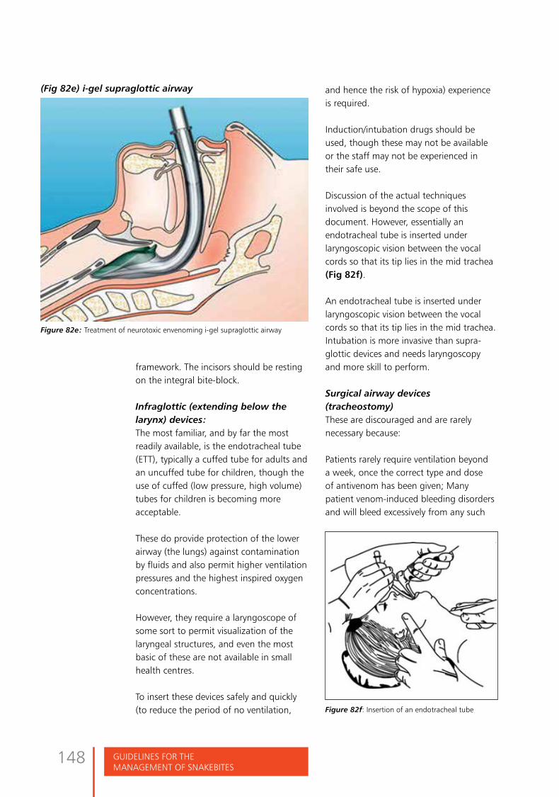
GUIDELINES FOR THE MANAGEMENT OF SNAKEBITES
148
framework. The incisors should be resting on the integral bite-block.
Infraglottic (extending below the larynx) devices:The most familiar, and by far the most readily available, is the endotracheal tube (ETT), typically a cuffed tube for adults and an uncuffed tube for children, though the use of cuffed (low pressure, high volume) tubes for children is becoming more acceptable.
These do provide protection of the lower airway (the lungs) against contamination by fluids and also permit higher ventilation pressures and the highest inspired oxygen concentrations.
However, they require a laryngoscope of some sort to permit visualization of the laryngeal structures, and even the most basic of these are not available in small health centres.
To insert these devices safely and quickly (to reduce the period of no ventilation,
and hence the risk of hypoxia) experience is required.
Induction/intubation drugs should be used, though these may not be available or the staff may not be experienced in their safe use.
Discussion of the actual techniques involved is beyond the scope of this document. However, essentially an endotracheal tube is inserted under laryngoscopic vision between the vocal cords so that its tip lies in the mid trachea (Fig 82f).
An endotracheal tube is inserted under laryngoscopic vision between the vocal cords so that its tip lies in the mid trachea. Intubation is more invasive than supra-glottic devices and needs laryngoscopy and more skill to perform.
Surgical airway devices (tracheostomy)These are discouraged and are rarely necessary because:
Patients rarely require ventilation beyond a week, once the correct type and dose of antivenom has been given; Many patient venom-induced bleeding disorders and will bleed excessively from any such
(Fig 82e) i-gel supraglottic airway
Figure 82e: Treatment of neurotoxic envenoming i-gel supraglottic airway
Figure 82f: Insertion of an endotracheal tube
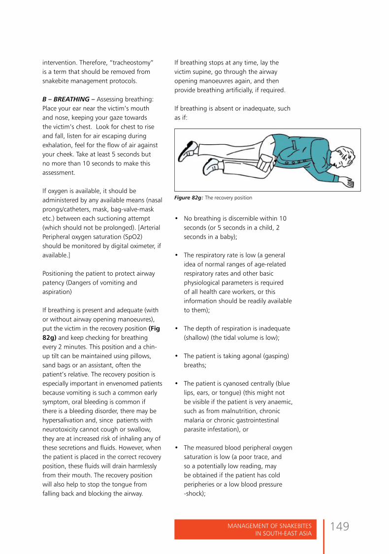
149ManageMent of snakebites in south-east asia
intervention. Therefore, “tracheostomy” is a term that should be removed from snakebite management protocols.
B – BREATHING – Assessing breathing: Place your ear near the victim’s mouth and nose, keeping your gaze towards the victim’s chest. Look for chest to rise and fall, listen for air escaping during exhalation, feel for the flow of air against your cheek. Take at least 5 seconds but no more than 10 seconds to make this assessment.
If oxygen is available, it should be administered by any available means (nasal prongs/catheters, mask, bag-valve-mask etc.) between each suctioning attempt (which should not be prolonged). [Arterial Peripheral oxygen saturation (SpO2) should be monitored by digital oximeter, if available.]
Positioning the patient to protect airway patency (Dangers of vomiting and aspiration)
If breathing is present and adequate (with or without airway opening manoeuvres), put the victim in the recovery position (Fig 82g) and keep checking for breathing every 2 minutes. This position and a chin-up tilt can be maintained using pillows, sand bags or an assistant, often the patient’s relative. The recovery position is especially important in envenomed patients because vomiting is such a common early symptom, oral bleeding is common if there is a bleeding disorder, there may be hypersalivation and, since patients with neurotoxicity cannot cough or swallow, they are at increased risk of inhaling any of these secretions and fluids. However, when the patient is placed in the correct recovery position, these fluids will drain harmlessly from their mouth. The recovery position will also help to stop the tongue from falling back and blocking the airway.
If breathing stops at any time, lay the victim supine, go through the airway opening manoeuvres again, and then provide breathing artificially, if required.
If breathing is absent or inadequate, such as if:
Figure 82g: The recovery position
• No breathing is discernible within 10seconds (or 5 seconds in a child, 2seconds in a baby);
• The respiratory rate is low (a generalidea of normal ranges of age-relatedrespiratory rates and other basicphysiological parameters is requiredof all health care workers, or thisinformation should be readily availableto them);
• The depth of respiration is inadequate(shallow) (the tidal volume is low);
• The patient is taking agonal (gasping)breaths;
• The patient is cyanosed centrally (bluelips, ears, or tongue) (this might notbe visible if the patient is very anaemic,such as from malnutrition, chronicmalaria or chronic gastrointestinalparasite infestation), or
• The measured blood peripheral oxygensaturation is low (a poor trace, andso a potentially low reading, maybe obtained if the patient has coldperipheries or a low blood pressure-shock);

GUIDELINES FOR THE MANAGEMENT OF SNAKEBITES
150
• The end-tidal CO2, by whichever method is being used, is high, or climbing;
ASSISTED VENTILATION IS REQUIRED!How this is delivered will depend on:
• The clinical circumstances, i.e. whether the patient is in the bush, in an small health centre or in a hospital;
• The skills of the rescuer;• The availability of assistants;• Equipment available.
Methods for providing assisted ventilationExpired Air Resuscitation (EAR) (no health facilities immediately available):
Deliver 2 initial “rescue breaths” as follows (Fig 82h): close the patient’s nose with one hand and pull the jaw down with the other, and place your mouth over the
patient’s mouth and deliver a breath over a second, enough to see the chest rise, and allowing 4 seconds for exhalation before giving a second breath. In the case of a small child the mouth and nose may be covered by the rescuer’s mouth.
Assisted ventilation by this means will provide a maximum of approximately only 16% inspired oxygen concentration (FIO2 0.16).
The risk of communicable disease transmission to the rescuer is low, but can be diminished further by the use of:
• A special EAR face shield, manufactured for this purpose;
• A mask of the type used with Bag-Valve-Mask (devices);
• A piece of cloth thin enough to allow for the free passage of air.
If the patient does not begin to breathe, or to breathe more effectively, at this point, the decision will need to be made about how long to continue this method of assisted ventilation.
This will depend on:
• The clinical circumstances, e.g. the likely diagnosis, the age of the patient (the rescuer might reasonably persist for longer if the patient is a small child), or the distance from medical assistance or from more advanced health care facilities;
• The presence of effective cardiac activity; as determined by the presence of a carotid pulse (palpate on both sides before determining that this is absent), more advanced tests such as auscultation of audible heart sounds, or visible cardiac contraction on ultrasound; are supportive tests which may be available.Figure 82h: Technique of giving “rescue breaths”
If chest movement is inadequate, a
correctly-sized OPA should be inserted.
This will usually assist with air flow in and
out of the lungs. If there is no bleeding
disorder, a NPA (or 2) may be used instead
of, or in addition to, an OPA.

151ManageMent of snakebites in south-east asia
The presence of electrical activity, as determined using a cardiac monitor of some kind, with no demonstrable cardiac output or pulse (so-called “pulse-less electrical activity”) usually carries a very poor prognosis, though there is a list of potentially reversible conditions which may cause this. This topic is not covered in these guidelines, but should be covered in any advanced life support course material.
Non-invasive ventilation:Bag-mask/Bag-mask-valve ventilation: In a health-care centre, a commonly used method for providing initial respiratory assistance is with a bag-mask/Bag-Valve-Mask (BVM) (resuscitator bag) (Fig 82i) Even the simplest health-care centre should have these, with different sizes required for different ages of patients, and the staff should be well versed in how to check that the device is functioning correctly and know how to use it correctly and safely.
The rescuer positions him/herself at the victim’s head end. Holding the bag with one hand, he/she places the mask on the victim’s face with the apex of the mask on the bridge of the nose and its base on the groove over the chin. With the other hand, he/she seals the mask around the victim’s nose and mouth, tilts (extends) the victim’s head, lifts the jaw forward and gives breaths at approximately the normal respiratory rate for the patient by squeezing/pressing the bag, looking for visible chest rise. Any chest movement is usually adequate, especially in children.
If chest movement is inadequate, a correctly-sized OPA should be inserted. This will usually assist with air flow in and out of the lungs. If there is no bleeding disorder, a NPA (or 2) may be used instead of, or in addition to, an OPA.
Bag-mask breathing suffices for short-
term respiratory support but if there is no breathing or inadequate breathing that needs longer term support, the airway needs to be secured more definitively and even the bag may have to be replaced with a ventilator. However, many snakebite patients around the developing world have been kept alive for days, or week, using simply an OPA and a BVM, or with an ETT and a BVM, using relatives to squeeze the bag, though this is far from ideal.
Invasive Ventilation: Indications for intubation and mechanical ventilation are Type I respiratory failure (primary failure of oxygenation as in pulmonary oedema in Russell’s viper envenoming) and Type II respiratory failure (primary ventilatory failure due to respiratory muscle paralysis or obstruction as in elapid envenoming). Suggestive clinical features in patients receiving oxygen by mask or nasal catheters include tachpnoea (respiratory rate > 25/min) or paradoxical respirations (abdomen moves out on inspiration) and deteriorating ventilatory capacity (see above); hypoxia (central cyanosis, SpO2 < 90%, arterial PO2 < 50 mm Hg, < 6.7 kPa); and respiratory acidosis (arterial PCO2 > 50 mm Hg, < 6.7 kPa, arterial pH <7.36). Supraglottic or infraglottic devices are used to open the airway and assist with both the supply of oxygen to the lungs and removal of carbon dioxide from the lungs. Generally, and where possible,
Figure 82i: bag-mask/Bag-Valve-Mask (BVM) (resuscitator bag)

GUIDELINES FOR THE MANAGEMENT OF SNAKEBITES
152
endotracheal intubation (insertion of an ETT) should be performed and the patient placed on a ventilator, attended constantly by a suitably qualified nurse, with a suitably qualified doctor always on call to provide additional assistance, if required. Tube blockages are a frequent cause of morbidity and mortality. They are prevented by regular suctioning, humidification and training of staff to recognize the symptoms of a blocked tube.
Sedation for patients being mechanically ventilated: fentanyl 2-40 microg/hour and midazolam 2-4 mg/hour OR (to avoid acute kidney injury) morphine 1-5 mg/hour with lorazepam 1-2 mg/hour.
Adequacy of respiratory assistance can be assessed by reversal of clinical signs of inadequate respiration and stabilization of SpO2 and end-tidal CO2 (where available) and improvement in the victim’s level of consciousness if respiratory failure was the sole cause of his/her unresponsiveness. However, assessing the level of consciousness of a patient with neurotoxic envenoming can be difficult because of their often generalized flaccid paralysis, such that the normal method of ascertaining the level of consciousness (the Glasgow Coma Scale – GCS) is irrelevant since the patient is unable to open their eyes, unable to speak and often unable to obey commands. They are, contrary to common belief, awake, able to hear everything which is said around them. They should be sedated during the period of their ventilation.
Other important aspects of the care of a ventilated patient include:
• Adequate hydration, monitoring ofrenal function and of fluid balance;
• Nutrition (in addition to the minimalnutrition contained in normal IV fluids);
• Physiotherapy (including chestphysiotherapy, regular turning toprevent pressure areas and lungcollapse, and maintenance of the rangeof motion of paralysed, immobilejoints and muscles, as well as limbswhere significant cytotoxicity and tissuedamage has occurred).
• Eye care (synthetic tears and closureof the eyelids to prevent drying anddamage to corneas in patients onventilators).
• Safe and effective weaning fromventilation.
Always be careful to minimize trauma to the airway of a snakebite patient; this includes the insertion of basic airway devices, advanced airway devices and gastric tubes. Orogastric tubes are much preferred over nasogastric ones – the latter often lead to bleeding which is difficult to deal with in the patient who has a coagulopathy, and may even lead inexperienced staff to give more antivenom or blood products when these are not necessary.
Such care also applies to the insertion of intravenous catheters and urinary (urethral) catheters. Central venous lines may be inserted, if required and possible, but the insertion site will depend on the presence or absence of a bleeding disorder. Intra-arterial lines may be useful in many respects, but, again, are discouraged in the presence of a bleeding disorder.
6.9.3 Trial of anticholinesteraseAnticholinesterase drugs have a variable, but potentially very useful effect in patients with neurotoxic envenoming, especially those bitten by cobras (Banerji et al., 1972; Watt et al., 1986; Watt et al., 1989; Faiz et al., cobra in press), but not usually in those envenomed by kraits (Anil

153ManageMent of snakebites in south-east asia
et al., 2010). However, recent claims that intra-nasal neostigmine might provide a universal first-aid method for snakebite victims have achieved a high media profile but are unsubstantiated, misleading and fanciful (Lewin et al., 2013; Lewin et al., 2014).
A trial of anticholinesterase (e.g. “Tensilon test”) should be performed in every patient with neurotoxic envenoming, as it would be in any patient with suspected myasthenia gravis. However, this should not delay antivenom treatment or endotracheal intubation. Patients must be observed closely as they may deteriorate while the trial of anticholinesterase is being carried out
Procedure1. Baseline observations or
measurements are made againstwhich to assess the effectiveness ofthe anticholinesterase.
2. Atropine sulphate (0.6 mg foradults; 50 μg/kg for children) orglycopyrronium is given by intravenousinjection followed by neostigminebromide or methylsulphate(Prostigmin) (or distigmine,pyridostigmine, ambenomium etc. inappropriate doses) by intramuscularinjection 0.02 mg/kg for adults, 0.04mg/kg for children. Short actingedrophonium chloride (Tensilon) isideal for this test but is rarely availablein the Region. It is given by slowintravenous injection in an adult doseof 10 mg, or 0.25 mg/kg for children.
3. The patient is observed over thenext 30-60 minutes (neostigmine) or
10-20 minutes (edrophonium) for signs of improved neuromuscular transmission. Ptosis may disappear (Fig 83) and ventilatory capacity (peak flow, FEV-1 or maximum expiratory pressure) may improve.
4. Patients who respond convincinglycan be maintained on neostigminemethylsulphate, 0.5-2.5 mg every1-3 hours up to 10 mg/24 hoursmaximum for adults or 0.01-0.04mg/kg every 2-4 hours for childrenby intramuscular, intravenous orsubcutaneous injection togetherwith atropine to block muscarinicside effects. Patients able to swallowtablets may be maintained onatropine 0.6 mg twice each day,neostigmine 15 mg four times eachday or pyridostigmine 60 mg fourtimes each day.
Anticholinesterase (e.g. “Tensilon”) test• Baselineobservations• Giveatropineintravenously• Giveanticholinesterase
drug (e.g. neostigmineintramuscularly)
• Observeeffect• Ifpositive,instituteregular
atropine and neostigmine
Figures 83: Anti-cholinesterase: (a) Before and (b) after intravenous atropine followed by intravenous edrophonium chloride in a patient envenomed by a Malayan krait (Bungarus candidus) (Copyright DA Warrell)
[a] [b]

GUIDELINES FOR THE MANAGEMENT OF SNAKEBITES
154
The “ice pack test” as a possible alternative to the Tensilon test (Golnik et al., 1999). In patients with myasthenia gravis who have ptosis, cooling of the ptotoc (drooping) eyelid results in improvement in ptosis, due to inhibition of intrinsic cholinesterase (Golnik et al., 1999). This quick and simple test might obviate the need for the Tensilon test in predicting the effectiveness of anticholinesterase (neostigmine) treatment. However, it has not yet been evaluated in patients with neurotoxic envenoming. This study must be done!
Suggested method: in a patient with bilateral ptosis from neurotoxic snake-bite, the distance between the upper and lower lid margins of both eyes (palpebral fissures) is measured using a mm ruler. Digital pressure is applied to the frontalis muscle to avoid its influencing upper eyelid retraction. An ice-filled plastic glove or frozen ice pack is then gently applied to one eyelid for 2 minutes, after which the palpebral fissures of both eyes are immediately re-measured. A more-than-2mm difference in palpebral fissure between the cooled and control eyelids might be considered a positive result (Fig 84).
6.9.4 Treatment of Hypotension and shockThis is usually the result of hypovolaemia [from loss of circulating volume into the swollen limb, as a result of generalized increase in capillary permeability (e.g. Russell’s viper envenoming in Myanmar) or internal/external haemorrhage], venom-induced vasodilatation or direct myocardial effects with or without arrhythmias.
Postural (orthostatic) blood pressure measurement: hypovolaemia is best detected by measuring blood pressure with the patient lying supine, and repeating the measurement when the patient is propped up, ideally in a fully sitting position but, depending on their condition, at 45 degrees if 90 degrees is not tolerated (Fig 85). A clinically significant fall in blood pressure during this procedure (orthostatic hypotension) is a fall in systolic blood pressure of 20 mm Hg or diastolic blood pressure of 10 mm Hg or more.
Passive leg raising test (fig. 85c): another way of assessing fluid responsiveness is to sit the patient in a 45 degrees head-up semi-recumbent position. Lower their upper body to the horizontal and passively raise their legs at 45 degrees
Figure 84: Ice test recovery of ptosis in the left eye after applying an ice pad that inhibits intrinsic cholinesterase in a patient with post-synaptic blockade of neuro-muscular transmission. (Delaney Y, Khooshabeh R, Benjamin L (2002). Acute severe ocular myasthenia in a 92-year-old woman. Eye (Lond);16(3):323-4).

155ManageMent of snakebites in south-east asia
up, increasing circulating volume by about 150-300 ml. The maximal effect of this “auto-infusion” occurs at 30-90 seconds, measured by clinical features of improved cardiac output (e.g. increase in pulse pressure and decrease in heart rate) (Boulain et al., 2002; Marik et al., 2014).
Treatment with crystalloids should be controlled by observation of the fluid status (jugular venous pressure, respiratory rates and crepitations, see Annex 6). Excessive volume replacement may cause pulmonary oedema when plasma extravasated in the bitten limb and elsewhere is reabsorbed into the circulation.
In patients with evidence of a generalized increase in capillary permeability, a selective vasoconstrictor such as dopamine
Figures 85 a, b: measurement of postural change in blood pressure as evidence of hypovolaemia: (a) supine; (b) sitting up (Copyright DA Warrell)
Figure 85. c: Passive leg raising test to guide fluid therapy (see Marik et al., 2011)
may be given by intravenous infusion, preferably into a central vein (starting dose 2.5-5 μ (micro) g/kg/minute).
Snakebite: causes of hypotension and shock1º Anaphylaxis Vasodilatation Cardiotoxicity Hypovolaemia2º Antivenom reaction
Respiratory failureAcute pituitary adrenal
insufficiency Septicaemia
In victims of Russell’s viper bites in Myanmar, India and Sri Lanka, acute pituitary adrenal insufficiency resulting
[a] [b]
Passive leg raising. The passive leg raising test consists in measuring the hemodynamic effects of a leg elevation up to 45º. A simple way to perform the postural maneuver is to transfer the patient from the semirecumbent posture to the passive leg raising position by using the automatic motion of the bed.
Passive leg raising
transfer of blood from the legs and abdominal compartments
= test for fluid responsiveness

GUIDELINES FOR THE MANAGEMENT OF SNAKEBITES
156
from haemorrhagic infarction of the anterior pituitary may contribute to shock, while neurological manifestations of hypoclycaemia, such as impaired consciousness, extensor posturing and other involuntary movements, may be presenting signs that respond to a test dose of intravenous 50% dextrose injection (Warrell, 2009) (Tun-Pe et al., 1987) (Fig 52a). Hydrocortisone, fluid and electrolyte replacement may needed acutely in these patients. In chronic cases, addition of thyroxin, oestrogen or testosterone may be needed (Antonypillai et al., 2011; Jeevagan et al., 2013).
6.9.5 Treatment of oliguria and acute kidney injury(Sitprija and Boonpucknavig 1979; Chugh 1989)
Detection of acute kidney injury
• Abrupt(within48h)reductionin kidney function (Acute Kidney Injury Network criteria)
Decreasing or no urine output (<0.5 mL/kg/h for more than 6 h)
Increase in serum creatinine concentration [serum creatinine≥26.5μmol/L(≥0.3mg/dL)]orincreasingby≥150- 200% (x 1.5 - 2) compared to baseline [?urea]
• Clinical“uraemiasyndrome”: nausea, vomiting, acidotic (“Kussmaul”)
breathing hiccups, fetor, drowsiness, confusion, coma, flapping tremor, muscle
twitching, convulsions pericardial friction rub, signs of fluid overload
In patients with any of the above features, the following should be monitored
• other signs of “uraemia syndrome”• pulse rate• blood pressure, lying and sitting, to
detect postural hypotension (see 14 Treatment of hypotension and shock)
• respiratory rate• temperature• height of jugular venous pulse• auscultation of lung bases for
crepitations• fluid balance chart and/or daily weight
6.9.5.1 Oliguric phase of renal failureMost, but not all, patients with acute renal failure are oliguric, defined as a urine output of less than 400 ml/day or less than 30 ml/hour (children less than 0.5ml/kg bodyweight /hour). Conservative management may tide the patient over, avoiding the need for renal replacement therapy (dialysis).
If the patient has intravascular volume depletion, indicated by supine or postural hypotension, or empty neck veins, proceed as follows:
1. Establish intravenous access
2. Give a cautious fluid challenge: an adult patient can be given 250-500 ml of isotonic saline over one hour or, until the jugular venous pressure/central venous pressure has risen to 8-10 cm above the sternal angle (with the patient propped up at 45º). The patient must be closely observed while this is being done. The fluid challenge must be stopped immediately if pulmonary oedema develops. If the urine output does not improve it is reasonable to perform a furosemide stress/challenge test (see below for details).

157ManageMent of snakebites in south-east asia
Note – in some patients it can be difficult to determine the height of the central venous pressure by clinical examination. Direct measurement of central venous (superior vena caval) pressure through a long catheter, preferably inserted at the antecubital fossa (see Annex 6), can be helpful in this circumstance. The catheter is connected to a saline manometer, the 0 point of which must be placed at the same level as the right atrium (that is, at the sternal angle when the patient is propped up at 45º). This measurement is reliable only if the level varies freely with respiration and the zero point is correctly positioned.
In a patient who is obviously volume-depleted, resuscitation should start immediately, and not be delayed until a central venous line has been inserted.
3. Insert a urethral catheter (full sterileprecautions!) but in male patients, tryusing a condom catheter first.
4. Furosemide (frusemide) stress/challenge: once the patient has beengiven adequate fluid replacement1-1.5 mg/kg of furosemide is injectedslowly (4-5 mg/minute) intravenouslyand urine output is monitored for 2h. A urine output of < 200 ml in hour(100 ml/hour) predicts progression of renal failure (Chawla et al., 2013). In this case, the patient is likely to progress to acute kidney injury and requires conservative management. Results of a small study done in Myanmar, showed a statistically significant lower requirement for renal replacement therapy (acute peritoneal dialysis) in patients given furosemide infusion compared to those given placebo (Kyaw San Lwin, personal communication, 2012).
5. Conservative management for acutekidney injury: if the urine output doesnot improve, despite these challenges,no further diuretics should be givenand fluid intake should be restrictedto a total of the previous day’s outputplus “insensible losses” (500-1000 ml/day). If possible, the patient should bereferred to a renal unit. The diet shouldbe bland, high in calories (1800/day), normal in protein (0.8-1 g/day),low in potassium (avoid fruit, fruitjuices, energy drinks and potassium-containing drugs) and low in salt.Infections will cause tissue breakdownand increase urea levels. They shouldbe prevented or treated promptly withnon-nephrotoxic antibiotics (i.e. avoidaminoglycosides such as gentamicin).
6. Biochemical monitoring: In patientswith acute kidney injury it isrecommended that serum/plasmaurea, creatinine and electrolytes bemonitored daily if possible, until renalfailure is resolving. Serum potassium,urea, creatinine and, if possible, pH,bicarbonate, calcium and phosphateshould be measured. If this is notpossible the electrocardiogram (ECG)should be examined for evidence ofhyperkalaemia, especially followingbites by sea snakes, or Sri Lankan,Indian, Bangladeshi or MyanmarRussell’s vipers or if the patient ispassing dark brown urine, suggestiveof rhabdomyolysis or intravascularhaemolysis.
7. Detection and management ofhyperkalaemia (UK Renal Association,2014)
• ECG evidence of hyperkalaemia: tallpeaked T waves, prolonged P-R interval,absent P waves, wide QRS complexes.Emergency treatments, which willcontrol hyperkalaemia for 3-6 hours

GUIDELINES FOR THE MANAGEMENT OF SNAKEBITES
158
only, should be given if serum potassium >6.5 mmol/l or ECG changes
• Calcium Gluconate 10%: 0.5 ml/kg 10 ml slow intravenous injection over 2-5 minutes if haemodynamically unstable, or over 15-20min if stable (onset of action 3 minutes). Check for normalisation of ECG. Repeat if necessary.
• β2 agonist aerosol by inhaler (e.g. salbutamol nebulisation 5mg q 1-2h - onset of action 30 minutes - up to 10-20 mg.
• Give 50 ml of 50% dextrose with 10 units of soluble insulin intravenously (onset of action 15 minutes)
• Sodium Bicarbonate 8.4% 1 ml/kg IV slow over 5 minutes
Followed by a low potassium diet.8. Management of severe acidosis If the patient is hypotensive and
profoundly acidotic (deep sighing “Kussmaul” respirations, very low plasma bicarbonate concentration or very low pH - <7.10),Sodium bicarbonate should be given. Based on volume of distribution of bicarbonate which is 40% of body weight, bicarbonate deficit can be calculated. Usually 2-3 ampoules (40 ml. of 8.4% sodium bicarbonate equivalent to 1 mmol/ml) in 5% dextrose water, or half of the calculated deficit can be replaced in 3-4 hours. Severe acidosis in snakebite is usually associated with acute renal failure. Volume expansion by sodium bicarbonate can cause fluid overload. Therefore, if there is no clinical improvement dialysis is required. Caution: Intravenous bicarbonate may precipitate profound hypocalcaemia and fitting, especially in patients with rhabdomyolysis.
9. Renal replacement therapy Dialysis (Fig 86)
Figures 86: Dialysis for treatment of acute kidney injury: (a) Peritoneal dialysis in a township hospital in Myanmar; (b) Haemodialysis in a district hospital in India (Copyright DA Warrell)
[a] [b]

159ManageMent of snakebites in south-east asia
Transfer the patient who has developed the following indications to a health facility where they can receive renal replacement therapy.
Indications for dialysis
a) clinical uraemia (encephalopathy, pericarditis etc.)
b) fluid overload not responding to diuretics
c) plasma potassium concentration > 7 mmol/l (or hyperkalaemic ECG changes)
d) symptomatic acidosise) blood biochemistry - one or
more of the following Creatinine > 4 mg/ dl (354
micromol/l) Urea > 130 mg/ dl (46
mmol/l)Caution: biochemical criteria alone is not sufficient to start dialysis
The choice between peritoneal dialysis and haemodialysis depends on availability. Haemodialysis is referred for management of hyperkalaemia, fluid overload and hypercatabolic patients. Each dialysis session should be decided based on daily clinical assessment (fluid status, urine output and biochemical data). In patients who are hypotensive, slow efficiency daily dialysis (SLEDD) or continuous renal replacement therapy (CRRT [dialysis over 24 hours]) is preferred. However, these are available only in well-equipped ICUs. In patients with acute kidney injury, dialysis may be required for up to 3 weeks. After that time, the kidney should be biopsied to establish the diagnosis and indicate prognosis.
6.9.5.2 Bleeding/ blood clotting disturbances in patients with acute kidney injuryMost patients with acute kidney injury associated with snakebite will have been envenomed by Viperidae, notably Daboia, Hypnale and Trimeresurus species. The only major exception is sea snakebites. Viper-bite victims may have venom-induced bleeding and blood coagulation disturbances although in most Daboia cases these will have been corrected by antivenom before acute kidney injury has developed. However, patients who still have active bleeding/clotting disturbances require fresh frozen plasma and platelet infusions to cover insertion of dialysis catheters and dialysis.
In patients with snakebite related thrombotic microangiopathy with acute kidney injury, the role of plasmapheresis using cryosupernatant (by analogy with thrombotic thrombocytopenic purpura/haemolytic uraemic syndrome) is not clear and has no sound theoretical basis.
6.9.5.3 Prevention of renal damage in patients with myoglobinuria or haemoglobinuria
To minimise the risk of renal damage from excreted myoglobin and/or haemoglobin:• correcthypovolaemia(see
above) and maintain urine output of 200-300 ml/hour.
• correctsevereacidosiswithbicarbonate
• promotealkalinisediuresis(urine pH > 6.5)
• continuethesemeasuresuntil there is evidence that of rhabdomyolysis is decreasing (CK level <5000 U).

GUIDELINES FOR THE MANAGEMENT OF SNAKEBITES
160
6.9.5.4 Diuretic phase of renal failureSome patients will develop polyuria that is as important and as life-threatening as the oliguric phase. Urine output increases to 5-10 litres/24 hours following the period of anuria. The patient may become volume depleted so that salt and water must be carefully replaced. Hypokalaemia may develop, in which case a diet rich in potassium (fruit and fruit juices) is recommended.
6.9.5.5 Renal recovery phaseThe diuretic phase may last for months after Russell’s viper bite.
6.9.5.6 Persisting renal dysfunctionIn Myanmar, persistent tubular degenerative changes were observed in Russell’s viper bite victims who showed continuing albuminuria, hypertension and nocturia for up to 11 months after the bite, despite apparent recovery in renal function. In India, 20-25% of patients referred to renal units with acute kidney injury following Russell’s viper bite suffered oliguria for more than 4 weeks suggesting the possibility of bilateral renal cortical necrosis and the need for referral to a nephrologist. Patients with patchy cortical necrosis show delayed and partial recovery of renal function but those with diffuse cortical necrosis require regular maintenance dialysis and eventual renal transplantation.
6.10 Haemostatic disturbancesBleeding and clotting disturbances usually respond satisfactorily to treatment with specific antivenom, but the dose may need to be repeated several times, at six hourly intervals, before blood coagulability (assessed by the 20WBCT) is finally and permanently restored.
In exceptional circumstances, such as persistent severe bleeding or imminent
urgent surgery, once specific antivenom has been given to neutralize venom procoagulants and other antihaemostatic toxins, restoration of coagulability and platelet function can be accelerated by giving fresh frozen plasma, cryoprecipitate (fibrinogen, factor VIII), fresh whole blood or platelet concentrates.
Heparin is ineffective against venom-induced thrombin and may cause bleeding on its own account. It should never be used in cases of snakebite.
Antifibrinolytic agents are not effective and should not be used in victims of snakebite.
6.10.1 Dangers of venipuncture in patients with haemostatic abnormalitiesIn patients with incoagulable blood, any injection (subcutaneous, intramuscular) and, particularly venipuncture, carries a risk of persistent bleeding and haematoma formation.
Arterial puncture is contraindicated in such patients.Repeated venipuncture can be avoided by using an indwelling cannula and three-way tap system. When blood coagulability has been restored, the dead space should be filled with heparinised saline, but beware! If this is not flushed out before blood sampling, misleading results will be obtained in clotting tests, including the 20WBCT.
In patients with coagulopathy, sites of venous access and placement of intravenous cannulae or catheters should be chosen where haemostasis by external pressure is most likely to be effective, eg the antecubital fossa. If possible, avoid
161ManageMent of snakebites in south-east asia
jugular, subclavian and femoral vein puncture. A pressure pad must be applied at the site of any venipuncture.
6.11 Treatment of the bitten partThe bitten limb, which may be painful and swollen, should be nursed in the most comfortable position, but not excessively elevated as this may reduce arterial perfusion pressure in a tensely swollen limb and increase the risk of intra-compartmental ischaemia.
6.11.1 Wound management Blisters/bullae/”blebs” may be large and tense but they should not be de-roofed and require aspiration only if they threaten to rupture. Abscesses should be aspirated with a needle and the pus cultured (see below infection). Once frank skin necrosis is detected (demarcated, hypo/hyperpigmented areas with an odour of putrefaction), surgical débridement is indicated to remove the risk of anaerobic sepsis. During debridement, all unhealthy tissue should be excised till viable tissue margins are achieved. However, bruised muscle may appear dead but should not be excised as muscle fibres may regenerate. Amputation may be indicated in case of gangrene of toes or limbs.
6.11.2 Bacterial infectionsThe oral flora of wild snakes includes aerobic and anaerobic bacteria, especially the faecal gram-negative rods as their prey may defecate while being ingested. Oral and venom cultures from American snakes yielded Enterobacteriaceae including Morganella spp., Escherichia coli, Group D streptococci, Aeromonas spp., and anaerobes such as Clostridium spp.
In Asia, local bite wound infections may be due to single or multiple bacteria including gram-positive aerobes (Staph. aureus, coagulase-negative Staphylococcus
and Enterococcus), aerobic gram-negative bacterial (E. coli, Klebsiella, Pseudomonas, Enterobacter, Morganella morganii), anaerobic bacteria (Peptostreptococcus and Bacteroides fragilis) (Theakston et al., 1990; Chena et al., 2011; Garg et al., 2009). Infection at the time of the bite with organisms from the snake’s venom and buccal cavity is a problem with some species such as the Malayan pit viper (Calloselasma rhodostoma) and severe and fatal tetanus has been reported. However, prophylactic antibiotics were not effective in a controlled study in Brazil (Jorge et al., 2004). Interference with the wound (incisions made with an unsterilised razor blade/knife etc.) creates a risk of secondary bacterial infection and justifies the use of immediate broad spectrum antibiotics (e.g. gentamicin with benzyl penicillin, amoxycillin or a cephalosporin plus a single dose only of gentamicin – to avoid the risk of kidney injury- plus metronidazole) and tetanus prophylaxis. Other possible antibiotics include amoxycillin/caluvulenic acid, piperacillin/tazobactam, ciprofloxacin and third generation cephalosporin (Mao et al., 2016). Mao YC, Liu PY, Hung DZ, Lai WC, Huang ST, Hung YM, Yang CC. Bacteriology of Naja atra Snakebite Wound and Its Implications for Antibiotic Therapy. Am J Trop Med Hyg. 2016 May 4;94(5):1129-35Later infections in patients treated in hospital include nosocomial pneumonias and urinary tract infections.
6.11.3 Compartmental syndromes and fasciotomy (Fig 83) (Matsen 1980; Mars and Hadley 1998; Mars et al., 1991)
The appearance of an immobile, tensely-swollen, cold and apparently pulseless snake-bitten limb may suggest to surgeons the possibility of increased intracompartmental pressure, especially if the digital pulp spaces or the anterior tibial compartment are involved. Swelling

GUIDELINES FOR THE MANAGEMENT OF SNAKEBITES
162
of envenomed muscle within such tight fascial compartments could result in an increase in tissue pressure above the venous pressure, resulting in ischaemia. However, the classical signs of an intracompartmental pressure syndrome may be difficult to assess in snakebite victims and many unnecessary, dangerous and debilitating fasciotomies are performed, especially where surgeons rather than physicians have the primary responsibility for managing snakebite
Figure 87: Results of unnecessary fasciotomies in snakebite victims in Thailand: (a) profuse bleeding in a patient with mild local envenoming but severe coagulopathy following bites by green pit vipers (Trimeresurus T. albolabris); (b) Residual skin loss and exposure of tendons following fasciotomy for mild local envenoming in a patient bitten by a green pit viper (Trimeresurus T. albolabris) (Copyright the late Sornchai Looareesuwan); (c) Persistent bleeding for 10 days, resulting in haemorrhagic shock despite transfusion of 20 unites of blood, in a victim of Malayan pit viper bite in whom fasciotomy was performed before adequate antivenom treatment had been given to correct the coagulopathy (Copyright DA Warrell)
[a] [b]
[c]
Clinical features of a compartmental syndrome• Disproportionately severe pain• Weakness of intracompartmental
muscles• Pain on passive stretching of
intracompartmental muscles• Hypoaesthesia of areas of skin
supplied by nerves runningthrough the compartment
• Obvious tenseness of thecompartment on palpation
Detection of arterial pulses by palpation or doppler ultrasound probes, does not exclude intracompartmental ischaemia. The most reliable test is to measure intracompartmental pressure directly through a cannula introduced into the compartment and connected to a pressure transducer or manometer (Annex 5). In orthopaedic practice, intracompartmental pressures exceeding 40 mmHg (less in children) may carry a risk of ischaemic
cases (Fig 87). Fasciotomy is generally falling out of favour for the treatment of snake-bitten limbs (Cumpston, 2011).
163ManageMent of snakebites in south-east asia
necrosis (eg Volkmann’s ischaemia or anterior tibial compartment syndrome). However, envenomed muscle may not be saved by fasciotomy. Animal studies have suggested that muscle sufficiently envenomed and swollen to cause intracompartmental syndromes, may already be irreversibly damaged by the direct effects of the venom (Garfin et al., 1984). In any case, fasciotomy should not be contemplated until haemostatic abnormalities have been corrected, otherwise the patient may bleed to death (Fig 66c).
Among 105 cases of rattle snakebites in USA in which fasciotomy was either just discussed or actually carried out, the 28 patients given surgery showed no advantage in morbidity, but spent on average 2 extra days in hospital (Darracq et al., 2015).
In green pit viper bite (genus Trimeresurus) victims, antivenom not only reversed coagulopathy, but also reducing severe limb oedema (Rojnuckarin et al., 2006). However, corticosteroids are not effective in ameliorating local effects of envenoming and, since they carry the risk of side-effects, they should not be used (Reid et al., 1963; Nuchprayoon et al., 2008).
Early treatment with antivenom remains the best way of preventing irreversible muscle damage.
Criteria for fasciotomy in snake-bitten limbshaemostatic abnormalities have been corrected (antivenom with or without clotting factors)• clinical evidence of an
intracompartmental syndrome• intracompartmental pressure >40
mmHg (in adults)
6.12 RehabilitationIn patients with severe local envenoming, the limb should be maintained in a functional position. For example, in the leg, equinus deformity of the ankle should be prevented by application of a back slab and avoiding the pressure of heavy bed clothes.
Functional effects of local envenoming range from persistent stiffness and induration due to sclerosis of veins, lymphatics and tissue planes through which the venom has spread, to severe deformity, arthrodeses, contractures, tissue loss, especially dermonecrosis and chronic ulceration requiring skin grafting, and gangrene requiring débridement and amputation. Restoration of normal function in the bitten part should be started by simple exercises while the patient is still in hospital. After the patient has been discharged from hospital rehabilitation is rarely supervised but relatives can be instructed and given a time table of rehabilitating activities. Conventional physiotherapy may accelerate functional rec overy of the bitten limb.
6.13 Discharge assessmentBefore the patient leaves hospital, discuss the following topics with them and their accompanying family members.
• Implications of having had a snakebite:most patients can be assured of a fullrecovery in time.
• Rehabilitation exercises: encouragethem to continue until normal functionis restored to the bitten limb.
• Follow-up appointment: encourage thepatient to return after an interval of 1-2weeks to check on their progress andto allow further reassurance.
• Late serum sickness-type reactions:

GUIDELINES FOR THE MANAGEMENT OF SNAKEBITES
164
warn them of the symptoms and reassure them that this complication of antivenom can be treated.
• Reducing the risk of further bites:provide advice, ideally in the form ofa leaflet, explaining the principles ofsnakebite prevention (see Section 1above), to be shared with their familiesand neighbours.
6.14 Management of cobra spit ophthalmia (Chu et al., 2010)First aid: consists of urgent irrigating the affected eyes and other mucous membranes with liberal quantities of water, saline, Ringer’s lactate, milk or any other available bland liquid (even urine has been used). No medicines, oils or traditional ointments should be put in the eye(s) and there should be no delay in going to hospital. The spitting cobra should not be pursued or killed as this may result in further multiple spits.
Medical treatment consists of:1. Further urgent decontamination is
achieved by copious irrigation
2. Pain is intense. Topical vasoconstrictorswith weak mydriatic activity [e.g.instillation of 0.5% epinephrine(adrenaline) drops] relieve pain andinflammation. However, topical localanaesthetics (eg 0.4% oxybuprocainehydrochloride (“Novesin”), 4%lidocaine hydrochloride or tetracainehydrochloride (“Decicaine”) are more
effective but, by rendering the eye temporarily insensitive, may allow trauma to the cornea. They should be applied only once and the eye must be protected until sensation returns. By overcoming blepharospasm, these drugs may allow more efficient irrigation.
3. Corneal abrasions must be excludedby fluorescein staining and/or slitlamp examination
4. Endophthalmitis or blinding cornealopacities must be preventedby application of prophylactictopical antibiotics (eg tetracycline,chloramphenicol, 0.5% framycetin“Soframycin”, ciprofloxacin, penicillin-streptomycin ointment, polymixinB sulphate, gatifloxacin, andmoxifloxacin).
5. Posterior synechiae, ciliary spasm anddiscomfort can be prevented withtopical cycloplegics (e.g. 2% atropine,scopolamine or homatropine).However, beware, these mydriaticsmay precipitate acute glaucoma.
6. Allergic kerato-conjunctivitis maydevelop in those previously sensitisedby spitting or other habitual exposureto snake venoms. It is treated withantihistamines.
Instillation of diluted antivenom causes irritation and is of no benefit since any residual venom in the conjunctival sac is removed by liberal irrigation.
Topical corticosteroids are contraindicated because of the risk of Herpes simplex keratitis.
Some ophthalmologists recommend the use of a dressing pad to close the eye.
Consists of urgent irrigating the affected eyes
and other mucous membranes with liberal
quantities of water, saline, Ringer’s lactate,
milk or any other available bland liquid.

165ManageMent of snakebites in south-east asia
6.15 Management of snakebites at different levels of the health service(refer to sections above for details of treatments etc.)All levels of the health service can contribute to the management of patients with suspected snakebite. Since the treatment of severe envenoming is a medical emergency that may require a range of medical skills, equipment, antivenom and other medicines, referral should be to the highest level of care that is readily available. However, in the rural areas where snakebites are most frequent, transfer to a hospital may not be feasible within the reasonable time frame of a few hours. In that case, a lower level of health facility must cope with the emergency as suggested below. Ideally, a specialist team and a defined area or ward should be allocated for the management of snakebite victims. Training of doctors, nurses, dispensers, health assistants and paramedics in all the skills required for diagnosis (including snake identification) and early management of snakebite patients (including indications for and practicalities of administering antivenom) is essential at every level.
A. At community or village level1. Assess: check history of snakebite
and look for obvious evidence of a bite(fang puncture marks, swelling of thebitten part).
2. Reassure
3. First-aid: immobilize the wholepatient as far as possible by laying him/her down in a relaxed but safe position(e.g. recovery position), immobilizethe bitten limb with a splint and applypressure-pad.
4. Transport: Arrange (by emergencycontact number) transport of the
patient to medical care as quickly, safely and passively as possible by vehicle, boat, bicycle, motorbike, stretcher etc. Ideally the patient should lie in the recovery position (prone, on the left side) with his/her airway protected to minimise the risk of shock and inhalation of vomit.
5. Snake: if the snake responsible hasalready been caught or killed take it(safely in a bag or container) with thepatient or take images on a mobile‘phone , but ensure safety by avoidingdirect contact.
6. Traditional treatment: discouragetime-wasting and potentially dangeroustraditional treatments such as tightligatures (tourniquets), incisions,suction and application of herbs, ice,chemicals, “snakestones” etc.
B. At the Rural Clinic, Dispensary, Health Post, or Primary Health Centre1. Assess for signs of local and
systemic envenoming: carry out asimple medical assessment includinghistory and simple physical examination– local swelling, painful tenderenlarged local lymph glands, persistent bleeding from the bite wound, blood pressure, pulse rate, bleeding (gums, nose, vomit, stool or urine), level of consciousness, drooping eyelids (ptosis) and other signs of paralysis. Monitor these signs hourly.
2. Check: 20 minute whole bloodclotting test (20WBCT), urineexamination (appearance, sticks testingfor blood etc.). Identify the snake or aphoto of it (if brought).
3. Analgesia: give analgesia by mouth ifrequired: paracetamol (acetaminophen)(adult dose 500 mg to 1 g maximum4 g in 24 hours; children 10-15 mg/
GUIDELINES FOR THE MANAGEMENT OF SNAKEBITES
166
kg maximum 100mg/kg/day) or codeine phosphate (adult dose 30-60 mg maximum 240 mg in 24 hours; children more than 2 years old, 0.5 mg/kg, maximum 2 mg/kg/day) can be given every 4-6 hours by mouth as required (not aspirin or non-steroidal anti-inflammatory drugs which can cause bleeding).
4. Antivenom: if the patient fulfilscriteria for antivenom treatment andif the necessary skills, equipment,antivenom, adrenaline and othernecessary drugs are available, giveantivenom. These skills includeability to diagnose local and systemicenvenoming, set up intravenousinfusion or intravenous injection,identify the early signs of anaphylaxisand treat it with intramuscularadrenaline. Reasess for repeateddose(s) of antivenom. If no antivenomis available, transfer to a hospital.
5. If the patient is shocked/hypotensive: give cautiousintravenous fluid challenge (adult250-500 ml of 0.9% saline) to correcthypovolaemic shock.
6. If the patient has evidence ofrespiratory paralysis: give oxygenby mask, consider atropine andneostigmine, and transfer to hospital.It is assumed that assisted ventilationother than by a tight-fitting face maskconnected to an anaesthetic (Ambu)bag will not be possible at this level.
7. If the patient is oliguric: initiateconservative management.
8. The bite wound: if necrotic, tamperedwith (incisions etc.) or obviouslyseptic, give antibiotics and tetanusprophylaxis.
9. Assess the need and feasibility oftransporting the patient to a higherlevel of the health service (see A above)especially in case of:
a. Substantial bleeding, 20WBCT stillpositive (non-clotting) 6 hours afterinitial antivenom dose
b. Progressive paralysis (muscle weakness)or respiratory difficulty
c. Reduced urine outputd. Anaphylaxis –unresponsive to
adrenalinee. Shock/hypotension- unresponsive to
fluidsf. Severe local necrosis or signs suggestive
of compartment syndrome
10. Discourage the use of ineffectiveand potentially harmful drugs (e.g.corticosteroids, antihistamines, andheparin).
C. At the District HospitalProceed as in B above plus:
1. Assessment: carry out a moredetailed clinical and laboratoryassessment including biochemical andhaematological measurements, ECG orradiography, as indicated.
2. Antivenom: if no antivenom isavailable, transfer to a hospital that hasantivenom or treat conservatively; thismay require transfusion of blood orfresh frozen plasma (see above).
3. Analgesia: (see B above) and, ifrequired, consider stronger parenteralopioid drugs as required all withgreat caution (e.g. subcutaneous,intramuscular or even intravenouspethidine, initial adult dose 50-100 mg; children 1-1.5 mg/kg; ormorphine, initial adult dose 5-10 mg;children 0.03-0.05 mg/kg,).

167ManageMent of snakebites in south-east asia
4. If the patient has evidence of localnecrosis (gangrene): give tetanustoxoid booster, antibiotics and dosurgical débridement of dead tissue.
5. If the patient has evidence of bulbaror respiratory paralysis: insertendotracheal tube, laryngeal maskairway or i-gel aiway. If there isevidence of respiratory failure, assistventilation manually by anaesthetic(Ambu) bag or mechanical ventilator.
6. If the patient has evidence of acutekidney injury: treat with peritonealdialysis. If this is not available, transferto a specialized hospital.
7. If the patient is bleeding severely or isalready seriously anaemic: cross-matchand transfuse.
8. Rehabilitation: encourage exercising ofbitten limb.
D. At the Referral (Specialized) HospitalProceed as in B and C above plus:
1. More advanced surgical management oflocal necrosis (e.g. split skin grafting).
2. More advanced investigations includingbacterial cultures and imaging (CTscans) as indicated.
3. If the patient has evidence of acuterenal failure peritoneal or haemodialysisor haemofiltration.
4. Implement rehabilitation byphysiotherapists.
Conclusions: Strengthening of health system in managing snakebite
To reduce complications and deathsfrom snakebites, health systems need to
Figure 88 Health system strengthening
HEALTH SYSTEM
TERTIARY REFERRAL HOSPITAL
Infrastructure- dialysis and ICU, surgical set up, coagulation labPersonnel- physician, surgeon,
nephrologist, anaesthetist
DISTRICT HOSPITALInfrastructure- blood bank,
ICU, surgical set upPersonnel- physician, surgeon
PRIMARY HEALTH CENTREInfrastructure- basic emergency
care and resuscitation, airway and Ambu bag,
antivenom administrationPersonnel- doctor, nurse
VILLAGEInfrastructure- ambulance
TransportPersonnel- health aid, health
worker, paramedic
be improved at various levels. The government must develop a policy for snakebite, making it a notifiable disease, developing a snakebite programme that includes standard treatment guidelines, training of health personnel and ensures an adequate supply, distribution and storage of good quality antivenom. Strengthening of public health systems
Strengthening of health system
1. Training of all levels ofhealth system
2. Ensuring infrastructure ateach level
3. Antivenom availability4. Epidemiological
monitoring of snak-bite,deaths, complications andantivenom reactions
5. Community education
Snakebite policy1. Making snakebite reportable2. Developing a snakebite programme3. Standard treatment guidelines4. Ensuring production and supply of adequate
quantity and quality of antivenom5. Training of health personnel at all levels6. Research
GOVERNMENT
GUIDELINES FOR THE MANAGEMENT OF SNAKEBITES
168
requires an appropriate infrastructure of facilities and drugs at each level, training of personnel, epidemiological monitoring and community education.
6.16 Summary of current evidence for treatment of snakebite envenomingCurrently proposed treatment and their likely benefit
1. Antivenom:Benefits: recommended for treatment of bleeding/blood clotting disturbances, shock/hypotension, post-synaptic neurotoxicity (cobras, death adders, rhabdomyolysis (sea snakes). Optimal initial dosage largely unknown
Risks: early anaphylactic and pyrogenic reactions, late serum sickness-like reactions
2. Adrenaline (epinephrine)Benefits: recommended for prevention and treatment of early antivenom reactions
Risks: cerebral haemorrhage in elderly patients with underlying cerebrovascular disease
3. Blood clotting factorsBenefits: recommended to speed up restoration of normal haemostasis in patients already treated with antivenom and for conservative treatment of bleeding/blood clotting disturbances, when antivenom is not available
Risks: fatalities reported (reactions and possibly thromboses)
4. Antihistamines (H1 blockers) andcorticosteroidsBenefits: late serum sickness-type reactions
5. AntibioticsBenefits: treating secondary and primary bite wound infections, prophylaxis in necrotic wounds or those that have been tampered with using non-sterile instruments
6. Renal replacement therapy andassisted ventilation:Benefits: life-saving in patients with acute kidney injury and respiratory failure
7. Anticolinesterase (neostigmine)with atropineBenefits: can rapidly improve neuro muscular transmission in some patients with post-synaptic neurotoxicity (cobras, Australasian death adders)
Risks: muscarinic side-effects, cholinergic crisis
8. Traditional first-aid methods(incisions, tourniquets, suction, tattooing, topical herbal remedies, black “snake-stone”, electric shocks etc.)
Risks: harmful and useless – should never be used
9. First-aid pressure padimmobilization, rapid transport to medical care Benefits: reduces venom spread and risk of early death

169ManageMent of snakebites in south-east asia
References and further readingAli SS, O'Connell C, Kass L, Graff G Single-breath counting: a pilot study of a novel technique for measuring pulmonary function in children. Am J Emerg Med. 2011 Jan;29(1):33-6.
Amarasekera N, Jayawardena A, Ariyaratnam A, Hewage UC, de Silva A. Bite of a sea snake (Hydrophis spiralis): a case report from Sri Lanka. J Trop Med Hyg. 1994;97(4):195-8.
Anil A, Singh S, Bhalla A, Sharma N, Agarwal R, Simpson ID. Role of neostigmine and polyvalent antivenom in Indian common krait (Bungarus caeruleus) bite. Journal of Infection and Public Health. 2010;3(2):83-7.
Anker RL, Straffon WG, Loiselle DS, Anker KM. Retarding the uptake of "mock venom" in humans: comparison of three first-aid treatments. Med J Aust. 1982 Mar 6;1(5):212-4.
Antonypillai CN, Wass JA, Warrell DA, Rajaratnam HN. Hypopituitarism following envenoming by Russell's vipers (Daboia siamensis and D. russelii) resembling Sheehan's syndrome: first case report from Sri Lanka, a review of the literature and recommendations for endocrine management. QJM. 2011 Feb;104(2):97-108.
Ariaratnam CA, Sjöström L, Raziek Z, Kularatne SA, Arachchi RW, Sheriff MH, et al. An open, randomized comparative trial of two antivenoms for the treatment of envenoming by Sri Lankan Russell's viper (Daboia russelii russelii). Trans R Soc Trop Med Hyg. 2001 Jan-Feb;95(1):74-80. .
Ariaratnam CA, Sheriff MH, Theakston RD, Warrell DA. Distinctive epidemiologic and clinical features of common krait (Bungarus caeruleus) bites in Sri Lanka. Am J Trop Med Hyg. 2008 Sept;79(3):458-62.
Ariaratnam CA, Thuraisingam V, Kularatne SA, Sheriff MH, Theakston RD, de Silva A, et al. Frequent and potentially fatal envenoming by hump-nosed pit vipers (Hypnale hypnale and H. nepa) in Sri Lanka: lack of effective antivenom. Trans R Soc Trop Med Hyg.2008 Nov; 102:1120-6.
Ariaratnam CA, Sheriff MH, Arambepola C, Theakston RD, Warrell DA. Syndromic approach to treatment of snakebite in Sri Lanka based on results of a prospective national hospital-based survey of patients envenomed by identified snakes. Am J Trop Med Hyg. 2009 Oct;81(4):725-31.
Armentano RA, Bandt C, Schaer M, Pritchett J, Shih A. Thromboelastographic evaluation of hemostatic function in dogs treated for crotalid snake envenomation. J Vet Emerg Crit Care (San Antonio). 2014 Mar-Apr;24(2):144-53.
7

GUIDELINES FOR THE MANAGEMENT OF SNAKEBITES
170
Bandyopadhyay SK, Bandyopadhyay R, Dutta A, Pal SK. Hypopituitarism following poisonous viperbite. J Indian Med Assoc. 2012 Feb;110(2):120, 122.
Banerji RN, Sahni AL, Chacko KA. Neostigmine in the treatment of Elapidae bites. J Ass Physicins India. 1972 Jul 20(7):503-9.
Bell DJ, Wijegunasinghe D, Samarakoon S, Palipana H, Gunasekera S, de Silva HA,et al. Neurophysiological findings in patients one year after snakebite induced neurotoxicity in sri lanka. Transactions of the Royal Society of Tropical Medicine and Hygiene 2010 May;104(5): 351-6.
Belt PJ, Malhotra A, Thorpe RS, Warrell DA, Wuster W. Russell’s viper in Indonesia: snakebite and systematics. In: Thorpe RS, Wüster W, Malhotra A editions, Venomous snakes. Ecology, evolution and snakebite.Oxford:, Clarendon Press,1997.p. 219-34.
Bhat RN. Viperine snakebite poisoning in Jammu. J Indian Med Assoc. 1974 Dec 16; 63(12): 383-92.
Bhetwal BB, O’Shea M, Warrell DA.Snakes and snakebite in Nepal. Tropical Doctor. 1998 Oct; 28(4): 193-5.
Bon C, Goyffon M. Envenomings and their treatments. Lyon: Ed. Fondation Marcel Mérieux ;1995. 343 p.
Bücherl W, Buckley EE & Deulofeu V (eds) . Venomous animals and their venoms. Vols 1 and 2. Academic Press, New York. (1968,
1971)
Canale E, Isbister GK, Currie BJ. Investigating pressure bandaging for snakebite in a simulated setting: bandage type, training and the effect of transport. Emerg Med Australas. 2009 Jun; 21(3):184-90.
Caron EJ, Manock SR, Maudlin J, Koleski J, Theakston RD, Warrell DA, et al. Apparent marked reduction in early antivenom reactions compared to historical controls: was it prophylaxis or method of administration? Toxicon. 2009 Nov;54(6):779-83.
Chappuis F, Sharma SK, Jha N, Loutan L, Bovier PA. Protection against snakebites by sleeping under a bed net in southeastern Nepal. Am J Trop Med Hyg. 2007; 77(1):197-9.
Chakraborty PP, Bhattacharjee R. Bilateral parotid swelling: an unusual complication of viper bite. J Assoc Phys India.2010 Jul; 58: 460.
Chatterjee P, Mukhopadhyay P, Pandit K, Roychowdhury B, Sarkar D, Mukherjee S, et al. Profile of hypopituitarism in a tertiary care hospital of eastern India--is quality of life different in patients with growth hormone deficiency?.J Indian Med Assoc. 2008 Jun;106(6):384-5, 388.
Chawla LS, Davison DL, Brasha-Mitchell E, Koyner JL, Arthur JM, Shaw AD, et al. Development and standardization of a furosemide stress test to predict the severity of acute kidney injury. Crit Care. 2013 Sep 20; R207.
Chen CM, Wu KG, Chen CJ, Wang CM. Bacterial infection in association with snakebite: a 10-year experience in a northern Taiwan medical center. J Microbiol Immunol Infect. 2011 Dec;44(6):456-60.
Cherian AM, Girish TS, Jagannati M, Lakshmi M. High or Low- A Trial of Low Dose Anti Snake Venom in the Treatment of Poisonous Snakebites.J Assoc Physicians India. 2013 Jun; 61(6):387-9, 396.

171ManageMent of snakebites in south-east asia
Chippaux JP. Snakebites: appraisal of the global situation. Bull World Health Organ. 1998;76(5):515-24.
Chotenimitkhun R, Rojnuckarin P. Systemic antivenom and skin necrosis after green pit viper bites. Clin Toxicol (Phila). 2008 Feb;46(2):122-5.
Chu ER, Weinstein SA, White J, Warrell DA. Venom ophthalmia caused by venoms of spitting elapid and other snakes: Report of ten cases with review of epidemiology, clinical features, pathophysiology and management. Toxicon. 2010; 56(3):259-72.
Chugh KS. Snakebite-induced acute renal failure in India. Kidney International. 1989; 35: 891-907.
Cubitt M, Armstrong J, McCoubrie D, White J, Williams V, Isbister GK . Point-of-care testing in snakebite: an envenomed case with false negative coagulation studies. Emerg Med Australas. 2013; 25(4):372-3.
Cumpston KL. Is there a role for fasciotomy in Crotalinae envenomations in North America? Clin Toxicol (Phila). 2011; 49(5):351-65.
Currie BJ, Canale E, Isbister GK. Effectiveness of pressure-immobilization first aid for snakebite requires further study. Emerg Med Australas. 2008 Jun; 20(3):267-70.
Darracq MA, Cantrell FL, Klauk B, Thornton SL. A chance to cut is not always a chance to cure- fasciotomy in the treatment of rattlesnake envenomation: A retrospective poison center study. Toxicon. 2015;101:23-6.
Dassanayake AS, Karunanayake P, Kasturiratne KT, Fonseka MM, Wijesiriwardena B, Gunatilake SB et al. Safety of subcutaneous adrenaline as prophylaxis against acute adverse reactions to anti-venom serum in snakebite. Ceylon Med J. 2002;47:48-9.
Dayal M, Prakash S, Verma PK, Pawar M. Neurotoxin envenomation mimicking brain death in a child: A case report and review of literature. Indian J Anaesth. 2014 Jul;58(4):458-60.
Deepak M, Basavaprabhu A, Ramapuram JT, Nithyananda C, Mahalingam S. Bilateral parotid enlargement following snakebite : A rare sign. Asian PAc J Trop Biomed. 2013;3(2):154-5.
de Rezende NA, Torres FM, Dias MB, Campolina D, Chavez-Olortegui C, Amaral CF. South American rattlesnakebite (Crotalus durissus SP) without envenoming: insights on diagnosis and treatment. Toxicon. 1998 Dec;36(12):2029-32.
de Silva A, Mendis S, Warrell DA. Neurotoxic envenoming by the Sri Lankan krait (Bungarus ceylonicus) complicated by traditional treatment and a reaction to antivenom. Trans R Soc Trop Med Hyg. 1993;87(6):682-4.
de Silva HA, Pathmeswaran A, Ranasinha CD, Jayamanne S, Samarakoon SB, Hittharage A, et al. Low-dose adrenaline, promethazine, and hydrocortisone in the prevention of acute adverse reactions to antivenom following snakebite: a randomised, double-blind, placebo-controlled trial. PLoS Med. 2011May;8(5):e1000435.
de Silva HA, Ryan NM, de Silva HJ. Prevention and treatment of adverse reactions to snake antivenom. British Journal of Clinical Pharmacology. 2016; 81: 446-452.

GUIDELINES FOR THE MANAGEMENT OF SNAKEBITES
172
Deshpande RP, Motghare VM, Padwal SL, Pore RR, Bhamare CG, Deshmukh VS, et al. Adverse drug reaction profile of anti-snake venom in a rural tertiary care teaching hospital. Journal of Young Pharmacists. 2013 June; 5(2):41–45.
Directorate General of Health Services Ministry of Health and Family Welfare Government of India. National Snakebite Management Protocol [internet]. New Delhi;2009. Available from http://164.100.130.11:8091/nationalsnakebitemanagementprotocol.pdf
Eapen CK, Chandy N, Kochuvarkey KL, Zacharia PK, Thomas PJ, Ipe TI. Unusual complication of snakebite : hypopituitarism after viper bites. In Ohsaka A, Hayashi K, Sawai Y, editors. Animal, plant and microbial toxins.2nd edition. New York: Plenum;1976. P. 467-473.
Ediriweera DS, Kasturiratne A, Pathmeswaran A, Gunawardena NK, Wijayawickrama BA, Jayamanne SF, et al. Mapping the risk of snakebite in Sri Lanka- A national survey with geospatial analysis. PLoS Negl Trop Dis. 2016 July; 10(7):e0004813.
Faiz A, Ghose A, Ahsan F, Rahman R, Amin R, Hassan MU, et al. The greater black krait (Bungarus niger), a newly recognized cause of neuro-myotoxic snakebite envenoming in Bangladesh. Brain. 2010;133(11):3181-93.
Faiz, MA, Ahsan MF, Ghose A, Rahman MR, Amin R, Hossain, M,et al. Bites by the monocled cobra, Naja kaouthia, in Chittagong Division, Bangladesh: epidemiology, clinical features of envenoming and management of 70 identified cases. (in press)
Fan HW, Marcopito LF, Cardoso JL, França FO, Malaque CM, Ferrari RA, et al.Sequential randomised and double blind trial of promethazine prophylaxis against early anaphylactic reactions to antivenom for bothrops snakebites. BMJ. 1999 May 29;318(7196):1451-2.
Fan HW, Marcopito LF, Cardoso JL, França FO, Malaque CM, Ferrari RA, et al.Sequential randomised and double blind trial of promethazine prophylaxis against early anaphylactic reactions to antivenom for bothrops snakebites. BMJ. 1999 May 29;318(7196):1451-2.
Fernando WK, Kularatne SA, Wathudura SP, de Silva A, Mori A, Mahaulpatha D. First reported case of systemic envenoming by the Sri Lankan keelback (Balanophis ceylonensis). Toxicon. 2015;93:20-3.
Fox S, Rathuwithana AC, Kasturiratne A, Lalloo DG, de Silva HJ. Underestimation of snakebite mortality by hospital statistics in the Monaragala District of Sri Lanka. Trans R Soc Trop Med Hyg. 2006;100:693-5.
Gans C,Gans KA. eds. Biology of the reptilia. Vol 8. London: Academic Press;1978.782 p.
Garg A, Sujatha S, Garg J, Acharya NS, Chandra Parija S. Wound infections secondary to snakebite. J Infect Dev Ctries. 2009 Apr 30;3(3):221-3.
Gawarammana IB, Kularatne SA, Dissanayake WP, Kumarasiri RP, Senanayake N, Ariyasena H. Parallel infusion of hydrocortisone +/- chlorpheniramine bolus injection to prevent acute adverse reactions to antivenom for snakebites. Med J Aust. 2004 Jan 5; 180:20-3.
Garfin SR, Castilonia RR, Mubarak SJ . Rattle snakebites and surgical decompression: results using a laboratory model. Toxicon. 1984;22:177–82.
Gawarammana I, Mendis S, Jeganathan K. Acute ischemic strokes due to bites by Daboia russelii in Sri Lanka - first authenticated case series. Toxicon. 2009;54:421-8.

173ManageMent of snakebites in south-east asia
Ghose A, Faiz MA. Snake envenomation in Bangladesh. In: Gopalakrishnakone P, Faiz SM, Gnanathasan CA, Habib AG, Fernando R, Yang CC. Clinical Toxinology in Asia Pacific and Africa [internet].Netherlands. Springer ;2015. Chapter 11. Available from : http://link.springer.com/referencework/10.1007/978-94-007-6288-6/page/2 DOI: 10.1007/978-94-007-6288-6_25-1ISBN: 978-94-007-6288-6
Ghose A, Amin MR, Haq MA, Islam A, Chowdhury FR, Miah T, et al. Russell’s Viper (Daboia russelii): A newly recognized cause of neuro-myo-renal toxic envenomation in Bangladesh. In Proceeding 18th World Congress of the International Society on Toxinology ; 2015 Sept 25-30; oxford (United Kingdom) ; c2015. P. 91
Gimkala A, Ramana Rao GV, Bharti OK. Transporting Snakebite Victims to Appropriate Health Facility within Golden Hour through Toll Free Emergency Ambulance Service in India, Save Lives. Int J Trop Dis Health. 2016; 17(2): 1-12.
Golay V, Roychowdhary A, Dasgupta S, Pandey R. Hypopituitarism in patients with vasculotoxic snakebite envenomation related acute kidney injury: a prospective study on the prevalence and outcomes of this complication. Pituitary. 2014; 17(2):125-31.
Golnik KC, Pena R, Lee AG, Eggenberger ER. An ice test for the diagnosis of myasthenia gravis. Ophthalmology. 1999;106:1282–6.
Gong E, Martin LD, Burnham DA, Falk, AR. The birdlike raptor Sinornithosaurus was venomous. PNAS. 2009; 107:766–768.
Gopalakrishnakone P, Chou LM . Snakes of medical importance (Asia-Pacific Region).Singapore: National University of Singapore Press;1990.
Gowda CD, Rajesh R, Nataraju A, Dhananjaya BL, Raghupathi AR, Gowda TV, et al. Strong myotoxic activity of Trimeresurus malabaricus venom: role of metalloproteases. Mol Cell Biochem. 2006 Jan; 282(1-2):147-55.
Grunau BE, Wiens MO, Rowe BH, McKay R, Li J, Yi TW, et al.. Emergency Department Corticosteroid Use for Allergy or Anaphylaxis Is Not Associated With Decreased Relapses. Ann Emerg Med. 2015 Oct;66(4):381-9. Available from : http://www.ncbi.nlm.nih.gov/pubmed/25820033 DOI: 10.1016/j.annemergmed.2015.03.003
Gutiérrez JM, Warrell DA, Williams DJ, Jensen S, Brown N, Calvete JJ, et al. Global Snakebite Initiative. The need for full integration of snakebite envenoming within a global strategy to combat the neglected tropical diseases: the way forward. PLoS Negl Trop Dis. 2013 Jun 13;7(6):e2162.
Haile NS. Snakebites man: two recent Borneo cases. Sarawak Mus J. 1963;11:291-8.
Harris JB, Faiz MA, Rahman MR, Jalil MM, Ahsan MF, Theakston RD, et al. Snakebite in Chittagong Division, Bangladesh: a study of bitten patients who developed no signs of systemic envenoming. Trans R Soc Trop Med Hyg. 2010 May;104(5):320-7.
Harvey AL. Snake toxins. New York: Pergamon Press, 1991.

GUIDELINES FOR THE MANAGEMENT OF SNAKEBITES
174
Hasan SM, Basher A, Molla AA, Sultana NK, Faiz MA . The impact of snakebite on household economy in Bangladesh. Trop Doct. 2012; 42(1):41-3.
Hati AK, Mandal M, De MK, Mukherjee H, Hati RN. Epidemiology of snakebite in the district of Burdwan, West Bengal. J Indian Med Assoc. 1992 Jun; 90:145-7.
Ho M, Warrell DA, Looareesuwan S, Phillips RE, Chanthavanich P, Karbwang J, et al. Clinical significance of venom antigen levels in patients envenomed by the Malayan pit viper (Calloselasma rhodostoma). American J Trop Med Hyg. 1986 May; 35(3): 579-87.
Ho M, Silamut K, White NJ, et al. Pharmacokinetics of three commercial antivenoms in patients envenomed by the Malayan pit viper (Calloselasma rhodostoma) in Thailand. AmJ Trop Med Hyg. 1990; 42: 260-6.
Ho WK, Verner E, Dauer R, Duggan J. ADAMTS-13 activity, microangiopathic haemolytic anaemia and thrombocytopenia following snakebite envenomation. Pathology. 2010 Feb;42(2):200-2.
Höjer J, Tran Hung H, Warrell D. Life-threatening hyponatremia after krait bite envenoming - a new syndrome. Clin Toxicol (Phila). 2010;48(9):956-7.
Hossain J, Biswas A, Rahman F, Mashreky SR, Dalal K, Rahman, A. Snakebite Epidemiology in Bangladesh—A National Community Based Health and Injury Survey. Health. 2016; 8, 479-86.
Hung HT, Höjer J, Du NT. Clinical features of 60 consecutive ICU-treated patients envenomed by Bungarus multicinctus. Southeast Asian J Trop Med Public Health. 2009 May; 40(3):518-24.
Huq F, Islam MA, Sarker MH, Chowdhury B, Ali MW, Kabir MM. Epidemiology of snakebite in Bangladesh. Bangladesh J Zool. 1995; 23:61-4.
Hutton RA, Looareesuwan S, Ho M, Silamut K, Chanthavanich P, Karbwang J, et al. Arboreal green pit vipers (genus Trimeresurus) of South-East Asia: bites by T albolabris and T macrops in Thailand and a review of the literature. Trans R Soc Trop Med Hyg. 1990 Nov-Dec; 84(6):866-74.
Isbister GK, Brown SG, MacDonald E, White J, Currie BJ. Current use of Australian snake antivenoms and frequency of immediate-type hypersensitivity reactions and anaphylaxis. Med J Aust. 2008;188(8): 473-476.
Isbister GK, Maduwage K, Shahmy S, Mohamed F, Abeysinghe C, Karunathilake H, et al. Diagnostic 20-min whole blood clotting test in Russell's viper envenoming delays antivenom administration. QJM. 2013;106(10):925-32.
Isbister GK, Shahmy S, Mohamed F, Abeysinghe C, Karunathilake H, Ariaratnam A . A randomised controlled trial of two infusion rates to decrease reactions to antivenom. PLoS One. 2012; 7(6):e38739.
Jayanthi GP, Gowda TV Geographical variation in India in the composition and lethal potency of Russell's viper (Vipera russelli) venom. Toxicon. 1988;26(3):257-64.
Jeevagan V, Katulanda P, Gnanathasan CA, Warrell DA. Acute pituitary insufficiency and hypokalaemia following envenoming by Russell's viper (Daboia russelii) in Sri Lanka: Exploring the pathophysiological mechanisms. Toxicon. 2013 Mar 1;63:78-82.

175ManageMent of snakebites in south-east asia
Jorge MT, Malaque C, Ribeiro LA, Fan HW, Cardoso JL, Nishioka SA, et al. Failure of chloramphenicol prophylaxis to reduce the frequency of abscess formation as a complication of envenoming by Bothrops snakes in Brazil: a double-blind randomized controlled trial. Trans R Soc Trop Med Hyg. 2004 Sep; 98(9):529-34.
Joseph JK, Simpson ID, Menon NC, Jose MP, Kulkarni KJ, Raghavendra GB, et al. First authenticated cases of life-threatening envenoming by the hump-nosed pit viper (Hypnale hypnale) in India. Trans R Soc Trop Med Hyg. 2007 Jan;101(1):85-90.
Junghanss T, Bodio M. Notfal-Handbuch Gifttiere. Diagnose-Terapie-Biologie. Stuttgart: Georg Thieme Verlag. 1995.
Karnchanachetanee C, Hanvivatvong O, Mahasandana S. Monospecific antivenin therapy in Russell's viper bite. J Med Assoc Thai. 1994 Jun;77(6):293-7.
Kasturiratne A, Wickremasinghe AR, de Silva N, Gunawardena NK, Pathmeswaran A, Ranjan Premaratna et al. The global burden of snakebite: a literature analysis and modelling based on regional estimates of envenoming and deaths. PLoS Med. 2008 Nov 4; 5(11):e218.
Kellum JA. KDIGO Clinical Practice Guideline for Acute Kidney Injury Kidney International Supplements volume 2 [internet]. Brussell, International Society of Nephrology; 2012. 138 p. Available from http://www.kdigo.org/clinical_practice_guidelines/pdf/KDIGO%20AKI%20Guideline.pdf DOI:10.1038/kisup.2012.1
Kemp AM, Kemp SF. Pharmacotherapy in refractory anaphylaxis: when intramuscular epinephrine fails. Curr Opin Allergy Clin Immunol. 2014;14(4):371-8.
Kiem-Xuan-Trinh, Le-Khac-Quyen, Long-Xuan-Trinh, Warrell DA. Hyponatraemia, rhabdomyolysis, alterations in blood pressure and persistent mydriasis in patients envenomed by Malayan kraits (Bungarus candidus) in southern Viet Nam. Toxicon. 2010 Nov;56(6):1070-5
Kochar DK, Tanwar PD, Norris RL, Sabir M, Nayak KC, Agrawal TD, et al. Rediscovery of severe saw-scaled viper (Echis sochureki) envenoming in the Thar desert region of Rajasthan, India. Wilderness Environ Med. 2007;18(2):75-85.
Kramer E. Zur Schlangenfauna Nepals. Rev suisse Zool. 1977; 84 (3): 721-61.
Kuch U, Sharma SK, Alirol E, Chappuis F. Fatal neurotoxic envenomation from the bite of a Lesser Black Krait (Bungarus lividus) in Nepal. Southeast Asian J Trop Med Public Health. 2011 Jul; 42(4):960-4.
Kuch U, Mebs D. The identity of the Javan Krait, Bungarus javanicus Kopstein, 1932, (Squamata: Elapidae): evidence from mitochondrial and nuclear DNA sequence analyses and morphology. 2007; Zootaxa 1426: 1–26.
Kularatne SA, Hettiarachchi R, Dalpathadu J, Mendis AS, Appuhamy PD, Zoysa HD, et al. Enhydrina schistosa (Elapidae: Hydrophiinae) the most dangerous sea snake in Sri Lanka: three case studies of severe envenoming. Toxicon. 2014 Jan;77:78-86.
Lalloo D , Trevett A , Black J , Mapao J , Naraqi S , Owens D et al. Neurotoxicity and haemostatic disturbances in patients envenomed by the Papuan black snake (Pseudechis papuanus). Toxicon.1994; 32(8):927-36.
Lalloo DG, Trevett AJ, Korinhona A, Nwokolo N, Laurenson IF, Paul M, et al. Snakebites by the Papuan taipan (Oxyuranus scutellatus canni). Paralysis, hemostatic and electrocardiographic abnormalities, and effects of antivenom. Am J Trop Med Hyg. 1995 Jun; 52(6): 525-31.
Lalloo DG, Trevett AJ, Black J, Mapao J, Saweri A. Neurotoxicity, anticoagulant activity and evidence of rhabdomyolysis in patients bitten by death adders (Acanthophis sp.) in southern Papua New Guinea. QJM. 1996; 89:25-35.

GUIDELINES FOR THE MANAGEMENT OF SNAKEBITES
176
Langley RL. Snakebite during pregnancy: a literature review. Wilderness Environ Med. 2010 Mar;21(1):54-60.
Lee C-Y. Snake venoms. Handbook of experimental pharmacology. Berlin : Springer-Verlag Berlin Heidelberg.1979. 1132 p.
Lewin MR, Bickler P, Heier T, Feiner J, Montauk L, Mensh B. Reversal of experimental paralysis in a human by intranasal neostigmine aerosol suggests a novel approach to the early treatment of neurotoxic envenomation. Clin Case Rep. 2013;1(1):7-15.
Lewin MR, Samuel SP, Wexler DS, Bickler P, Vaiyapuri S, Mensh BD. Early Treatment with Intranasal Neostigmine Reduces Mortality in a Mouse Model of Naja naja (Indian Cobra) Envenomation. J Trop Med. 2014;131835.
Longkumer T, Armstrong LJ, Santra V, Finny P. Human, snake, and environmental factors in human-snake conflict in North Bihar - A descriptive study [internet]. Christian Journal for Global Health. 2016 May;3(1) available from: http://journal.cjgh.org/index.php/cjgh/article/view/77/290
Looareesuwan S, Viravan C, Warrell DA. Factors contributing to fatal snakebite in the rural tropics: analysis of 46 cases in Thailand. Trans Roy Soc Trop Med Hyg. 1988; 82(6): 930-4.
Mackessy SP, Williams K, Ashton KG. Ontogenetic variation in venom composition and diet of Crotalus oreganus concolor: A case of venom paedomorphosis. Copeia. 2003;4:769–82.
Maduwage K, Kularatne K, Wazil A, Gawarammana I. Coagulopthy, acute kidney injury and death following Hypnale zara envenoming: the first case report from Sri Lanka. Toxicon. 2011;58(8):641-3.
Magar CT, Devkota K, Gupta R, Shrestha RK, Sharma SK, Pandey DP. A hospital based epidemiological study of snakebite in Western Development Region, Nepal. Toxicon. 2013 Jul;69:98-102.
Malasit P, Warrell DA, Chanthavanich P, Viravan C, Mongkolsapaya J, Singhthong B, et al. Prediction, prevention and mechanism of early (anaphylactic) antivenom reactions in victims of snakebites. Br Med J (Clin Res Ed). 1986 Jan 4; 292(6512): 17-20.
Mao YC, Liu PY, Hung DZ, Lai WC, Huang ST, Hung YM, et al. Bacteriology of Naja atra Snakebite Wound and Its Implications for Antibiotic Therapy. Am J Trop Med Hyg. 2016 May 4;94(5):1129-35.
Marik PE, Lemson J. Fluid responsiveness: an evolution of our understanding. Br J Anaesth. 2014 Apr;112(4):617-20
Marik PE, Monnet X, Teboul JL. Hemodynamic parameters to guide fluid therapy. Ann Intensive Care. 2011 Mar 21;1(1):1.
Mars M, Hadley, GP. Raised intracompartmental pressure and compartment syndromes. Injury.1998. 29 (6), 403 – 11.
Mars M, Hadley GP, Aitchison JM. Direct intracompartmental pressure measurement of snakebites in children. S Afr Med J. 1991;80: 227-8.
Matsen FA. Compartmental syndromes. New York: Grune & Stratton.1980

177ManageMent of snakebites in south-east asia
Ménez A. The subtle beast. Snakes, from myth to medicine. London: Taylor and Francis. 2003. 176 p.
Menon JC, Joseph JK, Jose MP, Dhananjaya BL, Oommen OV. Clinical Profile and laboratory parameters in 1051 Victims of Snakebite from a Single Centre in Kerala, South India. J Assoc Physicians India. 2016 Aug; 64:18-26.
Menon JC, Joseph JK, Jose MP, Punde D, Mazumdar DB, Bawaskar HS, et al. Management protocol of venomous snakebite in India: a consensus statement. Toxin Reviews. 2016 May 24; 35(3-4):147-51.
Mohapatra B, Warrell DA, Suraweera W, Bhatia P, Dhingra N, Jotkar RM, et al. Snakebite mortality in India: a nationally representative mortality survey. PLoS Negl Trop Dis. 2011 Apr 12;5(4):e1018.
Mukherjee AK, Maity CR. The composition of Naja naja venom samples from three districts of West Bengal, India. Comp Biochem Physiol A Mol Integr Physiol. 1998 Feb;119(2):621-7.
Murthy GL, Krishanaprasad A, Srinivasan VR, Sundaram S. Acute pituitary necrosis following snake envenomation. J Assoc Physicians India. 2002 Mar; 50:452-3.
Myint-Lwin, Warrell DA, Phillips RE, Tin-Nu-Swe, Tun-Pe, Maung-Maung-Lay. Bites by Russell’s viper (Vipera russelli siamensis) in Burma: haemostatic, vascular and renal disturbances in response to treatment. Lancet. 1985 Dec 7;2(8467): 1259-64.
Nuchprayoon I, Garner P. Interventions for preventing reactions to snake antivenom. Cochrane Database of Systematic Reviews. 1999 Jul 6; 4. Art. No.: CD002153.
Nuchprayoon I, Pongpan C, Sripaiboonkij N. The role of prednisolone in reducing limb oedema in children bitten by green pit vipers: a randomized, controlled trial. Ann Trop Med Parasitol. 2008;102:643-9.
O'Leary MA, Kornhauser RS, Hodgson WC, Isbister GK. An examination of the activity of expired and mistreated commercial Australian antivenoms. Trans R Soc Trop Med Hyg. 2009 Sep;103(9):937-42.
Pathmeswaran A, Kasturiratne A, Fonseka M, Nandasena S, Lalloo DG, de Silva HJ. Identifying the biting species in snakebite by clinical features: an epidemiological tool for community surveys.Trans R Soc Trop Med Hyg. 2006 Sep; 100(9): 874-8.
Patel FN, Ekkiswata MM. Motor cycle ambulance: a cheaper and faster alternative. Indian J Appl Basic Med Sci. 2010; 126: 50-54.
Paul V, Pratibha S, Prahlad KA, Earali J, Francis S, Lewis F. High-dose anti-snake venom versus low-dose anti-snake venom in the treatment of poisonous snakebites--a critical study. The Journal of the Association of Physicians of India. 2004 Jan;52:14-7.
Phillips RE, Theakston RD, Warrell DA, Galigedara Y, Abeysekera DT, Dissanayaka P. Paralysis, rhabdomyolysis and haemolysis caused by bites of Russell’s viper (Vipera russelli pulchella) in Sri Lanka: failure of Indian (Haffkine) antivenom. Q J Med. 1988 Sep; 68: 691-716.

GUIDELINES FOR THE MANAGEMENT OF SNAKEBITES
178
Pillai LV, Ambike D, Husainy S, Khaire A, Captain A, Kuch U. Severe Neurotoxic Envenoming and Cardiac Complications after the Bite of a 'Sind Krait' (Bungarus cf. sindanus) in Maharashtra, India. Trop Med Health. 2012 Sep;40(3):103-8.
Pochanugool C, Limthongkul S, Wilde H. Management of thai cobra bites with a single bolus of antivenin. Wilderness Environ Med. 1997 Feb;8(1):20-3.
Prasad NB, Uma B, Bhatt SK, Gowda VT. Comparative characterisation of Russell's viper (Daboia/Vipera russelli) venoms from different regions of the Indian peninsula. Biochim Biophys Acta. 1999 Aug 5;1428(2-3):121-36.
Premawardena AP, Seneviratne SL, Gunatilake SB, de Silva HJ. Excessive fibrinolysis: the coagulopathy following Merrem's hump-nosed viper (Hypnale hypnale) bites. Am J Trop Med Hyg. 1998 Jun;58(6):821-3.
Premawardhena AP, de Silva CE, Fonseka MM, Gunatilake SB, de Silva HJ. Low dose subcutaneous adrenaline to prevent acute adverse reactions to antivenom serum in people bitten by snakes: randomised, placebo controlled trial. BMJ. 1999 Apr 17; 318(7190):1041-3.
Priyamvada PS, Shankar V, Srinivas BH, Rajesh NG, Parameswaran S. Acute Interstitial Nephritis Following Snake Envenomation: A Single-Center Experience. Wilderness Environ Med. 2016 Jun;27(2):302-6.
Rahman R, Faiz MA, Selim S, Rahman B, Basher A, Jones A. Annual incidence of snakebite in rural Bangladesh. PLoS Negl Trop Dis. 2010 Oct 26; 4(10): e860.
Raweerith R, Ratanabanangkoon K. Immunochemical and biochemical comparisons of equine monovalent and polyvalent snake antivenoms. Toxicon. 2005 Mar 1;45(3):369-75.
Reid HA, Thean PC, Chan KE, Baharom AR. Clinical effects of bites by Malayan viper (Ancistrodon rhodostoma). Lancet. 1963 Mar 23; 1(7282): 617-21.
Reid HA, Chan KE & Thean PC. Prolonged coagulation defect (defibrination syndrome) in Malayan viper bite. Lancet. 1963 Mar 23;281(7282): 621-26.
Reid HA. Cobra bites. Br Med J. 1964 Aug 29; 2(5408): 540-5.
Reid HA. Antivenom reactions and efficacy. Lancet. 1980;1:1024-5.
Reid HA. Symptomatology, pathology and treatment of land snakebite in India and South-East Asia. In: Bücherl W, Buckley EE & Deulofeu V, editors. Venomous Animals and their Venoms. New York. Academic Press; 1968. p. 611-42.
Reid HA. Symptomatology, pathology and treatment of the bites of sea snakes. In: Lee C Y, editor. Snake Venoms. Handbook of Experimental Pharmacology. Berlin: Springe Berlin Heidelberg; 1979.p.922–55.
Reid HA. Epidemiology of sea snakebites. J Trop Med Hyg. 1975 May; 78(5): 106-13.
Reid HA & Lim KJ. Sea snakebite. A survey of fishing villages in northwest Malaya. BMJ. 1957; 2: 1266-72.
Reid HA, Thean PC & Martin WJ. Specific antivenene and prednisone in viper bite poisoning: controlled trial. BMJ. 1963; 2: 1378-80.
Rivière G, Choumet V, Audebert F, Sabouraud A, Debray M, Scherrmann JM, et al. Effect of antivenom on venom pharmacokinetics in experimentally envenomed rabbits: toward an optimization of antivenom therapy. J Pharmacol Exp Ther. 1997 Apr; 281(1): 1–8

179ManageMent of snakebites in south-east asia
Rojnuckarin P, Mahasandana S, Intragumthornchai T, Sutcharitchan P, Swasdikul D. Prognostic factors of green pit viper bites. Am J Trop Med Hyg. 1998 Jan;58(1):22-5.
Rojnuckarin P, Chanthawibun W, Noiphrom J, et al. A randomized, double-blind, placebo-controlled trial of antivenom for local effects of green pit viper bites. Trans R Soc Trop Med Hyg. 2006;100:879–84.
Rusznak C, Peebles RS. Anaphylaxis and anaphylactoid reactions. A guide to prevention, recognition, and emergent treatment. Postgrad Med. 2002 May;111(5):101-4, 107-8, 111-4.
Saini RK, Singh S, Sharma S, Rampal V, Manhas AS, Gupta VK. Snakebite poisoning presenting as early morning neuroparalytic symptoms in jhuggi dwellers. 1986 Jun;J Assoc Physicians India 34(6): 415-7.
Sano-Martins IS, Fan HW, Castro SC, Tomy SC, Franca FO, Jorge MT, et al (1994). Reliability of the simple 20 minute whole blood clotting test (WBCT20) as an indicator of low plasma fibrinogen concentration in patients envenomed by Bothrops snakes. Toxicon; 1994 Sep; 32(9): 1045-50.
Sarkar MSU, Sarkar NJ, Patwary MS. Epidemiological Survey of Snakebite in Bangladesh. Dhaka (Bangladesh): Ministry of Science and Technology, Government of the People’s Republic of Bangladesh; 1999.
Sawai Y. Clinical problem of snakebites in Southeast Asia. In: Tu AT, editors. Toxin-related diseases. New Delhi: Oxford and IBH Publishing Co;1993. P.445-69.
Seneviratne SL, Opanayaka CJ, Ratnayake NS, Kumara KE, Sugathadasa AM, Weerasuriya N, et al. Use of antivenom serum in snakebite: a prospective study of hospital practice in the Gampaha district. Ceylon Med J. 2000 Jun;4(2):65-8.
Seneviratne SL, de Silva CE, Fonseka MM, Pathmeswaran A, Gunatilake SB, de Silva HJ. Envenoming due to snakebite during pregnancy. Trans R Soc Trop Med Hyg. 2002 may-Jun ;96(3):272-4.
Sharma LR, Lal V, Simpson ID. Snakes of medical significance in India: the first reported case of envenoming by the Levantine viper (Macrovipera lebetina). Wilderness Environ Med. 2008; 19(3):195-8.
Sharma SK, Alirol E, Ghimire A, Poncet A, Mostaguir K, Combescure C, et al. editors. A randomized, double blind, clinical trial of two dose regimens of VINS polyvalent antivenom for the treatment of snakebite with neurotoxic envenomation in Nepal In: 18th World Congress of the International Society – IST 2015; Sept 25-30; London (United Kingdom).
Sharma SK, Chappuis F, Jha N, Bovier PA, Loutan L, Koirala S. Impact of snakebites and determinants of fatal outcomes in southeastern Nepal. Am J Trop Med Hyg. 2004 Aug;71(2):234-8.
Sharma SK, Bovier P, Jha N, Alirol E, Loutan L, Chappuis F. Effectiveness of rapid transport of victims and community health education on snakebite fatalities in rural Nepal. Am J Trop Med Hyg. 2013 Jul;89(1):145-50.
Sharma SK, Khanal B, Pokhrel P, Khan A, Koirala S.Snakebite-reappraisal of the situation in Eastern Nepal. Toxicon. 2003 Mar 1;41(3):285-9.
Sharma SK, Pandey DP, Shah KB, Tillack F, Chappuis F, Thapa CL,et al. Venomous Snakes of Nepal, A Photographic Guide [internet]. Dharan: B.P. Koirala Institute of Health Sciences;2013. 78 p. Available from http://www.bik-f.de/files/publications/kuch_venomous_snakes_of_nepal_-_english_edition.pdf

GUIDELINES FOR THE MANAGEMENT OF SNAKEBITES
180
Shashidharamurthy R, Kemparaju K. Region-specific neutralization of Indian cobra (Naja naja) venom by polyclonal antibody raised against the eastern regional venom: A comparative study of the venoms from three different geographical distributions. Int Immunopharmacol. 2007 Jan;7(1):61-9.
Silveira PV, Nishioka Sde A. Venomous snakebite without clinical envenoming ('dry-bite'). A neglected problem in Brazil. Trop Geogr Med. 1995;47(2):82-5.
Soh C-T, et al. Current status of snakebite in Korea. Yonsei Reports on Trop Med. 1978; 9:48-56.
Sitprija V, Boonpucknavig V. Snake venoms and nephrotoxicity. In: Lee C-Y, editors. Snake venoms. Handbook of Experimental Pharmacology. Berlin: Springer Verlag; 1979. p.997-1018
Smith MA. The Fauna of British India, Ceylon and Burma, inlcuding the whole of the Indo-Chinese subregion. Reptilia and Amphibia. Vol III Serpentes. London: Taylor and Prancis; 1943. p. 482-5.
Srimannarayana J, Dutta TK, Sahai A, Badrinath S. Rational use of anti-snake venom: Trial of various regimens in Hemotoxic Snake envenomation. J Assoc Phys India. 2004 Oct;52:788-93.
Sutherland SK. Serum reactions. An analysis of commercial antivenoms and the possible role of anticomplementary activity in de-novo reactions to antivenoms and antitoxins. Med J Aust. 1977; 1:613-5.
Sutherland SK, Coulter AR, Harris RD. Rationalisation of first-aid measures for elapid snakebite. Lancet. 1979; 313(8109):183-6.
Sutherland SK, Tibballs J. Australian animal toxins: The creatures, their toxins and care of the poisoned patient. 2nd edition. Melbourne: Oxford University Press; 2001.
Swaroop S, Grab B. Snakebite mortality in the world. Bull World Health Org. 1954; 10:35-76.
Tariang DD, Philip PJ, Alexander G, Macaden S, Jeyaseelan L, Peter JV, et al. Randomized controlled trial on the effective dose of anti-snake venom in cases of snakebite with systemic envenomation. J Assoc Physicians India 1999;47(4):369-71.
Than-Than, Khin-Ei-Han, Hutton RA, Myint-Lwin,, Tin-Nu-Swe, Phillips RE, et al. Evolution of coagulation abnormalities following Russell’s viper bite in Burma. British J Haematology. 1987; 65: 193-198.
Than-Than, Hutton RA, Myint-Lwin, Khin-Ei-Han, Soe-Soe, Tin-Nu-Swe, et al. Haemostatic disturbances in patients bitten by Russell’s viper (Vipera russelli siamensis) in Burma. British J Haematology. 1988 Aug;69(4): 513-520.
Than-Than, Francis N, Tin-Nu-Swe, Myint-Lwin, Tun-Pe, Soe-Soe, et al. Contribution of focal haemorrhage and microvascular fibrin deposition to fatal envenoming by Russell’s viper (Vipera russelli siamensis) in Burma. Acta Trop. 1989 Jan; 46(1):23-38.
Theakston RDG, Phillips RE, Looareesuwan S, Echeverria P, Makin T, Warrell DA. Bacteriological studies of the venom and mouth cavities of wild Malayan pit vipers (Calloselasma rhodostoma) in southern Thailand. Trans Roy Soc Trop Med Hyg. 1990; 84: 875-9.
Theakston RD, Phillips RE, Warrell DA, Galagedera Y, Abeysekera DT, Dissanayaka P, et al. Envenoming by the common krait (Bungarus caeruleus) and Sri Lankan cobra (Naja naja naja): efficacy and complications of therapy with Haffkine antivenom. Trans R Soc Trop Med Hyg. 1990 Mar-Apr; 84(2): 301-8.

181ManageMent of snakebites in south-east asia
Thein T, Tin T, Hla P, Phillips RE, Myint L, Tin Nu S, et al. Development of renal function abnormalities following bites by Russell’s vipers (Daboia russelii siamensis) in Myanmar. Trans R Soc Trop Med Hyg. 1991;85:404–409.
Thiansookon A, Rojnuckarin P. Low incidence of early reactions to horse-derived F(ab')(2) antivenom for snakebites in Thailand. Acta Trop. 2008 Feb;105(2):203-5.
Thomas PP, Jacob J. Randomised trial of antivenom in snake envenomation with prolonged clotting time. British medical journal (Clinical research ed.) 1985 Aug;291(6489):177-8.
Tilbury CR. Observations on the bite of the Mozambique spitting cobra (Naja mossambica mossambica). S African Med J 1982;27 Feb:308-313.
Tin-Nu-Swe, Tin-tun, Myint-Lwin, Thein-than, Tun-pe, Robertson JL, et al. Renal ischaemia, transient glomerular leak and acute renal tubular damage in patients envenomed by Russell’s vipers (Daboia russelii siamensis) in Myanmar. Trans Roy Soc Trop Med Hyg. 1993; 87: 678-81.
Tin-Myint, Rai-Mra, Maung-Chit, Tun-Pe, Warrell DA. Bites by the king cobra (Ophiophagus hannah) in Myanamar: successful treatment of severe neurotoxic envenoming. QJM. 1991; 80(3): 751-62.
Tun Pe, Khin Aung Cho. Amount of venom injected by Russell's viper (Vipera russelli). Toxicon. 1986;24(7):730-3.
Tun-Pe, Phillips RE, Warrell DA, Moore RA, Tin-Nu-Swe, Myint-Lwin, et al. Acute and chronic pituitary failure resembling Sheehan’s syndrome following bites by Russell’s viper in Burma. Lancet. 1987 Oct 3;2(8562): 763-7.
Tun-Pe, Nu-Nu-Lwin, Aye-Aye-Myint, Kyi-May-Htwe, Khin-Aung-Cho. Biochemical and biological properties of the venom from Russell's viper (Daboia russelli siamensis) of varying ages. Toxicon. 1995;33(6):817-21.
Tun-Pe, Aye-Aye-Mying, Khin-Aye-Han, et al. Local compression pads as a first-aid measure for victims of bites by Russell’s viper (Daboia russelii siamensis) in Myanmar. Trans Roy Soc Trop Med Hyg. 1995; 89:293-5.
Tun-Pe, Aye-Aye-Myint, Khin-Aye-Kyu, Maung-Maung-Toe . Acceptability study of protective boots among farmers of Taungdwingyi township. New Delhi: WHO Regional Office for South-East Asia; 2002. pp. 7–11.
Viravan C, Veeravat U, Warrell MJ, et al. ELISA-confirmation of acute and past envenoming by the monocellate Thai cobra (Naja kaouthia). American J Trop Med Hyg. 1986; 35(1): 173-181.
Viravan C, Looareesuwan S, Kosakarn W, Wuthiekanun V, McCarthy CJ, Stimson AF, et al. A national hospital-based survey of snakes responsible for bites in Thailand. Transactions Roy Soc Trop Med Hyg. 1992 Jan-Feb;86(1): 100-6.
Warrell DA. Tropical snakebite: clinical studies in South-East Asia. In: Harris JB ,editors. Natural Toxins. Animal, plant and microbial. Oxford: Clarendon Press; 1986. p. 25-45.
Tun-Pe, Ba-Aye, Aye-Aye-Myint, Tin-Nu-Swe, Warrell DA. Bites by Russell’s viper (Daboia russelii siamensis) in Myanmar: effect of snake’s length and recent feeding on venom antigenaemia and severity of envenoming. Trans Roy Soc Trop Med Hyg. 1991 Nov-Dec;85(6) 85: 804-8.
Vaiyapuri S, Vaiyapuri R, Ashokan R, Ramasamy K, Nattamaisundar K, Jeyaraj A,et al. Snakebite and its socioeconomic impact on the rural population of Tamil Nadu, India. PLoSOne. 2013 Nov 21;8(11):e80090.

GUIDELINES FOR THE MANAGEMENT OF SNAKEBITES
182
Warrell DA. Russell’s viper: biology, venom and treatment of bites. Trans Roy Soc Trop Med Hyg. 1989 Nov-Dec; 83(6): 732-40.
Warrell DA. Treatment of snakebite in the Asia-Pacific Region: a personal view. In: Gopalakrishnakone P, Chou LM, editions. Snakes of medical importance (Asia-Pacific region). Singapore:National University of Singapore Press; 1990. p. 641-670.
Warrell DA. Bites by dark green pit vipers in Bangkok. Am. J. Trop. Med Hyg.1990;42: 623-624.
Warrell DA. The global problem of snakebite: its prevention and treatment. In: Gopalakrishnakone P, Tan CK, editions. Recent Advances in Toxinology Research. Singapore: National University of Singapore Press;1992. p. 121-53.
Warrell DA. Sea snakebites in the Asia-Pacific region. In Gopalkrishnakone P, editions. Sea Snake Toxinology. Singapore; Singapore University Press: 1994. p. 1-36.
Warrell DA. Clinical toxicology of snakebite in Asia. In: Meier J, White J, editions. Clinical Toxicology of Animal Venoms and Poisons. Boca Ranton: CRC Press; 1995.p. 493-594.
Warrell DA. Geographical and intraspecies variation in the clinical manifestations of envenoming by snakes. In Thorpe RS, Wüster W, Malhotra A, editors: Venomous snakes: Ecology, evolution and snakebite. Oxford:Clarendon Press;1997.p.189-203.
Warrell DA. Researching nature's venoms and poisons. Trans R Soc Trop Med Hyg. 2009 Sep;103(9):860-6.
Warrell DA. Snakebite. Lancet. 2010 Jan 2; 375(9708): 77-88.
Warrell DA. Snakebite: a neglected problem in twenty-first century India. Natl Med J India. 2011 Nov-Dec; 24(6):321-4.
Warrell DA, Arnett C. The importance of bites by the saw-scaled or carpet viper (Echis carinatus). Epidemiological studies in Nigeria and a review of the world literature. Acta Tropica Basel, 1976; 33: 307-41.
Warrell DA, Greenwood BM, Davidson NM, Ormerod LD, Prentice CR. Necrosis, haemorrhage and complement depletion following bites by the spitting cobra (Naja nigricollis). Q J Med. 1976 Jan; 45(177):1–22.
Warrell DA, Davidson NMcD, Greenwood BM, Ormerod LD, Pope HM, Watkins BJ et al. Poisoning by bites of the saw-scaled or carpet viper (Echiscarinatus) in Nigeria. Q J Med. 1977 Jan; 46(181): 33-62.
Warrell DA, Looareesuwan S, White NJ, Theakston RD, Warrell MJ, Kosakarn W,et al. Severe neurotoxic envenoming by the Malayan krait Bungarus candidus (Linnaeus): response to antivenom and anticholinesterase. BMJ 286: 678-80.
Warrell DA, Looareesuwan S, Theakston RD, Phillips RE, Chanthavanich P, Viravan C, et al. Randomised comparative trial of three monospecific antivenoms for bites by the Malayan pit viper (Calloselasma rhodostoma) in southern Thailand: clinical and laboratory correlations. Am J Trop Med Hyg. 1986 Nov; 35(6): 1235-47.
Warrell DA, Looareesuwan S, Stimson AF. Rediscovery and redefinition of Malcolm Smith's Trimeresurus kanburiensis in Thailand, with a report of envenoming. Trans R Soc Trop Med Hyg. 1992;86:95-9.
Warrell DA, Hudson BJ, Lalloo DG, Trevett AJ, Whitehead P, Bamler PR, et al. The emerging syndrome of envenoming by the New Guinea small-eyed snake Micropechis ikaheka. Quart J Med.1996 Jul; 89(7):523-30.

183ManageMent of snakebites in south-east asia
Warrell DA, Gutiérrez JM, Calvete JJ, Williams D. New approaches & technologies of venomics to meet the challenge of human envenoming by snakebites in India. Indian J Med Res. 2013;138:38-59.
Watt G, Theakston RD, Hayes CG, Yambao ML, Sangalang R, Ranoa CP, et al. Positive response to edrophonium in patients with neurotoxic envenoming by cobras (Naja naja philippinensis): a placebo-controlled study. New Engl J Med. 1986;315(23): 1444-8.
Watt G, Padre LP, Tuazon ML, Theakston RD, Laughlin LW. Tourniquet application after cobra bite: delay in the onset of neurotoxicity and the dangers of sudden release. Am J Trop Med & Hyg.1988;38: 618-22.
Watt G, Meade BD, Theakston RD, Padre LP, Tuazon ML, Calubaquib C, et al. Comparison of tensilon and antivenom for the treatment of cobra-bite paralysis. Trans Roy Soc Trop Med Hyg. 1989; 83: 570-3.
Watt G, Theakston RD. Seasnake bites in a freshwater lake. Am J Trop Med Hyg. 1985;34(4):770-3.
Weinstein SA, Griffin R, Ismail AK. Non-front-fanged colubroid ("colubrid") snakebites: three cases of local envenoming by the mangrove or ringed cat-eyed snake (Boiga dendrophila; Colubridae, Colubrinae), the Western beaked snake (Rhamphiophis oxyrhynchus; Lamprophiidae, Psammophinae) and the rain forest cat-eyed snake (Leptodeira frenata; Dipsadidae). Clin Toxicol (Phila). 2014;52(4):277-82.
Weinstein SA, Smith TL, Kardong KV.Reptile venom glands: Form, function, and future, In Mackessy SP, editor: Handbook of venoms and toxins of reptiles. Boca Raton : CRC Press; 2009. P. 65-91.
Weinstein SA, Warrell DA, White J, Keyler DE. (2011). “Venomous” bites from non-venomous snakes: A critical analysis of risk and management of “colubrid” snakebites, London: Elsevier; 2011. 364 p.
Weinstein SA, White J, Westerström A, Warrell DA. Anecdote vs. Substantiated Fact: The Problem of Unverified Reports in the Toxinological and Herpetological Literature Describing Non-front-fanged Colubroid (“Colubrid”) Snakebites. Herpetological Review. 2013 March;44(1), 23–9.
Whitaker R. Snakebite in India today. Neurol India.2015; 63(3) : 300-3.
Whitaker R, Martin G. Diversity and distribution of the medically important snakes of India. Clinical Toxinology in Asia pacific and Africa. 2015 January 2015; 2:115-36.
Williams DJ, Jensen SD, Nimorakiotakis B, Müller R, Winkel KD. Antivenom use, premedication and early adverse reactions in the management of snakebites in rural Papua New Guinea. Toxicon. 2007; 49(6):780-92.
Williams DJ, Jensen SD, O’Shea M. Snakebite management in Cambodia: towards improved prevention, clinical treatment and rehabilitation. Manila: WHO WPRO. 2009.
Williams D, Gutiérrez JM, Harrison R, Warrell DA, White J, Winkel KD, et al. The Global Snakebite Initiative: an antidote for snakebite. Lancet.2010 Jan 2; 375(9708): 89–91.
Williams SS, Wijesinghe CA, Jayamanne SF, Buckley NA, Dawson AH, Lalloo DG, et al. Delayed psychological morbidity associated with snakebite envenoming. PLoS Negl Trop Dis. 2011 August 2; 5(8):e1255.

GUIDELINES FOR THE MANAGEMENT OF SNAKEBITES
184
Win-Aung, Tin-Tun, Khin-Maung-Maung, Aye-Kyaw, Hla-Pe, Tin-Nu-Swe, et al. Clinical trial of intramuscular anti-snake venom administration as a first aid measure in the field in the management of Russell’s viper bite patients. Southeast Asian J Trop Med Public Health. 1996 Sep; 27(3):494-7.
Wongtongkam N, Wilde H, Sitthi-Amorn C, Ratanabanangkoon K. A study of 225 Malayan pit viper bites in Thailand. Mil Med. 2005 Apr;170(4):342-8.
World Health Organization. Progress in the characterization of venoms and standardization of antivenoms. Geneva :WHO Press; 1981.Report No. 58.
World Health Organization. Rabies and envenomings. A neglected public health issue. Report of a Consultative Meeting. Geneva, 10 January 2007. 32 p.
World Health Organization. WHO guidelines for the production, control and regulation of snake antivenom immunoglobulins and Venomous snakes distribution and species risk categories [internet] Geneva: WHO Press;2008. Available from http://www.who.int/bloodproducts/snake_antivenoms/snakeantivenomguideline.pdf?ua=1.
Wüster W, Warrell DA, Cox MJ, Jaintakune P, Narhitabhata. Redescription of Naja siamensis (Serpentes: Elapidae), a widely overlooked spitting cobra from South-East Asia: geographic variation, medical importance and designation of neotype. J Zool Lond.1997; 243(4): 771-88.
Zakariah A. Presentation on variability of polyvalent ASV efficacy- Clinical Experience at Christian Medical College, Vellore 3rd national conference on Tropical Medicine and Toxicology in Bangladesh. Dhaka (Bd) ; December 2014. Available from: http://medicinesomc.com/wp-content/uploads/2015/01/Variability-of-Polyvalent-ASV-efficacy-Clinical-experience-of-Christian-Medical-College-Vellore.pptx

185Annexes
AnnEXES

GUIDELINES FOR THE MANAGEMENT OF SNAKEBITES
186

187Annexes
Algorithm: Diagnosis of snakebite cases based on clinical data as a basis for antivenom treatment
1AnnEX
Algorithms should be created based on local data so that they can be relevant for local use. The example below is intended for use in Sri Lanka (Ariaratnam et al., 2009).
Patient presents with a history of snakebite BUT no dead snake and little/no
description of the snake
Neurotoxic signs Marked local swellingYES
YES
NO
NO
YES
YES
YES NO
YESNO
YES
Neurotoxic signs Neurotoxic signs
Non clothing blood/spontaneous
systemic bleeding
Non clothing blood/spontaneous
systemic bleeding
Early blistering/necrosis
Cobra bite
Russell’s Viper bite
Krait bite
Sea Snake bite
Hump NosedViper bite
Saw Scaled Viper bite
Acute Renal Failure
Bitten in sea
Bitten on land sleeping on
floor of house
Acute Renal Failure
Figure 89a. Algorithm for diagnosis of the snake responsible for a bite in Sri Lanka (Ariaratnam et al., 2009)

GUIDELINES FOR THE MANAGEMENT OF SNAKEBITES
188
Algorithm: Differentiating major Asian snake species by clinical syndrome
VIPERIDAE
2AnnEX
These should be created based on local data so that they can be relevant for local use. The example below is intended for use in Sri Lanka (Ariaratnam et al., 2009).
Daboia russelii
Trimeresurus trigonocephalus
Echis carinatus
Hypnale hypnale

189Annexes
ELAPIDAE
ELAPIDAENaja naja
Bungarus caeruleus Bungarus ceylonicus
Hydrophis cyanocinctus
Figure 89b: Syndromic approach to snakebite management in Sri Lanka: venomous snakes of Sri Lanka (Ariaratnam et al., 2009)

GUIDELINES FOR THE MANAGEMENT OF SNAKEBITES
190
Species No. Local effects (%)
Coagulopathy (%)
Neurotoxicity (%)
Renal toxicity (%)
Myotoxicity (%)
Russell’s viper 319 96 76 59 19 24
Hump-nosed viper 302 91 39 - 10 -
Common krait 88 9 - 95 - -
Cobra 45 91 - 80 - -
Clinical spectrum of syndromes of snakebites, Sri Lanka
Sensitivity and specificity of clinical syndromes as a screening test in identifying snakebites, Sri Lanka
Snake Sensitivity (%) Specificity (%)
Russell’s viper 14 100
Cobra 78 96
Common krait 66 100
Hump-nosed viper 10 97

191Annexes
3AnnEX
Antivenoms for treatment of bites by South-East Asian snakes (listed by country of manufacture). Recommended doses are those quoted by the manufacturers. For recommended initial doses of some of these antivenoms, according to the species of snake responsible for envenoming, based on clinical experience, are given in Table 1
A. AustraliaBio CSL Commonwealth Serum Laboratories45 Poplar Rd Parkville, Victoria 3052, Australia Phone: +61 3 9389 1911 Fax: +61 3 9389 [email protected] Phone: +61 39389 1204 Black snake (Pseudechis spp.), brown snake (Pseudonaja spp.), death adder (Acanthophis spp.), polyvalent, sea snake antivenoms.Recommended initial dose: 1-3 vials
B. ChinaShanghai Institute of Biological Products, Ministry of Health, 1262 Yan An Road (W),Shanghai 200052, China Tel + 8621-62803189; Fax +8621-62801807.Contact: Ms Minzhi Lu, Manager, International Affairs & Trade Department(Tel +8621-62805234)(liquid antivenoms, 10-15 ml/ampoule)
“Agkistrodon” acutus antivenin (purified) (= Deinagkistrodon acutus, found in North Viet Nam).Recommended initial dose: 8000 IU (= 4 ampoules)
“Agkistrodon halys” (= Gloydius brevicaudus) antivenin (purified) (said to be active against venoms of Protobothrops/Trimeresurus mucrosquamatus and Viridovipera/Trimeresurus stejnegeri).Recommended initial dose: 6000 IU (= 1 ampoule)
Bungarus multicinctus antivenin (purified) (said to be effective against the venom of Ophiophagus hannah).Recommended initial dose; for bites by both species 10 000 IU (= 1.25 ampoules)
“Naja naja” antivenom (purified) (= Naja atra).Recommended initial dose: 2000 IU (= 2 ampoules)
Antivenoms for treatment of bites by South-East Asian snakes

GUIDELINES FOR THE MANAGEMENT OF SNAKEBITES
192
C. IndiaPolyvalent antivenoms raised in equines against venoms of Bungarus caeruleus, Naja naja, Daboia russelii, Echis carinatus collected in Mamallapuram, Tamil Nadu but more recently also from other areas (to be confirmed). Antivenoms are lyophilised (reconstituted to 10ml per vial) or liquid.
Recommended initial dosage for all these antivenoms is:Bungarus caeruleus: 10-20 vialsNaja naja: 10-20 vialsDaboia/Vipera russelli: 10 vialsEchis carinatus: 5 vials (10 vials for E. c. sochureki in N and NW India)
Note: venoms of other species (including hump-nosed pit-viper Hypnale hypnale – SW India and Sri Lanka, and all other pit-vipers) are not covered by Indian polyvalent antivenoms, nor are venoms by Naja, Daboia or Echis species, sea snakes or other species occurring outside India.
Annual production estimates of number of 10 ml vials of polyvalent antivenom (“ASV”) (2015-16) are given in parentheses.
Public sector1. Central Research Institute, Kasauli 173204 Kasauli (H.P.) Tel:+91-1-792-72114 Fax:+91-1-792-72049
(reduced/stopped production)]
2. Haffkine Institute Haffkine Bio-Pharmaceutical Acharya
Donde Marg, Parel -400012 Mumbai Tel:+91-22-412-9320, Fax:+91-22-416-8578 (production 2015-16: 400 000 vials)
(supplying Maharashtra State)
3. Bengal Chemicals and Pharmaceuticals Ltd,
6 Ganesh Chunder Avenue 700013 Kolkota
Tel: +91-33-2237-1525 Fax:+91-33-2225-7697 (currently stopped production)
4. King Institute, Chennai (in 2016, announced resumption of production after a 15 year break)
Private sector
1. Serum Institute of India 212/2, Hadapsar
Off Soli Poonawalla Road 411028 Pune Tel:+91-20-26993900 Fax:+91-20-26993921 (reduced/stopped production)
2. VINS Bioproducts Ltd 806,Essjay House, Road Nr 3, Banjara Hills 500034
Hyderabad Tel:+91-40-23354550, Fax:+91-40-23350410 Cobra Antivenin, Russell’s Antivenin, Anti snake Venom Polyvalent
(production 2015-16: 1 000 000 vials)
3. Biological E. (Evans) Limited 18/1 and 3, Azamabad 500020 Hyderabad Tel:+91-40-30213999,
Fax:+91-40-27615309 E-mail [email protected] (production 2015-16: 500 000 vials of
liquid antivenom only)
4. Bharat Serum and Vaccine Plot No. K-27, Anand Nagar, Additional
M.I.D.C. Ambernath (East) Tel:+91-251-2621 645, Fax:+91-251-2621 089 Snake Venom Antiserum I.P. (production 2015-16: 800,00)

193Annexes
5. Mediclone Biotech Pvt. Ltd., 36/37Millenium House, M.K.Srinivasan NagarMain road, Perungudi, ChennaiTel +91-44-24963845Fax +91-44-24963846(production 2015-16: 40 000 vials)(reducing production)
6. Premium Serum and VaccinesNarayangaon406, B Wing, Highway Rose Co-op.Housing Society,92 Dixit Road Extensions,Vile Parle (East), Mumbai 400057,Maharashtra,Farm and manufacturing facilityS.No 354-1,354-2A/1 At and postNarayangaon, near Champagne India,Tal- Junnar, Dist- Pune-410504,Maharashtra,E-mail [email protected]@gmail.comwebsite: www.premiumserums.comPolyvalent Snake Venom Antiserumboth in Lyophilized or liquid form(production 2015-16: 600 000 vials)Venom sources Maharashtra and UttarPradesh in addition to Irula Society inTamilnadu.
D. IndonesiaPT Bio Farma (persero) Jl Pasteur no 28 Bandung - 40161Tel. +62 22033375Fax +62 222041306Website: www.biofarma.co.idEmail : [email protected] initial dose: 2 x 5 ml vials (maximum 80-100 ml) Liquid antivenom
Biosave (polyvalent Anti-Snake Venom Sera, Equine) “serum anti bisa ular polivalen (kuda)” Naja sputatrix-cobra, Bungarus fasciatus - banded krait, Agkistrodon rhodostoma – Malayan pit viper
E. IranState Serum & Vaccine Institute, Razi Hessarek, bP 656, Teheran(liquid antivenoms, 10 ml/ampoule)(Tel +98 2221 2005)Polyvalent snake antivenom (equine) (said to neutralize the venoms of two South-EastAsian species - Naja oxiana and Echis carinatus (probably E sochureki), Viperalebetina (= Macrovipera lebetina) and Pseudocerastes persicusRecommended initial dose: ?
F. JapanJapan Snake InstituteNihon Hebizoku Gakujutsu Kenkyujo3318 Yunoiri YabuzukaYabuzukahonmachi Nittagun Gunmaken 379-2301Tel 0277 785193 Fax 0277 [email protected]/~snake-c/
Yamakagashi (Rhabdophis tigrinus) antivenom (also effective against red-necked keelback R. subminiatus venom)
G. Myanmar (Burma) Myanmar Pharmaceutical Factory, Insein, Yangon (production capacity 100 000 vials/year)(lyophilized and liquid equine F(ab’)2 antivenoms, 10 ml/ampoule)Viper antivenom (V. russelli = Daboia siamensis)Cobra antivenom (N. kaouthia)Recommended initial dose: 8 vials for D. siamensis, 4 vials for N. kaouthiaA new ovine D. siamensis antivenom is now in production (Dr Aung Zaw, personal communication).
H. PakistanNational Institute of Health, Biological Production Division, Islamabad Tel +9251- 240946;Fax +9251-20797; Telex 5811-NAIB-PK
GUIDELINES FOR THE MANAGEMENT OF SNAKEBITES
194
(no recent information available)Dr. Birjees Mazher Kazi, Executive Director National Institute of Health, IslamabadTel: (051) 9255117 Fax: (051) 9255099, 9255125Email: [email protected] [email protected] Contact: Shahid Akhtar (liquid and lyophilized antivenoms, 10 ml/ampoule)Polyvalent anti-snake venom serum (B. caeruleus, E. carinatus, N. naja, V. lebetina (=Macrovipera lebetina), V. russelli (= Daboia russelii) Recommended initial dose: 5 vials for Echis carinatus, 10 vials for other species.
J. TaiwanNational Institute of Preventive Medicine, 161 Kun-Yang Street, Nan-Kang,Taipei, ROC 11513 Tel +8862-7859215;Fax +8862-7853944.Contact: Dr Gong-Ren Wang, Director(lyophilised antivenoms, 10 ml/ampoule)Bungarus multicinctus and N atra bivalent antivenomTrimeresurus mucrosquamatus (= Protobothrops mucrosquamatus) and Trimeresurus grammineus (= Viridovipera stejnegeri) bivalent antivenomRecommended initial dose: 5 vialsAgkistrodon acutus (= Deinagkistrodon acutus) antivenomRecommended initial dose: ?
K. Sri LankaA new polyspecific, freeze-dried whole IgG antivenom, manufactured by caprylic acid precipitation, has been developed through a collaboration between Instituto Clodomiro Picado (University of Costa Rica), Animal Venom Research International (AVRI), USA, and University of Peradeniya. The antivenom has been raised against venoms from Sri Lankan
D. russelii, E. carinatus, H. hypnale, and N. naja. It showed satisfactory preclinical potency and will be subjected to clinical trials in Sri Lanka. Antivenom against B. caeruleus is also being developed, to provide cover against all five medically important snakes.
L. Thailand The Thai Red Cross Society (production capacity 75 000 – 82 000 vials/year)Queen Saovabha Memorial Institute, 1871 Rama VI Road, Bangkok 10330Tel +662-2520161-4;Fax +662-2540212; Telex 82535 THRESCO TH)(freeze-dried monovalent antivenoms, 10 ml/ampoule)
1. Cobra antivenom (Naja kaouthia): recommended dose 100 ml*
2. King cobra antivenin (Ophiophagus hannah): 50 ml*
4. Russell’s viper antivenin (Daboia siamensis): 30 ml*
5. Malayan pit viper antivenin (Calloselasma rhodostoma): 30 ml*
6. Green pit viper antivenin (Cryptelytrops – Trimeresurus albolabris): 30 ml*
7. Malayan Krait Antivenin (Bungarus candidus): 50 ml*
(freeze-dried polyvalent antivenoms, 10 ml/ampoule)Neuro polyvalent (raised against 1-3 and 7): 20 ml* for all species
Haemato polyvalent (raised against 4-6): 20 ml* for all species
*Recommended doses according to approved registration dossiers from Thai FDA (Professor Sumana Khomvilai, personal communcation 21-07-2016). These differ from suggested initial doses quoted in Table 1, based on clinical experience.

195Annexes
4AnnEX
Pressure-immobilization methodsBites by cobras, king cobras, kraits, Australasian elapids or sea snakes may lead, on rare occasions, to the rapid development of life-threatening respiratory paralysis. This paralysis might be delayed by slowing down the absorption of venom from the site of the bite. The following techniques are currently recommended:
Pressure-pad plus immobilization (Anker et al., 1982; Tun-Pe et al., 1995) (Fig 90)
• pad of rubber or cloth approx. 6 x 6 x 3 cm applied directly over bite site• secured tightly with inelastic bandage at approx 70 mmHg pressure
Figure 90: Pressure-pad immobilization method (Copyright Dr David J Williams)
A rubber and/or folded material pad approximately 5 cm square and 2-3 cm thick is placed directly over the bite site anywhere on the body and bound in place with a non-elastic bandage at a pressure of at least 70 mmHg.
Pressure-bandage plus immobilizationSutherland et al., 1979 ; Sutherland and Tibballs, 2001) (Fig 91).
Ideally, an elasticated bandage, approximately 10 – 15 cm wide and at least
Pressure-immobilization and pressure

GUIDELINES FOR THE MANAGEMENT OF SNAKEBITES
196
Sutherland’s Pressure bandage -immobilisation: Elastic (not crepe) bandages 10 cm wide x 3.5 - 4.5 m longFigure 91: Pressure-bandage immobilization: left – lower limb; right - upper limb (Copyright Dr David J Williams)
4.5 metres long should be used (Canale et al., 2009). If that it not available, any long strips of material can be used. The bandage is bound firmly around the entire bitten limb, starting distally around the fingers or toes and moving proximally, to include
a rigid splint. The bandage is bound firmly (at a pressure of 50-70 mmHg), but not so tightly that the peripheral pulse (radial, posterior tibial, dorsalis pedis) is occluded or that the patient develops severe (ischaemic) pain in the limb.

197Annexes
5AnnEX
In seriously ill patients with shock or renal failure in whom clinical assessment of the jugular venous pressure is difficult or considered inaccurate, a central venous catheter should be inserted percutaneously. In those with no haemostatic problems, a catheter may be inserted into the jugular or subclavian vein provided adequate facilities for a sterile procedure and subsequent nursing are available. However, patients who have been bitten by vipers may have obvious haemostatic problems or may develop coagulopathy. In these cases, the antecubital approach is by far the safest as haemostasis can be achieved by local pressure. A long catheter (at least 50-70 cm for an adult) is required (Fig 92a). The catheter is connected via a three-way tap and pressure tubing to a manometer. The whole system is filled with sterile isotonic saline. Before readings can be taken, the zero on the manometer must be aligned as accurately as possible with the horizontal plane of the left atrium. A simple spiritlevel (eg a 20 ml glass ampoule with bubble, taped to a ruler) can be used to locate the manometer zero at the same height as an appropriate chest-wall landmark, such as the midaxillary line, in the supine patient (Fig 92b) or the sternal angle in a patient sitting up at 45º. There should be strict attention to asepsis. Infection and thrombosis are potential complications; especially if the catheter remains in place for a long time.
Figures 92: Central venous pressure monitoring in patients with shock: (a) 70 cm long catheter inserted into an antecubital vein (Seldinger percutaneous guidewire technique) and advanced until its tip was in the superior vena cava. An extension tube connects with a simple saline manometer whose zero point is at the level of the mid-axillary line; (b) Adjusting the zero point of the central venous pressure manometer to the midaxillary line, using a home-made ruler-plus-glass-ampoule “spirit level” (Copyright DA Warrell)
[a]
[b]
Measurement of central venous pressure

GUIDELINES FOR THE MANAGEMENT OF SNAKEBITES
198
6AnnEX
Measurement of intracompartmental pressure in tensely swollen snake-bitten limbsTo confirm a clinical suspicion of intracompartmental syndrome the pressure inside the particular compartment should be measured directly (Matsen 1980; Mars and Hadley 1998; Mars et al., 1991).
The threshold pressure required to initiate the flow of liquid into the fascial compartment is a measure of the tissue pressure inside that compartment. With full sterile precautions and after infiltrating local anaesthetic, a 21 or 22 gauge cannula, approximately 3-4 cm long, is inserted into the compartment through or around an introducing 20 or 21 gauge needle. The cannula is connected through narrow pressure tubing to a syringe or low speed infusion pump. Through a three-
way tap, the system is connected, through a side arm to a blood pressure transducer or saline or mercury manometer (Fig 93a). The system is filled with sterile isotonic saline. If a syringe-type infusion pump and arterial blood pressure transducer with monitor is used, the pressure can be measured continuously at a very slow rate of infusion (eg 0.7 ml/day). If a saline or mercury manometer is used, a much higher rate of infusion is required to initiate flow into the compartment. These systems are not suitable for continuous intracompartmental pressure monitoring. Alternatively, the simple but expensive Stryker pressure monitor can be used (Fig 93b).
Whatever system is employed, the zero point in the pressure measuring device must be aligned to the level at which the cannula enters the fascial compartment.
Figures 93: Measurement of intracompartmental pressure to exclude compartment syndrome in a snakebite victim: (a) infusion pump, saline manometer system in use for measuring the tissue pressure inside the anterior tibial compartment; (b) Stryker pressure monitor in use for measurement of intracompartmental pressure (Copyright DA Warrell)
[a] [b]
Measurement of intracompartmental pressure

199Annexes
7AnnEX
Professor Mohammad Abul FaizFormer Director General of Health Services & Professor of Medicine Dhaka, Bangladesh
Dr Md. Taufiqui IslamCivil SurgeonKurigramBangladesh
Mr Nechen DorjiHealth AssistantTrongsa HospitalThimphu, Bhutan
Dr Shiv Lal Senior Advisor (Public Health)National Institute of Health and Family WelfareMinistry of Health and Family WelfareNew Delhi, India
Dr Mradul Kumar DagaProfessor of MedicineMaulana Azad Medical CollegeNew Delhi, India
Mr Ajit NairDirector VINS Bioproducts LdHyderabad, India
Mr D.G. KulkarniVice President – Technical
List of ContributorsVINS Bioproducts LdHyderabad, India
Dr A.K. GadpayleConsultant Physician Department of MedicineDr R.M.L HospitalNew Delhi, India
Dr Anand ZachariahProfessor and Head, Department of MedicineMedicine Unit 1Christian Medical College, Vellore, India
Dr Tri Maharani Head of Emergency department Lung Hospital Dungus Madiun, Indonesia
Mrs Ipah EpaliaSenior RepresentativeBio FarmaBandung, Indonesia
Dr Fathimath ZuwaidaMedical OfficerIndira Gandhi Memorial HospitalMale, Maldives
Dr Aung ZawDeputy General Manager / Programme Manager

GUIDELINES FOR THE MANAGEMENT OF SNAKEBITES
200
Myanmar pharmaceutical IndustryMinistry of IndustryYangon, Myanmar
Professor Khin Thida ThwinProgramme Manager, WHO Snakebite Control Project, MyanmarProfessor and Head of DepartmentDepartment of Renal Medicine, University of Medicine (I)500 Beded Yangon Speciality HospitalYangon, Myanmar
Dr Khin Saw ThanAssociate ProfessorYangon General HospitalYangon, Myanmar
Dr Maung MaungGeneral ManagerMyanmar Pharmaceutical FactoryYangon, Myanmar
Mr Naing LinnManagerMyanmar PharmaceuticalFactoryYangon, Myanmar
Dr Sanjib Kumar SharmaProfessor, Department of Internal MedicineB P Koirala Institute of Health SciencesDharan, Nepal
Dr Chhabi Lal ThapaMedical SuperintendentSindhuli HospitalNepal
Dr Bhola Ram ShresthaSenior Consultant Medical GeneralistBharatpur Hospital, Nepal
Professor H Janaka de SilvaSenior Professor of MedicineFaculty of Medicine, University of KelaniyaColombo, Sri Lanka
Professor Christeine Ariaranee GnanathasanProfessor in Medicine,Faculty of Medicine, University of ColomboColombo, Sri Lanka
Dr H.M.K. WickramanayakeDirectorPrimary Care Services, Ministry of Healthcare & NutritionColombo, Sri Lanka
Dr Suchai SuteparukExpert ToxinologistWHO Collaborating Centre for Venomous Snake Toxicology and ResearchQueen Saovabha Memorial Institute Thai Red Cross SocietyBangkok, Thailand
Professor Dr Sumana KhomvilaiDeputy Technical Director and Deputy Director in Administrative Affairs WHO Collaborating Centre for Venomous Snake Toxicology and ResearchQueen Saovabha Memorial Institute Thai Red Cross SocietyBangkok, Thailand
Prof Dr Visith SitprijaDirectorQueen Saovabha Memorial InstituteThe Thai Red Cross Society,Bangkok, Thailand
Professor David Warrell Emeritus Professor of Tropical MedicineNuffield Department of Clinical MedicineUniversity of OxfordJohn Radcliffe HospitalOxford,United Kingdom
Fan Hui WenInstituto ButantanSau Paulo, Brazil

201Annexes
Dr Simon D. JensenConsultant Emergency PhysicianNambour General HospitalQueensland, Australia
Dr Rajesh BhatiaFormer Director Communicable Diseases WHO SEARONew Delhi, India
Dr Lin AungCoordinator Emerging DiseasesWHO SEARO, New Delhi, India
Dr Aparna Singh ShahRegional Adviser Health Laboratory Services and Blood SafetyWHO SEARONew Delhi, India
Dwi Adi MaryandiJunior Public Health Professional Health Laboratory Services and Blood SafetyWHO SEARONew Delhi, India

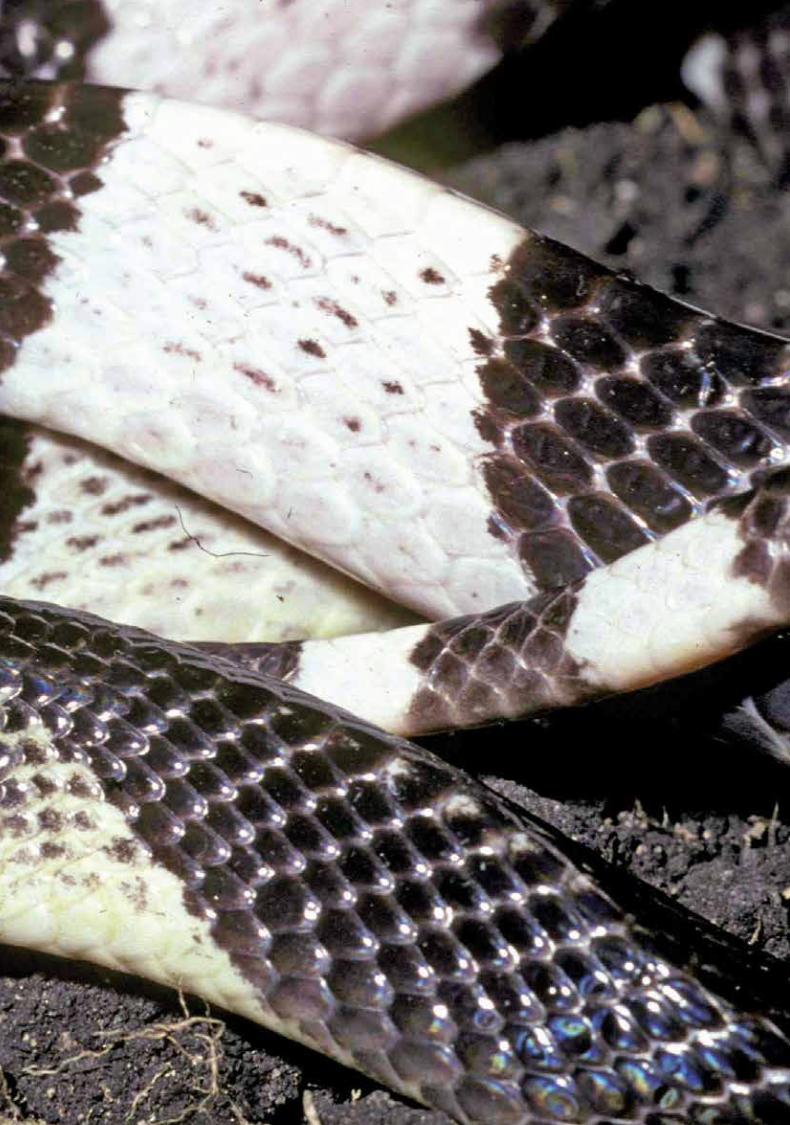
VENOMOUS SNAKES OF THE SEArO rEGION, THEIr VENOMS, AND THE PATHOPHYSIOLOGY...
203

GUIDELINES FOR THE MANAGEMENT OF SNAKEBITES
204
Snakebites are well-known medical emergencies in many parts of the world, especially in rural areas. Agricultural workers and children are the most affected. The incidence of snakebite mortality is particularly high in South-East Asia. Rational use of snake anti-venom can substantially reduce mortality and morbidity due to snakebites. These guidelines are a revised and updated version of Regional Guidelines for the Management of snakebites published by the WHO Regional Office in South-East Asia in 2011. These guidelines aim to promote the rational management of snakebite cases in various health facilities where trained health functionaries and quality snake anti-venom are available.
ISBN: 978-92-9022-530-0
World Health HouseIndraprastha Estate,Mahatma Gandhi Marg,New Delhi-110002, IndiaWebsite: www.searo.who.int