Unsuspected sonographic findings in patients with posttraumatic shoulder complaints
60
For Peer Review UNSUSPECTED SONOGRAPHIC FINDINGS IN PATIENTS WITH POST-TRAUMATIC SHOULDER COMPLAINTS Journal: Journal of Clinical Ultrasound Manuscript ID: JCU-10-047.R2 Wiley - Manuscript type: Research Article Keywords: Ultrasound, Shoulder, Trauma John Wiley & Sons Journal of Clinical Ultrasound peer-00576987, version 1 - 16 Mar 2011 Author manuscript, published in "Journal of Clinical Ultrasound 38, 9 (2010) 457" DOI : 10.1002/jcu.20745
-
Upload
jeroenboschziekenhuis -
Category
Documents
-
view
1 -
download
0
Transcript of Unsuspected sonographic findings in patients with posttraumatic shoulder complaints
For Peer Review
UNSUSPECTED SONOGRAPHIC FINDINGS IN PATIENTS
WITH POST-TRAUMATIC SHOULDER COMPLAINTS
Journal: Journal of Clinical Ultrasound
Manuscript ID: JCU-10-047.R2
Wiley - Manuscript type: Research Article
Keywords: Ultrasound, Shoulder, Trauma
John Wiley & Sons
Journal of Clinical Ultrasoundpe
er-0
0576
987,
ver
sion
1 -
16 M
ar 2
011
Author manuscript, published in "Journal of Clinical Ultrasound 38, 9 (2010) 457" DOI : 10.1002/jcu.20745
For Peer Review
1
UNSUSPECTED SONOGRAPHIC
FINDINGS
IN PATIENTS WITH POSTTRAUMATIC
SHOULDER COMPLAINTS
ARTICLE TYPE: SCIENTIFIC ARTICLE
Page 1 of 59
John Wiley & Sons
Journal of Clinical Ultrasound
123456789101112131415161718192021222324252627282930313233343536373839404142434445464748495051525354555657585960
peer
-005
7698
7, v
ersi
on 1
- 16
Mar
201
1
For Peer Review
2
ABSTRACT
PURPOSE. To prospectively assess the frequency of abnormal sonographic
findings in patients with posttraumatic shoulder pain and/or disability in whom
ultrasound (US) was not considered and to assess the effect of sonographic
findings on working diagnosis and therapeutic strategy, in order to analyse the
possible role of US in the diagnostic work-up of these patients.
METHODS. A survey was performed under general practitioners and orthopaedic
surgeons. They were requested to refer patients with persistent posttraumatic
complaints for an US examination of the shoulder and to fill in a questionnaire
concerning working diagnosis and therapy. In fifty patients examinations were
performed by two radiologists separately. Findings were confirmed with additional
radiographs and/or MRI and/or surgery. Shortly after US a survey was repeated.
RESULTS. Sonography showed relevant pathology in 45 (90%) of 50 patients, a
proximal humerus fracture in 25 (50%) patients, and a rotator cuff tear in 43 (86%)
patients. Twenty-three (92%) fractures were accompanied by a rotator cuff tear, and
23 (54%) rotator cuff tears were accompanied by a fracture. Ten fractures were
initially missed radiographically. Sonographic findings changed the working
diagnosis and therapeutic strategy in 37 (74%) and 26 (52%) patients, respectively.
CONCLUSION. In patients with posttraumatic shoulder complaints US showed a
high rate (90%) of relevant pathology. This changed the initial working diagnosis in
Page 2 of 59
John Wiley & Sons
Journal of Clinical Ultrasound
123456789101112131415161718192021222324252627282930313233343536373839404142434445464748495051525354555657585960
peer
-005
7698
7, v
ersi
on 1
- 16
Mar
201
1

For Peer Review
3
74% of the patients and the therapeutic strategy in more than half of the patients.
Active referral for sonographic examination may identify these abnormalities in an
earlier phase and improve clinical outcome.
KEYWORDS: Ultrasound, Shoulder, Trauma.
Page 3 of 59
John Wiley & Sons
Journal of Clinical Ultrasound
123456789101112131415161718192021222324252627282930313233343536373839404142434445464748495051525354555657585960
peer
-005
7698
7, v
ersi
on 1
- 16
Mar
201
1

For Peer Review
4
INTRODUCTION
Shoulder pain is frequently caused by an injury. Of all patients with shoulder
pain who visit primary care physicians in the United States, one third (33.2%)
present after an injury and in the other two thirds the cause is non-traumatic.1
Males and younger adults (age < 52) more often associate their shoulder
pain with previous injury.1 These shoulder injuries may remain undetected in the
acute phase.
Many studies have proven the efficacy of ultrasound (US) in the diagnosis of
partial- and full-thickness rotator cuff tears (RCT’s) 2-8
but immediately following
trauma US is often not performed because its value is considered limited.
Furthermore conventional x-ray’s are often inconclusive for the detection of
nondisplaced fractures of the tuberosity complex of the humerus.9 For this reason
diagnosis is often delayed which may cause longstanding shoulder complaints,
leading to temporary disability and considerable lost earnings.10
We hypothesize that many patients with shoulder complaints following
trauma have undetected and unsuspected shoulder injuries which may have
clinical consequences.
We prospectively assessed the frequency of abnormal sonographic findings
in patients with shoulder complaints following trauma in whom US was not
considered at the time of and following trauma and secondly we assessed the
effect of these findings on therapeutic strategy.
Page 4 of 59
John Wiley & Sons
Journal of Clinical Ultrasound
123456789101112131415161718192021222324252627282930313233343536373839404142434445464748495051525354555657585960
peer
-005
7698
7, v
ersi
on 1
- 16
Mar
201
1
For Peer Review
5
MATERIAL AND METHODS
Our institutional review board approved the study protocol and informed
consent was obtained from all patients. This prospective study was performed
during a two year period. In this period referring physicians were requested to refer
patients with shoulder complaints (pain and/or disability) following trauma in whom
diagnostic imaging with US, CT or MRI was not considered. Trauma was defined
as a serious injury to the shoulder, which urged the patient to seek medical care.
Time between trauma and referral should be at least two weeks and should not
exceed one year. Furthermore the referring physicians were requested to fill in a
questionnaire about the working diagnosis and treatment before referral. A second
survey within four weeks following US enquired about change in diagnosis and
treatment.
Patients
Fifty consecutive patients (29 women and 21 men, age range 21-80, mean age 49
years) were included.
Patients were referred by general practitioners (n=22) and orthopedic
surgeons (n=28). Time between trauma and the first visit to the general practitioner
or orthopedic surgeon ranged from 0-285 days (mean 38 days), and the time
between trauma and the US examination ranged from 14-304 days (mean 69
days). The referring physicians were asked about their working diagnosis and
therapy strategy. Patients with pre-existent shoulder complaints or previous
surgery were excluded from this study.
Page 5 of 59
John Wiley & Sons
Journal of Clinical Ultrasound
123456789101112131415161718192021222324252627282930313233343536373839404142434445464748495051525354555657585960
peer
-005
7698
7, v
ersi
on 1
- 16
Mar
201
1
For Peer Review
6
Two independent radiologists performed the US examinations during one
patient visit.
In the follow up of the patients, who were examined with US, we
retrospectively evaluated the patient files if conventional X-ray, MR imaging, and/or
open or arthroscopic surgery was performed. In all fifty patients conventional plain
radiographs were available, whereas US findings could be associated with MRI
and surgical findings in 10 and 19 patients, respectively.
Ultrasound examination
The US examinations were performed with an APLIO (Toshiba Medical Systems,
Tokyo, Japan) using a 12 (5 - 14) MHz broadband linear-array transducer (Toshiba
PLF-805ST). Patients were examined seated on a swivel chair, facing the
examiner. A standardized US imaging protocol of the shoulder was used.11-14
All
sonograms were performed by two radiologists, one experienced (>15 years)
musculoskeletal radiologist (M.J.R.) and one experienced (>20 years) abdominal
radiologist (G.J.), who had one year experience in performing US of the shoulder.
The patients were examined by both examiners separately, one after the other.
Both were blinded to the results of previously performed examinations and to their
mutual findings. Following the US examinations consensus reading was
performed. When discrepant findings between the two readers were found the
patient was at the same sitting re-examined by both readers together to determine
the cause of the discrepancy.
Page 6 of 59
John Wiley & Sons
Journal of Clinical Ultrasound
123456789101112131415161718192021222324252627282930313233343536373839404142434445464748495051525354555657585960
peer
-005
7698
7, v
ersi
on 1
- 16
Mar
201
1

For Peer Review
7
Plain radiographs
All 50 patients underwent a radiographic examination (i.e., trauma series), which
consisted of an anteroposterior view (external and/or internal rotation of the
humerus), an axillary view and a transscapular view of the shoulder. In 33 patients
plain radiographs of the shoulder were obtained following trauma. In the remaining
17 patients a fracture was not suspected clinically. They underwent plain
radiography subsequently in conjunction with their US examination.
Following the US examinations both sonologists performed a consensus
reading of all 33 initially obtained plain radiographs. If there was a discrepancy
between the US findings and the plain radiographic findings new trauma series
were obtained and if these were not conclusive additional radiographic views were
obtained. The image projection of these additional views were determined
according to the US findings, tangent to the expected fracture (Fig. 1).
Magnetic Resonance Imaging (MRI)
In 10 patients MR images were obtained with a 1.5-T scanner (Vision, Siemens
Medical systems, Erlangen, Germany) using a surface coil. Indications for the MR
examinations were to verify sonographic findings e.g., fractures not visible on plain
radiographs (n=2) and rotator cuff tears (n=2), to provide the surgeon with more
anatomical information in case surgery was considered, and for the detection of
intraarticular pathology (n=6). Four patients underwent MRI and 6 patients MR-
arthrography of the shoulder. The MR imaging protocol included axial, sagittal and
coronal T1 (repetition time (TR) 600 / echo time (TE) 30) and T2 weighted (TR
Page 7 of 59
John Wiley & Sons
Journal of Clinical Ultrasound
123456789101112131415161718192021222324252627282930313233343536373839404142434445464748495051525354555657585960
peer
-005
7698
7, v
ersi
on 1
- 16
Mar
201
1
For Peer Review
8
3000 / TE 50) spin-echo sequences. The field of view was 16 cm, and the data
acquisition matrix 196 x 512. A section thickness of 3 mm was used with a 0.5 mm
intersection gap. Three acquisitions were averaged.
The MR arthrography scanning protocol consisted of 3 dimensional (3D)-gradient
T1-weighted (FLASH 3D) oblique coronal scans with fat saturation, which were
reconstructed in the sagittal and transverse planes. FLASH 3D imaging (TR: 8.1
ms; TE: 4 ms; 2 acquisitions; 192 × 256 matrix; field of view (FOV): 24 cm) with 1-
mm consecutive slices was used.
Image evaluation
Sonographic examinations were evaluated for abnormalities of the deltoid muscle,
subacromial-subdeltoid bursa, rotator cuff, long head of the biceps tendon and the
proximal humerus including the tuberosity complex.
According to the criteria as established in the literature,15-20
a full-thickness rotator
cuff tear (FTT) is defined as non-visualization or absence of the rotator cuff, or a
full-thickness rotator cuff discontinuity, and a partial-thickness rotator cuff tear
(PTT) as a focal thinning of the rotator cuff, loss of convexity of the outer cuff
border and a hypoechoic defect involving the articular or bursal side or within the
tendon.
Sonographic features of bone fracture are: periosteal elevation, cortical bone
discontinuity (Fig. 2a and Fig. 3a and b), step-off deformity with one or more
hyperechoic reflections (i.e., avulsed, dislocated (Fig. 1a) or impacted bone
fragments),9,21
and the double line sign (two parallel hyperechoic lines).22
Page 8 of 59
John Wiley & Sons
Journal of Clinical Ultrasound
123456789101112131415161718192021222324252627282930313233343536373839404142434445464748495051525354555657585960
peer
-005
7698
7, v
ersi
on 1
- 16
Mar
201
1
For Peer Review
9
Initial plain radiographs were evaluated prospectively in routine practice by
residents and radiologists with a varying level of experience. The final retrospective
reading of all 50 trauma series including the additionally obtained plain radiographs
was performed by consensus by the two sonologists (Table 1).
Statistical analysis
For the analyses, we used descriptive statistics only. Because of the relatively
small number of patients, it was not considered useful to use inferential statistics.
For the descriptive statistics, we calculated means and standard deviations for
continuous variables and percentages (i.e., prevalences) for categorical variables.
For the interrater reproducibility between the 2 sonologists, we calculated Cohen's
kappa statistic for fractures (yes vs. no) and ruptures (no vs. partial vs. total).
RESULTS
Fifty patients (30 right and 20 left shoulders) were sonographically examined within
14–304 days (average 69 days) following trauma. The mechanism of trauma was a
direct fall on the shoulder (n=26), a fall on a hyperextended arm (n=8), falling while
grabbing (hyperextension with traction) (n=10) and various other causes (n=6)
such as luxation, forced external rotation or forced hyperextension with abduction.
US showed no abnormalities in 5 (10%) patients and pathologic findings
(proximal humerus fracture, RCT’s, long head of the biceps tendon luxation or
dislocation) in 45 (90%) of the patients. Both observers detected all fractures.
Therefore, the interobserver agreement for the sonographic detection of fractures
Page 9 of 59
John Wiley & Sons
Journal of Clinical Ultrasound
123456789101112131415161718192021222324252627282930313233343536373839404142434445464748495051525354555657585960
peer
-005
7698
7, v
ersi
on 1
- 16
Mar
201
1

For Peer Review
10
was perfect (Cohen’s kappa is 1.0). The less experienced sonographer interpreted
one PTT as tendinosis (Fig. 3), while in the remaining 49 (98%) cases both
observers were in agreement, which each other’s sonographic findings. The
interobserver agreement for the sonographic detection of RCT’s was almost
perfect (Cohen’s kappa is 0.96).
Rotator cuff tears
In 43 (86%) of the 50 patients one or more RCT’s were detected with US. We
identified 22 FTT and 16 PTT in the supraspinatus tendon and 7 FTT and 6 PTT in
the subscapularis tendon. One FTT was detected in the infraspinatus tendon and
no tears were detected in the teres minor tendon.
A RCT without other accompanying posttraumatic shoulder pathology was
sonographically depicted in 15 of the 43 cases, whereas 28 patients with a RCT
suffered from accompanying fractures of the proximal humerus (n=23) and/or
subluxation (n=3) or dislocation (n=4) of the long head of the biceps tendon. Four
PTT’s were accompanied by 3 subluxated and 1 dislocated biceps tendon and 3
FTT’s by a dislocated long head of the biceps tendon.
Fractures
In 25 (50%) of the 50 patients a fracture of the proximal humerus was detected
sonographically and confirmed with the initially or additionally obtained plain
radiographs and/or with MR imaging. In the remaining 25 patients no fracture could
be detected with either imaging technique.
Page 10 of 59
John Wiley & Sons
Journal of Clinical Ultrasound
123456789101112131415161718192021222324252627282930313233343536373839404142434445464748495051525354555657585960
peer
-005
7698
7, v
ersi
on 1
- 16
Mar
201
1
For Peer Review
11
In the patient group, who initially underwent plain radiography (n=33), a total
of 19 fractures were found. However, only 9 (47%) of these fractures were
detected prospectively. At retrospective review of the initially obtained plain
radiographs 5 additional fractures were detected.
Seven (28%) of the 25 fractures could only be depicted after obtaining
additional projections (Fig. 1) or were only visible on additionally obtained
radiographs (Fig. 2) or with MRI (Fig. 3). Sonography did not miss any fracture
depicted by plain radiography.
In the patient group (n=17), who did not undergo radiography initially, 6
fractures were detected with sonography and confirmed with conventional
radiographs.
According to Neer’s four-segment classification for fractures of the proximal
humerus,23
22 patients (92%) had a non- or minimally displaced one-part fracture,
and 3 patients a two-part fracture. None of the patients suffered from a three-part
or four-part fracture.
In 23 of the 25 patients with a fracture this was accompanied by a RCT (15
PTT and 8 FTT).
Associated posttraumatic pathologic musculoskeletal findings
A thickened subacromial-subdeltoid bursa (i.e., bursitis) (Fig. 2a) was
sonographically diagnosed in 2 patients. This is either caused by trauma or due to
chronic impingement. In neither patient was a fracture or RCT found.
Page 11 of 59
John Wiley & Sons
Journal of Clinical Ultrasound
123456789101112131415161718192021222324252627282930313233343536373839404142434445464748495051525354555657585960
peer
-005
7698
7, v
ersi
on 1
- 16
Mar
201
1

For Peer Review
12
Subluxation of the long head of the biceps tendon (Fig. 4a) was diagnosed in
3 patients and complete dislocation of the biceps tendon (Fig. 4b) was diagnosed
in 4 patients. These biceps tendon subluxation and dislocations were
accompanied by PTT (n=4) or FTT (n=3) of the subscapularis (n=2) and/or
supraspinatus tendon (n=5).
Working diagnosis and therapeutic strategy changes
Compared to the initial diagnosis and therapy the referring physician stated that
sonographic findings affected the working diagnosis and therapeutic strategy in 37
(74%) and 26 (52%) patients, respectively.
In 13 (26%) of the 50 patients US confirmed the working diagnosis. In 19
(38%) patients US confirmed the working diagnosis, but also detected additional
traumatically caused pathology, and in 18 (36%) patients US findings were not
concurrent with the clinical working diagnosis.
Therapy was changed because although the diagnosis was confirmed,
probably due to more diagnostic certainty in 9 (69%) of the 13 patients. Therapy
was also changed in 11 (58%) of the 19 patients with whom the working diagnosis
was confirmed and additional posttraumatic pathology was found. Finally, in only 6
(33%) of the 18 patients with whom the sonographic findings and working
diagnosis did not concurred, therapy did change. These six relatively young (37 to
52 years) patients presented with a FTT, who were operated upon following US
after initially conservative treatment. In the remaining 12 patients the initially
initiated conservative therapy was continued despite the sonographic finding of a
Page 12 of 59
John Wiley & Sons
Journal of Clinical Ultrasound
123456789101112131415161718192021222324252627282930313233343536373839404142434445464748495051525354555657585960
peer
-005
7698
7, v
ersi
on 1
- 16
Mar
201
1

For Peer Review
13
FTT in 4 patients, a PTT in 4 patients, a non-displaced fracture of the greater
tuberosity in 4 patients.
In 26 of the 50 patients US changed therapy strategy. In 21 of these 26
patients conservative therapy (rest, physiotherapy, subacromial injections) was
changed to surgical treatment following US. Twenty patients underwent surgical
rotator cuff repair of 4 PTT and 16 FTT and one patient because of a luxation of
the long head of the biceps tendon in combination with a FTT. In the remaining 5
of the 26 patients conservative treatment was changed in a lighter physiotherapy
program (n=4) or rest (n=2).
DISCUSSION
Trauma-related shoulder disorders are frequently initially missed, affecting quality
of life.24,25
In this prospective study we demonstrate that patients with
posttraumatic shoulder complaints, have a high prevalence of rotator cuff tears
(86%) and fractures (50%) of the proximal humerus. This confirms the findings of
Sørensen et al,4 who reported that clinical examination underestimates the
prevalence and severity of acute RCT’s and fractures. This has also been
indicated by Patten et al,9 who showed that 10 (42%) of the 24 sonographically
detected greater tuberosity fractures were missed with plain radiography. In our
study, 10 (53%) of the 19 fractures were not depicted at the initial reading. Even
retrospectively, 26% (5 of 19) of these fractures could not be depicted from the
initially obtained trauma series. Of all 25 fractures in our study 7 (28%) could not
be depicted from the trauma series, whereas all 25 fractures were depicted
Page 13 of 59
John Wiley & Sons
Journal of Clinical Ultrasound
123456789101112131415161718192021222324252627282930313233343536373839404142434445464748495051525354555657585960
peer
-005
7698
7, v
ersi
on 1
- 16
Mar
201
1
For Peer Review
14
sonographically.9,22
A fracture can easily be identified sonographically as a
discontinuity of the cortical bone, when scanning perpendicular to the fracture line.
Another characteristic sonographic feature of a fracture is the double line sign.22
In all but two patients with a fracture of the proximal humerus a PTT or FTT
could be detected. This is in contrast with Patten et al,9 who suggests that this
combination can be found in only 17-25%. The higher number of RCT in our study
is probably due to the selected patients, e.g., patients with persistent shoulder
complaints following trauma, and may also be related to the injury mechanism or
severity of trauma. Age seems not to be the reason for the relatively high
frequency of RCT in our study, in the age group of 21 to 30 years the frequency of
RCT was 33%, in the other age groups the frequency was (80-100%). In this study
no surgical therapy was performed in any of the 24 patients with a fracture of the
proximal humerus.
In the current study US was not performed immediately after injury or in the
emergency setting. Possible disadvantages of performing US in the acute phase is
the detection of chronic, irrelevant asymptomatic RCT’s 26
and the decreased
diagnostic performance of US due to limited shoulder motion, necessary for
visualization of the entire rotator cuff.5 However, Teefey et al
27 showed that on the
basis of location (midsubstance) and associated findings (the presence of joint or
bursal fluid) acute and chronic RCT can be differentiated. Farin et al 5 and
Sørensen et al 4 demonstrated that adequate sonographic examination was
feasible in 88% in the acute phase.
In our study we found that referring physicians stated that US findings
Page 14 of 59
John Wiley & Sons
Journal of Clinical Ultrasound
123456789101112131415161718192021222324252627282930313233343536373839404142434445464748495051525354555657585960
peer
-005
7698
7, v
ersi
on 1
- 16
Mar
201
1
For Peer Review
15
changed the working diagnosis in 74% and the therapeutic strategy in more than
half of the patients supporting the suggestion that sonography reveals clinically
relevant findings.28
We found no previous studies that addressed clinical efficacy,
29 which includes diagnostic impact and the therapeutic efficacy in patients with
shoulder complaints following trauma. Change in therapy is more objectively
ascertainable but it is difficult to assess to what extent the sonographic diagnosis
contributes to the change in therapy e.g., we found that therapeutic strategy
changed more often when the initial diagnosis was not changed.
When interpreting the results of our study, some limitations have to be
considered. First, the number of patients included in this study is relatively small.
Secondly, the high number of posttraumatic pathologic findings may be biased. It
is possible that patient selection was not exactly according the study purpose (e.g.,
patients with shoulder complaints following trauma in whom US was not
considered), but that patients with shoulder complaints following trauma in whom
US was already considered also answered the call up.
Thirdly, the methods for assessing diagnostic and therapeutic efficacy are not
well established. These are subjective and difficult to quantify.29
It is possible that
patient symptoms were increasing with time and sonographic findings were not the
only reason for a change in treatment. Furthermore the second survey we
performed 1 month following US, which is not ideal for the assessment of the
diagnostic impact and therapeutic efficacy.29
Therefore, the effect of US imaging
on diagnosis and therapeutic strategy may be overestimated.
In the literature there is no consensus about the optimal therapeutic strategy
Page 15 of 59
John Wiley & Sons
Journal of Clinical Ultrasound
123456789101112131415161718192021222324252627282930313233343536373839404142434445464748495051525354555657585960
peer
-005
7698
7, v
ersi
on 1
- 16
Mar
201
1

For Peer Review
16
and timing of treatment of RCT’s following trauma.30,31
Therefore, additional
prospective studies are needed to confirm the hypothesis that early sonographic
assessment of the rotator cuff integrity in patients with post traumatic shoulder
complaints improves patient outcome and to establish the best time to perform US
in these patients.
The results of such studies could potentially change the approach of acute injury to
the shoulder. It is arguable, considering the limited detection of fractures with
conventional radiographs to propose the following algorithm. First do an ultrasound
in the acute phase, and if the pain does not subside after a week then take an X-ray
In our study we would not have missed any substantial abnormalities and would
have made the right diagnosis in an earlier phase.. It would sure have clarified a lot
of pain caused by missed fractures and rotator cuff tears. Dislocations with
complete loss of function of course may have to be treated separately in this
algorithm, and if US is negative and/or clinical symptoms are related to intraarticular
disorders MR arthrography should be performed
CONCLUSION
Patients with posttraumatic shoulder complaints have a high prevalence of
unsuspected and initially missed RCT’s and fractures and US is accurate in the
detection of clinically relevant trauma-related shoulder pathology.
Further studies are needed to prove that early and active referral of these
patients for sonographic examination may improve patient outcome.
Page 16 of 59
John Wiley & Sons
Journal of Clinical Ultrasound
123456789101112131415161718192021222324252627282930313233343536373839404142434445464748495051525354555657585960
peer
-005
7698
7, v
ersi
on 1
- 16
Mar
201
1
For Peer Review
17
Page 17 of 59
John Wiley & Sons
Journal of Clinical Ultrasound
123456789101112131415161718192021222324252627282930313233343536373839404142434445464748495051525354555657585960
peer
-005
7698
7, v
ersi
on 1
- 16
Mar
201
1
For Peer Review
18
REFERENCES
1. Wofford JL, Mansfield RJ, Watkins RS. Patient characteristics and clinical
management of patients with shoulder pain in U.S. primary care settings:
secondary data analysis of the National Ambulatory Medical Care Survey.
BMC Musculoskelet Disord 2005; 6:-4.
2. Teefey SA, Rubin DA, Middleton WD, et al. Detection and quantification of
rotator cuff tears. Comparison of ultrasonographic, magnetic resonance
imaging, and arthroscopic findings in seventy-one consecutive cases. J Bone
Joint Surg Am 2004; 86-A:708.
3. van Holsbeeck MT, Kolowich PA, Eyler WR, et al. US depiction of partial-
thickness tear of the rotator cuff. Radiology 1995; 197:443.
4. Sørensen AKB, Bak K, Krarup AL, et al. Acute rotator cuff tear: Do we miss
the early diagnosis? A prospective study showing a high incidence of rotator
cuff tears after shoulder trauma. J Shoulder Elbow Surg 2007; 16:174.
5. Farin PU, Jaroma H. Acute traumatic tears of the rotator cuff: Value of
sonography. Radiology 1995; 197:269.
6. Bohndorf K, Kilcoyne RF. Traumatic injuries: imaging of peripheral
musculoskeletal injuries. Eur Radiol 2002; 12:1605.
7. Grechenig W, Clement H, Fankhauser F, et al. Ultrasound diagnosis in
shoulder trauma. Orthopade 2002; 31:250.
Page 18 of 59
John Wiley & Sons
Journal of Clinical Ultrasound
123456789101112131415161718192021222324252627282930313233343536373839404142434445464748495051525354555657585960
peer
-005
7698
7, v
ersi
on 1
- 16
Mar
201
1
For Peer Review
19
8. Weiss DB, Jacobson JA, Karunakar MA. The use of ultrasound in evaluating
orthopaedic trauma patients. J Am Acad Orthop Surg 2005; 13:525.
9. Patten RM, Mack LA, Wang KY, et al. Nondisplaced fractures of the greater
tuberosity of the humerus: Sonographic detection. Radiology 1992; 182:201.
10. Reville RT, Neuhauser FW, Bhattacharya J, et al. Comparing severity of
impairment for different permanent upper extremity musculoskeletal injuries.
J Occup Rehabil 2002; 12:205.
11. Middleton WD, Edelstein G, Reinus WR, et al. Ultrasonography of the rotator
cuff: Technique and normal anatomy. J Ultrasound Med 1984; 3:549.
12. Middleton WD, Reinus WR, Totty WG, et al. Ultrasonographic evaluation of
the rotator cuff and biceps tendon. J Bone Joint Surg [Am] 1986; 68:440.
13. Mack LA, Nyberg DA, Matsen FA. Sonographic evaluation of the rotator cuff.
Radiol Clin North Am 1988; 26:161.
14. Crass JR, Craig EV, Feinberg SB. The hyperextended internal rotation view
in rotator cuff ultrasound. J Clin Ultrasound 1987; 15:416.
15. Bretzke CA, Crass JR, Craig EV, et al. Ultrasonography of the rotator cuff.
Normal and pathologic anatomy. Invest Radiol 1985; 20:311.
16. Crass JR, Craig EV, Feinberg SB. Sonography of the postoperative rotator
cuff. AJR 1986; 146:561.
Page 19 of 59
John Wiley & Sons
Journal of Clinical Ultrasound
123456789101112131415161718192021222324252627282930313233343536373839404142434445464748495051525354555657585960
peer
-005
7698
7, v
ersi
on 1
- 16
Mar
201
1

For Peer Review
20
17. Mack LA, Matsen III FA, Kilcoyne RF, et al. US evaluation of the rotator cuff.
Radiology 1985; 157:205.
18. Middleton WD, Edelstein G, Reinus WR, et al. Sonographic detection of
rotator cuff tears. AJR 1985; 144:349.
19. Brandt TD, Cardone BW, Grant TH, et al. Rotator cuff sonography: A
reassessment. Radiology 1989; 173:323.
20. Rutten MJ, Jager GJ, Blickman JG. Ultrasound of the rotator cuff: pitfalls,
limitations and artifacts. Radiographics 2006; 26:589.
21. Cicak N, Bilic R, Delimar D. Hill-sachs lesion in recurrent shoulder
dislocation: sonographic detection. J Ultrasound Med 1998; 17:557.
22. Rutten MJ, Jager GJ, de Waal Malefijt MC, et al. Double Line Sign: A helpful
sonographic sign to detect occult fractures of the proximal humerus. Eur
Radiol 2007; 17:762.
23. Neer CS. Displaced proximal humeral fractures. 1. Classification and
evaluation. J Bone Joint Surg [Am] 1970; 52-A:1077.
24. Malhotra AK, Martin N, Jacoby M, et al. What are we missing: results of a 13-
month active follow-up program at a level I trauma center. J Trauma 2009;
66:1696.
25. Lichtveld RA, Spijkers AT, Panhuizen IF, et al. Background and
consequences of injuries missed when diagnosing severely injured accident
Page 20 of 59
John Wiley & Sons
Journal of Clinical Ultrasound
123456789101112131415161718192021222324252627282930313233343536373839404142434445464748495051525354555657585960
peer
-005
7698
7, v
ersi
on 1
- 16
Mar
201
1

For Peer Review
21
victims in prehospital care in patients transported by ambulance to the
University Medical Centre in Utrecht, 1999-2000. Ned Tijdschr Geneeskd
2006; 150:2197.
26. Reilly P, Macleod I, Macfarlane R, et al. Dead men and radiologists don't lie:
a review of cadaveric and radiological studies of rotator cuff tear prevalence.
Ann R Coll Surg Engl 2006; 88:116.
27. Teefey SA, Middleton WD, Bauer GS, et al. Sonographic differences in the
appearance of acute and chronic full-thickness rotator cuff tears. J
Ultrasound Med 2000; 19:377.
28. Lanzer WL. Clinical aspects of shoulder injuries. Radiol Clin North Am 1988;
26:157.
29. Thornbury JR. Eugene W. Caldwell Lecture. Clinical efficacy of diagnostic
imaging: love it or leave it. AJR 1994; 162:1.
30. Coghlan JA, Buchbinder R, Green S, et al. Surgery for rotator cuff disease.
Cochrane Database Syst Rev. 2008; 23:CD005619.
31. Ejnisman B, Andreoli CV, Soares BG, et al. Interventions for tears of the
rotator cuff in adults. Cochrane Database Syst Rev 2004; 1:-CD002758.
Page 21 of 59
John Wiley & Sons
Journal of Clinical Ultrasound
123456789101112131415161718192021222324252627282930313233343536373839404142434445464748495051525354555657585960
peer
-005
7698
7, v
ersi
on 1
- 16
Mar
201
1

For Peer Review
22
FIGURES AND LEGENDS
Fig. 1. 52-year-old man with a limited and painful external rotation of the right
shoulder following trauma.
A, Long axis sonogram of the subscapularis tendon (SSC) shows a linear
hyperechoic reflection (arrows) in the midsubstance of the subscapularis tendon
insertion, which is suggestive for an avulsion fracture of the lesser tuberosity (LT).
B, Additionally obtained radiographic projection (cranio-caudal view) confirmed the
lesser tuberosity fracture, whereas this was not visible on the initially obtained
conventional radiographs.
Fig. 2. 68-year-old woman with shoulder pain and disability following a fall on the
right shoulder.
A, The initial radiographs, obtained 6 days following trauma, were interpreted as
normal.
B, Long axis sonogram of the supraspinatus tendon (SSP) shows a cortical bone
discontinuity (large arrow) at the junction between the greater tuberosity (GT) and
the humeral head, representing a non-displaced greater tuberosity fracture. The
thickened subacromial-subdeltoid bursa (small arrows) is either caused by trauma
or due to chronic impingement.
C, The radiographs obtained 1 month following trauma showed a radiolucent zone
(arrows) along the fracture line due to bone resorption indicative of active bone
repair.
Page 22 of 59
John Wiley & Sons
Journal of Clinical Ultrasound
123456789101112131415161718192021222324252627282930313233343536373839404142434445464748495051525354555657585960
peer
-005
7698
7, v
ersi
on 1
- 16
Mar
201
1
For Peer Review
23
Fig. 3. 43-year-old man with pain and disability of the shoulder 19 days following a
fall on the right shoulder.
A, B, Long axis sonograms of the supraspinatus tendon (SSP) showing corticol
bone discontinuities (large arrows) and outpouchings of cortical bone (small arrows)
representing fractures of the greater tuberosity (GT), which are frequently
associated with intrasubstance rotator cuff tears (asterisks).
C, A radiograph obtained directly following US could not confirm the US findings.
D, Coronal T1-weighted FSE images with fat saturation of a MR arthrogram
confirmed the US findings by showing an elevated signal of the greater tuberosity
(GT) due to oedema because of a fracture and a longitudinally orientated
intrasubstance tear (arrow) of the supraspinatus tendon (SSP).
Fig. 4 Subluxation (A) and complete dislocation (B) of the long head of the biceps
tendon.
A, 49-year-old man. Transverse US image obtained at the level of the
intertubercular groove shows subluxation of the long head of the biceps tendon
(arrow), which is located over the lesser tuberosity (LT). D = Deltoid muscle, GT =
greater tuberosity.
B, 60-year-old woman. Transverse US image obtained at the level of the
intertubercular groove shows medial dislocation of the long head of the biceps
tendon (arrow) and an empty, fluid filled intertubercular groove (small black arrows).
B = distended subacromial-subdeltoid bursa, GT = greater tuberosity, LT = lesser
tuberosity.
Page 23 of 59
John Wiley & Sons
Journal of Clinical Ultrasound
123456789101112131415161718192021222324252627282930313233343536373839404142434445464748495051525354555657585960
peer
-005
7698
7, v
ersi
on 1
- 16
Mar
201
1
For Peer Review
24
Page 24 of 59
John Wiley & Sons
Journal of Clinical Ultrasound
123456789101112131415161718192021222324252627282930313233343536373839404142434445464748495051525354555657585960
peer
-005
7698
7, v
ersi
on 1
- 16
Mar
201
1

For Peer Review
1
UNSUSPECTED SONOGRAPHIC
FINDINGS
IN PATIENTS WITH POSTTRAUMATIC
SHOULDER COMPLAINTS
ARTICLE TYPE: SCIENTIFIC ARTICLE
Page 25 of 59
John Wiley & Sons
Journal of Clinical Ultrasound
123456789101112131415161718192021222324252627282930313233343536373839404142434445464748495051525354555657585960
peer
-005
7698
7, v
ersi
on 1
- 16
Mar
201
1

For Peer Review
2
ABSTRACT
PURPOSE. To prospectively assess the frequency of abnormal sonographic
findings in patients with posttraumatic shoulder pain and/or disability in whom
ultrasound (US) was not considered and to assess the effect of sonographic
findings on working diagnosis and therapeutic strategy, in order to analyse the
possible role of US in the diagnostic work-up of these patients.
METHODS. A survey was performed under general practitioners and orthopaedic
surgeons. They were requested to refer patients with persistent posttraumatic
complaints for an US examination of the shoulder and to fill in a questionnaire
concerning working diagnosis and therapy. In fifty patients examinations were
performed by two radiologists separately. Findings were confirmed with additional
radiographs and/or MRI and/or surgery. Shortly after US a survey was repeated.
RESULTS. Sonography showed relevant pathology in 45 (90%) of 50 patients, a
proximal humerus fracture in 25 (50%) patients, and a rotator cuff tear in 43 (86%)
patients. Twenty-three (92%) fractures were accompanied by a rotator cuff tear, and
23 (54%) rotator cuff tears were accompanied by a fracture. Ten fractures were
initially missed radiographically. Sonographic findings changed the working
diagnosis and therapeutic strategy in 37 (74%) and 26 (52%) patients, respectively.
CONCLUSION. In patients with posttraumatic shoulder complaints US showed a
high rate (90%) of relevant pathology. This changed the initial working diagnosis in
Page 26 of 59
John Wiley & Sons
Journal of Clinical Ultrasound
123456789101112131415161718192021222324252627282930313233343536373839404142434445464748495051525354555657585960
peer
-005
7698
7, v
ersi
on 1
- 16
Mar
201
1
For Peer Review
3
74% of the patients and the therapeutic strategy in more than half of the patients.
Active referral for sonographic examination may identify these abnormalities in an
earlier phase and improve clinical outcome.
KEYWORDS: Ultrasound, Shoulder, Trauma.
Page 27 of 59
John Wiley & Sons
Journal of Clinical Ultrasound
123456789101112131415161718192021222324252627282930313233343536373839404142434445464748495051525354555657585960
peer
-005
7698
7, v
ersi
on 1
- 16
Mar
201
1

For Peer Review
4
INTRODUCTION
Shoulder pain is frequently caused by an injury. Of all patients with shoulder
pain who visit primary care physicians in the United States, one third (33.2%)
present after an injury and in the other two thirds the cause is non-traumatic.1
Males and younger adults (age < 52) more often associate their shoulder
pain with previous injury.1 These shoulder injuries may remain undetected in the
acute phase.
Many studies have proven the efficacy of ultrasound (US) in the diagnosis of
partial- and full-thickness rotator cuff tears (RCT’s) 2-8
but immediately following
trauma US is often not performed because its value is considered limited.
Furthermore conventional x-ray’s are often inconclusive for the detection of
nondisplaced fractures of the tuberosity complex of the humerus.9 For this reason
diagnosis is often delayed which may cause longstanding shoulder complaints,
leading to temporary disability and considerable lost earnings.10
We hypothesize that many patients with shoulder complaints following
trauma have undetected and unsuspected shoulder injuries which may have
clinical consequences.
We prospectively assessed the frequency of abnormal sonographic findings
in patients with shoulder complaints following trauma in whom US was not
considered at the time of and following trauma and secondly we assessed the
effect of these findings on therapeutic strategy.
Page 28 of 59
John Wiley & Sons
Journal of Clinical Ultrasound
123456789101112131415161718192021222324252627282930313233343536373839404142434445464748495051525354555657585960
peer
-005
7698
7, v
ersi
on 1
- 16
Mar
201
1

For Peer Review
5
MATERIAL AND METHODS
Our institutional review board approved the study protocol and informed
consent was obtained from all patients. This prospective study was performed
during a two year period. In this period referring physicians were requested to refer
patients with shoulder complaints (pain and/or disability) following trauma in whom
diagnostic imaging with US, CT or MRI was not considered. Trauma was defined
as a serious injury to the shoulder, which urged the patient to seek medical care.
Time between trauma and referral should be at least two weeks and should not
exceed one year. Furthermore the referring physicians were requested to fill in a
questionnaire about the working diagnosis and treatment before referral. A second
survey within four weeks following US enquired about change in diagnosis and
treatment.
Patients
Fifty consecutive patients (29 women and 21 men, age range 21-80, mean age 49
years) were included.
Patients were referred by general practitioners (n=22) and orthopedic
surgeons (n=28). Time between trauma and the first visit to the general practitioner
or orthopedic surgeon ranged from 0-285 days (mean 38 days), and the time
between trauma and the US examination ranged from 14-304 days (mean 69
days). The referring physicians were asked about their working diagnosis and
therapy strategy. Patients with pre-existent shoulder complaints or previous
surgery were excluded from this study.
Page 29 of 59
John Wiley & Sons
Journal of Clinical Ultrasound
123456789101112131415161718192021222324252627282930313233343536373839404142434445464748495051525354555657585960
peer
-005
7698
7, v
ersi
on 1
- 16
Mar
201
1
For Peer Review
6
Two independent radiologists performed the US examinations during one
patient visit.
In the follow up of the patients, who were examined with US, we
retrospectively evaluated the patient files if conventional X-ray, MR imaging, and/or
open or arthroscopic surgery was performed. In all fifty patients conventional plain
radiographs were available, whereas US findings could be associated with MRI
and surgical findings in 10 and 19 patients, respectively.
Ultrasound examination
The US examinations were performed with an APLIO (Toshiba Medical Systems,
Tokyo, Japan) using a 12 (5 - 14) MHz broadband linear-array transducer (Toshiba
PLF-805ST). Patients were examined seated on a swivel chair, facing the
examiner. A standardized US imaging protocol of the shoulder was used.11-14
All
sonograms were performed by two radiologists, one experienced (>15 years)
musculoskeletal radiologist (M.J.R.) and one experienced (>20 years) abdominal
radiologist (G.J.), who had one year experience in performing US of the shoulder.
The patients were examined by both examiners separately, one after the other.
Both were blinded to the results of previously performed examinations and to their
mutual findings. Following the US examinations consensus reading was
performed. When discrepant findings between the two readers were found the
patient was at the same sitting re-examined by both readers together to determine
the cause of the discrepancy.
Page 30 of 59
John Wiley & Sons
Journal of Clinical Ultrasound
123456789101112131415161718192021222324252627282930313233343536373839404142434445464748495051525354555657585960
peer
-005
7698
7, v
ersi
on 1
- 16
Mar
201
1
For Peer Review
7
Plain radiographs
All 50 patients underwent a radiographic examination (i.e., trauma series), which
consisted of an anteroposterior view (external and/or internal rotation of the
humerus), an axillary view and a transscapular view of the shoulder. In 33 patients
plain radiographs of the shoulder were obtained following trauma. In the remaining
17 patients a fracture was not suspected clinically. They underwent plain
radiography subsequently in conjunction with their US examination.
Following the US examinations both sonologists performed a consensus
reading of all 33 initially obtained plain radiographs. If there was a discrepancy
between the US findings and the plain radiographic findings new trauma series
were obtained and if these were not conclusive additional radiographic views were
obtained. The image projection of these additional views were determined
according to the US findings, tangent to the expected fracture (Fig. 1).
Magnetic Resonance Imaging (MRI)
In 10 patients MR images were obtained with a 1.5-T scanner (Vision, Siemens
Medical systems, Erlangen, Germany) using a surface coil. Indications for the MR
examinations were to verify sonographic findings e.g., fractures not visible on plain
radiographs (n=2) and rotator cuff tears (n=2), to provide the surgeon with more
anatomical information in case surgery was considered, and for the detection of
intraarticular pathology (n=6). Four patients underwent MRI and 6 patients MR-
arthrography of the shoulder. The MR imaging protocol included axial, sagittal and
coronal T1 (repetition time (TR) 600 / echo time (TE) 30) and T2 weighted (TR
Page 31 of 59
John Wiley & Sons
Journal of Clinical Ultrasound
123456789101112131415161718192021222324252627282930313233343536373839404142434445464748495051525354555657585960
peer
-005
7698
7, v
ersi
on 1
- 16
Mar
201
1

For Peer Review
8
3000 / TE 50) spin-echo sequences. The field of view was 16 cm, and the data
acquisition matrix 196 x 512. A section thickness of 3 mm was used with a 0.5 mm
intersection gap. Three acquisitions were averaged.
The MR arthrography scanning protocol consisted of 3 dimensional (3D)-gradient
T1-weighted (FLASH 3D) oblique coronal scans with fat saturation, which were
reconstructed in the sagittal and transverse planes. FLASH 3D imaging (TR: 8.1
ms; TE: 4 ms; 2 acquisitions; 192 × 256 matrix; field of view (FOV): 24 cm) with 1-
mm consecutive slices was used.
Image evaluation
Sonographic examinations were evaluated for abnormalities of the deltoid muscle,
subacromial-subdeltoid bursa, rotator cuff, long head of the biceps tendon and the
proximal humerus including the tuberosity complex.
According to the criteria as established in the literature,15-20
a full-thickness rotator
cuff tear (FTT) is defined as non-visualization or absence of the rotator cuff, or a
full-thickness rotator cuff discontinuity, and a partial-thickness rotator cuff tear
(PTT) as a focal thinning of the rotator cuff, loss of convexity of the outer cuff
border and a hypoechoic defect involving the articular or bursal side or within the
tendon.
Sonographic features of bone fracture are: periosteal elevation, cortical bone
discontinuity (Fig. 2a and Fig. 3a and b), step-off deformity with one or more
hyperechoic reflections (i.e., avulsed, dislocated (Fig. 1a) or impacted bone
fragments),9,21
and the double line sign (two parallel hyperechoic lines).22
Page 32 of 59
John Wiley & Sons
Journal of Clinical Ultrasound
123456789101112131415161718192021222324252627282930313233343536373839404142434445464748495051525354555657585960
peer
-005
7698
7, v
ersi
on 1
- 16
Mar
201
1
For Peer Review
9
Initial plain radiographs were evaluated prospectively in routine practice by
residents and radiologists with a varying level of experience. The final retrospective
reading of all 50 trauma series including the additionally obtained plain radiographs
was performed by consensus by the two sonologists (Table 1).
Statistical analysis
For the analyses, we used descriptive statistics only. Because of the relatively
small number of patients, it was not considered useful to use inferential statistics.
For the descriptive statistics, we calculated means and standard deviations for
continuous variables and percentages (i.e., prevalences) for categorical variables.
For the interrater reproducibility between the 2 sonologists, we calculated Cohen's
kappa statistic for fractures (yes vs. no) and ruptures (no vs. partial vs. total).
RESULTS
Fifty patients (30 right and 20 left shoulders) were sonographically examined within
14–304 days (average 69 days) following trauma. The mechanism of trauma was a
direct fall on the shoulder (n=26), a fall on a hyperextended arm (n=8), falling while
grabbing (hyperextension with traction) (n=10) and various other causes (n=6)
such as luxation, forced external rotation or forced hyperextension with abduction.
US showed no abnormalities in 5 (10%) patients and pathologic findings
(proximal humerus fracture, RCT’s, long head of the biceps tendon luxation or
dislocation) in 45 (90%) of the patients. Both observers detected all fractures.
Therefore, the interobserver agreement for the sonographic detection of fractures
Page 33 of 59
John Wiley & Sons
Journal of Clinical Ultrasound
123456789101112131415161718192021222324252627282930313233343536373839404142434445464748495051525354555657585960
peer
-005
7698
7, v
ersi
on 1
- 16
Mar
201
1
For Peer Review
10
was perfect (Cohen’s kappa is 1.0). The less experienced sonographer interpreted
one PTT as tendinosis (Fig. 3), while in the remaining 49 (98%) cases both
observers were in agreement, which each other’s sonographic findings. The
interobserver agreement for the sonographic detection of RCT’s was almost
perfect (Cohen’s kappa is 0.96).
Rotator cuff tears
In 43 (86%) of the 50 patients one or more RCT’s were detected with US. We
identified 22 FTT and 16 PTT in the supraspinatus tendon and 7 FTT and 6 PTT in
the subscapularis tendon. One FTT was detected in the infraspinatus tendon and
no tears were detected in the teres minor tendon.
A RCT without other accompanying posttraumatic shoulder pathology was
sonographically depicted in 15 of the 43 cases, whereas 28 patients with a RCT
suffered from accompanying fractures of the proximal humerus (n=23) and/or
subluxation (n=3) or dislocation (n=4) of the long head of the biceps tendon. Four
PTT’s were accompanied by 3 subluxated and 1 dislocated biceps tendon and 3
FTT’s by a dislocated long head of the biceps tendon.
Fractures
In 25 (50%) of the 50 patients a fracture of the proximal humerus was detected
sonographically and confirmed with the initially or additionally obtained plain
radiographs and/or with MR imaging. In the remaining 25 patients no fracture could
be detected with either imaging technique.
Page 34 of 59
John Wiley & Sons
Journal of Clinical Ultrasound
123456789101112131415161718192021222324252627282930313233343536373839404142434445464748495051525354555657585960
peer
-005
7698
7, v
ersi
on 1
- 16
Mar
201
1

For Peer Review
11
In the patient group, who initially underwent plain radiography (n=33), a total
of 19 fractures were found. However, only 9 (47%) of these fractures were
detected prospectively. At retrospective review of the initially obtained plain
radiographs 5 additional fractures were detected.
Seven (28%) of the 25 fractures could only be depicted after obtaining
additional projections (Fig. 1) or were only visible on additionally obtained
radiographs (Fig. 2) or with MRI (Fig. 3). Sonography did not miss any fracture
depicted by plain radiography.
In the patient group (n=17), who did not undergo radiography initially, 6
fractures were detected with sonography and confirmed with conventional
radiographs.
According to Neer’s four-segment classification for fractures of the proximal
humerus,23
22 patients (92%) had a non- or minimally displaced one-part fracture,
and 3 patients a two-part fracture. None of the patients suffered from a three-part
or four-part fracture.
In 23 of the 25 patients with a fracture this was accompanied by a RCT (15
PTT and 8 FTT).
Associated posttraumatic pathologic musculoskeletal findings
A thickened subacromial-subdeltoid bursa (i.e., bursitis) (Fig. 2a) was
sonographically diagnosed in 2 patients. This is either caused by trauma or due to
chronic impingement. In neither patient was a fracture or RCT found.
Page 35 of 59
John Wiley & Sons
Journal of Clinical Ultrasound
123456789101112131415161718192021222324252627282930313233343536373839404142434445464748495051525354555657585960
peer
-005
7698
7, v
ersi
on 1
- 16
Mar
201
1
For Peer Review
12
Subluxation of the long head of the biceps tendon (Fig. 4a) was diagnosed in
3 patients and complete dislocation of the biceps tendon (Fig. 4b) was diagnosed
in 4 patients. These biceps tendon subluxation and dislocations were
accompanied by PTT (n=4) or FTT (n=3) of the subscapularis (n=2) and/or
supraspinatus tendon (n=5).
Working diagnosis and therapeutic strategy changes
Compared to the initial diagnosis and therapy the referring physician stated that
sonographic findings affected the working diagnosis and therapeutic strategy in 37
(74%) and 26 (52%) patients, respectively.
In 13 (26%) of the 50 patients US confirmed the working diagnosis. In 19
(38%) patients US confirmed the working diagnosis, but also detected additional
traumatically caused pathology, and in 18 (36%) patients US findings were not
concurrent with the clinical working diagnosis.
Therapy was changed because although the diagnosis was confirmed,
probably due to more diagnostic certainty in 9 (69%) of the 13 patients. Therapy
was also changed in 11 (58%) of the 19 patients with whom the working diagnosis
was confirmed and additional posttraumatic pathology was found. Finally, in only 6
(33%) of the 18 patients with whom the sonographic findings and working
diagnosis did not concurred, therapy did change. These six relatively young (37 to
52 years) patients presented with a FTT, who were operated upon following US
after initially conservative treatment. In the remaining 12 patients the initially
initiated conservative therapy was continued despite the sonographic finding of a
Page 36 of 59
John Wiley & Sons
Journal of Clinical Ultrasound
123456789101112131415161718192021222324252627282930313233343536373839404142434445464748495051525354555657585960
peer
-005
7698
7, v
ersi
on 1
- 16
Mar
201
1
For Peer Review
13
FTT in 4 patients, a PTT in 4 patients, a non-displaced fracture of the greater
tuberosity in 4 patients.
In 26 of the 50 patients US changed therapy strategy. In 21 of these 26
patients conservative therapy (rest, physiotherapy, subacromial injections) was
changed to surgical treatment following US. Twenty patients underwent surgical
rotator cuff repair of 4 PTT and 16 FTT and one patient because of a luxation of
the long head of the biceps tendon in combination with a FTT. In the remaining 5
of the 26 patients conservative treatment was changed in a lighter physiotherapy
program (n=4) or rest (n=2).
DISCUSSION
Trauma-related shoulder disorders are frequently initially missed, affecting quality
of life.24,25
In this prospective study we demonstrate that patients with
posttraumatic shoulder complaints, have a high prevalence of rotator cuff tears
(86%) and fractures (50%) of the proximal humerus. This confirms the findings of
Sørensen et al,4 who reported that clinical examination underestimates the
prevalence and severity of acute RCT’s and fractures. This has also been
indicated by Patten et al,9 who showed that 10 (42%) of the 24 sonographically
detected greater tuberosity fractures were missed with plain radiography. In our
study, 10 (53%) of the 19 fractures were not depicted at the initial reading. Even
retrospectively, 26% (5 of 19) of these fractures could not be depicted from the
initially obtained trauma series. Of all 25 fractures in our study 7 (28%) could not
be depicted from the trauma series, whereas all 25 fractures were depicted
Page 37 of 59
John Wiley & Sons
Journal of Clinical Ultrasound
123456789101112131415161718192021222324252627282930313233343536373839404142434445464748495051525354555657585960
peer
-005
7698
7, v
ersi
on 1
- 16
Mar
201
1
For Peer Review
14
sonographically.9,22
A fracture can easily be identified sonographically as a
discontinuity of the cortical bone, when scanning perpendicular to the fracture line.
Another characteristic sonographic feature of a fracture is the double line sign.22
In all but two patients with a fracture of the proximal humerus a PTT or FTT
could be detected. This is in contrast with Patten et al,9 who suggests that this
combination can be found in only 17-25%. The higher number of RCT in our study
is probably due to the selected patients, e.g., patients with persistent shoulder
complaints following trauma, and may also be related to the injury mechanism or
severity of trauma. Age seems not to be the reason for the relatively high
frequency of RCT in our study, in the age group of 21 to 30 years the frequency of
RCT was 33%, in the other age groups the frequency was (80-100%). In this study
no surgical therapy was performed in any of the 24 patients with a fracture of the
proximal humerus.
In the current study US was not performed immediately after injury or in the
emergency setting. Possible disadvantages of performing US in the acute phase is
the detection of chronic, irrelevant asymptomatic RCT’s 26
and the decreased
diagnostic performance of US due to limited shoulder motion, necessary for
visualization of the entire rotator cuff.5 However, Teefey et al
27 showed that on the
basis of location (midsubstance) and associated findings (the presence of joint or
bursal fluid) acute and chronic RCT can be differentiated. Farin et al 5 and
Sørensen et al 4 demonstrated that adequate sonographic examination was
feasible in 88% in the acute phase.
In our study we found that referring physicians stated that US findings
Page 38 of 59
John Wiley & Sons
Journal of Clinical Ultrasound
123456789101112131415161718192021222324252627282930313233343536373839404142434445464748495051525354555657585960
peer
-005
7698
7, v
ersi
on 1
- 16
Mar
201
1
For Peer Review
15
changed the working diagnosis in 74% and the therapeutic strategy in more than
half of the patients supporting the suggestion that sonography reveals clinically
relevant findings.28
We found no previous studies that addressed clinical efficacy,
29 which includes diagnostic impact and the therapeutic efficacy in patients with
shoulder complaints following trauma. Change in therapy is more objectively
ascertainable but it is difficult to assess to what extent the sonographic diagnosis
contributes to the change in therapy e.g., we found that therapeutic strategy
changed more often when the initial diagnosis was not changed.
When interpreting the results of our study, some limitations have to be
considered. First, the number of patients included in this study is relatively small.
Secondly, the high number of posttraumatic pathologic findings may be biased. It
is possible that patient selection was not exactly according the study purpose (e.g.,
patients with shoulder complaints following trauma in whom US was not
considered), but that patients with shoulder complaints following trauma in whom
US was already considered also answered the call up.
Thirdly, the methods for assessing diagnostic and therapeutic efficacy are not
well established. These are subjective and difficult to quantify.29
It is possible that
patient symptoms were increasing with time and sonographic findings were not the
only reason for a change in treatment. Furthermore the second survey we
performed 1 month following US, which is not ideal for the assessment of the
diagnostic impact and therapeutic efficacy.29
Therefore, the effect of US imaging
on diagnosis and therapeutic strategy may be overestimated.
In the literature there is no consensus about the optimal therapeutic strategy
Page 39 of 59
John Wiley & Sons
Journal of Clinical Ultrasound
123456789101112131415161718192021222324252627282930313233343536373839404142434445464748495051525354555657585960
peer
-005
7698
7, v
ersi
on 1
- 16
Mar
201
1

For Peer Review
16
and timing of treatment of RCT’s following trauma.30,31
Therefore, additional
prospective studies are needed to confirm the hypothesis that early sonographic
assessment of the rotator cuff integrity in patients with post traumatic shoulder
complaints improves patient outcome and to establish the best time to perform US
in these patients.
The results of such studies could potentially change the approach of acute injury to
the shoulder. It is arguable, considering the limited detection of fractures with
conventional radiographs to propose the following algorithm. First do an ultrasound
in the acute phase, and if the pain does not subside after a week then take an X-ray
In our study we would not have missed any substantial abnormalities and would
have made the right diagnosis in an earlier phase.. It would sure have clarified a lot
of pain caused by missed fractures and rotator cuff tears. Dislocations with
complete loss of function of course may have to be treated separately in this
algorithm, and if US is negative and/or clinical symptoms are related to intraarticular
disorders MR arthrography should be performed
CONCLUSION
Patients with posttraumatic shoulder complaints have a high prevalence of
unsuspected and initially missed RCT’s and fractures and US is accurate in the
detection of clinically relevant trauma-related shoulder pathology.
Further studies are needed to prove that early and active referral of these
patients for sonographic examination may improve patient outcome.
Page 40 of 59
John Wiley & Sons
Journal of Clinical Ultrasound
123456789101112131415161718192021222324252627282930313233343536373839404142434445464748495051525354555657585960
peer
-005
7698
7, v
ersi
on 1
- 16
Mar
201
1

For Peer Review
17
Page 41 of 59
John Wiley & Sons
Journal of Clinical Ultrasound
123456789101112131415161718192021222324252627282930313233343536373839404142434445464748495051525354555657585960
peer
-005
7698
7, v
ersi
on 1
- 16
Mar
201
1

For Peer Review
18
REFERENCES
1. Wofford JL, Mansfield RJ, Watkins RS. Patient characteristics and clinical
management of patients with shoulder pain in U.S. primary care settings:
secondary data analysis of the National Ambulatory Medical Care Survey.
BMC Musculoskelet Disord 2005; 6:-4.
2. Teefey SA, Rubin DA, Middleton WD, et al. Detection and quantification of
rotator cuff tears. Comparison of ultrasonographic, magnetic resonance
imaging, and arthroscopic findings in seventy-one consecutive cases. J Bone
Joint Surg Am 2004; 86-A:708.
3. van Holsbeeck MT, Kolowich PA, Eyler WR, et al. US depiction of partial-
thickness tear of the rotator cuff. Radiology 1995; 197:443.
4. Sørensen AKB, Bak K, Krarup AL, et al. Acute rotator cuff tear: Do we miss
the early diagnosis? A prospective study showing a high incidence of rotator
cuff tears after shoulder trauma. J Shoulder Elbow Surg 2007; 16:174.
5. Farin PU, Jaroma H. Acute traumatic tears of the rotator cuff: Value of
sonography. Radiology 1995; 197:269.
6. Bohndorf K, Kilcoyne RF. Traumatic injuries: imaging of peripheral
musculoskeletal injuries. Eur Radiol 2002; 12:1605.
7. Grechenig W, Clement H, Fankhauser F, et al. Ultrasound diagnosis in
shoulder trauma. Orthopade 2002; 31:250.
Page 42 of 59
John Wiley & Sons
Journal of Clinical Ultrasound
123456789101112131415161718192021222324252627282930313233343536373839404142434445464748495051525354555657585960
peer
-005
7698
7, v
ersi
on 1
- 16
Mar
201
1
For Peer Review
19
8. Weiss DB, Jacobson JA, Karunakar MA. The use of ultrasound in evaluating
orthopaedic trauma patients. J Am Acad Orthop Surg 2005; 13:525.
9. Patten RM, Mack LA, Wang KY, et al. Nondisplaced fractures of the greater
tuberosity of the humerus: Sonographic detection. Radiology 1992; 182:201.
10. Reville RT, Neuhauser FW, Bhattacharya J, et al. Comparing severity of
impairment for different permanent upper extremity musculoskeletal injuries.
J Occup Rehabil 2002; 12:205.
11. Middleton WD, Edelstein G, Reinus WR, et al. Ultrasonography of the rotator
cuff: Technique and normal anatomy. J Ultrasound Med 1984; 3:549.
12. Middleton WD, Reinus WR, Totty WG, et al. Ultrasonographic evaluation of
the rotator cuff and biceps tendon. J Bone Joint Surg [Am] 1986; 68:440.
13. Mack LA, Nyberg DA, Matsen FA. Sonographic evaluation of the rotator cuff.
Radiol Clin North Am 1988; 26:161.
14. Crass JR, Craig EV, Feinberg SB. The hyperextended internal rotation view
in rotator cuff ultrasound. J Clin Ultrasound 1987; 15:416.
15. Bretzke CA, Crass JR, Craig EV, et al. Ultrasonography of the rotator cuff.
Normal and pathologic anatomy. Invest Radiol 1985; 20:311.
16. Crass JR, Craig EV, Feinberg SB. Sonography of the postoperative rotator
cuff. AJR 1986; 146:561.
Page 43 of 59
John Wiley & Sons
Journal of Clinical Ultrasound
123456789101112131415161718192021222324252627282930313233343536373839404142434445464748495051525354555657585960
peer
-005
7698
7, v
ersi
on 1
- 16
Mar
201
1

For Peer Review
20
17. Mack LA, Matsen III FA, Kilcoyne RF, et al. US evaluation of the rotator cuff.
Radiology 1985; 157:205.
18. Middleton WD, Edelstein G, Reinus WR, et al. Sonographic detection of
rotator cuff tears. AJR 1985; 144:349.
19. Brandt TD, Cardone BW, Grant TH, et al. Rotator cuff sonography: A
reassessment. Radiology 1989; 173:323.
20. Rutten MJ, Jager GJ, Blickman JG. Ultrasound of the rotator cuff: pitfalls,
limitations and artifacts. Radiographics 2006; 26:589.
21. Cicak N, Bilic R, Delimar D. Hill-sachs lesion in recurrent shoulder
dislocation: sonographic detection. J Ultrasound Med 1998; 17:557.
22. Rutten MJ, Jager GJ, de Waal Malefijt MC, et al. Double Line Sign: A helpful
sonographic sign to detect occult fractures of the proximal humerus. Eur
Radiol 2007; 17:762.
23. Neer CS. Displaced proximal humeral fractures. 1. Classification and
evaluation. J Bone Joint Surg [Am] 1970; 52-A:1077.
24. Malhotra AK, Martin N, Jacoby M, et al. What are we missing: results of a 13-
month active follow-up program at a level I trauma center. J Trauma 2009;
66:1696.
25. Lichtveld RA, Spijkers AT, Panhuizen IF, et al. Background and
consequences of injuries missed when diagnosing severely injured accident
Page 44 of 59
John Wiley & Sons
Journal of Clinical Ultrasound
123456789101112131415161718192021222324252627282930313233343536373839404142434445464748495051525354555657585960
peer
-005
7698
7, v
ersi
on 1
- 16
Mar
201
1

For Peer Review
21
victims in prehospital care in patients transported by ambulance to the
University Medical Centre in Utrecht, 1999-2000. Ned Tijdschr Geneeskd
2006; 150:2197.
26. Reilly P, Macleod I, Macfarlane R, et al. Dead men and radiologists don't lie:
a review of cadaveric and radiological studies of rotator cuff tear prevalence.
Ann R Coll Surg Engl 2006; 88:116.
27. Teefey SA, Middleton WD, Bauer GS, et al. Sonographic differences in the
appearance of acute and chronic full-thickness rotator cuff tears. J
Ultrasound Med 2000; 19:377.
28. Lanzer WL. Clinical aspects of shoulder injuries. Radiol Clin North Am 1988;
26:157.
29. Thornbury JR. Eugene W. Caldwell Lecture. Clinical efficacy of diagnostic
imaging: love it or leave it. AJR 1994; 162:1.
30. Coghlan JA, Buchbinder R, Green S, et al. Surgery for rotator cuff disease.
Cochrane Database Syst Rev. 2008; 23:CD005619.
31. Ejnisman B, Andreoli CV, Soares BG, et al. Interventions for tears of the
rotator cuff in adults. Cochrane Database Syst Rev 2004; 1:-CD002758.
Page 45 of 59
John Wiley & Sons
Journal of Clinical Ultrasound
123456789101112131415161718192021222324252627282930313233343536373839404142434445464748495051525354555657585960
peer
-005
7698
7, v
ersi
on 1
- 16
Mar
201
1
For Peer Review
22
FIGURES AND LEGENDS
Fig. 1. 52-year-old man with a limited and painful external rotation of the right
shoulder following trauma.
A, Long axis sonogram of the subscapularis tendon (SSC) shows a linear
hyperechoic reflection (arrows) in the midsubstance of the subscapularis tendon
insertion, which is suggestive for an avulsion fracture of the lesser tuberosity (LT).
B, Additionally obtained radiographic projection (cranio-caudal view) confirmed the
lesser tuberosity fracture, whereas this was not visible on the initially obtained
conventional radiographs.
Fig. 2. 68-year-old woman with shoulder pain and disability following a fall on the
right shoulder.
A, The initial radiographs, obtained 6 days following trauma, were interpreted as
normal.
B, Long axis sonogram of the supraspinatus tendon (SSP) shows a cortical bone
discontinuity (large arrow) at the junction between the greater tuberosity (GT) and
the humeral head, representing a non-displaced greater tuberosity fracture. The
thickened subacromial-subdeltoid bursa (small arrows) is either caused by trauma
or due to chronic impingement.
C, The radiographs obtained 1 month following trauma showed a radiolucent zone
(arrows) along the fracture line due to bone resorption indicative of active bone
repair.
Page 46 of 59
John Wiley & Sons
Journal of Clinical Ultrasound
123456789101112131415161718192021222324252627282930313233343536373839404142434445464748495051525354555657585960
peer
-005
7698
7, v
ersi
on 1
- 16
Mar
201
1

For Peer Review
23
Fig. 3. 43-year-old man with pain and disability of the shoulder 19 days following a
fall on the right shoulder.
A, B, Long axis sonograms of the supraspinatus tendon (SSP) showing corticol
bone discontinuities (large arrows) and outpouchings of cortical bone (small arrows)
representing fractures of the greater tuberosity (GT), which are frequently
associated with intrasubstance rotator cuff tears (asterisks).
C, A radiograph obtained directly following US could not confirm the US findings.
D, Coronal T1-weighted FSE images with fat saturation of a MR arthrogram
confirmed the US findings by showing an elevated signal of the greater tuberosity
(GT) due to oedema because of a fracture and a longitudinally orientated
intrasubstance tear (arrow) of the supraspinatus tendon (SSP).
Fig. 4 Subluxation (A) and complete dislocation (B) of the long head of the biceps
tendon.
A, 49-year-old man. Transverse US image obtained at the level of the
intertubercular groove shows subluxation of the long head of the biceps tendon
(arrow), which is located over the lesser tuberosity (LT). D = Deltoid muscle, GT =
greater tuberosity.
B, 60-year-old woman. Transverse US image obtained at the level of the
intertubercular groove shows medial dislocation of the long head of the biceps
tendon (arrow) and an empty, fluid filled intertubercular groove (small black arrows).
B = distended subacromial-subdeltoid bursa, GT = greater tuberosity, LT = lesser
tuberosity.
Page 47 of 59
John Wiley & Sons
Journal of Clinical Ultrasound
123456789101112131415161718192021222324252627282930313233343536373839404142434445464748495051525354555657585960
peer
-005
7698
7, v
ersi
on 1
- 16
Mar
201
1

For Peer Review
24
Page 48 of 59
John Wiley & Sons
Journal of Clinical Ultrasound
123456789101112131415161718192021222324252627282930313233343536373839404142434445464748495051525354555657585960
peer
-005
7698
7, v
ersi
on 1
- 16
Mar
201
1
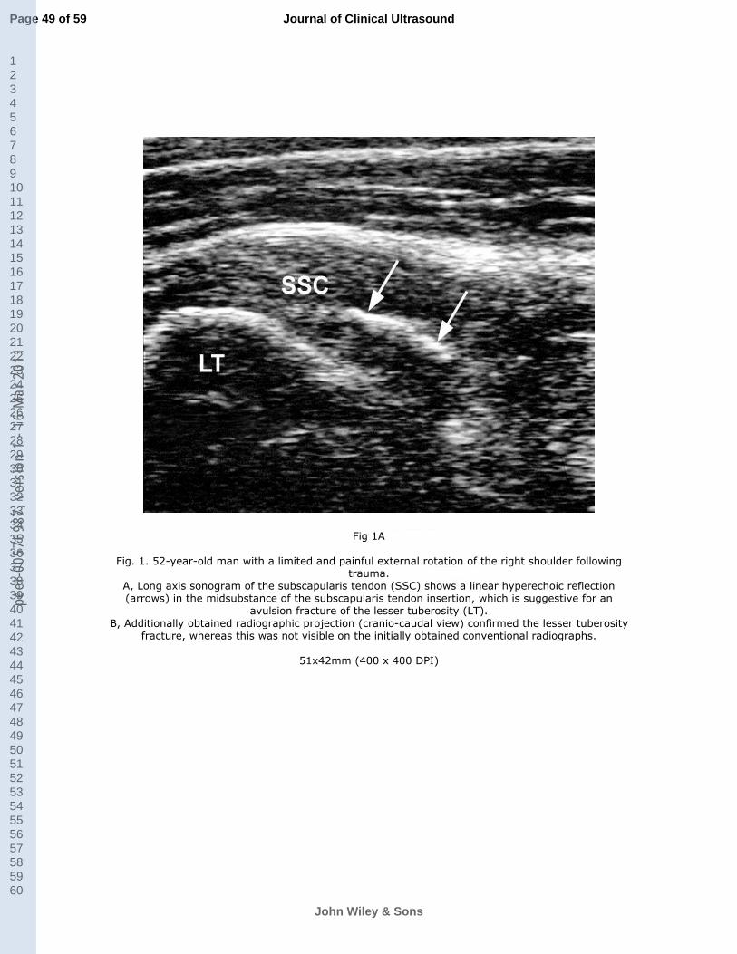
For Peer Review
Fig 1A
Fig. 1. 52-year-old man with a limited and painful external rotation of the right shoulder following trauma.
A, Long axis sonogram of the subscapularis tendon (SSC) shows a linear hyperechoic reflection (arrows) in the midsubstance of the subscapularis tendon insertion, which is suggestive for an
avulsion fracture of the lesser tuberosity (LT).
B, Additionally obtained radiographic projection (cranio-caudal view) confirmed the lesser tuberosity fracture, whereas this was not visible on the initially obtained conventional radiographs.
51x42mm (400 x 400 DPI)
Page 49 of 59
John Wiley & Sons
Journal of Clinical Ultrasound
123456789101112131415161718192021222324252627282930313233343536373839404142434445464748495051525354555657585960
peer
-005
7698
7, v
ersi
on 1
- 16
Mar
201
1
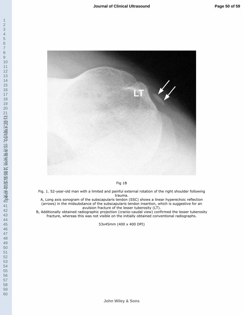
For Peer Review
Fig 1B
Fig. 1. 52-year-old man with a limited and painful external rotation of the right shoulder following
trauma. A, Long axis sonogram of the subscapularis tendon (SSC) shows a linear hyperechoic reflection (arrows) in the midsubstance of the subscapularis tendon insertion, which is suggestive for an
avulsion fracture of the lesser tuberosity (LT). B, Additionally obtained radiographic projection (cranio-caudal view) confirmed the lesser tuberosity
fracture, whereas this was not visible on the initially obtained conventional radiographs.
53x45mm (400 x 400 DPI)
Page 50 of 59
John Wiley & Sons
Journal of Clinical Ultrasound
123456789101112131415161718192021222324252627282930313233343536373839404142434445464748495051525354555657585960
peer
-005
7698
7, v
ersi
on 1
- 16
Mar
201
1
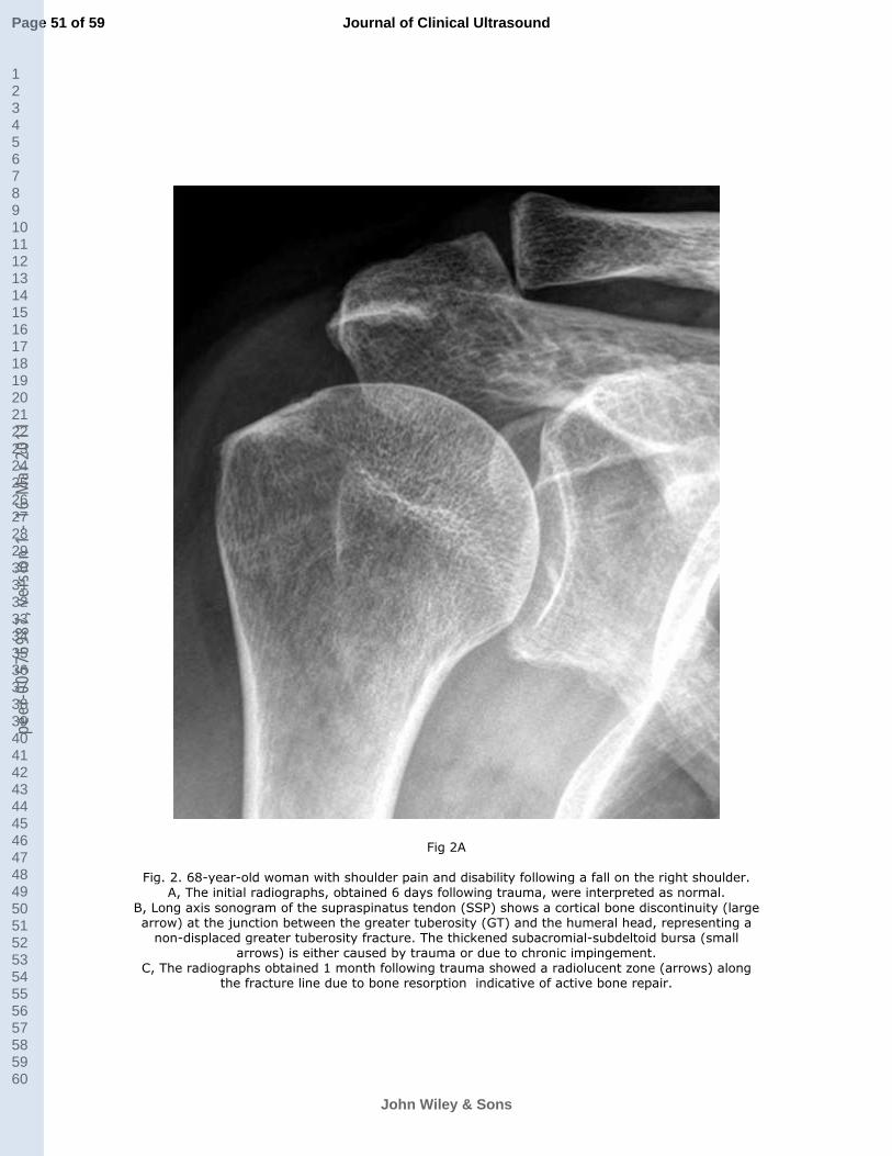
For Peer Review
Fig 2A
Fig. 2. 68-year-old woman with shoulder pain and disability following a fall on the right shoulder. A, The initial radiographs, obtained 6 days following trauma, were interpreted as normal.
B, Long axis sonogram of the supraspinatus tendon (SSP) shows a cortical bone discontinuity (large arrow) at the junction between the greater tuberosity (GT) and the humeral head, representing a
non-displaced greater tuberosity fracture. The thickened subacromial-subdeltoid bursa (small arrows) is either caused by trauma or due to chronic impingement.
C, The radiographs obtained 1 month following trauma showed a radiolucent zone (arrows) along the fracture line due to bone resorption indicative of active bone repair.
Page 51 of 59
John Wiley & Sons
Journal of Clinical Ultrasound
123456789101112131415161718192021222324252627282930313233343536373839404142434445464748495051525354555657585960
peer
-005
7698
7, v
ersi
on 1
- 16
Mar
201
1
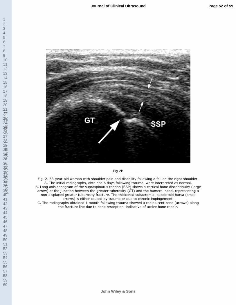
For Peer Review
Fig 2B
Fig. 2. 68-year-old woman with shoulder pain and disability following a fall on the right shoulder. A, The initial radiographs, obtained 6 days following trauma, were interpreted as normal.
B, Long axis sonogram of the supraspinatus tendon (SSP) shows a cortical bone discontinuity (large arrow) at the junction between the greater tuberosity (GT) and the humeral head, representing a
non-displaced greater tuberosity fracture. The thickened subacromial-subdeltoid bursa (small arrows) is either caused by trauma or due to chronic impingement.
C, The radiographs obtained 1 month following trauma showed a radiolucent zone (arrows) along the fracture line due to bone resorption indicative of active bone repair.
Page 52 of 59
John Wiley & Sons
Journal of Clinical Ultrasound
123456789101112131415161718192021222324252627282930313233343536373839404142434445464748495051525354555657585960
peer
-005
7698
7, v
ersi
on 1
- 16
Mar
201
1
For Peer Review
Fig 2C
Fig. 2. 68-year-old woman with shoulder pain and disability following a fall on the right shoulder. A, The initial radiographs, obtained 6 days following trauma, were interpreted as normal.
B, Long axis sonogram of the supraspinatus tendon (SSP) shows a cortical bone discontinuity (large arrow) at the junction between the greater tuberosity (GT) and the humeral head, representing a
non-displaced greater tuberosity fracture. The thickened subacromial-subdeltoid bursa (small arrows) is either caused by trauma or due to chronic impingement.
C, The radiographs obtained 1 month following trauma showed a radiolucent zone (arrows) along the fracture line due to bone resorption indicative of active bone repair.
Page 53 of 59
John Wiley & Sons
Journal of Clinical Ultrasound
123456789101112131415161718192021222324252627282930313233343536373839404142434445464748495051525354555657585960
peer
-005
7698
7, v
ersi
on 1
- 16
Mar
201
1
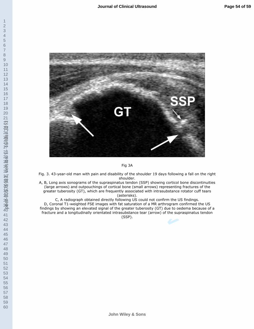
For Peer Review
Fig 3A
Fig. 3. 43-year-old man with pain and disability of the shoulder 19 days following a fall on the right shoulder.
A, B, Long axis sonograms of the supraspinatus tendon (SSP) showing corticol bone discontinuities (large arrows) and outpouchings of cortical bone (small arrows) representing fractures of the greater tuberosity (GT), which are frequently associated with intrasubstance rotator cuff tears
(asterisks). C, A radiograph obtained directly following US could not confirm the US findings.
D, Coronal T1-weighted FSE images with fat saturation of a MR arthrogram confirmed the US
findings by showing an elevated signal of the greater tuberosity (GT) due to oedema because of a fracture and a longitudinally orientated intrasubstance tear (arrow) of the supraspinatus tendon
(SSP).
Page 54 of 59
John Wiley & Sons
Journal of Clinical Ultrasound
123456789101112131415161718192021222324252627282930313233343536373839404142434445464748495051525354555657585960
peer
-005
7698
7, v
ersi
on 1
- 16
Mar
201
1
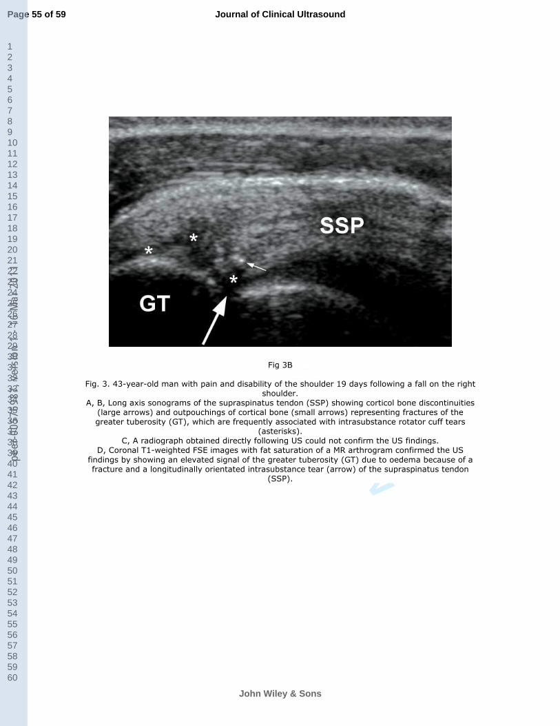
For Peer Review
Fig 3B
Fig. 3. 43-year-old man with pain and disability of the shoulder 19 days following a fall on the right shoulder.
A, B, Long axis sonograms of the supraspinatus tendon (SSP) showing corticol bone discontinuities (large arrows) and outpouchings of cortical bone (small arrows) representing fractures of the greater tuberosity (GT), which are frequently associated with intrasubstance rotator cuff tears
(asterisks). C, A radiograph obtained directly following US could not confirm the US findings.
D, Coronal T1-weighted FSE images with fat saturation of a MR arthrogram confirmed the US
findings by showing an elevated signal of the greater tuberosity (GT) due to oedema because of a fracture and a longitudinally orientated intrasubstance tear (arrow) of the supraspinatus tendon
(SSP).
Page 55 of 59
John Wiley & Sons
Journal of Clinical Ultrasound
123456789101112131415161718192021222324252627282930313233343536373839404142434445464748495051525354555657585960
peer
-005
7698
7, v
ersi
on 1
- 16
Mar
201
1
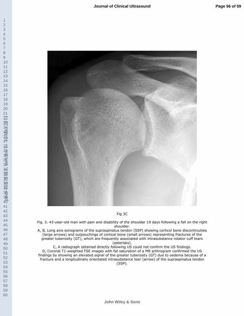
For Peer Review
Fig 3C
Fig. 3. 43-year-old man with pain and disability of the shoulder 19 days following a fall on the right shoulder.
A, B, Long axis sonograms of the supraspinatus tendon (SSP) showing corticol bone discontinuities (large arrows) and outpouchings of cortical bone (small arrows) representing fractures of the greater tuberosity (GT), which are frequently associated with intrasubstance rotator cuff tears
(asterisks). C, A radiograph obtained directly following US could not confirm the US findings.
D, Coronal T1-weighted FSE images with fat saturation of a MR arthrogram confirmed the US findings by showing an elevated signal of the greater tuberosity (GT) due to oedema because of a fracture and a longitudinally orientated intrasubstance tear (arrow) of the supraspinatus tendon
(SSP).
Page 56 of 59
John Wiley & Sons
Journal of Clinical Ultrasound
123456789101112131415161718192021222324252627282930313233343536373839404142434445464748495051525354555657585960
peer
-005
7698
7, v
ersi
on 1
- 16
Mar
201
1
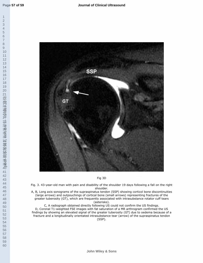
For Peer Review
Fig 3D
Fig. 3. 43-year-old man with pain and disability of the shoulder 19 days following a fall on the right shoulder.
A, B, Long axis sonograms of the supraspinatus tendon (SSP) showing corticol bone discontinuities (large arrows) and outpouchings of cortical bone (small arrows) representing fractures of the greater tuberosity (GT), which are frequently associated with intrasubstance rotator cuff tears
(asterisks). C, A radiograph obtained directly following US could not confirm the US findings.
D, Coronal T1-weighted FSE images with fat saturation of a MR arthrogram confirmed the US findings by showing an elevated signal of the greater tuberosity (GT) due to oedema because of a fracture and a longitudinally orientated intrasubstance tear (arrow) of the supraspinatus tendon
(SSP).
Page 57 of 59
John Wiley & Sons
Journal of Clinical Ultrasound
123456789101112131415161718192021222324252627282930313233343536373839404142434445464748495051525354555657585960
peer
-005
7698
7, v
ersi
on 1
- 16
Mar
201
1
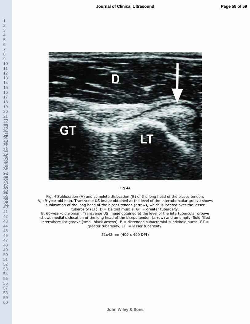
For Peer Review
Fig 4A
Fig. 4 Subluxation (A) and complete dislocation (B) of the long head of the biceps tendon. A, 49-year-old man. Transverse US image obtained at the level of the intertubercular groove shows
subluxation of the long head of the biceps tendon (arrow), which is located over the lesser tuberosity (LT). D = Deltoid muscle, GT = greater tuberosity.
B, 60-year-old woman. Transverse US image obtained at the level of the intertubercular groove shows medial dislocation of the long head of the biceps tendon (arrow) and an empty, fluid filled intertubercular groove (small black arrows). B = distended subacromial-subdeltoid bursa, GT =
greater tuberosity, LT = lesser tuberosity.
51x43mm (400 x 400 DPI)
Page 58 of 59
John Wiley & Sons
Journal of Clinical Ultrasound
123456789101112131415161718192021222324252627282930313233343536373839404142434445464748495051525354555657585960
peer
-005
7698
7, v
ersi
on 1
- 16
Mar
201
1

For Peer Review
Fig 4B
Fig. 4 Subluxation (A) and complete dislocation (B) of the long head of the biceps tendon. A, 49-year-old man. Transverse US image obtained at the level of the intertubercular groove shows
subluxation of the long head of the biceps tendon (arrow), which is located over the lesser
tuberosity (LT). D = Deltoid muscle, GT = greater tuberosity. B, 60-year-old woman. Transverse US image obtained at the level of the intertubercular groove shows medial dislocation of the long head of the biceps tendon (arrow) and an empty, fluid filled intertubercular groove (small black arrows). B = distended subacromial-subdeltoid bursa, GT =
greater tuberosity, LT = lesser tuberosity.
51x43mm (400 x 400 DPI)
Page 59 of 59
John Wiley & Sons
Journal of Clinical Ultrasound
123456789101112131415161718192021222324252627282930313233343536373839404142434445464748495051525354555657585960
peer
-005
7698
7, v
ersi
on 1
- 16
Mar
201
1

![Urbanisme Insoupçonné - Projeter la ville égyptienne au Nouvel Empire (Unsuspected Urbanism) [Extrait/Excerpt]](https://static.fdokumen.com/doc/165x107/631ca7d5665120b3330bf65c/urbanisme-insoupconne-projeter-la-ville-egyptienne-au-nouvel-empire-unsuspected.jpg)