Univerzita Karlova
90
Univerzita Karlova Přírodovědecká fakulta Fyziologie živočichů Autoreferát dizertační práce Molekulární mechanismy regulace transportu a funkce různých podtypů NMDA receptorů v hipokampálních neuronech Mgr. Kristýna Skřenková Praha, 2019
-
Upload
khangminh22 -
Category
Documents
-
view
0 -
download
0
Transcript of Univerzita Karlova
Univerzita Karlova
Přírodovědecká fakulta
Fyziologie živočichů
Autoreferát dizertační práce
Molekulární mechanismy regulace transportu a funkce různých podtypů NMDA receptorů v hipokampálních neuronech
Mgr. Kristýna Skřenková
Praha, 2019
2
Doktorské studijní programy v biomedicíně
Univerzita Karlova a Akademie věd České republiky
Program: Fyziologie živočichů
Předseda oborové rady: doc. RNDr. Jiří Novotný, DSc.
Školicí pracoviště:
Fyziologický ústav AVČR Oddělení buněčné neurofyziologie
Ústav experimentální medicíny AVČR Oddělení neurochemie
Autor: Mgr. Kristýna Skřenková
Vedoucí práce: Mgr. Martin Horák, Ph.D.
S dizertační prací je možno se seznámit v příslušných knihovnách Přírodovědecké fakulty Univerzity Karlovy.
3
Obsah
1. Shrnutí ..................................................................................... 4
2. Úvod ………………………………………………………………………………5
3. Hypotézy a cíle .......................................................................... 8
4. Materiály a metody ................................................................... 9
5. Výsledky .................................................................................. 13
5.1. N-glykosylace reguluje transport a povrchovou mobilitu NMDA
receptorů obsahujících GluN3A podjednotku ............................ 13
5.2. Strukturní změny v glycinových vazebných místech na GluN1
a GluN3A podjednotkách regulují povrchovou expresi NMDA
receptorů ................................................................................... 18
6. Diskuse .................................................................................... 29
6.1. N-glykosylace reguluje transport a povrchovou mobilitu NMDA
receptorů obsahujících GluN3A podjednotku ............................ 29
6.2. Strukturní změny v glycinových vazebných místech na GluN1
a GluN3A podjednotkách regulují povrchovou expresi NMDA
receptorů ................................................................................... 31
7. Závěr ………...............................................................................35
8. Použitá literatura .................................................................... 36
9. Seznam publikací ..................................................................... 42
10. Životopis .................................................................................. 44
4
1. Shrnutí
N-methyl-D-aspartátové (NMDA) receptory jsou ionotropní glutamátové
receptory, které mají klíčovou úlohu v savčí centrální nervové soustavě.
Za fyziologických podmínek jsou tyto receptory důležité pro excitační
synaptický přenos a tvorbu paměťových stop. Za patologických podmínek může
ovšem jejich abnormální regulace či aktivace vést k mnohým neurologickým
a psychiatrickým onemocněním, jako je například Alzheimerova choroba,
Parkinsonova choroba, Huntingtonova choroba, epilepsie, nebo schizofrenie.
Množství NMDA receptorů na povrchové membráně buněk je regulováno
na několika úrovních, zahrnujících jejich syntézu, skládání, internalizaci
či degradaci. Během transportu na buněčnou membránu dochází rovněž
ke kontrole vazby agonistů a správné aktivaci NMDA receptorů. Současně
dochází u NMDA receptorů k celé řadě posttranslačních modifikací, jako
je palmitoylace, fosforylace nebo N-glykosylace.
V této dizertační práci jsme se zabývali studiem molekulárních mechanismů,
které mohou ovlivnit transport a funkční vlastnosti NMDA receptorů v savčích
buněčných liniích a potkaních neuronech. Konkrétně jsme se zabývali otázkou,
jak mohou změny v extracelulární části NMDA receptoru ovlivnit jeho transport
na buněčnou membránu a modulovat jeho funkční vlastnosti. Věnovali jsme
se zejména roli N-glykosylace NMDA receptorů. U savců dochází k nejvíce
orgánově specifické N-glykosylaci právě v mozku (Hanus et al., 2016). Její
funkční význam v neuronech či na NMDA receptorech není dosud plně objasněn.
Rovněž jsme studovali podjednotkově specifický účinek jednotlivých lektinů.
Dalším tématem, kterému jsme se v této dizertační práci věnovali, byl vliv
integrity glycinových vazebných míst na GluN1 a GluN3A podjednotkách
na transport a funkci NMDA receptorů. Ke studiu těchto otázek jsme použili
celou řadu metod, včetně imunohistochemie, živé mikroskopie, biochemie
a elektrofyziologie. Použili jsme několik buněčných modelů – savčí linie HEK293
a COS-7 buněk, lidské fibroblasty odvozené z pacientů a potkaní hipokampální
a mozečkové neurony.
5
2. Úvod
NMDA receptory jsou podtřídou glutamátových receptorů a hrají zásadní roli
ve vývoji synapsí, excitačním přenosu a synaptické plasticitě v centrálním
nervovém systému (CNS) savců (Perez-Otano et al., 2016; Traynelis et al., 2010).
Bylo prokázáno, že dysregulace NMDA receptorů hraje klíčovou roli v etiologii
mnoha neuropsychiatrických a neurologických poruch a stavů, včetně
Huntingtonovy choroby (Mahfooz et al., 2016; Marco et al., 2013), schizofrenie
(Mueller and Meador-Woodruff, 2004), závislosti na kokainu (Yuan et al., 2013)
a závislosti na nikotinu (Chen et al., 2019). Rostoucí počet studií naznačuje,
že mnoho neuropsychiatrických poruch je spojeno s mutacemi v genech, které
kódují různé podjednotky NMDA receptorů, včetně GluN1 (Chen et al., 2017;
Lemke et al., 2016) a GluN3A (Shen et al., 2009; Takata et al., 2013) podjednotek.
Porozumění molekulárním mechanismům, které regulují NMDA receptory,
je tedy nezbytným krokem k navrhování účinných terapií pro pacienty trpící
těmito poruchami.
NMDA receptory jsou heterotetramery složené z GluN1 (s osmi sestřihovými
variantami), GluN2 (GluN2A až GluN2D) a/nebo GluN3 (GluN3A a GluN3B)
podjednotek. Všechny GluN podjednotky sdílejí stejnou topologii, včetně
extracelulární amino-terminální domény (ATD), ligand vázající domény (LBD)
tvořené segmenty S1 a S2, čtyř membránových domén (M1 až M4)
a intracelulární C-terminální domény (CTD) (Paoletti et al., 2013; Traynelis et al.,
2010). Konvenční podtyp NMDA receptorů GluN1/GluN2 je aktivován vazbou
agonisty do glutamátového vazebného místa na LBD GluN2 podjednotky
se současnou vazbou ko-agonisty do glycinového vazebného místa na LBD GluN1
podjednotky (Clements and Westbrook, 1991; Kleckner and Dingledine, 1988;
Patneau and Mayer, 1990; Traynelis et al., 2010). Je zajímavé, že nekonvenční
podtypy NMDA receptorů, jmenovitě GluN1/GluN3A a GluN1/GluN3B receptory,
jsou aktivovány vazbou agonisty do glycinového vazebného místa na LBD GluN3
podjednotky, zatímco vazba agonisty do glycinového vazebného místa na LBD
GluN1 podjednotky řídí desenzitizaci glycinem indukovaných proudů

6
GluN1/GluN3 receptorů (Awobuluyi et al., 2007; Kehoe et al., 2013; Kvist et al.,
2013; Madry et al., 2007). To znamená, že glycinová vazebná místa na LBD
různých GluN podjednotek mají odlišný vliv na funkci NMDA receptorů.
Počet i typ NMDA receptorů přítomných na neuronálním povrchu
je regulován na více úrovních (Hansen et al., 2017; Horak et al., 2014; Sanz-
Clemente et al., 2013), včetně jejich syntézy (Chazot and Stephenson, 1997; Huh
and Wenthold, 1999), sestavení podjednotek (Atlason et al., 2007; Farina et al.,
2011; Meddows et al., 2001; Schuler et al., 2008), zpracování v endoplasmickém
retikulu (ER) (Hawkins et al., 2004; Horak and Wenthold, 2009; Matsuda et al.,
2003; McIlhinney et al., 1998; Okabe et al., 1999; Perez-Otano et al., 2001; Qiu et
al., 2009; Standley et al., 2000), transportu na buněčnou membránu (Jeyifous et
al., 2009; Washbourne et al., 2002; Washbourne et al., 2004), laterální difúze
(Dupuis and Groc, 2019; Groc et al., 2004), internalizace/recyklace (Lavezzari et
al., 2004; Perez-Otano et al., 2006; Roche et al., 2001; Scott et al., 2004)
a degradace (Kato et al., 2005; Scott et al., 2004). Přestože se mnoho prací
zabývalo studiem molekulárních mechanismů, které regulují povrchovou
expresi a povrchovou mobilitu NMDA receptorů, zaměřovaly se převážně
na NMDA receptory obsahující GluN1/GluN2 podjednotky (Sanz-Clemente et al.,
2013; Traynelis et al., 2010). Již dříve bylo prokázáno, že GluN1/GluN2 receptory
jsou rozsáhle N-glykosylovány (Sanz-Clemente et al., 2013; Traynelis et al.,
2010), a to jak v heterologních buňkách (Chazot et al., 1995; Everts et al., 1997),
tak v nativním systému (Huh and Wenthold, 1999; Kaniakova et al., 2016). V ER
nejprve vzniká N-glykanový prekurzor, který je poté přenesen na protein
obsahující konzenzus N-glykosylační sekvenci N-X-S/T. Z tohoto prekurzoru
poté na proteinu vzniká vysoce-manózový typ N-glykanu, který může být dále
remodelován v Golgiho aparátu (GA) na hybridní typ a následně dalšími
modifikacemi na komplexní typ N-glykanu. Již dříve jsme ukázali, že na GluN1
podjednotce se nacházejí dvě N-glykosylační místa, která jsou vyžadována
pro uvolnění GluN1/Glu2B receptorů z ER (Lichnerova et al., 2015). Ovšem úloha
N-glykosylace v transportu a povrchové mobilitě NMDA receptorů obsahujících
nekonvenční GluN3A podjednotku nebyla doposud studována.
7
Avšak nejen N-glykosylace NMDA receptorů je důležitá pro jejich správný
transport a funkci. Modifikace v GluN podjednotkách, včetně LBD, jsou taktéž
kritické pro regulaci povrchové exprese NMDA receptorů (Kenny et al., 2009;
She et al., 2012). Konkrétně Kenny et al. ukázali, že narušení vazebného místa
pro glycin v LBD na GluN1 podjednotce mutací D732A snižuje povrchovou
expresi GluN1/GluN2A receptorů (Kenny et al., 2009). Podobně She et al. uvádí,
že vazebné místo pro glutamát v LBD na GluN2B podjednotce reguluje
povrchovou expresi GluN1/GluN2B receptorů (She et al., 2012). Toto pozorování
bylo později potvrzeno nedávnou studií využívající lidské GluN1/GluN2B
receptory s objevenými patogenními mutacemi (Swanger et al., 2016). Nicméně
zda strukturální změny v glycinovém vazebném místě v GluN1 a/nebo GluN3A
podjednotkách regulují povrchové počty funkčních NMDA receptorů
obsahujících GluN3A podjednotku dosud nebylo objasněno.
8
3. Hypotézy a cíle
a) Dřívější studie se zaměřovaly zejména na časný transport GluN1/GluN2
receptorů (Sanz-Clemente et al., 2013; Traynelis et al., 2010); v současné době
však chybí bližší informace o tom, jak strukturní a funkční determinanty
ovlivňují NMDA receptory obsahující GluN3A podjednotku v savčích
neuronech. Všechny ionotropní glutamátové receptory včetně NMDA
receptorů obsahují několik N-glykosylačních míst (N-X-S/T; X ≠ P) (Everts et
al., 1997; Lichnerova et al., 2015; Traynelis et al., 2010). A přestože GluN3A
podjednotka obsahuje 12 N-glykosylačních míst, role N-glykosylace
v transportu a funkci NMDA receptorů obsahujících GluN3A podjednotku
nebyla doposud objasněna.
Cíl: Identifikovat strukturální a funkční determinanty časného transportu
NMDA receptorů, které obsahují GluN3A podjednotku se zaměřením na N-
glykosylace, a prostudovat, jak remodelace N-glykanů reguluje časný
transport NMDA receptorů obsahujících GluN3A podjednotku.
b) Většina studií naznačuje, že intracelulární CTD různých podjednotek NMDA
receptorů hraje klíčovou roli při regulaci množství NMDA receptorů
na buněčné membráně, a to včetně receptorů, které obsahují GluN2A a/nebo
GluN2B podjednotky (Petralia et al., 2009; Sanz-Clemente et al., 2013).
Nicméně extracelulární doména savčích GluN podjednotek je poměrně
rozsáhlá a obsahuje v LBD vazebná místa pro agonisty a antagonisty.
Předpokládáme, že specifické mutace v glycinových vazebných místech
na GluN1 a GluN3A podjednotce vyvolají strukturální změny, které budou
regulovat počet NMDA receptorů na povrchu buňky.
Cíl: Charakterizovat způsob, jakým vazba glycinu na GluN1 a GluN3
podjednotky ovlivňuje množství povrchových receptorů a funkční vlastnosti
NMDA receptorů v savčích buněčných liniích a v hipokampálních neuronech.

9
4. Materiály a metody
Příprava a transfekce primárních hipokampálních neuronů
Všechny pokusy na zvířatech byly prováděny v souladu s pokyny a předpisy
pro zajištění dobrých životních podmínek zvířat. Primární kultury
hipokampálních neuronů byly připraveny z 18-ti denních embryí potkanů
Wistar (Lichnerova et al., 2015). Ve stručnosti: hipokampy byly vloženy
do studeného Hankova vyváženého solného roztoku obsahujícího 10 mM HEPES
(pH 7,4) a potom inkubovány po dobu 20 minut při 37 °C v disekčním médiu
obsahujícím 0,1 mg/ml DNázy I a 0,05% trypsinu (Merck). Buňky pak byly
promyty, disociovány pomocí skleněné pipety a resuspendovány v kultivačním
médiu bez séra s B-27 doplňkem a L-glutaminem (Thermo Fisher Scientific).
Buňky byly pěstovány v hustotě přibližně 2x10⁴ buněk na cm2 na miskách
potažených poly-L-lysinem (Sigma). Neuronům bylo každé 2-3 dny měněno
kultivační médium a byly transfekovány s použitím Lipofectaminu 2000.
Savčí buněčné kultury
COS-7 a HEK293 buňky byly kultivovány v Opti-MEM (Thermo Fisher
Scientific) obsahujícím 5% fetální bovinní sérum (Thermo Fisher Scientific)
(Kaniakova et al., 2012a; Lichnerova et al., 2015). Pro mikroskopii jsme používali
COS-7 buňky a pro elektrofyziologii HEK293 buňky. HEK293 i COS-7 buňky byly
transfekovány 2 μl Lipofectaminu 2000 (Thermo Fisher Scientific) a celkovým
množstvím 900 µg cDNA, která kódovala příslušné GluN1 a GluN3A podjednotky.
Pro elektrofyziologii byly transfekované buňky trypsinovány a pěstovány
při nízké hustotě; buňky určené pro mikroskopické pokusy byly pěstovány
bez trypsinizace. Pokusy byly prováděny 24 – 72 h po transfekci.
Imunofluorescenční mikroskopie
Povrchové NMDA receptory byly označeny tak, jak bylo popsáno dříve
(Kaniakova et al., 2012a; Lichnerova et al., 2015). Ve stručnosti: buňky byly omyty
ve fosfátem pufrovaném solném roztoku (PBS), pak inkubovány v blokujícím
10
roztoku obsahujícím PBS a 10% normální kozí sérum. Buňky pak byly
inkubovány po dobu 30 minut s primární protilátkou zředěnou v blokujícím
roztoku. Po omytí byly buňky inkubovány po dobu 30 minut se sekundární
protilátkou konjugovanou s fluorescenčním barvivem zředěnou blokujícím
roztokem. Buňky byly omyty, fixovány ve 4% paraformaldehydu (PFA) v PBS
po dobu 20 minut a poté byly vloženy do zalévacího média ProLong Antifade
(Thermo Fisher Scientific). Pro značení intracelulárních GluN podjednotek byly
buňky omyty v PBS, fixovány 4% PFA v PBS po dobu 20 minut, permeabilizovány
po dobu 5 minut roztokem obsahujícím 0,25% Triton X-100 (TX-100) v PBS
a poté blokovány 1 h blokovacím roztokem obsahujícím 0,1% TX-100. Buňky pak
byly inkubovány s primární protilátkou po dobu 1 hodiny, omyty a inkubovány
se sekundární protilátkou po dobu 30 minut. Snímky byly získány při pokojové
teplotě za použití fluorescenčního mikroskopu (Olympus Scan) s objektivem
pro olejovou imerzi 60x/1,35, nebo konfokálního skenovacího mikroskopu (Leica
TCS SP8) vybaveného lasery v pevné fázi a apochromatickým objektivem
pro olejovou imerzi 63x/1,30. Získané snímky byly analyzovány pomocí softwaru
ImageJ (NIH). Intenzita povrchového (P) a celkového (C) fluorescenčního signálu
v COS-7 buňkách a fibroblastech byla analyzována na celých buněčných
oblastech (Kaniakova et al., 2012b). U hipokampálních neuronů byla analyzována
intenzita povrchového a celkového fluorescenčního signálu v 10 μm dlouhých
segmentech sekundárních a terciárních dendritů (Lichnerova et al., 2015).
Konkanavalin A (conA) a Phaseolus vulgaris Leucoagglutinin (PHA-L) značené
rhodaminem (Vector Laboratories; 20 μg/ml) byly rozpuštěny v blokovacím
roztoku a inkubovány po dobu 5 minut s předem omytými živými buňkami.
Buňky byly poté znovu omyty, fixovány 4% PFA v PBS po dobu 20 minut
a vloženy do zalévacího média ProLong Antifade.
Kolokalizace GluN podjednotek s protilátkou značící GM130 byla analyzována
z jedné vrstvy pomocí automatizovaného makra v ImageJ. Maska celé oblasti
buněčného těla byla generována z nativního signálu zeleného fluorescenčního
proteinu (GFP, z ang. Green fluorescent protein). Pro lokalizaci oblasti GA byla
maska generována ze signálu GM130. GM130-negativní buněčná oblast byla
vytvořena odečtením masky GM130 od signální masky GFP. GA kolokalizace byla
11
poté vypočtena jako poměr průměrné intenzity GFP signálu přes generovanou
masku GM130 k průměrné intenzitě signálu GFP přes GM130-negativní
buněčnou oblast.
Povrchová mobilita
Sledování kvantové tečky (QD) GluN3A podjednotek bylo provedeno tak,
jak bylo popsáno dříve (Ferreira et al., 2017; Mikasova et al., 2017). Zkráceně,
kultivované hipokampální neurony v 13. až 15. den in vitro (DIV13-15) exprimující
GFP-GluN3A podjednotku byly omyty v předehřátém neurobazálním médiu
(Thermo Fisher Scientific) obsahujícím 1% albuminu hovězího séra (BSA; Merck)
a poté byly inkubovány po dobu 10 minut s králičí anti-GFP protilátkou (Merck,
1:2000), následovala 10 minutová inkubace s anti-králičím IgG konjugovaným
k QD605 (Thermo Fisher Scientific; 1:10 000); primární i sekundární protilátky
byly zředěny v neurobazálním médiu obsahujícím 1% BSA a byly inkubovány
s neurony při 37 °C. Po značení QD byly neurony omyty předehřátým
neurobazálním médiem a poté umístěny do extracelulárního roztoku obsahující
1 mM MgCl2 a 1 mM CaCl2; všechny záznamy byly získány při 37 °C do 30 minut
od QD značení. QD-značené GluN3A podjednotky byly detekovány s použitím
osvětlovacího modulu InsightSSI s objektivem pro imerzní olej (60x/1,42,
PlanApo N) na širokoúhlém fluorescenčním mikroskopu (DeltaVision OMXTM).
Snímky (1200 po sobě jdoucích snímků) byly získány s časovým odstupem 50 ms.
Jako negativní kontrola laterální mobility byly použity COS-7 buňky
transfekované GluN1-4a/GFP-GluN3A receptory, které byly značeny králičí anti-
GFP primární protilátkou a QD605-konjugovaným k anti-králičí IgG a poté
fixovány v 4% PFA v PBS, jak je popsáno výše. Pohyby QD byly analyzovány
v ImageJ pomocí programu Mosaic Particle Tracker 2D/3D. Pro každou QD
trajektorii byl získán difúzní koeficient (D) s krátkým dosahem: MSD (t) = 4 D t +
b (Kusumi et al., 1993; Triller and Choquet, 2008). Kumulativní rozdělení
pravděpodobnosti bylo vypočteno jako relativní kumulativní frekvence D
ze všech trajektorií v uvedeném experimentu.
12
Elektrofyziologie
Záznamy z terčíkového zámku z celých buněk byly pořízeny
na transfekovaných HEK293 buňkách exprimujících GluN1/GluN3A receptory
pomocí zesilovače Axopatch 200B (Molecular Devices), jak bylo popsáno dříve
(Kaniakova et al., 2018). Extracelulární roztok obsahoval (v mM): 160 NaCl, 2,5
KCl, 10 HEPES, 10 glukózy, 0,2 EDTA a 0,7 CaCl2 (pH bylo upraveno na 7,3 pomocí
NaOH). Intracelulární roztok obsahoval (v mM): 125 glukonovou kyselinu, 15
CsCl, 5 BAPTA, 10 HEPES, 3 MgCl2, 0,5 CaCl2 a 2 ATP-Mg soli (pH upravené na 7,2
pomocí CsOH). Skleněné pipety (odpor hrotu 3-6 MΩ) byly připraveny za použití
mikropipetového tahače P-97 (Sutter Instrument Co.). Pro aplikaci
extracelulárních roztoků byl použit mikroprocesorem řízený rychlý perfuzní
systém (s časovou konstantou pro výměnu roztoku kolem buňky ~ 10 ms)
(Lichnerova et al., 2014). Všechny elektrofyziologické experimenty byly
prováděny při pokojové teplotě. K záznamu elektrofyziologických odpovědí byl
použit pCLAMP 9 Software (Molecular Devices).
13
5. Výsledky
5.1. N-glykosylace reguluje transport a povrchovou mobilitu NMDA
receptorů obsahujících GluN3A podjednotku
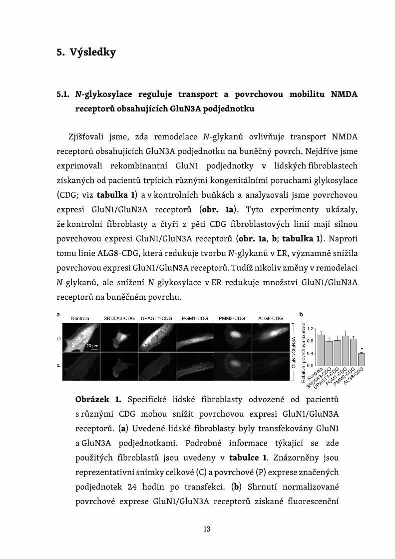
Zjišťovali jsme, zda remodelace N-glykanů ovlivňuje transport NMDA
receptorů obsahujících GluN3A podjednotku na buněčný povrch. Nejdříve jsme
exprimovali rekombinantní GluN1 podjednotky v lidských fibroblastech
získaných od pacientů trpících různými kongenitálními poruchami glykosylace
(CDG; viz tabulka 1) a v kontrolních buňkách a analyzovali jsme povrchovou
expresi GluN1/GluN3A receptorů (obr. 1a). Tyto experimenty ukázaly,
že kontrolní fibroblasty a čtyři z pěti CDG fibroblastových linií mají silnou
povrchovou expresi GluN1/GluN3A receptorů (obr. 1a, b; tabulka 1). Naproti
tomu linie ALG8-CDG, která redukuje tvorbu N-glykanů v ER, významně snížila
povrchovou expresi GluN1/GluN3A receptorů. Tudíž nikoliv změny v remodelaci
N-glykanů, ale snížení N-glykosylace v ER redukuje množství GluN1/GluN3A
receptorů na buněčném povrchu.
Obrázek 1. Specifické lidské fibroblasty odvozené od pacientů
s různými CDG mohou snížit povrchovou expresi GluN1/GluN3A
receptorů. (a) Uvedené lidské fibroblasty byly transfekovány GluN1
a GluN3A podjednotkami. Podrobné informace týkající se zde
použitých fibroblastů jsou uvedeny v tabulce 1. Znázorněny jsou
reprezentativní snímky celkové (C) a povrchové (P) exprese značených
podjednotek 24 hodin po transfekci. (b) Shrnutí normalizované
povrchové exprese GluN1/GluN3A receptorů získané fluorescenční
14
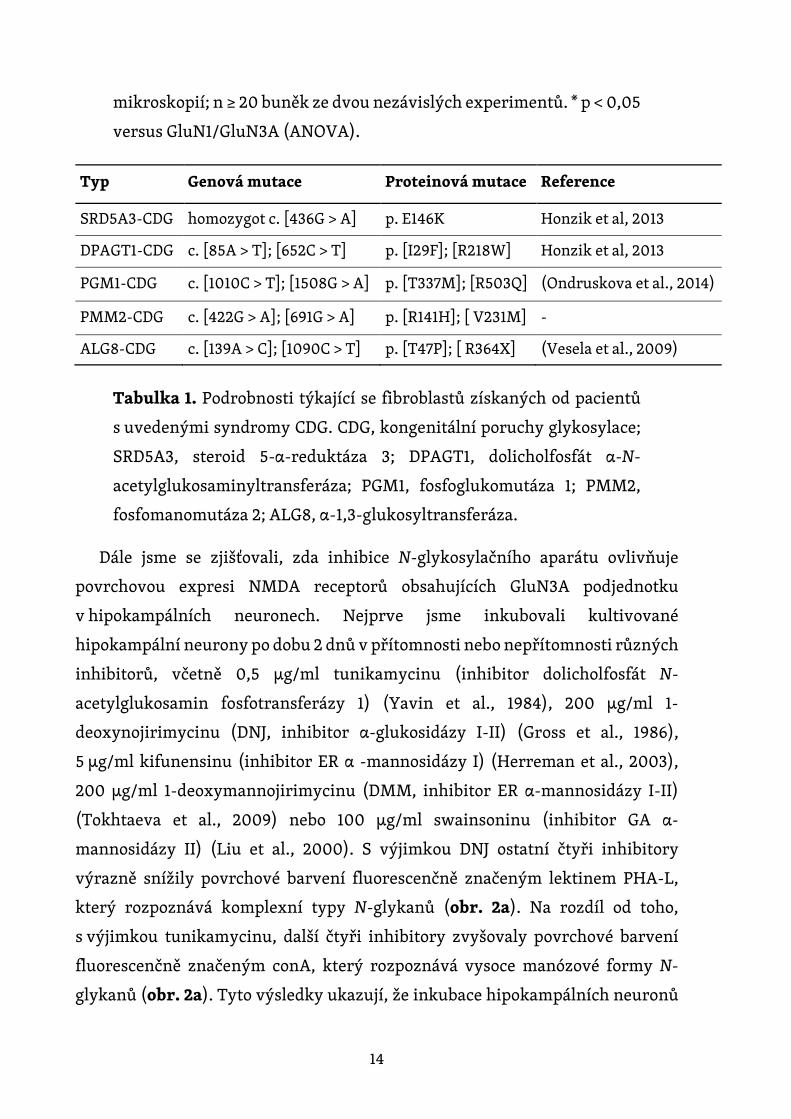
mikroskopií; n ≥ 20 buněk ze dvou nezávislých experimentů. * p < 0,05
versus GluN1/GluN3A (ANOVA).
Typ Genová mutace Proteinová mutace Reference
SRD5A3-CDG homozygot c. [436G > A] p. E146K Honzik et al, 2013
DPAGT1-CDG c. [85A > T]; [652C > T] p. [I29F]; [R218W] Honzik et al, 2013
PGM1-CDG c. [1010C > T]; [1508G > A] p. [T337M]; [R503Q] (Ondruskova et al., 2014)
PMM2-CDG c. [422G > A]; [691G > A] p. [R141H]; [ V231M] -
ALG8-CDG c. [139A > C]; [1090C > T] p. [T47P]; [ R364X] (Vesela et al., 2009)
Tabulka 1. Podrobnosti týkající se fibroblastů získaných od pacientů
s uvedenými syndromy CDG. CDG, kongenitální poruchy glykosylace;
SRD5A3, steroid 5-α-reduktáza 3; DPAGT1, dolicholfosfát α-N-
acetylglukosaminyltransferáza; PGM1, fosfoglukomutáza 1; PMM2,
fosfomanomutáza 2; ALG8, α-1,3-glukosyltransferáza.
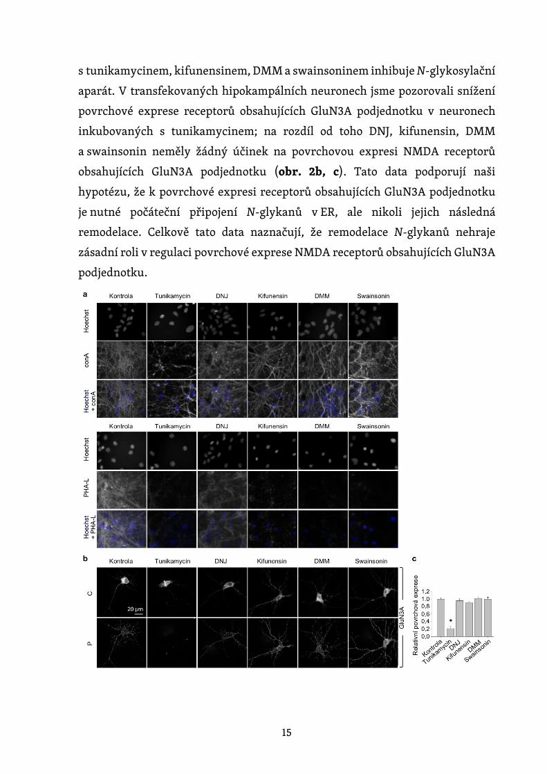
Dále jsme se zjišťovali, zda inhibice N-glykosylačního aparátu ovlivňuje
povrchovou expresi NMDA receptorů obsahujících GluN3A podjednotku
v hipokampálních neuronech. Nejprve jsme inkubovali kultivované
hipokampální neurony po dobu 2 dnů v přítomnosti nebo nepřítomnosti různých
inhibitorů, včetně 0,5 µg/ml tunikamycinu (inhibitor dolicholfosfát N-
acetylglukosamin fosfotransferázy 1) (Yavin et al., 1984), 200 µg/ml 1-
deoxynojirimycinu (DNJ, inhibitor α-glukosidázy I-II) (Gross et al., 1986),
5 µg/ml kifunensinu (inhibitor ER α -mannosidázy I) (Herreman et al., 2003),
200 µg/ml 1-deoxymannojirimycinu (DMM, inhibitor ER α-mannosidázy I-II)
(Tokhtaeva et al., 2009) nebo 100 µg/ml swainsoninu (inhibitor GA α-
mannosidázy II) (Liu et al., 2000). S výjimkou DNJ ostatní čtyři inhibitory
výrazně snížily povrchové barvení fluorescenčně značeným lektinem PHA-L,
který rozpoznává komplexní typy N-glykanů (obr. 2a). Na rozdíl od toho,
s výjimkou tunikamycinu, další čtyři inhibitory zvyšovaly povrchové barvení
fluorescenčně značeným conA, který rozpoznává vysoce manózové formy N-
glykanů (obr. 2a). Tyto výsledky ukazují, že inkubace hipokampálních neuronů
15
s tunikamycinem, kifunensinem, DMM a swainsoninem inhibuje N-glykosylační
aparát. V transfekovaných hipokampálních neuronech jsme pozorovali snížení
povrchové exprese receptorů obsahujících GluN3A podjednotku v neuronech
inkubovaných s tunikamycinem; na rozdíl od toho DNJ, kifunensin, DMM
a swainsonin neměly žádný účinek na povrchovou expresi NMDA receptorů
obsahujících GluN3A podjednotku (obr. 2b, c). Tato data podporují naši
hypotézu, že k povrchové expresi receptorů obsahujících GluN3A podjednotku
je nutné počáteční připojení N-glykanů v ER, ale nikoli jejich následná
remodelace. Celkově tato data naznačují, že remodelace N-glykanů nehraje
zásadní roli v regulaci povrchové exprese NMDA receptorů obsahujících GluN3A
podjednotku.
16
Obrázek 2. Tunikamycin, na rozdíl od inhibitorů pozdějších kroků N-
glykosylační dráhy, snižuje povrchovou expresi GluN3A podjednotek
v hipokampálních neuronech. (a) Hipokampální neurony byly
inkubovány po dobu 2 dnů s 0,5 µg/ml tunikamycinu, 200 µg /ml DNJ,
5 µg/ml kifunensinu, 200 µg/ml DMM nebo 100 µg/ml swainsoninu.
Neurony byly poté barveny fluorescenčně značeným conA nebo PHA-
L; uvedeny jsou reprezentativní obrázky. (b) Hipokampální neurony
byly transfekovány GFP-GluN3A podjednotkou (GluN3A) v DIV8
a inkubovány po dobu 2 dnů s uvedenými inhibitory, jak je popsáno
výše. Neurony byly poté imunohistochemicky barveny pro povrchový
(P) a celkový (C) GFP signál; zobrazeny jsou reprezentativní snímky
celkové a povrchové imunoreaktivity. (c) Shrnutí relativní povrchové
exprese GluN3A podjednotek v neuronech inkubovaných s uvedenými
inhibitory; data byla získána z 10 µm dlouhých segmentů sekundárních
a terciárních dendritů (n ≥ 30 segmentů z > 6 neuronů) a jsou vyjádřena
relativně ke kontrolním neuronům; * p < 0,05 versus kontrola
(ANOVA).
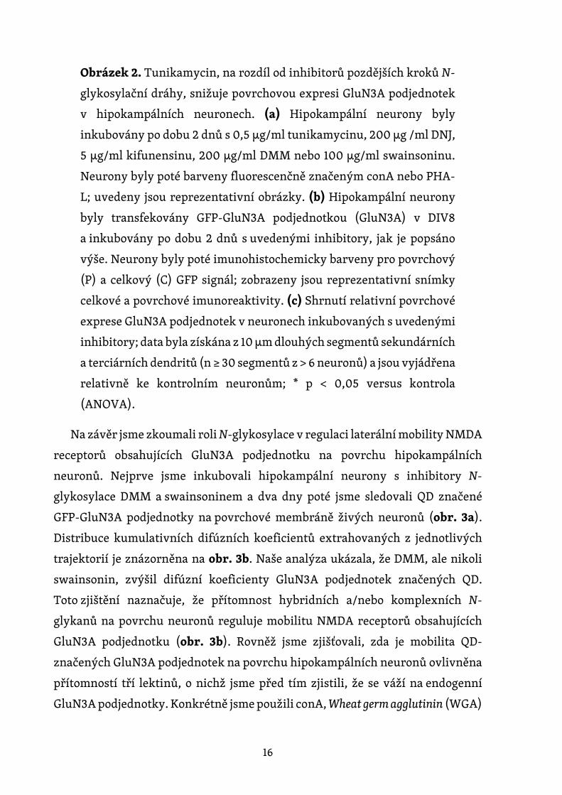
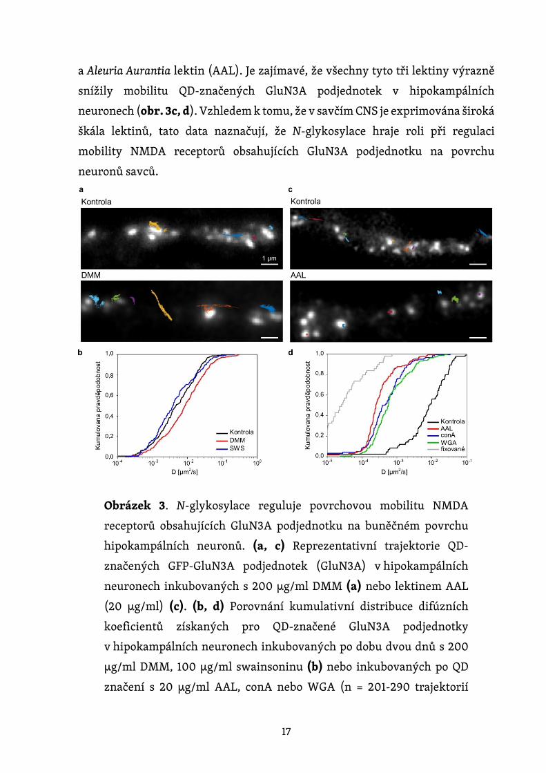
Na závěr jsme zkoumali roli N-glykosylace v regulaci laterální mobility NMDA
receptorů obsahujících GluN3A podjednotku na povrchu hipokampálních
neuronů. Nejprve jsme inkubovali hipokampální neurony s inhibitory N-
glykosylace DMM a swainsoninem a dva dny poté jsme sledovali QD značené
GFP-GluN3A podjednotky na povrchové membráně živých neuronů (obr. 3a).
Distribuce kumulativních difúzních koeficientů extrahovaných z jednotlivých
trajektorií je znázorněna na obr. 3b. Naše analýza ukázala, že DMM, ale nikoli
swainsonin, zvýšil difúzní koeficienty GluN3A podjednotek značených QD.
Toto zjištění naznačuje, že přítomnost hybridních a/nebo komplexních N-
glykanů na povrchu neuronů reguluje mobilitu NMDA receptorů obsahujících
GluN3A podjednotku (obr. 3b). Rovněž jsme zjišťovali, zda je mobilita QD-
značených GluN3A podjednotek na povrchu hipokampálních neuronů ovlivněna
přítomností tří lektinů, o nichž jsme před tím zjistili, že se váží na endogenní
GluN3A podjednotky. Konkrétně jsme použili conA, Wheat germ agglutinin (WGA)
17
a Aleuria Aurantia lektin (AAL). Je zajímavé, že všechny tyto tři lektiny výrazně
snížily mobilitu QD-značených GluN3A podjednotek v hipokampálních
neuronech (obr. 3c, d). Vzhledem k tomu, že v savčím CNS je exprimována široká
škála lektinů, tato data naznačují, že N-glykosylace hraje roli při regulaci
mobility NMDA receptorů obsahujících GluN3A podjednotku na povrchu
neuronů savců.
Obrázek 3. N-glykosylace reguluje povrchovou mobilitu NMDA
receptorů obsahujících GluN3A podjednotku na buněčném povrchu
hipokampálních neuronů. (a, c) Reprezentativní trajektorie QD-
značených GFP-GluN3A podjednotek (GluN3A) v hipokampálních
neuronech inkubovaných s 200 µg/ml DMM (a) nebo lektinem AAL
(20 µg/ml) (c). (b, d) Porovnání kumulativní distribuce difúzních
koeficientů získaných pro QD-značené GluN3A podjednotky
v hipokampálních neuronech inkubovaných po dobu dvou dnů s 200
µg/ml DMM, 100 µg/ml swainsoninu (b) nebo inkubovaných po QD
značení s 20 µg/ml AAL, conA nebo WGA (n = 201-290 trajektorií
18
pro každou podmínku). Negativní kontroly pro fixované buňky byly
získány z nezávislého experimentu (n = 86 trajektorií). * p < 0,01 a **
p < 0,001 versus kontrola (Kolmogorov-Smirnovův test a Mann-
Whitney-Wilcoxonův test).
5.2. Strukturní změny v glycinových vazebných místech na GluN1
a GluN3A podjednotkách regulují povrchovou expresi NMDA
receptorů
Předchozí studie ukázaly, že povrchová exprese GluN1/GluN2 receptorů
je regulována vazbou glycinu na GluN1 podjednotku (Kenny et al., 2009) a vazbou
glutamátu na GluN2 podjednotku (She et al., 2012). V této práci jsme zkoumali,
zda glycinová vazebná místa na GluN1 nebo GluN3A podjednotce také regulují
povrchovou expresi NMDA receptorů, které obsahují GluN3A podjednotku.
Nejprve jsme použili mutace v GluN1 podjednotce, pro které bylo dříve ukázáno,
že mění EC50 pro vazbu glycinu na GluN1/GluN2 receptorech (Kvist et al., 2013;
Williams et al., 1996) (obr. 4a). Konkrétně jsme do GluN1-4a podjednotky zavedli
mutaci A714L, která stabilizuje otevřenou konformaci LBD a mírně snižuje
citlivost receptoru na glycin; ostatní mutace v GluN1-4a podjednotce zahrnovaly
F484A, T518L (Kvist et al., 2013) a D732A (Kenny et al., 2009; Williams et al.,
1996), z nichž všechny snižují afinitu ke glycinu o dva řády. Kromě toho jsme
použili mutovanou podjednotku GluN1-4a-F484A+T518L, která byla dříve
ukázána jako necitlivá ke glycinu až do 30 mM koncentrace, pokud byla
exprimována společně s GluN2A podjednotkou (Kvist et al., 2013).
Nejprve jsme exprimovali jednotlivé mutované GluN1-4a podjednotky
společně s GluN3A podjednotkou v COS7 buňkách a sledovali jsme jejich
povrchovou expresi s použitím fluorescenční mikroskopie (obr. 4b, c). Zjistili
jsme, že povrchová exprese byla nejvyšší pro GluN1-4a/GluN3A receptory, které
byly následované (v sestupném pořadí podle míry povrchové exprese) GluN1-4a-
A714L/GluN3A, GluN1-4a-F484A/GluN3A, GluN1-4a-T518L/GluN3A, GluN1-4a-
D732A/GluN3A a GluN1-4a-F484A+T518L/GluN3A receptory. Je zajímavé, že toto
pořadí odpovídá dříve určeným hodnotám EC50 pro glycin na GluN1/GluN2

19
receptorech obsahujících tyto mutované GluN1 podjednotky (Kvist et al., 2013;
Williams et al., 1996).
Vzhledem k tomu, že dříve stanovené hodnoty EC50 pro glycin byly získané
na GluN1/GluN2 receptorech exprimovaných na oocytech z Xenopus laevis,
nemusí tyto hodnoty nutně reflektovat změnu v glycinové afinitě
na GluN1/GluN3A receptorech exprimované v savčích buňkách. Proto jsme
měřili proudy z celých buněk vyvolané zvyšující se koncentrací glycinu
(v rozmezí od 10 μM do 10 mM) v HEK293 buňkách exprimujících GluN1/GluN3A
receptory (obr. 4d); použití systému umožňujícího rychlou výměnu roztoků nám
pomohlo zjistit amplitudu proudu vyvolaného glycinem před desenzitizací
receptoru. Ukázalo se, že časová konstanta desenzitizace (τw) se lišila mezi
různými mutovanými podjednotkami testovaných receptorů a odpovídala
stejnému pořadí, jaké jsme pozorovali v případě povrchové exprese (obr. 4e).
Naše analýza relativní proudové odpovědi ukázala, že pouze mutace GluN1-4a-
F484A způsobila posunutí křivky doprava (obr. 4f), což bylo dáno snížením
účinnosti glycinu na této podjednotce o 5 % ve srovnání s nemutovanými GluN1-
4a/GluN3A receptory. Toto zjištění je v souladu s předchozími studiemi, které
ukázaly, že aktivace GluN1/GluN3A receptorů je regulována především vazbou
glycinu na GluN3A podjednotku (Awobuluyi et al., 2007; Kvist et al., 2013; Madry
et al., 2007). Buňky exprimující GluN1-4a-D732A/GluN3A receptory neměly
žádné detekovatelné proudy ani při nejvyšší testované koncentraci glycinu;
odpovědi GluN1-4a-F484A+T518L/GluN3A receptorů při žádných z testovaných
koncentrací glycinu nedesenzitizovaly (obr. 4d). Z tohoto důvodu jsme nemohli
provést detailní elektrofyziologickou analýzu těchto dvou mutovaných
receptorů. Tato data podporují naši hypotézu, že strukturální změny
ve vazebném místě pro glycin na GluN1 podjednotce jsou spojeny se změnami
v povrchové expresi a v desenzitizaci GluN1/GluN3A receptorů.

20
Obrázek 4. Mutace ve vazebném místě pro glycin na GluN1
podjednotce mění povrchovou expresi a desenzitizační kinetiku
GluN1/GluN3A receptorů. (a) Schéma reprezentující vazebné místo
pro glycin na GluN1 podjednotce (PDB kód: 1PB7); námi studované
aminokyselinové zbytky jsou znázorněny, stejně jako molekula
glycinu. (b) Reprezentativní snímky COS-7 buněk, které byly
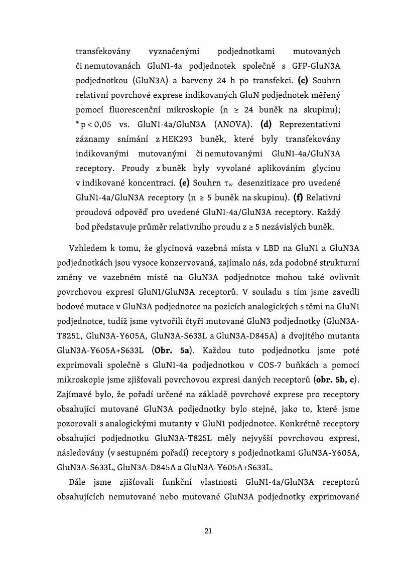
21
transfekovány vyznačenými podjednotkami mutovaných
či nemutovanách GluN1-4a podjednotek společně s GFP-GluN3A
podjednotkou (GluN3A) a barveny 24 h po transfekci. (c) Souhrn
relativní povrchové exprese indikovaných GluN podjednotek měřený
pomocí fluorescenční mikroskopie (n ≥ 24 buněk na skupinu);
* p < 0,05 vs. GluN1-4a/GluN3A (ANOVA). (d) Reprezentativní
záznamy snímání z HEK293 buněk, které byly transfekovány
indikovanými mutovanými či nemutovanými GluN1-4a/GluN3A
receptory. Proudy z buněk byly vyvolané aplikováním glycinu
v indikované koncentraci. (e) Souhrn τw desenzitizace pro uvedené
GluN1-4a/GluN3A receptory (n ≥ 5 buněk na skupinu). (f) Relativní
proudová odpověď pro uvedené GluN1-4a/GluN3A receptory. Každý
bod představuje průměr relativního proudu z ≥ 5 nezávislých buněk.
Vzhledem k tomu, že glycinová vazebná místa v LBD na GluN1 a GluN3A
podjednotkách jsou vysoce konzervovaná, zajímalo nás, zda podobné strukturní
změny ve vazebném místě na GluN3A podjednotce mohou také ovlivnit
povrchovou expresi GluN1/GluN3A receptorů. V souladu s tím jsme zavedli
bodové mutace v GluN3A podjednotce na pozicích analogických s těmi na GluN1
podjednotce, tudíž jsme vytvořili čtyři mutované GluN3 podjednotky (GluN3A-
T825L, GluN3A-Y605A, GluN3A-S633L a GluN3A-D845A) a dvojitého mutanta
GluN3A-Y605A+S633L (Obr. 5a). Každou tuto podjednotku jsme poté
exprimovali společně s GluN1-4a podjednotkou v COS-7 buňkách a pomocí
mikroskopie jsme zjišťovali povrchovou expresi daných receptorů (obr. 5b, c).
Zajímavé bylo, že pořadí určené na základě povrchové exprese pro receptory
obsahující mutované GluN3A podjednotky bylo stejné, jako to, které jsme
pozorovali s analogickými mutanty v GluN1 podjednotce. Konkrétně receptory
obsahující podjednotku GluN3A-T825L měly nejvyšší povrchovou expresi,
následovány (v sestupném pořadí) receptory s podjednotkami GluN3A-Y605A,
GluN3A-S633L, GluN3A-D845A a GluN3A-Y605A+S633L.
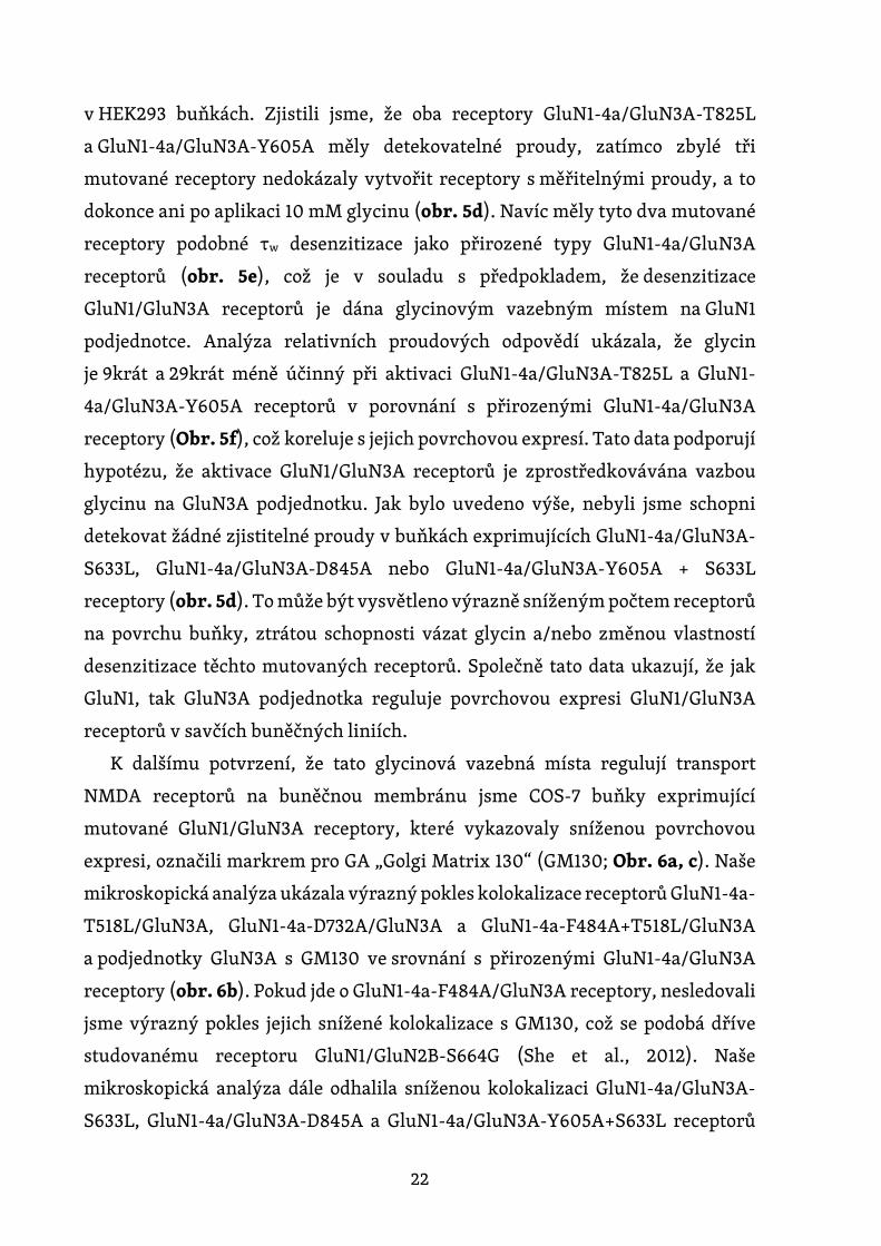
Dále jsme zjišťovali funkční vlastnosti GluN1-4a/GluN3A receptorů
obsahujících nemutované nebo mutované GluN3A podjednotky exprimované
22
v HEK293 buňkách. Zjistili jsme, že oba receptory GluN1-4a/GluN3A-T825L
a GluN1-4a/GluN3A-Y605A měly detekovatelné proudy, zatímco zbylé tři
mutované receptory nedokázaly vytvořit receptory s měřitelnými proudy, a to
dokonce ani po aplikaci 10 mM glycinu (obr. 5d). Navíc měly tyto dva mutované
receptory podobné τw desenzitizace jako přirozené typy GluN1-4a/GluN3A
receptorů (obr. 5e), což je v souladu s předpokladem, že desenzitizace
GluN1/GluN3A receptorů je dána glycinovým vazebným místem na GluN1
podjednotce. Analýza relativních proudových odpovědí ukázala, že glycin
je 9krát a 29krát méně účinný při aktivaci GluN1-4a/GluN3A-T825L a GluN1-
4a/GluN3A-Y605A receptorů v porovnání s přirozenými GluN1-4a/GluN3A
receptory (Obr. 5f), což koreluje s jejich povrchovou expresí. Tato data podporují
hypotézu, že aktivace GluN1/GluN3A receptorů je zprostředkovávána vazbou
glycinu na GluN3A podjednotku. Jak bylo uvedeno výše, nebyli jsme schopni
detekovat žádné zjistitelné proudy v buňkách exprimujících GluN1-4a/GluN3A-
S633L, GluN1-4a/GluN3A-D845A nebo GluN1-4a/GluN3A-Y605A + S633L
receptory (obr. 5d). To může být vysvětleno výrazně sníženým počtem receptorů
na povrchu buňky, ztrátou schopnosti vázat glycin a/nebo změnou vlastností
desenzitizace těchto mutovaných receptorů. Společně tato data ukazují, že jak
GluN1, tak GluN3A podjednotka reguluje povrchovou expresi GluN1/GluN3A
receptorů v savčích buněčných liniích.
K dalšímu potvrzení, že tato glycinová vazebná místa regulují transport
NMDA receptorů na buněčnou membránu jsme COS-7 buňky exprimující
mutované GluN1/GluN3A receptory, které vykazovaly sníženou povrchovou
expresi, označili markrem pro GA „Golgi Matrix 130“ (GM130; Obr. 6a, c). Naše
mikroskopická analýza ukázala výrazný pokles kolokalizace receptorů GluN1-4a-
T518L/GluN3A, GluN1-4a-D732A/GluN3A a GluN1-4a-F484A+T518L/GluN3A
a podjednotky GluN3A s GM130 ve srovnání s přirozenými GluN1-4a/GluN3A
receptory (obr. 6b). Pokud jde o GluN1-4a-F484A/GluN3A receptory, nesledovali
jsme výrazný pokles jejich snížené kolokalizace s GM130, což se podobá dříve
studovanému receptoru GluN1/GluN2B-S664G (She et al., 2012). Naše
mikroskopická analýza dále odhalila sníženou kolokalizaci GluN1-4a/GluN3A-
S633L, GluN1-4a/GluN3A-D845A a GluN1-4a/GluN3A-Y605A+S633L receptorů

23
s GM130 ve srovnání s přirozenými GluN1-4a/GluN3A receptory (Obr. 6d). Tato
data dohromady naznačují, že narušení vazby glycinu snižuje časný transport
GluN1/GluN3A receptorů do GA.
Obrázek 5. Mutace v glycinovém vazebném místě na GluN3A
podjednotce regulují povrchovou expresi a afinitu glycinu
ke GluN1/GluN3A receptorům. (a) Schéma reprezentující vazebné
místo pro glycin na GluN3A podjednotce (PDB kód: 2RC7, znázorněna

24
oranžově); námi studované aminokyselinové zbytky jsou znázorněny,
stejně jako molekula glycinu; pro porovnání je znázorněno i glycinové
vazebné místo na GluN1 podjednotce (zobrazeno modře).
(b) Reprezentativní snímky COS-7 buněk, které byly transfekovány
vyznačenými podjednotkami mutovaných či nemutovanách GluN3A
podjednotek společně s GluN1-4a podjednotkou, barveny 24 h
po transfekci. (c) Souhrn relativní povrchové exprese indikovaných
GluN podjednotek měřené pomocí fluorescenční mikroskopie (n ≥ 20
buněk na skupinu); * p < 0,05 vs. GluN1-4a/GluN3A (ANOVA).
(d) Reprezentativní záznamy snímání z HEK293 buněk, které byly
transfekovány indikovanými GluN1-4a/GluN3A receptory. Proudy
z buněk byly vyvolané aplikováním glycinu v indikované koncentraci.
(e) Souhrn τw desenzitizace pro uvedené GluN1-4a/GluN3A receptory
(n ≥ 5 buněk na skupinu). (f) Relativní proudová odpověď pro uvedené
GluN1-4a/GluN3A receptory. Každý bod představuje průměr
relativního proudu z ≥ 5 nezávislých buněk.

25
Obrázek 6. Glycinová vazebná místa na GluN1 a GluN3A regulují časný
transport GluN1/GluN3A receptorů. (a, c) Reprezentativní snímky
z konfokální mikroskopie COS-7 buněk, které byly transfekované
uvedenými podjednotkami GluN1-4a a GFP-GluN3A (GluN3A);
pro značení GA byla použita protilátka anti-GM130. Znázorněny jsou
snímky celkové exprese (C), GM130 a spojené snímky z předchozích
dvou. (b, d) Souhrn průměrné intenzity GFP-GluN3A (GluN3A) signálu
kolokalizujícího s GM130 (n ≥ 10 buněk); * p < 0,05 vs. GluN1-
4a/GluN3A (ANOVA).
Dále nás zajímalo, zda zavedení mutací v glycinovém vazebném místě GluN
podjednotek také ovlivní transport NMDA receptorů v neuronech. Z toho důvodu
jsme transfekovali potkaní hipokampální neurony v DIV10 přirozenými nebo
mutovanými GluN1-1a podjednotkami (tuto sestřihovou variantu jsme zvolili
z toho důvodu, že není sama transportována na buněčný povrch, pokud není
26
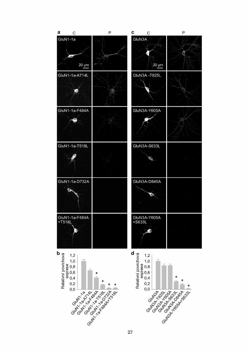
složena s GluN2 a/nebo GluN3 podjednotkou) a přirozenými či mutovanými
GluN3A podjednotkami. Čtyři dny po transfekci (tj. DIV14) jsme pak změřili
povrchový a celkový signál rekombinantních GluN podjednotek s použitím anti-
GFP protilátky a konfokální mikroskopie. Naše analýza ukázala následující
pořadí povrchové exprese mutovaných GluN1-1a podjednotek (v sestupném
pořadí): GluN1-1a-A714L, GluN1-1a-F484A, GluN1-1a-T518L, GluN1-1a-D732A
a GluN1-1a-F484A+T518L (obr. 7a, b). Stejně tak pět mutovaných GluN3
podjednotek vykazovalo následující pořadí povrchové exprese (v sestupném
pořadí): GluN3A-T825L, GluN3A-Y605A, GluN3A-S633L, GluN3A-D845A
a GluN3A-Y605A + S633L (obr. 7c, d). Tyto výsledky jsou konzistentní s našimi
daty získanými z analýzy povrchové exprese mutovaných NMDA receptorů
v COS-7 buňkách, což podporuje hypotézu, že glycinová vazebná místa na GluN1
a GluN3A podjednotkách jsou kritická pro povrchovou expresi NMDA receptorů.
27
28
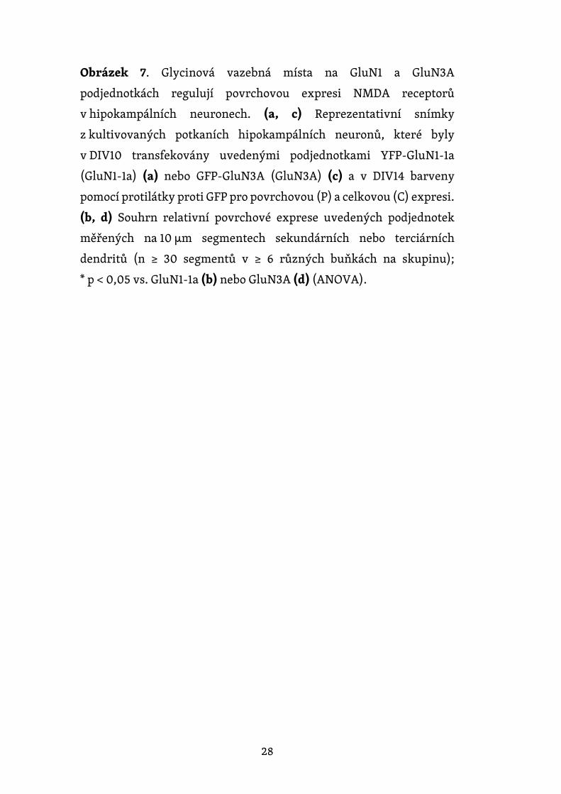
Obrázek 7. Glycinová vazebná místa na GluN1 a GluN3A
podjednotkách regulují povrchovou expresi NMDA receptorů
v hipokampálních neuronech. (a, c) Reprezentativní snímky
z kultivovaných potkaních hipokampálních neuronů, které byly
v DIV10 transfekovány uvedenými podjednotkami YFP-GluN1-1a
(GluN1-1a) (a) nebo GFP-GluN3A (GluN3A) (c) a v DIV14 barveny
pomocí protilátky proti GFP pro povrchovou (P) a celkovou (C) expresi.
(b, d) Souhrn relativní povrchové exprese uvedených podjednotek
měřených na 10 μm segmentech sekundárních nebo terciárních
dendritů (n ≥ 30 segmentů v ≥ 6 různých buňkách na skupinu);
* p < 0,05 vs. GluN1-1a (b) nebo GluN3A (d) (ANOVA).
29
6. Diskuse
6.1. N-glykosylace reguluje transport a povrchovou mobilitu NMDA
receptorů obsahujících GluN3A podjednotku
Zabývali jsme se studiem vlivu N-glykosylace GluN3A podjednotky
na transport a povrchovou mobilitu NMDA receptorů obsahujících GluN3A
podjednotku. Tato podjednotka působí jako „molekulární brzda“ a může tedy
zpomalovat zrání excitačních synapsí (Pérez-Otaño et al., 2016). Abnormální
funkce GluN3A podjednotky se rovněž předpokládá u řady neuropsychiatrických
onemocnění (Kehoe et al., 2014; Mohamad et al., 2013; Yuan and Bellone, 2013)
a je tedy možné, že narušená N-glykosylace GluN3A podjednotky může ovlivnit
celou řadu procesů v CNS.
Ukázali jsme, že N-glykosylace GluN3A podjednotky je nezbytná
pro povrchovou expresi GluN1/GluN3 receptorů jak v heterologních COS-7
buňkách, tak v potkaních hipokampálních neuronech. Ovšem není zcela zřejmé,
jaký je kontrolní mechanizmus pro správnou N-glykosylaci GluN3A
podjednotky. Jednou z možností je, že přítomnost N-glykanů na GluN3A
podjednotce ovlivňuje skládání a kvartérní strukturu NMDA receptoru, která
je v ER kontrolována. Další možností je, že N-glykany na GluN3A podjednotce
interagují v ER se specifickými vazebnými proteiny, jakými jsou galektiny
či další endogenní lektiny, které následně zajišťují transport NMDA receptorů
na buněčný povrch.
K našemu překvapení jsme zjistili, že remodelace N-glykanů, ke které dochází
v GA, není nezbytná pro povrchovou expresi NMDA receptorů obsahujících
GluN3A podjednotku. Tento výsledek je v souladu s nedávnou studií, v níž došli
Hanus et al. k podobným závěrům v případě několika typů membránových
proteinů, včetně AMPA receptorů (Hanus et al., 2016). Z tohoto důvodu jsme
se dále zabývali otázkou, jakou roli hrají hybridní a komplexní N-glykany
u NMDA receptorů obsahujících GluN3A podjednotky. Zaměřili jsme se zejména
na způsob, jakým ovlivňuje přítomnost hybridních a komplexních N-glykanů

30
mobilitu receptorů v buněčné membráně. V případě, kdy jsme zablokovali
remodelaci N-glykanů v GA pomocí specifických inhibitorů a tím inhibovali
syntézu hybridních a komplexních N-glykanů, pozorovali jsme na živých
hipokampálních neuronech zvýšenou povrchovou mobilitu NMDA receptorů
obsahujících GluN3A podjednotku. Dané výsledky jsou v souladu se zjištěním,
že povrchová mobilita NMDA (Groc et al., 2007) i AMPA (Frischknecht et al.,
2009) receptorů v neuronech může být ovlivněna změnami v extracelulární
matrix. V případě, kdy jsme neurony transfekované značenou GluN3A
podjednotkou inkubovali s vybranými lektiny, jsme pozorovali výrazné snížení
povrchové mobility NMDA receptorů obsahujících GluN3A podjednotku. Tento
výsledek lze vysvětlit jednak přímou vazbou lektinů na N-glykany přítomné
na NMDA receptorech, či jejich případnou interakcí s ostatními glykoproteiny
přítomnými v buněčné membráně. Podobné snížení mobility membránových
proteinů po inkubaci s lektiny bylo pozorováno již dříve. Například Henis and
Elson, 1981 pozorovali snížení povrchové mobility imunoglobulínů u myších
lymfocytů po inkubaci s lektinem ConA. Je tedy možné, že naše pozorování může
mít rovněž fyziologický význam, neboť v savčí CNS se exprimuje několik typů
lektinů, jako jsou například lektiny rozpoznávající galaktózu (Stanley et al.,
2015) nebo kyselinu sialovou (Macauley et al., 2014), které mohou zásadním
způsobem ovlivňovat mobilitu NMDA receptorů in vivo.
Pro studium role N-glykosylace za patologických podmínek jsme využili pět
linií lidských fibroblastů, které byly odvozeny z pacientů trpících řadou
glykosylačních poruch s mutacemi v enzymech N-glykosylačního aparátu.
Konkrétně jsme zkoumali linie fibroblastů PMM2-CDG a PGM1-CDG, u nichž
se nacházely defekty biosyntézy N-glykanového prekurzoru v cytosolu, SRD5A3-
CDG a DPAGT1-CDG linie s defekty v počátečních krocích syntézy N-glykanového
prekurzoru navázaného na lipid na cytosolické straně ER a linii ALG8-CDG
s defektem ve finálním kroku syntézy N-glykanového prekurzoru v ER.
Je důležité zmínit, že všichni pacienti, od nichž jsme linie fibroblastů získali,
vykazovali různý stupeň neurologického poškození (Ondruskova et al., 2014;
Vesela et al., 2009). Změnu povrchové exprese GluN1/GluN3A receptorů jsme
pozorovali pouze v případě ALG8-CDG linie. Pacienti s tímto typem CDG většinou
31
umírají během prvních měsíců života, a dané onemocnění je často doprovázeno
těžkými multiorgánovými dysfunkcemi (Freeze, 2006; Schollen, 2004).
V případě našeho pacienta obsahoval gen pro ALG8 mutace c.139A>C (p.T47P)
a c.1090C>T (p.R364X) které vedly k multiorgánovému selhání a úmrtí ve 2.
měsíci u předčasně narozeného dítěte (Vesela et al., 2009).
Je tedy pravděpodobné, že změny v glykosylačním aparátu pozorované
u pacientů trpících různými typy CDG mohou vést ke změnám v povrchové nebo
synaptické lokalizaci NMDA receptorů vzhledem k tomu, že se u těchto pacientů
často vyskytuje narušení řady nervových funkcí (Freeze, 2006; Moremen et al.,
2012). Rovněž řada fyziologických či patologických procesů může vést ke změně
N-glykanového složení, jak bylo například pozorováno u pacientů trpících
schizofrenií (Tucholski et al., 2013a). Změny v N-glykanovém složení mohou
ovlivňovat řadu vlastností NMDA receptorů, jako například jejich stabilitu
v synapsi, nebo modulaci prostřednictvím endogenních lektinů. Z toho vyplývá,
že poznatky o konkrétním složení N-glykanů na jednotlivých podjednotkách
NMDA receptorů a jejich funkčním významu by mohly případně v budoucnu být
využity pro terapii, neboť naše pokusy ukázaly, že funkční NMDA receptory
obsahují desítky relativně komplexních N-glykanových struktur.
Bohužel, nedostatek vzorků z mozků pacientů trpících těmito vzácnými
poruchami ztěžuje další výzkum daných onemocnění. Naštěstí lze nové metody
a techniky, jako je například konverze lidských fibroblastů na neuronální buňky,
využít pro další studium poruch N-glykosylace, což by mohlo pomoci při hledání
vhodné léčby pro tyto pacienty.
6.2. Strukturní změny v glycinových vazebných místech na GluN1
a GluN3A podjednotkách regulují povrchovou expresi NMDA
receptorů
V další práci jsme studovali vliv integrity glycinových vazebných míst
na GluN1 a GluN3A podjednotkách na transport a funkci NMDA receptorů.
GluN1/GluN3A receptory sice obsahují čtyři vazebná místa pro glycin, avšak
jeho vazba na GluN1 či GluN3A podjednotku má odlišný dopad na funkci

32
receptoru. Vazba glycinu na GluN3A podjednotku aktivuje receptor, zatímco
vazba na GluN1 podjednotku způsobuje rychlou desenzitizaci proudových
odpovědí (Awobuluyi et al., 2007; Kehoe et al., 2013; Kvist et al., 2013; Madry et
al., 2007). V naší studii jsme analyzovali proudy vyvolané glycinem, abychom
zjistili, jaký dopad na funkční vlastnosti GluN1/GluN3A receptorů budou mít
mutace v glycinovém vazebném místě na GluN1 nebo GluN3A podjednotce.
U GluN1 podjednotky jsme se podrobněji zaměřili na mutace, které se nacházejí
ve vazebném místě pro glycin a v předchozích studiích na GluN1/GluN2
receptorech u nich byl pozorován vliv na hodnoty EC50 pro glycin (Kvist et al.,
2013; Williams et al., 1996). Konkrétně jsme zkoumali mutace F484A, T518L,
F484A+T518L, A714L a D732A. U GluN3A podjednotky jsme zavedli mutace
analogické k těm na GluN1 podjednotce, a to konkrétně mutace Y605A, S633L,
Y605A+S633L, T825L a D845A. Zjistili jsme, že míra změn hodnot τw
pro desenzitizaci u mutovaných GluN1/GluN3A receptorů je v souladu s dříve
stanovenými změnami v hodnotách EC50 pro glycin u mutovaných GluN1/GluN2
receptorů (Kvist et al., 2013; Williams et al., 1996). Dále jsme pozorovali,
že mutace F484A v GluN1 podjednotce snižuje desenzitizaci u GluN1/GluN3A
receptorů, což je rovněž v souladu s předchozími studiemi (Kvist et al., 2013).
Změny v desenzitizační kinetice GluN1/GluN3A receptorů způsobené
zkoumanými mutacemi v glycinovém vazebném místě na GluN1 podjednotce
přímo korelovaly se změnami v povrchové expresi těchto receptorů. Analýza
maximálních proudových odpovědí nám umožnila stanovit hodnotu EC50
pro glycin na GluN1/GluN3A receptorech (89 µM), což dříve nebylo možné
vzhledem k tomu, že předchozí měření byla prováděna na receptorech
exprimovaných v oocytech Xenopus laevis. Z našich výsledků dále vyplývá,
že GluN1/GluN3A receptory s mutacemi v glycinovém vazebném
místě na GluN3A podjednotce, které vykazovaly funkční odpovědi na aplikaci
glycinu, měly podobnou desenzitizační kinetiku jako nemutované receptory
a naopak vykazovaly sníženou afinitu ke glycinu. Dané výsledky jsou v souladu
s hypotézou, že glycinové vazebné místo na GluN3A podjednotce je nezbytné
pro aktivaci GluN1/GluN3A receptoru, avšak nepřispívá k jeho desenzitizaci.
Toto pozorování je rovněž v souladu s nedávnou studií, v níž byla použita látka
33
CGP-78608, pomocí které byla odhalena přítomnost funkčních GluN1/GluN3A
receptorů v hipokampu u potkaních mláďat (Grand et al., 2018). Naše data
získaná z mikroskopických experimentů ukázala, že mutace v glycinovém
vazebném místě analogické u GluN1 a GluN3A podjednotek snižují do stejné míry
povrchovou expresi GluN1/GluN3A receptorů v COS-7 buňkách. Podobné
výsledky jsme pozorovali rovněž v případě GluN3A podjednotek exprimovaných
v hipokampálních neuronech.
Vzhledem k tomu, že v hipokampálních neuronech jsou endogenně
exprimovány různé typy podjednotek, včetně GluN1, GluN2A, GluN2B a GluN3A
(Grand et al., 2018; Rozeboom et al., 2015; Sanz-Clemente et al., 2013), naše data
naznačují, že strukturní změny v glycinovém vazebném místě u exogenních
GluN1 a GluN3A podjednotek ovlivňují rovněž transport NMDA receptorů
obsahujících endogenní GluN podjednotky, včetně triheteromerických
GluN1/GluN2/GluN3A receptorů. Přestože dosud není zcela zřejmé, jakým
mechanizmem mutace v glycinovém vazebném místě mění povrchovou expresi
GluN1/GluN3A receptorů, je pravděpodobné, že kvalitativní kontrola v ER bude
obdobná jako v případě GluN1/GluN2 receptorů (Kenny et al., 2009; She et al.,
2012) či AMPA receptorů (Penn et al., 2008). Předchozí studie odhadují,
že glutamát může být v ER přítomen až v milimolárních koncentracích (Berger et
al., 1977; Meeker et al., 1989) a mohl by tedy hrát roli při ověřování funkčnosti
správně složených GluN1/GluN2 receptorů či AMPA receptorů prostřednictvím
specifického kontrolního mechanismu v ER (Penn et al., 2008). Podobnou úlohu
by v ER mohl hrát i glycin či D-serin, přestože jejich přesná koncentrace
v neuronálním ER není známa. Avšak vzhledem k tomu, že se tito agonisté
účastní řady metabolických procesů, je pravděpodobné, že se mohou v lumen ER
nacházet v dostatečných koncentracích, aby se mohly vázat na nově složené
NMDA receptory. Teoreticky by tedy neurony mohly regulovat počty NMDA
receptorů na svém povrchu na základě množství agonistů, jako je glycin, D-serin
a glutamát, či antagonistů, jako například kyselina kynurenová (Moroni et al.,
1988), které se přirozeně nachází v savčí CNS. Podobně by mohly i různé
farmakologické látky působící prostřednictvím glycinového vazebného místa
na GluN1 a/nebo GluN3A podjednotce regulovat počty NMDA receptorů
34
na buněčném povrchu a tím poskytnout terapeutickou strategii za konkrétních
patologických podmínek. Za tímto účelem jsme testovali, zda 48 h inkubace
buněk s glycinem až v koncentraci 1 mM změní povrchovou expresi
nemutovaných či mutovaných GluN1/GluN3A receptorů. K našemu překvapení
jsme zjistili, že inkubace s glycinem nijak neovlivnila povrchovou expresi
studovaných GluN1/GluN3A receptorů. Je tedy možné, že intracelulárně je již
přítomna dostatečná koncentrace glycinu a extracelulárně přidaný glycin jí dále
nezvyšuje, nebo že extracelulární koncentrace glycinu nepůsobí na jeho
intracelulární koncentraci. Je rovněž možné, že vysoká intracelulární
koncentrace glycinu nemá vliv na transport GluN1/GluN3A receptorů.
Tato studie o významu glycinových vazebných míst na GluN1 a GluN3A
podjednotkách tedy pomáhá k pochopení fyziologických procesů v savčí CNS.

35
7. Závěr
Výlev glutamátu z jednoho neuronu a jeho následná detekce glutamátovými
receptory na přilehlém neuronu tvoří základ synaptického přenosu na více než
1014 synapsí v celém lidském mozku. Specifita této signalizace je navíc umožněna
expresí různých podjednotek NMDA receptorů a jejich alternativním sestřihem,
které se mohou lišit v místě i čase. Vzhledem k tomu, že daná signalizace je
zásadní pro synaptickou plasticitu a neuronální vývoj, může abnormální
transport, špatná lokalizace či neadekvátní aktivace NMDA receptorů vést k řadě
patologií a dysfunkcí. Z toho důvodu je pochopení základních mechanizmů
regulace transportu a funkce NMDA receptorů nezbytné pro objasnění těchto
dysfunkcí a vývoj budoucí terapie.
V této dizertační práci jsme ukázali, že narušená N-glykosylace NMDA
receptorů mění jejich transport a funkční vlastnosti. Zjistili jsme, že jednotlivé
podjednotky NMDA receptorů mají na svém povrchu navázané velké množství
N-glykanů a že se mezi sebou tyto podjednotky liší v jejich složení. Zajímavé bylo
pozorování, že u fibroblastů získaných od pacientů s diagnostikovanými
poruchami glykosylace může docházet mimo jiné i ke změnám v povrchové
expresi NMDA receptorů. Současně jsme pozorovali, že změny v remodelaci N-
glykanů či vazba specifických lektinů mohou mít vliv na povrchovou mobilitu
NMDA receptorů, což může vést ke změnám v lokalizaci a funkci NMDA
receptorů. Dále jsme ukázali, že glycinová vazebná místa na GluN1 a GluN3A
podjednotkách hrají odlišné funkční role u NMDA receptorů a že povrchová
exprese těchto receptorů souvisí s afinitou ke glycinu u GluN1 podjednotky.
Je tedy zřejmé, že jak specifický význam N-glykosylace pro neuronální funkci
NMDA receptorů, tak rovněž změny v aktivaci NMDA receptorů si zaslouží další
výzkum, neboť mohou hrát doposud neznámou úlohu ve fyziologii,
či patofyziologii savčí centrální nervové soustavy.
36
8. Použitá literatura
Atlason, P.T., Garside, M.L., Meddows, E., Whiting, P., and McIlhinney, R.A. (2007). N-Methyl-D-aspartate (NMDA) receptor subunit NR1 forms the substrate for oligomeric assembly of the NMDA receptor. J Biol Chem 282, 25299-25307.
Awobuluyi, M., Yang, J., Ye, Y., Chatterton, J.E., Godzik, A., Lipton, S.A., and Zhang, D. (2007). Subunit-specific roles of glycine-binding domains in activation of NR1/NR3 N-methyl-D-aspartate receptors. Mol Pharmacol 71, 112-122.
Berger, S.J., Carter, J.C., and Lowry, O.H. (1977). The distribution of glycine, GABA, glutamate and aspartate in rabbit spinal cord, cerebellum and hippocampus. J Neurochem 28, 149-158.
Chazot, P.L., Cik, M., and Stephenson, F.A. (1995). An investigation into the role of N-glycosylation in the functional expression of a recombinant heteromeric NMDA receptor. Mol Membr Biol 12, 331-337.
Chazot, P.L., and Stephenson, F.A. (1997). Biochemical evidence for the existence of a pool of unassembled C2 exon-containing NR1 subunits of the mammalian forebrain NMDA receptor. J Neurochem 68, 507-516.
Chen, J., Liu, Q., Fan, R., Han, H., Yang, Z., Cui, W., Song, G., and Li, M.D. (2019). Demonstration of critical role of GRIN3A in nicotine dependence through both genetic association and molecular functional studies. Addict Biol.
Chen, W., Shieh, C., Swanger, S.A., Tankovic, A., Au, M., McGuire, M., Tagliati, M., Graham, J.M., Madan-Khetarpal, S., Traynelis, S.F., et al. (2017). GRIN1 mutation associated with intellectual disability alters NMDA receptor trafficking and function. J Hum Genet.
Clements, J.D., and Westbrook, G.L. (1991). Activation kinetics reveal the number of glutamate and glycine binding sites on the N-methyl-D-aspartate receptor. Neuron 7, 605-613.
Dupuis, J.P., and Groc, L. (2019). Surface trafficking of neurotransmitter receptors: From cultured neurons to intact brain preparations. Neuropharmacology.
Everts, I., Villmann, C., and Hollmann, M. (1997). N-Glycosylation is not a prerequisite for glutamate receptor function but Is essential for lectin modulation. Mol Pharmacol 52, 861-873.
Farina, A.N., Blain, K.Y., Maruo, T., Kwiatkowski, W., Choe, S., and Nakagawa, T. (2011). Separation of domain contacts is required for heterotetrameric assembly of functional NMDA receptors. J Neurosci 31, 3565-3579.
Ferreira, J.S., Papouin, T., Ladepeche, L., Yao, A., Langlais, V.C., Bouchet, D., Dulong, J., Mothet, J.P., Sacchi, S., Pollegioni, L., et al. (2017). Co-agonists differentially tune GluN2B-NMDA receptor trafficking at hippocampal synapses. Elife 6.
Freeze, H.H. (2006). Genetic defects in the human glycome. Nat Rev Genet 7, 537-551. Freeze, H.H., Eklund, E.A., Ng, B.G., and Patterson, M.C. (2012). Neurology of inherited
glycosylation disorders. Lancet Neurol 11, 453-466. Frischknecht, R., Heine, M., Perrais, D., Seidenbecher, C.I., Choquet, D., and Gundelfinger, E.D.
(2009). Brain extracellular matrix affects AMPA receptor lateral mobility and short-term synaptic plasticity. Nat Neurosci 12, 897-904.
37
Grand, T., Abi Gerges, S., David, M., Diana, M.A., and Paoletti, P. (2018). Unmasking GluN1/GluN3A excitatory glycine NMDA receptors. Nat Commun 9, 4769.
Groc, L., Heine, M., Cognet, L., Brickley, K., Stephenson, F.A., Lounis, B., and Choquet, D. (2004). Differential activity-dependent regulation of the lateral mobilities of AMPA and NMDA receptors. Nat Neurosci 7, 695-696.
Groc, L., Choquet, D., Stephenson, F. A., Verrier, D., Manzoni, O. J., and Chavis, P. (2007). NMDA receptor surface trafficking and synaptic subunit composition are developmentally regulated by the extracellular matrix protein Reelin. J Neurosci 27, 10165–10175.
Gross, V., Tran-Thi, T.A., Schwarz, R.T., Elbein, A.D., Decker, K., and Heinrich, P.C. (1986). Different effects of the glucosidase inhibitors 1-deoxynojirimycin, N-methyl-1-deoxynojirimycin and castanospermine on the glycosylation of rat alpha 1-proteinase inhibitor and alpha 1-acid glycoprotein. Biochem J 236, 853-860.
Hansen, K.B., Yi, F., Perszyk, R.E., Menniti, F.S., and Traynelis, S.F. (2017). NMDA Receptors in the Central Nervous System. Methods Mol Biol 1677, 1-80.
Hanus, C., Geptin, H., Tushev, G., Garg, S., Alvarez-Castelao, B., Sambandan, S., Kochen, L., Hafner, A.S., Langer, J.D., and Schuman, E.M. (2016). Unconventional secretory processing diversifies neuronal ion channel properties. Elife 5.
Hawkins, L.M., Prybylowski, K., Chang, K., Moussan, C., Stephenson, F.A., and Wenthold, R.J. (2004). Export from the endoplasmic reticulum of assembled N-methyl-d-aspartic acid receptors is controlled by a motif in the c terminus of the NR2 subunit. J Biol Chem 279, 28903-28910.
Henis, Y. I., and Elson, E. L. (1981). Inhibition of the mobility of mouse lymphocyte surface immunoglobulins by locally bound concanavalin A. Proceedings of the National Academy of Sciences 78, 1072–1076.
Herreman, A., Van Gassen, G., Bentahir, M., Nyabi, O., Craessaerts, K., Mueller, U., Annaert, W., and De Strooper, B. (2003). gamma-Secretase activity requires the presenilin-dependent trafficking of nicastrin through the Golgi apparatus but not its complex glycosylation. J Cell Sci 116, 1127-1136.
Horak, M., Petralia, R.S., Kaniakova, M., and Sans, N. (2014). ER to synapse trafficking of NMDA receptors. Front Cell Neurosci 8, 394.
Horak, M., and Wenthold, R.J. (2009). Different roles of C-terminal cassettes in the trafficking of full-length NR1 subunits to the cell surface. J Biol Chem 284, 9683-9691.
Huh, K.H., and Wenthold, R.J. (1999). Turnover analysis of glutamate receptors identifies a rapidly degraded pool of the N-methyl-D-aspartate receptor subunit, NR1, in cultured cerebellar granule cells. J Biol Chem 274, 151-157.
Jeyifous, O., Waites, C.L., Specht, C.G., Fujisawa, S., Schubert, M., Lin, E.I., Marshall, J., Aoki, C., de Silva, T., Montgomery, J.M., et al. (2009). SAP97 and CASK mediate sorting of NMDA receptors through a previously unknown secretory pathway. Nat Neurosci 12, 1011-1019.
Kaniakova, M., Kleteckova, L., Lichnerova, K., Holubova, K., Skrenkova, K., Korinek, M., Krusek, J., Smejkalova, T., Korabecny, J., Vales, K., et al. (2018). 7-Methoxyderivative of tacrine is a 'foot-in-the-door' open-channel blocker of GluN1/GluN2 and GluN1/GluN3 NMDA receptors with neuroprotective activity in vivo. Neuropharmacology 140, 217-232.
Kaniakova, M., Krausova, B., Vyklicky, V., Korinek, M., Lichnerova, K., Vyklicky, L., and Horak, M. (2012a). Key amino acid residues within the third membrane domains of NR1 and NR2 subunits contribute to the regulation of the surface delivery of N-methyl-D-aspartate receptors. J Biol Chem 287, 26423-26434.
Kaniakova, M., Lichnerova, K., Skrenkova, K., Vyklicky, L., and Horak, M. (2016). Biochemical and electrophysiological characterization of N-glycans on NMDA receptor subunits. J Neurochem 138, 546-556.
38
Kaniakova, M., Lichnerova, K., Vyklicky, L., and Horak, M. (2012b). Single amino acid residue in the M4 domain of GluN1 subunit regulates the surface delivery of NMDA receptors. J Neurochem 123, 385-395.
Kato, A., Rouach, N., Nicoll, R.A., and Bredt, D.S. (2005). Activity-dependent NMDA receptor degradation mediated by retrotranslocation and ubiquitination. Proc Natl Acad Sci U S A 102, 5600-5605.
Kehoe, L.A., Bellone, C., De Roo, M., Zandueta, A., Dey, P.N., Perez-Otano, I., and Muller, D. (2014). GluN3A promotes dendritic spine pruning and destabilization during postnatal development. J Neurosci 34, 9213-9221.
Kehoe, L.A., Bernardinelli, Y., and Muller, D. (2013). GluN3A: an NMDA receptor subunit with exquisite properties and functions. Neural Plast 2013, 145387.
Kenny, A.V., Cousins, S.L., Pinho, L., and Stephenson, F.A. (2009). The integrity of the glycine co-agonist binding site of N-methyl-D-aspartate receptors is a functional quality control checkpoint for cell surface delivery. J Biol Chem 284, 324-333.
Kleckner, N.W., and Dingledine, R. (1988). Requirement for glycine in activation of NMDA-receptors expressed in Xenopus oocytes. Science 241, 835-837.
Kusumi, A., Sako, Y., and Yamamoto, M. (1993). Confined lateral diffusion of membrane receptors as studied by single particle tracking (nanovid microscopy). Effects of calcium-induced differentiation in cultured epithelial cells. Biophys J 65, 2021-2040.
Kvist, T., Greenwood, J.R., Hansen, K.B., Traynelis, S.F., and Brauner-Osborne, H. (2013). Structure-based discovery of antagonists for GluN3-containing N-methyl-D-aspartate receptors. Neuropharmacology 75, 324-336.
Lavezzari, G., McCallum, J., Dewey, C.M., and Roche, K.W. (2004). Subunit-specific regulation of NMDA receptor endocytosis. J Neurosci 24, 6383-6391.
Lemke, J.R., Geider, K., Helbig, K.L., Heyne, H.O., Schutz, H., Hentschel, J., Courage, C., Depienne, C., Nava, C., Heron, D., et al. (2016). Delineating the GRIN1 phenotypic spectrum: A distinct genetic NMDA receptor encephalopathy. Neurology 86, 2171-2178.
Lichnerova, K., Kaniakova, M., Park, S.P., Skrenkova, K., Wang, Y.X., Petralia, R.S., Suh, Y.H., and Horak, M. (2015). Two N-glycosylation Sites in the GluN1 Subunit Are Essential for Releasing N-methyl-d-aspartate (NMDA) Receptors from the Endoplasmic Reticulum. J Biol Chem 290, 18379-18390.
Lichnerova, K., Kaniakova, M., Skrenkova, K., Vyklicky, L., and Horak, M. (2014). Distinct regions within the GluN2C subunit regulate the surface delivery of NMDA receptors. Front Cell Neurosci 8, 375.
Liu, C., Rozmyslowicz, T., Stwora-Wojczyk, M., Wojczyk, B., and Spitalnik, S.L. (2000). Posttranslational modifications of the amyloid precursor protein : glycosylation. Methods Mol Med 32, 169-190.
Macauley, M.S., Crocker, P.R., and Paulson, J.C. (2014). Siglec-mediated regulation of immune cell function in disease. Nat Rev Immunol 14, 653-666.
Madry, C., Betz, H., Geiger, J.R., and Laube, B. (2008). Supralinear potentiation of NR1/NR3A excitatory glycine receptors by Zn2+ and NR1 antagonist. Proc Natl Acad Sci U S A 105, 12563-12568.
Madry, C., Mesic, I., Bartholomaus, I., Nicke, A., Betz, H., and Laube, B. (2007). Principal role of NR3 subunits in NR1/NR3 excitatory glycine receptor function. Biochem Biophys Res Commun 354, 102-108.
Mahfooz, K., Marco, S., Martinez-Turrillas, R., Raja, M.K., Perez-Otano, I., and Wesseling, J.F. (2016). GluN3A promotes NMDA spiking by enhancing synaptic transmission in Huntington's disease models. Neurobiol Dis 93, 47-56.
Marco, S., Giralt, A., Petrovic, M.M., Pouladi, M.A., Martinez-Turrillas, R., Martinez-Hernandez, J., Kaltenbach, L.S., Torres-Peraza, J., Graham, R.K., Watanabe, M., et al. (2013).
39
Suppressing aberrant GluN3A expression rescues synaptic and behavioral impairments in Huntington's disease models. Nat Med 19, 1030-1038.
Matsuda, K., Fletcher, M., Kamiya, Y., and Yuzaki, M. (2003). Specific assembly with the NMDA receptor 3B subunit controls surface expression and calcium permeability of NMDA receptors. J Neurosci 23, 10064-10073.
McIlhinney, R.A., Le Bourdelles, B., Molnar, E., Tricaud, N., Streit, P., and Whiting, P.J. (1998). Assembly intracellular targeting and cell surface expression of the human N-methyl-D-aspartate receptor subunits NR1a and NR2A in transfected cells. Neuropharmacology 37, 1355-1367.
Meddows, E., Le Bourdelles, B., Grimwood, S., Wafford, K., Sandhu, S., Whiting, P., and McIlhinney, R.A. (2001). Identification of molecular determinants that are important in the assembly of N-methyl-D-aspartate receptors. J Biol Chem 276, 18795-18803.
Meeker, R.B., Swanson, D.J., and Hayward, J.N. (1989). Light and electron microscopic localization of glutamate immunoreactivity in the supraoptic nucleus of the rat hypothalamus. Neuroscience 33, 157-167.
Mikasova, L., Xiong, H., Kerkhofs, A., Bouchet, D., Krugers, H.J., and Groc, L. (2017). Stress hormone rapidly tunes synaptic NMDA receptor through membrane dynamics and mineralocorticoid signalling. Sci Rep 7, 8053.
Mohamad, O., Song, M., Wei, L., and Yu, S. P. (2013). Regulatory roles of the NMDA receptor GluN3A subunit in locomotion, pain perception and cognitive functions in adult mice. The Journal of physiology 591, 149–68
Moremen, K.W., Tiemeyer, M., and Nairn, A.V. (2012). Vertebrate protein glycosylation: diversity, synthesis and function. Nat Rev Mol Cell Biol 13, 448-462.
Moroni, F., Russi, P., Lombardi, G., Beni, M., and Carla, V. (1988). Presence of kynurenic acid in the mammalian brain. J Neurochem 51, 177-180.
Mueller, H.T., and Meador-Woodruff, J.H. (2004). NR3A NMDA receptor subunit mRNA expression in schizophrenia, depression and bipolar disorder. Schizophr Res 71, 361-370.
Okabe, S., Miwa, A., and Okado, H. (1999). Alternative splicing of the C-terminal domain regulates cell surface expression of the NMDA receptor NR1 subunit. J Neurosci 19, 7781-7792.
Ondruskova, N., Honzik, T., Vondrackova, A., Tesarova, M., Zeman, J., and Hansikova, H. (2014). Glycogen storage disease-like phenotype with central nervous system involvement in a PGM1-CDG patient. Neuro Endocrinol Lett 35, 137-141.
Paoletti, P., Bellone, C., and Zhou, Q. (2013). NMDA receptor subunit diversity: impact on receptor properties, synaptic plasticity and disease. Nat Rev Neurosci 14, 383-400.
Patneau, D.K., and Mayer, M.L. (1990). Structure-activity relationships for amino acid transmitter candidates acting at N-methyl-D-aspartate and quisqualate receptors. J Neurosci 10, 2385-2399.
Penn, A.C., Williams, S.R., and Greger, I.H. (2008). Gating motions underlie AMPA receptor secretion from the endoplasmic reticulum. EMBO J 27, 3056-3068.
Perez-Otano, I., Larsen, R.S., and Wesseling, J.F. (2016). Emerging roles of GluN3-containing NMDA receptors in the CNS. Nat Rev Neurosci 17, 623-635.
Perez-Otano, I., Lujan, R., Tavalin, S.J., Plomann, M., Modregger, J., Liu, X.B., Jones, E.G., Heinemann, S.F., Lo, D.C., and Ehlers, M.D. (2006). Endocytosis and synaptic removal of NR3A-containing NMDA receptors by PACSIN1/syndapin1. Nat Neurosci 9, 611-621.
Perez-Otano, I., Schulteis, C.T., Contractor, A., Lipton, S.A., Trimmer, J.S., Sucher, N.J., and Heinemann, S.F. (2001). Assembly with the NR1 subunit is required for surface expression of NR3A-containing NMDA receptors. J Neurosci 21, 1228-1237.
40
Petralia, R.S., Al-Hallaq, R.A., and Wenthold, R.J. (2009). Trafficking and Targeting of NMDA Receptors.
Qiu, S., Zhang, X.M., Cao, J.Y., Yang, W., Yan, Y.G., Shan, L., Zheng, J., and Luo, J.H. (2009). An endoplasmic reticulum retention signal located in the extracellular amino-terminal domain of the NR2A subunit of N-Methyl-D-aspartate receptors. J Biol Chem 284, 20285-20298.
Roche, K.W., Standley, S., McCallum, J., Dune Ly, C., Ehlers, M.D., and Wenthold, R.J. (2001). Molecular determinants of NMDA receptor internalization. Nat Neurosci 4, 794-802.
Rozeboom, A.M., Queenan, B.N., Partridge, J.G., Farnham, C., Wu, J.Y., Vicini, S., and Pak, D.T. (2015). Evidence for glycinergic GluN1/GluN3 NMDA receptors in hippocampal metaplasticity. Neurobiol Learn Mem 125, 265-273.
Sanz-Clemente, A., Nicoll, R.A., and Roche, K.W. (2013). Diversity in NMDA receptor composition: many regulators, many consequences. Neuroscientist 19, 62-75.
Schollen, E. (2004). Clinical and molecular features of three patients with congenital disorders of glycosylation type Ih (CDG-Ih) (ALG8 deficiency). Journal of Medical Genetics 41, 550–556.
Schuler, T., Mesic, I., Madry, C., Bartholomaus, I., and Laube, B. (2008). Formation of NR1/NR2 and NR1/NR3 heterodimers constitutes the initial step in N-methyl-D-aspartate receptor assembly. J Biol Chem 283, 37-46..
Scott, D.B., Michailidis, I., Mu, Y., Logothetis, D., and Ehlers, M.D. (2004). Endocytosis and degradative sorting of NMDA receptors by conserved membrane-proximal signals. J Neurosci 24, 7096-7109.
She, K., Ferreira, J.S., Carvalho, A.L., and Craig, A.M. (2012). Glutamate binding to the GluN2B subunit controls surface trafficking of N-methyl-D-aspartate (NMDA) receptors. J Biol Chem 287, 27432-27445.
Shen, Y.C., Liao, D.L., Chen, J.Y., Wang, Y.C., Lai, I.C., Liou, Y.J., Chen, Y.J., Luu, S.U., and Chen, C.H. (2009). Exomic sequencing of the ionotropic glutamate receptor N-methyl-D-aspartate 3A gene (GRIN3A) reveals no association with schizophrenia. Schizophr Res 114, 25-32.
Skrenkova, K., Lee, S., Lichnerova, K., Kaniakova, M., Hansikova, H., Zapotocky, M., Suh, Y.H., and Horak, M. (2018). N-Glycosylation Regulates the Trafficking and Surface Mobility of GluN3A-Containing NMDA Receptors. Front Mol Neurosci 11, 188.
Stanley, P., Taniguchi, N., and Aebi, M. (2015). “N-Glycans,” in, eds. A. Varki, R. D. Cummings, J. D. Esko, P. Stanley, G. W. Hart, M. Aebi, et al. (Cold Spring Harbor (NY)), 99–111.
Standley, S., Roche, K.W., McCallum, J., Sans, N., and Wenthold, R.J. (2000). PDZ domain suppression of an ER retention signal in NMDA receptor NR1 splice variants. Neuron 28, 887-898.
Swanger, S.A., Chen, W., Wells, G., Burger, P.B., Tankovic, A., Bhattacharya, S., Strong, K.L., Hu, C., Kusumoto, H., Zhang, J., et al. (2016). Mechanistic Insight into NMDA Receptor Dysregulation by Rare Variants in the GluN2A and GluN2B Agonist Binding Domains. Am J Hum Genet 99, 1261-1280.
Takata, A., Iwayama, Y., Fukuo, Y., Ikeda, M., Okochi, T., Maekawa, M., Toyota, T., Yamada, K., Hattori, E., Ohnishi, T., et al. (2013). A population-specific uncommon variant in GRIN3A associated with schizophrenia. Biol Psychiatry 73, 532-539.
Tokhtaeva, E., Sachs, G., and Vagin, O. (2009). Assembly with the Na,K-ATPase alpha(1) subunit is required for export of beta(1) and beta(2) subunits from the endoplasmic reticulum. Biochemistry 48, 11421-11431.
Traynelis, S.F., Wollmuth, L.P., McBain, C.J., Menniti, F.S., Vance, K.M., Ogden, K.K., Hansen, K.B., Yuan, H., Myers, S.J., and Dingledine, R. (2010). Glutamate receptor ion channels: structure, regulation, and function. Pharmacol Rev 62, 405-496.
41
Triller, A., and Choquet, D. (2008). New concepts in synaptic biology derived from single-molecule imaging. Neuron 59, 359-374.
Tucholski, J., Simmons, M.S., Pinner, A.L., Haroutunian, V., McCullumsmith, R.E., and Meador-Woodruff, J.H. (2013). Abnormal N-linked glycosylation of cortical AMPA receptor subunits in schizophrenia. Schizophr Res 146, 177-183.
Vesela, K., Honzik, T., Hansikova, H., Haeuptle, M.A., Semberova, J., Stranak, Z., Hennet, T., and Zeman, J. (2009). A new case of ALG8 deficiency (CDG Ih). J Inherit Metab Dis 32 Suppl 1.
Vierbuchen, T., Ostermeier, A., Pang, Z.P., Kokubu, Y., Sudhof, T.C., and Wernig, M. (2010). Direct conversion of fibroblasts to functional neurons by defined factors. Nature 463, 1035-1041.
Washbourne, P., Bennett, J.E., and McAllister, A.K. (2002). Rapid recruitment of NMDA receptor transport packets to nascent synapses. Nat Neurosci 5, 751-759.
Washbourne, P., Liu, X.B., Jones, E.G., and McAllister, A.K. (2004). Cycling of NMDA receptors during trafficking in neurons before synapse formation. J Neurosci 24, 8253-8264.
Williams, K., Chao, J., Kashiwagi, K., Masuko, T., and Igarashi, K. (1996). Activation of N-methyl-D-aspartate receptors by glycine: role of an aspartate residue in the M3-M4 loop of the NR1 subunit. Mol Pharmacol 50, 701-708.
Yavin, E., Richter-Landsberg, C., Duksin, D., and Yavin, Z. (1984). Tunicamycin blocks neuritogenesis and glucosamine labeling of gangliosides in developing cerebral neuron cultures. Proc Natl Acad Sci U S A 81, 5638-5642.
Yuan, T., Mameli, M., O'Connor, E.C., Dey, P.N., Verpelli, C., Sala, C., Perez-Otano, I., Luscher, C., and Bellone, C. (2013). Expression of cocaine-evoked synaptic plasticity by GluN3A-containing NMDA receptors. Neuron 80, 1025-1038.
42
9. Seznam publikací
Publikace in extenso související s touto dizertační prací
1. Kristyna Skrenkova, Katarina Hemelikova, Marharyta Kolcheva, Stepan Kortus, Martina Kaniakova, Barbora Krausova, Martin Horak (2019) Structural features in the glycine-binding sites of the GluN1 and GluN3A subunits regulate the surface delivery of NMDA receptors. Scientific reports 9:12303 IF = 4,5 (2018)
2. Kristyna Skrenkova, Sanghyeon Lee, Katarina Lichnerova, Martina Kaniakova, Hana Hansikova, Martin Zapotocky, Young Ho Suh, Martin Horak (2018). N-Glycosylation Regulates the Trafficking and Surface Mobility of GluN3A-Containing NMDA Receptors. Frontiers in Molecular Neuroscience 11, 188. IF = 3,7 (2018)
3. Martina Kaniakova, Katarina Lichnerova, Kristyna Skrenkova, Ladislav Vyklicky, Martin Horak (2016). Biochemical and electrophysiological characterization of N-glycans on NMDA receptor subunits. Journal of Neurochemistry 546–556. IF = 4,1 (2016)
4. Lichnerova Katarina, Kaniakova Martina, Seung Pyo Park, Kristyna Skrenkova, Ya-Xiang Wang, Ronald S. Petralia, Young Ho Suh, Martin Horak (2015). Two N-glycosylation Sites in the GluN1 Subunit Are Essential for Releasing N-methyl-d-aspartate (NMDA) Receptors from the Endoplasmic Reticulum. Journal of Biological Chemistry 290, 18379–18390. IF = 4,3 (2015)
43
Publikace nesouvisející s touto dizertační prací
1. Katarina Hemelikova (Lichnerova), Marharyta Kolcheva, Kristyna Skrenkova, Martina Kanikova, Martin Horak (2019). Lectins modulate the functional properties of GluN1/GluN3A – containg receptors. Neuropharmacology 157, 107671. IF = 4,4 (2018)
2. Martina Kaniakova, Eugenie Nepovimova, Lenka Kleteckova, Kristyna Skrenkova, Kristina Holubova, Zofia Chrienova, Vendula Hepnarova, Tomas Kucera, Tereza Kobrlova, Karel Vales, Jan Korabecny, Ondrej Soukup, Martin Horak. (2019) Combination of Memantine and 6-Chlorotacrine as Novel Multi-Target Compound against Alzheimer's Disease. Current Alzheimer Research 16:1.
IF= 3,2 (2018)
3. Martina Kaniakova, Lenka Kleteckova, Katarina Lichnerova, Kristina Holubova, Kristyna Skrenkova, Miloslav Korinek, Jan Krusek, Tereza Smejkalova, Jan Korabecny, Karel Vales, Ondrej Soukup, Martin Horak. (2018). 7-Methoxyderivative of tacrine is a 'foot-in-the-door' open-channel blocker of GluN1/GluN2 and GluN1/GluN3 NMDA receptors with neuroprotective activity in vivo. Neuropharmacology 15; 140:217-232.
IF= 4,3 (2018)
4. Vendula Sepsova, Jana Z. Karasova, Gunnar Tobin, Daniel Jun, Jan Korabecny, Pavla Cabelova, Katerina Janska, Jan Krusek, Kristyna Skrenkova, Kamil Kuca, Ondrej Soukup (2015). Cholinergic properties of new 7-methoxytacrine-donepezil derivatives. General Physiology and Biophysics, 34(2):189-200.
IF= 0,9 (2015)
5. Katarina Lichnerova, Martina Kaniakova, Kristyna Skrenkova, Ladislav Vyklicky, Martin Horak (2014). Distinct regions within the GluN2C subunit regulate the surface delivery of NMDA receptors. Frontiers in Cellular Neuroscience 8, 375.
IF= 4,3 (2014)
6. Martina Kaniakova, Kristyna Skrenkova, Svatopluk Adamek, Frantisek Vyskocil, Jan Krusek (2014). Different effects of lobeline on neuronal and muscle nicotinic receptors. European Journal of Pharmacology, 738:352-9.
IF= 2,5 (2014)
44
10. Životopis
Kristýna Skřenková
Vzdělání
2014 - nyní Doktorský studijní program Fyziologie živočichů Univerzita Karlova v Praze, Přírodovědecká fakulta Dizertační práce: Molekulární mechanismy regulace transportu a funkce
různých podtypů NMDA receptorů v hipokampálních neuronech Vedoucí práce: Mgr.Martin Horák, Ph.D. 2012 - 2014 Magisterský studijní obor Fyziologie živočichů, zaměření neurobiologie Univerzita Karlova v Praze, Přírodovědecká fakulta Diplomová práce: Alosterické modulátory nikotinového acetylcholinového receptoru vhodné k léčbě neurodegenerativních onemocnění Vedoucí práce: RNDr. Jan Krůšek, CSc. 2009 - 2012 Bakalářský studijní obor Molekulární biologie a biochemie organismů Univerzita Karlova v Praze, Přírodovědecká fakulta Bakalářská práce: Ionotropní glutamátové receptory a excitotoxicita Vedoucí práce: prof. Ladislav Vyklický, DrSc.
Pracovní zkušenosti
2018 – nyní Ústav experimentální medicíny AVČR Oddělení neurochemie
2011 - nyní Fyziologický ústav AVČR Oddělení buněčné neurofyziologie
Laboratorní dovednosti
- Kultivace a transfekce heterologních buněčných linií - Příprava a kultivace primárních potkaních hipokampálních kultur - Příprava potkaních hipokampálních řízků - Transfekce a infekce hipokampálních neuronů - Elektrofyziologie – technika terčíkového zámku (snímání z celé buňky) - Molekulární biologie – cílená mutageneze, transformace bakterií, izolace plasmidové DNA,
klonování DNA do vektoru - Imunofluorescenční mikroskopie – povrchové a celkové značení, kolokalizace a internalizace. - Živá mikroskopie - povrchová mobilita - Biochemie- SDP-PAGE
45
Absolvované kurzy
2019 - Aplikované improvizace (Univerzita Karlova, Přírodovědecká fakulta) 2018 - Pokročilá praktická rétorika a prezentace (Univerzita Karlova, Přírodovědecká fakulta) 2018 - Zpracování a analýza mikroskopických obrazů pro biomedicínu (Ústav molekulární
genetiky, AVČR) 2018 - Time managment (Univerzita Karlova, Přírodovědecká fakulta) 2018 - Scientific Writing (Univerzita Karlova, Přírodovědecká fakulta) 2017 - Scientific Presentations (Univerzita Karlova, Přírodovědecká fakulta) 2017 - Praktická rétorika a prezentace (Univerzita Karlova, Přírodovědecká fakulta) 2015 - Kurz základů vědecké AVČR (AVČR) 2014 - Mikroskopické metody v biomedicíně (Ústav molekulární genetiky, AVČR)


Charles University
Faculty of Science
Animal Physiology
Ph.D. thesis summary
Molecular mechanisms of regulation of trafficking and function of different subtypes of NMDA receptors in hippocampal
neurons
Kristýna Skřenková
Prague, 2019
2
Doctoral Study programs in Biomedicine
Charles University and The Czech Academy of Sciences
Study program: Animal Physiology
Chairman of the Sector Board: doc. RNDr. Jiří Novotný, DSc.
Institute and Department:
Institute of Physiology CAS
Department of Cellular Neurophysiology
Institute of Experimental medicine CAS Department of Neurochemistry
Author: Kristýna Skřenková
Supervisor: Martin Horák, Ph.D.
The Ph.D. Thesis is available in the library of the Faculty of Science, Charles
University.

3
Contents
1. Summary ............................................................................... 4
2. Introduction .......................................................................... 5
3. Hypothesis and aims .............................................................. 8
4. Materials and methods ........................................................... 9
5. Results .................................................................................. 13
5.1. N-glycosylation regulates the trafficking and surface mobility
of GluN3A-containing NMDA receptors ............................... 13
5.2. Structural features in the glycine-binding sites of the GluN1
and GluN3A subunits regulate the surface delivery of NMDA
receptors .............................................................................. 18
6. Discussion ............................................................................ 29
6.1. N-glycosylation regulates the trafficking and surface mobility
of GluN3A-containing NMDA receptors ............................... 29
6.2. Structural features in the glycine-binding sites of the GluN1
and GluN3A subunits regulate the surface delivery of NMDA
receptors .............................................................................. 31
7. Conclusions .......................................................................... 34
8. References ............................................................................ 35
9. List of publications ............................................................... 41
10. Curriculum Vitae ................................................................. 43
4
1. Summary
N-methyl-D-aspartate (NMDA) receptors are ionotropic glutamate receptors
playing a key role in the mammalian central nervous system. Under physiological
conditions, these receptors are important for excitatory synaptic transmission
and memory formation. However, under pathological conditions, their abnormal
regulation or activation can lead to many neurological and psychiatric disorders,
such as Alzheimer's disease, Parkinson's disease, Huntington's disease, epilepsy
or schizophrenia. Previous work has shown that the number of NMDA receptors
on the cell surface is regulated at multiple levels, including their synthesis,
folding, internalization or degradation. During transport to the cell membrane,
agonist binding and NMDA receptor activation are being control. The NMDA
receptors also undergo many posttranslational modifications such as
palmitoylation, phosphorylation or N-glycosylation.
In this thesis, we studied the molecular mechanisms that effect the trafficking
and functional properties of NMDA receptors in mammalian cells lines and rat
hippocampal neurons. Specifically, we focused on how specific changes
in the extracellular domains of the NMDA receptor can affect trafficking
to the cell membrane and how it modulate the functional properties of the NMDA
receptor. In mammals, the most organ-specific N-glycosylation occurs
in the brain. However, the effect of this modification on functional properties
of NMDA receptors in neurons is not fully understood. Therefore, we focused
on the role of N-glycosylation on NMDA receptors. We also studied the subunit-
specific effect of individual lectins. Another topic we focused on was the effect
of the integrity of glycine-binding sites of GluN1 and GluN3A subunits on the
trafficking and function of NMDA receptors. We used a variety of methods,
including immunohistochemistry, live microscopy, biochemistry and
electrophysiology. We used several cell models – mammalian HEK293 and COS-7
cell lines, human fibroblast derived from patients suffering with various forms
of CDG, and rat hippocampal and cerebellar neurons.
5
2. Introduction
NMDA receptors are a subclass of glutamate receptors that play an essential
role in synapse development, excitatory neurotransmission as well as synaptic
plasticity in the mammalian central nervous system (CNS) (Perez-Otano et al.,
2016; Traynelis et al., 2010). It has been well established that dysregulation
of NMDA receptors plays a critical role in the aetiology of many neuropsychiatric
and neurological disorders and conditions, including Huntington’s disease
(Mahfooz et al., 2016; Marco et al., 2013), schizophrenia (Mueller and Meador-
Woodruff, 2004), cocaine addiction (Yuan et al., 2013), and nicotine dependence
(Chen et al., 2019). In addition, a growing number of studies suggest that many
neuropsychiatric disorders are associated with mutations in genes that encode
various NMDA receptor subunits, including the GluN1 (Chen et al., 2017; Lemke
et al., 2016), and GluN3A (Shen et al., 2009; Takata et al., 2013) subunits. Thus,
understanding the molecular mechanisms that regulate NMDA receptors
is an essential step towards designing effective therapies for these patients.
Structurally, NMDA receptors are heterotetramers composed of GluN1 (with
eight splice variants), GluN2 (GluN2A through GluN2D), and/or GluN3 (GluN3A
and GluN3B) subunits (Paoletti et al., 2013; Traynelis et al., 2010). All GluN
subunits have a common membrane topology, including an extracellular amino-
terminal domain (ATD), extracellular ligand-binding domains (LBDs) formed
by S1 and S2 segments, four membrane domains (M1 through M4), and
an intracellular C-terminal domain (CTD) (Paoletti et al., 2013; Traynelis et al.,
2010). The conventional NMDA receptors subtype – GluN1/GluN2 – is activated
by the binding of agonists to the glutamate-binding site in the LBD of GluN2
together with the simultaneous binding of a co-agonist to the glycine-binding
site in the LBD of GluN1 (Clements and Westbrook, 1991; Kleckner and
Dingledine, 1988; Patneau and Mayer, 1990; Traynelis et al., 2010). Interestingly,
unconventional NMDA receptor subtypes – namely GluN1/GluN3A and
GluN1/GluN3B receptors – are activated by the binding of agonist to the glycine-
binding site in the LBD of the GluN3 subunit, whereas binding of a co-agonist to

6
the glycine-binding site in the LBD of GluN1 drives the desensitization of glycine-
induced currents in GluN1/GluN3 receptors (Awobuluyi et al., 2007; Kehoe et al.,
2013; Kvist et al., 2013; Madry et al., 2007). Thus, the glycine-binding sites in
the LBD of various GluN subunits play distinct functional roles in NMDA
receptors.
Both the number and type of NMDA receptors present at the neuronal surface
are regulated at multiple levels (Hansen et al., 2017; Horak et al., 2014; Sanz-
Clemente et al., 2013), including their synthesis (Chazot and Stephenson, 1997;
Huh and Wenthold, 1999), subunit assembly (Atlason et al., 2007; Farina et al.,
2011; Meddows et al., 2001; Schuler et al., 2008), processing within the
endoplasmic reticulum (ER) (Hawkins et al., 2004; Horak and Wenthold, 2009;
Matsuda et al., 2003; McIlhinney et al., 1998; Okabe et al., 1999; Perez-Otano et
al., 2001; Qiu et al., 2009; Standley et al., 2000), trafficking to the cell membrane
(Jeyifous et al., 2009; Washbourne et al., 2002; Washbourne et al., 2004), lateral
diffusion (Dupuis and Groc, 2019; Groc et al., 2004), internalisation/recycling
(Lavezzari et al., 2004; Perez-Otano et al., 2006; Roche et al., 2001; Scott et al.,
2004), and degradation (Kato et al., 2005; Scott et al., 2004). Although many
studies have examined the molecular mechanisms that regulate the surface
expression and surface mobility of NMDA receptors, these studies focused
largely on NMDA receptors subtypes containing GluN1/GluN2 subunits (Sanz-
Clemente et al., 2013; Traynelis et al., 2010). GluN1/GluN2 receptors have been
found to be extensively N-glycosylated in both heterologous expression systems
(Chazot et al., 1995; Everts et al., 1997) and native preparations (Huh and
Wenthold, 1999; Kaniakova et al., 2016); moreover, we recently reported that two
conventional N-glycosylation sites in the GluN1 subunit are required for the
release of GluN1/GluN2 receptors from the endoplasmic reticulum (ER)
(Lichnerova et al., 2015); however, the role of N-glycosylation in the trafficking
and/or surface mobility of NMDA receptors containing the nonconventional
GluN3A subunit has not been examined.
In addition, wide varieties of regions in GluN subunits – including the LBD –
are critical for regulating the surface delivery of NMDA receptors (Kenny et al.,
2009; She et al., 2012). Specifically, Kenny et al. previously reported that
7
disrupting the glycine-binding site in the LBD of GluN1 by introducing the D732A
mutation reduces the surface delivery of GluN1/GluN2A receptors (Kenny et al.,
2009). Similarly, She et al. reported that the glutamate-binding site in the LBD of
GluN2B regulates the surface delivery of GluN1/GluN2B receptors (She et al.,
2012); this finding was supported by a recent study using human GluN1/GluN2B
receptors with known pathogenic mutations (Swanger et al., 2016). However,
whether structural changes in the glycine-binding sites in the GluN1 and/or
GluN3A subunits regulate the surface delivery of functional GluN3A-containing
NMDA receptors is currently unknown.
8
3. Hypothesis and aims
a) Previous studies primarily examined early processing of GluN1/GluN2
receptors (Sanz-Clemente et al., 2013; Traynelis et al., 2010); however, there
is currently a lack of understanding regarding the structural and functional
determinants with respect to the early processing of GluN3A-containing
NMDA receptors in mammalian neurons. In addition, all ionotropic glutamate
receptors - including NMDA receptors - contain several conventional N-
glycosylation consensus sites (N-X-S/T; X≠P) (Everts et al., 1997; Lichnerova
et al., 2015; Traynelis et al., 2010), and we recently reported that
GluN1/GluN2B receptors are occupied by diverse glycan structures
(Kaniakova et al., 2016). Interestingly, although the GluN3A subunit contains
12 conventional N-glycosylation sites, the role of N-glycosylation in
the trafficking and function of GluN3A-containing NMDA receptors remain
unknown.
Aim: Identify the structural and functional determinants of early processing
of GluN3A-containing NMDA receptors and study how N-glycan remodeling
regulates early processing of GluN3A-containing NMDA receptors.
b) Most studies suggest that the intracellular CTD of GluN subunits plays
a critical role in regulating the surface expression of NMDA receptors
(Petralia et al., 2009; Sanz-Clemente et al., 2013); however, the extracellular
domain of mammalian GluN subunits is extremely large and contains
the LBDs for receptor agonists and competitive antagonists. We hypothesise
that specific mutations in glycine-binding sites of GluN1 and GluN3A subunits
induces structural changes that regulate the number of NMDA receptors
at the cell surface.
Aim: Characterise how the glycine-binding site in GluN1 and GluN3 subunits
regulate the surface delivery and functional properties of NMDA receptors
in mammalian cells cultures and hippocampal neurons.
9
4. Materials and methods
Preparation and transfection of primary hippocampal neurons
All animal experiments were performed in accordance with institutional
ethics guidelines and regulations for ensuring animal welfare. Primary cultures
of hippocampal neurons were prepared from embryonic day 18 Wistar rats
(Lichnerova et al., 2015). In brief, the hippocampi were collected in cold Hank's
Balanced Salt Solution containing 10 mM HEPES (pH 7.4), and then incubated
for 20 min at 37°C in dissection media containing 0.1 mg/ml DNase I and 0.05%
trypsin (Merck). The cells were then washed, dissociated by trituration through
a fire-polished glass pipette, and resuspended in plating medium consisting
of serum-free Neurobasal media with B-27 supplement and L-glutamine (Thermo
Fisher Scientific). The cells were grown at a density of approximately 2x10⁴ cells
per cm2 on dishes coated with poly-L-lysine (Sigma). The neurons were fed every
2-3 days with plating media and transfected using Lipofectamine 2000.
Mammalian cell culture
COS-7 and HEK293 cells were obtained from ATCC and cultured in Opti-MEM
(Thermo Fisher Scientific) containing 5% fetal bovine serum (Thermo Fisher
Scientific) (Kaniakova et al., 2012a; Lichnerova et al., 2015). We employed
the COS-7 cells for microscopy and for electrophysiology, we used HEK293 cells
because they are (i.) smaller than the COS-7 cells that enables us to better
compensate patch-clamp recordings, (ii.) routinely used for electrophysiology
of NMDA receptors in most laboratories. HEK293 cells and COS-7 cells were
transfected with 2 µl Lipofectamine 2000 (Thermo Fisher Scientific) plus 900 ng
of total cDNAs encoding the GluN1 and GluN3A subunits. For electrophysiology,
transfected cells were trypsinised and grown at low density; cells intended
for microscopy were grown without the trypsinisation step. The experiments
were performed 24–72 h after transfection.
10
Immunofluorescence microscopy
Surface NMDA receptors were labeled as described previously (Kaniakova et
al., 2012a; Lichnerova et al., 2015). In brief, the cells were washed in phosphate-
buffered saline (PBS), then incubated in blocking solution containing PBS and
10% (v/v) normal goat serum. The cells were then incubated for 30 min with
the primary antibody diluted in blocking solution. After washing, the cells were
incubated for 30 min with a secondary antibody conjugated with a fluorescent
dye and diluted in blocking solution. The cells were washed and then fixed in 4%
(w/v) paraformaldehyde (PFA) in PBS for 20 min, and then mounted using
ProLong Antifade reagent (Thermo Fisher Scientific). For labeling intracellular
GluN3A subunits, the cells were washed in PBS, fixed with 4% PFA in PBS for 20
min, permeabilized for 5 min with 0.25% (w/v) Triton X-100 (TX-100) in PBS,
and then blocked for 1 h with blocking solution containing 0.1% TX-100. The cells
were then incubated with the primary antibody for 1 h, washed, and incubated
with the secondary antibody for 30 min. Images were acquired at room
temperature using a fluorescence microscope (Olympus Scan) with a 60x/1.35 oil
immersion objective or a confocal scanning microscope (Leica TCS SP8) fitted
with solid-state lasers and a 63x/1.30 oil immersion apochromat objective.
The images were analyzed using ImageJ software (NIH). The intensity
of the surface and total GFP signals in the COS-7 cells and fibroblasts were
analyzed on whole-cell areas (Kaniakova et al., 2012b). For hippocampal neurons,
the intensity of the surface and total GFP signals was analyzed in 10-μm long
segments of secondary and tertiary dendrites (Lichnerova et al., 2015).
Rhodamine-labeled Concanavalin A (conA) and Phaseolus vulgaris Leucoagglutinin
(PHA-L) (Vector Laboratories; 20 μg/ml) were dissolved in blocking solution
and incubated for 5 min with pre-washed live cells. The cells were then washed,
fixed in 4% PFA in PBS for 20 min, and mounted using ProLong Antifade reagent.
Colocalisation of GluN subunit combinations with GM130 marker was
analysed from single z-stack using an automated ImageJ macro to batch process
the data. First, the background was subtracted using ImageJ function Subtract
background with rolling factor set to 64. Mask of the entire cell area was
generated from native GFP signal by thresholding. For Golgi apparatus (GA) area
11
localisation a mask was generated from GM130 signal by thresholding.
The thresholds were determined from fluorescent intensity histograms using
ImageJ function Threshold with Otsu algorithm and the obtained thresholds were
used for batch processing across the all data. A GM130-negative cell area was
generated by subtraction of the GM130 mask from the GFP signal mask. The GA
colocalisation was then calculated as a ratio of the average intensity of GFP signal
through the generated GM130 mask over the average intensity of GFP signal
through the GM130-negative cell area.
Surface mobility
Quantum dot (QD) tracking of the GluN3A subunits was performed
as described previously (Ferreira et al., 2017; Mikasova et al., 2017). In brief,
cultured hippocampal neurons (DIV13-15) expressing the GFP-GluN3A subunit
were washed in pre-warmed Neurobasal Medium (Thermo Fisher Scientific)
containing 1% (w/v) bovine albumin serum (BSA; Merck) and then incubated for
10 min with rabbit anti-GFP antibody (Merck, 1:2000), followed by 10 min
incubation with an anti-rabbit IgG conjugated to QD605 (Thermo Fisher
Scientific; 1:10 000); both the primary and secondary antibodies were diluted
in Neurobasal Medium containing 1% BSA and were incubated with the neurons
at 37°C. After QD labeling, the neurons were washed extensively with pre-
warmed Neurobasal Medium and then placed in extracellular recording solution
containing 1 mM MgCl2 and 1 mM CaCl2; all recordings were performed at 37°C
within 30 min of QD labeling. QD-labeled GluN3A subunits were detected using
the InsightSSI illumination module, an oil immersion objective (60x 1.42,
PlanApo N), and excitation/emission filters (excitation 571/19, emission 609/37)
on a wide-field fluorescence microscope (DeltaVision OMXTM). Images (up to
1200 consecutive frames) were obtained with an acquisition time of 50 ms.
As a negative control for lateral mobility, COS-7 cells transfected with GluN1-
4a/GFP-GluN3A receptors were labeled with a rabbit anti-GFP primary antibody
and a QD605-conjugated anti-rabbit IgG and then fixed with PFA in PBS
as described above. QD movements were analyzed in ImageJ using the Mosaic
Particle Tracker 2D/3D plug-in. For each QD trajectory, the short-range diffusion
12
coefficient, D, was obtained from the linear fit of the first four points of
the computed mean-squared displacement (MSD) versus time delay t as follows:
MSD(t) = 4 D t + b (Kusumi et al., 1993; Triller and Choquet, 2008).
The cumulative probability distributions were computed as the relative
cumulative frequency of D from all trajectories in the indicated experiment.
Electrophysiology
Whole-cell patch-clamp recordings were performed on transfected HEK293
cells expressing GluN1/GluN3A receptors using an Axopatch 200B amplifier
(Molecular Devices) as described previously (Kaniakova et al., 2018).
The extracellular solution contained (in mM): 160 NaCl, 2.5 KCl, 10 HEPES, 10
glucose, 0.2 EDTA, and 0.7 CaCl2 (pH adjusted to 7.3 with NaOH).
The intracellular solution contained (in mM): 125 gluconic acid, 15 CsCl, 5 BAPTA,
10 HEPES, 3 MgCl2, 0.5 CaCl2, and 2 ATP-Mg salt (pH adjusted to 7.2 with CsOH).
Glass patch pipettes (3-6 MΩ tip resistance) were prepared using a model P-97
micropipette puller (Sutter Instrument Co.). A microprocessor-controlled multi-
barrel rapid perfusion system (with a time constant for solution exchange around
the cell of ~10 ms) was used to apply the extracellular solutions (Lichnerova et al.,
2014). All electrophysiology experiments were performed at room temperature.
pCLAMP 9 software (Molecular Devices) was used to record and analyse
the glycine-induced currents at a membrane potential of -60 mV.
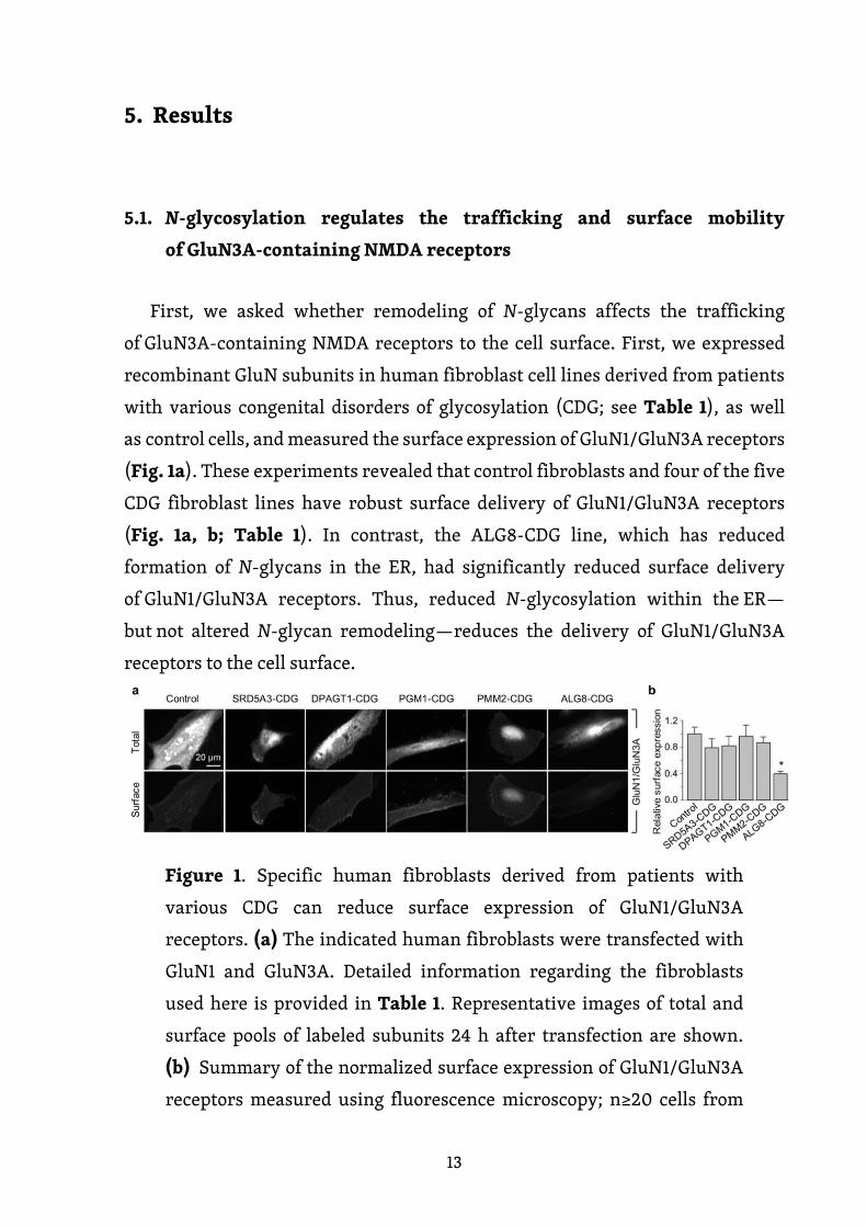
13
5. Results
5.1. N-glycosylation regulates the trafficking and surface mobility
of GluN3A-containing NMDA receptors
First, we asked whether remodeling of N-glycans affects the trafficking
of GluN3A-containing NMDA receptors to the cell surface. First, we expressed
recombinant GluN subunits in human fibroblast cell lines derived from patients
with various congenital disorders of glycosylation (CDG; see Table 1), as well
as control cells, and measured the surface expression of GluN1/GluN3A receptors
(Fig. 1a). These experiments revealed that control fibroblasts and four of the five
CDG fibroblast lines have robust surface delivery of GluN1/GluN3A receptors
(Fig. 1a, b; Table 1). In contrast, the ALG8-CDG line, which has reduced
formation of N-glycans in the ER, had significantly reduced surface delivery
of GluN1/GluN3A receptors. Thus, reduced N-glycosylation within the ER—
but not altered N-glycan remodeling—reduces the delivery of GluN1/GluN3A
receptors to the cell surface.
Figure 1. Specific human fibroblasts derived from patients with
various CDG can reduce surface expression of GluN1/GluN3A
receptors. (a) The indicated human fibroblasts were transfected with
GluN1 and GluN3A. Detailed information regarding the fibroblasts
used here is provided in Table 1. Representative images of total and
surface pools of labeled subunits 24 h after transfection are shown.
(b) Summary of the normalized surface expression of GluN1/GluN3A
receptors measured using fluorescence microscopy; n≥20 cells from

14
two independent experiments. *p<0.05 versus the GluN1/GluN3A
receptors (ANOVA).
Type Gene mutation(s) Protein mutations Reference
SRD5A3-CDG homozygous c.[436G>A] p.E146K Honzik et al, 2013
DPAGT1-CDG c.[85A>T];[652C>T] p.[I29F];[R218W] Honzik et al, 2013
PGM1-CDG c.[1010C>T];[1508G>A] p.[T337M];[R503Q] (Ondruskova et al., 2014)
PMM2-CDG c.[422G>A];[691G>A] p.[R141H];[ V231M] -
ALG8-CDG c.[139A>C];[1090C>T] p.[T47P];[ R364X] (Vesela et al., 2009)
Table 1. Details regarding the fibroblasts obtained from patients with
the indicated CDG syndromes. CDG, congenital disorders
of glycosylation; SRD5A3, steroid 5-alpha-reductase 3; DPAGT1,
dolichyl-phosphate alpha-N-acetylglucosaminyltransferase; PGM1,
phosphoglucomutase 1; PMM2, phosphomannomutase 2; ALG8, alpha-
1,3-glucosyltransferase.
Next, we asked whether inhibiting the N-glycosylation machinery affects
the surface delivery of GluN3A-containing NMDA receptors in hippocampal
neurons. We first treated cultured hippocampal neurons for 2 days in the
presence or absence of various inhibitors, including 0.5 µg/ml tunicamycin (an
inhibitor of dolichyl-phosphate N-acetylglucosamine phosphotransferase 1)
(Yavin et al., 1984), 200 µg/ml 1-deoxynojirimycin (DNJ, an inhibitor of α-
glucosidase I-II) (Gross et al., 1986), 5 µg/ml kifunensine (an inhibitor of the ER
α-mannosidase I) (Herreman et al., 2003), 200 µg/ml 1-deoxymannojirimycin
(DMM, an inhibitor of the ER α-mannosidase I-II) (Tokhtaeva et al., 2009), or 100
µg/ml swainsonine (an inhibitor of the GA α-mannosidase II) (Liu et al., 2000).
With the exception of DNJ, the other four inhibitors drastically reduced surface
labeling with fluorescently tagged PHA-L, which recognizes complex types of N-
glycans (Fig. 2a). In contrast, with the exception of tunicamycin, the other four
inhibitors increased surface labeling with fluorescently tagged conA, which
recognizes the high-mannose forms of N-glycans (Fig. 2a). These results indicate
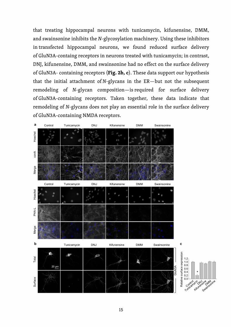
15
that treating hippocampal neurons with tunicamycin, kifunensine, DMM,
and swainsonine inhibits the N-glycosylation machinery. Using these inhibitors
in transfected hippocampal neurons, we found reduced surface delivery
of GluN3A-containg receptors in neurons treated with tunicamycin; in contrast,
DNJ, kifunensine, DMM, and swainsonine had no effect on the surface delivery
of GluN3A- containing receptors (Fig. 2b, c). These data support our hypothesis
that the initial attachment of N-glycans in the ER—but not the subsequent
remodeling of N-glycan composition—is required for surface delivery
of GluN3A-containing receptors. Taken together, these data indicate that
remodeling of N-glycans does not play an essential role in the surface delivery
of GluN3A-containing NMDA receptors.
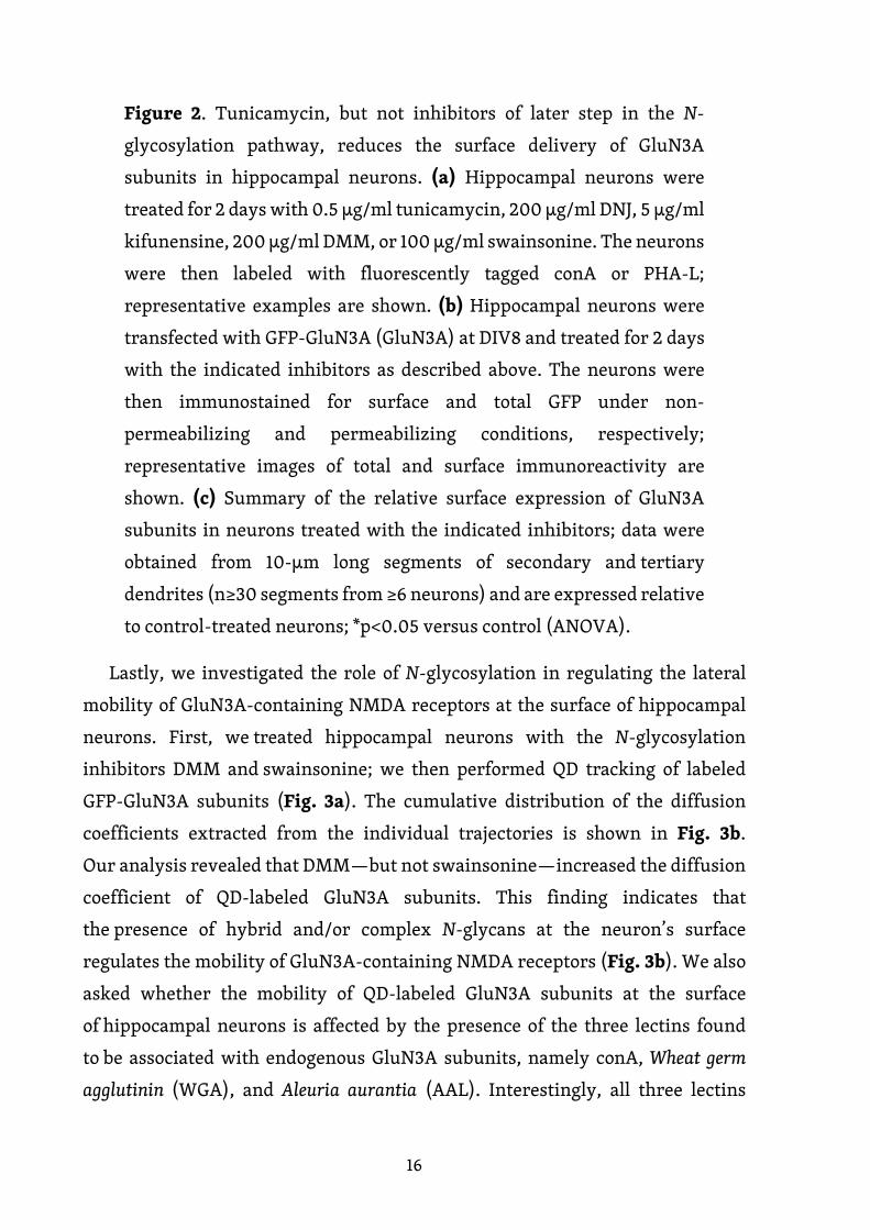
16
Figure 2. Tunicamycin, but not inhibitors of later step in the N-
glycosylation pathway, reduces the surface delivery of GluN3A
subunits in hippocampal neurons. (a) Hippocampal neurons were
treated for 2 days with 0.5 µg/ml tunicamycin, 200 µg/ml DNJ, 5 µg/ml
kifunensine, 200 µg/ml DMM, or 100 µg/ml swainsonine. The neurons
were then labeled with fluorescently tagged conA or PHA-L;
representative examples are shown. (b) Hippocampal neurons were
transfected with GFP-GluN3A (GluN3A) at DIV8 and treated for 2 days
with the indicated inhibitors as described above. The neurons were
then immunostained for surface and total GFP under non-
permeabilizing and permeabilizing conditions, respectively;
representative images of total and surface immunoreactivity are
shown. (c) Summary of the relative surface expression of GluN3A
subunits in neurons treated with the indicated inhibitors; data were
obtained from 10-µm long segments of secondary and tertiary
dendrites (n≥30 segments from ≥6 neurons) and are expressed relative
to control-treated neurons; *p<0.05 versus control (ANOVA).
Lastly, we investigated the role of N-glycosylation in regulating the lateral
mobility of GluN3A-containing NMDA receptors at the surface of hippocampal
neurons. First, we treated hippocampal neurons with the N-glycosylation
inhibitors DMM and swainsonine; we then performed QD tracking of labeled
GFP-GluN3A subunits (Fig. 3a). The cumulative distribution of the diffusion
coefficients extracted from the individual trajectories is shown in Fig. 3b.
Our analysis revealed that DMM—but not swainsonine—increased the diffusion
coefficient of QD-labeled GluN3A subunits. This finding indicates that
the presence of hybrid and/or complex N-glycans at the neuron’s surface
regulates the mobility of GluN3A-containing NMDA receptors (Fig. 3b). We also
asked whether the mobility of QD-labeled GluN3A subunits at the surface
of hippocampal neurons is affected by the presence of the three lectins found
to be associated with endogenous GluN3A subunits, namely conA, Wheat germ
agglutinin (WGA), and Aleuria aurantia (AAL). Interestingly, all three lectins
17
strongly reduced the mobility of QD-labeled GluN3A subunits in hippocampal
neurons (Fig. 3c, d). Given that a wide variety of lectins are expressed
in the mammalian CNS these data indicate that N-glycosylation plays a role
in regulating the mobility of GluN3A-containing NMDA receptors at the surface
of mammalian neurons.
Figure 3. N-glycosylation regulates the mobility of GluN3A-containing
NMDA receptors at the cell surface of hippocampal neurons.
(a,c) Representative trajectories of QD-labeled GFP-GluN3A (GluN3A)
subunits in hippocampal neurons treated with 200 µg/ml DMM (a)
or with the lectin AAL (20 µg/ml) (c). (b,d) Comparison of the
cumulative distribution of the diffusion coefficients measured for QD-
labeled GluN3A subunits in hippocampal neurons treated for two days
with 200 µg/ml DMM, 100 µg/ml swainsonine (b), or incubated after
QD labeling with 20 µg/ml of AAL, conA, or WGA (n=201-290
trajectories per condition). The negative controls for the fixed cells
shown in d were obtained from an independent experiment (n=86
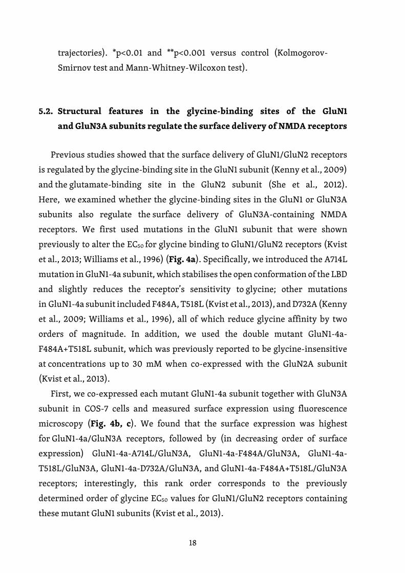
18
trajectories). *p<0.01 and **p<0.001 versus control (Kolmogorov-
Smirnov test and Mann-Whitney-Wilcoxon test).
5.2. Structural features in the glycine-binding sites of the GluN1
and GluN3A subunits regulate the surface delivery of NMDA receptors
Previous studies showed that the surface delivery of GluN1/GluN2 receptors
is regulated by the glycine-binding site in the GluN1 subunit (Kenny et al., 2009)
and the glutamate-binding site in the GluN2 subunit (She et al., 2012).
Here, we examined whether the glycine-binding sites in the GluN1 or GluN3A
subunits also regulate the surface delivery of GluN3A-containing NMDA
receptors. We first used mutations in the GluN1 subunit that were shown
previously to alter the EC50 for glycine binding to GluN1/GluN2 receptors (Kvist
et al., 2013; Williams et al., 1996) (Fig. 4a). Specifically, we introduced the A714L
mutation in GluN1-4a subunit, which stabilises the open conformation of the LBD
and slightly reduces the receptor’s sensitivity to glycine; other mutations
in GluN1-4a subunit included F484A, T518L (Kvist et al., 2013), and D732A (Kenny
et al., 2009; Williams et al., 1996), all of which reduce glycine affinity by two
orders of magnitude. In addition, we used the double mutant GluN1-4a-
F484A+T518L subunit, which was previously reported to be glycine-insensitive
at concentrations up to 30 mM when co-expressed with the GluN2A subunit
(Kvist et al., 2013).
First, we co-expressed each mutant GluN1-4a subunit together with GluN3A
subunit in COS-7 cells and measured surface expression using fluorescence
microscopy (Fig. 4b, c). We found that the surface expression was highest
for GluN1-4a/GluN3A receptors, followed by (in decreasing order of surface
expression) GluN1-4a-A714L/GluN3A, GluN1-4a-F484A/GluN3A, GluN1-4a-
T518L/GluN3A, GluN1-4a-D732A/GluN3A, and GluN1-4a-F484A+T518L/GluN3A
receptors; interestingly, this rank order corresponds to the previously
determined order of glycine EC50 values for GluN1/GluN2 receptors containing
these mutant GluN1 subunits (Kvist et al., 2013).
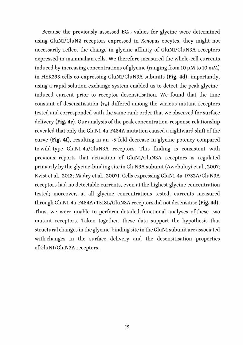
19
Because the previously assessed EC50 values for glycine were determined
using GluN1/GluN2 receptors expressed in Xenopus oocytes, they might not
necessarily reflect the change in glycine affinity of GluN1/GluN3A receptors
expressed in mammalian cells. We therefore measured the whole-cell currents
induced by increasing concentrations of glycine (ranging from 10 µM to 10 mM)
in HEK293 cells co-expressing GluN1/GluN3A subunits (Fig. 4d); importantly,
using a rapid solution exchange system enabled us to detect the peak glycine-
induced current prior to receptor desensitisation. We found that the time
constant of desensitisation (τw) differed among the various mutant receptors
tested and corresponded with the same rank order that we observed for surface
delivery (Fig. 4e). Our analysis of the peak concentration-response relationship
revealed that only the GluN1-4a-F484A mutation caused a rightward shift of the
curve (Fig. 4f), resulting in an ~5-fold decrease in glycine potency compared
to wild-type GluN1-4a/GluN3A receptors. This finding is consistent with
previous reports that activation of GluN1/GluN3A receptors is regulated
primarily by the glycine-binding site in GluN3A subunit (Awobuluyi et al., 2007;
Kvist et al., 2013; Madry et al., 2007). Cells expressing GluN1-4a-D732A/GluN3A
receptors had no detectable currents, even at the highest glycine concentration
tested; moreover, at all glycine concentrations tested, currents measured
through GluN1-4a-F484A+T518L/GluN3A receptors did not desensitise (Fig. 4d).
Thus, we were unable to perform detailed functional analyses of these two
mutant receptors. Taken together, these data support the hypothesis that
structural changes in the glycine-binding site in the GluN1 subunit are associated
with changes in the surface delivery and the desensitisation properties
of GluN1/GluN3A receptors.

20
Figure 4. Mutations in the glycine-binding site in the GluN1 subunit
alter the surface expression and desensitisation kinetics
of GluN1/GluN3A receptors. (a) Schematic representation
of the glycine-binding site in the GluN1 subunit (PDB code: 1PB7);
the amino acid residues studied here, and a glycine molecule,

21
are shown. (b) Representative images of COS-7 cells transfected
with the indicated wild-type or mutant GluN1-4a subunits together
with GFP-GluN3A (GluN3A) subunit and labelled 24 h after
transfection. (c) Summary of the relative surface expression of
the indicated GluN subunits measured using fluorescence microscopy
(n≥24 cells per group); *p<0.05 vs. GluN1-4a/GluN3A (ANOVA). (d)
Representative whole-cell patch-clamp recordings from HEK293 cells
transfected with the indicated wild-type or mutant GluN1-4a/GluN3A
receptors. Currents were elicited by applying glycine at the indicated
concentrations. (e) Summary of the τw of desensitisation for
the indicated GluN1-4a/GluN3A receptors (n≥5 cells per group). (f)
Peak concentration-response curves for the indicated GluN1-
4a/GluN3A receptors. Each data point represents the mean relative
current from ≥5 independent cells.
Given that the glycine-binding sites in the LBD of GluN1 and GluN3A subunits
are highly conserved, we next asked whether similar structural changes in
the glycine-binding site in the GluN3A subunit can also affect the surface
delivery of GluN1/GluN3A receptors. Accordingly, we introduced point
mutations in GluN3A subunit at the positions analogous to GluN1 subunit,
yielding four mutant GluN3 subunits (GluN3A-T825L, GluN3A-Y605A, GluN3A-
S633L, and GluN3A-D845A) and the double mutant GluN3A-Y605A+S633L
subunit (Fig. 5a); each subunit was then co-expressed together with GluN1-4a
subunit in COS-7 cells and surface expression was examined using microscopy
(Fig. 5b, c). Interestingly, the rank order of surface expression for the receptors
containing mutant GluN3A subunits was similar to the order that we observed
with the analogous mutant GluN1 subunits. Specifically, receptors containing
the GluN3A-T825L subunit had the highest surface expression, followed by
(in decreasing order) GluN3A-Y605A, GluN3A-S633L, GluN3A-D845A, and
GluN3A-Y605A+S633L subunits.
Next, we measured the functional properties of GluN1-4a/GluN3A receptors
containing wild-type or mutant GluN3A subunits expressed in HEK293 cells
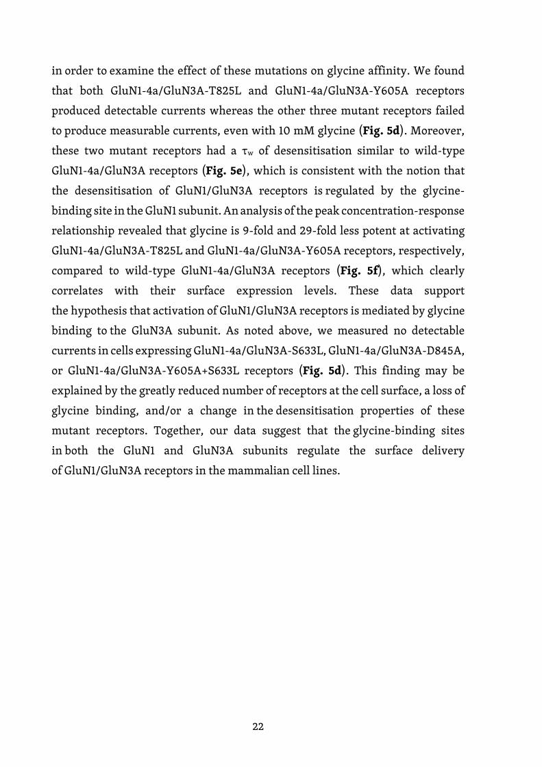
22
in order to examine the effect of these mutations on glycine affinity. We found
that both GluN1-4a/GluN3A-T825L and GluN1-4a/GluN3A-Y605A receptors
produced detectable currents whereas the other three mutant receptors failed
to produce measurable currents, even with 10 mM glycine (Fig. 5d). Moreover,
these two mutant receptors had a τw of desensitisation similar to wild-type
GluN1-4a/GluN3A receptors (Fig. 5e), which is consistent with the notion that
the desensitisation of GluN1/GluN3A receptors is regulated by the glycine-
binding site in the GluN1 subunit. An analysis of the peak concentration-response
relationship revealed that glycine is 9-fold and 29-fold less potent at activating
GluN1-4a/GluN3A-T825L and GluN1-4a/GluN3A-Y605A receptors, respectively,
compared to wild-type GluN1-4a/GluN3A receptors (Fig. 5f), which clearly
correlates with their surface expression levels. These data support
the hypothesis that activation of GluN1/GluN3A receptors is mediated by glycine
binding to the GluN3A subunit. As noted above, we measured no detectable
currents in cells expressing GluN1-4a/GluN3A-S633L, GluN1-4a/GluN3A-D845A,
or GluN1-4a/GluN3A-Y605A+S633L receptors (Fig. 5d). This finding may be
explained by the greatly reduced number of receptors at the cell surface, a loss of
glycine binding, and/or a change in the desensitisation properties of these
mutant receptors. Together, our data suggest that the glycine-binding sites
in both the GluN1 and GluN3A subunits regulate the surface delivery
of GluN1/GluN3A receptors in the mammalian cell lines.
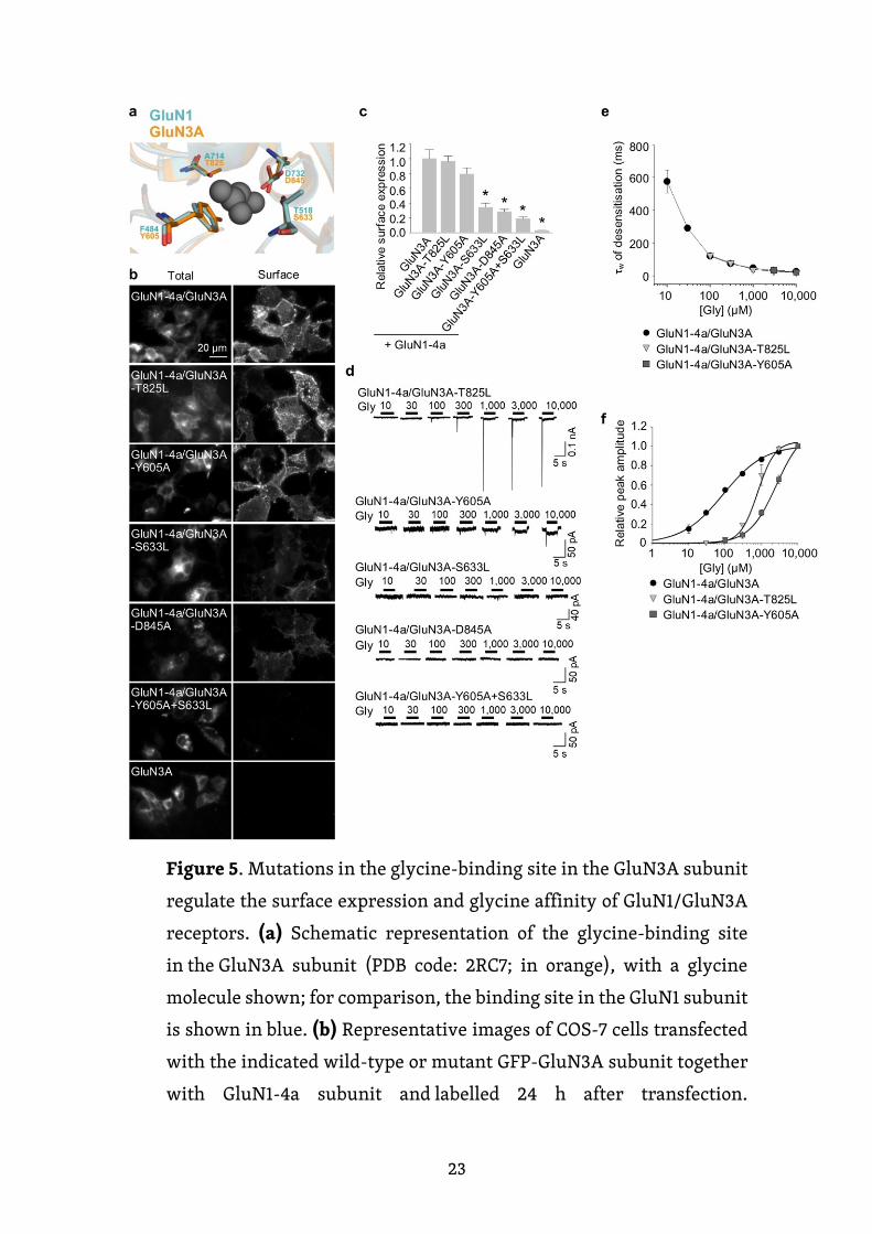
23
Figure 5. Mutations in the glycine-binding site in the GluN3A subunit
regulate the surface expression and glycine affinity of GluN1/GluN3A
receptors. (a) Schematic representation of the glycine-binding site
in the GluN3A subunit (PDB code: 2RC7; in orange), with a glycine
molecule shown; for comparison, the binding site in the GluN1 subunit
is shown in blue. (b) Representative images of COS-7 cells transfected
with the indicated wild-type or mutant GFP-GluN3A subunit together
with GluN1-4a subunit and labelled 24 h after transfection.
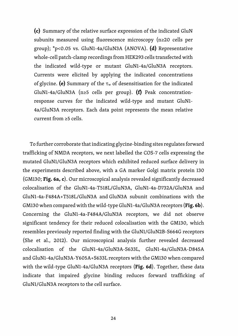
24
(c) Summary of the relative surface expression of the indicated GluN
subunits measured using fluorescence microscopy (n≥20 cells per
group); *p<0.05 vs. GluN1-4a/GluN3A (ANOVA). (d) Representative
whole-cell patch-clamp recordings from HEK293 cells transfected with
the indicated wild-type or mutant GluN1-4a/GluN3A receptors.
Currents were elicited by applying the indicated concentrations
of glycine. (e) Summary of the τw of desensitisation for the indicated
GluN1-4a/GluN3A (n≥5 cells per group). (f) Peak concentration-
response curves for the indicated wild-type and mutant GluN1-
4a/GluN3A receptors. Each data point represents the mean relative
current from ≥5 cells.
To further corroborate that indicating glycine-binding sites regulates forward
trafficking of NMDA receptors, we next labelled the COS-7 cells expressing the
mutated GluN1/GluN3A receptors which exhibited reduced surface delivery in
the experiments described above, with a GA marker Golgi matrix protein 130
(GM130; Fig. 6a, c). Our microscopical analysis revealed significantly decreased
colocalisation of the GluN1-4a-T518L/GluN3A, GluN1-4a-D732A/GluN3A and
GluN1-4a-F484A+T518L/GluN3A and GluN3A subunit combinations with the
GM130 when compared with the wild-type GluN1-4a/GluN3A receptors (Fig. 6b).
Concerning the GluN1-4a-F484A/GluN3A receptors, we did not observe
significant tendency for their reduced colocalisation with the GM130, which
resembles previously reported finding with the GluN1/GluN2B-S664G receptors
(She et al., 2012). Our microscopical analysis further revealed decreased
colocalisation of the GluN1-4a/GluN3A-S633L, GluN1-4a/GluN3A-D845A
and GluN1-4a/GluN3A-Y605A+S633L receptors with the GM130 when compared
with the wild-type GluN1-4a/GluN3A receptors (Fig. 6d). Together, these data
indicate that impaired glycine binding reduces forward trafficking of
GluN1/GluN3A receptors to the cell surface.
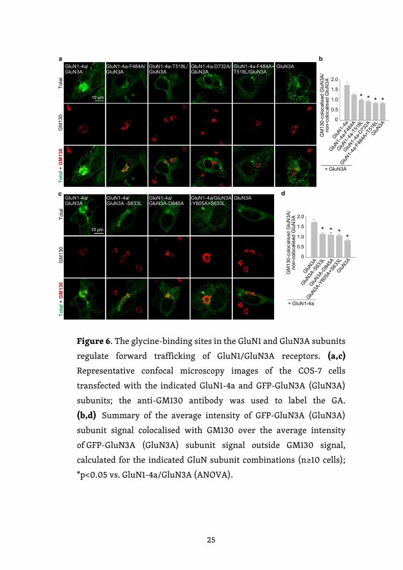
25
Figure 6. The glycine-binding sites in the GluN1 and GluN3A subunits
regulate forward trafficking of GluN1/GluN3A receptors. (a,c)
Representative confocal microscopy images of the COS-7 cells
transfected with the indicated GluN1-4a and GFP-GluN3A (GluN3A)
subunits; the anti-GM130 antibody was used to label the GA.
(b,d) Summary of the average intensity of GFP-GluN3A (GluN3A)
subunit signal colocalised with GM130 over the average intensity
of GFP-GluN3A (GluN3A) subunit signal outside GM130 signal,
calculated for the indicated GluN subunit combinations (n≥10 cells);
*p<0.05 vs. GluN1-4a/GluN3A (ANOVA).
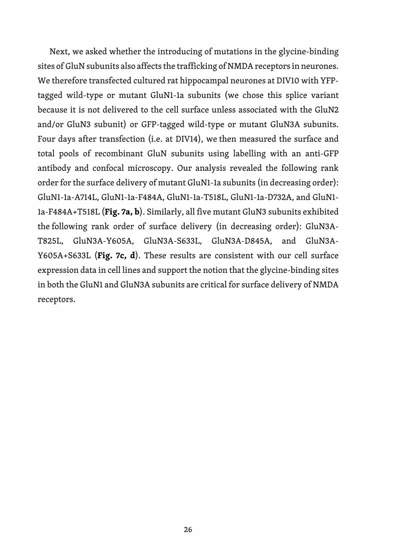
26
Next, we asked whether the introducing of mutations in the glycine-binding
sites of GluN subunits also affects the trafficking of NMDA receptors in neurones.
We therefore transfected cultured rat hippocampal neurones at DIV10 with YFP-
tagged wild-type or mutant GluN1-1a subunits (we chose this splice variant
because it is not delivered to the cell surface unless associated with the GluN2
and/or GluN3 subunit) or GFP-tagged wild-type or mutant GluN3A subunits.
Four days after transfection (i.e. at DIV14), we then measured the surface and
total pools of recombinant GluN subunits using labelling with an anti-GFP
antibody and confocal microscopy. Our analysis revealed the following rank
order for the surface delivery of mutant GluN1-1a subunits (in decreasing order):
GluN1-1a-A714L, GluN1-1a-F484A, GluN1-1a-T518L, GluN1-1a-D732A, and GluN1-
1a-F484A+T518L (Fig. 7a, b). Similarly, all five mutant GluN3 subunits exhibited
the following rank order of surface delivery (in decreasing order): GluN3A-
T825L, GluN3A-Y605A, GluN3A-S633L, GluN3A-D845A, and GluN3A-
Y605A+S633L (Fig. 7c, d). These results are consistent with our cell surface
expression data in cell lines and support the notion that the glycine-binding sites
in both the GluN1 and GluN3A subunits are critical for surface delivery of NMDA
receptors.
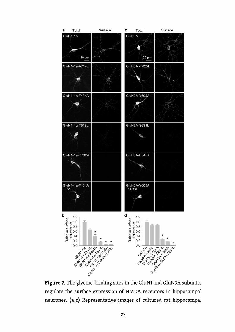
27
Figure 7. The glycine-binding sites in the GluN1 and GluN3A subunits
regulate the surface expression of NMDA receptors in hippocampal
neurones. (a,c) Representative images of cultured rat hippocampal
28
neurones transfected at DIV10 with the indicated YFP-GluN1-1a (a)
or GFP-GluN3A (c) subunits; at DIV14, surface and total subunits were
stained using an anti-GFP antibody. (b,d) Summary of the relative
surface expression of the indicated subunits measured in 10-µm
segments of secondary or tertiary dendrites (n≥30 segments in ≥6
different cells per group); *p<0.05 vs. GluN1-1a (b) or GluN3A (d)
(ANOVA).

29
6. Discussion
6.1. N-glycosylation regulates the trafficking and surface mobility
of GluN3A-containing NMDA receptors
We present compelling evidence that N-glycosylation of the GluN3A NMDA
receptor subunit is required for the receptor’s delivery to the cell surface.
The precise mechanism by which this process is mediated is currently unknown.
One possibility is that the presence of specific N-glycans confers a critical
structure to GluN3A-containing NMDA receptors, thereby permitting their
release from the ER (Vagin et al., 2009). Another possibility is that the GluN
subunits interact with specific glycan-binding proteins such as galactose-
binding lectins (Copits et al., 2014), which then facilitates the trafficking
of GluN3A-containing NMDA receptors to the cell surface. Other ionotropic
glutamatergic receptors, including α-amino-3-hydroxy-5-methyl-4-
isoxazolepropionic acid (AMPA) receptors and kainate receptors, also contain
a variety of N-glycan forms (Coleman et al., 2006; Greger et al., 2003; Hanus et
al., 2016; Mah et al., 2005; Standley et al., 1998); thus, it is likely that the N-glycan
composition of all ionotropic glutamate receptors regulates their trafficking
and function in vivo.
We studied human fibroblasts obtained from patients with deficits in N-
glycosylation machinery and found reduced surface delivery of GluN1/GluN3A
receptors only in cells in which the initial step in the N-glycosylation pathway
is impaired (i.e., ALG8-CDG). Taken together, our data suggest that
the trafficking machinery in heterologous expression systems and cultured
hippocampal neurons uses a common quality-control mechanism to regulate the
trafficking of GluN3A-containing receptors.
Our finding that N-glycan remodeling is not required for the surface delivery
of GluN3A-containing NMDA receptors is consistent with a recent study
by Hanus et al., who reached a similar conclusion with respect to various
neuronal membrane proteins (Hanus et al., 2016). In addition, we found that

30
the mobility of QD-labeled GluN3A subunits at the surface of hippocampal
neurons increases when hybrid and complex forms of N-glycans are removed.
These findings are consistent with the report that the mobility of AMPA receptors
can be regulated by enzymatic removal of the extracellular matrix (Frischknecht
et al., 2009). In contrast, the diffusion coefficient of GluN3A subunits was
reduced in the presence of lectins compared to control cells, corresponding to an
approximately 4-fold increase in the time required to diffuse across a given short
distance. Indeed, one possible interpretation of our data is that the lectins used
in our study may indirectly affect the mobility of GluN3A-containing NMDA
receptors by interacting with other glycoproteins at the neuronal cell surface.
Because a wide variety of lectins are expressed in the mammalian CNS, including
sialic acid–binding Ig-like lectins (Macauley et al., 2014) and galactose-binding
lectins (Cummings et al., 2017), these lectins could affect the mobility of NMDA
receptors in vivo. Other groups reported similar findings; for example,
the binding of conA restricts the mobility of surface receptors for this lectin in
mouse fibroblasts (Zagyansky and Edidin, 1976), and the protein galectin-3 alters
the lateral mobility and clustering of β1-integrin receptors in HeLa cells (Yang et
al., 2017).
The GluN3A subunit acts as a molecular brake, limiting the maturation
of excitatory synapses; this subunit has also been implicated in a number
of neuropsychiatric disorders (Fiuza et al., 2013; Kehoe et al., 2014; Kehoe et al.,
2013). Thus, N-glycosylation may play as-yet unrecognized roles in
the mammalian brain during health and/or disease. For example, N-
glycosylation of the GluN3A subunit may be essential for its
synaptic/perisynaptic localization and/or degradation (Chowdhury et al., 2013;
Pachernegg et al., 2012). With respect to the clinical relevance of our findings, it
is interesting to note that the glycosylation pattern on AMPA receptors is altered
in the cerebral cortex of schizophrenia patients (Tucholski et al., 2013); therefore,
it is reasonable to speculate that the N-glycosylation pattern of the GluN3A
subunit may be altered in certain neurological and/or psychiatric conditions.
Indeed, the altered function of the glycosylation machinery observed in patients
with a CDG syndrome could result in altered surface and/or synaptic delivery
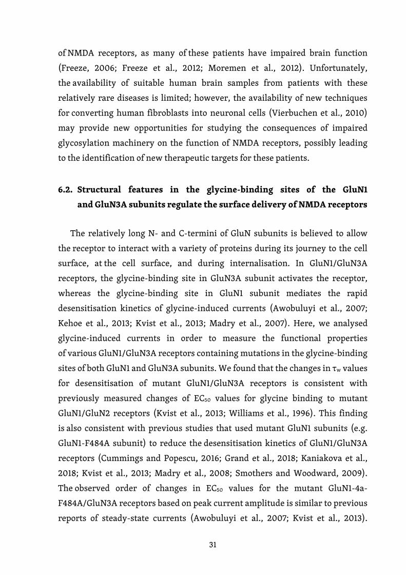
31
of NMDA receptors, as many of these patients have impaired brain function
(Freeze, 2006; Freeze et al., 2012; Moremen et al., 2012). Unfortunately,
the availability of suitable human brain samples from patients with these
relatively rare diseases is limited; however, the availability of new techniques
for converting human fibroblasts into neuronal cells (Vierbuchen et al., 2010)
may provide new opportunities for studying the consequences of impaired
glycosylation machinery on the function of NMDA receptors, possibly leading
to the identification of new therapeutic targets for these patients.
6.2. Structural features in the glycine-binding sites of the GluN1
and GluN3A subunits regulate the surface delivery of NMDA receptors
The relatively long N- and C-termini of GluN subunits is believed to allow
the receptor to interact with a variety of proteins during its journey to the cell
surface, at the cell surface, and during internalisation. In GluN1/GluN3A
receptors, the glycine-binding site in GluN3A subunit activates the receptor,
whereas the glycine-binding site in GluN1 subunit mediates the rapid
desensitisation kinetics of glycine-induced currents (Awobuluyi et al., 2007;
Kehoe et al., 2013; Kvist et al., 2013; Madry et al., 2007). Here, we analysed
glycine-induced currents in order to measure the functional properties
of various GluN1/GluN3A receptors containing mutations in the glycine-binding
sites of both GluN1 and GluN3A subunits. We found that the changes in τw values
for desensitisation of mutant GluN1/GluN3A receptors is consistent with
previously measured changes of EC50 values for glycine binding to mutant
GluN1/GluN2 receptors (Kvist et al., 2013; Williams et al., 1996). This finding
is also consistent with previous studies that used mutant GluN1 subunits (e.g.
GluN1-F484A subunit) to reduce the desensitisation kinetics of GluN1/GluN3A
receptors (Cummings and Popescu, 2016; Grand et al., 2018; Kaniakova et al.,
2018; Kvist et al., 2013; Madry et al., 2008; Smothers and Woodward, 2009).
The observed order of changes in EC50 values for the mutant GluN1-4a-
F484A/GluN3A receptors based on peak current amplitude is similar to previous
reports of steady-state currents (Awobuluyi et al., 2007; Kvist et al., 2013).
32
Moreover, an analysis of peak currents enabled us to estimate the EC50 value
for wild-type GluN1-4a/GluN3A receptors as well, which was previously not
possible due to the virtually negligible steady-state currents when these
receptors were expressed in Xenopus oocytes (Awobuluyi et al., 2007; Chatterton
et al., 2002; Kvist et al., 2013). However, we observed an apparent rightward shift
in the concentration-response curve for GluN1-4a/GluN3A receptors compared
to previously reported estimates of glycine EC50 values for steady-state currents
based on bell-shaped curves (Kvist et al., 2013) as well as to previously reported
Kd values from binding studies using soluble LBDs (Yao and Mayer, 2006);
this shift could be due to a variety of factors, including: i) decreased glycine
affinity of the peak current compared to the steady-state current, ii) the fast
desensitisation kinetics of GluN1/GluN3A receptors, and/or iii) the use
of different expression systems. Furthermore, we found that both
the GluN1/GluN3A-T825L and GluN1/GluN3A-Y605A receptors have similar
desensitisation kinetics but markedly decreased glycine affinity compared
to wild-type receptors. These findings support the conclusion that the glycine-
binding site in GluN3A subunit is required for activating the GluN1/GluN3A
receptors but does not contribute to the desensitisation of GluN1/GluN3A
receptors.
Our microscopy data showed that analogous mutations in the glycine-binding
sites of GluN1 and GluN3A subunits reduce the surface delivery of their
respective NMDA receptors when expressed in both mammalian cell lines and
hippocampal neurones. Given that endogenous GluN1, GluN2A-B, and GluN3
subunits are likely expressed in hippocampal neurones (Grand et al., 2018;
Hansen et al., 2017; Paoletti et al., 2013; Rozeboom et al., 2015; Sanz-Clemente et
al., 2013), our data support the view that the structural changes in the glycine-
binding sites in exogenous GluN1 and GluN3A subunits dominate the trafficking
of NMDA receptors that include endogenous GluN subunits (for example, in the
case of tri-heteromeric GluN1/GluN2/GluN3A receptors). Although
the underlying mechanism by which mutations in the glycine-binding sites
alters surface delivery is currently unknown, it is likely that the quality control
machinery in the ER plays a role, as was shown previously for GluN1/GluN2
33
receptors as well as AMPA receptors (Kenny et al., 2009; Penn et al., 2008; She et
al., 2012). Glutamate may be present in the ER in the millimolar range (Berger et
al., 1977; Meeker et al., 1989) and may be important for detecting properly
assembled functional GluN1/GluN2 receptors and AMPA receptors via an ER-
specific quality control mechanism. The precise concentration of glycine in
the ER is unknown; however, given the role that glycine plays in various
metabolic pathways (Wang et al., 2013), it is likely present in sufficient amounts
to interact with newly assembled receptors. In theory, a neurone could regulate
the number of NMDA receptors at the cell surface by controlling the production
of agonists such as glycine and D-serine or competitive antagonists such
as kynurenic acid, which are found naturally in the mammalian CNS (Moroni et
al., 1988; Traynelis et al., 2010). In addition, certain pharmacological compounds
that act on the glycine-binding sites of GluN1 and/or GluN3A subunits may alter
the surface expression of NMDA receptors, thereby providing a possible
therapeutic strategy for certain pathological conditions. In this respect, it is
important to note that treating cells with up to 1 mM glycine for 48 hours did not
affect the surface expression of either wild-type or mutant GluN1/GluN3A
receptors. This suggests that: i) intracellular glycine is already present at
saturating concentrations in cultured cells, ii) high levels of extracellular glycine
do not sufficiently alter the concentration of glycine within the ER, and/or iii)
high levels of glycine do not regulate the intracellular processing of
GluN1/GluN3A receptors.
Taken together, our findings regarding the role of glycine-binding sites
in GluN1 and GluN3A subunits in the surface delivery of NMDA receptors provide
new previously overlooked insight into the physiological and pathophysiological
regulation of these receptors in the mammalian CNS.
34
7. Conclusions
The glutamate release from one neuron and its detection by glutamate
receptors on the adjacent neuron form the synaptic transmission on more than
1014 synapses in the human brain. The specificity of this signalization is also
thanks to the neuron-specific expression of various NMDA receptor subunits and
their alternative splicing, which may vary in time and location. Since this
signaling is essential for synaptic plasticity and neuronal development, abnormal
transport, inappropriate localization or inadequate activation of these receptors
can lead to a number of pathologies and dysfunctions. Therefore, understanding
the basic mechanisms of regulation of trafficking and function of NMDA
receptors is essential to elucidate these dysfunctions and to develop future
therapies.
In this thesis, we have shown that insufficient N-glycosylation of NMDA
receptors alters the trafficking and functional properties of NMDA receptors.
We found that the subunits of NMDA receptors have a large variety of attached
N-glycans and that the subunits differ in their composition. It was interesting
to note that fibroblasts derived from patients with CDG may also alter in the
surface expression of NMDA receptors. At the same time, we observed that
changes in N-glycan remodeling or lectin binding may affect the lateral mobility
of NMDA receptors, which may lead to changes in the localization and function
of NMDA receptors. We further showed that glycine-binding sites on the GluN1
and GluN3A subunits have different functional roles at NMDA receptors and that
their surface expression correlates with affinity for glycine on the GluN1 subunit.
Thus, it is clear that as the specific role of N-glycosylation on the neuronal
function of NMDA receptors as the changes in NMDA receptor activation deserve
further attention as they may play a previously unknown role in the physiology
or pathophysiology of the mammalian central nervous system.
35
8. References
Atlason, P.T., Garside, M.L., Meddows, E., Whiting, P., and McIlhinney, R.A. (2007). N-Methyl-D-aspartate (NMDA) receptor subunit NR1 forms the substrate for oligomeric assembly of the NMDA receptor. J Biol Chem 282, 25299-25307.
Awobuluyi, M., Yang, J., Ye, Y., Chatterton, J.E., Godzik, A., Lipton, S.A., and Zhang, D. (2007). Subunit-specific roles of glycine-binding domains in activation of NR1/NR3 N-methyl-D-aspartate receptors. Mol Pharmacol 71, 112-122.
Berger, S.J., Carter, J.C., and Lowry, O.H. (1977). The distribution of glycine, GABA, glutamate and aspartate in rabbit spinal cord, cerebellum and hippocampus. J Neurochem 28, 149-158.
Chatterton, J.E., Awobuluyi, M., Premkumar, L.S., Takahashi, H., Talantova, M., Shin, Y., Cui, J., Tu, S., Sevarino, K.A., Nakanishi, N., et al. (2002). Excitatory glycine receptors containing the NR3 family of NMDA receptor subunits. Nature 415, 793-798.
Chazot, P.L., Cik, M., and Stephenson, F.A. (1995). An investigation into the role of N-glycosylation in the functional expression of a recombinant heteromeric NMDA receptor. Mol Membr Biol 12, 331-337.
Chazot, P.L., and Stephenson, F.A. (1997). Biochemical evidence for the existence of a pool of unassembled C2 exon-containing NR1 subunits of the mammalian forebrain NMDA receptor. J Neurochem 68, 507-516.
Chen, J., Liu, Q., Fan, R., Han, H., Yang, Z., Cui, W., Song, G., and Li, M.D. (2019). Demonstration of critical role of GRIN3A in nicotine dependence through both genetic association and molecular functional studies. Addict Biol.
Chen, W., Shieh, C., Swanger, S.A., Tankovic, A., Au, M., McGuire, M., Tagliati, M., Graham, J.M., Madan-Khetarpal, S., Traynelis, S.F., et al. (2017). GRIN1 mutation associated with intellectual disability alters NMDA receptor trafficking and function. J Hum Genet.
Chowdhury, D., Marco, S., Brooks, I.M., Zandueta, A., Rao, Y., Haucke, V., Wesseling, J.F., Tavalin, S.J., and Perez-Otano, I. (2013). Tyrosine phosphorylation regulates the endocytosis and surface expression of GluN3A-containing NMDA receptors. J Neurosci 33, 4151-4164.
Clements, J.D., and Westbrook, G.L. (1991). Activation kinetics reveal the number of glutamate and glycine binding sites on the N-methyl-D-aspartate receptor. Neuron 7, 605-613.
Coleman, S.K., Moykkynen, T., Cai, C.L., von Ossowski, L., Kuismanen, E., Korpi, E.R., and Keinanen, K. (2006). Isoform-specific early trafficking of AMPA receptor flip and flop variants. Journal of Neuroscience 26, 11220-11229.
Cummings, K.A., and Popescu, G.K. (2016). Protons Potentiate GluN1/GluN3A Currents by Attenuating Their Desensitisation. Sci Rep 6, 23344.
Dupuis, J.P., and Groc, L. (2019). Surface trafficking of neurotransmitter receptors: From cultured neurons to intact brain preparations. Neuropharmacology.
Everts, I., Villmann, C., and Hollmann, M. (1997). N-Glycosylation is not a prerequisite for glutamate receptor function but Is essential for lectin modulation. Mol Pharmacol 52, 861-873.
Farina, A.N., Blain, K.Y., Maruo, T., Kwiatkowski, W., Choe, S., and Nakagawa, T. (2011). Separation of domain contacts is required for heterotetrameric assembly of functional NMDA receptors. J Neurosci 31, 3565-3579.

36
Ferreira, J.S., Papouin, T., Ladepeche, L., Yao, A., Langlais, V.C., Bouchet, D., Dulong, J., Mothet, J.P., Sacchi, S., Pollegioni, L., et al. (2017). Co-agonists differentially tune GluN2B-NMDA receptor trafficking at hippocampal synapses. Elife 6.
Fiuza, M., Gonzalez-Gonzalez, I., and Perez-Otano, I. (2013). GluN3A expression restricts spine maturation via inhibition of GIT1/Rac1 signaling. Proc Natl Acad Sci U S A 110, 20807-20812.
Freeze, H.H. (2006). Genetic defects in the human glycome. Nat Rev Genet 7, 537-551. Freeze, H.H., Eklund, E.A., Ng, B.G., and Patterson, M.C. (2012). Neurology of inherited
glycosylation disorders. Lancet Neurol 11, 453-466. Frischknecht, R., Heine, M., Perrais, D., Seidenbecher, C.I., Choquet, D., and Gundelfinger, E.D.
(2009). Brain extracellular matrix affects AMPA receptor lateral mobility and short-term synaptic plasticity. Nat Neurosci 12, 897-904.
Grand, T., Abi Gerges, S., David, M., Diana, M.A., and Paoletti, P. (2018). Unmasking GluN1/GluN3A excitatory glycine NMDA receptors. Nat Commun 9, 4769.
Greger, I.H., Khatri, L., Kong, X., and Ziff, E.B. (2003). AMPA receptor tetramerization is mediated by Q/R editing. Neuron 40, 763-774.
Groc, L., Heine, M., Cognet, L., Brickley, K., Stephenson, F.A., Lounis, B., and Choquet, D. (2004). Differential activity-dependent regulation of the lateral mobilities of AMPA and NMDA receptors. Nat Neurosci 7, 695-696.
Gross, V., Tran-Thi, T.A., Schwarz, R.T., Elbein, A.D., Decker, K., and Heinrich, P.C. (1986). Different effects of the glucosidase inhibitors 1-deoxynojirimycin, N-methyl-1-deoxynojirimycin and castanospermine on the glycosylation of rat alpha 1-proteinase inhibitor and alpha 1-acid glycoprotein. Biochem J 236, 853-860.
Hansen, K.B., Yi, F., Perszyk, R.E., Menniti, F.S., and Traynelis, S.F. (2017). NMDA Receptors in the Central Nervous System. Methods Mol Biol 1677, 1-80.
Hanus, C., Geptin, H., Tushev, G., Garg, S., Alvarez-Castelao, B., Sambandan, S., Kochen, L., Hafner, A.S., Langer, J.D., and Schuman, E.M. (2016). Unconventional secretory processing diversifies neuronal ion channel properties. Elife 5.
Hawkins, L.M., Prybylowski, K., Chang, K., Moussan, C., Stephenson, F.A., and Wenthold, R.J. (2004). Export from the endoplasmic reticulum of assembled N-methyl-d-aspartic acid receptors is controlled by a motif in the c terminus of the NR2 subunit. J Biol Chem 279, 28903-28910.
Herreman, A., Van Gassen, G., Bentahir, M., Nyabi, O., Craessaerts, K., Mueller, U., Annaert, W., and De Strooper, B. (2003). gamma-Secretase activity requires the presenilin-dependent trafficking of nicastrin through the Golgi apparatus but not its complex glycosylation. J Cell Sci 116, 1127-1136.
Horak, M., Petralia, R.S., Kaniakova, M., and Sans, N. (2014). ER to synapse trafficking of NMDA receptors. Front Cell Neurosci 8, 394.
Horak, M., and Wenthold, R.J. (2009). Different roles of C-terminal cassettes in the trafficking of full-length NR1 subunits to the cell surface. J Biol Chem 284, 9683-9691.
Huh, K.H., and Wenthold, R.J. (1999). Turnover analysis of glutamate receptors identifies a rapidly degraded pool of the N-methyl-D-aspartate receptor subunit, NR1, in cultured cerebellar granule cells. J Biol Chem 274, 151-157.
Jeyifous, O., Waites, C.L., Specht, C.G., Fujisawa, S., Schubert, M., Lin, E.I., Marshall, J., Aoki, C., de Silva, T., Montgomery, J.M., et al. (2009). SAP97 and CASK mediate sorting of NMDA receptors through a previously unknown secretory pathway. Nat Neurosci 12, 1011-1019.
Kaniakova, M., Kleteckova, L., Lichnerova, K., Holubova, K., Skrenkova, K., Korinek, M., Krusek, J., Smejkalova, T., Korabecny, J., Vales, K., et al. (2018). 7-Methoxyderivative of tacrine is a 'foot-in-the-door' open-channel blocker of GluN1/GluN2 and GluN1/GluN3 NMDA receptors with neuroprotective activity in vivo. Neuropharmacology 140, 217-232.
37
Kaniakova, M., Krausova, B., Vyklicky, V., Korinek, M., Lichnerova, K., Vyklicky, L., and Horak, M. (2012a). Key amino acid residues within the third membrane domains of NR1 and NR2 subunits contribute to the regulation of the surface delivery of N-methyl-D-aspartate receptors. J Biol Chem 287, 26423-26434.
Kaniakova, M., Lichnerova, K., Skrenkova, K., Vyklicky, L., and Horak, M. (2016). Biochemical and electrophysiological characterization of N-glycans on NMDA receptor subunits. J Neurochem 138, 546-556.
Kaniakova, M., Lichnerova, K., Vyklicky, L., and Horak, M. (2012b). Single amino acid residue in the M4 domain of GluN1 subunit regulates the surface delivery of NMDA receptors. J Neurochem 123, 385-395.
Kato, A., Rouach, N., Nicoll, R.A., and Bredt, D.S. (2005). Activity-dependent NMDA receptor degradation mediated by retrotranslocation and ubiquitination. Proc Natl Acad Sci U S A 102, 5600-5605.
Kehoe, L.A., Bellone, C., De Roo, M., Zandueta, A., Dey, P.N., Perez-Otano, I., and Muller, D. (2014). GluN3A promotes dendritic spine pruning and destabilization during postnatal development. J Neurosci 34, 9213-9221.
Kehoe, L.A., Bernardinelli, Y., and Muller, D. (2013). GluN3A: an NMDA receptor subunit with exquisite properties and functions. Neural Plast 2013, 145387.
Kenny, A.V., Cousins, S.L., Pinho, L., and Stephenson, F.A. (2009). The integrity of the glycine co-agonist binding site of N-methyl-D-aspartate receptors is a functional quality control checkpoint for cell surface delivery. J Biol Chem 284, 324-333.
Kleckner, N.W., and Dingledine, R. (1988). Requirement for glycine in activation of NMDA-receptors expressed in Xenopus oocytes. Science 241, 835-837.
Kusumi, A., Sako, Y., and Yamamoto, M. (1993). Confined lateral diffusion of membrane receptors as studied by single particle tracking (nanovid microscopy). Effects of calcium-induced differentiation in cultured epithelial cells. Biophys J 65, 2021-2040.
Kvist, T., Greenwood, J.R., Hansen, K.B., Traynelis, S.F., and Brauner-Osborne, H. (2013). Structure-based discovery of antagonists for GluN3-containing N-methyl-D-aspartate receptors. Neuropharmacology 75, 324-336.
Lavezzari, G., McCallum, J., Dewey, C.M., and Roche, K.W. (2004). Subunit-specific regulation of NMDA receptor endocytosis. J Neurosci 24, 6383-6391.
Lemke, J.R., Geider, K., Helbig, K.L., Heyne, H.O., Schutz, H., Hentschel, J., Courage, C., Depienne, C., Nava, C., Heron, D., et al. (2016). Delineating the GRIN1 phenotypic spectrum: A distinct genetic NMDA receptor encephalopathy. Neurology 86, 2171-2178.
Lichnerova, K., Kaniakova, M., Park, S.P., Skrenkova, K., Wang, Y.X., Petralia, R.S., Suh, Y.H., and Horak, M. (2015). Two N-glycosylation Sites in the GluN1 Subunit Are Essential for Releasing N-methyl-d-aspartate (NMDA) Receptors from the Endoplasmic Reticulum. J Biol Chem 290, 18379-18390.
Lichnerova, K., Kaniakova, M., Skrenkova, K., Vyklicky, L., and Horak, M. (2014). Distinct regions within the GluN2C subunit regulate the surface delivery of NMDA receptors. Front Cell Neurosci 8, 375.
Liu, C., Rozmyslowicz, T., Stwora-Wojczyk, M., Wojczyk, B., and Spitalnik, S.L. (2000). Posttranslational modifications of the amyloid precursor protein : glycosylation. Methods Mol Med 32, 169-190.
Macauley, M.S., Crocker, P.R., and Paulson, J.C. (2014). Siglec-mediated regulation of immune cell function in disease. Nat Rev Immunol 14, 653-666.
Madry, C., Betz, H., Geiger, J.R., and Laube, B. (2008). Supralinear potentiation of NR1/NR3A excitatory glycine receptors by Zn2+ and NR1 antagonist. Proc Natl Acad Sci U S A 105, 12563-12568.
38
Madry, C., Mesic, I., Bartholomaus, I., Nicke, A., Betz, H., and Laube, B. (2007). Principal role of NR3 subunits in NR1/NR3 excitatory glycine receptor function. Biochem Biophys Res Commun 354, 102-108.
Mah, S.J., Cornell, E., Mitchell, N.A., and Fleck, M.W. (2005). Glutamate receptor trafficking: endoplasmic reticulum quality control involves ligand binding and receptor function. J Neurosci 25, 2215-2225.
Mahfooz, K., Marco, S., Martinez-Turrillas, R., Raja, M.K., Perez-Otano, I., and Wesseling, J.F. (2016). GluN3A promotes NMDA spiking by enhancing synaptic transmission in Huntington's disease models. Neurobiol Dis 93, 47-56.
Marco, S., Giralt, A., Petrovic, M.M., Pouladi, M.A., Martinez-Turrillas, R., Martinez-Hernandez, J., Kaltenbach, L.S., Torres-Peraza, J., Graham, R.K., Watanabe, M., et al. (2013). Suppressing aberrant GluN3A expression rescues synaptic and behavioral impairments in Huntington's disease models. Nat Med 19, 1030-1038.
Matsuda, K., Fletcher, M., Kamiya, Y., and Yuzaki, M. (2003). Specific assembly with the NMDA receptor 3B subunit controls surface expression and calcium permeability of NMDA receptors. J Neurosci 23, 10064-10073.
McIlhinney, R.A., Le Bourdelles, B., Molnar, E., Tricaud, N., Streit, P., and Whiting, P.J. (1998). Assembly intracellular targeting and cell surface expression of the human N-methyl-D-aspartate receptor subunits NR1a and NR2A in transfected cells. Neuropharmacology 37, 1355-1367.
Meddows, E., Le Bourdelles, B., Grimwood, S., Wafford, K., Sandhu, S., Whiting, P., and McIlhinney, R.A. (2001). Identification of molecular determinants that are important in the assembly of N-methyl-D-aspartate receptors. J Biol Chem 276, 18795-18803.
Meeker, R.B., Swanson, D.J., and Hayward, J.N. (1989). Light and electron microscopic localization of glutamate immunoreactivity in the supraoptic nucleus of the rat hypothalamus. Neuroscience 33, 157-167.
Mikasova, L., Xiong, H., Kerkhofs, A., Bouchet, D., Krugers, H.J., and Groc, L. (2017). Stress hormone rapidly tunes synaptic NMDA receptor through membrane dynamics and mineralocorticoid signalling. Sci Rep 7, 8053.
Moremen, K.W., Tiemeyer, M., and Nairn, A.V. (2012). Vertebrate protein glycosylation: diversity, synthesis and function. Nat Rev Mol Cell Biol 13, 448-462.
Moroni, F., Russi, P., Lombardi, G., Beni, M., and Carla, V. (1988). Presence of kynurenic acid in the mammalian brain. J Neurochem 51, 177-180.
Mueller, H.T., and Meador-Woodruff, J.H. (2004). NR3A NMDA receptor subunit mRNA expression in schizophrenia, depression and bipolar disorder. Schizophr Res 71, 361-370.
Okabe, S., Miwa, A., and Okado, H. (1999). Alternative splicing of the C-terminal domain regulates cell surface expression of the NMDA receptor NR1 subunit. J Neurosci 19, 7781-7792.
Ondruskova, N., Honzik, T., Vondrackova, A., Tesarova, M., Zeman, J., and Hansikova, H. (2014). Glycogen storage disease-like phenotype with central nervous system involvement in a PGM1-CDG patient. Neuro Endocrinol Lett 35, 137-141.
Pachernegg, S., Strutz-Seebohm, N., and Hollmann, M. (2012). GluN3 subunit-containing NMDA receptors: not just one-trick ponies. Trends Neurosci 35, 240-249.
Paoletti, P., Bellone, C., and Zhou, Q. (2013). NMDA receptor subunit diversity: impact on receptor properties, synaptic plasticity and disease. Nat Rev Neurosci 14, 383-400.
Patneau, D.K., and Mayer, M.L. (1990). Structure-activity relationships for amino acid transmitter candidates acting at N-methyl-D-aspartate and quisqualate receptors. J Neurosci 10, 2385-2399.
Penn, A.C., Williams, S.R., and Greger, I.H. (2008). Gating motions underlie AMPA receptor secretion from the endoplasmic reticulum. EMBO J 27, 3056-3068.
39
Perez-Otano, I., Larsen, R.S., and Wesseling, J.F. (2016). Emerging roles of GluN3-containing NMDA receptors in the CNS. Nat Rev Neurosci 17, 623-635.
Perez-Otano, I., Lujan, R., Tavalin, S.J., Plomann, M., Modregger, J., Liu, X.B., Jones, E.G., Heinemann, S.F., Lo, D.C., and Ehlers, M.D. (2006). Endocytosis and synaptic removal of NR3A-containing NMDA receptors by PACSIN1/syndapin1. Nat Neurosci 9, 611-621.
Perez-Otano, I., Schulteis, C.T., Contractor, A., Lipton, S.A., Trimmer, J.S., Sucher, N.J., and Heinemann, S.F. (2001). Assembly with the NR1 subunit is required for surface expression of NR3A-containing NMDA receptors. J Neurosci 21, 1228-1237.
Petralia, R.S., Al-Hallaq, R.A., and Wenthold, R.J. (2009). Trafficking and Targeting of NMDA Receptors.
Qiu, S., Zhang, X.M., Cao, J.Y., Yang, W., Yan, Y.G., Shan, L., Zheng, J., and Luo, J.H. (2009). An endoplasmic reticulum retention signal located in the extracellular amino-terminal domain of the NR2A subunit of N-Methyl-D-aspartate receptors. J Biol Chem 284, 20285-20298.
Roche, K.W., Standley, S., McCallum, J., Dune Ly, C., Ehlers, M.D., and Wenthold, R.J. (2001). Molecular determinants of NMDA receptor internalization. Nat Neurosci 4, 794-802.
Rozeboom, A.M., Queenan, B.N., Partridge, J.G., Farnham, C., Wu, J.Y., Vicini, S., and Pak, D.T. (2015). Evidence for glycinergic GluN1/GluN3 NMDA receptors in hippocampal metaplasticity. Neurobiol Learn Mem 125, 265-273.
Sanz-Clemente, A., Nicoll, R.A., and Roche, K.W. (2013). Diversity in NMDA receptor composition: many regulators, many consequences. Neuroscientist 19, 62-75.
Schuler, T., Mesic, I., Madry, C., Bartholomaus, I., and Laube, B. (2008). Formation of NR1/NR2 and NR1/NR3 heterodimers constitutes the initial step in N-methyl-D-aspartate receptor assembly. J Biol Chem 283, 37-46.
Scott, D.B., Michailidis, I., Mu, Y., Logothetis, D., and Ehlers, M.D. (2004). Endocytosis and degradative sorting of NMDA receptors by conserved membrane-proximal signals. J Neurosci 24, 7096-7109.
She, K., Ferreira, J.S., Carvalho, A.L., and Craig, A.M. (2012). Glutamate binding to the GluN2B subunit controls surface trafficking of N-methyl-D-aspartate (NMDA) receptors. J Biol Chem 287, 27432-27445.
Shen, Y.C., Liao, D.L., Chen, J.Y., Wang, Y.C., Lai, I.C., Liou, Y.J., Chen, Y.J., Luu, S.U., and Chen, C.H. (2009). Exomic sequencing of the ionotropic glutamate receptor N-methyl-D-aspartate 3A gene (GRIN3A) reveals no association with schizophrenia. Schizophr Res 114, 25-32.
Skrenkova, K., Lee, S., Lichnerova, K., Kaniakova, M., Hansikova, H., Zapotocky, M., Suh, Y.H., and Horak, M. (2018). N-Glycosylation Regulates the Trafficking and Surface Mobility of GluN3A-Containing NMDA Receptors. Front Mol Neurosci 11, 188.
Smothers, C.T., and Woodward, J.J. (2009). Expression of glycine-activated diheteromeric NR1/NR3 receptors in human embryonic kidney 293 cells Is NR1 splice variant-dependent. J Pharmacol Exp Ther 331, 975-984.
Standley, S., Roche, K.W., McCallum, J., Sans, N., and Wenthold, R.J. (2000). PDZ domain suppression of an ER retention signal in NMDA receptor NR1 splice variants. Neuron 28, 887-898.
Standley, S., Tocco, G., Wagle, N., and Baudry, M. (1998). High- and low-affinity alpha-[3H]amino-3-hydroxy-5-methylisoxazole-4-propionic acid ([3H]AMPA) binding sites represent immature and mature forms of AMPA receptors and are composed of differentially glycosylated subunits. J Neurochem 70, 2434-2445.
Swanger, S.A., Chen, W., Wells, G., Burger, P.B., Tankovic, A., Bhattacharya, S., Strong, K.L., Hu, C., Kusumoto, H., Zhang, J., et al. (2016). Mechanistic Insight into NMDA Receptor Dysregulation by Rare Variants in the GluN2A and GluN2B Agonist Binding Domains. Am J Hum Genet 99, 1261-1280.

40
Takata, A., Iwayama, Y., Fukuo, Y., Ikeda, M., Okochi, T., Maekawa, M., Toyota, T., Yamada, K., Hattori, E., Ohnishi, T., et al. (2013). A population-specific uncommon variant in GRIN3A associated with schizophrenia. Biol Psychiatry 73, 532-539.
Tokhtaeva, E., Sachs, G., and Vagin, O. (2009). Assembly with the Na,K-ATPase alpha(1) subunit is required for export of beta(1) and beta(2) subunits from the endoplasmic reticulum. Biochemistry 48, 11421-11431.
Traynelis, S.F., Wollmuth, L.P., McBain, C.J., Menniti, F.S., Vance, K.M., Ogden, K.K., Hansen, K.B., Yuan, H., Myers, S.J., and Dingledine, R. (2010). Glutamate receptor ion channels: structure, regulation, and function. Pharmacol Rev 62, 405-496.
Triller, A., and Choquet, D. (2008). New concepts in synaptic biology derived from single-molecule imaging. Neuron 59, 359-374.
Tucholski, J., Simmons, M.S., Pinner, A.L., Haroutunian, V., McCullumsmith, R.E., and Meador-Woodruff, J.H. (2013). Abnormal N-linked glycosylation of cortical AMPA receptor subunits in schizophrenia. Schizophr Res 146, 177-183.
Vagin, O., Kraut, J.A., and Sachs, G. (2009). Role of N-glycosylation in trafficking of apical membrane proteins in epithelia. Am J Physiol Renal Physiol 296, F459-469.
Vesela, K., Honzik, T., Hansikova, H., Haeuptle, M.A., Semberova, J., Stranak, Z., Hennet, T., and Zeman, J. (2009). A new case of ALG8 deficiency (CDG Ih). J Inherit Metab Dis 32 Suppl 1.
Vierbuchen, T., Ostermeier, A., Pang, Z.P., Kokubu, Y., Sudhof, T.C., and Wernig, M. (2010). Direct conversion of fibroblasts to functional neurons by defined factors. Nature 463, 1035-1041.
Wang, W., Wu, Z., Dai, Z., Yang, Y., Wang, J., and Wu, G. (2013). Glycine metabolism in animals and humans: implications for nutrition and health. Amino Acids 45, 463-477.
Washbourne, P., Bennett, J.E., and McAllister, A.K. (2002). Rapid recruitment of NMDA receptor transport packets to nascent synapses. Nat Neurosci 5, 751-759.
Washbourne, P., Liu, X.B., Jones, E.G., and McAllister, A.K. (2004). Cycling of NMDA receptors during trafficking in neurons before synapse formation. J Neurosci 24, 8253-8264.
Williams, K., Chao, J., Kashiwagi, K., Masuko, T., and Igarashi, K. (1996). Activation of N-methyl-D-aspartate receptors by glycine: role of an aspartate residue in the M3-M4 loop of the NR1 subunit. Mol Pharmacol 50, 701-708.
Yang, E.H., Rode, J., Howlader, M.A., Eckermann, M., Santos, J.T., Hernandez Armada, D., Zheng, R., Zou, C., and Cairo, C.W. (2017). Galectin-3 alters the lateral mobility and clustering of beta1-integrin receptors. PLoS One 12, e0184378.
Yao, Y., and Mayer, M.L. (2006). Characterization of a soluble ligand binding domain of the NMDA receptor regulatory subunit NR3A. J Neurosci 26, 4559-4566.
Yavin, E., Richter-Landsberg, C., Duksin, D., and Yavin, Z. (1984). Tunicamycin blocks neuritogenesis and glucosamine labeling of gangliosides in developing cerebral neuron cultures. Proc Natl Acad Sci U S A 81, 5638-5642.
Yuan, T., Mameli, M., O'Connor, E.C., Dey, P.N., Verpelli, C., Sala, C., Perez-Otano, I., Luscher, C., and Bellone, C. (2013). Expression of cocaine-evoked synaptic plasticity by GluN3A-containing NMDA receptors. Neuron 80, 1025-1038.
Zagyansky, Y., and Edidin, M. (1976). Lateral diffusion of concanavalin A receptors in the plasma membrane of mouse fibroblasts. Biochim Biophys Acta 433, 209-214.
41
9. List of publications
Publications in extenso, related to this thesis
5. Kristyna Skrenkova, Katarina Hemelikova, Marharyta Kolcheva, Stepan Kortus, Martina Kaniakova, Barbora Krausova , Martin Horak (2019) Structural features in the glycine-binding sites of the GluN1 and GluN3A subunits regulate the surface delivery of NMDA receptors. Scientific reports 9:12303. IF = 4.5 (2018)
6. Kristyna Skrenkova, Sanghyeon Lee, Katarina Lichnerova, Martina Kaniakova, Hana Hansikova, Martin Zapotocky, Young Ho Suh, Martin Horak (2018). N-Glycosylation Regulates the Trafficking and Surface Mobility of GluN3A-Containing NMDA Receptors. Frontiers in Molecular Neuroscience 11, 188. IF = 3.7 (2018)
7. Martina Kaniakova, Katarina Lichnerova, Kristyna Skrenkova, Ladislav Vyklicky, Martin Horak (2016). Biochemical and electrophysiological characterization of N-glycans on NMDA receptor subunits. Journal of Neurochemistry 546–556. IF = 4.1 (2016)
8. Lichnerova Katarina, Kaniakova Martina, Seung pyo Park, Kristyna Skrenkova, Ya-Xiang Wang, Ronald S. Petralia, Young Ho Suh, Martin Horak (2015). Two N-glycosylation Sites in the GluN1 Subunit Are Essential for Releasing N-methyl-d-aspartate (NMDA) Receptors from the Endoplasmic Reticulum. Journal of Biological Chemistry 290, 18379–18390. IF = 4.3 (2015)
42
Publications in extenso, unrelated to this thesis
1. Katarina Hemelikova (Lichnerova), Marharyta Kolcheva, Kristyna Skrenkova, Martina Kanikova, Martin Horak (2019). Lectins modulate the functional properties of GluN1/GluN3A – containg receptors. Neuropharmacology 157, 107671. IF = 4.4 (2018)
2. Martina Kaniakova, Eugenie Nepovimova, Lenka Kleteckova, Kristyna Skrenkova, Kristina Holubova, Zofia Chrienova, Vendula Hepnarova, Tomas Kucera, Tereza Kobrlova, Karel Vales, Jan Korabecny, Ondrej Soukup, Martin Horak. (2019) Combination of Memantine and 6-Chlorotacrine as Novel Multi-Target Compound against Alzheimer's Disease. Current Alzheimer Research 16:1.
IF= 3.2 (2018)
3. Martina Kaniakova, Lenka Kleteckova, Katarina Lichnerova, Kristina Holubova, Kristyna Skrenkova, Miloslav Korinek, Jan Krusek, Tereza Smejkalova, Jan Korabecny, Karel Vales, Ondrej Soukup, Martin Horak. (2018). 7-Methoxyderivative of tacrine is a 'foot-in-the-door' open-channel blocker of GluN1/GluN2 and GluN1/GluN3 NMDA receptors with neuroprotective activity in vivo. Neuropharmacology 15; 140:217-232.
IF= 4.3 (2018)
4. Vendula Sepsova, Jana Z. Karasova, Gunnar Tobin, Daniel Jun, Jan Korabecny, Pavla Cabelova, Katerina Janska, Jan Krusek, Kristyna Skrenkova, Kamil Kuca, Ondrej Soukup (2015). Cholinergic properties of new 7-methoxytacrine-donepezil derivatives. General Physiology and Biophysics, 34(2):189-200.
IF= 0.9 (2015)
5. Katarina Lichnerova, Martina Kaniakova, Kristyna Skrenkova, Ladislav Vyklicky, Martin Horak (2014). Distinct regions within the GluN2C subunit regulate the surface delivery of NMDA receptors. Frontiers in Cellular Neuroscience 8, 375.
IF= 4.3 (2014)
6. Martina Kaniakova, Kristyna Skrenkova, Svatopluk Adamek, Frantisek Vyskocil, Jan Krusek (2014). Different effects of lobeline on neuronal and muscle nicotinic receptors. European Journal of Pharmacology, 738:352-9.
IF= 2.5 (2014)

43
10. Curriculum Vitae
Kristýna Skřenková
Education
2014 - present Ph.D. program in Neuroscience – Animal Physiology, Charles University in Prague, Faculty of Science
Thesis: Molecular mechanisms of regulation of transport and function of different subtypes of NMDA receptors in hippocampal neurons
Supervisor: Martin Horák, Ph.D.
2012 - 2014 Master of Science in Animal Physiology Charles University in Prague, Faculty of Science Thesis: Inhibition of nicotinic acetylcholine receptors by tacrine and its derivatives Supervisor RNDr. Jan Krůšek, CSc.
2009 - 2012 Bachelor of Molecular biology and biochemistry of organisms Charles University in Prague, Faculty of Science Thesis: Ionotropic glutamate receptors and excitotoxicity
Supervisor: prof. Ladislav Vyklický, DrSc.
Research training
2018 – present Institute of Experimental medicine CAS
Department of Neurochemistry
2011 – present Institute of Physiology CAS
Department of Cellular Neurophysiology
Laboratory skills – a hands-on technique
- Cultivation and transfection of heterologous cell lines
- Preparation and cultivation of primary hippocampal cultures
- Preparation of hippocampal slices
- Transfection and infection of hippocampal neurons
- Electrophysiology – patch clamp techniques (whole-cell on heterologous cell lines)
- Molecular biology – site direction mutagenesis, transformation of bacterial cells, isolation of plasmid
DNA, cloning of DNA into a vector
- Immunofluorescence microscopy – surface and total labelling, colocalization and internalization essays.
- Live microscopy - surface mobility
- Biochemistry – SDP-PAGE

44
Completed courses
2019 - Applied improvisation (Charles University, Faculty of Science)
2018 - Advanced practical rhetoric and presentation (Charles University, Faculty of Science)
2018 - Processing and analysis of microscopic images in biomedicine (Institute of Molecular Genetics,
CAS)
2018 – Time managment (Charles University, Faculty of Science)
2018 - Scientific Writing (Charles University, Faculty of Science)
2017 - Scientific Presentations (Charles University, Faculty of Science)
2017 - Practical rhetoric and presentation (Charles University, Faculty of Science)
2015 - Course in the Fundamentals of Research Work Administered by CAS (CAS)
2014 - Microscopy methods in biomedicine (Institute of Molecular Genetics, CAS)