UNIVERSITÉ D'ORLÉANS - TEL Archives ouvertes
254
UNIVERSITÉ D’ORLÉANS ÉCOLE DOCTORALE ENERGIE, MATERIAUX, SCIENCES DE LA TERRE ET DE L’UNIVERS Institut des Sciences de la Terre d’Orléans THÈSE présentée par : Fengfeng ZHANG soutenue le 17 novembre 2020 pour obtenir le grade de : Docteur de l’Université d’Orléans Discipline/ Spécialité : Science de la Terre et de l’Univers Transformation of iron oxides in presence of mixed iron-reducing bacterial communities and dynamics of the associated trace elements As, Cr, and Cd THÈSE dirigée par : MOTELICA-HEINO Mikael Professeur, Université d’Orléans BATTAGLIA-BRUNET Fabienne Chercheur, BRGM Orléans RAPPORTEURS : DAVRANCHE Mélanie Professeur, Université de Rennes 1 JORAND Frédéric Professeur, Université de Lorraine JURY : GROSBOIS Cécile Président, Professeur, Université de Tours BYRNE James Maître de conférences, Université de Bristol MORIN Guillaume Professeur, Sorbonne Université GAUTRET Pascale CR CNRS, ISTO Orléans HELLAL Jennifer Chercheur, BRGM Orléans MOTELICA-HEINO Mikael Professeur, Université d’Orléans BATTAGLIA-BRUNET Fabienne Chercheur, BRGM Orléans
-
Upload
khangminh22 -
Category
Documents
-
view
0 -
download
0
Transcript of UNIVERSITÉ D'ORLÉANS - TEL Archives ouvertes

UNIVERSITÉ D’ORLÉANS
ÉCOLE DOCTORALE
ENERGIE, MATERIAUX, SCIENCES DE LA TERRE ET DE L’UNIVERS
Institut des Sciences de la Terre d’Orléans
THÈSE présentée par : Fengfeng ZHANG
soutenue le 17 novembre 2020
pour obtenir le grade de : Docteur de l’Université d’Orléans
Discipline/ Spécialité : Science de la Terre et de l’Univers
Transformation of iron oxides in presence of mixed
iron-reducing bacterial communities and dynamics of
the associated trace elements As, Cr, and Cd
THÈSE dirigée par : MOTELICA-HEINO Mikael Professeur, Université d’Orléans BATTAGLIA-BRUNET Fabienne Chercheur, BRGM Orléans
RAPPORTEURS : DAVRANCHE Mélanie Professeur, Université de Rennes 1 JORAND Frédéric Professeur, Université de Lorraine
JURY : GROSBOIS Cécile Président, Professeur, Université de Tours BYRNE James Maître de conférences, Université de Bristol MORIN Guillaume Professeur, Sorbonne Université GAUTRET Pascale CR CNRS, ISTO Orléans HELLAL Jennifer Chercheur, BRGM Orléans MOTELICA-HEINO Mikael Professeur, Université d’Orléans BATTAGLIA-BRUNET Fabienne Chercheur, BRGM Orléans

UNIVERSITÉ D’ORLÉANS
ÉCOLE DOCTORALE
ENERGIE, MATERIAUX, SCIENCES DE LA TERRE ET DE L’UNIVERS
Institut des Sciences de la Terre d’Orléans
THÈSE présentée par : Fengfeng ZHANG
soutenue le : 17 novembre 2020
pour obtenir le grade de : Docteur de l’Université d’Orléans
Discipline/ Spécialité : Science de la Terre et de l’Univers
Transformation of iron oxides in presence of mixed iron-reducing bacterial communities and dynamics of the associated trace elements As,
Cr, and Cd
THÈSE dirigée par :
MOTELICA-HEINO Mikael Pr, Université d’Orléans
BATTAGLIA-BRUNET Fabienne Chercheur, BRGM Orléans
Co-encadrée par :
GAUTRET Pascale CR CNRS, ISTO Orléans
HELLAL Jennifer Chercheur, BRGM Orléans

Acknowledgements
First of all, I really appreciate the attendance of the reviewers Pr. Frédéric Jorand, Pr. Mélanie
Davranche and external jury members Pr. Cécile Grosbois, Dr. James Byrne, Dr. Guillaume
Morin to my PhD defense. Thanks so much of their time and efforts, their suggestions and
comments are really helpful to improve this PhD work!
I would like to sincerely thank 4 of my supervisors, Fabienne who is a wonderful supervisor
and has directed this thesis very professionally with her knowledge and patience, Mikael who
is also a great supervisor and provided me full supports with his specialty and kindness, Jenny
who helps me a lot with her high scientific quality and strict efficiency, and Pascale who helped
me a lot in her field which was very good time. I’m really grateful that they provided me this
chance for a PhD journey and were with me all the time, to share the happiness and get me
through difficulties. I can't imagine how lucky I am to have them to be with me for this long
study period. During the past years, they provided me all the supports to everything in work
and in daily life, I appreciate their help more than they know.
I would like to thank BRGM and Center-Val de Loire Region for having co-funded this thesis
and I would like to thank all the colleagues from BRGM, e.g., Cathy who gave me a lot and
very specific help in molecular work and Mickael, Hafida, Cindy who were always with me in
the labs in BRGM. It’s my honor to work with them and all the other researchers, they might
be not involved into my PhD project but I cherish all the past time that staying with them.
I would also like to thank all the colleagues and friends from ISTO, it’s a great pleasure to know
other PhD students, postdocs and staff there. Many thanks to all the happiness and funny
moments they brought me.
I would like to give special thanks to my family and friends from China, they never been to
France yet but I know that they are always stand by me.
Finally, I won’t forget to thank to my great partner, Clément. It’s not easy to have two people
finishing their PhDs in the same moment, but we did it.
Life is a long journey and I believe that PhD experiences make us more critical, more open-
minded and stronger.
Contents
i
Thesis Fengfeng Zhang -2020
ISTO-BRGM
Contents Résumé étendu de la thèse en Français .................................................................................. 5
Chapter 0: General introduction and objectives ....................................................................... 13
Chapter I: Background knowledge ........................................................................................... 17
I-1 Biogeochemical cycle of Fe ............................................................................................ 17
I-1.1 Global Fe cycle ......................................................................................................... 17
I-1.2 Microbial transformations of Fe in the biogeosphere .............................................. 19
I-1.3 Influence of redox conditions and organic matter on iron reduction ....................... 21
I-2 Classification of Fe-minerals .......................................................................................... 24
I-3 Dissolution of Fe(III) (oxyhydr)oxides and iron cycling in surface environments ........ 29
I-3.1 Abiotic dissolution ................................................................................................... 29
I-3.2 Biotic dissolution ...................................................................................................... 30
I-3.3 Secondary minerals formed during the bio-reduction of Fe oxides ......................... 31
I-4 Iron-reducing bacteria (IRB) ........................................................................................... 32
I-4.1 Dissimilatory Iron-Reducing Bacteria (DIRB) ........................................................ 32
I-4.2 Iron reduction by fermentative bacteria ................................................................... 34
I-4.3 The genus Shewanella .............................................................................................. 36
I-4.3 The genus Geobacter................................................................................................ 39
I-4.4 Comparison of the genera Shewanella and Geobacter ............................................ 41
I-4.5 Primers for the detection and quantification of Shewanella and Geobacter ............ 43
I-4.6 Biofilms of iron reducing bacteria ........................................................................... 45
I-4.7 Studies involving complex iron reducing microbial communities ........................... 46
I-5 Mechanisms of microbial Fe(III) reduction .................................................................... 47
I-6 Cycling of Fe and mobility of associated As, Cr, Cd and other trace elements ............. 50
I-6.1 Association of As, Cr and Cd with Fe-Oxides ......................................................... 50
I.6.2 Transformations of As, Cr and Cd by bacteria ......................................................... 51
I-6.3 Mobility of trace elements during Fe-oxides microbial dissolution......................... 52
I-7 Positioning of the PhD thesis in regards to the state of the art ....................................... 54
Chapter II: General materials and methods .............................................................................. 57
II-1 Site information and soil / sediment sampling .............................................................. 57
II-2. Enrichment of iron-reducing bacteria (IRB) and subculture ........................................ 58
II-3 Iron (oxyhydr)oxides and laboratory synthesis ............................................................. 60
II-3.1 Ferrihydrite ............................................................................................................. 60
II-3.2 Lepidocrocite .......................................................................................................... 61
II-3.3 Goethite and hematite ............................................................................................. 61
Contents
ii
Thesis Fengfeng Zhang -2020
ISTO-BRGM
II-4 Physico-chemical analysis: pH, Eh, Fe(II)/FeT, As, Cr and Cd .................................... 61
II-5 BET surface areas .......................................................................................................... 62
II-6 SEM-EDS and SEM observations ................................................................................. 62
II-6.1 Observation of iron (oxyhydr)oxides ...................................................................... 62
II-6.2 Bacteria Observations ............................................................................................. 63
II-7 57Fe Mössbauer spectrometry ........................................................................................ 63
II-8 Diversity and physiology of bacteria ............................................................................. 63
II-8.1 Observation and counting of bacteria by Thoma cell ............................................. 63
II-8.2 DNA extraction and PCR amplifications ................................................................ 63
II-8.3 CE-SSCP fingerprints ............................................................................................. 64
II-8.4 Bacterial 16S rRNA gene quantification ................................................................ 65
II-8.5 Detection of Shewanella and Geobacter ................................................................. 65
II-8.6 Quantification of Shewanella and Geobacter by qPCR ......................................... 65
Chapter III: Experiments in slurry with four different iron oxides .......................................... 67
III-1 Introduction .................................................................................................................. 67
III-2 Specific Materials and Methods ................................................................................... 69
III-2.1 Characterization of the environmental source of bacteria ..................................... 69
Iron extraction in soils/sediments samples ....................................................................... 69
III-2.2 Synthetic Fe(III) (oxyhydr)oxides and bacterial inocula ....................................... 70
III-2.3 IRB incubation experiments .................................................................................. 71
III-2.4 Fe analyses and pH/Eh monitoring ........................................................................ 72
III-2.5 Determination of iron oxides solubilisation parameters ........................................ 72
III-2.6 SEM-EDS observation and Mössbauer spectrometry ........................................... 72
III-2.7 Biological analyses ................................................................................................ 72
III-2.8 Statistics ................................................................................................................. 73
III-3 Results .......................................................................................................................... 73
III-3.1 Characterization of the environmental sources of bacteria .................................... 73
III-3.2 Dissolution of Fe (oxyhydr)oxides ........................................................................ 74
III-3.3 Biological parameters ............................................................................................ 76
III-3.4 Mineral SEM-EDS observation ............................................................................. 80
III-3.5 Mössbauer spectroscopy ........................................................................................ 81
III-4 Discussion .................................................................................................................... 82
III-4.1 Influence of the type of iron oxide on bacterial iron solubilisation ....................... 82
III-4.2 Bacterial communities ........................................................................................... 83
III-4.3 Geobacter and Shewanella 16S genes abundances ............................................... 84
Contents
iii
Thesis Fengfeng Zhang -2020
ISTO-BRGM
III-4.4 Relation between iron solubilisation effectiveness and Geobacter and Shewanella
16S gene abundances ........................................................................................................ 86
III-4.5 SEM observations of Fe (oxyhydr)oxides and Mössbauer spectroscopy .............. 88
III-5 Conclusions and perspective ........................................................................................ 88
Supplementary Figures ..................................................................................................... 90
Supplementary Tables ....................................................................................................... 95
IV: Experiments with ferrihydrite fixed on slides .................................................................... 97
IV-1 Introduction .................................................................................................................. 97
IV-2 Specific materials and methods .................................................................................... 99
IV-2.1 Slide preparation with Fe(III) (oxyhydr)oxides .................................................... 99
IV-2.2 Slides incubation experiments ............................................................................. 100
IV-2.3 Monitoring ........................................................................................................... 101
IV-2.4 DNA extraction and molecular analysis .............................................................. 101
IV-3 Experimental results ................................................................................................... 102
IV-3.1 Bacterial growth .................................................................................................. 102
IV-3.2 Physico-chemical monitoring .............................................................................. 103
IV-3.3 Bacterial observations and molecular analysis .................................................... 104
IV-3.4 Mineral SEM-EDS observations ......................................................................... 105
IV-4 Discussion .................................................................................................................. 111
IV-4.1 Fe dissolution ...................................................................................................... 111
IV-4.2 Distribution of Shewanella and Geobacter 16S gene copies in the liquid medium
and in the biofilm ............................................................................................................ 111
IV-4.3 SEM observation of the solid particles ................................................................ 112
IV-5 Conclusion and perspective ....................................................................................... 113
Supplementary material ...................................................................................................... 115
Supplementary Figures ................................................................................................... 115
Chapter V: Mobility of As, Cr and Cd adsorbed on Fe (oxyhydr)oxides submitted to IRB .. 117
V-1. Abstract ...................................................................................................................... 117
V-2. Introduction ................................................................................................................ 117
V-3 Specific materials and methods ................................................................................... 119
V-3.1 Adsorption of As, Cr and Cd on synthetic iron (oxyhydr)oxides ............................ 119
V-3.1.1 Preparation of TEs stock solution ...................................................................... 119
V-3.1.2 Adsorption of TEs to iron oxyhydr(oxides) ....................................................... 119
V-3.2 Columns experimental setup .................................................................................... 120
V-3.2.1 Preparation of Fe (oxyhydr)oxides .................................................................... 120
Contents
iv
Thesis Fengfeng Zhang -2020
ISTO-BRGM
V-3.2.2 Preparation of silica gel and sand matrix ........................................................... 121
V-3.2.3 Column setup and experimental conditions ....................................................... 121
V-3.2.4 Monitoring ......................................................................................................... 123
V-3.2.5 SEM-EDS observation and Mössbauer spectrometry ....................................... 124
V-3.2.6 Biological analyses ............................................................................................ 124
V-4 Experimental results .................................................................................................... 124
V-4.1 Adsorption experimental results ........................................................................... 124
V-4.2 Column experiments ............................................................................................. 126
V-4.2.1 Visual evolution of the columns ........................................................................ 126
V-4.2.2 Spatial and temporal evolution of iron and absorbed elements in columns ...... 128
V-4.2.3 Relationship between remaining Fe and TEs in columns .................................. 136
V-4.2.3 Biological Parameters ........................................................................................ 139
V-4.2.4 Mineral SEM-EDS observation ......................................................................... 146
V-4.2.5 Mössbauer spectroscopy .................................................................................... 155
IV-5 Discussion .................................................................................................................. 158
IV-5.1 Spatial and temporal aspects of iron reduction of ferrihydrite and goethite ....... 158
IV 5.2 Impact of iron reduction on behavior and mobilities of TEs ............................... 161
IV-5.3 Distribution of global bacterial biomass and two targeted IRB (Shewanella and
Geobacter 16S genes) in the columns ............................................................................ 163
IV-6 Conclusions and perspective ...................................................................................... 164
Supplementary material ...................................................................................................... 166
Supplementary table ........................................................................................................ 166
Chapter VI: Conclusions and perspectives ............................................................................. 167
References .......................................................................................................................... 174
List of Figures ..................................................................................................................... 213
List of Tables ...................................................................................................................... 218
Annex 1 ............................................................................................................................... 220

Résumé étendu de la thèse en Français
5
Thesis Fengfeng Zhang -2020
ISTO-BRGM
Résumé étendu de la thèse en Français
Introduction
La mobilité des contaminants dans l'environnement, et en particulier celle des éléments traces
potentiellement toxiques (ETPT), tels que l'arsenic, le chrome et le cadmium, induisent des
risques de contamination des hydrosystèmes et de la chaîne alimentaire. Comprendre et prévoir
la mobilité et la biogéochimie de ces ETPT dans l’environnement permettra de développer et
d’appliquer des stratégies adaptées aux sites pollués. En général, le devenir et la mobilité de ces
ETPT dans le sol ou les aquifères sont étroitement liés aux conditions physico-chimiques, telles
que les conditions d’oxydo-réduction et le pH, mais aussi aux activités microbiennes, en
particulier de réduction ou d’oxydation du fer, par des voies directes ou indirectes. En effet, les
ETPT sont souvent associés à divers minéraux de fer dans les sols et les sédiments. Les bactéries
ferri-réductrices (BFR) sont fortement impliquées dans le cycle du fer dans les environnements
de surface qui attirent de plus en plus l'attention des communautés scientifiques et industrielles.
Les minéraux riches en fer peuvent changer en fonction de la disponibilité et du niveau
d'oxygène dans l'environnement. Ils sont généralement stables dans les zones bien oxygénées,
comme les sols aérés, mais peuvent être dissous lorsque l'oxygène n'est plus disponible, dans
des environnements saturés en eau: zones touchées par les crues en bord de rivière, zones
inondées ou sites présentant un aquifère peu profond à table variable, fond de vallées et de
zones humides. La dissolution de minéraux riches en fer, en l'absence d'oxygène, peut être liée
à des activités bactériennes dans les sols ou les sédiments. La réduction microbienne directe du
fer par voie enzymatique couplée à l'oxydation du carbone organique est le principal mécanisme
de réduction du Fe(III) en milieu anoxique non sulfuré. La réduction bactérienne du Fe(III) joue
un rôle important dans les fronts / interfaces redox. Les réactions redox contrôlent les
principaux cycles biogéochimiques (carbone, respiration et photosynthèse; fer, azote, etc.). En
particulier, ils influencent la mobilité et la biodisponibilité de nombreux oligo-éléments dans
les sols et les sédiments soumis à des conditions redox variables.
Jusqu'à présent, ces phénomènes ont été décrits dans des conditions de laboratoire contrôlées,
et la plupart d'entre eux ont été expliqués par interaction entre des souches pures et des systèmes
à un seul minéral ou à un polluant simple. Cependant, l'évaluation des flux de polluants associés
dans des systèmes complexes, avec des communautés bactériennes et plusieurs polluants
associés aux minéraux, nécessite le développement d'expériences multi-échelles.

Résumé étendu de la thèse en Français
6
Thesis Fengfeng Zhang -2020
ISTO-BRGM
Dans ce contexte, la présente thèse de doctorat était axée sur les questions scientifiques
suivantes: (1) quelle est l'influence du type de minerai Fe sur l'efficacité de la libération de Fe
en présence de communautés bactériennes mixtes? (2) Comment le type de minéral influence
la structure de ces communautés mixtes et la proportion dans ces communautés de deux espèces
bactériennes réductrices de fer spécifiques bien connues, à savoir Shewanella et Geobacter? (3)
Quelle est l'influence du mode de croissance, c'est-à-dire planctonique du biofilm, sur la
proportion de ces espèces réductrices de fer? (4) Quelle est l'influence du développement de
communautés bactériennes complexes réductrices de fer sur le comportement des oligo-
éléments ciblés, As, Cr et Cd, associés aux oxy-hydroxydes de fer?
Afin de répondre à ces questions, les objectifs de la présente étude étaient les suivants :
(1) déterminer l'influence du type d'oxydes de Fe(III) sur l'efficacité de la libération de Fe et la
formation de minéraux secondaires en présence de communautés bactériennes mixtes
réductrices de fer (BFR),
(2) observer comment le type d'oxyde de Fe influence la structure de ces communautés mixtes,
et la proportion dans ces communautés de deux espèces BFR spécifiques bien connues :
Shewanella et Geobacter,
(3) développer des stratégies expérimentales et évaluer l'effet des transformations du fer sur la
spéciation et la mobilité de trois éléments traces potentiellement toxiques (ETPT) fréquemment
associés au fer : As, Cr, Cd.
Un environnement naturel soumis à des oscillations redox a été sélectionné comme source de
bactéries réductrices de fer. Le site d'échantillonnage est situé à Decize (Bourgogne, France)
près d'un chenal de la Loire inondé une partie de l'année. Trois échantillons au total ont été
prélevés au niveau : du sol de la rive du fleuve, du sol inondé et des sédiments sous l'eau. Les
cultures BFR ont été obtenues en inoculant les échantillons de site dans un milieu contenant du
Fe(III) -NTA 10 mM comme accepteur d'électrons et plusieurs substrats (acétate, lactate,
formiate, glucose et petone) comme donneurs d'électrons. Trois enrichissements de bactéries
réductrices de fer ont été obtenus qui ont totalement réduit le Fe(III) -NTA en 1-2 jours.
Le programme de recherche de thèse de doctorat a ensuite été réalisé en trois étapes :
(1) Des expériences en batch avec des solides en suspension,

Résumé étendu de la thèse en Français
7
Thesis Fengfeng Zhang -2020
ISTO-BRGM
(2) Des expérience en batch avec des (oxyhydr)oxydes de fer immobilisés sur des lames
de verre,
(3) Des expériences en continu avec des (oxyhydr)oxydes de fer immobilisés avec du
sable en colonnes.
1. Expériences en suspension avec quatre oxydes de fer différents
Dans des expériences en batch avec des solides en suspension dans le milieu de culture, des
minéraux de ferrihydrite et de lépidocrocite synthétiques (abrégé FoL), de goethite et d'hématite
ont été fournis comme accepteurs d'électrons pour les enrichissements en BFR, et les espèces
minérales dissoutes de Fe(II) et Fe(III) ont été analysées. Les taux de mise en solution du fer à
partor des oxydes solides ont varié de façon décroissante d'un (oxyhydr)oxydes à l'autre selon
l’ordre suivant : FoL > ferrihydrite> goethite> hématite. Deux espèces bactériennes réductrices
de fer bien connues, Shewanella et Geobacter, ont été quantifiées à l'aide de techniques qPCR.
Ces deux espèces ont été détectées dans toutes les conditions, et la proportion de Shewanella a
toujours été prépondérante. De plus, Shewanella était plus abondante en présence de FoL,
l’oxydes le plus facilement réduit.
Ces résultats, obtenus avec une communauté de BFR complexe, est en accord avec ceux des
études précédentes réalisées avec des souches bactériennes pures. La FoL mal cristallisée et la
ferrihydrite amorphe ont des surfaces spécifiques plus élevées, qui pourraient favoriser des taux
élevés de solubilisation du fer. Par ailleurs, les proportions les plus élevées de Shewanella dans
les communautés bactériennes ont été obtenues avec une FoL mal cristallisée avec laquelle les
niveaux les plus élevés de solubilisation du fer ont été obtenus. De nouveaux minéraux de fer
secondaires néo-formés ont été observés en présence de ferrihydrite synthétique, en fin de batch
en suspension et en fin d’expérience sur colonne, par des observations en MEB-EDS. Des
analyses par spectroscopie Mössbauer ont indiqué la présence de phases de Fe(II) après
incubation en fin d’expérience en batch avec la goethite, et en fin d’expérience en colonne avec
la ferrihydrite. L'existence de ces phases de Fe(II) générées par des interactions entre des
microbes et des oxydes de fer a confirmé que la réduction du Fe(III) n'entraîne pas seulement
la production de Fe(II) en phase liquide mais aussi des «minéraux secondaires» contenant du
Fe(II).
L'expérience batch en suspension a montré que : (1) la procédure d’enrichissement avec le
milieu de culture riche en donneurs d’électrons, ainsi que l’apport de Fe(II)-NTA ou d’oxydes

Résumé étendu de la thèse en Français
8
Thesis Fengfeng Zhang -2020
ISTO-BRGM
de fer a modifié de manière significative la composition des communautés bactériennes par
rapport aux communautés naturelles du site; (2) dans nos conditions expérimentales, la diversité
bactérienne n'était pas significativement différente d'un type d'oxyde de fer pur à un autre; (3)
le type d'oxyde de Fe peut influencer la proportion de Geobacter et Shewanella. Parallèlement,
la nature des oxydes de fer semble avoir exercé une sélection sur le rapport de Geobacter et
Shewanella, alors qu'elle n'a pas eu d'impact significatif sur la structure de la communauté
bactérienne dans son ensemble. La concentration en Fe(III) biodisponible et le mélange de
donneurs d'électrons dans le milieu d'enrichissement ont favorisé le développement de
Shewanella par rapport au genre Geobacter. En présence d'oxydes de fer, les proportions les
plus élevées de Shewanella dans les communautés bactériennes ont été obtenues avec les
oxydes de fer formés avec le protocole de synthèse de lépidocrocite, et correspondaient aux
niveaux les plus élevés de solubilisation du fer. Ce résultat est cohérent avec l'hypothèse que le
développement de Shewanella pourrait être favorisé par une biodisponibilité élevée de Fe(III).
En revanche, Geobacter a été détecté dans des proportions plus élevées avec de la goethite qui
se dissout moins facilement. Globalement, tous les résultats suggèrent que la composition du
milieu de culture, en termes de substrats organiques, favorisait fortement Shewanella par
rapport à Geobacter, cependant le type de Fe(III) utilisé comme accepteur d'électrons a
également influencé les proportions finales et les abondances de Geobacter et Shewanella.
Geobacter semble être plus présent avec un minerai de fer présentant une solubilité plus faible,
peut-être parce que cette bactérie présente une affinité plus élevée pour Fe(III), qui favoriserait
cet organisme à de faibles niveaux de disponibilité en Fe(III).
2. Ferrihydrite fixée sur des lames pour étudier les bactéries réductrices de fer dans
les biofilms
Les expériences en batch avec des oxy(hydroxydes) de fer immobilisés sur des lames de verre
ont été focalisées uniquement sur la ferrihydrite. Des lames de verre contenant de la ferrihydrite
immobilisée ont été inoculées et incubées dans des bocaux étanches contenantdu milieu de
culture liquide. Pendant 85 jours, l'évolution de la concentration totale en fer dissous, du
potentiel redox, du pH et du nombre de cellules en suspension a été suivie et la morphologie de
la ferrihydrite a également été observée avant et après expérience. La ferrihydrite sur des lames
de verre a été prélevée à la fin de l'expérience pour étudier les deux genres de BFR: Shewanella
et Geobacter ont été quantifiées en fin d'expérimentation, à l'aide de techniques qPCR, en
milieu liquide et dans le biofilm attaché aux oxydes de fer solides. La proportion de Shewanella
était encore prépondérante à la fois, en milieu liquide et dans le biofilm, mais en proportion

Résumé étendu de la thèse en Français
9
Thesis Fengfeng Zhang -2020
ISTO-BRGM
équivalente, dans les communautés bactériennes globales, sous forme de cellules libres ou
attachées aux oxydes de fer. A l'inverse, Geobacter était moins abondant que Shewanella (de
102* à 203* moins), mais la proportion de Geobacter associée à la phase solide était 25 fois
plus élevée que sa proportion dans la communauté bactérienne non attachée (cellules
planctoniques). Nous avons donc trouvé des abondances différentes des deux IRB bien
caractérisés, à savoir, Shewanella et Geobacter dans le biofilm et le milieu liquide environnant
en fonction de leur métabolisme. Geobacter, un genre qui nécessite un contact direct avec des
solides pour utiliser Fe(III) comme accepteur d'électrons était significativement plus abondant
dans le biofilm que dans le milieu liquide, tandis que Shewanella, un genre qui peut utiliser la
respiration Fe(III) ou la fermentation pour se développer a été trouvé aussi. Les observations
au MEB-EDS ont montré que de minéraux secondaires contenant Fe, P et/ou C se sont formés
en présence de bactéries mais pas dans les témoins stériles. Cette expérience a donc permis de
visualiser le développement du biofilm sur les oxydes de fer et d'identifier la formation de
nouveaux minéraux. Dans de futurs projets, la méthode de préparation de lames de verre
pourrait être appliquée afin d'étudier l'influence du type de minéral Fe(III) sur la composition
des biofilms naturels, avec la possibilité d'immobiliser une gamme d'oxydes différents. Afin
d'être plus proche du système naturel, des concentrations en donneurs d'électrons moins élevées
devront être testées. Au-delà des études en laboratoire, des lames pourraient être placées sur le
terrain, insérées dans des sols ou des sédiments aquatiques, afin d'acquérir une meilleure
connaissance de l'apport et de la répartition des communautés BFR fixées sur les surfaces
d'oxy(hydroxydes) de fer. Cette approche pourrait aider à élucider la dynamique du Fe dans les
environnements de surface.
3. Mobilité d'As, Cr et Cd adsorbés sur des oxy(hydroxydes) de fer en présence de
bactéries ferri-réductrices
Deux expériences sur colonne ont été réalisées avec respectivement de la ferrihydrite et de la
goethite, enrichies en As, Cr et Cd par une étape d'adsorption préliminaire. Des colonnes
inoculées et non inoculées ont été mise en place en parallèle Le programme expérimental
comprenait trois phases successives : stabilisation avec de l'eau synthétique, inoculation avec
l’enrichissement en BFR, puis alimentation avec des mélanges d'eau synthétique et de substrats
organiques (glucose, acétate, formiate, lactate et peptone). L'évolution des concentrations
totales dissoutes en Fe, As, Cd et Cr, ainsi que les valeurs du potentiel redox et du pH ont été
suivies en sorties de colonnes. La concentration en fer était toujours plus élevée à la sortie des
colonnes ensemencées que dans les conditions abiotiques. A la fin de l'expérience, 96 mg de Fe
Résumé étendu de la thèse en Français
10
Thesis Fengfeng Zhang -2020
ISTO-BRGM
avaient été extraits de la colonne de goethite inoculée (0,8 mg de la condition abiotique), tandis
que 141 mg de Fe avaient été lessivés de la colonne de ferrihydrite (13 mg pour la condition
abiotique).
Concernant le comportement des ETPT, les résultats diffèrent selon les polluants considérés.
Avec la ferrihydrite, la libération d'arsenic liée à des réactions biologiques n'a été observée
qu'après 40 jours d'expérience, et semble augmenter du jour 40 au jour 99 (fin de l'expérience).
En revanche, l'effet de l'inoculation de la libération d'As de la colonne de goethite n'a été
observé que pendant les 10 premiers jours d'alimentation avec des substrats organiques et était
équivalent en sortie de colonnes biotiques et abiotiques. Le chrome était moins mobile en
conditions biotiques qu'en conditions abiotiques, pour les deux oxydes de fer, et moins mobile
avec la ferrihydrite qu'avec la goethite. Ainsi, l'activité biologique de réduction du fer n'a pas
induit la libération de Cr dans notre expérience. Le Cd était plus mobile dans la première période
(du jour 0 au jour 50) dans les conditions abiotiques que dans les conditions biotiques avec la
ferrihydrite. Cette tendance a été inversée de manière transitoire (du jour 57 au jour 86), avec
des concentrations plus élevées de Cd à la sortie de la colonne biotique, puis les deux colonnes
se sont comportées de manière similaire à la fin de l'expérience. Avec la goethite, le Cd était
toujours plus mobile en conditions abiotiques qu'en présence de bactéries. À la fin de
l'expérience, des échantillons ont été prélevés à trois niveaux différents dans chaque colonne
pour des analyses moléculaires, des analyses de spectrométrie Mössbauer et des observations
MEB-EDS. Les gènes 16S étaient plus abondants avec la ferrihydrite que dans la colonne de
goethite. Comme observé précédemment dans des expériences en suspension et sur lames, la
proportion de Shewanella dans les communautés bactériennes était toujours plus élevée que
celle de Geobacter. L'abondance de Geobacter, dans les colonnes de ferrihydrite et de goethite,
a diminué du bas (près de l'alimentation) vers le haut. En revanche, l'abondance de Shewanella
dans la colonne de ferrihydrite variait de façon décroissante suivant l’ordre milieu > bas > haut
et avec la goethite, elle était plus élevée dans les conditions du bas et du haut qu'au milieu de la
colonne. Les observations MEB-EDS suggèrent la formation de minéraux contenant Fe, P et/ou
C dans la colonne inoculée de ferrihydrite, mais pas en présence de goethite. Les analyses en
spectroscopie Mössbauer ont confirmé la présence de minéraux contenant du Fe(II) dans la
colonne de ferrihydrite inoculée, en quantités plus élevées en bas qu’en haut de colonne. En
revanche, les minéraux de Fe(II) n'ont pas été détectés dans la colonne de goethite.
Selon la littérature, les interactions entre les BFR, les bactéries sulfato-réductrices (BSR) et les
oxydes de Fe (oxihydr) pourraient influencer différemment les motilités des ETPT associées

Résumé étendu de la thèse en Français
11
Thesis Fengfeng Zhang -2020
ISTO-BRGM
aux oxydes de fer. En particulier, l'As est plus susceptible d'être mobilisé dans des conditions
réductrices qu'un métal divalent tel que le Cd, ou d'autres types de métaux tels que Cr, et ce
phénomène semble être lié à des processus biologiques, tels que la réduction d'As(V) en As(III),
ou la biosorption des métaux par le biofilm. En effet, l'immobilisation par des bio-mécanismes
semble contrecarrer l’effet de la dissolution de l’oxyde de fer qui représente le piège initial des
métaux Cd et Cr, alors que pour l'As, les mécanismes de mobilisation sont prépondérants.
L'expérience en colonne a également montré que le type d’oxyde de fer utilisé comme accepteur
d'électrons influençait la distribution spatiale finale et l'abondance de Geobacter et Shewanella.
4. Conclusion et perspectives
Dans leur ensemble, les résultats des différentes phases expérimentales de cette thèse de
doctorat ont montré que la minéralogie de l'oxyde de fer influençait la diversité des
communautés bactériennes réductrices du fer. Compte-tenu des deux espèces de BFR suivies
au cours de cette thèse, Shewanella a toujours été prépondérante par rapport à Geobacter dans
les communautés bactériennes, et ces deux espèces étaient différentiellement réparties entre les
populations attachées et planctoniques. De plus, dans les systèmes à colonnes, leur distribution
spatiale diffère. Les taux de réduction du fer dépendaient de la minéralogie des oxydes de fer,
selon des études antérieures. L'originalité du présent travail repose sur l'étude simultanée du
comportement de trois ETPT différents adsorbés sur deux oxy(hydroxydes) de fer différents et
soumis à des activités de communautés BFR complexes. La mobilité respective de ces éléments
différait d'un oxy(hydroxydes) de fer à l'autre, temporellement et en termes d'effets des activités
biologiques.
Ces résultats permettent de mieux comprendre la dynamique bactérienne du fer et du ETPT
associé dans l'environnement, dans des conditions expérimentales originales avec des systèmes
multi-polluants et des communautés bactériennes mixtes réductrices de fer.
Les observations originales et les résultats présentés dans ce manuscrit sont liés aux
transformations des oxy(hydroxydes) de fer et à la mobilité globale de trois ETPT pendant la
réduction microbienne de Fe(III) dans des conditions de laboratoire, avec des enrichissements
microbiens mixtes complexes en BFR. Cependant, des études complémentaires pourraient
apporter des réponses plus spécifiques sur le rôle de la réduction microbienne du Fe(III) dans
les systèmes naturels, étudier de nouvelles technologies sur site pour la surveillance des
processus mobilisant des polluants et des technologies moléculaires pour cibler les
communautés de BFR.
Résumé étendu de la thèse en Français
12
Thesis Fengfeng Zhang -2020
ISTO-BRGM
Par exemple, les nombreuses données acquises au cours des travaux de ce doctorat pourraient
être utilisées pour modéliser les processus associés à la réduction du Fe(III). Par conséquent,
ces résultats seront potentiellement utiles pour acquérir des connaissances supplémentaires sur
les interactions entre les cycles des ETPT et le cycle du fer en fonction de l'activité bactérienne,
grâce par exemple à la simulation du transport réactif de soluté / précipitation par modélisation
à l'aide de PhreeqC. Cela pourrait aider à comprendre les cycles biogéochimiques des ETPT
associés à la réduction microbienne du fer dans les sites pollués, à travers les mécanismes
identifiés et le développement méthodologique de lames recouvertes d'oxydes de Fe(III)
immobilisés qui pourraient être transportés sur site et insérés dans les sols et / ou sédiments.
Une meilleure connaissance de l'activité microbienne sur les sites pollués permettrait également
d'évaluer l'évolution de la qualité de l'environnement à moyen et long terme, à partir des
données acquises à partir des travaux de laboratoire et des simulations de modélisation.
Le travail effectué avec des techniques de biologie moléculaire utilisant des amorces
spécifiques pour cibler deux genres de BFR s'est avéré approprié et efficace pour étudier et
surveiller les proportions de Shewanella et Geobacter dans les systèmes de réduction de Fe(III).
D'autres technologies moléculaires pourraient être développées ciblé par ces amorces, comme
la détection/quantification des deux genres ciblés à partir des ARN ribosomiques produits par
les bactéries actives, et non seulement des ADNs présents dans toutes les cellules, comme
réalisé au cours de la présente thèse. Ainsi, une meilleure compréhension de la structure et de
la fonctionnalité des communautés microbiennes actives pourrait aider à évaluer et à surveiller
les processus biogéochimiques dans l'environnement.
Globalement, la présente thèse a généré des résultats liés aux interactions entre les activités
microbiennes et les oxy(hydroxydes) de fer purs ou mélangés à des sables, au lieu d'échantillons
de sites réels, et représentent une base pour des recherches ultérieures sur les environnements
pollués par des ETPT, afin de comprendre le fonctionnement des systèmes réels et élaborer des
stratégies efficaces de surveillance ou d’assainissement.

Chapter 0: General introduction and objectives
13
Thesis Fengfeng Zhang -2020
ISTO-BRGM
Chapter 0: General introduction and objectives
Contaminants’ mobility in surface environment, and particularly for potentially toxic trace
elements (PTTE), metals and metalloids, such as As, Cr and Cd, induces risks of contamination
of ecosystems and food chain. These elements can be naturally present in soils and sediments
through the local geology, or contaminated by diverse human activities (mines, industries,
agriculture…). They are often associated with Fe in soils and sediments. Yet, Fe-rich minerals
can change according to oxygen availability and concentration. These minerals are generally
stable in well oxygenated zones, such as aerated soils, but can be dissolved when oxygen is no
longer available, in water saturated environments, such as zones impacted by flooding in river
sides, flooded zones or sites presenting a shallow aquifer with a variable water table, bottom of
valleys and wetlands. The dissolution of Fe-rich minerals, in absence of oxygen, can be linked
to bacterial activities in soils or sediments. The direct microbial, enzymatic, iron reduction
pathway, coupled with the oxidation of organic carbon is the main mechanism for Fe(III)
reduction in non-sulfidic anoxic media. Bacterial reduction of Fe(III) plays an important role in
redox fronts / interfaces. Redox reactions control the major biogeochemical cycles (carbon,
respiration and photosynthesis; iron, nitrogen, etc.). In particular, they influence the mobility
and bioavailability of many trace elements in soils and sediments submitted to variable redox
conditions.
Up to now, these phenomena have mainly been described in controlled laboratory conditions,
studying interactions between pure bacterial strains and single minerals or simple pollutant
systems. However the evaluation of associated PTTE remobilisation in complex systems, with
bacterial communities and several PTTEs associated with Fe-minerals, requires the
development of multi-scale experiments with more realistic conditions.
In this context, this PhD thesis’ general objective was to evaluate the respective roles of
biological activity of complex bacterial communities and abiotic reactions in the control of
mineralogical transformation of different types of Fe(III) (oxyhydr)oxides, and the effects of
these transformations on the mobility of three PTTEs often associated with iron: As, Cr and Cd.
This research program aims to improve our understanding of the dynamics of trace
contaminants linked to biological iron cycling through focusing on specific phenomena that
have not already been well documented, at the interface between simplified fundamental
Chapter 0: General introduction and objectives
14
Thesis Fengfeng Zhang -2020
ISTO-BRGM
laboratory knowledge and complex natural environments, through multi-disciplinary
approaches.
More specifically, the PhD thesis was focused on the following scientific questions: (1) what is
the influence of the type of Fe mineral on the efficiency of Fe release in presence of mixed
bacterial communities? (2) How does the type of mineral influence the structure of these mixed
communities, and how are the abunances of two well known specific iron reducing bacterial
genera, i.e. Shewanella and Geobacter affected? (3) What is the influence of the growth mode,
i.e. planktonic or biofilm, on the proportion of these iron-reducing genera? (4) What is the
influence of the development of complex iron-reducing bacterial communities on the behavior
of the targeted PTTEs, As, Cr and Cd, associated to Fe (oxyhydr)oxides?
In order to assess the impact of reductive dissolution of Fe by microorganisms on the chemical
and mineralogical transformations of Fe oxides in comparison to abiotic processes, specific
experiments were set up. Laboratory scale experiments of complementary design were carried
out during this PhD thesis: batch tests in slurries, studies in batches with minerals immobilized
on slides, and finally continuous fed systems in columns, in presence of trace contaminants.
These different types of experiments should allow us not only to appreciate the structural and
mineralogical evolution of solid phases over time, but also to study the evolution of the bacterial
diversity and the proportion of known iron-reducing genera.
The scientific methods and the associated results are described and discussed in the present
manuscript, of which the organization presents the following structure:
- Chapter I gives an overview of the background knowledge in the subjects related with
iron cycling, iron oxides and their reduction in environment and laboratory systems,
iron reducing bacteria, mechanisms of iron bio-reduction, and mobility of trace elements
associated with these processes.
- Chapter II details the materials and methods that were used in all the following
experiments.
- Chapter III reports experiments performed with iron oxides without trace elements: (1)
the selection of iron-reducing bacterial enrichments, (2) batch experiments in slurry
with these enrichments and four different iron oxide minerals, and (3) development of

Chapter 0: General introduction and objectives
15
Thesis Fengfeng Zhang -2020
ISTO-BRGM
an experimental system to study the development of biofilms onto iron oxides
immobilized on glass slides.
- Chapter IV relates the study of the dynamics of three trace elements presenting
contrasting chemical behaviors, immobilized on iron oxides, in continuously fed
column systems.
- Finally, chapter V presents a general discussion, the conclusions and perspectives of the
whole research program.

Chapter I: Background knowledge
16
Thesis Fengfeng Zhang -2020
ISTO-BRGM

Chapter I: Background knowledge
17
Thesis Fengfeng Zhang -2020
ISTO-BRGM
Chapter I: Background knowledge
Fe is the fourth most-abundant element on Earth representing about 5% of the Earth's crust and
is an essential redox buffer on Earth (Bekker et al., 2010; Huston and Logan, 2004). In
sediments and soils, Fe exists mainly in two states: +II and +III and the ability of its species to
become oxidized or reduced plays a central role in redox chemistry and biogeochemistry
(Bonneville, 2005). As a redox-sensitive transition element, it is widely accepted that iron can
be used as an electron acceptor through chemical and biological reduction processes under
anaerobic conditions (atmosphere, oceans, soils, sediments) (Frenzel et al., 1999; Fuller et al.,
2014; Stumm and Sulzberger, 1992). Dissimilatory iron reducing bacteria (DIRB) transform
Fe(III) to Fe(II) at the surface of Fe(III) (oxyhydr)oxides particles in natural systems through
microbial processes (Bonneville et al., 2004; Esther et al., 2015; Zachara et al., 2001). During
these processes, not only the mineral’s surface reactivity is changed but also associated PTTEs
on (oxyhydr)oxides in soils and sediments. In doing so, the mineral’s ability to adsorb or release
metals, nutrients, and organic molecules is altered, which can thereby have a dramatic impact
on environmental quality (Bose et al., 2009).
I-1 Biogeochemical cycle of Fe
I-1.1 Global Fe cycle
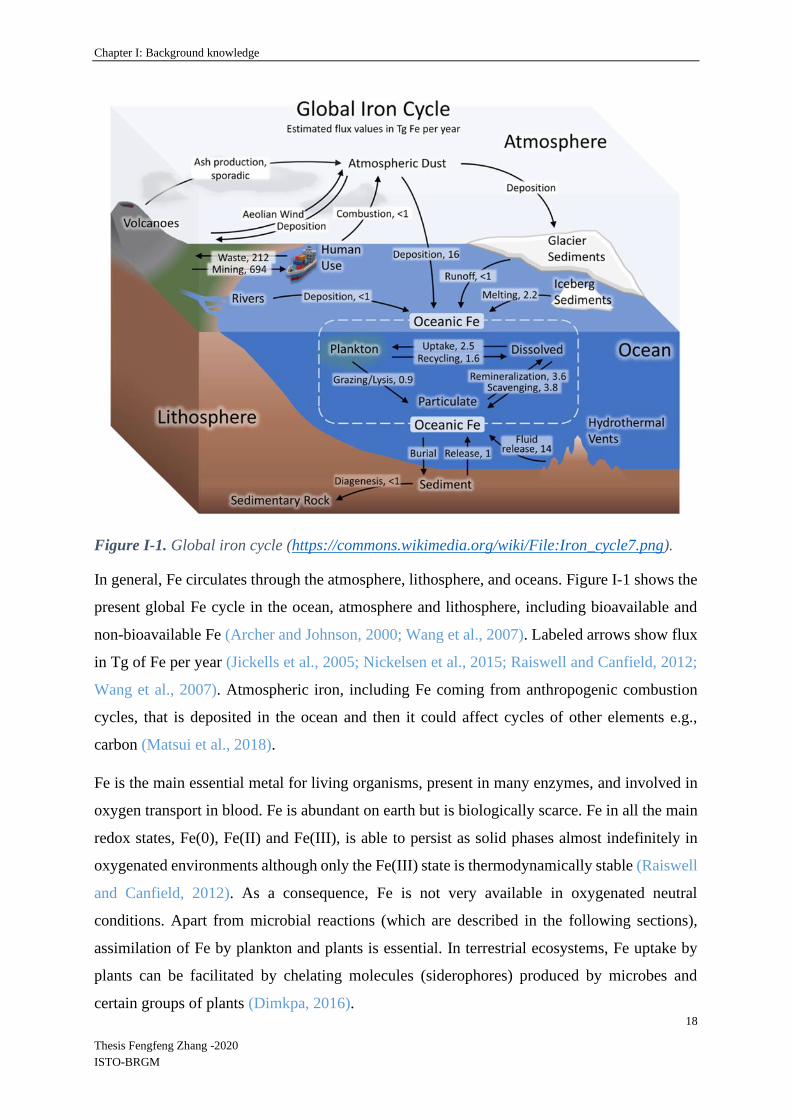
The Fe cycle involves the atmosphere, hydrosphere, biosphere and lithosphere. The main Fe
fluxes in the global cycle of this element (Figure I-1) are linked to human activities: mining and
waste production. The main natural fluxes are atmospheric dust deposition, oceanic fluids
release and the melting of iceberg sediments. The remaining fluxes are linked to living
organisms (Figure I-2).
Chapter I: Background knowledge
18
Thesis Fengfeng Zhang -2020
ISTO-BRGM
Figure I-1. Global iron cycle (https://commons.wikimedia.org/wiki/File:Iron_cycle7.png).
In general, Fe circulates through the atmosphere, lithosphere, and oceans. Figure I-1 shows the
present global Fe cycle in the ocean, atmosphere and lithosphere, including bioavailable and
non-bioavailable Fe (Archer and Johnson, 2000; Wang et al., 2007). Labeled arrows show flux
in Tg of Fe per year (Jickells et al., 2005; Nickelsen et al., 2015; Raiswell and Canfield, 2012;
Wang et al., 2007). Atmospheric iron, including Fe coming from anthropogenic combustion
cycles, that is deposited in the ocean and then it could affect cycles of other elements e.g.,
carbon (Matsui et al., 2018).
Fe is the main essential metal for living organisms, present in many enzymes, and involved in
oxygen transport in blood. Fe is abundant on earth but is biologically scarce. Fe in all the main
redox states, Fe(0), Fe(II) and Fe(III), is able to persist as solid phases almost indefinitely in
oxygenated environments although only the Fe(III) state is thermodynamically stable (Raiswell
and Canfield, 2012). As a consequence, Fe is not very available in oxygenated neutral
conditions. Apart from microbial reactions (which are described in the following sections),
assimilation of Fe by plankton and plants is essential. In terrestrial ecosystems, Fe uptake by
plants can be facilitated by chelating molecules (siderophores) produced by microbes and
certain groups of plants (Dimkpa, 2016).

Chapter I: Background knowledge
19
Thesis Fengfeng Zhang -2020
ISTO-BRGM
Figure I-2 presents the main bioprocesses involved in iron cycle, including oxidation of rocks
and Fe(II), biodegradation of Fe-containing organic matter, assimilation by living organisms,
and bio-reduction of diverse Fe(III) minerals.
Figure I-2. Biogeochemical processes involved in the iron cycle
The ferrous-ferric system is reversible, the reversibility is independent of pH, Eh, and the
reduction of Fe(III) (oxyhydr)oxides to dissolved Fe(II) is an important biogeochemical process
in all anaerobic sedimentary systems. Microbial reduction of Fe(III) also plays an important
role in the fate and transport of trace metals and nutrients in soils, sediments and aquatic
environments (Konhauser et al., 2011). Microbial reduction of Fe(III) (oxyhydr)oxides has been
extensively studied in relation to its essential roles on iron cycling.
I-1.2 Microbial transformations of Fe in the biogeosphere
Different groups of bacteria are responsible for direct Fe(II) oxidation in presence of oxygen
(O2) or of Fe(III) reduction in anaerobic conditions, in a large range of pH (Figure I-3).
Chapter I: Background knowledge
20
Thesis Fengfeng Zhang -2020
ISTO-BRGM
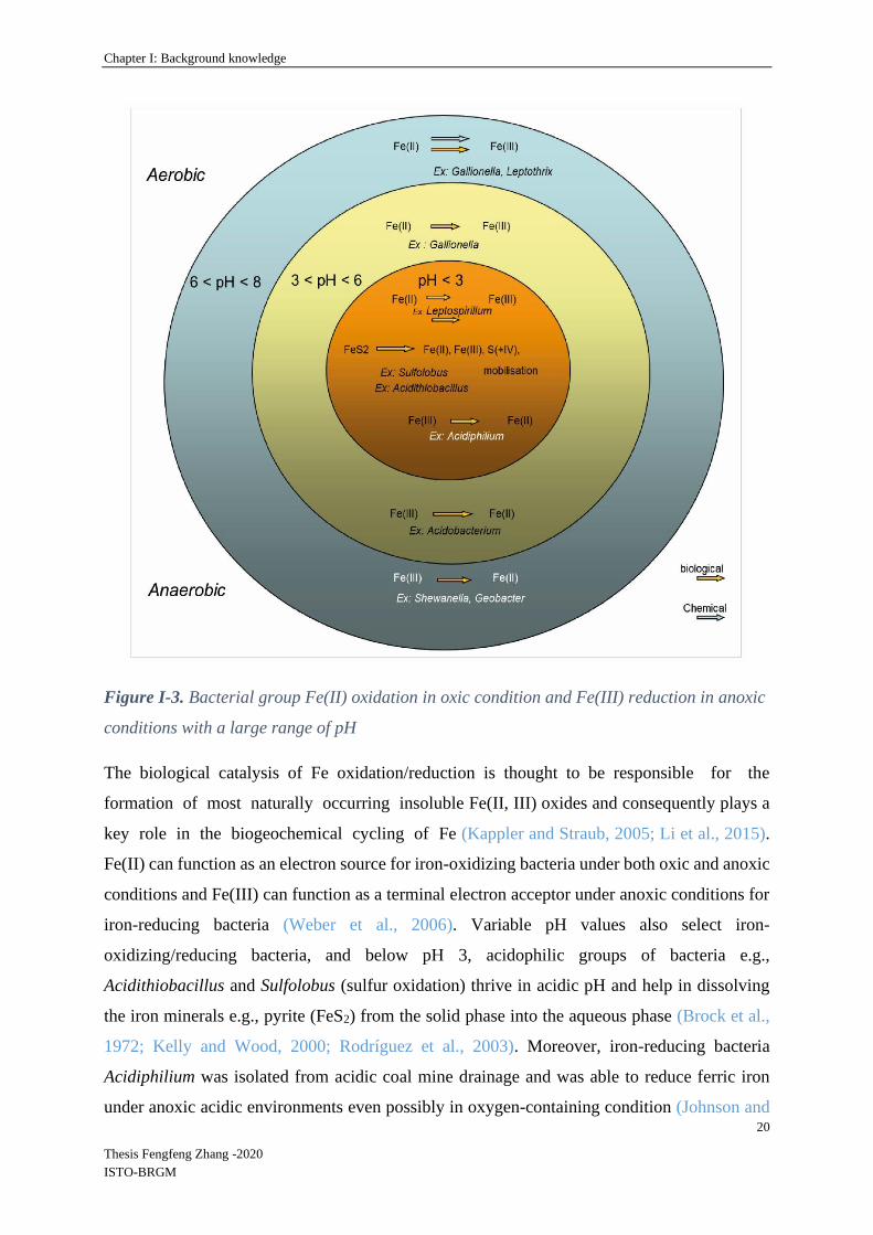
Figure I-3. Bacterial group Fe(II) oxidation in oxic condition and Fe(III) reduction in anoxic
conditions with a large range of pH
The biological catalysis of Fe oxidation/reduction is thought to be responsible for the
formation of most naturally occurring insoluble Fe(II, III) oxides and consequently plays a
key role in the biogeochemical cycling of Fe (Kappler and Straub, 2005; Li et al., 2015).
Fe(II) can function as an electron source for iron-oxidizing bacteria under both oxic and anoxic
conditions and Fe(III) can function as a terminal electron acceptor under anoxic conditions for
iron-reducing bacteria (Weber et al., 2006). Variable pH values also select iron-
oxidizing/reducing bacteria, and below pH 3, acidophilic groups of bacteria e.g.,
Acidithiobacillus and Sulfolobus (sulfur oxidation) thrive in acidic pH and help in dissolving
the iron minerals e.g., pyrite (FeS2) from the solid phase into the aqueous phase (Brock et al.,
1972; Kelly and Wood, 2000; Rodríguez et al., 2003). Moreover, iron-reducing bacteria
Acidiphilium was isolated from acidic coal mine drainage and was able to reduce ferric iron
under anoxic acidic environments even possibly in oxygen-containing condition (Johnson and

Chapter I: Background knowledge
21
Thesis Fengfeng Zhang -2020
ISTO-BRGM
Bridge, 2002; Wichlacz et al., 1986). Microbial ferrous Fe oxidation provides energy to
acidophiles such as Leptospirillum which is well known to achieve Fe(II) oxidation at pH below
3 (Ojumu and Petersen, 2011; Van Scherpenzeel et al., 1998). In the pH range 3-6,
Acidobacterium is an acidophilic, chemoorganotrophic bacterial genus isolated from acidic
mineral environments and capable of dissimilatory iron reduction under anaerobic conditions
(Coupland and Johnson, 2008; Kishimoto et al., 1991). The bacterial species of the genus
Gallionella are typical iron-oxidizing bacteria that grow under neutral (pH 6.5-8) or moderately
acidic (pH 4) conditions (De Vet et al., 2011; Fabisch et al., 2013). The genus Leptothrix
contains “iron-oxidizing” or “model Mn(II)-oxidizing” species and is ubiquitously distributed
in the aquatic environment (El Gheriany et al., 2009; Ghiorse, 1984). Leptothrix can be readily
found in sites with a circumneutral pH, an oxygen gradient and a source of reduced Fe and
manganese minerals (Emerson and Moyer, 1997; Sawayama et al., 2011). Finally, Geobacter
and Shewanella are the two most studied dissimilatory Fe reducing genera under anaerobic and
near-neutral pH conditions up to now (Engel et al., 2019; Han et al., 2018; Jiang et al., 2020;
Li et al., 2012).
I-1.3 Influence of redox conditions and organic matter on iron reduction
Iron redox reactions have the potential to support substantial microbial populations in soil and
sedimentary environments, as Fe is the fourth most abundant element in the Earth’s crust
(Weber et al., 2006). Motomuraand and Yokoi (1969) have suggested that the different forms
of ferrous Fe in flooded soils have a physical and chemical influence on the development and
stabilization of soil structure, which in turn exerts an influence on soil productivity (Gotoh and
Patrick Jr, 1974; Motomura and Yokoi, 1969). It is also well established now that flooded soils
in anaerobic conditions are subjected to a succession of Fe transformations from the ferric to
ferrous state under reducing conditions caused by a wide variety of facultative anaerobic soil
bacteria. Fe reduction can be the result of bacterial metabolism, Fe functioning as an electron
acceptor in dissimilatory iron reduction (DIR). Redox potential (oxidation-reduction) has a
marked effect on microbial Fe behavior because a change in redox status in soils implies
changing the availability of electron acceptors. As shown on the electron donors/acceptors
tower in Figure I-4, Fe(III) may be used as an electron acceptor after depletion of oxygen, nitrate
and manganese oxides. Pett-Ridge and Silver (2006) have indicated that some flexible
microorganisms were able to respire/ferment or use multiple electron acceptors under
fluctuating redox conditions, thus microbial populations can be periodically activated and

Chapter I: Background knowledge
22
Thesis Fengfeng Zhang -2020
ISTO-BRGM
inactivated, which in turn quickly alters the nature and rate of key biogeochemical
transformations (DeAngelis et al., 2010; Husson, 2013; Pett-Ridge et al., 2006).
Ginn et al. (2017) studied the influence of oxygen variations on Fe speciation in soils from the
Luquillo Critical Zone Observatory (Puerto Rico) through batch experiments in flasks
inoculated with Shewanella. Shewanella cultures reduced Fe(III) much faster under redox
fluctuations (cycles of oxic-anoxic conditions) than the oxic controls from soils (Ginn et al.,
2017).
Figure I-4. The “electron towers” of redox processes in biogeochemistry (Jørgensen, 2000).
The redox potentials are calculated for standard conditions at pH 7 and 1 mM concentrations
of substances.
Moreover, several mechanisms are involved in the dynamics of metals and metalloids
connected with the iron cycle: (1) dissolution of Fe(III) oxides can release the adsorbed/co-
precipitated elements; (2) DIRB may directly change the speciation of the associated elements,
because they can use both Fe(III) and other metals/metalloids as electron acceptors (Lovley,
2008), and (3) the natural microbial communities include both DIRB and other bacteria that use

Chapter I: Background knowledge
23
Thesis Fengfeng Zhang -2020
ISTO-BRGM
metals / metalloids as electron acceptors. The bio-reduction of the different metals/metalloids
can depend on the redox potential (Figure I-5).
Figure I-5. Redox tower of metal ion standard reduction potentials in acidic solutions, and
cytochrome-c reported vs. standard hydrogen electrode (SHE) (Bard, 2017; Dominguez-
Benetton et al., 2018).
Figure I-5 shows some standard reduction potentials of microbial electron transfer in metal
reduction (Dominguez-Benetton et al., 2018). Cytochrome-c oxidase was found to be
implicated in electron transfer during microbial metal reduction as an exemplar bacterial outer
membrane protein enzyme, with a standard potential of 0.26 vs. SHE/V (pH 7) (Lovley et al.,
1993b; Mathews, 1985). Cytochrome-c or other cell wall-associated enzymes are involved in
microbial metal reduction within specific ranges of redox potentials. Therefore this potential
provides an approximate thermodynamic limit for metal reduction. I.e., only metals with redox
potentials above 0.26 vs. SHE/V can be reduced by bacterial cells with these characteristics.
In soils, oxidation of FeII(aq) can induce the formation of colloidal complexes of Fe and organic
matter (Peiffer et al., 1999; Pullin and Cabaniss, 2003). Dissolved organic matter appears to be

Chapter I: Background knowledge
24
Thesis Fengfeng Zhang -2020
ISTO-BRGM
a key factor in the control of the Fe(III)-oxyhydroxide dissolution rate. More specifically,
organic matter, by strongly binding Fe(II), prevents Fe(II) readsorption and subsequent Fe
secondary mineral formation, both of which are known to strongly decrease Fe(III)-
oxyhydroxide dissolution rates (Davranche et al, 2013). In presence of humic substances, Fe
particles of nanometric size (colloids) are formed in the humic matrix (Pédrot et al., 2011).
Bioreduction experiments demonstrated that bacteria (Shewanella putrefaciens CIP 80.40 T)
were able to reduce these Fe nanoparticles associated with humic organic matter about eight
times faster than pure nano-lepidocrocite. These results suggest that in natural environments
organic matter influence the type of iron phases formed in presence of oxygen, and the rate of
Fe(II) production in anaerobic conditions. Alternance of redox conditions can lead to the
formation of colloids in soils, composed of natural organic matter, complexed iron and other
soil elements such as Al, Ti or Si (Thompson et al., 2006a). Moreover, under reducing
conditions, the pH rise can be a key factor controlling organic matter solubilization during Mn-
and Fe-oxyhydroxides reductive dissolution, as observed by Grybos et al., 2009 with a wetland
soil. Pédrot et al. (2008) performed soil column experiments and showed that some trace
elements, in particular Pb, Ti and U, were mobilized by humic acids containing iron
nanoparticles. Humic substances directly or indirectly promoted the colloidal transport of
insoluble trace elements either by binding trace elements or by stabilizing a ferric carrier phase
(Pédrot et al., 2008).
I-2 Classification of Fe-minerals
Fe and Fe-minerals are common components in several compartments of the critical zone (e.g.
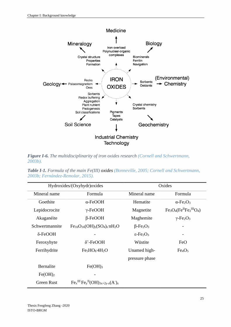
soils, sediments and aquifers) and are present in many different mineralogical forms. Many
different scientific disciplines are interested in Fe oxides (Figure I-6). There are sixteen known
Fe oxides, oxy-hydroxides and hydroxides with different mineral structures which are listed in
Table I-1 (Bonneville, 2005; Cornell and Schwertmann, 2003b; Fernández-Remolar, 2015).
Chapter I: Background knowledge
25
Thesis Fengfeng Zhang -2020
ISTO-BRGM
Figure I-6. The multidisciplinarity of iron oxides research (Cornell and Schwertmann,
2003b).
Table I-1. Formula of the main Fe(III) oxides (Bonneville, 2005; Cornell and Schwertmann,
2003b; Fernández-Remolar, 2015).
Hydroxides/(Oxyhydr)oxides Oxides
Mineral name Formula Mineral name Formula
Goethite α-FeOOH Hematite α-Fe2O3
Lepidocrocite γ-FeOOH Magnetite Fe3O4(FeIIFe2IIIO4)
Akaganéite β-FeOOH Maghemite γ-Fe2O3
Schwertmannite Fe16O16(OH)y(SO4)z.nH2O β-Fe2O3 -
δ-FeOOH - ε-Fe2O3 -
Feroxyhyte δ’-FeOOH Wüstite FeO
Ferrihydrite Fe5HO8.4H2O Unamed high-
pressure phase
Fe4O5
Bernalite Fe(OH)3
Fe(OH)2 -
Green Rust FexIII Fey
II(OH)3x+2y-z(A-)z

Chapter I: Background knowledge
26
Thesis Fengfeng Zhang -2020
ISTO-BRGM
Fe(III) (oxyhydr)oxides differ by their crystallinity, initially amorphous (the first formed after
oxidation of Fe(II), maturation under different conditions then leads to other forms. Iron oxides
polymorphs differ in the organization of the Fe(OH)n building blocks as shown in Figure I-7.
Figure I-7. FeOOH polymorphs (a) α-FeOOH; (b) β-Fe-OOH; (c) γ-FeOOH; (d) ε-FeOOH.
Centered atom is iron. Red atom is oxygen, white atom is hydrogen (green atom is chloride
for b = akageneite) (Sakamoto et al., 2019).
In flooded soils, the presence of Fe(III) will induce precipitation of Fe(II) from carbonate. Baas
Becking et al. (1960) reported the formation of the Fe(II)/Fe(III) hydroxy-carbonates siderite
and maghemite under natural aqueous environments and Halama et al. (2016) suggested that
diagenetic magnetite in banded iron formations was possibly formed by microbial Fe(III)
reduction during early diagenesis (Becking et al., 1960; Helama et al., 2016). Moreover, various
concentrations of Fe(II) and forms/amounts of total Fe(III) might cause other phases such as
magnetite/siderite/green rust to be formed in microbial (biotic) iron reduction systems
(Mortimer et al., 2011; Ona-Nguema et al., 2002; Taylor, 1980). In addition, oxidation leads to
the formation of either goethite, lepidocrocite, ferrihydrite or mixtures of these phases,
depending on the mode of oxidation, and the presence of impurities also may result in the
release of initially co-precipitated ions (Senn et al., 2017; Taylor, 1980).

Chapter I: Background knowledge
27
Thesis Fengfeng Zhang -2020
ISTO-BRGM
Apart from Fe (oxyhydr)oxides, iron sulfide, carbonate, phosphate and silicate minerals also
play a crucial role in a variety of biogeochemical processes (Raiswell and Canfield, 2012). The
principal features of the known Fe minerals are presented in the following sections. The main
characteristics of the minerals studied in previous researches are given in Tables I-2 and I-3.
Table I-2. The physicochemical parameters, crystal size, morphology and range of specific
surface area of the main studied Fe-oxides (Bonneville, 2005; Raiswell and Canfield, 2012;
Lagroix et al., 2016; Etique et al., 2016).
Fe mineral
class
Fe mineral
name
Formula Crystal
size
Specific
surface
area (m2 /g)
Morphology and crystal
symmetry
(Oxyhydr)
oxides
ferrihydrite Fe2O3/HFO/
Fe4HO8.4H2
O
a few nm 150 - 700 more or less spherical,
hexagonal
goethite α-FeOOH tens of nm
to several
microns
8 - 200 acicular or needle shaped,
orthorhombic
hematite α-Fe2O3 - 10 - 90 plates, discs, rods,
spindles, cubes, ellipsoids
and spheres,
rhombohedral
lepidocrocite γ-FeOOH - 15 - 260 lath-like, tabular,
diamond-shaped or
rectangular, orthorhombic
Magnetite Fe3O4 29-83 cubo-octahedron and
octahedron, cubic spinel

Chapter I: Background knowledge
28
Thesis Fengfeng Zhang -2020
ISTO-BRGM
Table I-3. Formula of Fe minerals.
* A is the interlayer anion and n its valency, with 1/4 <= x/(1+y) <= 1/3 and m <= (1-x+y)
(Oxy)hydroxyl-
sulphate
Schwertmannite Fe3O8(OH)1-1.8.
8H2O
Morphology References
Sulphides
Pyrite FeS2 grains, spherical
grains, spherulites,
euhedral grains
(Bonneville, 2005; Wang
& Morse, 1996)
Mackinawite FeS thin, tabular crystals (Devey et al., 2008)
Greigite Fe3S4 octahedral (Hunger & Benning, 2007)
Carbonates Siderite FeCO3 globule,
rhombohedral, disk-
like,
( Bonneville, 2005; Roh et
al., 2003)
Phosphates Vivianite Fe2+3 (PO4)2
.8H2O spherical, monoclinic-
prismatic
(Roldán et al., 2002)
Metavivianite Fe2+3−xFe3+
x(PO4)2(
OH)x.(8−x)H2O
flat, prismatic crystals (Ritz et al., 1974)
Chukanovite Fe2(CO3)(OH)2 acicular, fibrous
individuals combined
in spherulites
(Pekov et al., 2007)
Silicates : 7 Å
Minerals
Kaolinite Al2Si2O5(OH)4 high-crystallinity,
blocky
(Raiswell and Canfield,
2012; Kameda et al., 2005)
Silicates: 10 Å
Minerals
Illite (K, H3O) (Al, Mg,
Fe)2 (Si, Al)4 O10
(OH)2 (H2O)
parallelepiped (Raiswell and Canfield,
2012; Derrendinger &
Sposito, 2000)
Silicates: 14 Å
Minerals
Smectite (Ca, Na) (Al, Mg,
Fe)2 (Si, Al)4 O10
(OH)2. x H2O
dioctahedral (Raiswell and Canfield,
2012; Delavernhe et al.,
2015)
Ripidolite (Mg,
Fe-Chlorite)
(Mg, Fe, Mg)6 (Si,
Al)4 O10 (OH)8
ripidolite grain (Hamer et al., 2003)
Fougerite *[Fe12+ -
xFex3+Mgy(OH)2+2y]
+x[x/nA-n.mH2O]-x
hexagonal crystals (Raiswell and Canfield,
2012; Trolard et al., 2007)

Chapter I: Background knowledge
29
Thesis Fengfeng Zhang -2020
ISTO-BRGM
Ferrihydrite is an amorphous or weakly crystalline Fe mineral (Bonneville, 2005). Structural
studies carried out by (Feitknecht et al., 1973) indicated that 2-line ferrihydrite consists of local
coherently scattered regions formed by four planar Fe(O,OH)6 octahedrons and Michel et al.
indicated that ferrihydrite is nano-crystalline with a delta-Keggin local structure (Michel et al.,
2007; Michel et al., 2011). Whereas, in goethite each Fe(III) ion is surrounded by three O2- and
three OH- resulting in FeO3(OH3) octahedrons (Bonneville, 2005). Moreover, (Hiemstra, 2013)
elucidated the surface structure of ferrihydrite particles whose faces have a much higher site
density of singly-coordinated FeOH(H) groups in comparison to the main faces of goethite.
Therefore, the related adsorption capacity per unit surface area of ferrihydrite is much higher
than that of goethite. Lepidocrocite is metastable with respect to goethite which consists of
double chains of Fe(O, OH)6 octahedrons running parallel to the c-axis. Hematite consists of
layers of Fe(O)6 octahedrons and it has a similar thermodynamic stability to goethite, thus it is
very often found in association with goethite (Tardy and Nahon, 1985).
I-3 Dissolution of Fe(III) (oxyhydr)oxides and iron cycling in surface
environments
Fe (oxyhydr)oxides are common components in several compartments of the critical zone (e.g.
soils, sediments and aquifers) and are present in many different mineralogical forms.
Understanding biogeochemical behavior and iron cycling is fundamental for many scientific
communities (Bonneville et al., 2004; Roden et al., 2004). Indeed, the mobility of trace elements
(TE) is partly controlled by iron speciation, mineralogy and reactivity (Cornell and
Schwertmann, 2003b).
I-3.1 Abiotic dissolution
Fe(III) (oxyhydr)oxides can be dissolved by surface protonation, a dissolution mechanism
depending on pH conditions that can be enhanced by organic acids and anions (Cl-) (Zinder et
al., 1986). Besides, two other dissolution mechanisms have also been reported, ligand promoted
dissolution and bulk reductive dissolution (Afonso et al., 1990; Holmén and Casey, 1996;
Kraemer, 2004). (Siffert and Sulzberger, 1991) indicated that reductive dissolution of hematite
in the presence of oxalate occurs as a photocatalytic process, and Holmén & Casey studied the
rate of goethite dissolution in the presence of acetohydroxamic acid in different pH conditions.
The rate of reductive dissolution of several synthetic Fe(III) (oxyhydr)oxides in 10 mM
Chapter I: Background knowledge
30
Thesis Fengfeng Zhang -2020
ISTO-BRGM
ascorbate at pH 3 has been shown to occur according to the order ferrihydrite> lepidocrocite >
goethite> hematite (Bonneville et al., 2004; Larsen and Postma, 2001).
In natural environments, reduction of Fe(III) (oxyhydr)oxides may also be linked to the
presence of hydrogen sulfide (H2S/HS-) produced by sulfate-reducing bacteria (Dos Santos
Afonso and Stumm, 1992; Neal et al., 2001).
I-3.2 Biotic dissolution
The natural solubility of crystalline Fe (oxyhydr)oxides is low. However, interactions with
microbes and organic substances can improve the formation of soluble Fe(III) and increase the
availability of Fe and associated TEs (Colombo et al., 2014). Biogeochemical aspects of Fe
cycling in the major microbially mediated and abiotic reactions have been extensively covered
(Melton et al., 2014), together with Fe redox transformations and availability of TEs (Zhang et
al., 2012), as well as Fe redox cycling in bacteriogenic Fe oxide-rich sediments (Gault et al.,
2011). In aerobic environments at circumneutral pH conditions, Fe is generally relatively stable
and highly insoluble in the form of (oxyhydr)oxides (e.g., Fe(OH)3, FeOOH, Fe2O3). However,
in anaerobic conditions these minerals can be reductively dissolved (Roden et al., 2004; Roden
and Wetzel, 2002) by microbial and abiotic pathways (Bonneville et al., 2004; Hansel et al.,
2004; Shi et al., 2016; Thompson et al., 2006b). Microbial Fe(III) reduction is an important
mechanism for iron cycling: heterotrophic Fe(III)-reducing bacteria could convert solid-phase
Fe(III) minerals into dissolved and solid Fe(II) phases during their metabolic processes (Lovley,
1997). In particular, dissolutive reduction of iron (oxyhydr)oxides can be driven by
dissimilatory iron reducing bacteria (DIRB), significantly contributing to the biogeochemical
cycle of Fe and subsequent TE cycling (Cooper et al., 2006; Ghorbanzadeh et al., 2017; Levar
et al., 2017). Microbial dissimilatory iron reduction (DIR) is an ubiquitous biogeochemical
process in suboxic environments (Crosby et al., 2005; Lovley, 2000; Schilling et al., 2019;
Wilkins et al., 2006). DIRB use Fe (oxyhydr)oxides as electron acceptors instead of oxygen for
oxidizing organic matter. In general, for ferrihydrite and other short-range ordered (SRO)
poorly crystallized iron minerals, the microbial Fe(III) reduction rates are more rapid (typically
within hours), than the microbial reduction of the well-ordered minerals hematite (α-Fe2O3),
goethite (α-FeOOH) (e.g., several months), and lepidocrocite (γ-FeOOH) (Ginn et al., 2017;
Roden, 2006a). The rate of Fe(III) reduction will influence mobility of TEs initially
immobilized on or in Fe (oxyhydr)oxides through adsorption or co-precipitation. Crystallinity,
specific surface area and size among other factors may influence the reactivity of Fe

Chapter I: Background knowledge
31
Thesis Fengfeng Zhang -2020
ISTO-BRGM
(oxyhydr)oxides in relation to the metabolic activity and diversity of DIRB (Aino et al., 2018;
Cutting et al., 2009), as detailed in the next section.
I-3.3 Secondary minerals formed during the bio-reduction of Fe oxides
The first product of the bioreduction of iron oxides is soluble Fe(II). This chemical species can
sorb to residual Fe(III)-oxides, or be involved in (bio)geochemical reactions generating
secondary minerals (Table I-4) such as green rust (Génin et al., 1998; Ona-Nguema, et al.,
2002), vivianite, siderite, magnetite (Fredrickson et al., 1998; Maitte et al., 2015; Urrutia et al.,
1998; Zachara et al., 1998), chukanovite (O’Loughlin et al., 2010). The type of secondary
product depends on the composition of the liquid phase, the presence of complexants, and on
bacterial activity.
Table I-4. Formula of the main minerals potentially formed during bio-reduction of Fe(III)-
oxides.
Mineral Formula References
Siderite Fe2+CO3 (Roh et al., 2003; Zachara et al.,
1998)
Chukanovite Fe2+2(CO3)(OH)2 (O’Loughlin et al., 2010)
Vivianite Fe2+3(PO4)2
.8H2O (Roldán et al., 2002; Urrutia et
al., 1998)
Green rust [Fe2+4Fe3+
2(HO−)12]2+ ·[CO2−
3·2H2O]2−
/[Fe2+4Fe3+
2(HO−)12]2+ ·[SO2−
4·2H2O]2−
(Ona-Nguema et al., 2002; Ona-
Nguema et al., 2009; Zegeye et
al., 2005)
Magnetite Fe2+Fe3+2O3 (Maitte et al., 2015)

Chapter I: Background knowledge
32
Thesis Fengfeng Zhang -2020
ISTO-BRGM
Figure I-8. SEM images of (a) siderite globule formed by Shewanella (W3-7-1) under a
H2/CO2 atmosphere and (b) rhombohedral siderite formed by Shewanella (NV-1) under a
H2/CO2 atmosphere (Roh et al., 2003), (c) SEM images of the starting vivianite (Roldán et al.,
2002); SEM images of (d) magnetite/FHC formed in the unamended system and of (e) green
rust formed in the systems containing 500 μM phosphate (O’Loughlin et al., 2010).
As an example, using a continuously fed column experiment, Hansel et al. (2003) observed the
transformation of ferrihydrite by Shewanella into goethite (via dissolution/reprecipitation)
and/or magnetite (via solid-state conversion) (Hansel et al., 2003b), whereas Ona-Nguema et
al. (2002) observed the formation of carbonate green rust by Shewanella putrefaciens (Ona-
Nguema et al., 2002) and Kukkadapu et al. (2004) observed the formation of magnetite in the
absence of phosphate whereas carbonate green rust and vivianite were formed in presence of
phosphate during the bio-reduction of 2-line Si-ferrihydrite by the same iron-reducing bacterial
genus (Kukkadapu et al., 2004).
I-4 Iron-reducing bacteria (IRB)
I-4.1 Dissimilatory Iron-Reducing Bacteria (DIRB)
Dissimilatory Fe(III) reducing microorganisms are a group of microorganisms (both bacteria
and archaea) that can perform anaerobic respiration using Fe(III) as a terminal electron acceptor
before using molecular oxygen (O2), which is the terminal electron acceptor reduced to water
(H2O) in aerobic respiration (Lloyd, 2003). The role of DIRB in Fe redox transformations has

Chapter I: Background knowledge
33
Thesis Fengfeng Zhang -2020
ISTO-BRGM
been evidenced for more than three decades (Lovley, 1991; Lovley et al., 1987; Meile and
Scheibe, 2019; Stern et al., 2018; Su et al., 2020), during which more than 100 distinct IRB
species have been found. Geobacter and Shewanella are the two most studied IRB genera up to
now (Engel et al., 2019; Han et al., 2018; Jiang et al., 2020; Li et al., 2012), however DIRB are
widely distributed phylogenetically DIRB and iron-reducing archaea have been isolated from a
range of environments, including freshwater (Laverman et al., 1995) or sea water (Rosselló-
Mora et al., 1995), marsh sediments, groundwater (MacRae et al., 2007), soils (Ottow and
Glathe, 1971), geothermal systems (Lee et al., 2017), deep sea hydrothermal fields (Lin et al.,
2016; Zeng et al., 2015), and sediments (Tebo and Obraztsova, 1998).
Table I-5 gives a non-exhaustive overview of the diversity of Fe(III)-reducing bacteria.
Table I-5. Iron-reducing archaea and other iron-reducing bacteria strains.
Iron reducing
microorganism
Context References
Geoglobus
acetivorans sp. nov.
A Fe(III)-reducing archaeon (Slobodkina et al.,
2009)
Deferribacter
thermophilus gen.
nov., sp. nov.
A Novel Thermophilic Manganese- and
Iron-Reducing Bacterium
(Greene et al.,
1997)
Geothrix fermentans
gen. nov., sp. nov.
A novel Fe(III)-reducing bacterium (Coates et al., 1999)
Acidiphilium cryptum
JF-5
Microbial Reduction of Fe(III) in
Acidic Sediments (Glucose)
(Küsel et al., 1999)
Bacillus subterraneus
sp. nov.
An iron and manganese reducing
bacterium
(Kanso et al., 2002)
Pelosinus fermentans
gen. nov.
An iron(III)-reducing bacteria (Shelobolina et al.,
2007)
Geoalkalibacter
subterraneus sp. nov.
An anaerobic Fe(III)- and Mn(IV)-
reducing bacterium
(Greene et al.,
2009)
Bacillus pseudofirmus
MC02
Extracellular reduction: AQDS
(anthraquinone-2, 6-disulfonae), humic
(Ma et al., 2012)

Chapter I: Background knowledge
34
Thesis Fengfeng Zhang -2020
ISTO-BRGM
acids (HA) and Fe(III) oxides were
electron acceptors.
Pelobacter
carbinolicus
Fe(III) and S(0) reduction (Lovley et al., 1995)
Clostridium butyricum Isolated from a microbial fuel cell (Park et al., 2001)
Pyrobaculum igneiluti Thermophile isolated from geothermal
system
(Lee et al., 2017)
Pyrodictium delaneyi Thermophile isolated from active deep-
sea hydrothermal vent chimney
(Lin et al., 2016)
Caloranaerobacter
ferrireducens
Thermophile isolated from ocean
hydrothermal field
(Zeng et al., 2015)
Wolinella
succinogenes
Isolated from freshwater marsh (Laverman et al.,
1995)
Desulfotomaculum
reducens
Isolated from metal-contaminated
sediments, capable reducing Fe(III) in
presence of electron donors
(Dalla Vecchia et
al., 2014; Tebo and
Obraztsova, 1998)
Thus, iron-reducing bacteria form a very diverse group, with microbes belonging to a large
range of phyla. Up to now, no functional gene specific to dissimilatory Fe(III) reduction and
common to all these organisms has been identified, so it is very difficult to monitor all Fe(III)-
reducing micro-organisms in a natural complex communities. Enrichment and isolation
methods can still be applied, however they don’t give access to the whole Fe(III)-reducing
community. An alternative approach is to target specific well-known Fe(III)-reducing micro-
organisms.
The two most studied DIRB belong to the genera Shewanella and Geobacter. These model
strains have been used in many research studies, in particular those focused on the elucidation
of the mechanisms of bacterial Fe(III) reduction.
I-4.2 Iron reduction by fermentative bacteria
A wide variety of fermentative micro-organisms are able to reduce Fe(III) into Fe(II) during
anaerobic growth with fermetable sugars or amino-acids. As soon as 1926, Runov described
the reduction of Fe(III) by pure strains of Escherichia coli, Clostridium pasteurianum and
Chapter I: Background knowledge
35
Thesis Fengfeng Zhang -2020
ISTO-BRGM
Lactobacillus lactis. During fermentation in presence of Fe(III), a part of the electron flow
contributes to Fe(III) reduction. The benefit of this reaction for the growth of bacteria was
debated, less than 5% of the reducing equivalents were reported to be transferred to Fe(III)
(Lovley, 1987) however this reaction can significantly contribute to Fe(III) reduction in
presence of fermentable substrates.
Robert (1947) found with strains of Bacillus polymyxa that even if, in principle, 24 mol Fe(III)
should be reduced per mol of glucose, most of the electron equivalent were recovered as ethanol,
2,3 ethylene glycol, lactic acid, formic acid and hydrogen. Even if only a small part of electron
equivalents are transferred to Fe(III), this mechanism improves the fermentation balance
inducing more favorable thermodynamic conditions for the organism able to perform this
reduction. This could confer competitive advantage to these fermentative microbes in carbon-
limited environment like deep biosphere, according to Vandieken et al. (2018).
The composition of iron oxides can influence their bioreduction by fermentative micro-
organisms. In particular, the substitution of Fe by Al, Mn, Cr, or Co in synthetic goethites
induced a decrease of their reduction during fermentation of glucose (Bousserrhine et al., 1999;
Dominik et al., 2002).
The fermentation of glucose can be enhanced in presence of iron oxides (such as hematite, Dong
et al., 2017), not only by consuming electron equivalents, but also because of a buffering effect
preventing acidification of the medium. This phenomenon is linked to H2 production during
fermentation and a reaction of H2 with iron oxide that consumes H+:
Fe2O3 + 4H+ + H2 = 2 Fe2+ + 3 H2O (Dong et al., 2017)
Recently, a metagenomics analyse was performed on a glucose-fermenting microbial
consortium capable of reducing goethite in presence of glucose (Gagen et al., 2019). A
metagenome assembled genome (MAG) of an iron reducer belonging to the
alphaproteobacterial genus Telmatospirillum was obtained, encoding putative metal transfer
reductases and a novel outer membrane cytochrome potentially contributing to extracellular
electron transfer. Metabolic products suggested that goethite was a hydrogen sink in the culture.
Therefore, recent results from Dong et al. (2017) and Gagen et al., (2019) suggest that
fermentative bacteria could induce iron reduction via hydrogen production.

Chapter I: Background knowledge
36
Thesis Fengfeng Zhang -2020
ISTO-BRGM
Beside the Fe(III) reduction directly linked to the metabolism of fermentative bacteria,
fermentation products, such as organic acids and H2, can be important electron donors for
dissimilatory iron-reducing bacteria, either in laboratory culture media with mixed cultures, or,
possibly in natural environments (Lovley, 1987).
I-4.3 The genus Shewanella
Shewanella spp. are Gram negative (Gamma proteobacteria), non spore forming motile rods
with positive oxidase and catalase reactions (Satomi, 2014). Shewanella spp. are widely
distributed in natural environments and their natural habitats are all forms of water and soil
compartments, they have been found in marine environments, proteinaceous foods, and
occasionally clinical samples (Fredrickson et al., 2008; Vignier et al., 2013). The organism
belonging to the genus Shewanella was first isolated in 1931 from putrefied butter and was
successively called Achromobacter putrefaciens, Pseudomonas putrefaciens, Alteromonas
putrefaciens (Derby and Hammer, 1931; Vignier et al., 2013). Finally MacDonnell and Colwell
suggested that Alteromonas putrefaciens, along with two other marine species should be
transferred to a completely new genus, Shewanella, which was named in honor of Dr J. Shewan
for his work on the microbiology of fish (MacDonell and Colwell, 1985). Up to date, more than
50 species have been classified in this genus, showing various activities, such as metal reduction
(e.g., Fe reduction) or trimethylamine production (Satomi, 2014).
In fact, microbial Fe reduction was described by researchers at the beginning of 20th century
(Starkey and Halvorson, 1927), however the first strain of Shewanella with dissimilation Fe
reduction activity was isolated in 1987 from petroleum reservoirs (Semple and Westlake, 1987).
Currently known and studied Shewanella species are tolerant to oxygen and thus reasonably
robust for applications such as remediation of polluted environments with various oxygen
concentrations (Satomi, 2014; Serres and Riley, 2006).
Metabolic characteristics of Shewanella
Shewanella genus gather facultative anaerobe species displaying a wide diversity of metabolic
features. Frequent characteristics of species are the reduction of H2S from thiosulfate, reduction
of nitrate and iron oxides. A large diversity of organic molecules can be used in aerobic
conditions, including sugars, organic acids, amino-acids, polymers such as dextrin. In the
absence of oxygen, members of this genus have been proved to be capable of using a variety of
other electron acceptors e.g., iron, thiosulfate, sulfite, or elemental sulfur (Burns and
DiChristina, 2009), as well as fumarate (Pinchuk et al., 2011) while using a smaller range of

Chapter I: Background knowledge
37
Thesis Fengfeng Zhang -2020
ISTO-BRGM
organic electron donors such as lactate, formate and H2 for respiration (Meshulam-Simon et al.,
2007). Moreover, some Shewanella species can also grow by fermentation of glucose (Bowman
et al., 1997; Ivanova et al., 2001), tryptone, peptone, yeast extract and pyruvate when Fe(III) is
limited (Toffin et al., 2004). Apart from iron, some members of this genus have the ability to
use a wide range of metals and metalloids as electron acceptors, including manganese,
chromium, uranium and arsenic, due to the broad specificity of the anaerobic reductase enzyme
system in Shewanella (Tiedje, 2002). Moreover, S. oneidensis MR-1 strains have been shown
to be able to reduce and mobilize toxic and radioactive metallic pollutants, including arsenic,
cobalt, chromium, mercury, plutonium, selenium, technetium, and uranium (Satomi, 2014).
Morphologic observations on two species of Shewanella are given in Figure I-9.
Figure I-9. Transmission electron micrographs of cells of strains Shewanella psychrophila (a)
and Shewanella piezotolerans (b). Cells were grown on 2216E agar plates at 15 ◦C for 16 h.
One colony was picked and dispersed in 100 µL PBS buffer, and 5 µL liquid was placed on a
copper grid for negative staining using 1% uranyl acetate. Bars, 0.5 µm (Xiao et al., 2007).
The main studies performed with Shewanella species are summarized in Table I-6.
Table I-6. Selected studies about some Shewanella strains and Fe(III) reduction.
DIRB Context References
Shewanella
amazonensis sp. nov.
A novel metal-reducing facultative
anaerobe from Amazonian shelf muds
(Venkateswaran et
al., 1998)
Shewanella algae
strain BrY
Microbial and surface chemistry
controls on reduction of synthetic
Fe(III) oxide minerals by DIRB
(Urrutia et al., 1998)
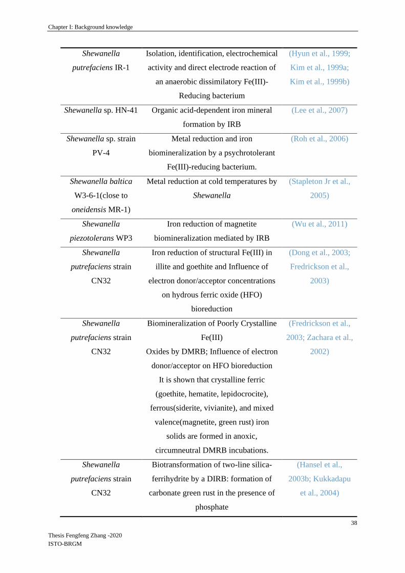
Chapter I: Background knowledge
38
Thesis Fengfeng Zhang -2020
ISTO-BRGM
Shewanella
putrefaciens IR-1
Isolation, identification, electrochemical
activity and direct electrode reaction of
an anaerobic dissimilatory Fe(III)-
Reducing bacterium
(Hyun et al., 1999;
Kim et al., 1999a;
Kim et al., 1999b)
Shewanella sp. HN-41 Organic acid-dependent iron mineral
formation by IRB
(Lee et al., 2007)
Shewanella sp. strain
PV-4
Metal reduction and iron
biomineralization by a psychrotolerant
Fe(III)-reducing bacterium.
(Roh et al., 2006)
Shewanella baltica
W3-6-1(close to
oneidensis MR-1)
Metal reduction at cold temperatures by
Shewanella
(Stapleton Jr et al.,
2005)
Shewanella
piezotolerans WP3
Iron reduction of magnetite
biomineralization mediated by IRB
(Wu et al., 2011)
Shewanella
putrefaciens strain
CN32
Iron reduction of structural Fe(III) in
illite and goethite and Influence of
electron donor/acceptor concentrations
on hydrous ferric oxide (HFO)
bioreduction
(Dong et al., 2003;
Fredrickson et al.,
2003)
Shewanella
putrefaciens strain
CN32
Biomineralization of Poorly Crystalline
Fe(III)
Oxides by DMRB; Influence of electron
donor/acceptor on HFO bioreduction
It is shown that crystalline ferric
(goethite, hematite, lepidocrocite),
ferrous(siderite, vivianite), and mixed
valence(magnetite, green rust) iron
solids are formed in anoxic,
circumneutral DMRB incubations.
(Fredrickson et al.,
2003; Zachara et al.,
2002)
Shewanella
putrefaciens strain
CN32
Biotransformation of two-line silica-
ferrihydrite by a DIRB: formation of
carbonate green rust in the presence of
phosphate
(Hansel et al.,
2003b; Kukkadapu
et al., 2004)
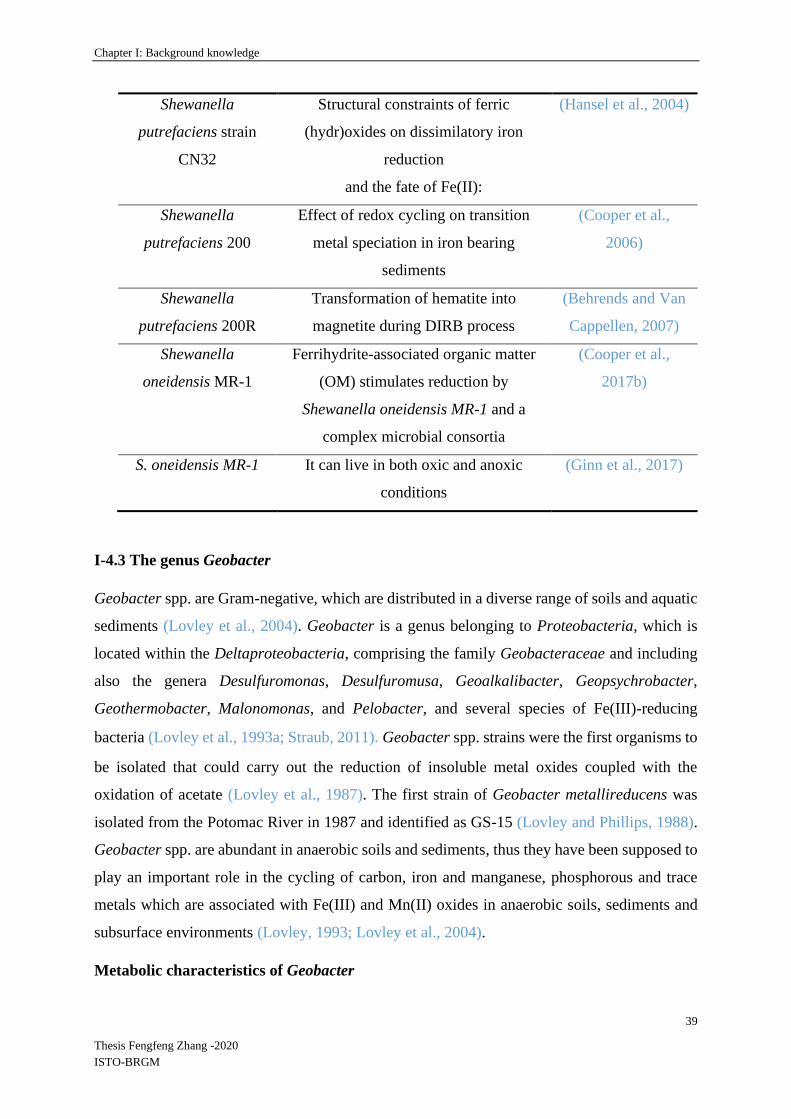
Chapter I: Background knowledge
39
Thesis Fengfeng Zhang -2020
ISTO-BRGM
Shewanella
putrefaciens strain
CN32
Structural constraints of ferric
(hydr)oxides on dissimilatory iron
reduction
and the fate of Fe(II):
(Hansel et al., 2004)
Shewanella
putrefaciens 200
Effect of redox cycling on transition
metal speciation in iron bearing
sediments
(Cooper et al.,
2006)
Shewanella
putrefaciens 200R
Transformation of hematite into
magnetite during DIRB process
(Behrends and Van
Cappellen, 2007)
Shewanella
oneidensis MR-1
Ferrihydrite-associated organic matter
(OM) stimulates reduction by
Shewanella oneidensis MR-1 and a
complex microbial consortia
(Cooper et al.,
2017b)
S. oneidensis MR-1 It can live in both oxic and anoxic
conditions
(Ginn et al., 2017)
I-4.3 The genus Geobacter
Geobacter spp. are Gram-negative, which are distributed in a diverse range of soils and aquatic
sediments (Lovley et al., 2004). Geobacter is a genus belonging to Proteobacteria, which is
located within the Deltaproteobacteria, comprising the family Geobacteraceae and including
also the genera Desulfuromonas, Desulfuromusa, Geoalkalibacter, Geopsychrobacter,
Geothermobacter, Malonomonas, and Pelobacter, and several species of Fe(III)-reducing
bacteria (Lovley et al., 1993a; Straub, 2011).Geobacter spp. strains were the first organisms to
be isolated that could carry out the reduction of insoluble metal oxides coupled with the
oxidation of acetate (Lovley et al., 1987). The first strain of Geobacter metallireducens was
isolated from the Potomac River in 1987 and identified as GS-15 (Lovley and Phillips, 1988).
Geobacter spp. are abundant in anaerobic soils and sediments, thus they have been supposed to
play an important role in the cycling of carbon, iron and manganese, phosphorous and trace
metals which are associated with Fe(III) and Mn(II) oxides in anaerobic soils, sediments and
subsurface environments (Lovley, 1993; Lovley et al., 2004).
Metabolic characteristics of Geobacter

Chapter I: Background knowledge
40
Thesis Fengfeng Zhang -2020
ISTO-BRGM
This strict anaerobe chemoorganotroph oxidizes several short-chain fatty acids (acetate,
propionate, butyrate, isobutyrate, valerate, isovalerate, pyruvate, formate, lactate), alcohols
(ethanol, propanol, butanol), and monoaromatic compounds, and sometimes H2, with Fe(III) as
the sole electron acceptor (Lovley et al., 1993; Coates et al., 1996).
Following the successful isolation of Geobacter metallireducens, several other Geobacter spp.
have been found and studied to explore their metabolic functions, including the potential for
biodegradation of environmental toxic pollutants. Geobacter spp. are capable of oxidizing
organic compounds to carbon dioxide through the reduction of various extracellular electron
acceptors, which makes them remarkable on those applications (Mahadevan et al., 2011). These
acceptors include insoluble Fe(III) and Mn(IV) oxides (Lovley and Phillips, 1988; Lovley et
al., 1987), other metal species such as uranium(VI) (Lovley et al., 1991) and vanadium(V)
(Ortiz-Bernad et al., 2004), and also humic substances (Lovley et al., 1996a).
Morphologic observations of Geobacter sulfurreducens are given in Figure I-10.
Figure I-10. (a) Transmission electron micrograph of a thin section of strain Geobacter
sulfurreducens PCA, bar 0.2 µm (Caccavo et al., 1994); (b) SEM images of Geobacter
sulfurreducens growing on a gold electrode (Richter et al., 2008).
Selected studies performed with some Geobacter species are summarized in Table I-7.
Table I-7. Selected studies about some Shewanella strains and Fe(III) reduction.
DIRB Researches References
(b) (a)
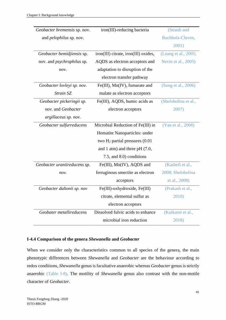
Chapter I: Background knowledge
41
Thesis Fengfeng Zhang -2020
ISTO-BRGM
Geobacter bremensis sp. nov.
and pelophilus sp. nov.
iron(III)-reducing bacteria (Straub and
Buchholz-Cleven,
2001)
Geobacter bemidjiensis sp.
nov. and psychrophilus sp.
nov.
iron(III) citrate, iron(III) oxides,
AQDS as electron acceptors and
adaptation to disruption of the
electron transfer pathway
(Leang et al., 2005;
Nevin et al., 2005)
Geobacter lovleyi sp. nov.
Strain SZ
Fe(III), Mn(IV), fumarate and
malate as electron acceptors
(Sung et al., 2006)
Geobacter pickeringii sp.
nov. and Geobacter
argillaceus sp. nov.
Fe(III), AQDS, humic acids as
electron acceptors
(Shelobolina et al.,
2007)
Geobacter sulfurreducens Microbial Reduction of Fe(III) in
Hematite Nanoparticles: under
two H2 partial pressures (0.01
and 1 atm) and three pH (7.0,
7.5, and 8.0) conditions
(Yan et al., 2008)
Geobacter uraniireducens sp.
nov.
Fe(III), Mn(IV), AQDS and
ferruginous smectite as electron
acceptors
(Kashefi et al.,
2008; Shelobolina
et al., 2008)
Geobacter daltonii sp. nov Fe(III)-oxhydroxide, Fe(III)
citrate, elemental sulfur as
electron acceptors
(Prakash et al.,
2010)
Geobater metallireducens Dissolved fulvic acids to enhance
microbial iron reduction
(Kulkarni et al.,
2018)
I-4.4 Comparison of the genera Shewanella and Geobacter
When we consider only the characteristics common to all species of the genera, the main
phenotypic differences between Shewanella and Geobacter are the behaviour according to
redox conditions, Shewanella genus is facultative anaerobic whereas Geobacter genus is strictly
anaerobic (Table I-8). The motility of Shewanella genus also contrast with the non-motile
character of Geobacter.

Chapter I: Background knowledge
42
Thesis Fengfeng Zhang -2020
ISTO-BRGM
If we look upon the two genera as groups of classified species, Shewanella is characterized by
a larger versatility of characteristics, in particular in the field of physiology and respiration, than
Geobacter.
Table I-8. Detailed description of the genera Shewanella and Geobacter.
Genus Shewanella (Holt et al.,
2005; Lemaire et al., 2020;
MacDonell & Colwell, 1985)
Geobacter (Childers et al., 2002; Coppi
et al., 2007; Lovley et al., 1993; Lovley
et al., 2011; Prakash et al., 2010)
Morphology and
motility
Straight or curved rods, non-
pigmented, motile by polar
flagella.
Rod-shaped cells 2-4 by 0.5 µm, non
motile, no spore formation.
Gram negative negative
Metabolism facultative anaerobic
chemoorganotroph
physiological and respiratory
versatility.
strict anaerobic chemoorganotroph
completely oxidizes acetate, ethanol,
propionate, butyrate, isobutyrate,
propanol, butanol, toluene, benzoate,
benzaldehyde, benzylalcool,
polyhydroxybutirate, p-
hydroxybenzaldehyde, p-
hydroxybenzylalcool, phenol, p-cresol
into CO2 with Fe(III) as electron
acceptor. also degrade acetate with
Mn(IV), nitrate and U(VI). Nitrate is
reduced into ammonium. The organic
electron donor is completely oxidized
to carbon dioxide.
Other characters produces hydrogen sulphide cells contain menaquinone and c‐type
cytochromes, but lack ubiquinone.
Vitamins are not required for growth,
but growth is enhanced by their
presence.
Number of
described species
70 (in 2020) 19 (in 2019)

Chapter I: Background knowledge
43
Thesis Fengfeng Zhang -2020
ISTO-BRGM
Type species Shewanella putrefaciens Geobacter metallireducens
GC% 44-47 50–61
I-4.5 Primers for the detection and quantification of Shewanella and Geobacter
Both Shewanella and Geobacter are genera with many species that have diverse respiratory
abilities. They are also considered to play an important role in bioremediation and in
biogeochemical cycles of several elements. The use of primers to detect specific species or
genera of bacteria or to quantify their abundance are well developed methods based on
molecular microbiology (Li et al., 2018). Thus, for the development of research programs
related to Fe(III) bioreduction it would be useful to find primers allowing to target and quantify
Shewanella and Geobacter by qPCR with accuracy and broad coverage, considering the
increasing number of isolated species belonging to these two genera.
According to several studies, different primers developed for the detection and/or quantification
of Shewanella and Geobacter in environmental samples are listed in Table I-9.
Table I-9. Primers for detection and/or quantification of Shewanella and Geobacter 16S
rRNA genes.
Gene target Primer Primer sequence alignment (5’ - 3’) References
Shewanella 16S
rRNA
211F
1259R
CGCGATTGGATGAACCTAG
GGCTTTGCAACCCTCTGTA
(Todorova and
Costello,
2006)
Shewanella 16S
rDNA
783F
l245R
AAAGACTGACGCTCAKGCA
TTYGCAACCCTCTGTACT
(Snoeyenbos-
West et al.,
2000; Zhou,
2008)
Shewanella 16S
rRNA
640F
815R
RACTAGAGTCTTGTAGAGG
AAGDYACCAAAYTCCGAGTA
(Li et al.,
2018)
Shewanella 16S
rRNA
120F
220R
GCCTAGGGATCTGCCCAGTCG
CTAGGTTCATCCAATCGCG
(Himmelheber
et al., 2009)

Chapter I: Background knowledge
44
Thesis Fengfeng Zhang -2020
ISTO-BRGM
Shewanella 16S
rRNA
211F
815cR
CGCGATTGGATGAACCTAG
AAGDCACCAAAYTCCGAGTA
(Li et al.,
2018)
Geobacter 16S
rDNA
561F
825R
GCGTGTAGGCGGTTTCTTAA
TACCCGCRACACCTAGTTCT
(Cummings et
al., 2003)
Geobacter 16S
rRNA
564F
840R
AAGCGTTGTTCG GAWTTAT
GGCACTGCAGGGGTCAAT
(Cummings et
al., 2003; Kim
et al., 2012)
Geobacter 16S
rDNA
Gx.182F
Gx.472R
AGA CCT TCG GCT GGG ATG CT
AGG TAC CGT CAA GTA ACA SS
(Snoeyenbos-
West et al.,
2000)
Geobacter 16S
rRNA
196F
535R
GAATATGCTCCTGATTC
TAAATCCGAACAACGCTT
(Amos et al.,
2007)
For the listed primers in Table I-9, target species, gene fragment length, PCR/qPCR condition,
coverage and specificity described in literature are given in Table I-10.
Table I-10. Summary of the primers coverage and specificity for Shewanella and Geobacter
species.
Primer
paris
Length
/bp
Target species *PCR/**qPCR condition Cover
age
(%)
Specif
icity
(%)
211F/1259R 1048 Shewanella sp. *3 min at 94 °C, 33 cycles of
30 s at 94 °C, 30 s at 53 °C
and 60 s at 72 °C
70 95
783F/1245R 518 Shewanella sp. **15 min at 95 °C; 40 cycles
of 10 s at 95 °C, 20 s at 55 °C,
and 20 s at 72 °C
90 71
640F/815R 195 Shewanellaceae **1 min at 95 °C, 40 cycles of
5 s at 95 °C, 30 s at 55 °C and
30 s at 72 °C
92.4 100
120F/220R 116 Shewanellaceae **1 min at 95 °C, 40cycles of
5 s at 95 °C and 30 s at 61 °C
70.8 100
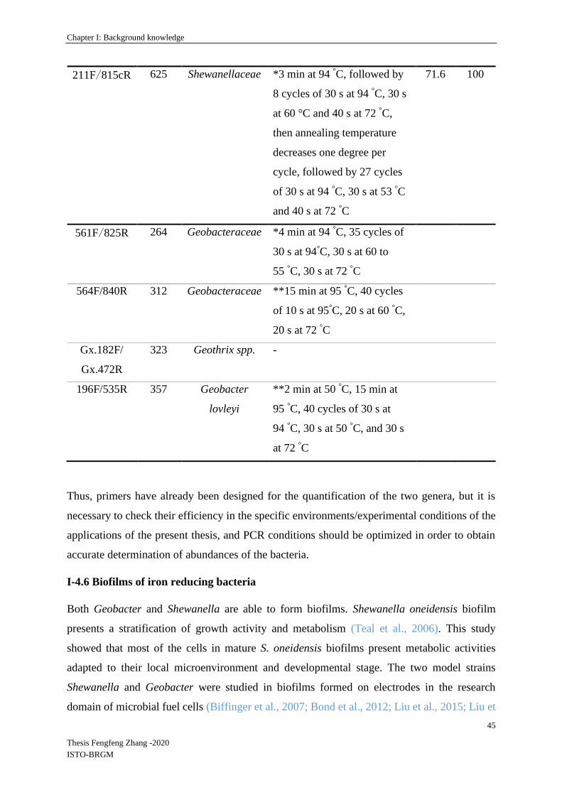
Chapter I: Background knowledge
45
Thesis Fengfeng Zhang -2020
ISTO-BRGM
211F/815cR 625 Shewanellaceae *3 min at 94 °C, followed by
8 cycles of 30 s at 94 °C, 30 s
at 60 °C and 40 s at 72 °C,
then annealing temperature
decreases one degree per
cycle, followed by 27 cycles
of 30 s at 94 °C, 30 s at 53 °C
and 40 s at 72 °C
71.6 100
561F/825R 264 Geobacteraceae *4 min at 94 °C, 35 cycles of
30 s at 94°C, 30 s at 60 to
55 °C, 30 s at 72 °C
564F/840R 312 Geobacteraceae **15 min at 95 °C, 40 cycles
of 10 s at 95°C, 20 s at 60 °C,
20 s at 72 °C
Gx.182F/
Gx.472R
323 Geothrix spp. -
196F/535R 357 Geobacter
lovleyi
**2 min at 50 °C, 15 min at
95 °C, 40 cycles of 30 s at
94 °C, 30 s at 50 °C, and 30 s
at 72 °C
Thus, primers have already been designed for the quantification of the two genera, but it is
necessary to check their efficiency in the specific environments/experimental conditions of the
applications of the present thesis, and PCR conditions should be optimized in order to obtain
accurate determination of abundances of the bacteria.
I-4.6 Biofilms of iron reducing bacteria
Both Geobacter and Shewanella are able to form biofilms. Shewanella oneidensis biofilm
presents a stratification of growth activity and metabolism (Teal et al., 2006). This study
showed that most of the cells in mature S. oneidensis biofilms present metabolic activities
adapted to their local microenvironment and developmental stage. The two model strains
Shewanella and Geobacter were studied in biofilms formed on electrodes in the research
domain of microbial fuel cells (Biffinger et al., 2007; Bond et al., 2012; Liu et al., 2015; Liu et
Chapter I: Background knowledge
46
Thesis Fengfeng Zhang -2020
ISTO-BRGM
al., 2019b). With Geobacter, flagella are widely expressed in anode biofilms and increases the
electron diffusion rate within the electroactive biofilm (Liu et al., 2019b). Reguera et al. (2007)
showed that Geobacter sulfurreducens needed the expression of electrically conductive pili to
grow as biofilms on Fe(III)-oxide surfaces, but pili were also necessary for the development of
the biofilm on a glass surface in presence of fumarate as an electron acceptor (Reguera et al.,
2007). These authors used a method developed by van Schie and Fletcher (1999) to develop
biofilms of pure Geobacter strains onto glass coverslips: glass slides surfaces with attached
amorphous Fe(III) oxide were obtained by dipping clean coverslips into a siliconizing agent
then coating coverslips by incubating them in a solution of FeCl3 for 12 h under gentle agitation,
then washed several times in distilled water followed by 0.1 mM NaCl, and air dried (van Schie
and Fletcher, 1999). The resulting preparations were sufficiently optically transparent to allow
light microscopy. Most studies of biofilms of iron-reducing bacteria were performed with pure
individual strains.
I-4.7 Studies involving complex iron reducing microbial communities
A few research studies reported results obtained with complex microbial communities reducing
iron oxides. Lentini et al. (2012) performed experiments to determine which proc
esses and microbial groups were responsible for the reduction of crystalline Fe(III) oxides
within sedimentary environments and how the type of Fe mineral could influence microbial
populations (Lentini et al., 2012). These authors performed cultures with several Fe(III) oxides
(ferrihydrite, goethite, hematite) and organic substrates (glucose, lactate, acetate) along a
dilution gradient to enrich microbial populations able to reduce Fe oxides displaying a wide
range of crystallinities. The type of electron donor was the most important factor influencing
community structure, that also varied with the nature of the Fe(III)-mineral. The availability of
carbon sources distinct from acetate induced the development of sulfate-reducing bacteria, that
could be able to indirectly dissolve Fe(III)-minerals through the production of H2S, whereas
acetate alone induced the dissolution of ferrihydrite and the development of Geobacter. When
glucose and lactate were provided, all Fe oxides were reduced and this reduction occurred with
the development of fermentative (e.g., Enterobacter spp.) and sulfate-reducing bacteria (e.g.,
Desulfovibrio spp.).
Hori et al. (2015) obtained IRB enrichments from diverse environments with only acetate as an
electron donor (Hori et al., 2015). A two-stage cultivation method was applied to selectively
enrich and isolate crystalline Fe(III) oxide reducing microorganisms from soils and sediments
Chapter I: Background knowledge
47
Thesis Fengfeng Zhang -2020
ISTO-BRGM
by 2-years successive culture on the crystalline ferric iron oxides goethite, lepidocrocite,
hematite, or magnetite as electron acceptors. High-throughput Illumina sequencing and clone
library analysis based on 16S rRNA genes showed that the enrichment cultures contained
bacteria belonging to the Deltaproteobacteria (mainly Geobacteraceae), followed by
Firmicutes and Chloroflexi. At the end of this research program, the chosen electron donor
(acetate) had favored the selection and isolation of organisms mainly belonging to the
Geobacter genus (five strains) and one strain belonging to the Pelobacter genus.
I-5 Mechanisms of microbial Fe(III) reduction
Fe microbial reduction, also known as Fe respiration, refers to the microbial extracellular
insoluble Fe oxide used as the terminal electron acceptor by bacteria. In this mechanism,
oxidation of an electron donor is coupled with Fe(III) reduction, and this process is required for
energy storage in microbial metabolisms (Tor and Lovley, 2001). The reduction product, Fe(II),
of the dissimilatory Fe(III) reduction process, accumulates outside the cell and does not enter
the cell as a cellular component. Moreover, in zones of oxic to anoxic transitions, Fe(III)
(oxyhydr)oxides reductively dissolve via a number of direct and indirect microbial pathways.
It can serve as a terminal electron acceptor for oxidation of organic matter by heterotrophic
bacteria respiring Fe(III) as described before. Fe(III) reduction is also a minor pathway for
electron flows in fermentative microorganisms (Lovley, 1987). In many soils, sediments and
groundwaters, ferric iron is a major potential electron acceptor for the oxidation of organic
matter. The concentration of ferric Fe (aq), is however limited by low solubility of Fe(III)
oxyhydroxides under circumneutral pH conditions compared with other terminal electron
acceptors, such as nitrate and sulfate. Consequently, strategies for transferring electrons to the
structural Fe(III) centers of the solid phases by some Fe reducing microorganisms have been
researched according to previous studies (Bonneville, 2005).
In order to know the details of the metabolic pathways and microbial Fe reduction strategies, it
is important to understand the microbe-mineral interactions. Iron and manganese redox-active
minerals are abundant in soils, aquatic and subsurface sediments, in which they support
microbial growth through at least four different pathways which are shown in Figure I-10
according to Shi et al., 2016. For solid Fe(III) reduction, biofilm formation is the major point
to connect iron reducers and solid Fe surfaces, which is the basis for current theories on electron
transfer, e.g., extracellular electron transport (EET) (Ding et al., 2016; Okamoto et al., 2014)
including the roles of extracellular polymeric substances (EPS) (Rollefson et al., 2011; Yang et

Chapter I: Background knowledge
48
Thesis Fengfeng Zhang -2020
ISTO-BRGM
al., 2019) and direct electron transfer between microbial cells through conductive nanowires
produced by organisms in response to electron acceptor limitation (Cologgi et al., 2011; Gorby
et al., 2006; Reguera et al., 2005).
Figure I-11. Electrical interactive processes between microorganisms and minerals.
Microorganisms use minerals that contain metal ions as terminal electron acceptors for
respiration (part a), electron and/or energy sources for growth (part b), electrical conductors
that facilitate electron transfer between microbial cells of the same and different species (part
c) and electron-storage materials, or batteries, to support microbial metabolism (part d).
In the absence of O2, Geobacter metallireducens GS‑15 and Shewanella oneidensis MR‑1 have
been reported to use Fe-minerals as electron sinks for heterotrophy-based respiration. In this
process, either organic matter or hydrogen (H2) are oxidized, then the released electrons are
transferred to Fe or Mn minerals (Lovley and Phillips, 1988; Lovley et al., 1987). Moreover,
the bacterium Geobacter sulfurreducens PCA was reported to be able to transfer electrons from
minerals to nitrate reducing bacteria to reduce nitrate, which was also called interspecies
electron transfer (IET) (Kato et al., 2012). Finally, some minerals function as electron storage
during the procedure of electron transferring between Fe(II)-oxidizing bacteria and Fe(III)-
reducing bacteria (Byrne et al., 2015).
In these four different cell-mineral interactions, Figure I-11 (a) and (d) present microbial Fe
reduction. Detailing these two pathways, Jacintha Esther et al. (2015) summarized four
mechanisms given in Figure I-12 that have been conceptualized for the electron transfer
between bacterial cells and mineral surface (Childers et al., 2002; Esther et al., 2015).
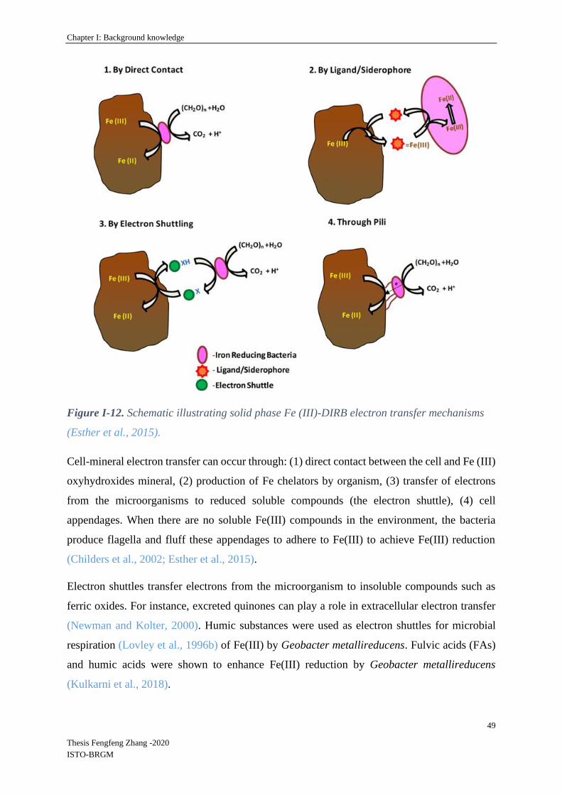
Chapter I: Background knowledge
49
Thesis Fengfeng Zhang -2020
ISTO-BRGM
Figure I-12. Schematic illustrating solid phase Fe (III)-DIRB electron transfer mechanisms
(Esther et al., 2015).
Cell-mineral electron transfer can occur through: (1) direct contact between the cell and Fe (III)
oxyhydroxides mineral, (2) production of Fe chelators by organism, (3) transfer of electrons
from the microorganisms to reduced soluble compounds (the electron shuttle), (4) cell
appendages. When there are no soluble Fe(III) compounds in the environment, the bacteria
produce flagella and fluff these appendages to adhere to Fe(III) to achieve Fe(III) reduction
(Childers et al., 2002; Esther et al., 2015).
Electron shuttles transfer electrons from the microorganism to insoluble compounds such as
ferric oxides. For instance, excreted quinones can play a role in extracellular electron transfer
(Newman and Kolter, 2000). Humic substances were used as electron shuttles for microbial
respiration (Lovley et al., 1996b) of Fe(III) by Geobacter metallireducens. Fulvic acids (FAs)
and humic acids were shown to enhance Fe(III) reduction by Geobacter metallireducens
(Kulkarni et al., 2018).

Chapter I: Background knowledge
50
Thesis Fengfeng Zhang -2020
ISTO-BRGM
Normally, the electron shuttles promoting the electron transfer during microbial Fe(III)
reduction come from natural organic compounds such as humic acids and fulvic acids (FAs)
(as electron acceptors), or analog quinones produced by bacteria. They are measured indirectly
through the release of Fe(II) (Newman and Kolter, 2000) or by other indirect ways such as
analyze of the electron accepting capacity.
I-6 Cycling of Fe and mobility of associated As, Cr, Cd and other trace
elements
Mobility of contaminants in the environment, and particularly that of toxic metals and
metalloids, such as As, Cr and Cd, induces risks of contamination of hydrosystems and food
chain (Beesley et al., 2010; Kumpiene et al., 2008). These elements can be present in soils and
sediments naturally enriched in metal trace elements (MTE) through the local geology, or
contaminated by diverse human activities (mines, industries, agriculture) (Flues et al., 2013;
Prokop et al., 2003). And MTEs are often associated with Fe in soils and sediments. As
described in previous section, Fe-rich minerals can change according to oxygen availability and
level in the environment. They are generally stable and highly insoluble in the form of
(oxyhydr)oxides (e.g., Fe(OH)3, FeOOH, Fe2O3) in well oxygenated zones at circumneutral pH
conditions. However, in anaerobic conditions, these minerals can be reductively dissolved
(Roden et al., 2004; Roden and Wetzel, 2002) by microbial and abiotic pathways (Bonneville
et al., 2004; Hansel et al., 2004; Shi et al., 2016; Thompson et al., 2006b). The dissolution of
Fe-rich minerals, in absence of oxygen, could be linked to bacterial activities in soils or
sediments. These phenomena were well described in controlled laboratory in liquid medium
conditions between iron (oxyhydr)oxides and IRB, however the evaluation of associated
pollutants fluxes, in real sediment conditions, need the development of multi-scale experiments.
I-6.1 Association of As, Cr and Cd with Fe-Oxides
The mobility of As associated with iron oxides depends on its speciation and pH (Dixit and
Hering, 2003). According to these authors, below pH 5-6, As(V) sorbs more readily to
amorphous iron oxides (HFO) than does As(III). Wang et al. (2003) state that the residual
concentration of As(V), after co-precipitation with iron hydroxide, is minimized in the pH 4-6
interval (Wang et al., 2003). Above pH 7-8, As(III) has a higher affinity for the solids (Dixit
and Hering, 2003). Wang et al. (2003) found that the residual concentration of As(III), after co-
precipitation with iron hydroxide, was minimized in the pH 6-9 interval. The toxicity of
Chapter I: Background knowledge
51
Thesis Fengfeng Zhang -2020
ISTO-BRGM
different forms of arsenic is quite different. The oxidation or methylation of arsenic generally
reduces its toxicity.
Cr(III) is poorly soluble and relatively nontoxic, while Cr(VI) is soluble and a known
carcinogen. Solid Fe(II) in iron-bearing minerals, such as pyrite, magnetite, and green rusts,
reduce the oxidation state of chromium, reducing its toxicity and mobility. Under oxic
conditions, solid Fe(II) associated with goethite resulting from rapid redox cycling is reactive
and available for electron transfer to Cr(VI), suggesting Fe(III) (hydr)oxides may act as
reservoirs of reactive electron density, even in oxygen saturated environments (Tomaszewski
et al., 2017).
Cd can be adsorbed by Fe(III) (oxyhydr)oxides and its adsorption on goethite and ferrihydrite
is enhanced by the presence of sulfate with metal-ligand bonding (Swedlund et al., 2003;
Swedlund et al., 2009). Zhang et al. (2016) studied whether crystalline structures influence the
adsorption of heavy metals on magnetic iron oxides metals and suggested adsorption of Cd by
amorphous iron oxide was superior to that of oxides with higher degrees of crystallization
(Zhang et al., 2016).
I.6.2 Transformations of As, Cr and Cd by bacteria
Bacteria are abundant in many environments and interact with iron oxides and As through
different mechanisms, e.g., sorption, mobilization, precipitation and redox and methylation
transformation (Huang, 2014). Some bacteria can use either Fe(III) or As(V) as electron
acceptors. Laverman et al. (1995) have found that some iron-reducing bacteria such as SES-3
reduce As(V) to As(III) (Laverman et al., 1995).
When dissimilatory metal reducing bacteria Shewanella alga strain BrY, Shewanella
putrefaciens strain CN32, Shewanella oneidensis strain MR-1, and Geobacter metallireducens
strain GS-15 were used to study metal reduction in presence of H2 used as an electron donor,
reduction rates were metal specific with the following decreasing rate order: Fe(III)citrate ≥
Fe(III)NTA > Co(III)EDTA− > UO22+ > CrO4
2− > TcO4−, except for CrO4
2− (Liu et al., 2002).
Independently from Fe transformations, diverse bacterial isolates from soil and sediment have
been shown to oxidize As(III) and/or reduce As(V) (Bachate et al., 2012; Inskeep et al., 2007;
Macur et al., 2004), or to methylate this toxic metalloid (Huang et al., 2011). These widespread
bio-reactions can be either linked to resistance mechanisms or coupled to growth though
energetic metabolisms, lithotrophic growth on As(III) or dissimilatory reduction of As(V).
Chapter I: Background knowledge
52
Thesis Fengfeng Zhang -2020
ISTO-BRGM
During the transformation of lepidocrocite to magnetite and green rust, As(III) could be
adsorbed on both lepidocrocite and green rust, whereas As(V) was associated only with green
rust. Morever, As(III) formed surface complexes on magnetite nanoparticles and As(V) was
thought to have been incorporated into the magnetite structure when magnetite precipitated
(Ona-Nguema wt al., 2009; Wang et al., 2010; Wang et al., 2014).
Among oxidized forms of chromium, the anionic hexavalent chromium Cr(VI) is, highly
soluble and toxic (Petrilli and De Flora, 1977), whereas the cationic trivalent form Cr(III), much
less toxic, plays a physiological role in mammals by participating in glycaemia regulation
(Schroeder, 1968) and tends to form insoluble hydroxides. Cr(III) is dominant in most
environmental compartments. Diverse microorganisms reduce Cr(VI) directly into Cr(III)
under either anaerobic (Turick and Apel, 1997) or aerobic (Wang and Chirwa, 1996) conditions.
When growth is anaerobic, the reduction might result in respiration (Tebo and Obraztsova,
1998), Cr(VI) resistance, or may be a non-specific enzymatic reaction.
Cadmium is not directly transformed by bacteria, however micro-organisms can indirectly
change the forms of Cd through binding to microbial biomass, or to microbial metabolism
products (Czaban and Wróblewska, 2005; Zhou et al., 2020). When sulfate-reduction activity
occurs, Cd can be precipitated as sulfide CdS, however sorption by bio-polymers produced by
sulfate-reducing bacteria also contributes to Cd immobilization (Pérez et al., 2015).
I-6.3 Mobility of trace elements during Fe-oxides microbial dissolution
A range of research studies provide information on the behavior of trace elements during the
reductive dissolution of iron oxides. Some were performed with model minerals and bacteria,
and others with real soils or sediments containing trace elements and complex bacterial
communities.
• Experiments with defined Fe-oxides
Solubilisation of synthetic goethite-bound Co or Ni by Shewanella was observed in batch
systems (Zachara et al., 2001). This study showed that the two metals can be mobilized in the
aqueous phase during bacterial reduction.
Impact of bio-reduction on remobilization of adsorbed cadmium on Fe minerals including
hematite, goethite, and two iron(III)‐rich clay minerals in anoxic condition was studied by
Ghorbanzadeh et al. (2017) in batch experiments. The desorption of Cd(II) was related to

Chapter I: Background knowledge
53
Thesis Fengfeng Zhang -2020
ISTO-BRGM
production of Fe(II) as a result of bio-reduction of the minerals by Shewanella (Ghorbanzadeh
et al., 2017).
Recently, Zhou et al. (2020) studied the influence of the concentration of natural organic matter
on the mobility of Cd during bioreduction of ferrihydrite by Geobacter. They showed that Cd
was immobilized by bonding with organic matter during the reductive dissolution of ferrihydrite
(Zhou et al., 2020).
Indirect dissolution of Fe-oxides by the H2S produced by SRB can also lead to the mobilization
of traces elements. The two mechanisms, direct Fe-oxide reduction by IRB and indirect Fe-
oxide reduction by biogenic H2S were studied in continuously fed columns filled with synthetic
Fe oxides spiked with Hg(II) (Hellal et al., 2015) and inoculated with microbial enrichments.
In the column where IRB were dominant, Hg mobilization was directly correlated to bacterial
Fe reduction. Conversely, when SRB were dominant, the biologically produced sulfide induced
indirect iron oxide reduction and rapid adsorption of the mobilized Hg on neo-formed iron
sulfides or its precipitation as HgS. Moreover, secondary minerals formed during iron reduction
can interact with the trace elements: reduction of HgII to Hg° by biogenic green rust produced
by microbial reduction of lepidocrocite was observed (Remy et al. 2015).
These studies provide useful information on the behavior of individual trace elements during
the dissolution of defined Fe(III)-oxides. However, data are lacking about the behavior of
systems simultaneously contaminated with several trace elements. Moreover, most of these
results were obtained with single pure IRB strains.
• Experiments with naturally complex materials
Fe oxides and mercury solubilization in tropical oxisols was studied in presence of
autochthonous ferri-reducing bacteria (Harris-Hellal et al., 2011. During bacterial growth in
batch conditions, no mercury was detected in the culture medium, however, chemical analysis
showed a decrease of the amounts of Hg associated to amorphous and well crystallized Fe-
oxides after 14 days of incubation, underlining the potential for iron-reducing bacteria to modify
mercury distribution in soil.
Muehe et al. (2013) provided evidence that secondary Fe(II) and Fe(II)/Fe(III) mixed minerals
could be a sink for Cd in soils under reducing conditions, thus decreasing the mobility of Cd in
the soil. By testing the mobility of Cd changes when a Cd-bearing soil was faced with organic
carbon input and reducing conditions (Muehe et al., 2013).

Chapter I: Background knowledge
54
Thesis Fengfeng Zhang -2020
ISTO-BRGM
While imposing a decrease of redox potential in a stirred bioreactor filled with a slurry of multi-
metals contaminated soil, Hindersmann and Mansfeldt (2014) observed the behavior of Fe and
a range of trace elements including As, Cd and Cr (Hindersmann and Mansfeldt, 2014). A
moderate release of Fe(II) (from undetectable to 1 mg L-1) was accompanied by the mobilization
of 2 µg L-1 Cd, 10 µg L-1 As and 10 µg L-1 Cr (Zachara et al., 2001).
Other experiments in batch bioreactors submitted clay soils contaminated with trace elements
to successive steps of oxic and anoxic conditions. Mn, Fe, and As were mobilized under anoxic
conditions, whereas Sb, Se, and U were mobilized under oxic conditions (Couture et al., 2015).
Fan et al. (2018) demonstrated with batch laboratory incubation experiments that SRB were
able to reductive dissolve high levels of arsenical ferric (oxyhydr)oxides in mine tailing
sediments at the soil/water interface. Their results showed that reactions with aqueous As(III)
and As(V) presented different trends, with As(III) being the dominant arsenic species. Aqueous
As behavior showed two distinct stages, first As(V) was released and then immediately reduced
to As(III) by biogenic sulfide (Fan et al., 2018a).
All these experiments demonstrate correlations between the mobility of trace elements and
reducing conditions that allow iron oxide bio-dissolution. There is a lot of data on these
mechanisms, however it’s difficult to access to a clear distinction between several mechanisms.
These subjects such as biological Fe(III) or sulfate reduction, and between the roles of the
different mineral and organic phases in the final distribution of metals and metalloids are still
needed to be verified.
I-7 Positioning of the PhD thesis in regards to the state of the art
This overview of the available literature on the subject of Fe(III)-oxides bio-reduction and the
fate of associated trace elements showed that detailed and abundant information is available on
(1) simplified model laboratory processes combining pure minerals and single bacterial strains
or (2) microcosms implemented with real complex soils or sediments, with their natural
complete microbial communities. However, a gap exists in the knowledge acquired in these two
radically different conditions. More specifically, the following research needs were identified:
- Only a few studies looked at mixed IRB communities, most research was performed
with model strains of Shewanella or Geobacter, not grown together but separately as
pure strains.
Chapter I: Background knowledge
55
Thesis Fengfeng Zhang -2020
ISTO-BRGM
- When previous studies were performed with natural mixed bacterial communities in
laboratory Fe-reducing experiments, the occurrence/dynamics of Shewanella and
Geobacter were not monitored.
All the knowledge acquired on these two organisms would be beneficially exploited if we could
understand how they are present and function in competition with other microbes in complex
communities.
- From the point of view of mineralogy, data is scarce about the formation of secondary
minerals during Fe(III)-oxides reduction in presence of complex bacterial communities,
starting with different types of oxides, most of the information was acquired with model
strains.
- The trace elements associated to Fe(III)-oxides are studied separately in studies with
model strains and minerals. When several elements are present, the studies were
performed with natural soils or sediments, thus the trace-elements-oxides proportion
and association is not controlled. Data about the relative behavior of several TE
simultaneously present in controlled conditions are lacking.
- Biofilms play a major role in natural environments, therefore biofilms of IRB attached
on iron oxides still need to be further studied, in particular biofilms of mixed IRB
populations were not described, and no data is available on the influence of biofilm
development on the proportion of different IRB.
Globally, the analysis of the state of the art reveals a need for tools to study the transformation
of different Fe(III)-oxides in natural environments, with complex natural bacterial communities.
In this context, the present PhD thesis was focused on the following objectives:
(1) To determine what is the influence of the type of Fe(III)-oxide on the efficiency of Fe release
and formation of secondary minerals in presence of mixed iron-reducing bacterial communities
in conditions favoring a high diversity of Fe(III)-reducing bioprocesses (reduction and
fermentation),

Chapter I: Background knowledge
56
Thesis Fengfeng Zhang -2020
ISTO-BRGM
(2) To observe how the type of iron oxide influences the structure of these mixed communities,
and the proportion in these communities of the two well-known specific iron reducing bacterial
genera Shewanella and Geobacter, applied as indicator parameters,
(3) To develop experimental strategies in order to evaluate the proportion of these two iron-
reducing genera in the planktonic bacterial community and in biofilms formed on Fe(III)-oxides,
for laboratory and on-site studies,
(4) To describe the influence of the development of complex Fe(III)-reducing bacterial
communities on the dynamics of targeted trace elements As, Cr and Cd, co-adsorbed on
different Fe (oxyhydr)oxides, and the distribution of Shewanella and Geobacter in these
systems whose complexity is intermediary between fully controlled conditions (pure strains and
defined oxided) and natural complex environments.

Chapter II: General materials and methods
57
Thesis Fengfeng Zhang -2020
ISTO-BRGM
Chapter II: General materials and methods
The present chapter describes materials and methods that are common to all the experiments
performed during the PhD thesis. Information specific to each experiment is then detailed in
the corresponding chapters.
II-1 Site information and soil / sediment sampling
The place of study is located near the Loire, bordered by the town of Decize and by an alluvial
island formed by the river (Figure II-1). The Loire River basin (117,800 km2-1013 km long) is
among the ten largest W-European basins and is the largest in France (Dhivert et al., 2015). The
site chosen for the present work, a non-active Loire river tributary, is located in an environment
naturally subjected to oscillations of redox conditions according to the level of the river.
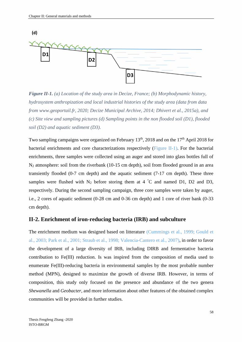
Chapter II: General materials and methods
58
Thesis Fengfeng Zhang -2020
ISTO-BRGM
Figure II-1. (a) Location of the study area in Decize, France; (b) Morphodynamic history,
hydrosystem anthropization and local industrial histories of the study area (data from data
from www.geoportail.fr, 2020; Decize Municipal Archive, 2014; Dhivert et al., 2015a), and
(c) Site view and sampling pictures (d) Sampling points in the non flooded soil (D1), flooded
soil (D2) and aquatic sediment (D3).
Two sampling campaigns were organized on February 13th, 2018 and on the 17th April 2018 for
bacterial enrichments and core characterizations respectively (Figure II-1). For the bacterial
enrichments, three samples were collected using an auger and stored into glass bottles full of
N2 atmosphere: soil from the riverbank (10-15 cm depth), soil from flooded ground in an area
transiently flooded (0-7 cm depth) and the aquatic sediment (7-17 cm depth). These three
samples were flushed with N2 before storing them at 4 °C and named D1, D2 and D3,
respectively. During the second sampling campaign, three core samples were taken by auger,
i.e., 2 cores of aquatic sediment (0-28 cm and 0-36 cm depth) and 1 core of river bank (0-33
cm depth).
II-2. Enrichment of iron-reducing bacteria (IRB) and subculture
The enrichment medium was designed based on litterature (Cummings et al., 1999; Gould et
al., 2003; Park et al., 2001; Straub et al., 1998; Valencia-Cantero et al., 2007), in order to favor
the development of a large diversity of IRB, including DIRB and fermentative bacteria
contribution to Fe(III) reduction. Is was inspired from the composition of media used to
enumerate Fe(III)-reducing bacteria in environmental samples by the most probable number
method (MPN), designed to maximize the growth of diverse IRB. However, in terms of
composition, this study only focused on the presence and abundance of the two genera
Shewanella and Geobacter, and more information about other features of the obtained complex
communities will be provided in further studies.

Chapter II: General materials and methods
59
Thesis Fengfeng Zhang -2020
ISTO-BRGM
In order to obtain Fe-reducing enrichments, 10 g of each soil and sediment sample (D1, D2 and
D3, see chapter II-1) were inoculated (1 week after sampling on site) into 200 mL basic medium
(composition detailed in the Table II-1) autoclaved (121°C, 20 min) then flushed with sterile N2
just after autoclaving. Sodium molybdate was added in the medium as a SRB inhibitor, because
the present study was focused on Fe cycling so it was preferred to inhibit sulfate respiration
metabolism. The headspace of vials (small volume because 200 mL bottles were used) was N2.
The following components were added to this medium: 10 mM of Fe(III) Nitrilotriacetic Acid
(NTA) as electron acceptor (Huguet, 2009; Lovley, 2006), 1.5 g.L-1 peptone, 10 mM of acetate,
lactate, and formate, 2 mM glucose as electron donors (Coates et al., 1996; Lovely et al., 1989;
Kwon et al., 2016; Shelobolina et al., 2007) in anaerobic conditions, and 0.4 mM of sodium
molybdate (these concentrations are very high and very different from the natural conditions).
Fe(III)-NTA (100 mM stock solution) was prepared by dissolving 1.64 g of NaHCO3 in 80 ml
water, adding 2.56 g C6H6NO6Na3 and 2.7 g FeCl3·6H2O, bringing the solution up to 100 ml,
flushing with N2 and filter sterilizing (0.22 µm, Millex -GP Syringe Filter, 33 mm diameter)
into a sterile, anaerobic serum bottle. Sterilization of the electron donors was performed by
autoclaving for acetate, lactate and formate, and filtration at 0.22 µm for peptone and glucose.
Sodium molybdate was autoclaved. All stock solutions were kept anaerobic under N2 after
sterilization. Cultures were incubated at 20°C under agitation (100 rpm) for 10 days. Then, they
were sub-cultured by inoculating at 10% (v/v) 50 mL of the same medium, under N2. Samples
(1.5-2 mL) were collected every day, in anaerobic conditions, filtered at 0.22 µm (Millex -GP
Syringe Filter, 33 mm diameter) and analyzed for Fe(II) content in order to evaluate Fe(III)
reduction. After 3-5 steps of sub-culturing (inoculation at 10% in fresh medium, every two
weeks), the three Fe-reducing cultures were able to reduce 10 mM Fe(III)-NTA in 1-2 days,
and were used as inocula for the following batch incubation experiments in slurry (chapter III),
slide experiments (chapter III) and column experiments (chapter IV). In order to maintain the
cultures activity, the enrichments were sub-cultured once in 1-2 weeks.
Table II-1. Composition of the enrichment medium (Cummings et al., 1999; Gould et al.,
2003; Park et al., 2001; Straub et al., 1998; Valencia-Cantero et al., 2007).
Basic solution Electron donors
(added as 10X
concentrated
Fe-NTA solution *SRB inhibitor
(Added in the
concentrated

Chapter II: General materials and methods
60
Thesis Fengfeng Zhang -2020
ISTO-BRGM
solution, filtered
0.22 µm)
(added as 10X
concentrated solution,
filtered 0.22 µm)
electron donor
solution, filtered
0.22 µm)
NaHCO3: 2.5 g L-1;
NH4Cl: 0.25 g L-1;
KCl: 0.1 g L-1;
NaH2PO4∙H2O: 0.6
g L-1;
10 mL L-1 of
vitamins solution*
and trace elements
solution*
Final
concentrations:
Acetate, lactate and
formate: 10 mM of
each;
Glucose: 2 mM;
Peptone: 1.5 g L-1
Final concentration, 10
mM:
NaHCO3: 1.64 g L-1,
C6H6NO6Na3 (sodium
nitrilotriacetic acid):
2.56 g L-1, and
FeCl3·6H2O: 2.7 g L-1
Sodium
molybdate:
0.04 mM
*SRB : sulfate-reducing bacteria; *Vitamins solution (1L): 2 mg biotin, 2 mg folic acid, 10 mg
pyridoxine HCL, 5 mg Riboflavin,5 mg Thiamine, 5 mg Nicotinic acid, 5 mg Pantothenic acid, 0.1g B12
vitamin, 5 mg p-aminobenzoic acid, 5 mg thioctic acid ; * Trace elements solution (1L): 1.5 g Trisodium
nitrilotriacetic, 0.5 g MgSO4, 0.5 g MgSO4∙H2O, 1 g NaCl, 0.1 g FeSO4∙7 H2O, 0.1 g CaCl2∙2H2O, 0.1
g CoCl2∙6H2O, 0.13 g ZnCl2, 0.1 g CuSO4 5H2O, 0.1 g AlK(SO4)∙12H2O, 0.1 g H3BO3, 0.25 g NaMoO4,
0.24 g NiCl2∙6H2O, 0.25 g Na2WO4∙2H2O. Note: (Fe-NTA solution needs to be purged with N2 gas and
filter-sterilized into a sterile, then stored in anaerobic serum bottle)
II-3 Iron (oxyhydr)oxides and laboratory synthesis
II-3.1 Ferrihydrite
Ferrihydrite and lepidocrocite were synthesized in the laboratory using the modified protocol
of Schwertmann and Cornell, (2008). Ferrihydrite was obtained by dissolving 40 g Fe(NO3)3∙
9H2O (M = 404.00 g mol-1) in 500 mL distilled water and adding 330 mL of 1 M KOH (M =
56.11 g mol-1) to adjust the pH to 7-8. The mixture was centrifuged at 5,000 rpm for 10 min
and the supernatant was subsequently removed. The solid fraction was then washed 5 times
with ultrapure water according to MilliQ quality. Then the ferrihydrite was freeze dried at -90
◦C (Lyophilizer Christ Beta 2-8).

Chapter II: General materials and methods
61
Thesis Fengfeng Zhang -2020
ISTO-BRGM
II-3.2 Lepidocrocite
Lepidocrocite was prepared with 11.93 g of unoxidized FeCl2∙4H2O (60 mmol Fe) salts
dissolved into 300 mL distilled water by stirring. The solution was adjusted to pH 6.7-6.9 with
1M NaOH using a pH-stat under aeration (100 mL min-1 air). The experimental arrangement
consisted of a 1-2 L glass beaker equipped with a stirrer, a pH electrode, a gas inlet and a burette
(containing 1 M NaOH) with the outlet just above the solution. If the crystals had an ochreous
tinge, the solution was filtered to remove any akaganeite precipitations. pH of the acidic
solution was adjusted to pH 6.7-6.9 with NaOH. During oxidation, the color of the suspension
changed from dark greenish blue to grey and finally to orange. Protons produced during the
oxidation/ hydrolysis reaction were constantly neutralized by NaOH added with the burette.
After consumption of 120 mL NaOH, oxidation was completed and the pH remained constant
without further addition of an alkaline solution. The entire reaction took 2-3 hr. Washing and
drying were performed as described for ferrihydrite.
II-3.3 Goethite and hematite
Goethite and hematite were ordered on line. Goethite was from Sigma-Aldrich (CAS No.
20344-49-4) and hematite was from VWR Chemicals (CAS No. 1309-37-1).
II-4 Physico-chemical analysis: pH, Eh, Fe(II)/FeT, As, Cr and Cd
pH and redox potential (Eh, ref. Ag/AgCl) were measured immediately after sampling using
standard hand-held portable meters (WTW Multi340i set) in the glove box for the slide and
column experiments.
During all the experiments described in the following chapters, 1.5 -10 mL aliquots were
collected with a syringe and filtered through a 0.2 μm filter (Millex -GP Syringe Filter, 33 mm
diameter) into sterile plastic tubes and acidified with concentrated HCl immediately in the glove
box before further analysis.
[FeII]D (the dissolved Fe(II) concentration) was measured using the ortho-phenanthroline
colorimetric method (Mamindy-Pajany et al., 2013; Murti et al., 1966). Analyses were
performed in Spectrophotometer cuvettes, 10 mm, into which were added 0.5 mL acetate buffer
(pH 4.5), 0.1 mL ortho-phenantroline 0.5%, and 0 to 0.9 mL of filtered sample. The mixture
was left at ambient temperature for 15 min in the dark, then 1 mL of buffer was added and the
total volume was adjusted to 2.5 mL with demineralized water. After mixing, the absorbance at

Chapter II: General materials and methods
62
Thesis Fengfeng Zhang -2020
ISTO-BRGM
510 nm was read with the UV-Visible spectrophotometer (Agilent G9821A CARY 100 UV-
Vis Spectrophotometer). A standard curve ranging between 0.1 and 5 mg L-1 [FeII]D and
prepared in the same conditions was used to convert absorbance measures.
[FeT]D (total dissolved iron concentration) was determined using the same method but with the
addition of 0.1 mL of 1% hydroquinone prepared in acetate buffer to reduce dissolved [FeIII]D
into [FeII]D, which was added in Spectrophotometer cuvettes before analyze.
In filtered samples, [As]D was determined with Atomic Absorption Spectroscopy (AAS, Varian
SpectrAA 220-Z) and [Cr]D and [Cd]D were determined by Microwave Plasma-Atomic
Emission Spectrometry (Agilent 4210 MP-AES).
II-5 BET surface areas
BET surface areas were determined by multipoint BET N2 adsorption (Brunauer et al., 1938)
in ISTO laboratory (University of Orléans). Specific surface areas were determined from N2
adsorption isotherms in the best linear range (with a minimum of 15 points) between the relative
pressure P/Po 0.03 and 0.33 (Cavelan et al., 2019).
II-6 SEM-EDS and SEM observations
II-6.1 Observation of iron (oxyhydr)oxides
Scanning electron microscopy (SEM) and energy dispersive X-ray spectroscopy (EDS) were
used for a general description of the sample (morphological view), and provided an
identification of elements (EDS microanalysis). SEM-EDS was performed to explore the
composition and distribution of Fe, TEs (As, Cr, Cd), and other elements in Fe (oxyhydr)oxides
and experimental samples. SEM was performed on a TM 3000 accompanied by a SwiftED3000
X-Stream module (Hitachi), and operated at 15 kV accelerating voltage, without any coating
needed (Thouin et al., 2016). SwiftED3000 provides standardless quantitative analyses,
normalized to 100%. The AZtecEnergy Analyser Software displays and interprets X-ray data
to provide accurate and reliable analysis without standard (Burgess et al., 2007). The acquisition
time of EDS point analyses was 300 s. All post-incubation samples were freeze dried and
observed by the same method described before from the following IRB incubation experiments
(chapter III), slide experiments (chapter III) and column experiments (chapter IV).
Chapter II: General materials and methods
63
Thesis Fengfeng Zhang -2020
ISTO-BRGM
II-6.2 Bacteria Observations
Frozen samples were observed under cryo-SEM conditions using the backscattered detector
imaging (BSE) mode and Secondary electrons imaging (SE) mode. The SEM was equipped
with a Peltier cryo-stage at a temperature of -50 ◦C, under low vacuum conditions (nitrogen
pressure 80 Pa). Morphologies of bacteria and bacterial biofilms are presented for slide (chapter
III) and column experiments (chapter IV).
II-7 57Fe Mössbauer spectrometry
Two series of Fe oxides samples from the following incubation experiments (chapter III) and
column experiments (chapter IV) were freeze dried and sent to the University of Tübingen to
perform Mössbauer spectrometry.
Samples were loaded into Plexiglas holders (1 cm²). Samples were then transferred to the
instrument and loaded inside a closed-cycle exchange gas cryostat (Janis cryogenics).
Measurements were collected at 140 K with a constant acceleration drive system (WissEL) in
transmission mode with a 57Co/Rh source and calibrated against a 7 µm thick α-57Fe foil
measured at room temperature. All spectra were analyzed using Recoil (University of Ottawa)
by applying a Voight Based Fitting (VBF) routine (Rancourt and Ping, 1991). The half width
at half maximum (HWHM) was fixed to a value of 0.124 mm s-1 for all samples.
II-8 Diversity and physiology of bacteria
II-8.1 Observation and counting of bacteria by Thoma cell
Images of bacteria were obtained with an optical microscope (X400 magnification). Cells
were estimated using a Thoma cell counting chamber (N* 104 *f cell mL-1, N= counted cell
number, f = medium concentration/dilution factor).
II-8.2 DNA extraction and PCR amplifications
DNA extractions were performed on solid and liquid samples either from the site or the end of
three laboratory experiments (chapter III and V). For liquid samples, biomass was harvested by
centrifugation at 10,000 rpm for 10 min of 2 mL of culture. Extractions were performed with
the Fast DNA™ SPIN Kit for Soil (MP Biomedicals, USA) according to the manufacturer's
instructions. The integrity of the DNA products was checked using agarose gel electrophoresis.
DNA concentrations were determined with a Quantus™ Fluorometer (Promega, USA).
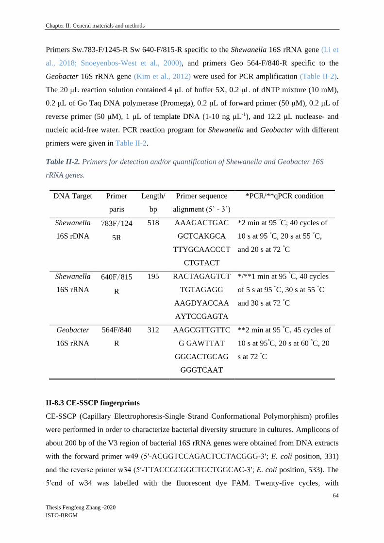
Chapter II: General materials and methods
64
Thesis Fengfeng Zhang -2020
ISTO-BRGM
Primers Sw.783-F/1245-R Sw 640-F/815-R specific to the Shewanella 16S rRNA gene (Li et
al., 2018; Snoeyenbos-West et al., 2000), and primers Geo 564-F/840-R specific to the
Geobacter 16S rRNA gene (Kim et al., 2012) were used for PCR amplification (Table II-2).
The 20 μL reaction solution contained 4 μL of buffer 5X, 0.2 μL of dNTP mixture (10 mM),
0.2 μL of Go Taq DNA polymerase (Promega), 0.2 μL of forward primer (50 μM), 0.2 μL of
reverse primer (50 μM), 1 μL of template DNA (1-10 ng μL-1), and 12.2 μL nuclease- and
nucleic acid-free water. PCR reaction program for Shewanella and Geobacter with different
primers were given in Table II-2.
Table II-2. Primers for detection and/or quantification of Shewanella and Geobacter 16S
rRNA genes.
DNA Target Primer
paris
Length/
bp
Primer sequence
alignment (5’ - 3’)
*PCR/**qPCR condition
Shewanella
16S rDNA
783F/124
5R
518 AAAGACTGAC
GCTCAKGCA
TTYGCAACCCT
CTGTACT
*2 min at 95 °C; 40 cycles of
10 s at 95 °C, 20 s at 55 °C,
and 20 s at 72 °C
Shewanella
16S rRNA
640F/815
R
195 RACTAGAGTCT
TGTAGAGG
AAGDYACCAA
AYTCCGAGTA
*/**1 min at 95 °C, 40 cycles
of 5 s at 95 °C, 30 s at 55 °C
and 30 s at 72 °C
Geobacter
16S rRNA
564F/840
R
312 AAGCGTTGTTC
G GAWTTAT
GGCACTGCAG
GGGTCAAT
**2 min at 95 °C, 45 cycles of
10 s at 95°C, 20 s at 60 °C, 20
s at 72 °C
II-8.3 CE-SSCP fingerprints
CE-SSCP (Capillary Electrophoresis-Single Strand Conformational Polymorphism) profiles
were performed in order to characterize bacterial diversity structure in cultures. Amplicons of
about 200 bp of the V3 region of bacterial 16S rRNA genes were obtained from DNA extracts
with the forward primer w49 (5′-ACGGTCCAGACTCCTACGGG-3′; E. coli position, 331)
and the reverse primer w34 (5′-TTACCGCGGCTGCTGGCAC-3′; E. coli position, 533). The
5′end of w34 was labelled with the fluorescent dye FAM. Twenty-five cycles, with

Chapter II: General materials and methods
65
Thesis Fengfeng Zhang -2020
ISTO-BRGM
hybridization at 61°C, and 30 s elongation at 72°C were used. 1 μL of diluted (20 or 50 fold in
nuclease-free water) PCR product was added to a mixture of 18.6 μL of deionized formamide
and 0.4 μL of Genescan-600 LIZ internal standard (Applied Biosystems). To obtain single-
strand DNA, samples were heat-denaturized for 5 min at 95 °C, and immediately cooled on ice.
CE-SSCP analyses were performed on an ABI Prism 310 genetic analyzer using a 47-cm length
capillary, a non-denaturating 5.6% CAP polymer (Applied Biosystems) and the following
electrophoresis conditions: run temperature 32°C, sample injection for 5 s at 15 kV, data
collection for 35 min at 12 kV.
II-8.4 Bacterial 16S rRNA gene quantification
Quantification of the bacterial 16S rRNA gene copies (abbreviated bacterial 16S), was
performed with primers 341-F (5'-CCT ACG GGA GGC AGC AG-3') and 515-R (5'-TGC
CAG CAG CCG CGG TAA T-3'), as described from Shewanella and Geobacter except 0.2 μL
T4GP32 at 500 ng·µL-1 were added into the reaction mixture. qPCR reaction program was as
follows: 3 min at 95°C, followed by 35 cycles: 30s at 95°C/30s at 60°C/30s at 72°C/30s at 80°C.
Plasmid DNA containing the 16S rRNA gene of Pseudomonas putida KT 2440 was 10-fold
serially diluted to obtain a calibration curve of known copy numbers of Pseudomonas putida
KT 2440 16S rRNA.
II-8.5 Detection of Shewanella and Geobacter
PCR conditions has been optimized in order to obtain accurate determination of abundances of
Shewanella and Geobacter.
Shewanella 16S and Geobacter 16S were performed by PCR using a CFX96 Touch™ Real-
Time PCR Detection System (Bio Rad, USA). Primers listed in Table II-2 specific to
Shewanella and Geobacter 16S rRNA genes were used for that. PCR reactions were performed
in a total volume of 20 μL containing solution containing 4 μL of buffer 5X, 0.2 μl of dNTP
mixture (10 mM), 0.2 μL of Go Taq DNA polymerase (GoTaq® Flexi DNA polymerase,
Promega), 0.2 μL of forward primer (50 μM), 0.2 μL of reverse primer (50 μM), 1 μL of
template DNA (1-5 ng/μL), and 12.2 μL H2O. PCR reaction programs are given in Table II-2.
II-8.6 Quantification of Shewanella and Geobacter by qPCR
Quantification of the Shewanella and Geobacter 16S rRNA gene copies (abbreviated
Shewanella 16S and Geobacter 16S) were performed by quantitative PCR (qPCR) using a
CFX96 Touch™ Real-Time PCR Detection System (Bio Rad, USA). The same primers as

Chapter II: General materials and methods
66
Thesis Fengfeng Zhang -2020
ISTO-BRGM
described in section II.8.2 were used. Reactions were performed in a total volume of 20 μL
containing: 7.68 μL of sterile nuclease- and nucleic acids-free water, 10 μL of SSO Advanced
Universal SYBR Green Supermix (Bio-Rad), 0.16 μL of each primer at 50 µM, and 2 μL of
culture DNA (1-5 ng·µL-1). qPCR reaction programs were given in Table II-2. Plasmid DNA
containing the target genes were constructed from Shewanella putrefaciens and Geobacter
metallireducens 16S rRNA gene PCR amplified with primers 640F/815R and 564F/840R
primers, respectively, and cloned using the TOPO™ TA Cloning™ Kit for Sequencing
(Invitrogen, USA) according to the instructions (more primer information was given in Index).
A calibration curve was obtained from serial dilutions of a known quantity of linearized
plasmids containing known copy numbers of Shewanella putrefaciens or Geobacter
metallireducens 16S rRNA genes. All samples, controls and standards were analyzed in
duplicates. Results were reported as gene copies per gram soil or microliter of culture.
Generation of a specific PCR product was confirmed by DNA melting curve analysis and
agarose gel electrophoresis.
Chapter III: Experiments in slurry with four different iron oxides
67
Thesis Fengfeng Zhang -2020
ISTO-BRGM
Chapter III: Experiments in slurry with four different iron oxides
This section has been published in jounal “Frontiers in Microbiology”, the corresponding
manuscript is given in Annex 1.
III-1 Introduction
Fe (oxyhydr)oxides are common components in several compartments of the critical zone (e.g.
soils, sediments and aquifers) and are present in many different mineralogical forms.
Understanding biogeochemical behavior and iron cycling is fundamental for many scientific
communities (Bonneville et al., 2004; Roden et al., 2004). Indeed, the mobility of trace elements
(TE) is partly controlled by iron speciation, mineralogy and reactivity (Cornell and
Schwertmann, 2003b). The natural solubility of crystalline Fe (oxyhydr)oxides is low. However,
the interaction with microbes and organic substances can improve the formation of soluble
Fe(III) and increase the availability of Fe and associated TE (Colombo et al., 2014).
Biogeochemical aspects of Fe cycling in the major microbially mediated and abiotic reactions
have been extensively covered (Melton et al., 2014), together with Fe redox transformations
and availability of TE (Zhang et al., 2012), as well as Fe redox cycling in bacteriogenic Fe
oxide-rich sediments (Gault et al., 2011). In aerobic environments at circumneutral pH
conditions, Fe is generally relatively stable and highly insoluble in the form of (oxyhydr)oxides
(e.g., Fe(OH)3, FeOOH, Fe2O3). However, in anaerobic conditions these minerals can be
reductively dissolved (Roden et al., 2004; Roden and Wetzel, 2002) by microbial and abiotic
pathways (Bonneville et al., 2004; Hansel et al., 2004; Shi et al., 2016; Thompson et al., 2006b).
In particular, reductive dissolution of iron (oxyhydr)oxides can be driven by dissimilatory iron
reducing bacteria (DIRB), significantly contributing to the biogeochemical cycle of Fe and
subsequent TE cycling (Cooper et al., 2006; Ghorbanzadeh et al., 2017; Levar et al., 2017).
Microbial dissimilatory iron reduction (DIR) is an ubiquitous biogeochemical process in
suboxic environments (Crosby et al., 2005; Lovley, 2000; Schilling et al., 2019; Wilkins et al.,
2006). DIRB use Fe (oxyhydr)oxides as electron accepters for oxidizing organic matter.
Moreover, the rate of Fe(III) reduction will influence mobility of TE initially immobilized on
or in Fe (oxyhydr)oxides through adsorption or co-precipitation. Crystallinity, specific surface
area and size among other factors may influence reactivity of Fe (oxyhydr)oxides in relation to
the metabolic activity and diversity of DIRB (Aino et al., 2018; Cutting et al., 2009).
The role of DIRB in Fe redox transformations has been evidenced for more than three decades
(Lovley, 1991; Lovley et al., 1987; Meile and Scheibe, 2019; Stern et al., 2018; Su et al., 2020),
Chapter III: Experiments in slurry with four different iron oxides
68
Thesis Fengfeng Zhang -2020
ISTO-BRGM
during which more than 100 distinct DIRB species were found. However, Geobacter and
Shewanella are the two most studied DIRB genera up to now (Engel et al., 2019; Han et al.,
2018; Jiang et al., 2020; Li et al., 2012). Some studies have focused on the observation of
secondary mineral formation in presence of Geobacter or Shewanella strains during the bio-
transformation of amorphous, poorly crystalline and highly crystalline iron (oxyhydr)oxides i.e.
ferric (ferrihydrite, goethite, hematite, lepidocrocite), ferrous (siderite, vivianite), and mixed
valence (magnetite, green rust) (Fredrickson et al., 1998; Han et al., 2018; Ona-Nguema et al.,
2002; Zachara et al., 2002; Zegeye et al., 2005). Moreover, molecular mechanisms that occur
during Fe(III) reduction have been characterized by electromicrobiology for Geobacter and
Shewanella (Nealson, 2017; Shi et al., 2019). Additionally, IRB communities may be
influenced by initial Fe mineralogy and the nature of available electron donors. Lentini et al.,
(2012) compared IRB cultures obtained with different organic substrates, i.e., glucose, lactate
and acetate, and different Fe(III)-minerals, i.e., ferrihydrite, goethite and hematite. Type of
electron donor was the most important factor influencing community structure, that also varied
with the nature of the Fe(III)-mineral. The availability of carbon sources, other than acetate,
induced the development of sulfate-reducing bacteria, that could indirectly dissolve Fe(III)-
minerals through the production of H2S, whereas acetate alone induced the dissolution of
ferrihydrite and the development of Geobacter. Hori et al., (2015) obtained IRB enrichments
from diverse environments with only acetate that favored the selection and isolation of
organisms belonging to the Geobacter genus (Hori et al., 2015). Acetate is a common small
organic acid that cannot support fermentation, thus its consumption is generally linked to
respiratory mechanisms. However, mixtures of organic substrates can be found in soils and
sediments. In order to obtain complementary information on complex IRB communities that
could be helpful to make the link with previous experiments involving pure strains only, the
present study was performed with a mixture of simple (acetate, formate, lactate, glucose) and
complex (peptone) electron donors and focused on the abundance of the two model genera,
Geobacter and Shewanella, in bacterial communities originating from natural environments.
Bio-reduction of four different Fe (oxyhydr)oxides presenting contrasting specific surfaces,
crystallinity and solubility features, i.e., two freshly synthetized Fe (oxyhydr)oxides, and the
two defined Fe-oxides goethite and hematite, was investigated with the obtained IRB
enrichments. The objective of this experiment was to assess (1) the dissolution rate of these
minerals in presence of mixed IRB communities while inhibiting sulfate reduction, and (2) the
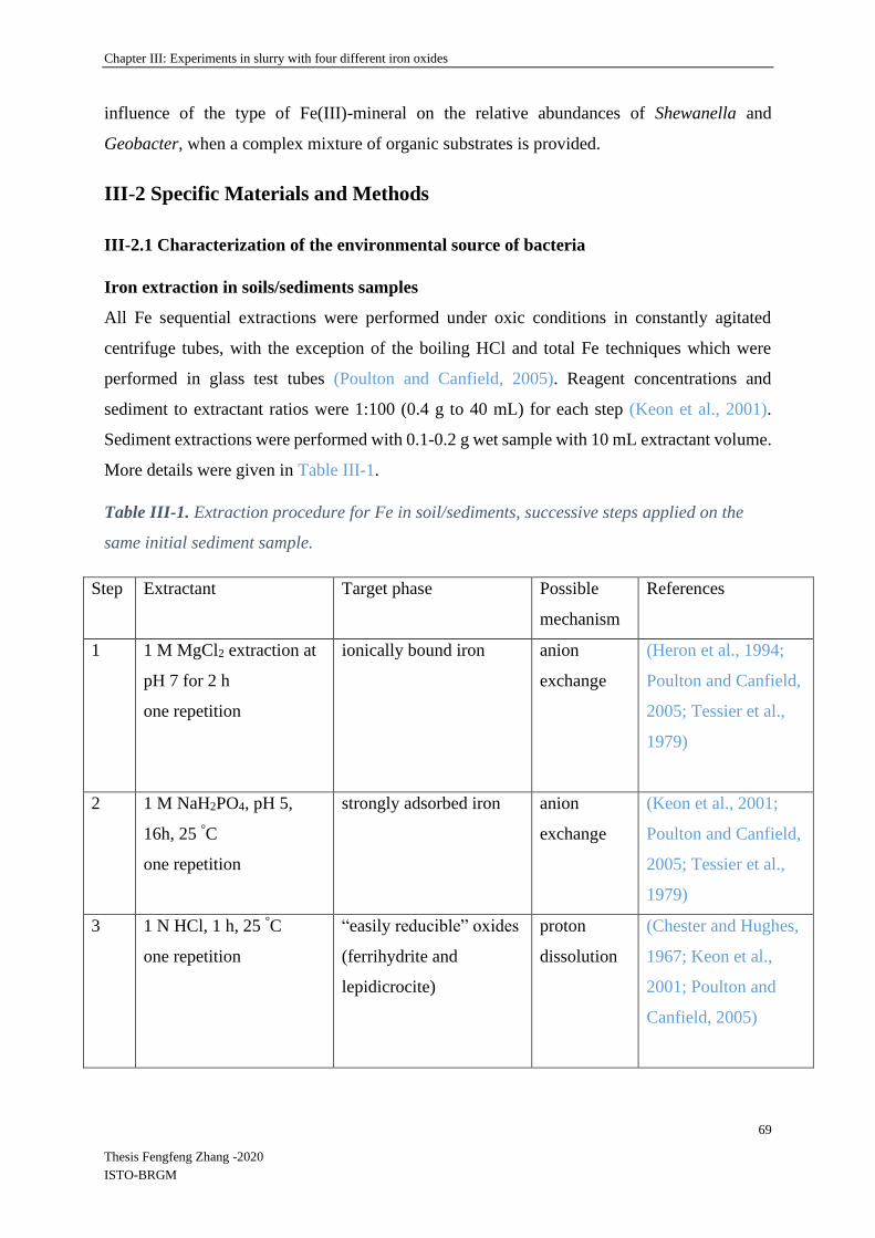
Chapter III: Experiments in slurry with four different iron oxides
69
Thesis Fengfeng Zhang -2020
ISTO-BRGM
influence of the type of Fe(III)-mineral on the relative abundances of Shewanella and
Geobacter, when a complex mixture of organic substrates is provided.
III-2 Specific Materials and Methods
III-2.1 Characterization of the environmental source of bacteria
Iron extraction in soils/sediments samples
All Fe sequential extractions were performed under oxic conditions in constantly agitated
centrifuge tubes, with the exception of the boiling HCl and total Fe techniques which were
performed in glass test tubes (Poulton and Canfield, 2005). Reagent concentrations and
sediment to extractant ratios were 1:100 (0.4 g to 40 mL) for each step (Keon et al., 2001).
Sediment extractions were performed with 0.1-0.2 g wet sample with 10 mL extractant volume.
More details were given in Table III-1.
Table III-1. Extraction procedure for Fe in soil/sediments, successive steps applied on the
same initial sediment sample.
Step Extractant Target phase Possible
mechanism
References
1 1 M MgCl2 extraction at
pH 7 for 2 h
one repetition
ionically bound iron anion
exchange
(Heron et al., 1994;
Poulton and Canfield,
2005; Tessier et al.,
1979)
2 1 M NaH2PO4, pH 5,
16h, 25 °C
one repetition
strongly adsorbed iron anion
exchange
(Keon et al., 2001;
Poulton and Canfield,
2005; Tessier et al.,
1979)
3 1 N HCl, 1 h, 25 °C
one repetition
“easily reducible” oxides
(ferrihydrite and
lepidicrocite)
proton
dissolution
(Chester and Hughes,
1967; Keon et al.,
2001; Poulton and
Canfield, 2005)

Chapter III: Experiments in slurry with four different iron oxides
70
Thesis Fengfeng Zhang -2020
ISTO-BRGM
4 0.2 M ammonium
oxalate/oxalic acid, pH 3,
2 h, 25 °C in dark
(wrapped in Al foil)
one repetition
“reducible” oxides
(goethite, hematite,
ferrihydrite, lepidocrocite
and akaganéite)
ligand-
promoted
dissolution
(Keon et al., 2001;
Poulton and Canfield,
2005; Ryan and
Gschwend, 1991)
5 DCB: Dithionite citrate-
bicarbonate,
pH 7, 2 h, 25 °C
one repetition + one
water wash
Magnetite, goethite,
hematite
reduction (McKeague and Day,
1966; Phillips and
Lovley, 1987; Poulton
and Canfield, 2005)
6 6 N HCl 24 h 125 rpm
two repetitions + one
water wash
Fe (oxyhydr) oxides, FeS Oxidation
and
dissolution
(Hellal et al., 2015;
Mikac et al., 2002)
III-2.2 Synthetic Fe(III) (oxyhydr)oxides and bacterial inocula
Two fresh Fe (oxyhydr)oxides were synthesized in the laboratory under the modified protocol
of (Schwertmann and Cornell, 2008). The Fe (oxyhydr)oxide named FoF was prepared
according to the protocol for ferrihydrite, by dissolving 40 g Fe(NO3)3∙ 9H2O in 500 mL
distilled water and adding 330 mL of 1 M KOH to adjust the pH to 7-8. The mixture was
centrifuged at 5,000 rpm for 10 min and the supernatant was subsequently removed. The solid
fraction was then washed 5 times with ultrapure water according to MilliQ quality. The Fe
(oxyhydr)oxide named FoL was prepared according to the protocol for lepidocrocite with 11.93
g of unoxidized FeCl2∙4H2O salts dissolved into 300 mL distilled water by stirring. The solution
was adjusted to pH 6.7-6.9 with NaOH using a pH-stat under aeration (100 mL/min air).
Washing was performed as described for FoF. Both synthesized minerals were freeze-dried.
Goethite from Sigma-Aldrich (CAS No. 20344-49-4) and hematite from VWR Chemicals
(CAS No. 1309-37-1) were also used. Mineralogical morphologies of all Fe (oxyhydr)oxides
were characterized using a scanning electron microscope (SEM) and BET surface area
measurement (determined by multipoint (Brunauer, Emmett and Teller) BET N2 adsorption)
(Brunauer et al., 1938). Specific surface areas were determined from N2 adsorption isotherms
in the best linear range (with a minimum of 15 points) between the relative pressure P/Po 0.03
and 0.33 (Cavelan et al., 2019).

Chapter III: Experiments in slurry with four different iron oxides
71
Thesis Fengfeng Zhang -2020
ISTO-BRGM
Specific surface areas of Fe (oxyhydr)oxides varied from 11.7 to 337 m2.g-1 (Table 1), and
compared well to some other synthetic (oxyhydr)oxides (Bonneville et al., 2004; Das et al.,
2013; Larsen and Postma, 2001; Pedersen et al., 2005). SEM was performed on a TM 3000
coupled to a SwiftED3000 X-Stream module (Hitachi), and operated at 15 kV accelerating
voltage (Thouin et al., 2016). The corresponding observed morphologies are given in Table III-
2.
Table III-2. Characteristics of Fe(III) oxides submitted to Fe-reducing bacteria.
Iron oxide Assumed morphologya Specific surface
areab (m2g-1)
goethite acicular 11.7
hematite cylinder/rod 31.4
FoF blocky 232
FoL blocky 337
aFor use in estimating mean particle size from morphology by SEM-EDS (TM3000
accompanied by a SwiftED3000 X-Stream module (Hitachi)); bDetermined by multipoint BET
N2 adsorption.
The 5th subcultures of inocula, D1, D2 and D3 described in chapter II-2 were used for the
following IRB incubation experiments.
III-2.3 IRB incubation experiments
Incubation experiments were performed in 50 mL glass bottles equipped with chlorobutyl
rubber stoppers, using 10 % (v/v) of inocula from D1, D2 and D3 enriched from the site samples
of Decize and the four Fe (oxyhydr)oxides presenting contrasting specific surfaces (FoF, FoL,
goethite and hematite), under anaerobic conditions. The compositions of the different solutions
used to prepare the culture medium were the same as for the enrichment cultures and are also
listed in Supplementary Figure III-S1. The total Fe(III) concentration added as Fe
(oxyhydr)oxides was adjusted to be close to that of dissolved Fe(III)-NTA in the enrichment
medium, i.e., 20 mM, based on the theoretical formula of each iron oxide (Supplementary
Table III-S1). The inoculation of Fe-reducing cultures was performed in an anaerobic glove
box. The gas phase of the bottles was N2 and the bottles were flushed with N2 before and after
sampling. The flasks were incubated at 20◦C, under agitation (100 rpm). Not inoculated controls
were prepared in the same conditions. Samples (1.5-2 mL) were collected as described in 2.1

Chapter III: Experiments in slurry with four different iron oxides
72
Thesis Fengfeng Zhang -2020
ISTO-BRGM
and analyzed for [FeT]D. The Fe solubilisation rates were calculated using the data of [FeT]D
collected during the first phase of the incubation, when this parameter increased linearly. After
27 days incubation, the remaining cultures were centrifuged at 5,000 rpm for 10 min.
Supernatants were removed and solids freeze-dried for observation under SEM-EDS (chapter
II-6.1).
III-2.4 Fe analyses and pH/Eh monitoring
For Fe analyses, 1.5 mL aliquot was sampled with a syringe and filtered through a 0.2 μm filter
into 5 mL tubes and immediately acidified with concentrated HCl in the glove box. [FeII]D
(dissolved Fe(II) concentration) and [FeT]D (total dissolved iron concentration) were
determined using the methods described in chapter II-4. pH and redox potential (Eh, ref.
Ag/AgCl) were measured using standard hand-held portable meters (WTW Multi340i set) in
glove box as described in chapter II-4 before and after the incubation.
III-2.5 Determination of iron oxides solubilisation parameters
The total dissolved Fe (Fe solubilisation) was calculated as the addition of the quantities of FeT
solubilized during all the discrete FeT solubilization events (from one sampling event to the
next one) during all the incubation period for the batch experiments. The decrease in FeT were
not taken into account in the calculation. The initial Fe reduction rate (mg L-1·h-1) was
calculated for the period of rapid increase of [FeT]D during the first stage (3-8 days) of the batch
experiments. The total dissolved Fe and initial Fe (oxyhydr)oxide dissolution rates are indicated
as “Fe solubilisation” and “solubilisation rate” in the following statistics.
III-2.6 SEM-EDS observation and Mössbauer spectrometry
Initial and post-incubation Fe (oxyhydr)oxides were observed by SEM-EDS using the method
described in chapter II-6.1 after freeze-drying (-90 ◦C Martin Christ Beta 2-8 LSCplus).
Mössbauer spectrometry was used to determine Fe species by the method described in chapter
II-7.
III-2.7 Biological analyses
DNA extractions were performed on samples using the method described in chapter II-8.2.
Bacterial 16S rRNA genes and Shewanella / Goebacter 16S rRNA genes quantification were
performed by the methods described in chapter II-8.4 and 8.6. CE-SSCP fingerprints were
performed by the method described in chapter II-8.3.

Chapter III: Experiments in slurry with four different iron oxides
73
Thesis Fengfeng Zhang -2020
ISTO-BRGM
III-2.8 Statistics
DNA quantification and qPCR data were analyzed using a Kruskal-Wallis test with XLSTAT
software (version 2019 21.1.3) to determine the significant differences between each culture or
between iron oxides. Variations in bacterial community structure were further analyzed by Non-
Metric multiDimensional Scaling –nMDS) and ANOSIM analysis and ANOSIM analysis
applied to a Bray-Curtis dissimilarity matrix of CE-SSCP data (generated in BioNumerics
V7.5), using R-Studio software (Vegan Package) (Team R, 2015).
Principal component analysis (PCA) was used to summarize the relationships between chemical
(Fe-reducing speed for the first few days and Fe reduction proportion) and microbial (molecular
biomass, i.e., DNA concentration, and Geobacter and Shewanella gene abundances) data with
XLSTAT software (version 2019 21.1.3).
III-3 Results
III-3.1 Characterization of the environmental sources of bacteria
Figure III-1 illustrates the visual aspect of the site materials (Figures III-1 a and b), and gives
the results of the sequential extractions of iron forms (Figure III-1 c). The sediment core can be
described by a horizon with anmoor (An), 3 reductic horizons (Gr) and the parent material
(alluvium from the Loire). Two Fe forms were detected in all samples, strongly adsorbed Fe
and the crystallized Fe forms (including magnetite, goethite, hematite and lepidocrocite). This
last category of Fe was more abundant in the riverbank soil than in the aquatic sediments. The
amorphous easily reducible oxides such as ferrihydrite was detected only in the riverbank soil
and in one sample of aquatic sediment (7-17 cm). Thus, the characterization of site samples
showed the probable presence of different types of Fe(III)-oxides in the environmental samples
used as source of IRB during the PhD thesis.
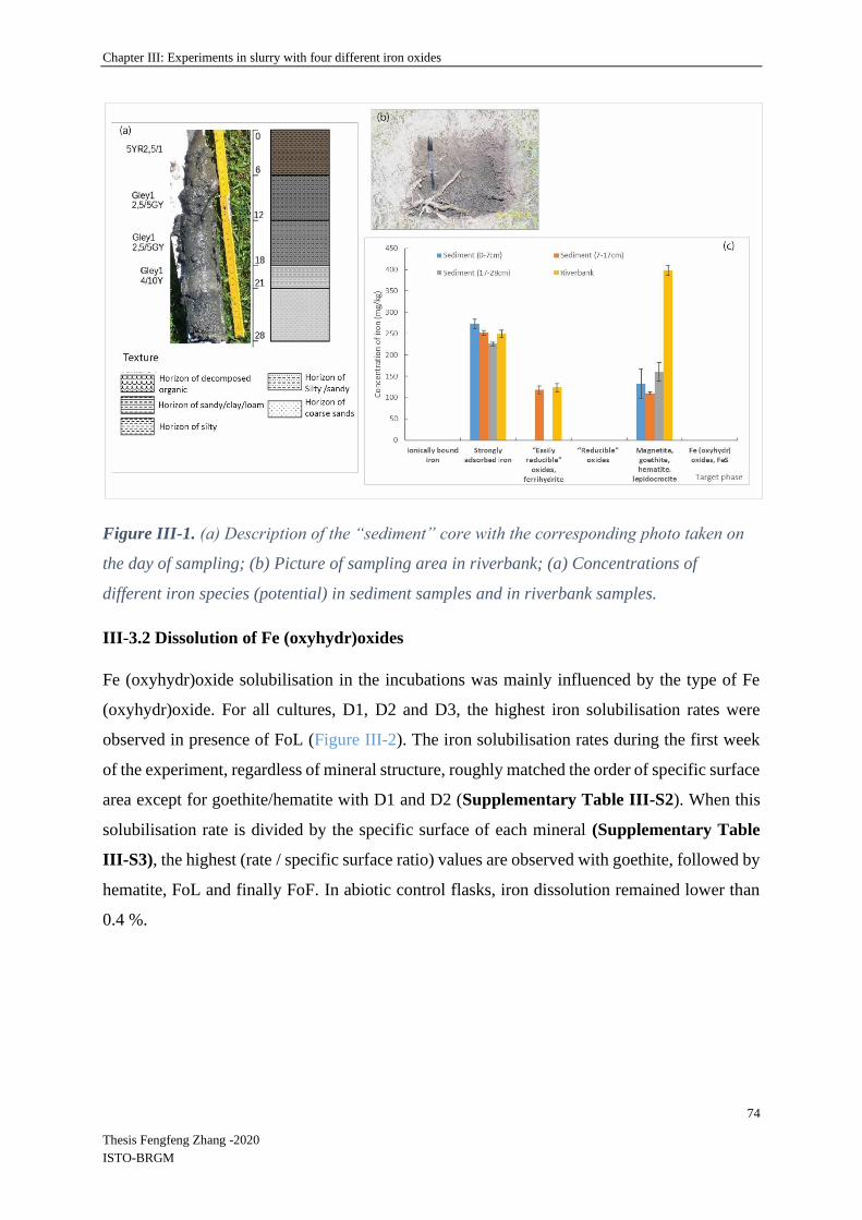
Chapter III: Experiments in slurry with four different iron oxides
74
Thesis Fengfeng Zhang -2020
ISTO-BRGM
Figure III-1. (a) Description of the “sediment” core with the corresponding photo taken on
the day of sampling; (b) Picture of sampling area in riverbank; (a) Concentrations of
different iron species (potential) in sediment samples and in riverbank samples.
III-3.2 Dissolution of Fe (oxyhydr)oxides
Fe (oxyhydr)oxide solubilisation in the incubations was mainly influenced by the type of Fe
(oxyhydr)oxide. For all cultures, D1, D2 and D3, the highest iron solubilisation rates were
observed in presence of FoL (Figure III-2). The iron solubilisation rates during the first week
of the experiment, regardless of mineral structure, roughly matched the order of specific surface
area except for goethite/hematite with D1 and D2 (Supplementary Table III-S2). When this
solubilisation rate is divided by the specific surface of each mineral (Supplementary Table
III-S3), the highest (rate / specific surface ratio) values are observed with goethite, followed by
hematite, FoL and finally FoF. In abiotic control flasks, iron dissolution remained lower than
0.4 %.
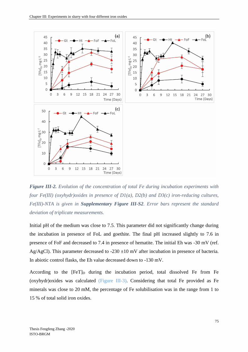
Chapter III: Experiments in slurry with four different iron oxides
75
Thesis Fengfeng Zhang -2020
ISTO-BRGM
Figure III-2. Evolution of the concentration of total Fe during incubation experiments with
four Fe(III) (oxyhydr)oxides in presence of D1(a), D2(b) and D3(c) iron-reducing cultures,
Fe(III)-NTA is given in Supplementary Figure III-S2. Error bars represent the standard
deviation of triplicate measurements.
Initial pH of the medium was close to 7.5. This parameter did not significantly change during
the incubation in presence of FoL and goethite. The final pH increased slightly to 7.6 in
presence of FoF and decreased to 7.4 in presence of hematite. The initial Eh was -30 mV (ref.
Ag/AgCl). This parameter decreased to -230 ±10 mV after incubation in presence of bacteria.
In abiotic control flasks, the Eh value decreased down to -130 mV.
According to the [FeT]D during the incubation period, total dissolved Fe from Fe
(oxyhydr)oxides was calculated (Figure III-3). Considering that total Fe provided as Fe
minerals was close to 20 mM, the percentage of Fe solubilisation was in the range from 1 to
15 % of total solid iron oxides.

Chapter III: Experiments in slurry with four different iron oxides
76
Thesis Fengfeng Zhang -2020
ISTO-BRGM
Figure III-3. Total amount of solubilized Fe from Fe (oxyhydr)oxides: goethite, hematite, FoF
and FoL in presence of D1, D2 and D3 inocula. The letters “a” and “b” represent the
significance of differences (Kruskal-Wallis test at p<0.05) between cultures. Values in the same
Fe (oxyhydr)oxides group are not significantly different from one another. Error bars represent
the standard deviation of triplicate measurements.
The highest quantities of dissolved Fe were obtained for FoL, with 0.074 mg iron per mL with
D3 inoculum. The extent of FoL solubilisation was more than 3 times higher than that of
hematite with the same inoculum. For goethite, low solubilisation, around 0.022 mg iron per
mL, were obtained, with no significant difference between the three inocula. Moreover, when
Fe (oxyhydr)oxides are grouped without distinguishing the origin of the inocula, the
solubilisation of FoL was significantly different from that of goethite and hematite (Figure III-
3), and that of FoF was significantly higher than that of hematite. For the same data set, the
significance of differences calculated for different inocula (Supplementary Figure III-S3)
shows that for both D1 and D2, FoL solubilisation was significantly different from that of
hematite. However, there was no significant difference in iron solubilisation between iron
oxides with D3. This suggests that D3 could be less influenced by Fe mineralogy than the other
two inocula.
III-3.3 Biological parameters
III-3.3.1 Soil samples
The ratio of gene copies of Shewanella and Geobacter (Figure III-4 b) 16S genes over total
bacterial 16S genes abundance increased from D1 to D2 and D3. Globally, Geobacter was

Chapter III: Experiments in slurry with four different iron oxides
77
Thesis Fengfeng Zhang -2020
ISTO-BRGM
clearly present in higher proportions than Shewanella in the bacterial communities present in
the environmental samples used to enrich D1, D2 and D3.
III-3.3.2 Effect of Fe (oxyhydr)oxides on bacterial community structure
The bacterial community structure profiles of the initial soil and sediment samples, the Fe-NTA
enrichments and the cultures in presence of the four different (oxyhydr)oxides were compared
using an nMDS ordination approach (Figure III-5). Full CE-SSCP profiles are available in
Supplementary Figure III-S4. These profiles show a high diversity with many peaks, with
thus the presence of different bacterial strains.
The structures of the initial communities were modified after inoculation on Fe (oxyhydr)oxides
samples (Figure III-5): the enrichments on Fe(III)-NTA are grouped on the right side of the
representation, whereas communities obtained by cultures with iron oxides are gathered on the
left side. The three initial communities from environmental samples are located between these
two poles. No clear separation is observed between the communities obtained with the four
pure iron oxides. Although, according to ANOSIM analysis, no significant difference in
community composition was found between initial inocula (significance >0.05), a significant
dissimilarity was found between community origins (i.e. initial soils and form of Fe(III)
provided to the consortia) (R = 0.345 and significance = 0.0249).
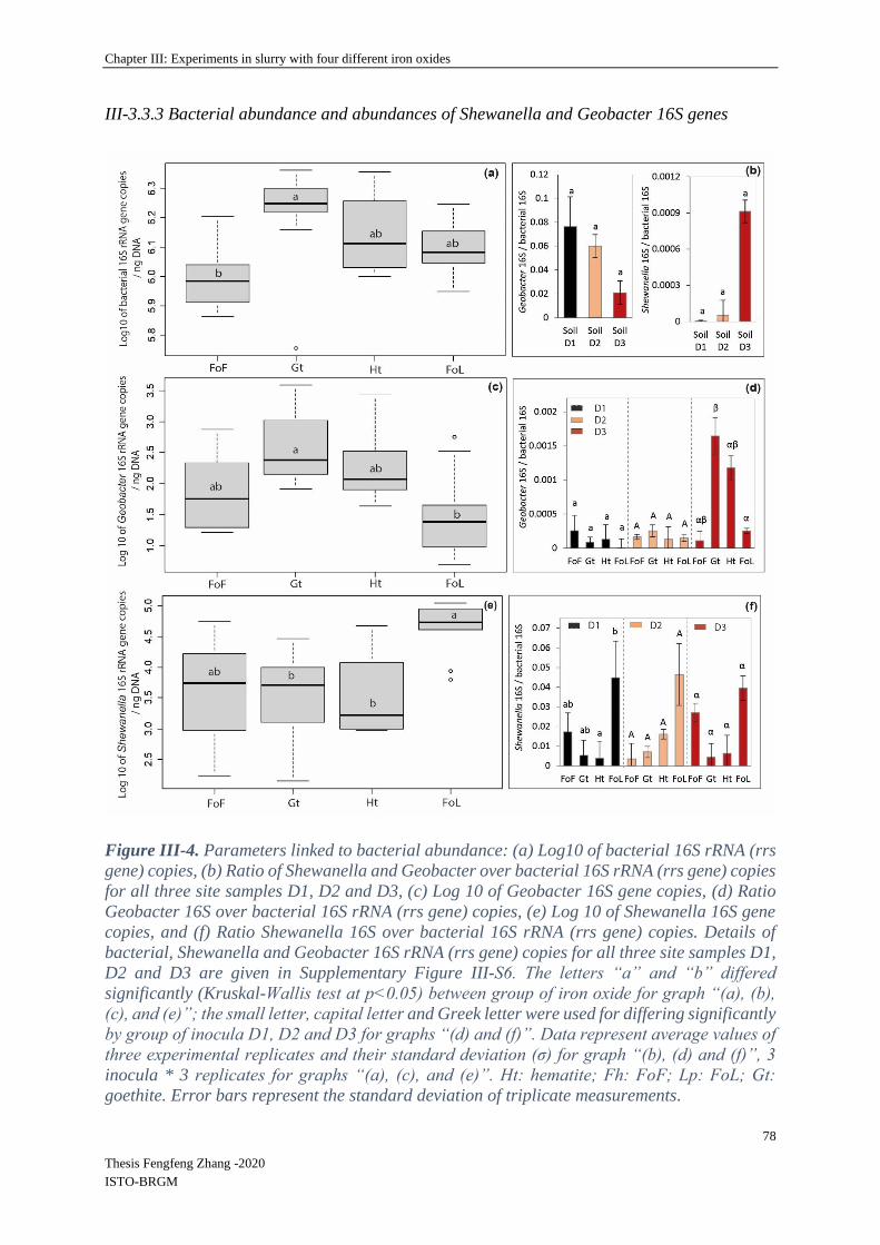
Chapter III: Experiments in slurry with four different iron oxides
78
Thesis Fengfeng Zhang -2020
ISTO-BRGM
III-3.3.3 Bacterial abundance and abundances of Shewanella and Geobacter 16S genes
Figure III-4. Parameters linked to bacterial abundance: (a) Log10 of bacterial 16S rRNA (rrs
gene) copies, (b) Ratio of Shewanella and Geobacter over bacterial 16S rRNA (rrs gene) copies
for all three site samples D1, D2 and D3, (c) Log 10 of Geobacter 16S gene copies, (d) Ratio
Geobacter 16S over bacterial 16S rRNA (rrs gene) copies, (e) Log 10 of Shewanella 16S gene
copies, and (f) Ratio Shewanella 16S over bacterial 16S rRNA (rrs gene) copies. Details of
bacterial, Shewanella and Geobacter 16S rRNA (rrs gene) copies for all three site samples D1,
D2 and D3 are given in Supplementary Figure III-S6. The letters “a” and “b” differed
significantly (Kruskal-Wallis test at p<0.05) between group of iron oxide for graph “(a), (b),
(c), and (e)”; the small letter, capital letter and Greek letter were used for differing significantly
by group of inocula D1, D2 and D3 for graphs “(d) and (f)”. Data represent average values of
three experimental replicates and their standard deviation (σ) for graph “(b), (d) and (f)”, 3
inocula * 3 replicates for graphs “(a), (c), and (e)”. Ht: hematite; Fh: FoF; Lp: FoL; Gt:
goethite. Error bars represent the standard deviation of triplicate measurements.

Chapter III: Experiments in slurry with four different iron oxides
79
Thesis Fengfeng Zhang -2020
ISTO-BRGM
Figure III-5. nMDS ordination of D1, D2 and D3 community fingerprints applied to a Bray-
Curtis dissimilarity matrix. Plot stress = 0.15. Ht: hematite; Fh: FoF; Lp: FoL; Gt: goethite.
The rrs gene abundances and Shewanella 16S/bacterial 16S ratio did not highlight any
significant differences between the three initial enrichments D1, D2 and D3 (Supplementary
Figure III-S4). As shown in Figure III-4 (a), globally, rrs gene abundance in goethite samples
was significantly different to FoF samples. It decreased in iron oxide cultures compared with
Fe(III)-NTA cultures (Supplementary Figure III-S5). Conversely, the abundance of the two
quantified IRB genera, i.e., Geobacter and Shewanella, differ between the types of Fe oxides
provided as Fe(III) source (Figure III-4 e and f). Globally, compared with Geobacter,
Shewanella was present in a larger proportion in all bacterial communities with the four iron
oxides. Moreover, the number of gene copies of Shewanella for FoL is significantly different
from that observed in presence of other iron oxides, but there are no significant differences
between the cultures for FoF, goethite and hematite samples. When the type of inocula is not
taken into account, the proportion of Shewanella 16S genes for FoL samples is significantly
different for goethite samples.
Considering specifically each quantified genus-specific 16S gene, the cultures in presence of
goethite inoculated with D3 bacteria contained a significantly different proportion of Geobacter
16S genes than the same culture (D3) with FoL (Figure III-4 d). In the other conditions (D1,
D2 with all oxides), no significant differences between the proportion of Geobacter 16S genes
in the global bacterial population was observed.

Chapter III: Experiments in slurry with four different iron oxides
80
Thesis Fengfeng Zhang -2020
ISTO-BRGM
III-3.4 Mineral SEM-EDS observation
SEM observations showed that initial FoF and hematite samples were characterized by
amorphous or poorly crystallized morphologies whereas crystalline minerals with an acicular
and a prismatic texture were observed in goethite and FoL (lepidocrocite) samples (Figure III-
6 (a, b, c, d-1)). No significant morphological changes were observed in abiotic samples after
incubation (Figure III-6 (a, b, c, d-2)). However, new crystalline forms with a prismatic texture
were observed in biotic samples of FoF and FoL after incubation (Figure III-6 (a-3, d-3)).
Figure III-6. Microscopic pictures of Fe(III) (oxhydr)oxides samples, FOF (ferrihydrite) (a),
goethite (b), hematite (c) and FOL (lepidocrocite) (d). Number “1” represented initial samples,
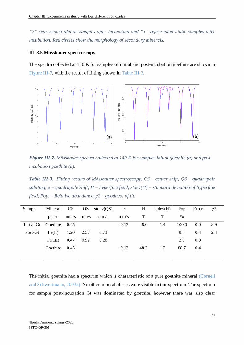
Chapter III: Experiments in slurry with four different iron oxides
81
Thesis Fengfeng Zhang -2020
ISTO-BRGM
“2” represented abiotic samples after incubation and “3” represented biotic samples after
incubation. Red circles show the morphology of secondary minerals.
III-3.5 Mössbauer spectroscopy
The spectra collected at 140 K for samples of initial and post-incubation goethite are shown in
Figure III-7, with the result of fitting shown in Table III-3.
Figure III-7. Mössbauer spectra collected at 140 K for samples initial goethite (a) and post-
incubation goethite (b).
Table III-3. Fitting results of Mössbauer spectroscopy. CS – center shift, QS – quadrupole
splitting, e – quadrupole shift, H – hyperfine field, stdev(H) – standard deviation of hyperfine
field, Pop. – Relative abundance, χ2 – goodness of fit.
Sample Mineral CS QS stdev(QS) e H stdev(H) Pop Error χ2
phase mm/s mm/s mm/s mm/s T T %
Initial Gt Goethite 0.45 -0.13 48.0 1.4 100.0 0.0 8.9
Post-Gt Fe(II) 1.20 2.57 0.73
8.4 0.4 2.4
Fe(III) 0.47 0.92 0.28
2.9 0.3
Goethite 0.45 -0.13 48.2 1.2 88.7 0.4
The initial goethite had a spectrum which is characteristic of a pure goethite mineral (Cornell
and Schwertmann, 2003a). No other mineral phases were visible in this spectrum. The spectrum
for sample post-incubation Gt was dominated by goethite, however there was also clear
1050-5-10
v (mm/s)
1.4
1.3
Inte
nsity (
10
6 c
ts)
1050-5-10
v (mm/s)
1.7
01.6
51.6
0
Inte
nsity (
10
6 c
ts)
(a) (b)

Chapter III: Experiments in slurry with four different iron oxides
82
Thesis Fengfeng Zhang -2020
ISTO-BRGM
indications for Fe(II) (8.4 %), and a short range Fe(III) phase (2.9 %) which was not
magnetically ordered at 140 K.
The FoF was also characterized by Mössbauer spectroscopy (Supplementary Figure III-S7) that
confirmed it was ferrihydrite. Operational problems prevented us from analyzing the other
samples.
III-4 Discussion
III-4.1 Influence of the type of iron oxide on bacterial iron solubilisation
FoL and FoF were synthetized in the laboratory and were more amorphous, thus more reactive,
than goethite and hematite (Usman et al., 2012). According to the literature, abiotic rates of
reductive iron dissolution are correlated with the solubility of Fe (oxyhydr)oxides (Larsen and
Postma, 2001). The rates of this abiotic reductive bulk dissolution decrease according to
ferrihydrite >lepidocrocite> goethite> hematite, emphasizing the importance of the crystal
structure on the dissolution rate (Larsen and Postma, 2001). Moreover, the rate of iron oxides
solubilisation is usually a function of surface area, rates increasing with surface area for a given
mineral (Schwertmann, 1991). However, Roden, (2006) found that in presence of IRB, oxides’
mineralogical and thermodynamical properties exert a minor influence on reduction rates
compared with the abundance of available oxide surface sites controlled by oxide surface area
and the accumulation of surface-bound biogenic Fe(II). This last process, of the precipitation
of new Fe(II) minerals (Urrutia et al., 1998; Zachara et al., 2002), might explain the late
decrease of soluble Fe and the fact that the iron solubilization rate by specific surface unit was
the highest with goethite, followed by hematite, FoL and finally FoF, thus it did not follow the
expected abiotic dissolution rate rank (Table III-S3). However, Fe solubilisation effectiveness
increased with the specific surface, in particular for the two freshly synthesized oxides. Here,
the fresh mineral prepared with the protocol of lepidocrocite (FoL) synthesis presented a higher
specific surface than the mineral produced using the protocol for ferrihydrite (FoF). Synthetic
lepidocrocites can present a very large range of specific surface areas, depending on their level
of crystallinity (Schwertmann, 1973). In the present case, the specific area of synthetic FoL
(337 m2·g
-1) is much higher than for other Fe (oxyhydr)oxides, possibly due to a rapid oxidation
of Fe(II) during synthesis that produced poor crystallization and formation of lepidocrocite
impurities (Schwertmann and Taylor, 1979). Schwertmann (1973) showed that the specific
surface of lepidocrocite increases with its solubility in presence of oxalate, and this specific

Chapter III: Experiments in slurry with four different iron oxides
83
Thesis Fengfeng Zhang -2020
ISTO-BRGM
surface area is anti-correlated with the crystallinity. In the present experiment, at the end of the
incubation, we detected 8 mg·L-1 total Fe in the FoL abiotic control, but almost no Fe in FoF
abiotic control. This supports the idea that FoL was more soluble than FoF. The high specific
surface area and probable poor crystallinity of FoL could have favored solubilisation by IRB.
Fe solubilisation rates were limited after a few days, probably due to evolution of the
composition of the liquid medium, or the accumulation of surface-bound biogenic Fe(II)
(Roden, 2006), as Fe(III) was not the limiting factor. Parameters such as humic substances,
quinones and organic carbon can strongly influence microbial Fe(III) reduction rates for
Shewanella (Adhikari et al., 2017) or Geobacter (Wolf et al., 2009) in natural environments.
Generally, in these studies, the highest reducing rates were observed during the first few days
of microbial Fe(III) reduction. In our study, the influence of the type of Fe (oxyhydr)oxides on
initial iron oxide-reducing rates were consistent with those of previous studies performed with
pure strains.
III-4.2 Bacterial communities
The structures of the initial bacterial communities present in the environmental samples were
modified by the enrichment in Fe(III)-NTA medium, and again showed an evolution when the
enrichments were grown in presence of solid Fe (oxyhydr)oxides (Figure III-5). This last result
could be due to the difference of bio-availability of Fe with minerals compared to Fe(III)-NTA.
Cai et al., (2019) also showed an influence of bio-available Fe(III) on microbial community
structure (Cai et al., 2019). Here, the accessibility of Fe(III) in the Fe(III)-NTA incubations
favored the iron reducing community that may have rapidly consumed available organic
substrates and probably lowered the development of other bacteria. With minerals, however,
Fe(III) is less accessible and competition for Fe(III) may induce changes in community
structure. For example, Zhuang et al., (2011) indicated that Rhodoferax and Geobacter genera
were acetate-oxidizing Fe(III)-reducers that compete in anoxic subsurface environments and
this competition could influence the in situ bioremediation of uranium-contaminated
groundwater by changing diversity structure (Zhuang et al., 2011). On the other hand, no clear
effect of the type of Fe mineral on the global community profile was observed. This may be
linked to the culture medium composition. Indeed, the addition of diverse C sources could
enable fermentative bacteria to develop without using Fe(III) for their growth. In contrast,
(Lentini et al., 2012) observed an effect of the type of Fe (oxyhydr)oxide, i.e. ferrihydrite,
goethite or hematite, on the global structure of bacterial communities in enrichment cultures
Chapter III: Experiments in slurry with four different iron oxides
84
Thesis Fengfeng Zhang -2020
ISTO-BRGM
using T-RFLP fingerprints. However, these authors did not use an initial Fe(III)-NTA
enrichment step and did not use a sulfate-reducing bacteria inhibitor, as in our study.
III-4.3 Geobacter and Shewanella 16S genes abundances
The relative abundance of Geobacter and Shewanella in the initial soils and sediment used as
sources of IRB in our experiment could be linked to the redox conditions of the environment.
Indeed, Shewanella 16S genes was present in higher proportions in the river sediment D3, than
in the flooded soil D2, itself richer than the non-saturated soil D1 whereas Geobacter presented
the opposite behavior. This indicates that the reducing conditions in the aquatic sediment was
more favorable for some IRB, such as Shewanella, but not for Geobacter.
Shewanella and Geobacter represented in our incubation experiments a small proportion of the
total community, however we made a focus on these two genera because they are the most
studied iron reducing bacteria. However, the other members of the community could contribute
either directly or indirectly to the iron solubilisation process. Considering the evolution of the
two targeted IRB genera, namely Geobacter and Shewanella, after the enrichment with Fe(III)-
NTA, the Shewanella / Geobacter 16S abundance ratio increased significantly compared to the
site samples (Figure III-4 compared with Supplementary Figure III-S5), suggesting that the
presence of a large amount of bio-available Fe(III) in the enrichment culture medium was in
favor of Shewanella.
Shewanella could have from 8 to 11 copies of the gene encoding 16S rRNA, whereas Geobacter
has 1 or 2 copies (Dikow, 2011; Holmes et al., 2004). Thus, the quantification of specific 16S
genes does not reflect the cell abundances of the respective strains. All the same, this factor
alone does not explain the differences between the abundances of Shewanella and Geobacter
in our cultures, even if it could attenuate the difference in terms of specific cells numbers.
Another explanation for the sharp increase of Shewanella 16S gene abundance compared to
Geobacter 16S gene abundance could be the composition of the culture medium in terms of
organic substrates. Geobacter can use acetate as electron donors while performing the
dissimilatory Fe reduction (Caccavo et al., 1994; Coates et al., 1996), but does not use lactate
nor glucose. Conversely, some Shewanella species were shown to be able to use glucose and
can present either respiratory or fermentative types of metabolisms (Ivanova et al., 2004; Nogi
et al., 1998; Ziemke et al., 1998). Lentini et al., (2012) showed that the presence of Geobacter
was favored by acetate whereas the growth of Shewanella was rather stimulated by lactate.
Chapter III: Experiments in slurry with four different iron oxides
85
Thesis Fengfeng Zhang -2020
ISTO-BRGM
Moreover, these authors suggested that production of acetate through incomplete degradation
of lactate by Shewanella could benefit Geobacter (Hori et al., 2015). Our medium contained
both acetate, formate, lactate and glucose, thus it should potentially support growth of both
Shewanella and Geobacter. However, a fermentative metabolism could explain the medium is
favored by Shewanella over Geobacter in our enrichments and in all incubation conditions,
because some species of this genus can grow either using fermentation or Fe(III) reduction
(Bowman, 1997). As our medium contained 1.5 g.L-1 peptone, this substrate could also favor
the growth of bacteria belonging to the Shewanella genus by fermentation (Toffin et al., 2004).
When these communities were grown in presence of solid iron oxides, the abundance of
Shewanella 16S gene (average of D1, D2 and D3) was 112, 30, 52 and 3058 times higher than
the abundance of Geobacter 16S gene, for FoF, goethite, hematite and FoL, respectively.
Moreover, we found that the cultures in presence of goethite inoculated with D3 bacteria
contained higher proportions of Geobacter 16S gene than the same culture (D3) with FoL.
These results might suggest that Geobacter could be more favored, in the competition with
other IRB such as Shewanella, for growth in presence of goethite and hematite than for growth
in presence of the more easily dissolved oxides, i.e., FoF and FoL. The type of Fe mineral can
exert a selection pressure on the communities of IRB, as previously shown by Lentini et al.,
(2012). This result could be linked to a higher affinity of Geobacter for Fe(III), that would favor
this organism at low Fe(III) availability levels. Reported Ks values for Fe(III) are 1.0 mM with
G. sulfurreducens (Esteve-Nunez et al., 2005) compared to 29 mM with S. putrefaciens (Liu et
al., 2001).
Yet, the solubility of ferrihydrite and lepidocrocite are higher than that of goethite and hematite
(Cornell and Schwertmann, 2003b; Liu et al., 2007), thus the availability of Fe(III) could be
higher with the first two oxides. Shewanella might be favored by high bio-available Fe(III), but
could be less efficient for growth in presence of less soluble Fe-oxides such as goethite and
hematite. The anaerobic respiration of Shewanella was highly dependent on electron shuttles.
Nevin and Lovley (2002) suggested that Shewanella alga strain BrY released compounds that
could solubilize Fe(III) from Fe(III) oxides, however, Geobacter metallireducens did not
produce electron shuttles or Fe(III) chelators to solubilize Fe(III) oxides (Nevin and Lovley,
2002). Kotloski and Gralnick (2013) determined the contribution of flavin electron shuttles in
extracellular electron transfer by Shewanella oneidensis (Kotloski and Gralnick, 2013) and Wu
et al., (2020) showed that exogenous electron mediators (EMs) favored high density current
production and increased the synthesis of extracellular polymeric substances which promoted

Chapter III: Experiments in slurry with four different iron oxides
86
Thesis Fengfeng Zhang -2020
ISTO-BRGM
biofilm formation during electron shuttling process (Wu et al., 2020). The conduction of
electrons along pili or other filamentous structures is one of the mechanisms proposed for
electron transfer to solid iron oxides. (Leang et al., 2010) showed that OmcS, a cytochrome that
is required for Fe(III) reduction by Geobacter sulfurreducens, was localized along the pili
(Leang et al., 2010). The electrically conductive pili play a major role in the adaptation of
Geobacter to perform dissimilatory iron reduction in natural environments (Liu et al., 2019a).
These differences in the Fe(III)-reducing mechanisms between the two genera might explain
their difference of affinity for Fe(III) (Esteve‐Núñez et al., 2005; Liu et al., 2001) and the
relative increase of Geobacter 16S gene abundance, observed here in presence of goethite and
hematite, with the enrichment culture that was the most efficient for Fe-oxide solubilisation,
i.e., culture D3.
III-4.4 Relation between iron solubilisation effectiveness and Geobacter and Shewanella
16S gene abundances
In batch experiments, the three inocula D1, D2 and D3, enriched from soil from the river bank,
flooded soil and an aquatic sediment of the Decize site gave similar results in terms of Fe
solubilisation effectiveness of iron (oxyhydr)oxides. However, there were differences between
different Fe (oxyhydr)oxides: the type of Fe (oxyhydr)oxides had more influence on Fe
solubilisation effectiveness than the origin of the inocula. Fe solubilisation rates were slower
with goethite and hematite compared to FoF and FoL. The same tendency was observed by
Bonneville et al., (2004), who found that, with the pure strain S. putrefaciens in presence of 20
mM Fe(III), reduction of 6-line ferrihydrite was faster than that obtained with lepidocrocite,
and faster than that obtained with low surface area hematite. (Li et al., 2012) reported similar
results for the microbial reduction of Fe(III) oxides by S. decolorationis strain S12, the reducing
speed decreasing according to the following order: lepidocrocite > goethite > hematite.
Principal Component Analysis (PCA) integrating chemical (Supplementary Table III-S2) and
molecular data was performed to identify the relationships and contributions between iron
solubilisation effectiveness and Geobacter / Shewanella 16S genes abundances in batch
experiment samples (Hong et al., 2015; Mercier et al., 2014; Zhu et al., 2014). In this study, a
biplot (Supplementary Figure III-S6) summarizes PCA results. The first principal component
is strongly influenced by the iron solubilisation effectiveness (higher on the right than on the
left side of the representation), with higher values associated with FoL, and the lowest
associated with goethite and hematite (not separated), FoF being in an intermediary position.
Chapter III: Experiments in slurry with four different iron oxides
87
Thesis Fengfeng Zhang -2020
ISTO-BRGM
Meanwhile proportion of Shewanella 16S gene, but not to the proportion of Geobacter 16S
gene, seems to be correlated to FoL. The second principal component reflects high values of
the proportion of Geobacter 16S gene in the bacterial communities. Thus, the proportion of
Shewanella 16S gene seems to be more correlated to iron solubilisation parameters than the
proportion of Geobacter 16S gene. PCA allows clear discrimination with different groups of
iron oxides, however not for the different types of inoculum (D1, D2 and D3).
According to Bonneville et al., (2004), the dissolution and solubility of goethite and hematite
are lower than that of ferrihydrite and lepidocrocite in the presence of Shewanella. (Cutting et
al., 2009) indicated that hematite and goethite are susceptible to limited Fe(III) reduction in
presence of G. sulfurreducens. Moreover, Poggenburg et al., (2016) mentioned that Fe(III)-
organic compounds (coprecipitates from solutions of FeCl3 and natural organic matter)
reduction by Shewanella putrefaciens was influenced by the amount of available electron
shuttling molecules induced by sorbed natural organic matter. Fe(III)-organic compounds’
reduction by Geobacter metallireducens was more influenced by particle size, physicochemical
properties and iron (oxyhydr)oxides (composition of sorbed natural organic matter and
aggregation state) (Poggenburg et al., 2016). In our study, FoL samples with a higher proportion
of Shewanella 16S gene in their total bacterial 16S genes were correlated with high initial iron
solubilisation rates and electron shuttling molecules might have a role in this phenomenon. In
contrast, Geobacter 16S gene abundance was not specifically associated to FoL but was found
in higher proportions with goethite in one condition. This tendency is in accordance with
findings of previous research performed with the less soluble iron oxides (Crosby et al., 2007),
showing that Geobacter sulfurreducens reduced 0.7 % hematite and 4.0 % goethite while
Shewanella putrefaciens reduced only 0.5 % of hematite 3.1 % goethite after 280 days of
incubation. Thus, our results suggest that Geobacter might suffer less from the competition
with Shewanella in low bio-available Fe(III) conditions, whereas the contribution of this genus,
present in lower quantities than Shewanella, to the iron solubilisation effectiveness is not
demonstrated. Moreover, other members in the community of IRB probably contributed to Fe
solubilisation. Our culture medium contained substrates such as glucose and peptone that could
support the growth of fermentative organisms. Lentini (2012) and Gagen (2019) indicated that
fermentation likely plays a key role in reduction of crystalline iron oxides by diverse bacteria,
such as Telmatospirillum, both through direct reduction and by the production of H2, potentially
used by dissimilatory iron reducers, during fermentation.
Chapter III: Experiments in slurry with four different iron oxides
88
Thesis Fengfeng Zhang -2020
ISTO-BRGM
III-4.5 SEM observations of Fe (oxyhydr)oxides and Mössbauer spectroscopy
In our study, the morphologies of four Fe (oxyhydr)oxides were observed by SEM before and
after incubation with a mixed IRB enrichment. As previously observed in the literature for
ferrihydrite, FoF presented an aggregated form with conchoidal fractures indicating its
amorphous nature (Das et al., 2013; Schwertmann and Cornell, 2008) and as previously
observed for lepidocrocite (Schwertmann, 1973; Schwertmann and Taylor, 1979) FoL
presented a poor crystallization in accordance with the high specific surface of this synthetic
material. Hematite samples presented submicron grains (Kim et al., 2008; Tadic et al., 2012)
and goethite showed acicular or needle-shaped crystallized forms (Das et al., 2013;
Schwertmann and Cornell, 2008). After incubation in presence of bacteria, secondary
crystallized minerals were observed with FoF and FoL. The secondary minerals formed in FoL
samples submitted to bioreduction showed a strong morphological similarity with the Fe(II)-
phosphate vivianite observed by (Roldán et al., 2002; Muehe et al., 2016). Our goethite mineral
before incubation presented a Mössbauer spectrum which was characteristic of goethite
(Cornell and Schwertmann, 2003a). At the end of incubation, Fe(II) phases were observed in
biotic goethite samples. We were not able to acquire Mössbauer spectra from the incubations
of other minerals, however this result confirms the reduction of Fe(III)-oxides in biotic
conditions, and suggests that the extent of reduction was higher than that evaluated only with
the soluble Fe concentration, because some Fe(II) was partially immobilized in secondary
minerals (Urrutia et al., 1998; Zachara et al., 2002). These observations and analyses proved to
be informative, thus SEM observation and Mössbauer analyses were applied to samples
generated by the further experiments, presented and discussed in the following sections.
III-5 Conclusions and perspective Microbial enrichments containing IRB, obtained from a site submitted to regular variations of
redox conditions, were able to grow and reduce Fe(III) in a short time. Experiments performed
with synthetic fresh oxides, goethite, and hematite confirmed the type of Fe mineral could
influence reducing rates and the abundances of two DIRB genera, Geobacter and Shewanella.
The present study’s results showed that: (1) the sub-culturing of IRB enrichments from Fe(III)-
NTA to pure iron oxides media markedly modified the bacterial communities; (2) the global
bacterial diversity was not significantly different from one type of pure (oxyhydr)oxide to
another; (3) the type of Fe oxide in terms of mineralogy can influence the proportion of specific
IRB genera, such as Geobacter and Shewanella. Meanwhile, the nature of Fe (oxyhydr)oxides
Chapter III: Experiments in slurry with four different iron oxides
89
Thesis Fengfeng Zhang -2020
ISTO-BRGM
seems to have exerted a selection on the ratio of Geobacter and Shewanella 16S genes, whereas
it did not impact the global bacterial community fingerprints. The concentration of bio-available
Fe(III) in the enrichment medium may have favored the development of Shewanella genus that
was clearly dominant compared with Geobacter genus. In presence of solid iron oxides, the
highest proportions of Shewanella in bacterial communities were obtained with FoL and
corresponded to the highest levels of iron reduction, possibly linked to the fact that FoL was
the most soluble (oxyhydr)oxide in our experiments. This result is consistent with a hypothesis
that Shewanella development could be favored by a high bioavailability of Fe(III). In contrast,
Geobacter was detected in higher proportions with goethite that is less easily dissolved, when
D3 culture was used.
Globally, all results suggest that both initial community composition of the sample used to
prepare the enrichments, as well as the type of Fe(III) oxide used as electron acceptor influenced
the relative abundance of Geobacter and Shewanella 16S genes. A better knowledge of the
influence of biological parameters, such as biomass, diversity and activity, associated with iron
reduction involving complex bacterial communities can help to elucidate Fe dynamics in
surface environments. As biofilms in natural soils and sediments contain a large part of the
bacterial biomass, future research could be focused on the contribution and distribution of IRB
communities attached on iron oxides surfaces.

Chapter III: Experiments in slurry with four different iron oxides
90
Thesis Fengfeng Zhang -2020
ISTO-BRGM
Supplementary material
Supplementary Figures
Supplementary Figure III-S1. Design and initial compositions of the batch experiments.
SRB1 : sulfate-reducing bacteria ; 2. Vitamin solution (1L): 2 mg biotin, 2 mg folic acid, 10 mg pyridoxine
HCL, 5 mg Riboflavin,5 mg Thiamine, 5 mg Nicotinic acid, 5 mg Pantothenic acid, 0.1g B12 vitamin, 5 mg p-
aminobenzoic acid, 5 mg thioctic acid ; 3. Trace element solution (1L): 1.5 g Trisodium nitrilotriacetic, 0.5 g
MgSO4, 0.5 g MgSO4∙H2O, 1 g NaCl, 0.1 g FeSO4∙7 H2O, 0.1 g CaCl2∙2H2O, 0.1 g CoCl2∙6H2O, 0.13 g ZnCl, 0.1
g CuSO4 5H2O, 0.1 g AlK(SO4)∙12H2O, 0.1 g H3BO3, 0.25 g NaMoO4, 0.24 g NiCl∙6H2O, 0.25 g Na2WO4∙2H2O.
0
100
200
300
400
500
600
700
800
900
1000
0 1 2 3 4 5 6
[TFe
] Dan
d [
TFeI
I]D
mg·
L-1
Time /d
D1-Fe(II) D2-Fe(II)
D3-Fe(II) Blank-Fe(II)
D1-Fe(T) D2-Fe(T)
D3-Fe(T) Blank-Fe(T)
(a)

Chapter III: Experiments in slurry with four different iron oxides
91
Thesis Fengfeng Zhang -2020
ISTO-BRGM
Supplementary Figure III-S2. Concentration of Fe(II) and total Fe (FeT) of D1, D2 and D3
incubated on Fe(III)-NTA medium (a), and evolution of the concentration of total Fe during
incubation experiments with four abiotic Fe(III) (oxyhydr)oxides (b). Error bars represent the
standard deviation of triplicate measurements.
Supplementary Figure III-S3. Fe(III) dissolution of Fe oxides: goethite, hematite, FoF and
FoL in presence of D1, D2 and D3 inocula. The small letter, capital letter and Greek letter
were used for differing significantly (Kruskal-Wallis test at p<0.05) by group of inocula D1,
D2 and D3 for different iron oxides. Error bars represent the standard deviation of triplicate
measurements.

Chapter III: Experiments in slurry with four different iron oxides
92
Thesis Fengfeng Zhang -2020
ISTO-BRGM
Supplementary Figure III-S4. CE-SSCP diversity profiles of the site samples (Soil), the
Fe(III)-NTA enrichments (Fe-NTA) and cultures in presence of the four Fe oxides: FoF, FoL,
goethite (Gt) and hematite (Ht). D1: soil from river bank; D2: sediment interface, flooded soil;
D3: sediment under water. SSCP is a fingerprinting approach, fluorescently labeled blunt-
ended PCR products are obtained from the highly variable V3 region of the 16S rRNA (rrs
gene), denatured at high temperature, and then rapidly cooled to form unique single-strand
conformations, which are separated based on their electrophoretic mobility in a polymer-filled
capillary (Delbes et al., 2000). The scales are relative to standards. These profiles present high
diversity with many peaks, with thus the presence of different bacterial strains.

Chapter III: Experiments in slurry with four different iron oxides
93
Thesis Fengfeng Zhang -2020
ISTO-BRGM
Supplementary Figure III-S5. Parameters linked to bacterial abundance: (a) Log10 of
bacterial 16S rRNA (rrs gene) copies, (b) Log 10 of Geobacter 16S gene copies, (c) Log 10 of
Shewanella 16S gene copies, for the three site samples D1, D2 and D3; Ratios of Shewanella
16S (d) and Geobacter 16S (e) over bacterial 16S rRNA genes copies in Fe(III)-NTA
enrichments. Error bars represent the standard deviation of triplicate measurements.

Chapter III: Experiments in slurry with four different iron oxides
94
Thesis Fengfeng Zhang -2020
ISTO-BRGM
Supplementary Figure III-S6. Principal component analysis (F1 × F2) biplot map generated
from iron solubilisation (percentage and rate of iron solubilisation) and ratios of Shewanella
(Shewanella 16S / bacterial 16S) and Geobacter (Geobacter 16S / bacterial 16S) in the
bacterial community, obtained for the FoF, goethite hematite and FoL incubations with D1, D2
and D3.
Supplementary Figure III-S7 Mössbauer spectra collected at 140 K for synthetic ferrihydrite
samples.
Shewanella 16S / bacterial 16S
Geobacter 16S / bacterial 16S
Fe solubilisationsolubilisation rate
FoF D1-1
FoF D1-2
FoF D2-1
FoF D2-2
FoF D2-3
FoF D3-2Gt D1-1
Gt D1-2Gt D1-3
Gt D2-1
Gt D2-2
Gt D2-3
Gt D3-1
Gt D3-2Ht D1-1Ht D1-2
Ht D1-3
Ht D2-1Ht D2-2
Ht D2-3
Ht …
FoL D1-1
FoL D1-2FoL D1-3
FoL D2-1 FoL D2-2
FoL D2-3
FoL D3-1FoL D3-2
FoL D3-3
-2
-1
0
1
2
3
4
-2 -1 0 1 2 3 4
F2
(3
0,1
1 %
)
F1 (58,17 %)
Biplot (axes F1 and F2: 88,28 %)
Active variables Active observations
100-10
v (mm/s)
05
10
Absorp
tion (
%)
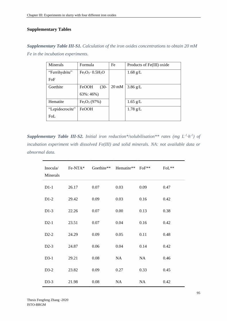
Chapter III: Experiments in slurry with four different iron oxides
95
Thesis Fengfeng Zhang -2020
ISTO-BRGM
Supplementary Tables
Supplementary Table III-S1. Calculation of the iron oxides concentrations to obtain 20 mM
Fe in the incubation experiments.
Minerals Formula Fe Products of Fe(III) oxide
“Ferrihydrite”
FoF
Fe2O3· 0.5H2O
20 mM
1.68 g/L
Goethite FeOOH (30-
63%: 46%)
3.86 g/L
Hematite Fe2O3 (97%) 1.65 g/L
“Lepidocrocite”
FoL
FeOOH 1.78 g/L
Supplementary Table III-S2. Initial iron reduction*/solubilisation** rates (mg L-1·h-1) of
incubation experiment with dissolved Fe(III) and solid minerals. NA: not available data or
abnormal data.
Inocula/
Minerals
Fe-NTA* Goethite** Hematite** FoF** FoL**
D1-1 26.17 0.07 0.03 0.09 0.47
D1-2 29.42 0.09 0.03 0.16 0.42
D1-3 22.26 0.07 0.00 0.13 0.38
D2-1 23.51 0.07 0.04 0.16 0.42
D2-2 24.29 0.09 0.05 0.11 0.48
D2-3 24.87 0.06 0.04 0.14 0.42
D3-1 29.21 0.08 NA NA 0.46
D3-2 23.82 0.09 0.27 0.33 0.45
D3-3 21.98 0.08 NA NA 0.42

Chapter III: Experiments in slurry with four different iron oxides
96
Thesis Fengfeng Zhang -2020
ISTO-BRGM
Supplementary Table III-S3. Ratio of Initial iron solubilisation rates (mg L-1·h-1) to the specific
surface of minerals (m2/g), incubation experiment with solid minerals. NA: not available data
or abnormal data.
Goethite Hematite FoF FoL
D1-1 6.0 .10-03 9.6 .10-04 3.9 .10-04 1.4 .10-03
D1-2 7.7 .10-03 9.6 .10-04 6.9 .10-04 1.0 .10-03
D1-3 6.0 .10-03 0.0 5.6 .10-04 1.1 .10-03
D2-1 6.0 .10-03 1.3 .10-03 6.9 .10-04 1.3 .10-03
D2-2 7.7 .10-03 1.6 .10-03 4.7 .10-04 1.4 .10-03
D2-3 5.1 .10-03 1.3 .10-03 6.0 .10-04 1.3 .10-03
D3-1 6.8 .10-03 NA NA 1.4 .10-03
D3-2 7.7 .10-03 8.6 .10-03 1.4 10-03 1.3 .10-03
D3-3 6.8 .10-03 NA NA 1.3 .10-03
Supplementary Table III-S4. Characteristics of Fe(III) oxides submitted to Fe-reducing
bacteria.
Iron oxide Assumed morphologya Surface areab
(m2g-1)
Estimated mean particle sizec
(µm)
goethite acicular 11.7 1-2
hematite cylinder/rod 31.4 30-120
ferrihydrite blocky 232 20-60
lepidocrocite blocky 337 10-80
aFor use in estimating mean particle size from morphology by SEM-EDS (TM3000
accompanied by a SwiftED3000 X-Stream module (Hitachi)); bDetermined by multipoint BET
N2 adsorption; cEstimated using the oxide densities quoted by (Roden, 2003), it assumed the
following proportions for plate and cylindrical and particle morphologies: plate diameter:
thickness = 10:1; cylinder length: diameter =10:1. Note that particle aggregation was ignored
in these calculations.

Chapter IV: Experiments with ferrihydrite fixed on slides
97
Thesis Fengfeng Zhang -2020
ISTO-BRGM
IV: Experiments with ferrihydrite fixed on slides
IV-1 Introduction
Microbial attachment to mineral surfaces is a fundamental process relevant to geomicrobiology,
the interdisciplinary field of geology and microbiology. Biofilm formation could be responsible
for geochemical cycling of multiple elements and local environmental pollution and is also
relevant to a broad variety of disciplines and applications (Dong, 2010; Elzinga et al., 2012;
Roberts et al., 2006; Torrentó et al., 2012; Yan et al., 2016). In aqueous environments, microbial
attachment behavior can significantly increase rates of mineral weathering (Ahmed and
Holmström, 2015; Sampson et al., 2000), controlling the mobility of metals via cell wall
sorption and metabolic utilization (Fein et al., 2001; Fein et al., 2002; Fowle and Fein, 1999;
Melton et al., 2014).
In general, it is essential for bacterial cells to gain access to surface adherent nutrients or other
substances e.g., electron donors/acceptors for cell metabolism by attachment on minerals
(Harneit et al., 2006; Li et al., 2019; Mueller, 1996). Dissimilatory iron reduction may require
microorganisms attachment onto the minerals in order to couple the oxidation of organic matter
to the reduction of mineral sources of Fe(III) (Lovley, 1987; Roberts et al., 2006). Therefore,
the solubility and bioavailability of Fe(III) might be the factor limiting interactions between
DIRB and Fe minerals in subsurface environments (Sulzberger et al., 1989). According to
previous research, DIRB have some proved strategies to overcome the inherent insoluble nature
of iron oxides in the environment, including the hypothesis of use of chelators and electron
shuttles to mobilize Fe(III) (Nevin and Lovley, 2002). Moreover, Esther et al. (2015)
summarized four mechanisms for the electron transfer between IRB cells and Fe mineral
surface, for two of which attachment is important: (1) direct contact between the cell and Fe
(III) (oxyhydr)oxides mineral, and (2) cell appendages. When there is no soluble Fe(III) in the
environment, bacteria produce flagella and fluff these appendages to adhere to Fe(III) to achieve
Fe(III) reduction (Childers et al., 2002; Esther et al., 2015). Direct attachment is widely
accepted as the predominant mechanism for accessing Fe(III) minerals in the environment
(Lovley et al., 2004).
In the absence of O2, Geobacter metallireducens GS‑15 and Shewanella oneidensis MR‑1 have
been reported to use minerals as electron sinks for heterotrophy-based respiration, by oxidizing

Chapter IV: Experiments with ferrihydrite fixed on slides
98
Thesis Fengfeng Zhang -2020
ISTO-BRGM
organic matter or hydrogen (H2) then transferring the released electrons to Fe or Mn minerals
(Lovley and Phillips, 1988; Lovley et al., 1987). This dissimilatory mineral reduction could be
linked to outer membrane (OM) expression of terminal reductases, joined to transmembrane
and intracellular electron transport components. Hartshorne et al. (2009) indicated that in some
species of Shewanella, deca-heme electron transfer proteins lie at the extracellular face of the
OM, where they can interact with insoluble substrates (Hartshorne et al., 2009), a process
referred to as extracellular electron transport (EET). To reduce extracellular substrates, these
redox proteins must be charged by the inner membrane/periplasmic electron transfer system.
Yang et al. (2019) used electrochemical measurements to demonstrate the correlation between
redox protein concentration and the extracellular electron transfer function linked to
extracellular polymeric substance (EPS) components by Geobacter biofilm (Yang et al., 2019).
These results are consistent with studies showing that Shewanella and Geobacter exhibit heme-
containing proteins located in the OM, suggesting a role of attachment in the reduction of Fe(III).
Furthermore, some IRB could also have direct electron transfer between microbial cells through
conductive nanowires (Cologgi et al., 2011; Gorby et al., 2006; Reguera et al., 2005). When
taken together, these results support the concept that direct cell-mineral contact is necessary for
certain IRB to access insoluble Fe(III) oxide and hydroxide phases. Both Shewanella and
Geobacter were reported to use specialized pili to attach onto Fe minerals used as terminal
electron acceptors (Childers et al., 2002; Elzinga et al., 2012; Jr and Das, 2002; Roberts et al.,
2006). However, some Shewanella species could continue to grow by fermentation when Fe(III)
became limiting (Ivanova et al., 2004; Nogi et al., 1998). Shewanella putrefaciens strain CN-
32 was attached to the surface of hematite at variable pH values (4.5 -7.7) which indicated that
phosphate-based functional groups on the cell wall play an important role in mediating adhesion
through formation of inner-sphere coordinative bonds to hematite surface sites (Elzinga et al.,
2012). While specific adhesion mechanisms for IRB pure stains have been studied in details at
the molecular scale, comparatively less attention has been given to the quantification of their
attachment behavior, in particular in mixed bacterial communities. Moreover, because of the
multiple metabolic respiration pathways of IRB genera, quantitative measurements of IRB
attachment to Fe(III) (oxyhydro)xides are still important to evaluate microbial iron reduction
and microbial contribution in environment. In this study, we examined the attachment behavior
of a mixed IRB community enriched from a natural site, as a function of microbial ferrihydrite
reduction, and quantified two genera of IRB i.e, Geobater and Shewanella, present in this
bacterial community together with many other potential bacteria contributing directly or

Chapter IV: Experiments with ferrihydrite fixed on slides
99
Thesis Fengfeng Zhang -2020
ISTO-BRGM
indirectly to Fe(III)-reduction. As biofilms play a major role in the porosity of natural soils
(Christiani et al., 2008) and sediments (Griebler et al., 2002), it seems relevant to explore the
link between biofilm formation and iron reduction, and the distribution of different IRB
between biofilm and free cells. On the way to develop experimental device to observe natural
biofilms formed at the surface of iron oxides, the objective of this experiment was to assess (1)
the distribution of Geobacter and Shewanella in mixed communities containing diverse IRB in
presence of ferrihydrite fixed on glass slides, and (2) the morphological evolution of the mineral
particles on the slides, linked to ferrihydrite reduction, when a complex mixture of organic
substrates is provided.
IV-2 Specific materials and methods
IV-2.1 Slide preparation with Fe(III) (oxyhydr)oxides
Glass slides (4.5*3 cm) were scratched by grinding and polishing machine to favor resin
attachment. The previously used synthetized iron (oxyhyd)oxide that was confirmed to be
ferrihydrite by Mössbauer spectra (Supplementary Figure III-S7), was attached to the slides
using resin Loctite glue-3. Resin was spread on the glass slide and then 20-30 mg ferryhidrite
powder was dispersed on the surface of glass uniformly using a 200 µm sieve. Then the glass
slides were let at ambient temperature (20-25 ⸰C) at least 30 min in order to harden the resin.
The prepared glass slides with fixed ferrihydrite were observed under SEM (SwiftED3000 X-
Stream module, Hitachi) at 15 kV accelerating voltage (Thouin et al., 2016) before and after
incubation. SEM-EDS were performed to explore the composition and distribution of Fe, and
other elements, the acquisition time of EDS point analyses was 300 s.
10 slides with fixed ferrihydrite (Figure IV-1) were prepared and 2 of them were observed with
whole views. Based on initial views, several specific positions were marked to make a focus to
compare mineralogy change before and after incubation.

Chapter IV: Experiments with ferrihydrite fixed on slides
100
Thesis Fengfeng Zhang -2020
ISTO-BRGM
Figure IV-1. Photo of slide with fixed ferrihydrite (a) images ferrihydrite observed with an
optical microscope (X40) (b).
IV-2.2 Slides incubation experiments
The culture medium used for slide-batch experiments was the same as the medium for IRB,
without iron, detailed in chapter II-3.
Experiments were performed in 9 glass sealed jars, each containing 100 mL of sterile culture
medium, prepared for 9 slides with fixed ferrihydrite. Sealed jars were sterilized at 121°C for
20 min and slides with fixed ferrihydrite were exposed to UV light for 10-20 min to decrease
the number of potential contaminants. All the jars were prepared identically, but 3 of them were
used for mineral observation, 3 for molecular analysis, 1 for SEM-EDS observation, and 2 were
not inoculated in order to represent control condition.
Bacteria inoculum was prepared with the 11th subculture of D3 bacterial enrichment. The
bacteria were collected on 0.22 µm filters to avoid the addition of dissolved iron from the
inoculum media into the fresh medium. In order to inoculate the jars, 10 mL bacterial culture
for each jar were filtered onto 0.22 µm filter, then the filter with bacteria was introduced into
the jar. All these operations were performed into the glove box under anaerobic conditions. The
jars sealed after inoculation are shown in Figure IV-2. They were incubated at 20°C in static
conditions.

Chapter IV: Experiments with ferrihydrite fixed on slides
101
Thesis Fengfeng Zhang -2020
ISTO-BRGM
Figure IV-2. The slides with fixed ferrihydrite in jars.
IV-2.3 Monitoring
[FeII]D (dissolved Fe(II) concentration) was measured using the ortho-phenanthroline
colorimetric method (Murti et al., 1966; Mamindy-Pajany et al., 2013) and [FeT]D (total
dissolved iron concentration) was determined using the same method but with the addition of
0.1 mL of 1% hydroquinone prepared in acetate buffer to reduce dissolved [FeIII]D into [FeII]D
(chapter II-4). During the incubation, 0.5 mL aliquot was collected with a syringe and placed
into a 2 mL eppendorf tube for bacteria counting (chapter II-8.1), and 1.5 mL aliquot was
collected and filtered through a 0.2 μm filter into 5 mL tubes and acidified with concentrated
HCl immediately in the glove box, once a day for the first week, and once a week in the
following weeks. At the same time, pH and Eh (ref. Ag/AgCl) were measured using standard
hand-held portable meters (WTW Multi340i set) in the glove box.
After 85 days incubation, the slides for biofilm collection were taken out of the jars and rinsed
with sterilized water, the other slides were rinsed with alcohol (75% v/v). Supernatants were
removed and freeze-dried for observation of bacteria and minerals under SEM-EDS, as
described in chapter II-6.
IV-2.4 DNA extraction and molecular analysis
Liquid cultures were collected for DNA extraction by centrifuging 10 mL at 10,000 rpm for
10 min. Solid cultures were collected for DNA extraction by harvesting the remaining solids
on slides surfaces by scraping the glass with a sterile blade (Figure IV-3).

Chapter IV: Experiments with ferrihydrite fixed on slides
102
Thesis Fengfeng Zhang -2020
ISTO-BRGM
Figure IV-3. Solid culture collection for DNA extraction after incubation
IV-3 Experimental results
IV-3.1 Bacterial growth
Evolution of bacterial concentration (cells mL-1) during 85 days incubation with ferrihydrite
fixed on slides is shown in Figure IV-4. The enumeration method (Thoma cell) can be reliable
in the conditions of the present experiment because bacterial concentration was in the 109 – 1010
cells.mL-1 range, far more concentrated than the suspended mineral particles resulting from
precipitation reactions.
Figure IV-4. Evolution of bacteria numbers (Log cells mL-1) in the liquid medium of 7 biotic
jars during incubation. Cell numbers in the two abiotic jars were always below 1.84E+3 mL-1
(not shown).

Chapter IV: Experiments with ferrihydrite fixed on slides
103
Thesis Fengfeng Zhang -2020
ISTO-BRGM
Cell density increased very rapidly during the first 5 days and kept increasing until day 35, then
remained almost constant in the following phase. Conversely, there were always only very few
visible bacteria in abiotic jars.
IV-3.2 Physico-chemical monitoring
Slide incubation experiments lasted 85 days. Evolution of [FeT]D and the correlation between
bacterial concentration and [FeT]D are given in Figure IV-5 a and b respectively.
Figure IV-5. Temporal evolution of [TFe]D (mg L-1) in biotic and abiotic jars (a), the
correlation between the dissolved [TFe]D and the number of bacteria (cells mL-1) in solutions
of biotic jars(b).
Ferrihydrite dissolution during the incubation was detected by the increase of [FeT]D in the
biotic experimental system. The IRB reduced Fe(III) of ferrihydrite during the first 50 days of
experiment (Figure IV-5 a). [FeT]D stabilized after day 50, corresponding to the end of bacterial
growth, possibly related with the depletion of the nutrients (electron donors). No ferrihydrite
dissolution was detected according to the evolution of [FeT]D in the abiotic experimental system.
A good correlation between [FeT]D and the concentration of bacteria in the liquid medium was
obtained (Figure IV-5 b).
Evolution of pH and Eh (ref. Ag/AgCl) monitored in jars are given in Figure IV-6.

Chapter IV: Experiments with ferrihydrite fixed on slides
104
Thesis Fengfeng Zhang -2020
ISTO-BRGM
Figure IV-6. 8Evolution of pH and Eh (ref. Ag/AgCl) monitored in jars. Error bars represent
the standard deviation of measurements in 7 the biotic jars / 2 abiotic jars.
During the experimental period, pH was globally identical in the biotic and abiotic jars.
Globally, the pH of the biotic jars slowly increased from 7.5 to 8.3 and the pH from abiotic jars
between 7.2 and 8.7 (Figure IV-6). Concerning Eh, it was identical in biotic and abiotic jars at
the beginning, then it gradually decreased in the biotic jars more intensely than in the abiotic
jars, reaching values as low as -240 mV at the end of the experiment in biotic conditions,
compared with -130 mV in abiotic conditions.
IV-3.3 Bacterial observations and molecular analysis
IV-3.3.1 Bacteria observations under SEM
After incubation, bacterial cells were observed on frozen samples by cryo-SEM using the
backscattered detector imaging (BSE) mode and Secondary electrons imaging (SE) mode
(Figure IV-S1).
IV-3.3.2 Bacterial abundance and abundances of Shewanella and Geobacter
Microbial biomass was quantified at the end of incubation by measuring the concentration of
total microbial DNA extracted from liquid and solid samples (Figure IV-7).
-280
-230
-180
-130
-80
-30
20
2
3
4
5
6
7
8
9
10
0 1 2 3 6 9 23 35 49 62 85
Eh (
mV
)
pH
Days
pH Blank-pH
Eh Blank-Eh

Chapter IV: Experiments with ferrihydrite fixed on slides
105
Thesis Fengfeng Zhang -2020
ISTO-BRGM
Figure IV-7. Parameters linked to bacterial abundance (a) ratio of Shewanella over bacterial
16S rRNA genes copies, (b) ratio of Geobacter over bacterial 16S rRNA genes copies, for liquid
and solid cultures. The letters “a” and “b” differed significantly (Kruskal-Wallis test at p<0.05)
between liquid and solid cultures. Data represent average values of experimental replicates
and their standard deviation (σ). Solid cultures included three replicates, liquid cultures
included 6 replicates.
No significant differences were observed between liquid and solid compartments for the
Shewanella 16S / bacterial 16S ratio (Figure IV-7). Conversely, a significantly higher
proportion of Geobacter / 16S gene in solids than in liquid medium was observed (Figure IV-
7). Thus, the proportion of the two quantified IRB i.e. Geobacter and Shewanella, differs in the
two bacterial communities, free suspended in the liquid medium and attached to the solids. As
already observed in previous experiments with our IRB enrichments, the proportion of
Shewanella in both liquid and solid cultures were significantly higher than for Geobacter.
IV-3.4 Mineral SEM-EDS observations
The global view of a glass slide fixed with ferrihydrite is shown in Figure IV-8 under 30X.
The marked area “23” was used to compare the mineral morphology change before and after
incubation with IRB.

Chapter IV: Experiments with ferrihydrite fixed on slides
106
Thesis Fengfeng Zhang -2020
ISTO-BRGM
Figure IV-8. The global morphology view of a glass slide with fixed ferrihydrite under SEM,
the marked number “23” was the main area used to focus on changes of mineral morphology
before and after incubation with IRB.
The comparison of SEM-EDS observations on area “23” before and after incubation are given
in Figure IV-9.

Chapter IV: Experiments with ferrihydrite fixed on slides
107
Thesis Fengfeng Zhang -2020
ISTO-BRGM
Figure IV-99. SEM-EDS analysis of ferrihydrite samples in the specific area “23” on the
glass slide, before incubation (a) and after incubation (b).
Table IV-11. Semi-quantitative EDS analysis (relative concentrations in weight %) of main
elements in point analyses located on SEM pictures and corresponding spectra shown Figure
IV-9 and P/Fe, O/Fe, C/Fe rations.
Sample Point C O Si P S Fe P/Fe O/Fe C/Fe
Before 1 7.41 24.64 0.46 0.01 0.00 66.93 0.00 0.37 0.11
2 6.93 40.34 0.32 0.00 0.00 52.07 0.00 0.77 0.13
After 1 7.25 52.86 1.35 4.33 0.26 28.18 0.15 1.88 0.26
2 7.61 57.38 0.58 10.04 0.07 14.85 0.68 3.86 0.51
The microscopic picture on the whole glass slide with fixed ferrihydrite was taken before the
experiment. The specific area was marked with number “23” to follow the morphology change
(Figure IV-9). Ferrihydrite was characterized, before incubation, by diverse particles of 10-
50 µm presenting irregular shapes (Figure IV-9) that mainly contained the elements O (20-40%)
and Fe (50-70%) (Table IV-1). After incubation, new forms of particles with crystalline aspects
and a prismatic morphology, 100 µm long and 10 µm large, were observed in the same area on

Chapter IV: Experiments with ferrihydrite fixed on slides
108
Thesis Fengfeng Zhang -2020
ISTO-BRGM
the glass slide (Figure IV-9). As revealed by SEM/EDS analysis, these prismatic crystals still
contained O (50-60%) and Fe (10-20%), but also P (10-15%) that was not detected in the initial
particles (Table IV-1).
More information on post-incubation slides (biotic and abiotic) were given by additional SEM-
EDS observations of fixed minerals and precipitates collected from the liquid medium of biotic
jars (Figure IV-10).

Chapter IV: Experiments with ferrihydrite fixed on slides
109
Thesis Fengfeng Zhang -2020
ISTO-BRGM
Figure IV-101. SEM-EDS analysis of ferrihydrite samples after incubation, biotic glass slides
with fixed ferrihydrite (“a” and “b”), precipitates collected in the liquid medium (c), and
abiotic glass slides with fixed ferrihydrite (d).
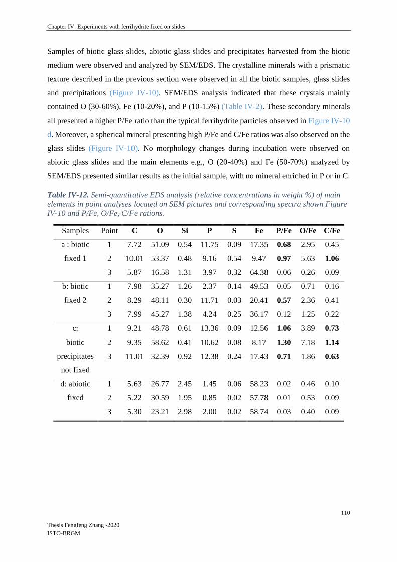
Chapter IV: Experiments with ferrihydrite fixed on slides
110
Thesis Fengfeng Zhang -2020
ISTO-BRGM
Samples of biotic glass slides, abiotic glass slides and precipitates harvested from the biotic
medium were observed and analyzed by SEM/EDS. The crystalline minerals with a prismatic
texture described in the previous section were observed in all the biotic samples, glass slides
and precipitations (Figure IV-10). SEM/EDS analysis indicated that these crystals mainly
contained O (30-60%), Fe (10-20%), and P (10-15%) (Table IV-2). These secondary minerals
all presented a higher P/Fe ratio than the typical ferrihydrite particles observed in Figure IV-10
d. Moreover, a spherical mineral presenting high P/Fe and C/Fe ratios was also observed on the
glass slides (Figure IV-10). No morphology changes during incubation were observed on
abiotic glass slides and the main elements e.g., O (20-40%) and Fe (50-70%) analyzed by
SEM/EDS presented similar results as the initial sample, with no mineral enriched in P or in C.
Table IV-12. Semi-quantitative EDS analysis (relative concentrations in weight %) of main
elements in point analyses located on SEM pictures and corresponding spectra shown Figure
IV-10 and P/Fe, O/Fe, C/Fe rations.
Samples Point C O Si P S Fe P/Fe O/Fe C/Fe
a : biotic
fixed 1
1 7.72 51.09 0.54 11.75 0.09 17.35 0.68 2.95 0.45
2 10.01 53.37 0.48 9.16 0.54 9.47 0.97 5.63 1.06
3 5.87 16.58 1.31 3.97 0.32 64.38 0.06 0.26 0.09
b: biotic
fixed 2
1 7.98 35.27 1.26 2.37 0.14 49.53 0.05 0.71 0.16
2 8.29 48.11 0.30 11.71 0.03 20.41 0.57 2.36 0.41
3 7.99 45.27 1.38 4.24 0.25 36.17 0.12 1.25 0.22
c:
biotic
precipitates
not fixed
1 9.21 48.78 0.61 13.36 0.09 12.56 1.06 3.89 0.73
2 9.35 58.62 0.41 10.62 0.08 8.17 1.30 7.18 1.14
3 11.01 32.39 0.92 12.38 0.24 17.43 0.71 1.86 0.63
d: abiotic
fixed
1 5.63 26.77 2.45 1.45 0.06 58.23 0.02 0.46 0.10
2 5.22 30.59 1.95 0.85 0.02 57.78 0.01 0.53 0.09
3 5.30 23.21 2.98 2.00 0.02 58.74 0.03 0.40 0.09

Chapter IV: Experiments with ferrihydrite fixed on slides
111
Thesis Fengfeng Zhang -2020
ISTO-BRGM
IV-4 Discussion
IV-4.1 Fe dissolution
[FeT]D increased over time in the biotic systems, but not in the abiotic control experiments.
Futhermore, the increase in [FeT]D was correlated to the increase of the bacterial cell numbers,
which suggested that ferrihydrite dissolution was due to bacterial activity in the biotic
conditions. Microbial ferrihydrite dissolution rate and efficiency in presence of inoculum D3
have been discussed in chapter III, where we showed the dissolution of 4-6 % of the initial 20
mM iron supplied as ferrihydrite in slurry experiments. In the present experiment, according to
[FeT]D reached in the liquid medium, 1-2 % (approximately) of the initial ferrihydrite was
dissolved. Although ferrihydrite reduction was less efficient in the present study compared with
the previous slurry system, bacteria were metabolically active. They might have contributed
less to iron dissolution for two reasons: (1) the ferrihydrite fixed on the glass slides was less
available for IRB because a part of its surface was inserted into the resin; (2) the static
experimental setup reduced the transfer of substances between the surface of ferrihydrite and
IRB present in the liquid medium. A study from (Bose et al., 2009) suggested that hematite
morphology, particle size, and/or degree of aggregation seemed to influence the mechanism
that Shewanella uses to access Fe(III) sites on hematite surfaces.
IV-4.2 Distribution of Shewanella and Geobacter 16S gene copies in the liquid medium
and in the biofilm
Evolution of bacterial numbers in suspension in the liquid phase indicated that the bacterial
population increased during the first 50 days. The proportion of Shewanella and Geobacter 16S
gene copies were quantified as free-living cells or attached to the solid phase at the end of the
experiment. Ratio of Shewanella/total bacteria (qPCR 16S approach, Figure IV-7) in the liquid
compartment was comparable to the ratio in the previous slurry experiment, and around 0.01 -
0.03 (chapter III). A similar ratio of Shewanella/total bacteria was in the biofilm. Conversely,
the ratio of Geobacter/total bacteria (qPCR 16S approach, Figure IV-7) was significantly higher
in the biofilm attached to the solid phase than in suspension in the liquid phase. However, as
observed in slurry (chapter III), Geobacter 16S gene abundance was still around 100 times
lower than Shewanella 16S gene in the bacterial community that developed during the slide
experiment. This could be attributed to the composition of the culture medium.
Chapter IV: Experiments with ferrihydrite fixed on slides
112
Thesis Fengfeng Zhang -2020
ISTO-BRGM
Previously, it has been shown that cell-ferrihydrite contact is necessary for IRB e.g., Shewanella
and Geobacter genera to use the electrons from Fe(III) (oxyhydro)oxides to reduce Fe(III)
(Gorby et al., 2006; Reguera et al., 2005). Geobacter genus requires iron reduction for its
metabolism, whereas, for Shewanella genus, some species can either use iron reduction or
switch to fermentation (Nogi et al., 1998). On the other hand, according to research on the
mechanisms of Fe(III) use as the electron acceptor by IRB cells, four IRB-mineral interactions
were evidenced, e.g., by direct contact, by ligands/siderophore, by electron shuttles, and by pili
(Esther et al., 2015). Shewanella has been reported to use electron shuttles for indirect iron
reduction, these electron shuttles can be exogenous like humic substances and sulfur
compounds (Liu et al., 2011a; Stams et al., 2006) or endogenous as those secreted by the
organism itself (Marsili et al., 2008; Newman and Kolter, 2000; Von Canstein et al., 2008).
Conversely, Geobacter genus has been found and reported to need physical contact during
Fe(III) reduction rather than use indirect reduction pathways. Geobacter genus is well known
to interact with solid Fe(III) (oxyhydr)oxides using pili filaments, also named as ‘bacterial
nanowires’ (Esther et al., 2015; Reguera et al., 2005; Roberts et al., 2006).
Moreover, Reguera et al., 2007 showed that Geobacter genus required the expression of
electrically conductive pili to form biofilms on Fe(III) oxide surfaces, and pili are also essential
for biofilm development on plain glass when fumarate was the sole electron acceptor. These
authors suggested that the pili of Geobacter genus had a structural role in biofilm formation
(Reguera et al., 2007). So, according to literature, ferrihydrite reduction by IRB could occur by
direct and indirect contact with IRB belonging to Shewanella genus and mainly by direct
contact with IRB belonging to Geobacter genus. This could explain why, in the present
experiment, Geobacter proportion was more important in the biofilm than in the in the bacterial
community suspended in the liquid medium.
IV-4.3 SEM observation of the solid particles
SEM observation showed the formation of new types of particles in biotic conditions. These
secondary minerals were enriched in phosphorous and/or in carbon. Precipitation of iron
phosphate minerals could have occurred (Zachara et al., 1998). Heiberg et al., 2012 observed
the precipitation of the Fe(II)-phosphate vivianite during the anoxic incubation of a natural soil.
In our systems, the phosphate present in the culture medium could precipitate with the Fe(II)
produced by the IRB. Muehe et al. indicated that Fe(II) inputs generally lead to vivianite
precipitation in phosphate-bearing environmentsfound that As(V) partly replaced phosphate in

Chapter IV: Experiments with ferrihydrite fixed on slides
113
Thesis Fengfeng Zhang -2020
ISTO-BRGM
vivianite, thus forming a vivianite-symplesite solid solution identified as
Fe3(PO4)1.7(AsO4)0.3·8H2O (Muehe et al., 2016).
In addition, particles with high C/Fe proportion could represent forms of iron carbonates
(siderite). The siderite could be formed by reducing akaganeite in the presence of Shewanella
under a H2-CO2 atmosphere (Roh et al., 2003). Such globular morphology of Fe carbonate were
already observed (Wiesli et al., 2004). The formation of Fe carbonate during the incubation of
Shewanella putrefasciens in presence of natural Fe(III) oxides was reported (Maitte et al., 2015).
In this study, the liquid phase was composed of synthetic mine water amended with formate
only, as an electron donor, contrary to our conditions corresponding to a culture medium with
high concentrations of phosphate and several electron donors. In our biotic conditions, the
degradation of organic electron donors resulted in the production of inorganic C that could react
with Fe(II) and other substances present in the liquid medium, to form secondary minerals.
SEM observations also showed the presence of bacteira on the attached solid particles.
(Thormann et al., 2004) reported the very rapid biofilm formation of Shewanella, in 48 to 120
h, in presence of a rich culture medium. However, bacteria were observed not only on the
ferrihydrite particles, but also on the secondary minerals. This observation suggests that the
particles were not colonized exclusively to facilitate Fe(III) dissolution, but also as a solid
support for the development of other bacteria, that could grow by fermentation of organic
molecules.
IV-5 Conclusion and perspective
This original experiment using ferrihydrite fixed with resin onto glass slides enabled biofilm
development on iron oxides and secondary minerals formation. In particular, we found different
abundances of two well characterized IRB genera in the biofilm and surrounding liquid medium,
and these differences might be related with their metabolism. Indeed, Geobacter, a genus that
requires direct contact with solids to use Fe(III) as an electron acceptor was significantly more
abundant in the biofilm than in the liquid medium, whereas Shewanella, a genus includes
species that can either use Fe(III) respiration or fermentation to thrive was found equally
distributed between the two phases. Up to now, studies have mainly been performed with pure
strains of IRB, and our results obtained with a complex enrichment of IRB, including many
potential DIRB and fermentative IRB, and focusing on quantification of Shewanella and
Chapter IV: Experiments with ferrihydrite fixed on slides
114
Thesis Fengfeng Zhang -2020
ISTO-BRGM
Geobacter in these communities, opens the perspective to study Fe(III) reduction processes in
conditions closer to those of natural sites.
Globally, all results suggest that our IRB inoculum contains a certain percentage of bacteria
affiliated to Geobacter and Shewanella genera which include species known to use Fe(III)
(oxyhydr)oxides as an electron acceptor during dissimilatory iron reduction. The method to
prepare glass slides could be applied in order to study the influence of the type of Fe(III) mineral
on the composition of natural biofilms, with the possibility to immobilize a range of different
oxides. Beyond laboratory studies, slides could be placed on site, inserted in soils or aquatic
sediments, in order to acquire a better knowledge of the contribution and distribution of IRB
communities attached on iron oxides surfaces can help to elucidate Fe dynamics in surface
environments.

Chapter IV: Experiments with ferrihydrite fixed on slides
115
Thesis Fengfeng Zhang -2020
ISTO-BRGM
Supplementary material
Supplementary Figures
(a) (b)
(c) (d)
(e) (f)

Chapter IV: Experiments with ferrihydrite fixed on slides
116
Thesis Fengfeng Zhang -2020
ISTO-BRGM
Figure IV-S1. Cryo-SEM photomicrographs of bacteria on the surface of glass slides with
fixed ferrihydrite at the end of the experiment. Red arrows indicate the minerals observed in (a)
and (b), and cells attached to minerals in (d), (f) and (j).
Some bacterial cells appeared spherical shaped and rod shaped, separated from each other,
turgid, and complete with smooth surface, on secondary mineral grains. Bacterial morphology
was very clear under the microscope field of Figure IV-S1 (i) and (j), cell size was between 1-
4 µm. Other bacteria seemed to be embedded in an exopolymeric matrix typical of biofilms
(Figure IV-S1 c to f). Moreover, ferrihydrite (Figure IV-S1 (a) and “secondary minerals”
(Figure IV-S1 (b) were both observed on the surface of glass slides, but bacterial cells and
biofilms were mainly present on surface of “secondary minerals” (Figure IV-S1 (c, d, e, f)) and
some “traces” (might be networks of corrosion-like channels that accommodate bacterial cells
and exopolymer filaments) were observed on the position of bacteria (Figure IV-S1 (j)).
(g) (h)
(i) (j)
Chapter V: Mobility of As, Cr and Cd adsorbed on Fe (oxyhydr)oxides submitted to IRB
117
Thesis Fengfeng Zhang -2020
ISTO-BRGM
Chapter V: Mobility of As, Cr and Cd adsorbed on Fe
(oxyhydr)oxides submitted to IRB
V-1. Abstract
A continuous column experiment was carried out to investigate the mobility of trace elements
(TEs, i.e. As, Cr, Cd) and transformation of ferrihydrite and goethite under iron-reducing
conditions. Two column experiments were performed with ferrihydrite and goethite
respectively, spiked with As, Cr and Cd through a preliminary adsorption step. The
experimental program included three successive phases: stabilization with synthetic water,
inoculation with IRB and feeding with mixtures of synthetic water and organic substrates
(glucose, acetate, formate, lactate and peptone). Evolution of total dissolved concentrations of
Fe, As, Cd and Cr, and the values of redox potential and pH were monitored in the column’s
outlets. Iron concentration was always higher in the outlet of inoculated columns than in un-
inoculated conditions. At the end of the experiment, 96 mg of Fe had been leached from the
inoculated goethite column (0.8 mg from the un-inoculated condition), whereas 141 mg of Fe
had been leached from the ferrihydrite column (13 mg for the un-inoculated condition). The
evolution of iron and TEs in inoculated and un-inoculated columns confirmed microbial Fe
reduction and that the type of Fe (oxyhydr)oxides could influence the mobility of As, Cr, and
Cd. Relation between type of Fe oxide and the relative abundances of two IRB genera
(Geobacter and Shewanella) was observed.
V-2. Introduction
In the previous chapters, we studied Fe reducing rates of three bacterial consortia enriched from
the site of Decize, in presence of four different synthetic Fe (oxyhydr)oxides (ferrihydrite,
goethite, hematite and lepidocrocite) in batch conditions. Results highlighted the influence of
the type of iron (oxyhydr)oxide in terms of iron reduction rates and abundances of two well-
known DIRB genera, Shewanella and Geobacter, in the complex iron-reducing community
potentially including both DIRB and fermentative bacteria. The highest reducing rates were
measured for ferrihydrite and lepidocrocite in the presence of a consortia enriched from D3, an
aquatic sediment (Decize), followed by hematite and goethite with the same inoculum.
Geobacter sp. was more abundant in presence of goethite than with the other (oxyhydr)oxides
and Shewanella was more abundant in presence of ferrihydrite and lepidocrocite.
Chapter V: Mobility of As, Cr and Cd adsorbed on Fe (oxyhydr)oxides submitted to IRB
118
Thesis Fengfeng Zhang -2020
ISTO-BRGM
Based on the interactions between Fe (oxyhydr)oxides and bacteria, microbial activities can
play either a direct role or an indirect role on speciation and mobility of trace elements (TEs).
The understanding and prediction of TEs’ mobility and biogeochemical cycling is essential to
develop remediation and management strategies for polluted sites. Although metal(oid)s bound
to iron oxides can be mobilized by changing physical and chemical conditions, which could
explain the solubilisation patterns of TEs within some fractions of sediment (Calmano et al.,
1993; Chuan et al., 1996), bacterial activities and their functions are attracting more and more
attention in relation to the fate of TEs in aquatic systems and soils (Gounou et al., 2010; Hansel
et al., 2003a; Kurek, 2002; Kurek and Bollag, 2004; Li et al., 2016).
Some iron-reducing bacteria (IRB), such as Shewanella, have the ability to reduce polyferric
sulfate, resulting in the release of Fe(II) and Cd(II) into solution, the released Cd(II)aq could
then be re-adsorbed by secondary iron minerals (Li et al., 2016). Cd (II) was also stabilized in
bacterial reactors using iron-reducing bacteria and magnetite iron oxide powder (Fe3O4) during
the process of wastewater treatment (Su et al., 2019). The impact of reducing conditions and
bacterial activities on the mobility of As adsorbed to iron oxyhydr(oxides), leading to lower
adsorption and higher mobility of As has previously been demonstrated (Bowell, 1994;
Cummings et al., 1999; Hellal et al., 2017). However Tufanoa and Fendorf, (2008), found that
reductive transformation of ferrihydrite promoted As retention, rather than releasing it, during
an experiment under Fe-reducing conditions with Shewanella. They also suggested As(III)
retention during Fe reduction to be temporally dependent on new formed secondary Fe minerals.
Cr(IV) could be reduced to Cr(III) by the direct role of IRB to mitigate its hazard (Gandhi et
al., 2002; Liu et al., 2011b; Peng et al., 2015; Wielinga et al., 2001). However, there are very
few studies on the bacterial mobility of Cr(III) in iron-reducing condition. Furthermore, few
studies are available regarding microbial impact on multiple TEs’ mobility in anaerobic
sediments. Much of the research carried out on TE mobility in various environments under iron-
reducing conditions has been performed with pure bacterial strains or one specific TE. In the
environment, however, we are faced with complex TE pollutions and highly diverse bacterial
communities. Thus it is necessary to carry out further research to understand the impact of
mixed IRB groups on the reduction of different Fe (oxyhydr)oxides, and indirectly on the
speciation and mobility of several TEs in combination. Continuously fed columns are systems
that enable long-term experiments mimicking a water flow in controlled conditions. They have
been previously used for monitoring the mobility and fate of some TEs e.g. As, Zn, Cd, Cu, Pb,

Chapter V: Mobility of As, Cr and Cd adsorbed on Fe (oxyhydr)oxides submitted to IRB
119
Thesis Fengfeng Zhang -2020
ISTO-BRGM
Hg, (Crampon et al., 2018; De Matos et al., 2001; Hartley et al., 2004; Hellal et al., 2015; Hu
et al., 2015), for studying interactions with bacteria.
To illustrate the influence of geo-microbial phenomena and the speciation and mobility of iron-
associated TEs, e.g., As, Cr and Cd, we developed a laboratory continuously-fed column
experiment representing a simplified model aquifer under anaerobic conditions (Hellal et al.,
2017). One amorphous and one crystallized Fe (oxyhydr)oxides: synthetic ferrihydrite and
goethite were used for this column study. Column systems were implemented with the
previously obtained IRB enrichment to assess the role of mixed bacterial communities in the
transformations of ferrihydrite and goethite in reducing conditions.
V-3 Specific materials and methods
V-3.1 Adsorption of As, Cr and Cd on synthetic iron (oxyhydr)oxides
V-3.1.1 Preparation of TEs stock solution
As, Cr and Cd solutions were prepared as following:
(1) As(V) stock solution: 1000 mg L-1, (Na2HAsO4), dilution of a 10 g L-1 stock solution.
(2) Cr(III) stock solution: CrCl3 6H2O, 100 mg L-1, dissolve 0.512 g CrCl3 6H2O into 1L
ultrapure water.
(3) Cd(II) stock solution: CdCl2, 100 mg L-1, dissolve 0.163 g CdCl2 into 1L ultrapure
water
V-3.1.2 Adsorption of TEs to iron oxyhydr(oxides)
Before initiating the column experiments, we determined the adsorption capacity of 4 types of
iron (oxyhydr)oxides: ferrihydrite, lepidocrocite, goethite and hematite for mixtures of As, Cr
and Cd. Experiments were performed in 250 mL erlenmeyer flasks, under agitation (100 RPM)
and at 20◦C for 24 h. Each batch experiment had 3 replicates for each condition.
Iron (oxyhydr)oxides (8 g L-1) were added as powders into 150 mL As(V), Cr(III), Cd(II) mixed
stock solutions, details can be found in Table V-1. Initial samples were collected from the
mixtures at the beginning of the adsorption reaction and filtered at 0.45 µm (Millex -GP Syringe
Filter, 33 mm diameter) to determine the total concentrations of As, Cr and Cd (abbreviated
[As]D, [Cr]D and [Cd]D). After 24h, the mixture was centrifuged at 15,000 rpm for 10 min and
the recovered solid Fe (oxyhydr)oxides were rinsed 11 times with ultrapure water. All rinsed
samples were collected and filtered to analyze [As]D, [Cr]D and [Cd]D in order to determine the

Chapter V: Mobility of As, Cr and Cd adsorbed on Fe (oxyhydr)oxides submitted to IRB
120
Thesis Fengfeng Zhang -2020
ISTO-BRGM
rinsing efficiency. In the end, the solid Fe (oxyhydr)oxide were recovered after freeze-drying
(-90 ◦C Martin Christ Beta 2-8 LSCplus). An amount of 0.1 g of each Fe (oxyhydr)oxide was
then dissolved into 10-20 mL HCl (1M) to measure the capacity of Fe (oxyhydr)oxides to
adsorb As(V), Cr(III), Cd(II). Final TEs’ adsorption capacities on Fe (oxyhydr)oxides were
determined and calculated by analysis of [As]D, [Cr]D and [Cd]D ([]D for dissolved elements) in
these solutions. [As]D, [Cr]D and [Cd]D were determined with Atomic Absorption Spectroscopy
(AAS, Varian SpectrAA 220-Z) and Microwave Plasma-Atomic Emission Spectrometry
(Agilent 4210 MP-AES). The pH was measured using standard hand-held portable meters
(WTW Multi340i set) before and after the adsorption reaction to avoid an effect of pH on
adsorption capacity (Figure V-2 (d)).
Table V-1. Initial TEs (As, Cr, and Cr) concentrations and Fe (oxyhydr)oxides solid/liquid
concentration during adsorption batch experiment.
TE adsorption ferrihydrite goethite hematite lepidocrocite
[As]D mg L-1
[Cr]D mg L-1
[Cd]D mg L-1
167
67
100
333
67
67
333
67
67
333
67
67
Fe (oxyhydr)oxides 1.2 g 1.2 g 1.2 g 1.2 g
Concentration of solid /
liquid phase
8 g L-1 8 g L-1 8 g L-1 8 g L-1
V-3.2 Columns experimental setup
V-3.2.1 Preparation of Fe (oxyhydr)oxides
Based on the previous discussion in chapter III, synthetic ferrihydrite with its amorphous nature
and goethite with its acicular or needle-shaped crystallized forms were used for the column
study. According to the results from the adsorption assays, 20 g of ferrihydrite and 20 g of
goethite were prepared using the same method for adsorping TEs (As, Cr and Cd) with 6 rinsing
steps. The final adsorption results, determined as metal contents on the solid oxides, are
presented in Table V-2.
Table V-2. Concentrations of TEs adsorbed by ferrihydrite and goethite.
mg g-1 ferrihydrite goethite
Chapter V: Mobility of As, Cr and Cd adsorbed on Fe (oxyhydr)oxides submitted to IRB
121
Thesis Fengfeng Zhang -2020
ISTO-BRGM
As 14.5 5.28
Cr 1.4 2.53
Cd 2.01 3.03
V-3.2.2 Preparation of silica gel and sand matrix
Silica gel (6 %) was used to stop the fine iron (oxyhydr)oxides migrating from the sand/iron
oxide mixture under the water flow as described in Hellal et al., (2017). A 10% silica gel
mixture was prepared by heating 20 g of silica gel in 200 mL of a solution of 7% KOH on a hot
plate, stirring with a magnetic stir bar until dissolution; then 300 mL of ultra-pure water was
added and the solution was cooled to ~20 °C as stock solution. This solution was quickly titrated
with diluted phosphoric acid (20 %) to pH 7.
For each column, we quickly mixed 50 mL liquid silica gel with 153 g of sterile sand and
previously added 8.7 g of As, Cd and Cr-absorbed Fe (oxyhydr)oxides (previously exposed to
UV light for 5-10 min) before it solidifies. We broke up the "jellified" mixture by mixing with
a spatula and kept it sterile before filling the columns.
V-3.2.3 Column setup and experimental conditions
In total, 4 columns were set up for this experiment: two for ferrihydrite which included one
inoculated column and one un-inoculated control column, and the same for goethite. All
columns used for mobility experiments were 25 cm long glass columns, with an internal
diameter of 2.5 cm, provided by Omnifit® (Diba industries, USA). Columns, pipes and
conenctions were sterilized by autoclaving. The columns were sealed in aseptic conditions and
set up vertically with an ascendant inflow of different solutions in 3 steps (more details are
given in Figure V-1 and Table V-3). All silicone tubes and caps used in the experiment were
made of PTFE to avoid interactions with TE. Columns were first filled with the mixture of sand,
Fe oxide (ferrihydrite/goethite with absorbed As, Cr and Cd) and 6% silica gel. The columns
were flushed with N2 (filtered through a 0.2 μm filter, flow speed between 1-5 mL min-1) to
evacuate O2 and avoid the formation of bubbles in the column, and thus obtain realistic
anaerobic conditions. For stabilization, columns were then filled with synthetic water, made to
mimic the composition of groundwater, sterilized by autoclaving (121 °C for 20 min), and
described as FIm (moderately hard) water by US EPA Report EPA-821-R-02-012, Section
7.2.3.1 (US EPA, 2002). This water was composed of 96 mg L-1 NaHCO3, 60 mg L-1 CaSO4
(2H2O) (separate autoclave), 60 mg L-1 MgSO4 and 4 mg L-1 KCl (Crampon et al., 2018). pH
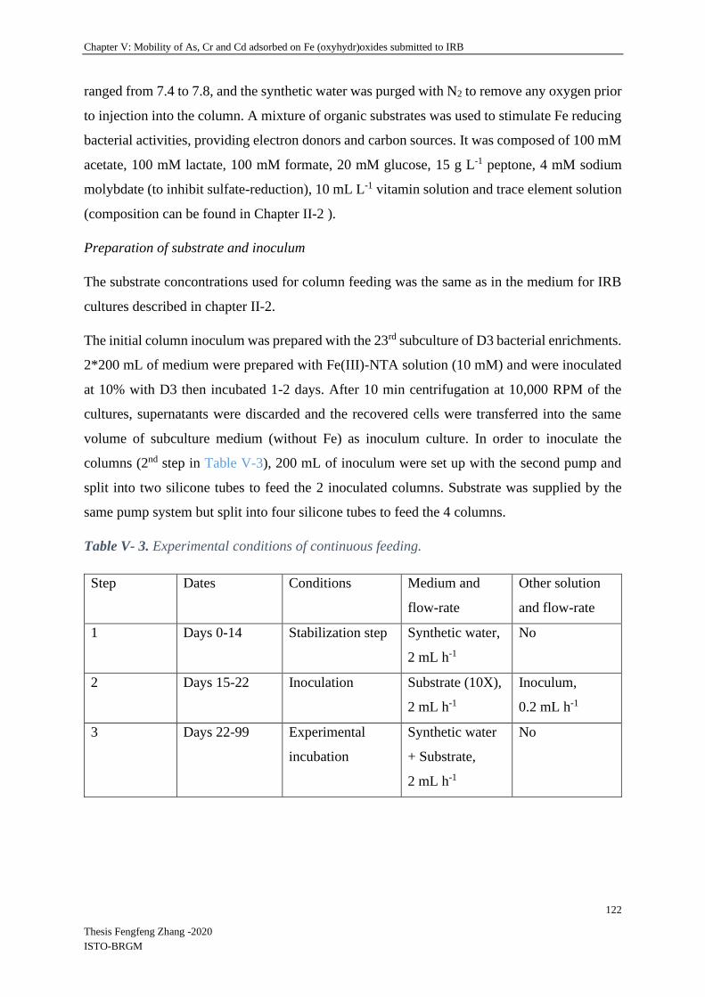
Chapter V: Mobility of As, Cr and Cd adsorbed on Fe (oxyhydr)oxides submitted to IRB
122
Thesis Fengfeng Zhang -2020
ISTO-BRGM
ranged from 7.4 to 7.8, and the synthetic water was purged with N2 to remove any oxygen prior
to injection into the column. A mixture of organic substrates was used to stimulate Fe reducing
bacterial activities, providing electron donors and carbon sources. It was composed of 100 mM
acetate, 100 mM lactate, 100 mM formate, 20 mM glucose, 15 g L-1 peptone, 4 mM sodium
molybdate (to inhibit sulfate-reduction), 10 mL L-1 vitamin solution and trace element solution
(composition can be found in Chapter II-2 ).
Preparation of substrate and inoculum
The substrate concontrations used for column feeding was the same as in the medium for IRB
cultures described in chapter II-2.
The initial column inoculum was prepared with the 23rd subculture of D3 bacterial enrichments.
2*200 mL of medium were prepared with Fe(III)-NTA solution (10 mM) and were inoculated
at 10% with D3 then incubated 1-2 days. After 10 min centrifugation at 10,000 RPM of the
cultures, supernatants were discarded and the recovered cells were transferred into the same
volume of subculture medium (without Fe) as inoculum culture. In order to inoculate the
columns (2nd step in Table V-3), 200 mL of inoculum were set up with the second pump and
split into two silicone tubes to feed the 2 inoculated columns. Substrate was supplied by the
same pump system but split into four silicone tubes to feed the 4 columns.
Table V- 3. Experimental conditions of continuous feeding.
Step Dates Conditions Medium and
flow-rate
Other solution
and flow-rate
1 Days 0-14 Stabilization step Synthetic water,
2 mL h-1
No
2 Days 15-22 Inoculation Substrate (10X),
2 mL h-1
Inoculum,
0.2 mL h-1
3 Days 22-99 Experimental
incubation
Synthetic water
+ Substrate,
2 mL h-1
No

Chapter V: Mobility of As, Cr and Cd adsorbed on Fe (oxyhydr)oxides submitted to IRB
123
Thesis Fengfeng Zhang -2020
ISTO-BRGM
Figure V-2. Photo and sketch (protocol in 3 steps) of the columns experiment.
V-3.2.4 Monitoring
Columns experiments lasted 99 days and were divided into 3 steps as described in Table V-3.
Outlet bottles were emptied everyday (except weekends) and the liquid samples were collected
in a glove box to analyse pH, redox potential (Eh, ref. Ag/AgCl), [FeT]D (total dissolved Fe
concentration), [As]D, [Cr]D and [Cd]D, and to observe the bacteria. [FeT]D, [As]D, [Cr]D and
[Cd]D were determined using the same method as described in chapter II-4 and samples for
those chemical analyses were filtered through a 0.2 μm filter into plastic tubes and acidified
with concentrated HCl in anaerobic condition. [AsIII] and [AsV] were separated using resins
(AG® 1-X8 Resin, Cat. # 140-1431, Biorad) (Battaglia-Brunet et al., 2006) and determined by
the same method as described in chapter II-4. pH and redox potential (Eh, ref. Ag/AgCl) were
measured after sampling immediately using standard hand-held portable meters (WTW
Multi340i set) in the glove box.
Chapter V: Mobility of As, Cr and Cd adsorbed on Fe (oxyhydr)oxides submitted to IRB
124
Thesis Fengfeng Zhang -2020
ISTO-BRGM
At the end of the experiment, the column system was stopped and the remaining liquid from
the columns was collected to analyze the biological parameters when columns were
disconnected. Then columns were weighed and the depths of visible reactions were measured
by observing the color change. The solid samples from the top, middle and bottom of the
columns were collected in a glove box to determine biological and chemical parameters.
V-3.2.5 SEM-EDS observation and Mössbauer spectrometry
Initial column samples and post-incubation samples were observed by SEM-EDS using the
same method as described in chapter II-6.1 after freeze-drying (-90 ◦C Martin Christ Beta 2-8
LSCplus). An amount of 0.1 g of each dried sample was dissolved in 10-20 mL HCl (1M) to
measure the remaining Fe, As, Cr and Cd in columns.
After incubation, the column samples for Mössbauer spectrometry analysis were collected in a
glove box and kept under anaerobic conditions. Mössbauer spectrometry was used to determine
Fe species in different depths of the column by the method described in chapter II-7.
V-3.2.6 Biological analyses
Bacterial biofilm was observed by SEM using the method described in chapter II-6.2 and
images of bacteria in outlet samples were observed with an optical microscope (X400)
described in chapter II-8.1.
DNA extractions were performed on samples solid and liquid samples using the method
described in chapter II-8.2. Bacterial 16S rRNA and Shewanella / Geobacter 16S rRNA gene
quantifications were performed by qPCR following the methods described in chapter II-8.4 and
8.6.
V-4 Experimental results
V-4.1 Adsorption experimental results
Concentrations of As, Cr and Cd in rinsing solutions after adsorption reactions are presented in
Figure V-2. This step aimed to eliminate as much as possible the remaining un-adsorbed metals
from the Fe (oxyhydr)oxides.

Chapter V: Mobility of As, Cr and Cd adsorbed on Fe (oxyhydr)oxides submitted to IRB
125
Thesis Fengfeng Zhang -2020
ISTO-BRGM
Figure V- 3. Concentrations of As(a), Cd(b) and Cr(c) in the according pellet-rinsing solutions
for the four studied iron (oxyhydr)oxides (ferrihydrite, goethite, hematite and lepidocrocite).
Results (Figure V-2) showed that after 6 rinsing steps most of the remaining un-adsorbed metals
were removed.
Adsorption capacities of As, Cr and Cd on iron (oxyhydr)oxides are presented in Figure V-3.
Globally, all the iron (oxyhydr)oxides had higher adsorption capacities for As than for Cr and
Cd (Figure V-3). As shown in Figure V-3 (a), lepidocrocite had the biggest adsorption capacity
on As, 31.7 mg g-1 at room temperature, pH 5-6. Adsorption capacity then decreased from
ferrihydrite to goethite and finally hematite. While the adsorption capacity for Cr was the
0
6
12
18
24
30
36
42
0 2 4 6 8 10 12
[As]
Do
f ri
nsi
ng
extr
acta
nts
/
mg
L-1
Rinsing time
Goethite
Hematite
Lepidocrocite
(a)-1
0
0.08
0.16
0.24
0.32
0 2 4 6 8 10 12
[As]
Do
f ri
nsi
ng
extr
acta
nts
/
mg
L-1
Rinsing time
Ferrihydrite(a)-2
0
0.2
0.4
0.6
0.8
1
0 2 4 6 8 10 12
[Cd
] Do
f ri
nsi
ng
extr
acta
nts
/
mg
L-1
Rinsing time
Goethite
Hematite
Lepidocrocite
(b)-1
0
5
10
15
20
25
0 3 6 9 12
Cd
] Do
f ri
nsi
ng
extr
acta
nts
/
mg
L-1
Rinsing time
Ferrihydrite(b)-2
0
0.2
0.4
0.6
0.8
1
0 2 4 6 8 10 12
[Cr]
Do
f ri
nsi
ng
extr
acta
nts
/
mg
L-1
Rinsing time
FerrihydriteGoethiteHematiteLepidocrocite
(c)

Chapter V: Mobility of As, Cr and Cd adsorbed on Fe (oxyhydr)oxides submitted to IRB
126
Thesis Fengfeng Zhang -2020
ISTO-BRGM
highest with ferrihydrite, it was equivalent for the three other oxides. For Cd, the adsorption
capacities of the different iron oxides were not significantly different (Figure V-3 (b) and (c)).
pH values for all the experiments did not change before and after the adsorption reaction.
Figure V-4. Adsorption capacity of and As (a), Cd (b) and Cr (c) on 4 Fe (oxyhydr)oxides
(ferrihydrite, goethite, hematite and lepidocrocite, pH values before and after adsorption
reaction (d).
V-4.2 Column experiments
V-4.2.1 Visual evolution of the columns
During the experiment, a black color appeared in both the columns inoculated with a bacterial
consortium. The feeding medium contained molybdate to inhibit the sulfate-reducing activity,
however this was possibly not sufficient to completely inhibit sulfate reduction in the column
system. As a fact, in the previous batch condition,
0
5
10
15
20
25
30
35
Fh Gt Ht Lp
Ad
sorp
tio
n c
apac
ity
of
As
mg
g-1
Iron oxides
12,9 4,2 3,2
31,7
(a)
0
0.5
1
1.5
2
2.5
3
3.5
4
4.5
5
Fh Gt Ht LpA
dso
rpti
on
cap
acit
y o
f C
r m
g g-1
Iron oxides
3,64
2,45
2,622,45
(b)
0
0.5
1
1.5
2
2.5
3
3.5
Fh Gt Ht Lp
Ad
sorp
tio
n c
apac
ity
of
Cd
mg
g-1
Iron oxides
2,16 2,482,32
2,69
(c)
0
2
4
6
8
10
Fh Gt Ht Lp
pH
Iron oxides
Initial pH pH after reaction(d)

Chapter V: Mobility of As, Cr and Cd adsorbed on Fe (oxyhydr)oxides submitted to IRB
127
Thesis Fengfeng Zhang -2020
ISTO-BRGM
As shown in Figure V-4, inoculated columns presented color changes: in the ferrihydrite
column a dark precipitation fromed at the bottom from 0cm to12 cm, replacing the original
reddish brown color. In the goethite goethite column a dark precipitation formed from 0 -16 cm,
replacing the original yellow color. Un-inoculated columns did not present any color changes.
Figure V-5. Photos of the columns at the end of the experiments. Inoculated column:
ferrihydrite (a) and goethite (c), un-inoculated columns: ferrihydrite (b) and goethite (d).
The sampling positions were determined according to the color changes and are provided in
Table V-4. Three samples were taken in each column, from bottom (0), middle (M) and top (T).
The middle samples were taken for inoculated columns at the boundary of the black color, i.e.,
at 12 cm for ferrihydrite and at 16 cm for goethite. Un-inoculated columns were sampled at
12.5 cm for middle positions.
Table V-4. Weight parameters and sampling position in columns.
Column Weight/g Bottom/cm
( “0”)
Height/cm
( “M”)
Top/cm
( “T”) Empty
column
Pre-
incubation
Post-
incubation
Gt 170.6 394.4 403.2 0 16 25
Gt-B 170.3 388.4 377.3 0 12.5 25
Fh 173.3 384.7 387.3 0 12 25
Fh-B 174.9 382.7 381.3 0 12.5 25
(a) (b) (c) (d)

Chapter V: Mobility of As, Cr and Cd adsorbed on Fe (oxyhydr)oxides submitted to IRB
128
Thesis Fengfeng Zhang -2020
ISTO-BRGM
V-4.2.2 Spatial and temporal evolution of iron and absorbed elements in columns
V-4.2.2.1 Ferrihydrite columns
Step 1: stabilization
During the first 14 days columns were flushed with sterile synthetic water in order to finalize
the removal of non-adsorbed metals.
Figure V-6. Temporal evolution of dissolved [FeT]D (upper panel), [AsT]D, [CrT]D and [CdT]D
(lower panel) monitored at the outlet of the ferrihydrite columns: Fh (inoculated column) and
FhB (un-inoculated blank column).
For ferrihydrite columns, the released elements detected at the column outlet during this step
are shown in Figure V-5 (lower panel). Cr was not detected at the column outlet except for the
first two days. Cadmium was leached on the first day and was no longer detected before
bacterial inoculation. The quantities of leached Cr and Cd during this first step were less than
1% of the total Cr and Cd contents of the columns. The released As represented more than 100

Chapter V: Mobility of As, Cr and Cd adsorbed on Fe (oxyhydr)oxides submitted to IRB
129
Thesis Fengfeng Zhang -2020
ISTO-BRGM
mg L-1 during the first 2 days, but leaching resulted in a total loss of 11.6 and 14.1 mg (being ~
10% and ~13% of the total As contents of the columns) in the inoculated and un-inoculated
columns, respectively. During this equilibration phase, pH oscillated between 8 and 9, and Eh
oscillated between -200 mV and -100 mV (ref. Ag/AgCl).
Steps 2 (inoculation days 15-22) and 3 (monitoring days 23-99)
Following inoculation of the inoculated columns with the DIRB communities, Fe leaching
increased sharply in this column while remaining low in the un-inoculated one (Figure V-5,
upper panel). In both columns [FeT]D reached a maximum concentration on day 23 (the end of
the inoculation period) in both columns, with 114 mg L-1 in the outlet of the inoculated column
and 28.2 mg L-1 in the un-inoculated one. Leaching from the un-inoculated column could be
due to the change in composition of the inflowing water which was mixed with carbon
substrates and minerals. Indeed, after the inoculation period, [FeT]D in the un-inoculated
column decreased rapidly to reach concentrations around 3 mg L-1 from day 36 until the end of
the experiment. In the inoculated column, [FeT]D decreased after day 22 and then increased
gradually to reach 68.4 mg L-1 at day 42, before oscillating in the range of 20-40 mg L-1. At the
end of the experiment, the total dissolved and reduced iron that was collected at the column
outlet amounted to 141 mg and 13.7 mg, representing ~ 2.8% and ~ 0.02% of the total iron
content of the columns, leached from the inoculated and un-inoculated columns, respectively.
During the experiment, from day 15 to day 99, [AsT]D showed a different trend to [FeT]D.
Indeed, [AsT]D oscillated between 0.1 and 0.3 mg L-1 during the period corresponding to days
23-39, but after day 39, where [FeT]D reached the value of 62.3 mg L-1 and became stable, As
concentration in the outlet increased from 0.1 to 8.3 mg L-1 until the end of the experiment. On
the contrary, [AsT]D in the un-inoculated column exhibited a similar trend to [FeT]D, with an
increase from 0.78 to 2.2 mg L-1 on day 23 and then a decrease to 0.67 mg L-1 at day 32. After
day 32, the release of [AsT]D peaked to a maximum of 2.5 mg L-1 at day 36 and then decreased
again until end of the experiment.
During the post-inoculation period of the experiment, pH of the inoculated column oscillated
steadily between 7.8 and 9 (Figure V-6). The pH of the un-inoculated column was identical to
that of the inoculated column during the inoculation period, then it decreased between days 15
and 29. After day 29, it showed similar values to the inoculated column. Eh was globally
identical in the inoculated and un-inoculated columns during inoculation, then Eh was lower in
the inoculated column than in the un-inoculated column, reaching values as low as -350 mV on
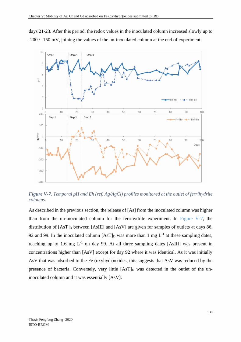
Chapter V: Mobility of As, Cr and Cd adsorbed on Fe (oxyhydr)oxides submitted to IRB
130
Thesis Fengfeng Zhang -2020
ISTO-BRGM
days 21-23. After this period, the redox values in the inoculated column increased slowly up to
-200 / -150 mV, joining the values of the un-inoculated column at the end of experiment.
Figure V-7. Temporal pH and Eh (ref. Ag/AgCl) profiles monitored at the outlet of ferrihydrite
columns.
As described in the previous section, the release of [As] from the inoculated column was higher
than from the un-inoculated column for the ferrihydrite experiment. In Figure V-7, the
distribution of [AsT]D between [AsIII] and [AsV] are given for samples of outlets at days 86,
92 and 99. In the inoculated column [AsT]D was more than 1 mg L-1 at these sampling dates,
reaching up to 1.6 mg L-1 on day 99. At all three sampling dates [AsIII] was present in
concentrations higher than [AsV] except for day 92 where it was identical. As it was initially
AsV that was adsorbed to the Fe (oxyhydr)oxides, this suggests that AsV was reduced by the
presence of bacteria. Conversely, very little [AsT]D was detected in the outlet of the un-
inoculated column and it was essentially [AsV].
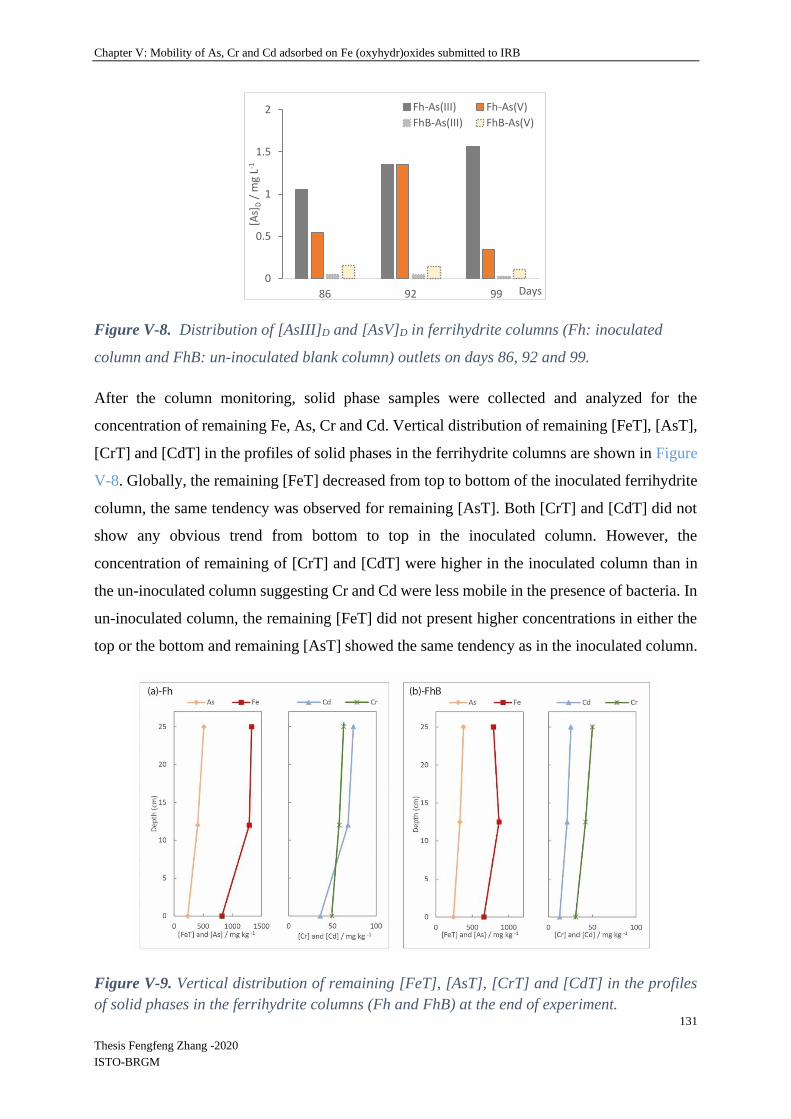
Chapter V: Mobility of As, Cr and Cd adsorbed on Fe (oxyhydr)oxides submitted to IRB
131
Thesis Fengfeng Zhang -2020
ISTO-BRGM
Figure V-8. Distribution of [AsIII]D and [AsV]D in ferrihydrite columns (Fh: inoculated
column and FhB: un-inoculated blank column) outlets on days 86, 92 and 99.
After the column monitoring, solid phase samples were collected and analyzed for the
concentration of remaining Fe, As, Cr and Cd. Vertical distribution of remaining [FeT], [AsT],
[CrT] and [CdT] in the profiles of solid phases in the ferrihydrite columns are shown in Figure
V-8. Globally, the remaining [FeT] decreased from top to bottom of the inoculated ferrihydrite
column, the same tendency was observed for remaining [AsT]. Both [CrT] and [CdT] did not
show any obvious trend from bottom to top in the inoculated column. However, the
concentration of remaining of [CrT] and [CdT] were higher in the inoculated column than in
the un-inoculated column suggesting Cr and Cd were less mobile in the presence of bacteria. In
un-inoculated column, the remaining [FeT] did not present higher concentrations in either the
top or the bottom and remaining [AsT] showed the same tendency as in the inoculated column.
Figure V-9. Vertical distribution of remaining [FeT], [AsT], [CrT] and [CdT] in the profiles
of solid phases in the ferrihydrite columns (Fh and FhB) at the end of experiment.
0
0.5
1
1.5
2
86 92 99
[As]
D/
mg
L-1
Days
Fh-As(III) Fh-As(V)
FhB-As(III) FhB-As(V)

Chapter V: Mobility of As, Cr and Cd adsorbed on Fe (oxyhydr)oxides submitted to IRB
132
Thesis Fengfeng Zhang -2020
ISTO-BRGM
V-4.2.2.2 Goethite columns
Step 1: stabilization
As for ferrihydrite columns, the goethite columns were first flushed with sterile synthetic water
for 14 days in order to finalize the removal of un-adsorbed metals. During this period, prior to
bacterial inoculation, released elements were detected at the column outlet as shown in Figure
V-9 (lower panel). Concerning chromium, 0.4 and 0.38 mg L-1 [CrT]D were detected at the
columns (inoculated and un-inoculated respectively) outlets on the first day and then there was
no Cr detected in the outlets until the inoculation period. Concerning Cd, leaching resulted in
the release of 2.8 and 4.1 mg L-1 [CdT]D on the first day, then concentrations decreased steadily
to an undetected value of < 0.1 mg L-1 on day 7. The released As concentration ([TAs]D) was
more than 100 mg L-1 on the first day and then decreased rapidly and stabilized to 3 mg L-1 on
day 7. The leaching resulted in a total quantity of 9.0 and 9.6 mg (being ~ 18% and ~ 21% of
the total As content of the inoculated and Blank un-inoculated columns, abbreviated as Gt and
GtB respectively) during the first 14 days of rinsing followed by a stabilization of the outflow
concentration at 1.0 ± 0.5 mg L-1. Moreover, the leached Cr and Cd quantities were less than
1% of the total Cr and Cd contents of the columns. During this equilibration phase, pH oscillated
between 7.5 and 9.5, and Eh oscillated between -200 mV and -100 mV, in both inoculated and
un-inoculated columns.
Steps 2 (inoculation days 15-22) and 3 (monitoring days 23-99)
During the inoculation (days 15 to 22) and monitoring periods (days 23 to 99) (Figure V-9,
upper panel), [FeT]D from the inoculated column showed the same trend as for the ferrihydrite
column with a sharp increase from day 18 to day 23 (the end of inoculation period), where
[FeT]D reached the value of 58 mg L-1. Globally the Fe release was much more important in the
inoculated than in the un-inoculated conditions. Here [FeT]D in the un-inoculated column did
not change with the addition of substrate. After the inoculation period, [FeT]D in the inoculated
column outlet decreased rapidly to minimum concentrations of 8.9 mg L-1 on day 28 and then
increased rapidly to reach the maximum value of 59 mg L-1 then gradually to reach values of
30 - 40 mg L-1 after day 50 until the end of the experiment. Conversely, during the experiment,
[FeT]D in the un-inoculated column remained in the range of 0.1-0.3 mg L-1 throughout the
experiment. To summarize, the total dissolved and reduced Fe collected at the column outlets
were 96.5 mg and 0.81 mg (being ~ 1.8-3.9% and < 0.01% of the total iron contents of the
columns) from inoculated and un-inoculated columns, respectively.

Chapter V: Mobility of As, Cr and Cd adsorbed on Fe (oxyhydr)oxides submitted to IRB
133
Thesis Fengfeng Zhang -2020
ISTO-BRGM
From days 15-99, [AsT]D in the inoculated column peaked at 34.7 mg L-1 during the inoculation
period, with an increase from days 15 to 17 and a decrease to 7.9 mg L-1 from day 17 to 28.
This sharp peak preceded that of dissolved iron by 6-7 days. It continued to decrease until the
end of the experiment except for a slight increase from day 31 to 36 (the second increase was
not convincing with only one analysis at day 86). [AsT]D from the un-inoculated column
exhibited a trend similar to that of the inoculated column, but with lower concentrations, with
an increase from 1.1 to 6.7 mg L-1 on day 15 to 22 and then a decrease from 6.7 to 1.7 mg L-1
on day 22 to 28. After day 28, the release of [AsT]D reached a maximum of 2.8 mg L-1 at day
39 and then decreased again until the end of the experiment. During the post-inoculation period
of the experiment, pH in the inoculated column carried on oscillating between 8 and 9, but pH
in the un-inoculated column oscillated between 7 and 8.5 except between days 15 to 35 during
which it oscillated between 5.5 and 7 (Figure V-10). Globally, pH was lower in the un-
inoculated column after the inoculation, but then increased progressively towards similar values.
Eh was always lower in the inoculated than in the un-inoculated column after inoculation, with
values close to -300 mV. In the un-inoculated column, Eh was between 0 and 50 mV in the 17-
30 days period, then decreased and varied between -200 and 0 mV until the end of experiment.

Chapter V: Mobility of As, Cr and Cd adsorbed on Fe (oxyhydr)oxides submitted to IRB
134
Thesis Fengfeng Zhang -2020
ISTO-BRGM
Figure V-10. Temporal evolution of dissolved [FeT]D (upper panel), [AsT]D, [CrT]D and
[CdT]D (lower panel) monitored at the outlet of the goethite columns: Gt (inoculated column)
and GtB (un-inoculated Blank column).
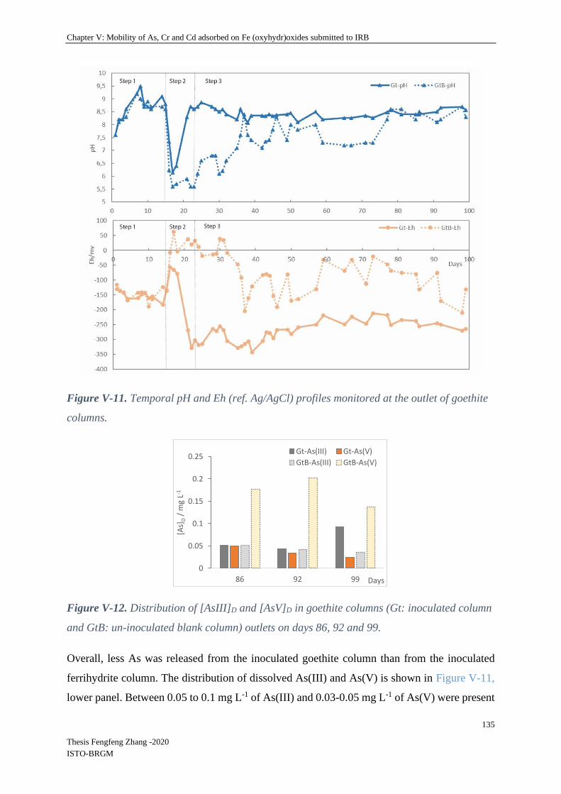
Chapter V: Mobility of As, Cr and Cd adsorbed on Fe (oxyhydr)oxides submitted to IRB
135
Thesis Fengfeng Zhang -2020
ISTO-BRGM
Figure V-11. Temporal pH and Eh (ref. Ag/AgCl) profiles monitored at the outlet of goethite
columns.
Figure V-12. Distribution of [AsIII]D and [AsV]D in goethite columns (Gt: inoculated column
and GtB: un-inoculated blank column) outlets on days 86, 92 and 99.
Overall, less As was released from the inoculated goethite column than from the inoculated
ferrihydrite column. The distribution of dissolved As(III) and As(V) is shown in Figure V-11,
lower panel. Between 0.05 to 0.1 mg L-1 of As(III) and 0.03-0.05 mg L-1 of As(V) were present
0
0.05
0.1
0.15
0.2
0.25
86 92 99
[As]
D/
mg
L-1
Days
Gt-As(III) Gt-As(V)
GtB-As(III) GtB-As(V)
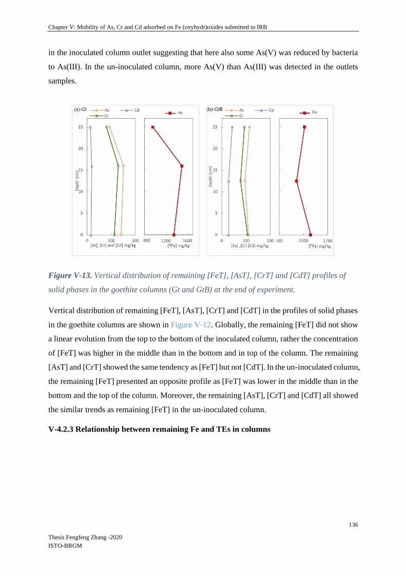
Chapter V: Mobility of As, Cr and Cd adsorbed on Fe (oxyhydr)oxides submitted to IRB
136
Thesis Fengfeng Zhang -2020
ISTO-BRGM
in the inoculated column outlet suggesting that here also some As(V) was reduced by bacteria
to As(III). In the un-inoculated column, more As(V) than As(III) was detected in the outlets
samples.
Figure V-13. Vertical distribution of remaining [FeT], [AsT], [CrT] and [CdT] profiles of
solid phases in the goethite columns (Gt and GtB) at the end of experiment.
Vertical distribution of remaining [FeT], [AsT], [CrT] and [CdT] in the profiles of solid phases
in the goethite columns are shown in Figure V-12. Globally, the remaining [FeT] did not show
a linear evolution from the top to the bottom of the inoculated column, rather the concentration
of [FeT] was higher in the middle than in the bottom and in top of the column. The remaining
[AsT] and [CrT] showed the same tendency as [FeT] but not [CdT]. In the un-inoculated column,
the remaining [FeT] presented an opposite profile as [FeT] was lower in the middle than in the
bottom and the top of the column. Moreover, the remaining [AsT], [CrT] and [CdT] all showed
the similar trends as remaining [FeT] in the un-inoculated column.
V-4.2.3 Relationship between remaining Fe and TEs in columns

Chapter V: Mobility of As, Cr and Cd adsorbed on Fe (oxyhydr)oxides submitted to IRB
137
Thesis Fengfeng Zhang -2020
ISTO-BRGM
Figure V-14. Remaining [AsT], [CrT] and [CdT] / [FeT] ratios in solid samples of both
ferrihydrite columns (Fh and FhB) at the end of experiment; Analysis of correlation between
remaining [AsT], [CrT], [CdT] and [FeT].
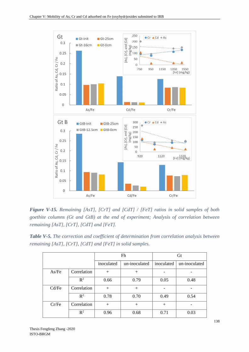
Chapter V: Mobility of As, Cr and Cd adsorbed on Fe (oxyhydr)oxides submitted to IRB
138
Thesis Fengfeng Zhang -2020
ISTO-BRGM
Figure V-15. Remaining [AsT], [CrT] and [CdT] / [FeT] ratios in solid samples of both
goethite columns (Gt and GtB) at the end of experiment; Analysis of correlation between
remaining [AsT], [CrT], [CdT] and [FeT].
Table V-5. The correction and coefficient of determination from correlation analysis between
remaining [AsT], [CrT], [CdT] and [FeT] in solid samples.
Fh Gt
inoculated un-inoculated inoculated un-inoculated
As/Fe Correlation + + - -
R2 0.66 0.79 0.05 0.48
Cd/Fe Correlation + + - -
R2 0.78 0.70 0.49 0.54
Cr/Fe Correlation + + + -
R2 0.96 0.68 0.71 0.03

Chapter V: Mobility of As, Cr and Cd adsorbed on Fe (oxyhydr)oxides submitted to IRB
139
Thesis Fengfeng Zhang -2020
ISTO-BRGM
The ratios of As, Cd and Cr/Fe in column filling materials and their evolution between the initial
and final states are given in Figure V-13, 14. In the ferrihydrite column, the As/Fe ratio
decreased during the experiment, more so at the bottom than the top of the column. In the
inoculated one, Cd and Cr ratios did not change during the experiment. The same phenomenon
was observed for Cr in the un-inoculated column. Conversely, the Cd/Fe ratio decreased in the
un-inoculated column suggesting a global leaching of this element from Fe-rich particles in un-
inoculated conditions. This last result was in accordance with the evolution of Cd
concentrations in the columns outlet (Figure V-5), that showed a release of Cd in un-inoculated
but not in inoculated conditions. For Cr, no release was observed in the outlet, which is
consistent with the results of Cr/Fe ratios in the solids.
With goethite, the decrease of the As/Fe ratio during experiment was uniform along the column.
Cd and Cr/Fe ratios decreased in both un-inoculated and inoculated conditions. The decrease
appeared uniformous along the column except for Cd in un-inoculated conditions, for which a
small increase of Cd/Fe ratio from the bottom to the top of the column was observed. During
the experiment (Figure V-8), no big differences were observed between un-inoculated and
inoculated conditions in the goethite columns, except for As during the inoculation period. Thus,
the very similar results observed for the As, Cd and Cr /Fe ratios in the solids is not very
surprising.
V-4.2.3 Biological Parameters
V-4.2.3.1 Spatial distribution of bacterial abundance
Molecular biomass was quantified as the total genomic DNA extracted from column solid
samples and liquid (abbreviated liquid/L), recovered from both inoculated and un-inoculated
columns. Globally, total DNA concentrations in solid samples of inoculated columns (DNA
concentrations, Fh: 2.2E+3 - 1.7E+4 ng g-1, Gt: 1.7E+3 - 9.9E+3 ng g-1) were significantly
higher than in solid samples of un-inoculated columns (DNA concentrations, FhB: under
detection limit or < 10 ng g-1, GtB: 0.13 - 61 ng g-1). The DNA concentrations in liquid samples
from both the un-inoculated columns were lower than the detection limit. This, along with the
low DNA amounts found in the solid samples confirms that although the uninoculated columns
were not complety sterile, little biomass had contaminated the columns. Bacterial abundance
(qPCR of bacterial 16S rRNA encoding gene per g or mL sample), Figure. IV-15, showed
similar tendencys as for molecular biomass. Results showed that 16S genes quantification in
solid samples of both inoculated columns were significantly higher than in solid samples of

Chapter V: Mobility of As, Cr and Cd adsorbed on Fe (oxyhydr)oxides submitted to IRB
140
Thesis Fengfeng Zhang -2020
ISTO-BRGM
both un-inoculated columns (16S genes copies, FhB: under detection 3.8E+5 - 8.3E+5 g-1, GtB:
2.3E+5 - 2.7E+6 g-1), and there was no significant difference between ferrihydrite and goethite
column globally. However, bacterial abundance in the bottom part of the goethite column (Gt0)
was significantly higher than in the top of the column (GtT), and no significant difference was
obtained between the middle (GtM) and bottom (Gt0) or top (GtT) samples. It seems that the
bacterial biomass decreased from the bottom to the top of the goethite column. For the
ferrihydrite column, both bottom (Fh0) and middle (FhM) had significantly higher bacterial
16S gene abundances than the top position (FhT), which suggested the bacterial biomass
decreases from the bottom to the top of the goethite column.
Figure V- 16. Bacterial 16S rRNA gene copies per g or mL sample. “0”, “M” and “T” refer
to the bottom, middle and top of the columns, ferrhydrite (Fh) column and goethite (Gt) columns.
The small letters “a” and “b” differed significantly (Kruskal-Wallis test at p<0.05) of spatial
positions of ferrihydrite and goethite column, the Greek letters were used for differing
significantly by group of inoculated column of ferrihydrite (Fh) and goethite (Gt), liquid
recovered from both inoculated columns (L). Data represent average values of three
experimental replicates and their standard deviation (σ).
V-4.2.3.2 Spatial evolution of Shewanella and Geobacter 16S rRNA gene abundances
The abundances of micro-organisms affiliated to Shewanella and Geobacter genera were
estimated in column samples using qPCR of genus-specific fragments of the 16s rRNA gene
1E+0
2E+9
4E+9
6E+9
8E+9
1E+10
1E+10
1E+10
2E+10
2E+10
Fh 0 Fh M Fh T Gt 0 Gt M Gt T L- Fh L- Gt
<16
s rR
NA
gen
e> c
op
ies
per
g o
r m
L sa
mp
le
Samples
Fh 0 Fh M Fh T
Gt 0 Gt M Gt T
L- Fh L- Gt
β
β
ab
ab
a
b
a
α
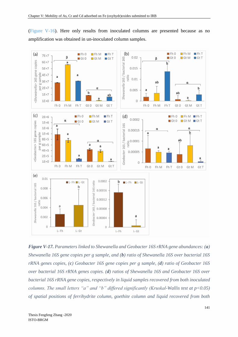
Chapter V: Mobility of As, Cr and Cd adsorbed on Fe (oxyhydr)oxides submitted to IRB
141
Thesis Fengfeng Zhang -2020
ISTO-BRGM
(Figure V-16). Here only results from inoculated columns are presented because as no
amplification was obtained in un-inoculated column samples.
Figure V-17. Parameters linked to Shewanella and Geobacter 16S rRNA gene abundances: (a)
Shewanella 16S gene copies per g sample, and (b) ratio of Shewanella 16S over bacterial 16S
rRNA genes copies, (c) Geobacter 16S gene copies per g sample, (d) ratio of Geobacter 16S
over bacterial 16S rRNA genes copies. (d) ratios of Shewanella 16S and Geobacter 16S over
bacterial 16S rRNA gene copies, respectively in liquid samples recovered from both inoculated
columns. The small letters “a” and “b” differed significantly (Kruskal-Wallis test at p<0.05)
of spatial positions of ferrihydrite column, goethite column and liquid recovered from both

Chapter V: Mobility of As, Cr and Cd adsorbed on Fe (oxyhydr)oxides submitted to IRB
142
Thesis Fengfeng Zhang -2020
ISTO-BRGM
inoculated columns. The Greek letter were used for differing significantly by group of
inoculated columns of ferrihydrite (Fh) and goethite (Gt). Data represent average values of
three experimental replicates and their standard deviation (σ).
As shown in Figure V-16 (b), a significant difference was observed between the numbers of
gene copies belonging to Shewanella in the ferrihydrite column and the goethite column.
Moreover, significant differences between vertical positions were obtained in the goethite
column. Globally, the abundance of Shewanella 16S gene copies in the ferrihydrite column was
significantly higher than in the goethite column and liquid samples. Moreover, in ferrihydrite
columns, no significant differences were observed between the three sampling positions, i.e.,
bottom (Fh0), middle (FhM) and top (FhT) for the number of gene copies of Shewanella. They
conversely presented significant differences in the goethite column: the bottom (Gt0) contained
significantly more Shewanella gene copies than the middle (GtM) but there were no significant
differences between the middle (GtM) and the top (GtT) (Figure V-16 (a)). The Shewanella
16S/ bacterial 16S ratio presented the same tendencies as the proportion of Shewanella was
globally significantly higher in the ferrihydrite column than in the goethite column (Figure V-
16 (b)). In Figure V-16 (b), results show that the ratio of Shewanella over bacterial 16S in the
top part of the ferrihydrite column (FhT) was significantly higher than in the bottom part (Fh0).
The same tendency of increase of the proportion of Shewanella from the bottom to the top was
observed in the goethite column. However, the Shewanella 16S/ bacterial 16S ratio in the top
part of goethite column (GtT) was significantly higher than in the middle (GtM) and no
significant differences were observed either between the middle (GtM) and the bottom (Gt0),
nor between the bottom (Gt0) and the top (GtT).
Concerning Geobacter, a significant difference was observed between the ferrihydrite and the
goethite column, as shown in Figure V-16 (c). However, there were no significant differences
between sampling positions although the abundance of Geobacter gene copies tended to
decrease from the bottom to the top of the column. The Geobacter 16S/bacterial 16S ratio did
not globally present any significant differences between column samples (Figure V-16 (d)). The
proportion of Geobacter did not show significant differences between different sampling
positions in the ferrihydrite column. Conversely, the Geobacter 16S/bacterial 16S ratio in the
middle part of the goethite column (GtM) presented significantly higher values than in the top
(GtT), but no significant differences were obtained between the middle (GtM) and the bottom
(Gt0), or the bottom (Gt0) and the top (GtT) (Figure V-16 (d)).

Chapter V: Mobility of As, Cr and Cd adsorbed on Fe (oxyhydr)oxides submitted to IRB
143
Thesis Fengfeng Zhang -2020
ISTO-BRGM
The proportion Shewanella 16S/bacterial 16S in the liquid phase of the columns (Figure V-16
(e)) was of the same range as the proportion attached onto the solid phases, with the exception
of the top part of the ferrihydrite column which showed a higher proportion of Shewanella
associated with the solids. Moreover, the proportion of Shewanella 16S/bacterial 16S in the
liquid phase was significantly higher in the goethite column than in the ferrihydrite column. On
the contrary, the Geobacter 16S/bacterial 16S proportion was significantly higher in the
ferrihydrite column than in the goethite column in the liquid phase.
V-4.2.3.3 SEM bacteria observation
Bacterial observations on ferrihydrite and goethite are given in Figure 17 and 18.
After incubation, bacterial cells were observed on solid samples under SEM (Figure V-17).
Bacterial cells appeared intact with spherical and rod shapes (Figure V-17 (b) and (d)),
separated from each other, turgid, and complete with smooth surface. Bacteria were mainly
present on the surface of the sand particles (Figure V-17 (a)) but also on the surface of
ferrihydrite (Figure V-17 (b) and (d)) and “secondary minerals” of iron (Figure V-17 (e)). The
bacterial morphology was clear to observe under the microscope field of 5-10 µm, its size was
between 1-4 µm depending on the shape.

Chapter V: Mobility of As, Cr and Cd adsorbed on Fe (oxyhydr)oxides submitted to IRB
144
Thesis Fengfeng Zhang -2020
ISTO-BRGM
Figure V-18. Cryo-SEM micrographs of bacteria from inoculated ferrihydrite column.
(b)
(c) (d)
(e) (f)
spherical
rod
spherical rod
rod
spherical
(a)

Chapter V: Mobility of As, Cr and Cd adsorbed on Fe (oxyhydr)oxides submitted to IRB
145
Thesis Fengfeng Zhang -2020
ISTO-BRGM
Bacterial observation on goethite
Figure V-19. Cryo-SEM micrographs of bacteria from inoculated goethite column.
(a) (b)
(c) (d)
(f) (e)
rod spherical
spherical
spherical
rod
Spherical and rod

Chapter V: Mobility of As, Cr and Cd adsorbed on Fe (oxyhydr)oxides submitted to IRB
146
Thesis Fengfeng Zhang -2020
ISTO-BRGM
In the goethite column, the same conditions were used for bacterial observations as described
for the ferrihydrite column. The same shapes of bacterial cells were observed as those in
samples from the ferrihydrite column, which was intact, spherical shapes, or rod shapes,
separated from each other, turgid, and complete with smooth surface (Figure V-18). Globally,
bacteria cells were clear and easy to be found under the microscope field of 5-10 µm. However,
it’s difficult to observe bacterial cells on the surface of goethite because the bacterial cells are
the same size as goethite crystals. No clear “secondary minerals” were observed in the goethite
experimental system.
V-4.2.4 Mineral SEM-EDS observation
V-4.2.4.1 Fe speciation in the solid phases of ferrihydrite and goethite columns
Initial column samples
Figure V-20. SEM image of initial ferrihydrite and goethite column’s sample in which it is
possible to observe the shape of ferrihydrite/goethite and sands.

Chapter V: Mobility of As, Cr and Cd adsorbed on Fe (oxyhydr)oxides submitted to IRB
147
Thesis Fengfeng Zhang -2020
ISTO-BRGM
SEM observations were performed to determine the structural evolution of mineral filling
material (a mixture of sand, silica gel and synthetic Fe (oxyhydr)oxides with adsorbed As, Cr
and Cd) between pre-incubation samples and post-incubation samples, and the differences
between inoculated columns and un-inoculated columns.
Figure V-21. SEM-EDS analysis of initial samples for ferrihydrite (Fh) and goethite (Gt)
columns. The blue and red spectra correspond to the analyses of point “1” and point “2”
(abbreviated P1 and P2), respectively.
Figure V-19 shows electronic micrographs of initial samples before incubation for ferrihydrite
(Fh) and goethite (Gt) columns. There were two main morphology types in the ferrihydrite
column sample: (1) clear irregular block-type ferrihydrite particles with some mixtures of silica
gel; (2) irregular round shaped sands. It can be seen that the initial ferrihydrite presented smooth
or rough surface in different blocks with length, width and height of approximately 5-150 µm.
The micrograph showed that the diameter of sand grains was around 100-150 µm. As shown in
the view of goethite column sample, a single goethite crystal presented an acicular form (1-5
µm) but in this experiment goethite crystals were agglomerated into small spherical (10-50 µm)
structures or distributed on the surface of sand particles.

Chapter V: Mobility of As, Cr and Cd adsorbed on Fe (oxyhydr)oxides submitted to IRB
148
Thesis Fengfeng Zhang -2020
ISTO-BRGM
Energy dispersive X-ray spectrometry (EDX) detector, coupled to SEM observations, was used
and enabled to characterize the major elements in the sand and mineral phase. In Figure V-20,
the main composition given as weight percentages shows that the major elements were Fe and
O in ferrihydrite particles (P2 on the Figure V-20, Fh), and Si and O in sand particles. The same
for goethite column sample which shows Fe, O and Si as major elements (Figure V-20, Gt).
Post-incubation column samples-ferrihydrite
The same SEM-EDS analysis were performed for post-incubation samples. Figure V-21 shows
electronic micrographs of column samples (bottom, middle and top) after incubation from
inoculated and un-inoculated ferrihydrite columns. For the un-inoculated column, there was no
obvious difference in the types of forms in post-incubation samples in comparison to initial
samples. Clear and irregular blocks of ferrihydrite could be found everywhere, whose size did
not show differences either (Figure V-21 (a), (b) and (c)). However, some regular forms of
minerals were present in inoculated column after incubation (Figure V-21 (A), (B) and (C)). It
can be seen that regular blocks stacked or arranged together in a radial pattern with smooth
surface in different blocks with length, width and height of approximately 50 -100 µm. Figure
V-21 show high concentrations of Fe and O in the particles corresponding to the morphology
of ferrihydrite, and higher concentration of P in the new “laminated” structures.

Chapter V: Mobility of As, Cr and Cd adsorbed on Fe (oxyhydr)oxides submitted to IRB
149
Thesis Fengfeng Zhang -2020
ISTO-BRGM
Figure V-22. SEM images of post-incubation ferrihydrite columns including samples from the
bottom, middle and top of inoculated ferrihydrite column and the same in un-inoculated column.
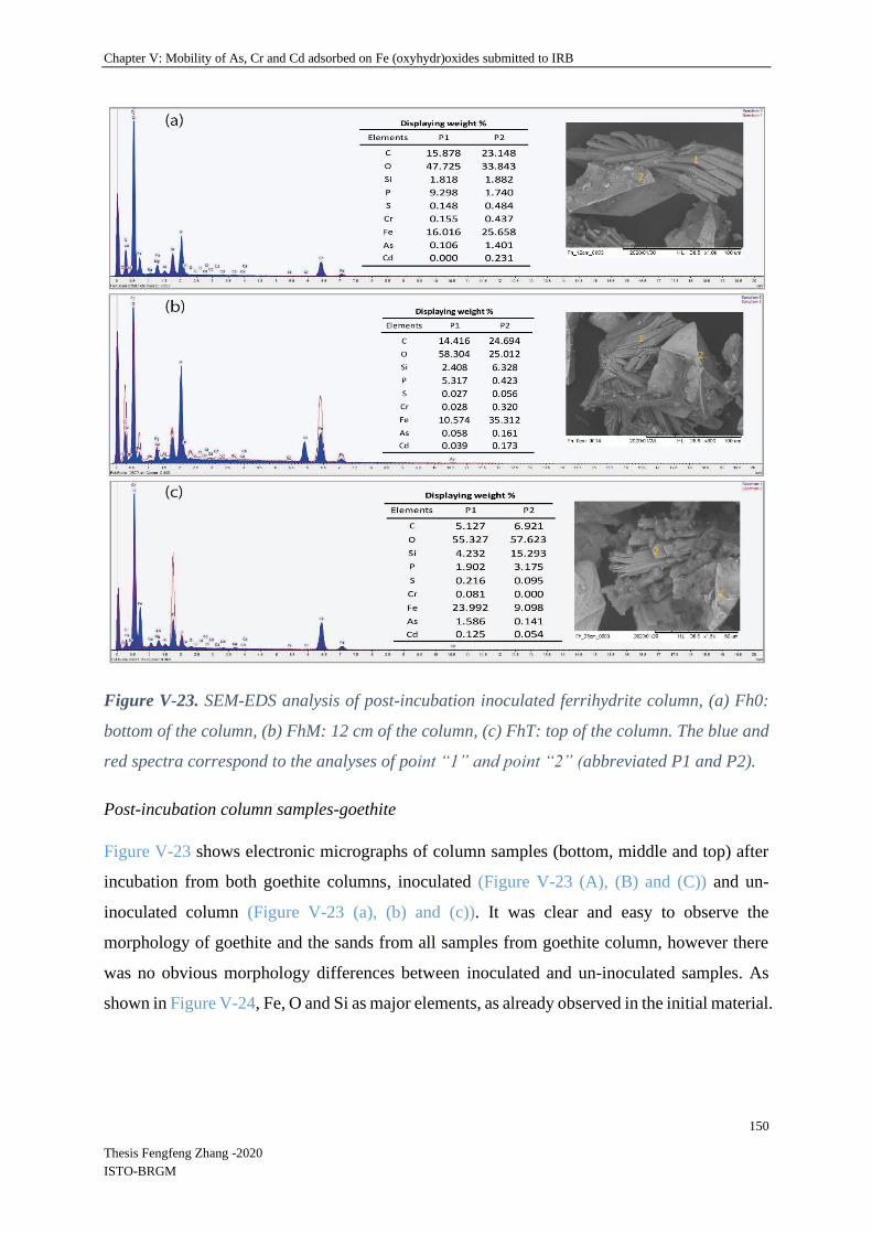
Chapter V: Mobility of As, Cr and Cd adsorbed on Fe (oxyhydr)oxides submitted to IRB
150
Thesis Fengfeng Zhang -2020
ISTO-BRGM
Figure V-23. SEM-EDS analysis of post-incubation inoculated ferrihydrite column, (a) Fh0:
bottom of the column, (b) FhM: 12 cm of the column, (c) FhT: top of the column. The blue and
red spectra correspond to the analyses of point “1” and point “2” (abbreviated P1 and P2).
Post-incubation column samples-goethite
Figure V-23 shows electronic micrographs of column samples (bottom, middle and top) after
incubation from both goethite columns, inoculated (Figure V-23 (A), (B) and (C)) and un-
inoculated column (Figure V-23 (a), (b) and (c)). It was clear and easy to observe the
morphology of goethite and the sands from all samples from goethite column, however there
was no obvious morphology differences between inoculated and un-inoculated samples. As
shown in Figure V-24, Fe, O and Si as major elements, as already observed in the initial material.
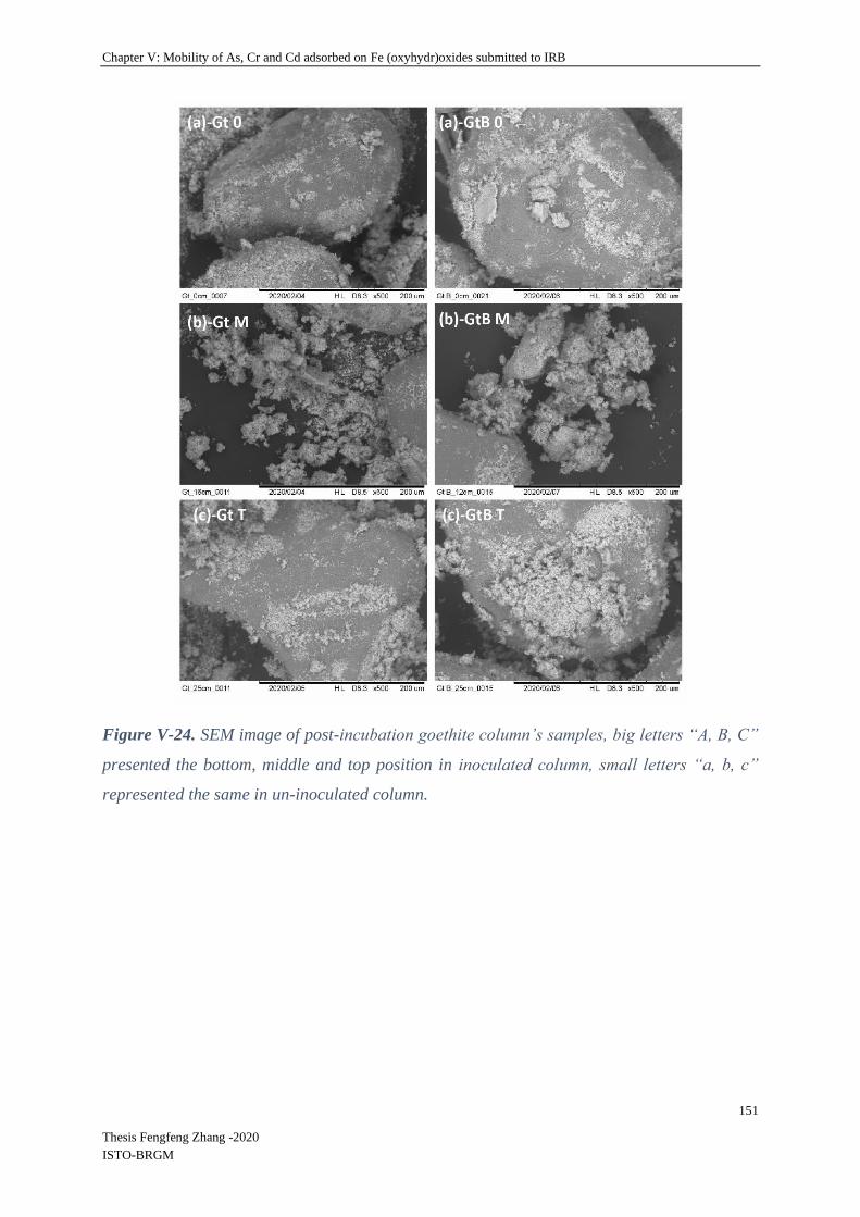
Chapter V: Mobility of As, Cr and Cd adsorbed on Fe (oxyhydr)oxides submitted to IRB
151
Thesis Fengfeng Zhang -2020
ISTO-BRGM
Figure V-24. SEM image of post-incubation goethite column’s samples, big letters “A, B, C”
presented the bottom, middle and top position in inoculated column, small letters “a, b, c”
represented the same in un-inoculated column.

Chapter V: Mobility of As, Cr and Cd adsorbed on Fe (oxyhydr)oxides submitted to IRB
152
Thesis Fengfeng Zhang -2020
ISTO-BRGM
Figure V-25. SEM-EDS analysis of post-incubation inoculated goethite column, (a) Gt0:
bottom of the column, (b) GtM: 12 cm of the column, (c) GtT: top of the column. The blue and
red spectra correspond to the analyses of point “1” and point “2” (abbreviated P1 and P2).
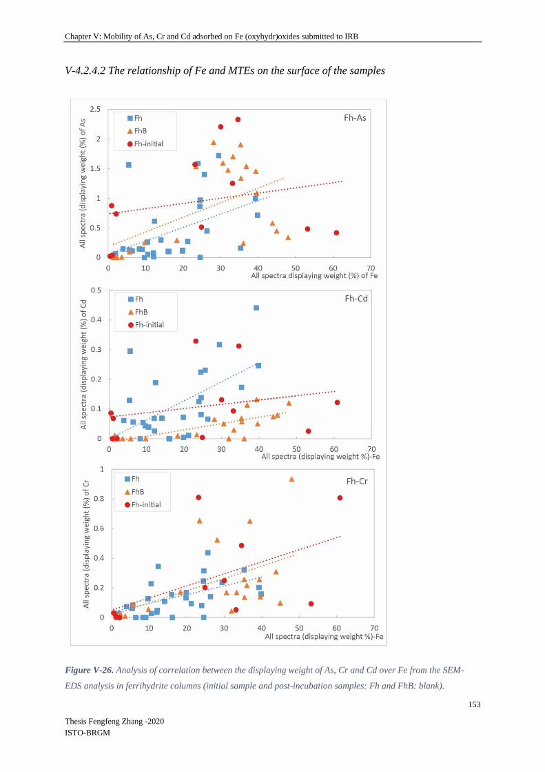
Chapter V: Mobility of As, Cr and Cd adsorbed on Fe (oxyhydr)oxides submitted to IRB
153
Thesis Fengfeng Zhang -2020
ISTO-BRGM
V-4.2.4.2 The relationship of Fe and MTEs on the surface of the samples
Figure V-26. Analysis of correlation between the displaying weight of As, Cr and Cd over Fe from the SEM-
EDS analysis in ferrihydrite columns (initial sample and post-incubation samples: Fh and FhB: blank).

Chapter V: Mobility of As, Cr and Cd adsorbed on Fe (oxyhydr)oxides submitted to IRB
154
Thesis Fengfeng Zhang -2020
ISTO-BRGM
Figure V-27. Analysis of correlation between the displaying weight of As, Cr and Cd over Fe from the
SEM-EDS analysis in goethite columns (initial sample and post-incubation samples: Gt and GtB: blank).
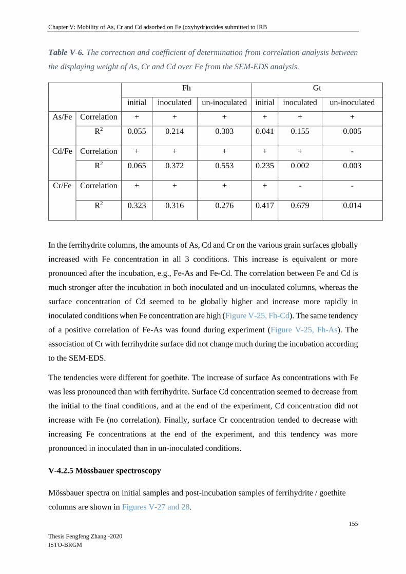
Chapter V: Mobility of As, Cr and Cd adsorbed on Fe (oxyhydr)oxides submitted to IRB
155
Thesis Fengfeng Zhang -2020
ISTO-BRGM
Table V-6. The correction and coefficient of determination from correlation analysis between
the displaying weight of As, Cr and Cd over Fe from the SEM-EDS analysis.
Fh Gt
initial inoculated un-inoculated initial inoculated un-inoculated
As/Fe Correlation + + + + + +
R2 0.055 0.214 0.303 0.041 0.155 0.005
Cd/Fe Correlation + + + + + -
R2 0.065 0.372 0.553 0.235 0.002 0.003
Cr/Fe Correlation + + + + - -
R2 0.323 0.316 0.276 0.417 0.679 0.014
In the ferrihydrite columns, the amounts of As, Cd and Cr on the various grain surfaces globally
increased with Fe concentration in all 3 conditions. This increase is equivalent or more
pronounced after the incubation, e.g., Fe-As and Fe-Cd. The correlation between Fe and Cd is
much stronger after the incubation in both inoculated and un-inoculated columns, whereas the
surface concentration of Cd seemed to be globally higher and increase more rapidly in
inoculated conditions when Fe concentration are high (Figure V-25, Fh-Cd). The same tendency
of a positive correlation of Fe-As was found during experiment (Figure V-25, Fh-As). The
association of Cr with ferrihydrite surface did not change much during the incubation according
to the SEM-EDS.
The tendencies were different for goethite. The increase of surface As concentrations with Fe
was less pronounced than with ferrihydrite. Surface Cd concentration seemed to decrease from
the initial to the final conditions, and at the end of the experiment, Cd concentration did not
increase with Fe (no correlation). Finally, surface Cr concentration tended to decrease with
increasing Fe concentrations at the end of the experiment, and this tendency was more
pronounced in inoculated than in un-inoculated conditions.
V-4.2.5 Mössbauer spectroscopy
Mössbauer spectra on initial samples and post-incubation samples of ferrihydrite / goethite
columns are shown in Figures V-27 and 28.

Chapter V: Mobility of As, Cr and Cd adsorbed on Fe (oxyhydr)oxides submitted to IRB
156
Thesis Fengfeng Zhang -2020
ISTO-BRGM
Figure V-28. Mössbauer spectra collected at 140 K for samples, (a) pre-incubation Fh initial,
post-incubation (b) bottom of inoculated column Fh-bottom, (c) top of inoculated column Fh-
top, and (d) un-inoculated column Fh-Blank for ferrihydrite columns.
100-10
v (mm/s)
05
10
Absorp
tion (
%)
100-10
v (mm/s)
01
23
Absorp
tion (
%)
100-10
v (mm/s)
01
23
4
Absorp
tion (
%)
100-10
v (mm/s)
01
23
4
Absorp
tion (
%)
1050-5-10
v (mm/s)
1.4
1.3
Inte
nsity (
10
6 c
ts)
100-10
v (mm/s)
01
23
Absorp
tion (
%)
(a) (b)
(a) (b)
(c) (d)

Chapter V: Mobility of As, Cr and Cd adsorbed on Fe (oxyhydr)oxides submitted to IRB
157
Thesis Fengfeng Zhang -2020
ISTO-BRGM
Figure V-29. Mössbauer spectra collected at 140 K for samples, (a) pre-incubation Gt init,
post-incubation (b) bottom of inoculated column Gt-bottom, (c) top of inoculated column Gt-
top, and (d) un-inoculated column Gt-Blank for goethite columns.
Table V-7. Fitting results of Mössbauer spectroscopy. CS – center shift, QS – quadrupole
splitting, e – quadrupole shift, H – hyperfine field, stdev(H) – standard deviation of hyperfine
field, Pop. – Relative abundance, χ2 – goodness of fit.
Sample Mineral CS QS stdev(QS) e H stdev(H) Pop
Erro
r χ2
phase mm/s mm/s mm/s mm/s T T % FH initial Fe(III) 0.43 0.77 0.35 100 0 3.2
Fh Bottom Fe(III) 0.40 0.82 0.44 70.4 2.0 0.6
Fe(II) 1.18 2.84 1.1 29.6 2.0
Fh Top Fe(III) 0.43 0.73 0.30 87.5 1.6 0.6
Fe(II) 1.28 2.79 0.45 12.5 1.6
Fh Blank Fe(III) 0.43 0.76 0.29 100 0 0.8
Gt Bottom Fe(III) 0.45 -0.13 48.4 0.60 100 0 1.1
Gt Top Fe(III) 0.46 -0.13 48.3 0.76 100 0 0.7
Gt Blank Fe(III) 0.45 -0.13 48.2 0.77 100 0 1.0
100-10
v (mm/s)
01
2
Absorp
tion (
%)
100-10
v (mm/s)
01
23
Absorp
tion (
%)
(c) (d)

Chapter V: Mobility of As, Cr and Cd adsorbed on Fe (oxyhydr)oxides submitted to IRB
158
Thesis Fengfeng Zhang -2020
ISTO-BRGM
All ferrihydrite spectra are dominated by a narrow doublet which is characteristic for a Fe(III)
phase, notably ferrihydrite (CS: 0.47 mm/s; QS: 0.77 mm/s). No additional mineral phase could
be detected for samples Fh initial and Fh-Blank.
The spectra for Fh-Bottom and Fh-Top were best fitted by including an additional Fe(II) phase.
It is not possible to identify this Fe(II) phase because the hyperfine parameters, i.e. CS and QS,
match several different minerals e.g. biotite (Cao et al., 2017), FeCl2 (Laufer et al., 2017),
hedenbergite (Amthauer and Rossman, 1984), etc.
The spectra collected for the goethite samples, i.e. Gt initial, Gt-Bottom, Gt-Top and Gt-Blank,
are characteristic for goethite as expected. No additional mineral phases were detected. An
increase in StDev(H) for Gt-Top and Gt-Blank in comparison to Gt-Bottom might reflect a
minor change in the mineral structure e.g. change in particle size, however other analytical
methods, e.g. TEM would be needed to confirm this.
According to the information, Fe(II) was clearly visible in samples Fh-Bottom and Fh-Top,
with Fh-Bottom (29.6% Fe(II)) showing a higher degree of reduction than Fh-Top (12.5%
Fe(II)) in bottom and top position of inoculated column. No evidence was found to indicate the
occurrence of any Fe(III) reduction in Fh initial, and Fh-Blank samples. Moreover, no defined
Fe phases (e.g. goethite, or magnetite) were detected in all Fh samples.
For goethite columns, no clear differences were observed between goethite (Gt) samples from
this experiment.
IV-5 Discussion
IV-5.1 Spatial and temporal aspects of iron reduction of ferrihydrite and goethite
The black color that appeared in both the columns inoculated with a bacterial consortium might
be linked to sulphide production. The feeding medium contained molybdate to inhibit the
sulfate-reducing activity, however this was possibly not sufficient to completely inhibit sulfate
reduction in the column system. As a fact, in the previous batch condition, molybdate was
homogeneously available simultaneously in all the volume of culture. In column, it might have
been reduced (some bacteria can reduce molybdate) and/or retained on the solid phases and
inhibition of sulfate-reduction might have been weakened. Another path for sulphide formation
might be the transformation proteins (AA sulphur) which are moreover abundant due to the
Chapter V: Mobility of As, Cr and Cd adsorbed on Fe (oxyhydr)oxides submitted to IRB
159
Thesis Fengfeng Zhang -2020
ISTO-BRGM
presence of peptone, by some bacteria such as some belonging to the Shewanella genus. Thus,
the potential production of H2S will be considered as a possible mechanism occurring in the
inoculated column, in the following discussion.
In ferrihydrite columns, Fe reduction was linked to bacterial inoculation and released iron was
detected in the outlet from day 17, three days after inoculation. Dissolved and reduced Fe in
this column could have been released in the outlet and/or re-precipitated onto other iron
minerals. Iron reduction in this column could have resulted from chemical processes, e.g.
reduction by the H2S produced by the processes mentioned above, or and/or bacterial iron
reduction either through fermentation, or dissimilatory reduction. However, Fe reduction by
IRB was likely a dominant reaction as sulfate reduction was at less partly inhibited by
molybdate. Moreover, two IRB genera, Shewanella and Geobacter, although not the only iron
reducers, were detected in the inoculated columns.
In the meantime, pH decreased suddenly on day 15 when the substrate was added to the feeding
liquid. The bacterial inoculum then lead to a gradual increase of the pH and also a drop of the
redox potential (Eh) in the days following inoculation. This phenomenon was already observed
in presence of Shewanella and Fe oxides reduction in some research (Perez-Gonzalez et al.,
2010; Zegeye et al., 2007). On the other hand, in this system, peptone was included in the
mixture of substrates and could be fermented producing ammonium that increases the pH
(Tepari et al., 2019). In addition, hydrogen (H2) can be produced and consumed by the bacteria
(including SRB), inducing an increase of pH (Liamleam and Annachhatre, 2007). Assumably,
both the metabolic activities of the bacteria and biogenic mineral formation modified the Eh as
well as the pH during the continuous feeding period through the following reactions (Ona-
Nguema et al., 2002; Wolf et al, 2009):
Hydrogenotrophic sulfate reduction, or sulfidogenesis, involving
(1) C6H12O6 + 4H2O → 2CH3COO− + 2HCO3− + 4 H+ + 4H2
(2) 4H2 + SO42− + H+ → HS− + 4H2O
Also undesirable but possible reactions of hydrogenotrophic methanogenesis and
homoacetogenesis reactions are the following:
(3) 4H2 + HCO3− + H+ → CH4 + 3H2O
(4) 4H2 + 2HCO3- + H+ → CH3COO- + 4H2O.

Chapter V: Mobility of As, Cr and Cd adsorbed on Fe (oxyhydr)oxides submitted to IRB
160
Thesis Fengfeng Zhang -2020
ISTO-BRGM
These reactions potentially led to the increase of pH in the inoculated columns and this was in
agreement with the quasi-stability of pH observed during and after the inoculation period (Fig.
IV-6, upper panel).
During the post-inoculation period, while Fe reduction steadily carried on occuring, less [FeT]D
was measured in the column outlet suggesting the precipitation of the produced [FeII] as
carbonate, with the inorganic carbon produced by degradation of organic substrates, or sulfides
FeS with H2S (Liamleam and Annachhatre, 2007; Urrutia et al., 1998; Zachara et al., 2002).
The low redox potential (down to -300 mV ref Ag/AgCl) and the observation of black
precipitates could be in accordance with this last hypothesis. The Mössbauer spectra did not
show any presence of iron phosphate, whereas this was suggested by SEM-EDS. Indeed,
(Fe3(PO4)2·8H2O) (vivianite) has a much higher center shift (1.3 mm/s) and quadrupole splitting
(3 mm/s) than was observed in our samples. The new crystalline minerals observed by MEB-
EDS might be iron phosphate precipitated during the inoculation period, but not the major FeII
minerals formed during the biological reduction of ferrihydrite, or maybe would represent other
Fe(II) phase containing the main elements quantified by MEB-EDS, e.g. Fe (20-30%), P (5-
11%), 0 (40-55%), etc. (weight percentages).
In the goethite columns, black precipitates were observed, as in the ferrihydrite column, and
might be caused by H2S according to the reaction described in the previous section. Globally,
iron reduction occurred with a first peak corresponding to inoculation and released iron was
detected in the outlet on day 18, one day later than in the ferrihydrite column. In the inoculated
column, the microbial reaction was likely the dominant reaction compared with the un-
inoculated column that had little [TFe]D released during the whole experiment. Moreover, this
is supported by the results of the quantification of 2 IRB, Shewanella and Geobacter, which
showed that these IRBs were not detected in the un-inoculated column. According to (Liu et al.,
2001), microbial goethite reduction has a first-order kinetic dependence on surface site
availability which was also the function of Fe(II) adsorption and secondary precipitation.
Correspondingly, we observed that at the beginning of the post-inoculation period (days 15 to
46), less [FeII]D was detected from the goethite column than from the ferrihydrite column,
because of the lower solubility of goethite relative to ferrihydrite resulting in lower [FeIII]D and
bacterially-generated [FeII]D concentrations (Hansel et al., 2003b; Hansel et al., 2004; Zhang et
al., 2020). Here, the previous batch experiments performed with the same materials (Chapter
III) showed the same tendency, with higher dissolution of ferrihydrite compared to goethite. In
our column systems, we observed that the total cumulated Fe released in the outlet was of the

Chapter V: Mobility of As, Cr and Cd adsorbed on Fe (oxyhydr)oxides submitted to IRB
161
Thesis Fengfeng Zhang -2020
ISTO-BRGM
same range for the ferrihydrite and goethite (nearly 3%). However, analyses of the final solid
(SEM-EDS and Mössbauer) suggested precipitation of the dissolved Fe(II) in the ferrihydrite
but not in the goethite column. This hypothesis is supported by the presence of 29.6% Fe(II)
detected by Mössbauer spectrometer in the ferrihydrite column, showing a higher proportion of
immobilized Fe(II) than in the goethite system. The precipitation of Fe(II) with carbonate,
sulphide or phosphate in the ferrihydrite column can by hypothesized. As a fact, a higher bio-
availability of electron acceptor Fe(III) in the ferrihydrite column could lead to higher
mineralization of organic substrates and production of inorganic carbon available to precipitate
with Fe(II) as carbonate.
IV 5.2 Impact of iron reduction on behavior and mobilities of TEs
Behavior and mobility of As
During the entire experiment, [AsT]D release was measured at the column outlets and did not
follow the trend of dissolved Fe concentration, appearing unrelated to bacterial iron reduction.
Indeed, in Fig. IV-5 (lower panel) we saw that very little [AsT]D was detected at the outlet
before day 39. In previous experiments with inoculated column systems, while ferrihydrite was
dissolved and reduced, spiked As was released from ferrihydrite from the bottom and migrated
towards the top, re-adsorbing itself on the remaining Fe (oxyhydr)oxides (Burton et al., 2011;
Hellal et al., 2015). Thus, in the same way, As in our column was possibly mobilized and moved
from the bottom to the top, progressively saturating the adsorption sites. When these adsorption
sites were saturated at the top of the column, [AsT]D increased steadily until the end of the
experiment. Conversely, two peaks of [AsT]D from the un-inoculated column may be attributed
to the changes in pH induced by the introduction of substrate to the feeding liquid during the
inoculation period (without IRB inoculation).
As mentioned in the previous section and the previous researches, ferrihydrite has a much
higher adsorption capacity for As than goethite (Jain and Loeppert, 2000; Mamindy-Pajany et
al., 2011). As a fact, As(III) adsorption on goethite is also less efficient than that of As(V) at
pHs lower than 7 (Dixit and Hering, 2003; Burton et al., 2011) which were observed during the
inoculation period. After the short period of acidification linked to the inoculation with high
substrates concentration, bacterial metabolic activities resulted in an increase of pH gradually
from 6.2-8.5 in days 18 to 22, whereas this was not observed in the un-inoculated column.
Chapter V: Mobility of As, Cr and Cd adsorbed on Fe (oxyhydr)oxides submitted to IRB
162
Thesis Fengfeng Zhang -2020
ISTO-BRGM
As was less mobile in the goethite experimental system than ferrihydrite, probably because the
dissolution of adsorption sites was less pronounced than in the ferrihydrite columns. Thus, both
As(III) and As(V) were more retained in the goethite than in the ferrihydite system. However
our results showed that the proportion of As(V) was still higher in the outlet of the un-inoculated
column than in the outlet of the inoculated column, suggesting that As(V) reduction occurred
in the inoculated columns. At pH higher than 7, As(III) is more retained than As(V) on
ferrihydrite and goethite (Dixit and Hering, 2003). This could explain why more As was present
in the outlet of the goethite un-inoculated column than in the outlet of the corresponding
inoculated column, in which As was mainly in the form of As(III).
Behavior and mobility of Cr
During the whole experiment, the release of Cr from the un-inoculated columns was always
higher than in the inoculated columns. One peak of [CrT]D from both columns could be
attributed to substrate added during the inoculation period. Then, very little [CrT]D (<0.02 mg/L)
was detected after day 22 in inoculated columns until the end of the experiment. Hansel et al.,
(2003a) investigated the fate of Cr following a coupled inoculated–un-inoculated reduction
pathway of chromate under iron-reducing conditions. They found that dissimilatory bacterial
reduction of two-line ferrihydrite could indirectly stimulate Cr(VI) reduction into an insoluble
mixed Fe(III)-Cr(III) hydroxide. The reduced form of Cr, Cr(III) is less mobile than Cr(VI),
and bacterial reducing activities could contribute to maintain Cr in the less mobile form.
Moreover, biofilm could retain the Cr(III) precipitates.
Cr(III), the form used in this experiment, presents a typical cationic sorption behavior and its
adsorption by minerals, e.g. goethite, steadily increased with pH (Arnfalk et al., 1996; Richard
and Bourg, 1991). This could explain why very little Cr was released during the incubation
except from day 15 to 22 (inoculation step with decreased pH as mentioned in the last section).
Higher values of Cr were detected from the un-inoculated column than the inoculated one
suggesting that metabolic activities of the bacteria restrained pH from decreasing and
consequently Cr from desorbing.
Behavior and mobility of Cd

Chapter V: Mobility of As, Cr and Cd adsorbed on Fe (oxyhydr)oxides submitted to IRB
163
Thesis Fengfeng Zhang -2020
ISTO-BRGM
During the entire experiment, very little [CdT]D from inoculated columns was measured at the
columns outlet. In the post-inoculation period, the second releasing peak of Cd from the un-
inoculated column might correspond to a decrease in Eh of 49.5mV to -101mV from day 32 to
37 in the pH value of 6.5-8 which suggested Cd was more unstable according to the stability in
Cd system (Hermann and Neumann-Mahlkau, 1985).
According to Granados-Correa et al., (2011), the adsorption capacity of Cd(II) by goethite is
gradually increased with pH from 2 to 7 and then achieves an adsorption equilibrium at pH 7
to 9. The release of Cd presented the same trend as Cr, and could be explained for the same
reasons. However, Collins et al., (1999), suggested that the presence of sulfate and phosphate
could enhance the adsorption of Cd on goethite by electrostatic interactions, moreover the
formation of cadmium oxalate and cadmium citrate precipitates could occur in presence of
citrate and oxalate. In this study, sulfate and phosphate were used in trace elements solution
and substrate medium as described for the ferrifydrite column. A black precipitate was also
observed in the goethite column which suggested H2S production might have happened here as
discussed previously.
In our systems, if Cd was desorbed from the iron oxides, in the inoculated column, there are
several hypothesis as to its fate. It would either have been re-absorbed or precipitated with the
produced Fe(II) as iron organic compounds (Du et al., 2018), or precipitated as CdS if H2S
production occurred (Vink et al., 2010). Finally, leached Cd could also be immobilized in
biofilms (GuibaSud et al., 2006).
IV-5.3 Distribution of global bacterial biomass and two targeted IRB (Shewanella and
Geobacter 16S genes) in the columns
This work showed the impact(s) of bacterial iron reduction in column bioreactors on the
mobility of TEs (As, Cr and Cd). A molecular approach was used to measure bacterial
abundance and distribution in the columns at the end of the experiment. Molecular biomass
(total genomic DNA) and abundance of the gene encoding 16S rRNA decreased from bottom
to top of both inoculated columns. This result can easily be explained by the experimental
design as the substrates were fed at the bottom of the column, inducing higher growth of
bacterial biofilm near the substrate entrance. Furthermore, a focus was made on two well
studied IRB genera, Shewanella and Geobacter, as IRB memebers of the mixed community

Chapter V: Mobility of As, Cr and Cd adsorbed on Fe (oxyhydr)oxides submitted to IRB
164
Thesis Fengfeng Zhang -2020
ISTO-BRGM
potentially including both IRB and fermentative bacteria that could contribute directly or
indirectly to Fe(III) reduction. At the end of the experiment, the Shewanella/bacterial 16S ratios
were 118 and 31 times higher than the Geobacter/bacterial 16S ratio in ferrihydrite and goethite
column respectively. This result is in accordance with those of previous experiments in slurry
that suggested higher association of Geobacter genus with goethite, and increase of Shewanella
16S relative abundance when Fe(III) is more bio-available. Ferrihydrite is widely considered as
a more bioavailable source of Fe(III) than goethite, which might explain the abundance of
Shewanella (Lentini et al., 2012). In the goethite column, we observed here that the
Geobacter/bacterial 16S ratio was higher in the community associated with the solid phases
than in the community suspended in liquid phase. This last result might suggest attachment of
bacteria belonging to the Geobacter genus on goethite, as previously observed with ferrihydrite
fixed on slides. However, this phenomenon was not clear in the ferrihydrite column. This might
be linked to the fact that solid/liqid ratio was strongly higher in columns than in the slide
experiment.
IV-6 Conclusions and perspective
Iron (oxyhydr)oxides adsorbing MTEs obtained from adsorption tests submitted to regular
variations of concentrations were able to carry 3-32 mg of As, 2-4.5 mg of Cr and 1.5-3 mg Cd
by per gram of ferrihydrite, goethite, hematite or lepidocrocite. Two column experiments were
performed with synthetic ferrihydrite and goethite respectively, spiked with As, Cr and Cd
through a preliminary adsorption step. The experimental program included three successive
phases: stabilization with synthetic water, inoculation with a complex IRB community
potentially including both DIRB and fermentative IRB, feeding with mixtures of synthetic
water and organic substrates (glucose, acetate, formate, lactate and peptone) under anaerobic
condition. The evolution of total dissolved concentrations in Fe, As, Cd and Cr, and the values
of redox potential and pH were monitored in the column’s outlets.
Concerning the behavior of TEs, the results differed according to the considered pollutants.
With ferrihydrite, release of As linked to biological reactions was observed only after 40 days
of experiment. In contrast, the effect of inoculation of the release of As from the goethite
column was only observed during the first 10 days of inoculation with organic substrates and
were equivalent in the outlet of inoculated and un-inoculated columns. Cr was less mobile in
inoculated than in un-inoculated conditions, for both Fe oxides, and less mobile with
ferrihydrite than with goethite. Thus, Fe reducing biological activity did not induce Cr release
Chapter V: Mobility of As, Cr and Cd adsorbed on Fe (oxyhydr)oxides submitted to IRB
165
Thesis Fengfeng Zhang -2020
ISTO-BRGM
in our experiment. Bacterial activities did not induce a strong release of Cd from the ferrihydrite
column, and with goethite, Cd was always more mobile in un-inoculated conditions than in
presence of bacteria. Both total DNA and 16S genes were more abundant in the ferrihydrite
than in the goethite column, and decreased from the bottom to the top of the columns,
suggesting biofilm development libked to the up-flow feeding with organic substrates. As
observed previously in batch and slide experiments, the proportion of Shewanella 16S gene
copies in the bacterial communities 16S gene copies was still higher than that of Geobacter 16S
gene copies in in the bacterial communities 16S gene copies. SEM-EDS observations suggested
the formation of secondary minerals containing P and Fe in the ferrihydrite inoculated column
but not in presence of goethite. Mössbauer spectroscopy analyses confirmed the presence of
Fe(II)-containing minerals in the inoculated ferrihydrite column, in higher quantities in the
bottom level than in the top. In contrast, Fe(II)-minerals were not detected in the goethite
column.
Collectively, our results suggest that interactions between IRB and Fe (oxihydr)oxides could
influence differently the motilities of different MTEs associated with Fe oxides. In particular,
As is more likely to be mobilized in reducing conditions than a divalent metal such as Cd2+, or
Cr in the CrIII form. As a fact, immobilizing bio-mechanisms seems to counteract the
dissolution of the primary sink of Cd and Cr, whereas for As, mobilization mechanisms are
preponderant. The experiment also suggested that the type of Fe(III) used as electron acceptor
could influence the final distribution and abundance of Geobacter and Shewanella 16S gene
copies among a mixed IRB consortium potentially including both DIRB and fermentative
bacteria. A better knowledge of the influence of biological parameters, diversity and activity,
associated with iron reduction involving complex bacterial communities will help to elucidate
Fe dynamics and the behaviour of different types of associated PTTE in surface environments.

Chapter V: Mobility of As, Cr and Cd adsorbed on Fe (oxyhydr)oxides submitted to IRB
166
Thesis Fengfeng Zhang -2020
ISTO-BRGM
Supplementary material
Supplementary table
Table IV-S1: Adsorption capacity of As, Cr and Cd on iron (oxyhydr)oxides
TEs / Fe
(oxyhydr
)oxides
Ferrihydrite
((Fe3+)2O3•0.5H2O)
Goethite
(α-FeO(OH))
Hematite
(Fe2O3, α-
Fe2O3)
Lepidocrocite
(γ-FeO(OH))
As 1) As(V):
Q=130-52 mg g-1
As(III):
Q=221-208 mg g-1
pH=7 -8
(Jain and Loeppert,
2000)
2) As(III):
Q=113.64 mg g-1
pH=5.5
(Peng et al., 2018)
1) As(V) :
Q= 9.29-7.965 mg g-1
pH=7.0-8.5
(Antelo et al., 2005)
2) As(V) :
Q=13.5-11.25 mg g-1
As(III) :
Q=18.75- 19.5 mg g -1
pH=7.0-8.0
(Grafe et al., 2001)
3) As(V): 1.228 mg g-1
pH= 6
(Mamindy-Pajany et al.,
2011)
1) As(V):
Q=0.41 mg g-1
pH= 6
(Mamindy-
Pajany et al.,
2011)
1) As(V):
Q=0.45 mg g-1
As(III):
Q=0.33 mg g-1
pH=5.5-6.5
(Farquhar et al.,
2002)
Cr --- 1) Cr(VI): 1.955 mg g-1
pH=8
(Ajouyed et al., 2010)
2) Cr(VI) : 1.482-
0.988 mg g-1
pH=7-8
(Xie et al., 2015)
1) Cr(VI):
Q=2.299 mg g-1
pH=8
(Ajouyed et al.,
2010)
---
Cd 1) Cd(T):
Q=0.0921-10.4 mg
g-1
pH = 7.58-7.68
(Swedlund et al.,
2003)
1) Cd(II)
Q=3.234-4.321 mg g-1
pH=7-8
(Boily et al., 2005;
Swedlund et al., 2009)
2)
Q=1.14-2.28 mg g-1
pH=7.7-7.6
(Parkman et al., 1999)
1) Cd(II)
Q= 195.69 µg g-
1
pH= 6
(Li et al., 2006)
2) Q=0.27 mg g-
1
pH=7-9
(Granados-
Correa et al.,
2011)
1)
Q=0.134-0.538 mg
g-1
pH=7.8-7.42
(Parkman et al.,
1999)

Chapter VI: Conclusions and perspectives
167
Thesis Fengfeng Zhang -2020
ISTO-BRGM
Chapter VI: Conclusions and perspectives
Mobility of contaminants in the environment, and particularly that of potentially toxic trace
elements (PTTE), such as As, Cr and Cd induce risks of contamination of hydrosystems,
terrestrial ecosystems and food chain. To understand and predict PTTE’s reactivity and mobility
in the environment will help to develop and apply appropriate strategies for remediation of
polluted sites. In general, the fate and mobility of PTTEs like As, Cr, Cd in soil or aquifers are
closely related to physico-chemical control factors, such as Eh and pH, but also to microbial
activities by either direct or indirect biogeochemical pathways. In the meantime, PTTEs are
often associated with various Fe minerals in soils and sediments. Fe reducing bacteria (IRB)
are strongly involved in Fe cycling in surface environments that is increasingly attracting the
attention of scientific and industrial communities.
The objectives of the present study were:
(1) to determine the influence of the type of Fe(III)-oxides on the efficiency of Fe release and
formation of secondary minerals in presence of mixed Fe-reducing bacterial (IRB) communities,
enriched in conditions favoring a large diversity of potential Fe(III) reducing bacteria including
DIRB and fermentative bacteria, potentially contributing directly or indirectly to Fe(III)
reduction;
(2) to observe how the type of Fe oxide influences the structure of these mixed communities,
with a focus on the proportion in these communities of two well- known specific IRB genera:
Shewanella and Geobacter, used as indicator parameters,
(3) to develop experimental strategies to investigate Fe cycling together with IRB activity and
to evaluate the effect of these transformations on the speciation and mobility of three potentially
toxic trace metal elements (PTTE) frequently associated with iron: As, Cr, Cd.
Scientific questions were defined based on these global objectives and the work performed
during the thesis contributed to give answers to these questions.
The first scientific question associated with these objectives was related to the influence of the
type of Fe(III) mineral on the efficiency of Fe release in presence of mixed bacterial
communities.

Chapter VI: Conclusions and perspectives
168
Thesis Fengfeng Zhang -2020
ISTO-BRGM
The research performed during the present PhD included slurry incubations performed with
fresh Fe (oxyhydr)oxides (synthetic ferrihydrite and lepidocrocite), goethite and hematite and
tests of continuous percolation in columns performed with ferrihydrite and goethite. These
experiments, involving mixed IRB consortia, confirmed that the type of Fe mineral could
influence Fe solubilisation rates. When bacteria were grown on Fe (oxyhydr)oxides, Fe
solubilisation rates decreased as follows: fresh Fe (oxyhydr)oxides > goethite > hematite. These
results were in accordance with those of previous studies implemented with pure IRB strains
only. Poorly crystallized lepidocrocite and amorphous ferrihydrite have higher specific surface
areas that could favor higher rates of Fe solubilisation. Newly formed secondary Fe minerals
were observed from synthetic ferrihydrite after the incubation in slurry and also in column
experiment thanks to SEM-EDS and Mössbauer characterizations that indicated the presence
of Fe(II) phases. The existence of these Fe(II) phases linked to interactions of microbes and Fe
(oxyhydr)oxides suggested that Fe(III) reduction did not only result into production of Fe(II)
in liquid phase but also generated secondary minerals.
In order to complete these results, further experiments should be performed in conditions of
liquid medium composition that would change the composition of the microbial community.
As a fact, diverse IRB genera use different organic substrates. During this PhD work, we used
only concentrated substrates in mixture. Testing the effect of specific electron donor in less
concentrated conditions on Fe (oxyhydr)oxides incubation would provide more knowledge on
Fe release in conditions closer to those of in-situ biogeocycles.
A second scientific question was related to how the type of Fe mineral influences the
structure of the mixed communities and the proportion in these communities of two well-
known specific Fe reducing bacterial genera, i.e. Shewanella and Geobacter.
Microbial enrichments containing IRB, prepared with a mixture of complex electron donors,
were able to grow and reduce Fe(III) in a short time. They were obtained in conditions designed
to maximize the growth of a large diversity of IRB, including DIRB and fermentative bacteria
contributing directly or indirectly to Fe(III)-reduction.
Thus, all our results in terms of biogeochemistry resulted from the activity of a compex set of
micro-organisms, possibly contributing directly and/or indirectly to iron reduction, and if we
acquired interesting information about two IRB genera present in our mixed communit, i.e.,
Shewanella and Geobacter, the amplitude of their contribution to the overall biogeochemical
reactions was not determined.

Chapter VI: Conclusions and perspectives
169
Thesis Fengfeng Zhang -2020
ISTO-BRGM
The slurry experiment showed that: (1) the sub-culturing of IRB enrichments from Fe(III)-NTA
to pure Fe oxides significantly modified the bacterial communities; (2) in our experimental
conditions, bacterial diversity was not significantly different from one type of pure
(oxyhydr)oxide to another; (3) the type of Fe oxide could influence the proportion of Geobacter
and Shewanella. Meanwhile, the nature of Fe (oxyhydr)oxides seems to have exerted a selection
on the ratio of Geobacter 16S gene copies/ total 16S gene copies and Shewanella / total 16S
gene copies, whereas it did not impact the bacterial community fingerprints. The concentration
of bio-available Fe(III) and the mixture of electron donors in the enrichment medium might
have favored the development of Shewanella compared with Geobacter genus. In presence of
Fe oxides, the highest proportions of Shewanella 16 S gene copies / total 16S gene copies in
bacterial communities were obtained with Fe oxides produced with the protocol of lepidocrocite
synthesis and corresponded to the highest levels of Fe solubilisation. This result is consistent
with the hypothesis that Shewanella development might be favored by a high bioavailability of
Fe(III). In contrast, Geobacter 16S gene copies / total 16S gene copies was higher with goethite
that is less easily dissolved. Geobacter seemed to be more present with Fe mineral presenting
lower solubility, possibly because this genus presents a higher affinity for Fe(III). Even if
Shewanella and Geobacter genera were far to the the only members of our complex mixed
community, this result confirmed that the type of Fe(III) mineral can influence the proportion
of individual IRBs in complex communites.
Globally, all results also suggested that the composition of the culture medium, in terms of
organic substrates, including fermentable ones (glucose and peptone) strongly favored
Shewanella compared with Geobacter. This could be linked to the fact that Shewanella species
were reported to be able to grow by fermentation.
As mentioned before, the culture medium included a large amount of electron donors that is not
representative of most natural systems. Therefore, complementary studies, performed with
lower concentrations of electron donors provided in continuous feeding conditions could help
to make the link with real environments, while observing both the evolution of some targeted
IRB such as Shewanella and Geobacter, and that of the whole bacterial communities by DNA
sequencing. As a fact, better knowledge of biological parameters, such as the research and
identification of other IRB in bacterial community by sequencing would help to access the
contribution of microbial Fe reduction in our experimental conditions.

Chapter VI: Conclusions and perspectives
170
Thesis Fengfeng Zhang -2020
ISTO-BRGM
A third scientific question was related to the influence of the growth mode, i.e. planktonic or
biofilm, on the proportion Shewanella and Geobacter.
The experiment performed using ferrihydrite fixed with resin onto glass slides enabled biofilm
development on Fe oxides and formation of secondary minerals. In particular, we found
different 16S gene copies relative abundances of two well characterized IRB genera, i.e.,
Shewanella and Geobacter, in the biofilm and surrounding liquid medium. Indeed, Geobacter,
a genus that requires direct contact with solids to use Fe(III) as an electron acceptor was
significantly more abundant in the biofilm than in the liquid medium, whereas Shewanella, a
genus that can either use Fe(III) respiration or fermentation to thrive was found equally
distributed between the two phases. However, in order to be closer to natural systems, less
concentrated electron donors should be tested.
In future projects, the method to prepare glass slides could be applied in order to study the
influence of the type of Fe(III) mineral on the composition of natural biofilms, with the
possibility to immobilize a larger range of different oxides. Beyond laboratory studies, slides
could be placed on site, inserted in soils or aquatic sediments, in order to acquire a better
knowledge of the contribution and distribution of IRB communities attached to Fe oxides
surfaces. This approach could help to further elucidate Fe redox dynamics in surface
environments.
As biofilms in natural soils and sediments contain a large part of the bacterial biomass, it would
be interesting to apply complementary microscopic techniques adapted to biofilms
characterisation, such as Fluorescent Microscopy, Scanning Transmission Electron Microscopy
(STEM)-in-Scanning Electron Microscopy (SEM) and fluorescence in situ hybridization (FISH)
to detect and locate specific groups of bacteria at the surface of fixed Fe minerals.
The last scientific question was related to the influence of the development of complex iron-
reducing bacterial communities on the behavior of the targeted trace elements, As, Cr and
Cd, associated to Fe (oxyhydr)oxides.
Fe (oxyhydr)oxides spiked PTTEs, i.e., As, Cr, Cd were obtained from adsorption experiment.
Biotic and abiotic column experiments were performed with synthetic ferrihydrite and goethite
respectively, spiked with As, Cr and Cd through a preliminary adsorption step. Concerning the
behavior of PTTEs, the results differed according to the considered pollutants.

Chapter VI: Conclusions and perspectives
171
Thesis Fengfeng Zhang -2020
ISTO-BRGM
With ferrihydrite, release of As linked to biological reactions was observed only at the last
period of the experiment (after 40 days of inoculation). In contrast, the effect of inoculation on
the release of As from the goethite column was only observed during the first 10 days of feeding
with organic substrates. Cr was less mobile in biotic than in abiotic conditions, for both Fe
oxides, and less mobile with ferrihydrite than with goethite. Thus, Fe reducing biological
activity did not induce Cr release in our experiment. Bacterial activities did not induce a strong
release of Cd from the ferrihydrite column, and with goethite, Cd was always more mobile in
non-inoculated conditions than in presence of bacteria. Simple correlations between trace
elements “As”, “Cr” and “Cd” and “Fe” based on analysis of final solids and SEM-EDS analysis
indicated a positive correlation, equivalent or more pronounced after incubation for the systems
Fe-As and Fe-Cd, but not for the system Fe-Cd. Mössbauer spectra suggested more abundant
Fe(II) minerals generated in the bottom than in the top of the ferrihydrite column. In contrast
Fe(II)-minerals were not detected in the goethite column. SEM-EDS observations showed the
morphology of new formed secondary minerals and indicated a higher proportion of P and O
in the secondary minerals than initial in Fe (oxyhrdr)oxides in the ferrihydrite column.
Collectively, our results suggested that interactions between IRB and Fe (oxihydr)oxides could
influence differently the mobility of different PTTEs associated with Fe oxides. In particular,
As is more likely to be mobilized in reducing conditions than a divalent metal such as Cd, or a
metal presenting trivalent form surch as Cr(III), and this phenomenon could be linked to
biological processes, such as Fe(III) reduction-related phenomena, biosorption or indirect effect
of pH. As a fact, immobilizing bio-mechanisms seems to counteract the dissolution of the
primary sink of Cd and Cr, whereas for As, mobilization mechanisms are preponderant. The
experiment also suggested that the type of Fe(III) used as electron acceptor influenced the final
spatial distribution and abundance of micro-organisms affiliated to Geobacter and Shewanella
genera among other iron-reducing members of the mixed IRB community.
To understand the speciation and mobility of PTTEs in relation to Fe redox cycling,
complementary studies should focus on a key question: how PTTEs are associated with
remaining Fe oxides and secondary minerals during the microbial Fe reduction. Thus, in the
point view of mineralogy, mineral characterization technologies should be used for acquiring
knowledge on the evolution of mineral features (morphology, crystal structure, chemical
components et al.), such as Transmission Electron Microscope (TEM) for inside structure of

Chapter VI: Conclusions and perspectives
172
Thesis Fengfeng Zhang -2020
ISTO-BRGM
minerals, and Extended X-ray Absorption Fine Structure (EXAFS) to access the speciation and
type of adsorption of PTTEs on Fe minerals.
Sequencing all 16S genes would be required for obtaining larger information on the global
microbial community, which would complete the present study of the evolution of proportions
of two IRB genera, i.e., Shewanella and Geobacter in mixed consortia. Once again, as low
electron donors concentrations are generally present in natural ecosystems, it would be helpful
to evaluate the role of the type and concentration of electron donors on the kinetics of PTTE
release during Fe(III) transformations in reducing conditions, in presence of mixed IRB
consortia.
General perspectives:
The original observations and results presented in this manuscript are related with general
physico-chemical transformations of Fe (oxyhydr)oxides and global mobility of three PTTEs
during microbial Fe(III) reduction in laboratory conditions, with complex mixed microbial IRB
enrichments. However, complementary studies could provide more specific answers on the role
of microbial Fe(III) reduction in natural systems, investigate new on-site technologies for
sediment monitoring and pollutants remediation, and molecular technologies for targeting IRB
communities.
For instance, numerous data acquired during the present PhD laboratory work could be used to
model the processes associated with Fe(III)-reduction. Therefore, these results will be
potentially useful to acquire further knowledge about interactions between PTTEs cycles and
Fe cycle based on bacterial activity, through simulation of reactive solute/precipitation transport
by modelling using PhreeqC, among other methods. Moreover, it has long been shown that
some microbial communities, such as IRB and SRB, play an essential role in the mobility and
speciation of PTTEs contaminants linked to Fe minerals in soils and sediments. A better
knowledge of microbial activity on polluted sites would also make it possible to assess the
evolution of environmental quality at medium and long term, based on the data acquired from
laboratory work and modelling simulation. This could help to understand the biogeochemical
cycles of PTTEs associated with the microbial Fe reduction in polluted sites, through identified
mechanisms, in complement to methodological development of slides covered with
immobilized Fe(III) oxides which could be transported on site and inserted in soils and/or
sediments.

Chapter VI: Conclusions and perspectives
173
Thesis Fengfeng Zhang -2020
ISTO-BRGM
The molecular work using specific primers to target two genus of IRB was shown suitable and
efficient for studying and monitoring the abundance of Shewanella and Geobacter 16S gene
copies in Fe(III)-reducing systems. Other molecular technologies could be applied according to
the DNA fragment targeted by these primers, such as detection of IRB genus from rRNA, which
means differentiate the active component (rRNA derived) from the total bacterial diversity
(ribosomal derived rDNA). Thus, better understanding of active microbial community structure
and functionality could help to evaluate and monitor biogeochemical processes in the
environment.
This thesis provided some results related with the interactions between microbial activities and
synthetic Fe (oxyhydr)oxides, pure or mixed with sands, instead of real site samples, and
represents a basis for further researches on real PTTEs polluted environments that will help to
understand natural systems and develop efficient monitoring or remediation strategies.

References
174
Thesis Fengfeng Zhang -2020
ISTO-BRGM
References
A
Adhikari D, Zhao Q, Das K, Mejia J, Huang R, Wang X, et al. Dynamics of ferrihydrite-bound
organic carbon during microbial Fe reduction. Geochimica et Cosmochimica Acta 2017;
212: 221-233.
Afonso MDS, Morando PJ, Blesa MA, Banwart S, Stumm W. The reductive dissolution of iron
oxides by ascorbate: The role of carboxylate anions in accelerating reductive dissolution.
Journal of Colloid and Interface Science 1990; 138: 74-82.
Ahmed E, Holmström SJ. Microbe–mineral interactions: the impact of surface attachment on
mineral weathering and element selectivity by microorganisms. Chemical Geology
2015; 403: 13-23.
Aino K, Hirota K, Okamoto T, Tu Z, Matsuyama H, Yumoto I. Microbial communities
associated with indigo fermentation that thrive in anaerobic alkaline environments.
Frontiers in microbiology 2018; 9: 2196.
Ajouyed O, Hurel C, Ammari M, Allal LB, Marmier N. Sorption of Cr (VI) onto natural iron
and aluminum (oxy) hydroxides: effects of pH, ionic strength and initial concentration.
Journal of Hazardous Materials 2010; 174: 616-622.
Amos BK, Sung Y, Fletcher KE, Gentry TJ, Wu W-M, Criddle CS, et al. Detection and
quantification of Geobacter lovleyi strain SZ: implications for bioremediation at
tetrachloroethene-and uranium-impacted sites. Applied and environmental
microbiology 2007; 73: 6898-6904.
Amthauer G, Rossman GR. Mixed valence of iron in minerals with cation clusters. Physics and
Chemistry of Minerals 1984; 11: 37-51.
Antelo J, Avena M, Fiol S, López R, Arce F. Effects of pH and ionic strength on the adsorption
of phosphate and arsenate at the goethite–water interface. Journal of colloid and
interface science 2005; 285: 476-486.
Archer D, Johnson K. A model of the iron cycle in the ocean. Global Biogeochemical Cycles
2000; 14: 269-279.
Arnfalk P, Wasay S, Tokunaga S. A comparative study of Cd, Cr (III), Cr (VI), Hg, and Pb
uptake by minerals and soil materials. Water, Air, and Soil Pollution 1996; 87: 131-148.
B
Bachate SP, Khapare RM, Kodam KM. Oxidation of arsenite by two β-proteobacteria isolated
from soil. Applied Microbiology and Biotechnology 2012; 93: 2135-2145.

References
175
Thesis Fengfeng Zhang -2020
ISTO-BRGM
Bard A. Standard potentials in aqueous solution: Routledge, 2017.
Battaglia-Brunet F, Itard Y, Garrido F, Delorme F, Crouzet C, Greffié C, et al. A simple
biogeochemical process removing arsenic from a mine drainage water.
Geomicrobiology Journal 2006; 23: 201-211.
Becking LB, Kaplan IR, Moore D. Limits of the natural environment in terms of pH and
oxidation-reduction potentials. The Journal of Geology 1960; 68: 243-284.
Beesley L, Moreno-Jimenez E, Clemente R, Lepp N, Dickinson N. Mobility of arsenic,
cadmium and zinc in a multi-element contaminated soil profile assessed by in-situ soil
pore water sampling, column leaching and sequential extraction. Environmental
Pollution 2010; 158: 155-160.
Behrends T, Van Cappellen P. Transformation of hematite into magnetite during dissimilatory
iron reduction—conditions and mechanisms. Geomicrobiology Journal 2007; 24: 403-
416.
Bekker A, Slack JF, Planavsky N, Krapez B, Hofmann A, Konhauser KO, et al. Iron formation:
the sedimentary product of a complex interplay among mantle, tectonic, oceanic, and
biospheric processes. Economic Geology 2010; 105: 467-508.
Biffinger JC, Pietron J, Ray R, Little B, Ringeisen BR. A biofilm enhanced miniature microbial
fuel cell using Shewanella oneidensis DSP10 and oxygen reduction cathodes.
Biosensors and Bioelectronics 2007; 22: 1672-1679.
Boily J-F, Sjöberg S, Persson P. Structures and stabilities of Cd (II) and Cd (II)-phthalate
complexes at the goethite/water interface. Geochimica et cosmochimica acta 2005; 69:
3219-3235.
Bond DR, Strycharz‐Glaven SM, Tender LM, Torres CI. On electron transport through
Geobacter biofilms. ChemSusChem 2012; 5: 1099-1105.
Bonneville S. Kinetics of microbial Fe (III) oxyhydroxidereduction: The role of mineral
properties. Utrecht University, 2005.
Bonneville S, Van Cappellen P, Behrends T. Microbial reduction of iron (III) oxyhydroxides:
effects of mineral solubility and availability. Chemical Geology 2004; 212: 255-268.
Bosch J, Heister K, Hofmann T, Meckenstock RU. Nanosized iron oxide colloids strongly
enhance microbial iron reduction. Appl. Environ. Microbiol. 2010; 76: 184-189.
Bosch J, Lee K-Y, Hong S-F, Harnisch F, Schröder U, Meckenstock RU. Metabolic efficiency
of Geobacter sulfurreducens growing on anodes with different redox potentials. Current
microbiology 2014; 68: 763-768.
References
176
Thesis Fengfeng Zhang -2020
ISTO-BRGM
Bose S, Hochella Jr MF, Gorby YA, Kennedy DW, McCready DE, Madden AS, et al.
Bioreduction of hematite nanoparticles by the dissimilatory iron reducing bacterium
Shewanella oneidensis MR-1. Geochimica et Cosmochimica Acta 2009; 73: 962-976.
Bousserrhine N., Gasser UG, Jeanroy E, Berthelin J. Bacterial and Chemical Reductive
Dissolution of Mn-, Co-, Cr-, and Al-Substituted Goethites, Geomicrobiology Journal
1999, 16:3, 245-258,
Bowell R. Sorption of arsenic by iron oxides and oxyhydroxides in soils. Applied geochemistry
1994; 9: 279-286.
Bowman JP, McCammon SA, Nichols DS, Skerratt JH, Rea SM, Nichols PD, McMeekin TA.
Shewanella gelidimarina sp. nov. and Shewanella frigidimarina sp. nov., novel
Antarctic species with the ability to produce eicosapentaenoic acid (20:5w3) and grow
anaerobically by dissimilatory Fe(III) reduction. Int J Syst Bacteriol. 1997;47:1040–7
Brock TD, Brock KM, Belly RT, Weiss RL. Sulfolobus: a new genus of sulfur-oxidizing
bacteria living at low pH and high temperature. Archiv für Mikrobiologie 1972; 84: 54-
68.
Brunauer S, Emmett PH, Teller E. Adsorption of gases in multimolecular layers. Journal of the
American chemical society 1938; 60: 309-319.
Burgess S, Statham P, Holland J, Chou Y. Standardless quantitative analysis using a drift
detector, what accuracy is possible from live and reconstructed data? Microscopy and
Microanalysis 2007; 13: 1432.
Burns JL, DiChristina TJ. Anaerobic respiration of elemental sulfur and thiosulfate by
Shewanella oneidensis MR-1 requires psrA, a homolog of the phsA gene of Salmonella
enterica serovar typhimurium LT2. Applied and environmental microbiology 2009; 75:
5209-5217.
Burton ED, Johnston SG, Bush RT. Microbial sulfidogenesis in ferrihydrite-rich environments:
Effects on iron mineralogy and arsenic mobility. Geochimica et Cosmochimica Acta
2011; 75: 3072-3087.
Burton ED, Johnston SG, Planer-Friedrich B. Coupling of arsenic mobility to sulfur
transformations during microbial sulfate reduction in the presence and absence of humic
acid. Chemical Geology 2013; 343: 12-24.
Byrne JM, Klueglein N, Pearce C, Rosso KM, Appel E, Kappler A. Redox cycling of Fe (II)
and Fe (III) in magnetite by Fe-metabolizing bacteria. Science 2015; 347: 1473-1476.
C

References
177
Thesis Fengfeng Zhang -2020
ISTO-BRGM
Caccavo F, Lonergan DJ, Lovley DR, Davis M, Stolz JF, McInerney MJ. Geobacter
sulfurreducens sp. nov., a hydrogen-and acetate-oxidizing dissimilatory metal-reducing
microorganism. Applied and environmental microbiology 1994; 60: 3752-3759.
Cai Y, Hu K, Zheng Z, Zhang Y, Guo S, Zhao X, et al. Effects of adding EDTA and Fe2+ on
the performance of reactor and microbial community structure in two simulated phases
of anaerobic digestion. Bioresource technology 2019; 275: 183-191.
Calmano W, Hong J, Förstner U. Binding and mobilization of heavy metals in contaminated
sediments affected by pH and redox potential. Water science and technology 1993; 28:
223-235.
Cao M, Qin K, Li G, Evans NJ, Hollings P, Maisch M, et al. Mineralogical evidence for
crystallization conditions and petrogenesis of ilmenite-series I-type granitoids at the
Baogutu reduced porphyry Cu deposit (Western Junggar, NW China): Mössbauer
spectroscopy, EPM and LA-(MC)-ICPMS analyses. Ore Geology Reviews 2017; 86:
382-403.
Cavelan A, Boussafir M, Le Milbeau C, Rozenbaum O, Laggoun-Défarge F. Effect of organic
matter composition on source rock porosity during confined anhydrous thermal
maturation: Example of Kimmeridge-clay mudstones. International Journal of Coal
Geology 2019; 212: 103236.
Chester R, Hughes M. A chemical technique for the separation of ferro-manganese minerals,
carbonate minerals and adsorbed trace elements from pelagic sediments. Chemical
geology 1967; 2: 249-262.
Childers SE, Ciufo S, Lovley DR. Geobacter metallireducens accesses insoluble Fe (III) oxide
by chemotaxis. Nature 2002; 416: 767-769.
Cristiani P, Franzetti A, Bestetti G. Monitoring of electro-active biofilm in soil. Electrochimica
Acta 2008; 54: 41-46.
Chuan M, Shu G, Liu J. Solubility of heavy metals in a contaminated soil: effects of redox
potential and pH. Water, Air, and Soil Pollution 1996; 90: 543-556.
Coates JD, Ellis DJ, Gaw CV, Lovley DR. Geothrix fermentans gen. nov., sp. nov., a novel Fe
(III)-reducing bacterium from a hydrocarbon-contaminated aquifer. International
journal of systematic and evolutionary microbiology 1999; 49: 1615-1622.
Coates JD, Phillips E, Lonergan DJ, Jenter H, Lovley DR. Isolation of Geobacter species from
diverse sedimentary environments. Applied and Environmental Microbiology 1996; 62:
1531-1536.

References
178
Thesis Fengfeng Zhang -2020
ISTO-BRGM
Collins CR, Ragnarsdottir KV, Sherman DM. Effect of inorganic and organic ligands on the
mechanism of cadmium sorption to goethite. Geochimica et Cosmochimica Acta 1999;
63: 2989-3002.
Cologgi DL, Lampa-Pastirk S, Speers AM, Kelly SD, Reguera G. Extracellular reduction of
uranium via Geobacter conductive pili as a protective cellular mechanism. Proceedings
of the National Academy of Sciences 2011; 108: 15248-15252.
Colombo C, Palumbo G, He J-Z, Pinton R, Cesco S. Review on iron availability in soil:
interaction of Fe minerals, plants, and microbes. Journal of soils and sediments 2014;
14: 538-548.
Cooper DC, Picardal FF, Coby AJ. Interactions between microbial iron reduction and metal
geochemistry: effect of redox cycling on transition metal speciation in iron bearing
sediments. Environmental science & technology 2006; 40: 1884-1891.
Cooper RE, Eusterhues K, Wegner C-E, Totsche KU, Kuesel K. Ferrihydrite-associated organic
matter (OM) stimulates reduction by Shewanella oneidensis MR-1 and a complex
microbial consortia. Biogeosciences 2017a; 14.
Cooper RE, Eusterhues K, Wegner C-E, Totsche KU, Küsel K. Ferrihydrite-associated organic
matter (OM) stimulates reduction by Shewanella oneidensis MR-1 and a complex
microbial consortia. Biogeosciences 2017b; 14: 5171.
Coppi MV, O'Neil RA, Leang C, Kaufmann F, Methe BA, Nevin KP, Woodard TL, Liu A,
Lovley DR. Involvement of Geobacter sulfurreducens SfrAB in acetate metabolism
rather than intracellular, respiration-linked Fe (III) citrate reduction. Microbiology.
2007 Oct 1;153(10):3572-85.
Cornell RM, Schwertmann U. The Iron Oxides Structure, Properties, Reactions, Occurences
and Uses. Weinheim, Germany: Wiley-VCH, 2003a.
Cornell RM, Schwertmann U. The iron oxides: structure, properties, reactions, occurrences and
uses: John Wiley & Sons, 2003b.
Coupland K, Johnson DB. Evidence that the potential for dissimilatory ferric iron reduction is
widespread among acidophilic heterotrophic bacteria. FEMS microbiology letters 2008;
279: 30-35.
Couture R-M, Charlet L, Markelova E, Madé Bt, Parsons CT. On–off mobilization of
contaminants in soils during redox oscillations. Environmental science & technology
2015; 49: 3015-3023.
References
179
Thesis Fengfeng Zhang -2020
ISTO-BRGM
Crampon M, Hellal J, Mouvet C, Wille G, Michel C, Wiener A, et al. Do natural biofilm impact
nZVI mobility and interactions with porous media? A column study. Science of the
Total Environment 2018; 610: 709-719.
Crosby HA, Johnson CM, Roden EE, Beard BL. Coupled Fe (II)− Fe (III) electron and atom
exchange as a mechanism for Fe isotope fractionation during dissimilatory iron oxide
reduction. Environmental science & technology 2005; 39: 6698-6704.
Crosby HA, Roden EE, Johnson CM, Beard BL. The mechanisms of iron isotope fractionation
produced during dissimilatory Fe (III) reduction by Shewanella putrefaciens and
Geobacter sulfurreducens. Geobiology 2007; 5: 169-189.
Cummings, D. E., Caccavo Jr, F., Spring, S., & Rosenzweig, R. F. (1999). Ferribacterium
limneticum, gen. nov., sp. nov., an Fe (III)-reducing microorganism isolated from
mining-impacted freshwater lake sediments. Archives of Microbiology, 171(3), 183-
188.
Cummings DE, Caccavo F, Fendorf S, Rosenzweig RF. Arsenic mobilization by the
dissimilatory Fe (III)-reducing bacterium Shewanella alga BrY. Environmental Science
& Technology 1999; 33: 723-729.
Cummings DE, Snoeyenbos-West OL, Newby DT, Niggemyer AM, Lovley DR, Achenbach
LA, et al. Diversity of Geobacteraceae species inhabiting metal-polluted freshwater lake
sediments ascertained by 16S rDNA analyses. Microbial ecology 2003; 46: 257-269.
Cutting R, Coker V, Fellowes J, Lloyd J, Vaughan D. Mineralogical and morphological
constraints on the reduction of Fe (III) minerals by Geobacter sulfurreducens.
Geochimica et Cosmochimica Acta 2009; 73: 4004-4022.
Czaban J, Wróblewska B. Microbial Transformation of Cadmium in Two Soils Differing in
Organic Matter Content and Texture. Polish Journal of Environmental Studies 2005; 14.
D
Das S, Hendry MJ, Essilfie-Dughan J. Adsorption of selenate onto ferrihydrite, goethite, and
lepidocrocite under neutral pH conditions. Applied Geochemistry 2013; 28: 185-193.
Davranche M, Dia A, Fakih M, Nowack B, Gruau G, Ona-nguema G, Petitjean P, Martin S,
Hochreutener R. Organic matter control on the reactivity of Fe (III)-oxyhydroxides and
associated As in wetland soils: A kinetic modeling study. Chemical Geology. 2013 Jan
6;335:24-35

References
180
Thesis Fengfeng Zhang -2020
ISTO-BRGM
De Matos A, Fontes M, Da Costa L, Martinez M. Mobility of heavy metals as related to soil
chemical and mineralogical characteristics of Brazilian soils. Environmental pollution
2001; 111: 429-435.
De Vet W, Dinkla I, Rietveld L, Van Loosdrecht M. Biological iron oxidation by Gallionella
spp. in drinking water production under fully aerated conditions. Water research 2011;
45: 5389-5398.
DeAngelis KM, Silver WL, Thompson AW, Firestone MK. Microbial communities acclimate
to recurring changes in soil redox potential status. Environmental Microbiology 2010;
12: 3137-3149.
Delavernhe, L., Steudel, A., Darbha, G. K., Schäfer, T., Schuhmann, R., Wöll, C., ... &
Emmerich, K. (2015). Influence of mineralogical and morphological properties on the
cation exchange behavior of dioctahedral smectites. Colloids and Surfaces A:
Physicochemical and Engineering Aspects, 481, 591-599.
Derby H, Hammer B. Bacteriology of butter IV. bacteriological studies on surface taint butter.
Research Bulletin (Iowa Agriculture and Home Economics Experiment Station) 1931;
11: 1.
Derrendinger, L., & Sposito, G. (2000). Flocculation kinetics and cluster morphology in
illite/NaCl suspensions. Journal of colloid and interface science, 222(1), 1-11.
Devey, A. J., Grau-Crespo, R., & De Leeuw, N. H. (2008). Combined density functional theory
and interatomic potential study of the bulk and surface structures and properties of the
iron sulfide mackinawite (FeS). The Journal of Physical Chemistry C, 112(29), 10960-
10967.
Dhivert E, Grosbois C, Coynel A, Lefèvre I, Desmet M. Influences of major flood sediment
inputs on sedimentary and geochemical signals archived in a reservoir core (Upper Loire
Basin, France). Catena 2015; 126: 75-85.
Dikow, R.B. Genome-level homology and phylogeny of Shewanella (Gammaproteobacteria:
lteromonadales: Shewanellaceae). BMC Genomics 2011; 12: 237.
Dimkpa C. Microbial siderophores: Production, detection and application in agriculture and
environment. Endocytobiosis & Cell Research 2016; 27.
Ding M, Shiu H-Y, Li S-L, Lee CK, Wang G, Wu H, et al. Nanoelectronic investigation reveals
the electrochemical basis of electrical conductivity in Shewanella and Geobacter. ACS
nano 2016; 10: 9919-9926.
References
181
Thesis Fengfeng Zhang -2020
ISTO-BRGM
Dixit S, Hering JG. Comparison of arsenic (V) and arsenic (III) sorption onto iron oxide
minerals: implications for arsenic mobility. Environmental science & technology 2003;
37: 4182-4189.
Dominguez-Benetton X, Varia JC, Pozo G, Modin O, Ter Heijne A, Fransaer J, et al. Metal
recovery by microbial electro-metallurgy. Progress in Materials Science 2018; 94: 435-
461.
Dominik P, Pohl HN, Bousserrhine N, Berthelin J, Kaupenjohann M. Limitations to the
reductive dissolution of Al-substituted goethites by Clostridium butyricum. Soil
Biology and Biochemistry 2002; 34: 1147-1155.
Dong H. Mineral-microbe interactions: a review. Frontiers of Earth Science in China 2010; 4:
127-147.
Dong H, Kukkadapu RK, Fredrickson JK, Zachara JM, Kennedy DW, Kostandarithes HM.
Microbial reduction of structural Fe (III) in illite and goethite. Environmental Science
& Technology 2003; 37: 1268-1276.
Dos Santos Afonso M, Stumm W. Reductive dissolution of iron (III)(hydr) oxides by hydrogen
sulfide. Langmuir 1992; 8: 1671-1675.
Du H, Peacock CL, Chen W, Huang Q. Binding of Cd by ferrihydrite organo-mineral
composites: implications for Cd mobility and fate in natural and contaminated
environments. Chemosphere 2018; 207: 404-412.
E
El Gheriany IA, Bocioaga D, Hay AG, Ghiorse WC, Shuler ML, Lion LW. Iron requirement
for Mn (II) oxidation by Leptothrix discophora SS-1. Applied and Environmental
Microbiology 2009; 75: 1229-1235.
Elzinga EJ, Huang J-H, Chorover J, Kretzschmar R. ATR-FTIR spectroscopy study of the
influence of pH and contact time on the adhesion of Shewanella putrefaciens bacterial
cells to the surface of hematite. Environmental science & technology 2012; 46: 12848-
12855.
Emerson D, Moyer C. Isolation and characterization of novel iron-oxidizing bacteria that grow
at circumneutral pH. Applied and environmental microbiology 1997; 63: 4784-4792.
Engel CEA, Schattenberg F, Dohnt K, Schröder U, Müller S, Krull R. Long-term behavior of
defined mixed cultures of Geobacter sulfurreducens and Shewanella oneidensis in
bioelectrochemical systems. Frontiers in Bioengineering and Biotechnology 2019; 7: 60.

References
182
Thesis Fengfeng Zhang -2020
ISTO-BRGM
Esteve‐Núñez A, Rothermich M, Sharma M, Lovley D. Growth of Geobacter sulfurreducens
under nutrient‐limiting conditions in continuous culture. Environmental microbiology
2005; 7: 641-648.
Esther J, Sukla LB, Pradhan N, Panda S. Fe (III) reduction strategies of dissimilatory iron
reducing bacteria. Korean Journal of Chemical Engineering 2015; 32: 1-14.
Etique M, Jorand FP, Ruby C. Magnetite as a precursor for green rust through the
hydrogenotrophic activity of the iron ‐ reducing bacteria Shewanella putrefaciens.
Geobiology. 2016 May;14(3):237-54.
F
Fabisch M, Beulig F, Akob DM, Küsel K. Surprising abundance of Gallionella-related iron
oxidizers in creek sediments at pH 4.4 or at high heavy metal concentrations. Frontiers
in microbiology 2013; 4: 390.
Fan L, Zhao F, Liu J, Frost RL. The As behavior of natural arsenical-containing colloidal ferric
oxyhydroxide reacted with sulfate reducing bacteria. Chemical Engineering Journal
2018a; 332: 183-191.
Fan Y-Y, Li B-B, Yang Z-C, Cheng Y-Y, Liu D-F, Yu H-Q. Abundance and diversity of iron
reducing only es in the sediments of a heavily polluted freshwater lake. Applied
microbiology and biotechnology 2018b; 102: 10791-10801.
Farquhar ML, Charnock JM, Livens FR, Vaughan DJ. Mechanisms of arsenic uptake from
aqueous solution by interaction with goethite, lepidocrocite, mackinawite, and pyrite:
An X-ray absorption spectroscopy study. Environmental science & technology 2002;
36: 1757-1762.
Fein JB, Martin AM, Wightman PG. Metal adsorption onto bacterial surfaces: development of
a predictive approach. Geochimica et Cosmochimica Acta 2001; 65: 4267-4273.
Fein JB, Scott S, Rivera N. The effect of Fe on Si adsorption by Bacillus subtilis cell walls:
insights into non-metabolic bacterial precipitation of silicate minerals. Chemical
Geology 2002; 182: 265-273.
Feitknecht W, Giovanoli R, Michaelis W, Müller M. Über die Hydrolyse von Eisen (III)
Salzlösungen. I. Die Hydrolyse der Lösungen von Eisen (III) Chlorid. Helvetica
Chimica Acta 1973; 56: 2847-2856.
Fernández-Remolar DC. Iron oxides, hydroxides and oxy-hydroxides. enas 2015: 1268-1270.
Flues M, Sato I, Scapin M, Cotrim M, Camargo I. Toxic elements mobility in coal and ashes of
Figueira coal power plant, Brazil. Fuel 2013; 103: 430-436.

References
183
Thesis Fengfeng Zhang -2020
ISTO-BRGM
Fowle DA, Fein JB. Competitive adsorption of metal cations onto two gram positive bacteria:
testing the chemical equilibrium model. Geochimica et Cosmochimica Acta 1999; 63:
3059-3067.
Fredrickson JK, Kota S, Kukkadapu RK, Liu C, Zachara JM. Influence of electron
donor/acceptor concentrations on hydrous ferric oxide (HFO) bioreduction.
Biodegradation 2003; 14: 91-103.
Fredrickson JK, Romine MF, Beliaev AS, Auchtung JM, Driscoll ME, Gardner TS, et al.
Towards environmental systems biology of Shewanella. Nature Reviews Microbiology
2008; 6: 592-603.
Fredrickson JK, Zachara JM, Kennedy DW, Dong H, Onstott TC, Hinman NW, et al. Biogenic
iron mineralization accompanying the dissimilatory reduction of hydrous ferric oxide
by a groundwater bacterium. Geochimica et Cosmochimica Acta 1998; 62: 3239-3257.
Frenzel P, Bosse U, Janssen PH. Rice roots and methanogenesis in a paddy soil: ferric iron as
an alternative electron acceptor in the rooted soil. Soil Biology and Biochemistry 1999;
31: 421-430.
Fuller SJ, McMillan DG, Renz MB, Schmidt M, Burke IT, Stewart DI. Extracellular electron
transport-mediated Fe (III) reduction by a community of alkaliphilic bacteria that use
flavins as electron shuttles. Applied and environmental microbiology 2014; 80: 128-
137.
G
Gagen EJ, Zaugg J, Tyson GW, Southam G. Goethite Reduction by a Neutrophilic Member of
the Alphaproteobacterial Genus Telmatospirillum. Frontiers in microbiology 2019; 10:
2938.
Gandhi S, Oh B-T, Schnoor JL, Alvarez PJ. Degradation of TCE, Cr (VI), sulfate, and nitrate
mixtures by granular iron in flow-through columns under different microbial conditions.
Water Research 2002; 36: 1973-1982.
Gault AG, Ibrahim A, Langley S, Renaud R, Takahashi Y, Boothman C, et al. Microbial and
geochemical features suggest iron redox cycling within bacteriogenic iron oxide-rich
sediments. Chemical Geology 2011; 281: 41-51.
Génin J-M, Refait P, Simon L, Drissi S. Preparation and E h--pH diagrams of Fe (II)--Fe (III)
green rust compounds; hyperfine interaction characteristics and stoichiometry of
hydroxy-chloride,-sulphate and-carbonate. Hyperfine interactions 1998; 111: 313-318.

References
184
Thesis Fengfeng Zhang -2020
ISTO-BRGM
Ghiorse W. Biology of iron-and manganese-depositing bacteria. Annual review of
microbiology 1984; 38: 515-550.
Ghorbanzadeh N, Lakzian A, Halajnia A, Choi UK, Kim KH, Kim JO, et al. Impact of
bioreduction on remobilization of adsorbed cadmium on iron minerals in anoxic
condition. Water Environment Research 2017; 89: 519-526.
Ginn B, Meile C, Wilmoth J, Tang Y, Thompson A. Rapid iron reduction rates are stimulated
by high-amplitude redox fluctuations in a tropical forest soil. Environmental science &
technology 2017; 51: 3250-3259.
Gorby YA, Yanina S, McLean JS, Rosso KM, Moyles D, Dohnalkova A, et al. Electrically
conductive bacterial nanowires produced by Shewanella oneidensis strain MR-1 and
other microorganisms. Proceedings of the National Academy of Sciences 2006; 103:
11358-11363.
Gotoh S, Patrick Jr W. Transformation of iron in a waterlogged soil as influenced by redox
potential and pH. Soil Science Society of America Journal 1974; 38: 66-71.
Gould, W. D., Stichbury, M., Francis, M., Lortie, L., & Blowes, D. W. (2003, May). An MPN
method for the enumeration of iron-reducing bacteria. In Mining and the environment
conference.
Gounou C, Bousserrhine N, Varrault G, Mouchel J-M. Influence of the iron-reducing bacteria
on the release of heavy metals in anaerobic river sediment. Water, Air, & Soil Pollution
2010; 212: 123-139.
Grafe M, Eick M, Grossl P. Adsorption of arsenate (V) and arsenite (III) on goethite in the
presence and absence of dissolved organic carbon. Soil Science Society of America
Journal 2001; 65: 1680-1687.
Granados-Correa F, Corral-Capulin N, Olguín M, Acosta-León C. Comparison of the Cd (II)
adsorption processes between boehmite (γ-AlOOH) and goethite (α-FeOOH). Chemical
Engineering Journal 2011; 171: 1027-1034.
Greene AC, Patel BKC, Sheehy AJ. Deferribacter thermophilus gen. nov., sp. nov., a novel
thermophilic manganese-and iron-reducing bacterium isolated from a petroleum
reservoir. International Journal of Systematic and Evolutionary Microbiology 1997; 47:
505-509.
Greene AC, Patel BKC, Yacob S. Geoalkalibacter subterraneus sp. nov., an anaerobic Fe (III)-
and Mn (IV)-reducing bacterium from a petroleum reservoir, and emended descriptions
References
185
Thesis Fengfeng Zhang -2020
ISTO-BRGM
of the family Desulfuromonadaceae and the genus Geoalkalibacter. International
journal of systematic and evolutionary microbiology 2009; 59: 781-785.
Griebler C, Mindl B, Slezak D, Geiger-Kaiser M. Distribution patterns of attached and
suspended bacteria in pristine and contaminated shallow aquifers studied with an in situ
sediment exposure microcosm. Aquat Microb Ecol 2002; 28:117–129.
Grybos M, Davranche M, Gruau G, Petitjean P, Pédrot M. Increasing pH drives organic matter
solubilization from wetland soils under reducing conditions. Geoderma. 2009 Dec
15;154(1-2):13-9.
Guibaud G, van Hullebusch E, Bordas F. Lead and cadmium biosorption by extracellular
polymeric substances (EPS) extracted from activated sludges: pH-sorption edge tests
and mathematical equilibrium modelling. Chemosphere 2006; 64: 1955-1962.
H
Halama M, Swanner ED, Konhauser KO, Kappler A. Evaluation of siderite and magnetite
formation in BIFs by pressure–temperature experiments of Fe (III) minerals and
microbial biomass. Earth and Planetary Science Letters. 2016 Sep 15;450:243-53.
Hamer, M., Graham, R. C., Amrhein, C., & Bozhilov, K. N. (2003). Dissolution of Ripidolite
(Mg, Fe‐Chlorite) in Organic and Inorganic Acid Solutions. Soil Science Society of
America Journal, 67(2), 654-661.
Han R, Liu T, Li F, Li X, Chen D, Wu Y. Dependence of secondary mineral formation on Fe
(II) production from ferrihydrite reduction by Shewanella oneidensis MR-1. ACS Earth
and Space Chemistry 2018; 2: 399-409.
Hansel C, Wielinga B, Fendorf S. Structural and compositional evolution of Cr/Fe solids after
indirect chromate reduction by dissimilatory iron-reducing bacteria. Geochimica et
Cosmochimica Acta 2003a; 67: 401-412.
Hansel CM, Benner SG, Neiss J, Dohnalkova A, Kukkadapu RK, Fendorf S. Secondary
mineralization pathways induced by dissimilatory iron reduction of ferrihydrite under
advective flow. Geochimica et Cosmochimica Acta 2003b; 67: 2977-2992.
Hansel CM, Benner SG, Nico P, Fendorf S. Structural constraints of ferric (hydr)oxides on
dissimilatory iron reduction and the fate of Fe(II)3 3Associate editor: J. B. Fein.
Geochimica et Cosmochimica Acta 2004; 68: 3217-3229.

References
186
Thesis Fengfeng Zhang -2020
ISTO-BRGM
Harneit K, Göksel A, Kock D, Klock J-H, Gehrke T, Sand W. Adhesion to metal sulfide
surfaces by cells of Acidithiobacillus ferrooxidans, Acidithiobacillus thiooxidans and
Leptospirillum ferrooxidans. Hydrometallurgy 2006; 83: 245-254.
Harris-Hellal J, Grimaldi M, Garnier-Zarli E, Bousserrhine N. Mercury mobilization by
chemical and microbial iron oxide reduction in soils of French Guyana.
Biogeochemistry 2011; 103: 223-234.
Hartley W, Edwards R, Lepp NW. Arsenic and heavy metal mobility in iron oxide-amended
contaminated soils as evaluated by short-and long-term leaching tests. Environmental
pollution 2004; 131: 495-504.
Hartshorne RS, Reardon CL, Ross D, Nuester J, Clarke TA, Gates AJ, et al. Characterization
of an electron conduit between bacteria and the extracellular environment. Proceedings
of the National Academy of Sciences 2009; 106: 22169-22174.
Heiberg L, Koch CB, Kjaergaard C, Jensen HS, Hansen HCB. Vivianite precipitation and
phosphate sorption following iron reduction in anoxic soils. Journal of Environmental
Quality 2012; 41: 938-949.
Hellal J, Burnol A, Locatelli A, Battaglia-Brunet F. Experimental column setup for studying
anaerobic biogeochemical interactions between iron (Oxy) hydroxides, trace elements,
and bacteria. JoVE (Journal of Visualized Experiments) 2017: e56240.
Hellal J, Guédron S, Huguet L, Schäfer J, Laperche V, Joulian C, et al. Mercury mobilization
and speciation linked to bacterial iron oxide and sulfate reduction: A column study to
mimic reactive transfer in an anoxic aquifer. Journal of contaminant hydrology 2015;
180: 56-68.
Hermann R, Neumann-Mahlkau P. The mobility of zinc, cadmium, copper, lead, iron and
arsenic in ground water as a function of redox potential and pH. Science of the total
environment 1985; 43: 1-12.
Heron G, Crouzet C, Bourg AC, Christensen TH. Speciation of Fe (II) and Fe (III) in
contaminated aquifer sediments using chemical extraction techniques. Environmental
science & technology 1994; 28: 1698-1705.
Hiemstra T. Surface and mineral structure of ferrihydrite. Geochimica et Cosmochimica Acta
2013; 105: 316-325.
Himmelheber DW, Thomas SH, Loffler FE, Taillefert M, Hughes JB. Microbial colonization
of an in situ sediment cap and correlation to stratified redox zones. Environmental
science & technology 2009; 43: 66-74.

References
187
Thesis Fengfeng Zhang -2020
ISTO-BRGM
Hindersmann I, Mansfeldt T. Trace element solubility in a multimetal-contaminated soil as
affected by redox conditions. Water, Air, & Soil Pollution 2014; 225: 2158.
Holmén BA, Casey WH. Hydroxamate ligands, surface chemistry, and the mechanism of
ligand-promoted dissolution of goethite [α-FeOOH (s)]. Geochimica et Cosmochimica
Acta 1996; 60: 4403-4416.
Holmes DE, Nevin KP, Lovley, DR. Comparison of 16S rRNA, nifD, recA, gyrB, rpoB and
fusA genes within the family Geobacteraceae fam. nov.. International Journal of
Systematic and Evolutionary Microbiology 2004; 54: 1591-1599.
Hong C, Si Y, Xing Y, Li Y. Illumina MiSeq sequencing investigation on the contrasting soil
bacterial community structures in different iron mining areas. Environmental Science
and Pollution Research 2015; 22: 10788-10799.
Hori T, Aoyagi T, Itoh H, Narihiro T, Oikawa A, Suzuki K, et al. Isolation of microorganisms
involved in reduction of crystalline iron (III) oxides in natural environments. Frontiers
in Microbiology 2015; 6: 386.
Holt HM, Gahrn-Hansen B, Bruun B. Shewanella algae and Shewanella putrefaciens: clinical
and microbiological characteristics. Clinical microbiology and infection. 2005 May
1;11(5):347-52.
Hu X, Ding Z, Zimmerman AR, Wang S, Gao B. Batch and column sorption of arsenic onto
iron-impregnated biochar synthesized through hydrolysis. Water Research 2015; 68:
206-216.
Huang J-H. Impact of microorganisms on arsenic biogeochemistry: a review. Water, Air, &
Soil Pollution 2014; 225: 1848.
Huang J-H, Hu K-N, Decker B. Organic arsenic in the soil environment: speciation, occurrence,
transformation, and adsorption behavior. Water, Air, & Soil Pollution 2011; 219: 401-
415.
Huguet L. Caractérisation biogéochimique et potentiel de méthylation du mercure de biofilms
en milieu tropical (retenue de Petit Saut et estuaire du Sinnamary, Guyane Française).
Nancy 1, 2009.
Hunger, S., & Benning, L. G. (2007). Greigite: a true intermediate on the polysulfide pathway
to pyrite. Geochemical transactions, 8(1), 1.
Husson O. Redox potential (Eh) and pH as drivers of soil/plant/microorganism systems: a
transdisciplinary overview pointing to integrative opportunities for agronomy. Plant and
Soil 2013; 362: 389-417.

References
188
Thesis Fengfeng Zhang -2020
ISTO-BRGM
Huston DL, Logan GA. Barite, BIFs and bugs: evidence for the evolution of the Earth’s early
hydrosphere. Earth and Planetary Science Letters 2004; 220: 41-55.
Hyun MS, Kim BH, Chang IS, Park HS, Kim HJ, Kim GT, et al. Isolation and identification of
an anaerobic dissimilatory Fe (III)-reducing bacterium, Shewanella putrefaciens IR-1.
Journal of Microbiology 1999; 37: 206-212.
Inskeep WP, Macur RE, Hamamura N, Warelow TP, Ward SA, Santini JM. Detection, diversity
and expression of aerobic bacterial arsenite oxidase genes. Environmental microbiology
2007; 9: 934-943.
I
Ivanova EP, Gorshkova NM, Bowman JP, Lysenko AM, Zhukova NV, Sergeev AF, et al.
Shewanella pacifica sp. nov., a polyunsaturated fatty acid-producing bacterium isolated
from sea water. International journal of systematic and evolutionary microbiology 2004;
54: 1083-1087.
Ivanova EP, Sawabe T, Gorshkova NM, Svetashev VI, Mikhailov VV, Nicolau DV, Christen
R. Shewanella japonica sp. nov. Int J Syst Evol Microbiol. 2001;51:1027–33.
J
Jain A, Loeppert RH. Effect of competing anions on the adsorption of arsenate and arsenite by
ferrihydrite. Journal of environmental quality 2000; 29: 1422-1430.
Jiang Z, Shi M, Shi L. Degradation of organic contaminants and steel corrosion by the
dissimilatory metal-reducing microorganisms Shewanella and Geobacter spp.
International Biodeterioration & Biodegradation 2020; 147: 104842.
Jickells T, An Z, Andersen KK, Baker A, Bergametti G, Brooks N, et al. Global iron
connections between desert dust, ocean biogeochemistry, and climate. science 2005;
308: 67-71.
Johnson D, Bridge T. Reduction of ferric iron by acidophilic heterotrophic bacteria: evidence
for constitutive and inducible enzyme systems in Acidiphilium spp. Journal of applied
Microbiology 2002; 92: 315-321.
Jørgensen BB. Bacteria and marine biogeochemistry. Marine geochemistry. Springer, 2000, pp.
173-207.
Jr FC, Das A. Adhesion of dissimilatory Fe (III)-reducing bacteria to Fe (III) minerals.
Geomicrobiology Journal 2002; 19: 161-177.
K
References
189
Thesis Fengfeng Zhang -2020
ISTO-BRGM
Kameda, J., Yamagishi, A., & Kogure, T. (2005). Morphological characteristics of ordered
kaolinite: Investigation using electron back-scattered diffraction. American
Mineralogist, 90(8-9), 1462-1465.
Kanso S, Greene AC, Patel BKC. Bacillus subterraneus sp. nov., an iron-and manganese-
reducing bacterium from a deep subsurface Australian thermal aquifer. International
journal of systematic and evolutionary microbiology 2002; 52: 869-874.
Kappler A, Straub KL. Geomicrobiological cycling of iron. Reviews in Mineralogy and
Geochemistry 2005; 59: 85-108.
Kashefi K, Shelobolina ES, Elliott WC, Lovley DR. Growth of thermophilic and
hyperthermophilic Fe (III)-reducing microorganisms on a ferruginous smectite as the
sole electron acceptor. Applied and environmental microbiology 2008; 74: 251-258.
Kato S, Hashimoto K, Watanabe K. Microbial interspecies electron transfer via electric currents
through conductive minerals. Proceedings of the National Academy of Sciences 2012;
109: 10042-10046.
Kelly DP, Wood AP. Reclassification of some species of Thiobacillus to the newly designated
genera Acidithiobacillus gen. nov., Halothiobacillus gen. nov. and Thermithiobacillus
gen. nov. International journal of systematic and evolutionary microbiology 2000; 50:
511-516.
Keon N, Swartz C, Brabander D, Harvey C, Hemond H. Validation of an arsenic sequential
extraction method for evaluating mobility in sediments. Environmental Science &
Technology 2001; 35: 2778-2784.
Kim BH, Ikeda T, Park HS, Kim HJ, Hyun MS, Kano K, et al. Electrochemical activity of an
Fe (III)-reducing bacterium, Shewanella putrefaciens IR-1, in the presence of
alternative electron acceptors. Biotechnology Techniques 1999a; 13: 475-478.
Kim BH, Kim HJ, Hyun MS, Park DH. Direct electrode reaction of Fe (III)-reducing bacterium,
Shewanella putrefaciens. Journal of Microbiology and Biotechnology 1999b; 9: 127-
131.
Kim H-S, Kang W-H, Kim M, Park J-Y, Hwang I. Comparison of hematite/Fe (II) systems with
cement/Fe (II) systems in reductively dechlorinating trichloroethylene. Chemosphere
2008; 73: 813-819.
Kim S-J, Koh D-C, Park S-J, Cha I-T, Park J-W, Na J-H, et al. Molecular analysis of spatial
variation of iron-reducing bacteria in riverine alluvial aquifers of the Mankyeong River.
The journal of microbiology 2012; 50: 207-217.

References
190
Thesis Fengfeng Zhang -2020
ISTO-BRGM
Kishimoto N, Kosako Y, Tano T. Acidobacterium capsulatum gen. nov., sp. nov.: an
acidophilic chemoorganotrophic bacterium containing menaquinone from acidic
mineral environment. Current Microbiology 1991; 22: 1-7.
Konhauser KO, Kappler A, Roden EE. Iron in microbial metabolisms. Elements 2011; 7: 89-
93.
Koretsky CM, Moore CM, Lowe KL, Meile C, DiChristina TJ, Van Cappellen P. Seasonal
oscillation of microbial iron and sulfate reduction in saltmarsh sediments (Sapelo Island,
GA, USA). Biogeochemistry 2003; 64: 179-203.
Kotloski NJ, Gralnick JA. Flavin electron shuttles dominate extracellular electron transfer by
Shewanella oneidensis. MBio 2013; 4.
Kraemer SM. Iron oxide dissolution and solubility in the presence of siderophores. Aquatic
sciences 2004; 66: 3-18.
Kukkadapu RK, Zachara JM, Fredrickson JK, Kennedy DW. Biotransformation of two-line
silica-ferrihydrite by a dissimilatory Fe (III)-reducing bacterium: formation of carbonate
green rust in the presence of phosphate. Geochimica et Cosmochimica Acta 2004; 68:
2799-2814.
Kulkarni HV, Mladenov N, McKnight DM, Zheng Y, Kirk MF, Nemergut DR. Dissolved fulvic
acids from a high arsenic aquifer shuttle electrons to enhance microbial iron reduction.
Science of The Total Environment 2018; 615: 1390-1395.
Kumpiene J, Lagerkvist A, Maurice C. Stabilization of As, Cr, Cu, Pb and Zn in soil using
amendments–a review. Waste management 2008; 28: 215-225.
Kurek E. 5 Microbial Mobilization of Metals from Soil Minerals under Aerobic. Interactions
between Soil Particles and Microorganisms: Impact on the Terrestrial Ecosystem 2002;
7: 189.
Kurek E, Bollag J-M. Microbial immobilization of cadmium released from CdO in the soil.
Biogeochemistry 2004; 69: 227-239.
Küsel K, Dorsch T, Acker G, Stackebrandt E. Microbial reduction of Fe (III) in acidic sediments:
isolation of Acidiphilium cryptum JF-5 capable of coupling the reduction of Fe (III) to
the oxidation of glucose. Applied and Environmental Microbiology 1999; 65: 3633-
3640.
Kwon, M. J., O’Loughlin, E. J., Boyanov, M. I., Brulc, J. M., Johnston, E. R., Kemner, K. M.,
& Antonopoulos, D. A. (2016). Impact of organic carbon electron donors on microbial

References
191
Thesis Fengfeng Zhang -2020
ISTO-BRGM
community development under iron-and sulfate-reducing conditions. PloS one, 11(1),
e0146689.
L
Lagroix, F., Banerjee, S. K., & Jackson, M. J. (2016). Geological occurrences and relevance of
iron oxides. Iron Oxides: From Nature to Applications, 7-30.
Larsen O, Postma D. Kinetics of reductive bulk dissolution of lepidocrocite, ferrihydrite, and
goethite. Geochimica et Cosmochimica Acta 2001; 65: 1367-1379.
Laufer K, Niemeyer A, Nikeleit V, Halama M, Byrne JM, Kappler A. Physiological
characterization of a halotolerant anoxygenic phototrophic Fe (II)-oxidizing green-
sulfur bacterium isolated from a marine sediment. FEMS microbiology ecology 2017;
93.
Laverman AM, Blum JS, Schaefer JK, Phillips E, Lovley DR, Oremland RS. Growth of strain
SES-3 with arsenate and other diverse electron acceptors. Applied and Environmental
Microbiology 1995; 61: 3556-3561.
Leang C, Adams LA, Chin KJ, Nevin KP, Methe BA, Webster J, et al. Adaptation to disruption
of the electron transfer pathway for Fe (III) reduction in Geobacter sulfurreducens.
Journal of bacteriology 2005; 187: 5918-5926.
Leang C, Qian X, Mester T, Lovley DR. Alignment of the c-type cytochrome OmcS along pili
of Geobacter sulfurreducens. Appl. Environ. Microbiol. 2010; 76: 4080-4084.
Lee J-H, Roh Y, Kim K-W, Hur H-G. Organic acid-dependent iron mineral formation by a
newly isolated iron-reducing bacterium, Shewanella sp. HN-41. Geomicrobiology
Journal 2007; 24: 31-41.
Lee JY, Iglesias B, Chu CE, Lawrence DJ, Crane III EJ. Pyrobaculum igneiluti sp. nov., a novel
anaerobic hyperthermophilic archaeon that reduces thiosulfate and ferric iron.
International Journal of Systematic and Evolutionary Microbiology 2017; 67: 1714-
1719.
Lemaire ON, Méjean V, Iobbi-Nivol C. The Shewanella genus: ubiquitous organisms
sustaining and preserving aquatic ecosystems. FEMS Microbiology Reviews. 2020
Mar;44(2):155-70.
Lentini CJ, Wankel SD, Hansel CM. Enriched iron (III)-reducing bacterial communities are
shaped by carbon substrate and iron oxide mineralogy. Frontiers in microbiology 2012;
3: 404.

References
192
Thesis Fengfeng Zhang -2020
ISTO-BRGM
Levar CE, Hoffman CL, Dunshee AJ, Toner BM, Bond DR. Redox potential as a master
variable controlling pathways of metal reduction by Geobacter sulfurreducens. The
ISME journal 2017; 11: 741-752.
Li B-B, Cheng Y-Y, Fan Y-Y, Liu D-F, Fang C-Y, Wu C, et al. Estimates of abundance and
diversity of Shewanella genus in natural and engineered aqueous environments with
newly designed primers. Science of The Total Environment 2018; 637: 926-933.
Li C, Yi X, Dang Z, Yu H, Zeng T, Wei C, et al. Fate of Fe and Cd upon microbial reduction
of Cd-loaded polyferric flocs by Shewanella oneidensis MR-1. Chemosphere 2016; 144:
2065-2072.
Li GL, Zhou CH, Fiore S, Yu WH. Interactions between microorganisms and clay minerals:
New insights and broader applications. Applied Clay Science 2019; 177: 91-113.
Li W, Beard BL, Johnson CM. Biologically recycled continental iron is a major component in
banded iron formations. Proceedings of the National Academy of Sciences 2015; 112:
8193-8198.
Li W, Zhang S, Jiang W, Shan X-q. Effect of phosphate on the adsorption of Cu and Cd on
natural hematite. Chemosphere 2006; 63: 1235-1241.
Li X, Liu T, Li F, Zhang W, Zhou S, Li Y. Reduction of structural Fe (III) in oxyhydroxides by
Shewanella decolorationis S12 and characterization of the surface properties of iron
minerals. Journal of soils and sediments 2012; 12: 217-227.
Liamleam W, Annachhatre AP. Electron donors for biological sulfate reduction. Biotechnology
advances 2007; 25: 452-463.
Lin TJ, El Sebae G, Jung J-H, Jung D-H, Park C-S, Holden JF. Pyrodictium delaneyi sp. nov.,
a hyperthermophilic autotrophic archaeon that reduces Fe (III) oxide and nitrate.
International journal of systematic and evolutionary microbiology 2016; 66: 3372.
Liu C, Gorby YA, Zachara JM, Fredrickson JK, Brown CF. Reduction kinetics of Fe (III), Co
(III), U (VI), Cr (VI), and Tc (VII) in cultures of dissimilatory metal‐reducing bacteria.
Biotechnology and bioengineering 2002; 80: 637-649.
Liu C, Kota S, Zachara JM, Fredrickson JK, Brinkman CK. Kinetic analysis of the bacterial
reduction of goethite. Environmental Science & Technology 2001; 35: 2482-2490.
Liu G, Zhou J, Wang J, Wang X, Jin R, Lv H. Decolorization of azo dyes by Shewanella
oneidensis MR-1 in the presence of humic acids. Applied microbiology and
biotechnology 2011a; 91: 417-424.
References
193
Thesis Fengfeng Zhang -2020
ISTO-BRGM
Liu H, Li P, Zhu M, Wei Y, Sun Y. Fe (II)-induced transformation from ferrihydrite to
lepidocrocite and goethite. Journal of Solid State Chemistry 2007; 180: 2121-2128.
Liu L, Yuan Y, Li F-b, Feng C-h. In-situ Cr (VI) reduction with electrogenerated hydrogen
peroxide driven by iron-reducing bacteria. Bioresource technology 2011b; 102: 2468-
2473.
Liu T, Yu YY, Deng XP, Ng CK, Cao B, Wang JY, et al. Enhanced Shewanella biofilm
promotes bioelectricity generation. Biotechnology and bioengineering 2015; 112: 2051-
2059.
Liu X, Ye Y, Xiao K, Rensing C, Zhou S. Molecular evidence for the adaptive evolution of
Geobacter sulfurreducens to perform dissimilatory iron reduction in natural
environments. Molecular Microbiology 2019a.
Liu X, Zhuo S, Jing X, Yuan Y, Rensing C, Zhou S. Flagella act as Geobacter biofilm scaffolds
to stabilize biofilm and facilitate extracellular electron transfer. Biosensors and
Bioelectronics 2019b; 146: 111748.
Lloyd JR. Microbial reduction of metals and radionuclides. FEMS microbiology reviews 2003;
27: 411-425.
Lovley DR.Organic matter mineralization with the reduction of ferric iron: A review,
Geomicrobiology Journal 1987, 5:3-4, 375-399,
Lovley D. Dissimilatory Fe (III)-and Mn (IV)-reducing prokaryotes. Prokaryotes 2006; 2: 635-
658.
Lovley DR. Organic matter mineralization with the reduction of ferric iron: a review.
Geomicrobiology Journal 1987; 5: 375-399.
Lovley DR. Dissimilatory Fe (III) and Mn (IV) reduction. Microbiology and Molecular Biology
Reviews 1991; 55: 259-287.
Lovley DR. Dissimilatory metal reduction. Annual review of microbiology 1993; 47: 263-290.
Lovley DR. Microbial Fe (III) reduction in subsurface environments. FEMS Microbiology
Reviews 1997; 20: 305-313.
Lovley DR. Fe (III) and Mn (IV) reduction. Environmental microbe-metal interactions.
American Society of Microbiology, 2000, pp. 3-30.
Lovley DR. Extracellular electron transfer: wires, capacitors, iron lungs, and more. Geobiology
2008; 6: 225-231.
References
194
Thesis Fengfeng Zhang -2020
ISTO-BRGM
Lovley, DR, Giovannoni, S.J., White, D.C. et al. Geobacter metallireducens gen. nov. sp. nov.,
a microorganism capable of coupling the complete oxidation of organic compounds to
the reduction of iron and other metals. Arch. Microbiol., 2000, 159: 336–344.
Lovley DR, Coates JD, Blunt-Harris EL, Phillips EJ, Woodward JC. Humic substances as
electron acceptors for microbial respiration. Nature 1996; 382: 445-448.
Lovley DR, Giovannoni SJ, White DC, Champine JE, Phillips E, Gorby YA, et al. Geobacter
metallireducens gen. nov. sp. nov., a microorganism capable of coupling the complete
oxidation of organic compounds to the reduction of iron and other metals. Archives of
microbiology 1993a; 159: 336-344.
Lovley DR, Holmes DE, Nevin KP. Dissimilatory fe (iii) and mn (iv) reduction. Advances in
microbial physiology 2004; 49: 219-286.
Lovley, D. R., Phillips, E. J., & Lonergan, D. J. (1989). Hydrogen and formate oxidation
coupled to dissimilatory reduction of iron or manganese by Alteromonas putrefaciens.
Applied and Environmental Microbiology, 55(3), 700-706.
Lovley DR, Phillips E, Lonergan DJ, Widman PK. Fe (III) and S0 reduction by Pelobacter
carbinolicus. Applied and environmental microbiology 1995; 61: 2132-2138.
Lovley DR, Phillips EJ. Novel mode of microbial energy metabolism: organic carbon oxidation
coupled to dissimilatory reduction of iron or manganese. Applied and environmental
microbiology 1988; 54: 1472-1480.
Lovley DR, Phillips EJ, Gorby YA, Landa ER. Microbial reduction of uranium. Nature 1991;
350: 413-416.
Lovley DR, Stolz JF, Nord GL, Phillips EJ. Anaerobic production of magnetite by a
dissimilatory iron-reducing microorganism. Nature 1987; 330: 252-254.
Lovley DR, Widman PK, Woodward JC, Phillips E. Reduction of uranium by cytochrome c3
of Desulfovibrio vulgaris. Applied and environmental microbiology 1993b; 59: 3572-
3576.
Lovley DR, Ueki T, Zhang T, Malvankar NS, Shrestha PM, Flanagan KA, Aklujkar M, Butler
JE, Giloteaux L, Rotaru AE, Holmes DE. Geobacter: the microbe electric's physiology,
ecology, and practical applications. InAdvances in microbial physiology 2011 Jan 1
(Vol. 59, pp. 1-100). Academic Press.
M
References
195
Thesis Fengfeng Zhang -2020
ISTO-BRGM
Ma C, Zhuang L, Zhou SG, Yang GQ, Yuan Y, Xu RX. Alkaline extracellular reduction:
isolation and characterization of an alkaliphilic and halotolerant bacterium, Bacillus
pseudofirmus MC02. Journal of applied microbiology 2012; 112: 883-891.
MacDonell M, Colwell R. Phylogeny of the Vibrionaceae, and recommendation for two new
genera, Listonella and Shewanella. Systematic and applied microbiology 1985; 6: 171-
182.
MacRae JD, Lavine IN, McCaffery KA, Ricupero K. Isolation and characterization of NP4,
arsenate-reducing Sulfurospirillum, from Maine groundwater. Journal of Environmental
Engineering 2007; 133: 81-88.
Macur RE, Jackson CR, Botero LM, Mcdermott TR, Inskeep WP. Bacterial populations
associated with the oxidation and reduction of arsenic in an unsaturated soil.
Environmental science & technology 2004; 38: 104-111.
Mahadevan R, Palsson BØ, Lovley DR. In situ to in silico and back: elucidating the physiology
and ecology of Geobacter spp. using genome-scale modelling. Nature Reviews
Microbiology 2011; 9: 39-50.
Maitte B, Jorand FP, Grgic D, Abdelmoula M, Carteret C. Remineralization of ferrous
carbonate from bioreduction of natural goethite in the Lorraine iron ore (Minette) by
Shewanella putrefaciens. Chemical Geology 2015; 412: 48-58.
Mamindy-Pajany Y, Bataillard P, Séby F, Crouzet C, Moulin A, Guezennec A-G, et al. Arsenic
in marina sediments from the Mediterranean coast: speciation in the solid phase and
occurrence of thioarsenates. Soil and Sediment Contamination: An International Journal
2013; 22: 984-1002.
Mamindy-Pajany Y, Hurel C, Marmier N, Roméo M. Arsenic (V) adsorption from aqueous
solution onto goethite, hematite, magnetite and zero-valent iron: effects of pH,
concentration and reversibility. Desalination 2011; 281: 93-99.
Marsili E, Baron DB, Shikhare ID, Coursolle D, Gralnick JA, Bond DR. Shewanella secretes
flavins that mediate extracellular electron transfer. Proceedings of the National
Academy of Sciences 2008; 105: 3968-3973.
Mathews FS. The structure, function and evolution of cytochromes. Progress in biophysics and
molecular biology 1985; 45: 1-56.
Matsui H, Mahowald NM, Moteki N, Hamilton DS, Ohata S, Yoshida A, et al. Anthropogenic
combustion iron as a complex climate forcer. Nature communications 2018; 9: 1-10.

References
196
Thesis Fengfeng Zhang -2020
ISTO-BRGM
McKeague J, Day J. Dithionite-and oxalate-extractable Fe and Al as aids in differentiating
various classes of soils. Canadian journal of soil science 1966; 46: 13-22.
Meshulam-Simon G, Behrens S, Choo AD, Spormann AM. Hydrogen metabolism in
Shewanella oneidensis MR-1. Appl Environ Microbiol. 2007;73(4):1153-1165.
Meile C, Scheibe TD. Reactive transport modeling of microbial dynamics. Elements: An
International Magazine of Mineralogy, Geochemistry, and Petrology 2019; 15: 111-116.
Melton ED, Swanner ED, Behrens S, Schmidt C, Kappler A. The interplay of microbially
mediated and abiotic reactions in the biogeochemical Fe cycle. Nature Reviews
Microbiology 2014; 12: 797-808.
Mercier A, Joulian C, Michel C, Auger P, Coulon S, Amalric L, et al. Evaluation of three
activated carbons for combined adsorption and biodegradation of PCBs in aquatic
sediment. Water research 2014; 59: 304-315.
Michel FM, Ehm L, Antao SM, Lee PL, Chupas PJ, Liu G, Strongin DR, Schoonen MA,
Phillips BL, Parise JB. The structure of ferrihydrite, a nanocrystalline material. Science.
2007 Jun 22;316(5832):1726-9.
Mikac N, Foucher D, Niessen S, Fischer J-C. Extractability of HgS (cinnabar and metacinnabar)
by hydrochloric acid. Analytical and bioanalytical chemistry 2002; 374: 1028-1033.
Mortimer RJ, Galsworthy AM, Bottrell SH, Wilmot LE, Newton RJ. Experimental evidence
for rapid biotic and abiotic reduction of Fe (III) at low temperatures in salt marsh
sediments: a possible mechanism for formation of modern sedimentary siderite
concretions. Sedimentology. 2011 Oct;58(6):1514-29.
Motomura S, Yokoi H. Characteristics of ferrous iron forms in paddy soil with reference to
development of the soil profile. Soil Science and Plant Nutrition 1969; 15: 38-46.
Muehe EM, Obst M, Hitchcock A, Tyliszczak T, Behrens S, Schroder C, et al. Fate of Cd during
microbial Fe (III) mineral reduction by a novel and Cd-tolerant Geobacter species.
Environmental science & technology 2013; 47: 14099-14109.
Mueller RF. Bacterial transport and colonization in low nutrient environments. Water Research
1996; 30: 2681-2690.
Murti GK, Volk V, Jackson M. Colorimetric determination of iron of mixed valency by
orthophenanthroline. Soil Science Society of America Journal 1966; 30: 663-664.
N
References
197
Thesis Fengfeng Zhang -2020
ISTO-BRGM
Neal AL, Techkarnjanaruk S, Dohnalkova A, McCready D, Peyton BM, Geesey GG. Iron
sulfides and sulfur species produced at hematite surfaces in the presence of sulfate-
reducing bacteria. Geochimica et Cosmochimica Acta 2001; 65: 223-235.
Nealson KH. Bioelectricity (electromicrobiology) and sustainability. Microbial biotechnology
2017; 10: 1114-1119.
Nevin KP, Holmes DE, Woodard TL, Hinlein ES, Ostendorf DW, Lovley DR. Geobacter
bemidjiensis sp. nov. and Geobacter psychrophilus sp. nov., two novel Fe (III)-reducing
subsurface isolates. International Journal of Systematic and Evolutionary Microbiology
2005; 55: 1667-1674.
Nevin KP, Lovley DR. Mechanisms for Fe (III) oxide reduction in sedimentary environments.
Geomicrobiology Journal 2002; 19: 141-159.
Newman DK, Kolter R. A role for excreted quinones in extracellular electron transfer. Nature
2000; 405: 94-97.
Nickelsen L, Keller D, Oschlies A. A dynamic marine iron cycle module coupled to the
University of Victoria Earth System Model: the Kiel Marine Biogeochemical Model 2
for UVic 2.9. Geoscientific Model Development 2015; 8: 1357-1381.
Nogi Y, Kato C, Horikoshi K. Taxonomic studies of deep-sea barophilic Shewanella strains
and description of Shewanella violacea sp. nov. Archives of microbiology 1998; 170:
331-338.
O
O’Loughlin EJ, Gorski CA, Scherer MM, Boyanov MI, Kemner KM. Effects of oxyanions,
natural organic matter, and bacterial cell numbers on the bioreduction of lepidocrocite
(γ-FeOOH) and the formation of secondary mineralization products. Environmental
Science & Technology 2010; 44: 4570-4576.
Ojumu TV, Petersen J. The kinetics of ferrous ion oxidation by Leptospirillum ferriphilum in
continuous culture: The effect of pH. Hydrometallurgy 2011; 106: 5-11.
Okamoto A, Nakamura R, Nealson KH, Hashimoto K. Bound flavin model suggests similar
electron‐transfer mechanisms in Shewanella and Geobacter. ChemElectroChem 2014;
1: 1808-1812.
Ona-Nguema G, Abdelmoula M, Jorand F, Benali O, Géhin A, Block JC, Génin JM. Iron (II,
III) hydroxycarbonate green rust formation and stabilization from lepidocrocite
bioreduction. Environmental Science & Technology. 2002 Jan 1;36(1):16-20.

References
198
Thesis Fengfeng Zhang -2020
ISTO-BRGM
Ona-Nguema G, Morin G, Wang Y, Menguy N, Juillot F, Olivi L, Aquilanti G, Abdelmoula M,
Ruby C, Bargar JR, Guyot F. Arsenite sequestration at the surface of nano-Fe (OH)2,
ferrous-carbonate hydroxide, and green-rust after bioreduction of arsenic-sorbed
lepidocrocite by Shewanella putrefaciens. Geochimica et Cosmochimica Acta. 2009
Mar 1;73(5):1359-81.
Ortiz-Bernad I, Anderson RT, Vrionis HA, Lovley DR. Vanadium respiration by Geobacter
metallireducens: novel strategy for in situ removal of vanadium from groundwater.
Applied and Environmental Microbiology 2004; 70: 3091-3095.
Ottow J, Glathe H. Isolation and identification of iron-reducing bacteria from gley soils. Soil
Biology and Biochemistry 1971; 3: 43-55.
P
Park, D. H., & Kim, B. H. (2001). Growth properties of the iron-reducing bacteria, Shewanella
putrefaciens IR-1 and MR-1 coupling to reduction of Fe (III) to Fe (II). Journal of
Microbiology, 39(4), 273-278.
Park HS, Kim BH, Kim HS, Kim HJ, Kim GT, Kim M, et al. A novel electrochemically active
and Fe (III)-reducing bacterium phylogenetically related to Clostridium butyricum
isolated from a microbial fuel cell. Anaerobe 2001; 7: 297-306.
Parkman R, Charnock J, Bryan N, Livens F, Vaughan D. Reactions of copper and cadmium
ions in aqueous solution with goethite, lepidocrocite, mackinawite, and pyrite.
American Mineralogist 1999; 84: 407-419.
Pedersen HD, Postma D, Jakobsen R, Larsen O. Fast transformation of iron oxyhydroxides by
the catalytic action of aqueous Fe (II). Geochimica et Cosmochimica Acta 2005; 69:
3967-3977.
Pédrot M, Dia A, Davranche M, Bouhnik-Le Coz M, Henin O, Gruau G. Insights into colloid-
mediated trace element release at the soil/water interface. Journal of Colloid and
Interface Science 2008; 325: 187-197.
Pédrot M, Le Boudec A, Davranche M, Dia A, Henin O. How does organic matter constrain
the nature, size and availability of Fe nanoparticles for biological reduction?. Journal of
colloid and interface science. 2011 Jul 1;359(1):75-85.
Peiffer S, Walton-Day K, Macalady DL. The interaction of natural organic matter with iron in
a wetland (Tennessee Park, Colorado) receiving acid mine drainage. Aquatic
Geochemistry 1999; 5: 207-223.

References
199
Thesis Fengfeng Zhang -2020
ISTO-BRGM
Pekov, I. V., Perchiazzi, N., Merlino, S., Kalachev, V. N., Merlini, M., & Zadov, A. E. (2007).
Chukanovite, Fe2(CO3)(OH)2, a new mineral from the weathered iron meteorite
Dronino. European Journal of Mineralogy, 19(6), 891-898.
Peng L, Liu Y, Gao S-H, Dai X, Ni B-J. Assessing chromate reduction by dissimilatory iron
reducing bacteria using mathematical modeling. Chemosphere 2015; 139: 334-339.
Peng Q, Zhang F, Zhou Y, Zhang J, Wei J, Mao Q, et al. Formation of composite sorbent by P.
chrysogenum strain F1 and ferrihydrite in water for arsenic removal. International
Biodeterioration & Biodegradation 2018; 132: 208-215.
Perez-Gonzalez T, Jimenez-Lopez C, Neal AL, Rull-Perez F, Rodriguez-Navarro A,
Fernandez-Vivas A, et al. Magnetite biomineralization induced by Shewanella
oneidensis. Geochimica et Cosmochimica Acta 2010; 74: 967-979.
Pérez PL, López RA, González MN. Cadmium removal at high concentration in aqueous
medium: mediated by Desulfovibrio alaskensis. International Journal of Environmental
Science and Technology 2015; 12: 1975-1986.
Petrilli FL, De Flora S. Toxicity and mutagenicity of hexavalent chromium on Salmonella
typhimurium. Applied and Environmental Microbiology 1977; 33: 805-809.
Pett-Ridge J, Silver WL, Firestone MK. Redox fluctuations frame microbial community
impacts on N-cycling rates in a humid tropical forest soil. Biogeochemistry 2006; 81:
95-110.
Phillips EJ, Lovley DR. Determination of Fe (III) and Fe (II) in oxalate extracts of sediment.
Soil Science Society of America Journal 1987; 51: 938-941.
Pinchuk GE, Geydebrekht OV, Hill EA, Reed JL, Konopka AE, Beliaev AS, et al. Pyruvate
and lactate metabolism by Shewanella oneidensis MR-1 under fermentation, oxygen
limitation, and fumarate respiration conditions. Applied and environmental
microbiology 2011; 77: 8234-8240.
Poggenburg C, Mikutta R, Sander M, Schippers A, Marchanka A, Dohrmann R, et al. Microbial
reduction of ferrihydrite-organic matter coprecipitates by Shewanella putrefaciens and
Geobacter metallireducens in comparison to mediated electrochemical reduction.
Chemical Geology 2016; 447: 133-147.
Poulton SW, Canfield DE. Development of a sequential extraction procedure for iron:
implications for iron partitioning in continentally derived particulates. Chemical
geology 2005; 214: 209-221.
References
200
Thesis Fengfeng Zhang -2020
ISTO-BRGM
Prakash OM, Gihring TM, Dalton DD, Chin K-J, Green SJ, Akob DM, et al. Geobacter daltonii
sp. nov., an Fe (III)-and uranium (VI)-reducing bacterium isolated from a shallow
subsurface exposed to mixed heavy metal and hydrocarbon contamination. International
journal of systematic and evolutionary microbiology 2010; 60: 546-553.
Prokop Z, Vangheluwe M, Van Sprang P, Janssen C, Holoubek I. Mobility and toxicity of
metals in sandy sediments deposited on land. Ecotoxicology and Environmental Safety
2003; 54: 65-73.
Pullin MJ, Cabaniss SE. The effects of pH, ionic strength, and iron–fulvic acid interactions on
the kinetics of non-photochemical iron transformations. I. Iron (II) oxidation and iron
(III) colloid formation. Geochimica et Cosmochimica Acta 2003; 67: 4067-4077.
R
Raiswell R, Canfield DE. The iron biogeochemical cycle past and present. Geochemical
perspectives 2012; 1: 1-2.
Rancourt DG, Ping JY. Voigt-based methods for arbitrary-shape static hyperfine parameter
distributions in Mössbauer spectroscopy. Nuclear Instruments and Methods in Physics
Research Section B: Beam Interactions with Materials and Atoms 1991; 58: 85-97.
Reguera G, McCarthy KD, Mehta T, Nicoll JS, Tuominen MT, Lovley DR. Extracellular
electron transfer via microbial nanowires. Nature 2005; 435: 1098-1101.
Reguera G, Pollina RB, Nicoll JS, Lovley DR. Possible nonconductive role of Geobacter
sulfurreducens pilus nanowires in biofilm formation. Journal of bacteriology 2007; 189:
2125-2127.
Remy PP, Etique M, Hazotte AA, Sergent AS, Estrade N, Cloquet C, Hanna K, Jorand FP.
Pseudo-first-order reaction of chemically and biologically formed green rusts with HgII
and C15H15N3O2: Effects of pH and stabilizing agents (phosphate, silicate, polyacrylic
acid, and bacterial cells). water research. 2015 Mar 1;70:266-78.
Richard FC, Bourg AC. Aqueous geochemistry of chromium: a review. Water research 1991;
25: 807-816.
Richter H, McCarthy K, Nevin KP, Johnson JP, Rotello VM, Lovley DR. Electricity generation
by Geobacter sulfurreducens attached to gold electrodes. Langmuir 2008; 24: 4376-
4379.
Ritz, C., Essene, E. J., & Peacor, D. R. (1974). Metavivianite, Fe3 (PO4) 2· 8H2O, a new
mineral. American Mineralogist: Journal of Earth and Planetary Materials, 59(9-10),
896-899.

References
201
Thesis Fengfeng Zhang -2020
ISTO-BRGM
Roberts JA, Fowle DA, Hughes BT, Kulczycki E. Attachment behavior of Shewanella
putrefaciens onto magnetite under aerobic and anaerobic conditions. Geomicrobiology
Journal 2006; 23: 631-640.
Roden EE. Geochemical and microbiological controls on dissimilatory iron reduction. Comptes
Rendus Geoscience 2006a; 338: 456-467.
Roden EE. Geochemical and microbiological controls on dissimilatory iron reduction. Comptes
Rendus Geoscience 2006b; 338: 456-467.
Roden EE, Sobolev D, Glazer B, Luther GW. Potential for microscale bacterial Fe redox
cycling at the aerobic-anaerobic interface. Geomicrobiology Journal 2004; 21: 379-391.
Roden EE, Wetzel RG. Kinetics of microbial Fe (III) oxide reduction in freshwater wetland
sediments. Limnology and Oceanography 2002; 47: 198-211.
Rodríguez Y, Ballester A, Blázquez ML, González F, Muñoz JA. Study of bacterial attachment
during the bioleaching of pyrite, chalcopyrite, and sphalerite. Geomicrobiology Journal
2003; 20: 131-141.
Roh Y, Gao H, Vali H, Kennedy DW, Yang ZK, Gao W, et al. Metal reduction and iron
biomineralization by a psychrotolerant Fe (III)-reducing bacterium, Shewanella sp.
strain PV-4. Applied and Environmental Microbiology 2006; 72: 3236-3244.
Roh Y, Zhang C-L, Vali H, Lauf R, Zhou J, Phelps T. Biogeochemical and environmental
factors in Fe biomineralization: magnetite and siderite formation. Clays and Clay
Minerals 2003; 51: 83-95.
Roldán R, Barrón V, Torrent J. Experimental alteration of vivianite to lepidocrocite in a
calcareous medium. Clay minerals 2002; 37: 709-718.
Rollefson JB, Stephen CS, Tien M, Bond DR. Identification of an extracellular polysaccharide
network essential for cytochrome anchoring and biofilm formation in Geobacter
sulfurreducens. Journal of bacteriology 2011; 193: 1023-1033.
Rosselló-Mora RA, Ludwig W, Kämpfer P, Amann R, Schleifer K-H. Ferrimonas balearica
gen. nov., spec. nov., a new marine facultative Fe (III)-reducing bacterium. Systematic
and applied microbiology 1995; 18: 196-202.
Runov EV. Die reduktion der eisen oxyde auf microbiologichen wege. Vestn Bakta-
Agronomich Stontsii 1926; 24: 75-82.
Ryan JN, Gschwend PM. Extraction of iron oxides from sediments using reductive dissolution
by titanium (III). Clays and Clay Minerals 1991; 39: 509-518.
S

References
202
Thesis Fengfeng Zhang -2020
ISTO-BRGM
Sakamoto Y, Noda Y, Ohno K, Koike K, Fujii K, Suzuki TM, et al. First principles calculations
of surface dependent electronic structures: a study on β-FeOOH and γ-FeOOH. Physical
Chemistry Chemical Physics 2019; 21: 18486-18494.
Sampson M, Phillips C, Ball A. Investigation of the attachment of Thiobacillus ferrooxidans to
mineral sulfides using scanning electron microscopy analysis. Minerals Engineering
2000; 13: 643-656.
Satomi M. Shewanella. 2014.
Sawayama M, Suzuki T, Hashimoto H, Kasai T, Furutani M, Miyata N, et al. Isolation of a
Leptothrix strain, OUMS1, from ocherous deposits in groundwater. Current
microbiology 2011; 63: 173-180.
Schilling K, Borch T, Rhoades CC, Pallud CE. Temperature sensitivity of microbial Fe (III)
reduction kinetics in subalpine wetland soils. Biogeochemistry 2019; 142: 19-35.
Schroeder HA. The role of chromium in mammalian nutrition. The American Journal of
Clinical Nutrition 1968; 21: 230-244.
Schwertmann U. Use of oxalate for Fe extraction from soils. Canadian Journal of Soil Science
1973; 53: 244-246.
Schwertmann U. Solubility and dissolution of iron oxides. Plant and soil. 1991 Jan 1;130(1-
2):1-25.
Schwertmann U. Relations between iron oxides, soil color, and soil formation. Soil color 1993;
31: 51-69.
Schwertmann U, Cornell RM. Iron oxides in the laboratory: preparation and characterization:
John Wiley & Sons, 2008.
Schwertmann U, Taylor R. Natural and synthetic poorly crystallized lepidocrocite. Clay
Minerals 1979; 14: 285-293.
Semple K, Westlake D. Characterization of iron-reducing Alteromonas putrefaciens strains
from oil field fluids. Canadian journal of microbiology 1987; 33: 366-371.
Senn AC, Kaegi R, Hug SJ, Hering JG, Mangold S, Voegelin A. Effect of aging on the structure
and phosphate retention of Fe (III)-precipitates formed by Fe (II) oxidation in water.
Geochimica et Cosmochimica Acta. 2017 Apr 1;202:341-60.
Serres MH, Riley M. Genomic analysis of carbon source metabolism of Shewanella oneidensis
MR-1: predictions versus experiments. Journal of bacteriology 2006; 188: 4601-4609.
Shelobolina ES, Nevin KP, Blakeney-Hayward JD, Johnsen CV, Plaia TW, Krader P, et al.
Geobacter pickeringii sp. nov., Geobacter argillaceus sp. nov. and Pelosinus
References
203
Thesis Fengfeng Zhang -2020
ISTO-BRGM
fermentans gen. nov., sp. nov., isolated from subsurface kaolin lenses. International
Journal of Systematic and Evolutionary Microbiology 2007; 57: 126-135.
Shelobolina ES, Vrionis HA, Findlay RH, Lovley DR. Geobacter uraniireducens sp. nov.,
isolated from subsurface sediment undergoing uranium bioremediation. International
Journal of Systematic and Evolutionary Microbiology 2008; 58: 1075-1078.
Shi L, Dong H, Reguera G, Beyenal H, Lu A, Liu J, et al. Extracellular electron transfer
mechanisms between microorganisms and minerals. Nature Reviews Microbiology
2016; 14: 651.
Shi L, Squier TC, Zachara JM, Fredrickson JK. Respiration of metal (hydr) oxides by
Shewanella and Geobacter: a key role for multihaem c‐type cytochromes. Molecular
microbiology 2007; 65: 12-20.
Shi M, Jiang Y, Shi L. Electromicrobiology and biotechnological applications of the
exoelectrogens Geobacter and Shewanella spp. Science China Technological Sciences
2019: 1-9.
Siffert C, Sulzberger B. Light-induced dissolution of hematite in the presence of oxalate. A case
study. Langmuir 1991; 7: 1627-1634.
Slobodkina GB, Kolganova TV, Querellou J, Bonch-Osmolovskaya EA, Slobodkin AI.
Geoglobus acetivorans sp. nov., an iron (III)-reducing archaeon from a deep-sea
hydrothermal vent. International journal of systematic and evolutionary microbiology
2009; 59: 2880-2883.
Snoeyenbos-West O, Nevin K, Anderson R, Lovley D. Enrichment of Geobacter species in
response to stimulation of Fe (III) reduction in sandy aquifer sediments. Microbial
Ecology 2000; 39: 153-167.
Somenahally AC, Hollister EB, Yan W, Gentry TJ, Loeppert RH. Water management impacts
on arsenic speciation and iron-reducing bacteria in contrasting rice-rhizosphere
compartments. Environmental science & technology 2011; 45: 8328-8335.
Stams AJ, De Bok FA, Plugge CM, Van Eekert MH, Dolfing J, Schraa G. Exocellular electron
transfer in anaerobic microbial communities. Environmental microbiology 2006; 8:
371-382.
Stapleton Jr RD, Sabree ZL, Palumbo AV, Moyer CL, Devol AH, Roh Y, et al. Metal reduction
at cold temperatures by Shewanella isolates from various marine environments. Aquatic
Microbial Ecology 2005; 38: 81.

References
204
Thesis Fengfeng Zhang -2020
ISTO-BRGM
Starkey R, Halvorson H. Studies on the transformations of iron in nature. II. Concerning the
importance of microörganisms in the solution and precipitation of iron. Soil Science
1927; 24: 381-402.
Stern N, Mejia J, He S, Yang Y, Ginder-Vogel M, Roden EE. Dual role of humic substances as
electron donor and shuttle for dissimilatory iron reduction. Environmental science &
technology 2018; 52: 5691-5699.
Straub, K. L., Hanzlik, M., & Buchholz-Cleven, B. E. (1998). The use of biologically produced
ferrihydrite for the isolation of novel iron-reducing bacteria. Systematic and Applied
Microbiology, 21(3), 442-449.
Straub KL. Geobacter. In: Reitner J, Thiel V, editors. Encyclopedia of Geobiology. Springer
Netherlands, Dordrecht, 2011, pp. 412-413.
Straub KL, Buchholz-Cleven BE. Geobacter bremensis sp. nov. and Geobacter pelophilus sp.
nov., two dissimilatory ferric-iron-reducing bacteria. International journal of systematic
and evolutionary microbiology 2001; 51: 1805-1808.
Stumm W, Sulzberger B. The cycling of iron in natural environments: considerations based on
laboratory studies of heterogeneous redox processes. Geochimica et Cosmochimica
Acta 1992; 56: 3233-3257.
Su C, Zhang M, Lin L, Yu G, Zhong H, Chong Y. Reduction of iron oxides and microbial
community composition in iron-rich soils with different organic carbon as electron
donors. International Biodeterioration & Biodegradation 2020; 148: 104881.
Su JF, Zhang H, Huang TL, Wei L, Li M, Wang Z. A new process for simultaneous nitrogen
and cadmium (Cd (II)) removal using iron-reducing bacterial immobilization system.
Chemical Engineering and Processing-Process Intensification 2019; 144: 107623.
Sulzberger B, Suter D, Siffert C, Banwart S, Stumm W. Dissolution of Fe (III)(hydr) oxides in
natural waters; laboratory assessment on the kinetics controlled by surface coordination.
Marine Chemistry 1989; 28: 127-144.
Sun M, Xiao T, Ning Z, Xiao E, Sun W. Microbial community analysis in rice paddy soils
irrigated by acid mine drainage contaminated water. Applied microbiology and
biotechnology 2015; 99: 2911-2922.
Sung Y, Fletcher KE, Ritalahti KM, Apkarian RP, Ramos-Hernández N, Sanford RA, et al.
Geobacter lovleyi sp. nov. strain SZ, a novel metal-reducing and tetrachloroethene-
dechlorinating bacterium. Applied and Environmental Microbiology 2006; 72: 2775-
2782.

References
205
Thesis Fengfeng Zhang -2020
ISTO-BRGM
Swedlund PJ, Webster JG, Miskelly GM. The effect of SO4 on the ferrihydrite adsorption of
Co, Pb and Cd: ternary complexes and site heterogeneity. Applied Geochemistry 2003;
18: 1671-1689.
Swedlund PJ, Webster JG, Miskelly GM. Goethite adsorption of Cu (II), Pb (II), Cd (II), and
Zn (II) in the presence of sulfate: properties of the ternary complex. Geochimica et
Cosmochimica Acta 2009; 73: 1548-1562.
T
Tadic M, Citakovic N, Panjan M, Stanojevic B, Markovic D, Jovanovic Đ, et al. Synthesis,
morphology and microstructure of pomegranate-like hematite (α-Fe2O3) superstructure
with high coercivity. Journal of alloys and compounds 2012; 543: 118-124.
Tardy Y, Nahon D. Geochemistry of laterites, stability of Al-goethite, Al-hematite, and Fe3+-
kaolinite in bauxites and ferricretes: an approach to the mechanism of concretion
formation. American Journal of Science 1985; 285: 865-903.
Taylor R. Formation and properties of Fe (II) Fe (III) hydroxy-carbonate and its possible
significance in soil formation. Clay minerals 1980; 15: 369-382.
Teal TK, Lies DP, Wold BJ, Newman DK. Spatiometabolic stratification of Shewanella
oneidensis biofilms. Applied and Environmental Microbiology 2006; 72: 7324-7330.
Tebo BM, Obraztsova AY. Sulfate-reducing bacterium grows with Cr (VI), U (VI), Mn (IV),
and Fe (III) as electron acceptors. FEMS Microbiology Letters 1998; 162: 193-199.
Tepari EA, Nakhla G, Haroun BM, Hafez H. Co-fermentation of carbohydrates and proteins
for biohydrogen production: Statistical optimization using Response Surface
Methodology. International Journal of Hydrogen Energy 2019.
Tessier A, Campbell PG, Bisson M. Sequential extraction procedure for the speciation of
particulate trace metals. Analytical chemistry 1979; 51: 844-851.
Thompson A, Chadwick OA, Boman S, Chorover J. Colloid mobilization during soil iron redox
oscillations. Environmental science & technology 2006a; 40: 5743-5749.
Thompson A, Chadwick OA, Rancourt DG, Chorover J. Iron-oxide crystallinity increases
during soil redox oscillations. Geochimica et Cosmochimica Acta 2006b; 70: 1710-
1727.
Thormann KM, Saville RM, Shukla S, Pelletier DA, Spormann AM. Initial phases of biofilm
formation in Shewanella oneidensis MR-1. Journal of bacteriology 2004; 186: 8096-
8104.
References
206
Thesis Fengfeng Zhang -2020
ISTO-BRGM
Thouin H, Le Forestier L, Gautret P, Hube D, Laperche V, Dupraz S, et al. Characterization
and mobility of arsenic and heavy metals in soils polluted by the destruction of arsenic-
containing shells from the Great War. Science of the Total Environment 2016; 550: 658-
669.
Tiedje JM. Shewanella—the environmentally versatile genome. Nature biotechnology 2002;
20: 1093-1094.
Toffin L, Bidault A, Pignet P, Tindall B, Slobodkin A, Kato C, Prieur D. Shewanella profunda
sp. nov., isolated from deep marine sediment of the Nankai Trough. International
Journal of Systematic and Evolutionnary Microbiology 2004, 54 : 1943-1949.
Todorova SG, Costello AM. Design of Shewanella‐specific 16S rRNA primers and application
to analysis of Shewanella in a minerotrophic wetland. Environmental microbiology
2006; 8: 426-432.
Tomaszewski EJ, Lee S, Rudolph J, Xu H, Ginder-Vogel M. The reactivity of Fe (II) associated
with goethite formed during short redox cycles toward Cr (VI) reduction under oxic
conditions. Chemical Geology 2017.
Tor JM, Lovley DR. Anaerobic degradation of aromatic compounds coupled to Fe (III)
reduction by Ferroglobus placidus. Environmental microbiology 2001; 3: 281-287.
Torrentó C, Urmeneta J, Edwards KJ, Cama J. Characterization of attachment and growth of
Thiobacillus denitrificans on pyrite surfaces. Geomicrobiology Journal 2012; 29: 379-
388.
Trolard, F., Bourrié, G., Abdelmoula, M., Refait, P., & Feder, F. (2007). Fougerite, a new
mineral of the pyroaurite-iowaite group: description and crystal structure. Clays and
Clay minerals, 55(3), 323-334.
Tufano KJ, Fendorf S. Confounding impacts of iron reduction on arsenic retention.
Environmental Science & Technology 2008; 42: 4777-4783.
Turick C, Apel W. A bioprocessing strategy that allows for the selection of Cr (VI)-reducing
bacteria from soils. Journal of Industrial Microbiology and Biotechnology 1997; 18:
247-250.
U
Urrutia M, Roden E, Fredrickson J, Zachara J. Microbial and surface chemistry controls on
reduction of synthetic Fe (III) oxide minerals by the dissimilatory iron‐reducing
bacterium Shewanella alga. Geomicrobiology Journal 1998; 15: 269-291.
References
207
Thesis Fengfeng Zhang -2020
ISTO-BRGM
Usman M, Hanna K, Abdelmoula M, Zegeye A, Faure P, Ruby C. Formation of green rust via
mineralogical transformation of ferric oxides (ferrihydrite, goethite and hematite).
Applied Clay Science 2012; 64: 38-43.
V
Valencia-Cantero, E., Hernández-Calderón, E., Velázquez-Becerra, C., López-Meza, J. E.,
Alfaro-Cuevas, R., & López-Bucio, J. (2007). Role of dissimilatory fermentative iron-
reducing bacteria in Fe uptake by common bean (Phaseolus vulgaris L.) plants grown
in alkaline soil. Plant and soil, 291(1-2), 263-273.
Vandieken V, Marshall, I, Niemann, H, Engelen, B, Cypionka, H. Labilibaculum
manganireducens gen. nov., sp. nov. and Labilibaculum filiforme sp. nov., Novel
Bacteroidetes Isolated from Subsurface Sediments of the Baltic Sea. Frontiers in
Microbiology 2018; 10.3389.
Van Scherpenzeel D, Boon M, Ras C, Hansford G, Heijnen JJ. Kinetics of ferrous iron oxidation
by Leptospirillum bacteria in continuous cultures. Biotechnology progress 1998; 14:
425-433.
van Schie PM, Fletcher M. Adhesion of biodegradative anaerobic bacteria to solid surfaces.
Applied and Environmental Microbiology 1999; 65: 5082-5088.
Venkateswaran K, Dollhopf ME, Aller R, Stackebrandt E, Nealson KH. Shewanella
amazonensis sp. nov., a novel metal-reducing facultative anaerobe from Amazonian
shelf muds. International Journal of Systematic and Evolutionary Microbiology 1998;
48: 965-972.
Vignier N, Barreau M, Olive C, Baubion E, Théodose R, Hochedez P, et al. Human infection
with Shewanella putrefaciens and S. algae: report of 16 cases in Martinique and review
of the literature. The American journal of tropical medicine and hygiene 2013; 89: 151-
156.
Vink JP, Harmsen J, Rijnaarts H. Delayed immobilization of heavy metals in soils and
sediments under reducing and anaerobic conditions; consequences for flooding and
storage. Journal of soils and sediments 2010; 10: 1633-1645.
Von Canstein H, Ogawa J, Shimizu S, Lloyd JR. Secretion of flavins by Shewanella species
and their role in extracellular electron transfer. Applied and environmental
microbiology 2008; 74: 615-623.
W

References
208
Thesis Fengfeng Zhang -2020
ISTO-BRGM
Wang JW, Bejan D, Bunce NJ. Removal of arsenic from synthetic acid mine drainage by
electrochemical pH adjustment and coprecipitation with iron hydroxide. Environmental
science & technology 2003; 37: 4500-4506.
Wang, Q., & Morse, J. W. (1996). Pyrite formation under conditions approximating those in
anoxic sediments I. Pathway and morphology. Marine Chemistry, 52(2), 99-121.
Wang T, Müller DB, Graedel T. Forging the anthropogenic iron cycle. Environmental science
& technology 2007; 41: 5120-5129.
Wang Y-T, Chirwa E. Bioremediation of chromium by packed-bed bioreactors. Hazardous and
Industrial Wastes- Proceedings of the Mid-Atlantic Industrial Waste Conference, 1996,
pp. 381-388.
Wang Y, Morin G, Ona-Nguema G, Brown Jr GE. Arsenic (III) and arsenic (V) speciation
during transformation of lepidocrocite to magnetite. Environmental science &
technology. 2014 Dec 16;48(24):14282-90.
Wang Y, Morin G, Ona-Nguema G, Juillot F, Guyot F, Calas G, Brown Jr GE. Evidence for
different surface speciation of arsenite and arsenate on green rust: an EXAFS and
XANES study. Environmental science & technology. 2010 Jan 1;44(1):109-15.
Weber KA, Achenbach LA, Coates JD. Microorganisms pumping iron: anaerobic microbial
iron oxidation and reduction. Nature Reviews Microbiology 2006; 4: 752-764.
Wichlacz PL, Unz RF, Langworthy TA. Acidiphilium angustum sp. nov., Acidiphilium facilis
sp. nov., and Acidiphilium rubrum sp. nov.: acidophilic heterotrophic bacteria isolated
from acidic coal mine drainage. International Journal of Systematic and Evolutionary
Microbiology 1986; 36: 197-201.
Wielinga B, Mizuba MM, Hansel CM, Fendorf S. Iron promoted reduction of chromate by
dissimilatory iron-reducing bacteria. Environmental science & technology 2001; 35:
522-527.
Wiesli RA, Beard BL, Johnson CM. Experimental determination of Fe isotope fractionation
between aqueous Fe (II), siderite and “green rust” in abiotic systems. Chemical Geology
2004; 211: 343-362.
Wilkin R. Iron sulfide-arsenite interactions: adsorption behavior onto iron monosulfides and
controls on arsenic accumulation in pyrite. 2001.
Wilkins MJ, Livens FR, Vaughan DJ, Lloyd JR. The impact of Fe (III)-reducing bacteria on
uranium mobility. Biogeochemistry 2006; 78: 125-150.

References
209
Thesis Fengfeng Zhang -2020
ISTO-BRGM
Wolf M, Kappler A, Jiang J, Meckenstock RU. Effects of humic substances and quinones at
low concentrations on ferrihydrite reduction by Geobacter metallireducens.
Environmental science & technology 2009; 43: 5679-5685.
Wu W, Li B, Hu J, Li J, Wang F, Pan Y. Iron reduction and magnetite biomineralization
mediated by a deep‐sea iron‐reducing bacterium Shewanella piezotolerans WP3.
Journal of Geophysical Research: Biogeosciences 2011; 116.
Wu Y, Luo X, Qin B, Li F, Haggblom MM, Liu T. Enhanced Current Production by Exogenous
Electron Mediators via Synergy of Promoting Biofilm Formation and the Electron
Shuttling Process. Environmental Science & Technology 2020.
Muehe EM, Morin G, Scheer L, Pape PL, Esteve I, Daus B, Kappler A. Arsenic (V)
incorporation in vivianite during microbial reduction of arsenic (V)-bearing biogenic Fe
(III)(oxyhydr) oxides. Environmental Science & Technology. 2016 Mar 1;50(5):2281-
91.
X
Xiao X, Wang P, Zeng X, Bartlett DH, Wang F. Shewanella psychrophila sp. nov. and
Shewanella piezotolerans sp. nov., isolated from west Pacific deep-sea sediment.
International journal of systematic and evolutionary microbiology 2007; 57: 60-65.
Xie J, Gu X, Tong F, Zhao Y, Tan Y. Surface complexation modeling of Cr (VI) adsorption at
the goethite–water interface. Journal of colloid and interface science 2015; 455: 55-62.
Xinjun W, Xueping C, Jing Y, Zhaosu W, Guoxin S. Effect of microbial mediated iron plaque
reduction on arsenic mobility in paddy soil. Journal of Environmental Sciences 2009;
21: 1562-1568.
Y
Yan B, Wrenn BA, Basak S, Biswas P, Giammar DE. Microbial reduction of Fe (III) in hematite
nanoparticles by Geobacter sulfurreducens. Environmental science & technology 2008;
42: 6526-6531.
Yan W, Wang H, Jing C. Adhesion of Shewanella oneidensis MR-1 to goethite: a two-
dimensional correlation spectroscopic study. Environmental Science & Technology
2016; 50: 4343-4349.
Yang G, Lin J, Zeng EY, Zhuang L. Extraction and characterization of stratified extracellular
polymeric substances in Geobacter biofilms. Bioresource technology 2019; 276: 119-
126.
Z

References
210
Thesis Fengfeng Zhang -2020
ISTO-BRGM
Zachara JM, Fredrickson JK, Li S-M, Kennedy DW, Smith SC, Gassman PL. Bacterial
reduction of crystalline Fe (super 3+) oxides in single phase suspensions and subsurface
materials. American mineralogist 1998; 83: 1426-1443.
Zachara JM, Fredrickson JK, Smith SC, Gassman PL. Solubilization of Fe (III) oxide-bound
trace metals by a dissimilatory Fe (III) reducing bacterium. Geochimica et
Cosmochimica Acta 2001; 65: 75-93.
Zachara JM, Kukkadapu RK, Fredrickson JK, Gorby YA, Smith SC. Biomineralization of
poorly crystalline Fe (III) oxides by dissimilatory metal reducing bacteria (DMRB).
Geomicrobiology Journal 2002; 19: 179-207.
Zegeye A, Ona-Nguema G, Carteret C, Huguet L, Abdelmoula M, Jorand F. Formation of
hydroxysulphate green rust 2 as a single iron (II-III) mineral in microbial culture.
Geomicrobiology Journal. 2005 Oct 1;22(7-8):389-99.
Zegeye A, Ruby C, Jorand F. Kinetic and thermodynamic analysis during dissimilatory γ-
FeOOH reduction: formation of green rust 1 and magnetite. Geomicrobiology Journal
2007; 24: 51-64.
Zeng X, Zhang Z, Li X, Jebbar M, Alain K, Shao Z. Caloranaerobacter ferrireducens sp. nov.,
an anaerobic, thermophilic, iron (III)-reducing bacterium isolated from deep-sea
hydrothermal sulfide deposits. International journal of systematic and evolutionary
microbiology 2015; 65: 1714-1718.
Zhang C, Ge Y, Yao H, Chen X, Hu M. Iron oxidation-reduction and its impacts on cadmium
bioavailability in paddy soils: a review. Frontiers of Environmental Science &
Engineering 2012; 6: 509-517.
Zhang C, Yu Z, Zeng G, Huang B, Dong H, Huang J, et al. Phase transformation of crystalline
iron oxides and their adsorption abilities for Pb and Cd. Chemical Engineering Journal
2016; 284: 247-259.
Zhang Q, Zhang L, Liu T, Liu B, Huang D, Zhu Q, et al. The influence of liming on cadmium
accumulation in rice grains via iron-reducing bacteria. Science of The Total
Environment 2018; 645: 109-118.
Zhou J. The development of molecular tools for the evaluation of the bioremediation of
chlorinated solvents: Utah State University, 2008.
Zhou Z, Muehe EM, Tomaszewski EJ, Lezama-Pacheco J, Kappler A, Byrne JM. Effect of
Natural Organic Matter on the Fate of Cadmium During Microbial Ferrihydrite
Reduction. Environmental Science & Technology 2020; 54: 9445-9453.

References
211
Thesis Fengfeng Zhang -2020
ISTO-BRGM
Zhu Y, Wang H, Li X, Hu C, Yang M, Qu J. Characterization of biofilm and corrosion of cast
iron pipes in drinking water distribution system with UV/Cl2 disinfection. Water
research 2014; 60: 174-181.
Zhuang K, Izallalen M, Mouser P, Richter H, Risso C, Mahadevan R, et al. Genome-scale
dynamic modeling of the competition between Rhodoferax and Geobacter in anoxic
subsurface environments. The ISME journal 2011; 5: 305-316.
Ziemke F, Höfle MG, Lalucat J, Rossellö-Mora R. Reclassification of Shewanella putrefaciens
Owen's genomic group II as Shewanella baltica sp. nov. International Journal of
Systematic and Evolutionary Microbiology 1998; 48: 179-186.
Zinder B, Furrer G, Stumm W. The coordination chemistry of weathering: II. Dissolution of Fe
(III) oxides. Geochimica et Cosmochimica Acta 1986; 50: 1861-1869.

References
212
Thesis Fengfeng Zhang -2020
ISTO-BRGM

List of Figures
213
Thesis Fengfeng Zhang -2020
ISTO-BRGM
List of Figures
Chapter I
Figure I-1. Global iron cycle (https://commons.wikimedia.org/wiki/File:Iron_cycle7.png). . 18
Figure I-2. Biogeochemical processes involved in the iron cycle .......................................... 19
Figure I-3. Bacterial group Fe(II) oxidation in aerobic condition and Fe(III) reduction in
anaerobic conditions with a large range of pH ....................................................................... 20
Figure I-4. The “electron towers” of redox processes in biogeochemistry (Jørgensen, 2000).
The redox potentials are calculated for standard conditions at pH 7 and 1 mM concentrations
of substances ............................................................................................................................ 22
Figure I-5. Redox tower of metal ion standard reduction potentials in acidic solutions, and
cytochrome-c reported vs. standard hydrogen electrode (SHE) (Bard, 2017; Dominguez-
Benetton et al., 2018). .............................................................................................................. 23
Figure I-6. The multidisciplinarity of iron oxides research (Cornell and Schwertmann, 2003b)
.................................................................................................................................................. 25
Figure I-7. FeOOH polymorphs (a) α-FeOOH; (b) β-Fe-OOH; (c) γ-FeOOH; (d) ε-FeOOH.
Centered atom is iron. Red atom is oxygen, white atom is hydrogen (green atom is chloride for
b = akageneite) (Sakamoto et al., 2019). ................................................................................. 26
Figure I-8. SEM images of (a) siderite globule formed by Shewanella (W3-7-1) under a
H2/CO2 atmosphere and (b) rhombohedral siderite formed by Shewanella (NV-1) under a
H2/CO2 atmosphere (Roh et al., 2003), (c) SEM images of the starting vivianite (Roldán et al.,
2002); SEM images of (d) magnetite/FHC formed in the unamended system and of (e) green
rust formed in the systems containing 500 μM phosphate (O’Loughlin et al., 2010). ............. 32
Figure I-9. Transmission electron micrographs of cells of strains Shewanella psychrophila (a)
and Shewanella piezotolerans (b). Cells were grown on 2216E agar plates at 15 ◦C for 16 h.
One colony was picked and dispersed in 100 µL PBS buffer, and 5 µL liquid was placed on a
copper grid for negative staining using 1% uranyl acetate. Bars, 0.5 µm (Xiao et al., 2007). 37
Figure I-10. (a) Transmission electron micrograph of a thin section of strain Geobacter
sulfurreducens PCA, bar 0.2 µm (Caccavo et al., 1994); (b) SEM images of Geobacter
sulfurreducens growing on a gold electrode (Richter et al., 2008). ........................................ 40
Figure I-11. Electrical interactive processes between microorganisms and minerals.
Microorganisms use minerals that contain metal ions as terminal electron acceptors for
respiration (part a), electron and/or energy sources for growth (part b), electrical conductors
that facilitate electron transfer between microbial cells of the same and different species (part
c) and electron-storage materials, or batteries, to support microbial metabolism (part d). ... 48
Figure I-12. Schematic illustrating solid phase Fe (III)-DIRB electron transfer mechanisms
(Esther et al., 2015). ................................................................................................................. 49
_Toc56108023
Chapter II
Figure II-1. (a) Location of the study area in Decize, France; (b) Morphodynamic history,
hydrosystem anthropization and local industrial histories of the study area (data from data
from www.geoportail.fr, 2020; Decize Municipal Archive, 2014; Dhivert et al., 2015a), and (c)
List of Figures
214
Thesis Fengfeng Zhang -2020
ISTO-BRGM
Site view and sampling pictures (d) Sampling points in the non flooded soil (D1), flooded soil
(D2) and aquatic sediment (D3). .............................................................................................. 58
Chapter III
Figure III-1. (a) Description of the “sediment” core with the corresponding photo taken on
the day of sampling; (b) Picture of sampling area in riverbank; (a) Concentrations of different
iron species (potential) in sediment samples and in riverbank samples. ................................. 74
Figure III-2. Evolution of the concentration of total Fe during incubation experiments with
four Fe(III) (oxyhydr)oxides in presence of D1(a), D2(b) and D3(c) iron-reducing cultures,
Fe(III)-NTA is given in Supplementary Figure S2. Error bars represent the standard deviation
of triplicate measurements. ...................................................................................................... 75
Figure III-3. Total amount of solubilized Fe from Fe (oxyhydr)oxides: goethite, hematite, FoF
and FoL in presence of D1, D2 and D3 inocula. The letters “a” and “b” represent the
significance of differences (Kruskal-Wallis test at p<0.05) between cultures. Values in the same
Fe (oxyhydr)oxides group are not significantly different from one another. Error bars represent
the standard deviation of triplicate measurements. ................................................................. 76
Figure III-4. Parameters linked to bacterial abundance: (a) Log10 of bacterial 16S rRNA (rrs
gene) copies, (b) Ratio of Shewanella and Geobacter over bacterial 16S rRNA (rrs gene) copies
for all three site samples D1, D2 and D3, (c) Log 10 of Geobacter 16S gene copies, (d) Ratio
Geobacter 16S over bacterial 16S rRNA (rrs gene) copies, (e) Log 10 of Shewanella 16S gene
copies, and (f) Ratio Shewanella 16S over bacterial 16S rRNA (rrs gene) copies. Details of
bacterial, Shewanella and Geobacter 16S rRNA (rrs gene) copies for all three site samples D1,
D2 and D3 are given in Supplementary Figure S6. The letters “a” and “b” differed significantly
(Kruskal-Wallis test at p<0.05) between group of iron oxide for graph “(a), (b), (c), and (e)”;
the small letter, capital letter and Greek letter were used for differing significantly by group of
inocula D1, D2 and D3 for graphs “(d) and (f)”. Data represent average values of three
experimental replicates and their standard deviation (σ) for graph “(b), (d) and (f)”, 3 inocula
* 3 replicates for graphs “(a), (c), and (e)”. Ht: hematite; Fh: FoF; Lp: FoL; Gt: goethite.
Error bars represent the standard deviation of triplicate measurements. ............................... 78
Figure III-5. nMDS ordination of D1, D2 and D3 community fingerprints applied to a Bray-
Curtis dissimilarity matrix. Plot stress = 0.15. Ht: hematite; Fh: FoF; Lp: FoL; Gt: goethite.
.................................................................................................................................................. 79
Figure III-6. Microscopic pictures of Fe(III) (oxhydr)oxides samples, FOF (ferrihydrite) (a),
goethite (b), hematite (c) and FOL (lepidocrocite) (d). Number “1” represented initial samples,
“2” represented abiotic samples after incubation and “3” represented biotic samples after
incubation. ................................................................................................................................ 80
Figure III-7. Mössbauer spectra collected at 140 K for samples initial goethite (a) and post-
incubation goethite (b). ............................................................................................................ 81
Chapter IV
Figure IV-1. Photo of slide with fixed ferrihydrite (a) images ferrihydrite observed with an
optical microscope (X40) (b). ................................................................................................. 100
Figure IV-2. The slides with fixed ferrihydrite in jars. ......................................................... 101
Figure IV-3. Solid culture collection for DNA extraction after incubation .......................... 102

List of Figures
215
Thesis Fengfeng Zhang -2020
ISTO-BRGM
Figure IV-4. Evolution of bacteria numbers (Log cells mL-1) in the liquid medium of 7 biotic
jars during incubation. Cell numbers in the two abiotic jars were always below 1.84E+3 mL-1
(not shown). ............................................................................................................................ 102
Figure IV-5. Temporal evolution of [TFe]D (mg L-1) in biotic and abiotic jars (a), the
correlation between the dissolved [TFe]D and the number of bacteria (cells mL-1) in solutions
of biotic jars(b). ...................................................................................................................... 103
Figure IV-6. 10Evolution of pH and Eh (ref. Ag/AgCl) monitored in jars. Error bars represent
the standard deviation of measurements in 7 the biotic jars / 2 abiotic jars. ........................ 104
Figure IV-7. Parameters linked to bacterial abundance (a) ratio of Shewanella over bacterial
16S rRNA genes copies, (b) ratio of Geobacter over bacterial 16S rRNA genes copies, for liquid
and solid cultures. The letters “a” and “b” differed significantly (Kruskal-Wallis test at p<0.05)
between liquid and solid cultures. Data represent average values of experimental replicates
and their standard deviation (σ). Solid cultures included three replicates, liquid cultures
included 6 replicates. ............................................................................................................. 105
Figure IV-8. The global morphology view of a glass slide with fixed ferrihydrite under SEM,
the marked number “23” was the main area used to focus on changes of mineral morphology
before and after incubation with IRB. .................................................................................... 106
Figure IV-9. SEM-EDS analysis of ferrihydrite samples in the specific area “23” on the glass
slide, before incubation (a) and after incubation (b). ............................................................ 107
Figure IV-10. SEM-EDS analysis of ferrihydrite samples after incubation, biotic glass slides
with fixed ferrihydrite (“a” and “b”), precipitates collected in the liquid medium (c), and
abiotic glass slides with fixed ferrihydrite (d). ....................................................................... 109
Chapter V
Figure V-1. Photo and sketch (protocol in 3 steps) of the columns experiment. ................... 123
Figure V- 2. Concentrations of As(a), Cd(b) and Cr(c) in the according pellet-rinsing solutions
for the four studied iron (oxyhydr)oxides (ferrihydrite, goethite, hematite and lepidocrocite).
................................................................................................................................................ 125
Figure V-3. Adsorption capacity of and As (a), Cd (b) and Cr (c) on 4 Fe (oxyhydr)oxides
(ferrihydrite, goethite, hematite and lepidocrocite, pH values before and after adsorption
reaction (d). ............................................................................................................................ 126
Figure V-4. Photos of the columns at the end of the experiments. Inoculated column:
ferrihydrite (a) and goethite (c), un-inoculated columns: ferrihydrite (b) and goethite (d). . 127
Figure V-5. Temporal evolution of dissolved [FeT]D (upper panel), [AsT]D, [CrT]D and [CdT]D
(lower panel) monitored at the outlet of the ferrihydrite columns: Fh (inoculated column) and
FhB (un-inoculated blank column). ....................................................................................... 128
Figure V-6. Temporal pH and Eh (ref. Ag/AgCl) profiles monitored at the outlet of ferrihydrite
columns. .................................................................................................................................. 130
Figure V-7. Distribution of [AsIII]D and [AsV]D in ferrihydrite columns (Fh: inoculated
column and FhB: un-inoculated blank column) outlets on days 86, 92 and 99. .................... 131
Figure V-8. Vertical distribution of remaining [FeT], [AsT], [CrT] and [CdT] in the profiles
of solid phases in the ferrihydrite columns (Fh and FhB) at the end of experiment. ............. 131
Figure V-9. Temporal evolution of dissolved [FeT]D (upper panel), [AsT]D, [CrT]D and [CdT]D
(lower panel) monitored at the outlet of the goethite columns: Gt (inoculated column) and GtB
(un-inoculated Blank column). ............................................................................................... 134

List of Figures
216
Thesis Fengfeng Zhang -2020
ISTO-BRGM
Figure V-10. Temporal pH and Eh (ref. Ag/AgCl) profiles monitored at the outlet of goethite
columns. .................................................................................................................................. 135
Figure V-11. Distribution of [AsIII]D and [AsV]D in goethite columns (Gt: inoculated column
and GtB: un-inoculated blank column) outlets on days 86, 92 and 99. ................................. 135
Figure V-12. Vertical distribution of remaining [FeT], [AsT], [CrT] and [CdT] profiles of solid
phases in the goethite columns (Gt and GtB) at the end of experiment. ................................ 136
Figure V-13. Remaining [AsT], [CrT] and [CdT] / [FeT] ratios in solid samples of both
ferrihydrite columns (Fh and FhB) at the end of experiment; Analysis of correlation between
remaining [AsT], [CrT], [CdT] and [FeT]. ........................................................................... 137
Figure V-14. Remaining [AsT], [CrT] and [CdT] / [FeT] ratios in solid samples of both
goethite columns (Gt and GtB) at the end of experiment; Analysis of correlation between
remaining [AsT], [CrT], [CdT] and [FeT]. ........................................................................... 138
Figure V- 15. Bacterial 16S rRNA gene copies per g or mL sample. “0”, “M” and “T” refer
to the bottom, middle and top of the columns, ferrhydrite (Fh) column and goethite (Gt) columns.
The small letters “a” and “b” differed significantly (Kruskal-Wallis test at p<0.05) of spatial
positions of ferrihydrite and goethite column, the Greek letters were used for differing
significantly by group of inoculated column of ferrihydrite (Fh) and goethite (Gt), liquid
recovered from both inoculated columns (L). Data represent average values of three
experimental replicates and their standard deviation (σ). ..................................................... 140
Figure V-16. Parameters linked to Shewanella and Geobacter 16S rRNA gene abundances: (a)
Shewanella 16S gene copies per g sample, and (b) ratio of Shewanella 16S over bacterial 16S
rRNA genes copies, (c) Geobacter 16S gene copies per g sample, (d) ratio of Geobacter 16S
over bacterial 16S rRNA genes copies. (d) ratios of Shewanella 16S and Geobacter 16S over
bacterial 16S rRNA gene copies, respectively in liquid samples recovered from both inoculated
columns. The small letters “a” and “b” differed significantly (Kruskal-Wallis test at p<0.05)
of spatial positions of ferrihydrite column, goethite column and liquid recovered from both
inoculated columns. The Greek letter were used for differing significantly by group of
inoculated columns of ferrihydrite (Fh) and goethite (Gt). Data represent average values of
three experimental replicates and their standard deviation (σ). ............................................ 141
Figure V-17. Cryo-SEM micrographs of bacteria from inoculated ferrihydrite column. ..... 144
Figure V-18. Cryo-SEM micrographs of bacteria from inoculated goethite column............ 145
Figure V-19. SEM image of initial ferrihydrite and goethite column’s sample in which it is
possible to observe the shape of ferrihydrite/goethite and sands. ......................................... 146
Figure V-20. SEM-EDS analysis of initial samples for ferrihydrite (Fh) and goethite (Gt)
columns. The blue and red spectra correspond to the analyses of point “1” and point “2”
(abbreviated P1 and P2), respectively. .................................................................................. 147
Figure V-21. SEM images of post-incubation ferrihydrite columns including samples from the
bottom, middle and top of inoculated ferrihydrite column and the same in un-inoculated column.
................................................................................................................................................ 149
Figure V-22. SEM-EDS analysis of post-incubation inoculated ferrihydrite column, (a) Fh0:
bottom of the column, (b) FhM: 12 cm of the column, (c) FhT: top of the column. The blue and
red spectra correspond to the analyses of point “1” and point “2” (abbreviated P1 and P2).
................................................................................................................................................ 150
Figure V-23. SEM image of post-incubation goethite column’s samples, big letters “A, B, C”
presented the bottom, middle and top position in inoculated column, small letters “a, b, c”
represented the same in un-inoculated column. ..................................................................... 151

List of Figures
217
Thesis Fengfeng Zhang -2020
ISTO-BRGM
Figure V-24. SEM-EDS analysis of post-incubation inoculated goethite column, (a) Gt0:
bottom of the column, (b) GtM: 12 cm of the column, (c) GtT: top of the column. The blue and
red spectra correspond to the analyses of point “1” and point “2” (abbreviated P1 and P2).
................................................................................................................................................ 152
Figure V-25. Analysis of correlation between the displaying weight of As, Cr and Cd over Fe
from the SEM-EDS analysis in ferrihydrite columns (initial sample and post-incubation
samples: Fh and FhB: blank). ................................................................................................ 153
Figure V-26. Analysis of correlation between the displaying weight of As, Cr and Cd over Fe
from the SEM-EDS analysis in goethite columns (initial sample and post-incubation samples:
Gt and GtB: blank). ................................................................................................................ 154
Figure V-27. Mössbauer spectra collected at 140 K for samples, (a) pre-incubation Fh initial,
post-incubation (b) bottom of inoculated column Fh-bottom, (c) top of inoculated column Fh-
top, and (d) un-inoculated column Fh-Blank for ferrihydrite columns. ................................. 156
Figure V-28. Mössbauer spectra collected at 140 K for samples, (a) pre-incubation Gt init,
post-incubation (b) bottom of inoculated column Gt-bottom, (c) top of inoculated column Gt-
top, and (d) un-inoculated column Gt-Blank for goethite columns. ....................................... 157

List of Tables
218
Thesis Fengfeng Zhang -2020
ISTO-BRGM
List of Tables
Chapter I
Table I-1. Formula of the main Fe(III) oxides (Bonneville, 2005; Cornell and Schwertmann,
2003b; Fernández-Remolar, 2015) .......................................................................................... 25
Table I-2. The physicochemical parameters, crystal size, morphology and range of specific
surface area of the main studied Fe-oxides (Bonneville, 2005; Raiswell and Canfield, 2012;
Lagroix et al., 2016; Etique et al., 2016). ................................................................................ 27
Table I-3. Formula of Fe minerals. ......................................................................................... 28
Table I-4. Formula of the main minerals potentially formed during bio-reduction of Fe(III)-
oxides ........................................................................................................................................ 31
Table I-5. Iron-reducing archaea and other iron-reducing bacteria strains. ......................... 33
Table I-6. Some selected studies about Shewanella strains and Fe(III) reduction. ................ 37
Table I-7. Some selected studies about Shewanella strains and Fe(III) reduction. ................ 40
Table I-8. Detailed description of the genera Shewanella and Geobacter.............................. 42
Table I-9. Primers for detection and/or quantification of Shewanella and Geobacter 16S rRNA
genes ......................................................................................................................................... 43
Table I-10. Summary of the primers coverage and specificity for Shewanella and Geobacter
species ...................................................................................................................................... 44
Chapter II
Table II-1. Composition of the enrichment medium (Cummings et al., 1999; Gould et al., 2003;
Park et al., 2001; Straub et al., 1998; Valencia-Cantero et al., 2007). ................................... 59
Table II-2. Primers for detection and/or quantification of Shewanella and Geobacter 16S rRNA
genes. ........................................................................................................................................ 64
Chapter III
Table III-1. Extraction procedure for Fe in soil/sediments, successive steps applied on the same
initial sediment sample. ............................................................................................................ 69
Table III-2. Characteristics of Fe(III) oxides submitted to Fe-reducing bacteria. ................. 71
Table III-3. Fitting results of Mössbauer spectroscopy. CS – center shift, QS – quadrupole
splitting, e – quadrupole shift, H – hyperfine field, stdev(H) – standard deviation of hyperfine
field, Pop. – Relative abundance, χ2 – goodness of fit. ............................................................ 81
Chapter IV
Table IV-1. Semi-quantitative EDS analysis (relative concentrations in weight %) of main
elements in point analyses located on SEM pictures and corresponding spectra shown Figure
IV-9 and P/Fe, O/Fe, C/Fe rations. ....................................................................................... 107
Table IV-2. Semi-quantitative EDS analysis (relative concentrations in weight %) of main
elements in point analyses located on SEM pictures and corresponding spectra shown Figure
IV-10 and P/Fe, O/Fe, C/Fe rations. ..................................................................................... 110
Chapter V

List of Tables
219
Thesis Fengfeng Zhang -2020
ISTO-BRGM
Table V-1. Initial TEs (As, Cr, and Cr) concentrations and Fe (oxyhydr)oxides solid/liquid
concentration during adsorption batch experiment. .............................................................. 120
Table V-2. Concentrations of TEs adsorbed by ferrihydrite and goethite. ............................ 120
Table V- 3. Experimental conditions of continuous feeding. ................................................. 122
Table V-4. Weight parameters and sampling position in columns. ....................................... 127
Table V-5. The correction and coefficient of determination from correlation analysis between
remaining [AsT], [CrT], [CdT] and [FeT] in solid samples. ................................................ 138
Table V-6. The correction and coefficient of determination from correlation analysis between
the displaying weight of As, Cr and Cd over Fe from the SEM-EDS analysis. ..................... 155
Table V-7. Fitting results of Mössbauer spectroscopy. CS – center shift, QS – quadrupole
splitting, e – quadrupole shift, H – hyperfine field, stdev(H) – standard deviation of hyperfine
field, Pop. – Relative abundance, χ2 – goodness of fit. .......................................................... 157

220
Annex 1
Accepted:
Impact of Fe(III) (oxyhydr)oxides Mineralogy on Iron
Solubilisation and Associated Microbial Communities
F. Zhang1,2, F. Battaglia-Brunet1,2, J. Hellal2, C. Joulian2, P. Gautret1, M. Motelica-
Heino1
1Univ. Orléans, CNRS, BRGM, ISTO, UMR 7327, F-45071, Orléans, France
2BRGM, 3 avenue Claude Guillemin, 45060 Orléans cedex 02, France
* Correspondence:
Fabienne Battaglia-Brunet
Keywords: iron-reducing bacteria1, Shewanella2, Geobacter3, iron (oxyhydr)oxides4,
solubilisation5
Abstract
Iron-reducing bacteria (IRB) are strongly involved in Fe cycling in surface environments. Transformation of Fe and associated trace elements is strongly linked to the reactivity of various iron minerals. Mechanisms of Fe (oxyhydr)oxides bio-reduction have been mostly elucidated with pure bacterial strains belonging to Geobacter or Shewanella genera, whereas studies involving mixed IRB populations remain scarce. The present study aimed to evaluate the iron reducing rates of IRB enriched consortia originating from complex environmental samples, when grown in presence of Fe (oxyhydr)oxides of different mineralogy. The abundances of Geobacter and Shewanella were assessed in order to acquire knowledge about the abundance of these two genera in relation to the effects of mixed IRB populations on kinetic control of mineralogical Fe (oxyhydr)oxides reductive dissolution. Laboratory experiments were carried out with two freshly synthetized Fe (oxyhydr)oxides presenting contrasting specific surfaces, and two defined Fe-oxides, i.e., goethite and hematite. Three IRB consortia were enriched from environmental samples from a riverbank subjected to cyclic redox oscillations related to flooding periods (Decize, France): an unsaturated surface soil, a flooded surface soil and an aquatic sediment, with a mixture of organic compounds provided as electron donors. The consortia could all reduce iron-nitrilotriacetate acid (Fe(III)-NTA) in 1-2 days. When grown on Fe (oxyhydr)oxides, Fe solubilisation rates decreased as follows: fresh Fe (oxyhydr)oxides > goethite > hematite. Based on a bacterial rrs gene fingerprinting approach (CE-SSCP), bacterial community structure in presence of Fe(III)-minerals was similar to those of the site sample communities from which they originated but differed from that of the Fe(III)-NTA enrichments. Shewanella was more abundant than Geobacter in all cultures. It’s abundance was higher in presence of the most efficiently reduced Fe (oxyhydr)oxide than with other Fe(III)-minerals. Geobacter as a proportion of the total community was highest in the presence of the least easily solubilized Fe (oxyhydr)oxides. This study highlights the influence of Fe mineralogy on the abundance of Geobacter and Shewanella in relation to Fe bio-reduction kinetics in presence of a complex mixture of electron donors.

221
Introduction
Fe (oxyhydr)oxides are ubiquitous components in several compartments of the critical zone
(e.g., soils, sediments and aquifers) and are present in many different mineralogical forms.
Understanding biogeochemical behavior and Fe cycling is fundamental for many scientific
communities (Bonneville et al., 2004; Roden et al., 2004). Indeed, the mobility of associated
trace elements (TE) is partly controlled by Fe speciation, mineralogy and reactivity (Cornell
and Schwertmann, 2003). The natural solubility of crystalline Fe (oxyhydr)oxides is low.
However, the interaction with microbes and organic substances can enhance the formation of
soluble Fe(III) and increase the availability of Fe and associated TE (Colombo et al., 2014).
Many biogeochemical aspects of Fe cycling, including the major microbially mediated and
abiotic reactions, have been extensively covered (Melton et al., 2014), together with Fe redox
transformations and availability of TE (Zhang et al., 2012), as well as Fe redox cycling in
bacteriogenic Fe oxide-rich sediments (Gault et al., 2011). In aerobic environments at
circumneutral pH conditions, Fe is generally relatively stable and highly insoluble in the form
of (oxyhydr)oxides (e.g., Fe(OH)3, FeOOH, Fe2O3). However, in anaerobic conditions these
minerals can be reductively dissolved (Roden and Wetzel, 2002; Roden et al., 2004) by
microbial and abiotic pathways (Bonneville et al., 2004; Hansel et al., 2004; Thompson et al.,
2006; Shi et al., 2016). In particular, reductive dissolution of Fe (oxyhydr)oxides can be driven
by dissimilatory iron-reducing bacteria (DIRB), significantly contributing to biogeochemical
cycling of Fe and subsequent TE mobilisation (Cooper et al., 2006; Ghorbanzadeh et al., 2017;
Levar et al., 2017). Microbial dissimilatory iron reduction (DIR) is a ubiquitous biogeochemical
process in suboxic environments (Lovley, 2000; Crosby et al., 2005; Wilkins et al., 2006;
Schilling et al., 2019). DIRB use Fe (oxyhydr)oxides as electron acceptors instead of oxygen
for oxidizing organic matter. Moreover, the rate of Fe(III) reduction will also influence mobility
of TE initially immobilized on or in Fe (oxyhydr)oxides through adsorption or co-precipitation.
Crystallinity, specific surface area and size among other factors may influence reactivity of Fe
(oxyhydr)oxides in relation to the metabolic activity and diversity of DIRB (Cutting et al., 2009;
Aino et al., 2018).
The role of iron-reducing bacteria (IRB) in Fe redox transformations has been evidenced for
more than three decades (Lovley et al., 1987; Lovley, 1991; Stern et al., 2018; Meile and
Scheibe, 2019; Su et al., 2020), during which more than 100 distinct IRB species have been
found. However, Geobacter and Shewanella are the two most studied IRB genera up to now
(Li et al., 2012; Han et al., 2018; Engel et al., 2019; Jiang et al., 2020). Some studies have
focused on the observation of secondary mineral formation in presence of Geobacter or
Shewanella strains during the bio-transformation of amorphous, poorly crystalline and highly
crystalline iron (oxyhydr)oxides i.e., ferric (ferrihydrite, goethite, hematite, lepidocrocite),
ferrous (siderite, vivianite), and mixed valence (magnetite, green rust) (Fredrickson et al., 1998;
Zachara et al., 2002; Han et al., 2018). Moreover, molecular mechanisms that occur during Fe
reduction have been characterized by electro microbiology for Geobacter and Shewanella
(Nealson, 2017; Shi et al., 2019). Additionally, IRB communities may be influenced by initial
Fe mineralogy and the nature of available electron donors. Lentini et al., (2012) compared IRB
cultures obtained with different organic substrates, i.e., glucose, lactate and acetate, and
different Fe(III)-minerals, i.e., ferrihydrite, goethite and hematite. Type of electron donor was
the most important factor influencing community structure, that also varied with the nature of
the Fe(III)-mineral. The availability of carbon sources, other than acetate, induced the
222
development of sulfate-reducing bacteria, that could indirectly dissolve Fe(III)-minerals
through the production of H2S, whereas acetate alone induced the dissolution of ferrihydrite
and the development of Geobacter. Hori et al., (2015) obtained IRB enrichments from diverse
environments with only acetate that favored the selection and isolation of organisms belonging
to the Geobacter genus. Acetate is a common small organic acid that cannot support
fermentation, thus its consumption is generally linked to respiratory mechanisms. However,
mixtures of organic substrates can be found in soils and sediments. In order to obtain
complementary information on complex IRB communities that could be helpful to make the
link with previous experiments involving pure strains only, the present study was performed
with a mixture of simple (acetate, formate, lactate, glucose) and complex (peptone) electron
donors and focused on the abundance of the two model genera, Geobacter and Shewanella, in
bacterial communities originating from natural environments. Bio-reduction of four different
Fe (oxyhydr)oxides presenting contrasting specific surfaces, crystallinity and solubility features,
i.e., two freshly synthetized Fe (oxyhydr)oxides, and the two defined Fe-oxides goethite and
hematite, was investigated with the obtained IRB enrichments. The objective of this experiment
was to assess (1) the dissolution rate of these minerals in presence of mixed IRB communities
while inhibiting sulfate reduction, and (2) the influence of the type of Fe(III)-mineral on the
relative abundances of Shewanella and Geobacter, when a complex mixture of organic
substrates is provided.
Materials and Methods
1.1 Soil and sediment sampling and enrichment of iron-reducing
bacteria (IRB)
Soils and riverbanks periodically subjected to flooding and thus to cyclic redox oscillations
represent one of the surface environments where IRB should actively contribute to Fe bio-
reduction. This study was based on IRB enrichments from soil and sediment samples from a
riverbank, in a site already studied in terms of Fe and TE total concentration profiles in sediment
cores (Dhivert et al., 2015). The sampling site is located in a Loire river channel, in Decize,
France (Dhivert et al., 2015). Three samples were collected using an auger and stored under a
N2 atmosphere: soil from the riverbank (10-15 cm depth), soil from flooded ground (0-7 cm
depth) and under-water sediment (7-17 cm depth), which were named D1, D2 and D3,
respectively. In order to obtain cultures enriched in Fe(III)-reducing bacteria, enrichment
medium containing Fe(III) as electron acceptor and Na-molybdate to inhibit the development
of sulfate-reducing bacteria was used. 10 g of each soil sample were inoculated into 200 mL
basic medium (composition detailed in the Supplementary Figure S2, Huguet, 2009; Lovley,
2006) autoclaved (121°C, 20 min) then flushed with sterile N2 just after autoclaving. The
headspace of vials (small volume because 200 mL bottles were used) was N2. The following
components were added to this medium: 10 mM of Fe(III) Nitrilotriacetic Acid as electron
acceptor, 1.5 g.L-1 peptone, 10 mM of acetate, lactate, and formate, 2 mM glucose as electron
donors (Coates et al., 1996; Lovely et al., 1989; Kwon et al., 2016; Shelobolina et al., 2007) in
anaerobic conditions, and 0.4 mM of sodium molybdate. Fe(III)-NTA (100 mM stock solution)
was prepared by dissolving 1.64 g of NaHCO3 in 80 ml water, adding 2.56 g C6H6NO6Na3 and
2.7 g FeCl3·6H2O, bringing the solution up to 100 ml, flushing with N2 and filter sterilizing (0.2
µm, Millex -GP Syringe Filter, 33 mm diameter) into a sterile, anaerobic serum bottle.
Sterilization of the electron donors was performed by autoclaving for acetate, lactate and
223
formate, and filtration at 0.2 µm for peptone and glucose. Sodium molybdate was autoclaved.
All stock solutions were kept anaerobic under N2 after sterilization. Cultures were incubated at
20°C under agitation (100 rpm) for 10 days. Samples (1.5-2 mL) were collected in an anaerobic
glove box, filtered at 0.45 µm (Millex -GP Syringe Filter, 33 mm diameter) and analyzed for
Fe(II) content in order to evaluate Fe(III) reduction. After 3-5 steps of sub-culturing
(inoculation at 10% in fresh medium, every two weeks), the three Fe-reducing cultures were
able to reduce 10 mM Fe(III) into 1-2 days, and were used as inocula for the following IRB
incubation experiments (see 2.3).
1.2 Fe(III) (oxyhydr)oxides
Two fresh Fe (oxyhydr)oxides were synthesized in the laboratory under the modified protocol
of (Schwertmann and Cornell, 2008). The Fe (oxyhydr)oxide named FoF was prepared
according to the protocol for ferrihydrite, by dissolving 40 g Fe(NO3)3∙ 9H2O in 500 mL
distilled water and adding 330 mL of 1 M KOH to adjust the pH to 7-8. The mixture was
centrifuged at 5,000 rpm for 10 min and the supernatant was subsequently removed. The solid
fraction was then washed 5 times with ultrapure water according to MilliQ quality. The Fe
(oxyhydr)oxide named FoL was prepared according to the protocol for lepidocrocite with 11.93
g of unoxidized FeCl2∙4H2O salts dissolved into 300 mL distilled water by stirring. The solution
was adjusted to pH 6.7-6.9 with NaOH using a pH-stat under aeration (100 mL/min air).
Washing was performed as described for FoF. Both synthesized minerals were freeze-dried.
Goethite from Sigma-Aldrich (CAS No. 20344-49-4) and hematite from VWR Chemicals
(CAS No. 1309-37-1) were also used. Mineralogical morphologies of all Fe (oxyhydr)oxides
were characterized using a scanning electron microscope (SEM) and Brunauer, Emmett and
Teller (BET) surface area measurement (determined by multipoint BET N2 adsorption)
(Brunauer et al., 1938). Specific surface areas were determined from N2 adsorption isotherms
in the best linear range (with a minimum of 15 points) between the relative pressure P/Po 0.03
and 0.33 (Cavelan et al., 2019).
Specific surface areas of Fe (oxyhydr)oxides varied from 11.7 to 337 m2.g-1 (Table 1), and
compared well to some other synthetic (oxyhydr)oxides (Larsen and Postma, 2001; Bonneville
et al., 2004; Pedersen et al., 2005; Das et al., 2013). SEM was performed on a TM 3000 coupled
to a SwiftED3000 X-Stream module (Hitachi), and operated at 15 kV accelerating voltage
(Thouin et al., 2016). The corresponding observed morphologies (Supplementary Figure S1)
are given in Table 1.
“Insert Table 1”
1.3 IRB incubation experiments
Incubation experiments were performed in 50 mL glass bottles containing 50 mL medium,
equipped with chlorobutyl rubber stoppers, using 10 % (v/v) of inocula from D1, D2 and D3
(see 2.1) enriched from the site samples of Decize and the four Fe (oxyhydr)oxides presenting
contrasting specific surfaces (FoF, FoL, goethite and hematite), under anaerobic conditions.
The compositions of the different solutions used to prepare the culture medium were the same
as for the enrichment cultures and are also listed in Supplementary Figure S2. The total Fe(III)

224
concentration added as Fe (oxyhydr)oxides was adjusted to be close to 20 mM, as Fe(III)-NTA
or solid iron oxides, based on the theoretical formula of each (Supplementary Table S1). The
inoculation of Fe-reducing cultures was performed in an anaerobic glove box. The gas phase of
the bottles was N2 and the bottles were flushed with N2 before and after sampling. The flasks
were incubated at 20◦C, under agitation (100 rpm). Not inoculated controls were prepared in
the same conditions. Samples (1.5-2 mL) were collected as described in 2.1 and analyzed for
[FeT]D. The Fe solubilisation rates were calculated using the data of [FeT]D collected during
the first phase of the incubation, when this parameter increased linearly. After 27 days
incubation, the remaining cultures were centrifuged at 5,000 rpm for 10 min. Supernatants were
removed and solids freeze-dried for observation under SEM-XEDS.
1.4 Fe analyses and pH/Eh
For Fe analyses, 1.5 mL aliquot was sampled with a syringe and filtered through a 0.2 μm filter
into 5 mL tubes and immediately acidified with concentrated HCl in the glove box. [FeII]D
(dissolved Fe(II) concentration) was determined using the ortho-phenanthroline colorimetry
method (Murti et al., 1966; Mamindy-Pajany et al., 2013). [FeT]D (total dissolved iron
concentration) was determined using the same method but with the addition of hydroquinone
to reduce dissolved [FeIII]D into [FeII]D. pH and redox potential (Eh, ref. Ag/AgCl) were
measured in samples taken from the incubation flasks using standard hand-held portable meters
(WTW Multi340i set) in glove box before and after the incubation.
1.5 DNA extraction and qPCR of 16S rRNA genes
DNA extractions were performed on all samples at the end of the experiments. Biomass was
harvested by centrifugation at 10,000 rpm for 10 min of 2 mL of culture. Microbial DNA was
extracted using the Fast DNA™ SPIN Kit for Soil (MP Biomedicals, USA) according to the
manufacturer's instructions. The integrity of the DNA products was checked with agarose gel
electrophoresis. The DNA concentrations were determined with a Quantus™ Fluorometer
(Promega, USA).
Quantification of Shewanella and Geobacter were performed by quantitative PCR (qPCR) of a
fragment of the gene encoding 16S rRNA (rrs gene) (abbreviated Shewanella 16S and
Geobacter 16S), using a CFX96 Touch™ Real-Time PCR Detection System (Bio Rad, USA).
Primers Sw 640-F (5'-RAC TAG AGT CTT GTA GAG G-3') and Sw 815-R (5'-AAG DYA
CCA AAY TCC GAG TA-3') specific to Shewanella rrs gene (Snoeyenbos-West et al., 2000;
Li et al., 2018), and primers Geo 564-F (5'-AAG CGT TGT TCG GAW TTA T-3') and Geo
840-R (5'-GGC ACT GCA GGG GTC AAT A-3') specific to Geobacter rrs gene (Kim et al.,
2012) were used. qPCR reactions were performed in a total volume of 20 μL containing: 7.68
μL of sterile nuclease- and nucleic acids-free water, 10 μL of SSO Advanced Universal SYBR
Green Supermix (Bio-Rad), 0.16 μL of each primer at 50 µM, and 2 μL of DNA (1-5 ng·µL-1).
qPCR reaction programs were as follows: for Shewanella, 1 min at 95°C, followed by 40 cycles:
5s at 95°C/30s at 55°C/30s at 72°C/30s at 80°C; for Geobacter, 2 min at 95°C, followed by 45
cycles: 10s at 95°C/20s at 60°C/20s at 72°C/30 s at 80°C. Plasmid DNA containing the target
genes were constructed from Shewanella putrefaciens and Geobacter metallireducens rrs gene,
PCR amplified with primers 640F/815R and 564F/840R, respectively, and cloned using the
TOPO™ TA Cloning™ Kit for Sequencing (Invitrogen, USA) according to the instructions. A
calibration curve was obtained from serial dilutions of a known quantity of linearized plasmids

225
containing known copy numbers of Shewanella putrefaciens or Geobacter metallireducens rrs
genes. All samples, controls and standards were analyzed in duplicates. Results were reported
as gene copies per gram or microliter of culture. Generation of a specific PCR product was
confirmed by DNA melting curve analysis and agarose gel electrophoresis.
Quantification of the bacterial rrs gene coding 16S rRNA (abbreviated bacterial 16S), was
performed with primers 341-F (5'-CCT ACG GGA GGC AGC AG-3') and 515-R (5'-TGC
CAG CAG CCG CGG TAA T-3'), as described for Shewanella and Geobacter except 0.2 μL
T4GP32 at 500 ng·µL-1 was added into the reaction mixture. qPCR reaction program was as
follows: 3 min at 95°C, followed by 35 cycles: 30s at 95°C/30s at 60°C/30s at 72°C/30s at 80°C.
Plasmid DNA containing the bacterial rrs gene of Pseudomonas putida KT 2440 was 10-fold
serially diluted to obtain a calibration curve of known copy numbers of Pseudomonas putida
KT 2440 rrs gene.
1.6 CE-SSCP fingerprints
CE-SSCP (Capillary Electrophoresis-Single Strand Conformational Polymorphism, Delbes et
al., 2000) profiles were performed in order to characterize the bacterial community structure in
cultures. About 200 bp of the V3 region of the bacterial rrs gene was amplified from DNA
extracts with the forward primer w49 (5′-ACGGTCCAGACTCCTACGGG-3′; E. coli position,
331) and the reverse primer w34 (5′-TTACCGCGGCTGCTGGCAC-3′; E. coli position, 533),
5′end labelled with the fluorescent dye FAM, using 25 cycles, hybridization at 61°C, and 30 s
elongation at 72°C. One μL of diluted (20 or 50 fold in nuclease-free water) PCR product was
added to a mixture of 18.6 μL of deionized formamide and 0.4 μL of Genescan-600 LIZ internal
standard (Applied Biosystems). To obtain single-strand DNA, samples were heat-denaturized
for 5 min at 95°C, and immediately cooled on ice. CE-SSCP analyses were performed on an
ABI Prism 310 genetic analyzer using a 47-cm length capillary, a non-denaturating 5.6 % CAP
polymer (Applied Biosystems) and the following electrophoresis conditions: run temperature
32°C, sample injection for 5 s at 15 kV, data collection for 35 min at 12 kV. CE-SSCP data
analyzes and lining CE-SSCP profiles up to the internal standard and to a common baseline
were performed using BioNumerics V7.5 (Applied Maths).
1.7 Determination of iron oxides solubilisation parameters
The total dissolved Fe (Fe solubilisation) was calculated from the [FeT]D curves during all the
incubation period for the batch experiments. The initial Fe reduction rate (mg L-1·h-1) was
calculated for the period of rapid increase of [FeT]D during the first stage (3-8 days) of the batch
experiments. The total dissolved Fe and initial Fe (oxyhydr)oxide dissolution rates are indicated
as “Fe solubilisation” and “solubilisation rate” in the following statistics.
1.8 Statistics
DNA quantification and qPCR data were analyzed using a Kruskal-Wallis test with XLSTAT
software (version 2019 21.1.3) to determine the significant differences between each culture or
between iron oxides. Variations in bacterial community structure were further analyzed by Non-
Metric multiDimensional Scaling –nMDS) and ANOSIM analysis applied to a Bray-Curtis
dissimilarity matrix of CE-SSCP data (generated in BioNumerics V7.5), using R-Studio
software (Vegan Package) (Team R, 2015).
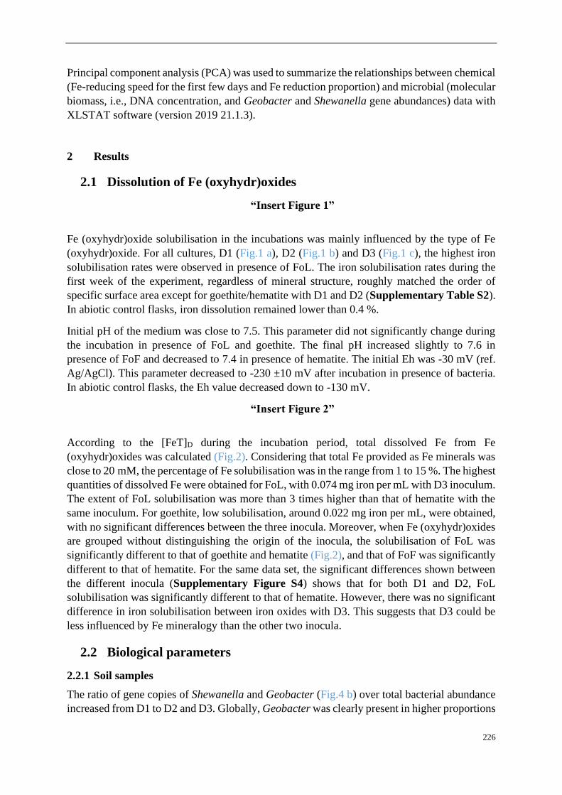
226
Principal component analysis (PCA) was used to summarize the relationships between chemical
(Fe-reducing speed for the first few days and Fe reduction proportion) and microbial (molecular
biomass, i.e., DNA concentration, and Geobacter and Shewanella gene abundances) data with
XLSTAT software (version 2019 21.1.3).
2 Results
2.1 Dissolution of Fe (oxyhydr)oxides
“Insert Figure 1”
Fe (oxyhydr)oxide solubilisation in the incubations was mainly influenced by the type of Fe
(oxyhydr)oxide. For all cultures, D1 (Fig.1 a), D2 (Fig.1 b) and D3 (Fig.1 c), the highest iron
solubilisation rates were observed in presence of FoL. The iron solubilisation rates during the
first week of the experiment, regardless of mineral structure, roughly matched the order of
specific surface area except for goethite/hematite with D1 and D2 (Supplementary Table S2).
In abiotic control flasks, iron dissolution remained lower than 0.4 %.
Initial pH of the medium was close to 7.5. This parameter did not significantly change during
the incubation in presence of FoL and goethite. The final pH increased slightly to 7.6 in
presence of FoF and decreased to 7.4 in presence of hematite. The initial Eh was -30 mV (ref.
Ag/AgCl). This parameter decreased to -230 ±10 mV after incubation in presence of bacteria.
In abiotic control flasks, the Eh value decreased down to -130 mV.
“Insert Figure 2”
According to the [FeT]D during the incubation period, total dissolved Fe from Fe
(oxyhydr)oxides was calculated (Fig.2). Considering that total Fe provided as Fe minerals was
close to 20 mM, the percentage of Fe solubilisation was in the range from 1 to 15 %. The highest
quantities of dissolved Fe were obtained for FoL, with 0.074 mg iron per mL with D3 inoculum.
The extent of FoL solubilisation was more than 3 times higher than that of hematite with the
same inoculum. For goethite, low solubilisation, around 0.022 mg iron per mL, were obtained,
with no significant differences between the three inocula. Moreover, when Fe (oxyhydr)oxides
are grouped without distinguishing the origin of the inocula, the solubilisation of FoL was
significantly different to that of goethite and hematite (Fig.2), and that of FoF was significantly
different to that of hematite. For the same data set, the significant differences shown between
the different inocula (Supplementary Figure S4) shows that for both D1 and D2, FoL
solubilisation was significantly different to that of hematite. However, there was no significant
difference in iron solubilisation between iron oxides with D3. This suggests that D3 could be
less influenced by Fe mineralogy than the other two inocula.
2.2 Biological parameters
2.2.1 Soil samples
The ratio of gene copies of Shewanella and Geobacter (Fig.4 b) over total bacterial abundance
increased from D1 to D2 and D3. Globally, Geobacter was clearly present in higher proportions

227
than Shewanella in the bacterial communities present in the environmental samples used to
enrich D1, D2 and D3.
2.2.2 Effect of Fe (oxyhydr)oxides on bacterial community structure
The bacterial community structure profiles of the initial soil and sediment samples, the Fe-NTA
enrichments and the cultures in presence of the four different (oxyhydr)oxides were compared
using an nMDS ordination approach (Fig.3). Full CE-SSCP profiles are available in
Supplementary Figure S5. These profiles show a high diversity with many peaks.
“Insert Figure 3”
The structures of the initial communities were modified after inoculation on solid Fe
(oxyhydr)oxides (Fig.3): enrichments on Fe(III)-NTA are grouped on the right side of the
representation, whereas communities obtained by cultures with iron oxides are gathered on the
left side. The three initial communities from environmental samples are located between these
two poles. No clear separation is observed between the communities obtained with the four
pure iron oxides. Although, according to ANOSIM analysis, no significant difference in
community composition was found between initial inoculums (significance >0.05), a
significant dissimilarity was found between community origins (i.e. initial soils and form of
Fe(III) provided to the consortia) (R = 0.345 and significance = 0.0249).
2.2.3 Bacterial abundance and abundances of Shewanella and Geobacter
“Insert Figure 4”
The rrs gene abundances and Shewanella 16S/bacterial 16S ratio did not highlight any
significant differences between the three initial enrichments D1, D2 and D3 (Fig.4). As shown
in Fig.4 (a), globally, the rrs gene abundance in goethite samples was significantly different to
FOF samples. It decreased in iron oxide cultures compared with Fe(III)-NTA cultures
(Supplementary Figure S6). Conversely, the abundance of the two quantified IRB, i.e.,
Geobacter and Shewanella, differ between the types of Fe oxides provided as Fe(III) source
(Fig.4 c and e). Globally, compared with Geobacter, Shewanella was present in a larger
proportion in all bacterial communities with the four iron oxides. Moreover, the number of gene
copies of Shewanella for FoL is significantly different in presence of other iron oxides but there
are no significant differences between the cultures for FoF, goethite and hematite samples.
When the type of inocula is not taken into account, the proportion of Shewanella 16S genes for
FoL samples is significantly different for goethite samples.
Considering specifically each quantified genus, the cultures in presence of goethite inoculated
with D3 bacteria contained a significantly different proportion of Geobacter 16S genes than the
same culture (D3) with FoL (Fig.4 f). In the other conditions (D1, D2 with all oxides), no
significant differences between the proportion of Geobacter 16S genes in the global bacterial
population was observed.
3 Discussion

228
3.1 Influence of the type of iron oxide on bacterial iron solubilisation
FoL and FoF were synthetized in the laboratory and were more amorphous, thus more reactive,
than goethite and hematite (Usman et al., 2012). According to the literature, abiotic rates of
reductive iron dissolution are correlated with the solubility of Fe (oxyhydr)oxides (Larsen and
Postma, 2001). The rates of this abiotic reductive bulk dissolution decrease according to
ferrihydrite >lepidocrocite> goethite> hematite, emphasizing the importance of the crystal
structure on the dissolution rate (Larsen and Postma, 2001). However, Roden (2006) found that
in presence of IRB, oxides’ mineralogical and thermodynamical properties exert a minor
influence on reduction rates compared with the abundance of available oxide surface sites
controlled by oxide surface areas and the accumulation of surface-bound biogenic Fe(II). This
last process, of the precipitation of new Fe(II) minerals (Urrutia et al., 1998; Zachara et al.,
2002), could explain the late decrease of soluble Fe (Figure 1). Present results are partially in
accordance with this last hypothesis, as Fe solubilisation effectiveness increased with the
specific surface, in particular for the two freshly synthesized oxides. Here, the fresh mineral
prepared with the protocol of lepidocrocite (FoL) synthesis presented a higher specific surface
than the mineral produced using the protocol for ferrihydrite (FoF). Synthetic lepidocrocites
can present a very large range of specific surface areas, depending on their level of crystallinity
(Schwertmann, 1973). In the present case, the specific area of synthetic FoL (337 m2·g
-1) is
much higher than for other Fe (oxyhydr)oxides, possibly due to a rapid oxidation of Fe(II)
during synthesis that produced poor crystallization and formation of lepidocrocite impurities
(Schwertmann and Taylor, 1979). Schwertmann (1973) showed that the specific surface of
lepidocrocite increases with its solubility in presence of oxalate, and this specific surface area
is anti-correlated with the crystallinity. In the present experiment, at the end of the incubation,
we detected 8 mg·L-1 total Fe in the FoL abiotic control, but almost no Fe in FoF abiotic control.
This supports the idea that FoL was more soluble than FoF. The high specific surface area and
probable poor crystallinity of FoL could have favored solubilisation by IRB.
Fe solubilisation rates were limited after a few days, probably due to evolution of the
composition of the liquid medium, or the accumulation of surface-bound biogenic Fe(II)
(Roden, 2006), as Fe(III) was not the limiting factor. Parameters such as humic substances,
quinones and organic carbon can strongly influence microbial Fe(III) reduction rates for
Shewanella (Adhikari et al., 2017) or Geobacter (Wolf et al., 2009) in natural environments.
Generally, in these studies, the highest reducing rates were observed during the first few days
of microbial Fe(III) reduction. In our study, the influence of the type of Fe (oxyhydr)oxides on
initial iron oxide-reducing rates were consistent with those of previous studies performed with
pure strains.
3.2 Bacterial communities
The structures of the initial bacterial communities present in the environmental samples were
modified by the enrichment in Fe(III)-NTA medium, and again showed an evolution when the
enrichments were grown in presence of solid Fe (oxyhydr)oxides (Fig.4). This last result could
be due to the difference of bio-availability of Fe with minerals compared to Fe(III)-NTA. Cai
et al., (2019) also showed an influence of bio-available Fe(III) on microbial community
structure. Here, the accessibility of Fe(III) in the Fe(III)-NTA incubations favored the iron
reducing community that may have rapidly consumed available organic substrates and probably
lowered the development of other bacteria. With minerals, however, Fe(III) is less accessible

229
and competition for Fe(III) may induce changes in community structure. For example, Zhuang
et al., (2011) indicated that Rhodoferax and Geobacter species were acetate-oxidizing Fe(III)-
reducers that compete in anoxic subsurface environments and this competition could influence
the in situ bioremediation of uranium-contaminated groundwater by changing diversity
structure. On the other hand, no clear effect of the type of Fe mineral on the global community
profile was observed. This may be linked to the culture medium composition. Indeed, the
addition of diverse C sources could enable fermentative bacteria to develop without using Fe(III)
for their growth. In contrast, Lentini et al., (2012) observed an effect of the type of Fe
(oxyhydr)oxide, i.e., FoF, goethite or hematite, on the structure of bacterial communities in
enrichment cultures using T-RFLP fingerprints. However, these authors did not use an initial
Fe(III)-NTA enrichment step and did not use a sulfate-reduction inhibitor, as in our study.
3.3 Geobacter and Shewanella 16S genes abundances
The relative abundance of Geobacter and Shewanella in the initial soils and sediment used as
sources of IRB in our experiment could be linked to the redox conditions of the environment.
Indeed, Geobacter and Shewanella were present in higher proportions in the river sediment D3,
than in the flooded soil D2, itself richer than the non-saturated soil D1. This suggests that the
reducing conditions in the aquatic sediment was more favorable for anaerobic bacteria, such as
IRB.
Shewanella and Geobacter represented in our incubation experiments a small proportion of the
total community, however we made a focus on these two genera because they are the most
studied iron reducing bacteria. However, the other members of the community could contribute
either directly or indirectly to the iron solubilisation process. Considering the evolution of the
two targeted IRB genera, namely Geobacter and Shewanella, after the enrichment with Fe(III)-
NTA, the Shewanella / Geobacter abundance ratio increased significantly compared to the site
samples (Figure 3 compared with Figure 5), suggesting that the presence of a large amount of
bio-available Fe(III) in the enrichment culture medium was in favor of Shewanella. Another
explanation for the sharp increase of Shewanella compared to Geobacter could be the
composition of the culture medium in terms of organic substrates. Geobacter can use acetate as
electron donors while performing the dissimilatory Fe reduction (Caccavo et al., 1994; Coates
et al., 1996), but does not use lactate nor glucose. Conversely, some Shewanella species were
shown to be able to use glucose and can present either respiratory or fermentative types of
metabolisms (Nogi and Horikoshi, 1998; Ziemke et al., 1998, Ivanova et al., 2004). Lentini et
al., (2012) showed that the presence of Geobacter was favored by acetate whereas the growth
of Shewanella was rather stimulated by lactate. Moreover, these authors suggested that
production of acetate through incomplete degradation of lactate by Shewanella could benefit
Geobacter (Hori et al., 2015). Our medium contained both acetate, formate, lactate and glucose,
thus it should potentially support growth of both Shewanella and Geobacter. However, a
fermentative metabolism could explain the selection of Shewanella over Geobacter in our
enrichments and in all incubation conditions, because this genus can grow either using
fermentation or Fe(III) reduction (Bowman, 1997). Moreover, there is a multitude of other
bacteria that can also explain the contribution of fermentative products, maybe some of them
are in favor of Shewanella sp. (or spp.) but not Geobacter spp. As our medium contained 1.5
g.L-1 peptone, this substrate could also favor the growth of bacteria belonging to the Shewanella
genus.
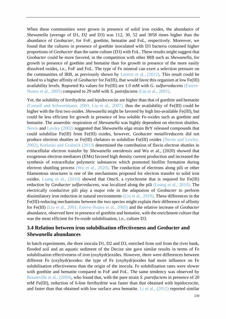
230
When these communities were grown in presence of solid iron oxides, the abundance of
Shewanella (average of D1, D2 and D3) was 112, 30, 52 and 3058 times higher than the
abundance of Geobacter, for FoF, goethite, hematite and FoL, respectively. Moreover, we
found that the cultures in presence of goethite inoculated with D3 bacteria contained higher
proportions of Geobacter than the same culture (D3) with FoL. These results might suggest that
Geobacter could be more favored, in the competition with other IRB such as Shewanella, for
growth in presence of goethite and hematite than for growth in presence of the more easily
dissolved oxides, i.e., FoF and FoL. The type of Fe mineral can exert a selection pressure on
the communities of IRB, as previously shown by Lentini et al., (2012). This result could be
linked to a higher affinity of Geobacter for Fe(III), that would favor this organism at low Fe(III)
availability levels. Reported Ks values for Fe(III) are 1.0 mM with G. sulfurreducens (Esteve-
Nunez et al., 2005) compared to 29 mM with S. putrefaciens (Liu et al., 2001).
Yet, the solubility of ferrihydrite and lepidocrocite are higher than that of goethite and hematite
(Cornell and Schwertmann, 2003; Liu et al., 2007), thus the availability of Fe(III) could be
higher with the first two oxides. Shewanella might be favored by high bio-available Fe(III), but
could be less efficient for growth in presence of less soluble Fe-oxides such as goethite and
hematite. The anaerobic respiration of Shewanella was highly dependent on electron shuttles.
Nevin and Lovley (2002) suggested that Shewanella alga strain BrY released compounds that
could solubilize Fe(III) from Fe(III) oxides, however, Geobacter metallireducens did not
produce electron shuttles or Fe(III) chelators to solubilize Fe(III) oxides (Nevin and Lovley,
2002). Kotloski and Gralnick (2013) determined the contribution of flavin electron shuttles in
extracellular electron transfer by Shewanella oneidensis and Wu et al., (2020) showed that
exogenous electron mediators (EMs) favored high density current production and increased the
synthesis of extracellular polymeric substances which promoted biofilm formation during
electron shuttling process (Wu et al., 2020). The conduction of electrons along pili or other
filamentous structures is one of the mechanisms proposed for electron transfer to solid iron
oxides. Leang et al., (2010) showed that OmcS, a cytochrome that is required for Fe(III)
reduction by Geobacter sulfurreducens, was localized along the pili (Leang et al., 2010). The
electrically conductive pili play a major role in the adaptation of Geobacter to perform
dissimilatory iron reduction in natural environments (Liu et al., 2019). These differences in the
Fe(III)-reducing mechanisms between the two species might explain their difference of affinity
for Fe(III) (Liu et al., 2001; Esteve-Nunez et al., 2005) and the relative increase of Geobacter
abundance, observed here in presence of goethite and hematite, with the enrichment culture that
was the most efficient for Fe-oxide solubilisation, i.e., culture D3.
3.4 Relation between iron solubilisation effectiveness and Geobacter and
Shewanella abundances
In batch experiments, the three inocula D1, D2 and D3, enriched from soil from the river bank,
flooded soil and an aquatic sediment of the Decize site gave similar results in terms of Fe
solubilisation effectiveness of iron (oxyhydr)oxides. However, there were differences between
different Fe (oxyhydr)oxides: the type of Fe (oxyhydr)oxides had more influence on Fe
solubilisation effectiveness than the origin of the inocula. Fe solubilisation rates were slower
with goethite and hematite compared to FoF and FoL. The same tendency was observed by
Bonneville et al., (2004), who found that, with the pure strain S. putrefaciens in presence of 20
mM Fe(III), reduction of 6-line ferrihydrite was faster than that obtained with lepidocrocite,
and faster than that obtained with low surface area hematite. Li et al., (2012) reported similar
231
results for the microbial reduction of Fe(III) oxides by S. decolorationis strain S12, the reducing
speed decreasing according to the following order: lepidocrocite > goethite > hematite.
Principal Component Analysis (PCA) integrating chemical (Supplementary Table S2) and
molecular data was performed to identify the relationships and contributions between iron
solubilisation effectiveness and Geobacter / Shewanella abundances in batch experiment
samples (Mercier et al., 2014; Zhu et al., 2014; Hong et al., 2015). In this study, a biplot
(Supplementary Figure S7) summarizes PCA results. The first principal component is
strongly influenced by the iron solubilisation effectiveness (higher on the right than on the left
side of the representation), with higher values associated with FoL, and the lowest associated
with goethite and hematite (not separated), FoF being in an intermediary position. Meanwhile
proportion of Shewanella in the bacterial community, but not to the proportion of Geobacter,
seems to be correlated to FoL. The second principal component reflects high values of the
proportion of Geobacter in the bacterial communities. Thus, the proportion of Shewanella
seems to be more correlated to iron solubilisation parameters than Geobacter. PCA allows clear
discrimination with different groups of iron oxides, however not for the different types of
inoculum (D1, D2 and D3).
According to Bonneville et al., (2004), the dissolution and solubility of goethite and hematite
are lower than that of ferrihydrite and lepidocrocite in the presence of Shewanella. Cutting et
al., (2009) indicated that hematite and goethite are susceptible to limited Fe(III) reduction in
presence of G. sulfurreducens. Moreover, Poggenburg et al., (2016) mentioned that Fe(III)-
organic compounds (coprecipitates from solutions of FeCl3 and natural organic matter)
reduction by Shewanella putrefaciens was influenced by the amount of available electron
shuttling molecules induced by sorbed natural organic matter. Fe(III)-organic compounds’
reduction by Geobacter metallireducens was more influenced by particle size, physicochemical
properties and iron (oxyhydr)oxides (composition of sorbed natural organic matter and
aggregation state) (Poggenburg et al., 2016). In our study, FoL samples with a higher proportion
of Shewanella in their bacterial communities were correlated with high initial iron solubilisation
rates and electron shuttling molecules might have a role in this phenomenon. In contrast,
Geobacter was not specifically associated to FoL but was found in higher proportions with
goethite in one condition. This tendency is in accordance with findings of previous research
performed with the less soluble iron oxides (Crosby et al., 2007), showing that Geobacter
sulfurreducens reduced 0.7 % hematite and 4.0 % goethite while Shewanella putrefaciens
reduced only 0.5 % of hematite 3.1 % goethite after 280 days of incubation. Thus, our results
suggest that Geobacter might suffer less from the competition with Shewanella in low bio-
available Fe(III) conditions, whereas the contribution of this genus, present in lower quantities
than Shewanella, to the iron solubilisation effectiveness is not demonstrated. Moreover, other
members in the community of IRB might have potential contribution to Fe solubilisation. Our
culture medium contained substrates such as glucose and peptone that could support the growth
of fermentative organisms. Lentini (2012) and Gagen (2019) indicated that fermentation likely
plays a key role in reduction of crystalline iron oxides by diverse species, such as
Telmatospirillum, both through direct reduction and by the production of H2, potentially used
by dissimilatory iron reducers, during fermentation.
Conclusion

232
Microbial enrichments containing IRB, obtained with a mixture of simple and complex electron
donors, were able to grow and reduce Fe(III) in a short time. Experiments performed with fresh
Fe (oxyhydr)oxides, goethite and hematite confirmed that the type of Fe mineral could influence
Fe solubilisation rates and the abundances of two IRB commonly involved as pure strains in
Fe-reducing experiments, i.e., Geobacter and Shewanella.
The present study’s results showed that: (1) the sub-culturing of IRB enrichments from Fe(III)-
NTA to pure iron oxides significantly modified the bacterial communities; (2) in our
experimental conditions, bacterial diversity was not significantly different from one type of
pure (oxyhydr)oxide to another; (3) the type of Fe oxide can influence the proportion of
Geobacter and Shewanella. Meanwhile, the nature of Fe (oxyhydr)oxides seems to have exerted
a selection on the ratio of Geobacter and Shewanella, whereas it did not impact the bacterial
community fingerprints. The concentration of bio-available Fe(III) and the mixture of electron
donors in the enrichment medium favored the development of Shewanella compared with
Geobacter genus. However, the culture medium included a large amount of electron donors that
is not representative of most natural systems. Therefore, complementary studies, performed
with lower concentrations of electron donors provided in continuous feeding conditions would
help to make the link with real environments. In presence of iron oxides, the highest proportions
of Shewanella in bacterial communities were obtained with FoL and corresponded to the
highest levels of iron solubilisation, possibly linked to the fact that FoL was the most soluble
(oxyhydr)oxide in our experiments. This result is consistent with the hypothesis that Shewanella
development could be favored by a high bioavailability of Fe(III). In contrast, Geobacter was
detected in higher proportions with goethite that is less easily dissolved, when D3 culture was
used.
Globally, all results suggested that both initial community composition of the sample used to
prepare the enrichments, as well as the type of Fe(III) oxide used as electron acceptor influenced
the final proportions and abundances of Geobacter and Shewanella. A better knowledge of
complementary biological parameters associated with these two organisms, such as their
activity during Fe(III) solubilisation and reduction in complex communities and distribution
between planktonic and Fe(III)-mineral-attached cells, could help to elucidate their role in
natural environments. As biofilms in soils and sediments contain a large part of the bacterial
biomass, future research could be focused on the distribution and activity of Geobacter and
Shewanella attached on iron oxides surfaces.
Conflict of Interest
The authors declare that the research was conducted in the absence of any commercial or
financial relationships that could be construed as a potential conflict of interest.
Author Contributions
F. Battaglia-Brunet, J. Hellal and M. Motelica-Heino conceived and designed the experiments
and arranged funds. F. Zhang performed major experiments. F. Zhang and F. Battaglia-Brunet
were responsible for manuscript preparation. M. Motelica-Heino, F. Battaglia-Brunet, J. Hellal
and F. Zhang arranged sampling from Decize, France. P. Gautret and F. Zhang performed SEM.
C. Joulian, J. Hellal and F. Zhang performed the qPCR of Shewanella / Geobacter and bacterial
16S rRNA (rrs gene) quantification.
233
Acknowledgments
This research work was performed in the frame of a PhD project, a scholarship co-funded by
the BRGM – Région Centre Val de Loire 2017-2020, contract N° SIRET 582 056 149 00120.
Additionally, the INSU-EC2CO project “Dycyfer” also contributed to this work. This work was
supported by a grant overseen by the French National Research Agency (ANR) as part of the
“Investments d’Avenir” Programme LabEx VOLTAIRE, 10-LABX-0100.
References
Adhikari, D., Zhao, Q., Das, K., Mejia, J., Huang, R., Wang, X., Poulson, S.R., Tang, Y.,
Roden, E.E., and Yang, Y. (2017). Dynamics of ferrihydrite-bound organic carbon
during microbial Fe reduction. Geochimica et Cosmochimica Acta 212, 221-233.
Aino, K., Hirota, K., Okamoto, T., Tu, Z., Matsuyama, H., and Yumoto, I. (2018). Microbial
communities associated with indigo fermentation that thrive in anaerobic alkaline
environments. Frontiers in microbiology 9, 2196.
Bonneville, S., Van Cappellen, P., and Behrends, T. (2004). Microbial reduction of iron (III)
oxyhydroxides: effects of mineral solubility and availability. Chemical Geology 212,
255-268.
Bowman, J. P. (2015). Shewanella. Bergey's Manual of Systematics of Archaea and Bacteria,
1-22.
Brunauer, S., Emmett, P.H., and Teller, E. (1938). Adsorption of gases in multimolecular
layers. Journal of the American chemical society 60, 309-319.
Caccavo, F., Lonergan, D. J., Lovley, D. R., Davis, M., Stolz, J. F., & McInerney, M. J.
(1994). Geobacter sulfurreducens sp. nov., a hydrogen-and acetate-oxidizing
dissimilatory metal-reducing microorganism. Applied and environmental
microbiology, 60(10), 3752-3759.
Cai, Y., Hu, K., Zheng, Z., Zhang, Y., Guo, S., Zhao, X., Cui, Z., and Wang, X. (2019).
Effects of adding EDTA and Fe2+ on the performance of reactor and microbial
community structure in two simulated phases of anaerobic digestion. Bioresource
technology 275, 183-191.
Cavelan, A., Boussafir, M., Le Milbeau, C., Rozenbaum, O., and Laggoun-Défarge, F. (2019).
Effect of organic matter composition on source rock porosity during confined
anhydrous thermal maturation: Example of Kimmeridge-clay mudstones.
International Journal of Coal Geology 212, 103236.
Coates, J. D., Phillips, E. J., Lonergan, D. J., Jenter, H., & Lovley, D. R. (1996). Isolation of
Geobacter species from diverse sedimentary environments. Applied and
Environmental Microbiology, 62(5), 1531-1536.
Colombo, C., Palumbo, G., He, J.-Z., Pinton, R., and Cesco, S. (2014). Review on iron
availability in soil: interaction of Fe minerals, plants, and microbes. Journal of soils
and sediments 14, 538-548.
Cooper, D.C., Picardal, F.F., and Coby, A.J. (2006). Interactions between microbial iron
reduction and metal geochemistry: effect of redox cycling on transition metal
speciation in iron bearing sediments. Environmental science & technology 40, 1884-
1891.
234
Cornell, R.M., and Schwertmann, U. (2003). The iron oxides: structure, properties, reactions,
occurrences and uses. John Wiley & Sons.
Crosby, H.A., Johnson, C.M., Roden, E.E., and Beard, B.L. (2005). Coupled Fe (II)− Fe (III)
electron and atom exchange as a mechanism for Fe isotope fractionation during
dissimilatory iron oxide reduction. Environmental science & technology 39, 6698-
6704.
Crosby, H.A., Roden, E.E., Johnson, C.M., and Beard, B.L. (2007). The mechanisms of iron
isotope fractionation produced during dissimilatory Fe (III) reduction by Shewanella
putrefaciens and Geobacter sulfurreducens. Geobiology 5, 169-189.
Cutting, R., Coker, V., Fellowes, J., Lloyd, J., and Vaughan, D. (2009). Mineralogical and
morphological constraints on the reduction of Fe (III) minerals by Geobacter
sulfurreducens. Geochimica et Cosmochimica Acta 73, 4004-4022.
Delbès, C., Moletta, R., & Godon, J. J. (2000). Monitoring of activity dynamics of an
anaerobic digester bacterial community using 16S rRNA polymerase chain reaction–
single‐strand conformation polymorphism analysis. Environmental Microbiology,
2(5), 506-515.
Das, S., Hendry, M.J., and Essilfie-Dughan, J. (2013). Adsorption of selenate onto
ferrihydrite, goethite, and lepidocrocite under neutral pH conditions. Applied
geochemistry 28, 185-193.
Dhivert, E., Grosbois, C., Rodrigues, S., and Desmet, M. (2015). Influence of fluvial
environments on sediment archiving processes and temporal pollutant dynamics
(Upper Loire River, France). Science of the total Environment 505, 121-136.
Engel, C.E.A., Schattenberg, F., Dohnt, K., Schröder, U., Müller, S., and Krull, R. (2019).
Long-term behavior of defined mixed cultures of Geobacter sulfurreducens and
Shewanella oneidensis in bioelectrochemical systems. Frontiers in Bioengineering
and Biotechnology 7, 60.
Esteve‐Núñez, A., Rothermich, M., Sharma, M., & Lovley, D. (2005). Growth of Geobacter
sulfurreducens under nutrient‐limiting conditions in continuous culture.
Environmental microbiology, 7(5), 641-648.
Fredrickson, J.K., Zachara, J.M., Kennedy, D.W., Dong, H., Onstott, T.C., Hinman, N.W.,
and Li, S.-M. (1998). Biogenic iron mineralization accompanying the dissimilatory
reduction of hydrous ferric oxide by a groundwater bacterium. Geochimica et
Cosmochimica Acta 62, 3239-3257.
Gagen, E. J., Zaugg, J., Tyson, G. W., & Southam, G. (2019). Goethite Reduction by a
Neutrophilic Member of the Alphaproteobacterial Genus Telmatospirillum. Frontiers
in microbiology, 10, 2938.
Gault, A.G., Ibrahim, A., Langley, S., Renaud, R., Takahashi, Y., Boothman, C., Lloyd, J.R.,
Clark, I.D., Ferris, F.G., and Fortin, D. (2011). Microbial and geochemical features
suggest iron redox cycling within bacteriogenic iron oxide-rich sediments. Chemical
Geology 281, 41-51.
Ghorbanzadeh, N., Lakzian, A., Halajnia, A., Choi, U.K., Kim, K.H., Kim, J.O., Kurade, M.,
and Jeon, B.H. (2017). Impact of bioreduction on remobilization of adsorbed cadmium
on iron minerals in anoxic condition. Water Environment Research 89, 519-526.
Han, R., Liu, T., Li, F., Li, X., Chen, D., and Wu, Y. (2018). Dependence of secondary
mineral formation on Fe (II) production from ferrihydrite reduction by Shewanella
oneidensis MR-1. ACS Earth and Space Chemistry 2, 399-409.

235
Hansel, C.M., Benner, S.G., Nico, P., and Fendorf, S. (2004). Structural constraints of ferric
(hydr) oxides on dissimilatory iron reduction and the fate of Fe (II). Geochimica et
Cosmochimica Acta 68, 3217-3229.
Hong, C., Si, Y., Xing, Y., and Li, Y. (2015). Illumina MiSeq sequencing investigation on the
contrasting soil bacterial community structures in different iron mining areas.
Environmental Science and Pollution Research 22, 10788-10799.
Hori, T., Aoyagi, T., Itoh, H., Narihiro, T., Oikawa, A., Suzuki, K., ... & Kamagata, Y.
(2015). Isolation of microorganisms involved in reduction of crystalline iron (III)
oxides in natural environments. Frontiers in Microbiology, 6, 386.
Huguet, L. (2009). Caractérisation biogéochimique et potentiel de méthylation du mercure de
biofilms en milieu tropical (retenue de Petit Saut et estuaire du Sinnamary, Guyane
Française) (Doctoral dissertation, Nancy 1). Jiang, Z., Shi, M., and Shi, L. (2020).
Degradation of organic contaminants and steel corrosion by the dissimilatory metal-
reducing microorganisms Shewanella and Geobacter spp. International
Biodeterioration & Biodegradation 147, 104842.
Kim, S.-J., Koh, D.-C., Park, S.-J., Cha, I.-T., Park, J.-W., Na, J.-H., Roh, Y., Ko, K.-S., Kim,
K., and Rhee, S.-K. (2012). Molecular analysis of spatial variation of iron-reducing
bacteria in riverine alluvial aquifers of the Mankyeong River. The journal of
microbiology 50, 207-217.
Kotloski, N. J., & Gralnick, J. A. (2013). Flavin electron shuttles dominate extracellular
electron transfer by Shewanella oneidensis. MBio, 4(1).
Kwon, M. J., O’Loughlin, E. J., Boyanov, M. I., Brulc, J. M., Johnston, E. R., Kemner, K. M.,
& Antonopoulos, D. A. (2016). Impact of organic carbon electron donors on microbial
community development under iron-and sulfate-reducing conditions. PloS one, 11(1),
e0146689.
Larsen, O., and Postma, D. (2001). Kinetics of reductive bulk dissolution of lepidocrocite,
ferrihydrite, and goethite. Geochimica et Cosmochimica Acta 65, 1367-1379.
Leang, C., Qian, X., Mester, T., and Lovley, D.R. (2010). Alignment of the c-type
cytochrome OmcS along pili of Geobacter sulfurreducens. Appl. Environ. Microbiol.
76, 4080-4084.
Lentini, C.J., Wankel, S.D., and Hansel, C.M. (2012). Enriched iron (III)-reducing bacterial
communities are shaped by carbon substrate and iron oxide mineralogy. Frontiers in
microbiology 3, 404.
Levar, C.E., Hoffman, C.L., Dunshee, A.J., Toner, B.M., and Bond, D.R. (2017). Redox
potential as a master variable controlling pathways of metal reduction by Geobacter
sulfurreducens. The ISME journal 11, 741-752.
Li, B.-B., Cheng, Y.-Y., Fan, Y.-Y., Liu, D.-F., Fang, C.-Y., Wu, C., Li, W.-W., Yang, Z.-C.,
and Yu, H.-Q. (2018). Estimates of abundance and diversity of Shewanella genus in
natural and engineered aqueous environments with newly designed primers. Science of
The Total Environment 637, 926-933.
Li, X., Liu, T., Li, F., Zhang, W., Zhou, S., and Li, Y. (2012). Reduction of structural Fe (III)
in oxyhydroxides by Shewanella decolorationis S12 and characterization of the
surface properties of iron minerals. Journal of soils and sediments 12, 217-227.
Liu, C., Kota, S., Zachara, J. M., Fredrickson, J. K., & Brinkman, C. K. (2001). Kinetic
analysis of the bacterial reduction of goethite. Environmental Science & Technology,
35(12), 2482-2490.

236
Liu, H., Li, P., Zhu, M., Wei, Y., and Sun, Y. (2007). Fe (II)-induced transformation from
ferrihydrite to lepidocrocite and goethite. Journal of Solid State Chemistry 180, 2121-
2128.
Liu, X., Ye, Y., Xiao, K., Rensing, C., and Zhou, S. (2019). Molecular evidence for the
adaptive evolution of Geobacter sulfurreducens to perform dissimilatory iron
reduction in natural environments. Molecular Microbiology.
Lovley, D.R. (1991). Dissimilatory Fe (III) and Mn (IV) reduction. Microbiology and
Molecular Biology Reviews 55, 259-287.
Lovley, D.R. (2000). "Fe (III) and Mn (IV) reduction," in Environmental microbe-metal
interactions. American Society of Microbiology), 3-30.
Lovley, D. (2006). Dissimilatory Fe (III)-and Mn (IV)-reducing prokaryotes. Prokaryotes, 2,
635-658.
Lovley, D. R., Phillips, E. J., & Lonergan, D. J. (1989). Hydrogen and formate oxidation
coupled to dissimilatory reduction of iron or manganese by Alteromonas putrefaciens.
Applied and Environmental Microbiology, 55(3), 700-706.
Lovley, D.R., Stolz, J.F., Nord, G.L., and Phillips, E.J. (1987). Anaerobic production of
magnetite by a dissimilatory iron-reducing microorganism. Nature 330, 252-254.
Ivanova, E. P., Gorshkova, N. M., Bowman, J. P., Lysenko, A. M., Zhukova, N. V., Sergeev,
A. F., ... & Nicolau, D. V. (2004). Shewanella pacifica sp. nov., a polyunsaturated
fatty acid-producing bacterium isolated from sea water. International journal of
systematic and evolutionary microbiology, 54(4), 1083-1087.
Mamindy-Pajany, Y., Bataillard, P., Séby, F., Crouzet, C., Moulin, A., Guezennec, A.-G.,
Hurel, C., Marmier, N., and Battaglia-Brunet, F. (2013). Arsenic in marina sediments
from the Mediterranean coast: speciation in the solid phase and occurrence of
thioarsenates. Soil and Sediment Contamination: An International Journal 22, 984-
1002.
Meile, C., and Scheibe, T.D. (2019). Reactive transport modeling of microbial dynamics.
Elements: An International Magazine of Mineralogy, Geochemistry, and Petrology 15,
111-116.
Melton, E.D., Swanner, E.D., Behrens, S., Schmidt, C., and Kappler, A. (2014). The interplay
of microbially mediated and abiotic reactions in the biogeochemical Fe cycle. Nature
Reviews Microbiology 12, 797-808.
Mercier, A., Joulian, C., Michel, C., Auger, P., Coulon, S., Amalric, L., Morlay, C., and
Battaglia-Brunet, F. (2014). Evaluation of three activated carbons for combined
adsorption and biodegradation of PCBs in aquatic sediment. Water research 59, 304-
315.
Murti, G.K., Volk, V., and Jackson, M. (1966). Colorimetric determination of iron of mixed
valency by orthophenanthroline. Soil Science Society of America Journal 30, 663-664.
Nealson, K.H. (2017). Bioelectricity (electromicrobiology) and sustainability. Microbial
biotechnology 10, 1114-1119.
Nevin, K.P., and Lovley, D.R. (2002). Mechanisms for Fe (III) oxide reduction in
sedimentary environments. Geomicrobiology Journal 19, 141-159.
Nogi, Y., Kato, C., & Horikoshi, K. (1998). Taxonomic studies of deep-sea barophilic
Shewanella strains and description of Shewanella violacea sp. nov. Archives of
microbiology, 170(5), 331-338.

237
Pedersen, H.D., Postma, D., Jakobsen, R., and Larsen, O. (2005). Fast transformation of iron
oxyhydroxides by the catalytic action of aqueous Fe (II). Geochimica et
Cosmochimica Acta 69, 3967-3977.
Poggenburg, C., Mikutta, R., Sander, M., Schippers, A., Marchanka, A., Dohrmann, R., and
Guggenberger, G. (2016). Microbial reduction of ferrihydrite-organic matter
coprecipitates by Shewanella putrefaciens and Geobacter metallireducens in
comparison to mediated electrochemical reduction. Chemical Geology 447, 133-147.
Roden, E. E. (2006). Geochemical and microbiological controls on dissimilatory iron
reduction. Comptes Rendus Geoscience, 338(6-7), 456-467.
Roden, E.E., Sobolev, D., Glazer, B., and Luther, G.W. (2004). Potential for microscale
bacterial Fe redox cycling at the aerobic-anaerobic interface. Geomicrobiology
Journal 21, 379-391.
Roden, E.E., and Wetzel, R.G. (2002). Kinetics of microbial Fe (III) oxide reduction in
freshwater wetland sediments. Limnology and Oceanography 47, 198-211.
Schilling, K., Borch, T., Rhoades, C.C., and Pallud, C.E. (2019). Temperature sensitivity of
microbial Fe (III) reduction kinetics in subalpine wetland soils. Biogeochemistry 142,
19-35.
Schwertmann, U. (1973). Use of oxalate for Fe extraction from soils. Canadian Journal of Soil
Science, 53(2), 244-246.
Schwertmann, U., & Taylor, R. M. (1979). Natural and synthetic poorly crystallized
lepidocrocite. Clay Minerals, 14(4), 285-293.
Schwertmann, U., and Cornell, R.M. (2008). Iron oxides in the laboratory: preparation and
characterization. John Wiley & Sons.
Shelobolina, E. S., Nevin, K. P., Blakeney-Hayward, J. D., Johnsen, C. V., Plaia, T. W.,
Krader, P., ... & Lovley, D. R. (2007). Geobacter pickeringii sp. nov., Geobacter
argillaceus sp. nov. and Pelosinus fermentans gen. nov., sp. nov., isolated from
subsurface kaolin lenses. International Journal of Systematic and Evolutionary
Microbiology, 57(1), 126-135.
Shi, L., Dong, H., Reguera, G., Beyenal, H., Lu, A., Liu, J., Yu, H.-Q., and Fredrickson, J.K.
(2016). Extracellular electron transfer mechanisms between microorganisms and
minerals. Nature Reviews Microbiology 14, 651.
Shi, M., Jiang, Y., and Shi, L. (2019). Electromicrobiology and biotechnological applications
of the exoelectrogens Geobacter and Shewanella spp. Science China Technological
Sciences, 1-9.
Snoeyenbos-West, O., Nevin, K., Anderson, R., and Lovley, D. (2000). Enrichment of
Geobacter species in response to stimulation of Fe (III) reduction in sandy aquifer
sediments. Microbial Ecology 39, 153-167.
Stern, N., Mejia, J., He, S., Yang, Y., Ginder-Vogel, M., and Roden, E.E. (2018). Dual role of
humic substances as electron donor and shuttle for dissimilatory iron reduction.
Environmental science & technology 52, 5691-5699.
Su, C., Zhang, M., Lin, L., Yu, G., Zhong, H., and Chong, Y. (2020). Reduction of iron
oxides and microbial community composition in iron-rich soils with different organic
carbon as electron donors. International Biodeterioration & Biodegradation 148,
104881.
Team, R. (2015). RStudio: integrated development for R. RStudio. Inc., Boston, MA, 700.
238
Thompson, A., Chadwick, O.A., Rancourt, D.G., and Chorover, J. (2006). Iron-oxide
crystallinity increases during soil redox oscillations. Geochimica et Cosmochimica
Acta 70, 1710-1727.
Thouin, H., Le Forestier, L., Gautret, P., Hube, D., Laperche, V., Dupraz, S., and Battaglia-
Brunet, F. (2016). Characterization and mobility of arsenic and heavy metals in soils
polluted by the destruction of arsenic-containing shells from the Great War. Science of
the Total Environment 550, 658-669.
Urrutia M, Roden E, Fredrickson J, Zachara J. Microbial and surface chemistry controls on
reduction of synthetic Fe (III) oxide minerals by the dissimilatory iron‐reducing
bacterium Shewanella alga. Geomicrobiology Journal 1998; 15: 269-291.
Usman, M., Hanna, K., Abdelmoula, M., Zegeye, A., Faure, P., and Ruby, C. (2012).
Formation of green rust via mineralogical transformation of ferric oxides (ferrihydrite,
goethite and hematite). Applied Clay Science 64, 38-43.
Wilkins, M.J., Livens, F.R., Vaughan, D.J., and Lloyd, J.R. (2006). The impact of Fe (III)-
reducing bacteria on uranium mobility. Biogeochemistry 78, 125-150.
Wolf, M., Kappler, A., Jiang, J., and Meckenstock, R.U. (2009). Effects of humic substances
and quinones at low concentrations on ferrihydrite reduction by Geobacter
metallireducens. Environmental science & technology 43, 5679-5685.
Wu, Y., Luo, X., Qin, B., Li, F., Haggblom, M. M., & Liu, T. (2020). Enhanced Current
Production by Exogenous Electron Mediators via Synergy of Promoting Biofilm
Formation and the Electron Shuttling Process. Environmental Science & Technology.
Zachara, J.M., Kukkadapu, R.K., Fredrickson, J.K., Gorby, Y.A., and Smith, S.C. (2002).
Biomineralization of poorly crystalline Fe (III) oxides by dissimilatory metal reducing
bacteria (DMRB). Geomicrobiology Journal 19, 179-207.
Zhang, C., Ge, Y., Yao, H., Chen, X., and Hu, M. (2012). Iron oxidation-reduction and its
impacts on cadmium bioavailability in paddy soils: a review. Frontiers of
Environmental Science & Engineering 6, 509-517.
Zhu, Y., Wang, H., Li, X., Hu, C., Yang, M., and Qu, J. (2014). Characterization of biofilm
and corrosion of cast iron pipes in drinking water distribution system with UV/Cl2
disinfection. Water research 60, 174-181.
Zhuang, K., Izallalen, M., Mouser, P., Richter, H., Risso, C., Mahadevan, R., and Lovley,
D.R. (2011). Genome-scale dynamic modeling of the competition between
Rhodoferax and Geobacter in anoxic subsurface environments. The ISME journal 5,
305-316.
Ziemke, F., Höfle, M. G., Lalucat, J., & Rossellö-Mora, R. (1998). Reclassification of
Shewanella putrefaciens Owen's genomic group II as Shewanella baltica sp. nov.
International Journal of Systematic and Evolutionary Microbiology, 48(1), 179-186.
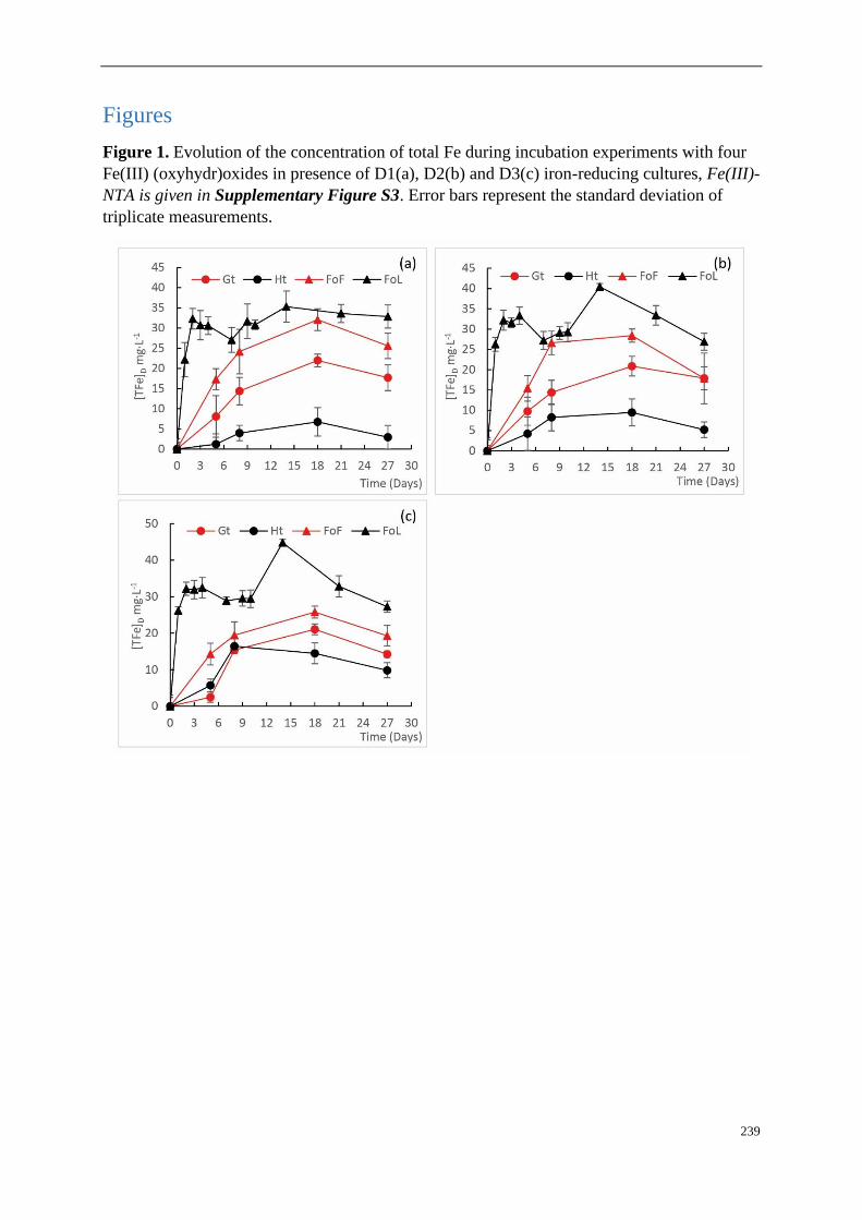
239
Figures
Figure 1. Evolution of the concentration of total Fe during incubation experiments with four
Fe(III) (oxyhydr)oxides in presence of D1(a), D2(b) and D3(c) iron-reducing cultures, Fe(III)-
NTA is given in Supplementary Figure S3. Error bars represent the standard deviation of
triplicate measurements.

240
Figure 2. Final total amount of solubilized Fe from Fe (oxyhydr)oxides: goethite, hematite, FoF
and FoL in presence of D1, D2 and D3 inocula with a mixture of C sources. The letters “a” and
“b” represent the significance of differences (Kruskal-Wallis test at p<0.05) between cultures.
Values in the same Fe (oxyhydr)oxides group are not significantly different from one another.
Error bars represent the standard deviation of triplicate measurements.
Figure 3. nMDS ordination of D1, D2 and D3 community fingerprints applied to a Bray-Curtis
dissimilarity matrix. Plot stress = 0.15. Ht: hematite; Fh: FoF; Lp: FoL; Gt: goethite.

241
Figure 4. Parameters linked to bacterial abundance: (a) Log10 of bacterial 16S rRNA (rrs gene)
copies, (b) Ratio of Shewanella and Geobacter over bacterial 16S rRNA (rrs gene) copies for
all three site samples D1, D2 and D3, (c) Log 10 of Geobacter 16S gene copies, (d) Ratio
Geobacter 16S over bacterial 16S rRNA (rrs gene) copies, (e) Log 10 of Shewanella 16S gene
copies, and (f) Ratio Shewanella 16S over bacterial 16S rRNA (rrs gene) copies. Details of
bacterial, Shewanella and Geobacter 16S rRNA (rrs gene) copies for all three site samples D1,
D2 and D3 are given in Supplementary Figure S6. The letters “a” and “b” differed
significantly (Kruskal-Wallis test at p<0.05) between group of iron oxide for graph “(a), (b),
(c), and (e)”; the small letter, capital letter and Greek letter were used for differing significantly
by group of inocula D1, D2 and D3 for graphs “(d) and (f)”. Data represent average values of
three experimental replicates and their standard deviation (σ) for graph “(b), (d) and (f)”, 3
inocula * 3 replicates for graphs “(a), (c), and (e)”. Ht: hematite; Fh: FoF; Lp: FoL; Gt: goethite.
Error bars represent the standard deviation of triplicate measurements.

242
Tables
Table 1. Characteristics of Fe(III) oxides submitted to Fe-reducing bacteria.
Iron oxide Assumed morphologya Surface areab
(m2g-1)
goethite acicular 11.7
hematite cylinder/rod 31.4
FoF blocky 232
FoL blocky 337 aFor use in estimating mean particle size from morphology by SEM-EDS (TM3000
accompanied by a SwiftED3000 X-Stream module (Hitachi)); bDetermined by multipoint BET
N2 adsorption.

243
Supplementary Material
Supplementary Figures
Supplementary Figure S13. SEM observation on initial Fe (oxyhydr)oxides: Morphology of
FoF (a), hematite (b), FoL (c) and goethite (d)
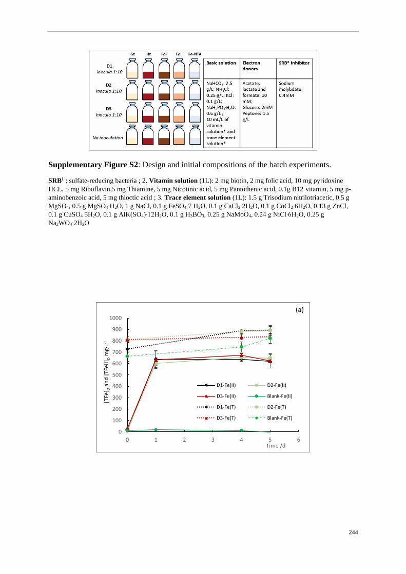
244
Supplementary Figure S2: Design and initial compositions of the batch experiments.
SRB1 : sulfate-reducing bacteria ; 2. Vitamin solution (1L): 2 mg biotin, 2 mg folic acid, 10 mg pyridoxine
HCL, 5 mg Riboflavin,5 mg Thiamine, 5 mg Nicotinic acid, 5 mg Pantothenic acid, 0.1g B12 vitamin, 5 mg p-
aminobenzoic acid, 5 mg thioctic acid ; 3. Trace element solution (1L): 1.5 g Trisodium nitrilotriacetic, 0.5 g
MgSO4, 0.5 g MgSO4∙H2O, 1 g NaCl, 0.1 g FeSO4∙7 H2O, 0.1 g CaCl2∙2H2O, 0.1 g CoCl2∙6H2O, 0.13 g ZnCl,
0.1 g CuSO4 5H2O, 0.1 g AlK(SO4)∙12H2O, 0.1 g H3BO3, 0.25 g NaMoO4, 0.24 g NiCl∙6H2O, 0.25 g
Na2WO4∙2H2O
0
100
200
300
400
500
600
700
800
900
1000
0 1 2 3 4 5 6
[TFe
] Dan
d [
TFeI
I]D
mg·
L-1
Time /d
D1-Fe(II) D2-Fe(II)
D3-Fe(II) Blank-Fe(II)
D1-Fe(T) D2-Fe(T)
D3-Fe(T) Blank-Fe(T)
(a)

245
Supplementary Figure S3. Concentration of Fe(II) and total Fe (FeT) of D1, D2 and D3
incubated on Fe(III)-NTA medium (a), and evolution of the concentration of total Fe during
incubation experiments with four abiotic Fe(III) (oxyhydr)oxides (b). Error bars represent the
standard deviation of triplicate measurements.
Supplementary Figure S4. Fe(III) dissolution of Fe oxides: goethite, hematite, FoF and FoL
in presence of D1, D2 and D3 inocula. The small letter, capital letter and Greek letter were used
for differing significantly (Kruskal-Wallis test at p<0.05) by group of inocula D1, D2 and D3
for different iron oxides. Error bars represent the standard deviation of triplicate measurements.

246
Supplementary Figure S5. CE-SSCP diversity profiles of the site samples (Soil), the Fe(III)-
NTA enrichments (Fe-NTA) and cultures in presence of the four Fe oxides: FoF, FoL, goethite
(Gt) and hematite (Ht). D1: soil from river bank; D2: sediment interface, flooded soil; D3:
sediment under water. SSCP is a fingerprinting approach, fluorescently labeled blunt-ended
PCR products are obtained from the highly variable V3 region of the 16S rRNA (rrs gene),
denatured at high temperature, and then rapidly cooled to form unique single-strand
conformations, which are separated based on their electrophoretic mobility in a polymer-filled
capillary (Delbes et al., 2000). The scales are relative to standards. These profiles present high
diversity with many peaks, with thus the presence of different bacterial strains.
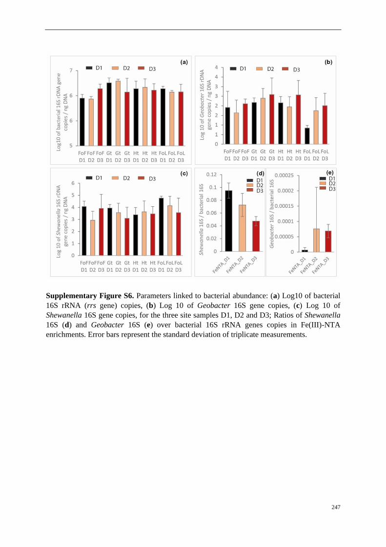
247
Supplementary Figure S6. Parameters linked to bacterial abundance: (a) Log10 of bacterial
16S rRNA (rrs gene) copies, (b) Log 10 of Geobacter 16S gene copies, (c) Log 10 of
Shewanella 16S gene copies, for the three site samples D1, D2 and D3; Ratios of Shewanella
16S (d) and Geobacter 16S (e) over bacterial 16S rRNA genes copies in Fe(III)-NTA
enrichments. Error bars represent the standard deviation of triplicate measurements.

248
Supplementary Figure S7. Principal component analysis (F1 × F2) biplot map generated from
iron solubilisation (percentage and rate of iron solubilisation) and ratios of Shewanella
(Shewanella 16S / bacterial 16S) and Geobacter (Geobacter 16S / bacterial 16S) in the bacterial
community, obtained for the FoF, goethite hematite and FoL incubations with D1, D2 and D3
Shewanella 16S / bacterial 16S
Geobacter 16S / bacterial 16S
Fe solubilisationsolubilisation rate
FoF D1-1
FoF D1-2
FoF D2-1
FoF D2-2
FoF D2-3
FoF D3-2Gt D1-1
Gt D1-2Gt D1-3
Gt D2-1
Gt D2-2
Gt D2-3
Gt D3-1
Gt D3-2Ht D1-1Ht D1-2
Ht D1-3
Ht D2-1Ht D2-2
Ht D2-3
Ht …
FoL D1-1
FoL D1-2FoL D1-3
FoL D2-1 FoL D2-2
FoL D2-3
FoL D3-1FoL D3-2
FoL D3-3
-2
-1
0
1
2
3
4
-2 -1 0 1 2 3 4
F2
(3
0,1
1 %
)
F1 (58,17 %)
Biplot (axes F1 and F2: 88,28 %)
Active variables Active observations
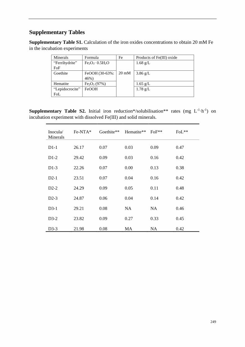
249
Supplementary Tables
Supplementary Table S1. Calculation of the iron oxides concentrations to obtain 20 mM Fe
in the incubation experiments
Minerals Formula Fe Products of Fe(III) oxide
“Ferrihydrite”
FoF
Fe2O3· 0.5H2O
20 mM
1.68 g/L
Goethite FeOOH (30-63%:
46%)
3.86 g/L
Hematite Fe2O3 (97%) 1.65 g/L
“Lepidocrocite”
FoL
FeOOH 1.78 g/L
Supplementary Table S2. Initial iron reduction*/solubilisation** rates (mg L-1·h-1) on
incubation experiment with dissolved Fe(III) and solid minerals.
Inocula/
Minerals
Fe-NTA* Goethite** Hematite** FoF** FoL**
D1-1 26.17 0.07 0.03 0.09 0.47
D1-2 29.42 0.09 0.03 0.16 0.42
D1-3 22.26 0.07 0.00 0.13 0.38
D2-1 23.51 0.07 0.04 0.16 0.42
D2-2 24.29 0.09 0.05 0.11 0.48
D2-3 24.87 0.06 0.04 0.14 0.42
D3-1 29.21 0.08 NA NA 0.46
D3-2 23.82 0.09 0.27 0.33 0.45
D3-3 21.98 0.08 MA NA 0.42

250
Fengfeng ZHANG
Transformation des oxydes de fer par des populations bactériennes mixtes
ferri-réductrices et dynamique des éléments traces associés As, Cr, et Cd
La mobilité des contaminants dans l'environnement, et en particulier celle des éléments traces potentiellement toxiques (ETPT),
tels que l'arsenic, le chrome et le cadmium induisent des risques de contamination des hydrosystèmes et de la chaîne alimentaire.
Comprendre et prévoir la mobilité et la biogéochimie de ces ETPT dans l’environnement permettra de développer et d’appliquer
des stratégies de remédiation appropriées pour les sites pollués. Afin d'évaluer l'impact de l'activité microbiologique et des
transformations géochimiques / minéralogiques des oxydes de fer et d'observer leur effet sur la spéciation et la mobilité de trois
ETPT associés au fer, trois expériences de laboratoire ont été menées : en batch avec des suspensions d’oxydes de fer et des
oxydes fixés sur lames de verre, puis en continu dans des colonnes de percolation. Les résultats ont montré que la minéralogie
de l'oxyde de fer influençait la vitesse de solubilisation bactérienne du fer. Les deux espèces bactériennes ferri-réductrices les
plus étudiées au laboratoire, Shewanella et Geobacter, ont été quantifiées par qPCR. Shewanella a toujours été prépondérante
par rapport à Geobacter dans les communautés bactériennes, et ces deux espèces étaient différentiellement réparties entre les
populations attachées et planctoniques : Geobacter pourrait être moins dépendant du Fe(III) soluble et plus présent dans la
communauté attachée à la surface des oxydes qu’en suspension dans le milieu liquide. De plus, la distribution spatiale des deux
espèces suivies différait dans le système de colonnes alimentées en continu. Dans cette expérience, la bioréduction des oxydes
de fer s’est accompagnée d’une mobilisation de l’As, surtout avec la ferrihydrite. Le Cd n’a été mobilisé que transitoirement,
ce qui suggère des mécanismes d’immobilisation biologiques de cet élément. Aucune mobilisation biologique du Cr n’a été
observée. La production de minéraux secondaires de Fer a été mise en évidence par MEB-EDS et spectroscopie Mössbauer au
cours de la réduction microbienne de la ferrihydrite. Ces résultats permettent de mieux comprendre la dynamique bactérienne
du fer et des ETPT associés dans l'environnement, à travers des conditions expérimentales originales avec des systèmes multi-
polluants et des communautés bactériennes mixtes réductrices de fer.
Mots clés : Oxy-hydroxydes de Fe, réduction microbienne du fer, bactéries ferri-réductrices, Shewanella, Geobacter, MEB-
EDS, Mössbauer, spéciation et mobilité des ETPT, As, Cr, Cd
Transformation of iron oxides in presence of mixed iron-reducing bacterial
communities and dynamics of the associated trace elements As, Cr, and Cd
Contaminants’ mobility in the environment, and in particular mobility of potentially toxic trace elements (PTTE), such as
arsenic, chromium and cadmium induces risks of contamination of hydrosystems and of the food chain. Understanding and
predicting PTTEs’ mobility and biogeochemistry in the environment will help to develop and apply appropriate remediation
strategies for polluted sites. In order to assess the impact of microbiological activity and geochemical/mineralogical
transformations of iron oxides and to observe their effects on the speciation and mobility of three PTTEs associated with iron,
three laboratory experiments were conducted: batch experiments with oxides in slurry, batch with oxides immobilized on slides,
then continuous percolation in columns containing oxides with adsorbed PTTEs. Results showed that the mineralogy of iron
oxides influenced the bacterial iron solubilization rate. The two most studied iron-reducing bacteria (IRB), i.e. Shewanella and
Geobacter, were quantified by qPCR. Considering the two IRB species monitored during this thesis, Shewanella was always
preponderant compared with Geobacter in bacterial communities, and these two species were differentially distributed between
attached and planktonic populations. Geobacter might be less dependent from soluble Fe(III) and more present in the bacterial
community attached to iron oxides than free suspended in the liquid medium. Moreover, the spatial distribution of the two
monitored species differed in column systems. In this experiment, iron oxides bioreduction induced As mobilization,
particularly in presence of ferrihydrite. Cadmium was only transiently mobilized, suggesting mechanisms of biological
immobilization of this element. Chromium was not biologically mobilized. The production of secondary iron minerals was
evidenced my SEM-EDS and Mössbauer spectroscopy during the microbial reduction of ferrihydrite. These results enlighten
the bacterial dynamics of iron and associated PTTE in environment, through original experimental conditions including several
pollutants and mixed iron-reducing bacterial communities.
Key words: Fe (oxyhydr)oxides, microbial iron reduction, IRB, Shewanella, Geobacter, SEM-EDS, Mössbauer, speciation and
mobility of PTTEs, As, Cr, Cd
BRGM - Service géologique national
Institut des Science de la Terre d’Orléans
CNRS/INSU, ISTO, UMR 7327, 45071 Orléans,
France