universidade federal de pernambuco - RI UFPE
127
UNIVERSIDADE FEDERAL DE PERNAMBUCO CENTRO DE CIÊNCIAS BIOLÓGICAS LABORATÓRIO DE IMUNOPATOLOGIA KEIZO-ASAMI PROGRAMA DE PÓS-GRADUAÇÃO EM CIÊNCIAS BIOLÓGICAS PREPARAÇÃO, CARACTERIZAÇÃO E ATIVIDADE BIOLÓGICA DE NANOCÁPSULAS FURTIVAS E SÍTIO ESPECÍFICAS CONTENDO ÁCIDO ÚSNICO PARA O TRATAMENTO DO CÂNCER FÁBIO JOSÉ FIDÉLIS ALMEIDA 2014 Recife – PE
-
Upload
khangminh22 -
Category
Documents
-
view
2 -
download
0
Transcript of universidade federal de pernambuco - RI UFPE

UNIVERSIDADE FEDERAL DE PERNAMBUCO
CENTRO DE CIÊNCIAS BIOLÓGICAS LABORATÓRIO DE IMUNOPATOLOGIA KEIZO-ASAMI
PROGRAMA DE PÓS-GRADUAÇÃO EM CIÊNCIAS BIOLÓGICAS
PREPARAÇÃO, CARACTERIZAÇÃO E ATIVIDADE BIOLÓGICA DE NANOCÁPSULAS FURTIVAS E SÍTIO ESPECÍFICAS CONTENDO ÁCIDO
ÚSNICO PARA O TRATAMENTO DO CÂNCER
FÁBIO JOSÉ FIDÉLIS ALMEIDA
2014 Recife – PE

iv
UNIVERSIDADE FEDERAL DE PERNAMBUCO
CENTRO DE CIÊNCIAS BIOLÓGICAS LABORATÓRIO DE IMUNOPATOLOGIA KEIZO-ASAMI
PROGRAMA DE PÓS-GRADUAÇÃO EM CIÊNCIAS BIOLÓGICAS
PREPARAÇÃO, CARACTERIZAÇÃO E ATIVIDADE BIOLÓGICA DE NANOCÁPSULAS FURTIVAS E SÍTIO ESPECÍFICAS CONTENDO ÁCIDO
ÚSNICO PARA O TRATAMENTO DO CÂNCER
Doutorando: FÁBIO JOSÉ FIDÉLIS ALMEIDA
Orientadora: Profa. Dra. NEREIDE STELA SANTOS MAGALHÃES Co-orientadora: Profa. Dra. NOEMIA PEREIRA DA SILVA SANTOS
2014
Recife – PE
Tese submetida ao Programa de Pós-Graduação em Ciências Biológicas do Centro de Ciências Biológicas da Universidade Federal de Pernambuco, como pré-requisito para obtenção do título de Doutor.

v

vi
PREPARAÇÃO, CARACTERIZAÇÃO E ATIVIDADE BIOLÓGICA DE NANOCÁPSULAS FURTIVAS E SÍTIO ESPECÍFICAS CONTENDO ÁCIDO
ÚSNICO PARA O TRATAMENTO DO CÂNCER
Tese submetida ao Programa de Pós-Graduação em Ciências Biológicas do Centro de Ciências Biológicas da Universidade Federal de Pernambuco, como pré-requisito para
obtenção do título de Doutor
COMISSÃO EXAMINADORA
____________________________________________________________ Profa. Dra. Nereide Stela Santos Magalhães (Orientadora) / UFPE
____________________________________________________________
Prof. Dr. Eryvaldo Sócrates Tabosa do Egito / UFRN
____________________________________________________________ Profa. Dra. Luana Breitenbach Barroso Coelho / UFPE
____________________________________________________________
Profa. Dra. Marcela Silvestre Outtes Wanderley / UPE
____________________________________________________________ Profa. Dra. Mariane Cajubá de Britto Lira Nogueira / UFPE
Defesa de Tese do Doutorando Fábio José Fidélis Almeida, realizada em 26 de fevereiro de 2014, como requisito final para obtenção do títuo de Doutor em Ciências Biológicas
da Universidade Federal de Pernambuco

vii
Dedico esta tese aos meus Pais, Niwton e Luísa, a minha irmã Jéssica e a minha amada esposa Paula, por serem sempre meu alicerce, meu porto seguro, minha fortaleza... Amo muito vocês...

viii AGRADECIMENTOS
Em primeiro lugar, agradeço a Deus pelo seu Amor e por sua presença em minha vida, me ajudando a superar todos os meus momentos de dificuldades; Aos meus pais, Niwton Roberto Almeida da Silva e Luísa Fidélis Almeida pelo carinho e dedicação me ensinando que “exigir faz parte de amar e certamente os frutos de hoje são os resultados das sementes plantadas na infância”; Aos meus avós, Dona Dora, Dona Do Carmo e Seu Chiquinho que já “partiram”, e a meu avô Seu Fidélis, que com certeza sonharam e sonham com esse momento; À minha irmã Jéssica (Jel) e aos meus familiares pelos momentos sinceros de amizade e fraternidade vivenciados; À minha amada Esposa Paula Sales (Minha Linda), pelo amor, companheirismo, compreensão e incentivo (nunca me deixando desistir), tendo ela fundamental importância na composição deste trabalho; Aos meus amigos irmãos, Naldo, Rui, Bruno, Magnus, Lola e Ricardo, pela amizade verdadeira; Aos meus amigos do coração Nathália e Diogo, pelos bons momentos vivenciados; À minha orientadora Profa. Dra. Nereide Stela Santos Magalhães pela oportunidade, confiança e orientações durante toda minha vida científica até o desenvolvimento desse trabalho, sendo ela para mim, um exemplo de profissional dedicada; À Minha co-orientadora Profa. Dra. Noemia Pereira da Silva Santos pela oportunidade, orientação, paciência, tranquilidade e por ter sido peça fundamental na confecção desse trabalho;
v

ix À grande amiga Doutora Milena Sales (Mi...) por toda sua amizade, apoio, dedicação e orientação durante minha iniciação científica, mestrado e doutorado, e pela IMPORTANTÍSSIMA colaboração dada a esse trabalho; Às amigas Professoras Doutoras Marcela Wanderley, Mariane Lira e Isabella Macário pela amizade, paciência e grande ajuda enriquecendo meus conhecimentos; Aos amigos que participam e participaram do Grupo de Sistema de Liberação Controlada de Fármacos (SLC), Taciana, Rafaela, Catarine, Larissa”s”, Laércio, Thiers, Camila, Rebecca, Ana Lygia, Sarah, Lina, Anivaldo, Círia, Marcela, Cássia, Laís, Miguel, Cybelle, João, Hannah, Víctor e Andresa pela amizade e convívio neste período; Aos amigos do LIKA, Monique, Lucas, Ricardo, Adriana, Luíza, Marek, Mari Cabrera por toda colaboração e amizade; Aos Funcionários do LIKA, Maria Helena, Felipe, Edson e Aílton pelo apoio na manutenção dos animais de experimentação no biotério; Otaviano, Rafael, Carmelita e Marina pelo auxílio técnico e a secretária Hilma. Ao Diretor do LIKA, Prof. Dr. José Luiz pelo suporte estrutural que o LIKA nos oferece para desenvolvermos nossos trabalhos; À coordenação do Programa de Pós-Graduação em Ciências Biológicas nas pessoas dos Professores Ranilson Bezerra e Tereza Correia e da Secretária Adenilda (Adê), pelo apoio sempre que necessário; À FACEPE pelo apoio financeiro, através de bolsa de estudos, durante a elaboração desse trabalho; Enfim, a todos que de forma direta e indireta contribuíram para a realização deste trabalho, Muito Obrigado!
vi

x RESUMO
O câncer é considerado um problema de saúde pública mundial e apesar das melhorias no tratamento, a eficácia terapêutica ainda é limitada. O ácido úsnico (AU) é um dos mais bem conhecidos e estudados derivados liquênicos, trata-se de um metabólito secundário liquênico que apresenta diversas atividades biológicas, dentre elas a antitumoral. O presente estudo objetivou desenvolver, caracterizar e avaliar a atividade antitumoral de nanocápsulas de ácido úsnico furtivas e sítio-específicas contendo folato em sua superfície (NC-PEG-FOL/AU). As nanocápsulas foram desenvolvidas pelo método de deposição interfacial de polímero pré-formado. As formulações foram caracterizadas por tamanho de partícula, índice de polidispersão, carga de superfície e eficiência de encapsulação. A atividade antitumoral in vivo do AU foi avaliada em camundongos machos Swiss, onde os animais receberam doses diárias de 15 mg/kg/dia do fármaco em suspensão e encapsulado em NC-PEG-FOL por 7 dias. Os resultados demonstraram que o método de preparação permitiu a formação de nanocápsulas nanométricas (Ø = 157,4 ± 2,97 nm), homogêneas (PDI = 0,27), com carga negativa (ζ = -10.58 ± 0.6 mV) e com uma alta eficiência de encapsulação do fármaco (EE = 98,0 ± 0,28 %). Nos estudos de citotoxicidade, as NC-PEG-FOL/AU exibiram citotoxicidade relevante (IC50 = 2,13 ± 0,02) quando comparada ao fármaco livre (IC50 = 10,04 ± 0,04). Observações em microscopia de fluorescência revelaram que as nanocápsulas sítio-específicas foram capturadas pelas células de Sarcoma-180. A distribuição da fluorescência nas células variou com o tempo. Nos estudos in vivo a atividade antitumoral do AU foi confirmada com inibição do crescimento tumoral em 40%. No entanto, a encapsulação em nanocápsulas furtivas e sítio-específicas potencializou a ação do AU resultando em inibição da massa tumoral acima de 80%. As nanocápsulas folato-específicas contendo ácido úsnico foram desenvolvidas como uma nova alternativa de aplicação destes sistemas para liberação seletiva do AU em células cancerígenas que expressam o receptor do folato, potencializando a ação do fármaco, visando uma futura aplicação terapêutica. Palavras-Chaves: Ácido úsnico, nanocápsulas sítio-específicas, PLGA-PEG, folato, atividade antitumoral.
vii

xi ABSTRACT
Cancer is considered a worldwide public health problem and despite improvements in treatment, the therapeutic efficacy is still limited. The usnic acid (UA), one of the most well known and studied compounds of natural origin, is a secondary metabolite liquenic that presents several biological activities, such as antitumor. The goals of this study were to develop, characterize and evaluate the antitumor activity of usnic acid (UA) encapsulated into targeted nanocapsules (UA/FOL-PEG-NC) and develop a simple, rapid and specific analytical method for quantification of UA in rat plasma by high performance liquid chromatography efficiency. Nanocapsules were developed by interfacial deposition of preformed polymer method. Formulations were characterized by particle size, polydispersity index, surface charge and encapsulation efficiency. The in vivo antitumor activity of the UA was evaluated in male Swiss mice, where animals received daily doses of 15 mg / kg / day of the drug in suspension and encapsulated in FOL-PEG-NC for 7 days. Results showed that the method of preparation allowed the formation of nanometric nanocapsules (Ø = 157.4 ± 2.97 nm) in homogeneous dispersion (PDI = 0.27), negatively surface charged (ζ = -10.58 ± 0.6 mV) and high drug encapsulation efficiency (EE = 98.0 ± 0.28 %). Concerning the cytotoxicity study, UA/FOL-PEG-NC exhibited relevant activity on Sarcoma-180 cell (IC50 of 3.67 ± 0.08) compared to the free drug (IC50 = 11.35 ± 0.32). Fluorescence microscopy revealed the uptake of targeted nanocapsules by Sarcoma-180 cells. The antitumor activity of the UA was confirmed by inhibition of tumor growth by 40%. However, the encapsulation in targeted nanocapsules increased the UA action, resulting in an inhibition of tumor mass above 80%. The folate-specific nanocapsules containing usnic acid have been developed as a new alternative for application of these systems for selective release of the UA in cancer cells that expressing folate receptor, potentiating the action of the drug, seeking a future therapeutic application. Keywords: Usnic acid, targeted nanocapsules, PLGA-PEG, folate, antitumor activity.
viii

xii LISTA DE FIGURAS
REVISÃO BIBLIOGRÁFICA Figura 1. Distribuição percentual dos dez tipos de câncer mais incidentes estimados para 2013 por sexo, exceto câncer de pele não melanoma.
16
Figura 2. Estrutura química do ácido úsnico. 18 Figura 3. Estrutura do ácido úsnico: (A) aspecto macroscópico em forma de pó com coloração amarelada e (B) aspecto microscópico na forma de cristais.
19
Figura 4. Nanocarreadores atualmente descritos em estudos pré-clínicos e clínicos.
27
Figura 5. Perfil farmacocinético de doses múltiplas ou em sistema de liberação controlada de fármacos.
28
Figura 6. Representação esquemática das nanopartículas poliméricas: a) fármaco dissolvido no núcleo oleoso das nanocápsulas; b) fármaco adsorvido à parede polimérica das nanocápsulas; c) fármaco retido na matriz polimérica das nanoesferas; d) fármaco adsorvido ou disperso molecularmente na matriz polimérica das nanoesferas.
29
Figura 7. Representação estrutural do PLGA, com os subprodutos do seu metabolismo (ácido láctico e ácido glicólico).
30
Figura 8. Representação esquemática de uma nanocápsula polimérica contendo cadeias de polietilenoglicol em sua superfície (furtiva).
31
Figura 9. Representação esquemática de uma nanopartícula sítio-específica PEGuilada.
32
Figura 10. Estrutura química do ácido fólico. 33 Figura 11. Endocitose de conjugados fármaco-folato. Os conjugados fármaco-folato exógenos ligam-se especificamente aos receptores de folato (RF) presentes na superfície das células alvo. A membrana plasmática sofre invaginação circundando o complexo conjugado/RF para formar uma vesícula intracelular (endossomo). Como o lúmem do endossomo acidifica. Os RF sofrem mudanças em sua conformação e liberam o conjugado no citosol.
34
ix

xiii CAPÍTULO 1 Figura 1. Número citações científicas a respeito do ácido úsnico no decorrer dos anos, baseando-se na busca no banco de pesquisa científica internacional, Scopus (www.scopus.com) e tendo como palavra-chave: Usnic Acid.
49
Figura 2. Estrutura química do ácido úsnico. 50 Figura 3. Via da biossíntese do ácido úsnico. 51 Figura 4. Percentual de publicações das atividades biológicas do ácido úsnico [Scopus (www.scopus.com) palavras-chave: usnic acid e as respectivas atividades].
54
CAPÍTULO 2 Figure 1. Chemical structure of usnic acid. 79 Figure 2. Cytotoxicity of free UA (■) and targeted nanocapsules (●) on Sarcoma-180 cancer cells (A) and Macrophages J774 healthy cells (B) after 48 h of incubation. Results are mean values ± SD of cell viability percentage (n = 3).
85
Figure 3. Fluorescence microscopy Sarcoma-180 incubated with fluorescent nanocapsules at different incubation time (0.5, 1, 3 and 24 h).
87
Figure 4. Evaluation of antitumor activity of free and nanoencapsulated usnic acid against Sarcoma 180: UA= usnic acid in suspension and UA/FOL-PEG-NC = usnic acid encapsulated into targeted nanocapsules.
89
Figure 5. Overall aspect of solid tumors (Sarcoma-180): UA/FOL-PEG-NC, UA free and control, respectively.
89
Figure 6. Histopathological analysis (HE, 400 ×) of tumor of animals treated: A) Control group (untreated animals); B) UA and C) UA/FOL-PEG-NC. Arrows indicate necrosis areas.
90
CAPÍTULO 3 Fig. 1. Representative HPLC Chromatograms for (A) the control plasma and (B) the plasma spiked with usnic acid in different concentrations.
103
Fig. 2. Blood levels of intraperitoneal injected usnic acid delivered in solution (■) and UA/FOL-PEG-NC (●) after i.p. administration. The values plotted are mean ± SD
107
x

xiv LISTA DE TABELAS
CAPÍTULO 1 Tabela 1. Cepas de bactérias suscetíveis ao ácido úsnico, de acordo com busca no banco de pesquisa científica internacional, Scopus (www.scopus.com).
56
Tabela 2. Linhagens de células cancerígenas suscetíveis ao ácido úsnico 58 CAPÍTULO 2 Table 1. IC50 values of free UA and targeted nanocapsules on Sarcoma-180 after 48 h of incubation.
86
Table 2. Analysis of blood of animals treated with free and encapsulated usnic acid in targeted nanocapsules.
91
CAPÍTULO 3 Table 1 System suitability results of the proposed analytical method for usnic acid in plasma.
104
Table 2 Linearity of the HPLC method for usnic acid determination in rat plasma. 105 Table 3 Accuracy for the quantification of usnic acid in rat plasma. 105 Table 4 Precision evaluation of the HPLC method for quantifying usnic acid in rat plasma.
106
Table 5 Comparative pharmacokinetic parameters of UA delivered in solution or through UA/FOL-PEG-NC after intraperitoneal injection.
108
xi

xv LISTA DE ABREVIATURAS E SIGLAS
ATP – adenosina trifosfato AU – ácido úsnico; EE – eficiência de encapsulação FOL – folato; NC – nanocápsulas; NC-PEG-FOL – nanocápsulas folato-específicas; NC-PEG-FOL/AU – nanocápsulas folato-específicas contendo ácido úsnico; PDI – índice de polidispersão; PEG – polietilenoglicol; PLGA – copolímero de ácido láctico e glicólico; RF – receptor de folato; SFM – sistema fagocitário mononuclear; UV – ultravioleta; Via i.p. – via intraperitoneal.
xii

xvi SUMÁRIO
RESUMO vii ABSTRACT viii LISTA DE FIGURAS ix LISTA DE TABELAS xi LISTA DE ABREVIATURAS E SIGLAS xii 1. INTRODUÇÃO 14 2. REVISÃO DE LITERATURA 16
2.1 – Câncer 16 2.1.1 – Tratamentos do câncer 17
2.2 – Ácido Úsnico 18 2.2.1 – Propriedades químicas 18 2.2.2 – Propriedades biológicas 19
2.2.3 – Mecanismo de ação e Toxicologia 24 2.3 – Nanotecnologia 26 2.4 – Sistemas de Liberação Controlada de Fármacos 26
2.4.1 - Nanopatículas poliméricas 28 2.4.2 - Nanopatículas furtivas 30 2.4.3 - Nanopatículas sítio-específicas 31
2.4.3.1 – Liberação de fármacos mediada por receptores de folato 32 3. REFERÊNCIAS 35 4. OBJETIVOS 45 5. CAPÍTULO 1: Ácido úsnico: De um antigo derivado liquênico a promissoras aplicações biológicas e nanotecnológicas
46
6. CAPÍTULO 2: Preparation and characterization of nanocapsules folate-specific containing usnic acid
76
7. CAPÍTULO 3: Pharmacokinetic of usnic acid loaded in folate-targeted nanocapsules 97 8. CONCLUSÕES 112 9. PERSPECTIVAS 113 10. ANEXOS 114

14 1. INTRODUÇÃO
Câncer é um termo genérico dado a um conjunto de mais de 100 doenças que tem em comum o crescimento desordenado de células que invadem os tecidos e órgãos. Esta é uma das doenças que mais atinge a população sendo considerado um problema de saúde pública mundial. A quimioterapia tornou-se um componente integrante do tratamento anticancerígeno. Atualmente, diferentes agentes quimioterápicos são utilizados de forma eficaz para a terapia anticancerígena, contudo, os agentes quimioterápicos convencionais ainda exibem baixa especificidade em atingir o tecido tumoral, além de frequentemente serem restritos por uma toxicidade dose-limitante. Desta forma, essa patologia tem estimulado os pesquisadores a buscar novos agentes anticancerígenos como uma estratégia importante para o estabelecimento de terapias alternativas (GU et al., 2007; PARHI; MOHANTY; SAHOO, 2012).
A fim de descobrir novas alternativas para a terapia do câncer, as substâncias naturais vêm recebendo uma atenção especial, dentre essas substâncias, o ácido úsnico, é um dos mais bem conhecidos e estudados, destacando-se por apresentar várias atividades biológicas, incluindo a atividade antitumoral (SANTOS et al., 2006). Porém, o ácido úsnico apresenta algumas características desfavoráveis, tais como baixa solubilidade em água e uma elevada hepatotoxicidade (INGÓLFSDÓTTIR, 2002; PRAMYOTHIN et al., 2004). Dessa maneira, a comunidade científica tem se empenhado no sentido de minimizar este problema sem perder a eficácia deste composto. Nesse contexto, os estudos impulsionaram o desenvolvimento de alternativas inovadoras, a exemplo da encapsulação do ácido úsnico em sistemas de liberação controlada, os quais são atrativas ferramentas da nanotecnologia farmacêutica.
Atualmente define-se como sistemas de liberação controlada, aqueles os quais o agente ativo é liberado com velocidade constante, mantendo constante a concentração plasmática do fármaco dentro da faixa terapêutica. São frequentemente descritos como “drug delivery systems” e oferecem diversas vantagens em relação às formas de dosagens convencionais, tais como: protegem certos princípios ativos lábeis da degradação e/ou inativação, melhoram a biodisponibilidade dos mesmos, aumentam a penetração celular de fármacos, além de reduzir os efeitos toxicológicos (PANYAM; LABHASETWAR, 2003).
Existe uma variedade de nanocarreadores descrito na literatura que apresentam diferentes tipos de aplicações. Dentre estes sistemas estão as nanocápsulas, as quais são constituídas por uma parede polimérica disposta ao redor de um núcleo oleoso, podendo o fármaco estar dissolvido neste núcleo e/ou adsorvido à parede polimérica (KUMARI; YADAV; YADAV, 2010).

15 O principal obstáculo para a utilização das nanocápsulas convencionais se deve a rápida
remoção destes nanocarredores do organismo pelo sistema fagocitário mononuclear (SFM), contudo as nanocápsulas podem ser revestidas com moléculas que alterem sua hidrofobicidade, proporcionando uma camada hidrofílica na superfície. O método mais comum para a modificação de superfície é a utilização de um polímero hidrofílico e não iônico, como o polietilenoglicol (PEG), um processo chamado “PEGuilação” (MOSQUEIRA et al., 2001).
Na tentativa de aumentar a especificidade de interação dos nanossistemas com células alvo e elevar a quantidade do fármaco liberado nestas células, a pesquisa neste campo foi focada no desenvolvimento de nanopartículas sítio-específicas. Estes utilizam ligantes acoplados em sua superfície, que conferem seletividade para distribuir o fármaco encapsulado no sítio de ação desejado proporcionando uma maior seletividade e eficácia em comparação com o direcionamento passivo (SAPRA; ALLEN, 2003).
Uma das mais extensivamente estudadas moléculas alvo para entrega de fármacos sítio-específicos é o ácido fólico (vitamina B9 ou folato) (LEAMON; LOW, 2001). O receptor de folato (RF) está presente em praticamente todas as células, porém é expresso em níveis elevados principalmente em células cancerígenas, tais como, cânceres epiteliais de ovário, colo-retal, mama, próstata, pulmão, nariz, garganta, cérebro e leucemia (SUDIMACK; LEE, 2000; LEAMON; LOW, 2001; ALEXIS et al., 2008).
Vários tipos de carreadores de fármacos têm sido conjugados ao folato, incluindo lipossomas, micelas e nanopartículas poliméricas. Para o direcionamento dos lipossomas e nanopartículas, uma cadeia de PEG ligante é frequentemente requerida. Múltiplas moléculas de folato são conjugadas a cada partícula, que permite uma elevada afinidade de interação com os RF (ZHAO; LI; LEE, 2008). Além disso, sistemas de liberação de fármacos conjugados com folato demonstraram uma alta citotoxicidade e captura celular de células cancerígenas que expressam o RF (YOO et al., 2004; ESMAELLI et al., 2008; NIE et al., 2009; SHMEEDA et al., 2010; LIANG et al., 2011; SAXENA; NAGUIB; HUSSAIN, 2012). Portanto, o desenvolvimento de sistemas de liberação controlada de fármacos, furtivos e sítios específicos, capazes de viabilizar a administração do ácido úsnico em uma formulação que melhore sua solubilidade, bem como, aperfeiçoe a dose terapêutica diminuindo os efeitos tóxicos e direcione o fármaco para o tumor aumentando a eficácia terapêutica, torna-se uma inovação útil no desenvolvimento de alternativas para o tratamento do câncer.

16 2. REVISÃO BIBLIOGRÁFICA
2.1 - Câncer Câncer é o nome conferido ao conjunto de doenças caracterizadas pela interrupção dos
mecanismos que regulam o crescimento e a divisão celular e pela habilidade das células cancerosas de invadirem outros tecidos (ROBBINS et al., 2000). É uma das doenças mais devastadoras a qual envolve várias alterações genéticas e anormalidades celulares. Esta complexidade e heterogeneidade promovem o crescimento agressivo de células cancerígenas que levam à morbidade e mortalidade em diversos pacientes (PARHI; MOHANTY; SAHOO, 2012).
Mais de 11 milhões de pessoas são diagnosticadas com câncer a cada ano, e esta doença é responsável por cerca de 7 milhões de mortes anuais (12,5% das mortes em todo o mundo), tornando esta doença um problema de saúde publica mundial (PARCK et al., 2008). De acordo com a Organização Mundial da Saúde (OMS), estima-se que em 2030 haja 27 milhões de casos incidentes de câncer, 17 milhões de mortes por câncer e 75 milhões de pessoas vivas portadoras de algum tipo de neoplasia maligna. (INCA, 2011).
No Brasil, as estimativas para o ano de 2014 apontam a ocorrência de aproximadamente 518.510 casos novos de câncer. Estima-se que o câncer de pele do tipo não melanoma (134 mil novos casos) será o mais incidente na população brasileira, seguido pelos tumores de próstata, pulmão, cólon e reto e estômago para o sexo masculino; e mama, colo do útero, cólon e reto e glândula tireoide para o sexo feminino (Figura 1) (INCA, 2011). Figura 1. Distribuição percentual dos dez tipos de câncer mais incidentes estimados para 2013 por sexo, exceto câncer de pele não melanoma.
Fonte: INCA, 2011.

17 2.1.1 - Tratamentos para o câncer A remoção cirúrgica de um tumor e dos tecidos circunvizinhos afetados é considerado o
procedimento primário para tumores grandes o suficiente para serem manipulados. Entretanto, dificilmente a cirurgia é suficiente e, geralmente, é inevitável a permanência de células residuais afetadas. Além da cirurgia, outros recursos terapêuticos podem ser usados como a quimioterapia (WANG et al., 2008).
A quimioterapia tornou-se um componente integrante do tratamento anticancerígeno. Atualmente, diferentes agentes quimioterápicos são utilizados de forma eficaz para a terapia anticâncer, contudo, apesar dos últimos 30 anos de esforço da oncologia em descobrir novos fármacos, os agentes quimioterápicos convencionais ainda exibem baixa especificidade em atingir o tecido tumoral, além de frequentemente serem restritos por uma toxicidade dose-limitante (GU et al., 2007; PARHI; MOHANTY; SAHOO, 2012).
A quimioterapia depende de vários fatores como os fármacos e as doses utilizadas, a via de administração e a condição do paciente. Além disso, durante o percurso até o tumor, o fármaco pode interagir com outros tecidos e ser metabolizado pelo organismo, comprometendo a sua biodisponibilidade no tumor e, por conseguinte, sua eficácia (JAIN, 2001; OOYAMA et al., 2008).
Apesar das melhorias no tratamento quimioterapêutico do câncer, os regimes existentes que usam agentes citotóxicos clássicos apresentam algumas limitações, que inclui um índice terapêutico estreito que não permite muitas vezes a administração de uma quantidade suficiente de fármaco a fim induzir a resposta pretendida. Além disso, a resposta à quimioterapia varia significativamente entre os pacientes, portanto, uma evolução da metodologia tradicional, baseada na necessidade de aumentar o índice terapêutico dos quimioterápicos e diminuir a toxicidade em células normais, terapia personalizada, está sendo buscada (HARLEY et al., 2008; NISHIYAMA et al., 2009).
A fim de se obter uma terapia eficaz, é necessário aprimorar o conhecimento sobre a fisiopatologia do câncer, descobrir novos fármacos, e desenvolver novas tecnologias biomédicas. Atualmente, a terapia anticancerígena tornou-se uma abordagem multidisciplinar a qual requer uma estreita colaboração entre os clínicos, os pesquisadores e os engenheiros biomédicos (DANHIER et al., 2012).
Existem diversos medicamentos aprovados para uso na terapia anticancerígena, porém a resistência ao tratamento e os efeitos colaterais são os maiores obstáculos de uma quimioterapia bem-sucedida. Essa resistência resulta em uma resposta terapêutica incompleta, recorrente e que muitas vezes acarreta metástase (WANG et al., 2011). Numerosos antineoplásicos exibem alta

18 citotoxicidade não-seletiva e baixo índice terapêutico, o que têm impulsionado pesquisas não apenas para o desenvolvimento de novos fármacos, mas também novas formas inovadoras para otimizar a utilização dos fármacos já existentes (JULIANO; DAOUD, 1990; PETRO; DE SIMONE, 2010).
2.2Ácido Úsnico 2.2.1 - Propriedades químicas Entre os vários compostos de origem natural, o ácido úsnico, [2,6-diacetil-7,9-dihidroxi-8-
9b-dimetil-1,3(2H,9β/αH)-dibenzofurandiona; C18H16O7, PM = 344.32] (Figura 2), é um dos mais bem conhecidos e estudados. Caracteriza-se por ser uma substância cristalina de pigmentação amarela (Figura 3), ocorrendo na natureza em duas formas enatioméricas (-) e (+) decorrente da projeção angular do grupamento metil localizado na posição 9b (COCCHIETTO et al., 2002; INGÓLFSDÓTTIR, 2002).
Figura 2. Estrutura química do ácido úsnico.
Trata-se de um metabólito secundário produzido por líquens (INGÓLFSDÓTTIR, 2002). Na natureza o ácido úsnico está presente principalmente nas seguintes espécies de liquens: Cladonia (cladoniaceae), Usnea (usneaceae), Lecanora (lecanoraceae), Ramalina (ramalinaceae) e Parmelia (parmeliaceae) (COCCHIETTO et al., 2002).

19 Figura 3. Estrutura do ácido úsnico: (A) aspecto macroscópico em forma de pó com coloração
amarelada e (B) aspecto microscópico na forma de cristais.
Fonte: LIRA, 2007
Este derivado liquênico apresenta caráter hidrofóbico, com solubilidade em água menor que 10 mg/100 mL a 25 °C (INGÓLFSDÓTTIR, 2002), sendo parcialmente solúvel em etanol e facilmente em éter quente, acetona, benzeno e clorofórmio (ASAHINA & SHIBATA, 1954; TAKAI & UEHARA, 1979). Tal característica hidrofóbica pode ser explicada pela presença dos três grupos cetônicos, bem como ao anel furano que une os anéis aromáticos (ASAHINA & SHIBATA, 1954), além de pontes de hidrogênio intramoleculares (MÜLLER, 2001). Sua acidez é justificada pela presença do anel fenólico, cuja estrutura é instável (SHIBATA, 2000). O ponto de fusão do ácido úsnico é em torno de 204 °C e o peso molecular de 344.32 g/mol (INDEX MERCK, 1995).
2.2.2 - Propriedades biológicas O acido úsnico costumava ser bastante utilizado na medicina popular no tratamento de
micoses, alívio na dor de garganta e dente, febre, com ação anti-séptica e cicatrizante (INGÓLFSDÓTTIR, 2002). Além disso, várias pesquisas tem confirmado potenciais atividades do ácido úsnico frente a terapias, tais como: antimicrobiana, antitumoral, antiviral, antiparasitária, antifúngica, gastroprotetora, cicatrizante, antioxidante, anti-inflamatóriaa, analgésica e antipirética (LAUTERWEIN et al., 1995; FRANCOLINI et al., 2004; SEGATORE et al., 2012; POMPILIO et al., 2013; BURLANDO et al., 2009; EINARSDÓTTIR et al., 2010; BRISDELLI et al., 2013; CAMPANELLA et al., 2002; SOKOLOV et al., 2012b; SUSSMANN et al., 2001; PIRES et al.,

20 2011; ODABASOGLU et al., 2006; BRUNO et al., 2013; ZHIJUN et al., 2011; KOHLHARDT-FLOEHR et al., 2010; BEHERA et al., 2012).
Antitumoral A atividade antitumoral do ácido úsnico foi revelada há quase quatro décadas. Em 1975,
Kupchan e Kopperman publicaram o primeiro estudo contra o carcinoma pulmonar de Lewis. Desde então, em estudos in vitro, o ácido úsnico demostrou sua ação antitumoral frente a diferentes linhagens celulares, tais como queratinócitos humanos (HaCaT) (KUMAR & MÜLLER, 1999; PEREIRA, 1994; LIMA, 1990; BURLANDO et al., 2009), células de câncer de mama (MCF7 e MDAMB231) e de câncer de pulmão (H1299 e NCIH 292) (MAYER et al., 2005; SANTOS et al., 2005). Quando incorporado à β-ciclodextrina, demonstrou sua atividade antiproliferativa contra células malignas da linhagem K-562 (leucemia) (CAMPANELLA et al., 2002), T-47-D (câncer de mama), Panc1 (câncer de pâncreas) e PC-3 (câncer de próstata) (KRISTMUNDSDÓTTIR et al., 2005). É importante notificar que a atividade antiproliferativa do ácido úsnico foi observada contra uma linhagem modelo para estudo dos efeitos de compostos citotóxicos de câncer humano, o carcinoma epidermóide da vulva (A431), e ainda contra uma linhagem de tumor agressivo e resistente à quimioterapia e letal, mesotelioma maligno (MM98) (BURLANDO et al., 2009).
A viabilidade celular in vitro do hepatoblastoma humano HepG2 mostrou ser comprometida com concentrações de 5 µM do ácido úsnico além de, apresentar a LC50 de 30 µM. No mesmo estudo, a exposição dessas células ao fármaco, principalmente em concentrações acima de 20 µM, resultou em um aumento significativo da atividade do citocromo P450, do estresse oxidativo e da disfunção mitocondrial comprovando a toxicidade do ácido úsnico frente a essas linhagens celulares (SAHU et al., 2011). Já em 2012, os mesmos pesquisadores, ao associarem o ácido úsnico a um lipopolissacarídeos, observaram um aumento no efeito tóxico frente a HepG2 (SAHU et al., 2012).
Além dos estudos realizados com o ácido úsnico isolado, vários autores compararam a atividade desse ácido com outros metabólitos liquênicos, e logo atribuíram um maior potencial antitumoral do ácido úsnico contra A2780 e HT-29 (BACKOROVÁ et al., 2012), MCF-7, HeLa e HCT-116 (BRISDELLI et al., 2013), e UACC-62 e B16-F10 (BRANDÃO et al., 2013).
Ademais, os efeitos anticancerígenos das duas formas enantioméricas do ácido úsnico foram investigados por Einarsdóttir et al (2010) os quais concluíram que ambas as formas apresentaram efeito inibitório sobre o crescimento e proliferação das células T-47D (câncer de mama) e Capan-2 (câncer de pâncreas).

21 Antimicrobiana A descoberta do ácido úsnico ocorreu mediante a busca por novos compostos antibióticos
(ABRANHAN & FLOREY, 1949; BUSTINZA, 1951), tendo suas primeiras descrições contra o Streptococcus mutans (agente etiológico da cárie dental e de doenças periodontais) (GRASSO et al., 1989) e os gêneros Escherichia, Salmonella e Shigella (HALE-Jr, 1983; LAUTERWEIN et al., 1995).
Adicionalmente, o ácido úsnico vem sendo utilizado como alternativa contra cepas resistentes aos antibióticos preconizados, como por exemplo, Enterococos resistentes à vancomicina (ERV) e Staphylococcus aureus resistente à meticilina (SARM) (ELO et al., 2007). Ainda sobre estas cepas, Pompilio e colaboradoes em 2013, além de comprovarem a atividade antimicrobiana do ácido úsnico, identificaram a sua ação antibiofilme.
A resistência à antibioticoterapia é um dos principais problemas no combate à tuberculose. Dessa forma, o ácido úsnico torna-se um importante candidato no combate a esta infecção. Assim, o ácido úsnico foi testado contra as cepas susceptíveis e resistentes a isoniazida, estreptomicina e rifampicina, fármacos usados na terapia atual, demonstrando que não ocorreu resistência cruzada, sugerindo que o mecanismo de ação do acido úsnico seja diferente dos fármacos atualmente usados na terapia da Tuberculose (RAMOS e DA SILVA, 2010), fator que viabiliza sua utilização contra o Mycobacterium tuberculosis.
Além das cepas acima relacionadas, a Helicobacter pylori demonstrou ser susceptível à ação o ácido úsnico, o qual apresentou uma forte atividade dose-dependente. Em um estudo posterior, Luo et al (2011) comprovou essa propriedade do ácido úsnico extraído de Nephromopsis pallescens, obtendo resultados semelhantes a ampicilina e eritromicina, fármacos que possuem ação contra H. pylori comprovada.
Antiviral Inicialmente, o ácido úsnico foi testado contra o Eptein-Barr, Papilomavírus Humano e o
Poliomavírus de ratos (YAMAMOTO et al., 1995; SCIRPA et al., 1999; CAMPANELLA et al., 2002). Em 1995, Yamamoto e colaboradores testaram ambas as formas enantioméricas do ácido úsnico, contra o Eptein-Barr vírus, e observaram que a forma dextrogira apresentou maior atividade antiviral em relação à levogira.
A ação contra o HPV foi observada no tratamento adjuvante da infecção associado com o sulfato de zinco. Neste estudo foi demonstrado que além da ação antiviral, o ácido úsnico favoreceu

22 a reepitelização do tecido lesado. Dessa maneira, possibilitou a terapia adjuvante no tratamento cirúrgico desobstrutivo e, em particular, da lesão causada pelo HPV (SCIRPA et al., 1999). Além disso, o ácido úsnico foi capaz de inibir a proliferação do Poliomavírus de ratos, fato este ocorrido pela destruição do DNA viral, através da inibição da transcrição do RNA (CAMPANELLA et al., 2002).
Recentemente foi avaliada a atividade do ácido úsnico e seus derivados (sinteticamente modificados) contra vírus da influenza H1N1 em células MDCK, cujos resultados sugerem que essas moléculas sejam substâncias antigripe (SOKOLOV et al., 2012b).
Antiparasitária Outra atividade atribuída ao ácido úsnico foi a sua propriedade antiparasitária. Os primeiros
relatos voltados a este foco foram descritos por Wu e colaboradores (1995), quando detectaram a atividade do ácido úsnico contra o Trichomonas vaginalis em estudos in vitro.
Em 2001, Sussmann e colaboradores, impulsionados pela resistência parasitária aos medicamentos convencionais, testaram o ácido úsnico frente ao Plasmodium falciparum. Nesta busca por novas moléculas alvo, os pesquisadores descobriram no ácido úsnico uma importante ação contra o parasita uma vez que ele inibiu de forma reversível a biossíntese de vitamina E, uma importante molécula para o desenvolvimento do parasita.
Estudos da avaliação da atividade in vitro do ácido úsnico extraído da Cladonia substellata contra o protozoário Trypanosoma cruzi apontaram o potencial uso desse composto na doença de Chagas, uma vez que, este metabólito foi efetivo em várias concentrações (10 a 50 mg / mL) frente às formas epimastigotas, tripomastigotas e amastigotas causando danos a mitocôndrias e cinetoplasto (DE CARVALHO et al., 2005).
Antifúngica A atividade antifúngica do ácido úsnico também foi descoberta nos anos 50, quando se
observou a inibição do fungo Tricophyton mentagrophytes após tratamento com o referido ácido (BUSTINZA, 1951). Em 1996, Broska e colaboradores observaram inibição no crescimento do Penicillum fraquentans e Verticillium albo-atrum, após tratamento com ácido úsnico.
Atualmente, o pequeno número de medicamentos disponíveis para o tratamento de fungos incentiva a busca de novos agentes quimioterápicos. Para tal propósito, o ácido úsnico foi testado contra a Candida orthopsilosis and C. parapsilosis, porém nenhuma ação frente ao biofilme formado por estas leveduras foi observada (PIRES et al., 2011).

23 Gastoprotetor Alguns estudos indicam que o ácido úsnico, isolado da Usnea longissima, vem sendo testado
no tratamento da úlcera gástrica em animais. Induzida pela indometacina, lesões gástricas foram significativamente reduzidas por todas as doses utilizadas de ácido úsnico (25, 50, 100 e 200 mg/kg de peso corporal), quando comparado com o grupo tratado com a ranitidina (25 mg/kg de peso corporal), fármaco referência. Esse efeito gastroprotetor do ácido úsnico pode ser atribuído ao seu efeito redutor contra o dano oxidativo e seu efeito inibitório na infiltração neutrofílica em estômago de ratos (ODABASOGLU et al., 2006).
Cicatrizante, Antioxidante e Anti-inflamatória A cicatrização é um processo preocupante em algumas situações como pós-cirurgia e
queimaduras. Para que a regeneração epitelial seja agilizada, é necessário o auxílio de terapia farmacológica que tanto inviabilize a ação microbiana, bem como favoreça a reepitelização. Desta forma, o ácido úsnico tem sido muito usado em preparações dermatológicas e cosméticas, devido suas atividades bacteriostática e antioxidante. Recentemente, Bruno e colaboradores 2013, avaliaram as propriedades de reparação de feridas de derivados do ácido úsnico em ensaios in vitro e in vivo, indicando menor citotoxicidade a células da pele combinados com melhor desempenho de cura, sugerindo a possibilidade de utilização destes compostos na cicatrização de feridas e preparações anti-idade para a pele.
Além disso, a ação cicatrizante do ácido úsnico também é favorecida pelo potencial anti-inflamatório deste composto liquênico. Zhijun e colaboradores (2011) observaram que tal propriedade decorre do downregulation de alguns mediadores da inflamação tais como iNOS, COX-2, IL-1β, IL-6 e TNF-α, COX-2, além da superexpressão da interleucina-10 e da hemeoxigenase-1.
Ainda tais achados vincularam o ácido úsnico como um potente bloqueador solar, tanto pela sua absorção em regiões ultravioleta (UV), como pelo poder antioxidante. Estudos indicaram que, alguns líquens sob estímulo das radiações UV, sintetizam seus metabólitos com uma forte absorção na região UV, gerando uma proteção própria contra radiações perigosas. Dentre esses metabólitos, o ácido úsnico teve um fator de proteção similar à substância comercial sintética usada como referência, exibindo valores de fator de proteção UV iguais (3,6-5,0) nos testes in vivo e superiores (4,03-4,83) que a substância referência (2,66-3,63) nos testes in vitro (RANCAN et al., 2002). Estudos mais recentes mostraram que este mesmo composto, agora obtido da Xanthoparmelia farinosa apresentou potente atividade antioxidante e pró-oxidante (comportamento bifuncional) em

24 linhagens celulares de linfócitos humanos (Jurkat-cells: E 6-1 leucemia aguda) sob irradiação UV-B (KOHLHARDT-FLOEHR et al., 2010).
2.2.3 – Mecanismo de ação e Toxicologia
Apesar das atraentes propriedades farmacológicas, o ácido úsnico é uma molécula tóxica. Uma vez que o seu mecanismo de ação não está completamente elucidado, a explicação para sua toxicidade também não é totalmente compreendida. Dessa maneira, a comunidade científica tem se empenhado para desvendar os mecanismos bioquímicos e moleculares envolvidos tanto nas atividades biológicas inerentes ao ácido úsnico, bem como na sua toxicidade.
Neste viés, diversos modelos de estudo (in silico, in vitro e in vivo) relacionam o mecanismo de ação do ácido úsnico na interrupção da função mitocondrial e alterações no estresse oxidativo (SANTOS et al., 2003; PRAMYOTHIN e t al., 2004; HAN et al., 2004; LIU et al., 2012).
Tendo em vista que a via mitocondrial é importante na regulação das etapas que decorrem para morte celular, sugere-se que este seja o principal mecanismo que reduz a viabilidade de células cancerígenas, sendo este um importante ponto que justifica a ação antitumoral. Adicionalmente, o estresse oxidativo e o rompimento do processo metabólico normal causado pelo ácido úsnico em células cancerígenas, também podem está diretamente relacionado à redução da síntese de RNA (AL-BEKAIRI et al., 1991), mas não envolvidos com danos ao DNA (MAYER et al., 2005).
É importante mencionar que, como o ácido úsnico é um ácido fraco de característica lipofílica, sua difusão pela membrana mitocondrial é favorecida acarretando inibição da produção de ATP na fosforilação oxidativa. Segundo Santos (2003) essa hipótese explica a sua atraente atividade antimicrobiana uma vez que, este mecanismo não permite que o microrganismo execute os processos anabólicos imprescindíveis ao seu crescimento.
Apesar dos relatos acima, a influência do ácido úsnico na respiração celular foi inicialmente citada na década de 50, quando Johnson e colaboradores (1950) descreveram o relevante o efeito na respiração e o desacoplamento da fosforilação oxidativa, em homogenato de rins e fígado de rato, com concentrações mínimas de 1,3-2,6 µg/ml. Desde então, estudiosos vem compartilhando achados que ressaltaram essa hipótese. Vavasseur e colaboradores (1991) evidenciaram o potencial inibitório do ácido úsnico sobre processos respiratórios aeróbicos em células vegetais de Cormmelina communis. Abo-Khatwa e colaboradores (1996) relataram que o ácido úsnico, o vulpínico e a atranorina apresentaram tal ação quando testados em mitocôndria de fígado de ratos.

25 Estes compostos atuaram no interior da membrana mitocondrial com características semelhantes ao do 2,4-dinitrofenol, substância desacopladora padrão. Em seguida, Santos (2003) reportou que, em concentrações de 32 µM, o ácido úsnico pôde inibir completamente a produção de ATP mitocondrial. No ano seguinte, Pramyothin e colaboradores (2004) constataram que o (+) ácido úsnico age alterando a integridade da membrana celular permitindo a liberação de enzimas hepatoespecíficas (aspartato aminotransferase e alanina aminotransferase), além de causar destruição da função mitocondrial. Diante disto, os dados expostos sugerem que o dano oxidativo esteja relacionado à toxicidade deste composto liquênico, principalmente em células hepáticas.
Ainda neste contexto, ensaios em modelos murinos avaliaram a hepatoxicidade do ácido úsnico, quando se observou 98 e 100% de necrose tecidual após tratamento de cultura de hepatócitos com 5 mM e 10 mM, respectivamente. Além disso, foi observada uma redução de até 90% nos níveis de ATP e a inibição da respiração mitocondrial. Os autores deste estudo identificaram uma inibição direta da função mitocondrial, que levaria à diminuição do consumo de oxigênio pela cadeia transportadora de elétrons, e consequentemente à morte celular (HAN et al., 2004).
Em humanos a hepatotoxicidade do ácido úsnico vêm sendo objeto de muitos relatos, uma vez que causam desde uma hepatite aguda à falência hepática (NEFF et al., 2004; BUNCHORNTAVAKUL, REDDY, 2012), além de dermatites alérgicas (PACHECO et al., 2012). Em anos anteriores, o dano hepatocelular foi divulgado quando indivíduos que consumiram LipoKinetix®, um suplemento dietético que continha o ácido úsnico como componente, e apresentaram falência aguda do fígado (NEFF et al., 2004).
Diante destes achados, sugere-se que ação de desacoplador da fosforilação oxidativa inicialmente sugerida na década de 50 para o ácido úsnico, vêm sendo confirmada através de diferentes estudos (in silico, in vitro e in vivo). Desta forma, os maiores impedimentos com relação a sua introdução na terapêutica são sua baixa solubilidade em água, consequentemente nos líquidos biológicos, e seus efeitos hepatotóxicos. Estas dificuldades para a aplicação terapêutica do ácido úsnico podem ser superadas pela sua nanoencapsulação em sistemas de liberação controlada para diminuição da toxicidade e aumento da eficácia terapêutica. Estudos realizados demonstraram, através de análises bioquímicas e histopatológicas, que há uma redução da toxicidade do ácido úsnico quando este é inserido em sistemas de liberação controlada de fármacos, tais como, nanocápsulas (SANTOS et al., 2006).

26 2.3 - Nanotecnologia A nanotecnologia é uma área da ciência dedicada ao design, construção, e utilização de
estruturas funcionais em escala nanométrica. Colocando este intervalo de tamanho em perspectiva, uma pequena molécula, um vírus, uma bactéria, e uma secção transversal de um cabelo humano são em torno de 1, 100, 100, e 100.000 nm, respectivamente (ALEXIS et al., 2008, PARCK et al., 2008).
Numerosas aplicações para a nanotecnologia, dentre elas, tratamento, diagnóstico, monitoramento e controle dos sistemas biológicos tem recentemente sido referidos como “nanomedicina” pelo Instituto Nacional de Saúde (PARK et al., 2008). A nanomedicina é um dos ramos mais promissores da medicina contemporânea, retendo boa parte dos esforços científicos na busca de novos tratamentos para doenças como câncer e doenças infecciosas (PISON et al., 2006; KAGAN; BAYIR; SHVEDOVA, 2005).
A nanotecnologia aplicada ao câncer é um novo campo da pesquisa interdisciplinar permeando através da biologia, química, engenharia e medicina, objetivando levar maiores avanços para o diagnóstico e tratamento do câncer (WANG et al., 2010). Atualmente, existe um grande foco na utilização de sistemas de liberação controlada de fármacos como uma evolução da terapia tradicional anticancerígena (PARK et al., 2008).
2.4 - Sistemas de Liberação Controlada de Fármacos A tecnologia de liberação controlada de fármacos surgiu na década de 60, como um método
comercialmente atrativo para administração do princípio ativo com liberação controlada, ou seja, com velocidade constante. Daí em diante, a liberação pôde ser manipulada para a produção de sistemas de liberação controlada ou prolongada da substância bioativa (SWARBRICK, 1996).
Atualmente definem-se como sistemas de liberação controlada, aqueles os quais o agente ativo é liberado com velocidade constante, mantendo constante a concentração plasmática do fármaco dentro da faixa terapêutica. Frequentemente descritos como “drug delivery systems”, eles oferecem diversas vantagens em relação às formas de dosagens convencionais, tais como: protegem certos princípios ativos lábeis da degradação e/ou inativação pelo suco gástrico, melhoram a biodisponibilidade dos mesmos, aumentam a penetração celular de fármacos, além de reduzir os efeitos tóxicos (PANYAM; LABHASETWAR, 2003).

Existe uma variedade de nanocarreadores descrito na lit
tipos de aplicações. Dentre os principais vetores utilizados como sistemas de liberação controlada, destacam-se os sistemas poliméricos como nanopartículas (nanoesferas e nanocápsulas), micelas poliméricas constituídas de polímeros anfifílicos associados em solução aquosa, além de outros sistemas, como os lipossomas, que são vesículas com núcleo aquoso envolvido por uma bicamada lipídica, e dendrímeros que representam séries repetidas de compostos macromoleculares que formam uma cavidade em seu interior (Figura 2010).
Figura 4. Nanocarreadores
Fonte: Adaptado de DANHIER; FERON; PRÉAT, 2010.Os sistemas de liberação controlada (
constante e dentro da faixa terapêutica a concentração sanguínea de uma determinada substância (fármaco), assegurando uma maior biodisponibilidade e reduzir os efeitos colaterais, assim, a adesão do paciente ao tratamento com um menor número de dosagens requeridas (MORAHUERTAS et al., 2010).
Existe uma variedade de nanocarreadores descrito na literatura que apresentam diferentes tipos de aplicações. Dentre os principais vetores utilizados como sistemas de liberação controlada,
se os sistemas poliméricos como nanopartículas (nanoesferas e nanocápsulas), micelas polímeros anfifílicos associados em solução aquosa, além de outros
sistemas, como os lipossomas, que são vesículas com núcleo aquoso envolvido por uma bicamada lipídica, e dendrímeros que representam séries repetidas de compostos macromoleculares que
interior (Figura 4) (RAWAT, 2006; DANHIER; FERON; PRÉAT,
anocarreadores atualmente descritos em estudos pré-clínicos e clínicos.
Fonte: Adaptado de DANHIER; FERON; PRÉAT, 2010.
Os sistemas de liberação controlada (Figura 5) apresentam dois objetivos principais: manter constante e dentro da faixa terapêutica a concentração sanguínea de uma determinada substância fármaco), assegurando uma maior biodisponibilidade e reduzir os efeitos colaterais,
adesão do paciente ao tratamento com um menor número de dosagens requeridas (MORA
27 eratura que apresentam diferentes
tipos de aplicações. Dentre os principais vetores utilizados como sistemas de liberação controlada, se os sistemas poliméricos como nanopartículas (nanoesferas e nanocápsulas), micelas
polímeros anfifílicos associados em solução aquosa, além de outros sistemas, como os lipossomas, que são vesículas com núcleo aquoso envolvido por uma bicamada lipídica, e dendrímeros que representam séries repetidas de compostos macromoleculares que
) (RAWAT, 2006; DANHIER; FERON; PRÉAT,
clínicos e clínicos.
Fonte: Adaptado de DANHIER; FERON; PRÉAT, 2010.
) apresentam dois objetivos principais: manter constante e dentro da faixa terapêutica a concentração sanguínea de uma determinada substância fármaco), assegurando uma maior biodisponibilidade e reduzir os efeitos colaterais, aumentando,
adesão do paciente ao tratamento com um menor número de dosagens requeridas (MORA-

28 Figura 5. Perfil farmacocinético de doses múltiplas ou em sistema de liberação controlada de
fármacos.
Fonte: LIRA, 2009.
Um sistema ideal de liberação controlada de fármacos deve ser capaz de direcionar fármacos para o sítio alvo desejado, com a mínima exposição dos demais tecidos não desejados. Esse sistema de liberação de fármacos, por si só, deve ser farmacologicamente inativo, ter toxicidade mínima, ser prontamente metabolizado e depurado da circulação após ter exercido sua função. Além de ser confortável para o paciente, simples de se administrar e remover, fácil de fabricar e esterilizar (FORSSEN; WILLIS, 1998; ZHOU et al., 2002).
2.4.1 - Nanopartículas poliméricas
Nanopartículas são estruturas esféricas e sólidas em uma escala de tamanho de 10 a 1000 nm de diâmetro, e refere-se a dois tipos de estruturas diferentes, nanoesferas e nanocápsulas, cada uma com suas características, as quais diferem entre si segundo a composição e organização estrutural (SCHAFFAZICK et al., 2003). As nanocápsulas são constituídas por um invólucro polimérico disposto ao redor de um núcleo oleoso, podendo o fármaco estar dissolvido neste núcleo e/ou adsorvido à parede polimérica. Por outro lado, as nanoesferas, que não apresentam óleo em sua composição, são formadas por uma matriz polimérica, onde o fármaco pode ficar retido ou adsorvido (Figura 6) (SOPPIMATH et al., 2001; KUMARI; YADAV; YADAV, 2010).

Figura 6. Representação esquemática núcleo oleoso das nanocápsulas; b) fármaco adsorvido à parede polimérica das nanocápsulas; c)
fármaco retido na matriz polimérica das nanoesferas; d) fármaco adsorvido ou disperso molecularmente na ma
Fonte: SCHAFFAZICK As nanopartículas poliméricas liberam
ou erosão controlada, a partir de um núcleo através damembrana de revestimento atua difusividade do fármaco na membrana poliméricamesmo. Além disso, a taxa de liberação também pode ser afetadafármaco e os excipientes da formulação. Havendo a intpara formar um complexo menos solúvel em água, com um pequeno efeito de liberação
Para desenvolver um sistema biodegradação polimérica, são fatores considerados importantes. Em geral, a taxa de liberação do fármaco depende (1) da solubilidade adsorvido, (3) na difusão do fármacoprocesso de erosão/difusão (MOHANRAJ; CHEN, 2006).
O poli(ácido láctico-co-glicólico) (PLGA) biodegradáveis mais utilizados na preparação das nanopartículas, porque a sua hidrólise leva aos monômeros ácido láctico e ácido glicólico, os quais são facilmente metabolizados corpo (DANHIER et al., 2012). aprovado pela Administração de Alimentos e Fármacos dos EUA (FDA/US) e pela Agência de
Representação esquemática das nanopartículas poliméricas: a) fármaco dissolvido no eoso das nanocápsulas; b) fármaco adsorvido à parede polimérica das nanocápsulas; c)
fármaco retido na matriz polimérica das nanoesferas; d) fármaco adsorvido ou disperso molecularmente na matriz polimérica das nanoesferas.
Fonte: SCHAFFAZICK et al., 2003.
As nanopartículas poliméricas liberam o medicamento por meio do mecanismo de da, a partir de um núcleo através da membrana ou matriz
membrana de revestimento atua como uma barreira para liberação, portanto, a a membrana polimérica tornam-se fatores determinante
taxa de liberação também pode ser afetada pela interaexcipientes da formulação. Havendo a interação do fármaco com um dos excipientes
para formar um complexo menos solúvel em água, a liberação do mesmo pode ser muito lentde liberação rápida inicial (efeito burst) (MUDSHINGE et al., 2011).
Para desenvolver um sistema nanopartículado eficaz, ambos, o fármaco liberado e a biodegradação polimérica, são fatores considerados importantes. Em geral, a taxa de liberação do
a solubilidade do fármaco, (2) da dessorção na superfície ligada ou fármaco do fármaco através da matriz das nanopartículas, e
difusão (MOHANRAJ; CHEN, 2006). glicólico) (PLGA) (Figura 7) é um dos polímeros
zados na preparação das nanopartículas, porque a sua hidrólise leva aos monômeros ácido láctico e ácido glicólico, os quais são facilmente metabolizados
Devido à mínima toxicidade associada ao PLGA, este políAdministração de Alimentos e Fármacos dos EUA (FDA/US) e pela Agência de
29 poliméricas: a) fármaco dissolvido no
eoso das nanocápsulas; b) fármaco adsorvido à parede polimérica das nanocápsulas; c) fármaco retido na matriz polimérica das nanoesferas; d) fármaco adsorvido ou disperso
por meio do mecanismo de difusão membrana ou matriz polimérica. A
, portanto, a solubilidade e determinantes na liberação do
interação iônica entre o eração do fármaco com um dos excipientes
pode ser muito lenta, e ) (MUDSHINGE et al., 2011).
ambos, o fármaco liberado e a biodegradação polimérica, são fatores considerados importantes. Em geral, a taxa de liberação do
na superfície ligada ou fármaco (4) da combinação do
é um dos polímeros biocompatíveis e zados na preparação das nanopartículas, porque a sua hidrólise leva aos
monômeros ácido láctico e ácido glicólico, os quais são facilmente metabolizados e eliminados pelo Devido à mínima toxicidade associada ao PLGA, este polímero é
Administração de Alimentos e Fármacos dos EUA (FDA/US) e pela Agência de

30 Medicina Européia (EMA) para seu uso em vários sistemas de liberação de fármacos em humanos (KUMARI; YADAV; YADAV, 2010).
Figura 7. Representação estrutural do PLGA, com os subprodutos do seu metabolismo (ácido
láctico e ácido glicólico).
Fonte: Adaptado de DANHIER et al., 2012
2.4.2 - Nanopartículas furtivas
O principal obstáculo ao uso das nanopartículas poliméricas nos tratamentos de enfermidades é a sua rápida remoção do organismo pelo sistema fagocitário mononuclear (SFM). Os macrófagos do SFM são capazes de remover nanopartículas desprotegidas da circulação sanguínea segundos após a administração intravenosa, tornando-as ineficazes como dispositivos de liberação de fármaco órgão-específico (GREF et al., 1995). Porém, esses macrófagos não podem identificar diretamente as nanopartículas, mas reconhecem opsoninas específicas ligadas a superfície dessas partículas. Com efeito, uma vez na corrente sanguínea, nanopartículas de superfície não modificadas (nanopartículas convencionais) são rapidamente opsonizadas e fagocitadas pelo SFM (SINGH, R.; LILLARD JR, 2009; KUMARI; YADAV; YADAV, 2010; DANHIER et al., 2012).
Vários métodos de modificações superficiais têm sido desenvolvidos para produzir nanopartículas que não sejam reconhecidos pelo SFM. As nanopartículas podem ser revestidas com moléculas que alterem sua hidrofobicidade, proporcionando uma camada hidrofílica na superfície. O método mais comum para a modificação de superfície é a utilização de um polímero hidrofílico e não-iônico, como o polietilenoglicol (PEG), um processo chamado “PEGuilação” (MOSQUEIRA et al., 2001) (Figura 8).

31 Figura 8. Representação esquemática de uma nanocápsula polimérica contendo cadeias de
polietilenoglicol em sua superfície (furtiva).
Para contribuir com as características “stealth” das nanopartículas PEGuiladas, existem três fatores importantes: (i) o peso molecular da cadeia do PEG, (ii) a densidade das cadeias de superfície e (iii) a conformação. Com a criação de uma camada hidrofílica protetora em torno das nanopartículas, as forças de repulsão estéricas repelem a absorção das proteínas opsoninas, desse modo bloqueando e atrasando o processo de opsonização. Amplamente, tem sido demonstrado que a "PEGuilação" aumenta o tempo de meia-vida das nanopartículas na circulação sanguínea (OWENS; PEPPAS, 2006).
Além disso, estudos relatam que as nanopartículas poliméricas que apresentam circulação prolongada nos vasos sanguíneos, acumulam-se passivamente nos tumores, sugerindo a existência de um mecanismo passivo de retenção e a potencialidade desses sistemas nanoestruturados em tratamentos para o câncer (PARK et al., 2008).
2.4.3 - Nanopartículas sítio-específicas
Na tentativa de aumentar a especificidade de interação dos nanossistemas com células alvo e elevar a quantidade do fármaco liberado nestas células, a pesquisa neste campo foi focada no desenvolvimento de lipossomas e nanopartículas sítio-específicas. Estes utilizam ligantes acoplados em sua superfície, que conferem seletividade para distribuir o fármaco encapsulado no sítio de ação desejado, proporcionando uma maior seletividade e eficácia em comparação com o direcionamento passivo (SAPRA; ALLEN, 2003).
No direcionamento ativo, os ligantes sítio-específicos são acoplados à superfície do nanocarreador (Figura 9) para a ligação a receptores adequados expressos no local alvo. O ligante é escolhido para se ligar a um receptor super expresso por células tumorais e menos expresso por
Lipofílico

células normais. Além disso, os receptoresas células-alvo (DANHIER et al., 2010).
Figura 9. Representação esquemática de uma nanopartícula sítio
Alguns exemplos de ligantes de reconhecimento são os anticorpos, polissacarídeos, peptídeos, proteínas virais e lectinas, que são ligados covalentemente a superfície dos nanossistemas a fim de carrear os fármacos para o local específico de ação (BATISTA et al., 2007; EDWABAEUMNER, 2006).
2.4.3.1 - Liberação de fármacos mediada por receptores de folato
Uma das mais extensivamente estudadasespecíficos é o ácido fólico (vitamina B9 ou molecular (Mw = 441 Da) requerida pelas células eucarióticas carbono no metabolismo dos ácido
A estrutura química do ácido fólico está demonstrada na Figura grupamentos ácidos carboxílicos posicionados nas extremidades distais da molécula, a permeabilidade passiva através da membrana é mínima. Para contornar este obstáculo, a naturezadesenvolveu dois mecanismos para a internalização celularenvolve uma proteína de membrana que transporta folato diretamente para o citosol celular. O segundo mecanismo utiliza um receptor glicoprotéico, geralmente refe
. Além disso, os receptores-alvo devem ser expressos de forma homogênea em todas (DANHIER et al., 2010).
Representação esquemática de uma nanopartícula sítio-específica PEGuilada.
Fonte: ALEXIS et al., 2008.
de ligantes de reconhecimento são os anticorpos, polissacarídeos, proteínas virais e lectinas, que são ligados covalentemente a superfície dos nanossistemas
os fármacos para o local específico de ação (BATISTA et al., 2007; EDWA
Liberação de fármacos mediada por receptores de folato is extensivamente estudadas molécula alvo para entrega de
vitamina B9 ou folato). O ácido fólico é uma vitaminarequerida pelas células eucarióticas como coenzima na transferência de
ácidos nucléicos e dos aminoácidos (LEAMON; A estrutura química do ácido fólico está demonstrada na Figura
grupamentos ácidos carboxílicos posicionados nas extremidades distais da molécula, a permeabilidade passiva através da membrana é mínima. Para contornar este obstáculo, a natureza
mecanismos para a internalização celular desta vitamina. O primeiro mecanismo envolve uma proteína de membrana que transporta folato diretamente para o citosol celular. O segundo mecanismo utiliza um receptor glicoprotéico, geralmente referido como receptor de folato
32 de forma homogênea em todas
específica PEGuilada.
de ligantes de reconhecimento são os anticorpos, polissacarídeos, proteínas virais e lectinas, que são ligados covalentemente a superfície dos nanossistemas
os fármacos para o local específico de ação (BATISTA et al., 2007; EDWARDS;
molécula alvo para entrega de fármacos sítio-folato). O ácido fólico é uma vitamina de baixo peso
como coenzima na transferência de (LEAMON; LOW, 2001).
A estrutura química do ácido fólico está demonstrada na Figura 10. Devido a dois grupamentos ácidos carboxílicos posicionados nas extremidades distais da molécula, a permeabilidade passiva através da membrana é mínima. Para contornar este obstáculo, a natureza
O primeiro mecanismo envolve uma proteína de membrana que transporta folato diretamente para o citosol celular. O
rido como receptor de folato

(RF), que preferencialmente medeia (Figura 11) (LEAMON; LOW, 2001; PARK et al
Figura
A proteína transportadora de folato está presente em praticamente todas as células, enquanto que o RF é expresso em níveis elevados principalmente em células cancerígenas, tais como, cânceres epiteliais de ovário, cololeucemia (SUDIMACK; LEE , 2000; LEAMON Vários tipos de carreadores de fármacos têm sido conjugados ao folato, incluindo lipossomas, micelas e nanopartículas poliméricas. Parnanopartículas, uma cadeia de PEG ligante é frequentemente requerida. Múltiplas moléculas de folato são conjugadas a cada partícula, (ZHAO; LI; LEE, 2008). Além demonstraram uma alta citotoxicidade e captura celular de célulasRF (YOO et al., 2004; ESMAELLI et al., 2008; NIE et al., 2009; SHMEEDA et al., 2010; et al., 2011; SAXENA; NAGUIB; HUSSAIN, 2012).
edeia à captura do folato para dentro das células por (LEAMON; LOW, 2001; PARK et al., 2008).
Figura 10. Estrutura química do ácido fólico.
Fonte: LEAMON; LOW, 2001.
A proteína transportadora de folato está presente em praticamente todas as células, enquanto é expresso em níveis elevados principalmente em células cancerígenas, tais como,
de ovário, colo-retal, mama, próstata, pulmão, nariz, garganta, cérebro e leucemia (SUDIMACK; LEE , 2000; LEAMON; LOW, 2001; ALEXIS et al., 2008).
Vários tipos de carreadores de fármacos têm sido conjugados ao folato, incluindo lipossomas, micelas e nanopartículas poliméricas. Para o direcionamento dos lipossomas e nanopartículas, uma cadeia de PEG ligante é frequentemente requerida. Múltiplas moléculas de folato são conjugadas a cada partícula, o que permite uma elevada afinidade de interação com os RF
disso, sistemas de liberação de fármacos conjugados com folato demonstraram uma alta citotoxicidade e captura celular de células cancerígenas que expressam o
(YOO et al., 2004; ESMAELLI et al., 2008; NIE et al., 2009; SHMEEDA et al., 2010; SAXENA; NAGUIB; HUSSAIN, 2012).
33 captura do folato para dentro das células por endocitose
A proteína transportadora de folato está presente em praticamente todas as células, enquanto é expresso em níveis elevados principalmente em células cancerígenas, tais como,
, próstata, pulmão, nariz, garganta, cérebro e ; LOW, 2001; ALEXIS et al., 2008).
Vários tipos de carreadores de fármacos têm sido conjugados ao folato, incluindo a o direcionamento dos lipossomas e
nanopartículas, uma cadeia de PEG ligante é frequentemente requerida. Múltiplas moléculas de afinidade de interação com os RF
disso, sistemas de liberação de fármacos conjugados com folato cancerígenas que expressam o
(YOO et al., 2004; ESMAELLI et al., 2008; NIE et al., 2009; SHMEEDA et al., 2010; LIANG

Figura 11. Endocitose de conjugados fármaco
ligam-se especificamente aos receptores de folato (RFmembrana plasmática sofre invaginação ci
vesícula intracelular (endossomo). Como o lúmudanças em sua conformação e liberam o conjugado no citosol.
Portanto, o desenvolvimento de sistemas de libersítios específicos, capazes de viabilizar a administração dmelhore sua solubilidade, bem como, aperfeiçoe a dose terapêutica diminuindo os direcione o fármaco para o tumor aumentando a eficácia terapêutica, tornadesenvolvimento de alternativas para o tratamento do câncer.
de conjugados fármaco-folato. Os conjugados fármaconte aos receptores de folato (RF) presentes na superfície das células alvo. A sofre invaginação circundando o complexo conjugado/
vesícula intracelular (endossomo). Como o lúmem do endossomo acidifica. Os mudanças em sua conformação e liberam o conjugado no citosol.
Fonte: LEAMON, LOW, 2001. o desenvolvimento de sistemas de liberação controlada de fármacoscapazes de viabilizar a administração do ácido úsnico em uma formulação que
melhore sua solubilidade, bem como, aperfeiçoe a dose terapêutica diminuindo os direcione o fármaco para o tumor aumentando a eficácia terapêutica, torna-se uma inovação útil no desenvolvimento de alternativas para o tratamento do câncer.
34 folato. Os conjugados fármaco-folato exógenos
) presentes na superfície das células alvo. A rcundando o complexo conjugado/RF para formar uma
mem do endossomo acidifica. Os RF sofrem mudanças em sua conformação e liberam o conjugado no citosol.
ação controlada de fármacos, furtivos e em uma formulação que
melhore sua solubilidade, bem como, aperfeiçoe a dose terapêutica diminuindo os efeitos tóxicos e se uma inovação útil no

35 3. REFERÊNCIAS ABO-KHATWA, A. N.; AL-ROBAI, A. A.; AL-JAWHARI, D. A. Lichen acids as uncouplers of oxidative phosphorylation of mouse-liver mitochondria. Natural Toxins, v.4, p.96-102, 1996. ABRAHAN, E. P.; FLOREY, H. W. Antimicrobial substances from lichens and algae. In: Antibiotic. v. 1,cap. 13, 1949. p. 566-575. AL-BEKAIRI, A. M.; QURESHI, S.; CHAUDHRY, M. A.; KRISHNA, D. R.; SHAH, A. H. Mitodepressive, clastogenic and biochemical effect of (+)-usnic acid in mice. Journal of Ethnopharmacology, v. 33, p. 217-220, 1991. ALEXIS, F. et al. New frontiers in nanotechnology for cancer treatment. Urologic Oncology: Seminars and Original Investigations, v.26, p.74–85, 2008. ASAHINA & SHIBATA. Chemistry of Lichen substances. Japan Society for Promotion of Science, p.240, 1954. BACKOROVÁ, M.; JENDZˇELOVSKY´, R.; KELLO, M.; BACˇKOR, M.; MIKEŠ, J.; FEDOROCˇKO, P. Lichen secondary metabolites are responsible for induction of apoptosis in HT-29 and A2780 human cancer cell lines. Toxicology in Vitro, v. 26, p. 462–468, 2012. BATISTA, C.M.; CARVALHO, C.M.B.; SANTOS-MAGALHÃES, N.S. Lipossomas e suas aplicações terapêuticas: Estado da arte. Revista Brasileira de Ciências Farmacêuticas, v. 43, p 167-179, 2007. BEHERA B.C.; MAHADIK, N.; MOREY. M. Antioxidative and cardiovascular-protective activities of metabolite usnic acid and psoromic acid produced by lichen species Usnea complanata under submerged fermentation. Pharmaceutical Biology, v. 50, p. 968-979, 2012. BRANDÃO, L.F.G.; ALCANTARA, G.B.; MATOS, M.F.C.; BOGO, D.; FREITAS, D.S.; OYAMA, N.M.; HONDA, N.K. Cytotoxic Evaluation of Phenolic Compounds from Lichens against Melanoma Cells. Chem. Pharm. Bull. v.61(2), p. 176-183, 2013. BRISDELLI, F.; PERILLI, M.; SELLITRI, D.; PIOVANO, M.; GARBARINO, J.A.; NICOLETTI, M.; BOZZI, A.; AMICOSANTE, G.; CELENZA, G. Cytotoxic Activity and Antioxidant Capacity of Purified Lichen Metabolites: An In Vitro Study. Phytotherapy Research, v. 27, p. 431-437, 2013. BROSKA, B.; STURDIKOVA, M.; PRONAYOVA, N. A.; LIPTAJ, T. (-)-Usnic acid and its derivatives. Their inhibition of fungal growth and enzyme activity. Pharmazie, v. 51, n. 3, p. 195-196, 1996.

36 BRUNO, M.; TRUCCHI, B.; BURLANDO, B.; RANZATO, E.; MARTINOTTI, S.; AKKOL, E.K.; SÜNTAR, I.; KELES, H.; VEROTTA, L. (+)-Usnic acid enamines with remarkable cicatrizing properties. Bioorganic & Medicinal Chemistry, v. 21, p. 1834-1843, 2013. BUNCHORNTAVAKUL, C.; REDDY, K.R. Review article: herbal and dietary supplement hepatotoxicity. Alimentary Pharmacology and Therapeutics, v. 37, p. 3-17, 2012. BURLANDO B.; RANZATO, E.; VOLANTE, A.; APPENDINO, G.; POLLASTRO, F.; VEROTTA, L. Antiproliferative Effects on Tumour Cells and Promotion of KeratinocyteWound Healing by Different Lichen Compounds. Planta Med, v. 75, p. 607-613, 2009. BUSTINZA, F. Antibacterial substances from lichens. Endeavour, p. 95-99, 1951. CAMPANELLA, L.; DELFINI, M.; ERCOLE, P.; IACOANGELI, A.; RISULEO, G. Molecular characterization and action of usnic acid: a drug that inhibits proliferations of mouse polyomavirus in vitro and whose main target is RNA transcription. Biochemistry, v.84, p. 329-334, 2002. COCCHIETTO, M.; SKERT, N.; NIMIS, P. L.; SAVA, G. A review on usnic acid, an interesting natural compound. Naturwissenschaften, v. 89, p. 137-146, 2002. DANHIER, F. et al. PLGA-based nanoparticles: An overview of biomedical applications. Journal Controlled Release. v.161, p.505-522, 2012. DANHIER, F.; FERON. O.; PRÉAT, V. To exploit the tumor microenvironment: Passive and active tumor targeting of nanocarriers for anti-cancer drug delivery. Journal of Controlled Release, v. 148, p.135–146, 2010. DE CARVALHO E. A. B.; ANDRADE, P.P.; SILVA, N.H.; PEREIRA, E.C.; FIGUEIREDO, R. C. B. Q. Effect of usnic acid from the lichen Cladonia substellata on Trypanosoma cruzi in vitro: an ultrastructural study. Mícron, v.36, p.155-161, 2005. EDWARDS, K. A.; BAEUMNER, A. J. Liposomes in analyses. Talanta, v. 68, p.1432-1441, 2006. EINARSDÓTTIR, E.; GROENEWEG, J.; BJÖRNSDÓTTIR, G.G.; HARÐARDOTTIR, G.; OMARSDÓTTIR, S.; INGÓLFSDÓTTIR, K.; ÖGMUNDSDÓTTIR, H.M. Cellular Mechanisms of the Anticancer Effects of the Lichen Compound Usnic Acid. Planta Med, v. 76, p. 969-974, 2010. ELO, H.; MATIKAINEN, J.; PELTTARI, E. Potent activity of the lichen antibiotic (+)-usnic acid against clinical isolates of vancomycin-resistant enterococci and methicillin-resistant Staphylococcus aureus. Naturwissenschaften, v. 94, p. 465–468, 2007.

37 ESMAEILI, F., et al. Folate-receptor-targeted delivery of docetaxel nanoparticles prepared by PLGA-PEG-folate conjugate. Journal of Drug Target, v.16, p.415–423, 2008. FORSSEN, E.; WILLIS, M. Ligand-targeted lipossomes, Advanced Drug Delivery Reviews, v. 29, p. 249-271, 1998. FRANCOLINI, P.; NORRIS, A.; PIOZZI, G.; DONELLI, P.; STOODLEY, P. Usnic acid, a natural antimicrobial agent able to inhibit bacterial biofilm formation on polymer surfaces. Antimicrob Agents Chemother, v.48, n.11, p.4360-4365, 2004. GREF, R.; DOMB, A.; QUELLEC, P.; BLUNK, T.; MULLER, R. H.; VERBAVATZ, J. M.; LANGER, R. The controlled intravenous delivery of drugs using PEG-coated sterically stabilized nanospheres. Advanced Drug Delivery Reviews, v. 16, p. 215-233, 1995. GU, F.X. et al. Targeted nanoparticles for cancer therapy. Nanotoday. v. 2, p. 14-21, 2007. HALE-JR., M. E. The Biology of Lichens. 3ed. London. Edward Arnold Pub., 1983, 90p. HALEY, B.; FRENKEL, E. Nanoparticles for drug delivery in cancer treatment. Urologic Oncology. v. 26, p. 57-64, 2008. HAN, D.; MATSUMARU, K.; RETTORI, D.; KAPLOWITZ, N. Usnic acid-induced necrosis of cultured mouse hepatocytes: inhibition of mitochondrial function and oxidative stress. Biochemical Pharmacology, v.67, p.439-451, 2004. INDEX MERCK. An enciclopedia of chemicals, drugs and biological. 11th ed., Rahway Merck & Co., 1606p., 1989. INGÓLFSDÓTTIR, K. Usnic acid. Phytochemistry, v.61, p.729-736, 2002. INSTITUTO NACIONAL DE CÂNCER José Alencar Gomes da Silva. Coordenação Geral de Ações Estratégicas. Coordenação de Prevenção e Vigilância. Estimativa 2012: incidência de câncer no Brasil. Rio de Janeiro: Inca, 2011.118 p. JAIN, R.K. Delivery of molecular medicine to solid tumors: lessons from in vivo imaging of gene expression and function. Journal Controlled Release, v.74, p.7-25, 2001. JOHNSON, R. B.; FELDOT, G.; LARDY, H. A. The mode of action of the antibiotic usnic acid. Ach. Biochem., v.28, p.317-323, 1950. JULIANO, R.L.; DAOUD, S.S. Liposomes as a delivery system for membrane-active antitumor drugs. Journal of Controlled Release, v. 11, p.225–232, 1990.

38 KAGAN, V. E.; BAYIR, H.; SHVEDOVA, A. A. Nanomedicine and nanotoxicology: two sides of the same coin. Nanomedicine, v.1, p. 313-316, 2005. KOHLHARDT-FLOEHR, C.; BOEHM, F.; TROPPENS, S.; LADEMANN, J.; TRUSCOTT, T.G. Prooxidant and antioxidant behaviour of usnic acid from lichens under UVB-light irradiation – Studies on human cells. Journal of Photochemistry and Photobiology B: Biology, v. 101, p. 97-102, 2010. KRISTMUNDSDÓTTIR, T.; JÓNSDÓTTIR, E.; ÖGMUNDSDÓTTIR, H. M. Solubilization of poorly soluble lichen metabolites for biological testing on cell lines. European Journal of Pharmaceutical Sciences, v.24, p.539-543, 2005. KUMAR, S.; MÜLLER, K. Lichen metabolites 2: antiproliferative and cytotoxic activity of gyrophoric, usnic and diffractaic acid on human keratinocyte growth. J.Nat.Proc, v.62, p.821-823, 1999. KUMARI, A.; YADAV, S.K.; YADAV, S.C. Biodegradable polymeric nanoparticles based drug delivery systems. Colloids and Surfaces B: Biointerfaces, v.75, p.1–18, 2010. KUPCHAN, S.M.; KOPPERMAN, H.L. l-Usnic acid: tumor inhibitor isolated from lichens. Experientia, v. 31 p. 625, 1975. LAUTERWEIN, M.; OETHINGER, M.; BELSNER, K.; PETER, T.; MARRE, R. In vivo activities of the lichen secondary metabolites vulpinic acid, (+) usnic acid, and (-) usnic acid against aerobic and anaerobic microorganisms. Antimicrobial agents and chemotherapy, v. 39, p. 2541-2543, 1995. LEAMON, C.P., LOW, P.S. Folate-mediated targeting: From diagnostics to drug and gene delivery. Drug Discovered. v. 6, p.44–51, 2001. LEAMON, C.P., REDDY, J.A. Folate-targeted chemotherapy. Advanced Drug Delivery Reviews. v.56, p.1127– 1141, 2004. LIANG, C., et al. Improved therapeutic effect of folate-decorated PLGA–PEG nanoparticles for endometrial carcinoma. Bioorganic & Medicinal Chemistry. v.19, p.4057-4066, 2011. LIMA, R.M.C.; NASCIMENTO, S.C.; PEREIRA, E.C.; CAMPOS-TAKAKI, G.M. Atividade citotóxica e antitumoral de extratos liquênicos. Bol. Soc. Brot., v. 63, n. 24, p.339 348, 1990. LIRA, M. C. B. Complexo de Inclusão Ácido Úsnico : β-Ciclodextrina : Preperação, Caracterização e Nanoencapsulação em Lipossomas. 2007. Dissertação de Mestrado, Programa de Mestrado em Ciências Farmacêuticas, UFPE.

39 LIRA, M. C. B.; FERRAZ, M. S.; DA SILVA, D. G. V. C.; CORTES, M. E.; TEIXEIRA, K. I.; CAETANO, N. P.; SINISTERRA, R. D.; PONCHEL, G.; SANTOS-MAGALHÃES, N. S. Inclusion complex of usnic acid with b-cyclodextrin: characterization and nanoencapsulation into liposomes. Journal of Inclusion Phenomena and Macrocyclic Chemistry, v. 64, p. 215-224, 2009. LIU, Q.; ZHAO, X.; LU, X.; FAN, X.; WANG, Y. Proteomic Study on Usnic-Acid-Induced Hepatotoxicity in Rats. Journal of Agricultural and Food Chemistry, v. 60, p. 7312-7317, 2012. LUO, H.; YAMAMOTO, Y.; JEON, H.; LIU, Y.P.; JUNG, J.S.; KOH, Y.J. HUR, J. Production of Anti-Helicobacter pylori Metabolite by the lichen-Forming Fungus Nephromopsis pallescens. The Journal of Microbiology, v. 49, p. 66-70, 2011. MAYER, M.; O’NEILL, M. A.; MURRAY, K. E.; SANTOS-MAGALHÃES, N.S.; CARNEIRO-LEÃO, A. M. A.; THOMPSON, A. M. APPLEYARD, V.C.L. Usnic acid, a non-genotoxic compound with anti-cancer properties. Anti-Cancer drugs, v. 16, p. 805-809, 2005. MOHANRAJ, V.J.; CHEN, Y. Nanoparticles – A Review. Tropical Journal of Pharmaceutical Research, v.5, p.561-573, 2006. MORA-HUERTAS, C. E.; FESSI, H.; ELAISSARI, A. Polymer-based nanocapsules for drug delivery. International Journal of Pharmaceutics. v. 385, p. 113-142, 2010. MOSQUEIRA, V. C. F., et al. Relationship between complement activation, cellular uptake and surface physicochemical aspects of novel PEG-modified nanocapsules Pharmacological Research. v. 18, p. 1411-1419, 2001. MUDSHINGE, S. R., A., et al. Nanoparticles: Emerging carriers for drug delivery. Saudi Pharmaceutical Journal, v.19, p.129-141, 2011. MÜLLER, K. Pharmaceutically relevant metabolites from lichens. Appl Microbiol Biotechnolol, v.56, p.9-16, 2001. NEFF, G.W.; REDDY, K.R.; DURAZO, F.A.; MEYER, D.; MARRERO, R.; KAPLOWITZ, N. Severe hepatotoxicity associated with the use of weight loss diet supplements containing ma huang or usnic acid. Journal of hepatology, v.41, p.1062-1064, 2004. NIE, Y., et al. Synthesis, characterization and transfection of a novel folate - targeted multipolymeric nanoparticles for gene delivery. Journal of Materials Science: Materials in Medicine. v. 20, p. 1849-57, 2009.

40 NISHIYAMA, M.; EGUCHI, H. Recent Advances in Cancer Chemotherapy: Current Strategies, Pharmacokinetics, and Pharmacogenomics. Advanced Drug Delivery Reviews, v. 61, p. 367–368, 2009. ODABASOGLU, F.; CAKIR, A.; SULEYMAN, H.; ASLAN, A.; BAYIR, Y.; HALICI, M.; KAZAZ, C. Gastroprotective and antioxidant effects of usnic acid on indomethacin-induced gastric ulcer in rats. Journal of Ethnopharmacology,v.103, p.59–65, 2006. OOYAMA, A. et al. Anti-angiogenic effect of 5-Fluorouracil-based drugs against human colon cancer xenografts.Cancer letters. v. 267, p. 26-36, 2008. OWENS III, D. E.; PEPPAS, N. A. Opsonization, biodistribution, and pharmacokinetics of polymeric nanoparticles. International Journal of Pharmaceutics, n. 307, p. 93–102, 2006. PACHECO, D.; TRAVASSOS, A.R.; ANTUNES, J.; SOARES DE ALMEIDA, L.; FILIPE, P.; CORREIA, T. Occupational airborne contact dermatitis caused by usnic acid in a domestic worker. Allergologia et Immunopathologia, 2012. PANYAM, J.; LABHASETWAR, V. Biodegradable nanoparticles for drug and gene delivery to cells and tissue. Advanced Drug Delivery Reviews, v.55, p. 329-347, 2003. PARHI, P., MOHANTY, C., SAHOO, S.K. Nanotechnology-based combinational drug delivery: an emerging approach for cancer therapy. Drug Discovery Today. v.17, p.1044-1042, 2012. PARK, J.H. et al. Polymeric nanomedicine for cancer therapy. Progress in Polymer Science. v.33, p.113–137, 2008. PEREIRA, E. C.; NASCIMENTO, S. C.; LIMA, R. M. C.; SILVA, N. H.; OLIVEIRA, A. F. M.; BOITARD, M.; BERIEL, H.; VICENTE, C.; LEGAZ, M.E. Analysis of Usnea fasciata crude extracts with antineoplasic activity. Tokai J. of Exp. and Clin. Medicine, v. 19 n. 12, p. 47 52, 1994. PETRO, R.A.; DE SIMONE, J.M. Strategies in the design of nanoparticles for therapeutic applications. Nature Reviews Drug Discovery, v. 9, p. 615–627, 2010. PIRES, R.H.; LUCARINI, R.; MENDES-GIANNINIA, M.J.S. Effect of Usnic Acid on Candida orthopsilosis and C. parapsilosis. Antimicrobial Agents and Chemotherapy, v. 56, p. 595–597, 2011. PISON, U. U.; WELTE, T. T.; GIERSIG, M, M.; GRONEBERG, D. A. Nanomedicine for respiratory diseases. European Journal of Pharmacology, v. 533, p. 341–350, 2006.

41 POMPILIO, A.; POMPONIO, S.; VINCENZO, V.D.; CROCETTA, V.; NICOLETTI, M.; PIOVANO, M.; GARBARINO, J.A.; BONAVENTURA G.D. Antimicrobial and antibiofilm activity of secondary metabolites of lichens against methicillinresistant Staphylococcus aureus strains from cystic fibrosis patients. Future Microbiol, v. 8, 281-292, 2013. PRAMYOTHIN, P.; JANTHASOOT, W.; PONGNIMITPRASERT, N.; PHRUKUDOMC, S.; RUANGRUNGSI, N. Hepatotoxic effect of (+)usnic acid from Usnea siamensis Wainio in rats, isolated rat hepatocytes and isolated rat liver mitochondria. Journal of Ethnopharmacology, v. 90, p.381-387, 2004. RAMOS, D.F.; DA SILVA, P.E.A. Antimycobacterial activity of usnic acid against resistant and susceptible strains of Mycobacterium tuberculosis and non-tuberculous mycobacteria. Pharmaceutical Biology, v. 48 p. 260-263, 2010. RANCAN, F.; ROSAN, S.; BOEHM, K.; FERNÁNDEZ, E.; HIDALGO, M.E.; QUILHOT, W.; RUBIO, C.; BOEHM, F.; PIAZENA, H.; OLTMANNS, U. Protection against UVB irradiation by natural filters extracted from lichens. Journal of Photochemistry Photobiology B: Biology, v. 68, p. 133-139, 2002. RAWAT, M. et al.. Nanocarriers: Promising Vehicle for Bioactive Drugs. Biological & Pharmaceutical Bulletin, v. 29, p. 1790-1798, 2006. ROBBINS, S.L.; COTRAN R.S.; KUMAR, V. Neoplasias. In: Robbins SL, Cotran RS, Kumar V. Patologia estrutural e funcional. 6ª ed. Rio de Janeiro: Guanabara Koogan; 2000. p. 213-68. SAFAK, B.; CIFTCI, I.H.; OZDEMIR, M.; KIYILDI, N.; CETINKAYA, Z.; AKTEPE, O.C.; ALTINDIS, M.; ASIK, G. In Vitro Anti-Helicobacter pylori Activity of Usnic Acid. Phytotherapy Research, v.23, p955-957, 2009. SAHU, S.C.; AMANKWA-SAKYI, M.; O’DONNELL Jr, M.W.; SPRANDO, R.L. Effects of usnic acid exposure on human hepatoblastoma HepG2 cells in culture. Journal of Applied Toxicology, v. 32, p. 722-730, 2011. SAHU, S.C.; O’DONNELL Jr, M.W.; SPRANDO, R.L. Interactive toxicity of usnic acid and lipopolysaccharides in human liver HepG2 cells. Journal of Applied Toxicology, v. 32, p. 739-748, 2012. SANTOS, N. P. S.; NASCIMENTO, S. C.; WANDERLEY, M. S. O.; PONTES-FILHO, N.T.; SILVA, J. F.; CASTRO, C.M.M.B.; PEREIRA, E. C.; SILVA, N. H.; HONDA, N. K.; SANTOS-MAGALHÃES, N. S. Nanoencapsulation of usnic acid: An attempt to improve antitumour activity

42 andreduce hepatotoxicity. European Journal of Pharmaceutics and Biopharmaceutics, v. 64, p. 154-160, 2006. SANTOS, N. P.; NASCIMENTO, S. C.; SILVA, J.F.; PEREIRA, E. C.; SILVA, N. H.; HONDA, N. K.; SANTOS-MAGALHÃES, N. S. Usnic acid-loaded nanocapsules: an evaluation of citotoxicity. Journal Drug Delivery Science Techology, v.15, n.5, p.355-361, 2005. SANTOS, N.P. Nanocápsulas de PLGA contendo ácido úsnico de Cladonia substellata(VAINIO) com potencial ação antitumoral. Recife, 140p. Tese (Doutorado) – Centro de Ciências Biológicas, Universidade Federal de Pernambuco, 2003. SAPRA, P.; ALLEN, T.M. Ligant-target lipossomal anticancer drugs. Progress in lipid Research, v. 42, p. 439–462, 2003. SAXENA, V., NAGUIB, Y., HUSSAIN, M. D. Folate receptor targeted 17-allylamino-17-demethoxygeldanamycin (17-AAG) loaded polymeric nanoparticles for breast cancer. Colloids and Surfaces B: Biointerfaces. v. 94, p.274– 280, 2012. SCHAFFAZICK, S. R.; GUTERRES, S. S.; FREITAS, L. L.; POHLMANN, A. R. Physicochemical characterization and stability of the polymeric nanoparticle systems for drug administration. Química Nova, v. 26, n. 5, p. 726-737, 2003. SCIRPA, P.; SCAMBIA, G.; MASCIULLO, V.; BATTAGLIA, F.; FOTI, E.; LOPEZ, R.; VILLA, P.; MALECOE, M.; MANCUSO, S. Terapia adiuvante com um preparato a base di zinco solfato e acido úsnico delle lesioni genitali da Human Papilloma Vírus (HPV) dopo trattamento chirurgico distruttivo. Minerva Ginecology, v.51, n.6, p.255-260, 1999. SEGATORE, B.; BELLIO, P.; SETACCI, D.; BRISDELLI, F.; PIOVANO, M.; GARBARINO, J.A.; NICOLETTI, M.; AMICOSANTE, G.; PERILLI, M.; CELENZA, G. In vitro interaction of usnic acid in combination with antimicrobial agents against methicillin-resistant Staphylococcus aureus clinical isolates determined by FICI and ∆E model methods. Phytomedicine, v. 19 p. 341- 347, 2012. SHIBATA S. Great discoberies in bryology and lichenology. The bryologist, v. 103, n. 4, p. 710-719, 2000. SHMEEDA, H. et al. Delivery of zoledronic acid encapsulated in folate-targeted liposome results in potent in vitro cytotoxic activity on tumor cells. Journal Controled Release. v.146, p 76–83, 2010.

43 SINGH, R.; LILLARD JR, J.W. Nanoparticle-based targeted drug delivery. Experimental and Molecular Pathology, v. 86, p. 215-223, 2009. SOKOLOV, D.N.; ZARUBAEV, V.V.; SHTRO, A.A.; POLOVINKA, M.P.; LUZINA, O.A.; KOMAROVA, N.I.; SALAKHUTDINOV, N.F.; KISELEV, O.I. Anti-viral activity of (-)- and (+)-usnic acids and their derivatives against influenza virus A(H1N1)2009. Bioorganic & Medicinal Chemistry Letters, v. 22, p. 7060-7064, 2012b. SOPPIMATH, K.S., et al. Biodegradable polymeric nanoparticles as drug delivery devices. Journal of Controlled Release, v.70, p.1-20, 2001. SUDIMACK, J., LEE, R.J.Targeted drug delivery via the folate receptor. Advanced Drug Delivery Reviews, v. 41, p. 147–162, 2000. SUSSMANN, R.A.C.; ANGELI, C.B.; PERES, V.J.; KIMURA, E.A.; KATZIN, A.M. Intraerythrocytic stages of Plasmodium falciparum biosynthesize vitamin E. FEBS Letters, v. 585, p. 3985–3991, 2011. SWARBRICK, J.; BOYLAN, J. C. Encyclopedia of Pharmaceutical Technology. Vol. 1, 1996. TAKAI, M.; UEHARA, Y. J. A. Beisler. Usnic acid as potential antineoplasic agents. Journal of medicinal chemistry, v. 22, n. 11, p. 1380-1384, 1979. VAVASSEUR, A.; GAUTIER, H. THIBAUD,M.C.; LASCÈVE,G. Effect of usnic acid on the oxygen exchange properties of mesophyll cell protoplast from Commelina communis L. J. Plant Physiol.,v.139, p.90-94, 1991. WANG, M.; THANOU, M. Targeting nanoparticles to cancer. Pharmacological Research, v. 62, p. 90-99, 2010. WANG, X.X. et al. The use of mitochondrial targeting resveratrol liposomes modified with a dequalinium polyethylene glycol-distearoylphosphatidyl ethanolamine conjugate to induce apoptosis in resistant lung cancer cells. Biomaterials, v. 32, p. 5673–87, 2011. WANG, Y.C. et al. Functionalized micelles from block copolymer of polyphosphoester and poly(epsilon-caprolactone) for receptor-mediated drug delivery. Journal Controled Release. v.128, p.32–40, 2008. WU, J.; ZHANG, M.; DING, D.; YAN, B. Effect of Cladonia alpestris on Trychomonas vaginalis in vitro, v.13, p.126-129, 1995.

44 YAMAMOTO, Y.; MIURA, Y.; KINOSHITA, Y.; HIGUCHI, M.; YAMADA, Y.; MURAKAMI, A.; OHIGASHI, H.; KOSHIMIZU, K. Screenig of tissues cultures and thalli of lichens and some their active constituents for inhibition of tumor promoter-induced Epstein-Barr virus activation. Chem Pharm Bull, v.43, n.8, p. 1388-1390, 1995. YOO, H. S., PARK, T.G. Folate receptor targeted biodegradable polymeric doxorubicin micelles. Journal of Controlled Release. v. 96, p. 273– 283, 2004. ZHAO, X., LI, H., LEE, R.J. Targeted drug delivery via folate receptors. Expert Opinion on Drug Delivery. v.5, p.309-319, 2008. ZHIJUN, H.; GUOHUA, Z.; JUNYAN, T.; JINLAN. R. Anti-inflammatory Effects and Mechanisms of Usnic Acid. Journal of Wuhan University of Technology-Mater. Sci. Ed. v. 26, p. 955-959, 2011. ZHOU, S.; LIAO, X.; LI, X.; DENG, X.; LI, H. Poly-DL-lactide-co-poly (ethylene glycol) microspheres as potential vaccine delivery systems. Jornal of Controlled Release, v. 8, n. 2-3, p. 195-205, 2002.
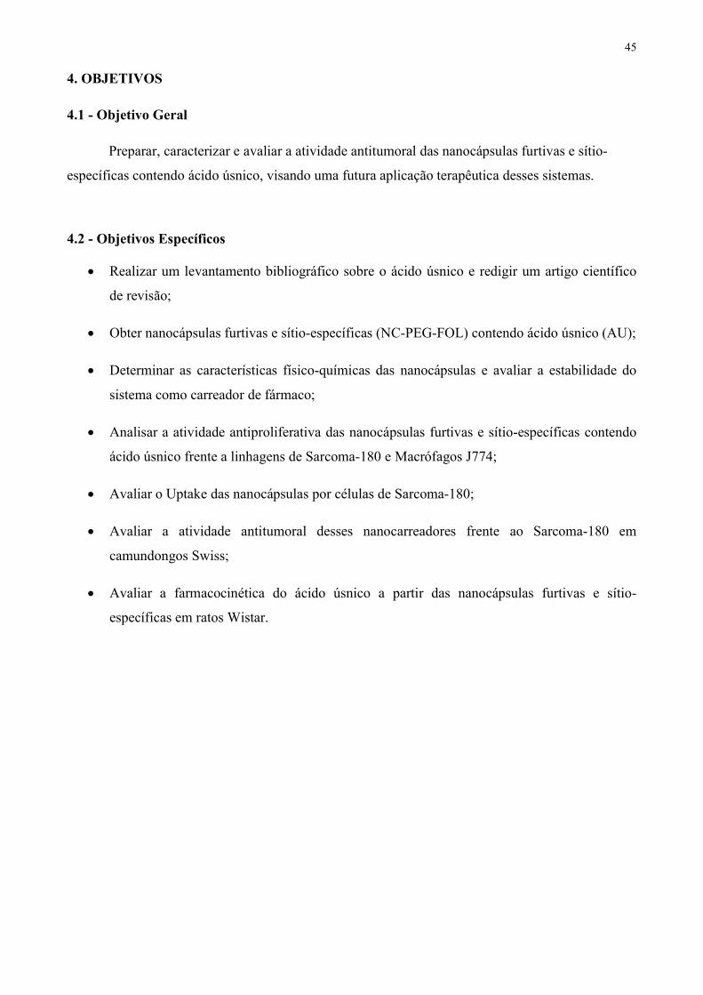
45 4. OBJETIVOS 4.1 - Objetivo Geral
Preparar, caracterizar e avaliar a atividade antitumoral das nanocápsulas furtivas e sítio-específicas contendo ácido úsnico, visando uma futura aplicação terapêutica desses sistemas.
4.2 - Objetivos Específicos
Realizar um levantamento bibliográfico sobre o ácido úsnico e redigir um artigo científico de revisão;
Obter nanocápsulas furtivas e sítio-específicas (NC-PEG-FOL) contendo ácido úsnico (AU); Determinar as características físico-químicas das nanocápsulas e avaliar a estabilidade do
sistema como carreador de fármaco; Analisar a atividade antiproliferativa das nanocápsulas furtivas e sítio-específicas contendo
ácido úsnico frente a linhagens de Sarcoma-180 e Macrófagos J774; Avaliar o Uptake das nanocápsulas por células de Sarcoma-180; Avaliar a atividade antitumoral desses nanocarreadores frente ao Sarcoma-180 em
camundongos Swiss; Avaliar a farmacocinética do ácido úsnico a partir das nanocápsulas furtivas e sítio-
específicas em ratos Wistar.

46 5. CAPÍTULO 1
Ácido úsnico: De um derivado liquênico a promissoras aplicações biológicas e nanotecnológicas
1Almeida, F. J. F.; 1Wanderley, M. S. O.; 1Ferraz, M. S.; 1,2Lira, M. C. B.; 4Pereira, E. C.; 3Silva, N.
H.; 1,2Santos, N. P.S.; 1Santos-Magalhães, N. S. 1Laboratório de Imunopatologia Keizo-Asami (LIKA), Centro de Ciências Biológicas (CCB),
Universidade Federal de Pernambuco (UFPE), Recife-PE, Brasil 2Centro Acadêmico de Vitória (CAV), Universidade Federal de Pernambuco (UFPE), Vitória de
Santo Antão-PE, Brasil 3Departamento de Bioquímica, Universidade Federal de Pernambuco (UFPE), Recife-PE, Brazil
4Departamento de Ciências Geográficas, Universidade Federal de Pernambuco (UFPE), Recife-PE, Brasil
* Correspondência: Prof. Nereide Stela Santos Magalhães - Universidade Federal de Pernambuco - UFPE Grupo de Sistemas de Liberação Controlada de Fármacos - SLC Laboratório de Imunopatologia Keizo-Asami - LIKA Av. Prof. Moraes Rego, 1235, Cidade Universitária, 50670-901, Recife-PE, Brazil Tel: +55 81 2126-8484; fax: +55 81 2126-8485 E-mail address: [email protected]

47 RESUMO
Entre os vários compostos liquênicos, o ácido úsnico (2,6-diacetil-7,9-dihidroxi-8,9b-
dimetil-1,3(2H,9bH)-dibenzofurano, C18H16O7) é um dos mais bem conhecidos e estudados. Isolado pela primeira vez ainda no século XIX, trata-se de um metabólito secundário produzido por várias espécies de líquens. O ácido úsnico apresenta-se na forma cristalina com coloração amarelada e possui duas formas enantioméricas (+)-ácido úsnico e (-)-ácido úsnico. Este metabólito liquênico apresenta caráter hidrofóbico, sendo praticamente insolúvel em água com uma solubilidade menor que 0.01g/100mL a 25 oC. O ponto de fusão do ácido úsnico é em torno de 204 oC e o peso molecular de 344.32 g/mol. O ácido úsnico apresenta diversas atividades farmacológicas, sendo referido como agente antimicrobiano, antitumoral, antiviral, antiparasitário dentre outras. Apesar das atraentes propriedades farmacológicas, o ácido úsnico é uma molécula tóxica. Uma vez que o seu mecanismo de ação não está completamente elucidado, a explicação para sua toxicidade também não é totalmente compreendida. Dessa maneira, a comunidade cientifica tem se empenhado no sentido de minimizar este problema sem perder a eficácia deste composto. Nesse contexto, os estudos impulsionaram o desenvolvimento de alternativas inovadoras, a exemplo da encapsulação do ácido úsnico em sistemas de liberação controlada, uma atrativa ferramenta da nanotecnologia farmacêutica. Os sistemas de liberação controlada de fármacos permitem que o princípio ativo encapsulado seja liberado com a velocidade de cedência otimizada, além de reduzir o regime de dosagem. Consequentemente, são capazes de aumentar a eficácia terapêutica minimizando os efeitos colaterais. Este trabalho apresenta uma revisão sobre as propriedades químicas, métodos analíticos, propriedades biológicas, mecanismo de ação e toxicologia do ácido úsnico. Ademais, foi abordada a utilização da nanotecnologia como ferramenta para superar os empecilhos da aplicação farmacológica do ácido úsnico.
Palavras-chave: Ácido úsnico; propriedades químicas; propriedades biológicas; mecanismos de ação; nanotecnologia.

48 INTRODUÇÃO
A ciência moderna tem despertado o interesse pela descoberta de novas moléculas com atrativas aplicações farmacológicas, seja oriundas de fontes naturais ou provenientes de modificações químicas a partir de moléculas preexistentes. Tendo em vista a vasta biodiversidade, essas moléculas bioativas podem ser extraídas de animais, vegetais, algas, bactérias, fungos, dentre outros. Neste quadro, chamamos atenção aos liquens, os quais são seres vivos simples resultantes da simbiose entre fungos (micobionte) e algas e/ou cianobactérias (ficobiontes) (NASH, 1996). Os líquens apresentam larga distribuição geográfica dos trópicos aos pólos, habitando sobre rochas, solos e troncos de árvores. A atenção dada a estes organismos é devido a sua habilidade de produzir substâncias atrativas, tais como os metabólitos secundários sintetizados pelas hifas do fungo, os quais são utilizados como mecanismo de defesa e proteção contra intempéries (MÜLLER, 2001). Tais metabólitos se classificam em quatro grupos químicos bioenergeticamente relacionados: depsídeos, depsidonas, dibenzofuranos e ácidos úsnicos (NASH, 1996), sendo o este último considerado um dos mais importantes metabólitos liquênicos biologicamente ativos (MÜLLER, 2001).
O ácido úsnico foi isolado inicialmente em meados do Século XX (ASAHINA & SHIBATA 1954). O interesse pelo isolamento desta molécula impulsionou a sua busca em diversas espécies liquênicas como Usnea deffractas (MÜLLER, 2001), Lecanora pseudogangalevides (LUMBSCH, 1995), Usnea laevis (MARCANO et al., 1999), Roccella montagnei (VIJAYAKUMAR et al., 2000), Sticta weigelii (PIOVANO et al., 2000), Cladonia substellata (DE CARVALHO et al., 2005) e Usnea longissima (ODABASOGLU et al., 2006).
Segundo Ingólfsdóttir e colaboradores (1985), o interesse terapêutico do ácido úsnico foi despertado após a utilização dos liquens na medicina popular, desde a antiguidade, no combate da tuberculose pulmonar e algumas doenças de pele. Posteriormente, outras atividades significativas biológicas lhe foram atribuídas, tais como: antimicrobiana (LAUTERWEIN et al., 1995), antiinflamatória, analgésica, antipirética (YAMAMOTO et al., 1995), antifúngica (BROSKA et al., 1996), antiparasitária (FOURNET et al., 1997), antitumoral (KUMAR & MÜLLER, 1999), antiviral (CAMPANELLA et al., 2002), de inibição enzimática (HUNECK, 1999) e gastroprotetora (ODABASOGLU et al., 2006).
Apesar de sua descoberta na década de 50 (BUSTINZA, 1951), o acido úsnico ainda desperta grande interesse científico, fato este comprovado pelo crescimento progressivo no quantitativo de publicações, totalizando um número aproximado de 160 entre os anos de 2009 e 2013 (Figura 1).

0
20
40
60
80
100
120
140
160
180
1979-1983
Number
of pub
lication
1974-1978
Figura 1. Número citações científicas a respeito do ácido úsnico no decorrer dos anos, baseandona busca no banco de pesquisa científica internacional, Scopus (
Uma vez que o nosso grupo de pesquisa apresenta liquênico, demonstrando relevante produção científica, propomos realizar esta revisão da literatura visando suprir a escassez a cerca de informações atualizadas. Nesta vertente,propriedades químicas, propriedades biológicas e aspectos toxicológicos desta atrativa molécula. Ademais, será abordada, pela primeira vez a utilização da nanotecnologia, como ferramenta para superar os empecilhos da aplicação farmaco PROPRIEDADES QUÍMICAS DO ÁCIDO ÚSNICO
O ácido úsnico, [2,6-diacetilC18H16O7, PM = 344.32] (Figura 2ocorrendo na natureza em duas formas enatioméricas (grupamento metil localizado na posição 2002).
2004-20081999-20031994-19981989-19931984-19881979-1983
YearsNúmero citações científicas a respeito do ácido úsnico no decorrer dos anos, baseando
na busca no banco de pesquisa científica internacional, Scopus (www.scopus.compalavra-chave: Usnic Acid.
ue o nosso grupo de pesquisa apresenta experiência relacionada a este derivado liquênico, demonstrando relevante produção científica, propomos realizar esta revisão da literatura visando suprir a escassez a cerca de informações atualizadas. Nesta vertente,
propriedades biológicas e aspectos toxicológicos desta atrativa molécula. Ademais, será abordada, pela primeira vez a utilização da nanotecnologia, como ferramenta para superar os empecilhos da aplicação farmacológica do ácido úsnico.
PROPRIEDADES QUÍMICAS DO ÁCIDO ÚSNICO diacetil-7,9-dihidroxi-8-9b-dimetil-1,3(2H,9/H)
Figura 2), caracteriza-se por ser uma substância de pigmentação amarela, ocorrendo na natureza em duas formas enatioméricas (-) e (+) decorrente da projeção angular do grupamento metil localizado na posição 9b (COCCHIETTO et al., 2002; INGÓLFSDÓTTIR,
Anos
49
2009-2013
Número citações científicas a respeito do ácido úsnico no decorrer dos anos, baseando-se www.scopus.com) e tendo como
relacionada a este derivado liquênico, demonstrando relevante produção científica, propomos realizar esta revisão da literatura visando suprir a escassez a cerca de informações atualizadas. Nesta vertente, este artigo foca nas
propriedades biológicas e aspectos toxicológicos desta atrativa molécula. Ademais, será abordada, pela primeira vez a utilização da nanotecnologia, como ferramenta para
H)-dibenzofurandiona; se por ser uma substância de pigmentação amarela,
) e (+) decorrente da projeção angular do , 2002; INGÓLFSDÓTTIR,

50
Figura 2. Estrutura química do ácido úsnico.
Este derivado liquênico apresenta caráter hidrofóbico, com solubilidade em água menor que
10 mg/100 mL a 25 °C (INGÓLFSDÓTTIR, 2002), sendo parcialmente solúvel em etanol e facilmente em éter quente, acetona, benzeno e clorofórmio (ASAHINA & SHIBATA, 1954; TAKAI & UEHARA, 1979). Tal característica hidrofóbica pode ser explicada pela presença dos três grupos cetônicos, bem como ao anel furano que une os anéis aromáticos (ASAHINA & SHIBATA, 1954), além de pontes de hidrogênio intramoleculares (MÜLLER, 2001). Sua acidez é justificada pela presença do anel fenólico, cuja estrutura é instável (SHIBATA, 2000). O valor do pKa para o grupo hidroxila fenólico na posição 3 é de 4.4, enquanto que para o grupo hidroxila fenólico nas posições 9 e 7 são 8.8 e 10.7 respectivamente (KRISTMUNDSDÓTTIR et al., 2002; HAN et al., 2004).
Segundo a literatura, a biossíntese do ácido úsnico procede pela via do acetato polimalonato (Figura 3). Inicialmente são formadas unidades fenólicas monocíclicas, originadas a partir de ácidos carboxílicos, derivados do ácido acético. Estas unidades fenólicas são sintetizadas a partir da acetil-CoA e malonil-CoA, através de sintetases aromáticas, que não possuem subunidades desidrogenadas. A síntese do metilfloroacetofenona, intermediário chave para formação do ácido úsnico, é catalisada pela enzima poliketidase synthase (HAWRANIK et al., 2009). Os passos subsequentes de sua biossíntese envolvem o acoplamento homólogo estereoespecífico de duas unidades metilfloroacetofenona originando, portanto, o ácido úsnico hidratado. Finalmente, ocorre a desidratação da molécula levando a formação da ligação éter (INGÓLFSDÓTTIR, 2002). Até o momento, métodos chave na obtenção do ácido úsnico são extrações a partir de liquens com a utilização de solventes orgânicos e subsequente precipitação, geralmente utilizando etanol (SOKOLOV et al., 2012a).

51
Figura 3. Via da biossíntese do ácido úsnico.
Além da biossíntese do ácido úsnico, sua caracterização é amplamente descrita na literatura.
Os manuscritos descrevem análises que utilizam espectrofotometria no infravermelho (IV), ressonância magnética nuclear e prótons (H1 RMN), análise térmica, dentre outras técnicas. A caracterização da molécula por espectrofotometria no infravermelho mostra grupamento cetona cíclico (1694 cm-1), bandas fracas em 1.716 e 1.676 cm-1 referentes aos grupamentos cetona não-cíclicos v(C=O) e bandas simétricas e assimétricas de aril, alquil éter (COC) próximo a 1.288 e 1.070 cm-1, respectivamente (EDWARDS et al., 2003; LIRA et al., 2009). Nesta vertente, a caracterização do ácido úsnico por ressonância magnética nuclear de prótons (H1 RMN) confirmou picos característicos da molécula tais como hidroxilas em 13.406 e 11.396 ppm, próton do anel aromático em 6.25 ppm, e prótons do grupamento metila em 2.61, e 2.04, 2.50 ppm nas posições 14, 15 e 16, respectivamente (LIRA et al., 2009). Análise térmica realizada por diferentes pesquisadores descrevem pico endotérmico que varia de 200 a 204 ºC correspondente ao pico de fusão do ácido úsnico e pico exotérmico em torno de 273 ºC correspondendo à temperatura de início de degradação (MARCANO et al., 1999, LIRA et al., 2009). A partir de uma molécula bem caracterizada é possível realizar modificações químicas visando proporcionar uma melhoria nas suas características como, por exemplo, as atividades biológicas. Atualmente vários trabalhos relatam estratégias para obtenção de derivados químicos do ácido úsnico que visam tanto potencializar sua hidrossolubilidade, como a atividade biológica e ainda, minimizar seus efeitos tóxicos, mais especificamente a hepatotoxicidade. As modificações para obtenção de novos derivados do ácido úsnico compreendem geralmente reações com amina no grupo carbonil e a formação de éster nos grupamentos hidroxila (SOKOLOV et al., 2012a).
Desta maneira, várias modificações vêm sendo descritas. Inicialmente a obtenção do sal do ácido úsnico, comercialmente conhecido como Usno, foi desenvolvida com o propósito de encontrar uma forma hidrossolúvel, mas sem a perda de sua atividade biológica (GRASSO et al.,1989). Outro sal de ácido úsnico proposto foi o sal de sódio o qual recebeu o nome de BINAN,

52 cuja atividade antimicrobiana foi semelhante à do ácido úsnico (NAJDENOVA et al., 2001). Ainda com objetivo de aumentar a solubilidade do ácido úsnico, recentemente Lukáč e colaboradores (2012) investigaram a influência do sal de bisamônio (representando um surfactante catiônico), do dialquilfosfocolina (representando um surfactante zwitteriônico) e da mistura dos dois, com objetivo de aumentar a solubilidade do ácido em solução micelar. Os autores visualizaram que a maior solubilidade foi observada para o surfactante catiônico (20 vezes) quando comparada à solubilidade do ácido em água.
Seguindo outro caminho, mas ainda com o intuito de aumentar a solubilidade aquosa do ácido úsnico, Kristimundsdóttir e colaboradores (2002) testaram diferentes valores de pH, várias concentrações de co-solventes e surfactantes, além de agentes complexantes como a 2-hydroxypropyl--cyclodextrina, onde observaram, na análise desse último parâmetro, que a solubilidade do ácido úsnico com a 2-hydroxypropyl--cyclodextrin foi de 0.68 mg/100mL. Mais tarde, Lira e colaboradores (2009) desenvolveram e caracterizaram complexos de inclusão do ácido úsnico e a β-ciclodextrina, utilizando o método de liofilização para tal finalidade. As interações químicas entre o ácido úsnico e a -ciclodextrina foram avaliadas por IR, H1 RMN, Raios X e análise térmica. Além das modificações químicas terem o objetivo de aumentar a solubilidade aquosa, relatos ainda indicam a potencialização da atividade biológica. Neste sentido, Luzina e colaboradores (2007) investigaram a condensação do ácido úsnico com vários aminoácidos a exemplo da glicina, β-alanina, L-valina, L-leucina, entre outros. Os autores seguiram a premissa de que a inserção de fragmentos de aminoácidos em moléculas biologicamente ativas pode aumentar a atividade biológica das mesmas. Outro exemplo de modificação química pode ser encontrado no manuscrito publicado por Bazin e colaboradores em 2008, os quais descrevem a síntese de nove derivados amino conjugados e a avaliação da atividade citotóxica em linhagens celulares de câncer humano e murino. Apesar das modificações químicas algumas vezes exibirem uma maior ação biológica, elas ainda não apresentam comportamento sinérgico capaz de reduzir os efeitos tóxicos. Para superar essas desvantagens uma estratégia inovadora é, sem dúvidas, a encapsulação do ácido úsnico em sistemas de liberação controlada de fármacos, que será abordado de forma pioneira neste manuscrito.
Além de vastos estudos com enfoque na caracterização físico-química, os mais diferentes métodos analíticos têm sido reportados. Tais métodos descrevem a identificação, separação e quantificação do ácido úsnico em extratos liquênicos (CANSARAN et al., 2006; IVANOVIC et al., 2013), em plasma (VENKATARAMANA, KRISHNA, 1992) e em preparações farmacêuticas

53 (RIBEIRO-COSTA et al., 2004; SANTOS et al., 2005; SIQUEIRA-MOURA et al., 2008). Dentre estes métodos são descritos espectrofotometria (SIQUEIRA-MOURA et al., 2009), espectrofluorimetria (BRUNO et al., 1983), cromatografia líquida de alta eficiência (CLAE) (VENKATARAMANA, KRISHNA, 1992; RIBEIRO-COSTA et al., 2004; SANTOS et al., 2005), cromatografia em camada fina (TCL) (MARCANO et al, 1999), cromatografia líquida de fase reversa (RP-LC) (JI, KHAN, 2005) e eletroforese capilar de zona (CZE) (KREFT et al., 2001). Alguns modelos cromatográficos foram validados, como o descrito inicialmente por Venkataramana e Krisnha (1992), os quais desenvolveram um método utilizando CLAE para determinação do ácido úsnico em plasma humano com 98% de recuperação do analito. Por outro lado, Ji e Khan (2005) utilizaram a RP-LC para doseamento do composto liquênico em preparações líquidas, cápsulas e comprimidos. Em contrapartida, o método CZE foi desenvolvido para a análise rotineira do ácido úsnico em produtos medicinais contendo extrato de líquens (KREFT et al., 2001).
Apesar de vários métodos analíticos previamente descritos, apenas em 2008, Siqueira-Moura e colaboradores propuseram um método espectrofotométrico simples para doseamento do ácido úsnico a partir de nanossistemas, especificamente de formulações lipossomais, sem interferência dos constituintes da formulação, confirmando a possível determinação do ácido úsnico por ultravioleta utilizando os comprimentos de onda na faixa de 233-290 nm.
PROPRIEDADES BIOLÓGICAS
Logo após a sua descoberta, o ácido úsnico teve sua atividade antimicrobiana estudada, atividade esta que se destaca até o presente momento (figura 4). Apesar de diversas propriedades terapêuticas lhe serem atribuídas, a maior parte das publicações (em torno de 500) remete o potencial antimicrobiano do ácido úsnico (44.44%) e inova ao relatar o seu espectro de ação contra, cada vez mais, novas cepas, principalmente aquelas resistentes a antibióticos tradicionais. Ao mesmo tempo, um aumento progressivo nas publicações relacionadas à sua propriedade antitumoral vem sendo percebido. Dentre os relatos a cerca das propriedades biológica do ácido úsnico, 21.60% se refere ao seu poder antitumoral, despertado para a sua aplicação contra as mais diferentes linhagens cancerígenas.

21,6%
Figura 4. Percentual de publicações das atividades biológicas do ácido úsnico (www.scopus.com) palavras
Atividade Antimicrobiana
A descoberta do ácido úsnico ocorreu mediante a busca por novos compostos antibióticos (ABRANHAN & FLOREY, 1949; BUSTINZA, Streptococcus mutans (agente etiológico da cárie dental e de doenças periodontais) (GRASSO et al1989) e os gêneros Escherichia,1995).
Adicionalmente, o ácido úsnico vem sendo utilizado como alternativa contra cepas resistentes aos antibióticos preconizados, como por exemplo, (ERV) e Staphylococcus aureusestas cepas, Pompilio e colaboradoes em 2013, além de comprovaremácido úsnico, identificaram a sua ação antibiofilme. Em fim de superar a resistência da SARM, avaliaram ação dantibióticos. Neste estudo foi averiguada uma ação sinérgica do ácido úsnico com a gentamicina, e
44,44%
33,95%
Others Activities Antitumoral Activity Antimicrobial Activity
Percentual de publicações das atividades biológicas do ácido úsnico ) palavras-chave: usnic acid e as respectivas atividades
A descoberta do ácido úsnico ocorreu mediante a busca por novos compostos antibióticos (ABRANHAN & FLOREY, 1949; BUSTINZA, 1951), tendo suas primeiras descrições contra o
(agente etiológico da cárie dental e de doenças periodontais) (GRASSO et al, Salmonella e Shighella (HALE-Jr, 1983; LAUTERWEIN
nte, o ácido úsnico vem sendo utilizado como alternativa contra cepas preconizados, como por exemplo, Enterococos resistentes à vancomicina
Staphylococcus aureus resistente à meticilina (SARM) (ELO et al, 2007). Ainda sestas cepas, Pompilio e colaboradoes em 2013, além de comprovarem a atividade antimicrobiana do
a sua ação antibiofilme. Em paralelo Segatore e colaboradores (2012), a fim de superar a resistência da SARM, avaliaram ação do ácido úsnico combinado a outrosantibióticos. Neste estudo foi averiguada uma ação sinérgica do ácido úsnico com a gentamicina, e
54
Others ActivitiesAntitumoral ActivityAntimicrobial Activity
Percentual de publicações das atividades biológicas do ácido úsnico [Scopus
e as respectivas atividades].
A descoberta do ácido úsnico ocorreu mediante a busca por novos compostos antibióticos tendo suas primeiras descrições contra o
(agente etiológico da cárie dental e de doenças periodontais) (GRASSO et al., Jr, 1983; LAUTERWEIN et al.,
nte, o ácido úsnico vem sendo utilizado como alternativa contra cepas resistentes à vancomicina
resistente à meticilina (SARM) (ELO et al, 2007). Ainda sobre a atividade antimicrobiana do
paralelo Segatore e colaboradores (2012), a o ácido úsnico combinado a outros
antibióticos. Neste estudo foi averiguada uma ação sinérgica do ácido úsnico com a gentamicina, e

55 antagônica com a levofloxacina, e uma ação indiferente com aclindamicina, eritromicina, gentamicina, levofloxacina e oxacilina.
Ainda explorando sua atividade antimicrobiana, e a utilização deste ácido no controle do crescimento de biofilmes pelas cepas de S. aureus e Pseudomonas aeruginosa, o qual, apesar de não inibir a fixação inicial das bactérias de S. aureus nas superfícies artificiais de polímero (in situ), o ácido úsnico foi capaz de matar as que se ligaram, inibindo assim a formação dos biofilmes. Enquanto que a utilização contra a P.aeruginosa, apesar da formação do biofilme, houve alteração na morfologia bacteriana (FRANCOLINI et al., 2004). Este é um achado importante, pois na medicina vem se tornando comum o uso de dispositivos para reparo ou substituição de partes do corpo danificadas, no entanto, superfícies artificiais são frequentemente susceptíveis a colonização por bactérias e fungos, resultando assim, na formação de biofilmes.
A resistência à antibioticoterapia é um dos principais problemas no combate à tuberculose. Dessa forma, o ácido úsnico torna-se um importante candidato no combate a esta infecção. Assim, o ácido úsnico foi testado contra as cepas susceptíveis e resistentes a isoniazida, estreptomicina e rifampicina, fármacos usados na terapia atual, demonstrando que não ocorreu resistência cruzada, sugerindo que o mecanismo de ação do acido úsnico seja diferente dos fármacos atualmente usados na terapia da Tuberculose (RAMOS e DA SILVA, 2010), fator que viabiliza sua utilização contra o Mycobacterium tuberculosis.
Além das cepas acima relacionadas, a Helicobacter pylori demonstrou ser susceptível à ação o ácido úsnico, o qual apresentou uma forte atividade dose-dependente. Este estudo foi realizado por Safak et al (2009) após observar que um determinado líquen (Usnea dasypoga) é comumente usado para tratamento de úlcera gástrica. Neste mesmo trabalho foi mostrado que o sinergismo do ácido úsnico com claritromicina pode ser bastante eficaz no tratamento da H. pylori, pois a mesma, em associação com inibidores da secreção ácida, é um fármaco de referência no tratamento da erradicação dessa bactéria. Em um estudo posterior, Luo et al (2011) comprovou essa propriedade do ácido úsnico extraído de Nephromopsis pallescens, obtendo resultados semelhantes a ampicilina e eritromicina (10 µg/mL), fármacos que possuem ação contra H. pylori comprovada.
Diante do exposto (Tabela 1), algumas cepas bacterianas mostraram suscetibilidade ao ácido úsnico. Estes relatos demonstram a ampla aplicação do ácido úsnico frente a diferentes bactérias justificando sua vasta citação como potente agente antimicrobiano.

56 Tabela 1. Cepas de bactérias suscetíveis ao ácido úsnico.
Microrganismo CIM (M) Referência
(+)-Ácido úsnico (-)-Ácido úsnico Enterococcus faecalis 11.6 23.2 LAUTERWEIN et al., 1995 Enterococcus faecium 46.4 46.4 LAUTERWEIN et al., 1995 Clostridium perfringens 11.6 11.6 LAUTERWEIN et al., 1995 Bacteroides vulgatus 11.6 23.2 LAUTERWEIN et al., 1995 Pseudomonas aeruginosa 742.4 ND (*) FRANCOLINI et al., 2004
Staphylococcus aureus
17.4 23.2 CORRECHE,1998 23.2 23.2 LAUTERWEIN et al., 1995 92.8 ND (*) FRANCOLINI et al., 2004 5.8 ND (*) SEGATORE et al., 2012 5.8 ND (*) POMPILIO et al., 2013
Mycobacterium aurum 92.8 ND (*) INGÓLFSDÓTTIR et al., 1998 Mycobacterium tuberculosis 4.4 ND (*) RAMOS e DA SILVA, 2010 Helicobacter pylori 0.18 ND (*) SAFAK et al., 2009 ND = Não definido
Atividade Antitumoral
A atividade antitumoral do ácido úsnico foi revelada há quase quatro décadas. Em 1975, Kupchan e Kopperman publicaram o primeiro estudo contra o carcinoma pulmonar de Lewis. A partir daí, em estudos in vitro, o ácido úsnico demostrou sua ação antitumoral frente a diferentes linhagens celulares, tais como queratinócitos humanos (HaCaT) (KUMAR & MÜLLER, 1999; PEREIRA, 1994; LIMA, 1990; BURLANDO et al., 2009), células de câncer de mama (MCF7 e MDAMB231) e de câncer de pulmão (H1299 e NCIH 292) (MAYER et al., 2005; SANTOS et al.,

57 2005). Quando incorporado à -ciclodextrina, o ácido úsnico demonstrou sua atividade antiproliferativa contra células malignas da linhagem K-562 (leucemia) (CAMPANELLA et al., 2002), T-47-D (câncer de mama), Panc1 (câncer de pâncreas) e PC-3 (câncer de próstata) (KRISTMUNDSDÓTTIR et al., 2005).
É importante notificar que a atividade antiproliferativa do ácido úsnico foi observada contra uma linhagem modelo para estudo dos efeitos de compostos citotóxicos de câncer humano, o carcinoma epidermóide da vulva (A431), e ainda contra uma linhagem de tumor agressivo e resistente à quimioterapia e letal, mesotelioma maligno (MM98) (BURLANDO et al., 2009).
A viabilidade celular in vitro do hepatoblastoma humano HepG2 foi reduzida na concentração de 5 µM do ácido úsnico além de, apresentar a LC50 de 30 µM. No mesmo estudo, a exposição dessas células ao fármaco, principalmente em concentrações acima de 20 µM, resultou em um aumento significativo da atividade do citocromo P450, do estresse oxidativo e da disfunção mitocondrial comprovando a toxicidade do ácido úsnico frente a essas linhagens celulares (SAHU et al., 2011). Já em 2012, os mesmos pesquisadores, ao associarem o ácido úsnico a lipopolissacarídeos, observaram um aumento no efeito tóxico frente a HepG2 (SAHU et al., 2012).
Além dos estudos realizados com o ácido úsnico isolado, vários autores compararam a atividade desse ácido com outros metabólitos liquênicos, e logo atribuíram um maior potencial antitumoral do ácido úsnico contra A2780 e HT-29 (BACKOROVÁ et al., 2012), MCF-7, HeLa e HCT-116 (BRISDELLI et al., 2013), e UACC-62 e B16-F10 (BRANDÃO et al., 2013).
Ademais, os efeitos anticancerígenos das duas formas enantioméricas do ácido úsnico foram investigados por Einarsdóttir et al (2010) os quais concluíram que ambas as formas apresentaram efeito inibitório sobre o crescimento e proliferação das células T-47D (câncer de mama) e Capan-2 (câncer de pâncreas).
Por fim, O’Neill e colaboradores (2010), sabendo do importante papel tanto na divisão celular quanto nos mecanismos de morte celular por apoptose, investigaram se o ácido úsnico afetava a formação e/ou estabilização dos microtúbulos como alvo quimioterapêutico, porém tal atividade não foi constatada.
Em resumo, a Tabela 2 demonstra algumas linhagens celulares cancerígenas que mostraram suscetibilidade ao ácido úsnico, confirmando o elevado número de citações como agente antiproliferativo.

58 Tabela 2. Linhagens de células cancerígenas suscetíveis ao ácido úsnico.
Linhagem celular CI50 (µM) Referência (+)-Ácido úsnico (-)-Ácido úsnico Carcinoma epidermóide
(A431) 39 ND(*) BURLANDO et al., 2009
Mesotelioma maligno (MM98) 23 ND(*) BURLANDO et al., 2009
Queratinócitos humanos (HaCaT)
2.1 ND(*) KUMAR & MÜLLER, 1999
35 ND(*) BURLANDO et al., 2009 Câncer de mama
(T-47D) 12.2 11.6 EINARSDÓTTIR et al., 2010
Câncer de pâncreas (Capan-2) 15.4 14.5 EINARSDÓTTIR et al.,
2010 Hepatoblastoma humano
(HepG2) 30 ND(*) SAHU, 2011
Adenocarcinoma do colo do útero (HeLa) 23.7 ND(*) BRISDELLI et al., 2013
Carcinoma do cólon HCT-116 17.7 ND(*) BRISDELLI et al., 2013
Adenocarcinoma de mama (MCF-7) 75.7 ND(*) BRISDELLI et al., 2013
Melanoma humano (UACC-62) 91.5 ND(*) BRANDÃO et al., 2013
Melanoma murino (B16-F10) 137.7 ND(*) BRANDÃO et al., 2013
ND = Não definido

59 Outras Atividades Tendo em vista, todos estes relatos a cerca das propriedades antimicrobiana e antitumoral, diversos autores vêm atribuindo outras atividades biológicas ao ácido úsnico, conforme exposto a seguir. Atividade antiviral
Diferentes tipos virais têm sido alvo do ácido úsnico. Inicialmente, este composto liquênico foi testado contra o Eptein-Barr, Papilomavírus Humano e o Poliomavírus de ratos (YAMAMOTO et al., 1995; SCIRPA et al., 1999; CAMPANELLA et al., 2002). Em 1995, Yamamoto e colaboradores testaram ambas as formas enatioméricas do ácido úsnico, contra o Eptein-Barr vírus, e observaram que a forma dextrógira apresentou maior atividade antiviral em relação à levógira.
A ação contra o HPV foi observada no tratamento adjuvante da infecção associado com o sulfato de zinco. Neste estudo foi demonstrado que além da ação antiviral, o ácido úsnico favoreceu a reepitelização do tecido lesado. Dessa maneira, possibilitou a terapia adjuvante no tratamento cirúrgico desobstrutivo e, em particular, da lesão causada pelo HPV (SCIRPA et al., 1999).
Além disso, o ácido úsnico foi capaz de inibir a proliferação do Poliomavírus de ratos, fato este ocorrido pela destruição do DNA viral, através da inibição da transcrição do RNA (CAMPANELLA et al., 2002). Recentemente foi avaliada a atividade do ácido úsnico e seus derivados (sinteticamente modificados) contra vírus da influenza H1N1 em células MDCK , cujos resultados sugerem que essas moléculas sejam substâncias antigripe (SOKOLOV et al., 2012b). Atividade antiparasitária
Outra atividade atribuída ao ácido úsnico foi a sua propriedade antiparasitária. Os primeiros relatos voltados a este foco foram descritos por Wu e colaboradores (1995), quando detectaram a atividade do ácido úsnico contra o Trichomonas vaginalis em estudos in vitro. Em seguida, o (+) ácido úsnico e como outros metabólitos secundários, isolados de liquens Chilenos, foram testados frente à forma promastigota de três cepas L. braziliensis, L. amazonensis e L. donovani. Neste estudo o ácido úsnico (25 g/mL) promoveu lise total das três cepas nos ensaios in vitro, além de mostrar uma significante diminuição da lesão cutânea em camundongos BALB/c infectados com L. amazonensis, demonstrando a sua interessante atividade antiparasitária (FOURNET et al., 1997).

60 Em 2001, Sussmann e colaboradores, impulsionados pela resistência parasitária aos
medicamentos convencionais, testaram o ácido úsnico frente ao Plasmodium falciparum. Nesta busca por novas moléculas alvo, os pesquisadores descobriram no ácido úsnico uma importante ação contra o parasita uma vez que ele inibiu de forma reversível a biossíntese de vitamina E, uma importante molécula para o desenvolvimento do parasita.
Atividade antifúngica
A atividade antifúngica do ácido úsnico também foi descoberta nos anos 50, quando se observou a inibição do fungo Tricophyton mentagrophytes após tratamento com o referido ácido (BUSTINZA, 1951). Em 1996, Broska e colaboradores observaram inibição no crescimento do Penicillum frequentans e Verticillium albo-atrum, após tratamento com ácido úsnico.
Atualmente, o pequeno número de medicamentos disponíveis para o tratamento de fungos incentiva a busca de novos agentes quimioterápicos. Para tal propósito, o ácido úsnico foi testado contra a Candida orthopsilosis and C. parapsilosis, porém nenhuma ação frente ao biofilme formado por estas leveduras foi observada (PIRES et al., 2011). Atividade Gastroprotetora
Alguns estudos indicam que o ácido úsnico, isolado da Usnea longissima, vem sendo testado no tratamento da úlcera gástrica em animais. Lesões gástricas foram significativamente reduzidas por todas as doses utilizadas de ácido úsnico (25, 50, 100 e 200 mg/kg de peso corporal), quando comparado com o grupo tratado com a ranitidina (25 mg/kg de peso corporal), fármaco referência. Esse efeito gastroprotetor do ácido úsnico pode ser atribuído ao seu efeito redutor contra o dano oxidativo e seu efeito inibitório na infiltração neutrofílica em estômago de ratos (ODABASOGLU et al., 2006). Atividades Cicatrizante, Antioxidante e Anti-inflamatória
A cicatrização é um processo preocupante em algumas situações como pós-cirurgia e queimaduras. Para que a regeneração epitelial seja agilizada, é necessário o auxílio de terapia farmacológica que tanto inviabilize a ação microbiana, bem como favoreça a reepitelização. Desta forma, o ácido úsnico tem sido muito usado em preparações dermatológicas e cosméticas, devido

61 suas atividades bacteriostática e antioxidante. Recentemente, Bruno e colaboradores 2013, avaliaram as propriedades de reparação de feridas de derivados do ácido úsnico em ensaios in vitro e in vivo, indicando menor citotoxicidade a células da pele combinados com melhor desempenho de cura, sugerindo a possibilidade de utilização destes compostos na cicatrização de feridas e preparações anti-idade para a pele.
Além disso, a ação cicatrizante do ácido úsnico também é favorecida pelo potencial anti-inflamatório deste composto liquenico. Zhijun e colaboradores (2011) observaram que tal propriedade decorre do downregulation de alguns mediadores da inflamação tais como iNOS, COX-2, IL-1β, IL-6 e TNF-α, COX-2 e , além da superexpressão da interleucina-10 e da hemeoxigenase-1.
Tais achados vincularam o ácido úsnico como um potente bloqueador solar, tanto pela sua absorção em regiões ultravioleta (UV), como pelo poder antioxidante. Estudos indicaram que, alguns líquens sob estímulo das radiações UV, sintetizam seus metabólitos com uma forte absorção na região UV, gerando uma proteção própria contra radiações perigosas. Dentre esses metabólitos, o ácido úsnico teve um fator de proteção similar à substância comercial sintética usada como referência, exibindo valores de fator de proteção UV iguais (3.6-5.0) nos testes in vivo e superiores (4.03-4.83) que a substância referência (2.66-3.63) nos testes in vitro (RANCAN et al., 2002). Estudos mais recentes mostraram que este mesmo composto, agora obtido da Xanthoparmelia farinosa apresentou potente atividade antioxidante e pró-oxidante (comportamento bifuncional) em linhagens celulares de linfócitos humanos (Jurkat-cells: E 6-1 leucemia aguda) sob irradiação UV-B (KOHLHARDT-FLOEHR et al., 2010).
Os radicais livres em relação às doenças cardiovasculares são importantes no desenvolvimento da aterosclerose, tornando-se um fator de risco, consequentemente, a propriedade antioxidade do ácido úsnico repercutiu em uma proteção cardiovascular em ensaios contra a aterosclerose utilizando o ácido úsnico extraído da Usnea complanata (BEHERA et al., 2012). MECANISMO DE AÇÃO E TOXICOLOGIA DO ÁCIDO ÚSNICO
Apesar das atraentes propriedades farmacológicas, o ácido úsnico é uma molécula tóxica. Uma vez que o seu mecanismo de ação não está completamente elucidado, a explicação para sua toxicidade também não é totalmente compreendida. Dessa maneira, a comunidade científica tem se empenhado para desvendar os mecanismos bioquímicos e moleculares envolvidos tanto nas atividades biológicas inerentes ao ácido úsnico, bem como na sua toxicidade.

62 Neste viés, diversos modelos de estudo (in silico, in vitro e in vivo) relacionam o mecanismo
de ação do ácido úsnico na interrupção da função mitocondrial e alterações no estresse oxidativo (SANTOS et al., 2003; PRAMYOTHIN e t al., 2004; HAN et al., 2004; LIU et al., 2012). Estas evidências sugerem que a ruptura da função mitocondrial seja o principal mecanismo que subjaz a morte celular induzida pelo ácido úsnico.
Tendo em vista que a via mitocondrial é importante na regulação das etapas que decorrem para morte celular, sugere-se que este seja o principal mecanismo que reduz a viabilidade de células cancerígenas, sendo este um importante ponto que justifica a ação antitumoral. Adicionalmente, o estresse oxidativo e o rompimento do processo metabólico normal causado pelo ácido úsnico em células cancerígenas, podem estar diretamente relacionado à redução da síntese de RNA (AL-BEKAIRI et al., 1991), mas não envolvidos com danos ao DNA (MAYER et al., 2005). Outra hipótese justifica que o efeito antitumoral decorre das alterações no gradiente de pH em células carcinogênicas, conforme demonstrado nas linhagens T47D e Capan-2 de câncer de mama e pâncreas, respectivamente. Nestas, foram visualizadas modificações nos transportadores de prótons da membrana plasmática gerando um gradiente eletroquímico, o qual permite o acúmulo de ácidos fracos em seu interior (BESSADOTTIR et al., 2012).
É importante mencionar que, como o ácido úsnico é um ácido fraco de característica lipofílica, sua difusão pela membrana mitocondrial é favorecida acarretando inibição da produção de ATP na fosforilação oxidativa. Segundo Santos (2003) essa hipótese explica a sua atraente atividade antimicrobiana uma vez que, este mecanismo não permite que o microrganismo execute os processos anabólicos imprescindíveis ao seu crescimento.
Apesar dos relatos acima, a influência do ácido úsnico na respiração celular foi inicialmente citada na década de 50, quando Johnson e colaboradores (1950) descreveram o relevante o efeito na respiração e o desacoplamento da fosforilação oxidativa, em homogenato de rins e fígado de rato, com concentrações mínimas de 1.3-2.6 µg/ml. Desde então, estudiosos vem compartilhando achados que ressaltaram essa hipótese. Vavasseur e colaboradores (1991) evidenciaram o potencial inibitório do ácido úsnico sobre processos respiratórios aeróbicos em células vegetais de Cormmelina communis. Abo-Khatwa e colaboradores (1996) relataram que o ácido úsnico, o vulpínico e a atranorina apresentaram tal ação quando testados em mitocôndria de fígado de ratos. Estes compostos atuaram no interior da membrana mitocondrial com características semelhantes ao do 2,4-dinitrofenol, substância desacopladora padrão. Em seguida, Santos (2003) reportou que, em concentrações de 32 µM, o ácido úsnico pôde inibir completamente a produção de ATP mitocondrial. No ano seguinte, Pramyothin e colaboradores (2004) constataram que o (+) ácido

63 úsnico agia alterando a integridade da membrana celular permitindo a liberação de enzimas hepatoespecíficas (aspartato aminotransferase e alanina aminotransferase), principalmente das transaminases, além de causar destruição da função mitocondrial. Diante disto, os dados expostos sugerem que o dano oxidativo esteja relacionado à toxicidade deste composto liquênico, principalmente em células hepáticas.
Ainda neste contexto, ensaios em modelos murinos avaliaram a hepatoxicidade do ácido úsnico, quando se observou 98 e 100% de necrose tecidual após tratamento de cultura de hepatócitos com 5 mM e 10 mM, respectivamente. Além disso, foi observada uma redução de até 90% nos níveis de ATP e a inibição da respiração mitocondrial. Os autores deste estudo identificaram uma inibição direta da função mitocondrial, que levaria à diminuição do consumo de oxigênio pela cadeia transportadora de elétrons, e consequentemente à morte celular (HAN et al., 2004).
Além de estudos bioquímicos e histopatológicos contribuírem para desvendar o mecanismo de ação do ácido úsnico, ferramentas da biologia molecular vêm sendo utilizadas para obtenção de dados que culminem para a elucidação das vias celulares envolvidas com os fascinantes efeitos biológicos do referido ácido.
Para tal propósito, a análise genômica mostrou a expressão de genes envolvidos com o trânsito de prótons contra o gradiente de elétrons mitocondrial (BESSADOTTIR et al., 2012), o complexos I a IV da cadeia de transporte de elétrons, a oxidação dos ácidos graxos, o ciclo de Krebs e a apoptose (JOSEPH et al., 2009; BESSADOTTIR et al., 2012). Concomitantemente, proteínas envolvidas com a expressão desses genes foram identificadas através da proteômica, e assim enfatizando que a hepatotoxicidade induzida pelo ácido úsnico está associado ao estresse oxidativo (LIU et al., 2012).
Dentre as centenas de proteínas relacionadas ao mecanismo de ação do ácido úsnico, a detecção da heat-shock proteína 60, Apo A-I, proteínas peroxiredoxina (Prx4) e da proteína 29 do reticulo endoplasmático (ERp29) explicam o envolvimento da molécula liquênica ao estresse oxidativo mitocondrial, à remoção do colesterol e consequente efeito emagrecedor, além da indução da apoptose (LIU et al, 2012).
Em humanos a hepatotoxicidade do ácido úsnico vêm sendo objeto de muitos relatos, uma vez que causam desde uma hepatite aguda à falência hepática (NEFF et al., 2004; BUNCHORNTAVAKUL & REDDY, 2013), além de dermatites alérgicas (PACHECO et al., 2012). Em anos anteriores, o dano hepatocelular foi divulgado quando indivíduos que consumiram

64 LipoKinetix®, um suplemento dietético que continha o ácido úsnico como componente, e apresentaram falência aguda do fígado (NEFF et al., 2004).
Considerando que a ação de desacoplador da fosforilação oxidativa leva a transtornos no metabolismo energético da célula, ensaios em hepatócitos, avaliaram a influência do ácido úsnico no metabolismo hepático. Inicialmente, 13C isótopos foram utilizados para acompanhar o metabolismo da glicose durante a citotoxicidade induzida por baixas concentrações de ácido úsnico (1 – 5 µM), e indicaram que o aumento da fosforilação oxidativa pode ocorrer como uma resposta adaptativa celular compensando a diminuição da função mitocondrial (SONKO et al., 2011). Tal influência, foi prontamente esclarecida quando Moreira e colaboradores (2013) observaram que o ácido úsnico (1 – 5 µM) estimulou o consumo de oxigênio, reduziu os níveis de ATP intracelular, reduziu a proporção NADH/NAD+ mitocondrial e inibiu fortemente a gliconeogênese através da indução da glicólise, frutólise, glicogenólise, amoniagênese, inibição da ureogênese, retardo da cetogêneses e aumento da β-oxidação. Em contrapartida, elevadas concentrações (10 µM) bloquearam a cadeia transportadora de elétrons e a oxidação de ácidos graxos de cadeia média. Como consequência, estes eventos deletérios combinados levaram a diminuição da glicólise hepática, demanda de corpos cetônicos para o cérebro e aumento da produção de amônia (MOREIRA et al., 2013). Neste mesmo raciocínio, estudos in silico suportam a hipótese de que a exposição ao ácido úsnico acarreta distúrbios nos metabolismo energético dos aminoácidos, lipídeos e nucleotídeos, através de estresse oxidativo (LU et al., 2011).
Além de vários relatos mencionarem os danos hepáticos induzidos pelo ácido úsnico, ensaios in vitro evidenciaram a sua neurotoxicidade. Em tal estudo o tratamento com o ácido (20 µg/mL) favoreceu o aumento de radicais livres intracelulares, levando a modificações neuronal e consequente morte celular (RABELO et al., 2012).
Diante destes achados, sugere-se que ação de desacoplador da fosforilação oxidativa inicialmente sugerida na década de 50 para o ácido úsnico, vêm sendo confirmada através de diferentes estudos in silico, in vitro e in vivo em diversas áreas do conhecimento tais como bioquímica, histopatologia, genômica e proteômica.
APLICAÇÕES NANOTECNOLÓGICAS
Conforme abordado neste trabalho de revisão, é possível observar as mais diversas e promissoras atividades biológicas do ácido úsnico. Porém, as limitações para a sua aplicação terapêutica, como hepatotoxicidade, baixa hidrossolubilidade e consequentemente eficácia reduzida,

65 impulsionaram o desenvolvimentos de alternativas inovadoras, a exemplo da encapsulação do ácido úsnico em sistemas de liberação controlada, uma atrativa ferramenta da nanotecnologia farmacêutica.
Os sistemas de liberação controlada de fármacos permitem que o princípio ativo encapsulado seja liberado com a velocidade de cedência otimizada, além de reduzir o regime de dosagem. Consequentemente, são capazes de aumentar a eficácia terapêutica minimizando os efeitos colaterais. Neste sentido a encapsulação do ácido úsnico torna-se uma estratégia promissora para superar seus empecilhos físico-quimicos, toxicológicos e potencializar seus propósitos biológicos.
Em 2004, foi descrita pela primeira vez a encapsulação do ácido úsnico em microesferas de copolímero de ácido láctico e glicólico (PLGA). Os autores avaliaram a atividade antitumoral in vivo do ácido úsnico em sua forma livre e microencapsulada. Observou-se que o grupo tratado com o ácido na sua forma livre apresentou uma inibição de 42%, em relação ao grupo controle, enquanto que uma inibição de 63% para o grupo tratado com o acido encapsulado nas microesferas (RIBEIRO-COSTA et al., 2004), resultando em um ganho significativo de 21%. Dessa maneira, a potencialização da atividade antitumoral do ácido úsnico na sua forma encapsulada impulsionou novos estudos na encapsulação desta molécula em outros sistemas de liberação a fim de explorar as suas diversas atividades biológicas.
O mesmo grupo de pesquisa deu continuidade a estudos relacionados à avaliação da atividade biológica do ácido úsnico encapsulado em sistemas de liberação controlada. Desta vez, nanocápsulas de PLGA contendo ácido úsnico, isolado da Cladonia substella, foram desenvolvidas demonstrando mais uma vez a potencialização da atividade antitumoral, neste estudo contra células cancerígenas NCI-H 292 (SANTOS et al., 2005). Um ano mais tarde, dando sequência ao estudo da atividade antitumoral da molécula encapsulada, Santos e colaboradores publicaram a atividade in vivo frente ao Sarcoma-180. Neste trabalho, novamente foi verificado o aumento de 25% da inibição tumoral na forma nanoencapsulada comparada à forma livre. Além disso, através de análises bioquímicas e histopatológicas, verificou-se que houve uma redução da toxicidade do ácido úsnico quando nanoencapsulado após administração em camundongos por via intraperitoneal (SANTOS et al., 2006).
Visando explorar a atividade antimicrobiana, lipossomas contendo ácido úsnico foram desenvolvidos. Os resultados indicaram uma forte interação entre lipossomas e os macrófagos J774, facilitando a penetração do ácido úsnico nessas células de defesa, como também, uma potencialização de sua atividade contra o Mycobacterium tuberculosis após a nanoencapsulação (LIRA et al., 2009a). Também em 2009, a atividade antimicrobiana do acido úsnico livre e

66 complexado com β-ciclodextrinas foi avaliada frente a S. Mutans, E. faecalis e A. actinomycetemcomitans, onde os resultados não mostraram diferenças significativas na atividade do fármaco livre e complexado, indicando que a atividade do ácido úsnico foi preservada após a formação do complexo de inclusão. O processo de inclusão, por conseguinte, não interfere com a atividade antimicrobiana da droga (LIRA et al., 2009b).
Estudos pioneiros com a encapsulação do ácido úsnico em nanocápsulas e lipossomas oferecem novas perspectivas para a sua utilização no tratamento do câncer e da tuberculose. CONCLUSÃO
A crescente contribuição que a comunidade científica fornece, com dados sobre as propriedades químicas e pesquisas sobre mecanismo de ação e toxicologia, além das estratégias que minimizem os efeitos tóxicos e potencializem sua ação, são informações promissoras para o desenvolvimento de estudos futuros cada vez mais diversificados e detalhados sobre o ácido úsnico.
REFERÊNCIAS BIBLIOGRÁFICAS
(1) ABO-KHATWA, A. N.; AL-ROBAI, A. A.; AL-JAWHARI, D. A. Lichen acids as uncouplers of oxidative phosphorylation of mouse-liver mitochondria. Natural toxins, v.4, p.96-102, 1996.
(2) ABRAHAN, E. P.; FLOREY, H. W. Antimicrobial substances from lichens and algae. In: Antibiotic. v. 1,cap. 13, 1949. p. 566-575.
(3) AL-BEKAIRI, A. M.; QURESHI, S.; CHAUDHRY, M. A.; KRISHNA, D. R.; SHAH, A. H. Mitodepressive, clastogenic and biochemical effect of (+)-usnic acid in mice. Journal of Ethnopharmacology, v. 33, p. 217-220, 1991.
(4) ASAHINA & SHIBATA. Chemistry of Lichen substances. Japan Society for Promotion of Science, p.240, 1954.
(5) BACˇKOROVÁ, M.; JENDZˇELOVSKY´, R.; KELLO, M.; BACˇKOR, M.; MIKEŠ, J.; FEDOROCˇKO, P. Lichen secondary metabolites are responsible for induction of apoptosis in HT-29 and A2780 human cancer cell lines. Toxicology in Vitro, v. 26, p. 462–468, 2012.

67 (6) BAZIN, M.A.; LAMER, A.C.L.; DELCROS, J.G.; ROUAUD, I.; URIAC, P.; BOUSTIE,
J.; CORBEL, J.C.; TOMASI, S. Synthesis and cytotoxic activities of usnic acid derivatives. Bioorganic & Medicinal Chemistry, v.16, p. 6860-6866, 2008.
(7) BEHERA B.C.; MAHADIK, N.; MOREY. M. Antioxidative and cardiovascular-protective activities of metabolite usnic acid and psoromic acid produced by lichen species Usnea complanata under submerged fermentation. Pharmaceutical Biology, v. 50, p. 968-979, 2012.
(8) BESSADOTTIR, M.; EGILSSON, M.; EINARSDOTTIR, E.; MAGNUSDOTTIR, I.H.; OGMUNDSDOTTIR, M.H.; OMARSDOTTIR, S.; OGMUNDSDOTTIR, H.M. Proton-shuttling lichen compound usnic acid affects mitochondrial and lysosomal function in cancer cells. Plos One, v. 7, p. 1-7, 2012.
(9) BRANDÃO, L.F.G.; ALCANTARA, G.B.; MATOS, M.F.C.; BOGO, D.; FREITAS, D.S.; OYAMA, N.M.; HONDA, N.K. Cytotoxic Evaluation of Phenolic Compounds from Lichens against Melanoma Cells. Chem. Pharm. Bull. v.61(2), p. 176-183, 2013.
(10) BRISDELLI, F.; PERILLI, M.; SELLITRI, D.; PIOVANO, M.; GARBARINO, J.A.; NICOLETTI, M.; BOZZI, A.; AMICOSANTE, G.; CELENZA, G. Cytotoxic activity and antioxidant capacity of purified lichen metabolites: An in vitro study. Phytotherapy Research, v. 27, p. 431-437, 2013.
(11) BROSKA, B.; STURDIKOVA, M.; PRONAYOVA, N. A.; LIPTAJ, T. (-)-Usnic acid and its derivatives. Their inhibition of fungal growth and enzyme activity. Pharmazie, v. 51, n. 3, p. 195-196, 1996.
(12) BRUNO, M.; TRUCCHI, B.; BURLANDO, B.; RANZATO, E.; MARTINOTTI, S.; AKKOL, E.K.; SÜNTAR, I.; KELES, H.; VEROTTA, L. (+)-Usnic acid enamines with remarkable cicatrizing properties. Bioorganic & Medicinal Chemistry, v. 21, p. 1834-1843, 2013.
(13) BUNCHORNTAVAKUL, C.; REDDY, K.R. Review article: herbal and dietary supplement hepatotoxicity. Alimentary Pharmacology and Therapeutics, v. 37, p. 3-17, 2013.
(14) BURLANDO B.; RANZATO, E.; VOLANTE, A.; APPENDINO, G.; POLLASTRO, F.; VEROTTA, L. Antiproliferative effects on tumour cells and promotion of keratinocytewound healing by different lichen compounds. Planta Med, v. 75, p. 607-613, 2009.
(15) BUSTINZA, F. Antibacterial substances from lichens. Endeavour, p. 95-99, 1951. (16) CAMPANELLA, L.; DELFINI, M.; ERCOLE, P.; IACOANGELI, A.; RISULEO, G.
Molecular characterization and action of usnic acid: a drug that inhibits proliferations of

68 mouse polyomavirus in vitro and whose main target is RNA transcription. Biochemistry, v.84, p. 329-334, 2002.
(17) CANSARAN, D.; KAHYA, D.; YURDAKULOL, E.; ATAKOL, O. Identification and Quantitation of Usnic Acid from the Lichen Usnea Species of Anatolia and Antimicrobial Activity. Z. Naturforsch. v. 61, p. 773-776, 2006.
(18) COCCHIETTO, M.; SKERT, N.; NIMIS, P.L.; SAVA, G. A review on usnic acid, an interesting natural compound. Naturwissenschaften, v.89, n.4, p.137-146, 2002.
(19) CORRECHÉ, E. R.; CARRASCO, M.; ESCUDERO, M. E.; VELÁZQUEZ, L.; GUZMÁN, A. M. S.; GIANNINI, F.; ENRIZ, R.D.; JAÚREGUI, E.A.; CEÑAL, J.P.; GIORDANO, O. S. Study of citotoxicity and antimicrobial activities of usnic acid derivatives. Fitoterapia, LXIX, v.6, p.493-501. 1998.
(20) DE CARVALHO E. A. B.; ANDRADE, P.P.; SILVA, N.H.; PEREIRA, E.C.; FIGUEIREDO, R. C. B. Q. Effect of usnic acid from the lichen Cladonia substellata on Trypanosoma cruzi in vitro: an ultrastructural study. Mícron, v.36, p.155-161, 2005.
(21) EDWARDS, H.G.M.; NEWTON, E.M.; WYNN-WILLIAMS, D.D. Molecular structural studies of lichen substances II: atranorin, gyrophoric acid, fumarprotocetraric acid, rhizocarpic acid, calycin, pulvinic dilactone and usnic acid. Journal of Molecular Structure, v. 651–653, p. 27-37, 2003.
(22) EINARSDÓTTIR, E.; GROENEWEG, J.; BJÖRNSDÓTTIR, G.G.; HARÐARDOTTIR, G.; OMARSDÓTTIR, S.; INGÓLFSDÓTTIR, K.; ÖGMUNDSDÓTTIR, H.M. Cellular Mechanisms of the Anticancer Effects of the Lichen Compound Usnic Acid. Planta Med, v. 76, p. 969-974, 2010.
(23) ELO, H.; MATIKAINEN, J.; PELTTARI, E. Potent activity of the lichen antibiotic (+)-usnic acid against clinical isolates of vancomycin-resistant enterococci and methicillin-resistant Staphylococcus aureus. Naturwissenschaften, v. 94, p. 465–468, 2007.
(24) FOURNET, A.; FERREIRA, M. E.; ARIAS, R. A.; ORTIZ, T. S.; INCHAUSTI, A.; YALUFF, G.; QUILHOT, W.; FERNANDEZ, E.; HIDALGO, M. E. Activity of compounds isolated from chilean lichens against experimental cutaneous leishmaniasis. Comp. Biochem. Physiol, v. 116, n. 1, p. 51-54, 1997.
(25) FRANCOLINI, P.; NORRIS, A.; PIOZZI, G.; DONELLI, P.; STOODLEY, P. Usnic acid, a natural antimicrobial agent able to inhibit bacterial biofilm formation on polymer surfaces. Antimicrob Agents Chemother, v.48, n.11, p.4360-4365, 2004.

69 (26) GRASSO L.; GHIRARDI, P. E.; GHIONE, M. Usnic acid, a selective antimicrobial agent
against Streptococcus mutans: a pilot clinical study. Current Therapeutic Research, v. 46, n. 6, p. 1067-1070, 1989.
(27) HALE-JR., M. E. The Biology of Lichens. 3ed. London. Edward Arnold Pub., 1983, 90p. (28) HAN, D.; MATSUMARU, K.; RETTORI, D.; KAPLOWITZ, N. Usnic acid-induced
necrosis of cultured mouse hepatocytes: inhibition of mitochondrial function and oxidative stress. Biochemical Pharmacology, v.67, p.439-451, 2004.
(29) HAWRANIK, D. J.; ANDERSON, K. S.; SIMMONDS, R.; SORENSEN, J. L. The chemoenzymatic synthesis of usnic acid. Bioorganic & Medicinal Chemistry Letters, v. 19, p.2383-2381, 2009.
(30) HUNECK, S. The significance of lichens and their metabolites. Naturwissenschaften, v.86, n.12, p.559-570, 1999.
(31) INGÓLFSDÓTTIR K.; BLOOMFIELD S. F.; HYLANDS, P. J. In vitro evaluation of antimicrobial activity of lichen metabolites as potential preservatives. Antimicrobial Agents and Chemotherapy, v. 28, n. 2, p. 289-292, 1985.
(32) INGÓLFSDÓTTIR, K. Usnic acid. Phytochemistry, v.61, p.729-736, 2002. (33) INGÓLFSDÓTTIR. K.; CHUNG, G. A. C.; SKÚLASON, V.G.; GISSURARSON, S. R.;
VILHELMSDÓTTIR, M. Antimycobacterial activity of lichen metabolites in vitro. European Journal of Pharmaceutical Sciences, v.6, p.141-144, 1998.
(34) IVANOVIC J.; MEYER, F.; MISIC, D.; ASANIN, J.; JAEGER, P.; ZIZOVIC, I.; EGGERS, R. Influence of different pre-treatment methods on isolation of extracts with strong antibacterial activity from lichen Usnea barbata using carbon dioxide as a solvent. The Journal of Supercritical Fluids, v. 76, p. 1-9, 2013.
(35) JOHNSON, R. B.; FELDOT, G.; LARDY, H. A. The mode of action of the antibiotic usnic acid. Ach. Biochem., v.28, p.317-323, 1950.
(36) JOSEPH A.; LEE, T.; MOLAND, C.L.; BRANHAM, W.S.; FUSCOE, J.C.; LEAKEY, J.E.A.; ALLABEN, W.T.; LEWIS, S.M.; ALI, A.A.; DESAI, V.G. Effect of (+)-usnic acid on mitochondrial functions as measured by mitochondria-specific oligonucleotide microarray in liver of B6C3F1 mice. Mitochondrion, v. 9, p. 149-158, 2009.
(37) KOHLHARDT-FLOEHR, C.; BOEHM, F.; TROPPENS, S.; LADEMANN, J.; TRUSCOTT, T.G. Prooxidant and antioxidant behaviour of usnic acid from lichens under UVB-light irradiation – Studies on human cells. Journal of Photochemistry and Photobiology B: Biology, v. 101, p. 97-102, 2010.

70 (38) KREFT, J. U., PICIOREANU, C., WIMPENNY, J. W. T., VAN LOOSDRECHT, M. C.
M. Individual-based modelling of biofilms. Microbiology, v. 147, p. 2897-2912, 2001.
(39) KRISTMUNDSDÓTTIR, T.; ARADÓTTIR, H. A.; INGÓLFSDÓTTIR, K.; ÖGMUNDSDÓTTIR, H. M. Solubilization of lichen metabolite (+)-usnic acid for testing in tissue culture. Journal of Pharmacy and Pharmacology, v.54, p.1447-1452, 2002.
(40) KRISTMUNDSDÓTTIR, T.; JÓNSDÓTTIR, E.; ÖGMUNDSDÓTTIR, H. M. Solubilization of poorly soluble lichen metabolites for biological testing on cell lines. European Journal of Pharmaceutical sciences, v.24, p.539-543, 2005.
(41) KUMAR, S.; MÜLLER, K. Lichen metabolites 2: antiproliferative and cytotoxic activity of gyrophoric, usnic and diffractaic acid on human keratinocyte growth. J.Nat.Proc, v.62, p.821-823, 1999.
(42) KUPCHAN, S.M.; KOPPERMAN, H.L. l-Usnic acid: tumor inhibitor isolated from lichens. Experientia, v. 31 p. 625, 1975.
(43) LAUTERWEIN, M.; OETHINGER, M.; BELSNER, K.; PETER, T.; MARRE, R. In vivo activities of the lichen secondary metabolites vulpinic acid, (+) usnic acid, and (-) usnic acid against aerobic and anaerobic microorganisms. Antimicrobial Agents and Chemotherapy, v. 39, p. 2541-2543, 1995.
(44) LIMA, R.M.C.; NASCIMENTO, S.C.; PEREIRA, E.C.; CAMPOS-TAKAKI, G.M. Atividade citotóxica e antitumoral de extratos liquênicos. Bol. Soc. Brot., v. 63, n. 24, p.339 348, 1990.
(45) LIRA, M. C. B., SIQUEIRA-MOURA, M. P., ROLIM-SANTOS, H. M. L., GALETTI, F. C. S., SIMIONI, A. R., SANTOS, N. P., EGITO, E. S. T., SILVA, C. L., TEDESCO, A. C., SANTOS-MAGALHÃES, N. S. In vitro uptake and antimycobacterial activity of liposomal usnic acid formulation. Journal of Liposome Research. v. 19, p. 49-58, 2009a.
(46) LIRA, M. C. B.; FERRAZ, M. S.; DA SILVA, D. G. V. C.; CORTES, M. E.; TEIXEIRA, K. I.; CAETANO, N. P.; SINISTERRA, R. D.; PONCHEL, G.; SANTOS-MAGALHÃES, N. S. Inclusion complex of usnic acid with b-cyclodextrin: characterization and nanoencapsulation into liposomes. Journal of Inclusion Phenomena and Macrocyclic Chemistry, v. 64, p. 215-224, 2009b.
(47) LIU, Q.; ZHAO, X.; LU, X.; FAN, X.; WANG, Y. Proteomic Study on Usnic-Acid-Induced Hepatotoxicity in Rats. Journal of Agricultural and Food Chemistry, v. 60, p. 7312-7317, 2012.

71 (48) LU, X.; ZHAO, Q.; TIAN, Y.; XIAO, S.; JIN, T.; FAN, X. A Metabonomic
Characterization of (+)-Usnic Acid-Induced Liver Injury by Gas Chromatography-Mass Spectrometry-Based Metabolic Profiling of the Plasma and Liver in Rat. International Journal of Toxicology, v. 30, p. 478-491, 2011.
(49) LUKÁČ, M.; PROKIPČÁK, I.; LACKO, I.; DEVÍNSKY, F. Solubilisation of (+)-usnic acid in aqueous micellar solutions of gemini and heterogemini surfactants and their equimolar mixture. Acta facultatis pharmaceuticae universitatis comenianae, v.2, p. 36-43, 2012.
(50) LUMBSCH, H. T. A new species in the Lecanora subfusca group containing usnic acid in addition to atranorin. Lichenologist, v.27, n.3, p.161-167, 1995.
(51) LUO, H.; YAMAMOTO, Y.; JEON, H.; LIU, Y.P.; JUNG, J.S.; KOH, Y.J. HUR, J. Production of Anti-Helicobacter pylori Metabolite by the lichen-Forming Fungus Nephromopsis pallescens. The Journal of Microbiology, v. 49, p. 66-70, 2011.
(52) LUZINA, O.A.; POLOVINKA, M.P.; SALAKHUTDINOV, N.F.; TOLSTIKOV, G.A. Chemical modification of usnic acid. Russian Chemical Bulletin, International Edition, v. 56, p. 1249-1251, 2007.
(53) MARCANO, V.; RODRIGUEZ-ALCOCER, V.; MORALES, M. Occurrence of usnic acid in Usnea laevis Nylander (lichenized ascomycetes) from the Venezuelan Andes. Journal of Ethnopharmacology, v.66, p.343-346, 1999.
(54) MAYER, M.; O’NEILL, M. A.; MURRAY, K. E.; SANTOS-MAGALHÃES, N.S.; CARNEIRO-LEÃO, A. M. A.; THOMPSON, A. M. APPLEYARD, V.C.L. Usnic acid, a non-genotoxic compound with anti-cancer properties. Anti-Cancer drugs, v. 16, p. 805-809, 2005.
(55) MOREIRA C.T.; OLIVEIRA, A.L.; COMAR, J.F.; PERALTA, R.M.; BRACHT, A. Harmful effects of usnic acid on hepatic metabolism. Chemico-Biological Interactions, v. 203, p. 502-511, 2013.
(56) MÜLLER, K. Pharmaceutically relevant metabolites from lichens. Appl Microbiol Biotechnolol, v.56, p.9-16, 2001.
(57) NAJDENOVA, V.; LISICKOV, K.; ZOLTÁN, D.; Antimicrobial activity and stability of usnic acid and its derivatives in some cosmetic products. Olaj, Szappan, Kozmetika, v. 50, p. 158-160, 2001.
(58) NASH, T. H. Lichen Biology. Cambridge University. Press. 1a edição. 303p,1996.

72 (59) NEFF, G.W.; REDDY, K.R.; DURAZO, F.A.; MEYER, D.; MARRERO, R.;
KAPLOWITZ, N. Severe hepatotoxicity associated with the use of weight loss diet supplements containing ma huang or usnic acid. Journal of hepatology, v.41, p.1062-1064, 2004.
(60) O’NEILL, M.A.; MAYER, M.; MURRAY, K.E.; ROLIM-SANTOS, H.M.L.; SANTOS-MAGALHÃES, N.S.; THOMPSON, A.M.; APPLEYARD, V.C.L. Does usnic acid affect microtubules in human cancer cells? Braz. J. Biol., v. 70, no. 3, p. 659-664, 2010.
(61) ODABASOGLU, F.; CAKIR, A.; SULEYMAN, H.; ASLAN, A.; BAYIR, Y.; HALICI, M.; KAZAZ, C. Gastroprotective and antioxidant effects of usnic acid on indomethacin-induced gastric ulcer in rats. Journal of Ethnopharmacology,v.103, p.59–65, 2006.
(62) PACHECO, D.; TRAVASSOS, A.R.; ANTUNES, J.; SOARES DE ALMEIDA, L.; FILIPE, P.; CORREIA, T. Occupational airborne contact dermatitis caused by usnic acid in a domestic worker. Allergologia et Immunopathologia, 2012.
(63) PEREIRA, E. C.; NASCIMENTO, S. C.; LIMA, R. M. C.; SILVA, N. H.; OLIVEIRA, A. F. M.; BOITARD, M.; BERIEL, H.; VICENTE, C.; LEGAZ, M.E. Analysis of Usnea fasciata crude extracts with antineoplasic activity. Tokai J. of Exp. and Clin. Medicine, v. 19 n. 12, p. 47 52, 1994.
(64) PIOVANO, M.; CHAMY, M.C.; GARBARINO, J. A.; QUILHOT, W. Secondary metabolites in the genus Sticta (lichens). Biochemical systematic and Ecology, v.28, p.589-590, 2000.
(65) PIRES, R.H.; LUCARINI, R.; MENDES-GIANNINIA, M.J.S. Effect of Usnic Acid on Candida orthopsilosis and C. parapsilosis. Antimicrobial Agents and Chemotherapy, v. 56, p. 595–597, 2011
(66) POMPILIO, A.; POMPONIO, S.; VINCENZO, V.D.; CROCETTA, V.; NICOLETTI, M.; PIOVANO, M.; GARBARINO, J.A.; BONAVENTURA G.D. Antimicrobial and antibiofilm activity of secondary metabolites of lichens against methicillinresistant Staphylococcus aureus strains from cystic fibrosis patients. Future Microbiol, v. 8, 281-292, 2013.
(67) PRAMYOTHIN, P.; JANTHASOOT, W.; PONGNIMITPRASERT, N.; PHRUKUDOMC, S.; RUANGRUNGSI, N. Hepatotoxic effect of (+)usnic acid from Usnea siamensis Wainio in rats, isolated rat hepatocytes and isolated rat liver mitochondria. Journal of Ethnopharmacology, v. 90, p.381-387, 2004.
(68) RABELO, T.K.; ZEIDÁN-CHULIÁ, F.; VASQUES, L.M.; DOS SANTOS, J.P.A.; DA ROCHA, R.F.; PASQUALI, M.A.B.; RYBARCZYK-FILHO, J.L.; ARAÚJO, A.A.S.;

73 MOREIRA, J.C.F.; GELAIN, D.P. Redox characterization of usnic acid and its cytotoxic effect on human neuron-like cells (SH-SY5Y). Toxicology in Vitro, v. 26, p. 304-314, 2012.
(69) RAMOS, D.F.; DA SILVA, P.E.A. Antimycobacterial activity of usnic acid against resistant and susceptible strains of Mycobacterium tuberculosis and non-tuberculous mycobacteria. Pharmaceutical Biology, v. 48 p. 260-263, 2010.
(70) RANCAN, F.; ROSAN, S.; BOEHM, K.; FERNÁNDEZ, E.; HIDALGO, M.E.; QUILHOT, W.; RUBIO, C.; BOEHM, F.; PIAZENA, H.; OLTMANNS, U. Protection against UVB irradiation by natural filters extracted from lichens. Journal of Photochemistry Photobiology B: Biology, v. 68, p. 133-139, 2002.
(71) RIBEIRO-COSTA, R. M.; ALVES, A. J.; SANTOS, N. P.; NASCIMENTO, S. C.; GONÇALVES, E. C. P.; SILVA, N. H.; HONDA, N. K.; SANTOS-MAGALHÃES, N. S. In vitro and in vivo properties of usnic acid encapsulated into PLGA-microspheres. Journal of Microencapsulation, v. 21, p. 371-384, 2004.
(72) SAFAK, B.; CIFTCI, I.H.; OZDEMIR, M.; KIYILDI, N.; CETINKAYA, Z.; AKTEPE, O.C.; ALTINDIS, M.; ASIK, G. In Vitro Anti-Helicobacter pylori Activity of Usnic Acid. Phytotherapy Research, v.23, p955-957, 2009.
(73) SAHU, S.C.; AMANKWA-SAKYI, M.; O’DONNELL Jr, M.W.; SPRANDO, R.L. Effects of usnic acid exposure on human hepatoblastoma HepG2 cells in culture. Journal of Applied Toxicology, v. 32, p. 722-730, 2011.
(74) SAHU, S.C.; O’DONNELL Jr, M.W.; SPRANDO, R.L. Interactive toxicity of usnic acid and lipopolysaccharides in human liver HepG2 cells. Journal of Applied Toxicology, v. 32, p. 739-748, 2012.
(75) SANTOS, N. P. S.; NASCIMENTO, S. C.; WANDERLEY, M. S. O.; PONTES-FILHO, N.T.; SILVA, J. F.; CASTRO, C.M.M.B.; PEREIRA, E. C.; SILVA, N. H.; HONDA, N. K.; SANTOS-MAGALHÃES, N. S. Nanoencapsulation of usnic acid: An attempt to improve antitumour activity andreduce hepatotoxicity. European Journal of Pharmaceutics and Biopharmaceutics, v. 64, p. 154-160, 2006.
(76) SANTOS, N. P.; NASCIMENTO, S. C.; SILVA, J.F.; PEREIRA, E. C.; SILVA, N. H.; HONDA, N. K.; SANTOS-MAGALHÃES, N. S. Usnic acid-loaded nanocapsules: an evaluation of citotoxicity. Journal Drug Delivery Science Techology, v.15, n.5, p.355-361, 2005.

74 (77) SANTOS, N.P. Nanocápsulas de PLGA contendo ácido úsnico de Cladonia
substellata(VAINIO) com potencial ação antitumoral. Recife, 140p. Tese (Doutorado) – Centro de Ciências Biológicas, Universidade Federal de Pernambuco, 2003.
(78) SCIRPA, P.; SCAMBIA, G.; MASCIULLO, V.; BATTAGLIA, F.; FOTI, E.; LOPEZ, R.; VILLA, P.; MALECOE, M.; MANCUSO, S. Terapia adiuvante com um preparato a base di zinco solfato e acido úsnico delle lesioni genitali da Human Papilloma Vírus (HPV) dopo trattamento chirurgico distruttivo. Minerva Ginecology, v.51, n.6, p.255-260, 1999.
(79) SEGATORE, B.; BELLIO, P.; SETACCI, D.; BRISDELLI, F.; PIOVANO, M.; GARBARINO, J.A.; NICOLETTI, M.; AMICOSANTE, G.; PERILLI, M.; CELENZA, G. In vitro interaction of usnic acid in combination with antimicrobial agents against methicillin-resistant Staphylococcus aureus clinical isolates determined by FICI and ∆E model methods. Phytomedicine, v. 19 p. 341- 347, 2012.
(80) SHIBATA S. Great discoberies in bryology and lichenology. The Bryologist, v. 103, n. 4, p. 710-719, 2000.
(81) SIQUEIRA-MOURA, M. P.; LIRA, M.C.B.; SANTOS-MAGALHÃES, N.S. Validation of a UV-spectrophotometric analytical method for the determination of usnic acid in liposomes. Brazilian Journal of Pharmaceutical Sciences, v. 44, p. 621-628, 2008.
(82) SOKOLOV, D.N.; ZARUBAEV, V.V.; SHTRO, A.A.; POLOVINKA, M.P.; LUZINA, O.A.; KOMAROVA, N.I.; SALAKHUTDINOV, N.F.; KISELEV, O.I. Anti-viral activity of (-)- and (+)-usnic acids and their derivatives against influenza virus A(H1N1)2009. Bioorganic & Medicinal Chemistry Letters, v. 22, p. 7060-7064, 2012b.
(83) SOKOLOV, D. N.; LUZINA, O. A.; SALAKHUTDINOV, N. F. Usnic acid: preparation, structure, properties and chemical transformations. Russian Chemical Reviews, v.81 (8) 747-768, 2012a.
(84) SONKO B.J.; Schmitt, T.C.; Guo, L.; Shi, Q.; Boros, L.G.; Leakey, J.E.A.; Beger, R.D. Assessment of usnic acid toxicity in rat primary hepatocytes using 13C isotopomer distribution analysis of lactate, glutamate and glucose. Food and Chemical Toxicology, v. 49, p. 2968-2974, 2011.
(85) SUSSMANN, R.A.C.; ANGELI, C.B.; PERES, V.J.; KIMURA, E.A.; KATZIN, A.M. Intraerythrocytic stages of Plasmodium falciparum biosynthesize vitamin E. FEBS Letters, v. 585, p. 3985–3991, 2011.
(86) TAKAI, M.; UEHARA, Y. J. A. Beisler. Usnic acid as potential antineoplasic agents. Journal of Medicinal Chemistry, v. 22, n. 11, p. 1380-1384, 1979.

75 (87) VAVASSEUR, A.; GAUTIER, H. THIBAUD,M.C.; LASCÈVE,G. Effect of usnic acid on
the oxygen exchange properties of mesophyll cell protoplast from Commelina communis L. J. Plant Physiol.,v.139, p.90-94, 1991.
(88) VENKATARAMANA, D.; KRISHNA, D.R. High-performance liquid of usnic acid in plasma. Journal of Chromatography, v. 575, p. 167-170, 1992.
(89) VIJAYAKUMAR, C.S.; VISWANATHAN, S.; REDDY, M.K.; PARVATHAVARTHINI, A.B.; KUNDU, A.B.; SUKUMAR, E. Anti-inflammatory activity of (+)-usnic acid. Fitoterapia, v.71, p.564 -566, 2000.
(90) WU, J.; ZHANG, M.; DING, D.; YAN, B. Effect of Cladonia alpestris on Trychomonas vaginalis in vitro, v.13, p.126-129, 1995.
(91) YAMAMOTO, Y.; MIURA, Y.; KINOSHITA, Y.; HIGUCHI, M.; YAMADA, Y.; MURAKAMI, A.; OHIGASHI, H.; KOSHIMIZU, K. Screenig of tissues cultures and thalli of lichens and some their active constituents for inhibition of tumor promoter-induced Epstein-Barr virus activation. Chem Pharm Bull, v.43, n.8, p. 1388-1390, 1995.
(92) ZHIJUN, H.; GUOHUA, Z.; JUNYAN, T.; JINLAN. R. Anti-inflammatory Effects and Mechanisms of Usnic Acid. Journal of Wuhan University of Technology-Mater. Sci. Ed. v. 26, p. 955-959, 2011.

76 6. CAPÍTULO 2 A ser submetido ao Journal of Biomedical Nanotechnology.
PREPARATION AND CHARACTERIZATION OF NANOCAPSULES FOLATE-SPECIFIC CONTAINING USNIC ACID
F. J. F. Almeida1, M. S. Ferraz1, R. M. Souto-Maior3, N. P. S. Santos 1,2, N. S. Santos-Magalhães1*
1Laboratório de Imunopatologia Keizo-Asami (LIKA), Universidade Federal de Pernambuco
(UFPE), Recife-PE, Brazil 2Laboratório de Nanotecnologia, Biotecnologia e Cultura de Células - Centro Acadêmico de Vitória
(CAV), Universidade Federal de Pernambuco (UFPE), Vitória de Santo Antão-PE, Brazil 3Departamento de Química Fundamental (DQF), Universidade Federal de Pernambuco (UFPE),
Recife-PE, Brazil * Corresponding author: Drª. Nereide Stela Santos Magalhães Universidade Federal de Pernambuco Laboratório de Nanotecnologia Farmacêutica Laboratório de Imunopatologia Keizo-Asami (LIKA) Av. Prof. Moraes Rego, 1235, Cidade Universitária, 50670-901, Recife-PE, Brazil Tel: + 55 –81-21268587; Fax: +55-81 –21268485 E-mail: [email protected]

77 ABSTRACT
The aim of this study was the encapsulation of usnic acid (UA) into folate targeted nanocapsules (UA/FOL-PEG-NC) to improve UA antitumor activity. Nanocapsules were developed by interfacial deposition of preformed polymer method. Formulations were characterized by particle size, polydispersity index, surface charge and drug encapsulation efficiency. Cytotoxicity was evaluated on J774 macrophage and Sarcoma-180 cell lines. Cellular uptake of labeled nanocapsules was investigated by fluorescence microscopy. The antitumor activity of the UA was evaluated in male Swiss mice, where animals received daily doses of 15 mg / kg / day of the drug in suspension and encapsulated in FOL-PEG-NC for 7 days. Histopathological and hematological analyses were carried out. Results showed that the method of preparation allowed the formation of nanometric nanocapsules (Ø = 157.4 ± 2.97 nm) in homogeneous dispersion (PDI = 0.27), presentin a negatively surface charged (ζ = -10.58 mV) and high drug encapsulation efficiency (EE = 98.0 ± 0.28 %). In the cytotoxicity study, UA/FOL-PEG-NC exhibited relevant activity on Sarcoma-180 cell (IC50 of 3.67 ± 0.08) compared to free drug (IC50 = 11.35 ± 0.32). Fluorescence microscopy revealed the uptake of targeted nanocapsules by Sarcoma-180 cells. The antitumor activity of the UA was confirmed by inhibition of tumor growth by 40 %. However, the encapsulation in targeted nanocapsules increased the UA action, resulting in an inhibition of tumor mass above 80 %. The folate-specific nanocapsules containing usnic acid have been developed as a new alternative for application of these systems for selective release of the UA in cancer cells that expressing folate receptor, potentiating the action of the drug, seeking a future therapeutic application. KEYWORDS: Usnic acid, targeted nanocapsules, PLGA-PEG, folate, antitumor activity.

78 INTRODUCTION In recent decades, cancer has been considered a problem of global public health. According to the World Health Organization (WHO), in 2030, it can be expected 27 million incident cases of cancer, 17 million cancer deaths and 75 million people alive, annually, with cancer 1. Despite improvements in cancer therapy, chemotherapeutic regimens usually use classical anticancer agents, which have some limitations including, for example, a narrow therapeutic index and cytotoxic effects on healthy cells. Moreover, the chemotherapeutic effects can vary significantly among patients 2. Therefore, attempts to evolve conventional cancer therapy, based on the need to increase the efficacy of drugs and reduce their toxicity in healthy cells, and should be considered by applying pharmaceutical nanotechnology 3.
Drug delivery systems based on nanotechnology have some advantages over conventional therapeutic systems for cancer treatment. They are able to maintain drug plasma concentrations constant and within the therapeutic range, thereby ensuring a higher bioavailability and reduced side effects, and decreasing the number of doses required, thus improving patient compliance to the treatment 4. In this way, it becomes essential to develop nanocarriers systems, such as nanoparticles, which cold be multifunctional devices, so as to be able to diagnose and treat, effectively and safely, diseases with high mortality such as cancer 5.
Nanoparticles coated with specific ligands are able to recognize cancer cells and to mediate ligand-receptor interaction. Due to this, these nanosystems have been studied extensively as a strategy to target the drug to the disease focus releasing it specifically within the cell 6,7. Folate has been utilized as a targeting moiety for enhancing the therapeutic efficacy of many anticancer drugs. Folate receptor (FR) is a membrane glycoprotein, over-expressed on the surfaces of many cancer cells, while it is almost absent in most normal tissues 8,9. In this context, folate-targeted nanoparticles have emerged as a significant promise for the treatment and diagnosis of various cancers 10. Various drug carries such as liposomes 11,12, polymers conjugates 13, polymeric micelles 14, 15 and nanoparticles 10 have been successfully linked to folic acid for targeting drug delivery to cancer cells. Recently, it has been reported that folate-mediated delivery of drug-loaded nanoparticles can enable binding, promote uptake, and have increased cytotoxicity to cancer cells in vitro 16-19.
Usnic acid, [2,6-diacetyl-7,9-dihydroxy-8-9b-dimethyl-1,3(2H,9bH)-dibenzofurandione; C18H16O7] (figure 1), a compound resulting from secondary metabolism of various species of lichens such as Cladonia (Cladoniaceae), Usnea (Usneaceae), Lecanora (Lecanoraceae), Ramalina (Ramalinaceae) and Parmelia (Parmeliaceae). Is characterized as a yellowish pigment substance and low water solubility 20,21

79
Figure 1. Chemical structure of usnic acid.
This liquenic derivative is one of the most extensively studied by presenting diverse
biological activities such as antimicrobial, antiviral, antiparasitic, antifungal, gastroprotective, cicatrizant, antioxidant and anti-inflammatory 22-29. However, its antitumor activity has been receiving special attention 30-32. In vivo studies show that, with use of usnic acid, there was a significant reduction in the tumor and this reduction can be increased by the use of drug delivery systems 33,34.
The aim of this study was to develop folate-specific nanocapsules, with the purpose of increase the targeting of the drug to the tumor tissue observing in particular the characteristics of particle size, polydispersity index, surface charge and encapsulation efficiency, beyond antiproliferative evaluation, cellular uptake and antitumor activity of these systems. MATERIAL AND METHODS 1. Material
Poly (d,l-lactide-co-glycolide) (PLGA) (L:G molar ratio: 50:50, Mw ~ 50.000) with terminal carboxylic acid group was purchased from Lactel Absorbable Polymers (Birmingham, Germany). Poly (ethylene glycol)-bis-amine (PEG-bis-amine, Mw: 3400), N-hydroxysuccinimide (NHS), dicyclohexylcarbodiimide (DCC), folate (FOL), usnic acid (UA), soybean oil, Poloxamer 407 and 3-(4,5-dimethylthiaol-2-yl)-2,5-diphenyltetrazolium bromide (MTT) were obtained from Sigma (St. Louis, USA). Soybean phosphatidylcholine (PC) (Lipoid S 100®) was purchased from Lipoid GMBH (Ludwigshafen, Germany). Dulbecco’s modified Eagle’s medium (DMEM) and penicillin/streptomycin solution were obtained from Invitrogen (Gaithersburg, USA). Solvents and other chemicals were supplied by Merck (USA).

80 2. Synthesis of PLGA-PEG-FOL
The synthesis and characterization of the polymer PLGA-PEG-FOL was performed previously 35.
3. Preparation of the targeted nanocapsules
Targeted nanocapsules containing usnic acid (UA/FOL-PEG-NC) were prepared by the interfacial deposition technique 36. Briefly, an organic mixture consisting of PLGA / PLGA-PEG-FOL (0.135 / 0.015 g), soya phosphatidylcholine (0.15 g), soybean oil (0.15 g) and UA (0.01 g) was dissolved in acetone (15 mL). This mixture was then slowly added under magnetic stirring to an aqueous phase consisting of poloxamer (0.15 g) dissolved in 45 mL of pH 7.4 phosphate buffer solution. Nanocapsules were thus formed immediately after mixing the organic and aqueous phases. The dispersion remained under magnetic stirring for 30 minutes and the organic solvent was removed under vacuum (Eyela NAJ-160, Rikakikai, Tokyo, Japan). Next, the colloidal dispersion was concentrated to a 10 ml final volume to obtain UA/FOL-PEG-NC at a drug concentration of 1.0 mg/ml. 4. Characterization of the targeted nanocapsules
The mean size and polydispersity index of nanocapsules were measured by photon autocorrelation spectroscopy (PCS) using a laser particle analyzer DelsaTMNano-S (Beckman Coulter, UK). Zeta potential of the nanocapsules was determined by electrophoretic mobility using a ZetasizerNano-ZS90 (Malvern, Worcestershire, UK). Analyses were performed using samples diluted with deionized water (2:1) at 25 °C. 5. Drug content
Usnic acid concentration encapsulated into nanocapsules was determined using HPLC. The chromatographic analysis was performed in an equipment Aliance 2695 (Waters, Milford, USA) coupled to a photodiode array (PDA) 2998 (Waters, Milford Massachusetts, USA), operated at 280 nm. Reversed phase column C18 (250 mm × 4.6 mm, 5 mm, XBridge™ Waters) protected by pre-column with the same composition (20 mm × 4.6 mm, 5 mm, XBridge™ Waters), used with mobile phase composed of methanol, water and acetic acid (80:19:1, v/v/v), at a flow rate of 1 mL/min at 37 ºC and injection volume of 50 μL. The calibration curve UA was prepared from dilutions of a stock solution (0.4 mg/mL) and varying concentrations 0.25-12 µg/mL.
The encapsulation efficiency of UA into nanocapsules was determined after submitting samples to ultrafiltration associated with centrifugation at 10000 rpm for 1 h using Amicon® filter units (Millipore, Massachusetts, USA). The concentration of UA in the ultrafiltrate was determined
81 by HPLC and the encapsulation efficiency (%) was calculated by the difference between the total drug concentrations in the preparation and the unencapsulated (free in the ultrafiltrate) drug. 6. Cytotoxicity studies
The experiments were conducted with ascitic tumor cells of Sarcoma-180 (S-180) and a healthy cell line, macrophages (J774) were cultivated in RPMI medium supplemented with 10 % fetal bovine serum and 1 % penicillin–streptomycin at 37 ºC in a humidified incubator with 5 % CO2. The cells were maintained in an exponential growth phase by periodic subcultivation.
The cancer cells were seeded in 96-well plates at a density of 1 × 10⁶ viable cells per well and incubated for 24 h to allow cell attachment. The cells were incubated with the free UA drug, and UA-loaded targeted nanocapsules suspension at concentrations of 2.5-40 µg/mL for 48 h. The diluent for preparing the working solution was the RPMI culture medium.
At designated time interval, 20 µL MTT (5 mg/mL in PBS) was added to each well and the culture medium containing MTT solution was removed after 3 h. The formazan crystals were dissolved in 200 µL DMSO and read at 490 nm by a microplate reader (BioTek Instruments, Winooski, USA). Cell viability was calculated using the following equation: % Cell viability = (Abssample / Abscontrol) × 100, where Abssample was the absorbance of cells incubated with the samples and Abscontrol is the absorbance of control cells incubated with cell culture medium only. The cytotoxicity of samples was expressed as the drug concentration required inhibiting growth of the cells by 50 % relative to control (IC50).
7. Cellular uptake of the targeted nanoparticles
The uptake of nanocapsules (FOL-PEG-NC) was investigated on cells of Sarcoma-180 using fluorescence microscopy (AXIO Imager.M2, ZEISS, Germany) in an increase of 200×. A density of 1 × 10⁵ cells / mL (1mL / well) was grown for 48 h on glass cover-slip placed in 12-well culture plate. The labeled formulation was prepared using rhodamine, added to the organic phase (200 μg/mL) and following the protocol described previously. 100 µl of the formulation was diluted separately in 900 µL of unsupplemented medium, and added 100 µL in each well. The volume of each well was completed to 1 mL to adjust the final concentration in nanocapsules to 10 µg/mL. The plates were then incubated for 0.5, 1, 3 and 24 h at 37 ºC. After incubation, the cells were washed with culture medium and fixed by addition of 800 µL of 4% paraformaldehyde in phosphate buffer saline (PBS) (w/v) during 20 min. The cells were washed with culture medium. In order to block the reactivity of the aldehyde, 1 mL of 50 mM ammonium chloride (NH4Cl) was added and incubated for at least 10 min. The cells were then washed again with culture medium. Finally, cover

82 slip with cells was overlaid on a microscope glass slide and sealed with nail polish. The mounted cells were kept at room temperature and protected from light until observations. 8. Antitumor activity of usnic acid-loaded targeted nanocapsules
An antitumor study was conducted using 24 male, 45 to 60-day-old Swiss mice weighing 40-50 g. The animals were kept in a climate controlled temperature (22 ± 2 °C), with food and water ad libitum and acclimated to a light:dark cycle of 12 h:12 h. The animal experiments were performed according to the protocol recommended by the National Cancer Institute (NCI) with the approval of the Ethics Committee on Animal Experimentation of the Federal University of Pernambuco, Brazil (# 23076.037373/2011-19).
First, a cell suspension (5.0 × 10⁶ cells/ml/animal) of the ascitic tumor Sarcoma-180, was subcutaneously inoculated in the subaxilar area of mice. The animals were randomly distributed into three experimental groups of eight animals each. Treatment was started 24 h after tumor inoculation, and maintained for seven consecutive days with daily injections (i.p.) of UA suspension in sodium bicarbonate solution (pH 8.0) or UA/FOL-PEG-NC at a dose of 15 mg/kg/day. The control group was treated with saline. After treatment, the animals were anesthetized with Urethane, sacrificed by cardiac puncture and the tumors dissected.
Tumor inhibition was determined from the mean tumor weight of treated animals related to the untreated control group, as following: % tumor inhibition = (C – T)/C × 100, where C is the mean tumor weight of the control group and T is the mean tumor weight of the UA-treated groups.
Blood samples were collected in microtubes for hematological analysis (MiniCollect K3EDTA®, Greiner Bio-One, Austria). The hematological parameters including hemoglobin (Hb), hematocrit (Hct), mean corpuscular volume (MCV), mean corpuscular hemoglobin (MCH), mean corpuscular hemoglobin concentration (MCHC), amplitude distribution of red cells (RDW), white cell count (WBC), red blood cell count (RBC) and platelets (Plt) were analyzed.
The tumors of the animals were dissected, weighed and appropriately treated for subsequent histopathological analysis. The tissues were kept in 10 % buffered formalin by inclusion in paraffin, cut in samples of 4 mm, stained with hematoxylin and eosin (HE), and analyzed using optical microscopy (Olympus BH-2 microscope, Japan).

83 RESULTS AND DISCUSSION 1. Characterization of the targeted nanocapsules
The size distribution and polydispersity index are the main parameters to be evaluated during a study of development and stability of nanoparticles, because they indicate the uniformity and homogeneity of the formulation 39. In addition, they can influence drug loading, drug release, in vivo distribution, and targeting ability of these delivery systems 5,40.
In this study, nanocapsules presented a mean size of 157.4 ± 2.97 nm and, according to the literature, nanocapsules presented a mean size of less than 200 nm, which is a size considered suitable for the in vivo intraperitoneal administration 34. The folate presence on the surface of the nanocapsules probably led to an increase in the mean size of the nanocapsules. This size increase was observed in PLGA-PEG-FOL nanoparticles containing placlitaxel 38 and dodetaxel 41. The polydispersity index was 0.27 and values lower than 0.30 indicate a monodisperse population of particles or a narrow particle size distribution 42.
Zeta potential is another important index for the stability of the nanoparticles suspension. The high absolute value of zeta potential indicates a high electric charge on the surface of the drug-loaded nanoparticles, which can cause strong repellent forces among particles to prevent aggregation 5. The value of the zeta potential for targeted nanocapsules (UA/FOL-PEG-NC) was -10.58 ± 0.6 mV. The literature does not report any specific suitable value for zeta potential to confer stability of particulate dispersed systems. However, in most reported cases the zeta potential of nanocapsules ranged from -20 to -30 mV, which predicts good colloidal stability, avoiding particles aggregation by electrostatic repulsion 4. For the PLGA–PEG–FOL nanocapsules, the protonated amino acid groups of folate may affect the surface charge present on the surface of PLGA nanocapsules. The small and negative zeta potential of PLGA-PEG-FOL was also reported in other studies 43,44.
The encapsulation efficiency of usnic acid into nanocapsules was determined by HPLC. First, an aliquot of 400 µL was filtered using Ultra-0.5 centrifugal filter devices (Amicon®, Millipore, USA) and centrifuged at 10000 rpm for 1h at 4 ° C. Then, the drug efficiency encapsulation (%) was calculated by the difference between the drug content in the nanocapsules and the drug concentration in the filtrate, which corresponds to the non encapsulated drug. Thus, the UA concentration in the filtrate was determined at 280 nm in HPLC (Aliance 2695 - Waters, Milford Massachusetts, USA).
In this study, a high value of usnic acid encapsulation efficiency in FOL-PEG-NC (98.0 ± 0.28 %) was obtained, which corroborates that previous reported by our research group for UA encapsulation of 99.4 ± 0.2 % within conventional PLGA nanocapsules 45. When using the

84 interfacial deposition technique, nanocapsules are obtained with high encapsulation capacity for lipophilic drugs in a reproducible way and effective, due to its central oily cavity 4,46,47. Results of the lipophilic drugs encapsulation efficiency in nanocapsules suspensions around 100 % were observed in several studies 45,47.
2. Cytotoxicity studies
In the present study, the cytotoxicity of free UA loaded targeted nanocapsules was evaluated against S-180 and macrophages J774 cell lines after 48 h of incubation at different concentrations (figure 2). As the UA concentration increased, the cell viability of cancer cells was decreased. The cells lines exhibit a significant dose-dependent reduction in their viability after treatment with free UA and the drug formulated in the nanocapsules. On other hand, healthy cells did not suffer sufficient toxicity to achieve the IC50.

85
Figure 2. Cytotoxicity of free UA (■) and targeted nanocapsules (●) on Sarcoma-180 cancer cells (A) and Macrophages J774 healthy cells (B) after 48 h of incubation. Results are mean values ± SD of cell viability percentage (n = 3).
B
A

86 The cytotoxicity of usnic acid against Sarcoma-180 cells was increased after encapsulation
(figure 2B). The UA/FOL-PEG-NC showed high cytotoxicity (3.67 ± 0.08 g/mL) in comparison to free drug (11.35 ± 0.32 g/mL) (table 1). To date, no study has evaluated the antiproliferative activity of the usnic acid against sarcoma-180 cells, but, IC50 values of 13.8 and 10.0 g/ml were found for free and encapsulated usnic acid, respectively by conventional nanocapsules against human lung mucoepidermoid carcinoma cells (NCI-H 292) 45.
Table 1. IC50 values of free UA and targeted nanocapsules on Sarcoma-180 after 48 h of incubation.
Formulation IC50 (g/mL) Sarcoma-180
UA Free 11.35 ± 0.32 UA/FOL-PEG-NC 3.67 ± 0.08
Results are mean values ± SD of cell viability percentage (n = 3). 3. Cellular uptake of the targeted nanoparticles
The internalization of nanocapsules by cells was investigated by fluorescence microscopy using rhodamine-labeled nanocapsules. The labeling method allowed a stable incorporation of the fluorescent dye in the core of the nanocapsules.

87
Figure 3. Fluorescence microscopy of Sarcoma-180 cells incubated with rhodamone-labelled
nanocapsules at different incubation time (0.5, 1, 3 and 24 h). Fluorescence microscopy showed that nanocapsules were taken up by Sarcoma-180 cells in
less than 0.5 hour of incubation and kept 24 h later (figure 3). As viability of the cells incubated with UA/FOL-PEG-NC was lower than that of the cells
incubated with UA free, the cellular uptake of the nanocapsules could be correlated with the increased cytotoxicity against sarcoma-180 of this study. The increase in cytotoxicity may indicate that FOL-receptor mediated endocytosis mechanism could have a role in the cellular uptake of the UA/FOL-PEG-NC.
It has been reported in the literature that conventional and FOL-targeted PLGA nanoparticles could have a higher cellular uptake compared with free drug. For SKOV3 cells, the

88 uptake efficiency of DTX (20 nM) was increased from 20% to 50 and 62% when encapsulated in non-targeted and FOL-targeted NPs, respectively, after 2 h incubation 43.
4. Antitumor activity of UA/FOL-PEG-NC
The literature shows that the body distribution profile of drugs used in anticancer therapies may be partially controlled by encapsulating them in nanocarriers. These nanosystems succeed in increasing the effectiveness of a drug and reducing adverse effects 48.
The study of antitumor activity revealed that nanoencapsulated UA prompted a tumor inhibition of 81.35 ± 4.81 % (figure 4) when compared to the group treated with the UA in suspension form (39.86 ± 6.36 %). The nanoencapsulation potentiated thus the action of the drug at around 42 % inhibition of tumor growth (figure 5).
These results corroborate the previous study conducted by our research group, where antitumor activity of UA encapsulated into conventional PLGA nanocapsules was improved (26 % of tumor inhibition) 34. Furthermore, the encapsulation of UA in PLGA microspheres promoted an improvement of 21 % in tumor inhibition in comparison with UA suspension 33. The inhibition results obtained by nanocapsules seem to be more effective because they penetrate more rapidly than the microspheres into the tissue of the tumor. Thus, according to a widespread hypothesis in the literature, the particle size of a nanocarrier system is an important parameter to be considered in drug bioavailability studies.

89
Figure 4. Evaluation of antitumor activity of free and nanoencapsulated usnic acid against Sarcoma 180: UA= usnic acid in suspension and UA/FOL-PEG-NC = usnic acid encapsulated into targeted
nanocapsules.
Figure 5. Overall aspect of solid tumors (Sarcoma-180) after treatment with UA/FOL-PEG-NC
(A), UA (B) and untreated (C), respectively.
A B C
81.35 ± 4.81 %
39.86 ± 6.36 %

90 Histopathologic analyses of the tumors of animals treated with free and encapsulated UA in
targeted nanocapsules are shown in figure 6. Micrographs of the tumors revealed pleomorphic and anaplastic cells, with nucleus-cytoplasm increased ratios. The intensely stained nuclei of neoplastic cells showed loose chromatin and prominent nucleoli (yellow arrows). The stroma consisted of a few collagen fibers and delicate capillaries (red arrows). Variable necrosis areas and hemorrhagic foci were also detected, besides invasion of the skeletal striated muscle, unilocular adipose tissue, nerves and blood vessels, thus characterizing local aggressive behavior of the tumor.
In animals treated with free and nanoencapsulated UA, intense necrosis areas were observed, as shown in figure 6, confirming the antitumoral action of UA against Sarcoma-180 34.
Figure 6. Histopathological analysis (HE, 400 ×) of tumor of animals treated: A) Control group
(untreated animals); B) UA and C) UA/FOL-PEG-NC. Arrows indicate necrosis areas.
5. Hematological of animals treated with UA/FOL-PEG-NC In relation to animal blood analysis (table 2), no significant alterations were observed in the
hematimetric indices of treated and untreated animals when compared with the negative control, except an increase in the WBC levels, when animals inoculated with sarcoma-180 were compared with the healthy ones (negative control with WBC = 3.2 ± 0.12). This may be an indication that the sick animals suffered alteration in their immune system (inflammatory process), thus justifying the leukocytosis (positive control with WBC = 13.99 ± 0.98) 49. However, in animals treated with nanoencapsulated UA, this increase was lower (WBC = 7.45 ± 0.72) when compared with the group treated with UA (WBC = 11.53 ± 0.68).

91 Table 2. Analysis of blood of animals treated with free and encapsulated usnic acid in targeted nanocapsules.
Formulations
Hematological parameters
WBC 103/µl RBC 106/µl Hb g/dL Hct (%) VCM HCM CHCM RDW
Control ( - ) 3.20±0.12 10.40±0.03 18.50±0.05 51.00± 0.44 52.80±0.49 17.80±0.05 33.40±0.57 16.60±0.45
Control ( + ) 13.99±0.98 8.70±1.66 14.90±2.59 43.18±3.79 49.75±1.39 17.20±1.15 34.62±1.88 16.97±1.00
UA 11.53±0.68 7.70±0.87 12.58±1.20 37.97±3.48 49.38±1.69 16.34±0.55 33.10±0.54 18.02±1.22
UA/FOL-PEG-NC 7.45±0.72 8.53±0.50 15.52±1.50 41.72±2.90 48.75±2.05 18.23±1.68 36.46±1.80 17.64±0.63
Control (-) = healthy animals and control (+) = untreated animals with sarcoma 180; UA=usnic acid; UA/FOL-PEG-NC= usnic acid encapsulated into targeted nanocapsules. White blood cell count (WBC), red blood cell count (RBC), hemoglobin (Hb), hematocrit (Hct), mean corpuscular volume (MCV), mean corpuscular hemoglobin (MCH), mean corpuscular hemoglobin concentration (MCHC) and red cell distribution width (RDW). CONCLUSION Targeted nanocapsules nanometric sized and homogeneously dispersed, with negative surface charge and a high drug encapsulation efficiency were obtained. The cell cytotoxicity of the encapsulated in targeted nanocapsules against Sarcoma-180 cells was higher than that of the free drug. The antitumor experimental study confirmed the antitumor activity of usnic acid against Sarcoma 180. Moreover, a targeted nanosystem is offered as a novel approach to improve antitumor activity of usnic acid. ACKNOWLEDGEMENTS
The authors thank the Brazilian Council for Scientific and Technological Development (CNPq) and the Foundation of Science and Technology Development of the State of Pernambuco (FACEPE) for financial support. REFERENCES
1. INSTITUTO NACIONAL DE CÂNCER José Alencar Gomes da Silva. Coordenação Geral de Ações Estratégicas. Coordenação de Prevenção e Vigilância. Estimativa 2012: Incidência de câncer no Brasil. Rio de Janeiro: Inca, 118 p. 2011.
2. NISHIYAMA, M.; EGUCHI, H. Recent Advances in Cancer Chemotherapy: Current strategies, pharmacokinetics, and pharmacogenomics. Advanced Drug Delivery Reviews, v. 61, p. 367–368, 2009.

92 3. HALEY, B.; FRENKEL, E. Nanoparticles for drug delivery in cancer treatment. Urologic
Oncology. v. 26, p. 57-64, 2008. 4. MORA-HUERTAS, C. E.; FESSI, H.; ELAISSARI, A. Polymer-based nanocapsules for
drug delivery. International Journal of Pharmaceutics. v. 385, p. 113-142, 2010. 5. SING, R., LILLARD Jr, J.W. Nanoparticle-based targeted drug delivery. Exp. Mol. Pathol.
86, 215-223, 2009. 6. MOGHIMI, S. M.; CHRISTY HUNTER, A.; CLIFFORD M. J. Long-Circulating and
Target-Specific Nanoparticles: Theory to Practice. Pharmacological Reviews, Vol. 53, n. 2, p. 283-318, 2001.
7. WANG, M.; THANOU, M. Targeting nanoparticles to cancer. Pharmacological Research, v. 62, p. 90-99, 2010.
8. SUDIMACK, J., LEE, R.J.Targeted drug delivery via the folate receptor. Advanced Drug Delivery Reviews, v. 41, p. 147-162, 2000.
9. HILGENBRINK, A.R.; LOW, P.S. Folate receptor-mediated drug targeting: From therapeutics to diagnostics. J. Pharm. Sci. v. 94, p. 2135-2146, 2005.
10. GARCIA-BENNETT, A.; NEES, M.; FADEEL, B. In search of the Holy Grail: Folate-targeted nanoparticles for cancer therapy. Biochemical Pharmacology, v.81, p. 976-984, 2011.
11. XIANG, G., WU, J., LU, Y., LIU, Z., LEE, R.J., Synthesis and evaluation of a novel ligand for folate-mediated targeting liposomes. Int. J. Pharm. v. 356, p. 29-36, 2008.
12. TYAGI, N, GHOSH, P. C. Folate receptor mediated targeted delivery of ricin entrapped into stericallystabilized liposomes to human epidermoid carcinoma (KB) cells: Effect of monensin intercalated into folate-tagged liposomes. Eur. J. Pharm. Sci. v. 43, p. 343-353, 2011.
13. YOO, H. S., PARK, T.G. Folate-receptor-targeted delivery of doxorubicin nano-aggregatesstabilized by doxorubicin–PEG–folate conjugate. J. Control. Rel. v. 100, p. 247-256, 2004a.
14. YOO, H. S., PARK, T.G. Folate receptor targeted biodegradable polymeric doxorubicin micelles. J. Control. Rel. 96, 273– 283, 2004b.
15. ZHAO, H., YUNG, L.Y.L. Selectivity of folate conjugated polymer micelles against different tumor cells. Int. J. Pharm. v. 349, p. 256-268, 2008.
16. VALENCIA, P.M., HANEWICH-HOLLATZ, M.H., GAO, W., KARIM, F., LANGER, R., KARNIK, R., FAROKHZAD, O.C. Effects of ligands with different water solubilities on self-assembly and properties of targeted nanoparticles. Biomaterials, v. 32, p. 6226-6233, 2011.

93 17. BODDU, S.H.S., VAISHYA, R., JWALA, J., VADLAPUDI, A., PAL, D., MITRA, A.K.
Preparation and Characterization of folate conjugated nanoparticles of doxorubicin using PLGA-PEG-FOL Polymer. Med. Chem. 2, 068-075, 2012.
18. SAXENA, V., NAGUIB, Y., HUSSAIN, M. D. Folate receptor targeted 17-allylamino-17-demethoxygeldanamycin (17-AAG) loaded polymeric nanoparticles for breast cancer. Colloids Surf. B: Biointer. v. 94, p. 274- 280, 2012.
19. ZHAO, P., WANG, H., YU, M., LIAO, Z., WANG, X., ZHANG, F., JI, W., WU, B., HAN, J., ZHANG, H., WANG, H., CHANG, J., NIU, R. Paclitaxel loaded folic acid targeted nanoparticles of mixed lipid-shelland polymer-core: In vitro and in vivo evaluation. Eur. J. Pharm. Biopharm. v. 81, p. 248-56, 2012.
20. COCCHIETTO, M.; SKERT, N.; NIMIS, P. L.; SAVA, G. A review on usnic acid, an interesting natural compound. Naturwissenschaften, v. 89, p. 137-146, 2002.
21. INGÓLFSDÓTTIR, K. Usnic acid. Phytochemistry, v.61, p.729-736, 2002. 22. LAUTERWEIN, M.; OETHINGER, M.; BELSNER, K.; PETER, T.; MARRE, R. In vivo
activities of the lichen secondary metabolites vulpinic acid, (+) usnic acid, and (-) usnic acid against aerobic and anaerobic microorganisms. Antimicrobial Agents and Chemotherapy, v. 39, p. 2541-2543, 1995.
23. FRANCOLINI, P.; NORRIS, A.; PIOZZI, G.; DONELLI, P.; STOODLEY, P. Usnic acid, a natural antimicrobial agent able to inhibit bacterial biofilm formation on polymer surfaces. Antimicrob Agents Chemother, v.48, n.11, p.4360-4365, 2004.
24. POMPILIO, A.; POMPONIO, S.; VINCENZO, V.D.; CROCETTA, V.; NICOLETTI, M.; PIOVANO, M.; GARBARINO, J.A.; BONAVENTURA G.D. Antimicrobial and antibiofilm activity of secondary metabolites of lichens against methicillinresistant Staphylococcus aureus strains from cystic fibrosis patients. Future Microbiol, v. 8, 281-292, 2013.
25. SOKOLOV, D.N.; ZARUBAEV, V.V.; SHTRO, A.A.; POLOVINKA, M.P.; LUZINA, O.A.; KOMAROVA, N.I.; SALAKHUTDINOV, N.F.; KISELEV, O.I. Anti-viral activity of (-)- and (+)-usnic acids and their derivatives against influenza virus A(H1N1)2009. Bioorganic & Medicinal Chemistry Letters, v. 22, p. 7060-7064, 2012b.
26. SUSSMANN, R.A.C.; ANGELI, C.B.; PERES, V.J.; KIMURA, E.A.; KATZIN, A.M. Intraerythrocytic stages of Plasmodium falciparum biosynthesize vitamin E. FEBS Letters, v. 585, p. 3985–3991, 2011.
27. PIRES, R.H.; LUCARINI, R.; MENDES-GIANNINIA, M.J.S. Effect of Usnic Acid on Candida orthopsilosis and C. parapsilosis. Antimicrobial Agents and Chemotherapy, v. 56, p. 595–597, 2011.

94 28. ODABASOGLU, F.; CAKIR, A.; SULEYMAN, H.; ASLAN, A.; BAYIR, Y.; HALICI, M.;
KAZAZ, C. Gastroprotective and antioxidant effects of usnic acid on indomethacin-induced gastric ulcer in rats. Journal of Ethnopharmacology. v.103, p.59-65, 2006.
29. BRUNO, M.; TRUCCHI, B.; BURLANDO, B.; RANZATO, E.; MARTINOTTI, S.; AKKOL, E.K.; SÜNTAR, I.; KELES, H.; VEROTTA, L. (+)-Usnic acid enamines with remarkable cicatrizing properties. Bioorganic & Medicinal Chemistry, v. 21, p. 1834-1843, 2013.
30. KUMAR, S.; MÜLLER, K. Lichen metabolites 2: antiproliferative and cytotoxic activity of gyrophoric, usnic and diffractaic acid on human keratinocyte growth. J.Nat.Proc, v.62, p.821-823, 1999.
31. BURLANDO B.; RANZATO, E.; VOLANTE, A.; APPENDINO, G.; POLLASTRO, F.; VEROTTA, L. Antiproliferative Effects on Tumour Cells and Promotion of KeratinocyteWound Healing by Different Lichen Compounds. Planta Med, v. 75, p. 607-613, 2009.
32. BRISDELLI, F.; PERILLI, M.; SELLITRI, D.; PIOVANO, M.; GARBARINO, J.A.; NICOLETTI, M.; BOZZI, A.; AMICOSANTE, G.; CELENZA, G. Cytotoxic Activity and Antioxidant Capacity of Purified Lichen Metabolites: An In Vitro Study. Phytotherapy Research, v. 27, p. 431-437, 2013.
33. RIBEIRO-COSTA, R. M.; ALVES, A. J.; SANTOS, N. P.; NASCIMENTO, S. C.; GONÇALVES, E. C. P.; SILVA, N. H.; HONDA, N. K.; SANTOS-MAGALHÃES, N. S. In vitro and in vivo properties of usnic acid encapsulated into PLGA-microspheres. Journal of Microencapsulation, v. 21, p. 371-384, 2004.
34. SANTOS, N. P. S.; NASCIMENTO, S. C.; WANDERLEY, M. S. O.; PONTES-FILHO, N.T.; SILVA, J. F.; CASTRO, C.M.M.B.; PEREIRA, E. C.; SILVA, N. H.; HONDA, N. K.; SANTOS-MAGALHÃES, N. S. Nanoencapsulation of usnic acid: An attempt to improve antitumour activity andreduce hepatotoxicity. European Journal of Pharmaceutics and Biopharmaceutics, v. 64, p. 154-160, 2006.
35. FERRAZ, M. S. Preparação e caracterização de nanocápsulas furtivas e sítio-específicas para o tratamento do câncer. Tese (Doutorado em Ciências Biológicas) - Universidade Federal de Pernambuco, 2013.
36. FESSI, H., PUISIEUX, F., DEVISSAGUET, J., AMMOURY, N. E., BENITA, S.: Nanocapsules formation by interfacial polymer deposition following solvent displacement. Int J Pharm. v. 55: P1-R4, 1989.
37. KASPERCZYK, J. Microstructural Analysis of poly(l-lactide)-co-(glycolide) by 1H and 13C n.m.r. Spectroscopy. Polymer v. 37, p. 201-203, 1996.

95 38. LIANG, C.; YANG, Y.; LING, Y.; HUANG, Y.; LI, T.; LIBIOORG, X. Improved
therapeutic effect of folate-decorated PLGA–PEG nanoparticles for endometrial carcinoma. Bioorganic & Medicinal Chemistry, v.19, n.13, p.4057-4066, 2011.
39. SCHAFFAZICK, S. R.; GUTERRES, S. S.; FREITAS, L. L.; POHLMANN, A. R. Physicochemical characterization and stability of the polymeric nanoparticle systems for drug administration. Química Nova, v. 26, n. 5, p. 726-737, 2003.
40. FUKUMORI, Y., CHIKAWA, H. Nanoparticles for cancer therapy and diagnosis. Adv Powder Tech. v. 17, p. 1-28, 2006.
41. ZHANG,Y.; LI, J.; LANG, M.; TANG, X.; LI, L.; SHEN, X. Folate-functionalized nanoparticles for controlled 5-Fluorouracil delivery. Journal of Colloid Interface Science, v. 354, n.1, p. 202-209, 2011.
42. HINRICHS, W.L.J., MANCENIDO, F.A., SANDERS, N.N., DE SMEDT, S.C., DEMEESTER, J., FRIJLINK, H.W. The choice of a suitable oligosaccharide to prevent aggregation of PEGylated nanoparticles during freeze thawing and freeze drying, International Journal of Pharmaceutics. v. 311, p. 237-244, 2006.
43. ESMAEILI , F., GHAHREMANI , M.H., OSTAD , S.N., ATYABI , F., SEYEDABADI, M., MALEKSHAHI, M.R., AMINI , M., DINARVAND, R. Folate-receptor-targeted delivery of docetaxel nanoparticles prepared by PLGA–PEG–folate conjugate. J. Drug Target. v. 16, p. 415-423, 2008.
44. EBRAHIMNEJAD, P., DINARVAND, R., SAJADI, A., JAAFARI, M.R., NOMANI, A.R., AZIZI, E., RAD-MALEKSHAHI, M., ATYABI, F. Characterization, blood profile and biodistribution properties of surface modified PLGA nanoparticles of SN-38. Int J. Pharm. v. 406, p. 122–127, 2011.
45. SANTOS, N. P.; NASCIMENTO, S. C.; SILVA, J.F.; PEREIRA, E. C.; SILVA, N. H.; HONDA, N. K.; SANTOS-MAGALHÃES, N. S. Usnic acid-loaded nanocapsules: an evaluation of citotoxicity. Journal Drug Delivery Science Techology, v.15, n.5, p.355-361, 2005.
46. AMMOURY, N., FESSI, H., DEVISSAGUET, J.P., PUISIEUX, F., BENITA, S. In vitro release kinetic pattern of indomethacin from poly-(D,L-lactide) nanocapsules. Journal of Pharmaceutical Sciences. 79, 763-767 (1990).
47. CAUCHETIER, E., DENIAU, M., FESSI, H., ASTIER, A., PAUL, M. Atovaquone-loaded nanocasules: influence of the nature of the polymer on their in vitro characteristics. Int J Pharm. v. 250, p. 273-281, 2003.
48. BRIGGER, I., DUBERNET, C., COUVREUR, P. Nanoparticles in cancer therapy and diagnosis, Advanced Drug Delivery Reviews. v. 64, p. 24–36, 2012.

96 49. CHEN, Y., YANG, W., CHANG, B., HU, H., FANG, X., SHA, X. In vivo distribution and
antitumor activity of doxorubicin-loaded N-isopropylacrylamide-co-methacrylic acid coated mesoporous silica nanoparticles and safety evaluation, European Journal of Pharmaceutics and Biopharmaceutics. v. 85, p. 406-412, 2013.

97 7. CAPÍTULO 3 A ser submetido ao Journal of Chromatography B
PHARMACOKINETIC OF USNIC ACID LOADED IN FOLATE-TARGETED NANOCAPSULES
Fábio J. F. Almeida a, Milena S. Ferraz a, Noemia P. S. Santos a,b, Nereide S. Santos-Magalhãesa,*
aLaboratório de Imunopatologia Keizo-Asami, Universidade Federal de Pernambuco, Recife, PE,
Brazil bLaboratório de Nanotecnologia, Biotecnologia e Cultivo de células - Centro Acadêmico de Vitória,
Universidade Federal de Pernambuco, Vitória de Santo Antão, PE, Brazil
* Corresponding author: Drª. Nereide Stela Santos Magalhães Universidade Federal de Pernambuco Laboratório de Nanotecnologia Farmacêutica Laboratório de Imunopatologia Keizo-Asami (LIKA) Av. Prof. Moraes Rego, 1235, Cidade Universitária, 50670-901, Recife-PE, Brazil Tel: + 55 –81-21268587; Fax: +55-81 –21268485 E-mail: [email protected]

98 ABSTRACT
The aim of this study was to assess the potential of nanocapsules to improve the pharmacokinetics of usnic acid, with a goal of improve its bioavailability seeking an improved the antitumor activity. UA/FOL-PEG-NC were intraperitoneal administered at a single dose in rats, and the pharmacokinetic parameters were evaluated and compared with the usnic acid suspension. Pharmacokinetic parameters were estimated by a non-compartmental approach using classic equations. In the pharmacokinetics study, the targeted nanocapsules showed significant changes in usnic acid pharmacokinetic profile compared to free drug. These results suggest that targeted folate nanocapsules are potential carriers for the delivery of usnic acid and its use can be promising strategic in cancer therapy, but further studies are warranted. Keywords: Usnic Acid, Folate-Targeted Nanocapsules, Rat Plasma, HPLC, Pharmacokinetics.

99 1. Introduction
Usnic acid, [2,6-diacetyl-7,9-dihydroxy-8-9b-dimethyl-1,3(2H,9bH)-dibenzofurandione; C18H16O7], characterized by being a yellow pigment substance that occurring naturally in two enantiomeric forms (-) and (+) resulting from the angular projection of the methyl grouping located in position 9b, presenting hydrophobic character, with solubility in water less than 10 mg/100 mL - 25 °C [1,2]. This compound was originally isolated in the first half of the century XX. The interest in the isolation of this molecule boosted their searches in various lichen species such as Usnea deffractas [3], Lecanora pseudogangalevides [4], Usnea laevis [5], Roccella montagnei [6], Sticta weigelii [7], Cladonia substellata [8] and Usnea longissima [9].
This liquenic derivative presents significant biological activities, such as: antimicrobial [10], antiviral [11], antiparasitic [12], antifungal [13], gastroprotective [9], cicatrizant, antioxidant and anti-inflammatory [14]. However, its antitumor activity has been receiving special attention [15,16,17].
Cancer is already included as the second cause of death in the population, preceded only by cardiovascular diseases. In recent decades, prostate and breast cancer have emerged as one of the most common diseases and are considered the leading cause of cancer death in men and women, respectively [18]. Despite improvements in cancer treatment, the actual chemotherapy regimens have limitations, such as a narrow therapeutic index which does not allow the administration of an appropriate amount of a drug aimed pharmacological effectiveness. In addition, conventional treatment (radiotherapy and chemotherapy) do not ensure that normal cells are not affected by non-selective drugs [19]. Therefore, the development of new effective therapeutics with minimal side effects is the objective study of the scientific community in the area of oncology.
Nanoparticles, by using active targeting strategies, can enhance the intracellular concentration of drugs in cancer cells while avoiding toxicity in normal cells. Furthermore, when nanoparticles bind to specific receptors and then enter the cell, they are usually enveloped by endosomes via receptor-mediated endocytosis [20]. Some ligands can substantially increase site-specific targeting. In particular, folate has been used as a targeting moiety for enhancing the therapeutic efficacy and improved pharmacokinetic of many anticancer drugs encapsulated in nanoparticles [21]. Folate receptor (FR) is a membrane glycoprotein, over-expressed on the surfaces of many cancer cells, while it is almost absent in most normal tissues [22,23]. In the literature, folate decorated nanoparticles of biodegradable polymers have been found to increase the cellular uptake and cell cytotoxicity of the formulated anticancer drugs [24,25,26,27].

100 The aim of this work was to study the pharmacokinetics of usnic acid in rats after
intraperitoneal administration of folate-targeted nanocapsules in comparison with free drug. 2. Material and Methods 2.1. Reagents and nanocpasules
Poly (d,l-lactide-co-glycolide) (PLGA) (L:G molar ratio: 50:50, Mw ~ 50.000) was purchased from Lactel Absorbable Polymers (Birmingham, Germany). Poly (ethylene glycol)-bis-amine (PEG-bis-amine, Mw: 3400), N-hydroxysuccinimide (NHS), dicyclohexylcarbodiimide (DCC), folate (FOL), usnic acid (UA), soybean oil and Poloxamer 407 were obtained from Sigma (St. Louis, USA). Soybean phosphatidylcholine (PC) (Lipoid S 100®) was purchased from Lipoid GMBH (Ludwigshafen, Germany). Solvents and other chemicals were supplied by Merck (Darmstadt, Germany).
2.2. Preparation of the targeted nanocapsules
The synthesis of the polymer PLGA-PEG-FOL was performed previously [28]. Targeted nanocapsules containing usnic acid (UA/FOL-PEG-NC) were prepared as previously reported [29]. Briefly, an organic mixture consisting of PLGA / PLGA-PEG-FOL (0.135 / 0.015 g), soya phosphatidylcholine (0.15 g), soybean oil (0.15 g) and UA (0.01 g) was dissolved in acetone (15 mL). This mixture was then slowly added under magnetic stirring to an aqueous phase consisting of poloxamer (0.15 g) dissolved in 45 mL of pH 7.4 phosphate buffer solution. Nanocapsules were thus formed immediately after mixing the organic and aqueous phases. The dispersion remained under magnetic stirring for 30 minutes and the organic solvent was removed under vacuum (Eyela NAJ-160, Rikakikai, Tokyo, Japan). Next, the colloidal dispersion was concentrated to a 10 mL final volume to obtain UA/FOL-PEG-NC at a drug concentration of 1.0 mg/mL.
2.3. Instruments and experimental conditions
Chromatographic analysis were performed using Aliance 2695 equipment (Waters, Miliford, EUA) coupled to diode array detector (PDA) 2998 (Waters, Miliford, EUA) under isocratic elution at 40 °C. Reversed phase column C18 (250 mm × 4.6 mm, 5 mm, XBridge™ Waters) equipped with a C18 precolumn (20 mm × 4.6 mm, 5 mm, XBridge™ Waters) was used with mobile phase composed of methanol, water and acetic acid (80:19:1, v/v/v), at a flow rate of 1 mL/min. Injection volume was 50 μL and the wavelength for UV detection was set to 280 nm. Data acquisition was performed with Waters Empower software.

101 The performance of the chromatographic system was evaluated based on the following
parameters: number of theoretical plates (N), tailing factor (T), chromatographic resolution (Rs) and height equivalent to a theoretical plate (HETP).
2.4. Sample preparation
Standard stock solution (1 mg/mL) of usnic acid was prepared in methanol. The stock solution was serially diluted with methanol to obtain the desired concentrations. Analytical curve was prepared by spiking 50 L of the appropriate standard solution to 100 L of blank rat plasma.
The obtained plasma concentrations were 0.25, 0.5, 1, 2, 4, 8 and 12 µg / mL. Each usnic acid-containing rat plasma sample was extracted and deproteinized by mixing it with 150 L methanol at 4 ºC. The mixture was briefly vortex-mixed for 5 min and centrifuged at 20000 g for 10 min at 25 ºC. The supernatant was filtered (Millex® filters, 0.22 m, Millipore, Massachusetts, USA) and injected into the HPLC system.
2.5. Method validation
The HPLC method to quantify usnic acid in plasma was validated by determining its linearity, recovery, accuracy, precision, specificity, quantification and detection limits, according to international guidelines of the International Conference on Harmonization of Technical Requirements for Registration of Pharmaceuticals for Human Use [30]. 2.5.1. Linearity
The linearity of the method was verified by the average of three authentic analytical curves at six different concentration levels of UA (were 0.25, 0.5, 1, 2, 4, 8 and 12 µg/mL). The linearity of the analytical curve was established by linear correlation between concentration, independent variable (x), and ratio between the areas of the chromatographic peaks of the drug and the internal standard, dependent variable (y). The correlation parameters were estimated through the least square regression method. 2.5.2. Recovery
The extraction recovery was determined by comparing the results of plasma samples added UA subjected to the extraction process at analyzes of UA samples in methanol not subjected to this process, in three different concentrations (0.5, 2 e 8 µg/mL) and three replications.

102 2.5.3. Accuracy
The accuracy was assessed by analysis of plasma samples for quality control at three different concentration levels: low (0.5 μg/mL), medium (2 μg/mL) and high (8 μg/mL). All samples were prepared in triplicate (n = 9). The coefficient of variation and percentage of recovery were used to assess the accuracy defined as: Accuracy = (found drug concentration / theoretical drug concentration) × 100. 2.5.4. Precision
The precision assays were determined using six samples at the same concentration (2 μg/mL) performed at the same day (intraday) and in two consecutive days by two analysts (inter-day). Precision was expressed as RSD %. Statistical analyses were performed using Student’s t-test. 2.5.5. Limits of detection (LOD) and quantification (LOQ)
Limits of detection (LOD) and quantification (LOQ) were estimated, mathematically, from the average of three authentic analytical curves. The calculation to determine the corresponding values based on the standard deviation of the residual of the regression line and its relation to the slope in the analytical curve. To determine the limit of detection and quantification were utilized equations: LOQ = (SD / I) × 3.3 and LOQ = (SD / I) × 10, where LOD is the limit of detection; LOQ is the limit of quantification, SD is the standard deviation of the intercept with respect to the y axis and I is the slope of the calibration curve. 2.6. Pharmacokinetic Studies
The experiments were approved by the Ethics in Research Committee of the Federal University of Pernambuco, Recife, Brazil (#Protocol 23076.037373/2011-19). The applicability of the developed HPLC method for UA in rat plasma was demonstrated by the results obtained from pharmacokinetic studies conducted in three male Wistar rats (250 g - 350 g). Each rat received an intraperitoneal dose of 15 mg/kg of UA suspentions or UA/FOL-PEG-NC. At predetermined times (15, 30, 60, 120, 240, 480, 720 e 1440 min) three animals were anesthetized and submitted at blood collection by cardiac puncture. Plasma was separated by centrifugation and stored at -20 ° C until analysis that was processed as described in sample preparation. Pharmacokinetic parameters were estimated by a non-compartmental approach using classic equations. The data were presented as mean ± SD, and the Student’s t test was used to analyze differences between both groups. A p-value < 0.05 was considered significant.

103 3. Results and Discussion 3.1. Method validation
The developed method was specific for usnci acid and good separation was obtained with the plasma components, with retention time of 8.45 minutes, allowing total analysis time for each sample about 12 minutes. Moreover, the chromatographic peaks obtained were symmetrical with excellent baseline resolution (Fig. 1). The studies developed previously [31], had a retention time of 5 min for usnic acid and 4.4 min for the plasma components, which leads to a proximity of these peaks, which may cause difficulty in the interpretation and identification components.
0 2 4 6 8 10 12
0,00
0,05
0,10
0,15
0,20
0,25A
Abso
rbanc
e
Time (min)
0 2 4 6 8 10 12
0,00
0,05
0,10
0,15
0,20
0,25
Abso
rbanc
e
Time (min)
0.25 g/mL 0.5 g/mL 1.0 g/mL 2.0 g/mL 4.0 g/mL 8.0 g/mL 12.0 g/mL
Usnic acidB
Fig. 1. Representative HPLC Chromatograms for (A) the control plasma and (B) the plasma spiked with usnic acid in different concentrations.

104 Chromatographic parameters, number of theoretical plates (N), tailing factor (T),
chromatographic resolution (Rs) and height equivalent to a theoretical plate (HETP), were also calculated. The results of system suitability in comparison with the required limits are shown in Table 1. The proposed method met these requirements within the accepted limits [32]. Table 1 System suitability results of the proposed analytical method for usnic acid in plasma. Parameter Recommended limits Results Plate number (N) N >2000 7349 Tailing factor (T) T ≤ 2 1.894 Chromatographic resolution (Rs) Rs >2 13.629 Height equivalent to a theoretical plate (HETP) HETP < 0.15 0.034
The method was linear between 0.25 to 12 g/mL, and the mean linear regression equation obtained from three calibration curve was y = 3.682 x – 0.218, where y is the area (mAU / min) and x the concentration (g/mL) usnic acid equivalents. The correlation coefficient obtained was 0.9993, meaning that 99.93 % of the total variation around the mean is explained by the linear regression, confirming the suitability of the method to the range evaluated. The data relative standard deviations (RSD) of the method range are shown in Table 2. The maximum relative standard deviation was 4.31 %, thus lower than the maximum value of 5 % required by ICH. The relative recovery of high, medium and low concentrations was 103.73 ± 1.27%, 96.12 ± 0.6% and 100.69 ± 3.54%, respectively. Table 2

105 Linearity of the HPLC method for usnic acid determination in rat plasma. [UA]theorical (g/mL) Area SD [UA]found SD RSD (%)
0.25 0.77 0.03 0.27 0.007 3.64 0.5 1.68 0.01 0.51 0.002 0.64 1 3.41 0.15 0.99 0.040 4.31 2 7.03 0.26 1.97 0.071 3.74 4 13.76 0.51 3.80 0.139 3.73 8 28.69 0.08 7.85 0.022 0.28 12 44.21 1.03 12.07 0.279 2.33
SD= Standard deviation; RSD (%) = Relative standard deviation. The accuracy was verified for three concentration levels: low, medium and high. The
experimental data obtained showed an average accuracy of 99.3 % (98.40 to 103.15 %) and the maximum standard deviation was about 4 % (Table 3). The ICH regulates the accuracy results should not be less than 95 % and the relative standard deviation does not exceed 5 %, so the results of this study demonstrate that small variations in the concentration of UA can be quantified by the method developed, therefore, the analytical method developed is sufficiently accurate.
Table 3 Accuracy for the quantification of usnic acid in rat plasma.
[UA] (µg/mL) Statistics
Theorical Found Mean SD RSD (%) A (%) 0.5 0.52 0.51 0.51 0.52 0.003 0.56 103.15 2 2.05 1.95 1.91 1.97 0.071 3.62 98.40 8 7.83 7.85 7.87 7.85 0.022 0.28 100.55 SD= Standard deviation; RSD (%) = Relative standard deviation.
The precision evaluates the variation of tests performed on different days and analysts. In
this study, using the Student t test was evaluated the possible existence of a statistically significant difference between the mean values of assays performed on different days and with different

106 analysts. As can be seen in Table 4 in all cases t calculated was always less than t critical, showing a lack of statistically significant difference between the means. Table 4 Precision evaluation of the HPLC method for quantifying usnic acid in rat plasma.
Theorical concentration (g/mL)
t values tcalculate tcritical
Interday Intraday 2.0 0.694 0.010 2.23*
*Theoretical value t critical, based on the t-test comparing average with standard value at p = 0.05 level.
The values related to the limits of detection (LOD) and quantitation (LOQ) estimates were 0.06 and 0.15 g/mL respectively. The results corroborate with the literature [31]. With these results, it appears that the method has sensitivity to detect and quantify the usnic acid. 3.2 Pharmacokinetic Studies
The proposed HPLC method was successfully applied to monitor quantitatively the time course of plasma UA concentrations after intraperitoneal administration of 15 mg/kg dose of the drug to three adult male Wistar rats. The mean plasma concentration-time profiles for the UA free or UA-loaded in targeted nanocapsules (UA/FOL-PEG-NC) are presented in Fig. 2. Pharmacokinetic parameters of UA were determined using non-compartmental analysis, and the pharmacokinetic parameters are summarized in Table 5.
As shown at all-time points in Fig. 2, the UA plasma concentrations were higher in rats administered with UA/FOL-PEG-NC than those administered with UA solution. The Cmax value of UA in the targeted nanocapsules (19.91 ± 3.43 g/mL) was significantly (p < 0.05) higher than that obtained with UA solution (10.07 ± 0.60 g/mL) and the Tmax in animals treated with UA/FOL-PEG-NC was less than in the animals treated with UA solution. The increase in Cmax indicates that the nanoparticles were effective in increasing drug absorption. In addition, the MRT and AUC∞ of UA delivered through targeted nanocapsules were higher than for the UA solution.

107
0 240 480 720 960 1200 14400
5
10
15
20
25
Usnic
acid
(mg/m
L)
Time (min)
Fig. 2. Blood levels of intraperitoneal injected usnic acid delivered in solution (■) and UA/FOL-PEG-NC (●) after i.p. administration. The values plotted are mean ± SD.
In addition, the half-life (t1/2) of usnic acid was increased 3-fold by nanocapsules (p < 0.05).
The elimination rate constant of UA delivered through nanocapsules (0.04 h–1) was lower than for UA solution (0.12 h–1). The lower elimination constant (ke) of UA delivered through nanocapsules compared to UA solution indicates their slow clearance (CL) from the body, which is supported by the enhanced half-life and AUC.
Compared with the UA solution, CL was decreased with UA/FOL-PEG-NC. These results showed that the incorporation of UA into targeted nanocapsules decreases the clearance of drug from peritoneal cavity into blood can be useful in local chemotherapy of peritoneal tumors, where in the tumor cells are exposed to the nanocapsules containing UA for longer time and are expected to result in greater antitumor activity.

108 Table 2 Comparative pharmacokinetic parameters of UA delivered in solution or through UA/FOL-PEG-NC after intraperitoneal injection.
Pharmacokinetic parameters UA solution UA/FOL-PEG-NC ke (h-1) 0.12 ± 0.02 0.04 ± 0.02 AUC∞ (g/h/L) 106.56 ± 12.06 330.13 ± 69.90 Cmax (g/mL) 10.07 ± 0.60 19.91 ± 3.43 tmax (h) 1.00 ± 0.00 0.25 ± 0.00 t1/2 (h) 5.98 ± 1.11 18.04 ± 9.97 MRT (h) 11.76 ± 1.97 28.59 ± 5.29 CL (mL/kg/h) 0.04 ± 0.00 0.01 ± 0.00 Vdss/F (L/kg) 1.60 ± 0.08 1.25 ± 0.71
In our study, the significant difference in pharmacokinetic parameters, mainly
bioavailability and half-life, between the free UA aqueous suspension and the UA-loaded targeted nanocapsules dispersions is explained by the inherent properties of colloidal nanoparticles in biological media, which prolong drug release and its in vivo trajectory. It is well supported that pharmacokinetic parameters are altered depending upon the nanoparticles used, and their surface composition plays an important role in drug bioavailability [33,34]. Moreover, physico-chemical properties of nanoparticles such as size, surface charge, hydrophobicity, polymer composition and modifying the surface all contribute toward protein binding, cellular uptake and immune response. It has been reported that the use of nanoparticles modifies the drug pharmacokinetic parameters [35]. Other advantages include improved bioavailability and decreased toxicity due to high loading efficiency which results in lower doses administered. These factors all culminate in an increase in patient permanence to treatment [36,37].
In this study, PLGA-PEG-FOL nanocapsules showed significant changes in the UA pharmacokinetic profile compared to free drug and similar results with others drugs can be observed in others pharmacokinetic studies using PLGA nanoparticles as drug delivery system [38,39,40].
The increased usnic acid bioavailability obtained with the nanoparticulate systems confirms the excellent abilities of the nanocapsules to modulate the physicochemical properties of drugs, resulting in improved pharmacokinetics profiles. The poor water solubility of usnic acid is one of the drawbacks in its medicinal application, the studied nanocapsules represent an important step in the development of a medicine containing usnic acid, using the nanotechnology as a tool. To date, no study has compared the pharmacokinetics of free usnic acid and loaded in PLGA-PEG-FOL nanocapsules.

109 4. Conclusion
The pharmacokinetics study showed that the encapsulation of usnic acid into nanocapsules changes its pharmacokinetic profile after i.p. administration to rats. The usnic acid Cmax, t1/2 and AUC were significantly increased by nanocapsules, while Ke and clearance were decreased. The folate-target nanocapsules were able to increase the usnic acid bioavailability compared to usnic acid suspension. These results suggested that the use of folate-target nanocapsules in cancer therapy can be promising strategic to enhanced anticancer activity, but further studies are warranted. Acknowledgements
The authors thank the Brazilian Council for Scientific and Technological Development (CNPq) and the Science and Technology Development of the Pernambuco State of Brazil (FACEPE) for financial support. References [1] M. Cocchietto, N. Skert, P. L. Nimis, G. Sava. Naturwissenschaften. 89 (2002) 137. [2] K. Ingólfsdóttir. Phytochemistry. 61 (2002) 729. [3] K. Müller. Appl Microbiol Biotechnolol. 56 (2001) 9. [4] H. T. Lumbsch. Lichenologist. 27 (1995) 161. [5] V. Marcano, V. Rodriguez-Alcocer, M. Morales. Journal of Ethnopharmacology, 66 (1999) 343. [6] C. S. Vijayakumar, S. Viswanathan, M. K. Reddy, A. B. Parvathavarthini, A. B. Kundu, E. Sukumar. Fitoterapia. 71 (2000) 564. [7] M. Piovano, M. C. Chamy, J. A. Garbarino, W. Quilhot. Biochemical systematic and Ecology. 28 (2000) 589. [8] E. A. B. De Carvalho, P. P. Andrade, N. H. Silva, E. C. Pereira, R. C. B. Q. Figueiredo. Mícron. 36 (2005) 155. [9] F. Odabasoglu, A. Cakir, H. Suleyman, A. Aslan, Y. Bayir, M. Halici, C. Kazaz. Journal of Ethnopharmacology. 103 (2006) 59. [10] A. Pompilio, S. Pomponio, V. D. Vincenzo, V. Crocetta, M. Nicoletti, M. Piovano, J. A. Garbarino, G. D. Bonaventura. Future Microbiol. 8 (2013) 281. [11] D. N. Sokolov, V. V. Zarubaev, A. A. Shtro, M. P. Polovinka, O. A. Luzina, N. I. Komarova, N. F. Salakhutdinov, O. I. Kiselev. Bioorganic & Medicinal Chemistry Letters. 22 (2012) 7060.

110 [12] R. A. C. Sussmann, C. B. Angeli, V. J. Peres, E. A. Kimura, A. M. Katzin. FEBS Letters. 585 (2011) 3985. [13] B. Broska, M. Sturdikova, N. A. Pronayova, T. Liptaj. Pharmazie. 51 (1996) 195. [14] M. Bruno, B. Trucchi, B. Burlando, E. Ranzato, S. Martinotti, E. K. Akkol, I. Süntar, H. Keles, L. Verotta. Bioorganic & Medicinal Chemistry. 21 (2013) 1834. [15] S. KUMAR, K. MÜLLER. J.Nat.Proc. 62 (1999) 821. [16] B. Burlando, E. Ranzato, A. Volante, G. Appendino, F. Pollastro, L. Verotta. Planta Med. 75 (2009) 607. [17] F. Brisdelli, M. Perilli, D. Sellitri, M. Piovano, J. A. Garbarino, M. Nicoletti, A. Bozzi, G. Amicosante, G. Celenza. Phytotherapy Research. 27 (2013) 431. [18] R. Siegel, E. Ward, O. Brawley, A. Jemal. Cancer J. Clin. 61 (2011) 212. [19] M. Nishiyama, H. Eguchi. Adv. Drug Deliv. Rev. 61 (2009) 367. [20] C. P. Leamon, J. A. Reddy. Adv. Drug Deliv. Rev. 56 (2004) 1127. [21] A. Garcia-Bennett, M. Nees, B. Fadeel. Biochem Pharmacol. 81 (2011) 976. [22] J. Sudimack, R. J. Lee. Adv. Drug Deliv. Rev. 41 (2000) 147. [23] A. R. Hilgenbrink, P. S. Low. J. Pharm. Sci. 94 (2005) 2135. [24] P. M. Valencia, M. H. Hanewich-Hollatz, W. Gao, F. Karim, R. Langer, R. Karnik, O. C. Farokhzad. Biomaterials. 32 (2011) 6226. [25] S. H. S. Boddu, R. Vaishya, J. Jwala, A. Vadlapudi, D. Pal, A. K. Mitra. Med. Chem. 2 (2012) 068. [26] V. Saxena, Y. Naguib, M. D. Hussain. Colloids Surf. B: Biointer. 94 (2012) 274. [27] P. Zhao, H. Wang, M. Yu, Z. Liao, X. Wang, F. Zhang, W. Ji, B. Wu, J. Han, H. Zhang, H. Wang, J. Chang, R. Niu. Eur. J. Pharm. Biopharm. 81 (2012) 248. [28] M. S. Ferraz. Preparação e caracterização de nanocápsulas furtivas e sítio-específicas para o tratamento do câncer. Tese (Doutorado em Ciências Biológicas) - Universidade Federal de Pernambuco, 2013. [29] F. J. F. Almeida. Preparação e caracterização e atividade biológica de nanocápsulas furtivas e sítio-específicas contendo ácido úsnico para o tratamento do câncer. Tese (Doutorado em Ciências Biológicas) - Universidade Federal de Pernambuco, 2014. [30] International Conference on Harmonization of Technical Requirements for Registration of Pharmaceuticals for Human Use (ICH), Q2B: Guideline on Validation of Analytical Procedure-Methodology, E-Publishing, London, (1996) p. 10. [31] D. Venkataramana and D.R. krishna, Journal of Chromatography. 575 (1992) 167.

111 [32] US FDA. Guidance for industry - Bioanalytical Method Validation. US epartment of Health and Human Services Food and Drug Administration, Center for Drug Evaluation and Research, Center for Veterinary Medicine, 2001. http://www/fda.gov/cder/guidance/index.htm. [33] V. Hoffart, A. Lamprecht, P. Maincent, T. Lecompte, C. Vigneron, N. Ubrich. J. Control. Release. 113 (2006) 38. [34] N. Ubrich, C. Schmidt, R. Bodmeier, M. Hoffman, P. Maincent. Int. J. Pharm. 288 (2005) 169. [35] P. Couvreur, C. Vauthier. Pharm. Res. 23 (2006) 1417. [36] J. Kingsley, H. Dou, J. Morehead, B. Rabinow, H. Gendelman, C. Destache. J. Neuroim. Pharmacol. 1 (2006) 340. [37] N. P. S. Santos, S. C. Nascimento, M. S. O. Wanderley, N. T. Pontes-Filho, J. F. Silva, C. M. M. B. Castro, E. C. Pereira, N. H. Silva, N. K. Honda, N. S. Santos-Magalhães. European Journal of Pharmaceutics and Biopharmaceutics. 64 (2006) 154. [38] R. K. Averineni, G. V. Shavi, A. K. Gurram, P. B. Deshpande, K. Arumugam, N. Maliyakkal, S. R. Meka, U. Nayanabhirama. Bull. Mater. 35 (2012) 319. [39] L. L. I. J. Booysen, L. Kalombo, Brooks, R. Ehansend, J. Gilliland, V. Gruppo, Lungenhofer, P. B. Semete-Makokotlela, H. S. Swai, A. F. Kotze, A. Lenaerts, L. H. Plessis. Int J. Pharm. 444 (2013) 10. [40] N. M. Khalil, T. C. F. Nascimento, D. M. Casa, L. F. Dalmolin, A. C. Mattos, I. Hoss, M. A. Romano, R. M. Mainardes. Colloids and Surfaces B: Biointerfaces. 101 (2013) 353.

112 8. CONCLUSÕES
Uma revisão completa e atualizada sobre o ácido úsnico foi realizada, o que levou à
elaboração de um artigo científico; Nanocápsulas folato-específicas nanométricas, homogêneas, com uma carga negativa e com
uma elevada eficiência de encapsulação do ácido úsnico foram obtidas. A atividade antiploriferativa do ácido úsnico livre e encapsulado em nanocápsulas folato-
específicas foi avaliada frente a células cancerígenas de Sarcoma-180, onde as nanocápsulas exibiram relevante citotoxicidade em comparação ao fármaco livre.
O estudo experimental antitumoral confirmou a atividade potencial do ácido úsnico e excelentes resultados, com inibição acima de 80%, contra Sarcoma 180 foram obtidos. Além disso, o nanossistema é oferecido como uma nova abordagem para melhorar a atividade antitumoral.
Através do estudo de farmacocinética, verificou-se que o encapsulamento de ácido úsnico em nanocápsulas folato-específicas modifica seu perfil farmacocinético aumentando significativamente sua biodisponibilidade após administração intraperitoneal em ratos Wistar.
Os resultados obtidos neste estudo sugerem que o uso de nanocápsulas folato-específicas contendo ácido úsnico na terapia do câncer pode ser estrategicamente promissor na potencialização da atividade anticancerígena, porém, mais estudos são necessários.

113 9. PERSPECTIVAS
Realizar ensaios de atividade antiproliferativa e estudos de atividade antitumoral desses
sistemas frente a outras linhagens celulares e outros tipos de tumores com a intenção de confirmar o potencial das nanocápsulas folato-específicos contendo ácido úsnico.
Realizar estudos de biodistribuição tecidual e toxicidade aguda e crônica das nanocápsulas folato-específicos contendo ácido úsnico.

114 10. ANEXOS ANEXO A ARTIGO COMPLETO PUBLICADO EM REVISTA LAPENDA, T. L. S., MORAES, W. A., ALMEIDA, F. J. F., FERRAZ, M. S., LIRA, M. C., SANTOS, N. P. S., MARCIEL, M. A. M., MAGALHAES, N. S. S. Encapsulation of t-DCTN in liposomes: An enhancement of the antitumor activity. Journal of Biomedical Nanotechnology, v. 9, p. 499-510, 2013. ANEXO B ARTIGO COMPLETO SUBMETIDO EM REVISTA COSTA, L. C., SOUZA, B. N. R. F., ALMEIDA, F. J. F., LAGRANHA, C. J., CADENA, P. G., SANTOS-MAGALHÃES, N. S., LIRA, M. C. B. Glutamine-loaded liposomes: development, characterization and neutrophils viability evaluation. Amino Acid, 2014. ANEXO C RESUMOS PUBLICADOS EM ANAIS DE CONGRESSOS NACIONAIS E INTERNACIONAL MENDES, L. M. S., ALMEIDA, F. J. F., FERRAZ, M. S., SANTOS-MAGALHAES, N. S. Atividade antitumoral de nanocápsulas poliméricas convencionais e furtivas contendo ácido úsnico. In: II Congresso de Biomedicina e Farmácia da ASCES, 2010. Anais do II Congresso de Biomedicina e Farmácia da ASCES, 2010. ALMEIDA, F. J. F., FERRAZ, M. S., RODRIGUES-FONSECA, A. V., SANTOS-MAGALHAES, N. S. Production, Characterization and in vitro Studies of PEGylated PLGA Nanoparticles as Carries for Usnic Acid. In: Workshop on Understanding Transient Molecular Interactions in Biology, 2010. ANEXO D RESUMOS EXPANDIDOS PUBLICADOS EM ANAIS DE CONGRESSO INTERNACIONAIS

115 ALMEIDA, F. J. F., FERRAZ, M. S., SANTOS-MENDES, L. M., ROCHA, F. S. P., LIRA, M. C. B., SANTOS, N. P., SANTOS-MAGALHÃES, N. S. Stealth nanocapsules containing usnic acid: development, characterization and antitumor activity evaluation In: 23rd INTERNATIONAL SYMPOSIUM ON PHARMACEUTICAL & BIOMEDICAL ANALYSIS, 2011, João Pessoa - PB. ANEXO E ORGANIZAÇÃO DE EVENTO E AVALIAÇÃO DE PÔSTER ALMEIDA, F. J. F. III Simpósio de Inovação em Ciências Biológicas - Biodiversidade: uma visão multidisciplinar. 2010. ANEXO F PARTICIPAÇÃO EM BANCA EXAMINADORA DE MONOGRAFIA: SANTOS-MAGALHAES, N. S.; FERRAZ, M. S.; CAVALCANTI, I. M. F.; ALMEIDA, F. J. F.. Participação em banca de Marcileide de Holanda Santos. Desenvolvimento e caracterização físico-química de lipossomas contendo trans-desidrocrotonina. 2010. Trabalho de Conclusão de Curso (Graduação em Biomedicina) - Universidade Federal de Pernambuco. SANTOS, N. P. S., ALMEIDA, F. J. F., CAVALCANTI, I. M. F.. Participação em banca de Maria Mariana Barros Melo da Silveira. Desenvolvimento de nanocápsulas cotendo ácido barbático e atividade antiproliferativa. 2013. Trabalho de Conclusão de Curso (Graduação em Enfermagem) - Universidade Federal de Pernambuco. ANEXO G NORMAS DAS REVISTAS JOURNAL OF BIOMEDICAL NANOTECHNOLOGY Instructions for Authors Journal of Biomedical Nanotechnology (JBN) is an interdisciplinary peer-reviewed journal focused on all aspects of nanoscale research in the fields of nanobiotechnology, medicine, biology, biomaterials, biomedical devices, and applications of nanotechnology in all fields of life sciences. JBN publishes original full papers and timely state-of-the-art reviews with author's photo and biography, and short communications encompassing the fundamental and applied research aspects.

116 JOURNAL POLICY: It is journal policy to publish only original and unpublished research work therefore Journal of Biomedical Nanotechnology does not wish to receive any papers on research work that has already been reported in parts or contains already published text, data, figures, tables or other illustrations or any copyright materials whatsoever that has been submitted or accepted for publication either in a journal or conference proceedings elsewhere in any form, in print or in electronic media. When submitting a manuscript, authors should make a full statement to the Editor-in-Chief or Editors that the research work contained in their manuscript is completely original and unpublished. If redundant or duplicate publication is attempted or occurs authors should expect immediate editorial action to be taken including prompt rejection of the submitted manuscript. Submission of any manuscript that contains redundant or duplicate publication of the same or very similar research work violates the policies of this journal. ETHICAL COMPLIANCE: Research papers reporting animal or clinical studies should, where appropriate, contain a statement that they have been carried out with animal or human ethics committee approval. All scientific studies should be carried out in accordance with the relevant national and local guidelines. Each author(s) warrants that his or her research institution has fully approved the protocol for all scientific studies involving animals or humans and that all experiments of any kinds were conducted in compliance with ethical and humane principles of research after ethics committee approval. SUBMISSION OF MANUSCRIPT: Authors are kindly advised to read Journal of Biomedical Nanotechnology policy and also the uniform requirements for manuscripts submitted to Biomedical Journals as proposed by the International Committee of Medical Journal Editors (http://www.icmje.org) before submitting their manuscript to Editor-in-Chief or Editors. Authors are encouraged to submit high quality original research work that has not been published or nor under consideration by other journals or conference proceedings elsewhere. Authors should submit manuscript Submit manuscript online to the Manuscript Tracking System
EDITOR-IN-CHIEF Dr.Omathanu Perumal Associate Professor
Department of Pharmaceutical Sciences College of Pharmacy
257 Avera Health Science Bldg, Box 2202 C South Dakota State University Brookings, SD-57007, USA
Phone: (605) 688-4745 Fax: (605) 688-6232
Email: [email protected]
Authors should submit a list of FIVE (5) potential referees accompanied with their complete mailing address, telephone, fax and email address, who may be contacted for reviewing the manuscript though refereeing, is done by anonymous reviewers. In order to ensure that the highest quality manuscripts are published, reviewing process is carried out in two stages. A mandatory editor's approval is required in the first stage for the manuscripts before it is submitted to the peer review process in the second stage. COMMUNICATIONS: Highest priority will be given to the communications reporting important new scientific and technological findings. Rapid publication is provided for concise and up-to-date reports. These articles should not exceed three-four published pages. No section headings should be used for these short communications. RESEARCH ARTICLES: Full length papers that report original research work on new ideas in the fields of biomedical nanotechnology. REVIEWS: The state-of-the-art review articles with author's short biography and photo will be published. Reviews are limited to a maximum length of 30 journal pages. It is author's responsibility to obtain written copyright permissions to reproduce any copyright materials from other sources. Authors are advised to cite proper references in figure/tables captions of all previously published figures/tables/illustrations including their own published work and obtain copyright permissions from appropriate publishers and authors. TYPING: All manuscripts must be in English, typed double-spaced on one side of the page throughout (including footnotes, references, tables, legends) on 8.5" x 11" or A4 white paper leaving at least 1 inch left hand margin. INTRODUCTORY MATERIAL: The first page of the manuscript should have a concise title limited to about 15 words and the names of all authors, complete mailing address for correspondence, telephone, fax numbers and email address. Please indicate with an asterisk (*) the author to whom correspondence regarding the manuscript should be directed.

117 ABSTRACT: All manuscripts must contain an informative 100 to 250 words abstract explaining the essential contents of the work, key ideas and results. KEYWORDS: A list of 5-10 "Keywords" should be included with the abstract. MAIN AUTHOR'S BIOGRAPHICAL SKETCH (FOR REVIEW ARTICLES ONLY): JBN publishes author's biographical sketch for the review articles. Prepare the sketch based on the following template.
FIGURES: It is very important to supply high quality figures in a form suitable for reproduction. All figures, tables, illustrations, photographs should be prepared in such a way that they could be printed in a single column size with a width of 3 1/4 inches or 8.25 cm. Only if absolutely necessary should figures/tables/photos occupy double columns. Each figure must be referred to in the text and will be printed in black and white unless otherwise instructed by the authors. Each Figure should be submitted on a separate sheet and marked with the name of the author, title of manuscript and figure number. All formulae and figures should be carefully drafted and never drawn freehand. Use same font and size for all figure legends. High quality original figures and glossy prints of all photographs are required. Photocopies of the figures and photographs are not acceptable. Use 12 font for numbering and 14 font for legends. MANUSCRIPT PROCESSING FEES: Authors should understand that there are many occurring publishing expenses per journal page associated with manuscript processing, editorial, production, typesetting, printing, paper, binding, shipping, web-hosting, and archiving. However authors pay no publication fees as well as the costs of any color figure/illustration in a print edition to American Scientific Publishers whatsoever, therefore, publisher is doing all services to authors free of costs and/or at fractional costs, hence, publisher is not obligated to provide any hard/softcopies of journal issue and/or research article to anyone whatsoever. Authors may download pdf file of their published papers from Ingenta after paying fractional costs. If authors require a hardcopy of the journal issue having their research articles then authors may also purchase hardcopies of the journal issue or subscribe to the journal by contacting the publisher. No free hard/softcopies of journal issue and/or research article are provided. On the other hand, if authors want to support the journal by paying full manuscript processing fees plus all costs of color figures/illustrations/photos in a print edition through their research grants or affiliated institutions, then publisher will provide hardcopy of journal issue or pdf file of his/her research article after receiving a full payment. American Scientific Publishers highly appreciate for author's kind understanding and support. FREE COLOR PRINTING: Color illustrations are most welcome by the journal as they are effective in conveying complex graphs and photographs. Free color printing at the Editor-in-Chief's discretion, will provide an opportunity to publish color figures/illustrations in print at NO COST to the authors. PHOTOGRAPHS: Half-tone illustrations should be supplied as clear, glossy, unmounted prints. The author's name, title of manuscript and figure number should be written on the back. TABLES: Each table must be referred to in the text. Each table should be typed double-spaced on a separate sheet and identified sequentially by Arabic numerals corresponding to the order in which they appear in the text. Each table should have a brief explanatory title, which should be labeled unambiguously. The position of each table should be clearly marked in the text. UNITS: Internationally accepted units of measurement must be used. The units of measurement are used in conjunction with their numerical values; the units should be abbreviated as suggested below. If more commonly used units are adopted, conversion factors should be given at their first occurrence. Greek symbols may be used. %, ºC, nm, µm (not m), mm, cm, cm3, m, h (or hr), min, s (or sec), µg, mg, g (or gm), kg, cal, kcal, in. (or write out inch), ml [write out liter(s)]. The APS style guide can be used as a general reference on matter of units, grammar and formatting.

118 ABBREVIATIONS: No abbreviations are allowed in the title and abstract and should be defined the first time they are used within the text. The "Journal of Biomedical Nanotechnology" should be abbreviated as J. Biomed. Nanotech. for the citation purpose. . REFERENCES: References should be in the proper format on a separate page, numbered in the sequence (1,2,3,4,5, ..) in which they occur in the text. Cite the references in the text as Arabic numbers in one consecutive series by the order of appearance in the text, with the text citations presented as unparenthesized superscripts. Include the title of the article in the cited reference. References should be listed in the following style: 1. Journal Article J. Davda, V. Labhasetwar, Sustained activity of vascular endothelial growth factor following encapsulation in nanoparticles., J. Biomed. Nanotechnol. 1, 74-82 (2005). J. M. Koziara, P. R. Lockman, D. D. Allen, and R. J. Mumper, The Blood-Brain Barrier and Brain Drug Delivery, J. Nanosci. Nanotechnol. 6, 2712–2735 (2006) 2. Book H. S. Nalwa, Editor, Handbook of Nanostructured Biomaterials and Their Applications in Nanobiotechnology, Vol. 1-2, American Scientific Publishers, Los Angeles (2005) 3. Chapter in a Book T. Schalkhammer, in Encyclopedia of Nanoscience and Nanotechnology, Edited H. S. Nalwa, American Scientific Publishers, Los Angeles (2004), Vol. 1, pp.329-360. 4. Conference Proceedings J. Kimura and H. Shibasaki, Editors. Recent advances in clinical neurophysiology. Proceedings of the 10th International Congress of EMG and Clinical Neurophysiology, (1995) October 15-19; Kyoto, Japan 5. Patent C. E. Larsen, R. Trip, and C.R. Johnson, Methods for procedures related to the electrophysiology of the heart. U.S. Patent 5,529,067, Jun 25 (1995). Do not use the phrases "et al." and "ibid." in the reference section. Instead, the names of all authors in a reference must be listed. ACKNOWLEDGEMENTS: These should be brief and placed at the end of the text before the references. PROOFS: Page proofs for the correction of printer's errors only will be dispatched to the corresponding author denoted with an asterik (*) unless otherwise requested. Alterations at this stage are not allowed as they are expensive and may have to be charged to the authors. The proofread copy and reprint order form must be returned within 72 hours. OFFPRINTS: Authors may order reprints of articles with a minimum of 100 from ASP prior to publication. An order form accompanying the page proofs will be sent directly to the author. Please return your reprint order form with the page proofs. ELECTRONIC SUBMISSION: Upon acceptance of the manuscript for publication, authors should submit to the publisher a diskette or CD containing the final version of the manuscript accompanied by a hard copy printout to improve the accuracy and speed up the typesetting process. The disk or CD should be clearly labeled with the author's name, manuscript title, the date, and the hardware and software package used in preparation. The preferred medium is 3.5-inch diskette or a CD in Windows/MS-DOS or Macintosh having TeX. We are also able to accept line artwork/graphics on disk or CD if you supply us the source files to enables us to modify the artwork as required for the journal format. REFEREEING: If you are interested in reviewing the manuscripts for JBN, please send 15-20 keywords in the field of your research expertise, your short biography and your complete mailing address, phone, fax and email address to the Editors-in-Chief. Referee's report can be submitted by e-mail to the Editors-in-Chief. To speed up the reviewing process, editors will communicate with authors and the referees via email. ASP or Editors will not return manuscripts to authors and keep any records whatsoever after a formal decision has been made about the manuscript. Editors have the right to reject the manuscript.

119 WARRANTIES AND COPYRIGHTS: By submitting the manuscript, the authors warrant that the entire work is original and unpublished; it is submitted only to this Journal and all text, data, figures/tables or other illustrations included in the research article are completely original and unpublished, and these have not been previously published or submitted elsewhere in any form or media whatsoever. All authors are responsible for the complete contents of their manuscript. The author(s) warrant that the work contains no unlawful or libelous statements and opinions and liable materials of any kind whatsoever, do not infringe on any copyrights, intellectual property rights, personal rights or rights of any kind of others, and does not contains any plagiarized, fraudulent, improperly attributed materials, instructions, procedures, information or ideas that might cause any harm, damage, injury, losses or costs of any kind to person or property. Each author(s) agrees to defend, indemnify, and hold harmless American Scientific Publishers and the Editors for any breach of such warranties. It is authors' responsibility to obtain written copyright permissions from other sources (publishers) for reproduction of any figures, tables, photos, illustrations, text or other copyright materials from previously published work. It is the policy of American Scientific Publishers to own the copyright of all contributions it publishes. To comply with the U.S. Copyright Law, a Copyright Transfer Form that transfer copyright of the article to the publisher must be completed by the authors prior to publication of an accepted article in this journal. Authors must submit a signed copy of the Copyright Transfer Agreement with their manuscript. ETHICAL ISSUES: Authors cannot submit a manuscript for publication to other journals simultaneously. It is waste of valuable resources because editors and referees spent a great deal of time processing submitted manuscripts. It is also unethical to republish similar research articles (text/figures/tables) again because journals have limited page space and it most likely violates copyrights which have already been transferred to the first journal. Therefore, all journals require a signed author's copyright transfer agreement stating the originality of the research work submitted through a manuscript. As stated above that authors should submit original, new and unpublished research work to the journal. The ethical issues such as plagiarism, fraudulent and duplicate publication, violation of copyrights, authorship and conflict of interest are serious issues concerning ethical integrity when submitting a manuscript to a journal for publication. WITHDRAWAL OF MANUSCRIPTS: If the author requests withdrawal of manuscript after submission within the time span when the manuscript is still in the peer-reviewing process with Editors/Referees, author is allowed to withdraw the manuscript without paying any withdrawal penalty whatsoever. However, it is unethical to withdraw a submitted manuscript from one journal if accepted by another journal. The withdrawal of manuscripts from the journals after submitting to the publisher will incur a withdrawal penalty from American Scientific Publishers. After the manuscript is accepted for publication either through journal editors or guest editors or conference organizers, the withdrawal is not permitted by American Scientific Publishers. If the authors or conference organizers or a third party withdraw manuscripts any time after final manuscripts have already been submitted to American Scientific Publishers for processing, the request is not entertained without a significant withdrawal penalty. Authors or conference organizers or a third party are not allowed to withdraw submitted manuscripts because the withdrawal wastes valuable manuscript processing time, money and works invested by the publisher. The authors or conference organizers or a third party must always pay $200 per page manuscript processing charges as withdrawal penalty to the publisher even if the withdrawal is permitted. The withdrawal of the conference papers by conference organizers will never be permitted and the conference organizers will be punished for withdrawal by paying a withdrawal penalty of US$500 per manuscript. Withdrawal of manuscripts is only allowed after withdrawal penalty has been fully paid to the American Scientific Publishers by the authors or conference organizers or a third party. Any reason whatsoever of withdrawal of submitted manuscripts is treated as invalid and completely unacceptable under any circumstances. Publisher is not responsible for any damages whatsoever resulting from this consequence of the author's or conference organizer's or a third party decision. All questions or differences whatsoever concerning manuscripts withdrawal from American Scientific Publishers whether as to construction or otherwise, shall be held in the County of Los Angeles, California, United States of America. UNAUTHORIZED CONFERENCES: American Scientific Publishers would like to caution potential authors that there are fraudulent activities going on in some countries where Conference Organizers are cheating fellow colleagues by falsely promising the publication of their research papers in SCIE/EI journals. It is very unfortunate to mention that some conference organizers are using the ASP logo and the journal titles/covers from ASP's websites without any authorization, which is completely illegal. Be aware that such conference organizers also disconnect emails and phones after receiving full payment for registration and publication charges from potential authors. Kindly be vigilant of such illegal activity. American Scientific Publishers is not responsible for such fraudulent activities and for any kind of damages brought on by the author's decisions. Author(s) hereby undertake to indemnify and hold harmless American Scientific Publishers and the Editors for any and all damages/claims/liabilities incurred from any and all actions by the conference organizers or any third party. WARRANTIES FOR CONFERENCE ORGANIZERS: The Conference Organizer(s) submitting manuscripts to American Scientific Publishers warrant and represent that any material prepared by the Author(s) for the Journal shall be 100% new, original and unpublished in any form, shall not infringe upon or violate any copyright, trademark, obligation of confidentiality or other right or the privacy of others, or contain any libelous or unlawful statements or material or instructions that may cause harm or injury to a person or property; and that he/she will not include in the

120 Journal any Article that the Editor believes or has any reason to believe may infringe or violate such rights or contain such material or matter or instructions that may cause harm or injury. The Conference Organizer(s) submitting manuscripts to American Scientific Publishers represent and warrant that they have full power and authority to enter into this agreement. The Conference Organizer(s) agree to indemnify and hold the Publisher and any of its representatives harmless from any and all claims, demands, for any special, indirect, incidental, consequential or punitive damages of any kind whatsoever, including, but not limited to all liabilities, lost opportunities, costs, charges and expenses (including counsel fees) that the Publisher incurs or sustains arising out of any breach by the Conference Organizer(s) of the foregoing warranties and representations. The Conference Organizer(s) hereby agrees to indemnify and defend American Scientific Publishers from claims, costs, charges, damages, fees, expenses or liabilities of any kind whatsoever brought or preferred by any third party against Publisher. The relationship between the parties is that of independent contractors and does not constitute a partnership and the Conference Organizer(s) shall have no authority to bind American Scientific Publishers in any way. All questions or differences whatsoever which may at any time hereafter arise between the parties hereto concerning this Agreement or the subject matter thereof and whether as to construction or otherwise, shall be held in the County of Los Angeles, California. These terms and conditions shall be construed and interpreted under the laws of the State of California. JOURNAL OF CHROMATOGRAPHY B GUIDE FOR AUTHORS INTRODUCTION The Journal of Chromatography B publishes papers on developments in separation science relevant to biology and biomedical research including both fundamental advances and applications. Analytical techniques which may be considered include the various facets of chromatography, electrophoresis and related methods, affinity and immunoaffinity-based methodologies, hyphenated and other multi-dimensional techniques, and microanalytical approaches. The journal also considers articles reporting developments in sample preparation, detection techniques including mass spectrometry, and data handling and analysis. Developments related to preparative separations for the isolation and purification of components of biological systems may be published, including chromatographic and electrophoretic methods, affinity separations, field flow fractionation and other preparative approaches. Applications to the analysis of biological systems and samples will be considered when the analytical science contains a significant element of novelty, e.g. a new approach to the separation of a compound, novel combination of analytical techniques, or significantly improved analytical performance. Areas to be considered include: • the qualitative and quantitative analysis of biopolymers including proteins, peptides and their post-translational modifications as well as nucleic acids and glycans • the comparative analysis of biological systems using proteomics, genomics, metabonomics and other "omics" approaches • clinical analysis, pharmacokinetics, metabolism, therapeutic drug monitoring, toxicological analysis, doping analysis, veterinary applications, analysis of environmental contaminants in biological systems • the screening and profiling of body fluids, tissues, cells, biological matrices and systems, analysis of endogenous compounds, biomarkers • identification of new bioactive compounds Applications which utilize published or commercial analytical or preparative protocols with little or no modification or where the results of the application rather than the analytical methodology comprise the major element of novelty of the manuscript should be directed to more specialized journals. Modifications to a previously published method may be considered for a short communication in cases where the improvement in performance is significant. Reports of analytical methods for compounds in early pharmaceutical development often lack general interest and will not be published unless the authors can demonstrate the broader significance of the methodology involved. Quality control analyses of bulk drugs, natural products or pharmaceutical formulations are not within scope. Types of Paper The following types of papers are published in the Journal of Chromatography B: Regular research papers (full-length papers), Review articles, Short Communications, Discussions, and Letters to the Editor. Review articles are invited or proposed in writing to the Editors, who welcome suggestions for subjects. An outline of the proposed Review should first be forwarded to the Editors for preliminary discussion prior to preparation. Short Communications are usually descriptions of short investigations, or they can report technical improvements of previously published procedures: they reflect the same quality of research as full-length papers, but should preferably not exceed five printed pages (typically no more than 2850 words (including title, authors and references), with no more than five figures and tables combined). BEFORE YOU BEGIN Ethics in publishing For information on Ethics in publishing and Ethical guidelines for journal publication see http://www.elsevier.com/publishingethics and http://www.elsevier.com/ethicalguidelines. Conflict of interest

121 All authors are requested to disclose any actual or potential conflict of interest including any financial, personal or other relationships with other people or organizations within three years of beginning the submitted work that could inappropriately influence, or be perceived to influence, their work. See also http://www.elsevier.com/conflictsofinterest. Submission declaration and verification Submission of an article implies that the work described has not been published previously (except in the form of an abstract or as part of a published lecture or academic thesis or as an electronic preprint, see http://www.elsevier.com/postingpolicy), that it is not under consideration for publication elsewhere, that its publication is approved by all authors and tacitly or explicitly by the responsible authorities where the work was carried out, and that, if accepted, it will not be published elsewhere in the same form, in English or in any other language, including electronically without the written consent of the copyright-holder. To verify originality, your article may be checked by the originality detection service CrossCheck http://www.elsevier.com/editors/plagdetect. Changes to authorship This policy concerns the addition, deletion, or rearrangement of author names in the authorship of accepted manuscripts: Before the accepted manuscript is published in an online issue: Requests to add or remove an author, or to rearrange the author names, must be sent to the Journal Manager from the corresponding author of the accepted manuscript and must include: (a) the reason the name should be added or removed, or the author names rearranged and (b) written confirmation (e-mail, fax, letter) from all authors that they agree with the addition, removal or rearrangement. In the case of addition or removal of authors, this includes confirmation from the author being added or removed. Requests that are not sent by the corresponding author will be forwarded by the Journal Manager to the corresponding author, who must follow the procedure as described above. Note that: (1) Journal Managers will inform the Journal Editors of any such requests and (2) publication of the accepted manuscript in an online issue is suspended until authorship has been agreed. After the accepted manuscript is published in an online issue: Any requests to add, delete, or rearrange author names in an article published in an online issue will follow the same policies as noted above and result in a corrigendum. Copyright Upon acceptance of an article, authors will be asked to complete a 'Journal Publishing Agreement' (for more information on this and copyright see http://www.elsevier.com/copyright). Acceptance of the agreement will ensure the widest possible dissemination of information. An e-mail will be sent to the corresponding author confirming receipt of the manuscript together with a 'Journal Publishing Agreement' form or a link to the online version of this agreement. Subscribers may reproduce tables of contents or prepare lists of articles including abstracts for internal circulation within their institutions. Permission of the Publisher is required for resale or distribution outside the institution and for all other derivative works, including compilations and translations (please consult http://www.elsevier.com/permissions). If excerpts from other copyrighted works are included, the author(s) must obtain written permission from the copyright owners and credit the source(s) in the article. Elsevier has preprinted forms for use by authors in these cases: please consult http://www.elsevier.com/permissions. Retained author rights As an author you (or your employer or institution) retain certain rights; for details you are referred to: http://www.elsevier.com/authorsrights. Role of the funding source You are requested to identify who provided financial support for the conduct of the research and/or preparation of the article and to briefly describe the role of the sponsor(s), if any, in study design; in the collection, analysis and interpretation of data; in the writing of the report; and in the decision to submit the article for publication. If the funding source(s) had no such involvement then this should be stated. Please see http://www.elsevier.com/funding. Funding body agreements and policies Elsevier has established agreements and developed policies to allow authors whose articles appear in journals published by Elsevier, to comply with potential manuscript archiving requirements as specified as conditions of their grant awards. To learn more about existing agreements and policies please visit http://www.elsevier.com/fundingbodies. Open access This journal does not ordinarily have publication charges; however, authors can now opt to make their articles available to all (including non-subscribers) via the ScienceDirect platform, for which a fee of $3000 applies (for further information on open access see http://www.elsevier.com/about/open-access/open-access-options). Please note that you can only make this choice after receiving notification that your article has been accepted for publication, to avoid any perception of conflict of interest. The fee excludes taxes and other potential costs such as color charges. In some cases, institutions and funding bodies have entered into agreement with Elsevier to meet these fees on behalf of their authors. Details of these agreements are available at http://www.elsevier.com/fundingbodies. Authors of accepted articles, who wish to take advantage of this option, should complete and submit the order form (available at http://www.elsevier.com/locate/openaccessform.pdf). Whatever access option you choose, you retain many rights as an author, including the right to post a revised personal version of your article on your own website. More information can be found here: http://www.elsevier.com/authorsrights. Language (usage and editing services) Please write your text in good English (American or British usage is accepted, but not a mixture of these). Authors who feel their English language manuscript may require editing to eliminate possible grammatical or spelling errors and to

122 conform to correct scientific English may wish to use the English Language Editing service available from Elsevier's WebShop http://webshop.elsevier.com/languageediting/ or visit our customer support site http://support.elsevier.com for more information. Submission Submission to this journal proceeds totally online and you will be guided stepwise through the creation and uploading of your files. The system automatically converts source files to a single PDF file of the article, which is used in the peer-review process. Please note that even though manuscript source files are converted to PDF files at submission for the review process, these source files are needed for further processing after acceptance. All correspondence, including notification of the Editor's decision and requests for revision, takes place by e-mail removing the need for a paper trail. Every paper must be accompanied by a letter from the senior author, stating that he/she is submitting the paper for publication in the Journal of Chromatography B. Please submit your article via http://www.elsevier.com/locate/jchromb Referees Please submit, with the manuscript, the names, addresses and e-mail addresses of three potential referees. Note that the editor retains the sole right to decide whether or not the suggested reviewers are used. PREPARATION Use of wordprocessing software It is important that the file be saved in the native format of the wordprocessor used. The text should be in single-column format. Keep the layout of the text as simple as possible. Most formatting codes will be removed and replaced on processing the article. In particular, do not use the wordprocessor's options to justify text or to hyphenate words. However, do use bold face, italics, subscripts, superscripts etc. When preparing tables, if you are using a table grid, use only one grid for each individual table and not a grid for each row. If no grid is used, use tabs, not spaces, to align columns. The electronic text should be prepared in a way very similar to that of conventional manuscripts (see also the Guide to Publishing with Elsevier: http://www.elsevier.com/guidepublication). Note that source files of figures, tables and text graphics will be required whether or not you embed your figures in the text. See also the section on Electronic artwork. To avoid unnecessary errors you are strongly advised to use the 'spell-check' and 'grammar-check' functions of your wordprocessor. Article structure Subdivision - numbered sections Divide your article into clearly defined and numbered sections. Subsections should be numbered 1.1 (then 1.1.1, 1.1.2, ...), 1.2, etc. (the abstract is not included in section numbering). Use this numbering also for internal cross-referencing: do not just refer to 'the text'. Any subsection may be given a brief heading. Each heading should appear on its own separate line. Introduction State the objectives of the work and provide an adequate background, avoiding a detailed literature survey or a summary of the results. Material and methods Provide sufficient detail to allow the work to be reproduced. Methods already published should be indicated by a reference: only relevant modifications should be described. Theory/calculation A Theory section should extend, not repeat, the background to the article already dealt with in the Introduction and lay the foundation for further work. In contrast, a Calculation section represents a practical development from a theoretical basis. Results Results should be clear and concise. Discussion This should explore the significance of the results of the work, not repeat them. A combined Results and Discussion section is often appropriate. Avoid extensive citations and discussion of published literature. Conclusions The main conclusions of the study may be presented in a short Conclusions section, which may stand alone or form a subsection of a Discussion or Results and Discussion section. Appendices If there is more than one appendix, they should be identified as A, B, etc. Formulae and equations in appendices should be given separate numbering: Eq. (A.1), Eq. (A.2), etc.; in a subsequent appendix, Eq. (B.1) and so on. Similarly for tables and figures: Table A.1; Fig. A.1, etc. Essential title page information • Title. Concise and informative. Titles are often used in information-retrieval systems. Avoid abbreviations and formulae where possible. • Author names and affiliations. Where the family name may be ambiguous (e.g., a double name), please indicate this clearly. Present the authors' affiliation addresses (where the actual work was done) below the names. Indicate all affiliations with a lower-case superscript letter immediately after the author's name and in front of the appropriate address. Provide the full postal address of each affiliation, including the country name and, if available, the e-mail address of each author.

123 • Corresponding author. Clearly indicate who will handle correspondence at all stages of refereeing and publication, also post-publication. Ensure that phone numbers (with country and area code) are provided in addition to the e-mail address and the complete postal address. Contact details must be kept up to date by the corresponding author. • Present/permanent address. If an author has moved since the work described in the article was done, or was visiting at the time, a 'Present address' (or 'Permanent address') may be indicated as a footnote to that author's name. The address at which the author actually did the work must be retained as the main, affiliation address. Superscript Arabic numerals are used for such footnotes. Abstract A concise and factual abstract is required. The abstract should state briefly the purpose of the research, the principal results and major conclusions. An abstract is often presented separately from the article, so it must be able to stand alone. For this reason, References should be avoided, but if essential, then cite the author(s) and year(s). Also, non-standard or uncommon abbreviations should be avoided, but if essential they must be defined at their first mention in the abstract itself. Highlights Highlights are mandatory for this journal. They consist of a short collection of bullet points that convey the core findings of the article and should be submitted in a separate file in the online submission system. Please use 'Highlights' in the file name and include 3 to 5 bullet points (maximum 85 characters, including spaces, per bullet point). See http://www.elsevier.com/highlights for examples. Keywords Immediately after the abstract, provide a maximum of 6 keywords, using American spelling and avoiding general and plural terms and multiple concepts (avoid, for example, 'and', 'of'). Be sparing with abbreviations: only abbreviations firmly established in the field may be eligible. These keywords will be used for indexing purposes. Abbreviations Define abbreviations that are not standard in this field in a footnote to be placed on the first page of the article. Such abbreviations that are unavoidable in the abstract must be defined at their first mention there, as well as in the footnote. Ensure consistency of abbreviations throughout the article. Acknowledgements Collate acknowledgements in a separate section at the end of the article before the references and do not, therefore, include them on the title page, as a footnote to the title or otherwise. List here those individuals who provided help during the research (e.g., providing language help, writing assistance or proof reading the article, etc.). Nomenclature Widely accepted symbols, abbreviations and units (SI) should be used. If there is any doubt about a particular symbol or abbreviation, the full expression followed by the abbreviation should be given the first time it appears in the text. Abbreviations used in tables and figures should be explained in the captions. In general, the recommendations of the International Union of Pure and Applied Chemistry (IUPAC) should be followed and attention should be given to the recommendations of the Analytical Chemistry Division in the journal Pure and Applied Chemistry: Nomenclature for Chromatography, Pure Appl. Chem., 65 (1993) 819-872. Decimal points should be indicated by full stops. All decimal numbers smaller than unity should include a leading zero (e.g. 0.11). Math formulae Present simple formulae in the line of normal text where possible and use the solidus (/) instead of a horizontal line for small fractional terms, e.g., X/Y. In principle, variables are to be presented in italics. Powers of e are often more conveniently denoted by exp. Number consecutively any equations that have to be displayed separately from the text (if referred to explicitly in the text). Footnotes Footnotes should be used sparingly. Number them consecutively throughout the article, using superscript Arabic numbers. Many wordprocessors build footnotes into the text, and this feature may be used. Should this not be the case, indicate the position of footnotes in the text and present the footnotes themselves separately at the end of the article. Do not include footnotes in the Reference list. Table footnotes Indicate each footnote in a table with a superscript lowercase letter. Artwork Electronic artwork General points • Make sure you use uniform lettering and sizing of your original artwork. • Embed the used fonts if the application provides that option. • Aim to use the following fonts in your illustrations: Arial, Courier, Times New Roman, Symbol, or use fonts that look similar. • Number the illustrations according to their sequence in the text. • Use a logical naming convention for your artwork files. • Provide captions to illustrations separately. • Size the illustrations close to the desired dimensions of the printed version. • Submit each illustration as a separate file.

124 A detailed guide on electronic artwork is available on our website: http://www.elsevier.com/artworkinstructions You are urged to visit this site; some excerpts from the detailed information are given here. Formats If your electronic artwork is created in a Microsoft Office application (Word, PowerPoint, Excel) then please supply 'as is' in the native document format. Regardless of the application used other than Microsoft Office, when your electronic artwork is finalized, please 'Save as' or convert the images to one of the following formats (note the resolution requirements for line drawings, halftones, and line/halftone combinations given below): EPS (or PDF): Vector drawings, embed all used fonts. TIFF (or JPEG): Color or grayscale photographs (halftones), keep to a minimum of 300 dpi. TIFF (or JPEG): Bitmapped (pure black & white pixels) line drawings, keep to a minimum of 1000 dpi. TIFF (or JPEG): Combinations bitmapped line/half-tone (color or grayscale), keep to a minimum of 500 dpi. Please do not: • Supply files that are optimized for screen use (e.g., GIF, BMP, PICT, WPG); these typically have a low number of pixels and limited set of colors; • Supply files that are too low in resolution; • Submit graphics that are disproportionately large for the content. Color artwork Please make sure that artwork files are in an acceptable format (TIFF, EPS or MS Office files) and with the correct resolution. If, together with your accepted article, you submit usable color figures then Elsevier will ensure, at no additional charge, that these figures will appear in color on the Web (e.g., ScienceDirect and other sites) regardless of whether or not these illustrations are reproduced in color in the printed version. For color reproduction in print, you will receive information regarding the costs from Elsevier after receipt of your accepted article. Please indicate your preference for color: in print or on the Web only. For further information on the preparation of electronic artwork, please see http://www.elsevier.com/artworkinstructions. Please note: Because of technical complications which can arise by converting color figures to 'gray scale' (for the printed version should you not opt for color in print) please submit in addition usable black and white versions of all the color illustrations. Figure captions Ensure that each illustration has a caption. Supply captions separately, not attached to the figure. A caption should comprise a brief title (not on the figure itself) and a description of the illustration. Keep text in the illustrations themselves to a minimum but explain all symbols and abbreviations used. Tables Number tables consecutively in accordance with their appearance in the text. Place footnotes to tables below the table body and indicate them with superscript lowercase letters. Avoid vertical rules. Be sparing in the use of tables and ensure that the data presented in tables do not duplicate results described elsewhere in the article. References Citation in text Please ensure that every reference cited in the text is also present in the reference list (and vice versa). Any references cited in the abstract must be given in full. Unpublished results and personal communications are not recommended in the reference list, but may be mentioned in the text. If these references are included in the reference list they should follow the standard reference style of the journal and should include a substitution of the publication date with either 'Unpublished results' or 'Personal communication'. Citation of a reference as 'in press' implies that the item has been accepted for publication. Web references As a minimum, the full URL should be given and the date when the reference was last accessed. Any further information, if known (DOI, author names, dates, reference to a source publication, etc.), should also be given. Web references can be listed separately (e.g., after the reference list) under a different heading if desired, or can be included in the reference list. References in a special issue Please ensure that the words 'this issue' are added to any references in the list (and any citations in the text) to other articles in the same Special Issue. Reference management software This journal has standard templates available in key reference management packages EndNote (http://www.endnote.com/support/enstyles.asp) and Reference Manager (http://refman.com/support/rmstyles.asp). Using plug-ins to wordprocessing packages, authors only need to select the appropriate journal template when preparing their article and the list of references and citations to these will be formatted according to the journal style which is described below. Reference Style Reference Style Text: Indicate references by number(s) in square brackets in line with the text. The actual authors can be referred to, but the reference number(s) must always be given. Example: "..... as demonstrated [3,6]. Barnaby and Jones [8] obtained a different result ...." List: Number the references (numbers in square brackets) in the list in the order in which they appear in the text.

125 Examples: Reference to a journal publication: [1] J. van der Geer, J.A.J. Hanraads, R.A. Lupton, J. Sci. Commun. 163 (2000) 51. Reference to a book: [2] W. Strunk Jr., E.B. White, The Elements of Style, Macmillan, New York, 3rd ed., 1979. Reference to a chapter in an edited book: [3] G.R. Mettam, L.B. Adams, in: B.S. Jones, R.Z. Smith (Eds.), Introduction to the Electronic Age, E-Publishing, New York, 1994, p. 281. Journal abbreviations source Journal names should be abbreviated according to Index Medicus journal abbreviations: http://www.nlm.nih.gov/tsd/serials/lji.html; List of title word abbreviations: http://www.issn.org/2-22661-LTWA-online.php; CAS (Chemical Abstracts Service): http://www.cas.org/content/references/corejournals. Video data Elsevier accepts video material and animation sequences to support and enhance your scientific research. Authors who have video or animation files that they wish to submit with their article are strongly encouraged to include links to these within the body of the article. This can be done in the same way as a figure or table by referring to the video or animation content and noting in the body text where it should be placed. All submitted files should be properly labeled so that they directly relate to the video file's content. In order to ensure that your video or animation material is directly usable, please provide the files in one of our recommended file formats with a preferred maximum size of 50 MB. Video and animation files supplied will be published online in the electronic version of your article in Elsevier Web products, including ScienceDirect: http://www.sciencedirect.com. Please supply 'stills' with your files: you can choose any frame from the video or animation or make a separate image. These will be used instead of standard icons and will personalize the link to your video data. For more detailed instructions please visit our video instruction pages at http://www.elsevier.com/artworkinstructions. Note: since video and animation cannot be embedded in the print version of the journal, please provide text for both the electronic and the print version for the portions of the article that refer to this content. Supplementary data Elsevier accepts electronic supplementary material to support and enhance your scientific research. Supplementary files offer the author additional possibilities to publish supporting applications, highresolution images, background datasets, sound clips and more. Supplementary files supplied will be published online alongside the electronic version of your article in Elsevier Web products, including ScienceDirect: http://www.sciencedirect.com. In order to ensure that your submitted material is directly usable, please provide the data in one of our recommended file formats. Authors should submit the material in electronic format together with the article and supply a concise and descriptive caption for each file. For more detailed instructions please visit our artwork instruction pages at http://www.elsevier.com/ artworkinstructions. Submission checklist The following list will be useful during the final checking of an article prior to sending it to the journal for review. Please consult this Guide for Authors for further details of any item. Ensure that the following items are present: One author has been designated as the corresponding author with contact details: • E-mail address • Full postal address • Phone numbers All necessary files have been uploaded, and contain: • Keywords • All figure captions • All tables (including title, description, footnotes) Further considerations • Manuscript has been 'spell-checked' and 'grammar-checked' • References are in the correct format for this journal • All references mentioned in the Reference list are cited in the text, and vice versa • Permission has been obtained for use of copyrighted material from other sources (including the Web) • Color figures are clearly marked as being intended for color reproduction on the Web (free of charge) and in print, or to be reproduced in color on the Web (free of charge) and in black-and-white in print • If only color on the Web is required, black-and-white versions of the figures are also supplied for printing purposes For any further information please visit our customer support site at http://support.elsevier.com. AFTER ACCEPTANCE Use of the Digital Object Identifier The Digital Object Identifier (DOI) may be used to cite and link to electronic documents. The DOI consists of a unique alpha-numeric character string which is assigned to a document by the publisher upon the initial electronic publication. The assigned DOI never changes. Therefore, it is an ideal medium for citing a document, particularly 'Articles in press'

126 because they have not yet received their full bibliographic information. Example of a correctly given DOI (in URL format; here an article in the journal Physics Letters B): http://dx.doi.org/10.1016/j.physletb.2010.09.059. When you use a DOI to create links to documents on the web, the DOIs are guaranteed never to change. Proofs One set of page proofs (as PDF files) will be sent by e-mail to the corresponding author (if we do not have an e-mail address then paper proofs will be sent by post) or, a link will be provided in the e-mail so that authors can download the files themselves. Elsevier now provides authors with PDF proofs which can be annotated; for this you will need to download Adobe Reader version 7 (or higher) available free from http://get.adobe.com/reader. Instructions on how to annotate PDF files will accompany the proofs (also given online). The exact system requirements are given at the Adobe site: http://www.adobe.com/products/reader/tech-specs.html. If you do not wish to use the PDF annotations function, you may list the corrections (including replies to the Query Form) and return them to Elsevier in an e-mail. Please list your corrections quoting line number. If, for any reason, this is not possible, then mark the corrections and any other comments (including replies to the Query Form) on a printout of your proof and return by fax, or scan the pages and e-mail, or by post. Please use this proof only for checking the typesetting, editing, completeness and correctness of the text, tables and figures. Significant changes to the article as accepted for publication will only be considered at this stage with permission from the Editor. We will do everything possible to get your article published quickly and accurately – please let us have all your corrections within 48 hours. It is important to ensure that all corrections are sent back to us in onecommunication: please check carefully before replying, as inclusion of any subsequent corrections cannot be guaranteed. Proofreading is solely your responsibility. Note that Elsevier may proceed with the publication of your article if no response is received. Offprints The corresponding author, at no cost, will be provided with a PDF file of the article via email (the PDF file is a watermarked version of the published article and includes a cover sheet with the journal cover image and a disclaimer outlining the terms and conditions of use). For an extra charge, paper offprints can be ordered via the offprint order form which is sent once the article is accepted for publication. Both corresponding and co-authors may order offprints at any time via Elsevier's WebShop (http://webshop.elsevier.com/myarticleservices/offprints). Authors requiring printed copies of multiple articles may use Elsevier WebShop's 'Create Your Own Book' service to collate multiple articles within a single cover (http://webshop.elsevier.com/myarticleservices/offprints/myarticlesservices/booklets). AUTHOR INQUIRIES For inquiries relating to the submission of articles (including electronic submission) please visit this journal's homepage. For detailed instructions on the preparation of electronic artwork, please visit http://www.elsevier.com/artworkinstructions. Contact details for questions arising after acceptance of an article, especially those relating to proofs, will be provided by the publisher. You can track accepted articles at http://www.elsevier.com/trackarticle. You can also check our Author FAQs at http://www.elsevier.com/authorFAQ and/or contact Customer Support via http://support.elsevier.com.