UNIVERSIDADE ESTADUAL PAULISTA “JULIO DE MESQUITA ...
110
3 UNIVERSIDADE ESTADUAL PAULISTA “JULIO DE MESQUITA FILHO” FACULDADE DE MEDICINA VETERINÁRIA Campus de Araçatuba MARILENE OLIVEIRA DOS SANTOS MACIEL ELISA PLASMÔNICA NA DETECÇÃO DE ANTICORPOS IgG anti-Leishmania sp. Araçatuba 2019
-
Upload
khangminh22 -
Category
Documents
-
view
0 -
download
0
Transcript of UNIVERSIDADE ESTADUAL PAULISTA “JULIO DE MESQUITA ...
3
UNIVERSIDADE ESTADUAL PAULISTA
“JULIO DE MESQUITA FILHO”
FACULDADE DE MEDICINA VETERINÁRIA
Campus de Araçatuba
MARILENE OLIVEIRA DOS SANTOS MACIEL
ELISA PLASMÔNICA NA DETECÇÃO DE ANTICORPOS IgG
anti-Leishmania sp.
Araçatuba
2019

4
MARILENE OLIVEIRA DOS SANTOS MACIEL
ELISA PLASMÔNICA NA DETECÇÃO DE ANTICORPOS IgG
anti-Leishmania sp.
Dissertação apresentada à Faculdade de Medicina Veterinária de Araçatuba da Universidade Estadual Paulista “Júlio de Mesquita Filho” – UNESP, como parte dos requisitos para a obtenção do título de Mestre em Ciência Animal (Área de Medicina Veterinária Preventiva e Produção Animal).
Orientadora: Prof.ª Ass. Valéria Marçal Felix de Lima
Araçatuba
2019

5

6

7
AGRADECIMENTOS
Agradeço a minha família, esposo, mãe, avô e avó materna e irmãos por
todo incentivo e compreensão da importância dessa conquista na minha vida. Amo
vocês!
A minha orientadora Profª Associada Valéria Marçal Félix de Lima que me
propiciou essa oportunidade, compartilhando momentos de aprendizados, que muito
contribuiu para meu crescimento científico e intelectual, e por sempre ter me
incentivado e me ouvido nos momentos difíceis. Obrigada por tudo!
Ao Centro de Controle de Zoonoses de Araçatuba e de São Vicente por
permitirem o acompanhamento de suas rotinas para realização das coletas das
amostras. Ao Instituto Adolfo Lutz, por em parceria com o Centro de Controle de
Zoonoses de Araçatuba, fornecer o kit TR-DPP®) para realização do teste
imunocromatógráfico.
Ao Instituto de Pesquisa de Doenças Infecciosas, Seattle, Washington,
EUA, por fornecerem o antígeno recombinante rK28 para o desenvolvimento do
ensaio.
A minha banca de qualificação, Profª Dra. Tatiane Ferreira Petroni e o Profº
Dr. Guilherme de Paula Nogueira que são um exemplo profissional, por toda
dedicação e comprometimento, obrigada pelas considerações dadas ao meu trabalho.
Agradeço a todos os professores que de alguma forma me ajudaram e me
deram apoio nessa minha jornada. Obrigada!
Ao Programa de Pós-graduação em Ciência Animal e a Faculdade de
Medicina Veterinária Campus de Araçatuba pela oportunidade, por toda a
infraestrutura oferecida e por ser tão bem recebida por todos.
O apoio da Fundação de Amparo à Pesquisa do Estado de São Paulo
(FAPESP) processos nº 2017/11016-6 e também ao Conselho Nacional de
Desenvolvimento Científico e Tecnológico (CNPq) nº 302165 / 2018-5 que financiou a
pesquisa que deu origem ao artigo científico.
E a Coordenação de Aperfeiçoamento de Pessoal de Nível Superior
(CAPES) por financiar a discente por um período.

8
“Consagre ao Senhor tudo o que você faz, e os seus planos serão bem-sucedidos.”
(Provérbios 16:3)

9
MACIEL, M.O.S. et al. ELISA plasmônica na detecção de anticorpos IgG anti-
Leishmania sp. 2019. 109 f. Dissertação (Mestrado) - Faculdade de Medicina
Veterinária, Universidade Estadual Paulista, Araçatuba, 2019.
RESUMO
O cão tem sido alvo do controle da Leishmaniose Visceral (LV), pois são reservatórios
potenciais de Leishmania infantum e desempenham um papel fundamental na cadeia
epidemiológica da doença no homem. Portanto, o diagnóstico da leishmaniose canina
(Lcan) no Brasil tem sido um desafio para os órgãos de controle de endemias, uma
vez que apresentam limitações quanto à sensibilidade e especificidade em áreas
endêmicas. Nesta perspectiva a presente pesquisa objetivou desenvolver e validar um
ELISA plasmônica indireto rK28 (pELISA) para o diagnóstica da Lcan. Para o
desenvolvimento do pELISA, foram realizados diferentes ensaios de otimização,
determinação das concentrações ideais de peróxido de hidrogênio, íons ouro,
anticorpo IgG anti-dog biotinilado e também do soro. Para a validação do ensaio, 170
amostras de soro de cães de área endêmica para Lcan e 26 amostras de cães
saudáveis de área não endêmica para a doença foram testadas pelo pELISA e
comparadas com ELISA indireto rk28 e com o teste imunocromatográfico (Dual Path
Platform, TR_DPP®) usando como teste padrão-ouro o qPCR em amostras de sangue
e/ou swab de subconjuntival. O ensaio foi padronizado com as concentrações de 250
μM de peróxido de hidrogênio, 0,30 mM de íons ouro e a melhor diluição do conjugado
de estreptavidina-catalase foi de 1/50. O TR_DPP®, ELISA indireto rK28 e pELISA
apresentaram sensibilidade de 79,0%, 89,5% e 94,7% e especificidade de 90,1%,
91,4% e 100,0%, respectivamente. Os maiores valores preditivos positivos (100,0%),
negativos (99,3%) e de precisão (99,4%) foram observados no pELISA. O coeficiente
Kappa entre o pELISA e a qPCR mostrou excelente concordância (0,970),
diferentemente do ELISA indireto rK28 e do TR_DPP®, que mostraram um boa
concordância (0,645 e 0,551, respectivamente). O resultados revelaram que o pELISA
melhorou a sensibilidade e apresentou alta especificidade em relação ao método
oficial recomendado pelo Ministério da Saúde no Brasil e pode aumentar a praticidade
do diagnóstico em países com recursos limitados, pois não requer instrumentos
sofisticados para leitura, sugerindo que este método pode ser usado como uma
ferramenta adicional para o diagnóstico de Lcan nessas áreas.

10
Palavras-chave: Cães. Diagnóstico. Leishmaniose Visceral. Nanopartículas
metálicas.

11
MACIEL, M.O.S. et al. plasmonic ELISA for the detection of anti-leishmania sp.
IgG antibodies. 2019. 109 f. Dissertação (Mestrado) - Faculdade de Medicina
Veterinária, Universidade Estadual Paulista, Araçatuba, 2019.
ABSTRACT
Dogs have been the target of control of Visceral Leishmaniasis (VL) in humans, as
they are potential reservoirs of Leishmania infantum and play a key role in the
epidemiological chain of the disease. Therefore, the diagnosis of Canine
Leishmaniasis (CanL) in Brazil has been a challenge for endemic control organs, since
they have limitations on sensitivity and specificity in endemic areas. In this perspective
the present research aimed to develop and validate an indirect plasmonic ELISA rK28
(pELISA) for the diagnosis of CanL. For the development of pELISA, different
concentrations of hydrogen peroxide, gold ions, biotinylated anti-dog IgG antibody and
serum were tested in order to establish ideal values to each parameter. For the
validation of the assay, 170 dog serum samples from endemic area to CanL and 26
healthy dog samples from an area nonendemic to the disease were tested by pELISA
and compared with indirect ELISA rk28 and the imunocromatografic test (Dual Path
Platform, TR_DPP®) using as gold standard assay the real-time PCR in blood samples
and/or subconjunctival swab. The assay was standardized with the concentrations of
250 μM hydrogen peroxide, 0.30 mM gold ions, and dilution of the streptavidin-catalase
conjugate of 1/50. The TR_DPP®, indirect ELISA rK28 and pELISA presented
sensitivity of 79.0%, 89.5% and 94.7% and specificity of 90.1%, 91.4% and 100%,
respectively. The highest predictive positive (100%), negative (99.3%) and accuracy
(99.4%) values were observed in pELISA. Kappa coefficient between pELISA with
real-time PCR showed excellent agreement (0.970), differently of indirect ELISA rK28
and TR_DPP®, which showed good agreement (0.645 and 0.551 respectively). The
results revealed that the pELISA improved sensitivity and presented higher specificity
compared to official method recommended by the Ministry of Health in Brazil and may
increasing the practicality of diagnosis in resource-constrained countries, because it
does not require sophisticated instruments to read, suggesting that this method can be
used as an additional tool for the diagnosis of CanL in these areas.
Keywords: Dogs. Diagnosis. Leishmaniasis Visceral. Metal nanoparticles.

10
SUMÁRIO
1 INTRODUÇÃO GERAL .......................................................................................... 11
1.1 Leishmaniose Visceral (LV) ................................................................................. 11
1.2 Leishmaniose Canina (Lcan) ............................................................................... 13
1.3 Diagnóstico laboratorial da Lcan ......................................................................... 15
1.4 ELISA plasmônica indireto versus ELISA indiretol .............................................. 19
2 CAPÍTULO 1 – DEVELOPMENT OF PLASMONIC ELISA FOR THE DETECTION
OF anti-Leishmania sp. IgG ANTIBODIES ................................................................ 23
2.1 Abstract................................................................................................................24
2.2 Introduction...........................................................................................................24
2.3 Material and methods .......................................................................................... 25
2.3.1 Reagents and materials .................................................................................... 26
2.3.2 Samples and dilutions ...................................................................................... 26
2.3.3 Preparation of solutions and conjugation of streptavidin to catalase ……........26
2.3.4 Determination of the optimum concentration of hydrogen peroxide and gold
ions.............................................................................................................................27
2.3.5 Indirect pELISA protocol ................................................................................... 27
2.3.6 Instrumental Analysis ....................................................................................... 28
2.4 Results…………………………………………………………………………………..28
2.4.1 Optimization of streptavidin-catalase conjugate, hydrogen peroxide and gold ion
concentrations............................................................................................................28
2.4.2 Assay performance ……………….....................................................................31
2.5 Discussion………………………………………………………………………………33
2.6 Conclusion………………………………………………………………………………34
2.7 Acknowledgements……………… ........................................................................ 34
2.8 Conflict of interest statement ............................................................................... 35
REFERENCE……..………………………………………………………………………...35

11
2.9 Supplementary information….. ............................................................................ 38
3. CAPÍTULO 2 - INDIRECT PLASMONIC ELISA rK28 IMPROVES THE
DIAGNOSIS OF LEISHMANIASIS CANINE.............................................................. 39
3.1 Abstract................................................................................................................40
3.2 Introduction...........................................................................................................40
3.3 Material and methods .......................................................................................... 42
3.3.1 Dogs…………………………………………………………………………………...42
3.3.2 Samples and clinical signs ..........………………………………………………….42
3.3.3 DNA extration ................................................................................................... 43
3.3.4 Molecular diagnosis by real-time PCR in periferic blood and swuab
conjuntival…………………………………………………………………………………...43
3.3.5 Imunocromatografic test (TR-DPP®) ................................................................ 44
3.3.6 Indirect ELISA rK28 .......................................................................................... 44
3.3.7Indirect plasmonic ELISA rK28 .......................................................................... 44
3.3.8 Statistical analysis ............................................................................................ 45
3.4 Results…………………………………………………………………………………..45
3.4.1 Samples……………………………………………………………………………….45
3.4.2 Clinical signs and laboratory findings ............................................................... 46
3.4.3 Validation of indirect plasmonic ELISArK28 ..................................................... 47
3.5 Discution...............................................................................................................50
3.6 Conclusion………………………………………………………………………………51
3.7 Acknowledgements ............................................................................................. 51
3.8 Conflict of interest statement ............................................................................... 51
REFERENCE.............................................................................................................52
3.9 Supplementary information .................................................................................. 56
REFERÊNCIAS DA INTRODUÇÃO GERAL………….....................….......................75
ANEXO A.......................................................................………….....................…......85
ANEXO B..........................................................................…………...........................99

11
1 INTRODUÇÃO GERAL
1.1 Leishmaniose Visceral
A Leishmaniose Visceral (LV) é uma antropozoonose causada por
protozoários do gênero Leishmania (Kinetoplastida, Trypanosomatidae), sendo
Leishmania infantum (previamente Leishmania chagasi) o agente etiológico mais
comum no Brasil. A forma promastigota do parasita desenvolve-se no tubo digestivo
dos vetores, nos hospedeiros mamíferos apresenta-se sob a forma amastigota,
arredondadas e sem flagelo livre, multiplicando-se em macrófagos (READY, 2014).
A transmissão entre hospedeiros vertebrados no Novo Mundo ocorre por
meio da picada de vetores da Família Phlebotominae, durante o repasto da fêmea,
cuja espécie de maior importância no Brasil é Lutzomyia longipalpis (READY, 2014),
embora L. cruzi já tenha sido descrita como espécie vetor (DESJEUX, 2004). Outros
artrópodes como carrapatos (COUTINHO et al., 2005; DANTAS-TORRES et al., 2010)
e pulgas (COUTINHO; LINARDI, 2007; FERREIRA et al., 2009) podem apresentar
formas viáveis de Leishmania, no entanto, o papel desses vetores alternativos na
transmissão ainda não é conhecido. A transmissão via transfusão sanguínea (OWENS
et al., 2001), transmissão venérea (SILVA et al., 2009) e a transmissão vertical
(BOGGIATTO et al., 2011) também já foram reportadas.
O ciclo da doença inicia a partir do repasto de fêmeas infectadas com o
protozoário, as formas promastigotas são inoculadas na pele de um novo hospedeiro
vertebrado, onde dentro dos macrófagos diferenciam nas formas amastigotas.
Rapidamente, ocorre então a disseminação linfática para outros tecidos ricos em
células do sistema mononuclear fagocitário, tais como, linfonodos, baço e medula
óssea dentro das primeiras horas (READY, 2014). Outros órgãos como, bexiga,
sistema digestivo, pele, pulmões e órgãos reprodutores também podem ser
parasitados (MOLYNEUX; ASHFORD, 1983).
Os sintomas da LV variam em decorrência dos mecanismos imunológicos
ativados pelo hospedeiro bem como dos órgãos acometidos. As manifestações
clínicas são similares no homem e no cão, e incluem alterações inespecíficas tais
como apatia, emagrecimento progressivo, hiporexia ou anorexia, linfoadenomegalia,
hepatoesplenomegalia dentre outras (SOLANO-GALLEGO et al., 2011).

12
A LV é considerada uma das doenças parasitarias de maior impacto sobre
a Saúde Pública mundial, é uma doença sistêmica grave, fatal se não diagnosticada
e tratada. Nas Américas, a LV é endêmica em 12 países, com 59.769 novos casos
registrados entre 2001 e 2017, com uma média de 3.516 casos por ano.
Aproximadamente 96% dos casos foram relatados pelo Brasil, no entanto, países da
América do Sul como Argentina, Colômbia, Paraguai e Venezuela estão entre aqueles
com os maiores registros de casos (WHO, 2019)
Mudanças ambientais como desmatamento e a migração de pessoas das
zonas rurais para áreas urbanas pode ter sido determinante para que ocorresse uma
rápida urbanização da LV no Brasil, aumentando o risco de infecção em humanos pelo
aumento da exposição ao vetor flebotomíneo (DESJEUX, 2001). Além disso, as
formas graves e letais da LV podem surgir quando associada à desnutrição ou em
indivíduos imunossuprimidos (WHO, 2019).
A expansão da LV no Brasil ocorreu principalmente no Nordeste (Dantas-
Torres, 2006) e Sudeste (Santiago et al., 2013). De acordo com dados do Ministério
da Saúde (MS), em média, cerca de 3.500 casos de LV são registrados anualmente
no Brasil, representando um coeficiente de incidência de 2,0 casos/100,000
habitantes. Nos últimos anos, a letalidade vem aumentando gradativamente,
passando de 3,1% em 2000 para 7,1% em 2012 (BRASIL, 2014). Os primeiros casos
autóctones de LV no estado de São Paulo foram notificados na cidade de Araçatuba
em 1999 (FEITOSA; LUVIZOTTO, 2000). Isto ocorreu após a detecção de L.
longipalpis em área urbana em 1997, seguido da detecção em cães, em 1998
(CAMARGO-NEVES et al., 2001). Desde então, a LV se expandiu para diversos
municípios do estado de São Paulo, sendo que as regiões de Araçatuba, Presidente
Prudente e Marília apresentam maiores taxas de incidência e mortalidade humana por
LV (CARDIM et al., 2013).
A ocorrência da LV em uma determinada área depende basicamente da
presença do vetor e de hospedeiros suscetíveis sendo que, em meio urbano, o cão é
o principal reservatório (READY, 2014). No ambiente silvestre, já foram descritos
reservatórios como as raposas (Dusicyon vetulus e Cerdocyon thous) e os marsupiais
(Didelphis albiventris) e outros canídeos selvagens (LIMA et al., 2009). Em algumas
regiões endêmicas, estudos de análises espaço-temporal urbanos concluem que a
ocorrência de casos humanos da doença tende a se concentrar em áreas com maior
incidência de Leishmaniose canina (Lcan) (CAMARGO-NEVES et al., 2001;

13
TEIXEIRA-NETO et al., 2014), o qual estudos mostram uma associação entre a
prevalência canina e incidência da doença em humanos (PALATNIK-DE-SOUSA et
al., 2001; ARAÚJO et al., 2013).
1.2 Leishmaniose Canina (Lcan)
Os cães tem sido alvo do controle da LV em área endêmica, uma vez que
são importantes para a manutenção do ciclo epidemiológico da doença, dentre os
fatores que justificam essa importância é possível citar; I) LV é mais prevalente na
população canina que na humana; II) a infecção no homem normalmente é precedida
por casos caninos; III) cães apresentam maior quantidade de parasitos na pele do que
o homem, fato que favorece a infecção dos vetores (SANTA ROSA; OLIVEIRA, 1997).
Nos cães, a doença apresenta um período de incubação variável de 1 mês
a 4 anos e tem caráter multissistêmico, sendo o parasito pode ser encontrado em
diferentes tecidos e órgãos, provocando reações imunológicas e alterações tissulares
características de Lcan. O desenvolvimento da doença vai depender especificamente
do tipo de resposta imune estabelecido pelo hospedeiro e da proliferação do parasita.
Em alguns animais o desenvolvimento da LV pode ser latente, evoluindo inclusive
para a cura espontânea (ALVAR et al., 2004).
O estado clínico do cão está diretamente relacionado ao tipo de resposta
imunológica que será desencadeada (SOLANO-GALLEGO et al., 2011). Embora o
termo "cão assintomático" ainda seja utilizado na literatura, ele está caindo em desuso
(DANTAS-TORRES et al., 2014; DANTAS-TORRES; OTRANTO, 2014) e os termos
cães infectados “saudáveis” ou “doentes” com base não apenas em exames clínicos
físicos, como também nas anormalidades patológicas, como por exemplo;
hemograma, perfil bioquímico e análise de urina, têm sido utilizado para o
estadiamento da doença e classificação dos animais (SOLANO-GALLEGO et al.,
2011).
A presença de resposta imunológica celular tem sido observada em
animais resistentes à doença, especialmente do grupo “clinicamente saudável”,
podendo evoluir para cura; enquanto a resposta humoral é observada em animais
suscetíveis à doença com manifestação da forma sintomática (MORENO; ALVAR,
2002).

14
A resistência à infecção está associada à ativação e diferenciação das
células efetoras T helper (TH) CD4+ do tipo Th1, com um padrão de secreção de
citocinas específicas, tais como, IL 2, IL12 (IL-12), IFN-γ e TNF-α que aumentam a
eficiência das células fagocíticas e dos linfócitos citotóxicos (SADICK et al., 1991). A
supressão da imunidade celular é o aspecto mais importante na patogênese e
progressão da doença canina. Em cães, a ausência de resposta das células T aos
antígenos de Leishmania sp. foi observada in vivo, com um teste de Montenegro
negativo (DOS-SANTOS et al., 2008). Foi verificado também que cães infectados com
Leishmania infantum, apresentam uma redução do número de linfócitos T em sangue
periférico (BOURDOISEAU et al., 1997; LIMA et al., 2012) e desorganização da polpa
branca do baço (SANTANA et al., 2008).
Em contraste, a suscetibilidade à infecção está associada a um perfil do
tipo Th2, com predomínio de citocinas anti-inflamatórias (IL-4, IL-5, IL-10, IL- 13) e
TGF-β, as quais desencadearão uma resposta imune humoral com a proliferação de
linfócitos B com subsequente superprodução de imunoglobulinas específicas e
inespecíficas. Esses anticorpos não são efetivos contra parasitos intracelulares e
podem agravar ainda mais a inflamação dos tecidos via ativação do sistema
complemento, que tem um papel direto na imunopatologia de diversos órgãos e
tecidos. Portanto, a maioria dos cães desenvolvem uma resposta imune humoral, com
altos títulos de IgG anti-Leishmania sp. e uma grande variedade de sinais clínicos da
leishmaniose (DE ALMEIDA LEAL et al., 2014).
A soroprevalência da Lcan em áreas endêmicas pode atingir de 67% da
população canina (SOLANO-GALLEGO et al., 2001), essa alta prevalência pode ser
justificada devido ao fato de que os cães apresentam uma maior quantidade de
parasitas na pele (COURTENAY et al., 2014), o que favorece a infecção dos vetores
(DESJEUX, 2001). Há ainda evidências de que a taxa de prevalência da infecção em
áreas endêmicas pode ser mais alta do que as obtidas por estudos sorológicos
(LOPES et al., 2017), uma vez que nessas áreas, a doença pode permanecer
clinicamente inaparente por longos períodos, possibilitando a ocorrência de cães
infectados, porém “clinicamente saudáveis” e com baixos títulos de anticorpos
(SOLANO-GALLEGO et al., 2001).
O papel dos cão como fonte de infecção em áreas endêmicas para LV de
acordo com o seu estado clínico ainda é controverso, enquanto alguns autores
mostraram que os cães infectados com L. infantum mas “clinicalmente saudáveis” não

15
são eficazes na transmissão do vetor em comparação aos cães infectados “doentes”
(COURTENAY et al., 2014); outros autores sugerem que esse grupo de cães possuem
capacidade de infectar os vetores em uma proporção semelhante aos cães “doentes”
(LAURENTI et al., 2013). A avaliação de cães infectados por L. infantum em áreas
endêmicas, mostraram que cães classificados como “clinicamente saudáveis”
demonstraram alta grau de parasitismo na pele (SOLANO-GALLEGO et al., 2004) e
no sangue (DE ASSIS et al., 2010), o que alerta para a chance de transmissão da
doença. Este fato reforça a importância da utilização de novas técnicas altamente
sensíveis capazes de identificar cães com LV, tanto em áreas endêmicas como em
áreas silenciosas (SOLANO-GALLEGO et al., 2011).
1.3 Diagnóstico laboratorial da Lcan
O diagnóstico da Lcan no Brasil tem sido um desafio para os órgãos de
controle de endemias. O diagnóstico clínico pode ser confirmado por métodos
laboratoriais, dentre eles, exames parasitológicos, imunológicos e moleculares (WHO,
2010). A detecção do parasita, por meio do exame parasitológico direto com a
visualização da forma amastigota em aspirados de baço, fígado, medula óssea ou
linfonodos, e apesar das discordâncias entre alguns autores, é considerado, ainda,
“padrão ouro” para o diagnóstico da doença (SOLANO-GALLEGO et al., 2009). A
especificidade é de 100% e a sensibilidade é variável, pois depende do grau de
parasitismo, do tipo de material biológico coletado, do seu processamento e
coloração, além da prática do observador.
A análise histopatológica de fragmentos teciduais também é importante no
diagnóstico da Lcan, pois permite identificar as formas parasitárias e ao mesmo tempo
contribuir para o diagnóstico diferencial com outras doenças (LIMA et al., 2004). Outro
método que também pode detectar antígenos parasitários nos tecidos é a
imunohistoquímica (TAFURI et al., 2004) . No entanto, biópsias e as punções
esplênicas e de medula óssea são consideradas procedimentos invasivos e exigem
ambientes apropriados para a coleta, não sendo procedimentos adequado para
estudos epidemiológicos em larga escala (GONTIJO; MELO, 2004).
A detecção do DNA parasitário em diversas amostras biológicas, tais como
sangue periférico, biópsia de pele, mucosa subconjuntival e aspirados de medula
óssea, fígado e baço por meio da reação em cadeia da polimerase em tempo real

16
(qPCR) vem sendo descrito na literatura. De modo geral, há grande variação na
sensibilidade do método de qPCR, particularmente no que se refere à escolha dos
oligonucleotídeos iniciadores utilizados (LACHAUD et al., 2002; IKONOMOPOULOS
et al., 2003) e das amostras clínicas utilizadas (REALE et al., 1999; MANNA et al.,
2004; NUNES et al., 2007; QUARESMA et al., 2009).
Vários sistemas baseados em qPCR têm sido desenvolvidos para
Leishmania sp. No que se refere ao tipo de amostra ideal, alguns relatam que as
amostras de medula óssea, baço, linfonodo e pele demonstraram ser mais adequadas
para a detecção de Leishmania sp. por qPCR (REALE et al., 1999; QUARESMA et al.,
2009; COURTENAY et al., 2014; LOPES et al., 2017). No entanto, métodos não-
invasivos, tais como sangue periférico (LACHAUD et al., 2002; IKONOMOPOULOS et
al., 2003; MANNA et al., 2004) e mucosa subconjuntival são preferidos porque
permitem uma fácil amostragem em pesquisas de campo, especialmente para triagem
em massa e têm uma melhor aceitação do proprietário (STRAUSS‐AYALI et al., 2004;
FERREIRA et al., 2008; LEITE et al., 2010; LOMBARDO et al., 2012; LOPES et al.,
2017).
O qPCR em sangue periférico tem se tornado um bom instrumento para o
diagnóstico da Lcan, pois apresenta uma boa sensibilidade e especificidade
(LACHAUD et al., 2002; IKONOMOPOULOS et al., 2003; MANNA et al., 2004). No
entanto, opiniões divergentes, quanto ao uso do sangue periférico foram descritos por
outros autores (REALE et al., 1999; DE ALMEIDA LEAL et al., 2014). Para Nunes et
al., (2007) a baixa sensibilidade (55%) e especificidade (66,3%) observada no
diagnóstico da Lcan em amostras de sangue periférico por qPCR pode estar
relacionada a qualidade das amostras clínicas, uma vez que se as coletas de sangue
forem realizadas por agentes de saúde não capacitados e não forem adequadamente
homogeneizadas podem resultar em amostras com presença de coágulos que
dificultarão a extração do DNA alvo (NUNES et al., 2007).
A quantificação do DNA parasitário de L. infantum em amostras de
subconjuntivais tem se destacado na literatura. Ferreira et al. (2008) destacam que a
carga parasitária em amostras subconjuntivais foi equivalente à medula óssea em
cães. Este resultado destaca o potencial de uma amostra obtida de forma não invasiva
como um recurso no diagnóstico molecular Lcan (FERREIRA et al., 2008).
Embora o qPCR aparentemente tenha melhorado a sensibilidade do
diagnóstico da LV, é preciso considerar as dificuldades intrínsecas de incluir testes

17
moleculares nos serviços de saúde pública. As limitações do qPCR e outras técnicas
moleculares, incluem necessidade de instalações e equipamentos que pode implicar
em um alto custo, tornando-os difíceis de ser incluídos em programas de controle de
financiamento público, especialmente nos países com restrição de recursos
(GONTIJO; MELO, 2004). Além disso, recomenda-se ainda, evitar o rastreio de cães
infectados mas “clinicalmente saudáveis” usando apenas testes moleculares, pois um
resultado positivo confirma infecção por Leishmania sp., mas não a doença no cão
(SOLANO-GALLEGO et al., 2011).
Os métodos imunológicos baseados na detecção de anticorpos IgG anti-
Leishmania sp. circulantes no soro se destacam, principalmente para triagem de
casos em massa, tais como em inquéritos epidemiológicos (SOLANO-GALLEGO et
al., 2011). Diferentes técnicas sorológicas têm sido utilizadas no diagnóstico da Lcan,
sendo métodos recomendados pelo Ministério da Saúde para os órgãos de saúde
pública no Brasil, o teste rápido imunocromatográfico TR-DPP® (Dual Path Plattaform)
como triagem e o teste imunoenzimático [enzyme-linked immunosorbent assay
(ELISA)] como confirmatório (BRASIL, 2014).
A sensibilidade e especificidade dos métodos sorológicos dependem do
tipo de antígeno empregado. Além do antígeno total, atualmente diferentes antígenos
recombinantes sozinhos ou combinados têm sido descritos na literatura, sendo os
principais o rK39, rK26, rK9 e o rK28. O antígeno recombinante rK39 possui uma
seqüência idêntica em sete espécies de Leishmania sp. sem demonstrar reatividade
com outros tripanossomatídeos (BURNS et al., 2006). Apresenta alta densidade de
epítopos de célula B imunodominantes da proteína relacionada à kinesina de 230 kDa
de L. infantum (anteriormente L. chagasi) (BURNS et al., 2006). Em Leishmania sp.,
o antígeno K39 é expresso principalmente no estágio amastigota e induz uma forte
resposta imune em cães infectados, assintomáticos ou doentes (ROSATI et al., 2003).
A massa molecular esperada para o antígeno recombinante rK9 é de 35 kDa e para o
rK26 é de 46 kDa (ROSATI et al., 2003). Ambos representam uma região central de
repetição de 14 aminoácidos de proteínas hidrofílicas que é específica de L. chagasi
e L. donovani, alem disso, foi demonstrado que os pacientes com LV desenvolvem
respostas de anticorpos fortes e específicas contra o K26, o que poderia
complementar o rK39 em um diagnóstico mais preciso da LV humana (BHATIA et al.,
1999). A reatividade específica e anticorpos independentes para cada um desses
três antígenos (rK9, rK26 e rK39) também já foi estudada e utilizada no diagnóstico

18
sorológico de Lcan (ROSATI et al., 2003). No que se refere ao antígeno recombinante
rK28, ele foi gerado a partir de um gene sintético, fundindo-se múltiplas sequências
genicas repetidas dos genes haspb1 e k39 kinesina de L. donovani, aumentando
desse modo a densidade do epitopo antigênico, enquanto proporcionava epitopos
complementares na proteína recombinante resultante (PATTABHI et al., 2010).
O teste imunocromatográfico usado no diagnóstico oficial da Lcan, TR-
DPP®, pode ser caracterizado como um teste qualitativo para detecção de anticorpos
anti-Leishmania sp. que emprega de um lado, a combinação de proteína A conjugada
a partículas de ouro coloidal, e de outro, antígenos recombinantes k28 (fusão do rk39,
rk26 e rk9) específicos de Leishmania sp. ligados a uma membrana. As vantangens
são de um teste rápido, simples e de fácil uso, podendo ser executado em campo e
apresenta sensibilidade de 98,3% (167/171) e especificidade de 97,4% (175/186)
(BIO-MANGUINHOS, 2011).
O ELISA é um tipo particular de imunoensaio em que um sinal é gerado
principalmente pela conversão biocatalítica de uma molécula por uma enzima
(CLARK; LISTER; BAR-JOSEPH, 1986). O ELISA indireto utilizando antígeno total de
Leishmania sp. tem apresentado uma sensibilidade variando de 88% a 90% e
especificidade variando de 93% a 100% no diagnóstico da Lcan (DO ROSÁRIO et al.,
2005; PORROZZI et al., 2007; CÂNDIDO et al., 2008). No entanto, estudos
comparativos entre o antígeno bruto e recombinantes mostraram que antígenos
recombinantes tem sido mais precisos na detecção da Lcan (DO ROSÁRIO et al.,
2005; PORROZZI et al., 2007). Em levantamentos sorológicos humanos, o ELISA
indireto com o antígeno recombinante rK28 mostraram alta sensibilidade e
especificidade (100%) no Sudão (PATTABHI et al., 2010) e na Índia (VAISH et al.,
2012). Na Lcan, o ELISA com antígeno recombinante rK28 apresentou um
sensibilidade variando de 91% a 100% e especificidade de 100% (VENTURIN et al.,
2015; LAURICELLA et al., 2016).
Diversas limitações do ELISA indireto para o diagnóstico da Lcan têm sido
descrito na literatura, tais como baixos valores de sensibilidade, acarretando taxas de
infecções subestimadas e consequentemente permitindo a manutenção de animais
infectados em áreas endêmicas. Um recente estudo, revelou que um a cada cinco
cães soronegativos estão infectados com Leishmania sp. Os autores avaliaram 975
cães de uma área endêmica para LV utilizando os testes TR-DPP®, ELISA indireto
com antígeno recombinante rK39 e qPCR em amostras de sangue periférico,

19
aspirados de linfonodos e mucosa subconjuntival. Verificou-se que o diagnóstico
oficial da Lcan nas áreas endêmicas brasileiras não conseguiu acusar um número
expressivo de animais infectados quando comparados com os testes moleculares. Na
primeira amostragem, cerca de dois em dez animais TR-DPP® negativos estavam
infectados e na segunda amostragem, essa proporção aumenta para cinco em dez,
ou seja, metade dos animais soronegativos no ELISA indireto estariam infectados
(LOPES et al., 2017). Essas limitações podem estar relacionadas com o limite de
detecção do ELISA indireto, que normalmente é de aproximadamente 1 ng/mL de
anticorpos (WU et al., 2007; OGISO et al., 2013), sendo, portanto, pouco preciso na
detecção de cães infectados no período pré-patente e antes da soroconversão, cães
que nunca farão soroconversão (“clinicalmente saudáveis”) ou mesmo àqueles que
apresentam um baixo título de anticorpos, mas ainda permanecem infectados
(SOLANO-GALLEGO et al., 2011). Uma outra limitação do ELISA indireto
principalmente com antígeno total é a possibilidade de reações cruzadas com outros
tripanosomatídeos que podem infectar cães (ROSYPAL et al., 2007).
1.4 ELISA plasmônica indireto versus ELISA indireto
No ELISA indireto para o diagnóstico da Lcan, antígenos solúveis e
purificados de Leishmania sp. obtidos a partir de cultura “in vitro” são previamente
adsorvidos nas cavidades de microplacas e posteriormente é adicionado o soro do
cão. Na presença do anticorpo IgG anti-Leishmania sp. ocorre uma ligação
antígeno/anticorpo específica, sendo a visualização desta interação possível com o
uso de uma antiglobulina de cão marcada com a enzima peroxidase, que na presença
de peróxido de nitrogênio (H2O2) e cromógeno gera um sinal colorido. O sinal é gerado
pela conversão do substrato enzimático em uma molécula colorida e a intensidade da
cor da solução pode ser observada pela medição da absorbância com um leitor de
placas (espectrofotometria) (LIMA et al., 2003).
Embora a distinção entre solução com presença de cor e sem cor possa
ser detectada a olho nu, a detecção adequada só é possível quando há concentrações
elevadas do anticorpo. Por exemplo, quando o anticorpo estiver presente em baixas
concentrações no soro pode resultar na geração de uma solução levemente corada,
o qual seria difícil diferenciar a partir da solução não corada obtida na ausência dele.
Além disso, em situações reais, uma solução levemente corada também pode ser
20
gerada na ausência deste anticorpo por causa de interações inespecíficas entre os
elementos do imunoensaio e o substrato (DE LA RICA; STEVENS, 2012). Portanto, a
detecção de anticorpos IgG anti-Leishmania sp. em baixas concentrações poderia
ocasionar um resultado equivocado. Além disso, o limite de detecção de anticorpos
específicos é relativamente alto, ou seja, esses métodos podem ser inadequados para
atingir o limiar clínico de muitos biomarcadores, principalmente na fase inicial das
doenças (RISSIN et al., 2010; DE LA RICA; STEVENS, 2012; NIE et al., 2014).
Com o intuito de suprir esta lacuna surge uma nova variação do ELISA,
conhecida como ELISA plasmônica (pELISA), a qual é caracterizada por uma
combinação de uma Ressonância Plasmônica de Superfície Localizada (LSPR) de
nanopartículas metálicas nobres (por exemplo ouro e cobre) associada à técnica
ELISA (DE LA RICA; STEVENS, 2013). O LSPR é um fenômeno óptico especial
conferido pela interação da luz com elétrons nas superfícies metálicas das
nanopartículas (EUSTIS; EL-SAYED, 2006; GUO; KIM, 2012). A luz, sob a qual ocorre
LSPR, é fortemente dependente do tamanho, forma, superfície e estado de agregação
das nanopartículas metálicas (OGISO et al., 2013a). Assim, variações no estado de
agregação das nanopartículas metálicas acarreta em alterações nas propriedade
ópticas, oferecendo uma abordagem potencial para ensaios ultra-sensíveis de
moléculas alvo (SATIJA et al., 2016).
Neste novo método, o ciclo biocatalítico do rótulo enzimático está ligado ao
crescimento de nanopartículas ouro em diferentes estados de agregação, para obter
soluções de cor azul ou vermelha na presença ou ausência do analito (DE LA RICA;
STEVENS, 2013). O fato do resultado do pELISA apresentar soluções coradas
diferentemente ocorre pela alteração nas condições da superfície das nanopartículas,
ou seja, as nanopartículas de ouro possuem elétrons livres e na sua superfície esses
elétrons formam uma nuvem eletrônica que se move com a interação de uma onda
eletromagnética (a luz). Com uma mudança na morfologia, essa superfície muda e,
portanto a maneira com que essa nuvem eletrônica interage com a luz também muda,
e consequentemente, a solução coloidal poderá apresentar diferentes cores
dependendo do estado de agregação das nanopartículas. Além disso, a LSPR pode
ser influenciada por condições que rodeiam a partícula, como o tipo de ligante
(moléculas que interagem com nanopartículas) (EUSTIS; EL-SAYED, 2006).
O peróxido de hidrogênio é uma das moléculas capazes de interagir com
as nanopartículas ouro, assim, a concentração dele é um fator chave para o estado
21
de agregação delas. A fim de testar o impacto da concentração de peróxido de
hidrogênio, na morfologia e propriedades ópticas de nanopartículas ouro, um estudo
analisou diferentes concentrações de peróxido de hidrogênio, que foram adicionados
a uma solução contendo íons de ouro (0,1 mM) em tampão (MÊS) (1 mM, pH 6,5). Os
autores observaram que a tonalidade da solução muda de vermelho para azul em uma
faixa de concentração estreita entre 120 e 100 mM, sugerindo que o crescimento de
nanopartículas ouro no estado agregadas ocorre em baixas concentrações de
peróxido de hidrogênio. Além disso, também é possível monitorar esse crescimento
medindo a absorbância a 550 nm com um leitor de placas convencional (DE LA RICA;
STEVENS, 2013).
Sabendo- se ainda que a enzima catalase pode decompor eficazmente o
peróxido de hidrogênio em oxigênio e água (BOUYAHIA et al., 2011), isto poderia ser
utilizado para o surgimento desta nova variação de ELISA. Assim no pELISA, é
adicionado um anticorpo secundário biotinilado e o conjugado streptavidina/catalase,
a enzima catalase consome o peróxido de hidrogênio e isso retarda a cinética do
crescimento de nanopartículas de ouro, o que resulta no estado agregado das
nanopartículas de ouro, como conseqüência, a solução fica azul. Por outro lado, na
ausência da catalase, a redução dos íons ouro com peróxido de hidrogênio ocorre a
um ritmo rápido, e sob estas condições, espera-se que haja um crescimento das
nanopartículas num estado monodisperso e que a solução coloidal se torne vermelha.
A cor azul e vermelha são facilmente distinguíveis, facilitando a detecção a olho nú
(DE LA RICA; STEVENS, 2013).
Recentes pesquisas mostram que o pELISA têm sido eficaz na detecção
ultrassensível de biomarcadores de doenças e que podem ser visualizadas a olho nu.
La-Rica e Stevens (2012) estudaram o impacto de matrizes reais do pELISA na
detecção de antígeno de capsídeo do HIV-1 (p24), uma proteína da partícula viral de
grande importância clínica, pois pode ser detectado em sangue periférico no estágio
inicial, antes da soroconversão. Em conclusão o pELISA detectou em soro integro o
antígeno p24 do HIV-1 na concentração ultra-baixa de 1x10-18 g /ml (DE LA RICA;
STEVENS, 2012). Nie et al. (2014) realizaram um estudo analítico do princípio do
pELISA para a detecção ultra-sensível de anticorpos de Treponema pallidum. O
pELISA exibiu uma resposta quase linear para as concentrações de anticorpos
logarítmicos de T. pallidum na gama de 1pg/mL-10ng/mL com um limite de detecção
de 0,98 pg/mL. O limite mínino de detecção foi 1000 vezes mais sensível em relação

22
ao ELISA convencional (NIE et al., 2014). Satija et al., (2014) por meio de uma revisão
de literatura sistemática descrevem que pELISA é eficaz na detecção ultra-sensível
de vários biomarcadores de patógenos clinicamente relevantes, e oferece inúmeras
vantagens em termos de sensibilidade, confiabilidade, dispensa o uso de instrumentos
sofisticados para a leitura do sinal e, portanto, reduz as despesas de detecção,
tornando-a uma alternativa viável para regiões com recursos limitados (SATIJA et al.,
2016).
Em contrapartida, algumas limitações são citadas na literatura, tais como;
a agregação espontânea, também chamada de autoagregação das nanopartículas
devido à presença de vários fatores ambientais, dentre elas a existência de impurezas,
pH, temperatura extrema, força iônica elevada e outros fatores que podem induzir
agregação indesejável de nanopartículas de metal, resultando em falsos positivos; os
íons metálicos e as soluções de peróxido de hidrogênio são instáveis, o que pode
afetar os resultados do pELISA na ausência de uma rotina rigorosa e reagentes de
qualidade para os testes (SATIJA et al., 2016).

23
2 CAPÍTULO 1 - DEVELOPMENT OF PLASMONIC ELISA FOR THE
DETECTION OF anti-Leishmania sp. IgG ANTIBODIES
Marilene Oliveira dos Santos Maciel1, Matheus Fujimura Soaresa; Sidnei
Ferro Costaa; Jaqueline Poleto Bragatoa; Jéssica Henrique de Freitasa; Gabriela
Lovizutto Venturina; Larissa Martins Melo1, Gabriela Torres Rebecha; Steve Reedb,
Valéria Marçal Felix de Limaa*
aDepartment of Clinics, Surgery and Animal Reproduction, São Paulo State
University (UNESP), School of Veterinary Medicine, Araçatuba (SP), Brazil.
bInfectious Disease Research Institute, Seattle, WA, USA
*Correspondence: Department of Clinical, Surgery and Animal
Reproduction, São Paulo State University (UNESP), School of Veterinary Medicine,
Araçatuba (SP), Brazil.
Clóvis Pestana, 793 - Jardim Dona Amélia
Zip Code: 16050-680 - São Paulo, Brazil
Phone Number: +55-18-36361422
Email: Valéria Marçal Felix de Lima: [email protected]
Journal of Immunological Methods – ANEXO A

24
2.1 Abstract
Recently, a novel Enzyme-Linked Immunosorbent Assay (ELISA) strategy
has emerged, known as “plasmonic ELISA” (pELISA), which enables the detection of
disease biomarkers at low concentrations with the naked eyes. For the first time, this
research has developed, a signal-generation mechanism for the detection of anti-
Leishmania sp. IgG antibodies with the naked eye using pELISA. The immunoassay
incorporates an indirect ELISA with successive growth of gold nanoparticles to obtain
blue or red-colored solutions in the presence or absence of anti-Leishmania sp. IgG
antibodies in canine serum, respectively. For the development of pELISA, different
concentrations of hydrogen peroxide, gold ions, biotinylated anti-dog IgG antibody and
serum were tested in order to establish ideal values to each parameter. The assay was
standardized with the concentrations of 250 μM hydrogen peroxide, 0.30 mM gold ions,
and dilution of the streptavidin-catalase conjugate of 1/50. The technique was
successfully tested in canine serum positive and negative for canine leishmaniasis
(CanL) and was shown to be an effective method that could be used as an additional
tool for CanL diagnosis. It will be particularly useful in resource-constrained countries,
because it does not require sophisticated instruments to read the results, increasing
the practicality of CanL detection in these areas.
Keywords: Enzyme-Linked Immunosorbent Assay; Metal nanoparticles;
Canine Leishmaniasis
2.2 Introduction
Visceral leishmaniasis (VL) is a severe systemic disease that is fatal if not
diagnosed and treated. In the Americas, VL is endemic in 12 countries with 59,769
new human cases reported from 2001-2017. Approximately 96% of these cases were
reported by Brazil, however, southern countries like Argentina, Colombia, Paraguay
and Venezuela are among those with the highest case records (WHO, 2019). In
endemic areas for VL, infected dogs are the main reservoir for the zoonotic disease
and play an important role in human transmission. Therefore, the diagnosis of canine
leishmaniasis (CanL) is an essential measure for disease control (SOLANO-
GALLEGO et al., 2009).

25
Various techniques are available for diagnosing CanL, which are typically
classified into parasitological, immunological and molecular methods (SOLANO-
GALLEGO et al., 2011). Indirect ELISA is one of the most commonly used
immunoassay platforms in official VL control programs in Brazil (BRASIL, 2014).
However, recent research has shown the low sensitivity of this method compared with
molecular tests, suggesting that one out of five seronegative dogs are infected by
Leishmania infantum (LOPES et al., 2017).
The problem with current indirect ELISA strategies is the high detection limit
(1 ng/mL), which means they are inaccurate at the clinical threshold of many
biomarkers, especially in the initial phase of the disease (WU et al., 2007; OGISO et
al., 2013a). In addition, the need for an expensive instrument to read the results has
limited its wide applicability, particularly in resource-constrained countries (SATIJA et
al., 2016).
A novel immunoassay pELISA has emerged that enables the detection of a
few molecules with the naked eye (DE LA RICA; STEVENS, 2013). This technique is
based on the optical properties (Localized Surface Plasmon Resonance, LSPR) of
metal nanoparticles, especially gold, and the successive growth of these nanoparticles
mediated by a biocatalytic cycle of the enzymes (OGISO et al., 2013a). LSPR may be
defined as a special optical phenomenon conferred by the interaction of light with
electrons on the metallic nanoparticle surfaces (EUSTIS; EL-SAYED, 2006; GUO;
KIM, 2012). Generally, following modulation in the growth
(size/morphology/aggregation) of the gold nanoparticles, a prominent color change
occurs (e.g. red to blue when there is a growth of gold nanoparticles in their
aggregation state) that can be easily differentiated with the naked eye. In addition,
minor changes in the state of gold nanoparticles leads to large optical changes in their
properties (SATIJA et al., 2016).
Although, pELISA has been used to investigate different diseases
diagnoses, including cancer and HIV (de La Rica and Stevens, 2012), fungal
(SOJINRIN et al., 2017) and bacterial infections (NIE et al., 2014; CHEN et al., 2015;
MOHD BAKHORI et al., 2018), in CanL, this method has not yet been investigated.
Thus, for the first time, we developed a signal-generation mechanism to detect anti-
Leishmania sp. IgG antibodies with the naked eye using pELISA.
2.3 Materials and methods

26
2.3.1 Reagents and materials
Recombinant antigen rK28 (Infectious Disease Research Institute, Seattle,
Washington, USA), streptavidin (Sigma-Aldrich, cat. no. S4762), catalase from bovine
liver (Sigma-Aldrich, cat. no. C1345), 24-unit ethylene glycol functionalized with
succinimidyl and maleimido ends (Thermo Scientific SM (PEG)24), bovine serum
albumin (BSA; Sigma-Aldrich, cat. no. A7030), phosphate-buffered saline (PBS),
Tween 20 (Sigma-Aldrich, cat. no. P9416), deionized water (resistivity ~18 MΩ cm),
anti-dog IgG biotinylated antibodies and nonbiotinylated antibodies produced in goat
(Sigma, St. Louis, MO, USA), MES (Sigma-Aldrich, cat. no. M3671), sodium hydroxide
(NaOH; Sigma-Aldrich, cat. no. S8045), gold (III) chloride trihydrate (Sigma-Aldrich,
cat. no. 520918), hydrogen peroxide (Sigma-Aldrich, cat. no. H1009), DMSO,
carbonate buffer (0.05M and pH 9.6), vacuum filtration systems (0.2 μm), disposable
PD-10 desalting columns (GE Healthcare Life Sciences, cat. no. 17-0851-01) and
polystyrene microtiter plates, 96 well (Greiner Bio-one, cat. no. 655081 and Corning
Inc., Corning, NY, USA, no. 3690) were used.
2.3.2 Samples and dilutions
For assay performance, blood samples of dogs were obtained from the “Luiz
Quintiliano de Oliveira” Veterinary Hospital of the School of Veterinary Medicine, São
Paulo State University (UNESP), Araçatuba, SP, Brazil. The samples were considered
positive and negative by a combination of immunochromatographic test (Dual Path
Platform - DPP®) and indirect rK28-ELISA (VENTURIN et al., 2015) followed by
confirmation by real-time PCR in peripheral blood (PEROSSO et al., 2014).
In order to determine the optimal concentration of canine serum for use in
indirect pELISA, serial dilutions of serum positive and negative for CanL (1/2, 1/50,
1/100, 1/200, 1/300 and 1/400) were analyzed by indirect pELISA. The concentration
of the anti-dog IgG biotinylated antibodies was also tested by indirect pELISA in the
following dilutions (1/50, 1/100, 1/200, 1/300 and 1/400). Blank wells without samples
were included in all assays as control and the samples were analyzed in duplicate.
After 15 min, the absorbance values at 550 nm were determined and photographs
were taken showing the growth of the gold nanoparticles in different colors and
intensities.
2.3.3 Preparation of solutions and conjugation of streptavidin to catalase

27
The preparation of gold-III and MES stock solution (100 nM), washing and
blocking buffer and the procedure for conjugating streptavidin to catalase were
performed as previously described (DE LA RICA; STEVENS, 2013). To establish the
success of the conjugation reaction and determine a suitable working dilution,
microtiter plates (area 1/2) were modified with 50 μl of recombinant antigen and
incubated overnight at 4°C. Then, was added 50ul of anti-dog IgG biotinylated and
nonbiotinylated antibodies diluted 1/200 in PBS-BSA (PBS and BSA to a 1 mg/mL, pH
7.2) After washing and blocking the plates, the conjugate (streptavidin-catalase) was
added at different dilutions (1/10, 1/20, 1/50, 1/100; 1/200, 1/300,1/400, 1/800 and
1/2000) and the plasmonic signal was generated.
2.3.4 Determination of the optimal concentration of hydrogen peroxide and gold
ions
To test the impact of the concentration of hydrogen peroxide on morphology
and optical properties of the growth of gold nanoparticles, 100 μl of different
concentrations of hydrogen peroxide (500, 400, 300, 250, 200, 150, 125, 100, 75, 50,
25 μM) and gold ions (0.10, 0.20, 0.25 and 0.30 mM) were diluted in MES buffer (1
mM, pH 6.5) and added to the microtiter plates in triplicate. After 15 min, the
absorbance values at 550 nm were determined and photographs were taken showing
the growth of the gold nanoparticles in different colors and intensities.
2.3.5 Indirect pELISA protocol
The assay was performed as previously described (DE LA RICA;
STEVENS, 2013) with modifications for the detection of anti-Leishmania sp. IgG
antibodies. A synthetic gene, K28, was generated by fusing multiple tandem repeat
sequences of the L. donovani haspb1 and k39 kinesin genes to the complete open
reading frame of haspb2, thereby increasing antigen epitope density, while providing
complementing epitopes in the resulting recombinant protein (PATTABHI et al., 2010)
and donated for use in the assay. Briefly, the microtiter plates were sensitized with 100
μl of recombinant antigen rK28 protein diluted in 0.05M carbonate buffer, pH 9.6, at a
concentration of 250 ng/mL, as described by Venturin et al. (2015). The plates were
then incubated overnight at 4°C, washed three times in PBS containing 0.05% Tween
20, pH 7.4 (washing buffer), and saturated for 1 h with 300 µl per well of PBS-BSA at
room temperature. Next, the plates were washed again three times with washing buffer

28
and 100 µl of serum sample diluted in PBS-BSA (1/50) was added to each well and
incubated at room temperature for 3 h, followed by three washes with washing buffer.
Subsequently, 100 µl of biotinylated secondary antibody diluted 1/200 in PBS-BSA was
added to each well of the microtiter plate, incubated at room temperature for 1 h and
washed three times with washing buffer. After that, 100 μl of streptavidin-catalase
conjugate diluted in PBS-BSA (1/50) was added to each well and incubated at room
temperature for 1 h. The microtiter plate was then washed three times with washing
buffer, twice with PBS and at least once with deionized water. This step was meant to
remove remaining salts from buffers used in the previous steps. Next, 100 μl of
hydrogen peroxide solution (250 μM) was added to each well of the microtiter plate
and incubated at room temperature for 30 min. After homogenization, 100 μl of the
0.30mM gold solution was added and incubated at room temperature for 15 to 30 min.
When the process is performed correctly, positive controls present blue coloration,
while negative controls present red or pink coloration.
2.3.6 Instrumental Analysis
The growth of the gold nanoparticles determined with the naked eye was
compared with absorbance obtained by a Tecan microplate reader (Sunrise model ref.
16039400) using a wavelength of 550 nm.
2.4 Results
2.4.1 Optimization of streptavidin-catalase conjugate, hydrogen peroxide and
gold ion concentrations
In this experiment, the streptavidin-catalase conjugate was tested at
different dilutions (1/10 to 1/2000) and the indirect pELISA signals were recorded
(Figure 1). The growth of gold nanoparticles was monitored by measuring the
absorbance at 550 nm and the signal shows a dilution-dependent behavior, and
therefore that the conjugation was successful (Figure 1A). We verified that the solution
changed blue in wells that were sensitized with anti-dog IgG biotinylated antibodies,
except dilution 1/2000. Therefore, to obtain a good balance between low nonspecific
signal and high detection sensitivity, the optimal dilution of the conjugate was set at
1/50 for the assay (Figure 1B).

29
Figure 1. Indirect pELISA in positive sample for CanL with biotinylated and nonbiotinylated anti-dog IgG after incubation with the streptavidin-catalase conjugate at different dilutions. Observation of growth of the gold nanoparticles with the naked (A) and absorbance values at 550 nm for measurements performed (B).
Different concentrations of hydrogen peroxide (500 to 25 μM) were tested
at 0.10, 0.20, 0.25 and 0.30 mM of gold ion concentrations. The color tonality of
solutions changed from pink to blue with the decrease in hydrogen peroxide
concentration (Figure 2). Gold ions at concentrations of 0.10 and 0.20 mM were
insufficient to clearly distinguish the color of the solutions with the naked eye (Figure
2A); however, gold ion concentrations of 0.25 and 0.30 μM showed adequate growth
of nanoparticles to differentiate the color of the solutions, which changed from pink to
blue at a hydrogen peroxide concentration between 250 and 150 μM (Figure 2B).

30
Figure 2. The growth of the gold nanoparticles depends on the concentration of hydrogen peroxide. Gold ion concentrations of 0.10 and 0.20 mM (A) and 0.25 mM and 0.3 mM (B) at different hydrogen peroxide concentrations.
Furthermore, the growth of gold nanoparticles was monitored by measuring the
absorbance at 550 nm with different gold ion concentrations (Figure 3A), taken
together, these results indicated that the optimal negative control condition of hydrogen
peroxide was 250 μM with gold ions at 0.30 mM for indirect pELISA. In the Figure 3B,
the growth of gold nanoparticles was monitored at different wavelengths (450 – 700
nm) and a peak shift of absorption spectra with gold nanoparticles growth in an
aggregated state when compared to growth of gold nanoparticles in a monodisperse
state was observed.

31
Figure 3. Absorbance curve of gold nanoparticles according to the hydrogen peroxide concentration. (A) Absorbance values at 550 nm. (B) Absorption spectra at different wavelengths (450-700nm) for the growth of nanoparticles gold in the monodispersed (red line) and aggregated (blue line) state.
2.4.2 Assay performance
The performance of the indirect pELISA developed was tested at different
dilutions (1/50 to 1/400) of anti-dog IgG biotinylated antibodies (Figure 4, A and B) and
at different dilutions (1/2 to 1/400) of a canine serum positive and negative for CanL
(Figure 5, A and B). The results were confirmed after visual reading in the laboratory
by three observers properly trained.
The concentration of anti-dog IgG biotinylated antibodies was optimized to
detect a suitable dilution that yielded the maximum signal with minimal nonspecific
interactions. However, no differences between the dilutions were observed with the

32
naked eye or in absorbance measurements (Figure 4, A and B). Therefore, the dilution
of anti-dog IgG biotinylated antibodies was set at 1/200 for the assay.
Figure 4. Optimization of the concentration of biotinylated secondary antibody for the assay. (A) The growth of the gold nanoparticles in different biotinylated secondary antibody dilutions using canine serum positive and negative for CanL. (B) Mean absorbance values at 550 nm for measurements performed in duplicate.
In addition, our technique was successfully tested on canine serum positive
and negative for CanL. We clearly observed the appearance of a blue colored solution
only in the positive samples, while the pink coloration was observed in the negative
samples. To test the possibility of nonspecific reactions with other proteins present in
the solutions with the plate, we included controls (blank wells without samples) in the
microtiter plates that also showed pink colored solutions (Figure 5A). Although we were
unable to verify any differences among serum dilutions with the naked eye, the
absorbance values at 550 nm determined that 1/50 dilution optimized the assay. In
addition, high mean absorbance values (0.340) were determined for negative samples,
while positive samples showed low values (0.217) (Figure 5B).
Figure 5. Assay performance and optimization of the concentration of canine serum the assay. (A) The growth of the gold nanoparticles in different dilutions of positive and negative serum for CanL. (B) Mean absorbance values at 550 nm for measurements performed in duplicate (B).

33
2.5 Discussion
We adapted the analytical principle of the pELISA (DE LA RICA; STEVENS,
2013) to an indirect format (S1 Figure). In the method proposed in this research, the
signal generation mechanism is associated with the successive growth of gold
nanoparticles that produce blue or red colored solutions in the presence or absence of
the anti-Leishmania sp. IgG antibodies in canine serum. Briefly, in the absence of the
antibodies, the reduction in gold ions due to hydrogen peroxide occurs at a rapid rate,
and under these conditions, the solution is expected to present the growth of gold
nanoparticles in a monodisperse state, which produces a pink or red solution.
Conversely, in the presence of the antibodies, the catalase enzyme consumes the
hydrogen peroxide. This slows down the kinetics of gold nanoparticle growth, which
results in an aggregated state of gold nanoparticles, producing a blue solution. These
blue and red colored solutions are easily distinguishable at a glance, therefore
facilitating the detection of positive and negative results with the naked eye.
The optimal concentration of hydrogen peroxide was 250 μM with gold ions
at 0.30 mM for pELISA. These concentrations were chosen because together they
produced the lowest concentration of hydrogen peroxide without the growth of gold
nanoparticles in the aggregated state and, consequently, promoted a change in the
color of the solution. The essential step for the success of pELISA is a variation in the
concentration of hydrogen peroxide to control the growth and state of aggregation of
gold nanoparticles (DE LA RICA; STEVENS, 2013). Therefore, during an actual case
assay using indirect pELISA to diagnose CanL, the biocatalytic action of catalase
should reduce the hydrogen peroxide concentration to less than 200 μM, producing
blue-colored nanoparticle solutions.
The optimal streptavidin-catalase conjugate dilution was 1/50, which was
chosen because it presented a good balance between a low nonspecific signal and
high detection sensitivity. This is key factor that directly affects the pELISA sensitivity
(DE LA RICA; STEVENS, 2013).
We used hydrogen peroxide, gold ions and streptavidin-catalase conjugate
at different concentrations than those determined previously (DE LA RICA; STEVENS,
2012; HAN et al., 2018; MOHD BAKHORI et al., 2018). These discrepancies in the
concentrations can be explained by environmental factors, including humidity and
temperature, that can significantly affect the stability of the nanoparticles. Systematic

34
experiments to test the ideal hydrogen peroxide and gold ions concentration should be
performed and modifications must be adopted in the protocol, where necessary, to
ensure the success of the immunoassay (SATIJA et al., 2016). Hence, the reasoning
that supports the standardization carried out in our report.
The indirect pELISA was successfully tested on canine sera positive and
negative for CanL, in which positive serum yielded aggregated gold nanoparticles
growth and solutions clearly distinguished by its blue coloration, whereas negative
serum presented monodispersed gold nanoparticles growth and red-colored solution.
Similarly, the pELISA was effective at detecting the disease biomarkers with the naked
eye. This makes the test particularly attractive because it provides good sensitivity,
without requiring expensive equipment to read the results (CHEN et al., 2015; DE LA
RICA; STEVENS, 2013; DE LA RICA; STEVENS, 2012; LIANG et al., 2015; MOHD
BAKHORI et al., 2018; NIE et al., 2014; SOJINRIN et al., 2017).
Considering that the human VL is associated with high morbidity and
mortality, especially in resource-constrained countries (WHO, 2015), and that one of
the strategies to reduce the disease in these areas is the monitoring and control of
CanL cases (BRASIL, 2014), indirect pELISA could be a useful additional tool in its
diagnosis, dispensing the use of sophisticated instruments determine the results, while
increasing the practicality of CanL detection in these areas.
2.6 Conclusion
In conclusion, we have demonstrated the detection of anti-Leishmania sp.
IgG antibodies by indirect pELISA for the first time. The new immunoassay was shown
to be an effective method that can be used as an additional tool for the diagnosis of
CanL. We hope this report leads to future studies that lead to the validation of pELISA
for CanL.
2.7 Acknowledgements
The authors would like to thank the Infectious Disease Research Institute,
Seattle, Washington, USA, for donating the recombinant antigen rK28 to develop the
assay, and Flavia Mari Yamamoto for assisting with the experiments. Funding for this
research was provided by the São Paulo Research Foundation (FAPESP) [Grant no.
2017/11016-6] and the National Council for Scientific and Technological Development

35
(CNPq) [Grant no. 302165/2018-5]. The Coordination for the Improvement of Higher
Education Personnel (CAPES) contributed toward a Masters scholarship of MOSM,
SFC, JPB, JHF, GLV, LMM, GTR.
2.8 Conflict of interest statement
The authors declare that they have no conflicts of interest.
REFERENCE
BRASIL. Manual de vigilância e controle da leishmaniose visceral. Ministério da
Saúde, Secretaria de Vigilância em Saúde, Departamento de Vigilância
Epidemiológica. 1. ed. [s.l: s.n.]
CHEN, R. et al. Plasmonic Enzyme-Linked Immunosorbent Assay Using
Nanospherical Brushes as a Catalase Container for Colorimetric Detection of Ultralow
Concentrations of Listeria monocytogenes. ACS Applied Materials and Interfaces,
v. 7, n. 51, p. 28632–28639, 2015.
DE LA RICA, R.; STEVENS, M. M. Plasmonic ELISA for the ultrasensitive detection of
disease biomarkers with the naked eye. Nature Nanotechnology, v. 7, n. 12, p. 821–
824, 2012.
DE LA RICA, R.; STEVENS, M. M. Plasmonic ELISA for the detection of analytes at
ultralow concentrations with the naked eye. Nature Protocols, v. 8, n. 9, p. 1759–
1764, 22 set. 2013. Disponível em: <http://dx.doi.org/10.1038/nprot.2013.085>.
EUSTIS, S.; EL-SAYED, M. A. Why gold nanoparticles are more precious than pretty
gold: Noble metal surface plasmon resonance and its enhancement of the radiative
and nonradiative properties of nanocrystals of different shapes. Chemical Society
Reviews, v. 35, n. 3, p. 209–217, 2006.
GUO, L.; KIM, D. H. LSPR biomolecular assay with high sensitivity induced by
aptamer-antigen-antibody sandwich complex. Biosensors and Bioelectronics, v. 31,
n. 1, p. 567–570, 2012. Disponível em: <http://dx.doi.org/10.1016/j.bios.2011.10.047>.

36
HAN, S. et al. Gold nanoparticle-based colorimetric ELISA for quantification of
ractopamine. Microchimica Acta, v. 185, n. 4, p. 210, 7 abr. 2018. Disponível em:
<http://link.springer.com/10.1007/s00604-018-2736-3>.
LIANG, J. et al. Silver nanoprism etching-based plasmonic ELISA for the high sensitive
detection of prostate-specific antigen. Biosensors and Bioelectronics, v. 69, p. 128–
134, 2015.
LOPES, E. G. et al. Serological and molecular diagnostic tests for canine visceral
leishmaniasis in Brazilian endemic area: One out of five seronegative dogs are
infected. Epidemiology and Infection, v. 145, n. 12, p. 2436–2444, 2017.
MOHD BAKHORI, N. et al. Immuno Nanosensor for the Ultrasensitive Naked Eye
Detection of Tuberculosis. Sensors (Basel, Switzerland), v. 18, n. 6, p. 1–10, 2018.
NIE, X. M. et al. Plasmonic ELISA for the ultrasensitive detection of Treponema
pallidum. Biosensors and Bioelectronics, v. 58, p. 314–319, 2014. Disponível em:
<http://dx.doi.org/10.1016/j.bios.2014.03.007>.
OGISO, M. et al. Carbohydrate immobilized on a dendrimer-coated colloidal gold
surface for fabrication of a lectin-sensing device based on localized surface plasmon
resonance spectroscopy. Biosensors and Bioelectronics, v. 41, n. 1, p. 465–470,
2013. Disponível em: <http://dx.doi.org/10.1016/j.bios.2012.09.003>.
PATTABHI, S. et al. Design, development and evaluation of rK28-based point-of-care
tests for improving rapid diagnosis of visceral leishmaniasis. PLoS Neglected
Tropical Diseases, v. 4, n. 9, 2010.
PEROSSO, J. et al. Alteration of sFAS and sFAS ligand expression during canine
visceral leishmaniosis. Veterinary Parasitology, v. 205, n. 3–4, p. 417–423, out.
2014. Disponível em:
<https://linkinghub.elsevier.com/retrieve/pii/S0304401714004919>.
SATIJA, J. et al. Plasmonic-ELISA: Expanding horizons. RSC Advances, v. 6, n. 88,

37
p. 85440–85456, 2016. Disponível em: <http://dx.doi.org/10.1039/C6RA16750K>.
SOJINRIN, T. et al. Plasmonic gold nanoparticles for detection of fungi and human
cutaneous fungal infections. Analytical and Bioanalytical Chemistry, v. 409, n. 19,
p. 4647–4658, 2 jul. 2017. Disponível em: <http://link.springer.com/10.1007/s00216-
017-0414-7>.
SOLANO-GALLEGO, L. et al. Directions for the diagnosis, clinical staging, treatment
and prevention of canine leishmaniosis. Veterinary Parasitology, v. 165, n. 1–2, p.
1–18, out. 2009. Disponível em:
<https://linkinghub.elsevier.com/retrieve/pii/S0304401709003124>.
SOLANO-GALLEGO, L. et al. LeishVet guidelines for the practical management of
canine leishmaniosis. Parasites & Vectors, v. 4, n. 1, p. 86, 2011. Disponível em:
<http://www.parasitesandvectors.com/content/4/1/86>.
VENTURIN, G. L. et al. Recombinant K28 antigen in ELISA in the diagnosis of canine
visceral leishmaniosis. Parasite Immunology, v. 37, n. 12, p. 670–673, 2015.
WHO. WHO bi-regional consultation on the status of implementation of
leishmaniasis control strategies and epidemiological situations in Eastern
Africa, Addis Ababa, Ethiopia, 9–11 april 2018: summary report, 2015.
WHO. Epidemiological Report of the Americas. World Health Organization technical
report series, v. 7, 2019. Disponível em:
<http://iris.paho.org/xmlui/bitstream/handle/123456789/50505/Leishreport2019_eng.p
df?ua=1>.
WU, J. et al. Biomedical and clinical applications of immunoassays and
immunosensors for tumor markers. TrAC - Trends in Analytical Chemistry, v. 26, n.
7, p. 679–688, 2007.

38
2.9 Supplementary information
S1 Figure. Scheme of analytical principle of the pELISA for detection of anti-Leishmania sp. IgG antibodies in canine serum.
39
3 CAPÍTULO 2 - INDIRECT PLASMONIC ELISA rK28 IMPROVES THE
DIAGNOSIS OF LEISHMANIASIS CANINE
Marilene Oliveira dos Santos Maciela, Matheus Fujimura Soaresa; Sidnei
Ferro Costaa; Jaqueline Poleto Bragatoa; Jéssica Henrique de Freitasa; Gabriela
Lovizutto Venturina; Larissa Martins Meloa, Gabriela Torres Rebecha; Graziella Borges
Alves Ináciob; Talita Carolina Bragança de Oliveirab; Katia Denise Saraiva Brescianib;
Steve Reedc; Valéria Marçal Felix de Limaa
aDepartment of Clinics, Surgery and Animal Reproduction, São Paulo State
University (UNESP), School of Veterinary Medicine, Araçatuba (SP), Brazil.
bDepartment of Support, Production and Animal Health, São Paulo State
University (UNESP), School of Veterinary Medicine, Araçatuba (SP), Brazil.
cInfectious Disease Research Institute, Seattle, WA, USA
*Correspondence: Department of Clinics, Surgery and Animal
Reproduction, São Paulo State University (UNESP), School of Veterinary Medicine,
Araçatuba (SP), Brazil.
Clóvis Pestana, 793 - Jardim Dona Amélia
Zip Code: 16050-680 - São Paulo, Brazil
Phone Number: +55-18-36361422
Email: Valéria Marçal Felix de Lima: [email protected]
Veterinary Parasitology – ANEXO B

40
3.1 Abstract
In this study, we evaluated the performance of a new immunoassay “Indirect
plasmonic ELISA rK28” (pELISA) for diagnosis of Canine Leishmaniasis (CanL). One
hundred seventy serum samples of dogs from endemic area for CanL and 26 of healthy
dogs from a nonendemic area were tested by pELISA and compared to indirect ELISA
rk28 and imunocromatografic test (Dual Path Platform, TR_DPP®) using as gold
standard the real-time PCR in blood samples or conjunctival swab. The TR_DPP®,
indirect ELISA rK28 and pELISA presented sensitivity of 79.0%, 89.5% and 94.7% and
specificity of 90.1%, 91.4% and 100%, respectively. The highest predictive positive
(100%), negative (99.3%) and accuracy (99.4%) value were observed in pELISA.
Kappa coefficient between pELISA with real-time PCR showed excellent agreement
(0.970), differently of indirect ELISA rK28 and TR_DPP®, which showed good
agreement (0.645 and 0.551 respectively). Analysis of the results revealed that the
pELISA improved sensitivity and presented high specificity compared to official
method recommended by the Ministry of Health in Brazil and may increase the
practicality of diagnosis in resource-constrained countries, because it does not require
sophisticated instruments to read, suggesting that this method can be used as an
additional tool for the diagnosis of CanL in these areas.
Keywords: Visceral Leishmaniasis; Dogs; Enzyme-Linked Immunosorbent
Assay; Diagnosis; Metal nanoparticles.
3.2 Introduction
Visceral leishmaniasis (VL) is a zoonosis caused by protozoa of the genus
Leishmania (Kinetoplastida, Trypanosomatidae). The Leishmania infantum is the main
etiological agent in Américas and the most common vectors are Lutzomyia longipalpis
and Lutzomyia cruzi (READY, 2014). The VL is associated with high morbidity and
mortality and have a wide worldwide distribution, the most cases occur in Africa, Asia
and the Americas (WHO, 2019). In endemic areas for VL, infected dogs are the main
reservoir for the zoonotic disease and play an important role in human transmission
(SOLANO-GALLEGO et al., 2011).
In canine leishmaniasis (CanL) most dogs develop a humoral immune
response, with high IgG anti-Leishmania sp. and may determine the clinical form of the

41
disease. However, in endemic areas the disease may remain clinically inapparent for
long periods, many infected dogs are “clinically healthy” and have low antibody levels
(SOLANO-GALLEGO et al., 2011). In endemic areas, there is a high prevalence of
subclinical infection and may represent up to 67% of infected dogs (SOLANO-
GALLEGO et al., 2001) and thus, may serve as reservoir for vector transmission,
increasing the risk of transmission of disease (DA COSTA-VAL et al., 2007;
MICHALSKY et al., 2007; SOARES et al., 2011).
Various techniques are available for diagnosing CanL, which are typically
sorted into parasitological, immunological and molecular methods (SOLANO-
GALLEGO et al., 2011). In official VL control programs from Brazil, the methods
recommended are the imunocromatografic test (Dual Path Platform, TR_DPP®) and
indirect Enzyme-Linked Immunosorbent Assay (ELISA) as confirmatory (BRASIL,
2014). The sensitivity and specificity of serological methods depend on the type of
antigen used. In addition of total antigen, several Leishmania antigens have been
characterized, and recombinant technology has been used for the development of
recombinant antigens to improve the sensitivity, mainly rK28 antigen (VENTURIN et
al., 2015). The rK28 antigen is a synthetic gene, generated by fusing multiple tandem
repeat sequences of the L. donovani haspb1 and k39 kinesin genes to the complete
open reading frame of haspb2, thereby increasing antigen epitope density, while
providing complementing epitopes in the resulting recombinant protein (PATTABHI et
al., 2010).
A novel plasmonic ELISA (pELISA) has emerged that enables the detection
of a few molecules with the naked eye (DE LA RICA; STEVENS, 2013). This technique
is based on the optical properties (Localized Surface Plasmon Resonance, LSPR) of
metal nanoparticles, especially gold, and the successive growth of these nanoparticles
mediated by a biocatalytic cycle of the enzymes (OGISO et al., 2013a). Thus, a smaller
change in the state of aggregation these metallic nanoparticles will lead to huge
changes in their optical properties, offering a potential approach to ultra-sensitive
assays of target molecules (SATIJA et al., 2016).
Although, pELISA has been used to investigate different diseases,
including cancer and HIV (de La Rica and Stevens, 2012), fungal (SOJINRIN et al.,
2017) and bacterial infections (NIE et al., 2014; CHEN et al., 2015; MOHD BAKHORI
et al., 2018) there was not been performed in diagnosis of CanL. Recently, our group
developed and successfully tested the indirect Plasmonic ELISA rK28 in canine serum

42
positive and negative for VL, facilitating the detection at the naked eye and may be
dispensing the use of sophisticated instruments to read (data not yet publishd).
the present study compared the performance of pELISA in relation to
indirect ELISA rK28 and imunocramatografic test (Dual Path Platform TR_DPP®)
using as gold standard the real-time PCR in biological samples of dogs from an
endemic and nonendemic area for this disease, in order to validate the pELISA as an
alternative method for the for diagnosis of CanL.
3.3 Materials and methods
3.3.1 Dogs
The required sample size was calculated and tabulated with marginal errors
of 5%, 95% confidence level and expected a proportion of positivity in the canine
population (LWANGA; LEMESHOW, 1991). A total of 170 adults dogs (100 females
and 70 males) with an average age of 4.6±3.3 years from the different sectors defined
by the Epidemiological Surveillance Service of Araçatuba (2°12’32” S and 50°25’58”
W), Northwestern São Paulo state, Brazil, a municipality with high endemicity for CanL
(S1 Figure) and 26 adult dogs (16 females and 10 males) with an average age of
2.2±3.1 years from the municipality of São Vicente (23°57’48” S and 46°23’32” W),
Southern São Paulo state, Brazil, nonendemic area for this disease (group control)
were selected for this study (S1 Table). This study was approved by the Brazilian
Council on Animal Experimentation (COBEA), with the approval of the Committee for
Ethics in Animal Use (CEUA) of the Araçatuba, School of Veterinary Medicine of São
Paulo State University (FOA/UNESP) under protocol no. 00766_2017.
3.3.2 Samples and clinical signs
From each dog, 3 ml of blood were collected by venepuncture via femoral
or jugular in three aliquots, one in a tube without anticoagulant and the other two in a
tube with EDTA (10%). Biological samples were taken at the owner’s residence. EDTA
blood was used for hematological examination, DNA extraction, and real-time PCR.
The other fraction without anticoagulant was centrifuged at 3000 rpm for 5 min, the
serum was transferred to 1.5 ml microtubes and immediately stored at -20°C until the
time of serological and biochemical analysis. Sterile swabs were used to scrape the
conjunctival mucosa from the lower eyelid of the dog´s left eye, and then the tips of the

43
swab were broken, transferred to DNase-free microtubes without the addition of any
solution, stored at -20°C. Epidemiological records were filled with information such as
sex, race, age, and presence of dog symptoms.
3.3.3 DNA extration
The genomic DNA was obtained from peripheric blood samples and
conjunctival swab, according to the described by Bashiruddin et al. (1999) and Ferreira
et al. (2008), respectively. DNA was extracted using the phenol-chloroform purification
method (SAMBROOK et al, 1989) and the concentration of extracted DNA (ng/μl) and
degree of purity (coefficient A260nm/A280nm) was determined by spectrophotometer
(NanoDrop Technologies ND 1000 UV/VIS, USA).
3.3.4 Molecular diagnosis by real-time PCR in periferic blood and swab
conjunctival
The detection of DNA Leishmania sp. in peripheric blood samples and
conjunctival swab was used as the gold standard. The real-time PCR was performed
on a Mastercycler ep Gradient S No. 5345 (Eppendorf North America, Westbury, NY,
USA). The reaction was standardized with 50 ng of genomic DNA purified, 4 µl of 5x
HOT FIREPol® Evagreen® qPCR Supermix (Solis BioDyne, Tartu, Estonia), 10 pmol
(at a final concentration of 500nM) of each primer (ITS1)
(5'AGCTGGATCATTTTCCGATG3' and 5'TATGTGAGCCGTTATCCACGC3') (EL TAI
et al., 2000) and 13 μl molecular ultrapure water in a final volume of 20 μl. The thermal
cycle conditions consisted of a 12 min initial incubation at 95°C, followed by 40 cycles
at 95°C for 15 min, 60°C for 20 s and 72°C for 20 s each. The dissociation curve of the
amplified fragment was determined at 95°C for 15 sec, 60°C for 15 sec, followed by 20
min until reaching 95°C for 15 s. Standard curves were with DNA from Leishmania
infantum promastigotes (MHOM/BR00/MER02) with a dilution of 107 a 102 parasites.
The standard curve, calculated by independent experiments was linear over at least 6
log ranges of DNA concentration points. Samples were amplified in a single 96-well
plate in triplicates. The samples were considered positive when they presented a
threshold cycle (Ct) and melting temperature (Tm) corresponding to the standard curve.
The parasitic DNA load was determined in each sample examined by comparison of
the data with a specific standard curve. The S18 ribosomal protein (RPS18) was used
as the reference gene, for the evaluation of DNA quality. Its expression was determined

44
by real-time PCR using primers (5'TCCAGCACATTTTGCGAGTA3' and
5'CCACACAGGTTCTTCTTTATTTGG3') (PETERS et al., 2007) at a final
concentration of 200nM and the reaction was performed with a final volume of 20 μl.
As negative control of the reaction was used only water and the reaction efficiency and
correlation coefficient values (R2) were obtained from 7 serial dilutions of the genomic
DNA pool of the standard curve.
3.3.5 Imunocromatografic test (TR-DPP®)
The immunological test performed was Dual Path Platform (TR_DPP®)
(Bio-Manguinhos, FIOCRUZ, Rio de Janeiro, Brazil), distributed by the Ministry of
Health (Brazil), and executed as recommended by the manufacturer’s instructions
(BIO-MANGUINHOS, 2011).
3.3.6 Indirect ELISA rK28
The serum of the dogs was analyzed by indirect ELISA rK28 according to
the described by Venturin et al. (2015). The results were expressed as the mean optical
density obtained from duplicate serum. The absorbance was measured at 490 nm
using the automatic reader Spectra Count (Packard Bio Science Company, Meriden,
CT, EUA). For the determination of the cut-off value, it was performed a controlled
study using 20 serum samples of healthy dogs from the noneendemic area for CanL
and indirect ELISA as assay. The point was stipulated from the mean plus three
standard deviations of the optical density reading (OD), which was considered to be
0.206 (VENTURIN et al., 2015).
3.3.7 Indirect Plasmonic ELISA rK28
The serum of the dogs were tested by pELISA to the detection of IgG anti-
rK28 antibodies with the naked eye. Briefly, in the pELISA, the microtiter plates (1/2
area) (Corning Inc., Corning, NY, USA, Cat. 3690) after sensitized with 50 μl of
recombinant antigen rK28 protein diluted in carbonate buffer 0.05M and pH 9.6 in the
concentration of 250 ng/mL (VENTURIN et al., 2015). Then washed three times in
phosphate-buffered saline (PBS) containing 0.05% Tween 20 and pH 7.2 (washing
buffer) and saturated for 1h30 with 150 µl by well of PBS-BSA (PBS and BSA to a 1
mg/ml, pH 7.2) at room temperature. Next, the plate was washed again three times
with washing buffer. Then 50 µl of serum sample diluted in PBS-BSA (1/50) was added

45
to each well and incubated at room temperature for 3h, followed by three washes with
washing buffer. Subsequently, 50 µl of anti-dog IgG biotinylated antibodies (Sigma, St.
Loius, MO, USA) diluted 1/200 in PBS-BSA was added to each well of the microtiter
plate, incubated at room temperature for 1h and washed three times with washing
buffer. Then, 50 μl of the streptavidin-catalase conjugate (Sigma-Aldrich, cat. no.
S4762 and C1345) diluted in PBS-BSA (1/50) was added into each well and incubated
at room temperature for 1h. After, the microtiter plate was washed three times with
washing buffer, twice with PBS and at least once with deionized water. This step was
meant to remove remaining salts from buffers used in the previous steps. Then, 50 μl
of hydrogen peroxide solution (Sigma-Aldrich, cat. no. H1009) in the 250 μM
concentration was added to each well of the microtiter plate and incubated at room
temperature for 30 min. After homogenization, 50 μl of the gold solution (0.30 mM) was
added and incubated at room temperature for 15 to 30 min. The blue color should
appear on positive control and the red or pink color on negative control. All samples
received a different number from the original sample, the assays were performed blind
and in duplicate, the results were read with the naked eye for three independent
observers properly trained.
3.3.8 Statistical analysis
Statistical analyses were performed using the Biostat program version 4.0,
the different methods were submitted to statistical analysis to obtain the sensitivity
(SE), specificity (SP), positive and negative predictive values (PV), positive and
negative likelihood ratio (LR) and accuracy (AC) considering as references the results
obtained in the real-time PCR. The agreement between diagnostic techniques was
evaluated by the use the Kappa (κ) concordance index (95% confidence interval) being
determined as follows: κ <0.4 accepted as weak agreement, 0.4 <κ <0.7 as good and
κ> 0.7 as optimal (ROSNER, 2006). The McNemar test was used to verify the
difference between paired proportions obtained with the diagnostic tests. Differences
were considered significant when p<0.05.
3.4 Results
3.4.1 Samples

46
To examine the sensitivity and specificity of new indirect Plasmonic ELISA
rk28 (pELISA) and compared with indirect ELISA rk28 and Dual Path Platform
(TR_DPP®) 196 dogs were included in the analysis, among these 86.7% (170/196)
are dogs from an endemic area and 13.3% (26/196) from nonendemic area. From the
170 samples from endemic area Leishmania sp. DNA was detected in 11.2% (19/170)
by real-time PCR (using peripheral blood and/or conjunctival swab samples) and was
therefore considered infected by Leishmania sp. Of the 88.8% (151/170) dogs that
were negative by real-time PCR were considered noninfected. Overall, the reactions
of real-time PCR presented high efficiency (0.96-1.09) and correlation coefficient (R2)
(0.992-0.999). Moreover, all negative samples presented expression for S18 ribosomal
protein (reference gene) excluding the possibilities of negatives false by the gold
standard. The real-time PCR was considered the gold standard, because of its high
sensitivity and specificity in detecting positive dogs (METTLER et al., 2005).
3.4.2 Clinical signs and laboratory findings
Thirty-one percent (6/19) of the dogs from endemic area were positive by
real-time PCR (gold standard) presented three or more clinical signs compatible with
CanL. The clinical signs of positive dogs were lymphoadenomegaly (42.1%),
onychogryphosis (31.6%) and skin lesions (26.3%) (S2 Table). About of 36% (7/19) of
these dogs presented hyperproteinemia (>71g/L) with inversion of the albumin-globulin
ratio, characterized by hypergammaglobulinemia (globulin>44g/l) and
hypoalbuminemia (albumin <26g/L) (S3 Table). The main hematological alteration of
positive dogs was the reduction of MCHC present in 68.4% (13/19) of the dogs,
followed by reduction of platelets in 63.2% (12/19) of the dogs (S4 Table). Dogs from
the nonendemic area were negative by real-time PCR (gold standard) and no clinical
signs compatible with CanL were observed (S2 Table). All dogs from the nonendemic
area (26/26) presented biochemical and hematological exams within the parameters
of normality (S5 and S6 table).
Dogs from the endemic area were negative real-time PCR (gold standard)
showed onychogryphosis (29.8%), lymphoadenomegaly (27.8%), skin lesions and
dermatitis both with 15.2% (S7 Table). The main biochemical alterations were
hyperproteinemia in 43.3% and the increase of AST protein (4.6%) and urea (2.6%) in
the blood (S8Table). The hematological profile of dogs showed an increase WBC in

47
41.7% (63/151), followed by reduction of platelets in 31.8% (48/163) and PVC in 24.5%
(37/151) (S9 Table).
Dogs with VL were staged based on the presence of clinical signs,
laboratory findings (complete blood count, biochemical profile) and levels of antibodies
(indirect ELISA rK28) (SOLANO-GALLEGO et al., 2011) (Table 1).
Table 1. Clinical staging of positive dogs by real-time PCR (gold standard) based on
serological status, clinical signs, and laboratory findings in endemic area for CanL.
Clinical staging
N (%) Feactures
“Clinically healthy”
1
5.3
Dogs with negative sorology and no clinicopathological abnormalities, but have a confirmed L. infantum infection.
Stage I* 4 21.1
Dogs with negative sorology or low positive antibody levels, no clinicopathological abnormalities observed but mild clinical signs such as peripheral lymphadenomegaly.
Stage II** 13 68.3
Dogs, which apart from the signs listed in stage I, may present cutaneous lesions and weight loss. Positive sorology and of low to high antibody levels, inversion of the albumin globulin ratio, characterized by hypergammaglobulinemia (globulin> 44g/l) and hypoalbuminemia (albumin <26g/l) with renal function still within the parameters of normality (creatinine <1.4 mg/dl).
Stage III*** 1 5.3
Dogs, which apart from the signs and clinicopathological abnormalities listed in stage I and II. Positive sorology and medium to high antibody levels, with renal function outside normality parameters (creatinine 1.4-2.0mg /dl).
Total 19 100%
* Stage I: mild-moderate disease (level I); ** Stage II: moderate disease; *** Stage III: Severe disease(SOLANO-GALLEGO et al., 2011).
3.4.3 Validation of indirect plasmonic rK28 ELISA
Among the 170 samples from the endemic area, Leishmania DNA was
detected in 11.2% (19/170) by real-time PCR, which 94.7% (18/19) were positive in
pELISA, 89.5% (17/19) in indirect ELISA rK28 and 78.9% (15/19) in TR_DPP® (Table
2).
Considering the clinical staging, none of methods used detected anti-k28
IgG antibodies in the dogs of the group classified as “clinically healthy”. In the dogs of
the group classified as “sick stage I” of disease, pELISA detected 100.0% (4/4) of

48
seropositive dogs when compared to indirect ELISA rK28 75.0% (3/4) and to
TR_DPP® 50.0% (2/4). In the group of dogs in stage II, the pELISA and indirect ELISA
rK28 detected anti-k28 IgG antibodies in 100.0% (13/13) of dog with the disease,
whereas the TR_DPP® in detected 92.3% (12/13). In the group of dogs in stage III of
disease, all methods were equally efficient in the detection of seropositive dogs (Table
2).
Table 2. Comparison of molecular and serological (rK28 antigen) diagnosis according
to the clinical staging of dogs from endemic area for CanL.
Group (N) *
Result serology – N (%)
Indirect pELISA rK28
Indirect ELISA rK28
TR_DPP Bio-Manguinhos
+ - + - + -
Real-time PCR + (19) 18 (94.7) 1 (5.3) 17 (89.5) 2 (10.5) 15 (78.9) 4 (21.1)
Real-time PCR - (151) 0 (0,0) 151 (100.0) 13 (8.6) 138 (91.4) 15 (9.9) 136 (90.1)
Total (170) 18 152 30 140 30 140
Clinical staging**
"Clinically healthy" (1) 0 (0.0) 1 (100.0) 0 (0.0) 1 (100.0) 0 (0.0) 1 (100.0)
"Sick" - Stage I (4) 4 (100.0) 0 (0.0) 3 (75.0) 1 (25.0) 2 (50.0) 2 (50.0)
"Sick" - Stage II (13) 13 (100.0) 0 (0.0) 13 (100.0) 0 (0.0) 12 (92.3) 1 (7.7)
"Sick" - Stage III (1) 1 (100.0) 0 (0.0) 1 (100.0) 0 (0.0) 1 (100.0) 0 (0.0)
Total (19) 18 1 17 2 15 4
** Gold test (real-time PCR in peripheral blood and/or conjunctival swab samples; *** Clinical staging of the disease (SOLANO-GALLEGO et al., 2011).
Values of SE, SP, PPV, NPV, PLR, NLR, AC, k and McN were calculated
and used to compare the results between the tests pELISA, indirect ELISA rK28 and
TR_DPP® for the diagnostic of CanL in the endemic area (Table 3).

49
Table 3. Comparison of serological diagnosis of VL in dogs from endemic area in
relation to real-time PCR (gold standard).
Sorology
Real-time PCR Total K
McN SE SP PPV NPV PLR NLR AC
+ - (%) (%) (%) (%) (%) (%) (%) (%)
TR_DPP®
p 0.02 79.0 90.1 50.0 97.1 8.0 0.2 88.2 Positive 15 15 30 0.551
Negative 4 136 140 good
Total 19 151 170
Indirect ELISA rK28
p 0.0098
89.5 91.4 56.7 98.6 10.4 0.1 91.2 Positive 17 13 30 0.645
Negative 2 138 140 good
Total 19 151 170
Indirect pELISA rK28
p 1.00 94.7 100.0 100.0 99.3 1.4x10¹⁰ 0.05 99.4 Positive 18 0 18 0.970
Negativo 1 151 152 excellent
Total 19 151 170
SE, sensitivity; SP, specificity; PPV, positive predictive value; NPV, negative predictive value; PLR, Positive likelihood ratio; NLR, Negative likelihood ratio; K, Kappa index; McN, McNemar; AC, Accuracy (95% confidence interval).
The immunochromatographic test TR_DPP®, indirect ELISA rK28 and
pELISA presented sensitivity of 79.0%, 89.5% and 94.7% and specificity of 90.1%,
91.4% and 100%, respectively (Table 3). The pELISA detected the lowest number of
false negative samples (5.26%) and not showed false positive samples. Analysis of
the data indicated that pELISA was a more sensitive and specificity test than indirect
ELISA rK28 and TR_DPP®. In addition, the highest predictive positive (100%),
negative (99.3%) and accuracy (99.4%) value were observed in pELISA (Table 3). The
best PLR and NLR value were also obtained by pELISA with 1.4X10¹⁰ and 0.05,
respectively.
Based on the interpretation of Kappa by Rosner, 2006, Kappa coefficient
from the comparison between pELISA with real-time PCR the panels of positive and
negative samples showed excellent agreement, differently of indirect ELISA rK28 and
TR_DPP® with real-time PCR which showed good agreement. Detection frequencies
of positive and negatives samples were not statistically significant (P > 0.01, McNemar
test) between pELISA with real-time PCR.

50
3.5 Discussion
In view of the complexity of the diagnosis of CanL in endemic areas, mainly
by serological methods whose efficiency is not yet totally satisfactory (LOPES et al.,
2017) and that accurate diagnosis of seroreagent dogs is fundamental in the control of
human VL (SOLANO-GALLEGO et al., 2011), this report showing the validation of new
pELISA for improve the diagnosis of CanL in this areas.
The samples randomly collected from dogs showed 11,2 % positivity in
Leishmania sp. DNA by real time PCR (blood samples or conjunctival swab), in an
endemic area, previous study using the same method showed 3-32% positivity in an
endemic area for canL (LOPES et al., 2017). In this study Leishmania sp. positive dogs
showed clinical signal and biochemical and hematological alteration compatible with
the disease (SOLANO-GALLEGO et al., 2004), the characterization of samples qualify
the samples to assay a new diagnostic indirect plasmonic ELISA method.
The pELISA tests improves the sensitivity of the detection of disease
biomarkers (Nie et al., 2014), this is crucial for CanL diagnosis, given that the strategies
that propose control of CanL as a measure to reduce human VL incidence depend on
appropriate identification of infected dogs in situations where the elimination of
seropositive dogs already tends to be imperfect (COSTA et al., 2013). Few studies
reported the diagnosis for CanL considering the clinical staging of dogs from an
endemic area (PEIXOTO et al, 2015). In this report, the clinical staging was performed
and the test pELISA correctly showed positive in all dogs in stage I, showing highest
sensibility than ELISA indirect rK28 e o TR_DPP®.
The new pELISA showed excellent agreement with real-time PCR. In
addition, it decreases false negatives (5.26%) and enables the detection of IgG anti-
k28 molecules in canine serum with the naked eye, useful in resource-constrained
countries, because it does not require sophisticated instruments to read the results,
increasing the practicality of CanL detection in these areas. The pELISA has been
used to investigate different diseases diagnoses using capture ELISA protocol,
including cancer and HIV (DE LA RICA; STEVENS, 2012), fungal (SOJINRIN et al.,
2017) and bacterial infections (CHEN et al., 2015; MOHD BAKHORI et al., 2018) with
several advantage in sensitivity, but with indirect assay ELISA protocol only one report
for diagnosis was described (NIE et al., 2014) and therefore, the success observed in

51
indirect plasmonic ELISA to diagnosis in CanL, suggest future investigation in this
method to diagnose other diseases.
The pELISA showed high number SE, SP, PPV, NPV, PLR, NLR, and AC,
than it was observed in official VL control programs from Brazil, (TR_DPP®) (BRASIL,
2014), that may fail in detect all seropositive dogs (LOPES et al., 2017), and it taken
in account the improvement in diagnosis observed in our study, we suggest that this
method as a additional tool to diagnostic of the LCan.
3.6 Conclusion
In conclusion, we have validated a novel indirect plasmonic ELISA for the
diagnosis of CanL in an endemic area. The pELISA not only improved sensitivity and
present high specific compared to official method recommended by the Ministry of
Health in Brazil, as increasing the practicality of CanL detection in resource-
constrained countries, because it does not require sophisticated instruments to read,
suggesting that this method could be used as an additional tool for the diagnosis of
CanL in these areas.
3.7 Acknowledgements
The authors would like to thank the Infectious Disease Research Institute,
Seattle, Washington, USA, for donating the recombinant antigen rK28 to develop the
assay, to the Control Center of Zoonoses of Araçatuba and São Vicente (São Paulo
state, Brazil) for allow the monitoring of their routines for the collection of samples. To
the Adolfo Lutz Institute (São Paulo state, Brazil), in partnership with the Araçatuba
Zoonoses Control Center, for donating the TR-DPP® and Flavia Mari Yamamoto for
assisting with the experiments. Funding for this research was provided by the São
Paulo Research Foundation (FAPESP) [Grant no. 2017/11016-6] and the National
Council for Scientific and Technological Development (CNPq) [Grant no. 302165/2018-
5]. The Coordination for the Improvement of Higher Education Personnel (CAPES)
contributed toward a Masters scholarship of MOSM, SFC, JPB, JHF, GLV, LMM, GTR,
GBAI, TCBO.
3.8 Conflict of interest statement
The authors declare that they have no conflicts of interest.

52
REFERENCES
BASHIRUDDIN, J. B.; CAMMÀ, C.; REBÊLO, E. Molecular detection of Babesia equi
and Babesia caballi in horse blood by PCR amplification of part of the 16S rRNA gene.
Veterinary Parasitology, v. 84, n. 1–2, p. 75–83, 1999.
BRASIL. Manual de vigilância e controle da leishmaniose visceral / Ministério da
Saúde, Secretaria de Vigilância em Saúde, Departamento de Vigilância
Epidemiológica. 1. ed. [s.l: s.n.]
CHEN, R. et al. Plasmonic Enzyme-Linked Immunosorbent Assay Using
Nanospherical Brushes as a Catalase Container for Colorimetric Detection of Ultralow
Concentrations of Listeria monocytogenes. ACS Applied Materials and Interfaces,
v. 7, n. 51, p. 28632–28639, 2015.
COSTA, D. N. C. C. et al. Culling Dogs in Scenarios of Imperfect Control: Realistic
Impact on the Prevalence of Canine Visceral Leishmaniasis. PLoS Neglected
Tropical Diseases, v. 7, n. 8, 2013.
DA COSTA-VAL, A. P. et al. Canine visceral leishmaniasis: Relationships between
clinical status, humoral immune response, haematology and Lutzomyia (Lutzomyia)
longipalpis infectivity. Veterinary Journal, v. 174, n. 3, p. 636–643, 2007.
DE LA RICA, R.; STEVENS, M. M. Plasmonic ELISA for the ultrasensitive detection of
disease biomarkers with the naked eye. Nature Nanotechnology, v. 7, n. 12, p. 821–
824, 2012.
DE LA RICA, R.; STEVENS, M. M. Plasmonic ELISA for the detection of analytes at
ultralow concentrations with the naked eye. Nature Protocols, v. 8, n. 9, p. 1759–
1764, 22 set. 2013. Disponível em: <http://dx.doi.org/10.1038/nprot.2013.085>.
EL TAI, N. O. et al. Genetic heterogeneity of ribosomal internal transcribed spacer in
clinical samples of Leishmania donovani spotted on filter paper as revealed by single-
strand conformation polymorphisms and sequencing. Transactions of the Royal

53
Society of Tropical Medicine and Hygiene, v. 94, n. 5, p. 575–579, set. 2000.
Disponível em: <https://doi.org/10.1016/S0035-9203(00)90093-2>.
FERREIRA, S. de A. et al. Evaluation of the conjunctival swab for canine visceral
leishmaniasis diagnosis by PCR-hybridization in Minas Gerais State, Brazil.
Veterinary Parasitology, v. 152, n. 3–4, p. 257–263, 2008.
LOPES, E. G. et al. Serological and molecular diagnostic tests for canine visceral
leishmaniasis in Brazilian endemic area: One out of five seronegative dogs are
infected. Epidemiology and Infection, v. 145, n. 12, p. 2436–2444, 2017.
LWANGA, S. K.; LEMESHOW, S. Sample size determination in health studies: a
practical manual. [s.l: s.n.]
METTLER, M. et al. EducationVPVol.III_. v. 43, n. 11, p. 5515–5519, 2005.
MICHALSKY, É. M. et al. Infectivity of seropositive dogs, showing different clinical
forms of leishmaniasis, to Lutzomyia longipalpis phlebotomine sand flies. Veterinary
Parasitology, v. 147, n. 1–2, p. 67–76, jun. 2007. Disponível em:
<https://doi.org/10.1016/j.vetpar.2007.03.004>.
MOHD BAKHORI, N. et al. Immuno Nanosensor for the Ultrasensitive Naked Eye
Detection of Tuberculosis. Sensors (Basel, Switzerland), v. 18, n. 6, p. 1–10, 2018.
NIE, X. M. et al. Plasmonic ELISA for the ultrasensitive detection of Treponema
pallidum. Biosensors and Bioelectronics, v. 58, p. 314–319, 2014. Disponível em:
<http://dx.doi.org/10.1016/j.bios.2014.03.007>.
OGISO, M. et al. Carbohydrate immobilized on a dendrimer-coated colloidal gold
surface for fabrication of a lectin-sensing device based on localized surface plasmon
resonance spectroscopy. Biosensors and Bioelectronics, v. 41, n. 1, p. 465–470,
2013. Disponível em: <http://dx.doi.org/10.1016/j.bios.2012.09.003>.
PATTABHI, S. et al. Design, development and evaluation of rK28-based point-of-care

54
tests for improving rapid diagnosis of visceral leishmaniasis. PLoS Neglected
Tropical Diseases, v. 4, n. 9, 2010.
PEIXOTO, H. M.; DE OLIVEIRA, M. R. F.; ROMERO, G. A. S. Serological diagnosis
of canine visceral leishmaniasis in Brazil: Systematic review and meta-analysis.
Tropical Medicine and International Health, v. 20, n. 3, p. 334–352, 2015.
PETERS, I. R. et al. Development and application of multiple internal reference
(housekeeper) gene assays for accurate normalisation of canine gene expression
studies. Veterinary Immunology and Immunopathology, v. 117, n. 1–2, p. 55–66,
2007.
READY. Epidemiology of visceral leishmaniasis. Clinical Epidemiology, v. 6, n. 1, p.
147–154, 2014.
ROSNER, B. Hypothesis testing: categorical data. In: Rosner B (ed).
Fundamentals of Biostatistics. [s.l: s.n.]
SAMBROOK, J.; FRITSCH, E. F.; MANIATIS., T. Molecular cloning: a laboratory
manual. 2. ed. [s.l: s.n.]
SATIJA, J. et al. Plasmonic-ELISA: Expanding horizons. RSC Advances, v. 6, n. 88,
p. 85440–85456, 2016. Disponível em: <http://dx.doi.org/10.1039/C6RA16750K>.
SOARES, M. R. A. et al. Canine visceral leishmaniasis in Teresina, Brazil: Relationship
between clinical features and infectivity for sand flies. Acta Tropica, v. 117, n. 1, p. 6–
9, jan. 2011. Disponível em: <https://doi.org/10.1016/j.actatropica.2010.08.015>.
SOJINRIN, T. et al. Plasmonic gold nanoparticles for detection of fungi and human
cutaneous fungal infections. Analytical and Bioanalytical Chemistry, v. 409, n. 19,
p. 4647–4658, 2 jul. 2017. Disponível em: <http://link.springer.com/10.1007/s00216-
017-0414-7>.
SOLANO-GALLEGO, L. et al. Prevalence of Leishmania infantum Infection in Dogs

55
Living in an Area of Canine Leishmaniasis Endemicity Using PCR on Several Tissues
and Serology. Journal of Clinical Microbiology, v. 39, n. 2, p. 560–563, 1 fev. 2001.
Disponível em: <http://jcm.asm.org/cgi/doi/10.1128/JCM.39.2.560-563.2001>.
SOLANO-GALLEGO, L. et al. Histological and immunohistochemical study of clinically
normal skin of Leishmania infantum-infected dogs. Journal of Comparative
Pathology, v. 130, n. 1, p. 7–12, 2004.
SOLANO-GALLEGO, L. et al. LeishVet guidelines for the practical management of
canine leishmaniosis. Parasites & Vectors, v. 4, n. 1, p. 86, 2011. Disponível em:
<http://www.parasitesandvectors.com/content/4/1/86>.
VENTURIN, G. L. et al. Recombinant K28 antigen in ELISA in the diagnosis of canine
visceral leishmaniosis. Parasite Immunology, v. 37, n. 12, p. 670–673, 2015.
WHO. Epidemiological Report of the Americas. World Health Organization technical
report series, v. 7, 2019. Disponível em:
<http://iris.paho.org/xmlui/bitstream/handle/123456789/50505/Leishreport2019_eng.p
df?ua=1>.

56
3.9 Supplementary information
Figure S1. Spatial distribution of dogs of endemic area according to the different different sectors defined by the Epidemiological Surveillance Service of Araçatuba, SP, Brazil, a municipality with high endemicity for CanL.

57
Table S1. Demographic data and clinical signs of dogs for Araçatuba, SP, Brazil
endemic area for CanL and dogs from São Vicente, SP, Brazil (group control),
nonendemic area for this disease.
Variable Endemic area
Nonendemic area
n % n %
Sex
Male 100 58.8 10 38.5
Female 70 41.2 16 61.5
Total 170 100.0 26 100.0
Age years
1-2 years 55 32.4 18 69.2
3-5 years 60 35.3 7 26.9
6-10 years 43 25.3 1 3.8
over 10 years 12 7.1 0 0.0
Total 170 100.0 26 100.0
Breed
Pure 48 28.2 5 19.2
Mixed 122 71.8 21 80.8
Total 170 100.0 26 100.0
Clinical signs
Alopecia 7 3.0 0 0.0
Anemia 15 6.5 0 0.0
Conjutivite 7 3.0 0 0.0
Dermatitis 27 11.6 0 0.0
Skin lesions 29 12.5 0 0.0
Lymphadenomegaly 52 22.4 0 0.0
Pale Mucosa 11 4.7 0 0.0
Onychogrifose 53 22.8 0 0.0
Weight loss 13 5.6 0 0.0
Seborrhea 18 7.8 0 0.0
Total 232 100.0 0 0.0

58
Table S2. Clinical signs of positive dogs from Araçatuba, SP, Brazil, endemic area
for CanL and negative dogs from São Vicente, SP, Brazil, nonendemic area for this
disease (real-time PCR - gold standard).
Dogs
Endemic area n°
signals Dogs
Nonendemic area
LY ON WL SE DE AL SL CO PM AN n° signals
1 X 1 1 0
2 0 2 0
3 0 3 0
4 X X X 3 4 0
5 0 5 0
6 X 1 6 0
7 0 7 0
8 X 1 8 0
9 X X 2 9 0
10 X X 2 10 0
11 X X X 3 11 0
12 X X X X X X X X 8 12 0
13 X X 2 13 0
14 X X X X 4 14 0
15 0 15 0
16 X X X X 4 16 0
17 X X 2 17 0
18 X 1 18 0
19 X X X X 4 19 0
- - - - - - - - - - - - 20 0
- - - - - - - - - - - - 21 0
- - - - - - - - - - - - 22 0
- - - - - - - - - - - - 23 0
- - - - - - - - - - - - 24 0
- - - - - - - - - - - - 25 0
- - - - - - - - - - - - 26 0
Lymphoadenomegaly (LY); onychogryphosis (ON); weight loss (WL); seborrhea(SE); dermatitis (DE); alopecia (AL); skin lesions (SL); conjunctivitis (CO); pale mucous (PM); anemia (AN).

59
Table S3. Biochemical profile of positive dogs by real-time PCR (gold standard) from
Araçatuba, SP, Brazil, endemic area for CanL.
Dogs
Biochemical profile
Albumin Globulin Uric acid ALT AST C TP Urea GGT
26-33 (g/L)
27-44 (g/L)
0-2 (mmol/L)
21-102 (U/L)
23-66 (U/L)
0.5-1.5 (µmol/
L)
54-71 (g/L)
1.67-8.33 (mmol/L)
1.2-6.4 (U/L)
1 28.93 32.52 0.62 39.65 36.66 1.08 61.45 3.75 1.16
2 25.10 51.08 3.93 37.24 29.77 0.78 76.18 8.06 2.60
3 37.84 37.41 4.80 44.75 27.58 1.13 70.25 5.64 1.59
4 11.50 73.67 0.53 86.15 132.96 0.83 85.17 4.03 0.00
5 20.24 47.79 0.54 26.31 39.45 1.09 68.03 11.59 2.06
6 26.93 41.07 0.43 54.35 34.38 1.09 71.00 3.58 2.76
7 20.23 55.25 1.04 24.32 60.25 0.97 75.48 3.14 1.25
8 23.1 51.07 0.93 31.2 49.74 2.11 74.17 4.83 5.04
9 26.18 76.86 0.47 24.53 37.51 1.16 93.04 3.81 1.24
10 28.60 46.01 2.65 52.32 39.33 0.73 74.61 5.98 1.66
11 27.29 33.34 1.92 43.15 60.07 1.00 40.63 5.67 4.94
12 26.71 68.18 0.73 22.72 37.5 4.21 84.89 3.32 2.87
13 27.50 63.91 1.74 39.86 74.91 0.68 81.41 4.5 1.16
14 26.82 60.46 0.77 22.21 42.65 1.16 76.28 5.35 1.9
15 27.71 54.37 0.46 47.32 32.25 1.12 79.08 3.89 1.3
16 19.76 49.51 0.53 113.97 95.54 0.70 69.27 5.03 0
17 26.57 63.18 0.58 42.32 39.55 1.23 79.75 5.23 1.56
18 26.84 36.54 0.96 30.79 37.84 1.2 63.38 2.98 1.95
19 32.96 32.55 1.19 27.87 24.46 1.01 65.51 5.16 2.58
Alanine aminotransferase (ALT); Aspartate aminotransferase (AST); Creatinine (C); Gamma glutamyltransferase (GGT); Total protein (TP). sferase (GGT); Total protein (TP).

60
Table S4. Complete blood count of positive dogs by real-time PCR (gold standard)
from Araçatuba, SP, Brazil, endemic area for CanL.
Dogs
Complete blood count
HE HB MVC PCV MCHC PL WBC NEU LYM MON EOS
5.5-8.5 (x106/µL)
10-18 (g/dL)
60-77 (fL)
30-36 (g/dL
32-36 (%)
160-430 (x103/µL)
6-17 (x109/L)
3.0-11.5 (x109/L)
1.0-4,8 (x109/L)
0.15-1.35 (x109/L)
0.15-1.25 (x109/L)
1 6.09 14.9 72.2 44.0 33.8 209 9.9 5.742 2.48 0.50 1.19 2 4.97 10.4 63.4 31.5 33.0 9 6.8 3.468 2.24 0.75 0.34 3 7.51 18.7 71.5 53.7 34.8 374 12 6.840 2.88 0.84 1.44 4 3.04 5.8 69.9 21.2 27.4 56 6 5.100 0.72 0.18 0.12 5 6.8 13.6 63.4 43.1 31.5 467 13.3 6.650 3.33 1.33 2.00 6 6.62 15.7 71.5 47.3 33.1 170 5.6 3.752 1.18 0.45 0.22 7 7.45 11.9 53.2 39.6 30.0 104 5.8 3.364 1.39 0.58 0.46 8 7.03 16.1 75 52.7 30.5 251 11 6.380 3.85 0.55 0.22 9 3.94 8.1 68.2 26.9 30.2 56 3.8 1.938 1.824 0.00 0.038 10 5.62 11.9 67.4 37.9 31.4 144 8.8 4.048 3.872 0.35 0.528 11 3.06 6.6 66.9 20.5 32.3 59 4.2 2.688 1.344 0.08 0.084 12 3.95 7.7 64.1 25.3 30.4 136 4.5 3.285 0.99 0.05 0.18 13 5.66 11.4 65.3 37.0 30.8 107 6.8 4.080 2.312 0.14 0.272 14 6 11.3 63.2 37.9 29.8 209 8.6 4.558 2.322 0.09 1.634 15 7.63 17.1 69.1 52.7 32.4 155 9.1 5.460 3.003 0.18 0.455 16 4.12 9.2 71.4 29.4 31.2 82 7.3 5.840 1.095 0.22 0.146 17 5.44 10.8 65.9 35.8 30.1 44 8.1 5.832 1.215 0.89 0.162 18 6.44 14.3 70.1 45.1 31.7 230 14.4 9.216 4.32 0.43 0.432 19 2.31 5 71.2 16.4 30.4 218 8.1 2.025 5.346 0.65 0.081
Eosinophils (EOS); Hemoglobin (HB); Lymphocytes (LYM); Neutrophils (NEU); Mean Corpuscular Volume (MVC); Mean Corpuscular Hemoglobin Concentration (MCHC); Monocytes (MON); Platelets (PL); Package Cell Volume Hematocrit (PVC); Red Blood Cells (RBC); White Blood Cells (WBC).

61
Table S5. Biochemical profile of negative dogs by real-time PCR (gold standard) from
São Vicente, SP, Brazil (group control), nonendemic area for CanL.
Dogs
Biochemical profile
Albumin Globulin Uric acid ALT AST C TP Urea GGT
26-33 (g/L)
27-44 (g/L)
0-2 (mmol/L)
21-102 (U/L)
23-66 (U/L)
0.5-1.5 (µmol/L)
54-71(g/L)
1.67-8.33 (mmol/L)
1.2-6.4 (U/L)
1 33.05 36.32 0.69 25.81 46.06 0.97 69.37 7.36 5.33
2 30.70 28.61 0.64 27.44 43.03 1.50 59.31 6.21 1.21
3 31.27 22.04 0.45 64.62 46.12 0.87 53.31 7.86 2.78
4 27.83 36.64 0.53 35.91 41.42 1.21 64.47 5.7 3.54
5 33.17 28.53 0.84 44.66 38.06 1.10 61.70 3.11 1.45
6 25.94 44.40 2.00 30.98 31.26 0.81 70.34 7.72 0.99
7 29.61 28.64 0.70 44.61 51.29 0.94 58.25 7.79 0.61
8 23.67 33.46 0.41 36.92 45.66 0.88 57.13 5.98 2.27
9 16.07 42.63 0.64 24.60 40.54 1.05 58.42 4.82 0.48
10 26.30 44.02 0.70 39.55 48.24 1.13 68.32 7.34 0.00
11 27.78 42.49 0.61 46.32 49.84 0.97 70.27 4.36 0.00
12 28.29 31.22 0.66 33.05 41.95 1.14 59.51 6.38 2.02
13 22.70 35.79 0.45 32.34 40.93 1.09 58.49 6.22 1.41
14 20.94 28.48 0.96 24.78 65.62 0.90 49.42 3.93 0.59
15 27.17 22.76 0.42 52.27 54.40 0.93 49.93 7.68 0.56
16 31.83 21.11 0.33 43.17 39.62 1.21 52.94 5.43 2.84
17 23.03 25.77 0.55 40.43 65.87 0.81 48.8 7.91 4.23
18 27.3 20.68 0.39 37.99 47.25 0.87 47.98 7.21 2.26
19 29.2 28.4 0.38 31.82 30.89 0.96 57.6 6.23 1.3
20 27.34 34.54 0.48 20.15 30.29 0.79 61.88 7.27 3.44
21 28.1 29.78 0.59 35.66 43.77 1.21 57.88 3.93 2.89
22 33.07 33.08 0.54 66.19 30.74 0.85 66.15 6.40 5.95
23 29.21 25.58 0.54 41.20 30.03 1.09 54.79 6.55 2.86
24 27.92 42.98 0.76 42.20 57.53 1.46 70.90 7.22 3.15
25 31.06 31.07 0.93 23.00 39.46 1.11 62.13 9.45 0.00
26 29.25 35.52 0.69 46.53 52.31 1.22 64.77 4.12 0
Alanine aminotransferase (ALT); Aspartate aminotransferase (AST); Creatinine (C); Gamma glutamyltransferase (GGT); Total protein (TP).

62
Table S6. Complete blood count of negative dogs by real-time PCR (gold standard)
from São Vicente, SP, Brazil (group control), nonendemic area for CanL.
Complete blood count
Dogs
HE HB MVC PCV MCHC PL WBC NEU LYM MON EOS
5.5 – 8.5 (x106/µL)
12-18 (g%)
60-77 (U³)
30-36 (g/dL)
200-500 (x10³/µL)
5.5-8.0
(g/L)
6000-17000 (uL)
3600-13090 (uL)
720-5100 (uL)
180-1700 (uL)
120-1700 (uL)
1 8.6 18.9 73.3 30.0 301 8.0 11000.0 7810.0 1870.0 880.0 440.0
2 6.97 15.9 76.0 30.0 203 6.6 13300.0 7315.0 3591.0 1596.0 798.0
3 7.84 17.8 75.3 30.2 310 5.6 9000.0 6570.0 1800.0 450.0 180.0
4 8.16 19.1 77.2 30.3 204 6.8 14300.0 8580.0 3003.0 1287.0 1430.0
5 7.28 17.0 78.3 29.8 237 6.4 17900.0 12530.0 2327.0 1969.0 1074.0
6 6.64 14.3 76.8 28.0 200 7.4 8800.0 6424.0 1408.0 528.0 440.0
7 7.08 14.8 73.4 28.5 259 6.6 12300.0 8733.0 2706.0 615.0 246.0
8 5.24 11.6 78.2 28.3 599 6.0 18100.0 11222.0 3439.0 1810.0 1629.0
9 6.89 11.0 58.1 27.5 511 7.2 14700.0 7938.0 3087.0 1911.0 1764.0
10 6.21 14.0 77.3 29.2 296 7.6 16300.0 9128.0 2608.0 1956.0 2445.0
11 6.53 13.0 70.4 28.3 305 7.8 17400.0 12702.0 2436.0 2088.0 174.0
12 6.92 15.5 75.1 29.8 302 7.2 3400.0 2516.0 238.0 544.0 102.0
13 6.61 14.2 74.1 29.0 408 6.6 15200.0 9272.0 2888.0 1520.0 1520.0
14 5.80 12.3 74.1 28.6 296 6.0 15100.0 9815.0 1812.0 2718.0 755.0
15 7.30 16.0 75.3 29.1 244 5.8 7400.0 3848.0 2294.0 814.0 444.0
16 6.94 15.8 76.4 29.8 203 5.8 13500.0 9315.0 2700.0 675.0 810.0
17 6.89 15.0 75.5 28.8 562 7.0 15300.0 8874.0 3672.0 918.0 1836.0
18 7.23 14.8 70.5 29.0 445 5.2 16500.0 11715.0 2640.0 1320.0 660.0
19 7.38 13.7 74.5 24.9 620 7.0 20100.0 14070.0 2613.0 2211.0 1206.0
20 7.22 14.0 67.9 28.6 540 7.2 12300.0 8487.0 1722.0 984.0 1107.0
21 6.51 14.6 76.8 29.2 637 7.0 14000.0 11200.0 1820.0 560.0 420.0
22 6.93 15.1 75.0 29.0 305 7.2 10500.0 6930.0 2625.0 840.0 105.0
23 6.63 14.7 76.9 28.8 279 6.2 10400.0 5304.0 3744.0 832.0 520.0
24 7.78 17.3 78.4 28.4 260 7.2 19300.0 12352.0 3281.0 1158.0 2509.0
25 6.41 14.7 81.1 28.3 201 6.8 9800.0 5586.0 2646.0 882.0 686.0
26 8.07 17.3 73.1 29.3 311 7.4 14700.0 9702.0 3822.0 588.0 588.0
Eosinophils (EOS); Hemoglobin (HB); Lymphocytes (LYM); Neutrophils (NEU); Mean Corpuscular Volume (MVC); Mean Corpuscular Hemoglobin Concentration (MCHC); Monocytes (MON); Platelets (PL); Package Cell Volume Hematocrit (PVC); Red Blood Cells (RBC); White Blood Cells (WBC).

63
Table S7. Clinical signs of negative dogs by real-time PCR (gold standard) from
Araçatuba, SP, Brazil, endemic area for CanL.
Dogs
Endemic area n°
signals
LY ON WL SE DE AL SL CO PM AN
1 X 1
2 X 1
3 X 1
4 0
5 0
6 0
7 X X 2
8 0
9 0
10 X X 2
11 X 1
12 0
13 0
14 0
15 X 1
16 0
17 X 1
18 X X X X 4
19 X X 2
20 X X 2
21 0
22 X 1
23 0
24 x X X 3
25 0
26 X X 2
27 0
28 0
29 X 1
30 0
31 X 1
32 0
33 0
34 0
35 0
36 0
37 X 1
38 X X 2
39 X X 2
40 X X 2

64
41 0
42 0
43 X X 2
44 X X 2
45 X 1
46 X 1
47 0
48 0
49 0
50 X 1
51 X 1
52 X 1
53 X X 2
54 X X 2
55 0
56 0
57 0
58 X 1
59 0
60 0
61 X X 2
62 X X 2
63 0
64 X 1
65 X X 2
66 X 1
67 X 1
68 X X X 3
69 X X X X 4
70 X 1
71 X X X 3
72 X X 2
73 0
74 X 1
75 X 1
76 X 1
77 0
78 X 1
79 0
80 X X X X X 5
81 X X X X X 5
82 X 1
83 0
84 0
85 0
86 0

65
87 0
88 X X X 3
89 0
90 0
91 X 1
92 0
93 X 1
94 X 1
95 X 1
96 X 1
97 X 1
98 X X X 3
99 X 1
100 0
101 0
102 0
103 0
104 X X 2
105 0
106 X X 2
107 0
108 X 1
109 X 1
110 X 1
111 X X X X X X X 7
112 X X X X X X X 7
113 0
114 X 1
115 X X X X X X X 7
116 X X X 3
117 X 1
118 X 1
119 X X X 3
120 0
121 X X X 3
122 X X X 3
123 X X 2
124 0
125 X X X 3
126 X X 2
127 0
128 X X X 3
129 X X X 3
130 0
131 0
132 X X X X X 5

66
133 X X X 3
134 X 1
135 X X 2
136 X X X 3
137 X X 2
138 X 1
139 X X 2
140 X X X 3
141 0
142 0
143 0
144 X 1
145 X X 2
146 X 1
147 X X 2
148 X X X 3
149 X 1
150 0
151 0 Lymphoadenomegaly (LY); onychogryphosis (ON); weight loss (WL); seborrhea(SE); dermatitis (DE); alopecia (AL); skin lesions (SL); conjunctivitis (CO); pale mucous (PM); anemia (AN).

67
Table S8. Biochemical profile of negative dogs by real-time PCR (gold standard) from
Araçatuba, SP, Brazil, endemic area for CanL.
Dogs
Biochemical profile
Albumin Globulin Uric acid ALT AST C TP Urea GGT
26-33 (g/L)
27-44 (g/L)
0-2 (mmol/L)
21-102 (U/L)
23-66 (U/L)
0.5-1.5 (µmol/L
)
54-71 (g/L)
1.67-8.33 (mmol/L)
1.2-6.4 (U/L)
1 17,51 44,70 0,53 37,31 31,04 1,19 62,21 4,38 0,20 2 28,88 40,60 1,68 49,56 52,60 0,95 69,48 5,01 0,00
3 23,74 47,92 0,32 31,69 99,74 1,06 71,66 3,90 1,30
4 36,29 40,84 0,62 37,35 49,51 1,20 77,13 7,38 1,40
5 43,14 44,00 8,85 52,89 28,45 1,00 87,14 4,56 2,01
6 34,00 33,90 1,26 19,70 37,09 1,04 67,90 4,13 0,66
7 25,59 49,71 0,56 41,31 36,25 1,29 75,30 8,41 1,00
8 26,48 44,51 0,72 56,58 34,43 1,33 70,99 4,24 1,01
9 32,46 36,84 0,55 60,31 39,12 1,03 69,30 5,04 1,11
10 13,66 72,44 0,25 21,14 37,19 1,17 86,10 5,81 0,59
11 37,38 35,68 1,65 19,86 31,53 1,04 73,06 5,30 0,14
12 18,18 50,82 1,32 30,04 70,23 1,03 69,00 6,41 1,75
13 25,66 42,41 0,78 44,75 43,10 1,07 68,07 5,19 0,43
14 23,45 48,56 0,89 58,65 47,16 1,27 72,01 6,79 1,21
15 21,49 62,58 0,32 21,95 29,13 1,11 84,07 5,06 0,10
16 26,58 37,32 0,35 28,25 27,50 0,99 63,90 4,70 0,42
17 29,19 35,21 0,34 23,85 39,96 0,86 64,40 5,55 4,28
18 24,51 52,72 0,62 53,41 52,48 1,10 77,23 5,40 0,00
19 30,88 25,95 0,39 54,49 25,17 0,94 56,83 3,37 3,94
20 33,12 34,95 1,22 77,53 62,21 0,88 68,07 2,59 1,65
21 29,57 43,13 1,73 74,96 48,19 1,28 72,70 7,73 1,41
22 22,34 59,06 0,98 16,04 53,58 1,20 81,40 7,28 0,00
23 30,67 42,69 0,25 24,90 23,96 1,09 73,36 4,03 6,58
24 24,83 52,74 0,64 48,25 43,39 0,99 77,57 4,57 1,87
25 24,47 55,37 1,27 47,96 57,47 0,91 79,84 9,12 2,74
26 30,16 38,04 2,08 49,78 37,56 0,82 68,20 5,22 1,55
27 20,50 37,04 0,42 39,87 44,02 1,35 57,54 3,76 3,17
28 21,52 42,42 0,33 58,19 39,81 1,38 63,94 2,90 2,13
29 29,99 68,15 0,83 52,18 49,61 1,05 98,14 4,01 3,38
30 12,37 49,16 1,48 15,99 44,63 0,86 61,53 7,95 0,00
31 21,97 23,22 0,97 93,26 48,89 0,92 45,19 7,53 0,78
32 31,41 39,60 1,59 29,84 43,85 1,08 68,31 6,02 5,26
33 19,29 45,17 0,92 26,88 35,70 1,04 64,46 3,11 3,30
34 31,33 29,60 1,81 63,55 83,85 1,04 50,93 6,60 1,30
35 34,43 28,95 1,55 43,94 36,81 0,87 63,38 7,19 5,79
36 32,72 28,54 0,74 33,26 38,07 1,23 61,26 5,62 9,37
37 32,86 33,20 1,46 34,36 37,39 0,98 66,06 7,14 3,81
38 15,22 38,50 1,19 50,80 89,87 0,70 53,72 5,32 2,15
39 19,21 52,32 0,63 32,14 32,99 0,87 71,53 5,70 1,46

68
40 22,43 29,33 0,71 43,19 64,36 0,87 51,76 5,01 1,45
41 22,91 56,47 0,67 23,92 33,31 1,12 79,38 6,04 1,24
42 27,38 63,46 2,86 46,36 48,54 0,97 91,93 4,28 0,72
43 29,35 39,67 1,20 36,87 49,46 0,92 69,02 4,06 5,48
44 25,56 35,39 0,52 62,01 35,00 1,18 60,95 5,69 6,06
45 28,72 33,73 0,51 29,04 31,94 0,96 62,45 2,87 3,60
46 23,82 82,42 1,10 28,05 34,13 1,24 96,24 7,49 1,00
47 26,97 68,82 1,18 55,23 38,00 1,32 85,79 8,68 1,06
48 26,54 45,96 0,42 69,23 36,36 0,98 71,50 4,44 6,08
49 31,74 30,91 0,86 31,32 50,57 1,12 62,55 3,79 5,96
50 29,21 35,63 1,86 32,78 74,66 1,39 64,84 6,34 3,06
51 28,96 50,65 2,77 44,02 38,68 0,94 71,61 3,82 1,37
52 30,92 30,69 0,80 45,56 39,44 1,31 61,61 4,52 5,15
53 28,67 45,79 0,71 41,81 45,15 1,36 74,46 6,23 4,42
54 33,16 33,10 0,51 31,66 35,55 1,10 66,26 4,19 4,70
55 30,99 32,68 1,51 20,12 29,47 1,00 63,67 4,56 2,67
56 27,03 46,40 1,05 28,98 38,35 0,84 71,63 5,80 2,29
57 30,26 29,48 1,51 82,63 64,74 0,94 59,74 7,65 5,15
58 31,35 30,91 0,82 41,84 49,54 0,68 62,26 3,65 3,68
59 34,35 39,14 1,76 28,82 44,65 1,16 71,49 5,64 3,66
60 31,45 27,97 1,32 22,84 33,90 1,07 56,42 3,83 5,08
61 28,10 28,07 1,03 36,10 27,41 0,95 56,17 3,51 2,86
62 28,93 26,55 1,40 30,32 27,94 1,07 55,48 5,21 4,77
63 26,90 46,03 1,07 29,84 37,17 0,91 72,93 3,57 1,02
64 34,14 38,08 1,97 41,54 60,92 0,67 72,22 8,00 2,97
65 26,05 39,04 1,99 26,32 27,31 0,79 65,09 1,73 3,42
66 32,68 31,36 1,26 74,40 51,09 1,05 64,04 5,45 2,42
67 26,94 27,56 0,65 29,59 33,41 0,84 54,50 1,95 2,43
68 23,92 36,99 0,53 27,91 33,57 1,16 60,91 2,06 1,02
69 26,41 39,06 0,64 94,15 46,93 0,95 64,47 8,08 2,65
70 31,60 26,70 2,02 20,35 35,43 0,96 58,30 4,37 1,32
71 30,72 37,32 1,39 72,58 35,05 1,02 68,04 4,24 3,44
72 37,08 39,89 1,11 37,83 46,18 1,25 76,97 7,00 1,30
73 26,27 54,93 0,54 22,75 26,50 1,27 80,20 1,79 1,75
74 18,34 81,21 1,21 43,98 51,19 1,10 99,55 3,66 6,27
75 26,68 35,61 0,51 78,42 46,51 1,16 62,29 3,94 4,25
76 18,83 36,05 0,79 49,38 44,32 0,87 52,88 3,59 6,76
77 29,64 34,71 1,70 38,49 49,35 1,21 64,35 8,26 4,88
78 26,12 40,79 1,25 93,60 24,19 0,88 62,91 5,11 5,82
79 32,39 32,45 0,61 97,16 39,09 1,31 64,84 5,84 3,22
80 27,33 37,91 0,38 21,06 24,20 0,82 53,24 6,03 2,23
81 26,54 60,70 0,79 44,31 68,44 1,04 77,24 5,53 2,20
82 26,80 67,93 0,57 21,41 29,96 1,13 90,73 3,33 1,08
83 26,64 72,12 0,97 22,94 29,66 1,09 92,76 2,98 2,50
84 28,50 30,42 0,60 34,62 30,58 1,12 58,92 3,22 3,55
85 80,95 92,33 49,48 142,2 43,25 1,11 173,2 6,68 0,00

69
86 34,53 29,29 2,11 23,59 29,10 1,30 63,82 7,45 1,59
87 28,01 35,55 0,35 53,03 25,05 0,95 63,56 6,09 2,14
88 27,76 77,22 0,77 31,72 34,46 0,93 88,98 3,11 2,39
89 26,98 27,55 0,99 32,77 37,30 0,75 53,53 5,62 1,69
90 30,57 27,31 0,57 6,64 34,06 1,07 57,88 6,31 3,11
91 31,94 34,55 0,59 46,61 57,79 1,01 66,49 5,66 0,00
92 36,21 51,74 1,20 29,56 40,60 0,93 87,95 4,29 1,20
93 29,30 43,21 0,44 41,23 28,24 1,53 72,51 5,11 1,81
94 31,45 30,75 1,63 62,50 39,08 1,09 62,20 6,91 1,93
95 32,25 32,58 0,42 66,44 35,78 1,05 64,83 5,10 6,32
96 26,73 56,88 0,32 67,51 25,03 1,03 58,61 5,61 3,74
97 26,58 31,17 0,32 69,03 23,83 1,13 57,75 5,17 3,24
98 30,62 51,19 1,32 31,36 38,28 1,26 81,81 5,61 1,14
99 31,08 27,85 0,63 37,52 33,29 1,11 56,93 5,96 3,57
100 32,02 28,17 0,57 62,17 39,07 1,23 60,19 4,35 2,04
101 26,32 36,82 0,47 31,24 36,86 1,33 62,14 2,39 1,53
102 27,31 28,31 0,68 27,00 33,97 1,65 55,89 9,59 5,03
103 27,73 31,53 1,18 32,46 31,58 1,31 59,26 6,12 2,93
104 29,53 31,77 0,45 35,21 26,26 1,31 61,30 5,50 3,01
105 33,32 35,16 0,87 66,08 34,30 0,93 68,48 4,01 2,26
106 29,61 34,24 0,59 37,53 34,19 1,20 63,85 6,16 3,54
107 27,96 61,74 0,61 59,85 36,54 1,04 83,70 4,49 1,18
108 35,77 34,65 1,02 24,32 42,02 1,21 70,42 5,32 1,10
109 33,26 26,45 0,77 33,61 46,82 1,20 59,71 5,34 4,78
110 30,10 32,49 1,53 44,71 38,33 1,25 62,59 6,98 2,21
111 22,99 32,80 0,62 48,95 32,60 1,03 55,79 5,99 2,36
112 27,28 65,16 0,88 25,95 43,79 0,79 77,44 5,24 1,18
113 25,55 31,50 1,02 34,20 59,89 0,96 57,05 8,68 1,36
114 18,90 63,03 0,40 22,23 50,88 1,49 81,93 3,31 0,00
115 13,37 73,02 0,41 22,74 32,27 1,07 86,39 8,02 1,19
116 21,51 42,50 0,33 31,73 27,09 2,48 64,01 8,96 1,80
117 25,79 49,78 0,26 21,01 24,54 1,14 75,57 6,63 3,47
118 26,96 53,57 1,56 20,54 30,01 0,91 75,53 5,89 1,00
119 20,73 62,35 1,93 27,67 26,28 0,99 83,08 7,05 1,99
120 32,36 33,91 0,99 45,15 34,82 1,11 66,27 5,91 2,89
121 15,06 64,60 2,00 40,45 55,73 1,13 79,66 8,21 0,00
122 22,32 43,27 0,52 28,97 23,77 1,04 62,59 2,62 2,16
123 19,21 32,96 0,45 39,18 25,08 0,91 52,17 3,05 2,53
124 28,68 37,20 2,40 54,71 44,91 1,07 65,88 6,44 2,91
125 37,70 33,40 1,68 44,11 62,84 1,13 71,10 3,83 6,49
126 24,01 45,30 1,26 32,96 40,21 0,92 69,31 6,60 3,10
127 32,27 34,16 1,29 44,04 30,59 1,15 66,43 8,84 1,72
128 31,67 32,08 0,71 40,84 35,74 0,97 63,75 4,30 2,51
129 22,60 43,47 1,11 25,72 25,78 0,96 66,07 5,60 2,03
130 10,14 81,41 0,89 24,87 72,17 0,80 91,55 5,07 0,00
131 29,67 29,51 1,69 67,30 21,38 1,11 59,18 7,97 1,94
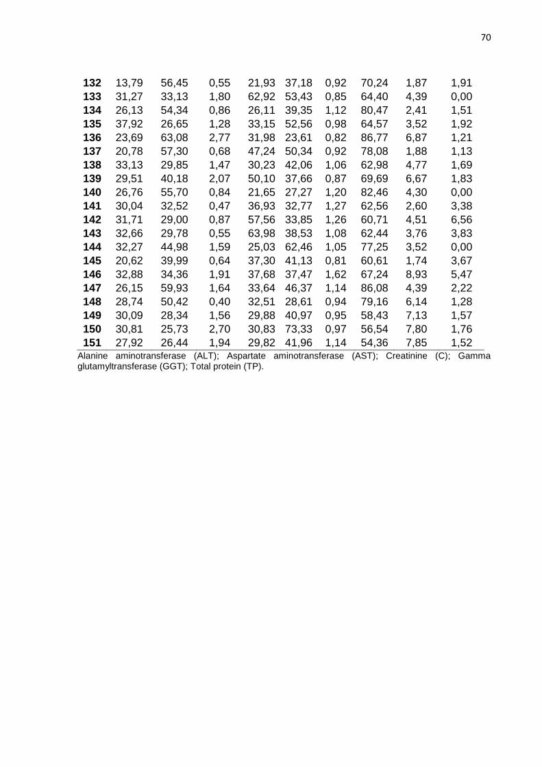
70
132 13,79 56,45 0,55 21,93 37,18 0,92 70,24 1,87 1,91
133 31,27 33,13 1,80 62,92 53,43 0,85 64,40 4,39 0,00
134 26,13 54,34 0,86 26,11 39,35 1,12 80,47 2,41 1,51
135 37,92 26,65 1,28 33,15 52,56 0,98 64,57 3,52 1,92
136 23,69 63,08 2,77 31,98 23,61 0,82 86,77 6,87 1,21
137 20,78 57,30 0,68 47,24 50,34 0,92 78,08 1,88 1,13
138 33,13 29,85 1,47 30,23 42,06 1,06 62,98 4,77 1,69
139 29,51 40,18 2,07 50,10 37,66 0,87 69,69 6,67 1,83
140 26,76 55,70 0,84 21,65 27,27 1,20 82,46 4,30 0,00
141 30,04 32,52 0,47 36,93 32,77 1,27 62,56 2,60 3,38
142 31,71 29,00 0,87 57,56 33,85 1,26 60,71 4,51 6,56
143 32,66 29,78 0,55 63,98 38,53 1,08 62,44 3,76 3,83
144 32,27 44,98 1,59 25,03 62,46 1,05 77,25 3,52 0,00
145 20,62 39,99 0,64 37,30 41,13 0,81 60,61 1,74 3,67
146 32,88 34,36 1,91 37,68 37,47 1,62 67,24 8,93 5,47
147 26,15 59,93 1,64 33,64 46,37 1,14 86,08 4,39 2,22
148 28,74 50,42 0,40 32,51 28,61 0,94 79,16 6,14 1,28
149 30,09 28,34 1,56 29,88 40,97 0,95 58,43 7,13 1,57
150 30,81 25,73 2,70 30,83 73,33 0,97 56,54 7,80 1,76
151 27,92 26,44 1,94 29,82 41,96 1,14 54,36 7,85 1,52 Alanine aminotransferase (ALT); Aspartate aminotransferase (AST); Creatinine (C); Gamma glutamyltransferase (GGT); Total protein (TP).

71
Table S9. Complete blood count of negative dogs by real-time PCR (gold standard)
from Araçatuba, SP, Brazil, endemic area for CanL.
Dogs
Complete blood count
HE HB MVC PCV MCHC PL WBC NEU LYM MON EOS
5.5-8.5 x10⁶/µL
10-18 (g/dL)
60-77 (fL)
30-36 (g/dL
32-36 (%)
160-430 (x103/µL)
6-17 (x109/L)
3.0-11.5 (x109/L)
1.0-4,8 (x109/L)
0.15-1.35 (x109/L)
0.15-1.25 (x109/L)
1 4,63 9,80 68,70 31,81 30,80 34,00 7,00 4,62 1,47 0,63 0,28 2 5,93 14,90 73,50 43,59 34,20 170,00 11,90 5,71 4,88 0,48 0,83
3 7,03 17,20 73,40 51,60 33,30 358,00 13,60 11,29 1,09 0,54 0,82
4 6,77 19,10 71,10 48,13 39,70 242,00 8,80 5,28 2,38 0,26 0,88
5 7,18 17,20 72,80 52,27 32,90 424,00 12,20 9,76 1,46 0,61 0,37
6 6,45 15,00 70,50 45,47 33,00 101,00 19,60 12,15 2,94 1,37 3,14
7 5,79 14,10 70,30 40,70 34,60 59,00 14,10 9,45 2,40 0,99 1,27
8 7,37 18,90 74,20 54,69 34,60 121,00 10,80 6,16 2,59 1,19 0,86
9 7,40 17,10 74,10 54,83 31,20 309,00 9,50 6,08 2,19 0,48 0,76
10 4,91 8,60 57,30 28,13 30,60 68,00 9,80 5,68 3,43 0,49 0,20
11 6,76 16,90 75,20 50,84 33,20 282,00 11,70 6,90 3,04 0,59 1,17
12 6,28 13,10 69,60 43,71 29,90 66,00 20,20 9,29 7,27 2,02 1,62
13 7,23 16,30 72,30 52,27 31,20 87,00 15,30 10,71 3,06 1,07 0,46
14 6,57 13,20 67,80 44,54 29,60 80,00 6,90 4,35 1,93 0,55 0,69
15 5,80 11,30 71,10 41,24 29,10 113,00 13,30 10,51 0,53 1,06 1,20
16 6,38 14,90 70,30 44,85 33,20 79,00 18,00 11,16 4,14 0,90 1,80
17 7,26 18,90 74,80 54,30 34,80 311,00 7,50 4,95 1,20 0,68 0,68
18 5,59 13,30 71,70 40,08 33,20 67,00 12,90 5,68 4,90 0,65 1,68
19 6,89 16,90 70,00 48,23 35,00 306,00 12,10 7,50 2,30 0,48 1,82 20 8,69 22,30 69,30 60,22 36,90 197,00 16,80 13,27 1,01 2,35 0,17 21 7,33 17,20 68,50 50,21 34,20 153,00 8,00 3,28 3,84 0,32 0,56 22 6,38 14,00 69,00 44,02 31,80 309,00 19,00 12,16 4,94 0,95 0,95 23 7,09 14,80 65,70 46,58 31,80 135,00 14,80 10,36 2,66 0,59 1,18 24 2,96 7,00 71,40 21,13 33,10 23,00 4,20 3,36 0,63 0,08 0,13 25 6,36 14,60 71,60 45,54 32,00 172,00 10,00 7,80 1,70 0,10 0,40 26 6,44 14,80 70,60 45,47 32,50 203,00 11,80 4,96 6,14 0,12 0,59 27 6,72 16,10 73,50 49,39 32,60 37,00 16,70 12,02 2,67 1,00 1,00 28 7,31 18,20 75,20 54,97 33,10 121,00 15,20 9,27 2,89 1,52 1,52 29 6,42 18,60 80,90 51,94 35,80 152,00 6,40 4,74 0,90 0,58 0,19 30 8,10 20,20 72,50 58,73 34,40 249,00 14,60 11,10 1,75 1,02 0,73 31 7,81 19,60 71,10 55,53 35,30 217,00 12,20 8,05 2,93 0,61 0,61 32 7,96 18,00 75,50 60,10 30,00 50,00 7,70 4,85 2,23 0,23 0,39 33 7,00 18,30 79,10 55,37 33,00 340,00 9,10 3,61 4,26 0,33 0,16 34 8,43 20,80 73,70 62,13 33,40 396,00 9,90 6,24 3,56 0,00 0,10 35 5,57 14,40 73,60 41,00 35,20 228,00 16,70 8,02 8,52 0,00 0,17 36 7,99 19,70 71,70 57,29 34,40 206,00 10,20 5,20 4,59 0,31 0,10 37 8,26 19,60 71,20 58,81 33,30 82,00 7,30 4,23 2,77 0,07 0,22 38 1,62 4,10 84,30 13,66 30,10 90,00 11,00 5,72 5,17 0,00 0,11 39 3,89 8,70 74,60 29,02 30,00 29,00 6,90 4,55 2,07 0,07 0,21 40 4,79 11,00 73,60 35,25 31,20 37,00 7,30 3,80 3,29 0,07 0,15 41 5,98 13,30 71,40 42,70 31,20 90,00 16,20 6,97 9,07 0,00 0,16

72
42 6,21 15,90 75,70 47,01 33,80 59,00 10,50 6,30 3,78 0,11 0,32 43 5,05 12,50 74,20 37,47 33,40 192,00 12,80 8,19 4,48 0,00 0,13 44 5,66 12,80 70,20 39,73 32,20 96,00 18,40 13,43 4,60 0,00 0,37 45 5,57 12,40 67,40 37,54 33,00 248,00 12,40 7,94 3,72 0,12 0,62 46 3,80 9,40 76,80 29,18 32,30 44,00 19,80 10,69 8,91 0,00 0,20 47 3,65 19,60 68,70 25,08 38,40 72,00 10,30 7,31 2,37 0,21 0,41 48 6,13 17,50 73,60 45,12 34,50 226,00 10,40 6,97 3,02 0,31 0,10 49 5,74 18,20 73,80 42,36 31,20 181,00 9,80 6,47 1,76 0,59 0,98 50 7,10 16,20 75,90 53,89 33,00 411,00 17,40 10,79 6,09 0,00 0,52 51 7,35 15,70 70,00 51,45 25,30 33,00 9,20 5,52 3,59 0,00 0,09 52 6,43 15,70 71,50 45,97 34,10 374,00 20,40 10,81 8,57 0,20 0,82 53 6,75 16,10 73,90 49,88 32,50 373,00 16,70 10,35 5,85 0,00 0,50 54 6,18 16,50 76,40 47,22 34,30 355,00 15,00 10,95 3,00 0,45 0,60 55 6,54 12,50 74,30 48,59 32,00 318,00 18,10 11,77 4,89 0,18 1,27 56 6,92 14,10 69,60 48,16 32,10 149,00 9,60 5,09 4,42 0,00 0,10 57 7,43 12,40 74,50 55,35 31,30 428,00 10,50 7,25 2,42 0,42 0,42 58 5,59 16,40 72,80 40,70 32,10 481,00 10,00 7,90 2,00 0,00 0,10 59 7,89 15,40 73,20 57,75 34,70 256,00 11,70 7,72 3,86 0,00 0,12 60 5,38 13,10 73,70 39,65 33,00 69,00 3,90 2,11 1,56 0,12 0,12 61 5,48 12,00 72,50 39,73 32,90 337,00 9,00 6,03 2,16 0,00 0,81 62 6,62 16,70 72,80 48,19 34,70 215,00 14,70 8,23 6,03 0,29 0,15 63 6,85 16,80 72,20 49,46 34,00 226,00 8,70 4,18 4,35 0,09 0,09 64 6,97 17,60 72,10 50,25 35,00 351,00 10,30 5,36 3,91 0,21 0,82 65 6,65 15,20 67,90 45,15 33,70 197,00 13,50 5,94 5,13 1,08 1,35 66 7,49 18,60 74,00 55,43 33,50 281,00 9,20 6,62 1,84 0,37 0,37 67 5,65 12,80 69,10 39,04 32,80 369,00 12,90 7,35 3,87 0,13 1,55 68 5,53 12,20 65,60 36,28 33,70 268,00 17,40 13,40 3,48 0,35 0,17 69 6,55 13,60 67,80 44,41 30,60 169,00 18,00 9,54 5,22 0,90 2,34 70 7,75 17,70 73,00 56,58 31,30 339,00 9,50 6,08 2,47 0,38 0,57 71 7,85 17,20 66,90 52,52 32,70 336,00 11,10 5,66 4,66 0,22 0,56 72 8,27 18,80 69,40 57,39 32,80 348,00 12,80 6,91 4,22 0,13 1,54 73 7,45 16,10 70,40 52,45 30,70 198,00 12,40 3,72 7,69 0,12 0,87 74 5,91 12,80 68,60 40,54 31,60 257,00 15,80 7,90 7,74 0,00 0,16 75 5,94 13,90 71,20 42,29 32,90 140,00 13,80 6,90 6,62 0,00 0,28 76 4,77 11,10 70,60 33,68 33,00 29,00 14,00 6,44 6,86 0,56 0,14 77 6,40 15,90 75,10 48,06 33,10 481,00 16,70 6,68 9,69 0,00 0,33 78 5,47 12,80 73,60 40,26 31,80 470,00 16,70 8,68 6,68 0,33 1,00 79 9,26 21,20 68,20 63,15 33,50 75,00 8,70 4,87 2,52 0,17 1,13 80 4,28 9,20 67,70 28,98 31,80 90,00 7,30 3,21 3,21 0,58 0,29 81 4,54 9,20 62,90 28,56 32,20 39,00 8,90 5,43 3,12 0,18 0,18 82 6,28 14,20 67,60 Z 33,40 213,00 12,50 7,88 3,00 0,63 1,00 83 6,23 13,50 69,20 43,11 31,30 75,00 5,10 2,35 2,24 0,15 0,36 84 6,95 16,50 72,90 50,67 32,60 85,00 8,20 4,84 2,05 0,16 1,15 85 7,17 20,30 72,50 51,98 39,10 414,00 9,00 7,02 1,89 0,00 0,09 86 7,46 17,30 69,30 51,70 23,10 236,00 7,70 5,39 1,93 0,00 0,39 87 7,88 17,10 67,40 53,11 32,20 70,00 5,90 3,66 1,77 0,41 0,06 88 5,53 11,50 63,10 34,89 33,00 79,00 5,60 2,35 2,80 0,11 0,34 89 6,18 14,30 69,50 42,95 33,30 278,00 12,20 5,49 5,37 0,24 1,10 90 7,27 17,60 70,70 51,40 34,30 270,00 9,70 4,75 4,46 0,29 0,19 91 7,78 18,70 73,90 57,49 32,50 229,00 9,50 5,32 3,90 0,19 0,10

73
92 6,89 16,60 72,20 49,75 33,40 186,00 8,70 5,66 2,35 0,00 0,70 93 5,19 13,00 74,30 38,56 33,70 545,00 20,60 15,45 3,50 0,41 1,24 94 7,67 18,60 68,30 52,39 35,50 180,00 10,70 6,10 4,28 0,21 0,11 95 7,56 18,50 69,70 52,69 35,10 266,00 9,90 5,54 3,47 0,20 0,69 96 6,57 12,00 69,50 45,66 69,50 365,00 10,00 6,20 2,70 0,70 0,40 97 5,01 13,30 76,20 38,18 34,90 215,00 12,10 7,26 4,48 0,24 0,12 98 6,59 14,90 67,20 44,28 33,70 254,00 10,50 5,78 4,31 0,21 0,21 99 7,12 17,90 70,40 50,12 35,70 292,00 12,90 8,90 2,84 1,03 0,13
100 6,56 16,70 79,40 52,09 32,10 302,00 10,90 6,98 2,51 0,44 0,98 101 6,76 14,40 68,20 46,10 31,20 308,00 19,60 10,19 4,31 1,18 3,92 102 5,18 10,80 65,50 33,93 31,80 269,00 10,90 4,14 5,45 0,33 0,98 103 6,44 13,80 63,50 40,89 33,80 170,00 20,60 10,51 5,77 0,62 3,71 104 6,64 16,00 73,60 48,87 32,70 95,00 11,40 7,07 3,08 0,46 0,80 105 7,26 16,10 67,60 49,08 32,80 221,00 9,40 6,86 2,26 0,19 0,09 106 7,94 17,10 67,30 53,44 32,00 295,00 7,90 5,61 1,98 0,24 0,08 107 8,66 19,10 70,00 60,62 31,50 145,00 9,20 5,70 2,85 0,46 0,18 108 7,41 17,70 76,10 56,39 31,40 545,00 11,30 8,59 1,92 0,57 0,23 109 8,11 17,80 67,80 54,99 32,40 124,00 10,30 6,18 2,88 0,21 1,03 110 7,82 17,50 68,80 53,80 32,50 273,00 7,20 3,17 3,24 0,22 0,58 111 6,90 16,60 71,80 49,54 24,00 115,00 9,20 4,69 3,40 0,18 0,92 112 6,86 17,40 76,50 52,48 33,20 181,00 16,60 5,15 6,14 3,15 2,16 113 5,19 10,70 65,50 33,99 31,50 117,00 13,70 4,25 6,03 2,74 0,69 114 5,39 10,60 65,40 35,25 30,10 108,00 5,10 2,45 1,99 0,36 0,31 115 5,24 9,90 59,50 31,18 31,80 48,00 7,70 4,93 1,46 0,39 0,92 116 4,66 10,60 69,40 32,34 32,80 88,00 5,90 2,36 2,07 0,89 0,59 117 6,25 11,90 61,70 38,56 30,90 165,00 11,70 4,68 5,85 0,00 1,17 118 4,96 11,00 69,50 34,47 31,90 246,00 16,20 8,26 5,83 6,48 1,46 119 5,33 11,10 64,80 34,54 64,80 109,00 9,20 5,43 2,94 0,18 0,64 120 7,53 16,60 66,80 50,30 33,00 274,00 9,80 7,45 1,76 0,29 0,29 121 6,20 13,30 69,50 43,09 30,90 318,00 15,70 9,58 3,14 1,41 1,57 122 6,02 14,10 69,00 41,54 33,90 232,00 9,30 7,07 1,30 0,28 0,65 123 7,21 15,70 69,40 50,04 31,40 314,00 18,40 13,06 2,58 2,39 0,37 124 7,44 18,10 74,70 55,58 32,60 258,00 10,50 6,83 3,36 0,21 0,11 125 6,80 14,00 71,50 48,62 31,60 351,00 16,50 9,90 4,79 0,99 0,83 126 7,87 19,70 73,90 58,16 33,90 193,00 9,00 5,58 2,34 0,45 0,36 127 7,00 18,10 75,90 53,13 34,00 215,00 12,10 7,14 2,90 0,61 1,45 128 5,93 13,60 68,30 40,50 33,50 127,00 9,10 3,91 3,09 1,91 0,18 129 6,30 14,90 72,60 45,74 32,60 380,00 11,10 8,55 1,55 0,56 0,44 130 7,71 17,80 66,50 56,16 34,70 345,00 14,50 10,59 3,77 0,00 0,15 131 7,36 19,00 76,30 56,16 33,80 149,00 14,60 6,86 5,40 0,88 1,46 132 4,68 6,90 46,40 21,72 31,70 59,00 9,00 2,88 3,60 1,62 0,90 133 7,49 17,70 69,60 52,13 33,90 199,00 8,70 5,82 2,09 0,61 0,17 134 6,59 16,50 69,50 45,80 36,00 127,00 9,80 4,80 2,25 1,67 1,08 135 6,84 16,70 75,00 51,30 32,50 258,00 18,10 8,87 5,79 1,27 2,17 136 6,97 16,70 70,20 48,93 34,10 261,00 18,30 9,33 4,39 1,65 2,93 137 7,64 16,50 67,30 51,42 32,10 154,00 8,70 4,52 3,48 0,61 0,09 138 6,94 17,30 74,00 51,36 24,90 326,00 9,30 5,12 1,67 0,93 1,58 139 6,82 18,30 72,20 49,24 37,10 245,00 18,00 9,18 6,12 1,80 0,90 140 6,18 15,00 69,50 42,95 34,90 138,00 13,70 5,34 4,66 2,19 1,51 141 7,17 17,40 71,40 51,19 34,00 260,00 15,40 8,16 5,39 0,31 1,54

74
142 6,93 17,00 76,90 53,29 31,90 60,00 9,20 5,52 2,39 0,28 1,01 143 6,79 16,50 73,10 49,63 33,20 340,00 11,70 5,62 3,74 0,94 1,40 144 6,65 16,10 71,90 47,81 33,60 27,00 24,50 12,74 6,37 2,45 2,94 145 5,29 11,90 69,80 36,92 32,40 371,00 7,20 5,69 0,72 0,14 0,65 146 6,86 17,70 72,60 49,80 35,50 280,00 9,50 5,04 3,33 0,86 0,29 147 7,58 17,00 69,70 52,83 32,10 124,00 9,00 3,87 2,52 2,16 0,45 148 6,98 15,60 70,30 49,07 31,80 136,00 10,70 8,03 1,39 0,54 0,75 149 6,63 14,30 68,80 45,61 31,30 345,00 11,20 7,95 1,68 1,23 0,34 150 6,01 14,50 70,30 42,25 34,30 315,00 9,40 5,55 3,20 0,56 0,09 151 5,37 12,70 69,30 37,21 34,10 318,00 10,90 8,72 1,53 0,55 0,11
Eosinophils (EOS); Hemoglobin (HB); Lymphocytes (LYM); Neutrophils (NEU); Mean Corpuscular Volume (MVC); Mean Corpuscular Hemoglobin Concentration (MCHC); Monocytes (MON); Platelets (PL); Package Cell Volume Hematocrit (PVC); Red Blood Cells (RBC); White Blood Cells (WBC).

75
REFERÊNCIAS DA INTRODUÇÃO GERAL
ALVAR, J. et al. Canine Leishmaniasis. In: Advances in Parasitology. [s.l: s.n.]57p.
1–88.
ARAÚJO, V. E. M. de et al. Relative Risk of Visceral Leishmaniasis in Brazil: A Spatial
Analysis in Urban Area. PLoS Neglected Tropical Diseases, v. 7, n. 11, p. e2540, 7
nov. 2013. Disponível em: <https://dx.plos.org/10.1371/journal.pntd.0002540>.
BHATIA, A. et al. Cloning, characterization and serological evaluation of K9 and K26:
Two related hydrophilic antigens of Leishmania chagasi. Molecular and Biochemical
Parasitology, v. 102, n. 2, p. 249–261, 1999.
BOGGIATTO, P. M. et al. Transplacental Transmission of Leishmania infantum as a
Means for Continued Disease Incidence in North America. PLoS Neglected Tropical
Diseases, v. 5, n. 4, p. e1019, 12 abr. 2011. Disponível em:
<http://dx.plos.org/10.1371/journal.pntd.0001019>.
BOURDOISEAU, G. et al. Lymphocyte subset abnormalities in canine leishmaniasis.
Veterinary Immunology and Immunopathology, v. 56, n. 3–4, p. 345–351, 1997.
BOUYAHIA, N. et al. Impedance spectroscopy and conductometric biosensing for
probing catalase reaction with cyanide as ligand and inhibitor. Bioelectrochemistry,
v. 80, n. 2, p. 155–161, fev. 2011. Disponível em:
<http://dx.doi.org/10.1016/j.bioelechem.2010.07.006>.
BRASIL. Manual de vigilância e controle da leishmaniose visceral / Ministério da
Saúde, Secretaria de Vigilância em Saúde, Departamento de Vigilância
Epidemiológica. 1. ed. [s.l: s.n.]
BURNS, J. M. et al. Molecular characterization of a kinesin-related antigen of
Leishmania chagasi that detects specific antibody in African and American visceral
leishmaniasis. Proceedings of the National Academy of Sciences, v. 90, n. 2, p.

76
775–779, 2006.
CAMARGO-NEVES, V. L. F. de et al. Utilização de ferramentas de análise espacial
na vigilância epidemiológica de leishmaniose visceral americana – Araçatuba , São
Paulo , Use of spatial analysis tools in the epidemiological surveillance of American
visceral leishmaniasis ,. Caderno de Saúde Pública, v. 17 (5), n. 5, p. 1263–1267,
2001.
CÂNDIDO, T. C. et al. Comparative evaluation of enzyme-linked immunosorbent assay
based on crude and purified antigen in the diagnosis of canine visceral leishmaniasis
in symptomatic and oligosymptomatic dogs. Veterinary Parasitology, v. 157, n. 3–4,
p. 175–181, 2008.
CARDIM, M. F. M. et al. Introduction and expansion of human American visceral
leishmaniasis in the state of Sao Paulo, Brazil, 1999-2011. Revista de Saude Publica,
v. 47, n. 4, p. 1–9, 2013.
CLARK, M. F.; LISTER, R. M.; BAR-JOSEPH, M. ELISA techniques. In: [s.l:
s.n.]871p. 742–766.
COURTENAY, O. et al. Heterogeneities in Leishmania infantum Infection: Using Skin
Parasite Burdens to Identify Highly Infectious Dogs. PLoS Neglected Tropical
Diseases, v. 8, n. 1, p. 26, 2014.
COUTINHO, M. T. Z. et al. Participation of Rhipicephalus sanguineus (Acari: Ixodidae)
in the epidemiology of canine visceral leishmaniasis. Veterinary Parasitology, v. 128,
n. 1–2, p. 149–155, mar. 2005. Disponível em:
<https://doi.org/10.1016/j.vetpar.2004.11.011>.
COUTINHO, M. T. Z.; LINARDI, P. M. Can fleas from dogs infected with canine visceral
leishmaniasis transfer the infection to other mammals? Veterinary Parasitology, v.
147, n. 3–4, p. 320–325, jul. 2007. Disponível em:
<https://doi.org/10.1016/j.vetpar.2007.04.008>.

77
DANTAS-TORRES, F. et al. Detection of Leishmania infantum in Rhipicephalus
sanguineus ticks from Brazil and Italy. Parasitology Research, v. 106, n. 4, p. 857–
860, 3 mar. 2010. Disponível em: <http://link.springer.com/10.1007/s00436-010-1722-
4>.
DANTAS-TORRES, F. et al. Further thoughts on “Asymptomatic dogs are highly
competent to transmit Leishmania (Leishmania) infantum chagasi to the natural
vector”. Veterinary Parasitology, v. 204, n. 3–4, p. 443–444, 2014. Disponível em:
<http://dx.doi.org/10.1016/j.vetpar.2014.04.018>.
DANTAS-TORRES, F.; OTRANTO, D. When is an “asymptomatic” dog asymptomatic?
Veterinary Parasitology, v. 202, n. 3–4, p. 341–342, 2014. Disponível em:
<http://dx.doi.org/10.1016/j.vetpar.2014.02.008>.
DE ALMEIDA LEAL, G. G. et al. Immunological profile of resistance and susceptibility
in naturally infected dogs by Leishmania infantum. Veterinary Parasitology, v. 205,
n. 3–4, p. 472–482, out. 2014. Disponível em:
<https://doi.org/10.1016/j.vetpar.2014.08.022>.
DE ASSIS, J. et al. Estudo comparativo dos métodos diagnósticos para leishmaniose
visceral em cães oriundos de Ilha Solteira, SP. Revista Brasileira de Parasitologia
Veterinaria, v. 19, n. 1, p. 17–25, 2010.
DE LA RICA, R.; STEVENS, M. M. Plasmonic ELISA for the ultrasensitive detection of
disease biomarkers with the naked eye. Nature Nanotechnology, v. 7, n. 12, p. 821–
824, 2012.
DE LA RICA, R.; STEVENS, M. M. Plasmonic ELISA for the detection of analytes at
ultralow concentrations with the naked eye. Nature Protocols, v. 8, n. 9, p. 1759–
1764, 22 set. 2013. Disponível em: <http://dx.doi.org/10.1038/nprot.2013.085>.
DE QUEIROZ, N. M. G. P. et al. Canine Visceral Leishmaniasis diagnosis by
immunohistochemistry and PCR in skin tissues in association with RIFI and ELISA-
test. Revista Brasileira de Parasitologia Veterinaria, v. 19, n. 1, p. 34–40, 2010.

78
DESJEUX, P. The increase in risk factors for leishmaniasis worldwide. Transactions
of the Royal Society of Tropical Medicine and Hygiene, v. 95, n. 3, p. 239–243,
2001.
DESJEUX, P. Leishmaniasis: current situation and new perspectives. Comparative
immunology, microbiology and infectious diseases, v. 27, n. 5, p. 305–18, 2004.
Disponível em: <http://www.ncbi.nlm.nih.gov/pubmed/15225981>.
DO ROSÁRIO, E. Y. et al. Evaluation of enzyme-linked immunosorbent assay using
crude Leishmania and recombinant antigens as a diagnostic marker for canine visceral
leishmaniasis. Memorias do Instituto Oswaldo Cruz, v. 100, n. 2, p. 197–203, 2005.
DOS-SANTOS, W. L. C. et al. Associations among immunological, parasitological and
clinical parameters in canine visceral leishmaniasis: Emaciation, spleen parasitism,
specific antibodies and leishmanin skin test reaction. Veterinary Immunology and
Immunopathology, v. 123, n. 3–4, p. 251–259, 2008.
EUSTIS, S.; EL-SAYED, M. A. Why gold nanoparticles are more precious than pretty
gold: Noble metal surface plasmon resonance and its enhancement of the radiative
and nonradiative properties of nanocrystals of different shapes. Chemical Society
Reviews, v. 35, n. 3, p. 209–217, 2006.
FEITOSA, MM, IKEDA FA , LUVIZOTTO MCR, P. S. Aspectos clínicos de cães com
leishmaniose visceral no município de Araçatuba–São Paulo (Brasil). Clínica
Veterinária, v. 5, n. 28, p. 36–44, 2000.
FERREIRA, M. G. P. A. et al. Potential role for dog fleas in the cycle of Leishmania
spp. Veterinary Parasitology, v. 165, n. 1–2, p. 150–154, out. 2009. Disponível em:
<https://linkinghub.elsevier.com/retrieve/pii/S0304401709003689>.
FERREIRA, S. de A. et al. Evaluation of the conjunctival swab for canine visceral
leishmaniasis diagnosis by PCR-hybridization in Minas Gerais State, Brazil.
Veterinary Parasitology, v. 152, n. 3–4, p. 257–263, 2008.

79
GONTIJO, C. M. F.; MELO, M. N. Visceral Leishmaniasis in Brazil: current status,
challenges and prospects. Revista Brasileira de Epidemiologia, v. 7, n. 3, p. 338–
349, 2004. Disponível em:
<http://www.scielo.br/scielo.php?script=sci_arttext&pid=S1415-
790X2004000300011&lng=pt&tlng=pt>.
GUO, L.; KIM, D. H. LSPR biomolecular assay with high sensitivity induced by
aptamer-antigen-antibody sandwich complex. Biosensors and Bioelectronics, v. 31,
n. 1, p. 567–570, 2012. Disponível em: <http://dx.doi.org/10.1016/j.bios.2011.10.047>.
IKONOMOPOULOS, J. et al. Molecular diagnosis of leishmaniosis in dogs. Veterinary
Parasitology, v. 113, n. 2, p. 99–113, abr. 2003. Disponível em:
<https://linkinghub.elsevier.com/retrieve/pii/S030440170300061X>.
LACHAUD, L. et al. Comparison of Six PCR Methods Using Peripheral Blood for
Detection of Canine Visceral Leishmaniasis. Journal of Clinical Microbiology, v. 40,
n. 1, p. 210–215, 1 jan. 2002. Disponível em:
<http://jcm.asm.org/cgi/doi/10.1128/JCM.40.1.210-215.2002>.
LAURENTI, M. D. et al. Asymptomatic dogs are highly competent to transmit
Leishmania (Leishmania) infantum chagasi to the natural vector. Veterinary
Parasitology, v. 196, n. 3–4, p. 296–300, 2013. Disponível em:
<http://dx.doi.org/10.1016/j.vetpar.2013.03.017>.
LAURICELLA, M. A. et al. An rK28-based immunoenzymatic assay for the diagnosis
of canine visceral leishmaniasis in Latin America. American Journal of Tropical
Medicine and Hygiene, v. 95, n. 1, p. 92–98, 2016.
LEITE, R. S. et al. PCR diagnosis of visceral leishmaniasis in asymptomatic dogs using
conjunctival swab samples. Veterinary Parasitology, v. 170, n. 3–4, p. 201–206,
2010. Disponível em: <http://dx.doi.org/10.1016/j.vetpar.2010.02.020>.
LIMA, V. M. F. de et al. Apoptosis in T lymphocytes from spleen tissue and peripheral
80
blood of L. (L.) chagasi naturally infected dogs. Veterinary Parasitology, v. 184, n. 2–
4, p. 147–153, 2012.
LIMA, V. M. F. et al. Anti-leishmania antibodies in cerebrospinal fluid from dogs with
visceral leishmaniasis. Brazilian Journal of Medical and Biological Research, v. 36,
n. 4, p. 485–489, 2003.
LIMA, V. M. F. et al. Evidence of Leishmania spp. Antibodies and DNA in Bush Dogs
(Speothos venaticus) in Brazil. Journal of Zoo and Wildlife Medicine, v. 40, n. 1, p.
91–94, mar. 2009. Disponível em: <http://www.bioone.org/doi/full/10.1638/2008-
0043.1>.
LIMA, W. G. et al. Canine visceral leishmaniasis: a histopathological study of lymph
nodes. Acta Tropica, v. 92, n. 1, p. 43–53, set. 2004.
LOMBARDO, G. et al. Detection of Leishmania infantum DNA by real-time PCR in
canine oral and conjunctival swabs and comparison with other diagnostic techniques.
Veterinary Parasitology, v. 184, n. 1, p. 10–17, 2012. Disponível em:
<http://dx.doi.org/10.1016/j.vetpar.2011.08.010>.
LOPES, E. G. et al. Serological and molecular diagnostic tests for canine visceral
leishmaniasis in Brazilian endemic area: One out of five seronegative dogs are
infected. Epidemiology and Infection, v. 145, n. 12, p. 2436–2444, 2017.
MANNA, L. et al. Comparison of different tissue sampling for PCR-based diagnosis
and follow-up of canine visceral leishmaniosis. Veterinary Parasitology, v. 125, n. 3–
4, p. 251–262, 2004.
MOLYNEUX, D. H.; ASHFORD, R. W. The biology of Trypanosoma and
Leishmania, parasites of man and domestic animals. [s.l: s.n.]
MORENO, J.; ALVAR, J. Canine leishmaniasis: epidemiological risk and the
experimental model. Trends in Parasitology, v. 18, n. 9, p. 399–405, set. 2002.
Disponível em: <https://linkinghub.elsevier.com/retrieve/pii/S1471492202023474>.
81
NIE, X. M. et al. Plasmonic ELISA for the ultrasensitive detection of Treponema
pallidum. Biosensors and Bioelectronics, v. 58, p. 314–319, 2014. Disponível em:
<http://dx.doi.org/10.1016/j.bios.2014.03.007>.
NUNES, C. M. et al. Avaliação da reação em cadeia pela polimerase para diagnóstico
da leishmaniose visceral em sangue de cães. Revista brasileira de parasitologia
veterinária = Brazilian journal of veterinary parasitology : Órgão Oficial do
Colégio Brasileiro de Parasitologia Veterinária, v. 16, n. 1, p. 5–9, 2007.
OGISO, M. et al. Carbohydrate immobilized on a dendrimer-coated colloidal gold
surface for fabrication of a lectin-sensing device based on localized surface plasmon
resonance spectroscopy. Biosensors and Bioelectronics, v. 41, n. 1, p. 465–470,
2013a. Disponível em: <http://dx.doi.org/10.1016/j.bios.2012.09.003>.
OGISO, M. et al. Carbohydrate immobilized on a dendrimer-coated colloidal gold
surface for fabrication of a lectin-sensing device based on localized surface plasmon
resonance spectroscopy. Biosensors and Bioelectronics, v. 41, n. 12, p. 465–470,
mar. 2013b. Disponível em:
<https://linkinghub.elsevier.com/retrieve/pii/S0956566312006094>.
OWENS, S. D. et al. Transmission of visceral leishmaniasis through blood transfusions
from infected English Foxhounds to anemic dogs. Journal of the American
Veterinary Medical Association, v. 219, n. 8, p. 1076–1083, out. 2001. Disponível
em: <https://doi.org/10.2460/javma.2001.219.1076>.
PALATNIK-DE-SOUSA, C. B. et al. Impact of canine control on the epidemiology of
canine and human visceral leishmaniasis in Brazil. The American Journal of Tropical
Medicine and Hygiene, v. 65, n. 5, p. 510–517, 1 nov. 2001. Disponível em:
<https://www.ncbi.nlm.nih.gov/pubmed/11716106>.
PATTABHI, S. et al. Design, development and evaluation of rK28-based point-of-care
tests for improving rapid diagnosis of visceral leishmaniasis. PLoS Neglected
Tropical Diseases, v. 4, n. 9, 2010.

82
PORROZZI, R. et al. Comparative evaluation of enzyme-linked immunosorbent assays
based on crude and recombinant leishmanial antigens for serodiagnosis of
symptomatic and asymptomatic Leishmania infantum visceral infections in dogs.
Clinical and Vaccine Immunology, v. 14, n. 5, p. 544–548, 2007.
QUARESMA, P. F. et al. Molecular diagnosis of canine visceral leishmaniasis:
Identification of Leishmania species by PCR-RFLP and quantification of parasite DNA
by real-time PCR. Acta Tropica, v. 111, n. 3, p. 289–294, 2009.
READY. Epidemiology of visceral leishmaniasis. Clinical Epidemiology, v. 6, n. 1, p.
147–154, 2014.
REALE, S. et al. Detection of Leishmania infantum in dogs by PCR with lymph node
aspirates and blood. Journal of Clinical Microbiology, v. 37, n. 9, p. 2931–2935,
1999.
RISSIN, D. M. et al. Single-molecule enzyme-linked immunosorbent assay detects
serum proteins at subfemtomolar concentrations. Nature Biotechnology, v. 28, n. 6,
p. 595–599, 2010. Disponível em: <http://dx.doi.org/10.1038/nbt.1641>.
ROSATI, S. et al. Prokaryotic Expression and Antigenic Characterization of Three
Recombinant Leishmania Antigens for Serological Diagnosis of Canine Leishmaniasis.
Clinical and Vaccine Immunology, v. 10, n. 6, p. 1153–1156, 2003.
ROSYPAL, A. C. et al. Serological survey of Leishmania infantum and Trypanosoma
cruzi in dogs from urban areas of Brazil and Colombia. Veterinary Parasitology, v.
149, n. 3–4, p. 172–177, 2007.
SADICK, M. D. et al. Cytokine regulation of murine leishmaniasis: Interleukin 4 is not
sufficient to mediate progressive disease in resistant C57BL/6 mice. Infection and
Immunity, v. 59, n. 12, p. 4710–4714, 1991.
SANTA ROSA I.C.A.; OLIVEIRA I.C.S. Leishmaniose visceral: breve revisão sobre

83
uma zoonose reemergente. Clinica Veterinária, v. 2, n. 11, p. 24–28, 1997.
SANTANA, C. C. et al. Inflammation and structural changes of splenic lymphoid tissue
in visceral leishmaniasis: a study on naturally infected dogs. Parasite immunology,
v. 30, n. 10, p. 515–24, 2008. Disponível em:
<http://www.ncbi.nlm.nih.gov/pubmed/18665902%0Ahttp://www.pubmedcentral.nih.g
ov/articlerender.fcgi?artid=PMC2592477>.
SATIJA, J. et al. Plasmonic-ELISA: Expanding horizons. RSC Advances, v. 6, n. 88,
p. 85440–85456, 2016. Disponível em: <http://dx.doi.org/10.1039/C6RA16750K>.
SILVA, F. L. et al. Venereal transmission of canine visceral leishmaniasis. Veterinary
Parasitology, v. 160, n. 1–2, p. 55–59, mar. 2009. Disponível em:
<https://linkinghub.elsevier.com/retrieve/pii/S0304401708006031>.
SOLANO-GALLEGO, L. et al. Prevalence of Leishmania infantum Infection in Dogs
Living in an Area of Canine Leishmaniasis Endemicity Using PCR on Several Tissues
and Serology. Journal of Clinical Microbiology, v. 39, n. 2, p. 560–563, 1 fev. 2001.
Disponível em: <http://jcm.asm.org/cgi/doi/10.1128/JCM.39.2.560-563.2001>.
SOLANO-GALLEGO, L. et al. Histological and immunohistochemical study of clinically
normal skin of Leishmania infantum-infected dogs. Journal of Comparative
Pathology, v. 130, n. 1, p. 7–12, 2004.
SOLANO-GALLEGO, L. et al. Directions for the diagnosis, clinical staging, treatment
and prevention of canine leishmaniosis. Veterinary Parasitology, v. 165, n. 1–2, p.
1–18, out. 2009. Disponível em:
<https://linkinghub.elsevier.com/retrieve/pii/S0304401709003124>.
SOLANO-GALLEGO, L. et al. LeishVet guidelines for the practical management of
canine leishmaniosis. Parasites & Vectors, v. 4, n. 1, p. 86, 2011. Disponível em:
<http://www.parasitesandvectors.com/content/4/1/86>.
STRAUSS‐AYALI, D. et al. Polymerase Chain Reaction Using Noninvasively Obtained

84
Samples, for the Detection of Leishmania infantum DNA in Dogs . The Journal of
Infectious Diseases, v. 189, n. 9, p. 1729–1733, 2004.
TAFURI, W. L. et al. An alternative immunohistochemical method for detecting
Leishmania amastigotes in paraffin-embedded canine tissues. Journal of
Immunological Methods, v. 292, n. 1–2, p. 17–23, 2004.
TEIXEIRA-NETO, R. G. et al. Canine visceral leishmaniasis in an urban setting of
Southeastern Brazil: An ecological study involving spatial analysis. Parasites and
Vectors, v. 7, n. 1, p. 1–10, 2014.
VAISH, M. et al. Evaluation of rK28 antigen for serodiagnosis of visceral Leishmaniasis
in India. Clinical Microbiology and Infection, v. 18, n. 1, p. 81–85, 2012.
VENTURIN, G. L. et al. Recombinant K28 antigen in ELISA in the diagnosis of canine
visceral leishmaniosis. Parasite Immunology, v. 37, n. 12, p. 670–673, 2015.
WHO. Control of the leishmaniasis: report of a meeting of the WHO Expert
Committee on the Control of Leishmaniases. Disponível em:
<http://apps.who.int/iris/bitstream/10665/44412/1/WHO_TRS_949_eng.pdf>. Acesso
em: 15 fev. 2019.
WHO. Epidemiological Report of the Americas. World Health Organization technical
report series, v. 7, 2019. Disponível em:
<http://iris.paho.org/xmlui/bitstream/handle/123456789/50505/Leishreport2019_eng.p
df?ua=1>.
WU, J. et al. Biomedical and clinical applications of immunoassays and
immunosensors for tumor markers. TrAC - Trends in Analytical Chemistry, v. 26, n.
7, p. 679–688, 2007.

85
ANEXO
ANEXO A – NORMAS PARA PUBLICAÇÃO NA REVISTA JOURNAL OF
IMMUNOLOGICAL METHODS
DESCRIPTION
The Journal of Immunological Methods is devoted to covering techniques for: (1) Quantitating and detecting antibodies and/or antigens. (2) Purifying immunoglobulins, lymphokines and other molecules of the immune system. (3) Isolating antigens and other substances important in immunological processes. (4) Labelling antigens and antibodies. (5) Localizing antigens and/ or antibodies in tissues and cells. (6) Detecting, and fractionating immunocompetent cells. (7) Assaying for cellular immunity. (8) Documenting cell-cell interactions. (9) Initiating immunity and unresponsiveness. (10) Transplanting tissues. (11) Studying items closely related to immunity such as complement, reticuloendothelial system and others. (12) Molecular techniques for studying immune cells and their receptors. (13) Imaging of the immune system. (14) Methods for production or their fragments in eukaryotic and prokaryotic cells. In addition the journal will publish articles on novel methods for analysing the organization, structure and expression of genes for immunologically important molecules such as immunoglobulins, T cell receptors and accessory molecules involved in antigen recognition, processing and presentation. Submitted full length manuscripts should describe new methods of broad applicability to immunology and not simply the application of an established method to a particular substance - although papers describing such applications may be considered for publication as a short Technical Note. Review articles will also be published by the Journal of Immunological Methods. In general these manuscripts are by solicitation however anyone interested in submitting a review can contact the Reviews Editor and provide an outline of the proposed review. A Computational Methods Section reflects the increasingly central role of sophisticated statistical and mathematical methods and computational models in the analysis, visualization, interpretation of the results of techniques and investigations within the general scope of the Journal. These methods can generally be divided into data-driven Immunomics methods involving high throughput assays or model-driven methods that provide a framework for data analysis, visualization, and interpretation. Descriptions of high-quality immunological databases are also of interest. This Section will publish papers that present important advances to current practice or new "gold standards" for mathematical and computational methods and analyses in the field of immunology. Areas of interest reflect the general areas of interest of the Journal, particularly novel methods for analyzing the organization, structure and expression of genes and proteins of immunologically important molecules such as immunoglobulins, T cell receptors, and various accessory molecules involved in antigen recognition, processing, and presentation, and inflammation. AUDIENCE
All immunologists IMPACT FACTOR
2017: 2.190 © Clarivate Analytics Journal Citation Reports 2018 Your Paper Your Way We now differentiate between the requirements for new and revised submissions. You may choose to submit your manuscript as a single Word or PDF file to be used in the refereeing process. Only when your paper is at the revision stage, will you be requested to put your paper in to a 'correct format' for acceptance and provide the items required for the publication of your article. To find out more, please visit the Preparation section below. INTRODUCTION
Journal of Immunological Methods is the Official Journal of the Association of Medical Laboratory Immunologists. Types of paper Review-type articles (see below) on methods, including their development, applicability and present status; these are usually invited by the Reviews Editor but interested authors may contact him at [email protected] to discuss ideas or present an outline. • There are 2 types of reviews published by JIM. The Reviews Editor will determine suitability and suggest the appropriate format (either a Short Analytical Review or a Full Review Format as detailed below). Reviews may encompass clinical, applied and basic research in immunology from academia,

86
industry and clinical operations in humans and animals. • 1. Short Analytical Reviews (SARs)
Usually not greater than 15 double spaced pages (exclusive of references) with up to 4 figures/tables and less than 40 references. These SARs should be quite specific and critical regarding a specific methodological concern, and not an attempt to provide a comprehensive treatise on the subject matter. They could include. For example, a: review of a new technique/technology, a new analysis method (e.g. statistical or software application), validation/evaluations, clinical trial outcomes of new biologics, new animal models. etc. • 2. Full Reviews Full reviews will cover a particular issue in depth with a comprehensive review of the literature. Manuscripts can be any length but are not to exceed 40 double spaced pages (exclusive of references). The liberal use of Figures is strongly encouraged. There is no limit on the number of references. It is expected that a full length review will provide an up to date, comprehensive treatise on the subject matter with detailed citations to direct readers to the appropriate original work. Research reports, full-length articles describing original work;
Technical notes (short communications and application notes, being 4 pages of type plus up to 10 references and 2-3 display items); Letters to the editors - in order to expedite publication of the letters no proofs will be sent to the authors; Computational Methods articles - articles dealing with the uses of computers and modelling techniques
in facilitating immunological research, and appropriate for on-line publication, should be submitted to one of the Section Editors, Stephen J. Merrill or Vladimir Brusic. Book reviews, News and Announcements are welcomed.
Submission checklist You can use this list to carry out a final check of your submission before you send it to the journal for review. Please check the relevant section in this Guide for Authors for more details. Ensure that the following items are present:
One author has been designated as the corresponding author with contact details: • E-mail address • Full postal address All necessary files have been uploaded: Manuscript:
• Include keywords • All figures (include relevant captions) • All tables (including titles, description, footnotes) • Ensure all figure and table citations in the text match the files provided • Indicate clearly if color should be used for any figures in print Graphical Abstracts / Highlights files (where applicable) Supplemental files (where applicable) Further considerations • Manuscript has been 'spell checked' and 'grammar checked' • All references mentioned in the Reference List are cited in the text, and vice versa • Permission has been obtained for use of copyrighted material from other sources (including the Internet) • A competing interests statement is provided, even if the authors have no competing interests to declare • Journal policies detailed in this guide have been reviewed • Referee suggestions and contact details provided, based on journal requirements For further information, visit our Support Center. BEFORE YOU BEGIN Ethics in publishing Please see our information pages on Ethics in publishing and Ethical guidelines for journal publication.

87
Policy and ethics Informed consent Investigations on human subjects must include a statement indicating that informed consent was obtained after the nature and possible consequences of the studies had been fully explained. Animal welfare Authors using experimental animals must state that their care was in accordance with institutional guidelines. For animals subjected to invasive procedures, the anesthetic, analgesic and tranquilizing agents used, as well as the amounts and frequency of administration, must be stated. Availability of Materials Publication of an article in the Journal of Immunological Methods is taken to imply that the authors are prepared to freely distribute materials used in the published experiments (e.g. antibodies, cell lines) to academic researchers for their own use. Declaration of interest All authors must disclose any financial and personal relationships with other people or organizations that could inappropriately influence (bias) their work. Examples of potential competing interests include employment, consultancies, stock ownership, honoraria, paid expert testimony, patent applications/registrations, and grants or other funding. Authors must disclose any interests in two places: 1. A summary declaration of interest statement in the title page file (if double-blind) or the manuscript file (if single-blind). If there are no interests to declare then please state this: 'Declarations of interest: none'. This summary statement will be ultimately published if the article is accepted. 2. Detailed disclosures as part of a separate Declaration of Interest form, which forms part of the journal's official records. It is important for potential interests to be declared in both places and that the information matches. More information. Submission declaration and verification
Submission of an article implies that the work described has not been published previously (except in the form of an abstract, a published lecture or academic thesis, see 'Multiple, redundant or concurrent publication' for more information), that it is not under consideration for publication elsewhere, that its publication is approved by all authors and tacitly or explicitly by the responsible authorities where AUTHOR INFORMATION PACK 10 Mar 2019 www.elsevier.com/locate/jim 6 the work was carried out, and that, if accepted, it will not be published elsewhere in the same form, in English or in any other language, including electronically without the written consent of the copyrightholder. To verify originality, your article may be checked by the originality detection service Crossref Similarity Check. Preprints Please note that preprints can be shared anywhere at any time, in line with Elsevier's sharing policy. Sharing your preprints e.g. on a preprint server will not count as prior publication (see 'Multiple, redundant or concurrent publication' for more information). Use of inclusive language
Inclusive language acknowledges diversity, conveys respect to all people, is sensitive to differences, and promotes equal opportunities. Articles should make no assumptions about the beliefs or commitments of any reader, should contain nothing which might imply that one individual is superior to another on the grounds of race, sex, culture or any other characteristic, and should use inclusive language throughout. Authors should ensure that writing is free from bias, for instance by using 'he or she', 'his/her' instead of 'he' or 'his', and by making use of job titles that are free of stereotyping (e.g. 'chairperson' instead of 'chairman' and 'flight attendant' instead of 'stewardess'). Authorship All authors should have made substantial contributions to all of the following: (1) the conception and design of the study, or acquisition of data, or analysis and interpretation of data, (2) drafting the article or revising it critically for important intellectual content, (3) final approval of the version to be submitted. Changes to authorship Authors are expected to consider carefully the list and order of authors before submitting their
manuscript and provide the definitive list of authors at the time of the original submission. Any

88
addition, deletion or rearrangement of author names in the authorship list should be made only before the manuscript has been accepted and only if approved by the journal Editor. To request such a change, the Editor must receive the following from the corresponding author: (a) the reason
for the change in author list and (b) written confirmation (e-mail, letter) from all authors that they agree with the addition, removal or rearrangement. In the case of addition or removal of authors, this includes confirmation from the author being added or removed. Only in exceptional circumstances will the Editor consider the addition, deletion or rearrangement of authors after the manuscript has been accepted. While the Editor considers the request, publication
of the manuscript will be suspended. If the manuscript has already been published in an online issue, any requests approved by the Editor will result in a corrigendum. Article transfer service This journal is part of our Article Transfer Service. This means that if the Editor feels your article is more suitable in one of our other participating journals, then you may be asked to consider transferring the article to one of those. If you agree, your article will be transferred automatically on your behalf with no need to reformat. Please note that your article will be reviewed again by the new journal. More information. Copyright Upon acceptance of an article, authors will be asked to complete a 'Journal Publishing Agreement' (see more information on this). An e-mail will be sent to the corresponding author confirming receipt of the manuscript together with a 'Journal Publishing Agreement' form or a link to the online version of this agreement. Subscribers may reproduce tables of contents or prepare lists of articles including abstracts for internal circulation within their institutions. Permission of the Publisher is required for resale or distribution outside the institution and for all other derivative works, including compilations and translations. If excerpts from other copyrighted works are included, the author(s) must obtain written permission from the copyright owners and credit the source(s) in the article. Elsevier has preprinted forms for use by authors in these cases. For gold open access articles: Upon acceptance of an article, authors will be asked to complete an 'Exclusive License Agreement' (more information). Permitted third party reuse of gold open access articles is determined by the author's choice of user license. Author rights As an author you (or your employer or institution) have certain rights to reuse your work. More information. Elsevier supports responsible sharing Find out how you can share your research published in Elsevier journals. Role of the funding source You are requested to identify who provided financial support for the conduct of the research and/or preparation of the article and to briefly describe the role of the sponsor(s), if any, in study design; in the collection, analysis and interpretation of data; in the writing of the report; and in the decision to submit the article for publication. If the funding source(s) had no such involvement then this should be stated. Funding body agreements and policies Elsevier has established a number of agreements with funding bodies which allow authors to comply with their funder's open access policies. Some funding bodies will reimburse the author for the gold open access publication fee. Details of existing agreements are available online. Open access This journal offers authors a choice in publishing their research: Subscription
• Articles are made available to subscribers as well as developing countries and patient groups through our universal access programs.
89
• No open access publication fee payable by authors. • The Author is entitled to post the accepted manuscript in their institution's repository and make this public after an embargo period (known as green Open Access). The published journal article cannot be shared publicly, for example on ResearchGate or Academia.edu, to ensure the sustainability of peerreviewed research in journal publications. The embargo period for this journal can be found below. Gold open access • Articles are freely available to both subscribers and the wider public with permitted reuse. • A gold open access publication fee is payable by authors or on their behalf, e.g. by their research funder or institution. Regardless of how you choose to publish your article, the journal will apply the same peer review criteria and acceptance standards. For gold open access articles, permitted third party (re)use is defined by the following Creative Commons user licenses: Creative Commons Attribution (CC BY) Lets others distribute and copy the article, create extracts, abstracts, and other revised versions, adaptations or derivative works of or from an article (such as a translation), include in a collective work (such as an anthology), text or data mine the article, even for commercial purposes, as long as they credit the author(s), do not represent the author as endorsing their adaptation of the article, and do not modify the article in such a way as to damage the author's honor or reputation. Creative Commons Attribution-NonCommercial-NoDerivs (CC BY-NC-ND) For non-commercial purposes, lets others distribute and copy the article, and to include in a collective work (such as an anthology), as long as they credit the author(s) and provided they do not alter or modify the article. The gold open access publication fee for this journal is USD 2950, excluding taxes. Learn more about
Elsevier's pricing policy: https://www.elsevier.com/openaccesspricing. Green open access Authors can share their research in a variety of different ways and Elsevier has a number of green open access options available. We recommend authors see our open access page for further information. Authors can also self-archive their manuscripts immediately and enable public access from their institution's repository after an embargo period. This is the version that has been accepted for publication and which typically includes author-incorporated changes suggested during submission, peer review and in editor-author communications. Embargo period: For subscription articles, an article becomes freely available to the public. This is the embargo period and it begins from the date the article is formally published online in its final and fully citable form. Find out more. This journal has an embargo period of 12 months. Elsevier Researcher Academy Researcher Academy is a free e-learning platform designed to support early and mid-career researchers throughout their research journey. The "Learn" environment at Researcher Academy offers several interactive modules, webinars, downloadable guides and resources to guide you through the process of writing for research and going through peer review. Feel free to use these free resources to improve your submission and navigate the publication process with ease. Language (usage and editing services) Please write your text in good English (American or British usage is accepted, but not a mixture of these). Authors who feel their English language manuscript may require editing to eliminate possible grammatical or spelling errors and to conform to correct scientific English may wish to use the English Language Editing service available from Elsevier's WebShop. Submission
Our online submission system guides you stepwise through the process of entering your article details and uploading your files. The system converts your article files to a single PDF file used in

90
the peer-review process. Editable files (e.g., Word, LaTeX) are required to typeset your article for final publication. All correspondence, including notification of the Editor's decision and requests for revision, is sent by e-mail. Submit your article Please submit your article via http://ees.elsevier.com/jim. Please suggest 6 potential reviewers for your submission, providing contact details and specific reasons for your suggestions. Please note that the journal may not use your suggestions, but your help is appreciated and will speed up the selection of appropriate reviewers. PREPARATION NEW SUBMISSIONS
Submission to this journal proceeds totally online and you will be guided stepwise through the creation and uploading of your files. The system automatically converts your files to a single PDF file, which is used in the peer-review process. As part of the Your Paper Your Way service, you may choose to submit your manuscript as a single file to be used in the refereeing process. This can be a PDF file or a Word document, in any format or layout that can be used by referees to evaluate your manuscript. It should contain high enough quality figures for refereeing. If you prefer to do so, you may still provide all or some of the source files at the initial submission. Please note that individual figure files larger than 10 MB must be uploaded separately. References There are no strict requirements on reference formatting at submission. References can be in any style or format as long as the style is consistent. Where applicable, author(s) name(s), journal title/ book title, chapter title/article title, year of publication, volume number/book chapter and the article number or pagination must be present. Use of DOI is highly encouraged. The reference style used by the journal will be applied to the accepted article by Elsevier at the proof stage. Note that missing data will be highlighted at proof stage for the author to correct. Formatting requirements There are no strict formatting requirements but all manuscripts must contain the essential elements needed to convey your manuscript, for example Abstract, Keywords, Introduction, Materials and Methods, Results, Conclusions, Artwork and Tables with Captions. If your article includes any Videos and/or other Supplementary material, this should be included in your initial submission for peer review purposes. Divide the article into clearly defined sections. Figures and tables embedded in text
Please ensure the figures and the tables included in the single file are placed next to the relevant text in the manuscript, rather than at the bottom or the top of the file. The corresponding caption should be placed directly below the figure or table. Peer review This journal operates a single blind review process. All contributions will be initially assessed by the editor for suitability for the journal. Papers deemed suitable are then typically sent to a minimum of two independent expert reviewers to assess the scientific quality of the paper. The Editor is responsible for the final decision regarding acceptance or rejection of articles. The Editor's decision is final. More information on types of peer review. REVISED SUBMISSIONS Use of word processing software
Regardless of the file format of the original submission, at revision you must provide us with an editable file of the entire article. Keep the layout of the text as simple as possible. Most formatting

91
codes will be removed and replaced on processing the article. The electronic text should be prepared in a way very similar to that of conventional manuscripts (see also the Guide to Publishing with Elsevier). See also the section on Electronic artwork. To avoid unnecessary errors you are strongly advised to use the 'spell-check' and 'grammar-check' functions of your word processor. Article structure Subdivision - numbered sections Divide your article into clearly defined and numbered sections. Subsections should be numbered 1.1 (then 1.1.1, 1.1.2, ...), 1.2, etc. (the abstract is not included in section numbering). Use this numbering also for internal cross-referencing: do not just refer to 'the text'. Any subsection may be given a brief heading. Each heading should appear on its own separate line. Introduction State the objectives of the work and provide an adequate background, avoiding a detailed literature survey or a summary of the results. Material and methods Provide sufficient details to allow the work to be reproduced by an independent researcher. Methods that are already published should be summarized, and indicated by a reference. If quoting directly from a previously published method, use quotation marks and also cite the source. Any modifications to existing methods should also be described. Theory/calculation A Theory section should extend, not repeat, the background to the article already dealt with in the Introduction and lay the foundation for further work. In contrast, a Calculation section represents a practical development from a theoretical basis. Results
Results should be clear and concise. Discussion This should explore the significance of the results of the work, not repeat them. A combined Results and Discussion section is often appropriate. Avoid extensive citations and discussion of published literature. Conclusions The main conclusions of the study may be presented in a short Conclusions section, which may stand alone or form a subsection of a Discussion or Results and Discussion section. Appendices If there is more than one appendix, they should be identified as A, B, etc. Formulae and equations in appendices should be given separate numbering: Eq. (A.1), Eq. (A.2), etc.; in a subsequent appendix, Eq. (B.1) and so on. Similarly for tables and figures: Table A.1; Fig. A.1, etc. Essential title page information • Title. Concise and informative. Titles are often used in information-retrieval systems. Avoid abbreviations and formulae where possible. • Author names and affiliations. Please clearly indicate the given name(s) and family name(s) of each author and check that all names are accurately spelled. You can add your name between parentheses in your own script behind the English transliteration. Present the authors' affiliation addresses (where the actual work was done) below the names. Indicate all affiliations with a lower case superscript letter immediately after the author's name and in front of the appropriate address. Provide the full postal address of each affiliation, including the country name and, if available, the e-mail address of each author. • Corresponding author. Clearly indicate who will handle correspondence at all stages of refereeing and publication, also post-publication. This responsibility includes answering any future queries about Methodology and Materials. Ensure that the e-mail address is given and that contact details are kept up to date by the corresponding author. • Present/permanent address. If an author has moved since the work described in the article was

92
done, or was visiting at the time, a 'Present address' (or 'Permanent address') may be indicated as a footnote to that author's name. The address at which the author actually did the work must be retained as the main, affiliation address. Superscript Arabic numerals are used for such footnotes. Abstract A concise and factual abstract is required. The abstract should state briefly the purpose of the research, the principal results and major conclusions. An abstract is often presented separately from the article, so it must be able to stand alone. For this reason, References should be avoided, but if essential, then cite the author(s) and year(s). Also, non-standard or uncommon abbreviations should be avoided, but if essential they must be defined at their first mention in the abstract itself. Graphical abstract Although a graphical abstract is optional, its use is encouraged as it draws more attention to the online article. The graphical abstract should summarize the contents of the article in a concise, pictorial form designed to capture the attention of a wide readership. Graphical abstracts should be submitted as a separate file in the online submission system. Image size: Please provide an image with a minimum of 531 × 1328 pixels (h × w) or proportionally more. The image should be readable at a size of 5 × 13 cm using a regular screen resolution of 96 dpi. Preferred file types: TIFF, EPS, PDF or MS Office files. You can view Example Graphical Abstracts on our information site. Authors can make use of Elsevier's Illustration Services to ensure the best presentation of their images and in accordance with all technical requirements. Highlights
Highlights are mandatory for this journal. They consist of a short collection of bullet points that convey the core findings of the article and should be submitted in a separate editable file in the online submission system. Please use 'Highlights' in the file name and include 3 to 5 bullet points (maximum 85 characters, including spaces, per bullet point). You can view example Highlights on our information site. Keywords Immediately after the abstract, provide a maximum of 6 keywords, using American spelling and avoiding general and plural terms and multiple concepts (avoid, for example, 'and', 'of'). Be sparing with abbreviations: only abbreviations firmly established in the field may be eligible. These keywords will be used for indexing purposes. Abbreviations
Define abbreviations that are not standard in this field in a footnote to be placed on the first page of the article. Such abbreviations that are unavoidable in the abstract must be defined at their first mention there, as well as in the footnote. Ensure consistency of abbreviations throughout the article. Acknowledgements Collate acknowledgements in a separate section at the end of the article before the references and do not, therefore, include them on the title page, as a footnote to the title or otherwise. List here those individuals who provided help during the research (e.g., providing language help, writing assistance or proof reading the article, etc.). Formatting of funding sources
List funding sources in this standard way to facilitate compliance to funder's requirements: Funding: This work was supported by the National Institutes of Health [grant numbers xxxx, yyyy]; the Bill & Melinda Gates Foundation, Seattle, WA [grant number zzzz]; and the United States Institutes of Peace [grant number aaaa]. It is not necessary to include detailed descriptions on the program or type of grants and awards. When funding is from a block grant or other resources available to a university, college, or other research institution, submit the name of the institute or organization that provided the funding. If no funding has been provided for the research, please include the following sentence: This research did not receive any specific grant from funding agencies in the public, commercial, or not-for-profit sectors. Nomenclature and units Follow internationally accepted rules and conventions: use the international system of units (SI). If other quantities are mentioned, give their equivalent in SI. You are urged to consult IUB: Biochemical Nomenclature and Related Documents for further information.

93
DNA sequences and GenBank Accession numbers Many Elsevier journals cite "gene accession numbers" in their running text and footnotes. Gene accession numbers refer to genes or DNA sequences about which further information can be found in the database at the National Center for Biotechnical Information (NCBI) at the National Library of Medicine. Elsevier authors wishing to enable other scientists to use the accession numbers cited in their papers via links to these sources, should type this information in the following manner": For each and every accession number cited in an article, authors should type the accession number in bold, underlined text. Letters in the accession number should always be capitalised. (See Example
1 below). This combination of letters and format will enable Elsevier's typesetters to recognize the relevant texts as accession numbers and add the required link to GenBank's sequences. Example 1: "GenBank accession nos. AI631510, AI631511, AI632198, and BF223228, a B-cell tumor from a chronic lymphatic leukemia (GenBank accession no. BE675048), and a T-cell lymphoma (GenBank accession no. AA361117)".
Authors are encouraged to check accession numbers used very carefully. An error in a letter or number can result in a dead link. In the final version of the printed article, the accession number text will not appear bold or underlined
(see Example 2 below). Example 2: "GenBank accession nos. AI631510, AI631511, AI632198, and BF223228), a B-cell
tumor from a chronic lymphatic leukemia (GenBank accession no. BE675048), and a T-cell lymphoma (GenBank accession no. AA361117)". In the final version of the electronic copy, the accession number text will be linked to the appropriate
source in the NCBI databases enabling readers to go directly to that source from the article (see Example 3 below). Example 3: "GenBank accession nos. AI631510, AI631511, AI632198, and BF223228), a B-cell
tumor from a chronic lymphatic leukemia (GenBank accession no. BE675048), and a T-cell lymphoma (GenBank accession no. AA361117)". Footnotes Footnotes should be used sparingly. Number them consecutively throughout the article. Many word processors build footnotes into the text, and this feature may be used. Should this not be the case, indicate the position of footnotes in the text and present the footnotes themselves separately at the end of the article. Artwork Electronic artwork General points • Make sure you use uniform lettering and sizing of your original artwork. • Preferred fonts: Arial (or Helvetica), Times New Roman (or Times), Symbol, Courier. • Number the illustrations according to their sequence in the text. • Use a logical naming convention for your artwork files. • Indicate per figure if it is a single, 1.5 or 2-column fitting image. • For Word submissions only, you may still provide figures and their captions, and tables within a single file at the revision stage. • Please note that individual figure files larger than 10 MB must be provided in separate source files. A detailed guide on electronic artwork is available. You are urged to visit this site; some excerpts from the detailed information are given here.
Formats Regardless of the application used, when your electronic artwork is finalized, please 'save as' or convert the images to one of the following formats (note the resolution requirements for line drawings, halftones, and line/halftone combinations given below): EPS (or PDF): Vector drawings. Embed the font or save the text as 'graphics'. TIFF (or JPG): Color or grayscale photographs (halftones): always use a minimum of 300 dpi. TIFF (or JPG): Bitmapped line drawings: use a minimum of 1000 dpi. TIFF (or JPG): Combinations bitmapped line/half-tone (color or grayscale): a minimum of 500 dpi is required.

94
Please do not:
• Supply files that are optimized for screen use (e.g., GIF, BMP, PICT, WPG); the resolution is too low. • Supply files that are too low in resolution. • Submit graphics that are disproportionately large for the content. Color artwork
Please make sure that artwork files are in an acceptable format (TIFF (or JPEG), EPS (or PDF), or MS Office files) and with the correct resolution. If, together with your accepted article, you submit usable color figures then Elsevier will ensure, at no additional charge, that these figures will appear in color online (e.g., ScienceDirect and other sites) regardless of whether or not these illustrations are reproduced in color in the printed version. For color reproduction in print, you will receive information regarding the costs from Elsevier after receipt of your accepted article. Please
indicate your preference for color: in print or online only. Further information on the preparation of electronic artwork. Figure captions Ensure that each illustration has a caption. A caption should comprise a brief title (not on the figure
itself) and a description of the illustration. Keep text in the illustrations themselves to a minimum but explain all symbols and abbreviations used. Tables
Please submit tables as editable text and not as images. Tables can be placed either next to the relevant text in the article, or on separate page(s) at the end. Number tables consecutively in accordance with their appearance in the text and place any table notes below the table body. Be sparing in the use of tables and ensure that the data presented in them do not duplicate results described elsewhere in the article. Please avoid using vertical rules and shading in table cells. References Citation in text Please ensure that every reference cited in the text is also present in the reference list (and vice versa). Any references cited in the abstract must be given in full. Unpublished results and personal communications are not recommended in the reference list, but may be mentioned in the text. If these references are included in the reference list they should follow the standard reference style of the journal and should include a substitution of the publication date with either 'Unpublished results' or 'Personal communication'. Citation of a reference as 'in press' implies that the item has been accepted for publication. Reference links Increased discoverability of research and high quality peer review are ensured by online links to the sources cited. In order to allow us to create links to abstracting and indexing services, such as Scopus, CrossRef and PubMed, please ensure that data provided in the references are correct. Please note that incorrect surnames, journal/book titles, publication year and pagination may prevent link creation. When copying references, please be careful as they may already contain errors. Use of the DOI is highly encouraged. A DOI is guaranteed never to change, so you can use it as a permanent link to any electronic article. An example of a citation using DOI for an article not yet in an issue is: VanDecar J.C., Russo R.M., James D.E., Ambeh W.B., Franke M. (2003). Aseismic continuation of the Lesser Antilles slab beneath northeastern Venezuela. Journal of Geophysical Research, https://doi.org/10.1029/2001JB000884. Please note the format of such citations should be in the same style as all other references in the paper. Web references As a minimum, the full URL should be given and the date when the reference was last accessed. Any further information, if known (DOI, author names, dates, reference to a source publication, etc.), should also be given. Web references can be listed separately (e.g., after the reference list) under a different heading if desired, or can be included in the reference list. Data references
This journal encourages you to cite underlying or relevant datasets in your manuscript by citing them in your text and including a data reference in your Reference List. Data references should include the

95
following elements: author name(s), dataset title, data repository, version (where available), year, and global persistent identifier. Add [dataset] immediately before the reference so we can properly identify it as a data reference. The [dataset] identifier will not appear in your published article. References in a special issue Please ensure that the words 'this issue' are added to any references in the list (and any citations in the text) to other articles in the same Special Issue. Reference management software Most Elsevier journals have their reference template available in many of the most popular reference management software products. These include all products that support Citation Style Language styles, such as Mendeley. Using citation plug-ins from these products, authors only need to select the appropriate journal template when preparing their article, after which citations and bibliographies will be automatically formatted in the journal's style. If no template is yet available for this journal, please follow the format of the sample references and citations as shown in this Guide. If you use reference management software, please ensure that you remove all field codes before submitting the electronic manuscript. More information on how to remove field codes from different reference management software. Users of Mendeley Desktop can easily install the reference style for this journal by clicking the following link:http://open.mendeley.com/use-citation-style/journal-of-immunological-methods When preparing your manuscript, you will then be able to select this style using the Mendeley plugins for Microsoft Word or LibreOffice. Reference formatting There are no strict requirements on reference formatting at submission. References can be in any style or format as long as the style is consistent. Where applicable, author(s) name(s), journal title/ book title, chapter title/article title, year of publication, volume number/book chapter and the article number or pagination must be present. Use of DOI is highly encouraged. The reference style used by the journal will be applied to the accepted article by Elsevier at the proof stage. Note that missing data will be highlighted at proof stage for the author to correct. If you do wish to format the references yourself they should be arranged according to the following examples: Reference style Text: All citations in the text should refer to: 1. Single author: the author's name (without initials, unless there is ambiguity) and the year of publication; 2. Two authors: both authors' names and the year of publication; 3. Three or more authors: first author's name followed by 'et al.' and the year of publication.
Citations may be made directly (or parenthetically). Groups of references can be listed either first alphabetically, then chronologically, or vice versa. Examples: 'as demonstrated (Allan, 2000a, 2000b, 1999; Allan and Jones, 1999)…. Or, as demonstrated (Jones, 1999; Allan, 2000)… Kramer et al. (2010) have recently shown …' List: References should be arranged first alphabetically and then further sorted chronologically if necessary. More than one reference from the same author(s) in the same year must be identified by the letters 'a', 'b', 'c', etc., placed after the year of publication. Examples: Reference to a journal publication: Van der Geer, J., Hanraads, J.A.J., Lupton, R.A., 2010. The art of writing a scientific article. J. Sci. Commun. 163, 51–59. https://doi.org/10.1016/j.Sc.2010.00372. Reference to a journal publication with an article number: Van der Geer, J., Hanraads, J.A.J., Lupton, R.A., 2018. The art of writing a scientific article. Heliyon.19, e00205. https://doi.org/10.1016/j.heliyon.2018.e00205. Reference to a book:
96
Strunk Jr., W., White, E.B., 2000. The Elements of Style, fourth ed. Longman, New York. Reference to a chapter in an edited book: Mettam, G.R., Adams, L.B., 2009. How to prepare an electronic version of your article, in: Jones, B.S., Smith , R.Z. (Eds.), Introduction to the Electronic Age. E-Publishing Inc., New York, pp. 281–304. Reference to a website: Cancer Research UK, 1975. Cancer statistics reports for the UK. http://www.cancerresearchuk.org/ aboutcancer/statistics/cancerstatsreport/ (accessed 13 March 2003). Reference to a dataset: [dataset] Oguro, M., Imahiro, S., Saito, S., Nakashizuka, T., 2015. Mortality data for Japanese oak wilt disease and surrounding forest compositions. Mendeley Data, v1. https://doi.org/10.17632/ xwj98nb39r.1. Video
Elsevier accepts video material and animation sequences to support and enhance your scientific research. Authors who have video or animation files that they wish to submit with their article are strongly encouraged to include links to these within the body of the article. This can be done in the same way as a figure or table by referring to the video or animation content and noting in the body text where it should be placed. All submitted files should be properly labeled so that they directly relate to the video file's content. . In order to ensure that your video or animation material is directly usable, please provide the file in one of our recommended file formats with a preferred maximum size of 150 MB per file, 1 GB in total. Video and animation files supplied will be published online in the electronic version of your article in Elsevier Web products, including ScienceDirect. Please supply 'stills' with your files: you can choose any frame from the video or animation or make a separate image. These will be used instead of standard icons and will personalize the link to your video data. For more detailed instructions please visit our video instruction pages. Note: since video and animation cannot be embedded in the print version of the journal, please provide text for both the electronic and the print version for the portions of the article that refer to this content. Data visualization
Include interactive data visualizations in your publication and let your readers interact and engage more closely with your research. Follow the instructions here to find out about available data visualization options and how to include them with your article. Supplementary material Supplementary material such as applications, images and sound clips, can be published with your article to enhance it. Submitted supplementary items are published exactly as they are received (Excel or PowerPoint files will appear as such online). Please submit your material together with the article and supply a concise, descriptive caption for each supplementary file. If you wish to make changes to supplementary material during any stage of the process, please make sure to provide an updated file. Do not annotate any corrections on a previous version. Please switch off the 'Track Changes' option in Microsoft Office files as these will appear in the published version. Research data
This journal encourages and enables you to share data that supports your research publication where appropriate, and enables you to interlink the data with your published articles. Research data refers to the results of observations or experimentation that validate research findings. To facilitate reproducibility and data reuse, this journal also encourages you to share your software, code, models, algorithms, protocols, methods and other useful materials related to the project. Below are a number of ways in which you can associate data with your article or make a statement about the availability of your data when submitting your manuscript. If you are sharing data in one of these ways, you are encouraged to cite the data in your manuscript and reference list. Please refer to the "References" section for more information about data citation. For more information on depositing, sharing and using research data and other relevant research materials, visit the research data page. Data linking If you have made your research data available in a data repository, you can link your article directly to

97
the dataset. Elsevier collaborates with a number of repositories to link articles on ScienceDirect with relevant repositories, giving readers access to underlying data that gives them a better understanding of the research described. There are different ways to link your datasets to your article. When available, you can directly link your dataset to your article by providing the relevant information in the submission system. For more information, visit the database linking page. For supported data repositories a repository banner will automatically appear next to your published article on ScienceDirect. In addition, you can link to relevant data or entities through identifiers within the text of your manuscript, using the following format: Database: xxxx (e.g., TAIR: AT1G01020; CCDC: 734053; PDB: 1XFN). Mendeley Data This journal supports Mendeley Data, enabling you to deposit any research data (including raw and processed data, video, code, software, algorithms, protocols, and methods) associated with your manuscript in a free-to-use, open access repository. During the submission process, after uploading your manuscript, you will have the opportunity to upload your relevant datasets directly to Mendeley Data. The datasets will be listed and directly accessible to readers next to your published article online. For more information, visit the Mendeley Data for journals page. Data in Brief You have the option of converting any or all parts of your supplementary or additional raw data into one or multiple data articles, a new kind of article that houses and describes your data. Data articles ensure that your data is actively reviewed, curated, formatted, indexed, given a DOI and publicly available to all upon publication. You are encouraged to submit your article for Data in Brief as an additional item directly alongside the revised version of your manuscript. If your research article is accepted, your data article will automatically be transferred over to Data in Brief where it will be editorially reviewed and published in the open access data journal, Data in Brief. Please note an open access fee of 500 USD is payable for publication in Data in Brief. Full details can be found on the Data in Brief website. Please use this template to write your Data in Brief. Data statement To foster transparency, we encourage you to state the availability of your data in your submission. This may be a requirement of your funding body or institution. If your data is unavailable to access or unsuitable to post, you will have the opportunity to indicate why during the submission process, for example by stating that the research data is confidential. The statement will appear with your published article on ScienceDirect. For more information, visit the Data Statement page. AFTER ACCEPTANCE Online proof correction Corresponding authors will receive an e-mail with a link to our online proofing system, allowing annotation and correction of proofs online. The environment is similar to MS Word: in addition to editing text, you can also comment on figures/tables and answer questions from the Copy Editor. Web-based proofing provides a faster and less error-prone process by allowing you to directly type your corrections, eliminating the potential introduction of errors. If preferred, you can still choose to annotate and upload your edits on the PDF version. All instructions for proofing will be given in the e-mail we send to authors, including alternative methods to the online version and PDF. We will do everything possible to get your article published quickly and accurately. Please use this proof only for checking the typesetting, editing, completeness and correctness of the text, tables and figures. Significant changes to the article as accepted for publication will only be considered at this stage with permission from the Editor. It is important to ensure that all corrections are sent back to us in one communication. Please check carefully before replying, as inclusion of any subsequent corrections cannot be guaranteed. Proofreading is solely your responsibility. Offprints The corresponding author will, at no cost, receive a customized Share Link providing 50 days free

98
access to the final published version of the article on ScienceDirect. The Share Link can be used for sharing the article via any communication channel, including email and social media. For an extra charge, paper offprints can be ordered via the offprint order form which is sent once the article is accepted for publication. Both corresponding and co-authors may order offprints at any time via Elsevier's Webshop. Corresponding authors who have published their article gold open access do not receive a Share Link as their final published version of the article is available open access on ScienceDirect and can be shared through the article DOI link. AUTHOR INQUIRIES
Visit the Elsevier Support Center to find the answers you need. Here you will find everything from Frequently Asked Questions to ways to get in touch. You can also check the status of your submitted article or find out when your accepted article will be published. © Copyright 2018 Elsevier | https://www.elsevier.com

99
ANEXO B – NORMAS PARA PUBLICAÇÃO NA REVISTA VETERINARY
PARASITOLOGY
DESCRIPTION
Veterinary Parasitology has an open access mirror journal, Veterinary Parasitology: X. Both journals share the same aims and scope, editorial team, submission system and rigorous peer review. The difference between the journals is the access model under which the journals will publish your work and the indexation status. Veterinary Parasitology: X will be indexed in Scopus if the parent is also indexed there; if the parent is indexed in MEDLINE, then Veterinary Parasitology: X will also be eligible for fast inclusion in PubMed Central. However, please note that Veterinary Parasitology: X will not have a CiteScore or Impact Factor initially. Applications for inclusion in Science Citation Index / Social Sciences Citation Index and any other relevant citation indexing databases will be made as soon as possible. Types of contribution
1. Original research papers (Regular Papers) 2. Review articles 3. Short Communications 4. Letters to the Editor 5. Book Reviews Original research papers should report the results of original research. The material should not have been previously published elsewhere, except in a preliminary form. Review articles should cover subjects falling within the scope of the journal which are of active current interest.
They may be submitted or invited. Short Communications should consist of original observations or new methods within the scope of the journal. The Communications should be concise and limited to 3,000 words and can include up to 3 figures or tables, combined. Not more than 20 references should be cited. The Communications need not be formally structured as are full papers, but should give sufficient methods and data necessary for their comprehension. Letters to the Editor offering comment or useful critique on material published in the journal are welcomed. The decision to publish submitted letters rests purely with the Editors-in-Chief. It is hoped that the publication of such letters will permit an exchange of views which will be of benefit to both the journal and its readers. Book Reviews will be included in the journal on a range of relevant books which are not more than 2 years old and were written in English. Book reviews will be solicited by the Book Review Editor. Unsolicited reviews will not usually be accepted, but suggestions for appropriate books for review may be sent to one of the Book Review Editors noted below: Dr G. Baneth School of Veterinary Medicine, Hewbrew University, Rehovot, Israel [email protected] Dr E. Papadopoulos Faculty of Veterinary Medicine, Aristotle University of Thessaloniki, Thessaloniki, Greece [email protected] Submission checklist
You can use this list to carry out a final check of your submission before you send it to the journal for review. Please check the relevant section in this Guide for Authors for more details. Ensure that the following items are present:
One author has been designated as the corresponding author with contact details: • E-mail address • Full postal address

100
All necessary files have been uploaded: Manuscript: • Include keywords • All figures (include relevant captions) • All tables (including titles, description, footnotes) • Ensure all figure and table citations in the text match the files provided • Indicate clearly if color should be used for any figures in print Graphical Abstracts / Highlights files (where applicable) Supplemental files (where applicable) Further considerations • Manuscript has been 'spell checked' and 'grammar checked' • All references mentioned in the Reference List are cited in the text, and vice versa • Permission has been obtained for use of copyrighted material from other sources (including the Internet) • A competing interests statement is provided, even if the authors have no competing interests to declare • Journal policies detailed in this guide have been reviewed • Referee suggestions and contact details provided, based on journal requirements For further information, visit our Support Center. Ethics in publishing
Please see our information pages on Ethics in publishing and Ethical guidelines for journal publication. Animal Welfare
Circumstances relating to animal experimentation must meet the International Guiding Principles for Biomedical Research Involving Animals as issued by the Council for the International Organizations of Medical Sciences. They are obtainable from the following URL: https://grants.nih.gov/grants/olaw/guiding_principles_2012.pdf. Unnecessary cruelty in animal experimentation is not acceptable to the Editors of Veterinary Parasitology. Please include an animal welfare statement under the heading "Declaration of interest" at the end of the text. Please include this under a heading "Conflict of interest statement" at the end of the text. Declaration of interest
All authors must disclose any financial and personal relationships with other people or organizations that could inappropriately influence (bias) their work. Examples of potential competing interests include employment, consultancies, stock ownership, honoraria, paid expert testimony, patent applications/registrations, and grants or other funding. Authors must disclose any interests in two places: 1. A summary declaration of interest statement in the title page file (if double-blind) or the manuscript file (if single-blind). If there are no interests to declare then please state this: 'Declarations of interest: none'. This summary statement will be ultimately published if the article is accepted. 2. Detailed disclosures as part of a separate Declaration of Interest form, which forms part of the journal's official records. It is important for potential interests to be declared in both places and that the information matches. More information. Submission declaration and verification
Submission of an article implies that the work described has not been published previously (except in the form of an abstract, a published lecture or academic thesis, see 'Multiple, redundant or concurrent publication' for more information), that it is not under consideration for publication elsewhere, that its publication is approved by all authors and tacitly or explicitly by the responsible authorities where the work was carried out, and that, if accepted, it will not be published elsewhere in the same form, in English or in any other language, including electronically without the written consent of the copyright-holder. To verify originality, your article may be checked by the originality detection service Crossref Similarity Check. Preprints
Please note that preprints can be shared anywhere at any time, in line with Elsevier's sharing policy. Sharing your preprints e.g. on a preprint server will not count as prior publication (see 'Multiple, redundant or concurrent publication' for more information). Use of inclusive language
Inclusive language acknowledges diversity, conveys respect to all people, is sensitive to differences, and promotes equal opportunities. Articles should make no assumptions about the beliefs or commitments of any reader, should contain nothing which might imply that one individual is superior to another on the grounds of race, sex, culture or

101
any other characteristic, and should use inclusive language throughout. Authors should ensure that writing is free from bias, for instance by using 'he or she', 'his/her' instead of 'he' or 'his', and by making use of job titles that are free of stereotyping (e.g. 'chairperson' instead of 'chairman' and 'flight attendant' instead of 'stewardess'). Author contributions
For transparency, we encourage authors to submit an author statement file outlining their individual contributions to the paper using the relevant CRediT roles: Conceptualization; Data curation; Formal analysis; Funding acquisition; Investigation; Methodology; Project administration; Resources; Software; Supervision; Validation; Visualization; Roles/Writing - original draft; Writing - review & editing. Authorship statements should be formatted with the names of authors first and CRediT role(s) following. More details and an example Authorship
All authors should have made substantial contributions to all of the following: (1) the conception and design of the study, or acquisition of data, or analysis and interpretation of data, (2) drafting the article or revising it critically for important intellectual content, (3) final approval of the version to be submitted. Changes to authorship
Authors are expected to consider carefully the list and order of authors beforesubmitting their manuscript and
provide the definitive list of authors at the time of the original submission. Any addition, deletion or rearrangement of author names in the authorship list should be made only before the manuscript has been accepted and only if
approved by the journal Editor. To request such a change, the Editor must receive the following from the corresponding author: (a) the reason for the change in author list and (b) written confirmation (e-mail, letter)
from all authors that they agree with the addition, removal or rearrangement. In the case of addition or removal of authors, this includes confirmation from the author being added or removed. Only in exceptional circumstances will the Editor consider the addition, deletion or rearrangement of authors after the manuscript has been accepted. While the Editor considers the request, publication of the
manuscript will be suspended. If the manuscript has already been published in an online issue, any requests approved by the Editor will result in a corrigendum. Article transfer service This journal is part of our Article Transfer Service. This means that if the Editor feels your article is more suitable in one of our other participating journals, then you may be asked to consider transferring the article to one of those. If you agree, your article will be transferred automatically on your behalf with no need to reformat. Please note that your article will be reviewed again by the new journal. More information. Copyright
Upon acceptance of an article, authors will be asked to complete a 'Journal Publishing Agreement' (see more information on this). An e-mail will be sent to the corresponding author confirming receipt of the manuscript together with a 'Journal Publishing Agreement' form or a link to the online version of this agreement. Subscribers may reproduce tables of contents or prepare lists of articles including abstracts for internal circulation within their institutions. Permission of the Publisher is required for resale or distribution outside the institution and for all other derivative works, including compilations and translations. If excerpts from other copyrighted works are included, the author(s) must obtain written permission from the copyright owners and credit the source(s) in the article. Elsevier has preprinted forms for use by authors in these cases. Elsevier supports responsible sharing
Find out how you can share your research published in Elsevier journals. Role of the funding source
You are requested to identify who provided financial support for the conduct of the research and/or preparation of the article and to briefly describe the role of the sponsor(s), if any, in study design; in the collection, analysis and interpretation of data; in the writing of the report; and in the decision to submit the article for publication. If the funding source(s) had no such involvement then this should be stated. Authors wishing to publish open access can choose to publish open access in Veterinary Parasitology: X, the open access mirror journal of Veterinary Parasitology. One, unified editorial team manages the peer-review for both titles using the same submission system. The author’s choice of publishing model will determine in which

102
journal, Veterinary Parasitology or Veterinary Parasitology: X, the accepted manuscript will be published. The choice of publishing model will be blinded to referees, ensuring the editorial process is identical. Green open access
Authors can share their research in a variety of different ways and Elsevier has a number of green open access options available. We recommend authors see our open access page for further information. Authors can also self-archive their manuscripts immediately and enable public access from their institution's repository after an embargo period. This is the version that has been accepted for publication and which typically includes author-incorporated changes suggested during submission, peer review and in editor-author communications. Embargo period: For subscription articles, an appropriate amount of time is needed for journals to deliver value to subscribing customers before an article becomes freely available to the public. This is the embargo period and it begins from the date the article is formally published online in its final and fully citable form. Find out more. This journal has an embargo period of 12 months. Elsevier Researcher Academy Researcher Academy is a free e-learning platform designed to support early and mid-career researchers throughout their research journey. The "Learn" environment at Researcher Academy offers several interactive modules, webinars, downloadable guides and resources to guide you through the process of writing for research and going through peer review. Feel free to use these free resources to improve your submission and navigate the publication process with ease. Language (usage and editing services) Please write your text in good English (American or British usage is accepted, but not a mixture of these). Authors who feel their English language manuscript may require editing to eliminate possible grammatical or spelling errors and to conform to correct scientific English may wish to use the English Language Editing service available from Elsevier's WebShop. Submission
Our online submission system guides you stepwise through the process of entering your article details and uploading your files. The system converts your article files to a single PDF file used in the peer-review process. Editable files (e.g., Word, LaTeX) are required to typeset your article for final publication. All correspondence, including notification of the Editor's decision and requests for revision, is sent by e-mail. A cover letter is required for each new submission
It should address the novelty and significance of the work and how it fits within the defined scope of Veterinary Parasitology. Essential information, issues of concern or potential problems, (such as other publications or submissions containing similar information) should be identified in the cover letter. Authors who submit papers based on local data/surveys will need to indicate why their paper is relevant to a broader readership. Authors are invited to suggest the names of up to 5 referees (with email addresses) whom they feel are qualified to evaluate their submission. Submission of such names does not, however, imply that they will definitely be used as referees. For queries concerning the submission process or journal procedures please visit the Elsevier Support Center. Authors can check the status of their manuscript within the review procedure using Elsevier Editorial System. Authors submitting hard copy papers will be asked to resubmit using Elsevier Editorial System. Submission of an article is understood to imply that the article is original and is not being considered for publication elsewhere. Submission also implies that all authors have approved the paper for release and are in agreement with its content. Upon acceptance of the article by the journal, the author(s) will be asked to transfer the copyright of the article to the Publisher. This transfer will ensure the widest possible dissemination of information. Article Transfer Service
This journal is part of our Article Transfer Service. This means that if the Editor feels your article is more suitable in one of our other participating journals, then you may be asked to consider transferring the article to one of those. If you agree, your article will be transferred automatically on your behalf with no need to reformat. More information about this can be found here: https://www.elsevier.com/authors/article-transfer-service. Submit your article

103
Please submit your article via http://ees.elsevier.com/vetpar. Peer review
This journal operates a single blind review process. All contributions will be initially assessed by the editor for suitability for the journal. Papers deemed suitable are then typically sent to a minimum of two independent expert reviewers to assess the scientific quality of the paper. The Editor is responsible for the final decision regarding acceptance or rejection of articles. The Editor's decision is final. More information on types of peer review. If at all possible please refrain from sending chasers to the Editorial Office asking about the status of your paper under review, as the Editors aim to review your paper as efficiently as possible and the enquiry is unlikely to speed up the process. Use of word processing software
It is important that the file be saved in the native format of the word processor used. The text should be in single-column format. Keep the layout of the text as simple as possible. Most formatting codes will be removed and replaced on processing the article. In particular, do not use the word processor's options to justify text or to hyphenate words. However, do use bold face, italics, subscripts, superscripts etc. When preparing tables, if you are using a table grid, use only one grid for each individual table and not a grid for each row. If no grid is used, use tabs, not spaces, to align columns. The electronic text should be prepared in a way very similar to that of conventional manuscripts (see also the Guide to Publishing with Elsevier: https://www.elsevier.com/guidepublication). Note that source files of figures, tables and text graphics will be required whether or not you embed your figures in the text. See also the section on Electronic artwork. To avoid unnecessary errors you are strongly advised to use the 'spell-check' and 'grammar-check' functions of your word processor. Article structure
Manuscripts should have numbered lines with wide margins and double spacingthroughout, i.e. also for abstracts, footnotes and references. Every page of the manuscript should be numbered. However, in the text no reference
should made to page numbers; if necessary, one may refer to sections. Avoid excessive usage of italics to emphasize part of the text. Manuscripts in general should be organized in the following order: Title (should be clear, descriptive and not too long) Name(s) of author(s) Complete postal address(es) of affiliations Full telephone, Fax No. and e-mail address of the corresponding author Present address(es) of author(s) if applicable Complete correspondence address including e-mail address to which the proofs should be sent Abstract Keywords (indexing terms), normally 3-6 items. Please refer to last index (Vol. 100/3-4). Introduction Material studied, area descriptions, methods, techniques Results Discussion Conclusion Acknowledgments and any additional information concerning research grants, etc. References Tables Figure captions Tables (separate file(s)) Figures (separate file(s)). Titles and subtitles should not be run within the text. They should be typed on a separate line, without indentation. Use lower-case letter type. SI units should be used. Elsevier reserves the privilege of returning to the author for revision accepted manuscripts and illustrations which are not in the proper form given in this guide. Subdivision - numbered sections

104
Divide your article into clearly defined and numbered sections. Subsections should be numbered 1.1 (then 1.1.1, 1.1.2, ...), 1.2, etc. (the abstract is not included in section numbering). Use this numbering also for internal cross-referencing: do not just refer to 'the text'. Any subsection may be given a brief heading. Each heading should appear on its own separate line. Essential title page information
• Title. Concise and informative. Titles are often used in information-retrieval systems. Avoid abbreviations and formulae where possible. • Author names and affiliations. Please clearly indicate the given name(s) and family name(s) of each author and
check that all names are accurately spelled. You can add your name between parentheses in your own script behind the English transliteration. Present the authors' affiliation addresses (where the actual work was done) below the names. Indicate all affiliations with a lower-case superscript letter immediately after the author's name and in front of the appropriate address. Provide the full postal address of each affiliation, including the country name and, if available, the e-mail address of each author. • Corresponding author. Clearly indicate who will handle correspondence at all stages of refereeing and publication, also post-publication. This responsibility includes answering any future queries about Methodology and Materials. Ensure that the e-mail address is given and that contact details are kept up to date by the corresponding author.
• Present/permanent address. If an author has moved since the work described in the article was done, or was
visiting at the time, a 'Present address' (or 'Permanent address') may be indicated as a footnote to that author's name. The address at which the author actually did the work must be retained as the main, affiliation address. Superscript Arabic numerals are used for such footnotes. Highlights
Highlights are mandatory for this journal. They consist of a short collection of bullet points that convey the core findings of the article and should be submitted in a separate editable file in the online submission system. Please use 'Highlights' in the file name and include 3 to 5 bullet points (maximum 85 characters, including spaces, per bullet point). You can view example Highlights on our information site. Abstract
A concise and factual abstract is required. The abstract should state briefly the purpose of the research, the principal results and major conclusions. An abstract is often presented separately from the article, so it must be able to stand alone. For this reason, References should be avoided, but if essential, then cite the author(s) and year(s). Also, non-standard or uncommon abbreviations should be avoided, but if essential they must be defined at their first mention in the abstract itself. The abstract should be clear, descriptive and not more than 400 words. Graphical abstract Although a graphical abstract is optional, its use is encouraged as it draws more attention to the online article. The graphical abstract should summarize the contents of the article in a concise, pictorial form designed to capture the attention of a wide readership. Graphical abstracts should be submitted as a separate file in the online submission system. Image size: Please provide an image with a minimum of 531 × 1328 pixels (h × w) or proportionally more. The image should be readable at a size of 5 × 13 cm using a regular screen resolution of 96 dpi. Preferred file types: TIFF, EPS, PDF or MS Office files. You can view Example Graphical Abstracts on our information site. Authors can make use of Elsevier's Illustration Services to ensure the best presentation of their images and in accordance with all technical requirements. Formulae
1. Give the meaning of all symbols immediately after the equation in which they are first used. 2. For simple fractions use the solidus (/) instead of a horizontal line. 3. Equations should be numbered serially at the right-hand side in parentheses. In general only equations explicitly referred to in the text need be numbered. 4. The use of fractional powers instead of root signs is recommended. Powers of e are often more conveniently denoted by exp. 5. In chemical formulae, valence of ions should be given as, e.g. Ca2+, not as Ca++. 6. Isotope numbers should precede the symbols e.g. 18O.

105
7. The repeated use of chemical formulae in the text is to be avoided where reasonably possible; instead, the name of the compound should be given in full. Exceptions may be made in the case of a very long name occurring very frequently or in the case of a compound being described as the end product of a gravimetric determination (e.g. phosphate as P2O5). Abbreviations Define abbreviations that are not standard in this field in a footnote to be placed on the first page of the article. Such abbreviations that are unavoidable in the abstract must be defined at their first mention there, as well as in the footnote. Ensure consistency of abbreviations throughout the article. Acknowledgements
Collate acknowledgements in a separate section at the end of the article before the references and do not, therefore, include them on the title page, as a footnote to the title or otherwise. List here those individuals who provided help during the research (e.g., providing language help, writing assistance or proof reading the article, etc.). Formatting of funding sources List funding sources in this standard way to facilitate compliance to funder's requirements: Funding: This work was supported by the National Institutes of Health [grant numbers xxxx, yyyy]; the Bill & Melinda Gates Foundation, Seattle, WA [grant number zzzz]; and the United States Institutes of Peace [grant number aaaa]. It is not necessary to include detailed descriptions on the program or type of grants and awards. When funding is from a block grant or other resources available to a university, college, or other research institution, submit the name of the institute or organization that provided the funding. If no funding has been provided for the research, please include the following sentence: This research did not receive any specific grant from funding agencies in the public, commercial, or not-for-profit sectors. Nomenclature
1. Authors and editors are, by general agreement, obliged to accept the rules governing biological nomenclature, as laid down in the International Code of Botanical Nomenclature, the International Code of Nomenclature of Bacteria, and the International Code of Zoological Nomenclature. 2. All biotica (crops, plants, insects, birds, mammals, etc.) should be identified by their scientific names when the English term is first used, with the exception of common domestic animals. 3. All biocides and other organic compounds must be identified by their Geneva names when first used in the text. Active ingredients of all formulations should be likewise identified. 4. For chemical nomenclature, the conventions of the International Union of Pure and Applied Chemistry and the official recommendations of the IUPAC-IUB Combined Commission on Biochemical Nomenclature should be followed. 5. For the denomination of parasitic diseases or infections, authors are advised to consult the Standardized Nomenclature of Animal Parasitic Diseases (SNOAPAD) published in 1988 in Veterinary Parasitology (Kassai, T. et al., 1988. Vet. Parasitol. 29, 299-326). Submission of sequence data to databases
New nucleotide or amino acid sequence data must be deposited in publicly accessible databases, such as GenBank™, EMBL or DDJB, and an accession number obtained and submitted to the Publisher (at the latest) together with the final, revised manuscript. The accession number should appear in a separate paragraph in the Materials and Methods section (example: Nucleotide sequence data reported in this paper are available in the GenBank™, EMBL and DDBJ databases under the accession numbers: XXXX, XXXX). In order for automatic links to be made between papers and databases, authors should type the accession number in bold, underlined text. Letters in the accession number should always be capitalised. When published they will appear in normal type. Elsevier encourages authors to connect articles with external databases, giving their readers one-click access to relevant databases that help to build a better understanding of the described research. Please refer to https://www.elsevier.com/databaselinking for more information and a full list of supported databases. Footnotes
Footnotes should be used sparingly. Number them consecutively throughout the article, using superscript Arabic numbers. Many word processors build footnotes into the text, and this feature may be used. Should this not be the case, indicate the position of footnotes in the text and present the footnotes themselves separately at the end of the article. Do not include footnotes in the Reference list. Table footnotes

106
Indicate each footnote in a table with a superscript lowercase letter. Artwork
Electronic artwork General points • Make sure you use uniform lettering and sizing of your original artwork. • Embed the used fonts if the application provides that option. • Aim to use the following fonts in your illustrations: Arial, Courier, Times New Roman, Symbol, or use fonts that look similar. • Number the illustrations according to their sequence in the text. • Use a logical naming convention for your artwork files. • Provide captions to illustrations separately. • Size the illustrations close to the desired dimensions of the published version. • Submit each illustration as a separate file. A detailed guide on electronic artwork is available. You are urged to visit this site; some excerpts from the detailed information are given here.
Formats If your electronic artwork is created in a Microsoft Office application (Word, PowerPoint, Excel) then please supply 'as is' in the native document format. Regardless of the application used other than Microsoft Office, when your electronic artwork is finalized, please 'Save as' or convert the images to one of the following formats (note the resolution requirements for line drawings, halftones, and line/halftone combinations given below): EPS (or PDF): Vector drawings, embed all used fonts. TIFF (or JPEG): Color or grayscale photographs (halftones), keep to a minimum of 300 dpi. TIFF (or JPEG): Bitmapped (pure black & white pixels) line drawings, keep to a minimum of 1000 dpi. TIFF (or JPEG): Combinations bitmapped line/half-tone (color or grayscale), keep to a minimum of 500 dpi. Please do not:
• Supply files that are optimized for screen use (e.g., GIF, BMP, PICT, WPG); these typically have a low number of pixels and limited set of colors; • Supply files that are too low in resolution; • Submit graphics that are disproportionately large for the content. Color artwork Please make sure that artwork files are in an acceptable format (TIFF (or JPEG), EPS (or PDF), or MS Office files) and with the correct resolution. If, together with your accepted article, you submit usable color figures then Elsevier will ensure, at no additional charge, that these figures will appear in color online (e.g., ScienceDirect and other sites) regardless of whether or not these illustrations are reproduced in color in the printed version. For color reproduction in print, you will receive information regarding the costs from Elsevier after receipt of your accepted article. Please indicate your preference for color: in print or online only. Further information on the
preparation of electronic artwork. Figure captions
Ensure that each illustration has a caption. Supply captions separately, not attached to the figure. A caption should comprise a brief title (not on the figure itself) and a description of the illustration. Keep text in the illustrations
themselves to a minimum but explain all symbols and abbreviations used. Illustration services Elsevier's WebShop offers Illustration Services to authors preparing to submit a manuscript but concerned about the quality of the images accompanying their article. Elsevier's expert illustrators can produce scientific, technical and medical-style images, as well as a full range of charts, tables and graphs. Image 'polishing' is also available, where our illustrators take your image(s) and improve them to a professional standard. Please visit the website to find out more.

107
Tables
1. Authors should take notice of the limitations set by the size and lay-out of the journal. Large tables should be avoided. Reversing columns and rows will often reduce the dimensions of a table. 2. If many data are to be presented, an attempt should be made to divide them over two or more tables. 3. Tables should be numbered according to their sequence in the text. The text should include references to all tables. 4. Each table should occupy a separate page of the manuscript. Tables should never be included in the text. 5. Each table should have a brief and self-explanatory title. 6. Column headings should be brief, but sufficiently explanatory. Standard abbreviations of units of measurement should be added between parentheses. 7. Vertical lines should not be used to separate columns. Leave some extra space between the columns instead. 8. Any explanation essential to the understanding of the table should be given as a footnote at the bottom of the table. References
Citation in text Please ensure that every reference cited in the text is also present in the reference list (and vice versa). Any references cited in the abstract must be given in full. Unpublished results and personal communications are not recommended in the reference list, but may be mentioned in the text. If these references are included in the reference list they should follow the standard reference style of the journal and should include a substitution of the publication date with either 'Unpublished results' or 'Personal communication'. Citation of a reference as 'in press' implies that the item has been accepted for publication. Data references This journal encourages you to cite underlying or relevant datasets in your manuscript by citing them in your text and including a data reference in your Reference List. Data references should include the following elements: author name(s), dataset title, data repository, version (where available), year, and global persistent identifier. Add [dataset] immediately before the reference so we can properly identify it as a data reference. The [dataset] identifier will not appear in your published article. Reference management software Most Elsevier journals have their reference template available in many of the most popular reference management software products. These include all products that support Citation Style Language styles, such as Mendeley. Using citation plug-ins from these products, authors only need to select the appropriate journal template when preparing their article, after which citations and bibliographies will be automatically formatted in the journal's style. If no template is yet available for this journal, please follow the format of the sample references and citations as shown in this Guide. If you use reference management software, please ensure that you remove all field codes before submitting the electronic manuscript. More information on how to remove field codes from different reference management software. Users of Mendeley Desktop can easily install the reference style for this journal by clicking the following link: http://open.mendeley.com/use-citation-style/veterinary-parasitology When preparing your manuscript, you will then be able to select this style using the Mendeley plug-ins for Microsoft Word or LibreOffice. Reference Style 1. All publications cited in the text should be presented in a list of references following the text of the manuscript. The manuscript should be carefully checked to ensure that the spelling of author's names and dates are exactly the same in the text as in the reference list. 2. In the text refer to the author's name (without initial) and year of publication, followed – if necessary – by a short reference to appropriate pages. Examples: "Since Peterson (1988) has shown that..." "This is in agreement with results obtained later (Kramer, 1989, pp. 12–16)". 3. If reference is made in the text to a publication written by more than two authors the name of the first author should be used followed by "et al.". This indication, however, should never be used in the list of references. In this list names of first author and co-authors should be mentioned. 4. References cited together in the text should be arranged chronologically. The list of references should be arranged alphabetically on author's names, and chronologically per author. If an author's name in the list is also mentioned with co-authors the following order should be used: publications of the single author, arranged according to publication dates – publications of the same author with one co-author – publications of the author with more than one co-author. Publications by the same author(s) in the same year should be listed as 1974a, 1974b, etc.

108
5. Use the following system for arranging your references: a. For periodicals Lanusse, C.E., Prichard, R.K., 1993. Relationship between pharmacological properties and clinical efficacy of ruminant anthelmintics. Vet. Parasitol. 49, 123–158. b. For edited symposia, special issues, etc., published in a periodical Weatherley, A.J., Hong, C., Harris, T.J., Smith, D.G., Hammet, N.C., 1993. Persistent efficacy of doramectin against experimental nematode infections in calves. In: Vercruysse, J. (Ed.), Doramectin – a novel avermectin. Vet. Parasitol. 49, 45–50. c. For books Blaha, T. (Ed.), 1989. Applied Veterinary Epidemiology. Elsevier, Amsterdam, 344 pp. d. For multi-author books Wilson, M.B., Nakane, P.K., 1978. Recent developments in the periodate method of conjugating horseradish peroxidase (HRPO) to antibodies. In: Knapp, W., Holubar, K., Wick, G. (Eds.), Immunofluorescence and Related Staining Techniques. North Holland, Amsterdam, pp. 215–224. 6. Abbreviate the titles of periodicals mentioned in the list of references in accordance with BIOSIS Serial Sources, published annually by BIOSIS. The correct abbreviation for this journal is Vet. Parasitol. 7. In the case of publications in any language other than English, the original title is to be retained. However, the titles of publications in non-Latin alphabets should be transliterated, and a notation such as "(in Russian)" or "(in Greek, with English abstract)" should be added. 8. Work accepted for publication but not yet published should be referred to as "in press". 9. References concerning unpublished data and "personal communications" should not be cited in the reference list but may be mentioned in the text. 10. Web references may be given. As a minimum, the full URL is necessary. Any further information, such as Author names, dates, reference to a source publication and so on, should also be given. 11. Articles available online but without volume and page numbers may be referred to by means of their Digital Object identifier (DOI) code. Journal abbreviations source
Journal names should be abbreviated according to the List of Title Word Abbreviations. Data visualization
Include interactive data visualizations in your publication and let your readers interact and engage more closely with your research. Follow the instructions here to find out about available data visualization options and how to include them with your article. Supplementary material
Supplementary material such as applications, images and sound clips, can be published with your article to enhance it. Submitted supplementary items are published exactly as they are received (Excel or PowerPoint files will appear as such online). Please submit your material together with the article and supply a concise, descriptive caption for each supplementary file. If you wish to make changes to supplementary material during any stage of the process, please make sure to provide an updated file. Do not annotate any corrections on a previous version. Please switch off the 'Track Changes' option in Microsoft Office files as these will appear in the published version. Research data
This journal encourages and enables you to share data that supports your research publication where appropriate, and enables you to interlink the data with your published articles. Research data refers to the results of observations or experimentation that validate research findings. To facilitate reproducibility and data reuse, this journal also encourages you to share your software, code, models, algorithms, protocols, methods and other useful materials related to the project. Below are a number of ways in which you can associate data with your article or make a statement about the availability of your data when submitting your manuscript. If you are sharing data in one of these ways, you are encouraged to cite the data in your manuscript and reference list. Please refer to the "References" section for more information about data citation. For more information on depositing, sharing and using research data and other relevant research materials, visit the research data page. Data linking If you have made your research data available in a data repository, you can link your article directly to the dataset. Elsevier collaborates with a number of repositories to link articles on ScienceDirect with relevant repositories, giving readers access to underlying data that gives them a better understanding of the research described. There are different ways to link your datasets to your article. When available, you can directly link your dataset to your article by providing the relevant information in the submission system. For more information, visit the database linking page.

109
For supported data repositories a repository banner will automatically appear next to your published article on ScienceDirect. In addition, you can link to relevant data or entities through identifiers within the text of your manuscript, using the following format: Database: xxxx (e.g., TAIR: AT1G01020; CCDC: 734053; PDB: 1XFN). Mendeley Data This journal supports Mendeley Data, enabling you to deposit any research data (including raw and processed data, video, code, software, algorithms, protocols, and methods) associated with your manuscript in a free-to-use, open access repository. During the submission process, after uploading your manuscript, you will have the opportunity to upload your relevant datasets directly to Mendeley Data. The datasets will be listed and directly accessible to readers next to your published article online. For more information, visit the Mendeley Data for journals page. Data statement To foster transparency, we encourage you to state the availability of your data in your submission. This may be a requirement of your funding body or institution. If your data is unavailable to access or unsuitable to post, you will have the opportunity to indicate why during the submission process, for example by stating that the research data is confidential. The statement will appear with your published article on ScienceDirect. For more information, visit the Data Statement page. Online proof correction
Corresponding authors will receive an e-mail with a link to our online proofing system, allowing annotation and correction of proofs online. The environment is similar to MS Word: in addition to editing text, you can also comment on figures/tables and answer questions from the Copy Editor. Web-based proofing provides a faster and less error-prone process by allowing you to directly type your corrections, eliminating the potential introduction of errors. If preferred, you can still choose to annotate and upload your edits on the PDF version. All instructions for proofing will be given in the e-mail we send to authors, including alternative methods to the online version and PDF. We will do everything possible to get your article published quickly and accurately. Please use this proof only for checking the typesetting, editing, completeness and correctness of the text, tables and figures. Significant changes to the article as accepted for publication will only be considered at this stage with permission from the Editor. It is important to ensure that all corrections are sent back to us in one communication. Please check carefully before replying, as inclusion of any subsequent corrections cannot be guaranteed. Proofreading is solely your responsibility. Authors can also keep track of the progress of their accepted article, and set up e-mail alerts informing them of changes to their manuscript's status, by using the "Track your accepted article" option on the journal's homepage https://www.elsevier.com/locate/vetpar For privacy, information on each article is password-protected. The author should key in the "Our Reference" code (which is in the letter of acknowledgement sent by the Publisher on receipt of the accepted article) and the name of the corresponding author. Offprints
The corresponding author will, at no cost, receive a customized Share Link providing 50 days free access to the final published version of the article on ScienceDirect. The Share Link can be used for sharing the article via any communication channel, including email and social media. For an extra charge, paper offprints can be ordered via the offprint order form which is sent once the article is accepted for publication. Both corresponding and co-authors may order offprints at any time via Elsevier's Webshop. Corresponding authors who have published their article gold open access do not receive a Share Link as their final published version of the article is available open access on ScienceDirect and can be shared through the article DOI link.