UNDER A WATCHFUL EYE… New Medication and ...
168
UNDER A WATCHFUL EYE… New Medication and Monitoring of Sedation and Analgesia in Pediatric Intensive Care Onder een toeziend oog… Nieuwe medicijnen en bewakingstechnieken voor sedatie en analgesie in de pediatrische intensieve zorg
-
Upload
khangminh22 -
Category
Documents
-
view
0 -
download
0
Transcript of UNDER A WATCHFUL EYE… New Medication and ...

UNDER A WATCHFUL EYE… New Medication and Monitoring of Sedation and Analgesia
in Pediatric Intensive Care
Onder een toeziend oog… Nieuwe medicijnen en bewakingstechnieken voor sedatie en analgesie
in de pediatrische intensieve zorg
CIP-gegevens Koninklijke Bibliotheek, Den Haag © Prins S.A., 2005 ISBN 90-8559-111-2 Printed by: Optima Grafische Communicatie, Rotterdam Lay-out: Margo Terlouw-Willebrand, Nieuwerkerk aan den IJssel Photo cover: Levien Willemse, Rotterdam. [email protected] Printing of this thesis was financially supported by: Aspect Medical Systems, Natick, USA Het David Vervatfonds
UNDER A WATCHFUL EYE… New Medication and Monitoring of Sedation and Analgesia
in Pediatric Intensive Care
Onder een toeziend oog… Nieuwe medicijnen en bewakingstechnieken voor sedatie en analgesie
in de pediatrische intensieve zorg
Proefschrift
ter verkrijging van de graad van doctor aan de Erasmus Universiteit Rotterdam,
op gezag van de rector magnificus Prof.dr. S.W.J. Lamberts
en volgens besluit van het College voor Promoties
De openbare verdediging zal plaatsvinden op donderdag 15 december 2005 om 11.00 uur
door
Sandra Albertine Prins
geboren te Noordoostpolder
Promotiecommissie Promotoren: Prof.dr. D. Tibboel Overige leden: Prof.dr. J.N. van den Anker Prof.dr. A.H.J. Danser Prof.dr. J. Klein Copromotor: Dr. M. van Dijk Paranimfen: Janine F. Felix Annemarie Illsley Maaike W. Schaart

aan Gerrit Jan
aan Lieke

Table of Contents page
11
22
33
44
55
66
77
88
99
1100
1111
General Introduction 1 Population Pharmacokinetic and Pharmacodynamic Modeling of 13 Midazolam and Its Metabolites in Postoperative Non-Ventilated Infants Following Major Craniofacial Surgery Propofol 6% as Sedative in Children under 2 Years of Age Following 31 Major Craniofacial Surgery Propofol Pharmacokinetics and Pharmacodynamics for Depth of 43 Sedation in Non-Ventilated Infants after Major Craniofacial Surgery Pharmacokinetics and Pharmacodynamics of Intravenous Propacetamol 61 Versus Rectal Paracetamol in Children Less than 2 Years of Age: a Double Blind Placebo Controlled Randomized Trial
The Ramsay Sedation Scale versus the COMFORT Behavior Scale to 79 Assess Sedation in Infants: an Observational Study Bispectral Index Monitoring as a Tool for Monitoring Sedation in 89 Critically Ill Children During Neuromuscular Blockade Continuous Non-Invasive Monitoring of Barbiturate Coma in Critically 101 Ill Children Using the Bispectral (BIS) Index Monitor The AEP Monitor in Infants: First Data Outside the Operation Room 113 General Discussion 123 Summary / Samenvatting 147
Dankwoord 155 Curriculum Vitae 159


General Introduction
Cha
pter
Chapter 1
2
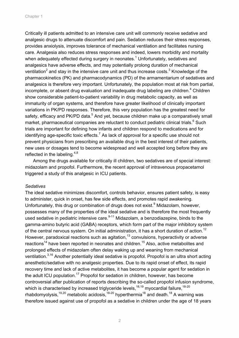
Critically ill patients admitted to an intensive care unit will commonly receive sedative and analgesic drugs to attenuate discomfort and pain. Sedation reduces their stress responses, provides anxiolysis, improves tolerance of mechanical ventilation and facilitates nursing care. Analgesia also reduces stress responses and indeed, lowers morbidity and mortality when adequately effected during surgery in neonates.1 Unfortunately, sedatives and analgesics have adverse effects, and may potentially prolong duration of mechanical ventilation2 and stay in the intensive care unit and thus increase costs.3 Knowledge of the pharmacokinetics (PK) and pharmacodynamics (PD) of the armamentarium of sedatives and analgesics is therefore very important. Unfortunately, the population most at risk from partial, incomplete, or absent drug evaluation and inadequate drug labeling are children.4 Children show considerable patient-to-patient variability in drug metabolic capacity, as well as immaturity of organ systems, and therefore have greater likelihood of clinically important variations in PK/PD responses. Therefore, this very population has the greatest need for safety, efficacy and PK/PD data.5 And yet, because children make up a comparatively small market, pharmaceutical companies are reluctant to conduct pediatric clinical trials.6 Such trials are important for defining how infants and children respond to medications and for identifying age-specific toxic effects.7 As lack of approval for a specific use should not prevent physicians from prescribing an available drug in the best interest of their patients, new uses or dosages tend to become widespread and well accepted long before they are reflected in the labeling.4,8
Among the drugs available for critically ill children, two sedatives are of special interest: midazolam and propofol. Furthermore, the recent approval of intravenous propacetamol triggered a study of this analgesic in ICU patients. Sedatives The ideal sedative minimizes discomfort, controls behavior, ensures patient safety, is easy to administer, quick in onset, has few side effects, and promotes rapid awakening. Unfortunately, this drug or combination of drugs does not exist.9 Midazolam, however, possesses many of the properties of the ideal sedative and is therefore the most frequently used sedative in pediatric intensive care.9-11 Midazolam, a benzodiazepine, binds to the gamma-amino butyric acid (GABA) receptors, which form part of the major inhibitory system of the central nervous system. On initial administration, it has a short duration of action.12 However, paradoxical reactions such as agitation,13 convulsions, hyperactivity or adverse reactions14 have been reported in neonates and children.15 Also, active metabolites and prolonged effects of midazolam often delay waking up and weaning from mechanical ventilation.3,16 Another potentially ideal sedative is propofol. Propofol is an ultra short acting anesthetic/sedative with no analgesic properties. Due to its rapid onset of effect, its rapid recovery time and lack of active metabolites, it has become a popular agent for sedation in the adult ICU population.17 Propofol for sedation in children, however, has become controversial after publication of reports describing the so-called propofol infusion syndrome, which is characterised by increased triglyceride levels,18,19 myocardial failure,18-20 rhabdomyolysis,19,20 metabolic acidosis,18-20 hyperthermia18 and death.18 A warning was therefore issued against use of propofol as a sedative in children under the age of 18 years
General Introduction
3
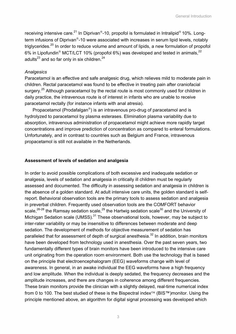
receiving intensive care.21 In Diprivan-10, propofol is formulated in Intralipid 10%. Long-term infusions of Diprivan-10 were associated with increases in serum lipid levels, notably triglycerides.20 In order to reduce volume and amount of lipids, a new formulation of propofol 6% in Lipofundin MCT/LCT 10% (propofol 6%) was developed and tested in animals,22 adults23 and so far only in six children.24 Analgesics Paracetamol is an effective and safe analgesic drug, which relieves mild to moderate pain in children. Rectal paracetamol was found to be effective in treating pain after craniofacial surgery.25 Although paracetamol by the rectal route is most commonly used for children in daily practice, the intravenous route is of interest in infants who are unable to receive paracetamol rectally (for instance infants with anal atresia).
Propacetamol (Prodafalgan) is an intravenous pro-drug of paracetamol and is hydrolyzed to paracetamol by plasma esterases. Elimination plasma variability due to absorption, intravenous administration of propacetamol might achieve more rapidly target concentrations and improve prediction of concentration as compared to enteral formulations. Unfortunately, and in contrast to countries such as Belgium and France, intravenous propacetamol is still not available in the Netherlands. Assessment of levels of sedation and analgesia In order to avoid possible complications of both excessive and inadequate sedation or analgesia, levels of sedation and analgesia in critically ill children must be regularly assessed and documented. The difficulty in assessing sedation and analgesia in children is the absence of a golden standard. At adult intensive care units, the golden standard is self-report. Behavioral observation tools are the primary tools to assess sedation and analgesia in preverbal children. Frequently used observation tools are the COMFORT behavior scale,26-28 the Ramsay sedation scale,29 the Hartwig sedation scale30 and the University of Michigan Sedation scale (UMSS).31 These observational tools, however, may be subject to inter-rater variability or may be insensitive to differences between moderate and deep sedation. The development of methods for objective measurement of sedation has paralleled that for assessment of depth of surgical anesthesia.32 In addition, brain monitors have been developed from technology used in anesthesia. Over the past seven years, two fundamentally different types of brain monitors have been introduced to the intensive care unit originating from the operation room environment. Both use the technology that is based on the principle that electroencephalogram (EEG) waveforms change with level of awareness. In general, in an awake individual the EEG waveforms have a high frequency and low amplitude. When the individual is deeply sedated, the frequency decreases and the amplitude increases, and there are changes in coherence among different frequencies. These brain monitors provide the clinician with a slightly delayed, real-time numerical index from 0 to 100. The best studied of these is the Bispectral index (BIS™)monitor. Using the principle mentioned above, an algorithm for digital signal processing was developed which
Chapter 1
4
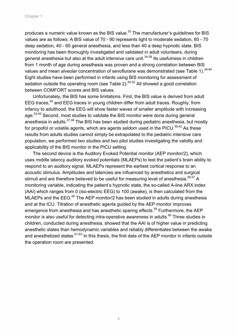
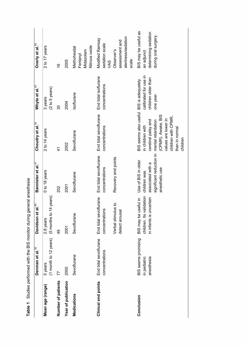
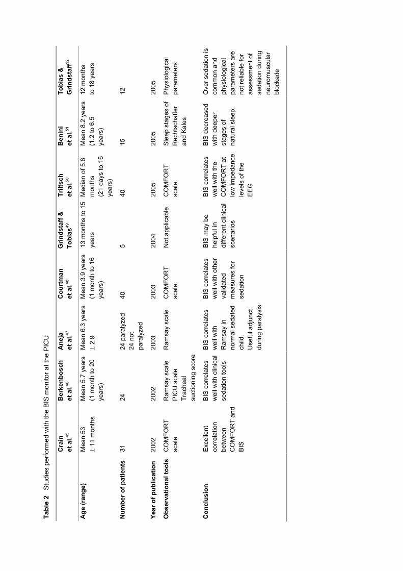
produces a numeric value known as the BIS value.33 The manufacturer’s guidelines for BIS values are as follows: A BIS value of 70 - 90 represents light to moderate sedation, 60 - 70 deep sedation, 40 - 60 general anesthesia, and less than 40 a deep hypnotic state. BIS monitoring has been thoroughly investigated and validated in adult volunteers, during general anesthesia but also at the adult intensive care unit.34-38 Its usefulness in children from 1 month of age during anesthesia was proven and a strong correlation between BIS values and mean alveolar concentration of sevoflurane was demonstrated (see Table 1).39-44 Eight studies have been performed in infants using BIS monitoring for assessment of sedation outside the operating room (see Table 2).45-52 All showed a good correlation between COMFORT scores and BIS values.
Unfortunately, the BIS has some limitations. First, the BIS value is derived from adult EEG traces,34 and EEG traces in young children differ from adult traces. Roughly, from infancy to adulthood, the EEG will show faster waves of smaller amplitude with increasing age.53-55 Second, most studies to validate the BIS monitor were done during general anesthesia in adults.37,38 The BIS has been studied during pediatric anesthesia, but mostly for propofol or volatile agents, which are agents seldom used in the PICU.39,42 As these results from adults studies cannot simply be extrapolated to the pediatric intensive care population, we performed two studies and two pilot studies investigating the validity and applicability of the BIS monitor in the PICU setting.
The second device is the Auditory Evoked Potential monitor (AEP monitor/2), which uses middle latency auditory evoked potentials (MLAEPs) to test the patient’s brain ability to respond to an auditory signal. MLAEPs represent the earliest cortical response to an acoustic stimulus. Amplitudes and latencies are influenced by anesthetics and surgical stimuli and are therefore believed to be useful for measuring level of anesthesia.56,57 A monitoring variable, indicating the patient’s hypnotic state, the so-called A-line ARX index (AAI) which ranges from 0 (iso-electric EEG) to 100 (awake), is then calculated from the MLAEPs and the EEG.58 The AEP monitor/2 has been studied in adults during anesthesia and at the ICU. Titration of anesthetic agents guided by the AEP monitor improves emergence from anesthesia and has anesthetic sparing effects.59 Furthermore, the AEP monitor is also useful for detecting intra-operative awareness in adults.60 Three studies in children, conducted during anesthesia, showed that the AAI is of higher value in predicting anesthetic states than hemodynamic variables and reliably differentiates between the awake and anesthetized states.61-63 In this thesis, the first data of the AEP monitor in infants outside the operation room are presented.
Tabl
e 1
Stu
dies
per
form
ed w
ith th
e B
IS m
onito
r dur
ing
gene
ral a
nest
hesi
a
D
enm
an e
t al.39
D
avid
son
et a
l.40
Ban
nist
er e
t al.41
C
houd
ry e
t al.42
W
hyte
et a
l.43
Ove
rly e
t al.44
Mea
n ag
e (r
ange
) 5
year
s (1
mon
th to
12
year
s)
2.8
year
s (5
mon
ths
to 1
4 ye
ars)
0 to
18
year
s 2
to 1
4 ye
ars
3 ye
ars
(2 to
5 y
ears
) 2
to 1
7 ye
ars
Num
ber o
f pat
ient
s 77
49
20
2 41
30
16
Year
of p
ublic
atio
n 20
00
2001
20
01
2002
20
04
2005
Med
icat
ions
S
evof
lura
ne
Sev
oflu
rane
S
evof
lura
ne
Sev
oflu
rane
Is
oflu
rane
M
etho
hexi
tal
Fent
anyl
M
idaz
olam
N
itrou
s ox
ide
Clin
ical
end
poi
nts
End
tida
l sev
oflu
rane
co
ncen
tratio
ns
End
tida
l sev
oflu
rane
co
ncen
tratio
ns
Ver
bal s
timul
us to
de
tect
aro
usal
End
tida
l sev
oflu
rane
co
ncen
tratio
ns
Rec
over
y en
d po
ints
End
tida
l sev
oflu
rane
co
ncen
tratio
ns
End
tida
l iso
flura
ne
conc
entra
tions
M
odifi
ed R
amsa
y se
datio
n sc
ale
VAS
Obs
erve
r’s
asse
ssm
ent a
nd
aler
tnes
s/se
datio
n sc
ale
Con
clus
ion
BIS
see
ms
prom
isin
g in
ped
iatri
c an
esth
esia
BIS
may
be
usef
ul in
ch
ildre
n. It
s va
lidat
ion
in in
fant
s is
unc
erta
in
Use
of B
IS in
old
er
child
ren
was
as
soci
ated
with
a
sign
ifica
nt re
duct
ion
in
anes
thet
ic u
se
BIS
see
ms
also
use
ful
in c
hild
ren
with
ce
rebr
al p
alsy
and
m
enta
l ret
arda
tion
(CP
MR
). A
wak
e, B
IS
valu
es a
re lo
wer
in
child
ren
with
CP
MR
, th
an in
nor
mal
ch
ildre
n
BIS
is a
dequ
atel
y ca
libra
ted
for u
se in
ch
ildre
n ol
der t
han
one
year
.
BIS
may
be
usef
ul a
s an
adj
unct
de
term
inin
g se
datio
n du
ring
oral
sur
gery
.
Tabl
e 2
Stu
dies
per
form
ed w
ith th
e B
IS m
onito
r at t
he P
ICU
C
rain
et
al.45
B
erke
nbos
ch
et a
l.46
Ane
ja
et a
l.47
Cou
rtm
an
et a
l.48
Grin
dsta
ff &
To
bias
49
Trilt
sch
et
al.50
B
enin
i et
al.51
To
bias
&
Grin
dsta
ff52
Age
(ran
ge)
Mea
n 53
±
11 m
onth
s
Mea
n 5.
7 ye
ars
(1 m
onth
to 2
0 ye
ars)
Mea
n 6.
3 ye
ars
± 2.
9 M
ean
3.9
year
s (1
mon
th to
16
year
s)
13 m
onth
s to
15
year
s M
edia
n of
5.6
m
onth
s
(21
days
to 1
6 ye
ars)
Mea
n 8.
2 ye
ars
(1.2
to 6
.5
year
s)
12 m
onth
s to
18
year
s
Num
ber o
f pat
ient
s 31
24
24
par
alyz
ed
24 n
ot
para
lyze
d
40
5 40
15
12
Year
of p
ublic
atio
n 20
02
2002
20
03
2003
20
04
2005
20
05
2005
Obs
erva
tiona
l too
ls
CO
MFO
RT
scal
e R
amsa
y sc
ale
PIC
U s
cale
Tr
ache
al
suct
ioni
ng s
core
Ram
say
scal
e C
OM
FOR
T sc
ale
Not
app
licab
le
CO
MFO
RT
scal
e S
leep
sta
ges
of
Rec
htsc
haffe
r an
d K
ales
Phy
siol
ogic
al
para
met
ers
Con
clus
ion
Exc
elle
nt
corr
elat
ion
betw
een
CO
MFO
RT
and
BIS
BIS
cor
rela
tes
wel
l with
clin
ical
se
datio
n to
ols
BIS
cor
rela
tes
wel
l with
R
amsa
y in
no
rmal
sed
ated
ch
ild.
Use
ful a
djun
ct
durin
g pa
raly
sis
BIS
cor
rela
tes
wel
l with
oth
er
valid
ated
m
easu
res
for
seda
tion
BIS
may
be
help
ful i
n di
ffere
nt c
linic
al
scen
ario
s
BIS
cor
rela
tes
wel
l with
the
CO
MFO
RT
at
low
impe
danc
e le
vels
of t
he
EE
G
BIS
dec
reas
ed
with
dee
per
stag
es o
f na
tura
l sle
ep.
Ove
r sed
atio
n is
co
mm
on a
nd
phys
iolo
gica
l pa
ram
eter
s ar
e no
t rel
iabl
e fo
r as
sess
men
t of
seda
tion
durin
g ne
urom
uscu
lar
bloc
kade
General Introduction
7
The ongoing debate on the optimal sedative for children in the ICU as well as the determination of the significance of noninvasive monitoring of the level of sedation triggered a number of studies described in this thesis. These studies generally aimed at gaining more insight in the PK/PD profiles of the sedatives midazolam and propofol, as well as the analgesic intravenous propacetamol, and in methods of monitoring sedation and analgesia effects. Outline and aims: The overall aims of the work presented in this thesis are:
1. To obtain insight in safety aspects and PK/PD profiles of the sedatives midazolam and propofol, and the analgesic intravenous propacetamol (Chapters 2 - 5).
2. To obtain better insight in the available tools to assess sedation and analgesia in infants at a pediatric intensive care unit (Chapter 6).
3. To determine whether new techniques such as the Bispectral Index monitor and the AEP monitor are valid to assess sedation in this age group (Chapters 7 - 9).
Chapter 10 contains a general discussion and future directives for relevant research. Chapter 11 summarizes all the results.
Chapter 1
8
References 1. Anand KJ, Hickey PR. Halothane-morphine compared with high-dose sufentanil for anesthesia and
postoperative analgesia in neonatal cardiac surgery. N Engl J Med 1992;326:1-9
2. Jacobi J, Fraser GL, Coursin DB et al. Clinical practice guidelines for the sustained use of sedatives and analgesics in the critically ill adult. Crit Care Med 2002;30:119-41
3. Shafer A. Complications of sedation with midazolam in the intensive care unit and a comparison with other sedative regimens. Crit Care Med 1998;26:947-56
4. t Jong GW, Vulto AG, de Hoog M et al. A survey of the use of off-label and unlicensed drugs in a Dutch children's hospital. Pediatrics 2001;108:1089-93
5. Cote CJ, Alexander J. Drug development for children: the past, the present, hope for the future. Paediatr Anaesth 2003;13:279-83
6. Cote CJ, Kauffman RE, Troendle GJ, Lambert GH. Is the "therapeutic orphan" about to be adopted? Pediatrics 1996;98:118-23
7. Berde CB, Sethna NF. Analgesics for the treatment of pain in children. N Engl J Med 2002;347:1094-103
8. Unapproved uses of approved drugs: the physician, the package insert, and the Food and Drug Administration: subject review. American Academy of Pediatrics Committee on Drugs. Pediatrics 1996;98:143-5
9. Fonsmark L, Rasmussen YH, Peder C. Occurence of withdrawal in critically ill sedated children. Crit Care Med 1999;27:196-9
10. Cote CJ, Karl HW, Notterman NA et al. Adverse sedation events in pediatrics:analysis of medications used for sedation. Pediatrics 2000;106:633-44
11. Martin LD, Bratton SL, Quint P, Mayock DE. Prospective documentation of sedative, analgesic, and neuromuscular blocking agent use in infants and children in the intensive care unit: A multicenter perspective. Pediatr Crit Care Med 2001;2:205-10
12. Allonen H, Ziegler G, Klotz U. Midazolam kinetics. Clin Pharmacol Ther 1981;30:653-61
13. Cheng C, Roemer-Becuwe C, Pereira J. When midazolam fails. J Pain Symptom Manage 2002;23:256-65
14. Booker PD, Beechey A, Lloyd-Thomas AR. Sedation of children requiring artificial ventilation using an infusion of midazolam. Br J Anaesth 1986;58:1104-8
15. Ng E, Taddio A, Ohlsson A. Intravenous midazolam infusion for sedation of infants in the neonatal intensive care unit. Cochrane Database Syst Rev 2000:CD002052
16. Tobias JD. Sedation and analgesia in paediatric intensive care units: a guide to drug selection and use. Paediatr Drugs 1999;1:109-26
17. Fulton B, Sorkin EM. Propofol. An overview of its pharmacology and a review of its clinical efficacy in intensive care sedation. Drugs 1995;50:636-57
18. Parke TJ, Stevens JE, Rice AS et al. Metabolic acidosis and fatal myocardial failure after propofol infusion in children: five case reports. Bmj 1992;305:613-6
19. Bray RJ. Propofol infusion syndrome in children. Paediatr Anaesth 1998;8:491-9
General Introduction
9
20. Vasile B, Rasulo F, Candiani A, Latronico N. The pathophysiology of propofol infusion syndrome: a simple name for a complex syndrome. Intensive Care Med 2003;29:1417-25
21. FDA. Pediatric Exclusivity Labeling Changes: Center for Drug Evaluation and Research, 2003
22. Cox EH, Knibbe CA, Koster VS et al. Influence of different fat emulsion-based intravenous formulations on the pharmacokinetics and pharmacodynamics of propofol. Pharm Res 1998;15:442-8
23. Knibbe CA, Naber H, Aarts LP et al. Long-term sedation with propofol 60 mg ml(-1) vs. propofol 10 mg(-1) ml in critically ill, mechanically ventilated patients. Acta Anaesthesiol Scand 2004;48:302-7
24. Knibbe CA, Melenhorst-de Jong G, Mestrom M et al. Pharmacokinetics and effects of propofol 6% for short-term sedation in paediatric patients following cardiac surgery. Br J Clin Pharmacol 2002;54:415-22
25. van der Marel CD, van Lingen RA, Pluim MA et al. Analgesic efficacy of rectal versus oral acetaminophen in children after major craniofacial surgery. Clin Pharmacol Ther 2001;70:82-90
26. Ambuel B, Hamlett KW, Marx CM, Blumer JL. Assessing distress in pediatric intensive care environments: the COMFORT scale. J Pediatr Psychol 1992;17:95-109
27. Marx CM, Smith PG, Lowrie LH et al. Optimal sedation of mechanically ventilated pediatric critical care patients. Crit Care Med 1994;22:163-70
28. Ista E, Van Dijk, M, Tibboel, D, De Hoog, M. Assessment of sedation levels in pediatric intensive care patients can be improved by using the COMFORT "behavior" scale. Pediatr Crit Care Med 2005;6:58-63
29. Ramsay MA, Savege TM, Simpson BR, Goodwin R. Controlled sedation with alphaxalone-alphadolone. Br Med J 1974;2:656-9
30. Brunow de Carvalho W, Lucas da Silva PS, Paulo CS et al. Comparison between the Comfort and Hartwig sedation scales in pediatric patients undergoing mechanical lung ventilation. Sao Paulo Med J 1999;117:192-6
31. Malviya S, Voepel-Lewis T, Tait AR et al. Depth of sedation in children undergoing computed tomography: validity and reliability of the University of Michigan Sedation Scale (UMSS). Br J Anaesth 2002;88:241-5
32. Carrasco G. Instruments for monitoring intensive care unit sedation. Crit Care 2000;4:217-25. Epub 2000 Jul 13
33. Sigl JC, Chamoun NG. An introduction to bispectral analysis for the electroencephalogram. J Clin Monit 1994;10:392-404
34. Glass PS, Bloom M, Kearse L et al. Bispectral analysis measures sedation and memory effects of propofol, midazolam, isoflurane, and alfentanil in healthy volunteers. Anesthesiology 1997;86:836-47
35. De Deyne C, Struys M, Decruyenaere J et al. Use of continuous bispectral EEG monitoring to assess depth of sedation in ICU patients. Intensive Care Med 1998;24:1294-8
36. Frenzel D, Greim CA, Sommer C et al. Is the bispectral index appropriate for monitoring the sedation level of mechanically ventilated surgical ICU patients? Intensive Care Med 2002;28:178-83
37. Sebel PS, Lang E, Rampil IJ et al. A multicenter study of bispectral electroencephalogram analysis for monitoring anesthetic effect. Anesth Analg 1997;84:891-9
38. Myles PS, Leslie K, McNeil J et al. Bispectral index monitoring to prevent awareness during anaesthesia: the B-Aware randomised controlled trial. Lancet 2004;363:1757-63

Chapter 1
10
39. Denman WT, Swanson EL, Rosow D et al. Pediatric evaluation of the bispectral index (BIS) monitor and correlation of BIS with end-tidal sevoflurane concentration in infants and children. Anesth Analg 2000;90:872-7
40. Davidson AJ, McCann ME, Devavaram P et al. The Differences in the Bispectral Index Between Infants and Children During Emergence from Anesthesia After Circumcision Surgery. Pediatr Anesthesia 2001;93:326-30
41. Bannister CF, Brosius KK, Sigl JC et al. The effect of bispectral index monitoring on anesthetic use and recovery in children anesthetized with sevoflurane in nitrous oxide. Anesth Analg 2001;92:877-81
42. Choudhry DK, Brenn BR, Goyal P et al. Bispectral index monitoring: a comparison between normal children and children with quadriplegic cerebral palsy. Anesth Analg 2002;95:1582-5
43. Whyte SD, Booker PD. Bispectral index during isoflurane anesthesia in pediatric patients. Anesth Analg 2004;98:1644-9
44. Overly FL, Wright RO, Connor FA et al. Bispectral Analysis During Deep Sedation of Pediatric Oral Surgery Patients. J Oral Maxillofac Surg 2005;63:215-9
45. Crain N, Slonim A, Pollack MM. Assessing sedation in the pediatric intensive care unit by using the BIS and the COMFORT scale. Pediatr Crit Care Med 2002;3:11-5
46. Berkenbosch JW, Fichter CR, Tobias JD. The correlation of the bispectral index monitor with clinical sedation scores during mechanical ventilation in the pediatric intensive care unit. Anesth Analg 2002;94:506-11
47. Aneja R, Heard AM, Fletcher JE, Heard CM. Sedation monitoring of children by the Bispectral Index in the pediatric intensive care unit. Pediatr Crit Care Med 2003;4:60-4
48. Courtman SP, Wardurgh A, Petros AJ. Comparison of the bispectral index monitor with the Comfort score in assessing level of sedation of critically ill children. Intensive Care Med 2003;29:2239-46
49. Grindstaff RJ, Tobias JD. Applications of bispectral index monitoring in the pediatric intensive care unit. J Intensive Care Med 2004;19:111-6
50. Triltsch AE, Nestmann G, Orawa H et al. Bispectral index versus COMFORT score to determine the level of sedation in paediatric intensive care patients: a prospective study. Crit Care 2005;9:R9-R17
51. Benini F, Trapanotto M, Sartori S et al. Analysis of the Bispectral Index During Natural Sleep in Children. Anest Analg 2005;101:641-4
52. Tobias JD, Grindstaff RJ. Bispectral Index Monitoring During the Administration of Neuromuscular Agents in the Pediatric Intensive Care Unit Patient. J Intensive Care Med 2005;20:233-7
53. Nieuwenhuijs D, Coleman EL, Douglas NJ et al. Bispectral index values and spectral edge frequency at different stages of physiologic sleep. Anesth Analg 2002;94:125-9, table of contents
54. Hoppenbrouwers T, Hodgman J, Arakawa K et al. Sleep and waking states in infancy: normative studies. Sleep 1988;11:387-401
55. Ficca G, Fagioli I, Giganti F, Salzarulo P. Spontaneous awakenings from sleep in the first year of life. Early Hum Dev 1999;55:219-28
56. Thornton C, Sharpe RM. Evoked responses in anaesthesia. Br J Anaesth 1998;81:771-81
57. Gajraj RJ, Doi M, Mantzaridis H, Kenny GN. Comparison of bispectral EEG analysis and auditory evoked potentials for monitoring depth of anaesthesia during propofol anaesthesia. Br J Anaesth 1999;82:672-8
General Introduction
11
58. Litvan H, Jensen, EW, Galan, J, Lund, J, Rodriguez, BE, Henneberg, SW, Caminal, P, Villar Landeira, JM. Comparison of conventional averaged and rapid averaged, autoregressive -based extracted auditory evoked potentials for monitoring the hypnotic level during propofol induction. Anesthesiology 2002;97:351-8
59. Recart A, White PF, Wang A et al. Effect of auditory evoked potential index monitoring on anesthetic drug requirements and recovery profile after laparoscopic surgery: a clinical utility study. Anesthesiology 2003;99:813-8
60. Trillo-Urrutia L, Fernandez-Galinski S, Castano-Santa J. Awareness detected by auditory evoked potential monitoring. Br J Anaesth 2003;91:290-2
61. Weber F, Seidl M, Bein T. Impact of the AEP-Monitor/2-derived composite auditory-evoked potential index on propofol consumption and emergence times during total intravenous anaesthesia with propofol and remifentanil in children. Acta Anaesthesiol Scand 2005;49:277-83
62. O'Kelly SW, Smith DC, Pilkington SN. The auditory evoked potential and paediatric anaesthesia. Br J Anaesth 1995;75:428-30
63. Weber F, Bein T, Hobbhahn J, Taeger K. Evaluation of the Alaris Auditory Evoked Potential Index as an Indicator of Anesthetic Depth in Preschool Children during Induction of Anesthesia with Sevoflurane and Remifentanil. Anesthesiology 2004;101:294-8

Population Pharmacokinetic and Pharmacodynamic Modeling of Midazolam and Its Metabolites in Postoperative Non-Ventilated Infants Following Major Craniofacial Surgery
Mariska Y.M. Peeters, Sandra A. Prins, Catherijne A.J. Knibbe, Joost DeJongh, Ron A.A. Mathôt, Celesta Warris, Ron H.N. van Schaik, Dick Tibboel and Meindert Danhof
Cha
pter
Chapter 2
14
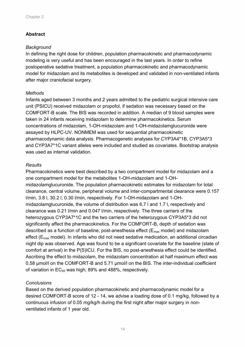
Abstract Background In defining the right dose for children, population pharmacokinetic and pharmacodynamic modeling is very useful and has been encouraged in the last years. In order to refine postoperative sedative treatment, a population pharmacokinetic and pharmacodynamic model for midazolam and its metabolites is developed and validated in non-ventilated infants after major craniofacial surgery. Methods Infants aged between 3 months and 2 years admitted to the pediatric surgical intensive care unit (PSICU) received midazolam or propofol, if sedation was necessary based on the COMFORT-B scale. The BIS was recorded in addition. A median of 9 blood samples were taken in 24 infants receiving midazolam to determine pharmacokinetics. Serum concentrations of midazolam, 1-OH-midazolam and 1-OH-midazolamglucuronide were assayed by HLPC-UV. NONMEM was used for sequential pharmacokinetic pharmacodynamic data analysis. Pharmacogenetic analyses for CYP3A4*1B, CYP3A5*3 and CYP3A7*1C variant alleles were included and studied as covariates. Bootstrap analysis was used as internal validation. Results Pharmacokinetics were best described by a two compartment model for midazolam and a one compartment model for the metabolites 1-OH-midazolam and 1-OH-midazolamglucuronide. The population pharmacokinetic estimates for midazolam for total clearance, central volume, peripheral volume and inter-compartmental clearance were 0.157 l/min, 3.8 l, 30.2 l, 0.30 l/min, respectively. For 1-OH-midazolam and 1-OH-midazolamglucuronide, the volume of distribution was 6.7 l and 1.7 l, respectively and clearance was 0.21 l/min and 0.047 l/min, respectively. The three carriers of the heterozygous CYP3A7*1C and the two carriers of the heterozygous CYP3A5*3 did not significantly affect the pharmacokinetics. For the COMFORT-B, depth of sedation was described as a function of baseline, post-anesthesia effect (Emax model) and midazolam effect (Emax model). In infants who did not need sedative medication, an additional circadian night dip was observed. Age was found to be a significant covariate for the baseline (state of comfort at arrival) in the P(S)ICU. For the BIS, no post-anesthesia effect could be identified. Ascribing the effect to midazolam, the midazolam concentration at half maximum effect was 0.58 µmol/l on the COMFORT-B and 5.71 µmol/l on the BIS. The inter-individual coefficient of variation in EC50 was high; 89% and 488%, respectively. Conclusions Based on the derived population pharmacokinetic and pharmacodynamic model for a desired COMFORT-B score of 12 - 14, we advise a loading dose of 0.1 mg/kg, followed by a continuous infusion of 0.05 mg/kg/h during the first night after major surgery in non-ventilated infants of 1 year old.
Population PK/PD of midazolam and metabolites in infants
15
Introduction The majority of drugs used in children are not licensed for children1 and there is considerable evidence that drug dosing inaccuracies leads to adverse events and even fatalities.2 With the aim to provide evidence to support the safe and effective use of drugs in the pediatric population, research is ongoing in Europe and the USA.3 The FDA encourages the application of population pharmacokinetic (PK) and pharmacodynamic (PD) modeling since this powerful approach allows for sparse sampling and the description of inter- en intrapatient variability.4 By identifying patient characteristics the variability can be explained and doses individualized.
Midazolam is one of the most used agents for sedation in the Pediatric Intensive Care Unit (PICU) and was studied in children and neonates receiving mechanical ventilation5,6,7 and after oral administration as premedication.8,9 After craniosynostosis, midazolam can be an adjuvant in the care of infants admitted to the PICU, since the development of edematous eyelids gives an extra stressful stimulus to the physical and emotional distress and discomfort that young children often encounter in the PICU.10 However, in particular in this non-ventilated postoperative population where ontogeny and genetic polymorphism may complicate the characterization, there are no rational dose schemes available based on population PK/PD, using the validated pediatric clinical sedation score COMFORT-B11 or the bispectral index (BIS).12,13
Midazolam is hydroxylated by hepatic cytochrome P450 3A subfamily (CYP3A4 and CYP3A5) in the major metabolite 1-OH-midazolam (50 - 70% of the metabolism),14,15 which is as potent as the parent drug16,17 and the minor metabolites 4-OH-midazolam and 1,4-OH-midazolam. Especially after oral administration 1-OH-midazolam contributes to the effect,8 whereas after intravenous administration relatively low concentrations of 1-OH-midazolam are found.7,18 The metabolites are rapidly converted to their glucuronide conjugates and excreted in the urine. 1-OH-midazolamglucuronide is only of clinical relevance in renal failure when accumulation occurs.19
In this study we describe a population PK/PD model for midazolam and investigate the variability in concentration and effect between infants with a standard dosage regimen and explore covariates as genotyping influencing interpatient variability to develop optimal dosing. Methods The study was performed in the Pediatric Surgical Intensive Care Unit (PSICU) of the Erasmus MC-Sophia Children’s Hospital. The study protocol was approved by the ethics committee of the Erasmus MC-Sophia Children’s Hospital, Rotterdam The Netherlands. Written informed consent was obtained from the parents. The studied patients and the design of the study is given in detail in the article of Prins et al,20 and shortly repeated where relevant to this article.
Chapter 2
16
Patients Children were randomly allocated to receive midazolam or propofol if sedative medication was required according to the COMFORT-B score (score ≥ 17). Criteria for eligibility included age between one month and two years, admitted to the PSICU following major craniofacial surgery and no respiratory infections, epilepsy, hypertriglyceridemia or family histories of hypercholesterolemia, allergic history to midazolam, propofol, eggs or soybean oil.
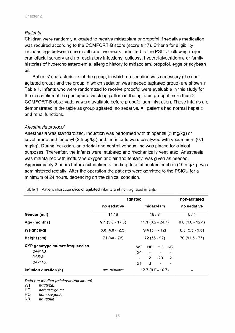
Patients’ characteristics of the group, in which no sedation was necessary (the non-agitated group) and the group in which sedation was needed (agitated group) are shown in Table 1. Infants who were randomized to receive propofol were evaluable in this study for the description of the postoperative sleep pattern in the agitated group if more than 2 COMFORT-B observations were available before propofol administration. These infants are demonstrated in the table as group agitated, no sedative. All patients had normal hepatic and renal functions. Anesthesia protocol Anesthesia was standardized. Induction was performed with thiopental (5 mg/kg) or sevoflurane and fentanyl (2.5 µg/kg) and the infants were paralyzed with vecuronium (0.1 mg/kg). During induction, an arterial and central venous line was placed for clinical purposes. Thereafter, the infants were intubated and mechanically ventilated. Anesthesia was maintained with isoflurane oxygen and air and fentanyl was given as needed. Approximately 2 hours before extubation, a loading dose of acetaminophen (40 mg/kg) was administered rectally. After the operation the patients were admitted to the PSICU for a minimum of 24 hours, depending on the clinical condition. Table 1 Patient characteristics of agitated infants and non-agitated infants
agitated non-agitated
no sedative midazolam no sedative
Gender (m/f) 14 / 6 16 / 8 5 / 4
Age (months) 9.4 (3.8 - 17.3) 11.1 (3.2 - 24.7) 8.8 (4.0 - 12.4)
Weight (kg) 8.8 (4.8 -12.5) 9.4 (5.1 - 12) 8.3 (5.5 - 9.6)
Height (cm) 71 (60 - 76) 72 (58 - 92) 70 (61.5 - 77)
CYP genotype mutant frequencies 3A4*1B 3A5*3 3A7*1C
WT 24 -
21
HE - 2 3
HO -
20 -
NR - 2 -
infusion duration (h) not relevant 12.7 (0.0 - 16.7) -
Data are median (minimum-maximum). WT wildtype; HE heterozygous; HO homozygous; NR no result
Population PK/PD of midazolam and metabolites in infants
17
Sedative and analgesic regimen From arrival at the PSICU, depth of sedation was evaluated using the COMFORT-B score, which rates 6 behavioral items.21,11 Alertness, calmness, muscle tone, body movement, facial tension, crying (non-ventilated children) or respiratory response (ventilated children) are scored on a five-point scale, resulting in a total score varying from 6 (no distress) to 30 (severe distress). The inter-observer reliability proved to be good for all nurses and the principal investigator (κ was > 0.65). In addition, the BIS was recorded continuously and noted at 15 minute interval (Bispectral A 2000 version 3.12, Aspect Medical Systems Natick MA USA with pediatric BIS sensors). The BIS ranges from 100 (awake) to 0 (iso-electric EEG). Midazolam was initially given as bolus 0.1 mg/kg followed by a continuous infusion of 0.05 mg/kg/h, titrated up after an additional bolus or down by 0.025 mg/kg/h. To determine whether restlessness was induced by pain, the trained nurses also obtained the VAS pain score. Patients received standard 4 times daily 120 - 240 mg acetaminophen rectally.22 Blood sampling Arterial blood samples (500 - 1000 µl) were collected in each infant at the following times: at baseline before the start of the midazolam bolus, approximately 45 or 30 min, 90 or 60 min, 120 min after the start of the midazolam infusion, three times in steady state, just before and 1 h after dose adjustment, just before discontinuation of the midazolam infusion, and 30 or 45, 60 or 90, 120 and 180 - 240 min after the end of the infusion (median of 11 samples per child). If the arterial line was lost, venous samples were collected from a central line. After collection the samples were centrifuged and stored at -80ºC until analysis. Analytical methods Midazolam, 1-OH-midazolam and 1-OH-midazolamglucuronide concentrations were measured in serum using high performance liquid chromatography with ultraviolet detection at 230 nm. Temazepam was used as internal standard. 500 µl 0.05 M borate buffer (pH 9.2) was added to 200 µl serum. Following liquid-liquid extraction with 6 ml dichloromethane, the organic layer was evaporated to dryness at 37ºC. The mobile phase was prepared as follows: 400 µl phosphoric acid 85% and 146 µl tri-ethylamine were added to 530 ml water. The pH was adjusted to 3.2 with 10% potassium hydroxide and 470 ml acetonitrile was added. The residue was reconstituted in 200 µl of mobile phase and 75 µl was injected onto the analytical column (Lichrosphere 100RP-18 encapped 5 µm, Merck). Total drug concentration of 1-OH-midazolam were measured after enzymatic hydrolysis of 200 µl serum with 100 UI β-glucuronidase for 24 hours at 37 ºC. The differences between total and unconjugated 1-OH-midazolam was taken as the 1-OH-midazolamglucuronide. The limits of quantification were 11 µg/l for midazolam and 6 µg/l for 1-OH-midazolam using 200 µl of serum. Inter- and intra-assay coefficients of variation were less than 8% and 13% respectively. Total recovery was greater than 90% for both compounds. Genomic DNA was isolated from EDTA blood (MasterAmp, Epicentre). CYP3A4*1B, CYP3A5*3 and CYP3A71C analyses were performed, using PCR restriction fragment length polymorphism assays, as described previously.23-25
Chapter 2
18
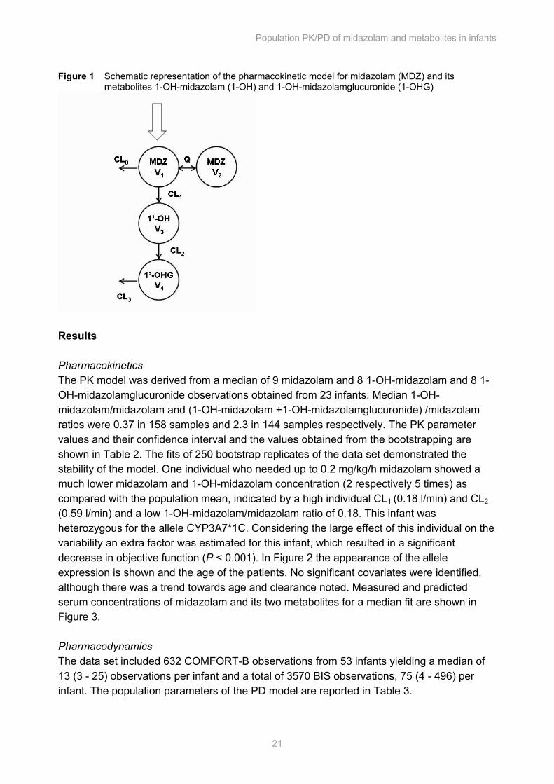
Data analysis The analysis was performed in NONMEM (Non-Linear Mixed effect Modeling) (University of California, San Francisco, CA, version V)26 by use of the first-order conditional estimation (Method 1) with η-ε interaction. S-plus (Insightful software, Seattle, WA, version 6.2) was used to visualize the data. Population PK and PD data were sequentially analyzed. Discrimination between different models was made by comparison of the objective function. A value of P < 0.005, representing a decrease of 7.8 in the objective function, was considered statistically significant. In addition, goodness of fit plots including (A. Observed versus individually predicted, B. Observed versus population predicted, C. Time versus Weighted Residuals, D. Population predictions versus Weighted Residuals) were used for diagnostic purposes. Furthermore, the confidence interval of the parameter estimates, the correlation matrix and visual improvement of the individual plots were used to evaluate the model. Covariate analysis Covariates were plotted independently against the individual post-hoc parameter estimates and the weighted residuals to visualize potential relationships. The following covariates were tested: body weight, age, body surface area (BSA), body mass index (BMI) (if height was known) and gender. The PK parameters were also tested for correlation with heart frequency, blood pressure, sampling (venous or arterial), mechanical ventilation (6 samples from 2 infants during mechanical ventilation) and the genotypes (CYP3A4*1B, 3A5*3, 3A7*1C). Potential covariates were separately entered into the model and statistically tested by use of the objective function. A significant covariate that most reduces the objective function was left in the model. Additional covariates had to reduce this objective function further to be retained in the model. The choice of the model was further evaluated as above discussed. Validation The internal validity of the population pharmacokinetic and pharmacodynamic models was assessed by the bootstrap re-sampling method (repeated random sampling to produce another data set of the same size but with a different combination of individuals). Pharmacokinetic model Midazolam and metabolite data were fitted simultaneously and were expressed as µmol/l. The pharmacokinetic model used is schematically depicted in Figure 1. The midazolam data were described with a two-compartment model, parameterized in terms of volume of the central compartment (V1), volume of the peripheral volume (V2), the inter-compartmental clearance (Q) and the clearances CL0 and CL1. The formation of 1-OH-midazolam and 1-OH-midazolamglucuronide was best described with a one-compartment model. Cl1 was assumed to be 60% of the total clearance (the sum of CL0 and CL1) as reported in the literature. The volume of distribution of 1-OH-midazolam was modeled as a fraction of V1 + V2 of midazolam. The individual value (post hoc value) of the parameters of the ith subject was modeled by
Population PK/PD of midazolam and metabolites in infants
19
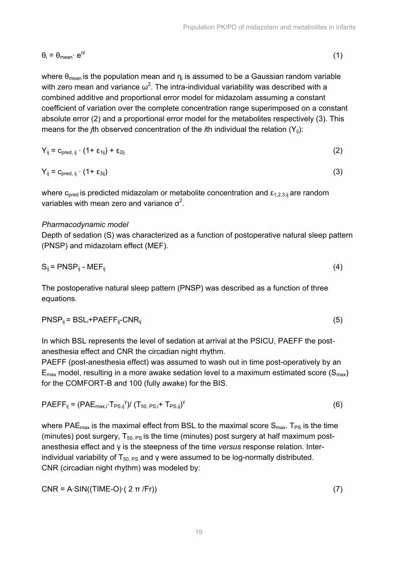
θi = θmean· eηi (1) where θmean is the population mean and ηi is assumed to be a Gaussian random variable with zero mean and variance ω2. The intra-individual variability was described with a combined additive and proportional error model for midazolam assuming a constant coefficient of variation over the complete concentration range superimposed on a constant absolute error (2) and a proportional error model for the metabolites respectively (3). This means for the jth observed concentration of the ith individual the relation (Yij): Yij = cpred, ij · (1+ ε1ij) + ε2ij (2) Yij = cpred, ij · (1+ ε3ij) (3) where cpred is predicted midazolam or metabolite concentration and ε1,2,3,ij are random
variables with mean zero and variance σ2. Pharmacodynamic model Depth of sedation (S) was characterized as a function of postoperative natural sleep pattern (PNSP) and midazolam effect (MEF). Sij = PNSPij - MEFij (4) The postoperative natural sleep pattern (PNSP) was described as a function of three equations. PNSPij = BSLi+PAEFFij-CNRij (5) In which BSL represents the level of sedation at arrival at the PSICU, PAEFF the post-anesthesia effect and CNR the circadian night rhythm. PAEFF (post-anesthesia effect) was assumed to wash out in time post-operatively by an Emax model, resulting in a more awake sedation level to a maximum estimated score (Smax) for the COMFORT-B and 100 (fully awake) for the BIS. PAEFFij = (PAEmax,i·TPS,ij
γ)/ (T50, PS,i+ TPS,ij)γ (6) where PAEmax is the maximal effect from BSL to the maximal score Smax. TPS is the time (minutes) post surgery, T50, PS is the time (minutes) post surgery at half maximum post-anesthesia effect and γ is the steepness of the time versus response relation. Inter-individual variability of T50, PS and γ were assumed to be log-normally distributed. CNR (circadian night rhythm) was modeled by: CNR = A·SIN((TIME-O)·( 2 π /Fr)) (7)
Chapter 2
20
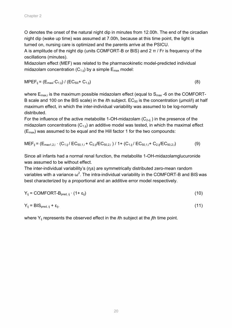
O denotes the onset of the natural night dip in minutes from 12.00h. The end of the circadian night dip (wake up time) was assumed at 7.00h, because at this time point, the light is turned on, nursing care is optimized and the parents arrive at the PSICU. A is amplitude of the night dip (units COMFORT-B or BIS) and 2 π / Fr is frequency of the oscillations (minutes). Midazolam effect (MEF) was related to the pharmacokinetic model-predicted individual midazolam concentration (C1,ij) by a simple Emax model: MPEFij = (Emaxi·C1,ij) / (EC50i+ C1,ij) (8) where Emax,i is the maximum possible midazolam effect (equal to Smax -6 on the COMFORT-B scale and 100 on the BIS scale) in the ith subject. EC50 is the concentration (µmol/l) at half maximum effect, in which the inter-individual variability was assumed to be log-normally distributed. For the influence of the active metabolite 1-OH-midazolam (C2,ij ) in the presence of the midazolam concentrations (C1,ij) an additive model was tested, in which the maximal effect (Emax) was assumed to be equal and the Hill factor 1 for the two compounds: MEFij = (Emax1,2,i · (C1,ij / EC50,1,i + C2,ij/EC50,2,i ) / 1+ (C1,ij / EC50,1,i+ C2,ij/EC50,2,i) (9) Since all infants had a normal renal function, the metabolite 1-OH-midazolamglucuronide was assumed to be without effect. The inter-individual variability’s (ηis) are symmetrically distributed zero-mean random variables with a variance ω2. The intra-individual variability in the COMFORT-B and BIS was best characterized by a proportional and an additive error model respectively. Yij = COMFORT-Bpred, ij · (1+ εij) (10) Yij = BISpred, ij + εij. (11) where Yij represents the observed effect in the ith subject at the jth time point.
Population PK/PD of midazolam and metabolites in infants
21
Figure 1 Schematic representation of the pharmacokinetic model for midazolam (MDZ) and its metabolites 1-OH-midazolam (1-OH) and 1-OH-midazolamglucuronide (1-OHG)
Results Pharmacokinetics The PK model was derived from a median of 9 midazolam and 8 1-OH-midazolam and 8 1-OH-midazolamglucuronide observations obtained from 23 infants. Median 1-OH-midazolam/midazolam and (1-OH-midazolam +1-OH-midazolamglucuronide) /midazolam ratios were 0.37 in 158 samples and 2.3 in 144 samples respectively. The PK parameter values and their confidence interval and the values obtained from the bootstrapping are shown in Table 2. The fits of 250 bootstrap replicates of the data set demonstrated the stability of the model. One individual who needed up to 0.2 mg/kg/h midazolam showed a much lower midazolam and 1-OH-midazolam concentration (2 respectively 5 times) as compared with the population mean, indicated by a high individual CL1 (0.18 l/min) and CL2 (0.59 l/min) and a low 1-OH-midazolam/midazolam ratio of 0.18. This infant was heterozygous for the allele CYP3A7*1C. Considering the large effect of this individual on the variability an extra factor was estimated for this infant, which resulted in a significant decrease in objective function (P < 0.001). In Figure 2 the appearance of the allele expression is shown and the age of the patients. No significant covariates were identified, although there was a trend towards age and clearance noted. Measured and predicted serum concentrations of midazolam and its two metabolites for a median fit are shown in Figure 3. Pharmacodynamics The data set included 632 COMFORT-B observations from 53 infants yielding a median of 13 (3 - 25) observations per infant and a total of 3570 BIS observations, 75 (4 - 496) per infant. The population parameters of the PD model are reported in Table 3.
Chapter 2
22
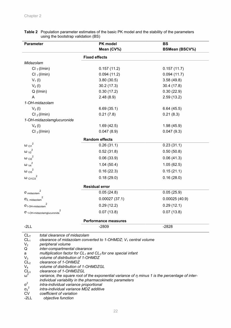
Table 2 Population parameter estimates of the basic PK model and the stability of the parameters using the bootstrap validation (BS)
Parameter PK model Mean (CV%)
BS BSMean (BSCV%)
Fixed effects Midazolam Cl T (l/min) 0.157 (11.2) 0.157 (11.7) Cl 1 (l/min) 0.094 (11.2) 0.094 (11.7) V1 (l) 3.80 (30.5) 3.58 (49.8) V2 (l) 30.2 (17.3) 30.4 (17.8) Q (l/min) 0.30 (17.2) 0.30 (22.9) A 2.48 (8.9) 2.59 (13.2) 1-OH-midazolam V3 (l) 6.69 (35.1) 6.64 (45.5) Cl 2 (l/min) 0.21 (7.8) 0.21 (8.3) 1-OH-midazolamglucuronide V4 (l) 1.69 (42.5) 1.98 (45.9) Cl 3 (l/min) 0.047 (8.9) 0.047 (9.3) Random effects ω Cl1
2 0.26 (31.1) 0.23 (31.1) ω V2
2 0.52 (31.8) 0.50 (50.8)
ω Cl22 0.06 (33.9) 0.06 (41.3)
ω V42 1.04 (50.4) 1.05 (62.5)
ω Cl32 0.16 (22.3) 0.15 (21.1)
ω Cl1Cl32 0.18 (29.0) 0.16 (28.0)
Residual error σ midazolam
2 0.05 (24.8) 0.05 (25.9)
σ2, midazolam2 0.00027 (37.1) 0.00025 (40.9)
σ1-OH-midazolam2
0.29 (12.2) 0.29 (12.1)
σ 1-OH-midazolamglucuronide2 0.07 (13.8) 0.07 (13.8)
Performance measures -2LL -2809 -2828
CLT total clearance of midazolam CL1 clearance of midazolam converted to 1-OHMDZ; V1 central volume V2 peripheral volume Q inter-compartmental clearance a multiplication factor for CL1 and CL2 for one special infant V3 volume of distribution of 1-OHMDZ CL2 clearance of 1-OHMDZ V4 volume of distribution of 1-OHMDZGL CL3 clearance of 1-OHMDZGL ω2 variance, the square root of the exponential variance of η minus 1 is the percentage of inter-
individual variability in the pharmacokinetic parameters σ2 intra-individual variance proportional σ2
2 intra-individual variance MDZ additive CV coefficient of variation -2LL objective function

Population PK/PD of midazolam and metabolites in infants
23
0 5 10 15 20 25age (months)
0.0
0.1
0.2
0.3
CL 1
(L/m
in)
0 300 600 900 1200 1500time (min)
10-2.00
10-1.00
100.00
1-O
H- m
idaz
olam
(mic
rom
ol/L
)
ID: 13
0 300 600 900 1200 1500time (min)
10-2.00
10-1.00
100.00
1-O
H-m
idaz
olam
gluc
uron
ide
(mic
rom
ol/L
)Figure 2 Scatter plot showing relationship between age and clearance and the identification of the genotype analysis
The two carriers of the CYP3A5*3 heterozygous allele are represented by (□), the two infants with no result for CYP3A5*3 by (◊). The three carriers of CYP3A7*1C heterozygous allele are represented by an empty triangle and (▲) for the infant in which an extra factor was estimated. Figure 3 Serum concentration – time observations and predictions of midazolam, 1-OH-midazolam and 1-OH-midazolamglucuronide for a median performance after a loading dose of 1 mg, followed by a continuous infusion of 0.5 mg/h
0 300 600 900 1200 1500time (min)
10-2.00
10-1.00
100.00
mid
azol
am (m
icro
mol
/l)
The solid circles represent measured midazolam
and metabolite concentrations; the solid lines represent the individual predicted concentrations and the dash lines represent the population predicted concentrations.
Chapter 2
24
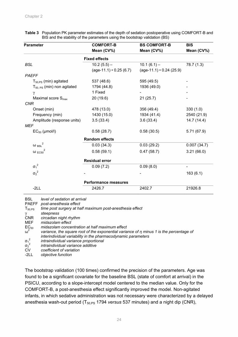
Table 3 Population PK parameter estimates of the depth of sedation postoperative using COMFORT-B and BIS and the stability of the parameters using the bootstrap validation (BS)
Parameter COMFORT-B Mean (CV%)
BS COMFORT-B Mean (CV%)
BIS Mean (CV%)
Fixed effects BSL 10.2 (5.5) –
(age-11.1) • 0.25 (6.7) 10.1 (6.1) – (age-11.1) • 0.24 (25.9)
78.7 (1.3)
PAEFF T50,PS (min) agitated 537 (48.6) 595 (49.5) - T50, PS (min) non agitated 1794 (44.8) 1936 (49.0) - γ 1 Fixed - - Maximal score Smax 20 (19.6) 21 (25.7) - CNR Onset (min) 478 (13.0) 356 (49.4) 330 (1.0) Frequency (min) 1430 (15.0) 1934 (41.4) 2540 (21.9) Amplitude (response units) 3.5 (33.4) 3.6 (33.4) 14.7 (14.4) MEF EC50 (µmol/l) 0.58 (28.7) 0.58 (30.5) 5.71 (67.9) Random effects ω BSL
2 0.03 (34.3) 0.03 (29.2) 0.007 (34.7) ω EC50
2 0.58 (59.1) 0.47 (58.7) 3.21 (66.0)
Residual error σ1
2 0.09 (7.2) 0.09 (8.0) -
σ22 - - 163 (6.1)
Performance measures -2LL 2426.7 2402.7 21926.8
BSL level of sedation at arrival PAEFF post-anesthesia effect T50,PS time post surgery at half maximum post-anesthesia effect γ steepness CNR circadian night rhythm MEF midazolam effect EC50 midazolam concentration at half maximum effect ω2 variance, the square root of the exponential variance of η minus 1 is the percentage of
interindividual variability in the pharmacodynamic parameters σ1
2 intraindividual variance proportional σ2
2 intraindividual variance additive CV coefficient of variation -2LL objective function The bootstrap validation (100 times) confirmed the precision of the parameters. Age was found to be a significant covariate for the baseline BSL (state of comfort at arrival) in the PSICU, according to a slope-intercept model centered to the median value. Only for the COMFORT-B, a post-anesthesia effect significantly improved the model. Non-agitated infants, in which sedative administration was not necessary were characterized by a delayed anesthesia wash-out period (T50,PS 1794 versus 537 minutes) and a night dip (CNR),
Population PK/PD of midazolam and metabolites in infants
25
300 800 1300time (min)
6
10
14
18
22
6
10
14
18
22
6
10
14
18
22
CO
MFO
RT-
B
0.0
0.1
0.2
0.3
0.40.0
0.1
0.2
0.3
0.40.0
0.1
0.2
0.3
0.4
Mid
azol
am c
once
ntra
tion
(µm
ol/L
)
A
B
C
10 mo15 mo
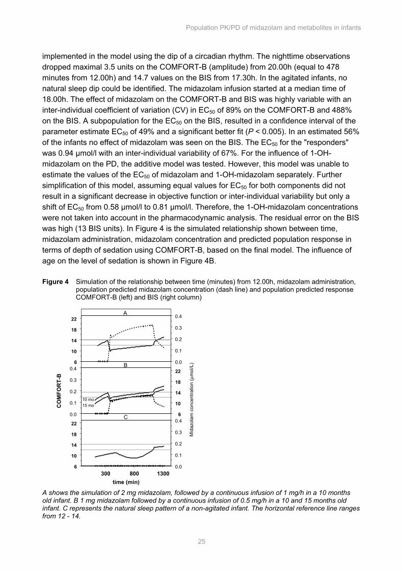
implemented in the model using the dip of a circadian rhythm. The nighttime observations dropped maximal 3.5 units on the COMFORT-B (amplitude) from 20.00h (equal to 478 minutes from 12.00h) and 14.7 values on the BIS from 17.30h. In the agitated infants, no natural sleep dip could be identified. The midazolam infusion started at a median time of 18.00h. The effect of midazolam on the COMFORT-B and BIS was highly variable with an inter-individual coefficient of variation (CV) in EC50 of 89% on the COMFORT-B and 488% on the BIS. A subpopulation for the EC50 on the BIS, resulted in a confidence interval of the parameter estimate EC50 of 49% and a significant better fit (P < 0.005). In an estimated 56% of the infants no effect of midazolam was seen on the BIS. The EC50 for the "responders" was 0.94 µmol/l with an inter-individual variability of 67%. For the influence of 1-OH-midazolam on the PD, the additive model was tested. However, this model was unable to estimate the values of the EC50 of midazolam and 1-OH-midazolam separately. Further simplification of this model, assuming equal values for EC50 for both components did not result in a significant decrease in objective function or inter-individual variability but only a shift of EC50 from 0.58 µmol/l to 0.81 µmol/l. Therefore, the 1-OH-midazolam concentrations were not taken into account in the pharmacodynamic analysis. The residual error on the BIS was high (13 BIS units). In Figure 4 is the simulated relationship shown between time, midazolam administration, midazolam concentration and predicted population response in terms of depth of sedation using COMFORT-B, based on the final model. The influence of age on the level of sedation is shown in Figure 4B. Figure 4 Simulation of the relationship between time (minutes) from 12.00h, midazolam administration, population predicted midazolam concentration (dash line) and population predicted response COMFORT-B (left) and BIS (right column) A shows the simulation of 2 mg midazolam, followed by a continuous infusion of 1 mg/h in a 10 months old infant. B 1 mg midazolam followed by a continuous infusion of 0.5 mg/h in a 10 and 15 months old infant. C represents the natural sleep pattern of a non-agitated infant. The horizontal reference line ranges from 12 - 14.
Chapter 2
26
Discussion A population PK/PD model of midazolam and its metabolites 1-OH-midazolam and 1-OH-midazolamglucuronide based on COMFORT-B and BIS is described in order to refine postoperative sedative treatment in non-ventilated infants aged 3 months to 2 years post surgery in the PICU.
The PK model derived in this study estimated a total clearance (157 ml/min; 16.7 ml/kg/min) that was comparable to the clearance found by Reed (11.3 ml/kg/min) for children aged 6 month-2 years after a single dose prior to minor in-hospital or day-stay procedures9 and to healthy adults (16.1 ml/kg/min).27 In critically ill children in this age group, elimination is impaired: Hughes28 estimated a median clearance of 3.1 ml/kg/min from steady-state concentrations in critically ill infants aged one month to 1 year. De Wildt7 found a mean clearance of 5.0 ml/kg/min in intensive care patients aged 2 days to 17 years. They also found a 2.5 times lower ratio for 1-OH-midazolam/midazolam. In neonates, clearance is markedly decreased (0.94 ml/kg/min).6 In our study a high degree of variability in clearance (CV is 54%) was seen, but was not reflected by the influence of sampling site, mechanical ventilation, bodyweight, age or frequencies of CYP3A5 variant alleles. Midazolam is only slightly metabolized by CYP3A7, which is predominantly expressed in the fetal liver and decreases immediately after birth to approximately 10% of newborn levels between 6 and 12 months of age. During the first 6 months of age, CYP3A4 activity increases gradually.29,30 The expression of CYP3A5 was found to be generally independent of age.30 The genetic variants in CYP3A4/5 have only a limited impact on the metabolism indicated by the study of He et al.31 and Shih et al.,32 who found no significant differences in the pharmacokinetics between heterozygous extensive metabolizers CYP3A5*1/*3 and the poor metabolizers CYP3A5*3/*3 and in the frequency of the allele CYP3A4*1B. The allele CYP3A7*1C is associated with high hepatic and intestinal CYP3A7 expression.33 In one heterozygous CYP3A7*1C infant, who needed high doses of midazolam, multiplication of CL1 and CL2 resulted in a significant better fit, which means that the oxidation and glucuronidation was faster than in the other infants. In general the infants heterozygous for CYP3A7*1C or CYP3A5*3 on CL1 did not effect the midazolam clearance significantly, although these carriers tended to occur more frequently in the upper level of the midazolam clearance values as shown in Figure 2. To answer the question if the investigated alleles play a significant role, a larger population is needed. The estimated volume of distribution of the metabolites have to be taken with caution, since accurate estimates can only be obtained by separate administration. Mandema17 showed in healthy adult volunteers that the volume of 1-OH-midazolam was equal to the volume of midazolam. In our infants, modeling the volume of distribution of 1-OH-midazolam as a fraction of volume of steady state and estimated for 1-OH-midazolamglucuronide resulted in a significantly better description of the data.
Depth of sedation is difficult to assess in children. The COMFORT scale is validated in the PICU and measures the six behavioral items as also two physiological items (mean arterial pressure and heart rate).34 However, it is suggested that the physiological variables have limited validity, since these items are controlled in the intensive care unit.11,35 The BIS is objective and easy to use, but is not validated yet for children below the age of 1 year.36,37
Population PK/PD of midazolam and metabolites in infants
27
Using the COMFORT-B as PD endpoint in a population approach, depth of sedation was described as a function of midazolam effect, post-anesthesia effect and a circadian night rhythm. Using the BIS, no significant post-anesthesia effect was found and a large residual error was detected. This is possible due to the light level of sedation, since light sedation may be influenced more by the environment.38 Recently, no evident relationship was found between PK/PD for midazolam in pediatric critically ill patients covering the age range of 2 days-17 years, using a mixed-model ANOVA taking no covariates in account, and the COMFORT scale as sedation scale, divided in three categories.39 In our study age was found to be a significant covariate for the baseline BSL (the state of comfort at arrival) in the PSICU. This indicates that young children may be more sensitive to the environment and emotional distress than older infants. Non-agitated children were characterized by a night dip starting at 20.00h on the COMFORT-B and 17.30h on the BIS and a slower wash-out period of the anesthesia effect on the COMFORT-B (1794 versus 537 minutes, respectively) compared to agitated infants. In agitated children no night dip could be identified. It has been shown clearly that the metabolite 1-OH-midazolam has pharmacological properties17 and contributes significantly to the pharmacodynamic response after oral administration when the concentration is relatively high. In adults after coronary artery bypass grafting (CABG), 1-OH-midazolam levels were above 10 µg/l in 11% of the patients and the ratio was at most 0.20.40 No effect of 1-OH-midazolam could be detected.41 In our study, the 1-OH-midazolam/midazolam ratio was 0.37. No separate EC50 could be identified for parent and metabolite, since the concentration profiles ran parallel in time. Therefore, the effect was only ascribed to midazolam, using a simple Emax model. Large variability in EC50 was seen (89% CV for the COMFORT-B and 488% for the BIS as a result of a fraction of 0.56 of the infants in which no effect of midazolam on the BIS could be detected). No patient characteristics could increase the predictability. At present, no population PD studies in adults are available for comparison of the sensitivity of infants to adults using these sedation scales. Using the Ramsay scale, the midazolam concentration in adults associated with 50% probability of a level of sedation ≥ 2 (cooperative) to 4 (asleep but responses to glabellar tap) were 0.017, 0.22 and 0.52 µmol/l, respectively.41 Following a bolus of 1 mg and a continuous infusion of 0.5 mg/h the predicted concentration in the infants are 0.16 µmol/l, corresponding with a COMFORT–B of 12 to 14 (light sedated). Although comparison is difficult, it seems that the midazolam concentration to achieve light sedation in infants is comparable to adults.
Based on the population PK/PD model we advise a loading dose of 0.1 mg/kg, followed by a continuous infusion of 0.05 mg/kg/h for infants of 1 year of age to achieve a sedation scale of 12 to 14 on the COMFORT-B. Individual titration is very important for midazolam. Acknowledgements The authors wish to thank Ilse P. van der Heiden and Marloes van der Werf from the Department of Clinical Chemistry, Erasmus MC, Rotterdam, The Netherlands for genotyping, and the medical and nursing staff of the Pediatric Surgical Intensive Care Unit for their help and cooperation.
Chapter 2
28
References 1. t Jong GW, Vulto AG, de Hoog M, Schimmel KJ, Tibboel D, van den Anker JN: A survey of the use of
off-label and unlicensed drugs in a Dutch children's hospital. Pediatrics 2001;108:1089-93
2. Baber N, Pritchard D: Dose estimation for children. Br J Clin Pharmacol 2003;56:489-93
3. Baber NS: Paediatric special issue. Br J Clin Pharmacol 2005;59:651-4
4. Administration FaD: Guidance for Industry. General considerations for pediatric pharmacokinetic studies for drugs and biological products. available from: http://www.fda.gov/cder/guidance/index.htm 1998;10:1-10
5. Blumer JL: Clinical pharmacology of midazolam in infants and children. Clin Pharmacokinet 1998;35:37-47
6. Lee TC, Charles BG, Harte GJ, Gray PH, Steer PA, Flenady VJ: Population pharmacokinetic modeling in very premature infants receiving midazolam during mechanical ventilation: midazolam neonatal pharmacokinetics. Anesthesiology 1999;90:451-7
7. de Wildt SN, de Hoog M, Vinks AA, van der Giesen E, van den Anker JN: Population pharmacokinetics and metabolism of midazolam in pediatric intensive care patients. Crit Care Med 2003;31:1952-8
8. Johnson TN, Rostami-Hodjegan A, Goddard JM, Tanner MS, Tucker GT: Contribution of midazolam and its 1-hydroxy metabolite to preoperative sedation in children: a pharmacokinetic-pharmacodynamic analysis. Br J Anaesth 2002;89:428-37
9. Reed MD, Rodarte A, Blumer JL, Khoo KC, Akbari B, Pou S, Pharmd, Kearns GL: The single-dose pharmacokinetics of midazolam and its primary metabolite in pediatric patients after oral and intravenous administration. J Clin Pharmacol 2001;41:1359-69
10. Bennett NR: Paediatric intensive care. Br J Anaesth 1999;83:139-56
11. Ista E, van Dijk M, Tibboel D, de Hoog M: Assessment of sedation levels in pediatric intensive care patients can be improved by using the COMFORT "behavior" scale. Pediatr Crit Care Med 2005;6:58-63
12. Courtman SP, Wardurgh A, Petros AJ: Comparison of the bispectral index monitor with the Comfort score in assessing level of sedation of critically ill children. Intensive Care Med 2003;29:2239-46
13. Triltsch AE, Nestmann G, Orawa H, Moshirzadeh M, Sander M, Grosse J, Genahr A, Konertz W, Spies CD: Bispectral index versus COMFORT score to determine the level of sedation in paediatric intensive care unit patients: a prospective study. Crit Care 2005;9:R9-17
14. Heizmann P, Ziegler WH: Excretion and metabolism of 14C-midazolam in humans following oral dosing. Arzneimittelforschung 1981;31:2220-3
15. Sarnquist FH, Gustafson J, Blaschke T: Steady state pharmacokinetics of midazolam maleate. Clin Pharamcol Ther 1980;27:283
16. Ziegler WH, Schalch E, Leishman B, Eckert M: Comparison of the effects of intravenously administered midazolam, triazolam and their hydroxy metabolites. Br J Clin Pharmacol 1983;16 Suppl 1:63S-69S
17. Mandema JW, Tuk B, van Steveninck AL, Breimer DD, Cohen AF, Danhof M: Pharmacokinetic-pharmacodynamic modeling of the central nervous system effects of midazolam and its main metabolite alpha-hydroxymidazolam in healthy volunteers. Clin Pharmacol Ther 1992 51:715-28
Population PK/PD of midazolam and metabolites in infants
29
18. Swart EL, Zuideveld KP, de Jongh J, Danhof M, Thijs LG, Strack van Schijndel RM: Comparative population pharmacokinetics of lorazepam and midazolam during long-term continuous infusion in critically ill patients. Br J Clin Pharmacol 2004;57:135-45
19. Bauer TM, Ritz R, Haberthur C, Ha HR, Hunkeler W, Sleight AJ, Scollo-Lavizzari G, Haefeli WE: Prolonged sedation due to accumulation of conjugated metabolites of midazolam. Lancet 1995;346:145-7
20. Prins SA, Peeters MY, Houmes RJ, van Dijk M, Knibbe CA, Danhof M, Tibboel D: Propofol 6% as sedative in children under 2 years of age following major craniofacial surgery. Br J Anaesth 2005;94:630-5
21. van Dijk M, de Boer JB, Koot HM, Tibboel D, Passchier J, Duivenvoorden HJ: The reliability and validity of the COMFORT scale as a postoperative pain instrument in 0 to 3-year-old infants. Pain 2000;84:367-77
22. van der Marel CD, van Lingen RA, Pluim MA, Scoones G, van Dijk M, Vaandrager JM, Tibboel D: Analgesic efficacy of rectal versus oral acetaminophen in children after major craniofacial surgery. Clin Pharmacol Ther 2001;70:82-90
23. van Schaik RH, van der Heiden IP, van den Anker JN, Lindemans J: CYP3A5 variant allele frequencies in Dutch Caucasians. Clin Chem 2002;48:1668-71
24. van Schaik RH, de Wildt SN, van Iperen NM, Uitterlinden AG, van den Anker JN, Lindemans J: CYP3A4-V polymorphism detection by PCR-restriction fragment length polymorphism analysis and its allelic frequency among 199 Dutch Caucasians. Clin Chem 2000;46:1834-6
25. Smit P, van Schaik RH, van der Werf M, van den Beld AW, Koper JW, Lindemans J, Pols HA, Brinkmann AO, de Jong FH, Lamberts SW: A common polymorphism in the CYP3A7 gene is associated with a nearly 50% reduction in serum dehydroepiandrosterone sulfate levels. J Clin Endocrinol Metab 2005;90:5313-6
26. Beal SL, Sheiner LB: NONMEM Users Guides. NONMEM Project Group, University of California at San Francisco, CA 1992
27. Knoester PD, Jonker DM, Van Der Hoeven RT, Vermeij TA, Edelbroek PM, Brekelmans GJ, de Haan GJ: Pharmacokinetics and pharmacodynamics of midazolam administered as a concentrated intranasal spray. A study in healthy volunteers. Br J Clin Pharmacol 2002;53:501-7
28. Hughes J, Gill AM, Mulhearn H, Powell E, Choonara I: Steady-state plasma concentrations of midazolam in critically ill infants and children. Ann Pharmacother 1996;30:27-30
29. Lacroix D, Sonnier M, Moncion A, Cheron G, Cresteil T: Expression of CYP3A in the human liver--evidence that the shift between CYP3A7 and CYP3A4 occurs immediately after birth. Eur J Biochem 1997;247:625-34
30. Stevens JC, Hines RN, Gu C, Koukouritaki SB, Manro JR, Tandler PJ, Zaya MJ: Developmental expression of the major human hepatic CYP3A enzymes. J Pharmacol Exp Ther 2003;307:573-82
31. He P, Court MH, Greenblatt DJ, Von Moltke LL: Genotype-phenotype associations of cytochrome P450 3A4 and 3A5 polymorphism with midazolam clearance in vivo. Clin Pharmacol Ther 2005;77:373-87
32. Shih PS, Huang JD: Pharmacokinetics of midazolam and 1'-hydroxymidazolam in Chinese with different CYP3A5 genotypes. Drug Metab Dispos 2002;30:1491-6
Chapter 2
30
33. Burk O, Tegude H, Koch I, Hustert E, Wolbold R, Glaeser H, Klein K, Fromm MF, Nuessler AK, Neuhaus P, Zanger UM, Eichelbaum M, Wojnowski L: Molecular mechanisms of polymorphic CYP3A7 expression in adult human liver and intestine. J Biol Chem 2002;277:24280-8
34. Ambuel B, Hamlett KW, Marx CM, Blumer JL: Assessing distress in pediatric intensive care environments: the COMFORT scale. J Pediatr Psychol 1992;17:95-109
35. Carnevale FA, Razack S: An item analysis of the COMFORT scale in a pediatric intensive care unit. Pediatr Crit Care Med 2002;3:177-180
36. Courtman SP, Wardurgh A, Petros AJ: Comparison of the bispectral index monitor with the Comfort score in assessing level of sedation of critically ill children. Intensive Care Med 2003;29:2239-46.
37. Triltsch AE, Nestmann G, Orawa H, Moshirzadeh M, Sander M, Grosse J, Genahr A, Konertz W, Spies CD: Bispectral index versus COMFORT score to determine the level of sedation in paediatric intensive care patients: a prospective study. Crit Care 2005;9:R9-R17
38. Kim DW, Kil HY, White PF: The effect of noise on the bispectral index during propofol sedation. Anesth Analg 2001;93:1170-3
39. de Wildt SN, de Hoog M, Vinks AA, Joosten KF, van Dijk M, van den Anker JN: Pharmacodynamics of midazolam in pediatric intensive care patients. Ther Drug Monit 2005;27:98-102
40. Zomorodi K, Donner A, Somma J, Barr J, Sladen R, Ramsay J, Geller E, Shafer SL: Population pharmacokinetics of midazolam administered by target controlled infusion for sedation following coronary artery bypass grafting. Anesthesiology 1998;89:1418-29
41. Somma J, Donner A, Zomorodi K, Sladen R, Ramsay J, Geller E, Shafer SL: Population pharmacodynamics of midazolam administered by target controlled infusion in SICU patients after CABG surgery. Anesthesiology 1998;89:1430-43
Propofol 6% as Sedative in Children under 2 Years of Age Following Major Craniofacial Surgery
Sandra A. Prins*, Mariska Y.M. Peeters*, Robert Jan M. Houmes, Monique van Dijk, Catherijne A.J. Knibbe, Meindert Danhof and Dick Tibboel. (* S.A. Prins and M.Y.M. Peeters contributed equally to this paper) Br J Anaesth 2005; 94: 630-635
Cha
pter
Chapter 3
32
Abstract Background After alarming reports concerning deaths after sedation with propofol, infusion of this drug was contraindicated by the US Food and Drug Administration in children < 18 yr receiving intensive care. We describe our experiences with propofol 6%, a new formula, during postoperative sedation in non-ventilated children following craniofacial surgery. Methods In a prospective cohort study, children admitted to the paediatric surgical intensive care unit following major craniofacial surgery were randomly allocated to sedation with propofol 6% or midazolam, if judged necessary on the basis of a COMFORT behaviour score. Exclusion criteria were respiratory infection, allergy for proteins, propofol or midazolam, hypertriglyceridaemia, familial hypercholesterolaemia or epilepsy. We assessed the safety of propofol 6% with triglycerides (TG) and creatine phosphokinase (CPK) levels, blood gases and physiological parameters. Efficacy was assessed using the COMFORT behaviour scale, Visual Analogue Scale and Bispectral IndexTM monitor. Results Twenty-two children were treated with propofol 6%, 23 were treated with midazolam and 10 other children did not need sedation. The median age was 10 (IQR 3 to 17) months in all groups. Median duration of infusion was 11 (range 6 to18) h for propofol 6% and 14 (range 5 to 17) h for midazolam. TG levels remained normal and no metabolic acidosis or adverse events were observed during propofol or midazolam infusion. Four patients had increased CPK levels. Conclusion We did not encounter any problems using propofol 6% as a sedative in children with a median age of 10 (IQR 3 to 17) months, with dosages < 4 mg kg-1 h-1 during a median period of 11 (range 6 to 18) h.
Propofol 6% as Sedative in Children
33
Introduction Propofol for sedation in children has become controversial after reports describing the propofol infusion syndrome, which is characterized by increased triglyceride (TG) levels,1,2 myocardial failure,1-3 rhabdomyolysis,2,3 metabolic acidosis,1-3 hyperthermia1 and death.1 Therefore a warning was issued against use of propofol by infusion as a sedative in children < 18 yr in intensive care.4
In Diprivan®-10, propofol is formulated in Intralipid® 10%. Long-term infusions of Diprivan®-10 have been associated with increases in serum lipid levels, notably TG.3 In order to reduce the volume and amount of lipids, a new formulation of propofol 6% in Lipofundin® MCT/LCT 10% (propofol 6%) was developed and tested in animals,5 adults6 and six children.7
In contrast with propofol, midazolam is a widely used sedative for children.8,9 On initial administration, it has a short duration of action.10 However, paradoxical reactions such as agitation,11 convulsions, hyperactivity and adverse reactions12 have been reported in neonates and children.13 Also, the active metabolites and prolonged effect of midazolam often delay awakening and weaning from mechanical ventilation.14,15 A new formula for propofol would be an alternative or additional sedative in children receiving intensive care. In view of the existing controversies, we present our experiences with propofol 6% as a postoperative sedative in non-ventilated children < 2 yr of age following major craniofacial surgery. Methods With approval from the Erasmus MC research ethics board and written consent from a parent or legal guardian, from July 2002 until September 2003 we studied children aged between 1 month and 2 yr of age admitted to our paediatric surgical intensive care unit (PSICU) during the first 24 h after elective craniofacial surgery. Exclusion criteria for propofol were known allergies for proteins, egg or propofol, respiratory infections, hypertriglyceridaemia, epilepsy, familial hypercholesterolaemia or weight < 6 kg.
At least 1 day before surgery, the parents of eligible patients were asked to give written informed consent for either propofol or midazolam. If consent for propofol was refused, consent was asked for midazolam, even though midazolam is our standard of care. Four patients were excluded from receiving propofol on the grounds of familial hypercholesterolaemia, one patient was excluded as his TG level was 2.62 mmol litre-1 the day before surgery, probably because he had been fed just before blood sampling, and parents of two patients refused consent for propofol. These seven patients received midazolam for sedation instead of propofol. Perioperative procedure Anaesthesia was induced with either sevoflurane or i.v. thiopental. An arterial line and a central venous line were placed for clinical purposes and blood was drawn to evaluate liver

Chapter 3
34
and kidney function, TG level and creatine phosphokinase (CPK) level. After i.v. administration of vecuronium 0.1 mg kg-1 and fentanyl 2.5 µg kg-1, the trachea was intubated and ventilated with air, oxygen and isoflurane. Approximately 2 h before anticipated extubation, acetaminophen 40 mg kg-1 was administered rectally as previously described.16 After surgery, the trachea was extubated and the patient was transferred to the PSICU, where heart rate, arterial pressure, oxygen saturation and central venous pressure were monitored continuously. Body temperature was measured every 2 h. Routine postoperative care included evaluation of haemoglobin, haematocrit, thrombocytes, white blood count and arterial blood gases. The children received no parenteral nutrition during the study period. Sedation and analgesia protocol On admission to the PSICU, usually in the early afternoon, sedation and analgesia levels were assessed using the COMFORT behaviour scale and the Visual Analogue Scale (VAS). At COMFORT behaviour scores < 17, no sedatives were given. At scores ≥ 17, propofol or midazolam was started. At VAS scores ≥ 4, more analgesia was given. During the first 2 h after start of sedation, sedation and analgesia levels were assessed at least three times using the COMFORT, VAS and Bispectral Index (BIS) values. After the first 2 h, the level of sedation was assessed every 2 h until the next morning. If the COMFORT behaviour score remained ≥ 17 after administration of a sedative, propofol and midazolam dosing were increased by 0.1 ml h-1 and 0.025 mg kg-1 h-1, respectively. If scores remained ≥ 17 during propofol infusion of a maximum of 4 mg kg-1 h-1, midazolam was added. At scores < 9, propofol and midazolam dosing were decreased by 0.1 ml h-1 and 0.025 mg kg-1 h-1, respectively.
At 8 a.m. the next morning, the sedatives were stopped to allow the patients to wake up and prepare for transfer to medium care. The effects of stopping the infusion were assessed using the COMFORT, VAS and BIS scores for the next 2 h. At approximately 11 a.m., all children were transferred to medium care. The COMFORT behaviour scale The COMFORT behaviour scale is an adapted version of the scale that was originally developed by Ambuel and colleagues17 in 1992 and consists of six behavioural items and two physiological parameters, heart rate and blood pressure. Marx and colleagues18 showed that this scale was useful to assess sedation. We showed that, leaving out the physiological items, the scale was still valid for both postoperative pain and sedation in children aged 0 to 3 yr.19 The COMFORT behaviour scale assesses six patterns of behaviour: alertness, calmness, muscle tone, body movement, facial tension, crying (non-ventilated children) or respiratory response (ventilated children). The total score ranges from 6 to 30: the higher the score, the more uncomfortable the child is. All nurses were trained to use the COMFORT behaviour scale, as reported in our earlier analgesia study.19 Inter-observer reliability, represented by linearly weighted κ, was satisfactory, with κ > 0.65 for all nurses and the principal investigator. A COMFORT behaviour score < 9 represents over-sedation, a score between 9 and 17 represents no distress and a score ≥ 17 represents distress.
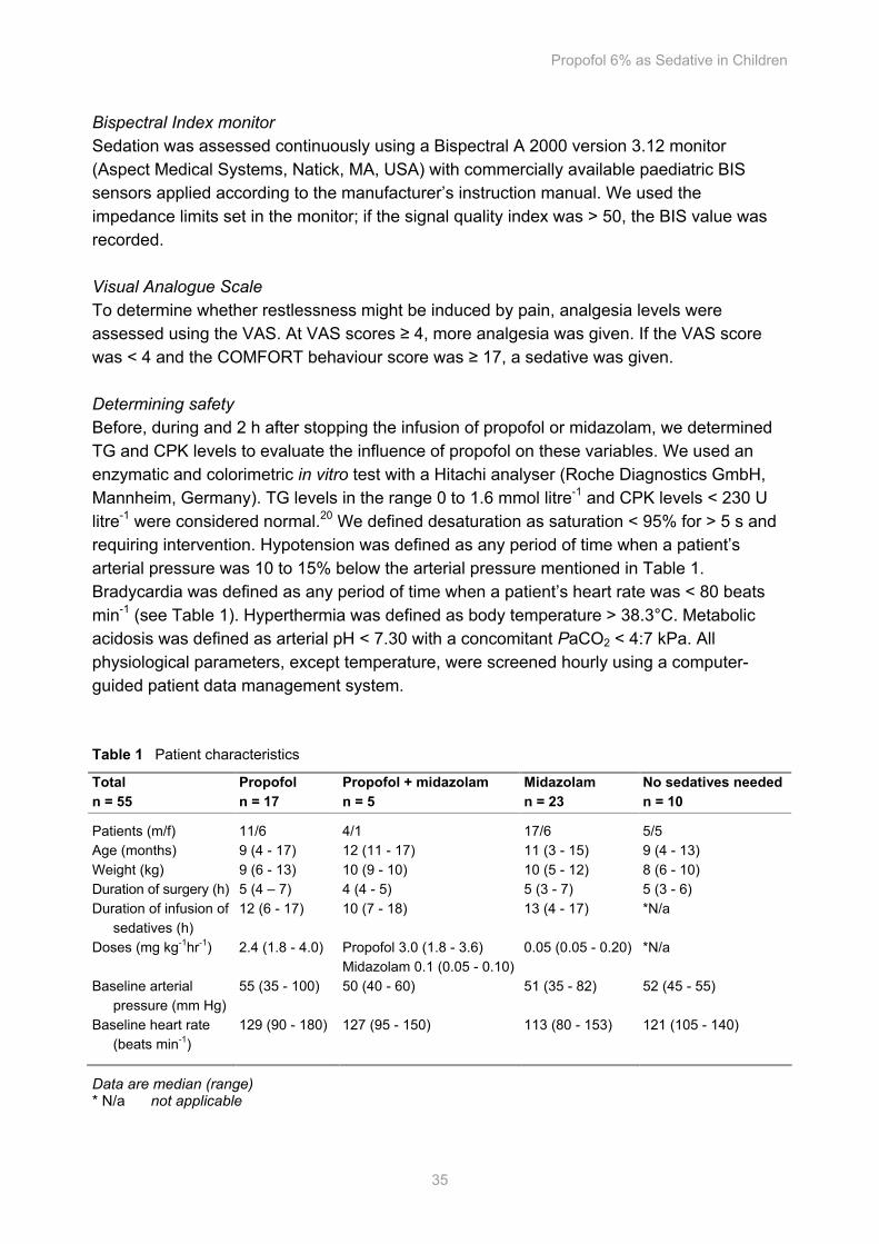
Propofol 6% as Sedative in Children
35
Bispectral Index monitor Sedation was assessed continuously using a Bispectral A 2000 version 3.12 monitor (Aspect Medical Systems, Natick, MA, USA) with commercially available paediatric BIS sensors applied according to the manufacturer’s instruction manual. We used the impedance limits set in the monitor; if the signal quality index was > 50, the BIS value was recorded. Visual Analogue Scale To determine whether restlessness might be induced by pain, analgesia levels were assessed using the VAS. At VAS scores ≥ 4, more analgesia was given. If the VAS score was < 4 and the COMFORT behaviour score was ≥ 17, a sedative was given. Determining safety Before, during and 2 h after stopping the infusion of propofol or midazolam, we determined TG and CPK levels to evaluate the influence of propofol on these variables. We used an enzymatic and colorimetric in vitro test with a Hitachi analyser (Roche Diagnostics GmbH, Mannheim, Germany). TG levels in the range 0 to 1.6 mmol litre-1 and CPK levels < 230 U litre-1 were considered normal.20 We defined desaturation as saturation < 95% for > 5 s and requiring intervention. Hypotension was defined as any period of time when a patient’s arterial pressure was 10 to 15% below the arterial pressure mentioned in Table 1. Bradycardia was defined as any period of time when a patient’s heart rate was < 80 beats min-1 (see Table 1). Hyperthermia was defined as body temperature > 38.3°C. Metabolic acidosis was defined as arterial pH < 7.30 with a concomitant PaCO2 < 4:7 kPa. All physiological parameters, except temperature, were screened hourly using a computer-guided patient data management system. Table 1 Patient characteristics
Total n = 55
Propofol n = 17
Propofol + midazolam n = 5
Midazolam n = 23
No sedatives needed n = 10
Patients (m/f) 11/6 4/1 17/6 5/5 Age (months) 9 (4 - 17) 12 (11 - 17) 11 (3 - 15) 9 (4 - 13) Weight (kg) 9 (6 - 13) 10 (9 - 10) 10 (5 - 12) 8 (6 - 10) Duration of surgery (h) 5 (4 – 7) 4 (4 - 5) 5 (3 - 7) 5 (3 - 6) Duration of infusion of sedatives (h)
12 (6 - 17) 10 (7 - 18) 13 (4 - 17) *N/a
Doses (mg kg-1hr-1) 2.4 (1.8 - 4.0) Propofol 3.0 (1.8 - 3.6) Midazolam 0.1 (0.05 - 0.10)
0.05 (0.05 - 0.20) *N/a
Baseline arterial pressure (mm Hg)
55 (35 - 100) 50 (40 - 60) 51 (35 - 82) 52 (45 - 55)
Baseline heart rate (beats min-1)
129 (90 - 180) 127 (95 - 150) 113 (80 - 153) 121 (105 - 140)
Data are median (range) * N/a not applicable
Chapter 3
36
Determining efficacy To compare the efficacy of propofol with that of midazolam, we considered COMFORT behaviour, VAS scores and BIS values in four groups: children receiving propofol, children receiving propofol with additional midazolam, children receiving midazolam and children who did not need sedation. Additionally, we determined the dose change frequency, i.e. the number of times that dosing of propofol or midazolam was adjusted. Medication preparation Propofol 6% was prepared in the Department of Clinical Pharmacy, St Antonius Hospital, Nieuwegein, The Netherlands.21 Propofol 6% was given through a central venous line in order to prevent pain from injection. Midazolam hydrochloride was dissolved in glucose 5% to make an i.v. solution. Statistical analysis The data were analysed using SPSS for Windows (version 10.0; SPSS, Chicago, IL). The safety parameters of children receiving propofol 6% and those receiving no propofol 6% were compared using the Mann–Whitney U-test. Statistical differences were considered significant if P < 0.05. A correlation r of 0.10 to 0.29 was considered small, 0.30 to 0.49 was considered medium and ≥ 0.50 was considered large. Results We studied 55 patients, with a median age of 10 (IQR 3 to 17) months and weight 9 (5 to 13) kg. Preoperative diagnoses were scaphocephaly (n = 26), trigonocephaly (n = 18), brachycephaly (n = 2), encephalocele (n = 1), plagiocephaly (n = 5) and Saethre–Chotzen syndrome (n = 3). There were no significant differences between the groups with regard to age, weight, duration of surgery or duration of infusion of sedatives (Table 1).
In one patient the TG level was 2.00 mmol litre-1 during propofol infusion without metabolic acidosis, disturbance of physiological parameters or increase of CPK levels (Fig. 1). Four patients had raised CPK levels, ranging from 261 to 313 U litre-1 during and after the end of infusion (Fig. 2). Three patients had received propofol and one patient had no medication. Two patients receiving propofol had elevated CPK levels before the start of infusion and one of these patients had elevated CPK levels during and after infusion. The first patient had CPK levels of 261 U litre-1 before infusion. The second patient had CPK levels of 336 U litre-1 before infusion, 276 U litre-1 during infusion and 240 - 282 U litre-1 after infusion. One patient receiving propofol had a CPK level of 313 U litre-1 after infusion. These patients showed no acidosis, no abnormal physiological parameters and no increased TG levels.
There were no respiratory complications. Three patients, one receiving propofol and two receiving midazolam, experienced short periods of desaturation with spontaneous recovery.
Median minimum arterial pressure was 56 mm Hg and 59 mm Hg for propofol 6% and no propofol 6%, respectively (Mann–Whitney U-test, 330; P = 0.57). Median minimal heart
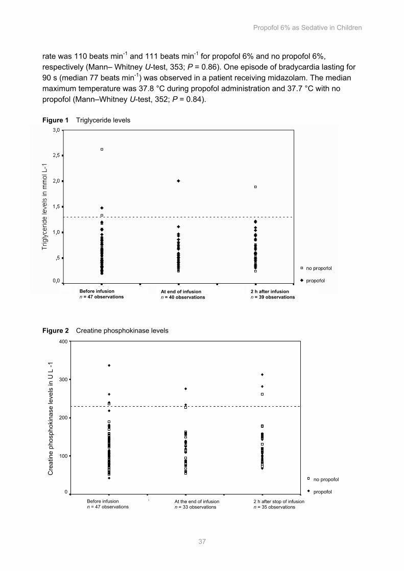
Propofol 6% as Sedative in Children
37
rate was 110 beats min-1 and 111 beats min-1 for propofol 6% and no propofol 6%, respectively (Mann– Whitney U-test, 353; P = 0.86). One episode of bradycardia lasting for 90 s (median 77 beats min-1) was observed in a patient receiving midazolam. The median maximum temperature was 37.8 °C during propofol administration and 37.7 °C with no propofol (Mann–Whitney U-test, 352; P = 0.84). Figure 1 Triglyceride levels
Figure 2 Creatine phosphokinase levels
2,52,01,51,0,50,0-,5
Cre
atin
e ph
osph
okin
ase
leve
ls in
U L
-1
400
300
200
100
0
no propofol
propofol
Before infusion n = 47 observations At end of infusion
n = 40 observations 2 h after infusion n = 39 observations
Before infusion n = 47 observations
At the end of infusion n = 33 observations
2 h after stop of infusion n = 35 observations
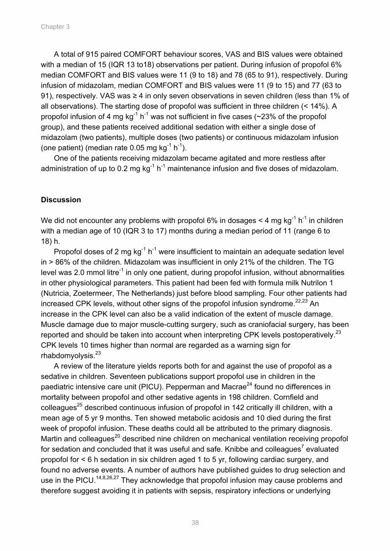
Chapter 3
38
A total of 915 paired COMFORT behaviour scores, VAS and BIS values were obtained with a median of 15 (IQR 13 to18) observations per patient. During infusion of propofol 6% median COMFORT and BIS values were 11 (9 to 18) and 78 (65 to 91), respectively. During infusion of midazolam, median COMFORT and BIS values were 11 (9 to 15) and 77 (63 to 91), respectively. VAS was ≥ 4 in only seven observations in seven children (less than 1% of all observations). The starting dose of propofol was sufficient in three children (< 14%). A propofol infusion of 4 mg kg-1 h-1 was not sufficient in five cases (~23% of the propofol group), and these patients received additional sedation with either a single dose of midazolam (two patients), multiple doses (two patients) or continuous midazolam infusion (one patient) (median rate 0.05 mg kg-1 h-1).
One of the patients receiving midazolam became agitated and more restless after administration of up to 0.2 mg kg-1 h-1 maintenance infusion and five doses of midazolam. Discussion We did not encounter any problems with propofol 6% in dosages < 4 mg kg-1 h-1 in children with a median age of 10 (IQR 3 to 17) months during a median period of 11 (range 6 to 18) h.
Propofol doses of 2 mg kg-1 h-1 were insufficient to maintain an adequate sedation level in > 86% of the children. Midazolam was insufficient in only 21% of the children. The TG level was 2.0 mmol litre-1 in only one patient, during propofol infusion, without abnormalities in other physiological parameters. This patient had been fed with formula milk Nutrilon 1 (Nutricia, Zoetermeer, The Netherlands) just before blood sampling. Four other patients had increased CPK levels, without other signs of the propofol infusion syndrome.22,23 An increase in the CPK level can also be a valid indication of the extent of muscle damage. Muscle damage due to major muscle-cutting surgery, such as craniofacial surgery, has been reported and should be taken into account when interpreting CPK levels postoperatively.23 CPK levels 10 times higher than normal are regarded as a warning sign for rhabdomyolysis.23
A review of the literature yields reports both for and against the use of propofol as a sedative in children. Seventeen publications support propofol use in children in the paediatric intensive care unit (PICU). Pepperman and Macrae24 found no differences in mortality between propofol and other sedative agents in 198 children. Cornfield and colleagues25 described continuous infusion of propofol in 142 critically ill children, with a mean age of 5 yr 9 months. Ten showed metabolic acidosis and 10 died during the first week of propofol infusion. These deaths could all be attributed to the primary diagnosis. Martin and colleagues20 described nine children on mechanical ventilation receiving propofol for sedation and concluded that it was useful and safe. Knibbe and colleagues7 evaluated propofol for < 6 h sedation in six children aged 1 to 5 yr, following cardiac surgery, and found no adverse events. A number of authors have published guides to drug selection and use in the PICU.14,8,26,27 They acknowledge that propofol infusion may cause problems and therefore suggest avoiding it in patients with sepsis, respiratory infections or underlying
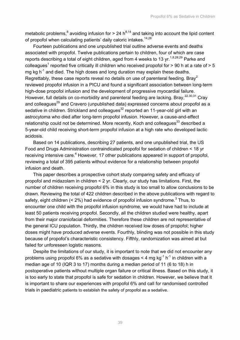
Propofol 6% as Sedative in Children
39
metabolic problems,8 avoiding infusion for > 24 h8,14 and taking into account the lipid content of propofol when calculating patients’ daily caloric intakes.14,26
Fourteen publications and one unpublished trial outline adverse events and deaths associated with propofol. Twelve publications pertain to children, four of which are case reports describing a total of eight children, aged from 4 weeks to 13 yr.1,8,28,29 Parke and colleagues1 reported five critically ill children who received propofol for > 90 h at a rate of > 5 mg kg h-1 and died. The high doses and long duration may explain these deaths. Regrettably, these case reports reveal no details on use of parenteral feeding. Bray2 reviewed propofol infusion in a PICU and found a significant association between long-term high-dose propofol infusion and the development of progressive myocardial failure. However, full details on co-morbidity and parenteral feeding are lacking. Bray,22,30,31 Cray and colleagues29 and Cravero (unpublished data) expressed concerns about propofol as a sedative in children. Strickland and colleagues32 reported an 11-year-old girl with an astrocytoma who died after long-term propofol infusion. However, a cause-and-effect relationship could not be determined. More recently, Koch and colleagues33 described a 5-year-old child receiving short-term propofol infusion at a high rate who developed lactic acidosis.
Based on 14 publications, describing 27 patients, and one unpublished trial, the US Food and Drugs Administration contraindicated propofol for sedation of children < 18 yr receiving intensive care.4 However, 17 other publications appeared in support of propofol, reviewing a total of 395 patients without evidence for a relationship between propofol infusion and death.
This paper describes a prospective cohort study comparing safety and efficacy of propofol and midazolam in children < 2 yr. Clearly, our study has limitations. First, the number of children receiving propofol 6% in this study is too small to allow conclusions to be drawn. Reviewing the total of 422 children described in the above publications with regard to safety, eight children (< 2%) had evidence of propofol infusion syndrome.3 Thus, to encounter one child with the propofol infusion syndrome, we would have had to include at least 50 patients receiving propofol. Secondly, all the children studied were healthy, apart from their major craniofacial deformities. Therefore these children are not representative of the general ICU population. Thirdly, the children received low doses of propofol; higher doses might have produced adverse events. Fourthly, blinding was not possible in this study because of propofol’s characteristic consistency. Fifthly, randomization was aimed at but failed for unforeseen logistic reasons.
Despite the limitations of our study, it is important to note that we did not encounter any problems using propofol 6% as a sedative with dosages < 4 mg kg-1 h-1 in children with a median age of 10 (IQR 3 to 17) months during a median period of 11 (6 to 18) h in postoperative patients without multiple organ failure or critical illness. Based on this study, it is too early to state that propofol is safe for sedation in children. However, we believe that it is important to share our experiences with propofol 6% and call for randomised controlled trials in paediatric patients to establish the safety of propofol as a sedative.
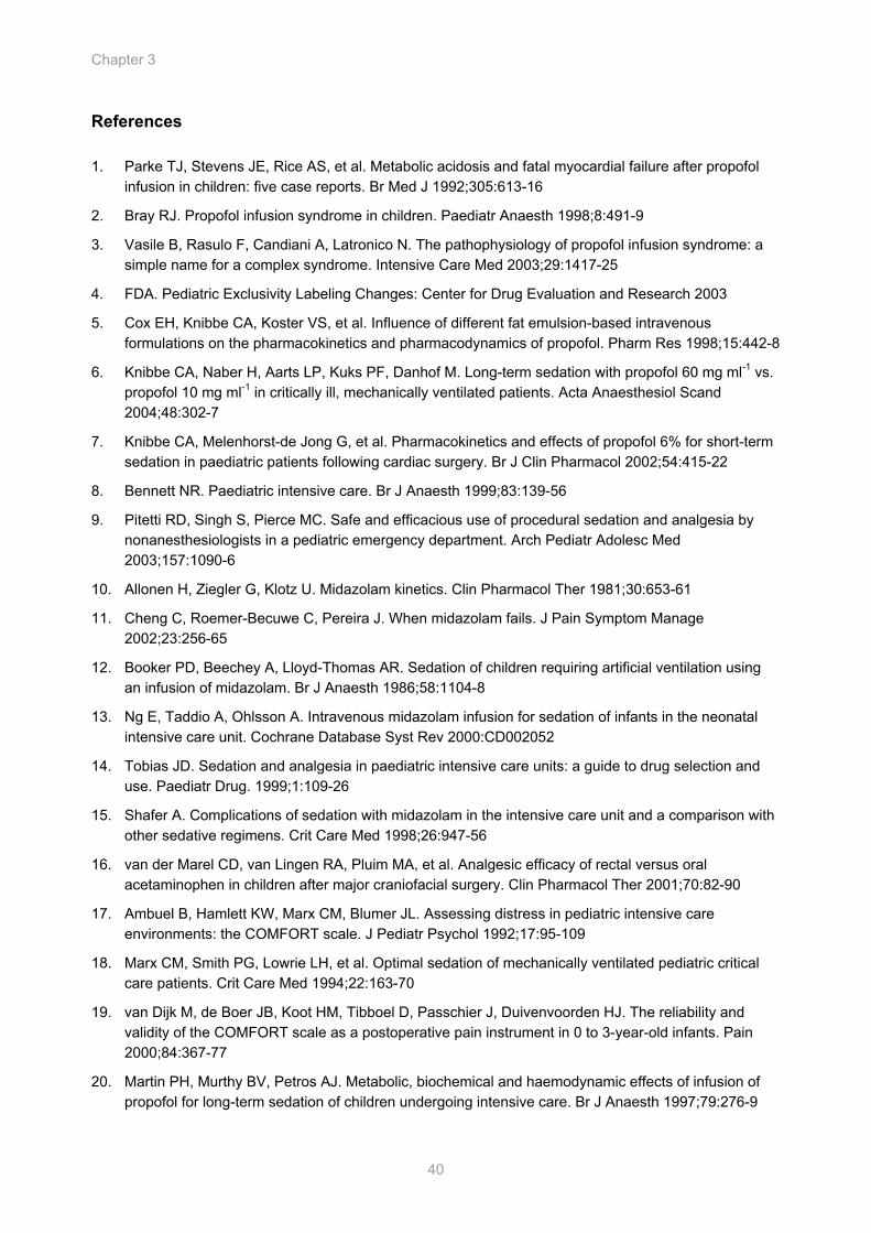
Chapter 3
40
References 1. Parke TJ, Stevens JE, Rice AS, et al. Metabolic acidosis and fatal myocardial failure after propofol
infusion in children: five case reports. Br Med J 1992;305:613-16
2. Bray RJ. Propofol infusion syndrome in children. Paediatr Anaesth 1998;8:491-9
3. Vasile B, Rasulo F, Candiani A, Latronico N. The pathophysiology of propofol infusion syndrome: a simple name for a complex syndrome. Intensive Care Med 2003;29:1417-25
4. FDA. Pediatric Exclusivity Labeling Changes: Center for Drug Evaluation and Research 2003
5. Cox EH, Knibbe CA, Koster VS, et al. Influence of different fat emulsion-based intravenous formulations on the pharmacokinetics and pharmacodynamics of propofol. Pharm Res 1998;15:442-8
6. Knibbe CA, Naber H, Aarts LP, Kuks PF, Danhof M. Long-term sedation with propofol 60 mg ml-1 vs. propofol 10 mg ml-1 in critically ill, mechanically ventilated patients. Acta Anaesthesiol Scand 2004;48:302-7
7. Knibbe CA, Melenhorst-de Jong G, et al. Pharmacokinetics and effects of propofol 6% for short-term sedation in paediatric patients following cardiac surgery. Br J Clin Pharmacol 2002;54:415-22
8. Bennett NR. Paediatric intensive care. Br J Anaesth 1999;83:139-56
9. Pitetti RD, Singh S, Pierce MC. Safe and efficacious use of procedural sedation and analgesia by nonanesthesiologists in a pediatric emergency department. Arch Pediatr Adolesc Med 2003;157:1090-6
10. Allonen H, Ziegler G, Klotz U. Midazolam kinetics. Clin Pharmacol Ther 1981;30:653-61
11. Cheng C, Roemer-Becuwe C, Pereira J. When midazolam fails. J Pain Symptom Manage 2002;23:256-65
12. Booker PD, Beechey A, Lloyd-Thomas AR. Sedation of children requiring artificial ventilation using an infusion of midazolam. Br J Anaesth 1986;58:1104-8
13. Ng E, Taddio A, Ohlsson A. Intravenous midazolam infusion for sedation of infants in the neonatal intensive care unit. Cochrane Database Syst Rev 2000:CD002052
14. Tobias JD. Sedation and analgesia in paediatric intensive care units: a guide to drug selection and use. Paediatr Drug. 1999;1:109-26
15. Shafer A. Complications of sedation with midazolam in the intensive care unit and a comparison with other sedative regimens. Crit Care Med 1998;26:947-56
16. van der Marel CD, van Lingen RA, Pluim MA, et al. Analgesic efficacy of rectal versus oral acetaminophen in children after major craniofacial surgery. Clin Pharmacol Ther 2001;70:82-90
17. Ambuel B, Hamlett KW, Marx CM, Blumer JL. Assessing distress in pediatric intensive care environments: the COMFORT scale. J Pediatr Psychol 1992;17:95-109
18. Marx CM, Smith PG, Lowrie LH, et al. Optimal sedation of mechanically ventilated pediatric critical care patients. Crit Care Med 1994;22:163-70
19. van Dijk M, de Boer JB, Koot HM, Tibboel D, Passchier J, Duivenvoorden HJ. The reliability and validity of the COMFORT scale as a postoperative pain instrument in 0 to 3-year-old infants. Pain 2000;84:367-77
20. Martin PH, Murthy BV, Petros AJ. Metabolic, biochemical and haemodynamic effects of infusion of propofol for long-term sedation of children undergoing intensive care. Br J Anaesth 1997;79:276-9
Propofol 6% as Sedative in Children
41
21. Peeters MYM, Lange R, Aarts LPHJ, Talsma H, Knibbe CAJ. Stability of an intravenous fat emulsion containing 6% propofol and a low amount of emulsifier. Eur J Hosp Pharmacy 2004;1:201-8
22. Bray RJ. Propofol-infusion syndrome in children Lancet 1999;353:2074-5
23. Laurence AS. Serum myoglobin and creatine kinase following surgery. Br J Anaesth 2000;84:763-6
24. Pepperman ML, Macrae D. A comparison of propofol and other sedative use in paediatric intensive care in the United Kingdom. Paediatr Anaesth 1997;7:143-53
25. Cornfield DN, Tegtmeyer K, Nelson MD, Milla CE, Sweeney M. Continous propofol infusion in 142 critically ill children. Pediatrics 2002;110:1177-81
26. Aun CS. New i.v. agents. Br J Anaesth 1999;83:29-41
27. Fulton B, Sorkin EM. Propofol. An overview of its pharmacology and a review of its clinical efficacy in intensive care sedation. Drugs 1995;50:636-57
28. Bray RJ. Fatal myocardial failure associated with a propofol infusion in a child. Anaesthesia 1995;50:94
29. Cray SH, Robinson BH, Cox PN. Lactic acidaemia and bradyarrhythmia in a child sedated with propofol. Crit Care Med 1998;26:2087-92
30. Bray RJ. Heart block following propofol in a child. Paediatr Anaesth 2000;10:226
31. Bray RJ. Propofol infusion for ICU sedation in children. Anaesthesia 2002;57:521
32. Strickland RA, Murray MJ. Fatal metabolic acidosis in a pediatric patient receiving an infusion of propofol in the intensive care unit: is there a relationship? Crit Care Med 1995;23:405-9
33. Koch M, De Backer D, Vincent JL. Lactic acidosis: an early marker of propofol infusion syndrome? Intensive Care Med 2004;30:522

Propofol Pharmacokinetics and Pharmacodynamics for Depth of Sedation in Non-Ventilated Infants after Major Craniofacial Surgery
Mariska Y.M. Peeters*, Sandra A. Prins*, Catherijne A.J. Knibbe, Joost DeJongh, Ron H.N. van Schaik, Monique van Dijk, Ilse P. van der Heiden, Dick Tibboel, and Meindert Danhof (* S.A. Prins and M.Y.M. Peeters contributed equally to this paper) Accepted for publication in Anesthesiology
Cha
pter
Chapter 4
44
Abstract Background Little is known about the use and dose of propofol for sedation in non-ventilated children. To support the safe and effective use of propofol, a population model for propofol pharmacokinetics and pharmacodynamics and depth of sedation is described in non-ventilated children after surgery. Methods Following craniofacial surgery, children were randomly allocated to receive propofol or midazolam, if sedative medication was judged necessary on the basis of the COMFORT-behavior (COMFORT-B) score. Data of 44 infants (aged 3 -17 months) in the Pediatric Surgical Intensive Care Unit were analyzed, of which 22 infants received sedation with propofol in doses up to 4 mg/kg/h during a median of 12,5 hours. COMFORT-B scores and Bispectral index (BIS) values were recorded simultaneously and a median of 11 arterial blood samples per infant were collected for propofol concentration analysis. Population pharmacokinetic and pharmacodynamic modeling was performed using NONMEM V. Bootstrap resampling was used as internal validation. Results In the two-compartment pharmacokinetic model, body weight (median 8.9 kg) was incorporated as a power function for elimination clearance. Typical values of the pharmacokinetic parameters were Cl = 0.70· (BW/8.9)0.61 L min , Vc = 18.8 L, Q = 0.35 L min and Vss = 146 L. In infants who received no sedative, depth of sedation was found to be a function of a post-anesthesia effect (Emax model) in combination with a circadian night rhythm. In agitated infants, no circadian night dip was seen and depth of sedation was best described by a post-anesthesia effect and the effect of propofol (Emax model). The propofol concentration at half maximum effect (EC50) was 1.76 mg/l with interindividual coefficient of variation (CV) of 47 % characterized on the COMFORT-B scale and 3.71 mg/l (CV 145%) on the BIS. Conclusions Propofol clearance was found to be two times higher in non-ventilated healthy children than reported in the literature for ventilated children and adults. Based on the model we advise a propofol dose of 30 mg/h in a 10 kg infant to achieve values of 12 -14 on the COMFORT-B and 70 - 75 on the BIS during the night. Wide pharmacodynamic variability emphasizes the importance of dose titration.
PK/PD Model for Propofol for Infants
45
Introduction To correct craniosynostosis, most infants are operated in the first years of life. Due to edematous eyelids, separation from parents and the need to stay at the intensive care unit for control of vital signs and the possible development of neurological sequelae, these children often experience stress postoperatively. Although propofol is widely used for sedation in the adult intensive care, its use is subject for debate in sedated children in the pediatric intensive care since the report of 5 deaths in children receiving high doses (> 5 mg/kg/h) of propofol.1 In general, larger doses of propofol are required in children and it is suggested that this is due to differences in pharmacokinetics2 and/or sensitivity.3
To date, there are no population models in children investigating the pharmacodynamics to study the variability between and within children. As pharmacodynamic endpoint, a number of clinical sedation scores have been devised for use in children, in which the COMFORT-behavior (COMFORT-B) scale would be a reliable alternative to the original, most used COMFORT scale.4,5 The Bispectral Index (BIS) may have benefits in comparison with clinical sedation, because it assesses sedation continuously and may provide an objective, quantitative measure of the level of sedation.6 However up till date, the BIS has only been validated in children older than 1 year old.
Our clinical experiences concerning the use of propofol evaluated by COMFORT-B in young children in the Pediatric Surgical Intensive Care Unit (PSICU) have recently been published by Prins et al.7 In the current paper, the propofol pharmacokinetics (PK) and pharmacodynamics (PD), characterized by use of COMFORT-B and BIS on the postoperative sleep pattern in non-ventilated infants is described using population modelling, to select appropriate doses in infants and to support the safe and effective use of propofol. Methods The study was performed in the PSICU of the Erasmus MC–Sophia Children's Hospital. The study protocol was approved by the ethics committee of the Erasmus MC–Sophia Children's Hospital. Written informed consent was obtained from the parents. The study design and sedative and analgesic regimen is presented in detail in the article of Prins et al,7 and shortly repeated if relevant to this article. Patients Eligibility criteria included major craniofacial surgery, age between one month and two years, postoperative admitted to the PSICU. The children were randomly allocated to receive propofol or midazolam if sedative medication was judged necessary on the basis of the COMFORT-B score (score ≥ 17). Infants were excluded when they suffered from respiratory infections, epilepsy, hypertriglyceridemia or family histories of hyper-cholesterolemia, allergic history to propofol, eggs or soybean oil.
Patients' characteristics of the group, in which no sedation was necessary (the non-agitated group) and the group in which sedation was needed (agitated group) are presented
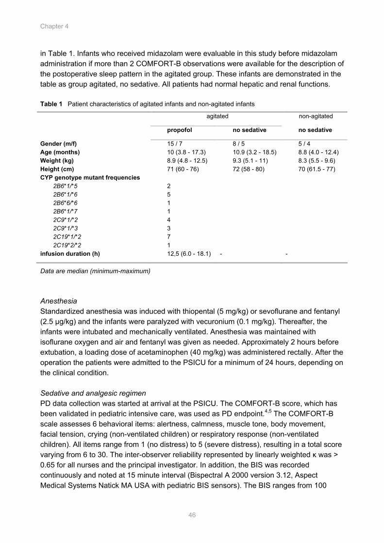
Chapter 4
46
in Table 1. Infants who received midazolam were evaluable in this study before midazolam administration if more than 2 COMFORT-B observations were available for the description of the postoperative sleep pattern in the agitated group. These infants are demonstrated in the table as group agitated, no sedative. All patients had normal hepatic and renal functions. Table 1 Patient characteristics of agitated infants and non-agitated infants
agitated non-agitated
propofol no sedative no sedative
Gender (m/f) 15 / 7 8 / 5 5 / 4 Age (months) 10 (3.8 - 17.3) 10.9 (3.2 - 18.5) 8.8 (4.0 - 12.4) Weight (kg) 8.9 (4.8 - 12.5) 9.3 (5.1 - 11) 8.3 (5.5 - 9.6) Height (cm) 71 (60 - 76) 72 (58 - 80) 70 (61.5 - 77) CYP genotype mutant frequencies 2B6*1/*5 2B6*1/*6 2B6*6/*6 2B6*1/*7 2C9*1/*2 2C9*1/*3 2C19*1/*2 2C19*2/*2
2 5 1 1 4 3 7 1
infusion duration (h) 12,5 (6.0 - 18.1) - -
Data are median (minimum-maximum) Anesthesia Standardized anesthesia was induced with thiopental (5 mg/kg) or sevoflurane and fentanyl (2.5 µg/kg) and the infants were paralyzed with vecuronium (0.1 mg/kg). Thereafter, the infants were intubated and mechanically ventilated. Anesthesia was maintained with isoflurane oxygen and air and fentanyl was given as needed. Approximately 2 hours before extubation, a loading dose of acetaminophen (40 mg/kg) was administered rectally. After the operation the patients were admitted to the PSICU for a minimum of 24 hours, depending on the clinical condition. Sedative and analgesic regimen PD data collection was started at arrival at the PSICU. The COMFORT-B score, which has been validated in pediatric intensive care, was used as PD endpoint.4,5 The COMFORT-B scale assesses 6 behavioral items: alertness, calmness, muscle tone, body movement, facial tension, crying (non-ventilated children) or respiratory response (non-ventilated children). All items range from 1 (no distress) to 5 (severe distress), resulting in a total score varying from 6 to 30. The inter-observer reliability represented by linearly weighted κ was > 0.65 for all nurses and the principal investigator. In addition, the BIS was recorded continuously and noted at 15 minute interval (Bispectral A 2000 version 3.12, Aspect Medical Systems Natick MA USA with pediatric BIS sensors). The BIS ranges from 100
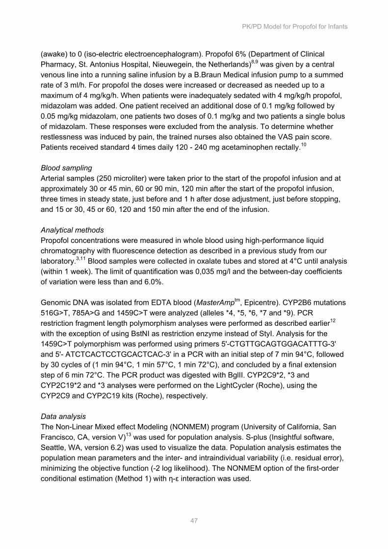
PK/PD Model for Propofol for Infants
47
(awake) to 0 (iso-electric electroencephalogram). Propofol 6% (Department of Clinical Pharmacy, St. Antonius Hospital, Nieuwegein, the Netherlands)8,9 was given by a central venous line into a running saline infusion by a B.Braun Medical infusion pump to a summed rate of 3 ml/h. For propofol the doses were increased or decreased as needed up to a maximum of 4 mg/kg/h. When patients were inadequately sedated with 4 mg/kg/h propofol, midazolam was added. One patient received an additional dose of 0.1 mg/kg followed by 0.05 mg/kg midazolam, one patients two doses of 0.1 mg/kg and two patients a single bolus of midazolam. These responses were excluded from the analysis. To determine whether restlessness was induced by pain, the trained nurses also obtained the VAS pain score. Patients received standard 4 times daily 120 - 240 mg acetaminophen rectally.10 Blood sampling Arterial samples (250 microliter) were taken prior to the start of the propofol infusion and at approximately 30 or 45 min, 60 or 90 min, 120 min after the start of the propofol infusion, three times in steady state, just before and 1 h after dose adjustment, just before stopping, and 15 or 30, 45 or 60, 120 and 150 min after the end of the infusion. Analytical methods Propofol concentrations were measured in whole blood using high-performance liquid chromatography with fluorescence detection as described in a previous study from our laboratory.3,11 Blood samples were collected in oxalate tubes and stored at 4°C until analysis (within 1 week). The limit of quantification was 0,035 mg/l and the between-day coefficients of variation were less than and 6.0%. Genomic DNA was isolated from EDTA blood (MasterAmptm, Epicentre). CYP2B6 mutations 516G>T, 785A>G and 1459C>T were analyzed (alleles *4, *5, *6, *7 and *9). PCR restriction fragment length polymorphism analyses were performed as described earlier12 with the exception of using BstNI as restriction enzyme instead of StyI. Analysis for the 1459C>T polymorphism was performed using primers 5'-CTGTTGCAGTGGACATTTG-3' and 5'- ATCTCACTCCTGCACTCAC-3' in a PCR with an initial step of 7 min 94°C, followed by 30 cycles of (1 min 94°C, 1 min 57°C, 1 min 72°C), and concluded by a final extension step of 6 min 72°C. The PCR product was digested with BglII. CYP2C9*2, *3 and CYP2C19*2 and *3 analyses were performed on the LightCycler (Roche), using the CYP2C9 and CYP2C19 kits (Roche), respectively. Data analysis The Non-Linear Mixed effect Modeling (NONMEM) program (University of California, San Francisco, CA, version V)13 was used for population analysis. S-plus (Insightful software, Seattle, WA, version 6.2) was used to visualize the data. Population analysis estimates the population mean parameters and the inter- and intraindividual variability (i.e. residual error), minimizing the objective function (-2 log likelihood). The NONMEM option of the first-order conditional estimation (Method 1) with η-ε interaction was used.
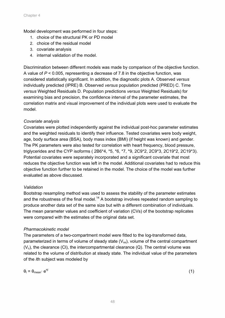
Chapter 4
48
Model development was performed in four steps: 1. choice of the structural PK or PD model 2. choice of the residual model 3. covariate analysis 4. internal validation of the model.
Discrimination between different models was made by comparison of the objective function. A value of P < 0.005, representing a decrease of 7.8 in the objective function, was considered statistically significant. In addition, the diagnostic plots A. Observed versus individually predicted (IPRE) B. Observed versus population predicted (PRED) C. Time versus Weighted Residuals D. Population predictions versus Weighted Residuals) for examining bias and precision, the confidence interval of the parameter estimates, the correlation matrix and visual improvement of the individual plots were used to evaluate the model. Covariate analysis Covariates were plotted independently against the individual post-hoc parameter estimates and the weighted residuals to identify their influence. Tested covariates were body weight, age, body surface area (BSA), body mass index (BMI) (if height was known) and gender. The PK parameters were also tested for correlation with heart frequency, blood pressure, triglycerides and the CYP isoforms ( 2B6*4, *5, *6, *7, *9, 2C9*2, 2C9*3, 2C19*2, 2C19*3). Potential covariates were separately incorporated and a significant covariate that most reduces the objective function was left in the model. Additional covariates had to reduce this objective function further to be retained in the model. The choice of the model was further evaluated as above discussed. Validation Bootstrap resampling method was used to assess the stability of the parameter estimates and the robustness of the final model.14 A bootstrap involves repeated random sampling to produce another data set of the same size but with a different combination of individuals. The mean parameter values and coefficient of variation (CVs) of the bootstrap replicates were compared with the estimates of the original data set. Pharmacokinetic model The parameters of a two-compartment model were fitted to the log-transformed data, parameterized in terms of volume of steady state (Vss), volume of the central compartment (Vc), the clearance (Cl), the intercompartmental clearance (Q). The central volume was related to the volume of distribution at steady state. The individual value of the parameters of the ith subject was modeled by θi = θmean· eηi (1)
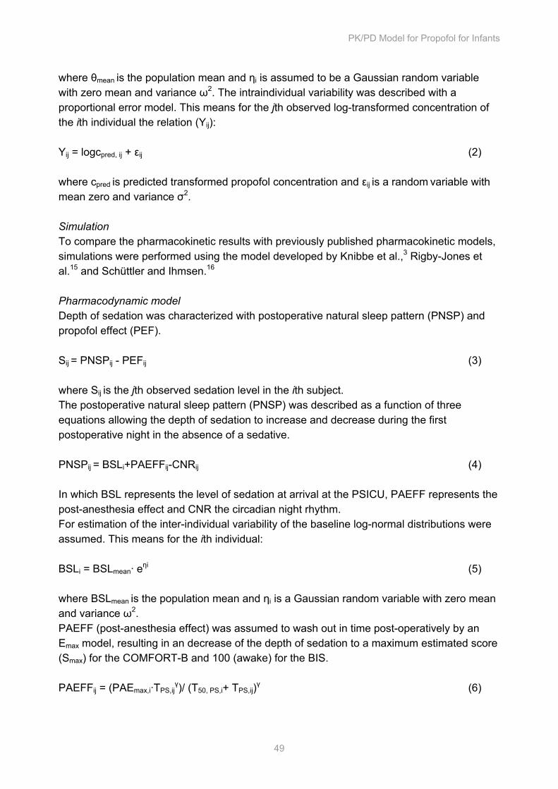
PK/PD Model for Propofol for Infants
49
where θmean is the population mean and ηi is assumed to be a Gaussian random variable with zero mean and variance ω2. The intraindividual variability was described with a proportional error model. This means for the jth observed log-transformed concentration of the ith individual the relation (Yij): Yij = logcpred, ij + εij (2) where cpred is predicted transformed propofol concentration and εij is a random variable with mean zero and variance σ2. Simulation To compare the pharmacokinetic results with previously published pharmacokinetic models, simulations were performed using the model developed by Knibbe et al.,3 Rigby-Jones et al.15 and Schüttler and Ihmsen.16 Pharmacodynamic model Depth of sedation was characterized with postoperative natural sleep pattern (PNSP) and propofol effect (PEF). Sij = PNSPij - PEFij (3) where Sij is the jth observed sedation level in the ith subject. The postoperative natural sleep pattern (PNSP) was described as a function of three equations allowing the depth of sedation to increase and decrease during the first postoperative night in the absence of a sedative. PNSPij = BSLi+PAEFFij-CNRij (4) In which BSL represents the level of sedation at arrival at the PSICU, PAEFF represents the post-anesthesia effect and CNR the circadian night rhythm. For estimation of the inter-individual variability of the baseline log-normal distributions were assumed. This means for the ith individual: BSLi = BSLmean· eηi (5) where BSLmean is the population mean and ηi is a Gaussian random variable with zero mean and variance ω2. PAEFF (post-anesthesia effect) was assumed to wash out in time post-operatively by an Emax model, resulting in an decrease of the depth of sedation to a maximum estimated score (Smax) for the COMFORT-B and 100 (awake) for the BIS. PAEFFij = (PAEmax,i·TPS,ij
γ)/ (T50, PS,i+ TPS,ij)γ (6)
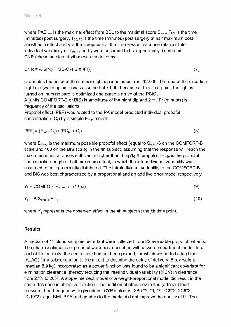
Chapter 4
50
where PAEmax is the maximal effect from BSL to the maximal score Smax. TPS is the time (minutes) post surgery, T50, PS is the time (minutes) post surgery at half maximum post-anesthesia effect and γ is the steepness of the time versus response relation. Inter-individual variability of T50, PS and γ were assumed to be log-normally distributed. CNR (circadian night rhythm) was modeled by: CNR = A·SIN((TIME-O)·( 2 π /Fr)) (7) O denotes the onset of the natural night dip in minutes from 12.00h. The end of the circadian night dip (wake up time) was assumed at 7.00h, because at this time point, the light is turned on, nursing care is optimized and parents arrive at the PSICU. A (units COMFORT-B or BIS) is amplitude of the night dip and 2 π / Fr (minutes) is frequency of the oscillations. Propofol effect (PEF) was related to the PK model-predicted individual propofol concentration (Cij) by a simple Emax model: PEFij = (Emaxi·Cij) / (EC50i+ Cij) (8) where Emax,i is the maximum possible propofol effect (equal to Smax -6 on the COMFORT-B scale and 100 on the BIS scale) in the ith subject, assuming that the response will reach the maximum effect at doses sufficiently higher than 4 mg/kg/h propofol. EC50 is the propofol concentration (mg/l) at half maximum effect, in which the interindividual variability was assumed to be log-normally distributed. The intraindividual variability in the COMFORT-B and BIS was best characterized by a proportional and an additive error model respectively. Yij = COMFORT-Bpred, ij · (1+ εij) (9) Yij = BISpred, ij + εij. (10) where Yij represents the observed effect in the ith subject at the jth time point.
Results A median of 11 blood samples per infant were collected from 22 evaluable propofol patients. The pharmacokinetics of propofol were best described with a two-compartment model. In a part of the patients, the central line had not been primed, for which we added a lag time (ALAG) for a subpopulation to the model to describe the delay of delivery. Body weight (median 8.9 kg) incorporated as a power function was found to be a significant covariate for elimination clearance, thereby reducing the interindividual variability (%CV) in clearance from 27% to 20%. A slope-intercept model or a weight-proportional model did result in the same decrease in objective function. The addition of other covariates (arterial blood pressure, heart frequency, triglycerides, CYP isoforms (2B6 *5, *6, *7, 2C9*2, 2C9*3, 2C19*2), age, BMI, BSA and gender) to the model did not improve the quality of fit. The
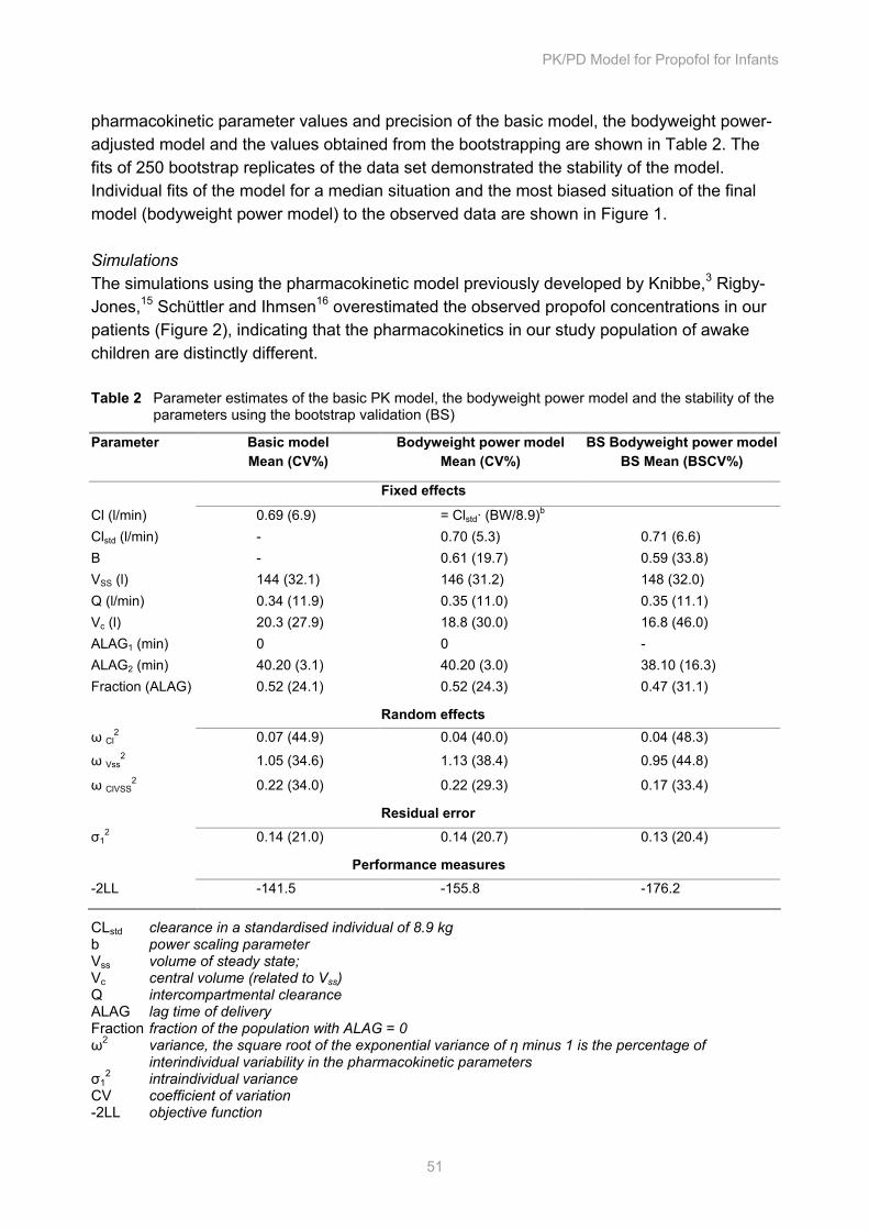
PK/PD Model for Propofol for Infants
51
pharmacokinetic parameter values and precision of the basic model, the bodyweight power-adjusted model and the values obtained from the bootstrapping are shown in Table 2. The fits of 250 bootstrap replicates of the data set demonstrated the stability of the model. Individual fits of the model for a median situation and the most biased situation of the final model (bodyweight power model) to the observed data are shown in Figure 1. Simulations The simulations using the pharmacokinetic model previously developed by Knibbe,3 Rigby-Jones,15 Schüttler and Ihmsen16 overestimated the observed propofol concentrations in our patients (Figure 2), indicating that the pharmacokinetics in our study population of awake children are distinctly different. Table 2 Parameter estimates of the basic PK model, the bodyweight power model and the stability of the parameters using the bootstrap validation (BS)
Parameter Basic model Mean (CV%)
Bodyweight power model Mean (CV%)
BS Bodyweight power modelBS Mean (BSCV%)
Fixed effects
Cl (l/min) 0.69 (6.9) = Clstd· (BW/8.9)b Clstd (l/min) - 0.70 (5.3) 0.71 (6.6) B - 0.61 (19.7) 0.59 (33.8) VSS (l) 144 (32.1) 146 (31.2) 148 (32.0) Q (l/min) 0.34 (11.9) 0.35 (11.0) 0.35 (11.1) Vc (l) 20.3 (27.9) 18.8 (30.0) 16.8 (46.0) ALAG1 (min) 0 0 - ALAG2 (min) 40.20 (3.1) 40.20 (3.0) 38.10 (16.3) Fraction (ALAG) 0.52 (24.1) 0.52 (24.3) 0.47 (31.1)
Random effects ω Cl
2 0.07 (44.9) 0.04 (40.0) 0.04 (48.3)
ω Vss2 1.05 (34.6) 1.13 (38.4) 0.95 (44.8)
ω ClVSS2 0.22 (34.0) 0.22 (29.3) 0.17 (33.4)
Residual error σ1
2 0.14 (21.0) 0.14 (20.7) 0.13 (20.4)
Performance measures -2LL -141.5 -155.8 -176.2
CLstd clearance in a standardised individual of 8.9 kg b power scaling parameter Vss volume of steady state; Vc central volume (related to Vss) Q intercompartmental clearance ALAG lag time of delivery Fraction fraction of the population with ALAG = 0 ω2 variance, the square root of the exponential variance of η minus 1 is the percentage of
interindividual variability in the pharmacokinetic parameters σ1
2 intraindividual variance CV coefficient of variation -2LL objective function
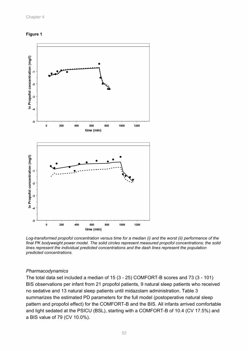
Chapter 4
52
Figure 1
0 200 400 600 800 1000 1200time (min)
-5
-4
-3
-2
-1
ln P
ropo
fol c
once
ntra
tion
(mg/
l)
0 200 400 600 800 1000 1200time (min)
-5
-4
-3
-2
-1
ln P
ropo
fol c
once
ntra
tion
(mg/
l)
Log-transformed propofol concentration versus time for a median (i) and the worst (ii) performance of the final PK bodyweight power model. The solid circles represent measured propofol concentrations; the solid lines represent the individual predicted concentrations and the dash lines represent the population predicted concentrations. Pharmacodynamics The total data set included a median of 15 (3 - 25) COMFORT-B scores and 73 (3 - 101) BIS observations per infant from 21 propofol patients, 9 natural sleep patients who received no sedative and 13 natural sleep patients until midazolam administration. Table 3 summarizes the estimated PD parameters for the full model (postoperative natural sleep pattern and propofol effect) for the COMFORT-B and the BIS. All infants arrived comfortable and light sedated at the PSICU (BSL), starting with a COMFORT-B of 10.4 (CV 17.5%) and a BIS value of 79 (CV 10.0%).

PK/PD Model for Propofol for Infants
53
Table 3 PD parameter estimates of the depth of sedation postoperative using COMFORT-B and BIS and the stability of the parameters using the bootstrap validation (BS)
parameter COMFORT-B Mean (CV%)
BS COMFORT-BMean (CV%)
BIS Mean (CV%)
BS BIS Mean (CV%)
Fixed effects
BSL 10.4 (5.1) 10.4 (5.6) 79.2 (1.2) 78.9 (1.1)
T50,PS (min) agitated 518 (44.2) 548 (49.7) 1044 (7.1) 1048 (10.8) T50, PS (min) non agitated 1580 (46.3) 1694 (49.9) 2052 (24.3) 2106 (41.5) γ 1 Fixed - 8.3 (27.3) 9.7 (46.3)
PAEFF
Maximal score Smax 20.0 (25.1) 19.7 (28.5) 100 Fixed -
Onset (min) 480 (1.2) 376 (42) 330 (0.8) 323 (11.8) Frequency (min) 1390 (8.6) 1752 (38.4) 2440 (20.3) 2796 (31.1)
CNR
Amplitude (response units) 3.5 (36.7) 3.7 (33.7) 14.5 (16.2) 16.8 (18.7)
PEF EC50 (mg/l) 1.76 (28.4) 2.01 (38.7) 3.71 (31.3) 4.01 (38.0)
Random effects ω BSL
2 0.03 (33.6) 0.02 (37.1) 0.01 (22.7) 0.01 (18.9)
ω T50,PS2 - - 0.05 (48.0) 0.08 (55.0)
ω γ2 - - 0.84 (48.3) 0.73 (65.9)
ω EC502 0.20 (70.2) 0.21 (80.7) 1.13 (43.2) 1.04 (59.3)
Residual error σ1
2 0.10 (8.1) 0.10 (8.1) - -
σ22 - - 178 (6.0) 175 (6.5)
Performance measures -2LL 2470.9 2446 16497 16430
BSL level of sedation at arrival PAEFF post-anesthesia effect T50,PS time post surgery at half maximum post-anaesthesia effect γ steepness CNR circadian night rhythm PEF propofol effect EC50 propofol concentration at half maximum effect ω2 variance, the square root of the exponential variance of η minus 1 is the percentage of
interindividual variability in the PD parameters σ1
2 intraindividual variance proportional σ2
2 intraindividual variance additive CV coefficient of variation -2LL objective function In the agitated infants during the postoperative night the narcotic effect washed out earlier, indicated by a smaller T50,PS (518 versus 1580 minutes for the COMFORT-B and 1044 versus 2052 minutes for the BIS). The steepness value of the wash-out effect (γ) for the BIS was 8, while the steepness for the COMFORT-B was not found to be significantly different from 1. During the night the infants were 'deeper' asleep, which was implemented in the model using the dip of a circadian rhythm. The start of the dip was estimated at 20.00 h
Chapter 4
54
(equal to 480 minutes from 12.00 h) on COMFORT-B with an amplitude of 3.5 units, respectively 17.30h (equal to 330 minutes) on the BIS with an amplitude of 14.5. For the agitated infants receiving propofol during the night a night dip could not be estimated. Propofol was started at a median time of 19.00h, which is equal to 5.5 h after surgery. The induced BIS depression as function of the propofol concentration showed considerable intersubject variability (CV 145%). The bootstrap validation (100 times) confirmed the precision of the parameters. Figure 3 i shows a median fit of a non-agitated infant who received no sedative, with a reduction in response during the night. Figure 3 ii and 3 iii show a median and a worse fit of the sleep pattern of an agitated infant and the influence of propofol. Figure 4 illustrates the simulated relationship between time, propofol infusion rate, propofol concentration and predicted population response in terms of depth of sedation using COMFORT-B and BIS. The difference of a 10 kg infant and a 5 kg infant is shown at the infusion rate of 18 mg/h. The difference in postoperative natural sleep pattern between infants who did or did not became agitated is shown at propofol infusion rate 0 mg/h.
There was not enough evidence to support gender, age and bodyweight as covariates on the PD parameters. Figure 2
0 200 400 600 800 1000 1200time (min)
0.0
0.5
1.0
1.5
2.0
Prop
ofol
con
cent
ratio
n (m
g/l)
Simulated population propofol concentrations (line) versus observed concentrations (•) in an infant aged 1 year and 8 kg, based on the current study (black line) and published PK models in ventilated children after cardiac surgery (--)3 (-···)15 and during anesthesia (gray line).16
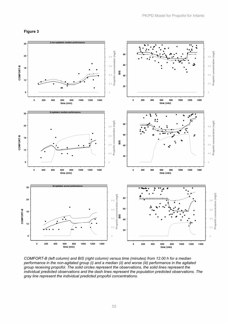
PK/PD Model for Propofol for Infants
55
0 200 400 600 800 1000 1200 1400time (min)
20
40
60
80
BIS
0
0.2
0.8
0.6
0.4
Prop
ofol
con
cent
ratio
n (m
g/l)
0 200 400 600 800 1000 1200 1400time (min)
20
40
60
80
BIS
0
0.2
0.8
0.6
0.4P
ropo
fol c
once
ntra
tion
(mg/
l)
0 200 400 600 800 1000 1200 1400time (min)
6
12
18
24
30
CO
MFO
RT-
B
0
0.2
0.8
0.6
0.4
Prop
ofol
con
cent
ratio
n (m
g/l)
iii) agitated, worse performance
0 200 400 600 800 1000 1200 1400time (min)
6
12
18
24
30
CO
MFO
RT-
B
0
0.2
0.8
0.6
0.4
Prop
ofol
con
cent
ratio
n (m
g/l)
ii) agitated, median performance
0 200 400 600 800 1000 1200 1400time (min)
6
12
18
24
30
CO
MFO
RT-
B
0
0.2
0.8
0.6
0.4
Prop
ofol
con
cent
ratio
n (m
g/l)
i) non-agitated, median performance
Figure 3
COMFORT-B (left column) and BIS (right column) versus time (minutes) from 12.00 h for a median performance in the non-agitated group (i) and a median (ii) and worse (iii) performance in the agitated group receiving propofol. The solid circles represent the observations, the solid lines represent the individual predicted observations and the dash lines represent the population predicted observations. The gray line represent the individual predicted propofol concentrations.
0 200 400 600 800 1000 1200 1400time (min)
20
40
60
80
BIS
0
0.2
0.8
0.6
0.4
Prop
ofol
con
cent
ratio
n (m
g/l)
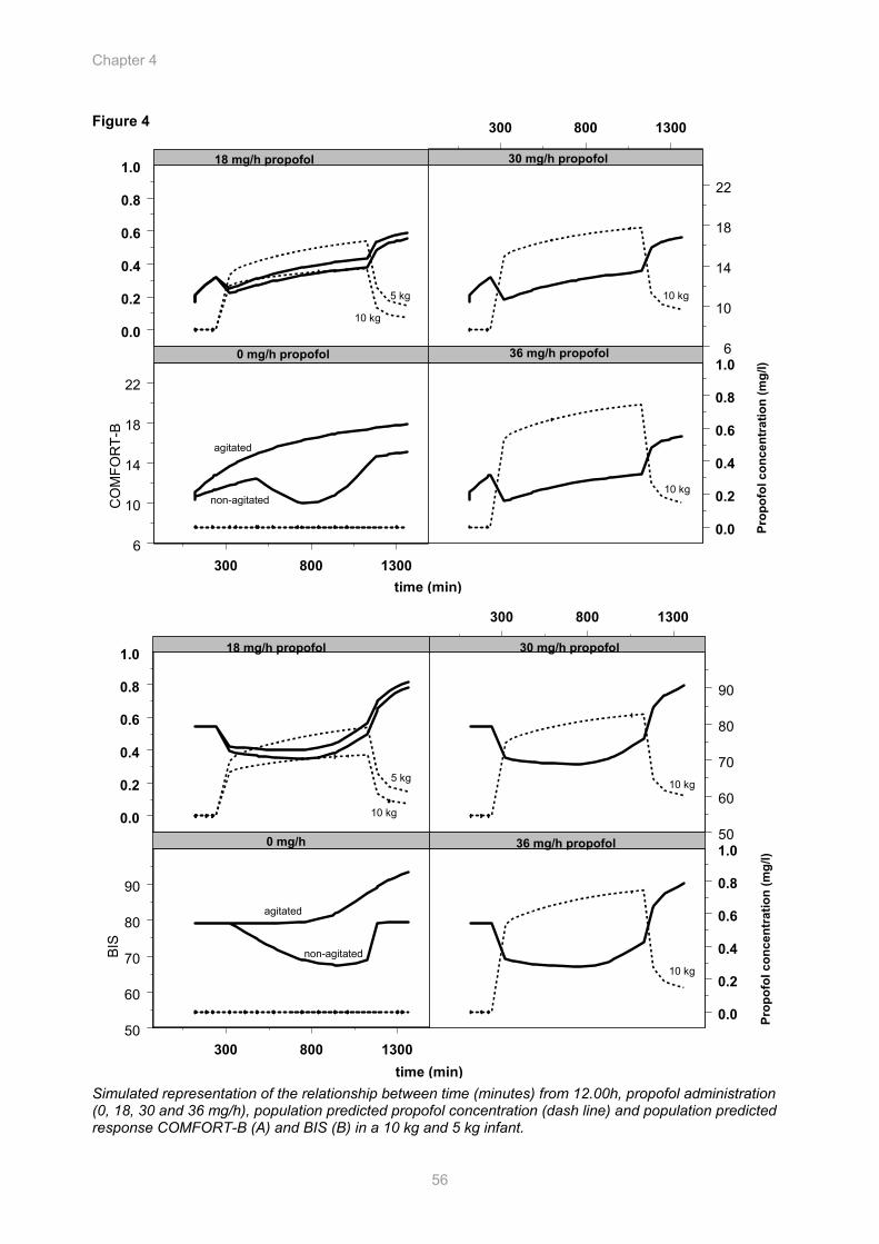
Chapter 4
56
300 800 1300
300 800 1300
time (min)
6
10
14
18
22
6
10
14
18
22
CO
MFO
RT-
B
0.0
0.2
0.4
0.6
0.8
1.0
0.0
0.2
0.4
0.6
0.8
1.0
0 mg/h propofol
30 mg/h propofol18 mg/h propofol
36 mg/h propofol
Prop
ofol
con
cent
ratio
n (m
g/l)
10 kg
10 kg
5 kg
agitated
non-agitated
10 kg
300 800 1300
300 800 1300
time (min)
50
60
70
80
90
50
60
70
80
90
0.0
0.2
0.4
0.6
0.8
1.0
0.0
0.2
0.4
0.6
0.8
1.0
Prop
ofol
con
cent
ratio
n (m
g/l)
0 mg/h
30 mg/h propofol18 mg/h propofol
36 mg/h propofol
10 kg
10 kg
10 kg
BIS
agitated
non-agitated
5 kg
Figure 4
Simulated representation of the relationship between time (minutes) from 12.00h, propofol administration (0, 18, 30 and 36 mg/h), population predicted propofol concentration (dash line) and population predicted response COMFORT-B (A) and BIS (B) in a 10 kg and 5 kg infant.
PK/PD Model for Propofol for Infants
57
Discussion In order to support safe and effective use of propofol during the first night after major surgery in non-ventilated infants less than 1.5 years of age, a population model for the influence of propofol PK/PD on the depth of sedation was described, assessed using COMFORT-B and BIS.
Clearance in post-surgery healthy non-ventilated infants was found to be 2 times higher than reported in the literature for ventilated children and adults.3,15,16 Based on the PK model propofol doses have to be doubled in this pediatric group to obtain similar blood concentrations. We believe that the higher estimate of the Cl (0.70 l/min) in an infant with bodyweight 8.9 kg in our study compared to 0.030 l/kg/min reported in the literature can partly be explained by the effect of the surgery and the condition of the patients. Rigby-Jones15 found that patients aged 1 week to 12 years undergoing cardiac surgery had reduced values for metabolic clearance (-26%). Cardiac patients in general show a reduced cardiac output, which may effect the propofol elimination since the clearance of propofol (a high-extraction drug) is dependent upon liver blood flow. In addition, mechanical ventilation may be of influence on the clearance of propofol. In patients with trauma and those in the surgical ICU, increasing the PEEP (positive end expiratory pressure) during mechanical ventilation has been shown to decrease total hepatic blood flow.17 Murat.2 reported a large clearance (0.049 l/kg/min) in spontaneously breathing children aged 1 - 3 years with minor burns following a single dose of 4 mg/kg. Healthy ventilated children undergoing anesthesia did show a lower estimate of the clearance.16,18 The model developed by Schüttler and Ihmsen16 for healthy ventilated children undergoing anesthesia from 2 years of age showed less overprediction of the blood concentration than the model developed by Knibbe and Rigby-Jones3,15 for ventilated children following cardiac surgery. They also found a smaller value of the central volume compared to our model (5 - 12 l versus 19 l), which may be a consequence of the relation of the central volume to the volume of distribution of steady state. Bodyweight partially explained the interpatient variability in Cl. The influence of a slope-intercept model, a proportional model or a power model with a power scaling parameter of 0.61 on the clearance was comparable in the range of 4.8 - 12.5 kg. We choose for the power model since an allometric ¾ power model has been used with success for interspecies scaling.19 As with other studies, age was not found to be a significant covariate.15,20 In addition, the genetic expression of the investigated CYP isoforms did not explain the interindividual differences in the clearance. 2B6 would be predominantly involved and at a lower rate 2C9 and 2C19 in the minor metabolic hydroxylation pathway.21 The homogeneous patient characteristics and the relatively small number of patients may account for the unexplained interpatient variability. The large PD inter- and intraindividual variability in BIS and COMFORT-B emphasizes the complexity of depth of sedation in infants. Young children can vary in depth of sedation in the absence of sedatives as a result of day-night rhythm, the presence of parents and/or medical staff, hunger, light and noise.5,22 Especially at lighter sedation levels, noise has a greater effect on the BIS.23 To account for natural variation, data of infants not receiving a sedative and until sedative administration were used to describe a post-anesthesia effect
Chapter 4
58
(PAEFF) and a night dip (CNR). For adults a similar PAEFF has been described after CABG surgery by assuming a virtual drug that washes out over time.24 Since stress and severe discomfort entail risks, a complete natural sleep pattern of agitated infants could not be described. The administration of the sedative may cover the night dip, which could not been estimated in the agitated children. The EC50 of propofol for the reduction of the BIS was different from the COMFORT-B, indicating that both measurements are not interchangeable measures of the propofol effect in a spontaneously breathing child. Courtman et al.,25 and Crain et al.,26 also suggest that BIS and COMFORT are only moderately correlated: a child can be comfortable, but fully awake. The use of the BIS has the advantage that it assesses the sedation continuously and may allow more objective assessment of sedation. It gives additive information and can be useful for patients who are difficult to assess clinically. The use of 2/3 of the COMFORT-B scale and a smaller number of observations make it difficult to determine which sedation scale is more sensitive in this population based on the EC50, but in lightly sedated children the COMFORT-B seems in advance. The COMFORT-B has never been used before as a PD instrument in a PK-PD propofol analysis, but the effect of propofol on BIS in adults has been described. Interestingly, the sensitivity of infants to propofol, defined as EC50 seems comparable to adults. Defining the Emax as the maximum effect seen on the BIS, Bouillon et al., estimated an EC50 of 3.07 mg/l (CV% 12.1) and Doufas et al., a value of 2.4 mg/l (CV% 30).27,28 By fixing the Emax to 100, Calvo29 estimated the EC50 on 3.91 mg/l (41%), which may suggest that infants only require higher doses due to differences in PK rather than PD. In general, the sensitivity to propofol between infants is very variable. Unfortunately, no explanation could be found based on patient characteristics as age, bodyweight and gender. In this narrow age group the potential stressful environment as inability to see, separation from parents, unknown voices, may play a major role. Based on the population PD model we advise a propofol infusion rate of 30 mg/h for a 10 kg non-ventilated infant to achieve a COMFORT-B between 12 and 14, 6 h post-surgery during the night, which will correspond to BIS values of 70 - 75 (Figure 4). The considerable variability emphasizes the importance of drug titration to a maximum of 4 mg/kg/h. Further PD studies in larger groups of children are needed to explain the variability in response and help clinicians to improve individualization. For drugs like propofol, this is especially important due to the troublesome reports in the literature concerning the safety of the use of propofol in children beyond procedures. Acknowledgements The authors wish to thank the staff in the Division of Pharmacy of the Erasmus MC, Rotterdam, the Netherlands for their help and cooperation in particular professor Arnold G. Vulto PharmD. PhD., Lidwien M. Hanff PharmD. and Ron AA Mathôt PharmD. PhD. and the medical and nursing colleagues of the Pediatric Surgical Intensive Care Unit.
PK/PD Model for Propofol for Infants
59
References
1. Parke TJ, Stevens JE, Rice AS, Greenaway CL, Bray RJ, Smith PJ, Waldmann CS, Verghese C: Metabolic acidosis and fatal myocardial failure after propofol infusion in children: five case reports. Bmj 1992;305:613-6
2. Murat I, Billard V, Vernois J, Zaouter M, Marsol P, Souron R, Farinotti R: Pharmacokinetics of propofol after a single dose in children aged 1-3 years with minor burns. Comparison of three data analysis approaches. Anesthesiology 1996;84:526-32
3. Knibbe CA, Melenhorst-de Jong G, Mestrom M, Rademaker CM, Reijnvaan AF, Zuideveld KP, Kuks PF, van Vught H, Danhof M: Pharmacokinetics and effects of propofol 6% for short-term sedation in paediatric patients following cardiac surgery. Br J Clin Pharmacol 2002;54:415-22
4. van Dijk M, de Boer JB, Koot HM, Tibboel D, Passchier J, Duivenvoorden HJ: The reliability and validity of the COMFORT scale as a postoperative pain instrument in 0 to 3-year-old infants. Pain 2000;84:367-77
5. Ista E, van Dijk M, Tibboel D, de Hoog M: Assessment of sedation levels in pediatric intensive care patients can be improved by using the COMFORT "behavior" scale. Pediatr Crit Care Med 2005;6:58-63
6. Berkenbosch JW, Fichter CR, Tobias JD: The correlation of the bispectral index monitor with clinical sedation scores during mechanical ventilation in the pediatric intensive care unit. Anesth Analg 2002;94:506-11; table of contents
7. Prins SA, Peeters MY, Houmes RJ, van Dijk M, Knibbe CA, Danhof M, Tibboel D: Propofol 6% as sedative in children under 2 years of age following major craniofacial surgery. Br J Anaesth 2005;94:630-5
8. Koster VS, Maring JG, Knibbe CAJ, Lange R, Kuks PFM, Langemeijer JJM, Talsma H, Lie-A-Huen L: Propofol 6% SAZN: preparation and stability of a new formulation of propofol. EHP 2000;6:92-96
9. Peeters MYM, Lange R, Aarts LPHJ, Talsma H, Knibbe CAJ: Stability of an intravenous fat emulsion containing 6% propofol and a low amount of emulsifier. EJHP 2004;10:201-8
10. van der Marel CD, van Lingen RA, Pluim MA, Scoones G, van Dijk M, Vaandrager JM, Tibboel D: Analgesic efficacy of rectal versus oral acetaminophen in children after major craniofacial surgery. Clin Pharmacol Ther 2001;70:82-90
11. Knibbe CA, Koster VS, Deneer VH, Stuurman RM, Kuks PF, Lange R: Determination of propofol in low-volume samples by high-performance liquid chromatography with fluorescence detection. J Chromatogr B Biomed Sci Appl 1998;706:305-10
12. Lang T, Klein K, Fischer J, Nussler AK, Neuhaus P, Hofmann U, Eichelbaum M, Schwab M, Zanger UM: Extensive genetic polymorphism in the human CYP2B6 gene with impact on expression and function in human liver. Pharmacogenetics 2001;11:399-415
13. Beal SL, Sheiner LB: NONMEM Users Guides. NONMEM Project Group, University of California at San Francisco, CA 1992
14. Ette EI: Stability and performance of a population pharmacokinetic model. J Clin Pharmacol 1997; 37:486-95
15. Rigby-Jones AE, Nolan JA, Priston MJ, Wright PM, Sneyd JR, Wolf AR: Pharmacokinetics of propofol infusions in critically ill neonates, infants, and children in an intensive care unit. Anesthesiology 2002;97:1393-400
Chapter 4
60
16. Schuttler J, Ihmsen H: Population pharmacokinetics of propofol: a multicenter study. Anesthesiology 2000;92:727-38
17. Bonnet F, Richard C, Glaser P, Lafay M, Guesde R: Changes in hepatic flow induced by continuous positive pressure ventilation in critically ill patients. Crit Care Med 1982;10:703-5
18. Kataria BK, Ved SA, Nicodemus HF, Hoy GR, Lea D, Dubois MY, Mandema JW, Shafer SL: The pharmacokinetics of propofol in children using three different data analysis approaches. Anesthesiology 1994;80:104-22
19. Knibbe CA, Zuideveld KP, Aarts LP, Kuks PF, Danhof M: Allometric relationships between the pharmacokinetics of propofol in rats, children and adults. Br J Clin Pharmacol 2005;59:705-11
20. Reed MD, Yamashita TS, Marx CM, Myers CM, Blumer JL: A pharmacokinetically based propofol dosing strategy for sedation of the critically ill, mechanically ventilated pediatric patient. Crit Care Med 1996;24:1473-81
21. Court MH, Duan SX, Hesse LM, Venkatakrishnan K, Greenblatt DJ: Cytochrome P-450 2B6 is responsible for interindividual variability of propofol hydroxylation by human liver microsomes. Anesthesiology 2001;94:110-9
22. Sleigh JW, Andrzejowski J, Steyn-Ross A, Steyn-Ross M: The bispectral index: a measure of depth of sleep? Anesth Analg 1999;88:659-61
23. Kim DW, Kil HY, White PF: The effect of noise on the bispectral index during propofol sedation. Anesth Analg 2001;93:1170-3
24. Somma J, Donner A, Zomorodi K, Sladen R, Ramsay J, Geller E, Shafer SL: Population pharmacodynamics of midazolam administered by target controlled infusion in SICU patients after CABG surgery. Anesthesiology 1998;89:1430-43
25. Courtman SP, Wardurgh A, Petros AJ: Comparison of the bispectral index monitor with the Comfort score in assessing level of sedation of critically ill children. Intensive Care Med 2003;29:2239-46
26. Crain N, Slonim A, Pollack MM: Assessing sedation in the pediatric intensive care unit by using BIS and the COMFORT scale. Pediatr Crit Care Med 2002;3:11-4
27. Bouillon TW, Bruhn J, Radulescu L, Andresen C, Shafer TJ, Cohane C, Shafer SL: Pharmacodynamic interaction between propofol and remifentanil regarding hypnosis, tolerance of laryngoscopy, bispectral index, and electroencephalographic approximate entropy. Anesthesiology 2004;100:1353-72
28. Doufas AG, Bakhshandeh M, Bjorksten AR, Shafer SL, Sessler DI: Induction speed is not a determinant of propofol pharmacodynamics. Anesthesiology 2004;101:1112-21
29. Calvo R, Telletxea S, Leal N, Aguilera L, Suarez E, De La Fuente L, Martin-Suarez A, Lukas JC: Influence of formulation on propofol pharmacokinetics and pharmacodynamics in anesthetized patients. Acta Anaesthesiol Scand 2004;48:1038-48
Pharmacokinetics and Pharmacodynamics of Intravenous Propacetamol Versus Rectal Paracetamol in Children Less than 2 Years of Age: a Double Blind Placebo Controlled Randomized Trial
Sandra A. Prins, Ron A.A. Mathôt, Monique van Dijk, Pim van Leeuwen, Susana Searle, Brian J. Anderson, Dick Tibboel.
Cha
pter
Chapter 5
62
Abstract Background Little is known about the use and dose of i.v. propacetamol for analgesia in non-ventilated children. To support the safe and effective use of i.v. propacetamol, a population model for i.v. propacetamol pharmacokinetics (PK) and pharmacodynamics (PD) is described in non-ventilated infants (6 months to 2 years) after craniofacial surgery. Methods The infants were given paracetamol suppository 40 mg/kg and then randomly assigned to receive either i.v. propacetamol 40 mg/kg infusion over 15 min or 20 mg/kg paracetamol rectally every 6 hours. Placebo suppository and placebo intravenous solution were used to blind the investigator and nurses. The visual analogue scale (VAS) (score 0 to 10) and COMFORT Behavior scale (score 6 to 30) were used as pharmacodynamic endpoints. Data from 26 infants were analyzed: 12 received i.v. propacetamol and 14 rectal paracetamol. Population PK modeling was performed using NONMEM. Results The pharmacokinetics of paracetamol were described according to a four-compartmental model: iv-depot, rectum, central and peripheral. The following population parameters were estimated: absorption half-life from the rectum (Tabs) 5.4 h, hydrolysis half-life in iv-depot (Thydr) 0.20 h, clearance (CL/Frect) 11 l/h/70 kg, intercompartmental clearance (Q/Frect) 35 l/h/70 kg, central volume of distribution (V2/Frect) 24 l/70 kg and peripheral volume of distribution (V3/Frect) 22 l/70 kg. The ratio of intravenous and rectal bioavailability (Fiv/Frect) was 0.41. A target concentration of 10 mg/l was reached after 0.4 to 2.0 h in the i.v. propacetamol group and 0.6 to 16.6 h in the rectal group. During the 24-h study period twelve patients had VAS pain scores less than 4, but received midazolam for COMFORT scores exceeding 17; three of them were in the i.v. propacetamol group and nine in the rectal paracetamol group (P = 0.05). One child from the i.v. propacetamol treatment group was given rescue paracetamol 20 mg/kg rectally for a VAS pain score > 4. Conclusions The i.v. propacetamol route proved to be more effective than the rectal paracetamol one in this age group. Use of midazolam was significantly higher in the rectal paracetamol group, indicating that these children experienced more distress, possibly caused by pain.
Population PK/PD of i.v. propacetamol in infants
63
Introduction Paracetamol is an effective and safe analgesic drug, which relieves mild to moderate pain in children. Relationships between paracetamol plasma concentration and dose, analgesic effect and dose, and analgesic effect and concentration have been reported in the literature.1-4
Extrapolated from findings in adults, the optimal plasma concentration to obtain analgesia is usually considered to be 10 - 20 mg/l.5 Indeed, in children aged from 2 to 15 years undergoing tonsillectomy, a 10 mg/l paracetamol effect compartment concentration was shown to produce adequate pain relief.6 A concentration-response relationship using an Emax model has been described for children undergoing tonsillectomy: an effect compartment concentration of 5 mg/l was associated with a 1.7/10 pain reduction, while 10 mg/l was associated with 2.6/10.4 In 10-month-old children undergoing craniofacial surgery, our group demonstrated that adequate analgesia was obtained at plasma concentrations lower than 10 mg/l.7 This may be attributable to either reduced pain or a different concentration-response relationship.
Paracetamol is usually administered orally or rectally. Comparative findings of these routes of administration in infants after major surgery by our group were published in 2001.7 The delayed and erratic absorption after rectal administration has been reported to produce unpredictable paracetamol plasma concentrations and does not consistently produce a rapid onset of pain relief.8 A paracetamol rectal loading dose of 40 mg/kg is recommended to achieve satisfactory pain relief. Although the rectal route is commonly used for children in daily practice, the intravenous (i.v.) route is of interest for infants who are unable to receive paracetamol rectally, e.g. infants with anal atresia.
Propacetamol is a prodrug, a N,N’-diethylglycine ester of paracetamol, and is hydrolyzed by plasma esterases to paracetamol.9 Compared to rectal and/or oral formulations, intravenous administration of propacetamol might improve prediction of target concentrations and consequent effect by reducing variability due to absorption kinetics and relative bio-availability inherent to enteral formulations.9,10
We conducted a double-blind placebo controlled randomized trial to compare the effectiveness of i.v. propacetamol and rectal paracetamol in young children between six months and two years of age undergoing major craniofacial surgery. In this study plasma concentrations were assessed in combination with score changes for two behavioral assessment tools, the pain visual analogue scale (VAS) and the COMFORT Behavior scale. Methods Patients The study was performed at the pediatric surgical intensive care unit (PSICU) of the Erasmus MC - Sophia Children’s hospital between September 2004 and February 2005. The Sophia Children’s Hospital serves as a level III referral center for all pediatric surgical
Chapter 5
64
specialties and is the only designated pediatric craniofacial center in the Netherlands. Approximately 70 major craniofacial corrections are performed annually.
The study protocol was approved by the Erasmus MC Ethics Review Board and written informed consent was obtained from parents or legal guardians. Children of either sex, aged between six months and two years admitted to the PSICU after craniofacial surgery were eligible. Parents of eligible patients received a letter describing the study protocol at least one week before the scheduled surgery. The investigator requested parental informed consent on the child’s admission one day before surgery. Exclusion criteria were the following: contraindication to i.v. propacetamol or rectal paracetamol, documented history of allergy to other drugs, participation in another clinical trial, glucose-6-phosphate dehydrogenase (G6PD) deficiency, rectal atresia or cognitive impairment. Participation was ended if parents withdrew their informed consent during the study or if a patient developed bradycardia, hypotension or a coagulation disturbance postoperatively. Then, all data of this patient were excluded from further analysis and the randomization number was not used again. Procedure Sevoflurane or intravenous thiopentone was used to induce anesthesia. During induction, arterial and central venous lines were placed for clinical purposes and blood was drawn to evaluate liver and kidney function, triglyceride and creatine phosphokinase levels. After administration of 0.1 mg/kg vecuronium and 2.5 mcg/kg fentanyl intravenously, patients were intubated and anesthetized with air, oxygen and isoflurane (0.5 - 2%). Approximately 2 hours before anticipated extubation, a loading dose of 40 mg/kg paracetamol was administered rectally.11 After surgery, the patients were extubated, admitted to the PSICU and observed for a minimum of 24 hours, depending on the clinical condition.7
Patients were randomly assigned to receive either i.v. propacetamol 40 mg/kg infusion over 15 min or 20 mg/kg paracetamol rectally 6, 12, 18 and 24 hours after the loading dose. Blood samples of 1 ml were taken from the arterial catheter at 15 min, 1 and 6 hours after the first dose, at 5 min, 4 and 6 hours after the third dose, and at 30 min, 2 and 3 hours after the fourth dose. During the study, a maximum of 9 ml of blood per patient was drawn.
Trained intensive care nurses and medical students (P.v.L and S.S.) obtained pain scores using the visual analogue scale (VAS) and the COMFORT Behavior scale (COMFORT-B).12,13 Pain was scored before every blood sample and incidentally depending on the child’s clinical condition. VAS is an observational assessment tool with proven low inter-rater variability between nurses and physicians in judging postoperative pain in pediatric patients is low.14-16 The concurrent validity of the VAS with other observational pain assessment tools has also been established by different authors.12,14,17 Rescue medication An extra dose of 20 mg/kg rectal paracetamol was given if VAS scores remained ≥ 4 cm. Before this extra dose, a blood sample was taken. If COMFORT-B scores ≥ 17 coincided with VAS scores < 4 cm, a single i.v. bolus dose of 0.1 mg/kg midazolam was given. If COMFORT-B scores remained ≥ 17, another single i.v. dose or continuous infusion of
Population PK/PD of i.v. propacetamol in infants
65
midazolam was given. The subsequent paracetamol dose was then administered according to schedule.
Blood samples were placed on ice, immediately centrifuged and stored at -20˚C until analysis. Paracetamol plasma concentrations were determined using fluorescence polarization immunoassay (ADX system; Abbott Laboratories, North Chicago, IL, USA). The detection limit of this method was 1.0 mg/l. Precision was evaluated at paracetamol concentrations 15, 35 and 150 mg/l. Relative standard deviations at these concentrations were 7.2%, 3.4% and 3.1%, respectively (n = 55). Accuracy was between 90 and 110% in the concentration range 10 - 150 mg/l. Assignment and formulations A computer-generated randomization schedule assigned treatments in equal ratio to sequential patients. After inclusion of a patient, the hospital’s pharmacist prepared the study medication according to the randomization schedule. The schedule was kept solely by the pharmacist to ensure blinding until the end of the study.
A placebo suppository (verum) and placebo intravenous solution were used to blind the investigator and nurses. Both verum and placebo were presented to the nurses in a closed and sealed box. An independent nurse prepared the i.v. solution of 20 mg/ml propacetamol in saline 30 minutes before administration and left it for the attending nurse to administer, without any encounter between the caregivers. The maintenance paracetamol therapy comprised either propacetamol 40 mg/kg i.v. (Bristol Meyers Squibb, Paris, France), which is hydrolyzed to 20 mg/kg paracetamol,9,18 or 20 mg/kg paracetamol rectally (hospital pharmacy) every 6 hours. The actual paracetamol doses delivered rectally never deviated more than 10% from 20 mg/kg. The suppositories and all ingredients met the requirements of the European Pharmacopoeia (Ed. 5) and were manufactured according to the Dutch Pharmacist Formulary. Pharmacokinetic analysis PK models were fitted to data from all individuals simultaneously using non-linear mixed effect modeling (NONMEM). The NONMEM model accounts for inter-patient and residual PK variability (random effects) as well as PK differences predicted by patient factors (fixed effects). The typical population parameters, inter-patient and residual variances were estimated using the NONMEM software program (double precision; version V, level 1.1). The first-order conditional estimate method was used throughout the analysis.
The PK of paracetamol were described according to a four-compartmental model (ADVAN5): depot, rectum (V1), central (V2) and peripheral V3 (Figure 1). In case of intravenous administration the drug was infused in the depot compartment and a rate constant (Khydr) was used to describe movement (hydrolysis) into the central compartment. Absorption from the rectum was characterized by absorption rate constant Ka.

Chapter 5
66
Figure 1
The PK model of paracetamol consisted of 4 compartments: depot, rectum, central and peripheral. In case of intravenous administration drug was infused in the depot compartment and a rate constant (Khydr) was used to describe movement (hydrolysis) in the central compartment. Absorption from the rectum was characterized by absorption rate constant Ka. The following parameters were estimated: hydrolysis half-life (Thydr = ln(2) / Khydr), absorption half-life (Tabs = ln (2) / Ka), the volume of distribution of the central compartment (V2), clearance from the central compartment (Cl), volume of distribution of the peripheral compartment (V3) and inter-compartmental clearance (Q). The following parameters were estimated: hydrolysis half-life (Thydr = ln(2) / Khydr), absorption half-life (Tabs = ln (2) / Ka), the volume of distribution of the central compartment (V2), clearance from the central compartment (CL), volume of distribution of the peripheral compartment (V3) and inter-compartmental clearance (Q). Since paracetamol was administered rectally, the terms “volume of distribution” and “clearance” represent the ratios of these parameters to the unknown bioavailability Frect (V2/Frect, Cl/Frect, V3/Frect, Q/Frect). In the analysis Frect was fixed at 1; the ratio Fiv / Frect was estimated representing the relative bioavailability of the intravenous formulation compared to the suppositories.
The parameter values were standardized for a body weight of 70 kg using an allometric model according to
Pi = Pstd (Wti / Wstd) PWR
depot
rectum
central peripheral
i.v. propacetamol
paracetamol suppository
Khydr
Ka
V2 V3
Q
Cl
Population PK/PD of i.v. propacetamol in infants
67
where Pi is the parameter in the ith individual, WTi is the weight in the ith individual and Pstd is the parameter in the standard individual with a weight of 70 kg (Wstd). The PWR exponent was 0.75 for clearances, 1 for volumes of distribution and 0.25 for hydrolysis and absorption half-lives. This standardization allows comparison of parameter estimates in infants with those reported for adults.7,19,20 Simultaneous analysis of the data from all patients requires statistical models for inter-patient and residual variances. Inter-patient variability of the PK parameters was estimated using a exponential error model. For instance, inter-individual variability in Cl/Frect was estimated using: Cl / Frect,i = Cl / Frect,pop · e ηi in which i represents the number of the individual, Cl/Fi is the clearance of the ith individual, Cl/Fpop is the Cl/F value of a typical individual and η is the inter-individual random effect with mean 0 and variance ω2. Besides inter-patient variability of the PK parameters also the covariance between those parameters was estimated. For a NONMEM model, the residual variance corresponds to the difference between the observed concentration (Cobs) and predicted concentration (Cpred). The latter is predicted on basis of individual parameters (e.g. Cl/Fi, V1/Fi, etc.). Residual variance was modeled with a combined additive and proportional error model: Cobs i = Cpred i + ε1 + ε2 · Cpred i where ε1 and ε2 are independent random variables with zero mean and a variance of σ2. The population model was built stepwise. At each step, a specific assumption was tested (e.g. two-compartment versus three-compartment model). The main criterion of decision was the likelihood ratio test. For hierarchical models the difference in the objective function is approximately chi-squared distributed and formal testing between models can be performed. The level of significance was set at P < 0.05, corresponding to a decrease of the objective function of 3.8 points. Model adequacy was further evaluated by using various residual plots (“goodness-of-fit” plots), values of random-effects variances and precision of the parameter estimates. For the graphical goodness-of-fit analysis, extensive plotting was available through the use of Xpose,21 a purpose built set of subroutines in S-plus (version 6.2; Insightful Corp. Seattle WA, USA ). Statistical methods Group demographics and PK/PD parameters were statistically compared using the Mann-Whitney U test and the Chi-square test. The significance level was set at 0.05.22
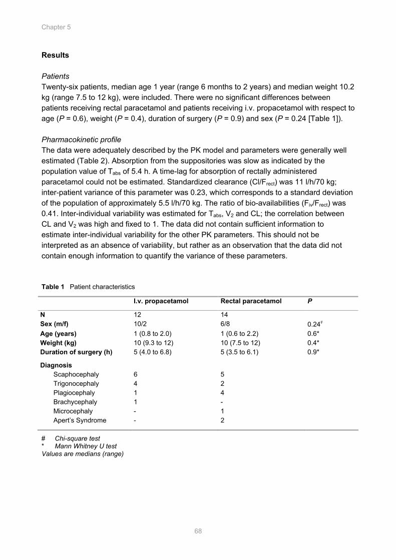
Chapter 5
68
Results Patients Twenty-six patients, median age 1 year (range 6 months to 2 years) and median weight 10.2 kg (range 7.5 to 12 kg), were included. There were no significant differences between patients receiving rectal paracetamol and patients receiving i.v. propacetamol with respect to age (P = 0.6), weight (P = 0.4), duration of surgery (P = 0.9) and sex (P = 0.24 [Table 1]). Pharmacokinetic profile The data were adequately described by the PK model and parameters were generally well estimated (Table 2). Absorption from the suppositories was slow as indicated by the population value of Tabs of 5.4 h. A time-lag for absorption of rectally administered paracetamol could not be estimated. Standardized clearance (Cl/Frect) was 11 l/h/70 kg; inter-patient variance of this parameter was 0.23, which corresponds to a standard deviation of the population of approximately 5.5 l/h/70 kg. The ratio of bio-availabilities (Fiv/Frect) was 0.41. Inter-individual variability was estimated for Tabs, V2 and CL; the correlation between CL and V2 was high and fixed to 1. The data did not contain sufficient information to estimate inter-individual variability for the other PK parameters. This should not be interpreted as an absence of variability, but rather as an observation that the data did not contain enough information to quantify the variance of these parameters. Table 1 Patient characteristics
I.v. propacetamol Rectal paracetamol P
N 12 14 Sex (m/f) 10/2 6/8 0.24# Age (years) 1 (0.8 to 2.0) 1 (0.6 to 2.2) 0.6* Weight (kg) 10 (9.3 to 12) 10 (7.5 to 12) 0.4* Duration of surgery (h) 5 (4.0 to 6.8) 5 (3.5 to 6.1) 0.9*
Diagnosis Scaphocephaly 6 5 Trigonocephaly 4 2 Plagiocephaly 1 4 Brachycephaly 1 - Microcephaly - 1 Apert’s Syndrome - 2
# Chi-square test * Mann Whitney U test Values are medians (range)
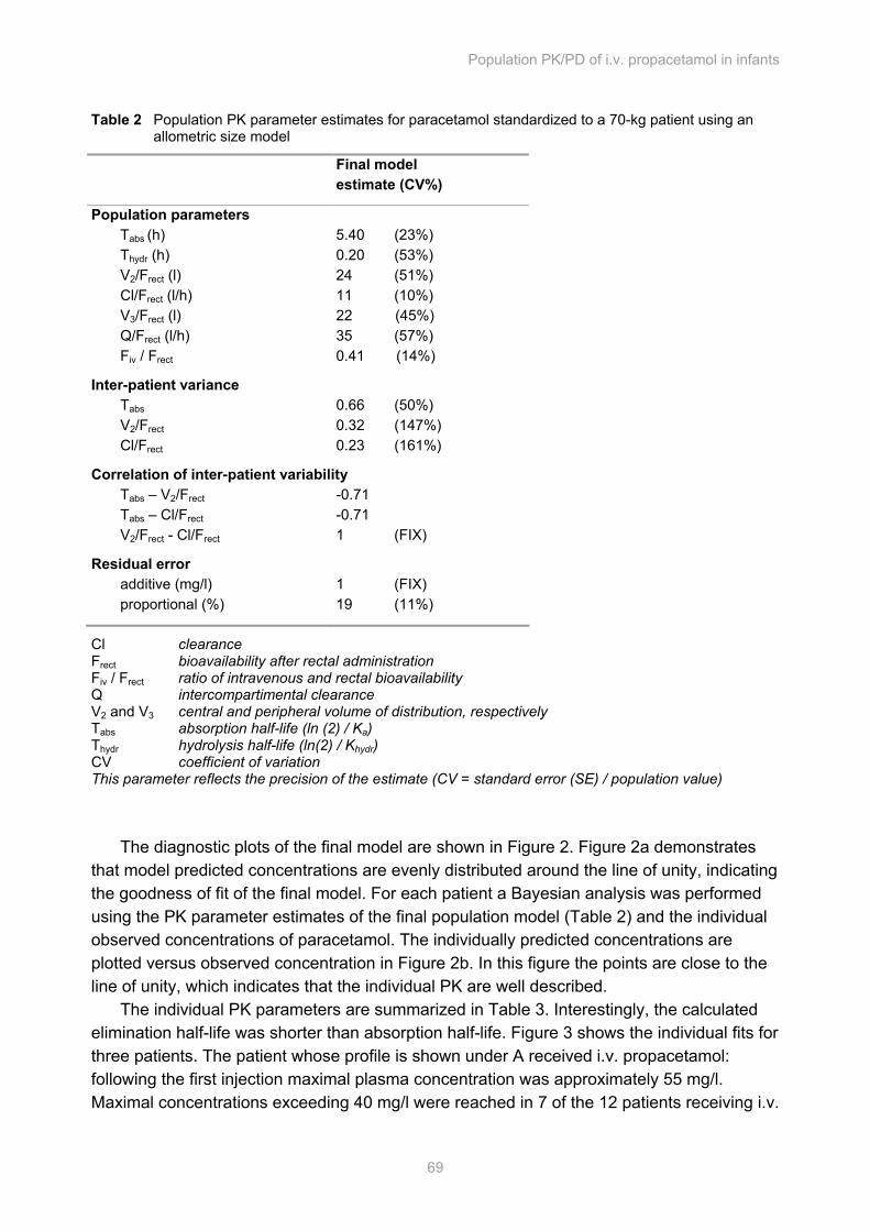
Population PK/PD of i.v. propacetamol in infants
69
Table 2 Population PK parameter estimates for paracetamol standardized to a 70-kg patient using an allometric size model
Final model estimate (CV%)
Population parameters Tabs (h) 5.40 (23%) Thydr (h) 0.20 (53%) V2/Frect (l) 24 (51%) Cl/Frect (l/h) 11 (10%) V3/Frect (l) 22 (45%) Q/Frect (l/h) 35 (57%) Fiv / Frect 0.41 (14%)
Inter-patient variance Tabs 0.66 (50%) V2/Frect 0.32 (147%) Cl/Frect 0.23 (161%)
Correlation of inter-patient variability Tabs – V2/Frect -0.71 Tabs – Cl/Frect -0.71 V2/Frect - Cl/Frect 1 (FIX)
Residual error additive (mg/l) 1 (FIX) proportional (%) 19 (11%)
Cl clearance Frect bioavailability after rectal administration Fiv / Frect ratio of intravenous and rectal bioavailability Q intercompartimental clearance V2 and V3 central and peripheral volume of distribution, respectively Tabs absorption half-life (ln (2) / Ka) Thydr hydrolysis half-life (ln(2) / Khydr) CV coefficient of variation This parameter reflects the precision of the estimate (CV = standard error (SE) / population value)
The diagnostic plots of the final model are shown in Figure 2. Figure 2a demonstrates
that model predicted concentrations are evenly distributed around the line of unity, indicating the goodness of fit of the final model. For each patient a Bayesian analysis was performed using the PK parameter estimates of the final population model (Table 2) and the individual observed concentrations of paracetamol. The individually predicted concentrations are plotted versus observed concentration in Figure 2b. In this figure the points are close to the line of unity, which indicates that the individual PK are well described.
The individual PK parameters are summarized in Table 3. Interestingly, the calculated elimination half-life was shorter than absorption half-life. Figure 3 shows the individual fits for three patients. The patient whose profile is shown under A received i.v. propacetamol: following the first injection maximal plasma concentration was approximately 55 mg/l. Maximal concentrations exceeding 40 mg/l were reached in 7 of the 12 patients receiving i.v.
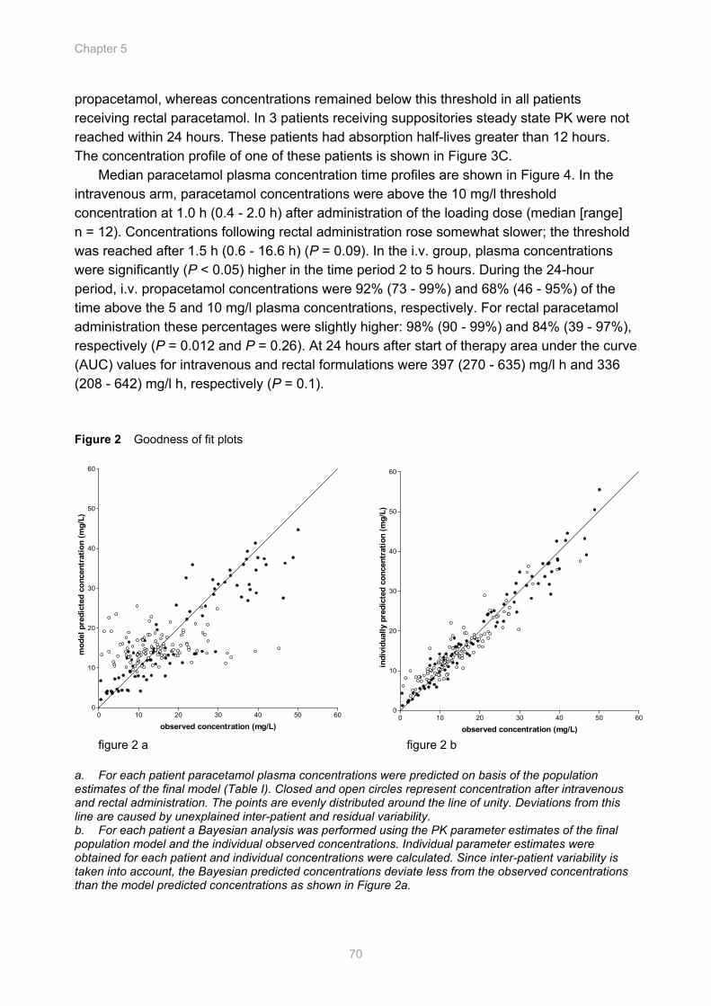
Chapter 5
70
0 10 20 30 40 50 600
10
20
30
40
50
60
observed concentration (mg/L)
indi
vidu
ally
pre
dict
ed c
once
ntra
tion
(mg/
L)
propacetamol, whereas concentrations remained below this threshold in all patients receiving rectal paracetamol. In 3 patients receiving suppositories steady state PK were not reached within 24 hours. These patients had absorption half-lives greater than 12 hours. The concentration profile of one of these patients is shown in Figure 3C.
Median paracetamol plasma concentration time profiles are shown in Figure 4. In the intravenous arm, paracetamol concentrations were above the 10 mg/l threshold concentration at 1.0 h (0.4 - 2.0 h) after administration of the loading dose (median [range] n = 12). Concentrations following rectal administration rose somewhat slower; the threshold was reached after 1.5 h (0.6 - 16.6 h) (P = 0.09). In the i.v. group, plasma concentrations were significantly (P < 0.05) higher in the time period 2 to 5 hours. During the 24-hour period, i.v. propacetamol concentrations were 92% (73 - 99%) and 68% (46 - 95%) of the time above the 5 and 10 mg/l plasma concentrations, respectively. For rectal paracetamol administration these percentages were slightly higher: 98% (90 - 99%) and 84% (39 - 97%), respectively (P = 0.012 and P = 0.26). At 24 hours after start of therapy area under the curve (AUC) values for intravenous and rectal formulations were 397 (270 - 635) mg/l h and 336 (208 - 642) mg/l h, respectively (P = 0.1).
Figure 2 Goodness of fit plots
figure 2 a figure 2 b
a. For each patient paracetamol plasma concentrations were predicted on basis of the population estimates of the final model (Table I). Closed and open circles represent concentration after intravenous and rectal administration. The points are evenly distributed around the line of unity. Deviations from this line are caused by unexplained inter-patient and residual variability. b. For each patient a Bayesian analysis was performed using the PK parameter estimates of the final population model and the individual observed concentrations. Individual parameter estimates were obtained for each patient and individual concentrations were calculated. Since inter-patient variability is taken into account, the Bayesian predicted concentrations deviate less from the observed concentrations than the model predicted concentrations as shown in Figure 2a.
0 10 20 30 40 50 600
10
20
30
40
50
60
observed concentration (mg/L)
mod
el p
redi
cted
con
cent
ratio
n (m
g/L)
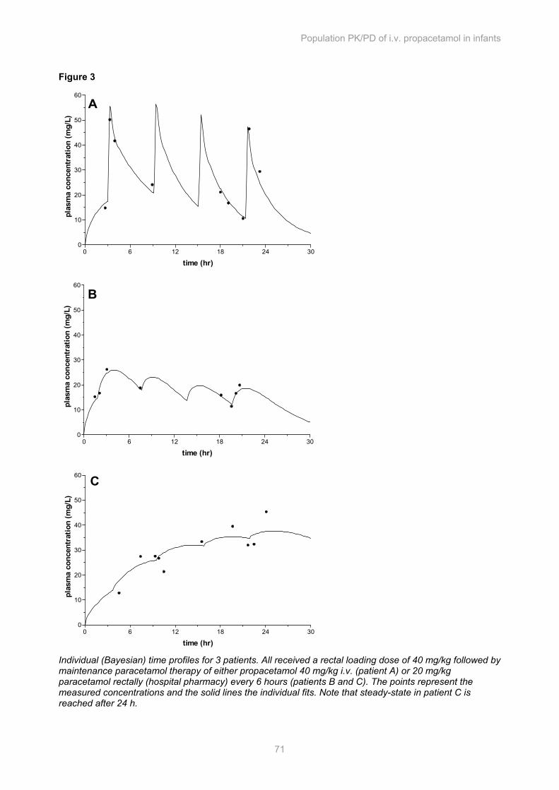
Population PK/PD of i.v. propacetamol in infants
71
Figure 3
Individual (Bayesian) time profiles for 3 patients. All received a rectal loading dose of 40 mg/kg followed by maintenance paracetamol therapy of either propacetamol 40 mg/kg i.v. (patient A) or 20 mg/kg paracetamol rectally (hospital pharmacy) every 6 hours (patients B and C). The points represent the measured concentrations and the solid lines the individual fits. Note that steady-state in patient C is reached after 24 h.
0 6 12 18 24 300
10
20
30
40
50
60
time (hr)
plas
ma
conc
entr
atio
n (m
g/L)
A
0 6 12 18 24 300
10
20
30
40
50
60
time (hr)
plas
ma
conc
entr
atio
n (m
g/L)
B
0 6 12 18 24 300
10
20
30
40
50
60
time (hr)
plas
ma
conc
entr
atio
n (m
g/L)
C
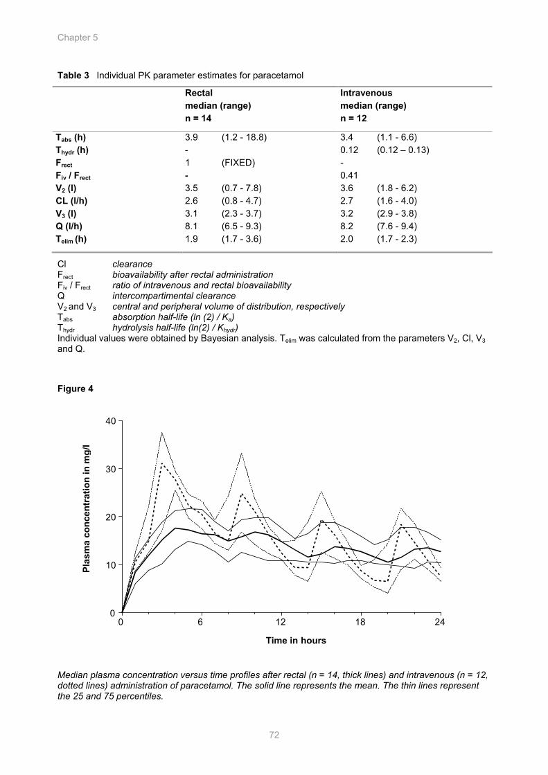
Chapter 5
72
Table 3 Individual PK parameter estimates for paracetamol
Rectal median (range) n = 14
Intravenous median (range) n = 12
Tabs (h) 3.9 (1.2 - 18.8) 3.4 (1.1 - 6.6) Thydr (h) - 0.12 (0.12 – 0.13) Frect 1 (FIXED) - Fiv / Frect - 0.41 V2 (l) 3.5 (0.7 - 7.8) 3.6 (1.8 - 6.2) CL (l/h) 2.6 (0.8 - 4.7) 2.7 (1.6 - 4.0) V3 (l) 3.1 (2.3 - 3.7) 3.2 (2.9 - 3.8) Q (l/h) 8.1 (6.5 - 9.3) 8.2 (7.6 - 9.4) Telim (h) 1.9 (1.7 - 3.6) 2.0 (1.7 - 2.3)
Cl clearance Frect bioavailability after rectal administration Fiv / Frect ratio of intravenous and rectal bioavailability Q intercompartimental clearance V2 and V3 central and peripheral volume of distribution, respectively Tabs absorption half-life (ln (2) / Ka) Thydr hydrolysis half-life (ln(2) / Khydr) Individual values were obtained by Bayesian analysis. Telim was calculated from the parameters V2, Cl, V3 and Q. Figure 4
Median plasma concentration versus time profiles after rectal (n = 14, thick lines) and intravenous (n = 12, dotted lines) administration of paracetamol. The solid line represents the mean. The thin lines represent the 25 and 75 percentiles.
0 6 12 18 24 0
10
20
30
40
Plas
ma
conc
entr
atio
n in
mg/
l
Time in hours
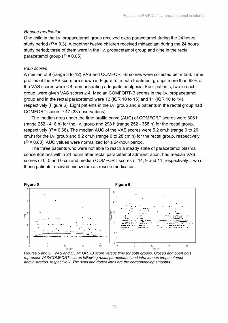
Population PK/PD of i.v. propacetamol in infants
73
Rescue medication One child in the i.v. propacetamol group received extra paracetamol during the 24 hours study period (P = 0.3). Altogether twelve children received midazolam during the 24 hours study period; three of them were in the i.v. propacetamol group and nine in the rectal paracetamol group (P = 0.05). Pain scores A median of 9 (range 8 to 12) VAS and COMFORT-B scores were collected per infant. Time profiles of the VAS score are shown in Figure 5. In both treatment groups more than 98% of the VAS scores were < 4, demonstrating adequate analgesia. Four patients, two in each group, were given VAS scores ≥ 4. Median COMFORT-B scores in the i.v. propacetamol group and in the rectal paracetamol were 12 (IQR 10 to 15) and 11 (IQR 10 to 14), respectively (Figure 6). Eight patients in the i.v. group and 9 patients in the rectal group had COMFORT scores ≥ 17 (33 observations).
The median area under the time profile curve (AUC) of COMFORT scores were 306 h (range 252 - 418 h) for the i.v. group and 298 h (range 252 - 358 h) for the rectal group, respectively (P = 0.68). The median AUC of the VAS scores were 5.2 cm.h (range 0 to 20 cm.h) for the i.v. group and 8.2 cm.h (range 0 to 28 cm.h) for the rectal group, respectively (P = 0.68). AUC values were normalized for a 24-hour period.
The three patients who were not able to reach a steady state of paracetamol plasma concentrations within 24 hours after rectal paracetamol administration, had median VAS scores of 0, 0 and 0 cm and median COMFORT scores of 14, 9 and 11, respectively. Two of these patients received midazolam as rescue medication. Figure 5 Figure 6
0 6 12 18 24time (hr)
0
2
4
6
8
VAS
0 6 12 18 24time (hr)
0
6
12
18
24
30
CO
MFO
RT
Figures 5 and 6. VAS and COMFORT-B score versus time for both groups. Closed and open dots represent VAS/COMFORT scores following rectal paracetamol and intravenous propacetamol administration, respectively. The solid and dotted lines are the corresponding smooths.
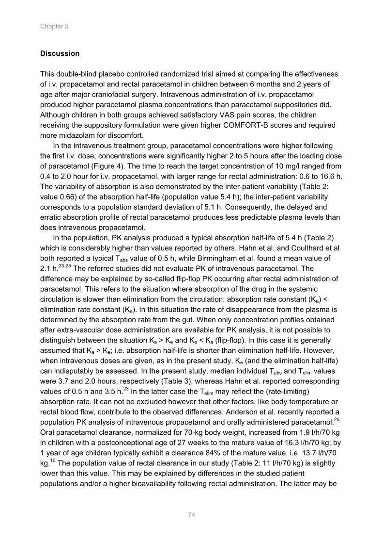
Chapter 5
74
Discussion This double-blind placebo controlled randomized trial aimed at comparing the effectiveness of i.v. propacetamol and rectal paracetamol in children between 6 months and 2 years of age after major craniofacial surgery. Intravenous administration of i.v. propacetamol produced higher paracetamol plasma concentrations than paracetamol suppositories did. Although children in both groups achieved satisfactory VAS pain scores, the children receiving the suppository formulation were given higher COMFORT-B scores and required more midazolam for discomfort.
In the intravenous treatment group, paracetamol concentrations were higher following the first i.v. dose; concentrations were significantly higher 2 to 5 hours after the loading dose of paracetamol (Figure 4). The time to reach the target concentration of 10 mg/l ranged from 0.4 to 2.0 hour for i.v. propacetamol, with larger range for rectal administration: 0.6 to 16.6 h. The variability of absorption is also demonstrated by the inter-patient variability (Table 2: value 0.66) of the absorption half-life (population value 5.4 h); the inter-patient variability corresponds to a population standard deviation of 5.1 h. Consequently, the delayed and erratic absorption profile of rectal paracetamol produces less predictable plasma levels than does intravenous propacetamol.
In the population, PK analysis produced a typical absorption half-life of 5.4 h (Table 2) which is considerably higher than values reported by others. Hahn et al. and Coulthard et al. both reported a typical Tabs value of 0.5 h, while Birmingham et al. found a mean value of 2.1 h.23-25 The referred studies did not evaluate PK of intravenous paracetamol. The difference may be explained by so-called flip-flop PK occurring after rectal administration of paracetamol. This refers to the situation where absorption of the drug in the systemic circulation is slower than elimination from the circulation: absorption rate constant (Ka) < elimination rate constant (Ke). In this situation the rate of disappearance from the plasma is determined by the absorption rate from the gut. When only concentration profiles obtained after extra-vascular dose administration are available for PK analysis, it is not possible to distinguish between the situation Ka > Ke and Ke < Ka (flip-flop). In this case it is generally assumed that Ka > Ke; i.e. absorption half-life is shorter than elimination half-life. However, when intravenous doses are given, as in the present study, Ke (and the elimination half-life) can indisputably be assessed. In the present study, median individual Tabs and Telim values were 3.7 and 2.0 hours, respectively (Table 3), whereas Hahn et al. reported corresponding values of 0.5 h and 3.5 h.23 In the latter case the Telim may reflect the (rate-limiting) absorption rate. It can not be excluded however that other factors, like body temperature or rectal blood flow, contribute to the observed differences. Anderson et al. recently reported a population PK analysis of intravenous propacetamol and orally administered paracetamol.26 Oral paracetamol clearance, normalized for 70-kg body weight, increased from 1.9 l/h/70 kg in children with a postconceptional age of 27 weeks to the mature value of 16.3 l/h/70 kg; by 1 year of age children typically exhibit a clearance 84% of the mature value, i.e. 13.7 l/h/70 kg.10 The population value of rectal clearance in our study (Table 2: 11 l/h/70 kg) is slightly lower than this value. This may be explained by differences in the studied patient populations and/or a higher bioavailability following rectal administration. The latter may be
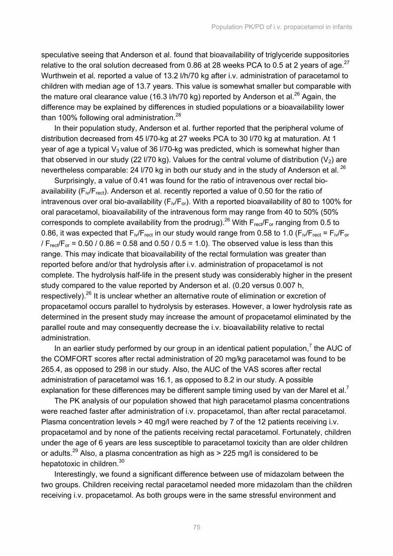
Population PK/PD of i.v. propacetamol in infants
75
speculative seeing that Anderson et al. found that bioavailability of triglyceride suppositories relative to the oral solution decreased from 0.86 at 28 weeks PCA to 0.5 at 2 years of age.27 Wurthwein et al. reported a value of 13.2 l/h/70 kg after i.v. administration of paracetamol to children with median age of 13.7 years. This value is somewhat smaller but comparable with the mature oral clearance value (16.3 l/h/70 kg) reported by Anderson et al.26 Again, the difference may be explained by differences in studied populations or a bioavailability lower than 100% following oral administration.28
In their population study, Anderson et al. further reported that the peripheral volume of distribution decreased from 45 l/70-kg at 27 weeks PCA to 30 l/70 kg at maturation. At 1 year of age a typical V3 value of 36 l/70-kg was predicted, which is somewhat higher than that observed in our study (22 l/70 kg). Values for the central volume of distribution (V2) are nevertheless comparable: 24 l/70 kg in both our study and in the study of Anderson et al. 26
Surprisingly, a value of 0.41 was found for the ratio of intravenous over rectal bio-availability (Fiv/Frect). Anderson et al. recently reported a value of 0.50 for the ratio of intravenous over oral bio-availability (Fiv/For). With a reported bioavailability of 80 to 100% for oral paracetamol, bioavailability of the intravenous form may range from 40 to 50% (50% corresponds to complete availability from the prodrug).26 With Frect/For ranging from 0.5 to 0.86, it was expected that Fiv/Frect in our study would range from 0.58 to 1.0 (Fiv/Frect = Fiv/For / Frect/For = 0.50 / 0.86 = 0.58 and 0.50 / 0.5 = 1.0). The observed value is less than this range. This may indicate that bioavailability of the rectal formulation was greater than reported before and/or that hydrolysis after i.v. administration of propacetamol is not complete. The hydrolysis half-life in the present study was considerably higher in the present study compared to the value reported by Anderson et al. (0.20 versus 0.007 h, respectively).26 It is unclear whether an alternative route of elimination or excretion of propacetamol occurs parallel to hydrolysis by esterases. However, a lower hydrolysis rate as determined in the present study may increase the amount of propacetamol eliminated by the parallel route and may consequently decrease the i.v. bioavailability relative to rectal administration.
In an earlier study performed by our group in an identical patient population,7 the AUC of the COMFORT scores after rectal administration of 20 mg/kg paracetamol was found to be 265.4, as opposed to 298 in our study. Also, the AUC of the VAS scores after rectal administration of paracetamol was 16.1, as opposed to 8.2 in our study. A possible explanation for these differences may be different sample timing used by van der Marel et al.7
The PK analysis of our population showed that high paracetamol plasma concentrations were reached faster after administration of i.v. propacetamol, than after rectal paracetamol. Plasma concentration levels > 40 mg/l were reached by 7 of the 12 patients receiving i.v. propacetamol and by none of the patients receiving rectal paracetamol. Fortunately, children under the age of 6 years are less susceptible to paracetamol toxicity than are older children or adults.29 Also, a plasma concentration as high as > 225 mg/l is considered to be hepatotoxic in children.30
Interestingly, we found a significant difference between use of midazolam between the two groups. Children receiving rectal paracetamol needed more midazolam than the children receiving i.v. propacetamol. As both groups were in the same stressful environment and
Chapter 5
76
experienced the same hunger, fear and anxiety, an explanation for this difference could be that children receiving rectal paracetamol experienced pain that went unrecognized by VAS assessment.
Clearly, our study has some limitations. First, the homogeneous patient characteristics and the relatively small number of patients may account for lack of difference between the pain scores of the two groups. Inclusion of more children of different ages might have resulted in other pain scores. However, in order to compare two different formulations of paracetamol, the patients had to show comparable developmental stage and experience the same stressful environment characterized by separation from the parents. Second, the homogeneous patient population makes it difficult to extrapolate our PK findings to older patients. Third, all studied children were healthy, apart from their craniofacial deformities. These children are therefore not representative of the general PICU population.
In conclusion, i.v. propacetamol proved to be more effective than rectal paracetamol in children under 2 years of age. There was a significant difference between use of midazolam between the two groups, indicating that children receiving rectal paracetamol experienced more distress, possibly caused by pain.
Population PK/PD of i.v. propacetamol in infants
77
References 1. Anderson BJ, Woolard GA, Holford NH. Pharmacokinetics of rectal paracetamol after major surgery
in children. Paediatr Anaesth 1995;5:237-42
2. Bertin L, Pons G, d'Athis P et al. Randomized double-blind, multicenter controlled trial of ibuprofen versus acetaminophen (paracetamol) and placebo for treatment of symptoms of tonsillitis and pharyngitis in children. J Pediatr 1991;119:811-4
3. Anderson BJ, Holford NHG, Woollard GA et al. Perioperative pharmacodynamics of acetaminophen analgesia in children. Anesthesiology 1999;90:411-21
4. Anderson BJ, Woollard GA, Holford NHG. Acetominophen analgesia in children: placebo effect and pain resolution after tonsillectomy. Eur J Clin Pharmacol 2001;57:559-69
5. Gaudreault P, Guay J, Nicol O, Dupuis C. Pharmacokinetics and clinical efficacy of intrarectal solution of acetaminophen. Can J Anaesth 1988;35:149-52
6. Anderson B, Kanagasundarum S, Woollard G. Analgesic efficacy of paracetamol in children using tonsillectomy as a pain model. Anaesth Intensive Care 1996;24:669-73
7. van der Marel CD, van Lingen RA, Pluim MA et al. Analgesic efficacy of rectal versus oral acetaminophen in children after major craniofacial surgery. Clin Pharmacol Ther 2001;70:82-90
8. Montgomery CJ, McCormack JP, Reichert CC, Marsland CP. Plasma concentrations after high-dose (45 mg.kg-1) rectal acetaminophen in children. Can J Anaesth 1995;42:982-6
9. Autret E, Dutertre JP, Breteau M et al. Pharmacokinetics of paracetamol in the neonate and infant after administration of propacetamol chlorhydrate. Dev Pharmacol Ther 1993;20:129-34
10. Arana A, Morton NS, Hansen TG. Treatment with paracetamol in infants. Acta Anaesthesiol Scand 2001;45:20-9
11. Prins SA, Peeters MYM, Houmes RJ et al. Propofol 6% as sedative in children under 2 years of age following major craniofacial surgery. Br J Anaesth 2005;94:630-5
12. van Dijk M, de Boer JB, Koot HM et al. The reliability and validity of the COMFORT scale as a postoperative pain instrument in 0 to 3-year-old infants. Pain 2000;84:367-77
13. Ambuel B, Hamlett KW, Marx CM, Blumer JL. Assessing distress in pediatric intensive care environments: the COMFORT scale. J Pediatr Psychol 1992;17:95-109
14. McGrath P, Johnson G, Goodman JT, Schillinger J, Dunn J, Chapman J-A. CHEOPS: A behavioral scale for rating postoperative pain in children. New York: Raven Press, 1985
15. Hendrickson M, Myre L, Johnson DG et al. Postoperative analgesia in children: a prospective study of intermittent intramuscular injection versus continuous intravenous infusion of morphine. J Pediatr Surg 1990;25:185-91
16. Romsing J, Moller-Sonnergaard J, Hertel S, Rasmussen M. Postoperative pain in children: comparison between ratings of children and nurses. J Pain Symptom Manage 1996;11:42-6
17. Tarbell SE, Cohen IT, Marsh JL. The Toddler-Preschooler Postoperative Pain Scale: an observational scale for measuring postoperative pain in children aged 1-5. Preliminary report. Pain 1992;50:273-80
18. Granry JC, Rod B, Monrigal JP et al. The analgesic efficacy of an injectable prodrug of acetaminophen in children after orthopaedic surgery. Paediatr Anaesth 1997;7:445-9
Chapter 5
78
19. Anderson BJ, McKee AD, Holford NHG. Size, myths and the clinical pharmacokinetics of analgesia in paediatric patients. Clin Pharmacokinet 1997;33:313-27
20. Anderson BJ, Woolard GA, Holford NHG. A model for size and age changes in the pharmacokinetics of paracetamol in neonates, infants and children. Br J Clin Pharmacol 2000;50:125-34
21. Jonsson EN, Karlsson MO. Xpose--an S-PLUS based population pharmacokinetic/pharmacodynamic model building aid for NONMEM. Comput Methods Programs Biomed 1999;58:51-64
22. Matthews JNS, Altman DG, Campbell MJ, Royston P. Analysis of serial measurements in medical research. Br Med J 1990;300:230-5
23. Hahn TW, Henneberg SW, Holm-Knudsen RJ et al. Pharmacokinetics of rectal paracetamol after repeated dosing in children. Br J Anaesth 2000;85:512-9
24. Birmingham PK, Tobin MJ, Fisher DM et al. Initial and subsequent dosing of rectal acetaminophen in children: a 24-hour pharmacokinetic study of new dose recommendations. Anesthesiology 2001;94:385-9
25. Coulthard KP, Nielson HW, Schröder M et al. Relative bioavailability and plasma paracetamol profiles of Panadol suppositories in children. Paediatr Child Health 1998;34:425-31
26. Anderson B, Pons, G., Autret-Leca, E., Allegaert, K., Boccard, E. Pediatric intravenous paracetamol (propacetamol) pharmacokinetics: a population analysis. Pediatric Anesthesia 2005;15:282-92
27. Anderson BJ, van Lingen RA, Hassen TG et al. Acetaminophen developmental pharmacokinetics in premature neonates and infants. Anesthesiology 2002;96:1336-45
28. Wurthwein G, Koling S, Reich A et al. Pharmacokinetics of intravenous paracetamol in children and adolescents under major surgery. Eur J Clin Pharmacol 2005;60:883-8
29. Rumack BH, Matthew H. Acetominophen poisoning and toxicity. Pediatrics 1975;55:871-6
30. Anderson BJ, Holford NH, Armishaw JC, Aicken R. Predicting concentrations in children presenting with acetaminophen overdose. J Pediatr 1999;135:290-5
The Ramsay Sedation Scale versus the COMFORT Behavior Scale to Assess Sedation in Infants: an Observational Study
Sandra A. Prins, Dick Tibboel, Monique van Dijk Submitted
Cha
pter
Chapter 6
80
Abstract Introduction In infants, level of sedation is assessed using observational tools, as self report is not possible. The Ramsay sedation scale (RS) is used in children, but was never properly validated for this age group – as opposed to the COMFORT behavior scale. To investigate the applicability of the RS in infants, we compared the RS with COMFORT behavior scores in a prospective, observational cohort study in sedated, not mechanically ventilated postoperative infants. Methods The Ramsay sedation scale consists of 6 response categories ranging from level 1, where a patient is anxious, agitated and restless, to level 6 where a patient is asleep and not responding to a light glabellar tap. The COMFORT behavior scale comprises 6 behavioral items each rated on a 5-point scale, with total score ranging from 6 to 30. Scores above 22 are assumed to reflect distress; scores below 11 over-sedation. Adequate sedation was defined as a COMFORT behavior score between 11 and 22. Results Thirty-six sedated, not mechanically ventilated postoperative infants with median age 11 months (IQR 7 to 12 months) were included. Fifteen infants received propofol, 17 midazolam and 4 infants received both propofol and midazolam for sedation. A median of 14 (IQR 9 to 17) paired COMFORT behavior and RS observations per patient revealed a median inter-individual correlation between COMFORT behavior scores and RS of –0.78 (IQR –0.83 to -0.67). Conclusions The correlation between the Ramsay sedation scale and COMFORT behavior scale was good. However, some infants’ sedation level could not be adequately rated using a single item of the RS. As the RS has never been properly validated for assessment of sedation, we advice against the use of the Ramsay sedation scale in the studied population, and recommend using validated sedation scales instead
The Ramsay Sedation Scale in Infants
81
Introduction To diminish anxiety and pain during intensive care admission, patients – adults and children alike – often require sedation and analgesia therapy. Sedation is defined as ”a state of reduced excitement or anxiety that is induced by administering a sedative agent and in which a patient is able to maintain a patent airway independently and continuously can be aroused by physical stimuli. The patient is unable to hold a conversation, but responds to commands by appropriate action or brief verbalization”.1,2 In the literature, the term consciousness is often used as synonym for level of sedation. However, consciousness rather describes an alert cognitive state in which one is aware of oneself and one’s situation.1 Several tools have been developed to assess level of sedation, some to assess consciousness. The ideal tool would be easy to use, show no inter-rater variability and be validated in clearly defined populations with respect to age, severity of illness and the presence of mechanical ventilation. Unfortunately, only few of the available tools have been tested for reliability and validity in adults, and even less have been validated for children.3,4
Thirty years ago the Ramsay sedation scale (RS) was introduced to evaluate the level of sedation using alphaxalone-alphadolone in adult ICU patients without testing reliability or validity.5 At that time it was not deemed necessary to validate assessment tools. However, despite this limitation, the RS was used to assess the effects of sedative agents in adults and in children.6,7 Furthermore, it was used to validate new sedation-agitation tools in adult intensive care.3,8 Finally, the RS was used to validate the Bispectral Index Monitor in adults and children.9-11
To our knowledge, two sedation scales have now been properly validated for infants: the University of Michigan Sedation Scale (UMSS)12 and the COMFORT behavior scale.13,14 The UMSS was validated for procedures in children aged 4 months to 5 years.12 The COMFORT behavior scale has been tested for its validity and reliability during sedation in children.15-17 To investigate the applicability of the RS in infants, we compared RS and COMFORT behavior scale ratings in sedated, not mechanically ventilated postoperative infants. Patients and methods With approval from the Erasmus MC research ethics board and written consent from a parent or legal guardian, we studied children between one month and two years of age admitted to our pediatric surgical intensive care unit (PSICU) in the period from July 2002 until September 2003 during the first 24 hours following elective craniofacial surgery. The characteristics of this study with regards to patient demographics and study design have recently been published.18 Sedation protocol Sedation levels were assessed using paired observations of the COMFORT behavior scale and the RS after a patient’s transfer to the PSICU, approximately at 2 pm. Only the
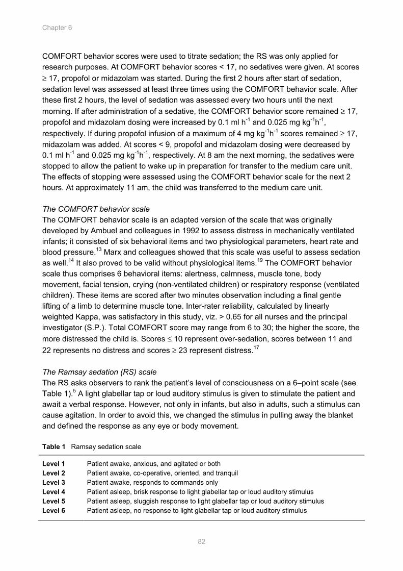
Chapter 6
82
COMFORT behavior scores were used to titrate sedation; the RS was only applied for research purposes. At COMFORT behavior scores < 17, no sedatives were given. At scores ≥ 17, propofol or midazolam was started. During the first 2 hours after start of sedation, sedation level was assessed at least three times using the COMFORT behavior scale. After these first 2 hours, the level of sedation was assessed every two hours until the next morning. If after administration of a sedative, the COMFORT behavior score remained ≥ 17, propofol and midazolam dosing were increased by 0.1 ml h-1 and 0.025 mg kg-1h-1, respectively. If during propofol infusion of a maximum of 4 mg kg-1h-1 scores remained ≥ 17, midazolam was added. At scores < 9, propofol and midazolam dosing were decreased by 0.1 ml h-1 and 0.025 mg kg-1h-1, respectively. At 8 am the next morning, the sedatives were stopped to allow the patient to wake up in preparation for transfer to the medium care unit. The effects of stopping were assessed using the COMFORT behavior scale for the next 2 hours. At approximately 11 am, the child was transferred to the medium care unit. The COMFORT behavior scale The COMFORT behavior scale is an adapted version of the scale that was originally developed by Ambuel and colleagues in 1992 to assess distress in mechanically ventilated infants; it consisted of six behavioral items and two physiological parameters, heart rate and blood pressure.13 Marx and colleagues showed that this scale was useful to assess sedation as well.14 It also proved to be valid without physiological items.19 The COMFORT behavior scale thus comprises 6 behavioral items: alertness, calmness, muscle tone, body movement, facial tension, crying (non-ventilated children) or respiratory response (ventilated children). These items are scored after two minutes observation including a final gentle lifting of a limb to determine muscle tone. Inter-rater reliability, calculated by linearly weighted Kappa, was satisfactory in this study, viz. > 0.65 for all nurses and the principal investigator (S.P.). Total COMFORT score may range from 6 to 30; the higher the score, the more distressed the child is. Scores ≤ 10 represent over-sedation, scores between 11 and 22 represents no distress and scores ≥ 23 represent distress.17 The Ramsay sedation (RS) scale The RS asks observers to rank the patient’s level of consciousness on a 6–point scale (see Table 1).5 A light glabellar tap or loud auditory stimulus is given to stimulate the patient and await a verbal response. However, not only in infants, but also in adults, such a stimulus can cause agitation. In order to avoid this, we changed the stimulus in pulling away the blanket and defined the response as any eye or body movement. Table 1 Ramsay sedation scale
Level 1 Patient awake, anxious, and agitated or both Level 2 Patient awake, co-operative, oriented, and tranquil Level 3 Patient awake, responds to commands only Level 4 Patient asleep, brisk response to light glabellar tap or loud auditory stimulus Level 5 Patient asleep, sluggish response to light glabellar tap or loud auditory stimulus Level 6 Patient asleep, no response to light glabellar tap or loud auditory stimulus
The Ramsay Sedation Scale in Infants
83
Procedure According to the sedation protocol mentioned above, paired COMFORT behavior and RS scores were obtained. As observation requires unobstructed view of the infant’s body, it was necessary to pull away the blanket. This then was considered the impulse for assessing the RS, at the beginning of the two-minutes observation period for the COMFORT behavior scale. The RS and COMFORT behavior scores were recorded after the two-minutes period. Interventions and medications were documented as well. Statistical analysis In the final analysis we included only patients receiving sedatives and for whom 3 or more paired observations had been made. The data were analyzed using SPSS for Windows (version 10.0; SPSS, Chicago, Il).
Inter-individual correlations of COMFORT behavior vs. RS were described using the Spearman rank correlation coefficient. Results Fifty-seven patients were included. Forty-seven patients received sedatives. For 36 patients more than 3 paired observations of COMFORT behavior and RS were available. Their median age and weight was 11 months (IQR 7 to 12) and 9.5 kg (IQR 7.5 to 10), respectively. Fifteen infants received propofol, 17 midazolam and 4 infants received both propofol and midazolam for sedation. Correlation between COMFORT behavior and RS observations A median of 14 (IQR 9 to 17) paired COMFORT behavior and RS observations revealed a median inter-individual correlation between COMFORT behavior and RS of –0.76 (IQR –0.83 to -0.66).
Infants with COMFORT behavior scores ≤ 10 had RS scores ranging from 2 to 6 (from awake and cooperative to unarousable). Infants with COMFORT behavior scores between 11 and 22 had a median RS score of 2 (range 1 to 6). COMFORT behavior scores ≥ 23 were associated with RS scores of 1 (no range). Figure 1 shows the distribution of COMFORT scores across the six Ramsay categories. Applicability of RS Proper scoring appeared impossible in 26 of the 582 RS observations (4.5%) in 15 patients, as the infant’s response was not consistent with just one particular RS level.
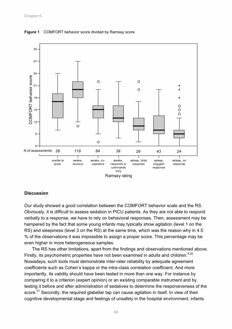
Chapter 6
84
Figure 1 COMFORT behavior score divided by Ramsay score Discussion Our study showed a good correlation between the COMFORT behavior scale and the RS. Obviously, it is difficult to assess sedation in PICU patients. As they are not able to respond verbally to a response, we have to rely on behavioral responses. Then, assessment may be hampered by the fact that some young infants may typically show agitation (level 1 on the RS) and sleepiness (level 3 on the RS) at the same time, which was the reason why in 4.5 % of the observations it was impossible to assign a proper score. This percentage may be even higher in more heterogeneous samples.
The RS has other limitations, apart from the findings and observations mentioned above. Firstly, its psychometric properties have not been examined in adults and children.4,20 Nowadays, such tools must demonstrate inter-rater reliability by adequate agreement coefficients such as Cohen’s kappa or the intra-class correlation coefficient. And more importantly, its validity should have been tested in more than one way. For instance by comparing it to a criterion (expert opinion) or an existing comparable instrument and by testing it before and after administration of sedatives to determine the responsiveness of the score.21 Secondly, the required glabellar tap can cause agitation in itself. In view of their cognitive developmental stage and feelings of unsafety in the hospital environment, infants
unable to score
awake, anxious
awake, co-operative
awake, responds to commands
on ly
asleep, brisk response
asleep, sluggish response
asleep, no response
Ramsay rating
6
9
12
15
18
21
24
27
30
CO
MFO
RT
beha
vior
sco
re
N of assessments: 26 119 84 39 29 43 24
The Ramsay Sedation Scale in Infants
85
may respond to such a stimulus with anxiety. Thirdly, the RS measures level of consciousness rather than sedation and therefore also lacks a sufficient measure of agitation.20,22 To achieve an adequate level of sedation, there is no need for unconsciousness.
Obviously, our study has limitations as well. First, we substituted the prescribed stimulus, a glabellar tap or a loud auditory stimulus, for one we thought would not cause as much agitation. Pulling away the blanket may not be the best stimulus, but it is more suited to young children. Secondly, the observer assessed both the COMFORT behavior scale and RS, which may have led to a flattered picture of the association between the two measures. Furthermore, the observer was not blinded for treatment and a second observer was not available to determine inter-rater reliability for the Ramsay. Because of the complexity of assessing distress up to oversedation in young children it is ill advisable to describe a child’s behavior using one item, like the RS does, we recommend using validated sedation scales instead. List of abbreviations RS Ramsay sedation scale. UMSS University of Michigan Sedation Scale PSICU pediatric surgical intensive care unit VAS visual analogue scale
Chapter 6
86
References 1. www.hyperdictionary.com.
2. Practice guidelines for sedation and analgesia by non-anesthesiologists: a report by the American Society of Anesthesiologists Task Force on Sedation and Analgesia by Non-Anesthesiologists. Anesthesiology 1996;84:459-471
3. Riker RR, Picard JT, Fraser GL: Prospective evaluation of the Sedation-Agitation Scale for adult critically ill patients. Crit Care Med 1999;27(7):1325-1329
4. De Jonghe B, Cook D, Appere-De-Vecchi C, Guyatt G, Meade M, Outin H: Using and understanding sedation scoring systems: a systematic review. Intensive Care Med 2000;26(3):275-285
5. Ramsay MA, Savege TM, Simpson BR, Goodwin R: Controlled sedation with alphaxalone-alphadolone. Br Med J 1974;2(920):656-659
6. Knibbe CA, Melenhorst-de Jong G, Mestrom M, Rademaker CM, Reijnvaan AF, Zuideveld KP, Kuks PF, van Vught H, Danhof M: Pharmacokinetics and effects of propofol 6% for short-term sedation in paediatric patients following cardiac surgery. Br J Clin Pharmacol 2002;54(4):415-422
7. Barr J, Zomorodi K, Bertaccini EJ, Shafer SL, Geller E: A double-blind, randomized comparison of i.v. lorazepam versus midazolam for sedation of ICU patients via a pharmacologic model. Anesthesiology 2001;95(2):286-298
8. Sessler CN, Gosnell MS, Grap MJ, Brophy GM, O'Neal PV, Keane KA, Tesoro EP, Elswick RK: The Richmond Agitation-Sedation Scale: validity and reliability in adult intensive care unit patients. Am J Respir Crit Care Med 2002;166(10):1338-1344
9. Mondello E, Siliotti R, Noto G, Cuzzocrea E, Scollo G, Trimarchi G, Venuti FS: Bispectral Index in ICU: correlation with Ramsay Score on assessment of sedation level. J Clin Monit Comput 2002;17(5):271-277
10. Agrawal D, Feldman HA, Krauss B, Waltzman ML: Bispectral index monitoring quantifies depth of sedation during emergency department procedural sedation and analgesia in children. Ann Emerg Med 2004;43(2):247-255
11. Aneja R, Heard AM, Fletcher JE, Heard CM: Sedation monitoring of children by the Bispectral Index in the pediatric intensive care unit. Pediatr Crit Care Med 2003;4(1):60-64
12. Malviya S, Voepel-Lewis T, Tait AR, Merkel S, Tremper K, Naughton N: Depth of sedation in children undergoing computed tomography: validity and reliability of the University of Michigan Sedation Scale (UMSS). Br J Anaesth 2002;88(2):241-245
13. Ambuel B, Hamlett KW, Marx CM, Blumer JL: Assessing distress in pediatric intensive care environments: the COMFORT scale. J Pediatr Psychol 1992;17(1):95-109
14. Marx CM, Smith PG, Lowrie LH, Hamlett KW, Ambuel B, Yamashita TS, Blumer JL: Optimal sedation of mechanically ventilated pediatric critical care patients. Crit Care Med 1994;22(1):163-170
15. Anand KJ, Barton BA, McIntosh N, Lagercrantz H, Pelausa E, Young TE, Vasa R: Analgesia and sedation in preterm neonates who require ventilatory support: results from the NOPAIN trial. Neonatal Outcome and Prolonged Analgesia in Neonates. Arch Pediatr Adolesc Med 1999;153(4):331-338
16. Wielenga J, De Vos, R, De Leeuw, R, De Haan, RJ: COMFORT scale: a reliable and valid method to measure the amount of stress of ventilated preterm infants. Neonatal Netw 2004;23(2):39-44
The Ramsay Sedation Scale in Infants
87
17. Ista E, Van Dijk, M, Tibboel, D, De Hoog, M.: Assessment of sedation levels in pediatric intensive care patients can be improved by using the COMFORT "behavior" scale. Pediatr Crit Care Med 2005;6(1):58-63
18. Prins SA, Peeters M.Y.M, Houmes R.J., van Dijk M., Knibbe C.A.J., Danhof M., Tibboel D.: Propofol 6% as sedative in children under 2 years of age following major craniofacial surgery. Br J Anaesth 2005;94(5):630-635
19. Carnevale FA, Razack S: An item analysis of the COMFORT scale in a pediatric intensive care unit. Pediatr Crit Care Med 2002;3(2):177-180
20. Kress JP, Pohlman AS, Hall JB: Sedation and analgesia in the intensive care unit. Am J Respir Crit Care Med 2002;166(8):1024-1028
21. Johnston CC: Psychometric Issues in the Measurement of Pain, vol. 10. Seattle: IASP Press; 1998
22. Hansen-Flaschen J, Cowen J, Polomano RC: Beyond the Ramsay scale: need for a validated measure of sedating drug efficacy in the intensive care unit. Crit Care Med 1994;22(5):732-733

Bispectral Index Monitoring as a Tool for Monitoring Sedation in Critically Ill Children During Neuromuscular Blockade
Sandra A. Prins, Monique van Dijk, Matthijs de Hoog, Dick Tibboel.
Cha
pter
Chapter 7
90
Abstract Objective Neuromuscular blockade impedes the clinical assessment of sedation and analgesia. We describe a serie of data in critically ill children using the Bispectral Index monitor to assess sedation and analgesia during continuous neuromuscular blockade in children. Design Prospective observational study. Setting ICU of a level three children's hospital. Patients and participants Children receiving continuous neuromuscular blockade > 4 hours. Interventions During neuromuscular blockade, children were monitored with the Bispectral Index monitor. BIS values were compared with conventional measures heart rate and mean arterial blood pressure. Measurements and results Twenty-four children with a median age of 12 days (IQR 1 to 446) were included. A median of 33 (IQR 17 to 64) paired BIS, heart rate and mean arterial blood pressure measurements per infant revealed a median inter-individual correlation between BIS and heart rate of –0.02 (IQR –0.22 to 0.15) and between BIS and mean arterial blood pressure of –0.03 (IQR –0.15 to 0.33), respectively. In 16 of the 24 patients, BIS values were > 60 during NMB, with a median duration of 2 hours (range 1 to 24 hours). We observed tachycardia in three and hypertension in four of them, with a maximum duration of ten hours and 4 hours, respectively. Conclusion There is no correlation between BIS values and physiological parameters during neuromuscular blockade. Furthermore, 16 patients had BIS values > 60 during neuromuscular blockade, suggesting inadequate sedation, without changes in physiological parameters. We recommend development of practical guidelines to assess sedation and analgesia during NMB for children.
BIS to Monitor Sedation in Infants During NMB
91
Introduction Neuromuscular blockade (NMB) is the intentional paralysis of a patient without affecting cardiac or smooth muscle. NMB drugs do not affect consciousness, nor do they control pain. In the pediatric intensive care setting, single doses of NMB drugs are used to facilitate intubation and long term infusions to facilitate mechanical ventilation in critically ill patients. Other indications for NMB in children include traumatic brain injury, pulmonary hypertension, treatment of refractory intense agitation and protection of surgical repairs.1,2 A recent survey of the use of NMB drugs in pediatric intensive care units in the United Kingdom revealed that a mean 31% of patients (range 15 to 90%) were given these drugs.3
Assessment of sedation and analgesia during NMB is difficult and regrettably, a gold standard is lacking. Under these circumstances, physiological parameters such as blood pressure and heart rate are used as surrogate parameters for sedation and analgesia. In critically ill patients, however, tachycardia does not necessarily result from pain or distress; it may also result from hypovolemia, anemia, or vasopressor drugs.4 Several authors have suggested, therefore, that physiological parameters are not at all reliable to assess sedation and analgesia.5-8 As under-sedation or pain during NMB is undesirable and unethical, we set up a study exploring the usefulness of the Bispectral Index (BIS) monitor for monitoring sedation and analgesia during continuous NMB treatment in pediatric intensive care. Materials and methods From February 2002 until March 2003 we conducted a prospective observational pilot study in 24 patients admitted to either the pediatric surgical intensive care unit (PSICU) or the pediatric intensive care unit (PICU) of our level three children's hospital. The PSICU serves as a referral center for all surgical specialties and for extra corporeal membrane oxygenation (ECMO), admitting 550 patients annually, from neonates up to adolescents. The PICU serves as a referral center for critical care of non-surgical origin, admitting 525 patients annually, also ranging from neonates to adolescents. Patients receiving NMB drugs for at least four hours were eligible for the study. Patients with a history of seizures, traumatic brain injury or other conditions that would influence the appropriate use of the BIS monitor were excluded from this study. The institutional review board approved use of the BIS monitor. Parental informed consent was obtained. Patient characteristics The patients' age, weight, diagnosis and indication for NMB were documented. The choice of sedatives, neuromuscular blockade drugs and analgesics was independent of the study; they were prescribed by the attending physician, independent of BIS values. BIS monitor The Bispectral Index (BIS) monitor uses a frontal, two channel electroencephalogram (EEG) to quantify hypnotic effects of anesthetic and sedative drugs on the brain. Formerly
Chapter 7
92
mainly used during surgery, over the years it has found its way to the (pediatric) intensive care unit as well.9,10 Fourier transformation of the information and bispectral analysis are used to compute a number between 98 (fully awake) and zero (absence of brain electrical activity).11 BIS values > 60 are considered to be indicative of inadequate sedation and even risk of awareness in adults.12,13 A Bispectral A 2000 version 3.12 monitor (Aspect Medical Systems, Natick, MA, USA) with commercially available BIS sensor strips designed for children was used. The electrodes were placed near the center of the forehead and either on the right or left temple. The algorithm within the BIS monitor sets limits for electrode impedance and signal quality. Therefore, no BIS value is displayed if the signal has too much noise or artifacts. We used the limits set in the monitor: if the BIS value was displayed, meaning a Signal Quality Index (SQI) > 50, it was recorded. BIS and physiological parameters recording method The BIS values were directly transferred to a patient data management system (PDMS). Hourly, heart rate (HR), mean arterial blood pressure (MABP) and BIS values were retrieved from PDMS. Relevant interventions and treatments were recorded as well. Definitions A patient's baseline MABP and HR values were determined using all available MABP and HR values, at least 4 and no more than 12 values, before NMB was started, calculating a mean MABP and HR. Hypertension was defined as any period of time when a patient's MABP was 15% above baseline MABP.14,15 Tachycardia was defined as any period of time when a patient's HR was 15 % above or below baseline HR.14 Statistical analysis The data were analyzed using SPSS for Windows (version 10.0; SPSS, Chicago, Il). Inter-individual correlations of BIS vs. HR and MABP were expressed by the Pearson product-moment correlation coefficient. Results Patients Twenty-four of 58 eligible patients (41%) in the one-year study period were included, 21 in the PSICU and 3 in the PICU. Their characteristics are listed in Table 1. Age distribution was as follows: 13 neonates (younger than 28 days), 5 infants between 28 days and 1 year, and 6 children > 1 year. Median age and weight were 12.5 days (IQR 1 to 446.3) and 4.2 kg (IQR 3.6 to 10), respectively. Nine patients, equally distributed over the age groups, were on extra corporeal membrane oxygenation.
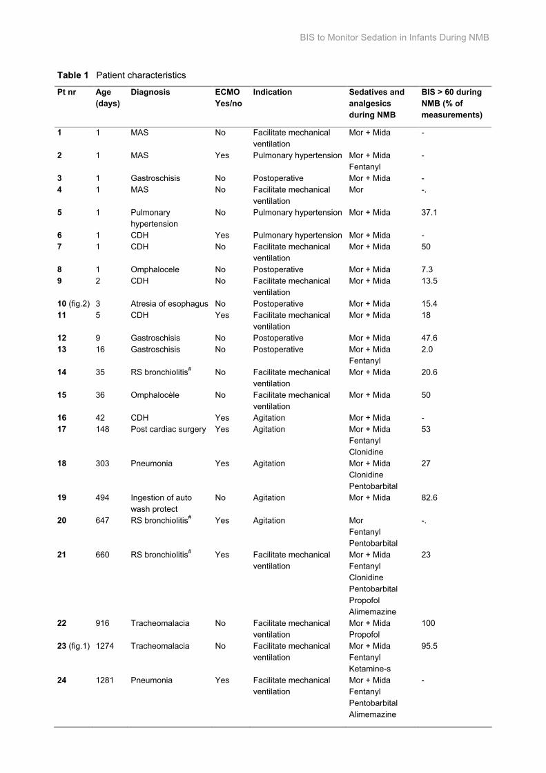
BIS to Monitor Sedation in Infants During NMB
Table 1 Patient characteristics
Pt nr Age (days)
Diagnosis ECMO Yes/no
Indication Sedatives and analgesics during NMB
BIS > 60 during NMB (% of measurements)
1 1 MAS No Facilitate mechanical ventilation
Mor + Mida -
2 1 MAS Yes Pulmonary hypertension Mor + Mida Fentanyl
-
3 1 Gastroschisis No Postoperative Mor + Mida - 4 1 MAS No Facilitate mechanical
ventilation Mor -.
5 1 Pulmonary hypertension
No Pulmonary hypertension Mor + Mida 37.1
6 1 CDH Yes Pulmonary hypertension Mor + Mida - 7 1 CDH No Facilitate mechanical
ventilation Mor + Mida 50
8 1 Omphalocele No Postoperative Mor + Mida 7.3 9 2 CDH No Facilitate mechanical
ventilation Mor + Mida 13.5
10 (fig.2) 3 Atresia of esophagus No Postoperative Mor + Mida 15.4 11 5 CDH Yes Facilitate mechanical
ventilation Mor + Mida 18
12 9 Gastroschisis No Postoperative Mor + Mida 47.6 13 16 Gastroschisis No Postoperative Mor + Mida
Fentanyl 2.0
14 35 RS bronchiolitis# No Facilitate mechanical ventilation
Mor + Mida 20.6
15 36 Omphalocèle No Facilitate mechanical ventilation
Mor + Mida 50
16 42 CDH Yes Agitation Mor + Mida - 17 148 Post cardiac surgery Yes Agitation Mor + Mida
Fentanyl Clonidine
53
18 303 Pneumonia Yes Agitation Mor + Mida Clonidine Pentobarbital
27
19 494 Ingestion of auto wash protect
No Agitation Mor + Mida 82.6
20 647 RS bronchiolitis# Yes Agitation Mor Fentanyl Pentobarbital
-.
21 660 RS bronchiolitis# Yes Facilitate mechanical ventilation
Mor + Mida Fentanyl Clonidine Pentobarbital Propofol Alimemazine
23
22 916 Tracheomalacia No Facilitate mechanical ventilation
Mor + Mida Propofol
100
23 (fig.1) 1274 Tracheomalacia No Facilitate mechanical ventilation
Mor + Mida Fentanyl Ketamine-s
95.5
24 1281 Pneumonia Yes Facilitate mechanical ventilation
Mor + Mida Fentanyl Pentobarbital Alimemazine
-
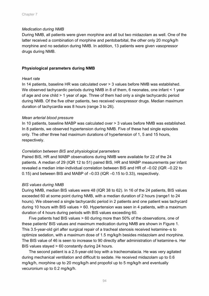
Chapter 7
94
Medication during NMB During NMB, all patients were given morphine and all but two midazolam as well. One of the latter received a combination of morphine and pentobarbital, the other only 20 mcg/kg/h morphine and no sedation during NMB. In addition, 13 patients were given vasopressor drugs during NMB. Physiological parameters during NMB Heart rate In 14 patients, baseline HR was calculated over > 3 values before NMB was established. We observed tachycardic periods during NMB in 8 of them, 6 neonates, one infant < 1 year of age and one child > 1 year of age. Three of them had only a single tachycardic period during NMB. Of the five other patients, two received vasopressor drugs. Median maximum duration of tachycardia was 8 hours (range 3 to 26). Mean arterial blood pressure In 10 patients, baseline MABP was calculated over > 3 values before NMB was established. In 8 patients, we observed hypertension during NMB. Five of these had single episodes only. The other three had maximum durations of hypertension of 1, 5 and 15 hours, respectively. Correlation between BIS and physiological parameters Paired BIS, HR and MABP observations during NMB were available for 22 of the 24 patients. A median of 29 (IQR 12 to 51) paired BIS, HR and MABP measurements per infant revealed a median inter-individual correlation between BIS and HR of –0.02 (IQR –0.22 to 0.15) and between BIS and MABP of –0.03 (IQR –0.15 to 0.33), respectively. BIS values during NMB During NMB, median BIS values were 48 (IQR 38 to 62). In 16 of the 24 patients, BIS values exceeded 60 at some point during NMB, with a median duration of 2 hours (range1 to 24 hours). We observed a single tachycardic period in 2 patients and one patient was tachycard during 10 hours with BIS values > 60. Hypertension was seen in 4 patients, with a maximum duration of 4 hours during periods with BIS values exceeding 60.
Five patients had BIS values > 60 during more than 50% of the observations, one of these patients' BIS values and maximum medication during NMB are shown in Figure 1. This 3.5-year-old girl after surgical repair of a tracheal stenosis received ketamine–s to optimize sedation, with a maximum dose of 1.5 mg/kg/h besides midazolam and morphine. The BIS value of 46 is seen to increase to 90 directly after administration of ketamine-s. Her BIS values stayed > 60 constantly during 24 hours.
The second patient is a 2.5-year-old boy with a tracheomalacia. He was very agitated during mechanical ventilation and difficult to sedate. He received midazolam up to 0.6 mg/kg/h, morphine up to 20 mcg/kg/h and propofol up to 5 mg/kg/h and eventually vecuronium up to 0.2 mg/kg/h.

BIS to Monitor Sedation in Infants During NMB
95
0
60
120
180
17:00
19:00
21:00
23:00 1:0
03:0
05:0
07:0
09:0
011
:0013
:0015
:0017
:0019
:0021
:0023
:00 1:00
3:00
5:00
7:00
9:0011
:0013
:0015
:00 9:0011
:0013
:00
BIS HRvecuronium max dose 0.1 mg/kg/h midazolam max dose 0.3 mg/kg/hmorphine max dose 20 mcg/kg/h ketamine-s max dose 1.5 mg/kg/hBIS = 60
Figure 1 Increase of BIS values after administering ketamine-s
A 3.5-year-old girl, very agitated during mechanical ventilation after surgery for a severe tracheomalacia. Before administering ketamine-s, the BIS value was 46. Directly after administration of ketamine-s, BIS increased to 90.
The third patient is a 36-day-old girl with an omphalocèle, who received vecuronium to facilitate mechanical ventilation. Despite 0.4 mg/kg/h of midazolam and 10 mcg/kg/h of morphine, she was still able to fight mechanical ventilation.
The fourth patient is a 1.5-year-old boy who was sedated and paralyzed because of severe agitation. He was admitted to the PICU after a chemical burn of the esophagus. He received midazolam up to 0.3 mg/kg/h and morphine up to 20 mcg/kg/h.
The fifth patient is a 5-month-old girl who was on ECMO after cardiac surgery. During the ECMO run, before NMB, she was highly agitated. She was sedated, therefore, by midazolam up to 0.5 mg/kg/h, morphine up to 50 mcg/kg/h, clonidine up to 0.20 mcg/kg/h and fentanyl up to 6.1 mcg/kg/h was given. Severe agitation, despite midazolam and morphine in high dosages, eventually necessitated the administration of vecuronium up to 0.1 mg/kg/h. Decrease of BIS values after start of NMB Of 7 patients, BIS values before and during NMB were known. In two of them, BIS values decreased directly after start of infusion of NMB without changes in sedatives or analgesics. One received 0.1 mg/kg/h midazolam and 20 mcg/kg/h morphine. The other patient received 0.25 mg/kg/h midazolam and 32 mcg/kg/h morphine. Figure 2 shows details of the first patient; a 3-day-old boy on postoperative mechanical ventilation whose BIS values decreased by almost 40 points directly after administration of 0.1 mg/kg/h of vecuronium. The second time he received vecuronium, 0.06 mg/kg/h, BIS values did not decrease. After vecuronium dosing was increased to 0.1mg/kg/h, BIS values decreased again by 40 points.
Time
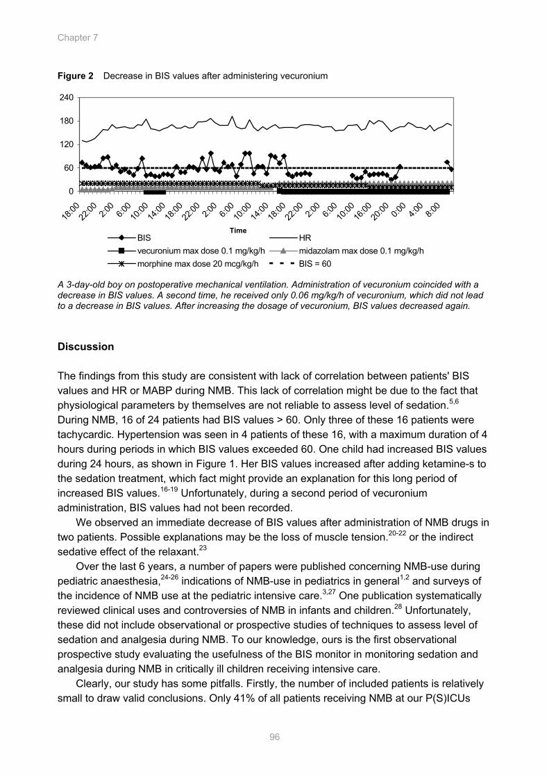
Chapter 7
96
0
60
120
180
240
18:00
22:00 2:0
06:0
010
:0014
:0018
:0022
:00 2:00
6:00
10:00
14:00
18:00
22:00 2:0
06:0
010
:0016
:0020
:00 0:00
4:00
8:00
BIS HRvecuronium max dose 0.1 mg/kg/h midazolam max dose 0.1 mg/kg/hmorphine max dose 20 mcg/kg/h BIS = 60
Figure 2 Decrease in BIS values after administering vecuronium
A 3-day-old boy on postoperative mechanical ventilation. Administration of vecuronium coincided with a decrease in BIS values. A second time, he received only 0.06 mg/kg/h of vecuronium, which did not lead to a decrease in BIS values. After increasing the dosage of vecuronium, BIS values decreased again. Discussion The findings from this study are consistent with lack of correlation between patients' BIS values and HR or MABP during NMB. This lack of correlation might be due to the fact that physiological parameters by themselves are not reliable to assess level of sedation.5,6 During NMB, 16 of 24 patients had BIS values > 60. Only three of these 16 patients were tachycardic. Hypertension was seen in 4 patients of these 16, with a maximum duration of 4 hours during periods in which BIS values exceeded 60. One child had increased BIS values during 24 hours, as shown in Figure 1. Her BIS values increased after adding ketamine-s to the sedation treatment, which fact might provide an explanation for this long period of increased BIS values.16-19 Unfortunately, during a second period of vecuronium administration, BIS values had not been recorded.
We observed an immediate decrease of BIS values after administration of NMB drugs in two patients. Possible explanations may be the loss of muscle tension.20-22 or the indirect sedative effect of the relaxant.23
Over the last 6 years, a number of papers were published concerning NMB-use during pediatric anaesthesia,24-26 indications of NMB-use in pediatrics in general1,2 and surveys of the incidence of NMB use at the pediatric intensive care.3,27 One publication systematically reviewed clinical uses and controversies of NMB in infants and children.28 Unfortunately, these did not include observational or prospective studies of techniques to assess level of sedation and analgesia during NMB. To our knowledge, ours is the first observational prospective study evaluating the usefulness of the BIS monitor in monitoring sedation and analgesia during NMB in critically ill children receiving intensive care.
Clearly, our study has some pitfalls. Firstly, the number of included patients is relatively small to draw valid conclusions. Only 41% of all patients receiving NMB at our P(S)ICUs
Time
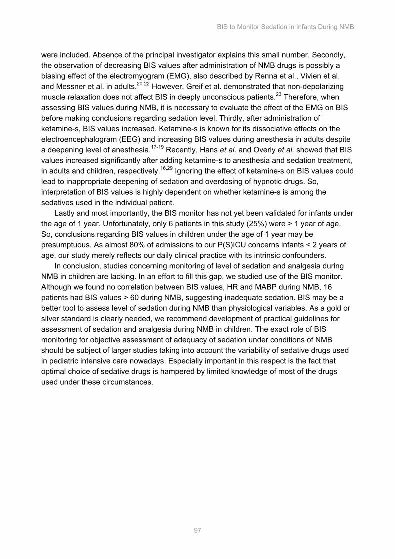
BIS to Monitor Sedation in Infants During NMB
97
were included. Absence of the principal investigator explains this small number. Secondly, the observation of decreasing BIS values after administration of NMB drugs is possibly a biasing effect of the electromyogram (EMG), also described by Renna et al., Vivien et al. and Messner et al. in adults.20-22 However, Greif et al. demonstrated that non-depolarizing muscle relaxation does not affect BIS in deeply unconscious patients.23 Therefore, when assessing BIS values during NMB, it is necessary to evaluate the effect of the EMG on BIS before making conclusions regarding sedation level. Thirdly, after administration of ketamine-s, BIS values increased. Ketamine-s is known for its dissociative effects on the electroencephalogram (EEG) and increasing BIS values during anesthesia in adults despite a deepening level of anesthesia.17-19 Recently, Hans et al. and Overly et al. showed that BIS values increased significantly after adding ketamine-s to anesthesia and sedation treatment, in adults and children, respectively.16,29 Ignoring the effect of ketamine-s on BIS values could lead to inappropriate deepening of sedation and overdosing of hypnotic drugs. So, interpretation of BIS values is highly dependent on whether ketamine-s is among the sedatives used in the individual patient.
Lastly and most importantly, the BIS monitor has not yet been validated for infants under the age of 1 year. Unfortunately, only 6 patients in this study (25%) were > 1 year of age. So, conclusions regarding BIS values in children under the age of 1 year may be presumptuous. As almost 80% of admissions to our P(S)ICU concerns infants < 2 years of age, our study merely reflects our daily clinical practice with its intrinsic confounders.
In conclusion, studies concerning monitoring of level of sedation and analgesia during NMB in children are lacking. In an effort to fill this gap, we studied use of the BIS monitor. Although we found no correlation between BIS values, HR and MABP during NMB, 16 patients had BIS values > 60 during NMB, suggesting inadequate sedation. BIS may be a better tool to assess level of sedation during NMB than physiological variables. As a gold or silver standard is clearly needed, we recommend development of practical guidelines for assessment of sedation and analgesia during NMB in children. The exact role of BIS monitoring for objective assessment of adequacy of sedation under conditions of NMB should be subject of larger studies taking into account the variability of sedative drugs used in pediatric intensive care nowadays. Especially important in this respect is the fact that optimal choice of sedative drugs is hampered by limited knowledge of most of the drugs used under these circumstances.
Chapter 7
98
References 1. Adelson PD, Bratton SL, Carney NA et al. Chapter 9. Use of sedation and neuromuscular blockade
in the treatment of severe pediatric traumatic brain injury. Pediatr Crit Care Med 2003;4:S34-7
2. Almeida JF, Kalil Filho WJ, Troster EJ. Neuromuscular blockade in children. Rev Hosp Clin Fac Med Sao Paulo 2000;55:105-10
3. Playfor SD, Thomas DA, Choonara I. Sedation and neuromuscular blockade in paediatric intensive care: a review of current practice in the UK. Paediatr Anaesth 2003;13:147-51
4. Grindstaff RJ, Tobias JD. Applications of bispectral index monitoring in the pediatric intensive care unit. J Intensive Care Med 2004;19:111-6
5. Carnevale FA, Razack S. An item analysis of the COMFORT scale in a pediatric intensive care unit. Pediatr Crit Care Med 2002;3:177-80
6. Ista E, van Dijk M, Tibboel D, de Hoog M. Assessment of sedation levels in pediatric intensive care patients can be improved by using the COMFORT "behavior" scale. Pediatr Crit Care Med 2005;6:58-63
7. Sweet SD, McGrath PJ. Physiological measures of pain. In: Finley GA, McGrath PJ, eds. Measurement of pain in infants and children. Progress in pain research and management Seattle: IASP Press, 1998:59-81
8. van Dijk M, de Boer JB, Koot HM et al. The association between physiological and behavioral pain measures in 0- to 3-year-old infants after major surgery. J Pain Symptom Manage 2001;22:600-9
9. Frenzel D, Greim CA, Sommer C et al. Is the bispectral index appropriate for monitoring the sedation level of mechanically ventilated surgical ICU patients? Intensive Care Med 2002;28:178-83
10. Berkenbosch JW, Fichter CR, Tobias JD. The correlation of the bispectral index monitor with clinical sedation scores during mechanical ventilation in the pediatric intensive care unit. Anesth Analg 2002;94:506-11
11. Glass PS, Bloom M, Kearse L et al. Bispectral analysis measures sedation and memory effects of propofol, midazolam, isoflurane, and alfentanil in healthy volunteers. Anesthesiology 1997;86:836-47
12. Sebel PS, Lang E, Rampil IJ et al. A multicenter study of bispectral electroencephalogram analysis for monitoring anesthetic effect. Anesth Analg 1997;84:891-9
13. Myles PS, Leslie K, McNeil J et al. Bispectral index monitoring to prevent awareness during anaesthesia: the B-Aware randomised controlled trial. Lancet 2004;363:1757-63
14. Ambuel B, Hamlett KW, Marx CM, Blumer JL. Assessing distress in pediatric intensive care environments: the COMFORT scale. J Pediatr Psychol 1992;17:95-109
15. Marx CM, Smith PG, Lowrie LH et al. Optimal sedation of mechanically ventilated pediatric critical care patients. Crit Care Med 1994;22:163-70
16. Hans P, Dewandre P,-Y, Brichant J.F., Bonhomme, V,. Comparative effects of ketamine on Bispectral Index and spectral entropy of the electroencephalogram under sevoflurane anaesthesia. Br J Anaesth 2005;94:336-40
17. Arbour R. Continuous nervous system monitoring, EEG, the bispectral index, and neuromuscular transmission. AACN Clin Issues 2003;14:185-207
BIS to Monitor Sedation in Infants During NMB
99
18. Vereecke HE, Struys MM, Mortier EP. A comparison of bispectral index and ARX-derived auditory evoked potential index in measuring the clinical interaction between ketamine and propofol anaesthesia. Anaesthesia 2003;58:957-61
19. Sakai T, Singh H, Mi WD et al. The effect of ketamine on clinical endpoints of hypnosis and EEG variables during propofol infusion. Acta Anaesthesiol Scand 1999;43:212-6
20. Renna M, Wigmore T, Mofeez A, Gillbe C. Biasing effect of the electromyogram on BIS: a controlled study during high-dose fentanyl induction. J Clin Monit Comput 2002;17:377-81
21. Vivien B, Di Maria S, Ouattara A et al. Overestimation of Bispectral Index in sedated intensive care unit patients revealed by administration of muscle relaxant. Anesthesiology 2003;99:9-17
22. Messner M, Beese U, Romstock J et al. The bispectral index declines during neuromuscular block in fully awake persons. Anesth Analg 2003;97:488-91, table of contents
23. Greif R, Greenwald, S, Schweitzer, E, Laciny, S, Rajek, A, Caldwell, J.E. Sessler, D.I. Muscle Relaxation Does Not Alter Hypnotic Level During Propofol Anesthesia. Anest Analg 2002;94:604-8
24. Reich DL, Hollinger I, Harrington DJ et al. Comparison of cisatracurium and vecuronium by infusion in neonates and small infants after congenital heart surgery. Anesthesiology 2004;101:1122-7
25. Fisher DM. Neuromuscular blocking agents in paediatric anaesthesia. Br J Anaesth 1999;83:58-64.
26. Ansermino JM, Sanderson PM, Bevan JC, Bevan DR. Acceleromyography improves detection of residual neuromuscular blockade in children. Can J Anaesth 1996;43:589-94
27. Rhoney DH, Murry KR. National survey on the use of sedatives and neuromuscular blocking agents in the pediatric intensive care unit. Pediatr Crit Care Med 2002;3:129-33
28. Martin LD, Bratton SL, O'Rourke PP. Clinical uses and controversies of neuromuscular blocking agents in infants and children. Crit Care Med 1999;27:1358-68
29. Overly FL WR, Connor FA, Fontaine B, Jay GD, Linakis JG. Bispectral Analysis During Pediatric Procedural Sedation. Pediatric Emergency Care 2005;21:6-11


Continuous Non-Invasive Monitoring of Barbiturate Coma in Critically Ill Children Using the Bispectral (BIS) Index Monitor
Sandra A. Prins, Matthijs de Hoog, Joleen H. Blok, Dick Tibboel, Gerhard H. Visser.
Cha
pter
Chapter 8
102
Abstract Objective It is generally accepted that the target of a barbiturate induced coma in traumatic brain injury (TBI) or generalized convulsive status epilepticus (GCSE) is to reach a burst-suppression pattern. The aim of this study was to evaluate the usefulness of bispectral index monitoring as an adjunct to full channel electroencephalogram monitoring. Methods The BIS monitor expresses depth of anesthesia and sedation by a value ranging from 0 (iso-electric) to 100 (fully awake). One of its parameters, the suppression ratio (SR) represents the percentage of epochs in which the EEG signal was considered suppressed in the last minute. SRs of the BIS monitor were compared with the SRs of 1-minute segments of full channel EEGs as assessed by quantitative visual analysis. Results Five patients with GCSE and three patients after TBI, with a median age of 11.6 years (range 4 months to 15 years), were included. Correlations between SR-BIS and SR-EEG could be calculated for four patients only. The individual correlations between SR-BIS and SR-EEG for these patients were 0.67, 0.64, 0.70 and 0.70, respectively. Lack of homogeneity in the sample was observed for two patients. Yet two other patients had an iso-electric EEG, with SR-BIS values ranging from 43 to 100 (mean of 95 ± 1.6). Conclusions The BIS monitor is a potential aid in monitoring a barbiturate induced coma around the clock. However, correlations between SR-BIS and SR-EEG were found to be small to moderate for 4 patients. One explanation is the presence of an asymmetrical EEG in a patient who had suffered a TBI resulting in intra-cranial hemorrhage at the left side of the head. Another explanation can be short bursts (less than 1 second); with the algorithm of the BIS monitor simply over-estimating the length of the burst and therefore under-estimating the SR-BIS. Also, lack of time synchronization in four patients may have also under-estimated correlations between SR-BIS and SR-EEG.
Continuous Monitoring of Barbiturate Coma using BIS
103
Introduction Traumatic brain injury (TBI) and generalized convulsive status epilepticus (GCSE) are conditions which need aggressive management. Barbiturates are used to lower intracranial pressure (ICP) or to stop epileptiform activity with the aim to improve neurological outcome. Either effect is considered the result of the induced decrease in cerebral metabolism and blood flow.1 However, barbiturate therapy has serious side effects, in particular cardiovascular depression and hypotension.2,3 Dosing barbiturates beyond the point of burst suppression may increase the risk of complications without offering further therapeutic benefits.3 As dosing is usually guided by the extent of an induced burst suppression pattern on the electroencephalogram (EEG),4 careful monitoring of EEG parameters is mandatory. Several methods of monitoring a barbiturate coma are available, viz. interval or continuous EEG monitoring and regular testing of barbiturate blood levels. Winer et al.5 showed in 10 adult patients that continuous EEG monitoring was the best modality, as it showed the presence of burst suppression on a moment-to-moment base. They also found poor correlations between serum and cerebrospinal fluid barbiturate levels at any given time, suggesting that barbiturate levels are difficult to interpret given a specific patient’s distribution and metabolism.
When EEG is used to determine the optimal depth of a barbiturate coma, the goal is to induce a burst suppression pattern – usually defined as 5 to10 episodes per minute with suppression of cerebro-electrical activity below 10 - 20 µV.5 A practical drawback of the standard EEG recording method is that recording and interpretation requires qualified EEG technicians and a clinical neurophysiologist. Also, most centers do not have the facilities to monitor EEGs around the clock.6-8
Against the background of the scarcity of data on barbiturate induced coma in children.9 we conducted a study to explore the usefulness of the BIS monitor during a barbiturate induced coma in critically ill children needing intensive neuro-monitoring. BIS recordings were compared with standard full channel EEG recordings. Methods Patients We conducted a prospective observational pilot study at the pediatric surgical intensive care unit (PSICU) and the pediatric intensive care unit (PICU) of our level three children’s hospital. Due to the strictly observational and non-invasive nature of the study, the institutional review board waived the need for approval. Annually our PSICU admits some 10 patients with a Glascow Coma Score ≤ 8 after TBI which is considered an indication for intracranial pressure monitoring. In about half of the patients, it is necessary to induce a barbiturate coma, after failure of all other methods to decrease ICP.10 In addition, our PICU yearly admits 3 - 4 patients with refractory GCSE for treatment of their condition with a barbiturate coma. All children with TBI or GCSE in whom a barbiturate coma was induced
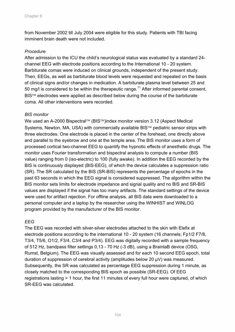
Chapter 8
104
from November 2002 till July 2004 were eligible for this study. Patients with TBI facing imminent brain death were not included. Procedure After admission to the ICU the child’s neurological status was evaluated by a standard 24-channel EEG with electrode positions according to the International 10 - 20 system. Barbiturate comas were induced on clinical grounds, independent of the present study. Then, EEGs, as well as barbiturate blood levels were requested and repeated on the basis of clinical signs and/or changes in medication. A barbiturate plasma level between 25 and 50 mg/l is considered to be within the therapeutic range.11 After informed parental consent, BIS electrodes were applied as described below during the course of the barbiturate coma. All other interventions were recorded. BIS monitor We used an A-2000 Bispectral (BIS)index monitor version 3.12 (Aspect Medical Systems, Newton, MA, USA) with commercially available BIS pediatric sensor strips with three electrodes. One electrode is placed in the center of the forehead, one directly above and parallel to the eyebrow and one at the temple area. The BIS monitor uses a form of processed cortical two-channel EEG to quantify the hypnotic effects of anesthetic drugs. The monitor uses Fourier transformation and bispectral analysis to compute a number (BIS value) ranging from 0 (iso-electric) to 100 (fully awake). In addition the EEG recorded by the BIS is continuously displayed (BIS-EEG), of which the device calculates a suppression ratio (SR). The SR calculated by the BIS (SR-BIS) represents the percentage of epochs in the past 63 seconds in which the EEG signal is considered suppressed. The algorithm within the BIS monitor sets limits for electrode impedance and signal quality and no BIS and SR-BIS values are displayed if the signal has too many artifacts. The standard settings of the device were used for artifact rejection. For offline analysis, all BIS data were downloaded to a personal computer and a laptop by the researcher using the WINHIST and WINLOG program provided by the manufacturer of the BIS monitor. EEG The EEG was recorded with silver-silver electrodes attached to the skin with Elefix at electrode positions according to the international 10 - 20 system (16 channels; Fp1/2 F7/8, T3/4, T5/6, O1/2, F3/4, C3/4 and P3/4). EEG was digitally recorded with a sample frequency of 512 Hz, bandpass filter settings 0,13 - 70 Hz (-3 dB), using a BrainlaB device (OSG, Rumst, Belgium). The EEG was visually assessed and for each 10 second EEG epoch, total duration of suppression of cerebral activity (amplitudes below 20 µV) was measured. Subsequently, the SR was calculated as percentage EEG suppression during 1 minute, as closely matched to the corresponding BIS epoch as possible (SR-EEG). Of EEG registrations lasting > 1 hour, the first 11 minutes of every full hour were captured, of which SR-EEG was calculated.
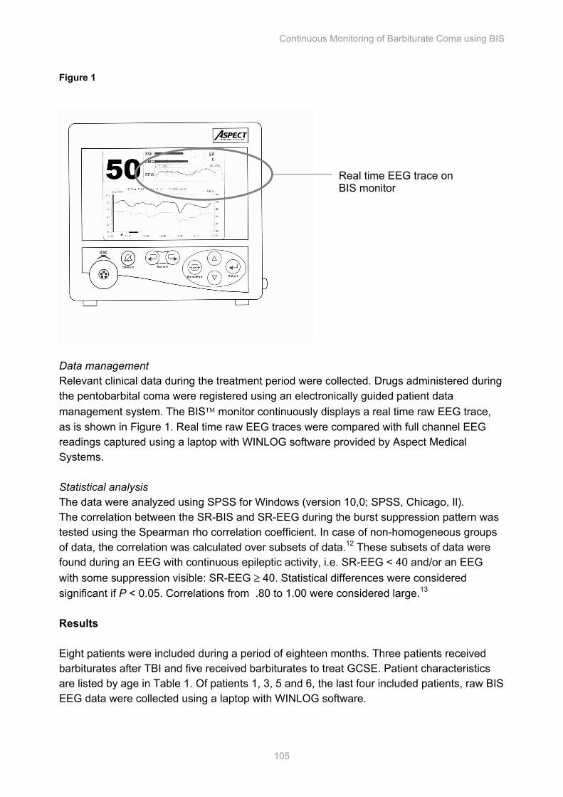
Continuous Monitoring of Barbiturate Coma using BIS
105
Figure 1
Data management Relevant clinical data during the treatment period were collected. Drugs administered during the pentobarbital coma were registered using an electronically guided patient data management system. The BIS monitor continuously displays a real time raw EEG trace, as is shown in Figure 1. Real time raw EEG traces were compared with full channel EEG readings captured using a laptop with WINLOG software provided by Aspect Medical Systems. Statistical analysis The data were analyzed using SPSS for Windows (version 10,0; SPSS, Chicago, Il). The correlation between the SR-BIS and SR-EEG during the burst suppression pattern was tested using the Spearman rho correlation coefficient. In case of non-homogeneous groups of data, the correlation was calculated over subsets of data.12 These subsets of data were found during an EEG with continuous epileptic activity, i.e. SR-EEG < 40 and/or an EEG with some suppression visible: SR-EEG ≥ 40. Statistical differences were considered significant if P < 0.05. Correlations from .80 to 1.00 were considered large.13 Results Eight patients were included during a period of eighteen months. Three patients received barbiturates after TBI and five received barbiturates to treat GCSE. Patient characteristics are listed by age in Table 1. Of patients 1, 3, 5 and 6, the last four included patients, raw BIS EEG data were collected using a laptop with WINLOG software.
Real time EEG trace on BIS monitor
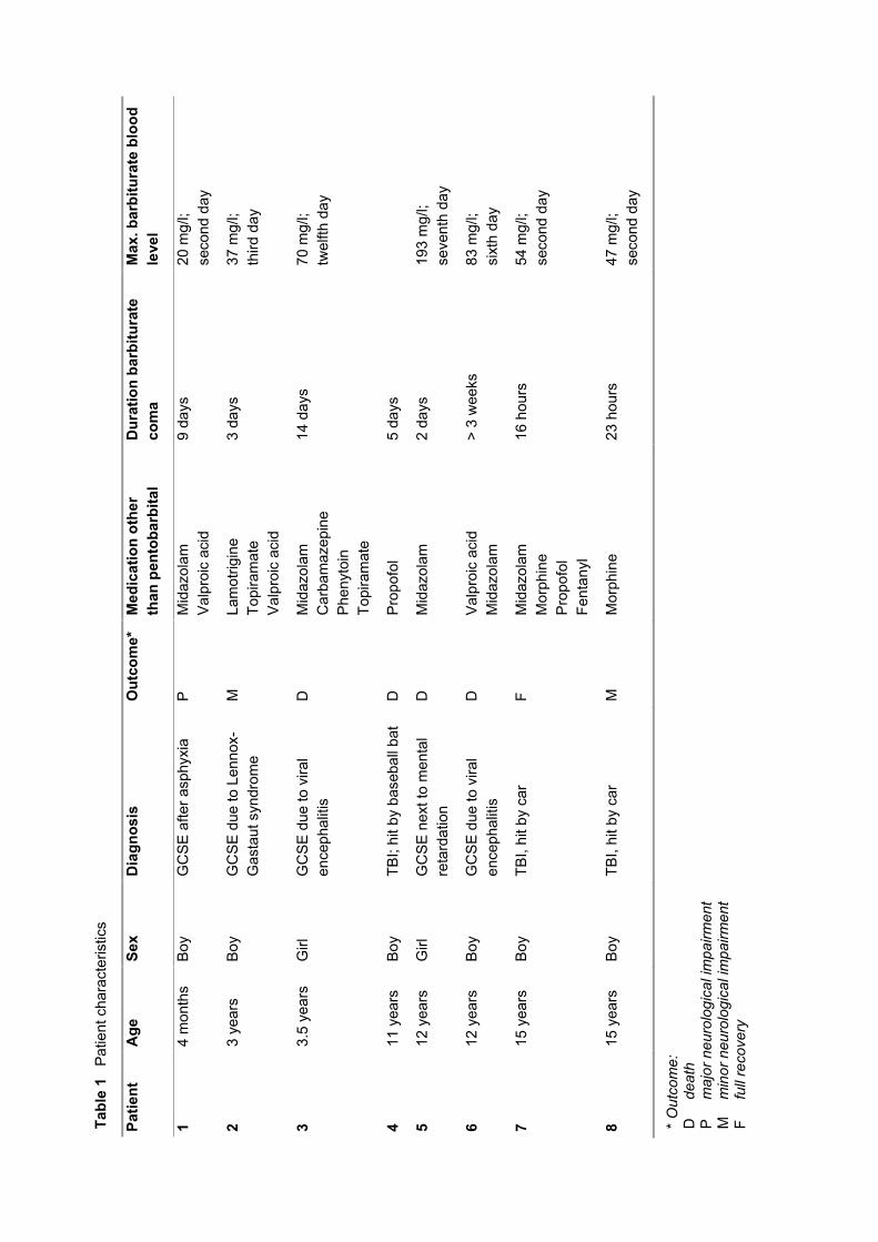
Tabl
e 1
Patie
nt c
hara
cter
istic
s
Patie
nt
Age
Se
x
Dia
gnos
is
Out
com
e*
Med
icat
ion
othe
r th
an p
ento
barb
ital
Dur
atio
n ba
rbitu
rate
co
ma
Max
. bar
bitu
rate
blo
od
leve
l
1 4
mon
ths
Boy
G
CS
E a
fter a
sphy
xia
P
Mid
azol
am
Val
proi
c ac
id
9 da
ys
20 m
g/l;
seco
nd d
ay
2 3
year
s B
oy
GC
SE
due
to L
enno
x-G
asta
ut s
yndr
ome
M
Lam
otrig
ine
Topi
ram
ate
Val
proi
c ac
id
3 da
ys
37 m
g/l;
third
day
3 3.
5 ye
ars
Girl
G
CS
E d
ue to
vira
l en
ceph
aliti
s D
M
idaz
olam
C
arba
maz
epin
e P
heny
toin
To
pira
mat
e
14 d
ays
70 m
g/l;
twel
fth d
ay
4 11
yea
rs
Boy
TB
I; hi
t by
base
ball
bat
D
Pro
pofo
l 5
days
5 12
yea
rs
Girl
G
CS
E n
ext t
o m
enta
l re
tard
atio
n D
M
idaz
olam
2
days
19
3 m
g/l;
se
vent
h da
y
6 12
yea
rs
Boy
G
CS
E d
ue to
vira
l en
ceph
aliti
s D
V
alpr
oic
acid
M
idaz
olam
>
3 w
eeks
83
mg/
l; si
xth
day
7 15
yea
rs
Boy
TB
I, hi
t by
car
F M
idaz
olam
M
orph
ine
Pro
pofo
l Fe
ntan
yl
16 h
ours
54
mg/
l;
seco
nd d
ay
8 15
yea
rs
Boy
TB
I, hi
t by
car
M
Mor
phin
e
23 h
ours
47
mg/
l;
seco
nd d
ay
* O
utco
me:
D
de
ath
P m
ajor
neu
rolo
gica
l im
pairm
ent
M
min
or n
euro
logi
cal i
mpa
irmen
t F
fu
ll re
cove
ry

Continuous Monitoring of Barbiturate Coma using BIS
107
Correlation between SR-BIS and SR-EEG The paired observations of all patients are displayed in Figure 2. Correlations between SR-BIS and SR-EEG could be calculated for four patients only (3, 4, 6 and 7). The individual correlations between SR-BIS and SR-EEG for these patients were 0.67, 0.64, 0.70 and 0.70, respectively. For patients 1 and 2, a lack of homogeneity in the sample was observed, shown as two “data clouds” (see Figure 3). Patients 5 and 8 had an iso-electric EEG, with SR-BIS values ranging from 43 to 100 (mean of 95 ± 1.6) Correlation between SR-BIS and SR-EEG in subsets Of patients 1 and 2, subsets of data were determined. The best correlations between SR-BIS and SR-EEG in these patients were 0.5 and 0.4, respectively. Calculation for patients 5 and 8 were impossible as the SR-EEG was constant. Time synchronization For patient 6, correlation between SR-BIS and SR-EEG over all EEGs was 0.70. This correlation was surprisingly bad for one EEG, viz. -0.003. After the SR-BIS and SR-EEG were time-synchronized, moving the SR-BIS values 5 minutes back, the correlation improved to 0.92 (as shown in Figure 4). Burst types Patients showing a poor correlation had a burst suppression pattern with bursts less than 1 second, as shown in Figure 5 for patient 3. The SR-BIS was 70% and the SR-EEG 88%. Figure 2 Scatterplot of all 8 patients
SR-EEG
100806040200
SR
-BIS
100
80
60
40
20
0
8
7
6
5
4
3
2
1
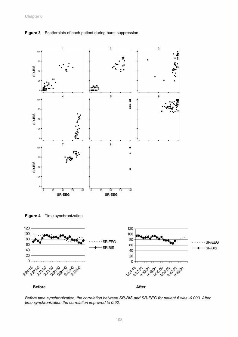
Chapter 8
108
Figure 3 Scatterplots of each patient during burst suppression
1 2 3
4 5 6
7 8
0
25
50
75
100
SR-B
IS
VVVVVVVVVVVVVVVVVVVVVVVVVVVVVVVVVVVVVVVV VVV
VVV
VVVV
VV V V
VVVV V
V
VVVVVVVVVVVVVVVVVVVVVVVV
V
VVVVVVVVV
VVV VVVV
VVVV
VVVVVVVVV
VVVVVVVVVV
VV V
VV
V
V
VV
VVVVV
V
VVVVVVVVV
0
25
50
75
100
SR-B
IS
VVVV
V
VVVVVVVVVVVVVVVVVVVV
VVVV
VVVVV
VVVVVVVVVVVVVVVVVVVVVVV
VVVVVVVVVVVVVVV V
VVVVVVVVVVV
VVVVVVVV
VVV
VVVVVVVVVVVVVVVVVVVVVVVVVVVVVV
0 25 50 75 100
SR-EEG
0
25
50
75
100
SR-B
IS
VVVVVVVV
VVVVVVVVVVV
VVVVVVVVVVVVV
0 25 50 75 100
SR-EEG
VV
VVVVVVVVVVVVVVVVVVVVV
Figure 4 Time synchronization
Before After
Before time synchronization, the correlation between SR-BIS and SR-EEG for patient 6 was -0.003. After time synchronization the correlation improved to 0.92.
020406080
100120
9:24
:16
9:27
:00
9:30
:00
9:33
:00
9:36
:00
9:39
:00
9:42
:00
9:45
:00
SR-EEGSR-BIS
020406080
100120
9:24
:16
9:27
:00
9:30
:00
9:33
:00
9:36
:00
9:39
:00
9:42
:00
9:45
:00
SR-EEGSR-BIS
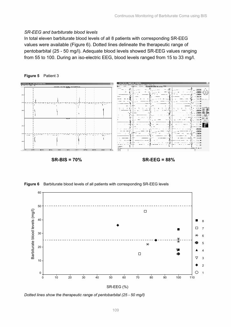
Continuous Monitoring of Barbiturate Coma using BIS
109
SR-EEG and barbiturate blood levels In total eleven barbiturate blood levels of all 8 patients with corresponding SR-EEG values were available (Figure 6). Dotted lines delineate the therapeutic range of pentobarbital (25 - 50 mg/l). Adequate blood levels showed SR-EEG values ranging from 55 to 100. During an iso-electric EEG, blood levels ranged from 15 to 33 mg/l. Figure 5 Patient 3
Figure 6 Barbiturate blood levels of all patients with corresponding SR-EEG levels
SR-EEG (%)
1101009080706050403020100
Bar
bitu
rate
blo
od le
vels
(mg/
l)
60
50
40
30
20
10
0
8
7
6
5
4
3
2
1
Dotted lines show the therapeutic range of pentobarbital (25 - 50 mg/l)
SR-BIS = 70% SR-EEG = 88%
Chapter 8
110
Discussion The aim of this study was to evaluate the usefulness of the BIS monitor during a barbiturate coma in PICU patients, as proposed by Arbour and Jaggi et al.8,13 We found application of the BIS monitor as a continuous monitor of the burst suppression pattern to be promising. Its continuously displayed real time raw EEG traces correlated well with the full channel EEG, both at bedside and at comparison between the EEG of the BIS and the full channel EEG afterwards. Barbiturate blood levels within the normal range corresponded with SR-EEG values ranging from 55 to 100. Children with an iso-electric EEG, could have barbiturate blood levels ranging from 15 to 33, showing these children’s individual susceptibility to barbiturates.
However, correlations between SR-BIS and SR-EEG were found to be small to moderate for four patients. Several explanations present themselves. One is the presence of an asymmetrical EEG in a patient who had suffered a TBI resulting in intra-cranial hemorrhage at the left side of the head (patient 4). Another explanation can be short bursts (less than 1 second); with the algorithm of the BIS monitor simply over-estimating the length of the burst and therefore under-estimating the SR-BIS (patient 3). This underestimation might be caused by the EEG signal’s slow return to baseline after a high amplitude burst, which is a characteristic of a high pass filter. Also, lack of time synchronization in four patients may also have under-estimated correlations between SR-BIS and SR-EEG.
Our study has several limitations. Firstly, group size is small due to the rare occurrence of barbiturate induced coma. However, we managed to include most eligible patients presenting to our unit. As our hospital serves as a level three PICU and regional trauma center (1100 admissions a year, reference area 4.106 inhabitants), not many units will admit more patients requiring a barbiturate coma. Recently, van Gestel et al. described 33 patients with GCSE over a period of 11 years, admitted to another large teaching hospital in the Netherlands.9 They reviewed the treatments of all patients and suggest the use of propofol for treatment of GCSE before thiopental. Secondly, we did not monitor EEGs continuously, due to organizational limitations. Thirdly, of only four patients raw BIS EEG data were collected using a laptop with special software. Burst suppression on the real-time EEG trace of the BIS monitor did, however, correspond to the full channel EEG at bedside. Therefore, the comparison of real-time EEG traces of both the BIS monitor and the standard EEG in these four patients reflects the way in which the BIS monitor can be used in daily practice.
In short, clinical evaluation of a pentobarbital coma is difficult; barbiturate blood levels may not be reliable5 and continuous full channel EEG monitoring is not possible in our setting, leaving monitoring of a barbiturate coma with the BIS monitor as a possibility.
The BIS monitor has the potential of continuous monitoring of brain function. It is relatively easy to use, and nurses and physicians can easily be taught how to interpret recordings. SR-BIS and EEG traces recorded by the BIS monitor are continuously displayed, thus enabling continuous monitoring of cerebral function. We feel that the burst suppression pattern needs to be evaluated using a full channel EEG, combined with BIS monitoring on an individual basis. If the optimal SR-BIS values and EEG trace displayed on the BIS monitor are comparable with the full channel EEG and remain stable, a full channel
Continuous Monitoring of Barbiturate Coma using BIS
111
EEG once a day can be informative. A new EEG must be made upon changes in EEG pattern of the BIS or the SR-BIS values, or upon changes in clinical situation or medication. Children showing asymmetrical EEGs and children showing short bursts (< 1 sec) need to be evaluated per case. For objective evaluation of the safety and efficacy of barbiturate induced comas in children we recommend pharmacokinetic and pharmacodynamic studies of barbiturates, with continuous EEG and BIS monitoring.

Chapter 8
112
References 1. Kwan P, Brodie MJ. Phenobarbital for the Treatment of Epilepsy in the 21st Century: A Critical
Review. Epilepsia 2004;45(9):1141-1149
2. Yanay O, Brogan TV, Martin LD. Continuous pentobarbital infusion in children is associated with high rates of complications. J Crit Care 2004;19(3):174-178
3. Finfer SR, Cohen J. Severe traumatic brain injury. Resuscitation 2001;48(1):77-90
4. Adelson PD, Bratton SL, Carney NA, Chesnut RM, du Coudray HE, Goldstein B, et al. Guidelines for the acute medical management of severe traumatic brain injury in infants, children, and adolescents. Chapter 13. The use of barbiturates in the control of intracranial hypertension in severe pediatric traumatic brain injury. Pediatr Crit Care Med 2003;4(3 Suppl):S49-52
5. Winer JW, Rosenwasser RH, Jimenez F. Electroencephalographic activity and serum and cerebrospinal fluid pentobarbital levels in determining the therapeutic end point during barbiturate coma. Neurosurgery 1991;29(5):739-741; discussion 741-732
6. Tasker RC, Boyd SG, Harden A, Matthew DJ. The cerebral function analysing monitor in paediatric medical intensive care: applications and limitations. Arch Dis Child 1999;81(1):90-95
7. Grindstaff RJ, Tobias JD. Applications of bispectral index monitoring in the pediatric intensive care unit. J Intensive Care Med 2004;19(2):111-116
8. Arbour R. Continuous nervous system monitoring, EEG, the bispectral index, and neuromuscular transmission. AACN Clin Issues 2003;14(2):185-207
9. Van Gestel JPJ, Blusse van Oud-Alblas HJ, Malingre M, Ververs FFT, Braun KPJ, van Nieuwenhuizen O. Propofol and thiopental for refractory status epilepticus in children. Neurology 2005;65:591-592
10. Mazzola CA, Adelson PD. Critical care management of head trauma in children. Crit Care Med 2002;30(11 (suppl)):S393-S401
11. Bland JM, Altman DG. Statistical methods for assessing agreement between two methods of clinical measurement. Lancet 1986;1(8476):307-310
12. Hinkle DE, Wiersma W, Jurs SG. Applied Statistics for the Behavioral Sciences: Houghton Mifflin Co., 1998
13. Jaggi P, Schwabe MJ, Gill K, Horowitz IN, Cremer OL, Moons KG, et al. Use of an anesthesia cerebral monitor bispectral index to assess burst-suppression in pentobarbital coma. Pediatr Neurol 2003;28(3):219-222
The AEP Monitor in Infants: First Data Outside the Operation Room
Sandra A. Prins, Heleen J. Blussé van Oud-Alblas, Monique van Dijk, Dick Tibboel.
Cha
pter
Chapter 9
114
Abstract The A-line ARX index (AAI) derived from the AEP monitor/2 is a measure of the hypnotic component of anesthesia. This study was designed to test the feasibility and beginning validity of the AEP monitor to assess level of sedation in infants at the PICU. Twice an hour, infants were observed for 2 minutes, during which AAI, mean arterial pressure, heart frequency, visual analogue scale, sound level and COMFORT behavior score were noted. Of the 8 included patients, median age was 40 days (range 1 to 795 days). Overall correlation between COMFORT behavior and AAI values was 0.48. Inter-individual correlation ranged from –0.29 to 0.95 . The results were artifact free in 49% of all observations. Mean intensity of environmental noise was 54 dB. The intensity of the auditory stimulus was 75 dB in 80% of the measurements. These delivered combined with environmental sound levels of the AEP monitor/2 are too high for a PICU. Also, the correlation between AAI values and COMFORT behavior scores was only moderate. These limitations of the AEP monitor/2 will have to be overcome before daily use in the PICU is an option.
The AEP Monitor in Infants
115
Introduction Infants admitted to a pediatric intensive care unit (PICU) often receive sedatives and analgesics to facilitate mechanical ventilation, provide comfort, and reduce anxiety. Monitoring of sedation levels is essential, as both under and over-sedation may have negative effects. Inadequate sedation and/or analgesia in neonates and infants may result in tachycardia, hypertension and failure of ventilator-patient synchrony leading to hypoxia.1,2 Over-sedation may lead to cardiovascular depression, prolonged duration of mechanical ventilation, ventilator associated pneumonia or lung injury, hypotension, immune-suppression and withdrawal symptoms on cessation.3,4 Levels of sedation and analgesia in pre-verbal children may be assessed using observational tools and brain monitors.3 Three relevant tools have been validated for children under the age of 3 three years, the COMFORT behavior scale, the Hartwig sedation scale and the University of Michigan Sedation Scale are validated, as reviewed by Ista et al.5 Recently, two fundamentally different types of brain monitors have found their way from the operation room to the intensive care unit. These allow for more objective assessment of sedation levels. Both use algorithms to simplify the electroencephalogram (EEG) and provide the clinician with a slightly delayed, real-time numerical index from 0 - 100. Best studied is the Bispectral index monitor (BIS™), which was validated in adults6 and prevents awareness during general anesthesia.7
The Auditory Evoked Potential monitor (AEP monitor/2) uses middle latency auditory evoked potentials (MLAEPs) to test the patient's brain ability to respond to an auditory signal. MLAEPs represent the earliest cortical response to an acoustic stimulus. Amplitudes and latencies are influenced by both anesthetics and surgical stimuli and are therefore believed to be useful for measuring level of anesthesia. A monitoring variable indicating the patient's hypnotic state, the so-called A-line ARX index (AAI) which ranges from 0 (iso-electric EEG) to 100 (awake), is calculated from the MLAEPs and the EEG.8 The AEP monitor/2 has been studied in adults during anesthesia and at the ICU. During anesthesia, use of the AEP monitor/2 improved emergence from anesthesia, spared the use of anesthetics and lead to detection of intra-operative awareness.9-11 At the adult ICU, the AEP monitor/2 correlated well with clinical sedation scales, such as the Ramsay sedation scale.12,13 Three studies in children during anesthesia showed that the AAI is more valuable in predicting anesthetic states than hemodynamic variables and reliably differentiates between the awake and anesthetized states.14-16 Data from children and infants outside the operation room are lacking so far.
We report a pilot study conducted to evaluate feasibility and validity of the AEP monitor/2 in postoperative infants admitted to our pediatric surgical ICU (PSICU).

Chapter 9
116
Methods Patients and setting Those eligible for this study were postoperative patients aged 0 to 3 years admitted to the PSICU of the Erasmus MC – Sophia Children's Hospital and receiving sedatives and/or analgesics. Exclusion criteria were head trauma (which interferes with application of the electrodes), hearing abnormalities, mental retardation and treatment with neuromuscular blocking agents. Hearing abnormalities were detected with the ALGO hearing screening for newborns, based on an automated auditory brain stem electric response (ABR).17 Choices of sedation and /or analgesic regimens were made at the discretion of the attending physician, independent of the AAI. The Erasmus MC research ethics board approved the study, and parental informed consent was sought. Auditory evoked potential recording, analysis and data collection MLAEPs were recorded using the AEP monitor/2 (Danmeter A/S, Odense, Denmark; software version 1.6). The infant's skin was cleansed with water and soap, and disposable electrodes were positioned at the mid forehead (+), left forehead (reference) and left mastoid (-). Impedance of the electrodes was tested. Especially designed earphones for children were checked for the audible signal to be heard and put in place. The MLAEP analysis window was 20 to 80 ms. AAI values, which theoretically range from 99 (fully awake) to 0 (very deep hypnosis), were then calculated from the MLAEPs and EEGs. The target AAI range during anesthesia in children, is between 30 ± 5.18 AEP monitor/2 data were transferred to a personal computer and subsequently analyzed using the AAI graph software package (version 2.0, Danmeter A/S, Odense, Denmark). Measurements Every thirty minutes during a maximum of eight hours, the children were observed for 2 minutes. During observation, heart frequency (HF), mean arterial blood pressure (MABP), AAI, and environmental sound level in decibels (dB) were noted every 20 seconds. Finally, the observer assessed, muscle tone, COMFORT behavior score and visual analogue scale (VAS) score at the end of each observation. COMFORT behavior scale The COMFORT behavior scale has been validated for both postoperative pain assessment in children under age 3 years and for sedation assessment on the PICU.1,19,20 After 2 minutes' observation, the observer rates six behavioral items (alertness, calmness, respiratory response/crying, physical movement, muscle tone and facial tension) on a scale from 1 to 5. Total score thus varies from 6 to 30. Inter-rater reliability of the one observer in this study was found to be satisfactory (linearly weighted Kappa 0.65). Visual Analogue Scale (VAS) The VAS is a 10-cm continuous line with the anchors 'no pain' on the left side and 'extreme pain 'on the right side. The observer places a mark on this line between these extremes,
The AEP Monitor in Infants
117
representing his or her expert opinion of the child's pain.21 A score > 4 is considered to represent moderate to severe pain.19,21 In our study, VAS scores were used for research purposes only and had no consequences for the patient's medication. Digital sound level meter A digital sound level meter was used (Velleman®, type dvm1326) to determine environmental noise level. Sound pressure levels are measured in decibels(dB), which is a logarithmic scale; a 6-dB increase correlates to a doubling in perceived loudness. The sound level meter was positioned close to the patient in bed. Background noise levels were noted simultaneously with the parameters mentioned above. The US Environmental Protection agency has proposed hospital sound levels of maximum 45 dB during the day and maximum 35 at night.22 Statistical analysis Data from the monitor were converted to a text file, using the Graph 2.0 software package. SPSS for Windows (version 10.1) was used for statistical analysis. Associations between COMFORT behavior scores and AAI values were determined by Spearman's rho correlation coefficient. Results Patients Parental consent was obtained for only 10 of 18 eligible infants. Parents refused participation on the grounds of: parental emotional distress, aversion to placement of electrodes at the child's head and belief that the noise might be harmful to the child. Data from 2 of these 10 children could not be used because the children were diagnosed with hypacusis after the study. Characteristics of the remaining 8 patients are listed in Table 1. Their median age was 40 days (1 to 795 days). Four patients received only morphine, with a median dose of 10 mcg/kg/h (range 5 to 21 mcg/kg/h). One patient received midazolam, with a maximum dose of 0.25 mg/kg/h. Three patients received intravenous midazolam with a median dose of 0.2 mg/kg/h (range 0.1 to 0.5 mg/kg/h) for sedation together with intravenous morphine with a median dose of 15 mcg/kg/h (range 10 to 16 mcg/kg/h) for analgesia. Three patients received additional medication (see Table 1). A total of 1438 data were recorded. Signal quality AEP monitor Signal quality was artifact free in 49% of recordings. The median signal quality was 99 (range 30 to 100). In 2 children, signal quality was occasionally < 50.
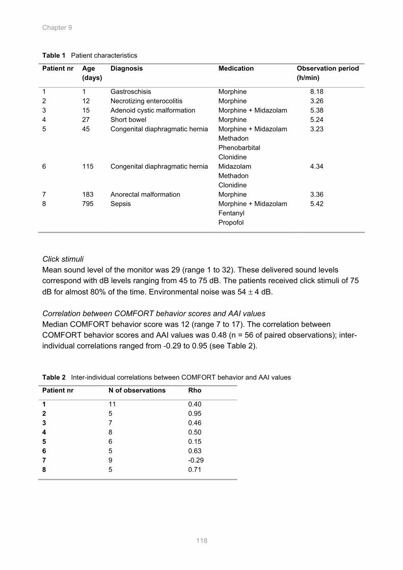
Chapter 9
118
Table 1 Patient characteristics
Click stimuli Mean sound level of the monitor was 29 (range 1 to 32). These delivered sound levels correspond with dB levels ranging from 45 to 75 dB. The patients received click stimuli of 75 dB for almost 80% of the time. Environmental noise was 54 ± 4 dB. Correlation between COMFORT behavior scores and AAI values Median COMFORT behavior score was 12 (range 7 to 17). The correlation between COMFORT behavior scores and AAI values was 0.48 (n = 56 of paired observations); inter-individual correlations ranged from -0.29 to 0.95 (see Table 2). Table 2 Inter-individual correlations between COMFORT behavior and AAI values
Patient nr N of observations Rho
1 11 0.40 2 5 0.95 3 7 0.46 4 8 0.50 5 6 0.15 6 5 0.63 7 9 -0.29 8 5 0.71
Patient nr Age (days)
Diagnosis Medication Observation period (h/min)
1 1 Gastroschisis Morphine 8.18 2 12 Necrotizing enterocolitis Morphine 3.26 3 15 Adenoid cystic malformation Morphine + Midazolam 5.38 4 27 Short bowel Morphine 5.24 5 45 Congenital diaphragmatic hernia Morphine + Midazolam
Methadon Phenobarbital Clonidine
3.23
6 115 Congenital diaphragmatic hernia Midazolam Methadon Clonidine
4.34
7 183 Anorectal malformation Morphine 3.36 8 795 Sepsis Morphine + Midazolam
Fentanyl Propofol
5.42
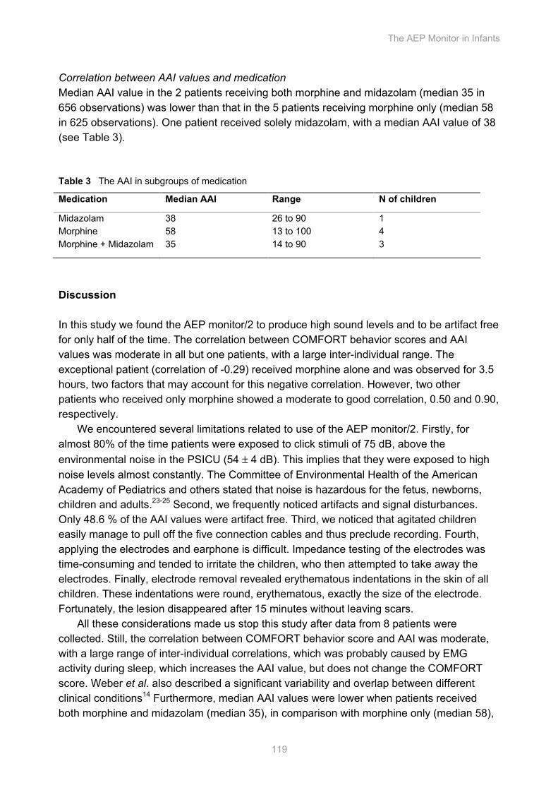
The AEP Monitor in Infants
119
Correlation between AAI values and medication Median AAI value in the 2 patients receiving both morphine and midazolam (median 35 in 656 observations) was lower than that in the 5 patients receiving morphine only (median 58 in 625 observations). One patient received solely midazolam, with a median AAI value of 38 (see Table 3). Table 3 The AAI in subgroups of medication
Medication Median AAI Range N of children
Midazolam 38 26 to 90 1 Morphine 58 13 to 100 4 Morphine + Midazolam 35 14 to 90 3
Discussion In this study we found the AEP monitor/2 to produce high sound levels and to be artifact free for only half of the time. The correlation between COMFORT behavior scores and AAI values was moderate in all but one patients, with a large inter-individual range. The exceptional patient (correlation of -0.29) received morphine alone and was observed for 3.5 hours, two factors that may account for this negative correlation. However, two other patients who received only morphine showed a moderate to good correlation, 0.50 and 0.90, respectively.
We encountered several limitations related to use of the AEP monitor/2. Firstly, for almost 80% of the time patients were exposed to click stimuli of 75 dB, above the environmental noise in the PSICU (54 ± 4 dB). This implies that they were exposed to high noise levels almost constantly. The Committee of Environmental Health of the American Academy of Pediatrics and others stated that noise is hazardous for the fetus, newborns, children and adults.23-25 Second, we frequently noticed artifacts and signal disturbances. Only 48.6 % of the AAI values were artifact free. Third, we noticed that agitated children easily manage to pull off the five connection cables and thus preclude recording. Fourth, applying the electrodes and earphone is difficult. Impedance testing of the electrodes was time-consuming and tended to irritate the children, who then attempted to take away the electrodes. Finally, electrode removal revealed erythematous indentations in the skin of all children. These indentations were round, erythematous, exactly the size of the electrode. Fortunately, the lesion disappeared after 15 minutes without leaving scars.
All these considerations made us stop this study after data from 8 patients were collected. Still, the correlation between COMFORT behavior score and AAI was moderate, with a large range of inter-individual correlations, which was probably caused by EMG activity during sleep, which increases the AAI value, but does not change the COMFORT score. Weber et al. also described a significant variability and overlap between different clinical conditions14 Furthermore, median AAI values were lower when patients received both morphine and midazolam (median 35), in comparison with morphine only (median 58),
Chapter 9
120
suggesting that the AEP monitor/2 detects the effects of midazolam. Unfortunately, only one patient received midazolam alone, so we can not prove this assumption.
In conclusion, although the AEP monitor/2 yields objective data and requires no verbal interaction or physical stimulus to the patient, it delivers sound levels that are too high for a PICU. Furthermore, this study showed only a moderate correlation between AAI values and COMFORT behavior scores. The AEP monitor/2 has several limitations which need to be overcome before daily use in the PICU is an option.
Acknowledgements The authors would like to thank Professor Jan Klein for his careful review of the manuscript, Elsbeth van den Berg, medical student, for her assistance in data collection and Ko Hagoort for editing.
The AEP Monitor in Infants
121
References 1. Marx CM, Smith PG, Lowrie LH et al. Optimal sedation of mechanically ventilated pediatric critical
care patients. Crit Care Med 1994;22:163-70
2. Anand KJ, Barton BA, McIntosh N et al. Analgesia and sedation in preterm neonates who require ventilatory support: results from the NOPAIN trial. Neonatal Outcome and Prolonged Analgesia in Neonates. Arch Pediatr Adolesc Med 1999;153:331-8
3. De Jonghe B, Cook D, Appere-De-Vecchi C et al. Using and understanding sedation scoring systems: a systematic review. Intensive Care Med 2000;26:275-85
4. Detriche O, Berre J, Massaut J, Vincent JL. The Brussels sedation scale: use of a simple clinical sedation scale can avoid excessive sedation in patients undergoing mechanical ventilation in the intensive care unit Br J Anaesth, 1999:698-701
5. Ista E, Van Dijk, M, Tibboel, D, De Hoog, M. Assessment of sedation levels in pediatric intensive care patients can be improved by using the COMFORT "behavior" scale. Pediatr Crit Care Med 2005;6:58-63
6. Glass PS, Bloom M, Kearse L et al. Bispectral analysis measures sedation and memory effects of propofol, midazolam, isoflurane, and alfentanil in healthy volunteers. Anesthesiology 1997;86:836-47
7. Myles PS, Leslie K, McNeil J et al. Bispectral index monitoring to prevent awareness during anaesthesia: the B-Aware randomised controlled trial. Lancet 2004;363:1757-63
8. Gajraj RJ, Doi M, Mantzaridis H, Kenny GN. Comparison of bispectral EEG analysis and auditory evoked potentials for monitoring depth of anaesthesia during propofol anaesthesia. Br J Anaesth 1999;82:672-8
9. Recart A, White PF, Wang A et al. Effect of auditory evoked potential index monitoring on anesthetic drug requirements and recovery profile after laparoscopic surgery: a clinical utility study. Anesthesiology 2003;99:813-8
10. Haenggi M, Ypparila H, Takala J et al. Measuring Depth of Sedation with Auditory Evoked Potentials During Controlled Infusion of Propofol and Remifentanil in Healthy Volunteers. Anest Analg 2004;99:1728-36
11. Trillo-Urrutia L, Fernandez-Galinski S, Castano-Santa J. Awareness detected by auditory evoked potential monitoring. Br J Anaesth 2003;91:290-2
12. Schulte-Tamburen AM, Scheier J, Briegel J et al. Comparison of five sedation scoring systems by means of auditory evoked potentials. Intensive Care Med 1999;25:377-82
13. Rundshagen I, Schnabel K, Pothmann W et al. Cortical arousal in critically ill patients: an evoked response study. Intensive Care Med 2000;26:1312-8
14. Weber F, Seidl M, Bein T. Impact of the AEP-Monitor/2-derived composite auditory-evoked potential index on propofol consumption and emergence times during total intravenous anaesthesia with propofol and remifentanil in children. Acta Anaesthesiol Scand 2005;49:277-83
15. O'Kelly SW, Smith DC, Pilkington SN. The auditory evoked potential and paediatric anaesthesia. Br J Anaesth 1995;75:428-30
16. Weber F, Bein T, Hobbhahn J, Taeger K. Evaluation of the Alaris Auditory Evoked Potential Index as an Indicator of Anesthetic Depth in Preschool Children during Induction of Anesthesia with Sevoflurane and Remifentanil. Anesthesiology 2004;101:294-8
Chapter 9
122
17. van Straaten HL, Groote ME, Oudesluys-Murphy AM. Evaluation of an automated auditory brainstem response infant hearing screening method in at risk neonates. Eur J Pediatr 1996;155:702-5
18. Weber F, Bein, T., Hobbhahn J., Taeger K. Evaluation of the Alaris Auditory Evoked Potential Index as an Indicator of Anesthetic Depth in Preschool Children during Induction of Anesthesia with Sevoflurane and Remifentanil. Anesthesiology 2004;101:294-8
19. van Dijk M, de Boer JB, Koot HM et al. The reliability and validity of the COMFORT scale as a postoperative pain instrument in 0 to 3-year-old infants. Pain 2000;84:367-77
20. Ista E, van Dijk M, Tibboel D, de Hoog M. Assessment of sedation levels in pediatric intensive care patients can be improved by using the COMFORT "behavior" scale. Pediatr Crit Care Med 2005;6:58-63
21. Lawrence J, Alcock D, McGrath P et al. The development of a tool to assess neonatal pain. Neonatal Netw 1993;12:59-66
22. Environmental Protection Agency. Information on levels of environmental noise requisite to protect public health and welfare with an adequate margin of safety. Washington DC: Government Printing Office, 1974
23. Anonymous. Noise: A Hazard for the Fetus and Newborn. Committee on Environmental Health. Pediatrics 1997;100:724-7
24. Milette IH, Carnevale FA. I'm trying to heal...noise levels in a pediatric intensive care unit. Dynamics 2003;14:14-21
25. Morrison WE. Noise, stress, and annoyance in a pediatric intensive care unit. Crit Care Med 2003;31:113-9
General Discussion
Accepted in adapted form, as editorial for Intensive Care Medicine
Cha
pter
Chapter 10
124
Introduction Several factors account for the level of anxiety and fear experienced by children during their stay at the pediatric intensive care unit (PICU) -pain, separation from parents, invasive procedures, mechanical ventilation, disruption of the usual sleep-wake cycle, noise of unknown origin and the presence of unfamiliar people and machines. Although reassurance and parental presence may relieve part of the anxiety, pharmacological intervention is required in many cases. Over the years, several agents have been used for sedation in combination with analgesia.
In this chapter, frequently used sedatives and analgesics in the PICU will be discussed. Besides a short description of the working profiles and adverse effects, the availability of randomized trials in adults and children are highlighted and results of the medication trials in this thesis are described. Furthermore, behavioral assessment tools and electroencephalogram (EEG)- based monitors to assess sedation will be discussed, as well as the drawbacks of the new techniques. Lastly, possible unresponsiveness to sedatives or analgesics and the development of opioid-and benzodiazepine withdrawal after a long PICU stay are discussed with suggestions for future research. Sedatives Midazolam The benzodiazepines are a class of drugs with hypnotic, anxiolytic, anticonvulsant, amnestic and muscle relaxant properties. Benzodiazepines are often used for short-term relief of severe, disabling anxiety or insomnia. They are believed to act on the gamma amino butyric acid (GABA) receptor, the activation of which dampens higher neuronal activity.
Benzodiazepines are commonly classified into three categories. Short-acting compounds that act for less than six hours, intermediate-acting compounds have an effect for 6-10 hours and long-acting compounds have strong sedative effects that may persist for more than 24 hours.1 Midazolam is a short acting sedative2, provides anterograde amnesia3 and is the most frequently used sedative in pediatric intensive care nowadays.4-7 However, paradoxical reactions such as agitation,8 convulsions, hyperactivity or adverse reactions9 have been reported in neonates and children.10 Also, active metabolites and prolonged effects of midazolam often delay waking up and weaning from mechanical ventilation.11,12 In premature infants even convulsions have been documented following the use of midazolam. Also, long term use can be problematic due to the development of tolerance and dependency.
Several studies have evaluated the pharmacokinetic and pharmacodynamic (PK/PD) profile of intravenous and oral midazolam in preterm infants and children. These studies included heterogeneous patient populations and did not describe profiles of term infants and young children.13-17
In Chapter 2, the PK/PD responses are described for midazolam in children under the age of 2 years. Based on the population PK/PD model we advise a loading dose of 1 mg,
General Discussion
125
followed by a continuous infusion of 0.5 mg/kg for infants of 10 kg to achieve COMFORT-behavior scores from 12 to 14. Our study also showed a large variability of clearance and midazolam plasma concentration at half maximum effect, which makes individual titration of midazolam still very important. Propofol Propofol is an ultra short-acting intravenous anesthetic agent, which is also a popular sedative for mechanically ventilated adults. Upon disturbing reports of adverse events after use of propofol as a sedative in children, the Food and Drug Administration (FDA) issued a warning against the use of propofol as a sedative in children under the age of 18 years in pediatric intensive care.18 However, we did not encounter any problems using propofol 6% as a sedative in children with a median age of 10 months (IQR 3 to 17 months), with dosages < 4 mg/kg/h, during a median period of 11 hours (range 6 to 18 hours) (Chapter 3). Furthermore, based on the population PK model of propofol 6% (Chapter 4) we found the propofol clearance to be two times higher in non-ventilated postoperative children than that reported in the literature for ventilated children and adults. We therefore advise a propofol dose of 30 mg/h in a 10 kg infant to achieve COMFORT behavior scores from 12 to 14 and BIS values from 70 to 75 during the night. Based on these studies, it is too early to say that propofol can be used safely for more than 12 hours, however, based on the literature review shown in Chapter 3, the warning of the Food and Drug Administration against the use of propofol may have been presumptuous. Propofol can be used safely during procedures in children.19,20 Larger studies, including more patients in different age groups are needed to determine the place of propofol as a sedative at the pediatric intensive care unit and also to alter the warning of the FDA18 (Chapter 4). Ketamine-s Ketamine-s is a non-competitive glutamate inhibitor at the N-methyl-D-aspartate glutamate (NMDA) receptors. Glutamate is the major excitatory transmitter in the central nervous system (CNS), and inhibition of this receptor decreases neuronal activity, which results in a state of anesthesia. In low doses, ketamine-s causes analgesia and sedation; in high doses general anesthesia. Ketamine-s has a low incidence of complications, can be administered through any route and is pharmacologically very predictable, which make it a popular agent for pediatric procedural sedation.21,22 Recovery time depends on the dose and emergence can be complicated by hallucinations or vivid dreams. Ketamine-s can elevate intracranial and intra-ocular pressure and is contraindicated in patients with increased intracranial pressure or glaucoma.23 Recently, Lin et al. reviewed the available literature and stated that ketamine-s is an excellent choice for procedural sedation and analgesia.21 Two studies compared the analgesic efficacy of ketamine-s with morphine and found similar postoperative analgesia in children aged 6 to 15 years24 and aged 1 to 16 years, respectively.25 Large, randomized controlled trials concerning the safety and efficacy of ketamine-s as a long term sedative are lacking.
Chapter 10
126
Barbiturates Barbiturates act by increasing inhibition in the CNS through enhancing the action of gamma amino butyric acid (GABA), the primary inhibitory neurotransmitter in the CNS. Drugs that stimulate the production GABA produce slow down brain activity and induce a drowsy or calm feeling, thus producing a wide spectrum from mild sedation to general anesthesia. Pentobarbital is a short acting barbiturate, which is used for procedural sedation26 and to treat generalized convulsive status epilepticus with a burst suppression pattern.27,28 Also, pentobarbital infusion has been used for long term sedation. In 2004, Yanay et al. concluded after a retrospective chart review study that continuous pentobarbital infusion was an effective sedative, when other drugs fail. However, they also observed a high rate of clinically significant complications requiring discontinuation of the drug.29 Recently, van Gestel et al. reviewed the complications of propofol and thiopental for the treatment of refractory status epilepticus in children.30 Over 11 years, 33 children were treated with either propofol or thiopental. Propofol proved to be effective, with infrequent side effects and therefore the authors suggested the use of propofol before thiopental. Randomized controlled trials regarding PK/PD, safety and efficacy of barbiturates used as sedative in children are lacking. As barbiturates have a narrow therapeutic window between sedation, coma and death, they should only be considered if other sedative drugs fail and in strictly selected patient populations.11,29,31 Clonidine Clonidine is a lipid-soluble, partial alfa-2 adrenoreceptor agonist with anti-hypertensive, analgesic and sedative properties. Data of the last 5 years regarding the safety and efficacy of clonidine as a sedative in the PICU are limited to two studies: one concerning oral clonidine for sedation and one using intravenous clonidine.32,33 Both were cohort studies and found clonidine to be a safe and effective sedative in combination with an opioid and benzodiazepine in young children. Clonidine has also the advantage of decreasing requirements of sedatives and facilitating opioid withdrawal symptoms.34-37 Clonidine causes a rapid and significant decrease in opiate withdrawal signs and symptoms in patients addicted to methadone; this effect was later confirmed for other synthetic opioids and heroin.35 Clonidine can also be given orally. Arenas-Lopez et al. included 24 infants, median age of 3 months, receiving oral clonidine for sedation at the PICU and demonstrated a benzodiazepine and opioid sparing effect.33 Withdrawal of clonidine has been associated with hypertension and seizures and abrupt discontinuation should be avoided. Although clonidine is increasingly used,32 safe and effective use of clonidine in children has not been adequately established; there are no clear guidelines for clonidine use in pediatric patients. Chloral hydrate Chloral hydrate is an enteral sedative that is rapidly absorbed from the gastro-intestinal tract and starts to act within 15 to 60 minutes. Duration of action varies from 60 to 120 minutes, depending on the presence of renal or hepatic disease. Only 10 years ago, chloral hydrate was one of the most frequent employed sedative, next to benzodiazepines, in the United States and Canada as reported by Marx et al.38 Nowadays, chloral hydrate is less popular
General Discussion
127
due to at least two reports describing the limitations of chloral hydrate; i.e. cardiac dysrhythmias, airway obstruction and an individual response which may be variable and unpredictable. Furthermore, the effect of choral hydrate is irreversible, making an extended observation period required.4,39,40 Therefore, chloral hydrate should not be used as a first line sedative. If used, the patient’s respiratory status must be monitored continuously. Trimeprazine Trimeprazine (Alimemazine; Nedeltran) is another enteral sedative. Originally, Trimepazine is an antihistamine with well-known sedative effects. Trimeprazine is a phenothiazine derivative and has anti-emetic and sedative properties and is used to alter the sleep pattern of children or treat children with sleep disturbances.41,42 Adverse events after administration of trimeprazine include bradycardia, hypotension, QTc prolongation but also excitement, agitation and even hallucinations and convulsions. Only one study described the pharmacokinetics of trimeprazine in 1990.43 Therefore, in the absence of safety and efficacy data of trimeprazine for children and the possibility of adverse events, other sedatives, for instance midazolam or propofol, would be more suitable for short term sedation of children and infants under the age of 2 years. Conclusion For short term infusions, less than 24 hours, midazolam, and propofol are drugs of first choice. Second line drugs are clonidine, ketamine-s and barbiturates, in that particular order. The use of enteral sedatives like chloral hydrate and trimeprazine can not be recommended based on the available literature. Analgesics If pain occurs, analgesic drugs should be administered in the following order (according to the WHO painladder:44 non-opioids, like paracetamol, then, if necessary, mild opioids like codeine or anti-inflammatory drugs (NSAIDs) such as ketorolac, then strong opioids such as morphine until the patient is free of pain. Paracetamol and intravenous propacetamol Paracetamol is an effective and safe analgesic drug, which relieves mild to moderate pain in children. Dose-effect relationships, dose-concentration relationships, and the antipyretic effects of paracetamol in children and in premature neonates have been reported in the literature.45-49 Anderson et al. described developmental PK of premature neonates and infants, following administration of oral and rectal paracetamol and found target concentrations of > 10 mg/l after 25 mg/kg/day in premature neonates at 30 weeks’ postconception, 45 mg/kg/day at 34 weeks’ gestation, 60 mg/kg/day at term, and 90 mg/kg/day at 6 months of age. Similar concentrations can be achieved with maintenance rectal doses of 25 (capsule suppository) or 30 (triglyceride suppository) mg/kg/day in premature neonates at 30 weeks’ gestation, increasing to 90 (capsule suppository) or 120
Chapter 10
128
(triglyceride suppository) mg/kg/day at 6 months. These regimens may cause hepatotoxicity in some individuals if used for longer than 2 - 3 days. In children aged 2 to 15 years undergoing tonsillectomy, a paracetamol plasma concentration of 10 mg/l is considered necessary to obtain adequate pain relief.45 The optimal plasma concentration to obtain analgesia in children with a mean age of 10 months was found to be < 10 to 20 mg/l.48 The delayed and erratic absorption after rectal administration leads to unpredictable paracetamol plasma concentrations and does not consistently produce a rapid onset of pain relief.
Prodafalgan and Perfalgan, i.e. intravenous (i.v.) propacetamol and i.v. paracetamol respectively, are of interest, because they might achieve more rapidly target concentrations and improve prediction of concentration as compared to enteral formulations. In other Europe countries, like Belgium and France, i.v. propacetamol and paracetamol have been used for more than 10 years and was found to be safe and effective in children.50-55
In Chapter 5 we report of a double-blind placebo controlled randomized trial designed to characterize the PK/PD profile of i.v. propacetamol and compare this with that of rectal paracetamol in children after major craniofacial surgery. Twenty-six children (6 months to 2 years) were given a paracetamol suppository of 40 mg/kg during surgery and then assigned to treatment groups of either i.v. propacetamol 40 mg/kg infusion over 15 min or 20 mg/kg paracetamol rectally every 6 hours. Placebo suppository and placebo intravenous solution were used to blind the investigator and nurses. The visual analogue scale (VAS) (score 0 to 10) and COMFORT behavior scale (score 6 to 30) were used as endpoints. PK/PD modeling was performed using NONMEM. During the 24-hours study period 12 patients were assigned a VAS score less than 4, but received midazolam for COMFORT behavior scores exceeding 17; three of them were in the i.v. propacetamol group and 9 of them in the rectal paracetamol group (P = 0.05). One child from the i.v. propacetamol treatment group was given rescue rectal paracetamol 20 mg/kg for a VAS pain score > 4. In conclusion, i.v. propacetamol proved to be more effective than rectal paracetamol in children under 2 years of age. There was a significant difference between use of midazolam between the two groups, indicating that these children experienced more distress, possibly caused by pain. Nonsteroidal anti-inflammatory drugs (NSAIDs) Nonsteroidal anti-inflammatory drugs (NSAIDs) are mild analgesics that inhibit prostaglandin synthesis. Prostaglandin’s appear to be involved in the smooth muscle contraction seen in renal and biliary colic, conditions in which these agents are particularly effective. In addition to displaying anti-inflammatory activity, NSAIDs are antipyretic and inhibit platelet aggregation; they do not cause the sedation, respiratory depression, and hypotension that are common with opioid analgesics. Major side effects are platelet dysfunction, renal dysfunction, and gastrointestinal ulceration or irritation. NSAIDs are limited by the lack of intravenous formulations; however, ketorolac tromethamine (Toradol) may be administered intramuscularly or intravenously. In 2002, Dsida et al. showed that the pharmacokinetic variables of ketorolac tromethamine did not differ among pediatric patients <17 yr old and were similar to adult values.56 In 2004, van der Marel et al. studied 26 infants with a mean age of 4.5 years (SD 1.5 years) undergoing tonsillectomy who received another NSAID,
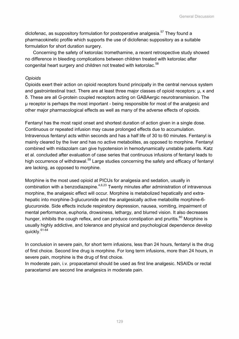
General Discussion
129
diclofenac, as suppository formulation for postoperative analgesia.57 They found a pharmacokinetic profile which supports the use of diclofenac suppository as a suitable formulation for short duration surgery.
Concerning the safety of ketorolac tromethamine, a recent retrospective study showed no difference in bleeding complications between children treated with ketorolac after congenital heart surgery and children not treated with ketorolac.58 Opioids Opioids exert their action on opioid receptors found principally in the central nervous system and gastrointestinal tract. There are at least three major classes of opioid receptors: µ, κ and δ. These are all G-protein coupled receptors acting on GABAergic neurotransmission. The µ receptor is perhaps the most important - being responsible for most of the analgesic and other major pharmacological effects as well as many of the adverse effects of opioids. Fentanyl has the most rapid onset and shortest duration of action given in a single dose. Continuous or repeated infusion may cause prolonged effects due to accumulation. Intravenous fentanyl acts within seconds and has a half life of 30 to 60 minutes. Fentanyl is mainly cleared by the liver and has no active metabolites, as opposed to morphine. Fentanyl combined with midazolam can give hypotension in hemodynamically unstable patients. Katz et al. concluded after evaluation of case series that continuous infusions of fentanyl leads to high occurrence of withdrawal.59 Large studies concerning the safety and efficacy of fentanyl are lacking, as opposed to morphine. Morphine is the most used opioid at PICUs for analgesia and sedation, usually in combination with a benzodiazepine.4,6,23 Twenty minutes after administration of intravenous morphine, the analgesic effect will occur. Morphine is metabolized hepatically and extra-hepatic into morphine-3-glucuronide and the analgesically active metabolite morphine-6-glucuronide. Side effects include respiratory depression, nausea, vomiting, impairment of mental performance, euphoria, drowsiness, lethargy, and blurred vision. It also decreases hunger, inhibits the cough reflex, and can produce constipation and pruritis.60 Morphine is usually highly addictive, and tolerance and physical and psychological dependence develop quickly.61-64 In conclusion in severe pain, for short term infusions, less than 24 hours, fentanyl is the drug of first choice. Second line drug is morphine. For long term infusions, more than 24 hours, in severe pain, morphine is the drug of first choice. In moderate pain, i.v. propacetamol should be used as first line analgesic. NSAIDs or rectal paracetamol are second line analgesics in moderate pain.
Tabl
e 1
Beh
avio
ral a
sses
smen
t too
ls
A
lert
ness
A
gita
tion
Vent
ilatio
n Pa
in
Phys
iolo
gica
l va
riabl
es
Oth
er
Valid
ated
for
(Pop
ulat
ion)
CO
MFO
RT
scal
e69-7
2 x
x x
x x
Mus
cle
Tone
Pe
diat
ric
Har
twig
Sed
atio
n S
cale
73
x x
x x
R
eact
ion
to
trach
eal s
uctio
ning
P
edia
tric
Chi
ldre
n’s
Hos
pita
l of W
isco
nsin
S
edat
ion
Sca
le (m
odifi
ed R
amsa
y)74
x
x
P
edia
tric
Neo
nata
l Pai
n A
gita
tion
and
Sed
atio
n sc
ale75
x
x
x x
N
eona
te
Uni
vers
ity o
f Mic
higa
n S
edat
ion
Sca
le76
x
P
edia
tric
Van
couv
er S
edat
ive
Rec
over
y S
cale
77
x
Ped
iatri
c
Rep
rinte
d w
ith p
erm
issi
on o
f Ist
a et
al.
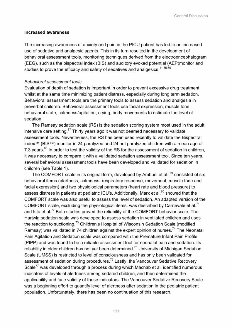
General Discussion
131
Increased awareness The increasing awareness of anxiety and pain in the PICU patient has led to an increased use of sedative and analgesic agents. This in its turn resulted in the development of behavioral assessment tools, monitoring techniques derived from the electroencephalogram (EEG), such as the bispectral index (BIS) and auditory evoked potential (AEP)monitor and studies to prove the efficacy and safety of sedatives and analgesics.11,65,66 Behavioral assessment tools Evaluation of depth of sedation is important in order to prevent excessive drug treatment whilst at the same time minimizing patient distress, especially during long term sedation. Behavioral assessment tools are the primary tools to assess sedation and analgesia in preverbal children. Behavioral assessment tools use facial expression, muscle tone, behavioral state, calmness/agitation, crying, body movements to estimate the level of sedation.
The Ramsay sedation scale (RS) is the sedation scoring system most used in the adult intensive care setting.67 Thirty years ago it was not deemed necessary to validate assessment tools. Nevertheless, the RS has been used recently to validate the Bispectral index™ (BIS™) monitor in 24 paralyzed and 24 not paralyzed children with a mean age of 7.3 years.68 In order to test the validity of the RS for the assessment of sedation in children, it was necessary to compare it with a validated sedation assessment tool. Since ten years, several behavioral assessment tools have been developed and validated for sedation in children (see Table 1).
The COMFORT scale in its original form, developed by Ambuel et al.,69 consisted of six behavioral items (alertness, calmness, respiratory response, movement, muscle tone and facial expression) and two physiological parameters (heart rate and blood pressure) to assess distress in patients at pediatric ICU's. Additionally, Marx et al.70 showed that the COMFORT scale was also useful to assess the level of sedation. An adapted version of the COMFORT scale, excluding the physiological items, was described by Carnevale et al.71 and Ista et al.72 Both studies proved the reliability of the COMFORT behavior scale. The Hartwig sedation scale was developed to assess sedation in ventilated children and uses the reaction to suctioning.73 Children’s Hospital of Wisconsin Sedation Scale (modified Ramsay) was validated in 74 children against the expert opinion of nurses.74 The Neonatal Pain Agitation and Sedation scale was compared with the Premature Infant Pain Profile (PIPP) and was found to be a reliable assessment tool for neonatal pain and sedation. Its reliability in older children has not yet been determined.75 University of Michigan Sedation Scale (UMSS) is restricted to level of consciousness and has only been validated for assessment of sedation during procedures.76 Lastly, the Vancouver Sedative Recovery Scale77 was developed through a process during which Macnab et al. identified numerous indicators of levels of alertness among sedated children, and then determined the applicability and face validity of these indicators. The Vancouver Sedative Recovery Scale was a beginning effort to quantify level of alertness after sedation in the pediatric patient population. Unfortunately, there has been no continuation of this research.
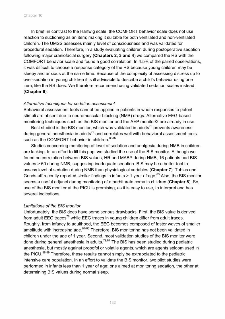
Chapter 10
132
In brief, in contrast to the Hartwig scale, the COMFORT behavior scale does not use reaction to suctioning as an item; making it suitable for both ventilated and non-ventilated children. The UMSS assesses mainly level of consciousness and was validated for procedural sedation. Therefore, in a study evaluating children during postoperative sedation following major craniofacial surgery (Chapters 2, 3 and 4) we compared the RS with the COMFORT behavior scale and found a good correlation. In 4.5% of the paired observations, it was difficult to choose a response category of the RS because young children may be sleepy and anxious at the same time. Because of the complexity of assessing distress up to over-sedation in young children it is ill advisable to describe a child’s behavior using one item, like the RS does. We therefore recommend using validated sedation scales instead (Chapter 6).
Alternative techniques for sedation assessment Behavioral assessment tools cannot be applied in patients in whom responses to potent stimuli are absent due to neuromuscular blocking (NMB) drugs. Alternative EEG-based monitoring techniques such as the BIS monitor and the AEP monitor/2 are already in use.
Best studied is the BIS monitor, which was validated in adults78 prevents awareness during general anesthesia in adults79 and correlates well with behavioral assessment tools such as the COMFORT behavior in children.80-82
Studies concerning monitoring of level of sedation and analgesia during NMB in children are lacking. In an effort to fill this gap, we studied the use of the BIS monitor. Although we found no correlation between BIS values, HR and MABP during NMB, 16 patients had BIS values > 60 during NMB, suggesting inadequate sedation. BIS may be a better tool to assess level of sedation during NMB than physiological variables (Chapter 7). Tobias and Grindstaff recently reported similar findings in infants > 1 year of age.83 Also, the BIS monitor seems a useful adjunct during monitoring of a barbiturate coma in children (Chapter 8). So, use of the BIS monitor at the PICU is promising, as it is easy to use, to interpret and has several indications. Limitations of the BIS monitor Unfortunately, the BIS does have some serious drawbacks. First, the BIS value is derived from adult EEG traces78 while EEG traces in young children differ from adult traces. Roughly, from infancy to adulthood, the EEG becomes composed of faster waves of smaller amplitude with increasing age.84-86 Therefore, BIS monitoring has not been validated in children under the age of 1 year. Second, most validation studies of the BIS monitor were done during general anesthesia in adults.79,87 The BIS has been studied during pediatric anesthesia, but mostly against propofol or volatile agents, which are agents seldom used in the PICU.88,89 Therefore, these results cannot simply be extrapolated to the pediatric intensive care population. In an effort to validate the BIS monitor, two pilot studies were performed in infants less than 1 year of age; one aimed at monitoring sedation, the other at determining BIS values during normal sleep.
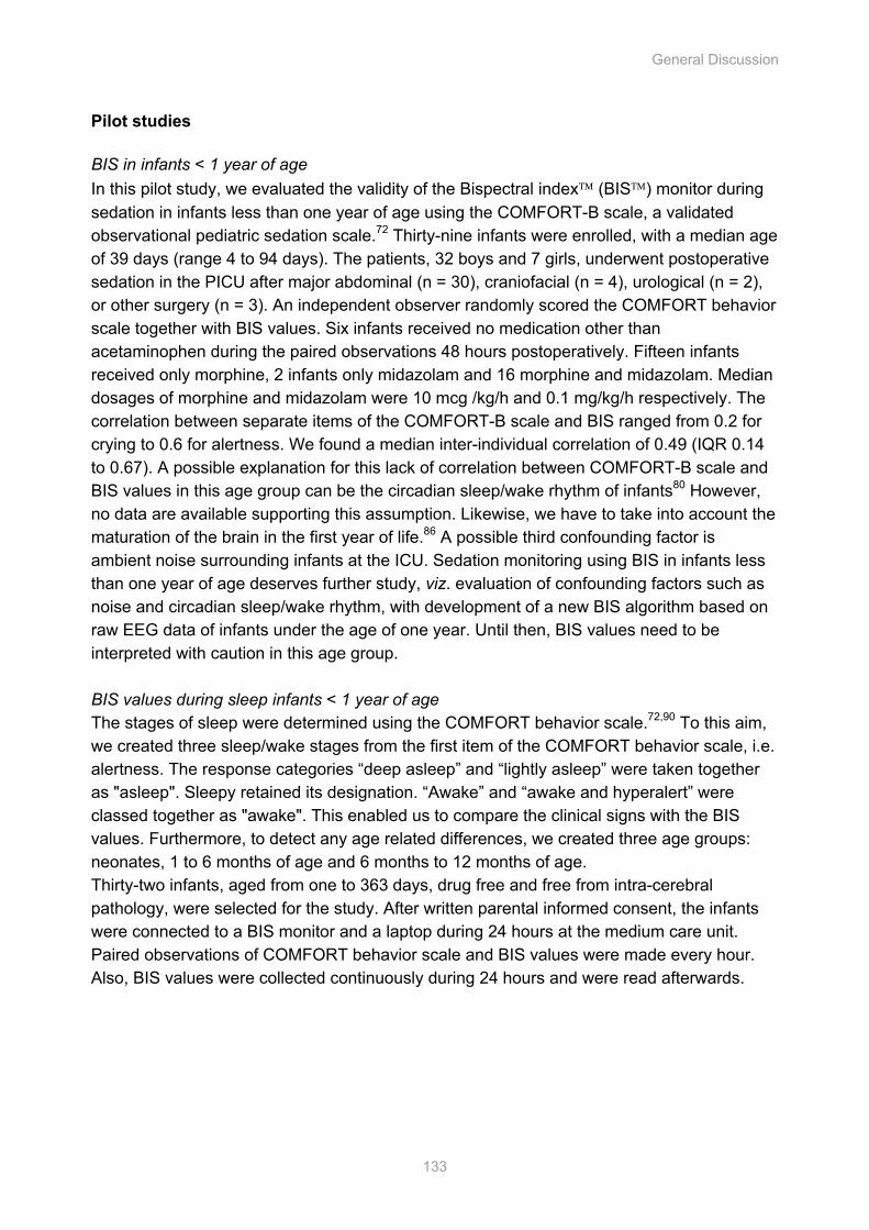
General Discussion
133
Pilot studies BIS in infants < 1 year of age In this pilot study, we evaluated the validity of the Bispectral index (BIS) monitor during sedation in infants less than one year of age using the COMFORT-B scale, a validated observational pediatric sedation scale.72 Thirty-nine infants were enrolled, with a median age of 39 days (range 4 to 94 days). The patients, 32 boys and 7 girls, underwent postoperative sedation in the PICU after major abdominal (n = 30), craniofacial (n = 4), urological (n = 2), or other surgery (n = 3). An independent observer randomly scored the COMFORT behavior scale together with BIS values. Six infants received no medication other than acetaminophen during the paired observations 48 hours postoperatively. Fifteen infants received only morphine, 2 infants only midazolam and 16 morphine and midazolam. Median dosages of morphine and midazolam were 10 mcg /kg/h and 0.1 mg/kg/h respectively. The correlation between separate items of the COMFORT-B scale and BIS ranged from 0.2 for crying to 0.6 for alertness. We found a median inter-individual correlation of 0.49 (IQR 0.14 to 0.67). A possible explanation for this lack of correlation between COMFORT-B scale and BIS values in this age group can be the circadian sleep/wake rhythm of infants80 However, no data are available supporting this assumption. Likewise, we have to take into account the maturation of the brain in the first year of life.86 A possible third confounding factor is ambient noise surrounding infants at the ICU. Sedation monitoring using BIS in infants less than one year of age deserves further study, viz. evaluation of confounding factors such as noise and circadian sleep/wake rhythm, with development of a new BIS algorithm based on raw EEG data of infants under the age of one year. Until then, BIS values need to be interpreted with caution in this age group. BIS values during sleep infants < 1 year of age The stages of sleep were determined using the COMFORT behavior scale.72,90 To this aim, we created three sleep/wake stages from the first item of the COMFORT behavior scale, i.e. alertness. The response categories “deep asleep” and “lightly asleep” were taken together as "asleep". Sleepy retained its designation. “Awake” and “awake and hyperalert” were classed together as "awake". This enabled us to compare the clinical signs with the BIS values. Furthermore, to detect any age related differences, we created three age groups: neonates, 1 to 6 months of age and 6 months to 12 months of age. Thirty-two infants, aged from one to 363 days, drug free and free from intra-cerebral pathology, were selected for the study. After written parental informed consent, the infants were connected to a BIS monitor and a laptop during 24 hours at the medium care unit. Paired observations of COMFORT behavior scale and BIS values were made every hour. Also, BIS values were collected continuously during 24 hours and were read afterwards.
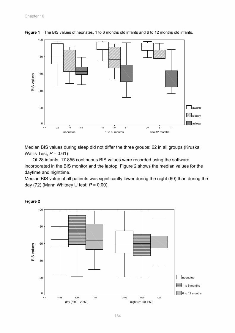
Chapter 10
134
Figure 1 The BIS values of neonates, 1 to 6 months old infants and 6 to 12 months old infants.
176153 81910 294522N =
6 to 12 months1 to 6 monthsneonates
BIS
val
ues
100
80
60
40
20
0
awake
sleepy
asleep
Median BIS values during sleep did not differ the three groups: 62 in all groups (Kruskal Wallis Test, P = 0.61)
Of 28 infants, 17.855 continuous BIS values were recorded using the software incorporated in the BIS monitor and the laptop. Figure 2 shows the median values for the daytime and nighttime. Median BIS value of all patients was significantly lower during the night (60) than during the day (72) (Mann Whitney U test: P = 0.00). Figure 2
10391151 39995086 24624118N =
night (21:00-7:59)day (8:00 - 20:59)
BIS
val
ues
100
80
60
40
20
0
neonates
1 to 6 months
6 to 12 months
General Discussion
135
Both pilot studies found discomforting data which raise further questions. Infants, without medication, can reach BIS™ values even as low as 22 during their physiological sleep. Sleigh et al. questioned the use of the BIS monitor already in 1999, as they found decreased BIS values during normal sleep in 5 adults.91 Nieuwenhuijs et al. in 2002 found BIS values as low as 45 in 10 adults and concluded that using an new algorithm incorporated in the BIS monitor, derived from naturally sleeping subjects, the BIS could discriminate between different physiologic as well as pharmacological states.84 Recently, Benini et al. performed a study in children with a mean age of 8.2 years (range 1.2 to 16.5 years) which also seem to indicate that the effects of natural sleep on the BIS appear to be similar to the effects of general anesthesia on the BIS.92 However, it is questionable if the adult BIS algorithm is also applicable to children less than one year of age. A new algorithm, derived from BIS values of infants < 1 year of age during normal wake and sleep, needs to be developed for this age group, taking into account the maturation of the brain during the first year of life. The AEP monitor/2 The AEP monitor/2 uses middle latency auditory evoked potentials (MLAEPs) to test the patient’s brain ability to respond to an auditory signal. MLAEPs represent the earliest cortical response to an acoustic stimulus. Amplitudes and latencies are influenced by anesthetics and surgical stimuli and are believed to be useful foe measuring level of anesthesia. A monitoring variable, indicating the patient’s hypnotic state, the so-called A-line ARX index (AAI) which ranges from 0 (iso-electric EEG) to 100 (awake), is then calculated from the MLAEPs and the EEG. The AEP monitor/2 has been studied in adults during anesthesia and at the ICU. During anesthesia, use of the AEP monitor/2 improved emergence from anesthesia, limited the amounts of anesthetics and led to detection of intra-operative awareness.93-95 In the adult ICU setting, the AEP monitor/2 correlated well with clinical sedation scales, such as the Ramsay sedation scale. 96,97 Three studies in children during anesthesia showed that the AAI is more valuable in predicting anesthetic states than hemodynamic variables and reliably differentiates between the awake and anesthetized states.98-100 Data from children and infants outside the operation room are lacking so far. In Chapter 9 a pilot study is described evaluating feasibility and validity of the AEP monitor/2 in 8 postoperative infants admitted to the PICU with median ages of 40 days (1 to 795 days). In this study we found the AEP monitor/2 to produce high sound levels and to be artifact-free for only half of the time. The correlation between COMFORT behavior scores and AAI values was moderate in all but one patient, with a large inter-individual range. Moreover, the AEP monitor/2 was difficult to apply, impedance testing of the electrodes was time-consuming and tended to irritate the children and electrode removal revealed erythematous indentations of the skin of all children. These limitations made us stop this study after data from 8 patients were collected and need to be overcome before daily use in the PICU is an option. Recommended sedation assessment tools The level of sedation should be regularly assessed and documented using validated behavioral assessment tools. The COMFORT-B scale should be used for assessment of
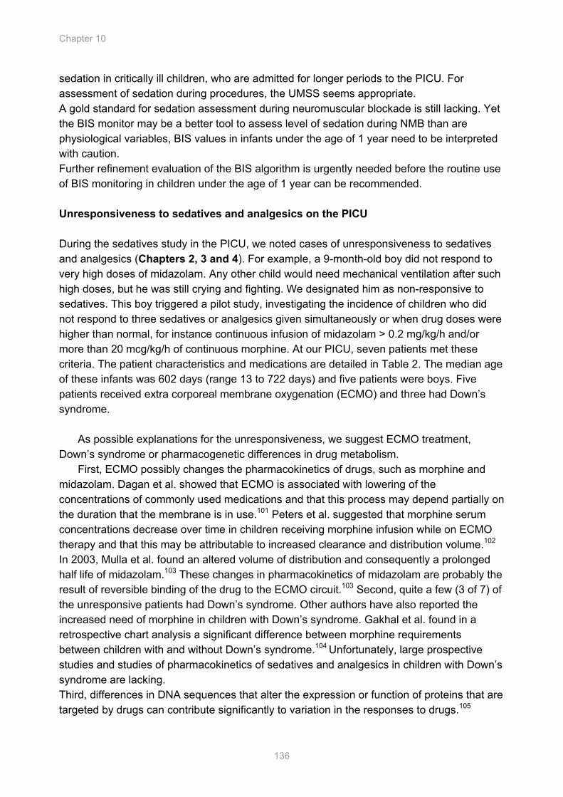
Chapter 10
136
sedation in critically ill children, who are admitted for longer periods to the PICU. For assessment of sedation during procedures, the UMSS seems appropriate. A gold standard for sedation assessment during neuromuscular blockade is still lacking. Yet the BIS monitor may be a better tool to assess level of sedation during NMB than are physiological variables, BIS values in infants under the age of 1 year need to be interpreted with caution. Further refinement evaluation of the BIS algorithm is urgently needed before the routine use of BIS monitoring in children under the age of 1 year can be recommended. Unresponsiveness to sedatives and analgesics on the PICU During the sedatives study in the PICU, we noted cases of unresponsiveness to sedatives and analgesics (Chapters 2, 3 and 4). For example, a 9-month-old boy did not respond to very high doses of midazolam. Any other child would need mechanical ventilation after such high doses, but he was still crying and fighting. We designated him as non-responsive to sedatives. This boy triggered a pilot study, investigating the incidence of children who did not respond to three sedatives or analgesics given simultaneously or when drug doses were higher than normal, for instance continuous infusion of midazolam > 0.2 mg/kg/h and/or more than 20 mcg/kg/h of continuous morphine. At our PICU, seven patients met these criteria. The patient characteristics and medications are detailed in Table 2. The median age of these infants was 602 days (range 13 to 722 days) and five patients were boys. Five patients received extra corporeal membrane oxygenation (ECMO) and three had Down’s syndrome.
As possible explanations for the unresponsiveness, we suggest ECMO treatment,
Down’s syndrome or pharmacogenetic differences in drug metabolism. First, ECMO possibly changes the pharmacokinetics of drugs, such as morphine and
midazolam. Dagan et al. showed that ECMO is associated with lowering of the concentrations of commonly used medications and that this process may depend partially on the duration that the membrane is in use.101 Peters et al. suggested that morphine serum concentrations decrease over time in children receiving morphine infusion while on ECMO therapy and that this may be attributable to increased clearance and distribution volume.102 In 2003, Mulla et al. found an altered volume of distribution and consequently a prolonged half life of midazolam.103 These changes in pharmacokinetics of midazolam are probably the result of reversible binding of the drug to the ECMO circuit.103 Second, quite a few (3 of 7) of the unresponsive patients had Down’s syndrome. Other authors have also reported the increased need of morphine in children with Down’s syndrome. Gakhal et al. found in a retrospective chart analysis a significant difference between morphine requirements between children with and without Down’s syndrome.104 Unfortunately, large prospective studies and studies of pharmacokinetics of sedatives and analgesics in children with Down’s syndrome are lacking. Third, differences in DNA sequences that alter the expression or function of proteins that are targeted by drugs can contribute significantly to variation in the responses to drugs.105
Tabl
e 2
Pat
ient
cha
ract
eris
tics
Pa
tient
1
Patie
nt 2
Pa
tient
3
Patie
nt 4
Pa
tient
5
Patie
nt 6
Pa
tient
7
Sex
m
ale
mal
e m
ale
mal
e fe
mal
e fe
mal
e m
ale
Age
in d
ays
722
660
297
602
644
78
13
Rac
e D
utch
D
utch
Fi
lipin
o D
utch
D
utch
D
utch
D
utch
D
iagn
oses
S
ubgl
ottic
ste
nosi
s af
ter
lary
ngot
rach
eopl
astic
RS
viru
s
Sta
tus
afte
r AV
SD
Pu
lmon
ary
hype
rtens
ion
Esop
hage
al a
tresi
a Su
rger
y
RS
viru
s A
fter
card
iopu
lmon
ary
surg
ery
CD
H p
ulm
onar
y hy
perte
nsio
n
Kid
ney
func
tion
Nor
mal
N
orm
al
Nor
mal
N
orm
al
Nor
mal
N
orm
al
Nor
mal
Li
ver f
unct
ion
Not
eva
luat
ed#
Nor
mal
N
ot e
valu
ated
N
orm
al
Nor
mal
N
orm
al
Not
eva
luat
ed
Dow
n’s
synd
rom
e y/
n N
o Ye
s Ye
s N
o Ye
s N
o N
o E
CM
O*
y/n
No
Yes
Y
es
no
Yes
Y
es
Yes
R
emar
kabl
e pe
riod
D
urin
g E
CM
O
Dur
ing
EC
MO
Dur
ing
EC
MO
D
urin
g an
d af
ter
EC
MO
A
fter E
CM
O
Max
. med
icat
ion
Sed
ativ
es
Mid
azol
am in
mg/
kg/h
0.
30
0.50
0.
40
0.50
0.
44
0.60
0.
30
D
iaze
pam
in m
g/kg
/h
- -
- 0.
15
0.27
0.
08
-
Alim
emaz
ine
in m
g/kg
1.
25
1.25
-
1.25
0.
80
-
C
hlor
alhy
drat
e in
mg
- -
- 62
.5
-
-
Ane
sthe
tics
Pro
pofo
l in
mg/
kg/h
13
2
- 6.
60
9 1
-
Ket
amin
e-s
in m
g/kg
/h
0.80
0.
20
- 2.
50
- 1.
4 -
Neu
rom
uscu
lar b
lock
ers
Vec
uron
ium
in m
g/kg
/h
- -
0.20
-
0.20
0.10
Ant
i-epi
lept
ics
Pent
obar
bita
l in
mg/
kg/h
-
4 2.
80
- -
-
Ana
lges
ics
Fent
anyl
in m
cg/k
g/h
5 -
- 8.
90
18
16.6
7.
20
M
orph
ine
in m
cg/k
g/h
15
20
32
20
25
60
30
Ant
i-hyp
erte
nsiv
es
Clo
nidi
ne in
mcg
/kg/
h -
0.60
3.
50
1.10
1.
40
4.0
0.40
*
extra
cor
pore
al m
embr
ane
oxyg
enat
ion
# no
live
r fun
ctio
n te
stin
g w
as p
erfo
rmed
prio
r to
adm
inis
tratio
n of
sed
ativ
es a
nd a
nalg
esic
s
Chapter 10
138
For instance, midazolam is hydroxylated by hepatic cytochrome P-450 3A subfamily (CYP3A4 and CYP3A5) in the major metabolite 1-OH-midazolam (50 - 70% of the metabolism),106 which is as potent as the parent drug107,108 and the minor metabolites 4-OH-midazolam and 1,4-OH-midazolam. Another example is propofol of which Court et al. described in 2001 that cytochrome P-450 2B6 is the principal determinant of inter-individual variability in the hydroxylation of this drug by human liver microsomes.109
Lastly, Simons et al.110 found that the frequency of additional morphine use in patients with COMT wild type genotype was significantly higher than that in patients with the COMT mutation.
These findings indicate that mutations in any of these DNA sequences can result in unresponsiveness to drugs. The Human Genome Project has raised expectations for medicines that can be customized to match the genetic make-up of patients, thereby dramatically improving safety and efficacy.105 Future directives for research The studies presented in this thesis showed that PK/PD modeling using non-linear mixed effect modeling (NONMEM) can predict a clinically safe dose with scarce data sampling and small groups of patients. Also, long-used (Ramsay sedation scale) and relatively new (Bispectral index monitor and Auditory Evoked Potential monitor) sedation assessment tools were evaluated, showing that there is still much work to be done. For instance, although the BIS monitor proved to be easy to use under a variety of conditions, it still is not validated for infants under the age of 1 year. In collaboration with the manufacturer of the BIS monitor, a new algorithm, based on EEG’s of infants in this particular age group will be developed. Importantly, from that point on the BIS monitor can be considered in this population of patients that makes up 80% of the children admitted to PICUs.
The increasing awareness of anxiety and pain in the PICU led to an increased use of sedatives and opioids of which long term infusion can result in withdrawal and tolerance. To provide optimal sedation and analgesia, use of an algorithm should be part of the daily routine of nursing care in the PICU. The ideal algorithm would incorporate causes of agitation within the child, like absence of parents, a wet diaper or hunger. Environmental causes for agitation are wrong settings of the mechanical ventilation, need for suctioning, but also noise and light.111 Once these factors are checked, assessment and documentation of the level of sedation and analgesia using validated assessment tools, such as the COMFORT behavior scale or the visual analogue scale (VAS) will determine the next steps.72,112 If this assessment reaches the predetermined cut off points, start of sedatives and/or analgesics is indicated.
To prevent complications of over-sedation or the development of dependence, sedative and analgesic drugs need to be titrated to optimal doses and a predetermined levels of sedation.
Apart from the algorithm that determines start of sedation or analgesia, a weaning algorithm should also be available for patients who have received sedatives and analgesics
General Discussion
139
for more than 5 days.61,63,64 Abrupt discontinuation of opioids and benzodiazepines may cause a withdrawal syndrome, characterized by agitation, tremors, jitteriness, diarrhea, sweating and tachycardia.113,114 Optimal weaning rates of opioids and benzodiazepines have been proposed by Ducharme et al. recently, but as this is the first prospective study since ten years presenting this issue, new research in this area is warranted. 61,115
Furthermore, individual responses and sometimes unpredictable effects of drugs are possibly explained by individual variations in pharmacogenetic profiles, the immune system, drug metabolic pathways and drug-drug interactions. New studies, considering PK/PD and pharmacogenetic profiles, may help us developing guidelines to manage sedative and analgesic drugs safely in critically ill children. Initiatives, such as the Best Pharmaceuticals Act for Children (BPCA)116 in the USA and the forthcoming change of the law in Europe will enable caregivers to bend the widespread use of off-label and/or unlicensed drugs in infants towards evidence based medicine.
New research protocols will be developed by our research group to study sedatives, analgesics and new tools to monitor level of sedation and analgesia, but all “under a watchful eye”; a condition sine qua non for optimal patient care in the PICU environment.
Chapter 10
140
References 1. Vermeeren A. Residual effects of hypnotics: epidemiology and clinical implications. CNS Drugs
2004;18:297-328
2. Allonen H, Ziegler G, Klotz U. Midazolam kinetics. Clin Pharmacol Ther 1981;30:653-61
3. Pringle B, Dahlquist LM. Memory in pediatric patients undergoing conscious sedation for aversive medical procedures. Health Psychology 2003;22:263-9
4. Cote CJ, Karl HW, Notterman NA et al. Adverse sedation events in pediatrics:analysis of medications used for sedation. Pediatrics 2000;106:633-44
5. Martin LD, Bratton SL, Quint P, Mayock DE. Prospective documentation of sedative, analgesic, and neuromuscular blocking agent use in infants and children in the intensive care unit: A multicenter perspective. Pediatr Crit Care Med 2001;2:205-10
6. Fonsmark L, Rasmussen YH, Peder C. Occurence of withdrawal in critically ill sedated children. Crit Care Med 1999;27:196-9
7. Buffett-Jerrott SE, Stewart SH, Finley GA, Loughlan HL. Effects of benzodiazepines on explicit memory in a paediatric surgery setting. Psychopharmacology 2003;168:377-86
8. Cheng C, Roemer-Becuwe C, Pereira J. When midazolam fails. J Pain Symptom Manage 2002;23:256-65
9. Booker PD, Beechey A, Lloyd-Thomas AR. Sedation of children requiring artificial ventilation using an infusion of midazolam. Br J Anaesth 1986;58:1104-8
10. Ng E, Taddio A, Ohlsson A. Intravenous midazolam infusion for sedation of infants in the neonatal intensive care unit. Cochrane Database Syst Rev 2000:CD002052
11. Tobias JD. Sedation and analgesia in paediatric intensive care units: a guide to drug selection and use. Paediatr Drugs 1999;1:109-26
12. Shafer A. Complications of sedation with midazolam in the intensive care unit and a comparison with other sedative regimens. Crit Care Med 1998;26:947-56
13. de Wildt SN, de Hoog M, Vinks AA et al. Pharmacodynamics of midazolam in pediatric intensive care patients. Ther Drug Monit 2005;27:98-102
14. de Wildt SN, de Hoog M, Vinks AA et al. Population pharmacokinetics and metabolism of midazolam in pediatric intensive care patients. Crit Care Med 2003;31:1952-8
15. de Wildt SN, Kearns GL, Hop WC et al. Pharmacokinetics and metabolism of intravenous midazolam in preterm infants. Clin Pharmacol Ther 2001;70:525-31
16. de Wildt SN, Kearns GL, Hop WC et al. Pharmacokinetics and metabolism of intravenous midazolam in preterm infants. Clin Pharmacol Ther 2001;70:521-31
17. de Wildt SN, Kearns GL, Hop WC et al. Pharmacokinetics and metabolism of oral midazolam in preterm infants. Br J Clin Pharmacol 2002;53:390-2
18. FDA. Pediatric Exclusivity Labeling Changes: Center for Drug Evaluation and Research, 2003
19. Wheeler DS, Vaux KK, Ponaman ML, Poss BW. The safe and effective use of propofol sedation in children undergoing diagnostic and therapeutic procedures: experience in a pediatric ICU and a review of the literature. Pediatr Emerg Care 2003;19:385-92

General Discussion
141
20. Rothermel LK. Newer pharmacologic agents for procedural sedation of children in the emergency department-etomidate and propofol. Curr Opin Pediatr 2003;15:200-3
21. Lin C, Durieux ME. Ketamine and kids:an update. Pediatr Anesthesia 2005;15:91-7
22. Evans D, Turnham L, Barbour K et al. Intravenous ketamine sedation for painful oncology procedures. Pediatr Anesthesia 2005;15:131-8
23. Mazurek MS. Sedation and analgesia for procedures outside the operating room. Seminars in Pediatric Surgery 2004;13:166-73
24. Marcus RJ, Victoria BA, Rushman SC, Thompson JP. Comparison of ketamine and morphine for analgesia after tonsillectomy in children. Br J Anaesth 2000;84:739-42
25. Aspinall RL, Mayor A. A prospective randomized controlled study of the efficacy of ketamine for postoperative pain relief in children after adenotonsillectomy. Pediatr Anesthesia 2001;11:333-6
26. Malviya S, Voepel-Lewis T, Tait AR et al. Pentobarbital vs chloral hydrate for sedation of children undergoing MRI: efficacy and recovery characteristics. Pediatr Anesthesia 2004;14:589-95
27. Adelson PD, Bratton SL, Carney NA et al. Chapter 9. Use of sedation and neuromuscular blockade in the treatment of severe pediatric traumatic brain injury. Pediatr Crit Care Med 2003;4:S34-7
28. Kwan P, Brodie MJ. Phenobarbital for the Treatment of Epilepsy in the 21st Century: A Critical Review. Epilepsia 2004;45:1141-9
29. Yanay O, Brogan TV, Martin LD. Continuous pentobarbital infusion in children is associated with high rates of complications. J Crit Care 2004;19:174-8
30. Van Gestel JPJ, Blusse van Oud-Alblas HJ, Malingre M et al. Propofol and thiopental for refractory status epilepticus in children. Neurology 2005;65:591-2
31. Coupey SM. Barbiturates. Pediatr Rev 1997;18:260-4; quiz 5
32. Ambrose C, Sale S, Howells R et al. Intravenous clonidine infusion in critically ill children:dose-dependent sedative effects and cardiovascular stability. Br J Anaesth 2000;84:794-6
33. Arenas-Lopez S, Riphagen S, Tibby SM et al. Use of oral clonidine for sedation in ventilated paediatric intensive care patients. Intensive Care Med 2004;30:1625-9. Epub 2004 Jun 10
34. Jasinski DR, Johnson RE, Kocher TR. Clonidine in morphine withdrawal. Differential effects on signs and symptoms. Arch Gen Psychiatry 1985;42:1063-6
35. Gold MS, Kleber HD. Clinical utility of clonidine in opiate withdrawal: a study of 100 patients. Prog Clin Biol Res 1981;71:299-306
36. Gold MS, Redmond DE, Jr., Kleber HD. Noradrenergic hyperactivity in opiate withdrawal supported by clonidine reversal of opiate withdrawal. Am J Psychiatry 1979;136:100-2
37. O'Connor PG. Methods of detoxification and their role in treating patients with opioid dependence. JAMA 2005;294:961-3
38. Marx CM, Rosenberg DI, Ambuel B et al. Pediatric Intensive Care Sedation: Survey of Fellowship Training Programs. Pediatrics 1993;91:369-78
39. Malviya S, Voepel-Lewis T, Tait AR. Adverse events and risk factors associated with the sedation of children by nonanesthesiologists. Anest Analg 1997;85:1207-13
40. Pershad J, Palmisano P, Nichols M. Chloral hydrate: The good and the bad. Pediatr Emergency Care 1999;15:432-5
41. Valman HB. ABC of 1 to 7 (revised). Sleep problems. Br Med J (Clin Res Ed) 1987;294:828-30
Chapter 10
142
42. Younus M, Labellarte MJ. Insomnia in children: when are hypnotics indicated? Paediatr Drugs 2002;4:391-403
43. Sponheim S, Aune H, Gulliksen M, Morland J. Pharmacokinetics of trimeprazine in children. Pharmacol Toxicol 1990;67:243-5
44. http://www.who.int/cancer/palliative/painladder
45. Anderson B, Kanagasundarum S, Woollard G. Analgesic efficacy of paracetamol in children using tonsillectomy as a pain model. Anaesth Intensive Care 1996;24:669-73
46. Arana A, Morton NS, Hansen TG. Treatment with paracetamol in infants. Acta Anaesthesiol Scand 2001;45:20-9
47. Bertin L, Pons G, d'Athis P et al. Randomized double-blind, mulitcenter controlled trial of ibuprofen versus acetaminophen (paracetamol) and placebo for treatment of symptoms of tonsillitis and pharyngitis in children. J Pediatr 1991;119:811-4
48. van der Marel CD, van Lingen RA, Pluim MA et al. Analgesic efficacy of rectal versus oral acetaminophen in children after major craniofacial surgery. Clin Pharmacol Ther 2001;70:82-90
49. Anderson BJ, van Lingen RA, Hassen TG et al. Acetaminophen developmental pharmacokinetics in premature neonates and infants. Anesthesiology 2002;96:1336-45
50. Murat I, Baujard, C., Foussat, C., Guyot, E., Petel, H., Rod, B., Ricard, C. Tolerance and analgesic efficacy of a new i.v. paracetamol solution in children after inguinal hernia repair. Paediatr Anaesth 2005:1-8
51. Allegaert K, Van der Marel CD, Debeer A et al. Pharmacokinetics of single dose intravenous propacetamol in neonates: effect of gestational age. Arch Dis Child Fetal Neonatal Ed 2004;89:F25-8
52. Allegaert K, Anderson BJ, Naulaers G et al. Intravenous paracetamol (propacetamol) pharmacokinetics in term and preterm neonates. Eur J Clin Pharmacol 2004;60:191-7. Epub 2004 Apr 8
53. Montgomery CJ, McCormack JP, Reichert CC, Marsland CP. Plasma concentrations after high-dose (45 mg.kg-1) rectal acetaminophen in children. Can J Anaesth 1995;42:982-6
54. Autret E, Dutertre JP, Breteau M et al. Pharmacokinetics of paracetamol in the neonate and infant after administration of propacetamol chlorhydrate. Dev Pharmacol Ther 1993;20:129-34
55. Granry JC, Rod B, Monrigal JP et al. The analgesic efficacy of an injectable prodrug of acetaminophen in children after orthopaedic surgery. Paediatr Anaesth 1997;7:445-9
56. Dsida RM, Wheeler M, Birmingham PK et al. Age-stratified pharmacokinetics of ketorolac tromethamine in pediatric surgical patients. Anesth Analg 2002;94:266-70
57. van der Marel CD, Anderson BJ, Romsing J et al. Diclofenac and metabolite pharmacokinetics in children. Paediatr Anaesth 2004;14:443-51
58. Gupta AK, Dagget C, Ludwick J et al. Ketorolac after congenital heart surgery: does it increase the risk of significant bleeding complications? Paediatr Anaesth 2005;15:139-42
59. Katz R, Kelly W, Hsia A. Prospective study on the occurrence of withdrawal in critically ill children who receive fentanyl by continuous infusion. Crit Care Med 1994;22:763-7
60. Esmail Z, Montgomery C, Court C et al. Efficacy and complications of morphine infusions in postoperative paediatric patients. Paediatr Anesthesia 1999;9:321-7
61. Anand KJ, Arnold JH. Opioid tolerance and dependence in infants and children. Crit Care Med 1994;22:334-42
General Discussion
143
62. Anand KJ, Ingraham J. Pediatric. Tolerance, dependence, and strategies for compassionate withdrawal of analgesics and anxiolytics in the pediatric ICU. Crit Care Nurse 1996;16:87-93
63. Tobias JD. Tolerance, withdrawal, and physical dependency after long-term sedation and analgesia of children in the pediatric intensive care unit. Crit Care Med 2000;28:2122-32
64. Suresh S, Anand KJ. Opioid tolerance in neonates:mechanisms, diagnosis, assessment, and management. Semin Perinatol. 1998;22:425-33
65. De Jonghe B, Cook D, Appere-De-Vecchi C et al. Using and understanding sedation scoring systems: a systematic review. Intensive Care Med 2000;26:275-85
66. Grindstaff RJ, Tobias JD. Applications of bispectral index monitoring in the pediatric intensive care unit. J Intensive Care Med 2004;19:111-6
67. Ramsay MA, Savege TM, Simpson BR, Goodwin R. Controlled sedation with alphaxalone-alphadolone. Br Med J 1974;2:656-9
68. Aneja R, Heard AM, Fletcher JE, Heard CM. Sedation monitoring of children by the Bispectral Index in the pediatric intensive care unit. Pediatr Crit Care Med 2003;4:60-4
69. Ambuel B, Hamlett KW, Marx CM, Blumer JL. Assessing distress in pediatric intensive care environments: the COMFORT scale. J Pediatr Psychol 1992;17:95-109
70. Marx CM, Smith PG, Lowrie LH et al. Optimal sedation of mechanically ventilated pediatric critical care patients. Crit Care Med 1994;22:163-70
71. Carnevale FA, Razack S. An item analysis of the COMFORT scale in a pediatric intensive care unit. Pediatr Crit Care Med 2002;3:177-80
72. Ista E, Van Dijk M, Tibboel D, De Hoog M. Assessment of sedation levels in pediatric intensive care patients can be improved by using the COMFORT "behavior' scale. Crit Care Med 2005;6:58-63
73. Brunow de Carvalho W, Lucas da Silva PS, Paulo CS et al. Comparison between the Comfort and Hartwig sedation scales in pediatric patients undergoing mechanical lung ventilation. Sao Paulo Med J 1999;117:192-6
74. Soetenga D, Frank J, Pellino TA. Assessment of the validity and reliability of the University of Wisconsin Children's Hospital Pain scale for Preverbal and Nonverbal Children. Pediatr Nurs 1999;25:670-6
75. www.n-pass.com
76. Malviya S, Voepel-Lewis T, Tait AR et al. Depth of sedation in children undergoing computed tomography: validity and reliability of the University of Michigan Sedation Scale (UMSS). Br J Anaesth 2002;88:241-5
77. Macnab AJ, Levine M, Glick N et al. A research tool for measurement of recovery from sedation: the Vancouver Sedative Recovery Scale. J Pediatr Surg 1991;26:1263-7
78. Glass PS, Bloom M, Kearse L et al. Bispectral analysis measures sedation and memory effects of propofol, midazolam, isoflurane, and alfentanil in healthy volunteers. Anesthesiology 1997;86:836-47
79. Myles PS, Leslie K, McNeil J et al. Bispectral index monitoring to prevent awareness during anaesthesia: the B-Aware randomised controlled trial. Lancet 2004;363:1757-63
80. Berkenbosch JW, Fichter CR, Tobias JD. The correlation of the bispectral index monitor with clinical sedation scores during mechanical ventilation in the pediatric intensive care unit. Anesth Analg 2002;94:506-11
Chapter 10
144
81. Courtman SP, Wardurgh A, Petros AJ. Comparison of the bispectral index monitor with the Comfort score in assessing level of sedation of critically ill children. Intensive Care Med 2003;29:2239-46
82. Triltsch AE, Nestmann G, Orawa H et al. Bispectral index versus COMFORT score to determine the level of sedation in paediatric intensive care patients: a prospective study. Crit Care 2005;9:R9-R17
83. Tobias JD, Grindstaff RJ. Bispectral Index Monitoring During the Administration of Neuromuscular Agents in the Pediatric Intensive Care Unit Patient. J Intensive Care Med 2005;20:233-7
84. Nieuwenhuijs D, Coleman EL, Douglas NJ et al. Bispectral index values and spectral edge frequency at different stages of physiologic sleep. Anesth Analg 2002;94:125-9, table of contents
85. Hoppenbrouwers T, Hodgman J, Arakawa K et al. Sleep and waking states in infancy: normative studies. Sleep 1988;11:387-401
86. Ficca G, Fagioli I, Giganti F, Salzarulo P. Spontaneous awakenings from sleep in the first year of life. Early Hum Dev 1999;55:219-28
87. Sebel PS, Lang E, Rampil IJ et al. A multicenter study of bispectral electroencephalogram analysis for monitoring anesthetic effect. Anesth Analg 1997;84:891-9
88. Denman WT, Swanson EL, Rosow D et al. Pediatric evaluation of the bispectral index (BIS) monitor and correlation of BIS with end-tidal sevoflurane concentration in infants and children. Anesth Analg 2000;90:872-7
89. Choudhry DK, Brenn BR, Goyal P et al. Bispectral index monitoring: a comparison between normal children and children with quadriplegic cerebral palsy. Anesth Analg 2002;95:1582-5
90. van Dijk M, de Boer JB, Koot HM et al. The reliability and validity of the COMFORT scale as a postoperative pain instrument in 0 to 3-year-old infants. Pain 2000;84:367-77
91. Sleigh JW, Andrzejowski J, Steyn-Ross A, Steyn-Ross M. The bispectral index: a measure of depth of sleep? Anesth Analg 1999;88:659-61
92. Benini F, Trapanotto M, Sartori S et al. Analysis of the Bispectral Index During Natural Sleep in Children. Anest Analg 2005;101:641-4
93. Recart A, White PF, Wang A et al. Effect of auditory evoked potential index monitoring on anesthetic drug requirements and recovery profile after laparoscopic surgery: a clinical utility study. Anesthesiology 2003;99:813-8
94. Haenggi M, Ypparila H, Takala J et al. Measuring Depth of Sedation with Auditory Evoked Potentials During Controlled Infusion of Propofol and Remifentanil in Healthy Volunteers. Anest Analg 2004;99:1728-36
95. Trillo-Urrutia L, Fernandez-Galinski S, Castano-Santa J. Awareness detected by auditory evoked potential monitoring. Br J Anaesth 2003;91:290-2
96. Schulte-Tamburen AM, Scheier J, Briegel J et al. Comparison of five sedation scoring systems by means of auditory evoked potentials. Intensive Care Med 1999;25:377-82
97. Rundshagen I, Schnabel K, Pothmann W et al. Cortical arousal in critically ill patients: an evoked response study. Intensive Care Med 2000;26:1312-8
98. Weber F, Seidl M, Bein T. Impact of the AEP-Monitor/2-derived composite auditory-evoked potential index on propofol consumption and emergence times during total intravenous anaesthesia with propofol and remifentanil in children. Acta Anaesthesiol Scand 2005;49:277-83
99. O'Kelly SW, Smith DC, Pilkington SN. The auditory evoked potential and paediatric anaesthesia. Br J Anaesth 1995;75:428-30
General Discussion
145
100. Weber F, Bein T, Hobbhahn J, Taeger K. Evaluation of the Alaris Auditory Evoked Potential Index as an Indicator of Anesthetic Depth in Preschool Children during Induction of Anesthesia with Sevoflurane and Remifentanil. Anesthesiology 2004;101:294-8
101. Dagan O, Klein J, Gruenwald C et al. Preliminary studies of the effects of extracorporeal membrane oxygenation on the disposition of common pediatric drugs. Ther Drug Monit 1993;15:263-6
102. Peters JWB, Anderson BJ, Simons SHP et al. Morphine pharmacokinetics during venoarterial extracorporeal membrane oxygenation in neonates. Intensive Care Med 2005;31:257-63
103. Mulla H, McCormack P, Lawson G et al. Pharmacokinetics of midazolam in neonates undergoing extracorporeal membrane oxygenation. Anesthesiology 2003;99:275-82
104. Gakhal B, Scott CS, Macnab AJ. Comparison of morphine requirements for sedation in Down's syndrome and non-Down's patients following pediatric cardiac surgery. Pediatr Anesthesia 1998;8:229-33
105. Evans WE, Relling MV. Moving towards individualized medicine with pharmacogenomics. Nature 2004;429:464-8
106. Heizmann P, Ziegler WH. Excretion and metabolism of 14C-midazolam in humans following oral dosing. Arzneimittelforschung 1981;31:2220-3
107. Ziegler WH, Schalch E, Leishman B, Eckert M. Comparison of the effects of intravenously administered midazolam, triazolam and their hydroxy metabolites. Br J Clin Pharmacol 1983;16 Suppl 1:63S-9S
108. Mandema JW, Tuk B, van Steveninck AL et al. Pharmacokinetic-pharmacodynamic modeling of the central nervous system effects of midazolam and its main metabolite alpha-hydroxymidazolam in healthy volunteers. Clin Pharmacol Ther 1992;51:715-28
109. Court MH, Duan SX, Hesse LM et al. Cytochrome P-450 2B6 is responsible for interindividual variability of propofol hydroxylation by human liver microsomes. Anesthesiology 2001;94:110-9
110. Simons SH. Morphine more fine. Rotterdam: thesis Erasmus University Rotterdam, 2004
111. Milette IH, Carnevale FA. I'm trying to heal...noise levels in a pediatric intensive care unit. Dynamics 2003;14:14-21
112. Buchholz M, Karl HW, Pomietto M, Lynn A. Pain scores in infants: a modified infant pain scale versus visual analogue. J Pain Symptom Manage 1998;15:117-24
113. Tobias JD. Tolerance, withdrawal, and physical dependency after long-term sedation and analgesia of children in the pediatric intensive care unit. Crit Care Med 2000;28:2122-32
114. Franck LS, Naughton I, Winter I. Opioid and benzodiazepine withdrawal symptoms in paediatric intensive care patients. Intensive and Critical Care Nursing 2004;20:344-51
115. Ducharme C, Carnevale FA, Clermont M-S, Shea S. A prospective study of adverse reactions to the weaning of opioids and benzodiazepines among critically ill children. Intensive and Critical Care Nursing 2005;21:179-86
116. Wilson JT. An update on the therapeutic orphan. Pediatrics 1999;104:585-90


Summary Samenvatting
Cha
pter
Chapter 11
148
Summary Critically ill patients admitted to an intensive care unit will normally receive sedative and analgesic drugs to attenuate discomfort and pain. Unfortunately, sedatives and analgesics have adverse effects, and may potentially prolong duration of mechanical ventilation and stay in the intensive care unit and thus increase costs. Also, the ones most at risk from partial, incomplete, or absent drug evaluation and inadequate drug labeling are children. In order to avoid possible complications of both excessive and inadequate sedation or analgesia, levels of sedation and analgesia in critically ill children must be regularly assessed and documented. In view of these considerations, we studied safety aspects and pharmacokinetic and pharmacodynamic profiles of the sedatives midazolam and propofol, as well as intravenous propacetamol, an analgesic.
Pharmacokinetics is the study of the time course of drug and metabolite levels in different fluids, tissues, and excreta of the body, and of the mathematical relationships required to develop models to interpret such data. Pharmacodynamics study the effects and adverse effects of the drug on the body. Furthermore, we explored the use of the available observational and EEG monitoring techniques to assess sedation and analgesia in infants at a pediatric intensive care unit (PICU). Sedatives depress the central nervous system and cause calmness, relaxation and anxiety reduction. Midazolam is a short acting sedative and is the most frequently used sedative in the PICU). Chapter 2 describes the pharmacokinetics and –dynamics for midazolam in children under the age of 2 years. Based on this study, we advise a loading dose of 0.1 mg/kg, followed by a continuous infusion of 0.05 mg/kg/h during the first night after major surgery in non-ventilated infants to achieve COMFORT behavior scores from 12 to 14 (moderate sedation). An alternative for midazolam might be propofol, an ultra short-acting intravenous anesthetic agent which is also used for sedation in mechanically ventilated adults. After disturbing reports of adverse events after use of propofol as a sedative in children, the FDA issued a warning against its use in children under the age of 18 years in pediatric intensive care. A new formulation, propofol 6%, proved to be a safe sedative in postoperative patients without multiple organ failure or critical illness, at dosages less than 4 mg/kg/h. The studied children had a median age of 10 months (IQR 3 to 17 months), and received propofol 6% during a median period of 11 hours (range 6 to 18 hours) (Chapter 3). Propofol clearance in non-ventilated healthy children was found to be two times higher than that reported in the literature for ventilated children and adults. Based on the pharmacokinetic model of propofol, we advise a propofol dosage of 3 mg/kg/h to achieve scores from 12 to 14 on the COMFORT behavior scale and values from 70 to 75 on the BIS monitor during the night. Wide pharmacodynamic variability emphasizes the importance of dose titration (Chapter 4).

Summary
149
Not only sedation is very important, analgesia is another cornerstone of the treatment of children admitted to the PICU. Analgesic treatment at the PICU of the Erasmus MC-Sophia Children’s Hospital includes morphine for severe pain and paracetamol for moderate pain. Although paracetamol by the rectal route is typically used in daily practice, the intravenous route is of interest in infants who are unable to receive paracetamol rectally (for instance those with anal atresia).
Propacetamol (Prodafalgan) is an intravenous pro-drug of paracetamol and is hydrolyzed to paracetamol by plasma esterases. Intravenous propacetamol achieved more rapidly target concentrations and improved prediction of concentration as compared to rectal paracetamol in infants under the age of 2 years (Chapter 5). In order to avoid possible complications of both excessive and inadequate sedation or analgesia, levels of sedation and analgesia in critically ill children must be regularly assessed and documented. The difficulty in assessing sedation and analgesia in children is the absence of a golden standard. At adult intensive care units, the golden standard is self-report. Behavioral observation tools are the primary tools to assess sedation and analgesia in preverbal children. Frequently used observation tools are the COMFORT behavior scale, the Ramsay sedation scale (RS), the Hartwig sedation scale and the University of Michigan Sedation scale (UMSS). The COMFORT behavior scale consists of 6 items: alertness, calmness, muscle tone, movement, facial tension, crying (in non-ventilated infants) or respiratory response (in ventilated infants). Each item is rated on a five-point scale, and total COMFORT behavior scores thus range from 6 to 30. The COMFORT behavior scale is routinely used for assessment of sedation and pain at our PICU. To investigate the applicability of the RS in infants, we compared the RS with COMFORT behavior scores in a prospective, observational cohort study in sedated, not mechanically ventilated postoperative infants. The correlation between the RS and the COMFORT behavior scale was found to be good. However, some infants’ sedation level could not be adequately rated using the single item RS. As the RS has never been properly validated for assessment of sedation, we advice against its use in the studied population, and recommend using validated sedation scales instead (Chapter 6). Sedation assessment using behavioral assessment tools is impeded in patients requiring neuromuscular blockade (NMB). Under these circumstances, physiological parameters such as blood pressure and heart rate are used as proxy parameters for sedation and analgesia. In critically ill patients, however, tachycardia does not necessarily result from pain or distress; it may also result from fever, hypovolemia, anemia, or vasopressor drugs. As under-sedation or pain during NMB is undesirable and unethical, we set up a study exploring the usefulness of the Bispectral Index (BIS) monitor for monitoring sedation and analgesia during continuous NMB treatment in pediatric intensive care. The BIS monitor is a two-points electroencephalogram (EEG) which quantifies the hypnotic effects of sedative/anesthetic drugs on the brain. The monitor computes a number ranging from 98 (fully awake) to 0 (no brain activity). BIS has been thoroughly investigated in adults and children during anesthesia but also at the intensive care unit. BIS values were compared with heart rate and mean
Chapter 11
150
arterial blood pressure. There was no correlation between BIS values and physiological parameters during NMB. Furthermore, 16 patients had BIS values > 60 during NMB, suggesting inadequate sedation, without changes in physiological parameters. Therefore, BIS may be a better tool than physiological variables to assess level of sedation during NMB. As a gold or silver standard is clearly needed, we recommend development of practical guidelines for assessment of sedation and analgesia during NMB in children. The exact role of BIS monitoring for objective assessment of adequacy of sedation under conditions of NMB should be subject of larger studies taking into account the variability of sedative drugs used in pediatric intensive care nowadays. Development of practical guidelines to assess sedation and analgesia during NMB for children is recommended (Chapter 7). Another indication for the BIS monitor could be monitoring children during a barbiturate coma, induced to treat either traumatic brain injury or generalized convulsive status epilepticus. End point of a barbiturate induced coma is a burst-suppression pattern, which requires regular monitoring of cerebral electrical activity. As barbiturate therapy has a number of serious side effects, cardiovascular depression and hypotension in particular, barbiturate dosing is usually guided by the extent of an induced burst-suppression pattern on the EEG. Dosing barbiturates beyond the point of burst suppression may increase the risk of the above mentioned complications without offering further therapeutic benefits. A drawback of the standard EEG recording method is that recording and interpretation requires qualified EEG technicians and a clinical neurophysiologist. Not all centers, therefore, are equipped to monitor EEGs around the clock. The utilization of the BIS monitor during barbiturate-induced coma in 8 critically ill children needing intensive neuro-monitoring was explored in Chapter 8. Another, fundamentally different, type of brain monitor is the Auditory Evoked Potential monitor (AEP monitor/2), which uses middle latency auditory evoked potentials (MLAEPs) to test the patient’s brain ability to respond to an auditory signal. MLAEPs represent the earliest cortical response to an acoustic stimulus. The AEP monitor/2 displays a monitoring variable indicating the patient’s hypnotic state, the so-called A-line ARX index (AAI) which ranges from 0 (iso-electric EEG) to 100 (awake). In Chapter 9 we evaluated the feasibility and validity of the AEP monitor/2 in postoperative infants admitted to our pediatric surgical ICU (PSICU). The intensity of the auditory stimulus produced by the AEP monitor was 75 dB in 80% of the measurements. Such a level combined with environmental sound levels produces sound levels that are too high for a PICU. Also, the correlation between AAI values and COMFORT behavior scores was only moderate. These limitations of the AEP monitor/2 will have to be overcome before daily use in the PICU is an option. The results of our studies are discussed in Chapter 10. Future perspectives are indicated.
Samenvatting
151
Samenvatting Ernstig zieke kinderen die op een kinder-intensive care (IC) afdeling liggen, krijgen regelmatig kalmerende middelen (sedativa) en pijnstillers (analgetica) toegediend om discomfort, onrust en pijn te voorkomen. Helaas hebben deze medicijnen bijwerkingen die tot gevolg kunnen hebben dat een kind langer aan de kunstmatige beademing moet liggen, wat weer leidt tot een langere ligduur op de afdeling.
Om bijwerkingen van deze middelen te voorkomen – veiligheid – en om er voor te zorgen dat ze goed hun werk doen, is gedegen onderzoek nodig. Helaas zijn de kinderen vaak de laatsten waarvoor medicijnen getest worden. Om deze reden hebben wij twee veel gebruikte sedativa, te weten midazolam en propofol, alsmede een nieuwe toedieningsvorm (intraveneus) van de pijnstiller paracetamol onderzocht op hun veiligheid en werkzaamheid. Tevens hebben wij gekeken of aan de hand van farmacokinetische en farmacodynamische gegevens een doseringsadvies kan worden opgesteld. Farmacokinetiek beschrijft de processen waaraan een medicijn in het lichaam wordt onderworpen – opname, verspreiding en uitscheiding. Farmacodynamiek beschrijft het therapeutisch effect en de bijwerkingen van een medicijn.
Daarnaast hebben wij onderzocht hoe de diepte van sedatie bij kinderen op de IC het best kan worden bepaald. Voor dit doel zijn de toepassing van een score die de mate van sedatie bepaalt door naar het gedrag te kijken (gedrags-observatieschaal) en de resultaten van twee nieuwe hersenfunctiemonitoren (BIS en AEP) geëvalueerd. Sedativa onderdrukken het centraal zenuwstelsel en maken een persoon kalm, ontspannen en minder angstig. Midazolam is een kortwerkend en veel gebruikt sedativum op de kinder-IC. In Hoofdstuk 2 worden de farmacokinetiek en farmacodynamiek van midazolam beschreven bij kinderen onder de 2 jaar die een ingrijpende craniofaciale (schedel en aangezichts) operatie hebben ondergaan. De uitscheiding van midazolam bij niet-beademde kinderen na een operatie bleek 3 tot 5 keer sneller te gaan dan bij ernstiger zieke kinderen, maar is vergelijkbaar met de uitscheiding van midazolam bij gezonde volwassenen. Uit dit onderzoek bleek dat een oplaaddosis midazolam van 0,1 mg/kg met daarop volgend een continu infuus van 0,05 mg/kg/u leidt tot een COMFORT-gedragscore van 12 tot 14 (komt overeen met lichte sedatie). Aangezien kinderen heel verschillend kunnen reageren op medicijnen blijft het individueel aanpassen van de dosering noodzakelijk. Een alternatief voor midazolam is propofol. Dit is een zeer kortwerkend slaapmiddel dat voornamelijk gebruikt wordt om narcose in te leiden en in stand te houden, maar ook als sedativum op de IC voor volwassenen. Ondanks het feit dat propofol op de IC voor volwassenen tot goede resultaten leidt, is het gebruik van propofol bij kinderen op de IC begin jaren ’90 ter discussie komen te staan toen enkele 'case reports' een fatale afloop meldden. Om meer inzicht te krijgen hebben we het gebruik van propofol bestudeerd bij kinderen die een ingrijpende craniofaciale operatie hadden ondergaan. Deze kinderen waren in de leeftijd tot twee jaar en lagen niet aan de beademing. We hebben bepaalde parameters voor veiligheid gemeten in de eerste nacht na de operatie en een
Chapter 11
152
populatiemodel beschreven voor de farmacokinetische en –dynamische eigenschappen van propofol (op basis van scores op de COMFORT-gedragschaal en waarden van de BIS-monitor). Uit Hoofdstuk 3 blijkt dat er geen bijwerking werd geconstateerd bij gebruik van propofol tot een dosering van 4 mg/kg/u. De uitscheiding van propofol was twee maal zo snel als beschreven in de literatuur voor beademde kinderen en volwassenen. Op grond van dit onderzoek adviseren we een propofoldosering van 3 mg/kg/u om lichte sedatie (COMFORT-gedragscores van 12 tot 14 en BIS-waarden van 70 tot 75) te verkrijgen. Aangezien kinderen heel verschillend kunnen reageren op medicijnen blijft het individueel aanpassen van de dosering noodzakelijk. (Hoofdstuk 4) Naast het wegnemen van onrust, is het voorkómen van pijn een halszaak op de kinder-IC. Veel gebruikte analgetica zijn morfine (bij hevige pijn) en paracetamol (bij matige pijn). Paracetamol wordt normaliter in de vorm van een zetpil toegediend, maar is al 25 jaar ook intraveneus toepasbaar. In Frankrijk en België wordt deze toedieningsvorm al jaren met goed gevolg gebruikt. Sinds kort is intraveneuze paracetamol ook verkrijgbaar in Nederland en derhalve werd een studie opgezet waarin de veiligheid, werkzaamheid en farmacokinetiek en farmacodynamiek van intraveneuze paracetamol werden onderzocht. Propacetamol (Prodafalgan) is een intraveneuze voorloper van paracetamol, die in het lichaam wordt omgezet tot de werkzame stof paracetamol. In onze studie werd duidelijk dat bij kinderen onder de 2 jaar intraveneuze toediening veel sneller leidt tot voldoende hoge concentraties van paracetamol in het bloed en dat deze veel beter voorspelbaar waren dan na toediening van een zetpil. (Hoofdstuk 5) Om mogelijke complicaties van te diepe of inadequate sedatie of analgesie tegen te gaan, moet de mate van sedatie en analgesie regelmatig bepaald worden. De moeilijkheid hierbij is dat er geen gouden standaard bestaat voor kleine kinderen. Voor volwassenen is zelfrapportage de gouden standaard. Bij kinderen echter vormen observatieschalen voor het gedrag nu nog het enige beschikbare instrument. Veel gebruikte schalen zijn de COMFORT-gedragschaal, de Ramsay sedatieschaal (RS), de Hartwig sedatieschaal en de sedatieschaal van de Universiteit van Michigan (UMSS). De RS werd ongeveer 30 jaar geleden geïntroduceerd op de IC voor volwassenen en is sindsdien veel gebruikt, maar nooit goed gevalideerd, noch voor volwassenen, noch voor kinderen. De RS bestaat uit een schaal van 6 antwoordmogelijkheden variërend van 1 (wakker en angstig) tot 6 (diep in slaap). De COMFORT-gedragschaal bestaat uit 6 items: alertheid, kalmte, huilen of ademhalingsreactie, spierspanning, lichaamsbewegingen en gezichtsspanning, die elk op een 5-puntsschaal gescoord worden. Hoe hoger de COMFORT-score, des te onrustiger is het kind. De bruikbaarheid en validiteit van de COMFORT-gedragschaal werden in eerdere studies bij kinderen onder de 3 jaar onderzocht en aangetoond. Om de bruikbaarheid van de RS te onderzoeken voor de kinder-IC, werd de RS vergeleken met de COMFORT-gedragschaal bij kinderen die net geopereerd waren, niet aan de beademing lagen en sedativa kregen. We vonden een goede correlatie tussen de RS en de COMFORT-gedragschaal. Echter, aangezien de RS een schaal is die slechts één aspect meet met zes antwoordmogelijkheden, kon in een aantal gevallen de diepte van sedatie niet bepaald
Samenvatting
153
worden. Verder is de RS een schaal die eerder het bewustzijn meet dan de diepte van sedatie. Daarnaast is de RS nooit gevalideerd en derhalve adviseren we deze schaal bij kinderen niet te gebruiken (Hoofdstuk 6). Tijdens het verblijf op de kinder-IC is het incidenteel noodzakelijk om spierverslappende medicijnen te geven. Deze verlammen de patiënt, maar hebben geen effect op de hartspier. Spierverslappende medicijnen hebben geen kalmerende werking en geven geen pijnstilling. Indicaties voor spierverslapping op de kinder-IC zijn: vergemakkelijken van beademen, hersenletsel na een ongeval, extreme onrust, bescherming van littekens van recente operaties. Tijdens een periode van spierverslapping is het noodzakelijk om kalmerende en pijnstillende medicijnen te geven. Observatieschalen zoals de COMFORT-gedragschaal kunnen echter niet gebruikt worden tijdens periodes van spierverslapping, bijvoorbeeld omdat de spierspanning (een van de items op de COMFORT-gedragschaal) niet meer aanwezig is. Om toch een indruk te krijgen van de mate van sedatie en analgesie, worden de bloeddruk en de hartslag geobserveerd. Een te hoge bloeddruk (hypertensie) of een te hoge hartslag (tachycardie) wordt gezien als een uiting van stress, die mogelijk veroorzaakt wordt door onvoldoende sedatie of analgesie. Echter, ook andere factoren kunnen leiden tot hypertensie of tachycardie, zoals bloedarmoede, koorts, ondervulling of bloeddrukverhogende medicijnen. In Hoofdstuk 7 beschrijven we een studie waarbij we de BIS monitor hebben gebruikt om de diepte van sedatie te meten bij kinderen die spierverslappers kregen. De Bispectral index (BIS) monitor is een hersenfunctiemonitor die de diepte van narcose en sedatie weer kan geven. Deze monitor is uitgebreid onderzocht en waardevol gebleken bij volwassenen en kinderen tijdens narcose en tijdens het verblijf op een IC. De BIS geeft de diepte van anesthesie en sedatie weer met een waarde tussen 0 (geen hersenactiviteit) en 100 (wakker). Van 24 kinderen met een mediane leeftijd van 12 dagen, werden hartslag, bloeddruk en BIS waarden met elkaar vergeleken. We vonden een slechte correlatie tussen deze drie variabelen. Bovendien hadden enkele kinderen langere tijd te hoge BIS-waarden – mogelijk een teken van niet-optimale sedatie of analgesie. Tijdens deze periodes van hoge BIS-waarden bleven de hartslag en bloeddruk stabiel. Uit dit onderzoek werd duidelijk dat de hartslag en bloeddruk niet gebruikt kunnen worden om de diepte van sedatie en analgesie te bepalen tijdens spierverslapping. Nader onderzoek is noodzakelijk om tot een goed beleid voor sedatie en analgesie tijdens spierverslapping te komen. Voor behandeling van hersenletsel na een ongeval en zware, niet te behandelen epileptische aanvallen, is het soms nodig om kinderen met bepaalde medicijnen (barbituraten) in coma te brengen om een te hoge druk in de hersenen (intracraniële hypertensie) onder controle te krijgen en hersenbeschadiging te voorkomen. De barbituraten worden gedoseerd aan de hand van de burst-suppressie (BS) op het elektro-encefalogram (EEG, hersenfilmpje). Continue EEG-monitoring is echter niet altijd mogelijk. De gebruiksvriendelijke (BIS) monitor is mogelijk geschikt om een barbituratencoma continu te monitoren. Een van de parameters van de BIS monitor is de Suppressie Ratio (SR), het
Chapter 11
154
percentage dat het EEG-signaal onderdrukt was gedurende één minuut . Hoe hoger de SR, des te dieper is het coma. De dosering van de barbituraten werd aangepast aan de hand van regelmatig gemaakte EEG’s, met een gemiddelde suppressieduur van 5 - 10 seconden als doel. De SR-waarden van de BIS monitor (SR-BIS) werden vergeleken met SR-waarden van een standaard-EEG (SR-EEG). Verder werd het continu meelopende EEG op de BIS monitor vergeleken met het standaard-EEG. Klinische observaties bij 8 patiënten van wie SR-BIS en SR-EEG werden vergeleken, lieten een goede correlatie zien. Bij het analyseren van de benodigde gegevens uit de BIS monitor, bleek deze correlatie matig tot goed te zijn. Een drietal verklaringen voor het verminderen van de correlatie kunnen worden aangevoerd. Ten eerste liepen de tijden van de verschillende monitoren niet synchroon. Ten tweede was er bij een patiënt sprake van een asymmetrisch EEG-beeld, wat een onnauwkeurige SR-BIS geeft. Ten derde onderschat de BIS monitor wellicht een BS-patroon met korte bursts (< 1 seconde). Om een uitspraak te kunnen doen over de bruikbaarheid van de BIS monitor als continue maat voor het BS-patroon is een prospectieve, liefst farmacokinetische en farmacodynamische studie in een grotere groep patiënten noodzakelijk (Hoofdstuk 8). Een tweede, fundamenteel andere hersenmonitor, is de Auditory Evoked Potential monitor (AEP monitor/2). Deze maakt gebruik van de eerste respons van de hersenschors van de patiënt op een geluidsprikkel om de diepte van sedatie te meten. De AEP monitor/2 laat een waarde zien, de zogenaamde A-line ARX index (AAI), die kan variëren van 0 (geen hersenactiviteit) tot 100 (wakker). In Hoofdstuk 9 werden de bruikbaarheid en validiteit van deze monitor getest bij kinderen die na een operatie waren opgenomen op de kinder-IC. Daartoe werd de AAI vergeleken met de scores op de COMFORT-gedragschaal. De AEP monitor/2 produceerde in 80% van de metingen geluidsprikkels van 75 dB. Dit geluidsniveau, met daarbij opgeteld het omgevingsgeluid, is veel te hoog voor een kinder-IC. Daarnaast bleek de correlatie tussen de AAI en de COMFORT-gedragschaal matig te zijn. Na 8 patiënten werd de studie gestopt. We adviseren dan ook om de AEP monitor/2 niet op de kinder-IC te gebruiken. De resultaten van alle studies en de toekomstperspectieven worden besproken in hoofdstuk 10.

155
Dankwoord Na 3.5 jaar hard werken, lange dagen en veel in de auto zitten, geeft het me een enorme kick om het eindresultaat in handen te hebben Dit zou echter niet gelukt zijn zonder de hulp van velen. Allereerst wil ik de deelnemende kinderen en hun ouders bedanken voor hun medewerking. Dankzij jullie en uw medewerking was het mogelijk om de studies beschreven in dit proefschrift uit te voeren. Dr. M. van Dijk, mijn copromotor. Beste Monique, alweer je derde boekje… Zonder jouw hulp met statistiek, steun en bemoedigende woorden zou het me niet gelukt zijn. Dank je wel! Prof.dr. D. Tibboel, mijn promotor. Beste Dick, één goed gesprek met jou en ik maakte de overstap van AGNIO naar arts-onderzoeker. Nu, 3.5 jaar later, promoveer ik en ben ik in opleiding tot kinderarts. Zonder jouw vertrouwen, steun, gedrevenheid en visie was me dit nooit gelukt. Bedankt voor alles! Prof.dr. J. Klein, Prof. dr. A.H.J. Danser en Prof.dr. J.N. van den Anker wil ik hartelijk bedanken voor hun tijd en moeite om het proefschrift te lezen en te beoordelen. Tevens wil ik Prof.dr.M. Danhof en Prof.dr. J.M. Wit bedanken voor hun bereidheid zitting te nemen in de grote commissie. Dear Brian Anderson, thank you very much for your fast help in the last stages of the thesis. Beste Tim, na 1.5 AGNIO te zijn geweest in het Flevoziekenhuis was het tijd voor mij om te gaan. Je adviseerde me om contact op te nemen met Dick Tibboel en de rest is geschiedenis…Ontzettend bedankt voor alles! Lieve Janine, van kamergenootjes tot vriendinnen en nu ben je mijn paranimf. Je kritische blik en de levendige discussies hebben me enorm geholpen. Thanks! Lieve Annemarie, twee vroege vogels, met allebei spinnen als hobby. Helaas lukt het me niet meer om elke week te spinnen, maar de cd’s draai ik nog steeds ;-). Tof dat je mijn paranimf wilt zijn! Lieve Maaike, een derde paranimf; wat een luxe! Heel fijn dat je ons wilt ondersteunen. Beste Ko, de hoofdstukken zijn “ge-KO-ed “ en zijn daarmee een stuk leesbaarder geworden. Dank je wel voor je scherpe observaties en je snelle hulp.
156
Beste Margo, de lay-out maken van een proefschrift is niet eenvoudig en ook erg persoonlijk. Dankzij je ervaring en luisterend oor is het een heel mooi boekje geworden. Waanzinnig bedankt voor al je hulp! Dr. C.A.J. Knibbe en drs M.Y.M. Peeters. Beste Catherijne en Rifka, dankzij jullie expertise op het gebied van PK/PD, heeft de propofol studie drie interessante hoofdstukken opgeleverd. Enorm bedankt! Dr. R.A.A. Mathôt, beste Ron, de laatste maanden heb je ontzettend hard gewerkt om de propacetamol data op tijd af te krijgen. Het is een mooi, klinisch hoofdstuk geworden. Ontzettend bedankt voor de input en je kennis. Kamergenootjes en collega’s. Anne, Anne-Marijke, Chris, Frans, Freek, Heleen, Joanne, Maaike (R), Merel, Patricia, Petra, Renata, Rhodee, Tom, Yolanda : dankzij jullie ben ik nooit met tegenzin naar Rotterdam gereden! Succes met jullie boekjes! Studenten Liesbeth, Elsbeth, Pim en Susana, ontzettend bedankt voor jullie hulp en inzet bij de studies. Caroline, Jessie, Jeroen, Marieke en Sinno. Jullie gingen me voor. Dank jullie wel voor het uitwisselen van ervaringen en de discussies over uiteenlopende onderwerpen. Ik heb veel van jullie geleerd! Dr. M. de Hoog. Beste Matthijs, je klinische blik en goede suggesties hebben me enorm geholpen in het interpreteren en verwerken van soms lastige data. Dr. G.H. Visser. Beste Gerhard, het vergelijken van een full-channel EEG met een 2-punts EEG is niet altijd makkelijk. Desondanks zijn we erin geslaagd om een fraai hoofdstuk af te leveren. Bedankt! Drs. S. Gischler, Dr. A. Guldemeester en Drs. R.J.M. Houmes wil ik bedanken voor hun ondersteuning tijdens alle studies. Daarnaast wil ik alle verpleegkundigen van de afdelingen Intensive Care Chirurgie en de Intensive Care Pediatrie bedanken voor de ondersteuning en gezelligheid tijdens de onderzoeken. Dr. P. Manberg, Gabi Sennholz and others from Aspect Medical Systems. Thank you very much for your help during our research of the BIS monitor. Hopefully, future research will lead to a new algorithm for infants under 1 year of age. Collega-assistenten en kinderartsen in Gouda: fijn dat ik de tijd heb gekregen om alles rustig af te ronden. Lieve Alice, Eva, Jolanda, Marina, Marianne en Petra, binnenkort heb ik weer tijd voor een heerlijk dagje winkelen, sauna of gewoon gezellig thee drinken!

157
Beste Gerrit Jan sr, Aafke, Corné, Ottine, Sytske en Rogier, bedankt voor de steun de afgelopen tijd! Lieve papa, mama, Dirk-Jan, Saskia en Julia, het was niet makkelijk, maar nu is het dan zover. Als ik het even niet meer zag zitten, was een bemoedigend woord of soms alleen een bepaalde blik weer voldoende om door te gaan. Dankzij jullie steun, vertrouwen en liefde sta ik nu hier en zal ik over 5 jaar kinderarts zijn. Er zijn geen woorden om jullie te bedanken… Lieve Gerrit Jan, mijn steun en toeverlaat. Van werken in het Flevoziekenhuis met onregelmatige diensten, tot het op en neer reizen naar Rotterdam en het krijgen van een prachtige dochter. Je stond elke keer achter me en steunde me door dik en dun. Ik hou van je! Op naar de volgende uitdaging! Lieve Lieke, ik heb een geweldige tijd met je achter de rug. Een soort sabbatical, wat een luxe, wat een zaligheid! Na deze heerlijke tijd is mama weer full-time gaan werken. Maar wees gerust; de vrije tijd die mama heeft, is voor jou!


Curriculum Vitae Sandra Prins was born on December 11th, 1973, in Noordoostpolder, the Netherlands. She attended the secondary school “de Rietlanden” in Lelystad and passed the VWO exam in 1993. In the same year she started her medical training at the Faculty of Medicine of the University of Amsterdam. She obtained her medical degree in November 1999. From December 1999 until February 2000, she worked as a resident at the department of surgery of the Flevoziekenhuis, Almere. From March 2000 until December 2000 she worked as a resident at the pediatric department of Dokter J.H. Jansen hospital in Emmeloord. In January 2001, she started working as a resident in the pediatric department of the Flevoziekenhuis, Almere. From June 2002 onwards, she has worked as a research fellow at the pediatric surgical intensive care unit of the ErasmusMC - Sophia’s Children’s Hospital, studying new ways of sedation and analgesia in children admitted to the pediatric surgical intensive care unit, propofol and intravenous propacetamol, respectively. Furthermore, she studied the value of the Bispectral Index (BIS) monitor, in the assessment of sedation of children receiving several different medications and its value during longitudinal quantification of a pentobarbital coma. In January 2006, she will start her clinical pediatric residency in training (AIOS) at the Leiden University Medical Center (head: Prof.dr. J.M. Wit). She lives together with Gerrit Jan Voerman and is the mother of Lieke.