Two CGD Families with a Hypomorphic Mutation in the Activation Domain of p67phox
8
Two CGD Families with a Hypomorphic Mutation in the Activation Domain of p67 phox Dirk Roos 1* , Jaap D van Buul 1 , Anton TJ Tool 1 , Juan D Matute 2 , Christophe M Marchal 2 , Bu’Hussain Hayee 3 , M Yavuz Köker 4 , Martin de Boer 1 , Karin van Leeuwen 1 , Anthony W Segal 3 , Edgar Pick 5 and Mary C Dinauer 2 1 Sanquin Research and Landsteiner Laboratory, Academic Medical Centre, University of Amsterdam, Amsterdam, The Netherlands 2 Departments of Pediatrics (Hematology/Oncology), Microbiology/Immunology, and Medical and Molecular Genetics, Riley Hospital for Children, Indiana University School of Medicine, Indianapolis, IN, USA 3 Department of Medicine, University College London, London, United Kingdom 4 Department of Immunology and Immunology Laboratory, Faculty of Medicine, University of Erciyes, Kayseri, Turkey 5 Julius Friedrich Cohnheim Laboratory of Phagocyte Research, Sackler School of Medicine, Tel Aviv University, Israel * Corresponding author: Dr. Dirk Roos, Sanquin Research, Plesmanlaan 125, 1066 CX Amsterdam, The Netherlands, Tel: 00-31-20-5123317; Fax: 00-31-20-5123310; E-mail [email protected] Received date: May 14, 2014, Accepted date: June 28, 2014, Published date: June 30, 2014 Copyright: © 2014 Roos D, et al. This is an open-access article distributed under the terms of the Creative Commons Attribution License, which permits unrestricted use, distribution, and reproduction in any medium, provided the original author and source are credited. Abstract Study background: Chronic granulomatous Disease (CGD) is a rare immunodeficiency caused by a defect in the leukocyte NADPH oxidase. This enzyme generates superoxide, which is needed for the killing of bacteria and fungi by phagocytic leukocytes. Most CGD patients have mutations in CYBB, the X-linked gene that encodes gp91 phox , the catalytic subunit of the leukocyte NADPH oxidase. We report here three autosomal recessive CGD patients from two families with a homozygous mutation in NCF2, the gene that encodes p67 phox , the activator subunit of the NADPH oxidase. Methods: Leukocyte NADPH oxidase activity, expression of oxidase components and gene sequences were measured with standard methods. The mutation found in the patients’ NCF2 gene was expressed as Ala202Val- p67 phox in K562 cells to measure its effect on NADPH oxidase activity. Translocation of the mutated p67 phox from the cytosol of the patients’ neutrophils to the plasma membrane was measured by confocal microscopy and by Western blotting after membrane purification. Results: The exceptional feature of the A67 CGD patients reported here is that the p.Ala202Val mutation in the activation domain of p67 phox was clearly hypomorphic: substantial expression of p67 phox protein was noted and the NADPH oxidase activity in the neutrophils of the patients was 20-70% of normal, dependent on the stimulus used to activate the cells. The extent of Ala202Val-p67 phox translocation to the plasma membrane during cell activation was also stimulus dependent. Ala202Val-p67 phox in K562 cells mediated only about 3% of normal oxidase activity compared to cells transfected with the wild-type p67 phox . Conclusion: The mutation found in NCF2 is the cause of the decreased NADPH oxidase activity and the (mild) clinical problems of the patients. We propose that the p.Ala202Val mutation has changed the conformation of the activation domain of p67 phox , resulting in reduced activation of gp91 phox . Keywords: Chronic granulomatous disease; NADPH oxidase; p67 phox ; NCF2; p67 phox activation domain; Hypomorphic mutation; p67 phox translocation Abbreviations Ala: Alanine; Arg: Arginine; Asp: Aspartic acid; CGD: Chronic Granulomatous Disease; CH50: Complement Hemolysis 50%; CYBA: Cytochrome b alpha; CYBB: Cytochrome b beta; DHR: Dihydrorhodamine-1,2,3; DFP: Diisopropyl fluorophosphate; FAD: Flavine Adenine Dinucleotide; fMLP: formyl-methionyl-leucyl- phenylalanine; GEF: Guanine nucleotide Exchange Factor; GTP: Guanosine 5’-triphosphate; Lys: Lysine; mAb: Monoclonal antibody; MR: Magnetic Resonance; NADPH: Nicotinamide Adenine Dinucleotide Phosphate (reduced); NBT: Nitro-Blue Tetrazolium; NCF: Neutrophil Cytosolic Factor; Nox: NADPH oxidase; O 2 - : Superoxide; pAb: Polyclonal Antibody; PAF: Platelet-Activating Factor; PAGE: Polyacrylamide Gel Electrophoresis; PB1: Phox and Bem-1; PBS: Phosphate-Buffered Salt; phox: Phagocyte oxidase; PI3: Phospho-inositol-3; PMA: Phorbol Myristate Acetate; ROS: Reactive Oxygen Species; S.D.: Standard Deviation; SDS: Sodium dodecylsulphate; STZ: Serum-Treated Zymosan; TPR: Tetratricopeptide; Val: Valine; X-CGD: X chromosome-linked CGD Introduction Phagocytic leukocytes protect us against bacteria, yeasts and fungi by ingesting these micro-organisms, followed by intracellular killing, or by attachment and extracellular killing. In this process, release of Clinical & Cellular Immunology Roos et al., J Clin Cell Immunol 2014, 5:3 http://dx.doi.org/10.4172/2155-9899.1000231 Research Article Open Access J Clin Cell Immunol ISSN:2155-9899 JCCI, an open access journal Volume 5 • Issue 3 • 1000231
Transcript of Two CGD Families with a Hypomorphic Mutation in the Activation Domain of p67phox
Two CGD Families with a Hypomorphic Mutation in the Activation Domain ofp67phox
Dirk Roos1*, Jaap D van Buul1, Anton TJ Tool1, Juan D Matute2, Christophe M Marchal2, Bu’Hussain Hayee3, M Yavuz Köker4, Martin de Boer1, Karin vanLeeuwen1, Anthony W Segal3, Edgar Pick5 and Mary C Dinauer2
1Sanquin Research and Landsteiner Laboratory, Academic Medical Centre, University of Amsterdam, Amsterdam, The Netherlands2Departments of Pediatrics (Hematology/Oncology), Microbiology/Immunology, and Medical and Molecular Genetics, Riley Hospital for Children, Indiana UniversitySchool of Medicine, Indianapolis, IN, USA3Department of Medicine, University College London, London, United Kingdom4Department of Immunology and Immunology Laboratory, Faculty of Medicine, University of Erciyes, Kayseri, Turkey5Julius Friedrich Cohnheim Laboratory of Phagocyte Research, Sackler School of Medicine, Tel Aviv University, Israel*Corresponding author: Dr. Dirk Roos, Sanquin Research, Plesmanlaan 125, 1066 CX Amsterdam, The Netherlands, Tel: 00-31-20-5123317; Fax: 00-31-20-5123310;E-mail [email protected]
Received date: May 14, 2014, Accepted date: June 28, 2014, Published date: June 30, 2014
Copyright: © 2014 Roos D, et al. This is an open-access article distributed under the terms of the Creative Commons Attribution License, which permits unrestricteduse, distribution, and reproduction in any medium, provided the original author and source are credited.
Abstract
Study background: Chronic granulomatous Disease (CGD) is a rare immunodeficiency caused by a defect inthe leukocyte NADPH oxidase. This enzyme generates superoxide, which is needed for the killing of bacteria andfungi by phagocytic leukocytes. Most CGD patients have mutations in CYBB, the X-linked gene that encodesgp91phox, the catalytic subunit of the leukocyte NADPH oxidase. We report here three autosomal recessive CGDpatients from two families with a homozygous mutation in NCF2, the gene that encodes p67phox, the activatorsubunit of the NADPH oxidase.
Methods: Leukocyte NADPH oxidase activity, expression of oxidase components and gene sequences weremeasured with standard methods. The mutation found in the patients’ NCF2 gene was expressed as Ala202Val-p67phox in K562 cells to measure its effect on NADPH oxidase activity. Translocation of the mutated p67phox from thecytosol of the patients’ neutrophils to the plasma membrane was measured by confocal microscopy and by Westernblotting after membrane purification.
Results: The exceptional feature of the A67 CGD patients reported here is that the p.Ala202Val mutation in theactivation domain of p67phox was clearly hypomorphic: substantial expression of p67phox protein was noted and theNADPH oxidase activity in the neutrophils of the patients was 20-70% of normal, dependent on the stimulus used toactivate the cells. The extent of Ala202Val-p67phox translocation to the plasma membrane during cell activation wasalso stimulus dependent. Ala202Val-p67phox in K562 cells mediated only about 3% of normal oxidase activitycompared to cells transfected with the wild-type p67phox.
Conclusion: The mutation found in NCF2 is the cause of the decreased NADPH oxidase activity and the (mild)clinical problems of the patients. We propose that the p.Ala202Val mutation has changed the conformation of theactivation domain of p67phox, resulting in reduced activation of gp91phox.
Keywords: Chronic granulomatous disease; NADPH oxidase;p67phox; NCF2; p67phox activation domain; Hypomorphic mutation;p67phox translocation
AbbreviationsAla: Alanine; Arg: Arginine; Asp: Aspartic acid; CGD: Chronic
Granulomatous Disease; CH50: Complement Hemolysis 50%; CYBA:Cytochrome b alpha; CYBB: Cytochrome b beta; DHR:Dihydrorhodamine-1,2,3; DFP: Diisopropyl fluorophosphate; FAD:Flavine Adenine Dinucleotide; fMLP: formyl-methionyl-leucyl-phenylalanine; GEF: Guanine nucleotide Exchange Factor; GTP:Guanosine 5’-triphosphate; Lys: Lysine; mAb: Monoclonal antibody;MR: Magnetic Resonance; NADPH: Nicotinamide AdenineDinucleotide Phosphate (reduced); NBT: Nitro-Blue Tetrazolium;
NCF: Neutrophil Cytosolic Factor; Nox: NADPH oxidase; O2-:
Superoxide; pAb: Polyclonal Antibody; PAF: Platelet-ActivatingFactor; PAGE: Polyacrylamide Gel Electrophoresis; PB1: Phox andBem-1; PBS: Phosphate-Buffered Salt; phox: Phagocyte oxidase; PI3:Phospho-inositol-3; PMA: Phorbol Myristate Acetate; ROS: ReactiveOxygen Species; S.D.: Standard Deviation; SDS: Sodiumdodecylsulphate; STZ: Serum-Treated Zymosan; TPR:Tetratricopeptide; Val: Valine; X-CGD: X chromosome-linked CGD
IntroductionPhagocytic leukocytes protect us against bacteria, yeasts and fungi
by ingesting these micro-organisms, followed by intracellular killing,or by attachment and extracellular killing. In this process, release of
Clinical & Cellular Immunology Roos et al., J Clin Cell Immunol 2014, 5:3http://dx.doi.org/10.4172/2155-9899.1000231
Research Article Open Access
J Clin Cell ImmunolISSN:2155-9899 JCCI, an open access journal
Volume 5 • Issue 3 • 1000231
stored bactericidal proteins as well as generation of reactive oxygenspecies (ROS) by the phagocytes is essential [1]. Superoxide (O2
-), as aprecursor of other ROS, is produced by the leukocyte NADPHoxidase. This enzyme consists of two membrane-bound components,glycoprotein (gp)91phox (phox from phagocyte oxidase), also calledNox2, and p22phox, together forming flavocytochrome b558, and threecytosolic proteins called p40phox, p47phox and p67phox. The gp91phox
protein is the catalytic subunit; it contains an NADPH binding site,one FAD and two heme prosthetic groups. The p22phox proteinstabilizes gp91phox in membranes and also provides a docking site forthe cytosolic p47phox subunit. The three cytosolic components form atight complex that changes its conformation and translocates to thegp91phox/p22phox complex in the plasma membrane upon cellactivation, e.g. after contact with micro-organisms [2]. Superoxideproduction also requires membrane translocation and activation of thesmall Rho GTPase Rac (preferentially Rac1 in macrophages and Rac2in neutrophils), which subsequently binds to the tetratricopeptideregions (TPR) in p67phox (Supplementary Figure S1) and to the plasmamembrane [2,3]. Following assembly and activation of the cytosolicsubunits on flavocytochrome b558, the NADPH binding site ofgp91phox becomes available for NADPH in the cytosol. NADPHdonates two electrons to gp91phox, which are then transported withinthe protein to FAD, thereafter to the hemes, and finally to molecularoxygen at the other side of the membrane. Electron transfer requiresRac-activated p67phox binding to gp91phox as well as interactionsbetween Rac and gp91phox [4]. In this way, superoxide is generatedwithin the phagosome or on the cell surface, in close proximity to theingested or attached micro-organisms.
Genetic failure of superoxide generation leads to a rare syndrome ofrecurrent, life-threatening infections called Chronic GranulomatousDisease (CGD) [5]. The most common form of CGD (about 70% of allcases) is due to mutations in CYBB, the gene that encodes gp91phox
[6]. Since CYBB is located on the X chromosome, this form of CGD isfound almost exclusively in males. The autosomal forms of CGD areless common, with mutations in NCF1 (p47phox) in about 20% of casesand mutations in CYBA (p22phox) or NCF2 (p67phox) each in about 5%of cases [7]. A single patient with mutations in NCF4 (p40phox) hasalso been described [8]. Usually, these mutations lead to completeabsence of the protein involved, but a few cases are known withdiminished expression of gp91phox (resulting in diminished NADPHoxidase activity) or normal expression of completely inactive gp91phox
[9]. We describe here three unusual CGD patients from two familieswith an identical mutation in NCF2, leading to diminished to nearnormal p67phox expression and substantial residual NADPH oxidaseactivity in their neutrophils. Expression of the mutated p67phox proteinin a cellular test system proved this mutation to be the cause of thedisease.
Materials and Methods
Cell purificationBlood samples were obtained from healthy controls, patients and
their family members by their physician, following the procedures andappropriate consent protocols approved by the Human SubjectsCommittee of the hospitals involved. Total leukocytes were obtainedby lysis of the erythrocytes in the pellet fraction with a non-fixing lysissolution of 155 mM NH4Cl, 10 mM NaHCO3 and 0.1 mM EDTA.Neutrophils were purified by centrifugation of the leukocyte fraction
over a layer of Percoll with a specific gravity of 1.077 g/ml. The cells inthe pellet (neutrophils) were suspended in Hepes medium [132 mMNaCl, 6 mM KCl, 1 mM MgSO4, 1.2 mM KH2PO4, 20 mM Hepes, 5.5mM glucose and 0.5% (wt/vol) human albumin (pH 7.4)], and the cellsin the ring fraction (mononuclear leukocytes) were used for RNApurification.
NADPH oxidase testsOxygen consumption by neutrophils activated with serum-treated
zymosan (STZ) or phorbol myristate acetate (PMA) was measuredwith an oxygen electrode [10]. The dihydrorhodamine-1,2,3 (DHR)test was performed with total leukocytes as described in Köker et al.[11]. This test measures the oxidation of DHR by hydrogen peroxideto the fluorescent compound rhodamine-1,2,3 on a per-cell basis in aflow cytometer by gating of the neutrophils on the basis of forwardand side scatter. The nitro-blue tetrazolium (NBT) slide test wasperformed as described by Meerhof and Roos [12]. This test wasperformed with purified neutrophils and measures in a semi-quantitative way the reduction of NBT by superoxide to dark blueprecipitates in each cell. The formation of superoxide by purifiedneutrophils was also measured by reduction of ferricytochrome c toferrocytochrome c, followed in a spectrophotometer at 550 nm [13].Finally, the secretion of hydrogen peroxide by purified neutrophils wasevaluated in a 96-well plate with Amplex Red (Molecular Probes, LifeTechnologies, Carlsbad, CA, USA) and horse-radish peroxidase [14].The resulting resorufin was measured over a period of 30 min in aplate reader (Genios Plus, Tecan, Männedorf, Switzerland) at 590 nm(excitation at 535 nm). The steepest part of the slope was used forcalculating the maximal rate of H2O2 production.
Expression of NADPH oxidase componentsThe expression of gp91phox, p22phox, p47phox and p67phox was
analyzed in a flow cytometer with permeabilized and fixed blood cellsas described [11]. Western blot analysis was performed after SDS-10%PAGE of DFP-treated neutrophil lysates in 2-mercapto-ethanol,transfer to nitrocellulose, blocking with 5% (w/v) milkpowder andincubation with mAb anti-p47phox (Santa Cruz Biotechnology, SantaCruz, CA, USA; mouse-anti-human p47phox, clone 10, cat. no.SC-17845) and pAb anti-p67phox (Merck Millipore, Billerica, MA,USA; rabbit-anti-human p67phox, cat.no. 07-002). Conjugates werefluorescently labelled (LI-COR Biosciences, Lincoln, NE, USA),detected by scanning with the Odyssey Infrared Imagine System andquantified with Odyssey Application Software V3.0 (LI-COR).
Translocation of p67phox to the membrane in intactneutrophils
Neutrophils (5×10⁶/ml, 1 ml) were stimulated with PMA (100ng/ml) or STZ (1 mg/ml), washed once, and resuspended in 0.5 mlPBS with 0.1 mM diisopropyl fluorophosphate (DFP) for 10 min at4°C. The cells were centrifuged and the pellet was resuspended in 100µl of digitonin for 10 min at 4°C; for PMA-stimulated neutrophils aconcentration of 150 µM digitonin in PBS was used, and for STZstimulation a concentration of 300 µM digitonin in PBS. Thereafter,the cells were centrifuged (20 sec 20,000xg), the pellets wereresuspended in Laemmli sample buffer and a Western blot wasperformed as described above. At these concentrations of digitonin,>90% of LDH was released from the cells and less than 5% of the
Citation: Roos D, van Buul JD, Tool ATJ, Matute JD, Marchal CM, et al. (2014) Two CGD Families with a Hypomorphic Mutation in the ActivationDomain of p67phox. J Clin Cell Immunol 5: 231. doi:10.4172/2155-9899.1000231
Page 2 of 8
J Clin Cell ImmunolISSN:2155-9899 JCCI, an open access journal
Volume 5 • Issue 3 • 1000231
protease content (data not shown), indicating proper separation of thecytosol and the rest of the cell.
For immunofluorescence, neutrophils were incubated with PMA(100 ng/ml) or left untreated for 10 minutes at 37°C in suspension.Next, the cells were allowed to adhere for 10 minutes on fibronectin(10 ng/ml)-coated glass covers, followed by a 10-minute incubationwith STZ (1 mg/ml) or PMA (100 ng/ml), or left untreated. Thereafter,the cells were fixed with 3.7% (w/v) formaldehyde for 10 minutes andpermeabilized with 0.5% (w/v) Triton X-100 for 10 minutes. Tovisualize p67phox protein, the cells were incubated with thecorresponding rabbit-anti-human antibody (Merck) for 30 minutes atroom temperature, followed by a 30-minute incubation with asecondary goat-anti-rabbit-Ig ALEXA-568 antibody (Invitrogen).Coverslips were mounted with Vectashield (Vector Laboratories Inc.,Peterborough, UK) on microscope slides and imaged with a confocalmicroscope through a 63x oil-objective (LSM510 META; Carl ZeissMicroImaging, Inc.).
Expression and functional testing of recombinant p67phox inK562 cells
K562 cells, immature myeloid cell line cells that constitutivelyexpress p22phox, were first stably transfected with gp91phox cDNA inpEF-PGKpac [15] and then with p47phox cDNA in pEF-PGKhygro[16]. Cells were selected as individual clones in 2 μg/ml puromycinand 250 μg/ml hygromycin for 3 weeks. A clone with highrecombinant gp91phox and p47phox expression (K562-91-47) was usedfor further studies, and immunoblots were made as described [8].K562-91-47 cells were transiently transfected with Amaxa by means ofNucleofector Kit V and protocol T-16kit V (Walkersville, MD, USA).Superoxide production by these cells was determined with isoluminolchemiluminescence in the presence of HRP as described [17].
Mutation analysisGenomic DNA was isolated from total leukocytes by standard
procedures and analyzed for mutations in NCF2 exons and exon-intron boundaries by PCR amplification of each exon with its intronicboundaries, followed by bi-directional sequencing. The PCRconditions were as follows: 50 cycles of 5 s at 95°C, 30 s at 60°C and 15s at 72°C, and for exon 16 50 cycles of 5 s at 95°C, 30 s at 52°C and 15 sat 72°C. The PCR products were sequenced with the Big dyeterminator sequencing kit v1.1 (Applied Biosystems, Foster City, CA,USA).
Total mRNA was purified from the mononuclear leukocyte fractionand converted into cDNA by means of Superscript III first-strandsynthesis system for RT-PCR (Invitrogen, Carlsbad, CA, USA).
Case Presentations
Family A, a presumably non-consanguineous Turkishimmigrant family with three daughters and one son,originally from the province of Tokat, Turkey, and nowliving in London, UK
Patient A1 (eldest female sibling), born 1970: This lady presented atage 17 with a 4-year history of recurrent cutaneous abscesses. These
were controlled with antibiotics alone, and she managed minor flaresof these on her own without the need to seek medical advice. She alsohas a chronic inflammatory, discoid lupus-like rash on her face. At age30, she had an episode of peripheral ulcerative keratitis with adjacentconjunctival granulomata. The keratitis itself was non-granulomatouson biopsy. There was a recurrent episode of keratitis at age 35 (duringthe second trimester of pregnancy). Both episodes responded well totopical steroids and chloramphenicol drops. Since the diagnosis ofCGD was made, the patient has been on trimethoprim-sulfamethoxazole and itraconazole prophylaxis. Neutrophil testing(ferricytochrome c reduction assay) revealed about 10% of normalNADPH oxidase activity with PMA.
Patient A2 (male sibling), born 1987: The male sibling wasdiagnosed with CGD at birth by NBT slide testing. He completed allchildhood vaccinations without complications, but suffered fromrecurrent oral ulceration, leg ulcers, folliculitis and skin abscessesthroughout childhood, controlled with repeated courses of topical andsystemic antibiotics. Levels of all immunoglobulin sub-classes werenormal. The patient has been taking trimethoprim-sulfamethoxazoleand itraconazole prophylaxis during the last twelve years. At age 19, hehad a short episode of what was thought to be inflammatory boweldisease, with diarrhoea and rectal bleeding, although an MR-imagingof the abdomen at age 21 demonstrated no small or large bowelinflammation. He is currently in symptomatic remission with nohistory of other bacterial infections. Other members of this family didnot present with medical problems.
Family B, Turkish family living in the province of Adana,Turkey, with no obvious relation to family A
The patient in this family is the only sibling, a girl born in 1990,whose parents are first cousins, with no history of early death in thefamily. She was referred to hospital at 8 years of age with diffusepustular and eczematous lesions of the scalp skin, which were treatedwith systemic antibiotics, but without complete cure. CGD wasdiagnosed by impaired NBT test (all cells weakly positive). T cellfunction and lymphocyte subsets as well as CH50 were also normal.Her serum immunoglobulin levels were IgG 2380 mg/l, IgA 273 mg/l,IgM 73 mg/l, and IgE 71 mg/l, which is within normal limits. Whenshe was 8 months of age, strabismus in the left eye was noticed afterconvulsions. During ophthalmological inspection, chorioretinitis anddecreased vision in the left eye was diagnosed. She suffered repeatedlyfrom chorioretinitis attacks in both eyes and has severe bilateraluveitis. At present, that has resulted in an almost 75% loss of vision inher left eye. She has been doing well for 10 years on prophylactictrimethoprim-sulfamethoxazole and itraconazole, at half dose duringthe last five years. Eczematous lesions of the scalp skin disappearedwith Fe++ supplements. DHR analysis showed 5-10% of normalNADPH oxidase activity after stimulation of her neutrophils withPMA [11].
ResultsAll three patients had considerable NADPH oxidase activity,
measured as oxygen consumption and as hydrogen peroxide release, inneutrophils activated by various stimuli (Figure 1A). This activity wasabout 50% of control values with unopsonized zymosan, 60-70% withserum-treated zymosan (STZ), 15-25% with phorbol-myristate acetate(PMA) and 20% with formyl-methionyl-leucyl-phenylalanine (fMLP)
Citation: Roos D, van Buul JD, Tool ATJ, Matute JD, Marchal CM, et al. (2014) Two CGD Families with a Hypomorphic Mutation in the ActivationDomain of p67phox. J Clin Cell Immunol 5: 231. doi:10.4172/2155-9899.1000231
Page 3 of 8
J Clin Cell ImmunolISSN:2155-9899 JCCI, an open access journal
Volume 5 • Issue 3 • 1000231

in platelet-activating factor (PAF)-primed neutrophils. The differencein residual NADPH oxidase activity in the patients’ cells activated withSTZ as compared with PMA was highly significant, both in the oxygenconsumption assay (p=0.009) and in the H2O2 release assay(p<0.0001). For comparison, neutrophils from two “classical” CGDpatients (one with a one-nucleotide insertion in CYBA and anotherwith a p.Arg102X nonsense mutation in NCF2) were also tested, in thesame assay run. These neutrophils showed only 6% residual oxidase
activity with unopsonized zymosan and 3% or less of control valueswith the other stimuli (Supplementary Table 1). The parents andsisters of patients A1 and A2 showed normal hydrogen peroxiderelease from their neutrophils (not shown). The mother of patient Bshowed normal hydrogen peroxide generation by her neutrophils inthe DHR test (not shown). Western blots of neutrophil lysates from allthree patients showed substantial expression of p47phox and p67phox
(Figure 1B), as well as of gp91phox and p22phox (not shown).
Figure 1. Characteristics of patients’ neutrophils. (A) NADPH-oxidase activity. Neutrophils were incubated with serum-treated zymosan(STZ, 1 mg/ml, 70 particles per neutrophil) or PMA (100 ng/ml), and the oxygen consumption was measured with an oxygen electrode (10).The maximal rate of oxygen consumption of patient cells is displayed in nmoles/min/106 cells. Alternatively, the release of hydrogen peroxidefrom the cells was measured with the Amplex Red assay (14) after stimulation with zymosan (1 mg/ml), STZ (1 mg/ml), PMA (100 ng/ml orPAF (1 µM) followed by fMLP (1 µM). The maximal rate of H2O2 release is displayed as nmoles/min/106 cells. Open bars, control neutrophils;closed bars, patient neutrophils (patients A1, A2, and B). Mean ± SEM of 3 (oxygen consumption) or 4 (H2O2 release) independentexperiments (in the H2O2 release assay, one patient was tested twice). Significance of differences was calculated with the paired, two-tailed t-test. (B) Western blot of p47phox and p67phox. Neutrophils from a control donor (lane 1), patient B (lane 2), patient A1 (lane 3), patient A2(lane 4) and a CGD patient with a p.Trp137Arg mutation in p67phox (lane 5) were lysed and subjected to SDS-PAGE as described underMethods. The proteins were blotted onto nitrocellulose, treated with antibodies to p47phox and to p67phox and visualized by fluorescence. Thelower (green) band indicates the presence of p47phox, the upper (red) band the presence of p67phox.
We started DNA analysis by sequencing the exons and intron-exonboundaries of CYBB, as well as the first 600 nucleotides of itspromoter region, because mutations are known in this gene to causediminished expression of gp91phox and diminished NADPH oxidaseactivity [9]. However, no mutations were found in CYBB in thesepatients. We then investigated whether a common dinucleotidedeletion in NCF1 was present, because deficiency of p47phox is knownto leave some residual NADPH oxidase activity [18]. However, a genescan [19] failed to detect this GT deletion in NCF1. We thensequenced the relevant parts of CYBA (from gDNA) and NCF1 (fromcDNA) but found no mutations or indications for mRNA missplicing.Finally, in NCF2 we did find a homozygous c.605C>T mutation in allthree patients, predicting p.Ala202Val in p67phox (Figure 2A). Theparents and sisters of patients A1 and A2 were heterozygotes for the c.605C>T mutation, as were the parents of patient B. In more than 100healthy controls we did not observe this mutation. To investigatewhether the mutant p67phox mRNA was as stable as the wild-typep67phox mRNA, we amplified the relevant part of p67phox cDNA in themother of patient B and found both cDNA species to be present in
similar amounts (Figure 2B). Moreover, the wild-type and the mutatedcDNA amplicon had a similar size, which rules out activation of acryptic splice site by the mutation.
The question remained whether this mutation in p67phox was reallythe cause of the diminished NADPH oxidase activity in theneutrophils of the patients. To investigate this, we expressed themutant p67phox Ala202Val and the wild-type p67phox in K562 cellsstably transfected with p47phox and gp91phox and expressingendogenous Rac and p22phox. As shown in Figure 3A, both mutant andwild-type p67phox proteins were expressed in similar amounts in thesecells. For comparison, we also expressed p67phoxVal204Ala in the K562cells, because this mutation, which – like Ala202Val – resides in thep67phox "activation domain" that is critical for activation of electrontransport in gp91phox, has been shown in an in vitro system to lack alloxidase-activating potency [20,21]. Moreover, an Ala202Asn mutationin p67phox markedly reduces NADPH oxidase activity in a gp91phox-dependent whole cell system stimulated with PMA [21]. Thep67phoxVal204Ala protein we used also contained a C-terminal myc
Citation: Roos D, van Buul JD, Tool ATJ, Matute JD, Marchal CM, et al. (2014) Two CGD Families with a Hypomorphic Mutation in the ActivationDomain of p67phox. J Clin Cell Immunol 5: 231. doi:10.4172/2155-9899.1000231
Page 4 of 8
J Clin Cell ImmunolISSN:2155-9899 JCCI, an open access journal
Volume 5 • Issue 3 • 1000231

tag, which does not have any effect on the superoxide productionsupported by p67phox wt-tagged protein [16]. Figure 3B shows thatboth the p67phox Ala202Val and the p67phox Val204Ala variant werefar less effective than the wild-type p67phox in inducing NADPHoxidase activity in PMA-activated K562 cells. In three separateexperiments, the Ala202Val variant induced 2.7 ± 1% (S.D.) of wild-type p67phox-induced oxidase activity, whereas the Val204Ala variantinduced 1.0 ± 0.8% of wild-type p67phox-induced oxidase activity.
Finally, we studied the translocation of p67phox to the cellmembrane after NADPH oxidase activation of neutrophils with twodifferent assays, as described under Methods. The results are shown inFigure 4 and indicate that with PMA as the stimulus, the translocationof the p67phox protein from the cytosol to the membrane was clearlydiminished, whereas with STZ, the translocation was close to normal.In control experiments with classical X-CGD neutrophils (withoutexpression of gp91phox) the translocation of p67phox was completelyabsent with either PMA or STZ (Supplementary Figure S2).
Figure 2: DNA and RNA sequencing of NCF2 (end exon 6). Figure 2A shows the sequence obtained from genomic DNA of family A, withpatients A1 and A2 being homozygous for the c.605C>T mutation and all other family members heterozygous for this mutation. Figure 2Bshows the sequence obtained with cDNA of patient B and her mother. The c.605C peak and the c.605T peak in the mother have a similarheight, indicating that the mutated c.605T mRNA is as stable as the wild-type c.605C mRNA.
Figure 3: Expression and function of p67phox in K562 cells. K562 cells containing all NADPH oxidase components except p67phox weretransfected with cDNA encoding p67phoxAla202Val (p67A202), p67phoxVal204Ala with a myc-tag (p67V204myc) or p67phoxAla202 (p67wt).A representative Western blot in Figure 3A shows that all proteins were expressed in similar amounts in the cells. Figure 3B shows that uponactivation with PMA, the mutated proteins did not support the oxidase activity in K562 cells, in contrast to the wild-type protein.
Citation: Roos D, van Buul JD, Tool ATJ, Matute JD, Marchal CM, et al. (2014) Two CGD Families with a Hypomorphic Mutation in the ActivationDomain of p67phox. J Clin Cell Immunol 5: 231. doi:10.4172/2155-9899.1000231
Page 5 of 8
J Clin Cell ImmunolISSN:2155-9899 JCCI, an open access journal
Volume 5 • Issue 3 • 1000231
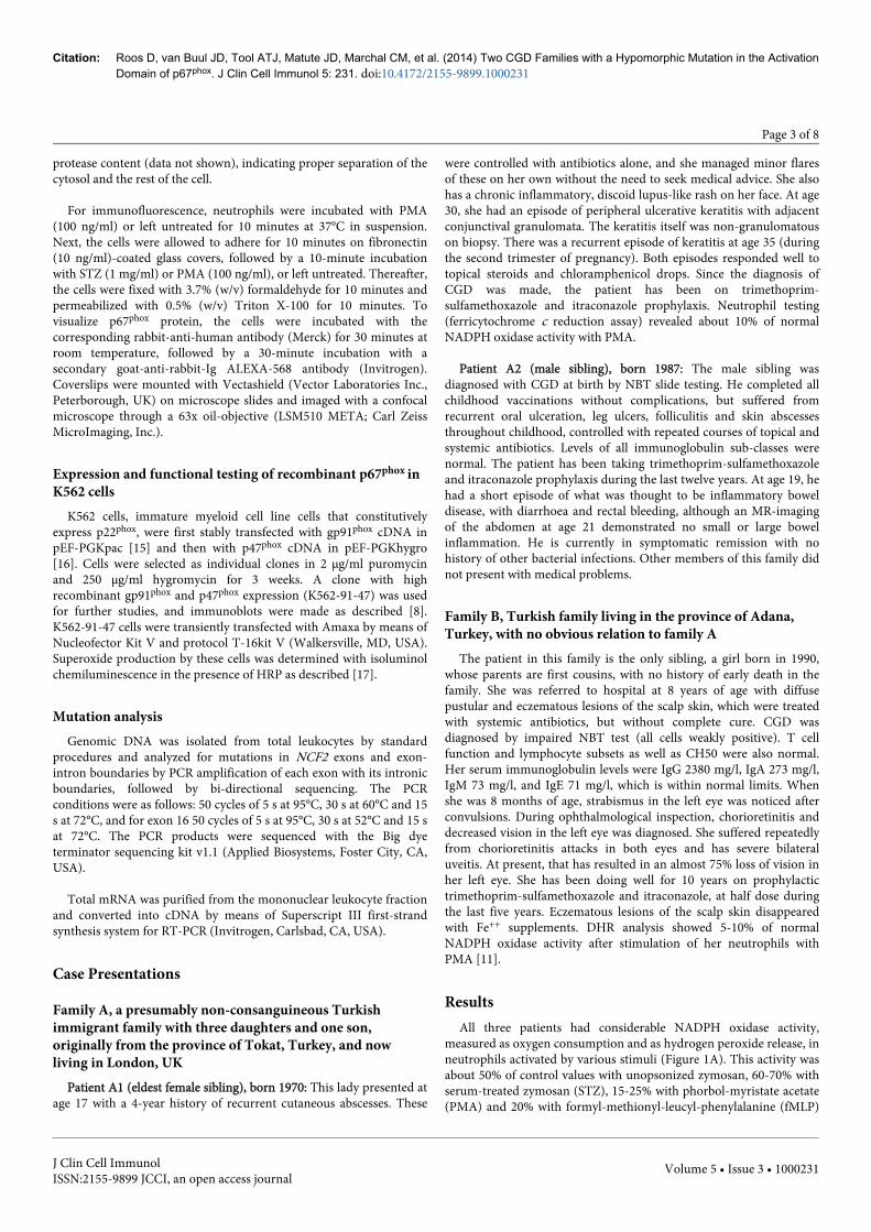
Figure 4: Translocation of p67phox to the cell membrane in intact neutrophils. (A) Quantification of p67phox translocation in stimulatedhuman neutrophils. Human neutrophils (5x10⁶/ml) were stimulated with PMA (100 ng/ml) for 10 min or with STZ (1 mg/ml) for 20 min.Separation of cytosol and the rest of the cells, followed by Western blot analysis of p67phox was performed as described under Materials andMethods. The amount of p67phox was quantified by means of fluorescently labeled conjugates, and detected by scanning with the OdysseyInfrared Imagine System and Odyssey Application Software V3.0. Black bars, control neutrophils; Red bars, patient neutrophils (patient A1,A2, and B). Mean ± SEM of 3 independent experiments for PMA. With STZ only the cells of patient B were tested. Significance of differenceswas calculated with the paired, two-tailed t-test. (B) Visualization of p67phox translocation in stimulated human neutrophils. Neutrophils froma control donor and from patient B were incubated with PMA (100 ng/ml) or left untreated for 10 minutes at 37°C in suspension. The cellswere then allowed to adhere on fibronectin-coated glass covers, followed by a 10-minute incubation with STZ (1 mg/ml) or left untreated.Thereafter, the cells were fixed with formaldehyde and permeabilized with Triton X-100. To visualize p67phox protein, the cells were incubatedwith rabbit-anti-human-p67phox, followed by incubation with a secondary goat-anti-rabbit-Ig ALEXA-568-labeled. Coverslips were mountedwith Vectashield on microscope slides and imaged with a confocal microscope through a 63x oil-objective. Note that with PMA, p67phox
translocates to the plasma membrane of control neutrophils (arrows), but much less so to the plasma membrane of patient neutrophils. Incontrast, translocation of p67phox to the phagosomal membrane surrounding internalized STZ (arrowheads) is similar in control and patientneutrophils.
DiscussionCGD patients with residual expression of NADPH oxidase
components as well as residual NADPH oxidase activity are rare. Onlyfour patients in three families have been described with low expressionof p67phox and/or low NADPH oxidase activity [22-24]. The first ofthese had a deletion of one amino acid (Lys58) on one allele of NCF2and an undefined large deletion on the other allele [22]. The Lys58-deleted protein was expressed to a certain extent (tested on Westernblot with a polyclonal antibody against p67phox), but whether this wasa normal expression (from one allele) or diminished expression couldnot be decided. The Lys58 deletion is in the fourth TPR domain anddestroyed the interaction with Rac and the translocation of p67phox tothe membrane in PMA- or STZ-activated neutrophils [22]. TheNADPH oxidase activity in the neutrophils of this patient wascompletely absent with all stimuli tested. However, in the so-calledcell-free system with recombinant proteins and neutrophilmembranes, SDS and GTPγS did induce the translocation of thesecytosolic proteins, although the NADPH oxidase activity was stillabsent.
The second patient had a missense mutation in NCF2 that causedreplacement of aspartic acid by valine at position 108 in p67phox [23].This Asp108Val replacement is between the third and fourth TPRregion in p67phox and left substantial residual NADPH oxidase activity(15-20% of normal) in the patient’s PMA-activated neutrophils (tested
in the DHR assay). In a flow cytometric assay with permeabilizedneutrophils, p67phox was undetectable with a monoclonal antibody.The authors speculate that the mutation may have changed theconformation of p67phox, rendering it undetectable with themonoclonal antibody used, but still able to interact to some extentwith gp91phox for inducing some NADPH oxidase activity.
In the last family, two brothers were recognized as CGD patientswhen they were already in their fifties [24]. They had a splice sitemutation in NCF2 that gave rise to an in-frame deletion of exons 11and 12 (amino acids 309-342, PB1 domain). The neutrophils fromthese patients showed 10-15% of normal NADPH oxidase activity afterstimulation with PMA in the DHR assay and in the lucigenin-enhanced chemiluminescence assay. This result was reproduced inK562 cells that contained all NADPH oxidase components exceptp67phox transduced with the Δexon11_12p67phox cDNA. The authorsspeculate that the p67phox protein with the exon11_12 deletion was tosome extent expressed and functional in the patients’ phagocytes.
The hypomorphic mutation in the three patients described in thisarticle is in the so-called Activation Domain of p67phox
(Supplementary Figure S1). This stretch of twelve amino acids(199-210) is essential for the oxidase-inducing capacity of p67phox[20].In a cell-free oxidase system, it was found that mutations in thisdomain do not affect binding of p67phox to p47phox or to Rac but doinhibit the oxidase activity [20]. Alanine-202 is highly conserved in
Citation: Roos D, van Buul JD, Tool ATJ, Matute JD, Marchal CM, et al. (2014) Two CGD Families with a Hypomorphic Mutation in the ActivationDomain of p67phox. J Clin Cell Immunol 5: 231. doi:10.4172/2155-9899.1000231
Page 6 of 8
J Clin Cell ImmunolISSN:2155-9899 JCCI, an open access journal
Volume 5 • Issue 3 • 1000231
p67phox from humans, mouse, chicken, frog, fish and lancelet, as wellas in Noxa1 of humans, mouse and fish and in fungal NoxR [21].Mutation of alanine-202 in p67phox into leucine inhibits the cell-freeoxidase activity induced by arachidonic acid by about 50%, and aVal204Ala mutant totally blocks this activity. This last mutantassociates with the membrane (presumably with gp91phox) as well asdoes the wild-type p67phox [20]. Direct interaction of p67phox withgp91phox was shown by Dang et al. [25,26] by overlay techniques andGST pull-down assays, but the Activation Domain of p67phox was notnecessary for this reaction. Thus, the binding of p67phox to gp91phox isprobably mediated by a site in p67phox different from the ActivationDomain, but the induction of oxidase activity in gp91phox is strictlydependent on this domain. The site in gp91phox interacting with theActivation Domain of p67phox is not known.
The findings in our patients corroborate these notions and extendthe findings to intact, primary phagocytes. We found partial inhibitionof oxidase activity in the patients’ intact neutrophils with Ala202Valp67phox, as was found with the recombinant Ala202Leu variant ofp67phox in the cell-free system [20]. Remarkably, much more oxidaseactivity was induced in the patients’ neutrophils with zymosan or STZthan with the soluble activators PMA or PAF/fMLP. This correlateswith the normal translocation of p67phox to the membrane afterneutrophil activation with STZ and clearly diminished translocationafter activation with PMA. It suggests that the Ala202Val mutation inp67phox affects the translocation and – perhaps as a result – also theproper assembly or activation of the NADPH oxidase complexfollowing translocation of the cytosolic components toflavocytochrome b558 in the plasma membrane. This reducedtranslocation of Ala202Val-p67phox might be due to reduced bindingof p67phox to gp91phox, which would be in contrast to the conclusiondrawn by Dang et al. from studies with purified neutrophil andrecombinant proteins that the Activation Domain of p67phox does notinteract with gp91phox [25,26]. Unfortunately, the 3D structure of theActivation Domain of p67phox is unknown [27-30]. However, it isknown that different stimuli induce different activation pathways inneutrophils, especially with respect to the synthesis of various lipidproducts needed for assembly of an active oxidase complex [31-34].The type or amount of lipid mediators generated in STZ-activatedneutrophils may have been sufficient for almost normal oxidaseactivation by Ala202Val-p67phox, in contrast to the situation in PMA-activated neutrophils. Since lipid mediator generation in K562 cellsmay be different from neutrophils, this may also explain the lowoxidase activation by Ala202Val-p67phox in transfected K562 cells ascompared to the patients’ neutrophils. Alternatively, since the signaltransduction pathway induced by PMA (protein kinase C activationleading to p47phox phosphorylation) is different from that induced bySTZ (tyrosine phosphorylation of PI3 kinase leading to GEF and Racactivation), the translocation of p67phox to gp91phox and subsequentactivation of gp91phox may be differently affected by mutations inp67phox. Thus, the p47phox-dependent pathway induced by PMA maybe more sensitive to mutations leading to conformational changes inp67phox than the Rac-dependent pathway induced by STZ.
Our patients raise the question how much NADPH oxidase activityis required to be able to lead a normal life. The high residual oxidaseactivity in the patients’ neutrophils may have protected the patients toa certain extent from the full-blown CGD symptomatology. Indeed,their clinical problems were mild in comparison to those of oxidase-null CGD patients. On the other hand, it should be taken into accountthat we tested the neutrophil NADPH oxidase activity in in vitro assaysystems, with strong stimuli. In vivo, more subtle stimuli may be
encountered, with which the mutant p67phox may be unable toproperly activate the NADPH oxidase. Thus, high residual NADPHoxidase activity in vitro is no guarantee for protection againstpathogenic infections in vivo, but it may help in ameliorating thesymptoms [35] and increase the chance of survival [36].
AcknowledgementsDR and MYK are recipients of an EURO-CGD grant from the E-
RARE program of the European Union.
References1. Roos D, van Bruggen R, Meischl C (2003) Oxidative killing of microbes
by neutrophils. Microbes Infect 5: 1307-1315.2. Nauseef WM (2004) Assembly of the phagocyte NADPH oxidase.
Histochem Cell Biol 122: 277-291.3. Koga H, Terasawa H, Nunoi H, Takeshige K, Inagaki F, et al. (1999)
Tetratricopeptide repeat (TPR) motifs of p67(phox) participate ininteraction with the small GTPase Rac and activation of the phagocyteNADPH oxidase. J Biol Chem 274: 25051-25060.
4. Diebold BA, Bokoch GM (2001) Molecular basis for Rac2 regulation ofphagocyte NADPH oxidase. Nat Immunol 2: 211-215.
5. Roos D, Holland SM, Kuijpers TW (2014) Chronic granulomatousdisease. In: HD Ochs, CIE Smith, JM Puck (Eds), PrimaryImmunodeficiency Diseases. A Molecular and Genetic Approach.(3rdedn) Oxford University Press, Oxford, pp: 689-722.
6. Roos D, Kuhns DB, Maddalena A, Roesler J, Lopez JA, et al. (2010)Hematologically important mutations: X-linked chronic granulomatousdisease (third update). Blood Cells Mol Dis 45: 246-265.
7. Roos D, Kuhns DB, Maddalena A, Bustamante J, Kannengiesser C, et al.(2010) Hematologically important mutations: the autosomal recessiveforms of chronic granulomatous disease (second update). Blood CellsMol Dis 44: 291-299.
8. Matute JD, Arias AA, Wright NA, Wrobel I, Waterhouse CC, et al.(2009) A new genetic subgroup of chronic granulomatous disease withautosomal recessive mutations in p40 phox and selective defects inneutrophil NADPH oxidase activity. Blood 114: 3309-3315.
9. Stasia MJ, Li XJ (2008) Genetics and immunopathology of chronicgranulomatous disease. Semin Immunopathol 30: 209-235.
10. Weening RS, Roos D, Loos JA (1974) Oxygen consumption ofphagocytizing cells in human leukocyte and granulocyte preparations: acomparative study. J Lab Clin Med 83: 570-577.
11. Köker MY, Sanal O, van Leeuwen K, de Boer M, Metin A, et al. (2009)Four different NCF2 mutations in six families from Turkey and anoverview of NCF2 gene mutations. Eur J Clin Invest 39: 942-951.
12. Meerhof LJ, Roos D (1986) Heterogeneity in chronic granulomatousdisease detected with an improved nitroblue tetrazolium slide test. JLeukoc Biol 39: 699-711.
13. Elloumi HZ, Holland SM (2007) Diagnostic assays for chronicgranulomatous disease and other neutrophil disorders. Methods Mol Biol412: 505-523.
14. Zhou M, Diwu Z, Panchuk-Voloshina N, Haugland RP (1997) A stablenonfluorescent derivative of resorufin for the fluorometric determinationof trace hydrogen peroxide: applications in detecting the activity ofphagocyte NADPH oxidase and other oxidases. Anal Biochem 253:162-168.
15. Zhen L, King AA, Xiao Y, Chanock SJ, Orkin SH, et al. (1993) Genetargeting of X chromosome-linked chronic granulomatous disease locusin a human myeloid leukemia cell line and rescue by expression ofrecombinant gp91phox. Proc Natl Acad Sci U S A 90: 9832-9836.
16. Price MO, McPhail LC, Lambeth JD, Han CH, Knaus UG, et al. (2002)Creation of a genetic system for analysis of the phagocyte respiratoryburst: high-level reconstitution of the NADPH oxidase in anonhematopoietic system. Blood 99: 2653-2661.
Citation: Roos D, van Buul JD, Tool ATJ, Matute JD, Marchal CM, et al. (2014) Two CGD Families with a Hypomorphic Mutation in the ActivationDomain of p67phox. J Clin Cell Immunol 5: 231. doi:10.4172/2155-9899.1000231
Page 7 of 8
J Clin Cell ImmunolISSN:2155-9899 JCCI, an open access journal
Volume 5 • Issue 3 • 1000231
17. Tian W, Li XJ, Stull ND, Ming W, Suh CI, et al. (2008) Fc gamma R-stimulated activation of the NADPH oxidase: phosphoinositide-bindingprotein p40phox regulates NADPH oxidase activity after enzymeassembly on the phagosome. Blood 112: 3867-3877.
18. Vowells SJ, Fleisher TA, Sekhsaria S, Alling DW, Maguire TE, et al.(1996) Genotype-dependent variability in flow cytometric evaluation ofreduced nicotinamide adenine dinucleotide phosphate oxidase functionin patients with chronic granulomatous disease. J Pediatr 128: 104-107.
19. Dekker J, de Boer M, Roos D (2001) Gene-scan method for therecognition of carriers and patients with p47(phox)-deficient autosomalrecessive chronic granulomatous disease. Exp Hematol 29: 1319-1325.
20. Leusen JH, de Klein A, Hilarius PM, Ahlin A, Palmblad J, et al. (1996)Disturbed interaction of p21-rac with mutated p67-phox causes chronicgranulomatous disease. J Exp Med 184: 1243-1249.
21. Yu G, Hong DK, Dionis KY, Rae J, Heyworth PG, et al. (2008) Focus onFOCIS: the continuing diagnostic challenge of autosomal recessivechronic granulomatous disease. Clin Immunol 128: 117-126.
22. Roesler J, Segerer F, Morbach H, Kleinert S, Thieme S, et al. (2012) P67-phox (NCF2) lacking exons 11 and 12 is functionally active and leads toan extremely late diagnosis of chronic granulomatous disease (CGD).PLoS One 7: e34296.
23. Han CH, Freeman JL, Lee T, Motalebi SA, Lambeth JD (1998) Regulationof the neutrophil respiratory burst oxidase. Identification of an activationdomain in p67(phox). J Biol Chem 273: 16663-16668.
24. Maehara Y, Miyano K, Yuzawa S, Akimoto R, Takeya R, et al. (2010) Aconserved region between the TPR and activation domains of p67phoxparticipates in activation of the phagocyte NADPH oxidase. J Biol Chem285: 31435-31445.
25. Dang PM, Cross AR, Babior BM (2001) Assembly of the neutrophilrespiratory burst oxidase: a direct interaction between p67PHOX andcytochrome b558. Proc Natl Acad Sci U S A 98: 3001-3005.
26. Dang PM, Cross AR, Quinn MT, Babior BM (2002) Assembly of theneutrophil respiratory burst oxidase: a direct interaction betweenp67PHOX and cytochrome b558 II. Proc Natl Acad Sci U S A 99:4262-4265.
27. Lapouge K, Smith SJ, Walker PA, Gamblin SJ, Smerdon SJ, et al. (2000)Structure of the TPR domain of p67phox in complex with Rac.GTP. MolCell 6: 899-907.
28. Grizot S, Fieschi F, Dagher MC, Pebay-Peyroula E (2001) The active N-terminal region of p67phox. Structure at 1.8 A resolution andbiochemical characterizations of the A128V mutant implicated inchronic granulomatous disease. J Biol Chem 276: 21627-21631.
29. Yuzawa S, Miyano K, Honbou K, Inagaki F, Sumimoto H (2009) Thedomain organization of p67 phox, a protein required for activation of thesuperoxide-producing NADPH oxidase in phagocytes. J Innate Immun 1:543-555.
30. Durand D, Vivès C, Cannella D, Pérez J, Pebay-Peyroula E, et al. (2010)NADPH oxidase activator p67(phox) behaves in solution as amultidomain protein with semi-flexible linkers. J Struct Biol 169: 45-53.
31. Bromberg Y, Pick E (1983) Unsaturated fatty acids as second messengersof superoxide generation by macrophages. Cell Immunol 79: 240-252.
32. Bromberg Y, Pick E (1984) Unsaturated fatty acids stimulate NADPH-dependent superoxide production by cell-free system derived frommacrophages. Cell Immunol 88: 213-221.
33. Shiose A, Sumimoto H (2000) Arachidonic acid and phosphorylationsynergistically induce a conformational change of p47phox to activate thephagocyte NADPH oxidase. J Biol Chem 275: 13793-13801.
34. Palicz A, Foubert TR, Jesaitis AJ, Marodi L, McPhail LC (2001)Phosphatidic acid and diacylglycerol directly activate NADPH oxidase byinteracting with enzyme components. J Biol Chem 276: 3090-3097.
35. Köker MY, Camcıoğlu Y, van Leeuwen K, Kılıç SŞ, Barlan I, Yılmaz M, etal. (2013) Clinical, functional, and genetic characterization of chronicgranulomatous disease in 89 Turkish patients. J Allergy Clin Immunol132: 1156-1163.
36. Kuhns DB, Alvord WG, Heller T, Feld JJ, Pike KM, et al. (2010) ResidualNADPH oxidase and survival in chronic granulomatous disease. N Engl JMed 363: 2600-2610.
Citation: Roos D, van Buul JD, Tool ATJ, Matute JD, Marchal CM, et al. (2014) Two CGD Families with a Hypomorphic Mutation in the ActivationDomain of p67phox. J Clin Cell Immunol 5: 231. doi:10.4172/2155-9899.1000231
Page 8 of 8
J Clin Cell ImmunolISSN:2155-9899 JCCI, an open access journal
Volume 5 • Issue 3 • 1000231