Prognostic value of routinely assessed serum biomarkers in septic shock
Upload
independentCategory
view
0download
0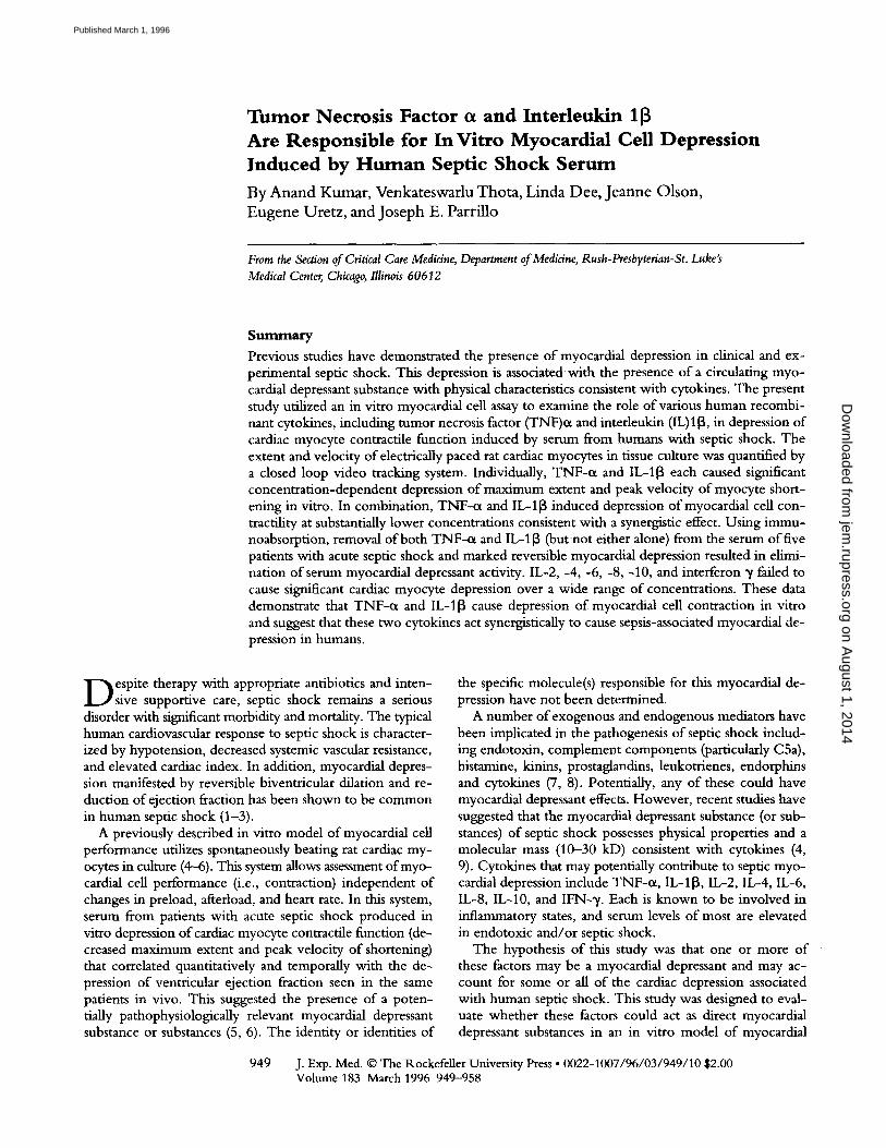
Tumor Necrosis Factor et and Interleukin 1~ Are Responsible for In Vitro Myocardial Cell Depression Induced by Human Septic Shock Serum B y A n a n d Kumar , Venkateswarlu Tho ta , L inda Dee , Jeanne Olson, Eugene Uretz , and Joseph E. Parril lo
From the Section of Critical Care Medicine, Department of Medicine, Rush-Presbyterian-St. Luke's Medical Center, Chicago, Illinois 60612
Summary Previous studies have demonstrated the presence of myocardial depression in clinical and ex- perimental septic shock. This depression is associated with the presence of a circulating myo- cardial depressant substance with physical characteristics consistent with cytokines. The present study utilized an in vitro myocardial cell assay to examine the role of various human recombi- nant cytokines, including tumor necrosis factor (TNF)ot and interleukin (IL)113, in depression of cardiac myocyte contractile function induced by serum from humans with septic shock. The extent and velocity of electrically paced rat cardiac myocytes in tissue culture was quantified by a closed loop video tracking system. Individually, TNF-0c and IL-113 each caused significant concentration-dependent depression of maximum extent and peak velocity of myocyte short- ening in vitro. In combination, TNF-cx and IL-1[3 induced depression of myocardial cell con- tractility at substantially lower concentrations consistent with a synergistic effect. Using immu- noabsorption, removal of both TNF-cx and IL-113 (but not either alone) from the serum of five patients with acute septic shock and marked reversible myocardial depression resulted in elimi- nation of serum myocardial depressant activity. IL-2, -4, -6, -8, -10, and interferon ~/failed to cause significant cardiac myocyte depression over a wide range of concentrations. These data demonstrate that TNF-0t and IL-113 cause depression of myocardial cell contraction in vitro and suggest that these two cytokines act synergistically to cause sepsis-associated myocardial de- pression in humans.
D espite therapy with appropriate antibiotics and inten- sive supportive care, septic shock remains a serious
disorder with significant morbidity and mortality. The typical human cardiovascular response to septic shock is character- ized by hypotension, decreased systemic vascular resistance, and elevated cardiac index. In addition, myocardial depres- sion manifested by reversible biventricular dilation and re- duction of ejection fraction has been shown to be common in human septic shock (1-3).
A previously described in vitro model of myocardial cell performance utilizes spontaneously beating rat cardiac my- ocytes in culture (4-6). This system allows assessment of myo- cardial cell performance (i.e., contraction) independent of changes in preload, afterload, and heart rate. In this system, serum from patients with acute septic shock produced in vitro depression of cardiac myocyte contractile function (de- creased maximum extent and peak velocity of shortening) that correlated quantitatively and temporally with the de- pression of ventricular ejection fraction seen in the same patients in vivo. This suggested the presence of a poten- tially pathophysiologicaUy relevant myocardial depressant substance or substances (5, 6). The identity or identifies of
the specific molecule(s) responsible for this myocardial de- pression have not been determined.
A number of exogenous and endogenous mediators have been implicated in the pathogenesis of septic shock includ- ing endotoxin, complement components (particularly CSa), histamine, kinins, prostaglandins, leukotrienes, endorphins and cytokines (7, 8). Potentially, any of these could have myocardial depressant effects. However, recent studies have suggested that the myocardial depressant substance (or sub- stances) of septic shock possesses physical properties and a molecular mass (10-30 kD) consistent with cytokines (4, 9). Cytokines that may potentially contribute to septic myo- cardial depression include TNF-cx, IL-113, IL-2, IL-4, IL-6, IL-8, IL-10, and IFN-~/. Each is known to be involved in inflammatory states, and serum levels of most are elevated in endotoxic and/or septic shock.
The hypothesis of this study was that one or more of these factors may be a myocardial depressant and may ac- count for some or all of the cardiac depression associated with human septic shock. This study was designed to eval- uate whether these factors could act as direct myocardial depressant substances in an in vitro model of myocardial
949 j. Exp. Med. �9 The Rockefeller University Press �9 0022-1007/96/03/949/10 $2.00 Volume 183 March 1996 949-958
on August 1, 2014
jem.rupress.org
Dow
nloaded from
Published March 1, 1996

function and whether elimination of these substances from human septic serum would attenuate its in vitro myocardial depressant activity,
Materials and Methods
The methods employed were a modification of those previ- ously described (4-6). The basic myocyte assay is designed to measure the depression or enhancement of contractility of beat- ing cardiac myocytes in cell culture caused by agents that are in- troduced into the growth medium. Spontaneously beating new- born rat myocardial cells were established using a modification of the technique described by Harary and Farley (10). Using sterile technique, hearts from 2-d-old Lewis rat pups were removed, pooled, and minced into small blocks. The cells were disaggre- gated with 0.15% bovine pancreatic trypsin (Sigma Chemical Co., St. Louis, MO) in a modified HBSS (potassium, calcium, and magnesium free; GIBCO BRL, Gaithersburg, MD). Suspensions were centrifuged at 1,500 rpm (500 g) for 15 min and plated into 35 • 10 mm petri dishes at a density of 300,000 cells/ml. Plating media (standard control media) consisted of 25% Hepes-buffered Medium 199 and 10% heat-inactivated newborn calf serum (both from GIBCO BIKL) diluted in a balanced salt solution and sup- plemented with glutamine, penicillin, and streptomycin (all from Sigma Chemical Co.). Cells were incubated at 37~ in 5% CO2 and growth media was changed every 48 h. After plating, cells became confluent and began spontaneously beating within 4 d. Latex microbeads were introduced into the culture after 4 d and affixed themselves to cell membranes. Beating cells were used for the assay between 5 and 10 d after plating.
All pipettes, plates, and other equipment used for preparation, culture, or testing of cardiac myocytes were endotoxin tested and disposable. All liquid media contained < 1 pg/ml endotoxin with the exception of newborn calf serum which contained 0.48 ng/ ml endotoxin. All recombinant cytokines contained <50 pg en- dotoxin per big cytokine. Culture media, cytokine solutions, and other test solutions were tested for endotoxin content using a quantitative, chromogenic Limulus amebocyte lysate assay (Whit- taker M.A. Bioproducts, Walkersville, MD).
The extent and velocity of myocardial cell shortening during cell contraction was assayed by a modification of previously de- scribed techniques (4--6). The petri dish containing beating myo- cardial cells was fastened to the heated (37~ stage of an inverted- optics, phase-contrast microscope that had an attached video camera. A television monitor displayed the image of the target cells. A custom-built electronic tracking system was used to quan- titate the movement of a latex bead selected from the many beads attached to the membranes of beating myocytes. The typical maxi- mum initial extent of rhythmic displacement of the bead was be- tween 3 and 10 p,m, depending on the length of the myocyte to which it was adherent. The tracking system produced analog sig- nals that were relayed to an intervening electronic instrument (which derived contraction velocity from rate of change of bead displacement) and a two-channel strip chart recorder that printed an analog recording of extent and velocity of bead displacement. To ensure a fixed contraction frequency, a custom-built alternat- ing current electrical pulse generator was used to pace myocytes (12 V, maximum 40 mA, 0.7-7-ms pulse duration). The mini- mum current and pulse duration required to effectively pace car- diac myocytes was used for each experiment.
Each individual assay was performed as follows. Plates were re- moved from the incubator and fresh growth media applied. Plates
were mounted on the microscope stage and myocytes were paced to 60 contractions per rain. An appropriate bead was located and the extent and velocity ofmyocyte shortening measured for 5 rain (baseline contractility). If extent of cell contraction was stable (maximum 2.5% variation over 5 rain), a test or control solution was added. Subsequently, measurements of maximum extent and peak velocity of myocyte shortening and velocity were obtained every 5 min for 30 rain. By comparing the maximum extent and peak velocity of shortening at each 5-rain interval to the baseline value, changes were referenced to initial contractility.
Five groups of experiments were performed. (a) Human re- combinant TNF-o~ and IL-113 were initially assayed (each along with controls) in order to evaluate whether either cytokine could individually produce concentration-dependent depression of car- diac myocyte shortening. The concentrations tested were: for TNF-ee (Sigma Chemical Co.), 0 (control), 0.0125, 0.05, 0.2, 0.8, 3.2, 12.5, 25, 50, and 100 ng/ml (n = 8 each concentration); for IL-I~ (Endogen, Inc., Boston, MA), 0 (control), 2, 8, 32, 125, 500, and 1,000 ng/ml (n = 8 each concentration). Test media consisted of standard culture media (as previously described) with the addition of the specified cytokine concentrations.
Reversibility of depression induced by TNF-ot (50 ng/ml) and IL-1 [3 (500 ng/ml) was individually determined by repeated wash- out with standard control media (n = 8 each group). 5 min after washing, maximum extent and peak velocity of shortening was determined and compared to the level of depression of similarly washed cardiac myocytes exposed only to control media.
In addition, cardiac myocytes were separated from nonmyo- cytes by centrifugation through a Percoll step gradient (Sigma Chemical, Co.) resulting in a >95-98% pure cardiac myocyte culture (viable cardiac myocytes/all viable cells). Myocyte purity was determined 48 h after plating by a combination oftrypan blue exclusion to differentiate viable and nonviable cells followed by differentiation ofmyocytes from nonmyocytes by cell contraction in response to an electrical pulse. TNF-ot (50 ng/ml) and IL-I[~ (500 ng/ml) were individually tested for effects on cardiac myo- cyte maximum extent and peak velocity of shortening using puri- fied cardiac myocyte cultures (n = 8 each group).
(b) In a second group of experiments, combinations of TNF-c~ and IL-113 were evaluated at lower concentrations to evaluate the possible additive or synergistic effects of these cytokines. In addi- tion to a control solution without either cytokine, paired test concentrations of TNF-~/IL-I[3 (ng/ml) included (a) 0.003: 0.125; (b) 0.0125:0.5; (c) 0.05:2; (d) 0.2:8 (n = 8 each group). Test media consisted of standard culture media as described with the addition of the specified cytokine concentrations.
(c) To determine the specific roles of TNF-ot and IL-113 in car- diac myocyte depression induced by human septic serum, these cytokines were removed from serum by immunoabsorption. Se- rum from five human survivors with acute septic shock associated with marked, reversible depression of left ventricular ejection fraction (mean + standard deviation left ventricular ejection frac- tion 26 _ 7% during septic shock, 55 + 7% at recovery; mean absolute decrease 28 + 10%) was studied. Each serum sample had also been previously documented to contain myocardial depres- sant activity. Serum was treated by exposing it overnight (16 h) at 4~ to agarose beads bonded to specific rabbit-derived anti- human cytokine polyclonal antibodies (Endogen, Inc.). Serum was treated with antibodies against recombinant human TNF-~x, recombinant human IL-IJ3, both, or neither (native antibodies derived from rabbits before immunization with recombinant hu- man TNF-oe or IL-I[3). Each serum sample had TNF-ot and IL- l13 concentrations determined by ELISA (TNF-ot by T Cell Sci-
950 TNF and IL-1 Cause Septic Serum-induced Myocardial Cell Depression
on August 1, 2014
jem.rupress.org
Dow
nloaded from
Published March 1, 1996

ences, Inc., Boston, MA; IL-l[3 by Citron Biotechnologies, Pine Brook, NJ) (11). The lower limit of detection in these assays was 20 pg/ml.
Insuf[icient serum remained after performing the contractility assay to test cytokine concentrations after treatment. However, effectiveness of the cytokine immunoahsorption process was con- firmed by adding TNF-ot (50 ng/ml) or IL-l[3 (500 ng/ml) to standard media (with 10% serum), similarly treating that serum to remove TNF-u or IL-113 using specific polyclonal antibodies, measuring remaining TNF-cx or IL-113 by ELISA, and confirm- ing elimination of depressant activity in the cardiac myocyte con- tractility assay.
Test solutions were created by mixing standard plating media without serum with the resultant (antibody-treated) human septic serum at a concentration of 10%. Four groups (and a control group) were tested to determine serum-induced changes in maxi- mum extent and peak velocity of cardiac myocyte shortening. The control solution consisted of 10% neonatal bovine calf serum (standard plating media) and was cormnon to all experiments (nonseptic control). The test solutions consisted of 10% native (septic control), anti-TNF-~, anti-IL-1]3, or anti-TNF-ot and anti-IL-113 treated serum (n = 8 each group). Additional control solutions in = 8 each group) consisting of 10% newborn calf se- rum treated with antibodies against recombinant human TNF-~x, recombinant human IL-113, both, or neither (native antibodies derived from rabbits before immunization with recombinant hu- man TNF-ot or IL-113).
(d) Six other human recombinant cytokines were also initially assayed (each along with controls) in order to evaluate whether any could individually produce concentration-dependent depres- sion of cardiac myocyte shortening. The concentrations tested were: IL-2, 0 (control), 1.6, 8, 40, 200, and 1,000 ng/ml (n = 8 each concentration); IL-4, 0 (control), 1.6, 8, 40, 200, and 1,000 ng/ml (n = 6 each concentration); IL-6, 0 (control), 0.5, 5, 20, 80, 320, 1,250, and 3,000 ng/ml in = 8 each concentration); IL-8, 0 (control), 1.6, 8, 40, 200, and 1,000 ng/ml (n = 7 each concen- tration); IL-10, 0 icontrul), 1.6, 8, 40, 200, and 1,000 ng/ml (n = 6 each concentration); and IFN-y, 0 icontrol), 4, 16, 64, 250, and 1,000 ng/ml in = 7 each concentration; all from Endogen, Inc.).
Where known, cytokine concentrations were selected based on a range from one order of magnitude below to two orders of magnitude above the serum cytokine concentrations documented during human septic shock. Test media consisted of standard cul- ture media including 10% neonatal calf serum with the addition of the specified cytokine concentrations.
(e) To study the potential role of endotoxin in cytokine and septic shock serum-mediated cardiac myocyte depression, se- lected test solutions were depleted ofendotoxin by overnight co- incubation with silica bead-bonded endotoxin neutralizing pro- tein derived from horseshoe crab amebocytes (Associates of Cape Cod, Inc., Falmouth, MA) and then assayed for myocardial de- pressant activity as previously described. Test media included solutions containing TNF-r (50 ng/ml), IL-1[3 (500 ng/ml), TNF-0e (0.05 ng/ml) with IL-113 (2 ng/ml), or 10% serum from one of the five patients with acute septic shock as well as controls containing 10% neonatal calf serum without cytokines or septic serum (n = 9 each group).
Statistical Analysis. For cytokine testing, data at all concentra- tions and time points were pooled and analyzed by multivariate regression to determine whether a significant relationship existed between cytokine concentration or time and degree of cardiac myocyte depression.
Data for the change in maximum extent and peak velocity of
cardiac myocyte shortening (percent change from baseline) were plotted as a function of time for each control and test solution concentration. Linear regression analysis was employed to fit a line for the each resulting plot. For cytokines that were demon- strated to exert depressant activity, the slopes of these lines were compared to that for the control solution by a two-tailed Stu- dent's t test in order to determine at what concentration these slopes became significantly different (i.e., at what concentration significant depression of cardiac myocyte contractihty occurred). In this manner, increased depressant activity was indicated by a more negative value for slope of the regression hne. Similarly, if any factor caused significant depression, a two-tailed Student's t test was used to determine at what time point significant depres- sion began.
Data for the comparison of the effects of TNF-oe and IL-113 on purified and unpurified cardiac myocytes were similarly pooled for each group. Regression-derived slopes were determined and compared to slopes for control media via a two-tailed Student's t test. A Bonferonni adjustment for multiple comparisons was apphed so that each comparison was considered significant only ifp ~<0.025.
Similarly, for analysis of antibody-treated septic serum and for endotoxin-removal experiments, the slope of the plot for the con- trol media was compared to each treatment group by a two-tailed Student's t test. A Bonferonni adjustment for multiple compari- sons was again made in both sets of experiments so that each com- parison was considered significant only ifp ~<0.0125.
Results
Effects of TNF-ee or IL-1~8. Both T N F - a and IL-113 in- dividually exhibited concentration-dependent depression of cardiac myocyte contractility.
TNF-c~-induced depression o f maximum extent o f car- diac myocyte shortening began at a concentration o f 0.8 ng /ml (p <0.05) and progressed through concentrations o f 3.2 (p <0.02), 12.5 (p <0.01), 25 (p <0.01), 50 (p <0.001), and 100 ng/ml (p <0.001). Fig. 1 A shows car- diac myocyte depression as a function o f time for the con- trol media without TNF-ot, 0.8 ng /ml TNF-tx, and 100 ng/ml T N F - a . The slopes o f the lines fit to the data at each concentration were plotted against the log o f the concen- trations (Fig. 1 /9). A highly significant relationship (p <0.0001) existed, suggesting that increased depression of maximum extent o f myocyte shortening occurred with in- creasing concentrations o f TNF-ot. Parallel and similarly significant changes in peak velocity o f myocyte shortening were also demonstrated.
Significant depression occurred by 10 min (p <0,02) with progressively more depression to 30 min (p <0.001) for both changes in maximum extent and peak velocity o f cardiac myocyte shortening. Overall, TNF-R- induced car- diac myocyte depression was significandy time dependent (p <0.0001).
IL-l[3-induced depression of maximum extent o f cardiac myocyte shortening began at a concentration o f 32 ng/ml (p <0.05) and progressed through concentrations of 128 (p <0.01), 500 (p <0.01), and 1,000 ng/ml (p <0.001). Fig- ure 1 B shows cardiac myocyte depression as a function o f time for the control media without IL-113, IL-113 32 ng/ml, and 1,000 ng /ml IL-113. The slopes of the lines fit to the
951 Kumar et al.
on August 1, 2014
jem.rupress.org
Dow
nloaded from
Published March 1, 1996

A 0
g -2o E=O
E g - * 0 .=_ ~" e ca g._~
o u ~ - 60
B 0
z g
i ' E -2O
E =
E g - * o ._= ~" }._~ o ~ - 60
C 0
~g
i=~_20 E o E "
E o ~ -40 = ~-
~_ o
~ -60
D ~ 0.5
s lope = -0 .145 E ~ • 0.146 ~K/min ~ 0.0
,,ope = - 0 . 5 9 0 o :t 0.188 ~ /min c
T p<.05 vs . con t ro l ~ o -0 .5
o:.7, ._c ~ -1 .0
s lope = --1,598 ~ o • 0.289 ~ / m i n o = ~ - I . 5
�9 control p<.O01 vs, control "~ c ~ ~7 0.8 ng/mL TNF ~ "~ �9 100 ng/mL TNF ~ ~ -2 .0 g o
5 10 1'5 20 25 3 0 ~ = t ime (minutes)
E
~ T - ~ . ~ . ~ slope = -0 .398 • 0.165 ~ /min
I I I • 0.259 ~/min
L ~ i i - l ~ p<.05 vs . con t ro l
i i ; ;go~:)m . lope = -2 .009 I • 0,467 ~ /mln p< .O01 vs . con t ro l
i i , , i
5 10 15 20 25 30 time (minutes)
F
s lope = -0 .498 • 0.167 ~ /min
s lope = --0.962 • 0.289 ~ /min
p< .05 VS. con t ro l
+0.5 ng/mk I1_-1 I slope = -2 .370 • 0.398 ~/min �9 0.2 ng/mL TNF J p<.001 vs . con t ro l
+8 ng/mL IL-1 , i = * = = 5 10 15 20 25 30
time (minutes)
o o o I
. . ~ / j , , ~ , i
0 0.01 0.1 1 10 100 TNF concen t ro t~on ( n ~ / m L )
~._. 0.0 = E EE
= -0 .5 c ~ B ~
o = - 1 . 0
E~ c ~ - 1 , 5
u o
-~ g -2.0
o .o 0~: -2 .5
o;;;::: . , r . i i i i O" I 10 100 1000 IL-1 concentration ( n g / m L )
e
-~ ~ -0.5
�9 = x . o
E = .-r 2 - 1 . 5
e o
o - 2 . 0 g g
- 2 . 5
TNF
concen t ro t ion -dependent depress ion (s lope ) p <.0001
0.003 0.0125 0.05 0.2 0.125 0.5 2.0 8.0
[ l og sca le ]
F i g u r e 1. The change in maxi- mum extent of myocardial cell shortening a~:er exposure to con- trol and the specified concentra- tions of TNF-tx (A), IL-113 (B), or TNF-ot + IL-113 (C 3. p value refers to the difference in slope between specified concentrations of cytokines and control media without cytokine. The slopes representing rate of decrease of maximum extent of myocardial cell shortening in response to dif- ferent cytokine concentrations tested are shown in D, E, and F. In each case slopes become pro- gressively more negative to the maximum cytokine concentra- tions tested, p value refers to dif- ference from 0 slope. A signifi- cant p value indicates the existence of a concentration-dependent re- lationship. Error bars, SEM.
data at each concentration were plotted against the log o f the concentrations (Fig. 1 E). As with TNF-ot, a highly sig- nificant relationship (p <0.0001) existed, suggesting that increased depression o f maximum extent o fmyocy te short- erring occurred with increasing concentrations o f IL-1I~. Data for changes in peak velocity were similar. Also similar to TNF-a , significant depression of both maximum extent and peak vdoci ty o f cardiac myocyte shortening occurred by 10 min (p <0.02) and increased through 30 min (p <0.001). Depression was significantly time dependent (p <0.0001) in addition to being concentration dependent for both maximum extent and peak velocity o f cardiac myocyte shortening.
50 ng/ml TNF-ot and 500 ng /ml IL-113 each also pro- duced significant depression of both maximum extent and
peak velocity of shortening (p <0.01 each) when applied to highly purified cardiac myocyte tissue cultures (>95-98% purity versus normal 75-80%).
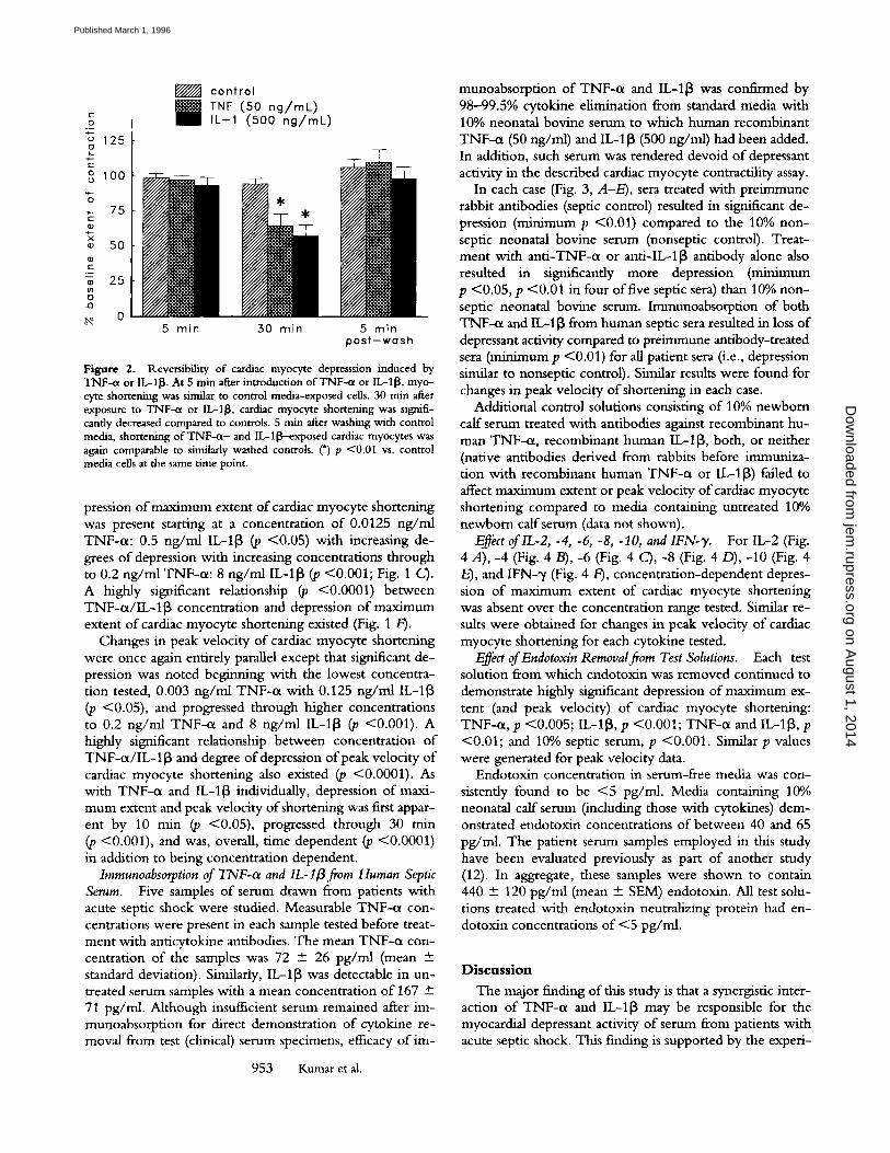
Reversal o f 50 ng /ml TNF-0t or 500 ng /ml IL-I[3 in- duced depression o f maximum extent (and peak velocity) o f cardiac myocyte contractility could be produced by wash- ing the cell monolayer with standard growth media with- out cytokine (Fig. 2). Contractile function o f cardiac myo- cytes reverted to baseline within 5 min of washing the cells with cytokine-free media.
Trypan blue supravital staining failed to demonstrate loss of cell viability within the cardiac myocyte culture. N o in- crease in supematant lactate dehydrogenase was noted before and after exposure o f cardiac myocytes to TNF-o~ or IL-I~.
Effects of TNF-a and IL-113 in Combination. Significant de-
952 TNF and IL-1 Cause Septic Serum-induced Myocardial Cell Depression
on August 1, 2014
jem.rupress.org
Dow
nloaded from
Published March 1, 1996
o 125 a
o~ 100
-~ 75
~ 50 O2 C
~ 25
N 0
control B TNF (50 ng/mL)
I L - I (500 ng /mL)
5 mln 30 min 5 rain post -wash
Figure 2. Reversibility of cardiac myocyte depresssion induced by TNF-ct or IL-1I~. At 5 rain after introduction of TNF-qx or IL-118, myo- cyte shortening was similar to control media-exposed cells. 30 rain after exposure to TNF-cx or IL-1I~, cardiac myocyte shortening was signifi- cantly decreased compared to controls. 5 min after washing with control media, shortening of TNF-a- and IL-l~-exposed cardiac myocytes was again comparable to similarly washed controls. (*) p <0.01 vs. control media cells at the same time point.
pression of maximum extent of cardiac myocyte shortening was present starting at a concentration of 0.0125 ng/ml TNF-ct: 0.5 ng/ml IL-1{3 (p <0.05) with increasing de- grees of depression with increasing concentrations through to 0.2 ng/ml TNF-ot: 8 ng/ml IL-113 (p <0.001; Fig. 1 C). A highly significant relationship (p <0.0001) between TNF-cx/IL-lJ3 concentration and depression of maximum extent of cardiac myocyte shortening existed (Fig. 1 F').
Changes in peak velocity of cardiac myocyte shortening were once again entirely parallel except that significant de- pression was noted beginning with the lowest concentra- tion tested, 0.003 ng/ml TNF-cx with 0.125 ng/ml IL-lJ3 (p <0.05), and progressed through higher concentrations to 0.2 ng/ml TNF-ot and 8 ng/ml IL-113 (p <0.001). A highly significant relationship between concentration of TNF-a/IL-lJ3 and degree of depression of peak velocity of cardiac myocyte shortening also existed (p <0.0001). As with TNF-ct and IL-1[3 individually, depression of maxi- mum extent and peak velocity of shortening was first appar- ent by 10 rain (p <0.05), progressed through 30 rain (p <0.001), and was, overall, time dependent (p <0.0001) in addition to being concentration dependent.
Immunoabsorption of TNF-ot and IL-l~S from Human Septic Serum. Five samples of serum drawn from patients with acute septic shock were studied. Measurable TNF-ct con- centrations were present in each sample tested before treat- ment with anticytokine antibodies. The mean TNF-et con- centration of the samples was 72 +- 26 pg/ml (mean +- standard deviation). Similarly, IL-1[3 was detectable in un- treated serum samples with a mean concentration of 167 -+ 71 pg/ml. Although insufficient serum remained after im- munoabsorption for direct demonstration of cytokine re- moval from test (clinical) serum specimens, efficacy of im-
953 Kumar et al.
munoabsorption of TNF-ot and IL-1J3 was confirmed by 98-99.5% cytokine elimination from standard media with 10% neonatal bovine serum to which human recombinant TNF-ct (50 ng/ml) and IL-113 (500 ng/ml) had been added. In addition, such serum was rendered devoid of depressant activity in the described cardiac myocyte contractility assay.
In each case (Fig. 3, A-E), sera treated with preimmune rabbit antibodies (septic control) resulted in significant de- pression (minimum p <0.01) compared to the 10% non- septic neonatal bovine serum (nonseptic control). Treat- ment with anti-TNF-ot or anti-IL-l[3 antibody alone also resuked in significantly more depression (minimum p <0.05, p <0.01 in four of five septic sera) than 10% non- septic neonatal bovine serum. Immunoabsorption of both TNF-cx and I[.-113 from human septic sera resulted in loss of depressant activity compared to preimmune antibody-treated sera (minimum p <0.01) for all patient sera (i.e., depression similar to nonseptic control). Similar results were found for changes in peak velocity of shortening in each case.
Additional control solutions consisting of 10% newborn calf serum treated with antibodies against recombinant hu- man TNF-ct, recombinant human IL-lJ3, both, or neither (native antibodies derived from rabbits before immuniza- tion with recombinant human TNF-cx or IL-113) failed to affect maximum extent or peak velocity of cardiac myocyte shortening compared to media containing untreated 10% newborn calf serum (data not shown).
Effect of lL-2, -4, -6, -8, -10, and IFN-% For IL-2 (Fig. 4 A), -4 (Fig. 4 B), -6 (Fig. 4 C), -8 (Fig. 4 D), -10 (Fig. 4 E), and IFN-~/(Fig. 4 F), concentration-dependent depres- sion of maximum extent of cardiac myocyte shortening was absent over the concentration range tested. Similar re- suits were obtained for changes in peak velocity of cardiac myocyte shortening for each cytokine tested.
Effect of Endotoxin Removal from Test Solutions. Each test solution from which endotoxin was removed continued to demonstrate highly significant depression of maximum ex- tent (and peak velocity) of cardiac myocyte shortening: TNF-ct, p <0.005; IL-1[3, p <0.001; TNF-ct and IL-lJ3, p <0.01; and 10% septic serum, p <0.001. Similar p values were generated for peak velocity data.
Endotoxin concentration in serum-free media was con- sistently found to be <5 pg/ml. Media containing 10% neonatal calf serum (including those with cytokines) dem- onstrated endotoxin concentrations of between 40 and 65 pg/ml. The patient serum samples employed in this study have been evaluated previously as part of another study (12). In aggregate, these samples were shown to contain 440 + 120 pg/ml (mean +- SEM) endotoxin. All test solu- tions treated with endotoxin neutralizing protein had en- dotoxin concentrations of <5 pg/ml.
D i s c u s s i o n
The major finding of this study is that a synergistic inter- action of TNF-~t and IL-1[3 may be responsible for the myocardial depressant activity of serum from patients with acute septic shock. This finding is supported by the experi-
on August 1, 2014
jem.rupress.org
Dow
nloaded from
Published March 1, 1996

A
gE ~ . 0 . 0 E ~
_ E c ~ - O . 5 t - - r
m o -I,5
~
,;~ -2 .0
oko - 2 . 5
D
~ o.o E ~ ~ o
= E -0.5
~ - 1 . 0
~2 ~ o - t . S
o o
~ T •
T
J_
n o n - sep t ic a n t { - a n t i - a n t i - sept;c c o n t r o l TNF i L - I TNF c o n t r o l + a n t i - -
I L - 1
T
,T *~ I •
B Q ~ 7-.. ':
~ 00 E ~
.~ ~-E -o..5 e
x ,o - 1 . o
~ o - t . 5
c - 2 , 0 o C
~ - 2 . 5
E
E~
~ -0.s I ~ i- i~-,0 ~ o - 1 . 5 c ~ o o
~ - z . 0
m ~
ok; -2.5
•
T
_L
non-- sept lc sept{c cont ro l con t ro l
a n t i - a n t l - TNF IL--I
a n t l - TNF
+ a n t i - I L - 1
T /
1
n o n - sept ic a n t i - a n t i - a n t i - n o n - sept ic a n t l - a n t i - a n t i - sept ic cont ro l TNF I L - t TNF mept;c con t ro l TNF I L - t TNF cont ro l + o n t ~ - c o n t r o l + o n t l -
I L -1 LL - t
C o~ ;._c
~ o.o E~ <>a
~E -o.5
o
~2 a o - 1 . 5
g - o
e - 2 . 0
T
* l
n o n - sept ic a n t i - a n t i - a n t l - =ept lc cont ro l TNF t k - 1 TNF cont ro l + a n t l -
IL-I
Figure 3. Regression slopes representing the change of maximum extent of myocardial cell shortening (as a function of time) in response to an- ticytokine-treated human septic serum for each pa- tient (A-E). Septic serum treated with preimmune antibodies (septic control) consistently demonstrated greater depressant activity than did nonseptic con- tro] serum (minimum p <0.01). Only immunoab- sorption of both TNF-~x and IL-1lg from the septic sere resulted in attenuation of serum depressant ac- tivity so that myocyte contractility was similar to nonseptic controls for each patient. Serum immu- noabsorbed of TNF-cx and IL-113 exerted signifi- cantly less depressant effect on maximum extent of cardiac myocyte shortening than did preimmune
antibody treated (septic control) samples (minimum p <0,0I), A Bonferonni adjustment for multiple comparisons was made so that each comparison was considered significant only ifp ~<0,0125, (*) p <0.05; (i') P <0.01; (:[:) p <0.001 vs, nonseptic control, Error bars, SEM.
mental data which demonstrate that immunoabsorption o f both TNF-~x and IL-113 abrogates in vitro myocardial de- pressant activity o f serum from humans with acute septic shock associated with clinical myocardial depression (de- creased left ventricular ejection fraction). This conclusion is strengthened by additional data which show that, whereas T N F m and IL-113 independently produce a concentra- t ion-dependent depression o f cardiac myocyte contractility, the same cytokines in combination produce similar depres- sion at far lower concentrations (comparable to those found in human septic serum).
The primary evidence of the importance of an interac- tion o f TNF-ol and IL-113 during human septic shock is provided by experiments involving immunoabsorption o f TNF-oL, IL-113, or both from human septic serum. This se- rum was drawn from patients with acute septic shock and marked reversible myocardial depression as evidenced by a decreased ejection fraction (26 --- 7% during acute septic shock, 55 -+ 7% after recovery) and had been shown to possess a substantial degree o f myocardial depressant activ- ity in this in vitro cardiac myocyte assay, tmmunoabsorp- tion o f either TNF-ot or IL-113 from this serum resulted in relatively little attenuation o f myocardial depressant activity in comparison to preimmune, antibody-treated septic se- rum (septic control). However, immunoabsorption of both TNF-ct and IL-113 from the serum resulted in virtual elim-
ination o f depressant activity. This result was seen in each of the five individual patient sera tested and was statistically highly significant.
Synergism between TNF-~x and IL-113 in the context of this study implies that complete elimination o f either should result in loss o f serum myocardial depressant activ- ity. There are at least two possible reasons for the limited attenuation o f serum myocardial depressant activity seen in this study when only TNF-oe or IL-113 was immunoab- sorbed. As we have shown, immunoabsorption is not en- tirely effective in cytokine removal. The small amounts o f TNF-cx or IL-113 that remain may be sufficient to sustain synergy. However, when both cytokines are targetted, total TNF-oe and IL-113 may drop below the threshold required for expression of synergistic activity. Alternately, the gener- ation o f septic serum-induced cardiac myocyte dysfunction may be more complex than a limited interaction between TNF-ot and IL-1[~. Although other cytokines have no de- monstrable depressant activity o f their own, they may pro- duce a synergistic effect so that removal of only TNF-cl or IL-t13 is inadequate to eliminate serum depressant activity.
Further support for the synergistic role o f TNF-oe and IL-113 in septic myocardial depression comes from our ex- periments which show that, in combination, TNF-oe and IL-113 cause cardiac myocyte depression at concentrations that are incompatible with merely additive depressant ac-
954 TNF and IL-1 Cause Septic Serum-induced Myocardial Cell Depression
on August 1, 2014
jem.rupress.org
Dow
nloaded from
Published March 1, 1996

A ~ 0.0
.E -o.5 c : ~
" ~ o c
x o
E ~-1 .o .E 2
o~ ~o
B ~ 0.0 ~-,.E
if~_.
fi_ - 0 . 5 c ~
" ~ o
if ~ -1 .o
go g ~ -~ ~-1 .5 c o n c e n t r a t i o n - d e p e n d e n t p = NS
d e p r e s s i o n ( s l o p e ) ~o r
~ o o J c
0 I 10 100 1 0 0 0 -~ ~
tL - -2 r ( n g / r n L )
C ~ 0.0
o if
'~ ~-0 .5
x a
if ~ -1 .0
~ o o , c ~ o o
c o n c e n t r a t i o n - d e p e n d e n t p = NS d e p r e s s i o n ( s l o p e ) ~ "~
0 " 1 10 100 1 0 0 0
I L - 4 c o n c e n t r a t i o n ( n g / m L )
+ + -
c o n c e n t r o t i o n - d e p e n d e n t p = NS d e p r e s s i o n ( e l o p e )
= / / i i i ~ i 0 0.1 1 10 100 1 0 0 0
I L - 6 c o n c e n t r o t ~ o n ( n g / r n L )
D ~.~EE 0.0
u E o.~ E L
~6 ~ - 0 . 5
~ c
E ~ - 1 . 0
g~
"g ~-1.5 "~'~
o . 1 :
E ~ 0 .0 ~ , .E u E
E L
"~ ~-0.5
~g
c ~
go g- o
o . o
t ! TI F ~ 0 .0
u i f o
E L
~ ._~ -0.5
if ~ -1 .0 .c 2 go gg
concentratJon-dependent p = NS c o n c e n t r a t i o n - d e p e n d e n t p = NS c o n c e n t r a t i o n - d e p e n d e n t p = NS d e p r e s s i o n ( s l o p e ) d e p r e s s i o n ( s l o p e ) d e p r e s s i o n ( s l o p e )
| / A = m i i l / t i i | 1 i , = m = m |
0 I 10 100 1 0 0 0 0 " 1 10 100 1 0 0 0 0 1 10 100 1000
I L - B c o n c e n t r o t i o n ( n g / r n L ) I L - I O c o n c e n t r a t i o n ( n g / m L ) I F N - g o m m o c o n c e n t r a t i o n ( n g / m L )
Figure 4. Slopes representing rate of change of maximum extent of myocardial cell shortening in response to the specified concentrations of (A) IL-2, (B) IL-4, (C-) IL-6, (D) IL-8, (E) IL-10, and (F) IFN-~/. None of these cytokines resulted in significant concentration-dependent decrease in maximum extent of cardiac myocyte shortening. Error bars represent SEM of the calculated slopes.
tivity. Since concentrations of 0.003-0.0125 ng/ml T N F - a and 0.125-0.5 ng/ml IL-113 together produce depression of maximum extent and peak velocity of cardiac myocyte shortening, whereas 64-256-fold higher concentrations are required to produce depression individually, synergism of their depressant activity appears likely. O f note is the fact that these concentrations of TNF-o~ and IL-I~ are two to three orders of magnitude smaller than those previously documented to cause depression of myocardial tissue and fall well within the range commonly seen in patients with severe sepsis and septic shock (11, 13-17).
A number of studies have inferred a potential role for TNF-oc and IL-113 in inflammatory myocardial depression by demonstrating that cytokine-containing supematants of activated macrophages exhibit myocardial depressant activ- ity (decreased myocardial sensitivity to catecholamines; 18, 19). However, cytokine concentrations in such superna- tants are typically extremely high compared to levels circu- lating during sepsis. These studies have not demonstrated any form of a synergistic action between cytokines in the generation of myocardial depressant activity.
Several previous studies have shown that a combination of the two cytokines, T N F - a and IL-I~, exerts synergistic hemodynamic effects in a number of in vivo models of sep- sis and septic shock (20-22). Weinberg et al. (20) have shown that doses of LPS, TNF-oc, and IL-113 too low to
cause a hemodynamic disturbance individually can act syn- ergistically to produce hypotension in unanesthetized rab- bits. Okusawa et al. (21) have shown that low doses ofIL-1 [3 in combination with TNF-0t produced a shocklike state in rabbits. Waage and Espevik (22) demonstrated that [L-I~ synergistically potentiated the lethal effect of TNF-o~ ad- ministered to mice.
Whereas TNF-~x and IL-113, in combination, cause in vitro cardiac myocyte depression at concentrations well within the serum range documented in human septic shock, indi- vidually each can also cause cardiac myocyte depression. T N F - a begins to cause significant concentration-depen- dent depression of cardiac myocyte contraction (both max- imum extent and peak velocity of shortening) beginning at concentrations within the range documented during septic shock (0.2-0.8 ng/ml). This concentration, though, is clearly infrequently documented during clinical septic shock. Sim- ilarly, cardiac myocyte depression induced by lL-l[3 begins at concentrations 4-10 times higher (8-32 ng/ml) than those typically documented during human septic shock. Again, only rare patients reach such serum levels during clinical septic shock.
Myocardial depression in septic shock has been well char- acterized in both spontaneous human septic shock and in experimental animal models of septic shock (1-3, 23, 24). Available evidence suggests that myocardial dysfunction in
955 Kumar et al.
on August 1, 2014
jem.rupress.org
Dow
nloaded from
Published March 1, 1996

septic shock is mediated by a circulating myocardial depres- sant substance or substances (4, 6, 7, 9, 25). Early studies of a myocardial depressant factor associated with endotoxic and septic shock (26, 27) focused on a small (<1 kD) pep- tide of pancreatic origin. Later studies by other groups im- phcated at least two distinct factors of unknown chemical composition with differing molecular masses (<1 kD and 1-10 kD; 28) or a heat-stable, lipid-soluble estrogenic compound of <1 kD (29, 30). None of these substances was definitively identified or isolated.
The most recent studies attempting to characterize the circulating myocardial depressant substances in human sep- tic shock (4) and canine endotoxic shock (9) have imph- cated a heat-labile, proteinase-sensitive molecule or mole- cules ranging from 10 to 30 kD. Such physical properties exclude prostaglandins and leukotrienes but are compatible with most known cytokines.
The human recombinant cytokines used in this study are those that have been suggested to play a potential role in sepsis and septic shock in animals or humans. Each, to a varying extent, possesses biologic activity in rat tissues. At one extreme, TNF-et, IL-I~, and IL-6 are generally ac- knowledged to exert a broad range of biologic effects with substantial cross-species activity. At the other, IFN-~/ and IL-4 exert a narrow range of activity and are, with occa- sional exceptions, highly species specific. Other cytokines utilized in this study have intermediate ranges ofbioactivity and cross-species specificity. Of the cytokines studied, TNF-tx, IL-I~, IL-2, and IL-6 have previously been sug- gested to cause depression of contractility in some studies of in vitro myocardial tissue (16, 31, 32).
In contrast to our findings that support a rapid (<10 rain) onset of cardiac myocyte depression by TNF-et, IL-113, and human septic serum, a number of recent studies have implicated a later onset, cytokine-mediated [3-adrenergic signal transduction defect (18, 33) and/or stimulation of an inducible nitric oxide synthetase (19, 34-36) in septic and inflammatory myocardial depression. Depression of myo- cardial tissue contractility in these studies is delayed (hours to days) relative to the early depressant activity in our model. Similarly, evidence of relatively delayed (days) de- pression of myocardial contractility in animal models of septic shock (23, 37) is parallelled by data that show that in- fusion ofendotoxin or TNF can result in early (<1 h) de- pression of cardiac function (38-40). Therefore, it seems hkely that different processes may be responsible for early (14, 16) and late (34-36) cytokine-induced septic myocar- dial dysfunction. Such a dual or biphasic, cytokine-driven mechanism of inflammatory cardiac dysfunction is consis- tent with our observations that the degree of septic serum- induced cardiac myocyte depression (occurring within rain of exposure) significantly correlates with the amount of clinical myocardial depression as measured by decrease in ejection fraction (which typically occurs 2-3 d after the on- set of sepsis; 2, 6).
The specific intracellular mechanisms by which cyto- kines cause depression of cardiac myocyte contractility in this model have not been addressed in this study. However,
a number of inferences can be made. First, the effect does not appear to be related to direct cytotoxicity since, in the present study, lactate dehydrogenase levels are not in- creased, supravital staining is not decreased, and most im- portandy, the cardiac myocyte depressant effect induced by TNF-et and IL-1[3 is reversed within minutes after removal of cytokines. Second, since depression was also produced by apphcation of TNF-tx or IL-I~ to >95-98% purified myocyte cultures, it is unlikely that cytokine-induced pro- duction of secondary mediators from cardiac nonmyocytes (such as fibroblasts, endothehal cells, or dendritic cells) plays a significant role. Third, since depression occurs and re- verses within 5-10 min of introduction or removal ofcyto- kine or septic serum, de novo protein synthesis cannot be involved.
A significant amount of endotoxin is present in most of the test solutions used in this study. Endotoxin has been suggested by one group to have a potential role in suppres- sion of the beating rate of neonatal rat cardiac myocytes via coinduction (with IL-1[3) of an inducible nitric oxide syn- thetase (41). Others have suggested that a similar inducible nitric oxide synthetase dependent mechanism may also ex- plain delayed onset depression of myocardial tissue exposed to IL-1[3 or activated macrophage products (19, 34-36). Such a mechanism, whether endotoxin or cytokine driven, is unlikely to be relevant in these observations since the time frame of the depressant response (<10 rain after the introduction of cytokines) is far too rapid to be explained by any form of de novo protein synthesis. However, the same authors have correctly pointed out that evidence is accumulating that nitric oxide has been shown to be pro- duced by endotoxin-sfimulated constitutive nitric oxide syn- thetase in endothelial cells (42). They have raised the possi- bility that endotoxin contamination, rather than cytokine effects, may account for myocardial tissue depression in some studies (16). Although endotoxin is clearly present in this model, it is unlikely to be the sole cause of depression since control samples (which contain identical amounts of endotoxin) would then also be expected to express similar depressant activity. In addition, we have demonstrated that endotoxin concentrations as high as 1 p,g/ml in this assay fail to produce depression of cardiac myocyte contractility (43). Finally, the possibility that endotoxin contributes sig- nificandy to TNF-et, IL-113, or human septic serum-induced cardiac myocyte depression is strongly refuted by the dem- onstration that test solutions depleted of endotoxin con- tinue to exhibit significant myocardial depressant activity.
There exist two hmitations to this model. First, the neo- natal rat cardiac myocytes utilized have the disadvantage of not being terminally differentiated as are adult myocytes. Their characteristics change as they age. They exhibit met- abolic differences from adult cells including significant dif- ferences in membrane receptors (44). It can be argued that phenomena observed in neonatal rat myocytes may have limited relevance to human adult cardiac pathophysiology. However, we have already shown a significant relationship between septic serum-induced in vitro cardiac depression in this model and in vivo cardiac depression (decreased
956 TNF and IL-1 Cause Septic Serum-induced Myocardial Cell Depression
on August 1, 2014
jem.rupress.org
Dow
nloaded from
Published March 1, 1996

ejection fraction) in the septic shock patients from w h o m the serum is obtained (4, 6). This argues in favor o f the po- tential clinical relevance o f this phenomenon. The use o f serum-based media, although a necessary component o f this study, also introduces additional variables. Apart from the cytokines o f interest, serum contains a variety o f poorly un- derstood, unidentified factors that can affect myocyte me- tabolism. However, the use o f controls using the same se- rum for comparison purposes compensates for this variable.
The results o f this study strongly suggest that a synergistic interaction o f TNF-cx and IL-113 may be at least partially responsible for septic myocardial depression in vivo. Each
cytokine is individually capable o f myocardial depressant activity at concentrations somewhat higher than those typi- cally found during human septic shock. In combination, however, concentrations o f T N F - a and IL-1~3 well within the range documented during clinical septic shock appear sufficient to cause myocyte depression, whereas elimination o f TNF-c~ and IL-1[3 from human septic serum abrogates its depressant activity. These observations suggest that the search for a single myocardial depressant substance in sepsis and shock may be fruitless because myocardial depression in such conditions may be the result o f interaction o f more than one factor.
The authors express gratitude to Drs. Larry Casey, Anthony Suffredini, and Richard Proctor, for measure- ment of cytokines and endotoxin concentrations, and to Geri Byrd for assistance in manuscript preparation.
A. Kumar was supported by a Research Fellowship Award from the Society of Critical Care Medicine (1992-93) and a Research Fellowship of the Medical Research Council of Canada (1993-94).
Address correspondence to Dr. Anand Kumar. Dr. Kumar's present address is Section of Critical Care Med- icine, 10th floor, Jelke Bldg., Rush-Presbyterian-St. Luke's Medical Center, 1653 West Congress Parkway, Chicago, IL 60612.
Received for publication 20 March 1995 and in revised form 17 November 1995.
References
1. Parker, M.M., K.E. McCarthy, F.P. Ognibene, andJ.E. Par- riUo. 1990. Right ventricular dysfunction and dilatation, sim- ilar to left ventricular changes, characterize the cardiac de- pression of septic shock in humans. Chest. 97:126--131.
2. Parker, M.M., J.H. Shelhamer, S.L. Bacharach, M.V. Green, C. Natanson, T.M. Frederick, B.A. Damske, and J.E. Par- rillo. 1984. Profound but reversible myocardial depression in patients with septic shock. Ann. Intern. Med. 100:483-490.
3. EUrodt, A.G., M.S. Riedinger, A. Kimchi, D.S. Berman, J. Maddahi, H.J.C. Swan, and G.H. Murata. 1985. Left ventric- ular performance in septic shock: reversible segmental and global abnormalities. Am. HeartJ. 110:402-409.
4. Reilly, J.M., R.E. Cunnion, C. Burch-Whitman, M.M. Parker, J.H. Shelhamer, andJ.E. Parrillo. 1989. A circulating myocardial depressant substance is associated with cardiac dys- function and peripheral hypoperfusion (lactic acidemia) in patients with septic shock. Chest. 95:1072-1080.
5. Schuette, W.H., C. Burch, P.O. Roach, and J.E. ParriUo. 1987. Closed loop television tracking of beating heart cells in vitro. Cytometry. 8:101-103.
6. Parrillo, J.E., C. Burch, J.H. Shelhamer, M.M. Parker, C. Natanson, and W. Schuette. 1985. A circulating myocardial depressant substance in humans with septic shock. Septic shock patients with a reduced ejection fraction have a circu- lating factor that depresses in vitro myocardial cell perfor- mance.J. Clin. Invest. 76:1539-1553.
7. Parrillo, J.E., M.M. Parker, C. Natanson, A.F. Suffredini, R.L. Danner, R.E. Cunnion, and F.P. Ognibene. 1990. Sep- tic shock in humans. Advances in the understanding of pathogenesis, cardiovascular dysfunction, and therapy. Ann. Intern. Med. 113:227-242.
8. Parrillo, J.E. 1993. Pathogenetic mechanisms of septic shock. N. Engl.J. Med. 328:1471-1477.
9. Jha, P., H. Jacobs, D. Bose, R. Wang, J. Yang, R.B. Light, and S. Mink. 1993. Effects orE. coli sepsis and myocardial de- pressant factor on interval-force relations in dog ventricle. Am.J. Physiol. 264:H1402-H1410.
10. Harary, I., and B. Farley. 1960. In-vitro studies of single iso- lated beating rat heart cells. Science (Wash. DC). 131:1674-1675.
11. Casey, L.C., R.A. Balk, and R.C. Bone. 1993. Plasma cyto- kine and endotoxin levels correlate with survival in patients with the sepsis syndrome. Ann. Intern. Med. 119:771-778.
12. Danner, R.L., R.J. Elin, J.M. Hosseini, R.A. Wesley, J.M. Reilly, andJ.E. Parrillo. 1991. Endotoxemia in human septic shock. Chest. 99:169-175.
13. Hospenud, J.D. 1993. The effects of interleukin 1 on myo- cardial function and metabolism. Clin. Immunol. Immunopathol. 68:175-180.
14. Yokoyama, T., L. Vaca, R.D. Rossen, W. Durante, P. Haz- arika, and D.L. Mann. 1993. Cellular basis for the negative inotropic effects of tumor necrosis factor-oc in the adult mam- malian heart.J. Clin. Invest. 92:2303-2312.
15. Waage, A., P. Brandtzaeg, A. Halstensen, P. Kierulf, and T. Espevik. 1989. The complex pattern of cytokines in serum from patients with meningococcal septic shock. J. Exp. Med. 169:333-338.
16. Finkel, M.S., C.V. Oddis, T.D. Jacobs, S.C. Watkins, B.G. Hattler, and R.L. Simmons. 1992. Negative inotropic effects of cytokines on the heart mediated by nitric oxide. Science (Wash. DC). 257:387-389.
17. Girardin, E., G.E. Grau, J.M. Dayer, P. Roux-Lombard, and P.H. Lambert. 1989. Plasma tumor necrosis factor and inter- leukin-1 in the serum of children with severe infectious pur- pura. N. Engl.J. Med. 319:397-400.
18. Gulick, T., M.K. Chung, S,J. Pieper, L.G. Lange, and G.F. Schreiner. 1989. Interleukin 1 and tumor necrosis factor in-
957 Kumar et al.
on August 1, 2014
jem.rupress.org
Dow
nloaded from
Published March 1, 1996

hibit cardiac myocyte adrenergic responsiveness. Proc. Natl. Acad. Sci. USA. 86:6753-6757.
19. Balligand, J., D. Ungureanu, R.A. Kelly, L. Kobzik, D. Pi- mental, T. Michel, and T.W. Smith. 1993. Abnormal con- tractlle function due to induction of nitric oxide synthesis in rat cardiac myocytes follows exposure to activated macro- phage-conditioned medium. J. Clin. Invest. 91:2314-2319.
20. Weinberg, J.R., P. Boyle, A. Meager, and A. Guz. 1992. Li- popolysaccharide, tumor necrosis factor, and interleukin-1 interact to cause hypotension.J. Lab. Clin. Med. 120:205-211.
21. Okusawa, S., J.A. Gelfand, T. Ikejima, R.J. Connolly, and C.A. Dinarello. 1988. Interleukin-1 induces a shock like state in rabbits.J. Clin. Invest. 81:1162-1172.
22. Waage, A., and T. Espevik. 1988. Interleukin-1 potentiates the lethal effect of tumor necrosis factor et/cachectin in mice. J. Exp. Med. 167:1987-1992.
23. Natanson, C., M.P. Fink, H.K. Ballantyne, T.J. MacVittie, J.J. Conklin, and J.E. Parrillo. 1986. Gram-negative bactere- mia produces both severe systolic and diastolic cardiac dys- function in a canine model that simulates human septic shock.J. Clin. Invest. 78:259-270.
24. Goldfarb, R.D., L.M. Nightingale, P. Kish, P.B. Weber, and D.J. Loegering. 1986. Left ventricular function during lethal and sublethal endotoxemia in swine. Am. J. Physiol. 251: H364-H373.
25. Lefer, A.M., and J. Martin. 1970. Origin of myocardial de- pressant factor in shock. Am.J. Physiol. 218:1423-1427.
26. Wangensteen, S.L., W.T. Geissenger, W.L. Lovett, T.M. Glenn, and A.M. Lefer. 1971. Relationship between splanch- nic blood flow and a myocardial depressant factor in endo- toxin shock. Surgery (St. Louis). 69:410-418.
27. Lovett, W.L., S.L. Wangensteen, T.M. Glenn, and A.M. Lefer. 1971. Presence of a myocardial depressant factor in pa- tients with circulatory shock. Surgery (St. Louis). 70:223-231.
28. McConn, R., J.K. Greineder, F. Wasserman, and G.H.A. Clowes. 1979. Is there a humoral factor that depresses ven- tricular function in sepsis? Circ. Shock. 1:9-22.
29. Carli, A., M.C. Auclair, C. Vernimmen, and P. Jourdon. 1979. Reversal by calcium of rat heart cell dysfunction in- duced by human sera in septic shock. Circ. Shock. 6:147-157.
30. Benassayag, C., M.C. Christeff, M.C. Auclair, C. Vernim- men, C. Carli-Vielle, E. Nunez, and A. Carli. 1984. Early re- leased lipid-soluble cardiodepressant factor and elevated oestro- genic substances in human septic shock. Eur. J. Clin. Invest. 14:288-294.
31. Kinugawa, K., T. Takahashi, O. Kohmoto, A. Yao, T. Ao- yagi, S. Monomura, Y. Hirata, and T. Serizawa. 1994. Nitric oxide-mediated effects of interleukin-6 on [Ca2+]i and cell contraction in cultured chick ventricular myocytes. Circ. Res. 75:285-295.
32. Weisensee, D., J. Bereiter-Hahn, W. Schoeppe, and I. Low- Friedrich. 1993. Effects of cytokines on the contractility of cultured cardiac myocytes. Int.J. Immunopharmacol. 15:581-587.
33. Chung, M.K., T.S. Gulick, R.E. Rotondo, G.F. Schreiner, and L.G. Lange. 1990. Mechanism of cytokine inhibition of beta-adrenergic agonist stimulation of cychc AMP in rat car- diac myoctyes: impairment of signal transduction. Circ. Res. 67:753-763.
34. Schulz, R., E. Nava, and S. Moncada. 1992. Induction and po- tential biological relevance ofa Ca(Z+)-independent nitric ox- ide synthase in the myocardium. Br.J. Pharmacol. 105:575-580.
35. Schulz, R., D.L. Panas, S. Moncada, P.M. Olley, and G.D. Lopaschuk. 1992. Depression of cardiac function in cytokine treated hearts is diminished by inhibition of nitric oxide syn- thesis and abolished by dexamethasone. Circulation. 86:I295. (Abstr.)
36. Evans, H.G., M.J. Lewis, and A.M. Shah. 1993. Interleukin-1 beta modulates myocardial contraction via dexamethasone sensitive production of nitric oxide. Cardiovasc. Res. 27:1486- 1490.
37. Natanson, C., R.L. Danner, M.P. Fink, T.J. MacVittie, R.I. Walker, J.J. Conklin, and J.E. Parrillo. 1988. Cardiovascular performance with E. coli challenges in a canine model of hu- man sepsis. Am. J. Physiol. 254:H558-H569.
38. Eichenholz, P.W., P.Q. Eichacker, W.D. Hoffman, S.M. Banks, J.E. Parrillo, R.L. Danner, and C. Natanson. 1992. Tumor necrosis factor challenges in canines: patterns of car- diovascular dysfunction. Am.J. Physiol. 263:H668-H675.
39. Natanson, C., P.W. Eichenholz, R.L. Danner, P.Q. Eichacker, W.D. Hoffman, G.C. Kuo, S.M. Banks, T.J. MacVittie, and J.E. Parrillo. 1989. Endotoxin and tumor ne- crosis factor challenges in dogs simulate the cardiovascular profile of human septic shock.J. Exp. Med. 169:823-832.
40. WaUey, K.R., P.C. Hebert, Y. Wakai, P.G. Wilcox, J.D. Road, and D.J. Cooper. 1994. Decrease in left ventricular contractility after tumor necrosis factor-alpha infusion in dogs.J. Appl. Physiol. 76:1060-1067.
41. Roberts, A.B., Y. Vodovotz, N.S. Roche, M.B. Sporn, and C.F. Nathan. 1992. Role of nitric oxide in antagonistic ef- fects of transforming growth factor-beta and interleukin-1 beta on the beating rate of cultured cardiac myocytes. Mol. Endocrinol. 6:1921-1930.
42. Salvemini, D., R. Korbut, E. Anggard, and J.R. Vane. 1990. Immediate release of a nitric oxide-like factor from bovine aortic endothelial cells by Escherichia coli lipopolysaccharide. Proc. Natl. Acad. Sci. USA. 87:2593-2597.
43. Kumar, A., R. Kosuri, P. Kandula, L. Lakshminarayanan, E. Bunnell, L. Dee, J. Olson, E. Uretz, and J.E. Parrillo. 1994. Interleukin-1 beta but not endotoxin, interferon-gamma, or interleukin-6 depresses myocardial cell contractility in-vitro. Clin. Res. 42:168a. (Abstr.)
44. Stemmer, P., P.L. Wider, and A.M. Watanabe. 1991. Isolated myocytes in experimental cardiology. In The Heart and Car- diovascular System. Scientific Foundations. H.A. Fozzard, E. Haber, R.B. Jennings, A.M. Katz, and H.E. Morgan, editors. Raven Press, New York. 387-404.
958 TNF and IL-1 Cause Septic Serum-induced Myocardial Cell Depression
on August 1, 2014
jem.rupress.org
Dow
nloaded from
Published March 1, 1996
Copyright © 2022 FDOKUMEN