Treatment plan comparison using grading analysis based on clinical judgment
25
Institutional Repository of Lund University Found at http://www.lu.se http://dx.doi.org/10.3109/0284186X.2012.734926
-
Upload
independent -
Category
Documents
-
view
1 -
download
0
Transcript of Treatment plan comparison using grading analysis based on clinical judgment

Institutional Repository of Lund UniversityFound at http://www.lu.se
http://dx.doi.org/10.3109/0284186X.2012.734926
Treatment plan comparison using grading analysis
based on clinical judgment
Kristoffer Petersson, M.Sc.,* Jacob Engellau M.D., Ph.D.,† Per Nilsson, Ph.D.,
†
Per Engström, Ph.D.,* ††
Tommy Knöös, Ph.D.,* ††
and Crister Ceberg, Ph.D.*
*Department of Medical Radiation Physics, Clinical Sciences, Lund, Lund
University, SE-221 85 Lund, Sweden. †Department of Radiotherapy, Department of Oncology, Skåne University
Hospital, SE-221 85 Lund, Sweden. ††
Radiation Physics, Skåne University Hospital, SE-221 85 Lund, Sweden.
Author of correspondence:
Kristoffer Petersson
Medical Radiation Physics,
Clinical Sciences, Lund,
Skåne University Hospital Lund
Barngatan 2:1
221 85 Lund
Sweden
Phone: +46705973984
Fax: +4646136156
Clinical grading analysis
Keywords
Clinical grading, tomotherapy, IMRT, comparison.

2
Conflict of interest notification
Any actual or potential conflicts of interest do not exist.

3
Abstract
Purpose
In this work we explore a method named clinical grading analysis (CGA) which is
based on clinical assessments performed by radiation oncologists (ROs). The
purpose is to investigate how useful the method is for treatment plan comparisons,
and how the CGA results correlate with dosimetric evaluation parameters,
traditionally used for treatment plan comparisons.
Materials and methods
Helical tomotherapy (HTT) and seven-beam step-and-shoot intensity modulated
radiation therapy (SS-IMRT) plans were compared and assessed by ten
experienced ROs for 23 patient cases. A CGA was performed where the plans
were graded based on how the ROs thought they compared to each other. The
resulting grades from the CGA were analyzed and compared to dose-volume
statistics and equivalent uniform dose (EUD) data.
Results
For eight of the 23 cases the CGA revealed a significant difference between the
HTT and the SS-IMRT plans, five cases were in favor of HTT, and three in favor
of SS-IMRT. Comparing the dose-volume statistics and EUD-data with the result
from the CGA showed that CGA results correlated well with dose-volume
statistics for cases regarding difference in target coverage or doses to organs at
risk. The CGA results also correlated well with EUD-data for cases with

4
difference in clinical target volume (CTV) coverage but the correlation for cases
with difference in planning target volume (PTV) coverage was not as clear.
Conclusions
This study presents CGA as a useful method of comparing radiotherapy treatment
plans. The proposed method offers a formalized way of introducing and
evaluating the implementation of new radiotherapy techniques in a clinical
setting. The CGA identify patients that have a clinical benefit of one or the other
of the advanced treatment techniques available to them, i.e. in this study HTT and
SS-IMRT, which facilitates a more optimal use of a clinics’ advanced treatment
resources.

5
Introduction
When treatment plan comparisons are performed in the clinic, the planner
normally presents the dose distributions in all CT-slices together with dose-
volume histograms (DVHs) and relevant dose-volume metrics for the radiation
oncologists (ROs). The ROs use not only these data but also their clinical
experience to thoroughly evaluate the differences between plans, in order to
choose, in their opinion, the one most clinically beneficial for the patient. The
ROs’ review primarily addresses treatment quality aspects but it may also take
into account treatment resource allocation. If this form of plan comparison is
quantified it becomes a type of clinical grading of a treatment plan. Visual grading
of the reproduction of important anatomical structures has become a well
established method to determine image quality within the field of radiology [1]. In
this study we use a similar analysis method as the one used in radiology for visual
grading (visual grading analysis, VGA) to benefit from the clinical assessment by
ROs for the comparison of treatment plans. Hence, we call the method clinical
grading analysis (CGA). Published studies on treatment plan comparisons often
involve quantitative comparisons of physical measures, e.g. DVH parameters,
dose-volume statistics [2-6], and sometimes parameters derived from biological
models, e.g. normal tissue complication probability (NTCP), tumor control
probability (TCP) or equivalent uniform dose (EUD) [7]. Such comparisons may
show a numerical advantage for one plan (or treatment technique) over another,
but the clinical relevance of the results may not be as clear. Furthermore, by only
reviewing such parameters important treatment plan details might be overlooked,
e.g. hot-spots, cold-spots, or the extension of the “dose bath” volume, details only
clearly visible in the 3D-dose distributions. As dose distributions inspections are

6
included in the CGA and as it also involves clinical judgments, it could potentially
offer information other than what is acquirable from published studies based
solely on dose-volume metrics.
In this study we use CGA to compare treatment plans generated for the different
advanced treatment techniques available at our clinic, i.e. helical tomotherapy
(HTT) and step-and-shoot intensity-modulated radiation therapy (SS-IMRT).
Results from the CGA are compared with dose-volume statistics and EUD-data.
The purpose was to see if CGA could be useful for treatment plan comparisons
and how it correlates with the dosimetric evaluation parameters mentioned above.
With CGA, the quality of the investigated treatment plans are not assessed or
compared in an absolute sense. Rather, the idea with the method is to identify
clinically relevant differences between the plans. These are assumed to be
revealed by analyzing the grading scores, resulting from the clinical assessments
performed by the ROs. The systematic use of clinical grading could provide a
support for treatment technique decisions and help optimize the use of a clinic’s
advanced treatment resources. It would also ensure that a clinical judgment is
included in treatment plan comparisons.
Materials and Methods
Twenty-three HTT plans, originally made for patients treated at our tomotherapy
unit (TomoTherapy Incorporated, WI, USA) were randomly selected for this
study. Five brain tumor cases, five head and neck (H&N) cancers, eight cases with
intrathoracic tumors, two cases with tumors in the abdominal region, and three in

7
the pelvic region were chosen (see Appendix). A seven-beam SS-IMRT plan was
generated for each of these patient cases with the use of SharePlanTM
software, a
back-up system for HTT plans. Previous studies have shown that plans generated
in SharePlan are deliverable and comparable to plans generated by conventional
SS-IMRT planning [8, 9]. All cases had originally been considered by the ROs to
be in need of treatment with an advanced treatment technique, although being of
varying complexity. It could be expected that for the more complex cases HTT
should be the superior technique while for less complex cases there might be no
significant difference between HTT and the SS-IMRT plans [8].
Ten experienced ROs participated in this study. The different treatment plans
were presented to each RO individually. During the demonstration, they were
shown dose-volume histograms, regions of interest (ROI) data, and dose
distributions in every CT slice. The study was designed to mimic as much as
possible the way radiotherapy plans are normally presented to the ROs during
ordinary clinical rounds. To facilitate the comparison between different delivery
techniques, the plans were exported and shown side-by-side in the Oncentra®
treatment planning system (Nucletron B.V., Veenendaal, The Netherlands), see
Figure 1. A grading scale was constructed and the ROs were asked to grade the
SS-IMRT plan, based on how it compared to the HTT plan. The grade “A” was
given if the SS-IMRT plan was judged as considerably better than the HTT plan,
“B” as somewhat better, “C” as equivalent, “D” as somewhat worse, and “E” as
considerably worse. The ROs were also asked to motivate their judgment.

8
One-sided sign tests [10] were performed to test the statistical significance of the
clinical grading results from the plan comparison. The tests were performed on
the results for all cases separately, for all ROs separately, as well as for all results
combined. The significance level chosen was 5% (α=0.05).
The following dose-volume statistics for the plans were taken from the Oncentra
treatment planning system; dose coverage for the clinical target volume (CTV)
and the planning target volume (PTV) as well as the mean doses for all organs at
risk (OARs). The mean doses to the OARs for each of the cases were condensed
to a single value by calculating the average mean dose value for an OAR
(AMDOAR). This value is not correlated with a clinical end-point but can still be
useful for treatment plan comparisons, especially when comparing plans that are
very similar and given that all hard dose constraints are fulfilled. This
methodology was inspired by the remaining volume at risk (RVR) concept
presented in ICRU 83 [11]. DVHs for the plans were exported from Oncentra to
MS Excel where generalized EUD [12] data were calculated for all OARs and
targets, according to:
ai
a
ii DvEUD1
, (Eq. 1)
where Di and vi are the dose in bin i and its differential fractional volume,
respectively, and a is a tissue-specific parameter describing the volume
dependence of the organ [13]. The a-values used for these calculations for the
OARs were taken from the QUANTEC report [14] and references therein. The a-
value for tumor tissue was set to -10, for all target structures. An EUD-based
index proposed by Semenenko et al. [13] as an overall quantitative measure of

9
dosimetric and biological plan effectiveness, was calculated for each plan
according to:
j jTumor
i iOAR
EUD
EUDfEUD
1
1, (Eq. 2)
where i iOAREUD and j jTumorEUD are the sums of the EUD- values for all
OARs and all PTVs, respectively. Weighting factors could be added for the
different OARs and tumors to further evolve the model but no such factors were
added in this study. i.e. each volume contributes equally.
All plans were generated in a way that all clinical dose constraints for the critical
(dose limiting) OARs were fulfilled, i.e. the maximum doses to the critical OARs
were kept below the dose levels associated with a risk for (unwanted) serious side
effects. Hence, the dose limiting OARs were not specifically considered by the
ROs during the clinical grading, and the maximum doses to these are therefore not
presented in the results.
Results
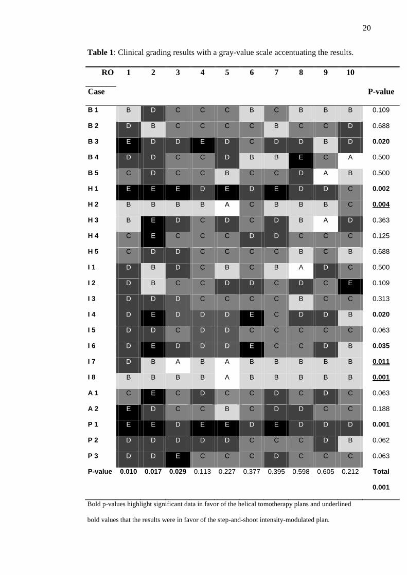
The results from the CGA are presented in Table 1. For eight of the 23 cases the
CGA revealed a significant difference between the HTT and the SS-IMRT plans
(cases with bold p-values in Table 1). Five cases were in favor of HTT; one brain
tumor case (B 3), one H&N cancer (H 1), two intrathoracic tumor cases (I 4 and I
6), and one case with tumor in the pelvic region (P 1). Three cases were in favor
of SS-IMRT (underlined p-values in Table 1), one H&N (H 2), and two
intrathoracic (I 7 and I 8). For all cases combined the CGA gave a significant

10
difference between the techniques in favor of HTT (Total p-value in Table 1). The
grading results from three individual ROs (RO 1, RO 2, and RO 3) all had HTT as
the significantly superior treatment technique for all cases combined. One RO
(RO 10) seemed to favor SS-IMRT though that result was not significant, see
Table 1.
An advantage with HTT treatment that was identified by the ROs during the
clinical grading was the ability to spare the intestines while maintaining target
coverage for treatment of pelvic tumors including elective lymph nodes. Another
advantage was the target coverage compared to SS-IMRT for mesothelioma
treatments, and also the ability to obtain sharp dose gradients especially between
target and spinal cord for H&N treatments. The main drawback identified with
HTT treatment was the wide penumbra in the cranio-caudal direction. This is due
to the fixed jaw positions and the characteristics of the helical irradiation which
depends on the jaw setting used, i.e. the fan beam thickness. Hence, the radiation
starts to build up and fall off, correspondingly, at 1.0, 2.5 or 5 cm from the cranio-
caudal side of the target. Another drawback was identified for cases where most
of the radiation delivered was limited to enter the patient in a few small angle
intervals. For these cases, the HTT plans were often considered inferior to the SS-
IMRT plans.
Dose-volume statistics and corresponding EUD-data for the different plans are
displayed in Table 2. These results reveal that a difference in CTV coverage of 1.3
% or more (≥ 0.5 Gy difference in EUD-data) correlates with a significant CGA
result, i.e. for these cases the ROs agreed that there was a clinical advantage for

11
the plan with the superior dose coverage (cases H 1, and P 1). Cases with similar
CTV coverage, but with a difference in PTV coverage of 2.2 % or more, also had
a significant result in the CGA (cases B 3, I 4, and I 6). However, there was not a
clear specific difference in PTV EUD-value correlating with a significant result in
the CGA (0.6, 0.1, and 0.2 Gy for cases B 3, I 4, and I 6, respectively). Cases
where the SS-IMRT plan had similar or somewhat superior (0.3 %) CTV and PTV
coverage than the HTT plan, and also had a lower average mean dose value for an
OAR (AMDOAR) of at least 2.5 Gy, corresponded to a significant result in the
CGA (cases I 7, and I 8). For cases where the plans had similar AMDOAR but one
plan had a somewhat worse target coverage, the ROs disagreed on whether the
differences in target coverage were of clinical importance or if the plans were
equivalent. This was indicated in the results from the CGA (Table 1) as one plan
seemed to be somewhat superior but the superiority was too unclear to give a
significant result (cases H 4, I 3, I 5, A 1, P 2, and P 3). For cases where one plan
had a somewhat worse target coverage but also had a lower AMDOAR value, the
ROs disagreed on whether one or the other plan was the superior one, or if the
differences cancelled out making the plans equivalent (cases B 4, H 3, I 1, and A
2).
Discussion
In studies comparing plans generated with different IMRT treatment delivery
techniques, the clinical relevance for the differences found is often unclear [4]. In
this study we try to mitigate this issue by using CGA as a tool for treatment plan
comparisons. CGA is easy to use as it is based on the same type of clinical

12
assessments performed on a daily basis in the clinic. The CGA requires in
addition that these assessments are performed in a systematic way, and that the
results are quantified and registered. A positive side-effect with the method is that
the ROs become educated and aware of what is achievable with the treatment
techniques available to their patients, and that the pros and cons of the different
treatment techniques are elucidated. Hence, a CGA would be particularly useful
during implementation of a new treatment technique into a clinical setting, where
it could be employed as part of the commissioning process of the new technique.
The CGA gave significant results for eight of the 23 cases (five in favor of the
HTT plan and three in favor of the SS-IMRT plan Table 1). This means that for
most of the cases (in total 15) the ROs could not agree on whether or not there
was clinical advantage with one of the treatment techniques. Three of the ten ROs
significantly favored HTT over SS-IMRT, for all cases combined. None
significantly favored SS-IMRT. This means that although the overall results
favored HTT over SS-IMRT the differences between plans are generally so small
that the clinical advantage of the technique is often questionable. The exception is
for complex cases where HTT was clearly regarded as the superior treatment
technique, confirming our initial expectations. For five cases there was a
significant result favoring HTT, and for three cases there was a significant
favoring of SS-IMRT, indicating a clear clinical advantage for those patients
receiving HTT or SS-IMRT treatment. To be able to identify these patients at an
early stage in the treatment planning process and prioritizing those for HTT or SS-
IMRT would ensure a more optimal use of the clinic’s HTT and SS-IMRT
treatment resources.

13
The results from the CGA correlated well with differences in target coverage and
doses to OARs (presented under dose-volume statistics in Table 2). However, the
correlation was weaker between the results from the CGA and EUD-data. The
better correlation between CGA and dose-volume statistics than between CGA
and EUD-data is likely due to the fact that the dose-volume statistics parameters
are directly visible in the DVHs. These were, as mentioned earlier, among the data
presented to the ROs during the clinical grading while EUD-data was not. The
EUD-based index (fEUD) might have correlated even better with the CGA results
if the ROs in the clinic had agreed on weighting factors to be used in the model.
Alternatively, such factors could be derived from the CGA results. Limitations of
different DVH-reduction methods such as the generalized EUD-model have been
discussed by e.g. the QUANTEC-group [14]. The tissue-specific parameters
describing the dose-volume dependence (a-values) are not well determined for
some organs which confine the general usefulness of the calculated EUD-data and
hence the fEUD-values. However, these values should still be useful for
comparing treatment plans generated for the same patient cases.
The cranio-caudal penumbra effect for tomotherapy treatments was the main
reason why some of the HTT plans were considered significantly inferior to the
SS-IMRT plans, similar to results found in other studies [2, 15]. In order to reduce
this unwanted effect, a dynamic jaw is under development by the vendor, which
has the potential to essentially remove the penumbra effect [15]. The other
drawback found was for cases where the rotating beam was limited by OARs to
only a few and small angle intervals. This resulted in poor treatment plans for

14
helical delivery mode, which has also been reported in previous studies [2, 16].
Such cases should not be prioritized for treatment with the tomotherapy system,
since treatments delivered by SS-IMRT are comparable or better.
This study was not blind, i.e. the ROs were told which plan was generated for
HTT, and which was generated for SS-IMRT treatment delivery, since this was
obvious merely by observing the dose distributions for the various treatment
techniques. In order to ensure that all participants had the same background
information, everyone was informed about the treatment modalities. This
knowledge could possibly have biased the results, if the participants preferred
some treatment technique, and it might have influenced their grading score. For
cases where the OAR dose constraints were all fulfilled, the differences in
judgment seen between the participating ROs could be due to subjective
preferences, differences in educational background, or due to the lack of specific
treatment objectives in the clinic [17]. By performing a CGA these differences are
revealed which can be a first step towards developing a more congruent judgment
within the clinic.
This study presents CGA as a useful method of comparing radiotherapy treatment
plans. Another useful method for comparing treatment plans is the Pareto
evaluation concept, which has some advantages compared to conventional DVH-
based methods [8, 17-19]. A CGA study would serve as a good complement to a
Pareto evaluation study since it takes advantage of the ROs clinical assessment to
identify the clinical relevant differences between treatment plans. These
subjective assessments are quantified in this CGA study, and used to decide which

15
patients that had a clinical benefit of one or the other of the advanced treatment
techniques available to them, i.e. HTT and SS-IMRT. The result from the study
provides a support for decision making on treatment technique at our clinic with a
limited number of treatment slots available for HTT and SS-IMRT treatment,
which ensures a more optimal use of our advanced treatment resources.
Information from published studies regarding choice of treatment technique might
not be applicable for every clinic, as they rarely involve clinical judgments and do
not take into account characteristics of a specific clinic, e.g. resources available.
Hence, a CGA can help to decide how to best implement the treatment technique,
locally. In summary, the proposed method for comparing treatment techniques
offers a formalized way of introducing and evaluating the implementation of new
radiotherapy techniques in a clinical setting.
Acknowledgments The authors would like to acknowledge the following radiation oncologists;
Thomas Björk-Eriksson, Jens Engleson, Adalsteinn Gunnlaugsson, Maria Gebre-
Medhin, Michael Garkavij, Anders Ask, Sven-Börje Ewers, Henriette Lindberg,
and Michael Gubanski, who with the time spent to grade plans helped to
accomplish this project.

16
References
1. Båth, M. and Månsson, L.G., Visual grading characteristics (VGC) analysis: a non-
parametric rank-invariant statistical method for image quality evaluation. Br J Radiol,
2007. 80(951): p. 169-76.
2. Bauman, G., Yartsev, S., Rodrigues, G., Lewis, C., Venkatesan, V.M., Yu, E., et al., A
prospective evaluation of helical tomotherapy. Int J Radiat Oncol Biol Phys, 2007.
68(2): p. 632-41.
3. Cattaneo, G.M., Dell'oca, I., Broggi, S., Fiorino, C., Perna, L., Pasetti, M., et al.,
Treatment planning comparison between conformal radiotherapy and helical
tomotherapy in the case of locally advanced-stage NSCLC. Radiother Oncol, 2008.
88(3): p. 310-8.
4. Elith, C., Dempsey, S.E., Findlay, N., and Warren-Forward, H.M., An Introduction to
the Intensity-modulated Radiation Therapy (IMRT) Techniques, Tomotherapy, and
VMAT. Journal of Medical Imaging and Radiation Sciences, 2011. 42(1): p. 37-43.
5. van Vulpen, M., Field, C., Raaijmakers, C.P., Parliament, M.B., Terhaard, C.H.,
MacKenzie, M.A., et al., Comparing step-and-shoot IMRT with dynamic helical
tomotherapy IMRT plans for head-and-neck cancer. Int J Radiat Oncol Biol Phys,
2005. 62(5): p. 1535-9.
6. Zhang, X., Penagaricano, J., Moros, E.G., Corry, P.M., Yan, Y., and Ratanatharathorn,
V., Dosimetric comparison of helical tomotherapy and linac-IMRT treatment plans for
head and neck cancer patients. Med Dosim, 2010. 35(4): p. 264-8.
7. Niemierko, A., Reporting and analyzing dose distributions: a concept of equivalent
uniform dose. Med Phys, 1997. 24(1): p. 103-10.

17
8. Petersson, K., Ceberg, C., Engström, P., Benedek, H., Nilsson, P., and Knöös, T.,
Conversion of helical tomotherapy plans to step-and-shoot IMRT plans- Pareto front
evaluation of plans from a new treatment planning system. Med Phys, 2011. 38(6): p.
3130-3138.
9. Petersson, K., Ceberg, C., Engström, P., and Knöös, T., Beam commissioning and
measurements validating the beam model in a new TPS that converts helical
tomotherapy plans to step-and-shoot IMRT plans. Med Phys, 2011. 38(1): p. 40-46.
10. Mould, R.F., Introductory Medical Statistics. 3rd ed, ed. R.F. Mould, et al. 1998,
Bristol and Philadelphia: Institute of Physics Publishing.
11. ICRU, Prescribing, Recording, and Reporting Photon-Beam Intensity-Modulated
Radiation Therapy (IMRT). ICRU REPORT 83. Journal of the ICRU, 2010. 10.
12. Niemierko, A., A generalized concept of equivalent uniform dose (EUD). Med Phys,
1999. 26(6): p. 1100.
13. Semenenko, V.A., Reitz, B., Day, E., Qi, X.S., Miften, M., and Li, X.A., Evaluation of
a commercial biologically based IMRT treatment planning system. Med Phys, 2008.
35(12): p. 5851-60.
14. Quantitative Analyses of Normal Tissue Effects in the Clinic. Int J Radiat Oncol Biol
Phys, 2010. 76(3 Suppl): p. S1-160.
15. Sterzing, F., Uhl, M., Hauswald, H., Schubert, K., Sroka-Perez, G., Chen, Y., et al.,
Dynamic jaws and dynamic couch in helical tomotherapy. Int J Radiat Oncol Biol
Phys, 2010. 76(4): p. 1266-73.
16. McIntosh, A., Read, P.W., Khandelwal, S.R., Arthur, D.W., Turner, A.B., Ruchala,
K.J., et al., Evaluation of coplanar partial left breast irradiation using tomotherapy-
based topotherapy. Int J Radiat Oncol Biol Phys, 2008. 71(2): p. 603-10.

18
17. Knöös, T., Benedek, H., Ceberg, C., Nilsson, P., and Petersson, K., Uncertainties in
the Evaluation of Treatment Plans, in Uncertainties in External Beam Radiation
Therapy: AAPM Medical Physics Monograph No. 35 J.R. Palta and T.R. Mackie,
Editors. 2011, Medical Physics Publishing: Madison, WI. p. 117-127.
18. Ottosson, R.O., Engstrom, P.E., Sjostrom, D., Behrens, C.F., Karlsson, A., Knoos, T.,
et al., The feasibility of using Pareto fronts for comparison of treatment planning
systems and delivery techniques. Acta Oncol, 2009. 48(2): p. 233-7.
19. Thor, M., Benedek, H., Knoos, T., Engstrom, P., Behrens, C.F., Hauer, A.K., et al.,
Introducing multiple treatment plan-based comparison to investigate the performance
of gantry angle optimisation (GAO) in IMRT for head and neck cancer. Acta Oncol.
51(6): p. 743-51.

19
Figure 1: A screen capture showing how the dose distribution for
treatment plans were presented side-by-side for the radiation oncologists,
in the Oncentra treatment planning system. To the left is the helical
tomotherapy plan, and to the right the step-and-shoot intensity-modulated
radiation therapy plan, for treatment of an intrathoracic tumor (case I 3).

20
Table 1: Clinical grading results with a gray-value scale accentuating the results.
RO 1 2 3 4 5 6 7 8 9 10
P-value Case
B 1 B D C C C B C B B B 0.109
B 2 D B C C C C B C C D 0.688
B 3 E D D E D C D D B D 0.020
B 4 D D C C D B B E C A 0.500
B 5 C D C C B C C D A B 0.500
H 1 E E E D E D E D D C 0.002
H 2 B B B B A C B B B C 0.004
H 3 B E D C D C D B A D 0.363
H 4 C E C C C D D C C C 0.125
H 5 C D D C C C C B C B 0.688
I 1 D B D C B C B A D C 0.500
I 2 D B C C D D C D C E 0.109
I 3 D D D C C C C B C C 0.313
I 4 D E D D D E C D D B 0.020
I 5 D D C D D C C C C C 0.063
I 6 D E D D D E C C D B 0.035
I 7 D B A B A B B B B B 0.011
I 8 B B B B A B B B B B 0.001
A 1 C E C D C C D C D C 0.063
A 2 E D C C B C D D C C 0.188
P 1 E E D E E D E D D D 0.001
P 2 D D D D D C C C D B 0.062
P 3 D D E C C C D C C C 0.063
P-value 0.010 0.017 0.029 0.113 0.227 0.377 0.395 0.598 0.605 0.212 Total
0.001
Bold p-values highlight significant data in favor of the helical tomotherapy plans and underlined
bold values that the results were in favor of the step-and-shoot intensity-modulated plan.

21
Table 2: Dose-volume statistics and EUD-data
Dose-volume statistics (HTT/SS-IMRT) EUD-data (HTT/SS-IMRT)
Case V95%, CTV (%) V95%, PTV (%) AMDOAR (Gy) EUDCTV (Gy) EUDPTV (Gy) fEUD
B 1 100/100 100/99.9 24.0/22.4 55.1/55.1 55.3/55.3 0.21/0.21
B 2 99.7/99.6 99.7/99.1 23.5/23.8 48.1/48.2 48.1/48.1 0.22/0.22
B 3 100/100 99.5/97.0 14.7/14.3 54.0/53.8 53.9/53.3 0.19/0.19
B 4 100/100 100/98.9 9.77/8.97 50.3/50.3 50.3/50.0 0.33/0.34
B 5 100/100 99.9/99.7 20.0/19.1 54.6/54.5 54.3/54.2 0.39/0.40
H 1 99.2/96.9 98.8/93.9 25.3/24.4 45.6/44.4 45.4/43.6 0.27/0.27
H 2 100/100 99.9/99.6 36.5/32.0 69.9/70.2 69.5/69.7 0.41/0.44
H 3 100/99.9 99.6/98.2 33.5/28.7 67.9/67.6 67.7/67.1 0.55/0.58
H 4 100/99.9 99.8/97.7 22.6/22.3 53.9/53.7 53.7/53.5 0.35/0.35
H 5 100/100 99.5/99.2 32.8/32.4 68.6/68.7 68.5/68.3 0.44/0.44
I 1 100/100 99.8/99.5 19.6/16.0 64.7/64.9 64.5/64.6 0.30/0.33
I 2 99.8/99.9 96.5/96.3 21.9/21.8 43.8/43.8 43.5/43.5 0.27/0.27
I 3 100/99.9 99.6/98.3 15.4/14.7 39.9/40.0 39.9/39.9 0.23/0.24
I 4 100/100 99.9/97.7 20.7/19.0 45.2/45.4 45.2/45.1 0.21/0.22
I 5 100/100 100/99.3 7.88/7.31 25.1/25.1 25.1/25.0 0.46/0.48
I 6 100/99.8 99.5/95.9 15.9/14.8 44.9/45.0 44.8/44.6 0.22/0.23
I 7 99.9/100 99.9/99.9 14.8/12.3 35.9/36.1 35.9/36.0 0.23/0.26
I 8 100/100 99.5/99.8 16.0/11.8 44.4/44.4 44.2/44.3 0.32/0.36
A 1 100/100 100/98.1 14.5/13.0 50.3/50.4 50.3/50.1 0.36/0.38
A 2 100/100 100/99.8 14.0/11.2 50.5/50.4 50.6/50.5 0.41/0.44
P 1 99.8/98.5 99.3/97.3 32.5/33.0 60.0/59.5 59.9/59.4 0.51/0.51
P 2 100/100 98.9/98.1 33.1/32.9 46.8/46.7 46.8/46.7 0.23/0.23
P 3 100/100 99.7/97.5 25.6/24.0 49.9/49.6 50.1/49.8 0.37/0.37
Bold values indicate that the difference in value between plans correlates with a significant result
in the clinical grading analysis favoring the helical tomotherapy plans, and underlined bold values
that the results favored the step-and-shoot intensity-modulated plan.

22
Appendix: The cases involved in the study.
Site Case
ID
Diagnosis TNM or
clinical
stage
Prescribed tumor
dose (Gy)/ # of
fractions
Elective target
doses (Gy)/ # of
fractions Brain B 1 Astrocytoma n/a 55.8 / 31
n/a
B 2 Astrocytoma n/a 59.0 / 33 n/a
B 3 Astrocytoma n/a 54.0 / 30 n/a
B 4 Pituitary adenoma n/a 50.4 / 28 n/a
B 5 Oligodendroglioma n/a 54.0 / 30 n/a
Head and
neck
H 1 Lacrimal duct
cancer
T4N0M0 56.0 / 39 n/a
H 2 Nasopharyngeal
cancer
T1N2M0 68.0 / 34 54.4 / 34
H 3 Nasopharyngeal
cancer
T2b1N2M0 68.0 / 34 62.9, 54.4 / 34
H 4 Oropharyngeal
cancer
T2N2cM0 68.0 / 34 62.9, 54.4 / 34
H 5 Oropharyngeal
cancer
T3N2bM0 68.0 / 34 62.9, 54.4 / 34
Intrathoracic I 1 Esophageal cancer T3N1M0 64.0 / 32 n/a
I 2 Hodgkin’s disease Stage 2B 43.2 / 24 n/a
I 3 Hodgkin’s disease Stage 2B 40.0 / 20 n/a
I 4 Malignant Thymoma T0N0M1 45.0 / 25 n/a
I 5 Mesothelioma rT4N1M1 25.0 / 5 n/a
I 6 Mesothelioma T1N0M0 54.0 / 30 n/a
I 7 Mesothelioma T4N0M0 36.0 / 12 n/a
I 8 Non-small cell lung T4N2bM0 44.0 / 22 n/a

23
cancer
Abdominal A 1 Liposarcoma rT2bN0M0 50.0 / 25 n/a
A 2 Pancreatic cancer T4N0M0 50.4 / 28 50.4 / 28
Pelvic P 1 Anal cancer T2N2M0 60.0 / 30 46.0 / 23
P 2 Cervical cancer rT3bN0M0 64.8 / 36 46.8 / 26
P 3 Prostate cancer T3N1M0 50.0 / 25 50.0 / 25