Central bank boards around the world: Why does membership size differ
Upload
independentCategory
view
5download
0
1995, 15(3):1554. Mol. Cell. Biol.
Gizang-Ginsberg, V Cavailles, M G Parker and P J KushnerY Sadovsky, P Webb, G Lopez, J D Baxter, P M Fitzpatrick, E TATA-box-binding protein.responses to overexpression of Transcriptional activators differ in their
http://mcb.asm.org/content/15/3/1554Updated information and services can be found at:
These include:
CONTENT ALERTS more»cite this article),
Receive: RSS Feeds, eTOCs, free email alerts (when new articles
http://journals.asm.org/site/misc/reprints.xhtmlInformation about commercial reprint orders: http://journals.asm.org/site/subscriptions/To subscribe to to another ASM Journal go to:
on March 6, 2014 by guest
http://mcb.asm
.org/D
ownloaded from
on M
arch 6, 2014 by guesthttp://m
cb.asm.org/
Dow
nloaded from

MOLECULAR AND CELLULAR BIOLOGY, Mar. 1995, p. 1554–1563 Vol. 15, No. 30270-7306/95/$04.0010Copyright q 1995, American Society for Microbiology
Transcriptional Activators Differ in Their Responses toOverexpression of TATA-Box-Binding ProteinYOEL SADOVSKY,1† PAUL WEBB,1 GABRIELA LOPEZ,1 JOHN D. BAXTER,1
POLLY M. FITZPATRICK,2 ELENA GIZANG-GINSBERG,2 VINCENT CAVAILLES,3
MALCOLM G. PARKER,3 AND PETER J. KUSHNER1*
Metabolic Research Unit, University of California, San Francisco, California 941431; Department of Oncology andImmunology, Lederle Laboratories-Medical Research Division, Pearl River, New York 109652; and Laboratory
of Molecular Endocrinology, Imperial Cancer Research Fund, London WC2A 3PX, United Kingdom3
Received 18 May 1994/Returned for modification 6 September 1994/Accepted 14 December 1994
We investigated how overexpression of human TATA-box-binding protein (TBP) affects the action of estro-gen receptor (ER) and compared the response with that of other activators. When ER activates a simplepromoter, consisting of a response element and either the collagenase or tk TATA box, TBP overexpressionpotentiates transcription. TBP potentiates only estrogen-induced and not basal transcription and does soindependent of spacing between response element and TATA box. TBP overexpression also reduces autoinhi-bition by overexpressed ER, suggesting that one target of the autoinhibition may be TBP itself. Both AF-1 andAF-2 domains of ER are potentiated by TBP, and each domain binds TBP in vitro. Like ER, chimericGAL4/VP16 and GAL4/Tat activators are also potentiated by TBP, as is the synergistic activation by ER andGAL4/VP16 on a complex promoter. Unlike ER, GAL4/Sp1 and GAL4/NF-I become less potent when TBP isoverexpressed. Furthermore, synergy between ER and Sp1 or between ER and NF-I, whether these are suppliedby transfected GAL4 fusions or by the endogenous genes, is inhibited by TBP overexpression. Thus, ERresembles VP16 in response to TBP overexpression and is different from Sp1 and NF-I, which predominate overER in setting the response on complex promoters.
The estrogen receptor (ER) is an upstream activator proteinthat binds an estrogen response element (ERE) on DNA andenhances transcription from nearby promoters. Two ER acti-vation domains contribute to this process: AF-1 in the aminoterminus, which is constitutive, and AF-2 in the C-terminalligand-binding domain (LBD), which is active only when hor-mone is bound (13, 57). Neither of the ER domains is markedby an abundance of glutamine, proline, or acidic amino acids ashas been noted for many other transcriptional activation do-mains.The mechanism whereby the ER domains contribute to tran-
scriptional activation is unknown, but by analogy with better-understood viral activators, it is thought that interactions withtarget proteins within the transcriptional apparatus are impor-tant (for a review, see reference 55; see also references 11 and27 and references therein). The TATA-box-binding protein(TBP) is one candidate target. TBP binding to the promoter isa pivotal event leading to unwinding of the DNA at the TATAbox and widening of the minor groove (31, 32, 47). TBP bind-ing is needed for the subsequent recruitment of TFIIB andRNA polymerase II (reviewed in reference 60). Several acti-vators bind TBP in vitro. These include the acidic activatorVP16 (26, 52), E1a (3, 24, 37), c-Rel (30, 59), and Tax 1 (7).For several of these activator mutations that decrease bindingto TBP decrease transcriptional activation, suggesting that thebinding is of functional significance. In higher eukaryotes, TBPis tightly associated with a class of coactivators called TBP-associated factors (TAFs). This complex, but not isolated TBP,
mediates transcriptional enhancement by activator proteins invitro (10, 14, 53, 61; reviewed in reference 55). TAFs andactivators also appear to directly interact. VP16 binds TAF40.Sp1 binds TAF110 (10, 19, 23), and mutations in Sp1 thatdecrease binding decrease transcriptional activation (17). Ahuman TAFII30 that binds the ER AF-2 domain and is re-quired for transcriptional activation by ER in vitro has recentlybeen described (27).These observations suggest that upstream activators may
regulate some aspect of TBP function, either directly orthrough an adapter. Further support for the notion that TBP isa target of upstream activators comes from studies of theeffects of TBP overexpression. TBP supplied by transfection ofmammalian cells potentiates the activity of retinoic acid betareceptor working in concert with E1a (2, 29), of c-Rel (30, 59),of Tax 1 (7), and of bovine papillomavirus E2 (22). The abilityof TBP to potentiate bovine papillomavirus E2 was strong onminimal promoters with a TATA box and was limited by thepresence of an initiator element, an effect that had earlier beenseen in Sp1-TBP interactions in Drosophila cells (12).The foregoing observations raise the question of whether the
ER or other steroid receptor activation domains, which havenot been examined, interact with TBP. More generally, theyraise the question of whether all activators are potentiated byTBP overexpression. In this study, we investigated whetheroverexpression of human TBP potentiates transcriptional ac-tivation by ER on a minimal promoter consisting of an EREand either a collagenase or tk TATA box. We tested the AF-1and AF-2 domains together and separately, both for TBP po-tentiation and for TBP binding in vitro. We also compared theresponse of the ER activation domains with that of represen-tative acidic (herpes simplex virus VP16), glutamine-rich(Sp1), and proline-rich (NF-I/CTF) activation domains, eachtested on the same reporter gene. We also tested a domainfrom the human immunodeficiency virus activator Tat, which
* Corresponding author. Mailing address: MRU 1141 HSW, Uni-versity of California, San Francisco, CA 94143-0540. Phone: (415) 476-6790. Fax: (415) 476-1660. Electronic mail address: [email protected].† Present address: Department of Obstetrics and Gynecology,
Washington University School of Medicine, St. Louis, MO 63110.
1554
on March 6, 2014 by guest
http://mcb.asm
.org/D
ownloaded from

has recently been reported to bind TBP (28). We foundthat TBP overexpression enhances activation by ER, its in-dividual AF-1 and AF-2 domains, and the activation domainsof Tat and VP16. TBP overexpression also enhances activationby combinations of ER and VP16 or ER and Tat. However,TBP does not potentiate all activators when tested on thesereporters. TBP overexpression inhibits activation by Sp1 andNF-I activation domains and becomes even more inhibitorywhen these activators synergize with ER on complex promot-ers.
MATERIALS AND METHODS
Expression vectors and promoter constructs. ERE-tkTATA was prepared byligation of an oligonucleotide corresponding to a minimal ERE (59-AGGTCACAGTGACCT-39) into the HindIII site of tkTATA, which was previously de-scribed and spans tk sequences 232 to 145 with respect to the tk transcriptionalinitiation site (38, 39). This plasmid has two b-globin transcription terminationsignals upstream of the promoter to eliminate the influence of plasmid tran-scripts. A single binding site for GAL4 (59-GCGGAGTACTGTCCTCCGA-39)was placed at the Sal site between the ERE and the core promoter. It should benoted that this and other reporter plasmids used in this work have the pUC AP-1site (35) removed. Spacing mutations were constructed by digesting ERE-tk-TATA with XbaI (43 space), SphI (39 space), or BamHI and SphI (15 space)followed by limited mung bean nuclease treatment. Vectors were self-ligated,and ligation products were sequenced to determine the relative spacing of theERE and TATA box. ERE-tk109 was prepared by ligation of the minimal EREinto the HindIII site of pBLCAT3, which had been modified to remove the pUCAP-1 site (40) by digestion with NcoI and Eco 109. CollTATA was prepared byligation of an oligonucleotide, spanning interstitial collagenases sequences from232 to 18 relative to the start site of transcription, into tkTATA from which tkupstream sequences had been removed by digestion with XbaI and PstI. ERE-CollTATA was prepared by ligation of the minimal ERE into the HindIII site ofCollTATA. ERE-Coll60 was prepared by ligation of the minimal ERE upstreamof interstitial collagenase sequences260 to163 (40) in a vector without the pUCAP-1 site.LEN-TBP was prepared by subcloning the BglII-BamHI fragment, spanning
the TBP cDNA, from vector pGPP26 (48) into the BamHI site of pLEN (36).Human ER expression vectors HE0, HE19, HE15, and HE11 have been previ-ously described (34), as have mouse ER expression vectors MOR121-599 and itsmutant derivatives (13). The GAL4, GAL4/VP16 (50), GAL4/NF-I (53), andGAL4/Tat (16) expression vectors have been described. An expression vector forGAL4 (amino acids [aa] 1 to 147) fused to Sp1 (aa 83 to 778) and driven by theRous sarcoma virus promoter was a kind gift of Brian West and Dale Leitman(37a). The reporter gene for transfection efficiency, in which the b-actin pro-moter drives expression of human choriogonadotropin (hCG), has been de-scribed elsewhere (58).To express glutathione S-transferase (GST) fusions to the amino-terminal
domain (aa 1 to 185) of the human ER, an EcoRI–blunt-ended KpnI fragmentspanning this domain from EGE (34) was cloned into SmaI- and EcoRI-digestedpGEX-5X-1, one of the vectors of the pGEX series (Pharmacia Biotech Inc.,Piscataway, N.J.). A similar fusion of the wild-type (Gly-400) LBD (aa 282 to595) was constructed in two steps. An Xba fragment from HE19G (56) wasinserted into the equivalent position of XbaI-digested SG5-HE14, which spansthe ER LBD (34) to correct the mutation at aa 400 from Val to Gly (56). Then,an EcoRI fragment spanning the wild-type ER LBD was cloned into pGEX-3X.A fusion of the wild-type full-length receptor to GST was prepared by insertingan EcoRI fragment from HEG0 spanning the ER cDNA into pGEX5X-1. Vec-tors to express GST fusions to the mouse ER LBD (aa 313 to 599), either wildtype or mutant, were constructed by inserting a SalI fragment with repaired endsspanning the domain into pGEX-2X (51).For the GST-TBP fusion pull-down assays, pGexTBP, a plasmid that encodes
sequences corresponding to the full-length human TBP protein (aa 1 to 339)cloned into pGEX2TK (43, 44), was used for expression. For in vitro expressionof the human full-length, N-terminal, and C-terminal ER proteins, HE0, HE15,and HE19 plasmids (34), respectively, were used. Plasmid pBX49, which encodesthe carboxy-terminal region of the UL80 protein from human cytomegalovirus(1), was also used for in vitro expression as a negative control protein for thesestudies.Cell lines. Both HeLa and Chinese hamster ovary (CHO) cells were grown in
Coon’s F-12–Dulbecco’s modified Eagle’s (1:1) medium supplemented with 10%calf serum (Sigma Serumax4, batch 19F-0156, low in E2) and 1% penicillin-streptomycin in a 5% CO2 atmosphere. CHO cells transfected with ER cDNA(clone D20) were grown in the same medium and supplemented by 40 mMCdSO4 and 50 mM ZnSO4 after transfection (58). For cell counts, the cells werediluted in trypan blue-containing buffer, and viable, dye-excluding cells werecounted in a hemocytometer. Cell viability was greater than 95%.Transient transfections. HeLa and CHO cells were transiently transfected in
triplicate with different combinations of plasmids as described for each experi-
ment. Expression vector DNA was held constant in each experiment by theaddition of an empty expression vector, and the total amount of transfected DNAwas kept below 20 mg/1 million cells. Gene transfer was by electroporation asdescribed previously (58). The cells were then electroporated at 960 mF andplated in growth medium in six-well plates (Corning). Chloramphicol acetyltrans-ferase (CAT) activity, corrected to background from mock-transfected cells andnormalized for transfection efficiency with a cotransfected reporter gene in whichb-hCG was driven by the human b-actin promoter (58), was determined after 48to 72 h, using chloramphenicol and 8 mCi of [3H]acetyl coenzyme A (Du Pont,Wilmington, Del.) per ml as described previously (46). The b-hCG reporter wasassayed with a standard kit using iodinated antibody (Hybri-Tech Tandem As-say). Results were expressed as mean6 standard error of the mean. CAT activityis calculated as the increase in counts per minute per hour at room temperature(corrected for background) for 10 ml of cell extract and normalized to productionof 100 standard units of b-hCG.Receptor measurement. The ER concentration in transfected cells was mea-
sured in triplicate by a whole-cell binding assay (54). The growth medium wasreplaced by serum-free medium containing 0.1% bovine serum albumin (BSA)and 1 nM [3H]17b-estradiol (specific activity, 92.5 Ci/mmol; Du Pont, NewEngland Nuclear, Boston, Mass.) in the presence or absence of 100 nM unla-beled 17b-estradiol to determine nonspecific binding. After 90 min, the cellswere washed with phosphate buffered saline (PBS) containing 0.5% BSA once atroom temperature for 30 min and twice in 48C and then lysed with ethanol andcounted in a beta counter. Results were calculated as receptor sites per cell andexpressed as mean 6 standard error of the mean.Gel electrophoresis and TBP immunoblots. Cells transfected with either TBP
(10 mg) or ER (0.5 mg) or both were scraped from culture plates directly intosample buffer containing 0.4% sodium dodecyl sulfate (SDS), and each samplewas sonicated at 30% intensity for 15 s (Dismembrator model 300; Fisher). Thesamples, which contained 100 mg of protein, were subjected to electrophoresis in48C for 4 h at 50 V (Miniprotean; Bio-Rad, Richmond, Calif.) in 10% polyacryl-amide gel slabs. Samples were transferred to nitrocellulose at 100 mA overnight,using a Bio-Rad Transblot system. The blots were then incubated for 1 h withmouse anti-human TBP monoclonal antibody 58 at a 1:1,000 dilution. Afterwashing, the blot was incubated with a rabbit anti-mouse polyclonal antibody(Cappel, Durham, N.C.) as the secondary antibody and then with horseradishperoxidase-linked donkey anti-rabbit antibodies (Amersham). The reaction wasdeveloped by using a Renaissance kit (Du Pont, New England Nuclear), and thefilm was exposed for 1 min.GST fusion proteins. Fusions of GST to various domains of the human and
mouse ER were prepared as described previously (51). Briefly, bacteria express-ing the fusion proteins were resuspended in buffer IPAB-80 (20 mM N-2-hy-droxyethylpiperazine-N9-2-ethanesulfonic acid [HEPES], 80 mM KCl, 6 mMMgCl2, 10% glycerol, 1 mM dithiothreitol, 1 mM ATP, 0.2 mM phenylmethyl-sulfonyl fluoride, protease inhibitors [pH 7.9]) and sonicated mildly, and thedebris was pelleted at 12,000 rpm for 1 h in an SS34 rotor. The supernatant wasincubated for 2 h with 500 ml of glutathione-Sepharose 4B beads that werepreviously washed with 5 volumes of PBS–0.2% Triton X-100 and equilibratedwith 5 volumes of IPAB-80. GST fusion protein beads were then washed with 5volumes of PBS–0.05% Nonidet P-40 and resuspended in 1 ml of IPAB-80 forstorage at 48C until use. All the foregoing procedures were done in a cold roomat 48C.Assays of GST-ER fusions were carried out in a 100-ml volume that contained
40 ml of bead suspension (equivalent to 10 ml of compact bead volume) and 1 to2 ml of 35S-labeled in vitro-translated TBP (prepared with plasmid GPP 26 [48])in IPAB-80–2.5% nonfat milk and incubated for 1.5 h at 48C. Beads were washedfive to six times with IPAB-80 containing 0.05% Nonidet P-40. Input labeledproteins, proteins bound to GST, GST-human ER, GST-mouse LBD, or GST-mouse LBD-L547A/M548A beads were then subjected to SDS-polyacrylamidegel electrophoresis (PAGE) in 10% acrylamide and then to autoradiography.For assays of GST-TBP fusions, appropriate ER and UL80 cDNAs were
transcribed in vitro, translated, and labeled with 35S, using the TNT7-coupledrabbit reticulocyte lysate system as described by the manufacturer (PromegaCorp., Madison, Wis.). The TBP-GST fusion protein was prepared and purifiedas previously described (20). Interaction studies involving the TBP-GST fusionand the in vitro-translated products were carried out as follows. Five hundrednanograms of GST-TBP fusion protein coupled to glutathione-Sepharose beadswas added to 2 to 5 ml of radiolabeled, in vitro-translated ER proteins or UL80protein in 0.2 ml of Z9 buffer (25 mM HEPES [pH 7.5], 12.5 mM MgCl2, 20%glycerol, 0.1% Nonidet P-40, 150 mM KCl, 20 mM ZnSO4) and rocked for 1 h atroom temperature. The beads were then washed three times in 1 ml of NETNbuffer (100 mM NaCl, 1 mM EDTA, 0.5% Nonidet P-40, 20 mM Tris-HCl [pH8.0]), pelleted by centrifugation, and boiled in 13 SDS sample buffer. Proteinbound to the beads were resolved by SDS-PAGE.
RESULTS
TBP overexpression potentiates transcriptional activationby ER. To determine whether TBP is limiting for transcrip-tional activation by ER, we examined the effect of overexpress-
VOL. 15, 1995 DIFFERENT ACTIVATOR RESPONSES TO OVEREXPRESSED TBP 1555
on March 6, 2014 by guest
http://mcb.asm
.org/D
ownloaded from

ing TBP on the response of estrogen-regulated reporter genesin transiently transfected HeLa cells. For these experiments,we used an ER variant (G400V) that, unlike wild-type-trans-fected receptor, absolutely requires ligand for transcriptionalactivation (56). The CAT reporter contained a collagenasegene TATA box downstream of a single palindromic consensusERE. As expected, transfection of ER alone caused three- tosixfold activation in response to estradiol (Fig. 1A). Cotrans-fection of TBP along with ER potentiated the activation by anadditional 3- to 8-fold, resulting in a 40-fold total activation byestrogen. Transfection of TBP without ER caused a minimalincrease in estrogen-induced CAT activity from the reporter,which may reflect potentiation of a low concentration of en-dogenous ER in HeLa cells by TBP (49a). The potentiation ofER by TBP was dose dependent and was not saturated even inthe presence of 10 mg of TBP expression vector (Fig. 1A,inset). Basal activity showed no response to TBP in HeLa cells(Fig. 1A), nor did the activity of a promoter that lacks the ERE(not shown). Similar effects were found in CHO cells (not
shown). These observations suggest that TBP is specificallylimiting for ER-activated transcription of the collagenase min-imal promoter in HeLa and CHO cells.To test whether TBP potentiated ER action on core pro-
moters other than the collagenase promoter, we tested severalherpes simplex virus tk promoter constructions. We also variedthe ERE-TATA box distance in these constructions. The ac-tion of the ER at these promoters varied with the relativerotational position of the ERE, consistent with previous ob-servations (15, 49) (Fig. 1B). Nonetheless, TBP potentiated theestrogen-induced activity of all of the constructions to similardegrees. These observations indicate that the effects of over-expressed TBP are not limited to particular core promoters orparticular arrangements of promoter and response element.They also reinforce the conclusion that TBP is limiting for ERaction at minimal promoters.To rule out the possibility that the expression of TBP
changed the concentration of ER in the transfected cells, orvice versa, we measured the concentration of each of the twoproteins. The mean concentration of ER in the pool of trans-fected HeLa cells increased approximately three- to fourfoldwith transient transfection, and this increase was not signifi-cantly influenced by the presence or absence of coexpressedTBP (Fig. 2A). Similarly, transfection of TBP led to an in-crease in the immunoblotted TBP, and this increase was notinfluenced by the presence or absence of cotransfected ER(Fig. 2B).
FIG. 1. Potentiation of ER action by overexpressed TBP. (A) TBP stimula-tion of a CAT reporter (ERE-CollTATA, illustrated) driven by a palindromicERE and the collagenase promoter TATA box. The CAT activity of the reporterin HeLa cells transfected with expression vectors for ER (0.5 mg), TBP (10 mg),or both and in the presence (striped bars) or absence (black bars) of estrogen(1027 M estradiol) is shown. The inset shows the response of the reporter tovarious amounts of cotransfected TBP when cotransfected with ER and exposedto estrogen. (B) TBP stimulation of reporters driven by an ERE and herpessimplex virus tk TATA box. Spacing of the ERE and TATA box is indicated.CAT activities of each reporter in the absence (open bars) and presence (shadedbars) of expression vector for TBP are shown. All samples were cotransfectedwith ER as described above, and all were exposed to estrogen.
FIG. 2. Overexpression of TBP does not effect ER concentration. HeLa cellswere transiently transfected with expression vectors for either ER (0.5 mg) orTBP (10 mg) or both. The cells were grown for 48 h before the assay. (A)Whole-cell binding assay for ER, expressed as sites per cell, was performed asdescribed in Materials and Methods. Note that ER was increased in the ER-transfected cells, and the concentration was not dependent on the presence ofcoexpressed TBP. (B) Western blot (immunoblot) analysis for TBP. E2 indicatesthe presence of estrogen. Note that the TBP concentration was increased in thepool of TBP-transfected cells and that the concentration was not dependent onthe presence of unliganded or liganded ER.
1556 SADOVSKY ET AL. MOL. CELL. BIOL.
on March 6, 2014 by guest
http://mcb.asm
.org/D
ownloaded from

TBP expression reduces autoinhibition observed at high ERconcentration. Overexpression of ER causes autoinhibition ofestrogen-induced transcriptional activation (45, 58). Autoinhi-bition may occur when excess ER interacts with a limitingcomponent of the basal transcription machinery, thereby de-creasing its availability. Since TBP is limiting for estrogenresponses, we tested whether overexpression of TBP wouldrelieve autoinhibition by ER. As shown in Fig. 3A, when theamount of transfected expression vector for ER was increasedfrom 0.5 to 10 mg, the estrogen response of the ERE-coll-TATA promoter to estrogen was diminished by almost 90%.This autoinhibition was reduced to only 50% by TBP overex-pression. As a separate test of the effect of TBP overexpressionon ER autoinhibition, we used CHO derivatives that expressmore than 1 million ER molecules per cell (58) and in whichinhibition can be demonstrated by increasing the amount ofestrogen in the medium. As shown in Fig. 3B, the overexpres-sion of TBP potentiated the expression of the ERE-collTATApromoter. In addition, there was a shift in the half-maximal doseof estrogen to a fourfold-higher concentration (indicated). Thisshift, which was observed in three independent repetitions ofthis experiment, is consistent with a relief of autoinhibition.
Taken together, these results indicate that autoinhibition in-duced by a high concentration of ER is partly due to limitingamounts of TBP. Nonetheless, the persistence of some auto-inhibition in the presence of overexpressed TBP suggests thatTBP is not the sole target.TBP potentiates AF-1 and AF-2. The ability of overex-
pressed TBP to potentiate transcriptional activation by ER islikely to reflect an interaction with the ER transcriptionalactivation functions. To examine this, we tested whether over-expressed TBP potentiates transcription by each of the indi-vidual activation domains of ER. The action of AF-1 wastested in two ways. The first used a truncated receptor in whichthe LBD and its AF-2 function had been removed (HE15; Fig.4A and B) and which shows constitutive activity of AF-1. Thesecond method was to test the full-length receptor (HE0; Fig.4A and B) liganded to tamoxifen and hence inactivated forAF-2 (57). When TBP was not present, the full-length receptorliganded to tamoxifen (HE0) and the receptor missing theLBD (HE15) barely activated transcription of the reportergenes (ERE-collTATA [Fig. 4A] and ERE-tkTATA [Fig. 4B])over the basal level (one- to threefold activation). This absenceof response is consistent with the weak activity of AF-1 whentested in isolation in HeLa cells (57). In the presence of TBP,however, both the HE15 receptor and the full-length receptorliganded to tamoxifen showed greatly increased ability to stim-ulate transcription (up to 20-fold). This pattern was seen withboth reporters. We conclude that AF-1 acting in isolation fromAF-2 is potentiated by overexpressed TBP. The action of theligand-dependent AF-2 function was tested with a truncatedER lacking the amino terminus and its AF-1 function (HE19;Fig. 4). Transcription supported only by HE19 was weak (nomore than 1.5-fold) on the simple promoters used for thesestudies. This finding is consistent with previous reports thatAF-2 is weak on promoters consisting of an ERE and a min-imal TATA box (57). Nonetheless, in the presence of overex-pressed TBP, the isolated AF-2 domain in HE19 activatedtranscription to a readily measured degree, especially with theERE-tkTATA promoter (25-fold induction). Thus, each of thetranscriptional activation domains of ER acting in isolation ispotentiated by overexpressed TBP.The cooperation between overexpressed TBP and the HE19
truncated receptor appears to require the AF-2 function itself.This is indicated by the failure of the tamoxifen-liganded HE19receptor derivative to stimulate transcription when TBP ispresent (Fig. 4A and B). To confirm that AF-2 is needed forthe cooperative interaction, we tested some derivatives of themouse ER that bear mutations in the LBD that abolish theability of this domain to activate transcription when tested witheither its own or a heterologous DNA-binding domain. Thesemutations thus abolish AF-2 function. They are known to leaveintact the ability of the receptor to bind hormone and topromote dimerization and DNA binding (13). These mutationscluster in a small region of interdigitated hydrophobic andacidic amino acids that is highly conserved among steroid re-ceptors. We tested mutations of both the hydrophobic andacidic amino acids in the context of a truncated mouse receptormissing the amino-terminal AF-1 domain. The truncated de-rivative of the mouse ER (MOR121-599; Fig. 4C) cooperatedwith TBP to stimulate transcription from the test promoter(Fig. 4C), but derivatives bearing mutations in a pair of adja-cent hydrophobic amino acids (L543A/L544A) or in threeclosely spaced acidic residues (D542N/E546Q/D549N) failedto stimulate transcription either with or without overexpressedTBP. Thus, TBP potentiation of HE19 action requires AF-2function and hence reflects cooperation between TBP andAF-2 itself.
FIG. 3. TBP relief of autoinhibition by ER. (A) Autoinhibition of transcrip-tional activation by excess ER, supplied by 10 mg of expression vector, in thepresence or absence of TBP. CAT activity generated by the ERE-CollTATAreporter is shown. Note that excess ER represses promoter activity 90% in theabsence of TBP but only 50% fold in the presence of TBP. (B) Autoinhibition ina cell line (ERC-1 [58]) that stably expresses excess levels (1 million ER mole-cules per cell) of ER, in the presence or absence of TBP. CAT activity generatedby the ERE-CollTATA reporter in response to increasing levels of estrogen isshown. Note the shift of the half-maximal response (dashed line) to the right inthe presence of TBP.
VOL. 15, 1995 DIFFERENT ACTIVATOR RESPONSES TO OVEREXPRESSED TBP 1557
on March 6, 2014 by guest
http://mcb.asm
.org/D
ownloaded from

Each of the ER activation domains binds TBP. To testwhether the functional interaction of ER and TBP might re-flect biochemical interactions, we examined the abilities ofvarious ER derivatives fused to GST and attached to glutathi-one beads to pellet in vitro-translated, 35S-radiolabeled TBP.We tested full-length human ER and the isolated AF-1 andAF-2 domains. We also tested three mutants, mentionedabove, in the mouse ER AF-2 domain that are known toeliminate AF-2 function but maintain estrogen binding (13).Either full-length human ER (data not shown), the AF-1 re-
gion (Fig. 5A, lane 4), or the AF-2 region (Fig. 5A, lane 3)bound radiolabeled TBP. Control GST beads did not bind(Fig. 5A, lane 2). The isolated LBD of the mouse ER alsobound TBP (Fig. 5B, lane 3). Mutant versions of the mouseLBD with changes in a small region around aa 545 that abolishAF-2 activity (L547A/M548A [not shown], L543A/544A [lane4], and D542N/E546Q/D549N [lane 5]), however, also bound.Moreover, estrogen had no effect on the ER binding to TBP(data not shown). We therefore retested the binding betweenER and TBP in a reverse assay in which TBP was fused to theGST beads and ER was translated and labeled in vitro. Onceagain, full-length human ER (HE0 [Fig. 5C, lane 5]) and trun-cations missing the LBD (HE15 [Fig. 5C, lane 6]) or N termi-nus (HE19 [Fig. 5C, lane 7]) bound to GST-TBP but not tocontrol GST beads. A control protein from the cytomegalovi-rus UL80 protein (lane 8) did not bind the GST-TBP beads.None of these proteins bound control beads with GST. Sincethese assays are carried out in the presence of a vast excess ofunlabeled control proteins, the TBP binding to ER is likely tobe specific and not merely a reflection of a general tendency ofTBP or ER to bind other proteins.Thus, both full-length ER and the isolated AF-1 and AF-2
domains can interact with TBP. Because the interaction withTBP is independent of hormone and occurs with the mutantLBD derivatives that are unable to stimulate transcription, itappears that AF-2 function must involve additional aspectsseparate from TBP interactions. The possible nature of theseseparate steps is discussed below.Activators respond differently to TBP. To investigate
whether other types of transcriptional activator become morepotent when TBP is overexpressed, we used expression vectorsin which various activation domains were fused to the DNA-binding domain of the yeast protein GAL4. We tested theseactivators in HeLa cells, using a reporter with a single GAL4-binding site (and also an ERE) next to the collagenase TATAbox. ER, included for comparison, gave a very strong activationin this series of experiments (Table 1), whereas the isolatedGAL4 DNA-binding domain had no activity. The chimericGAL4 activators varied in potency. TBP coexpression poten-tiated transcription mediated by the acidic activator GAL4/VP16, and by the HIV activator GAL4/Tat (Table 1). In con-trast, TBP cotransfection did not potentiate and slightlydiminished activation mediated by the glutamine-rich activatorGAL4/Sp1 or the proline rich activator GAL4/NF-I. Thus,when tested with the same core promoter, TBP potentiatessome activators and inhibits others.We further investigated the pattern of interaction with TBP
when transcription was stimulated by ER and a second activa-tor in concert. We first tested ER with GAL4/Tat or GAL4/VP16, since TBP had potentiated each of these when testedindividually. GAL4/Tat and ER did not synergize when bothwere active on the test promoter containing a cognate site foreach (Fig. 6), but the combined effect of ER and GAL4/Tatwas potentiated threefold by TBP. GAL4/VP16 and ER syn-ergized strongly with each other (Fig. 6, ER and GAL4/VP16,no TBP). The combined effect of the two was further potenti-ated by TBP (approximately twofold; Fig. 6). Thus, even whenthese two potent activators synergize strongly, TBP potentiatestheir concerted activity.We next tested the combination of ER with either GAL4/
Sp1 or GAL4/NF-I. These activators had been slightly inhib-ited by overexpressed TBP. GAL4/Sp1 or GAL4/NF-I and ERsynergized strongly with each other when cotransfected intoHeLa cells (Fig. 7). When TBP was overexpressed, however,the cooperative stimulation of transcription by ER and GAL4/Sp1 or by ER and GAL4/NF1 was diminished (Fig. 7). The
FIG. 4. Potentiation of AF-1 and AF-2 by TBP. The CAT activity of theERE-CollTATA reporter gene (A) or the ERE-tkTATA reporter (B) in HeLacells transfected with the indicated human ER expression vectors (0.5 mg) in thepresence or absence of overexpressed TBP (10 mg) is shown. The ligand is eitherno hormone, estrogen, or tamoxifen (not done with HE15). Note that TBPpotentiates under conditions in which AF-1 is active (HE15 and HE0 withtamoxifen) and also in which AF-2 is active (HE19). (C) Similar experiment withthe AF-2 domain of the mouse ER (MOR121-599) and with two mutant deriv-atives (L543A/L544A and D542N/E546Q/D549N). Note that the two mutations,which disable AF-2 function, block cooperation with TBP.
1558 SADOVSKY ET AL. MOL. CELL. BIOL.
on March 6, 2014 by guest
http://mcb.asm
.org/D
ownloaded from
inhibitory effect of TBP on transcription stimulated by ER andGAL4/NF-I was modest (about 30%) and similar to that seenwith GAL4/NF-I acting alone (20%). The inhibitory effect ofTBP on transcription stimulated by ER and GAL4/Sp1, how-ever, was strong (70%) and more marked than that withGAL4/Sp1 alone (about 10%). Indeed, the contribution ofGAL4/Sp1 to the transcriptional activation was nearly elimi-nated by overexpressed TBP. Thus, GAL4/NF1 and GAL4/Sp1predominate over ER in determining the reaction to TBP.Endogenous Sp1 and NF-I binding to native sites change
potentiation of ER action by overexpressed TBP into repres-sion. The studies described above show that when ER andGAL4/Sp1 or GAL4/NF-I supplied by transfection activate areporter gene with synthetic GAL4 binding sites, the overex-pression of TBP inhibits transcription. We wondered, there-fore, whether reporter genes that contain an ERE upstream ofa native promoter with sites for endogenous Sp1 and NF-Iwould show a similar inhibition with overexpressed TBP. Wetherefore examined the response to overexpression of TBP ofa reporter gene (ERE-tk109) with an ERE upstream of asegment of the herpes simplex virus tk promoter that includestwo binding sites for Sp1 and a single binding site for NF-I(42). The ERE-tk109 reporter was strongly activated by ERand estrogen, an indication of synergy between the variousactivators (Fig. 8). Nonetheless, overexpression of TBP dimin-ished activation of ERE-tk109. TBP had little effect on thetk109 promoter in the absence of activation by ER, indicatingthat the inhibitory effects of TBP overexpression are exerted
on the synergy between ER and the other activators. In con-trast to the negative influence of TBP on ERE-tk109, tran-scription of a control reporter in which the Sp1 and NF-I sitesare deleted (ERE-tkTATA [Fig. 8]; see also Fig. 1B) waspotentiated by TBP. Hence, endogenous Sp1 and NF-I bindingto their native sites in the tk promoter act similarly to thefusion proteins GAL4/Sp1 and GAL4/NF-I acting at GAL4sites. In each case, the activators synergize with ER but confera sensitivity to inhibition by TBP.Because we had previously seen that TBP can potentiate ER
action with a reporter gene containing an ERE and the colla-genase gene TATA box, we also tested a reporter in which anERE was placed upstream of the proximal collagenase pro-moter (ERE-Coll60 [Fig. 8]). This promoter includes a bindingsite for an activator protein (box 1 [33]) that increases basalactivity (Fig. 8). Overexpression of TBP did not increase theactivity of the collagenase promoter activated by endogenous
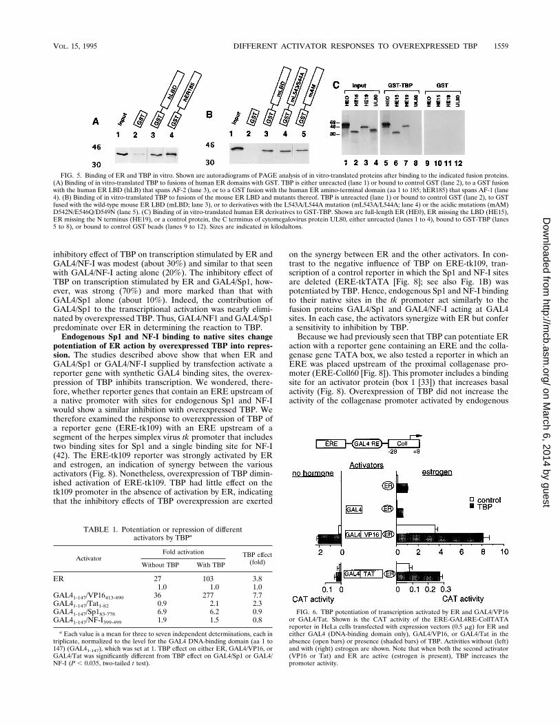
FIG. 5. Binding of ER and TBP in vitro. Shown are autoradiograms of PAGE analysis of in vitro-translated proteins after binding to the indicated fusion proteins.(A) Binding of in vitro-translated TBP to fusions of human ER domains with GST. TBP is either unreacted (lane 1) or bound to control GST (lane 2), to a GST fusionwith the human ER LBD (hLB) that spans AF-2 (lane 3), or to a GST fusion with the human ER amino-terminal domain (aa 1 to 185; hER185) that spans AF-1 (lane4). (B) Binding of in vitro-translated TBP to fusions of the mouse ER LBD and mutants thereof. TBP is unreacted (lane 1) or bound to control GST (lane 2), to GSTfused with the wild-type mouse ER LBD (mLBD; lane 3), or to derivatives with the L543A/L544A mutation (mL543A/L544A; lane 4) or the acidic mutations (mAM)D542N/E546Q/D549N (lane 5). (C) Binding of in vitro-translated human ER derivatives to GST-TBP. Shown are full-length ER (HE0), ER missing the LBD (HE15),ER missing the N terminus (HE19), or a control protein, the C terminus of cytomegalovirus protein UL80, either unreacted (lanes 1 to 4), bound to GST-TBP (lanes5 to 8), or bound to control GST beads (lanes 9 to 12). Sizes are indicated in kilodaltons.
FIG. 6. TBP potentiation of transcription activated by ER and GAL4/VP16or GAL4/Tat. Shown is the CAT activity of the ERE-GAL4RE-CollTATAreporter in HeLa cells transfected with expression vectors (0.5 mg) for ER andeither GAL4 (DNA-binding domain only), GAL4/VP16, or GAL4/Tat in theabsence (open bars) or presence (shaded bars) of TBP. Activities without (left)and with (right) estrogen are shown. Note that when both the second activator(VP16 or Tat) and ER are active (estrogen is present), TBP increases thepromoter activity.
TABLE 1. Potentiation or repression of differentactivators by TBPa
ActivatorFold activation TBP effect
(fold)Without TBP With TBP
ER 27 103 3.81.0 1.0 1.0
GAL41-147/VP16413-490 36 277 7.7GAL41-147/Tat1-82 0.9 2.1 2.3GAL41-147/Sp183-778 6.9 6.2 0.9GAL41-147/NF-I399-499 1.9 1.5 0.8
a Each value is a mean for three to seven independent determinations, each intriplicate, normalized to the level for the GAL4 DNA-binding domain (aa 1 to147) (GAL41-147), which was set at 1. TBP effect on either ER, GAL4/VP16, orGAL4/Tat was significantly different from TBP effect on GAL4/Sp1 or GAL4/NF-I (P , 0.035, two-tailed t test).
VOL. 15, 1995 DIFFERENT ACTIVATOR RESPONSES TO OVEREXPRESSED TBP 1559
on March 6, 2014 by guest
http://mcb.asm
.org/D
ownloaded from

box 1 factor (Fig. 8, Coll60). In the presence of estrogen, ERand box 1 factor cooperated and activated strongly (Fig. 8,ERE-Coll60). TBP, however, diminished their cooperative ac-tivation. ER action on a control reporter without the box 1sequences (Fig. 8, ERE-CollTATA), in contrast, was potenti-ated by TBP. Thus, endogenous box 1 activator also synergizeswith ER but confers a sensitivity to inhibition by TBP.These observations of TBP effects on ER-activated tran-
scription from complex promoters indicate that the presence ofbinding sites for other activators may abolish the potentiationof ER action by TBP.
DISCUSSION
Interactions between the ER activation domains and TBP.The studies described above indicate that overexpression ofTBP in HeLa and CHO cells potentiates transcription activa-tion by ER in a dose-dependent manner. The potentiation,which varies between 3- and 10-fold, occurs on promoters thatcontain an ERE and a minimal TATA box region from eitherthe human collagenase promoter or the herpes simplex virus tkpromoter. Potentiation persists despite varying distances be-tween the ERE and the TATA box. Control experiments showthat overexpression of TBP does not change the concentrationof ER, nor does ER change the accumulation of TBP. Impor-tantly, TBP overexpression in these cells has no effect on basalactivation. These observations indicate that TBP is specificallylimiting for ER-activated transcription of these minimal pro-moters. Previous studies of several activators in mammaliancells have reported a similar pattern of interaction with over-expressed TBP (2, 7, 59). TBP potentiated activated but notbasal transcription from core promoters with a TATA box. Onsome more complex promoters, TBP is not limiting for ERaction because of inhibitory interactions between TBP andother transcription factors that predominate over the interac-tion with ER. These inhibitory interactions are discussed indetail below.ER has two separate activation domains, AF-1 and AF-2,
and each of these is potentiated by TBP when tested in isola-tion. Mutations that abolish AF-2 activity abolish the activity ofthis domain seen in the presence of overexpressed TBP. This
FIG. 7. TBP inhibition of transcription activated by both ER and GAL4/NF-Ior GAL4/Sp1. Shown is the CAT activity of the ERE-G4RE-CollTATA reporterin HeLa cells transfected with expression vectors (0.5 mg) for ER and either nosecond activator, GAL4/Sp1, or GAL4/NF-I in the absence (open bars) orpresence (shaded bars) of TBP. Activities without (left) and with (right) estrogenare shown. Note that when both the second activator (GAL4/Sp1 or GAL4/NF-I)and ER are active (estrogen is present), although the activators synergizestrongly, TBP decreases the promoter activity.
FIG. 8. TBP effects on ER action on promoters that contain an ERE and binding sites for endogenous activators. Shown is CAT activity generated by promoterscontaining an ERE and the tk flanking region including its binding sites for Sp1 and NF-I (ERE-tk109), by the collagenase promoter flanking region including the box1 site, or by control promoters containing an ERE and lacking the other sites (ERE-tkTATA and ERE-CollTATA). Each promoter is tested with transfected ER (0.5mg) or both ER and TBP (10 mg) and in the presence (striped bars) or absence (black bars) of estrogen. Note that whereas TBP overexpression increases ER actionat ERE-tkTATA and at ERE-CollTATA, TBP decreases ER action at ERE-tk109 and ERE-Coll60.
1560 SADOVSKY ET AL. MOL. CELL. BIOL.
on March 6, 2014 by guest
http://mcb.asm
.org/D
ownloaded from

finding indicates that TBP does not potentiate the activity ofthis domain by uncovering a cryptic activation function notrelated to the activity in the absence of TBP. Using the GSTfusion protein system, we have seen that in vitro-translatedTBP directly binds full-length ER and each of the activationdomains. However, variants of the AF-2 domain bearing mu-tations in a small region around aa 545 that eliminate all AF-2function are still able to bind TBP. This indicates that muta-tions of the aa 545 region disrupt a step in ER action inaddition to TBP binding. This step does not appear to bebinding to TFIIB, because although the ER LBD binds TFIIB(reference 25 and unpublished data), the mutations do notaffect such binding (27a). It has recently been shown that thesemutations disrupt the binding of ER to a candidate coactivator,RIP/ERAP (6, 8, 9, 21). Our preliminary mapping studies (notshown) place the region of interaction between ER and TBPoutside of the region that mediates interaction with RIP/ERAP. It may thus be possible to identify mutations that dis-rupt specifically the ER-TBP interaction. Our binding studiesare consistent with the previous studies of activation by reti-noic acid receptor and E1A, by c-Rel, and by Tax-1 (2, 7, 29,30, 59). Each of these proteins binds TBP in vitro; and theaction of each is potentiated by overexpressed TBP. However,the connection between the ability of TBP to bind ER in vitroand the ability to potentiate ER action in vivo is currentlyspeculative, as is true for TBP effects on the other activatorsnoted above. An adequate test will require the in vivo testingof TBP derivatives that disrupt binding in vitro.The ER shows potent autoinhibition when overexpressed,
and we find that coexpression of TBP partly relieves this au-toinhibition. The behavior of ER is similar to the previouslyreported behavior of overexpressed viral activator Tax-1 (7).The autoinhibition of transcription produced by overexpres-sion of activator proteins is commonly explained by the forma-tion of nonfunctional complexes between the excess activatorand a limiting target (18, 41, 45). Thus, our observations sug-gest that TBP may be among the targets of autoinhibition bytranscriptional activators. However, some autoinhibition byER continues when TBP is overexpressed, and TBP fails torelieve autoinhibition at complex promoters (reference 58 anddata not shown). This finding suggests that there are othertargets involved. Recent in vitro studies suggest that autoinhi-bition by ER, E1a, or VP16 can be fully relieved by TFIIDcomplexes and not by TBP alone (3–5). Thus, a second likelycandidate for a target of autoinhibition by ER is a TFIID-associated coactivator molecule needed for ER-activated tran-scription. A human TAF present in only a subfraction ofTFIID that preferentially stimulates transcription by the ERactivation domains (4, 5) has recently been cloned (27). Thisprotein, TAFII30, may be among the targets. In addition, a pairof proteins, RIP and ERAP, that bind the ER LBD only in thepresence of agonist ligands and do not bind the LBD of the aa545 region mutants defective in transcriptional activity havebeen identified (9, 21) and cloned (6, 8). These proteins arealso candidates for the target of autoinhibition.Different activators respond differently to overexpressed
TBP. We have compared the response of the ER activationdomains to overexpressed TBP with that of several other ac-tivation domains. Like ER, transcription activated by GAL4/VP16 or GAL4/Tat is further potentiated by overexpressedTBP. Although ER activation domains are not similar in com-position to VP16, some similarities in function have beennoted (4, 5). Unlike the case for ER, activation by GAL4/Sp1,GAL4/NF-I, or endogenous Sp1 and NF-I either is not in-creased or is diminished by TBP. The reporter gene was heldconstant in these studies. Thus, different activators show dif-
ferent responses to overexpressed TBP, at least when testedwith the collagenase or herpes simplex virus tk core promoter.Our finding that Sp1 does not respond to TBP contrasts with
previous observations that Sp1 activation of a promoter con-sisting of multiple response elements and the adenovirus majorlate promoter TATA box is potentiated by TBP overexpressionin Drosophila Schneider cells (12). We do not know the reasonfor these differences in the Sp1 response to TBP. The differ-ences may reflect the potent increase in basal transcription thatwas seen in the Schneider cells after overexpression with TBP(12). In the mammalian cells and promoters used in our stud-ies, no increase in basal transcription occurred with TBP. Inthe Schneider cells, TBP stimulated basal transcription morethan 100-fold and Sp1 activated transcription only 2- to 3-fold.These discrepancies may also reflect differences in the corepromoters used in the different studies. In this latter regard, itshould be noted that in Schneider cells, TBP failed to activatecore promoters that contained an initiator element and lackeda TATA box.When the ER is tested in concert with GAL4/VP16 on the
collagenase core promoter, the paired activators synergize andare further potentiated by TBP. Thus, TBP can superactivatepromoters that are already very strongly activated. When, onthe other hand, ER is paired with GAL4/NF-I or GAL4/Sp1,although the activators synergize, TBP overexpression inhibitstranscription. The inhibition by TBP is especially strong withthe ER-GAL4/Sp1 combination. Thus, GAL4/Sp1 appears topredominate over ER and sensitizes the promoter for inhibi-tion by TBP. Endogenous Sp1 and NF-I acting on the tk pro-moter, or the box 1 factor acting on the collagenase promoter,also synergize strongly with ER acting on an upstream EREand confer a sensitivity to inhibition by TBP. Thus, whereasTBP is limiting for ER action on the minimal collagenase orherpes simplex virus tk promoter, on complex promoters, thebinding of other upstream activators along with ER can makeTBP no longer limiting.Our observations of inhibitory effects of TBP overexpression
on synergy of ER and Sp1 are consistent with a recent reportof TBP interactions with the bovine papillomavirus E2 activa-tor (22). TBP inhibits E2-Sp1-synergized transcription but po-tentiates E2 in the absence of Sp1 (22). Again the effects ofpromoter context must be considered. Our studies were donewith the collagenase or tk core promoter containing the TATAbox and immediate downstream sequences. Although the im-mediate downstream sequences are not known to have initiatorfunction in these promoters, we did not test the isolated TATAboxes or potential initiator elements. However, in the study ofE2-Sp1-activated transcription recently reported, TBP inhib-ited synergy between these two activators, whether from theisolated adenovirus major late promoter TATA box or anisolated initiator element. Hence, the ability of Sp1 to bothsynergize with other activators and confer sensitivity to inhibi-tion by overexpressed TBP may be a general property.The mechanisms underlying the different responses to TBP
overexpression of different activators are obscure. One possi-bility is that they reflect differences in activator function. Forexample, an activator that was able to recruit TBP efficiently tothe TATA box in a cell with normal concentrations of TBPmight not have improved function when TBP concentrationsare elevated. An activator that was poor at recruiting TBP, incontrast, might have improved function when TBP concentra-tions are elevated. A second possibility is that overexpressionof TBP alters the composition of the different types of TBP-TAF (TFIID) complexes present in the cell. ER has recentlybeen shown to activate transcription with a distinct TFIIDcomplex that is not active with some other activators (27). It is
VOL. 15, 1995 DIFFERENT ACTIVATOR RESPONSES TO OVEREXPRESSED TBP 1561
on March 6, 2014 by guest
http://mcb.asm
.org/D
ownloaded from

possible that TBP overexpression favors the overproduction ofthis complex.In summary, ER differs from other transcriptional activa-
tors, such as Sp1 and NF-I, with respect to its ability to bepotentiated by overexpressed TBP. This may point to differ-ences in the mechanisms of action of these transcriptionalactivators.
ACKNOWLEDGMENTS
We thank Brian West, Greg Peterson, Grace Gill, Robert Tjian,Boris Peterlin, Danny Reinberg, Richard Metz, Rodrigo Bravo, EllenBaum, and Pierre Chambon for plasmids. We thank Geoffrey Greeneand Robert Weinzierl for antibody. We also thank Marrieta Dunaway,David Gardner, and Fred Schaufele for their critical comments on themanuscript.This work was supported by grant BE61E from the American Can-
cer Society to P.J.K.
REFERENCES1. Baum, E. Z., G. A. Bebernitz, J. D. Hulmes, V. P. Muzithras, T. R. Jones, andY. Gluzman. 1993. Expression and analysis of the human cytomegalovirusUL80-encoded protease: identification of autoproteolytic sites. J. Virol. 67:497–506.
2. Berkenstam, A., M. D. M. V. Ruiz, D. Barettino, M. Horikoshi, and H. G.Stunnenberg. 1992. Cooperativity in transactivation between retinoic acidreceptor and TFIID requires an activity analogous to E1A. Cell 69:401–412.
3. Boyer, J. B., and A. J. Berk. 1993. Functional interaction of adenovirus E1Awith holo-TFIID. Genes Dev. 7:1810–1823.
4. Brou, C., S. Chaudhary, I. Davidson, Y. Lutz, J. Wu, J. M. Egly, L. Tora, andP. Chambon. 1993. Distinct TFIID complexes mediate the effect of differenttranscriptional activators. EMBO J. 12:489–499.
5. Brou, C., J. Wu, S. Ali, E. Scheer, C. Lang, I. Davidson, P. Chambon, and L.Tora. 1993. Different TBP-associated factors are required for mediating thestimulation of transcription in vitro by the acidic transactivator GAL-VP16and the two nonacidic activation functions of the estrogen receptors. NucleicAcids Res. 21:5–12.
6. Brown, M., H. MacKay, S. Cariati, E. Marden, G. L. Martin, and S.Halachmi. 1994. Identification and cloning of proteins which bind estrogenreceptor contingent on the presence of ligand. J. Cell. Biochem. Suppl.18B:387.
7. Caron, C., R. Rousset, C. Beraud, V. Moncollin, J. M. Egly, and P. Jalinot.1993. Functional and biochemical interaction of the HTLV-1 TAX1 trans-activator with TBP. EMBO J. 12:4269–4278.
8. Cavailles, V., P. S. Danielan, R. White, and M. G. Parker. 1994. Character-ization of a potential mediator of estrogen receptor hormone-dependenttranscriptional activity. J. Cell. Biochem. Suppl. 18B:341.
9. Cavailles, V., S. Dauvois, P. S. Danielian, and M. G. Parker. 1994. Interac-tion of proteins with transcriptionally active estrogen receptors. Proc. Natl.Acad. Sci. USA 91:10009–10013.
10. Chen, J.-L., L. D. Attardi, C. P. Verrijzer, K. Yokomori, and R. Tjian. 1994.Assembly of recombinant TFIID reveals differential coactivator require-ments for distinct transcriptional activators. Cell 79:93–105.
11. Choy, B., and M. R. Green. 1993. Eukaryotic activators function duringmultiple steps of preinitiation complex assembly. Nature (London) 366:531–536.
12. Colgan, J., and J. L. Manley. 1991. TFIID can be rate limiting in vivo forTATA containing, but not TATA-lacking RNA polymerase II promoters.Genes Dev. 6:304–315.
13. Danielian, P. S., R. White, J. L. Lees, and M. G. Parker. 1992. Identificationof a conserved region required for hormone dependent transcription activa-tion by steroid hormone receptors. EMBO J. 11:1025–1033.
14. Dynlacht, B. D., T. Hoey, and R. Tjian. 1991. Isolation of coactivatorsassociated with the TATA-binding protein that mediate transcriptional ac-tivation. Cell 66:563–576.
15. Gaston, K., A. Bell, A. Kolb, H. Buc, and S. Busby. 1990. Stringent spacingrequirements for transcription activation by CRP. Cell 62:733–743.
16. Ghosh, S., M. J. Selby, and B. M. Peterlin. 1993. Synergism between Tat andVP16 in trans-activation of HIV-1 LTR. J. Mol. Biol. 234:610–619.
17. Gill, G., E. Pascal, Z. H. Tseng, and R. Tjian. 1994. A glutamine-richhydrophobic patch in transcription factor-Sp1 contacts the dTAF(II)110component of the Drosophila TFIID complex and mediates transcriptionalactivation. Proc. Natl. Acad. Sci. USA 91:192–196.
18. Gill, G., and M. Ptashne. 1988. Negative effect of transcriptional activatorGAL4. Nature (London) 334:721–724.
19. Goodrich, J. A., T. Hoey, C. J. Thut, A. Admon, and R. Tjian. 1993. Dro-sophila TAFII40 interacts with both VP16 activation domain and the basaltranscription factor TFIIB. Cell 75:519–530.
20. Hagemeier, C., A. J. Bannister, A. Cook, and T. Kouzarides. 1993. The
activation domain of the transcription factor PU.1 binds the retinoblastoma(RB) protein and the transcription factor TFIID in vitro: RB shows sequencesimilarity to TFIID and TFIIB. Proc. Natl. Acad. Sci. USA 90:1580–1584.
21. Halachmi, S., E. Marden, G. Martin, H. Mackay, C. Abbondanza, and M.Brown. 1994. Estrogen receptor-associated proteins: possible mediators ofhormone induced transcription. Science 264:1455–1458.
22. Ham, J., G. Steger, and M. Yaniv. 1994. Cooperativity in vivo between the E2transactivator and the TATA box binding protein depends on core promoterstructure. EMBO J. 13:147–157.
23. Hoey, T., R. O. J. Weinzierl, G. Gill, J. L. Chen, B. D. Dynlacht, and R. Tjian.1993. Molecular cloning and functional analysis of drosophila TAF110 revealproperties expected of coactivators. Cell 72:247–260.
24. Horikoshi, N., K. Maguire, A. Kralli, E. Maldonado, D. Reinberg, and R.Weinmann. 1991. Direct interaction between adenovirus E1A protein andthe TATA box binding transcription factor IID. Proc. Natl. Acad. Sci. USA88:5124–5128.
25. Ing, N. H., J. M. Beekman, S. Y. Tsai, M. J. Tsai, and B. W. O’Malley. 1992.Members of the steroid receptor superfamily interact with TFIIB (S300-II).J. Biol. Chem. 267:17617–17623.
26. Ingles, C. J., M. Shales, W. D. Cress, S. J. Triezenberg, and J. Greenblatt.1991. Reduced binding of TFIID to transcriptionally compromised mutantsof VP16. Nature (London) 351:588–590.
27. Jacq, X., C. Brou, Y. Lutz, I. Davidson, P. Chambon, and L. Tora. 1994.Human TAFII30 is present in a distinct TFIID complex and is required fortranscriptional activation by the estrogen receptor. Cell 79:107–117.
27a.Lopez, G. Unpublished data.28. Kashanchi, F., G. Piras, M. F. Radonovich, J. F. Duvall, A. Fattaey, C.-M.
Chiang, R. G. Roeder, and J. N. Brady. 1994. Direct interaction of humanTFIID with the HIV-1 transactivator Tat. Nature (London) 367:295–299.
29. Keaveney, M., A. Berkenstam, M. Feigenbutz, G. Vriend, and H. G. Stun-nenberg. 1993. Residues in the TATA-binding protein required to mediatea transcriptional response to retinoic acid in EC cells. Nature (London)365:562–566.
30. Kerr, L. D., L. J. Ransone, P. Wamsley, M. J. Schmitt, T. G. Boyer, Q. Zhou,A. J. Berk, and I. M. Verma. 1993. Association between proto-oncoproteinRel and TATA-binding protein mediates transcriptional activation by NF-kappa B. Nature (London) 365:412–419.
31. Kim, J. L., D. B. Nikolov, and S. K. Burley. 1993. Co-crystal structure of TBPrecognizing the minor groove of a TATA element. Nature (London) 365:520–527.
32. Kim, Y., J. H. Geiger, S. Hahn, and P. B. Siegler. 1993. Crystal structure ofa yeast TBP/TATA-box complex. Nature (London) 365:512–520.
33. Konig, H., H. Ponta, H. J. Rahmsdorf, and P. Herrlich. 1992. Interferencebetween pathway-specific transcription factors: glucocorticoids antagonizephorbol ester-induced AP-1 activity without altering AP-1 site occupation invivo. EMBO J. 11:2241–2246.
34. Kumar, V., S. Green, G. Stack, M. Berry, J. R. Jin, and P. Chambon. 1987.Functional domains of the human estrogen receptor. Cell 51:941–951.
35. Kushner, P. J., J. D. Baxter, K. G. Duncan, G. N. Lopez, F. Schaufele, R. M.Uht, P. Webb, and B. L. West. 1994. Eukaryotic regulatory elements lurkingin plasmid DNA: the activator protein-1 site in pUC. Mol. Endocrinol.8:405–407.
36. Kushner, P. J., E. Hort, J. Shine, J. D. Baxter, and G. L. Greene. 1990.Construction of cell lines that express high levels of the human estrogenreceptor and are killed by estrogens. Mol. Endocrinol. 4:1465–1473.
37. Lee, W. S., C. C. Kao, G. O. Bryant, X. Liu, and A. J. Berk. 1991. AdenovirusE1A activation domain binds the basic repeat in the TATA box transcriptionfactor. Cell 67:365–376.
37a.Leitman, D., and B. West. Unpublished data.38. Leitman, D. C., R. C. J. Ribeiro, E. R. Mackow, J. D. Baxter, and B. L. West.
1991. Identification of a tumor necrosis factor-responsive element in thetumor necrosis factor a gene. J. Biol. Chem. 266:9343–9346.
39. Leitman, D. C., E. R. Mackow, T. Williams, J. D. Baxter, and B. L. West.1992. The core promoter region of the tumor necrosis factor alpha geneconfers phorbol ester responsiveness to upstream transcriptional activators.Mol. Cell. Biol. 12:1352–1356.
40. Lopez, G., F. Schaufele, P. Webb, J. M. Holloway, J. D. Baxter, and P. J.Kushner. 1993. Positive and negative modulation of Jun action by thyroidhormone receptor at a unique AP1 site. Mol. Cell. Biol. 13:3042–3049.
41. Martin, K. J., J. W. Lillie, and M. R. Green. 1990. Evidence for an interac-tion of different eukaryotic transcriptional activators with distinct cellulartargets. Nature (London) 346:147–152.
42. McKnight, S., and R. Tjian. 1986. Transcriptional selectivity of viral genes inmammalian cells. Cell 46:795–805.
43. Metz, R., A. J. Bannister, J. A. Sutherland, C. Hagemeier, E. C. O’Rourke,A. Cook, R. Bravo, and T. Kouzarides. 1994. c-Fos-induced activation of aTATA-box-containing promoter involves direct contact with TATA-box-binding protein. Mol. Cell. Biol. 14:6021–6029.
44. Metz, R., T. Kouzarides, and R. A. Bravo. 1994. C-terminal domain in FosB,absent in FosB/SF and Fra-1, which is able to interact with the TATAbinding protein, is required for altered cell growth. EMBO J. 13:3832–3842.
45. Meyer, M. E., H. Gronemeyer, B. Turcotte, M. T. Bocquel, D. Tasset, and P.
1562 SADOVSKY ET AL. MOL. CELL. BIOL.
on March 6, 2014 by guest
http://mcb.asm
.org/D
ownloaded from

Chambon. 1989. Steroid hormone receptors compete for factors that medi-ate their enhancer function. Cell 57:433–442.
46. Neumann, J. R., C. A. Morency, and K. O. Russian. 1987. A novel rapid assayfor chloramphenicol acetyltransferase gene expression. BioTechniques5:444–447.
47. Nikolov, D. B., S. H. Hu, J. Lin, A. Gasch, A. Hoffmann, M. Horikoshi, N. H.Chua, R. G. Roeder, and S. K. Burley. 1992. Crystal structure of TFIIDTATA-box binding protein. Nature (London) 360:40–46.
48. Peterson, M. G., N. Tanese, B. F. Pugh, and R. Tjian. 1990. Functionaldomains and upstream activation properties of cloned human TATA bindingprotein. Science 248:1625–1630.
49. Ponglikitmongkol, M., J. H. White, and P. Chambon. 1990. Synergistic ac-tivation of transcription by the human estrogen receptor bound to tandemresponsive elements. EMBO J. 9:2221–2231.
49a.Sadovsky, Y. Unpublished data.50. Sadowski, I., J. Ma, S. Triezenberg, and M. Ptashne. 1988. GAL4-VP16 is an
unusually potent transcriptional activator. Nature (London) 335:563–564.51. Smith, D. B., and K. S. Johnson. 1988. Single-step purification of polypep-
tides expressed in Escherichia coli as fusions with glutathione S-transferase.Gene 67:31–40.
52. Stringer, K. F., C. J. Ingles, and J. Greenblatt. 1990. Direct and selectivebinding of an acidic transcriptional activator domain to the TATA-box factorTFIID. Nature (London) 345:783–786.
53. Tanese, N., B. F. Pugh, and R. Tjian. 1991. Coactivators for proline-richactivator purified from the multisubunit human TFIID complex. Genes Dev.5:2212–2224.
54. Taylor, C. M., B. Blanchard, and D. T. Zava. 1984. A simple method todetermine whole cell uptake of radiolabelled oestrogen and progesteroneand their subcellular localization in breast cancer cell lines in monolayerculture. J. Steroid Biochem. 20:1083–1088.
55. Tjian, R., and T. Maniatis. 1994. Transcriptional activation: a complex puz-zle with few easy pieces. Cell 77:5–8.
56. Tora, L., A. Mullick, D. Metzger, M. Ponglikitmongkol, I. Park, and P.Chambon. 1989. The cloned human oestrogen receptor contains a mutationwhich alters its hormone binding properties. EMBO J. 8:1981–1986.
57. Tora, L., J. White, C. Brou, D. Tasset, N. Webster, E. Scheer, and P.Chambon. 1991. The human estrogen receptor has two independent non-acidic transcriptional activation functions. Cell 59:477–487.
58. Webb, P., G. N. Lopez, G. L. Greene, J. D. Baxter, and P. J. Kushner. 1992.The limits of the cellular capacity to mediate an estrogen response. Mol.Endocrinol. 6:157–167.
59. Xu, X., C. Prorock, H. Ishikawa, E. Maldonado, Y. Ito, and C. Gelinas. 1993.Functional interaction of the v-Rel and c-Rel oncoproteins with the TATA-binding protein and association with transcription factor IIB. Mol. Cell. Biol.13:6733–6741.
60. Zawel, L., and D. Reinberg. 1993. Initiation of transcription by RNA poly-merase II: a multi-step process. Prog. Nucleic Acid Res. Mol. Biol. 44:67–108.
61. Zhou, Q., P. M. Lieberman, T. G. Boyer, and A. J. Berk. 1992. Holo-TFIIDsupports transcriptional stimulation by diverse activators and from a TATA-less promoter. Genes Dev. 6:1964–1974.
VOL. 15, 1995 DIFFERENT ACTIVATOR RESPONSES TO OVEREXPRESSED TBP 1563
on March 6, 2014 by guest
http://mcb.asm
.org/D
ownloaded from
Copyright © 2022 FDOKUMEN