Toxicology effects of Morning Glory Seed in rat: A metabonomic method for profiling of urine...
9
Journal of Ethnopharmacology 130 (2010) 134–142 Contents lists available at ScienceDirect Journal of Ethnopharmacology journal homepage: www.elsevier.com/locate/jethpharm Toxicology effects of Morning Glory Seed in rat: A metabonomic method for profiling of urine metabolic changes Chao Ma, Kaishun Bi, Ming Zhang, Dan Su, Xinxin Fan, Wei Ji, Chao Wang, Xiaohui Chen ∗ School of Pharmacy, Shenyang Pharmaceutical University, Wenhua Road 103, Shenyang 110016, PR China article info Article history: Received 31 January 2010 Received in revised form 16 April 2010 Accepted 20 April 2010 Available online 6 May 2010 Keywords: Morning Glory Seed Nephrotoxicity Metabonomics UPLC–MS PCA abstract Ethnopharmacological relevance: Morning Glory Seed (MGS) is a valuable traditional Chinese medicine which is widely used for the treatment of edema, ascites, hydroncus, simple obesity, lung fever and ardent fever. In recent years, long-term exposure to Morning Glory Seed (MGS) has shown to pose progressive renal damage in clinical practice. We hypothesize that changes in metabolic profile could have occurred before symptoms were observed, which may allow early treatment. Aim of the study: To investigate the metabolic changes caused by Morning Glory Seed-induced renal damage. Method: Metabonomics method was used for chronic toxicology study of MGS in Wistar rats. With a therapeutic dose, the model rats were orally administered the extract of MGS for 10 weeks continuously. The urine samples of model and control rats were collected in various time-points and the endogenous metabolites were analyzed by ultraperformance liquid chromatography coupled with mass spectrometry. The identification of all the potential biomarkers was performed using reference standard by comparing their mass spectra, MS/MS fragmentation and retention time. Furthermore, histopathology and clinical chemistry studies were carried out to ensure the success of MGS-induced nephrotoxicity model. Results: The difference in metabolic profiles between the control and the dosed rats was well observed by the principal component analysis (PCA) of the MS spectra. Significant changes of 12 metabolite biomarkers were detected in the rat urine samples. Metabonomics method could discriminate the model rats from the control rats in 2nd, 6th and10th week respectively before serious organic damage of kidney was found in 10th week by histopathology method. Conclusion: We believe that metabolic profiling may act as a preclinical protocol for MGS exposure before symptoms are observed. © 2010 Elsevier Ireland Ltd. All rights reserved. 1. Introduction In ancient China and Asian countries, naturally occurring drugs are traditionally used as legal medicines or folk medicines. By sci- entific medical system of some developed western countries, they have been increasingly recognized now. However, ever since a long time ago, there is a cognitive misunderstanding about the utilization of Chinese medicine that naturally occurring drugs are nonvenomous and have no side effects. As a matter of fact, Abbreviations: ATP, adenosine triphosphate; BUN, blood urea nitrogen; BPI, base peak intensity; CRF, chronic renal failure; CID, collision-induced dissociation; Ccr, creatinine clearance rate; ESI, electrospray ionization; HCG, healthy control group; IDO, indoleamine-2,3-dioxygenase; KYN, kynurenine; MGSG, Morning Glory Seed group; MAO, monoamine oxidase; PCA, principal component analysis; RSD, rela- tive standard deviations; ROS, reactive oxygen species; TCM, traditional Chinese medicine; TIC, total ion current. ∗ Corresponding author. Tel.: +86 24 23986296; fax: +86 24 23986259. E-mail address: cxh [email protected] (X. Chen). the toxicity and adverse reaction of Chinese medicine have been demonstrated in ancient time. In recent years, the ever-increasing reports about adverse reaction of Chinese herb such as Caulis Aris- tolochiae Manshuriensis and Aristolochia fangchi make the issue of Chinese medicine toxicity conspicuous. Morning Glory Seed (MGS), also called QianNiuZi (Chinese name), Semen Pharbitidis (Latin name) and Pharbitis seed (English name), is prepared from dried mature seeds of Pharbitis nil (L.) Choisy or Pharbitis purpurea (L.) Voigt. It is a valuable traditional Chinese medicine which has gained ever-increasing popularity in today’s China because of its involvement in the slimming regi- men of people. Morning Glory Seed has the therapeutic effect on relieving constipation by purgation, dispersing phlegm and wash- ing excessive fluid, and is widely used for the treatment of edema, ascites, hydroncus, simple obesity, lung fever and ardent fever (Tsai et al., 2004; Lee et al., 2008; The State Pharmacopoeia Commission of PR China, 2005). It could also be used as an obstetrical medicine for labor induction (Albert, 1979). It was commonly used as pill, powder and other solid dosage forms. In the usage of modern pro- 0378-8741/$ – see front matter © 2010 Elsevier Ireland Ltd. All rights reserved. doi:10.1016/j.jep.2010.04.031
-
Upload
independent -
Category
Documents
-
view
0 -
download
0
Transcript of Toxicology effects of Morning Glory Seed in rat: A metabonomic method for profiling of urine...
Tp
CS
a
ARRAA
KMNMUP
1
aehlua
pcIgtm
0d
Journal of Ethnopharmacology 130 (2010) 134–142
Contents lists available at ScienceDirect
Journal of Ethnopharmacology
journa l homepage: www.e lsev ier .com/ locate / je thpharm
oxicology effects of Morning Glory Seed in rat: A metabonomic method forrofiling of urine metabolic changes
hao Ma, Kaishun Bi, Ming Zhang, Dan Su, Xinxin Fan, Wei Ji, Chao Wang, Xiaohui Chen ∗
chool of Pharmacy, Shenyang Pharmaceutical University, Wenhua Road 103, Shenyang 110016, PR China
r t i c l e i n f o
rticle history:eceived 31 January 2010eceived in revised form 16 April 2010ccepted 20 April 2010vailable online 6 May 2010
eywords:orning Glory SeedephrotoxicityetabonomicsPLC–MSCA
a b s t r a c t
Ethnopharmacological relevance: Morning Glory Seed (MGS) is a valuable traditional Chinese medicinewhich is widely used for the treatment of edema, ascites, hydroncus, simple obesity, lung fever and ardentfever. In recent years, long-term exposure to Morning Glory Seed (MGS) has shown to pose progressiverenal damage in clinical practice. We hypothesize that changes in metabolic profile could have occurredbefore symptoms were observed, which may allow early treatment.Aim of the study: To investigate the metabolic changes caused by Morning Glory Seed-induced renaldamage.Method: Metabonomics method was used for chronic toxicology study of MGS in Wistar rats. With atherapeutic dose, the model rats were orally administered the extract of MGS for 10 weeks continuously.The urine samples of model and control rats were collected in various time-points and the endogenousmetabolites were analyzed by ultraperformance liquid chromatography coupled with mass spectrometry.The identification of all the potential biomarkers was performed using reference standard by comparingtheir mass spectra, MS/MS fragmentation and retention time. Furthermore, histopathology and clinicalchemistry studies were carried out to ensure the success of MGS-induced nephrotoxicity model.
Results: The difference in metabolic profiles between the control and the dosed rats was well observed bythe principal component analysis (PCA) of the MS spectra. Significant changes of 12 metabolite biomarkerswere detected in the rat urine samples. Metabonomics method could discriminate the model rats fromthe control rats in 2nd, 6th and10th week respectively before serious organic damage of kidney wasfound in 10th week by histopathology method.Conclusion: We believe that metabolic profiling may act as a preclinical protocol for MGS exposure before symptoms are observed.. Introduction
In ancient China and Asian countries, naturally occurring drugsre traditionally used as legal medicines or folk medicines. By sci-ntific medical system of some developed western countries, they
ave been increasingly recognized now. However, ever since aong time ago, there is a cognitive misunderstanding about thetilization of Chinese medicine that naturally occurring drugsre nonvenomous and have no side effects. As a matter of fact,
Abbreviations: ATP, adenosine triphosphate; BUN, blood urea nitrogen; BPI, baseeak intensity; CRF, chronic renal failure; CID, collision-induced dissociation; Ccr,reatinine clearance rate; ESI, electrospray ionization; HCG, healthy control group;DO, indoleamine-2,3-dioxygenase; KYN, kynurenine; MGSG, Morning Glory Seedroup; MAO, monoamine oxidase; PCA, principal component analysis; RSD, rela-ive standard deviations; ROS, reactive oxygen species; TCM, traditional Chinese
edicine; TIC, total ion current.∗ Corresponding author. Tel.: +86 24 23986296; fax: +86 24 23986259.
E-mail address: cxh [email protected] (X. Chen).
378-8741/$ – see front matter © 2010 Elsevier Ireland Ltd. All rights reserved.oi:10.1016/j.jep.2010.04.031
© 2010 Elsevier Ireland Ltd. All rights reserved.
the toxicity and adverse reaction of Chinese medicine have beendemonstrated in ancient time. In recent years, the ever-increasingreports about adverse reaction of Chinese herb such as Caulis Aris-tolochiae Manshuriensis and Aristolochia fangchi make the issueof Chinese medicine toxicity conspicuous.
Morning Glory Seed (MGS), also called QianNiuZi (Chinesename), Semen Pharbitidis (Latin name) and Pharbitis seed (Englishname), is prepared from dried mature seeds of Pharbitis nil (L.)Choisy or Pharbitis purpurea (L.) Voigt. It is a valuable traditionalChinese medicine which has gained ever-increasing popularity intoday’s China because of its involvement in the slimming regi-men of people. Morning Glory Seed has the therapeutic effect onrelieving constipation by purgation, dispersing phlegm and wash-ing excessive fluid, and is widely used for the treatment of edema,
ascites, hydroncus, simple obesity, lung fever and ardent fever (Tsaiet al., 2004; Lee et al., 2008; The State Pharmacopoeia Commissionof PR China, 2005). It could also be used as an obstetrical medicinefor labor induction (Albert, 1979). It was commonly used as pill,powder and other solid dosage forms. In the usage of modern pro-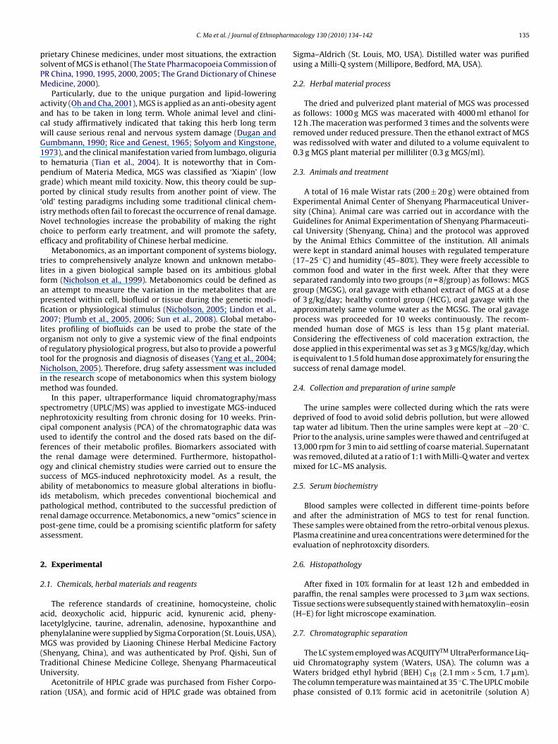
harm
psPM
aacwG1tpgp‘iNce
tlfapfi2lootNim
sncuftosaiprpa
2
2
alpM(TU
r
C. Ma et al. / Journal of Ethnop
rietary Chinese medicines, under most situations, the extractionolvent of MGS is ethanol (The State Pharmacopoeia Commission ofR China, 1990, 1995, 2000, 2005; The Grand Dictionary of Chineseedicine, 2000).Particularly, due to the unique purgation and lipid-lowering
ctivity (Oh and Cha, 2001), MGS is applied as an anti-obesity agentnd has to be taken in long term. Whole animal level and clini-al study affirmatively indicated that taking this herb long termill cause serious renal and nervous system damage (Dugan andumbmann, 1990; Rice and Genest, 1965; Solyom and Kingstone,973), and the clinical manifestation varied from lumbago, oliguriao hematuria (Tian et al., 2004). It is noteworthy that in Com-endium of Materia Medica, MGS was classified as ‘Xiapin’ (lowrade) which meant mild toxicity. Now, this theory could be sup-orted by clinical study results from another point of view. The
old’ testing paradigms including some traditional clinical chem-stry methods often fail to forecast the occurrence of renal damage.ovel technologies increase the probability of making the righthoice to perform early treatment, and will promote the safety,fficacy and profitability of Chinese herbal medicine.
Metabonomics, as an important component of systems biology,ries to comprehensively analyze known and unknown metabo-ites in a given biological sample based on its ambitious globalorm (Nicholson et al., 1999). Metabonomics could be defined asn attempt to measure the variation in the metabolites that areresented within cell, biofluid or tissue during the genetic modi-cation or physiological stimulus (Nicholson, 2005; Lindon et al.,007; Plumb et al., 2005, 2006; Sun et al., 2008). Global metabo-
ites profiling of biofluids can be used to probe the state of therganism not only to give a systemic view of the final endpointsf regulatory physiological progress, but also to provide a powerfulool for the prognosis and diagnosis of diseases (Yang et al., 2004;icholson, 2005). Therefore, drug safety assessment was included
n the research scope of metabonomics when this system biologyethod was founded.In this paper, ultraperformance liquid chromatography/mass
pectrometry (UPLC/MS) was applied to investigate MGS-inducedephrotoxicity resulting from chronic dosing for 10 weeks. Prin-ipal component analysis (PCA) of the chromatographic data wassed to identify the control and the dosed rats based on the dif-erences of their metabolic profiles. Biomarkers associated withhe renal damage were determined. Furthermore, histopathol-gy and clinical chemistry studies were carried out to ensure theuccess of MGS-induced nephrotoxicity model. As a result, thebility of metabonomics to measure global alterations in bioflu-ds metabolism, which precedes conventional biochemical andathological method, contributed to the successful prediction ofenal damage occurrence. Metabonomics, a new “omics” science inost-gene time, could be a promising scientific platform for safetyssessment.
. Experimental
.1. Chemicals, herbal materials and reagents
The reference standards of creatinine, homocysteine, choliccid, deoxycholic acid, hippuric acid, kynurenic acid, pheny-acetylglycine, taurine, adrenalin, adenosine, hypoxanthine andhenylalanine were supplied by Sigma Corporation (St. Louis, USA),GS was provided by Liaoning Chinese Herbal Medicine Factory
Shenyang, China), and was authenticated by Prof. Qishi, Sun ofraditional Chinese Medicine College, Shenyang Pharmaceuticalniversity.
Acetonitrile of HPLC grade was purchased from Fisher Corpo-ation (USA), and formic acid of HPLC grade was obtained from
acology 130 (2010) 134–142 135
Sigma–Aldrich (St. Louis, MO, USA). Distilled water was purifiedusing a Milli-Q system (Millipore, Bedford, MA, USA).
2.2. Herbal material process
The dried and pulverized plant material of MGS was processedas follows: 1000 g MGS was macerated with 4000 ml ethanol for12 h .The maceration was performed 3 times and the solvents wereremoved under reduced pressure. Then the ethanol extract of MGSwas redissolved with water and diluted to a volume equivalent to0.3 g MGS plant material per milliliter (0.3 g MGS/ml).
2.3. Animals and treatment
A total of 16 male Wistar rats (200 ± 20 g) were obtained fromExperimental Animal Center of Shenyang Pharmaceutical Univer-sity (China). Animal care was carried out in accordance with theGuidelines for Animal Experimentation of Shenyang Pharmaceuti-cal University (Shenyang, China) and the protocol was approvedby the Animal Ethics Committee of the institution. All animalswere kept in standard animal houses with regulated temperature(17–25 ◦C) and humidity (45–80%). They were freely accessible tocommon food and water in the first week. After that they wereseparated randomly into two groups (n = 8/group) as follows: MGSgroup (MGSG), oral gavage with ethanol extract of MGS at a doseof 3 g/kg/day; healthy control group (HCG), oral gavage with theapproximately same volume water as the MGSG. The oral gavageprocess was proceeded for 10 weeks continuously. The recom-mended human dose of MGS is less than 15 g plant material.Considering the effectiveness of cold maceration extraction, thedose applied in this experimental was set as 3 g MGS/kg/day, whichis equivalent to 1.5 fold human dose approximately for ensuring thesuccess of renal damage model.
2.4. Collection and preparation of urine sample
The urine samples were collected during which the rats weredeprived of food to avoid solid debris pollution, but were allowedtap water ad libitum. Then the urine samples were kept at −20 ◦C.Prior to the analysis, urine samples were thawed and centrifuged at13,000 rpm for 3 min to aid settling of coarse material. Supernatantwas removed, diluted at a ratio of 1:1 with Milli-Q water and vertexmixed for LC–MS analysis.
2.5. Serum biochemistry
Blood samples were collected in different time-points beforeand after the administration of MGS to test for renal function.These samples were obtained from the retro-orbital venous plexus.Plasma creatinine and urea concentrations were determined for theevaluation of nephrotoxcity disorders.
2.6. Histopathology
After fixed in 10% formalin for at least 12 h and embedded inparaffin, the renal samples were processed to 3 �m wax sections.Tissue sections were subsequently stained with hematoxylin–eosin(H–E) for light microscope examination.
2.7. Chromatographic separation
The LC system employed was ACQUITYTM UltraPerformance Liq-uid Chromatography system (Waters, USA). The column was aWaters bridged ethyl hybrid (BEH) C18 (2.1 mm × 5 cm, 1.7 �m).The column temperature was maintained at 35 ◦C. The UPLC mobilephase consisted of 0.1% formic acid in acetonitrile (solution A)
136 C. Ma et al. / Journal of Ethnopharmacology 130 (2010) 134–142
Table 1Gradient elution program of UPLC–MS for urine analysis.
Time (min) Flow rate (ml/min) A% B% Curve
Initial 0.3 5 95 Initial8 0.3 20 80 6
acsr
2
Mwmtwptsmc
2
Lwdtrfmav
3
3
vifoHms
3
dantnatC
The applied method was validated prior to the analysis of theexperimental samples, including the precision of injection, thewithin-day stability and the repeatability of sample preparation.Extracted ion chromatographic peaks of seven ions (with the reten-
11 0.3 50 50 615 0.3 90 10 617 0.3 5 95 6
nd 0.1% formic acid in water (solution B). While maintaining aonstant flow rate of 0.3 ml/min, the gradient elution program ishown in Table 1. Every 10 �l sample solution was injected for eachun.
.8. Mass spectrometry
Mass spectrometry was conducted on a Micromass QuattroicroTM API mass spectrometer (Waters Corp., Milford, MA, USA)ith an electrospray ionization (ESI) interface and triple quadrupleass analyser. The capillary voltage was 3.0 kV for positive ioniza-
ion mode while the cone voltage was 30 V. The source temperatureas set to 120 ◦C with a cone gas flow of 50 l/h, a desolvation tem-erature of 350 ◦C and a desolvation gas flow of 600 l/h. A scanime of 1 s with an inter-scan delay of 0.1 s was used for the analy-is. The mass was corrected with NaCsI before the study. Full scanode from m/z 100 to 900 and from 0 to 17 min was used for data
ollection.
.9. Data analysis
Raw UPLC/MS data were analyzed using Micromass Marker-ynx Application Version 4.1 (Waters, Milford, MA). MarkerLynxas employed for peak selection and peak alignment. The originalata were processed using the following parameters: initial reten-ion time 0 min, final retention time 16 min, mass tolerance 0.1 Da,etention time tolerance 0.01 min. The raw data were then trans-ormed into a single matrix containing aligned peaks with the same
ass/retention time pair along with peak normalized intensitiesnd sample name. The resulting dataset was analyzed by unsuper-ised principal component analysis (PCA).
. Results and discussion
.1. Clinical chemistry results
Serum biochemistry result was presented in Fig. 1. The averagealues of selected clinical chemistry parameters for the nephrotox-city study in MGSG did not show significant difference (p < 0.05)rom their time-matched control group in 2nd week. In 6th week,nly BUN value showed marked increase in MGSG compared withCG. At 10th week, the creatinine and BUN of MGSG, which arearkers of renal damage were elevated significantly (p < 0.05), pre-
enting a marked reduction of renal function.
.2. Histopathology
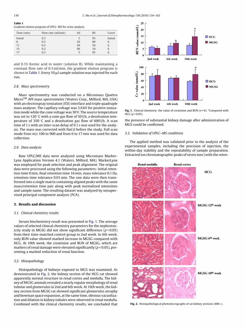
Histopathology of kidneys exposed to MGS was examined. Asemonstrated in Fig. 2, the kidney section of the HCG rat showedpparently normal structure in renal cortex and medulla. The kid-ey of MGSG animals revealed a nearly regular morphology of renal
ubular and glomerulus in 2nd and 6th week. At 10th week, the kid-ey section from MGSG rat showed significant glomerulus atrophynd bowman space expansion, at the same time, obvious vacuoliza-ion and dilation in kidney tubules were observed in renal medulla.ombined with the clinical chemistry results, we concluded thatFig. 1. Clinical chemistry: the value of creatinine and BUN (n = 8). *Compared withHCG (p < 0.05).
the presence of substantial kidney damage after administration ofMGS could be confirmed.
3.3. Validation of UPLC–MS conditions
Fig. 2. Histopathological photomicrographs of rat kidney sections (400×).
C. Ma et al. / Journal of Ethnopharmacology 130 (2010) 134–142 137
Table 2Potential biomarkers of nephrotoxicity induced by MGS.
Retention time (min) m/z (Da) Scan mode Quasi-molecular ion Metabolites Levels in abnormal urine
0.5 114 + [M+H]+ Creatinine ↓2nd, 10th week112 − [M−H]− ↑6th week
3.2 180 + [M+H]+ Hippuric acid ↑2nd week178 − [M−H]−
4.0 194 + [M+H]+ Phenylacetylglycine ↑2nd week192 − [M−H]− ↓6th week
2.9 190 + [M+H]+ Kynurenic acid ↑2nd week188 − [M−H]−
0.7 136 + [M+H]+ Homocysteine ↑6th, 10th week134 − [M−H]−
0.6 126 + [M+H] + Taurine ↑6th week124 − [M−H]− ↓10th week
11.3 357 + [M+H–2H2O]+ Deoxycholic acid ↑6th, 10th week437 − [M+HCOO]−
783 − [2M−H]−
12.5 373 + [M+H–2H2O]+ Cholic acid ↑6th–10th week453 − [M+HCOO]−
815 − [2M−H] −
1.1 166 + [M+H]+ Phenylalanine ↑10th week164 − [M−H]−
0.5 184 + [M+H]+ Adrenalin ↑10th week182 − [M−H]−
H]+
H]−
H]+
H]−
t3miscsctp
r04bt
3
rnPw
p6rp
3
vtp(p(
0.7 268 + [M+266 − [M−
0.7 137 + [M+135 − [M−
ion time and mass pairs of 1.4 297, 11.4 355, 7.9 441, 4.5 591,.8 618, 7.9 745, 11.4 840, in positive ion mode) were selected forethod validation. The relative standard deviations (RSDs) of peak
ntensities and retention time for the selected ion in pooled urineamples were calculated. The precision was examined by six repli-ated injections from the same sample, and a total of six differentamples which were obtained through the same preparation pro-edure was used for the repeatability. For the within-day stabilityest, six injections from one urine sample after preparation wereerformed in 24 h with an interval of 4 h.
The RSDs of retention time for precision, stability andepeatability were estimated to be 0.07–1.41%, 0.10–1.21% and.05–0.50%; while the RSDs for intensity varied within the ranges of.00–12.68%, 2.57–11.55% and 3.63–9.66%. The good precision, sta-ility, and repeatability indicated that the method could be utilizedo the analysis of urine samples.
.4. Data analysis
Principal component analysis, a chemometric model whicheduces matrix of data to their lowest dimension of the most sig-ificant factors, was used for analyzing the chromatographic data.eak list generated by Makerlynx from the positive LC–MS analysesas provided to the PCA program for pattern reorganization.
Fig. 3 shows the PCA score plot based on the urine metabolicrofiles in different time-points. At the time-points of 2nd (Fig. 3A),th (Fig. 3B) and 10th (Fig. 3C) week, the PCA score plots could beeadily divided into two clusters, which indicated urine metabolicattern significantly changed after the treatment of MGS.
.5. Biomarker identification
The loading profile (Fig. 3) that visualizes the influences ofariables, was used for the selection of biomarkers. Judged by
he distance from the origin, a series of ions which were foundredominantly in the loading plot were chosen as biomarkersTable 2). Furthermore, we compared the full scan intensity ofutative potential biomarkers in urine of control and MGSG ratsFig. 4).Adenosine ↑10th week
Hypoxanthine ↑10th week
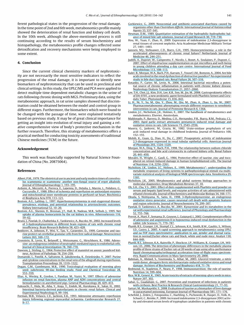
In our study, the identifications of the biomarkers were doneusing the commercial available standards by comparing theirMS/MS spectra and retention time. The biomarker with retentiontime and m/z pairs of 4.1 194 was identified as phenylacetyl-glycine, we take it as an example to illustrate the identificationprocess. First, the corresponding [M+H]+ peak of m/z 194 was foundaccording to the retention time 4.1 min in TIC chromatogram fromESI+ scan. Then, the MS/MS information about the fragmentationpattern of m/z 194 was acquired from the daughter scan mode.Under positive mode, the MS/MS figure contains high abundancefragment ion m/z 176, m/z 148, m/z 119, m/z 91, m/z 77 represent-ing [M+H−H2O]+, [M+H−H2O−CO]+, [M+H−H2O−CO−CH2−NH]+,[M+H−H2O−(CO)2−CH2−NH]+ and phenyl cation, respectively(Fig. 5). Then the databases of KEGG (http://www.genome.jp/kegg/)and METLIN (http://metlin.scripps.edu/) were searched based onthe clues we got from the process above. Finally, by comparingwith the standard sample of phenylacetylglycine according to itsretention time, m/z and product ion, the biomarker was identifiedas phenylacetylglycine.
With the same process, another example for the identificationof metabolite is cholic acid. The biomarker with retention time andmass pairs of 11.4 373 was identified as characteristic fragmentpeak of cholic acid with reference standard. Its MS, MS/MS infor-mation and possible MS fragmentation mechanism were shown inFig. 6.
3.6. Biochemical interpretation
According to the time after administration of MGS, PCA loadingplot was presented in Fig. 3, the different biomarkers in differenttime-points suggested that MGS-induced kidney morbidity seemedto progress in a stepwise fashion.
In the time-point of 2nd week, the main biomarkers shown inFig. 3A were hippuric acid, kynurenic acid, phenylacetylglycine and
creatinine. Hippuric acid and phenylacetylglycine are the metabo-lites of phenylalanine by the gut microflora. In this research, MGScaused changes in the concentration of metabolites related to thegut microflora, which strongly suggests that there are variationsin the gut microflora in response to MGS administration. This is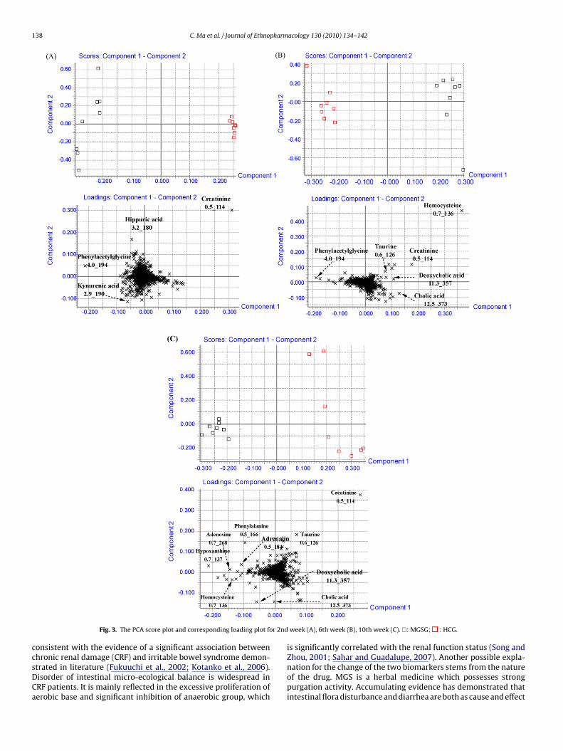
138 C. Ma et al. / Journal of Ethnopharmacology 130 (2010) 134–142
r 2nd
ccsDCa
Fig. 3. The PCA score plot and corresponding loading plot fo
onsistent with the evidence of a significant association between
hronic renal damage (CRF) and irritable bowel syndrome demon-trated in literature (Fukuuchi et al., 2002; Kotanko et al., 2006).isorder of intestinal micro-ecological balance is widespread inRF patients. It is mainly reflected in the excessive proliferation oferobic base and significant inhibition of anaerobic group, whichweek (A), 6th week (B), 10th week (C). �: MGSG; : HCG.
is significantly correlated with the renal function status (Song and
Zhou, 2001; Sahar and Guadalupe, 2007). Another possible expla-nation for the change of the two biomarkers stems from the natureof the drug. MGS is a herbal medicine which possesses strongpurgation activity. Accumulating evidence has demonstrated thatintestinal flora disturbance and diarrhea are both as cause and effect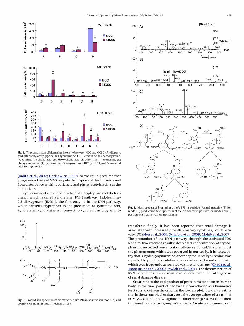
C. Ma et al. / Journal of Ethnopharmacology 130 (2010) 134–142 139
Fig. 4. The comparison of biomarker intensity between HCG and MGSG. (A) Hippurica(pw
(pflb
b2wk
Fp
cid, (B) phenylacetylglycine, (C) kynurenic acid, (D) creatinine, (E) homocysteine,F) taurine, (G) cholic acid, (H) deoxycholic acid, (I) adrenalin, (J) adenosine, (K)henylalanine and (L) hypoxanthine. *Compared with HCG (p < 0.01) and #comparedith HCG (p < 0.05).
Judith et al., 2007; Gorkiewicz, 2009), so we could presume thaturgation activity of MGS may also be responsible for the intestinalora disturbance with hippuric acid and phenylacetylglycine as theiomarkers.
Kynurenic acid is the end product of a tryptophan metabolismranch which is called kynurenine (KYN) pathway. Indoleamine-,3-dioxygenase (IDO) is the first enzyme in the KYN pathway,hich converts tryptophan to the precursors of kynurenic acid,
ynurenine. Kynurenine will convert to kynurenic acid by amino-
ig. 5. Product ion spectrum of biomarker at m/z 194 in positive ion mode (A) andossible MS fragmentation mechanism (B).
Fig. 6. Mass spectra of biomarker at m/z 373 in positive (A) and negative (B) ionmode, (C) product ion scan spectrum of the biomarker in positive ion mode and (D)possible MS fragmentation mechanism.
transferase finally. It has been reported that renal damage isassociated with increased proinflammatory cytokines, which acti-vate IDO (Hou et al., 2009; Schefold et al., 2009; Mohib et al., 2007).The promotion of the KYN pathway through the activated IDOleads to two relevant results: decreased concentration of trypto-phan and increased concentration of kynurenic acid. The later is justthe phenomenon which was observed in our study. It is notewor-thy that 3-hydroxykynurenine, another product of kynurenine, wasreported to produce oxidative stress and caused renal cell death,which was frequently associated with renal damage (Okuda et al.,1998; Bruns et al., 2002; Pawlak et al., 2001). The determination ofKYN metabolites in urine may be conducive to the clinical diagnosisof renal damage disease.
Creatinine is the end product of protein metabolism in humanbody. In the time-point of 2nd week, it was chosen as a biomarker
for its distance from the origin in the loading plot. It was interestingthat in the serum biochemistry test, the average values of creatininein MGSG did not show significant difference (p < 0.05) from theirtime-matched control group in 2nd week. Creatinine clearance rate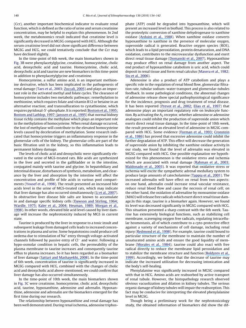
1 pharm
(fcwssMh
Fadi
irthmarBtitlcgbp
viciacmatIiW1as
stochpeooMal
iatfi
b
40 C. Ma et al. / Journal of Ethno
Ccr), another important biochemical indicator to evaluate renalunction, which is defined as the ratio of urine and serum creatinineoncentration, may be helpful to explain this phenomenon. In 2ndeek, the metabonomics result indicated that creatinine level is
ignificantly decreased in MGSG compared with HCG. Although theerum creatinine level did not show significant difference betweenGSG and HCG, we could tentatively conclude that the Ccr may
ave declined slightly.In the time-point of 6th week, the main biomarkers shown in
ig. 3B were phenylacetylglycine, creatinine, homocysteine, choliccid, deoxycholic acid and taurine. Homocysteine, cholic acid,eoxycholic acid and taurine are new biomarkers in this time-point
n addition to phenylacetylglycine and creatinine.Homocysteine, a sulfur amino acid, is an important methion-
ne derivative, which has been implicated in the pathogenesis ofenal damage (Taes et al., 2003; Zoccali, 2005) and plays an impor-ant role in the activated methyl and folate cycles. The clearance ofomocysteine includes two metabolic pathways: remethylation toethionine, which requires folate and vitamin B12 or betaine in an
lternative reaction; and transsulfuration to cystathionine, whichequires pyridoxal-5′-phosphate. It is reported (Bostom et al., 1995;ostom and Lathlop, 1997; Janssen et al., 1995) that normal kidneyissue richly contains the methylase which plays an important rolen the methylation of homocysteine. When renal damage occurred,he lost of methylase will contribute to the elevated homocysteineevels caused by deceleration of methylation. Some research indi-ated that homocysteine triggers an inflammatory response in thelomerular cells of the kidney. The glomerular cells are part of theasic filtration unit in the kidney, so this inflammation leads toermanent kidney damage.
The levels of cholic acid and deoxycholic were significantly ele-ated in the urine of MGS-treated rats. Bile acids are synthesizedn the liver and secreted in the gallbladder or in the intestine,onjugated mainly with taurine and glycine. In hepatobiliary andntestinal disease, disturbances of synthesis, metabolism, and clear-nce by the liver and absorption by the intestine will affect theoncentration and profile of bile acids in various pool compart-ents (Yousef et al., 1998). The result presented an increased bile
cids level in the urine of MGS-treated rats, which may indicatehat liver damage has also occurred simultaneously (Li et al., 2007).n obstructive liver disease bile salts are known to accumulaten and damage specific kidney cells (Dawson and Stirling, 1964;
ardle, 1975; Kaler et al., 2004; Heuman, 1989; Morgan et al.,998). In other words, elevated bile acid level caused by liver dam-ge will increase the nephrotoxicity induced by MGS in currenttate.
Taurine is produced by the liver in response to a toxic insult andubsequent leakage from damaged cells leads to increased concen-rations in plasma and urine. Some hepatotoxins could produce celledema due to stimulation of Na+ entry through voltage-activatedhannels followed by passive entry of Cl− and water. Following aypo-osmolar condition in hepatic cells, the permeability of thelasma membrane to taurine increases and consequently taurinefflux to plasma increases. So it has been regarded as a biomarkerf liver damage (Sattari and Mashayekhi, 2008). In the time-pointf 6th week, concentration of taurine is significantly increased inGSG compared with HCG, combined with the changes of cholic
cid and deoxycholic acid above-mentioned, we could confirm thativer damage has also occurred simultaneously.
In the time-point of 10th week, the main biomarkers shownn Fig. 3C were creatinine, homocysteine, cholic acid, deoxycholic
cid, taurine, hypoxanthine, adenosine and adrenalin. Hypoxan-hine, adenosine and adrenalin are the biomarkers emerged for therst time during our research.The relationship between hypoxanthine and renal damage haseen well demonstrated. During renal ischemia, adenosine triphos-
acology 130 (2010) 134–142
phate (ATP) could be degraded into hypoxanthine, which willpresent an increased level in biofluid. This process is also related tothe proteolytic conversion of xanthine dehydrogenase to xanthineoxidase (Arduini et al., 1988). When xanthine oxidase convertshypoxanthine to xanthine in the presence of molecular oxygen,superoxide radical is generated. Reactive oxygen species (ROS),which leads to a lipid peroxidation, protein denaturation, and DNAoxidation, contributes to the microvascular dysfunction and exertdirect renal tissue damage (Domanski et al., 2007). Hypoxanthinemay produce effect on renal damage from another aspect. Theend product of hypoxanthine catabolism is uric acid, which coulddeposit in renal tissue and form renal calculus (Masera et al., 1982;Siu et al., 2006).
Adenosine is also a product of ATP catabolism and plays aspecific role in the regulation of renal blood flow, glomerular filtra-tion rate, tubular sodium–water transport and glomerular-tubulesfeedback. In some pathological conditions, the abnormal changesof adenosine release show special pathophysiological significancefor the incidence, prognosis and drug treatment of renal disease.It has been reported (Prevot et al., 2002; Elias et al., 1997) thatadenosine plays an important regulatory role on leukocyte func-tion. By activating the A2 receptor, whether adenosine or adenosineanalogues could inhibit the production of superoxide anion whichexerts direct renal tissue damage. In the time-point of 10th week,the result presented an elevated level of adenosine in MGSG com-pared with HCG. Some evidence (Forman et al., 1993; Cronsteinet al., 1986) has proved that excessive adenosine could inhibit thefurther degradation of ATP. This effect will decrease the generationof superoxide anion by inhibiting the xanthine oxidase activity.Inour study, we found that the level of adrenalin was elevated inMGSG compared with HCG. One possible explanation we hypoth-esized for this phenomenon is the oxidative stress and ischemiawhich are associated with renal damage (Rahman et al., 2003;Maldonado et al., 2003). It was reported that oxidative stress orischemia will excite the sympathetic adrenal medullary system toproduce large amounts of catecholamine (Tappia et al., 2001). Theincreased adrenalin will have effect on renal from two aspects:on one hand, adrenalin could increase renal vascular resistance,reduce renal blood flow and cause the necrosis of renal cell; onthe other hand, the oxidation of adrenaline by monoamine oxidase(MAO) will release free radicals which exert direct renal tissue dam-age.In this stage, taurine is a biomarker again. However, we foundits level was decreased significantly in MGSG compared with HCG.This situation presented a sharp contrast with the 6th week. Tau-rine has extensively biological functions, such as stabilizing cellmembrane, scavenging oxygen free radicals, regulating intracellu-lar hosmostasis, all of which contribute to a cyto-protective effectagainst a variety of mechanisms of cell damage, including renalinjury (Redmond et al., 1998). For example, taurine could bound toparticular structure of the membrane to maintain the number ofunsaturated amino acids and ensure the good liquidity of mem-brane (Morales et al., 1984); taurine could also react with freeradical directly to reduce the membrane lipid peroxidation andto stabilize the membrane structure and function (Boldyrev et al.,1999). Accordingly, we believe that the decrease of taurine mayindicate the increased utilization for decreasing intoxication andthe body’s self-healing.
Phenylalanine was significantly increased in MGSG comparedwith that in HCG. Amino acids are reabsorbed by active transportof renal tubule. However, the histopathology research observedobvious vacuolization and dilation in kidney tubules. The serious
organic damage of kidney tubules will impair the reabsorption. Thisfact may be conducive for interpreting the elevated phenylalaninelevel in MGSG.Though being a preliminary work for the nephrotoxicologystudy, the obtained information of biomarkers did show the dif-
harm
fIsIchds
4
ipbcdrmidbbgafpC
A
d
R
A
A
B
B
B
B
C
D
D
D
E
F
F
C. Ma et al. / Journal of Ethnop
erent pathological states in the progression of the renal damage.n the time point of 2nd and 6th week, metabonomics profile mainlyhowed the deterioration of renal function and kidney cell death.n the 10th week, although the above-mentioned process is stillontinuing according to the results of serum biochemistry andistopathology, the metabonomics profile changes reflected someetoxification and recovery mechanisms were being employed toome extent.
. Conclusion
Since the current clinical chemistry markers of nephrotoxic-ty are not necessarily the most sensitive indicators to reflect therogression of the renal damage, it is important to identify newiomarkers of nephrototoxicity that can be used in preclinical andlinical settings. In this study, the UPLC/MS and PCA were applied toetect multiple time dependent metabolic changes in the urine ofats following chronic dosing with MGS. Preliminary data, using theetabonomic approach, in rat urine samples showed that discrim-
nation could be obtained between the model and control group inifferent stages. Furthermore, the biomarkers which were found toe changed with the passage of time, were explained tentativelyased on previous study. It may be of great clinical importance foretting an insight into evolution of renal injury and gaining a rel-tive comprehensive view of body’s protective mechanism in theurther research. Therefore, this strategy of metabonomics offers aractical method for conducting toxicity assessments of traditionalhinese medicines (TCM) in the future.
cknowledgement
This work was financially supported by Natural Science Foun-ation of China (No. 20875064).
eferences
lbert, P.M., 1979. The obstetrical use in ancient and early modern times of convolvu-lus scammonia or scammony: another non-fungal source of ergot alkaloids.Journal of Ethnopharmacology 1, 193–195.
rduini, A., Mezzetti, A., Porreca, E., Lapenna, D., DeJulia, J., Marzio, L., Polidoro, G.,Cuccurullo, F., 1988. Effect of ischemia and reperfusion on antioxidant enzymesand mitochondrial inner membrane proteins in perfused rat heart. Biochimicaet Biophysica Acta 970, 113–121.
ostom, A.G., Lathlop, L., 1997. Hyperhomoeysteinemia in end-stagerenal disease:prevalence, etiology, and potential relationship to arteriosclerotic outcomes.Kidney International 52, 10–20.
ostom, A.G., Shemin, D., Brosnan, J.T., Hall, B., Nadeau, M.R., Selhub, J., 1995. Netuptake of plasma homocystein by the rat kidney in vivo. Atheroselerosis 116,59–62.
runs, J., Pawlak, D., Chabielska, E., Tankiewicz, A., Buczko, W., 2002. Increased levelsof 3-hydroxykynurenine in different brain regions of rats with chronic renalinsufficiency. Brain Research Bulletin 58, 423–428.
oldyrev, A., Johnson, P., Wei, Y., Tan, Y., Carpenter, D., 1999. Carnosine and tau-rine protect rat cerebellar granular cells from free radical damage. NeuroscienceLetters 263, 169–172.
ronstein, B., Levin, R., Belanoff, J., Weissmann, G., Hirschhorn, R., 1986. Adeno-sine: an endogenous inhibitor of neutrophil-mediated injury to endothelial cells.Journal of Clinical Investigation 78, 760–770.
awson, J., Stirling, G., 1964. Protective effect of mannitol on anoxic jaundiced kid-neys. Archives of Pathology 78, 254–259.
omanski, L., Pawlik, A., Safranow, K., Jakubowska, K., Dziedziejko, V., 2007. Purineand cytokine concentrations in the renal vein of the allograft during reperfusion.Transplantation Proceedings 39, 1319–1322.
ugan, G.M., Gumbmann, M.R., 1990. Toxicological evaluation of morning gloryseed: subchronic 90-day feeding study. Food and Chemical Toxicology 28,553–559.
lias, A., Wesley, R., Gordon, I., Pandian, M., Vaziri, N., 1997. Effects of adenosineinfusion on renal function, plasma ANP and ADH concentrations and centralhemodynamics in anesthetized pigs. General Pharmacology 28, 429–433.
ukuuchi, F., Hida, M., Aiba, Y., Koga, Y., Endoh, M., Kurokawa, K., Sakai, H., 2002.Intestinal bacteria-derived putrefactants in chronic renal failure. Clinical andExperimental Nephrology 6, 99–104.
orman, M.B., Velasco, C.E., Jackson, E.K., 1993. Adenosine attenuates reperfusioninjury following regional myocardial ischaemia. Cardiovascular Research 27,9–17.
acology 130 (2010) 134–142 141
Gorkiewicz, G., 2009. Nosocomial and antibiotic-associated diarrhoea caused byorganisms other than Clostridium difficile. International Journal of AntimicrobialAgents 33, S37–S41.
Heuman, D.M., 1989. Quantitative estimation of the hydrophilic-hydrophobic bal-ance of mixed bile salt solutions. Journal of Lipid Research 30, 719–730.
Hou, W., Yuan, F., Zhao, H., 2009. Expression of indoleamine 2,3-dioxygenase inrenal tissues of crescent nephritis. Acta Academiae Medicinae Militaris Tertiae27, 1491–1493.
Janssen, M.J., Stehoawer, C.D., Boers, G.H., 1995. Homocysteinemia: a role in theaccelerated atheroegenesis of chronic renal failure? Netherlands Journal ofMedicine 46, 244–251.
Judith, A., Dupriet, W., Campeotto, F., Nicolis, I., Bonet, A., Soulaines, P., Dupont, C.,2007. Effect of oligofructose supplementation on gut microflora and well-beingin young children attending a day care centre. International Journal of FoodMicrobiology 113, 108–113.
Kaler, B., Morgan, W.A., Bach, P.H., Karram, T., Yousef, I.M., Bomzon, A., 2004. Are bileacids involved in the renal dysfunction of obstructive jaundice? An experimentalstudy in bile duct ligated rats. Renal Failure 26, 507–516.
Kotanko, P., Carter, M., Levin, N., 2006. Intestinal bacterial microflora a poten-tial source of chronic inflammation in patients with chronic kidney disease.Nephrology Dialysis Transplantation 21, 2057–2060.
Lee, T.H., Choi, J.J., Kim, D.H., Lee, K.R., Son, M., Jin, M., 2008. Gastroprokinetic effectsof DA-9701, a new prokinetic agent formulated with Pharbitis Semen and Cory-dalis Tuber. Phytomedicine 15, 836–843.
Li, H., Ni, Y., Su, M., Qiu, Y., Zhou, M., Qiu, M., Zhao, A., Zhao, L., Jia, W., 2007.Pharmacometabonomic phenotyping reveals different responses to xenobioticintervention in rats. Journal of Proteome Research 6, 1364.
Lindon, J.C., Nicholson, J.K., Holmes, E., 2007. The handbook of metabonomics andmetabolomics. Elsevier, Amsterdam.
Maldonado, P., Barrera, D., Medina, C.O., Hernandez, P.R., Ibarra, R.M., Pedraza, C.J.,2003. Aged garlic extract attenuates gentamicin induced renal damage andoxidative stress in rats. Life Sciences 73, 2543–2556.
Masera, G., Jankovic, M., Grazia, M., 1982. Urate-oxidase prophylaxis of uricacid–induced renal damage in childhood leukemia. Journal of Pediatrics 100,152–155.
Mohib, K., Guan, Q., Diao, H., Du, C., 2007. Proapoptotic activity of indoleamine2,3-dioxygenase expressed in renal tubular epithelial cells. American Journalof Physiology 293, 1324–1329.
Morgan, W.A., Ding, Y., Bach, P.H., 1998. The relationship between sodium chlorideconcentration and bile acid cytotoxicity in cultured kidney cells. Renal Failure20, 441–450.
Morales, H., Wright, C., Gaull, G., 1984. Protective effect of taurine, zinc and toco-pherol on retinol-lnduced damage in human lymphoblastoid cells. The Journalof Nutrition 114, 2256–2261.
Nicholson, J.K., Lindon, J.C., Holmes, E., 1999. Metabonomics”: understanding themetabolic responses of living systems to pathophysiological stimuli via multi-variate statistical analysis of biological NMR spectroscopic data. Xenobiotica 29,1181.
Nicholson, J.K., 2005. Metabonomics and global systems biology approaches tomolecular diagnostics. Drug Metabolism Reviews 37, 10.
Oh, S.H., Cha, Y.S., 2001. Effect of diets supplemented with Pharbitis seed powder onserum and hepatic lipid levels, and enzyme activities of rats administered withethanol chronically. Journal of Biochemistry and Molecular Biology 34, 166–171.
Okuda, S, Nishiyama, N., Satio, H., 1998. 3-Hydroxykynurenine, an endogenousoxidative stress generator, causes neuronal cell death with apoptotic featuresand region selectivity. Journal of Neurochemistry 70, 299–307.
Pawlak, D, Tankiewicz, A., Buczko, W., 2001. Kynurenine and its metabolites in therat with experimental renal sufficiency. Journal of Physiology and Pharmacology52, 755–766.
Prevot, A., Huet, F., Semama, D., Gouyon, J., Guignard, J., 2002. Complementary effectsof adenosine and angiotensin II in hypoxemia-induced renal dysfunction in therabbit. Life Sciences 71, 779–787.
Plumb, R.S., Granger, J.H., Stumpf, C.L., Johnson, K.A., Smith, B.W., Gaulitz, S., Wilson,I.D., Castro, J., 2005. A rapid screening approach to metabonomics using UPLCand oa-TOF mass spectrometry: application to age, gender and diurnal varia-tion in normal/Zucker obese rats and black, white and nude mice. Analyst 130,844–849.
Plumb, R.S., Johnson, K.A., Rainville, P., Shockcor, J.P., Williams, R., Granger, J.H., Wil-son, I.D., 2006. The detection of phenotypic differences in the metabolic plasmaprofile of three strains of Zucker rats at 20 weeks of age using ultra-performanceliquid chromatography/orthogonal acceleration time-of-flight mass spectrom-etry. Rapid Communications in Mass Spectrometry 20, 2800.
Rahman, A., Ahmed, S., Vasenwala, S., Athar, M., 2003. Glyceryl trinitrate, a nitricoxide donor, abrogates ferric nitrilotriacetate-induced oxidative stress and renaldamage. Archives of Biochemistry and Biophysics 418, 71–79.
Redmond, H., Stapleton, P., Neary, P., 1998. Immunonutrition: the role of taurine.Nutrition 14, 599–604.
Rice, W.B., Genest, K., 1965. Acute toxicity of extracts of morning-glory seeds in mice.Nature 207, 302.
Sahar, G., Guadalupe, G., 2007. Prevention and treatment of infections in patients
with cirrhosis. Best Practice & Research Clinical Gastroenterology 21, 77–93.Sattari, M., Mashayekhi, S., 2008. Evaluation of taurine as a biomarker of liver damagein paracetamol poisoning. European Journal of Pharmacology 581, 171–176.
Schefold, J., Zeden, J., Fotopoulou, C., Haehling, S., Pschowski, R., Hasper, D., Volk, H.,Schuett, C., Reinke, P., 2009. Increased indoleamine 2,3-dioxygenase (IDO) activ-ity and elevated serum levels of tryptophan catabolites in patients with chronic
1 pharm
S
S
S
S
T
T
T
T
59.
42 C. Ma et al. / Journal of Ethno
kidney disease: a possible link between chronic inflammation and uraemicsymptoms. Nephrology Dialysis Transplantation 24, 1901–1908.
iu, Y., Leung, K., Tong, M., Kwan, T., 2006. Use of allopurinol in slowing the progres-sion of renal disease through its ability to lower serum uric acid level. AmericanJournal of Kidney Diseases 47, 51–59.
olyom, L., Kingstone, E., 1973. An obsessive neurosis following morning glory seedingestion treated by aversion relief. Journal of Behavior Therapy and Experimen-tal Psychiatry 4, 293–295.
ong, S., Zhou, S., 2001. The changes of intestinal microecology and the effect ofmicroecological products in patients with chronic renal failure. Journal of Inter-nal Intensive Medicine 7, 100–101.
un, J.C., Schnackenberg, L.K., Holland, R.D., 2008. Metabonomics evaluation of urinefrom rats given acute and chronic doses of acetaminophen using NMR andUPLC/MS. Journal of Chromatography B871, 328–340.
aes, Y.E., Delanghe, J.R., Lameire, N.H., Lameire, N.H., 2003. Creatine supplementa-tion decreases homocysteine in an animal model of uremia. Kidney International64, 1331–1337.
appia, P., Hata, T., Hozaima, L., Sandhu, M., Panagia, V., Dhalla, N., 2001. Role of
oxidative stress in catecholamine-induced changes in cardiac sarcolemmal Ca2+transport. Archives of Biochemistry and Biophysics 387, 85–92.he State Pharmacopoeia Commission of PR China, 2005. Pharmacopoeia of PR China.
Chemical Industry Press, Beijing.he State Pharmacopoeia Commission of PR China, 2000. Pharmacopoeia of PR China.
Chemical Industry Press, Beijing.
acology 130 (2010) 134–142
The State Pharmacopoeia Commission of PR China, 1995. Pharmacopoeia of PR China.Chemical Industry Press, Beijing.
The State Pharmacopoeia Commission of PR China, 1990. Pharmacopoeia of PR China.Chemical Industry Press, Beijing.
2000. The Grand Dictionary of Chinese Medicine. China Traditional Chinese MedicinePress Beijing.
Tian, L.Q., Zhang, Z.L., Zhang, B.S., 2004. Research on the pharmacology, toxicity andclinical application of Morning glory seed. Chinese Journal of Nature medicine3, 146–147.
Tsai, J.C., Tsai, S.L., Chang, W.C., 2004. Comparison of two Chinese medical herbs,Huangbai and Qianniuzi, on influence of short circuit current across the ratintestinal epithelia. Journal of Ethnopharmacology 93, 21–25.
Wardle, E.N, 1975. Renal failure in obstructive jaundice: pathogenic factors. Post-graduate Medical Journal 15, 512–514.
Yang, J., Xu, G.W., Zheng, Y.F., Kong, H.W., Pang, T., Lv, S., Yang, Q., 2004. Diagnosisof liver cancer using HPLC-based metabonomics avoiding false-positive resultfrom hepatitis and hepatocirrhosis diseases. Journal of Chromatography B 813,
Yousef, I.M., Bouchard, G., Tuchweber, B., Plaa, G.L., 1998. Mechanism involved inbile acid-induced cholestasis. Toxicology of The Liver 2, 347–382.
Zoccali, C., 2005. Biomarkers in chronic kidney disease: utility and issues towardsbetter understanding. Current Opinion in Nephrology and Hypertension 14,532–537.