Tinnitus and Temporomandibular Disorders - IntechOpen
20
2 Tinnitus and Temporomandibular Disorders Kengo Torii Nippon Dental University Japan 1. Introduction Temporomandibular disorders (TMDs) are a group of related disorders of the masticatory system (the masticatory musculature and the temporomandibular joint). The most frequent symptom is pain, usually localized in the muscles of mastication, the preauricular region, and the temporomandibular joint (TMJ). Patients often complain of jaw ache, earache, headache, and facial pain. In addition to pain, patients with these disorders frequently have limited or asymmetric jaw movement and joint sounds that are described as clicking or crepitus (McNeill, 1990). The causes of TMDs remain unclear, and numerous factors have been implicated. The first description of the relationship between TMJ dysfunction and aural symptoms is thought to have been made by Costen in 1934. Costen reported various clinical cases of patients with ear and nasal symptoms; he summarized his findings by stating that hearing tests showed a mild type of catarrhal otitis with eustachian tube involvement (usually simple obstruction). The prognosis of such cases reportedly depended on these factors: (a) the accuracy with which refitted dentures relieved the abnormal pressure on the joint; and (b) the extent of the injury to the tube and to the condyle, meniscus, and joint capsule. A more general acknowledgement of the relationship between ear symptoms and TMJ dysfunction was subsequently made (Costen, 1934, 1936, 1944). Sicher reported that from an anatomical perspective, the eustachian tube cannot be compressed during the closure of the mandible; neither can the opening action of the tensor palati muscle on the tube be impaired in this condition, making Costen’s theory impossible (Sicher, 1948). Schwartz theorized that contractive muscle spasms may cause pain as a direct result of myofascial trigger mechanisms and by referred routes (Schwartz, 1956). 2. Tinnitus and treatment for TMDs After Schwarzt’s proposal, Dolowitz et al. reported that muscle exercises were effective for the relief of aural symptoms, especially ear fullness (44 out of 47 diagnosed patients) and tinnitus (40 out of 43 diagnosed patients). Various therapies have since been tried for the relief of TMD symptoms, including aural symptoms (Dolowitz et al., 1964). These therapies include selective grinding of the teeth, bite plate, biofeedback training, thermotherapy and muscle training (Koskinnen et al., 1980; Principato & Barwell, 1978). Parker and Chole reported that tinnitus and vertigo were significantly more prevalent among a TMD group than among either of their control groups (Parker & Chole, 1995). Erlandsson et al. reported that stomatognathic treatment (occlusal splint, occlusal adjustment, and exercise therapy) www.intechopen.com
-
Upload
khangminh22 -
Category
Documents
-
view
1 -
download
0
Transcript of Tinnitus and Temporomandibular Disorders - IntechOpen

2
Tinnitus and Temporomandibular Disorders
Kengo Torii Nippon Dental University
Japan
1. Introduction
Temporomandibular disorders (TMDs) are a group of related disorders of the masticatory system (the masticatory musculature and the temporomandibular joint). The most frequent symptom is pain, usually localized in the muscles of mastication, the preauricular region, and the temporomandibular joint (TMJ). Patients often complain of jaw ache, earache, headache, and facial pain. In addition to pain, patients with these disorders frequently have limited or asymmetric jaw movement and joint sounds that are described as clicking or crepitus (McNeill, 1990). The causes of TMDs remain unclear, and numerous factors have been implicated. The first description of the relationship between TMJ dysfunction and aural symptoms is thought to have been made by Costen in 1934. Costen reported various clinical cases of patients with ear and nasal symptoms; he summarized his findings by stating that hearing tests showed a mild type of catarrhal otitis with eustachian tube involvement (usually simple obstruction). The prognosis of such cases reportedly depended on these factors: (a) the accuracy with which refitted dentures relieved the abnormal pressure on the joint; and (b) the extent of the injury to the tube and to the condyle, meniscus, and joint capsule. A more general acknowledgement of the relationship between ear symptoms and TMJ dysfunction was subsequently made (Costen, 1934, 1936, 1944). Sicher reported that from an anatomical perspective, the eustachian tube cannot be compressed during the closure of the mandible; neither can the opening action of the tensor palati muscle on the tube be impaired in this condition, making Costen’s theory impossible (Sicher, 1948). Schwartz theorized that contractive muscle spasms may cause pain as a direct result of myofascial trigger mechanisms and by referred routes (Schwartz, 1956).
2. Tinnitus and treatment for TMDs
After Schwarzt’s proposal, Dolowitz et al. reported that muscle exercises were effective for the relief of aural symptoms, especially ear fullness (44 out of 47 diagnosed patients) and tinnitus (40 out of 43 diagnosed patients). Various therapies have since been tried for the relief of TMD symptoms, including aural symptoms (Dolowitz et al., 1964). These therapies include selective grinding of the teeth, bite plate, biofeedback training, thermotherapy and muscle training (Koskinnen et al., 1980; Principato & Barwell, 1978). Parker and Chole reported that tinnitus and vertigo were significantly more prevalent among a TMD group than among either of their control groups (Parker & Chole, 1995). Erlandsson et al. reported that stomatognathic treatment (occlusal splint, occlusal adjustment, and exercise therapy)
www.intechopen.com
Up to Date on Tinnitus
16
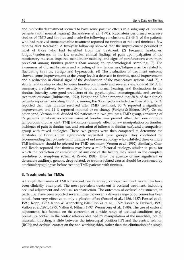
and biofeedback treatment seemed to have some positive effects in a subgroup of tinnitus patients (with normal hearing) (Erlandsson et al., 1991). Rubinstein performed extensive studies of TMD and tinnitus and made the following conclusions: (1) 46 % of the patients who had received stomatognathic treatment reported no tinnitus or reduced tinnitus a few months after treatment. A two-year follow-up showed that the improvement persisted in most of those who had benefited from the treatment. (2) Frequent headaches, fatigue/tenderness in the jaw muscles, clinical findings of pain upon palpation of the masticatory muscles, impaired mandibular mobility, and signs of parafunctions were more prevalent among tinnitus patients than among an epidemiological sampling. (3) The awareness of diurnal bruxism and a feeling of jaw tenderness/fatigue may be related to fluctuating tinnitus, vertigo and hyperacusis. (4) The evaluation of treatment outcome showed some improvements at the group level: a decrease in tinnitus, mood improvement, and a reduction in clinical signs of the dysfunction of the masticatory system. And (5), a strong relationship existed between tinnitus complaints and several symptoms of TMD. In summary, a relatively low severity of tinnitus, normal hearing, and fluctuations in the tinnitus intensity were good predictors of the psychological, stomatognathic, and cervical treatment outcome (Rubinstein, 1993). Wright and Bifano reported that 38 % of their clinic’s patients reported coexisting tinnitus; among the 93 subjects included in their study, 56 % reported that their tinnitus resolved after TMD treatment, 30 % reported a significant improvement, and 14 % reported minimal or no change (Wright & Bifano, 1997). On the other hand, Vernon et al. divided 929 patients into two groups: a TMD group, consisting of 69 patients in whom no known cause of tinnitus was present other than one or more temporomandibular joint (TMJ) indicators (example: effect of jaw manipulation on tinnitus, incidence of pain in tinnitus ear, and sensation of fullness in tinnitus ear), and a comparison group with mixed etiologies. These two groups were then compared to determine the attributes of tinnitus that significantly separated these groups. They concluded by recommending that patients with tinnitus of unknown etiology who exhibited three or more TMJ indicators should be referred for TMD treatment (Vernon et al., 1992). Similarly, Chan and Reade reported that tinnitus may have a multifactorial etiology, similar to pain, for which the correction or elimination of any one of the factors may result in the complete resolution of symptoms (Chan & Reade, 1994). Thus, the absence of any significant or detectable auditory, genetic, drug-related, or trauma-related causes should be confirmed by otorhinolaryngologists before treating TMD patients with tinnitus.
3. Treatments for TMDs
Although the causes of TMDs have not been clarified, various treatment modalities have
been clinically attempted. The most prevalent treatment is occlusal treatment, including
occlusal adjustment and occlusal reconstruction. The outcomes of occlusal adjustments, in
particular, have been reported several times; however, a diverse range of outcomes has been
noted, from very effective to only a placebo effect (Forssel et al., 1986, 1987; Forssel et al.,
1999; Kopp, 1979; Kopp & Wenneberg,1981; Tsolka et al., 1992; Tsolka & Preiskel, 1993;
Vallon et al.,1991, 1995; Vallon & Nilner, 1997; Wenneberg et al., 1988). The use of occlusal
adjustments has focused on the correction of a wide range of occlusal conditions (e.g.,
premature contact in the centric relation obtained by manipulation of the mandible, not by
muscular directing; a slide between the intercuspal position [IP] and the centric relation
[RCP]; and occlusal contact on the non-working side), rather than the elimination of a single
www.intechopen.com

Tinnitus and Temporomandibular Disorders
17
condition (e.g., only premature contact in the centric relation). Thus, the results of these
treatments have been difficult to interpret. Egermark-Eriksson et al. performed a long-term
epidemiologic study of the relationship between occlusal factors and TMD and reported that
occlusal interference, as conventionally described (in an occlusal analysis based on the
reference position of the RCP obtained by manipulating the mandible), does not seem to be
of much help in explaining the development or persistence of TMD (Egermark-Eriksson et
al., 1987). Pullinger et al. also reported that occlusion cannot be considered the unique or
dominant factor in defining TMD populations (Pullinger et al., 1993). Since the relationship
between these occlusal factors (premature contact in the centric relation obtained by
manipulation, not a straight slide from the centric to intercuspal position) and TMD have
not been scientifically demonstrated, the Technology Conference on the Management of
Temporomandibular Disorders of the NIH/NIDR (held on April 29 through May 1, 1996, in
the USA) concluded that no data exists to support many commonly held beliefs regarding
TMD, nor does any data exist to support the superiority of any method of management
compared with the effect of a placebo. The conference participants insisted that validated
diagnostic methods, with respect to sensitivity and specificity, for the identification of TMD
were lacking and noted the obvious need for basic research on TMD. They then stated their
belief that occlusal adjustments are invasive and that the superiority of such treatment over
other non-invasive therapies has not been demonstrated in randomized controlled
prospective trials. Since this conference, many conflicting opinions have been reported
(Dawson, 1999; Green et al., 1998), and most clinical researchers and basic researchers have
become estranged from investigations of the relationship between occlusion and TMD.
Therefore, studies on occlusion and TMD have been rarely performed since that time, and
treatment for TMD has shifted to more conservative modalities (Tsukiyama et al., 2001). The
outcomes of these conservative treatments for TMD have been symptomatic and temporary,
not resulting in a cure, because the treatments have not been applied to patients based on
evidence, as the causes of TMD remain unknown. Therefore, the effects of these treatments
for tinnitus have also varied (Dolowitz et al, 1964; Erlandsson et al.,1991; Parker & Chole,
1995; Principato & Barwell, 1978; Schwartz, 1956). In 2005, Torii and Chiwata reported a
highly significant relationship between the TMJ sounds and the discrepancy between the
habitual occlusal position (HOP) and the bite plate-induced occlusal position (BPOP)
(Torii & Chiwata, 2005). TMJ sounds are the most common and most relevant sign of
TMD (Könönen et al., 1993; Köhler et al., 2009; Nydell et al., 1994). Thus, the possible effect
of occlusal equilibration of the BPOP on TMD signs and symptoms has also been inferred.
In their study, the BPOP was defined using a physiological muscular position as a
reference position. The BPOP was obtained during voluntary jaw closing, while the
patient was in an upright position and after wearing an anterior bite plate for a short
period time (Fig. 1).
The muscular position has been reported to vary with the posture of the subject and is thought to be less reproducible than the manually obtained centric relation (which is guided by a dentist). However, Torii and Chiwata reported that the variation in the BPOP decreased after wearing an anterior bite plate, resulting in a reduction in the severity of TMD symptoms, compared with the symptoms experienced at the beginning of treatment (Fig. 2) (Torii & Chiwata, 2010). In addition, they reported the temporary effect of a bite plate on TMD and described the need for occlusal equilibration of the BPOP (Table 1, Fig. 3).
www.intechopen.com

Up to Date on Tinnitus
18
Fig. 1. Anterior flat plane bite plate. The plate covers the six upper anterior teeth and the first premolar teeth on both sides. The BPOP (physiological muscular position) is obtained during voluntary jaw closing after wearing the bite plate for a short period time.
Fig. 2. Mean variations in the three axes on different days. x: mediolateral; y: anteroposterior; z: superoinferior. HOP: habitual occlusal position; BPOP: bite plate-induced occlusal position. This data was obtained in a case with TMJ arthralgia (Fig. 3). The symptoms disappeared 12 days after wearing an anterior flat plane bite plate.
Torii and Chiwata then performed a pilot study on the effect of occlusal equilibration using
the BPOP on TMD and reported that the percentage of symptom-free patients at one year
after treatment was 86% according to the Subjective Dysfunction Index (SDI) and 76%
according to the Clinical Dysfunction Index (CDI); these changes in both indices after
treatment were significant (P < 0.01) (Fig. 4 and 5) (Tables 1 and 2) (Torii & Chiwata 2010).
www.intechopen.com

Tinnitus and Temporomandibular Disorders
19
After occlusal adjustment
Occlusal discrepancy No discrepancy Discrepancy
Before occlusal adjustment
Discrepancy 20 0
No discrepancy 1 0
McNemar’s test: X2cal = 18.05 >X2(0.001) = 10.83, Significant. HOP: habitual occlusal position; BPOP: bite plate-induced occlusal position.
Table 1. Changes in the occlusal discrepancy between the HOP and BPOP before and after occlusal adjustment
Myofascial Disc disp. Arthritis
CDI 1st Exam. 1 y 1st Exam. 1 y 1st Exam. 1 y
DiO 0% 80% 0% 86% 0% 0%
DiI 0% 20% 36% 14% 0% 100%
DiII 40% 0% 43% 0% 50% 0%
DiIII 20% 0% 21% 0% 50% 0%
CDI: Helkimo Clinical Index; 1st Exam.: first examination; 1 y: 1-year follow-up examination; Myofascial: Myofascial pain; Disc disp.: Disc displacement. DiO: no TMD; DiI: mild TMD; DiII: moderate TMD; and DiIII: severe TMD.
Table 2. Changes in Helkimo Clinical Index at first examination and after a 1-year follow-up examination for each TMD subtype
NS H1 B1
H2 H6 H7
↑
S ↓
NS B2 H3 B3
H4 B4
H5 B5
B6 B7 H8 B8
H9 B9
H10 B10
Days 1 3 6 9 12 15 25 33 50 110
VAS 6 3 2 2 0 0 0 0 0 0
Max. 40 42 42 42 42 42 45 45 45 45
NS: Not significant; S: Significant; Days: Days of visits; H1-10: Habitual occlusal position on different day; B1-10: Bite plate-induced occlusal position on different day; VAS: Score on a 10-point Visual Analogue Scale; Max.: Maximum unassisted opening (mm).
Fig. 3. HOP and BPOP positions and TMD symptoms on different days. The occlusal discrepancy between the HOP and the BPOP disappeared on day 6, and the disappearance was maintained between days 6 and 12. However, the discrepancy reappeared after 15 days. Therefore, equilibration of the occlusion was required to resolve the discrepancy. The occlusal equilibration was performed at 17 days.
www.intechopen.com

Up to Date on Tinnitus
20
ッefore treatment
ケ
ゲケ
コケ
ゴケ
サケ
ザケ
ヅiO ヅiI ヅiII ヅiIII
ヅysfunction Indices
% of patients
Subjective Index
ツlinical Index
Fig. 4. Dysfunction Index at first examination. Distribution of dysfunction indices before
occlusal adjustment. Di O: no TMD; Di I: mild TMD; Di II: moderate TMD; Di III: severe TMD.
ヂfter treatment
ケ
コケ
サケ
シケ
8ケ
ゲケケ
ヅiO ヅiI ヅiII ヅiIII
ヅysfunction Indices
% of patients
Subjective Index
ツlinical Index
Fig. 5. Dysfunction Index at 1-year evaluation. Distribution of dysfunction indices after
occlusal adjustment. Di O: no TMD; Di I: mild TMD; Di II: moderate TMD; Di III: severe TMD. None of the patients required an anterior bite plate during a one-year follow-up period after the completion of the occlusal adjustment. The number of visits varied between
2 and 34, with a mean of 11.0 ± 6.0 visits. Between 1 and 13 sessions of occlusal adjustment
were performed, with a mean of 4.7 ± 3.5 sessions. The changes in the SDI and CDI were significant (p<0.01).
www.intechopen.com

Tinnitus and Temporomandibular Disorders
21
Fig. 6. Mean HOP-BPOP difference before and after treatment. Changes in the mean diffrence between the habitual occlusal position (HOP) and the bite plate-induced occlusal position (BPOP) before and after occlusal adjustment. x: mediolateral; y: anteroposterior; z: superoinferior. The changes were significant on the x-axis and y-axis (p<0.05), whereas the change on the z-axis was not significant (p>0.1).
Figure 6 and Table 1 indicate that a good outcome was obtained with resolution of the
occlusal discrepancy by occlusal equilibration of the BPOP. Therefore, this treatment is a
causal treatment, not a symptomatic treatment. In this pilot study, they reported that the
otalgia of one patient and the tinnitus of another patient also completely disappeared after
treatment. Table 3 shows the outcomes of the treatment of occlusal equilibration for TMDs
and tinnitus in our practice.
4. Causal treatment for TMDs
Until now, occlusal discrepancy has been considered to be only one among several possible
causes of TMD. At present, the only evidence-based causal treatment of TMD is occlusal
equilibration, in which the habitual occlusal position (HOP) is aligned with the muscular
position (BPOP). This treatment requires an articulator that can precisely reproduce the
BPOP (voluntary or habitual closure movement). Torii observed various mandibular
closures and found that the voluntary closing movement of the mandible consisted of a
vertical movement against the occlusal plane at a point of less than 1 mm to the plane (Torii,
1989). He then developed an articulator that has a mechanism allowing vertical movement.
Thus, the BPOP can be precisely reproduced using this articulator. When the upper and
lower dental models are attached to the articulator with the BPOP wax record, the
anteroposterior parallel space remains between both occlusal surfaces of the models after
the wax record has been removed (Fig. 7).
www.intechopen.com

Up to Date on Tinnitus
22
Table 3. Treatment outcome of TMD patients with aural symptoms
No: Patient number; Visits: Number of visits; Period: Treatment period. Disc Disp.: Disc displacement; Myofascial: Myofascial Pain. M: Male ; F: Female. (R), (L) and (B): Mainly affected side on the (R): right; (L): left; (B): both sides.
Fig. 7. Mounting of upper and lower dental casts with BPOP wax record. After the upper and lower casts are mounted with the BPOP wax record, and the wax record is removed, an anteroposteriorly and mediolaterally uniform space remains between both casts.
To interdigitate the teeth of the upper and lower models under this condition, a movement that is almost vertical to the occlusal plane of either model is necessary. If the models are allowed to make occlusal contact based on a hinge movement (like in a conventional articulator), the incisors and premolars make contact, and the molars will not occlude.
www.intechopen.com

Tinnitus and Temporomandibular Disorders
23
Therefore, to reproduce the occlusal relationship in the intercuspal position using an articulator, a mechanism that allows the upper model to be moved perpendicular to the occlusal plane is needed (Fig. 8).
Fig. 8. Vertically movable upper mounting ring.
The procedure for occlusal analysis and occlusal equilibration in patients with TMDs is as follows. 1. After patient’s medical history has been taken and a clinical examination has been
performed, an HOP record is taken using a polysiloxane material while the patient is in an upright position. While the HOP is being recorded, the patient is asked to close his or her mouth so as to achieve maximum intercuspation and to hold that position until the material has set (approximately one minute). An anterior bite plate is then fabricated directly in the mouth using a self curing acrylic resin material; the vertical dimension should be sufficient to produce a 1.0 mm-jaw separation at the second molars. The plate should cover the upper six anterior teeth and both first premolars. The occlusal surface of the plate should be flat and perpendicular to the mandibular incisors to allow free movement in all directions. The bite plate should be worn until the severity of symptoms is resolved, usually after one or two weeks; the bite plate is worn all day except while eating, speaking, or brushing one’s teeth. If the opening limitation and/or pain are not resolved using a bite plate, other symptomatic therapies, such as pharmacotherapy or physical medicine, may be necessary. Once the patient can freely open his or her mouth without pain, the treatment can proceed to the next step.
2. A BPOP record is then taken using the same material as that used to record the HOP. After the bite plate has been worn for 5 minutes, during which time the patient is asked to tap or slide his or her lower anterior teeth against the plate (muscular conditioning), the plate is removed and the recording material is applied to the occlusal surface using a syringe; the patient is then instructed to close his or her mouth until tooth contact is made and to hold that position. A BPOP wax record is then made using a wax registration material (Bite Wafer; Kerr USA, Romulus, MI, USA) and the same procedure mentioned above. The anterior part of the wafer should be cut off and softened in 60 ℃ water. After muscular conditioning using the bite plate has been
performed, the softened wafer is placed on the premolars and molars on both sides. The patient is then instructed to bite the wafer gently, without biting through the wafer. The intercondylar distance is then measured using a caliper. Then, the distance between the condylar point and the lower incisal point in the occluded position is measured.
www.intechopen.com

Up to Date on Tinnitus
24
3. The intercondylar distance of the articulator is set to the same value as that of the patient. The gradation for zero on the scales on both sides indicates an intercondylar distance of 13 cm (the distance between the outsides of both condylar spheres). Therefore, when the condylar distance of the patient is 12 cm, the articulator should be reduced medially by 5 mm on each side. The articulator is then inverted, and the incisal point is marked on the mandibular cast table at a point the same distance from the condylar ball of the articulator as present in the patient. The lower cast is attached to the lower member of the articulator. The upper cast is then attached to the upper member using the BPOP wax record. At this time, a sheet of wrapping film is placed on the base of the upper cast, and some plaster is poured into the mounting ring of the upper member. The film will separate between the base of the upper cast and the poured plaster. Once the plaster has set, the film is removed and the upper cast and the poured plaster are adhered in place using α-cyanoacrylate adhesive material. This mounting
method prevents dimensional changes in the mounting plaster and subsequent inaccuracies in the articulated casts.
4. Next, the occlusal discrepancy is examined. Both condylar rods of the articulator are exchanged with the analyzing rods. Recording discs attached to graph papers are then inserted into the condylar shaft. An HOP polysiloxane record is placed between the upper and lower casts and the positions are tattooed on the graph papers using colored occlusal paper with recording needles. In the same manner, the BPOP is tattooed on the same graph paper using a different color occlusal paper. The difference between the HOP and the BPOP and the direction from the BPOP to the HOP are recorded (Fig. 9).
Fig. 9. Use of the articulator as an occlusal position analyzer.
The analyzing rods are then replaced with the condylar rods, the BPOP wax record is placed between the upper cast and the lower cast, and all the adjustable parts are set. The BPOP wax record is then removed, and the upper cast is moved downward until tooth contact has been made. Premature occlusal contact is located on the casts by marking and pulling an occlusal tape (Occlusion Foil; Coltene/Whaledent Gmbh/Co. KG, Langenau, Germany). One of several therapies (selective tooth grinding, prosthodontic and orthodontic treatments, or orthognathic surgery) may then be selected for the occlusal equilibration of the BPOP, according to the degree of the discrepancy between the HOP and the BPOP.
www.intechopen.com

Tinnitus and Temporomandibular Disorders
25
5. The patient should be sitting in an upright position without a headrest support. The bite plate is worn in the patient’s mouth. After muscular conditioning has been performed by tapping and sliding the lower anterior teeth against the plate for 5 minutes, the plate is removed from the patient’s mouth. The operator holds Miller ribbon holders (Buffalo Dental Manufacturing Co., Inc. Brooklyn, New York, USA) for the right and left quadrant recordings with red occlusal foils and asks the patient to close his or her jaw until tooth contact, then to hold that position. True occlusal contact should be confirmed by pulling the foil laterally. The premature contact should be compared with the marked contact on the casts of the articulator.
6. The premature contact on the cast is removed using a small pear-shaped carbide bur with a slow speed handpiece. The ground spot is marked with a pencil. The incisal pin of the articulator is then removed from the upper member and the articulator is closed to make occlusal contact. The incisal pin must be removed to enable a hinge movement of the articulator, because during mandibular closure, the condition in which the condyle is suspended in the capsule changes to a condition in which the condyle is contracted against the disc and the eminence, resulting in a hinge movement. The location where the next premature contact appears should be confirmed by marking and pulling with an occlusal foil. The contacts should then be recorded.
7. The patient is then reclined and the premature contact is ground down using a diamond
point of a similar size and shape and a high speed handpiece. Then, the patient is raised to
an upright position and the bite plate is placed in the mouth. After muscular conditioning
has been performed, the patient is asked to close his or her mouth until tooth contact has
been made and to mark the premature contact with an occlusal foil. If the new contact is
located at the same position as on the articulator, the articulator is regarded as precisely
reproducing the BPOP and it can be reliably used in the following steps during future
appointments. After the new contact has been confirmed, the contact is ground down. The
impressions of the upper and lower dental arches are taken in the mouth. After the patient
wears the bite plate in the mouth for 5 minutes, the bite plate is removed, and a BPOP
wax record is then taken for the next appointment. 8. New upper and lower casts are made and attached to the articulator in the previously
described manner. Steps 6 and 7 are repeated. The occlusal adjustments are continued in the mouth by referring to the contacts marked on the casts. The occlusal adjustment is completed by confirming the occlusal contacts on the premolars and molars on both sides of the casts attached to the articulator and in the mouth. Generally, two types of premature occlusal contact occur in TMD patients: one is a premature contact at the most posterior tooth with the occlusion opened anteriorly (often seen in young patients), the other is a premature contact at the anterior tooth with the occlusion opened posteriorly (often seen in elderly patients). The former type should be treated using occlusal adjustments. The latter should be treated by restoration of the teeth or using a fixed, or removable prosthesis. After completing the occlusal equilibration, HOP and BPOP polysiloxane records are taken and both positions are confirmed to be consistent with each other on the articulator.
5. Possible mechanism of Tinnitus related to TMD
Table 3 indicates that the side affected by TMD coincides with the side affected by tinnitus. Figure 10 shows the shifts recorded on both condylar areas of the articulator in case number
www.intechopen.com

Up to Date on Tinnitus
26
9 shown in Table 3. This case has been previously reported (Torii & Chiwata, 2007). The patient complained of pain in her right ear and TMJ as well as tinnitus in her right ear. The shift on the right side was larger than that on the left side. The shifts noted after the resolution of her symptoms indicated that a displacement of the mandible from its correct position (muscular position) to a deflected position (habitual occlusal position) existed before treatment.
Fig. 10. Recorded mandibular shift from the habitual occlusal position to the correct occlusal position. In this case, the patient was diagnosed as having Méniére disease by two independent otorhinolaryngologists and was treated with isosorbide, etc. However, when the drugs failed to improve her severe symptoms, she visited our clinic complaining of pain in the right TMJ region. An occlusal analysis was performed using dental models mounted on an articulator after relieving the painful symptoms using appliance therapy, and an occlusal discrepancy was identified. Occlusal equilibration of the BPOP was then performed (see Table 3).
This displacement seems to have distorted the normal relationship between the disc and the
condyle in the TMJ, since the correct relationship between the disc and the condyle is
thought to be fundamentally established during the development of the function of the
masticatory muscles (with regard to the muscular position, that is the BPOP) before tooth
eruption. On the other hand, the existence of an occlusal discrepancy between the HOP and
the BPOP means that as the mandible voluntarily closes, the elevator muscles require
additional activity to adapt the mandible from the BPOP to the HOP as multiple teeth come
in contact with a stable position during the change from isotonic muscle contraction to
isometric. This condition may cause muscle tension, muscle fatigue and muscle spasm,
resulting in muscle pain. Therefore, when considering the mechanism of tinnitus related to
TMD, the displacement of the TMJ and muscle tension or spasm should be taken into
account. First of all, regarding the displacement of the TMJ, Pinto found that a tiny ligament
connects the neck and anterior process of the malleus to the medioposterosuperior part of
the TMJ capsule, the interarticular disc, and the sphenomandibular ligament. He also
described that the fibrous layer of the tympanic membrane seemed to be continuous with
this tiny ligament and that the tiny ligament had an embryologic origin common with that
of the malleus and incus (Pinto, 1962). However, Komori et al. performed an anatomical
study and concluded that no evidence was found to support the “tiny ligament theory,” as
the ligament did not show any visible movement in response to a given strain. (Komori et
www.intechopen.com

Tinnitus and Temporomandibular Disorders
27
al., 1986) Rodriguez-Vásquez et al. reported that the discomalleolar ligament is a capsular
structure similar to an intrinsic ligament that is formed by the association of capsular fibers
coming from the posterosuperior and medial end of the articular disc. They then described
that these fibers belong to the upper lamina of the bilaminar zone (Rodríguez-Vásquez et al.,
1998). Eckerdal observed the petrotympanic fissure and reported that the dimensions of the
fissure and the morphologic arrangement of the intrafissural and extrafissural soft tissues do
not corroborate the theory that forces generally may be conducted through the fissure from
the TMJ to the ear ossicles (Eckerdal, 1991). Parker and Chole reported that although the
discomalleolar ligament and the mallear portion of the sphenomandibular ligament exist
and could conceivably transmit mechanical energy to the malleus, they can not account for
high frequency subjective tinnitus from local perturbations of the position of the malleus
(Parker & Chole, 1995). In summary, the direct transmission of force arising from the
displacement of the TMJ seems unlikely to be capable of causing ear symptoms. However,
Johansson et al. described in their radiographic and histologic study that a medially
displaced TMJ disc may exert traction or friction or may rub against the auriculotemporal
nerve trunk. Since the auriculotemporal nerve innervates the tympanic membrane, the
irritation of the nerve medially or posteromedially to the TMJ, before the nerve divergence,
may give rise to symptoms from the ear region (Johansson et al., 1990). Williamson reported
that noxious pain stimuli to the peridiscal tissues (similar to condyle or disc displacement)
could result in the constriction of the internal auditory and posterior auricular arteries. Such
constriction may result in a decrease in the blood supply to the middle and inner ears. Thus,
the displacement of the TMJ may cause tinnitus (Williamson, 1990). Myers reported that
beginning with the appearance of a reciprocal clicking in the TMJ, the disk is forced
anteromedially with full intercuspation of the teeth. This change is accompanied by the
stretching of the posterior laminae and the stretching or tearing of the lateral collateral
ligament. As the laminae are stretched, vascular soft tissue is pulled forward and tends to
become trapped between the head of the condyle and the roof of the fossa. This injury recurs
each time the teeth are intercuspated, with displacement of the disk. This causes a chronic
inflammation with extension of the inflammatory area, especially along three pathways.
Among these three pathways, the second pathway is located in the carotid sheath area. In
the case of a displaced disk specimen, the arteries, veins and nerves are impossible to dissect
because of the fibrous material adhering to them and the surrounding structures. Tension
would thus be produced on the whole carotid sheath in response to any movement of the
neck or jaw. This tension of the carotid sheath fibrosis would be transmitted to the jugular
foramen, making the saccus endolymphaticus unable to expand to perform any possible
regulation of the endolymphatic pressure; as a result, the increased endolymphatic pressure
on the hair cells of the cochlea could cause tinnitus (Myers, 1988). Regarding masticatory muscle fatigue or tension, Fuchs observed the masticatory muscle activity during sleep at night and concluded that the results of the observed activity supports the suggestion that dysfunctional patients without biteplates have a higher muscular activity during sleep than healthy persons (Fuchs, 1975). In addition, Mao et al. reported that the near absence of type II A fibers (fast-contracting, fatigue-resistant fibers) from human jaw muscles seems to suggest that these fibers may be readily susceptible to fatigue during sustained efforts at stronger force levels, since fast, fatigue-susceptible (type IIB) fibers must be recruited at these levels (Mao et al., 1993). Myrhaug reported that bite deformity is known among otologists to be a cause of tinnitus, and this type of tinnitus
www.intechopen.com
Up to Date on Tinnitus
28
arises as an autogenous vibration in the sound-conducting system, possibly because of tremor or myoclonus (myorhythmia) in the tensor muscle of the middle ear. This condition represents a fatigue reaction resulting from prolonged irritation and stress of the muscles innervated by the trigeminal nerve (masticators). This form of fatigue reaction apparently only affects the small muscles in this group because of their anatomical relationship. Hence, the two tensor muscles are in a vulnerable position in bite anomalies (Myrhaug, 1964). Koskinnen et al. and Parker and Chole were skeptical about Myrhaug’s theory (Koskinnen et al., 1980; Parker & Chole, 1995). However, Watanabe et al. reported that tinnitus occurred whenever a certain mimic facial muscle contracted voluntarily or involuntarily. Under the operating microscope, the contraction of the stapedial muscle synchronous with the contraction of the mimic facial muscle was observed. The tinnitus disappeared completely immediately after the tendon of the stapedial muscle was sectioned (Watanabe et al., 1974). Myrhaug described a matter similar to that in the report of Watanabe et al. in which contractions of the tensor tympany muscle were viewed directly under a microscope during voluntary stimulation by grinning and clenching movements of the jaw. Taking into account the facts that myofascial pain, disc displacement, tension-type headaches and tinnitus were resolved after occlusal equilibrating treatment for the BPOP (Table 3), tinnitus related to TMD may be caused by the spasmodic synkinesis of the tensor tympani and the stapedial muscle with muscle spasm of the masticatory muscles (including relative muscles), or by a decrease in the blood supply to the middle and inner ear with noxious stimuli to the peridiscal tissue, or by increased endolymphatic pressure with injury to the peridiscal tissues of the TMJ.
6. Conclusion
First, an otorhinolaryngologist should confirm the absence of any significant or detectable auditory, genetic, drug-related, or trauma-related causes of tinnitus. Then, in cases with suspected TMD, the patient should be referred to a dentist with experienced treating TMD. The dentist should perform the examination based on the RDC/TMD (Dworkin & LeResche, 1992), with particular attention to the occlusal discrepancy on an articulator. Occlusal equilibration of the BPOP should be performed to resolve the main symptoms of TMD (TMJ clicking, TMJ pain, muscle pain and difficulty in mandibular movements), since the mechanism of tinnitus related to TMD remains uncertain. Patients are fortunate if the tinnitus related to TMD disappears as a result of the treatment. In most cases, tinnitus related to TMD is likely to disappear together with the other TMD symptoms after the occlusal equilibration of the BPOP. In the absence of TMD, further medical treatments (e.g., psychiatric, neurological and otorhinolaryngological) may be indicated.
7. Acknowledgment
The author is grateful to the staff at the library of Nippon Dental University for their cooperation.
8. References
Costen, JB. (1934). A syndrome of ear and sinus symptoms dependent upon disturbed function of the temporomandibular joint. The Annals of Otology, Rhinology & Laryngology, Vol.43, No.1, (March 1934), pp. 1-15, ISSN 0003-4894
www.intechopen.com

Tinnitus and Temporomandibular Disorders
29
Costen, JB. (1936). Neuralgias and ear symptoms; associated with disturbed function of the temporomandibular joint. The Journal of the American Medical Association, Vol.107, No.4, (July 1936), pp. 252-255, ISSN 0022-3913
Costen, JB. (1944). Diagnosis of mandibular joint neuralgia and its place in general head pain. The Annals of Otology, Rhinology & Laryngology Vol.53, No.4, (December 1944), pp. 655-659, ISSN 0003-4894
Chan, SWY. & Reade, PC. (1994). Tinnitus and temporomandibular pain-dysfunction disorder. Clinical Otolaryngology and Allied Science, Vol.19, No.5, (October 1994), pp. 370-380, ISSN 0307-7772
Dolowitz, DA.; Ward, JW.; Fingerle, CO & Smith, CC. (1964). The role of muscular incoordination in the pathogenesis of temporomandibular joint syndrome. Laryngoscope, Vol.74, (June 1964), pp. 790-801, ISSN 0023-852X
Dawson, PE. (1999). Position paper regarding diagnosis, management, and treatment of temporomandibular disorders. The American Equilibration Society. The Journal of Prosthetic Dentistry, Vol.81, No.2, (February 1999), pp. 174-178, ISSN 0022-3913
Dworkin, SF. & LeResche L. (1992). Research diagnostic criteria for temporomandibular disorders. Review, criteria, examinations and specifications, critique. Journal of Craniomandibular Disorders: Facial & Oral Pain, Vol.6, No.4, (Autumn 1992), pp. 301-355, ISSN 0890-2739
Egermark-Eriksson, I.; Carlsson, GE. & Magnusson, T. (1987). A long-term epidemiologic study of the relationship between occlusal factors and mandibular dysfunction in children and adolescents. Journal of Dental Research, Vol.66, No.1, (January 1987), pp. 67-71, ISSN 0022-0345
Eckerdal, O. (1991). The petrotympanic fissure: a link connecting the tympanic cavity and the temporomandibular joint. The Journal of Craniomandibular Practice, Vol.9, No.1, (January 1991), pp. 15-22, ISSN 0886-9634
Erlandsson, SI.; Rubinstein, B. & Carlsson, SG. (1991). Tinnitus: evaluation of biofeedback and stomatognathic treatment. British Journal of Audiology, Vol.25, No.3, (June 1991), pp. 151-161, ISSN 0300-5364
Fuchs, P. (1975). The muscular activity of the chewing apparatus during night sleep. An examination of healthy subjects and patients with functional disturbances. Journal of Oral Rehabilitation, Vol.2, No.1, (January 1975), pp. 35-48, ISSN 0305-182X
Forssel, H.; Kirveskari, P. & Kangasniemi, P. (1986). Effect of occlusal adjustment on mandibular dysfunction. A double-blind study. Acta Odontologica Scandinavica, Vol.44, No.2, (April 1986), pp. 63-69, ISSN 0001-6357
Forssel, H.; Kirveskari, P. & Kangasniemi, P. (1987). Response to occlusal adjustment in headache patients previously treated by mock occlusal adjustment. Acta Odontologica Scandinavica, Vol,45, No.2 , (April 1987), pp. 77-80, ISSN 0001-6357
Forssel, H.; Kalso, E.; Koskela, P.; Vehmanen, R.; Puukka, P. & Alanen, P. (1999). Occlusal treatments in temporomandibular disorders: a qualitative systemic review of randomized controlled trials. Pain, Vol.83, No.3 , (December 1999), pp. 549-560, ISSN 0304-3959
Greene, CS.; Mohl, ND.; McNeill, C.; Clark, GT. & Truelove, EL. (1998). Temporomandibular disorders and science: A response to the critics. The Journal of Prosthetic Dentistry, Vol.80, No.2, (August 1998), pp. 214-215, ISSN 0022-3913
www.intechopen.com
Up to Date on Tinnitus
30
Johansson, AS.; Isberg, A. & Isacsson, G. (1990). A radiographic and histologic study of the topographic relations in the temporomandibular joint region: Implications for a nerve entrapment mechanism. Journal of Oral and Maxillofacial Surgery, Vol.48, No.9, (September 1990), pp. 953-961, ISSN 0278-2391
Kopp, S. (1979). Short-term evaluation of counseling and occlusal adjustment in patients with mandibular dysfunction involving the temporomandibular joint. Journal of Oral Rehabilitation, Vol.6, No.2, (April 1979) pp. 101-109, ISSN 0305-182X
Koskinnen, J.; Paavolainen, M.; Raivio, M. & Roschier, J. (1980). Otological manifestations in temporomandibular joint dysfunction. Journal of Oral Rehabilitation, Vol.7, No.3, (May 1980) pp. 249-254, ISSN 0305-182X
Komori, E.; Sugisaki, M.; Tanabe, H. & Kato, S. (1986). Discomalleolar ligament in the adult human. The Journal of Craniomandibular Practice, Vol.4, No.4, (October 1986) pp. 299-305, ISSN 0886-9634
Könönen, M. & Nyström, M. (1993). A longitudinal study of craniomandibular disorders in Finnish adolescents. Journal of Orofacial Pain, Vol.7, No.4, (Autumn 1993), pp. 329-336, ISSN 1064-6655
Kopp, S. & Wenneberg, B. (1981). Effects of occlusal adjustment and intra-articular injections on temporomandibular joint pain and dysfunction. Acta Odontologica Scandinavica, Vol.39, No.2 , (1981), pp. 87-96, ISSN 0001-6357
Köhler, AA.; Helkimo, AN.; Magnusson, T. & Hugoson, A. (2009). Prevalence of symptoms and signs indicative of temporomandibular disorders in children and adolescents. A cross-sectional epidemiological investigation covering two decades. European Archives of Paediatric Dentistry, Vol.10 (Suppl. 1), (November 2009) pp. 16-25, ISSN 1818-6300
McNeill C (Ed). (1990). Craniomandibular disorders. Guideline for evaluation, diagnosis, and management. Quintessence, ISBN0-86715-227-3, Chicago
Myrhaug, H. (1965). The incidence of ear symptoms in cases of malocclusion and temporomandibular joint disturbances. The British Journal of Oral Surgery, Vol.2, No.1, (July 1964), pp. 28-32, ISSN 0007-117X
Myers, LJ. (1988). Possible inflammatory pathways relating temporomandibular joint dysfunction to otic symptoms. The Journal of Craniomandibular Practice, Vol.6, No.1, (January 1988), pp. 64-70, ISSN 0886-9634
Mao, J.; Stein, RB. & Osborn, JW. (1993). Fatigue in human jaw muscles: a review. Journal of Orofacial Pain, Vol.7, No.2, (Spring 1993), pp. 135-142, ISSN 1064-6655
Nydell, A.; Helkimo, M. & Koch G. (1994). Craniomandibular disorders in children - A critical review of the literature. Swedish Dental Journal, Vol.18, No.5, (1994), pp. 191-205, ISSN 0347-9994
Pinto, OF. (1962). A new structure to the temporomandibular joint and middle ear. The Journal of Prosthetic Dentistry, Vol.12, No.1, (January-February 1962), pp. 95-103, ISSN 0022-3913
Principato, JJ. & Barwell, DR. (1978). Biofeedback training and relaxation exercises for treatment of temporomandibular joint dysfunction. Otolaryngology, Vol.86, No.5, (September-October 1978), pp. 766-769, ISSN 0161-6439
Pullinger, AG.; Seligman, DA. & Gornbein, JA. (1993). A multiple logistic regression analysis of the risk and relative odds of temporomandibular disorders as a function of
www.intechopen.com
Tinnitus and Temporomandibular Disorders
31
common occlusal features. Journal of Dental Research, Vol.72, No.6, (June 1993), pp. 968-979, ISSN 0022-0345
Parker, WS. & Chole, RA. (1995). Tinnitus, vertigo, and temporomandibular disorders. American Journal of Orthodontics and Dentofacial Orthopedics, Vol.107, No.2, (February 1995), pp. 153-158, ISSN 0889-5406
Rubinstein, B. (1993). Tinnitus and craniomandibular disorders: Is there a link? Swedish Dental Journal, Vol.95(Suppl), (1993), pp. 1-46, ISSN 0348-6672
Rodríguez-Vásquez, JF.; Mérida-Velasco, JR.; Mérida-Velasco, JA. & Jiménez-Collado, J.
(1998). Anatomical considerations on the discomalleolar ligament. Journal of Anatomy, Vol.192, No.4, (May 1998), pp. 617-621, ISSN 0021-8782
Sicher, H. (1948). Temporomandibular articulation in mandibular over-closure. The Journal of the American Dental Association, Vol.36, No.2, (February 1948), pp. 131-139, ISSN 0002-8177
Schwartz, LL. (1956). A temporomandibular joint pain dysfunction syndrome. Journal of Chronic Diseases, Vol.3, No.3, (March 1956), pp. 284-293, ISSN 0021-9681
Tsolka, P.; Morris, RW. & Preiskel, HW. (1992). Occlusal adjustment therapy for craniomandibular disorders: A clinical assessment by a double-blind method. The Journal of Prosthetic Dentistry, Vol.68, No.6, (December 1992), pp. 957-964, ISSN 0022-3913
Tsolka, P. & Preiskel, HW. (1993). Kinesiographic and electromyographic assessment of the effects of occlusal adjustment therapy on craniomandibular disorders by a double-blind method. The Journal of Prosthetic Dentistry, Vol69, No.1, (January 1993), pp. 85-92, ISSN 0022-3913
Torii, K. (1989). Analysis of rotation centers of various mandibular closures. The Journal of Prosthetic Dentistry, Vol.61, No.3, (March 1989), pp. 285-291, ISSN 0022-3913
Tsukiyama, Y.; Baba, K. & Clark, GT. (2001). An evidence-based assessment of occlusal adjustment as a treatment for temporomandibular disorders. The Journal of Prosthetic Dentistry, Vol.86, No.1, (July 2001), pp. 57-66, ISSN 0022-3913
Torii, K. & Chiwata, I. (2005). Relationship between habitual occlusal position and flat bite plane-induced occlusal position in volunteers with and without temporomandibular joint sounds. The Journal of Craniomandibular Practice, Vol. 23, No.1, (January 2005), pp. 16-21, ISSN 0886-9634
Torii, K. & Chiwata, I. (2007). Occlusal management for a patient with aural symptoms of unknown etiology: a case report. Journal of Medical Case Reports, 2007,1:85, Available from
http://www.jmedicalcasereports.com/content/1/1/85 Torii, K. & Chiwata, I. (2010). Occlusal adjustment using the bite plate-induced occlusal
position as a reference position for temporomandibular disorders: a pilot study. Head & Face Medicine, 2010;6:5, Available from
http://www.head-face-med.com/content/6/1/5 Torii, K. & Chiwata, I. (2010). A case report of the symptom-relieving action of an anterior
flat plane bite plate for temporomandibular disorder. The Open Dentistry Journal, 2010;4:218-222, Available from
http://www.benthamscience.com/open/todentj/index.htm
www.intechopen.com

Up to Date on Tinnitus
32
Vallon, D.; Ekberg, EC.; Nilner, M. & Kopp, S. (1991). Short-term effect of occlusal adjustment on craniomandibular disorders including headaches. Acta Odontologica Scandinavica, Vol.49, No.2, (April 1991), pp. 89-96, ISSN 0001-6357
Vernon, J.; Griest, S. & Press, L. (1992). Attributes of tinnitus that may predict temporomandibular joint dysfunction. The Journal of Craniomandiblar Practice, Vol.10, No.4, (October 1992), pp. 282-287, ISSN 0886-9634
Vallon, D.; Ekberg, EC.; Nilner, M. & Kopp, S. (1995). Occlusal adjustment in patients with craniomandibular disorders including headaches. A 3- and 6-month follow-up. Acta Odontologica Scandinavica, Vol.53, No.1, (February 1995), pp. 55-59, ISSN 0001-6357
Vallon, D. & Nilner, M. (1997). A longitudinal follow-up of the effect of occlusal adjustment in patients with craniomandibular disorders. Swedish Dental Journal, Vol.21, No.3, (1997), pp. 85-91, ISSN 0347-9994
Watanabe, I.; Kumagami, H. & Tsuda, Y. (1974). Tinnitus due to abnormal contraction of stapedial muscle. Journal for Oto-Rhino-Laryngology and its related specialties, Vol.36, No.4, (1974), pp. 217-226, ISSN 0301-1569
Williamson, EH. (1990). The interrelationship of internal derangements of the temporomandibular joint, headache, vertigo, and tinnitus: A survey of 25 patients. The Journal of Craniomandibular Practice, Vol.8, No.4, (October 1990), pp. 301-306, ISSN 0886-9634
Wright, EF. & Bifano, SL. (1997). Tinnitus improvement through TMD therapy. The Journal of the American Dental Association, Vol.128, No.10, (October 1997), pp. 1424-1432, ISSN 0002-8177
Wenneberg, B.; Nystrom, T. & Carlsson, GE. (1998). Occlusal equilibration and other stomatognathic treatment in patients with mandibular dysfunction and headache. The Journal of Prosthetic Dentistry, Vol.59, No.4, (April 1988), pp. 478-483, ISSN 0022-3913
www.intechopen.com

Up to Date on TinnitusEdited by Prof. Fayez Bahmad
ISBN 978-953-307-655-3Hard cover, 186 pagesPublisher InTechPublished online 22, December, 2011Published in print edition December, 2011
InTech EuropeUniversity Campus STeP Ri Slavka Krautzeka 83/A 51000 Rijeka, Croatia Phone: +385 (51) 770 447 Fax: +385 (51) 686 166www.intechopen.com
InTech ChinaUnit 405, Office Block, Hotel Equatorial Shanghai No.65, Yan An Road (West), Shanghai, 200040, China
Phone: +86-21-62489820 Fax: +86-21-62489821
Up to Date on Tinnitus encompasses both theoretical background on the different forms of tinnitus and adetailed knowledge on state-of-the-art treatment for tinnitus, written for clinicians by clinicians and researchers.Realizing the complexity of tinnitus has highlighted the importance of interdisciplinary research. Therefore, allthe authors contributing to the this book were chosen from many specialties of medicine including surgery,psychology, and neuroscience, and came from diverse areas of expertise, such as Neurology, Otolaryngology,Psychiatry, Clinical and Experimental Psychology and Dentistry.
How to referenceIn order to correctly reference this scholarly work, feel free to copy and paste the following:
Kengo Torii (2011). Tinnitus and Temporomandibular Disorders, Up to Date on Tinnitus, Prof. Fayez Bahmad(Ed.), ISBN: 978-953-307-655-3, InTech, Available from: http://www.intechopen.com/books/up-to-date-on-tinnitus/tinnitus-and-temporomandibular-disorders

© 2011 The Author(s). Licensee IntechOpen. This is an open access articledistributed under the terms of the Creative Commons Attribution 3.0License, which permits unrestricted use, distribution, and reproduction inany medium, provided the original work is properly cited.