Tijdschrift voor entomologie - Wikimedia Commons
352
-
Upload
khangminh22 -
Category
Documents
-
view
1 -
download
0
Transcript of Tijdschrift voor entomologie - Wikimedia Commons
7Ç''êO
HARVARD UNIVERSITY
Library of the
Museum of
Comparative Zoology

^,


DEEL 126 1983
TIJDSCHRIFTVOOR ENTOMOLOGIE
UITGEGEVEN DOOR
DE NEDERLANDSE ENTOMOLOGISCHE VERENIGING
„-^OLOg/
Tijdschrift voor Entomologie, deel 126, 1983
NEDERLANDSE ENTOMOLOGISCHE VERENIGING
BESTUUR (BOARD)
Voorzitter (Chairman) CA. W. Jeekel
Vice-voorzitter (Vice-President) L. H. M. BlommersSecretaris (Secretary) R. de Jong
Address Rijksmuseum van Natuurlijke Historie,
Raamsteeg 2, Leiden 2311 PLle Penningmeester (Treasurer I) L. P. S. van der Geest
Address Doornenburg 9, Landsmeer 1211 GP2e Penningmeester (Treasurer II) P. Oosterbroek
Address Baanstraat 2, Edam 1135 CBBibliothecaris (Librarian) W. N. EUis
Plantage Middenlaan 64, Amsterdam 1018 DHLid (Member) B. van Aartsen
TIJDSCHRIFT VOOR ENTOMOLOGIE
Redactie (Editorial Board) P. J. van Helsdingen, R. de Jong, J. Krikken,
M. A. Lieftinck, C. van Achterberg,
S. A. Ulenberg
Address Rijksmuseum van Natuurlijke Historie,
Raamweg 2, Leiden 2311 PL
The Journal serves the publication of papers on Insecta, Myriapoda and Arachnoidea.
Subscription rate: D.Fl. 245,— per year.
Issues 1—6 appeared on 20.V.1983
Issues 7—12 appeared on 15.XII. 1983
ISSN 0040-7496

INHOUD VAN DEEL 126
Achterberg, C. van. — Revisionary notes on the genera Dapsilarthra auct. and MesocrinaFoerster (Hymenoptera, Braconidae, Alysiinae) 1
Achterberg, C. van. — Revisionary notes on the subfamily Gnaptodontinae, with description of
eleven new species (Hymenoptera, Braconidae) 25Achterberg, C. van. — Six new genera of Braconinae from the Afrotropical Region (Hymeno-
ptera, Braconidae) 1 75
Belle,].— A review of the genus Zonophora Selys (Odonata, Gomphidae) 145
Belle, J.— On the species of the polygonus group of Progomphus with a description of a new
species (Odonata, Gomphidae) I37Brock, Th. C. M. & G. van der Velde. — An autecological study on Hydromyza livens
(Fabricius) (Diptera, Scatomyzidae), a fly associated with nymphaeid vegetation
dominated by Nuphar 59
Brussaard, L. — Reproductive behaviour and development of the dung beetle Typhaeus typhoeus
(Coleoptera, Geotrupidae) 203Duijm, M. & L. Oudman.— Interspecific mating in Ephippiger (Orthoptera, Tettigonioidea) .... 97Duijm, M., L. Oudman & B. G. Veldstra. — Copulation in Ephippiger (Orthoptera, Tettigonioi-
dea) 91
Evenhuis, H. H. & H. J. Vlug. — The hymenopterous parasites of leaf-feeding apple tortricids
(Lepidoptera, Tortricidae) in the Netherlands 109
Jong, R. de. — Annotated list of the Hesperiidae (Lepidoptera) of Surinam, with descriptions of
new taxa 233Lieftinck, M. A. — Notes on the nomenclature and synonymy of Old World Melectine and An-
thophorine bees (Hymenoptera, Anthophoridae) 269Oudman, L., see Duijm, M.Oudman, L., see Duijm, M., e.a.
Velde, G. van der, see Brock, Th. C. M.Veldstra, B. G., see Duijm, M., e.a.
Vlug, H. J., see Evenhuis, H. H.
DEEL 126 . ^ :,...
AFLEVERING 1—2 1983
L/BRARY
"TIJDSCHRIFTVOOR ENTOMOLOGIE
UITGEGEVEN DOOR
DE NEDERLANDSE ENTOMOLOGISCHE VERENIGING
INHOUD
C. VAN Achterberg. — Revisionary notes on the genera Dapsilarthra auct. and
Mesocrina Foerster (Hymenoptera, Braconidae, Alysiinae), pp. 1—24, figs.
1—108.
C. VAN Achterberg. — Revisionary notes on the subfamily Gnaptodontinae, with
description of eleven new species (Hymenoptera, Braconidae), pp. 25—57,
figs. 1—131.
Tijdschrift voor Entomologie, deel 126, afl. 1—2 Gepubliceerd 20-V-1983

REVISIONARY NOTES ON THE GENERA DAPSILARTHRAAUCT. AND MESOCRINA FOERSTER (HYMENOPTERA,
BRACONIDAE, ALYSIINAE)
by
C. VAN ACHTERBERGRijksmuseum van Natuurlijke Historie, Leiden, Netherlands
Abstract
The described species of the genera Dapsilarthra auct. and Mesocrina Foerster are re-
vised, keyed and partly illustrated. Dapsilarthra auct. is divided into two genera, Adelurola
Strand and Dapsilarthra Foerster and a subgeneric division of Dapsilarthra is proposed. Aneotype is selected for Mesocrina indagatnx Foerster, and lectotypes for Adelura gahani
Baume-Pluvinel, Alysia apii Curtis, A. florimela Haliday, and Heterolexis subtilis Foerster.
New combinations are Prorima thienemanni (Bischoff), Adelurola amplidens (Fischer), A.
florimela (Haliday), Dapsilarthra subtilis (Foerster), D. indagatrix (Foerster), D. dalhou-
siensis (Sharma), and D. tirolensis (Königsmann). New synonyms of Dapsilarthra are Het-
erolexis Foerster, Mesocrina Foerster, Pseudomesocrina Königsmann, and Paraorthostigma
Königsmann. Opisendea Foerster is excluded from the synonymy with Dapsilarthra. Opi-
sendea tenuicornis Foerster, 1862, is a new synonym of Pentapleura angustata (Haliday,
1838). Neocarpa Fischer is a new junior synonym oi Adelurola and Phaenocarpa multiarti-
culata Marshall, 1898, is a new junior synonym of Adelurola florimela (Haliday, 1838).
Dapsilarthra nowakowskii Königsmann, 1959, is synonymized with D. gahani (Baume-
Pluvinel, 1914), Orthostigma americana Brues, 1907, with Dapsilarthra apii (Curtis, 1826),
Dapsilarthra testacea Griffiths, 1968, with Dapsilarthra subtilis (Foerster, 1862), Mesocrina
venatrix Marshall, 1895, with Dapsilarthra indagatnx (Foerster, 1862), and Dapsilarthra
fuscula Griffiths, 1968, with D. ruflventris (Nees, 1814). Additionally a new species, Dapsi-
larthra carpathica spec, nov., is described from Romania, and a new genus. Prorima gen.
nov., is erected to include the Oriental species Mesocrina thienemanni Bischoff, 1932.
Introduction
The capture of a new species of Dapsilarthra
during my holidays in Romania is the main rea-
son to pubHsh some of the numerous synony-
mies, which I had already established during myresearch for a generic revision of the Braconi-
dae. Adelurola, previously included in Dapsi-
larthra, stands clearly apart from the rest of the
species by virtue of the shape of its mandible
and its enlarged 4th antennal segment. The re-
maining species may be grouped in three subge-
nera of Foerster, viz., Heterolexis, Mesocrina,
and Dapsilarthra. The scanty information on
their biology indicates that the groups treated in
this paper are endoparasites of mining dipterous
larvae, belonging to the families Anthomyiidae,
Agromyzidae, Tephritidae, Psilidae, and Scato-
phagidae.
Species marked with an asterisk are new to
the Dutch fauna. For the terminology used in
this paper, see Van Achterberg (1979: 242
—
249).
Key to the species of the generaDapsilarthra and Mesocrina auct.
1. Antescutal depression distinct (figs. 95, 96);
vein M + CU of hind wing shorter than
vein 1-M (fig. 97); claws very slender,
sickle-shaped (fig. 101); propodeum com-
pletely carinated and antero-dorsal part of
propodeum differentiated and about as long
as posterior part of propodeum (figs. 95,
99); lower outer orbits of eyes depressed
(fig. 98); (genus Prorima nov.)
thienemanni (Bischoff)
— Antescutal depression (virtually) absent
(figs. 15, 26, 72); vein M + CU of hind
wing longer than vein 1-M (figs. 6, 70);
claws less slender (figs. 10, 22, 32, 47); pro-
podeum not carinated; antero-dorsal part of
propodeum not differentiated (fig. 1) or
much shorter than posterior part (fig. 26);
lower outer orbits of eyes flat or convex
(fig. 12);
• • 2
2. Mandible with a ventral (4th) lamelliform
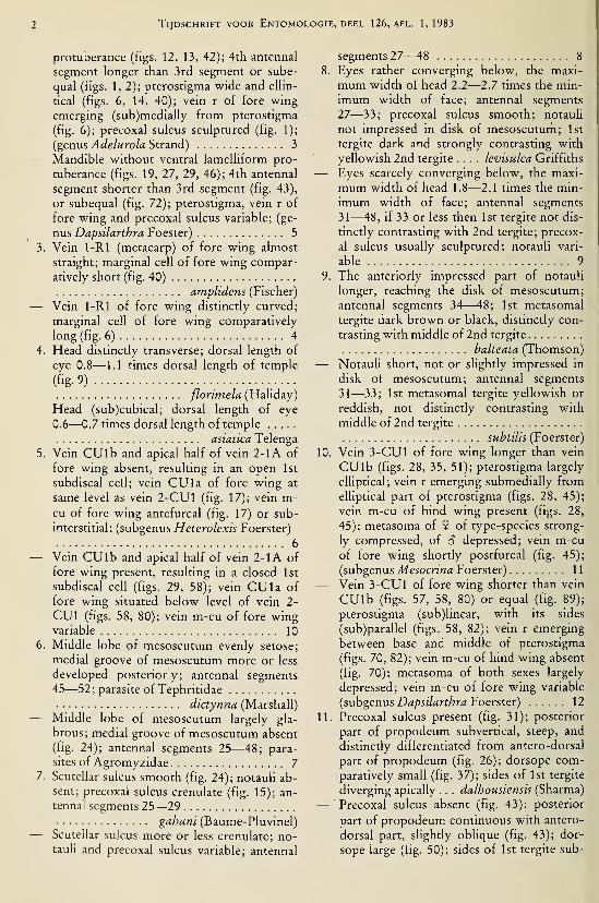
Tijdschrift voor Entomologie, deel 126, afl. 1,1983
protuberance (figs. 12, 13, 42); 4th antennal
segment longer than 3rd segment or sube-
qual (figs. 1,2); pterostigma wide and elhp-
tical (figs. 6, 14, 40); vein r of fore wing
emerging (sub)medially from pterostigma
(fig. 6); precoxal sulcus sculptured (fig. 1);
(genus Adelurola Strand) 3
— Mandible without ventral lamelliform pro-
tuberance (figs. 19, 27, 29, 46); 4th antennal
segment shorter than 3rd segment (fig. 43),
or subequal (fig. 72); pterostigma, vein r of
fore wing and precoxal sulcus variable; (ge-
nus Dapsilarthra Foester) 5
3. Vein 1-Rl (metacarp) of fore wing almost
straight; marginal cell of fore wing compar-
atively short (fig. 40)
amplidens (Fischer)
— Vein 1-Rl of fore wing distinctly curved;
marginal cell of fore wing comparatively
long (fig. 6) 4
4. Head distinctly transverse; dorsal length of
eye 0.8—1.1 times dorsal length of temple
(%9)fiorimela (Haliday)
— Head (sub)cubical; dorsal length of eye
0.6—0.7 times dorsal length of temple
asiatica Telenga
5. Vein CU lb and apical half of vein 2-1A of
fore wing absent, resulting in an open 1st
subdiscal cell; vein CUI a of fore wing at
same level as vein 2-CUl (fig. 17); vein m-cu of fore wing antefurcal (fig. 17) or sub-
interstitial; (subgenus Heterolexis Foerster)
6
— Vein CU lb and apical half of vein 2-1A of
fore wing present, resulting in a closed 1st
subdiscal cell (figs. 29, 58); vein CUla of
fore wing situated below level of vein 2-
CUl (figs. 58, 80); vein m-cu of fore wingvariable 10
6. Middle lobe of mesoscutum evenly setose;
medial groove of mesoscutum more or less
developed posteriorly; antennal segments45—52; parasite of Tephritidae
dictynna (Marshall)— Middle lobe of mesoscutum largely gla-
brous; medial groove of mesoscutum absent
(fig. 24); antennal segments 25—48; para-
sites of Agromyzidae 7
7. Scutellar sulcus smooth (fig. 24); notauH ab-
sent; precoxal sulcus crenulate (fig. 15); an-
tennal segments 25—29
gahani (Baume-Pluvinel)— Scutellar sulcus more or less crenulate; no-
tauli and precoxal sulcus variable; antennal
segments 27—48 8
8. Eyes rather converging below, the maxi-
mum width of head 2.2—2.7 times the min-
imum width of face; antennal segments27—33; precoxal sulcus smooth; notauli
not impressed in disk of mesoscuturh; 1st
tergite dark and strongly contrasting with
yellowish 2nd tergite .... levisulca Griffiths
— Eyes scarcely converging below, the maxi-
mum width of head 1.8—2.1 times the min-
imum width of face; antennal segments
31—48, if 33 or less then 1st tergite not dis-
tinctly contrasting with 2nd tergite; precox-
al sulcus usually sculptured; notauli vari-
able 9
9. The anteriorly impressed part of notauli
longer, reaching the disk of mesoscutum;
antennal segments ÒA—48; 1st metasomal
tergite dark brown or black, distinctly con-
trasting with middle of 2nd tergite
balteata (Thomson)— Notauli short, not or slightly impressed in
disk of mesoscutum; antennal segments
31—33; 1st metasomal tergite yellowish or
reddish, not distinctly contrasting with
middle of 2nd tergite
subtilis (Foerster)
10. Vein 3-CUl of fore wing longer than vein
CUlb (figs. 28, 35, 51); pterostigma largely
elliptical; vein r emerging submedially from
eUiptical part of pterostigma (figs. 28, 45);
vein m-cu of hind wing present (figs. 28,
45); metasoma of 9 of type-species strong-
ly compressed, of S depressed; vein m-cu
of fore wing shortly postfurcal (fig. 45);
(subgenus Mesocrina Foerster) 11
— Vein 3-CUl of fore wing shorter than vein
CUlb (figs. 57, 58, 80) or equal (fig. 89);
pterostigma (sub)linear, with its sides
(sub)parallel (figs. 58, 82); vein r emerging
between base and middle of pterostigma
(figs. 70, 82); vein m-cu of hind wing absent
(fig. 70); metasoma of both sexes largely
depressed; vein m-cu of fore wing variable
(subgenus Dapsilarthra Foerster) 12
11. Precoxal sulcus present (fig. 31); posterior
part of propodeum subvertical, steep, and
distinctly differentiated from antero-dorsal
part of propodeum (fig. 26); dorsope com-
paratively small (fig. 37); sides of 1st tergite
diverging apically . . . dalhousiensis (Sharma)
— Precoxal sulcus absent (fig. 43); posterior
part of propodeum continuous with antero-
dorsal part, slightly oblique (fig. 43); dor-
sope large (fig. 50); sides of 1st tergite sub-

Van Achterberg: Dapsilarthra and Mesocrina
parallel indagatrix (Foerster)
12. Vein r of fore wing (very) short, shorter
than width of pterostigma (figs. 82, 90) or
subequal (fig. 38); pterostigma not reaching
beyond middle of marginal cell of fore
wing, rather stout (figs. 38, 82, 90); vein m-cu of fore wing shortly antefurcal (fig. 38)
13
— Vein r of fore wing comparatively long,
longer than width of pterostigma (figs. 58,
70); pterostigma reaching beyond middle of
marginal cell of fore wing, and slender (figs.
58, 70); vein m-cu of fore wing shortly
postfureal (fig. 70) or subinterstitial .... 15
13. Antennal segments 40—41; vein 3-CUl of
fore wing subhorizontal (fig. 89); antenna
about twice as long as bodyisabella (Haliday)
— Antennal segments 25—33; vein 3-CUl of
fore wing oblique or subvertical (figs. 38,
92); antenna shorter than twice length of
body 14
14. Antenna short, about 1.3 times length of
body; marginal cell of fore wing distinctly
removed from wing apex (fig. 38); hind fe-
mur and tibia dark browntirolensis (Königsmann)
— Antenna medium-sized, about 1.7 times
length of body; marginal cell of fore wingcomparatively close to wing apex (fig. 90);
hind femur and tibia brownish-yellow;
(nominate form has hind coxa and 2nd ter-
gite yellowish, which are largely dark
brown or blackish in forma fuscula Grif-
fiths) rufiventris (Nees)
15. Notauli distinctly impressed posteriorly;
precoxal sulcus extensively sculptured
Sylvia (Haliday)
— Posterior half of notauH absent (fig. 78);
precoxal sulcus smooth or narrowly and su-
perficially sculptured (figs. 56, 72) 16
16. Sides of mandible parallel (figs. 61, 62);
laterope absent (fig. 56); 3rd and 4th anten-
nal segments yellowish; vein 1-Rl (meta-
carp) of fore wing less curved (fig. 58); ven-
tral half of side of pronotum largely smooth(fig. 56); length of fore wing ca. 2.8 mm ....
carpathica spec. nov.
— Mandible widened dorsally (figs. 68, 69),
because of the protruding 1st tooth;
laterope present (fig. 72); 3rd and 4th an-
tennal segments dark brown; vein 1-Rl of
fore wing more curved (fig. 70); ventral half
of side of pronotum distinctly sculptured
(fig. 72); length of fore wing 3.5—4.8 mm . .
apii (Curtis)
Genus Prorima nov.
Type-species: Mesocrina thienemanni Bi-
schoff, 1932.
Etymology: From "pro" (Latin for "in front
of", "forward") and "rima" (Latin for "cleft")
because of the antescutal depression on the an-
terior part of the mesosoma.Diagnosis. — Fourth antennal segment
sHghtly shorter than 3rd segment (including an-
nellus); eyes glabrous, not emarginate (fig. 104);
lower outer orbits of eyes depressed (fig. 98);
mandible slender, parallelsided, with 3 small
teeth, 2nd tooth somewhat longer than 1st
tooth, without ventral protruding tooth or la-
mella, and with carina to 3rd tooth (fig. 98); an-
tescutal depression distinct (figs. 95, 96); pro-
nope medium-sized (fig. 99); medio-posterior
groove of mesoscutum present (fig. 99); precox-
al sulcus present, superficially crenulate (fig.
95); metanotum blunt dorsally (fig. 95); propo-
deum completely areolated (fig. 99), its antero-
dorsal part differentiated and about as long as
posterior part of propodeum (fig. 95); pterostig-
ma elliptical; vein r emerges somewhat behind
middle of pterostigma (fig. 97); vein CUlb of
fore wing present, somewhat longer than vein
3-CUl (fig. 105); 1st subdiscal cell of fore wingclosed; vein m-cu of fore wing distinctly post-
furcal (fig. 97); vein M -I- CU of hind wingshorter than vein 1-M (fig. 97); vein m-cu of
hind wing faintly indicated; marginal cell of
hind wing medium-sized (fig. 97); claws very
slender, sickle-shaped (fig. 101); dorsope large
(fig. 102); 2nd tergite smooth; ovipositor sheath
(fig. 106) longer than apical height of metasoma.
Distribution. — Contains only the Oriental
type-species.
Prorima thienemanni (Bischoff) comb. nov.
(figs. 95—108)
Mesocrina thienemanni Bischoff, 1932: 742—744, fig.
5. Shenefelt, 1974:996.
Holotype, 9 , length of head and mesosomacombined 1.5 mm, of fore wing 2.8 mm.Head. — Antenna missing, except for scapus
and pedicellus; an earlier examination proved
that the length of the 3rd segment is slightly
longer than 4th segment; length of maxillary
palp about 0.7 times height of head; dorsal
length of eye 2.0 times temple (fig. 103); POL:
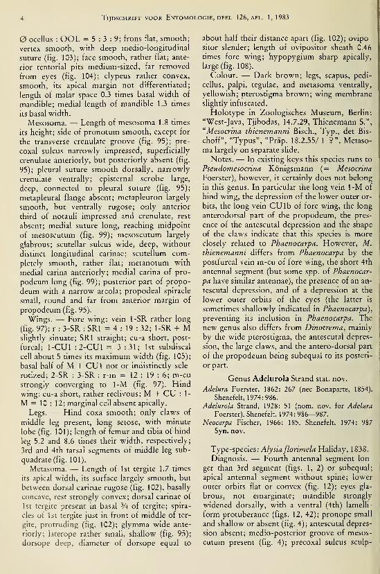
Tijdschrift voor Entomologie, deel 126, afl. 1,1983
ocellus : OOL = 5:3:9; frons flat, smooth;
vertex smooth, with deep medio-longitudinal
suture (fig. 103); face smooth, rather flat; ante-
rior tentorial pits medium-sized, far removed
from eyes (fig. 104); clypeus rather convex,
smooth, its apical margin not differentiated;
length of malar space 0.3 times basal width of
mandible; medial length of mandible 1.3 times
its basal width.
Mesosoma. — Length of mesosoma 1.8 times
its height; side of pronotum smooth, except for
the transverse crenulate groove (fig. 95); pre-
coxal sulcus narrowly impressed, superficially
crenulate anteriorly, but posteriorly absent (fig.
95); pleural suture smooth dorsally, narrowly
crenulate ventrally; episternal scrobe large,
deep, connected to pleural suture (fig. 95);
metapleural flange absent; metapleuron largely
smooth, but ventrally rugose; only anterior
third of notauli impressed and crenulate, rest
absent; medial suture long, reaching midpoint
of mesoscutum (fig. 99); mesoscutum largely
glabrous; scutellar sulcus wide, deep, without
distinct longitudinal carinae; scutellum com-
pletely smooth, rather flat; metanotum with
medial carina anteriorly; medial carina of pro-
podeum long (fig. 99); posterior part of propo-
deum with a narrow areola; propodeal spiracle
small, round and far from anterior margin of
propodeum (fig. 95).
Wings. — Fore wing: vein 1-SR rather long
(fig. 97); r : 3-SR : SRI = 4 : 19 : 32; 1-SR + Mslightly sinuate; SRI straight; cu-a short, post-
furcal; 1-CUl : 2-CUl = 3 :31; 1st subdiscal
cell about 5 times its maximum width (fig. 105);
basal half of M -I- CUI not or indistinctly scle-
rotized; 2-SR : 3-SR : r-m = 12 : 19 : 6; m-custrongly converging to 1-M (fig. 97). Hindwing: cu-a short, rather reclivous; M -I- CU :
1-
M = 10 : 12; marginal cell absent apically.
Legs. — Hind coxa smooth; only claws of
middle leg present, long setose, with minute
lobe (fig. 101); length of femur and tibia of hind
leg 5.2 and 8.6 times their width, respectively;
3rd and 4th tarsal segments of middle leg sub-
quadrate (fig. 101).
Metasoma. — Length of 1st tergite 1.7 times
its apical width, its surface largely smooth, but
between dorsal carinae rugose (fig. 102), basally
concave, rest strongly convex; dorsal carinae of
1st tergite present in basal V^, of tergite; spira-
cles of 1st tergite just in front of middle of ter-
gite, protruding (fig. 102); glymma wide ante-
riorly; laterope rather small, shallow (fig. 95);
dorsope deep, diameter of dorsope equal to
about half their distance apart (fig. 102); ovipo-
sitor slender; length of ovipositor sheath 0.46
times fore wing; hypopygium sharp apically,
large (fig. 108).
Colour. — Dark brown; legs, scapus, pedi-
cellus, palpi, tegulae, and metasoma ventrally,
yellowish; pterostigma brown; wing membraneslightly infuscated.
Holotype in Zoologisches Museum, Berlin:
"West-Java, Tjibodas, 14.7.29, Thienemann S.",
"Mesocrina thienemanni Bisch., Typ., det Bis-
choff", "Typus", "Präp. 18.2.55/ 1 9". Metaso-
ma largely on separate slide.
Notes. — In existing keys this species runs to
Pseudomesocrina Königsmann (= Mesocrina
Foerster), however, it certainly does not belong
in this genus. In particular the long vein 1-M of
hind wing, the depression of the lower outer or-
bits, the long vein CUlb of fore wing, the long
anterodorsal part of the propodeum, the pres-
ence of the antescutal depression and the shape
of the claws indicate that this species is moreclosely related to Phaenocarpa. However, M.thienemanni differs from Phaenocarpa by the
postfurcal vein m-cu of fore wing, the short 4th
antennal segment (but some spp. of Phaenocar-
pa have similar antennae), the presence of an an-
tescutal depression, and of a depression at the
lower outer orbits of the eyes (the latter is
sometimes shallowly indicated in Phaenocarpa),
preventing its inclusion in Phaenocarpa. The
new genus also differs from Dinotrema, mainly
by the wide pterostigma, the antescutal depres-
sion, the large claws, and the antero-dorsal part
of the propodeum being subequal to its posteri-
or part.
Genus Adelurola Strand stat. nov.
Adelura Foerster, 1862: 267 (nee Bonaparte, 1854).
Shenefelt, 1974:986.
Adelurola Strand, 1928: 51 (nom. nov. for Adelura
Foerster). Shenefelt, 1974: 986—987.
Neocarpa Fischer, 1966: 185. Shenefelt, 1974: 987
Syn. nov.
Type-species: Alysia fiorimela Haliday, 1838.
Diagnosis. — Fourth antennal segment lon-
ger than 3rd segment (figs. 1, 2) or subequal;
apical antennal segment without spine; lower
outer orbits flat or convex (fig. 12); eyes gla-
brous, not emarginate; mandible strongly
widened dorsally, with a ventral (4th) lamelli-
form protuberance (figs. 12, 42); pronope small
and shallow or absent (fig. 4); antescutal depres-
sion absent; medio-posterior groove of mesos-
cutum present (fig. 4); precoxal sulcus sculp-
Van Achterberg: Dapsilarthra and Mesocrina
tured (fig. 1); metanotum blunt dorsally (fig. 1);
antero-dorsal part of propodeum not differ-
entiated from posterior part of propodeum (fig.
1); pterostigma wide and largely elliptical (figs.
6, 14); vein M + CUI of fore wing largely un-
sclerotized; vein r of fore wing emerging
(sub)medially from pterostigma (fig. 6); vein
CUlb of fore wing present, longer than vein 3-
CUl (fig. 8); 1st subdiscal cell of fore wing
closed; vein m-cu of fore wing slightly postfur-
eal (figs. 6, 14); veimM -t- CU of hind wing lon-
ger than vein 1-M (fig. 6); vein m-cu of hind
wing absent; marginal cell of hind wing medi-
um-sized basally (fig. 6); claws moderately slen-
der (fig. 10), not sickle-shaped; dorsope large
(fig. 7); 2nd tergite smooth; setae of metasoma
in subapical rows; ovipositor sheath shorter
than apical height of metasoma (fig. 1).
Distribution. — Contains 3 Palaearctic spe-
cies.
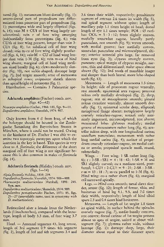
Adelurola amplidens (Fischer) comb. nov.
(figs. 40^2)Neocarpa amplidens Fischer, 1966: 185, figs. 9— 11.
Dapsilarthra amplidens; Shenefelt, 1974: 987.
Only known from 4 S from Iraq, of which
the holotype should be housed in the Zoolo-
gische Sammlung des Bayerischen Staates in
München, where it could not be traced. Owingto the kindness of Dr. Fischer I was able to ex-
amine two topotypic paratypes upon which the
insertion in the key is based. This species is very
close to A. ßorimela; the difference of the short
marginal cell of fore wing is not significant be-
cause this is also common in males oi fiorimela
(fig. 14).
Adelurola florimela (Haliday) comb. nov.
(figs. 1-14)
Alysia florimela Haliday, 1838: 239.
Dapsilarthra florimela; Shenefelt, 1974: 988—989.
Phaenocarpa multiarticulata Marshall, 1898: 245.
Syn. nov.
Dapsilarthra multiarticulata; Shenefelt, 1974: 989.
Dapsilarthra pentapleuroides Fischer, 1971: 85, figs.
25—27 (unavailable name, used in synonymy with
D. multiarticulata).
Redescribed after a female from the Nether-
lands (Linschoterbos), compared with the lecto-
type, length of body 3.3 mm, of fore wing 3.7
mm.Head. — Antennal segments 43, long setose,
length of 3rd segment 0.9 times 4th segment
(fig. 2), length of 3rd and 4th segments 3.4 and
3.6 times their width, respectively; penultimate
segment of antenna 2.6 times its width (fig. 5),
and apical segment without spine; length of
maxillary palp 1.1 times height of head; dorsal
length of eye 1.1 times temple; POL : ocel-
lus: OOL = 5 : 3 : 12; frons slightly convex,
medially somewhat rugose, laterally partly
punctulate, rest smooth (fig. 9); vertex smooth,with medial groove; face medially convex,
somewhat punctulate and microsculptured, shi-
ny; anterior tentorial pits large, far removedfrom eyes (fig. 3); clypeus strongly convex,
punctate; apical margin of clypeus straight, nar-
row, thin and differentiated; malar space almost
absent (fig. 12); 2nd tooth of mandible longer
and sharper than both lateral, more lobe-shaped
teeth (fig. 13).
Mesosoma. — Length of mesosoma 1.5 times
its height; side of pronotum rugose ventrally,
rest smooth; epicnemial area rugose; precoxal
sulcus only medially developed (fig. 2), deep,
rugose; rest of mesopleuron smooth; pleural
sulcus crenulate ventrally, almost smooth dor-
sally (fig. 1); episternal scrobe deep, elliptical;
metapleural flange absent; metapleuron largely
coarsely reticulate-rugose; notauli only ante-
riorly impressed, microsculptured, rest absent;
mesoscutal lobes glabrous medially; medial
groove of mesoscutum rather long (fig. 4); scu-
tellar sulcus deep, with one longitudinal carina;
scutellum punctulate; metanotum with rather
long medial carina (fig. 4); surface of propo-
deum coarsely reticulate-rugose, no medial cari-
na or areola; propodeal spiracle small, round,
submedially.
Wings. — Fore wing; 1-SR rather long (fig.
6); r : 3-SR : SRI = 4 : 18 : 42; 1-SR + M and
SRI slightly curved; cu-a medium-sized, post-
furcal; 1-CUl : 2-CUl = 9 : 25; 2-SR : 3-SR :
r-m = 13 : 18 : 7; m-cu parallel to 1-M (fig. 6).
Hind wing: cu-a rather short (fig. 6); marginal
cell narrowed apically.
Legs. — Hind coxa smooth; tarsal claws slen-
der, setose (fig. 10); length of femur, tibia, and
basitarsus of hind leg 4.1, 9.5, and 7.0 times
their width, respectively; length of hind tibial
spurs 0.3 and 0.4 times hind basitarsus.
Metasoma. — Length of 1st tergite 1.8 times
its apical width, its surface behind the spiracles
coarsely rugose (fig. 7), medio-basally concave,
rest convex; dorsal carinae of 1st tergite present
almost to apex of tergite, united at about mid-
segment (fig. 7); glymma rather wide, without
laterope (fig. 1); dorsope deep, large, their
diameter about equal to their distance apart;

Tijdschrift voor Entomologie, deel 126, afl. 1, 1983
ovipositor straight; length of ovipositor sheath
0.06 times fore wing; hypopygium large (fig. 1),
truncate apically.
Colour. — Black; scapus, annellus, palpi,
veins of hind vt^ing mainly, tegulae, and legs,
light yellowish; mandibles, antenna (except for
scapus and annellus), pterostigma, most of fore
wing veins, and metasoma behind 1st tergite,
dark brown; apical % of hind tibia and base of
hind tarsus darkened.
Redescribed after a specimen in the Rijksmu-seum van Natuurlijke Historie, Leiden: "Lin-
schoterbos, 8.x. 1966, v. Ooststroom", "com-pared & conspecific with lectotype of Alysia
florimela Haliday, C. v. Achterberg, 1979". Thetype-series in the Haliday Collection (Dublin)
consists of 1 9 and 1 â, both with the label
"British Haliday, 20.2.82/Box 10 AWS." Origi-
nally I was inclined to consider only the 9 an
original type-specimen, because Haliday did not
state he had a male. However, Stelfox in a copyof his notes (discovered by Dr. C. O'Riordanand a partial copy kindly supplied by Dr. J. P.
O'Connor) stated under Alysia florimela Hal-
iday, that Haliday did include the male: "In de-
scribing the male of this species as unknownMarshall (Trans, ent. Soc. London, 1895, p.
365) fell into error. When describing species of
which he had only one sex Haliday always be-
gan his diagnosis with the words mas or fem. as
the case might be: — e.g., "Sp. 43. Punctigera.
Al. & c. fern, fusco-castanea ..." or again "Sp.
48. Perdita Al & c. mas nigra ..."
On the other hand, when he had both sexes
before him, he only mentioned the sexes whenreferring to some character which belonged to
that sex alone: in the present case "fem. terebra
subexerta" and "fem. corpore duplo longiores
articulis 50." That he had both sexes of the pre-
sent species is manifest, for box 10 I found a S&a 9 beside each other (on 26.1.1935)."
The indirect evidence put forward by Stelfox
in his notes makes it possible to accept bothspecimens from box 10 as types; I prefer to des-
ignate here the 9 as lectotype (and not the 6 as
Stelfox did without publishing it). The 9 lecto-
type is in a fairly good condition, the antennahas 50 segments (as stated by Haliday) and the
dorsal length of the eye is 1.1 times the dorsal
length of the temple. The lectotype stems fromEngland (London). In the Haliday Collection
there are two specimens (1 9 ("Clifden, July 8,
[18]39") and 1 6, which do not belong to the
type series. Additionally 9 9 and 5 6 were ex-
amined from the Netherlands (Waarder; Aspe-
ren; Udenhout), West Germany (Taufkirchen
bei München), and Bulgaria (Rhodopi Moun-tains, Thagortsharn; id., N. Zdraves). The spec-
imen from Den Haag reported by Snellen van
Vollenhoven (1873: 195) belongs to Phaenocar-
pa.
Variation. — Antennal segments 43—54;
length of 3rd segment 0.9—1.0 times 4th seg-
ment; dorsal length of eye 0.8—1.1 times dorsal
length of temple; vein 2-SR of one fore wing of
specimen from N. Zdraves partly absent;
pterostigma of male enlarged (fig. 14); metaso-
ma of some specimens more or less yellowish
banded.
Note.— The proposed synonymy is based onthe redescription by Fischer (1971) and the vari-
ation found in the series collected in the Nether-
lands. A. florimela is an easily recognizable spe-
cies, with IS not uncommon in Waarder (Neth-
erlands). The enlargement of the pterostigma in
the male misled Marshall, so that he described
the male as a separate species {multiarticulata).
A. florimela has been reared from Pegomya ni-
gritarsis Zetterstedt (Anthomyiidae) and Acidia
cognata (Wiedemann) (Tephritidae).
Adelurola asiatica Telenga
Adelurola asiatica Telenga, 1935: 186.
Dapsilarthra asiatica; Shenefelt, 1974: 987.
The type from Turkestan (USSR) is destroyed
and additional specimens of this species have
not been recorded.
Genus Dapsilarthra Foerster
Gnamptodon Haliday, 1833: 265 (suppression re-
quested to ICZN).Dapsilarthra Foerster, 1862: 267. Shenefelt, 1974:
986—991. Marsh, 1979: 222.
Heterolexis Foerster, 1862: 268; Shenefelt, 1974: 992.
Syn. nov.
Grammospila Foerster, 1862: 269; Shenefelt, 1974:
987.
Mesocrina Foerster, 1862: 266; Shenefelt, 1974: 996.
Syn. nov.
Pseudomesocnna Königsmann, 1959b: 611; Shenefelt,
1974: 1018. Syn. nov.
Paraorthostigma Köningsmann, 1972: 25—26, 1 fig.
Syn. nov.
Diagnosis. — Antennal segments 25—52; 4th
antennal segment shorter than 3rd segment (fig.
43) or subequal (fig. 72); apical segment of an-
tenna without spine; eyes glabrous, not emar-
ginate: lower outer orbits of eyes flat or convex
(fig. 29); mandible without ventral lamelliform
protuberance (figs. 29, 46), robust (fig. 29) or

Van Achterberg: Dapsilarthra and Mesocrina
slender (fig. 25); pronope usually absent (fig.
49), seldom present (fig. 24); antescutal depres-
sion absent; medio-posterior groove of meso-
scutum present (fig. 49) or absent (fig. 24);
metanotum blunt dorsally (fig. 15) or slightly
protruding (fig. 26); propodeum not carinated,
its antero-dorsal part not differentiated from
posterior part (fig. 72) or much shorter than
posterior part (fig. 26); vein 1-SR of fore wing
medium-sized (fig. 58), exceptionally short (fig.
28); 1st subdiscal cell of fore wing parallel-
sided; vein M + CUI of fore wing largely un-
sclerotized; vein M -t- CU of hind wing longer
than vein 1-M; marginal cell of hind wing nar-
rowed apically or subparallel; claws moderately
slender, not sickle-shaped (figs. 22, 32); dorsope
rather small (fig. 18) to large (fig. 50); laterope
absent or shallow; 2nd tergite smooth; setae of
metasoma in subapical rows; length of oviposi-
tor sheath subequal to apical depth of metasoma
(fig. 54) or shorter (fig. 72).
Distribution. — All three subgenera occur in
the Palaearctic region (including the Himalayan
area), the subgenus Dapsilarthra Foerster also
occurs in the Nearctic region (partly intro-
duced).
Subgenus Heterolexis Foerster stat. nov.
Heterolexis Foerster, 1862: 268. Shenefelt, 1974: 992.
Grammospila Foerster, 1862: 269. Shenefelt, 1974:
987. Syn. nov.
Diagnosis. — Antennal segments 25—52;
medio-posterior groove of mesoscutum absent
(fig. 24), only present in dictynna; precoxal sul-
cus impressed, smooth or crenulate (fig. 15);
vein CUlb and apical half of vein 2-1A of fore
wing absent, resulting in an open 1st subdiscal
cell (fig. 17); vein CUla of fore wing at same
level as vein 2-CUl; vein r of fore wing arising
before middle of pterostigma; vein m-cu of fore
wing antefurcal (fig. 17) or subintersitial;
pterostigma linear (fig. 17); dorsope rather
small (fig. 18).
Distribution. — Contains five Palaearctic
species; one species is introduced in NorthAmerica (Hendrickson & Barth, 1979).
Dapsilarthra (Heterolexis) dictynna (Marshall)
Adelura dictynna Marshall, 1895a: 422—423, fig. 6.
Dapsilarthra dictynna; Königsmann, 1972: 22. Shene-
felt, 1974: 988.
A scarcely collected but widespread species;
not yet collected in the Netherlands. Examined:
1 9 (Bulgaria, Rhodopi Mountains, Pamporo-va) and 1 6 (Switzerland, Aeschi-Ried, 1000
m). Parasite of Pycnoglossa flavipennis (Fallen)
(Tephritidae) in Pteridium aquilinum L.
Dapsilarthra (Heterolexis) gahani (Baume-Pluvinel)
(figs. 15—25)
Adelura gahani'?>a.VLmc-\*\\iymt\, 1915: 47, 11 figs.
Dapsilarthra gahani; Shenefelt, 1974: 989.
Dapsilarthra nowakowskii Königsmann, 1959a: 591,
figs. Shenefelt, 1974: 989. Syn. nov.
Lectotype, $, length of body 1.5 mm, of fore
wing 2.1 mm.Head. — Antennal segments 29, length of 3rd
segment 1.1 times 4th segment, length of 3rd
and 4th segments 4.5 and 4.0 times their width,
respectively; penultimate segment of antenna
3.0 times its width (fig. 20); length of maxillary
palp 0.6 times height of head; dorsal length of
eye 1.3 times temple (fig. 21); POL : ocellus :
OOL = 14 : 5 : 18; frons flat and smooth; ver-
tex smooth, with medial groove; face slightly
convex, smooth; anterior tentorial pits very
large, wider than long and almost reaching eyes
(fig. 16); clypeus convex and smooth, its ventral
margin not differentiated, straight medially; eye
nearly touching base of mandible (fig. 15); man-dible rather slender, not widened apically (fig.
25), with three sharp teeth.
Mesosoma. — Length of mesosoma 0.9 times
its height; pronope distinct (fig. 24); prenotai
side largely smooth, except for some rugae (fig.
15); precoxal sulcus rather shallow and rugu-
lose; rest of mesopleuron smooth; pleural sulcus
finely crenulate; episternal scrobe narrow (fig.
15); metapleural flange absent; metapleuron
largely smooth; notauli absent on dorsal disc of
mesoscutum (fig. 24), faintly impressed in front
of disc (fig. 15); mesoscutum rather convex,
largely glabrous and smooth; medio-posterior
groove of mesoscutum absent; scutellar sulcus
deep, with no carinae (fig. 24); scutellum rather
flat and smooth; surface of propodeum largely
smooth, except for some rugulosity, without
medial carina or areola; posterior pat of propo-
deum not differentiated from antero-dorsal
part; propodeal spiracle small and in front of
middle of propodeum.
Wings. — Fore wing: r : 3-SR : SRI = 8 :
24 : 92; SRI sinuate (fig. 17); 1-CUl : 2-CUl =
3: 14; 2-SR : 3-SR: r-m = 18: 24: 11.
Hindwing: cu-a reclivous; marginal cell
(sub)parallel-sided apically.
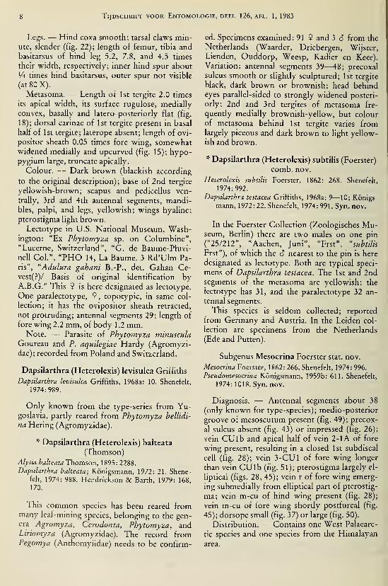
Tijdschrift voor Entomologie, deel 126, afl. 1, 1983
Legs. — Hind coxa smooth; tarsal claws min-ute, slender (fig. 22); length of femur, tibia andbasitarsus of hind leg 5.2, 7.8, and 4.5 times
their width, respectively; inner hind spur about
'A times hind basitarsus, outer spur not visible
(at 80 X).
Metasoma. — Length of 1st tergite 2.0 times
its apical width, its surface rugulose, medially
convex, basally and latero-posteriorly flat (fig.
18); dorsal carinae of 1st tergite present in basal
half of 1st tergite; laterope absent; length of ovi-
positor sheath 0.05 times fore wing, somewhatwidened medially and upcurved (fig. 15); hypo-pygium large, truncate apically.
Colour. — Dark brown (blackish according
to the original description); base of 2nd tergite
yellowish-brown; scapus and pedicellus ven-
trally, 3rd and 4th antennal segments, mandi-bles, palpi, and legs, yellowish; wings hyaline;
pterostigma light brown.
Lectotype in U.S. National Museum, Wash-ington: "Ex Phytomyza sp. on Columbine","Lucerne, Switzerland", "G. de Baume-Pluvi-nell Col", "PHO 14, La Baume. 3 Rd'Ulm Pa-
ris", ''Adulura gahani B.-P., det. Gahan Ce-vest(?)/ Basis of original identification byA.B.C." This 9 is here designated as lectotype.
One paralectotype, 9, topotypic, in same col-
lection; it has the ovipositor sheath retracted,
not protruding; antennal segments 29; length of
fore wing 2.2 mm, of body 1 .2 mm.Note. — Parasite of Phytomyza minuscula
Goureau and P. aquilegiae Hardy (Agromyzi-dae); recorded from Poland and Switzerland.
Dapsilarthra (Heterolexis) levisulca Griffiths
Dapsilarthra levisulca Griffiths, 1968a: 10. Shenefelt,
1974:989.
Only known from the type-series from Yu-goslavia, partly reared from Phytomyza bellidi-
na Hering (Agromyzidae).
'•" Dapsilarthra (Heterolexis) balteata
(Thomson)
Alysia balteata Thomson, 1895: 2288.
Dapsilarthra balteata; Königsmann, 1972: 21. Shene-felt, 1974: 988. Hendrickson & Barth, 1979: 168,
170.
This common species has been reared frommany leaf-mining species, belonging to the gen-era Agromyza, Cerodonta, Phytomyza, andLiriomyza (Agromyzidae). The record fromPegomya (Anthomyiidae) needs to be confirm-
ed. Specimens examined: 91 9 and 3 â from the
Netherlands (Waarder, Driebergen, Wijster,
Lienden, Ouddorp, Weesp, Kadier en Keer).
Variation: antennal segments 39—48; precoxal
sulcus smooth or slightly sculptured; 1st tergite
black, dark brown or brownish; head behindeyes parallel-sided to strongly widened posteri-
orly; 2nd and 3rd tergites of metasoma fre-
quently medially brownish-yellow, but colour
of metasoma behind 1st tergite varies fromlargely piceous and dark brown to light yellow-
ish and brown.
* Dapsilarthra (Heterolexis) subtilis (Foerster)
comb. nov.
Heterolexis subtilis Foerster, 1862: 268. Shenefelt,
1974:992.
Dapsilarthra testacea Griffiths, 1968a: 9—10; Königs-mann, 1972: 22. Shenefelt, 1974: 991. Syn. nov.
In the Foerster Collection (Zoologisches Mu-seum, Berlin) there are two males on one pin
("25/212", "Aachen, Juni", "Frst", "subtilis
Frst"), of which the â nearest to the pin is here
designated as lectotype. Both are typical speci-
mens of Dapsilarthra testacea. The 1st and 2ndsegments of the metasoma are yellowish; the
lectotype has 31, and the paralectotype 32 an-
tennal segments.
This species is seldom collected; reported
from Germany and Austria. In the Leiden col-
lection are specimens from the Netherlands
(Ede and Putten).
Subgenus Mesocrina Foerster stat. nov.
Mesocrina Foerster, 1862: 266. Shenefelt, 1974: 996.
Pseudomesocrina Königsmann, 1959b: 611. Shenefelt,
1974: 1018. Syn. nov.
Diagnosis. — Antennal segments about 38
(only known for type-species); medio-posterior
groove of mesoscutum present (fig. 49); precox-
al sulcus absent (fig. 43) or impressed (fig. 26);
vein CUlb and apical half of vein 2-1A of fore
wing present, resulting in a closed 1st subdiscal
cell (fig. 28); vein 3-CUl of fore wing longer
than vein CUlb (fig. 51); pterostigma largely el-
liptical (figs. 28, 45); vein r of fore wing emerg-
ing submedially from elliptical part of pterostig-
ma; vein m-cu of hind wing present (fig. 28);
vein m-cu of fore wing shortly postfurcal (fig.
45); dorsope small (fig. 37) or large (fig. 50).
Distribution. — Contains one West Palaearc-
tic species and one species from the Himalayan
area.

Van Achterberg: Dapsilarthra and Mesocrina
* Dapsilarthra (Mesocrina) indagatrix
(Foerster) comb. nov.
(figs. 43—55)
Mesocrina indagatrix Foerster, 1862: 266. Shenefelt,
1974:996.
Mesocrina venatrix Marshall, 1895a: 429—430, fig.
Shenefelt, 1974: 1018—1019. Syn. nov.
The interpretation of this obviously scarcely
collected species has been hindered by the loss
of the type (Königsmann, 1959b: 611), but in
my opinion the fairly complete original descrip-
tion by Foerster enables a correct identification
of the species to be made. This interpretation is
close to that of Marshall (1895a: 430) and dis-
agrees with the statements made by Königs-
mann (1959b: 610). Königsmann's argument
that Foerster was unlikely to have overlooked
the strongly compressed metasoma of the 9 is
not conclusive, because Foerster's specimen
may have been a 6, which has a normally de-
pressed metasoma. Because no closely related
species are known, there is no reason to doubt
the synonymy of venatrix and indagatrix. Theholotype of D. venatrix (Marshall) has been ex-
amined; it is a 9 with the typical compressed
metasoma. To fix the type-species of the genus
Mesocrina Foerster I designate here the ê de-
scribed and figured below as neotype of Meso-crina indagatrix Foerster, 1862.
Neotype, S , length of body 3,5 mm, of fore
wing 3,9 mm.Head. — Antennal segments 21 (apical seg-
ments missing), length of 3rd segment 1.7 times
4th segment, length of 3rd and 4th segments 4.5
and 2.4 times their width, respectively; penulti-
mate segment of antenna of neotype missing,
but in 9 from Ede (Netherlands), length 1.7
times its width (fig. 55); length of maxillary palp
1.2 times height of head; dorsal length of eye
subequal to dorsal length of temple (fig. 53);
POL : ocellus : OOL = 12 : 7 : 20; frons
smooth, vertex smooth and with medial suture;
face rather flat and with some punctulation (fig.
44); anterior tentorial pits large and far removedfrom eyes, distance to eyes 1.5 times maximaldiameter of pit; clypeus strongly convex,
smooth, its apical margin narrowly differ-
entiated and straight medially; malar space
short, eye almost touching base of mandible
(fig. 46); mandible slightly widened dorsally,
2nd tooth sharp and longer than both lateral,
more obtuse teeth (figs. 46, 48).
Mesosoma. — Length of mesosoma 1.4 times
its height; pronope absent (fig. 49); side of pro-
notum with a crenulate medial groove, rest
smooth (fig. 43); precoxal sulcus absent; meso-pleuron smooth; pleural sulcus narrowly crenu-
late ventrally, dorsally largely smooth (fig. 43);
metapleural flange absent; metapleuron smooth,but crenulate rugose-reticulate; notauli only an-
teriorly impressed and crenulate (fig. 49); meso-scutum largely glabrous, only anteriorly densely
setose; medio-posterior groove long and nar-
row, rather shallow (fig. 49); scutellar sulcus
with one weak longitudinal carina; scutellum
sparsely punctulate; side of scutellum smooth;surface of propodeum anteriorly and medio-posteriorly finely rugulose, rest mainly smooth,
medial carina and areola absent; posterior part
of propodeum not differentiated from antero-
dorsal part; propodeal spiracle rather small,
round, somewhat protruding and in front of
middle of propodeum.Wings. — Fore wing: r much shorter than
width of pterostigma (fig. 45); r : 3-SR : SRI =3:21:41; SRI straight; 1-CUl : 2-CUl =6 : 17; 2-SR : 3-SR : r-m = 17 : 21 : 9; 1st sub-
discal cell somewhat widened apically. Hindwing: cu-a straight; marginal cell absent apical-
Legs. — Hind coxa punctulate; tarsal claws
rather slender (fig. 47); length of femur, tibia
and basitarsus of hind leg 4.8, 8.2, and 6.2 times
their width, respectively; length of hind tibial
spurs 0.3 and 0.4 times hind basitarsus.
Metasoma. — Length of 1st tergite 2.0 times
its apical width, its surface basally smooth, its
posterior half rugose (fig. 50), basally concave,
medially convex; dorsal carina of 1st tergite pre-
sent in basal 0.9 of tergite, but apically superfi-
cial; dorsope large (fig. 50); hypopygium of
neotype rather short, truncate apically; 9 from
Ede has length of ovipositor sheath 0.13 times
fore wing, ovipositor slender, with no distinct
teeth or nodus, only slightly widened subapi-
cally (fig. 54); hypopygium medium-sized and
subtruncate apically; metasoma of neotype 6depressed (fig. 43), of 9 strongly compressed
(fig. 54).
Colour. — Black; metasoma behind 1st ter-
gite, wing veins, and tegulae, brownish-yellow;
hind tarsus and apex of hind tibia somewhat in-
fuscated; wing membrane subhyaline.
Neotype in the Zoologische Staatssammlung,
München: "Harthausen b. München, A.
27.9.68, Haeselb.". Additional specimens exam-
ined: 1 9, Museum Budapest, holotype of
Mesocrina venatrix Marshall {''venatrix M.,
Coll. Marshall", ''Pseudomesocrina venatrix
10 Tijdschrift voor Entomologie, deel 126, afl. 1,1983
(Marsh), det. Königsmann") from England,
which is conspecific with the neotype of inda-
gatrix. Length of fore wing 3.7 mm, and length
of ovipositors sheath about 0.12 times fore
wing. First metasomal tergite missing (figured
by Königsmann), rest of metasoma on separate
slide, and antenna incomplete. 1 9, Rijksmu-
seum van Natuurlijke Historie, Leiden: Nether-
lands ("Ede, trap [= Malaise-trap], 22—28. ix,
Van Rossem"). Length of fore wing 4.2 mm, of
body 4.3 mm; metanotum rather coarsely sculp-
tured; length of 1st tergite 1.9 times its apical
width. Parasite of Pegomya and Amaurosoma
spp. (Anthomyiidae and Scatophagidae, re-
spectively).
Dapsilarthra (Mesocrina) dalhousiensis
(Sharma) comb. nov.
(figs. 26—37)
Acrobela dalhousiensis Sharma, 1978: 127—128, figs.
4—6.
Holotype, â , length of body 5.4 mm, of fore
wing 4.7 mm.Head. — Remaining antennal segments 11,
apical segments missing, scapus compressed;
length of 3rd segment 1.2 times 4th segment,
length of 3rd and 4th segments 3.0 and 2.6 times
their width, respectively; length of maxillary
palp 1.2 times height of head; dorsal length of
eye 1.2 times temple; temple smooth and subpa-
rallel-sided (fig. 31); POL : ocellus : OOL =
8 : 4 : 14; frons flat and smooth; vertex with
medial groove; face unevenly convex, largely
smooth, with some punctulation (fig. 30); ante-
rior tentorial pits large, far removed from eyes
(fig. 30); clypeus convex, somewhat punctulate,
rugulose near dorsal margin, its apical margin
not distinctly differentiated medially, convex;
malar space absent, eye touching base of mandi-
ble (fig. 27); mandible widened dorso-apically,
finely rugose medially, 3rd tooth large and lobe-
shaped; ventral margin of mandible with small
incision (fig. 29) (but without ventral protrud-
ing lamella); 2nd tooth of mandible large, longer
than lateral teeth, acute apically (figs. 27, 29).
Mesosoma. — Length of mesosoma 1.2 times
its height; pronope absent; side of pronotummedially crenulate, dorsally largely smooth,
ventrally microsculptured (fig. 26); epicnemial
area ventrally rugose (fig. 26); precoxal sulcus
present in anterior 0.7 of mesopleuron, densely
reticulate-rugose; rest of mesopleuron smooth;pleural sui is rather shallow and narrow, finely
crenulate (lig. 26); episternal scrobe large;
metapleural flange small, obtuse apically;
metapleuron finely rugose; notauli only ante-
riorly impressed, finely crenulate (fig. 34);
mesoscutal lobes smooth, medially glabrous;
medio-posterior groove of mesoscutum slender,
also anteriorly with a shallow groove (fig. 34);
scutellar sulcus deep, long, with 8 longitudinal
carinae; scutellum smooth; sides of scutellum
rugose; metanotum somewhat protruding me-
dially (fig. 26); posterior part of propodeum dif-
ferentiated and much longer than antero-dorsal
part (fig. 26); surface of propodeum densely ru-
gose anteriorly, with some short apical carinae
and indistinct sculpture posteriorly, medial cari-
na and areola absent; propodeal spiracle rather
small, round, and in front of middle of propo-
deum.
Wings. — Fore wing: r subequal to width of
pterostigma (fig. 28); r : 3-SR : SRI = 8 : 28 :
61; SRI straight; 1-CUl : 2-CUl = 2 : 20; 2-
SR : 3-SR : r-m = 18 : 28 : 13; 3-SR of left
wing with stub (fig. 36), indistinct in right wing
(fig. 28). Hind wing: cu-a straight; marginal cell
narrowed apically.
Legs. — Hind coxa smooth; tarsal claws rath-
er slender (fig. 32); length of femur, tibia and
basitarsus of hind leg 3.9, 8.9, and 6.4 times
their width, respectively; length of hind tibial
spurs 0.3 times hind basitarsus, subequal.
Metasoma. — Length of 1st tergite equal to
its apical width, its surface largely striate (fig.
37), flat basally, and medially slightly convex;
dorsal carinae distinct in front of spiracles, short
(fig. 37); dorsope small; metasoma depressed;
hypopygium medium-sized and truncate apical-
ly-
Colour. — Black; mesonotum, pronotum
(largely), mesosternum and mandible, brown-
ish-red; legs (except for middle and hind tarsi,
apex of hind tibia and femur), palpi, tegulae,
scapus, and pedicellus, yellowish; pterostigma,
most wing veins, metasoma (dorsally rather
blackish), fore and middle tarsi (except telotar-
si), apices of hind femur and tibia partly, dark
brown; wing membrane slightly infuscate.
Holotype (and only known specimen) in
Gupta Collection, Delhi: "India: H.P. Dalhou-
sie, 2132 m, 22.ix.1971, Sykh. Dev. No. JD158", "Holotype Acrobela dalhousiensis V.
Sharma, 1975". Biology unknown.
Subgenus Dapsilarthra Foerster
Dapsilarthra Foerster, 1862: 267. Shenefelt, 1974:
986—991. Marsh, 1979: 222.
Paraorthostigma Königsmann, 1972: 25—26, 1 fig.
Syn. nov.
Van Achterberg: Dapsilarthra and Mesocrina 11
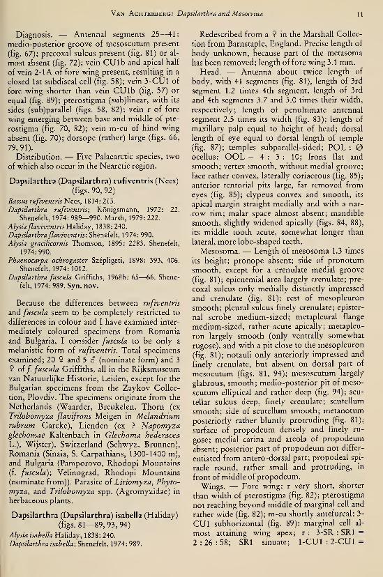
Diagnosis. — Antennal segments 25—41:
medio-posterior groove of mesoscutum present
(fig. 67); precoxal sulcus present (fig. 81) or al-
most absent (fig. 72); vein CU lb and apical half
of vein 2-1A of fore wing present, resulting in a
closed 1st subdiscal cell (fig. 58); vein 3-CUl of
fore wing shorter than vein CU lb (fig. 57) or
equal (fig. 89); pterostigma (sub)linear, with its
sides (sub)parallel (figs. 58, 82); vein r of fore
wing emerging between base and middle of pte-
rostigma (fig. 70, 82); vein m-cu of hind wing
absent (fig. 70); dorsope (rather) large (figs. 66,
79,91).
Distribution. — Five Palaearctic species, two
of which also occur in the Nearctic region.
Dapsilarthra (Dapsilarthra) rufiventris (Nees)
(figs. 90, 92)
BassHS rufiventris Nees, 1814: 213.
Dapsilarthra rufiventris; Königsmann, 1972: 22.
Shenefelt, 1974: 989—990. Marsh, 1979: 222.
Alysia ßaviventris Haliday, 1838: 240.
Dapsilarthra flaviventris; Shenefelt, 1974: 990.
Alysia gracilicornis Thomson, 1895: 2283. Shenefelt,
1974:990.
Phaenocarpa ochrogaster Szépligeti, 1898: 393, 406.
Shenefelt, 1974: 1012.
Dapsilarthra fuscula Griffiths, 1968b: 65—66. Shene-
felt, 1974: 989. Syn. nov.
Because the differences between rufiventris
and fuscula seem to be completely restricted to
differences in colour and I have examined inter-
mediately coloured specimens from Romaniaand Bulgaria, I consider fuscula to be only a
melanistic form of rufiventris. Total specimens
examined; 20 9 and 5 â (nominate form) and 3
9 of/, fuscula Griffiths, all in the Rijksmuseumvan Natuurlijke Historie, Leiden, except for the
Bulgarian specimens from the Zaykov Collec-
tion, Plovdiv. The specimens originate from the
Netherlands (Waarder, Breukelen, Thorn (ex
Trilobomyza flavifrons Meigen in Melandriumrubrum Garcke), Lienden (ex ? Napomyzaglechomae Kaltenbach in Glechoma hederacea
L.), Wijster), Switzerland (Schwyz, Brunnen),
Romania (Sinaia, S. Carpathians, 1300-1400 m),
and Bulgaria (Pamporovo, Rhodopi Mountains
(f. fuscula); Velinograd, Rhodopi Mountains
(nominate from)). Parasite of Liriomyza, Phyto-
myza, and Trilobomyza spp. (Agromyzidae) in
herbaceous plants.
Dapsilarthra (Dapsilarthra) isabella (Haliday)
(figs. 81—89, 93, 94)
Alysia isabella Haliday, 1838: 240.
Dapsilarthra isabella; Shenefelt, 1974: 989.
Redescribed from a 9 in the Marshall Collec-
tion from Barnstaple, England. Precise length of
body unknown, because part of the metasoma
has been removed; length of fore wing 3.1 mm.Head. — Antenna about twice length of
body, with 41 segments (fig. 81), length of 3rd
segment 1.2 times 4th segment, length of 3rd
and 4th segments 3.7 and 3.0 times their width,
respectively; length of penultimate antennal
segment 2.5 times its width (fig. 83); length of
maxillary palp equal to height of head; dorsal
length of eye equal to dorsal length of temple
(fig. 87); temples subparallel-sided; POL :
ocellus: OOL = 4 : 3 : 10; frons flat and
smooth; vertex smooth, without medial groove;
face rather convex, laterally coriaceous (fig. 85);
anterior tentorial pits large, far removed from
eyes (fig. 85); clypeus convex and smooth, its
apical margin straight medially and with a nar-
.row rim; malar space almost absent; mandible
smooth, slightly widened apically (figs. 84, 88),
its middle tooth acute, somewhat longer than
lateral, more lobe-shaped teeth.
Mesosoma. — Length of mesosoma 1.3 times
its height; pronope absent; side of pronotum
smooth, except for a crenulate medial groove
(fig. 81); epicnemial area largely crenulate; pre-
coxal sulcus only medially distinctly impressed
and crenulate (fig. 81); rest of mesopleuron
smooth; pleural sulcus finely crenulate; epister-
nal scrobe medium-sized; metapleural flange
medium-sized, rather acute apically; metapleu-
ron largely smooth (only ventrally somewhat
rugose), and with a pit close to the mesopleuron
(fig. 81); notauh only anteriorly impressed and
finely crenulate, but absent on dorsal part of
mesocutum (figs. 81, 94); mesoscutum largely
glabrous, smooth; medio-posterior pit of meso-
scutum elliptical and rather deep (fig. 94); scu-
tellar sulcus deep, finely crenulate; scutellum
smooth; side of scutellum smooth; metanotum
posteriorly rather bluntly protruding (fig. 81);
surface of propodeum densely and finely ru-
gose; medial carina and areola of propodeum
absent; posterior part of propodeum not differ-
entiated from antero-dorsal part; propodeal spi-
racle round, rather small and protruding, in
front of middle of propodeum.
Wings. — Fore wing: r very short, shorter
than width of pterostigma (fig. 82); pterostigma
not reaching beyond middle of marginal cell and
rather wide (fig. 82); m-cu shortly antefurcal; 3-
CUl subhorizontal (fig. 89): marginal cell al-
most attaining wing apex; r : 3-SR : SRI =
2 : 26 : 58; SRI sinuate; 1-CUl : 2-CUl =

12 Tijdschrift voor Entomologie, deel 126, afl. 1,1983
1 : 11; CUlb subvertical and equal to 3-CUl
(fig. 89); 2-SR : 3-SR : r-m = 12 : 26 : 6. Hind
wing: cu-a straight; marginal cell absent apical-
ly-
Legs. — Hind coxa smooth; tarsal claws slen-
der (fig. 86); length of femur, tibia and basitar-
sus of hind leg 4.7, 9.2, and 4.7 times their with,
respectively; length of hind tibial spurs 0.3
times hind basitarsus, subequal.
Metasoma. — Length of 1st tergite 1.6 times
its apical width, its surface coarsely punctate-
rugose (fig. 91), antero-medially concave, and
medially convex; dorsal carina of 1st tergite ab-
sent; distance between dorsope far more than
their width (fig. 91); ovipositor missing, but
according to Marshall (1895b: 366) subexserted.
Colour. — Black or blackish-brown; antenna
dark brown, but basally partly yellowish; palpi,
legs (except for the infuscated apical half of hind
tibia and base of hind coxae), and tegulae,
brownish-yellow; wing membrane hyahne; pte-
rostigma, wing veins, and 2nd tergite, brown.
Type-series (obviously consisting of one male
received from Walker, from the surroundings of
London) could not be found in the Haliday
Collection (Dublin) and is probably lost. The
redescription is based on the 9 in the Marshall
Collection (British Museum, London): "Eng-
land, ND, Barnstaple, Marshall Coll., B.M.
1904—120", ''Isabella Hal.", "In B.M. 1950,
Under Adelura isabella Hal.", "prep.
20.3.59/1".
Alysia isabella Haliday has not been recog-
nized since Marshall (1895a, 1895b). Because
the redescribed 9 fully agrees with Haliday's
fairly complete description and Marshall is the
first revisor, Marshall's interpretation has to be
accepted. The biology is unknown.
Dapsilarthra (Dapsilarthra) sylvia (HaUday)
Alysia sylvia Haliday, 1839: 25.
Dapsilarthra sylvia; Königsmann, 1972: 22. Shenefelt,
1974:990—991.
This is a sparsely collected species, not yet
found in the Netherlands. Parasite of
Agromyza, Cerodontha, Phytomyza, and Trilo-
bomyza spp. (Agromyzidae) in herbaceous
plants.
Dapsilarthra (Dapsilarthra) tirolensis
(Königsmann) comb. nov.Paraorthostigma tirolense Königsmann, 1972: 26, 1
fig-
Owing to the kindness of Dr. E. Haeselbarth
(München) I was able to examine the holotype
of Paraorthostigma tirolense {S , Haeselbarth
Collection: "Unser Frau in Schnals, Südtirol,
1600 m, 14.7.66, Hbth", "Holotypus", ''Pa-
raorthostigma tirolense n.sp"). It belongs to the
subgenus Dapsilarthra, is close to rufiventris,
and differs mainly by the dark legs, somewhat
longer vein r (fig. 38), short antenna (fig. 39)
and shorter marginal cell of fore wing (fig. 38
versus fig. 90).
Dapsilarthra (Dapsilarthra) carpathica spec.
nov.
(figs. 56—67)
Holotype, 9, length of body 1.8 mm, of fore
wing 2.8 mm.Head. — Antennal segments 34 (but apical
segments missing), length of 3rd segment 1.1
times 4th segment, length of 3rd and 4th seg-
ments 4.7 and 4.2 times their width, respective-
ly; scapus rather slender (fig. 65); length of
maxillary palp 1.1 times height of head; dorsal
length of eye 1.6 times temple; temple roundly
narrowed posteriorly (fig. 63); POL : ocel-
lus : OOL = 7 : 4 : 12; frons smooth, with
small medial pit; vertex smooth, with shallow
groove (fig. 63); face smooth, rather flat; ante-
rior tentorial pits large, less robust than in apii,
oval, not well differentiated from face, distance
to eye about Vi of maximum diameter of pit
(fig. 59); clypeus convex and smooth, its apical
margin slightly differentiated and convex me-
dially (fig. 59); malar space absent; sides of
mandible parallel, rather slender (figs. 61, 62),
2nd tooth acute, longer than both, more lobe-
shaped lateral teeth, without distinct carinae.
Mesosoma. — Length of mesosoma 1.2 times
its height; pronope absent; side of pronotum
largely smooth, only medially crenulate (fig.
56); precoxal sulcus rather slender, superficially
sculptured, anteriorly and posteriorly absent
(fig. 56); rest of mesopleuron smooth; pleural
sulcus dorsally largely smooth, ventrally finely
crenulate (fig. 56); episternal scrobe slender;
metapleural flange medium-sized, directed for-
wards; metapleuron ventrally rugulose, dorsally
largely smooth; notauli anteriorly impressed
and crenulate (fig. 67), but posterior half absent;
mesoscutum largely smooth and glabrous; me-
dio-posterior groove of mesoscutum medium-
sized, deep; scutellar suture with one longitudi-
nal carina; scutellum (including sides) smooth;
metanotum posteriorly coriaceous and medial
Van Achterberg: Dapsilarthra and Mesocrina 13
carina short (fig. 67); surface of propodeum re-
ticulate-rugulose; medial carina and areola of
propodeum absent; posterior part of propo-
deum not differentiated from antero-dorsal
part; propodeal spiracle rather small, round, in
front of middle of propodeum.
Wings. — Fore wing: r longer than width of
pterostigma (fig. 58); pterostigma reaching be-
yond middle of marginal cell and slender; m-cu
shortly postfurcal (fig. 58); 3-CUl oblique and
shorter than CUlb (fig. 57); 1-Rl less curved
than in apii (fig. 58 versus fig. 70); r :3-
SR : SRI = 13 : 49 : 119; SRI almost straight;
1-CUl : 2-CUl = 7 : 22; 2-SR : 3-SR : r-m =
17 : 49 : 16. Hind wing; cu-a almost straight;
marginal cell parallel-sided apically.
Legs.— Hind coxa smooth; tarsal claws rath-
er slender (fig. 64); length of femur, tibia, and
basitarsus of hind leg 6.4, 10.4 and 6.3 times
hind basitarsus, respectively.
Metasoma. — Length of 1st tergite 2.2 times
its apical width, its surface rugulose, flat medio-
basally, rest convex; dorsal carinae of 1st tergite
present in front of spiracles; laterope absent;
dorsope medium-sized (fig. 66); ovipositor
straight, its apex somewhat curved downwards,
without notch or nodus, with some ventral
teeth; length of ovipositor sheath 0.07 times
fore wing, sheath somewhat widened subapi-
cally; hypopygium large, truncate apically (fig.
56).
Colour. — Black; tegulae basally dark
brown, apically yellowish; palpi whitish; face,
antenna (except 4 basal segments) and meta-
soma behind 1st tergite, dark brown; wing
veins, and pterostigma, brown; mandibles, la-
brum, legs, and 4 basal segments of antenna,
largely yellowish; apex of hind tibia and telotar-
si somewhat infuscated; wing membrane hya-
line.
Holotype (and only known specimen) in the
Rijksmuseum van Natuurlijke Historie, Leiden:
"Museum Leiden, Romania, Sinaia, S. Carpathi-
ans, 1300—1400 m, 8—10.viii.l978, C. van
Achterberg".
Dapsilarthra (Dapsilarthra) apii (Curtis)
(figs. 68—80)
Alysia apii Curtis, 1826: 141, figs.
Dapsilarthra apii; Shenefelt, 1974: 987.
Alysia laevipectus Thomson, 1895: 2288.
Orthostigma americana Brues, 1907: 59—60. Syn.
nov.
Dapsilarthra americana; Fischer, 1973: 256—258, fig.
Redescribed after a female from Austria
(Aschbach, Tirol), which is compared and con-
specific with the lectotype of Alysia apii Curtis;
length of body 3.6 mm, of fore wing 4.8 mm.Head. — Antennal segments 50, 3rd segment
equal to 4th segment, length of 3rd and 4th seg-
ments both 4.0 times their width; scapus very
robust, compressed (fig. 75); penultimate seg-
ment of antenna 3.3 times its width (fig. 71);
length of maxillary palp 1.3 times height of
head; dorsal length of eye 1.2 times temple (fig.
73); temples subparallel-sided, rounded posteri-
orly; POL : ocellus; OOL = 6 : 3 : 12; frons
smooth, except for a shallow medial impression
(fig. 74); vertex smooth, with medial groove:
face rather flat, largely shiny-coriaceous; ante-
rior tentorial pits large, not well differentiated
from face, distance to eye about equal to maxi-
mum diameter of pit (fig. 74); clypeus convex,
punctulate, its apical margin superficially differ-
entiated, weakly convex (fig. 74); malar space
absent; mandible robust, widened apically (figs.
68, 69), with 2nd tooth large and acute, longer
than both large and more lobe-shaped lateral
teeth.
Mesosoma. — Length of mesosoma 1.2 times
its height; pronope absent, except for a narrow
slit-shaped depression (fig. 78); side of prono-
tum dorsally smooth, its ventral half largely ru-
gose (fig. 72); epicnemial area ventrally crenu-
late, dorsally smooth; only anterior half of pre-
coxal sulcus impressed, smooth (fig. 72); rest of
mesopleuron smooth; pleural sulcus very finely
crenulate; episternal scrobe linear (fig. 72);
metapleural flange medium-sized, directed for-
wards; metapleuron rugulose, but medially
smooth; notauli anteriorly impressed (fig. 78),
their posterior half absent; between notauh two
rows of punctures, rest of mesoscutal lobes
smooth; medio-posterior groove medium-sized,
droplet-shaped (fig. 78); scutellar sulcus finely
crenulate (but in lectotype of apii and in speci-
men from Solden with one medial carina only);
scutellum and its sides smooth; metanotum with
weak superficial crest (fig. 78), slightly protrud-
ing dorsally (fig. 72); surface of propodeum
densely rugulose, with no carina; posterior part
of propodeum not differentiated from antero-
dorsal part (fig. 72), with a scarcely differ-
entiated oval areola; propodeal spiracle rather
small, round and in front of middle of propo-
deum.
Wings. — r longer than width of pterostigma
(fig. 70); pterostigma reaching beyond middle

14 Tijdschrift voor Entomologie, deel 126, afl. 1, 1983
of marginal cell, and slender; m-cu shortly post-
fureal; marginal cell ends close to wing apex
(fig. 70); 1-Rl strongly curved; r: 3-SR :
.
SRI = 6 : 25 : 58; SRI curved; 1-CUl :2-
CUl = 9 : 36; CUlb longer than 3-CUl; 3-
CUl oblique (fig. 80); 2-SR : 3-SR : r-m = 10 :
25 : 8. Hind wing: cu-a reclivous; marginal cell
parallel-sided apically.
Legs. — Hind coxa smooth; tarsal claws rath-
er slender (fig. 76); length of femur, tibia, and
basitarsus of hind leg 5.6, 10.8, and 6.2 times
their width, respectively.
Metasoma. — Length of 1st tergite 1.7 times
its apical width, its surface rugulose, flat medio-
basally, medially convex; dorsal carinae present
in basal third, in front of spiracles (fig. 79);
laterope deep and large (fig. 72); dorsope large,
their distance apart much more than diameter of
dorsope (fig. 79); ovipositor straight; ovipositor
sheath 0.05 times fore wing, long setose (fig.
72); hypopygium large, truncate apically.
Colour. — Black; antenna, 2nd tergite, cly-
peus, hind tibia and tarsus largely, wing veins
(largely), and tegulae, dark brown; pterostigma
brown; palpi, labrum, and rest of legs, yellow-
ish; wing membrane subhyaline.
Redescribed 9 in the Rijksmuseum van Na-tuurlijke Historie, Leiden: "Austria, Tirol,
Aschbach, 1400 m, 16.viii.l975, C. J. Zwak-hals".
Lectotype of Alysia apii Curtis, 1826, here
designated: 9, Curtis Collection (Melbourne):"30", ''apii", "Type", "Type of Alysia apii
Curt., G. Nixon det. 1948" [unpublished]. Thefigure of apii given in the original description is
fairly correct, but the separately figured wing(fig. 9) obviously belongs to another species
(vein 3-SR shorter than 2-SR!). Curtis had morethan one specimen, because he used the pleural
form ("For specimens of this insect and their
history I am indebted to a lady . . ."); the only
remaining specimen has to be accepted as lecto-
type.
The holotype of Orthostigma americana
Brues (Milwaukee Public Museum, Milwaukee)
is a female of rather small size and with whitish
palpi, not, however, essentially differing fromapii. It bears the following labels: "Milw., Co.Wis., vi-18, 1906", "Type", "Orthostigmaamericana Brues", "22062". Additionally exam-
ined 1 9 (Rijksmuseum van Natuurlijke Histo-
rie, Leiden). "Austria, Tirol, Solden, 1800 m,
17.viii.l975, C. J. Zwakhals". It has vein SRI of
the fore wing less curved, while the palpi, sea-
pus, pedicellus, and hind tibia (except apex) are
more reddish than of the figured specimen. Thespecimen reported from Driebergen by Snellen
van Vollenhoven (1873: 195) belongs to D. bal-
teata (Thomson).
Parasite of Philiphyllla heraclei L. (Tephriti-
dae) and Psila rosae F. (Psilidae).
Species excluded from Dapsilarthra
Dapsilartha tenuicornis (Foerster)
Opisendea tenuicornis Foerster, 1862: 266.
Dapsilarthra tenuicornis; Shenefelt, 1974: 991.
Pentapleura angustula Haliday, 1838: 229 Syn. nov.
The lectotype (9, Zoologisches Museum,Berlin) of O. tenuicornis has the ventral protu-
berance of the mandible comparatively weakly
developed, and vein 3-SR of fore wing is
somewhat longer than vein 2-SR; however,
both features are not uncommon among Penta-
pleura spp.
Dapsilarthra sulcifera Papp
Dapsilarthra sulcifera Papp, 1967: 209, figs. Shenefelt,
1974:990.
Opius comatus Wesmael, 1835; Fischer, 1974: 48.
As pointed out by Fischer (1974) this species
was wrongly assigned to the Alysiinae.
Dapsilarthra barthii (Brues)
Asobara barthii Brues, 1907: 57—58.
Dapsilarthra barthii; Fischer, 1973: 258—260, figs.
9—10.
I have examined the lectotype selected byFischer ( 9 , Milwaukee Public Museum, Mil-
waukee); it proved to be a Phaenocarpa species
with stout antenna (however, 3rd antennal seg-
ment distinctly shorter than 4th segment).
Phaenocarpa barthii (Brues, 1907) is a newcombination, except for the (accidental?) use of
this combination in the text to figs. 9—10 in
Fischer, 1973.
Acknowledgements
I wish to express my sincere thanks to the fol-
lowing persons for the loan of (type-)specimens
and/or gift of unidentified specimens: Dr. T.
van Dijk (Wijster), Dr. M. Fischer (Wien), Mr.
A. van Frankenhuyzen (Wageningen), Mr. M. J.
Gijswijt (Ankeveen), Prof. Dr. V. K. Gupta(Gainesville), Dr. E. Haeselbarth (München),
Mr. T. Huddleston (London), Mr. K. J. Huis-
man (Melissant), t Dr- E. Königsmann (Berlin),
Mr. B. J. Lempke (Amsterdam), Dr. P. M.
Van Achterberg: Dapsilarthra andMesocrina 15
Marsh (Washington), Dr. A. Neboiss (Mel-
bourne), Dr. G. R. Noonan (Milwaukee), Dr. J.
P. O'Connor (Dublin), f Dr. S. J. van Oost-
stroom (Oegstgeest), Dr. J. Papp (Budapest),
Mr. G. van Rossem (Ede), Prof. Dr. J. van der
Vecht (Putten), Dr. A. Zaykov (Plovdiv), Drs.
C. J. Zwakhals (Arkel), and Drs. K. W. R.
Zwart (Wageningen).
References
Achterberg, C. van, 1979. A revision of the subfamily
Zelinae auct. (Hym., Braconidae). — Tijdschr.
Ent. 122: 241—479, figs. 1—900.
Baume-Pluvinel, G. de la, 1915. Evolution et formes
larvaires d'un braconide {Adelura gahani n.sp.),
parasite interne de la larve d'un Phytomyzinae
(diptère). — Archs Zool. exp. gén. 55: 47—59, 11
figs.
Bischoff, H., 1932. Hymenoptera (excl. Formicidae
und Cynipidae) der Deutschen Limnologischen
Sunda-Expedition. — Arch. Hydrobiol., Suppl. 9:
738—746.
Brues, C. T., 1907. Notes and descriptions of North
American parasitic Hymenoptera. III. — Bull.
Wis. nat. Hist. Soc. 5: 54—62.
Curtis, J., 1826. British Entomology, pt. 3, Hymeno-ptera. London.
Fischer, M., 1966. Studien über Alysiinae (Hym., Bra-
conidae). — Annln naturh. Mus. Wien 69: 177
—
205, figs. 1—24.
, 1971. Untersuchungen über die europäischen
Alysiini mit besonderer Berücksichtigung der
Fauna Niederösterreichs (Hym., Braconidae). —Polskie Pismo ent. 41 : 19—160, figs. 1—56.
, 1973. Redeskriptionen von Alysiinen (Hym.,
Braconidae). — Annin naturh. Mus. Wien 77:
245—261, figs. 1—10.
, 1974. Studien an Alysiinen-Typen (Hym., Bra-
conidae, Alysiinae). — Z. Arb. öst. Ent. 25: 47
—
51,figs. 1—3.
Foerster, A., 1862. Synopsis der Familien und Gat-
tungen der Braconen. — Verh. naturh. Ver.
preuss. Rheinl. 19: 225—288.
Griffiths, G. C. D., 1968a. The Alysiinae (Hym., Bra-
conidae) parasites of the Agromyzidae (Diptera).
V. The parasites of Liriomyza Mik and certain
small genera of Phytomyzinae. — Beitr. Ent. 18:
5—62, figs. 171—185.
, 1968b. Id.VI. The parasites of Cerodontha
Rondani s.l. — Beitr. Ent. 18: 63—152, figs.
186—209.
Haliday, A. H., 1833. An essay on the classification of
the parasitic Hymenoptera of Britain, which cor-
respond with the Ichneumones minuti of Lin-
naeus.— Em. Mag. 1 : 259—276.
, 1838. Essay on the classification of parasitic
Hymenoptera.— Ent. Mag. 5: 209—248.
, 1839. Hymenoptera Britannica: Alysia, p. 1
—
28.— Baüière, London.Hendrickson, R. M., & S. E. Barth, 1979. Introduced
parasites of Agromyza frontella (Rondani) in the
U.S.A.— N.Y. ent. Soc. 87: 167—174.Königsmann, E., 1959a. Revision der paläarktischen
Arten der Gattung Dapsilarthra. 1. Beitrag zur
systematischen Bearbeitung der Alysiinae (Hym.,Braconidae). — Beitr. Ent. 9: 580—608, figs. 1—9, pis. 33—38.
, 1959b. Revision der paläarktischen Arten der
Gattung Mesocrina. 1. Beitrag zur systematischen
Bearbeitung der Alysiinae (Hym., Braconidae). —Beitr. Ent. 9: 609—619, figs. 1—6.
, 1972. Zur Kenntnis verschiedener Gattungender Alysiinae nebst Beschreibung der neuen Gat-
tung Paraorthostigma (Hym., Braconidae). —Dtsch. Ent. Z. (N.F.) 19: 21—30, 1 fig.
Marsh, P. M., 1979. Braconidae, p. 144—313. In:
K.V. Krombein et all. Catalog of Hymenoptera in
America North of Mexico, 1 : i-xvi, 1— 1 198.
Marshall, T. A., 1895a. Species des Hyménoptèresd'Europe & d'Algérie. V. Les Braconides, p.
401—480, pis. 12—14., 1895b. A Monograph of British Braconidae.
VI. — Trans, ent. Soc. London, p. 363—398, figs.
1-12.
, 1898. Species des Hyménoptères d'Europe &d'Algérie, V.bis. Les braconides (supplément), p.
145—288, pis. 7— 12.
Nées von Esenbeck, C. G. 1814. Ichneumonides
adsciti, in genera et familias divisi. — Mag. Ges.
Naturf. Fr. Berhn 6: 183—221.
Papp, J., 1967. Ergebnisse der Zoologischen For-
schungen von Dr. Z. Kaszab in der Mongolei.
101. Braconidae 1. — Acta Zool. Acad. sci. hung.
13: 191—226.
Sharma, V., 1978. Taxonomie Studies on Indian Bra-
conidae (Hym.). — Oriental Ins. 12: 123— 132,
figs. 1—14.
Shenefelt, R. D., 1974. Hymenopterorum Catalogus
(nov. ed.) Part 11, Braconidae 7: 937—1113. Junk,
's-Gravenhage.
Snellen van Vollenhoven, S. C, 1873. Nieuwe naam-
Hjst van Nederlandse vHesvleugehge insecten
(Hym.).— Tijdschr. Ent. 16: 147—220.
Strand, E. 1928. Miscellanea nomenclatorica zoolog-
ica et palaeontologica. — Arch. Naturgesch. 92
(A): 30—75.
Szépligeti, G. v., Beiträge zur Kenntnis der ungari-
schen Braconiden, 3. Theil. — Természetr. Füz.
21:381—408.
Telenga, N. A., 1935. Beträge zur Kenntnis der Trib-
us Alysiini (Braconidae, Hym.) aus der USSR. —KonowiaH: 186—190.
Thomson, C. G. 1895. LH. Bidrag tili Braconidernas
kännedom.— Opusc. ent. 20: 2141—2339.

16 Tijdschrift voor Entomologie, deel 126, afl. 1, 1983
.S ^o

Van Achterberg: Dapsilarthra and Mesocrina 17

18 Tijdschrift voor Entomologie, deel 126, afl. 1,1983
(N X
o ..
^ os
bbX
'S bl
Oh '->
Ë-
vo 'S
Van Achterberg: Dapsilarthra andMesocrina 19

20 Tijdschrift voor Entomologie, deel 126, afl. 1, 1983
tri <i) r^
^^

Van Achterberg: Dapsilarthra and Mesocrina 21
oo"S f*^
.-^ I
<U ^ LO
vS^" i^'
o u Vr, ""
^ ^ oT
•^ rK' oo"
O *^ u
^"=E
_^ j; g"re 3 o
,x S
beO o
> —, „o -g «e -5 ^
<U --? sD
<S C il
-S £^
^. a,x
S —

22 Tijdschrift voor Entomologie, deel 126, afl. 1,1983
c/T ï^ (U
O öS^ .5 X
3 'W3- m
'55 "53 (^
"O "O ^
sOON
f —. o
O o
" ScxT 'S 'ÎJ
ND S >
u "O
<
0+ "O rt
3 .-^
1) 1)
«, CL,
r> (« m^ 3 ?^
ö-

Van Achterberg: Dapsilarthra and Mesocrina 23
0° W3-^ OO
"O =: ^î' 3
*1 S-x
? d c^ .Sen! „
—-
ni - . rt
g 00 ÖJ3=«
D-_^
nS
ÖJDO OCL, O .5
—; o; X <ug (-; rt u,
-^ c "^ _„ .- -o ^^ J= c c
oo !_. "p "^
^ IS o^c
— -:3 o _^
w o ïi4iÒJD
^"^
:3 LT) -.a
X • - °: .X
•~,

24 Tijdschrift voor Entomologie, deel 126, afl. 1, 1983
u,

REVISIONARY NOTES ON THE SUBFAMILYGNAPTODONTINAE, WITH DESCRIPTION OF ELEVEN
NEW SPECIES (HYMENOPTERA, BRACONIDAE)
by
C. VAN ACHTERBERGRijksmuseum van Natuurlijke Historie, Leiden, Netherlands
Abstract
The tribe Gnaptogastrini Tobias, 1976, is synonymized with the tribe Gnaptodontini
Fischer, 1970, and the group is given subfamily rank. The genera Gnaptogaster Tobias,
1976, and Gnaptodon Hahday, 1837, are redescribed and illustrated. A neotype for Bracon
pumilio Nees, 1834, (type-species of Gnaptodon) is selected, described and illustrated. Alectotype is designated for Diraphus pygmaeus Wesmael, 1838. Keys are given to the Pal-
aearctic and Afrotropical species of Gnaptodon, of which eight new Palaearctic and two
new Afrotropical species are described; additionally the first Oriental Gnaptodon species is
described. Mesotages Foerster, 1862, is synonymized with Gnaptodon Haliday, 1837, and
Mesotages decorts Foerster, 1862, is a new senior synonym of G. bachmaieri Fischer, 1957,
and of G. klemensiewiczii (Niezabitowski, 1910). The ecology of the reared Palaearctic
Gnaptodon species and the phylogenetic relationships of the Gnaptodontinae are discussed.
Introduction
The genus Gnaptodon contains some of the
smallest Braconidae, usually scarcely longer
than 1 mm and exclusively parasites of the min-
ing caterpillars of Nepticulidae (Lepidoptera).
Gnaptodon and its relatives are recognizable bythe peculiar basal elevation of the 2nd tergite
(figs. 8, 31). However, in a ntw species de-
scribed in this paper both the elevation and the
2nd suture of the metasoma are absent (fig. 19).
Owing to a fine collection of parasites of Nepti-
culidae assembled by Dr. G. Bryan and Mr. E.
J. van Nieukerken (Vrije Universiteit, Am-sterdam) and the Gnaptodon species reared byDr. J. Szöcs (Budapest) it has been possible to
revise the Palaeartic species of Gnaptodon. Thebreviradialis group (only described species: bre-
viradialis) could be sorted out, resulting in six
new species.
Tobias (1979: 240) on the basis of his material
(which was not available for this study) con-
cluded that ail described Palaearctic forms actu-
ally belong to one large polymorphic species
with enormous variation: G. pumilio (Nees). I
agree with Tobias that the degree of sculpture
and the colour alone are inadequate and often
very variable character-states. However, the
wing venation, the setosity, the shape of the
grooves of the metasoma and (to some extent)
coloration appear to allow of the recognition of
several species, and a study of the available
reared series has confirmed the usefulness of
these characters.
Additionally the opportunity is taken to syn-
onymize the tribe Gnaptogastrini Tobias, 1976,
with the Gnaptodontinae, to redescribe the ge-
nus Gnaptogaster Tobias, 1976, and to describe
two new species of Gnaptodon from Africa (S.
Africa and Somalia) and one new species from
Thailand. In the cosmopolitan genus Gnapto-
don 21 valid species have been described and in
this paper 11 species are added. The generic
combinations of the host species mentioned in
this paper are on behalf of Mr. E. J. van Nieu-
kerken (Amsterdam), who is currently studying
the biosystematics of the Nepticulidae. If the
genus is given in inverted commas then the ge-
neric placement is uncertain. For the terminolo-
gy used in this paper, see Van Achterberg
(1979:242—246, figs. 1—8).
I wish to thank the following persons, whoprovided unidentified material, types and/or in-
formation: Dr. F. Bin (Perugia), Dr. G. Bryan
(Amsterdam), Dr. P. Dessart (Brussels), Dr. E.
Haeselbarth (München), Mr. T. Huddleston
(London), Dr. E. Kierych (Warsaw), f Dr. E.
Königsmann (Berlin), Dr. J. Macek (Prague),
Mr. E. J. van Nieukerken (Amsterdam), Mr. G.
M. Nishida (Honolulu), Dr. J. Papp (Budapest),
Dr. M. R. Shaw (Edinburgh), Mr. H. J. Vlug
25

26 Tijdschrift voor Entomologie, deel 126, afl. 2,1983
(Wageningen), Mrs. I. Wegener (Berlin), and
Dr. A. Zaykov (Plovdiv). Dr. M. R. Shaw, Mr.
T. Huddleston, and Mr. E. J. van Nieukerken
provided useful critical notes on the text.
Subfamily Gnaptodontinae Fischer stat. nov.
Gnaptodonina Fischer, 1970: 85, 87.
Gnaptodontini Fischer, 1972: 55—57.
Gnaptogastrini Tobias, 1976a: 319. Syn. nov.
Diagnosis. — Length of fore wing 1.2—1.9
mm.; antennal segments 14—26; maxillary palp
with 5 or 6 segments; labial palp with 3 seg-
ments; scapus truncate apically, stout (figs. 10,
22); hypostomal, occipital, and prepectal cari-
nae usually absent, but occipital carina present
in Liparophleps Enderlein and Gnaptodon
novohrittanicus Fischer, and prepectal carina
present in Liparophleps; eyes glabrous, shghtly
or not emarginate; antennal sockets closer to
eyes than to each other (fig. 32); anterior tento-
rial pits rather small and deep; clypeus (largely)
smooth, rather convex; apical margin of clypeus
slightly concave (fig. 14); hypoclypeal depres-
sion present, transverse, but labrum (nearly) flat
and glabrous; malar suture absent; length of
malar space 1.0—1.5 times basal width of man-
dible; mandible slender, twisted apically and 1st
tooth much longer than 2nd; pronope, antescu-
tal depression, lateral carina of mesoscutum,
precoxal sulcus (except for a medial pit as in
Gnaptogaster, fig. 1) absent; posterior flange of
propleuron virtually absent; notauli reduced
posteriorly (figs. 9, 18); episternal scrobe nar-
row (fig. 10); metapleural flange small or ab-
sent; scutellum completely smooth; metanotum
at most with a short antero-medial carina, pos-
teriorly smooth and rather convex, not distinct-
ly protruding (fig. 22); propodeum smooth,
without any areolation or carinae; propodeal
spiracle round, rather small and in front of mid-
dle of propodeum; propodeal tubercle absent;
1st discal cell of fore wing petiolate; vein
M -I- CUI of fore wing completely sclerotized;
vein cu-a of both wings present, short (figs. 3,
24); veins CUlb and 2-lA of fore wing more or
less reduced, resulting in a partly open 1st sub-
discal cell (figs. 3, 24); parastigma small (fig. 13)
or medium-sized (fig. 3); vein m-cu of fore
wing antefureal and parallel to vein 1-M; veins
2A and 2-Rl of fore wing absent; vein SR of
hind wing largely absent apically (figs. 3, 24);
vein M -t- CU of hind wing shorter than vein 1-
M; length of vein 3-SR of fore wing 0.2—0.7
times vein 2-SR; tarsal claws slender apically,
simple, without lobe or pectination (fig. 2);
spurs of hind tibia subequal, their length 0.2
—
0.3 times the basitarsus; fore tibia without spin-
es or pegs; 1st metasomal tergite sessile, rather
flat medio-basally (at least in the Oriental and
Palaearctic species); spiracles of 1st tergite sub-
medial or behind middle (figs. 1, 8, 19, 31);
laterope absent (fig. 1) or small and pit-shaped
(fig. 22); 2nd tergite with a curved basal eleva-
tion (figs. 8, 31), except in Gnaptodon apheles
spec. nov. (fig. 19) which has only weak lateral
traces; 2nd intersegmental suture of metasoma
distinctly impressed, except in G. apheles spec,
nov. (fig 19); spiracles situated in notum of 2nd
and 3rd metasomal tergites; metasoma de-
pressed; ovipositor almost straight (fig. 1, 22);
length of ovipositor sheath in Palaearctic and
Oriental species 0.03—0.12 times fore wing.
Contains 4 genera: Gnaptodon Haliday, 1837
(cosmopolitan), Pseudognaptodon Fischer, 1965
(New World, possibly only a subgenus of
Gnaptodon), Gnaptogaster Tobias, 1976 (Pal-
aearctic), and (provisionally included) Lipa-
rophleps Enderlein, 1920 (Neotropical).
Key to genera of the subfamily
Gnaptodontinae
1. Occipital and prepectal carinae present
Liparophleps Enderlein (Synonym of Plesa-
demon Fischer, 1961; may belong to the
Doryctinae, but lacks the characteristic
pegs of the fore tibia).
— Occipital and prepectal carinae absent, at
most with a faint trace of the occipital cari-
na present 2
2. Vein r-m of fore wing absent
Pseudognaptodon Fischer
— Vein r-m of fore wing present (as an unscle-
rotized, slightly pigmented vein, figs. 3, 13)
3
3. Dorsal carinae of 1st tergite absent (fig. 8);
mesoscutal pit present (fig. 9); apex of an-
tenna without short spine (fig. 4); pleural
sulcus crenulate (fig. 1); maxillary palp with
5 segments Gnaptogaster Tobias
— Dorsal carinae of 1st tergite present (figs.
19, 31); mesoscutal pit absent (fig. 18); apex
of antenna with short spine (fig. 11); pleural
sulcus smooth (fig. 10); maxillary palp with
6 segments (fig. 16) ... Gnaptodon Haliday
Genus Gnaptogaster Tobias, 1976
Gnaptogasterloh'id^s, 1976a: 319.
Type-species: Gnaptogaster mongolica Tobi-
as, 1976. Gender: feminine.
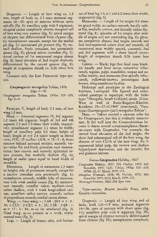
Van Achterberg: Gnaptodontinae 11
Diagnosis. — Length of fore wing ca. 1.8
mm, length of body ca. 2.1 mm; antennal seg-
ments 18—20; apex of antenna without spine
apically (fig. 4); maxillary palp with 5 segments;
occipital carina completely absent; marginal cell
of fore wing very narrow (fig. 3); apical margin
of clypeus not differentiated from clypeus (fig.
6); mesopleuron smooth, except for the medial
pit (fig. 1); mesoscutal pit present (fig. 9); no-
tauli shallow, finely crenulate, but posteriorly
absent (fig. 9); pleural sulcus crenulate (fig. 1);
dorsal carinae of 1st metasomal tergite absent
(fig. 8); basal elevation of 2nd tergite distinctly
differentiated by the curved groove (fig. 8);
length of ovipositor sheath ca. 0.06 times fore
wing.
Contains only the East Palaearctic type-spe-
cies.
Gnaptogaster mongolica Tobias, 1976
(figs. 1-9)
Gnaptogaster mongolica Tobias, 1976a: 319—321,
figs. 2—5.
Paratype, 9, length of body 2.1 mm, of fore
wing 1.8 mm.Head. — Antennal segments 19, 3rd segment
1.2 times 4th segment, length of 3rd and 4th
segment 2.3 and 2.0 times their width, respecti-
vely, penultimate segment 2.1 times its width;
length of maxillary palp 0.5 times height of
head; length of eye 2.6 times temple in dorsal
view; POL: ocellus: OOL = 10 : 5 : 8; frons
concave behind antennal sockets, smooth; ver-
tex rather flat and finely granulate near stemma-
ticum; face convex and smooth; epistomal su-
ture present, but medially shallow (fig. 6);
length of malar space equal to basal width of
mandible.
Mesosoma. — Length of mesosoma 1.3 times
its height; side of pronotum smooth, except for
a narrow crenulate area posteriorly (fig. 1);
metapleuron smooth; middle lobe of mesoscu-
tum with weak medial suture (fig. 8); mesoscu-
tum smooth; scutellar sulcus medium-sized,
rather shallow, with 4 weak longitudinal cari-
nae; scutellum rather convex, smooth; surface
of propodeum smooth; medial carina absent.
Wings. — Fore wing: r : 3-SR : SRI = 4 : 5 :
34; 1-CUl : 2-CUl = 5 : 23; 2-SR : 3-SR : r-
m = 17 : 5 : 14; m-cu far antefureal (fig. 3).
Hind wing: m-cu present as a weak, unpig-
mented trace (fig. 3).
Legs. — Length of femur, tibia, and basitar-
sus of hind leg 3.3, 6.1 and 6.0 times their width,
respectively (fig. 5).
Metasoma. — Length of 1st tergite 0.6 times
its apical width, its surface smooth, basally rath-
er flat, medially convex, and laterally rather flat-
tened (fig. 8); spiracles of 1st tergite after mid-
dle of tergite and not protruding (fig. 8); glym-
ma completely absent; 2nd tergite smooth and
2nd intersegmental suture deep and smooth; all
metasomal setae widely spaced, scattered; 2ndand 3rd segments with a sharp lateral crease;
length of ovipositor sheath 0.06 times fore
wing.
Colour. — Black; legs (but hind coxa black-
ish basally and hind tarsus somewhat infus-
cated), tegulae, palpi, mandibles, scapus and an-
nellus mainly, and metasoma (but apically infus-
cated), yellowish-brown; pterostigma dark
brown, wing membrane hyaline.
Holotype and paratypes in the Zoological
Institute, Leningrad. The figured and redes-
cribed paratype is topotypic with the holo-
type: "Mongolia, South Gobijskij ajmak, 20 km.
West of well of Barin-Bugatyn-Khuduk,
Kerzhner, 25—27.vii.1969" (translated), "Para-
typus Gnamptogaster (sic!) mongolica Tobias".
Note. — Tobias erected a separate tribe for
his Gnaptogaster, but this is evidently unneces-
sary. It fits well into the Gnaptodontinae be-
cause it shares several synapomorphous charac-
ter-states with Gnaptodon. For example, the
curved basal elevation of the 2nd tergite, the
short 2nd submarginal cell of the fore wing, the
absence of vein CU lb of the fore wing, the 3-
segmented labial palp, the narrow and shallow
hypoclypeal depression, and the (nearly) flat
and glabrous labrum.
Genus Gnaptodon Haliday, 1837
Gnaptodon Haliday, 1837: 220. Fischer, 1972: 569.
Shenefelt, 1975: 1123. Tobias, 1976a: 315—318;
1976b: 22, 47. Marsh, 1979: 173.
Diraphus Wesmael, 1838: 89. Fischer, 1972: 569.
Shenefelt, 1975: 1123. Marsh, 1979: 173.
Mesotages Foerster, 1862: 258. Syn. nov.
Type-species: Bracon pumilio Nees, 1834.
Gender: masculine.
Diagnosis. — Length of fore wing and of
body, both 1.0—1.9 mm; antennal segments
14—26; apex of antenna with short spine (fig.
11); maxillary palp with 6 segments (fig. 28);
apical margin of clypeus narrowly differentiated
from clypeus (fig. 14); mesopleuron completely
28 Tijdschrift voor Entomologie, deel 126, afl. 2,1983
smooth, exceptionally precoxal sulcus superfi-
cially impressed; mesoscutal pit absent (fig. 18),
exceptionally with a shallow impression; pleural
sulcus smooth (fig. 10); notauli of Palaearctic
and Oriental species smooth, and posteriorly
(nearly) absent (figs. 18, 26); dorsal carinae of
1st tergite basally present (figs. 19, 31); length
of ovipositor sheath of Palaearctic and Oriental
species 0.03—0.12 times length of fore wing.
Biology. — The larvae are obligatory para-
sites of (mining) larvae of Nepticulidae (Lepido-
ptera). Pupation takes place in the host cocoon,
but endoparasitism remains to be proven (see
chapter on phylogenetic relationships).
Distribution: Palaearctic: 12 species; Nearc-
tic: 7 species; Neotropical: 1 species; Afrotropi-
cal: 3 species; Oriental: 1 species; Australian: 8
species.
A. Palaearctic Region
Key to the Palaearctic species of theGENUS GnAPTODON HaLIDAY
1. Second intersegmental suture of metasoma
deep medially (figs. 31, 43, 51); transverse
elevation of 2nd tergite distinct medially
(figs. 31, 63), exceptionally obsolete (fig.
102); colour of 3rd antennal segment vari-
able, if dark brown, then hind femur yel-
lowish 2
— Second suture (almost) absent (fig. 19);
transverse elevation of 2nd tergite absent
medially (fig. 19); 3rd antennal segment
dark brown; hind femur largely dark brownapheles spec. nov.
2. Distance between wing apex and apex of
marginal cell 1.5—3.6 times vein 1-Rl (figs.
57, 65, 74, 91); length of pterostigma 1.5
—
3.0 times vein 1-Rl; intermediate speci-
mens have posterior margin of elevation of
2nd tergite weakly curved or straight me-dially (figs. 63, 80) and metasoma of 9
comparatively robust (fig. 110) 3
— Distance between wing apex and apex of
marginal cell 0.2— 1.4 times vein 1-Rl (figs.
20, 24), exceptionally up to 1.8 times (fig.
46); length of pterostigma 0.7—1.9 times
vein 1-Rl; intermediate specimens have
posterior margin of elevation of 2nd tergite
distinctly curved (figs. 51, 53) and metaso-
ma of 9 comparatively slender (fig. 53) . . 9
3. Vein r of fore wing about as long as vein 3-
SR or longer (fig. 91); antero-lateral
grooves of 3rd tergite rather deep (excep-
tionally obsolete) and (very) finely crenu-
late (fig. 92); vertex (largely) smooth; an-
tennal segments 17—19
hreviradialis Fischer
— Vein r of fore wing shorter than vein 3-SR
(fig. 74, 84); antero-lateral grooves of 3rd
tergite absent or shallow and smooth (figs.
72, 80); vertex often coriaceous; antennal
segments 18—23 4
4. Marginal cell of fore wing narrow (fig. 57);
distance between wing apex and apex of
marginal cell about 3 times vein 1-Rl (fig.
57); metasoma completely yellowish;
length of vein SRI of fore wing about 3
times vein 3-SR (fig. 57); episternal scrobe
deep (fig. 55); ocelh small, POL 2.5—3
times diameter of posterior ocellus (fig. 56);
medial length of 2nd tergite about 1.5 times
medial length of 3rd tergite (fig. 63)
hrevis spec. nov.
— Marginal cell of fore wing wider (figs. 65,
96); distance between wing apex and apex
of marginal cell 1.5—2.2 times vein 1-Rl;
colour of metasoma variable; length of vein
SRI of fore wing 4.0—6.7 times vein 3-SR
(figs. 74, 96); episternal scrobe rather shal-
low or obsolete (fig. 94); ocelli larger, POLabout twice diameter of posterior ocellus or
shorter (figs. 93, 98); medial length of 2nd
tergite less than 1.5 times medial length of
3rd tergite (figs. 80, 85) 5
5. Vertex almost completely smooth and
strongly shiny (fig. 71); 3rd tergite with
shallow and smooth antero-lateral grooves
(fig. 72); 1st and 2nd tergites, antero-lateral
corners and hind tarsi (except telotarsus),
ivory-whitish; 2nd suture of metasoma dis-
tinctly crenulate medially (fig. 72); vein SRI
of fore wing straight (fig. 65)
vlugi spec. nov.
— Vertex coriaceous, moderately shiny (fig.
83); 3rd tergite with no antero-lateral
grooves (figs. 80, 102), colour and vein SRI
variable; 2nd suture smooth or indistinctly
crenulate (figs. 80, 102) 6
6. Long dense setose between antennal sock-
ets (figs. 76, 77); medio-longitudinal groove
of mesoscutum anteriorly as deep as notauli
(fig. 81); medial length of 3rd tergite equal
to medial length of 2nd tergite (fig. 80) or
slightly less; 2nd tergite dark brown poste-
riorly pilosus spec. nov.
— Sparsely setose between antennal sockets
(fig. 83); medio-longitudinal groove of
mesoscutum anteriorly shallower than no-

Van Achterberg: Cnaptodontinae 29
tauli (figs. 82), 100); medial length of 3rd
tergite variable (figs. 85, 110); 2nd tergite
frequently completely yellowish 7
7. Hind coxa at least basally and hind femur
dorso-apically infuscated; 2nd tergite pos-
teriorly and hind tarsus dark brown; medial
length of 3rd tergite of 9 1.1—1.3 times
medial length of 2nd tergite (fig. 85); mar-
ginal cell of fore wing comparatively nar-
row (fig. 84), distance between wing apex
and apex of marginal cell 1.9—2.4 times
vein 1-Rl; face (except medially) densely
and transversely rugulose-coriaceous
nieukerkeni spec. nov.
— Hind coxa, hind femur apically, 2nd tergite
posteriorly and hind tarsus (except telotar-
sus), yellowish; medial length of 3rd tergite
of 9 0.7—0.8 times medial length of 2nd
tergite (figs. 102, 110); marginal cell of fore
wing somewhat wider (figs. 96, 104), dis-
tance between wing apex and apex of mar-
ginal cell 1.6—1.9 times vein 1-Rl; face
coriaceous or smooth 8
8. Head and apical half of metasoma yellow-
ish; 2nd suture of metasoma rather angular-
ly bent (fig. 102); pterostigma and 3rd and
4th antennal segments dark brown; anten-
nal segments of 9 about 21; scutellar sulcus
distinct (fig. 100) ruficeps spec. nov.
— Head and apical half of metasoma black;
2nd suture of metasoma rather evenly
curved (fig. 110); pterostigma light brown;
3rd and 4th antennal segments of 9 about
18; scutellar sulcus very shallow (fig. 107) . .
erasmi spec. nov.
9. Distance between wing apex and apex of
marginal cell of fore wing 0.2—0.5 times
vein 1-Rl (fig. 24); vein SRI of fore winglargely straight (fig. 24); area behind trans-
verse elevation of 2nd tergite often more or
less sculptured (fig. 31) 10
— Distance between wing apex and apex of
marginal cell of fore wing 0.7— 1.8 times
vein 1-Rl (figs. 20, 46); vein SRI rather sin-
uate (fig. 46); area behind elevation of 2ndtergite often largely smooth (figs. 51, 53)
11
10. Length of vein 1-Rl of fore wing 2—
4
times distance between wing apex and apex
of marginal cell of fore wing (fig. 24); 3rd
antennal segment yellowish, exceptionally
dark brown; mainly parasite of Nepticuli-
dae in trees pumilio (Nees)
— Length of vein 1-Rl of fore wing about 5
times distance between wing apex and apex
of marginal cell of fore wing (fig. 96 in
Fischer, 1966); 3rd antennal segment dark
brown nepalicus Fisher
11. Four basal segments of antenna of 9 infus-
cated or dark brown dorsally, exceptionally
(largely) yellowish; medial length of basal
elevation of 2nd tergite less than 0.6 times
medial length of rest of tergite or, if longer,
less narrowed laterally (cf. fig. 31); medio-
longitudinal groove of mesoscutum often
distinct; metasoma usually dark brown or
black, if partly yellowish then elevation of
2nd tergite indistinct medially; antennal
segments of 9 mostly 21; parasites of Nep-ticulidae in herbs and (low) shrubs
decoris (Foerster)
— Four basal antennal segments of 9 yellow-
ish; medial length of basal elevation of 2ndtergite 0.6—1.0 times medial length of rest
of tergite and elevation distinctly narrowed
laterally (figs. 51, 53); medio-longitudinal
groove of mesoscutum usually obsolete (fig.
49); several tergites of metasoma yellowish;
basal elevation of 2nd tergite distinct me-dially; antennal segments of 9 usually 19 or
20; parasites of NepticuHdae in trees and
(tall) shrubs georginae spec. nov.
Note. — Of the 7 Nearctic species, 3 are
close to Palaearctic spp.: Gnaptodon bicolor
Fischer, 1965, is near erasmi, but bicolor has
length of mesosoma about 1.5 times its height
and distance between apex of fore wing to apex
of marginal cell about 1.5 times vein 1-Rl (fig.
876 in Fischer, 1977). G. glaber Fischer, 1965, is
close to georginae but the latter has infuscated
hind tarsi, pterostigma dark brown, and face
less sculptured. Finally G. nepticulae (Rohwer,
1915) (= G. pulchrigaster Fischer, 1965, accord-
ing to Marsh, 1974) is close tot pumilio, but
nepticulae differs by the reddish base of the
metasoma and the deep, crenulate, antero-
lateral grooves of 3rd tergite. The only Neo-
tropical species described, G. novoteutonicus
Fischer, 1977, is exceptional because of its long
ovipositor.
Gnaptodon apheles spec. nov.
(figs. 10—19)
Holotype, 9 , length of body and of fore wing
both 1.6 mm.Head. — Antennal segments 20, length of 3rd
segment 1.2 times 4th segment, length of 3rd
and 4th segments 3.5 and 2.8 times their width
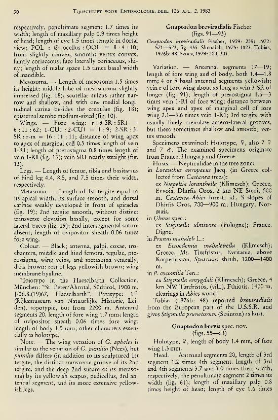
30 Tijdschrift voor Entomologie, deel 126, afl. 2,1983
respectively, penultimate segment 1.7 times its
width; length of maxillary palp 0.9 times height
of head; length of eye 1.5 times temple in dorsal
view; POL : ocellus : OOL = 8:4:10;frons slightly convex, smooth; vertex convex,
faintly coriaceous; face laterally coriaceous, shi-
ny; length of malar space 1.5 times basal width
of mandible.
Mesosoma. — Length of mesosoma 1.5 times
its height; middle lobe of mesoscutum slightly
impressed (fig. 18); scutellar sulcus rather nar-
row and shallow, and with one medial longi-
tudinal carina besides the crenulae (fig. 18);
episternal scrobe medium-sized (fig. 10).
Wings. — Fore wing: r : 3-SR : SRI =
6 : 11 : 62; 1-CUl : 2-CUl = 1:9; 2-SR :3-
SR : r-m = 16 : 11 : 11; distance of wing apex
to apex of marginal cell 0.5 times length of vein
1-Rl; length of pterostigma 0.8 times length of
vein 1-Rl (fig. 13); vein SRI nearly straight (fig.
Legs. — Length of femur, tibia and basitarsus
of hind leg 4.4, 8.5, and 7.5 times their width,
respectively.
Metasoma. — Length of 1st tergite equal to
its apical width, its surface smooth, and dorsal
carinae weakly developed in front of spiracles
(fig. 19); 2nd tergite smooth, without distinct
transverse elevation basally, except for somelateral traces (fig. 19); 2nd intersegmental suture
absent; length of ovipositor sheath 0.06 times
fore wing.
Colour. — Black; antenna, palpi, coxae, tro-
chanters, middle and hind femora, tegulae, pte-
rostigma, wing veins, and metasoma ventrally,
dark brown; rest of legs yellowish brown; wing
membrane hyaline.
Holotype in the Haeselbarth Collection,
München: "St. Peter/Ahrntal, Südtirol, 1900 m,
J/26.8. (19)67, Haeselbarth". Paratype: 19(Rijksmuseum van Natuurlijke Historie, Lei-
den), topotypic, but from 2200 m. Antennal
segments 20, length of fore wing 1.7 mm; length
of ovipositor sheath 0.06 times fore wing;
length of body 1.5 mm; other characters essen-
tially as holotype.
Note. — The wing venation of G. apheles is
similar to the venation of G. pumilio (Nees), but
pumilio differs (in addition to its sculptured 1st
tergite, the distinct transverse groove of its 2ndtergite, and the deep 2nd suture of its metaso-
ma) by its yellowish scapus, pedicellus, 3rd an-
tennal segment, and its more extensive yellow-
ish legs.
Gnaptodon breviradialis Fischer
(figs. 91—93)
Gnaptodon breviradialis Fischer, 1959: 259; 1972:
571—572, fig. 435. Shenefelt, 1975: 1123. Tobias,
1976b: 48. Szócs, 1979: 200, 201.
Variation. — Antennal segments 17—19;
length of fore wing and of body, both 1.4—1.8
mm; 4 or 5 basal antennal segments yellowish;
vein r of fore wing about as long as vein 3-SR of
longer (fig. 91); length of pterostigma 1.6—
3
times vein 1-Rl of fore wing; distance between
wing apex and apex of marginal cell of fore
wing 2.1—3.6 times vein 1-Rl; 3rd tergite with
usually finely crenulate antero-lateral grooves,
but these sometimes shallow and smooth; ver-
tex smooth.
Specimens examined: Holotype, 9, also 7 9
and 7 6. The examined specimens originate
from France, Hungary and Greece.
Hosts.— Nepticulidae in the tree zone:
in Loranthus europaeus Jacq. (in Greece col-
lected from Castanea trees):
ex Niepeltia loranthella (Klimesch); Greece,
Ewoia, Dhirfis Oros, 2 km NE Steni, 500
m, Castanea-Abies forest; id., S slopes of
Dhirfis Oros, 700—900 m; Hungary, Nor-
mafa.
in Ulmus spec:
ex Stigmella ulmivora (Fologne); France,
Digne,
in Prunus mahaleh L.:
ex Ectoedemia mahalehella (Klimesch);
Greece, Mt. Timfristos, Evritania, above
Karpenission, Spartium shrub, 1200—1400
m.
in P. cocomilia Ten.:
ex Stigmella amygdali (Klimesch); Greece, 4
km NW Timfristos, (vili.), Fthiotis, 1400 m,
clearings in Abies wood.
Tobias (1976b: 48) reported breviradialis
from the European part of the U.S.S.R. and
gives Stigmella prunetorum (Stainton) as host.
Gnaptodon brevis spec. nov.
(figs. 55—63)
Holotype, 9, length of body 1.4 mm, of fore
wing 1.3 mm.Head. — Antennal segments 20, length of 3rd
segment 1.2 times 4th segment, length of 3rd
and 4th segments 3.7 and 3.0 times their width,
respectively, the penultimate segment 2 times its
width (fig. 61); length of maxillary palp 0.8
times height of head; length of eye 1.6 times

Van Achterberg: Gnaptodontinae 31
temple in dorsal view (fig. 56); POL : ocel-
lus : OOL = 14 : 5 : 19; frons rather flat and
coriaceous; vertex distinctly coriaceous, face
medially smooth, otherwise coriaceous (fig. 62);
length of malar space 1.5 times basal width of
mandible.
Mesosoma. — Length of mesosoma 1.2 times
its height; medio-longitudinal depression of
mesoscutum absent (fig. 60); scutellar sulcus
rather shallow and medium-sized, crenulate
(fig. 60); episternal scrobe deep (fig. 55).
Wings. — Fore wing : r : 3-SR : SRI =
3:6:16; 1-CUl : 2-CUl = 1:8; 2-SR :3-
SR : r-m = 16 : 11 : 11; length of pterostigma
2.3 times vein 1-Rl; distance between apex of
fore wing and apex of marginal cell 3 times vein
1-Rl (fig. 57); vein SRI straight.
Legs. — Length of femur, tibia and basitarsus
of hind leg 3.1, 8.5, and 5.3 times their width,
respectively.
Metasoma. — Length of 1st tergite 0.6 times
its apical width, its surface superficially coria-
ceous (fig. 63), and dorsal carinae present in
basal half of tergite; 2nd tergite behind basal el-
evation coriaceous, basal elevation smooth and
distinctly transverse (fig. 63), its medial length
0.4 times medial length of rest of tergite; medial
length of 2nd tergite 1.5 times 3rd tergite; 3rd
and following tergites largely coriaceous, with
no antero-lateral grooves; 2nd suture deep,
curved and indistinctly crenulate; length of ovi-
positor sheath 0.12 times fore wing.
Colour. — Dark brown (including pterostig-
ma and veins); antenna dark brown, but 3 basal
segments ventrally yellowish; palpi, legs (but te-
lotarsi infuscated), tegulae, and whole metaso-
ma, yellowish; mesosoma laterally largely rath-
er reddish; wing membrane slightly infuscated.
Holotype in Museum Budapest: "Budaörs,
Törokugrato" (= Hungary), 1975. vii. 16, Szöcs
J., ''Filipendula vulgaris", "ex Nepticula hexa-
petalae Szöcs".
Host: Ectoedemia hexapetalae (Szöcs) in Fil-
ipendula vulgaris Moench.
Gnaptodon decoris (Foerster) comb. nov.
(fig. 20)
Mesotages decoris Foerster, 1862: 258.
Gnamptodon bachmaieri Fischer, 1957: 41. Syn. nov.Gnaptodon bachmaieri; Fischer, 1972: 570—571.
Shenefelt, 1975: 1123. Tobias, 1976b: 48. Szöcs,
1979:200—201.
Gnaptodon bachaien (sic!); Fischer, 1980: 202.
Gnamptodon klemensiewiczii Niezabitowski, 1910:
44. Shenefelt, 1975:1123—1124. Syn. nov.
Variation. — Antennal segments 20—23
(mostly 21); length of fore wing 1.2—1.7 mm,of body 1.1— 1.7 mm; 5th—7th tergites of
metasoma sometimes yellowish-brown, usually
dark brown; length of pterostigma 1.0— 1.6
(usually 1.1— 1.2) times length of vein 1-Rl;
distance between apex of fore wing and apex of
marginal cell 0.9—1.5 times vein 1-Rl; 3rd an-
tennal segment and base of metasoma dark
brown, exceptionally yellowish; 2nd and 3rd
tergites usually smooth, but sometimes superfi-
cially sculptured behind the transverse elevation
and the 2nd metasomal suture; vertex coria-
ceous. Specimens examined: 22 9 and 29 6
.
The examined specimens originate fromGreat Britain (Wales), Netherlands (Wijster,
dunes of Meijendel (near the Hague) and of
Oostvoorne, Kunrade (Kunderberg), and Hil-
versum (Spanderswoud)), West Germany, Italy,
Austria, Hungary, Bulgaria and Greece.
Hosts. — Nepticulidae in herbs and (low)
shrubs:
in Lotus corniculatus L. :
ex Trifurcula cryptella (Stainton); Nether-
lands, Kunrade, Kunderberg (2nd tergite
dark brown and hind coxa infuscated).
in Coronilla varia L.:
from same host; Hungary, Svar.
in Fragaria vesca L. :
ex Stigmella /raganella (Heyd.) (on label, cf.
aurelia (F.)); Hungary, Budapest, Petnehâzi-
rét (apex of metasoma reddish, rest of meta-
soma dark brown, hind coxa yellowish),
in Sanguisorha officinalis L. :
ex S. geminella (Frey) (on label, cf. poterii
(Stainton)); Hungary, Fòt, Mogyorodi p.
(only 1st tergite and apex of metasoma dark
brown, rest yellowish, hind coxa infuscated).
in Rubus spec:
ex S. splendidissimella (H.-S.) or aurelia (F.);
Netherlands, Hilversum, Spanderswoud
(2nd tergite yellowish),
in Rosa pimpinellifolia L.:
ex S. spec; U.K., Wales, Pembrey, Carmar-
then,
in Agremonia agrimonoides (L.):
ex Ectoedemia agrimoniae (Frey); Greece,
Ewoia, Dhirfis Oros, S. slopes, Castanea-
Abies-iortsi, 700—900 m (elevation of 2nd
tergite largely absent, 2nd tergite and hind
coxa yellowish),
in Potentilla erecta L. :
ex Stigmella poterii (Stainton); Hungary,
Svar.

32 Tijdschrift voor Entomologie, deel 126, afl. 2,1983
in Potentilla spec:
ex S. occultella (Hein.) (on label, cf. poterli
(Stainton)); Austria, Schorfling, north of At-
tersee; id., Hinter-Stoder.
in Prunus spinosa L. :
ex S. plagicolella (Stainton); Hungary, Vërz-
teskozma (aberrant because of yellowish 3rd
and 4th antennal segments; 2nd tergite dark
brown).
in Betula nana L.:
ex S. betulicola nanivora Petersen; WestGermany (types of G. hachmaieri (Fischer),
examined: 2nd tergite dark brown, hind
coxa yellowish.
Notes. — The type of Gnaptodon klemensie-
wiczii Niezabitowski, 1910, was not available
for study (it is part of a private collection;
according to Tobias (1979: 237) the type is lost).
Judging from the original description (1st and
2nd tergites smooth and 3rd antennal segment
blackish) it is most likely to be a junior syno-
nym of G. decoris (Foerster).
During my visits to the Berlin Museum I ex-
amined the holotype of Mesotages decoris
Foerster, 1982. It is a completely yellowish
specimen, probably owing to ageing (as manyother small specimens in the Foerster collection
are bleached). The length of the pterostigma is
1.5 times vein 1-Rl (metacarp) and the distance
between the apex of the fore wing and the apex
of the marginal cell is 1.4 times vein 1-Rl. Thevertex is distinctly coriaceous and the 2nd su-
ture of the metasoma smooth. The basal eleva-
tion of the 2nd tergite is rather transverse.
Foerster stated in the original description that
the transverse elevation of the 2nd tergite is ab-
sent, but the surface of the 2nd tergite of the
metasoma is obscured by some glue and by part
of the hind wing. If looked for carefully the typ-
ical transverse elevation of the 2nd tergite can
be seen. This explains how Foerster arrived at
his incorrect statement in the original descrip-
tion and Fischer's description of this species as
hachmaieri. The holotype of decoris in the
Foerster Collection (Zoologisches Museum,Berlin) bears the following labels: "24/926",
"Rheinprovinz", "Frst", "decoris Frst", "Gnap-todon hachmaieri Fi., det. Fischer", "Glaubenicht dass dieser Ex. die Type von Mesotages
decoris sem kann. Foerster schreibt ausdrück-
lich dass Mesotages keine bogenförmige
gekrümmten Fürchen an den 2. Tergit hat,
Fischer, 1963". As pointed out above, I disagree
with Fischer's statement and synonymizeGnamptodon . hachmaieri Fischer, 1957, with
Gnaptodon decoris (Foerster, 1862) comb. nov.
There is a second specimen under decoris in the
Foerster Collection, which does not belong to
the type-series, and also has a reduced, easily
overlooked transverse elevation on the 2nd ter-
gite.
Gnaptodon erasmi spec. nov.
(figs. 103—111)
Holotype, 9, length of body 1.6 mm, of fore
wing 1.5 mm.Head. — Antennal segments 18, length of 3rd
segment 1.1 times 4th segment, length of 3rd
and 4th segments 3.5 and 3.2 times their width,
respectively, penultimate segment 1.9 times its
width (fig. 105); length of maxillary palp 0.7
times height of head; length of eye twice temple
in dorsal view (fig. 108); POL : ocel-
lus : OOL = 6:4:9; frons slightly convex, co-
riaceous; vertex completely coriaceous; face
largely smooth, only laterally coriaceous, rather
flat (fig. Ill); length of malar space 1.7 times
basal width of mandible.
Mesosoma. — Length of mesosoma 1.4 times
its height; medio-longitudinal groove of mesos-
cutum absent, represented only by a wide shal-
low depression (fig. 107); scutellar sulcus very
shallow, narrowly crenulate (fig. 107); epister-
nal scrobe rather shallow (fig. 103).
Wings. — Fore wing: r : 3-SR : SRI =7 : 10 : 46; 1-CUl : 2-CUl = 2 : 15; 2-SR : 3-
SR : r-m = 11:5:8; length of pterostigma
twice length of vein 1-Rl; distance between
wing apex and apex of marginal cell 1.9 times
vein 1-Rl; vein SRI slightly curved (fig. 104).
Legs. — Length of femur, tibia and basitarsus
of hind leg 3.2, 6.8 and 4.5 times their width,
respectively.
Metasoma. — Length of 1st tergite 0.9 times
its apical width, its surface coriaceous, and pos-
teriorly rugulose (fig. 110); dorsal carinae dis-
tinct in basal 0.6 of 1st tergite; basal elevation of
2nd tergite distinct medio-posteriorly (fig. 110),
its medial length 0.3 times rest of tergite; meta-
soma behind elevation of 2nd tergite largely co-
riaceous (fig. 103); 2nd suture deep, finely cre-
nulate and rather evenly curved (fig. 110); medi-
al length of 3rd tergite 0.7 times length of 2ndtergite; length of ovipositor sheath 0.07 times
fore wing.
Colour. — Black; 4 basal segments of anten-
na (rest dark brown), palpi, tegulae and legs,
brownish-yellow; 1st and 2nd tergite, and ante-
ro-lateral corners of 3rd tergite, reddish-brown;
pterostigma light brown.

Van Achterberg: Gnaptodontinae 33
Holotype in Haeselbarth Collection: "Riva s.
Garda (North Italy), 150 m, G/10.9.67, Hae-
selb."; (G = collected in vegetation of Quercus
i/ex-shrubwoodland, on lime, with very strong
dominance of Q. ilex, mixed with Fraxinus or-
nus). Paratype, 1 S (Rijksmuseum van Natuur-
lijke Historie, Leiden):"Ectoedemia groschkei
(Skala) in Vitex agnus-castus, VU no. 80675
KE", "Ellas (Greece), Sept. 1980, S. B. J. Men-ken & E. J. V. Nieukerken", "4 km SW Pa-
padates (Préveza), cult, area, roadside, 100 m,
39.17N-20.46 E, 25.ix.1980, st. 53"; antennal
segments 19, basal elevation of 2nd tergite obso-
lete medially, otherwise as holotype.
Host: "Ectoedemia' groschkei (Skala) in Vi-
tex agnus-castHS L.
Note. It is a pleasure to me to dedicate this
species to Dr. E. Haeselbarth (München).
Gnaptodon georginae spec. nov.
(figs. 44—54)
Holotype, 6 , length of body 1.5 mm, of fore
wing 1.4 mm.Head. — Antennal segments 20, length of 3rd
segment 1.1 times 4th segment, length of 3rd
and 4th segments 3 and 2.7 times their width,
respectively, penultimate segment 1.8 times its
width (fig. 48); length of maxillary palp 0.7
times height of head; length of eye twice temple
in dorsal view; POL : ocellus : OOL =
7:5:10; frons flat, largely coriaceous; vertex
distinctly coriaceous (fig. 52); face superficially
coriaceous-pimply (fig. 45); length of malar
space 1 .3 times basal width of mandible.
Mesosoma. — Length of mesosoma 1.3 times
its height; medio-longitudinal groove obsolete
(fig. 49); scutellar sulcus narrow, rather shallow
(fig. 49); episternal scrobe indistinct (fig. 44).
Wing. — Fore wing: r : 3-SR : SRI = 5 : 9 :
60; 1-CUl : 2-CUl = 3 : 23; 2-SR : 3-SR : r-
m = 24 : 9 : 15; length of pterostigma 1.9 times
vein 1-Rl; distance between wing apex and
apex of marginal cell 1.8 times vein 1-Rl; vein
SRI slightly sinuate (fig. 46).
Legs. — Length of femur, tibia, and basitar-
sus of hind leg 3.4, 7 .7 and 5 times their width,
respectively.
Metasoma. — Length of 1st tergite 0.8 times
its apical width, its surface smooth; dorsal cari-
nae distinct in basal 0.7 of 1st tergite (fig. 51);
basal elevation of 2nd tergite distinctly nar-
rowed laterally, medio-posteriorly distinctly
curved (fig. 51), its medial length 0.8 times rest
of tergite; area directly behind elevation of 2nd
tergite, as rest of tergites basally, weakly coria-
ceous (fig. 44); tergites otherwise smooth; 2ndsuture rather deep, smooth; 3rd tergite with no
antero-lateral grooves; medial length of 3rd ter-
gite 0.9 times length of 3nd tergite (fig. 51).
Colour. — Black; tegulae (largely), pterostig-
ma, 1st tergite medially, and antenna, dark
brown; palpi, rest of 1st tergite, 2nd and 3rd
tergites, antero-lateral corners of 3rd tergite,
and legs (except the infuscated apex of hind tibia
and the hind tarsus), brownish-yellow.
Holotype in Rijksmuseum van Natuurlijke
Historie, Leiden: "ex Stigmella hybnerella
(Hbn.), Crataegus monogyna, VU no. 80061
Kl" "11 km ESE of Morris O.m., 26.iv.1980,
Marsh & Pistacia carr", "Stat. 19", "Algeria, E.
V. Nieukerken, G. Bryan & P. Oosterbroek".
Paratypes, 59-F2(?:l9-hlc5 (Museum Bu-
dapest): "Tahi, Pilis hegys." "1973.v.28, Szöcs
J.", ''Nepticula malella Stt. det. J. Szöcs", "'Mal-
us sylvestris''; 2 9 (Shaw Collection, Edin-
burgh): "Sulzchopf, BL., Switz(erland), LT/96,
470 m, H: Stigmella tityrella, mine coll.
11.10.76, PIE 23.7.77, S.E. Whitebread"; 1 9
(Zaykov Collection, Plovdiv): "22.9.1968, Ro-
dopi, Parvenez, leg. A. Germanov"; 1 â (id):
"21.5.1968, Plovdivsko Starosel, leg. A. Ger-
manov"; 1 9 (Rijksmuseum van Natuurlijke
Historie, Leiden): "Jambol, Ormana, 1981.V.27,
leg. Zaykov". The latter 3 specimens are from
Bulgaria.
Variation. — Antennal segments 19—21 (9)
or 20—23 (a); 4—6 basal segments of 9 anten-
na yellowish, penultimate segment twice its
width (fig. 54), 6 has basal antennal segments
infuscated or blackish; length of fore wing 1.4
mm (3 specimens); length of pterostigma 1.2
—
1.9 times vein 1-Rl; distance between apex of
fore wing and apex of marginal cell 1.1—1.8
times vein 1-Rl; medial length of basal eleva-
tion of 2nd tergite 0.6-1.0 times rest of tergite;
2nd tergite often completely smooth (fig. 53);
2nd and 3rd tergites usually yellowish, if dark
brown then apical half of metasoma yellowish
(ex S. tityrella); length of ovipositor sheath 0,04
times fore wing.
Hosts: Stigmella hybnerella (Hübner) in Cra-
taegus monogyna L., S. malella (Stainton) in
Malus sylvestris (L.), S. ruficapitella (Haworth)
in Quercus, and S. tityrella (Stainton) in Fagus
sylvatica L.
It is a great pleasure to me to dedicate this
species to Dr. Georgina Bryan (Amsterdam),
who made an important collection of reared
parasites of Nepticulidae available for study.
34 Tijdschrift voor Entomologie, deel 126, afl. 2, 1983
Gnaptodon nepalicus Fischer
Fischer, 1966: 159—161, figs. 96—97. Shenefelt,
1975:1124.
Fischer considered G. nepalicus an Oriental
species; however, I prefer to include it amongthe Palaearctic spp., because it was collected at
6800 ft. in the Himalayas. The fauna at such
high altitudes is more closely correlated with
the (South) Palaearctic region than with the
Oriental region. As shown in the foregoing key
nepalicus is close to G. pumilio and seems to dif-
fer mainly in the colour of the antenna and in
the wing venation.
Gnaptodon nieukerkeni spec. nov.
(figs. 82—90)
Holotype, 9 , length of body and of fore wing
1.4 mm.Head. — Antennal segments 18 (right anten-
na) or 19 (left one), length of 3rd segment 1.1
times 4th segment, length of 3rd and 4th seg-
ments 3.2 and 3.0 times their width, respective-
ly, penultimate segment 2.2 times its width (fig.
89); length of maxillary palp 0.7 times height of
head; length of eye 2.1 times temple in dorsal
view (fig. 83); POL : ocellus : OOL =
14 : 7 : 18; frons coriaceous, flat; vertex com-pletely coriaceous; face rather convex and
obliquely, finely rugulose-coriaceous, medially
narrowly smooth (fig. 90); length of malar space
1.7 times basal width of mandible.
Mesosoma. — Length of mesosoma 1.4 times
its height; medio-longitudinal groove obsolete
(fig. 87); scutellar sulcus deep, rather narrow,
and distinctly crenulate (fig. 87); episternal
scrobe absent.
Wings. — Fore wing: r : 3-SR : SRI = 2:5:24; 1-CUl : 2-CUl = 1 : 10; 2-SR : 3-SR : r-
m = 8 : 5 : 6; length of pterostigma 2.1 times
vein 1-Rl; distance between wing apex and
apex of marginal cell 2.2 times vein 1-Rl; vein
SRI slightly sinuate (fig. 84).
Legs. — Length of femur, tibia and basitarsus
of hind leg 3.4, 7.3 and 5.0 times their width,
respectively.
Metasoma. — Length of 1st tergite 0.8 times
its apical width, its surface smooth (fig. 85);
dorsal carinae present in basal third of 1st ter-
gite; basal elevation of 2nd tergite distinct, its
medial length 0.4 times medial length of rest of
tergite; 2nd tergite behind elevation and rest of
metasoma largely coriaceous; 2nd suture deep
and smooth; 3rd tergite with no antero-lateral
grooves and its medial length 1.2 times medial
length of 2nd tergite (fig. 85); length of oviposi-
tor sheath 0.03 times fore wing.
Colour. — Black; antenna (except yellowish
annellus), tegulae, pterostigma and wing veins,
dark brown; palpi, and legs, yellowish (but hind
coxa dorsally largely, hind femur apico-dorsally
and hind tarsus (other tarsi slightly) infuscated;
2nd tergite medially and antero-laterally brown,
rest of tergite dark brown.
Holotype in Rijksmuseum van Natuurlijke
Historie, Leiden: "Parnassós Oros, 2 km, W.Summit (Voiotia), rocks, dwarf shrub, 38.32 N-22.35 E, 2000 m, 28.ix.1980, St. 58", "No.
80.685 KE, 21/24.X.80, in leaf-litter!", "ex Stig-
mella cf. rhamnophila (Amsel), Rhamnus saxa-
tilis, VU no. 80.685 K.", "Ellas, Greece, Sep-
tember 1980, S. B. J. Menken, E. J. van Nieu-
kerken". Paratypes: 12 2 and 2 â from same
series (Vrije Universiteit, Amsterdam; Rijksmu-
seum van Natuurlijke Historie, Leiden; Mu-seum Budapest; Collectie Zaykov, Plovdiv).
Variation. — Antennal segments 17—20;
length of fore wing 1.2—1.4 mm; vein SRI of
fore wing usually more curved than in fig. 84;
length of pterostigma 1.8—2.3 times vein 1-Rl;
distance between apex of fore wing and apex of
marginal cell 1.9—2.4 times vein 1-Rl; medial
length of 3rd tergite 1.1—1.3 (9) or 0.9 (S)
times medial length of 2nd tergite; 2nd tergite
often (rather dark) brownish medially.
Host: Stigmella cf. rhamnophila (Amsel) in
Rhamnus saxatilis Jacq.
Gnaptodon pilosus spec. nov.
(figs. 73—81)
Holotype, 9, length of body 1.4 mm, of fore
wing 1.3 mm.Head. — Antennal segments 21, length of 3rd
segment 1.3 times 4th segment, length of 3rd
and 4th segments 2.2 and 1.7 times their width,
respectively, penultimate segment 1.4 times its
width (fig. 75); length of maxillary palp 0.8
times height of head; length of eye 2.2 times
temple in dorsal view (fig. VI); POL : ocel-
lus : OOL = 7:4:11; frons sUghtly concave
behind antennal sockets, coriaceous; vertex dis-
tinctly coriaceous (fig. 77); face rather convex
and largely smooth, only microsculptured
laterally (fig. 76); long and densely setose be-
tween antennal sockets (figs. 76, 77; this setosi-
ty is absent in all other spp. treated in this pa-
per); length of malar space 1.1 times basal width
of mandible.
Mesosoma. — Length of mesosoma 1.1 times
its height; medio-longitudinal groove rather

Van Achterberg: Gnaptodontinae 35
deep, similar to notauli anteriorly (fig. 81); scu-
tellar sulcus rather wide and deep, narrowly
crenulate (fig. 81); episternal scrobe indistinct
(fig. 73).
Wings. — Fore wing: r : 3-SR : SRI =
5:9:61; 1-CUl : 2-CUl = 1:8; 2-SR :3-
SR : r-m = 20 : 9 : 1 1 ; length of pterostigma
1.8 times vein 1-Rl; distance between wing
apex and apex of marginal cell 1.8 times vein 1-
Rl; vein SRI straight (fig. 74).
Legs. — Length of femur, tibia, and basitar-
sus of hind leg 3.4, 6.4 and 4 times their width,
respectively.
Metasoma. — Length of 1st tergite 0.8 times
its apical width, its surface aciculate in posterior
half, the rest smooth (fig. 80), dorsal carinae
present in basal 0.7; basal elevation of 2nd ter-
gite distinct (somewhat cariniform at posterior
edge), its medio-longitudinal length half the
length of rest of 2nd tergite; metasoma behind
elevation of 2nd tergite largely coriaceous (fig.
80); medio-longitudinal length of 2nd tergite
equal to length of 3rd tergite; 2nd suture finely
crenulate and rather deep; length of ovipositor
sheath 0.08 times fore wing.
Colour. — Black; 4 basal segments of anten-
na (but scapus and pedicellus dark dorsally),
palpi, humeral plate (tegula dark brown), legs
(except dark brown tarsi), metasoma ventrally,
1st tergite (medially infuscated) and anterior
half of 2nd tergite, yellowish; rest of antenna
medially brown, its apical half dark brown; pro-
notal sides and mesopleuron with brownish
stripe ventrally (absent in paratypes); apex of
pterostigma whitish, rest dark brown.
Holotype in Museum Budapest: "Budapest,
Rupphegy", "1974. vi.9, Szöcs J.", "Quercus pu-
bescens, semen", "ex Nepticula sp., 1974. vii. 8."
Paratypes, ì 9 + \ 6:\ 6 (Museum Budapest):
"Hungaria, Budaörs. Naphegy", "1977. v.26.,
Szöcs J.", "QuercHs pubescens" , "ex Nepticula
eberhardi ]oh.., det. J. Szöcs". As holotype, but
metasoma slender, antennal segments 23, 1st
tergite smooth, and pterostigma completely
dark brown; 1 9 (Rijksmuseum van Natuurlijke
Historie, Leiden): "Fòt (= Hungary),
1960. ix. 19", "lg. Mihâlyi". Antennal segments
21; as holotype, but medial length of 2nd tergite
1.1 times 3rd tergite.
Host: Stigmella eberhardi Qoh.) in Quercus
pubescens Willd.
Variation. — Antennal segments 21—23,
length of fore wing 1.3—1.5 mm; length of pte-
rostigma 1.5—2.1 times vein 1-Rl; distance be-
tween apex of fore wing and apex of marginal
cell 1.6—2.1 times vein 1-Rl.
Note. G. pilosus resembles the Nearctic G.
glaber Fischer, 1965, but glaber has the pteros-
tigma yellowish, length of mesosoma about 1.5
times its height, 1st tergite longer than apical
width, metasoma completely smooth and face
distinctly coriaceous.
Gnaptodon pumilio (Nees)
(figs. 22—33)
Bracon pumilio Nees, 1834: 90—91.
Gnaptodon pumilio; Fischer, 1972: 572—574, figs.
436—437. Shenefelt, 1975: 1124. Tobias, 1976b:
48. Van Achterberg, 1976: 60, figs. 37, 38. Hud-dleston, 1978: 47. Fischer, 1980: 202.
Gnamptodon pumilio; Shaw & Askew, 1976: 131.
Diraphus pygmaeus Wesinael, 1838: 90, figs. 11, F.
A neotype of Bracon pumilio Nees, 1834, is
here selected, because the original type-series is
lost, it is the type-species of the genus Gnapto-
don Haliday, 1837, and closely related species
occur in the same faunal region. The hind coxae
of the neotype are infuscated contrary to the
original description, but Nees probably over-
looked this feature.
Description of neotype, 9, length of body
and of fore wing 1 .9 mm.Head. — Antennal segments 21, length of 3rd
segment 1.3 times 4th segment, length of 3rd
and 4th segments 4.3 and 3.0 times their width,
respectively, the penultimate segment 1.8 times
its width; length of maxillary palp equal to
height of head; eyes slightly emarginate (fig.
32); length of eye 2.6 times temple in dorsal
view; POL:0 ocellus : OOL = 12:7:18;
frons flat, coriaceous; vertex largely smooth,
convex; face rather convex (more so than in
decoris) and laterally distinctly coriaceous (fig.
32); length of malar space 1.4 times basal width
of mandible.
Mesosoma. — Length of mesosoma 1.5 times
its height; middle lobe of mesoscutum antero-
medially slightly depressed (fig. 26); scutellar
sulcus narrow, merely crenulate (fig. 26); medi-
al carina of propodeum absent.
Wings. — Fore wing: r : 3-SR : SRI = 4 :7 :
36; 1-CUl : 2-CUl = 5 : 31; 2-SR : 3-SR : r-
m = 27 : 14 : 15; distance of wing apex to apex
of marginal cell 0.3 times vein 1-Rl; length of
pterostigma 1.1 times vein 1-Rl (fig. 24); vein
SRI straight (fig. 24).
Legs. — Length of femur, tibia, and basitar-
sus of hind leg 3.8, 9.2 and 7.5 times their width,
respectively.
Metasoma. — Length of 1st tergite equal to
36 Tijdschrift voor Entomologie, deel 126, afl. 2, 1983
its apical width, its surface behind the spiracles
finely rugulose and in front of the spiracles
largely smooth (fig. 31); dorsal carinae of the 1st
tergite present in basal half of tergite; posterior
margin of transverse elevation of 2nd tergite
straight medio-posteriorly, behind it rugulose;
medial length of elevation of 2nd tergite 0.6
times medial length of rest of tergite; 2nd tergite
with some punctures, its medial length 1.5 times
medial length of 3rd tergite (fig. 31); 2nd inter-
segmental suture medially deep and laterally
obliterated, with a pair of obsolete, posteriorly
diverging submedial grooves antero-laterally
and a pair of obsolete grooves close to suture;
ovipositor straight, without notch, and with a
dorso-subapical nodus and some minute ventral
teeth (fig. 30); length of ovipositor sheath 0.11
times fore wing.
Colour. — Black; mouthparts (including pal-
pi), 3 basal antennal segments, tegulae, vein C -I-
SC -I- R of fore wing, legs (but hind coxa largely
infuscated) and metasoma ventrally, more or
less yellowish-brown; pterostigma and 4th an-
tennal segment dark brown; metasoma behind
1st tergite dorsally blackish-brown; wing mem-brane hyaline.
Neotype in Rijksmuseum van Natuurlijke
Historie, Leiden: "Nederland, Wijster (Dr.),
opposite Biol. Stat., 18—25.vi.1976, C. v. Ach-terberg".
Note. Gnaptodon pumilio (Nees) differs fromdecoris, in addition to the characters given in the
key, by the distinctly setose back of the head
(above the hypostomal carina) and the moreconvex face.
The type-series of Diraphus pygmaeus Wes-mael, 1838, consists of one typical female speci-
men of G. pumilio (Nees) with the 2nd and 3rd
tergites brownish, one female with yellowish
2nd and 3rd tergites ("var. 1" of Wesmael) and
one male (probably not conspecific). The first
specimen mentioned is here designated as lecto-
type. The lectotype has no distinct antero-
lateral grooves at the 3rd tergite, the whole 1st
tergite is dark brown, the 2nd metasomal suture
is rather deep but smooth, the vertex is coria-
ceous, the 2nd tergite is distinctly sculptured
and the hind coxae are yellowish.
Variation. — The variation within pumilio as
interpreted in this paper is considerable. I havetried to split up the complex, but the results
were unsatisfactory. The variation (especially of
coloration) within a reared series is so extensive
that any attempted division has been frustrated.
Considering the available data, it is likely that
the specialisation within the complex has not re-
sulted (yet) in recognizable species.
At least three forms can be discerned, of
which form A seems to be ecologically differ-
entiated from both other forms because it has
been reared from Nepticulidae in herbs and
bramble {Rubus spp.). The other forms have
been reared from Nepticulidae in trees. I men-tion these three forms only to indicate the mainunes of variations encountered. All characters
mentioned are very variable, even within the
same reared series.
Form A (including pygmaeus var. 1 (Wes-mael)) has the 1st and 2nd tergites yellowish,
with the middle of the 1st tergite usually dark
brown, but sometimes the whole metasoma (ex-
cept 1st tergite) is yellowish; vertex behind
stemmaticum coriaceous; 2nd suture of metaso-
ma shallow; antero-lateral grooves of 3rd tergite
absent; hind coxa yellowish.
Form B has basal half of metasoma blackish
or yellowish; vertex behind stemmaticum often
smooth; 2nd suture of metasoma deep and cre-
nulate; antero-lateral grooves of 3rd tergite
deep and often finely crenulate; exceptionally
the 3rd tergite has a separate crenulate and
curved croove, directly behind 2nd suture in
stead of antero-lateral grooves (this indicates
that these grooves may be the result of an amal-
gamation of the groove and the 2nd suture in
the middle of the tergite); hind coxa yellowish
or infuscated. Even within series from the samehost there is considerable variation. The typical
form has the whole metasoma blackish-brown;
vertex behind stemmaticum smooth; 2nd suture
of metasoma deep and smooth (fig. 31); antero-
lateral grooves of 3rd tergite incomplete, shal-
low and smooth; hind coxa infuscated or yel-
lowish. Intermediates to form B occur frequent-
ly, e.g. in a series from North Italy (St. Peter,
Ahrntal, 1270 m, Haeselbarth Collection).
Specimens examined: 52 9 and 22 6 (of
which 7 9 and 4 6 belong to form A). Antennal
segments 19—23 (of both sexes mostly 21 or
22); length of body 1.6— 1.8 mm; length of fore
wing 1.7—1.9 mm; 4th antennal segment yel-
lowish, exceptionally dark brown; area behind
transverse elevation of 2nd tergite smooth to
rather extensively rugulose; apex of metasomafrequently reddish brown; length of pterostig-
ma: vein 1-Rl of fore wing : distance betweenwing apex and apex of marginal cell of fore
wing = 0.8—1.2 : 1 : 0.25—0.6. The examined
specimens originate from Norway, Sweden,
Great Britain (Scotland, England), Ireland,
Van Achterberg: Gnaptodontinae 37
Netherlands (Bemelen (Bemelerberg); Cadier
(Schiepersberg); Castricum (dunes); 's Graven-
land (Ankeveense Plassen); Grevenbicht; Hil-
versum (heath Spanderswoud); Hulshorst (Leu-
venumse Bos); Nederhorst den Berg (Spiegel-
polder); Waarder; Wijster; Winterswijk),
France, Italy, Austria, Hungary, Bulgaria and
Greece.
Hosts: Reared from Nepticulidae in trees
(form B and typical form) :
in Betula verrucosa Ehrh.: (= pendula auct.)
ex Stigmella luteella (Stainton); U.K., Dan-bury, and Debden, both Essex,
in Betula spec:
ex S. betulicola (Stainton); U.K., Thorpe-
ness, Suffolk.
ex 5. confusella (Wood); Norway,Andalsnes, More & Romsd.ex 5. continuella (Stainton): Netherlands,
Hilversum, heath.
ex S. lapponica (Wocke); Netherlands, dunes
of Castricum; U.K., Delamere, Cheshire,
in Salix caprea L.:
ex S. Salicis (Stainton); U.K., Benenden,
Kent, (intermediate between form A and
typical form, apex of metasoma partly yel-
lowish),
in Tilia spec:
ex S. tiliae (Frey); Hungary, Vâsarosbée.
in Ulmus procera Salisbury:
ex S. ulmivora (Fologne); U.K., Hallow,
Worcestershire,
in U. glabraMiW.:
ex Nepticulidae; Hungary, Batorliget.
in Ostrya carpinifolia Scopoli :
ex 5. carpinella (Heinemann); Greece, Oîti
Oros, Fthiotis, 4 km E, pavUani, 850 m.in Sorbus aucuparia L.:
ex S. magdalenae (Klimesch); Norway,Storen, Sor-Trondelag, 60 m.
ex S. sorbi (Stainton); U.K., Blackford Hill,
Edinburgh.
ex S. spec; Ireland, Ballinahinch, Co. Gal-
way,
in Malus domestica L.:
ex 5. malella (Stainton); Netherlands, Gre-
venbicht (one â has 2nd tergite yellowish,
the other dark brown!).
ex S. pomella (Vaughan); Netherlands, Win-terswijk,
in Rhamnus catharticus L.:
ex S. rhamnella (H.-S.); Hungary, Budaörs.
in Quercus robur L.:
ex S. ruficapitella-^roup; Netherlands,
Hulshorst, Leuvenumse Bos; id., Hilversum,
heath; id., Bemelen, Bemelerberg.
ex Ectoedemia caradjai (Hering); France,
Digne.
Form A:
in Rubus fruticosus agg. :
ex Ectoedemia erythrogenella (de Joannis);
U.K., Benfleet, Essex.
ex Stigmella splendidissimella (H.-S.) or au-relia (F.); Netherlands, Nederhorst denBerg, Spiegelpolder; id., 's Graveland, An-keveense Plassen, (part of series has almost
whole metasoma yellowish).
in Rubus spec:
from same host(s); Netherlands, Hilversum.
Spanderswoud, (vv^hole metasoma (except
middle of 1st tergite) yellowish).
in Agrimonia eupatoria L. :
ex S. aeneofasciella (H.-S.); Netherlands,
Cadier, Schiepersberg (part of series has
whole metasoma yellowish ( 9 ) or behind
2nd tergite blackish ((?)); Austria,
Schörfling, north of Attersee.
Gnaptodon ruficeps spec. nov.
(figs. 94—102)
Holotype, 9, length of body 1.6 mm, of fore
wing 1.5 mm.Head. — Antennal segments 21, length of 3rd
segment 1.1 times 4th segment, length of 3rd
and 4th segments 3.5 and 3 times their width,
respectively, penultimate segment 1.8 times its
width (fig. 95); length of maxillary palp 0.8
times height of head; length of eye 1.8 times
temple in dorsal view (fig. 98); POL : ocel-
lus : OOL = 12 : 7 : 18; frons slightly convex,
coriaceous; vertex completely coriaceous (fig.
98); face rather convex and largely coriaceous
(fig. 101); length of malar space 2.1 times basal
width of mandible.
Mesosoma. — Length of mesosoma 1.2 times
its height; medio-longitudinal groove of mesos-
cutum absent, represented by only a wide, shal-
low depression (fig. 100); scutellar sulcus rather
wide and deep (fig. 100); episternal scrobe shal-
low.
Wings. — Fore wing: r : 3-SR : SRI = 3 : 6 :
22; 1-CUl : 2-CUl =1:8; 2-SR : 3-SR : r-
m = 10 : 6 : 7; length of pterostigma 1.5 times
vein 1-Rl; distance between apex of fore wing
and apex of marginal cell 1.6 times vem 1-Rl;
vein SRI slightly curved (fig. 96).
Legs. — Length of femur, tibia and basitarsus
of hind leg 3.9, 9 and 6 times their width, re-
spectively.

38 Tijdschrift voor Entomologie, deel 126, afl. 2, 1983
Metasoma. — Length of 1st tergite 0.6 times
its apical width, its surface superficially coria-
ceous; dorsal carinae present in basal half of 1st
tergite (fig. 102); basal elevation of 2nd tergite
indistinct, its medial length 0.5 times rest of ter-
gite; 2nd tergite behind elevation and rest of
metasoma largely coriaceous (fig. 94); 2nd su-
ture deep, smooth and distinctly bent (fig. 102);
3rd tergite with no antero-lateral grooves; me-dial length of 3rd tergite 0.8 times medial length
of 2nd tergite (fig. 102); length of ovipositor
sheath 0.08 times fore wing.
Coulour. — Yellowish-brown; stemmaticum,
antenna (but scapus largely yellowish), mesoso-
ma (but propleuron, mesopleuron dorsally and
metapleuron brownish), and pterostigma, dark
brown; vertex (partly) and telotarsi, infuscated.
Holotype in Museum Budapest: "Nagy-kovàcsi (= Hungary), 1967, vii. 9, Szöcs J.", "ex
Nepticula dorycniella Suire", "Dorycnium ger-
manicum"
.
Host: Trifurcula dorycniella (Suire) in Doryc-
nium germanicum (Grenli).
Gnaptodon vlugi spec. nov.
(figs. 64—72)
Holotype, 6, length of body 1.2 mm, of fore
wing 1.4 mm.Head. — Antennal segments 18, antenna
widened apically (fig. 64), length of 3rd segment
1.1 times 4th segment, length of 3rd and 4th
segments 3.5 and 3.2 times their width, respecti-
vely, penultimate segment 2.2 times its width
(fig. 66); length of maxillary palp 0.9 times
height of head; length of eye 1.7 times temple in
dorsal view (fig. 71); POL : ocellus : OOL =
16 : 8 : 19; frons almost smooth, slightly de-
pressed behind antennal sockets; vertex largely
smooth; face largely smooth, laterally indis-
tinctly micro-sculptured, rather convex (fig.
68); length of malar space equal to basal width
of mandible.
Mesosoma. — Length of mesosoma 1.3 times
its height; medio-longitudinal groove of mesos-
cutum shallow but distinct (fig. 69); scutellar
sulcus rather wide, distinctly crenulate (fig. 69);
episternal scrobe deep (fig. 64).
Wings. — Fore wing: r : 3-SR : SRI = 6 :
11 : 54; 1-CUl : 2-CUl = 3:11; 2-SR : 3-SR :
r-m = 24 : 11 : 16; length of pterostigma 1.9
times vein 1-Rl; distance between apex of fore
wing and apex of marginal cell 2.2 times vein 1-
Rl (fig. 65); vein SRI straight.
Legs. — Length of femur, tibia and basitarsus
of hind leg 3.6, 6.6 and 5.2 times their width,
respectively.
Metasoma. — Length of 1st tergite 0.9 times
its apical width, its surface smooth, and dorsal
carinae obsolescent (fig. 72); basal elevation of
2nd tergite weakly differentiated, strongly
transverse, its medial length 0.4 times medial
length of rest of tergite (fig. 72); 2nd tergite be-
hind elevation coriaceous but posteriorly
smooth; medial length of 2nd tergite 1.2 times
medial length of 3rd tergite; 3rd tergite smoothand with shallow and smooth antero-lateral
grooves (fig. 72); 2nd suture deep, medially dis-
tinctly crenulate.
Colour. — Black; palpi, legs (except infus-
cated telotarsi), 5 basal segments of antenna, te-
gulae, 1st and 2nd tergites, antero-lateral cor-
ners of 3rd tergite and anterior half of metasomaventrally, ivory-whitish; pterostigma brown;rest of antenna (dark) brown.
Holotype in Rijksmuseum van Natuurlijke
Historie, Leiden: "Sweden, Hallandsl., Särö-
Hamra, swept, 26.6.1977, leg. H. J. Vlug". It is
a pleasure to me to dedicate this species to its
collector, Mr. H.J. Vlug (Scherpenzeel).
The ecology of the reared PalaearcticGnaptodon species
As shown in fig. 131, if reared more than
once, the hosts of Gnaptodon species are usual-
ly found on plants of several families. Obvious-
ly the parasites do not select certain plant
groups. If the host plants are arranged accord-
ing to their morphology (trees, (low) shrubs,
and (non-woody) herbs), however, the picture
changes (fig. 130). The assignment of host
plants of the Nepticulidae to a tree, shrub, or
herb zone is, of course rough: the low shoots of
a tree may be lower than a large herb, and the
same applies for a dwarf shrub. Nevertheless,
the species seem to select for a certain vegeta-
tion zone; if more than one zone is included
(e.g. pumilio, no. 1 in fig. 130) then the speci-
mens in another zone are aberrant in colour and/
or morphology with respect to the nominate
form (e.g. form A of pumilio in herbs) and mayconstitute already a good biological species or
be in the process of active sympatric speciation.
If two morphologically very close species oc-
cur in the same zone (nos 3 and 5 in fig. 130)
then one of the species is specialized: in this case
no. 5 (pilosHs) seems to be restricted to Nepticu-
lidae in QnercHS, while no. 3 (georginae) parasi-
tizes Nepticulidae in other trees (and high

Van Achterberg: Gnaptodontinae 39
shrubs). In the case of erasmi (no. 6) and nieu-
kerkeni (no. 7), the latter seems to be restricted
to dwarf shrubs among rocks at high altitude.
G. erasmi is known from 100—150 m altitude
and occurs in higher shrubwood.
Summarizing, the selection of a vegetation
zone is of major importance. Within one zone
closely related species are ecologically separated
because of their preference to â certain host-
plant (pilosHS in QuercHS, georginae in other
trees) or to a certain altitude {erasmi at low alti-
tude, nieukerkeni at high altitude). Obviously a
lot of research, especially cross-breeding experi-
ments, still has to be done before definitive con-
clusions can be drawn.
B. Afrotropical Region
Key to Afrotropical species of the genusGnaptodon Haliday
1. Second tergite of metasoma with distinct
transverse elevation basally (figs. 119, 128);
length of vein SRI of fore wing 7—10.5
times vein 3-SR (figs. 114, 122) 2
— Second tergite with no distinct transverse
elevation basally; length of vein SRI of fore
wing about 3 times vein 3-SR
unifossa Fischer
2. Distance between fore wing apex and apex
of marginal cell about 0.7 times vein 1-Rl
(metacarp); length of pterostigma about 1.1
times vein 1-Rl; length of vein SRI of fore
wing about 10 times vein 3-SR (fig. 122);
1st tergite largely aciculate-rugulose (fig.
128); antenna comparatively slender basally
(fig. 121) similis spec. nov.
— Distance between fore wing apex and apex
of marginal cell about 1.3 times vein 1-Rl;
length of pterostigma 1.5— 1.9 times vein 1-
Rl; length of vein SRI of fore wing 7—
8
times vein 3-SR (fig. 114); 1st tergite largely
smooth (fig. 119); antenna rather robust
basally (fig. 112) bini spec. nov.
Gnaptodon bini spec. nov.
(figs. 112—120)
Holotype, 9, length of body, and of fore
wing 1.1 mm.Head. — Antennal segments 14, length of 3rd
segment 1.3 times 4th segment, length of 3rd
and 4th segments 2.5 and 2.0 times their width,
respectively, penultimate segment 1.8 times its
width; length of maxillary palp 0.8 times height
of head; length of eye 1.9 times temple in dorsal
view, POL : ocellus : OOL = 9 : 4 : 10;
frons slightly impressed, coriaceous (figs. 116,
117); vertex coriaceous; face rather convex,
laterally coriaceous, rest smooth (fig. 117);
length of malar space 1.2 times basal width of
mandible
Mesosoma. — Length of mesosoma 1.4 times
its height; medio-longitudinal groove of mesos-
cutum absent; scutellar sulcus narrow (fig. 120);
episternal scrobe obsolescent (fig. 112).
Wings. — Fore wing: r : 3-SR : 3-Rl = 5 :
10 : 68; 1-CUl : 2-CUl = 3 : 26; 2-SR : 3-SR :
r-m = 25 : 10 : 11; length of pterostigma 1.9
times vein 1-Rl; distance between wing apex
and apex of marginal cell 1.4 times vein 1-Rl;
vein SRI slightly curved.
Legs. — Length of femur, tibia and basitarsus
of hind leg 3.2, 6 3 and 4 times their width, re-
spectively.
Metasoma. — Length of 1st tergite 0.7 times
its apical width, its surface smooth; dorsal cari-
nae of 1st tergite obsolescent (fig. 119); basal el-
evation of 2nd tergite slightly developed medial-
ly, rest of 2nd tergite and 3rd tergite coriaceous,
rest of metasoma largely transversely micro-ac-
iculate (fig. 112); medial length of basal eleva-
tion of 2nd tergite 0.4 times rest of tergite; me-dial length of 2nd tergite 0.9 times length 3rd
tergite (fig. 119); 2nd suture deep, smooth, 3rd
tergite with no antero-lateral grooves; length of
ovipositor sheath 0.08 times fore wing.
Colour. — Blackish or dark brown; clypeus
ventrally, mandibles, 4 basal segments of anten-
na, tegula, legs (but fore and middle telotarsi
and hind tarsus infuscated), and 3 basal seg-
ments of metasoma, brownish-yellow; rest of
metasoma dark brown; humeral plate whitish;
pterostigma dark brown; palpi whitish.
Holotype in Rijksmuseum van Natuurlijke
Historie, Leiden: "Somalia, Afgoi, iv.l977.
Lower Shabelli Valley, F. Bin". Additional
material : 1 9 (non-type), in same institute, to-
potypic, length of fore wing 1 mm, antennal
segments 16, robust antenna as holotype (length
of 3rd and 4th segments 2.5 and 2 times their
width, respectively), length of pterostigma 1.5
times vein 1-Rl, distance between apex of fore
wing and apex of marginal cell 1.3 times vein 1-
Rl; hind tarsus and whole metasoma yellowish;
head (except stemmaticum) brownish-yellow;
1st tergite with some micro-aciculae. Not la-
belled as paratype because of aberrant colora-
tion.
Note. The combination of the short distance
between the apex of the fore wing and the apex
of the marginal cell, the short vein 1-Rl, and the

40 Tijdschrift voor Entomologie, deel 126, afl. 2,1983
low number of antennal segments, differentiates
it from the known Palaearctic species. The only
Oriental species (described in this paper) differs
e.g. by its slender pterostigma. I am pleased to
dedicate this species to Dr. F. Bin (Perugia),
who collected several interesting Braconidae in
Somalia.
Gnaptodon similis spec, nov
(figs. 121—129)
Holotype, 9, length of body 1.2 mm, of fore
wing 1.3 mm.Head. — Antennal segments 17, length of 3rd
segment 1.2 times 4th segment, length of 3rd
and 4th segments 3 and 2.5 times their width,
respectively, penultimate segment 2.3 times its
width (fig. 123); length of maxillary palp 0.8
times height of head; length of eye 1.1 times
temple in dorsal view; POL : ocellus : OOL= 6:3:8; frons slightly impressed, coriaceous;
vertex strongly shiny, coriaceous (fig. 124); face
rugulo-coriaceous laterally, rest smooth, rather
convex (fig. 125); length of malar space equal to
basal width of mandible.
Mesosoma. — Length of mesosoma 1.4 times
its height; medio-longitudinal groove of mesos-
cutum absent (fig. 129); scutellar sulcus narrow,
merely finely crenulate (fig. 129); episternal
scrobe small and shallow.
Wings. — Fore wing: r : 3-SR : SRI = 4 : 4 :
42; 1-CUl : 2-CUl =1:11; 2-SR : 3-SR : r-
m = 14 : 4 : 10; length of pterostigma 1.1 times
vein 1-Rl (fig. 122); distance between wingapex and apex of marginal cell 0.7 times vein 1-
Rl ; vein SRI straight (fig. 122).
Legs. — Length of femur, tibia, and basitar-
sus of hind leg 3.6, 6.2 and 4.5 times their width,
respectively.
Metasoma. — Length of 1st tergite equal to
its apical width, its surface largely aciculate-ru-
gulose (fig. 128); dorsal carinae distinct in basal
half of 1st tergite; basal elevation of 2nd tergite
distinct, posteriorly straight (fig. 128), its medial
length 0.3 times length of rest of tergite; 2ndtergite medially and 3rd tergite basally coria-
ceous, rest of metasoma smooth (fig. 121); 2ndsuture deep and smooth; medial length of 2ndtergite 1.1 times length of 3rd tergite; length of
ovipositor sheath 0.08 times fore wing.
Colour. — Brownish-yellow; 6th—17th an-
tennal segments, 3rd and 4th metasomal ter-
gites, ovipositor sheath and stemmaticum, dark
brown or blackish; mesosoma and rest of meta-
soma, brown; pterostigma rather light brown.Holotype in British Museum (Natural Histo-
ry), London: "Port St. John(s), Pondoland,
April 5—30, 1923", "S. Africa, R. E. Turner,
Brit. Mus., 1923—286".Note. Resembles the Palaearctic G. decoris,
but similis has vein SRI of fore wing straight,
head and base of antenna yellowish, metasomadistinctly sculptured, and fewer antennal seg-
ments (20—23 in decoris).
C. Oriental Region
Gnaptodon orientalis spec. nov.
(figs. 34-43)
Holotype, 9, length of body 1.0 mm, of fore
wing 1.2 mm.Head. — Antennal segments 19, length of 3rd
segment 1.3 times 4th segment, length of 3rd
and 4th segments 3.3 and 2.5 times their width,
respectively; penultimate segment 2.3 times its
width (fig. 41); length of maxillary palp 0.6
times height of head; length of eye 2.6 times
temple in dorsal view; POL : ocellus :
OOL = 8 : 4 : 12; frons virtually flat and
smooth; vertex convex and smooth; face rather
convex and smooth; length of malar space 1.5
times basal width of mandible.
Mesosoma. — Length of mesosoma 1.2 times
its height; mesoscutal lobes evenly setose, with-
out medial depression (fig. 42); scutellar sulcus
narrow and virtually smooth (fig. 42).
Wings. — Fore wing: r : 3-SR : SRI = 3 : 4 :
41; 1-CUl : 2-CUl = 3 : 25; 2-SR : 3-SR : r-
m = 11 : 4 : 8; length of pterostigma 1.4 times
vein 1-Rl; length of distance between apex of
wing and apex of marginal cell 0.8 times vein 1-
Rl (fig. 37); pterostigma slender (fig. 37; all
other spp. treated in this paper have the pteros-
tigma more robust (figs. 13, 20, 24)); vein SRI
shghtly curved (fig. 37).
Legs. — Length of femur, tibia, and basitar-
sus of hind leg 3.5, 8.0, and 5.5 times their
width, respectively.
Metasoma. — Length of 1st tergite equal to
its apical width, its surface largely smooth, with
dorsal carinae developed in basal two-thirds of
tergite (fig. 43); curved transverse elevation of
2nd tergite distinct, in front smooth, behind it
superficially coriaceous (fig. 43); 2nd interseg-
mental suture of metasoma medially distinct,
smooth, with no additional grooves; setae rath-
er sparse and spread; ovipositor straight, with
nodus subapically (fig. 34); length of ovipositor
sheath 0.05 times fore wing.
Colour. — Yellowish-brown; antenna (ex-
cept scapus); stemmaticum, mesoscutum

Van Achterberg: Cnaptodontinae 41
largely, 3rd-5th tergites medially, ovipositor
sheath, pterosigma, and vein 1-Rl, dark brown;
rest of veins brown; scutellum, medial part of
metanotum, propodeum, and 1st tergite basally,
hind tarsus and telotarsi, infuscated; wing mem-brane hyaline.
Holotype in Bernice P. Bishop Museum, Ho-nolulu: "Thailand: Trang Prov., KhaophapphaKhaochang, 200 m., 11—15.Ì.1964", "G. A.
Samuelson, Malaise Trap, Bishop".
Note. — G. Orientalis spec. nov. runs in mykey to the Palaearctic species to G. decoris
(Foerster); however, orientalis differs by the
more slender pterostigma and longer marginal
cell of the fore wing (fig. 37 versus fig. 20), the
yellowish head and base of the metasoma, and
by the (nearly) smooth face and 1st tergite (both
more or less sculptured in decoris). Of the Afro-
tropical spp., it most resembles G. similis spec,
nov. from S. Africa; however, orientalis differs
by the slender pterosigma and marginal cell of
fore wing (fig. 37 versus fig. 122) and by the
smooth 1st tergite (fig. 43 versus fig. 128). It
runs in the key to the Australian species of
Gnaptodon by Fischer (1978: 397—398) to G.
novobrittanicus Fischer, 1971, because of the
rather evenly setose mesoscutum. However, G.
novobrittanicus is not closely related; it has a
longer ovipositor sheath (longer than half length
of metasoma), longer maxillary palp (equal to
height of head), more antennal segments (26 in
holotype), different colour (mesosoma black),
and is twice as large. Additionally G. novobrit-
tanicus has a weakly developed ventral part of
the occipital carina and the propodeum has a
medial carina.
Phylogenetic relationships
The Cnaptodontinae may be easily mistaken
for a group of the Opiinae because of the ab-
sence of the prepectal carina, the almost flat and
glabrous labrum, and the presence of a shallow
hypoclypeal depression. However, the wing ve-
nation, the more or less developed transverse el-
evation of the 2nd metasomal tergite, the pit in
front of the propodeal spiracle, and the 3-seg-
mented labial palp of the Cnaptodontinae
would be aberrant within the Opiinae. Also the
biology is aberrant; Opiinae are obligatory lar-
val endoparasites of Diptera, while the Cnapto-
dontinae (as far as known) are obligatory (?en-
do)parasites of Nepticulidae (Lepidoptera).
The cephalic structures of the final instar lar-
va of Gnaptodon are fairly complete and include
robust and toothed mandibles (fig. 43 in Capek,
1970). The mandibles of the larvae of Opiinaeare slender and toothless. The toothed mandi-bles of the larvae of the Cnaptodontinae are pe-
culiar (if it is a real endoparasite), because endo-parasites such as Opiinae, Alysiinae and the en-
doparasitic groups, e.g. of the Rogadinae, havesmooth mandibles. This may indicate that
Gnaptodon actually is an ectoparasite, whichglues the egg to the host's intersegmental mem-brane (as in the ectoparasitic Rhysipolis; Dr. M.R. Shaw, in litt.). A really sound and accurate
proof that Gnaptodon is an endoparasite has not
yet been obtained; it is only deduced fromcircumstantial evidence (e.g. the host remains
look as though an endoparasite has been there
and the parasitized host is mobile and able to
spin a cocoon when it leaves the mine). The del-
icate cocoon of Gnaptodon is formed in the co-
coon of the host.
The cephalic structures of the larvae of someectoparasitic Rogadinae are similar to those of
the Cnaptodontinae. However, he similarity is
based on symplesiomorphous character-states
(e.g., the toothed mandibles). The cephalic
structures of the larvae of Cnaptodontinae are
completely different from the more or less re-
duced cephalic structures of the larvae of Opii-
nae. Despite this evidence Fischer (1970, 1972
& 1977) and Tobias (1976a, not 1976b) favour
the inclusion of the Cnaptodontinae as a tribe in
the Opiinae, because, for example, the trans-
verse elevation of the 2nd tergite is considered
to be too aberrant an evolutionary tendency to
include it in the Rogadinae (Fischer, 1972: 56)
as proposed by Capek (1970). Peculiarly, it is
forgotten that this evolutionary tendency is also
absent in the Opiinae. Fischer (1972: 56) provi-
sionally retains his Cnaptodontini in the Opii-
nae because of "konventionellen Gründen".
Within the Rogadinae the Cnaptodontinae
would fit in near the Exothecini. The synapo-
morphous character-states shared with the Exo-
thecini are the absence of the prepectal carina
and the precoxal sulcus, and the scarcely devel-
oped laterope. Unfortunately these are all re-
ductions and such "negative apomorphous
character-states" are comparatively less reliable
for a phylogenetic arrangement than "positive
apomorphous character-states", such as the pe-
culiar curved transverse elevation of the 2nd
tergite in the Cnaptodontinae. The only positive
synapomorphy with the Exothecini is in the bi-
ology and then only in being parasites of leaf-
miners.
The morphology of the Cnaptodontinae (as
42 Tijdschrift voor Entomologie, deel 126, afl. 2,1983
tar as I have examined the group) seems too de-
rived to give any firm clue about its sister-
group. An exception may be the rather flat la-
brum combined with the transverse, rather shal-
low hypoclypeal depression (figs. 6, 14), which
is aberrant for (almost all of) the Rogadinae and
normal for a large part of the Opiinae. The only
alternative to its inclusion in the Rogadinae or
in the Opiinae seems to be to consider it an old
archaic subfamily, and the presence of rather
numerous species both in the Australian and
Palaearctic regions supports this view. Thus the
Gnaptodontinae are considered to have
branched off somewhere from the stem of
group B (Van Achterberg, 1976, fig. 123),
which includes the Opiinae and Alysiinae. All
of the group B are now exclusively parasites of
Diptera and originally endoparasites of mining
larvae of Diptera (as the majority of the Opiinae
still are). Later they switched to larvae in (more
or less decaying) fruits, followed by accepting
hosts in decaying vegetable matter (e.g. dung),
and finally in decaying animal matter (carrion).
It is interesting to note that the Gnaptodontinae
are (still) specialized (?ecto)parasites of mining
larvae of an archaic group of Lepidoptera; this
makes it likely that the Gnaptodontinae
branched off before the Opiinae became special-
ized on dipterous hosts (and if Gnaptodon is an
ectoparasite, even before the development of
endoparasitism), since the group from which
the Gnaptodontinae, Opiinae and Alysiinae
stem seems to consist of solitary ectoparasites of
various orders, including Lepidoptera and Di-
ptera, as still are found in the Rogadinae.
Another problem is the position of the group
B within the Braconidae. Tobias (1967, fig. 43)
placed it as a sister-group of the Helconinae,
while Capek (1970) and Van Achterberg (1976)
considered the group a branch of the "cyclosto-
me"-group, including the Rogadinae. The latter
view has recently gained much support from the
work of Edson & Vinson (1979) on the mor-
phology of the venom apparatus of the female.
The venom apparatus of the Opiinae and Alysii-
nae is similar to that of the Rogadinae and dif-
ferent from the venom apparatus of the Helco-
ninae. Unfortunately the venom apparatus of
the Gnaptodontinae remains still to be investi-
gated; thus its similarity to that of the Opiinae
remains to be proved.
References
Achterberg, C. van, 1976. A preliminary key to the
subfamiUes of the Braconidae (Hym.). —
Tijdschr. Ent. 119: 33—78, figs. 1—123.
, 1979. A revision of the subfamily Zelinae auct.
(Hymenoptera, Braconidae), — Tijdschr. Ent.
122: 241—479, figs. 1—900.
Capek, M., 1970. A new classification of the Braconi-
dae (Hym.) based on the cephalic structures of the
final instar larva and biological evidence. — Can.
Ent. 102: 846—875, figs. 1—58.
Edson, K. M. & S. B. Vinson, 1979. A comparative
morphology of the venom apparatus of female
Braconids (Hym., Braconidae). — Can. Ent. Ill:
1013—1024, figs. 1—2.
Fischer, M., 1957. Zwei neue Parasiten aus der in den
Blättern der Zwergbirke minierende Raupe von
Stigmella nanivora Pet. (Hym., Braconidae). —NachrBl. bayer. Ent. 6: 41—43.—— , 1959. Die europäische Opiinae (Hym., Braconi-
dae). — Sb. ent. Odd. nâr. Mus. Praze 33: 241
—
263.
, 1963. Die Opiinae der äthiopische Region
(Hym., Braconidae).— Beitr. Ent. 13: 662—747.
, 1966. Revision der Indo-Australischen Opiinae
(Hym., Braconidae). — Ser. ent. 1: 1—166, figs.
1—97.
, 1970. Probleme der Systematik bei den Opiinae
(Hym., Braconidae). — Z. ArbGem. öst. Ent. 22:
81—88, figs. 1—14.
, 1972. Opiinae, Hym., Braconidae. — Tierreich
91: i-xii -I- 1—620, figs. 1—463.
, 1977. Subfamilia Opiinae II, Amerika (neark-
tische und neotropische Region). — Tierreich 96:
i-xxvii -h 1—1001, figs. 1—890.
, 1978. Neue Opiinae (Hym., Braconidae) von
der australischen Region, besonders aus Tasma-
nien. — Polskie Pismo Ent. 48: 371—412, figs.
1—33.
, 1980. Opiinen aus den österreichischen Al-
penländern und angrenzenden Gebieten, gesam-
melt von Herrn Dr. E. Haeselbarth (München)
(Insecta: Hym., Braconidae). — Ber. naturw-med.
Ver. Innsbruck 67: 185—212, figs. 1—6.
Foerster, A., 1862. Synopsis der Famihen und Gat-
tungen der Braconen. — Verh. naturf. Ver. preuss.
Rheinl. 19:224—288.
Haliday, A. H., 1837. Essay on parasitic Hymeno-
ptera.— Ent. Mag. 4:203—221.
Huddieston, T., 1978. Braconidae, p. 46—62. In:
Kloet, G. S. & W. D. Hincks. A check Hst of Brit-
ish Insects, Hymenoptera (ed. M. G. Fitton et
all.). — Handbk. Ident. Br. Insects 11 (4) : i-ix 4-
1—159.
Marsh, M. P., 1974. New combinations and new syn-
onyms in North American Braconidae (Hym.). —Proc. ent. Soc. Wash. 76: 285—289.
, 1979. Braconidae, p. 144—295. In: Krombein,
K.V. et all. Catalog of Hymenoptera in America
North of Mexico 1 : i-xvi + 1— 1 198.
Nees von Esenbeck, C. G., 1834. Hymenopterorum
Ichneumonibus affinum monographiae, genera
Europaea et species illustrantes 1 : 320. — Cottae,
Stuttgartiae et Tubingae.

Van Achterberg: Cnaptodontinae 43
Niezabitowski, E. L., 1910. Materyaly do Brako-
nidów Polski, I. — Spraw. Kom fizyogr. Krajow.
44:47—106.
Shaw, M. R., & R. R. Askew, 1976. Ichneumonoidea
(Hym.) parasitic upon leaf-mining insects of the
orders Lepidoptera, Hymenoptera and Coleo-
ptera.— Ecol. Ent. 1 : 127—133.
Shenefelt, R. D., 1975. Hymenopterorum Catalogus
(nov. ed.). Part 12, Braconidae 8: 1115—1262.
Junk, 's-Gravenhage.
Szócs, J., 1979. Angaben zu den Parasiten der mini-
renden Motten (Hym., Braconidae). — Folia ent.
hung. (ser. nov.) 32: 199—206.
Tobias, V. I., 1969. A review of the classification,
phylogeny and evolution of the family Braconidae
(Hym.). — Ent. Obozr. 46: 645—669, figs. 1—43.
English transi.: Ent. Rev. 46: 387—399., 1976a. Gnaptogaster mongolica gen. et sp. n.
from a new tribe of braconids (Hym., Braconidae,
Opiinae) and some problems of the phylogenyand evolution of this family. — Nasekomye Mon-goUi 4: 315—321, figs. 1—5 (Russian).
, 1976b. Brakonidy Kavkaza (Hym., Braconi-
dae). — Opred. Faune SSSR 110: 1—287, figs. 1—67.
, 1979. Sistematika, izmentsjivost i istorija v Pal-
earktike roda Gnaptodon Hal. (Hym., Braconi-
dae). — Verb. VII. int. Symp. Entomofaun. Mitte-
leuropa (1977): 237—240, figs. 1—5.
Wesmael, C, 1838. Monographie des Braconides de
Belgique. — Mém. Acad. Brux. 11: 1—166, figs.
1—17 -h A—F.
Checklist of names used in the genus Gnaptodon Haliday
Name
apheles spec. nov.
bachmaieri (Fischer, 1957)
bicolor Fischer, 1 965
bini spec. nov.
breviradialis Fischer, 1959
brevis spec. nov.
catamaranensis Fischer, 1978
darimacula Fischer, 1978
^ecom (Foerster, 1862)
^«/J^r Fischer, 1978
erasmi spec. nov.
georginae spec. nov.
g/i2^er Fischer, 1965
klemensiewiczii Niezabitowski, 1910
longicauda Fischer, 1965
nepalicHS Fischer, 1966
nepticulae (Rohwer, 1915)
nieukerkeni spec. nov.
novobritannicus Fischer, 1978
novoteutonicHS Fischer, 1967
orientalis spec. nov.
pilosus spec. nov.
pulchrigaster Fischer, 1965
pumilio (Nees, 1 834)
pygmaeus (Wesmael, 1838)
recticarinatus Fischer, 1965
rotundincisus Fischer, 1978
ruficeps spec. nov.
rugulosus Fischer, 1965
similis spec. nov.
sinuatus Fischer, 1965
talumalausensis Fischer, 1978
tasmanicHS Fischer, 1978
tricrenulatus Fischer, 1978
unifossa Fischer, 1963
vlugi spec. nov.
Type-locality P'^g^
North Italy 29
West Germany 31
U.S.A. (North Carolina) 29
Somalia 39
Hungary 30
Hungary 30
Tasmania —Tasmania —West Germany 31
Tasmania —North Italy 32
Algeria 33
U.S.A. (Florida) 29, 35
Poland 31
U.S.A. (Maryland) —Nepal 34
U.S.A. (Virginia) 29
Greece 34
Bismarck Archipelago 41
Brazil 29
Thailand 40
Hungary 34
U.S.A. (New York) 29
Netherlands (neotype) 35
Belgium '35
Canada (Yukon Terr.) —Tasmania —Hungary 37
U.S.A. (New York) —South Africa 40
Canada (Yukon Terr.) —Bismarck Archipelago —Tasmania —Tasmania —Tasmania 39
Sweden 38
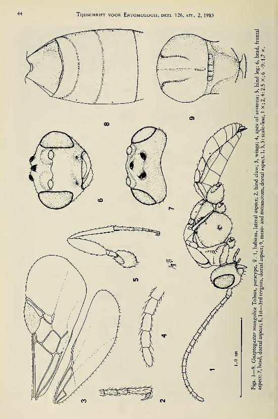
44 Tijdschrift voor Entomologie, deel 126, afl. 2, 1983
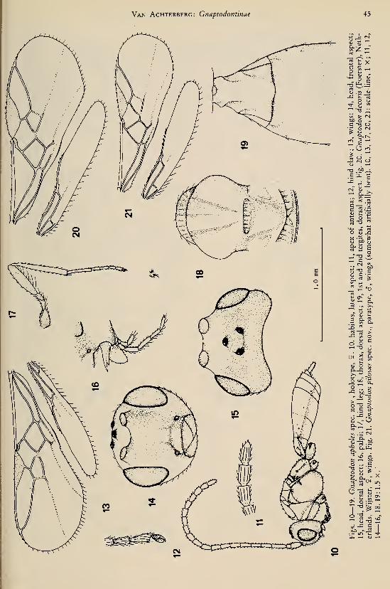
Van Achterberg: Cnaptodontinae 45
g-z -
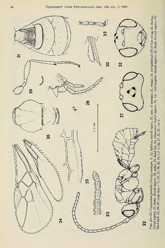
46 Tijdschrift voor Entomologie, deel 126, afl. 2, 1983

Van Achterberg: Cnaptodontinae 47
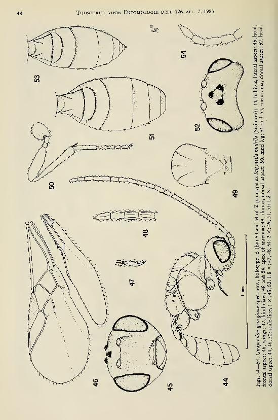
48 Tijdschrift voor Entomologie, deel 126, afl. 2,1983
Br ^
_Q O
o -o
n. ra
Cu X '-
CH- u, r<-i
, . O tn
un O
^ c X
Q. ^>:
^Xo '^
^ 00
k/ "! CM
o oo "^
Oh-^ X
« tì <"K s r*=5 ? .5
-I i*^
p-( >*; -o

Van Achterberg: Gnaptodontinae 49
-a ^
C ^D
O oo-O LO
-a'^'
(U «^
-C "^^ LTl
« CL,O tn
in rt
-^ S«r o3 rt
^1-C „
u+

50 Tijdschrift voor Entomologie, deel 126, afl. 2, 1983
Ë X
OßP
^ vO
u
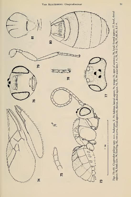
Van Achterberg: Gnaptodontinae 51
"^ X
j: CL,
*-»
IS ^'n! oo
Of _^
O aj
= a
y o
II
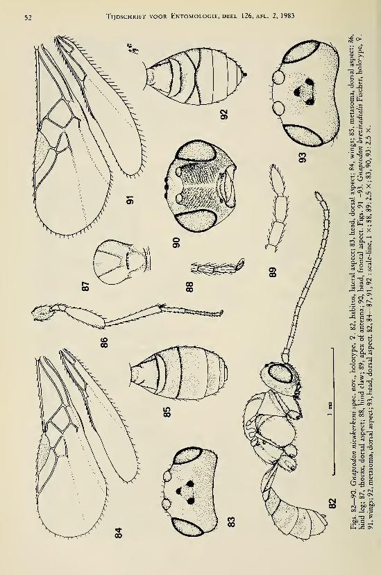
52 Tijdschrift voor Entomologie, deel 126, afl. 2, 1983
:& o+
o Cl,
li ^
O Ö-o -r:
.~ url wSU.O V,C/3 Ir*
"^ 5 X00 £
C K ^.Ü o "^fe -TS <>
1-2 OOO ^ „
a.^ X
rt j, 7J!2 os óso . 00
"^ tÜ
r<^ CL, -
"i-S<U VJ (U
"^ -—'"
tu rt CTN
^ ^ ^- » os
3 Os rC
<uCl,

Van Achterberg: Gnaptodontinae 53
:

54 Tijdschrift voor Entomologie, deel 126, afl. 2, 1983
rt

Van Achterberg: Gnaptodontinae 55
, X
" X-a ^
^ ^
C —c —
(N

56 Tijdschrift voor Entomologie, deel 126, afl. 2, 1983
1 <U 1—
I
r~i vo
os
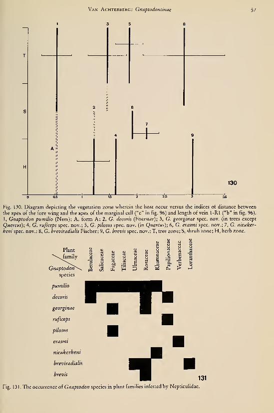
Van Achterberg: Cnaptodontinae
3 5 8
57


•^ DEEL 126'"'' AFLEVERING 3 1983
TIJDSCHRIFTVOOR ENTOMOLOGIE
UITGEGEVEN DOOR
DE NEDERLANDSE ENTOMOLOGISCHE VERENIGING
^Qs^tTo^^
INHOUD
Th. C. M. Brock and G. van der Velde. — An autecological study on
Hydromyza livens (Fabricius) (Diptera, Scatomyzidae), a fly associated with
nymphaeid vegetation dominated by Nuphar, pp. 59—90, figs. 1—25.
Tijdschrift voor Entomologie, deel 126, afl. 3 Gepubliceerd 20-V-1983
AN AUTECOLOGICAL STUDY ON HYDROMYZA LIVENS(FABRICIUS) (DIPTERA, SCATOMYZIDAE), A FLYASSOCIATED WITH NYMPHAEID VEGETATION
DOMINATED BY NUPHAR^)
by
Th. C. M. BROCK and G. VAN DER VELDE
Laboratory ofAquatic Ecology, Catholic University ofNijmegen, The Netherlands
Abstract
The life history and autecology of the fly Hydromyza livens (Fabricius) were investigated
in detail in two Dutch waters. Special attention was paid to its effects on and interrelations
with vegetation dominated by Nuphar lutea (L.) Sm. The food plants, geographical
distribution, aspects of population dynamics and the morphology of H. livens are described
and discussed.
Investigations have been made on general behaviour, feeding behaviour, spatial
occurrence, sexual behaviour, sex ratio, oviposition, mortality and prédation of the adults.
The function of the chorion of the egg is discussed. Some experiments have been carried out
to study the development of the eggs in relation with humidity, oxygen and temperature.
The process of hatching, feeding behaviour, mine patterns, the effect of temperature on
feeding behaviour and the mortality of the larvae are described. Data are given of the
position in the leaf stalks, emergence and hibernation of the pupae.
The life history of H. livens is compared with that of H. conßuens Loew, a species bound
to Nuphar in North America.
Introduction
Nymphaeid vegetations, which are very com-
mon and show an optimal development in TheNetherlands, have been studied by us during
several years. Nymphaeids are aquatic plants
with floating leaves, which root in the bottom
and have flowers above or floating on the water
surface. Because of their structure they have an
important function as a habitat for semi-aquatic
insects.
The species Hydromyza livens (Fabricius,
1794) (syn. Musca livens Fabricius, 1794, and
Nupharia rivularis Robineau-Desvoidy, 1830)
of the dipteran family Scatomyzidae (syn. Cor-
dyluridae, Scatophagidae) is such an insect oc-
curring abundantly on the nymphaeids studied,
especially on Nuphar lutea (L.) Sm. The larvae
of most species belonging to this family live in
dung; only a few genera are known to mine in
leaves or petioles of plants (Sack, 1937).
According to Hering (1926) the larvae of most
Hydromyza species mine the leaves and/or peti-
oles of Liliaceae and Orchidaceae.
') Contribution no. 19 of the Nymphaeid Project.
\
Hydromyza livens, however, can complete its
whole life cycle on Nuphar lutea (Nymphaea-
ceae), one of the nymphaeid species of which
the structure and function is a subject of investi-
gations in our laboratory. The larvae of H.
livens mine the floating leaves of Nuphar. Bytheir consumption of living plant tissue, the lar-
vae are an important link in the grazer food
chain and decomposition processes connected
with Nuphar lutea. Because of our interest in
these interrelations the life history and habits of
H. livens were studied in detail and a review
was made of the literature.
Furthermore the results of our study could be
compared with those of Welch (1914, 1917)
who studied the North American species
H. conßuens Loew, 1863, of which the life his-
tory and habits resemble those of H. livens in
many ways.
Description of the study areas
Most observations have been made in the
Oude Waal near Nijmegen (municipaHty of Ub-
bergen, Province of Gelderland); some addi-
tional observations have been made in the
Haarsteegse Wiel. (municipality of VHjmen,
59
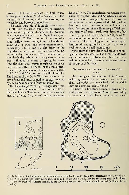
60 Tijdschrift voor Entomologie, deel 126, afl. 3, 1983
Province of Noord-Brabant). In both vi^ater
bodies pure stands of Nuphar lutea occur. The
waters differ, hoM^ever, in their dimensions, wa-
ter quahty and bottom composition.
The Oude "Waal (fig. 1) is an old river branch
cut off from the river Waal, where extensive
nymphaeid vegetation dominated by Nuphar
lutea, Nymphaea alba L. and Nymphoides pel-
tata (Gmel.) O. Kuntze occur. It consists of a
large shallow water body, 1 km in length and
about 250 m wide, and three interconnected
ponds (fig. 1, D, E and F). The depth of the
large shallow water body varies from 0.5 to 1.5
m; in the dry summer of 1976 it became almost
completely dry. About once every two years the
area is flooded in winter or spring by water
from the river Waal; summer high waters occur
only occasionally. The depth of the three inter-
connected ponds increases towards their centres
to 2.5, 5.5 and 5.5 m, respectively (D, E and F).
The bottom of the Oude Waal consists of a pat-
tern of clay and sand, sometimes mixed, covered
by a sapropelium layer of varying thickness.
The Haarsteegse Wiel (fig. 1) originated from
two, but not simultaneous, bursts in the dike of
the river Meuse. This water body has a surface
area of 17.9 ha (44.2 acres) and a maximum
depth of 17 m. The nymphaeid vegetation dom-inated by Nuphar lutea and Nymphaea candida
Presi, is almost completely restricted to the
southern and western parts of the lake, where
they are sheltered against wave- and wind ac-
tion. The bottom of the Haarsteegse Wiel con-
sists mainly of sand (wash-over deposits), but
where nymphaeids grow there is a layer of sa-
propelium, becoming thicker towards the litto-
ral border. The hydrology of the lake is depen-
dent on rain and ground water only; the water-
level shows only small fluctuations.
Apart from the two described areas of inves-
tigation several waters in The Netherlands with
vegetation dominated by Nuphar have been vis-
ited and checked for floating leaves with mines
of the larvae of H. livens.
Geographical distribution and foodPLANTS
The ecological distribution of H. livens is
mainly governed by its affinity for the food
plants on which the larvae feed and on which
they can complete their life-cycle.
In table 1 a literature review is given of the
food plants of the larvae of H. livens. According
to most authors the larvae mine in the leaves
50 100 150 200m
Fig. 1. Left side: the location of the areas studied in The Netherlands (open dot: Haarsteegse Wiel, closed dot:
Oude Waal). Right side: bathymétrie map of pond F of the Oude Waal, showing the nymphaeid beds (dotted
area), the situation of transects studied in the Nuphar zone and the isolated Nymphaea bed (indicated by an
asterisk).
Brock & van der Velde: Hydromyza livens 61
Table 1 . Review of the food plants of H. livens as mentioned in the literature.
Taxon Author
NymphaeaceaeNymphaeaNymphaea alba L.
NupharNuphar lutea (L.) Sm.
Nupharpumila (Timm) DC.Nuphar X intermedia Ledeb.
Caltha palustris L.
Collin, 1958
Wesenberg-Lund, 1943
DeMeyere, 1940
Hackman, 1956; Vockeroth, 1978
De Meyere, 1 895 ; Eberle, 1 943;
Gercke, 1877; Brauer, 1883;
Hendel, 1928; Gaevskaya, 1966;
Grünberg, 1910; Hering, 1926;
Heslop-Harrison, 1955;
Monchadskii, 1940; Sack, 1937; Seguy, 1934;
Schütte, 1921; Van der Velde, 1978;
Van der Velde et al., 1978
Heslop-Harrison, 1955
Heslop-Harrison, 1955
Gaevskaya, 1966; Monchadskii, 1940
and petioles of Nuphar lutea. This is confirmed
in the present study; in all investigated Dutchwaters with Nuphar stands floating leaves with
mines of H. livens larvae were found. Heslop-
Harrison (1955) mentioned H. livens larvae also
from the leaves and petioles of Nuphar pumila
(Timm) DC. and Nuphar X intermedia Ledeb.
(= N. lutea X N. pumila). They were also
found by us in floating leaves of N. X interme-
dia. It can be concluded from these data that all
European Nuphar species are adequate food
plants for H. livens. In the botanical garden of
Haren (State University of Groningen, TheNetherlands) the second author of this paper
observed floating leaves of the North-AmericanNuphar advena Ait. with mines of H. livens.
The occurrence of H. livens in America has not
been reported.
In the literature there is some misunderstand-
ing concerning the plant species on which H.livens can complete its life-cycle. De Meijere
(1940) reported the occurrence of H. livens lar-
vae in floating leaves of Nymphaea alba; in his
paper, however, a drawing of a Nuphar leaf
with mines of H. livens is given. In Wesenberg-Lund's "Biologie der Süsswasserinsecten"
(1943) an obscure picture of Hydromyza mines
in a so-called Nymphaea leaf is shown. Eberle
(1943), however, who transplanted Hydromyzalarvae from Nuphar leaves to Nymphaea leaves,
and also to other Nuphar leaves, concluded that
the transplanted larvae fail to develop in floating
leaves of Nymphaea. We confirmed this in the
laboratory for transplanted larvae in leaves of
Nymphaea alba and Nymphaea candida. Wenever observed Hydromyza larvae in Nym-phaea leaves.
According to Gaevskaya (1966), Monchadskii
(1940) reported the occurrence of Hydromyzamines in leaves of Caltha palustris L.; this fact,
however, is neither confirmed by other litera-
ture sources nor observed by the authors of the
present paper.
Therefore, it can be concluded that in EuropeH. livens is strictly bound to Nuphar species.
In the Limnofauna Europaea, Vockeroth
(1978) reported several distribution areas for
H. livens (see fig. 2), viz.:
(A) Areas with real records: the western
secondary mountain chain (8), the central
secondary mountain chain (9), the western low-
lands (13), the central lowlands (14), Ireland
(17) (Coon River near Partry, North of LoughMask, 3 July 1982 on Nuphar lutea (L.) Sm. (G.
van der Velde), the United Kingdom (18),
Northern Sweden (22), Taiga (23), Uzbekistan,
Siberia;
(B) Areas in which H. livens is expected to
occur: the Alps (4), the Carpathians (10), the
Hungarian lowlands (11), the Baltic Province
(15), the eastern lowlands (16);
(C) An area in which H. livens does absolu-
tely not occur: Iceland (19).
The distribution of H. livens does not extend
beyond the confined distribution of the genus
Nuphar in Europe. In fig. 2 both the geograph-
ical distribution of Nuphar (according to Meu-sel et al., 1965) and H. livens (according to
Vockeroth, 1978) are compared. Both distribu-
tion areas coincide nearly perfectly. When con-
sidering fig. 2, the statement in the Limnofauna
Europaea that H. livens does not occur in Ice-
land (19) is quite obvious, because Nuphar does
not occur there either. On account of the geo-
i-

62 Tijdschrift voor Entomologie, deel 126, afl. 3, 1983
Fig. 2. Map of the European distribution of both the genus Nuphar (dotted areas) (modified after Meusel et al.,
1965) and H. livens (areas with numbers according to Limnofauna Europaea).
graphical distribution of Nuphar, H. livens
might also be expected to occur in other regions
such as Italy, the Balkans and Norway.
Development in time
In order to obtain an idea of the population
dynamics of H. livens the occurrence in time of
adults and larvae was quantified in various
ways. Both the larval and adult stage can be eas-
ily recognized in the field. Adult H. livens can
be found on the upper side of the nymphaeidfloating leaves; the flies are not adapted to walk-
ing over the water surface, they have to fly fromleaf to leaf. To quantify adult H. livens in time
the insect-lime method was used (adhesive or
sticky trap).
The mine tracks of the larvae of H. livens in
the floating Nuphar leaves can be easily recog-
nized and counted. To quantify the larvae andthe number of affected floating leaves in time,
the leaf-marking method was used.
The quantitative data of adults and larvae are
dealt with together in order to maintain to over-
all picture of the various generations of H.livens per year.
The insect-lime method. — Each week, from
June until October, 1977, six undamaged float-
ing leaves (five of Nuphar lutea and one of
Fig. 3. Adhesive trap (for explanation, see text).

Brock & van der Velde: Hydromyza livens 63
Nymphaea alba) of the same size were gathered
in the Oude Waal, pond F, and covered with in-
sect-Hme (Tangletrap or Stikem), which has no
odour and consequently does not attract or re-
pel the insects. These leaves, of which the peti-
oles were removed, were attached onto six other
floating leaves and then protected from préda-
tion by birds by wire netting (with meshes of 3
cm) which was supported by cork floaters
(fig. 3). The wire netting with cork floaters was
attached with a string to the petiole of the sup-
porting leaf. After 24 hours these leaves were
taken to the laboratory in plastic bags, so that
the catch remained as undamaged as possible.
The fauna was washed from the floating leaves
with refined petrol and then fixed in 70% alco-
hol.
The leaves were laid out in transects from the
littoral border vegetation to the open water, so
that the distribution of the animals over the en-
tire nymphaeid zone could be studied (see sec-
tion on spatial occurrence on the nymphaeids,
further below).
Leaf-marking method. — During two years
(1976 and 1977) floating leaves and flowers of
Nuphar lutea have been marked with 12 mmRotex-tape on which a number was printed
with a Universal 12 printer. One end of the tape
was fixed around the petiole by means of a sta-
ple and the other end was drawn through the
leaf-sinus and then bent over the leaf surface
Hke a flag so that it could be easily read (fig. 4).
One plant forms too many leaves to follow
the development in detail so that plots of one
square metre were made with frames of PVCtubes. The perforated tubing was sunk into the
water and held at approximately 15 cm depth by
using a string-corner attachment to cork float-
Fig. 4. A Nuphar leaf, marked with rotex tape (leaf-
marking method).
ers. Each frame was anchored (Van der Velde,
1980). Observations and marking of newly un-
rolled leaves took place every week during the
season. The marking of floating leaves made it
possible to study the numbers of mines and the
numbers of affected leaves per plot per time.
Temperatures of air and water were read regu-
larly.
Results. — In fig. 5 the leaf-marking observa-
tions for 1976 in the Oude Waal are presented;
the summer of that year was characterized by a
longlasting drought resulting in the emergence
of parts of the Nuphar vegetation.
The numbers of mines, the numbers of af-
fected leaves, and their percentages of the total
numbers of floating leaves per plot, show the
same tendency (fig. 5). The number of mines
and affected floating leaves of Nuphar per plot
remained relatively low in the months of May,
June and July, while at the end of August and in
the beginning of September the numbers of lar-
val H. livens clearly reached their maximum.Also the quantity of mines per floating leaf was
at its maximum in this period (fig. 5).
Figs. 6 and 7 present the data of 1977 in the
Oude Waal (marking-method and insect-lime
method) and the Haarsteegse Wiel (marking-
method). The summer of 1977 was quite normal
without droughts. At the end of May and the
beginning of June small numbers of H. livens
larvae occurred in both waters. The quantity of
affected leaves reached a maximum in the
Haarsteegse Wiel from the last week of July un-
til the second half of September. The larval pop-
ulation of H. livens in the Oude Waal was also
large in this period with a small peak at the end
of July and an absolute maximum in the last
week of August.
The data concerning the adult stage of H.
livens, obtained with the insect-lime method,
are summarized in table 2 and fig. 6. Among the
total catch of approximately 23,158 specimens
(of which 99.4 Hexapoda and 0.6% Arachnida)
1,055 specimens were adult H. livens, i.e.
4.56% of the total catch (see for some other re-
sults Van der Velde (1978), Van der Velde &Brock (1980) and Van der Velde (1980)). The
highest numbers of adult H. livens were caught
from 18 August until 8 September.
Discussion. — With respect to the insect-lime
method one should keep in mind that the result
is not only dependent on the size of the H. li-
vens population, but also on changes in the ac-
tivity pattern of adult H. livens as, for example,
caused by weather conditions. This is also valid
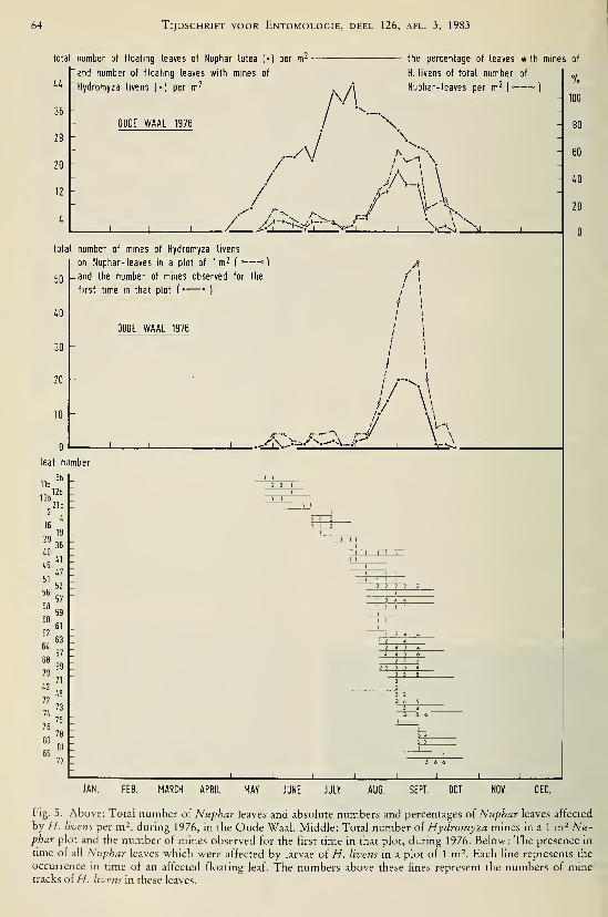
64 Tijdschrift voor Entomologie, deel 126, afl. 3, 1983
total number of floating leaves of Nuphar lutea !•) per m^
and number of floatmg leaves with mines of
Hydromyza livens (4 )per m^
36
28
20
12
I* h
i\/\
the percentage of leaves
H. livens of total number
Nuphar-leaves per m^ (-
OUOE WAAL 1976
..w /^vl \
„r--//p'J/\-.
Ux.total number of mines of Hydromyza livens
on Nuphar-leaves m a plot of 1 m^ (^—
°
-and the number of mines observed for the
first time in that plot (•— • )
50
40 h
30
20 -
10-
OUOE WAAL 1976
/ \
leaf number
12b
I. :
2 2 2 2 2
with mines of
100
80
60
40
H 20
JAN. FEB. MARCH APRIL MAY JUNE JULY AUG. SEPT. OCT. NOV. OEC.
Fig. 5. Above: Total number of Nuphar leaves and absolute numbers and percentages of Nuphar leaves affected
by H. livens per m^, during 1976, in the Oude Waal. Middle: Total number oi Hydromyza mines in a 1 m- Nu-phar plot and the number of mines observed for the first time in that plot, during 1976. Below: The presence in
time of all Nuphar leaves which were affected by larvae of H. livens in a plot of 1 m^. Each line represents the
occurrence in time of an affected floating leaf. The numbers above these lines represent the numbers of minetracks of//, livens in these leaves.

Brock & van der Velde: Hydromyza livens 65
Table 2. Number of adult H. livens caught with insect-lime on floating leaves from 16 June until 29 September,
1977. For the localities of the different leaves see fig. 1 and the text.
Date 1977

66 Tijdschrift voor Entomologie, deel 126, afl. 3, 1983
number ot floating leaves I«) and open flowers |o| per m^
/.O
36
32
28
lu
20
16
12
water temperature
air temperature
OüDE WAAL
8.»
\•
. ,7 y^^ \ ^^^^ X
\
\/"•V
\^ .//
Nuphar lutea
OÜOE WAAL
/ \
/ ^J F M
1977
number of leaves with mines —20
J J
18
16
U -
12 -
10 -
8
6
i.
2
Hydromyza livens
OUDE WAAL 1977
li IL
N D
1977
number of specimens
240
220 -
200
180
160 |-
UO
120
100
80
60
to
20
Hydromyza livens Adults
OUDE WAAL 1977
lilli
Fig. 6. Course of water and air temperature, presence of floating leaves and flowers of Nuphar lutea in a plot of
1 m^, numbers of floating leaves affected by Hydromyza larvae in that plot and numbers of adult H. livens
caught with insect-lime on the floating leaves during 1977 on the Oude Waal.

Brock & van der Velde: Hydromyza livens 67
water temperature
HAARSTEEGSEWIEL
//
V.
\\
''
'
I
number ot floating leaves I») and open flowers |o) per m^
/.O -
36 - Nuphar lutea
32 - HAARSTEEGSE WIEL
28
24
20 -
16 -
12 -
/
/^-/
r X1977
number of leaves with mines
-
2/.
22 I-
20
18 |-
IB
U12
10
8
B
i
2
Hydromyza livens
HAARSTEEGSE WIEL 1977
J-l III. .1 Il . I
Fig. 7. Course of water temperature, presence of floating leaves and flowers of Nuphar lutea in a plot of 1 m^
and numbers of floating leaves affected by H. livens in that plot during 1977 on the Haarsteegse Wiel.
generation is ca. 8 times larger than the first
one.
In the Haarsteegse Wiel in 1977 both the sec-
ond and third larval generations are ca. 7 times
larger than the first one.
The first, and under favourable conditions al-
so the second, generation complete their life-cy-
cle within the growing season of Nuphar while
the pupae of the third (under bad environmental
circumstances probably also those of the sec-
ond) generation hibernate and form the parents
of the first generation, the next year.
That there are more generations per year is
also supported by differences in behaviour and
morphological characteristics of the larvae and
pupae of the various generations, as will be dis-
cussed later. Schütte (1921) reported that sum-
mer and winter pupae are both present m the
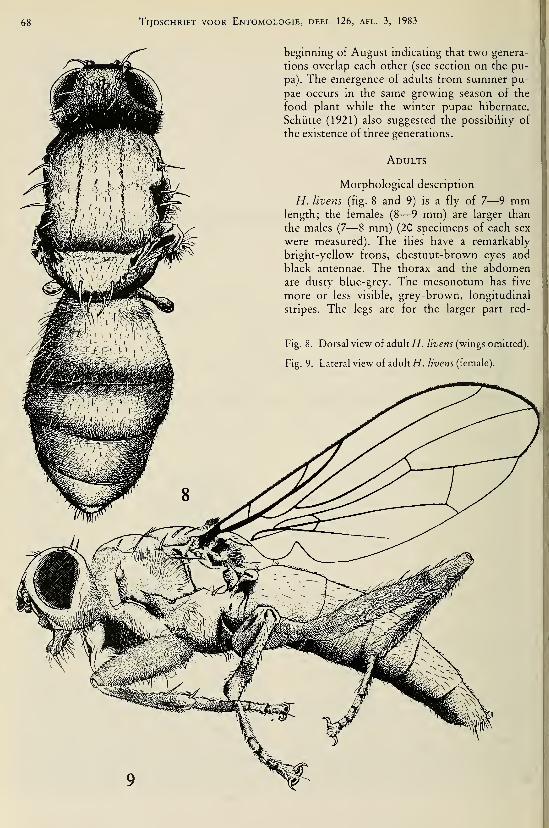
Tijdschrift voor Entomologie, deel 126, afl. 3, 1983
beginning of August indicating that two genera-
tions overlap each other (see section on the pu-
pa). The emergence of adults from summer pu-
pae occurs in the same growing season of the
food plant while the winter pupae hibernate.
Schütte (1921) also suggested the possibility of
the existence of three generations.
Adults
Morphological description
H. livens (fig. 8 and 9) is a fly of 7—9 mmlength; the females (8—9 mm) are larger than
the males (7—8 mm) (20 specimens of each sex
were measured). The flies have a remarkably
bright-yellow frons, chestnut-brown eyes and
black antennae. The thorax and the abdomenare dusty blue-grey. The mesonotum has five
more or less visible, grey-brown, longitudinal
stripes. The legs are for the larger part red-
Fig. 8. Dorsal view of adult H. livens (wings omitted).
Fig. 9. Lateral view of adult H. livens (female).
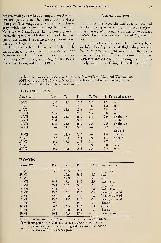
Brock & van der Velde: Hydromyza livens 69
brown, with yellow-brown ginglymae; the fem-
ora are partly blackish, tinged with a dusty
blue-grey. The wings are of a translucent dusty-
grey while the veins are slightly brownish.
Veins R 4 -I- 5 and M are sHghtly convergent to-
wards the apex; vein lA does not reach the mar-
gin of the wing. The relatively very short bris-
tles on the body and the legs and the three very
small proclimate frontal bristles and the single
sternopleural bristle are characteristic for
Hydromyza. For further descriptions, see
Grünberg (1910), Séguy (1934), Sack (1937),
Hackman (1956), and Collin (1958).
General behaviour
In the areas studied the flies usually occurred
on the floating leaves of the nymphaeids Nym-phaea alba, Nymphaea candida, Nymphoidespettata, but preferably on those of Nuphar lu-
tea.
In spite of the fact that these insects have
well-developed powers of flight they are not
found at any great distance from the nym-phaeids. They are difficult to capture and moverestlessly around over the floating leaves, alter-
nately walking or flying. They fly only short
Table 3. Temperature measurements in °C with a Wallacoy Universal Thermometer
(GST 32; probes Ni-lOlx and Ni-106) in the flowers and on the floating leaves of
Nuphar lutea and of the ambient water and air.
FLOATING LEAVES
Date (1977)
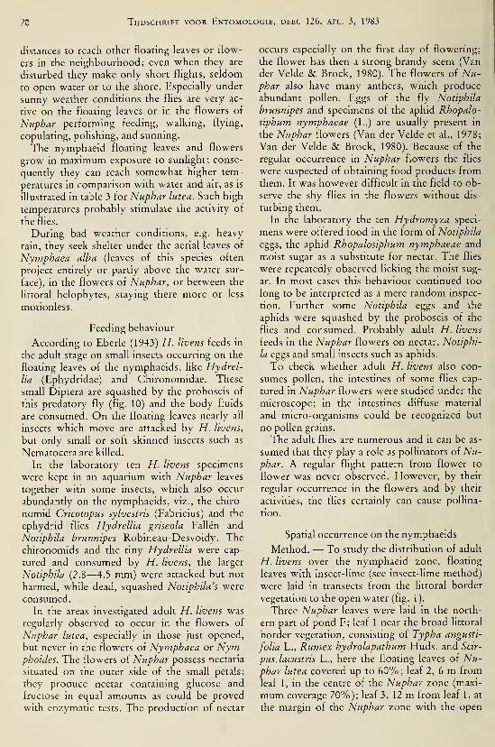
70 Tijdschrift voor Entomologie, deel 126, afl. 3, 1983
distances to reach other floating leaves or flow-
ers in the neighbourhood; even when they are
disturbed they make only short flights, seldom
to open water or to the shore. Especially under
sunny weather conditions the flies are very ac-
tive on the floating leaves or in the flowers of
Nuphar performing feeding, walking, flying,
copulating, polishing, and sunning.
The nymphaeid floating leaves and flowers
grow in maximum exposure to sunlight; conse-
quently they can reach somewhat higher tem-
peratures in comparison with water and air, as is
illustrated in table 3 for Nuphar lutea. Such high
temperatures probably stimulate the activity of
the flies.
During bad weather conditions, e.g. heavy
rain, they seek shelter under the aerial leaves of
Nymphaea alba (leaves of this species often
project entirely or partly above the water sur-
face), in the flowers of Nuphar, or between the
littoral helophytes, staying there more or less
motionless.
Feeding behaviour
According to Eberle (1943) H. livens feeds in
the adult stage on small insects occurring on the
floating leaves of the nymphaeids, like Hydrel-
lia (Ephydridae) and Chironomidae. These
small Diptera are squashed by the proboscis of
this predatory fly (fig. 10) and the body fluids
are consumed. On the floating leaves nearly all
insects which move are attacked by H. livens,
but only small or soft skinned insects such as
Nematocera are killed.
In the laboratory ten H. livens specimens
were kept in an aquarium with Nuphar leaves
together with some insects, which also occur
abundantly on the nymphaeids, viz., the chiro-
nomid Cricotopus sylvestris (Fabricius) and the
ephydrid flies HydreIlia gnseola Fallen and
Notiphila brunnipes Robineau-Desvoidy. The
chironomids and the tiny Hydrellia were cap-
tured and consumed by H. livens, the larger
Notiphila (2.8—4.5 mm) were attacked but not
harmed, while dead, squashed Notiphila's were
consumed.
In the areas investigated adult H. livens was
regularly observed to occur in the flowers of
Nuphar lutea, especially in those just opened,
but never in the flowers of Nymphaea or Nym-phoides. The flowers of Nuphar possess nectaria
situated on the outer side of the small petals;
they produce nectar containing glucose and
fructose in equal amounts as could be proved
with enzymatic tests. The production of nectar
occurs especially on the first day of flowering;
the flower has then a strong brandy scent (Van
der Velde & Brock, 1980). The flowers of Nu-phar also have many anthers, which produce
abundant pollen. Eggs of the fly Notiphila
brunnipes and specimens of the aphid Rhopalo-
siphum nymphaeae (L.) are usually present in
the Nuphar flowers (Van der Velde et al., 1978;
Van der Velde & Brock, 1980). Because of the
regular occurrence in Nuphar flowers the flies
were suspected of obtaining food products from
them. It was however difficult in the field to ob-
serve the shy flies in the flowers without dis-
turbing them.
In the laboratory the ten Hydromyza speci-
mens were offered food in the form of Notiphila
eggs, the aphid Rhopalosiphum nymphaeae and
moist sugar as a substitute for nectar. The flies
were repeatedly observed licking the moist sug-
ar. In most cases this behaviour continued too
long to be interpreted as a mere random inspec-
tion. Further some Notiphila eggs and the
aphids were squashed by the proboscis of the
flies and consumed. Probably adult H. livens
feeds in the Nuphar flowers on nectar, Notiphi-
la eggs and small insects such as aphids.
To check whether adult H. livens also con-
sumes pollen, the intestines of some flies cap-
tured in Nuphar flowers were studied under the
microscope; in the intestines diffuse material
and micro-organisms could be recognized but
no pollen grains.
The adult flies are numerous and it can be as-
sumed that they play a role as pollinators of Nu-phar. A regular flight pattern from flower to
flower was never observed. However, by their
regular occurrence in the flowers and by their
activities, the flies certainly can cause pollina-
tion.
Spatial occurrence on the nymphaeids
Method. — To study the distribution of adult
H. livens over the nymphaeid zone, floating
leaves with insect-lime (see insect-lime method)
were laid in transects from the littoral border
vegetation to the open water (fig. 1).
Three Nuphar leaves were laid in the north-
ern part of pond F; leaf 1 near the broad littoral
border vegetation, consisting of Typha angusti-
folia L., Rumex hydrolapathum Huds. and Scir-
pus.lacustris L., here the floating leaves of Nu-phar lutea covered up to 60%; leaf 2, 6 m from
leaf 1, in the centre of the Nuphar zone (maxi-
mum coverage 70%); leaf 3, 12 m from leaf 1, at
the margin of the Nuphar zone with the open
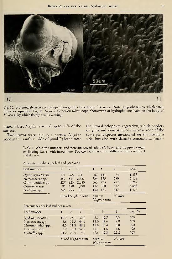
Brock & van der Velde: Hydromyza livens 71
10 11
Fig. 10. Scanning electron microscope photograph of the head of//, livens. Note the proboscis by which small
preys are squashed. Fig. 11. Scanning electron microscope photograph of hydrophobous hairs on the body of
//. livens by which the fly avoids wettmg.
water, where Nuphar covered up to 60% of the
surface.
Two leaves were laid in a narrow Nuphar
zone at the southern side of pond F; leaf 4 near
the littoral helophyte vegetation, which borders
on grassland, consisting of a narrow zone of the
same plant species mentioned for the northern
side, but also with Mentha aquatica L. (maxi-
Table 4. Absolute numbers and percentages, of adult H. livens and its preys caught
on floating leaves with insect-lime. For the localities of the different leaves see fig. 1
and the text.
Absolute numbers per leaf and per taxon
Leaf number total
Hydromyza livens
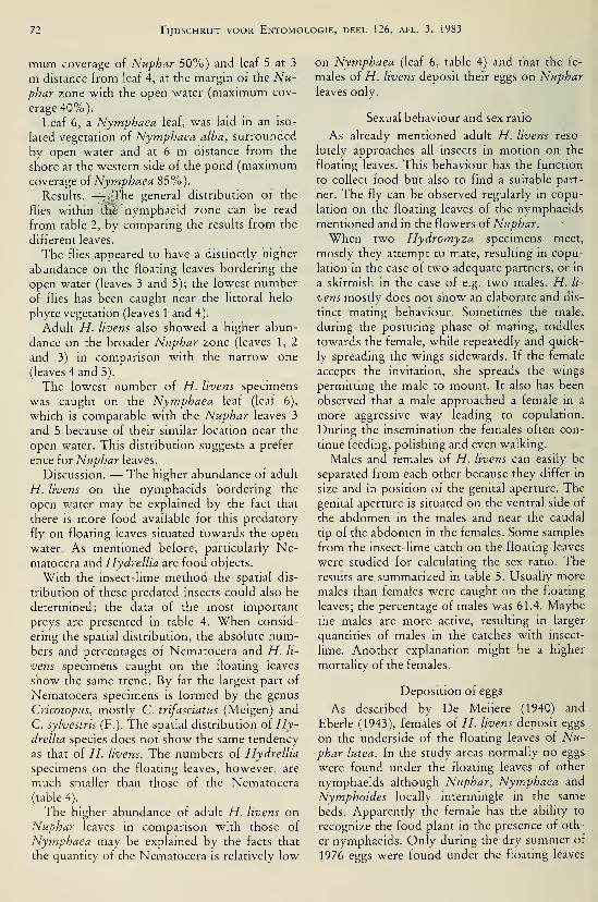
72 Tijdschrift voor Entomologie, deel 126, afl. 3, 1983
mum coverage of Nuphar 50%) and leaf 5 at 3
m distance from leaf 4, at the margin of the Nu-phar zone with the open water (maximum cov-
erage 40%).
Leaf 6, a Nymphaea leaf, was laid in an iso-
lated vegetation of Nymphaea alba, surrounded
by open water and at 6 m distance from the
shore at the western side of the pond (maximumcoverage oi Nymphaea 85%).
Results. — The general distribution of the
flies within thè nymphaeid zone can be read
from table 2, by comparing the results from the
different leaves.
The fUes appeared to have a distinctly higher
abundance on the floating leaves bordering the
open water (leaves 3 and 5); the lowest number
of flies has been caught near the littoral helo-
phyte vegetation (leaves 1 and 4).
Adult H. livens also showed a higher abun-
dance on the broader Nuphar zone (leaves 1, 2
and 3) in comparison with the narrow one
(leaves 4 and 5).
The lowest number of H. livens specimens
was caught on the Nymphaea leaf (leaf 6),
which' is comparable with the Nuphar leaves 3
and 5 because of their similar location near the
open water. This distribution suggests a prefer-
ence for Nuphar leaves.
Discussion. — The higher abundance of adult
H. livens on the nymphaeids bordering the
open water may be explained by the fact that
there is more food available for this predatory
fly on floating leaves situated towards the open
water. As mentioned before, particularly Ne-matocera and Hydrellia are food objects.
With the insect-lime method the spatial dis-
tribution of these predated insects could also be
determined; the data of the most important
preys are presented in table 4. When consid-
ering the spatial distribution, the absolute num-bers and percentages of Nematocera and H. li-
vens specimens caught on the floating leaves
show the same trend. By far the largest part of
Nematocera specimens is formed by the genus
Cricotopus, mostly C. trifasciatus (Meigen) and
C. sylvestris (F.). The spatial distribution oi Hy-drellia species does not show the same tendency
as that of H. livens. The numbers of Hydrellia
specimens on the floating leaves, however, are
much smaller than those of the Nematocera
(table 4).
The higher abundance of adult H. livens onNuphar leaves in comparison with those of
Nymphaea may be explained by the facts that
the quantity of the Nematocera is relatively low
on Nymphaea (leaf 6, table 4) and that the fe-
males of H. livens deposit their eggs on Nupharleaves only.
Sexual behaviour and sex ratio
As already mentioned adult H. livens reso-
lutely approaches all insects in motion on the
floating leaves. This behaviour has the function
to collect food but also to find a suitable part-
ner. The fly can be observed regularly in copu-
lation on the floating leaves of the nymphaeids
mentioned and in the flowers of Nuphar.
When two Hydromyza specimens meet,
mostly they attempt to mate, resulting in copu-
lation in the case of two adequate partners, or in
a skirmish in the case of e.g. two males. H. li-
vens mostly does not show an elaborate and dis-
tinct mating behaviour. Sometimes the male,
during the posturing phase of mating, toddles
towards the female, while repeatedly and quick-
ly spreading the wings sidewards. If the female
accepts the invitation, she spreads the wings
permitting the male to mount. It also has been
observed that a male approached a female in a
more aggressive way leading to copulation.
During the insemination the females often con-
tinue feeding, polishing and even walking.
Males and females of H. livens can easily be
separated from each other because they differ in
size and in position of the genital aperture. The
genital aperture is situated on the ventral side of
the abdomen in the males and near the caudal
tip of the abdomen in the females. Some samples
from the insect-lime catch on the floating leaves
were studied for calculating the sex ratio. The
results are summarized in table 5. Usually more
males than females were caught on the floating
leaves; the percentage of males was 61.4. Maybethe males are more active, resulting in larger
quantities of males in the catches with insect-
lime. Another explanation might be a higher
mortality of the females.
Deposition of eggs
As described by De Meijere (1940) and
Eberle (1943), females of H. livens deposit eggs
on the underside of the floating leaves of A'^;^-
phar lutea. In the study areas normally no eggs
were found under the floating leaves of other
nymphaeids although Nuphar, Nymphaea and
Nymphoides locally intermingle in the same
beds. Apparently the female has the ability to
recognize the food plant in the presence of oth-
er nymphaeids. Only during the dry summer of
1976 eggs were found under the floating leaves
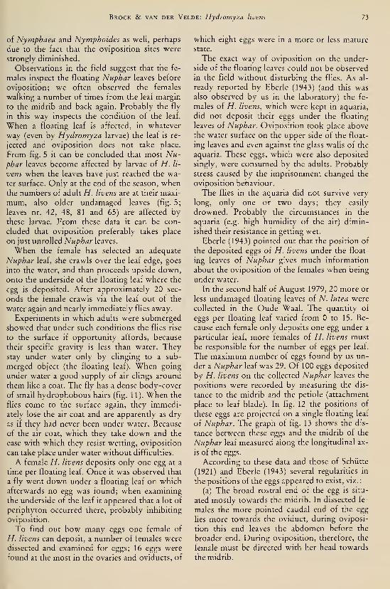
Brock & van der Velde: Hydromyza livens 73
of Nymphaea and Nymphoides as well, perhaps
due to the fact that the oviposition sites were
strongly diminished.
Observations in the field suggest that the fe-
males inspect the floating Nuphar leaves before
oviposition; we often observed the females
walking a number of times from the leaf margin
to the midrib and back again. Probably the fly
in this way inspects the condition of the leaf.
When a floating leaf is affected, in whatever
way (even by Hydromyza larvae) the leaf is re-
jected and oviposition does not take place.
From fig. 5 it can be concluded that most Nu-phar leaves become affected by larvae of H. li-
vens when the leaves have just reached the wa-
ter surface. Only at the end of the season, whenthe numbers of adult H. livens are at their maxi-
mum, also older undamaged leaves (fig. 5;
leaves nr. 42, 48, 81 and 65) are affected bythese larvae. From these data it can be con-
cluded that oviposition preferably takes place
on just unrolled Nuphar leaves.
When the female has selected an adequate
Nuphar leaf, she crawls over the leaf edge, goes
into the water, and than proceeds upside down,onto the underside of the floating leaf where the
egg is deposited. After approximately 20 sec-
onds the female crawls via the leaf out of the
water again and nearly immediately flies away.
Experiments in which adults were submerged
showed that under such conditions the flies rise
to the surface if opportunity affords, because
their specific gravity is less than water. Theystay under water only by clinging to a sub-
merged object (the floating leaf). When going
under water a good supply of air clings around
them like a coat. The fly has a dense body-cover
of small hydrophobous hairs (fig. 11). When the
flies come to the surface again, they immedi-
ately lose the air coat and are apparently as dry
as if they had never been under water. Because
of the air coat, which they take down and the
ease with which they resist wetting, oviposition
can take place under water without difficulties.
A female H. livens deposits only one egg at a
time per floating leaf. Once it was observed that
a fly went down under a floating leaf on whichafterwards no egg was found; when examining
the underside of the leaf it appeared that a lot of
periphyton occurred there, probably inhibiting
oviposition.
To find out how many eggs one female of
H. livens can deposit, a number of females were
dissected and examined for eggs; 16 eggs were
found at the most in the ovaries and oviducts, of
which eight eggs were in a more or less mature
state.
The exact way of oviposition on the under-
side of the floating leaves could not be observed
in the field without disturbing the flies. As al-
ready reported by Eberle (1943) (and this wasalso observed by us in the laboratory) the fe-
males of H. livens, which were kept in aquaria,
did not deposit their eggs under the floating
leaves of Nuphar. Oviposition took place above
the water surface on the upper side of the float-
ing leaves and even against the glass walls of the
aquaria. These eggs, which were also deposited
singly, were consumed by the adults. Probably
stress caused by the imprisonment changed the
oviposition behaviour.
The flies in the aquaria did not survive very
long, only one or two days; they easily
drowned. Probably the circumstances in the
aquaria (e.g. high humidity of the air) dimin-
ished their resistance in getting wet.
Eberle (1943) pointed out that the position of
the deposited eggs of H. livens under the float-
ing leaves of Nuphar gives much information
about the oviposition of the females when being
under water.
In the second half of August 1979, 20 more or
less undamaged floating leaves of N. lutea were
collected in the Oude Waal. The quantity of
eggs per floating leaf varied from to 15. Be-
cause each female only deposits one egg under a
particular leaf, more females of H. livens must
be responsible for the number of eggs per leaf.
The maximum number of eggs found by us un-
der a Nuphar leaf was 29. Of 100 eggs deposited
by H. livens on the collected Nuphar leaves the
positions were recorded by measuring the dis-
tance to the midrib and the petiole (attachment
place to leaf blade). In fig. 12 the positions of
these eggs are projected on a single floating leaf
of Nuphar. The graph of fig. 13 shows the dis-
tance between these eggs and the midrib of the
Nuphar leaf measured along the longitudinal ax-
is of the eggs.
According to these data and those of Schütte
(1921) and Eberle (1943) several regularities in
the positions of the eggs appeared to exist, viz.:
(a) The broad rostral end of the egg is situ-
ated mostly towards the midrib. In dissected fe-
males the more pointed caudal end of the egg
Ues more towards the oviduct, during oviposi-
tion this end leaves the abdomen before the
broader end. During oviposition, therefore, the
female must be directed with her head towards
the midrib.
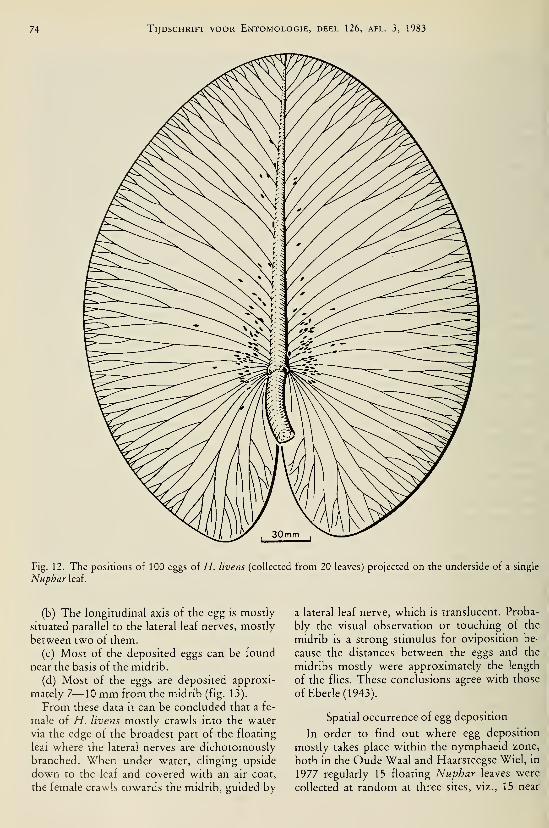
74 Tijdschrift voor Entomologie, deel 126, afl. 3, 1983
Fig. 12. The positions of 100 eggs of H. livens (collected from 20 leaves) projected on the underside of a single
Nuphar leaf.
(b) The longitudinal axis of the egg is mostly
situated parallel to the lateral leaf nerves, mostly
between two of them.
(c) Most of the deposited eggs can be found
near the basis of the midrib.
(d) Most of the eggs are deposited approxi-
mately 7—10 mm from the midrib (fig. 13).
From these data it can be concluded that a fe-
male of H. livens mostly crawls into the water
via the edge of the broadest part of the floating
leaf where the lateral nerves are dichotomously
branched. When under water, clinging upside
down to the leaf and covered with an air coat,
the female crawls towards the midrib, guided by
a lateral leaf nerve, which is translucent. Proba-
bly the visual observation or touching of the
midrib is a strong stimulus for oviposition be-
cause the distances between the eggs and the
midribs mostly were approximately the length
of the flies. These conclusions agree with those
of Eberle (1943).
Spatial occurrence of egg deposition
In order to find out where egg deposition
mostly takes place within the nymphaeid zone,
both in the Oude Waal and Haarsteegse Wiel, in
1977 regularly 15 floating Nuphar leaves were
collected at random at three sites, viz., 15 near

Brock & van der Velde: Hydromyza livens 75
the littoral border vegetation, 15 in the middle
of the Nuphar zone and 15 near the open water.
Leaves were collected every month from Mayuntil September in the Oude "Waal and every
two weeks from June until September in the
Haarsteegse Wiel. The eggs and larvae occur-
ring under and in the leaves were counted per
site per date.
In the Oude Waal in total 62 larvae and eggs
were counted on the leaves bordering the litto-
ral helophyte vegetation, 38 on leaves from the
middle of the Nuphar zone and 65 on floating
leaves from the location near the open water.
In the Haarsteegse Wiel, in total 81 larvae and
eggs were found on the leaves near the littoral
border vegetation, 79 on leaves from the middle
of the Nuphar zone and 94 on leaves bordering
the open water.
It can be concluded that oviposition in the
Oude Waal takes place more often under leaves
at the two edges of the Nuphar belt than under
leaves in the middle of the Nuphar zone. Theresults from the Haarsteegse Wiel show this
trend less clearly.
From the data obtained with the insect-lime
method it appeared that the adults were defi-
nitely more abundant on the nymphaeids bor-
dering the open water because of the higher
abundance of food organisms there. The large
number of eggs and larvae under and m floating
leaves of the sites near the open water can be ex-
plained in this way. The high frequency of egg
deposition under floating leaves bordering the
littoral helophytes may be explained by the fact
that wind and wave action are less pronounced
here.
15H
Qi n1 2 3 4 5 6 7 8 9 10 11 12 13 14 15 16 17 18 1920>20mm.
DISTANCE
Fig. 13. The distances of 100 eggs of H. livens to the
midribs of the floating leaves on which they were de-
posited, measured along the longitudinal axis of the
eggs (see also fig. 12).
Mortality and prédation
As may be concluded from fig. 6, the num-bers of adult H. livens decrease at the end of the
growing season of Nuphar. The mortality of
adults may have several reasons, e.g. falling
temperatures, diminishing of the floating leaf
area (figs. 5, 6 and 7) and lack of food (table 2).
Exhaustion of females and risks under water
during oviposition, such as prédation by fish,
might also cause mortality; the sex ratio data
mostly showed a majority of males (table 5). Af-
ter heavy rain the population size of adult H. li-
vens seems to be diminished. Although the flies
have hydrophobous hairs, they easily drownwhen their skins become wet. Probably a long-
lasting contact with water or even very humidconditions (aquaria) diminish their resistance
against getting wet. Prédation on adult H. livens
has never been observed by us. However, Odo-nata, swifts {Apus apus (L.)), swallows (Hirun-
do rustica L., Delichon urhica (L.)), terns {Ster-
na hirundo L., Chlidonias niger (L.)) and the
Green frog (Rana esculenta (L.)) prey on the in-
sects from the floating leaves and thus can occa-
sionally catch H. livens.
The egg
Morphological description
The eggs of H. livens have a length of 1.9
—
2.0 mm and a width of 0.5—0.6 mm. Freshly
laid eggs are yellow-white m colour, a few
hours before hatching they become darker. The
eggs have the appearance of a boat (fig. 14a),
with differently shaped ends: the caudal end is
bluntly pointed, while the broader rostral end is
more rounded and contains the micropyle. Astraight, acute carina extends longitudinally
from end to end and runs medially along the
bottom of a rather deep, broad, longitudinal
fossa (fig. 14b), dividing it into two similar
parts. This divided fossa is laterally bordered by
two other longitudinal carinae, which run al-
most parallel to the median carina, converging
and uniting with the ends of the egg. The eggs'
are always deposited singly under the floating
leaves of Nuphar and are firmly fixed to the
substrate so that it is nearly impossible to sepa-
rate them from a floating leaf without damage
to the chorion or the leaf tissue. Apparently a
small amount of sealing fluid accompanies the
egg at oviposition causing the firm attachment
to the floating leaf.
Eggs have been studied by means of scanning

76 Tijdschrift voor Entomologie, deel 126, afl. 3, 1983
Table 5. Sex distribution and the percentage of males in samples of H. livens caught
with insect-lime.
Date (1977)

•J^ .,^^''''

78 Tijdschrift voor Entomologie, deel 126, afl. 3, 1983
24 "c
TEMP
Fig. 15. Duration of the development of H. livens
eggs into larvae in relation with temperature.
Development in relation to temperature
Eberle (1943) already discussed the fact that
the development of eggs into larvae is depen-
dent on temperature. In spring, when the water
is relatively cold, the duration of the devel-
opment can last out up to two weeks, while in
summer, when the water is warmer, the larvae
hatch after 3 to 4 days after the time of egg de-
position (Eberle, 1943).
It is difficult to study the development of the
eggs in relation to temperature under controlled
circumstances because it is difficult to obtain
enough eggs of the same age which are freshly
deposited by the females of H. livens. As al-
ready discussed one can only sporadically ob-
serve the oviposition in natural habitats, while
the flies in captivity do not deposit their eggs in
the normal way under the floating leaves. In or-
der to obtain eggs which were more or less of
the same age, 25 undamaged, young leaves of
Nuphar, under which no eggs were deposited,
were marked in the Oude Waal at the end of
August 1979. After 24 hours these leaves were
collected and 19 eggs of H. livens were obtained
from them. The differences in age of these eggs
could be 24 hours at most; the temperature of
the Oude Waal water was at that time 18 °C. In
the laboratory the floating leaves with eggs
were kept in aquaria and placed in different cli-
mate chambers which had constant tempera-
tures of 8, 12, 16, 20 and 24 °C. Temperatures
within this range had also been measured in the
Oude Waal (fig. 6). In the climate chamber of
8 °C three eggs were placed, in the other cham-
Fig. 16. Photograph of a H. livens larva sampled
from a leaf blade.
bers four each. By daily observations the devel-
opment of the eggs into larvae was followed (see
fig. 15).
The data thus obtained confirm that the de-
velopment of the eggs is indeed dependent on
temperature and that the development goes
faster at temperatures which are normally mea-
sured in summer in comparison with colder cir-
cumstances in spring and autumn. Below a cer-
tain temperature, between 8 and 12 °C, the eggs
do not develop at all into larvae. The large dif-
ferences in developmental rate of the eggs, espe-
cially in the climate chambers of 12 °C and
16 °C, are probably caused by the way the eggs
were obtained, viz. at a temperature of 18 °C.
The larva
Morphological description
The larva of H. livens (fig. 16) is yellow-
white in colour with the exception of the black
cephalopharynx skeleton (fig. 18), the brown,
kidney-shaped, frontal stigmata and the dark
terminal stigmata (fig. 17).
The anterior stigmata consist of short pro-
cesses, each with several buds; the total numberof buds on one stigma being about 200 (De
Meijere, 1895). According to Schütte (1921)
there is no difference between the number of
stigmatic buds of larvae and pupae of the differ-
ent generations. In the summer larvae (in our
case the first and second generation) the diame-
ters of these buds are smaller, however, than in
the winter larvae (in our case the third genera-
tion). The anterior stigmata are not yet visible
on freshly hatched larvae.
In the translucent body the long, green intes-
tine, the main tracheae and the very well devel-
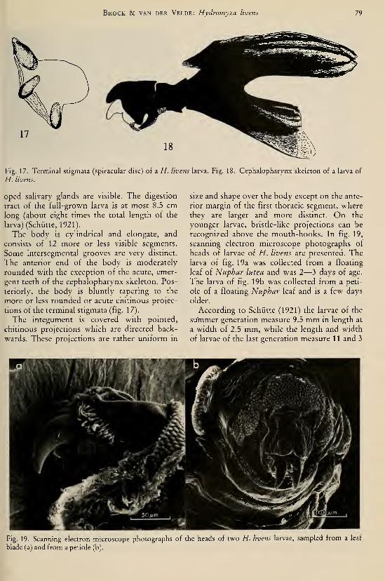
Brock & van der Velde: Hydromyza livens 79
Fig. 17. Terminal stigmata (spiracular disc) of a H. livens larva. Fig. 18. Cephalopharynx skeleton of a larva of
H. livens.
oped salivary glands are visible. The digestion
tract of the full-grown larva is at most 8.5 cmlong (about eight times the total length of the
larva) (Schütte, 1921).
The body is cylindrical and elongate, and
consists of 12 more or less visible segments.
Some intersegmental grooves are very distinct.
The anterior end of the body is moderately
rounded with the exception of the acute, emer-
gent teeth of the cephalopharynx skeleton. Pos-
teriorly, the body is bluntly tapering to the
more or less rounded or acute chitinous projec-
tions of the terminal stigmata (fig. 1 7).
The integument is covered with pointed,
chitinous projections which are directed back-
wards. These projections are rather uniform in
size and shape over the body except on the ante-
rior margin of the first thoracic segment, where
they are larger and more distinct. On the
younger larvae, bristle-like projections can be
recognized above the mouth-hooks. In fig. 19,
scanning electron microscope photographs of
heads of larvae of H. livens are presented. Thelarva of fig. 19a was collected from a floating
leaf of Nuphar lutea and was 2—3 days of age.
The larva of fig. 19b was collected from a peti-
ole of a floating Nuphar leaf and is a few days
older.
According to Schütte (1921) the larvae of the
summer generation measure 9.5 mm in length at
a width of 2.5 mm, while the length and width
of larvae of the last generation measure 11 and 3
Fig. 19. Scanning electron microscope photographs of the heads of two H. livens larvae, sampled from a leaf
blade (a) and from a petiole (b).
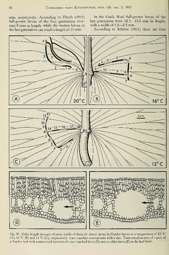
80 Tijdschrift voor Entomologie, deel 126, afl. 3, 1983
mm, respectively. According to Eberle (1943)
full-grown larvae of the first generation mea-
sure 9 mm in length, while the mature larvae of
the last generation can reach a length of 1 1 mm.
In the Oude Waal full-grown larvae of the
last generation were 10.5—13.0 mm in length,
with a width of 1.5—2.5 mm.According to Schütte (1921) there are four
Fig. 20. Daily length increase of mine tracks of three H. livens larvae in Nuphar leaves at a temperature of 20 °C(A), 16 °C (B) and 12 °C (C), respectively. Each number corresponds with a day. Transversal section of a part of
a Nuphar leaf with a mine track (arrow) of a just hatched larva (D) and an older larva (E) in the leaf blade.

Brock & van der Velde: Hydromyza livens 81
^K^ 17' V S-
Fig. 21. Photograph of a part of a Nuphar leaf with
mine tracks of H. livens larvae.
instars; this author also gives an extensive
anatomical description of the larva and its wayof nutrition.
The process of hatching from the egg
Shortly before the time of hatching, the egg
becomes darker in colour, indicating that the
larva within the egg is fully developed. Theplace of emergence of the larva through the egg
shell is very constant. With the aid of relatively
large, acute, chitinous mouthparts the larva
makes an exit through the egg shell on the side
next to the leaf. The position of the exit hole is
determined by the position of the larva in the
egg. So the exit is made near the blunt, broad,
rostral end of the egg, which is mostly directed
towards the midrib of the floating leaf. Via this
hole the larva bores directly through the lower
epidermis into the mesophyll of the floating
Nuphar leaf. After the process of hatching the
empty egg shell remains sticking to the leaf,
closing off the exit, so that water is not able to
enter the mine gallery in the leaf.
General feeding behaviour
In the investigated areas the larvae of H. li-
vens feed on the floating leaf and petiole tissue
of Nuphar lutea. During the early period of
feeding only the spongy parenchyma of the
floating leaf is consumed, while in a later stage,
when the larva has increased in volume, also the
palissade parenchyma of the mesophyll is eaten
(fig. 20dand20e).
While eating through the floating Nuphar leaf
the larva produces a mine gallery in it, and af-
terwards also in the petiole where the larva eats
itself spirally downwards (fig. 21 and fig. 22c).
With the aid of the large acute mouth-hooks
Fig. 22. (A) Just hatched larva of H. livens in its mine
track and the empty egg shell. (B) Larval consumption
by day (white) and by night (black and marked with a)
in a Nuphar leaf. (C) Mine tracks of H. livens in a pet-
iole of a Nuphar leaf. (Modified after Eberle, 1943.)
the larva loosens the plant cells while making
mowing movements with the head. According
to Schiatte (1921) the larvae of H. livens secrete
digestion enzymes which dissolve the plant cells
before they are ingested. Consequently one can
find no recognizable fragments in the intestines
of the larvae.
In the floating leaves and petioles of Nuphar
many sharp, hair-like branched trichosclereids
occur which consist of calcium oxalate. The se-
creted digestion enzymes also dissolve these
sharp trichosclereids; the intestines of the larvae
are not harmed by them in this way. The calci-
um oxalate is transformed into calcium carbon-
ate in the Malpighian organs (Schütte, 1921).
Eberle (1943) already demonstrated that the
larva of H. livens feeds by day and by night, as
is illustrated in fig. 22b. The larval consumption
and consequently the length of the mine is
somewhat larger by day than by night, probably
due to differences in temperature between day
and night.
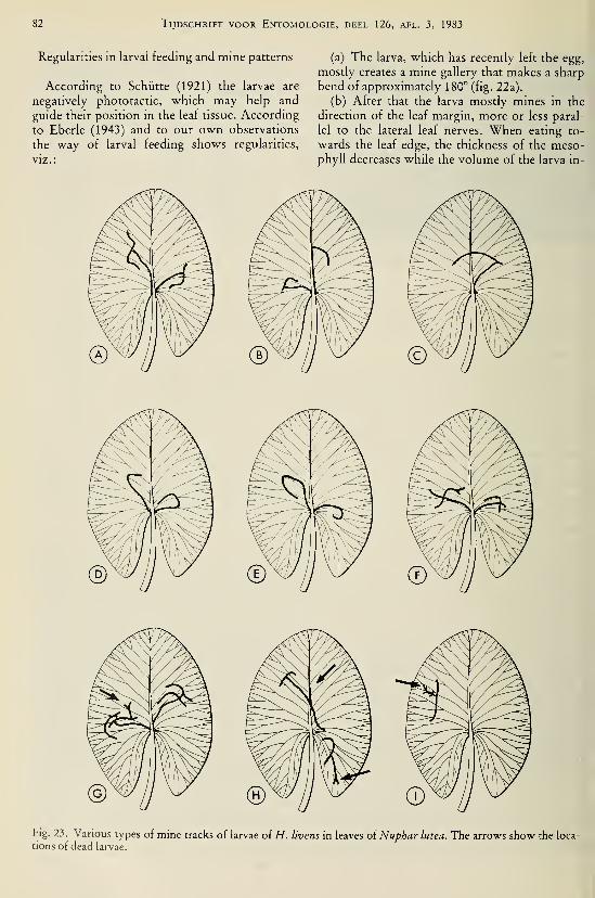
82 Tijdschrift voor Entomologie, deel 126, afl. 3, 1983
Regularities in larval feeding and mine patterns
According to Schütte (1921) the larvae are
negatively phototactic, which may help and
guide their position in the leaf tissue. According
to Eberle (1943) and to our own observations
the way of larval feeding shows regularities,
viz.:
(a) The larva, which has recently left the egg,
mostly creates a mine gallery that makes a sharp
bend of approximately 180° (fig. 22a).
(b) After that the larva mostly mines in the
direction of the leaf margin, more or less paral-
lel to the lateral leaf nerves. When eating to-
wards the leaf edge, the thickness of the meso-phyll decreases while the volume of the larva in-
Fig. 23. Various types of mine tracks of larvae of H. livens in leaves of Nuphar lutea. The arrows show the loca-
tions of dead larvae.

Brock & van der Velde: Hydromyza livens 83
creases; this results in a more or less sharp
gradual bend of the mine gallery parallel to the
leaf margin (fig. 23a—e).
(c) After this the feeding behaviour of the
larva suddenly changes. This change is corre-
lated with the first moulting period of the larva.
After having cast the old skin, the larva mines
more or less at right angles to the previously
created part of the mine gallery in the direction
of the midrib or petiole of the floating leaf
(fig. 23a-e).
(d) On reaching the petiole the larva mines
spirally downwards where ultimately the full
grown larva pupates.
These regularities in larval feeding behaviour
cause regularities in the shape of the mine galle-
ries and consequently one can distinguish cer-
tain mine types, in the leaf blades, viz. :
(1) The T-shaped mine gallery (fig. 23a, b
and c). This type is created by larvae which after
the first moulting period crawl backwards in the
older part of the mine gallery before starting to
eat in the direction of the midrib or petiole.
(2) The arch-like mine gallery (fig. 23d and
e). In this type, the larva does not crawl back-
wards after moulting.
(3) The cross-like or irregularly shaped mine
gallery (fig. 23f, g, h, i). This type of mine gal-
lery is the result of disturbances in the mining
behaviour of the larva, or it is caused by the
damaged state of the leaf blade. Although the
eggs of H. livens are mostly deposited under
young and undamaged floating leaves, these
leaves might be damaged before the eggs have
hatched. This damage on the leaves of Nupharlutea might, for instance, be caused by the feed-
ing by the Coot {Fulica atra L.), the gastropod
Lymnaea stagnalis (L.), the beetles Donacia
crassipes F. and Pyrrhalta nymphaeae (L.), the
caterpillar Nymphula nymphaeata (L.), but
above all by other H. livens larvae. "When the
floating leaf is not damaged too much, the
younger larva has the ability to avoid the older
affected leaf part (fig. 20a).
"When the larval numbers are at their maxi-
mum, there is a great chance that a larva of
H. livens crosses an older mine gallery. In that
case the larva sometimes crosses the mine under
a right angle but mostly the larva makes use of
the older gallery for a while (fig. 23 g). This can
result in creeping into wrong directions of the
larva, and, consequently, in an irregular shape
of the mine gallery. It also happens that a larva
mines in the wrong direction after the first
moulting (fig. 23h, i) also resulting in an irregu-
lar shape of the mine gallery in the leaf blade.
In order to get an idea of the occurrence of
the different types of mine galleries, 34 floating
leaves were collected in the Oude Waal on 11
September 1979. In total, 207 mine galleries
could be recognized in these leaves. Of the min-
es 60.5% could be classified within the T-
shaped type, 30.5% in the arch-like type, and
9.0% in the cross-like or irregular shaped type.
Feeding behaviour and temperature
In the laboratory the larval consumption in
floating leaf blades was followed in different cli-
mate chambers.
In fig. 22b the larva has reached the petiole on
the 4th day. This also happened with a larva in
the laboratory in the climate chamber of 24 °C.
In the climate chamber of 20 °C a larva reached
the petiole on the 6th day, while at constant
temperatures of 16 °C and 12 °C this happened
on the 11th an 18th day after the moment of
hatching from the egg, respectively. The daily
consumption of the three larvae in the climate
chambers of 20 °C, 16 °C and 12 °C was fol-
lowed in detail (fig. 20 and table 6).
Although the consuming activity of only a
few larvae was followed under controlled cir-
Table 6. Daily length increase of mine tracks in mmof three H. livens larvae in leaf blades of Nuphar at
temperatures of 20, 16 and 12 °C, respectively.
Number

84 Tijdschrift voor Entomologie, deel 126, afl. 3, 1983
cumstances one can draw some general conclu-
sions from the data thus obtained. The con-
suming activity is temperature dependent and
apparently the development rate is faster under
warmer conditions during the summer, than in
spring and autumn when colder air and water
temperatures occur.
The daily consumption of the larvae is not
constant at a constant temperature. Generally
the larvae consume more when they become
older, only during the moulting period the lar-
val consumption is very low (fig. 20, table 6).
Also the time needed for moulting seems to
be temperature dependent and consequently
takes a longer time under colder circumstances.
Differences in consumption and feeding
behaviour between the generations
According to Schütte (1921) and Hendel
(1928) the length of the mine galleries in the
petioles of the summer and winter pupae is ca.
10 cm and 20—25 cm respectively. In order to
get an impression of the larval consumption of
plant tissue of the various generations both in
the second half of July and in September, 1977,
a number of floating leaves and leaf stalks of
Nuphar were gathered in the Oude Waal. Thelengths of 25 mine tracks in the leaf blades and
another 25 in the leaf stalks were measured (ta-
ble 7).
The measurements of July, 1977, correspond
with the second generation of H. livens. This
generation, as well as the first one, completes its
life cycle within one growing season of Nuphar.
The lengths of the mine galleries measured in
September correspond with the third genera-
tion, which generation hibernates as pupa.
Three stages of the mine track have been dis-
tinguished in the floating leaf and petiole, viz.:
(1) mine stage 1; this stage corresponds with
Table 7. Lengths of 25 mine tracks of H. livens larvae in leaf blades and petioles of
Nuphar, in July and September, 1977. For explanation of the mine stages, see the text.
July, 1977
Mine stage 1
in mmMine stage 2
in mmTotal length
in leaf blade
(mm)
Mine stage 3
in petiole
(mm)
1

Brock & van der Velde: Hydromyza livens 85
the consumed length of the mine gallery before
the first larval moulting period in the leaf blade;
(2) mine stage 2; this stage corresponds vv^ith
the consumed length of the mine gallery in the
leaf blade after the moulting of the larva;
(3) mine stage 3; the length of the mine gal-
lery in the petiole.
The results of the measurements are summa-
rized in table 7. The differences in the mean
length of the mine galleries in the leaf blade be-
tween the second and the third larval generation
of H. livens are not large. The differences in
mean length of the mine galleries in the petioles,
however, are very conspicuous between these
generations with mean lengths of 42.6 ± 12.9
and 95.1 ± 28.3 mm, respectively. Therefore,
the total length of the mine and consequently
the consumption of plant tissue of the genera-
tion that completes its life cycle within the
growing season of Nuphar is smaller than that
of the one which hibernates as pupa during the
next winter. Apparently the generation that hi-
(Continuation from page 84)
bernâtes needs more energy to survive this cold
period.
Also the feeding behaviour of the first andsecond generation differs from the third one, in
the Oude Waal. Schütte (1921) and Eberle
(1943) already recorded that the fuUgrown lar-
vae of the first generation construct a provision
for the emergence of the adults in the form of a
window. In constructing this window the full-
grown larva eats towards the exterior of the pet-
iole until it reaches the epidermis. Here it re-
moves all surroundmg tissue from a more or less
circular area with a diameter of 2—3 mm, whichIS destined to be the window and is composed of
the epidermis only. The window is only large
enough to allow the passage of the emergent
adult. In the Oude Waal the fuUgrown larvae of
both the first and second generation construct
such a provision.
According to Eberle (1943) and according to
our own observations the fuUgrown larvae of
the last generation (in our case the third one) do
September, 1977

86 Tijdschrift voor Entomologie, deel 126, afl. 3, 1983
not construct windows for emergence. Whenthey have pupated they stay as long as possible
in the petioles, in the broadened end part of the
mine gallery.
Mortality
Predators of the larvae of H. livens were nev-
er observed. The floating leaves and petioles of
Nuphar not only provide the larvae with food
and oxygen, but also with protection against
possible predators.
As already discussed a damaged state of the
floating leaf can cause irregularities in the min-
ing behaviour of the larvae, resulting in their
death (fig. 23g). Also when the larvae mine in
wrong directions they die before pupation
(fig. 23g, h). When a particular floating leaf is
affected by various larvae of H. livens, the leaf
becomes too much damaged causing a faster
rate of decomposition. The larvae, which have
not reached the petiole in time drown, die be-
cause of lack of fresh plant tissue, or become in-
fected by fungi. When too many larvae reach a
particular petiole and consume too much tissue
the leaf stalk easily breaks off or decomposestoo fast. Then only the larvae which arrived first
in the petioles, and which have eaten themselves
a longer way downwards survive.
An important factor of mortality is the intra-
specific competition especially at the end of the
season when the number of larvae is maximaland more larvae mine in the same floating leaf.
In the last week of August, 1977, 25 floating
leaves of Nuphar which were affected by H. li-
vens larvae were gathered in the Oude Waal;the mean number of mine tracks per floating
leaf was four. At the end of September, 1977, 25
leaf stalks of affected leaves were gathered and
in them 1—3 pupae were found with a meannumber per leaf stalk of two. So in 1977 about
50% of the larvae of the last generation did pu-
pate, while apparently the other 50% died be-
cause of competition, especially with the older
larvae. On 11 September, 1979, the mean num-ber of mine galleries per floating leaf was 6
(maximum 15) (34 affected leaves from the
Oude Waal were examined). On 15 October,
1979, 20 leaf stalks were gathered and in them32 pupae were found. The maximum number of
pupae per petiole was three, while the meannumber per leaf stalk was 1.6. Therefore, in
1979 less than 1/3 of the larval population of the
third generation in the Oude Waal pupated.
It can be concluded that the petiole offers
room to 1—3 pupae only. Schütte (1921) re-
corded that the mine tracks of the larvae in the
triangular petioles are mostly situated in their
edges, using the space optimally in this way.
Pupation
According to Schütte (1921) the metamor-phosis of summer larvae into summer pupaetakes only a short time; transitional stages were
never found. The metamorphosis of larvae into
winter pupae, however, takes a period of at least
8—10 days.
The process of metamorphosis starts after the
4th moulting period. Just before pupation the
larvae excrete calcium carbonate via their diges-
tive tracks and originating from the Malpighian
organs. A clod of calcium carbonate can be
found near the caudal parts of the pupae in the
petioles (fig. 24).
During the pupation of the winter generation
the skin becomes ca. 9 times thicker by the for-
mation of chitinous layers; furthermore calcium
and silicium are chemically bound to chitine and
thus incorporated in the pupal skin (Schütte,
1921).
The pupa
Morphological description
The pupae which emerge within the growingseason of Nuphar, according to Schütte (1921)
and Eberle (1943) summer pupae (in the OudeWaal and Haarsteegse Wiel the first and second
generation), measure 7.0—8.3 mm in length
:
Fig. 24. Positions of a summer and a winter pupa in
the petioles oi Nuphar leSives (Eberle, 1943).

Brock & van der Velde: Hydromyza livens 87
with a width of 2.6—3.3 mm (n = 10). Their
pupal skin is rather soft, yellow-brown in col-
our and more or less translucent so that the de-
veloping adult is to be seen. The cylindrical
body mostly consists of 12 clearly visible seg-
ments, while a large amount of minor chitinous
projections on the pupal skin gives it a famtly,
transversally striated appearance. The kidney-
shaped frontal stigmata and the acute projec-
tions of the terminal stigmata are brown-black
in colour and thus can be easily recognized.
The pupae of the third generation, according
to Schütte (1921) and Eberle (1943) winter pu-
pae, which hibernate during the winter, measure
6.2—7.2 mm in length at a width of liA—4.1
mm, so they are shorter but more robust than
those of the first and second generations
(iig. 24). The pupal skin is stout, yellow-brown
to brown-black in colour and not translucent.
The thick-set body, on which the 12 segments
are not all clearly visible, has on its dorsal side a
more or less visible yellow-brown line running
from the kidney-shaped anterior stigmata to the
large, dark-brown, acute projections of the ter-
minal stigmata. The skin is also faintly transver-
sally striated (fig. 25).
In the summer pupae the thoracic stigmata
are in connection with the anterior stigmata of
the pupal skin. This is not the case in the wmterpupae; here the anterior stigmata of the pupal
skin are in connection with a well-developed air
layer, ca. two times as large in the winter pupae
as in the summer pupae (Schütte, 1921).
Position of the pupae in the leaf stalks
Eberle (1943) already described that the pu-
pae of the first generation have an other posi-
tion in the petiole than the pupae which have to
hibernate (fig. 24). The pupa which emerges
within the growing season of Nuphar (the first
and second generation in our case) is situated in
a small room in the leaf stalk with its rostral end
below and its caudal end above. This rostral end
faces the window, the provision for the emer-
gence of the adult, while the acute projections
of the terminal stigmata are stuck into the living
tissue of the leaf stalk. The room in which the
pupa matures is closed off from the remainder
of the mine gallery by a plug of loosened plant
cells.
The pupa of the last generation (in our case
the third one) is situated in a larger room in the
petiole with its rostral end upward and its cau-
dal end downward, so the other way round,
when compared with the other generations. The
acute projections of the caudal stigmata are also
stuck into the leaf stalk tissue. The broad roomin the petiole in which the pupa occurs is often
closed off by a small plug of loosened cells. Asalready mentioned this third generation has not
made a window for emergence.
Emergence and hibernation
According to Schütte (1921) the pupal stage
of the summer generation lasts 2—3 weeks. Atthe time of emergence the adult, originating
from a pupa of the first or second generation,
crawls out of the pupal skin and out of the leaf
stalk via the window by means of its ptilinum.
The new adult rises towards the water surface
because its specific gravity is less than that of
water.
The third generation hibernates as pupa and
stays in the leaf stalk as long as possible until the
petiole tissue has completely decomposed. Dur-ing the decomposition of the leaf stalk, the pupatogether with parts of the petiole, can sink to-
wards the bottom of the pool, but when the pet-
iole has decayed too much the pupa rises to the
water surface. The pupal skin is partly filled
with gas so that the pupa remains floating. In
the Oude Waal floating pupae could not be
found in autumn or the first part of the winter.
In spring however a few of them were found be-
tween material washed ashore. Apparently the
decomposition rate of the leaf stalks is not very
fast; the pupae can stay a fairly long time in the
petioles before they rise to the surface or washashore.
During the hibernation period a lot of pupae
die; the spring generation is only very small,
while in autumn most petioles of Nuphar con-
tain pupae of H. livens. The solid pupal skin of
the winter pupae has probably the function of
protection against ice, predators or desiccation.
Summary and discussion
There are several relationships between the
fly H. livens and the vegetation dominated byNuphar lutea.
Adult H. livens visit the flowers of Nupharfor several reasons, viz.:
(a) collecting food in the form of nectar, fur-
thermore preying in the flowers on soft skinned
insects or on Notiphila brunnipes eggs;
(b) protection and shelter against bad weath-
er conditions, e.g. heavy rain;
(c) resting site; the temperature in the flow-
ers is higher than those of the surroundings dur-
ing sunny weather conditions (table 3), which
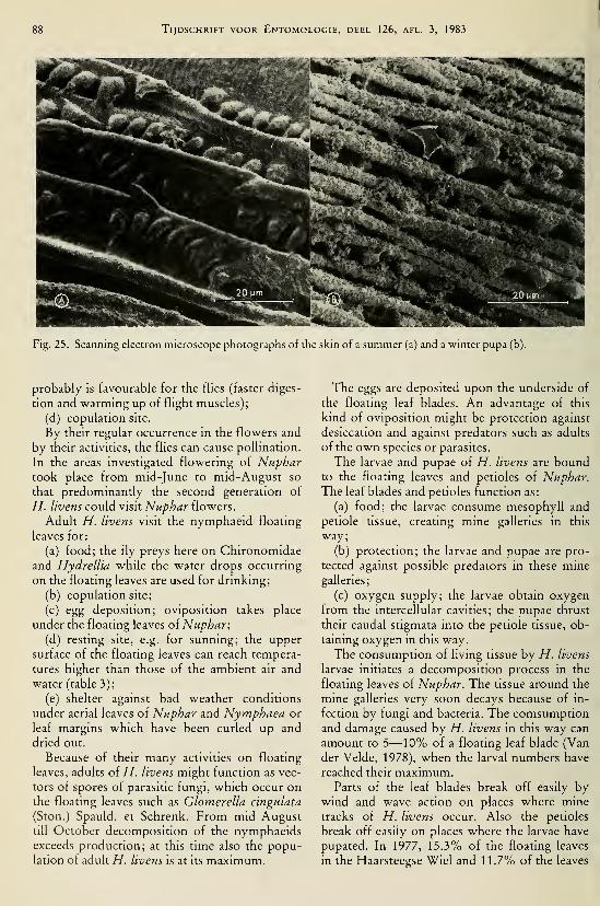
Tijdschrift voor Entomologie, deel 126, afl. 3, 1983
Fig. 25. Scanning electron microscope photographs of the skin of a summer (a) and a winter pupa (b).
probably is favourable for the flies (faster diges-
tion and warming up of flight muscles);
(d) copulation site.
By their regular occurrence in the flowers and
by their activities, the flies can cause pollination.
In the areas investigated flowering of Nuphartook place from mid-June to mid-August so
that predominantly the second generation of
H. livens could visit Nuphar flowers.
Adult H. livens visit the nymphaeid floating
leaves for:
(a) food; the fly preys here on Chironomidae
and Hydrellia while the water drops occurring
on the floating leaves are used for drinking;
(b) copulation site;
(c) egg deposition; oviposition takes place
under the floating leaves of Nuphar;
(d) resting site, e.g. for sunning; the upper
surface of the floating leaves can reach tempera-
tures higher than those of the ambient air and
water (table 3);
(e) shelter against bad weather conditions
under aerial leaves of Nuphar and Nymphaea or
leaf margins which have been curled up and
dried out.
Because of their many activities on floating
leaves, adults of H. livens might function as vec-
tors of spores of parasitic fungi, which occur onthe floating leaves such as Glomerella cingulata
(Ston.) Spauld. et Schrenk. From mid-Augusttill October decomposition of the nymphaeidsexceeds production; at this time also the popu-lation of adult H. livens is at its maximum.
The eggs are deposited upon the underside of
the floating leaf blades. An advantage of this
kind of oviposition might be protection against
desiccation and against predators such as adults
of the own species or parasites.
The larvae and pupae of H. livens are boundto the floating leaves and petioles of Nuphar.The leaf blades and petioles function as:
(a) food; the larvae consume mesophyll and
petiole tissue, creating mine galleries in this
way;
(b) protection; the larvae and pupae are pro-
tected against possible predators in these minegalleries;
(c) oxygen supply; the larvae obtain oxygen
from the intercellular cavities; the pupae thrust
their caudal stigmata into the petiole tissue, ob-
taining oxygen in this way.
The consumption of living tissue by H. livens
larvae initiates a decomposition process in the
floating leaves of Nuphar. The tissue around the
mine galleries very soon decays because of in-
fection by fungi and bacteria. The comsumptionand damage caused by H. livens in this way can
amount to 5—10% of a floating leaf blade (Van
der Velde, 1978), when the larval numbers have
reached their maximum.Parts of the leaf blades break off easily by
wind and wave action on places where mine
tracks of H. livens occur. Also the petioles
break off easily on places where the larvae have
pupated. In 1977, 15.3% of the floating leaves
in the Haarsteegse Wiel and 11.7% of the leaves
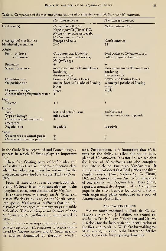
Brock Sc van der Velde: Hydromyza livens 89
Table 8. Comparison of the most important features of the hfe histories of H. livens and H. conßuens.

90 Tijdschrift voor Entomologie, deel 126, afl. 3, 1983
References
Beai, E. O., 1956. Taxonomie revision of the genus
Nuphar Sm. of North America and Europe. — J.
Elisha Mitchell Sci. Soc. 72: 317—346.
Berg, CO., 1950. HydrelUa (Ephydridae) and some
other acalyptrate Diptera reared from Potamoge-
ton.— Ann. Ent. Soc. America 43: 37^1—398.
Brauer, F., 1883. Die Zweiflügler des Kaiserlichen
Museums zu Wien III. Systematische Studien auf
Grundlage der Dipteren-Larven nebst einer
Zusammenstellung von Beispielen aus der Litera-
tur über dieselben und Beschreibung neuer For-
men. — Denkschr. mat.-naturwiss. Gl. K. Akad.
Wiss. 47: 1—100, 5 pis.
Gollin, J. E., 1958. A short synopsis of the British
Scatophagidae (Diptera). — Trans. Soc. British
Ent. 13:37—56.Eberle, G., 1943. Die MummelfUege {Hydromyza
livens Fall). — Natur und Volk, Frankfurt am
Main 73: 293—306.
Gaevskaya, N. S., 1966. The role of higher aquatic
plants in the nutrition of the animals in fresh-wa-
ter basins. Vol. 1: 1—232. — National Lending
Library for Science and Technology. Boston Spa,
Yorkshire, England.
Gercke, G., 1877. Ueber die Metamorphose der Hy-dromyza livens Fall. — Verb. Ver. naturwiss. Un-
terh. Hamburg (1879) 4: 229—234.
Grünberg, K., 1910. Diptera, Zweiflügler. Heft 2a.
In: A. Brauer (ed.). Die Süsswasserfauna Deutsch-
lands, eine Excursionsfauna: 1—312. — Gustav
Fischer, Jena.
Hackman, W., 1956. The Scatophagidae (Dipt.) of
Eastern Fennoscandia.— Fauna Fenn. 2: 1—67.
Hendel, F., 1928. Blattminenkunde Europas. I. Die
Dipterenminen 2 (Bog. 5—7, pl. 3—5).
Hering, E. M., 1926. Die Ökologie der blattminieren-
den Insectenlarven. — Zoologische Bausteine,
Ausschnitte aus dem Gesamtgebiet der Zoologie.
Berlin 1 (2): 1—253.
Heslop-Harrison, Y., 1955. Nuphar Sm. Biological
flora of the British Isles.— J. Ecol. 43: 342—364.
Hinton, H. E., 1960. How some insects, especially the
egg stages avoid drowning when it rains. — Proc.
S. Lond. ent. nat. Hist. Soc. 1960: 138—154.
, 1967. The respiratory systems of the egg-shell
of the common housefly. — J. Insect Physiol. 13:
647—651.
, 1971. Polyphyletic evolution of respiratory sys-
tems of egg shells, with a discussion of structure
and density-independent and density dependent
selective pressures. In: V. H. Heywood (ed.).
Scanning electron microscopy. Systematic and
evolutionary applications. The Systematics Asso-
ciation Special Volume 4: 1—331 (pp. 17—36). —Academic Press.
De Meijere, J. H. C., 1895. Ueber zusammengesetzte
Stigmen bei Dipterenlarven, nebst einem Beitrag
zur Metamorphose von Hydromyza livens. —Tijdschr. Ent. 38:65—100.
, 1940. Hydromyza livens Fall, en Notiphila
brunnipes Rob.-Desv., twee Dipteren, wier
levenswijze verband houdt met Nymphaea alba L.
— Ent. Ber. 10:22(^222.
Meusel, H., E. Jäger & E. Weinert, 1965. Verglei-
chende Chorologie der zentraleuropäischen Flora.
Vol. 1: Text: 1—583. Vol. 2: Karten: 1—258. —Gustav Fischer Verlag. Jena.
Monchadskii, A. S., 1940 (according to Gaevskaya,
1966). (Diptera excluding Tendipedidae and He-leidae). In: V. I. Zhadin (ed.). Freshwater life in
the USSR 1 : 233—263.— Moscow Acad. USSR.Roos, P. J., 1979. Two-stage life cycle of a Cordylo-
phora population in the Netherlands. — Hydro-biologia 62: 231—239.
Sack, P., 1937. Cordyluridae. 62a. In: E. Lindner
(ed.). Die Fliegen der palearktischen Region 7 (1):
1—103.— Stuttgart.
Séguy, E., 1934. Diptères (Brachycères) (Muscidae,
Acalypterae et Scatophagidae). — Faune de
France 28: 1—832 + pis. 1—27. Paris.
Schütte, L., 1921. Die Metamorphose von Hydromy-za livens. Inaug. Diss., Greifswald: 1—48. — E.
Hartmann, Greifswald.
Van der Velde, G., 1978. Structure and function of a
nymphaeid-dominated system. — Proc. EWRS5th Symp. on Aquatic Weeds (Amsterdam) 1978:
127—133.
, 1980. Studies in nymphaeid-dominated systems
with special emphasis on those dominated by
Nymphoides peltata (Gmel.) O. Kuntze (Menyan-
thaceae). Thesis Nijmegen, 163 pp.
&Th. C. M. Brock, 1980. The life history and
habits of Notiphila brunnipes Robineau-Desvoidy
(Diptera, Ephydridae), an autecological study on a
fly associated with nymphaeid vegetations. —Tijdschr. Ent. 123:105—127.
, Th. C. M. Brock, M. Heine & P. M. P. M. Pee-
ters, 1978. Flowers of Dutch Nymphaeaceae as a
habitat for insects. — Acta Bot. Neerl. 27: 429
—
430.
Vockeroth, J. R., 1978. Scatomyzidae. In: J. lilies
(ed.), Limnofauna Europaea. Eine Zusammenstel-
lung aller die europäischen Binnengewässer be-
wohnenden mehrzelligen Tierarten mit Angaben
über ihre Verbreitung und Ökologie. (2nd revised
ed.): 1—532 (p. 489). — Gustav Fischer Verlag,
Stuttgart, New York. Swets and Zeitlinger,
Amsterdam.
Welch, P. S., 1914. Observations on the life history
and habits of Hydromyza confluens Loew (Di-
ptera).— Ann. Ent. Soc. America 7: 135—147.
, 1917. Further studies on Hydromyza confluens
Loew, (Diptera). — Ann. Ent. Soc. America 10:
35—45.
Wesenberg-Lund, C., 1943. Biologie der Süsswasser-
insecten: 1—682. — Gyldendalske Boghandel
Nordish Forlag, Kopenhagen und Verlag J.
Springer, Berlin, Wien.

3DEEL 126 f'
,AFLEVERING 4—
5
1983
TIJDSCHRIFTVOOR ENTOMOLOGIE
UITGEGEVEN DOOR
DE NEDERLANDSE ENTOMOLOGISCHE VERENIGING
INHOUD
M. DuijM, L. OuDMAN and B. G. Veldstra. — Copulation in Ephippiger
(Orthoptera, Tettigonioidea), pp. 91—96, figs. 1—4.
M. DuijM and L. Oudman. — Interspecific mating in Ephippiger (Orthoptera, Tet-
tigonioidea), pp. 97—108, figs. 1—6.
Tijdschrift voor Entomologie, deel 126, afl. 4—
5
Gepubliceerd 20-V-1983

COPULATION IN EPHIPPIGER (ORTHOPTERA,TETTIGONIOIDEA)
by
M. DUIJM, L. OUDMAN and B. G. VELDSTRA
Dept. of Zoology, State University Groningen, P.O. Box 14, Haren, The Netherlands
Abstract
Observations have been made in a number of Ephippiger species from Southern France
and the role various reproductive structures play durmg the copulatory process.
Introduction
A number of species belonging to the genus
Ephippiger occur in Southern France and in the
adjacent parts of Spain and Italy. In some cases
these species vicariate geographically, but there
are also several instances of overlapping distri-
bution of two or more species. The question has
to be raised whether all taxa distmguished as
species (e.g. Chopard, 1951; Harz, 1969) are re-
productively isolated. In this paper we will in-
vestigate whether the interspecific differences in
the form of cerei, epiproct, titillators and sub-
genital plate could function as mechanical bar-
riers possibly operating against interspecific
matings. In another, the next, paper the likeli-
hood of interspecific mating is dealt with.
Material and methods
Animals of six species were collected during
field trips in August and September, 1977, 1979
and 1980. They were measured and taken to the
laboratory where observations were made dur-
ing the second half of September and during
October. In November most animals died. De-
tailed observations of copulations were madewith a stereomicroscope (magnifications 6.5
—
16x). Some copulations were documented by
photography or videorecording.
The species studied and their collecting sta-
tions are the following.
Ephippiger ephippiger (Fieb., 1784): Niaux
(Ariège) 1977, 1979; Mézel (Ht. Provence)
1979, 1980; Causse de Larzac (Aveyron) 1979;
Plan d'Aups (Var) 1979, 1980.
Ephippiger cunii (Bol., 1877): Mont Louis
1) Nadig (1980) considers E. bormansi a subspecies of
E. terrestris, but the conventional classification is
retained here for the time being.
(Pyr. Orient.) 1977, 1979; Canigou (Pyr.
Orient.) 1979.
Ephippiger cruciger (Fieb., 1853): Neffiès
(Hérault) 1979; Leucate (Aude) 1979.
Ephippiger provincialis (Yers., 1854): Plan
d'Aups (Var) 1979, 1980.
Ephippiger terrestris (Yers., 1854)>): Mézel
and several other places in the vicinity of Digne
(Ht. Provence) 1979, 1980, 1981; Fayence (Var)
1980; Col de Braus (Alp. Mar.) 1980; Tende
(Alp. Mar.) 1980.
Ephippiger bormansi (Br., 1882)'): Col de
Tende (Alp. Mar.) 1980; Vallone del Arma (Pié-
mont) 1981.
Some pairs were killed during or just after
copulation and preserved to study the position
of the genital organs and their elements with re-
spect to each other as well as the position of the
spermatophore.
The copulation
Description of the behaviour
The general course of copulation in Ephippig-
er conforms to that in other Tettigonioids, as
described by Gerhard (1913, 1914). A short de-
scription will be given here emphasizing the de-
tails relevant to the questions raised.
A normal copulation will take about half an
hour. Ephippiger males stridulate during part of
the day and night, while sitting on a plant or
bush, the height of which generally does not ex-
ceed two metres. A sexually motivated female
will walk straight to a stridulating male (Busnel
& Dumortier, 1954; Duijm & Van Oijen, 1948).
On encountering the male the female stops.
Both male and female touch each other with the
antennae. The male may tremble with his body
several times (Busnel et al., 1955). After some
time the male will place the extremity of his ab-
91

92 Tijdschrift voor Entomologie, deel 126, afl. 4 (1983)
Fig. 1. A pair of Ephippiger ephippiger just before the
cereal clamp: ce, cercus, ep, epiproct, gr, copulatory
groove, ov, ovipositor, pa, paraproct, ph, phallus
with: // lateral lobes, dl, dorsal lobes and vl, ventral
lobes; pi, pleural region, sa, saddle-like part of sper-
matophore, sp, subgenital plate, st, stylus, sx, sperma-
tophylax, s6, sixth sternite, t9, ninth tergite, tlO, tenth
tergite.
dornen in front of the female's head; the female
will then touch the male's somewhat ventrally
curved abdomen with her maxillary palps. Mov-ing forward slowly, continually touching, she
finally mounts the male's back. When the fe-
male has entirely mounted, the male bends his
abdomen dorsally, extends the lobes of the
phallus and turns his subgenital plate down-
ward. The titillators are now visible between the
phallus lobes. The cerei are extended and spread
in a laterodorsal direction. The female is nowsitting on the back of the male with her head
above his pronotum and the median planes of
both partners have tO' coincide at this point. In
the female the caudal part of the ventral abdom-
inal wall is contracted, the ovipositor making an
angle of 20—45° with its longitudinal axis (fig.
1). Only in this position the female's subgenital
plate is situated between the male's cerei in such
a way that the copulation may proceed. The
male circles with its cerei caudally and medially.
Suddenly the inner teeth of the cerei get a grip
on the female's subgenital plate and at the same
time the cerei clamp the female tightly. Clamp-ing does not occur randomly, but only on twosmall grooves — the copulatory grooves —which are easy to distinguish by their dark col-
our, owing to extra sclerotisation. By adducting
the cerei the male pulls the female firmly against
its body. In this position the epiproct pushes
dorsocaudally against the female's subgenital
plate and provides one of the factors for deter-
mining the position of the male abdomen with
respect to the female's. At the same time, push-
ing the epiproct against the female subgenital
plate results in the lifting of the caudal part of
the subgenital plate from the female gonotrema.
By this action the gonotrema will be opened up
and exposed. Immediately after being clamped,
the female brings the ovipositor downward very
rapidly to such a position that it fits in the emar-
gination of the caudal rim of the subgenital plate
and contacts the styli (fig. 2). After some sec-
onds the ovipositor returns to its normal posi-
tion. The function of this action is not clear.
Perhaps in this way a final check is made to de-
termine whether the partners are in the correct
position to enable the next step in the copula-
tion procedure, viz., the rotation. In this posi-
tion the male will extend and contract the phal-
lus alternately. The female now makes a few
steps forward, the male meanwhile somersault-
ing until he gets hold of the female's ovipositor
with his fore and middle legs (fig. 3). In doing
so, the male is rotated about an axis through the
inner cereal teeth. This rotation is only possible
when the cerei are tightly clamped. The position
of the male is now entirely fixed with respect to
the female and evidently in the only one in
which the spermatophore can be deposited.
When extended the phallus now reaches the ba-
sis of the ovipositor. The phallus makes pulsat-
Fig. 2. The position of male and female E. ephippiger
immediately after the cereal clamp, the female having
lowered the ovipositor to contact the male's subgeni-
tal plate and styli. See fig. 1 for explanations.
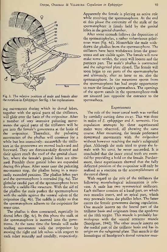
DuijM, OuDMAN & Veldstra: Copulation in Ephippiger 93
Fig. 3. The relative position of male and female after
the rotation in Ephippiger. See fig. 1 for explanations.
ing movements during which its dorsal lobes,
together with the apical parts of the titillators,
will glide over the basis of the ovipositor. After
a number of very intensive pulsating move-
ments the apical parts of the titillators will be
put into the female's gonotrema at the basis of
the ovipositor. Thereafter, the pulsating
movements of the phallus will continue for a
while but less intensively: meanwhile the titilla-
tors in the gonotrema are moved backward and
foreward. They are dorsocaudally directed and
their apices reach the roof of the genital cham-
ber, where the female's genital lobes are situ-
ated. Possibly these genital lobes are expanded
during this phase. After some time the pulsating
movements stop, the phallus being in a maxi-
mally extended position. The phallus lobes part
and the spermatophore is extruded (fig. 4a). Theglobular caudal part of the spermatophore bears
dorsally a saddle-like structure. With the aid of
the phallus the male pushes the spermatophore
with the saddle against the ventral valves of the
ovipositor (fig. 4b). The saddle is sticky so that
the spermatophore adheres to the ovipositor for
some time.
The phallus is then contracted, especially its
dorsal lobes (fig. 4c). In this phase the stalk of
the spermatophore is inserted into the gono-
trema. At the same time the female will makesculling movements with the ovipositor by
moving the right and left valves with respect to
each other rostrally and caudally, respectively.
Apparently the female is playing an active role
while receiving the spermatophore. At the endof this phase the extremity of the stalk of the
spermatophore is firmly held by the genital
lobes in the genital chamber.
After some seconds follows the deposition of
the spermatophylax, a rather voluminous gelati-
nous mass (fig. 4d). Meanwhile the male with-
draws the phallus from the spermatophore. Thetitillators have been withdrawn from the gono-
trema and are visible again. The female will nowmake some strides, the cerei will loosen and the
partners part. The male's phallus is contracted
and the subgenital plate closed. The female will
soon begin to eat parts of the spermatophylax
and ultimately, after an hour or so, also the
spermatophore. In the meantime sperm fromthe spermatophore has had ample opportunity
to enter the female's spermatheca. The openings
of the sperm canals in the spermatophore-stalk
are situated just opposite the entrance to the
spermatheca.
Experiments
The role of the inner cereal teeth was verified
by carefully cutting them away. This was done
in males of E. ephippiger and E. terrestris. Five
copulation attempts by these experimental
males were observed, all showing the same
course. After mounting, the female performed
downward movements with the ovipositor
without however reaching the male's subgenital
plate. Although the male tried to grasp the fe-
male with his cerei, he never succeeded. It is
concluded that the inner cereal teeth are essen-
tial for providing a hold on the female. Further-
more, these experiments showed that the fully
deflected position of the ovipositor will only be
realised as a reaction to the accomplishment of
the cereal clamp.
With regard to the role of the titillators the
following morphological information is rele-
vant. A male has two symmetrical titillators.
Each titillator consists of a basal part, on which
two muscles insert, and an apical part, which
may protrude from the phallus lobes. The latter
enters the female gonotrema during copulation.
One muscle inserts on the lateral part of the
titillator basis and originates from the apodeme
of the 10th tergite. This muscle is probably ho-
mologous with the ventral retractor muscle
(Snodgrass, 1940). The other muscle inserts on
the medial part of the titillator basis and has its
origin on the subgenital plate. This muscle is the
homologue of Snodgrass's dorsal retractor mus-

94 Tijdschrift voor Entomologie, deel 126, afl. 4 (1983)
©Fig. 4. Production of the spermatophore in Ephippiger: (a) the globular part of the spermatophore becomes vis-
ible; (b) the saddle-Hke part of the spermatophore is being pressed against the ovipositor; (c) the dorsal lobes of
the phallus are being contracted; (d) the spermatophylax appears. See fig. 1 for explanations.
cle. The following movements have been ob-
served: (a) dorsal deflection, (b) lateral deflec-
tion. In (a) and (b) the apices of both titillators
stay close together pointing in the same direc-
tion, whereas when the phallic lobes are maxi-
mally extended, the apices may diverge. To ob-
tain some insight into the way the titillators
function during copulation the following exper-
iments were performed. In eight males of E.
ephippiger the apical parts of the titillators were
cut off. Both titillators where shortened to the
same extent, but the length of the cut parts dif-
fered. In the extreme case the entire denticu-
lated part was removed. The operation did not
alter sexual motivation or copulatory behav-
iour. One copulation was observed in each
male. In no case were the remnants of the titilla-
tors inserted into the female's gonotrema, inde-
pendent of the length of the remaining part.
Probably the cut ends of the titillators cannot
readily enter the gonotrema because they lack
the rounded apex which is characteristic of the
intact titillator. Notwithstanding the lack of in-
sertion, a spermatophore was deposited in all
cases. In half of these cases the stalk was
brought into the normal position within the
gonotremal space. In the remaining cases this
did not occur and the female ^hen lost the sper-
matophore immediately after the copulation.
From these observations it follows that the in-
sertion of the titillators is not a prerequisite for
completion of the copulation. Evidently the ac-
tion of the male's phallic lobes together with the
action of the female's genital lobes may result in
normal reception of the spermatophore stalk.
The number of failures was nevertheless high;
DuijM, OuDMAN & Veldstra: Copulation in Ephippiger 95
this points to the role of the titillators in provid-
ing additional reliability. In our opinion the
titillators do not play a role in opening up the
female gonotrema nor in delivering a stimulus
to the female, which is essential for receiving the
spermatophore. Insertion of the titillators into
the gonotrema probably orientates the position
of the gonotrema just opposite the male's gono-
phore. In the end phase of copulation, the titil-
lators are situated against the rostral wall of the
gonotrema in such a way that the stalk of the
spermatophore may glide along the smoothventral sides of the titillators and in this waymay be led to the gonotrema, as by a shoe-horn.
Because the curved denticles on the apical parts
of the titillators are situated on their dorsolat-
eral sides, they will therefore grip the rostral
wall of the gonotrema in the end phase of the
copulation. Without this gripping, however,
successful insertion of the spermatophore still
remains possible. In two other males of E.
ephippiger a part of only one titillator was cut
off, the other being left intact. Insertion ap-
peared to be normal in these males during copu-
lation. In one copulation the stalk of the sper-
matophore was inserted quite normally, in the
other case the spermatophore was lost by the fe-
male. In the latter case, orientation of the male
gonopore with respect to the female's gono-
trema was in our view not precise enough to en-
sure reception of the stalk by the female's geni-
tal lobes.
Discussion
The copulatory behaviour in Ephippiger con-
sists of two closely interlocked complementaryseries of movements by male and female. Bothfemale and male copulatory apparatuses func-
tion as mechanical systems of considerable com-plexity. For their successful interaction not only
has highly standardised behaviour to be per-
formed, but also the form and dimensions of the
various parts in male and female must conform
to a high extent. In the course of the copulation
a consecutive series of mechanical conditions
has to be satisfied after the female has mounted :
(1) do the median planes of both partners co-
incide?
(2) is the female's subgenital plate situated
between the male's cerei?
(3) can the inner cereal teeth get a hold in the
copulatory grooves of the female's subgenital
plate?
(4) does the ventrally deflected ovipositor fit
the emarginated rim of the male's subgenital
plate so that their gonopores are just in front of
each other?
(5) do the phallic lobes with the titillators
reach the female's gonotrema?Only when all conditions are fulfilled is the
cereal clamp carried out correctly and rotation
ensues. After this, spermatophore and sperma-tophylax will be produced and the spermato-phore stalk will be inserted into the female's
gonotrema when:
(6) the titillators have been introduced in the
gonotrema.
Form, position and size of the various parts
of the genital apparatus have to correspond; it is
analogous to a lock and key system. However,this system does not appear to be very rigorous-
ly determined: interspecific copulations may oc-
cur occasionally under laboratory conditions.
Moreover, not every morphological detail ap-
pears to be of decisive importance as is indicated
by the experiments with cut titillators.
In the following paper, the differences be-
tween species with regard to their genital appa-
ratus and copulatory behaviour will be consid-
ered together with the possibility of interspecif-
ic matings.
Summary
The course of copulation in the genus Ephip-
piger has been described emphasising the suc-
cession of mechanical conditions that must be
satisfied for the copulation to be completed.
The male cerei play a central role; they have to
grasp into the copulatory grooves on the fe-
male's subgenital plate with their inner teeth.
Only after having successfully clamped the fe-
male does the male rotate about the axis
through the inner cereal teeth and continues
copulation. The titillators appear to be used as
shoe-horns for guiding the spermatophore stalk
when it is being introduced into the female
gonotrema.
Acknowledgements
Our thanks are due to the students who as-
sisted in collecting the insects and in all other
connected activities: Geert Damsma, Gertjaap
van Klinken, Willy Koolstra, Bert Lotz, An-neke Oerlemans, Gerben Poortenga, Gerard
van der Veen and especially Wybren Landman.We are grateful to Dr. G. Thomas for critically
reading the manuscript and for correcting the
English. Furthermore we thank Mrs. J. Poel-
stra-Hiddinga for typing the manuscript and to
Mr. D. Visser for making the drawings.

Tijdschrift voor Entomologie, deel 126, afl. 4 (1983)
Literature
Busnel, R. G., & B. Dumortier, 1954. Observations
sur Ie comportement acoustico-sexuel de la fe-
melle d'Ephippiger bitterensis. — CR. Séanc. Soc.
Biol. 148: 1589—1592.
Busnel, R. G., P. Pasquinelly & B. Dumortier, 1955.
La trémulation du corps et la transmission aux
supports des vibrations en résultants commemoyen d'information à courte portée des Ephip-
pigères mâle et femelle. — Bull. Soc. Zool. France
80:18—22.
Chopard, L., 1951. Orthopteroides. Faune de France
56: 1—359.— Lechevalier, Paris.
Duijm, M., & T. van Oijen, 1948. Het sjirpen van de
sabelsprinkhaan.— Lev. Nat. 51 : 81—87.
Gerhardt, U., 1913. Copulation und Spermatophoren
von Grylliden und Locustiden. — Zool. Jahrb. 35:
415—532.
, 1914. Idem, IL— Zool. Jahrb., Syst. 37: 1—64.
Harz, K., 1969. Die Orthopteren Europas: 1—749. —Junk, The Hague.
Nadig, A., 1980. Ephippiger terrestris (Yersin) und E.
bormansi (Brunner von Wattenwyl) (Orthoptera):
Unterarten einer polytypischen Art. Beschreibung
einer dritten Unterart: E. terrestris caprai ssp. n.
aus den Ligurischen Alpen. — Rev. Suisse Zool.
87:473—512.
Snodgrass, A. E., 1940. Male genitalia of Ortho-
pteroid insects.— Smiths, misc. Coll. 96: 1—102.

INTERSPECIFIC MATING IN EPHIPPIGER (ORTHOPTERA,TETTIGONIOIDEA)
by
M. DUIJM and L. OUDMANDept. ofZoology, State University of Groningen, P.O. Box 14, Haren, The Netherlands
Abstract
A number of (sub)species of Ephippiger were compared with respect to the possibiHty of
interspecific mating, differences in the morphology of genital parts and geographical distri-
bution. A tentative conclusion on the taxonomie status of the (sub)species studied is pre-
sented.
Introduction
In Southern France a number of species of the
genus Ephippiger occur, which are mainly en-
demic. Their morphological characters are rath-
er variable and show extensive overlapping.
Therefore, the question may be raised whether
this situation is at least partly due to interspecif-
ic interbreeding. Nadig (1980) postulates an ex-
tensive hybridisation zone in the French Alps
between Ephippiger terrestris and E. bormansi,
which he considers subspecies. To look further
into this matter we have, in the preceding paper
(Duijm, Oudman & Veldstra, 1983), analysed
the copulatory behaviour in Ephippiger in gen-
eral, emphasising those morphological and posi-
tional details that could be decisive in determin-
ing whether successful copulation between twopartners will be possible. In this paper data are
given with regard to the possibility of the oc-
currence of interspecific matings under labora-
tory conditions. Moreover, relations between
these observations and the morphological dif-
ferences in the copulatory apparatus are given.
Material and methods
Specimens of eight (sub)species of Ephippiger
were collected during field trips in August and
September, 1977, 1979, 1980 and 1981. Theywere measured and taken to the laboratory
where observations were made during the sec-
ond half of September and October. Animals
were kept individually in small cages and fed a
diversity of wild plants (e.g. Stellaria media
Nadig (1980) regards E. bormansi as a subspecies
of E. terrestris; the conventional classification is re-
tained here for the time being.
(L.), Calystegia sepium (L.), Aegopodium poda-graria (L.), Taraxacum spec.) and occasionally a
meal-worm. The cages were regularly sprayed
with water and females could deposit their eggs
in small boxes with moist turf.
For observation a male and a female wereplaced together on a branch. During the copula-
tions details were observed with a stereomicros-
cope (magnification 6.5—16x). Some copula-
tions were documented by photography or byvideorecording. The species studied and their
collection stations are: (France, unless stated
otherwise)
Ephippiger ephippiger vitium Serv., 1931:
Causse de Larzac (Aveyron), 1979; Niaux(Ariège) 1977, 1979; Mézei (Ht. Provence)
1979, 1980; Plan d'Aups (Var) 1979, 1980;
Ephippiger ephippiger vicheti Harz, 1966:
Miglieglia (Ticino, Switzerl.) 1980, 1981; Nag-gio (Lago di Como, Italy) 1981
;
Ephippiger cunii BoL, 1877: Mont Louis (Pyr.
Orient.) 1977, 1979; Canigou (Pyr. Orient.)
1979;
Ephippiger cruciger (Fieber, 1853): Neffiès
(Hérault) 1979; Leucate (Aude) 1979;
Ephippiger provincialis (Yers., 1854): Plan
d'Aups (Var) 1979, 1980, 1981;
Ephippiger terrestris (Yers., 1854)'): Mézel
and several other places in the vicinity of Digne
(Ht. Provence) 1979, 1980; CoUobrières (Var)
1980; Fayence (Var), 1980; Col de Braus (Alp.
Mar.) 1980; Gréolières (Alp. Mar.) 1981; Col de
Brouis (Alp. Mar.) 1981; Col de Castillon (Alp.
Mar.) 1981; Tende (Alp. Mar.) 1980; Cle. Scra-
vaion and Mt. Carmo (Ligur. Alps, Italy 1981 ;
Ephippiger bormansi (Br. v. W., 1882)'): Col
de Tende (Alp. Mar.) 1980, 1981; Vallone del
Arma (Piémont, Italy) 1981; Naggio (Lago di
97

98 Tijdschrift voor Entomologie, deel 126, afl. 5 (1983)
Como, Italy) 1981; Miglieglia Ticino, Switzerl.)
1981.
The results of attempts to interbreed in
the laboratory
We shall confine our attention to successful
copulations. In this paper we speak of a success-
ful copulation when the copulatory process
takes the normal course and the spermatophore
stalk is correctly inserted into the female's
gonotrema. No observations were made on sub-
sequent fertilisation nor on the development of
the eggs. The crosses attempted are dealt with
below.
( 1 ) Ephippiger ephippiger
Not all pairs of E. ephippiger formed by
bringing together males and females of different
populations copulated successfully. In all three
cases observed of crossing a female of the sub-
species E. e. vicheti with a male of the subspe-
cies E. e. vitium (from different sources) the
male's attempt to clasp the female failed; appar-
ently the male's abdomen was too broad and
therefore the distance between the cerei too big
for getting hold in the female's copulatory
grooves. No data are available at the moment onthe reverse combination (£. e. vicheti 6 XE. e. vitium 9).
Among the populations studied of the sub-
species E. e. vitium there is one that differs con-
siderably from the others. This is the population
from Niaux, where the animals are rather big.
Their titillators are shorter than in the other
populations of E. e. vitium and as seen in lateral
view, they are more curved. Moreover the api-
cal part, bearing teeth, is longer. There are also
differences in song. Because of this heterogenei-
ty the observations with specimens of E. e.
vicheti and E. e. Vitium from Niaux are left out
of consideration when dealing with crosses be-
tween E. ephippiger and other Ephippiger spe-
cies.
(2) Ephippiger terrestris
Ten copulations were observed betweentwenty individuals from different sources. In all
cases copulations were performed, not-
withstanding the different sources and bodysizes. The samples included rather small animals
(forma minor, collected 1200 m above sea level)
and rather big animals (nominate form, col-
lected at 300 m), the difference in body length
amounting to 15—25%. Also specimens fromTende, probably belonging to the subspecies
E. terrestris caprai Nadig (1980) were included.
Later observations showed the possibility of
successful copulations between E. terrestris ter-
restris and E. terrestris caprai (from Cle. Scra-
vaion and Mt. Carmo in the Ligurian Alps).
(3) Ephippiger bormansi
One copulation between a pair of Ephippiger
bormansi (from Col de Tende) was observed.
As was expected the copulation ended success-
fully.
(4) Crosses between E. terrestris and E. bor-
mansi
On three occasions an E. terrestris 9 was
placed together with an E. bormansi 6 (from
Col de Tende). In all cases copulation was suc-
cessful. The same applies to the reverse combi-
nation: four successful copulations were ob-
served. Later on, successful copulations were
observed between E. bormansi (from Vallone
del Arma and from Naggio) with E. terrestris
caprai (both from Tende and from Mt. Carmo)and with E. terrestris terrestris.
(5) Crosses between E. terrestris and other
Ephippiger species (excluding E. bormansi)
Six females of E. terrestris were placed with
an E. ephippiger vitium male and two females of
E. terrestris with a male of E. cunii. In none of
these cases did a successful copulation ensue.
Generally the female mounted and brought the
ovipositor downward. The male tried to clamp
the female with his cerei but did not succeed in
getting hold on the female's subgenital plate.
The same result was obtained when placing a fe-
male E. bormansi with an E. ephippiger male.
On two occasions an E. terrestris male was
placed with an E. ephippiger female. In both the
male clamped the female with the cerei, but on
the wrong place, i.e. on the soft rostral parts of
the seventh sternum and not on the sclerotised
copulatory grooves. In one of these cases the
male pierced the female sternal wall whereupon
the female left the male. In the other case, the
copulatory rotation was effected and the male
extruded the phallus. During pulsating
movements of the phallus the titillators glided
over the open gonotrema but they were not in-
serted. A spermatophore was deposited but was
lost by the female immediately after the copula-
tion; obviously the spermatophore stalk had not
been inserted into the female's gonotrema. This
failure was probably due firstly to the male go-
nopore not being placed precisely opposite the
female's gonotrema and secondly to the titilla-
tors not being able to conduct the sperma-
tophore stalk.
Comparable results were obtained in combi-
nations (two cases) consisting of a male E. bor-
DuijM & OuDMAN: Interspecific mating in Ephippiger 99
mansi and a female E. ephippiger.
(6) Crosses between E. ephippiger and E.
cunii
As might be expected successful copulations
between E. cunii males and females were readily
obtained.
On three occasions a male E. cunii was con-
fronted with a female E. ephippiger: in one case
the cereal clamp did not succeed; in the second
case the cereal clamp held only for a short time;
in the third case the cereal clamp held only after
five failures. In the latter rotation followed and
the titillators were inserted. After that the copu-
lation attempt was terminated because the fe-
male fled.
On two occasions a male E. ephippiger was
placed with a female E. cunii. Neither male suc-
ceeded in getting a hold on the female's copula-
tory grooves and copulation attempts were ter-
minated.
(7) Crosses between E. ephippiger and E.
cruciger
One observation is available on a pairing be-
tween a male E. cruciger with a female E. ephip-
piger. The male did not succeed in clasping the
female definitively; several times the cereal teeth
entered the copulatory grooves but time and
again they sprang loose with a conspicuous
cUck. The female's abdomen appeared to be too
narrow for the male. Similar results were ob-
tained in two comparable cases with female
E. ephippiger from Niaux. No data are available
on the reverse combination owing to a lack of
E. cr«ager females.
(8) Crossings between E. cruciger and E.
cunii.
We have one observation of a fully successful
copulation between a male E. cunii and a female
E. cruciger.
Interspecific differences in themorphology of the copulatory apparatus
In order to elucidate the factors that may pre-
vent successful copulation between a variety of
Ephippiger species, a study was made of mor-
phological characters of those genital parts, that
could be relevant to the question raised.
(i) The cerei (fig. 1)
Notwithstanding the rather great variation in
cereal form three groups of species may be dis-
tinguished with regard to the general form of
the cerei and the positioning of their inner teeth,
i.e.:
(1) Ephippiger provincialis: cercus short and
stout with strongly curved inner tooth and also
a strongly curved terminal tooth;
(2) Ephippiger terrestris and E. bormansi
with cerei that are more or less cylindrical in
general outline, not conical as in the next group
(Nadig, 1960). There is no terminal tooth. Theinner cereal teeth are situated just near the cer-
eal apex; in the living animal these teeth lay well
behind the hind rim of the epiproct (Nadig,
1960). In preserved specimens this relative posi-
tion may be altered;
(3) Ephippiger ephippiger, E. cruciger and
E. cunii: the cerei are more conical than cylin-
drical. The inner teeth are not situated near the
cereal apex but half or three quarters way along
the cercus. In the living male the inner teeth are
situated adjacent to the sides of the epiproct.
(ii) The epiproct (fig. 1)
In group 3 the epiproct is entirely sclerotised;
the connection with the 10th tergite, however,
is not sclerotised and therefore the epiproct is
movable with regard to the 10th tergite. On the
other hand in group 2 the epiproct itself is not
entirely sclerotised: neither its caudal edges nor
its lateral rims are hardened. The sclerotised
part of the epiproct is, in these species, not
movable with respect to the 10th tergite.
(iii) The titillators (fig. 2)
In group 2 the apical parts of the titillators
bear one row of denticles, whereas in group 3
there are often more rows of denticles. The bas-
al edge of the titillators is broadened in group 2,
but not so in group 3. Seen from the side the ap-
ical parts of the titillators are distinctly curved
in group 3 and rather straight in group 2. In
group 1 the basal parts are strongly upcurved
with a broadened end part.
(iv) The seventh sternite and subgenital plate
in the female (figs. 3 and 4)
In group 1 nearly the entire subgenital plate is
sclerotised, but in group 2 and 3 the seventh
sternite and the subgenital plate of the female
are only partially sclerotised. The two groups
differ clearly with regard to the form of the scle-
rotised parts. Indications of such differences are
present in the systematic descriptions (e.g.
Harz, 1969). A more detailed description is
given here.
In group 3 the subgenital plate is not as exten-
sively sclerotised as in group 2. The caudal rim
is sclerotised and generally there are two sepa-
rate shield-like sclerotised parts on either side of
the midline. (These shields are lacking in E.
ephippiger vicheti). Moreover, the rostrolateral
parts bearing the copulatory grooves are sclero-
tised. These copulatory grooves are not situated
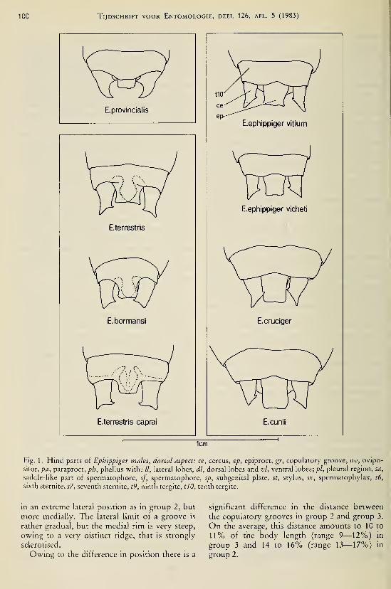
100 Tijdschrift voor Entomologie, deel 126, afl. 5 (1983)
E.provincialis
E.terrestris
E. bormansi
E.terrestris caprai
E.ephippiger Vitium
E.ephippiger vicheti
E.cruciger
E.cunii
lem
Fig. 1 . Hind parts of Ephippiger males, dorsal aspect: ce, cercus, ep, epiproct, gr, copulatory groove, ov, ovipo-
sitor, pa, paraproct, ph, phallus with: //, lateral lobes, dl, dorsal lobes and vl, ventral lobes; pi, pleural region, sa,
saddle-like part of spermatophore, sf, spermatophore, sp, subgenital plate, st, stylus, sx, spermatophylax, s6,
sixth sternite, s7, seventh sternite, t9, ninth tergite, tlO, tenth tergite.
in an extreme lateral position as in group 2, but
more medially. The lateral limit of a groove is
rather gradual, but the medial rim is very steep,
owing to a very distinct ridge, that is strongly
sclerotised.
Owing to the difference in position there is a
significant difference in the distance between
the copulatory grooves in group 2 and group 3.
On the average, this distance amounts to 10 to
11% of the body length (range 9—12%) in
group 3 and 14 to 16% (range 13—17%) in
group 2.

DuijM & OuDMAN: Interspecific mating in Ephippiger 101
E.terrestris (Fayence)
E.bormansi (Col de Tende)
E.terrestris caprai (de. scravaion)
basal part2E.ephippiger Vitium (Couvertoirade )
Eephippiger vicheti
E.cunii(Mont Louis)
E.cruciger(Neffies)
1mm
Fig. 2. Right titillators, dorsal view (left), right view (right)
Interspecific differences in behaviour
(i) Trembling
The trembling behaviour in Ephippiger is de-
scribed by Busnel et al. (1955). The animai
makes rapid up and down movements with the
body, by flexing and extending the legs alterna-
tely while keeping the tarsi to the substrate. Themale often trembles after having contact with a
female; also the female may tremble in the
neighbourhood of a male, but this is seen less
frequently. Males may tremble also towards
other males. Trembling has been observed regu-

102 Tijdschrift voor Entomologie, deel 126, afl. 5 (1983)
s7 gr
E.bormansi
E.terrestris
Fig. 3. Hind parts of Ephtppiger females, group 2, left view (left), ventral view (right); see fig. 1 for explana-
tions.
larly in E. ephippiger and also in Ephippiger
cunii, E. cruciger and E. provincialis. It has nev-
er been observed during copulation preliminar-
ies in E. bormansi or in E. terrestris.
(ii) The course of the copulation
A general description of the course of copula-
tion in Ephippiger is given in the previous paper
(Duijm, Oudman & Veldstra, 1983). In this pa-
per we restrict ourselves to pointing to an ob-
vious difference in the course of copulation be-
tween group 2 and group 3.
Before the cerei clasp the female's subgenital
plate, the partners have to position themselves
very accurately with respect to each other. Thefemale contracts the caudal part of the sternal
wall and directs the ovipositor downward to-
gether with the subgenital plate. In this position
the male epiproct is directed caudally against
the female's subgenital plate. The female's ven-
tral wall (including the medial parts of the 6th
and 7th sternum and the rostral part of the sub-
genital plate) is pushed tightly against the male's
tergum including the dorsal side of the epiproct.
In group 3, the caudal rim of the epiproct is
held in place by the rostral part of the subgenital
plate, but in group 2 by the caudal part of the
subgenital plate, which is that part behind the
two shield-like sclerotised parts. In both cases
the distance between the copulatory grooves is
determined by the connecting sclerotised parts,
which are formed mainly by the 7th sternum in
group 3 but by the subgenital plate in group 2
(fig. 5).
The geographical distribution of theephippiger species
To judge the likelihood of interspecific mat-
ings occurring in the field and to consider the
taxonomie status of the taxa dealt with, data on
their geographical distributions are essential.
Not all data from the literature are usable on ac-
count of the confusion often met with regarding
the exact distinction between the species. Ourinterpretation of the pattern of distribution is
given in fig. 6 for the (sub)species studied. For
the distribution of E. terrestris and E. bormansi
we have mainly followed Nadig (1980) with the
addition of a few of our own data. For the dis-
tribution of E. ephippiger we used the data from
Chopard (1951), Harz (1969), Dreux (1962) and
Voisin (1979), with some additions and modifi-
cations based on our own observations. The dis-
tribution of E. cruciger and E. cunii is mainly
based on Chopard (1951) and Busnel (1963),
with some additions, as is the distribution of
E. provincialis.

DuijM & Oudman: Interspecific mating in Ephippiger 103
Eephippiger vicheti
Eephippiger vitium
E.cruciger
E.cunii
Fig. 4. Hind parts of Ephippiger females, group 3, left view (left), ventral view (right); see fig. 1 for explana-
tions.
The first question that comes up now is least, are likely to meet each otherin such a situ-
whether there are species with overlapping ation that interspecific matings could occur. For
areas and if so, whether members of two or both of these field investigations are necessary,
more species will indeed meet each other or, at From the distributional data available at the
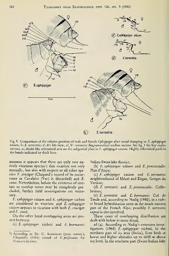
104 Tijdschrift voor Entomologie, deel 126, afl. 5 (1983)
9
(?) E.ephippiger vitium
s6 s7
E.terrestris
E. terrestris
Fig. 5. Comparison of the relative position of male and female Ephippiger after cereal clamping; a: E. ephippiger
vitium, b: E. terrestris; a', b': left view; a^, b^: tentative diagrammatical median section. See fig. 1 for key expla-
nations, sc, shield-like sclerotised area on the subgenital plate in E. ephippiger vittum. Highly sclerotised parts in
the female indicated by thick lines.
moment it appears that there are only two en-
tirely vicarious species') that vicariate not onlymutually, but also with respect to all other spe-
cies: E. cruciger (Chopard's record of its occur-
rence in Cavalaire (Var) is discarded) and E.
cunii. Nevertheless, before the existence of con-tact or overlap zones may be completely pre-
cluded, further field investigations are neces-
sary.
E. ephippiger vitium and E. ephippiger vicheti
are considered to vicariate and E. ephippiger
vitium appears to vicariate with both E. cruciger
and E. cunii.
On the other hand overlapping areas are pre-
sent between:
(a) E. ephippiger vicheti and E. hormansi:
') According to Dr. G. Kruseman (pers. comm.),Chopard's (1951) record of E. perforatus for
France is in error.
Italian-Swiss lake district;
(b) E. ephippiger vitium and E. provincialis:
Plan d'Aups;
(c) E. ephippiger vitium and E. terrestris:
neighbourhood of Mézel and Digne, Gorges duVerdon;
(d) E. terrestris and E. provincialis: Collo-
brières;
(e) E. terrestris and E. hormansi: Col de
Tende and, according to Nadig (1980), in a rath-
er broad hybridisation zone in the south-eastern
part of the French Alps; possibly E. terrestris
caprai is also involved.
These cases of overlapping distribution are
dealt with below in more detail.
ad (a). According to Nadig's extensive inves-
tigations (1968) E. ephippiger vicheti, in the
northern part of its area (Swiss), lives both at
lower and higher altitudes up to 1650 m above
sea level. In the southern part (Swiss-Italian lake

DuijM & Oudman: Interspecific mating in Ephippiger 105
u \

106 Tijdschrift voor Entomologie, deel 126, afl. 5 (1983)
district), however, where E. hormansi also oc-
curs, a distinct niche separation is present,
E. bormansi occurring above about 1000 up to
1900 m and E. e. vicheti occurring at lower alti-
tude. Nevertheless, according to Nadig (1968)
both species may live in an intermediate zone
(950—1100 m). In September 1981 we found
these two species living together above Naggio
(Lago di Como) at 750 m and at the foot of the
Mt. Lema between 920 and 1100 m. The two
species differ considerably in song character and
in behaviour. According to our observations in
this region E. e. vicheti lives much less on ex-
posed shrubs, but more in sheltered places and
generally rather high up in trees and bushes,
whereas E. hormansi occurs more in the open
and on lower plants and shrubs. The differences
in song will be dealt with in a separate paper.
Specimens of both species may meet each other,
but there is no indication of hybridisation in the
field. This agrees with the laboratory observa-
tions reported above (p.o.).
ad (b). In the neighbourhood of Plan d'Aups
and Mazaugues (Var) E. provincialis and E.
ephippiger vitium do occur together not only in
the same geographical area, but also, to a certain
extent, in the same habitat. In the field we had
the impression that there is some difference in
microhabitat. E. provincialis lives preferably ondwarf-shrubs, not more than 50 cm above the
ground, whereas E. ephippiger mainly lives in
the higher parts of bushes and trees, with pref-
erence on oaks, blackberry, etc. As might be ex-
pected from the considerable morphological dif-
ferences between these species no hybridisation
occurs.
ad (c). As is indicated by Dreux (1962) E.
ephippiger occurs in the French Alps generally
at lower altitudes up to 1500 m (maximally 2000
m). We found E. terrestris and E. ephippiger oc-
curring together at several places in the Alpes-
de-Haute-Provence. In the neighbourhood of
Mézel and Digne E. ephippiger occurred up to
1100 m and E. terrestris from 800 m upwards.
In this area E. ephippiger lived mainly onbushes and high plants, whereas E. terrestris
lived chiefly on low herbs and in the meadows.On the slope leading to the Gol de la Cine
(coming from Digne), E. ephippiger was foundat 770 m and E. terrestris at 870 m. In this case
no real contact zone could be established.
Near the Gorges du Verdon both species
were found occurring in close proximity at 750
m as well as at an altitude of 850 m. In this case,
too, E. ephippiger showed at preference for
blackberry bushes along the road, unlike E. ter-
restris.
No indication of possible hybridisation wasencountered in the field, which is in accordance
with the results of the laboratory observations.
ad (d). Near CoUobrières (Var) at an altitude
of about 130 m, E. terrestris lived on bushes,
vine and low herbs, whereas E. provincialis wasfound preferentially in vineyards.
ad (e). In the neighbourhood of Tende E. ter-
restris occurs rather commonly at altitudes be-
tween 700 and 1000 m. This population belongs
probably to the subspecies E. terrestris caprai
(Nadig, 1980). When climbing to the Col de
Tende, we found E. hormansi between 1200 and
1750 m, but at lower altitudes individuals were
found with intermediate morphological charac-
ters (especially with respect to the form of cer-
cus and epiproct). Therefore, hybridisation is
Hkely to occur in this zone.
Discussion
The six species of Ephippiger studied have
been arranged in three groups according to the
morphology of the cerei and other genital parts.
Also described is a difference between group 2
and group 3 in the copulatory positions, that re-
sults from these morphological differences.
Moreover, the premating "trembling" behav-
iour has not been observed in group 2.
The data presented above on the occurrence
of successful interspecific copulations under
laboratory (no choice) conditions agree with the
division into three groups: between these
groups mechanical barriers to interbreeding
prevail, whereas within the groups interbreed-
ing is not excluded in a number of combina-
tions.
Tentative inferences with regard to the grade
of relationship may be drawn from the above.
Ephippiger provincialis (group 1) forms the
only example among the species studied, of
completed speciation. In this regard it is inter-
esting that this species is distributed on a very
Hmited area without apparent barriers. All the
other species studied appear to be in interme-
diate stages of evolutionary divergence. Themembers of group 2, E. terrestris and E. hor-
mansi, are evidently highly related taxa. No re-
productive barrier whatsoever appears to exist
between them. Nadig (1980) considers E. bor-
mansi to be a subspecies of E. terrestris and de-
scribed three vicarious subspecies, viz., E. ter-
restris terrestris, E. t. bormansi and E. t. caprai.
Our data do not contradict his opinion. Nadig
DuijM & Oudman: Interspecific mating in Ephippiger 107
(1980), furthermore, describes an extensive hy-
bridisation zone in the south western part of the
French Alps, which he considered to result
from secondary intergradation. Nadig's hybri-
disation zone extends in the east-west direction
from the French-ItaKan border to Grasse, that
is, over a range of more than 50 km. Genetical
information should be collected to verify this
hypothesis. More morphometric data are being
collected. Research on the allozymic variation
in the various populations studied is in progress.
It is hoped that the independent elaboration of
"genetic distances" will elucidate the phyloge-
netic history. This applies also to the position of
the species of the third group: E. ephippiger
which is widely spread over Europe with a
number of vicarious subspecies. The other spe-
cies in this group are endemic ones with re-
stricted areas, one (£. cruciger) being a lowland
species, the other one (£. cunii) a mountain spe-
cies.
With regard to the systematic status of the
three species within this group. Voisin (1979:
127) stated that it is really impossible to distin-
guish E. cruciger, E. cunii and E. ephippiger and
he cites Delmas (in litt.) that transition zones
exist where hybridisation might occur. Accord-
ing to Voisin (1979: 127) many species and sub-
species have been previously described, which
are not more than local forms. Therefore, Voi-
sin tends to lump the three species together into
one very polymorphic species, E. ephippiger.
Our data do not support this proposition: mat-
ing barriers do appear to exist between the three
species (and also between E. e. vitium and
E. e. vicheti), although these are generally not
absolute.
Probably this situation could best be charac-
terised by regarding group 3 as a superspecies: a
monophyletic group of essentially allopatric
species that are too different to include in a sin-
gle species (Mayr, 1970). Speciation appears to
have proceeded furthest in those two taxa
(£. cunii and E. cruciger) that are adapted to
habitats that differ rather largely from the gen-
eral habitat E. ephippiger inhabits. Probably all
members of the third group have the status of
semispecies, because their process of speciation
is not fully completed. Gene exchange is still
possible among them but this is usually prevent-
ed by geographic isolation (Mayr, 1970).
Summary
In a number of (sub)species of Ephippiger
from southern France, Switzerland and north-
ern Italy, observations have been made on the
occurrence of interspecific matings in no choice
combinations. Secondly, (sub)species differ-
ences in the morphology of genital parts (cerei,
epiproct, titillators, female subgenital plate) andin behaviour have been described. Thirdly, the
geographical distribution is dealt with, specially
emphasising the available data on partially over-
lapping distribution.
An interpretation of the data is presented and
three groups are distinguished, between whichno mating is possible:
group 1 : with E. provincialis, forming the on-
ly example among the species studied of com-pleted speciation;
group 2: including the species E. terrestris
with (in accordance with Nadig, 1980) three
subspecies, E. t. terrestris, E. t. bormansi and
E. t. caprai; no mating barriers between these
subspecies appear to exist;
group 3: forming something like a superspe-
cies, including E. ephippiger vitium, E. ephip-
piger vicheti, E. cunii and E. cruciger; somemating barriers appear to exist between these
species and subspecies, but these are not gener-
ally absolute.
Acknowledgements
We are grateful to Dr. A. Nadig, who gener-
ously supplied us with information about col-
lection stations. We thank the students, who as-
sisted in capturing the specimens and helped in a
variety of activities: Geert Damsma, Arthur
Decae, Henk Hiddema, Gertjaap van Klinken,
Willy Koolstra, Bert Lotz, Lieke Tromp en es-
pecially Wybren Landman. We wish to thank
Dr. G. Thomas for critically reading the manu-script and for correcting the English. Finally wethank Mrs. J. Poelstra-Hiddinga for typing the
manuscript and Mr. D. Visser for executing the
figures.
Literature
Busnel, M.-C, 1963. Characterisation acoustique des
populations d'Ephippiger écologiquement voi-
sines.— Ann. Epiphyties 14: 25—34.
Busnel, R. G., P. Pasquinelly & B. Dumortier, 1955.
La trémulation du corps et la transmission aux
supports des vibrations en résultant commemoyen d'information à courte portée des Ephip-
pigères mâle et femelle. — Bull. Soc. Zool. France
80:18—22.
Chopard, L., 1951. Orthopteroides. Faune de France
56: 1—359.— Lechevalier, Paris.
Dreux, Ph., 1962. Recherches écologiques et biogéo-
graphiques sur les Orthoptères des Alpes fran-
çaises, 765 pp.— Thèse, Paris.

108 Tijdschrift voor Entomologie, deel 126, afl. 5 (1983)
Duijm, M., L. Oudman & B. G. Veldstra, 1983. Cop-
ulation in Ephippiger (Insecta, Orthoptera, Tetti-
gonoidea).— Tijdschr. Ent. 126: 91—96.
Harz, K., 1969. Die Orthopteren Europas: 1—749. —Junk, The Hague.
Mayr, E., 1970. Populations, species and evolution:
1—453.— Harvard Univ. Press, Cambridge.
Nadig, A., 1960. Beiträge zur Kenntnis der Ortho-
pteren der Schweiz und angrenzender Gebiete: I,
Neubeschreibung von Ephippiger bormansi Br. v.
W.— Mitt. Schweiz, ent. Ges. 33 :27—46.
, 1968. lieber die Bedeutung des Massifs de Re-
fuge am südlichen Alpenrand (dargelegt am Bei-
spiel einiger Orthopterenarten). — Mitt. Schweiz,
ent. Ges. 41: 341—358.
, 1980. Ephippiger terrestris (Yersin) und E. bor-
mansi (Brunner von Wattenwyl): Unterarten einer
polytypischen Art. Beschreibung einer dritten
Unterart: E. terrestris caprai ssp. nov. aus den Li-
gurischen Alpen. — Rev. Suisse Zool. 87: 473
—
512.
Voisin, J. F., 1979. Autécologie et biogeographie des
Orthoptères du Massif Central: 1—354. — Thèse,
Paris.


DEEL 126 AFLEVERING 6 1983
TIJDSCHRIFTVOOR ENTOMOLOGIE3 CO^^' ^, UITGEGEVEN DOOR
" ' DE NEDERLANDSE ENTOMOLOGISCHE VERENIGING
•jjHlV^-^-^AA.
INHOUD
H. H. EvENHUis and H. J. Vlug. — The hymenopterous parasites of leaf-feeding
apple tortricids (Lepidoptera, Tortricidae) in The Netherlands, pp. 109—135,
figs. 1—103.
Tijdschrift voor Entomologie, deel 126, afl. 6 Gepubliceerd 20-V-1983

THE HYMENOPTEROUS PARASITES OF LEAF-FEEDINGAPPLE TORTRICIDS (LEPIDOPTERA, TORTRICIDAE) IN
THE NETHERLANDS
by
H. H. EVENHUIS and H. J. VLUGResearch Institutefor Plant Protection (I. P.O.), Wageningen, The Netherlands
Abstilact
Fifty-six Hymenopterous parasites, reared from apple leafroller caterpillars and pupae in
the Netherlands, are listed. Brief descriptions and a key are given to separate related para-
site species. The great difficulties encountered in discriminating between narrowly related,
insufficiently described species are discussed. These difficulties in most cases make it im-
possible to obtain reliable species names. Data about host specificity are given because iden-
tification of host caterpillars and pupae now became possible. Knowledge of host specificity
is considered an essential and often indispensable aid in species discrimination.
Introduction
In the Netherlands leafroller caterpillars be-
long to the most important apple pests. In par-
ticular Adoxophyes orana is harmful, though
caterpillars of Archips rosana, A. padana, Pan-
demis heparana, P. cerasana, Spilonota ocellana
and Hedya nubiferana may also cause consid-
erable damage to the fruits.
The role that natural enemies play in control-
ling apple leafrollers is appreciated in quite dif-
ferent ways. Whereas Janssen (1958) does not
consider the parasites of Adoxophyes orana es-
sentially important in Western Germany,
Auersch (1960) states that they contribute to a
considerable reduction of its host numbers in
the same region.
In connection with investigations on inte-
grated control in apple growing, it was neces-
sary to study the parasites of apple leafrollers.
This means in the first place an inventory of the
several parasites, together with investigations
on the relationships with their hosts, and esti-
mates of their importance as mortality factors of
the leafrollers.
A number of papers concerning the parasite
fauna of apple leafrollers in Europe have been
published, viz., by Lehmann (1969), Papp &Reichart (1973), Carl (1974), Charles (1974),
Evenhuis (1974a, b) and Miczulski & KosHnska
(1976) for the D.D.R., Hungary, Switzerland,
France, the Netherlands and Poland, respective-
ly. These regions have many species in com-mon, but also show differences. This may be
largely caused by different identifications for
one and the same species.
There is an urgent need for a reliable species
identification of both parasites and hosts. It is a
prerequisite to study their interrelationships and
to compare the results with other regions. Wewill discuss these matters in the next chapters.
Methods
Leafroller caterpillars and pupae were col-
lected from apple in various orchards in the
Netherlands from 1970 onwards. Each speci-
men was separately kept in a glass vial under
outdoor conditions and the caterpillars were
provided with an artificial diet as recommendedby Ankersmit (1968). They were inspected
three times a week.
The emerging adult insects were either leaf-
roller moths or Hymenopterous or Dipterous
parasites. The Hymenoptera were glued on a
triangular card board. The host caterpillar re-
mains with the parasite cocoon were glued on a
separate card board on the same pin. We took
care to show the inner sides of the caterpillar
mandibles, as these present important characters
(Evenhuis & Vlug, 1972). Egg parasites are ex-
cluded from this study.
Identification of the host
Though identifying adult Dutch Tortricidae
does not give too much trouble, thanks to the
works of Hannemann (1961), Bentinck &Diakonoff (1968) and Bradley et al. (1973),
identification of the caterpillars and pupae is
much more of a problem. The older living
instars of the apple leafroller caterpillars may be
identified with the key of De Jong & Vlug
109
110 Tijdschrift voor Entomologie, deel 126, afl. 6 (1983)
(1974), based on superficial characters, particu-
larly colours. Swatschek (1958) in his keys of
last-instar caterpillars of Central European leaf-
rollers mainly used the position of the chetae on
thorax and abdomen as differentiating charac-
ters (chaetotaxy). However, after a parasite has
emerged and the host caterpillar has died, the
less sclerotized parts of the body appear too
much shrivelled and discoloured to make a reh-
able identification possible.
We have been looking for characters that
would better suit our particular needs. We con-
sider the shape of the teeth of the retinaculum in
the last three instar caterpillars of Tortricidae
Tortricinae important in this respect (Evenhuis
& Vlug, 1972). This character, however, seems
to hold only for the identification of the genera.
Thus genera with only one species occurring on
apple, e.g., the most important Adoxophyes ora-
ria, do not give particular difficulties. In genera
with two or more species on apple, these species
may be often distinguished by colour differ-
ences in the more sclerotized parts as head and
pronotum. The caterpillars of the three species
of Tortricidae Olethreutinae occurring on
apple, viz., Spilonota ocellana, Rhopohota
naevana and Hedya nubiferana, are in the older
instars, even in a shrivelled condition, generally
sufficiently characteristic to be recognized.
Though we were not able to distinguish be-
tween the leafroUer species in the first and sec-
ond larval instars, this is hardly necessary as, till
yet, we only reared parasites from the older
instars.
As to the retinacular teeth we may state nowthat this character has turned out to be more
useful than we suggested in our 1972 publica-
tion. Abrasion of the teeth seems only an excep-
tion.
A number of parasites emerged from leafrol-
ler pupae. Identification of pupae gives less
problems than that of the caterpillars. A key for
the Dutch species has been published (Even-
huis, De Jong & Vlug, 1973).
Identification of parasitic Hymenoptera
The present paper is an attempt to name the
Hymenoptera species that we reared from apple
leafroUers. When possible, specialists were con-
sulted; for the rest we tried to find out names
ourselves. These names, of course, remain
doubtful as long as the groups to which the spe-
cies in question belong, have not yet adequately
been revised.
Descriptions are given in which attention is
paid to the characters we consider important for
species discrimination, especially in those spe-
cies about the identity of which we are in doubt.
We tried to find new morphological characters.
When closely related species for comparison
were lacking, we assumed that certain morpho-logical structures might represent good charac-
ters. We hope then that during a later revision
of the taxonomical groups in question the iden-
tity of those species might be established.
For the several species a discussion on host
specificity and the period of emergence of adults
according to our investigations are given. In ta-
ble 1 data about host ranges are represented.
The drawings were made by the junior au-
thor.
Family Ichneumonidae
In Europe this may be the largest insect fami-
ly. Perkins (1959) estimates the number of Brit-
ish species at 2000. The number of Dutch spe-
cies is estimated to be of the same order of mag-
nitude (Van Achterberg, 1982).
In the sequence of subfamihes, tribus and
genera, as well as for the taxonomie terminolo-
gy, we follow Townes (1969—1971), with some
necessary alterations in the names.
Subfamily Pimplinae (= Ephialtinae, sensu
Townes, 1969)
Scambus brevicornis (Gravenhorst)
(figs. 1-4)
Colour characters. — Female. Head black, except
for maxillar and labial palps which are yellowish; an-
tenna dark brown. Thorax black, tegula yellowish
white, coxae and legs light brown, middle tibia
sometimes with subproximal dark ring or spot; hind
tibia whitish, subproximally and distally with dark
ring; hind tarsus whitish, with distally darkened seg-
ments. Abdomen black.
Male. Differs from female in having scape and pedi-
cel whitish yellow below. Fore and middle coxae
whitish yellow, rest of fore and middle legs yellow,
the latter without dark tibial ring or spot; hind coxa
for the most part black, hind trochanter yellow. The
smaller males are on the whole somewhat Hghter.
Morphological characters. — Female. Clypeus
membranaceous and excavated (fig. 1). Prothorax
smooth, shiny; mesoscutum and scutellum finely
punctate and pilose; remainder of thorax more strong-
ly pilose; propodeum with two incomplete longitudi-
nal carinae, sparsely pilose in front outside the cari-
nae, laterally longer and more densely pilose. Fore
wing with small, rhombic areolet; hind wing with
nervellus broken beneath the middle (fig. 2). First ter-
gite with two distally converging carinae, area be-

EvENHUis & Vlug: Parasites of apple tortncids 111
tween them smooth in front, shiny, remainder rough-
ly punctate and somewhat transversely wrinkled; re-
maining tergites rather strongly punctate, except for
the narrow hind parts of segments 2, 3, 4, and 5,
which are finely striated transversely (fig. 3); oviposi-
tor two times the length of hind tibia, lanceolate and
serrate ventrally near tip (fig. 4).
Male. Smaller and narrower than female. Carinae of
propodeum sometimes extending to hind border.
Narrow hind parts of tergites smoother, almost with-
out transverse wrinkles.
Length female: 5—7 mm, ovipositor: 3—4 mm,length male: 3.5—5.5 mm.
The genus Scambus has not been revised in a
modern way and this makes species identifica-
tion doubtful. We use the name brevicornis be-
cause its description, together with Schmiede-
knecht's interpretation (1914), apparently is
closest to it. How intricate matters are, may be
inferred from Schmiedeknecht's remark (1914):
"Die Gruppe der P. [Pimpla, in which Scambusat that time was incorporated] brevicornis ist
die schwierigste der ganzen Gattung. Wir stos-
sen hier auf eine ganze Reihe in Habitus und in
der Färbung verschiedener Formen, deren Ab-grenzung als Art oder Varietät unübersteigliche
Schwierigkeiten bereitet. Ich glaube nicht ein-
mal, dass sorgfältige Zuchtversuche viel
Klärung beitragen würden, denn wollten wir die
aus den einzelnen Wirten gezüchteten Formenals besondere Arten auffassen, wo wäre danndas Ende".
We quote Schmiedeknecht's opinion especial-
ly because of his misuse of the species concept.
A species has to be considered a reproductive
community. Only this species concept may be
handled in ecological investigations, like that of
apple leafroUer parasites. As time-consumingcross breeding experiments cannot be easily
performed, taxonomists have to rely upon mor-phological and colour characters from whichthey usually do not know the variation limits. In
contrast to Schmiedeknecht's view, we think
that rearing parasites from well-known hosts is
always a great help in fixing this variability.
Oehlke (1965) reports Scambus brevicornis to
be reared from the sawfly Neodiprion sertifer;
he further mentions the species as a primary
parasite of several Microlepidoptera, Diptera
and of Anthonomus pomorum (Coleoptera). If
these records are indeed reliable, Scambusbrevicornis might be considered pantophagous.
''Scambus brevicornis" in our investigations
was reared in large numbers as a sohtary ecto-
parasite of second generation Adoxophyes orana
caterpillars in well-kept orchards. However, the
first instar parasite larva seems to live endopara-sitically; the larval moult may be seen protrud-
ing from the caterpillar skin. It was observed to-
gether with the gregarious ectoparasitic larvae
of Colpoclypeus florus, which is much morecommon. The parasite larvae feed on dead cat-
erpillars, just like those of C. florus. We do notknow if any ecological relation between the twoparasite species exists.
Adults: 24 July—28 September. A single
specimen appeared on 24 April next spring.
Itoplectis alternans (Gravenhorst)
(figs. 5, 6)
Colour characters. — Female and male. Black. An-tenna brownish beneath, maxillar and labial palps
hght brown. Legs predominantly brown, coxae black,
hind tibia light and dark brown with a white subbasal
ring, tarsal segments of hind leg dark brown, proxi-
mally white except for the short fourth one; all claws
dark.
Morphological characters. — Female and male.
Body for the greater part pubescent. Antenna long,
extending far beyond middle of abdomen; inner mar-
gin of eye distinctly excavated above the middle, face
finely and widely punctate, with short, thin hairs
(fig. 5). Mesoscutum rather finely punctate, covered
with short, fine hairs, distance between punctures
much larger than their diameters; propodeum with
two incomplete longitudinal carinae, shiny, bare, ex-
cept for the pubescent outer front corners. Fore wingwith small, rhombic areolet; hind wing with nervellus
broken considerably above the middle (fig. 6). Ter-
gites roughly punctate, first smooth between the twolateral carinae in front; tergites 6 and especially 7
more finely punctate than the preceding ones.
Length female: 4.5—7.5 mm, ovipositor: 1—1.5
mm, length male: 2.5—8 mm.
We reared this species as a primary parasite
from the pupae of several leafroUer species and
as a secondary parasite from cocoons of other
leafroUer parasites. It is known as a primary
parasite of the pupae of Lepidoptera belonging
to several families and also as a hyperparasite of
Lepidoptera through other primary Hymeno-pterous parasites. Perkins (1957) mentions sea-
sonal dimorphism in the male that we could not
confirm.
Adults: 15 August—16 September.
Itoplectis maculator (Fabricius)
(fig- 7)
Morphological and colour characters. — This spe-
cies resembles the former in many respects. Differ-
ences are given by Perkins (1941). The hairs on head
and thorax are longer and the punctation is stronger.
This is especially shown on the face (fig. 7) and the

112 Tijdschrift voor Entomologie, deel 126, afl. 6 (1983)
middle of the mesoscutum; on the latter the diameter
of the punctures is about equal to the distance be-
tween these punctures, whereas in the preceding spe-
cies this distance is much larger. There are also a num-
ber of colour differences, the hind borders of the mid-
dle tergites and in the female also the lateral borders
being brownish.
Length female: 5.5—9 mm, ovipositor: 1.5—2.5
mm, length male: 5.5—7.5 mm.
Though we reared this species in somewhat
smaller numbers than the preceding one, it does
not seem to be a rare parasite of apple leafroUer
pupae. We reared it from Pandemis heparana,
Adoxophyes orana, Archips rosana and Hedyanubiferana. We also reared it from a parasite co-
coon, probably Meteorus ictehcus, on apple; the
leafroUer remains were lost, however.
Adults: 21 August— 1 October. One speci-
men appeared on 18 July.
Apechthis spp.
(figs. 8, 9)
We reared only a few specimens, belonging to
three species of this genus, from apple leafroUer
pupae. Just like in the related genus Itoplectis,
the inner margin of the eye is distinctly exca-
vated (fig. 8). However, in contrast with this ge-
nus, the head shows distinct yellow markings
and the ovipositor is bent downwards (fig. 9).
With the keys of Perkins (1941) and Kaspa-
ryan (1973) the species could be identified as
Apechthis compunctor (L.), A. quadridentatus
(Thomson) (= A. resinator Thunberg), and
A. rufatus (Gmelin). Our rearings yielded one
male of A. compunctor from Hedya nubiferana
(collected as pupa on 24 June), one female of
A. quadridentatus from Pandemis heparana
(adult on 3 August), and two males oi A. rufatus
from Archips rosana and Ptycholoma lecheana,
respectively (adults on 29 and 30 June).
Considering the small numbers reared, it is of
no use going into further details.
Subfamily Tryphoninae
Phytodietus segmentator (Gravenhorst)
(figs. 10,11)
Colour characters. — Female. Black, with small,
yellow markings above, especially on scutellum, post-
scutellum and propodeum. Antenna brownish, above
proximally black. Legs predominantly rufous. Hindborders of tergites to a varying extent yellowish.
Male. Yellow, above for the most part black, with
rather extensive yellow markings. Antenna and legs as
in female. Hind borders of tergites yellowish.
Morphological characters. — Female and male. See
figures of fore tarsal claw (fig. 10) and of wings
(fig. 11).
Length female: 4.5—7 mm, ovipositor: 1.5—2.5
mm, length male: 5.5—7.5 mm.
We reared only a few specimens, from a num-ber of localities, exclusively from larvae of
Archips., both Archips podana and A. rosana.
This species is recorded as a well-known para-
site of Tortrix viridana. Horstmann (1971)
reared it from Archips xylosteana, A. rosana,
Eudemis porphyrana and in autumn from Ancy-
lis mitterbachiana on oaks. Bogenschütz (1965)
considers it an oligophagous parasite of Tortri-
cidae. Horstmann regards it to be plurivoltine.
Adults: 29 June—5 July.
Subfamily Cryptinae (= Gelinae, sensu
Townes, 1969)
Members of this in taxonomie respect often
so difficult subfamily were reared from apple
leafroUers as hyperparasites in only very smaU
numbers. They do not seem to play an impor-
tant role in the natural control of apple leafrol-
lers. Therefore we will only mention them with-
out going into details.
We reared four specimens of Gelis sp., three
from cocoons of Porizontinae, two of these
from Archips rosana, and one probably from
Spilonota ocellana as indirect hosts; one speci-
men was reared from a pupa of Adoxophyes
orana. Furthermore, one specimen of Acrolyta
sp. was obtained from Spilonota ocellana
through some unidentified Braconid as a direct
host, and a few specimens of Lysibia nana
(Gravenhorst) from cocoons of the gregarious
Apanteles ater, Pandemis heparana being the in-
direct host. The identifications were made by
Drs. K. W. R. Zwart and Mr. G. van Rossem.
Subfamily Banchinae
The Glyptini, represented here by the genera
Teleutaea, Apophua, and Glypta, are character-
ized by the presence of oblique furrows on the
tergites 2, 3, and 4 (cf. figs. 14 and 15). Wereared five species, viz., Teleutaea striata
(Gravenhorst), two Apophua and two Glypta
species. Teleutaea striata, the only species that
was reared in considerable numbers from apple
leafroUers, is easily known by its characteristic
black and yellow markings.
Identification of Apophua and Glypta species
provide considerable difficulties. There exists a
modern revision by Aubert (1978), but identifi-
cation with his key is a precarious business

EvENHUis & Vlug: Parasites of apple tortrkids 113
Figs. 1—4. Scambus brevicorms: 1, head anteriorly; 2, wings; 3, gaster; 4, tip of ovipositor. Figs. 5—6. Itoplectis
alternans: 5, head anteriorly; 6, wings. Fig. 7. Itoplectis maculator, head anteriorly. Figs. 8—9. Apechthis qua-
dridentatus: 8, head anteriorly; 9, tip of ovipositor. Figs. 10—11. Phytodietus segmentator: 10, claw; 11, wings.
Figs. 12—14. Teleutaea striata: 12, head anteriorly; 13, thorax dorsally; 14, first four tergites. Fig. 15. Glypta
varicoxa, gaster dorsally.

114 Tijdschrift voor Entomologie, deel 126, afl. 6 (1983)
which, in our species, was not always conclu-
sive.
We think that head, pronotum and prepectal
carina show sufficient differential species char-
acters. These will be given in the following de-
scriptions.
Teleutaea striata (Gravenhorst)
(figs. 12—14, 89)
Colour characters. — Female and male. Preponder-
antly black. Clypeus, cheek beneath and mouthparts,
except for the tips, yellow; antenna brown yellow.
Pronotum along front margin partially and along hind
margin entirely yellow. Tegula and small adjoining
part of mesopleurum yellow, just like a large spot in
front of middle coxa. Scutellum, postscutellum and
posterior part of propodeum before the apical trans-
verse carina yellow; coxae and legs yellow and yel-
lowish red. Hind margins of the tergites yellowish.
The extension of the yellow markings may vary con-
siderably.
Morphological characters. — Female and male.
Clypeus incised; a small unpaired tubercle before the
insertion of the antennae (fig. 12). Parapsidal furrows
distinct in front part of mesoscutum (fig. 13). Apical
transverse carina of propodeum distinct, the other ca-
rinae absent. Abdomen, see fig. 14; second, third, and
fourth tergites, especially in the male, sometimes with
a longitudinal middle carina in the first third.
Length female: 7.5—12 mm, ovipositor 5—7.5 mm,length male: 8—12 mm.
This species, easily recognized by its colour
pattern, was reared in large numbers from apple
leafroUer caterpillars, mainly from Adoxophyes
orana, but also in small numbers from Pandemis
heparana and Ptycholoma lecheana, however
not from other caterpillars. There appear to be
two generations which coincide with those of
Adoxophyes orana, perhaps the reason that it
appears especially adapted to this host.
Adults: 1 May—28 August. A single speci-
men emerged on 6 April.
Apophua cicatricosa (Ratzeburg)
(figs. 16—19,99)
Colour characters. — Female and male. Black. Cly-
peus and mouth-parts yellow; antenna brownish. Up-per embossed rim and hind corner of pronotum yel-
low; tegula, tip of scutellum and hind border of post-
scutellum yellow; legs yellowish brown, hind femur
distally and hind tibia and tarsus darker except in the
female, where the hind femur is whitish and both sub-
proximally and distally darker. One male had no yel-
low markings on the thorax, except for the hind cor-
ner of pronotum and the tegula.
Morphological characters. — Female and male.
Clypeus broadly rounded, without incision in the
middle (fig. 16); base of mandible 1.6 as long as the
shortest distance between mandible and eye; genal ca-
rina not strongly sinuated (fig. 17). Pronotal collar
separated from the rest of the pronotum by a deep
groove, the collar covering the propleurum entirely,
thus not visible from above; epomia long, appearing
as a sharp edge, reaching to near upper edge of prono-
tum (fig. 18). Propodeal carinae complete and strong,
more or less variable. Spur of front tibia distinctly ex-
ceeding the middle of the first tarsal segment (fig. 19).
Length female: 10 mm, ovipositor: 5.5 mm (as long
as hind tibia), length male: 10 mm.
We reared seven specimens, six from Pande-
mis heparana and one from P. cerasana.
Adults: June, July.
Apophua sp.
This species, which we can not identify, re-
sembles A. cicatricosa in many respects. Oursingle specimen, a female, differs from it by be-
ing somewhat larger, 11.5 mm, ovipositor 7 mm(two times length of hind tibia), having the cari-
nae on the propodeum more obhque, and only
the anterior part of the median longitudinal ca-
rinae and the costula being distinctly defined. It
was reared from Archips podana.
Glypta nigrina (Desvignes)
(figs. 20—23)
Colour characters. — Female. Black. Clypeus with
indistinct yellow markings; antenna brown. Tegula
yellow; hind tibia without distinct bands, inner side
whitish.
Male. As female, however clypeus yellow, its front
border brownish.
Morphological characters. — Female and male.
Clypeus narrow, rather convex, with small incision
(fig. 20); base of mandible as long as shortest distance
between mandible and eye; genal carina strongly sinu-
ate (fig. 21). Pronotal characters similar as in A. cica-
tricosa, but less distinctly defined; collar narrower,
showing lower part of propleurum, visible from above
(fig. 22); pronotum rather uniformly scattered and
coarsely punctate; mesopleural fovea absent. Spur of
fore tibia not quite reaching middle of first tarsal seg-
ment (fig. 23).
Length female: 7—8 mm, ovipositor: 4—5 mm,length male: 8 mm.
This species seems quite similar to Glypta
varicoxa, but may easily be distinguished by the
structures of clypeus, genal carina and prono-
tum. Considering the length of the front tibial
spur in this species, the separation between
Apophua with the spur reaching to middle of
first tarsal segment or beyond and Glypta, the
spur not reaching to the middle, seems hardly
justified.
We only reared two specimens from Adoxo-

EvENHUis & Vlug: Parasites of apple tortricids 115
;' O"'--. O
20 Z_^^^^J) 24
28
Figs. 16— 19. Apophua cicatricosa: 16, clypeus with mandibles; 17, head laterally, showing genal carina; 18, pro-
notum laterally; 19, tibial spur of fore leg. Figs. 20—23. Glypta nigrina: 20, clypeus with mandibles; 21, head
laterally, showing genal carina; 22, pronotum laterally; 23, tibial spur of fore leg. Figs. 24—28. Glypta vancoxa:
24, clypeus with mandibles; 25, head laterally, showing genal carina; 26, pronotum laterally; 27, tibial spur of
fore leg; 28, tip of ovipositor. Figs. 29—30. Diadegma praerogator: 29, pronotum laterally; 30, first tergite
laterally. Figs. 31—32. Tranosema arenicola: 31, pronotum laterally; 32, first tergite laterally.
116 Tijdschrift voor Entomologie, deel 126, afl. 6 (1983)
phyes orana and two from Ptycholoma lechea-
Glypta varicoxa Thomson(figs. 15,24—28,90)
Colour characters. — Female and male. Black. Cly-
peus dark brownish, palps yellow, antenna brownish.
Hind corner of pronotum yellow; tegula yellow; legs
yellow and reddish yellow, hind tibia with subbasal
and apical dark ring, hind tarsus with dark rings.
Morphological characters. — Female and male.
Clypeus broadly rounded, more or less depressed in
the middle, without median incision (fig. 24); base of
mandible 1.5 as long as shortest distance between
mandible and eye; genal carina only very shallowly
sinuate (fig. 25). Pronotal collar parallel to front mar-
gin as in Apophua cicatricosa, but less distinct and not
especially deepened between collar and front margin;
propleurum visible, epomia short (fig. 26); pronotum
more or less uniformly punctate, without smooth or
even weakly punctate middle part; spur of front tibia
not reaching to middle of the first tarsal segment
(fig. 27). Tergites 2, 3, and 4 with V-shaped impres-
sions as in other Glyptini (fig. 15); ovipositor with ap-
ical, dorsal notch (fig. 28).
Length female: (y—7 mm, ovipositor: 4.5—5 mm,length male: 5.5—7 mm.
As far as apple leafroUers are concerned, this
parasite seems specialized on Spilonota oceliana.
Adults : 28 June—1 7 July.
Though the characters mentioned by Aubert
(1978) do not exactly fit our species, we provi-
sionally maintain the name; it was mentioned
earlier under the same name by Evenhuis
(1974b). Perhaps G. pedata Devignes should be
better. Both Charles (1974) and Couturier
(1973) report to have reared a Glypta species
from Spilonota ocellana on apple in France,
which they name G. pedata and G. (?) fracti-
gena, respectively. There can be little doubt that
they refer to the same species as ours. G. fracti-
gena is considered by Aubert a synonym of
G. nigrina Desvignes. Thus either the species
mentioned as G. nigrina in this paper, or the
species stated under that name by Couturier, or
perhaps both, might be misidentified. Further
taxonomical investigations could elucidate these
questions.
Lissonota complicator Aubert
This species was mentioned earlier as Lisso-
nota errahunda Holmgren (Evenhuis, 1974b).
According to Aubert's key (1978) it fits better
into Lissonota complicator.
Colour characters. — Female. Black. Antenna dark
brown, mouth-parts yellow. Fore and hind margins
and hind corners of pronotum yellowish brown. Legs
red yellow.
Male. Conspicuously lighter coloured than female.
Black. Antenna dark brown, scape and pedicel yellow
below; a yellow spot between eye and outer ocellus
on each side of frons; face and mouth-parts yellow,
with black middle stripe extending from insertion of
antennae downwards, not reaching front margin of
clypeus. Front margin of pronotum lighter and with
yellow spot on the outer hind corners; mesoscutumbroad yellow on the front margin on each side, tegula
yellow; legs red brown, fore and middle coxae and
trochantera whitish yellow, hind coxa darker, hind
tibia distally and first three segments of hind tarsus
darkened. Hind borders of proximal tergites lighter.
Morphological characters. — As Aubert (1978)
gives predominantly colour characters and as we have
no specimens of related species to compare, we should
refrain from giving morphological characters.
Length female: 4.5—6.5 mm, ovipositor: 5 mm,length male: A—5.5 mm.
We reared this parasite in rather large num-bers from caterpillars of Archips podana onapple. It might be a rather strictly specialized
parasite of this leafroUer. However, we saw
Swiss specimens reared from a caterpillar of
Pandemis cerasana by Dr. A. Schmid, Nyon,Switzerland.
It is a gregarious larval endoparasite with
varying ratios of females and males per host.
From one single host caterpillar 3 to 8 parasites
emerged with an average of 6.
Adults: 11 June—15 July.
Subfamily Porizontinae
Diadegma praerogator (Linnaeus), (Diadegmainterrupta (Holmgren) (Horstmann, 1973))
(figs. 29, 30)
Colour characters. — Female and male. Black.
Mouth-parts yellow. Tegula yellow; fore and middle
coxae often partly yellow; fore and middle trochan-
tera yellow; femora light brown, hind tibia yellowish
white, with broad basal and apical dark rings, all tarsi
light brown, distally darker, last tarsal segment espe-
cially dark.
Morphological characters. — See Tranosema areni-
cola.
Length female D. praerogator: 4—6.5 mm, oviposi-
tor: 1— 1.5 mm, length male: 3.5—5.5 mm.
This species is one of the most common para-
sites of apple leafroUers. We reared it from cat-
erpillars of several leafroller species (cf. table 1).
Till yet we did not rear it from caterpillars of
other families of Lepidoptera on apple, repre-
sentatives of which, however, were only col-
lected in relatively small numbers. Neither does
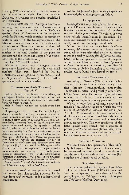
EvENHUis & Vlug: Parasites of apple tortricids 117
Herting (1965) mention it from Geometridae
and Noctuidae on apple. Thus we consider
Diadegma praerogator as a parasite, specialized
on Tortricidae.
Townes (1969) referred Diadegma interrupta
to the genus Tranosema Forster. Horstmann in
his type revision of the European Diadegmaspecies, placed D. interrupta in the subgenus
Nythobia Förster, which contains the extremely
uniform majority of the Diadegma species. Thelatter of course seriously interferes with species
identification. Often males cannot be identified
at all, because important characters, as eventual
notches in the hind borders of the posterior
sternites and the length and shape of the ovipo-
sitor, refer to the female sex only.
Adults: 13 May—7 October.
We reared some more Diadegma species
from apple leafrollers, but only in very small
numbers. They were identified by Dr. K.
Horstmann as D. apostata (Gravenhorst), and
as D. fenestralis (Holmgren). Three further
specimens could not be identified.
Tranosema arenicola (Thomson)(figs. 31, 32)
Colour characters. — Female. As in Diadegmapraerogator, however legs entirely light brown, but
fore and middle coxae darker brown or even partly
black, hind tarsus darkened.
Male. As female, but fore and middle coxae light
brown.
Morphological characters. — The morphological
characters may be treated together with those of the
other Porizontini. As their general appearance is rath-
er alike, it seems useful to compare some of their dif-
ferentiating characters here. In the three Diadegmaspecies mentioned above, the epomia is lacking
(fig. 29), whereas this is short but evident in Trano-
sema arenicola (fig. 31). The lateral carinae on the first
abdominal segment, running from its hindborder for-
ward, are evident in the three Diadegma species and
pass distinctly beneath the spiracles (fig. 30); in
Tranosema arenicola they stop distinctly quite near
the spiracle (fig. 32). As two of the Diadegma species
that we reared are not important as apple leafroller
parasites at all, it will be enough to refer to the publi-
cation of Horstmann (1969) for the differentiating
characters. Horstmann (1973) discussed the similarity
OÎ Diadegma praerogator and Tranosema arenicola.
Length female T. arenicola: 5.5—6.5 mm, oviposi-
tor: 1.5—2 mm, length male: 4.5—5.5 mm.
Tranosema arenicola was quite often reared
from several leafroller species, however, far the
most from Archips rosana. It is a solitary, larval
endoparasite.
Adults: 14 June—26 July. A single specimen
hibernated; the adult appeared on 7 May.
Campoplex spp.
Campoplex is a very large genus, like so manygenera of Porizontinae. It gives tremendous dif-
ficulties in species discrimination. No modernrevision of the genus exists. Therefore, in mostcases reliable identification is impossible. Be-
cause our material from apple leafrollers is only
scanty, we do not want to go into details.
We obtained five specimens from Pandemiscerasana, Adoxophyes orana and Acleris rhom-hana, which were identified by Dr. K. Horst-
mann as possibly Campoplex difformis Graven-
horst. Six further specimens, no doubt conspeci-
fic and of which five were reared from Spilonota
ocellana, could not be identified, just hke 13
specimens, probably belonging to at least three
species, reared from several leafroller species.
Subfamily Mesochorinae
According to Townes (1971) the species be-
longing to this subfamily are secondary para-
sites through Ichneumonidae, Braconidae,
Tachinidae (Diptera) and probably other fami-
lies as direct hosts. He does not give informa-
tion on indirect hosts. It is not excluded that
some species might be primary parasites.
We reared only four specimens, a male and a
female of Mesochorus silvarum Curtis and twomales of Stictopisthus lineatus Thomson (det.
Prof. Dr. W. Schwenke). The two specimens of
the former species were reared from the cater-
pillars of Pandemis cerasana and Adoxophyesorana, respectively. The two specimens of the
latter species were reared from a cocoon of
probably Meteorus ictericus (Braconidae) with-
out caterpillar host remains, and from a caterpil-
lar oi Adoxophyes orana, respectively.
Subfamily Metopiinae
We reared only a few specimens of this subfa-
mily, belonging to four species. They can easily
be recognized, especially by their smooth bodyand the conspicuously short and stout legs (fig.
96); they are all larval-pupal parasites.
Triclistus Förster
The western palaearctic species have been
dealt with by Aeschlimann (1973). Our material
contains two species, that were identified by Dr.
Aeschlimann as Triclistus pallipes Holmgrenand Triclistus glohulipes (Desvignes).

118 Tijdschrift voor Entomologie, deel 126, afl. 6 (1983)
Triclistus pallipes Holmgren(figs. 33, 34, 96)
Ten specimens of this species, all females,
were reared exclusively from Rhopobota nae-
vana. Aeschlimann (1973) mentions Strophedra
weirana (Lepidoptera, Tortricidae) as a host,
which species is associated with Fagus and Car-
pinus.
Adults: 16 May—15 June.
Triclistus globulipes (Desvignes)
(figs. 35—37)
Morphological characters. — Figures 33—36 showdifferences in the form of the head and in the pattern
of pubescence on the gaster in both Triclistus species.
Fig. 37 shows the propodeum of T. globulipes with
the longitudinal carinae.
Of this species we reared two females,
Archips podana being the host, and one male
from Archips xylosteana. Aeschlimann (1973)
mentions five more Tortricid host species.
Adults: 7 July—9 August.
Exochus Gravenhorst
We reared two species, each represented byone specimen, from pupae of Clepsis spectrana
and Acleris rhombana. As there is no modernrevision of the genus, it is not possible to namethe two species.
Exochus sp. 1
(fig. 38)
Colour characters. — Black. Head and mouth-parts, except ocellar area, bright yellow; scape black
and yellow, rest of antenna brown, underside lighter.
Thorax conspicuously marked with yellow; legs
bright yellow, hind femur basally and apically black.
Morphological characters. — See Exochus sp. 2.
Fig. 38 shows the propodeum with peculiarly shaped
longitudinal carinae.
This species was reared from Acleris rhom-bana.
Adult: 7july.
Exochus sp. 2
Colour characters. — Black. Antenna apically
brownish, coxae and trochantera dark brown, remain-
der of legs lighter.
Morphological characters. — Differs from the for-
mer species in having basal vein less curved and hav-
ing complete costulae.
This species was reared from Clepsis spec-
trana.
Subfamily Anomaloninae
The species of this subfamily are larval-pupal
parasites. We reared only six specimens, be-
longing to two species. Two specimens of
Habronyx (Camposcopus) canaliculatus (Ratze-
burg) were reared from Archips rosana. Fourspecimens, belonging to Agrypon anxium (Wes-mael), were reared from Acleris rhombana(three specimens) and from Spilonota ocellana
(one specimen). The subfamily was recently
treated by Gauld & Mitchell (1977).
Adults o{ Habronyx canaliculatus: 7 July—19
July. Adults of Agrypon anxium: 9 June—24
July.
Family Braconidae
Reliable identification of the species of Braco-
nidae that parasitize apple leafrollers seems only
exceptional. The difficulties will be discussed in
the special cases. As to the division into subfa-
milies we follow Van Achterberg (1976).
Subfamily Microgastrinae
Apanteles Forster
Two species of this very large genus, viz.,
A. ater (Ratzeburg) and A. xanthostigma (Hal-
iday), may be considered the most important
Apanteles species associated with apple leafrol-
lers by far (Evenhuis & Vlug, 1973). Nixon(1965, 1973) gave a division in groups; in sever-
al publications he dealt with the species in detail
(Nixon, 1972, 1973, 1974, 1976). Several groups
of Nixon are treated as independent genera byMason (1981).
We reared seven species from apple leafrol-
lers. They may be distinguished especially by
characters of the pronotum (figs. 39—45), of
colouration of the pterostigma (figs. 46—52), of
hind wing venation and of propodeum and ter-
gite of the first gastral segment (figs. 53—59).
Apanteles ater (Ratzeburg)
(figs. 39, 47, 53, 60)
Colour characters. — Female and male. Preponder-
antly black. Mouth-parts, distal part of fore femur,
fore tibia, fore tarsus, base of middle tibia, middle tar-
sus and all tibial spurs lighter. Wing veins for the
greater part pale, however costal vein and pterostigma
distinctly pigmented; the parts of 2r proximally of Rs
and short distal part of M less distinctly pigmented;
pterostigma brownish yellow in the center, margins
darker (fig. 47).
Morphological characters. — Female and male.
Propodeum with two distinct carinae, converging in
EvENHUis & Vlug: Parasites of apple tortnads 119
Figs. 33—34. TricUstHS pallipes: 33, head laterally; 34, gaster dorsally. Figs. 35—37. Triclistus globulipes: 35,
head laterally; 36, abdomen dorsally; 37, propodeum. Fig. 38. Exochus sp. 1: propodeum. Figs. 39—45. Micro-gastrinae spp., pronotum laterally: 39, Apanteles ater; 40, A. xanthostigma; 41, Dolichogenidea sicaria; 42,
D. laevigata; 43, D. corvina; 44, D. dilecta; 45, D. longicaudus.
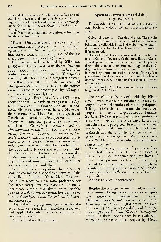
120 Tijdschrift voor Entomologie, deel 126, afl. 6 (1983)
front and thus forming a V, a litte coarse, but smooth
and shiny between and just outside the keels. First
tergite twice as long as broad, the sides rather strongly
converging distally (fig. 53). Ovipositor valves about
as long as hind tibiae.
Length female: 2—2.5 mm, ovipositor: 0.5— 1 mm,length male: 2—2.5 mm.
Nixon (1976) states that this species is poorly
characterized as a whole, but that it is easily rec-
ognizable in the female by the presence of a
fine, curved spine on the ventral side of the last
tarsal segment of the front leg (fig. 60).
This species has been treated by Wilkinson
(1945) in such an accurate way, that we have no
doubt about its identity. This author also
studied Ratzeburg's type material. The species
was originally described as Microgaster carbon-
arms Ratzeburg, 1848, but later on renamed
Microgaster ater Ratzeburg, 1852, as the former
name appeared to be preoccupied by Microgas-
ter carbonariusWesmzei, 1837.
Ratzeburg (cf. Evenhuis & Vlug, 1973) states
about the host: "Von mir aus versponnenen Ap-felblüthen erzogen, wahrscheinlich aus der bru-
mata, die diese versponnen hatte". However,
the host may as well have been some species of
Tortricidae instead of Operophtera hrumata.
"Wilkinson states the parasite to have been
reared from Cacoecia (= Archips) podana,
Hyponomeuta malinella (= Yponomeuta mali-
nellus), Tortrix {= Lozotaenia) forsterana, No-tocelia udmanniana, and a specimen from a leaf-
roller of Ribes nigrum. From this enumeration
only Yponomeuta malinellus does not belong to
the Tortricidae. It does not seem improbable
that the mention of this host is due to a mistake,
as Yponomeuta caterpillars live gregariously in
large nests and some Tortricid host caterpillar
may have been mixed with them.
According to our opinion Apanteles ater
must be considered a specialized parasite of the
caterpillars of various Tortricidae. However,
the species seems to show some preference for
the larger caterpillars. We reared rather manyspecimens, almost exclusively from Archips
podana and Pandemis heparana, but only a few
from Adoxophyes orana, Ptycholoma lecheana,
and Acleris spp.
This is the only gregarious species within the
genus that we reared from leafroUers associated
with apple. Like other Apanteles species it is a
larval endoparasite.
Adults: 25 May- 18 September.
Apanteles xanthostigma (Haliday)
(figs. 40, 46, 54)
This species is very similar to the preceding
one, both in colour and in morphological re-
spect.
Colour characters. — Female and male. The species
differs from A. ater by the centre of the pterostigma
being more yellowish instead of white (fig. 46) and in
the female sex by the legs being more extensively
light-coloured.
Morphological characters. — Female and male. Themost striking difference with the preceding species is,
according to our opinion, the structure of the propo-
deum. This contains a longitudinal trough in the mid-
dle, which is at least distinct in its hind part and there
bordered by short longitudinal carinae (fig. 54). Thepropodeum, on the whole, is also coarser. The female
does not have a curved spine on the last tarsal segment
of the fore leg.
Length female: 2.5—3 mm, ovipositor: 0.5— 1 mm,length male: 2.5—3 mm.
The species has been dealt with by Nixon
(1976), who mentions a number of hosts, be-
longing to several families of Microlepidoptera.
Telenga (1955) also enumerates the hosts, be-
longing to various Lepidopterous families.
Zwölfer (1962) characterizes its host preference
as follows: „Die von uns seit einigen Jahren un-_
tersuchte, recht polyphage Braconide Apanteles
xanthostigma Hai. beschränkt ihr Suchgebiet
praktisch auf die Strauch- und Baumschicht,
greift hier aber eine grössere Zahl von Wirten,
meist Wickler und verwandte Kleinschmetter-
lingsgruppen an".
We reared a large number of specimens from
several leafroller species of apple (cf. table 1),
but we have no experience with the hosts of
other Lepidopterous families. If indeed only
one and the same species is involved, it might be
considered a polyphagous parasite of Lepido-
ptera. Apanteles xanthostigma is a solitary en-
doparasite.
Adults: 13 May—8 September.
Besides the two species mentioned, we reared
some more Microgastrinae, however in quite
low numbers: Dolichogenidea corvina
(Reinhard) from Nixon's "metacarpalis" group;
Dolichogenidea laevigata (Ratzeburg), D. dilec-
ta (Haliday), D. sicaria (Marshall) and D. longi-
caudus (Wesmael) from Nixon's "laevigatus"
group. As these species have been dealt with
thoroughly in taxonomical respect by Nixon
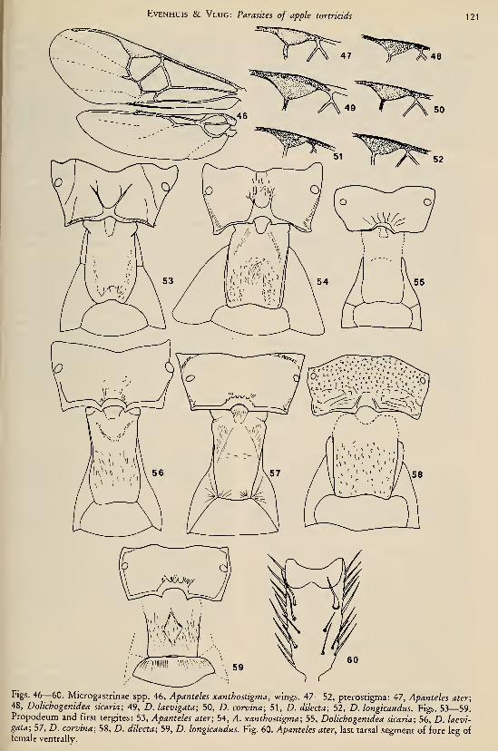
EvENHUis & Vlug: Parasites of apple tortncids 121
Figs. 46—60. Microgastrinae spp. 46, Apanteles xanthostigma, wings. 47—52, pterostigma: 47, Apanteles ater;
48, Dolichogenidea sicaria; 49, Z). laevigata; 50, D. corvina; 51, D. dilecta; 52, D. longicaudus. Figs. 53—59.Propodeum and first tergites: 53, Apanteles ater; 54, ^. xanthostigma; 55, Dolichogenidea sicaria; 56, D. /aeuï-gaf<2; 57, D. corvina; 58, Z). dilecta; 59, Z). longicaudus. Fig. 60. Apanteles ater, last tarsal segment of fore leg offemale ventrally.
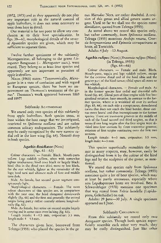
122 Tijdschrift voor Entomologie, deel 126, afl. 6 (1983)
(1972, 1973) and as they apparently do not play
any important role in the natural control of
apple leafrollers, it does not seem necessary to
treat them here in detail.
Our material is far too poor to allow any con-
clusion as to their host specialization. In
figs. 39—60, morphological details of the sever-
al Apanteles species are given, which may be
sufficient to separate them.
Twelve further specimens of the subfamily
Microgastrinae, all belonging to the genus Lis-
sogaster Bengtsson (= Microgaster auct.), were
reared. They belong to at least four species and
apparently are not important as parasites of
apple leafrollers.
Nixon (1965) states: "Taxonomically, Micro-
gaster is in a state of confusion and with regard
to European species, there has been no im-
provement on Thomson's treatment of the ge-
nus". Thomson's work appeared between 1869
and 1897!
Subfamily Agathidinae
We reared only two species of this subfamily
from apple leafrollers. Both species seem, at
least within the host range that we investigated,
restricted to Spilonota ocellana and are rather
common solitary endoparasites. The subfamily
may be easily recognized by the very narrow ra-
dial cell in the fore wing (fig. 64). Notauli deep
in both species.
Agathis dimidiator (Nees)
(figs. 61—63)
Colour characters. — Female. Black. Mouth-parts
yellow. Legs reddish yellow, often with somewhat
lighter trochantera; hind coxa black or largely black;
hind tibia in the distal two third more or less black,
for the rest whitish, lighter as the remainder of the
legs; hind tarsi and ultimate ends of fore and middle
tarsi dark.
Male. As female, but second gaster segment con-
spicuously light.
Morphological characters. — Female. The most
salient characters of this species are, in comparison
with the next one, the head elongated downwardsslightly (fig. 61) and the first and generally the second
tergite being partly rather coarsely striated longitudi-
nally (fig. 62).
Male. As female, but striae on second tergite largely
varying and in some cases even lacking (fig. 63).
Length female: 4—4.5 mm, ovipositor: 3.5 mm,length male: 4—4.5 mm.
The characters given here, borrowed fromTelenga (1955) who placed the species in the ge-
nus Microdus Nees, are rather doubtful. A revi-
sion of this genus and allied genera seems ur-
gent. Until so far we shall use the species namedimidiator, quoted from Telenga (1955).
As stated above we reared this species only,
but rather commonly, from Spilonota ocellana.
Telenga (1955) mentions Archips rosana, Croe-
sia bergmanniana and Epinotia tetraquetrana as
hosts, all Tortricids.
Adults: 4 July—13 August.
Agathis rufipes (Nees) (Braunsia rufipes (Nees)
(in Telenga, 1955))
(figs. 64—66)
Colour characters. — Female and male. Black.
Mouth-parts, tegula and legs reddish yellow, except
for the extreme distal end of the hind tibia and the
greater part of the hind tarsus; tibial spurs all more or
less whitish.
Morphological characters. — Female and male. Asin the former species first radial and discoidal cells
fused (fig. 64). Distal part of first tergite with fine lon-
gitudinal striation; base smooth, in contrast to the for-
mer species, where it is wrinkled all over its surface
(figs. 65, 66); on each side a conspicuous, dorsolateral
carina that does not reach the hind border, these cari-
nae more lateral and thus inconspicuous in the former
species. There are transverse grooves in the middle of
each of the fused second and third tergites, so that it
seems if one long tergite after the first tergite were di-
vided into four sections by three transverse grooves;
striation of first tergite continuing onto the first two
sections.
Length female: 4—5 mm, ovipositor: 3.5 mm,length male: 4—5 mm.
This species superficially resembles the for-
mer in many respects, may, however, easily be
distinguished from it by the colour of the hind
legs and by the sculpture of the gaster, as men-
tioned.
We reared this species only from Spilonota
ocellana, but rather commonly. Telenga (1955)
mentions quite a lot of host species, which mayat least be partly erroneous, especially where
non-Lepidopterous hosts are involved. Betz &Schwerdtfeger (1970) mention one specimen
that was reared from Teleia luculella (Lepido-
ptera, Gelechiidae) on oak.
Adults: 29 June—30 July. A single specimen
appeared on 8 June.
Subfamily Cheloninae
From this subfamily we reared only two
Ascogaster species. Though both species super-
ficially resemble each other very much, they
may be easily distinguished. Just like other
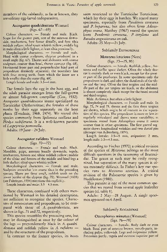
EvENHUis & Vlug: Parasites of apple tortncids 123
members of the subfamily, as fas ar known, they
are sohtary egg-larval endoparasites.
Ascogaster quadridentata Wesmael(figs. 67—69)
Colour characters. — Female and male. Black.
Scape for the greater part, rest of the antenna down-side, trochantera, fore femur distally, and fore tibia
reddish yellow; tibial spurs whitish yellow; middle leg
in male often a little lighter, at least tibia proximally.
Morphological characters. — Female and male.
Head with coarse sculpture, clypeus produced in a
small angle (fig. 67). Thorax and abdomen with coarse
sculpture, coarser than head, thorax coarsest (fig. 68).
Horizontal front part and perpendicular hind part of
propodeum distinctly separated; the boundary line
with four strong teeth, from which the inner are a
httle smaller than the outer (fig. 69).
Length female and male: 3.5—4 mm.
The female lays the egg in the host egg, and
the adult parasite emerges from the full-grown
or almost full-grown caterpillar (Cox, 1932).
Ascogaster quadridentata seems specialized on
Tortricidae Olethreutinae; the females of these
moths lay their eggs separately on the host
plants (Evenhuis, 1969, 1974). We reared this
species commonly from Spilonota ocellana and
Hedya nubiferana. It is a well-known parasite
of Laspeyresia pomonella.
Adults: 19 June—24 July.
Ascogaster rufidens Wesmael(figs. 70—72)
Colour characters. — Female and male. Black.
Mandible, palps, antenna basally downside, tegula,
trochantera, femora and tibiae reddish yellow; middle
of the tibiae and femora of the middle and hind legs a
little darker; tibial spurs whitish yellow.
Morphological characters. — Female and male.
Sculpture somewhat less coarse than in the preceding
species. There are three small, reddish teeth on the
lower border of the clypeus (fig. 70). Wesmael (1835)
(see Fahringer, 1934) mentions this explicitly.
Length female and male: 3.5—4.5 mm.
These characters, combined with others men-tioned by Wesmael in his detailed description,
are sufficient to recognize the species. Charac-
ters of mesoscutum and propodeum, to be com-pared with those of A. quadridentata, are
shown in figs. 71 and 72, respectively.
This species resembles the precedmg one, but
may be distinguished at once by the colour of
the legs — preponderantly black in A. quadri-
dentata and reddish yellow in A. rufidens —and by the structures of the propodeum.
In contrast to the former species, its hosts
seem restricted to the Tortricidae Tortricinae,
which lay their eggs in batches. We reared manyspecimens, especially from Pandemis cerasana
and P. heparana, but also often from Adoxo-phyes orana. Matthey (1967) reared the species
from Pandemis cerasana, P. corylana and
Batodes angustiorana on oak.
Adults: 25 May—21 July.
Subfamily Euphorinae
Meteorus ictericus (Nees)
(figs. 73—75, 95)
Colour characters. — Female. Reddish yellow. An-tenna and ocellar space darker. Often the whole tho-
rax IS entirely dark or even black, except for the great-
er part of the prothorax, in some specimens only the
propodeum is dark and often the distal ends of the tib-
iae and the tarsi. Often the first tergites and the mid-
dle part of the last tergites are black, or the abdomenis almost completely black except for the fused second
and third tergites.
Male. As female, but often a little lighter.
Morphological characters. — Female and male. In
figs. 73, 74 and 75, thorax and the first three tergites
are shown, which might be of interest in the separa-
tion of this species. The propodeum is more or less ir-
regularly reticulated and shows some variability; in
specimens reared from Adoxophyes orana it seems
coarser than in other specimens. The first gastral seg-
ment shows longitudinal wrinkles and two dorsal pits
(dorsope: van Achterberg, 1974).
Length female: 3.5—5 mm, ovipositor: 2 mm,length male: 3.5—4.5 mm.
According to Fischer (1970) a critical revision
of the species of Meteorus belongs to the most
difficult questions in the taxonomy of Braconi-
dae. The genus as such may be easily recog-
nized, but separation of the many species is of-
ten extremely difficult. In Fisher's key our spe-
cies runs to Meteorus ictericus. A critical
revision of the Palaearctic species is given by
Huddleston(1980).
This parasite is one of the most common spe-
cies that we reared from several apple leafroller
species (cf. table 1).
Adults: 2 May—28 August. A single speci-
men appeared on 6 April.
Subfamily Rogadinae
Oncophanes minutus (Wesmael)
(figs. 76—78)
Colour characters. — Female. Body dark or even
black. Basal part of antenna brown, mouth-parts, in-
cluding palps, yellowish. Legs and ovipositor yellow.
Pronotum partly, tegula and extreme superior part of

124 Tijdschrift voor Entomologie, deel 126, afl. 6 (1983)
Figs. 61—63. Agathis dimidiator: 61, head anteriorly; 62, gaster female dorsally; 63, abdomen male dorsally.
Figs. 64—66. Agathis rufipes: 64, wings; 65, gaster female dorsally; 66, gaster male dorsally. Figs. 67—69. Asco-
gaster quadridentata: 67, front edge of clypeus; 68, thorax dorsally; 69, propodeum. Figs. 70—72. Ascogaster
rufidens: 70, front edge of clypeus; 71, thorax dorsally; 72, propodeum.

EvENHUis & Vlug: Parasites of apple tortncids 125
Figs. 73—75. Meteorus ictericus: 73, thorax dorsally; 74, propodeum; 75, first three tergites. Figs. 76—78. On-cophanes minutus: 76, thorax dorsally; 77, propodeum; 78, first three tergites. Figs. 79—80. Bracon sp.: 79, tho-
rax dorsally; 80, propodeum. Figs. 81—87. Macrocentrus linearis: 81, head anteriorly; 82, mandible; 83, thorax
dorsally; 84, wings; 85, tarsal claw; 86, propodeum; 87, first three tergites.
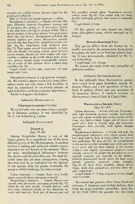
126 Tijdschrift voor Entomologie, deel 126, afl. 6 (1983)
mesopleurum reddish brown. Second tergile for the
greater part yellowish.
Male. As female, but second tergile not as yellow.
Morphological characters. — Female and male. The
exact difference between Oncophanes minutus and
O. laevigatus (Ratzeburg) (= O. lanceolator (Nées))
is not clear from Fahringer's revision (1930). This is
the last revision of the genus anyway! Occipital carina
above fine, but distinct. Mesopleurum polished, but
extreme superior part coarse. Mesoscutum sparsely
covered with fine long hairs, parapsidal furrows evi-
dent (fig. 76). Propodeum with delimited areas
(fig. 77). First tergile striated longitudinally, in front
with two distally converging carinae, space between
carinae smooth. Second and third tergites only sepa-
rated by a shallow, in the middle almost faded trans-
verse groove, second tergile longitudinally striated.
The extension of this striation shows a rather large
variability (fig. 78).
Length female: 2—2.5 mm, ovipositor: 0.5 mm,length male: 2 mm.
Oncophanes minutus is a gregarious ectopara-
site. We reared it almost exclusively from Adox-
ophyes orana and only from a few localities. It
must be considered an occasional parasite of
apple leafrollers, without economic importance.
Adults: 6 August—26 August.
Subfamily Homolobinae
Charmon cruentatus Haliday
We reared only one specimen from a caterpil-
lar of Spilonota ocellana. It was identified by
Dr. C. van Achterberg, Leiden.
Subfamily Braconinae
Bracon sp.
(figs. 79, 80)
Among Braconidae Bracon is one of the
largest genera. It is considered one of the most
difficult genera of the Hymenoptera. A modernrevision is lacking and sufficient reliable species
characters are not known at the moment. There
are many morphological differences between
this species and the preceding one. We shall de-
scribe some that are most conspicuous, hoping
that they may be an indication for the identity
of the species, after the genus, eventually the
group to which it belongs, has been revised.
Colour characters. — Female. Body dark, locally
lighter, especially thorax and gaster downside.
Morphological characters. — Female. Mesoscutumsmooth, with percurrent, converging notauli (fig. 79),
which do not meet distally. Notauli shallow, with
long setae, orientated mainly in two directions, in-
wards and outwards. Hind part of mesoscutum and
also scutellum densely pilose. Propodeum smooth,
without areas (fig. 80). First tergite with two deep,
distally converging grooves that enclose a triangular
field.
Length female: 2.5 mm.
Of this species we only reared one female
from Adoxophyes orana, which emerged on 12
August.
Bracon obscurator Nees
This species differs from the former by its
smaller size and by the mesoscutum being muchless pilose, the hairs on its hind part almost lack-
ing. It was, provisionally, identified by Dr. C.
van Achterberg.
Length male: 1.5—2 mm.We reared two males from one caterpillar of
Spilonota ocellana.
Subfamily MacROCentrinaeIn this subfamily three Macrocentrus species
were reared from apple leafrollers, mainly M.
linearis (Nees), and a few specimens of the re-
lated M. pallipes (Nees) and one specimen of
M. thoracicus (Nees). A modern revision of
Macrocentrus is that of Eady & Clark (1964).
Macrocentrus linearis (Nees)
(figs. 81—87)
Colour characters. — Female and male. Preponder-
antly light brownish, however variable. Interocellar
space and a point on both sides of the insertion of the
wings very dark to black. Upper side of thorax and
gaster often dark in varying tinge and extension.
Pterostigma dark centrally, with lighter margins
(fig. 84).
Morphological characters. — Female and male. As
morphological differences with related species have
been dealt with by Eady & Clark (1964), we refer to
that publication. We may add that the mesopleurum
in this species is rather uniformly and widely punctate
at its centre and not more densely in the furrow,
which is only sHghtly pronounced; prepectal carina
strongly curved (in M. pallipes the mesopleurum is ir-
regularly punctate, and especially in the shallow,
however pronounced furrow above the hind coxae
densely punctate; prepectal carina straight).
Figs. 81—87 show head, mandible, thorax above,
wings, tarsal claw, propodeum, and first three ter-
gites, respectively.
Length female: 4—5 mm, ovipositor: 6 mm, length
male: 4—5 mm.
We reared this parasite often from Pandemis
cerasana, P. heparana and Archips podana, thus
from the larger leafroUer caterpillars. After Ea-
dy & Clark (1964) the parasite has also been

EvENHUis & Vlug: Parasites of apple tortnads \17
reared from other Microlepidoptera than Tor-
tricidae.
It is a gregarious larval endoparasite, appar-
ently showing polyembryony. In most cases the
progeny of one single host consists of either
males or females exclusively. However, in a
number of cases we reared both sexes from one
single host, probably indicating that then at
least two eggs were laid in a single host, giving
rise to a female and a male progeny respectively.
Adults: 26 June—11 August.
Macrocentrus pallipes (Nees)
This species is very similar to the preceding
one. Differentiating characters have been given
by Eady & Clark (1964); a few supplementary
characters were mentioned under M. linearis.
We reared 18 female specimens from one sin-
gle caterpillar of Hedya nuhijerana.
One specimen of Macrocentrus thoracicus
(Nees) was reared from a caterpillar of Spilono-
ta ocellana. It was, just like M. linearis and
M. pallipes, identified by Dr. C. van Achter-
berg, Leiden.
Superfamily Chalcidoidea
This superfamily of parasitic Hymenoptera
has found a number of prolific workers during
the last decennia. Especially from the groups
that interest us as parasites of apple leafrollers,
there exist some modern taxonomie revisions.
Only one species, Colpoclypeus florus, is of
real importance in respect to apple leafrollers;
the others were only reared in small numbers.
Thus we will not treat them in detail and, as far
as their identity is involved, refer to the revi-
sions.
Family Pteromalidae
Habrocytus chrysos (Walker)
(fig. 101)
This species has been dealt with by Graham(1969). We reared only three specimens as hy-
perparasites of the caterpillars of Adoxophyes
orana through Scambus brevicornis, Onco-
phanes minutus and an unidentified Ichneumo-
nid, respectively. Graham mentions some more
hosts, so that H. chrysos must be considered a
widely specialized hyperparasite of various
Lepidoptera, through various species of parasit-
ic Hymenoptera as direct hosts.
Adults: 28 September.
Habrocytus semotus (Walker)
We reared this species three times from apple
leafrollers, namely once as a hyperparasite of
Archips podana through Scambus brevicornis
and two times from pupae of Adoxophyes ora-
na, possibly as hyperparasites.
Graham (1969) mentions the parasite both as
a primary and as a secondary parasite of various
Lepidoptera, but also from certain Coleoptera
and in one case from Hymenoptera Diprioni-
dae.
Adults: 25 August—25 September.
Dibrachys cavus (Walker)
(figs. 97, 102)
According to Graham (1969) the species of
Dibrachys have not yet been fully sorted out.
On the authority of Peck (1963) he mentions
Dibrachys cavus to be a very polyphagous para-
site, usually hyperparasitic.
We reared only two small specimens from co-
coons of Apanteles ater, parasitizing a caterpil-
lar of Pandemis sp. and two specimens from a
cocoon of Diadegma sp., Archips podana being
the host.
Adults: 6 July.
Family Eulophidae
Pnigalio pectinicornis (Linnaeus)
We reared only two specimens of this species,
the hosts being caterpillars of Adoxophyes ora-
na and Rhopobota naevana, respectively.
Askew (1968) mentions it as a parasite of
Microlepidoptera, especially of Lithocolletis (=
Phyllonorycter), which are leafminers.
Adults: 4 September.
Sympiesis xanthostoma (Nees)
We reared two specimens, both from caterpil-
lars of Adoxophyes orana. Askew (1968) men-
tions it a quite common parasite, especially of
Tortricidae and Gracilariidae.
Adults: 30 August and 4 October.
Colpoclypeus florus (Walker)
(figs. 98, 103)
This is no doubt the most important parasite
of Adoxophyes orana in apple orchards. Wereared it in large numbers in some orchards dur-
ing late summer and early autumn. There seems
to be a serious discrepancy between the pheno-
logy of the parasite and of Adoxophyes orana, as
we were not able to recover the parasite before

128 Tijdschrift voor Entomologie, deel 126, afl. 6 (1983)
100
Figs. 88—95. Fore wing: 88, Lysibia nana; 89, Teleutaea striata; 90, Glypta varicoxa; 91, Mesochorus sp.; 92,
Habronyx canaliculatus; 93, Agrypon anxium; 94, Lissogaster sp.; 95, Meteorus ictericus. Fig. 96. Tridistus pal-
lipes, fore leg. Figs. 97—98. Fore tibia: 97, Dibrachys cavus; 98, Colpodypeus florus. Fig. 99. Apophua dcatrico-
sa, propodeum. Fig. 100. Diadegma sp., first tergite. Figs. 101—102. Fore wing: 101. Habrocytus chrysos; 102,
Dibrachys cavus. Fig. 103. Colpodypeus florus, front margin of clypeus.

EvENHUis & Vlug: Parasites of apple tortricids 129
July (Evenhuis, 1974b). During and after this
month its population increases rapidly. The dis-
crepancy makes the feasibility of the parasite in
integrated control doubtful. In spite of efforts in
later years, we could not find any alternative
leafroUer host on which it could maintain itself
and reproduce, neither in apple orchards, nor
outside on other foodplants than apple. Thusthe problem remains unsolved. More knowl-
edge of the bionomics and ecological particu-
larities of the parasite seems a prerequisite in us-
ing it in integrated control in apple growing.
Adults: From the end of July until October.
It is interesting that in Swiss material, kindly
sent to us by Dr. A. Schmid, Nyon, Switzer-
land, four additional species of parasitic
Hymenoptera, reared from apple leafroUers, but
only in small numbers, are represented: Sympie-
sis acalle (Walker) from Adoxophyes orana,
Dicladocerus westwoodii Westwood from Ar-
chips sp., Euderus alhitarsis Zetterstedt, and Go-niozus claripennis Förster, both from Adoxo-phyes orana. The three former species are Chal-
cidoidea Eulophidae, the latter belongs to the
Bethylidae.
Key to the adult parasites of apple
leafrollers in the netherlands
The characters used in this key are simple.
Differences between the taxa only refer to spe-
cies that are dealt with in the body of the paper.
Where species identification is very doubtful,
the key generally does not run to species level,
especially when the species are no important
apple leafroller parasites.
1. Fore wings with longitudinal veins and
cross veins, delimiting several cells (cf.
figs. 88—95); (Ichneumonoidea) 2
— Venation of fore wing strongly reduced
(figs. 101, 102). Small species with metallic
green or blue body; (Chalcidoidea) 44
2. Second recurrent vein present (figs. 88—93:
srv); first cubital cell and discoidal cell
fused (figs. 88—93). Second and third gas-
ter segments separated (cf. figs. 3, 14, 15,
34, 36); (Ichneumonidae) 3
— Second recurrent vein lacking; first cubital
cell (Cu) and discoidal cell (Di) separated
by a vein (figs. 94, 95). Second and third
gaster segments fused (cf. figs. 62, 63, 65,
66, 75, 78, 87); suture, however, generally
visible; (Braconidae) 25
3. Gaster depressed, petiole broadly attached
to propodeum (cf. figs. 14, 15) 4
— Gaster compressed, petiole proximally slen-
der (cf. figs. 30, 32, 100) 18
4. Tergites 2, 3 and 4 with oblique furrows (cf.
figs. 14, 15) 5
— Tergites 2, 3 and 4 without furrows (cf.
figs. 3, 34, 36) 9
5. Areolet present (fig. 89: ar). Species gay-
coloured black and yellow
Teleutaea striata
— Areolet absent (fig. 90). Yellow markings
less conspicuous 6
6. Frontal tibial spur reaching to middle of
first tarsal segment or beyond (fig. 19) . . . 7
— Frontal tibial spur not reaching to middle of
first tarsal segment (figs. 23, 27). Genal ca-
rina not strongly smuated (fig. 25)
Glypta varicoxa
7. Genal carina strongly sinuated (fig. 21);
clypeus with small incision (fig. 20)
Glypta nigrina
— Genal carina moderately sinuated (fig. 17);
clypeus without incision (fig. 16) 8
8. Propodeal carinae strongly developed
(fig. 99) Apophua cicatricosa
— Propodeal carinae more or less obliterated . .
Apophua sp.
9. Clypeus and face conspicuously strongly
convex (figs. 33, 35). Legs, especially femo-
ra, conspicuously stout (fig. 96) 10
— Clypeus and face not convex. Legs rather
slender 12
10 Antennal sockets separated by a high lamel-
la, the latter with a deep median groove
dorsally, just below ocellus 11
— Antennal sockets not separated by a lamella
Exochus spp.
11. Tergites almost completely pubescent
(fig. 36) Triclistus globulipes
— Tergites almost completely glabrous, (fig.
34) Triclistus pallipes
12. Inner orbits at the level of the antennal
sockets deeply incised (figs. 5, 7, 8) 13
— Inner orbits not or weakly incised (fig. 1)
15
13. Head of male with yellow markings. Ovi-
positor curved downwards (fig. 9)
Apechthis spp.
— Head of male black. Ovipositor straight
(fig- 4) 14
14. Head beneath antennae and centre of meso-
scutum finely punctate (fig. 5). Tergites
wholly black Itoplectis alternans
— Head beneath antennae and centre of meso-
scutum roughly punctate (fig. 7). Hind bor-
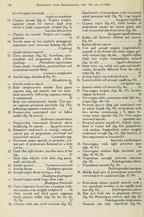
130 Tijdschrift voor Entomologie, deel 126, afl. 6 (1983)
der of tergites brownish
Itoplectis maculator
15. Clypeus incised (fig. 1). Tergites roughly
punctate except for narrow hind strip
which is finely transversely striated (fig. 3)
Scambus brevicornis
— Clypeus not incised. Tergites not roughly
punctate 16
16. Areolet more or less regularly pentagonal,
sometimes outer crossvein lacking (fig. 88)
Cryptinae
— Areolet not pentagonal 17
17. Claws pectinate (fig. 10). Scutellum, post-
scutellum and propodeum with yellow
markings Phytodietus segmentator
— Claws simple. Scutellum, postscutellum and
propodeum entirely black
Lissonota complicator
18. Areolet large, rhombical (fig. 91)
Mesochorus sp.
— Areolet small or absent 19
19. Body conspicuously slender. First gaster
segment long and narrow, but not petio-
lated anteriorly, following segments strong-
ly compressed 20
— Body not conspicuously slender. First gas-
ter segment petiolated anteriorly (fig. 100),
following segments compressed 21
20. Postnervulus intercepted near or below
middle (fig. 92: arrow)
Habronyx canaliculatus
— Postnervulus intercepted distinctly above
middle (fig. 93: arrow) . . . Agrypon anxium
21. Pronotum moderately to strongly striated;
central part of propodeum excavated and
transversely striated Campoplex spp.
— Pronotum rather smooth or punctate; cen-
tral part of propodeum flattened or a little
convex 22
22. Hind tibia light brown, just like most of the
legs 23
— Hind tibia whitish, with dark ring proxi-
mally and distally 24
23. Areolet present Tranosema arenicola
— Areolet absent Diadegma apostata
24. Second tergite about as long as wide
Diadegma praerogator
— Second tergite much longer than wide
Diadegmafenestralis25. Gaster segments fused into a carapace with-
out sutures, oval, roughly sculptured ... 26
— Only second and third gaster segments
fused, sutures visible (figs. 62, 63, 65, 66,
75, 78) 27
26. Clypeus with two small incisions (fig. 70).
Hind border of propodeum with two lateral
spinal processes only (fig. 72). Legs largely
rufous Ascogaster rufidens
— Clypeus entire (fig. 67). Hind border of
propodeum except for lateral spines also
with two medial spinal processes (fig. 69) . . .
Ascogaster quadridentata
27. Radial cell distinctly defined and narrow
(fig-.64) 28
— Radial cell not narrow 29
28. First and second tergites longitudinally
striated, in the female also third tergite par-
tially striated (figs. 65, 66) . . Agathis rufipes
— Only first tergite longitudinally striated
(figs. 62, 63) Agathis dimidiator
29. Some veins, e.g. radial vein, pale and hardly
visible, distinctly different from the other
veins (fig. 46) 30
— At least radial vein well-developed 37
30. Second cubital cell present, small (fig. 94) . .
.
Lissogaster spp.
— Second cubital cell absent (fig. 46) 31
31. Pterostigma hyahne (figs. 46, 47), borders
slightly darkened 32
— Pterostigma dark, sometimes with light bas-
al spot (figs. 48—52) 33
32. Pronotal groove deep and crenulated over
its whole length (fig. 39); propodeum with
two carinae, constituting a V, space be-
tween rather smooth (fig. 53). First tergite
narrowed apically Apanteles ater
— Pronotal groove superficial, slightly crenu-
lated in caudal half (fig. 40); propodeum
with median, longitudinal, rather roughly >
sculptured trough (fig. 54). Side borders of
first tergite more or less parallel
Apanteles xanthostigma
33. Pterostigma with light proximal spot
(figs. 49, 51) 34
— Pterostigma without light proximal spot
(figs. 48, 50, 52) 35
34. Propodeum strongly punctate reticulate
(fig. 58) Dolichogenidea dilecta
— Propodeum smooth (fig. 56)
Dolichogenidea laevigata
35. Middle hind part of propodeum excavated,
crenulated in its caudal end (figs. 57, 59) ....
36
— Propodeum almost entirely smooth, with a
few superficial wrinkles in the middle hind
part (fig. 55) Dolichogenidea sicaria
36. Rim near hind side of pronotum distinct
(fig. 45). Pterostigma entirely dark (fig. 52)
Dolichogenidea longicaudus
— Pronotal rim only superficial (fig. 43).

EvENHUis & Vlug: Parasites of apple tortriads 131
Centre of pterostigma somewhat lighter
(fig. 50) Dolichogenidea corvina
37. Second tergite at least as long as wide ... 38
— Second tergite distinctly wider than long
41
38. Mesoscutum divided into three vaulted
lobes, the middle the highest (fig. 83) ... 39
— Mesoscutum more or less flat, not divided
Charmon cruentatus
39. Pterostigma almost colourless (fig. 84)
Macrocentrus thoracicus
— Pterostigma dark, proximally somewhat
lighter 40
40. Body predominantly rufous
Macrocentrus linearis
— Body predominantly black
Macrocentrus pallipes
41. First gastral segment narrow proximally,
with two deep pits (dorsope) in front of the
middle (fig. 75) Meteorus ictericus
— First gastral segment broadly inserted to
propodeum, without deep dorsope (fig. 78)
42
42. Propodeum with dilimited areas, separated
by irregular carinae (fig. 77)
Oncophanes minutus— Propodeum without delimited areas (fig.
80) 43
43. Mesoscutum densely pilose (fig. 79)
Bracon sp.
— Mesoscutum less pilose, hind part almost
bare Bracon obscurator
44. Fore tibiae with large, curved spur (fig. 97)
45
— Fore tibiae with short, straight spur (fig. 98)
46
45. Postmarginal vein as long as stigmal vein
(fig. 102: arrow) Dibrachys cavus
— Postmarginal vein longer than stigmal vein
(fig. 101 : arrow) Habrocytus spp.
46. Anterior margin of clypeus medially nar-
rowly incised (fig. 103) Colpoclypeus florus— Clypeus not incised 47
47. Propodeum with carinae
Pnigalio pectinicornis
— Propodeum without carinae
Sympiesis xanthostoma
Discussion
Among the Hymenopterous parasites, asso-
ciated with apple leafrollers, some species were
reared commonly and in rather large numbers,
whereas most of them were only obtained occa-
sionally. Many of these latter are no rare species
whatever, but do not really belong to the apple
orchard community (tab,le 1).
In provisional investigations parasites were
also reared by collecting leafroUer caterpillars
on other foodplants and in different habitats.
Many of these parasites belong to other species
than those from apple orchards or to the samespecies but in entirely different ratios. The samefeature holds also for some leafroUer species,
e.g., Adoxophyes orana was only found occa-
sionally outside apple orchards. If these investi-
gations might prove to have a general bearing, it
could mean that leafroUer caterpillars in habitats
adjoining apple orchards, only play a minor role
as a reservoir for apple leafroUer parasites. It
must be born in mind that there are several
types of apple orchards, with different ecologi-
cal conditions to satisfy the needs of the several
parasite species. As an example of the special
needs of a leafroUer parasite might be men-tioned Zwölfer's remark concerning Apanteles
xanthostigma, the female of which only looks
for its hosts in the shrubs and the lowermost
parts of the trees (Zwölfer, 1962). Such ecologi-
cal preferences might at least partly explain the
scarcity in apple orchards of so many parasite
species that are quite common elsewhere.
It is obvious that numerous investigations are
still necessary for obtaining a more clear insight
in the intricate interrelationships between the
different leafroUer species on apple and their
parasites. By rearing parasites from knownhosts, large series of parasites belonging to one
species, may be obtained. Rearing not only
gives information about host specificity of the
parasite, but also may be of interest in under-
standing species variation.
It does not seem superfluous to emphasize
the tremendous importance of a sound taxo-
nomical base in distinguishing the species. Fromour investigations it may be concluded that for
most groups of Hymenopterous leafroUer para-
sites this base is not present and this might ap-
ply as well to the group of parasitic Hymeno-ptera as a whole.
Parasitic Hymenoptera constitute an insect
group very rich in species. We estimate that
Dutch species far outnumber 7000, the total
number of insect species in the Netherlands
probably not exceeding 25,000. We do not ven-
ture to give figures for larger areas, but we sup-
pose that parasitic Hymenoptera are extraordi-
nary numerous throughout the world. Large
genera with quite similar species are no excep-

132'
Tijdschrift voor Entomologie, deel 126, afl. 6 (1983)
Table 1. Enumeration of parasitic Hymenoptera with the numbers that were reared from their apple leafroller
hosts.
1
EvENHUis & Vlug: Parasites of apple tortncids 133
tion in this group. Thus it is hardly surprising
that identification meets with very large diffi-
culties. Yet, exact species discrimination seems
essential, as very similar species show consid-
erable differences in biological and ecological
respects.
Descriptions of species by the earlier taxo-
nomists are generally too poor to allow of any
conclusion about their identity. They often suit
more than one species. Later authors, who tried
to interpret them in their own way, added newspecies, just as ill-described. In this way taxo-
nomie literature has been overburdened with
"species" from which no one can establish their
identity to-day.
In the past a number of taxonomie works
have been written, dealing with larger groups of
parasitic Hymenoptera and based mainly on a
compilation of these insufficiently described
species, which were often not fully understood
by the compilators themselves. Some examples
of these works are Schmiedeknecht's Opuscula
Ichneumonologica (1902—1936), Fahringer's
Opuscula Braconologica (1928—1937), Kief-
fer's work in "Das Tierreich" on Proctotrupoi-
dea (191^1—1926) and that of Dalla Torre &Kieffer on Cynipoidea (1910), also in "Das
Tierreich". It is not our intention to criticize
these works. They present excellent surveys of
the knowledge of the several species at that
time, and any specialist who occupies himself
with these groups now, has to consult them.
However, they do not satisfy the needs of a
modern entomologist who wants to know the
reliable names and taxonomical status of the
species which he encounters in his ecological m-vestigations. Unreliable species names of parasi-
tic Hymenoptera show up in reviewing jour-
nals, e.g., The Review of Applied Entomology,
and from these they are compiled in compre-
hensive works like Thompson's host-parasite
lists.
In view of the difficulties discussed above, it
cannot be expected that the gap in our taxo-
nomie knowledge of the species of parasitic
Hymenoptera will be filled up within a reasona-
ble time. Fortunately in the last decades an in-
creasing interest in the taxonomy of parasitic
Hymenoptera has arisen, and many groups have
now been or will soon be revised.
Ecologists have to rely on identifications by
specialists. Only these specialists are thoroughly
acquainted with the taxonomie difficulties and
their nomenclatorial consequences. Only a spe-
cialist in a certain not too large taxonomie
group can really judge in how far his identifica-
tions are reliable.
It might be desirable that a specialist, in iden-
tifying material on behalf of ecological investi-
gations, should add some more information
than merely the name. The ecologist should also
be informed about the reliability of the species
name, which can only be judged by the special-
ist.
Acknowledgements
We wish to thank the following specialists in
the various taxonomie groups of parasitic
Hymenoptera: Dr. Ing. C. van Achterberg, the
Netherlands, Dr. J. P. Aeschlimann, France,
Mr. M. J. Gijswijt, the Netherlands, Dr. K.
Horstmann, Bundesrepublik Deutschland, Dr.
W. R. M. Mason, Canada, Dr. G. E. J. Nixon,
United Kingdom, Mr. G. van Rossem, the
Netherlands, Prof. Dr. W. Schwenke, Bundes-
republik Deutschland, and Drs. K. W. R.
Zwart, the Netherlands. Without their help
with the identifications and their advice wecould not have completed these investigations.
Thanks are also due to Prof. Dr. J. T. Wiebes
for critically reading the manuscript. Dr. P.
Gruys, Drs. D. J. de Jong and Ir. P. M. J. Ra-
makers, all from the Netherlands, and Dr. A.
Schmid, Switzerland, for providing material, to
Ir. A. B. A. M. Baudoin and Ir. E. Neering,
both students of the Agricultural University at
Wageningen at that time, who investigated the
parasites of leafroUers on several foodplants in
habitats outside apple orchards, and to Mr. C.
van der Horst, IPO, Wageningen, for revising
the English text.
References
Achterberg, C. van, 1974. The features of the petiolar
segment in some Braconidae. — Ent. Ber., Amst.
34:213—214.
, 1976. A preliminary key to the subfamilies of
the Braconidae (Hymenoptera). — Tijdschr. Ent.
119:33—78.
, 1982. Familietabel van de Hymenoptera in
Noordwest Europa. — Wet. Meded. K. Ned. na-
tuurh. Veren. 152: 1—50.
Aeschlimann, J. P., 1973. Révision des espèces ouest-
paléarctiques du genre Tuclistus Foerster
(Hymenoptera: Ichneumonidae). — Mitt.
Schweiz, ent. Ges. 46: 219—252.
Ankersmit, G. W., 1968. The photoperiod as a control
agent against Adoxophyes reticulana (Lepido-
ptera; Tortricidae). — Entomologia exp. appi. 11:
231—240.

134 Tijdschrift voor Entomologie, deel 126, afl. 6 (1983)
Askew, R. R., 1968. Hymenoptera Chalcidoidea,
Elasmidae and Eulophidae (Elachertinae, Eulophi-
nae, Euderinae). — Handbks Identif. Br. Insects 8
(2b): 1—39.
Aubert, J. T., 1978. Les Ichneumonides ouest-palé-
arctiques et leurs hôtes 2. Banchinae et suppl. au
Pimplinae. — ÉDIFAT-OPIDA, Echauffour: 1—318.
Auersch, O., 1960. Weitere Beobachtungen zum Auf-
treten des Fruchtschalenwicklers (Apfelschalen-
wicklers) Adoxophyes reticulana Hb. — Anz.
Schädlingsk. 33:83—87.
Bentinck, G. A. Graaf, & A. Diakonoff, 1968. De Ne-
derlandse bladrollers (Tortricidae). — Monogr.
Ned. ent. Veren. 3: 1—200.
Betz, E., & F. Schwerdtfeger, 1970. Lepidopteren-
Parasiten aus zwei nordwest-deutschen Ei-
chenwäldern.— Entomophaga 15: 347—351.
Bogenschütz, H., 1965. Untersuchungen über Parasi-
ten des Eichenwicklers Tortrix viridana. L. II.
Bestimmungstabellen der Larven und Puppen ei-
niger an Eichen lebender Tortriciden als Hilfsmit-
tel bei Parasitenzuchten. — Entomophaga 10:
67—72.
Bradley, J. D., W. G. Tremewan & A. Smith, 1973.
British Tortricoid Moths. Cochyhdae and Tortri-
cidae: Tortricinae. — Ray Soc, Lond. 147: 1
—
251:
Carl, K. P., 1974. Observations sur les parasites de
Capua. — Les organismes auxiliaires en verger de
pommiers OILB/SROP, Brochure 3: 49—52.
Charles, P. J., 1974. Le complexe de parasites et de
prédateurs des tordeuses autres que le Carpocapse
et le Capua. — Les organismes auxiliaires en ver-
ger de pommiers OILB/SROP, Brochure 3: 39—47.
Couturier, G., 1973. Étude éthologique et biocoenoti-
que du peuplement d'insectes dans un verger "na-
turel". — Trav. Docums. O.R.S.T.O.M. 22: 1—96.
Cox, J. A., 1932. Ascogaster carpocapsae Viereck, an
important larval parasite of the codling moth and
oriental fruit moth. — Bull. N. Y. St. agric. Expn.
Stn. 188:1—26.Eady, R. D., & J. A. J. Clark, 1964. A revision of the
genus Macrocentrus Curtis (Hym. Braconidae) in
Europe, with descriptions of four new species. —Entomologist's Gaz. 15: 97—127.
Evenhuis, H. H., 1969. Two Ascogaster species
(Hymenoptera, Braconidae), parasites of leafrol-
lers (Lepidoptera, Tortricidae) in apple orchards,
with different host selection. — Ent. Ber., Amst.
29: 159—160.
, 1974a. Les ennemies des tordeuses du pommier.— Les organismes auxiliaires en verger de pom-miers OILB/SROP, Brochure 3: 29—30.
, 1974b. Les Hyménoptères parasites des tor-
deuses nuisibles aux vergers de pommiers aux
Pays-Bas. — Les organismes auxiliaires en verger
de pommiers OILB/SROP, Brochure 3: 53—59.Evenhuis, H. H., & H. J. Vlug, 1972. Morphologische
Unterschiede zwischen den Mandibeln der amApfel lebenden Raupen der Blattwicklerarten. —Z. angew. Ent. 71 : 152—155.
, 1973. Over twee Apanteles-soorien (Hymeno-ptera, Braconidae) als parasieten van bladrollers
(Lepidoptera, Tortricidae) van appel. — Ent. Ber.,
Amst. 33:86—90.Evenhuis, H. H., D. J. de Jong & H. J. Vlug, 1973.
Die Puppen der in den Niederlanden am Apfel
vorkommenden Blattwickler (Lepidoptera, Tor-
tricidae).— Z. angew. Ent. 73: 351—365.
Fahringer, J., 1934. Opuscula braconologica. Palae-
arktische Region III, Braconinae (Cyclostomi
Aut.), Cheloninae (Cryptogastres Aut.). — Wien,
Verlag Fritz Wagner: 1—594.
Fischer, M., 1970. Die Meteorus-Arten des Burgen-
landes (Hymenoptera, Braconidae, Euphorinae).
— Wiss. Arb. Burgenld 44: 254—300.
Fitton, M. G., et al., 1978. A check hst of British In-
sects Hymenoptera. In: Kloet & Hincks. —Handbs Identif. Br. Insects 11 (4): i-ix + 1—159.
Gauld, I. D., Sc P. A. Mitchell, 1977. Ichneumonidae
Orthopelmatinae & Anomaloninae. — HandbksIdentif. Br. Insects 7 (2b): 1—32.
Graham, M. W. R. de V., 1969. The Pteromahdae of
Northwestern Europe (Hymenoptera: Chalcidoi-
dea). — Bull. Br. Mus. nat. Hist., Entomology,
Supplement 16: 1—908.
Hannemann, H. J., 1961. Kleinschmetterlinge oder
Microlepidoptera I. Die Wickler (s. str.) (Tortrici-
dae).— Tierweh Dtl. 48: 1—223.
Herting, B., 1965. Die Parasiten der in Nordwest-
deutschland an Obstbäumen und Laubholz
schädlich auftretenden Spanner- und Eulenraupen
(Lep.: Geometridae, Noctuidae, Thyatiridae). —Z. angew. Ent. 55: 236—263.
Horstmann, K., 1969. Typenrevision der euro-
päischen Arten der Gattung Diadegma Förster
(syn. Angitia Holmgren). — Beitr. Ent. 19: 413
—
472.
, 1971. Untersuchungen über die Parasiten der an
Eichen lebenden Tortriciden während einer Mas-
senvermehrung des grünen Eichenwicklers {Tor-
trix viridana L.).— Waldhygiene 8: 226—242.
, 1973. Nachtrag zur Revision der europäischen
Diadegma-Knen.— Beitr. Ent. 23: 131—150.
Huddieston, T., 1980. A revision of the western Pal-
aearctic species of the genus Meteorus (Hym.:
Braconidae). — Bull. Br. Mus. nat. Hist. (Ento-
mology) 41 (1): 1—58, figs. 1—58.
Janssen, M., 1958. Über Biologie, Massenwechsel und
Bekämpfung von Adoxophyes orana Fischer von
Roeslerstamm.— Beitr. Ent. 8: 291—324.
Jong, D. J. de, & H. J. Vlug, 1974. Clef d'identifica-
tion des chenilles des tordeuses du pommier aux
Pays-Bas. — Les organismes auxihaires en verger
de pommiers OILB/SROP, Brochure 3: 35—37.
Kasparyan, D. R., 1973. A review of the palaearctic
Ichneumonids of the tribe Pimplini (Hymeno-ptera, Ichneumonidae). The genera Itoplectis
Forst, and Apechthis Forst. — Ent. Obozr. 52:
EvENHUis & Vlug: Parasites of apple tortricids 135
665—681 [Russian, with English summary; com-plete translation in English in: Ent. Rev. 52: 444
—
455].
Lehmann, W., 1969. Beitrag zur Kenntnis der Tortri-
cidenfauna an Apfelbäumen und ihrer Parasiten.
— NachrBl. dt Pfl Schutzdienst, Beri. 23 : 83—86.
Mason, W. R. M., 1981. The polyphyletic nature of
Apanteles Foerster (Hymenoptera: Braconidae):
A phylogeny and reclassification of Microgastri-
nae.— Mem. ent. Soc. Can. 115: 1—147.
Matthey, W., 1967. The natural history of three oak-
feeding Tortricids: Ptycholoma lecheana (L.),
Pandemis cerasana (Hb.) and Batodes angustiora-
na (Haw.).— Entomologist 100: 315—320.
Miczulski, B. & M. Koslinska, 1976. Parasites of the
leafrollers (Tortricidae) occurring in apple or-
chards in some regions of Poland. [Polish, with
English summary.] — Polski Pismo ent. 46: 165
—
178.
Nixon, G. E. J., 1965. A reclassification of the tribe
Microgasterini (Hymenoptera, Braconidae). —Bull. Br. Mus. nat. Hist., Entomology, Supple-
ment 2: 3—284.
, 1972. A revision of the north-western Europeanspecies of the laevigatus-^ro\i^ of Apanteles
Förster (Hymenoptera, Braconidae). — Bull. ent.
Res. 61:701—743., 1973. A revision of the north-western European
species of the vitripennis, pallipes, octonarius, tri-
angulator, fraternus, formosus, parasitellae, meta-
carpalis and circumscriptus-gronps of Apanteles
Förster (Hymenoptera, Braconidae). — Bull. ent.
Res. 63:169—228., 1974. A revision of the north-western European
species of the glomeratus-group of Apanteles
Förster (Hymenoptera, Braconidae). — Bull. ent.
Res. 64:453—524., 1976. A revision of the north-western European
species of the merula, lacteus, vipio, ultor, ater,
butalidis, popularis, carbonarms and validus-
groups of Apanteles Förster (Hymenoptera, Bra-
conidae). —Bull, em. Res. 65: 687—735.
Oehlke, J., 1965. Die in europäischen Kiefernbusch-
hornblattwespen (Diprionidae) parasitierenden
Ichneumonidae.— Beitr. Ent. 15: 791—879.
Papp, J. und G. Reichart, 1973. Angaben zur Kennt-nis der Brackwespenparasiten einiger in Obst-bäume schädhchen Motten (Lepidoptera et
Hymenoptera). [Hungarian with German sum-mary.]— Folia ent. hung. 26: 363—371.
Peck, O., 1963. A catalogue of the Nearctic Chalci-
doidea (Insecta: Hymenoptera). — Can. Ent.,
Suppl. 30:1—1092.
Perkins, J. F., 1941. A synopsis of the British Pimpli-
ni, with notes on the synonymy of the Europeanspecies (Hymenoptera, Ichneumonidae). — Trans.
R. ent. Soc. Lond. 91 : 637—659., 1957. Notes on some Eurasian " Itoplectis" , with
descriptions of new species (Hym., Ichneumoni-dae).— Mitt. Schweiz- ent. Ges. 30: 323—326.
, 1959. Hymenoptera, Ichneumonoidea Ichneu-
monidae, key to subfamilies and Ichneumoninae I.
— Handbks Identif. Br. Insects 7 (2al ) : 1—1 1 6.
Schmiedeknecht, O., 1914. Die Ichneumonidengat-tung Pimpla.— Z. angew. Ent. 1 : 396—478.
Swatschek, B., 1958. Die Larvalsystematik der Wick-ler (Tortricidae und Carposinidae). — Akademie-Verlag, Berlin: 1—269.
Telenga, N. A., 1955. Braconidae (Microgasterinae
and Agathinae). — Fauna SSSR 5 (4). (Russian,
English translation: Israel Program for scientific
Translations, Jerusalem, 1964).
Townes, H., 1969. The genera of Ichneumonidae, 1.
— Mem. Am. ent. Inst. 1 1 : 1—300.
, 1970. Id., 2.— Mem. Am. ent. Inst. 12: 1—537.
, 1970. Id., 3.— Mem. Am. em. Inst. 13: 1—307.
, 1971 . Id., 4.— Mem. Am. ent. Inst. 17: 1—372.
Wilkinson, D. S., 1945. Description of palaearctic spe-
cies oî Apanteles (Hym. Braconidae). — Trans. R.
ent. Soc. Lond. 95:35—226.Zwölfer, H., 1962. Die Orientierung entomophager
Parasiten als Problem der angewandten Entomo-logie.— Z. angew. Ent. 50: 93—98.


DEEL 126 AFLEVERING 7—8 1983
TIJDSCHRIFTVOOR ENTOMOLOGIE
UITGEGEVEN DOOR
DE NEDERLANDSE ENTOMOLOGISCHE VERENIGING
:0?^^^^S.
^QïïtTo^
MUS. COiV.P. ZOOLLIBRARY
. ^ - O 1984
HARVARDUNIVERSITY
INHOUD
J. Belle. — On the species of the polygonus group of Progomphus with a descrip-
tion of a new species (Odonata, Gomphidae), pp. 137— 144, figs. 1—23.
J. Belle. — A review of the genus Zonophora Selys (Odonata, Gomphidae), pp.
145—173, figs. 1—76.
Tijdschrift voor Entomologie, deel 126, afl. 7—
8
Gepubliceerd 15-XII-1983

ON THE SPECIES OF THE POLYGONUS GROUP OFPROGOMPHUS WITH A DESCRIPTION OF A NEW SPECIES
(ODONATA, GOMPHIDAE)
by
JEAN BELLE
Onder de Beumkes 35, Velp, The Netherlands
Abstract
P. occidentalis spec. nov. {6 holotype: San Antonio, Venezuela?) is described and eluci-
dated by figures. The right pair of wings of P. polygonus Selys is depicted. A distribution
map of P. phyllochromus Ris is given. The male of P. abbreviatus Belle is redescribed and its
female is described for the first time. The four species mentioned here are formmg an mfra-
generic group.
Introduction
Since my revision of the genus Progomphus
Selys in 1973, five new species have been de-
scribed, two from Brazil {P. victor St. Quentin,
1973, and P. perithemoides Belle, 1980), one
from Paraguay {P. fUnti Belle, 1975), one from
the USA {P. bellet Knopf & Tennessen, 1980)
and one from Venezuela {P. racenisi De Mar-
mels, 1983). In the present account a new spe-
cies, P. occidentalis, is described from continen-
tal South America. The new descriptions showthat the genus Progomphus is very rich in spe-
cies, and that we still have not come to the end
of our discoveries.
Together with P. polygonus Selys, P. phyl-
lochromus Ris and P. abbreviatus Belle, the newspecies mentioned above belongs to an infrage-
neric group, the polygonus group. The membersof this group correspond in the occurrence of an
extra cubito-anal cross-vein in the wings (occa-
sionally present in P. abbreviatus), the more or
less broken distal side of the fore wing discoidal
triangle, the medially excised posterior margin
of the occipital plate, the wedge-shaped first
pale antehumeral stripes, and the lack of a sec-
ond pale antehumeral stripe immediately in
front of the humeral sutures, while the abdomenof the females is about as long as the hind wings.
The new material of the polygonus group en-
ables me to publish additional data on its knownmembers. A figure of the right pair of wings of
the female lectotype of P. polygonus is given. Adistribution map of P. phyllochromus has been
made with the aid of old and new records. Themale of P. abbreviatus, known only from a sin-
gle male in poor condition, is redescribed from
fully mature specimens; and the female of this
species is described for the first time.
The material on which the present paper is
based belongs to the institutions and personal
collections mentioned below; the names are
preceded by the abbreviations used throughout
the text and followed by the names of the per-
sons from whom material was received on loan
or as a gift. These persons are most gratefully
acknowledged here.
ANSP — Academy of Natural Sciences of
Philadelphia, Philadelphia; Dr.
Dave Rentz.
FSC — Florida State Collection of Ar-
thropods, Gainesville; Prof. Dr.
Minter J. Westfall, Jr.
IML — Instituto Miguel Lillo, San Miguel
de Tucumân; Dr. A. Willink.
IRSN — Institut Royal des Sciences Natu-
relles de Belgique, Bruxelles; Dr.
Georges Demoulin.
IZM — Instituto de Zoologìa Agrìcola,
Maracay; Dr. F. Fernandez Yépez
and Mr. Jorge de Marmels.
ML — Rijksmuseum van Natuurlijke
Historie, Leiden; Dr. P. H. van
Doesburg.
MP — Muséum National d'Histoire Na-
turelle, Paris; Dr. Jean Legrand.
SMF — Natur-Museum Senckenberg,
Frankfurt am Main; Dr. Heinz
Schröder.
137

138 Tijdschrift voor Entomologie, deel 126, afl. 7 (1983)
Progomphus polygonus Selys, 1878
(fig. 23)
This species was described after two very ten-
eral females from Merida, Venezuela. No addi-
tional material is available. In 1973 a lectotype
was designated by me. lii the present paper a
depiction of the right pair of wings of the lecto-
type is published.
P. polygonus is the largest representative of
the polygonus group. The reticulation of its
wings is denser than that of the other members.
The trigonal interspace in the fore wings of the
lectotype starts with three rows of cells from
the triangle outwards, three cells long. The dis-
tance between the nodus and the pterostigma is
a little more than 2.75 times the length of the
pterostigma. The abdomen of the female is rela-
tively shorter than that of the females of the
other members of the group. But the two fe-
males of P. polygonus are very teneral and fully
mature females may have a relatively longer ab-
domen.
Progomphus phyllochromus Ris, 1918
(figs. 1^,6)Material. — Argentina: Catamarca, Concep-
ción, 15.Ì.1960, 1 d, 1 9, FSC; Salta, Oren (24
km of Agua Blancos), 9.V.1969, 1 S, IML, all A.
Willink leg. — Colombia: 1 9 (ex coll. R. Mar-
tin), MP. — Venezuela: Aragua, Cumbre de
Choronî, 13.viii.l957, 1 9 (author's collection),
1 9, J. Racenis leg.; Miranda, Petare,
26.xi.1961, 1 S, Bordon leg., IZM.
This mountain species is chiefly Andean in its
distribution and it occurs from the north coast
of Venezuela to Catamarca in Argentina (dis-
tance between the two localities almost 6000
km). It has been recorded from Argentina, Bo-
livia, Colombia, Ecuador, Peru and Venezuela.
In drawing up the distribution map (fig. 6) the
present localities as well as those published ear-
Her (Ris, 1918; Belle, 1973) were used.
The male from Salta is small and its wings
have a less dense reticulation. The mea-
surements are: total length, 35.5 mm; abdomen,26 mm; hind wing, 23 mm; costal edge of pte-
rostigma in fore wing, 3.3 mm. The fore wings
have an open subtriangle, a single row of cells in
the anal field, and no extra cubito-anal cross-
vein. The frons is higher than is normally the
case, and there is a weak development of a fron-
tal ridge. The two wedge-shaped, (first) pale an-
tehumeral stripes are rather narrow and they are
almost parallel-sided on the dorsal half.
The female from Colombia has no extra cubi-
to-anal cross-vein in the wings.
The larva of Progomphus, described byNeedham (1941) as No. 14, most likely belongs
to P. phyllochromus. The venational characters
found in the pair of wings of this larva are in
agreement with those of the adults of P. phyl-
lochromus, and its place of capture (Estado Mi-randa) lies within the range of the species.
Needham's larva No. 14 differs from that of P.
abbreviatus described by De Marmels (1981b)
in having the front margin of the middle lobe of.
the labium armed with a single median tooth
instead of one submedian pair of such teeth
(Needham, 1941:240).
Progomphus abbreviatus Belle, 1973
(figs. 7-17)
Material. — Colombia: Dept. Magdalena,
Sierra San Lorenzo (4500-5600 ft), HaciendaCincinatti, 20.vii.l920, 1 â, ANSP; Prima,
l.vi.1965, 1 CÎ, J. & B. Bechyne leg., IZM; Sier-
ra de Perija (1350), Mission Finca Marganta (6
km SW of Socorpa, rocky stream, half shaded),7—8.VÌÌ1.1968, 1 3, Borys Malkin leg., ML. —Ecuador: 1 â, MP. — Venezuela: Miranda, Se-
bucan, 16.x. 1955, 3 â (author's collection), 3 6all Klisans leg.; Miranda, El Marqués, QuebradaPasaquire, Avila (1000 m), lO.x.1980, 1 9, J. de
Marmels leg., IZM.
This species was described after a single male
from Colombia. The type is in poor condition,
very teneral and flattened for its entire length.
Due to this bad condition some features are
misinterpreted or insufficiently described in the
original description. Therefore descriptive notes
of the present fully mature males are given be-
low.
The female is also described.
Male. — Total length, 39—41 mm; abdomen,29.5—30.5 (incl. app.); hind wing, 23—25.5mm; costal edge of pterostigma in fore wing,
3.0—3.5 mm.Labrum with grey-green band along free bor-
der, the width of the band very narrow in one
male but very broad in another male. Antecly-
peus, lateral sides of postclypeus and bases of
mandibles externally grey-green. Frons low,
without anterior ridge, its superior surface
largely grey-green but dark brown at base. Ver-
tex and occipital plate dark brown. Postocellar
ridges well-developed. Mid-dorsal width of oc-

Belle: The polygonus group of Progomphus 139
cipital plate a quarter the length of frontal mar-
gin of occipital plate. Posterior margin of occip-
ital plate concave and with a shallow median ex-
cision which is very small and V-shaped in somemales. Crest of occiput with very long brownhairs. Rear of head brown but tempora with a
pale green spot and a (lower) pale green band.
Labrum and adjacent mouth parts pale green.
Prothorax dark brown, the hind lobe blackish
brown. Dark colour of pterothorax blackish
brown. Pale antehumeral stripes grey-green,
very broad in male from Ecuador. Three pale
lateral stripes of pterothorax olive-green.
Legs brown but inner sides of femora green.
Third tarsi three-quarters the length of third
tibia.
Abdomen dark brown, becoming blackish
brown on apical segments. Sides of 1 entirely
green. Sides of 2 largely green. Genital hamules
and hood of penial peduncle brown. Sides of 3
with a long, triangular, green basal spot. Seg-
ments 2 to 7 with a pale mid-dorsal line which is
rather wide and green on 2, narrow but green at
base and becommg yellow at apex on 3, very
fine and yellow on 4 to 7. Dorsum of 7 yellow
on basal third. Caudal appendages black but up-
per surface of superiors pale on apical two-
fifths. Inner process of branches of inferior ap-
pendage variable, generally reduced to a lowhump but in some males entirely lacking; length
of branch beyond this inner process (if avail-
able) also variable.
Venation of wings dark brown but costa with
a very fine and inconspicuous yellow line. Pte-
rostigma yellowish brown. Two cubito-anal
cross-veins in addition to inner side of sub-
triangle in seven fore wings; other wings with-
out extra cubito-anal cross-veins. Discoidal tri-
angle open in one fore wing, two-celled in other
wings. Subtriangle three-celled in two fore
wings and two hind wings, two-celled in other
wings. Supratriangle (once) crossed in two hind
wings, open in other wings. Trigonal inter-
spaces in fore wings starting with two rows of
cells from triangle outwards, those in hind
wings starting with a row of two (one male) or
three (other males) cells against triangle fol-
lowed by two rows of cells. Anal field in five
fore wmgs one cell wide, in other fore wings
two cells wide. Second anal interspace in three
hind wings starting with a single large cell
against anal vein, in other hind wings with twocells against anal vein.
Female (hitherto unknown; degutted and
treated with aceton). — Total length, 37.5 mm;
abdomen, 28 mm (inch app.); hind wing, 26.5
mm; greatest width of hind wing, 8.2 mm; cos-
tal edge of pterostigma in fore wing, 3.7 mm.Similar to male regarding stature and colour
design but abdomen stouter. Grey-green ante-
rior band on superior surface of frons narrowerthan in male, in dorsal view occupying one-
third of superior surface of frons. Pale markingson pterothorax greenish white, on abdomenyellowish white. Second pale antehumeral
stripes somewhat narrower than in male. Pale
markings on abdomen as follows: A fine mid-dorsal line on 2 to 8; lateral stripes becomingsuccessively shorter on 4 to 6, reaching to apex
of segment on 2 and 3, and to a point half waythe segment on 6, the stripes being interrupted
by the supplementary transverse carinae; dor-
sum of 7 pale from base to supplementary trans-
verse carinae. Abdominal segments 7, 8, 9, and10 approximately in ratio 46 : 26 : 19 : 10, with
the caudal appendages (stylets) 15 on the samescale. Vulvar lamina one-third the length of
ninth sternum, its posterior margin medially ex-
cised U-shaped for three-quarters the length of
vulvar lamina. Caudal appendages white at tips
and spine-like pointed.
Basal subcostal cross-vein present. No extra
cubito-anal cross-vein. Anal field in fore wings
two cells wide. Nodal index 14 : 17—18 : 12/
13 : 13—14 : 13. Second primary antenodal
cross-vein the sixth in right fore wing, the fifth
in other wings. Intermedian cross-veins 6—6/
4—4 in fore and hind wings, respectively. Su-
pratriangles open. Subtriangle in right fore wingtwo-celled, in other wings three-celled. Discoi-
dal triangles two-celled. Trigonal interspaces
starting with two (right fore wing) or three
(other wings) cells against triangle followed bytwo rows of cells. Hind wings with five paranal
cells, four (left) and five (right) postanal cells,
and four to five rows of cells posterior to Cu2.
The female of P. ahhreviatus resembles that
of P. phyllochromus but the latter has the frons
distinctly angled, the occipital plate muchbroader (mid-dorsal width two-fifths the length
of frontal margin), and the fore wings generally
with extra cubito-anal cross-veins.
Mr. De Marmels wrote me that several morefemales were seen and photographed at the
place where the female described was captured
(cf. De Marmels, 1981a: 40). The following ve-
national characters could be determined from
the colour slides: 9 (1—15.viii.l979), numberof antenodal cross-veins in left fore wing 16 and

140 Tijdschrift voor Entomologie, deel 126, afl. 7 (1983)
Figs. 1—4. Progomphus phyllochromus Ris, ô holotype: 1, tenth abdominal segment and caudal appendages,dorsal view; 2, the same, left lateral view; 3, the same, ventral view; 4, diagram of thoracic colour pattern. Fig. 5.
Progomphus occidentalis spec, nov., ô holotype: Diagram of thoracic colour pattern.
in left hind wing 12, discoidal triangles and sub-
triangles in left wings two-celled; 9 (viii-ix.
1979), nodal index of left pair of wings 11 : 14/
12 : 11. A female collected by him at the same
place on 25. ix. 1981 has the following features:
total length, 37.5 mm; abdomen, 27.5 mm; hind
wing, 25 mm; costal edge of pterostigma in fore
wing, 3.8 mm. Sternum of abdominal segment
10 not shorter than sternum of abdominal seg-
ment 9. All triangles and subtriangles two-
celled. Trigonal interspace starting with twocells against triangle in fore wings, with three
cells in hind wings. Nodal index 11 : 16—17 :
12/11 : 12—12 : 12. Second primary antenodal
cross-vein the fifth in all wings (all data commu-nicated by Mr. De Marmels).
The larva of P. ahhreviatus was described byDe Marmels (1981b).
Progomphus occidentalis spec. nov.
(figs. 5, 18—22)Material. — Venezuela (?): San Antonio, 1 $
(holotype), MP.
Male (abdomen broken between segments 5
and 6). — Total length, 43 mm; abdomen, 33.5
mm; hind wing, 26.5 mm; greatest width of
hind wing, 8 mm; costal edge of pterostigma of
fore wing, 3.6 mm.Face brown. Frons low and slightly angled,
its superior surface with a broad, leaden-grey
anterior band. Vertex dark brown, the posterior
ridge of lateral ocelli swollen and provided with
long brown hairs. Occipital plate dark brown,
its posterior margin concave in middle portion,
provided with long brown hairs. Rear of head
brown above. Tempora with a brown-yellow
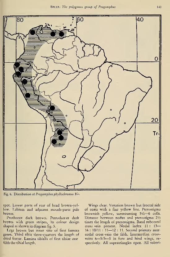
Belle: The polygonus group of Progomphus 141
Fig. 6. Distribution ol Progomphus phyllochromus Ris.
spot. Lower parts of rear of head brown-yel-
low. Labium and adjacent mouth-parts pale
brown.
Prothorax dark brown. Pterothorax dark
brown with green stripes, its colour design
shaped as shown in diagram fig. 5.
Legs brown but inner side of first femora
green. Third tibia three-quarters the length of
third femur. Lamina tibialis of first tibiae one-
fifth the tibial length.
Wings clear. Venation brown but frontal side
of costa with a fine yellow line. Pterostigma
brownish yellow, surmounting 51/2—6 cells.
Distance between nodus and pterostigma IV2
times the length of pterostigma. Basal subcostal
cross-vein present. Nodal index 11 : 15
—
16 : 10/11 : 11—12 : 11. Second primary ante-
nodal cross-vein the fifth. Intermedian cross-
veins 6—5/5—5 in fore and hind wmgs, re-
spectively. All supratriangles open. All subtri-

142 Tijdschrift voor Entomologie, deel 126, afl. 7 (1983)
Figs. 7— 17. Progomphus abbreviatus Belle: 7, tenth abdominal segment and caudal appendages of male, dorsal
view; 8, the same, left lateral view; 9, the same, ventral view; 10, occipital plate of female; 11, vulvar lamina; 12,
accessory genitalia, right lateral view; 13, frons of male, dorsal view; 14, ventral view of right branch of inferior
caudal appendage, male from Miranda; 15, the same, another male from Miranda; 16, the same, male from Sierra
San Lorenzo; 17, accessory genitalia, ventral view.
angles and discoidal triangles two-celled. There
is an extra cubito-anal cross-vein in each wing.
Trigonal interspace in fore wings starting with
two rows of cells from triangle outwards (that
of left fore wing with an extra initial cell next
hind angle of triangle) eight cells long, in ante-
rior row, that in hind wings starting with three
rows of cells against triangle followed by tworows of cells two (right) and three (left) cells
long, in anterior row. Anal field in right fore
wing one cell wide, that in left fore wing with
one of the cells doubled. Hind wings with five
(right) and six (left) paranal cells, four rows of
cells behind Cu2 and a three-celled anal trian-
gle.
Abdomen predominantly dark brown. Mid-dorsum of segments 2 to 7 with a yellow line
which is rather broad on segment 2 but whichbecomes very fine on segments 5 to 7. Sides of
segment 2 with yellow marking behind and be-
low auricle. Lower parts of sides of segments 3
to 6 with yellow marking from base to apex of
segment. Basal third portion of segment 7 yel-
low. Caudal appendages shaped as shown in ac-
companying figures, blackish brown, the supe-
riors becoming pale on apical half.
The unique (pinned) specimen of P. occiden-
talis was found in the collection of René Martin
at Paris. Attached to the pin is an old label "'Pro-
gomphus polygonus Selys S. Antonio" but there
is no label referring to the country in which the
locality San Antonio lies. However, from its
relationship with the other members of the
polygonus group we can say that the specimen
hails from western continental South America,
hence the specific name occidentalis. But René
Martin referred the male to P. polygonus, a spe-
cies known only from Venezuela. I therefore
suspect that the specimen is from the west of
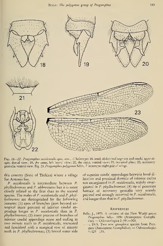
Belle: The polygonus group of Progomphus 143
Figs. 18—22. Progomphus occidentalis spec, nov., 6 holotype: 18, tenth abdominal segment and caudal append-
ages, dorsal view; 19, the same, left lateral view; 20, the same, ventral view; 21, occipital plate; 22, accessory
genitaha, ventral view. Fig. 23. Progomphus polygonus Selys, 9 lectotype: right pair of wings.
this country (State of Tachira) where a village
San Antonio lies.
P. occidentalis is intermediate between P.
phyllochromus and P. ahbreviatus but it is moreclosely related to the first than to the second
species. The males of P. occidentalis and P. phyl-
lochromus are distinguished by the following
features: (1) apex of branches (part beyond an-
teapical inner process) of inferior caudal ap-
pendage longer in P. occidentalis than in P.
phyllochromus; (2) inner process of branches of
inferior caudal appendage acute and endmg in
two minute teeth in P. occidentalis, truncated
and furnished with a marginal row of minute
teeth in P. phyllochromus; (3) lateral outer side
of superior caudal appendages between basal di-
latation and proximal denticle of inferior carina
not emarginated in P. occidentalis, widely emar-
ginated in P. phyllochromus; (4) tip of posterior
hamule of accessory genitalia very acutely
pointed and strongly recurved in P. occidentalis,
and longer than that in P. phyllochromus.
References
Belle, J., 1973. A revision of the New World genus
Progomphus Selys, 1854 (Anisoptera: Gomphi-dae).— Odonatologica 2: 191—308.
, 1975. Two new gomphine species from Para-
guay (Anisoptera: Gomphidae). — Odonatologica
4:129—135.
144 Tijdschrift voor Entomologie, deel 126, afl. 7 (1983)
, 1980. Two new gomphoidine species from Bra-
zil (Anisoptera: Gomphidae). — Odonatologica9:173—180.
De Marmels, J., 1981a. Aeshna rufipes Ris in Venezu-ela, with a description of the male (Aniso-
ptera: Aeshnidae).— Odonatologica 10: 39—42.
, 1981b. The larva of Progomphus abbreviatus
Belle, 1973 from Venezuela (Anisoptera: Gom-phidae).— Odonatologica 10: 147— 149.
, 1983. The Odonata of the region of MountAuyantepui and the Sierra de Lema, in Venezue-lan Guyana. 3. Additions to the families Gomphi-dae, Aeshnidae and Corduliidae, with description
of Progomphus racenisi spec. nov. — Odonato-logica 12: 5— 13.
Knopf, K. W. & K. J. Tennessen, 1980. A new species
of Progomphus Selys, 1854 from North America
(Anisoptera: Gomphidae). — Odonatologica 9:
247—252.
Needham, J. G., 1941. Life history studies on Pro-
gomphus and its nearest allies (Odonata: Aeshni-
dae).— Trans. Am. ent. Soc. 67: 221—245; pi. 20.
Ris, F., 1918. Libellen (Odonata) aus der Region der
amerikanischen Kordilleren von Costarica bis
Catamarca. — Arch. Naturg. 82 (9): 1—197; 2
tabs.
Selys Longchamps, E. de, 1878. Quatrièmes additions
au synopsis des Gomphines. — Bull. Acad. r.
Belg. (2) 46: 408—471, 658—698 (3—106 sep.).
St. Quentin, D. 1973. Die Gomphidenfauna Südame-
rikas (Ordn.: Odonata). — Annln naturh. Mus.
Wien 77: 335—363.
A REVIEW OF THE GENUS ZONOPHORA SELYS(ODONATA, GOMPHIDAE)
by
JEAN BELLE
Onder de Beumkes 35, Velp, The Netherlands
Abstract
The genus Zonophora Selys is revised. A number of problems in the classification of its
members are solved and elucidated by figures. A new subfamily, the Zonophorinae, is in-
troduced. 2. diversa spec. nov. (d holotype: Nova Teutonia, Santa Catarina, Brazil), 2.
campanulata annulata subspec. nov. {6 holotype, 9 allotype: Jatai, Goias, Brazil) and 2.
nobilis spec. nov. {S holotype: Taracua, Amazonas, Brazil) are described. 2. machadoi St.
Quentin and 2. obscura Belle are considered to be subspecies of 2. campanulata (Burmeis-ter) and 2. solitaria Râcenis, respectively. The females of these subspecies are also de-
scribed. A neotype for the type-species of the genus and lectotypes for 2. supratriangularis
Schmidt and 2. wucherpfennigi Schmidt are selected. Illustrations of some structures of the
accessory genitaha of 2. surinamensis Needham are given. A key to the species and subspe-cies is constructed. New distributional records and distribution maps are added.
ContentsPage
Introduction 145
Material and acknowledgements 146
Zonophora Selys, 1854 146
Disposition of species and infrageneric groups . . 148
Systematic section 149
Key to the (sub)species 149
Treatment of the species 150
c. campanulata (Burmeister) 151
e. machadoi St. Quentin 152
e. annulata subspec. nov 152
diversa spec, nov 153
c. calippus Selys 1 54
c. spectahilis Campion 155
c. klugi Schmidt 155
batesi Selys 156
wucherpfennigi Schmidt 156
surinamensis Needham 156
5. solitaria Râcenis 157
s. obscura Belle 157
rega/z5 Belle 158
supratriangularis Schmidt 158
nobilis spec, nov 159
The species in the mimeographed list of Râcenis 159
Habitat and behaviour in the field 160
Geographic distribution 160
References 161
Introduction
The genus Zonophora was created for a cer-
tain Neotropical gomphid by Selys in 1854. Into
this genus, he placed Diastatomma campanula-
ta, described on the basis of a single male from
Brazil by Burmeister (1839). No further addi-
tions were made to the genus for fifteen years,
when Selys (1869) published descriptions of twonew species (batesi and calippus). Fifty-one
years later, Campion (1920) described spectahi-
lis. In 1941, Schmidt described two species
[supratriangularis and wucherpfennigi) and one
subspecies (klugi as subspecies of calippus).
Needham (1944) described one species {surina-
mensis), St. Quentin (1973) one species {macha-
doi), Racenis (1970) one species {solitaria), and
Belle (1976) two species {regalis and obscura).
One species referred to Zonophora (Z.
rokitanskyi St. Quentin, 1973) does not fall
within the limits of this genus and was assigned
to Diaphlehia Selys, 1854 (Belle, 1977a). Here I
report on material of two new species {diversa
and nobilis) and one new subspecies {campanu-
lata annulata), bringing the total number of
taxa to fifteen.
Schmidt (1941) treated spectahilis and klugi as
subspecies but Belle (1963) elevated them to the
rank of species. A restudy of these representa-
tives, however, has revealed that there is little
reason for retaining this status and that
Schmidt's classification should be followed. St.
Quentin (1973) considered wucherpfennigi as a
subspecies of batesi but here wucherpfennigi is
treated as species, following again Schmidt. Onthe contrary, St. Quentin's machadoi and
Belle's obscura are considered subspecies of
145
146 Tijdschrift voor Entomologie, deel 126, afl. 8 (1983)
campanulata and solitaria, respectively. It is
clear that in the interests of a stable nomencla-
ture a neotype designation for the type-species
of the genus is required since the holotype of
Burmeister's campanulata must be considered
lost.
The knowledge of the proventriculus of larval
Zonophora has led to the introduction of the
subfamily Zonophorinae, to contain the genus
Zonophora. The diverse specific characters
show that the members of the genus Zonophora
can be conveniently divided into five groups.
The critical structures on which the present
classification is based are elucidated by original
camera lucida drawings. Distributional maps for
the groups are added and a key to the species
and subspecies is constructed. I take the oppor-
tunity to publish illustrations of some important
structures of the accessory genitalia of 2. sun-
namensis.
Material and acknowledgements
The material on which the present review is
based belongs to the institutions and personal
collections mentioned below; the names are
preceded by the abbreviations used throughout
the text of this paper.
AC — Author's collection;
BM — British Museum (Natural Histo-
ry), London; Dr. Peter H. Ward;
CC — Collection Cook, Center; Mr.
Carl Cook;
CJ — Collection Jurzitza, Karlsruhe;
Prof. Dr. Gerhard Jurzitza;
CM — Collection Machado, Belo Hori-
zonte; Prof. Dr. Angelo B. M.Machado
CU — Cornell University, Ithaca; Dr. L.
L. Pechuman;IRSN — Institut Royal des Sciences Natu-
relles de Belgique, Bruxelles; Dr.
Georges Demoulin;
IZM — Instituto de Zoologìa Agrìcola,
Maracay; Dr. F. Fernandez
Yépez;
ML — Rijksmuseum van Natuurlijke
Historie, Leiden; Dr. P. H. van
Doesburg;
MP — Muséum National d'LIistoire Na-turelle, Paris; Dr. Jean Legrand;
UMMZ — Museum of Zoology, University
of Michigan, Ann Arbor; Mrs.
Leonora K. Gloyd and Dr. T. E.
Moore;
SMF — Natur-Museum Senckenberg,
Frankfurt-am-Main; Dr. Heinz
Schröder.
For the loan of material I am also indebted to
the following persons: Mr. Jorge de Marmels
(Caracas) and Dr. Janis Racenis (f) (Caracas).
Zonophora Selys, 1854
Zonophora Selys, 1854: 80 (61 sep.); 1873: 517 (73
Sep.). Selys & Hagen, 1858: 493—494 (233—234Sep.). Kirby, 1890: 75. Karsch, 1890: 371. Car-
pentier, 1897: 453. Navas, 1916: 28. Campion,1920: 139—140. Williamson, 1920: 9, 11—12.
Kennedy, 1928: 370. Tiliyard & Fraser, 1938—1940: 372. Byers, 1939: 21. Fraser, 1940: 549;
1957: 93. Needham, 1940: 388—389; 1944: 172—173, 215, 218, 221—222. Schmidt, 1941: 76—78.
Belle, 1963: 60, 62—63; 1976: 197, 200, 206;
1979: 46. St. Quentin, 1967: 150; 1973: 335,
337—338, 341, 361. Paulson, 1977: 179. Kiauta,
1979: 268. Davies, 1981:33.
Type species. — The type-species of the ge-
nus Zonophora is Diastatomma campanulata
Burmeister, 1839, as designated by Kirby
(1890).
Position. — The systematic position of Zono-
phora, based on a certain combination of vena-
tional characters, has been changed several
times. Williamson (1920) placed Zonophora in
his "Zonophora series", a series which only
contained this genus. Zonophora was character-
ized by the discoidal triangles and subtriangles
of the fore and hind wings being similar, the dis-
coidal triangles long and once crossed, the sub-
triangles and supratriangles all free, absence of
trigonal supplements and sectors of Rs and M4,and by the nearly right proximal angle at the
point of separation of Al from Cu2 + Al and
not as obtuse as the distal angle at the same
point. However, the supratriangles are normally
crossed in two species and incidentally crossed
in some other species. In the classification as
proposed by Tiliyard and Fraser (1938—1940),
Zonophora becomes one of the members of the
Ictinogomphinae, distinguished by their posi-
tion of crossed discoidal, supra- and subtrian-
gles. However, Zonophora has the subtriangles
never crossed and the supratriangles of most
species generally not crossed. In the revised re-
classification. Fraser (1957) transferred Zono-
phora into the Gomphoidinae, which were sep-
arated from the other genera by the presence of
crossed discoidal, supra- and subtriangles, and
in having only a single cross-vein (Ac) in the cu-
bital space. Zonophora, however, has uncrossed
subtriangles and generally uncrossed supra-

Belle: Zonophora \A7
triangles while in some species there are nor-
mally two or three cubito-anal cross-veins in
addition to the inner side of the subtriangle
(Belle, 1963, tabs. 3, 4). Belle (1979), finally,
placed Zonophora in the Epigomphinae, which
were characterized by the uncrossed subtrian-
gles in the fore wing, the number of intermedian
cross-veins in the hind wing being two or more,
and the absence of a trigonal supplement. How-ever, the aberrant proventricular armature of
larval Zonophora (cf. below) shows the isolated
position of this genus in the Gomphidae and
justifies the erection of a sixth subfamily, the
Zonophorinae, to contain Zonophora. The pro-
ventricular character together with the vena-
tional character for the hind wings cited above
(proximal angle at point of separation of Alfrom Cu2-I-Al nearly a right angle, and not as
obtuse as distal angle at same point) separate
Zonophora from the rest of the Epigomphinae
in which it was lastly placed.
Immature stages. — In 1944, Needham gave a
description of a larval cast-off skin, which he re-
ferred to Zonophora by a process of exclusion.
In 1966, Belle published photographs of the ex-
uviae of Z. batest and Z. calippus, while he gave
a brief description of the exuviae of the latter
species. The reference of this exuvial material to
these two species was based on the difference in
size and on the fact that the two species were
the only regional ones of the genus occurring in
the well-explored surroundings of the airport
Zanderij of Surinam. Some half-grown larvae of
Zonophora were also secured by sifting mud or
silt removed from the bottom and sloping banks
of the creeks crossing the gallery forests in the
surroundings of Zanderij. The gizzard of these
larvae was recently examined by me. The result
was most surprising as the proventriculus ex-
hibited an unexpected specialized armature, es-
sentially different from that hitherto found in
the Gomphidae.In figs. 1—5 the complete proventriculus of a
half-grown larva of Z. batest is shown. Con-trary to what is normally found in the Gomphi-dae, there are two pairs of dissimilar, elevated,
sclerotized areas (dental folds) in juxtaposition,
one dorsal pair consisting of pyramid-shaped
folds and one ventral pair consisting of molar-
shaped folds. Each of the folds has a specialized
dentition of large, posteriorly directed teeth
and, on the posterior end, numerous minute
posteriorly directed teeth.
In shape, the dental folds of Zonophora muchresemble those o{ Aphylla (cf. Beatty, 1956), but
the occurrence of numerous minute teeth on
these folds seems to be confined to Zonophora
(and some Aeshnidae and Lestidae, cf. Bullens,
1966). In the Gomphidae, this aberrant proven-
tricular armature apparently can be employed as
a taxonomie character.
Characterization. — The genus Zonophora is
characterized by the following combination of
features :
In both sexes:
(1) Numerous intermedian cross-veins in
hind wings.
(2) No trigonal supplements and sectors of
Rs and M4.
(3) Vein Al in hind wings well-developed,
the proximal angle at the point of separation of
Al from fused portion Al -I- Cu2 a right angle
or nearly so, the distal angle at same point ob-
tuse.
(4) Discoidal triangles and subtriangles of
fore wings more or less similar to those of hind
wings; the discoidal triangles elongated in axis
of wing and generally once crossed (occasional-
ly two cross-veins); the subtriangles uncrossed.
(5) Pterostigma in hind wings somewhat lon-
ger than that in fore wings.
(6) Meso- and meta-paraptera conspicuously
produced in an acute point.
(7) Vertex with conical process behind each
lateral ocellus.
(8) Occiput angled at rear.
(9) Ventral tergal margins of abdominal seg-
ments 8 and 9 not expanded.
In the male:
(10) Anal triangle in hind wings not reaching
backward to anal angle of wing.
(11) Superior caudal appendages twice to
three times as long as abdominal segment 10 and
incurved at tip; inferior caudal appendage about
as long as abdominal segment 10 and with two
branches widely spread apart.
In the female:
(12) Vulvar lamina reaching from a point
one-third the way along ninth sternum to apex
of ninth sternum; its posterior margin medially
excised.
In the larva:
(13) No dorsal hooks on abdomen.
(14) Abdomen with lateral spines only on ab-
dominal segments 8 and 9.
(15) Caudal appendages about three-and-
one-half times as long as abdominal segment 10.
(16) Lateral lobes of labium with incurving
teeth along inner margin and with a strong end
hook.

148 Tijdschrift voor Entomologie, deel 126, afl. 8 (1983)
(17) Gizzard with two pairs of differentiated
dental folds bearing undifferentiated large teeth
and numerous minute teeth.
Disposition of species and infrageneric
GROUPS(figs. 6—30)
There are excellent characters available for se-
parating the species and subspecies of the genus
Zonophora into five groups. The members of
each group exhibit a striking resemblance in the
shape of some structures although some of the
statements concerning the shape of these struc-
tures will have to be taken with due allowance
for individual variation.
(1) The campanulata group, comprising 2.
campanulata campanulata (Burmeister) with its
subspecies Z. campanulata machadoi St. Quen-
tin and 2. campanulata annulata subspec. nov.,
and 2. diversa spec. nov.
The distal segment of the penis has a pair of
short and inconspicuous tusk-like spines. The
anterior hamules of the male have a well-devel-
oped, finger-like, internal lobe. The vulvar lami-
na is more or less parallel-sided and long, reach-
ing to the apex of the ninth sternum; the tip of
the vulvar lamina exhibits much variation, the
divisions being sometimes widely spread out
(cf. Schmidt, 1941).
The wings have no basal subcostal cross-vein
and no extra cubito-anal cross-vein. There is,
normally, no traversing cross-vein in the supra-
triangles (cf. Belle, 1963, Verification tables).
The hind wings have sometimes an open anal
loop, i.e. open at the proximal side (cf. Schmidt,
1941), but generally an ill-defined anal loop of
three to five cells.
There are no distinct morphological differ-
ences between 2. campanulata and its subspe-
cies. The distinction is based only on colour dif-
ferences. 2. c. campanulata is the darkest of the
three. The pale markings on the sides of the
pterothorax are grey-green. The abdomen is
predominantly dark brown. The tergite of the
segments 3 to 8 is densely set with minute black
denticles, in particular on the basal part of the
segments. The two subspecies 2. c. machadoi
and 2. c. annulata have distinct greenish yellow
and yellow markings which are extensive and
very striking on the abdomen. The abdominal
segments 3 to 8 of the two subspecies are scanti-
ly set with minute black denticles.
2. diversa does exhibit great colour and mor-
phological differences with 2. campanulata and
its subspecies. The superior lobe of the anterior
hamules of the male is vestigial in diversa, well-
developed in campanulata.
(2) The calippus group, comprising 2. calip-
pus calippus Selys, 2. calippus spectabilis Camp-ion and 2. calippus klugi Schmidt.
The distal segment of the penis has a pair of
short flagella. The anterior hamules of the male
are much smaller than those of the campanulata
group; the internal lobe is small and the superi-
or lobe is long and slender. The vulvar lamina is
smaller and more slender than that of the cam-
panulata group, and it exhibits but little varia-
tion.
The wings have, normally, a basal subcostal
cross-vein and generally extra cubito-anal
cross-veins. The anal loop in the hind wings is
generally open at the proximal side. The supra-
triangles often have a traversing cross-vein, in
the females more frequently so than in the
males.
The three members of the group are subspeci-
fically distinct only. They can be discerned on
the ground of their coloration. Slight differences
are found in the configuration of the anterior
hamules of the males but no structural differ-
ences are found in the shape of the vulvar lami-
nae.
2. c. klugi is somewhat more robust than the
other two. The superior lobe of the anterior
hamules is very long and slightly upcurved at
the tip. 2. c. klugi is also the darkest of the
group. The pale frontal band on the superior
surface of the frons is medially interrupted by a
pitch black band that is broader in the female
than in the male.
2. c. spectabilis is the palest of the group. It is
distinguished from the other two by the pres-
ence of large pale markings on the abdominal
segments 8 to 10. The superior lobe of the ante-
rior hamules is widened at the tip.
(3) The hatesi group, comprising 2. batesi
Selys and 2. wucherpfennigt Schmidt.
The distal segment of the penis has a pair of
very long flagella. The anterior hamules of the
male are as large as those of the campanulata
group; the superior lobe is short and stout; the
inferior lobe is much larger than that of the
campanulata group; the internal lobe is very
small. The vulvar lamina is widest at base and
roughly triangular in outline; it exhibits some
variation, the divisions of the tip being often
spread out.
The wings have no basal subcostal cross-vein,
no extra cubito-anal cross-vein, and no travers-
ing cross-vein in the supratriangles. The anal

Belle: Zonophora 149
loop in the hind wings is open at the proximal
side.
The two species of the group differ struc-
turally in the penile organ, the male caudal ap-
pendages and vulvar laminae, but the differ-
ences are slight and they can be shown morereadily by figures than description (cf. Schmidt,
1941). The darkest of the two is wucherpfennigi.
The seventh abdominal segment of the male is
entirely black in this species; it is markedly yel-
low-coloured at the base in batesi. The caudal
appendages (stylets) of the female are largely
blackish or brownish (paler in middle and black
at base) in wucherpfennigi (cf. Schmidt, 1941:
fig. 12f); they are largely pale-white (black at
base) in batesi.
(4) The supratriangularis group, comprising
Z. supratriangularis Schmidt and Z. nobilis spec,
nov. They differ structurally in the male caudal
appendages and genital hamules. The distal seg-
ment of the penis has a pair of very long flagella.
The internal lobe of the anterior hamules of the
male is small. The vulvar lamina is about half
the length of the ninth sternum.
The wings have no basal subcostal cross-vein
but there are extra cubito-anal cross-veins. Thesupratriangles normally have a traversing cross-
vein. The anal loop in the hind wings is open at
the proximal side although not always very dis-
tinct.
The two species are the palest representatives
of the genus.
(5) The surinamensis group, comprising Z.
surinamensis Needham, Z. solitaria solitaria Râ-
cenis, Z. solitaria obscura Belle, and Z. regalis
Belle.
The anterior hamules of the male have no in-
ternai lobe. The male superior caudal append-
ages are not knotty at the internal margin. Thevulvar lamina is about one-third as long as the
ninth sternum.
The wings have no basal subcostal cross-vein
and generally no extra cubito-anal cross-vein.
The supratriangles occasionally have a travers-
ing cross-vein. The hind wings have a more or
less distinct anal loop.
Z. 5. solitaria is the palest of the group having
the lateral sides of the pterothorax largely pale
and the dorsum of the eighth abdominal seg-
ment with a well-developed pale basal spot. Z. 5.
obscura is the darkest of the group and likewise
the darkest representative of the genus. It is dis-
tinguished from all congeners by the lack of sec-
ond pale antehumeral stripes (these stripes are
partly developed in Z. 5. solitaria). Z. regalis is
the largest of the genus.
Systematic section
As considered in this study, the genus Zono-
phora comprises 15 taxa of which three are de-
scribed as new. The taxa are summarized in ta-
ble 2, together with their type localities, type
status and type locations.
The males of all species and subspecies are
known. The females of diversa, nobilis, regalis,
solitaria solitaria, and surinamensis are yet un-
known. Their discovery may corroborate the
relationships hitherto found between some spe-
cies.
The recognition of the diverse species and
subspecies is comparatively easy. Separate keys
have been prepared for the identification of
males and females. The key to the males has
been based upon the structural characters dis-
played by the anterior hamules, the penis, the
superior caudal appendages, the reticulation of
the wings, and some other details. The key to
the females has been based on characters dis-
played by the vulvar lamina and on some color-
ation details.
Key to the (sub)species
Males
1. Basal subcostal cross-vein normally present
{calippus group) 2
— No basal subcostal cross-vein 4
2. Abdominal segments 8 to 10 with extensive
yellow markings calippus spectabilis
— Abdominal segments 8 to 10 without or
with weakly developed yellow markings . 3
3. Interrupted median stripe ot pale anterior
band of frons well-developed and pitch
black, its width normally one-third the
mid-dorsal length of superior surface of
frons or more calippus klugi
— Interrupted median stripe of pale anterior
band of frons absent, ill-defined or very
narrow, its width smaller than one-third the
mid-dorsal length of superior surface of
frons calippus calippus
4. Superior caudal appendages knotty near
middle at internal margin; anterior hamules
with internal lobe (fig. 9) 8
— Superior caudal appendages not knotty at
internal margin; anterior hamules without
internal lobe {surinamensis group) 5
5. Lateral sides of pterothorax largely pale ....
solitaria solitaria

150 Tijdschrift voor Entomologie, deel 126, afl. 8 (1983)
— Lateral sides of pterothorax with well-de-
veloped dark stripes 6
6. Second pale antehumeral stripes undevel-
oped solitaria obscura
— Second pale antehumeral stripes well-devel-
oped 7
7. Distal segment of penis with a pair of very
short spines surinamensis
— Distal segment of penis with a pair of re-
curved leaf-like plates regalis
8. Distal segment of penis with short tusk-like
spines; internal lobe of anterior hamules-
well-developed and finger-like {campanu-
lata group) 9
— Distal segment of penis with very long flag-
ella; internal lobe of anterior hamules very
small 12
9. Distal halves of posterior hamules parallel;
anterior hamules distinctly trilobate .... 10
— Distal halves of posterior hamules diver-
gent; anterior hamules not evidently trilo-
bate due to an ill-defined superior lobe
diversa
10. Terminal segments of abdomen without or
with weakly developed yellow markings . . .
campanulata campanulata— Terminal segments of abdomen with exten-
sive yellow markings 11
11. First pale antehumeral stripes connected
with pale collar and second pale antehu-
meral stripes campanulata machadoi— First pale antehumeral stripes neither con-
nected with pale collar nor with second pale
antehumeral stripes
campanulata annulata
12. Supratriangles crossed {supratriangularis
group) 13
— Supratriangles open {hatesi group) 14
13. Superior caudal appendages almost three
times as long as mid-dorsum of abdominal
segment 10; internal tooth of each appen-
dage at three-fifths the appendage's length . .
nobilis
— Superior caudal appendages twice as long as
mid-dorsum of abdominal segment 10; in-
ternal tooth of each appendage at half the
appendage's length supratriangularis
14. Abdominal segment 7 with large, pale basal
marking hatesi
— Abdominal segment 7 entirely black
wucherpfennigi
Females
1. Basal subcostal cross-vein normally present
[calippus group) 2
— No basal subcostal cross-vein 4
2. Abdominal segments 8 to 10 with extensive
yellow markings calippus spectabilis
— Abdominal segments 8 to 10 without or
with weakly developed yellow markings . 3
3. Pale frontal band on superior surface of
frons widely interrupted in middle by pitch
black calippus klugi
— Pale frontal band on superior surface of
frons medially not or not distinctly inter-
rupted by black calippus calippus
4. Vulvar lamina reaching to apex of ninth
sternum 5
— Vulvar lamina half the length of ninth ster-
num of shorter 9
5. Vulvar lamina roughly triangular in outline,
widest at base and about twice as long as its
basal width {batest group, fig. 17) 6
— Vulvar lamina much longer than wide and
subparallel-sided, becoming somewhat nar-
rower towards apex {campanulata group,
%15) 7
6. Caudal appendages (stylets) largely pale-
white, black at base; lobes of vulvar lamina
distinctly remote at bottom of excision
batesi
— Caudal appendages (stylets) largely brown,
paler in middle, black at base; lobes of vul-
var lamina close together at bottom of exci-
sion wucherpfennigi
7. Abdominal segments without or with
weakly developed yellow markings
campanulata campanulata— Abdominal segments with extensive yellow
markings 8
8. First pale antehumeral stripe connected
with pale collar and second pale antehu-
meral stripe campanulata machadoi— First pale antehumeral stripe neither con-
nected with pale collar nor with second pale
antehumeral stripe . . campanulata annulata
9. Second pale antehumeral stripe well-devel-
oped; vulvar lamina half the length of ninth
sternum {supratriangularis group)
supratriangularis
— Second pale antehumeral stripe undevel-
oped; vulvar lamina one-third the length of
ninth sternum {surinamensis group)
solitaria obscura
Treatment of the species
The species have been treated in a sequence,
which allows expression of apparent rela-
tionships. Under each species a list of syno-
Belle: Zonophora 151
833
62
nyms, data on the material studied, description
or descriptive notes, and remarks are given.
Zonophora campanulata campanulata
(Burmeister, 1839)
(figs. 6, 9, 15, 19—20, 23, 31—34, 72)
Diastatomma campanulata Burmeister, 1839:
(d). Calvert, 1898: 35, 52, 94, 94—95.
Zonophora campanulata Selys, 1854: 80—81 (61
sep.) (9); 1869: 199—200 (36—37 sep.). Selys &Hagen, 1858: 492, 494—496 (232, 234—236, 238
Sep.); pi. 23, fig. 5. Hagen, 1861: 313; 1875: 54.
Kirby, 1890: 75. Navas, 1916: 73. Campion, 1920:
137—138, 140. St. Quentin, 1939: 227. Schmidt,
1941: 77—78, 80, 83, 86, 89—90, 92; figs. 5c, 6c,
8c, 9cl, lOd, lie, 12c. Needham, 1944: 218—219.
Belle, 1963: 60—63. Dias dos Santos, 1970: 204.
St. Quentin, 1973: 338—339; fig. 3.
Material. — Brazil: 1 ô (neotype), 1 9 (neotype se-
ries of Diastatomma campanulata Burmeister); Tiju-
ca, 2 (5, IRSN; Porto Catherina de Santa Leopoldina,
3 S, UMMZ; Minas Gerais, Caraça, Sta. Barbara,
February 1970, 1 6, A.M.M. Machado leg., CM; Es-
pirito Santo, 3 d (1 (5 ex coll. Fruhstorfer), 1 9, MP.— ? Guyana ? : 1 cÏ, MP. — ? Surinam ? : 2 d, 1 9,
MP.
This species was described by Burmeister
(1839) after a single male from "Brazil". This
male must be considered to be identical with the
darkest representative of the campanulata
group since the statement "fusca, subtus dilu-
tior; abdominis segmento septimo fascia basali
pallida" in the original description evaluates the
specimen. If all its abdominal segments has had
extensive pale markings, they should undoubt-
edly be mentioned in the description.
The location of the type of D. campanulata is
unknown. Calvert (1898) as well as Belle (1963)
were unable to find it. Also further search madeby myself was in vain. I am strongly inclined to
believe that the original specimen is lost, possi-
bly destroyed by a dermestid infestation {An-
threnus). The species, however, has been very
well described and figured in the Monographie
des Gomphines (Selys & Hagen, 1858). In order
to stabilize its identity a neotype is here desig-
nated.
Neotype designation of Diastatomma cam-
panulata Burmeister. — The chosen neotype is
a male from a remarkable uniform series placed
under Zonophora campanulata (Burmeister) in
the Selysian collection in Brussels. This series
consists of the following specimens:
(1) A male with the pin label "CI. 2."
(2) A female with the pin labels "CI. 3" and
"Zonophora campanulata B 9 ", this latter in
Selys' handwriting.
(3) A male with the pin labels "122", "122",
"Tijuca P.B.", and ''Zonophora campanulata B.
6", this latter in Selys' handwriting.
(4) A male with the pin label "Tijuca P.B."
The abbreviation "P.B." may refer to Paul de
Borchgrave who has collected Odonata in Tiju-
ca near Rio de Janeiro round-about 1860 (cf.
Calvert, 1956:88, 175— 176).
The abbreviation "Cl." may refer to Dr.
Clausen from whom Selys received the speci-
mens mentioned in the Monographie des Gom-phines, p. 496 (236 Sep.).
I think, that the male which served for Ha-gen's figures in this monograph belonged to the
above mentioned series. This male should have
borne at the pin the (missing) label "Cl. 1" and
probably also Selys's identification label of the
Tijuca male. The specimens of Dr. Clausen are
homotypes as may appear from Selys's (1858)
remark in parentheses: "C'est le type de M.Burmeister qui offre les nombres les plus
grands". The homotype male labelled "Cl. 2" is
here designated as neotype of Diastatomma
campanulata Burmeister, 1839.
Z.C. campanulata exhibits some striking vari-
ations. The hind wings of none of the present
specimens have a distinct open anal loop, due to
a rather well-developed vein A2. The hind
wings of some specimens have more or less a
distinct anal loop of 3 to 5 cells, with the vein
Al angled at the lower distal corner of the anal
loop.
The form of the vulvar lamina is also variable,
in particular at the tip. The vulvar lamina of the
present females approaches that depicted by St.
Quentin (1973) but it differs considerably from
that depicted by Schmidt (1941).
The female of the neotype series has a vulvar
lamina with a damaged tip. As this female was
the only one available for Hagen, it apparently
served for his figure in the Monographie des
Gomphines (pi. 13, fig. It).
Some data in Selys's redescription of 1854 are
incorrect. The measurement for the female ab-
domen is 50 mm (inch caud. app.) and not 58
mm (cf. Campion, 1920, foot-note on p. 137).
Further, the number of given antenodal cross-
veins in the fore wings of the male is 18 instead
of 13 (cf. Selys, 1869). The nodal index of the
neotype is 14 : 20—20 : 13/13 : 14—15 : 14.
Fraser (1940) published a figure of the penis

152 Tijdschrift voor Entomologie, deel 126, afl. 8 (1983)
of a Zonophora male, which he referred to Z.
campanulata. But judging from the conforma-
tion of the penile organ his male belonged to the
batest group.
Remark. — Caution should be made regard-
ing the locality labels of the specimens in the
Martin collection (lodged in the Muséum Na-tional d'Histoire Naturelle, Paris) since several
of these old labels prove to be unreliable. Somespecimens of 2. c. campanulata have the pin la-
bels "Surinam" and "Guyana" but these locali-
ties may be incorrect since nor Dr. D. C.
Geijskes nor I myself have encountered this
species during our extensive explorations in Su-
rinam and Guyana. On that account these re-
cords have not been inserted in the distribution
map.
To further illustrate the unreliability of somelocality labels of the Martin collection, in the
boxes in which the specimens of Zonophora
were stored, I found four males of Phyllogom-
phoides pacificus (Selys, 1873) labelled "Minas
Gerais" and "Brésil", and a male oi Aphylla car-
aiba Selys, 1854, labelled "Brésil".
Zonophora campanulata machadoiSt. Quentin, 1973
(figs. 21,24, 35—36)
Zonophora machadoi St. Quentin, 1973: 338—340;
figs. 4a—b(<5). Kiauta, 1981:35,60.
Material. — Brazil: Minas Gerais, Belo Horizonte,
18 March 1979, 1 S, AC; Tabuoes, Ibirité (near Belo
Horizonte), 22 March 1979, 1 6 , CJ, both G. Jurzitza
leg.; Ibirité, 21 March 1979, 1 9, A. B. M. Machadoleg., CM.
This subspecies was described by St. Quentin
(1973) after a single male from Belo Horizonte,
Brazil. The female is described below.
Female (hitherto unknown; degutted and
treated with acetone). — Total length, 64 mm;abdomen, 49 mm (including caudal appendages,
4.3 mm); hind wing, 45 mm; costal edge of
pterostigma in fore wing, 6.0 mm, in hind wing,
6.5 mm.Coloration resembling that of male holotype.
Face brown with conspicuous bright yellow
band covering suture between postclypeus and
vertical part of frons. Labium with pale cross-
band in midfield. Anteclypeus becoming paler
on outer edges. Superior surface of frons brownwith oblong yellow spot on either lateral side.
Colour pattern of prothorax and pterothorax
very similar to that of male. Abdomen relatively
shorter than that of male. Pale markings on
sides of abdominal segments 8 and 9 muchsmaller than in male, and abdominal segment 10
entirely dark brown (fig. 36).
Proportional length of abdominal segments 7,
8, 9, and 10 approximately as 45 : 30 : 20 : 10,
with the caudal appendages 34 on same scale.
Vulvar lamina shaped as shown in fig. 21.
Pterostigma surmounting eight cells. No bas-
al subcostal cross-vein. No extra cubito-anal
cross-vein. No traversing cross-vein in supra-
triangles. No anal loop in hind wings. Nodal in-
dex 14 : 20—22 : 13/13 : 13—13 : 15. Second
primary antenodal cross-vein the eighth in fore
wings, the sixth in hind wings. Intermedian
cross-veins 7—9/3—3 in fore and hind wings,
respectively. Hind wings with six (left) and sev-
en (right) paranal cells, six (left) and seven
(right) postanal cells, and area posterior to Cu2six to seven cells wide.
Zonophora campanulata annulata
subspec. nov.
(figs. 22, 23, 37—38)
Material. — Brazil: Gioas, Jatai' Qatahy), 3 6 holo-
type and paratypes), 1 9 (allotype), MP.
This subspecies, though very similar in its
morphology to Z. c. campanulata and Z. c.
machadoi, is readily distinguished from both by
the aberrant colour pattern of the pterothorax,
which, in lateral view, shows a conspicuous pair
of well-developed pale twin-stripes, the first
pair being formed by the second pale antehu-
meral stripe and the first pale lateral stripe, and
the second pair by the second and third pale
lateral stripes. The colour pattern of the dorsum
of the pterothorax is most like that of Z. c.
campanulata by the isolated pale antehumeral
stripes and pale collar, while the abdomengreatly resembles that of Z. c. machadoi by the
annulated appearance owing to the extensive
pale markings at the base of the segments.
According to some of the hitherto used char-
acters, the colour differences mentioned above
are important enough to consider the present
examples from Jatai a distinct variety or subspe-
cies. I prefer to regard them as a subspecies
since they are forming a homogeneous series,
and since they seem to be the only form occur-
ring in that part of the distribution area. How-ever, as long as our knowledge is restricted to
the scarcely available material it is difficult to
Indicate the value of the characters used for the
subspecific distinction. The features often vary
Belle: Zonophora 153
in some widely ranging gomphid species to such
a degree that it remains to be seen whether the
status of the subspecies introduced will stand
the test of future collecting.
Male (holotype; abdomen broken on segment
6 and between segments 3 and 4; left superior
caudal appendage broken off and lost). — Total
length, 66 mm; abdomen, 50 mm (incl. caud.
app., 4 mm); hind wing, 42 mm; costal edge of
pterostigma in fore wing, 5.6 mm, in hind wing,
6.0 mm.Face yellowish green but brown along border
of labrum and just below frontal ridge of frons.
Superior surface of frons yellowish green on an-
terior half, brown on basal half. Vertex black.
Occipital plate and rear of occiput yellow-
brown. Rear of head yellowish brown. Base of
mandibles, genae, labium and adjacent mouthparts pale brown.
Prothorax dark brown with greenish yellow
markings. Median lobe with pale middorsal
twin-spot and pale spot on each lateral side;
posterior lobe with pair of pale oblong spots.
Pterothorax dark brown to black, the pale
markings greenish yellow, becoming yellow on
metepimeron. Colour pattern of pterothorax
shaped as shown in accompanying diagram.
Middorsal carina and antealar sinus pale. Pale
collar and first pale antehumeral stripe narrow.
Second pale antehumeral stripe and pale lateral
stripes rather broad.
Femora reddish-brown but inner side of first
pair green. Tibiae, tarsi, and claws dark brown.
Lamina tibialis of first tibiae one-seventh the
tibial length.
Wings clear, its venation pale brown, becom-
ing darker on posterior margm of wings.
Pterostigma brown-yellow, surmounting 6
—
GVi cells. Brace vein present. No basal subcostal
cross-vein. A single cubito-anal cross-vem in
addition to inner side of subtriangle in all wings.
Supratriangle in right hind wing with a travers-
ing cross-vein, in other wings uncrossed. Sub-
triangles open. Discoidal triangles two-celled.
Nodal index 12 : 18—16 : 11/13 : 12—11 : 12.
Second primary antenodal cross-vein the sev-
enth in left fore wing, the sixth in other wings.
Intermedian cross-veins 8—8/4—4 in fore and
hind wings, respectively. Hind wings with open
anal loop (open at proximal side), a four-celled
anal triangle, five (right) and six (left) paranal
cells, five (right) and six (left) postanal cells, and
area posterior to Cu2 five cells wide.
Abdomen dark brown to black, the pale
markings yellow. Segment 1 pale on lower part
of lateral sides and along dorsal posterior mar-
gin. Segment 2 pale on lower parts of lateral
sides, on auricles, and just above auricles. Seg-
ments 3 to 8 with large pale basal markings
which become successively larger on 3 to 8,
laterally reaching to supplementary transverse
carina on 3, and extending to beyond this carina
on 4 to 8. Segment 9 with pale lateral markings
extending along lower part of side to apex of
segment. Segment 10 largely pale. Tubercles
with externo-lateral pale spots. Inferior append-
age black. Superior appendages pale yellow but
extreme base brown.
Female (allotype). — Total length, 69.5 mm;abdomen, 53 mm (incl. caud. app., 4 mm); hind
wing, 46 mm; costal edge of pterostigma in fore
wing, 6.4 mm, in hind wing, 6.6 mm.Very similar to male holotype in stature and
coloration but abdomen stouter. Proportional
length of abdominal segments 7, 8, 9, and 10 ap-
proximately as 40 : 23 : 19 : 10, with the caudal
appendages (stylets) 28 on the same scale.
Pterostigma surmounting 5 1/2
—
GVz cells. Nobasal subcostal cross-vein. No extra cubito-anal
cross-vein. No traversing cross-vein in supra-
triangles. Second primary antenodal cross-vein
the eighth in left hind wing and right fore wing,
the seventh in other wings. Hind wings with a
weak development of an anal loop of four cells.
The male paratypes are very similar to the ho-
lotype. All their wings have uncrossed sub-
triangles and supratriangles. The triangles have
one dividing cross-vein. The anal triangles are
four-celled. The hind wings of one of the male
paratypes have a weak development of an anal
loop of four cells.
Zonophora diversa spec. nov.
(figs. 39—50)
Material. — Argentina: Misiones, Dos de Mayo,
January 1982, 2 â (paratypes), J. Foerster leg., ACand CJ. — Brazil: Santa Catarina, Nova Teutonia, 2
March 1942, 1 â (paratype, AC), 3 March 1942, 2 â
(paratypes), 4 March 1942, 1 â (paratype), UMMZ;same locality, January 1946, 1 â (holotype, CC), all
Fritz Plaumann leg. — Paraguay: Alto Parana, Ta-
queri (Tacuri), near Rio Nacunday, 20 February 1980,
1 â (paratype), H. Miers leg., CM.
This species is closely related to the type-spe-
cies of the genus, 2. c. campanulata, but it is
rather easily distinguished in the male sex by the
stouter superior caudal appendages, the ill-de-
154 Tijdschrift voor Entomologie, deel 126, afl. 8 (1983)
fined superior lobe of the anterior hamules, and
the divergent distal halves of the posterior ham-
ules.
The female is unknown.
Male (holotype). — Total length, 70 mm; ab-
domen, 54 mm (including caud. app., 4 mm);hind wing, 47 mm; costal edge of pterostigma in
fore wing, 5.0 mm, in hind wing, 5.4 mm.Face greenish-yellow but labrum black along
free-border. Superior surface of frons black at
base, greenish yellow on its three-fifths anterior
portion, yellow on anterior ridge. Vertex black.
Occipital plate yellowish-green, its posterior
margin slightly and evenly convex, and heavily
fringed with brown hairs.
Prothorax dark brown, the middle lobe with
reddish brown lateral markings and a more yel-
lowish mid-dorsal twin-spot, the hind collar
dark reddish brown.
Pterothorax blackish brown with greenish
yellow markings, its colour pattern shaped as
shown in fig. 46. Femora blackish brown, save
for a streak of yellowish brown on outer basal
two-thirds portion of third femora. Tibiae and
tarsi black. Claws dark reddish brown.
Wings hyaline, its venation brown including
frontal margin of costa. Pterostigma brown,
surmounting SVi—6^2 cells. Brace vein present.
No basal subcostal cross-vein. Nodal index
14 : 18—18 : 14/13: 14—13 :15. Second prima-
ry antenodal cross-vein the seventh in right hind
wing, the eighth in other wings. Intermedian
cross-veins 7—6/4—5 in fore and hind wings,
respectively. No extra cubito-anal cross-vein.
All subtriangles and supratriangles free from
cross-veins. All discoidal triangles two-celled.
Hind wings with an ill-defined anal loop of four
(left) and five (right) cells, an anal triangle of six
cells (two cells small), six paranal cells, seven
(left) and six (right) postanal cells, and area pos-
terior to Cu2 six cells wide.
Abdomen black with yellow markings, its
colour pattern shaped as shown in fig. 43. Seg-
ment 10 with brownish yellow mid-dorsal spot.
Sterna of segments 6 to 8 brown to blackish
brown but yellow on basal one-fifth; sternum of
8 with an extra, oblong yellow spot on either
side half-way the sternum. Inferior caudal ap-
pendage black. Superior caudal appendages dark
brown. Caudal appendages and accessory geni-
talia shaped as shown by the accompanying fig-
ures.
The following colour differences between the
holotype and the paratypes from Nova Teuto-
nia are noteworthy. One male has the black
band along the free border of the labrum not de-
veloped in the middle and at either side; two
males have the black basal band on the superior
surface of the frons medially interrupted by
greenish yellow; in three males the black mark-
ings on the dorsum of the pterothorax are re-
duced; one male has the black antehumeral
stripes almost lacking; one male has the pale
markings of the prothorax yellow.
The coloration of one of males from Argenti-
na is very similar to that of the holotype. The
other Argentine male has the black humeral and
femoral stripes largely undeveloped.
The male from Paraguay exhibits the most
striking colour differences. Its labrum is entirely
green. The black basal band on the superior sur-
face of the frons is reduced to two narrow
streaks at the level of the antennae. The vertex
has a greenish yellow median stripe between the
lateral ocelli. The occipital plate is yellow. The
hind collar of the prothorax is entirely reddish
brown. The pterothorax Is almost entirely
green; the dorsum is entirely green; the black
humeral, interpleural and metapleural stripes
are developed only near the subalar carinae. The
terminal segments of the abdomen have exten-
sive yellow markings; segments 5 and 6 are yel-
low on the apical half; the yellow marking of 7
reaches to a point three-quarters the way along
the segment on the sides; segment 8 is yellow on
slightly more than the basal half; segment 9 is
yellow on the basal half; segment 10 is yellow
with black baso-lateral markings and a black
hind margin, the tubercles are yellow.
Zonophora calippus calippus Selys, 1869
(figs. 7, 10, 16, 73)
Zonophora calippus Selys, 1869: 199—200 (36—37
sep.) (d, 9). Hagen, 1875: 54. Kirby, 1890: 75.
Campion, 1920: 140. Schmidt, 1941: 76—77, 80—81, 84. Needham, 1944: 171, 218—219, pi. 16,
figs. 18a—f. Belle, 1963: 60, 62—63, 65—69;
1966: 64, figs. 97—98, pi. lib (exuviae); 1972:'
217, 237—238, fig. 39. St. Quentin, 1973: 338—339. Kiauta, 1979:267—269.
Zonophora calippus calippus Schmidt, 1941: 84—85,
figs. 5d, 6a, 8a, 8f, 9a, 11a.
Material. — Brazil: Para, Santarém, Amazon River
(Mahica), December 1920, 1 d, A. H. Fassl, leg.,
UMMZ; Para, Fordlandia, February 1957, 1 9; Para,
Belem (Utinga Forest), February 1957, 2 c5, all
A.B.M. Machado leg.; Paraiba, Joào Pessoa (Bura-
quinho Forest), May 1976, 1 S, Kesselring leg., CM.

Belle: Zonophora 155
— Guyana: Georgetown, March 1955, 1 6 , M. Alva-
renga leg., CM.
This species was described by Selys (1869) af-
ter a single pair from Santarém, Brazil. Need-
ham (1944) recorded it from Surinam, Belle
(1972) from Venezuela, and St. Quentin (1973)
from Guyana. Some specimens of Zonophora
already placed on record by St. Quentin (1973)
were kindly sent to me for restudy by Prof. Ma-chado.
The present male from Santarém has the pale
frontal band on the superior surface of the frons
narrowly interrupted in the middle by black;
the pale band is normally not or not distinctly
interrupted in the middle. The male from Parai-
ba has the mid-lateral pale stripe on either side
of the pterothorax almost absent.
St. Quentin (1973) referred the female from
Fordlandia (100 km south-west of Santarém) to
the genus only, possibly due to the fact that in
this specimen the wings have no basal subcostal
cross-vem.
The first and second pale antehumeral stripes
are often confluent in Z. c. calippus. The occipi-
tal ridge of the female has generally a pair of
denticles which are often unsymmetrically situ-
ated or partly developed.
The larva of Z. c. calippus was described from
Surinam by Belle (1966).
Zonophora calippus spectabilis Campion, 1920
(fig- 12)
Zonophora spectabilis Campion, 1920: 138— 140; pi.
7, fig. 15 (<5). Schmidt, 1941: 76—77, 80—81, 86.
Belle, 1963: 61—62, 66—68; 1972: 237—238; figs.
36—38 (9). Kimmins, 1969: 298. St. Quentin,
1973: 338—339. Paulson, 1977: 176.
Zonophora calippus spectabilis Schmidt, 1941: 86—88,
92; figs. 7c, lie. Needham, 1944:218.
Material. — Bolivia: Santa Cruz, (San José de)
Chiquitos, Roboré (300 m), 26 December 1953, 1 6,
W. Forster leg., IZM. — Brazil: Sao Paulo, Itapeti-
ninga, 10 February 1978, 1 6 (very teneral), E. Dente
leg., CM.
Z. c. spectabilis was described by Campion
(1920) after a single male from Sapucay, Para-
guay. Belle (1972) recorded this subspecies from
Bolivia. Paulson (1977) included Argentina in
its range. I accordingly wrote to Dr. Paulson on
May 24, 1980, about this record in his list and
he kindly replied, under date of June 8, 1980,
that he had wrongly entered on his cards Need-
ham's (1944) record that Campion described
spectabilis from "Argentina" (instead of Para-
guay). Thè presence of Z. c. spectabilis in Brazil
was not known before.
The yellow marking of the tenth abdominal
segment of the male from Brazil is much moreextended than as shown by Schmidt (1941)' for a
male from Paraguay. The male from Bolivia, on
the contrary, has the yellow markings on the
abdominal segments 8 to 10 smaller, and it lacks
the lance-shaped yellow extension on the mid-
dorsum of the segments 8 and 9.
Zonophora calippus klugi Schmidt, 1941
(fig- 11)
Zonophora calippus klugi Schmidt, 1941: 80—81,
85—86; figs. 4a, 5a—b, 7a, 8g, 9b, 10a, lib, 12a
{S, 9); 1958: 235, 251. Racenis, 1959: 492. St.
Quentin, 1973:339.
Zonophora klugi Belle, 1963: 60—63, 66—68; pis. 3—4; 1972: 238. St. Quentin, 1973: 338—339.
Material. — Brazil: Amazonas, Tefé, January 1958,
1 (5, Carvalho leg.; Acre, Feijó, no date, 1 c5 ; Terri-
tòrio de Rondônia, Pòrto Velho, 16 November 1962,
1 9, both Bokermann leg., CM; Territòrio de Ron-
dônia, Madeira-Mamoré River, near Jaci Parana (km
87—91), 7 February 1922, 1 6, J. W. Strohm & J. H.
Williamson leg.; Amazonas, Sao Paulo de Olivença
(Alto Rio Solimòes), April 1932, 1 d, F. Wucherpfen-
nig leg., UMMZ. — Ecuador: Napo-Pastaza (Rio Na-
po vv^atershed), Rio Ilapulin, Jatun Yacu (800 m), 13
March 1937, 1 9; same locality (700 m), 28 March
1937, 2 9 , all Wm Clark-Macintyre leg., UMMZ.
Z. c. klugi was described by Schmidt (1941)
after several males and females from Mishiyacu,
Peru. St. Quentin (1973) recorded it from Bra-
zil. The subspecies is new to the fauna of
Ecuador. Belle (1963) selected the male with the
Senckenberg's catalogue number 24000 as the
lectotype.
The triangular envelope with the male taken
by Strohm & Williamson is labelled "State of
Matto Grosso" instead of Territòrio de Ron-
dônia. The envelope has the field note: "Flew
into wind-shield of motor-car (a fine wire
screen which caught and held many kinds of in-
sects)". The inserted male has the pitch-black
stripe which divides the pale frontal band on the
superior surfacce of the frons about one-tenth
the width of the frons. This stripe is generally
one-fourth the width of the frons in the males of
this subspecies. The male has further three small
yellow spots on either side of abdominal seg-
ment 8. This segment is entirely black in the
other examples of this subspecies.
The present females from Ecuador are the

156 Tijdschrift voor Entomologie, deel 126, afl. 8 (1983)
largest specimens of the series; their mea-
surements are; abdomen, 49 mm (incl. caud.
app.); hind wing, 45—46 mm.The male from Tefé was referred to 2. batesi
by St. Quentin (1973).
Zonophora batesi Selys, 1869
(figs. 1—5,8,13,17,74)
Zonophora batest Selys, 1869: 198—200 (35—37sept.) (d). Hagen, 1875: 54. Kirby, 1890: 75.
Campion, 1920: 137. Schmidt, 1941: 76, 80, 82,
84, 92—94, 96; figs. 4c, 6d, 8d, 9e, llf, 12d, 12g.
Needham, 1944: 218—219; pi. 16, fig. 19a. Cal-
vert, 1948: 61—62. Belle, 1963: 60—65, 67; 1966:
62—64; fig. 95—96; pi. lib (exuviae); 1972: 217,
236; 1977b: 292. Geijskes, 1971: 666. St. Quentin,
1973:338—339.
Zonophora batesi batest St. Quentin, 1973: 338.
Zonophora bodktni Campion, 1920: 136—138; pi. 7,
figs. 10—14 (9). Schmidt, 1941: 76, 78, 92—94,96. Needham, 1944: 218. Calvert, 1948: 62. Belle,
1963: 61, 65. Kimmins, 1969: 291. Geijskes, 1971:
666.
Material. — Brazil: Amapa, Serra do Navio, Octo-
ber 1957, 1 9, Lenko leg.; Para, Ananindeua, 7 Sep-
tember 1964, 1 d, Souza leg., CM. — Guyana: Tuma-tumari, 5 February 1912, 1 (5,1 9, L. A. & E. B. Wil-
liamson, and B. J. Rainey leg., UMMZ. — Surinam:
Coropina Creek (upper part), Dauvi'dropkamp, 6 No-vember 1955, 1 9,J. Belle leg., ML.
2. batesi was described by Selys (1869) after a
single male from Fonte Boa, Brazil. Campion(1920) recorded it (under 2. bodkini) fromGuyana, Needham (1944) from Surinam and
Geijskes (1971) from French Guyana. The larva
of this species was described by Needham(1944) and Belle (1966).
Some features of the female here recorded
from Surinam were already published by Belle
(1963) but its locality data were not yet put onrecord.
Zonophora wucherpfennigi Schmidt, 1941
Zonophora wucherpfenntgi Schmidt, 1941: 80, 84,
94—96; figs. 3a, 4d, 5e, 8e, llg, 12e—f {S, 9).
Belle, 1963: 62—63, 69. St. Quentin, 1973: 338.
Zonophora batesi wucherpfenntgi St. Quentin, 1973:
338—339.
Material. — Brazil: Para, Itaituba, November 1961,
1 9, J. B. Ferreira leg. (No. 90 of St. Quentin, 1973),
CM.
Lectotype designation of Zonophorawucherpfennigi Schmidt. — The type series of
this Brazilian species belonged to the collection
of Schmidt and consisted of two males from
Manicoré and one female from Tefé. Schmidt
did not designate a holotype so that a lectotype
must be selected now. The penile organ of one
of two males has been extruded and figured bySchmidt (his fig. llg). The male which served
for this figure is here designated as the lectotype
of Zonophora wucherpfennigi Schmidt, 1941.
This lectotype is in the collection of Dr. S. Asa-
hina, Tokyo, because Schmidt's collection has
been incorporated into it.
The female of Zonophora wucherpfennigi in
the collection formerly owned by Schmidt was
tentatively referred to this species. The female
from Itaituba, referred to 2. wucherpfennigi by
St. Quentin (1973), was kindly lent to me for re-
study by Prof. Machado. It fits fairly well the
description and figures of the female from Tefé
given by Schmidt (1941) except for the large
pale basal marking of the seventh abdominal
segment, which in the female from Itaituba is
reduced to a pair of dorso-lateral spots. In the
female of batesi this pale basal marking is mid-
dorsally interrupted by a black line. In my opin-
ion the females from Tefé and Itaituba are both
conspecific with wucherpfennigi.
Zonophora surinamensis Needham, 1944
(figs. 26—30, 76)
Zonophora surinamensis Needham, 1944: 219—221
pi. 16, figs. 17a—b ((?). Belle, 1963: 60, 62, 69
1966: 62—63; figs. 93—94; pi. 11a; 1972: 238
1976: 206. Racenis, 1970: 28. St. Quentin, 1973
338.
2. surinamensis was described by Needham(1944) after a single male collected in Brazil at
the Rio Mapaoni, a left-tributary of the Jari
River, in the Serra de Tumucumaque. Belle
(1972) recorded it from Surinam.
The original description of this species was
elucidated by depictions of the conventional
dorsal and lateral views of the male caudal ap-
pendages only. Belle (1966) published figures of
the accessory genitalia of the male holotype in
lateral and ventral views. The increasing num-ber of related species, however, makes it advis-
able to pay more attention to some critical
structures, hence the here offered original cam-
era lucida drawings of the penis, penis guard,
and anterior genital hamule of this species. A di-
agram of the thoracic colour pattern is also
added. The male of 2. surinamensis taken in Su-
rinam (cf. Belle, 1972) has served to furnish the
Belle: Zonophora 157
present illustrations. As clearly appears fromthe figures, the colour design of the pterothorax
resembles greatly that of 2. regalis while the
penis agrees with that of 2. campanulata in that
the tip possesses a pair of very short spines.
Contrary to the holotype, the male from Suri-
nam has no extra cubito-anal cross-vein in the
wings, whereas the anal loop in either hind wingconsists of four cells only (five cells in the type).
Zonophora solitaria solitaria Râcenis, 1970
(figs. 51—54)
Zonophora solitaria Râcenis, 1970: 26—28; figs. 2—
3
(d). DeMarmels, 1980: 128; 1983:8.
This species was described by Râcenis (1970)
after a single male from Guayaraca-Auyantepui,
Venezuela.
Mr. De Marmels, who investigated the male
holotype of 2. solitaria Racenis in the Instituto
de Zoologia Agrìcola in Maracay, generously
provided me with valuable sketches of the
markings of some important details (figs. 51
—
54). He also compared the type with that of 2.
obscura Belle and wrote from Egg in Zwitser-
land, July 29, 1981: "It seems rather different
from your obscura, at least at a first sight. Thesolitaria's thorax sides are nearly uniformly
green olivaceous, almost lacking black bands.
On dorsum of (abdominal) segment 8 there is a
big tripartite yellow basal spot. Pterostigma is
darker brown. The anal appendages seem iden-
tical, as well as the (accessory) genitalia al-
though I did not extract the penis. Racenis's (or
his assistant's) figures are always very accu-
rate".
Zonophora solitaria obscura Belle, 1976
(figs. 18,55)
Zonophora obscura Belle, 1976: 197, 203—206; figs.
16—24 (cJ). Kiauta, 1981: 35, 49. De Marmels,
1983:8.
Material. — Venezuela: Bolivar, at km 125 from El
Dorado to Santa Elena (1100 m), 25 September 1967,
1 9 , Resales, Gelvez & Rodriguez leg., IZM.
This subspecies was described by Belle (1976)
after a single male from Sta. Elena, Venezuela.
Mr. De Marmels, who compared the male holo-
type of 2. obscura Belle with that of 2. solitaria
Râcenis, did find structural differences neither
in the caudal appendages nor in the accessory
genitalia, although he did not extract the penis
of solitaria. The colour differences between the
two males, however, are great and they seem
important enough to consider obscura a subspe-
cies of solitaria, at least provisionally. The dis-
tinction may be apparent from the following
comparison:
(1) In obscura, the second pale antehumeral
stripes are not developed; in solitaria, they are
developed on the lower (anterior) part of the
thoracic dorsum.
(2) In obscura, the dark lateral stripes of the
pterothorax are well-developed, the first andsecond dark lateral stripes being entirely or
largely confluent; in solitaria, the lateral sides of
the pterothorax are almost uniformly pale, lack-
ing distinct black bands.
(3) in obscura, the abdominal segment 8 is en-
tirely black; in solitaria, it has a large tripartite
yellow basal spot.
Differences are also found in the coloration of
the face, the vertex and the occipital plate. If a
comparison is made with the sketches of Mr. DeMarmels (figs. 52, 54) the following differences
are noteworthy:
(4) In obscura, the vertical part of the frons
and the postclypeus are black except for a pale
cross-stripe covering the fronto-clypeal suture;
in solitaria, they are largely pale.
(5) In obscura, the vertex and occipital plate
are entirely black; in solitaria, each of them has
a pale central spot.
Although obscura is a darker species than sol-
itaria, the pterostigmata of obscura are lighter
brown than those of solitaria.
The female of 2. 5. obscura is described be-
low.
Female (hitherto unknown; abdomen partly
flattened; tips of fore wings broken off but not
lost). — Total length, 63 mm; abdomen, 47 mm(incl. caud. app.); hind wing, 46 mm; costal
edge of pterostigma in fore wings, 5.8 mm, in
hind wings, 6.0 mm.Similar to male but labrum with an extra pale
mid-basal spot, vertex with a pale spot between
conical processes (tips of these processes yel-
low), and pale metepisternal stripes weakly de-
veloped near subalar ridge. Pale baso-lateral
spots of abdominal segments 3 to 6 reduced,
reaching to supplementary transverse carina on
3, being nearly absent on 6. Pale basal marking
on dorsum of 7 well-developed. Occipital plate
with several whimsical grooves in middle part,
the crest densely set with long, stiff, black hairs
(broken off in middle part of crest). Caudal ap-
pendages (stylets) black-brown at bases, becom-ing paler (brown) towards tips. Abdominal seg-
ments 7, 8, 9, and 10 approximately in ratio
158 Tijdschrift voor Entomologie, deel 126, afl. 8 (1983)
37 : 24 : 17 : 10, with the caudal appendages
(stylets) 19 on the same scale. Vulvar lamina
one-third the length of ninth sternum, in ventral
view triangular in outline, projecting in an
oblique direction downwards and rearwards
(possibly due to the fact that the abdomen is
partly flattened), its posterior margin medially
excised U-shaped for about two-thirds the
length of vulvar lamina, the lobes triangular and
acute.
No basal subcostal cross-vein. Nodal index
16 :21—21 : 16/16 :17—17 : 16. Second prima-
ry antenodal cross-vein the eighth in right hind
wing, the seventh in other wings. Cell between
first and second postnodal cross-veins divided
by a cross-vein subparallel to costa. Intermedian
cross-veins 10—9/4—5 in fore and hind wings,
respectively. Supratriangle in left fore wing with
one cross-vein, in other wings open. No extra
cubito-anal cross-veins. Subtriangles open. Dis-
coidal triangles two-celled. Hind wings without
anal loop, with six paranal cells, six postanal
cells, and area posterior to Cu2 five to six cells
wide.
Zonophora regalis Belle, 1976
Zonophora regalis Belle, 1976: 197, 200—203; figs.
5—15 ((î).Kiauta, 1981:35,49.
This large species was described by Belle
(1976) after three males from La Ceiba del Ven-
tuari, Venezuela.
The anal loop in the hind wings of the holo-
type is two-celled owing to the fact that one of
the dividing cross-veins is only partly devel-
oped. The anal loop in the hind wings of the
two paratypes consists of three cells.
Mr. De Marmels kindly informed me, under
date of February 8, 1982, that all the Odonata
from the Racenis collection formerly in Caracas
have been transferred to the Instituto de Zoolo-
gia Agricola in Maracay. The holotype of Zono-
phora regalis Belle, consequently, is now in that
institution.
Zonophora supratriangularis Schmidt, 1941
(figs. 14, 56, 75)
Zonophora supratriangularis Schmidt, 1941: 77—78,
80—81, 83, 88: figs. 1, 2a, 4b, 6b, 7b, 8b, 9c,
10b—c, lid, 12b (d, 9). Belle, 1963: 61—63, 65
{spuratriangularis is a misprint for supratriangula-
ris), 69. Racenis, 1966: 8. St. Quentin, 1973: 338.
Material. — Brazil: Amazonas, Manaus (Manâos),
17 July 1976, 1 9, collector unknown; same locality, 9
August 1977, 1 (5, A. Castrilon leg., CM. — Venezu-
ela: Territorio Federal Amazonas, San Fernando de
Atabapo, 24 February 1957, 1 S ,]. Racenis leg., IZM.
Schmidt (1941) described this species after
five males and three females from several locali-
ties in Brazil. He again (see Z. wncherpfennigi,
antea) did not designate a holotype. After
Schmidt, in case of type designation in the fu-
ture, preference should be given to the male
from Borba since the pair from this locality has
served him to furnish most of the figures of this
species. Of the male from Borba illustrations of
the wings, caudal appendages and accessory
genitalia are given, but of the male from Tefé
depictions of the whole insect (in side view) and
of the basal abdominal segments (in dorsal view)
are offered. In my opinion, there is no principle
objection to select the male from Tefé as lecto-
type.
Lectotype designation of Zonophora supra-
triangularis Schmidt. — There are five "coty-
pes", three males and two females, of this spe-
cies among the material of the Ris collection in
the Natur-Museum Senckenberg, Frankfurt-
am-Main. Each of the specimens is stored in a
triangular envelope with the printed label "det.
Dr. Erich Schmidt". The five envelopes possess
the Senckenberg's catalogue numbers 24025 till
24029. Of these syntypes, the male numbered
24026 is here designated as the lectotype of
Zonophora supratriangularis Schmidt, 1941.
The lectotype has the locality data: Brazil, Am-azonas, Tefé (Ega), IX.1921, A. H. Fassl leg.
The specimen has served for the above men-
tioned figures (Schmidt's figures 4b and 7b) and
it formerly has been lent to me for study (cf.
Belle, 1963).
Z. supratriangularis is new to the fauna of
Venezuela. If compared with the Brazilean ex-
amples of this species, the male from Venezuela
is a darker specimen. The yellow markings
along the ventral tergal margins of the abdomi-
nal segments 4 to 9 are much reduced and very
narrow; they are well-developed and broad in
the other examples. The superior caudal ap-
pendages are black for their proximal one-third;
those of the male from Manaus are black at the
extreme bases only. The caudal appendages
(stylets) of the female from Manaus are entirely
yellow.
The present males have three cubito-anal
cross-veins (in addition to the inner side of the
subtriangle) in the left fore wing, two in the

Belle: Zonophora 159
other wings. The female has two in each of its
wings.
All wings have a traversing cross-vein in the
supratriangle except for the left fore wing of the
female which has none.
Zonophora nobilis spec. nov.
(figs. 57—66)
Material. — Brazil: Amazonas, Taracuâ (Uaupes
River), 14 August 1964, 1 6 (holotype), Machado &Pereira leg., CM.
This species is most nearly allied to Z. supra-
triangularis and it agrees with it in having the
supratriangles traversed by a cross-vein. In the
male sex it differs from supratriangularis in the
following particulars:
(1) Superior caudal appendages almost three
times as long as mid-dorsum of tenth abdominal
segment; in supratriangularis, twice.
(2) Internal tooth of each superior caudal ap-
pendage at three-fifths the appendage's length;
m supratriangularis, at half its length.
(3) Inferior lobe of anterior hamules long and
slender; short and broad in supratriangularis.
(4) Inner margin of posterior hamules morestrongly and more distally elbowed than in
supratriangularis
.
The female is unknown.
Male (holotype; much broken and missing
some details such as labium, antennae, hairs of
occipital crest, left posterior hamule, left superi-
or caudal appendage). — Total length, 50 mm;abdomen, 40 mm (incl. caud. app., 3.1 mm);hind wing, 34 mm; costal edge of pterostigma in
fore wing, 3.5 mm, in hind wing, 4.0 mm.Face black, the following yellow: A medially
interrupted stripe parallel to free border of la-
brum; a medially interrupted stripe on antecly-
peus; and a complete cross-stripe on postcly-
peus covering fronto-clypeal suture. Mandibles
externally yellow, the tips black. Superior sur-
face of frons black except for two widely sepa-
rated anterior spots of yellow. Vertex black, the
conical prominences low. Occipital plate black-
ish brown, its posterior margin evenly concave.
Rear of head black except for a brown medianspot on occiput.
Prothorax dark brown, with mid-dorsal twin-
spot of yellow and a larger yellow spot at either
side. Pterothorax dark brown with yellow
stripes; its colour design shaped as shown in the
diagram.
Femora dark brown, the inner sides yellow.
Tibiae, tarsi and claws blackish brown.
Wings hyaline, the venation dark brown in-
cluding frontal margin of costa. Pterostigma
brown, surmounting 4'/2
—
SVi cells. Brace vein
present. No basal subcostal cross-vein. Nodalindex 12 : 19—19 : 10/12 : 14—14 : 12. Second
primary antenodal cross-vein the eighth in fore
wings, the seventh in hind wings. Intermedian
cross-veins 8—7/5—4 in fore and hind wings,
respectively. One extra cubito-anal cross-vein
in each wing. Supratriangles and discoidal trian-
gles two-celled. Subtriangles open. Hind wings
with a three-celled anal triangle, six paranal
cells, four postanal cells, and area posterior to
Cu2 four to five cells wide. Anal loop in hind
wings open but small and not distinct, A2 not
angulated but curved.
Abdomen black with yellow markings as fol-
lows: Sides of 1 with large yellow marking;
mid-dorsum of 2 with lanceolate yellow spot,
the spot becoming wider at apex of segment,
sides of 2 with large yellow marking interrupted
by black behind auricle, margin of auricles
black; sides of 3 with yellow marking bordering
on whole ventral tergal margin, the marking be-
ing enlarged just in front of supplementary
transverse carina; sides of 4 to 6 with yellow
marking reaching to supplementary transverse
carina, the markings becoming smaller on 4, 5
and 6 successively; basal half of 7 yellow, the
yellow marking continued along ventral tergal
margin, the supplementary transverse carinae
black; sides of 8 with yellow basal marking, the
marking continued along ventral tergal margin;
segments 9 and 10 black. Superior caudal ap-
pendages pale yellow, becoming dark brown at
both ends. Inferior caudal appendage black.
Genital hamules and vesicle black. Caudal ap-
pendages and accessory genitalia shaped as
shown in accompanying figures.
The species in the mimeographed list of
Racenis
Racenis, in his mimeographed list of 1966, re-
corded four species of Zonophora from Venezu-
ela. Three are indicated to be new and of these,
two have been named (cruxenti and medinai). It
is impossible to make out from the list which
known species must be associated with these
two names since Racenis did not discuss them.
Mr. De Marmels wrote to me on July 29, 1981:
"I have almost all of his (Racenis) handwritten
manuscript notes, which later were published
(or not), and also his collection catalogue. I can
easily see what name he thought to put to which
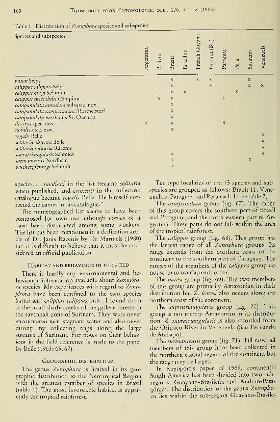
160 Tijdschrift voor Entomologie, deel 126, afl. 8 (1983)
Table 1 . Distribution of Zonophora species and subspecies
Species and subspecies
Ü
batest Selys
calippus calippus Selys
calippus klugi Schmidt
calippus spectabilis Campioncampanulata annulata subspec. nov.
campanulata campanulata (Burmeister)
campanulata machadoi St. Quentin
diversa spec. nov.
nobilis spec. nov.
regalis Belle
solitaria obscura Belle
solitaria solitaria Râcenis
supratriangulans Schmidt
surinamensis Needhamwucherpfennigi Schmidt
O >
species. . . medinai in the list became solitaria
when published, and cruxenti in the collection
catalogue became regalis Belle. He himself cor-
rected the names in his catalogue."
The mimeographed hst seems to have been
composed for own use although copies of it
have been distributed among some workers.
The list has been mentioned in a dedication arti-
cle of Dr. Janis Râcenis by De Marmels (1980)
but it is difficult to believe that it must be con-
sidered an official publication.
Habitat and behaviour in the field
There is hardly any environmental and be-
havioural information available about Zonopho-
ra species. My experiences with regard to Zono-
phora have been confined to the two species
batesi and calippus calippus only. I found themat the small shady creeks of the gallery forests in
the savannah zone of Surinam. They were never
encountered near stagnant water and also never
during my collecting trips along the large
streams of Surinam. For notes on their behav-
iour in the field reference is made to the paper
by Belle (1963: 65, 67).
Geographic distribution
The genus Zonophora is limited in its geo-
graphic distribution to the Neotropical Region
with the greatest number of species in Brazil
(table 1). The most favourable habitat is appar-
ently the tropical rainforest.
The type localities of the 15 species and sub-
species are grouped as follows: Brazil 11, Vene-
zuela 3, Paraguay and Peru each 1 (see table 2).
The campanulata group (fig. 67). The range
of this group covers the southern part of Brazil
and Paraguay, and the north-eastern part of Ar-
gentina. These parts do not fall within the area
of the tropical rainforest.
The calippus group (fig. 68). This group has
the largest range of all Zonophora groups. Its
range extends from the northern coast of the
continent to the southern part of Paraguay. Theranges of the members of the calippus group do
not seem to overlap each other.
The batesi group (fig. 69). The two members
of this group are primarily Amazonian in their
distribution but Z. batesi also occurs along the
northern coast of the continent.
The supratriangularis group (fig. 70). This
group Is not merely Amazonian in its distribu-
tion. Z. supratriangularis is also recorded from
the Orinoco River in Venezuela (San Fernando
de Atabapo).
The surinamensis group (fig. 71). Till now, all
members of this group have been collected in
the northern coastal region of the continent but
the range may be larger.
In Rapoport's paper of 1968, continental
South America has been divided into two sub-
regions, Guayano-Brasilena and Andino-Pata-
gónica. The distribution of the genus Zonopho-
ra lies within the sub-region Guayano-Brasile-
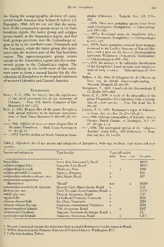
Belle: Zonophora 161
na. Using the Zoogeographie division of conti-
nental South America after Sclater & Sclater (cf.
Rapoport, 1968: 68) we can say that the mem-bers of the campanulata group occur in the Sud-
brasileho region, the batesi group and calippus
group mainly m the Amazónica region, and that
both groups penetrate into the Colombiana re-
gion as far as the northern coast (Venezuela and
the Guyanas), while the latter group also pene-
trates into the western part of the Sud-brasileho
region. The supratriangularis group occurs
mainly in the Amazónica region and the surina-
mensis group in the Colombiana region. The
two Cordilleras in the north-west of the conti-
nent seem to form a natural barrier for the dis-
tribution of Zonophora to the tropical rainforest
of western Colombia and Central-America.
References
Beatty, A. F., 1956. An inquiry into the significance
of the larval proventriculus in the taxonomy of
Odonata. — Proc. Xth. Intern. Congress of Ent.
Montreal. 1:367—372.Belie, J., 1963. Dragon flies of the genus Zonophora
with special reference to its Surinam representa-
tives. — Stud. Fauna Suriname 5: 60—69; pis. 3
—
4.
, 1966. Additional notes on some dragon-flies of
the genus Zonophora. — Stud. Fauna Suriname 8:
61—64; pi. 11.
, 1972. Further studies on South American Gom-
phidae (Odonata). — Ti)dschr. Ent. 115: 217
—
240.
, 1976. Three new gomphine species from Vene-zuela (Anisoptera: Gomphidae). — Odonatologi-ca5: 197—206.
, 1977a. Revisionai notes on Diaphlebia Selys,
1854 (Anisoptera: Gomphidae). — Odonatologica6:111—117.
, 1977b. Some gomphine material from Surinam,preserved in the Leyden Museum of Natural His-tory, with a note on the larva of Desmogomphustigrivensis Williamson (Anisoptera: Gomphidae).— Odonatologica 6: 289—292.
, 1979. An attempt at the subfamily classification
of the Gomphidae, based on some new interpreta-
tions of wing venation. — Odonatologica 8: 43
—
46.
Bullens, A. M., 1966. De fylogenie van de Odonata opbasis van de larvale kauwmaagbetanding. —Natuurh. Maandbl. 55: 88—95.
Burmeister, F., 1839. Handbuch der Entomologie II
(2), Berlin: 847—862.
Byers, C. F., 1939. A study of the dragonflies of the
genus Progomphus (Gomphoides) with a descrip-
tion of a new species. — Proc. Fla Acad. Sci. 4:
19—85.
Calvert, P. P., 1898. Burmeister's types of Odonata.— Trans. Am. ent. Soc. 25:27— 104; pi. 1.
, 1948. Odonata (dragonfhes) of Kartabo, Bartica
District, British Guiana. — Zoologica, N.Y. 33:
47—87; pis. 1—2., 1956. The neotropical species of the "subgenus
Aeschna" sensu Selys, 1883 (Odonata). — Mem.Am. ent. Soc. 15: 1—251.
Table 2. Alphabetic list of the species and subspecies of Zonophora, with type localities, type status and type
location.
Species and subspecies Type locality Type

162 Tijdschrift voor Entomologie, deel 126, afl. 8 (1983)
Figs. 1—5. Zonophora batest Selys, larva: 1, proventriculus; 2, first (left) dental fold, seen in oblique direction
from right; 3, second dental fold, the same; 4, third dental fold, the same; 5, fourth dental fold, seen in oblique
direction from left.
Campion, H., 1920. Some new or little known gom-phine dragonflies from South America. — Ann.Mag. nat. Hist. (9) 6: 130—141
; pis. 6—7.Carpenter, G. H., 1897. The geographical distribution
of dragonflies. — Scient. Proc. R. Dubl. Soc. 8:
439—468; pi. 17.
Davies, D. A. L., 1981. A synopsis of the extant gen-
era of the Odonata. — Soc. int. odonatol. rapid
Comm. 3: xiv -I- 59 pp.De Marmels, J., 1980. To Dr. Janis Racenis on his
65th birthday.— Odonatologica 9: 125— 129.
, 1983. The Odonata of the region of MountAuyantepui and the Sierra de Lema, in Venezue-lan Guyana. 3. Additions to the families Gomphi-dae, Aeshnidae and Corduliidae, with description
of Progomphus racenisi spec. nov. — Odonatolo-gica 12: 5— 13.
Dias dos Santos, N., 1970. Odonatas de liatiaia (Esta-
do do Rio de Janeiro) da coleçâo Zikan, do Insti-
tuto Oswaldo Cruz. — Atas. Soc. Biol. Rio de
Janeiro 13 (5 & 6): 204.
Fraser, F. C, 1940. A comparative study of the penes
of the family Gomphidae (order Odonata). —Trans. R. ent. Soc. Lond. (A) 90: 541—550; pis.
1—6.
, 1957. A reclassification of the order Odonata.
— R. zool. Soc. N.S.W., Sydney: 133 pp.; 1 pi.
Geijskes, D. C, 1971. List of Odonata known from
French Guyana, mainly based on a collection
brought together by the Mission of the "MuseumNational d'Histoire Naturelle", Paris, (1) (2).
—Ann. Soc. ent. Fr. (N.S.) 7 (3): 655—677.
Hagen, H. A., 1861. Synopsis of the Neuroptera of
North America. — Smithsonian Inst., Washing-

Belle: Zonophora 163
Figs. 6—8. Penes ot Zonophora, right lateral view: 6, Z. campanulata campanulata (Burmeister); 7, Z. calippus
calippus Selys; 8, 2. hatesi Selys. Figs. 9— 14. Left anterior hamules of Zonophora, right lateral view, showing
superior lobe (a), inferior lobe (b), and internal lobe (c): 9, Z. campanulata campanulata (Burmeister); 10, Z.
calippus calippus Selys; 11, Z. calippus klugi Schmidt; 12, Z. calippus spectabilis Campion; 13, Z. batesi Selys; 14,
2. supratriangularis Schmidt (more enlarged). Figs. 15— 18. Vulvar laminae of Zonophora, ventral view: 15, Z.
campanulata campanulata (Burmeister); 16, Z. calippus calippus Selys; 17, Z. batesi Selys; 18, Z. solitaria obscu-
ra Belle, first described female.

164 Tijdschrift voor Entomologie, deel 126, afl. 8 (1983)
Figs. 19—22. Vulvar laminae of Zonophora, ventral view, showing variability: 19 and 20, 2. campanulata cam-panulata (Burmeister); 21, 2. campanulata machadoi St. Quentin, first described female; 22, 2. campanulataannulata subspec. nov., female allotype. Figs. 23—25. Diagram of thoracic colour pattern of Zonophora: 23, 2.campanulata campanulata (Burmeister); 24, 2. campanulata machadoi St. Quentin, male holotype; 25, 2. cam-panulata annulata subspec. nov., male holotype. Figs. 26—30. Zonophora sunnamensis Needham, male: 26, di-
agram of thoracic colour pattern; 27, penis guard, frontal view; 28, the same, right lateral view; 29, penis, rightlateral view; 30, left anterior hamule, right lateral view.

Belle: Zonophora 165
34
36
Figs. 31—34. Zonophora campanulata campanulata (Burmeister), male: 31, accessory genitalia (penis and penis
guard not drawn), ventral view; 32, same, right lateral view; 33, tenth abdominal segment and caudal append-
ages, left lateral view; 34, the same, dorsal view. Figs. 35—36. Zonophora campanulata machadoi St. Quentin,
left lateral view of abdomen: 35, male holotype; 36, first described female. Figs. 37—38. Zonophora campanula-
ta annulata subspec. nov., left lateral view of abdomen: 37, male holotype; 38, female allotype.

166 Tijdschrift voor Entomologie, deel 126, afl. 8 (1^
Figs. 39—50. Zonophora diversa spec, nov., male holotype: 39, accessory genitalia (penis and penis guard not
drawn), ventral view; 40, the same, right lateral view; 41, tenth abdominal segment and caudal appendages, left
lateral view; 42, the same, dorsal view; 43, abdomen, left lateral view; 44, left anterior hamule, right lateral view;
45, the same, seen in oblique direction from right; 46, diagram of thoracic colour pattern; 47, penis guard, fron-
tal view; 48, the same, right lateral view; 49, occipital plate; 50, penis, right lateral view.

Belle: Zonophora 167
Figs. 51—54. Zonophora solitaria solitaria Räcenis, male holotype: 51, diagram of thoracic colour pattern; 52,
face and frons, seen in oblique direction from front; 53, abdominal segments 7 and 8, dorsal view; 54, frons,
vertex and occipital plate. (Figs. 51—54, after J. de Marmels, June 1981.) Fig. 55. Zonophora solitaria obscura
Belle, first described female: Occipital plate. Fig. 56. Zonophora supratriangularis Schmidt: Left posterior ham-ule, ventral view.
ton: XX + 347 pp., 1875. Synopsis of the Odonata of America. —Proc. Boston Soc. nat. Hist. 18: 20—96.
Karsch, F., 1890. Ueber Gomphiden. — Entom.
Nachr. 16:370—382.Kennedy, C. H., 1928. Evolutionary level in relation
to geographic, seasonal and diurnal distribution of
insects.— Ecology 9 (4): 367—379.
Kiauta, B., 1979. The karyotypes of some Anisoptera
from Surinam.— Odonatologica 8: 267—283.
, 1981. Annotated catalogue and bibhography of
taxa introduced in Odonata from 1971 to 1980.—Kimmins, D. E., 1969. A list of the type-specimens of
Odonata in the British Museum (Natural Histo-
ry), Part 2. — Bull. Brit. Mus. (Nat. Hist.) 23:
287—314.
Kirby, W. F., 1890. A synonymic catalogue of Neu-roptera Odonata or dragonflies. — Gurney &Jackson, London: IX -I- 202 pp.
Navas, L., 1914— 1923. Neurópteros colombianos. —Boll. Soc. Cienc. nat. (Bogota), 1914: 141—148;
1916: 6—13, 21—29, 67—73, 105—110, 119—121, 137—143, 150—154; 1919: 137—144; 1920:
169—176; 1922: 201—208, 233—240; 1923:
265—272, 279—280.
Needham, J. G., 1940. Studies on Neotropical gom-
phine dragonflies (Odonata). — Trans. Am. ent.
Soc. 65: 363—394; pis. 20—22., 1944. Further studies on Neotropical gomphine
dragonflies (Odonata). — Trans. Am. ent. Soc. 69:
171—224; pis. 14— 16.
Paulson, D. R., 1977. Odonata. In: S. H. Hurlbert,
Ed., Biota acuatica de Sudamérica Austral: 170
—
184. San Diego St. Univ.
Racenis, J., 1959. Lista de los Odonata del Perù. —Acta biol. venez. 2: 335—367.
, 1966. Preliminary hst of Venezuelan Odonata.
Inst. Zool. trop., Univ. Centr. Venezuela, Cara-
cas.
, 1970. Los odonatos de la region del Auyantepui
y de la Sierra de Lema, en la Guyana venezolana.
2. Las familias Gomphidae, Aeshnidae y Cordulii-
dae.— Acta biol. venez. 7: 23—39.
Rapoport, E. H., 1968. Algunos problemas bio-
geograficos del nuevo mundo con especial referen-
cia a la region neotropical. — Biol. Amér. aust. 4:
53—110.
Schmidt, E., 1941. Revision der Gattung Zonophora
Selys (Odonata Gomphidae neotrop.). — Dt. ent.
Z., 1941:76—96., 1952. Odonata nebst Bemerkungen über die
Anomisna und Chalcopteryx des Amazonas-Ge-
biets. — Beitr. Fauna Perus, Jena 3: 207—256, 4
pis.
St. Quentin, D., 1938. Die Tibialleiste der Odonaten.
— Zool. Anz. 121 (9/10): 225—239.
, 1967. Die Gattung Gomphoides Selys (Ordnung
Odonata) und ihre Verwandten in der neotropi-
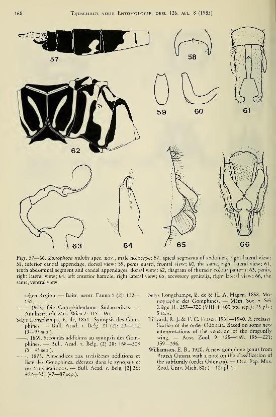
168 Tijdschrift voor Entomologie, deel 126, afl. 8 (1983)
Figs. 57—66. Zonophora nobilis spec, nov., male holotype: 57, apical segments of abdomen, right lateral view;
58, inferior caudal appendage, dorsal view; 59, penis guard, frontal view; 60, the same, right lateral view; 61,
tenth abdominal segment and caudal appendages, dorsal view; 62, diagram of thoracic colour pattern; 63, penis,
right lateral view; 64, left anterior hamule, right lateral view; 65, accessory genitalia, right lateral view; 66, the
same, ventral view.
sehen Region. — Beitr. neotr. Fauna 5 (2): 132
—
152.
, 1973. Die Gomphidenfauna Südamerikas. —Annin naturh. Mus. Wien 7: 335—363.
Selys Longchamps, E. de, 1854.. Synopsis des Gom-phines. — Bull. Acad. r. Belg. 21 (2): 23—112(3—93 sep.).
, 1869. Secondes additions au synopsis des Gom-phines. — Bul.. Acad. r. Belg. (2) 28: 168—208
(5—45 sep.).
, 1873. Appendices aux troisièmes additions et
liste des Gomphines, décrites dans le synopsis et
ses trois additions. — Bull. Acad. r. Belg. [2] 36:
492—531 (47—87 sep.).
Selys Longchamps, E. de & H. A. Hagen, 1858. Mo-nographie des Gomphines. — Mém. Soc. r. Sci.
Liège 11: 257—720 (VIII + 460 pp. sep.); 23 pis.;
5 tabs.
Tillyard, R. J. & F. C. Fraser, 1938—1940. A reclassi-
fication of the order Odonata. Based on some newinterpretations of the venation of the dragonfly
wing. — Aust. Zool. 9: 125—169; 195—221;
359—396.
Williamson, E. B., 1920. A new gomphine genus from
British Guiana with a note on the classification of
the subfamily (order Odonata). — Occ. Pap. Mus.
Zool. Univ. Mich. 80: 1—12; pi. 1.

Belle: Zonophora 169

170 Tijdschrift voor Entomologie, deel 126, afl. 8 (1983)

Belle: Zonophora 171
Fig. 71 . Distribution of the sunnamensis group.

172 Tijdschrift voor Entomologie, deel 126, afl. 8 (1983)
Figs. 72—74. Right pair of wings of male: 72, Zonophora campanulata campanulata (Burmeister); 73, Zonopho-
ra calippus calippus Selys; 74, Zonophora batesi Selys.
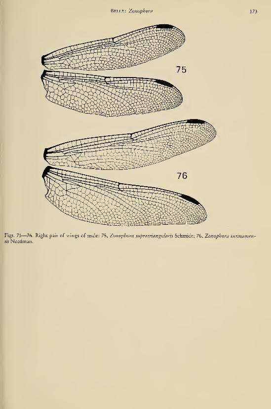
Belle: Zonophora 173
Figs. 75—76. Right pair of wings of male: 75, Zonophora supratnangulans Schmidt; 76, Zonophora sunnamen-sis Needman.
I


DEEL 126 AFLEVERING 9 1983
TIJDSCHRIFTVOOR ENTOMOLOGIE
UITGEGEVEN DOOR
DE NEDERLANDSE ENTOMOLOGISCHE VERENIGING
:0^-^OlOG/
^OstTo"^'
'^'US COMP. ZOOILIBRARY
'
'' 0^984
HARVARD
i INHOUD
C. VAN Achterberg. — Six new genera of Braconinae from the Afrotropical
Region (Hymenoptera, Braconidae), pp. 175—202, figs. 1—147.
Tijdschrift voor Entomologie, deel 126, afl. 9 Gepubliceerd 15-XII-1983

SIX NEW GENERA OF BRACONINAE FROM THEAFROTROPICAL REGION (HYMENOPTERA, BRACONIDAE)
by
C. VAN ACHTERBERGRijksmuseum van Natuurlijke Historie, Leiden, The Netherlands
Abstract
The new tribe Adeshini is defined for the genera Adesha Cameron, 1912, and Adeshoides
gen. nov. (type-species: A. asulcatus sp. nov. from Senegal). The genera with a facial protu-
berance m the Braconinae (excluding the Atanycolus group with petiolate pedicellus) are
keyed and (re)described. Three new genera from the Afrotropical region are described, viz.,
Malagopsis (type-species: Plaxopsis grandidien Szépligeti, 1913), Deltaphyma (type-spe-
cies: D. horstoki sp. nov. from South Africa), and Zanzopsis (type-species: Plaxopsis liogas-
ter Szépligeti, 1913). Bracon nigrtceps Brulle, 1846, is a new junior synonym of Lasiophorus
lanceolator (Fabncius, 1804) and Lasiophorus seyrigi Granger, 1949, is synonymized with
Malagopsis grandidieri (Szépligeti, 1913) comb, et syn. nov. The genus Plaxopsis Szépligeti,
1905, IS removed from the synonymy with Lasiophorus Haliday, 1833, and a lectotype is
designated for Lasiophorus seyrigi Granger, 1949, and Plesiohracon carinatus Cameron,1903. Finally the Plesiohracon group is defined, the known genera are keyed and two newgenera are described: Carinibracon (type-species: C. danielssoni sp. nov. from Senegal) and
Kenema (type-species: K. quickei sp. nov. from Sierra Leone). Odontogaster Szépligeti,
1 906, is a new junior svnonym of Soter Saussure, 1 892.
In this paper some recently discovered newgenera of the Braconinae from the Afrotropical
region are described, illustrated and keyed.
Among these are two new genera collected dur-
ing the Gambia-Senegal Expedition of the LundUniversity in 1977, kindly sent on loan by Dr.
R. Danielsson (Lund). Both new genera have a
sistergroup in the Oriental region as far as can
be deduced from our limited knowledge of the
genera of Braconidae. Dr. D. Quicke (Notting-
ham) kmdly allowed me to describe a related
new genus from Sierra Leone.
During my visits to the Berlin Museum (1979,
1982) I found several genera from the Afrotrop-
ical region which at present are included in the
genus Plaxopsis. These genera are (re)described
in this paper, together with a new genus fromSouth Africa found in the collection of the
Rijksmuseum van Natuurlijke Historie at Lei-
den. For the terminology used in this paper, see
Van Achterberg, 1979: 242—249.
Adeshini tribus nov.
Diagnosis. — Contains the only known Bra-
coninae with vein CUI a of fore wing at samelevel as vein 2-CUl (fig. 4) and vein CUlb of
fore wing much longer than vein 3-CUl (a syn-
apomorphy within the Braconinae); scapus
truncate apically (fig. 3); mesoscutum at least
sparsely setose; vein r-m of hind wing very
short (fig. 4); propodeum with complete longi-
tudinal carina (fig. 11); 1st tergite movably con-
nected to 2nd tergite; 2nd and 3rd metasomaltergites with no antero-lateral diverging grooves
(fig- 1)..
Contains two genera: Adesha Cameron, 1912
(Oriental region) and Adeshoides gen. nov. (Af-
rotropical region). The biology is unknown.
Key to genera of the tribe Adeshini nov.
1. Mesoscutum with deep medio-posterior
groove (fig. 146); laterope large and round
(fig. 139); angle between veins 1-SR and
C+SC+R of fore wing about 35° (fig. 140);
head and mesosoma smooth; mesoscutumdensely setose Adesha Cameron
— Mesoscutum without medio-posterior
groove (fig. 11); laterope (virtually) absent
(fig. 1); angle between veins 1-SR and
C-I-SC-I-R of fore wing about 65° (fig. 7);
head and mesosoma coriaceous; mesoscu-
tum sparsely setose, mainly near the notauli
Adeshoides gen. nov.
Adeshoides gen. nov.
Type-species: Adeshoides asulcatus sp. nov.
175
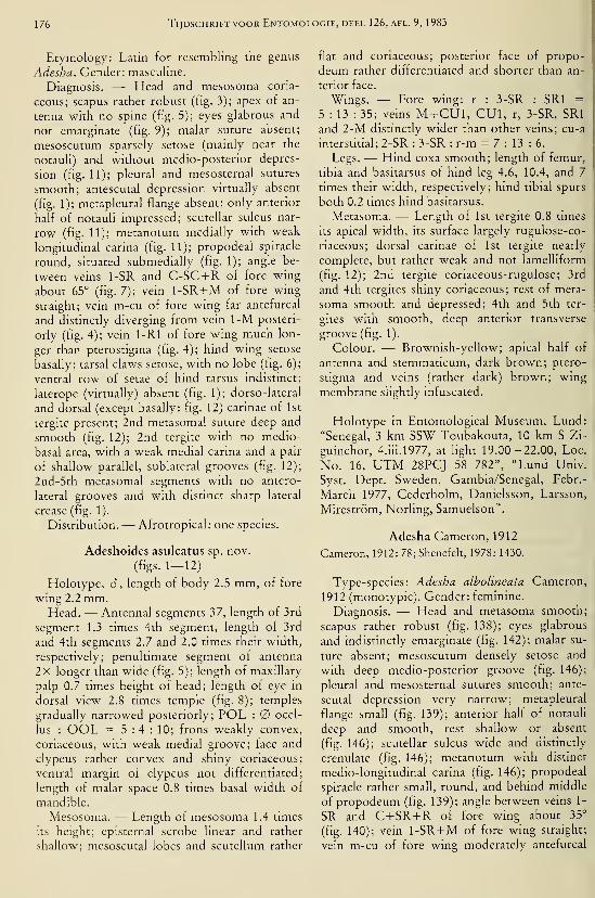
176 Tijdschrift voor Entomologie, deel 126, afl. 9, 1983
Etymology: Latin for resembling the genus
Adesha. Gender: masculine.
Diagnosis. — Head and mesosoma coria-
ceous; scapus rather robust (fig. 3); apex of an-
tenna with no spine (fig. 5); eyes glabrous and
not emarginate (fig. 9); malar suture absent;
mesoscutum sparsely setose (mainly near the
notauU) and without medio-posterior depres-
sion (fig. 11); pleural and mesosternal sutures
smooth; antescutal depression virtually absent
(fig. 1); metapleural flange absent; only anterior
half of notauli impressed; scutellar sulcus nar-
row (fig. 11); metanotum medially with weak
longitudinal carina (fig. 11); propodeal spiracle
round, situated submedially (fig. 1); angle be-
tween veins 1-SR and C-SC-l-R of fore wing
about 65° (fig. 7); vein 1-SR-l-M of fore wing
straight; vein m-cu of fore wing far antefurcal
and distinctly diverging from vein 1-M posteri-
orly (fig. 4); vein 1-Rl of fore wing much lon-
ger than pterostigma (fig. 4); hind wing setose
basally; tarsal claws setose, with no lobe (fig. 6);
ventral row of setae of hind tarsus indistinct;
laterope (virtually) absent (fig. 1); dorso-lateral
and dorsal (except basally: fig. 12) carinae of 1st
tergite present; 2nd metasomal suture deep and
smooth (fig. 12); 2nd tergite with no medio-
basal area, with a weak medial carina and a pair
of shallow parallel, sublateral grooves (fig. 12);
2nd-5th metasomal segments with no antero-
lateral grooves and with distinct sharp lateral
crease (fig. 1).
Distribution. — Afrotropical: one species.
Adeshoides asulcatus sp. nov.
(figs. 1-12)
Holotype, 6 , length of body 2.5 mm, of fore
wing 2.2 mm.Head. — Antennal segments 37, length of 3rd
segment 1.3 times 4th segment, length of 3rd
and 4th segments 2.7 and 2.0 times their width,
respectively; penultimate segment of antenna
2x longer than wide (fig. 5); length of maxillary
palp 0.7 times height of head; length of eye in
dorsal view 2.8 times temple (fig. 8); temples
gradually narrowed posteriorly; POL : ocel-
lus : OOL = 5:4:10; frons weakly convex,
coriaceous, with weak medial groove; face and
clypeus rather convex and shiny coriaceous;
ventral margin of clypeus not differentiated;
length of malar space 0.8 times basal width of
mandible.
Mesosoma. — Length of mesosoma 1.4 times
its height; episternal scrobe linear and rather
shallow; mesoscutal lobes and scutellum rather
flat and coriaceous; posterior face of propo-
deum rather differentiated and shorter than an-
terior face.
Wings. — Fore wing: r : 3-SR : SRI =5 : 13 : 35; veins M+ CUl, GUI, r, 3-SR, SRI
and 2-M distinctly wider than other veins; cu-a
interstitial; 2-SR : 3-SR : r-m = 7:13:6.Legs. — Hind coxa smooth; length of femur,
tibia and basitarsus of hind leg 4.6, 10.4, and 7
times their width, respectively; hmd tibial spurs
both 0.2 times hind basitarsus.
Metasoma. — Length of 1st tergite 0.8 times
its apical width, its surface largely rugulose-co-
riaceous; dorsal carinae of 1st tergite nearly
complete, but rather weak and not lamelliform
(fig. 12); 2nd tergite coriaceous-rugulose; 3rd
and 4th tergites shiny coriaceous; rest of meta-
soma smooth and depressed; 4th and 5th ter-
gites with smooth, deep anterior transverse
groove (fig. 1).
Golour. — Brownish-yellow; apical half of
antenna and stemmaticum, dark brown; ptero-
stigma and veins (rather dark) brown; wing
membrane slightly infuscated.
Holotype in Entomological Museum, Lund:
"Senegal, 3 km SSW Toubakouta, 10 km S Zi-
guinchor, 4.iii.l977, at light 19.00—22.00, Loc.
No. 16, UTM 28PGJ 58 782", "Lund Univ.
Syst. Dept. Sweden, Gambia/Senegal, Febr.-
March 1977, Cederholm, Danielsson, Larsson,
Mireström, Norling, Samuelson".
Adesha Gameron, 1912
Cameron, 1912: 78; Shenefelt, 1978: 1430.
Type-species: Adesha albolineata Gameron,
1912 (monotypic). Gender: feminine.
Diagnosis. — Head and metasoma smooth;
scapus rather robust (fig. 138); eyes glabrous
and indistinctly emarginate (fig. 142); malar su-
ture absent; mesoscutum densely setose and
with deep medio-posterior groove (fig. 146);
pleural and mesosternal sutures smooth; ante-
scutal depression very narrow; metapleural
flange small (fig. 139); anterior half of notauli
deep and smooth, rest shallow or absent
(fig. 146); scutellar sulcus wide and distinctly
crenulate (fig. 146); metanotum with distinct
medio-longitudinal carina (fig. 146); propodeal
spiracle rather small, round, and behind middle
of propodeum (fig. 139); angle between veins 1-
SR and G+SR+R of fore wing about 35°
(fig. 140); vein 1-SR-l-M of fore wing straight;
vein m-cu of fore wing moderately antefurcal

Van Achterberg: Afrotropical Braconinae \77
and slightly diverging posteriorly from vein 1-
M (fig. 141); tarsal claws setose, without lobe
(fig. 143); ventral row of setae of hind tarsus
distinct; laterope large and round (fig. 139);
dorso-lateral carinae of 1st tergite distinct; dor-
sal carinae on basal quarter of tergite, rest ab-
sent (fig. 147); 2nd metasomal suture deep and
crenulate (fig. 147); 2nd tergite with narrow tri-
angular and smooth medio-basal area, bordered
by crenulate grooves, laterally with a pair of
posteriorly (weakly) converging depressions
(fig. 147); 2nd-5th tergites with sharp lateral
crease and without antero-lateral grooves
(fig. 139); ovipositor normal, with neither no-
dus nor teeth subapically.
Distribution.— Oriental: one species.
Adesha albolineata Cameron(figs. 138—147)
Cameron, 1912: 78; Shenefelt, 1978: 1431.
Holotype, 9, length of body 4.2 mm, of fore
wing 3.8 mm.Head. — Antennal segments mcomplete, 16
present, length of 3rd segment 1.5 times 4th
segment, length of 3rd and 4th segments 3 and 2
times their width, respectively; length of maxil-
lary palp 0.7 times height of head; length of eye
in dorsal view 2.8 times temple; temples round-
ly narrowed posteriorly (fig. 145); POL :
ocellus : OOL = 7 : 8 : 19; frons slightly con-
vex, largely smooth and with medial groove
(fig. 142); face weakly convex and smooth; cly-
peus flat and smooth, its ventral margin not dif-
ferentiated from clypeus, concave; length of
malar space 0.8 times basal width of mandible.
Mesosoma. — Length of mesosoma 1.7 times
its height; side of pronotum smooth, except for
the finely crenulate medial groove; mesopleu-
ron smooth; episternal scrobe deep, medium-sized and round (fig. 139); mesoscutum rather
convex, its middle lobe slightly impressed me-dially (fig. 146); scutellum weakly convex and
somewhat pimply; propodeum largely smooth,
with some short rugae near medial carina and its
posterior surface not differentiated (fig. 146).
Wings. — Fore wing: r : 3-SR : SRI =
5 : 10 : 38; veins of similar width; 1-CUl :2-
CUl = 1:17; 2-SR : 3-SR : r-m =10:10:7.Legs. — Hind coxa smooth and slender
(fig. 139); length of femur, tibia and basitarsus
of hind leg 4.1, 9.8, and 6.4 times their width,
respectively; length of hind tibial spurs 0.2 and
0.3 times hind basitarsus.
Metasoma. — Length of 1st tergite 0.8 times
its apical width, its surface coarsely reticulate
medially, rest smooth (fig. 147); 3rd tergite
coarsely reticulate medially, as rest of metasoma(fig. 139); length of ovipositor sheath 0.20 times
fore wing, widened apically; hypopygium medi-
um-sized and truncate apically.
Colour. — Brownish-yellow; antenna, dorsal
part of head largely, meso- and metasoma dor-
sally, mesosternum partly, ovipositor sheath,
tarsi, patch on apex of hind tibia, hind femur
dorsally, dark brown or black; metasomalaterally, and apex of 5th tergite yellowish; wingveins and pterostigma brown; wing membranesubhyaline.
Holotype in British Museum (Natural Histo-
ry), London: "Type", "Kuching, J. H." (= JohnHewitt), "2.10", "P. Cameron Coll., 1914-
110", ''Adesha albolineata Cam., Type, Bor-
neo" (in Cameron's handwriting).
Key to the genera of the Braconinae withFACIAL protuberance (excluding the Atanyco-
lus group)
1. Head with a protuberance anteriorly
(figs. 13, 23, 35, 84); if rather cariniform
and small, then in dorsal view (short) semi-
circularly protruding; (some males of
Magalopsis grandidieri (Szépligeti) have no
protuberance but face is (narrowly) de-
pressed medially) 2
— Head without protuberance, at most cly-
peus with a scarcely protruding, medially
straight (in dorsal view), fine, dorsal carina;
(face without medio-dorsal depression) ....
other Braconinae
2. Pedicellus in dorsal view cylindrical, not
narrowed basally; scapus rounded subba-
sally (figs. 17, 32); inner aspect of scapus
not excavated apically (fig. 38), except in
Deltaphyma (fig. 56); length of fore tarsus
less than 1.7 times fore tibia 3
— Pedicellus in dorsal view petiolate, distinct-
ly narrowed basally; scapus rather angular-
ly protruding subbasally; inner aspect of
scapus deeply excavated medio-apically;
length of fore tarsus 1.7—2 times fore tibia
Atanycolus group
3. Middle of face with an irregularly incised,
vertical lamella (fig. 13); antenna of $
widened apically and with a blunt apex
(figs. 13, 14); marginal cell of fore wing
short, far removed from wing apex
(fig. 15); vein 1-SR of fore wing short
(fig. 15); tibial spurs dorsally glabrous and

178 Tijdschrift VOOR Entomologie, deel 126, afl. 9, 1983
ventrally setose Victoroviella Tobias
— Middle of face without vertical lamella
(figs. 23, 58, 60); antenna of 9 not widened
apically and with acute apex (figs. 24, 41);
marginal cell of fore wing long, almost
reaching wing apex (figs. 31, 70); vein 1-SR
of fore wing medium-sized (figs. 31, 42,
59); tibial spurs evenly setose or nearly
completely glabrous (at 50 X) 4
4. Face with protruding carina and/or lamella
(figs. 35, 52, 77); clypeus with ventral cari-
na (figs. 49, 77); mesoscutum largely gla-
brous; hind spurs (partly) setose; medio-
basal area of 2nd tergite robust (figs. 48, 65,
82); antero-lateral grooves of 3rd tergite
(partly) crenulate (figs. 48, 93); (Afrotropi-
cal and South Palaearctic) 5
— Face with robust horn above clypeus
(figs. 23, 26 of type species, in other species
spoon-shaped or tuberculiform); clypeus
without distinct ventral carina (fig. 23);
mesoscutum completely setose; hind spurs
almost completely glabrous; medio-basal
area of 2nd tergite very slender (fig. 34);
antero-lateral grooves of 3rd tergite
smooth; (Neotropical)
Lasiophorus Haliday
5. Upper half of face protruding (figs. 58, 75)
or shghtly concave (fig. 87); posterior half
of 4th and 5th tergites flat (figs. 57, 84); 1st
tergite usually without dorsal carinae
(figs. 65, 82), if present then not lamelli-
form; vein Ir-m of hind wmg straight
(fig. 55); 2nd tergite with (sub)parallel de-
pressions (figs. 65, 82, 93); vein 1-M of fore
wing straight (figs. 55, 70, 91); 1st discal
cell of fore wing less transverse (figs. 55,
91); lower facial protuberance of 9 dis-
tinctly lamelliform or absent; (Continental
Africa and South Palaearctical region) ... 6
— Upper half of face deeply concave (figs. 47,
49); posterior half of 4th and 5th tergites
strongly convex (fig. 35); 1st tergite with
lamelliform dorsal carinae (fig. 48); vein Ir-
m of hind wing curved (fig. 39); 2nd tergite
with (incomplete) diverging antero-lateral
grooves (fig. 48); vein 1-M of fore wing
somewhat bent posteriorly (fig. 42); 1st dis-
cal cell of fore wing comparatively trans-
verse (fig. 39); lower facial protuberance of
9 sublamelliform and absent in 6 and ex-
ceptionally in (small) 9 9 (Malagasy)
Malagopsis gen. nov.
6. Scapus truncate or at most moderately pro-
truding ventrally, not beyond apex of pedi-
cellus (figs. 69, 92); frons not or shallowly
concave (figs. 74, 88); face with either a la-
mella (figs. 84, 87) or a W-shaped protuber-
ance (figs. 75, 77); 2nd tergite with well-de-
fined basal area (figs. 82, 93); 2nd tergite
not projecting above 2nd suture (fig. 84);
groove between eye and antennal socket
rather shallow or absent (figs. 67, 84);
mesoscutum anteriorly at least as high as
the pronotum anteriorly (figs. 67, 84); scu-
tellum often with a small pit (fig. 76) 7
— Scapus strongly protruding ventro-apically,
well beyond apex of pedicellus (fig. 54);
frons distinctly concave (fig. 62); face with
protruding lamella and with a triangular
protuberance above it (figs. 58, 60); 2ndtergite with ill-defined basal area (fig. 65);
2nd tergite medio-posteriorly projecting
above 2nd suture (fig. 52); with a deep
groove between eye and antennal socket
(fig. 52); mesoscutum anteriorly lower than
pronotum anteriorly (fig. 52); scutellum
without small pit (fig. 63)
Deltaphyma gen. nov.
7. Face above clypeus with W-shaped protu-
berance (figs. 75, 77); marginal cell of hind
wing strongly narrowed apically (fig. 70)
Zanzopsis gen. nov.
— Face above clypeus without protuberance,
only submedially with a more or less semi-
circular (often minute) lamella (fig. 87);
marginal cell of hind wing (sub)-parallel-
sided or slightly narrowed apically (fig. 91)
Plaxopsis Szépligeti
Victoroviella Tobias, 1975
Tobias, 1975:962—964.
Type-species: Victoroviella deserticola Tobi-
as, 1975.
Diagnosis. — Scapus slightly longer ventrally
than dorsally (fig. 13), curved, with a rounded
anterior flange (figs. 13, 17) and subcylindrical;
antenna of 9 widened and rather compressed
apically (fig. 13); apex of antenna blunt
(fig. 14); face with longitudinal thin lamella,
which has one deep medial and some smaller in-
cisions (fig. 13), below this lamella a spoon-
shaped, weakly concave protuberance (fig. 18);
notauli absent; scutellar sulcus rather shallow
and narrow; metanotum without carinae; vein
1-Rl (metacarp) of fore wing much shorter than
pterostigma (fig. 15); fringe of wings very
short; basal half of fore wing only sparsely se-
tose; parastigma comparatively robust (fig. 15);
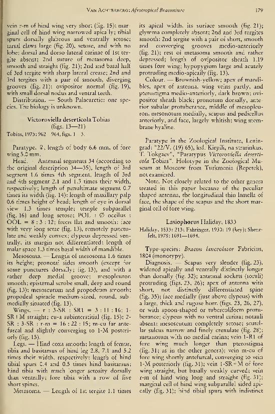
Van Achterberg: Afrotropical Braconinae 179
vein r-m of hind wing very short (fig. 15); mar-
ginal cell of hind wing narrowed apically; tibial
spurs dorsally glabrous and ventrally setose;
tarsal claws large (fig. 20), setose, and with no
lobe; dorsal and dorso-lateral carinae of 1st ter-
gite absent; 2nd suture of metasoma deep,
smooth and straight (fig. 21); 2nd and basal half
of 3rd tergite with sharp lateral crease; 2nd and
3rd tergites with a pair of smooth, diverging
grooves (fig. 21); ovipositor normal (fig. 19),
with small dorsal nodus and ventral teeth.
Distribution. — South Palaearctic: one spe-
cies. The biology is unknown.
Victoroviella deserticola Tobias
(figs. 13-21)
Tobias, 1975: 962—964, figs. 1—3.
Paratype, 9, length of body 6.6 mm, of fore
wing 5.0 mm.Head. — Antennal segments 34 (according to
the original description 34—35), length of 3rd
segment 1.6 times 4th segment, length of 3rd
and 4th segment 2.1 and 1.3 times their width,
respectively; length of penultimate segment 0.7
times its width (fig. 14); length of maxillary palp
0.6 times height of head; length of eye in dorsal
view 1.3 times temple; temple subparallel
(fig. 16) and long setose; POL : ocellus :
OOL = 8 : 3 : 12; frons flat and smooth; face
with very long setae (fig. 13), remotely punctu-
late and weakly convex; clypeus depressed ven-
trally, its margin not differentiated; length of
malar space 1.3 times basal width of mandible.
Mesosoma. — Length of mesosoma 1.6 times
its height; pronotal sides smooth (except for
some punctures dorsally; fig. 13), and with a
rather deep medial groove; mesopleuron
smooth; episternal scrobe small, deep and round
(fig. 13); mesoscutum and propodeum smooth;
propodeal spiracle medium-sized, round, sub-
medially situated (fig. 13).
Wings. — r : 3-SR : SRI = 3 : 11 : 16; 1-
SR-I-M straight; cu-a subinterstitial (fig. 15); 2-
SR : 3-SR : r-m = 16 : 22 : 15; m-cu far ante-
furcal and slightly converging to 1-M posteri-
orly (fig. 15).
Legs. — Hind coxa smooth; length of femur,
tibia and basitarsus of hind leg 2.8, 7.1 and 5.2
times their width, respectively; length of hind
tibial spurs 0.4 and 0.5 times hind basitarsus;
hind tibia with much longer setosity dorsally
than ventrally; fore tibia with a row of five
short spines.
Metasoma. — Length of 1st tergite 1.1 times
its apical width, its surface smooth (fig. 21);
glymma completely absent; 2nd and 3rd tergites
smooth; 2nd tergite with a pair of short, smoothand converging grooves medio-anteriorly
(fig. 21); rest of metasoma smooth and rather
depressed; length of ovipositor sheath 1.19
times fore wing; hypopygium large and acutely
protruding medio-apically (fig. 13).
Colour. — Brownish-yellow; apex of mandi-
bles, apex of antenna, wing veins partly, and
pterostigma medio-anteriorly, dark brown; ovi-
positor sheath black; pronotum dorsally, ante-
rior subalar protuberance, middle of mesopleu-
ron, mesonotum medially, scapus and pedicellus
anteriorly, and face, largely whitish; wing mem-brane hyaline.
Paratype in the Zoological Institute, Lenin-
grad: "22/V. (19) 65), ksf. Kirpili, na strarinkus,
T. Tokgaev", "Paratypus Victoroviella deserti-
cola Tobias". Holotype in the Zoological Mu-seum at Moscow from Turkmenia (Repetek),
not examined.
Note. Not closely related to the other genera
treated in this paper because of the peculiar
shaped antenna, the longitudinal thin lamella of
face, the shape of the scapus and the short mar-
ginal cell of fore wing.
Lasiophorus Haliday, 1833
Hallday, 1833: 213; Fahringer, 1930: 19 (key); Shene-
felt, 1978:1691— 1694.
Type-species: Bracon lanceolator Fabncius,
1804 (monotypy).
Diagnosis. — Scapus very slender (fig. 23),
widened apically and ventrally distinctly longer
than dorsally (fig. 32); antennal sockets (toruli)
protruding (figs. 23, 26); apex of antenna with
short, not distinctly differentiated spine
(fig. 35); face medially (just above clypeus) with
a large, thick and rugose horn (figs. 23, 26, 27),
or with spoon-shaped or tuberculiform protu-
berance; clypeus with no ventral carina; notauli
absent; mesoscutum completely setose; scutel-
lar sulcus narrow and finely crenulate (fig. 28);
metanotum with no medial carina; vein 1-Rl of
fore wing much longer than pterostigma
(fig. 31; as in the other genera); vein m-cu of
fore wing shortly antefurcal, converging to vein
1-M posteriorly (fig. 31); vein 1-SR-t-M of fore
wing straight, but basally weakly curved; vein
r-m of hind wing long and straight (fig. 31);
marginal cell of hind wing subparallel-sided api-
cally (fig. 31); hind tibial spurs with indistinct
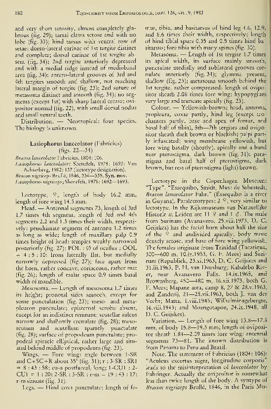
180 Tijdschrift voor Entomologie, deel 126, afl. 9, 1983
and very short setosity, almost completely gla-
brous (fig. 29); tarsal claws setose and with no
lobe (fig. 33); hind tarsus with ventral row of
setae; dorso-lateral carinae of 1st tergite distinct
and complete; dorsal carinae of 1st tergite ab-
sent (fig. 34); 2nd tergite anteriorly depressed
and with a medial ridge instead of mediobasal
area (fig. 34); antero-lateral grooves of 3rd and
4th tergites smooth and shallow, not reaching
lateral margin of tergite (fig. 23); 2nd suture of
metasoma distinct and smooth (fig. 34); no seg-
ments (except 1st) with sharp lateral crease; ovi-
positor normal (fig. 22), with small dorsal nodus
and small ventral teeth.
Distribution. — Neotropical: four species.
The biology is unknown.
Lasiophorus lanceolator (Fabricius)
(figs. 22—34)
Bracon lanceolator Vzhncms, 1804: 106.
Lasiophorus lanceolator; Shenefelt, 1978: 1692; VanAchterberg, 1982: 137 (lectotype designation).
Bracon nigriceps Brulle, 1846, 334—335. Syn. nov.
Lasiophorus nigriceps;Sheneieh, 1978: 1692—1693.
Lectotype, 9, length of body 16.2 mm,length of fore wing 14.5 mm.
Head. — Antennal segments 73, length of 3rd
1.7 times 4th segment, length of 3rd and 4th
segments 2.2 and 1.3 times their width, respecti-
vely; penultimate segment of antenna 1.2 times
as long as wide; length of maxillary palp 0.9
times height of head; temples weakly narrowed
posteriorly (fig. 27); POL : of ocellus : OOL= 4:5:10; frons laterally flat, but medially
narrowly depressed (fig. 27); face apart from
the horn, rather concave, coriaceous, rather mat
(fig. 26); length of malar space 0.9 times basal
width of mandible.
Mesosoma. — Length of mesosoma 1.7 times
its height; pronotal sides smooth, except for
some punctulation (fig. 23); meso- and meta-
pleuron punctulate; episternal scrobe absent,
except for an indistinct remnant; scutellar sulcus
narrow and shallowly crenulate (fig. 28); meso-
scutum and scutellum sparsely punctulate
(fig. 28); surface of propodeum punctulate; pro-
podeal spiracle elliptical, rather large and situ-
ated behind middle of propodeum (fig. 23).
Wings. — Fore wing: angle between 1-SR
and C+SC+R about 35° (fig. 31); r : 3-SR : SRI= 8 : 43 : 58; cu-a postfurcal, long; 1-CUl :
2-
CUl = 1 : 20; 2-SR : 3-SR : r-m = 19 : 43 : 17;
r-m sinuate (fig. 31).
Legs. — Hind coxa punctulate; length of fe-
mur, tibia, and basitarsus of hind leg 4.6, 12.9,
and 8.6 times their width, respectively; length
of hind tibial spurs 0.35 and 0.5 times hind ba-
sitarsus; fore tibia with many spines (fig. 30).
Metasoma. — Length of 1st tergite 1.7 times
its apical width, its surface mainly smooth,
punctulate medially and sublateral grooves cre-
nulate anteriorly (fig. 34); glymma present,
shallow (fig. 23); metasoma smooth behind the
1st tergite, rather compressed; length of ovipo-
sitor sheath 2.08 times fore wing; hypopygiumvery large and truncate apically (fig. 23).
Colour. — Yellowish-brown; head, antenna,
propleura, coxae partly, hind leg (except tro-
chanters partly, base and apex of femur, and
basal half of tibia), 5th—7th tergites and ovipo-
sitor sheath dark brown or blackish; palpi part-
ly infuscated; wing membrane yellowish, but
fore wing basally (shortly), apically and a band
near pterostigma, dark brown (fig. 31); para-
stigma and basal half of pterostigma, dark
brown, but rest of pterostigma (light) brown.
Lectotype in the Copenhagen Museum:"Type", "Essequibo, Smidt, Mus: de Sehestedt,
Bracon lanceolator Fabr." (Essequibo is a river
in Guyana). Paralectotypes: 2 9, very similar to
lectotype. In the Rijksmuseum van Natuurlijke
Historie at Leiden are 11 9 and 1 â. The male
from Surinam (Avanavero, 25.viii.1973, D. C.
Geijskes) has the facial horn about half the size
of the 9 and undivided apically, body more
densely setose, and base of fore wing yellowish.
The females originate from Trinidad (Tacariqua,
500—600 m, 10.ix.1953, G. F. Mees) and Suri-
nam (Republiek, 23. xi. 1963, D. C. Geijskes and
31.iii.l963, P. H. van Doesburg; Kabalebo Riv-
er, near Avanavero Falls, 14. ix. 1965, and
Brownsberg, 450—480 m, 16.xii.l973, both G.
F. Mees; Mapane area, camp 8, 27 & 28. v. 1963,
and Zanderij, 21—25.vii.1963, both J. van der
Vecht; Matta, l.viii.l945; Wilhelminagebergte,
16.viii.l943; and Moengotapoe, 24. ix. 1948, all
D. C. Geijskes).
Variation. — Length of fore wing 13.8—17.8
mm, of body 15.8—19.5 mm; length of oviposi-
tor sheath 1.84—2.09 times fore wing; antennal
segments 73—81. The known distribution is
from Panama to Peru and Brazil.
Note. The statement of Fabricius (1804: 106):
"Aculeus excertus niger, longitudine corporis"
leads to the misinterpretation of lanceolator by
Fahringer. Actually the ovipositor is somewhat
less than twice length of the body. A syntype of
Bracon nigriceps Brulle, 1846, in the Paris Mu-
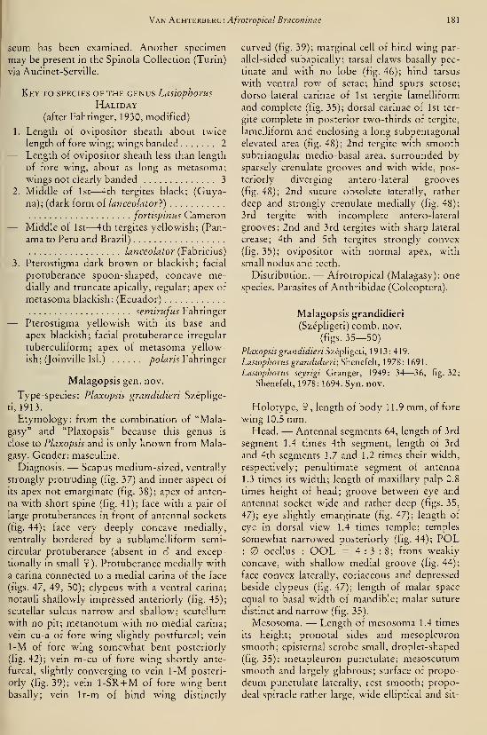
Van Achterberg: Afrotropical Braconinae 181
seum has been examined. Another specimen
may be present in the Spinola Collection (Turin)
via Audinet-Serville.
Key to species of the genus Lasiophorus
Haliday(after Fahringer, 1930, modified)
1. Length of ovipositor sheath about twice
length of fore wmg; wings banded 2
— Length of ovipositor sheath less than length
of fore wing, about as long as metasoma;
wings not clearly banded 3
2. Middle of 1st—4th tergites black; (Guya-
na); (dark form oi lanceolator})
fortispinus Cameron— Middle of 1st—4th tergites yellowish; (Pan-
ama to Peru and Brazil)
lanceolator (Fabricius)
3. Pterostigma dark brown or blackish; facial
protuberance spoon-shaped, concave me-dially and truncate apically, regular; apex of
metasoma blackish; (Ecuador)
semirufus Fahringer— Pterostigma yellowish with its base and
apex blackish; facial protuberance irregular
tuberculiform; apex of metasoma yellow-
ish; (Jomville Isl.) polaris Fahringer
Malagopsis gen. nov.
Type-species: Plaxopsis grandidieri Széplige-
ti, 1913.
Etymology: from the combination of "Mala-
gasy" and "Plaxopsis" because this genus is
close to Plaxopsis and is only known from Mala-
gasy. Gender: masculine.
Diagnosis. — Scapus medium-sized, ventrally
strongly protruding (fig. 37) and inner aspect of
its apex not emarginate (fig. 38); apex of anten-
na with short spine (fig. 41); face with a pair of
large protuberances in front of antennal sockets
(fig. 44); face very deeply concave medially,
ventrally bordered by a sublamelliform semi-
circular protuberance (absent in S and excep-
tionally in small 9). Protuberance medially with
a carina connected to a medial carina of the face
(figs. 47, 49, 50); clypeus with a ventral carina;
notauli shallowly impressed anteriorly (fig. 45);
scutellar sulcus narrow and shallow; scutellum
with no pit; metanotum with no medial carina;
vein cu-a of fore wing slightly postfurcal; vein
1-M of fore wing somewhat bent posteriorly
(fig. 42); vein m-cu of fore wing shortly ante-
furcal, slightly converging to vein 1-M posteri-
orly (fig. 39); vein 1-SR-l-M of fore wing bent
basally; vein Ir-m of hind wing distinctly
curved (fig. 39); marginal cell of hind wing par-
allel-sided subapically; tarsal claws basally pec-
tinate and with no lobe (fig. 46); hind tarsus
with ventral row of setae; hind spurs setose;
dorso-lateral carinae of 1st tergite lamelliform
and complete (fig. 35); dorsal carinae of 1st ter-
gite complete in posterior two-thirds of tergite,
lamelliform and enclosing a long subpentagonal
elevated area (fig. 48); 2nd tergite with smoothsubtriangular medio-basal area, surrounded bysparsely crenulate grooves and with wide, pos-
teriorly diverging antero-lateral grooves
(fig. 48); 2nd suture obsolete laterally, rather
deep and strongly crenulate medially (fig. 48);
3rd tergite with incomplete antero-lateral
grooves; 2nd and 3rd tergites with sharp lateral
crease; 4th and 5th tergites strongly convex
(fig. 35); ovipositor with normal apex, with
small nodus and teeth.
Distribution. — Afrotropical (Malagasy): one
species. Parasites of Anthribidae (Coleoptera).
Malagopsis grandidieri
(Szépligeti) comb. nov.
(figs. 35—50)
Plaxopsis grandidieri Szépligeti, 1913:419.
Lasiophorus grandidieri; Sheneieh, 1978: 1691.
Lasiophorus seyrigi Granger, 1949: 34—36, fig. 32;
Shenefelt, 1978: 1694. Syn. nov.
Holotype, 9 , length of body 11.9 mm, of fore
wing 10.5 mm.Head. — Antennal segments 64, length of 3rd
segment 1.4 times 4th segment, length of 3rd
and 4th segments 1.7 and 1.2 times their width,
respectively; penultimate segment of antenna
1.3 times its width; length of maxillary palp 0.8
times height of head; groove between eye and
antennal socket wide and rather deep (figs. 35,
47); eye slightly emarginate (fig. 47); length of
eye in dorsal view 1.4 times temple; temples
somewhat narrowed posteriorly (fig. 44); POL: ocellus : OOL = 4:3:8; frons weakly
concave, with shallow medial groove (fig. 44);
face convex laterally, coriaceous and depressed
beside clypeus (fig. 47); length of malar space
equal to basal width of mandible; malar suture
distinct and narrow (fig. 35).
Mesosoma. — Length of mesosoma 1.4 times
its height; pronotal sides and mesopleuron
smooth; episternal scrobe small, droplet-shaped
(fig. 35); metapleuron punctulate; mesoscutumsmooth and largely glabrous; surface of propo-
deum punctulate laterally, rest smooth; propo-
deal spiracle rather large, wide elliptical and sit-
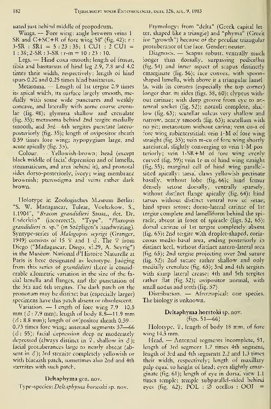
182 Tijdschrift voor Entomologie, deel 126, afl. 9, 1983
uated just behind middle of propodeum.Wings. — Fore wing: angle between veins 1-
SR and C+SC+R of fore wing 58° (fig. 42); r :
3-SR : SRI = 5 :23 : 35; 1-CUl : 2-CUl =
1 : 38; 2-SR : 3-SR : r-m = 10 : 23 : 10.
Legs. — Hind coxa smooth; length of femur,
tibia and basitarsus of hind leg 2.9, 7.8 and 4.0
times their width, respectively; length of hind
spurs 0.20 and 0.25 times hind basitarsus.
Metasoma. — Length of 1st tergite 0.9 times
its apical width, its surface largely smooth, me-dially with some wide punctures and weakly
concave, and laterally with some coarse crenu-
lae (fig. 48); glymma shallow and crenulate
(fig. 35); metasoma behind 2nd tergite medially
smooth, and 3rd—6th tergites punctate latero-
posteriorly (fig. 35); length of ovipositor sheath
0.59 times fore wing; hypopygium large, and
acute apically (fig. 35).
Colour. — Yellowish-brown; head (except
black middle of facial depression and of lamella,
stemmaticum, and area behind it), and pronotal
sides dorso-posteriorly, ivory; wing membranebrownish; pterostigma and veins rather dark
brown.
Holotype in Zoologisches Museum Berhn:
"S. W. Madagascar, Tulear, Voeltzkow, S.,
1.1904", "Bracon grandidieri Sauss., det. Dr.
Enderlein" (incorrect!), "Type", "Plaxopsis
grandidieri n. sp." (in Szépligeti's handwriting).
Syntype-series of Malagopsis seyrigi (Granger,
1949) consists of 15 9 and 1 d. The 2 fromDiego ("Madagascar, Diego, xi.29, A. Seyrig")
in the Museum National d'Histoire Naturelle at
Paris is here designated as lectotype. Judgingfrom this series of grandidieri there is consid-
erable allometric variation in the size of the fa-
cial lamella and flanges, and the punctation of
the 5th and 6th tergites. The dark patch on the
pronotum may be distinct but (especially larger)
specimens have this patch absent or obsolescent.
Variation. — Length of fore wing 7.9—10.5
mm {S: 7.9 mm); length of body 8.8—11.9 mm{6: 8.8 mm); length of ovipositor sheath 0.59
—
0.73 times fore wing; antennal segments 57—66
((?: 55); facial depression deep or moderately
depressed (always distinct in 9, shallow in 6);facial protuberances large to nearly absent (ab-
sent in cî); 3rd sternite completely yellowish or
with blackish patch, sometimes also 2nd and 4th
sternites with such patch.
Deltaphyma gen. nov.
Type-species: Deltaphyma horstoki sp. nov.
Etymology: from "delta" (Greek capital let-
ter, shaped like a triangle) and "phyma" (Greekfor "growth") because of the peculiar triangular
protuberance of the face. Gender: neuter.
Diagnosis. — Scapus robust, ventrally muchlonger than dorsally, surpassing pedicellus
(fig. 54) and inner aspect of scapus distinctly
emarginate (fig. 56); face convex, with spoon-
shaped lamella, with above it a triangular lamel-
la, with its corners (especially the top corner)
longer than its sides (figs. 58, 60); clypeus with-
out carinae; with deep groove from eye to an-
tennal socket (fig. 52); notauli complete, shal-
low (fig. 63); scutellar sulcus very shallow and
narrow, nearly smooth (fig. 63); scutellum with
no pit; metanotum without carina; vein cu-a of
fore wing subinterstitial; vein 1-M of fore wingstraight (fig. 55); vein m-cu of fore wing shortly
antefureal, slightly converging to vein 1-M pos-
teriorly; vein I-SR-HM of fore wing evenly
curved (fig. 59); vein Ir-m of hind wing straight
(fig. 55); marginal cell of hind wing parallel-
sided apically; tarsal claws yellowish pectinate
basally, without lobe (fig. 66); hind femur
densely setose dorsally, ventrally sparsely,
without distinct flange apically (fig. 64); hind
tarsus without distinct ventral row of setae;
hind spurs setose; dorso-lateral carinae of 1st
tergite complete and lamelliform behind the spi-
racle, absent in front of spiracle (figs. 52, 65);
dorsal carinae of 1st tergite completely absent
(fig. 65); 2nd tergite with droplet-shaped, coria-
ceous medio-basal area, ending posteriorly in
distinct keel, without distinct antero-lateral area
(fig. 65); 2nd tergite projecting over 2nd suture
(fig. 52); 2nd suture rather shallow and only
medially crenulate (fig. 65); 3rd and 4th tergites
with sharp lateral crease; 4th and 5th tergites
rather flat (fig. 52); ovipositor normal, with
small nodus and teeth (fig. 57).
Distribution. — Afrotropical: one species.
The biology is unknown.
Deltaphyma horstoki sp. nov.
(figs. 51—66)
Holotype, 9, length of body 18 mm, of fore
wing 14.5 mm.Head. — Antennal segments incomplete, 51,
length of 3rd segment 1.7 times 4th segment,
length of 3rd and 4th segments 2.2 and 1.3 times
their width, respectively; length of maxillary
palp equal to height of head; eyes slightly emar-
ginate (fig. 61); length of eye in dorsal view 1.1
times temple; temple subparallel-sided behind
eyes (fig. 62); POL : ocellus : OOL =
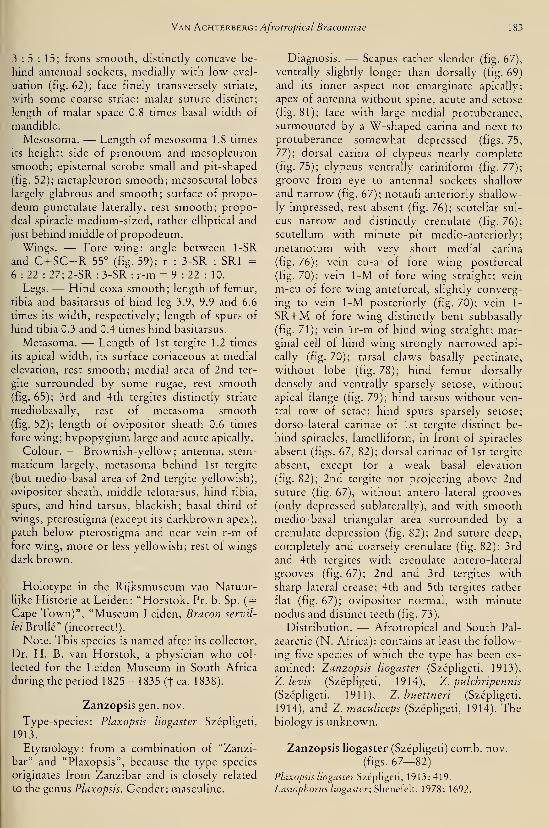
Van Achterberg: Afrotropical Braconinae 183
3 : 5 : 15; frons smooth, distinctly concave be-
hind antennal sockets, medially with low eval-
uation (fig. 62); face finely transversely striate,
with some coarse striae; malar suture distinct;
length of malar space 0.8 times basal width of
mandible.
Mesosoma. — Length of mesosoma 1.8 times
its height; side of pronotum and mesopleuron
smooth; episternal scrobe small and pit-shaped
(fig. 52); metapleuron smooth; mesoscutal lobes
largely glabrous and smooth; surface of propo-
deum punctulate laterally, rest smooth; propo-
deal spiracle medium-sized, rather elliptical and
just behind middle of propodeum.Wings. — Fore wing: angle between 1-SR
and C+SC+R 55° (fig. 59); r : 3-SR : SRI =
6:22: 27; 2-SR : 3-SR : r-m = 9 : 22 : 10.
Legs. — Hind coxa smooth; length of femur,
tibia and basitarsus of hind leg 3.9, 9.9 and 6.6
times its width, respectively; length of spurs of
hind tibia 0.3 and 0.4 times hind basitarsus.
Metasoma. — Length of 1st tergite 1.2 times
its apical width, its surface coriaceous at medial
elevation, rest smooth; medial area of 2nd ter-
gite surrounded by some rugae, rest smooth(fig. 65); 3rd and 4th tergites distinctly striate
mediobasally, rest of metasoma smooth(fig. 52); length of ovipositor sheath 0.6 times
fore wing; hypopygium large and acute apically.
Colour. — Brownish-yellow; antenna, stem-
maticum largely, metasoma behind 1st tergite
(but medio-basal area of 2nd tergite yellowish),
ovipositor sheath, middle telotarsus, hind tibia,
spurs, and hind tarsus, blackish; basal third of
wings, pterostigma (except its darkbrown apex),
patch below pterostigma and near vein r-m of
fore wing, more or less yellowish; rest of wings
dark brown.
Holotype in the Rijksmuseum van Natuur-lijke Historie at Leiden: "Horstok, Pr. b. Sp. (
=Cape Town)", "Museum Leiden, Bracon servil-
/ez' Brulle" (incorrect!).
Note. This species is named after its collector,
Dr. H. B. van Horstok, a physician who col-
lected for the Leiden Museum in South Africa
during the period 1825— 1835 (t ca. 11
Zanzopsis gen. nov.
Type-species: Plaxopsis liogaster Szépligeti,
1913.
Etymology: from a combination of "Zanzi-
bar" and "Plaxopsis", because the type species
originates from Zanzibar and is closely related
to the genus Plaxopsis. Gender: masculine.
Diagnosis. — Scapus rather slender (fig. 67),
ventrally slightly longer than dorsally (fig. 69)
and its inner aspect not emarginate apically;
apex of antenna without spine, acute and setose
(fig. 81); face with large medial protuberance,
surmounted by a W-shaped carina and next to
protuberance somewhat depressed (figs. 75,
77); dorsal carina of clypeus nearly complete
(fig. 75); clypeus ventrally cariniform (fig. 77);
groove from eye to antennal sockets shallow
and narrow (fig. 67); notauli anteriorly shallow-
ly impressed, rest absent (fig. 76); scutellar sul-
cus narrow and distinctly crenulate (fig. 76);
scutellum with minute pit medio-anteriorly;
metanotum with very short medial carina
(fig. 76); vein cu-a of fore wing postfurcal
(fig. 70); vein 1-M of fore wing straight; vein
m-cu of fore wing antefurcal, slightly converg-
ing to vein 1-M posteriorly (fig. 70); vein 1-
SR-I-M of fore wing distinctly bent subbasally
(fig. 71); vein Ir-m of hind wing straight; mar-
ginal cell of hind wing strongly narrowed api-
cally (fig. 70); tarsal claws basally pectinate,
without lobe (fig. 78); hind femur dorsally
densely and ventrally sparsely setose, without
apical flange (fig. 79); hind tarsus without ven-
tral row of setae; hind spurs sparsely setose;
dorso-lateral carinae of 1st tergite distinct be-
hind spiracles, lamelliform, in front of spiracles
absent (figs. 67, 82); dorsal carinae of 1st tergite
absent, except for a weak basal elevation
(fig. 82); 2nd tergite not projecting above 2ndsuture (fig. 67), without antero-lateral grooves
(only depressed sublaterally), and with smoothmedio-basal triangular area surrounded by a
crenulate depression (fig. 82); 2nd suture deep,
completely and coarsely crenulate (fig. 82); 3rd
and 4th tergites with crenulate antero-lateral
grooves (fig. 67); 2nd and 3rd tergites with
sharp lateral crease; 4th and 5th tergites rather
flat (fig. 67); ovipositor normal, with minute
nodus and distinct teeth (fig. 73).
Distribution. — Afrotropical and South Pal-
aearctic (N. Africa): contains at least the follow-
ing five species of which the type has been ex-
amined: Zanzopsis liogaster (Szépligeti, 1913),
Z. levis (Szépligeti, 1914), Z. pulchripennis
(Szépligeti, 1911), Z. huettneri (Szépligeti,
1914), and Z. maculiceps (Szépligeti, 1914). Thebiology IS unknown.
Zanzopsis liogaster (Szépligeti) comb. nov.
(figs. 67—82)
Plaxopsis liogaster Szépligeti, 1913:419.
Lasiophorus liogaster; Shenefelt, 1978: 1692.
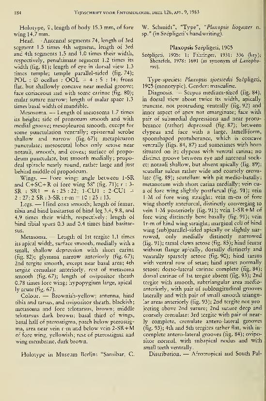
184 Tijdschrift voor Entomologie, deel 126, afl. 9, 1983
Holotype, 9 , length of body 15.3 mm, of fore
wing 14.7 mm.Head. — Antennal segments 74, length of 3rd
segment 1.5 times 4th segment, length of 3rd
and 4th segments 1.5 and 1.0 times their width,
respectively, penultimate segment 1.2 times its
width (fig. 81); length of eye in dorsal view 1.3
times temple; temple parallel-sided (fig. 74);
POL : ocellus : OOL = 4 : 5 : 14; frons
flat, but shallowly concave near medial groove;
face coriaceous and with some carinae (fig. 80);
malar suture narrow; length of malar space 1.3
times basal width of mandible.
Mesosoma. — Length of mesosoma 1.7 times
its height; side of pronotum smooth and with
medial groove; mesopleuron smooth, except for
some punctulation ventrally; episternal scrobe
shallow and narrow (fig. 67); metapleuron
punctulate; mesoscutal lobes only setose near
notauU, smooth, and covex; surface of propo-
deum punctulate, but smooth medially; propo-
deal spiracle nearly round, rather large and just
behind middle of propodeum.
Wings. — Fore wing: angle between 1-SR
and C+ SC +R of fore wing 58° (fig. 71); r :3-
SR : SRI = 6:25:22; 1-CUl : 2-CUl =
2 : 27; 2-SR : 3-SR : r-m = 10 : 25 : 13.
Legs. — Hind coxa smooth; length of femur,
tibia and hind basitarsus of hind leg 3.4, 9.8, and
4.9 times their width, respectively; length of
hind tibial spurs 0.3 and 0.4 times hind basitar-
sus.
Metasoma. — Length of 1st tergite 1.1 times
its apical width, surface smooth, medially with a
small, shallow depression with short carina
(fig. 82); glymma narrow anteriorly (fig. 67);
2nd tergite smooth, except near basal area; 4th
tergite crenulate anteriorly, rest of metasoma
smooth (fig. 67); length of ovipositor sheath
0.78 times fore wing; hypopygium large, apical-
ly acute (fig. 67).
Colour. — Brownish-yellow; antenna, hind
tibia and tarsus, and ovipositor sheath, blackish;
metasoma and fore telotarsus, brown; middle
telotarsus dark brown; basal third of wings,
basal half of pterostigma, patch below pterostig-
ma, area near vein r-m and below vein 2-SR-l-M
of fore wing, yellowish; rest of pterostigma and
wing membrane, dark brown.
Holotype in Museum Berlin: "Sansibar, C.
W. Schmidt", "Type", ''Plaxopsis liogaster n.
sp." (in Szépligeti's handwriting).
Plaxopsis Szépligeti, 1905
Szépligeti, 1905: 1; Fahrlnger, 1931: 336 (key);
Shenefelt, 1978: 1691 (as synonym of Lasiqpho-
rus).
Type-species: Plaxopsis sjoestedti SzépUgeti,
1905 (monotypic). Gender: masculine.
Diagnosis. — Scapus medium-sized (fig. 84),
in dorsal view about twice its width, apically
truncate, not protruding ventrally (fig. 92) and
inner aspect of apex not emarginate; face with
pair of submedial depressions and near protu-
berance (rather) depressed (fig. 87); between
clypeus and face with a large, lamelliform,
spoonshaped protuberance, which is concave
ventrally (figs. 84, 87) and sometimes with horn
situated on it; clypeus with ventral carina; no
distinct groove between eye and antennal sock-
et; notauli shallow, but absent apically (fig. 89);
scutellar sulcus rather wide and coarsely crenu-
late (fig. 89); scutellum with pit medio-basally;
metanotum with short carina medially; vein cu-
a of fore wing slightly postfurcal (fig. 91); vein
1-M of fore wing straight; vein m-cu of fore
wing shortly antefurcal, distinctly converging to
vein 1-M posteriorly (fig. 91); vein 1-SR-l-M of
fore wing distinctly bent basally (fig. 91); vein
Ir-m of hind wing straight; marginal cell of hind
wing (sub)parallel-sided apically or slightly nar-
rowed, only medially distinctly narrowed
(fig. 91); tarsal claws setose (fig. 85); hind femur
without flange apically, dorsally distinctly and
ventrally sparsely setose (fig. 90); hind tarsus
with ventral row of setae; hind spurs normally
setose; dorso-lateral carinae complete (fig. 84);
dorsal carinae of 1st tergite absent (fig. 93); 2nd
tergite with smooth, subtriangular area medio-
anteriorly, with pair of sublongitudinal grooves
laterally and with pair of small smooth triangu-
lar areas anteriorly (fig. 93); 2nd tergite not pro-
jecting above 2nd suture; 2nd suture deep and
coarsely crenulate; 3rd tergite with pair of near-
ly complete, crenulate antero-lateral grooves
(fig. 93); 4th and 5th tergites rather flat, with in-
complete antero-lateral grooves (fig. 84); ovipo-
sitor normal, with subapical nodus and with
small teeth ventrally.
Distribution. — Afrotropical and South Pal-

Van Achterberg: ^/rofropzc.«/ Braconinae 185
aearctic (N. Africa): moderately sized genus.
The biology is unknown.
Plaxopsis sjoestedti Szépligeti
(figs. 83—93)
Plaxopsis Sjostedti Szépligeti, 1905: 1—3, fig. 1.
Lasiophorus sjoestedti; Shenefelt, 1978: 1694.
Holotype, 9 , length of body 16.1 mm, of fore
wing 16.2 mm.Head. — Antennal segments 64 (apical seg-
ments missing), length of 3rd segment 1.5 times
4th segment, length of 3rd and 4th segments 1.6
and 1.1 times their width, respectively; length
of maxillary palp equal to height of head; length
of eye in dorsal view 1.5 times temple; POL :
ocellus : OOL = 3:3:11; frons weakly
concave; face smooth; clypeus largely flat and
smooth; malar suture shallow; length of malar
space 0.9 times basal width of mandible.
Mesosoma. — Length of mesosoma 1.7 times
its height; side of pronotum and mesopleuron
smooth; mesoscutal lobes largely glabrous,
smooth and rather convex; surface of propo-
deum smooth, posteriorly rather depressed
(fig. 93); propodeal spiracle rather large, subel-
liptical and at middle of propodeum (fig. 84).
"Wings. — Fore wing: angle between 1-SR
and C+ SC+R 56° (fig. 91); r : 3-SR : SRI =
9 : 50 : 51; 1-CUl : 2-CUl = 1 : 12; 2-SR :3-
SR : r-m = 24 : 50 : 22.
Legs. — Hind coxa indistinctly punctulate;
hind and fore claws missing; length of femur,
tibia, and basitarsus of hind leg 4.3, 10.8, and
6.1 times their width, respectively; length of
spurs of hind tibia 0.3 and 0.4 times hind ba-
sitarsus.
Metasoma. — Length of 1st tergite 1.7 times
its apical width, smooth (fig. 93); glymma nar-
row (fig. 84); 2nd tergite (except basal area)
striate (fig. 93); rest of metasoma smooth;
length of ovipositor sheath 0.84 times fore
wing; hypopygium large and apically acute
(fig. 84).
Colour. — Dark reddish-brown; head, palpi,
apex of antenna, prothorax, metasoma ventrally
(but posteriorly brownish) and 9th tergite, yel-
lowish; wing membrane dark brown, but fore
wing with subapical yellowish, incomplete band
and below pterostigma with a small light area
(fig. 91).
Holotype in the Stockholm Museum:"Camerun", "Sjöstedt", "Plaxopsis S)östedti
n.g. n. sp." (in Szépligeti's handwriting).
Plesiobracon group
The Plesiobracon group contains three genera
known to me, which have the 1st tergite of the
metasoma movably joined to the 2nd tergite,
with strong and complete dorso-lateral carinae,
the mesoscutum rather evenly setose and at the
same time the propodeum with a strong and
complete medio-longitudinal carina, the scapus
is (sub)truncate apically and ovoid (figs. 97,
113), the clypeus has no dorsal carina, the angle
between veins 1-SR and C-I-SC-I-R of fore wing
more than 60°, vein cu-a of fore wing slightly
postfurcal (figs. 98, 114), the laterope is absent,
and the 2nd tergite has a pair of sublateral, con-
verging or short longitudinal grooves (figs. 109,
121). The biology is unknown.
Key to genera of the Plesiobracon group
1. First tergite without dorsal carinae
(fig. 137); posterior margin of hind wing
(but sometimes hardly) concave subbasally
and shorter setose than surrounding fringe
(fig. 136); head strongly constricted behind
eyes (fig. 132); vein cu-a of hind wing recli-
vous (fig. 136); (Indo-Australian)
Plesiobracon Cameron— First tergite with strong, united dorsal cari-
nae (figs. 109, 121); posterior margin of
hind wing about straight subbasally and
evenly setose (figs. 104, 114); head rounded
posteriorly (fig. 107, 117); vein cu-a of hind
wing (nearly) straight (figs. 98, 114); (Afro-
tropical) 2
2. Medial length of 5th tergite ca. 0.8 times
medial length of 4th tergite behind basal de-
pression (fig. 95); vein 2-SR of fore wing
straight (fig. 98); 5th tergite straight latero-
apically (fig. 95); antescutal depression pre-
sent (fig. 95); propleuron without posterior
flange (fig. 95); 2nd tergite with small me-
dio-basal area (fig. 109); mesoscutum punc-
tulate (fig. 108); propodeal spiracle situated
submedially at propodeum (fig. 95); 4th
tergite more or less protrudmg medio-api-
cally; 2nd submarginal cell of fore wing
slender (fig. 98) . . . Carinibracon gen. nov.
— Medial length of 5th tergite (behind basal
depression) 1.3—1.5 times medial length of
4th tergite (fig. 110); veins 2-SR of fore
wing (rather) sinuate (fig. 114); 5th tergite
oblique latero-apically (fig. 110); antescutal
depression absent (fig. 110); propleuron
with small posterior flange (fig. 110); 2nd
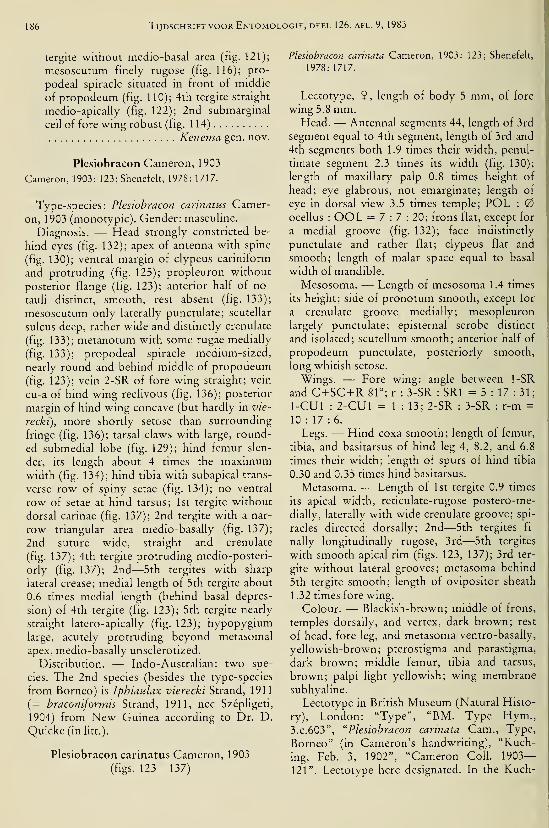
186 Tijdschrift voor Entomologie, deel 126, afl. 9, 1983
tergite without medio-basal area (fig. 121);
mesoscutum finely rugose (fig. 116); pro-
podeal spiracle situated in front of middle
of propodeum (fig. 110); 4th tergite straight
medio-apically (fig. 122); 2nd submarginal
cell of fore wing robust (fig. 114)
Kenema gen. nov.
Plesiobracon Cameron, 1903
Cameron, 1903: 123; Shenefelt, 1978: 1717.
Type-species: Plesiobracon cannatus Camer-
on, 1903 (monotypic). Gender: masculine.
Diagnosis. — Head strongly constricted be-
hind eyes (fig. 132); apex of antenna with spine
(fig. 130); ventral margin of clypeus cariniform
and protruding (fig. 125); propleuron without
posterior flange (fig. 123); anterior half of no-
tauH distinct, smooth, rest absent (fig. 133);
mesoscutum only laterally punctulate; scutellar
sulcus deep, rather wide and distinctly crenulate
(fig. 133); metanotum with some rugae medially
(fig. 133); propodeal spiracle medium-sized,
nearly round and behind middle of propodeum(fig. 123); vein 2-SR of fore wing straight; vein
cu-a of hind wing reclivous (fig. 136); posterior
margin of hind wing concave (but hardly in vie-
recki), more shortly setose than surrounding
fringe (fig. 136); tarsal claws with large, round-
ed submedial lobe (fig. 129); hind femur slen-
der, its length about 4 times the maximumwidth (fig. 134); hind tibia with subapical trans-
verse row of spiny setae (fig. 134); no ventral
row of setae at hind tarsus; 1st tergite without
dorsal carinae (fig. 137); 2nd tergite with a nar-
row triangular area medio-basally (fig. 137);
2nd suture wide, straight and crenulate
(fig. 137); 4th tergite protruding medio-posteri-
orly (fig. 137); 2nd—5th tergites with sharp
lateral crease; medial length of 5th tergite about
0.6 times medial length (behind basal depres-
sion) of 4th tergite (fig. 123); 5th tergite nearly
straight latero-apically (fig. 123); hypopygiumlarge, acutely protruding beyond metasomal
apex, medio-basally unsclerotized.
Distribution. — Indo-Australian: two spe-
cies. The 2nd species (besides the type-species
from Borneo) is Iphiaulax vierecki Strand, 1911
(= braconiformis Strand, 1911, nee Szépligeti,
1904) from New Guinea according to Dr. D.
Quicke (in litt.).
Plesiobracon carinatus Cameron, 1903
(figs. 123—137)
Plesiobracon carinata Cameron, 1903: 123; Shenefelt,
1978: 1717.
Lectotype, 9 , length of body 5 mm, of fore
wing 5.8 mm.Head. — Antennal segments 44, length of 3rd
segment equal to 4th segment, length of 3rd and
4th segments both 1.9 times their width, penul-
timate segment 2.3 times its width (fig. 130);
length of maxillary palp 0.8 times height of
head; eye glabrous, not emarginate; length of
eye in dorsal view 3.5 times temple; POL :
ocellus : OOL = 7:7: 20; frons flat, except for
a medial groove (fig. 132); face indistinctly
punctulate and rather flat; clypeus flat and
smooth; length of malar space equal to basal
width of mandible.
Mesosoma. — Length of mesosoma 1.4 times
its height; side of pronotum smooth, except for
a crenulate groove medially; mesopleuron
largely punctulate; episternal scrobe distinct
and isolated; scutellum smooth; anterior half of
propodeum punctulate, posteriorly smooth,
long whitish setose.
Wings. — Fore wing: angle between 1-SR
and C+SC +R 81°; r : 3-SR : SRI = 5 : 17 : 31;
1-CUl : 2-CUl = 1 : 13; 2-SR : 3-SR : r-m =
10 : 17 :6.
Legs. — Hind coxa smooth; length of femur,
tibia, and basitarsus of hind leg 4, 8.2, and 6.8
times their width; length of spurs of hind tibia
0.30 and 0.35 times hind basitarsus.
Metasoma. — Length of 1st tergite 0.9 times
its apical width, reticulate-rugose postero-me-
dially, laterally with wide crenulate groove; spi-
racles directed dorsally; 2nd—5th tergites fi-
nally longitudinally rugose, 3rd—5th tergites
with smooth apical rim (figs. 123, 137); 3rd ter-
gite without lateral grooves; metasoma behind
5th tergite smooth; length of ovipositor sheath
1.32 times fore wing.
Colour. — Blackish-brown; middle of frons,
temples dorsally, and vertex, dark brown; rest
of head, fore leg, and metasoma ventro-basally,
yellowish-brown; pterostigma and parastigma,
dark brown; middle femur, tibia and tarsus,
brown; palpi light yellowish; wing membranesubhyaline.
Lectotype in British Museum (Natural Histo-
ry), London: "Type", "BM. Type Hym.,
3.C.603", "Plesiobracon carinata Cam., Type,
Borneo" (in Cameron's handwriting), "Kuch-
ing, Feb. 3, 1902", "Cameron Coll. 1903—121". Lectotype here designated. In the Kuch-
Van Achterberg : Afrotropical Braconinae 187
ing Museum I examined 2 9 in the collection
under "cannata": one specimen collected April
1, 1902, the 2nd specimen at May 24, 1903. Be-
cause of the date of capture of the 2nd speci-
men, this specimen is excluded from the type-
series. The other 9 is considered to be a para-
lectotype. It differs from the lectotype by a nar-
row antescutal depression.
Carinibracon gen. nov.
Type-species: Carinibracon danielssoni sp.
nov.
Etymology: from "carina" (Latin for "keel")
and "Bracon", because it is closely related to the
genus Bracon, but it possesses several carinae
(propodeum, 1st and 2nd tergites) which are
normally absent in Bracon. Gender: masculine.
Diagnosis. — Head rounded posteriorly
(fig. 107); apex of antenna without spine, setose
(fig. 99); ventral carina of clypeus present, pro-
truding (figs. 95, 103); propleuron without pos-
terior flange (fig. 95); mesoscutum punctulate;
notauli completely smooth (fig. 108); scutellar
sulcus medium-sized, rather wide and crenulate
(fig. 108); metanotum with short medial carina;
propodeal spiracle round, near middle of propo-
deum (fig. 95); vein 2-SR of fore wing straight;
2nd submarginal cell of fore wing slender
(fig. 98); vein cu-a of hind wing straight
(fig. 104); posterior margin of hind wing nearly
straight basally and evenly setose (fig. 104); tar-
sal claws with peculiar lamelliform lobe and
with some pegs (fig. 100); hind femur robust,
about 3.5 times its maximum width (fig. 102);
hind tibia without subapical row of spiny setae
(fig. 102); hind tarsus with distinct ventral rowof setae; 1st tergite with dorsal carinae com-plete, meeting posteriorly, in front of strongly
convex area (fig. 109); 2nd tergite with small
subtriangular area medio-basally and with pair
of weakly converging sublateral grooves
(fig. 109); 2nd suture sinuate and crenulate
(fig. 109); 4th tergite somewhat protruding me-dio-posteriorly; medial length of 5th tergite
about 0.8 times medial length of 4th tergite be-
hind basal depression (fig. 95); 5th tergite
straight latero-apically (fig. 95); 2nd—6th ter-
gites with sharp lateral crease; hypopygiumlarge and acute apically (fig. 95).
Distribution.— Afrotropical: one species.
Carinibracon danielssoni sp. nov.
(figs. 94—109)
Holotype, 9, length of body 4.7 mm, of fore
wmg 4.6 mm.
Head. — Antennal segments 35, length of 3rd
segment 1.3 times 4th segment, length of 3rd
and 4th segments 2.3 and 1.8 times their width,
respectively, penultimate segment 1.7 times its
width (fig. 99); length of maxillary palp 0.7
times height of head; eye glabrous and slightly
emarginate (fig. 103); length of eye in dorsal
view 1.8 times temple; POL : ocellus :
OOL = 3:3:7; frons nearly flat, coriaceous
and with short groove near anterior ocellus (fig.
107); face largely coriaceous, but smooth above
clypeus; length of malar space 0.8 times basal
width of mandible.
Mesosoma. — Length of mesosoma 1.5 times
Its height; side of pronotum and mesopleuron
smooth, but mesopleuron ventrally sparsely
punctulate; episternal scrobe shallow (fig. 95);
scutellum punctulate; surface of propodeumsmooth and setose.
Wings. — Fore wing: angle between 1-SR
and C+SC-t-R 70° (fig. 101); r : 3-SR : SRI =
15 : 37 : 77; 1-CUl : 2-CUl = 2 : 25; 2-SR : 3-
SR:r-m = 28 : 37 : 17.
Legs. — Hind coxa punctulate; length of fe-
mur, tibia and basitarsus of hind leg 3.5, 7.5 and
5 times their width, respectively; length of spurs
of hind tibia 0.35 and 0.4 times hind basitarsus.
Metasoma. — Length of 1st tergite 0.8 times
its apical width, largely smooth (fig. 109); 2nd
tergite coarsely reticulate-rugose; 3rd—7th ter-
gites finely rugulose, with narrow subapical
transverse groove and posterior rim smooth
(fig. 95); length of ovipositor sheath 0.8 times
fore wing.
Colour. — Brownish-yellow; antenna (of
scapus and pedicellus only outer side), ptero-
stigma, veins, and ovipositor sheath, dark
brown; wing membrane brownish.
Holotype in Entomological Museum, Lund:
"Senegal, in forest, 1.5 km NE Djibélor, ca. 6.5
km SW Ziguinchor, 8.iii.l977. At light, 19.00—
21.30. Loc. no. 23A, UTM 28PCJ575885","Lund Univ., Syst. Dept. Sweden, Gambia/Se-
negal Febr.—March 1977, Cederholm, Daniels-
son, Larsson, Mireström, Norling, Samuels-
son".
Note. — In existing keys the species runs to
the genus Cratocnema, but Cratocnema differs
e.g., by the presence of a dorsal carina at the
clypeus. The descriptions of the Cratocnema
spp. do not fit C. danielssoni.
It is a pleasure to me to dedicate this species
to Mr. R. Danielsson (Lund), who has been
very helpful in providing types and new taxa.
Tijdschrift voor Entomologie, deel 126, afl. 9, 1983
Kenema gen. nov.
Type-species: Kenema quickei sp. nov.
Etymology: name based on the locality of the
type. Gender: feminine.
Diagnosis. — Head rounded posteriorly
(fig. 117); apex of antenna with small spine
(fig. Ill); ventral carina of clypeus not differ-
entiated (fig. 120); propleuron with small poste-
rior flange (fig. 110); mesoscutum finely rugose
(fig. 116); notauh only anteriorly distinct and
finely crenulate (fig. 116), rest absent; scutellar
sulcus narrow and finely crenulate (fig. 116);
metanotum with complete longitudinal carina;
propodeal spiracle small, round and in front of
middle of propodeum (fig. 110); vein 2-SR of
fore wing sinuate (fig. 114); 2nd submarginal
cell of fore wing robust (fig. 114); vein cu-a of
hind wing straight; posterior margin of hind
wing straight and evenly setose (fig. 114); tarsal
claws with small acute lobe (fig. 118); hind fe-
mur moderately robust (fig. 119); hind tibia
without row of spiny setae; hind tarsus with
ventral row of setae; 1st tergite with dorsal cari-
nae united medially, but medially rather weak
(fig. 121); 2nd tergite without medial area and
with short longitudinal grooves (fig. 121); 2nd
suture distinctly sinuate and crenulate; 4th ter-
gite straight medio-posteriorly (fig. 122); medi-
al length of 5th tergite (behind basal, crenulate
groove) 1.3— 1.5 times medial length of 4th ter-
gite (figs. 110, 122); 5th tergite oblique latero-
apically (fig. 110); 2nd—5th tergites with sharp
(cariniform) lateral crease.
Distribution. — Afrotropical: two species.
The 2nd species will be described in a forthcom-
ing paper.
Kenema quickei sp. nov.
(figs. 110—122)
Holotype, 6, length of body 6 mm, of fore
wing 4.5 mm.Head. — Antennal segments 52, length of 3rd
segment 1.2 times 4th segment, length of 3rd
and 4th segments 1.7 and 1.4 times their width,
respectively, penultimate segment 1.4 times its
width (fig. Ill); length of maxillary palp 0.6
times height of head; length of eye in dorsal
view 2.6 times temple; temple ruguloso-coria-
ceous; POL : ocellus : OOL = 6 : 4 : 12;
frons slightly convex, laterally rugose, medially
coriaceous, and with shallow medial depression
(fig. 117); stemmaticum comparatively strongly
protruding (fig. 120); face transversely rugose;
length of malar space equal to basal width of
mandible.
Mesosoma. — Length of mesosoma 1.5 times
its height; side of pronotum coriaceous, with
some striae (fig. 110); mesopleuron coriaceous;
episternal scrobe virtually absent; scutellum co-
riaceous; propodeum densely rugulose, medio-
laterally shortly lamelliform and angularly pro-
truding (fig. 110).
Wings. — Fore wing: angle between 1-SR
and C-I-SC +R 61°; r-m distinctly sinuate and
sclerotized (fig. 114); r : 3-SR : SRI 3 =
7 : 10 : 39; 2-SR : 3-SR : r-m = 11 : 10 : 11.
Legs. — Hind coxa coriaceous; length of fe-
mur, tibia, and basitarsus of hind leg 3.9, 8.2,
and 5.2 times their width, respectively; length
of spurs of hind tibia both 0.4 times hind ba-
sitarsus.
Metasoma. — Length of 1st tergite 0.4 times
its apical width, basally smooth, behind dorsal
carinae distinctly rugose; 2nd tergite rather
coarsely reticulate; 3rd tergite reticulate-rugose;
4th and 5th tergites densely coriaceous-rugu-
lose, with incomplete lateral grooves (fig. 110);
6th tergite shortly setose, exposed, smooth; 7th
tergite glabrous; 2nd—5th tergites with acute
lateral crease.
Colour. — Brownish-yellowish; antenna (ex-
cept annellus) black; wing membrane slightly
infuscated (but more pigmented near parastig-
ma; fig. 115); anterior margin of fore wing (ex-
cept base) blackish, rest of pterostigma and
most veins, brown.
Holotype in Quicke Collection, Nottingham:
"Kenema, Sierra Leone, Sept. 1981, D.
Quicke". I am pleased to name this species after
Dr. D. Quicke, who collected several interest-
ing taxa in Africa.
Note. This new genus resembles the genus
Soter Saussure, 1892 (= Odontogaster Széplige-
ti, 1906, syn. nov.), but Soter does not belong to
the Plesiobracon group because it has the meso-
scutum largely glabrous. Additionally it has the
mesoscutum smooth, the 2nd submarginal cell is
long, the 2nd tergite has converging grooves,
the 5th tergite is serrate, and the spiracle of the
propodeum is submedially situated.
Acknowledgements
I wish to express my gratitude to the follow-
ing persons for the loan of types and of newtaxa, and for assistance during visits: Mr. R.
Danielsson (Lund), Dr. J. Decelle (Tervuren),
Mr. A. A. Hamid (Kuching), Dr. K.-J. Hedq-
vist (Stockholm), Mr. T. Huddleston (London),
Mile Dr. S. Kelner-Pillault (Paris), f Dr. E.

Van Achterberg: /4/rofrop/c;2/ Braconinae 189
Königsmann (Berlin), Dr. B. Petersen (Copen-
hagen), Dr. D. Quicke (Nottingham), Mr. B.
Sigwalt (Paris), Dr. V. I. Tobias (Leningrad),
Mrs. I. Wegener (Berhn).
The Uyttenboogaart-EUasen Stichting finan-
cially supported the visit to the Kuching Mu-seum (Serawak).
References
Achterberg, C. van 1979. A revision of the subfamily
Zelinae auct. (Hym., Braconidae). — Tijdschr.
Ent. 122: 241—479, figs. 1—900., 1982. Notes on some type-species described by
Fabricius of the subfamilies Braconinae, Rogadi-
nae, Microgastrinae and Agathidinae (Hym.: Bra-
conidae). — Ent. Ber., Amst. 42: 133— 139,
figs. 1—9.
Brulle, A., 1846. Hyménoptères. In: A. L. M. Lepele-
tier de St. Fargeau, Histoire naturelle des insectes,
pi. 4: 1—689.
Cameron, P., 1903. Descriptions of new genera and
species of Hymenoptera taken by Mr. Robert
Shelford at Serawak, Borneo. — J. Straits Br.
Asiat. Soc. 39:89— 181.
, 1912. Descriptions of new genera and species of
parasitic Hymenoptera taken at Kuching, Sera-
wak, Borneo, by Mr. John Hewitt, B. A. — Soc.
ant. 27: 74—78.
Fabricius, J. C, 1804. Systema Piezatorum, p. 1—439.
— Reichard, Brunsvigae.
Fahringer, J., 1930. Über einige südamerikanische
Braconidengattungen. — Wien. ent. Ztg. 47: 19
—
31.
, 1931. Aethiopische Region. — Opuscula braco-
nologica 5: 305—384.Granger, C, 1949. Braconides de Madagascar. —
•
Mem. Inst, scient. Madagascar A: 1—428,
figs. 1—426.
Haliday, A. H., 1833. Essay on the classification of
parasitic Hymenoptera.— Ent. Mag. 5: 209—248.
Shenefelt, R. D., 1978. Braconidae, pt. 10. — Hym.Cat. (nov. éd.) 15: 1425—1872.
Széphgeti, G. V., 1905. Beiträge zur Kenntniss der In-
sektenfauna von Kamerun, 29, Braconidae. —Ark. Zool. 2(14): 1—11.
, 1913. Braconidae von Madagascar und anderen
Inseln Ostafrikas. In: A. Voeltzkov, Reise nach
Ostafrika in den Jahren 1903—1905. — Wissen-
schaftliche Ergebnisse 3: 419—423.
Tobias, V. I., 1975. Novij tropitseskogo proischozjde-
nija rod brakonid (Hym., Braconidae) v fauna
srednej Azij. — Zool. Zhurnal 54: 962—964,
figs. 1—3.

190 Tijdschrift voor Entomologie, deel 126, afl. 9, 1983
^"^
£ S
'S. JJ
•5 °^ -o
tlO- S" S1)
3 ^O .„
,„" tOD
_u .s^ -Q<u
0,0
c *^ni U
CL)
SS O,
Cl, rt
- o ^mc« (N
ni
,os ON
<N •- 00
S O^^„^„
-3 <U ^.ä-^ X"rt 0Ó" ^
-.Sir
-ii TS "rt
eu P ,r
'S <-> .ti
-5; -o w>
tu l-H (U
vD g
I
C rt
[1h o fN
Van Achterberg: Afrotropical Bracomnae 191
e X
2 1-
rt £
(U e
S ^
m
192 Tijdschrift voor Entomologie, deel 126, afl. 9, 1983

Van Achterberg : y4/rorro/?zc^/ Braconinae 193
a.

194 Tijdschrift voor Entomologie, deel 126, afl. 9, 1983

Van Achterberg: Afrotropical Braconinae 195
O

196 Tijdschrift VOOR Entomologie, deel 126, afl. 9, 1^
e<
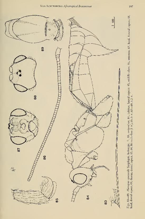
Van Achterbekg: Afrotropical Bracomnae 197
1)

198 Tijdschrift voor Entomologie, deel 126, afl. 9, 1983

Van Achterberg: Afrotropical Braconinae 199
oo rt rt
C -O p
rt 'j
ON O OJ
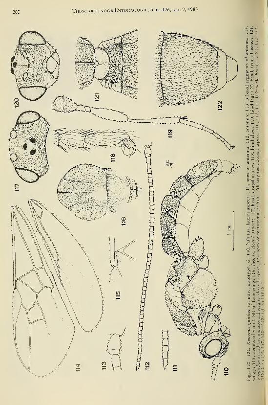
200 Tijdschrift voor Entomologie, deel 126, afl. 9, 1983
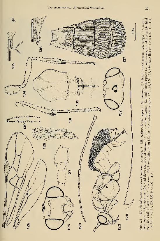
Van Achterberg: Afrotropical Braconinae 201
cxlc —rt

202 Tijdschrift voor Entomologie, deel 126, afl. 9, 1983
'"''5b
bD (u
.S^
^ "^
u o
° S
^1Cu u
_2 o- o
os '^
r<1 IS
w Cu
O,


DEEL 126 AFLEVERING 10 1983
TIJDSCHRIFTVOOR ENTOMOLOGIE
UITGEGEVEN DOOR
DE NEDERLANDSE ENTOMOLOGISCHE VERENIGING
O ^^^S.iViUS. COiViP. ZOOL
LIBRARY
^^- 0-984
HARVARDUNIVERSITY
INHOUD
L. Brussaard. — Reproductive behaviour and development of the dung beetle
Typhaeus typhoeus (Coleoptera, Geotrupidae), pp. 203—231, figs. 1— 17.
Tijdschrift voor Entomologie, deel 126, afl. 10 Gepubliceerd 15-XII-1983


REPRODUCTIVE BEHAVIOUR AND DEVELOPMENT OFTHE DUNG BEETLE TYPHAEUS TYPHOEUS (COLEOPTERA,
GEOTRUPIDAE)
by
LIJBERT BRUSSAARDDept. ofAnimal Ecology and Dept. ofSoil Science & Geology, Agricultural University, Wageningen,
The Netherlands
Abstract
This paper is part of a study of the contribution of dung beetles to soil formation in sandysoils. Typhaeus typhoeus (Linnaeus) has been selected because it makes deep burrows and is
locally abundant. The beetles are active from autumn until spring, reproduction takes place
from February to April. Sex pheromones probably influence pair formation. The sexes co-
operate in excavating a burrow (up to 0.7 m below surface) and in provisioning the burrowwith dung as food for the larvae. Co-operation is reset by scraping each other across the
thorax or elytra. Dung sausages, appr. 12.5 cm long and 15 mm in diameter, are manufac-tured above each other. Development is rapid at 13—17 °C. The life cycle is accelerated bya cold period in the third larval stage. These requirements are met by soil temperatures upto 15 °C in summer and down to 5 °C in winter. The life cycle lasts two years, but longer
under certain conditions. Newly hatched beetles make their way to the surface through the
soil, but do not follow the old shaft. Adults reproduce only once. Differential rate of com-pletion of the life cycle and occasional flying probably reduce the risk of local extinction.
The study is thought to be relevant for behavioural ecology and soil science.
ContentsIntroduction 203
Dung beetles and their association with soil .... 204
Choice of Typhaeus typhoeus as an object of
study 204
General biology 206
Distribution 206
Methods 207
Reproductive behaviour 209
Development 220
Discussion 226
Outlook 229
Acknowledgements 229
References 230
Introduction
There were two main reasons for this study
of dung beetles. First, ancient traces of former
activity by small burrowing or crawling ani-
mals, presumed to be dung beetles, can be found
today in sandy soils. By studying the behaviour
of dung beetles it should be possible to ascertain
whether these are indeed the relics of dungbeetle activity.
Second, in areas where dung beetles are abun-
dant nowadays, recent traces of their activity
are present in the soil and this raises the ques-
tion of how much dung beetles contribute to
soil formation today.
These topics will be discussed in subsequent
articles. The ancient traces (with an account of
their age) will be described in a forthcoming pa-
per and will be compared with the traces result-
ing from recent dung beetle activity. It will be
shown that dung beetles may indeed be respon-
sible for the ancient traces.
Knowledge about the reproductive behaviour
of the dung beetles is required for a proper un-
derstanding of their influence on soil morpholo-
gy, and therefore this is discussed in the present
paper. After a brief introduction to dung beetles
as a group, the general biology of the species se-
lected for study, i.e. Typhaeus typhoeus (Lin-
naeus, 1758), is described and its geographical
range is discussed.
In order to quantify the beetles' contribution
to soil formation it will eventually be necessary
to explain and predict their population dynam-ics. This considerable task was reduced to twobasic investigations in the present study. Thefirst pertains to the development of life stages
during the season and to phenological phenom-ena that seem to be related to the persistence of
203

204 Tijdschrift voor Entomologie, deel 126, afl. 10, 1983
the species in time and space. As this constitutes
part of the biology of the species it has been in-
cluded in the present paper. The second basic
investigation pertains to the environmental con-
ditions (including soil conditions) with which
the beetles have to cope during the season of
adult activity. These conditions may affect the
beetles' burrowing and reproduction. This will
be treated in another paper. It will be shown
that the number of dung targets and their distri-
bution in the field is of paramount importance
for the population ecology of T. typhoeus and
its impact on soil.
In its impact on soil Typhaeus typhoeus
should serve as a model species for all Geotrupi-
ni found in the temperate Holarctic and, to
some extent, also for other dung beetle species
of the paracoprid type in other parts of the
world.
Consequently, in this paper about T. ty-
phoeus, the emphasis will be on aspects of be-
haviour that may help to explain the impact on
soil formation caused by this kind of dung
beetle. Aspects of the reproductive behaviour of
T. typhoeus that have been published elsewhere
will be re-described in terms of their relevance
to soil formation. Furthermore, useful new in-
formation about behaviour and the devel-
opment of life stages will be presented.
D.UNG BEETLES AND THEIR ASSOCIATION WITHSOIL
Representatives of several coleopteran fami-
lies are regularly found inhabiting dung, e.g.,
Hydrophilidae and Histeridae. The term "dung
beetle", however, is usually restricted to a num-ber of species belonging to the superfamily
Scarabaeoidea. Scarab beetles, whether dung
beetles or not, show a close connection with
soil. The larvae of most species live under-
ground on a food-stock of dung or plant re-
mains, prepared by their parents, or they show a
free-living, root-sucking habit. Adults of most
species forage above-ground on fungi, dead or-
ganic matter or fresh leaves and burrow into the
soil to prepare food-stocks for their larvae, to
lay eggs or to hibernate or aestivate.
Living on dung may have evolved from living
on dead organic matter and fungi (lablokoff-
Khnzorian, 1977; Crowson, 1981). In the fami-
ly Geotrupidae, to which Typhaeus typhoeus
belongs, all these habits occur and this family
has been placed near the base of the scarabaeoid
evolutionary tree by the authors mentioned
above. Dung-feeding beetles may have devel-
oped the practice of digging into the soil as a re-
sult of searching for truffle-like fungi that
adopted a subterranean life history during times
of warming climate. The habit of making food-
stocks out of dead organic matter or dunginstead of searching for fungi is presumed to
have developed subsequently.
Dung beetles spread all over the world from
the Jurassic/Cretaceous periods onwards (lab-
lokoff-Khnzorian, 1977; Crowson, 1981). TheGeotrupini tribe probably radiated out from the
area of the Tertiary Tethys Sea (Krikken, 1980)
and now shows a predominantly temperate,
Holarctic distribution.
In addition to the Geotrupidae, one other
family of scarab beetles is important for our
study, viz., the Scarabaeidae. This family not
only contains dung beetles (e.g. Scarabaeinae
and Aphodiinae), but also beetles of the cock-
chafer type (Melolonthinae), which, as adults,
live on fresh leaves and, as larvae, show a root-
sucking way of live. Cockchafers will be dis-
cussed in a subsequent paper.
Choice of Typhaeus typhoeus as an object
OF study
The impact of dung beetles on soil may be
two-fold: enrichment with dung or plant re-
mains, and physical disturbance. On the basis of
their impact on soil, dung beetles may be divid-
ed into three ecological groups, as proposed by
Bornemissza (1969) in a different context.
First, the endocoprids, which pass their life
cycle from egg to adult in the dung on the sur-
face, or spend part of their life cycle a few centi-
metres deep in the soil, e.g. Scarabaeidae-Apho-
diinae. Second, the telecoprids, which make a
ball out of the dung, roll it some distance away
and bury it superficially as a food source for the
larva, e.g. Scarabaeidae-Scarabaeinae. And,
third, the paracoprids, which burrow a fairly
deep shaft under or close to the dung patch;
part of the burrow is filled with food for the lar-
vae and part of it is back-filled with soil, e.g.
Geotrupidae.
Because of the depth of the shafts and the
amount of dung transported below the ground,
dung beetles of the paracoprid type, especially
the larger species, may be considered to have
the greatest impact on soil. In the temperate re-
gions, paracoprid dung beetles of the Geotrupi-
ni tribe are the most important in this respect, as
has been shown by the studies of Fabre (±
1910), Schreiner (1906), Spaney (1910), VonLengerken (1954), Howden (1955, 1964, 1974),

Brussaard: Reproduction and development of Typhaeus typhoeus 205
Table 1. Depth of shafts in Geotrupidae.
faunal

206 Tijdschrift voor Entomologie, deel 126, afl. 10, 1983
Table 2. Number of eggs laid per nest or per female (if reported) in Geotrupidae.
species number of eggs laid
per nest or per female
Ceotrupes mutator'
G. spiniger'
C. stercorarius''''
G. vernalis'''
Lethrus apterus
Typhaeus typhoeus'' (1)
T. hiostius
4—16 per 9
9—10 per 9
4—17 per 9
(2) 3- 6 (8)
5—10 per 9
5— 7
6—11 per 9
4—16 (21) per 9
1
—
6 per nest (field)
= 10 per nest
^ 15 per 9
3— 6 (8)
2
—
8 per nest (field)
Teichert, 1955
Lumaret, 1980
Teichert, 1955
Spaney, 1910
Teichert, 1959a
Frantsevich et al.
Schreiner, 1906
present study
id.
Palmer, 1978
Kuyten, 1960
Spaney, 1910
Crovetti, 1971
1977
occurring in The Netherlands.
General biology
Nesting
The reproductive behaviour consists of bur-
rowing a branching shaft, provisioning the
branches with food for the larvae and sealing
the remaining burrow partly or completely with
soil. Though the female, once fertilized, can
perform the whole process on her own, the
beetles normally operate in pairs. Some aspects
of the reproductive behaviour have been out-
lined previously by Fabre (± 1910), Kuyten
(1960) and Palmer (1978).
Habitat
The habitat is open to half open heathland,
and the beetles are most abundant in bare areas
surrounded by Nardus stncta. Cladonia spp.
and Calluna vulgaris, and along paths. They al-
so occur in open pine woods, predominantly
along paths and in small clearings. A vital pre-
requisite is the presence of dung. The beetles are
found only on herbivore dung, mostly that of
rabbits, as the rabbit is the most abundant her-
bivore in the habitat of T. typhoeus. They will
also use dung from sheep, deer and roe.
Seasonal and diurnal incidence
From the second half of September onwardsand throughout the winter, T. typhoeus is active
whenever the temperature is above zero and
there is no snow. Intense activity occurs in Oc-tober and November, which is the main period
of maturation feeding, and from February to
April, which is the main period of reproduction.
From May onwards the reproductive activity
declines rapidly and from June to the latter half
of September there is no adult activity at the soil
surface (fig. 2). Contrary to the common as-
sumption that the beetles only reproduce after
the turn of the year (Fabre, ± 1910; Main, 1917;
Kuijten, 1960) I have established from field ob-
servations that pair formation, oviposition and
provisioning with dung is not exceptional as
early as the third week of October. On the oth-
er hand, unpaired beetles can be found in shal-
low burrows as late as March, where they are
apparently still involved in maturation feeding.
Although in overcast and humid weather ac-
tivity on the surface by day is not exceptional,
T. typhoeus is usually active above-ground at
dusk and at night.
Distribution
The genus Typhaeus
The genus Typhaeus Leach, 1815, contains
six species of about the same size: 14—22 mmlong and 8—11 mm wide. Five of these are re-
stricted to the Mediterranean area: T. hiostius
(Gene, 1836), T. momus (Olivier, 1789), T.fos-
sor (Wahl, 1838), T. latendens (Guérin, 1838)
and T. typhoeoides Fairmaire, 1852. The first of
these is endemic to Sardinia. The sixth species,
T. typhoeus has the largest geographical range:
from Morocco to South Sweden, westwards to
Ireland and eastwards to Poland (Horion, 1958;
Lindroth, 1960) and Yugoslavia (Miksic, 1956),
but it is absent in Hungary (pers. obs. and
Endrödi, pers. comm., 1981). A preliminary
map of the distribution in Europe is given in
fig. 3.

Brussaard: Reproduction and development of Typhaeus typhoeus
numbers
m
107
?o
AI
MI
JI
JI
AI
months
Fig. 2. Number of Typhaeus typhoeus captured in standard pitfalls in various heathlands in the Dutch province
of Drenthe during the years 1959—1967. (Courtesy of P. J. den Boer.)
Typhaeus typhoeus in The Netherlands
In The Netherlands, T. typhoeus is found in
sandy areas, even in isolated spots surrounded
by peat, clay or loam soils (Gaasterland, Be-
tuwe, South Limburg) but, remarkably, it is not
present in the dunes along the west coast and on
the Wadden islands in the north of the country.
Fig. 4 is a preliminary map of the distribution of
T. typhoeus in The Netherlands.
Absence from the dune region
In a preliminary experiment to ascertain the
reasons for the absence of T. typhoeus from the
dunes, it was found that under laboratory con-
ditions two pairs of beetles reproduced quite
normally when supplied with pellets of rabbit
dung from the Wadden island of Vlieland. The
trial was stopped when the larvae were in their
final instar. Under field conditions in enclosures
in the dunes of North Holland near Castricum,
beetles made normal burrows in which they
provisioned dung for their offspring, whether
supplied with pellets of rabbit dung from the
dunes (two pairs) or from the inland (two pairs).
When the burrows were excavated six months
later, it appeared, however, that only one of the
22 dung sausages contained a live larva, whereas
in most of the other cases the larva had died and
in some cases the egg had evidently not hatched.
This work needs to be continued to yield con-
clusive results.
Methods
To study the behaviour of T. typhoeus in the
laboratory, the beetles were kept in cages (1 mhigh and 0.60 m wide), similar to the one de-
scribed by Main (1916/17): a wooden frame in
which two windows (4 mm thick) were kept a
distance of 15 mm (sometimes 12 mm) apart.
The space between the windows was filled from
above with tamped down portions of sand that
came from a field at Wijster, in the Dutch prov-

208 Tijdschrift voor Entomologie, deel 126, afl. 10, 1983
Fig. 3. Geographical range of Typhaeus typhoeus. Grey area: after Pijpers (1981). Striped area: typhoeus pre-
sent according to Horion (1958), Lindroth (1960) and Sinclair (1977). Barred area: typhoeus not occurring
according to Endrödi (pers. comm., 1981).
Fig. 4. Distribution of Typhaeus typhoeus in The Netherlands. After Pijpers (1981). Each dot indicates at least
one specimen. Stars constitute additional occurrences, assessed by present author. The dot near the west coast
pertains to two specimens, captured around the turn of the century.
ince of Drenthe, where T. typhoeus occurs nat-
urally. The soil there is coversand to a depth of
1.40 m, in which a podzol has developed (Van
Heuveln, 1965). The predominant particle size
was 0.1—0.2 mm and particles larger than 0.6
mm were scarce. The organic matter content of
the sub-soil was 0.6%. The sand was air-dried
and subsequently moistened to a water content
of 10% (by mass) to approximate field condi-
tions in September at a depth of about 0.50 mbelow the surface. The tamping resulted in a
bulk density of approximately 1.50 g/cm^,
which was similar to the field situation at about
0.50 m below the soil surface.
The beetles made their burrows in the sand
between the windows. The light/dark regime in
the rooms with the cages was the same as in na-
ture. Daylight conditions were simulated with
TL-33 tubes supplemented with normal bulbs.
To prevent Ught affecting the beetles in the soil,
the windows of the cages were covered with
sheets of black plastic. A horizontal walking-
surface (0.50 X 0.60 m^) on which dung could
be offered was mounted on top of the glass cage
and covered with 0.8 mm mesh wire-netting to
prevent the beetles from flying away. Observa-
tions were usually carried out under dim red
light, after the plastic sheets had been removed.
For the rearing 60 cages were used. To com-
pare the laboratory results with the field situa-
tion, five cages were dug into the soil in the field
at Wijster. In addition, the experimental equip-
ment in the field included five 1 X 1 m^ and six
2 X 2 m^ enclosures consisting of 0.50 m wide
stainless steel plates which were inserted to a
depth of 0.20 m in the soil. These enclosures
were also spanned with the wire-netting.
Almost all beetles used in the experiments
were captured at the same site near Havelte in
the province of Drenthe. Newly hatched adults
can easily be collected in autumn from under
the small hummocks of soil, where they have re-
tired with some dung for their maturation feed-
ing. Prior to experiments the beetles were kept
in sand-filled plastic jars, 13 cm high and 10 cmin diameter, for a least six weeks at 5 °C.
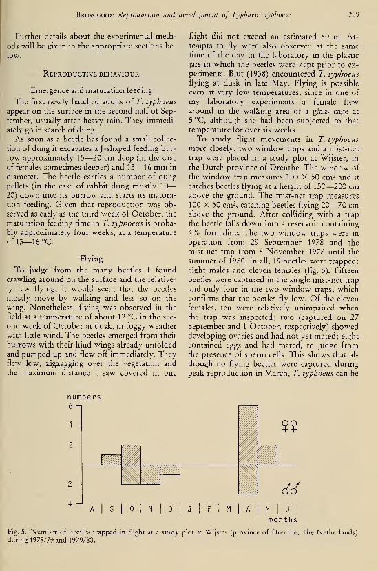
Brussaard: Reproduction and development of Typhaeus typhoeus 209
Further details about the experimental meth-
ods will be given in the appropriate sections be-
low.
Reproductive behaviour
Emergence and maturation feeding
The first newly hatched adults of T. typhoeus
appear on the surface in the second half of Sep-
tember, usually after heavy ram. They immedi-
ately go in search of dung.
As soon as a beetle has found a small collec-
tion of dung it excavates a J-shaped feeding bur-
row approximately 15—20 cm deep (in the case
of females sometimes deeper) and 13—16 mm in
diameter. The beetle carries a number of dungpellets (in the case of rabbit dung mostly 10
—
20) down into its burrow and starts its matura-
tion feeding. Given that reproduction was ob-
served as early as the third week of October, the
maturation feeding time in T. typhoeus is proba-
bly approximately four weeks, at a temperature
of 13—16 °C.
Flying
To judge from the many beetles I found
crawling around on the surface and the relative-
ly few flying, it would seem that the beetles
mostly move by walking and less so on the
wing. Nonetheless, flying was observed in the
field at a temperature of about 12 °C in the sec-
ond week of October at dusk, in foggy weather
with little wind. The beetles emerged from their
burrows with their hind wings already unfolded
and pumped up and flew off immediately. Theyflew low, zigzagging over the vegetation and
the maximum distance I saw covered in one
flight did not exceed an estimated 50 m. At-
tempts to fly were also observed at the sametime of the day in the laboratory in the plastic
jars in which the beetles were kept prior to ex-
periments. Blut (1938) encountered T. typhoeus
flying at dusk in late May. Flying is possible
even at very low temperatures, since in one of
my laboratory experiments a female flew
around in the walking area of a glass cage at
5 °C, although she had been subjected to that
temperature for over six weeks.
To study flight movements in T. typhoeus
more closely, two window traps and a mist-net
trap were placed in a study plot at Wijster, in
the Dutch province of Drenthe. The window of
the window trap measures 100 X 50 cm^ and it
catches beetles flying at a height of 150—200 cmabove the ground. The mist-net trap measures
100 X 50 cm2, catching beetles flying 20—70 cmabove the ground. After colliding with a trap
the beetle falls down into a reservoir containing
4% formaline. The two window traps were in
operation from 29 September 1978 and the
mist-net trap from 8 November 1978 until the
summer of 1980. In all, 19 beetles were trapped:
eight males and eleven females (fig. 5). Fifteen
beetles were captured in the single mist-net trap
and only four in the two window traps, whichconfirms that the beetles fly low. Of the eleven
females, ten were relatively unimpaired whenthe trap was inspected; two (captured on 27
September and 1 October, respectively) showeddeveloping ovaries and had not yet mated; eight
contained eggs and had mated, to judge fromthe presence of sperm cells. This shows that al-
though no flying beetles were captured during
peak reproduction in March, T. typhoeus can be
numbers6 -1
2 -
2 H
4
zwyMnnyg^^^^^^$^^^^^
M"
DI
JI
FI
AI
MI
JI
months
Fig. 5. Number of beetles trapped in flight at a study plot at Wijster (province of Drenthe, The Netherlands)
during 1978/79 and 1979/80.
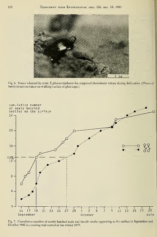
210 Tijdschrift voor Entomologie, deel 126, afl. 10, 1983
1 cm
Flg. 6. Stance adopted by male Typhaeus typhoeus for supposed pheromone release during defecation. (Photo of
beetle in nest entrance on walking surface of glass cage.)
cumu 1 ati ve numberof newly hatchedbeetles on the surface
20 -
O O Ç$• • 6ó
1 I \ \ 1 1 [
15 17 19 21 23 25 27 29
September
—r
1 3 5
October
"11 \ I
I
11 13 15 17 19
date
Fig. 7. Cumulative number of newly hatched male and female beetles appearing at the surface in September and
October 1980 in a rearing trial started in late winter 1979.

Brussaard: Reproduction and development of Typhaeus typhoeus 211
added to the list of species not obeying the oo-
genesis-fUght syndrome of Johnson (1969).
The gut content of 14 of the trapped beetles
was qualitatively estimated. In five beetles the
gut was half filled or less, in nine the gut was
more than half full. From these findings it can-
not be concluded that a shortage of dung is the
reason for flight.
Settlement and pair formation
Crovetti (1971) states that in Typhaeus hios-
tius the male penetrates the feeding burrow of a
female after maturation feeding. In T. typhoeus
I have observed behaviour that strongly sug-
gests that pheromones may play a role, at least
in some stage of adult life, in pair formation:
under laboratory conditions I have repeatedly
observed that a male who has abandoned a nest
with a female, then digs a shallow burrow near a
food source, similar to the J-shaped feeding
burrow. Next to this the male can be observed
defecating in a characteristic stance, his bodytilted at an angle of about 45° to the surface with
his head above the entrance of the burrow and
his abdomen lifted (fig. 6). This stance suggests
that a pheromone is released with the excre-
ment. Although the hypothesis of pheromonerelease needs experimental confirmation, it is
significant that this stance was invariably
adopted around the time that the light in the ex-
number of rabbitdung pellets taken down
20-
16-
14-
12
10-
4 -
2 -
o 19$Ç
—\ \
1
\
1
\ \ \ \ I
1 2 3 4 5 6 7 8 9 10
weeks after capture
Fig. 8. Mean number of rabbit dung pellets carried down by beetles kept in plastic jars at 13—16 °C for ten
weeks after their capture on 29 September and 4 October 1978. Vertical lines indicate upper half of standard
deviation.

212 Tijdschrift voor Entomologie, deel 126, afl. 10, 1983
perimental room was automatically switched on
or off, i.e., around dawn or dusk. At those times
the weather is usually favourable for odour
communication because of high air humidity
and moderate wind velocity.
Crovetti's (1971) observation that the male
joins the female after maturation feeding and
my observation that males appear to be trying
to attract females after the pair has split up, sug-
gests that the former behaviour changes to the
latter in the course of the season (assuming the
two Typhaeus spp. behave similarly). There is
some evidence to support this. In the rearing
trials I carried out, the females usually appeared
on the surface and started maturation feeding
before the males: the median of the total num-
ber of females on the surface was reached eight
days before that of males (fig. 7). Furthermore,
in a cohort of beetles captured in the field, fe-
males carried down the same quantity of dung
for maturation feeding as did the males (fig. 8).
If the duration of maturation feeding is the same
for both sexes then it seems probable that fe-
males mature sexually earlier than the males.
For a female, the prerequisites for reproduction
are a male and an adequate amount of dung, as a
food source for the future larvae, and therefore
her most profitable strategy seems to be not to
go and search for one of the few males available
for mating, but rather to settle near a spot rich
in dung, make a shallow burrow and wait there
for a male. This needs to be confirmed by addi-
tional research.
During the reproductive season the pattern
changes: whenever a pair of beetles abandons a
nest it is usually the male who leaves first, as
will be reported in greater detail in another pa-
per. Several days may pass before the female ap-
pears outside the burrow. By the time the male
leaves the burrow most females will be paired
and involved in breeding, so that it is unprofit-
able for him to search for one of the few bur-
rows with an unpaired female. Instead, he seeks
a spot with plenty of food, near which a newnest can be made, and tries to attract one of the
females that will appear above-ground after
abandoning a nest.
Copulation
On the first encounter, which usually takes
place in a shallow feeding burrow, the male vig-
orously sweeps his front tibiae across the fe-
male's thorax, the female turns around and then
the male sweeps across her elytra while half
mounted on her back. Finally, the female lifts
her abdomen and copulation follows, lasting
from 3 to 20 min. The female terminates the
copulation by stepping forwards a few cm,
turning around and pushing the male back.
Burrowing
Having paired and copulated the beetles makea nest that finally consists of a shaft that may or
may not divide into tunnels, from which a num-ber of brood chambers branch off, provisioned
with dung for the progeny and sealed by back-
filling with soil (fig. 9). The female excavates by
scraping the sand under her body with her front
tibiae and then using her middle and hind legs to
move it further back. As the sternites are dense-
ly covered with backward-pointing hairs, the
sand does not fall down past the beetle whenshe is in a vertical position. While excavating,
the beetle intermittently turns around its length
axis. Every time the beetle has excavated 0.5
—
1.0 cm of the shaft she moves several mm back-
wards, and by doing this tamps down the moist
sand behind her into a plug. Then she turns
around and pushes the plug into the shaft with
her head and thorax, again intermittently turn-
ing around her length axis. This turning enables
her to apply force to a different point and thus
facilitates the transport of the plug. The upper
part of the shaft, within a depth of 30 cm from
the surface, is made horizontal for some 10—15
cm (fig. 9). Here the female always leaves her
sand plug before returning down the burrow to
continue excavating. The male then burrows
through the sand plug. Since the female has left
the plug in a hoiizontal part of the passage, the
sand does not fall down the shaft. Once past the
small plug, the male turns around and shovels it
to the exit, transporting it in the same way as
the female. In this way a sand heap gradually ac-
cumulates on the surface, finally achieving a
height of some 5 cm and a diameter of 10—15
cm.
In one of the glass cages the excavation of the
shaft was closely monitored. Fig. 10 clearly
shows that the beetles may continue to excavate
for four days without pausing.
On four successive days, during periods indi-
cated in fig. 10, I recorded the intervals during
which the female was involved in sand excava-
tion and transport, respectively. On the first
three days the duration of the periods of excava-
tion was the same, on the fourth day the dura-
tion was much longer, presumably indicating
that the female was about to terminate the bur-

Brussaard: Reproduction and development of Typhaeus typhoeus
depth (cm)
213
I
4 -
io-
le
22-
28-
34 -
40-
46-
52 -
58 -
64-
70-
76 -
52 -
94 -
100
path fol 1 owedby emerging beetle
:x\
ii dung sausage in brood chamber
Vmwm0-.— egg chamber
-.— edge of frame
Fig. 9. Nest of Typhaeus typhoeus in one of the glass cages.

214 Tijdschrift voor Entomologie, deel 126, afl. 10, 1983
•r- OE S-
s- s_
O) :3+-) ^
-a
t^^
U
(ujD ) :;ieqs j.o 4:^d3p = •

Brussaard: Reproduction and development of Typhaeus typhoeus 215
rowing. The duration of sand transport steadily
increased to half an hour per plug. The cage had
been filled and the sand tamped down under
water. This resulted in a bulk density of 1.70
g/cm^, which is the maximum a beetle may gen-
erally encounter in the field. The temperature in
the room with the cage was 5 °C throughout the
reproduction of the beetles. This approximates
the soil temperature up to a depth of 1 m, mea-
sured in the field at the beginning of March, fol-
lowing the severe winter of 1978/79. This tem-
perature was, therefore, near the lowest the
beetles may encounter underground in the field
in The Netherlands. Even so the beetles pro-
gressed fairly rapidly (fig. 10). In a total of 60
cages filled with less densely packed sand (not
exceeding 1.55 g/cm^) and at temperatures of
5— 1 7 °C the shaft was excavated within 1—
3
days.
The morphology of the shaft was very vari-
able. However, a horizontal part was invariably
present within a depth of 30 cm. During the
process of excavation the female often made an-
other horizontal or slightly sloping gallery
where she left the sand plug while continuing
deeper. The shaft terminated in an oblique or
horizontal gallery 6—15 cm long: the future
first brood chamber (fig. 9). The depth of the
shaft varied considerably, as can be seen from
table 3.
The diameter of the shaft is determined by the
size of the larger of the two beetles. In the field,
casts of twelve shafts were made using liquid
paraffin wax that solidified on cooling, enabling
the diameter of the shafts to be measured. The
mean diameters of all shafts were then averaged:
the overall mean was 14.2 ± 0.7 mm (S.E.).
Oviposition and behaviour prior to and
following it
As soon as the female has terminated the bur-
rowing she begins to make a small cavity
(diameter about 0.5 cm) for the future egg by
moving her head and fore legs in the blind end
of the almost horizontal terminal part of the
burrow, alternately scraping some sand awayand pushing part of it back. When she has fin-
ished she walks up and down the passage until
the male arrives. The male joins the female,
sometimes before she has finished the egg
chamber, when he no longer encounters a sand
plug to transport upwards. Then the beetles
usually mate, as described before.
Copulation at this stage, i.e. prior to oviposi-
tion, is not obligatory but it is seldom omitted.
After copulation the male starts scraping sand
over a distance of about 10—15 cm from the
bottom of the future first brood chamber. Hemay do this several times, so that a sand plug is
formed. Then he turns around and transports
the plug upwards. The female continues to pre-
pare the egg chamber, breaking off to walk
through the future brood chamber, with her ab-
domen pulsating. These pulsating movementsare probably connected with the transport of an
egg into the oviduct. Finally she moves her ab-
domen into the egg room and oviposits, makinggentle pumping movements. Oviposition maylast 10—25 minutes. After oviposition the fe-
male shows the backward scraping behaviour,
just like the male, over a distance of some 10
—
15 cm, but in the opposite direction. Then she
turns around and pushes the sand into the end
of the burrow, thus sealing the egg chamber.
The wall that thus separates the egg chamber
from the future brood chamber becomes 10—15
mm thick (compare fig. 9). As a consequence of
the scraping of sand by male and female the
diameter of the brood chamber is slightly larger
than that of the rest of the burrow: 15.0 ± 0.7
mm (S.E.), n = 12.
While the female is completing the egg cham-
ber and preparing the brood chamber, the male
is involved in widening the shallow horizontal
part of the burrow, which is to become a store
room for dung pellets. The diameter of the store
room becomes 2—4 cm.
Table 3. Depth of shaft in Typhaeus typhoeus under laboratory and field conditions.
[Temperature in the lab 5° or 9 °C; bulk density in glass cages and casks about 1.55
g/cm^ to match field conditions. Enclosures 1 X 1 m-; casks 0.5 X 0.5 X 1 m^ Ample
supplies of dung were provided in all experiments.]
year experimental set-up depth (cm) range (cm)
1979
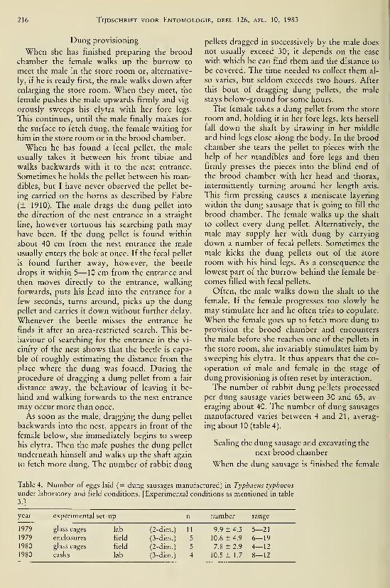
216 Tijdschrift voor Entomologie, deel 126, afl. 10, 1983
Dung provisioning
When she has finished preparing the broodchamber the female walks up the burrow to
meet the male in the store room or, alternative-
ly, if he is ready first, the male walks down after
enlarging the store room. When they meet, the
female pushes the male upwards firmly and vig-
orously sweeps his elytra with her fore legs.
This continues, until the male finally makes for
the surface to fetch dung, the female waiting for
him in the store room or in the brood chamber.
When he has found a fecal pellet, the male
usually takes it between his front tibiae and
walks backwards with it to the nest entrance.
Sometimes he holds the pellet between his man-dibles, but I have never observed the pellet be-
ing carried on the horns as described by Fabre
(± 1910). The male drags the dung pellet into
the direction of the nest entrance in a straight
line, however tortuous his searching path mayhave been. If the dung pellet is found within
about 40 cm from the nest entrance the male
usually enters the hole at once. If the fecal pellet
is found further away, however, the beetle
drops it within 5—10 cm from the entrance and
then moves directly to the entrance, walking
forwards, puts his head into the entrance for a
few seconds, turns around, picks up the dungpellet and carries it down without further delay.
Whenever the beetle misses the entrance he
finds it after an area-restricted search. This be-
haviour of searching for the entrance in the vi-
cinity of the nest shows that the beetle is capa-
ble of roughly estimating the distance from the
place where the dung was found. During the
procedure of dragging a dung pellet from a fair
distance away, the behaviour of leaving it be-
hind and walking forwards to the nest entrance
may occur more than once.
As soon as the male, dragging the dung pellet
backwards into the nest, appears in front of the
female below, she immediately begins to sweep
his elytra. Then the male pushes the dung pellet
underneath himself and walks up the shaft again
to fetch more dung. The number of rabbit dung
pellets dragged in successively by the male does
not usually exceed 30; it depends on the ease
with which he can find them and the distance to
be covered. The time needed to collect them al-
so varies, but seldom exceeds two hours. After
this bout of dragging dung pellets, the male
stays below-ground for some hours.
The female takes a dung pellet from the store
room and, holding it in her fore legs, lets herself
fall down the shaft by drawing in her middle
and hind legs close along the body. In the broodchamber she tears the pellet to pieces with the
help of her mandibles and fore legs and then
firmly presses the pieces into the blind end of
the brood chamber with her head and thorax,
intermittently turning around her length axis.
This firm pressing causes a meniscate layering
within the dung sausage that is going to fill the
brood chamber. The female walks up the shaft
to collect every dung pellet. Alternatively, the
male may supply her with dung by carrying
down a number of fecal pellets. Sometimes the
male kicks the dung pellets out of the store
room with his hind legs. As a consequence the
lowest part of the burrow behind the female be-
comes filled with fecal pellets.
Often, the male walks down the shaft to the
female. If the female progresses too slowly he
may stimulate her and he often tries to copulate.
When the female goes up to fetch more dung to
provision the brood chamber and encounters
the male before she reaches one of the pellets in
the store room, she invariably stimulates him bysweeping his elytra. It thus appears that the co-
operation of male and female in the stage of
dung provisioning is often reset by interaction.
The number of rabbit dung pellets processed
per dung sausage varies between 30 and 65, av-
eraging about 40. The number of dung sausages
manufactured varies between 4 and 21, averag-
ing about 10 (table 4).
Sealing the dung sausage and excavating the
next brood chamber
When the dung sausage is finished the female
Table 4. Number of eggs laid (= dung sausages manufactured) in Typhaeus typhoeus
under laboratory and field conditions. [Experimental conditions as mentioned in table
3.1
year experimental set-up iber range
1979

Brussaard: Reproduction and development of Typhaeus typhoeus 217
a
^^Mg^ìTff&^T!:'''--: -
..:-:-iAioifc*ì-•• •:-Vff?:p#^S!S-.-
^v|;p||;^:,
Fig. 11a. Dung sausage being sealed over a predetermined stretch with sand from the shaft walls. Fig. lib. The
widening i.« plastered with sand from the new brood chamber.
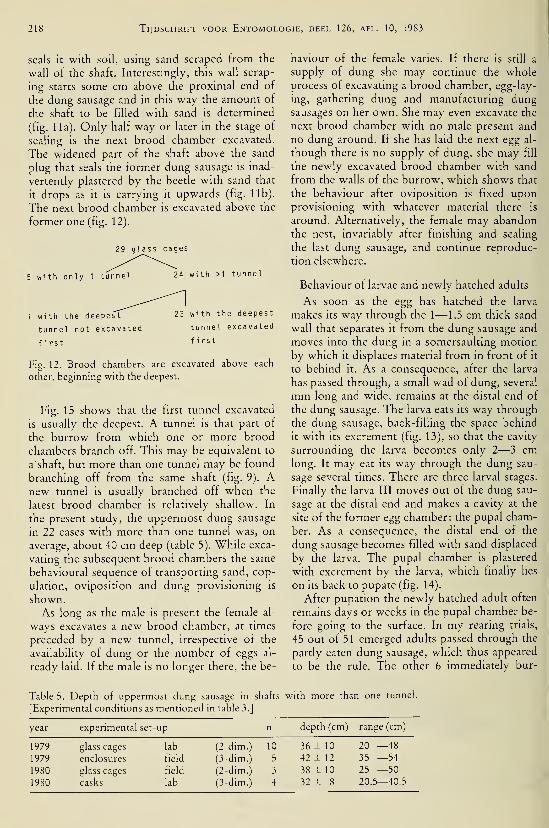
218 Tijdschrift voor Entomologie, deel 126, afl. 10, 1983
seals it with soil, using sand scraped from the
wall of the shaft. Interestingly, this wall-scrap-
ing starts some cm above the proximal end of
the dung sausage and in this way the amount of
the shaft to be filled with sand is determined
(fig. 11a). Only half way or later in the stage of
sealing is the next brood chamber excavated.
The widened part of the shaft above the sand
plug that seals the former dung sausage is inad-
vertently plastered by the beetle with sand that
it drops as it is carrying it upwards (fig. lib).
The next brood chamber is excavated above the
former one (fig. 12).
29 glass cages
5 with only 1 tunnel 24 with >1 tunnel
1 with the deepest
tunnel not excavated
first
23 with the deepest
tunnel excavated
fi rst
Fig. 12. Brood chambers are excavated above each
other, beginning with the deepest.
Fig. 15 shows that the first tunnel excavated
is usually the deepest. A tunnel is that part of
the burrow from which one or more brood
chambers branch off. This may be equivalent to
a' shaft, but more than one tunnel may be found
branching off from the same shaft (fig. 9). Anew tunnel is usually branched off when the
latest brood chamber is relatively shallow. In
the present study, the uppermost dung sausage
in 11 cases with more than one tunnel was, on
average, about 40 cm deep (table 5). While exca-
vating the subsequent brood chambers the same
behavioural sequence of transporting sand, cop-
ulation, oviposition and dung provisioning is
shown.
As long as the male is present the female al-
ways excavates a new brood chamber, at times
preceded by a new tunnel, irrespective of the
availability of dung or the number of eggs al-
ready laid. If the male is no longer there, the be-
haviour of the female varies. If there is still a
supply of dung she may continue the wholeprocess of excavating a brood chamber, egg-lay-
ing, gathering dung and manufacturing dungsausages on her own. She may even excavate the
next brood chamber with no male present and
no dung around. If she has laid the next egg al-
though there is no supply of dung, she may fill
the newly excavated brood chamber with sand
from the walls of the burrow, which shows that
the behaviour after oviposition is fixed uponprovisioning with whatever material there is
around. Alternatively, the female may abandonthe nest, invariably after finishing and sealing
the last dung sausage, and continue reproduc-
tion elsewhere.
Behaviour of larvae and newly hatched adults
As soon as the egg has hatched the larva
makes its way through the 1— 1.5 cm thick sand
wall that separates it from the dung sausage and
moves into the dung in a somersaulting motion
by which it displaces material from in front of it
to behind it. As a consequence, after the larva
has passed through, a small wad of dung, several
mm long and wide, remains at the distal end of
the dung sausage. The larva eats its way through
the dung sausage, back-filling the space behind
it with its excrement (fig. 13), so that the cavity
surrounding the larva becomes only 2—3 cmlong. It may eat its way through the dung sau-
sage several times. There are three larval stages.
Finally the larva III moves out of the dung sau-
sage at the distal end and makes a cavity at the
site of the former egg chamber: the pupal cham-
ber. As a consequence, the distal end of the
dung sausage becomes filled with sand displaced
by the larva. The pupal chamber is plastered
with excrement by the larva, which finally lies
on its back to pupate (fig. 14).
After pupation the newly hatched adult often
remains days or weeks in the pupal chamber be-
fore going to the surface. In my rearing trials,
45 out of 51 emerged adults passed through the
partly eaten dung sausage, which thus appeared
to be the rule. The other 6 immediately bur-
Table 5. Depth of uppermost dung sausage in shafts with more than one tunnel
[Experimental conditions as mentioned in table 3.]
year experimental set-up depth (cm) range (cm)
1979
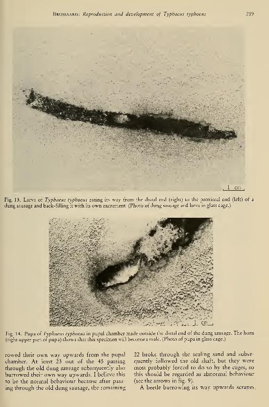
Brussaard: Reproduction and development of Typhaeus typhoeus 219
, 1 cm,
Fig. 13. Larva of Typhaeus typhoeus eating its way from the distal end (right) to the proximal end (left) of a
dung sausage and back-filling it with its own excrement. (Photo of dung sausage and larva in glass cage.)
-4 r-
m^m^^m^^Fig. 14. Pupa of Typhaeus typhoeus in pupal chamber made outside the distal end of the dung sausage. The horn
(right upper part of pupa) shows that this specimen will become a male. (Photo of pupa in glass cage.)
rowed their own way upwards from the pupal
chamber. At least 23 out of the 45 passing
through the old dung sausage subsequently also
burrowed their own way upwards. I believe this
to be the normal behaviour because after pass-
ing through the old dung sausage, the remaining
22 broke through the sealing sand and subse-
quently followed the old shaft, but they were
most probably forced to do so by the cages, so
this should be regarded as abnormal behaviour
(see the arrows in fig. 9).
A beetle burrowing its way upwards scrapes
.
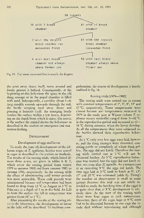
220 Tijdschrift voor Entomologie, deel 126, afl. 10, 1983
80 tunnels
16 with 1 brood
chamber
64 with >1 brood
chamber
3 with the deepest
brood chamber not
excavated first
61 with the deepest
brood chamber
excavated first
5 with next brood
chamber not al ways
above former one
Fig. 15. The tunnel excavated first is usually the deepest.
55 with next brood
chamber always above
former one
the sand away above itself, turns around and
firmly presses it behind. Consequently, at the
beginning of this behaviour the space in the old
dung sausage or in the pupal chamber is filled
with sand. Subsequently, a corridor about 8 cmlong steadily extends upwards through the soil,
the beetle scraping sand away above and
pressing it beneath itself. An emerging beetle
reaches the surface within a few hours, depend-
ing on the depth from which it starts. On arrival
at the surface the animal shows the behaviour as
described in the section on emergence and mat-
uration feeding.
Development
Development of eggs and larvae
To study the (rate of) development of the dif-
ferent stages of T. typhoeus, beetles were reared
in cages as described in the section on methods.
The results of the rearing trials, which lasted al-
most three years, are given in tables 6 & 7,
which cover the rearing period from winter
1979 to autumn 1980 and from autumn 1980 to
autumn 1981, respectively. In the rearing trials
the effect of administering cold winter periods
was most noticeable. These cold periods were
administered because soil temperature had been
found to drop from 13 °C in August to 3 °C in
February at a depth of 1 m in the field. At 0.20
m from the surface the fall in temperature was
greater (from 16° to 1 °C).
After presenting the results of the rearing tri-
als in the laboratory, the development of larvae
in the field will be described. To facilitate com-
prehension, the course of development is briefly
outlined in fig. 16.
Rearing trials (1979—1980)
The rearing trials were carried out in roomswith constant temperatures of 1°, 5°, 9°, 13° and
17 °C, respectively. These temperatures were
chosen because soil temperatures measured in
1979 in the study plot at Wijster (where T. ty-
phoeus occurs naturally) ranged from 3—10 °Cin the reproductive period and increased to 13
—
16 °C during summer, when the larvae develop.
At all the temperatures they were subjected to,
the beetles showed their reproductive behav-
iour.
At 1 °C only very few eggs were laid, howev-
er, and the dung sausages were abnormal, con-
sisting partly or completely of whole dung pel-
lets. The eggs did not hatch. Therefore the rear-
ing trials carried out at 1 °C will not be
discussed further. At 5 °C reproductive behav-
iour was normal, but the eggs did not hatch ei-
ther, not even after 20 months. At a temperature
at or exceeding 9 °C the eggs did hatch. The
time eggs laid at 5 °C took to hatch at 9°, 13°,
17° and 20 °C was estimated (table 8). Though
the number of observations is small in some
groups (because this trial was not solely in-
tended to study the hatching time of the eggs) it
is quite clear that at 9 °C development is rela-
tively slow. At the start of the rearing trials
there were four cages at each temperature.
However, three of the cages kept at 9 °C soon
had to be discarded because in one cage the fe-
male died without reproducing and although

Brussaard: Reproduction and development of Typhaeus typhoeus 111

222 Tijdschrift voor Entomologie, deel 126, afl. 10, 1983
Table 7. Results of ongoing rearing trials in glass cages from November 1980 until October 1981 in rooms kept at
a temperature of 13° or 17 °C.
, VAwinterX ^ Vperiods \ f>^ ^
offered \'\r
at 5°C \as indi ca ted
in Fig. 12

Brussaard: Reproduction and development of Typhaeus typhoeus 223
Table 8. Numbers of eggs hatching and median time
that elapsed before hatching in a rearing trial in glass
tubes.

224 Tijdschrift voor Entomologie, deel 126, afl. 10, 1983
' + •
85.7
1.+-
5.3
-1.+ -
83.3
_JO
_J85.2
35.7
WII
SPI
SUI
AUI
WII
SPI
SUI
AUI
WII
SPI
SU
1979 1980 1981
AU j season
I year
Fig. 17. Cold spell at 5 °C (+ ) or no cold spell (-; constant temperature of 13 °C or 17 °C) administered in
rearing trials during 1979— 1981 and percentages of adults emerging. Percentages pertain to numbers mentioned
in Table 6, columns 8 and 12, and Table 7, columns 4 and 5.
these six pupal chambers. But in the cages not
subjected to a second cold period no beetles
emerged from the seven pupal chambers. Fewbeetles emerged from cages that received no
cold treatments (five beetles from 14 pupal
chambers). As in 1979—1980, the number of
beetles that emerged after a cold period during
the 1980—1981 part of the rearing trials differed
very significantly from the number that
emerged from cages not subjected to a cold pe-
riod (x^ = = 20.12='-==--, P« 0.005).
Of the 33 beetles emerging from the cages in
1981, 18 were males and 15 females, which con-
firms the 1 : 1 sex ratio found the previous year.
All twelve larvae remaining after the 1980
—
1981 part of the rearing trials were in their third
instar (head capsules 4.6—5.2 mm).
Development of larvae in the field
Evidently, the quantity of cold that larvae of a
given generation experience in the field during
winter will differ because of the differing depths
at which eggs are laid in the soil and because
some winters are more severe than others. In
general, the larvae occurring closest to the sur-
face will experience more cold in winter and
more heat in summer, which may accelerate
their development vis-à-vis that of deeper lar-
vae. To test this, five cages were dug into the
soil in the study plot at Wijster in November1979, in such a way that their upper edges just
reached the surface.
At the beginning of March 1980 a pair of
dung beetles and ample dung were introduced
into each cage. The beetles were prevented from
exchanging with the surrounding field popula-
tion by wire-netting placed over the cages'
walking surface. The cages were dug out at the
beginning of September 1981, by which time
larvae that had completed their life cycle could
be found as newly hatched but not yet emerged
beetles; in addition, this was also the right time
to check whether, as expected, some of the lar-
vae had not yet completed their life cycle. Thefive cages proved to contain 39 dung sausages.
In eleven of these a living larva was found, one
of which was in a pupal chamber. All larvae
were in the third instar (head capsules 4.5—5.0
mm). In addition, one newly hatched beetle was
found. Though this is minimal evidence, the tri-
al does demonstrate that under field conditions
larvae of the same generation may give rise to
adult beetles in different years, assuming, of
course, that some of the remaining larvae would
complete their life cycle in the future, as oc-
curred in the laboratory rearing trials. Interes-
tingly, the cage with the adult beetle also con-
tained a larva in the pupal chamber and some
other larvae, still in dung, which shows that dif-
ferent rates of development may occur in the
offspring of one pair of beetles.
The results of another field trial, executed in
the same period, enable a judgement to be madeabout whether the poor survival obtained in the
above-mentioned field trial (life in 12 out of 39
cases = 31%) was somehow induced by the
glass cages. In this trial five pairs of beetles were
released in enclosures measuring 2x2 m^, at
the beginning of March 1980. Their nests were
dug up in May and June 1981, i.e. about three
months earlier than the cages. There proved to
be 32 dung sausages, 14 of which (i.e. 44%)contained a living larva; three of them had al-
ready made a pupal chamber. Assuming some
mortality in the enclosures between May/June
and September, the data suggest that there was
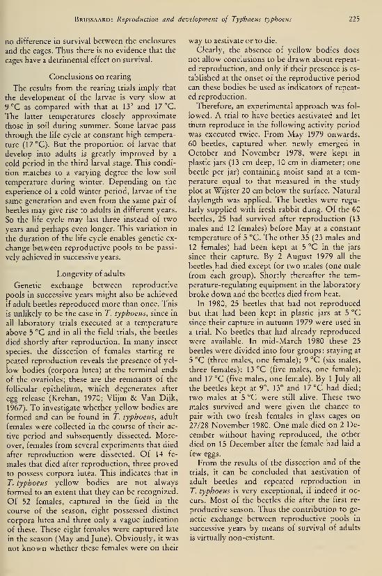
Brussaard: Reproduction and development of Typhaeus typhoeus lib
no difference in survival between the enclosures
and the cages. Thus there is no evidence that the
cages have a detrimental effect on survival.
Conclusions on rearing
The results from the rearing trials imply that
the development of the larvae is very slow at
9 °C as compared with that at 13° and 17 °C.
The latter temperatures closely approximate
those in soil during summer. Some larvae pass
through the life cycle at constant high tempera-
ture (17 °C). But the proportion of larvae that
develop into adults is greatly improved by a
cold period in the third larval stage. This condi-
tion matches to a varying degree the low soil
temperature during winter. Depending on the
experience of a cold winter period, larvae of the
same generation and even from the same pair of
beetles may give rise to adults in different years.
So the hfe cycle may last three instead of two
years and perhaps even longer. This variation in
the duration of the life cycle enables genetic ex-
change between reproductive pools to be passi-
vely achieved in successive years.
Longevity of adults
Genetic exchange between reproductive
pools in successive years might also be achieved
if adult beetles reproduced more than once. This
is unlikely to be the case in T. typhoeus, since in
all laboratory trials executed at a temperature
above 5 °C and in all the field trials, the beetles
died shortly after reproduction. In many insect
species, the dissection of females starting re-
peated reproduction reveals the presence of yel-
low bodies (corpora lutea) at the terminal ends
of the ovarioles; these are the remnants of the
follicular epithelium, which degenerates after
egg release (Krehan, 1970; Vlijm & Van Dijk,
1967). To investigate whether yellow bodies are
formed and can be found in T. typhoeus, adult
females were collected in the course of their ac-
tive period and subsequently dissected. More-
over, females from several experiments that died
after reproduction were dissected. Of 14 fe-
males that died after reproduction, three proved
to possess corpora lutea. This indicates that in
r. typhoeus yellow bodies are not always
formed to an extent that they can be recognized.
Of 52 females, captured in the field in the
course of the season, eight possessed distinct
corpora lutea and three only a vague indication
of these. These eight females were captured late
in the season (May and June). Obviously, it was
not known whether these females were on their
way to aestivate or to die.
Clearly, the absence of yellow bodies does
not allow conclusions to be drawn about repeat-
ed reproduction, and only if their presence is es-
tablished at the onset of the reproductive period
can these bodies be used as indicators of repeat-
ed reproduction.
Therefore, an experimental approach was fol-
lowed. A trial to have beetles aestivated and let
them reproduce in the following activity period
was executed twice. From May 1979 onwards,
60 beetles, captured when newly emerged in
October and November 1978, were kept' in
plastic jars (13 cm deep, 10 cm in diameter; one
beetle per jar) containing moist sand at a tem-
perature equal to that measured in the study
plot at Wijster 20 cm below the surface. Natural
daylength was applied. The beetles were regu-
larly supplied with fresh rabbit dung. Of the 60
beetles, 25 had survived after reproduction (13
males and 12 females) before May at a constant
temperature of 5 °C. The other 35 (23 males and
12 females) had been kept at 5 °C in the jars
since their capture. By 2 August 1979 all the
beetles had died except for two males (one male
from each group). Shortly thereafter the tem-
perature-regulating equipment m the laboratory
broke down and the beetles died from heat.
In 1980, 25 beetles that had not reproduced
but that had been kept in plastic jars at 5 °C
since their capture in autumn 1979 were used in
a trial. No beetles that had already reproduced
were available. In mid-March 1980 these 25
beetles were divided into four groups: staying at
5 °C (three males, one female); 9 °C (six males,
three females); 13 °C (five males, one female);
and 17 °C (five males, one female). By 1 July all
the beetles kept at 9°, 13° and 17 °C had died;
two males at 5 °C were still alive. These two
males survived and were given the chance to
pair with two fresh females in glass cages on
27/28 November 1980. One male died on 2 De-
cember without having reproduced, the other
died on 15 December after the female had laid a
few eggs.
From the results of the dissection and of the
trials, it can be concluded that aestivation of
adult beetles and repeated reproduction in
T. typhoeus is very exceptional, if indeed it oc-
curs. Most of the beetles die after the first re-
productive season. Thus the contribution to ge-
netic exchange between reproductive pools in
successive years by means of survival of adults
is virtually non-existent.

226 Tijdschrift voor Entomologie, deel 126, afl. 10, 1983
Discussion
Influence of glass cages on behaviour
and results of rearing
Although the behaviour of the beetles seemed
normal, the narrow space in which they were
confined may have influenced their behaviour.
Tables 3 & 4 show that there were no major dif-
ferences in the depth of the shaft and number of
dung sausages between beetles kept in the labo-
ratory and those kept or found in the field.
Moreover, the number of dung sausages pro-
duced in the present trials was very comparable
with figures reported in the literature (tables 2
& 4). As reported in the section on the devel-
opment of eggs and larvae, the survival of larvae
kept in glass cages dug into the soil and kept in
enclosures in the field was similar. This con-
firms that glass cages do not adversely affect the
behaviour of the beetles.
Pheromones
There are few data on settlement and pair for-
mation in the literature. Halffter & Matthews
(1966), in their extensive review of the biology
of Scarabaeidae, assume that the sexes meet by
chance near the dung patch. In addition to the
case of Typhaeus hiostius mentioned earlier
(Crovetti, 1971), the intrusion of a male into a
female's feeding burrow and joint feeding until
sexual maturity, is also reported by Rommel(1961) in Copris hispanus and by Halffter & Lo-
pez (1977) in Phanaeus daphnis. The hypothesis
of pheromone communication in T. typhoeus,
tentatively suggested in the present paper, is
supported by reports that in Scarabaeidae some
dung patches are densely populated with
beetles, whereas others are not, and that the
densely populated dung patches often differ
from each other in the taxon that is most repre-
sented (e.g. Halffter & Matthews, 1966; Pascha-
lidis, 1974). A similar clustered distribution is
known in bark beetles (Scolytidae), and this is
known to be caused by aggregation pheromones
(Blight et al., 1980). By analogy, pheromones
may be important in dung beetles, too. More-
over, the stance adopted by the male T. ty-
phoeus during the supposed pheromone release
is virtually the same as that described by Pas-
chalidis (1974) in three scarabaeid Sisyphus spp.
and by Tribe (1975) in Kheper nigroaeneus. In
the latter case pheromone release was proved.
In Kheper the pheromone is released from a de-
pression on either side of the first abdominal
sternite, with the help of paraffin tubules as a
carrier. This is very different from the way the
pheromone is supposed to be released by T. ty-
phoeus, i.e. along with the excrement. The latter
mechanism is also known for other groups, e.g.
in bark beetles, boll weevils and some Orthop-
tera Qacobson, 1972). If properly supported by
additional research, pheromone release byT. typhoeus would constitute the first knownexample of pheromone communication in Geo-trupidae. The conformity in releasing stance and
the difference in releasing mechanism point to a
convergent development and may be added to
arguments in favour of the modern view of clas-
sifying scarabaeids and geotrupids in distinct
families (Crowson, 1967, 1981) instead of in
subfamilies (Halffter & Matthews, 1966).
In all cases the pheromone release stance has
so far only been described in male dung beetles.
The possibility should not be excluded, howev-er, that after maturation feeding the male is also
attracted to his first female by means of phero-
mones. Fabre (± 1910) had already noticed that
at times two or three males of T. typhoeus can
be found in a burrow with only one female. Tei-
chert (1955) observed the same in G. mutator
and from this he inferred that males were at-
tracted by pheromones released by the female.
This would certainly improve the female's
chances of mating. It has been stated by Halffter
& Matthews (1966) that the similar size of the
antennal clubs in almost all male and female
dung beetle species is a sign of the unimpor-
tance of pheromone communication in these
beetles. But this argument does not hold if both
sexes respond to pheromones.
Burrowing
One may wonder how much faster the female
progresses in the burrowing stage thanks to
help from the male. The gain is estimated by
Teichert (1957) to be one-fourth to one-third of
the excavation time.
Co-operation between male and female in the
burrowing phase is not so close in some species
as it is in T. typhoeus. It is marked in the rela-
tively deep burrowing species Geotrupes verna-
lis, Lethrus apterus and Typhaeus hiostius,
whereas in the shallow burrowing species
G. mutator, G. stercorarius and G. spiniger the
male does little more than remove some soil
from the entrance (Teichert, 1955, 1959a,
1959b; Crovetti, 1971).
According to Klemperer (1979), in G. spinig-
er the female stops excavating the shaft and the
brood chamber as a response to their length.

Brussaard: Reproduction and development of Typhaeus typhoeus 117
Behaviour preceding and following
oviposition
Oviposition in T. typhoeus is almost always
preceded by copulation. According to Weaver
& Pratt (1977) repeated copulation has a stimu-
lating influence on the reproductive effort in the
cockroach Periplaneta americana. This is prob-
ably why a female of T. typhoeus that has lost
the male continues reproduction much more
slowly.
During the backward scraping activity that
follows oviposition, the beetle presses its head
and thorax hard against the walls of the brood
chamber, possibly to make the walls resist the
pressure they will be subjected to when being
provisioned with dung. Although he did not ob-
serve the pressing behaviour, Fabre (± 1910)
noticed that the walls of the brood chamber
were smoother than those of the shaft.
Dung provisioning
One may wonder how the male orients him-
self on the surface, when dragging a dung pellet
to the nest entrance. Kuyten carried out someunpublished experiments in 1961/62 from
which he concluded that the position of a light
source provides the beetle with a directional
cue. The same conclusion was independently
reached by Frantsevich et al. (1977) after some
very similar experiments with Lethrus apterus.
But it has not yet been explained how typhoeus
takes its bearings by night or on cloudy days,
when most of the excursions for dung are made.
Sealing and excavating brood chambers
Klemperer (1979) states that in Geotrupes
spiniger sealing a dung sausage is a side-effect of
excavating the next brood chamber. In Ty-
phaeus typhoeus, however, at the start of sealing
the sand is scraped from the wall and firmly
pressed into the shaft above the dung sausage.
Moreover, the last dung sausage is invariably
sealed; this is also evidence of a distinct behav-
ioural phase.
In the endemic Sardinian species Typhaeus
hiostius, which closely resembles typhoeus both
in its biology and in the reproductive season,
the sequence of excavation of brood chambers is
remarkably different. Contrary to typhoeus,
hiostius constructs the uppermost dung sausage
first, at about 60—70 cm below the surface, and
every subsequent sausage is 10—15 cm deeper,
the completed burrow generally reaching a
depth of 1.30—1.60 m. As a result, under field
conditions some weeks after the nest has been
completed the uppermost dung sausage already
contains a larva III, whereas the egg belonging
to the lowest dung sausage has not yet hatched
(Crovetti, 1971). This behavioural sequence
may well be related to soil moisture conditions
(Crovetti, pers. comm., 1981), the female begin-
ning to lay eggs approximately at ground-water
level and then deeper as the water table falls.
The ultimate function of this sequence may be
to ensure that eggs and larvae do not dry out
during the hot summer.
In T. typhoeus, however, the risk of drying
out may be considerably lower. In this species,
laying the first egg approximately at ground-
water level and thenceforth more shallowly mayensure that the larvae and their dung sausages
do not become waterlogged during the next
winter. In this context it would be interesting to
study the behavioural sequence in typhoeus in
the southern part of its geographical range.
Emergence through soil
The way the beetle moves to the surface is
seldom mentioned in the hterature. Sano
(1915/16) reports that Geotrupes stercorarius
does not always use the original tube made by
the parents, but makes its own route, as de-
scribed for Typhaeus typhoeus in this paper.
Crovetti (1971) reports that T. hiostius breaks
through the partly eaten dung sausage and the
sand that seals it and follows the shaft made by
the parents. The evidence available for T. ty-
phoeus (this paper) and for T. hiostius, suggests
that the newly hatched beetle starts moving up-
wards at a place where the resistance from soil is
least, i.e. through the remnants of the dung sau-
sage. However, it seems to be luck, rather than
strategy, whenever the beetle finds the open
part of the parental shaft. Emergence through
soil thus seems to be the rule.
Behaviour as a reaction chain
According to Klemperer (1979) the nesting
behaviour of Geotrupes spimger can be de-
scribed as a reaction chain in which each action
generates its own terminating stimulus and initi-
ates the subsequent response. Although experi-
mental analysis is beyond the scope of the pre-
sent paper, the nesting behaviour of T. typhoeus
is in many respects very similar to that of
G. spiniger. In addition, the way in which the
stretch of the burrow that is to be filled with
sealing sand is determined in T. typhoeus
(fig. 11) is an example of an action that gener-
ates its own terminating stimulus. So it may well
228 Tijdschrift voor Entomologie, deel 126, afl. 10, 1983
be that the behaviour of typhoeus can also be
described as a reaction chain. The extent to
which signalhng between the sexes by means of
pheromones, sweeping each other and stridula-
tion alters this picture requires further research,
however.
The role of temperature in diapause
and development
Diapause is induced in the third larval instar
and can be overcome by a cold winter period at
5 °C. Tauber & Tauber (1976) rightly point out
that this does not prove that diapause develops
under the influence of low temperature in na-
ture, as in many instances diapause can artifi-
cially be broken in several ways, including ma-nipulation of day-length and administering a
high temperature. Yet it is difficult to imagine
an environmental stimulus other than tempera-
ture that would induce, maintain and develop
the diapause in larval dung beetles, since they
live in complete darkness and in well-drained
soils in which, over the year, differences in
moisture are rather unpredictable. On the other
hand, although temperature is known to be the
single environmental stimulus maintaining
rhythms in a number of insects during the day
(Saunders, 1976), total reliance on temperature
as a diapause-regulating factor in the course of
the year has, to date, only been reported in
tropical insects (Tauber & Tauber, 1976).
Regarding the role of temperature in larval
development, the few larvae that deviated from
the general pattern in the rearing trials (tables 6
6 7) are interesting. Some larvae did not com-plete their life cycle after a cold period. This
suggests there may be considerable variation mthe amount of cold (duration and/or degree) a
larva needs before diapause is terminated. Onthe other hand, some larvae completed their life
cycle without a cold interlude. As the latter
were from the cages kept at 17°C this suggests
that a relatively high temperature can also over-
come diapause. Raising the temperature from15° to 21 °C has, for example, been shown to be
sufficient to overcome diapause in rearing trials
of Geotrupes spimger (CSIRO, 1980: 66). This
should be further studied in T. typhoeus, since it
may mean that in the southern part of the geo-
graphical range a warm period in the first sum-mer (summer 1979 in fig. 16) may be sufficient
to enable the species to pass through its life cy-
cle within one year. This may help to explain
why Fabre (± 1910), working in southern
France, found a one-year life cycle in his first
rearing trial (though not in his second). Thesame phenomenon may occasionally occur in
the northern part of the geographical range too,
e.g. in a hot summer, but as a rule in those re-
gions development will continue after a cold pe-
riod in winter (winter 1980 in fig. 16), which|
thus leads to a life cycle of a minimum of two[
years. Spaney's suggestion (1910) (he worked in
Germany) that in most cases T. typhoeus has a
one-year life cycle, should be regarded with
suspicion. Main (1916/17), working in England,
found a two-year cycle. Though most Geotru-
pidae have a one-year life cycle, some are
known to show a cycle of at least two years (Sa-
no, 1915/16; Main, 1916/17; Howden, 1955).
Field results support the hypothesis that lar-
vae of the same generation develop at different
rates: the rearing trial in glass cages, dug into
the soil and started in March 1980, yielded one
adult beetle and a number of larvae in their final
instar in the autumn of 1981 (see section Devel-
opment, p. 224). But as only one adult beetle
emerged, the hypothesis that the rate of devel-
opment is related, by means of temperature, to
the depth at which the larvae develop, could not
be tested.
Regarding the development of eggs laid be-
fore the turn of the year, it is unlikely that these
eggs can hatch before winter, let alone reach the
third larval stage, since soil temperature at 20
cm to 100 cm below the surface is uniformly ±9 °C by late October and subsequently de-
creases. As a consequence, the eggs do not
hatch until early spring together with the eggs
laid by that time, and the larvae do not experi-
ence a cold spell until the following winter. It
may thus be inferred that a one-year life cycle
probably cannot be achieved in The Nether-
lands, starting from these early eggs. This maynot be true, however, in the southern part of the
geographical range.
Flying
The Geotrupidae differ greatly in their use of
flight. As early as 1910, Spaney remarked that
Geotrupes stercorosus and Typhaeus typhoeus
can rarely be observed flying, whereas G. ster-
corarius regularly flies. He thought this was be-
cause beetles of the first two species generally
find plenty of food around in the place where
they emerge and reproduce, whereas those of
the last need to move from one fresh dung
source to another. As regards the significance of
the flying behaviour in the life history of T. ty-
phoeus, three observations seem to be relevant.

Brussaard: Reproduction and development of Typhaeus typhoeus 229
First, the behaviour is shown in the laboratory
in the presence of an ample supply of dung. Sec-
ond, most of the beetles captured flying in the
field did not have an empty gut. And third, I
have observed the flying behaviour in the field
in places with adequate supplies of dung that are
apparently easily accessible to the beetles. This
shows that flying is not solely connected with a
shortage of dung and it may well be that flying
is mainly related with searching for a partner.
At the end of the season, however, there may be
less dung available because it has been used by
the beetles and because the density of the rabbit
population has decreased, and then the beetles
may fly in search of new dung supplies. This as-
pect needs to be studied further.
Persistence of populations in space and time
Clearly, much speculation remains about the
cause and function of flying in adult T. ty-
phoeus. There is also speculation about the se-
lective forces that have led to and maintain the
pronounced differences, both within and be-
tween pairs of reproducing beetles, in the depth
at which the eggs are laid. Ignoring the causes,
the different rates of development of larvae
from one generation lead adult beetles to
emerge in different years; this passively contrib-
utes to a reduction of the risk of local popula-
tion extinction in time. Similarly the dispersal of
adults results in, although is not necessarily
aimed at, reducing the risk of local population
extinction in space (Den Boer, 1968). The extent
to which these phenomena contribute to the
persistence of T. typhoeus populations within a
certain area merits further study.
Outlook
The present investigation of the biology of
Typhaeus typhoeus will prove to be invaluable
for the follow-up studies. Among the most im-
portant findings for the understanding of soil
morphology and soil formation are: first, the
observations of how, and to what extent, the
burrows are excavated and, in particular, subse-
quently back-filled with dung and soil; and, sec-
ond, the way in which newly hatched adult
beetles make their way to the surface.
For the understanding of environmental fac-
tors (including soil conditions) that play a role
in the species' burrowing behaviour and repro-
ductive performance, results of rearing trials
will prove indispensable.
Apart from these aspects, which are closely
related to the soil-oriented approach of the
study, the- present investigation reveals a num-ber of starting points that may also be worth de-
veloping. The co-operation between male and
female is so striking that further study is prom-ising both from an ecological point of view
(what IS the gain in time and energy for the fe-
male and in net reproduction for both part-
ners?) and, if carried out as a comparative study
of related species, from an evolutionary point of
view (how and when did co-operation devel-
op?). In addition, the role of contests and the
probable role of pheromones deserve further
study, both in pair formation and in the exploi-
tation of food resources. These aspects, phero-
mones and co-operation (and concomitant in-
teraction) between the sexes, may reveal newinsights in future ethological analyses of the
nesting behaviour of dung beetles. Finally, a
long-term study may reveal how much the dif-
ferential development time of larvae and the dis-
persal of adults contribute to reducing the risk
of local population extinction and to stabilizing
beetle numbers.
Such studies may reveal the adaptive value of
numerous aspects of dung beetle behaviour,
about which we can only speculate at present.
Acknowledgements
Thanks are due to Prof. Dr. L. J. Pons and Ir.
K. J. Hoeksema for suggesting that dung beetles
are important agents in reworking soil, thereby
providing the stimulus for the present study.
The stimulating discussions I had with Prof. Dr.
H. Klomp, Dr. P. J. den Boer, Dr. Th. S. van
Dijk, Dr. S. Slager, Mrs. C. C. M. Brussaard-
Wüst, Ir. A. F. M. Olsthoorn, Mr. J. Krikken
and Dr. C. A. D. de Kort were very valuable.
Their constructive comments certainly im-
proved the manuscript. The students F. W. B.
M. Loonen and J. G. A. Outhuis were of great
help in observing beetle behaviour. The stu-
dents W. J. F. Visser and A. F. M. Olsthoorn
assisted in the fieldwork. I would also like to
make special mention of the interesting dis-
cussions I had with Mr. T. J. Palmer and Dr.
H. G. Klemperer during my stay in England.
This study would have been impossible, how-ever, without technical assistance from P.
Looijen, H. Snellen, Ing. P. W. T. Huisman,
Ing. T. H. P. van Huizen, H. J. Immink, J. Mo-lenaar, G. Buurman, R. de Fluiter, W. C. Th.
Middelplaats and Z. van Druuten. Their help is
gratefully acknowledged. Mrs. J. Burrough-
Boenisch corrected the English.

230 Tijdschrift voor Entomologie, deel 126, afl. 10, 1983
References
Baraud, J., 1977. Coléoptères Scarabaeoidea. Faune de
l'Europe occidentale. — Pubi. Nouv. Revue Ento-
mol. 4: 1—352. Toulouse, Univ. P. Sabatier.
Blight, M. C. J., C. J. King, L. J. Wadhams & M. J.
Wenham, 1980. Studies in chemically mediated
behaviour in the large elm bark beetle. — R&DPaper Forestry Commission 129.
Blut, H., 1938. Über Vorkommen und Lebensweise
des Ceratophyus typhoeus L. — Entomol. Blätter
34:274—276.
Boer, P. J. den, 1968. Spreading of risk and stabiliza-
tion of animal numbers. — Acta Biotheor. 18:
165—194.
Bornemissza, G. F., 1969. A new type of brood care
observed in the dung beetle Oniticellus cinctus
(Scarabaeidae).— Pedobiologia 9: 223—225.
Crovetti, A., 1971. Ricerche eco-etologische su Ty-
phoeus (Chelotrupes) hiostms Gene (Coleoptera,
Geotrupidae). — Annali della facoltà di agraria
dell'Università di Sassari 19: 1—71.
Crowson, R. A., 1967. The naturai classification of
the families of the Coleoptera. — Hampton, Clas-
sey.
, 1981. The biology of the Coleoptera. — NewYork/London, Ac. Press.
CSIRO, 1980. Annual Report 1979/80 of the Com-monwealth Scientific and Industrial Research Or-ganisation, Division of Entomology. — Canberra.
Fabre, J.-H., ca. 1910. Souvenirs entomologiques X.— 4th ed. Paris, Librairie Ch. Delagrave.
Frantsevich, L., V. Govardovsky, F. Gribakin, G. Ni-
kolajev, V. Pichka, A. Polanovsky, V. Shevchenko
& V. Zolotov, 1977. Astroorientation in Lethrus
(Col., Scar.).— J. comp. Physiol. 1 2 1 : 253—271
.
Halffter, G. & G. Y. Lopez, 1977. Development of
the ovary and mating behaviour in Phanaeus —Ann. Entomol. Soc. Am. 70: 203—213.
Halffter, G. & E. G. Matthews, 1966. The natural his-
tory of dung beetles of the subfamily Scarabaei-
nae.— Foha Entomol. Mex. 12— 14: 1—312.
Heuveln, B. van, 1965. De bodem van Drente. — Wa-geningen, Pudoc.
Horion, A., 1958. Faunistik der Mitteleuropäischen
Käfer. 6: Lamellicornia. — Uberlingen-Bodensee,
Feyel.
Howden, H. F., 1952. A new name for Geotrupes
{Peltotrupes) chalyhaeus Le Conte with a descrip-
tion of the larva and its biology. — Coleopt. Bull.
6:41—48., 1955. Biology and taxonomy of North Ameri-
can beetles of the subfamily Geotrupine, with re-
visions of the genera Bolbocerosoma, Eucanthus,
Geotrupes and Peltotrupes (Scarabaeidae). —Proc. U.S. Nat. Mus. 104: 151—319.
, 1964. The Geotrupinae of North and Central
America.— Mem. Entomol. Soc. Can. 39: 1—91.
, 1974. Additional records and descriptions of
North and Central American Geotrupinae
(Coleoptera, Scarabaeidae). — Can. J. Zool. 52:
567—573.
lablokoff-Khnzorian, S. M., 1977. Über die Phyloge-
nie der Lamellicornia. — Entomol. Abh. Staatl.
Mus. Tierk. Dresden 41 : 135—200.
Jacobson, M., 1972. Insect sex pheromones. — NewYork, Ac. Press.
Johnson, G. G., 1969. Migration and dispersal of in-
sects by flight.— London, Methuen.Klemperer, H. G., 1978. The repair of larval cells and
other larval activities in Geotrupes spiniger and
other species (Coleoptera, Scarabaeidae). — Ecol.
Entomol. 3: 119—132., 1979. An analysis of the nesting behavior of Ge-
otrupes spiniger (Coleoptera, Scarabaeidae). —Ecol. Entomol. 4: 133—150.
Krehan, I., 1970. Die Steuerung von Jahresrhythmik
und Diapause bei Larval- und Imagoüberwinte-
rern der Gattung Pterostichus (Col., Carab.). —Oecologia (Beri.) 6: 58—105.
Krikken, J., 1980. Geotrupidae from the Nepal Hima-layas. New flightless species of Geotrupes with a
biogeographical discussion. — Senckenbergiana
61:369—381.
Kuijten, P., 1960. Verhaltensbeobachtungen am Drei-
hornmistkäfer (Typhoeus typhoeus L., Col. Scar-
ab.).— Entomol. Z. 70: 223—233.Lengerken, H. von, 1954. Die Brutfürsorge- und
Brutpflegeinstinkte der Käfer. — Leipzig, Geest
& Portig K.-G.
Lindroth, C. H., 1960. Catalogus ColeopterorumFennoscandiae et Daniae. Lund, EntomologiskaSällskapet.
Lumaret, J. P., 1980. Analyse des communautés de
scarabéides coprophages dans le maquis Corse et
étude de leur rôle dans l'utilisation des
excréments.— Ecol. Mediterr. 5: 51—58.
Main, H., 1916/17. On rearing beetles of the genus
Geotrupes. — Proc. South Lond. Entomol. Nat.
Hist. Soc. 1916/17:18—22., 1917. Pupation of Geotrupes typhoeus. —
Trans. Entomol. Soc. Lond. Proc. 65: 90—91.
Miksic, R., 1956. Fauna Insectorum Balcanica. VI:
Scarabaeidae.— Sarajevu, Godisnjak Biol. Inst.
Olson, A. L., T. H. Hubbell & H. F. Howden, 1954.
The burrowing beetles of the genus Mycotrupes
(Col., Scar., Geotrupinae). — Misc. Papers Mus.Zool. Univ. Michigan 84: 5—59.
Palmer, T. J., 1978. A horned beetle which fights. —Nature 274: 583—584.
Paschalidis, K. M., 1974. The genus Sisyphus Latr.
(Coleoptera, Scarabaeidae) in Southern Africa. —Unpubl. MSc. -thesis, Rhodes Univ., Grahams-
town.
Pijpers, H. C, 1981. Mestkevers: verspreiding en eco-
logie in Nederland. In: J. van Tol & P. J. van
Helsdingen (eds.): Methods and results of Euro-
pean Invertebrate Survey mapping schemes in TheNetherlands. — Nieuwsbrief European Inverte-
Brussaard: Reproduction and development of Typhaeus typhoeus 231
brate Survey Nederland 10: 81—88.
Rommel, E., 1961. Ernährungsbiologie und Brutpfle-
geverhalten des Spanischen Mondhornkäfers Co-
pris hispanus (L.) (Col., Scarab.). — Biol. Zen-
tralbl. 80:327—346.Sano, C, 1915/16. On the metamorphosis of Geo-
trupes stercorarius L. — Proc. South Lond. Ento-
mol. Soci 91 5/1 6: 25—28.
Saunders, D. S., 1976. Insect clocks. — Oxford, Per-
gamon.
Schreiner, J., 1906. Die Lebensweise und Metamor-
phose des Rebenschneiders oder grossköpfigen
Zwiebelhornkäfers {Lethrus apterus Laxm.) (Col.,
Scar.). — Horae Soc. Entomol. Ross. 37: 197
—
207.
Sinclair, M., 1977. Typhaeus typhoeus (L.) (Col., Geo-trupidae) in Scotland. — Entomol. Monthly Mag.
113:166.
Spaney, A., 1910. Beiträge zur Biologie unserer ein-
heimischen Rosskäfer. — Deutsche Entomol. Z.
1910:625—634.
, A., 1911. Lebensweise und Brutpflege unserer
einheimischen Rosskäfer. — Entomol. Rundschau
28:15,24,26,34.
Tauber, M. J. & C. A. Tauber, 1976. Insect seasonah-
ty: diapause maintenance, termination, and post-
diapause development. — Ann. Rev. Entomol. 21:
81—107.
Teichert, M., 1955. Biologie und Brutfürsorgemass-
nahmen von Geotrupes mutator Marsh, und Geo-trupes stercorarius L. (Col. Scarab.). — Wiss. Z.
Martin Luther-Univ. Halle-Wittenberg 5: 187
—
218.
, 1956. Nahrungsspeicherung von Geotrupes ver-
nalis L. und Geotrupes stercorosus Scriba (Co-
leopt.. Scarab.). — Wiss. Z. Martin Luther-Univ.
Halle-Wittenberg 5: 669—672., 1957. Soziale Instinkthandlungen einheimischer
Geotrupini bei der Anlage ihrer Brutbauten. —Wiss. Z. Martin Luther-Univ. Halle-Wittenberg
6: 1045—1048.
, 1959a. Zum Brutfürsorgeverhalten des Geo-trupes vernalis L. (Col., Scarab.). — Wiss. Z. Mar-tin Luther-Univ. Halle-Wittenberg 8: 873—878.
, 1959b. Die bodenbiologische Bedeutung der'co-
prohagen Lamelhcornier. — Wiss. Z. Martin Lu-
ther-Univ. Halle-Wittenberg 8: 879—882.
Tribe, G. D., 1975. Pheromone release by dungbeetles (Coleoptera, Scarabaeidae). — S. Afr. J.
Sci. 71:277—278.Vlijm, L. & Th. S. van Dijk, 1967. Ecological studies
on carabid beetles. IL General pattern of popula-
tion structure in Calathus melanocephalus (Lmn.)
at Schiermonnikoog. — Z. Morph. Okol. Tiere
58:396—404.Weaver, R. J. & G. E. Pratt, 1977. The effect of en-
forced virginity and subsequent mating on the ac-
tivity of the corpus allatum of Periplaneta ameri-
cana measured in vitro, as related to changes in the
rate of ovarian maturation. — Physiol. Entomol.
2:59—76.


DEEL 126 AFLEVERING 11 1983
TIJDSCHRIFTVOOR ENTOMOLOGIE
UITGEGEVEN DOOR
DE NEDERLANDSE ENTOMOLOGISCHE VERENIGING
MUS. COtv.r. ^oOLLIBRARY
,^ O '984
HARVARDUNIVERSITY
INHOUD
R. DE Jong. — Annotateci list of the Hesperiidae (Lepidoptera) of Surinam, with
descriptions of new taxa, pp. 233—268, figs. 1—69.
Tijdschrift voor Entomologie, deel 126, afl. 1
1
Gepubliceerd 15-XII-1983

ANNOTATED LIST OF THE HESPERIIDAE(LEPIDOPTERA) OF SURINAM, WITH DESCRIPTIONS OF
NEW TAXA
by
R. DE JONGRijksmuseum van Natuurlijke Historie, Leiden
Abstract
A list is given of all known species of Hesperiidae of Surinam, based on the literature and
examined material. Material not yet recorded in the literature is listed with locality data.
Notes are given on many species, and the following taxa are described as new: Ribella
bicuspis (spec, nov.), Urbanus ambiguus (spec, nov.), Bungalotis sipa (spec, nov.), Clito jon-
kersi (spec, nov.), Cymaenes geijskesi (spec, nov.), Vehilius major (spec, nov.), Mnasitheus
similis (spec, nov.), Cobalopsis darpa (spec, nov.), Cobalopsis tanna (spec, nov.), Marys sub-
grisea paradoxa (subspec. nov.), Eutychide subpunctata intermedia (subspec. nov.),
Phlebades meesi (spec, nov.), Penicula criska extrema (subspec. nov.), Paktes vibicoides
(spec, nov.), Panoquina panoquinoides minima (subspec. nov.), Cravera rara (gen. nov. and
spec, nov.). Surina unica (gen. nov. and spec. nov.).
Introduction
Surinam has been popular with lepidopterists
and naturaUsts in general since the early days of
nature exploration. The famous water colours
of Surinam insects by Maria Sibylla Merian,
who lived from 1647 until 1717, testify of this
early interest, and the large number of Lepido-
ptera from Surinam described by Cramer
(1775—1780, continued by Stoll, 1780—1791)
speaks volumes. A general impression of ento-
mological research in Surinam up to 1950 was
given by Geijskes (1951). As far as Hesperiidae
are concerned three works are important, viz.,
Cramer (just mentioned), Sepp (1829—1852,
for publication dates, see De Jong, in press), and
Möschler (1876, 1882). Cramer described 78
species of Hesperiidae from Surinam, Sepp 21
species, and Möschler listed 145 species. The
work of Sepp is not outstanding because of the
number of species described or the quality of
the plates, but owing to the very detailed de-
scriptions of the larvae, pupae and food plants,
all described species having been bred.
Shortly before Sepp started the publication of
the work on Surinam Lepidoptera, Collin's the-
sis on the fauna of Surinam was published
(1822, see Holthuis, 1958). This rare work is of
little importance, since as far as the butterflies
are concerned it is a not very accurate compila-
tion of the species mentioned by Fabricius
(1793) from Surinam.
After Möschler, Surinam butterflies are only
incidentally mentioned in the literature. In
1951, however, Evans (1951—1955) started the
publication of the catalogue of all American
Hesperiidae in the British Museum (Natural
History), London. That museum proved to
have 194 species from Surinam.
In the Netherlands the interest in South
American Lepidoptera, already diminished in
the beginning of the 19th century in a period of
general impoverishment, seemed to have cometo a standstill after the publication of Sepp's
work, at least if judged from the complete lack
of publications on Surinam butterflies up to the
present time. The very limited edition of the lat-
ter work is significant in this respect. Although
a scientific approach to entomology was gaining
ground at the time in the Netherlands, most en-
tomologists were interested in the Dutch fauna
or the fauna of the Dutch East Indies. Never-
theless, limited amounts of material from Suri-
nam were still sent to the Rijksmuseum van Na-tuurlijke Historie, Leiden, e.g. by W. C. van
Heurn and the Penard brothers (1908), while
the druggist Bolten, who lived in Paramaribo
from 1902—1910, presented his small collection
of insects to the museum. More important
material was assembled much later. Dr. D. C.
Geijskes, entomologist of the Agricultural Ex-
periment Station at Paramaribo, later biologist
at the Department of Home Affairs and Direc-
233

234 Tijdschrift voor Entomologie, deel 126, afl. 11, 1983
tor of the Surinam Museum at the same place,
Hved in Surinam from 1938—1965. He collected
all kinds of insects during his expeditions into
the interior all over the country. His material,
now in the Rijksmuseum van Natuurlijke His-
torie, is not only important because of its quan-
tity, but because of the outlying districts repre-
sented. As Geijskes, like most other collectors
in Surinam, was not particularly interested in
Lepidoptera, the number of species in his
material is limited, but as he also collected manysmall and dull-coloured species (also by running
a Malaise trap) instead of restricting himself to
the bigger and more brightly coloured ones as
too often done, his material is quite interesting.
By far the best collection of Lepidoptera from
Surinam was made by E. H. Jonkers, who lived
in Surinam from 1956—1975. As an amateur-
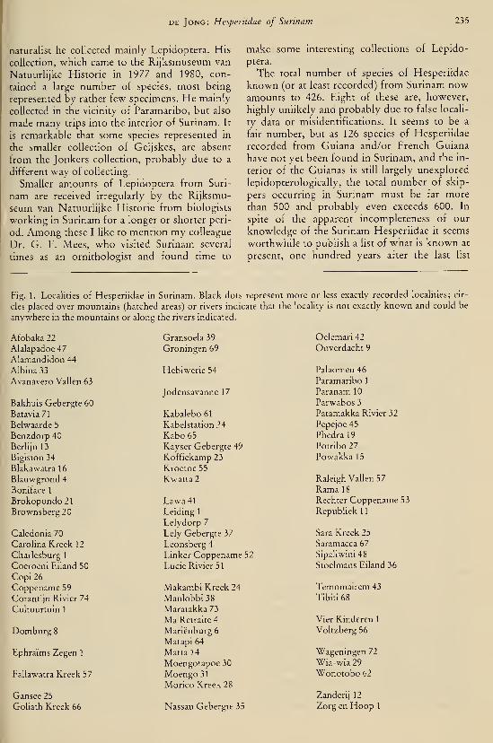
DE Jong: Hespenidae of Surinam 235
naturalist he collected mainly Lepidoptera. His
collection, which came to the Rijksmuseum van
Natuurlijke Historie in 1977 and 1980, con-
tained a large number of species, most being
represented by rather few specimens. He mainly
collected in the vicinity of Paramaribo, but also
made many trips into the interior of Surinam. It
is remarkable that some species represented in
the smaller collection of Geijskes, are absent
from the Jonkers collection, probably due to a
different way of collecting.
Smaller amounts of Lepidoptera from Suri-
nam are received irregularly by the Rijksmu-
seum van Natuurlijke Historie from biologists
working in Surinam for a longer or shorter peri-
od. Among these I like to mention my colleague
Dr. G. F. Mees, who visited Surinam several
times as an ornithologist and found time to
make some interesting collections of Lepido-
ptera.
The total number of species of Hesperiidae
known (or at least recorded) from Surinam nowamounts to 426. Eight of these are, however,
highly unlikely and probably due to false locali-
ty data or misidentifications. It seems to be a
fair number, but as 126 species of Hesperiidae
recorded from Guiana and/or French Guiana
have not yet been found in Surinam, and the in-
terior of the Guianas is still largely unexplored
lepidopterologically, the total number of skip-
pers occurring in Surinam must be far more
than 500 and probably even exceeds 600. In
spite of the apparent incompleteness of our
knowledge of the Surinam Hesperiidae it seems
worthwhile to publish a list of what is known at
present, one hundred years after the last list
Fig. 1. Localities of Hesperiidae in Surinam. Black dots represent more or less exactly recorded localities; cir-
cles placed over mountains (hatched areas) or rivers indicate that the locality is not exactly known and could be
anywhere in the mountains or along the rivers indicated.
Afobaka 22
Alalapadoe 47
Alamandidon 44
Albina 33
Avanavero Vallen 63
Bakhuis Gebergte 60
Batavia 71
Belwaarde 5
Benzdorp 40
Berlijn 13
Bigiston 34
Blakawatra 16
Blauwgrond 4
Boniface 1
Brokopondo 21
Brownsberg20
Caledonia 70
Carolina Kreek 12
Charlesburg 1
Coeroeni Eiland 50
Copi 26
Coppename 59
Coranti)n Rivier 74
Cultuurtuin 1
Domburg 8
Ephraims Zegen 1
Fallawatra Kreek 57
Gansée 25
Goliath Kreek 66
Gransoela 39
Groningen 69
Hebiwerie 54
Jodensavanne 17
Kabalebo61
Kabelstation 24
Kabo 65
Kayser Gebergte 49
Koffiekamp 23
Kroetoe 55
Kwatta 2
Lawa41Leiding 1
Lelydorp 7
Lely Gebergte 37
Leonsberg 4
Linker Coppename 52
Lucie Rivier 51
Makambi Kreek 24
Manlobbi 38
Maratakka 73
Ma Retraite 4
Mariënburg 6
Matapi 64
Matta 14
Moengotapoe 30
Moengo 31
Morico Kreek 28
Nassau Gebergte 35
Oelemari 42
Onverdacht 9
Palaomeu 46
Paramaribo 1
Paranam 10
Parwabos 3
Patamakka Rivier 32
Pepejoe 45
Phedral9
Potribo 27
Powakka 15
Raleigh Vallen 57
Rama 18
Rechter Coppename 53
Repubhekll
Sara Kreek 25
Saramacca67
Sipaliwini 48
Stoelmans Eiland 36
Temomairem 43
Tibiti68
Vier Kinderen 1
Voltzberg 56
Wageningen 72
Wia-wia29Wonotobo 62
Zanderij 12
Zorg en Hoop 1

236 Tijdschrift voor Entomologie, deel 126, afl. 11, 1983
(Möschler, 1882). It does not seem likely that
the number will increase largely in the near fu-
ture. Moreover, several species are new to sci-
ence, and publication may be a stimulus to fur-
ther research.
In view of the incomplete knowledge of the
species composition in Surinam and of the dis-
tribution of the recorded species in Surinam as
well as in South America in general, no attempt
has been made to analyse the species as to possi-
ble distributional types. At the moment the im-
pression is, that by far most of the species re-
corded from Surinam have a wide distribution
in tropical South America.
Acknowledgements
Few people, if any, are equally well acquaint-
ed with the insect world of Surinam as Dr. D.
C. Geijskes and I am most grateful for his help
in various ways. It is sad that Mr. E. H. Jonkers
(t 1979), whose collection formed the immedi-
ate cause and main basis for the present paper,
did not live to see this paper published or even
prepared. His enthousiastic tales and his wish to
have his collection safely kept in the Rijksmu-
seum van Natuurhjke Historie are gratefully re-
membered and acknowledged. Dr. W. Diehl
(Zoologische Staatssammlung, München), Mr.
W. Hogenes (Instituut voor Taxonomische
Zoologie, Amsterdam), and Mr. R. I. Vane-
Wright (British Museum (Natural History),
London), kindly permitted the author free ac-
cess to the collections under their care. Mr. E.
Neering (temporarily in Paramaribo) was help-
ful in sending material from his own collection
and the Nationale Zoologische Collectie (Para-
maribo) on loan.
List of the Hesperiidae of Surinam
In the hst below I have followed the sequence
and nomenclature adopted by Evans (1951
—
1955), unless recent literature or the material
studied gave cause to deviation. The species
name is followed by a reference to the literature
records from Surinam if any (records by Evans
always refer to the collection of the British Mu-seum (Natural History), London), and the
number and localities of specimens from Suri-
nam in the following institutions: Instituut voor
Taxonomische Zoölogie, Zoölogisch Museum,Amsterdam (abbreviated: ITZ); Nationale
Zoölogische Collectie, Universiteit van Suri-
name, Paramaribo (NZC); Rijksmuseum van
Natuurlijke Historie, Leiden (RMNH; by far
the most extensive collection of Surinam Hespe-
riidae), and Zoologische Staatssammlung,
München (ZSM; all Surinam material originat-
ing from Fruhstorfer, no precise locality data).
All localities are given on a map (fig. 1). Notesand descriptions of new taxa are given after the
list.
Pyrrhopyginae
Pyrrhopyge phidias Linnaeus, 1758. — Evans
(1951); Hewitson (1866); WilHams & Bell
(1931). RMNH: 6 CÎ, 2 2 , Brownsberg, Par-
amaribo, Surinam, Vier Kinderen. ZSM: 3
S ,\ 9 , Surinam. See Note 1
.
Pyrrhopyge aziza lexos Evans, 1951. —RMNH: 3 S, Brownsberg.
Pyrrhopyge proculus cintra Evans, 1951. —Evans (1951). RMNH: 26,29, Domburg,Lely Gebergte, Patamakka Rivier, Surinam.
ZSM:1 S,\ 9, Surinam.
Pyrrhopyge therides ponicea Evans, 1951. —Evans (1951). RMNH: 26,19, Rama, Su-
rinam, Zanderij. ZSM: 2 6 , Surinam.
Pyrrhopyge amythaon polka Evans, 1951. —RMNH: \ 6,1 9, Kabalebo, Republiek.
Pyrrhopyge sergius semana Evans, 1951. —Evans (1951). ITZ: 2 9, Paramaribo, Suri-
nam. RMNH: 2 6,1 9, Bakhuisgebergte,
Brownsberg, Republiek, Surinam.
Pyrrhopyge charybdis semita Evans, 1951. —RMNH -A 6, Surinam. See Note 2.
Pyrrhopyge amydas amydas Cramer, 1779. —Cramer (1779); Evans (1951); Fabricius
(1793, "Hesperia amiatus"); Möschler
(1876); Williams & Bell (1931). ITZ: 3 6,59, Paramaribo, Surinam. RMNH: 13 6, 12
9, Paramaribo, Surinam, Wageningen (at
Hght), Zorg en Hoop. ZSM: 1 6 , Surinam.
Pyrrhopyge arinas arinas Cramer, 1779. —Cramer (1779). RMNH: 1 6, Surinam (ho-
lotype).
Pyrrhopyge creusae Bell, 1931. — RMNH: 1 6,
Sipaliwini.
Pyrrhopyge ruhricollis Sepp, 1841. — Sepp
(1841). RMNH: 1 9, Surinam.
Pyrrhopyge comètes staudingeri Plötz, 1879. —Cramer (1779). RMNH: 1 6, Surinam. See
Note 3.
Elbella intersecta intersecta Herrich-Schäffer,
1869.— RMNH: 1 9 , Charlesburg.
Elbella patrobas blanda Evans, 1951. — Evans
(1951). RMNH: 1 c?,l 9, Domburg.Elbella bicuspis spec. nov.— See Note 4.
Elbella alburna MabiUe, 1891. — RMNH: 1 6,
Voltzberg.
Nosphistia zonara Hewitson, 1866. — Möschler
DE Jong: Hesperiidae of Surinam 237
(1876); possibly confused with a species of
Jemadia or Elhella.
Jemadia hospita ulixes Plötz, 1879. — Evans
(1951); Möschler (1882); Plötz (1879). ITZ:
1 (without abdomen), Surinam. RMNH: 1
9, Surinam.
Jemadia menechmus Mabille, 1878. — Evans
(1951).
Jemadia hewitsonii hewitsonii Mabdle, 1878. —RMNH: 2 (5, 2 9, Brownsberg, Domburg,Surmam.
Jemadia gnetus Yabncius, 1781. — Cramer
(1779, "Papilio vnlcanus"); Evans (1951).
RMNH:1 9, Surinam.
Jemadia fallax fallax Mabille, 1878. — RMNH:4 c?, Coeroeni, Coppename, Zanderij. See
Note 5.
Amenis pionia ponina Herrich-Schäffer, 1869.
— ITZ: 1 (without abdomen), Surinam.
Mysona harcastus harcastus Sepp, 1851. —Cramer (1775, "Papilio acastus"); Evans
(1951); Möschler (1876, "Pyrrhopyge acas-
tus"; 1882, "Pyrrhopyge harcastus" and
"Pyrrhopyge acastus"); Sepp (1851); Wil-
liams & Bell (1931). ITZ: 6 c?, 3 9, Surinam.
RMNH: 28 6, 11 9, Belwaarde, Mariën-
burg, Moengotapoe-Wiawia, Paramaribo,
Surinam, Zorg en Hoop. ZSM: 2 d, Suri-
nam.
Mysoria thasus thasus StoU, 1781. — Stoll
(1781). RMNH: 2 d,l 9, Surinam.
Myscelus nohilis Cramer, 1779. — Cramer
(1779); Evans (1951); Fabricius (1781, ''Pa-
pilio Salus").
Myscelus assancus assaricus Cramer, 1779. —Cramer (1779); Möschler (1876). RMNH: 2
9, Surinam.
Passova glacia Evans, 1951. — Evans (1951).
Passova passova stacer Evans, 1951. — Evans
(1951). RMNH: 1 S, Surinam. ZSM: 1 6,
Surinam.
Aspitha aspitha parima Plötz, 1886. — Plötz
(1886).
Pyrginae
Phocides palemon palemon Cramer, 1779. —Cramer (1779); Evans (1952, "Phocides
polyhius polyhius Fabricius, 1793");
Möschler (1876). ITZ: 1 9, Paramaribo.
NZC: 1 9, Paramaribo. RMNH: 1 cî, 9 9,
Boniface, Carolina Kreek, Paramaribo, Suri-
nam (including holotype, 6 , of Papilio pale-
mon Cramer).
Phocides metrodorus Bell, 1932. — NZC: 1 9,
Republiek. RMNH: 1 (5, Domburg.
Phocides pigmalion hewitsonius Mabille, 1883.
— Möschler (1876, "Erycides pygmalion
Cr."; determination uncertain, as he gave as
synonym "Gnetus Ltrll.", which belongs to
Jemadia). NZC: 1 9, Zorg en Hoop.RMNH: 1 S,2 9, Paramaribo, Surinam. See
Note 6.
Phocides lincea lincea Herrich-Schäffer, 1869.
— RMNH: 1 9, Charlesburg.
Tarsoctenus papias Hewitson, 1857. — "Williams
& Bell (1931).
Tarsoctenus corytus Cramer, 1777. — Cramer
(1777); Stoll (1782, "Papilio pyramus").
RMNH: 2 9, Surinam (including holotype
of Papilio corytus Cramer). ZSM: 1 6,2 9,
Surinam.
Tarsoctenus praecia rufibasis Mabille, 1910. —RMNH: 4 CÎ, 1 9, Carolina Kreek, Surinam,
Zanderi]. ZSM: 1 â , Surinam.
Phanus vitreus Stoll, 1781. — Evans (1952);
Möschler (1876, 1882); Stoll (1781); Wil-
liams & Bell (1931). RMNH: 1 (5,3 9, Av-anavero Vallen, Carolina Kreek, Voltzberg.
ZSM: 1 6 , Surinam.
Phanus ohscurior Kaye, 1924.— Evans (1952).
Phanus marshallii Kirby, 1880. — Evans (1952).
RMNH: 9 9, Avanavero Vallen, Browns-berg, Carolina Kreek, Jodensavanne, Rama,Surinam. ZSM: 3 (5,3 9 , Surinam.
Udranomia orcinus Felder, 1876. — Möschler
(1876).
Udranomia kikkawai Weeks, 1906. — ZSM: 1
9 , Surinam.
Drephalys phoenice Hewitson, 1867. —Möschler (1882). ZSM: 1 9, Surinam.
Drephalys dumeril Latreille, 1824. — Evans
(1952); Möschler (1876).
Drephalys oriander oriander Hewitson, 1867.
— Evans (1952). RMNH: 1 9, Goliath
Kreek.
Drephalys talhoti Le Cerf, 1922. — Evans
(1952).
Drephalys opifex Evans, 1952.— Evans (1952).
Drephalys alcmon Cramer, 1779. — Cramer
(1779); Evans (1952); Möschler (1876).
RMNH: 1 9, Zanderij. ZSM: 1 (5, 1 9, Su-
rinam.
Augiades crinisus Cramer, 1780. — Cramer
(1780); Evans (1952); Möschler (1882); Wil-
liams & Bell (1931). ITZ: 1 d, 4 9, Parama-
ribo, Surinam, Zanderij. NZC: 1 ô , Parama-
ribo. RMNH: 5 6,2 9, Lelydorp, Moengo,Paramaribo, Rama, Zanderij. ZSM: 2 (5, 3
9, Surinam.
Hyalothyrus infernalis mfernalis Möschler,
238 Tijdschrift voor Entomologie, deel 126, afl. 11, 1983
1876. — Evans (1952); Möschler (1876).
RMNH: 8 d, 3 9, Avanavero Vallen, Caro-
lina Kreek, Fallawatra Kreek, Goliath
Kreek, Hebiwerie, Lely Gebergte, Raleigh
Vallen (Voltzberg), Rama, Sipaliwini.
Hyalothyrus leucomelas Geyer, 1832. —Möschler (1876). RMNH: 1 â,\ 9, Avana-
vero Vallen. ZSM: 1 â , Surinam.
Hyalothyrus nitocris StoU, 1782. — Evans
(1952); Möschler (1882); Stoll (1782); Wil-
liams & Bell (1931). ITZ: 1 S, Surinam.
RMNH: 5 (?, 3 9, Goliath Kreek, Rama,
Zanderij. ZSM : 1 1 (5,4 9 , Surinam.
Hyalothyrus neleus pemphygargyra Mabille,
1888. — Evans (1952); Möschler (1882).
RMNH: 1 â, Coppename (Kroetoe).
Phareas coeleste Westwood, 1852. — Evans
(1952); Williams & Bell (1931). RMNH: 2
â , Sipaliwini. ZSM: 2 9 , Surinam.
Entheus eumelus Cramer, 1777. — Cramer
(1777); Williams & Bell (1931, ''Entheus
mina"). RMNH: 1 9, GoUath Kreek. ZSM:1 â , Surinam (lacks the costal fold).
Entheus gentius Cramer, 1779. — Cramer
(1779); Evans (1952); Möschler (1882); Wil-
Hams & Bell (1931). ITZ: 1 S, Surinam.
RMNH: 3 5,9 9, Avanavero Vallen, Blaka-
watra, Goliath Kreek, Lely Gebergte, Phe-
dra, Rama, Republiek, Zanderij. ZSM: 5 S,
5 9 , Surinam. See Note 7.
Entheus pnassus Linnaeus, 1758. — Evans
(1952); Möschler (1876, "Entheus talaus L."
and "Entheus peleus L."); Stoll (1782); Wil-
liams & Bell (1931). ITZ: 3 5, 1 9, Surinam.
RMNH: 5 9, Albina, Avanavero Vallen,
Brownsberg, Surinam, Zanderij. ZSM: 10 â ,
5 9 , Surinam.
Entheus matho dius Mabille, 1897. — Evans
(1952). RMNH: 5 5, 1 9, Kabelstation,
Kroetoe, Ramam, Surinam, Zanderij.
Cahirus procas Cramer, 1777. — Cramer
(1777); Evans (1952); Stoll (1787, "Paptlto
julettus"). RMNH: 2 5, Copi, Lely Ge-
bergte.
Proteides mercurius mercunus Fabricius, 1787.
— Cramer (1779, "Papilio idas")\ Möschler
(1882, "Thymele tdas Cr."); Sepp (1848,
"Papilio idas Cramer"). ITZ: 2 5, Surmam.
RMNH:1 (5,Oelemari.
Epargyreus clarus Cramer, 1775. — Cramer
(1775; described from Surinam, but locality
undoubtedly false, the species is not knownto occur south of Honduras); Evans (1952;
locality not trusted by Evans).
Epargyreus socus sinus Evans, 1952. — Evans
(1952).
Epargyreus exadeus Cramer, 1779. — Cramer
(1779); Möschler (1882). RMNH: 1 5, Suri-
nam. ZSM: 1 â , Surinam.
Polygonus leo Gmelin, 1790. — Evans (1952).
ITZ: 2 5, Surinam.
Chioides catillus catillus Cramer, 1779. —Cramer (1779); Evans (1952); Möschler
(1876); Sepp (1847, "Papilio longicauda");
Williams & Bell (1931). ITZ: 2 5,2 9, Para-
maribo, Surinam. RMNH : 1 8 5 , 1 1 9 , Albi-
na, Bigiston, Blauwgrond, Brokopondo,
Coppename Rivier, Goliath Kreek, Lely-
dorp. Ma Retraite, Onverdacht, Paramaribo,
Tibiti. ZSM : 4 5 , 1 9 , Surinam.
Aguna asander asander Hewitson, 1867. —Evans (1952).
Aguna claxon Evans, 1952. — RMNH: 1 5,
Brownsberg. See Note 8.
Aguna aurunce Hewitson, 1867. — RMNH: 1
5,19, Brokopondo, Carolina Kreek.
Aguna coelus Stoll, 1781. — Evans (1952);
Möschler (1876); Stoll (1781). RMNH: 2 9,
Lely Gebergte, Rama. ZSM: 2 9 , Surinam.
Aguna ganna Möschler, 1878. — Evans (1952);
Williams & Bell (1931).
Typhedanus orion Cramer, 1779. — Cramer
(1779); Evans (1952); Möschler (1882); Wil-
hams & Bell (1931). ITZ: 15,2 9 , Parama-
ribo, Surinam. RMNH: 10 5, 4 9, Carolina
Kreek, Domburg, Lelydorp, Paramaribo.
Typhedanus undulatus Hewitson, 1867. —Evans (1952); Möschler (1876). ITZ: 2 5, 1
9, Paramaribo, Surinam. RMNH: 3 5,4 9,
Ma Retraite, Paramaribo, Zorg en Hoop.
ZSM: 1 5, Surinam.
Typhedanus optica optica Evans, 1952. —RMNH: 1 5,2 9, Brokopondo. ZSM: 1 5,
Surinam.
Polythrix octomaculata octomaculata Sepp,
1844. — Möschler (1882); Sepp (1844).
RMNH: 1 5,5 9, Paramaribo. ZSM: 1 5,
Surinam.
Polythrix asine Hewitson, 1867. — RMNH: 1
9 , Paramaribo. See Note 9.
Polythrix minvanes Williams, 1926. — ZSM: 1
5, Surinam.
Polythrix caunus Herrich-Schäffer, 1869. —Evans (1952). RMNH: 4 5,2 9, Brokopon-
do, Paramaribo, Rama, Republiek. See Note
10.
Polythrix auginus Hewitson, 1867. — Evans
(1952); Möschler (1876). ZSM: 1 5, Suri-

DE Jong: Hesperiidae of Surinam 239
Polythrix metallescens Mabille, 1888. —RMNH: 3 (5, 1 9, Ma Retraite, Republiek,
Surinam.
Polythrix eudoxus StoU, 1781.— Stoll (1781).
Chrysoplectrum pervivax Hübner, 1819. —Evans (1952); Hübner (1819); Kirby (1871,
"Pamphila schelleri"); Möschler (1876, "Te-
legonus schellen Kirby"); Sepp (1832—1840,
''Pupillo pertinax StoU"); Stoll (1790, "Papi-
lio pertinax"); Williams & Bell (1931).
RMNH: 2 9, Leonsberg, Paramaribo.
Chrysoplectrum bahiana bahiana Herrich-
Schäffer, 1869. — Evans (1952). RMNH: 2
S -, Paramaribo, Surinam.
Chrysoplectrum otriades Hewitson, 1867. —RMNH: 2 (J, Brownsberg.
Chrysoplectrum perniciosus perniciosus Herrich-
Schäffer, 1869. — Evans (1952). RMNH: 1
(5,2 9, Brownsberg, Paramaribo.
Chrysoplectrum orpheus Plötz, 1882. — ZSM: 1
6 1 Surinam.
Urbanus proteus proteus Linnaeus, 1758. —Evans (1952); Möschler (1876); Williams &Bell (1931). ITZ: 2 0,7 9, Caledonia-Sara-
macca, Paramaribo, Surinam. RMNH: 8 â,
9 9, Albina, Marienburg, Paramaribo, Ra-
leigh Vallen. ZSM: 1 â , Surinam.
Urbanus viterboana alva Evans, 1952. — Evans
(1952). NZC: 1 S, Charlesburg. RMNH: 1
d, 4 9, Brokopondo, Paramaribo, Sipaliwi-
ni.
Urbanus esmeraldus Butler, 1877. — Evans
(1952). RMNH: 1 6, Surinam.
Urbanus esma Evans, 1952. — RMNH: 2 d,
Ma Retraite, Zanderij. See Note 11.
Urbanus esta Evans, 1952. — RMNH: 1 6,
Domburg (agrees with esta, but valves sym-metrical, both like Evans' figure of right
valve). ZSM: 1 9, Surinam.
Urbanus acawoios Williams, 1926. — Evans
(1952). ITZ: 1 6, Paramaribo. RMNH: 1 9,
Lelydorp. ZSM: 1 9, Surinam.
Urbanus dorantes dorantes Stoll, 1790. — Evans
(1952); Möschler (1882); Stoll (1790); Wil-
liams & Bell (1931). ITZ: 2 9, Surinam.
RMNH: 11 S, 10 9, Albina, Ma Retraite,
Paramaribo. ZSM: 2 c?, 1 9, Surinam.
Urbanus teleius Hübner, 1821. — Evans (1952);
Möschler (1876); Williams & Bell (1931,
"Goniurus eurycles Latr."). RMNH: 8 (5, 6
9, Domburg, Onverdacht, Paramaribo,
Zanderij-Saramacca.
Urbanus tanna Evans, 1952.— Evans (1952).
Urbanus cindra Evans, 1952. — RMNH: 1 â.
Copi.
Urbanus ambiguus spec. nov.— See Note 12.
Urbanus zagorus Plötz, 1881. — RMNH: 1 S,
Surinam. Unlikely locality; only knownfrom S. Brazil and Argentina.
Urbanus simplicius Stoll, 1790. — Evans (1952);
Möschler (1876, 1882); Stoll (1790); Wil-
liams & Bell (1931). ITZ: 7 6,4 9, Parama-
ribo, Surinam. RMNH: 38 cî, 17 9, Albina,
Avanavero Vallen, Bigiston, Brokopondo,
Brownsberg, Charlesburg, Domburg, Gro-
ningen, Lelydorp, Ma Retraite, Paramaribo,
Republiek, Zanderij, Zorg en Hoop. ZSM: 1
â , Surinam.
Urbanus procne Plötz, 1881. — Evans (1952).
ITZ: 4 0,2 9, Charlesburg, Paramaribo.
NZC: 2 0,2 9, Paramaribo. RMNH: 42 6,
17 9, Charlesburg, Cultuurtuin, Dom.burg,
Kabelstation, Kwatta, Lelydorp, Ma Re-
traite, Paramaribo, Republiek.
Urbanus doryssus doryssus Swainson, 1831. —Evans (1952); Möschler (1876); Williams &Bell (1931). ITZ: 1 c?, 1 9, Surinam. NZC: 1
5, Benzdorp. RMNH: 5 6, Brownsberg,
Corantijn Rivier, Domburg, Paramaribo.
ZSM: 5 6, A 9, Surinam.
Urbanus albimargo takuta Evans, 1952. —Evans (1952); Williams & Bell (1931).
RMNH: 2 9, Avanavero Vallen, Browns-
berg. ZSM: 1 6 , Surinam.
Urbanus virescens Mabille, 1877. — ZSM: 1 6
,
Surinam.
Astraptes talus Cramer, 1777. — Cramer
(1777); Möschler (1876); Sepp (1841, ''Papi-
lio lucidator"). ITZ: 1 9, Paramaribo.
RMNH: 4 (5, 4 9, Albina, Carolina Kreek,
Leonsberg, Paramaribo, Surinam. ZSM: 1 9,
Surinam.
Astraptes fulgerator fulgerator Walch, 1775. —Evans (1952); Möschler (1876); Sepp (1841,
"Pupillo fulminatore); Williams & Bell
(1931). ITZ: 1 9, Surinam. RMNH: A 6,79 , Brownsberg, Domburg, Ephraims Zegen,
Kabalebo/Corantijn Rivier, Lely Gebergte,
Paramaribo, Surinam. ZSM: 3 (5,5 9, Suri-
nam.
Astraptes upastus apustus Cramer, 1777. —Cramer (1777); Evans (1952). ITZ: 1 cÏ, 1 9,
Surinam. RMNH: \ 6,\ 9, Domburg, Suri-
nam. ZSM: 1 6 , Surinam.
Astruptes enotrus Stoll, 1781. — Stoll (1781).
RMNH: 1 6, Rama. ZSM: 1 6, Surinam.
Astruptes grunadensis Möschler, 1878. — Evans
(1952); Möschler (1882); Stoll (1780, "Pupi-
llo aulestes"). ITZ: 1 6, Surinam. RMNH: 1
6 , Surinam. ZSM: 1 6 , Surinam.

240 Tijdschrift voor Entomologie, deel 126, afl. 11, 1983
Astraptes narcosius Stell, 1790. — Evans (1952,
ssp. narcosius and ssp. aulina Evans, 1952);
Stell (1790). RMNH: 1 6, Surinam (ssp.
narcosius).
Astraptes alardus Stell, 1790. — Evans (1952);
Möschler (1876); Steil (1790). ITZ: 3 9,
Domburg, Paramaribo, Surinam. RMNH: 6
(5, 6 9, Charlesburg, Domburg, Paramari-
bo.
Astraptes alector hopfferi Plötz, 1882. —Möschler (1882, "Thymele hopfferi Plötz").
RMNH: 2 (?, 1 9, Brewnsberg, Lucie Riv-
ier, Rama.Astraptes cretatus Hayward, 1939. — RMNH:
1 â, \ 9, Carolina Kreek, Rama. See Note13.
Astraptes creteus creteus Cramer, 1780. —Cramer (1780); Evans (1952); Möschler
(1876, "Telegonus parmenides Cramer";
1882, previous record was based on misiden-
tification, referred to "Thymele Hopfferi');
Stoll (1781, "Papilio parmenides"). ITZ: 1
9 , Surinam. NZC -.2 0, Phedra.
Astraptes latimargo Herrich-Schäffer, 1869. —Möschler (1882); Williams & Bell (1931).
Astraptes chiriquensis oenander Hewitson,
1876.— ZSM:1 CÎ, Surinam.
Astraptes anaphus anaphus Cramer, 1777. —Cramer (1777); Evans (1952); Sepp (1830,
"Papilio leucogramma"). ITZ: 1 9, Surinam.
RMNH: 1 d, 3 9, Brokopondo, Surinam(including 6 holotype, Surinam).
Calliades zeutus Möschler, 1878. — ZSM: 1 9,
Surinam.
Autochton neis Geyer, 1832. — Evans (1952);
Möschler (1876, "Cecropterus hocus Hopf-fer"); Williams & Bell (1931, ''Cecropterus
neis Geyer" and "Cecropterus hocus Plötz").
ITZ: 1 6, Surinam. RMNH: 3 Ô, Domburg,Surinam. ZSM: 1 9, Surinam.
Autochton longipennis Plötz, 1882. — Williams
& Bell (1931, "Cecropterus capys (Godman& Salvin"). ITZ: 1 9, Surinam. RMNH: 5
d, Brownsberg, Domburg, Sipaliwini, Suri-
nam. ZSM: 1 c?, Surinam.
Autochton zarex Hübner, 1818. — Hübner(1818); Möschler (1876); Williams & Bell
(1931, "Cecropterus aunus Fabricius").
RMNH: 9 (Î, 3 9, Albina, Brownsberg,Carolina Kreek, Paramaribo, Surinam, Zan-derij. ZSM: 1 S,l> 9, Surinam.
Autochton hipunctatus Gmelin, 1790. —Möschler (1876). RMNH: 1 9, Albina.
Autochton itylus Hübner, 1823. — Evans
(1952); Hübner (1823). RMNH: 1 cî, 1 9,
Brownsberg, Paramaribo. ZSM: 3 d, 2 9,
Surinam.
Thoryhes daunus Cramer, 1777. — Cramer(1777); apparently wrong locality for this
strictly North American species.
Bungalotis erythus Cramer, 1775. — Cramer(1775). RMNH: 1 9, Kabalebo (at black
light).
Bungalotis diophorus Möschler, 1882. —Möschler (1882).
Bungalotis midas Cramer, 1775. — Cramer
(1775); Evans (1952); Fabricius (1781);
Möschler (1876); Plötz (1882a). NZC: 1 9,
Alalapadoe. RMNH: 2 S,2 9, Cultuurtuin,
Paramaribo, Sarakreek. ZSM: 1 6 , Surinam.
Bungalotis astylos Cramer, 1780. — Cramer
(1780); Möschler (1876). RMNH: 1 (Î, 1 9,
Paloemeu, Surinam. ZSM: 1 â , Surinam.
Bungalotis horax Evans, 1952. — RMNH: 1 â,
1 9 , Nassau Gebergte, Saramacca Rivier.
Bungalotis sipa spec. nov.— See Note 14.
Bungalotis quadratum Sepp, 1845. — Evans
(1952); Möschler (1876, "Telegonus annuli-
cornis"):, Sepp (1845). NZC: Paramaribo.
RMNH: 1 (5, 1 9, Paramaribo, Saramacca
Rivier. See Note 15.
Salatis salatis Stoll, 1782. — Evans (1952); Plötz
(1882a); Stoll (1782). RMNH: 1 6,2 9,
Ephraims Zegen, Lely Gebergte, Onver-
dacht.
Salatis cehrenus Cramer, 1777. — Cramer
(1777).
Salatis fulvius Plötz, 1882. — RMNH: 1 9,
Wonotobo.Salatis flavomarginatus Sepp, 1851. — Sepp
(1851).
Sarmientoia eriopis Hewitson, 1867. — Evans
(1952). RMNH: 1 9 , Onverdacht.
Dyscophellus eurihates Stoll, 1782. — Evans
(1952); Möschler (1876, "Telegonus hesus
Westwood"); Stoll (1782). RMNH: 3 d, 1
9, Goliath Kreek (at light), Saramacca Riv-
ier, Surinam. ZSM: 2 â , Surinam.
Dyscophellus porcius Felder, 1862. — Evans
(1952); Möschler (1882, "Thymele donscus
Hewitson").
Dyscophellus sehaldus Stoll, 1781. — Evans
(1952); Stoll (1781). RMNH: 2 9, Sipaliwini
(at light), Surinam.
Dyscophellus erythras Mabille, 1888. — Evans
(1952).
Dyscophellus diaphorus Mabille & Boullet,
1912.— Mabille & Boullet (1912).
Dyscophellus ramusis Stoll, 1781. — Evans
(1952); Möschler (1876, 1882); Stoll (1781).

DE Jong: Hesperiidae of Surinam 241
NZC: 1 9, Kabo. RMNH: 2 d, 1 9, Afoba-
ka, Goliath Kreek, Surinam.
Nascus phocHS Cramer, 1777. — Cramer (1777);
Möschler (1876, "Telegonus pherenice He-witson"); Sepp (1843, "Papilio decemmacu-lata"); Stoll (1782, "'Papilio morpheus").
NZC: 1 9, Paramaribo. RMNH: 2 (5, 1 9,
Domburg, Paramaribo, Surinam.
Nascus solon Plötz, 1882. — RMNH: 1 9, Suri-
nam.
Nascus hroteas Cramer, 1780. — Cramer(1780). ZSM: 2 (Î, Surinam.
Nascus paulliniae Sepp, 1842. — Evans (1952);
Sepp (1842). RMNH: 1 d, 1 9, Domburg,Paramaribo (at light). ZSM: 2 6 , Surinam.
Cephise cephise Herrich-Schäffer, 1869. —Evans (1952); Möschler (1876).
Porphyrogenes passalus passalus Herrich-
Schäffer, 1869.— RMNH: 1 cî,Rama.
Porphyrogenes probus Möschler, 1876. —Möschler (1876).
Porphyrogenes zohra Möschler, 1878. —RMNH: 1 6 , Coppename Rivier.
Porphyrogenes pausias Hewitson, 1867. —RMNH: 1 (5, Zanderij. ZSM: 1 S, Surinam.
Ablepsis azines Hewitson, 1867. — Mabille &BouUet (1912, "Telemiades perseus").
RMNH: 1 9, Zanderij. ZSM: 1 c5, 1 9, Su-
rinam.
Orneatus aegiochus Hewitson, 1876. —Möschler (1882), probably confused with an
Astraptes species, O. aegiochus being knownfrom C. America only.
Celaenorrhinus shema shema Hewitson, 1877.
— Evans (1952); Möschler (1882, "P/eszo-
neura ochrogutta").
Celaenorrhinus similis bifurcus Bell, 1934. —Evans (1952).
Celaenorrhinus spec. nov. — "Will be described
in a separate paper.
Celaenorrhinus eligius eligius Stoll, 1782. —Evans (1952); Stoll (1782); Williams & Bell
(1931).
Celaenorrhinus syllius Felder, 1862. — Evans
(1952). RMNH: 1 9, Brownsberg.
Spathilepia clonius Cramer, 1775. — Evans
(1953); Möschler (1876); Williams & Bell
(1931). RMNH: 7 d, 3 9, Brokopondo,Domburg, Paramaribo.
Cogia hassan Butler, 1870. — Williams & Bell
(1931, "Cogia freudiae"). RMNH: 13 0, 6
9 , Temomairem, Zanderi). See Note 16.
Cogia calchas Herrich-Schäffer, 1869. —Möschler (1876). RMNH: 2 â,\ 9, Broko-
pondo, Rama.
Telemiades vansa Evans, 1953. — ZSM: 6 9,
Surinam.
Telemiades squanda Evans, 1953. — RMNH: 1
9, Brokopondo.Telemiades trenda Evans, 1953. — Evans
(1953).
Telemiades nicomedes Möschler, 1878. — Evans
(1953).
Telemiades epicalus Hübner, 1819. — Evans
(1953).
Telemiades penidas Hewitson, 1876. —Möschler (1876, "Telegonus mygdon"); Wil-
hams & Bell (1931, "Telemiades ceramina
Herrich-Schäffer"). RMNH: 1 â,l 9, Bro-
kopondo, Brownsberg.
Telemiades avitus Stoll, 1781. — Stoll (1781);
Williams & Bell (1931).
Telemiades amphion amphion Hübner, 1826. —Möschler (1876, "Plesioneura compressa");
Williams & Bell (1931). ITZ: 1 9, Surinam.
RMNH: 3 d, 2 9, Domburg, Rama, SipaH-
wini.
Pyrdalus corbulo Stoll, 1781. — Möschler
(1876); Stoll (1781). RMNH: 1 S, Surinam.
ZSM:1 SA 9, Surinam.
Ectomis cythna Hewitson, 1878. — Evans
(1953). RMNH: 1 9, Brokopondo (the first
known female of this scarce species).
Conognathus platon Felder, 1862. — RMNH: 1
6 , Saramacca Rivier.
Arteurotia tractipennis Butler & Druce, 1872.
— Evans (1953).
Eracon paulinus Stoll, 1782. — Stoll (1782).
RMNH:1 9 , Lely Gebergte.
Eracon onorbo Möschler, 1882. — Evans
(1953); Möschler (1882).
Spioniades artemides Stoll, 1782. — Evans
(1953); Möschler (1876); Stoll (1782).
RMNH:1 9, Zanderij. ZSM: 2 d, Surinam.
Spioniades libethra Hewitson, 1868. — Evans
(1953).
Polyctor polyctor Prittwitz, 1868. — RMNH: 1
(?, Zanderij. ZSM: 1 9, Surinam.
Nisoniades bessus Möschler, 1876. — Evans
(1953); Möschler (1876). RMNH: 4 6, Bro-
kopondo, Paramaribo, Zanderij.
Nisoniades laurentina Williams & Bell, 1939. —RMNH: 1 d, Brokopondo.
Nisoniades rubescens Möschler, 1876. — Evans
(1953); Möschler (1876); Williams & Bell
(1931, "Pellicia bromias Godman & Salvin").
RMNH: 5 S, Domburg, Ma Retraite, Para-
maribo.
Nisoniades mimas Cramer, 1775. — Cramer
(1775); Evans (1953); Sepp (1845, "Papilio

242 Tijdschrift voor Entomologie, deel 126, afl. 11, 1983
hromius Stell"); Stoll (1790, "Papilio bro-
mms"). RMNH: 1 â , Sipaiiwini.
Nisoniades ephora Herrich-Schäffer, 1870. —Möschler(1882).
Nisoniades rimana Bell, 1942. — RMNH: 1 6,
Brownsberg. ZSM: 1 â , Surinam.
Nisoniades hrunneata Williams & Bell, 1939. —Evans (1953).
Nisoniades macarius Herrich-Schäffer, 1870. —Williams & Bell (1931). NZC: 1 â, Alaman-
didon.
Pachyneuria duidae Bell, 1932.— Evans (1953).
Pellicia dimidiata Herrich-Schäffer, 1870. —Möschler (1876, "Pellicia didia").
Morvina fissimacula rema Evans, 1953. —ZSM:1 9, Surinam.
Myrinia hinoculus Möschler, 1876. — Möschler
(1876).
Cydosemia herennius Stoll, 1782. — Evans
(1953); Stoll (1782).
Viola violella Mabille, 1897. — Evans (1953).
RMNH : 2 (Î , 1 9 , Paramaribo.
Plumbago plumbago Plötz, 1884. — RMNH: 1
9 , Paramaribo.
Gorgythion begga pyralina Möschler, 1876. —Evans (1953); Möschler (1876, "Nisoniades
plautia" and"Helias pyralina"); Williams &
Bell (1931). NZC: 1 9, Potribo. RMNH: 1
S, Zanderij. ZSM: 1 9, Surinam.
Gorgythion beggina escalophoides Hayward,
1941. — RMNH: 6 (?, 6 9, Brokopondo,
Domburg, Paramaribo.
Gorgythion canda Evans, 1953. — ZSM: 1 9,
Surinam. Identification must remain prelimi-
nary, as long as female genitalia of Gorgy-
thion species have not been studied.
Ouleus matria dampa Evans, 1953. — ZSM: 1
9, Surinam.
Ouleus fridericus fridericus Geyer, 1832. —Evans (1953); Möschler (1876); Williams &Bell (1931). RMNH: 8 cS, 1 9, Albina,
Domburg, Ma Retraite, Paloemeu, Parama-
ribo, Surinam.
2era tetrastigma Sepp, 1847. — Sepp (1847).
RMNH:1 9,Blakawatra.
Quadrus cerialis Stoll, 1782. — Evans (1953,
"cerealis", incorrect subsequent spelling);
Möschler (1876); Sepp (1847); Stoll (1782);
Williams & Bell (1931). ITZ: 2 cÏ, 2 9 , Para-
maribo, Surinam. RMNH: 8 â,6 9, Avana-
vero Vallen, Lely Gebergte, Ma Retraite,
Paramaribo, Surinam. ZSM: 1 6,3 9, Suri-
nam.
Quadrus fanda Evans, 1953. — ZSM: 1 6 , Suri-
nam.
Quadrus contubernalis contuhernalis Mabille,
1883. — RMNH: 3 6, Blakawatra, Browns-
berg. ZSM: 1 9 , Surinam.
Quadrus deyrolli deyrolli Mabille, 1877. —Evans (1953). RMNH: 2 d, 1 9, Browns-
berg, Copi, Zanderij. ZSM: 6 (5, Surinam.
Gindanes brebissoni Latreille, 1824. — RMNH:1 9, Carolina Kreek. Comes dosest to ssp.
phagesia Hewitson, 1868.
Pythonides jovianus jovianus Stoll, 1782. —Evans (1953); Möschler (1876); Stoll (1782);
Williams & Bell (1931). ITZ: 1 cî, 1 9 Suri-
nam. RMNH: 6 (Î, 7 9, Albina, Avanavero
Valien, Brownsberg, Lely Gebergte, Nassau
Gebergte, Rama, Surinam, Zanderij, Zande-
rij-Matta. ZSM: 6 (5, 4 9 , Surinam.
Pythonides lerina Hewitson, 1868. — Evans
(1953); Möschler (1876); Williams & Bell
(1931). RMNH: 3 6, Rama, Surinam, Zan-
derij. ZSM: 6 c?, 1 9, Surinam.
Pythonides grandis assecla Mabille, 1883. —RMNH: 2 S, Koffiekamp, Rama.
Pythonides herennius herennius Geyer, 1838. —ITZ: 1 6, Surinam. RMNH: 3 cî, Surinam.
Pythonides limaea limaea Hewitson, 1868. —Evans (1953); Möschler (1882).
Sostrata festiva Erichson, 1848. — Evans
(1953). ITZ: 1 6, Surinam. RMNH: 1 6, 1
9, Brokopondo, Surinam. ZSM: 1 6, Suri-
nam.
Sostrata bifasciata adamas Plötz, 1884. —Evans (1953).
Paches exosa Butler, 1877. — Plötz (1884, "Py-
thonides prudens").
Milanion hemes hemes Cramer, 1777. — Cram-er (1777); Evans (1953); Möschler (1876,
1882). ITZ: 1 S, Surinam. RMNH: 3 cî, 3
9, Brokopondo, Brownsberg, Sipaiiwini,
Surinam, Zanderij.
Milanion leucaspis Mabille, 1878. — ZSM: 1 â,
Surinam.
Milanion pilumnus albidior Mabille & BouUet,
1917.— ZMS: 1 CÎ, Surinam.
Paramimus scurra Hübner, 1809. — Evans
(1953); Möschler (1882, "Pythonides leuco-
desma Erichson"); WilUams & Bell (1981).
ITZ: 1 a, Surinam. RMNH: 5 c?, 2 9 , Albi-
na, Blakawatra, Brownsberg, Surinam, Zan-
derij. ZSM: 1 â ,6 9, Surinam.
Charidia lucaria lucaria Hewitson, 1868. —Evans (1953); Williams & Bell (1931).
RMNH: 1 9, Brownsberg. ZSM: 1 9 , Suri-
nam.
MyIon menippus Fabricius, 1776. — Cramer
(1782, "Papilio melander"); Fabricius

DE Jong: Hesperiidae of Surinam 243
(1776); Möschler (1876, "Achlyodes melan-
der Cramer"). ITZ: 1 â, Surinam. RMNH:2 9, Brownsberg.
MyIon Jason Ehrmann, 1907. — Evans (1953);
Williams & Bell (1931).
Carrhenes fuscescens conia Evans, 1953. —ZSM: 1 9, Surinam.
Carrhenes canescens leada Butler, 1860. —Evans (1953).
Clito jonkersi spec. nov.— See Note 17.
Clito clito Fabricius, 1787. — Möschler (1876).
ZSM: 1 (5, Surinam.
Clito zenda Evans, 1953.— Evans (1953).
Xenophanes tryxus StoU, 1780. — Evans (1953);
Möschler (1876); StoU (1780); Williams &Bell (1931). ITZ: 2 S, Surinam. RMNH: 12
d, 5 9, Brokopondo, Brownsberg, Lely-
dorp. Ma Retraite, Paramaribo, Sipaliwini,
Surinam. ZSM: 2 d, Surinam.
Antigonus erosus Hübner, 1812. — Evans
(1953); Möschler (1876, "Antigonus wester-
manni LatreiWe" ; 1882).
Timochreon dona Plötz, 1884. — RMNH: 1 9,
Rama.
Anisochoria pedaliodina polysticta Mabille,
1876.— Evans (1953).
Achlyodes busirus husirus Stoll, 1782. —Möschler (1882); Stoll (1782). RMNH: 3 S,
1 9, Brownsberg, Surinam, Zanderij.
Achlyodes thraso thraso Jung, 1792. — Evans
(1953); Möschler (1882); Williams & Bell
(1931). NZC: 1 â, \ 9, Paramaribo.
RMNH: 9 6,6 9, Brownsberg, Ma Re-
traite, Paramaribo, Zanderij. ZSM: 1 â, Su-
rinam.
Anastrus sempiternus simplicior Möschler, 1876.
— Möschler (1876). RMNH: 1 9 , Ma Re-
traite.
Anastrus tolimus rohigus Plötz, 1884. — Evans
(1953). RMNH: 3 6, A 9, Blakawatra, Bro-
kopondo, Paramaribo, Patamakka Rivier,
Surinam.
Anastrus petius Möschler, 1876. — Evans
(1953); Möschler (1876). ZSM: 5 c?, 2 9, Su-
rinam.
Anastrus obliqua Plötz, 1884. — RMNH: 1 9,
Zanderij.
Anastrus obscurus narva Evans, 1953. — Evans
(1953). ITZ: 3 6,2 9, Paramaribo, Surinam.
RMNH: 3 6,2 9, Brokopondo, Lelydorp,
Matapi. ZSM : 1 6 ,2 9 , Surinam.
Ebnetas infanda Butler, 1876. — RMNH: 1 9,
Brownsberg.
Cycloglypha thrasibulus thrasibulus Fabricius,
1793. — Evans (1953). RMNH: 1 6,2 9,
Brokopondo, Rama, Surinam. ZSM: 1 6,
Surinam.
Cycloglypha caeruleonigra Mabille, 1904. —RMNH: 2 6 , Paramaribo, Sipaliwini.
Cycloglypha enega Möschler, 1876. —Möschler (1876). RMNH: 2 cî, 1 9, Broko-pondo, Paloemeu, Paramaribo.
Helias phalaenoides phalaenoides Fabricius,
1807. — Evans (1953); Williams & Bell
(1931). ITZ: 1 6, Surinam. RMNH: 1 6, 3
9, Brownsberg, Coeroeni Eiland, Surinam.
Chiomara asychis asychis Stoll, 1780. — Evans
(1953); Möschler (1876), "Pythomdes diluci-
da" and "Achlyodes asychis Cramer"); Stoll
(1780); Williams & Bell (1931). RMNH: 8
6,5 9, Belwaarde, Kwatta, Ma Retraite,
Marienburg, Paramaribo, Surinam, Wia-wia.
Chiomara mithrax Möschler, 1878. — Evans
(1953). RMNH: 1 9 , Paramaribo.
Chiomara punctum Mabille, 1878. — Evans
(1953); Wilhams & Bell (1931). RMNH: 1
6,5 9, Copi, Ma Retraite, Zanderij. ZSM: 3
c?, 2 9, Surinam.
Ephyriades areas philemon Fabricius, 1775. —Möschler (1876, "Nisoniades otreus Cra-
mer"); Stoll (1780, "Papilio otreus", and
"Papilioflyas"). RMNH: 1 6, Surinam.
Pyrgus oileus orcus Stoll, 1780. — Evans (1953);
Hübner (1809, "Papilio tartarus"); Möschler
(1876, "Hesperia syrichtus Fabricius"); Stoll
(1780); Williams & Bell (1931). ITZ: 6 d, 4
9, Paramaribo, Surinam. RMNH: 35 6 , 21
9, Benzdorp, Brokopondo, Coeroeni Eiland
(in Coeroeni Rivier), Domburg, Kabelsta-
tion, Kwatta, Ma Retraite, Paramaribo, Par-
anam, Stoelmans Eiland (in Marowijne).
Brown & Heineman (1972: 389) considered
orcus specifically distinct from oileus, but ev-
idence of real sympatry is not yet convinc-
ing.
Heliopetes arsalte arsalte Linnaeus, 1758. —Evans (1953); Möschler (1882); Williams &Bell (1931). ITZ: 3 6,2 9, Caledonia-Sara-
macca, Paramaribo, South side Upper Sara-
macca, Surinam. RMNH: 20 6,9 9, Albina,
Kayser Gebergte, Kwatta, Leiding, Lely-
dorp, Maratakka, Paramaribo, Sipaliwini.
ZSM: 5 6,2 9, Surinam.
Heliopetes leucola Hewitson, 1868. — Möschler
(1882), probably confused this rare Brazilian
species with the next species, which is com-mon in Surinam.
Heliopetes alana Reakirt, 1868. — Evans

244 Tijdschrift voor Entomologie, deel 126, afl. 11, 1983
(1953). RMNH: 7 6,2 9, Brokopondo,
Goliath Kreek, Rama, Sipaliwini. ZSM: 1 9,
Surinam.
Hesperiinae
Synapte silius Latreille, 1824. — RMNH: 1 6,
Domburg.Lento ferrago Plötz, 1884. — RMNH: 1 9,
Morico Kreek.
Lento lento Mabille, 1878. — ITZ: 1 S, Repub-
liek.
Zariaspes mys Hübner, 1808. — Williams &Bell (1931). RMNH: 5 6,5 9, Domburg,
Lelydorp, Ma Retraite, Surinam.
Anthoptus epictetus Fabricius, 1793. — Evans
(1955); Möschler (1882); Williams & Bell
(1931). ITZ: 1 6,\ 9, Leiding, Paramaribo.
RMNH: \5 6, \ 9, Brokopondo, Dom-burg, Goliath Kreek, Kayser Gebergte, Par-
amaribo, Rama, Sipaliwini, Surinam. See
Note 18.
Corticea corticea corticea Plötz, 1883. — Evans
(1955); WiUiams & Bell (1931, ''Megistias
noctis Plötz"). RMNH: 6 6,3 9, Domburg,
Ma Retraite, Paramaribo, Surinam, Zanderij.
Cantha celeus Mabille, 1891. — WiUiams & Bell
(1931).
Vinius tryhana Kaye, 1913. — NZC: 1 6 , Pe-
pejoe. RMNH: 3 6, Domburg, Ma Retraite,
Paramaribo. See Note 19.
Molo mango Guenée, 1865. — Evans (1955).
RMNH: 3 6, Nassau Gebergte, Surinam.
ZSM: 1 cÏ, Surinam.
Molo menta Evans, 1955.— Evans (1955).
ApaustHS gracilis smarti Evans, 1955. — Wil-
liams & Bell (1931). RMNH: 4 cï, 1 9, Av-
anavero Vallen (Malaise trap), Raleigh Val-
len.
ApaustHS menés StoU, 1782. — Evans (1955);
Möschler (1876); Sepp (1842); Williams &Bell (1931). RMNH: 3 c?. Ma Retraite, Para-
maribo. ZSM: 1 6 , Surinam. See Note 20.
Callimormus radiola radiola Mabille, 1878. —Evans (1955); Williams & Bell (1931).
RMNH : 3 d , 1 9 , Brownsberg, Domburg.Callimormus alsimo Möschler, 1882. —
Möschler (1882). ZSM: 1 9, Surinam.
Callimormus Juventus Scudder, 1872. — Evans
(1955). RMNH: 1 6, \ 9, Kwatta, Lely-
dorp.
Callimormus corades Felder, 1862. — Williams
& Bell (1931). RMNH: A 6,1 9, Domburg,Lelydorp, Paramaribo, Zorg en Hoop.
Callimormus saturnus Herrich-Schäffer, 1869.
— Evans (1955). ITZ: 2 9, Paramaribo.
RMNH: 2 d, 1 9, Ma Retraite, Zorg en
Hoop.Eutocus facilis Plötz, 1884.— Plötz (1884).
Eutocus fabulinus Plötz, 1884.— Plötz (1884).
Eutocus matildae vinda Evans, 1855. —RMNH: 3 6, Avanavero Vallen, Ma Re-
traite, Surinam.
Virga virginius Möschler, 1882. — Möschler
(1882).
Methionopsis ina Plötz, 1882. — Evans (1955).
RMNH: 2 6 , !> 9, Domburg, Ma Retraite,
Paramaribo.
Sodalia sodalis Butler, 1877. — Evans (1955);
Williams & Bell (1931, "Euroto saramacca").
RMNH : 3 c? , 3 9 , Ma Retraite, Paramaribo.
Mnestheus servilius Möschler, 1882. —Möschler (1882).
Artines aepitus Geyer, 1832. — Williams & Bell
(1931, "Artines atizies Godman"). RMNH:1 6, \ 9, Gohath Kreek, Surinam. ZSM: 2
(5,19, Surinam.
Aecas aecas Stoll, 1781. — Stoll (1781); Wil-
liams & Bell (1931). RMNH: 2 6, i 9,
Domburg, Ma Retraite, Surinam, Zanderij.
ZSM: 1 6 , Surinam.
Mnaseas hicolor Mabille, 1889. — Williams &Bell (1931).
Thargella caura Plötz, 1882. — Möschler
(1882); Plötz (1882b); Williams & Bell
(1931, "Thargella fuliginosa Godman").RMNH: 2 d, Domburg.
Venas evans Butler, 1877. — RMNH: 1 6, Lely
Gebergte. ZSM: 9 6 ,2 9 , Surinam.
Venas caerulans Mabille, 1878. — ZSM: 1 6,
Surinam.
Monca telata Herrich-Schäffer, 1869. —Möschler (1882).
Nastra chao Mabille, 1897. — Williams & Bell
(1931, "Megistias huascari Lindsey"). It is
not certain, if the name given by Lindsey is
really a synonym of Mabille's name.
Nastra guianae Lindsey, 1925. — RMNH: 5 6
,
4 9, Charlesburg, Paramaribo, Zanderij,
Zorg en Hoop. See Note 23.
Cymaenes tripunctus theogenis Capronnier,
1874. — Evans (1955); Möschler (1882,
"Pamphila ancus"); Williams & Bell (1931).
RMNH: 2 (Î, 6 9, Charlesburg, Kwatta,
Lelydorp, Ma Retraite.
Cymaenes geijskesi spec. nov.— See Note 21
.
Cymaenes tripunctata alumna Butler, 1877. —Evans (1955); Möschler (1882, "Pamphila
obsoleta"); Williams & Bell (1931). NZC: 1
9, Manlobbi. RMNH: 1 6,2 9, Charles-
burg, Domburg, Paramaribo.

DE Jong: Hespenidae of Surinam 245
Vehilius stictomenes stictomenes Butler, 1877. —Evans (1955); Möschler (1882, "Apaustus
venosus Pittw."); Williams & Bell (1931,
''Vehilius venosus Plötz"). NZC: 1 6, Para-
maribo. RMNH: 5 (5,7 9, Avanavero Val-
len, Domburg, Lelydorp, Ma Retraite, Para-
maribo, Zanderij. ZSM: 2 c?, Surinam.
Vehilius inca Scudder, 1872. — Evans (1955);
Williams & Bell (1931, "Megistias labdacus
Godman"). NZC: 1 S, Paramaribo.
RMNH: 7 S, 2 9, Domburg, Kwatta, Lely-
dorp, Ma Retraite, Paramaribo, Zorg en
Hoop.Vehilius almoneus Schaus, 1902. — ITZ: 1 9,
Paramaribo. RMNH: 5 d, 6 9, Charles-
burg, Domburg, Kwatta, Paramaribo.
Vehilius major spec. nov.— See note 22.
Vehilius vetulus Mabille, 1878. — Evans (1955).
RMNH: 2 â , Brownsberg, Lely Gebergte.
Vehilius seriatus seriatus Mabille, 1891. —RMNH: 1 S, Coeroeni Eiland. (= Vehilius
vetustus Mielke, 1968, nomen novum pro
Cobalus vetulus Mabille, 1883).
Mnasilus allubita Butler, 1877. — Williams &Bell (1931, ''Mnasilus penicillatus God-man"). ITZ: 1 S, Paramaribo. RMNH: 9 6,
4 9 , Charlesburg, Lelydorp, Ma Retraite,
Paramaribo, Zanderij, Zorg en Hoop. ZSM:1 S ., Surinam. See Note 23.
Mnasitheus simplicissima Hernch-Schäffer,
1870.— Williams & Bell (1931).
Mnasitheus similis spec. nov.— See Note 24.
Moeris remus Fabricius, 1798. — Evans (1955);
Williams & Bell (1931).
Parphorus storax Mabille, 1891. — Williams &Bell (1931). RMHN: 2 6, Brokopondo, La-
wa.
Parphorus decora Herrich-Schäffer, 1869. —WilHams & Bell (1931), "Phlebodes fartuga
Schaus"). RMNH: 2 S, Makambi-kreek, MaRetraite. ZSM: 1 â , Surinam.
Papias phainis Godman, 1900. — RMNH: 1 9,
Brownsberg. See Note 25.
Papias phaeomelas Geyer, 1831. — Möschler
(1882); Williams & Bell (1931). RMHN: 2
(?, 1 9 , Brownsberg, Domburg.Papias subcostulata integra Mabille, 1891. —
Evans (1955). NZC: 1 â. Republiek.
RMNH: 3 â, Brokopondo, Brownsberg,
Domburg.Papias proximus Bell, 1934. — RMNH: 1 S Si-
paliwini.
Cobalopsis vemas Bell, 1942. — RMNH: 1 S,
Copi.
Cobalopsis dorpa spec. nov.— See Note 26.
Cobalopsis tanna spec. nov. — See Note 27.
Cobalopsis catocala Herrich-Schäffer, 1869. —Williams & Bell (1931).
Lerema ancillaris Butler, 1877. — Plötz (1886,
"Hespena mulla"). RMNH: 7 c5, 4 9,
Charlesburg, Domburg, Kwatta, Ma Re-
traite, Paramaribo, Zanderij, Zorg en Hoop.Morys compta compta Butler, 1877. — Evans
(1955); Plötz (1886, "Hespena aethra");
Williams & Bell (1931, "Euroto compta" and
"Euroto micythus Cramer").
Morys geisa geisa Möschler, 1978. — Evans
(1955). RMNH: 4 c?, 1 9, Lelydorp, Ma Re-
traite, Paramaribo.
Morys subgrisea paradoxa subspec. nov. — See
Note 28.
Vettius lafresnayei pica Herrich-Schäffer, 1869.
— RMNH: 2 (5, 1 9, Brownsberg, Sipaliwi-
ni.
Vettius nchardi Weeks, 1906. — RMNH: 1 S,
1 9 , Berlijn, Brokopondo.Vettius triangularis Geyer, 1831. — Williams &
Bell (1931). ZSM: 1 9, Surinam.
Vettius monacha Plötz, 1882. — Evans (1955);
Möschler (1882). ZSM: 1 c?, 1 9, Surinam.
Vettius phyllus phyllus Cramer, 1777. — Cram-er (1777); Evans (1955); Williams & Bell
(1931, "Vettius laurea Hewitson"). RMNH:1 S ,\ 9, Blakawatra. ZSM: 1 6 , Surinam.
Vettius marcus Fabricius, 1787. — Evans
(1955); Williams & Bell (1931).
Vettius fantasos fantasos Stoll, 1780. — Plötz
(1882b, "Hespena eucherus"); Sepp (1847);
Stoll (1780); Williams & Bell (1931).
RMNH: 2 6,2 9, Blakawatra, Domburg.Vettius artona Hewitson, 1868. — Evans
(1955); Williams & Bell (1931). RMNH: 1
9 , Zanderij-Phedra.
Vettius yalta Evans, 1955. — RMNH: 2 9, Co-eroeni Eiland, Paramaribo. See Note 29.
Vettius tertianus Herrich-Schäffer, 1869. —Möschler (1882 "Pamphila warra" and
"Pamphila zola").
Paracarystus hypargyra Herrich-Schäffer, 1869.
— Evans (1955); Möschler (1876; 1882, re-
cord of 1876 was false). RMNH: 3 S, Dom-burg, Paramaribo, Surinam.
Paracarystus menestriesi rona Hewitson, 1866.
— Evans (1955). RMNH: 2 S, 19, Browns-berg, Phedra. ZSM: 2 â , Surinam.
Turesis lucas Frabricius, 1793. — Möschler
(1876). RMNH: 3 â, 1 9, Domburg.Thoon dubia Bell, 1932. — RMNH: 2 â , l 9,

246 Tijdschrift voor Entomologie, deel 126, afl. 11, 1983
Brownsberg, Patamakka Rivier.
Thoon taxes Godman, 1900. — RMNH: 1 d,
Ma Retraite.
Justinia phaetusa phaetusa Hewitson, 1866. —RMNH: 4 CÎ, 1 9, Lely Gebergte, Phedra,
Rama.
Justinia gava Evans, 1955.— Evans (1955).
Justinia justinianus dappa Evans, 1955. — Wil-
liams & Bell (1931, "Eutychide cingulicornis
Herrich-Schäffer").
Eutychide complana Herrich-Schäffer, 1869. —Evans (1955); Williams & Bell (1931).
RMNH: 2 9 , Albina, Surinam.
Eutychide subcordata subcordata Herrich-
Schäffer, 1869. — Evans (1955). RMNH: 1
6 ,\ 9 , Republiek, Sipaliwini.
Eutychide subpunctata intermedia subspec. nov.
— See Note 30.
Onophas columbaria columbaria Herrich-
Schäffer, 1870. — Evans (1955).
Naevolus onus onus Mabille, 1883. — RMNH:2 S, 2 9, Goliath Kreek, Paramaribo, Zan-
derij.
Enosis angularis Möschler, 1876. — Möschler
(1876; 1882, ''Proteides parvipuncta" and
"Carystus infuscatus Plötz"). RMNH: 6 6',
3 9, Albina, Leonsberg, Paramaribo, Suri-
nam.
Vertica verticalis grandipuncta Mabille, 1883. —RMNH: 2 9 , Blakawatra, Ma Retraite.
Ebusus ebusus StoU, 1780. — Evans (1955)
Möschler (1876, "Carystus psecas Gr.")
Stoll (1780; 1781, "Papilio psecas"). RMNH1 â,3 9, Rama, Surinam, Zanderij. ZSM: 3
(5,2 9, Surinam.
Talides sergestus Gramer, 1775. — Gramer
(1775); Möschler (1876); Williams & Bell
(1931). RMNH: 2 (5, 1 9, Paramaribo, Zan-
derij.
Talides sinois sinois Hübner, 1819. — Evans
(1955); Hübner (1819); Stoll (1781, ''Papilio
sinon"). RMNH: 2 S,\ 9, Domburg, Para-
maribo, Surinam. See Note 3 1
.
Talides alternata alternata Bell, 1941. — Evans
(1955). RMNH: 2 6, Paramaribo. See Note31.
Nyctus hiarbas Gramer, 1775. — Gramer
(1775).
Carystus elvira Plötz, 1882. — RMNH: 1 9,
Surinam. See Note 32.
Carystus hocus Evans (1955). — Evans (1955).
NZG: 1 6 , Gransoela.
Carystus jolus Stoll, 1782. — Evans (1955); Stoll
(1782). RMNH: 1 9 , Saramacca Rivier.
Carystus senex Plötz, 1882. — ITZ: 1 d, 1 9,
Paramaribo. RMNH: 1 9, Paramaribo.
Carystus junior Evans, 1955. — RMNH: 2 5,39 , Gharlesburg, Lelydorp, Marienburg,
Powakka, Surinam.
Carystus phorcus phorcus Gramer, 1777. —Gramer {\777); Evans (1955); Möschler
(1882, "Carystus claudianus Ltrll." and
"Proteides marpesia Hew."). ITZ: 2 9, Par-
amaribo. RMNH: 3 9, Paramaribo. ZSM: 1
9, Surinam.
Teiles arcalaus Stoll, 1782. — Sepp (1850, ''Pa-
pilio uraniae"); Stoll (1782). RMNH: 1 9,
Surinam.
Moeros moeros Möschler, 1876. — Evans
(1955); Möschler (1876). RMNH: 1 cî, 2 9,
Paramaribo; agreeing with description and
figures, but midtibiae spined (cf. Evans,
1955:239).
Cobalus virbius virbius Gramer, 1777. —Gramer (1777); Evans (1955); Möschler
(1876); Williams & Bell (1931). ITZ: 1 9,
Paramaribo. ZSM: 5 S , Surinam.
Cobalus Calvina Hewitson, 1866. — RMNH: 1
6 , Zanderij. ZSM: 9 S , Surinam.
Dubiella fiscella Hewitson, 1877. — Williams &Bell (1931).
Dubiella dubius Stoll, 1781. — Stoll (1781).
RMNH: 3 c?, 1 9 , Moengotapoe-Wia-Wia.
Tellona variegata Hewitson, 1870. — Bell
(1931, "Thracides currani"). RMNH: \ 6,29 , Brownsberg.
Damas clavus Herrich-Schäffer, 1869. — Evans
(1955); Möschler (1876, "Proteides cervus").
RMNH: 3 d, Patamakka Rivier, Zanderij.
ZSM: 4 S, 1 9, Surinam.
Orphe gerasa Hewitson, 1871. — RMNH: 1 S,
Surinam.
Carystoides basoches basoches Latreille, 1824.
— Evans (1955); Möschler (1876, ''Proteides
brinoides" and "Caristus basochesii Ltrll.");
Williams & Bell (1931). ITZ: 1 9, Surinam.
RMNH: 4 6,2 9, Paramaribo, Surinam,
Zanderij. ZSM: 2 6,1 9, Surinam (9 possi-
bly type of Proteides brinoides Möschler).
See Note 32.
Carystoides noseda Hewitson, 1866. — Evans
(1955). NZG: 1 d, Lelydorp. See Note 33.
Carystoides sicania orbius Godman, 1901. —Evans (1955). RMNH: 3 6, Goeroeni Ei-
land, Linker Goppename Rivier, Patamakka.
ZSM: 1 6 , Surinam.
Carystoides maroma Möschler, 1876. — Evans
(1955); Möschler (1876); Plötz (1882b,

DE Jong: Hesperiidae of Surinam 247
"Hesperia Valentina').
Carystoides cathaea Hewitson, 1866. — Evans
(1955).
Perichares hutus Möschler, 1876. — Möschler
(1876). RMNH: 2 S, Domburg, Surinam.
Perichares philetes philetes Gmelin, 1790. —Evans (1955); Möschler (1876), "Caristus
corydon Fb."); Williams & Bell (1931, ''Pen-
hares [sic!] coridon Fabricius"). RMNH: 4
(5, 6 9, Domburg, Lelydorp, Paramaribo,
Surinam. ZSM: 1 6 , Surinam.
Perichares lotus Butler, 1870. — Williams &Bell (1931). RMNH: 1 cî, Domburg.
Perichares deceptus Butler & Druce, 1872. —RMNH: 1 CÎ, Rama. See Note 34.
Orses cynisca Swainson, 1821. — RMNH: 1 6,
Gansée.
Lycas godarti hoisduvalii Ehrmann, 1909. —Möschler (1876, "Proteides ceraca Hew.").
RMNH:1 d. Domburg.Saturnus tiherius Möschler, 1882. — Möschler
(1882); Williams & Bell (1931, "Phlebodes
tiherius Möschler" and "Phlebodes reticulata
Plötz").
Phlebodes pertinax StoU, 1781. — Evans (1955);
Stoll (1781). RMNH: 1 cî, Zandenj-Phedra.
Phlebodes meesi spec. nov.— See Note 35.
Joanna boxi Evans, 1955. — RMNH: 1 6 , Suri-
nam.
Quinta cannae Herrich-Schäffer, 1869. —Evans (1955); Möschler (1882, "Proteides
osembo"). ITZ: 1 â, Paramaribo. RMNH: 3
cî, 4 9 , Ma Retraite, Paramaribo.
Cynea anthracinus luctatius Schaus, 1913. —RMNH: \ S ,\ 9 , Brownsberg, Domburg.
Cynea cyrus Plötz, 1883. — RMNH: 2 (5, Ra-
ma. See Note 36.
Cynea iquita Bell, 1941. — Evans (1955).
Cynea corisana Möschler, 1882. — Möschler
(1882).
Cynea diluta Herrich-Schäffer, 1869. — Evans
(1955); Möschler (1882, "Pamphila zeppa");
Plötz (1883, "Hesperia vellejus"); Williams
& Bell (1931, "Cobalus zeppa Möschler").
ITZ: 1 9, Surinam. RMNH: 3 cî, 1 9,
Domburg, Ma Retraite, Paramaribo.
Cynea corope Herrich-Schäffer, 1869. —Möschler (1882).
Penicula bryanti Weeks, 1906. — Evans (1955).
RMNH: 6 (?, Domburg, Ma Retraite.
Penicula criska extrema subspec. nov. — See
Note 37.
Decinea lucifer Hübner, 1831. — Hübner
(1831).
Conga chydaea Butler, 1870. — Williams &Bell (1931, "Prenes vaia Mabille").
Hylephila phyleus phyleus Drury, 1773 (for cor-
rect spelling, see Brown & Heineman, 1972:
408). — Williams & Bell (1931). ITZ: 6 (5, 1
9, Paramaribo. RMNH: 9 6, A 9, Broko-pondo. Ma Retraite, Matapi, Paramaribo,
Zanderi), Zorg en Hoop.Hesperia uncas Edwards, 1863. — Möschler
(1882, 2 6 from Paramaribo, identified as
Hesperia uncas by Plötz); identification or
locality labels false, the species is NorthAmerican.
Polîtes vibex Geyer, 1832. — Williams & Bell
(1931). ITZ: 4 S, Paramaribo, Zanderij.
NZC: 1 9, Paramaribo. RMNH: 8 0,1 9,
Domburg, Paramaribo, Surinam. See Note38.
Polites vibicoides spec. nov.— See Note 39.
Polites coras Cramer, 1775. — Cramer (1775);
WiUiams & Bell (1931, "Catia otho Smith &Abbot"). A purely North American species.
The locality of Cramer's type must have
been false. The observation of Williams &Bell must be based on a misidentification.
Wallengrenia druryi curassavica Snellen, 1887.
— Evans (1955); Möschler (1882). RMNH:12 CÎ, 15 9, Albina, Domburg, Ma Retraite,
Paramaribo, Raleigh Vallen, Rama, Surinam.
Wallengrenia premnas Wallengren, 1860. —Evans (1955).
Pompeius pompeius Latreille, 1824. — Evans
(1955); Möschler (1882); Williams & Bell
(1931, "Polîtes athemon Hübner"). ITZ: 15
d, 10 9, Charlesburg, Leiding, Paramaribo,
Republiek, Zanderij. NZC: 1 9, Paramari-
bo. RMNH: 23 S, 12 9, Albina, Brokopon-
do, Brownsberg, Charlesburg, Coeroeni Ei-
land, Domburg, Lelydorp, Ma Retraite, Par-
amaribo, Surinam.
Choranthus vitellius Fabricius, 1793. — Wil-
liams & Bell (1931).
Mellana clavus Erichson, 1848. — Williams &Bell (1931, "Atrytone mella Godman" and
"Atrytone barbara").
Mellana helva Möschler, 1876. — Möschler
(1876).
Mellana villa Evans, 1955. — RMNH: 1 cï, 4
9, Brownsberg, Ma Retraite, Paramaribo,
Surinam.
Euphyes sirene kayei Bell, 1931. — Evans
(1955). RMNH: 1 cÏ, Zanderij.
Metron chrysogastra chrysogastra Butler, 1870.
— RMNH:1 d,Sipaliwini.

248 Tijdschrift voor Entomologie, deel 126, afl. 11, 1983
Metron fasciata Möschler, 1876. — Möschler
(1876). RMNH: 3 S, Domburg, Ma Re-
traite.
Propertius phineus Cramer, 1777. — Cramer
(1777); Evans (1955, ''Propertius albistriga
Tessmann"). RMNH: 1 c5, Surinam, holo-
type. See De Jong (1983).
Calpodes ethlms StoU, 1782. — Evans (1955);
Stoll (1782). ITZ: 1 6, Paramaribo. RMNH:5 (5,6 9, Ma Retraite, Paramaribo, Powak-
ka, Zanderi].
Panoquina panoquinoides minima subspec. nov.
— See Note 40.
Panoquina ocola Edwards, 1863. — Evans
(1955); Möschler (1882, "Pamphila orty-
gia"). ITZ: 11 (5, 8 9, Charlesburg, Leiding,
Paramaribo, Republiek. NZC: 1 6, Repub-
liek. RMNH: 4 â,2 9, Coppename Rivier,
Ma Retraite, Morico Kreek, Paramaribo,
Republiek.
Panoquina sylvicola Herrich-Schäffer, 1865. —Evans (1955); Möschler (1876). RMNH: 4
d, 5 9, Brownsberg, Carolina Kreek, Lely-
dorp. Ma Retraite, Paramaribo.
Panoquina fusina fusina Hewitson, 1868. —Evans (1955); WiUiams & Bell (1931).
RMNH: 4 S, Charlesburg, Morico Kreek,
Paramaribo.
Panoquina evadnes Stoll, 1781. — Evans
(1955); Plötz (1882, "Hespena chlorus").
RMNH: 5 (5,4 9, Paramaribo, Surinam.
Nyctelius nyctelius nyctelius Latreille, 1824. —Evans (1955); Williams & Bell (1931).
RMNH: 1 (5, 2 9, Lelydorp, Paramaribo,
Surinam.
Thespieus dalman Latreille, 1824. — Evans
(1955).
Vacerra honfilius litana Hewitson, 1866. —Williams & Bell (1931).
Oxynthes corusca Herrich-Schäffer, 1869. —ZSM: 1 9, Surinam.
Nicomades yoka Evans, 1955. — RMNH: 1 â,
1 9 , Morico Kreek, Patamakka Rivier.
Niconiades nikko Hayward, 1948. — NZC: 1
â. Ma Retraite. RMNH: 5 (5, 1 9, Ma Re-
traite, Paramaribo.
Niconiades caeso Mabille, 1891. — Williams &Bell (1931); possibly misidentification,
southern species only known from Paraguay
and S. Brazil.
Aides epitus epitus Stoll, 1781. — Evans (1955);
Stoll (1781). RMNH: 1 (5, Zanderij.
Aides brino Stoll, 1781. — Stoll (1781).
Aides aegita Hewitson, 1866. — RMNH: 2 S ,
4 9, Paramaribo, Surinam.
Aides ocnnus Plötz, 1 882.— Möschler (1 882).
Xeniades chalestra Hewitson, 1866. — RMNH:1 (5,2 9 , Paramaribo, Surinam.
Xeniades orchamus Cramer, 1777. — Cramer(1777). RMNH: 2 c5, 6 9, Paramaribo, Suri-
nam (including 9 holotype, Surinam).
Gravera rara gen. nov. et spec. nov. — Sec
Note 41.
Surina uììica gen. nov. et spec. nov. — See Note42.
Saliana chiomara Hewitson, 1871. — Evans
(1955). RMNH: 1 9, Paramaribo.
Saliana fisch eri Latreille, 1824. — Evans (1955).
RMNH:1 (5, Surinam.
Saliana nigel Evans, 1955.— Evans (1955).
Saliana espen Evans, 1955. — Evans (1955).
ZSM: 4 (5, Surinam.
Saliana antoninus Latreille, 1824. — Evans
(1955); Möschler (1876). ITZ: 1 9, Parama-
ribo. RMNH: 3 (5, 3 9, Brownsberg, MaRetraite, Surinam, Zanderij.
Saliana longirostris Sepp, [1840]. — Evans
(1955); Sepp (1832—1840). RMNH: 3 (5, 3
9, Albina, Domburg, Ma Retraite, Parama-
ribo, Patamakka Rivier. ZSM: 1 (5,1 9, Su-
rinam.
Saliana morsa Evans, 1955. — Evans (1955).
RMNH:1 (5, Zanderij.
Saliana salius Cramer, 1775. — Cramer (1775);
Evans (1955); Möschler (1876, "Thracides
telegonus Esper"); Williams & Bell (1931,
"Thracides telegonus Esper"). ITZ: 1 9,
Paramaribo. NZC: 1 (5, RepubUek. ZSM: 4
(5, Surinam.
Thracides phidon Cramer, 1779. — Cramer
(1779); Evans (1955); Möschler (1876); Sepp
(1841, "Papilio pyrophoros"). RMNH: 2 â,
Charlesburg, Paramaribo.
Neoxeniades cincia Hewitson, 1871. —RMNH: 1 c5, 1 9, Goliath Kreek. See Note
43.
Neoxeniades hraesia Hewitson, 1867. —RMNH: 1 9 , GoUath Kreek. See Note 44.
Pyrrhopygopsis socrates orasus Druce, 1876. —RMNH: 1 (5, 1 9, Batavia, Surinam.
Notes on the Hesperiidae of Surinam
1. Pyrrhopyge phidias Linnaeus, 1758.
Evans (1951) recognized 10 subspecies. They
are, however, not geographically defined, sever-
al subspecies being sympatric. For Surinam, for
instance, the following "subspecies" are record-
ed: hixae Linnaeus, garata Hewitson, and
phidias Linnaeus. Among the specimens from
Surinam in RMNH there are, moreover, two

DE JoNG: Hesperiidae of Surinam 249
Figs. 2—4. Ribella species, inside of left valva; 2, E. bicuspis spec, nov., Paramaribo (Surinam); 3, E. ambrata
acala, Muzo (Colombia); 4, £. umbrata ambrata, Rio Pancartambo (Peru). Figs. 5—8, Urbanus species, inside
of left valva; 5, JJ. ambiguus spec, nov., Surinam; 6, U. teleius, Domburg (Surinam); 7, U. simplicms, Avanavero
Vallen (Surinam); 8, U. zagorus, Surinam. Figs. 9— 11, Bimgalotis sipa spec. nov. Sipaliwini (Surinam); 9, inside
of left valva; 10, dorsal view of tegumen and uncus; 11, lateral view of tegumen, uncus and gnathos. Figs. 12
—
13, Clito jonkersi spec, nov.. Domburg (Surinam); 12, inside of left valva; 13, lateral view of tegumen, uncus and
gnathos. Figs. 2— 1 1 drawn at same magnification, 12—13 at twice this magnification.
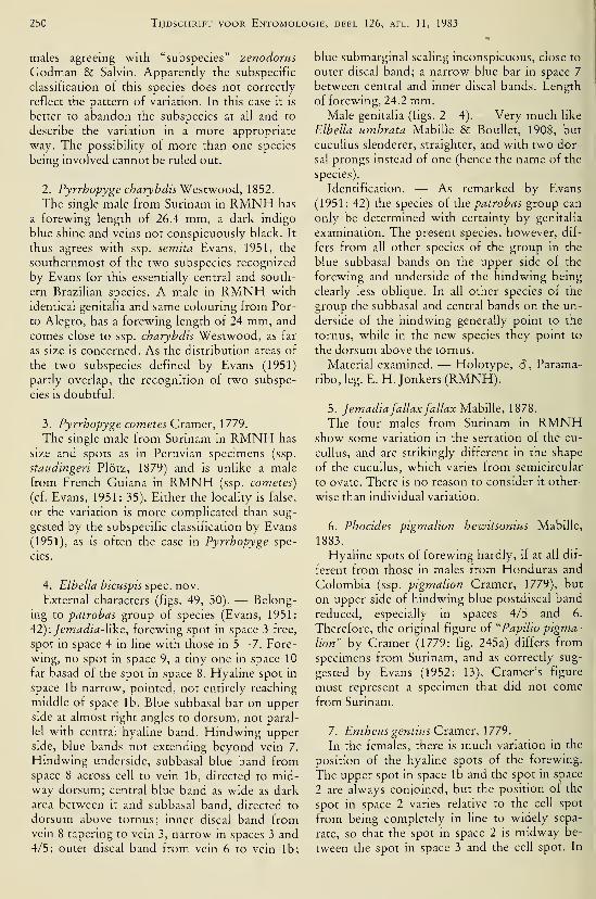
250 Tijdschrift voor Entomologie, deel 126, afl. 11, 1983
males agreeing with "subspecies" zenodorus
Godman & Salvin. Apparently the subspecific
classification of this species does not correctly
reflect the pattern of variation. In this case it is
better to abandon the subspecies at all and to
describe the variation in a more appropriate
way. The possibility of more than one species
being involved cannot be ruled out.
2. Pyrrhopyge charybdis Westwood, 1852.
The single male from Surinam in RMNH has
a forewing length of 26.4 mm, a dark indigo
blue shine and veins not conspicuously black. It
thus agrees with ssp. semita Evans, 1951, the
southernmost of the two subspecies recognized
by Evans for this essentially central and south-
ern Brazilian species. A male in RMNH with
identical genitalia and same colouring from Por-
to Alegro, has a forewing length of 24 mm, and
comes close to ssp. charybdis Westwood, as far
as size is concerned. As the distribution areas of
the two subspecies defined by Evans (1951)
partly overlap, the recognition of two subspe-
cies is doubtful.
3. Pyrrhopyge comètes Cramer, 1779.
The single male from Surinam in RMNH has
size and spots as in Peruvian specimens (ssp.
staudingeri Plötz, 1879) and is unlike a male
from French Guiana in RMNH (ssp. comètes)
(cf. Evans, 1951: 35). Either the locality is false,
or the variation is more complicated than sug-
gested by the subspecific classification by Evans
(1951), as is often the case in Pyrrhopyge spe-
cies.
4. Elhella hicuspis spec. nov.
External characters (figs. 49, 50). — Belong-
ing to patrohas group of species (Evans, 1951:
42): Jemadia-Yike, forewing spot in space 3 free,
spot in space 4 in line with those in 5—7. Fore-
wing, no spot in space 9, a tiny one in space 10
far basad of the spot in space 8. Hyaline spot in
space lb narrow, pointed, not entirely reaching
middle of space lb. Blue subbasal bar on upperside at almost right angles to dorsum, not paral-
lel with central hyaline band. Hindwing upperside, blue bands not extending beyond vein 7.
Hindwing underside, subbasal blue band fromspace 8 across cell to vein lb, directed to mid-way dorsum; central blue band as wide as dark
area between it and subbasal band, directed to
dorsum above tornus; inner discal band fromvein 8 tapering to vein 3, narrow in spaces 3 and
4/5; outer discal band from vein 6 to vein lb;
blue submarginal scaling inconspicuous, close to
outer discal band; a narrow blue bar in space 7
between central and inner discal bands. Length
of forewing, 24.2 mm.Male genitalia (figs. 2—4). — Very much like
Ribella umbrata Mabille & BouUet, 1908, but
cucullus slenderer, straighter, and with two dor-
sal prongs instead of one (hence the name of the
species).
Identification. — As remarked by Evans
(1951 : 42) the species of the patrobas group can
only be determined with certainty by genitalia
examination. The present species, however, dif-
fers from all other species of the group in the
blue subbasal bands on the upper side of the
forewing and underside of the hindwing being
clearly less oblique. In all other species of the
group the subbasal and central bands on the un-
derside of the hindwing generally point to the
tornus, while in the new species they point to
the dorsum above the tornus.
Material examined. — Holotype, â , Parama-
ribo, leg. E. H. Jonkers (RMNH).
5. JemadiafallaxfallaxìAzhiììe, 1878.
The four males from Surinam in RMNHshow some variation in the serration of the cu-
cullus, and are strikingly different in the shape
of the cucullus, which varies from semicircular
to ovate. There is no reason to consider it other-
wise than individual variation.
6. Phocides pigmalion hewitsonius Mabille,
1883.
Hyaline spots of forewing hardly, if at all dif-
ferent from those in males from Honduras and
Colombia (ssp. pigmalion Cramer, 1779), but
on upper side of hindwing blue postdiscal band
reduced, especially in spaces 4/5 and 6.
Therefore, the original figure of "Papilio pigma-
lion" by Cramer (1779: fig. 245a) differs from
specimens from Surinam, and as correctly sug-
gested by Evans (1952: 13), Cramer's figure
must represent a specimen that did not come
from Surinam.
7. EnthcMS gentius Cramer, \779.
In the females, there is much variation in the
position of the hyaline spots of the forewing.
The upper spot in space lb and the spot in space
2 are always conjoined, but the position of the
spot in space 2 varies relative to the cell spot
from being completely in line to widely sepa-
rate, so that the spot in space 2 is midway be-
tween the spot in space 3 and the cell spot. In
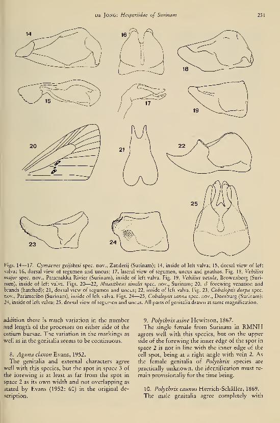
DE Jong: Hesperiidae of Surinam 251
Figs. 14— 17. Cymaenes geijskesi spec, nov., Zanderij (Surinam); 14, inside of left valva; 15, dorsal view of left
valva; 16, dorsal view of tegumen and uncus; 17, lateral view of tegumen, uncus and gnathos. Fig. 18, Vehilius
major spec, nov., Patamakka Rivier (Surinam), inside of left valva. Fig. 19, Vehilius vetula, Brownsberg (Suri-
nam), inside of left valva. Figs. 20—22, Mnasitheus similis spec, nov., Surinam; 20, 6 forewing venation and
brands (hatched); 21, dorsal view of tegumen and uncus; 22, inside of left valva. Fig. 23, Cobalopsis darpa spec,
nov., Paramaribo (Surinam), inside of left valva. Figs. 24—25, Cobalopsis tanna spec, nov.. Domburg (Surinam);
24, inside of left valva; 25, dorsal view of tegumen and uncus. All parts of genitalia drawn at same magnification.
addition there is much variation in the numberand length of the processes on either side of the
ostium bursae. The variation in the markings as
well as m the genitaha seems to be continuous.
8. Aguna claxon Evans, 1952.
The genitaha and external characters agree
well with this species, but the spot in space 3 of
the forewing is at least as far from the spot in
space 2 as its own width and not overlapping as
stated by Evans (1952: 60) in the original de-
scription.
9. Polythrix asine Hewkson, 1867.
The single female from Surinam m RMNHagrees well with this species, but on the upper
side of the forewing the inner edge of the spot in
space 2 is not in line with the inner edge of the
cell spot, being at a right angle with vein 2. As
the female genitaha of Polythrix species are
practically unknown, the identification must re-
main provisionally for the time being.
10. Polythrix caunus¥{trnch.-Sc\ri{{er, 1869.
The male genitalia agree completely with
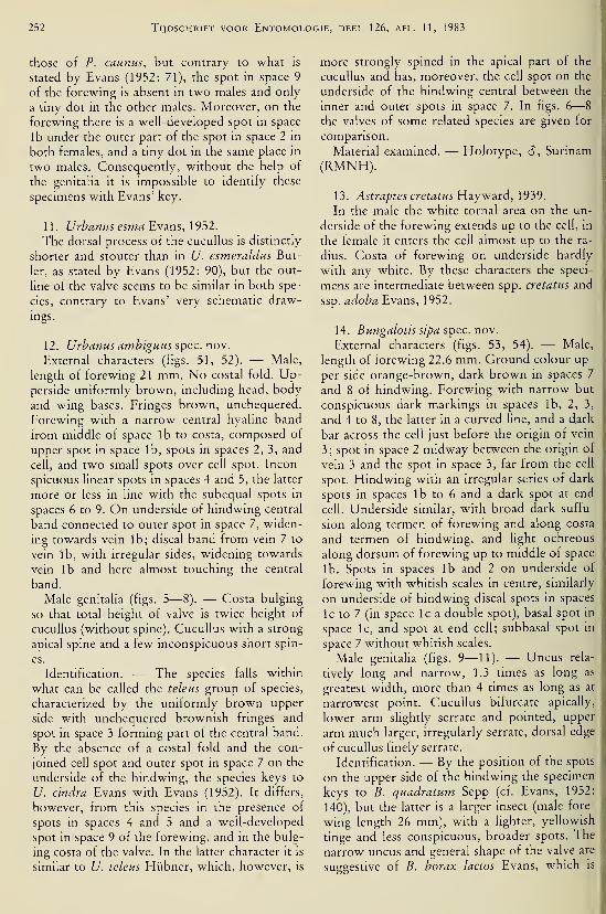
252 Tijdschrift voor Entomologie, deel 126, afl. 11, 1983
those of P. caunus, but contrary to what is
stated by Evans (1952: 71), the spot in space 9
of the forewing is absent in two males and only
a tiny dot in the other males. Moreover, on the
forewing there is a well-developed spot in space
lb under the outer part of the spot in space 2 in
both females, and a tiny dot in the same place in
two males. Consequently, without the help of
the genitalia it is impossible to identify these
specimens with Evans' key.
11. Urbanus esma Evans, 1952.
The dorsal process of the cucullus is distinctly
shorter and stouter than in U. esmeraldus But-
ler, as stated by Evans (1952: 90), but the out-
line of the valve seems to be similar in both spe-
cies, contrary to Evans' very schematic draw-
ings.
12. Urbanus ambiguus spec. nov.
External characters (figs. 51, 52). — Male,
length of forewing 21 mm. No costal fold. Up-perside uniformly brown, including head, body
and wing bases. Fringes brown, unchequered.
Forewing with a narrow central hyaline band
from middle of space lb to costa, composed of
upper spot in space lb, spots in spaces 2, 3, and
cell, and two small spots over cell spot. Incon-
spicuous linear spots in spaces 4 and 5, the latter
more or less in line with the subequal spots in
spaces 6 to 9. On underside of hindwing central
band connected to outer spot in space 7, widen-
ing towards vein lb; discal band from vein 7 to
vein lb, with irregular sides, widening towards
vein lb and here almost touching the central
band.
Male genitalia (figs. 5—8). — Costa bulging
so that total height of valve is twice height of
cucullus (without spine). Cucullus with a strong
apical spine and a few inconspicuous short spin-
es.
Identification. — The species falls within
what can be called the teleus group of species,
characterized by the uniformly brown upper
side with unchequered brownish fringes and
spot in space 3 forming part of the central band.
By the absence of a costal fold and the con-
joined cell spot and outer spot in space 7 on the
underside of the hindwing, the species keys to
U. cindra Evans with Evans (1952). It differs,
however, from this species in the presence of
spots in spaces 4 and 5 and a well-developed
spot in space 9 of the forewing, and in the bulg-
ing costa of the valve. In the latter character it is
similar to U. teleus Hübner, which, however, is
more strongly spined in the apical part of the
cucullus and has, moreover, the cell spot on the
underside of the hindwing central between the
inner and outer spots in space 7. In figs. 6—
8
the valves of some related species are given for
comparison.
Material examined. — Holotype, 6 , Surinam
(RMNH).
13. Astraptes cretatusYidiyw^rà, 1939.
In the male the white tornai area on the un-
derside of the forewing extends up to the cell, in
the female it enters the cell almost up to the ra-
dius. Costa of forewing on underside hardly
with any white. By these characters the speci-
mens are intermediate between spp. cretatus and
ssp. adoba Evans, 1952.
14. Bungalods sipa spec. nov.
External characters (figs. 53, 54). — Male,
length of forewing 22.6 mm. Ground colour up-
per side orange-brown, dark brown in spaces 7
and 8 of hindwing. Forewing with narrow but
conspicuous dark markings in spaces lb, 2, 3,
and 4 to 8, the latter in a curved line, and a dark
bar across the cell just before the origin of vein
3; spot in space 2 midway between the origin of
vein 3 and the spot in space 3, far from the cell
spot. Hindwing with an irregular series of dark
spots in spaces lb to 6 and a dark spot at end
cell. Underside similar, with broad dark suffu-
sion along termen of forewing and along costa
and termen of hindwing, and light ochreous
along dorsum of forewing up to middle of space
lb. Spots in spaces lb and 2 on underside of
forewing with whitish scales in centre, similarly
on underside of hindwing discal spots in spaces
Ic to 7 (in space Ic a double spot), basal spot in
space Ic, and spot at end cell; subbasal spot in
space 7 without whitish scales.
Male genitalia (figs. 9— 11). — Uncus rela-
tively long and narrow, 1.3 times as long as
greatest width, more than 4 times as long as at
narrowest point. Cucullus bifurcate apically,
lower arm slightly serrate and pointed, upper
arm much larger, irregularly serrate, dorsal edge
of cucullus finely serrate.
Identification. — By the position of the spots
on the upper side of the hindwing the specimen
keys to B. quadratum Sepp (cf. Evans, 1952:
140), but the latter is a larger insect (male fore-
wing length 26 mm), with a lighter, yellowish
tinge and less conspicuous, broader spots. The
narrow uncus and general shape of the valve are
suggestive of B. borax lactos Evans, which is

DE Jong: Hesperiidae of Surinam 253
Fig. 26. Eutychide subpunctata intermedia subspec. nov., Paramaribo (Surinam), mside of left valva. Figs. 27
—
29, Phlebodes meesi spec, nov., Lely Gebergte (Surinam); 17, dorsal view of tegumen and uncus; 28, inside of
left valva; 29, lateral view of tegumen, uncus and gnathos. Figs. 30—31, Penicula cnska extensa subspec. nov.,
Linker Coppename (Surinam); 30, dorsal view of tegumen and uncus; 31, inside of left valva. AU figures drawn
at same magnification.
only known from Peru, but in that species the
cucullus is not as strongly bifurcate and the ex-
ternal characters are different, the colour being
darker and the cell spot of the forewing being
conjoined to the inner edge of the spot in space
2 (I do not understand why Evans, 1952, de-
scribed lactos as a subspecies of B. borax Evans,
as the genitalia are as different from those of B.
borax as from those of any other Bungalotis
species; it seems better to consider lactos a sepa-
rate species).
Material examined. — Holotype, c5, S. Suri-
nam, Sipaliwini, air strip, 14.x. 1968, at hght, leg.
E.H. Jonkers (RMNH).
15. Bungalotis quadratum Sepp, 1848.
According to Evans (1952: 140) two subspe-
cies can be recognized, viz., ssp. quadratum
(Honduras, Colombia, Guiana, Surinam), and
spp. barba Evans (Fr. Guiana, Para, Santarem,
Peru). The differences in the male sex are as fol-
lows. In ssp. quadratum the ground colour is
yellow and the costa of the hindwing ("upf" in
Evans must be a misprint for "uph") broadly
dark brown on the upper side. Ssp. barba is
dark tawny, with dark brown area on upper side
of hindwing usually limited to a small area un-
der outer half of vein 7. One male from Parama-
ribo (RMNH) caught 5.1.1962, has the yellow
colour of ssp. quadratum and the extension of
the dark area on the upper side of the hindwing
along the costa as in ssp. barba. The other male
from Paramaribo (NZC) caught 23.x. 1981, has
the colour of ssp. barba and the extension of the
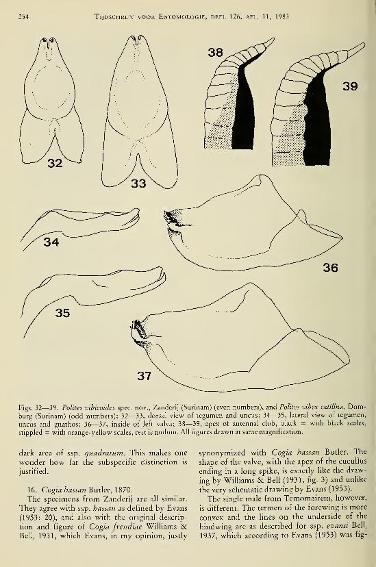
254 Tijdschrift voor Entomologie, deel 126, afl. 11, 1983
Figs. 32—39. Polites vibicoides spec, nov.. Zanderij (Surinam) (even numbers), and Polites vibex catUina, Dom-burg (Surinam) (odd numbers); 32—33, dorsal view of tegumen and uncus; 34—35, lateral view of tegumen,
uncus and gnathos; 36—37, inside of left valva; 38—39, apex of antennal club, black = with black scales,
stippled = with orange-yellow scales, rest is nudum. All figures drawn at same magnification.
dark area of ssp. quadratum. This makes one
wonder how far the subspecific distinction is
justified.
16. Cogia hassan Butler, 1870.
The specimens from Zanderij are all similar.
They agree with ssp. hassan as defined by Evans
(1953: 20), and also with the original descrip-
tion and figure of Cogia freudiae Williams &Bell, 1931, which Evans, in my opinion, justly
synonymized with Cogia hassan Butler. The
shape of the valve, with the apex of the cucullus
ending in a long spike, is exactly like the draw-
ing by WiUiams & Bell (1931, fig. 3) and unhke
the very schematic drawing by Evans (1953).
The single male from Temomairem, however,
is different. The termen of the forewing is more
convex and the lines on the underside of the
hindwing are as described for ssp. evansi Bell,
1937, which according to Evans (1953) was fig-

DE Jong: Hesperiidae of Surinam 255
ured by Draudt (1922, pi. 170d) under the nameof "hassan" (the colours of Draudt's figure musthave been invented by the printer, but the posi-
tion of the unes on the underside of the hind-
wing is clear). The genitaHa are similar to those
of the specimens from Zanderij.
As Zanderij is in the north of the country andTemomairem in the extreme south, a subspecific
difference does not seem to be too surprising.
Ssp. hassan, however, is the subspecies of north-
ern Brazil and ssp. evansi occurs in Bolivia andsouthern Brazil. The capture of a specimen with
characters of ssp. evansi far within the bounda-ries of the distribution area of ssp. hassan sug-
gests that the two taxa may be specifically dis-
tinct. On the other hand, they could also be
merely seasonal forms. The material is still too
scanty to make a firm statement.
17. Clito jonkersi spec. nov.
External characters (fig. 55). — Male. Length
of forewing 15.1 mm. Costal fold present.
Nudum 22 segments. Forewing upper side dark
brown with hyaline spots in spaces lb (acco-
lade-shaped), 2 (large, V-shaped), 3, 4, 5, and6—8 (in line, the latter longer, extending further
to termen), and in cell (U-shaped with rounded
base). Hmdwing upper side dark brown with
hyaline spots in spaces Ic (anvil-shaped, just ex-
tending into space lb), 2 (very small, at base), 6
(small, at base), 7 and in cell (filling more than
half of cell); inner edge of spots in Ic, 7 and cell
in line; faint whitish pointed submarginal spots
in spaces Ic—6. Underside as upper side; lighter
shading on forewing in spaces lb—3 directly
distad of the hyaline spots; also faint lighter
shading along termen of hindwing in spaces 4
—
5.
Male genitalia (figs. 12, 13). — Gnathos rela-
tively long, almost reaching apex of uncus. Cu-cullus stout, slightly higher than long, the broad
apex strongly spined. Costal process short,
stout, finely spined.
Identification. — Externally the new species
comes closest to C. littera anda Evans, 1953. It
differs in the white band of the hindwing being
narrower and divided by dark veins, and the cell
spot of the forewing not being rounded basally,
but cut off obliquely. In C. littera littera Ma-bille, 1877, the white band of the hindwing is
faint and the inner edge of the cell spot of the
forewing forms a straight line perpendicular to
the costa. The shape of the valve and length of
the gnathos are different from those of all de-
scribed Clito species.
Material examined. — Holotype, 6, Dom-burg, 8.iii.l975, leg. E. H. Jonkers (RMNH).
Discussion. — According to Evans (1953) the
genus Clito is characterized among other char-
acters by a nudum of 16 segments. I don't knowhow Evans arrived at this figure. In the BM I
checked the material arranged by Evans, andfound the following numbers of nudum seg-
ments:
C. littera Mabille, d, 22; C bihulus Riley, 618, 9 22; C dito Fabricius, d 18, 9 22; C.
sompa Evans, d 21; C. zelotes Hewitson, â 21;
C zenda Evans, S and 9 22; C. tuva Evans, âand 9 22.
Apparently all species are rarely caught,
judged from the very scanty material available.
18. AnthoptHs epictetHS Fabricius, 1793.
According to Evans (1955: 62) the femaleshould be entirely brown. However, in a series
of 448 specimens he had only two females. In
the series of 15 specimens from Surinam in
RMNH there is a single female that is similar to
the males except for a yellow stripe on the up-per side of the hindwing from base to termencovering vein lb.
19. Vinius tryhana Kaye, 1913.
The three males in RMNH have identical
genitalia corresponding with those of V. tryha-
na. One male has the tawny colour on the upperside more extensive, so that the cell of the
hindwing is tawny, and the cell of the forewingis tawny with a narrow black central streak. Bythe latter character the specimen is similar to V.
exilis Plötz, 1883, but the genitalia point defi-
nitely to V. tryhana.
20. ApaustHS menés Stoll, 1782.
According to Evans' (1955: 83) key, Apaustus
has six nudum segments. In the description of
the two species at p. 91, however, A. gracilis
Felder is stated to have 4—6 and A. menés 8
nudum segments. The three specimens in
RMNH which undoubtedly belong to A. men-
és, have 6 nudum segments.
21
.
Cymaenes geijskesi spec. nov.
External characters (fig. 56). — Male. Length
of forewing 12.4 mm. Upper side brown; fore-
wing with faint ochreous spots m spaces 2, 3
and 6, and traces of spots in spaces lb and 7,
and in cell; hindwing practically unicoloured.
Underside as upper side, a little paler, traces of

256 Tijdschrift voor Entomologie, deel 126, afl. 11, 1983
median spots in spaces 2—5 on the hindwing.
Fringes sullied. Nudum with 13 segments of
which 9 on apiculus.
Male genitalia (figs. 14— 17). — Indentation
of uncus not more than one third of total length
of uncus + tegumen, twice as long as wide. Cu-cullus greatly expanded to a semicircle with a
slightly serrate edge, turned over in horizontal
position.
Identification. — Rather similar to C.
tripunctus Herrich-Schäffer by the absence of
markings in spaces 4 and 5 on the underside of
the forewing, by the very faint cell spot on the
forewing, the faint spots on the underside, and
the broadened cucuUus. It differs in being
smaller (C. tripunctus, 13— 15.3 mm), underside
more yellowish than greyish brown, and abun-
dantly in the genitalia, where the semicircular
cucullus is unique for the genus and the indenta-
tion of the uncus is much narrower and shal-
lower than in C. tripunctus, in which moreoverthe uncus bulges strongly laterally. In C.
tripunctus, and according to Evans (1955) in all
other Cymaenes species as well, the nudumconsists of 11 segments, of which 8 are on the
apiculus. In C. geijskesi there are two more seg-
ments. As the number of nudum segments mayvary even within a species, examination of muchmore material is needed to make sure that this is
a constant difference between the new species
and its congeners.
Material examined. — Holotype, â , Surinam,
Paramaribo-Zanderij, savanne, 27—30.viii.
1964, leg. D. C. Geijskes (RMNH).
22. Vehilius major spec. nov.
External characters (figs. 57, 58). — Male.
Length of forewing 13.4 mm. Exactly like V.
vetulus Mabille, only larger (V. vetulus 10
—
12.2 mm) and upper cell spot on upper side of
forewing more conspicuous.
Male genitalia (figs. 18, 19). — Similar to
those of V. vetulus, but outline of valve trape-
zoid rather than triangular as in V. vetulus.
Material examined. — Holotype, 6 , Surinam,
Patamakka Rivier, 4. ix. 1969, leg. E. H. Jonkers
(RMNH).
23. Mnasilus allubita Butler, 1877, andNastra guianae Lindsey, 1925.
M. allubita is very difficult to separate fromTV. guianae. The only external difference I could
find is the colour of the palps and cheeks, white
in TV. guianae, cream-coloured or pale yellow in
M. allubita. According to Evans (1955) the only
difference between Nastra and Mnasilus is the
occurrence of a recumbent hair tuft on the up-
per side of the forewing in males of Mnasilus.
This is correct, but little obvious. It seems exag-
gerated to base a generic distinction on this
character only. Together with related genera
these two are badly in need of revision.
24. Mnasitheus similis spec. nov.
External characters (figs. 20, 59, 60). — Male.
Length of forewing 12.5 mm. Upper side en-
tirely dark brown, no spots; slight greenish me-tallic sheen on head, colar and thorax. Under-side dark brown, faintly lighter in tornai half of
forewing, in spaces la—c of hindwing, and with
very faint discal spots in spaces 2—6 of hind-
wing. Forewing with tripartite stigma: a short,
obliquely set patch at the base of space 2 and di-
rectly under it, a small patch in upper half of
space lb and a patch in lower half of space lb
similar to that in space 2.
Male genitalia (figs. 21, 22). — Uncus deeply
indented. Valve with cucullus strongly devel-
oped in dorso-proximal part, extending far be-
yond the ill-developed costa; apex of cucullus
upturned and pointed.
Identification. — With Evans (1955) the newspecies keys more or less to M. simplicissima
Herrich-Schäffer, which however has an entire
uncus and a differently shaped cucullus, without
the strong dorso-proximal expansion. Theshape of the cucullus is reminiscent of that of M.forma Evans, which does not have the deeply
indented uncus of the present species. The shape
of the tripartite stigma differs from that of any
other Mnasitheus species, the patches in space
lb usually being parallel to vein 1, or (in M.continua Evans) perpendicular to vein 1, and the
patch in space 2 either parallel to vein 2 or to the
cubitus.
Material examined. — Holotype, 6 , Surinam,
leg. E.H. Jonkers (RMNH).
25. Papias phainis Godman, 1900.
The single female was identified as belonging
to this species rather than to P. phaeomelas
Geyer because of the palps being yellower and
the hindwing having well visible spots on the
underside. Generally, identification of females
of this genus as well as of many related genera is
hampered by the fact that the female genitalia
have not been studied comparatively (and usual-
ly are not known at all).
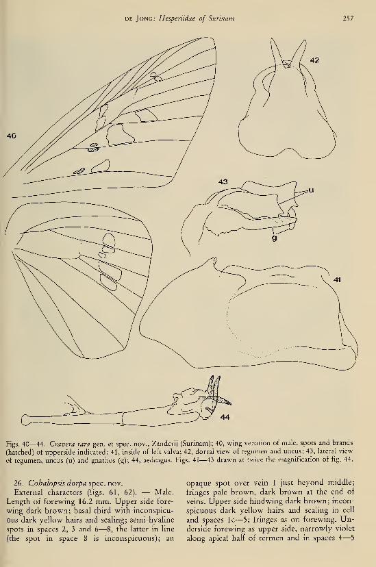
DE Jong: Hesperiidae of Surinam Tbl
Figs. 40—44. Gravera rara gen. et spec, nov., Zanderij (Surinam); 40, wing venation of male, spots and brands
(hatched) of upperside indicated; 41, inside of left valva; 42, dorsal view of tegumen and uncus; 43, lateral view
of tegumen, uncus (u) and gnathos (g); 44, aedeagus. Figs. 41—43 drawn at twice the magnification of fig. 44.
26. Cohalopsis dorpa spec. nov.
External characters (figs. 61, 62). — Male.
Length of forewing 16.2 mm. Upper side fore-
wing dark brown; basal third with inconspicu-
ous dark yellow hairs and scaling; semi-hyaline
spots in spaces 2, 3 and 6—8, the latter in hne
(the spot in space 8 is inconspicuous); an
opaque spot over vein 1 just beyond middle;
fringes pale brown, dark brown at the end of
veins. Upper side hindwing dark brown; incon-
spicuous dark yellow hairs and scaling in cell
and spaces Ic—5; fringes as on forewing. Un-derside forewing as upper side, narrowly violet
along apical half of termen and in spaces 4—
5
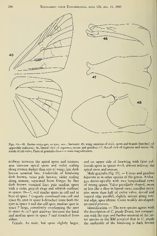
258 Tijdschrift voor Entomologie, deel 126, afl. 11, 1983
Figs. 45—48. Surina unica gen. et spec, nov., Surinam; 45, wing venation of male, spots and brands (hatched) of
upperside indicated; 46, lateral view of tegumen, uncus and gnathos; 47, dorsal view of tegumen and uncus; 48,
inside of left valva. Parts of genitalia drawn at same magnification.
midway between the apical spots and termen;
area between apical spots and violet scaling
along termen darker than rest of wing; fine dark
brown terminal line. Underside of hindwing
dark brown, veins pale brown; violet scaling
along termen, separated from fringes by fine
dark brown terminal line; pale median spots
with a violet-greyish tinge and whitish outlined
in spaces lb— 7, and similar spots in cell and at
base of space 7 (vaguely continued into cell and
space 8); spot in space 6 detached from both the
spot in space 5 and the cell spot; median spot in
space 7 large, completely overlapping the spot
in space 6; cell spot midway between the basal
and median spots in space 7 and detached fromeither.
Female. As male, but spots slightly larger,
and on upper side of forewing with faint yel-
lowish spots in spaces 4—5, almost midway the
apical spots and termen.
Male genitalia (fig. 23). — Uncus and gnathos
bipartite as in other species of the genus. Aedea-
gus dorso-apically with two longitudinal rows
of strong spines. Valve peculiarly shaped, more
or less Uke a shoe in lateral view; cucuUus occu-
pies more than half of entire valve, dorsal and
ventral edge parallel, slightly serrate along ven-
tral edge, apex obtuse. Costa weakly developed;
no costal process.
Identification. — The new species agrees with
the description of C. prado Evans, but compari-
son with the type and further material of the lat-
ter species in the BM revealed that in C. prado
the underside of the hindwing is dark brown

DE Jong: Hespenidae of Surinam 259
55 56
Figs. 49, 50. Libella bicuspis spec, nov., holotype, upper- and underside. Figs. 51, 52. Urbanus ambiguus spec,
nov., holotype, upper- and underside. Figs. 53, 54. Bungalotis sipa spec, nov., holotype, upper- and underside.
Fig. 55. Clito jonkersi spec, nov., holotype, upperside. Fig. 56. Cytnaenes geijskesi spec, nov., holotype, upper-
side.

260 Tijdschrift voor Entomologie, deel 126, afl. 11, 1983
57 58
59 60
61 62
63
Figs. 57, 58. Vehilius major spec, nov., holotype, upper- and underside. Figs. 59, 60. Mnasitheus similis, spec,
nov., holotype, upper- and underside. Figs. 61, 62. Cobalopsis dorpa spec, nov., holotype, upper- and underside.
Fig. 63. Cobalopsis tanna spec, nov., holotype, upperside.
DE Jong: Hesperiidae of Surinam 261
with faint lighter brown spots. The markings of
the new species are more similar to those of C.
catocala Herrich-Schäffer, which, however, has
the spot in space 6 on the underside of the
hindwing attached to the spot in space 5 and de-
tached from the spot in space 7. In the muchsmaller C. dagon Evans, the spots are arranged
as in C. dorpa except that in the former species
the spots in spaces 6 and 7 are conjoined to the
cell spot. The shape of the valve of C. dorpa is
not found in any other Cobalopsis species.
Material examined. — Holotype, 6 -, Surinam,
Paramaribo, 3.viii.l963, leg. E. H. Jonkers. Par-
atype, 9 , Surinam, Paramaribo, Ma Retraite,
1.x. 1960, leg. E. H. Jonkers. Both types in
RMNH.
27. Cobalopsis tanna spec. nov.
External characters (fig. 63). — Male. Length
of forewing 13.5 mm. Nudum occupying 12
segments of which 9 on apiculus. Mid tibiae
sparsely spined. No secondary sex characters.
Upper side of forewing brown, possibly with
yellowish basal clothing but specimen too wornto make sure; hyaline white spots in spaces 2, 3,
6, and in lower half of cell over origin of vein 3;
spot in space 2 slightly closer to cell spot than to
spot in space 3; a yellowish opaque spot in
space lb, and a trace of a minute dot in space 7.
Upper side hindwing brown with yellowish
clothing over greater part of the wing; no spots.
Underside of both wings brown, markings as on
upper side.
Male genitalia (figs. 24, 25). — Uncus and
gnathos bifid, uncus arms more widely apart
than gnathos arms. Valve seemingly trapezoid
in lateral view because apex turns inward.
Identification. — The new species agrees well
with the description of Cobalopsis as given by
Evans (1955), but it does not fit any of the de-
scribed species of the genus. As the specimen
was collected in a Malaise trap, its condition is
rather poor, but it is good enough to make sure
that there are no spots on the hindwing. The on-
ly known Cobalopsis species with an unmarkedhindwing is C. dedecora Plötz, which has the
underside of the hindwing reddish-grey and in
the forewing only spots in spaces 3, 6 and 7. Thepresence of a lower spot only in the cell is
unique in Cobalopsis. Upper cell spots are
found in C. autumna Plötz and C. nero Her-
rich-Schäffer, while the female of the first-men-
tioned species has a lower cell spot as well;
these two species have spots on the underside of
the hindwing, and their genitalia are quite dif-
ferent, the cucuUus being long and narrow.
Material examined. — Holotype, 6 , Surinam,
Domburg, 18—23.xii.1963, Malaise trap, leg. D.
C. Geijskes (RMNH).
28. Morys subgrisea paradoxa subspec. nov.
External characters. — Male. Length of fore-
wing 12.5 mm. Upper and underside uniform
dark brown. On upper and underside of fore-
wing very faint spots in spaces 2 and 3, and al-
most invisible in space 6. Faintest traces of me-dian spots on the underside of the hindwing.
Stigma on forewing and genitalia as in M. sub-
grisea subgrisea Mabille. In size it is closer to
the only other described subspecies, M. subgri-
sea prada Evans (cf. Evans, 1955: 168). It can be
distinguished by the almost complete lack of
spots and by the lack of grey scaling on the un-
derside of the hindwing, so that the name sub-
grisea is not very appropriate, hence the pro-
posed subspecies name.
Material examined. — Holotype, S , Surinam,
Zanderij, savanne, 13— 16. vili. 1964, leg. D. C.
Geijskes (RMNH).
29. Vettiusyalta Evans, 1955.
The two females directly key to V. artona
Hewitson or V. yalta Evans with Evans (1955).
They differ from the female of V. artona in be-
ing smaller (length of forewing 14—16.4 mm, as
against 17.8 mm), in having the spot in space 2
of the forewing more or less under the cell spots
(in V. artona mid cell spots and spot in space 3),
and in having a paler underside of the hindwing.
There are also differences in the genitalia. Theallocation of the two specimens to V. yalta must
be considered preliminary.
30. Eutychide subpunctata intermedia sub-
spec, nov.
Discussion. — The three specimens agree
with the original description of E. sempa Evans,
1955, except for the cucullus being serrate (fig.
26). Examination of the type and the only other
available male of sempa in the BM, however, re-
vealed that also in E. sempa the cucullus is ser-
rate, be it less obvious than in E. subpunctata
Hayward. The spined dorsal projection of the
cucullus, according to Evans' figures present in
E. sempa and absent in E. subpunctata, was pos-
sibly broken in the left valve figured by Evans,
as it is present in the right valve of the only
specimen of E. subpunctata studied by Evans,

262 Tijdschrift voor Entomologie, deel 126, afl. 11, 1983
more or less concealed by the strong serration
of the cucuUus. Further differences between E.
sempa and E. suhpunctata, are found in the un-
cus arms, which are about parallel in E. sempa
and divergent in E. suhpunctata, and in the col-
our of the wings, E. sempa being darker but
with paler area along dorsum on the underside
of the forewing, and of the cilia which are paler
in E. suhpunctata.
So far, E. suhpunctata and E. sempa are well-
defined separate entities. However, the three
specimens from Surinam are not only topo-
graphically intermediate (£. sempa is only
known from Guiana, E. suhpunctata from Ar-
gentine and French Guiana), but also morpho-
logically. The cucuUus is more rounded than in
both E. sempa and E. suhpunctata, the serration
is intermediate. The uncus arms are more or less
parallel. The pale dorsal area on the underside
of the forewing is inconspicuous. The cilia are
brownish grey.
On the basis of the taxonomically as well as
geographically intermediate position of the Su-
rinam specimens, it seems most appropriate to
unite E. sempa and E. suhpunctata with the
Surinam representatives into a single, geograph-
ically variable species. The Surinam specimens
are sufficiently different (see above) to warrant
subspecific distinction; the subspecies is namedEutychide suhpunctata intermedia here.
Thus E. suhpunctata consists of the following
subspecies: E. suhpunctata suhpunctata Hay-ward (Argentine, French Guiana), E. suhpunc-
tata intermedia subspec. nov. (Surinam), and E.
suhpunctata sempa Evans (comb, nov.) (Guia-
na).
Material examined. — E. suhpunctata suh-
punctata: 1 6, French Guiana (BM). E. suh-
punctata intermedia: holotype, 6, Surinam,
Paramaribo, Ma Retraite, 6—8. i. 1964, leg. D.
C. Geijskes; 2 6 ,paratypes, same data but 14
—
16. i. 1964 and 4—8.iii.l964, respectively (all
types in RMNH). E. suhpunctata sempa: 2 6 ,\
9, Guiana, Takutu River (incl. holotype, 6)(BM).
31. Talides sinois Hübner, 1819.
The differences between this species and T.
alternata Bell are slight. Moreover, the differ-
entiating characters given by Evans (1955:
226—227) are liable to variation. In the four
males from Surinam in the RMNH belonging to
T. sinois and/or T. alternata, the shape of the
cucuUus as well as the length of the tegumen
spike is different for each male. There is also
variation in the distance between spots 2 and 3
of the forewing. It makes one wonder if T.
sinois and T. alternata are really separate spe-
cies. The material listed has been assigned to the
two species according to the closest match with
Evans' descriptions, but especially for the males
assigned to T. sinois, this match is not perfect.
The single female has been listed provisionally
under T. sinois, no differentiating characters
having been published for the female sex.
32. Carystus elviraPlötz, \S82.
Evans (1955) who had no material of this spe-
cies at his disposal remarked: "May be an aber-
ration or a faulty figure", a rather bold
statement for an author who had not seen any
specimens. It is true that the figure in Draudt
(1923, pi. 189b) does not seem to have entirely
right colours, but Evans could not know this.
Moreover, the colour differences between
Draudt's figure (probably based on the original
figure by Plötz) and the specimen in the
RMNH may be a sexual difference, the figure
representing a male and the RMNH specimen
being female.
The female in the RMNH can be described as
follows. Head with a conspicuous white spot
directly behind the antennae. Length of fore-
wing, 21.4 mm. Basal third of costa of forewing,
patagia and tegulae with orange hairs. Upperside of forewing brown with sharply defined
hyaline spots in spaces lb (against vein 1), 2, 3,
6 and 7 (last two small, especially in space 7),
and two spots in cell slightly basad of spot in
space 2. Upper side hindwing brown, light band
from base to termen on underside weakly shin-
ing through. Underside of forewing as upper
side, but orange along costa up to end of vein
12, from there a dull yellowish subapical band,
overlapping the spots in spaces 6 and 7 (making
them obscure) and ending at termen in space
4/5; pale yellowish suffusion from the spot in
space lb towards the termen. Underside of
hindwing with, a warm brown-yellow colour,
paler dull yellow from base through cell and
space 4/5 to termen, this pale band flanked by
dark brown colouring which extends more or
less along the veins; space lb greyish brown.
33. Carystoides hasoches LatreiWe, 1824.
The males agree with C. hasoches. The fe-
males in RMNH differ in size (forewing, 20.1
and 21.5 mm) and spotting: in the smaller speci-
men the hyaline central spot on the upper side
of the hindwing is relatively small and contin-

DE Jong: Hesperiidae of Surinam 263
ued across space 3, in the larger specimen it is a
little larger and confined to spaces 4—5. Thus,
both females have some characters of C.
basoches and some of C. noseda Hewitson. Asthe males are undoubtedly C. basoches, I have
placed the females here as well.
34. Perichares deceptus Butler & Druce,
1872.
The cell spots of the forewing are separate in
the single male from Surinam. This is a commonfeature in ssp. drina Evans, but also occurs in
ssp. luscinia Plötz. The lilacine areas on the un-
derside of the hindwing concord with the situa-
tion in ssp. luscinia (which is only known from
S. Brazil). In view of its locality, one would ex-
pect the Surinam specimen to be closest to ssp.
fulvimargo Butler, which occurs in Colombia,
Venezuela, Ecuador and Peru, but the yellow
colour at the tornus of the hindwing, character-
istic of ssp. fulvimargo, does not occur in the
Surinam specimen. More and better preserved
material is needed to decide on the systematic
position of the population in Surinam.
35. Phlebodes meesi spec. nov.
Only a male available. External characters. —Length of forewing, 15.3 mm. Upper side very
dark brown; inconspicuous dark ochreous hairs
at base of forewing; hyaline spots only m spaces
2 and 3, spot in space 2 larger and less regular
than square spot in space 3; no other spots ex-
cepting a very faint spot in space lb mdicated
by some pale ochreous scales close to vein 1 just
beyond its middle; hindwing unmarked. Under-
side forewing as upper side, purplish in apical
area. Underside hindwing dark brown with
purplish gloss, but space lb and greater part of
space Ic plain brown; veins a little paler, no
spots. Forewing with sagittate brand over origin
of vein 2, and a long narrow brand under basal
third of vein 2.
Genitalia (figs. 27—29). — Uncus bifid, arms
about three times as long as wide, slightly diver-
ging. Tegumen with central process, broad at
base, much narrower in distal half, bluntly
ended, not reaching base of indentation of un-
cus. Gnathos consisting of two separate, sharply
pointed sclerites. CucuUus with long, narrow,
upturned apex; costa hardly developed, at distal
end with short, inwards curving flap over basal
part of cucullus.
Material examined. — Holotype, â , Surinam,
Lely Gebergte, 600—700 m, 30.x.l979, leg. G.
F. Mees. In RMNH.
Discussion. — P. meesi differs from all other
Phlebodes species (excepting some specimens of
the South Brazilian P. smithi) in the absence of
apical spots of the forewing and of any spots onupper and underside of the hindwing. In the
genitalia the new species seems to come close to
P. vira Butler, 1870, P. virgo Evans, 1955, and
P. torax Evans, 1955, although in the genitalic
drawmgs by Evans (1955) no trace can be seen
of the central process of the tegumen. External-
ly these species differ from P. meesi in the un-
derside of the hindwing being ochreous-brown
with conspicuous yellow veins.
36. Cynea cyrusVlotz, \8S3.
The two specimens are more like ssp. hippo
Evans, described from Trinidad, than ssp. rhino
Evans, described from French Guiana, UpperAmazons and Para. Length of forewing 21.2
mm. No spot in space lb on the upper side of
the forewing; apical spots in spaces 6—8 (al-
most invisible in one specimen). Underside
hindwing, spots in spaces 2 and 3 very faint.
37. Penicula criska extensa subspec. nov.
Description (figs. 30, 31). — Male. Externally
and in the genitalia similar to P. criska Evans,
but differs in the shape of the valva, where the
cucullus is expanded disto-ventrally, so that the
distal edge is almost perpendicular to the ventral
edge, and apex not extending above costa. In P.
criska criska the ventral edge of the cucullus
gently curves to the apex which extends above
the costa.
Material examined. — Holotype, â, Linker
Coppename, Zuidkreek ll.viii.l943, at light,
leg. D. C. Geijskes. In RMNH.
38. Polîtes vibex Geyer, IS32.
The extension of the tawny spots is variable,
so that it is not well possible to make a sharp
distinction between ssp. praeceps Scudder and
ssp. catilina Plötz. This may be due to the fact
that Surinam is in the area where both subspe-
cies meet, from Ecuador through Venezuela to
the Guianas.
39. Polites vibicoides spec. nov.
External characters (figs. 64, 65). — A small
replica of P. vibex praeceps/catilina. Male.
Length of forewing, 10.3— 10.5 mm. Markings
exactly as in P. vibex, but tawny colour slightly
darker and warmer. Extension of tawny colour
on upper side variable; median band of fore-
wing may be narrower than dark terminal area.

264 Tijdschrift voor Entomologie, deel 126, afl. 11, 1983
64 65
"^^^
68 69
Figs. 64, 65. Polites vibicoides spec, nov., holotype, upper- and underside. Figs. 66, 67. Gravera rara gen. et
spec, nov., paratype, upper- and underside. Figs. 68, 69. Surina unica gen. et spec, nov., holotype, upper- and
underside.
Female as large as male; on underside of hind-
wing band of yellow median spots from vein 2
to vein 8, flanked by dark brown spots in spaces
2, 3, 6 and 7, quite different from the dull grey-
ish-ochreous colours of P. vibex catilina fe-
males.
Male genitalia (figs. 32—37). — Similar to P.
vibex. Uncus and tegumen more rounded in
dorsal view. CucuUus and costa of valve not
suddenly narrowing as in P. vibex. Costa
broadened and hollowed apically so as to re-
ceive the apex of the cucuUus; apical spines of
costa directed caudad or more or less turned upas in P. vibex. The complex aedeagus is also
similar to the structure found in P. vibex.
Identification. — Polites is a mainly NorthAmerican genus consisting of seven species
(Evans, 1955). The only species up to now

DE Jong: Hesperiidae of Surinam 265
known to occur in South America is P. vibex
(distributed from the northeastern U.S.A. to
Argentina). The new species cannot be mistaken
for P. vibex because it is much smaller {P. vibex
from Surinam, length of forewing 13.8—14.8
mm). For the rest, however, they are so similar
that they could be considered subspecies if they
were not sympatric.
Material examined. — Holotype, c?, Surinam,
Zanderij, savanne, 27—30.viii.l964, leg. D. C.
Geijskes. Paratypes, 4 c?, 2 9, same data but
16—20.viii.l964 (1 (?), 20—24.viii.1964 (1 S, 1
9), 24—26.viii.1964 (1 6, 1 9), 4—7.ix.l964 (1
CÎ). All types in RMNH.Remark. — According to Evans (1955) the
Polites species have 11—12 nudum segments.
This may hold true for North American species,
but in the Surinam specimens of P. vibex and P.
vibicoides the number of nudum segments is 13
(figs. 38,39).
40. Panoquina panoquinoides minima sub-
spec, nov.
External characters. — Length of forewing
10.5—11.7 mm. Upper side forewing brown,
overlaid with yellow scales and hairs along costa
and in basal third. Pale yellow opaque spot in
space lb, pale yellow hyahne spots in spaces 2
and 3, inconspicuous apical spots in spaces 6
and 7 (one or both may be absent); fringes pale
ochreous. Upper side hindwing brown overlaid
with yellow hairs and scales; fringes pale och-
reous. Underside forewing as upper side, darker
brown in basal third. Underside hindwing grey-
ish brown, veins pale yellow, no spots or at
most traces of inconspicuous, streaklike, pale
yellow spots in spaces 2 and 3.
Male genitalia. — As in P. panoquinoides
Skinner.
Identification. — Because of its small size it
was surprising to find that the new subspecies
belonged to a Panoquina species. With 12— 14.3
mm for the already described subspecies, P.
panoquinoides is the smallest member of the ge-
nus, the other species varying from 16 to 25
mm. Apart from its size the new subspecies is
characterized by the reduction (in most speci-
mens complete absence) of spots on the hind-
wing.
Material examined. — Holotype, 6-, Parwa-
bos, Kwattaweg naar zee. 8
—
ll.ii.l964, in Ma-laise trap, leg. D. C. Geijskes. Paratypes: 1 6,
Paramaribo; 11 (5,2 9, same data as holotype,
but 1—4.ÌÌ.1964 (1 6), 8—11.11.1964 (2 6), 11—14.ii.1964 (3 S\ 14—18.ii.1964 (1 6), 18—
24.11.1964(1 c5), 29.il—2. iii. 1964(1 S,\ 9), 3—6.iii.l964 (2 CÎ, 1 9); 3 (5, Matapica in Malaise
trap. All types in RMNH.
41. Gravera ^en. now.
External characters. — Palpi second segmentflattened, third short, hardly protruding aboveclothing of second segment. Antennae reaching
to origin of vein 10. Antennal club swollen,
three times as wide as antennal shaft, length
5/18 of total length of antenna, constricted be-
fore apiculus which is almost twice as long as
width of club. Nudum covering 15 segments of
which 9 on apiculus. Forewing cell almost as
long as dorsum; vein 2 closer to vein 3 than to
base; vein 4 midway between veins 3 and 5; vein
5 decurved at origin towards vein 4; termen al-
most straight from vein 6 to vein 1, very slightly
incurved in space lb; white hyaline spots in
spaces lb, 2, 3, 6—7, 8 (may be absent) and cell;
spots in 6 and 7 directed to upper half of ter-
men. Hindwing cell about half as long as wingmeasured along vein 4; vein 7 originates muchcloser to base than vein 2; termen almost
straight (in female slightly curved) from vein 7
to vein lb; vein lb produced, longer than vein
la; white hyaline spots in spaces 2, 3, 5 and 6.
Hind tibiae with upper pair of spurs absent. Ab-domen as long as vein la of hindwing. Malewith short inconspicuous stigmas over and un-
der basal 1/4 of vein 2 and over basal half of
vein 1 of forewing (fig. 40). Female with dense
long hairs at tip of abdomen.Male genitalia. — Uncus and gnathos bipar-
tite; apex of aedeagus with complicate processes
and horns.
Discussion. — See after description of the
type-species.
Type-species.— Gravera rara spec. nov.
Gravera rara spec. nov.
External characters (figs. 66, 67). — Male.
Length of forewing 23.9 mm. Upper side black.
Forewing cell spot consisting of two spots
which just touch and are placed almost parallel
to termen. Hindwing with very small spot in
space 4. Thorax, abdomen, base of forewing to
origin of vein 2, and basal half of hindwing with
bluish-grey hairs. Underside brown. Forewing
on underside with white suffusion along costa
over cell spot, and extensive white suffusion in
space lb under the spot in space 2 distally from
and in continuation with the spot in space lb.
Hindwing on underside in addition to the hya-
line spots white patches in space Ic (large, be-

266 Tijdschrift voor Entomologie, deel 126, afl. 11, 1983
tween spot in space 2 and termen) and space 7
(small, in line with other spots), and a white dot
just before end cell. Fringes dark brown, white
on forewing at space lb and on hindwing at
spaces lb, Ic and, very narrowly, 2. Palpi and
underside abdomen greyish, underside thorax
pale bluish-grey. Antennae upper side at base of
club and basal half of shaft chequered, rest
black; underside white on basal half of club and
upper half of shaft, rest black.
Female. Length of forewing 25.5 mm. Asmale. Cell spot forewing undivided. Hindwingunderside with same spots as male, but upper
side no spot in space 4.
Male genitalia (figs. 41—44). — Tegumenshort and broad, with a central indentation in
proximal edge, so that proximal part appears to
be bilobed. Uncus dorsally with a gutterlike
longitudinal depression ending in an apical in-
dentation, the two apical processes very muchflattened laterally and expanded dorso-ventral-
ly. Gnathos consisting of two long, strongly in-
curving arms, as long as uncus. Valve trapezoi-
dal, cucuUus squarish; dorso-distal corner of
cucullus slightly prolonged and twisted; dorso-
proximal corner overlapped by the weakly de-
veloped costa. Aedeagus apically with long
curved horns.
Identification. — Externally the present spe-
cies seems close to Xeniades laureatus Draudt,
1924, known only from a single male caught at
Songo (Bolivia). If ever the type of the latter
species will turn up, it might prove to belong to
the same genus. Differences are found in the
presence of a suffused white band externally of
the hyaline spots on the underside of the hind-
wing, white underside of body and palpi and
dark green upperside of body and wing bases in
X. laureatus. The differences could even be sub-
specific, but without having studied the type,
one cannot be sure.
Material examined. — Holotype, 6 , Surinam,
Zanderij, Savannenbos, 28.1.1962, leg. E. H.Jonkers. Paratype, 9, same data. Both types in
RMNH.Discussion. — The new genus is undoubtedly
closely related to Xeniades, in which genus the
apex of the aedeagus can also be provided with
long processes. It differs, however, in the ab-
sence of the upper pair of spurs on the hind tib-
iae, the shorter hindwing cell, the black instead
of brown colour of the upper side, the white
instead of yellow spots on the upper side of the
hindwing, and the direction of the white bandon the underside of the hindwing towards the
outer instead of the basal part of vein 8.
Evans (1955) without having seen the type of
X. laureatus nor any additional material, trans-
ferred this species to the genus Vacerra, whichdiffers from Gravera in the possession of the
upper pair of spurs on the hind tibiae, in the
spots in spaces 6 and 7 of the forewing being di-
rected to the lower half of the termen, and in the
possession in the male of a long stigma over bas-
al half of vein 1 of the forewing and at most an
additional short stigma under basal quarter of
vein 2.
42. Surina gen. nov.
External characters. — Male. Palpi flattened.
Antennae longer than half length of forewing,
reaching to about origin of vein 9; antennal club
constricted before the angled apiculus; nudum14 segments of which 8 on apiculus. Forewingand hindwing with hyaline spots. Forewingwith linear stigmas on both sides of vein 2 andover vein 1. Cell of forewing as long as dorsum.
Cell of hindwing just over half wing; termen
straight, slightly excavate in space Ic; vein la
shorter than vein lb. Abdomen shorter, than
dorsum of hindwing. Mid tibiae spined. Hindtibia with the usual two pairs of spurs.
Discussion. — See after description of type-
species.
Type-species.— Surina unica spec. nov.
Surina unica spec. nov.
External characters (figs. 45, 68, 69). — Male.
Length of forewing 18.9 mm. Forewing upper
side brown; ochreous superscaling in basal part,
especially along costa; pale yellowish hyaline
spots in spaces lb (against vein 1), 2, 3, 6—8 and
two small spots in cell; spot in space 6 slightly
out of line with spots in spaces 7 and 8, towards
termen; fringes brown, paler towards tornus.
Hindwing upper side brown, basal hairs moreochreous tinged; conspicuous, whitish, hyaline
spots in spaces 2 and 3, and a vague spot at end
cell; fringes pale brown. Forewing underside
brown, paler brown along costa and termen
down to vein 2, and from costa to spot 3, leav-
ing a narrow darker brown area from outside
spot in space 3 to spot in space 6; spotting as on
upper side, but spot in space lb in a suffused
white patch. Underside hindwing pale brown;
spots as on upper side, spot end cell more con-
spicuous; indications of small spots in spaces
4—7; ground colour slightly darker around
spots. Forewing upper side with a long narrow
stigma over vein 1 from just under the spot in

DE Jong: Hesperiidae of Surinam 267
space lb towards base, and narrow stigmas on
both sides of vein 2 between the spot in space 2
and the origin of vein 2.
Male genitaUa (figs. 46—48). — Uncus sHght-
ly indented; arms divergent, flattened laterally.
Gnathos also bifid, just reaching beyond uncus.
CucuUus expanded, dorso-proximally with a
rounded projection over the weakly developed
costa. Aedeagus ventrally broad and flattened; a
big, spiky cornutus.
Material examined. — Holotype, 6 -, Surinam,
leg. Tengberg (RMNH).Discussion. — The flattened palpi, con-
stricted antennal club and straight termen of
hindwing readily distinguish the species as a
member of the Calpodes group (Evans, 1955:
398). It fits, however, none of the genera which
are recognized in this group. In Vacerra, e.g.,
the cell of the forewing is shorter, the abdomenas long as the dorsum of the hindwing, the stig-
mas are different, and the apical spots of the
forewing are exactly in line. In Niconiades the
cell of the forewing is also shorter and the stig-
mas different, while generally vein la of the
hindwing is longer than vein lb. Xeniades has
more nudum segments, and a conspicuous white
band on the underside of the hindwing. Neoxe-
niades has also more nudum segments, and the
wing bases blue or green. Also the genitalia do
not give a clue as to which genus the new spe-
cies could belong, so that I decided to erect a
new genus for it. A revision of the whole Cal-
podes group would be needed to establish the
relationships between the genera.
43. Neoxeniades cinciaYieWïison, 1871.
The male and female agree entirely with the
description of this species in Evans (1955), but
they have a single cell spot in the forewing. Ex-
amination of the three specimens in the BM (all
from Para) showed that also in this material
there is a single cell spot, and it is not clear whyEvans mentioned "a double hyaline white spot
in cell".
44. Neoxeniades braesia Hewitson, 1867.
According to Evans (1955) the cell spot of the
forewing is divided in ssp. braesia to which sub-
species the specimen from Surinam geograph-
ically should belong. This specimen, however,
has the cell spot undivided. For the rest it agrees
completely with the description.
Literature
Bell, E. L., 1931. Descriptions of new Hesperiidae
from Trinidad, B.W. I., and South America (Lepi-
doptera—Rhopalocera). — Journ. New York ent.
Soc. 39: 523—530.
Brown, F. M., 1941. Some notes on four primary ref-
erence works for Lepidoptera. — Ann. ent. Soc.
Amer. 34: 127—138.Brown, F. M., & B. Heineman, 1972. Jamaica and its
Butterflies.— E.W. Classey, London.
Cramer, P., 1775—1780. De Uitlandsche Kapellen
voorkomende in de drie Waereld-deelen Asia, Af-
rica en America. Vol. 1—4 (in part) (continued by
C. J. Stoll). — S. J. Baalde, Amsterdam. For publi-
cation dates 1 have followed Brown (1941).
Draudt, M., 1922. Grypocera. In: A. Seitz (ed.), Die
Grossschmetterlinge der Erde. Vol. 5: 857—904.
— Alfred Kernen Verslag, Stuttgart.
, 1923. Grypocera. In: A. Seitz (ed.). Die Gross-
schmetterlinge der Erde, Vol. 5: 905—1000. —Alfred Kernen Verlag, Stuttgart.
Evans, W. H., 1951. A Catalogue of the American
Hesperiidae in the British Museum. Vol. 1, Pyr-
rhopyginae. — British Museum (Natural Histo-
ry), London., 1952. A Catalogue of the American Hesperiidae
in the British Museum. Vol. 2, Pyrginae, Part 1. —British Museum (Natural History), London.
, 1953. A Catalogue of the American Hesperiidae
in the British Museum. Vol.3, Pyrginae, Part 2. —British Museum (Natural History), London.
, 1955. A Catalogue of the American Hesperiidae
in the British Museum. Vol. 4, Hesperiinae. —British Museum (Natural History), London.
Fabricius, J. C, 1781. Species Insectorum. — C. E.
Bohn, Hamburg and Kiel.
, 1793. Entomologia Systematica, III (1). — C.
G. Proft, Hafniae.
Geijskes, D. C, 1951. General entomological research
in Surinam up to 1950. — Entomology in the
Netherlands and their Overseas Territories. IXInt. Ent. Congress, Amsterdam: 46—54.
Hewitson, W. C, 1866. Descriptions of new Hesperi-
dae.— Trans, ent. Soc. Lond. (3) 2: 479—501.
Holthuis, L. B., 1958. G. D. Collin's Fauna Surina-
mensis.— De West-Indische Gids 38: 71—85.
Hübner, J., 1809. Sammlung Europäischer Schmett-
erlinge.— Selbstverlag, Augsburg: 142.
, 1818. Zuträge zur Sammlung exotischer
Schmettlinge [sie!]. Erstes Hundert. — Selbstver-
lag, Augsburg.
, 1819. Verzeichniss bekannter Schmettlmge
[sie!].— Selbstverlag, Augsburg: 17—176.
, 1823. Zuträge zur Sammlung exotischer
Schmettlinge [sie!]. Zweites Hundert. — Selbst-
verlag, Augsburg.
, 1831. Zuträge zur Sammlung exotischer
Schmetterlinge. Drittes Hundert. — C. Geyer,
Augsburg.
Jong, R. de, 1983. Rediscovery of the type of Papilio
phineus Cramer and its bearing on the genera Phe-
miades Hübner and Propertius Evans (Hesperii-
dae). —Journ. Lep. Soc. 36: 279—289, in press. Notes on "J. C. Sepp, De Surinaamsche
Vlinders".

268 Tijdschrift voor Entomologie, deel 126, afl. 11, 1983
Kirby, W. F., 1871. A synonymie catalogue of Diur-
nal Lepidoptera.— John van Voorst, London.
Mabille, P. & E. Boullet, 1912. Essai dé revision de la
famille des Hespérides. — Ann. Sci. Nat. Paris 16:
1—159.
Möschler, H. B., 1876. Beiträge zur Schmetterlings-
Fauna von Surinam. I. — Verh. k.k. zool. -bot.
Ges. Wien 1876: 293—352.
, 1882. Beiträge zur Schmetterlings-Fauna von
Surinam. V. Supplement. — Verh. k.k. zool. -bot.
Ges. Wien 1882: 3—62.
Plötz, C., 1879. Die Hesperiinen-Gattung Pyrrho-
pyga und ihre Arten. — Stettin, ent. Ztg. 40:
520—538.
, 1882a. Einige Hesperiinen-Gattungen und
deren Arten.— Beri. ent. Z. 26: 71—82.
, 1882b. Die Hesperiinen-Gattung Hespena Aut.
und ihre Arten. — Stettin, ent. Ztg. 43: 314—344,
436—456.
, 1883. Die Hesperiinen-Gattung Hesperia Aut.
und ihre Arten. Fortsetzung. — Stettin, ent. Ztg.
44:26—64.
, 1884. Die Hesperiinen-Gattung Apaustus Hüb.
und ihre Arten.— Stettin, ent. Ztg. 45: 151—166.
, 1886. Nachtrag und Berichtigungen zu den
Hesperiinen.— Stettin, ent. Ztg. 47: 83—117.
Sepp, J. C., 1829—1852. De Surinaamsche Vlinders.
— Sepp, Amsterdam. For publication dates, see
De Jong, in press.
Stoll, C., 1780—1791. De Uitlandsche Kapellen voor-
komende in de drie Waereld-deelen Asia, Africa
en America. Vol. 4 (in part). Supplement (contin-
uation of P. Cramer, 1775—1780). — S. J. Baalde,
Amsterdam. For publication dates I have followed
Brown (1941).
Williams, R. C. & E. L. Bell, 1931. Hesperiidae of the
Forbes Expedition to Dutch and British Guyana.— Trans. Amer. ent. Soc. 57: 249—287.


DEEL 126 AFLEVERING 12 1983
TIJDSCHRIFTVOOR ENTOMOLOGIE
UITGEGEVEN DOOR
DE NEDERLANDSE ENTOMOLOGISCHE VERENIGING
OC^OIOQ^
^Qm o^'
l/iU3. COiV.r'. ^wüLLIBRARY
,
^ O '984
HARVARDUNIVERSITY
INHOUD
M. A. LiEFTiNCK. — Notes on the nomenclature and synonymy of Old World Me-lectine and Anthophorine bees (Hymenoptera, Anthophoridae), pp. 269—284,
figs. 1—43.
Tijdschrift voor Entomologie, deel 126, afl. 12 Gepubliceerd 15-XII-1983
NOTES ON THE NOMENCLATURE AND SYNONYMY OFOLD WORLD MELECTINE AND ANTHOPHORINE BEES
(HYMENOPTERA, ANTHOPHORIDAE)
by
M. A. LIEFTINCK
"Kalliste", Rhenen, The Netherlands
Abstract
Tetralonioidella Strand, 1914, Is a forgotten name that has fallen into oblivion ever since
it was defined and published. The name was given to an anthophorid bee of the Melectinae,
which was described in some detail from Taiwan. As a vahdly proposed name it is resur-
rected, its monobasic type-species, T. hoozana Strand, 1914, being recognized as a distinct
species, congeneric with, but differing specifically from, all described taxa formerly placed
in Protomelissa Friese, 1914 (syn. nov.) or Callomelecta Cockerell, 1926. All ten presently
known species are (re)defined, arranged in a key and, where necessary, illustrated by the
author. Included are T. nepalensis spec. nov. (Nepal) und fukienensis spec. nov. (SE China),
the latter being the first of its genus recorded from the Chinese continent. A redescription
and figures are given of the little known holotype of T. habropodae (Cockerell, 1929), from
Thailand, while the only known specimen of T. indescens (Friese, 1914), from Taiwan, is
probably lost but considered conspecific with T. hoozana Strand from the same island.
A second chapter deals mainly with six undoubtedly validly proposed specific names
given by Newman (1835) to trivial variations of Melecta albifrons (Forster, 1771), collected
in England. Two of these, alecto and megaera, were first described as new by Newman but
quite accidentally proposed also by Lieftinck (1974), to denote two new species found in
the Near East. These new homonyms are here replaced by M. diligens nom. nov. and mun-
dula nom. nov., respectively. — Lastly, in the anthophorine host bee genus Habropoda F.
Smith, the unique type of H. krishna Bingham, 1909, proved to be conspecific with H. apa-
telia Lieftinck, 1974, the last-mentioned name thus being placed in the synonymy of H.
krishna Bingham.
Tetralonioidella Strand, 1914, a parasitic
Oriental bee genus re-instated
Since publication of my revision of the melec-
tine genus Protomelissa Friese, 1914 (see Lief-
tinck, 1972: 267—282, figs. 1—16, pi. 1 figs. 1,
2, with map), some important nomenclatura!
changes on the genus and species level have
again become necessary. As pointed out in that
last paper, Friese's diagnosis of Protomelissa
was published in June, 1914. Since the two Tai-
wanese melectines Anthophora sauteri Friese
(original description published 15 May, 1911),
and Melecta formosana Cockerell (the same
dated March, 1911), turned out later to be con-
specific, formosana became the type-species of
Protomelissa. However, at that time it was still
unknown that few months earlier (April—May,
1914), E. Strand had already published notes
and a description of a very puzzling bee, hke-
wise found in Taiwan (Formosa) which he
"conditionally" named Tetralonioidella, with
the monobasic type-species T. hoozana Strand.
As we will see, the unique type of this bee is
congeneric with both Protomelissa Friese and
the much later described genus Callomelecta
Cockerell, 1926 (type-species C. pendleburyi
Cockerell, from the Malay Peninsula).
The original account of Tetralonioidella hoo-
zana Strand was published in German and, for a
better understanding of the situation, is here
copied verbatim under that species. By the ab-
sence of a female, the unique male was mistaken
for some non-parasitic, pollen-collecting mem-ber of the Eucerinae presumably related to
Tetralonia. Unfortunately, this resulted in the
choice of that whimsical generic (or subgeneric)
name Tetralonioidella which is, of course, a ver-
itable misnomer. It is not strictly a nomen obli-
tum since the whole description was simply
269

270 Tijdschrift voor Entomologie, deel 126, afl. 12, 1983
overlooked and forgotten ever after its intro-
duction, no mention having been made of it in
any catalogue or faunal list of the Apoidea. The
reasons for this neglect are easily understood
because (1) Strand's story of the affinities of
Tetralonioidella is confused, the whole account
being full of thoughts couched in deceptive
terms leaving no room for conclusions; (2) one
of the most outstanding characters of this bee,
viz., the presence of a pair of robust mesoscutel-
lar processes, was left unnoticed; 'and (3)
Friese's almost simultaneously published defi-
nition of the genus Protomelissa was not knownto Strand.
Considering the above facts, it will be clear
that Tetralonioidella, though proposed condi-
tionally, is the oldest available name amongst
those of the included co-ordinate synonyms, i.e.
Protomelissa and Callomelecta. Even if the pro-
visions laid down in art. 33 (sect, b i and ii) of
the law of priority in the Code, are taken into
consideration, its validity and re-introduction
cannot be called in question.
The synonymy, then, is briefly as follows:
Tetralonioidella Strand
Tetralonioidella Strand, April—May, 1914: 139—141
(type-species: "Tetralonia (?) hoozana" Strand,
1914).
Protomelissa Friese, 1 June, 1914: 322, 323 (type-spe-
cies: Anthophora sauteri Friese, May, 1911 = Me-lecta formosana Cockerell, Mar., 1911). — Sand-
house, 1943: 592 (type-species invalidly proposed:
Protomelissa iridescens Friese, June, 1914 = Tetra-
lonioidella hoozana Strand, April—May, 1914;
iridescens syn. nov. — Lieftinck, 1972: 260, 261
(generic key), 267—270 (diagnostic char.) (type-
species: Melecta formosana Cockerell, Mar.,
1911).
Callomelecta Cockerell, 1926: 621 (type-species: Cal-
lomelecta pendleburyi Cockerell, 1926). — Lief-
tinck, 1944: 58—62 (gen. & spec, redefined); Lief-
tinck, 1972: 269, 270 (synon. notes).
The fullest account of the present genus is the
one given by Lieftinck (1944, sub Callomelec-
ta). It was based on a study of both sexes of
three Malaysian species which on that occasion
were elaborately described and keyed. These
and other species were arranged also in the key
published by me at a much later date (1972, sub
Protomelissa). Except for specific differences in
sculpture, body-colour, pubescent pattern and
sexual structures, the above definitions of the
male are fully applicable to the type-species T.
hoozana, of which sufficient details can be
found in the next descriptive key and in Strand's
original narrative copied thereafter.
Key to the males of Tetralonioidella(N.B.— The S of T. tricolor, and the 9 of 5
other species are unknown)
L Antenna long and slender, surpassing tegu-
la; scape short, claviform, little curved, less
than three times as long as diameter at apex;
anterior face with conspicuous compact
patch of longish, partly raised, silky and
finely branched, pale yellow hairs pointing
apicad. Segment 2 hairy, extremely short
and annular, retracted and occasionally hid-
den from view (fig. 20); 3 also short but
nearly thrice as long as 2, shiny and cup-
shaped, only slightly shorter than its width
at apex; 3—13 hairless or almost so, straight
and cylindrical; 4—6 slender, 4 up to three
times longer than its greatest diameter, 5—
6
subequal but shorter than 4; remaining seg-
ments successively shorter toward end,
more or less squarish, each with lateral cari-
na, posterior faces rather flattened and in-
creasingly more distinctly crenulated (fig.
13— 14, 20). Thorax bulky, clothed densely
with long fine plumose hairs. Abdominaltergites not banded, with short varicol-
oured pubescence. Inner rami of mid and
hind tarsal claws distinctly shorter than
outer, both acuminate and claw-like, not
squarish or axe-shaped 2
— Antenna shorter, not or barely surpassing
tegula; scape long and slender, at least three
times longer than broad, variably curved,
often hairy but lacking conspicuous com-
pact brush of finely branched silky hairs an-
teriorly; segment 2 often short and annular
though not retracted and always plainly vis-
ible (figs. 10—12); 4—5 less markedly elon-
gated, following segments relatively shorter
than in next three species, not noticeably
crenulated; last segment only little flattened
below, not downcurved. Mandibles uniden-
tate. Inner rami of mid and hind tarsal claws
much shorter, squarish or axe-shaped (fig.
5);
3
2. Integument of head, all thoracic sclerites
and propodeum, deep black, only the face
reddish black; ground colour of abdomenshining bright orange-rufous above and un-
derneath, only the (partly retracted) tergites
4—6 on either side in front of graduH
somewhat obscured, as is also the extreme

Lieftinck: Melectine and Anthophorine bees 17\
base of 7. Tergites not banded, almost bare,
clothed sparsely with extremely minute ap-
pressed pale hairs; graduli at sides of 1, and
2 except upon middle, with narrow but
slightly longer silvery yellow appressed
hair-lines, also seen on about half of ex-
posed portion of 4; hairs longer and sparser
on disk of next tergites and on apical ster-
nites, integument of the latter somewhat
obscured upon middle at base. The follow-
ing body parts are also light or dark ferrugi-
nous: glossal galea, mandible-bases, la-
brum, clypeus and antennae anteriorly, te-
gulae, and legs including coxae and
trochanters. Antennal scape and flagellar
segments posteriorly, brownish. Labrumrelatively long: subequal to its width near
base, anterior border broadly and deeply
emarginate with well-rounded lobes (fig.
19). Mandibles simply sickle-shaped, lack-
ing interior subapical tooth (figs. 17, 18).
Tergite 7 shallowly excised, broadly V-
shaped (fig. 21). Antennal segments (fig. 20)
shaped much as in himalayana and formo-
sana, the pale silky hair-patch covering
front of scape conspicuous, flagellar seg-
ments less markedly crenulated but last
joint distinctly swollen basally, then strong-
ly hollowed out, downcurved and tapermg
to a point. Thorax posteriorly glossy red-
dish black; scutellum behind with median
carina thickened at apex; spines long and
slender, finger-Uke, directed straight back,
feebly downbent, apices reddened, distinct-
ly pointed, shorter than surrounding pubes-
cence; metanotum and propodeum entirely
smooth and polished. Inner rami of mid and
hind tarsal claws but little shorter than out-
er. Thoracic pubescence uniformly pale-
coloured, long and fluffy, hairs rather
shorter and less closely set than in next twospecies. Thailand habropodae
Ground colour of abdomen less shiny, at
least with basal portion of tergites 1—
2
dark brown or black, clothed with short ap-
pressed tomentum differing in character:
hairs either sparsely distributed, shortly
branched, thin and darkest on exposed bas-
al surface, or longer, more crowded togeth-
er and distinctly plumose upon broad,
paler-coloured apical margins of tergites;
hence only basal part of 1—2 fully exposed
and shiny, those of 3—7 withdrawn, leav-
ing only dullish distal portions covered
with closely set, dark golden-yellow plu-
mose hairs. Mandibles with single interior
subapical prominence. (For further details,
see Lieftinck, 1972). Himalaya and Tai-
wan himalayana andformosana3. Pubescent colour-pattern of thorax and ab-
domen strongly contrasting, pile on thorax
long, dense and feathery, fox-red to orange-
rufous; and on abdominal tergites very
short, scanty and mainly dark on glossy
deep black ground, not concealing surface,
tergites not or incompletely pale-banded.
Three small to medium-sized closely similar
species, all with labrum concave dorsally,
strongly deeply punctate, the anterior bor-
der distinctly upturned 6
— Pubescent colour-pattern of thorax and ab-
domen more uniform: pile on thorax dense
though somewhat shorter and less bright,
ochraceous-buff to ochraceous-orange;
very short, less vivid and often forming
complete, more or less definite hair-
bands on dullish dark brown abdominal
tergites 4
4. Size rather large: body length ca. 13.5 mm;elongate, thorax and abdomen subequally
broad, the latter gradually tapering, dull,
lacking definite hair bands. Integument of
abdominal tergites and sternites distinctly
bicoloured: basal (i.e. narrowest) portion of
1—6 dark brown, the distal (postgradular
and broader) parts of same semitransparent,
distinctly paler, yellowish brown; all ter-
gites with extremely short appressed hairs
entirely concealing a finely superficially
punctate surface, colour throughout ochra-
ceous-buff to ochraceous-orange, hairs at
bases of tergites 2—4 shortest and but little
darker than on remaining parts. Labrummuch shorter than broad, widest basally,
apical border shallowly emarginate (fig. 4).
Antenna relatively short, hardly reaching
tegula; scape little curved, straight in dorsal
view, 2 small but distinct, slightly broader
than long, 3 and 4 elongate, distal flagellar
segments only slightly crenulated (figs. 1
—
3). Metanotum finely wrinkled, base of pro-
podeal triangle shiny though coarsely ru-
gose for a distance equaling length of meta-
notum, triangle for the rest (save laterally)
smooth and polished. Hind tarsal claw, fig.
5. Apex of tergite 7, fig. 6. Hidden sternites
and genitalia, figs. 7—9. Dorsal thoracic
pubescence relatively short but forming
tufts of much longer hair surrounding, and
practically concealing, the long curved scu-
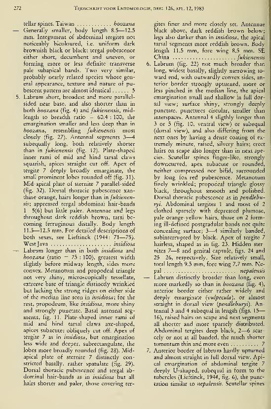
272 Tijdschrift voor Entomologie, deel 126, afl. 12, 1983
tellar spines. Taiwan hoozana— Generally smaller, body length 8.5—12.5
mm. Integument of abdominal tergites not
noticeably bicoloured, i.e. uniform dark
brownish black or black; tergal pubescence
either short, decumbent and uneven, or
forming more or less definite transverse
pale subapical bands. Two very similar,
probably nearly related species whose gen-
eral appearance, texture and nature of pu-
bescent pattern are almost identical 5
5. Labrum short, broadest and more parallel-
sided near base, and also shorter than in
both hoozana (fig. 4) and fukienensis, mid-
length to breadth ratio = 60.4 : 100, the
emargination smaller and less deep than in
hoozana, resembling fukienensis mostclosely (fig. 27). Antennal segments 3—
4
subequally long, both relatively shorter
than in fukienensis (fig. 12). Plate-shaped
inner rami of mid and hind tarsal claws
squarish, apices' straight cut off. Apex of
tergite 7 deeply broadly emarginate, the
small prominent lobes rounded off (fig. 31).
Mid-apical plate of sternite 7 parallel-sided
(fig. 32). Dorsal thoracic pubescence xan-
thine-orange, hairs longer than in fukienen-
sis; appressed tergal abdominal hair-bands
1—5(6) but little paler. Antennae and legs
throughout dark reddish brown, tarsi be-
coming ferruginous distally. Body length
11.5—12.5 mm. For detailed descriptions of
both sexes, see Lieftinck (1944: 71—75).
West Java insidiosa
— Labrum longer than in both insidiosa and
hoozana (ratio = 75 : 100), greatest width
slightly before midway length, sides moreconvex. Metanotum and propodeal triangle
not very shiny, microscopically tessellate,
extreme base of triangle distinctly wrinkled
but lacking the strong ridges on either side
of the median line seen in insidiosa; for the
rest, propodeum, like insidiosa, more shiny
and strongly punctate. Basal antennal seg-
ments, fig. 11. Plate-shaped inner rami of
mid and hind tarsal claws axe-shaped,
apices subacute: obliquely cut off. Apex of
tergite 7 as in insidiosa, but emargination
less wide and deeper, subrectangulate, the
lobes more broadly rounded (fig. 28). Mid-apical plate of sternite 7 distinctly con-
stricted basally, rather spatulate (fig. 29).
Dorsal thoracic pubescence and tergal ab-
dominal hair-bands as in insidiosa but all
hairs shorter and paler, those covering ter-
gites finer and more closely set. Antennaeblack above, dark reddish brown below;
legs also darker than in insidiosa, the apical
tarsal segments more reddish brown. Bodylength 11.5 mm, fore wing 8.5 mm. SEChina fukienensis
6. Labrum (fig. 22) not much broader than
long, widest basally, slightly narrowing to-
ward end, with outwardly convex sides, an-
terior border strongly upturned, more or
less pinched in the median line, the apical
emargination small and shallow in full dor-
sal view; surface shiny, strongly deeply
punctate, punctures circular, smaller than
interspaces. Antennal 4 sligthly longer than
3 or 5 (fig. 10, ventral view) or subequal
(dorsal view), and also differing from the
next ones by having a dense coating of ex-
tremely minute, raised, silvery hairs; erect
hairs on scape also longer than in next spe-
cies. Scutellar spines finger-like, strongly
downcurved, apex subacute or rounded,
neither compressed nor bifid, surrounded
by long fox-red pubescence. Metanotumfinely wrinkled; propodeal triangle glossy
black, throughout smooth and polished.
Dorsal thoracic pubescence as in pendlehu-
ryi. Abdominal tergites 1 and most of 2
clothed sparsely with depressed plumose,
pale orange-yellow hairs, those on 2 form-
ing ill-defined postgradular bands not quite
concealing surface; 3—4 similarly banded,
subinterrupted by black. Apex of tergite 7
hairless, shaped as in fig. 23. Hidden ster-
nites 7—8 and genital capsule, figs. 24 and
25—26, respectively. Size relatively small,
total length 9.5 mm, fore wing 7.7 mm. Ne-pal nepalensis
— Labrum distinctly broader than long, even
more markedly so than in hoozana (fig. 4),
anterior border either rather widely and
deeply emarginate (vulpecula), or almost
straight in dorsal view (pendleburyi). An-tennal 3 and 4 subequal in length (figs. 15
—
16), raised hairs on scape and next segments
all shorter and more sparsely distributed.
Abdominal tergites deep black, 2—6 scar-
cely or not at all banded, the much shorter
tomentum thin and more even 7
7. Anterior border of labrum hardly upturned
and almost straight in full dorsal view. Api-
cal emargination of abdominal tergite 7
deeply U-shaped, subequal in form to the
tubercles (Lieftinck, 1944, fig. 6), the punc-
tation similar to nepalensis. Scutellar spines
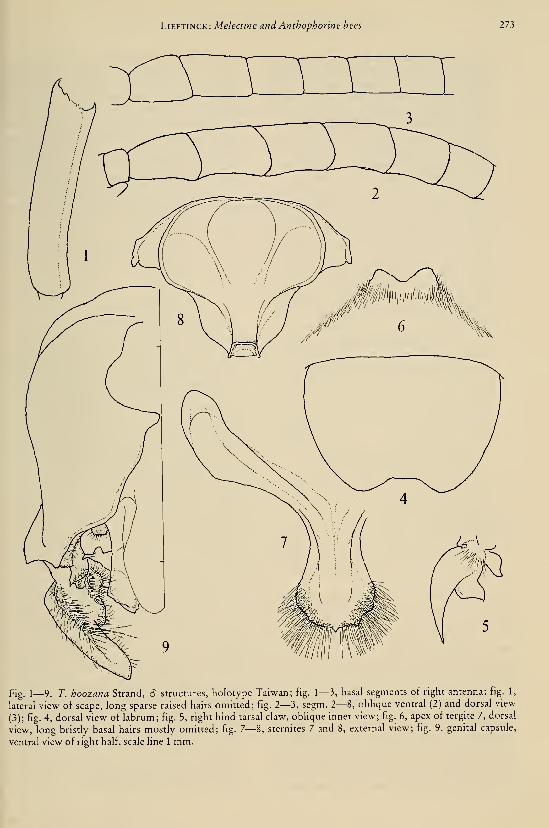
Lieftinck: Melectine and Anthophorine bees 273
Fig. 1—9. T. hoozana Strand, 6 structures, holotype Taiwan; fig. 1—3, basal segments of right antenna: fig. 1,
lateral view of scape, long sparse raised hairs omitted; fig. 2—3, segm. 2—8, oblique ventral (2) and dorsal view
(3); fig. 4, dorsal view of labrum; fig. 5, right hind tarsal claw, oblique inner view; fig. 6, apex of tergite 7, dorsal
view, long bristly basal hairs mostly omitted; fig. 7—8, sternites 7 and 8, external view; fig. 9. genital capsule,
ventral view of right half, scale Ime 1 mm.

274 Tijdschrift voor Entomologie, deel 126, afl. 12, 1983
robust, much shorter than in nepalensis, less
downcurved, apex laterally compressed,
distinctly bifid forming a pair of little
knobs. Metanotum and propodeum dullish,
finely tessellate, smooth, but much less shi-
ny than in nepalensis, base of triangle coar-
sely punctate on either side, these sclerites
separated from each other by a deep sulcus.
Dorsal thoracic pubescence sHghtly paler
than in vulpecula. Malay Peninsula
pendleburyi
— Anterior border of labrum abruptly up-
turned, the latter shaped much as in nepa-
lensis but longer, emargination distinctly
wider and deeper than in that species. Scu-
tellar spines still shorter than in pendlebu-
ryi, directed obliquely caudad and but
slightly downbent, apex somewhat com-
pressed and tapering, slanting and bluntly
pointed. Metanotum and propodeum muchas in pendleburyi, but basal area of triangle
coarsely longitudinally punctate along full
breadth of same. Dorsal thoracic pubes-
cence bright xanthine orange. Apex of ab-
dominal tergite 7 with shallow impunctate
median sulcus, otherwise black-haired,
emargination shallower and wider than in
both nepalensis (fig. 23) and pendleburyi
(fig. 37). Sumatra vulpecula
Tetralonioidella hoozana Strand
(figs. 1-9)
Tetralonioidella {"Tetralonia ? hoozana n.sp.") hoo-
zana Strand, 1914 (April/May): 139—141 {3 For-
mosa, Hoozan, ix.l910).
Protomelissa iridescens Friese, 1914 (1 June): 324 (c?
Formosa, Takao, Sept. 1908, H. Sauter). — Sand-
house, 1943: 592 (type-species of Protomelissa,
not seen). — Lieftinck, 1972: 272 (key), 277, 278
(orig. descr. & notes; type not seen). Syn. nov.
Type material. — 16 (terminalia dissected
out, glued on card and pinned on insect's pin),
labelled: ''Tetralonia (?) hoozana m. â Strand
det." (Strand's writing), "Hoozan Formosa H.Sauter x.lO" (print, date written), "TYP JS"
(typewritten on red), "Tetralonioidella hoozana
Strand HOLOTYPE (= Protomelissa iridescens
Friese), rev. MA Lieftinck 1979". Holotype âin the former Deutsch. Entom. Institut
(Eberswalde).
This is the first described and largest memberof the genus. It fits the existing amended diag-
noses of both Callomelecta and Protomelissa in
all important characters and now becomes the
type-species of the monobasic Tetralonioidella.
Male. — The uniform tint of the body pubes-
cence is longest, tufted, and deepest orangish on
the thoracic dorsum surrounding the practically
invisible mesoscutellar processes, these long
feathery hair-tufts being tipped with pale yel-
low. The neuration of the fore wing is much as
in the drawing given for T. pendleburyi (see
Lieftinck, 1972, fig. 11), i.e. the two proximal
sides of the 2nd submarginal cell are subequal in
length while the costal and anal sides of the lat-
ter are also about equally long though both are
slightly less than half as long as the proximal
sides.
The original description of T. hoozana is very
full, as far as it goes, and is best copied along
with the author's comments at full length, as
follows.
"Gen. Tetralonia Spin.
Tetralonia (?) hoozana Strd. n.sp.
Ein Ô von Hoozan IX.1910.
Dies Tier ist gewissermaszen ein Mittelding zwis-
chen Tetralonia und Anthophora. Eine Anthophorakann es aber nicht sein, weil die Ozellen in gerader
oder fast gerader Reihe angeordnet sind (bei Antho-phora bilden sie bekanntermaszen ein ausgespro-
chenes Dreieck)'). Für ein Tetralonia-yVinnchtn sind
aber die Antennen eigentlich viel zu kurz; schon da-
durch läszt die Art sich leicht von der sonst offenbar
sehr ähnlichen Tetr. himalayensis Rad. unterscheiden.
— Von Ancyla Lep. abweichend dadurch, dasz die
hinteren Metatarsen des 6 kaum so lang wie die fol-
genden Tarsenglieder zusammen sowie nicht ge-
krümmt sind, ferner ist der Clypeus vorstehend unddie Körpergrösze ist bedeutender als bei den bisher
bekannten Ancyla; soweit ohne Präparation erkenn-
bar, sind die Mundteile ziemlich kurz. — Das zweite
Geiszeiglied ist kaum so lang wie das dritte und auch
nicht dünner, das Tier somit, sowie durch die unter
sich weit entfernten und subparallelen Augen von
Meliturga Latr. leicht zu unterscheiden.
Es ist ganz wahrscheinlich, dasz für dies Tier eine
neue Gattung aufgestellt werden musz (die eventuell
den Namen Tetralonioidella m. bekommen möge);
um dies mit Sicherheit zu entscheiden, wäre aber die
Kenntnis auch des 9 eigentlich nötig. Aber auch
wenn dies eine typische Tetralonia sein sollte, dürfte
die neue Benennung als Untergattungsname verwend-
bar sein.
Schwarz; Mandibeln leicht gebräunt in der Mitte,
') Anm. Die von Friese in: Verh. zool. bot.Ges. Wien
1911, p. 27, beschriebene Anthophora Sauteri n.sp.
ist eher zu Tetralonia zu stellen, weil die Ocellen,
wenigstens beim 9, kein Dreieck bilden. In meiner
Besprechung der Art im I. Teil dieser Arbeit (in:
Supplem. Entomol. II, p. 51) habe ich leider unter-
lassen, dies Moment hervorzuheben.
Lieftinck: Melectine and Anthophorine hees 275
Fig. 10— 16. Tetralonioidella species, partial view of right and left ô antennae; fig. 10, T. nepalensis spec, nov.,
Nepal, right segm. 1—8 showing plainly visible 2nd segment, long sparse hairs at scape omitted; fig. 11, T. fu-
ktenensis spec, nov., SE China, dorsal view of right segm. 1—7, same details; fig. 12, T. insidiosa (Lieft.), W.Java, right segm. 2—8, same scale; fig. 13— 14, T. himalayana (Ckll.), Bhimtal, antero-ventral view of left segm.
2—13 and 1—7, lateral view, showing retracted 2nd segment (fig. 14 more enlarged, long silky hair-tuft at scape
omitted); fig. 15. T. vulpecula (Lieft.), S. Sumatra, right antennal segm. 2—6, showing cup-shaped 2nd segment;
fig. 16, T. pendleburyi (Ckll.), Larut Hills, Perak, the same of segm. 2—7 showing slightly shorter 2nd segment.
276 Tijdschrift voor Entomologie, deel 126, afl. 12, 1983
Labrum an der Basis beiderseits gerötet, Fühlergeiszel
unten schwach gebräunt, Tegulae bräunlichgelb, der
hintere Hälfte der Abdominalsegmente gerötet, die
Tarsen leicht gebräunt. Flügel gleichmäszig schwach
angeraucht und etwas gelbhch schimmernd, Geäderund Mal braunschwarz.
Der ganze Kopf hell graugelblich behaart und zwarauch auf dem Labrum lang abstehend, auf dem Cly-
peus auszerdem mit anliegender Behaarung. Thoraxsehr dicht und lang ziemHch abstehend behaart undzwar auf dem Rücken orangegelblich, an den Seiten
und unten heller behaart; die Skulptur daher nicht er-
kennbar. Rückensegmente des Abdomen mit feiner,
kurzer, anliegender, nicht dichter, goldgelbener Be-
haarung, die auf der helleren Hinterhälfte der Seg-
mente kaum heller als auf der basalen ist, als Gesamt-eindruck heben die helleren Binden sich daher wenigvon der Grundfarbe ab. Die Bauchsegmente verhalten
sich wie die Rückensegmente, jedoch ist ihre Behaa-
rung in der Endhälfte leicht abstehend und die Seg-
mente II und III zeigen auszerdem eine schmale helle
Basalhaarbinde. Die Beine kurz und spärlich messing-
gelblich behaart.
Kopf schmäler als Thorax, aber breiter als lang, mit
groszen, vorstehenden, subparallelen (nach unten
ganz schwach konvergierenden), innen nicht ausge-
randeten Augen und stark vorstehenden, vorn mitten
jedoch abgeflachtem Clypeus, dessen Kontur, in An-sicht von der Scheitel, fast trapezförmig erscheint, imProfil scheint die Vorderfläche des Clypeus mit demLabrum fast einen rechten Winkel zu bilden. Letz-
teres beiderseits an der Basis höckerig, am Vorder-
rande (im Profil gesehen) leicht vorstehend. Die An-tennen überragen die Mitte des Mesonotum, die Geis-
zel zylindrisch, nur das erste und die Basis des
zweiten Gliedes etwas dünner als die übrigen; das
zweite Geiszelglied ist etwa dreimal so lang wie das
erste. Abdomen erscheint in Draufsicht an der Basis
breit quergeschnitten, am Ende zugespitzt, das letzte
Tergit an der Spitze mitten leicht eingeschnitten. —Die dritte Kubitalzelle ist oben (vorn) so breit wiehinten, die zweite Kubitalquerader ist kurz unterhalb
der Mitte stark saumwärts konvex gebogen, die
zweite Kubitalzelle ist auf der Radialader nur halb so
lang wie auf der Kubitalader. Körperlänge 13.5,
Flügellänge 19 mm".
Protomelissa iridescens Friese,— a dubious
synonym?
For a discussion of this puzzling species, see
the above citations, one of which includes a
transcription of the original diagnosis. Expect-
ing the type not to turn up any more in someEuropean museum or private collection, it mustbe regarded as lost or destroyed. The locality
labels and dates of capture of the types of T.
hoozana and iridescens are quite different, so
that the chances are very slight that Strand andFriese, at a time when both authors werestudying bees from "Sauter's Formosa Ausbeu-te", did come across the same specimen andbased their descriptions successively on this oneindividual. On the other hand, with the recent
discovery of T. hoozana, one would be inclined
to think iridescens and hoozana are identical
species, because the probability of more than
two taxa of this rare genus occurring together in
such a small island as Taiwan, seems unlikely.
Yet the obvious discrepancies in structure pres-
ently observed (table 1, especially points 3 and
4) in the types (if correctly stated for irides-
cens\), should not be neglected. Despite the in-
congruities I have, with all reserve, decided to
synonymize these two Taiwanese species.
Tetralonioideila formosana (Cockerell) comb,nov.
Melecta formosana Cockerell, 1911: 227, 228 (9 Ko-sempo, Formosa).
Anthophora sauteri Friese, 1911: 127, 128 {6 Tainan,
Formosa).
Protomelissa bauten; Friese, 1914: 323, 324 ((5 9 Tai-
nan & Takao, Formosa).
Protomelissa formosana; Lieftinck, 1972: 273 (key),
27'^1—277, figs. {Ô9 Kosempo, Tainan & Takao;
orig. & further descr., synon. & full refs.).
Table 1. Differences in structure in the types of T. iridescens and T. hoozana.
T. iridescens T. hoozana
1
.
Abdomen dull and with slight metallic sheen
("Abdomen mit Erzglanz", and "fast matt, schwacherzfarben schillernd");
2. Labrum "viereckig, vorn schwach ausgerandet"
3. Antenne rot . . . 2tes Geisselglied (segm. 3) viel
kürzer als 3"
4. Apex of tergite 7 small and truncated ("klein undabgestutzt")
5. Body length 13 mm, width 4.5 mm
1
.
Abdomen somewhat shiny but lacking any
indication of metallic gloss
2. Labrum shaped as in fig. 4
3. Antenna yellow-brown in front, dark brownbehind, segm. 3 and 4 subequal in length (figs. 2, 3)
4. Apex of tergite 7 distinctly excised (fig. 6)
5. Body length 13.5 mm, width 4.5 mm
LiEFTiNCK: Melectine and Anthophorine bees 277
Tetralonioidella himalayana (Bingham) comb.nov.
(figs. 13, 14)
Melecta himalayana Bingham, 1897: 516, fig. 172, in-
sect, with left wing (9 "Kumaon, 5000 ft., and
probably throughout the Himalayas at and above
that altitude".
Protomelissa himalayana; Lieftinck, 1972: 273 (key),
274, figs. 3, 7, 8 (addit. notes & 6 struct., cJ 9 N& NE India).
Additional material. — Series of 12 d, NW India,
U.P., Bhimtal near Nainital, 1500 m, 13, 14, 15 and
18.x. 1978, M. A. Lieftinck, all at flowers of Labiate
shrub Plectranthus coetsa Ham. ex D. Don. (local
name "bhanira").
The above Bhimtal males are freshly emerged
specimens in perfect condition. They were tak-
en within a week's time, either in the early
morning hours around 8.30 a.m., or on a return
visit at 17 p.m. (4 on Oct. 14, 5 on Oct. 15, most
of the remainder solitarily). All were attracted
by flowers of the same plant, which grew in
threes or fours at some distance from each other
along a shaded forest path at the slope of a rav-
ine. The bees flew in company with both sexes
of 5 species of Habropoda, viz. H. apostasia m.,
deiopea (Cameron), hooken Cockerell, pelmata
m., and radoszkowskii (Dalla Torre), the latter
being by far the most common of these; fe-
males, besides taking nectar, assembled pollen at
their hind tibiae and basitarsi, apparently also
from Plectranthus. Their high-pitched notes
could be heard already at about three yard's dis-
tance. I had asked Mr. Fred Smetacek, the own-er of a small undisturbed patch of forest in the
hills above Bhimtal, to keep an eye on the flow-
ering season of Plectranthus, in 1979 and 1980.
He kindly informed me in his letters that, un-
fortunately, no single inflorescence of plants
could be found by him in any part of the same
area during these next two years.
As pointed out by me earher (1972: 273
—
277, sub Protomelissa), himalayana comes so
close to formosana in stature, pubescent colour-
pattern and other bisexual characters, that I
have failed to discover more features than those
already known for their separation. Nothing
definite is known of their host relations, but
both were associated in the field with Habropo-da species, not with those of Elaphropoda as
were other members of Tetralonioidella.
Tetralonioidella tricolor (Lieftinck) comb. nov.
Protomelissa tricolor Lieftinck, 1972; 273 (key), 278
—
281. fig. 4 (labrum; 9 Assam, Shillong; notes &host relations).
The 9 holotype in the Berlin museum is still
the only specimen so far known. By the absence
of a (5, nothing definite can yet be said about
the nearest relatives of tricolor, but as stated in
the original description, we may expect it to
come closest to himalayana and formosana. Thepresence of a conspicuous dense brush-like
patch of long hairs in front of the antennal scape
of the â (loc. cit.: 273), is a unisexual character,
so that my subsequent remark (loc. cit.: 278) onits absence in the 2 as a point of distinction be-
tween species-groups, is misleading.
Tetralonioidella habropodae (Cockerell) comb.nov.
(figs. 17-21)
Callomelecta habropodae Cockerell, 1929: 133. — 6"Siam: Doi Sutep, on summit, Feb. 9, 1928 (Cock-
erell)". — Lieftinck, 1944: 62, 75, footnote (origi-
nal material not seen).
Protomelissa habropodae; Lieftinck, 1972: 270 (key),
281 (orig. descr. copied, with notes; original
material not seen).
Type material. — \ 6 (holotype), labelled:
"Doi Setep Siam feb. 9 Alice Hacker" (?) in un-
known writing; and "Callomelecta habropodae
Ckll. TYPE", in T.D.A. Cockerell's hand; and,
"ace. 35740" (AMNH label). Holotype in
Amer. Mus. Nat. Hist., New York.
The more interesting characters of this re-
markable little species are summarized in the
preceding key. T. habropodae is known only
from the unique holotype 6 . In addition to the
key notes and the original description, copied in
full on a previous occasion (Lieftinck, loc. cit.
1972: 281), most of the hitherto unnoticed pe-
culiarities of structure are here also illustrated
(figs. 17-21).
Apart from its smallish size and the predomi-
nantly orange-brown colour of the legs and ab-
domen, the species is easily distinguished from
all allies by a combination of the following char-
acters: dense brush-like hair-tuft in front of the
antennal scape; characteristic shape of the midand hind tarsal claws; the unarmed mandibles;
and the superficially punctate shiny surface of
the first two abdominal tergites. Disk of labrum
(fig. 19) slightly concave, rather shiny, rugosely
punctate, colour yellow-brown, its anterior
border narrowly ferruginous, long hairs semi-
erect, pale yellow, the border fringed with a
278 Tijdschrift voor Entomologie, deel 126, afl. 12, 1983
Fig. 17—21. T. habropodae (CklI.), Thailand, â holotype Doi Sutep; fig. 17—18, left mandible, inside and exte-ro-lateral view; fig. 19, dorsal view of labrum; fig. 20, right antennal segm. 1—9, showing long silky hair-tuft at
scape and partly retracted 2nd segment, inner (ventral) view; fig. 21, apex of tergite 7, dorsal view, most longishappressed hairs omitted. — Fig. 22—26. T. nepalensis spec, nov., â holotype Nepal; fig. 22, dorsal view of la-
brum; fig. 23, apex of tergite 7, pardy lacking bristly hairs; fig. 24, sternites 7 and 8, external view; fig. 25—26,.genital capsule, dorsal and ventral view, scale line 1 mm.
Lieftinck: Melectine and Anthophorine hees 279
Fig. 17—30. T. fukienensis spec, nov., 6 holotype SE China; fig. 27, dorsal view of labrum; fig. 28, apex of ter-
gile 7, long depressed silky hairs mostly omitted; fig. 29, sternites 7 and 8, external view; fig. 30, genital capsule,
dorsal and vertral view, scale line 1 mm.
280 Tijdschrift voor Entomologie, deel 126, afl. 12, 1983
row of stiff, parallel, pale golden bristles which
are somewhat downcurved. Mandibles smooth,
almost impunctate, pale brown, the apex
obscured, almost black. Anterior rim of clypeus
strong, ferruginous. The neuration of the
strongly brownish amber-coloured fore wings
is practically identical with that of T. pendlebu-
ryi (Lieftinck, 1944, fig. 1; and id., 1972, fig.
11). Abdominal tergite 7 (fig. 21) almost flat,
surface densely rugoso-punctate, interspaces
somewhat shiny, hairs pale, depressed. Bodylength 9.5 mm approx., fore wing 6.8 mm.
T. habropodae is undoubtedly most closely
related to himalayana a.nd formosana. It is of in-
terest to note that in Thailand the species was
found flying in company with its supposed host
bee, Hahropoda sutepensis Cockerell (for the
latter, see Lieftinck, 1974: 198, 199, figs.). Aswe now know, sutepensis is a near ally of H.apostasia Lieft., which in N. India occurred to-
gether with Tetralonioidella himalayana.
Tetralonioidella nepalensis spec. nov.
(figs. 10,22—26)
Type material. — 2 6 (terminalia of holotype
dissected out, in pinned plastic capsule), E Hi-
malayan Range, Nepal, Kathmandu, Godavari,
5000 ft., 6.viii. 1967, Canad. Nepal Exped. (ho-
lotype); and same loc, 6000 ft., 24 July 1967,
same coll. (paratype). Holotype S in Biosyst.
Res. Inst., Ottawa (Ontario); paratype S in
Mus. Leiden.
Male. — Characters as given in the key and as
shown in the figures.
This is the third of a small group of slender
species characterized by a bicoloured pubescent
body pattern: a long and dense fox-red vestiture
covering the summit of head and all of the tho-
racic dorsum and sides, contrasting with an al-
most black unhanded abdomen. Differs fromthe Malaysian pendleburyi and vulpecula in the
paler colour of the very short appressed hairs onparts of the abdominal tergites. In vulpecula
these short .hairs are almost black and evenly
distributed, in the other two lighter and mostmarked at the postgradular lines, especially so in
the present new species.
In the type the yellowish brown postgradular
hairs on tergites 1—4 are broadest and longest
at the sides of 1, more or less broadly interrupt-
ed by dark brown in the median line on tergites
2—4. The paratype differs from the type in that
the pale hairs on 1 cover most of the surface and
are evenly distributed, whereas on 2—4 they are
somewhat more crowded together, forming in-
conspicuous but uninterrupted bands. Thesecomplete bands are wholly absent also in
pendleburyi.
Tetralonioidella vulpecula (Lieftinck)
(figs. 15,37—43)Callomelecta vulpecula Lieftinck, 1944: 68—71 &
75—77, pi. & figs. (<? 9 Sumatra).
Protomelissa vulpecula; Lieftinck, 1972: 272 (key),
282, pi. 1 fig. 2 (c? 9 Sumatra, refs.).
No additional material.
Tetralonioidella pendleburyi (Cockerell) comb,nov.
(fig. 16)
Callomelecta pendleburyi Cockerell, 1926: 621, 622
(9 Malaya, Selangor). — Lieftinck, 1944: 62—68,figs. 1—9 & 75—77 (incl. keys 9 S and descr. al-
lotype 6 , Perak and Pahang, Malaya).
Protomelissa pendleburyi; Lieftinck, 1972: (key), 282,
figs. 9—16 {Ô 9 Malaya, full refs.).
No fresh material. For structural details of
the only known S of this species, see Lieftinck,
1944 & 1972. On p. 62 line 17 in my 1944 pa-
per, "9 allotype" should be altered in "6 allo-
type".
Tetralonioidella fukienensis spec. nov.
(figs. 11,27—30)
Type material. — 1 6 (terminalia dissected
out, in pinned plastic capsule), SE China, NWFukien, Chungan distr., Bohea Hills, 600 m,25.ix.1939, T. C. Maa. Holotype 6 in Mus. Lei-
den.
Male (slightly discoloured, but otherwise in
fair condition). A species sufficiently character-
ized in the key. Superficially most closely re-
sembling T. insidiosa, from West Java, but easily
distinguished therefrom by comparing the
structural illustrations here given for both. Dif-
ferences not mentioned in the key are that fu-kienensis, besides being slightly larger in size, is
decidedly a more robustly built species than in-
sidiosa, especially the mid and hind femora and
tibiae oi fukienensis being markedly more swol-
len (expanded) about midway their length, than
in the slenderer insidiosa.
Tetralonioidella insidiosa (Lieftinck) comb.
nov.
(figs. 12,31—36)Callomelecta insidiosa Lieftinck, 1944: 71—75 &: 75
—
77,pl.&figs. ((5 9 Java).
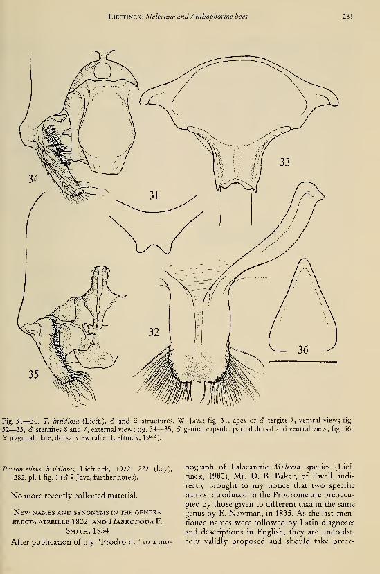
Lieftinck: Melectine and Anthophorine bees 281
Fig. 31—36. T. insidiosa (Lieft.), ô and 5 structures, W. Java; fig. 31, apex of 3 tergite 7, ventral view; fig.
32—33, S sternites 8 and 7, external view; fig. M—35, â genital capsule, partial dorsal and ventral view; fig. 36,
9 pygidial plate, dorsal view (after Lieftinck, 1944).
Protomelissa insidiosa; Lieftinck, 1972: 272 (key),
282, pi. 1 fig. 1 (<? 9 Java, further notes).
No more recently collected material.
New names and synonyms in the genera
ELECTA ATREILLE 1 802, AND HaBROPODA F.
Smith, 1854
After publication of my "Prodrome" to a mo-
nograph of Palaearctic Melecta species (Lief-
tinck, 1980), Mr. D. B. Baker, of Ewell, indi-
rectly brought to my notice that two specific
names introduced in the Prodrome are preoccu-
pied by those given to different taxa in the same
genus by E. Newman, in 1835. As the last-men-
tioned names were followed by Latin diagnoses
and descriptions in English, they are undoubt-
edly validly proposed and should take prece-
282 Tijdschrift voor Entomologie, deel 126, afl. 12, 1983
dence over those accidentally also chosen by
myself. By an unfortunate oversight, Newman's
introduction and diagnoses escaped my atten-
tion, while his names (extracted from Dalla
Torre's catalogue) were inadvertently consid-
ered nomina nuda and therefore left out of con-
sideration in my Prodrome. As a matter of fact,
on pp. 513, 514 of his Entomological Notes
(Newman, 1835), the author characterized six
British "species" of Melecta, which he all
named. Two of them were described as M. alec-
to and M. megaera, both from localities in
southern England and admittedly regarded by
Newman himself as mere variants of the com-
mon Melecta punctata (Fabricius, 1775), a spe-
cies now properly called M. alhifrons (Forster,
1771)^). The four remaining specimens of Me-
lecta, all of them collected in England, were de-
scribed in the same way to the former, receiving
the names M. atropos, clotho, lachesis, and tisi-
phone. Although the validity of this nomencla-
ture, like that of the other two, can not be called
in question and should be borne in mind by fu-
ture workers on the genus, they are of no con-
cern in the present context.
') In view of the many so-called "Spielarten" occur-
ring in the entomological literature of the past, I amtempted to quote certain passages in Newman's
cheerful introduction to the genus Melecta preced-
ing the definition of his novelties, — contempla-
tions which may give an idea of his and some con-
temporary authors' appraisal of specific names and
the valuation of nomenclatural problems in gener-
al: — "It is pleasant to create a smile; and I anti-
cipate that many smiles will be accorded me when I
coolly assure your readers, that I am going to make
six distinct species out of Melecta punctata, and
that I cannot, for the life of me, tell to which of
these the name punctata properly belongs, and
therefore have given it to neither. It is pleasant to
see one's new species given, without comment, as
synonyms; and when this happens, and happen it
certainly will to my Melectae, I hope I shall take it
as good-humouredly as Mr. Waterhouse did, when
he beheld his fourteen new Notiophili consigned to
utter oblivion". "In these cases there is this com-
fort, that if the new-made species are really species,
they will in the course of time be re-admitted: al-
low a year for each really new species parted from
an old one; then the Notiophili will be re-admitted
by the year 1847, and the Melectae — for I reckon
them already struck out— by the year 1 84 1"
.
Indeed, as time went on, re-adjustments in the
nomenclature became almost prevailing topics of
action, — though not always exactly in the wayEdward Newman had predicted!
Summarizing the above, it will be clear that
the two specific names M. alecto Lieftinck and
megaera Lieftinck, are preoccupied and should
be substituted by new names, as follows:
M. diligens nom. nov. pro M. alecto Lief-
tinck, 1980: 164, 186 (keys) & 273 (nom. pre-
occ). — Holotype S , Iraq, in Brit. Mus. (Nat.
Hist.), London.
M. mundula nom. nov. pro M. megaera Lief-
tinck, op. cit.: 175, 185 (keys) & 243 (nom. pre-
occ). — Holotype â, Cyprus, in Mus. f. Na-turk., Berlin DDR.
In the same work (1980), the following cor-
rections should be made: p. 140 line 12, the
wording "specific o/ varietal" should read "spe-
cific or varietal"; p. 290, line 11 from bottom,
"figs. 231—232" should read "fig. 231"; p. 291,
in the explanation of figs. 231—236, the word"antenna" should, of course, be replaced by"maxillary palpus", shown in fig. 232.
I shall gratefully accept any information
about further errors and omissions occurring in
this publication.
In the genus Hahropoda F. Smith, the follow-
ing case of synonymy could be established:
Hahropoda apatelia Lieftinck, 1974: 171, 178 (keys),
215, 216, figs. Ô 9 Darjeeling, Syn. nov.
This taxon proves to be conspecific with the
long-lost H. krishna Bingham, 1909 (Ree. Ind.
Mus. 2: 366—367, S Darjeeling 7000 ft.). Con-firmed after direct comparison of morphologyand internal structures of the well-preserved
holotype of krishna (still in coll. Zool. Survey of
India, Calcutta) with topotypical holotype of
apatelia in the Brit. Mus. (Nat. Hist.) and para-
types in the Leiden Museum.
Acknowledgements
My best thanks are due to a number of ento-
mologists who, through the years, kindly com-plied with my requests to recover certain melec-
tine types and other valuable anthophorid speci-
mens in various museum collections abroad.
They kindly arranged for long-term loans to be
made, and for waiting patiently for the ultimate
return of specimens under their care: Messrs.
H. Fankhänel (Leipzig, DDR), and G. Morgeand J. Oehlke (both D.E. I., Eberswalde), for
the loan of the long-concealed type of Tetralo-
moidella ? hoozana Strand. I am also grateful to
J. G. Rozen Jr. (Amer. Mus. Nat. Hist., New

Lieftinck: Melectine and Anthophorme bees 283
Fig. 37—43. T. vulpecula (Lieft.), 6 and $ structures, S. Sumatra; fig. 37, apex of S tergite 7, ventral view; fig.
38—39, 6 sternites 7 and 8, external view (sg slightly more downpressed); fig. 40, right lateral view of distal
portion of genital capsule; fig. 41—42, apices of 6 genital capsule, dorsal and ventral view; fig. 43, 9 pygidial
plate, dorsal view (after Lieftinck, 1944).
York) for the type loan of Callomelecta habro-
podae Ckll.; to J. R. Barron, J. E. R. Martin and
W. R. M. Mason (Biol Res. Sta., Ottawa, Ont.),
for specimens collected by members of the Ca-nadian Nepal Expedition 1967, which included
the type of Tetralonioidella nepalensis spec,
nov.; and to T. C. Maa (Taichung, Taiwan), for
his great interest in the cuckoo bees of the pre-
sent genus and their hosts, as well as for numer-
ous Anthophoridae assembled by him in Fukien
during the first years of World War II, on
which occasions the type of T. fukienensis spec,
nov. was discovered. Lastly, I am mdebted to L.
B. Holthuis (Leiden), who found out the exact
dates of issue of some articles, which appeared
almost simultaneously in different European
journals.
284 Tijdschrift voor Entomologie, deel 126, afl. 12, 1983
Selected references
Cockerell, T. D. A., 1911. Descriptions and records
of bees. 34. — Ann. Mag. Nat. Hist. (8) 7: 225—
227.
, 1926. Descriptions and records of bees. 113. —Ann. Mag. Nat. Hist. (9) 18: 621, 622 (part.).
, 1929. Descriptions and records of bees. 117. —Ann. Mag. Nat. Hist. (10) 4: 132, 133 (part.).
Friese, H., 1911. Neue Bienenarten von Formosa und
aus China (Kanton). — Verh. zool. -bot. Ges.
Wien 59: 127, 128 (part.).
, 1914. Neue Bienenarten der orientalischen Re-
gion.— Deutsch. Ent. Zeitschr.: 322—324 (part.).
Lieftinck, M. A., 1944. Some Malaysian bees of the
family Anthophoridae (Hym., Apoidea). — Treu-
bia (Buitenzorg), hors sér.: 57—138, 79 figs., pi.
42.
, 1972. Further studies on Old World melectine
bees, with stray notes on their distribution and
host relationships (Hym., Anthophoridae). —
Tijdschr. Ent. 115: 253—322, 2 pis., 57 figs., 1 tab.
& 2 maps. (With full bibliography).
, 1974. Review of Central and East Asiatic Ha-bropoda F. Smith, with Hahrophorula, a new ge-
nus from China (Hym., Anthophoridae). —Tijdschr. Ent. 117: 157—224, figs. 1—100.
, 1980. Prodrome to a monograph of the Pal-
aearctic species of the genus Melecta Latreille
1802 (Hym., Anthophoridae). — Tijdschr. Ent.
123: 129—349, text-figs. 1—359, pis. 1—8.
Newman, E., 1835. Entomological Notes. In: The
Entomological Magazine, London 2 (5): 512—514
(part.: Melecta).
Sandhouse, G. A., 1943. The type species of the gen-
era and subgenera of bees. — Proc. U.S. Nat.
Mus. 92:519—619.Strand, E., 1914. H. Sauter's Formosa Ausbeute. Api-
dae. III. — Arch. f. Naturgesch. 80 A (1): 139—141 (part.).
3478 034


TIJDSCHRIFT VOOR ENTOMOLOGIE
UITGEGEVEN DOOR
DE NEDERLANDSE ENTOMOLOGISCHE VERENIGING
REGISTER VAN DEEL 126
'" Een sterretje duidt aan een naam nieuw voor de wetenschap"' An asterisk denotes a name new to science
Of the contribution by R. de Jong — Annotated Ust of the Hesperiidae of Surinam
species and subspecies only those of the taxa described as new are included.
of the of
AMPHIBIARana esculenta 75
AVESApus apus 75
Chlidonias niger 75
Delichon urbica 75
Fulica atra 83
Hirundo rustica 75
Sterna hirundo 75
COLEOPTERAapterus, Lethrus 205, 226, 227
biostius, Typhaeus 205
crassipes, Donacia 83
daphnis, Phanaeis 226
egeriei, Geotrupes 205
farctum, Bolboceras 205
ferrugineus, Bradycinetulus 205
fossor, Typhaeus 206
gaigei, Mycotrupes 205
hiostius, Typhaeus 206, 211,
[226, 227
hispanus, Copris 226
hornii, Geotrupes 205
lateridens, Typhaeus 206
momus, Typhaeus 205, 206
mutator, Geotrupes 205, 226
nigroaeneus, Kheper 226
nymphaeae, Pyrrhalta 83
pomorum, Anthonomus 111
retusus, Mycotrupes 205
Sisyphus 226
spiniger, Geotrupes 205, 266
[sqq
stercorarius, Geotrupes 205,
[226 sqq
stercorosus, Geotrupes 205
typhoeoides, Typhaeus 206
typhoeus, Typhaeus 203 sqq
vernalis, Geotrupes 205, 226
youngi, Peltotrupes 205
DICTYOPTERAamericana, Periplaneta 227
DIPTERAAgromyza 12
Amaurosoma 10
aquilegiae, Phytomyza 8
bellidina, Phytomyza 8
brunnipes, Notophila 70, 87
Cerodontha 12
confluens, Hydromyza 59 sqq
flavipennis, Pycnoglossa 7
griseola, Hydrellia 70
heraclei, Phihphylla 14
Liriomiza 1
1
livens, Hydromyza 59 sqq
minuscula, Phytomyza 8
Pegomya 10
Phytomyza 11, 12
rosae, Psila 14
sylvestris, Cricotopus 72
trifasciatus, Cricotopus 72
Trilobomyza 11, 12
GASTROPODALymnaea stagnalis 83
HYDROZOACordylophora caspia 89
HYMENOPTERAacalle, Sympiesis 129
Acrolytall2, 132
Adelura 4
Adeshal75, 176
'•Adeshoides 175
albifrons, Melecta 282
albitarsis, Euderus 129
albolineata, Adesha 177
alecto, Melecta 282
alternans, Itoplectis 111, 129,
132
americana, Orthostigma 13
amplidens, Adelurola 2, 5
angustula, Pentapleura 14
anxium, Agrypon 118, 130, 132
apafelia, Habropoda 282
Apechthis 129
"apheles, Gnaptodon 26, 28, 29
apii, Dapsilarthra 3, 13
Apophuall2, 114
apostata, Diadegma 117, 130,
[132
arenicola, Tranosema 117, 130,
[132
asiatica, Adelurola 2, 6
"asulcatus, Adeshoides 176
ater, Apantelesll2, 118, 130,
[132
atropos, Melecta 282
bachmaieri, Gnaptodon 25, 31
balteata, Dapsilarthra 2, 8
barthii, Dapsilarthra 14
barthii, Phaenocarpa 14
bini, Gnaptodon 39
boozana, Tetralonioidella 269,
[272, 274
Braconl26, 132
brevicornis, Scambus 110, 127,
[130,132
breviradialis, Gnaptodon 28, 30
'•"brevis, Gnaptodon 28, 30
Callomelecta 270
Campoplex 130, 132
canaliculatus, Habronyx 118,
[130,132
carbonarius, Microgaster 120
carinatus, Plesiobracon 186
='-Carinibraconl85, 186, 187
'"carpathica, Dapsilarthra 3, 12
cavus,Dibrachys 127, 131,132
chrysos, Habrocytus 127, 132
cicatricosa, Apophua 114, 129,
[132
claripennis, Goniozus 129
285

286 Tijdschrift voor Entomologie, deel 126, 1983
clotho, Melecta 282
complicator, Lissonota 116,
[130,132
compuctor, Apechthis 1 12, 132
corvina, Dolichogenidia 120,
[131,132
cruentatus, Charmon 126, 131,
[132
dalhousiensis, Dapsilarthra 2, 10
danielssoni, Carinibracon 187
Dapsilarthra 6, 10
decoris, Gnaptodon 25, 29, 31
deserticola, Victoroviella 179
Diadegma 127
'•"diligens, Melecta 282
dictynna, Dapsilarthra 2, 7
difformis, Compoplex 117, 132
dilecta, Dolichogenidia 120,
[130,132
dimidiator, Agathis 122, 130,
[132
Diraphus 27
"erasmi, Gnaptodon 29, 32
errabunda, Lissonota 116
Exochusll8, 129, 132
fenestralis, Diadegma 117, 130,
[132
flaviventris, Alysia 1
1
fiorimela, Adelurola 2, 5
florus, Colpoclypeus 111, 127,
[131,132
formosana, Tetralonioidella
[269,271,274
fractigena, Glypta 116
fukiensis, Tetralonioidella 272,
[280
fuscula, Dapsilarthra 1
1
gahani, Dapsilarthra 2, 7
Gelisll2, 132
'•"georginae, Gnaptodon 29, 33
glaber, Gnaptodon 29
globulipes, TricHstus 117, 118,
[129, 132
Glypta 112
Gnamptodon 6
gracilicornis, Alysia 11
Grammospila 6
grandidieri, Malagopsis 177, 181
Habrocytus 131
habropodae, Tetralonioidella
[271,275
horstoki, Deltaphyma 182
ictericus, Meteorus 117, 123,
[131,132
indagatrix, Dapsilarthra 2, 9
insidiosa, Tetralonioidella 272,
[274,280
interrupta, Diadegma 116
iridescens. Protomelissa 270
isabella, Dapsilarthra 3,11
Kenemal86, 188
klemensiewiczii, Graptodes 25,
[31
krishna, Habropoda 282
lachesis, Melecta 282
laevigata, Dolichogenidia 120,
[130,132
laevipectus, Alysia 13
lanceolator, Lasiophorus 180
Lasiophorus 179
levisulca, Dapsilarthra 2, 8
linearis, Macrocentrus 126, 131,
[132
lineatus, Stictopisthus 117, 132
liogaster, Zanzopris 183
Lissogaster 122, 130, 132
longicaudus, Dolichogenidia
[120,130,132
maculator, Itoplectis 111, 130,
[132
megaera, Melecta 282
Malagopsis 181
Mesochorus 130
Mesocrina 6, 8
minutus, Oncophanes 123, 127,
[131,132
mongolica, Gnaptogaster 26
multiarticulata, Phaenocarpa 5
'•mundula, Melecta 282
nana, Lysibia 112, 132
Neocarpa 4
nepalensis, Tetralionioidella
[272,280
nepalicus, Gnaptodon 29, 34
nepticulae, Gnaptodon 29
'•'nieukerkeni, Gnaptodon 29, 34
nigrina, Glypta 114, 129, 132
nowskowskii, Dapsilarthra 7
obscurator, Bracon 126, 131,
[132
ochrogaster, Phaenocarpa 1
1
"orientalis, Gnaptodon 40
pallipes, Macrocentrus 127, 131,
[132
pallipes, TricHstus 117, 118,
[129,132
Paraorthostigma 6
pectinicornis, Pnigalio 127, 131,
[132
pedata, Glypta 116
pendleburyi, Tetralonioidella
[274,280
pentapleuroides, Dapsilarthra 5
'pilosus, Gnaptodon 28, 34
Plaxopsis 184
Plesiobraconl85, 186
praerogator, Diadegma 116,
[130,132
Protomelissa 269
Pseudomesocrina 6, 8
pulchrigaster, Gnaptodon 29
pumilio, Gnaptodon 29, 35
punctata, Melecta 282
pygmaeus, Diraphus 25, 35
quadridentata, Ascogaster 123,
[130, 132
quadridentatus, Apechthis 112,
[132
quickei, Kenema 188
resinator, Apechthis 112
rufatus, Apechthis 1 12, 132
•'ruficeps, Gnaptodon 29, 37
rufidens, Ascogaster 123, 130,
[132
rufipes, Agathis 122, 130, 132
rufiventris, Dapsilarthra 3,11
rufiventris, D., f. fuscula 3, 11
sauteri, Anthophora 269, 270
segmentator, Phytodietus 1 12,
[130,132
semotus, Habrocytus 127, 132
sertifer, Neodiprion 111
servillei, Bracon 183
seyrigi, Malagopsis 182
sicaria, Dolichogenidia 120,
[130,132
silvarum, Mesochorus 117, 132
"simiUs, Gnaptodon 39, 40
sjoestedti, Plaxopsis 185
striata, Teleutaea 112, 113, 129,
[132
subtilis, Dapsilarthra 2, 8
subtilis, Heterolexis 1
sulcifera, Dapsilarthra 14
Sylvia, Dapsilarthra 3, 12
tenuicornis, Dapsilarthra 14
Tetralonia 269, 274
Tetralonioidella 269, 270
testacea, Dapsilarthra 8
thienemanni, Porima 1, 3
thoracicus, Macrocenthrus 126,
[131,132
tirolensis, Dapsilarthra 3, 12
tisiphone, Melecta 282
tricolor, Tetralonioidella 270,
[275
unifossa, Gnaptodon 39
varicoxa, Glypta 1 14, 1 16, 129,
[132
venatrix, Mesocrina 9
Victoroviella 178
'•'vlugi, Gnaptodon 28, 38
vulpecula, Tetralonioidella 274,
[280

Tijdschrift voor Entomologie, deel 126, 1983 287
westwoodii, Dicladocerus 129
xanthostigma, Apanteles 120,
[130,132
xanthostoma, Sympiesis 127,
[131,132
"Zanzopsis 183
LEPIDOPTERAAblepsis241
Achlyodes 243
Aecas 244
aeneofasciella, Stigmella 37
agrimoniae, Ectoedemia 31
Aguna251
Aides 248
"ambiguus, Urbanus 239, 252
Amenis 237
amygdali, Stigmella 30
Anastrus 243
angustiorana, Batodes 123
Anisochoria 243
Anthoptus 244, 255
Antigonus 243
Apaustus 244, 255
Artines 244
Aspitha237
Astraptes 239, 240, 252
Athoptus 253
Atrytone 247
Augiades 237
aurelia, Stigmella 31
Autochton 240
bergmanniana, Croesia 122
betulicola nanivora, Stigmella
[32,37
"bicuspis, Elbella 236, 250
brumata, Operophtera 120
Bungalotis 240, 252, 253
Cabirus 238
Calliades 240
Callimormus 244
Calpodes 248
Cantha 244
caradjai, Ectoedemia 37
carpinella, Stigmella 37
Carrhenes 243
Carystoides 246, 262
Carystus 246, 262
Cecropterus 240
Celaenorrhinus 241
Cephise241
cerasana, Pandemis 109, 114,
[116,117,123,126,132
confusella, Stigmella 37
Charidia 242
Chioides 238
Chiomara 243
Choranthus 247
Chrysoplectrum 239
Clito 243, 255
Cobalopsis245,257, 261
Cobalus 245, 246, 247
Cogia241,254
Conga 247
Conognathus 241
continuella, Stigmella 37
Corticea 244
corylana, Pandemis 123
'•Gravera 248, 265
criska, Penicula, 247, 263
'"subsp. extensa 247, 263
cryptella, Trifurcula 31
Cycloglypha 243
Cyclosemia 242
Cymaenes 244, 255
Cynea 247, 263
"dorpa, Cobalopsis 245, 257
dorycniella, Trifurcula 38
Drephalys 237
Dubiella 246
Dyscophellus 240
Damas 246
Decinea 247
eberhardi, Stigmella 35
Ebrietas 242
Ebusus 246
Ectomis241
Elbella 236, 250
Enosis 246
Entheus 238, 250
Epargyreus 238
Ephyriades 243
Eracon 241
erythrogenella, Ectoedemia 37
Euphyes 247
Euroto 244, 245
Eutocus 244
Eutychide 246, 261
forterana, Lozotaenia 120
fragariella, Stigmella 31
geminella, Stigmella 31
'geijskesi, Cymaenes 244, 255
Gindanes 242
Gorgythion 242
groschkei, "Ectoedemia" 33
Helias 243
Heliopetes 243
heparana, Pandemis 109, 112,
[114,120,123,126,132
Hesperia 243, 245, 247
hexapetalae, Ectoedemia 30
holmiana, Croesia 132
Hyalothyrus 237, 238
hybnerella, Stigmella 33
Hylophila 247
Jemadia237
Jemado 250
Joanna 247
'•"jonkersi, Clito 243, 255
Justinia 246
lapponica, Stigmella 37
lecheana, Pandemis 112, 114,
[116,120,132
Lento 244
Lerema 245
loranthella, Niepeltia 30
lucella, Teleia 122
luteella, Stigmella 37
Lycas 247
magdalenae, Stigmella 37
mahalebella, Ectoedemia 30, 37
''major, Vehilius 246, 256
malella, Stigmella 33
malinella, Yponomeuta 120
''meesi, Phlebodes 245, 263
Megistias 244, 245
Mellana 247
Metionopsis 244
Metron 247, 248
Milanion 242
mitterbachiana, Ancylis 112
Mnaseas 244
Mnasilus 245, 256
Mnasitheus 245, 256
Mnestheus 244
Moeris 245
Moeros 246
Molo 244
Monca 244
Morvina 242
Morys245,261
Mylon 242, 243
Myrinia 242
Myscelus 237
Mysoria237
naevana, Rhopobota 110, 118,
[132
Naevolus 246
Nascus241
Nastra 244
Neoxeniades 248, 267
Niconiades 248
Nisoniades 241, 242
Nosphistia 236
nubiferana, Hedya 109, 110,
[112,123,132
Nyctelius 248
Nyctus 246
nymphaeata, Nymphaea 83
occultella, Stigmella 32
ocellana, Spilonota 109, 110,
[112,116,118,122,123,
[126,127,132
Onophas 246

288 Tijdschrift voor Entomologie, deel 126, 1983
orana, Adoxophyes 109, 111,
[114,116,117,120,123,126,
[127,129,132
Orneatus241
Orphe 246
Orses 247
Ouleus 242
Oxynthes 248
Faches 242
Pachyneuria 242
Pamphila 239, 244, 247
Panoquina 248, 265
panoquinoides, Panoquina 248,
[265
"subsp. minima 248, 265
Papias 245, 256
Papilio 239, 240, 242, 243, 246,
[248, 250
Parphorus 245
Paramimus 242
Paracarystus 245
Passova 237
Pellicia 242
Penicula 247, 263
Perichares 247, 263
Parihares 247
Phanus 237
Phareas238
Phlebodes 245, 247, 263
Phocides 237, 250
plagicolella, Stigmella 32
Plumbago 242
podana, Archips 109, 112, 116,
[118,120,126,127,132
Polites 247, 263
Polyctor 241
Polythrix238,239,251
pomella, Stigmella 37
pomonella, Laspeyresia 123
Pompeius 247
porphyrana, Eudemis 112
Porphyrogenes 241
Polygonus 238
poterii, Stigmella 31
Propertius 248
Proteides238,246
prunetorum, Stigmella 39
Pyrdalus241
Pyrgus 243
Pyrrhopyge 236, 237, 248, 250
Pyrrhopygopsis 248
Pythonides 242
Quadrus 242
Quinta 247
•rara. Gravera 248, 265
rhamnella, Stigmella 37
rhamnophila, Stigmella 34
rhombana, Acleris 117, 118
rosana, Archips 109, 112, 117,
[118,122,132
ruficapitella, Stigmella 33, 37
Salatis 240
Saliana 248
Salicis, Stigmella 37
Sarmientoia 240
Saturnus 247
Silius 244
"similis, Mnasitheus 245, 256
'•"sipa, Bungalotis 240, 252
Sodaha 244
Sostrata 242
Spathilepia 241
spectrana, Archips 118, 132
Spioniades 241
splendidissimella, Stigmella 31,
[37
sorbi, Stigmella 37
subgrisea, Morys 245, 261
"subsp. paradoxa 245, 261
subpunctata, Entychide 246,
[261
'subsp. intermedia 246, 261
•Surina 248, 266
Synapte 244
Talides 246, 262
'tanna, Cobalopsis 245, 261
Tarsoctenus 237
Telegonus 239,241
Telemiades 241
Telles 246
Tellona 246
tetraquetrana, Epinotia 122
Thargella 244
Thespieus 248
Thoon 245, 246
Thorybes 240
Thracides 246, 248
Thymele 240
tiliae, Stigmella 37
Timochreon 243
Turesis 245
Tityrella, Stigmella 33
Typhedanus 238
uddmanniana, Notocelia 120
Udranomia 237
ulmivora, Stigmella 30, 37
'unica, Surina 248, 266
Urbanus 239, 252
Vacerra 248
Vehilius 245, 256
Venas 244
Vertica 246
Vettius245,261
'•"vibicoides, Polites 247, 263
Vinius 244, 255
Viola 242
Virga 244
viridana, Tortrix 1 12
Wallengrenia 247
weirana, Strophedra 118
Xeniades 248
Xenophanes 243
xylosteana, Archips 112, 118,
Zariaspes 244
Zera 242
[132
ODONATAabbreviatus, Progomphus 137,
[138, sqq
batesi, Zonophora 148, 150, 156
calippus, Zonophora 148, 149,
[150, 154
spp. spectabilis 148, 149, 150,
[155
spp. klugi 148, 149, 150, 155
campanulata, Zonophora 145,
[148,150,151
'•"spp. annulata 145, 148, 150,
[152
ssp. machadoi 145, 148, 150,
[152
diversa, Zonophora 145, 148,
[150,153
nobilis, Zonophora 149, 150,
[159
'•"occidentalis, Progomphus 137,
[140
phyllochromus, Progomphus
[137,138
polygonus, Progomphus 137,
[138
regalis, Zonophora 149, 150,
[158
solitaria, Zonophora 149, 157
ssp. obscura 149, 150, 157
supratriangularis, Zonophora
[145,149,150,158
surinamensis, Zonophora 145,
[150,156
wucherpfennigi, Zonophora
[145,148,150,156
Zonophora 146
ORTHOPTERAbormansi, Ephippiger 91, 97,
[98,102,104,106
cruciger, Ephippiger 91, 97, 98,
[102,107
cunii, Ephippiger 91, 97, 99,
[102,104,107
ephippiger, Ephippiger 91, 93,
[97,98,102

Tijdschrift voor Entomologie, deel 126, 1983 289
ephippiger vicheti, Eph. 97, 98,
[99, 104, 106, 107
ephippiger vitium, Eph. 97, 98,
[104,106,107
provincialis, Ephippiger 91, 97,
[99, 102, 104, 106
terrestris, Ephippiger 91, 93, 97,
[98,99,104,106
terrestris caprai, Eph. 106
PLANTAEAgrimonia agrimonoides 31
Agrimonia eupatoria 37
Betuia 37
Betuia nana 32
Betuia verrucosa 37
Calluna vulgaris 206
Caltha palustris 61
Carpinus 118
Cladonia 206
Coronilla varia 31
Crataegus monogyna 33
Dorycnium germanicum 38
FagusllS
Fagus sylvatica 33
Filipendula vulgaris 31
Fragaria vesca 31
Glomerella cingulata 88
Loranthus europaeus 30
Lotus corniculatus 31
Malus domestica 37
Malus sylvestris 33
Mentha aquatica 71
Nardus stricta 206
Nymphaea alba 61, 69
Nymphaea candida 61, 69
Nuphar advena 61, 89
Nuphar intermedia 61
Nuphar lutea 59 sqq
Nuphar pumila 61, 89
Nupharia rivularis 59
Nymphoides peltata 69
Ostrya carpinifolia 37
Potentina 32
Potentina erecta 31
Prunus spinosa 32
Prunus cocomilia 30
Prunus mahaleb 30
Pteridium aquilinum 7
Quercus 33
Quercus pubescens 35
Quercus robur 37
Rhamnus catharticus 37
Rhamnus saxatilis 34
Ribes nigrum 120
Rosa pimpinellifolia 31
Rubus31,37
Rubus fruticosus 37
Rumex hydrolapathum 70
Salix caprea 37
Sanguisorba officinalis 31
Scirpus lacustris 70
Sorbus aucuparia 37
Tiha 37
Typha angustifolia 70
Ulmus 30
Ulmus glabra 37
Ulmus procera 37
Vitex agnus-castus 33


,n^ßrr
BOOK
FEB 9 1QBB

3 2044 114 196 405