Thermally Induced Disorder-Order Phase Transition of Gd 2 Hf ...
14
University of Texas Rio Grande Valley University of Texas Rio Grande Valley ScholarWorks @ UTRGV ScholarWorks @ UTRGV Chemistry Faculty Publications and Presentations College of Sciences 2-6-2019 Thermally Induced Disorder-Order Phase Transition of Gd 2 Hf 2 O Thermally Induced Disorder-Order Phase Transition of Gd 2 Hf 2 O 7 :Eu 3+ Nanoparticles and its Implication on Photo-and 7 :Eu 3+ Nanoparticles and its Implication on Photo-and Radioluminescence Radioluminescence Santosh K. Gupta Maya Abdou Partha Sarathi Ghosh Jose P. Zuniga Yuanbing Mao The University of Texas Rio Grande Valley Follow this and additional works at: https://scholarworks.utrgv.edu/chem_fac Part of the Chemistry Commons Recommended Citation Recommended Citation Gupta, S. K., Abdou, M., Ghosh, P. S., Zuniga, J. P., & Mao, Y. (2019). Thermally Induced Disorder–Order Phase Transition of Gd2Hf2O7:Eu3+ Nanoparticles and Its Implication on Photo- and Radioluminescence. ACS Omega, 4(2), 2779–2791. https://doi.org/10.1021/acsomega.8b03458 This Article is brought to you for free and open access by the College of Sciences at ScholarWorks @ UTRGV. It has been accepted for inclusion in Chemistry Faculty Publications and Presentations by an authorized administrator of ScholarWorks @ UTRGV. For more information, please contact [email protected], william.fl[email protected].
-
Upload
khangminh22 -
Category
Documents
-
view
0 -
download
0
Transcript of Thermally Induced Disorder-Order Phase Transition of Gd 2 Hf ...

University of Texas Rio Grande Valley University of Texas Rio Grande Valley
ScholarWorks @ UTRGV ScholarWorks @ UTRGV
Chemistry Faculty Publications and Presentations College of Sciences
2-6-2019
Thermally Induced Disorder-Order Phase Transition of Gd 2 Hf 2 O Thermally Induced Disorder-Order Phase Transition of Gd 2 Hf 2 O
7 :Eu 3+ Nanoparticles and its Implication on Photo-and 7 :Eu 3+ Nanoparticles and its Implication on Photo-and
Radioluminescence Radioluminescence
Santosh K. Gupta
Maya Abdou
Partha Sarathi Ghosh
Jose P. Zuniga
Yuanbing Mao The University of Texas Rio Grande Valley
Follow this and additional works at: https://scholarworks.utrgv.edu/chem_fac
Part of the Chemistry Commons
Recommended Citation Recommended Citation Gupta, S. K., Abdou, M., Ghosh, P. S., Zuniga, J. P., & Mao, Y. (2019). Thermally Induced Disorder–Order Phase Transition of Gd2Hf2O7:Eu3+ Nanoparticles and Its Implication on Photo- and Radioluminescence. ACS Omega, 4(2), 2779–2791. https://doi.org/10.1021/acsomega.8b03458
This Article is brought to you for free and open access by the College of Sciences at ScholarWorks @ UTRGV. It has been accepted for inclusion in Chemistry Faculty Publications and Presentations by an authorized administrator of ScholarWorks @ UTRGV. For more information, please contact [email protected], [email protected].
Thermally Induced Disorder−Order Phase Transition ofGd2Hf2O7:Eu
3+ Nanoparticles and Its Implication on Photo- andRadioluminescenceSantosh K. Gupta,†,‡ Maya Abdou,† Partha Sarathi Ghosh,§ Jose P. Zuniga,† and Yuanbing Mao*,†,∥
†Department of Chemistry and ∥School of Earth, Environmental, and Marine Sciences, University of Texas Rio Grande Valley, 1201West University Drive, Edinburg, Texas 78539, United States‡Radiochemistry Division and §Materials Science Division, Bhabha Atomic Research Centre, Trombay, Mumbai 400085, India
*S Supporting Information
ABSTRACT: Crystal structure has a strong influence on theluminescence properties of lanthanide-doped materials. In this work,we have investigated the thermally induced structural transition inGd2Hf2O7 (GHO) using Eu3+ ions as the spectroscopic probe. It wasfound that complete phase transition from the disordered fluoritephase (DFP) to the ordered pyrochlore phase (OPP) can be achievedin GHO with the increase of annealing temperature from 650 → 1100→ 1300 °C. OPP is the more stable structural form for the GHOEnanoparticles (NPs) annealed at a higher temperature based on theenergy calculation by density functional theory (DFT). Theasymmetry ratio of the GHOE-650 NPs was the highest, whereasthe quantum yield, luminescence intensity, and lifetime values of theGHOE-1300 NPs were the highest. Emission intensity of Eu3+ ionsincreases significantly with the phase transition from the DFP to OPPphase and is attributed to the higher radiative transition rate (281 s−1) of the 5D0 level of the Eu
3+ ion in the environment withrelatively lower symmetry (C2v) because of the increase of crystal size. As the structure changes from DFP to OPP,radioluminescence showed tunable color change from red to orange. The Eu3+ local structure obtained from DFT calculationconfirmed the absence of inversion symmetry in the DFP structure, which is consistent with the experimental emission spectraand Stark components. We also elucidated the host to dopant optical energy transfer through density of states calculations.Overall, our current studies present important observations for the GHOE NPs: (i) thermally induced order−disorder phasetransition, (ii) change of point group symmetry around Eu3+ ions in the two phases, (iii) high thermal stability, and (iv)tunability of radioluminescent color. This work provides fundamental understanding of the relationship between the crystalstructure and photophysical properties of lanthanide-doped materials and helps design a strategy for advanced optoelectronicmaterials.
1. INTRODUCTION
Materials with A2B2O7 composition belonging to the pyrochloregroup have been the focal point of research in the scientificcommunity for the past few decades owing to their variousinteresting properties such as low thermal conductivity, highdielectric constant, suitable refractive index, high structuralstability, high radiation stability, and so forth. These propertiesenable them to be suitable for many applications such ascatalysis,1 phosphor,2 nuclear waste host,3 scintillator,2,4 defectfluorescence,5 magnetism,6 thermal barrier coatings,7 andsensors.8 They are known to exist in two structural variants:ordered pyrochlore (OP, Fd3m) and disordered fluorite (DF,Fm3m). OP is structurally very close to DF, except it has twocationic sites, three anionic sites located at 48f (Oa), 8a (Ob),and 8b (Oc) Wyckoff positions, and 1/8th of oxygen ions at the8b site are missing. Hence, they are considered as similar phasesbut with different degrees of anion and cation ordering. The
phase transition is normally induced by chemical doping,9
pressure,10 temperature,11 irradiation,12 and so forth. The ionicradius ratio (IRR) plays an important role in the structural phasetransition of DF⇄OP. It is reported that the OP phase is stablewhen the IRR value is greater than 1.46, while the DF phase isthe more stable phase when the IRR value is below 1.46.13 Someof the complex A2B2O7 oxides fall in the “boundary” region withIRR ∼1.46, where they can have the DF structure or the OPstructure depending on the synthesis conditions adopted.Among various A2B2O7 compounds, Gd2Hf2O7 (GHO)
stands out owing to its interesting properties such as highdielectric constant, wide temperature range of phase stability,and high melting point, which gives GHO broad application
Received: December 10, 2018Accepted: January 17, 2019Published: February 6, 2019
Article
http://pubs.acs.org/journal/acsodfCite This: ACS Omega 2019, 4, 2779−2791
© 2019 American Chemical Society 2779 DOI: 10.1021/acsomega.8b03458ACS Omega 2019, 4, 2779−2791
This is an open access article published under an ACS AuthorChoice License, which permitscopying and redistribution of the article or any adaptations for non-commercial purposes.
Dow
nloa
ded
via
104.
129.
194.
195
on O
ctob
er 2
, 201
9 at
16:
59:0
1 (U
TC
).Se
e ht
tps:
//pub
s.ac
s.or
g/sh
arin
ggui
delin
es f
or o
ptio
ns o
n ho
w to
legi
timat
ely
shar
e pu
blis
hed
artic
les.

potentials as magnetic materials,14,15 high dielectric constantmaterials,16 high-temperature ceramics, solid electrolyte in solidoxide fuel cells,17 and thermal barrier coatings.18 GHO has acubic structure as well as high Zeff, making it a potential hostlattice for scintillators.19,20 Moreover, given its high meltingpoint and high structural and thermal stabilities, it can be apotential host for lanthanide ion-doped phosphor. The IRR ofGHO is approximately around 1.48 (close to 1.46),21 so it isexpected that the DF and OP phases can coexist in GHOdepending on the synthesis conditions.Recently, many studies have revealed that precise architec-
tural manipulation of nanomaterials have fetched lots ofscientific attention because the properties of nanocrystalsdepend strongly on shape, size, and structure.22 It is reportedthat when GHO is synthesized at a nanodomain, it can undergoDF to OP phase transition at temperatures above 1300 °C.13
The phase transition proceeds via formation of the pyrochlorenanoparticles (NPs) in the matrix of well-crystallized fluorite.Therefore, it becomes imperative to probe the structural phasetransition and its influence on the luminescence properties ofGHO from the perspective of using it as a host for otherlanthanide-based phosphors and scintillators. Recently, it wasfound that the crystal structure plays a very important role indesigning efficient luminescent materials. The hexagonalstructure of GdF3:Eu
3+ was found to be more efficient phosphorthan its orthorhombic counterpart.22 Similarly, hexagonal EuF3is more efficient luminescent material compared to ortho-rhombic EuF3.
23 There is very scarce literature collection on theoptical properties of GHO pyrochlore. Previously, Papan et al.have carried out luminescence spectroscopy and Judd−Ofeltanalysis on the combustion-synthesized europium-dopedY2Hf2O7, GHO, and Lu2Hf2O7.
24 Our group has alsoinvestigated the effect of A-site ions on the structural andoptical properties of a series of europium-doped rare-earthhafnate RE2Hf2O7 NPs (RE = Y, La, Pr, Gd, Er, Lu).25 Therehave been few work on luminescence properties of europiumion-doped gadolinium-based pyrochlores, such as zirconate,titanate, and stannate, wherein fundamental photophysicalproperties of Eu3+, its symmetry, and red-emitting phosphorapplications have been discussed.26−29 Zhang et al. observedhigh intensity of 5D0 →
7F1 transition than that of 5D0 →7F2
transition.30 Their group has also investigated the effect ofcodoping V5+ ions on an orange/red emission ratio ofGd2Ti2O7:Eu
3+ phosphor. Liao et al. have investigated a similartrend which indicated a local symmetry of the Eu3+ ion in theGSO crystal lattice has an inversion center of the Gd3+ ion withthe D3d point group.
31 None of the reported work investigatedthe thermally induced disorder−order phase transition and itseffect on photo- and radioluminescence properties oflanthanide-doped GHO NPs for possible applications in UV-based phosphors and scintillators.Temperature is an important physical parameter that can alter
lattice spacing and modify the band and therefore the electronicproperties of various materials. By executing high-temperatureannealing, one can modulate the structures, create novelproperties, and bring out the phenomena not observed atambient conditions.32−34 Thermally induced structural phasetransition of pyrochlore NPs would be expected to unravelvarious interesting optical properties. Therefore, it is of greatinterest to explore the disorder−order phase transition andluminescence properties of nanosized GHOE under variousannealing temperatures. Up until now, there has been no reportabout the thermally induced structural transition and its
implication on photo- and radioluminescence (RL) propertiesof nanosized GHOE. The studies on the high-temperatureannealing of nanosized GHOEwould be of great significance notonly to fundamental and applied research but also would givenew insights into the nature of the A2B2O7 system.In this study, we have first synthesized GHO NPs using a
molten-salt synthesis (MSS) method at a relatively lowtemperature of 650 °C. We have doped 5.0% of trivalenteuropium ions into GHO (GHOE) with the aim of exploringGHO as a host for phosphors and scintillators, which has neverbeen reported before. A europium ion is selected because itselectronic transitions are strongly affected by the structuralchange, coordination number, crystal field, and so forth. Wecorrelated the change of its photoluminescence (PL) propertiesas GHO undergoes the DF phase (DFP) → OP phase (OPP)phase transition using europium ions as a spectroscopic probe.35
Because DFP has a disordered array of cations/anions, whereasthe opposite prevails in OPP, structural changes could be easilyidentified based on PL properties such as asymmetry ratio, Starksplitting, and lifetime of europium ions. The goals of this workinclude the synthesis of GHOE with the DFP structure at lowtemperature using our MSS procedure, the investigation ofthermally induced structural phase transition and its implicationon the photo- and radioluminescence properties of GHOE, andthe determination of various optical parameters for the GHOEsamples. Therefore, our work does not only exploit the potentialof this interesting material as light-emitting phosphor andscintillator but also unveils a phase-dependent design strategy todevelop materials with desirable properties. We have alsoexplored the thermal stability of GHOE NPs for possibleapplication in thermal sensors and high-temperature lumines-cence.The phase transition from DF to OP at high temperature is
further supported by density functional theory (DFT)-calculated cohesive energies of both GHO and GHOE. DFTcalculations were performed to study the relative phase stabilityof DF and OP phases of both GHO and GHOE. DFTcalculation results were used to bring out the structure−PLcorrelation by explaining the origin of intense hypersensitiveelectric dipole transition (EDT) and large spectral splitting. Thecomplete host to europium energy transfer is also explainedusing density of state (DOS) calculations for both GHO andGHOE.
2. RESULTS AND DISCUSSION2.1. Structural Characterization by X-ray Diffraction,
Raman Spectroscopy, and Scanning Electron Micros-copy. Figure S1 shows the X-ray diffraction (XRD) patterns ofthe GHO-650, GHOE-650, GHOE-1100, and GHOE-1300NPs to see any kind of phase transition or structural evolution.All patterns and the corresponding 2θ and hkl values are inagreement with DFP,18 which reveals that all of the GHO andGHOE NPs are single phased with the Fm3m space group.There is no evidence of the diffraction peak corresponding to thepyrochlore phase (space group Fd3m), which is characterized bythe occurrence of superlattice reflections with 2θ at 29°, 37°, and44.7°. It is reported that the formation of theOP completes at anannealing temperature of ∼1000−1200 °C for 3 h, and thepyrochlore phase is detected by diffraction techniques.13 Inanother work, Popov et al. have also observed the formation ofthe OP structure from GHO NPs at 1200−1300 °C.36−38
However, our XRD results do not reveal theOP phase even fromthe GHOE-1300 NPs. Because of the closeness of the atomic
ACS Omega Article
DOI: 10.1021/acsomega.8b03458ACS Omega 2019, 4, 2779−2791
2780
numbers of gadolinium (Z = 64) and hafnium (Z = 72), thesuperstructure reflections are too small to be observed bynonresonant diffraction studies to confirm the formation of thelong-range cationic order of the pyrochlore phase.38
As can be seen fromTable 1, there is a proportional increase incrystalline size of the GHOE NPs as a function of annealing
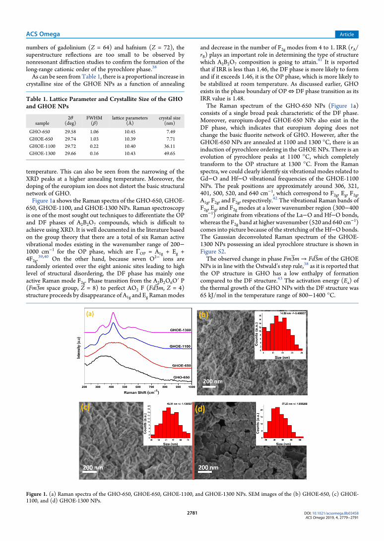
temperature. This can also be seen from the narrowing of theXRD peaks at a higher annealing temperature. Moreover, thedoping of the europium ion does not distort the basic structuralnetwork of GHO.Figure 1a shows the Raman spectra of the GHO-650, GHOE-
650, GHOE-1100, and GHOE-1300 NPs. Raman spectroscopyis one of the most sought out techniques to differentiate the OPand DF phases of A2B2O7 compounds, which is difficult toachieve using XRD. It is well documented in the literature basedon the group theory that there are a total of six Raman activevibrational modes existing in the wavenumber range of 200−1000 cm−1 for the OP phase, which are ΓOP = A1g + Eg +4F2g.
39,40 On the other hand, because seven O2− ions arerandomly oriented over the eight anionic sites leading to highlevel of structural disordering, the DF phase has mainly oneactive Raman mode F2g. Phase transition from the A2B2O6O′ P(Fm3m space group, Z = 8) to perfect AO2 F (Fd3m, Z = 4)structure proceeds by disappearance of A1g and Eg Ramanmodes
and decrease in the number of F2g modes from 4 to 1. IRR (rA/rB) plays an important role in determining the type of structurewhich A2B2O7 composition is going to attain.41 It is reportedthat if IRR is less than 1.46, the DF phase is more likely to formand if it exceeds 1.46, it is the OP phase, which is more likely tobe stabilized at room temperature. As discussed earlier, GHOexists in the phase boundary of OP⇔ DF phase transition as itsIRR value is 1.48.The Raman spectrum of the GHO-650 NPs (Figure 1a)
consists of a single broad peak characteristic of the DF phase.Moreover, europium-doped GHOE-650 NPs also exist in theDF phase, which indicates that europium doping does notchange the basic fluorite network of GHO. However, after theGHOE-650 NPs are annealed at 1100 and 1300 °C, there is aninduction of pyrochlore ordering in the GHOENPs. There is anevolution of pyrochlore peaks at 1100 °C, which completelytransform to the OP structure at 1300 °C. From the Ramanspectra, we could clearly identify six vibrational modes related toGd−O and Hf−O vibrational frequencies of the GHOE-1100NPs. The peak positions are approximately around 306, 321,401, 500, 520, and 640 cm−1, which correspond to F2g, Eg, F2g,A1g, F2g, and F2g, respectively.
42 The vibrational Raman bands ofF2g, Eg, and F2g modes at a lower wavenumber region (300−400cm−1) originate from vibrations of the La−O and Hf−O bonds,whereas the F2g band at higher wavenumber (520 and 640 cm−1)comes into picture because of the stretching of the Hf−Obonds.The Gaussian deconvoluted Raman spectrum of the GHOE-1300 NPs possessing an ideal pyrochlore structure is shown inFigure S2.The observed change in phase Fm3m→ Fd3m of the GHOE
NPs is in line with the Ostwald’s step rule,38 as it is reported thatthe OP structure in GHO has a low enthalpy of formationcompared to the DF structure.43 The activation energy (Ea) ofthe thermal growth of the GHO NPs with the DF structure was65 kJ/mol in the temperature range of 800−1400 °C.
Table 1. Lattice Parameter and Crystallite Size of the GHOand GHOE NPs
sample2θ
(deg)FWHM(β)
lattice parameters(Å)
crystal size(nm)
GHO-650 29.58 1.06 10.45 7.49GHOE-650 29.74 1.03 10.39 7.71GHOE-1100 29.72 0.22 10.40 36.11GHOE-1300 29.66 0.16 10.43 49.65
Figure 1. (a) Raman spectra of the GHO-650, GHOE-650, GHOE-1100, and GHOE-1300 NPs. SEM images of the (b) GHOE-650, (c) GHOE-1100, and (d) GHOE-1300 NPs.
ACS Omega Article
DOI: 10.1021/acsomega.8b03458ACS Omega 2019, 4, 2779−2791
2781
Scanning electron microscopy (SEM) images (Figure 1b−d)show that the GHOE-650, GHOE-1100, and GHOE-1300 NPsare either spheroidal or spherical and have certain degree ofagglomeration. The particle size distribution of the GHOE NPswas calculated from these SEM images by ImageJ software asshown in the insets. There is a progressing increase of theaverage particle size of these GHOE NPs, that is 14 nm of theGHOE-650 NPs, 48 nm of the GHOE-1100 NPs, and 57 nm ofthe GHOE-1300 NPs.2.2. Cohesive Energies of OP and DF Structures: A DFT
Study. The DFT-generalized gradient approximation (GGA)-calculated equilibrium lattice parameters, atomic positions, andbond lengths are summarized in Table 2 along with previousDFT calculation results.44 Table 2 clearly shows that our valuescalculated via GGA−Perdew−Burke−Ernzerhof agree well withprevious GGA−PW91 calculated values andGGA is sufficient toreproduce the insulating character of GHO. The lattice constant(ao), the internal structural parameter (x) which is related to theposition of O48f, the nearest Gd−O8b, Gd−O48f, and Hf−O48fdistances and the band gap are tabulated in Table 2.Table 3 shows our DFT-GGA-calculated cohesive energies of
OP and DF structures of GHO, and the OP structure is stable
with respect to DF. In the bulk state, the OP structure isfavorable with respect to the DF structure. Moreover, Jiang etal.45 and Li et al.46 have shown that the ground state structure ofGHO is OP. They have also calculated OP to DF transformationtemperatures for several pyrochlores. Therefore, our DFTresults are in agreement with previous DFT calculation results.Further, our DFT-GGA results show that with Eu doping (1 Euatom in the 88-atom supercell), the OP structure is stable withrespect to the DF.Our experimental results show that the crystallite size
increases with increasing annealing temperature (Table 1).Increasing crystallite size implies lower surface to volume ratiowith approaching bulk characteristics. The crystallite size of theGHOE-650 NPs is around 7.0 nm, whereas that of the GHOE-1300 NPs is ∼50 nm. Raman spectra of the GHOE-1100 andGHOE-1300 NPs confirm the formation of the OP structure.Therefore, the OP structure is favorable in the GHO andGHOENPs after high-temperature annealing and corroborating withour calculated DFT energetics shown in Table 3.2.3. Local Site Stability of Eu3+ Ions in the GHOE NPs.
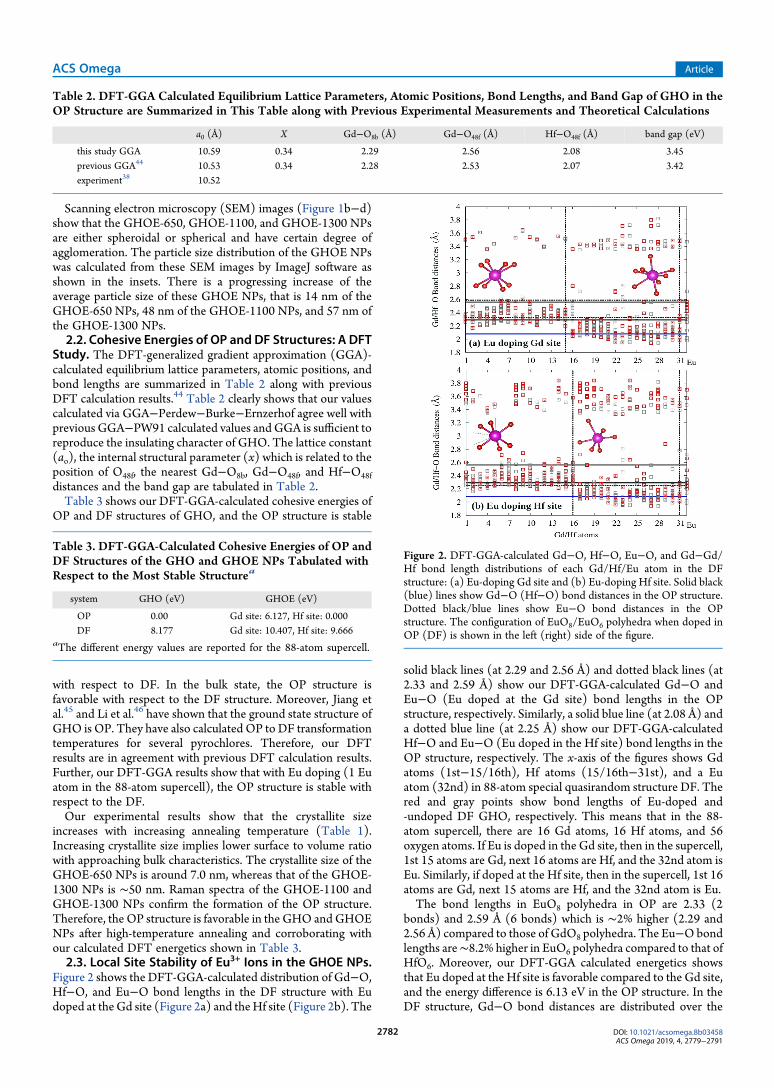
Figure 2 shows the DFT-GGA-calculated distribution of Gd−O,Hf−O, and Eu−O bond lengths in the DF structure with Eudoped at theGd site (Figure 2a) and theHf site (Figure 2b). The
solid black lines (at 2.29 and 2.56 Å) and dotted black lines (at2.33 and 2.59 Å) show our DFT-GGA-calculated Gd−O andEu−O (Eu doped at the Gd site) bond lengths in the OPstructure, respectively. Similarly, a solid blue line (at 2.08 Å) anda dotted blue line (at 2.25 Å) show our DFT-GGA-calculatedHf−O and Eu−O (Eu doped in the Hf site) bond lengths in theOP structure, respectively. The x-axis of the figures shows Gdatoms (1st−15/16th), Hf atoms (15/16th−31st), and a Euatom (32nd) in 88-atom special quasirandom structure DF. Thered and gray points show bond lengths of Eu-doped and-undoped DF GHO, respectively. This means that in the 88-atom supercell, there are 16 Gd atoms, 16 Hf atoms, and 56oxygen atoms. If Eu is doped in the Gd site, then in the supercell,1st 15 atoms are Gd, next 16 atoms are Hf, and the 32nd atom isEu. Similarly, if doped at the Hf site, then in the supercell, 1st 16atoms are Gd, next 15 atoms are Hf, and the 32nd atom is Eu.The bond lengths in EuO8 polyhedra in OP are 2.33 (2
bonds) and 2.59 Å (6 bonds) which is ∼2% higher (2.29 and2.56 Å) compared to those of GdO8 polyhedra. The Eu−Obondlengths are∼8.2% higher in EuO6 polyhedra compared to that ofHfO6. Moreover, our DFT-GGA calculated energetics showsthat Eu doped at the Hf site is favorable compared to the Gd site,and the energy difference is 6.13 eV in the OP structure. In theDF structure, Gd−O bond distances are distributed over the
Table 2. DFT-GGA Calculated Equilibrium Lattice Parameters, Atomic Positions, Bond Lengths, and Band Gap of GHO in theOP Structure are Summarized in This Table along with Previous Experimental Measurements and Theoretical Calculations
a0 (Å) X Gd−O8b (Å) Gd−O48f (Å) Hf−O48f (Å) band gap (eV)
this study GGA 10.59 0.34 2.29 2.56 2.08 3.45previous GGA44 10.53 0.34 2.28 2.53 2.07 3.42experiment38 10.52
Table 3. DFT-GGA-Calculated Cohesive Energies of OP andDF Structures of the GHO and GHOE NPs Tabulated withRespect to the Most Stable Structurea
system GHO (eV) GHOE (eV)
OP 0.00 Gd site: 6.127, Hf site: 0.000DF 8.177 Gd site: 10.407, Hf site: 9.666
aThe different energy values are reported for the 88-atom supercell.
Figure 2. DFT-GGA-calculated Gd−O, Hf−O, Eu−O, and Gd−Gd/Hf bond length distributions of each Gd/Hf/Eu atom in the DFstructure: (a) Eu-doping Gd site and (b) Eu-doping Hf site. Solid black(blue) lines show Gd−O (Hf−O) bond distances in the OP structure.Dotted black/blue lines show Eu−O bond distances in the OPstructure. The configuration of EuO8/EuO6 polyhedra when doped inOP (DF) is shown in the left (right) side of the figure.
ACS Omega Article
DOI: 10.1021/acsomega.8b03458ACS Omega 2019, 4, 2779−2791
2782
range of 2.16−2.60 Å in the form of GdO6, GdO7, and GdO8
polyhedra. The Hf−O bond distances are distributed over therange of 1.94−2.81 Å in the form of HfO6 and HfO7 polyhedra.The Hf−O bond distances are distributed in a wide rangecompared to an OPHfO6 bond length of 2.08 Å. In other words,the chemical environment around the Hf site is more distortedcompared to that of Gd.Figure 2 also shows the change in the first and second nearest
neighbor bond distances because of Eu doping at Gd and Hfsites. The Gd−Gd/Hf bond lengths change appreciably by Eudoping at the Hf site compared to Eu doping at the Gd site.Moreover, our DFT-GGA-calculated energetics shows that Eudoping in the Hf site is favorable compared to the Gd site, andthe energy difference is 0.74 eV. In addition, Figure 2 showsEuO8 (doped at the Gd site) and EuO6 (doped at the Hf site)polyhedra in OP (shown on the left) and DF (shown on theright) structures. The distribution of bond lengths andorientation shows that EuO8 and EuO6 polyhedra has inversionsymmetry when doped in the OP structure and inversion
symmetry is absent when doped in the DF structure. Theemission spectra from different excitation wavelengths andnumber of Stark components of the 5D0 → 7FJ (J = 0−4)transitions of the Eu3+ ion in the GHOE-650 NPs (Figures S3band 5) show that Eu atoms occupy a chemical environment inthe DF structure which has no inversion symmetry. Theseresults are consistent with our DFT-calculated results of thelocal structure around Eu atoms, which also confirm the absenceof inversion symmetry.
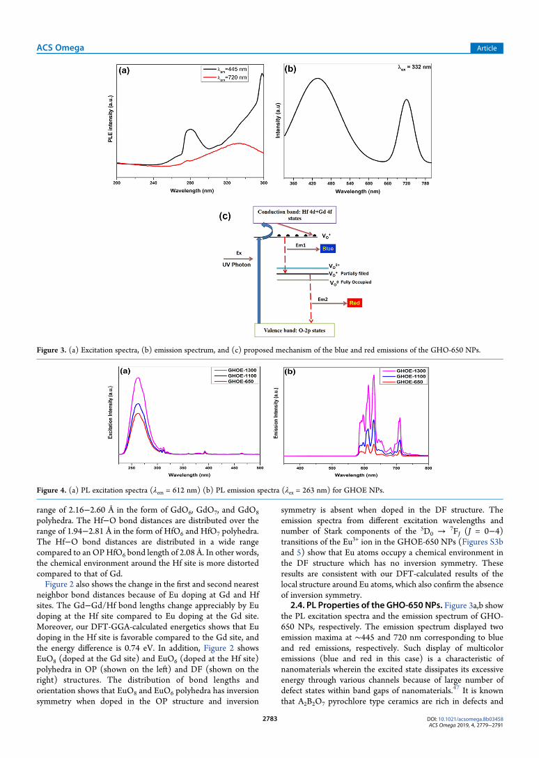
2.4. PL Properties of the GHO-650 NPs. Figure 3a,b showthe PL excitation spectra and the emission spectrum of GHO-650 NPs, respectively. The emission spectrum displayed twoemission maxima at ∼445 and 720 nm corresponding to blueand red emissions, respectively. Such display of multicoloremissions (blue and red in this case) is a characteristic ofnanomaterials wherein the excited state dissipates its excessiveenergy through various channels because of large number ofdefect states within band gaps of nanomaterials.47 It is knownthat A2B2O7 pyrochlore type ceramics are rich in defects and
Figure 3. (a) Excitation spectra, (b) emission spectrum, and (c) proposed mechanism of the blue and red emissions of the GHO-650 NPs.
Figure 4. (a) PL excitation spectra (λem = 612 nm) (b) PL emission spectra (λex = 263 nm) for GHOE NPs.
ACS Omega Article
DOI: 10.1021/acsomega.8b03458ACS Omega 2019, 4, 2779−2791
2783
more so in oxygen vacancies.48 The Raman spectrum of theGHO-650 NPs (Figure 1a) indicated that it exists in the DFphase with large concentration of oxygen vacancies in itsnetwork. Eagleman et al. reported that oxygen vacancies areresponsible for visible light emission in La2Hf2O7.
49,50 Inaddition, we have found that electronic transition involvingionized oxygen vacancies are responsible for such luminescenceproperties from our earlier work on Nd2Zr2O7 and Gd2Zr2O7
pyrochlore.5,27 The used excitation energy (∼3.37 eV, 332 nm)is less than the band gap (3.42 eV) of GHO,19,44 so directtransition from the valence band (VB) to the conduction band(CB) does not happen, and there exist certain localized defectstates within the band gap of the GHO NPs. Such defects couldarise during thermal treatment of the GHO NPs or may bepresent intrinsically in them.51 The responsible oxygenvacancies for blue and red emission in the GHO-650 NPs arepresented pictorially as a mechanism in Figure 3c. The blueemission could arise from the electronic transition of singlyionized oxygen vacancies to the VB, and the red emission from
that of shallow oxygen defect vacancies to deep ones. Theproposed different origins of these two emissions are consistentwith the different excitation spectra obtained with λem = 445 and720 nm at blue and red regions, respectively (Figure 3a).
2.5. PL Properties of the GHO:Eu3+. 2.5.1. Excitation andEmission Spectroscopy. Figure 4a shows the excitation spectraof the GHOE samples as a function of annealing temperature.These spectra consist of a very strong band peaking at∼263 nm,which is known as the charge transfer band (CTB) andattributed to electron transfer from a filled 2p orbital of O2− ionsto a vacant 4f orbital of the Eu3+ ion. The weak bands at 395 and465 nm are due to 7F0 →
5L6 and7F0 →
5D2 transitions of Eu3+
ions. The CTB as a La Porte-allowed transition has higherintensity than the forbidden f−f transitions. The excitationspectra monitored under various emission maxima (591, 630,654, and 711 nm) are also shown in Figure S3a. Except for themarginal change in intensity, the spectra remain the same atdifferent emission wavelengths. The emission spectra recordedwith 263 nm excitation is shown in Figure 4b. For comparison,
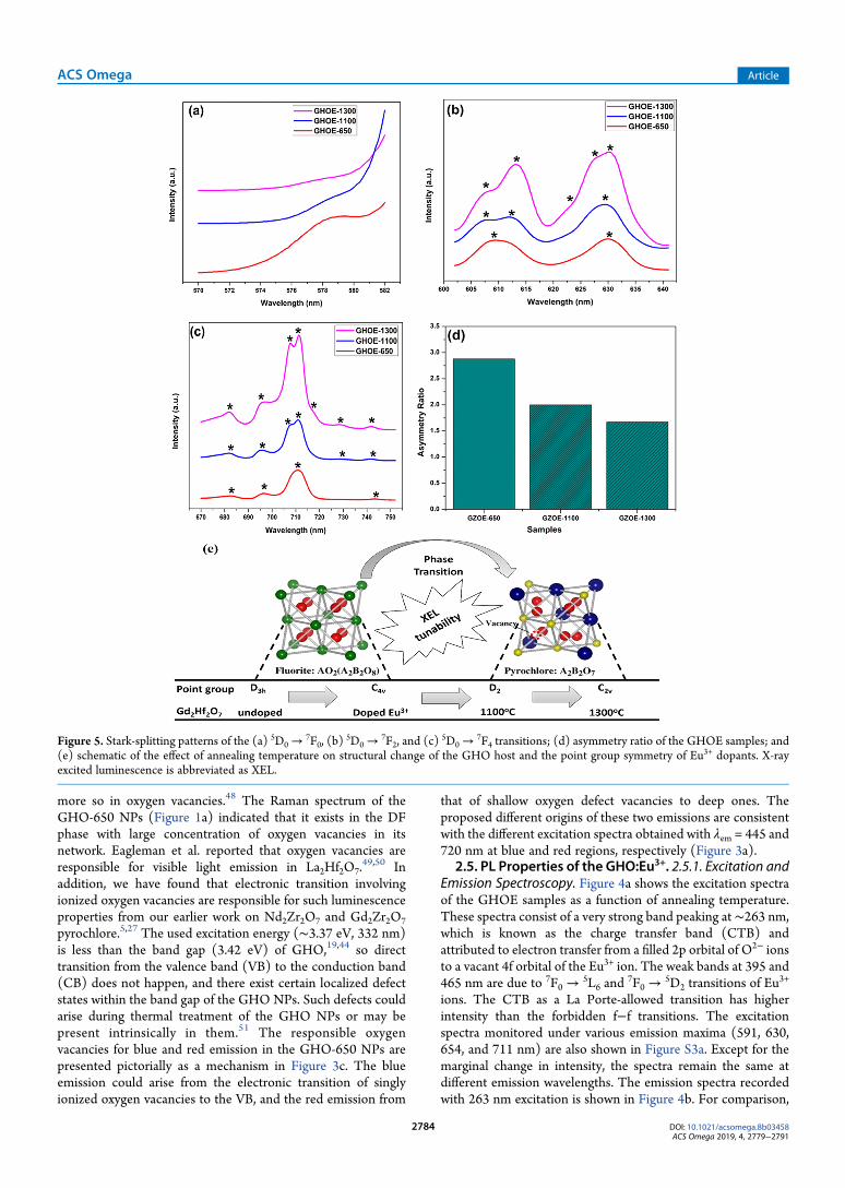
Figure 5. Stark-splitting patterns of the (a) 5D0 →7F0, (b)
5D0 →7F2, and (c)
5D0 →7F4 transitions; (d) asymmetry ratio of the GHOE samples; and
(e) schematic of the effect of annealing temperature on structural change of the GHO host and the point group symmetry of Eu3+ dopants. X-rayexcited luminescence is abbreviated as XEL.
ACS Omega Article
DOI: 10.1021/acsomega.8b03458ACS Omega 2019, 4, 2779−2791
2784

emission spectra recorded under 263, 395, and 465 nmexcitations are shown in Figure S3b. The intensity of the PLemission recorded with CTB excitation is much more intensethan that with f−f excitation bands. The emission spectra for allthree GHOE samples consist of five main peaks at 579, 591, 630,654, and 711 nm corresponding to the 5D0 →
7F0,5D0 →
7F1,5D0 →
7F2,5D0 →
7F3, and5D0 →
7F4 transitions, respectively.There are several interesting features of these spectra:appearance of 5D0 →
7F0 transition that is allowed neither bymagnetic dipole transition (MDT) nor by EDT, large splitting inthe spectral peaks, high asymmetry ratio (I5D0→
7F2/I5D0→
7F1), and
presence of relatively intense 5D0 →7F4 transition.
The presence of 5D0 → 7F0 transition and large spectralsplitting are signatures of the europium ion in highly disorderedenvironment.52 This is supported with the fact of relatively highemission intensity of 5D0 →
7F4 transition due to a distortedchemical surrounding around the Eu3+ ions.53 However, thereare no changes of the spectral profile in terms of peak symmetryor width on changing the excitation wavelength.Based on the emission spectra of the GHOE-650, GHOE-
1100, and GHOE-1300 NPs (Figure 4b), three effects ofannealing on the PL properties of the GHOE NPs were takeninto consideration. These effects include (a) emission intensity,(b) spectral width of the 5D0 →
7F0 emission lines, and (c) theratio of integrated PL intensities between MDT (5D0 →
7F1)and hypersensitive EDTs (5D0 →
7F2), that is, the asymmetryratio (IRO).First, the emission intensity of the 5D0 →
7F2 EDT is muchhigher than that of the 5D0 →
7F1 MDT for all three GHOEsamples, suggesting that the Eu3+ ions are localized in lowsymmetry sites. Moreover, the PL emission intensity increaseswith increasing annealing temperature, which could beattributed to the different crystallite sizes of the GHOE NPs.The GHOE-650 NPs with the smallest size (∼7 nm) have thehighest surface to volume ratio, so are rich with surface defects.Such surface defects act as nonradiative pathways to decreaseemission intensity. On the other hand, large-sized GHOE-1300NPs have less surface defects with high emission intensity.In all these three samples, the red emission due to 5D0 →
7F2transition is the most intense peak, and the color coordinates arevery similar, so only one of the representative samples, that is,the GHOE-650 NPs, is shown with the International del’Eclairage (CIE) diagram (Figure S3c).2.5.2. Point Group Symmetry and Asymmetry Ratio of Eu3+
Ions in the GHOE NPs. Structural change of the GHO host canbe corroborated with the change of Stark component numbersfrom the PL spectral pattern of Eu3+ ions, which is related to itspoint group symmetry.54,55 The original point group symmetryof Gd3+/Hf4+ sites in both OP and DF structures is D3d.
16
Interestingly, the number of Stark components (as highlightedwith black asterisks) of the EDT (ΔJ = ±2 and ΔJ = ±4) keepsincreasing as the annealing temperature of the GHOE NPsincreases as can be easily seen from Table S1. Such changes areclearly seen in the PL emission characteristics of 5D0 → 7F0(Figure 5a), 5D0 →
7F2 (Figure 5b), and5D0 →
7F4 (Figure 5c)transitions as well. Accordingly, the point group symmetry of theGHO host reduces to C4v for the GHOE-650 NPs because oflattice strain and distortion induced by the small particle size andcharge mismatch of ions after Eu3+ doping. Furthermore, itchanges to D2 (for the GHOE-1100 NPs) and then to C2v (forthe GHOE-1300 NPs) because of the effects of annealing andstructural change, as depicted schematically in Figure 5e.
The asymmetry ratio IRO is also highly sensitive to structuralchange and useful to understand the local symmetry around theEu3+ ions in the GHO host. As the GHO host goes from a highlyDF structure to a highly OP structure, there is a progressivedecrease of the asymmetry ratio of the Eu3+ ions (Figure 5d).The IRO values of the GHOE-650, GHOE-1100, and GHOE-1300 NPs are 2.9, 2.0, and 1.6, respectively. This indicated thatthe local surrounding of Eu3+ ions is highly asymmetric in theGHOE-650 NPs with the DF structure, whereas the asymmetrybecomes relatively low in the GHOE-1300 NPs with the OPstructure. Therefore, the change of the IRO values is inaccordance with the structural difference of the GHOE NPs.Menushenkov et al. also found that the Debye Waller factor asthe measure of root-mean-square deviation of the interatomicdistance from the average value decreases with increasingannealing temperature for all Gd−O and Hf−O bonds of GHOby means of extended X-ray absorption fine structure measure-ments.13 This confirms the increase of ordering of the crystallinestructure with annealing temperature as reflected in thecalculated IRO values.Neither MDT nor EDT allows 5D0 →
7F0 transition of Eu3+
ions. However, it is still often observed from Eu3+-dopedinorganic phosphors because of the CF-induced J-mixing effectthat lowers the symmetry.56 According to the selection rulegoverning the EDT, this transition exists in low local symmetrysituations of Eu3+ ions, including Cs, C1, C2, C3, C4, C6, C2v, C3v,C4v, and C6v.
57 As shown in Figure 5a, the 5D0 →7F0 peak of the
GHOE-650 NPs which are stabilized in the DF phase showssubstantial intensity, and that of the GHOE-1100 and GHOE-1300 NPs is nearly absent because of the pyrochlore ordering.Based on lifetime spectroscopy (Section 2.5.3), DFT-based
cohesive energy calculations (Section 2.2), and local structurestudy (Section 2.3), Eu3+ ions can be stabilized at both Gd3+ andHf4+ sites. Whatever the Stark component is considered in thiswork, it is based on composite emission spectra (Figure 3b)which have the contributions of Eu3+ ions at both Gd3+ and Hf4+
sites. To get individual emission spectra from these Eu@Gd andEu@Hf sites, we need to carry out time-resolved emissionspectroscopy (TRES) which can get individual spectra of Eu3+
ions at both Gd3+ and Hf4+ sites. We are trying to establishcollaborations to use TRES as our future projects.
2.5.3. Excited State Lifetime and Quantum Yield. The 5D0emission decay profiles of all three GHOE samples at the 5D0→7F2 transition display a biexponential behavior (Figure S4).Lifetime values were obtained by fitting with biexponentialfunction
τ τ= + − + −I t I A t A t( ) exp( / ) exp( / )0 1 1 2 2 (1)
where τ1 and τ2 are actual lifetime values related to decay rates ofcorresponding exponential components, and A1 and A2 arebiexponential fitting parameters. Based on the PL decay profilescorresponding to the 5D0 excited state of Eu
3+ ions in the GHOENPs and the lifetime values obtained after biexponential fitting,the average lifetime values were calculated using the followingequation
ττ ττ τ
=++
A AA Aav1 1
22 2
2
1 1 2 2 (2)
The average and individual lifetime values were mentioned inTable 4. In theGHOE-650NPs with theDF structure, Gd3+ ionsexist in highly symmetric GdO8 in the form of a cube, whereasHf4+ ions exist in highly distorted octahedra. The short and long
ACS Omega Article
DOI: 10.1021/acsomega.8b03458ACS Omega 2019, 4, 2779−2791
2785
lifetimes can be explained by the presence of two different localsites of Eu3+ ions in the GHOE-650 NPs. The short one isattributed to Eu3+ ions occupying the distorted HfO6 octahedra,whereas the long one is attributed to Eu3+ ions occupying thehighly symmetric GdO8 site. Eu3+ ions also exhibitedbiexponential decay in other pyrochlore hosts such asLa2Hf2O7, Gd2Zr2O7, and Nd2Zr2O7 wherein one of thelifetimes is attributed to A3+ site occupancy and other to B4+
site occupancy.33,58−63 Normally, the short lifetime is attributedto asymmetric environment as f−f transition becomes relaxed,and the long lifetime is mostly attributed to symmetricenvironment as f−f transition is La Porte forbidden.61,64−67
The biexponential behavior can also arise due to other reasonssuch as the presence of defects, energy transfer, and so forth.This phenomenon has been substantiated with theoreticalcalculations in the next section.On the other hand, the GHOE-1100 and GHOE-1300 NPs
have the prevailing OP structure where GdO8 is highly distortedscalenohedra and HfO6 exists in highly symmetrical octahedra.In this case, the longer lifetime is attributed to Eu3+ ions sitting atHfO6 sites, whereas the shorter one is due to Eu
3+ ions localizedat the distorted GdO8 site.The average lifetime values of the GHOE-650, GHOE-1100,
andGHOE-1300NPs were 2.09, 2.27, and 2.45ms, respectively.The increasing average lifetime values is partially due to thedecreasing distortion of Eu3+ ions in the GHO NPs withincreasing annealing temperature. In addition, the surface defectdensity is the least for the GHOE-1300 NPs, so they have thelowest relaxation probability through nonradiative transitions,and hence the highest lifetime value.Furthermore, the lower the nonradiative transition proba-
bility, the higher the quantum efficiency. With the highestabsolute quantum yield (AQY) reported (Table 4), it issuggested that the GHOE-1300 NPs are the most suitablesample for optical emitter and fluoroimmunoassay applicationswherein a high lifetime is needed. The lowest lifetime value fromthe GHOE-650NPs is also reflected in the minimal AQY amongall three samples.Judd−Ofelt analysis was carried out for the GHOE-650,
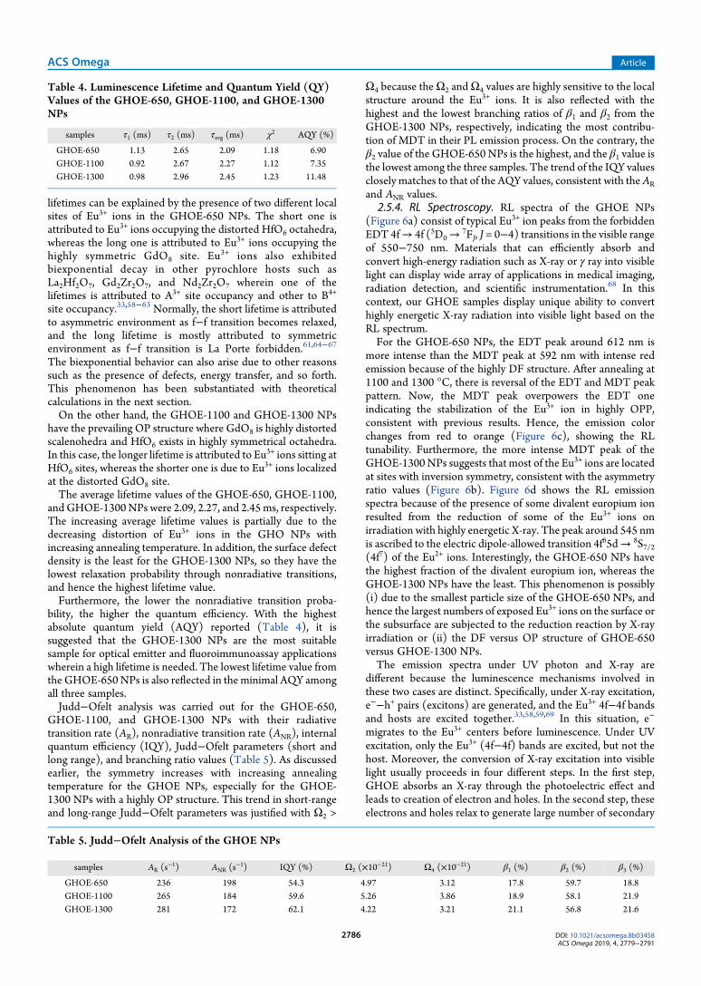
GHOE-1100, and GHOE-1300 NPs with their radiativetransition rate (AR), nonradiative transition rate (ANR), internalquantum efficiency (IQY), Judd−Ofelt parameters (short andlong range), and branching ratio values (Table 5). As discussedearlier, the symmetry increases with increasing annealingtemperature for the GHOE NPs, especially for the GHOE-1300 NPs with a highly OP structure. This trend in short-rangeand long-range Judd−Ofelt parameters was justified with Ω2 >
Ω4 because theΩ2 andΩ4 values are highly sensitive to the localstructure around the Eu3+ ions. It is also reflected with thehighest and the lowest branching ratios of β1 and β2 from theGHOE-1300 NPs, respectively, indicating the most contribu-tion of MDT in their PL emission process. On the contrary, theβ2 value of the GHOE-650 NPs is the highest, and the β1 value isthe lowest among the three samples. The trend of the IQY valuesclosely matches to that of the AQY values, consistent with theARand ANR values.
2.5.4. RL Spectroscopy. RL spectra of the GHOE NPs(Figure 6a) consist of typical Eu3+ ion peaks from the forbiddenEDT 4f→ 4f (5D0→
7FJ, J = 0−4) transitions in the visible rangeof 550−750 nm. Materials that can efficiently absorb andconvert high-energy radiation such as X-ray or γ ray into visiblelight can display wide array of applications in medical imaging,radiation detection, and scientific instrumentation.68 In thiscontext, our GHOE samples display unique ability to converthighly energetic X-ray radiation into visible light based on theRL spectrum.For the GHOE-650 NPs, the EDT peak around 612 nm is
more intense than the MDT peak at 592 nm with intense redemission because of the highly DF structure. After annealing at1100 and 1300 °C, there is reversal of the EDT and MDT peakpattern. Now, the MDT peak overpowers the EDT oneindicating the stabilization of the Eu3+ ion in highly OPP,consistent with previous results. Hence, the emission colorchanges from red to orange (Figure 6c), showing the RLtunability. Furthermore, the more intense MDT peak of theGHOE-1300NPs suggests that most of the Eu3+ ions are locatedat sites with inversion symmetry, consistent with the asymmetryratio values (Figure 6b). Figure 6d shows the RL emissionspectra because of the presence of some divalent europium ionresulted from the reduction of some of the Eu3+ ions onirradiation with highly energetic X-ray. The peak around 545 nmis ascribed to the electric dipole-allowed transition 4f65d→ 8S7/2(4f7) of the Eu2+ ions. Interestingly, the GHOE-650 NPs havethe highest fraction of the divalent europium ion, whereas theGHOE-1300 NPs have the least. This phenomenon is possibly(i) due to the smallest particle size of the GHOE-650 NPs, andhence the largest numbers of exposed Eu3+ ions on the surface orthe subsurface are subjected to the reduction reaction by X-rayirradiation or (ii) the DF versus OP structure of GHOE-650versus GHOE-1300 NPs.The emission spectra under UV photon and X-ray are
different because the luminescence mechanisms involved inthese two cases are distinct. Specifically, under X-ray excitation,e−−h+ pairs (excitons) are generated, and the Eu3+ 4f−4f bandsand hosts are excited together.33,58,59,69 In this situation, e−
migrates to the Eu3+ centers before luminescence. Under UVexcitation, only the Eu3+ (4f−4f) bands are excited, but not thehost. Moreover, the conversion of X-ray excitation into visiblelight usually proceeds in four different steps. In the first step,GHOE absorbs an X-ray through the photoelectric effect andleads to creation of electron and holes. In the second step, theseelectrons and holes relax to generate large number of secondary
Table 4. Luminescence Lifetime and Quantum Yield (QY)Values of the GHOE-650, GHOE-1100, and GHOE-1300NPs
samples τ1 (ms) τ2 (ms) τavg (ms) χ2 AQY (%)
GHOE-650 1.13 2.65 2.09 1.18 6.90GHOE-1100 0.92 2.67 2.27 1.12 7.35GHOE-1300 0.98 2.96 2.45 1.23 11.48
Table 5. Judd−Ofelt Analysis of the GHOE NPs
samples AR (s−1) ANR (s
−1) IQY (%) Ω2 (×10−21) Ω4 (×10−21) β1 (%) β2 (%) β3 (%)
GHOE-650 236 198 54.3 4.97 3.12 17.8 59.7 18.8GHOE-1100 265 184 59.6 5.26 3.86 18.9 58.1 21.9GHOE-1300 281 172 62.1 4.22 3.21 21.1 56.8 21.6
ACS Omega Article
DOI: 10.1021/acsomega.8b03458ACS Omega 2019, 4, 2779−2791
2786
electrons, holes, photons, and plasmons. Such relaxation alsoinduces several other electronic excitations. These secondaryelectrons and holes lose their energy via electron−phononinteraction to give electron−hole pairs with near band gapenergy. The third stage involves the transport of the electron−hole pairs (excitons) through the host material to a luminescentcenter (trap) and the excitation of the luminescent center. Thefinal stage concerns the resulting luminescence. Materials whichhave a high light output and a short lifetime under photo-excitation may have a very low light output and/or a longlifetime under X-ray excitation. This is due to the energy lossesand delays in the energymigration processes, which are absent inPL where the luminescent centers are directly and intentionallyexcited.2.5.5. Thermal Quenching Behavior of GHOE NPs. Thermal
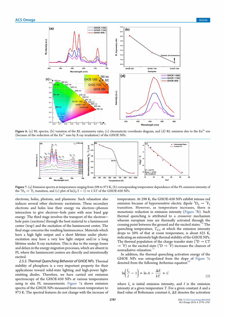
stability of phosphors is a very important property for theirapplications toward solid-state lighting and high-power light-emitting diodes. Therefore, we have carried out emissionspectroscopy of the GHOE-650 NPs at various temperaturesusing in situ PL measurements. Figure 7a shows emissionspectra of the GHOE NPs measured from room temperature to973 K. The spectral features do not change with the increase of
temperature. At 298 K, the GHOE-650 NPs exhibit intense redemission because of hypersensitive electric dipole 5D0 →
7F2transition. However, as temperature increases, there ismonotonic reduction in emission intensity (Figure 7b). Suchthermal quenching is attributed to a crossover mechanismwherein europium ions are thermally activated through thecrossing point between the ground and the excited states.70 Thequenching temperature, T0.5, at which the emission intensitydrops to 50% of that at room temperature, is about 625 K,indicating an extremely high thermal stability of the GHOENPs.The thermal population of the charge transfer state (5D → CT→ 7F) or the excited state (5D → 7F) increases the chances ofnonradiative relaxation.71
In addition, the thermal quenching activation energy of theGHOE NPs was extrapolated from the slope of Figure 7cdenoted from the following Arrhenius equation59
ikjjj
y{zzz− = − Δ +
II
AE
kTCln 1 lno
(3)
where Io is initial emission intensity, and I is the emissionintensity at a given temperature T. For a given constant A and afixed value of Boltzmann constant k, ΔE denotes the activation
Figure 6. (a) RL spectra, (b) variation of the RL asymmetry ratio, (c) chromaticity coordinate diagram, and (d) RL emission due to the Eu2+ ion(because of the reduction of the Eu3+ ions by X-ray irradiation) of the GHOE NPs.
Figure 7. (a) Emission spectra at temperatures ranging from 298 to 973 K, (b) corresponding temperature dependence of the PL emission intensity ofthe 5D0 →
7F2 transition, and (c) plot of ln(I0/I − 1) vs 1/kT of the GHOE-650 NPs.
ACS Omega Article
DOI: 10.1021/acsomega.8b03458ACS Omega 2019, 4, 2779−2791
2787
energy involved in thermal quenching process. The relationshipof ln(I0/I − 1) and 1/kT displayed linear behavior (Figure 7c).The activation energy calculated from this plot is equal to 0.425eV for the thermal quenching. In our earlier work onLa2Hf2O7:Eu
3+ NPs with an ideal pyrochlore structure, theactivation energy value was found to be 0.410 eV which isslightly lower than the defect fluorite GHOE-650 NPs.59
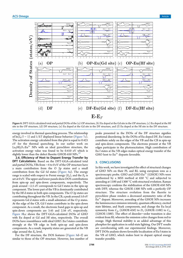
2.6. Efficiency of Host to Dopant Energy Transfer byDFT Calculations. Based on the DFT-GGA-calculated totaland partial DOSs, VBs from−4 to 0 eV of the OP structure havea main contribution from the O 2p states and a smallcontribution from the Gd 5d states (Figure 8a). The energyrange is scaled with respect to Fermi energy (EF), and the EF isset at 0 eV. The upper and lower panels showDOS contributionsfrom spin-up and spin-down components, respectively. Thepeak around −2.5 eV corresponds to Gd f states in the spin-upcomponent. The lower part of the VB is dominantly contributedby the Hf d states in both spin components. The O 2p states arecontributing throughout the VB. In the CB, a peak around 5.0 eVrepresents Gd d states with a small admixture of the O p states.At the edge of the CB, Gd f states contribute in the spin-downcomponent. As a result, the electronic band gaps in spin-up andspin-down components are 3.45 and 2.56 eV, respectively.Figure 8b,c shows the DFT-GGA-calculated DOSs of GHOwith Eu doped at Gd and Hf sites, respectively. The overallDOSs have resemblance with ideal GHO. Eu f states contributestrongly at the VB edge in both spin-up and spin-downcomponents. As a result, impurity states are generated at the VBedge around the EF level.For the DF structure, the DOS features (Figure 8d−f) are
similar to those of the OP structure. However, less number of
peaks presented in the DOSs of the DF structure signifiespositional disordering. In the DOSs of Eu-doped DF, Eu f statescontribute solely on the edges of the VB and the CB in spin-upand spin-down components. The electrons present at the VBedges participate in the photoexcitation. High contribution ofEu f states at the VB edges makes optical energy transfer of theGHO host to Eu3+ dopants favorable.
3. CONCLUSIONS
In this work, we have investigated the effect of structural changesof GHO NPs on their PL and RL using europium ions as aspectroscopic probe. GHO and GHO:Eu3+ (GHOE) NPs weresynthesized by a MSS method at 650 °C and subjected toannealing at 1100 and 1300 °Cwith structural evolution. Ramanspectroscopy confirms the stabilization of the GHOE-650 NPswith DFP, whereas the GHOE-1300 NPs with a perfectly OPstructure. The structure evolution from the fluorite topyrochlore phase renders a decreased asymmetry ratio of theEu3+ dopant. Moreover, annealing of the GHOE NPs increasesthe luminescence emission intensity, quantum efficiency, excitedstate lifetime, and Stark components, and change the Eu localsymmetry from C4v (GHOE-650) to D2 (GHOE-1100) to C2v(GHOE-1300). The effect of disorder−order transition is alsoevident from RLwherein the emission color changes from red toorange. High thermal stability is an added advantage of thisphosphor for application in solid-state lighting. Our DFT resultsare corroborating with our experimental findings. Moreover,DFT DOSs analysis shows favorable localization of Eu f states inthe VB of GHO, which makes host to dopant optical energytransfer possible.
Figure 8.DFT-GGA calculated total and partial DOSs of the (a) OP structure, (b) Eu doped at the Gd site in the OP structure, (c) Eu doped at the Hfsite in the OP structure, (d) DF structure, (e) Eu doped at the Gd site in the DF structure, and (f) Eu doped at the Hf site in the DF structure.
ACS Omega Article
DOI: 10.1021/acsomega.8b03458ACS Omega 2019, 4, 2779−2791
2788

4. EXPERIMENTAL: SYNTHESIS ANDCHARACTERIZATION
Gd2Hf2O7 (GHO) and Eu3+-doped GHO (GHOE) NPs weresynthesized using MSS at 650 °C (GHO-650 and GHOE-650),similar to our earlier work for La2Hf2O7 NPs.
2,4 However, bydoping europium ions into the GHO host, Gd3+ ions arereplaced based on closeness in terms of ionic radius and ioniccharge. The GHOE-650 sample was further annealed at 1100and 1300 °C in air for 6 h, and the products are noted as GHOE-1100 and GHOE-1300, respectively. The details of synthesis,characterization, theoretical methodology, Judd−Ofelt analysis,and QY measurements are included in the SupportingInformation as S1−S5, respectively.
■ ASSOCIATED CONTENT*S Supporting InformationThe Supporting Information is available free of charge on theACS Publications website at DOI: 10.1021/acsomega.8b03458.
Details of synthesis, characterization, and theoreticalmethodology (PDF)
■ AUTHOR INFORMATIONCorresponding Author*E-mail: [email protected]. Phone: +1-956-665-2986(Y.M.).ORCIDSantosh K. Gupta: 0000-0002-1178-0159Yuanbing Mao: 0000-0003-2665-6676NotesThe authors declare no competing financial interest.
■ ACKNOWLEDGMENTSThe authors acknowledge financial support by the NationalScience Foundation under CHE (award #1710160) and DMR(grant #1523577) and the USDANational Institute of Food andAgriculture (award #2015-38422-24059). The Department ofChemistry at the University of Texas Rio Grande Valley isgrateful for the generous support provided by a DepartmentalGrant from the Robert A. Welch Foundation (grant no. BX-0048). The in situ emission spectra were conducted at theCenter for Nanophase Materials Science, which is a U.S.Department of Energy, Office of Science User Facility, and theauthors thank Dr. A. Puretzky for technical assistance. S.K.G.thanks the United States−India Education Foundation (USIEF)and the Institute of International Education (IIE) for hisFulbright Nehru Postdoctoral Fellowship (award# 2268/FNPDR/2017).
■ REFERENCES(1) Zhang, W.; Tao, Y.; Li, C. Sol-gel synthesize and characterizationof χGd2Ti2O7/SiO2 photocatalyst for ofloxacin decomposition. Mater.Res. Bull. 2018, 105, 55−62.(2) Zuniga, J. P.; Gupta, S. K.; Pokhrel, M.; Mao, Y. Exploring theoptical properties of La2Hf2O7:Pr
3+ nanoparticles under UV and X-rayexcitation for potential lighting and scintillating applications. New J.Chem. 2018, 42, 9381−9392.(3) Gupta, S. K.; Reghukumar, C.; Pathak, N.; Sudarshan, K.; Tyagi,D.; Mohapatra, M.; Pujari, P. K.; Kadam, R. M. Speciation of uraniumand doping induced defects in Gd1.98U0.02Zr2O7: Photoluminescence,X-ray photoelectron and positron annihilation lifetime spectroscopy.Chem. Phys. Lett. 2017, 669, 245−250.
(4) Wahid, K.; Pokhrel, M.; Mao, Y. Structural, photoluminescenceand radioluminescence properties of Eu3+ doped La2Hf2O7 nano-particles. J. Solid State Chem. 2017, 245, 89−97.(5) Gupta, S. K.; Sudarshan, K.; Ghosh, P. S.; Srivastava, A. P.; Bevara,S.; Pujari, P. K.; Kadam, R. M. Role of various defects in thephotoluminescence characteristics of nanocrystalline Nd2Zr2O7: aninvestigation through spectroscopic and DFT calculations. J. Mater.Chem. C 2016, 4, 4988−5000.(6) Sibille, R.; Gauthier, N.; Yan, H.; Ciomaga Hatnean, M.; Ollivier,J.; Winn, B.; Filges, U.; Balakrishnan, G.; Kenzelmann, M.; Shannon,N.; Fennell, T. Experimental signatures of emergent quantumelectrodynamics in Pr2Hf2O7. Nat. Phys. 2018, 14, 711−715.(7) Yang, J.; Han, Y.; Shahid, M.; Pan, W.; Zhao, M.; Wu, W.; Wan, C.A promising material for thermal barrier coating: Pyrochlore-relatedcompound Sm2FeTaO7. Scr. Mater. 2018, 149, 49−52.(8) Bayart, A.; Szczepanski, F.; Blach, J.-F.; Rousseau, J.; Katelnikovas,A.; Saitzek, S. Upconversion luminescence properties and thermalquenching mechanisms in the layered perovskite La1.9Er0.1Ti2O7towards an application as optical temperature sensor. J. Alloys Compd.2018, 744, 516−527.(9) Zhang, S.; Zhang, H. B.; Zhao, F. A.; Jiang, M.; Xiao, H. Y.; Liu, Z.J.; Zu, X. T. Impact of isovalent and aliovalent substitution on themechanical and thermal properties of Gd2Zr2O7. Sci. Rep. 2017, 7, 6399.(10) Rittman, D. R.; Turner, K. M.; Park, S.; Fuentes, A. F.; Park, C.;Ewing, R. C.; Mao, W. L. Strain engineered pyrochlore at high pressure.Sci. Rep. 2017, 7, 2236.(11) Paul, B.; Singh, K.; Jaron, T.; Roy, A.; Chowdhury, A. Structuralproperties and the fluorite−pyrochlore phase transition in La2Zr2O7:The role of oxygen to induce local disordered states. J. Alloys Compd.2016, 686, 130−136.(12) Park, S.; Tracy, C. L.; Zhang, F.; Park, C.; Trautmann, C.;Tkachev, S. N.; Lang, M.; Mao, W. L.; Ewing, R. C. Radiation-induceddisorder in compressed lanthanide zirconates. Phys. Chem. Chem. Phys.2018, 20, 6187−6197.(13) Menushenkov, A. P.; Popov, V. V.; Zubavichus, Y. V.;Yaroslavtsev, A. A. Local peculiarities of the nanocrystalline structureof ternary oxides Ln2Hf2O7 (Ln=Gd, Tb, Dy). J. Struct. Chem. 2017, 57,1450−1458.(14) Durand, A. M.; Klavins, P.; Corruccini, L. R. Heat capacity of thefrustrated magnetic pyrochlores Gd2Zr2O7 and Gd2Hf2O7. J. Phys.:Condens. Matter 2008, 20, 235208.(15) Ali Biswas, A.; Jana, Y. Study on the low-temperature propertiesof pyrochlores Gd2Hf2O7 and Gd2Zr2O7, using crystal-field theory. AIPConference Proceedings; AIP, 2011; pp 1121−1122.(16) Kumar, S.; Gupta, H. C. First principles study of dielectric andvibrational properties of pyrochlore hafnates. Solid State Sci. 2012, 14,1405−1411.(17) Cepeda-Sanchez, N. M.; Fuentes, A. F.; Lopez-Cota, F. A.;Rodríguez-Reyes, M.; Díaz-Guillen, J. A. Mechanochemical synthesisand electrical properties of Gd2Hf2‑xZrxO7 solid electrolytes for their usein SOFC’s. J. Appl. Electrochem. 2015, 45, 1231−1237.(18) Sevastyanov, V. G.; Simonenko, E. P.; Simonenko, N. P.;Stolyarova, V. L.; Lopatin, S. I.; Kuznetsov, N. T. Synthesis,vaporization and thermodynamic properties of superfine Nd2Hf2O7and Gd2Hf2O7. Eur. J. Inorg. Chem. 2013, 4636−4644.(19) Chen, C. F.; Brennecka, G. L.; Synowicki, R. A.; Tegtmeier, E. L.;Brand, M. J.; Montalvo, J. D.; Ivy, J.; Cherepy, N. J.; Seeley, Z.; Payne, S.A. Transparent polycrystalline Gd2Hf2O7 ceramics. J. Am. Ceram. Soc.2018, 101, 3797−3807.(20) Chen, C.-F.; Brennecka, G. L.; Synowicki, R. A.; Tegtmeier, E. L.;Brand, M. J.; Montalvo, J. D.; Ivy, J.; Cherepy, N. J.; Seeley, Z.; Payne, S.A. Transparent polycrystalline Gd2Hf2O7 ceramics. J. Am. Ceram. Soc.2018, 101, 3797.(21) Shannon, R. D. Revised effective ionic radii and systematicstudies of interatomic distances in halides and chalcogenides. ActaCrystallogr., Sect. A: Cryst. Phys., Diffr., Theor. Gen. Crystallogr. 1976, 32,751−767.(22) Zhang, X.; Hayakawa, T.; Nogami, M.; Ishikawa, Y. Selectivesynthesis and luminescence properties of nanocrystalline GdF3:Eu
3+
ACS Omega Article
DOI: 10.1021/acsomega.8b03458ACS Omega 2019, 4, 2779−2791
2789
with hexagonal and orthorhombic structures. J. Nanomater. 2010, 2010,1.(23) Tian, Y.; Hua, R.; Chen, B.; Yu, N.; Zhang,W.; Na, L. Lanthanidedopant-induced phase transition and luminescent enhancement ofEuF3 nanocrystals. CrystEngComm 2012, 14, 8110−8116.(24) Papan, J.; Jovanovic, D. J.; Vukovic, K.; Smits, K.; Đorđevic, V.;Dramicanin, M. Europium (III)-doped A2Hf2O7 (A= Y, Gd, Lu)nanoparticles: Influence of annealing temperature, europium (III)concentration and host cation on the luminescent properties. Opt.Mater. 2016, 61, 68−76.(25) Pokhrel, M.; Wahid, K.; Mao, Y. Systematic studies onRE2Hf2O7:5%Eu
3+ (RE= Y, La, Pr, Gd, Er, and Lu) nanoparticles:effects of the A-site RE3+ cation and calcination on structure andphotoluminescence. J. Phys. Chem. C 2016, 120, 14828−14839.(26) Garbout, A.; Kallel- Kchaou, N.; Ferid, M. Relationship betweenthe structural characteristics and photoluminescent properties ofLnEuTi2O7 (Ln=Gd and Y) pyrochlores. J. Lumin. 2016, 169, 359−366.(27) Gupta, S. K.; Ghosh, P. S.; Reghukumar, C.; Pathak, N.; Kadam,R. M. Experimental and theoretical approach to account for greenluminescence from Gd2Zr2O7 pyrochlore: exploring the site occupancyand origin of host-dopant energy transfer in Gd2Zr2O7:Eu
3+. RSC Adv.2016, 6, 44908−44920.(28) Culubrk, S.; Antic, Z.; Lojpur, V.; Marinovic-Cincovic, M.;Dramicanin, M. D. Sol-Gel derived Eu3+-doped Gd2Ti2O7 pyrochlorenanopowders. J. Nanomater. 2015, 2015, 514173.(29) Culubrk, S.; Antic, Z.; Marinovic-Cincovic, M.; Ahrenkiel, P. S.;Dramicanin, M. D. Synthesis and luminescent properties of rare earth(Sm3+ and Eu3+) Doped Gd2Ti2O7 pyrochlore nanopowders. Opt.Mater. 2014, 37, 598−606.(30) Zhang, Y.; Jia, C.; Su, Z.; Zhang, W. The enhanced and color-tunable photoluminescence of Eu3+/V5+ co-doped Gd2Ti2O7 nano-crystals. J. Alloys Compd. 2009, 479, 381−384.(31) Liao, J.; Nie, L.; Wang, Q.; Liu, S.; Fu, J.; Wen, H.-R. Microwavehydrothermal method and photoluminescence properties of Gd2Sn2O7:Eu3+ reddish orange phosphors. J. Lumin. 2017, 183, 377−382.(32) Gupta, S. K.; Sudarshan, K.; Ghosh, P. S.; Sanyal, K.; Srivastava,A. P.; Arya, A.; Pujari, P. K.; Kadam, R. M. Luminescence of undopedand Eu3+ doped nanocrystalline SrWO4 scheelite: time resolvedfluorescence complimented by DFT and positron annihilationspectroscopic studies. RSC Adv. 2016, 6, 3792−3805.(33) Gupta, S. K.; Zuniga, J. P.; Abdou, M.; Mao, Y. Thermalannealing effects on La2Hf2O7:Eu
3+ nanoparticles: A curious case studyof structural evolution and site-specific photo- and radio-luminescence.Inorg. Chem. Front. 2018, 5, 2508−2521.(34) Gupta, S. K.; Sudarshan, K.; Ghosh, P. S.; Srivastava, A. P.;Bevara, S.; Pujari, P. K.; Kadam, R. M. Role of various defects in thephotoluminescence characteristics of nanocrystalline Nd2Zr2O7: aninvestigation through spectroscopic and DFT calculations. J. Mater.Chem. C 2016, 4, 4988−5000.(35) Gupta, S. K.; Rajeshwari, B.; Achary, S. N.; Patwe, S. J.; Tyagi, A.K.; Natarajan, V.; Kadam, R. M. Europium Luminescence as aStructural Probe: Structure-Dependent Changes in Eu3+-SubstitutedTh(C2O4)2·xH2O (x= 6, 2, and 0). Eur. J. Inorg. Chem. 2015, 4429−4436.(36) Popov, V. V.; Petrunin, V. F.; Korovin, S. A.;Menushenkov, A. P.;Kashurnikova, O. V.; Chernikov, R. V.; Yaroslavtsev, A. A.; Zubavichus,Y. V. Formation of nanocrystalline structures in the Ln2O3-MO2
systems (Ln =Gd, Dy;M=Zr, Hf).Russ. J. Inorg. Chem. 2011, 56, 1538.(37) Popov, V. V.; Zubavichus, Y. V.; Petrunin, V. F.; Menushenkov,A. P.; Kashurnikova, O. V.; Korovin, S. A.; Chernikov, R. V.;Yaroslavtsev, A. A. A study of the formation of Ln2+xMe2−xO7−x/2 (Ln= Gd, Dy; Me = Zr, Hf) nanocrystals. Glass Phys. Chem. 2011, 37, 512.(38) Popov, V. V.; Menushenkov, A. P.; Zubavichus, Y. V.;Yaroslavtsev, A. A.; Leshchev, D. S.; Kulik, E. S.; Bednarcik, J.;Petrunin, V. F.; Korovin, S. A.; Chernikov, R. V. Characteristic featuresof the nanocrystalline structure formation in Ln2Hf2O7 (Ln = Gd, Dy)compounds. Russ. J. Inorg. Chem. 2013, 58, 1400−1407.
(39) Sayed, F. N.; Grover, V.; Bhattacharyya, K.; Jain, D.; Arya, A.;Pillai, C. G. S.; Tyagi, A. K. Sm2− xDyxZr2O7 Pyrochlores: ProbingOrder−Disorder Dynamics andMultifunctionality. Inorg. Chem. 2011,50, 2354−2365.(40) Turner, K. M.; Rittman, D. R.; Heymach, R. A.; Tracy, C. L.;Turner, M. L.; Fuentes, A. F.; Mao, W. L.; Ewing, R. C. Pressure-induced structural modifications of rare-earth hafnate pyrochlore. J.Phys.: Condens. Matter 2017, 29, 255401.(41) Subramanian, M. A.; Aravamudan, G.; Subba Rao, G. V. Oxidepyrochlores - A review. Prog. Solid State Chem. 1983, 15, 55−143.(42) Garg, N.; Pandey, K.; Murli, C.; Shanavas, K.; Mandal, B. P.;Tyagi, A.; Sharma, S. M. Decomposition of lanthanum hafnate at highpressures. Phys. Rev. B: Condens. Matter Mater. Phys. 2008, 77, 214105.(43) Ushakov, S. V.; Navrotsky, A.; Tangeman, T. A.; Helean, K. B.Energetics of Defect Fluorite and Pyrochlore Phases in Lanthanum andGadolinium Hafnates. J. Am. Ceram. Soc. 2007, 90, 1171−1176.(44) Li, N.; Xiao, H. Y.; Zu, X. T.; Wang, L. M.; Ewing, R. C.; Lian, J.;Gao, F. First-principles study of electronic properties of La2Hf2O7 andGd2Hf2O7. J. Appl. Phys. 2007, 102, 063704.(45) Jiang, C.; Stanek, C. R.; Sickafus, K. E.; Uberuaga, B. P. First-principles prediction of disordering tendencies in pyrochlore oxides.Phys. Rev. B: Condens. Matter Mater. Phys. 2009, 79, 104203.(46) Li, Y.; Kowalski, P. M.; Beridze, G.; Birnie, A. R.; Finkeldei, S.;Bosbach, D. Defect formation energies in A2B2O7 pyrochlores. Scr.Mater. 2015, 107, 18−21.(47) Gupta, S. K.; Ghosh, P. S.; Pathak, N.; Arya, A.; Natarajan, V.Understanding the local environment of Sm3+ in doped SrZrO3 andenergy transfer mechanism using time-resolved luminescence: acombined theoretical and experimental approach. RSC Adv. 2014, 4,29202−29215.(48) Nakamura, K.; Mori, M.; Itoh, T.; Ohnuma, T. Theoretical andexperimental investigation of defect formation / migration inGd2Ti2O7: General rule of oxide-ion migration in A2B2O7pyrochlore. AIP Adv. 2016, 6, 115003.(49) Eagleman, Y.; Weber, M.; Chaudhry, A.; Derenzo, S.Luminescence study of cerium-doped La2Hf2O7: Effects due totrivalent and tetravalent cerium and oxygen vacancies. J. Lumin.2012, 132, 2889−2896.(50) Eagleman, Y.; Weber, M.; Derenzo, S. Luminescence study ofoxygen vacancies in lanthanum hafnium oxide, La2Hf2O7. J. Lumin.2013, 137, 93−97.(51) Zhang, B.; Dewasurendra, S.; Zhang, F. X. Blue and red up-conversion light emission in TM-doped A2B2O7 oxides. Mater. Lett.2016, 170, 53−57.(52) Gupta, S. K.; Mohapatra, M.; Kaity, S.; Natarajan, V.; Godbole, S.V. Structure and site selective luminescence of sol−gel derived Eu:Sr2SiO4. J. Lumin. 2012, 132, 1329−1338.(53) Ferreira, R. A. S.; Nobre, S. S.; Granadeiro, C. M.; Nogueira, H. I.S.; Carlos, L. D.; Malta, O. L. A theoretical interpretation of theabnormal 5D0→
7F4 intensity based on the Eu3+ local coordination inthe Na9[EuW10O36]·14H2O polyoxometalate. J. Lumin. 2006, 121,561−567.(54) Ju, Q.; Liu, Y.; Li, R.; Liu, L.; Luo, W.; Chen, X. OpticalSpectroscopy of Eu3+-Doped BaFCl Nanocrystals. J. Phys. Chem. C2009, 113, 2309−2315.(55) Binnemans, K. Interpretation of europium(III) spectra. Coord.Chem. Rev. 2015, 295, 1−45.(56) Chen, X. Y.; Zhao, W.; Cook, R. E.; Liu, G. K. Anomalousluminescence dynamics of Eu3+ in BaFCl microcrystals. Phys. Rev. B:Condens. Matter Mater. Phys. 2004, 70, 205122.(57) Gupta, S. K.; Bhide, M. K.; Godbole, S. V.; Natarajan, V. ProbingSite Symmetry Around Eu3+ in Nanocrystalline ThO2 Using TimeResolved Emission Spectroscopy. J. Am. Ceram. Soc. 2014, 97, 3694−3701.(58) Zuniga, J. P.; Gupta, S. K.; Abdou, M.; Mao, Y. Effect of MoltenSalt Synthesis Processing Duration on the Photo- and Radio-luminescence of UV-, Visible-, and X-ray-ExcitableGd2Hf2O7 :Eu3+
Nanoparticles. ACS Omega 2018, 3, 7757−7770.
ACS Omega Article
DOI: 10.1021/acsomega.8b03458ACS Omega 2019, 4, 2779−2791
2790
(59) Gupta, S. K.; Zuniga, J. P.; Ghosh, P. S.; Abdou, M.; Mao, Y.Correlating Structure and Luminescence Properties of Undoped andLa2Hf2O7:Eu
3+NPs Prepared with Different Coprecipitating pH Valuesthrough experimental and theoretical studies. Inorg. Chem. 2018, 57,11815−11830.(60) Pokhrel, M.; Wahid, K.; Mao, Y. Systematic studies onRE2Hf2O7:5% Eu3+ (RE= Y, La, Pr, Gd, Er, and Lu) nanoparticles:effects of the A-site RE3+ cation and calcination on structure andphotoluminescence. J. Phys. Chem. C 2016, 120, 14828−14839.(61) Gupta, S. K.; Reghukumar, C.; Kadam, R. M. Eu3+ local siteanalysis and emission characteristics of novel Nd2Zr2O7:Eu phosphor:insight into the effect of europium concentration on its photo-luminescence properties. RSC Adv. 2016, 6, 53614−53624.(62) Gupta, S. K.; Ghosh, P. S.; Reghukumar, C.; Pathak, N.; Kadam,R. M. Experimental and theoretical approach to account for greenluminescence from Gd2Zr2O7 pyrochlore: exploring the site occupancyand origin of host-dopant energy transfer in Gd2Zr2O7 :Eu
3+. RSC Adv.2016, 6, 44908−44920.(63) Gupta, S. K.; Reghukumar, C.; Sudarshan, K.; Ghosh, P. S.;Pathak, N.; Kadam, R. M. Orange-red emitting Gd2Zr2O7:Sm
3+:Structure-property correlation, optical properties and defect spectros-copy. J. Phys. Chem. Solids 2018, 116, 360−366.(64) Gupta, S. K.; Ghosh, P. S.; Yadav, A. K.; Jha, S. N.; Bhattacharyya,D.; Kadam, R. M. Origin of Blue-Green Emission in α-Zn2P2O7 andLocal Structure of Ln3+ Ion in α-Zn2P2O7: Ln
3+ (Ln= Sm, Eu): Time-Resolved Photoluminescence, EXAFS, and DFT Measurements. Inorg.Chem. 2016, 56, 167−178.(65) Gupta, S. K.; Ghosh, P. S.; Yadav, A. K.; Pathak, N.; Arya, A.; Jha,S. N.; Bhattacharyya, D.; Kadam, R. M. Luminescence Properties ofSrZrO3/Tb
3+ Perovskite: Host-Dopant Energy-Transfer Dynamics andLocal Structure of Tb3+. Inorg. Chem. 2016, 55, 1728−1740.(66) Gupta, S. K.; Mohapatra, M.; Godbole, S. V.; Natarajan, V. Onthe unusual photoluminescence of Eu3+ in α-Zn2P2O7: a time resolvedemission spectrometric and Judd−Ofelt study. RSC Adv. 2013, 3,20046−20053.(67) Gupta, S. K.; Sahu, M.; Krishnan, K.; Saxena, M. K.; Natarajan,V.; Godbole, S. V. Bluish white emitting Sr2CeO4 and red emittingSr2CeO4:Eu3+ nanoparticles: optimization of synthesis parameters,characterization, energy transfer and photoluminescence. J. Mater.Chem. C 2013, 1, 7054−7063.(68) Waetzig, G. R.; Horrocks, G. A.; Jude, J. W.; Villalpando, G. V.;Zuin, L.; Banerjee, S. Ligand-Mediated Control of Dopant OxidationState and X-ray Excited Optical Luminescence in Eu-Doped LaOCl.Inorg. Chem. 2018, 57, 5842−5849.(69) Zuniga, J. P.; Gupta, S. K.; Pokhrel, M.; Mao, Y. Exploring theoptical properties of La2Hf2O7 :Pr
3+ nanoparticles under UV and X-rayexcitation for potential lighting and scintillating applications. New J.Chem. 2018, 42, 9381−9392.(70) Gupta, S. K.; Sudarshan, K.; Ghosh, P. S.; Mukherjee, S.; Kadam,R. M. Doping-induced room temperature stabilization of metastable β-Ag2WO4 and origin of visible emission in α-and β-Ag2WO4: lowtemperature photoluminescence studies. J. Phys. Chem. C 2016, 120,7265−7276.(71) Su, B.; Xie, H.; Tan, Y.; Zhao, Y.; Yang, Q.; Zhang, S.Luminescent properties, energy transfer, and thermal stability of doubleperovskites La2MgTiO6:Sm
3+, Eu3+. J. Lumin. 2018, 204, 457−463.
ACS Omega Article
DOI: 10.1021/acsomega.8b03458ACS Omega 2019, 4, 2779−2791
2791