The yellow gene influences Drosophila male mating success ...
20
*For correspondence: [email protected] (DLS); [email protected] (PJW) Competing interest: See page 16 Funding: See page 16 Received: 17 June 2019 Accepted: 06 September 2019 Published: 15 October 2019 Reviewing editor: K VijayRaghavan, National Centre for Biological Sciences, Tata Institute of Fundamental Research, India Copyright Massey et al. This article is distributed under the terms of the Creative Commons Attribution License, which permits unrestricted use and redistribution provided that the original author and source are credited. The yellow gene influences Drosophila male mating success through sex comb melanization Jonathan H Massey 1,2 , Daayun Chung 1 , Igor Siwanowicz 2 , David L Stern 2 *, Patricia J Wittkopp 1,3 * 1 Department of Ecology and Evolutionary Biology, University of Michigan, Ann Arbor, United States; 2 Janelia Research Campus, Howard Hughes Medical Institute, Ashburn, United States; 3 Department of Molecular, Cellular, and Developmental Biology, University of Michigan, Ann Arbor, United States Abstract Drosophila melanogaster males perform a series of courtship behaviors that, when successful, result in copulation with a female. For over a century, mutations in the yellow gene, named for its effects on pigmentation, have been known to reduce male mating success. Prior work has suggested that yellow influences mating behavior through effects on wing extension, song, and/or courtship vigor. Here, we rule out these explanations, as well as effects on the nervous system more generally, and find instead that the effects of yellow on male mating success are mediated by its effects on pigmentation of male-specific leg structures called sex combs. Loss of yellow expression in these modified bristles reduces their melanization, which changes their structure and causes difficulty grasping females prior to copulation. These data illustrate why the mechanical properties of anatomy, not just neural circuitry, must be considered to fully understand the development and evolution of behavior. DOI: https://doi.org/10.7554/eLife.49388.001 Introduction “The form of any behavior depends to a degree on the form of the morphology performing it.” – West-Eberhard (2003) Over 100 years ago in Thomas Hunt Morgan’s fly room, Alfred Sturtevant described what is often regarded as the first example of a single gene mutation affecting behavior (Sturtevant, 1915; reviewed in Drapeau et al., 2003; Cobb, 2007; Greenspan, 2008): he noted that yellow mutant males, named for their loss of black pigment that gives their body a more yellow appearance (Figure 1A), mated successfully with wild-type females much less often than wild-type males. In 1956, in what is often considered the first ethological study (reviewed in Cobb, 2007; Green- span, 2008), Margaret Bastock compared courtship of yellow mutant and wild-type males and con- cluded that despite all courtship actions being present, loss of yellow function likely reduces courtship vigor or drive, leading to copulation inhibition (Bastock, 1956). Despite more recent data consistent with this hypothesis (Drapeau et al., 2003), the precise mechanism by which the yellow gene affects male mating success in D. melanogaster has remained a mystery. Consequently, Bastock’s statement about yellow from her 1956 paper is equally true today: “It seemed worthwhile therefore to examine more closely one example of a gene mutation affecting behavior and to ask two questions, (1) how does it bring about its effect? [and], (2) what part might it play in evolution?” The D. melanogaster yellow gene encodes a protein hypothesized to act either structurally (Geyer et al., 1986) or enzymatically (Wittkopp et al., 2002) in the synthesis of dopamine melanin, and a Yellow homolog has been shown to bind dopamine and other biogenic amines in the sand fly Massey et al. eLife 2019;8:e49388. DOI: https://doi.org/10.7554/eLife.49388 1 of 20 RESEARCH ARTICLE
-
Upload
khangminh22 -
Category
Documents
-
view
3 -
download
0
Transcript of The yellow gene influences Drosophila male mating success ...

*For correspondence:
[email protected] (DLS);
[email protected] (PJW)
Competing interest: See
page 16
Funding: See page 16
Received: 17 June 2019
Accepted: 06 September 2019
Published: 15 October 2019
Reviewing editor:
K VijayRaghavan, National
Centre for Biological Sciences,
Tata Institute of Fundamental
Research, India
Copyright Massey et al. This
article is distributed under the
terms of the Creative Commons
Attribution License, which
permits unrestricted use and
redistribution provided that the
original author and source are
credited.
The yellow gene influences Drosophilamale mating success through sex combmelanizationJonathan H Massey1,2, Daayun Chung1, Igor Siwanowicz2, David L Stern2*,Patricia J Wittkopp1,3*
1Department of Ecology and Evolutionary Biology, University of Michigan, AnnArbor, United States; 2Janelia Research Campus, Howard Hughes Medical Institute,Ashburn, United States; 3Department of Molecular, Cellular, and DevelopmentalBiology, University of Michigan, Ann Arbor, United States
Abstract Drosophila melanogaster males perform a series of courtship behaviors that, when
successful, result in copulation with a female. For over a century, mutations in the yellow gene,
named for its effects on pigmentation, have been known to reduce male mating success. Prior work
has suggested that yellow influences mating behavior through effects on wing extension, song,
and/or courtship vigor. Here, we rule out these explanations, as well as effects on the nervous
system more generally, and find instead that the effects of yellow on male mating success are
mediated by its effects on pigmentation of male-specific leg structures called sex combs. Loss of
yellow expression in these modified bristles reduces their melanization, which changes their
structure and causes difficulty grasping females prior to copulation. These data illustrate why the
mechanical properties of anatomy, not just neural circuitry, must be considered to fully understand
the development and evolution of behavior.
DOI: https://doi.org/10.7554/eLife.49388.001
Introduction“The form of any behavior depends to a degree on the form of the morphology performing it.” –
West-Eberhard (2003)
Over 100 years ago in Thomas Hunt Morgan’s fly room, Alfred Sturtevant described what is often
regarded as the first example of a single gene mutation affecting behavior (Sturtevant, 1915;
reviewed in Drapeau et al., 2003; Cobb, 2007; Greenspan, 2008): he noted that yellow mutant
males, named for their loss of black pigment that gives their body a more yellow appearance
(Figure 1A), mated successfully with wild-type females much less often than wild-type males. In
1956, in what is often considered the first ethological study (reviewed in Cobb, 2007; Green-
span, 2008), Margaret Bastock compared courtship of yellow mutant and wild-type males and con-
cluded that despite all courtship actions being present, loss of yellow function likely reduces
courtship vigor or drive, leading to copulation inhibition (Bastock, 1956). Despite more recent data
consistent with this hypothesis (Drapeau et al., 2003), the precise mechanism by which the yellow
gene affects male mating success in D. melanogaster has remained a mystery. Consequently,
Bastock’s statement about yellow from her 1956 paper is equally true today: “It seemed worthwhile
therefore to examine more closely one example of a gene mutation affecting behavior and to ask
two questions, (1) how does it bring about its effect? [and], (2) what part might it play in evolution?”
The D. melanogaster yellow gene encodes a protein hypothesized to act either structurally
(Geyer et al., 1986) or enzymatically (Wittkopp et al., 2002) in the synthesis of dopamine melanin,
and a Yellow homolog has been shown to bind dopamine and other biogenic amines in the sand fly
Massey et al. eLife 2019;8:e49388. DOI: https://doi.org/10.7554/eLife.49388 1 of 20
RESEARCH ARTICLE

Lutzomyia longipalpis (Xu et al., 2011). The interaction between Yellow and dopamine might
explain the protein’s effects on male mating success because dopamine acts as a modulator of male
courtship drive in D. melanogaster (Zhang et al., 2016). These effects of dopamine are mediated by
neurons expressing the gene fruitless (fru) (Zhang et al., 2016), which is a master regulator of sexu-
ally dimorphic behavior in D. melanogaster that can affect every component of courtship and copula-
tion (reviewed in Villella and Hall, 2008). fru has also been shown to regulate expression of yellow
in the central nervous system (CNS) of male D. melanogaster larvae (Drapeau et al., 2003). These
observations suggest that the pleiotropic effects of yellow on male mating success might result from
effects of yellow in the adult CNS, particularly in fru-expressing neurons. Consistent with this hypoth-
esis, functional links between the pigment synthesis pathway and behavior mediated by the nervous
system have previously been reported for other pigmentation genes (Hotta and Benzer, 1969; Hei-
senberg, 1971; Borycz et al., 2002; Richardt et al., 2002; True et al., 2005; Suh and Jackson,
2007).
Results and discussion
Fruitless-expressing cells do not mediate the effect of yellow on malemating successD. melanogaster males perform multiple behaviors, including tapping, chasing, singing, and genital
licking, before attempting to copulate with females by curling their abdomen and grasping the
female (Figure 1B, Video 1). In one-hour trials, we found that virgin males homozygous for a null
allele of the yellow gene (y1) successfully mated with wild-type virgin females only 3% of the time,
whereas wild-type males mated with wild-type virgin females 93% of the time (Fisher’s exact test,
p=6�10�13; Figure 1C). Videos of mating trials (e.g., Videos 1 and 2) indicated that the difference
in mating success between wild-type and yellow males did not come from differences in the amount
of time spent courting (courtship index, t-test, p=0.81; Figure 1D) or the number of wing extensions
during the trial period (t-test, p=0.37; Figure 1E). Courtship song analysis also indicated similar
amounts of pulse (t-test, p=0.90; Figure 1F), sine song (t-test, p=0.07; Figure 1G), and interpulse
eLife digest More than 100 years ago, Nobel-prize winning geneticist Thomas Hunt Morgan
and his colleagues discovered that some fruit flies inherited genetic mutations that caused their
body color to change. The yellow flies had a mutation in one specific gene and these mutants did
not only look different from normal flies, they behaved differently too. Specifically, yellow males
were far less successful at mating than normal males, demonstrating for the first time that some
behaviors had a genetic basis.
Since then it has remained a mystery how the genetic mutations that cause yellow coloration in
fruit flies lead to unsuccessful mating attempts. Geneticists have long suggested that mutations in
insect pigment genes cause changes in the fly’s brain because these pigments are made from
dopamine, a chemical messenger that acts in the brain. They proposed that yellow flies must have
altered levels of dopamine in their brains which was causing them to fail at mating.
To solve this mystery, Massey et al. used a series of genetic experiments and high speed-videos
to assess how mutations in male yellow fruit flies affected their mating behavior. The experiments
showed that yellow fruit flies mated poorly not because of changes in their brain but because of
changes in specialized structures on their legs called sex combs. The yellow males lack melanin
pigments in their sex combs, which changes their structure. As a result, the yellow males would
court female flies but were then unable to grab and mount them. This explains why yellow flies often
fail to mate and why fruit flies have sex combs in the first place.
The study reveals the importance of scientists considering that genes that affect behavior may do
so by changing anatomy rather than by altering the brain. The results also may benefit those
working to control insect pests. For example, they could help insect pest managers to develop
strategies that prevent reproduction in other insects that spread disease or destroy crops.
DOI: https://doi.org/10.7554/eLife.49388.002
Massey et al. eLife 2019;8:e49388. DOI: https://doi.org/10.7554/eLife.49388 2 of 20
Research article Evolutionary Biology Genetics and Genomics

interval (t-test, p=0.07; Figure 1H). Watching the courtship videos showed that copulation initiation
was most strikingly different between the two genotypes, with copulation initiation reduced in yel-
low males compared to wild-type (compare Videos 3 and 4).
To determine whether yellow activity in fru-expressing cells was responsible for this difference in
mating success, we used the UAS-GAL4 system (Brand and Perrimon, 1993) to drive expression of
yellow-RNAi (Dietzl et al., 2007) with fruGAL4 (Stockinger et al., 2005), knocking down native yellow
expression in these cells. We also used fruGAL4 to drive yellow expression in y1 mutants. In both
cases, when the experimental genotype was compared to the (1) GAL4 only and (2) UAS only control
0
25
50
75
100
!"#$%&'(")*&''+,#'
!"#$%&'(")*-$!!+--
.
/
0
20
40
60
80
CS y1
0.00
0.25
0.50
0.75
1.00
CS y1
0+1!+)'*,&'+2*()*34*,()
5'
6"$1'-7(#*8)2+9
:();*<9'+)-(")*="$'-
!"##$% 5' !"##$%
!
" #
&&&&
5' !"##$%
$%&
!"
>?
@44
4A>B
4AB4
4ACB
DA44
4
>4
E4
34
?4
'&##();
;+)('&%*%(!F();
0
10000
20000
30000
control y1
0$%-+-*0+1*G()
4
D+H4E
5' !"##$%
>+H4E
@+H4E
0
50000
100000
150000
control y1
I()+*0+1*G()
4
B+H4E
D+H4B
DAB+H4B
5' !"##$%
20
30
40
50
control y1
8)'+1*0$%-+*8)'+1J&%
>4
@4
E4
B4
5' !"##$%
//
.
/
.
/./
' ( )*
. .
5();*+9'+)-(")*K-");L!7&-();
Figure 1. The Drosophila melanogaster yellow gene is required for male mating success. (A) Photographs comparing wild-type and yellow (y1) body
pigmentation [Reprinted from Atlas of Drosophila Morphology, 1 st Edition, Sylwester Chyb and Nicolas Gompel, Body Markers, pp.173, 175, Copyright
(2013), with permission from Elsevier] This panel is not covered under the CC-BY 4.0 licence. (B) Snapshots from videos illustrating D. melanogaster
courtship behaviors. (C) y1 males (yellow) showed significantly lower mating success levels compared to wild-type males (black) in non-competitive, one-
hour trials. Sample sizes are shown at the top of each barplot. (D–H) y1 males showed similar levels of courtship activity and song compared to wild-
type males. (D) Courtship index: the proportion of time a male engages in courtship activity divided by the total observation period. (E) Wing extension
bouts: the number of unilateral wing extensions during the observation period. (F) Pulses per minute. (G) Sine per minute. (H) Inter pulse interval. (D–H)
Show individual points that represent single fly replicates. Circles represent means and lines SD. Significance was measured using Fisher’s exact test in
(C) and Welch’s Two Sample t-test in (D–H). Comparisons that were statistically (p<0.05) are indicated (****p<0.0001).
DOI: https://doi.org/10.7554/eLife.49388.003
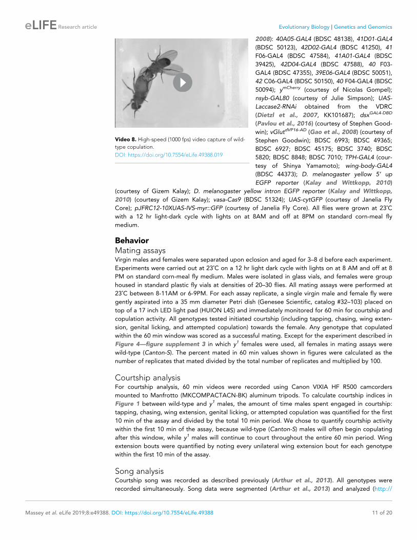
Video 1. Wild-type courtship and copulation.
DOI: https://doi.org/10.7554/eLife.49388.004
Video 2. y1 courtship with wild-type female.
DOI: https://doi.org/10.7554/eLife.49388.005
Massey et al. eLife 2019;8:e49388. DOI: https://doi.org/10.7554/eLife.49388 3 of 20
Research article Evolutionary Biology Genetics and Genomics

genotypes using a Fisher’s exact test (FET) with
p-values adjusted (Bonferroni) for the (n = 2)
control comparisons, we found no significant
effect on male mating success (Figure 2A, p=1
for both tests; Figure 2B, p=0.07 and 0.2), sug-
gesting that expression of yellow in fru-express-
ing cells is neither necessary nor sufficient for
yellow’s effect on male mating success.
Doublesex-expressing cells requireyellow for normal male matingsuccessTo continue searching for cells responsible for
yellow’s effects on mating, we examined a 209
bp sequence 5’ of the yellow gene called the
‘mating-success regulatory sequence’ (MRS) in a
prior study that reported it was required for male mating success (Drapeau et al., 2006). We
hypothesized that the MRS might contain an enhancer driving yellow expression and found that
ChIP-seq data indicate the Doublesex (Dsx) transcription factor binds to this region in vivo
(Clough et al., 2014). Like fru, dsx expression is required to specify sex-specific behaviors in D. mel-
anogaster (Rideout et al., 2010; Robinett et al., 2010; reviewed in Villella and Hall, 2008;
Yamamoto and Koganezawa, 2013), suggesting that yellow expression regulated by Dsx through
the MRS enhancer might be responsible for its effects on male mating behavior. We found that
reducing yellow expression in dsx-expressing cells with either of two different dsxGAL4 drivers
(Robinett et al., 2010; Rideout et al., 2010) strongly reduced male mating success (Figure 2C,
FET, p=7�10�9 and 1 � 10�7; Figure 2—figure supplement 1A, FET, p=0.002 and 0.002), whereas
restoring yellow activity in cells expressing dsxGAL4 in y1 mutants significantly increased male mating
success compared with y1 controls (Figure 2D, FET, p=0.001 and 0.0004; Figure 2—figure supple-
ment 1B, FET, p=5�10�10 and 5 � 10�10). Video recordings of male flies with reduced yellow
expression in dsx-expressing cells showed the same mating defect observed in y1 mutants: males
seem to perform all courtship actions normally, but repeatedly failed to copulate (Video 5). We
therefore conclude that yellow expression is required in dsx-expressing cells for normal male mating
behavior.
To determine whether the MRS sequence might be the enhancer mediating yellow expression in
dsx-expressing cells that affects male mating success, we manipulated yellow expression with GAL4
driven by a 2.7 kb DNA region located 5’ of yellow that includes the wing, body, and putative MRS
enhancers (Gilbert et al., 2006, Figure 2—figure supplement 2A). Altering yellow expression with
this GAL4 driver modified pigmentation as expected but did not affect male mating success (Fig-
ure 2—figure supplement 2B–D), possibly because this GAL4 line did not show any detectable
expression in the adult CNS (Figure 2—figure
supplement 2E). To test more directly whether
the MRS was necessary for male mating success,
we deleted 152 bp of the 209 bp MRS sequence
using CRISPR/Cas9 gene editing (Bassett et al.,
2013) (Figure 2—figure supplement 2F,G). We
found that this deletion had no significant effect
on male mating success (Figure 2—figure sup-
plement 2H, FET, p=0.99 compared to wild-
type (CS)), contradicting the previous deletion
mapping data (Drapeau et al., 2006). We con-
clude therefore that effects of yellow expression
in dsx-expressing cells on mating behavior are
likely mediated by other cis-regulatory sequen-
ces associated with the yellow gene.
Video 3. Wild-type copulation.
DOI: https://doi.org/10.7554/eLife.49388.006
Video 4. Copulation attempts between y1 male and
wild-type female after 3 hr of courtship.
DOI: https://doi.org/10.7554/eLife.49388.007
Massey et al. eLife 2019;8:e49388. DOI: https://doi.org/10.7554/eLife.49388 4 of 20
Research article Evolutionary Biology Genetics and Genomics

!"#$%&'&( )!'(*
!"#"$ !%&"%
!'&"'
!%(") !"("!!'*")!"("+
+,-")!"#$!
!%#"%
'./0
0
25
50
75
100
0
25
50
75
100
'!'!
'!
'+
'!
')
')
'!'+
%)'$
!"#"$
!%&"%
!'&"'
!%(")
!%#"%
!'&"!
!"("*
*+,")
!'-")
!"("!
!'&"!./#0!!"("*./#0!
1
2
2
1
1
2
1#2.3.45#6 1 1 2
2
1
2
2
2
1
%, '" %,
1#2.3.45#67879
3456478.9:84;.<7.)".9<7
====
==
3456478.9:84;.<7.)".9<7
'"
'"
=====
!
!"#$%&'
(%)*+*,-%.
0
25
50
75
100
!"#$"%&'()&"*'+%',-'(+%
.
.
./
/.
0- 1 0-
0
25
50
75
100"
+/00!"#$%&'
+/0(%)*+
2-
0101
!"#$"%&'()&"*'+%',-'(+%
.
.
./
/.
0
25
50
75
100
!"#$"%&'()&"*'+%',-'(+%
123$%&'
(%)*+*,-%..
#
2-
012-
.
./
/.
33333333
0
25
50
75
100$
+/00123$%&'
+/0(%)*+
24
4-
4-
!"#$"%&'()&"*'+%',-'(+%
.
.
./
/.
33333
0
25
50
75
100
4567*$%&'82+9*$%&'
.
/
/
.
.
/
(%)*+*,-%. . . /
/
.
/
/
/
.
!"#$"%&'()&"*'+%',-'(+% 5
060-
06 06
%
0
25
50
75
100&
!"#$"%&'()&"*'+%',-'(+%
"4:;*$%&'(%)*+*,-%.
.
.
./
/
.
0- 0- 0-
0
25
50
75
100
123$%&'*<=<
4567%<.
../
/.
(%)*+*,-%. . / .
!"#$"%&'()&"*'+%',-'(+% 0,
0,
0,
'
0
25
50
75
100
!"#$"%&'()&"*'+%',-'(+%
2-01
05
123$%&'*<=<
7$5#>1?@/A*%<.
.
.
.//
(%)*+*,-%. . / .
(
0
25
50
75
100
+/00123$%&'
+/082+9*$%&BC.
...
//
2-
06
05
+/0(%)*+ . / .
)
3333
!"#$"%&'()&"*'+%',-'(+%
!"#$%
!"##$%&'("))!
!"#$%
!"##$%&'("))!
!
" #
$ * !"##$%&'("))!
Figure 2. yellow expression in non-neuronal doublesex-expressing cells, but not fruitless-expressing cells, is
necessary and sufficient for male mating success. (A,B) Neither expressing yellow-RNAi nor yellow-cDNA in fru-
expressing cells using fruGAL4 (Stockinger et al., 2005) affected male copulation. (C) Expressing yellow-RNAi in
dsx-expressing cells using dsxGAL4 (Robinett et al., 2010) significantly inhibited male mating success. (D)
Figure 2 continued on next page
Massey et al. eLife 2019;8:e49388. DOI: https://doi.org/10.7554/eLife.49388 5 of 20
Research article Evolutionary Biology Genetics and Genomics

dsx-expressing cells outside the CNS require yellow for normal malemating successAlthough dsx is expressed broadly throughout
the fly (Robinett et al., 2010; Rideout et al.,
2010), we hypothesized that its expression in
the nervous system would be responsible for yel-
low’s effects on mating because yellow has been
reported to be expressed in the adult brain
(Hinaux et al., 2018) and behavioral effects of
other pigmentation genes are mediated by neu-
rons (Hotta and Benzer, 1969; Heisen-
berg, 1971; Borycz et al., 2002; True et al.,
2005). However, we found that suppressing yel-
low expression in the larval CNS, dopaminergic
neurons, or serotonergic neurons (Figure 2—fig-
ure supplement 3, FET, P values ranging from
0.45 to 1), or in all neurons (Figure 2E, FET, p=1
in all cases) or all glia (Figure 2F, FET, p=1), had
Figure 2 continued
Expressing yellow in dsx-expressing cells using dsxGAL4 in a y1 mutant background was sufficient to restore male
mating success. (E,F) Expressing yellow-RNAi using pan-neuronal (elav-GAL4 and nsyb-GAL4) and pan-glia (repo-
GAL4) drivers did not affect male mating success. (G) Restricting yellow-RNAi expression to dsx-expressing
neurons using the split-GAL4 technique, combining dsxGAL4-DBD (Pavlou et al., 2016) with elavVP16-AD (Luan et al.,
2006), did not affect male mating success. (H) Restricting yellow-RNAi expression to dsx-expressing glutamatergic
neurons using the split-GAL4 technique, combining dsxGAL4-DBD (Pavlou et al., 2016) with vGlutdVP16-AD
(Gao et al., 2008) did not affect male mating success. (I) Expressing yellow in dsx-expressing cells restricted
outside the CNS using dsxGAL4 and nsyb-GAL80 (courtesy of Julie Simpson) in a y1 mutant background
significantly increased male mating success. (J) Brain and ventral nerve cord of adult male ymCherry flies stained
with anti-N-Cadherin (N-cad) antibody labeling neuropil (white) and anti-DsRed antibody labeling Yellow::mCherry
(red). We observed sparse, inconsistent signal outside the CNS at the top of the brain in males (white arrow), but
we were unable to confirm a previous report that ymCherry is expressed in the adult brain (Hinaux et al., 2018). (K)
Diagram of the male exon structure of the dsx locus highlighting 10 genomic fragments between 1.7 and 4 kb
used to clone Janelia enhancer trap GAL4 drivers (Pfeiffer et al., 2008). Black boxes indicate coding exons. White
boxes indicate 5’ and 3’ UTRs, and the arrow in exon two denotes the transcription start site. (L) Expressing yellow-
RNAi using each Janelia dsx-GAL4 driver identified 42D04-GAL4 and 40 F03-GAL4 as affecting male mating
success when compared with the yellow-RNAi control. (M) A replicate experiment comparing 42D04-GAL4 and
40F03-GAL4 effects on male mating success with both GAL4 and UAS parental controls confirmed the significant
effect of 42D04-GAL4 but not 40F03-GAL4. We attribute differences in the 40F03-GAL4 effect between (L) and (M)
to between experiment variability in the levels of male mating success; each common genotype tested in (L), for
example, mated at higher levels in (M), but 42D04-GAL4 consistently showed a significant effect relative to
controls. Sample sizes are shown at the top of each barplot. Significance was measured using Fisher’s exact tests
with Bonferroni corrections for multiple comparisons. Comparisons that were statistically (p<0.05) are indicated
(*p<0.05, **p<0.01, ***p<0.001, ****p<0.0001).
DOI: https://doi.org/10.7554/eLife.49388.008
The following figure supplements are available for figure 2:
Figure supplement 1. yellow expression in dsx-expressing cells is necessary and sufficient for male mating
success.
DOI: https://doi.org/10.7554/eLife.49388.009
Figure supplement 2. The mating regulatory sequence (MRS) from Drapeau et al. (2006) does not affect male
mating success.
DOI: https://doi.org/10.7554/eLife.49388.010
Figure supplement 3. Expressing yellow-RNAi in subsets of CNS tissue does not affect male mating success.
DOI: https://doi.org/10.7554/eLife.49388.011
Figure supplement 4. ymCherry expression in adult female central nervous system.
DOI: https://doi.org/10.7554/eLife.49388.012
Video 5. Copulation attempts between male
expressing yellow-RNAi in dsxGAL4-expressing cells and
wild-type female.
DOI: https://doi.org/10.7554/eLife.49388.013
Massey et al. eLife 2019;8:e49388. DOI: https://doi.org/10.7554/eLife.49388 6 of 20
Research article Evolutionary Biology Genetics and Genomics

no significant effect on male mating success. Specifically reducing yellow expression in either all dsx-
expressing neurons (Figure 2G, FET, p=1 and 0.45) or all dsx-expressing glutamatergic neurons that
are required for genital coupling (Pavlou et al., 2016) (Figure 2H, FET, p=1 and 0.68) also had no
significant effect on male mating success. In addition, when we examined yellow expression in adult
brains, we were only able to observe non-specific signal at the anterior of the adult brain in females
(Figure 2J, Figure 2—figure supplement 4). Given this lack of evidence that yellow is required in
neuronal cells for normal male mating behavior, we limited dsxGAL4 activation of yellow expression in
y1 mutants to non-neuronal cells and found that these flies exhibited an increase in male mating suc-
cess compared with y1 mutant males (Figure 2I, FET, p=0.04 and 0.0002), showing that yellow
expression in non-neuronal dsx-expressing cells is required for normal male mating behavior.
To identify which non-neuronal dsx-expressing cells require yellow expression for normal male
mating success, we screened ten dsx-enhancer GAL4 lines that each contains a different ~ 3 kb
region of dsx noncoding sequence (Figure 2K; Pfeiffer et al., 2008). Two of these lines, 42D04-
GAL4 and 40F03-GAL4, significantly decreased male mating success when driving yellow-RNAi
(Figure 2L, FET, p=0.001 and 2 � 10�5). These two GAL4 drivers contain overlapping sequences
from intron 2 of dsx (Figure 2K), suggesting that their similar effects result from reduction of yellow
expression in the same cells. Line 42D04-GAL4 had stronger effects than 40 F03-GAL4 (Figure 2M,
FET, p=0.0009 for both controls for 42D04-GAL4 versus p=0.97 for both controls for 40 F03-GAL4),
so we performed all further analyses with 42D04-GAL4. Males with yellow reduced by 42D04-GAL4
performed courtship behavior in a pattern similar to y1 mutant males: males performed all precopu-
latory courtship behaviors normally, but repeatedly failed to copulate, even after hours of attempts
(Video 6). These data indicate that some or all cells in which 42D04-GAL4 drives expression require
yellow expression for normal male mating behavior.
Sex combs require yellow expression for normal male mating success42D04-GAL4 drives expression in a sexually dimorphic pattern in multiple neurons of the adult male
(Figure 3A,B) and female CNS (Figure 3—figure supplement 1A,B), consistent with previously
described dsxGAL4 expression in the posterior cluster, the abdominal cluster, and, in males, in the
prothoracic TN1 neurons (Robinett et al., 2010). 42D04-GAL4 also drives expression in male and
female larval CNS and genital discs, with expression in the genital tissues persisting into the adult
stage only in females (Figure 3—figure supplement 1C–G). Finally, we observed 42D04-GAL4
expression at the base of the sex combs (also observed by Robinett et al., 2010 and Rice et al.,
2019), which are modified bristles used during mating (Cook, 1975; Ng and Kopp, 2008; Hurtado-
Gonzales et al., 2015) that are present only on the first tarsal segment of adult male forelegs
(Figure 3C–F). Yellow protein is expressed in sex combs (Hinaux et al., 2018, Figure 3G,H), where
it is presumably required for synthesis of black dopamine melanin in the sex comb ‘teeth’. This
expression of yellow in sex comb cells is driven by enhancer sequences in the yellow intron (Fig-
ure 3—figure supplement 2), potentially explaining why manipulating yellow expression using
GAL4 driven by sequences 5’ of the yellow gene
failed to affect mating (Figure 2—figure supple-
ment 2A–D). Driving expression of yellow-RNAi
with 42D04-GAL4 eliminated expression of an
mCherry tagged version of the native Yellow
protein in sex combs and strongly reduced black
melanin in the sex combs (Figure 3I–L) but not
the abdomen (Figure 3—figure supplement
1J).
To test the impact of yellow expression in sex
combs on male mating behavior, we used
42D04-GAL4 to drive yellow-RNAi, but inhibited
the function of 42D04-GAL4 in the CNS with
nysb-GAL80 (courtesy of Julie Simpson). These
flies showed no GAL4 activity in the CNS
(Figure 3M,N), but lost black melanin in the sex
combs (Figure 3O) and had reduced male mat-
ing success (Figure 3P, FET, p=0.002 and 0.08).
Video 6. Copulation attempts between male
expressing yellow-RNAi in 42D04-GAL4-expressing
cells and wild-type female.
DOI: https://doi.org/10.7554/eLife.49388.014
Massey et al. eLife 2019;8:e49388. DOI: https://doi.org/10.7554/eLife.49388 7 of 20
Research article Evolutionary Biology Genetics and Genomics

0
25
50
75
100
!"#$!%&'(!)*)+',%&-.
!"#
!
"
#
$
$%&!"#'()*
$%&!"#'()* !"#
+,-.
/012344/
%
&
/35567012344/
+,-./35567012344/
!"#$!%&'(!)8)+',%/35567%9:';
' (
) *
+, !"##$%&
'()*
+,-./35567012344/
!"#
+,
+,-./35567012344/
!"#
!"##$%&'()*
</=>%&'(?$)8)!"#$!%&'(!)*)
+',%/35567%9:';
#/&0/'-1$2-/314'1561$4'
- 77
8*
9
8*
<=/>%&'(?$@)!"#$!%&'(!)*)
+',%/%9:';
<=/>%&'(?$@)!"#$!%&'(!)A)8
+',%/%9:';)A)8
.
/
0
!"##$%& '()*
'()*
$%&!"#
'()*$%&!"#
1
2
+,
+,
Figure 3. yellow expression in non-neuronal 42D04-GAL4 expressing cells is necessary for sex comb melanization
and male mating success. (A,B) Brain and ventral nerve cord of adult male fly stained with anti-GFP (green)
antibody for myrGFP expressed using 42D04-GAL4 and counterstained with anti-nC82 (magenta) for neuropil. (C)
Wild-type (wt) D. melanogaster adult male fly highlighting the location of sex combs (Nicolas Gompel). (D) Close
up of a wild-type (wt) sex comb on the first tarsal segment (ts1) of the front leg (courtesy of Nicolas Gompel). (E)
Bright field illumination of a male front leg expressing cytGFP (green) in sex-comb cells using 42D04-GAL4. (F)
Confocal image of the sex comb cells expressing cytGFP (green) with 42D04-GAL4 and leg cuticle
autofluorescence (blue). (G) Confocal image of a ymCherry male leg highlighting native ymCherry sex comb
expression (red). (H) Zoomed in confocal image shown in (G) with leg cuticle autofluorescence (blue) and native
ymCherry sex comb expression (red). (I) Wild-type (wt) sex comb. (J) Loss of black melanin in sex combs in males
Figure 3 continued on next page
Massey et al. eLife 2019;8:e49388. DOI: https://doi.org/10.7554/eLife.49388 8 of 20
Research article Evolutionary Biology Genetics and Genomics

High-speed videos (1000 frames per second) revealed that yellow mutant (y1) males fail repeatedly
to grasp the female abdomen with their sex combs when attempting to mount and copulate
(Video 7), whereas wild-type males more readily grasp the female with their melanized sex combs
and initiate copulation efficiently (Video 8). These observations suggest that yellow expression in
sex combs affects their melanization, which in turn affects their function.
Sex comb melanization is required for efficient grasping, mounting andcopulationTo test whether sex comb melanization (as opposed to some other unknown effect of losing yellow
expression in sex combs) is critical for male sexual behavior, we suppressed expression of Laccase2
(Arakane et al., 2005; Riedel et al., 2011) in sex combs using 42D04-GAL4 and Laccase2-RNAi
(Dietzl et al., 2007). Laccase2 is required to oxidize dopamine into dopamine quinones and thus
acts upstream of Yellow in the melanin synthesis pathway (Figure 4A; Riedel et al., 2011). Males
with Laccase2 suppressed in sex combs lacked both black and brown dopamine melanin, making
these sex combs appear translucent (Figure 4B). These males displayed strongly reduced mating
success compared with wild-type males (Figure 4C, FET, p=1�10�7 and 8 � 10�6) and behavioral
defects similar to those observed for y1 mutants (Videos 9 and 10), including inefficient grasping of
the female for mounting and copulation. We noticed, however, that flies with Laccase2-RNAi driven
by 42D04-GAL4 also showed a loss of melanin in the aedeagus (Figure 4—figure supplement 1A),
which is the main part of the male genitalia used for copulation, despite no visible expression of
42D04-GAL4 in the adult male genitalia (Fig-
ure 3—figure supplement 1G) nor changes in
aedeagus pigmentation in y1 mutants (Figure 4—
figure supplement 1A). We therefore used sub-
sets of the 42D04 enhancer (Figure 4—figure
supplement 1B) to drive expression of Lac-
case2-RNAi, separating the effects of expression
in the sex combs from expression in the genitalia
(Figure 4—figure supplement 1C). Male mating
success was reduced when Laccase2 suppression
reduced melanization in the sex combs, but not
the genitalia (Figure 4—figure supplement 1D–
G).
How can sex comb melanization affect sex
comb function? In insects, melanization impacts
not only the color of the adult cuticle but also its
mechanical stiffness (Xu et al., 1997;
Figure 3 continued
expressing yellow-RNAi using 42D04-GAL4. (K) Co-localization of ymCherry (red) at the base of the sex comb cells
expressing cytGFP (green) with 42D04-GAL4. (L) Loss of ymCherry (red) at the base of the sex comb cells expressing
cytGFP (green) and yellow-RNAi using 42D04-GAL4. (M,N) Brain and ventral nerve cord of adult male expressing
nsyb-GAL80 to block GAL4 activity in the CNS, stained with anti-GFP (green) antibody for myrGFP expressed
using 42D04-GAL4, and counterstained with anti-nC82 (magenta) for neuropil. (O) Loss of black melanin in sex
combs in nsyb-GAL80 males expressing yellow-RNAi using 42D04-GAL4. (P) Expressing yellow-RNAi using 42D04-
GAL4 in males expressing nsyb-GAL80 significantly inhibited male mating success. Scale bars in (I), (J), and (O)
measure 12.5 mm. Sample sizes are shown at the top of each barplot. Significance was measured using Fisher’s
exact tests with Bonferroni corrections for multiple comparisons. Comparisons that were statistically (p<0.05) are
indicated (**p<0.01).
DOI: https://doi.org/10.7554/eLife.49388.015
The following figure supplements are available for figure 3:
Figure supplement 1. Expression pattern of 42D04-GAL4.
DOI: https://doi.org/10.7554/eLife.49388.016
Figure supplement 2. yellow EGFP reporters localize yellow sex comb expression to the intronic bristle enhancer.
DOI: https://doi.org/10.7554/eLife.49388.017
Video 7. High-speed (1000 fps) video capture of
copulation attempts between y1 male and wild-type
female.
DOI: https://doi.org/10.7554/eLife.49388.018
Massey et al. eLife 2019;8:e49388. DOI: https://doi.org/10.7554/eLife.49388 9 of 20
Research article Evolutionary Biology Genetics and Genomics

Kerwin et al., 1999; Vincent and Wegst, 2004; Andersen, 2005; Arakane et al., 2005;
Suderman et al., 2006; Riedel et al., 2011; Noh et al., 2016). For example, expressing Laccase2-
RNAi in D. melanogaster wings softens the cuticle to such a degree that the wings collapse
(Riedel et al., 2011). Butterflies lacking dopamine melanin due to loss of yellow or another gene
required for melanin synthesis, Dopa decarboxylase, also show changes in the fine structure of their
wing scales (Matsuoka and Monteiro, 2018). Consistent with these observations, we observed
structural changes in D. melanogaster sex comb teeth lacking yellow or Laccase2 expression using
scanning electron microscopy (SEM), with a crack appearing in one of the Laccase2-RNAi comb teeth
(Figure 4D). We thus conclude that these structural changes in sex combs are responsible for inhibit-
ing the yellow mutant male’s ability to grasp a female for mounting and copulation (Video 10). In
1976, Wilson et al. (1976) speculated about this very hypothesis based on their own observations of
behavior in yellow mutant males.
Data from other Drosophila species are also consistent with this structural hypothesis. Specifically,
yellow mutants in D. subobscura, D. pseudoobscura, and D. gaucha, all of which have sex combs,
show reduced male mating success with wild-type females (Rendel, 1944; Tan, 1946; Frias and
Lamborot, 1970; Pruzan-Hotchkiss et al., 1992) whereas yellow mutants in Drosophila willistoni, a
species that lacks sex combs (Kopp, 2011; Atallah et al., 2014), do not (da Silva et al., 2005). Sex
comb morphology is highly diverse among species that have sex combs (Kopp, 2011), but these
structures generally seem to be melanized (Figure 4—figure supplement 2; Tanaka et al., 2009)
and used to grasp females (Videos 11–15). (Our high-speed video recordings of mating in D. anan-
nasae, D. bipectinata, D. kikkawai, D. malerkotiana, and D. takahashi show that differences in sex
comb morphology (Figure 4—figure supplement 2) correspond with differences in how (where on
the female and with which part of the male leg) the male grasps the female prior to copulation (Vid-
eos 11–15).
It remains unclear how D. willistoni males (and males of other species without sex combs) are able
to efficiently grasp females prior to copulation (Video 16). However, differences in females might be
part of the answer, as D. melanogaster y1 mutant males are able to mate with y1 mutant females at
rates similar to wild-type males (Bastock, 1956; Dow, 1976; Heisler, 1984; Liu et al., 2019; Fig-
ure 4—figure supplement 3A, FET, p=1). That said, removing all melanin from D. melanogaster sex
combs by knocking down Laccase-2 reduced mating efficiency with y1 females (Figure 4—figure
supplement 3B, FET, p=0.02 and 0.0001), suggesting that the brown melanin remaining in y1 sex-
combs (Figure 4B) played a role in the mating success of y1 males with y1 females.
ConclusionTaken together, our data show that melanization of a secondary sexual structure affects mating in D.
melanogaster. Specifically, we find that the reduced mating success of D. melanogaster yellow
mutant males, which was perceived as a behavioral defect for decades, is caused by changes in the
morphology of the structures used during mating. Other recent studies have also shown the impor-
tance of morphological structures for stickleback schooling (Greenwood et al., 2015), water strider
walking (Santos et al., 2017), and cricket singing (Pascoal et al., 2014) behaviors. These observa-
tions all underscore that behavior cannot be understood by studying the nervous system alone; anat-
omy and behavior function and evolve as an interconnected system.
Materials and methodsWe have included a Key Resources Table as Supplementary file 4.
Fly stocks and maintenanceThe following lines were used for this work: y1 [which was backcrossed into a wild-type (Canton-S)
line for six generations before starting our experiments; the y1 allele contains an A to C transversion
in the ATG initiation and is considered a null allele (Geyer et al., 1990)]; Canton-S as wild-type
(courtesy of Scott Pletcher); UAS-yellow-RNAi obtained from the Vienna Drosophila Resource Centre
(VDRC) (Dietzl et al., 2007, KK106068); y1;UAS-y (BDSC 3043); elav-GAL4 (BDSC 49226); nsyb-
GAL4 (BDSC 39171); repo-GAL4 (BDSC 7415); dsxGAL4 (Robinett et al., 2010) (courtesy of Bruce
Baker); dsxGAL4 (Rideout et al., 2010) (courtesy of Stephen Goodwin); fruGAL4 (Stockinger et al.,
2005) (courtesy of Barry Dickson); the following Janelia enhancer trap GAL4 lines (Pfeiffer et al.,
Massey et al. eLife 2019;8:e49388. DOI: https://doi.org/10.7554/eLife.49388 10 of 20
Research article Evolutionary Biology Genetics and Genomics
2008): 40A05-GAL4 (BDSC 48138), 41D01-GAL4
(BDSC 50123), 42D02-GAL4 (BDSC 41250), 41
F06-GAL4 (BDSC 47584), 41A01-GAL4 (BDSC
39425), 42D04-GAL4 (BDSC 47588), 40 F03-
GAL4 (BDSC 47355), 39E06-GAL4 (BDSC 50051),
42 C06-GAL4 (BDSC 50150), 40 F04-GAL4 (BDSC
50094); ymCherry (courtesy of Nicolas Gompel);
nsyb-GAL80 (courtesy of Julie Simpson); UAS-
Laccase2-RNAi obtained from the VDRC
(Dietzl et al., 2007, KK101687); dsxGAL4-DBD
(Pavlou et al., 2016) (courtesy of Stephen Good-
win); vGlutdVP16-AD (Gao et al., 2008) (courtesy of
Stephen Goodwin); BDSC 6993; BDSC 49365;
BDSC 6927; BDSC 45175; BDSC 3740; BDSC
5820; BDSC 8848; BDSC 7010; TPH-GAL4 (cour-
tesy of Shinya Yamamoto); wing-body-GAL4
(BDSC 44373); D. melanogaster yellow 5’ up
EGFP reporter (Kalay and Wittkopp, 2010)
(courtesy of Gizem Kalay); D. melanogaster yellow intron EGFP reporter (Kalay and Wittkopp,
2010) (courtesy of Gizem Kalay); vasa-Cas9 (BDSC 51324); UAS-cytGFP (courtesy of Janelia Fly
Core); pJFRC12-10XUAS-IVS-myr::GFP (courtesy of Janelia Fly Core). All flies were grown at 23˚C
with a 12 hr light-dark cycle with lights on at 8AM and off at 8PM on standard corn-meal fly
medium.
BehaviorMating assaysVirgin males and females were separated upon eclosion and aged for 3–8 d before each experiment.
Experiments were carried out at 23˚C on a 12 hr light dark cycle with lights on at 8 AM and off at 8
PM on standard corn-meal fly medium. Males were isolated in glass vials, and females were group
housed in standard plastic fly vials at densities of 20–30 flies. All mating assays were performed at
23˚C between 8-11AM or 6-9PM. For each assay replicate, a single virgin male and female fly were
gently aspirated into a 35 mm diameter Petri dish (Genesee Scientific, catalog #32–103) placed on
top of a 17 inch LED light pad (HUION L4S) and immediately monitored for 60 min for courtship and
copulation activity. All genotypes tested initiated courtship (including tapping, chasing, wing exten-
sion, genital licking, and attempted copulation) towards the female. Any genotype that copulated
within the 60 min window was scored as a successful mating. Except for the experiment described in
Figure 4—figure supplement 3 in which y1 females were used, all females in mating assays were
wild-type (Canton-S). The percent mated in 60 min values shown in figures were calculated as the
number of replicates that mated divided by the total number of replicates and multiplied by 100.
Courtship analysisFor courtship analysis, 60 min videos were recorded using Canon VIXIA HF R500 camcorders
mounted to Manfrotto (MKCOMPACTACN-BK) aluminum tripods. To calculate courtship indices in
Figure 1 between wild-type and y1 males, the amount of time males spent engaged in courtship:
tapping, chasing, wing extension, genital licking, or attempted copulation was quantified for the first
10 min of the assay and divided by the total 10 min period. We chose to quantify courtship activity
within the first 10 min of the assay, because wild-type (Canton-S) males will often begin copulating
after this window, while y1 males will continue to court throughout the entire 60 min period. Wing
extension bouts were quantified by noting every unilateral wing extension bout for each genotype
within the first 10 min of the assay.
Song analysisCourtship song was recorded as described previously (Arthur et al., 2013). All genotypes were
recorded simultaneously. Song data were segmented (Arthur et al., 2013) and analyzed (http://
Video 8. High-speed (1000 fps) video capture of wild-
type copulation.
DOI: https://doi.org/10.7554/eLife.49388.019
Massey et al. eLife 2019;8:e49388. DOI: https://doi.org/10.7554/eLife.49388 11 of 20
Research article Evolutionary Biology Genetics and Genomics

www.github.com/dstern/BatchSongAnalysis) without human intervention. Values for pulse per min-
ute, sine per minute, and interpulse interval were then extracted from the software.
High-speed video captureFor high-speed video capture of attempted mounting and copulation events, virgin males and
females were isolated upon eclosion and aged for 4–7 d before each assay. Using a Fascam Photron
SA4 (courtesy of Gwyneth Card) mounted with a 105 mm AF Micro Nikkor Nikon lens (courtesy of
Gwyneth Card), we recorded individual pairs of males and females that were gently aspirated into a
single well of a 96 well cell culture plate (Corning 05-539-200) partially filled with 2% agarose and
covered with a glass coverslip. We recorded mounting and copulation attempts at 1000 frames per
second (fps) and played back at 30 fps. Most wild-type males attempted mounting 3–5 times before
0
25
50
75
100
!"#$%&'( )$*+ )$*+,&'()$*+,&'(-.&'$'(
/0+12,(0+'&'
/#$3',(0+'&'
!" #$% &'%%'()*
+),,-./
0 1
!"#$!%&'(!)'*%(+,,+-."%/0'1
44
45
54
67
6767
8(#1('9:,+9(;:&':<=:,&' >>>>
>>>>39 23 (+,,+-."%/0'1
39 23 (+,,+-."%/0'1#
2
Figure 4. Sex comb melanization is specifically required for male mating success. (A) Simplified version of the
insect melanin synthesis pathway. (B) Light microscopy images of sex combs from wild-type (wt), y1, and 42D04-
GAL4; UAS-Laccase2-RNAi males. Expressing Laccase2-RNAi in sex combs completely blocked melanin synthesis.
(C) Expressing Laccase2-RNAi using 42D04-GAL4 in males significantly inhibited male mating success. (D)
Scanning Electron Microscopy (SEM) of sex combs from wild-type (wt), y1, and Laccase2-RNAi males (expressed
using 42D04-GAL4). Compared to wild-type, sex comb teeth in y1 mutants appeared thinner and smoother,
whereas Laccase2-RNAi sex comb teeth appeared even smoother than y1 mutants, and one comb tooth had a
visible crack in the cuticle (white rectangle, enlarged on the right). Scale bars in (B) measure 12.5 mm. Sample sizes
are shown at the top of each barplot. Significance in was measured using Fisher’s exact tests with Bonferroni
corrections for multiple comparisons. Comparisons that were statistically (p<0.05) are indicated (****p<0.0001).
DOI: https://doi.org/10.7554/eLife.49388.020
The following figure supplements are available for figure 4:
Figure supplement 1. Genetic dissection of the 42D04-GAL4 enhancer confirms the specific role of sex comb
melanization, and not the aedeagus, in male mating success.
DOI: https://doi.org/10.7554/eLife.49388.021
Figure supplement 2. Drosophila species with varying sex comb morphology used for high-speed video assays.
DOI: https://doi.org/10.7554/eLife.49388.022
Figure supplement 3. Sex comb melanization is required for male mating success with y1 females.
DOI: https://doi.org/10.7554/eLife.49388.023
Massey et al. eLife 2019;8:e49388. DOI: https://doi.org/10.7554/eLife.49388 12 of 20
Research article Evolutionary Biology Genetics and Genomics

copulating, whereas y1, yellow-RNAi, and Lac-
casse2-RNAi males repeatedly attempted mount-
ing without engaging in copulation, mirroring the
videos we captured on the Canon VIXIA HF R500
at 30 fps.
Imaging sex combs and genitaliaSex comb images highlighting different melani-
zation states (Figure 3I,J,O; Figure 4B) were
taken using a Zeiss Axio Cam ERc 5 s mounted
on a Zeiss Axio Observer A1 Inverted Micro-
scope. Front legs were cut and placed sex comb
side down on a microscope slide (Fisher brand
12-550-123) and imaged through a 40x objective.
Images were processed using AxioVision LE soft-
ware. Abdomens and genitalia images highlight-
ing different melanization states of the aedeagus
and female genital bristles were captured using a
Canon EOS Rebel T6 camera mounted with a
Canon MP-E 65 mm macro lens. Genitalia images were processed in Adobe Photoshop (version
19.1.5) (Adobe Systems Inc, San Jose, CA).
Focus Ion Beam Scanning Electron Microscope (FIB-SEM) images (Figure 4D) were taken by plac-
ing individual, dissected legs on carbon tape adhered to a scanning electron microscope pin stud
mount with sex combs facing up. The samples were then coated with a 20 nm Au layer using a Gatan
682 Precision Etching and Coating System, and imaged by scanning electron microscopy in a Zeiss
Sigma system. The samples were imaged using a 3-nA electron beam with 1.5 kV landing energy at
2.5MHz.
Immunohistochemistry and confocal imagingCentral Nervous SystemDissections, immunohistochemistry, and imaging of fly central nervous systems were done as previ-
ously described (Aso et al., 2014). In brief, brains and VNCs were dissected in Schneider’s insect
medium and fixed in 2% paraformaldehyde (diluted in the same medium) at room temperature for
55 min. Tissues were washed in PBT (0.5% Triton X-100 in phosphate buffered saline) and blocked
using 5% normal goat serum before incubation with antibodies. Tissues expressing GFP were stained
with rabbit anti-GFP (ThermoFisher Scientific A-11122, 1:1000) and mouse anti-BRP hybridoma
supernatant (nc82, Developmental Studies
Hybridoma Bank, Univ. Iowa, 1:30), followed by
Alexa Fluor 488-conjugated goat anti-rabbit and
Alexa Fluor 568-conjugated goat anti-mouse
Video 9. Copulation attempts between male
expressing Laccase2-RNAi in 42D04-GAL4-expressing
cells and wild-type female.
DOI: https://doi.org/10.7554/eLife.49388.024
Video 10. High-speed (1000 fps) video capture of
copulation attempts between male expressing
Laccase2-RNAi in 42D04-GAL4-expressing cells and
wild-type female.
DOI: https://doi.org/10.7554/eLife.49388.025
Video 11. Drosophila anannasae wild-type copulation.
DOI: https://doi.org/10.7554/eLife.49388.026
Massey et al. eLife 2019;8:e49388. DOI: https://doi.org/10.7554/eLife.49388 13 of 20
Research article Evolutionary Biology Genetics and Genomics

antibodies (ThermoFisher Scientific A-11034 and
A-11031), respectively. Tissues expressing
mCherry-tagged Yellow protein (ymCherry) were
stained with rabbit anti-dsRed (Clontech 632496,
1:1000) and rat anti-DN-Cadherin (DN-Ex #8,
Developmental Studies Hybridoma Bank, Univ.
Iowa, 1:100) as neuropil marker, followed by
Cy3-conjugated goat anti-rabbit and Cy5-conju-
gated goat anti-rat antibodies (Jackson Immu-
noResearch 111-165-144 and 112-175-167),
respectively. After staining and post-fixation in
4% paraformaldehyde, tissues were mounted on
poly-L-lysine-coated cover slips, cleared, and
embedded in DPX as described. Image z-stacks
were collected at 1 mm intervals using an
LSM710 confocal microscope (Zeiss, Germany)
fitted with a Plan-Apochromat 20x/0.8 M27
objective. Images were processed in Fiji (http://fiji.sc/) and Adobe Photoshop (version 19.1.5)
(Adobe Systems Inc, San Jose, CA).
Sex combs and genitaliaAdult flies were 2–7 d old and pupae were 96 hr old after pupal formation (APF) for the EGFP
reporter experiment summarized in Figure 3—figure supplement 2. Flies were anesthetized on ice,
submerged in 70% ethanol, rinsed twice in phosphate buffered saline with 0.1% Triton X-100 (PBS-
T), and fixed in 2% formaldehyde in PBS-T. Forelegs and genitalia/abdomen tips were removed with
fine scissors and mounted in Tris-buffered (pH 8.0) 80% glycerol. Serial optical sections were
obtained at 1.5 mm or 0.5 mm intervals on a Zeiss 880 confocal microscope with a LD-LCI 25x/0.8 NA
objective (genitalia) or a Plan-Apochromat 40x/1.3 NA objective (appendages/tarsal sex combs). The
native fluorescence of GFP, mCherry and autofluorescence of cuticle were imaged using 488, 594
and 633 lasers, respectively. Images were processed in Fiji (http://fiji.sc/), Icy (http://icy.bioimagea-
nalysis.org/) and Adobe Photoshop (version 19.1.5) (Adobe Systems Inc, San Jose, CA).
Generation of the mating regulatory sequence (MRS) deletion lineUsing the 209 bp region mapped in Drapeau et al. (2006) between �300 and �91 bp upstream of
yellow’s transcription start site, we designed two single guide RNA (gRNA) target sites at �291 bp
and �140 bp that maximized the MRS deletion region, given constraints of identifying NGG PAM
sites required for CRISPR/Cas9 gene editing (Figure 2—figure supplement 2). We in-vitro tran-
scribed these gRNAs using a MEGAscript T7 Transcription Kit (Invitrogen) following the PCR-based
protocol from Bassett et al. (2013). Two 1 kb homology arms were PCR amplified from the yellow
locus immediately upstream and downstream of
the gRNA target sites using forward and reverse
primers with NcoI and BglII tails, respectively, for
the Left Arm (5’-TTACCATGGGGGATCAAG
TTGAACCAC-3’, 5’-GGAGATCTGGCCTTCA
TCGACATTTA-3’) and the forward and reverse
primers with Bsu36I and MluI tails, respectively,
for the Right Arm (5’-TACATCCCTAAGGCCTGA
TTACCCGAACACT-3’, 5’-TATACGCGTTGCCA
TGCTATTGGCTTC-3’) and cloned into pHD-
DsRed-attp (Gratz et al., 2014; Addgene Plas-
mid # 51019) in two steps, digesting first with
NcoI and BglII (Left Arm) to transform the Left
Arm and second with Bsu36I and MluI (Right
Arm) to transform the Right Arm, flanking the
3xP3::DsRed, attP, and LoxP sites. Homology
Video 12. Drosophila bipectinata wild-type copulation.
DOI: https://doi.org/10.7554/eLife.49388.027
Video 13. Drosophila kikkawai wild-type copulation.
DOI: https://doi.org/10.7554/eLife.49388.028
Massey et al. eLife 2019;8:e49388. DOI: https://doi.org/10.7554/eLife.49388 14 of 20
Research article Evolutionary Biology Genetics and Genomics

arms were ligated into pHD-DsRed-attp using T4 DNA Ligase (ThermoFisher Scientific), and prod-
ucts were transformed into One Shot TOP10 (Invitrogen) DH5 alpha competent cells. Purified donor
plasmid was then co-injected at 500 ng/uL with the two gRNAs at 100 ng/uL total concentration into
a vasa-Cas9 (BDSC 51324) line. Flies were then screened for DsRed expression in the eyes, and
Sanger sequenced verified for a 3xP3::DsRed replacement of the MRS region (Figure 2—figure sup-
plement 2). We confirmed that we deleted 152 bp of the 209 bp region based on Sanger sequenc-
ing the CRISPR/Cas9 cut sites (Figure 2—figure supplement 2). Next, we crossed yDMRS+3xP3::DsRed
with a Cre-expressing fly line (courtesy of Bing Ye, University of Michigan) to excise 3xP3::DsRed
and screened for flies that lost DsRed expression in the eyes. Finally, we PCR-gel verified that DsRed
was indeed removed in creation of the yDMRS line using the forward and reverse primers, respectively
(5’-CAGTCGCCGATAAAGATGAACACTG-3’, 5’-CAAGGTGATCAGGGTCACAAGGATC-3’) (Fig-
ure 2—figure supplement 2).
Generation of the 42D04-GAL4 enhancer sub-fragment pBPGUw linesEnhancer sub-fragments (2 kb, 2 kb, 1.3 kb, 1.3 kb, and 1.3 kb for 42D04_A,B,C,D,E-GAL4, respec-
tively) were synthesized as IDT gene blocks (sequences available in Supplementary file 1) based off
of the 42D04 D. melanogaster dsx enhancer sequence (FBsf0000164494) (Figure 4—figure supple-
ment 1). The gene blocks were designed with 5’ and 3’ Gibson tails to facilitate Gibson assembly
(Gibson et al., 2009) into the GAL4 plasmid pBPGUw (Pfeiffer et al., 2008; Addgene Plasmid
#17575) after digestion with FseI and AatII. Products were transformed into Mix and Go! DH5 alpha
competent cells (Zymo). Clones were selected by ampicillin resistance on Amp-LB plates (60 mg/
mL). Purified plasmids were injected at 500 ng/uL into the phiC31 integrase-expressing 86Fb landing
site line BDSC 24749 (courtesy of Rainbow Transgenics) for phiC31 attP-attB integration and
screened for using a mini-white marker.
StatisticsSupplementary file 2 is a Microsoft Excel file
containing four worksheets with all of the data
used for analysis. The worksheet labeled ‘Uni-
var_Male_Mating_Success_Data’ contains a uni-
variate description of each mating trial. The
worksheet labeled ‘Summary of mating success
data’ shows the number of successful and unsuc-
cessful matings for each genotype tested
(grouped by figure panel including the data) and
was generated from the ‘Univar_Male_Mating_-
Success_Data’ worksheet using the Excel Pivot
Table function. The worksheet labeled ‘Court-
ship_Data’ includes the data for courtship index
and wing extensions shown in Figure 1D and E,
Video 14. Drosophila malerkotiana wild-type copulation.
DOI: https://doi.org/10.7554/eLife.49388.029
Video 15. Drosophila takahashi wild-type copulation.
DOI: https://doi.org/10.7554/eLife.49388.030
Video 16. Drosophila willistoni wild-type copulation.
DOI: https://doi.org/10.7554/eLife.49388.031
Massey et al. eLife 2019;8:e49388. DOI: https://doi.org/10.7554/eLife.49388 15 of 20
Research article Evolutionary Biology Genetics and Genomics

respectively. The worksheet labeled ‘Song Data’ includes the measures of pulses per minute, sin per
minute and interpulse interval (labeled ‘ModeEndToStartIPI’) exported from the software described
in Arthur et al. (2013). R version 3.6.1 (2019-07-05) (R Development Core Team, 2013) was used
for all statistical analyses using the code included in Source code 1. These analyses included t-tests
comparing courtship index, number of wing extensions, pulses per minute, sine per minute, and
interpulse interval that were run after exporting data in the Courtship Data and Song Data work-
sheets (separately) to tab delimited text files. Note that the default t-test parameters allowing for
unequal variance between samples were used. Source code 1 also contains the R code for the Fish-
er’s Exact Tests, which were coded using data from the ‘Summary of mating success data’ work-
sheet. Supplementary file 3 contains a summary of all statistical tests. Whenever an experimental
genotype was compared to two control genotypes, P-values were adjusted using a Bonferroni cor-
rection for N = 2 (see Supplementary file 3). We note that for N = 2, alternative adjustments avail-
able with the p.adjust function in R (‘holm’, ‘Hochberg’, ‘hommel’ and ‘fdr’) give the same adjusted
P-value.
AcknowledgementsWe thank members of the Wittkopp and Stern labs for helpful discussions. For fly strains, we thank
Bruce Baker, Carmen Robinett, Stephen Goodwin, Barry Dickson, Scott Pletcher, Julie Simpson, Shi-
nya Yamamoto, Bing Ye, Nicolas Gompel, Gizem Kalay, The Bloomington Drosophila Stock Center,
The Vienna Drosophila RNAi Center, and the Janelia Fly Core. For fly injections, we thank Rainbow
Transgenics Inc. For technical support with Scanning Electron Microscopy (SEM), we thank Harald
Hess and Song Pang. For use of the Photron for high-speed video capture, we thank Gwyneth Card
and W Ryan Williamson. CNS dissections, immunostaining, and imaging were performed by the
Janelia Project Technical Resource team with special thanks to Gudrun Ihrke, Kari Close, and Chris-
tina Christoforou. We thank Nicolas Gompel, Abby Lamb, and Henry Ertl for comments on the man-
uscript. We also thank Shyama Nandakumar and Ajai Pulianmackal for help with dissections and
confocal microscopy as well as Elena Kingston for capturing the Drosophila
willistoni copulation video.
Additional information
Competing interests
Patricia J Wittkopp: Senior editor, eLife. The other authors declare that no competing interests exist.
Funding
Funder Grant reference number Author
National Institutes of Health T32GM007544 Jonathan H Massey
National Institutes of Health GM089736 Patricia J Wittkopp
National Institutes of Health 1R35GM118073 Patricia J Wittkopp
The funders had no role in study design, data collection and interpretation, or the
decision to submit the work for publication.
Author contributions
Jonathan H Massey, Conceptualization, Data curation, Formal analysis, Funding acquisition,
Validation, Investigation, Visualization, Methodology, Writing—original draft, Writing—review and
editing; Daayun Chung, Conceptualization, Data curation, Formal analysis, Validation, Investigation,
Methodology, Writing—review and editing; Igor Siwanowicz, Resources, Data curation, Visualization,
Methodology; David L Stern, Supervision, Funding acquisition, Visualization, Writing—original draft,
Project administration, Writing—review and editing; Patricia J Wittkopp, Conceptualization,
Supervision, Funding acquisition, Visualization, Writing—original draft, Project administration,
Writing—review and editing
Massey et al. eLife 2019;8:e49388. DOI: https://doi.org/10.7554/eLife.49388 16 of 20
Research article Evolutionary Biology Genetics and Genomics

Author ORCIDs
Jonathan H Massey https://orcid.org/0000-0001-6182-2604
Igor Siwanowicz http://orcid.org/0000-0001-5819-1530
David L Stern https://orcid.org/0000-0002-1847-6483
Patricia J Wittkopp https://orcid.org/0000-0001-7619-0048
Decision letter and Author response
Decision letter https://doi.org/10.7554/eLife.49388.040
Author response https://doi.org/10.7554/eLife.49388.041
Additional filesSupplementary files. Source code 1. R code used for statistical analyses.
DOI: https://doi.org/10.7554/eLife.49388.032
. Supplementary file 1. Sequences used for cloning.
DOI: https://doi.org/10.7554/eLife.49388.033
. Supplementary file 2. Summary table containing data for all mating trials.
DOI: https://doi.org/10.7554/eLife.49388.034
. Supplementary file 3. Summary of all statistical tests reported in the paper.
DOI: https://doi.org/10.7554/eLife.49388.035
. Supplementary file 4. Key Resources Table.
DOI: https://doi.org/10.7554/eLife.49388.036
. Transparent reporting form
DOI: https://doi.org/10.7554/eLife.49388.037
Data availability
All data generated or analyzed during this study are included in the manuscript and supporting files.
Supplementary File 2 contains all source data and Supplementary File 3 contains R code for analyz-
ing it.
ReferencesAndersen SO. 2005. Cuticular sclerotization and tanning. Comprehensive Molecular Insect Science:145–170.DOI: https://doi.org/10.1016/b0-44-451924-6/00052-1
Arakane Y, Muthukrishnan S, Beeman RW, Kanost MR, Kramer KJ. 2005. Laccase 2 is the phenoloxidase generequired for beetle cuticle tanning. PNAS 102:11337–11342. DOI: https://doi.org/10.1073/pnas.0504982102,PMID: 16076951
Arthur BJ, Sunayama-Morita T, Coen P, Murthy M, Stern DL. 2013. Multi-channel acoustic recording andautomated analysis of Drosophila courtship songs. BMC Biology 11:11. DOI: https://doi.org/10.1186/1741-7007-11-11, PMID: 23369160
Aso Y, Hattori D, Yu Y, Johnston RM, Iyer NA, Ngo TT, Dionne H, Abbott LF, Axel R, Tanimoto H, Rubin GM.2014. The neuronal architecture of the mushroom body provides a logic for associative learning. eLife 3:e04577. DOI: https://doi.org/10.7554/eLife.04577, PMID: 25535793
Atallah J, Vurens G, Mavong S, Mutti A, Hoang D, Kopp A. 2014. Sex-specific repression of dachshund isrequired for Drosophila sex comb development. Developmental Biology 386:440–447. DOI: https://doi.org/10.1016/j.ydbio.2013.12.017, PMID: 24361261
Bassett AR, Tibbit C, Ponting CP, Liu JL. 2013. Highly efficient targeted mutagenesis of Drosophila with theCRISPR/Cas9 system. Cell Reports 4:220–228. DOI: https://doi.org/10.1016/j.celrep.2013.06.020, PMID: 23827738
Bastock M. 1956. A gene mutation which changes a behavior pattern. Evolution 10:421–439. DOI: https://doi.org/10.1111/j.1558-5646.1956.tb02868.x
Borycz J, Borycz JA, Loubani M, Meinertzhagen IA. 2002. tan and ebony genes regulate a novel pathway fortransmitter metabolism at fly photoreceptor terminals. The Journal of Neuroscience 22:10549–10557.DOI: https://doi.org/10.1523/JNEUROSCI.22-24-10549.2002, PMID: 12486147
Brand AH, Perrimon N. 1993. Targeted gene expression as a means of altering cell fates and generatingdominant phenotypes. Development 118:401–415. PMID: 8223268
Massey et al. eLife 2019;8:e49388. DOI: https://doi.org/10.7554/eLife.49388 17 of 20
Research article Evolutionary Biology Genetics and Genomics

Clough E, Jimenez E, Kim YA, Whitworth C, Neville MC, Hempel LU, Pavlou HJ, Chen ZX, Sturgill D, Dale RK,Smith HE, Przytycka TM, Goodwin SF, Van Doren M, Oliver B. 2014. Sex- and tissue-specific functions ofDrosophila doublesex transcription factor target genes. Developmental Cell 31:761–773. DOI: https://doi.org/10.1016/j.devcel.2014.11.021, PMID: 25535918
Cobb M. 2007. A gene mutation which changed animal behaviour: Margaret Bastock and the yellow fly. AnimalBehaviour 74:163–169. DOI: https://doi.org/10.1016/j.anbehav.2007.05.002
Cook R. 1975. Courtship of Drosophila melanogaster: rejection without extrusion. Behaviour 52:155–171.DOI: https://doi.org/10.1163/156853975X00010
da Silva LB, Leite DF, Valente VL, Rohde C. 2005. Mating activity of yellow and sepia Drosophila willistonimutants. Behavioural Processes 70:149–155. DOI: https://doi.org/10.1016/j.beproc.2005.06.004, PMID: 16098685
Dietzl G, Chen D, Schnorrer F, Su KC, Barinova Y, Fellner M, Gasser B, Kinsey K, Oppel S, Scheiblauer S, CoutoA, Marra V, Keleman K, Dickson BJ. 2007. A genome-wide transgenic RNAi library for conditional geneinactivation in Drosophila. Nature 448:151–156. DOI: https://doi.org/10.1038/nature05954, PMID: 17625558
Dow MA. 1976. The genetic basis of receptivity of yellow mutant Drosophila melanogaster females. BehaviorGenetics 6:141–143. DOI: https://doi.org/10.1007/BF01067144, PMID: 817705
Drapeau MD, Radovic A, Wittkopp PJ, Long AD. 2003. A gene necessary for normal male courtship, yellow, actsdownstream of fruitless in the Drosophila melanogaster larval brain. Journal of Neurobiology 55:53–72.DOI: https://doi.org/10.1002/neu.10196, PMID: 12605459
Drapeau MD, Cyran SA, Viering MM, Geyer PK, Long AD. 2006. A cis-regulatory sequence within the yellowlocus of Drosophila melanogaster required for normal male mating success. Genetics 172:1009–1030.DOI: https://doi.org/10.1534/genetics.105.045666, PMID: 16272418
Frias D, Lamborot M. 1970. Reproductive isolation between the yellow, white, and ‘‘wild’’ stocks of D. gaucha attwo temperatures (in Spanish). Archivos De Biologi�a Y Medicina Experimentales 7:67.
Gao S, Takemura SY, Ting CY, Huang S, Lu Z, Luan H, Rister J, Thum AS, Yang M, Hong ST, Wang JW,Odenwald WF, White BH, Meinertzhagen IA, Lee CH. 2008. The neural substrate of spectral preference inDrosophila. Neuron 60:328–342. DOI: https://doi.org/10.1016/j.neuron.2008.08.010, PMID: 18957224
Geyer PK, Spana C, Corces VG. 1986. On the molecular mechanism of gypsy-induced mutations at the yellowlocus of Drosophila melanogaster. The EMBO Journal 5:2657–2662. DOI: https://doi.org/10.1002/j.1460-2075.1986.tb04548.x, PMID: 3096713
Geyer PK, Green MM, Corces VG. 1990. Tissue-specific transcriptional enhancers may act in trans on the genelocated in the homologous chromosome: the molecular basis of transvection in Drosophila. The EMBO Journal9:2247–2256. DOI: https://doi.org/10.1002/j.1460-2075.1990.tb07395.x, PMID: 2162766
Geyer PK, Corces VG. 1987. Separate regulatory elements are responsible for the complex pattern of tissue-specific and developmental transcription of the yellow locus in Drosophila melanogaster. Genes& Development 1:996–1004. DOI: https://doi.org/10.1101/gad.1.9.996, PMID: 3123324
Gibson DG, Young L, Chuang RY, Venter JC, Hutchison CA, Smith HO. 2009. Enzymatic assembly of DNAmolecules up to several hundred kilobases. Nature Methods 6:343–345. DOI: https://doi.org/10.1038/nmeth.1318, PMID: 19363495
Gilbert MK, Tan YY, Hart CM. 2006. The Drosophila boundary element-associated factors BEAF-32A and BEAF-32B affect chromatin structure. Genetics 173:1365–1375. DOI: https://doi.org/10.1534/genetics.106.056002,PMID: 16648647
Gratz SJ, Ukken FP, Rubinstein CD, Thiede G, Donohue LK, Cummings AM, O’Connor-Giles KM. 2014. Highlyspecific and efficient CRISPR/Cas9-catalyzed homology-directed repair in Drosophila. Genetics 196:961–971.DOI: https://doi.org/10.1534/genetics.113.160713, PMID: 24478335
Greenspan RJ. 2008. The origins of behavioral genetics. Current Biology 18:R192–R198. DOI: https://doi.org/10.1016/j.cub.2008.01.015, PMID: 18334190
Greenwood AK, Ardekani R, McCann SR, Dubin ME, Sullivan A, Bensussen S, Tavare S, Peichel CL. 2015.Genetic mapping of natural variation in schooling tendency in the threespine stickleback. G3: Genes, Genomes,Genetics 5:761–769. DOI: https://doi.org/10.1534/g3.114.016519
Heisenberg M. 1971. Separation of receptor and Lamina potentials in the electroretinogram of normal andmutant Drosophila. The Journal of Experimental Biology 55:85–100.
Heisler IL. 1984. Inheritance of female mating propensities for yellow locus genotypes in DrosophilaMelanogaster. Genetical Research 44:133–149. DOI: https://doi.org/10.1017/S0016672300026343
Hinaux H, Bachem K, Battistara M, Rossi M, Xin Y, Jaenichen R, Le Poul Y, Arnoult L, Kobler JM, GrunwaldKadow IC, Rodermund L, Prud’homme B, Gompel N. 2018. Revisiting the developmental and cellular role ofthe pigmentation gene yellow in Drosophila using a tagged allele. Developmental Biology 438:111–123.DOI: https://doi.org/10.1016/j.ydbio.2018.04.003, PMID: 29634916
Hotta Y, Benzer S. 1969. Abnormal electroretinograms in visual mutants of Drosophila. Nature 222:354–356.DOI: https://doi.org/10.1038/222354a0, PMID: 5782111
Hurtado-Gonzales JL, Gallaher W, Warner A, Polak M. 2015. Microscale Laser Surgery Demonstrates theGrasping Function of the Male Sex Combs in Drosophila melanogaster and Drosophila bipectinata. Ethology121:45–56. DOI: https://doi.org/10.1111/eth.12316
Kalay G, Lusk R, Dome M, Hens K, Deplancke B, Wittkopp PJ. 2016. Potential direct regulators of theDrosophila yellow gene identified by yeast one-hybrid and RNAi screens. G3: Genes|Genomes|Genetics 6:3419–3430. DOI: https://doi.org/10.1534/g3.116.032607
Massey et al. eLife 2019;8:e49388. DOI: https://doi.org/10.7554/eLife.49388 18 of 20
Research article Evolutionary Biology Genetics and Genomics
Kalay G, Wittkopp PJ. 2010. Nomadic enhancers: tissue-specific cis-regulatory elements of yellow have divergentgenomic positions among Drosophila species. PLOS Genetics 6:e1001222. DOI: https://doi.org/10.1371/journal.pgen.1001222, PMID: 21151964
Kerwin JL, Turecek F, Xu R, Kramer KJ, Hopkins TL, Gatlin CL, Yates JR. 1999. Mass spectrometric analysis ofcatechol-histidine adducts from insect cuticle. Analytical Biochemistry 268:229–237. DOI: https://doi.org/10.1006/abio.1998.3069, PMID: 10075812
Kopp A. 2011. Drosophila sex combs as a model of evolutionary innovations. Evolution & Development 13:504–522. DOI: https://doi.org/10.1111/j.1525-142X.2011.00507.x, PMID: 23016935
Liu J, Champer J, Langmuller AM, Liu C, Chung J, Reeves R, Luthra A, Lee YL, Vaughn AH, Clark AG, MesserPW. 2019. Maximum likelihood estimation of fitness components in experimental evolution. Genetics 211:1005–1017. DOI: https://doi.org/10.1534/genetics.118.301893, PMID: 30679262
Luan H, Peabody NC, Vinson CR, White BH. 2006. Refined spatial manipulation of neuronal function bycombinatorial restriction of transgene expression. Neuron 52:425–436. DOI: https://doi.org/10.1016/j.neuron.2006.08.028, PMID: 17088209
Martin M, Meng YB, Chia W. 1989. Regulatory elements involved in the tissue-specific expression of the yellowgene of Drosophila. MGG Molecular & General Genetics 218:118–126. DOI: https://doi.org/10.1007/BF00330574, PMID: 2550760
Matsuoka Y, Monteiro A. 2018. Melanin pathway genes regulate color and morphology of butterfly wing scales.Cell Reports 24:56–65. DOI: https://doi.org/10.1016/j.celrep.2018.05.092, PMID: 29972791
Ng CS, Kopp A. 2008. Sex combs are important for male mating success in Drosophila melanogaster. BehaviorGenetics 38:195–201. DOI: https://doi.org/10.1007/s10519-008-9190-7, PMID: 18213513
Noh MY, Koo B, Kramer KJ, Muthukrishnan S, Arakane Y. 2016. Arylalkylamine N-acetyltransferase 1 gene(TcAANAT1) is required for cuticle morphology and pigmentation of the adult red flour beetle, Triboliumcastaneum. Insect Biochemistry and Molecular Biology 79:119–129. DOI: https://doi.org/10.1016/j.ibmb.2016.10.013, PMID: 27816487
Pascoal S, Cezard T, Eik-Nes A, Gharbi K, Majewska J, Payne E, Ritchie MG, Zuk M, Bailey NW. 2014. Rapidconvergent evolution in wild crickets. Current Biology 24:1369–1374. DOI: https://doi.org/10.1016/j.cub.2014.04.053, PMID: 24881880
Pavlou HJ, Lin AC, Neville MC, Nojima T, Diao F, Chen BE, White BH, Goodwin SF. 2016. Neural circuitrycoordinating male copulation. eLife 5:e20713. DOI: https://doi.org/10.7554/eLife.20713, PMID: 27855059
Pfeiffer BD, Jenett A, Hammonds AS, Ngo TT, Misra S, Murphy C, Scully A, Carlson JW, Wan KH, Laverty TR,Mungall C, Svirskas R, Kadonaga JT, Doe CQ, Eisen MB, Celniker SE, Rubin GM. 2008. Tools for neuroanatomyand neurogenetics in Drosophila. PNAS 105:9715–9720. DOI: https://doi.org/10.1073/pnas.0803697105,PMID: 18621688
Pruzan-Hotchkiss A, Sato K, Thompson JF. 1992. Genetic and behavioral studies on yellow Drosophila. BehaviorGenetics 22:747.
R Development Core Team. 2013. R: A Language and Environment for Statistical Computing. Vienna, Austria:http://www.r-project.org/
Rendel JM. 1944. Genetics and cytology of Drosophila subobscura. II. Normal and selective matings inDrosophila subobscura. Journal of Genetics 46:287–302. DOI: https://doi.org/10.1007/BF02989271
Rice GR, Barmina O, Luecke D, Hu K, Arbeitman M, Kopp A. 2019. Modular tissue-specific regulation ofdoublesex underpins sexually dimorphic development in Drosophila. Development 146:dev178285.DOI: https://doi.org/10.1242/dev.178285, PMID: 31285355
Richardt A, Rybak J, Stortkuhl KF, Meinertzhagen IA, Hovemann BT. 2002. Ebony protein in the Drosophilanervous system: optic neuropile expression in glial cells. Journal of Comparative Neurology 452:93–102.DOI: https://doi.org/10.1002/cne.10360, PMID: 12205712
Rideout EJ, Dornan AJ, Neville MC, Eadie S, Goodwin SF. 2010. Control of sexual differentiation and behaviorby the doublesex gene in Drosophila melanogaster. Nature Neuroscience 13:458–466. DOI: https://doi.org/10.1038/nn.2515, PMID: 20305646
Riedel F, Vorkel D, Eaton S. 2011. Megalin-dependent yellow endocytosis restricts melanization in the Drosophilacuticle. Development 138:149–158. DOI: https://doi.org/10.1242/dev.056309, PMID: 21138977
Robinett CC, Vaughan AG, Knapp JM, Baker BS. 2010. Sex and the single cell. II. there is a time and place forsex. PLOS Biology 8:e1000365. DOI: https://doi.org/10.1371/journal.pbio.1000365, PMID: 20454565
Rogers WA, Grover S, Stringer SJ, Parks J, Rebeiz M, Williams TM. 2014. A survey of the trans-regulatorylandscape for Drosophila melanogaster abdominal pigmentation. Developmental Biology 385:417–432.DOI: https://doi.org/10.1016/j.ydbio.2013.11.013, PMID: 24269556
Santos ME, Le Bouquin A, Crumiere AJJ, Khila A. 2017. Taxon-restricted genes at the origin of a novel traitallowing access to a new environment. Science 358:386–390. DOI: https://doi.org/10.1126/science.aan2748,PMID: 29051384
Siegal ML, Hartl DL. 1996. Transgene Coplacement and high efficiency site-specific recombination with the Cre/loxP system in Drosophila. Genetics 144:715–726. PMID: 8889532
Stockinger P, Kvitsiani D, Rotkopf S, Tirian L, Dickson BJ. 2005. Neural circuitry that governs Drosophila malecourtship behavior. Cell 121:795–807. DOI: https://doi.org/10.1016/j.cell.2005.04.026, PMID: 15935765
Sturtevant AH. 1915. Experiments on sex recognition and the problem of sexual selection in Drosophila. Journalof Animal Behaviour 5:e366. DOI: https://doi.org/10.1037/h0074109
Suderman RJ, Dittmer NT, Kanost MR, Kramer KJ. 2006. Model reactions for insect cuticle sclerotization: cross-linking of recombinant cuticular proteins upon their laccase-catalyzed oxidative conjugation with catechols.
Massey et al. eLife 2019;8:e49388. DOI: https://doi.org/10.7554/eLife.49388 19 of 20
Research article Evolutionary Biology Genetics and Genomics

Insect Biochemistry and Molecular Biology 36:353–365. DOI: https://doi.org/10.1016/j.ibmb.2006.01.012,PMID: 16551549
Suh J, Jackson FR. 2007. Drosophila ebony activity is required in Glia for the circadian regulation of locomotoractivity. Neuron 55:435–447. DOI: https://doi.org/10.1016/j.neuron.2007.06.038, PMID: 17678856
Tan CC. 1946. Genetics of sexual isolation between DROSOPHILA PSEUDOOBSCURA and DROSOPHILAPERSIMILIS. Genetics 31:558–573. PMID: 17247220
Tanaka K, Barmina O, Kopp A. 2009. Distinct developmental mechanisms underlie the evolutionarydiversification of Drosophila sex combs. PNAS 106:4764–4769. DOI: https://doi.org/10.1073/pnas.0807875106,PMID: 19255422
True JR, Yeh SD, Hovemann BT, Kemme T, Meinertzhagen IA, Edwards TN, Liou SR, Han Q, Li J. 2005.Drosophila tan encodes a novel hydrolase required in pigmentation and vision. PLOS Genetics 1:e63.DOI: https://doi.org/10.1371/journal.pgen.0010063, PMID: 16299587
Villella A, Hall JC. 2008. Neurogenetics of courtship and mating in Drosophila. Advances in Genetics 62:67–184.DOI: https://doi.org/10.1016/S0065-2660(08)00603-2, PMID: 19010254
Vincent JF, Wegst UG. 2004. Design and mechanical properties of insect cuticle. Arthropod Structure &Development 33:187–199. DOI: https://doi.org/10.1016/j.asd.2004.05.006, PMID: 18089034
West-Eberhard MJ. 2003. Developmental plasticity and evolution. Oxford University Press.Williams TM, Selegue JE, Werner T, Gompel N, Kopp A, Carroll SB. 2008. The regulation and evolution of agenetic switch controlling sexually dimorphic traits in Drosophila. Cell 134:610–623. DOI: https://doi.org/10.1016/j.cell.2008.06.052, PMID: 18724934
Wilson R, Burnet B, Eastwood L, Connolly K. 1976. Behavioural pleiotropy of the yellow gene in Drosophilamelanogaster. Genetical Research 28:75–88. DOI: https://doi.org/10.1017/s0016672300016748, PMID: 827464
Wittkopp PJ, True JR, Carroll SB. 2002. Reciprocal functions of the Drosophila yellow and ebony proteins in thedevelopment and evolution of pigment patterns. Development 129:1849–1858. PMID: 11934851
Xu R, Huang X, Hopkins TL, Kramer KJ. 1997. Catecholamine and histidyl protein cross-linked structures insclerotized insect cuticle. Insect Biochemistry and Molecular Biology 27:101–108. DOI: https://doi.org/10.1016/S0965-1748(96)00083-5
Xu X, Oliveira F, Chang BW, Collin N, Gomes R, Teixeira C, Reynoso D, My Pham V, Elnaiem DE, Kamhawi S,Ribeiro JM, Valenzuela JG, Andersen JF. 2011. Structure and function of a "yellow" protein from saliva of thesand fly Lutzomyia longipalpis that confers protective immunity against Leishmania major infection. Journal ofBiological Chemistry 286:32383–32393. DOI: https://doi.org/10.1074/jbc.M111.268904, PMID: 21795673
Yamamoto D, Koganezawa M. 2013. Genes and circuits of courtship behaviour in Drosophila males. NatureReviews Neuroscience 14:681–692. DOI: https://doi.org/10.1038/nrn3567, PMID: 24052176
Zhang SX, Rogulja D, Crickmore MA. 2016. Dopaminergic Circuitry Underlying Mating Drive. Neuron 91:168–181. DOI: https://doi.org/10.1016/j.neuron.2016.05.020, PMID: 27292538
Massey et al. eLife 2019;8:e49388. DOI: https://doi.org/10.7554/eLife.49388 20 of 20
Research article Evolutionary Biology Genetics and Genomics