The spectrum of HLA-DQ and HLA-DR alleles, 2006: a listing correlating sequence and structure with...
15
ORIGINAL PAPER The spectrum of HLA-DQ and HLA-DR alleles, 2006: a listing correlating sequence and structure with function George P. Bondinas & Antonis K. Moustakas & George K. Papadopoulos Received: 17 October 2006 / Accepted: 3 April 2007 / Published online: 12 May 2007 # Springer-Verlag 2007 Abstract The list of alleles in the HLA-DRB, HLA-DQA, and HLA-DQB gene loci has grown enormously since the last listing in this journal 8 years ago. Crystal structure determina- tion of several human and mouse HLA class II alleles, representative of two gene loci in each species, enables a direct comparison of ortholog and paralog loci. A new numbering system is suggested, extending earlier suggestions by [Fremont et al. in Immunity 8:305–317, (1998)], which will bring in line all the structural features of various gene loci, regardless of animal species. This system allows for structural equivalence of residues from different gene loci. The listing also highlights all amino acid residues participating in the various functions of these molecules, from antigenic peptide binding to homodimer formation, CD4 binding, membrane anchoring, and cytoplasmic signal transduction, indicative of the variety of functions of these molecules. It is remarkable that despite the enormous number of unique alleles listed thus far (DQA=22, DQB=54, DRA=2, and DRB=409), there is invariance at many specific positions in man, but slightly less so in mouse or rat, despite their much lower number of alleles at each gene locus in the latter two species. Certain key polymorphisms (from substitutions to an eight-residue inser- tion in the cytoplasmic tail of certain DQB alleles) that have thus far gone unnoticed are highly suggestive of differences or diversities in function and thus call for further investigation into the properties of these specific alleles. This listing is amenable to supplementation by future additions of new alleles and the highlighting of new functions to be discovered, providing thus a unifying platform of reference in all animal species for the MHC class II allelic counterparts, aiding research in the field and furthering our understanding of the functions of these molecules. Keywords MHC class II . Antigen presentation . HLA-DQ . HLA-DR Abbreviations APC antigen presenting cell CDR complementarity-determining region p pocket TCR T-cell receptor Introduction The class II region of the HLA system contains three distinct gene loci, HLA-DP , HLA-DQ, and HLA-DR (Gaur Immunogenetics (2007) 59:539–553 DOI 10.1007/s00251-007-0224-8 Note added in the revised version: While the manuscript was under revision, a second work appeared within the year of 2006, implicating the H2-Aβ225Lys in the process of ubiquitination of these molecules, hence intricately involved with their lifetime on the cell membrane (Ohmura-Oshimo et al. 2006; Shin et al. 2006). While the residue is conserved in HLA-DQ/DR, we do not wish to mark this property in the Supplementary Tables before confirmation in the human system. This nevertheless, further demonstrates the utility of the Supplementary Tables to all interested researchers, as the tables can be amended as new properties are firmly established. Electronic supplementary material The online version of this article (doi:10.1007/s00251-007-0224-8) contains supplementary material, which is available to authorized users. G. P. Bondinas : G. K. Papadopoulos (*) Laboratory of Biochemistry and Biophysics, Faculty of Agricultural Technology, Epirus Institute of Technology, GR47100 Arta, Greece e-mail: [email protected] A. K. Moustakas Department of Organic Farming, Educational Technological Institute of Ionian Islands, GR27100 Argostoli, Cephallonia, Greece
-
Upload
independent -
Category
Documents
-
view
0 -
download
0
Transcript of The spectrum of HLA-DQ and HLA-DR alleles, 2006: a listing correlating sequence and structure with...

ORIGINAL PAPER
The spectrum of HLA-DQ and HLA-DR alleles, 2006:a listing correlating sequence and structure with function
George P. Bondinas & Antonis K. Moustakas &
George K. Papadopoulos
Received: 17 October 2006 /Accepted: 3 April 2007 / Published online: 12 May 2007# Springer-Verlag 2007
Abstract The list of alleles in the HLA-DRB, HLA-DQA,and HLA-DQB gene loci has grown enormously since the lastlisting in this journal 8 years ago. Crystal structure determina-tion of several human and mouse HLA class II alleles,representative of two gene loci in each species, enables adirect comparison of ortholog and paralog loci. A newnumbering system is suggested, extending earlier suggestionsby [Fremont et al. in Immunity 8:305–317, (1998)], which willbring in line all the structural features of various gene loci,regardless of animal species. This system allows for structuralequivalence of residues from different gene loci. The listingalso highlights all amino acid residues participating in thevarious functions of these molecules, from antigenic peptide
binding to homodimer formation, CD4 binding, membraneanchoring, and cytoplasmic signal transduction, indicative ofthe variety of functions of these molecules. It is remarkable thatdespite the enormous number of unique alleles listed thus far(DQA=22, DQB=54, DRA=2, and DRB=409), there isinvariance at many specific positions in man, but slightly lessso in mouse or rat, despite their much lower number of allelesat each gene locus in the latter two species. Certain keypolymorphisms (from substitutions to an eight-residue inser-tion in the cytoplasmic tail of certain DQB alleles) that havethus far gone unnoticed are highly suggestive of differences ordiversities in function and thus call for further investigationinto the properties of these specific alleles. This listing isamenable to supplementation by future additions of new allelesand the highlighting of new functions to be discovered,providing thus a unifying platform of reference in all animalspecies for the MHC class II allelic counterparts, aidingresearch in the field and furthering our understanding of thefunctions of these molecules.
Keywords MHC class II . Antigen presentation .
HLA-DQ . HLA-DR
AbbreviationsAPC antigen presenting cellCDR complementarity-determining regionp pocketTCR T-cell receptor
Introduction
The class II region of the HLA system contains threedistinct gene loci, HLA-DP, HLA-DQ, and HLA-DR (Gaur
Immunogenetics (2007) 59:539–553DOI 10.1007/s00251-007-0224-8
Note added in the revised version: While the manuscript was underrevision, a second work appeared within the year of 2006, implicatingthe H2-Aβ225Lys in the process of ubiquitination of these molecules,hence intricately involved with their lifetime on the cell membrane(Ohmura-Oshimo et al. 2006; Shin et al. 2006). While the residue isconserved in HLA-DQ/DR, we do not wish to mark this property inthe Supplementary Tables before confirmation in the human system.This nevertheless, further demonstrates the utility of theSupplementary Tables to all interested researchers, as the tables can beamended as new properties are firmly established.
Electronic supplementary material The online version of this article(doi:10.1007/s00251-007-0224-8) contains supplementary material,which is available to authorized users.
G. P. Bondinas :G. K. Papadopoulos (*)Laboratory of Biochemistry and Biophysics,Faculty of Agricultural Technology,Epirus Institute of Technology,GR47100 Arta, Greecee-mail: [email protected]
A. K. MoustakasDepartment of Organic Farming,Educational Technological Institute of Ionian Islands,GR27100 Argostoli,Cephallonia, Greece

and Nepom 1996; Marsh et al. 2000; Robinson and Marsh2003; Janeway et al. 2005). All loci are presumed to beinvolved in antigen presentation to CD4+ T lymphocytes,although most research work has concerned the structure,function and properties of the latter two loci. All three locicode for hetero-dimeric proteins composed of an !- and a "-chain that form together a type 1 membrane protein, withtwo well-defined extracellular domains (!1, !2, "1, and"2), an intramembranous span, and a short cytoplasmicstretch. Of the four gene loci concerned, DRA is dimorphicwith just one substitution at the intracytoplasmic position227, while the other three are polymorphic. Either of thetwo DRA alleles can pair with any of the 430 DRB alleles.However, in the HLA-DQ locus, !" pairing is dependent onthe polymorphisms exhibited by these alleles in the regions!44–53 and "85–90 (Kwok et al. 1993; Siebold et al.2004). There are essentially two groups of polymorphismsfor each region; therefore, DQA alleles from one group(with 7 or 15 alleles) pair only with DQB alleles from oneof the two such groups (with 31 or 23 alleles, respectively).Thus, the DQAB possible combinations are only 7×31+15×23=562, instead of the 1,188 (22×54) that would beobserved if any DQA chain could pair with any DQB chain.
The antigen binding and presenting function resides inthe combined !1"1 domain, formed by the intertwining ofthe polypeptide chains of the respective domains resultingin a peptide antigen-binding groove (Brown et al. 1993;Stern et al. 1994; Ghosh et al. 1995; Fremont et al. 1996,1998; Murthy and Stern 1997; Dessen et al. 1997; Scott etal. 1998; Smith et al. 1998; Corper et al. 2000; Latek et al.2000; Li et al. 2000; Lee et al. 2001; He et al. 2002; Lang etal. 2002; Liu et al. 2002; Zhu et al. 2003; Kim et al. 2004;Siebold et al. 2004). The bound peptide is in a conforma-tion of polyproline type II helix, and nine of its residuesform the central binding core (Jardetzky et al. 1996). Thepeptide is bound specifically via interactions of its residuesat relative positions (p) 1, 4, 6, 7, and 9 with residuestermed anchors in pockets (p) of the antigen-bindinggroove. For every MHC class II allele, each of thefive pockets has a narrow or wider specificity fordifferent residues, determining thus the affinity of binding(from μM to nM) for the given peptides (Rammensee et al.1995). In addition, certain invariant polar residues arestrategically located on the walls and the base of the groove,forming via their termini hydrogen bonds with specificamide and carbonyl groups of the antigenic peptide from p2to p10.
The !2 and "2 domains are immunoglobulin-like andpresumably involved in complementary and effector func-tions of the MHC class II molecules: CD4 binding,homodimerization upon ligation by cognate T-cell receptor(TCR) molecules of two such MHC class II moleculesbound with the same antigenic peptide, other putative
functions such as ligation via an RGD loop (in the case ofcertain HLA-DQ alleles) and possible interaction withspecific tetraspanins (Hammond et al. 1998), and perhapsother hitherto unknown functions. The intramembranousregion shows little polymorphism, while the short intra-cytoplasmic stretch exhibits considerable polymorphismboth in its !- and "-chains. The genomic organization ofboth the DQAB/DRAB genes is an exon per domain (signalsequence, !1/"1, !2/"2, intramembrane, and cytoplasmic;Kumanovics et al. 2003).
The MHC class II proteins are synthesized in profes-sional antigen-presenting cells (APCs) as nonamer com-plexes of three trimeric chains, the !- and "- chains of theMHC II molecule proper, and the so-called invariant chain,Ii, which contains a peptide sequence termed CLIP that isinserted into the antigen-binding groove of the !" hetero-dimer, ensuring thus the stability of the latter complex, andthe overall nonamer (Ashman and Miller 1999; Jasanoff etal. 1998; Gregers et al. 2003). This de novo synthesizednonamer complex is directed to the endosome, an acidicorganelle containing many endocytosed proteins that havein the meantime been fragmented by the many endosomalproteases (Hsing and Rudensky 2005). There, the MHC IImolecules are loaded with generated peptides, replacingthus the CLIP peptide from the groove in a processcatalyzed by HLA-DM, an MHC II-like molecule (Mosyaket al. 1998; Hitbold and Roche 2002; Stratikos et al. 2002).Thereafter, the peptide-loaded MHC II molecules migrateto the cell membrane where they are stable for several days(Suri et al. 2005a). Any given APC may have as many as500 different peptides bound by its MHC class II moleculeson its cell membrane (Suri et al. 2005b).
The process of MHC-II-restricted antigen presentationand consequent T-cell activation involves at first therecognition by the CD4+ TCR complex. The co-receptorCD4 molecule aids in this MHC II–peptide–TCR recogni-tion process by binding to MHC II molecule and perhapsthe TCR, increasing the affinity of interaction in somecases, but more importantly setting in motion the process ofT cell activation via several signal transduction molecules(e.g., the p56lck lymphocyte kinase that is noncovalentlyattached to the cytoplasmic tail of CD4). It is generallyaccepted today that the process of T-cell activation requiresaggregation of identical MHC II–peptide complexes bycognate TCRs, in a junction between the cognate T cell andthe APC called the “immunological synapse” (Grakoui etal. 1999; Krummel et al. 2000; Germain 2001; Depoil et al.2005; Mossman et al. 2005; Kupfer 2006). The homodimerof both MHC II–peptide complexes and TCRs is consid-ered as the minimal unit in this process (Hamad et al. 1998;Cochran et al. 2000).
Because of their central involvement in the host immuneresponse, certain specific HLA-DR/DQ alleles have been
540 Immunogenetics (2007) 59:539–553

found to be involved in the pathogenesis of particularautoimmune diseases, such as rheumatoid arthritis, type 1diabetes mellitus, multiple sclerosis, and celiac disease, toname a few (Platz et al. 1981; Todd et al. 1988; Sollid et al.1989; Thorsby and Rønningen 1993; Oksenberg et al.1996; Nepom and Kwok 1998). It is actually remarkablethat the same autoimmune disease, or a slightly variantversion of it, can be associated in different populations withdistinct and only partially overlapping sets of HLA-DQalleles in different ethnic or racial populations (Kawasaki etal. 2006; Nepom and Kwok 1998; Thorsby and Rønningen1993). The structural properties of human MHC class IIalleles linked to autoimmune diseases are the subject of avery recent excellent review (Jones et al. 2006) and will notbe dealt with in this paper.
We provide in this study the amino acid sequences of theentire set of known HLA-DR and HLA-DQ alleles, fullyannotated and highlighted according to their heretoforeseveral known properties and functions. We point out theuniqueness of certain alleles, which, if studied at the proteinlevel, may offer important clues as to the function of theregions containing such unique polymorphisms. The sequen-ces are numbered in a scheme, modified from the one firstproposed by Fremont et al. (1998), that allows for structuralequivalence between identically numbered residues fromorthologous and paralogous MHC class II molecules withinand between species. We believe that this approach willbridge the gap between genetics, structural, and cell biologyconcerning these molecules and facilitate further research ina speedy way without obstacles in nomenclature andmeaning. Last, this scheme can easily be amended by theaddition of new alleles, as well as the highlighting of newbiochemical properties to be discovered, which may residewithin particular amino acid residues in man as well as inother animal species.
Materials and methods
Sequences The amino acid sequences were obtained fromthe web site of the Anthony Nolan Research Trust (http://www.anthonynolan.org.uk) and are shown in approximate-ly one exon per page. We have deleted all alleles withcomplete and identical amino acid sequences because ofsynonymous codons; we have, however, retained thosealleles with synonymous codons where the entire aminoacid sequences are not yet known. For comparisonpurposes, we have included the sequences of the mouseH2-Ad and H2-Ek alleles that have known crystal structuresto show the utility of the proposed numbering scheme.Because of the very large number of known alleles, thesequences are provided in tables listed as supplementarymaterials to this manuscript. The various colorings and
symbols used to indicate the functional groupings ofdifferent residues are explained after the listing of theactual sequences. This form of presentation of the sequen-ces allows the residues from each chain involved inantigenic peptide binding (essentially !1"1 domains) to beseen in a glance and has residues from the other domains indifferent pages, thus also seen in one glance.
Structural information The information regarding thestructures of the various MHC class II alleles was obtainedfrom the original publications and the respective structuralcoordinates deposited in the Protein Data Bank (http://www.rcsb.org).
Reporting of the data The actual sequences are listed in theSupplementary material as Tables 1 through 4 (HLA-DQA,HLA-DQB, HLA-DRA and HLA-DRB). This is so becauseof the very large number of alleles, and not to take up somuch of the journal’s text space. We have, however, includedin the main body of the text, in tabular form, summaries ofpolymorphisms in important regions to provide, in ready-to-use form, important information to the readers.
Results and discussion
Presentation of results
The sequences are shown with the amino acids in the one-letter code, and all amino acids involved in particularfunctions of the MHC class II molecules are highlighted bydistinct colors, each color indicative of a specific function.Furthermore, within a certain function, e.g., antigen-residueanchoring, a specific symbol at the bottom of each pagecontaining sequences from the !1 and "1 domains denotesthe specific anchoring pocket that the particular residueparticipates in shaping.
We adopt the amino acid numbering system, firstsuggested by Fremont et al. (1998) with one essentialmodification. Specifically, we consider the amino acidsequences of HLA-DR and H2-E as the basis for thenumbering of all sequences, and thus, any additional aminoacids found at specific sites within such sequences inorthologous proteins are considered as insertions, while anyamino acids found missing from the corresponding posi-tions are considered as deletions. Two types of insertionshave been observed: (1) within the sequences of HLA-DR/H2-E, e.g., between !9 and !10 (seen both in HLA-DQAand H2-A!), and (2) ahead of the first amino acid of the !-chain of HLA-DR/H2-E, e.g., in the !-chain of HLA-DQ/H2-A. For the first type of insertion, we adopt thesuggestion of Fremont et al. (1998), adding an “a,” “b,” etc,
Immunogenetics (2007) 59:539–553 541

depending on the number of inserted residues after theinsertion site (e.g., !9a in the example above). For the secondtype of insertion, we adopt the opposite of the suggestion ofthese authors (i.e., number the first residue ahead of theequivalent of HLA-DR!1 as !a1, of the second as !b1, thethird !c1, etc.), as our scheme will maintain an identicalnumbering system for identically positioned residues in caseof alleles with more residues ahead of the equivalent residueof HLA-DR!1 (see Supplementary Table 1). Thus far, tworesidues have been found ahead of !1 in HLA-DQA, as wellas in the ovine and swine LA-DQA alleles, and three residuesahead in H2-A and RT1B (Brown et al. 1988; Scott et al.1991; Ettinger et al. 2004; Chen et al. 2005). Remarkably, theentire extracellular amino acid sequences in HLA-DR/DQcontain no deletions. This is in contrast to the situation inmouse H2-A" and rat RT1-B" alleles, where deletions arefound in the region 65–67 and more rarely in a RT1-B! alleleat position 76.
The numbering system that we propose for all MHC IIalleles, regardless of species or gene locus, allows for structuralequivalence of residues in different MHC class II loci andacross animal species. It requires no preconditions and can beadapted to any alleles of different species that might havefurther and/or different insertions from those observed thus far(i.e., !a1, !9a, and "84a). In this way, the basic structuralmotif of the several mouse and human MHC class II allelesobserved in all crystal structures of 14 different such alleleswill be uniformly numbered, and residues in structurallyequivalent positions will bear identical numbering. In fact, theoften conflicting numbering systems used by the relevantresearchers in the deposited coordinates of the crystalstructures of MHC class II molecules at the Protein DataBank (http://www.rcsb.org) reinforces our point. While weacknowledge the difficulty in asking geneticists to consider anew numbering system, where for a number of genes the firstamino acid in the mature chain of an MHC class II proteinwill not be listed as “1,” but perhaps “!c1,” the advantageslisted above outweigh this drawback. The MHC II proteinsare integral membrane proteins, each individual chain with itsown signal peptide that is severed from the mature chain.Therefore, the current numbering scheme is already makingexceptions to the rule “first amino acid in a sequence listed asnumber one,” by omitting altogether the signal peptidesequence. Indicative of the advantage proposed in this paperis the following: More than 16 years ago, it was discoveredthat type 1 diabetes susceptible HLA-DQ alleles in the whitepopulation were "57Asp− and !52Arg+ (Khalil et al. 1990).Yet, the !52 residue of these authors is actually !49 in ournumbering scheme, as well as that of Fremont et al. (1998),and in the equivalent position to the HLA-DR/H2-E !49residue (Moustakas and Papadopoulos 2002). As such, thisparticular residue is not located in the antigen-binding groove,as most might have inferred, but might interact with the TCR
instead (Moustakas and Papadopoulos 2002; Rudolph andWilson 2002), as this is situated very close to the presumedTCR contact surface, in addition to influencing the characterof the anchor residue in the first pocket of the antigen-bindinggroove (Hennecke and Carfi 2000, 2002; Lee et al. 2001;Moustakas and Papadopoulos 2002; Rudolph and Wilson2002; Benkovic and Garcia 2003; Kim et al. 2004; Maynardet al. 2005). Therefore, the potential for misunderstanding isenormous, and the need for a uniform numbering systemwithout any ambiguities apparent. As demonstrated, ourproposed numbering system fulfills all these criteria.
Examination of the sequences and structures for uniqueness
The antigen-binding α1β1 domains
This domain shows a considerable body of polymorphisms inDQA, DQB, and DRB at several positions, mostly thoseinvolved in antigen binding via the anchoring pockets(Tables 1, 2, 3, 4 and 5, Supplementary Tables 1, 2, 3, 4).Table 5, in particular, lists all of the polymorphisms deemedto affect, alone or in concert with others, the strength of thevarious HLA-DQ/DR functions. A summary of all the MHCclass II structures in man is provided in Table 6. By contrast,the polar residues from the !- and "-chains participating inhydrogen bonding between the MHC class II molecule andthe backbone of the antigenic peptide are very highlyconserved within species and also between species (Supple-mentary Tables 1, 2, 3, 4). The residue in position "57,which is crucial in shaping the residue preferences for theanchor at pocket 9, can also participate in such hydrogenbonds in case it has a polar nature (e.g., Asp and Ser).Likewise, the conservative nature of the residues at thesepositions extends between the respective alleles in DQA/DRA and DQB/DRB, with two exceptions (!9a insertedglycine residue vs nothing and !68His vs Ala). Certainsolvent-exposed residues in the !1 and "1 helices of HLA-DQ/DR alleles are potential contact sites with the TCRmolecules, mostly through the complementarity-determiningregions (CDRs) of the latter (Hong et al. 1997; Henneckeand Carfi 2000, 2002; Li et al. 2005; Maynard et al. 2005).However, a number of TCR–MHC II structures do not showthe presumed canonical diagonal orientation of the TCR withrespect to the long axis of the MHC II groove, thus castingdoubt on whether the MHC II residues that are in contactwith the cognate TCR will always constitute the same set(Reinherz et al. 1999; Hahn et al. 2005). We therefore list inTables 1 and 2, the HLA-DQ/DR residues in contact with thecognate TCR when the latter is found in the canonicaldiagonal orientation (which is also found in TCR/MHC Icomplexes). We do not highlight these residues in theSupplementary Tables, until the uncertainty in what con-stitutes proper TCR contact residues is resolved.
542 Immunogenetics (2007) 59:539–553

The polymorphisms observed and worth noting in thisdomain are very few: In the DQA1*0107 allele only (out of20 known DQA alleles and 2 known DRA alleles), inposition 76, there is Cys instead of the invariant Arg. Thisnearly invariant !76Arg forms a salt bridge with "57Asp (orin "57Asp− alleles, the p9 acidic residue that is most oftenfound in such cases). Therefore, it is of interest to determinethe HLA-DQB alleles with which the DQA1*0107 allelepairs productively and the type of residues accepted atpocket 9 of such heterodimers. Structural and functionalevidence indicates that this allele would productively pairwith HLA-DQB5/6 alleles, in accordance with the pairingrules for the !44-53 and "85-90 regions (Kwok et al.1993). As the known DQB alleles have an Asp/Ser/Ala/Valpolymorphism at position "57, it cannot be predicted out-of-hand which ones would pair with allele DQA1*0107.Furthermore, the corresponding p9 pocket would certainlybe more spacious and, depending on the " chain it pairswith, might accommodate even bulky residues.
It should also be pointed out that the reporting of thesequence of a new MHC class II allele is usually notaccompanied by any functional analyses of possible uniquefeatures that this allele might have and consequent peculiar
properties arising thereof (see for example for the HLA-DQA1*0107 allele). Therefore, for many human alleles,this is the first such opportunity, to our knowledge, tohighlight the spectrum of properties that the currently 971possible HLA-DR/DQ alleles may show.
The variability in all the positions where specificresidues from the !1"1 domain participate in one of thefive pockets is indeed impressive, yielding a wide variety ofmotif specificities for the combination of the five pocketsand indicating thus that the MHC II system in man has somany alleles as to indeed handle, at the population level,the immune response to any pathogen, regardless of howpeculiar the amino acid sequence of the pathogen’scomponent proteins might be. This variability is alwaysobserved in the context of the same MHC II structuralframework. It has been remarkable, therefore, that in manycases that the results of homology modeling of the three-dimensional structure of MHC class II alleles with thenunknown structures went hand-in-hand with experimentalresults about motif preferences for particular pockets andthe subsequently established crystal structures of the samealleles (Vartdal et al. 1996; van de Wal et al. 1997; Corperet al. 2000; Latek et al. 2000; Moustakas et al. 2000a, b;
Table 1 Analysis of importantresidues in HLA-DQ/DR !1domain
a In a total of 22 allelesb In a total of 2 alleles;c Not applicable; !7 takes partin the formation of pocket 1 inHLA-DR but not in HLA-DQ molecules.d Not applicable; !9a is uniquein HLA-DQ alleles and severalother animal homologues;e The TCR contacts are puta-tive and based on four crystalstructures where the TCRorientation is canonical diago-nal. There are several crystalstructures of MHC class II/TCR complexes with non-canonical orientations, makingthe exact assignment of contactresidues tentative.
Residueposition
Location Potential contact DiversityHLA-DQa
DiversityHLA-DRb
7 " strand 1 Peptide pocket 1 NAc Invariant9 " strand 1 Peptide pocket 1 Dimorphic Invariant9a " bulge strand 1 Hydrogen bond to peptide Invariant NAd
11 " strand 1 Peptide pocket 6 Invariant Invariant24 " strand 2 Peptide pocket 1 Invariant Invariant31 " strand 3 Peptide pocket 1 Dimorphic Invariant32 " strand 3 Peptide pocket 1 Invariant Invariant39 Loop TCRe Invariant Invariant43 " strand 4 Peptide pocket 1 Invariant Invariant52 Extended chain Peptide pocket 1 Dimorphic Invariant55 Extended chain TCRe Invariant Invariant57 Helix TCRe Invariant Invariant58 Helix TCRe Dimorphic Invariant60 Helix TCRe Invariant Invariant61 Helix TCRe Dimorphic Invariant62 Helix Peptide pocket 6 + hydrogen bonds to peptide Invariant Invariant64 Helix TCRe Invariant Invariant65 Helix Peptide pocket 6 + TCRe Invariant Invariant66 Helix Peptide pocket 6 Polymorphic Invariant67 Helix TCRe Invariant Invariant68 Helix Peptide pocket 9 + TCRe + hydrogen bonds to
peptideInvariant Invariant
69 Helix Peptide pockets 6/9 + hydrogen bonds to peptide Invariant Invariant72 Helix Peptide pocket 9 Dimorphic Invariant73 Helix Peptide pocket 9 Polymorphic Invariant76 Helix Peptide pocket 9 + hydrogen bonds to peptide Near
invariantInvariant
88 Extended chain Homodimer of heterodimers Invariant Invariant
Immunogenetics (2007) 59:539–553 543

Lee et al. 2001; Reichstetter et al. 2002; Kim et al. 2004;Siebold et al. 2004).
The α2β2 “effector” domain
In similarity to the CH domains of immunoglobulin chains,we name the !2"2 domain as the effector extracellulardomain of MHC class II molecules because it has beenshown to be involved in the formation of a homodimer ofMHC class II !" heterodimers and the binding of the CD4co-receptor molecule; in addition, several HLA-DQB allelesexhibit a "167-169RGD sequence in the shape of a loop thatis very similar to those observed in proteins involved in celladhesion interactions, of as yet unknown function. Thehomodimer of heterodimers, seen in all crystal structures ofHLA-DR molecules (Brown et al. 1993; Stern et al. 1994;Ghosh et al. 1995; Dessen et al. 1997; Murthy and Stern1997; Smith et al. 1998; Li et al. 2000), is formed in a“cheek-to-cheek” association of the "1 domains of twoidentical MHC class II molecules in the homodimerization
patch "49–55, and by several interactions of specific residuesin the "2 domain of one MHC class II !" heterodimer andspecific apposed residues of the !2 domain of the otherheterodimer (Brown et al. 1993; König et al. 1995; Lindstedtet al. 2001). Because of this homodimer association, there isa consequent formation of two CD4-binding grooves (Brownet al. 1993; König et al. 1995).
Homodimerization As MHC class II homodimerizationseems obligatory for CD4 binding, we will first examinethe polymorphisms in the residues involved in homodime-rization. These actually stretch over the entire surface of themolecule, from the "49–55 homodimerization patch(termed region I), residues !88 and !111 (region II), andseveral residues from the !2"2 domain that interact witheach other in the homodimer of heterodimers (region III;König 2002). Most of the polymorphisms regardinghomodimerization residues are concentrated in region I.There is scattered polymorphism in HLA-DRB alleles andextensive polymorphism in HLA-DQB alleles. The poly-
Table 2 Analysis of importantresidues in HLA-DQ/DR "1domain
a In a total of 54 allelesb In a total of 409 allelesc The TCR contacts are puta-tive and based on four crystalstructures where the TCRorientation is canonical diago-nal. There are several crystalstructures of MHC class II/TCR complexes with non-canonical orientations, makingthe exact assignment of contactresidues tentative.
Residueposition
Location Potential contact DiversityHLA-DQa
DiversityHLA-DRb
9 " strand 1 Peptide pocket 9 Polymorphic Polymorphic11 " strand 1 Peptide pocket 6 Invariant Polymorphic13 " strand 1 Peptide pocket 4 Dimorphic Polymorphic26 " strand 2 Peptide pocket 4 Polymorphic Polymorphic28 " strand 2 Peptide pockets 4/7 Near invariant Polymorphic30 " strand 2 Peptide pocket 6 + hydrogen bond to peptide Polymorphic Polymorphic37 " strand 3 Peptide pocket 9 Polymorphic Polymorphic47 " strand 4 Peptide pocket 7 Near invariant Near dimorphic49 " strand 4 homodimerization patch Near invariant Near invariant50 Loop homodimerization patch Invariant Near invariant51 Loop homodimerization patch Invariant Near invariant52 Helix homodimerization patch Near invariant Near invariant53 Helix homodimerization patch Dimorphic Invariant54 Helix homodimerization patch Invariant Invariant55 Helix homodimerization patch Polymorphic Near invariant57 Helix Peptide pocket 9 + hydrogen bond to peptide Polymorphic Polymorphic61 Helix Peptide pocket 7 + hydrogen bond to peptide Invariant Invariant66 Helix TCRc Dimorphic Invariant67 Helix Peptide pocket 7 + TCRc Polymorphic Polymorphic69 Helix TCRc Invariant Near invariant70 Helix Peptide pocket 4 + TCRc Polymorphic Polymorphic71 Helix Peptide pockets 4/7+ TCRc Polymorphic Polymorphic74 Helix Peptide pocket 4 Polymorphic Polymorphic77 Helix TCRc Dimorphic Near dimorphic78 Helix Peptide pocket 4 Invariant Near invariant81 Helix TCRc + hydrogen bond to peptide Invariant Near invariant82 Helix hydrogen bond Invariant Near invariant85 Helix Peptide pocket 1 Dimorphic Near invariant86 Helix Peptide pocket 1 Polymorphic Near dimorphic89 Helix Peptide pocket 1 Dimorphic Near invariant90 Helix Peptide pocket 1 Dimorphic Near invariant
544 Immunogenetics (2007) 59:539–553

morphism in HLA-DRB alleles in this region is restricted toa few DRB1 alleles showing various conservative sub-stitutions ("49A→V, "50V→A/L, and "51T→M) andseveral more alleles (mostly DRB3) showing a "51T→R
substitution. The former are not expected to lead to anychanges in the propensity for homodimer formation, whilethe latter might interfere with the formation of the saltbridge between residues "52E and "55R from the two
Table 3 Analysis of importantresidues in HLA-DQ/DR !2domain
a In a total of 18 alleles withknown sequencesb In a total of two alleles
Residueposition
Location Potential contact HLA-DQa diversity HLA-DRb diversity
111 " strand 6 Homodimer of heterodimers Invariant Invariant116 Extended chain CD4 contact Invariant Invariant118 " strand 7 CD4 contact Invariant Invariant120 " strand 7 CD4 contact Invariant Invariant122 " strand 7 CD4 contact Invariant Invariant125 Loop CD4 contact Invariant Invariant127 Extended chain CD4 contact Near invariant Invariant129 Extended chain CD4 contact Invariant Invariant157 Loop Homodimer of heterodimers Polymorphic Invariant158 Loop Homodimer of heterodimers Near invariant Invariant162 " strand 11 Homodimer of heterodimers Invariant Invariant164 " strand 11 CD4 contact Invariant Invariant166 " strand 11 CD4 contact Invariant Invariant168 Loop CD4 contact Invariant Invariant171 Extended chain CD4 contact Invariant Invariant172 Extended chain CD4 contact Polymorphic Invariant173 Extended chain CD4 contact Invariant Invariant175 " strand 12 Homodimer of heterodimers Invariant Invariant177 " strand 12 Homodimer of heterodimers Invariant Invariant179 " strand 12 Homodimer of heterodimers Invariant Invariant181 Extended chain Homodimer of heterodimers Invariant Invariant
Table 4 Analysis of importantresidues in HLA-DQ/DR "2domain
a In a total of 22 alleles (one ofwhich has a known sequenceup to "135)b In a total of 73 alleles withknown sequences
Residueposition
Location Potential contact DiversityHLA-DQa
DiversityHLD-DRb
105 Extended chain Homodimer of heterodimers Invariant Near invariant111 Extended chain Homodimer of heterodimers Invariant Invariant112 Extended chain Homodimer of heterodimers Invariant Near invariant114 " strand 6 Homodimer of heterodimers Invariant Invariant134 Loop CD4 contact Invariant Invariant135 Loop CD4 contact Near invariant Near invariant136 Extended chain CD4 contact Near invariant Invariant137 " strand 8 CD4 contact Invariant Invariant138 " strand 8 CD4 contact Invariant Invariant139 Extended chain CD4 contact Invariant Invariant140 Extended chain CD4 contact Dimorphic Dimorphic141 Extended chain CD4 contact Invariant Invariant142 " strand 9 Homodimer of heterodimers + CD4 contact Invariant Near invariant143 " strand 9 Homodimer of heterodimers + CD4 contact Invariant Invariant144 " strand 9 CD4 contact Invariant Invariant145 Extended chain CD4 contact Invariant Invariant146 Extended chain CD4 contact Invariant Invariant147 Extended chain CD4 contact Invariant Invariant148 " strand 10 CD4 contact Invariant Invariant162 " strand 11 Homodimer of heterodimers Invariant Invariant167 Loop RGD loop Dimorphic No such sequence168 Loop RGD loop Near invariant No such sequence169 Extended chain RGD loop Invariant No such sequence
Immunogenetics (2007) 59:539–553 545

different "-chains. Of much greater interest is the non-conservative substitution "52E→K seen in DRB1*0328.Unfortunately, the sequence of the "2 domain of this allele isstill unknown, so that no safe conclusions can be drawn
regarding its propensity for homodimerization. Even withthis knowledge alone, we can predict that the HLA-DRB1*0328 allele would form homodimers with somedifficulty, a prediction that is amenable to experimental
Table 5 Unique HLA-DQ/DR alleles with particular sequence variations that may confer on the respective protein unusual functional properties
Domain Allele Residues at variance Putative function
!1 HLA-DQA1*0107 76R→C !76Arg in all other alleles forms a salt bridge with "57Asp, or in caseof a "57Asp− allele with the p9 acidic residue that is nearly alwaysfavored to occupy such a pocket, aiding thus the association of the !"chains with the bound peptide and hence the stability of the complex
"1 HLA-DRB3a 51T→R This residue is part of the "49–55 homodimerization patch (region I)and next to the crucial "52Glu-"′55Arg salt bridge. This secondarginine might enhance the rate of homodimerization
"1 HLA-DRB1*0328 52E→K This substitution totally disrupts the formation of a salt bridgeoutlined in the previous entry and might thus constitute aconsiderable barrier to homodimerization
!2 HLA-DQA1*0302/0303 157D This residue is a Thr in all HLA-DRA alleles, and its Oγ forms ahydrogen bond with Nɛ2 of "′112His. An aspartate in these twoalleles would make the resultant salt bridge stronger than ahydrogen bond, further stabilizing the homodimer of heterodimers
HLA-DQA1*0503 157S The same hydrogen bond is probably weaker because of the greaterdistance thus between the interacting residues
All other HLA-DQ alleles 157A A hydrogen bond is lost, hence one contributory factor missing fromthe homodimerization energy
"2 Several HLA-DR/DQ alleles 140A→T Residue in the middle of the CD4-binding loop of MHC II. Site-directed mutagenesis in the mouse system shows this substitution tobe inconsequential for CD4 binding
!3 HLA-DQA1*0201, 030101, 0302,0303, 040101, 050101, 0503, 0505,060101
215Q→R First amino acid of intracytoplasmic region; unknown significance
"3 Several HLA-DQ alleles 203V→I, 220-4RQRSR→HHRSQ;
Unknown significance; intracytoplasmic function probably involvedin signal transduction, but manner is unknown
HLA-DQB1*050301, 060101. 226-234PQGPPPAGinsertion
Most remarkable; nearly identical sequence insertion found in allmouse H-2A allelesIn HLA-DR and H2-E alleles, there is a different sequence with veryslight variability among alleles
aWhen referring to the MHC class II (!")2 homodimer of heterodimers, one set of !" chains will not be primed, while the other will be primed todistinguish between them.
Table 6 Human MHC II crystal structures
MHC II Molecule Bound Peptide source Peptide sequence (anchors in bold) p 1 4 67 9
PDBcode
ResolutionÅ
Reference
DRA1/B1*0101 Influenza hemagglutinin 306-318 PKYVKQNTLKLT 1DLH 2.8 Stern et al. 1994
DRA1/B1*0101 HLA-A2 103-117 VGSDWRFLRGYHQYA 1AQD 2.45 Murthy and Stern 1997
DRA1/B1*0101 Human collagen II 259-273 GIAGFKGEQGPKGEP na 3.1 Rosloniec et al. 2006
DRA1/B1*0301 Human CLIP 89-101 SKMRMATPLLMQA 1A6A 2.75 Ghosh et al. 1995
DRA1/B1*0401 Human collagen II 1168-1180 QYMRADQAAGGLR 2SEB 2.5 Dessen et al. 1997
DRA1/B1*01501 Myelin Basic Protein 86-99 NPVVHFFKNIVTPR 1BX2 2.6 Smith et al. 1998
DRA1/B5*0101 Myelin Basic Protein 90-102 HFFKNIVTPRTPP 1FV1 1.9 Li et al. 2000
DRA1/B5*0101 EBV DNA polymerase 627-641 TGGVYHFVKKHVHES 1H15 3.1 Lang et al. 2002.
DQA1*301/B1*0301 Human Insulin B11-23 LVEALYLVCGERG 1JK8 2.4 Lee et al. 2001
DQA1*501/B1*0201 Α1 gliadin QLQPFPQPELPY 1S9V 2.2 Kim et al. 2004
DQA1*102/B1*0602 Hypocretin 2-13 NLPSTKVSWAAV 1UVQ 1.8 Siebold et al. 2004
na Not available
546 Immunogenetics (2007) 59:539–553
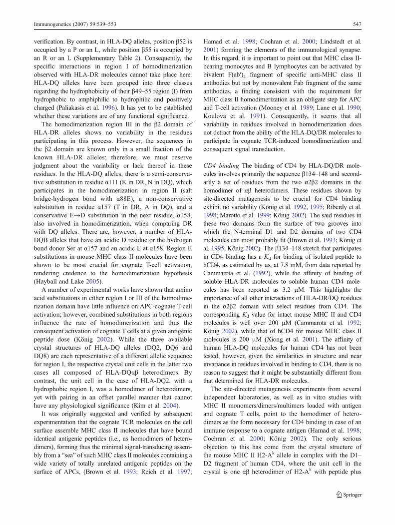
verification. By contrast, in HLA-DQ alleles, position "52 isoccupied by a P or an L, while position "55 is occupied byan R or an L (Supplementary Table 2). Consequently, thespecific interactions in region I of homodimerizationobserved with HLA-DR molecules cannot take place here.HLA-DQ alleles have been grouped into three classesregarding the hydrophobicity of their "49–55 region (I) fromhydrophobic to amphiphilic to hydrophilic and positivelycharged (Paliakasis et al. 1996). It has yet to be establishedwhether these variations are of any functional significance.
The homodimerization region III in the "2 domain ofHLA-DR alleles shows no variability in the residuesparticipating in this process. However, the sequences inthe "2 domain are known only in a small fraction of theknown HLA-DR alleles; therefore, we must reservejudgment about the variability or lack thereof in theseresidues. In the HLA-DQ alleles, there is a semi-conserva-tive substitution in residue !111 (K in DR, N in DQ), whichparticipates in the homodimerization in region II (saltbridge-hydrogen bond with !88E), a non-conservativesubstitution in residue !157 (T in DR, A in DQ), and aconservative E→D substitution in the next residue, !158,also involved in homodimerization, when comparing DRwith DQ alleles. There are, however, a number of HLA-DQB alleles that have an acidic D residue or the hydrogenbond donor Ser at !157 and an acidic E at !158. Region IIsubstitutions in mouse MHC class II molecules have beenshown to be most crucial for cognate T-cell activation,rendering credence to the homodimerization hypothesis(Hayball and Lake 2005).
A number of experimental works have shown that aminoacid substitutions in either region I or III of the homodime-rization domain have little influence on APC-cognate T-cellactivation; however, combined substitutions in both regionsinfluence the rate of homodimerization and thus theconsequent activation of cognate T cells at a given antigenicpeptide dose (König 2002). While the three availablecrystal structures of HLA-DQ alleles (DQ2, DQ6 andDQ8) are each representative of a different allelic sequencefor region I, the respective crystal unit cells in the latter twocases all composed of HLA-DQ!" heterodimers. Bycontrast, the unit cell in the case of HLA-DQ2, with ahydrophobic region I, was a homodimer of heterodimers,yet with pairing in an offset parallel manner that cannothave any physiological significance (Kim et al. 2004).
It was originally suggested and verified by subsequentexperimentation that the cognate TCR molecules on the cellsurface assemble MHC class II molecules that have boundidentical antigenic peptides (i.e., as homodimers of hetero-dimers), forming thus the minimal signal-transducing assem-bly from a “sea” of such MHC class II molecules containing awide variety of totally unrelated antigenic peptides on thesurface of APCs, (Brown et al. 1993; Reich et al. 1997;
Hamad et al. 1998; Cochran et al. 2000; Lindstedt et al.2001) forming the elements of the immunological synapse.In this regard, it is important to point out that MHC class II-bearing monocytes and B lymphocytes can be activated bybivalent F(ab′)2 fragment of specific anti-MHC class IIantibodies but not by monovalent Fab fragment of the sameantibodies, a finding consistent with the requirement forMHC class II homodimerization as an obligate step for APCand T-cell activation (Mooney et al. 1989; Lane et al. 1990;Koulova et al. 1991). Consequently, it seems that allvariability in residues involved in homodimerization doesnot detract from the ability of the HLA-DQ/DR molecules toparticipate in cognate TCR-induced homodimerization andconsequent signal transduction.
CD4 binding The binding of CD4 by HLA-DQ/DR mole-cules involves primarily the sequence "134–148 and second-arily a set of residues from the two !2"2 domains in thehomodimer of !" heterodimers. These residues shown bysite-directed mutagenesis to be crucial for CD4 bindingexhibit no variability (König et al. 1992, 1995; Riberdy et al.1998; Marotto et al. 1999; König 2002). The said residues inthese two domains form the surface of two grooves intowhich the N-terminal D1 and D2 domains of two CD4molecules can most probably fit (Brown et al. 1993; König etal. 1995; König 2002). The "134–148 stretch that participatesin CD4 binding has a Kd for binding of isolated peptide tohCD4, as estimated by us, at 7.8 mM, from data reported byCammarota et al. (1992), while the affinity of binding ofsoluble HLA-DR molecules to soluble human CD4 mole-cules has been reported as 3.2 μM. This highlights theimportance of all other interactions of HLA-DR/DQ residuesin the !2"2 domain with select residues from CD4. Thecorresponding Kd value for intact mouse MHC II and CD4molecules is well over 200 μM (Cammarota et al. 1992;König 2002), while that of hCD4 for mouse MHC class IImolecules is 200 μM (Xiong et al. 2001). The affinity ofhuman HLA-DQ molecules for human CD4 has not beentested; however, given the similarities in structure and nearinvariance in residues involved in binding to CD4, there is noreason to suggest that it might be substantially different fromthat determined for HLA-DR molecules.
The site-directed mutagenesis experiments from severalindependent laboratories, as well as in vitro studies withMHC II monomers/dimers/multimers loaded with antigenand cognate T cells, point to the homodimer of hetero-dimers as the form necessary for CD4 binding in case of animmune response to a cognate antigen (Hamad et al. 1998;Cochran et al. 2000; König 2002). The only seriousobjection to this has come from the crystal structure ofthe mouse MHC II H2-Ak allele in complex with the D1–D2 fragment of human CD4, where the unit cell in thecrystal is one !" heterodimer of H2-Ak with peptide plus
Immunogenetics (2007) 59:539–553 547

one hCD4 D1–D2 fragment (Wang et al. 2001). We do notfavor this view for the following reasons: (a) this is acomplex of a heterologous system (albeit with an apparentfunction in vitro) composed of two proteins, one human theother murine, with an affinity in the liquid phase (wholemolecules) of 200 μM (König 2002; Xiong et al. 2001); (b)residues implicated by site-directed mutagenesis to beinvolved in CD4 binding (e.g., "137E, 142V) and severalresidues from the !2 domain such as 125, 129, and 131(König et al. 1995) are not in contact with the hCD4molecule in this particular crystal structure. While thisfinding suggests a different mode of interaction of the twomolecules than the one proposed here via homodimerformation, the following two explanations are possible: (a)the structure might represent a low affinity interaction ofMHC II–peptide–noncognate TCR (which is missing in thiscomplex)–CD4 (König 2002). Such interactions, which donot lead to a bona fide immune response to the peptidepresented by the MHC II molecule, are necessary for theCD4+ T cells to maintain their viability in the host (e.g.,maintenance of tolerance, fine tuning of the immuneresponse, and CD4+ T-cell survival; Metz et al. 1997;Bhandoola et al. 2002); (b) this structure is an artifact,considering that only the D1–D2 domains were used for theco-crystallization. It would be impossible to co-crystallizethe entire D1–D4 CD4 molecule with an MHC II molecule,as that would mean a huge protrusion of the D3–D4domains of hCD4, resulting in the impossibility of closepacking of the complex in a crystal. Unfortunately, nobinding data of the individual molecular components in thecrystal are given by the authors; therefore, no estimate canbe made of the affinity of the soluble hCD4 D1–D2fragment for the H2-Ak allele, as compared to binding tothe latter of intact hCD4 molecules (Wang et al. 2001).
RGD loop The RGD loop found in "167–169 of severalHLA-DQ alleles is a structure in search of a function.Originally suggested from homology modeling of HLA-DQalleles based on the structure of HLA-DR1 (Routsias andPapadopoulos 1995; Paliakasis et al. 1996) has since beenfound in the predicted conformation in all three HLA-DQcrystal structures (Lee et al. 2001; Kim et al. 2004; Sieboldet al. 2004). The conformation (both modeled and found inthe crystal structures) is extremely similar to RGDconformations of proteins involved in cell adhesioninteractions [integrins, disintegrins, collagen, fibronectin,etc. (Hynes 1992; Paliakasis et al. 1996; Papadopoulos etal. 1998)]. This sequence is found in 14/20 HLA-DQBalleles with known sequences in this region. Of the sixalleles that do not show this sequence, five have an HGDsequence, while the sixth has an HA- sequence. Neither ofthese latter two possibilities has been documented toparticipate in any cell adhesion interactions. The few in
vitro tests to check the function of this RGD loop haveutilized the heterologous mouse system where neither of thetwo MHC II molecules (H2-A or H2-E) had an RGDsequence at the equivalent position (Szewczuk et al. 1996,1999); therefore, the observed effects may simply be due tointerference with other cell adhesion interactions.
Other associations in the α2β2 domain There are alsoscattered reports alluding to the participation of this domain intetraspanin (CD81 and CD83 binding). These proteinsassociate with HLA-DR/DQ alleles in immunoprecipitationexperiments, yet, the putative physiological role of such anassociation has not been established (Schick and Levy 1993;Rubinstein et al. 1996; Hammond et al. 1998; Engering andPieters 2001; Vogt et al. 2002). Tetraspanins are presumed toact as calcium channels (they contain several serine residueswithin each of their otherwise hydrophobic intramembranousstretches), yet, this activity is induced presumably byassociation with other membrane protein(s). As the tetraspa-nin cytoplasmic tail is also very short; the putative associationof tetraspanins with HLA-DQ/DR molecules might involveboth their intracellular as well as membrane-proximalextracellular domains of these two molecules. It is verytempting to speculate that the HLA-DR/DQ homodimer ofheterodimers (induced by identical cognate TCRs from thesame cell), with its twofold symmetry axis perpendicular tothe plane of the cell membrane bilayer, would be moreconducive to tetraspanin binding and thus activation of thelatter’s calcium channel activity.
Intramembranous segment and intracytoplasmic tail
The intramembranous segments of HLA-DR/DQ! are 21residues long, while those of HLA-DR/DQ" are one residuelonger. Nearly all residues are hydrophobic, and only slightpolymorphism, involving conservative or semi-conserva-tive substitutions, is exhibited at three positions, 196, 204,and 212. It is not yet possible to determine if the specificamino acid sequence of the intramembranous segmentrenders given MHC class II alleles more prone to enteringcholesterol-rich lipid rafts, where signal transductionprocesses take place (Vogt et al. 2002).
The intracytoplasmic tails in all !- and "-chains of HLA-DR/DQ alleles are short (14 amino acids in DRA/DQA, 10in DRB, and 10/18 in DQB). This precludes their directparticipation in known signal transduction events via attach-ment of any of them to any primary signal-transducingproteins. In HLA-DQA chains, there is dimorphism in thissection only at position 215, while in HLA-DQB there isdimorphism involving non-conservative substitutions at posi-tions 221, 222, and 224. Furthermore, there is an intriguingeight-residue insertion (–PRGPPPAG–) in a few HLA-DQB
548 Immunogenetics (2007) 59:539–553

alleles that is reminiscent, although distinct from, the mousepolyproline-containing, SH3-domain-binding sequence ofvarious different signal transducers (Sparks et al. 1996). Thethree consecutive prolines might also be part of a specialstructure, as in the extracellular MYPPPY sequence of theimportant human T lymphocyte proteins CD28 and CTLA-4,where in the latter protein the proline residues are respec-tively in conformations cis-trans-cis (Metzler et al. 1997;Stamper et al. 2001). Interestingly, a nearly identicalsequence is present in all mouse H2-A" alleles (Supplemen-tal Table 2, Brown et al. 1988), and a different sequence isfound in HLA-DR" and H2-E" alleles. There is extensivedivergence in the intracytoplasmic sequences of HLA-DRAcompared to HLA-DQA, as well as the sequences of HLA-DRB compared to those of HLA-DQB. By contrast, there isfar greater consensus for the corresponding sequences oforthologous gene loci between species (e.g., HLA-DR withH2-E and HLA-DQ with H2-A). The few works thatattempted to identify proteins bound to this segment ofMHC class II molecules were inconclusive, although onestudy identified one such binding protein as the type IIisozyme of the regulatory subunit of the PKA–cAMP-activated kinase (Newell et al. 1988).
The formation of the HLA-DR/DQ signal transductioncomplex would probably involve the intracytoplasmic regionsof the !- and "-chains of HLA-DR/DQ alleles, as well asmembrane-proximal extracellular segments. These shortregions in all alleles from both gene loci show extremevariability within and between the alleles of the two differentgene loci, suggestive of possible diversification in signaltransduction. Truncation or elimination of the cytoplasmicdomains of ! and/or " chains of MHC class II moleculesleads to their impaired cell membrane presence and severelimitations in the co-stimulatory function of APCs bearingsuch mutated MHC II molecules (André et al. 1994; Chia etal. 1994; Smiley et al. 1996; Rich et al. 1997). Yet, noparticular associations of these intracellular sequences withgiven signal-transducing proteins have been established. It isof significance, nonetheless, that the nonspecific activationof HLA-DR/DQ/DP molecules in human monocytes withthe F(ab′)2 fragments of locus-specific antibodies yieldsactivation of different downstream signal transduction path-ways indicative of a diversification in function among thethree loci (Matsuoka et al. 2001).
As our work deals with the structure and function of theexpressed HLA-DQ/DR molecules, we have not consideredthe gene polymorphisms in the 5′-upstream regions of theHLA-DQ and HLA -DR genes. There are unfortunatelyvery few works on the subject, yet, it seems that thepolymorphisms found here are among the richest foundanywhere in the human genome (Andersen et al. 1991;Morzycka-Wroblewska et al. 1993; Horton et al. 1998;MHC consortium 1999). In the few cases studied, certain
polymorphisms were shown to determine distinctly differ-ent levels of expression of HLA-DQ7 vs HLA-DQ8 alleles,the former associated with protection while the latter withsusceptibility to type 1 diabetes (Beaty et al. 1999). In fact,there are extensive polymorphisms in the 5′-upstreamregion of the HLA-DQA/B genes, and those upstream ofthe DQA gene are used in forensic medicine for individualidentifications (Cariolou et al. 1998).
Concluding remarks
In summary, the rich diversity of sequences in HLA-DR/DQ alleles signifies a versatile immune system of the hostthat can respond to a variety of pathogens, both at the levelof the individual host and the level of the population. Weprovide a single numbering platform for all MHC class IIalleles regardless of locus and species that providesstructural equivalence for identically numbered residues,facilitating thus structural and functional comparisons. Theanalysis of the extreme variety of polymorphisms observedthus far in HLA-DQ/DR alleles points out several areasfruitful for experimentation to verify or refute a number ofhypotheses put forth regarding the structure and function ofthese molecules. The involvement of the HLA-DR/DQmolecules in positive and negative selection in the thymusand the periphery, in the immune response of the host toinvading foreign bodies, as well as in the trophic functionsfor the T lymphocytes whose antigen receptors arerestricted to, paint a rich field of action for these molecules.The comparison of any new HLA-DR/DQ sequence withthe annotated sequences provided in the SupplementaryTables should immediately reveal any significant diver-gence suggestive of altered function on the part of the newallele, hence amenable to experimental verification. In thecase of HLA-DQ alleles, important outstanding questionsas to their precise function remain, for example, theirsuppressive or epistatic function vis-à-vis HLA-DR mole-cules (Hirayama et al. 1987), and the presumed HLA-DQrestricted suppression of CD8+ T cells mediated by solubleHLA-DQ molecules (Salgame et al. 1991). Our analyses ofthe polymorphisms of the HLA-DQ/DR alleles and theircorrelations with certain of their known and establishedfunctions indicates that there remain several importantunanswered questions where experimentation is expectedto shed further light in the link among HLA-DR/DQsequences, structure, and function.
Acknowledgments This workwas supported in part by a research grantfrom the European Union’s 3rd Framework for Regional Development inGreece (EPEAEK II scheme, Program Archimedes) to GKP (75% EUfunds, 25% Greek state funds). We thank Dr. Aikaterini Stavropoulos-Giokas for her help regarding nomenclature of HLA alleles.
Immunogenetics (2007) 59:539–553 549

References
Andersen LC, Beaty JS, Nettles JW, Seyfried CE, Nepom GT, NepomBS (1991) Allelic polymorphism in transcriptional regulatoryregions of HLA-DQB genes. J Exp Med 215:181–192
André P, Cambier JC, Wade TK, Raetz T, Wade WF (1994) Distinctstructural compartmentalization of the signal transducing functions ofmajor histocompatibility complex class II (Ia) molecules. J Exp Med179:763–768
Ashman JB, Miller J (1999) A role for the transmembrane domain inthe trimerization of the MHC class II-associated invariant chain. JImmunol 163:2704–2712
Beaty JS, Sukkiennicki T, Nepom GT (1999) Allelic variation intranscription modulates MHC class II expression and function.Microbes Infect 1:919–927
Benkovic AJ, Garcia KC (2003) Not just any T cell receptor will do.Immunity 18:7–11
Bhandoola A, Tai X, Eckhaus M, Auchincloss H, Mason K, Rubin S,Carbone FM, Grossman Z, Rosenberg A, Singer A (2002)Peripheral expression of self MHC-II influences the reactivityand self-tolerance of mature CD4+ T cells: evidence from alymphopenic T cell model. Immunity 17:425–436
Brown JH, Jardetzky T, Saper MA, Samraoui B, Bjorkman PJ, WileyDC (1988) A hypothetical model of the foreign antigen bindingsite of class II histocompatibility molecules. Nature 332:845–850
Brown JH, Jardetzky TS, Gorga JC, Stern LJ, Urban RG, StromingerJL, Wiley DC (1993) Three-dimensional structure of the humanclass II histocompatibility antigen HLA-DR1. Nature 364:33–39
Cammarota G, Scheirle A, Tacaks B et al (1992) Identification of aCD4 binding site on the "2 domain of HLA-DR molecules.Nature 356:799–801
CariolouMA,Manoli P, ChristophorouM, Bashiardes E, KaragrigoriouA, Budowle B (1998) Greek Cypriot allele and genotypefrequencies for amplitype PM-DQA1 and D1S80 loci. J ForensicSci 43:661–664
Chen F, Xie J, Li N, Zhou Y, Xin L, Chou KY (2005) Novel SLA-DQalleles and their recombinant molecules in xenogeneic stimula-tion of human T cells. Transpl Immunol 14:83–89
Chia CP, Khrebtukova I, McCluskey J, Wade WF (1994) MHC classII molecules that lack cytoplasmic domains are associated withthe cytoskeleton. J Immunol 153:3398–3407
Cochran JR, Cameron TO, Stern LJ (2000) The relationship of MHC-peptide binding and T cell activation probed using chemicallydefined MHC class II oligomers. Immunity 12:241–250
Corper AL, Stratmann T, Apostolopoulos V, Scott CA, Garcia KC,Kang AS, Wilson IA, Teyton L (2000) A structural frameworkfor deciphering the link between I-Ag7 and autoimmune diabetes.Science 288:505–511
Depoil D et al. (2005) Immunological synapses are versatile structuresenabling selective T cell polarization. Immunity 22:185–194
Dessen A, Lawrence CM, Cupo S, Zaller DM, Wiley DC (1997) X-ray crystal structure of HLA-DR4 (DRA*0101, DRB1*0401)complexed with a peptide from human collagen II. Immunity7:473–481
Engering A, Pieters J (2001) Association of distinct tetraspanins withMHC class II molecules at different subcellular locations inhuman immature dendritic cells. Intl Immunol 13:127–134
Ettinger RA, Moustakas AK, Lobaton SD (2004) Open reading framesequencing and structure-based alignment of polypeptidesencoded by RT1-Bb, RT1-Ba, RT1-Db, and RT1-Da alleles.Immunogenetics 56:585–596
Fremont DH, Hendrickson WA, Marrack P, Kappler J (1996)Structures of an MHC class II molecule with covalently boundsingle peptides. Science 272:1001–1004
Fremont DH, Monnaie D, Nelson CA, Hendrickson WA, Unanue ER(1998) Crystal structure of I-Ak in complex with a dominantepitope of lysozyme. Immunity 8:305–317
Gaur LK, Nepom GT (1996) Ancestral major histocompatibilitycomplex genes beget conserved patterns of localized polymor-phisms. Proc Natl Acad Sci USA 93:5380–5383
Germain RM (2001) The T-cell receptor for antigen: signaling andligand discrimination. J Biol Chem 276:35223–35226
Ghosh P, Amaya M, Mellins E, Wiley DC (1995) The structure of anintermediate in class II MHC maturation: CLIP bound to HLA-DR3. Nature 378:457–462
Grakoui A, Bromley SK, Sumen C, Davis MM, Shaw AS, Allen PM,Dustin ML (1999) The immunological synapse: a molecularmachine controlling T cell activation. Science 285:221–227
Gregers TF, Norden TW, Birkeland HC, Sandlie I, Bakke O (2003)The cytoplasmic tail of invariant chain modulates processing andpresentation. Eur J Immunol 33:277–286
Hamad AR, O’Herrin SM, Lebowitz MS, Srikrishnan A, Bieler J,Schneck J, Pardoll D (1998) Potent T cell activation with dimericpeptide-major histocompatibility complex class II ligand: the roleof CD4 co-receptor. J Exp Med 188:1633–1640
Hammond C, Denzin LK, Pan M, Griffith JM, Geuze HJ, Cresswell P(1998) The tetraspan protein CD82 is a resident of MHC class IIcompartments where it associates with HLA-DR, -DM, and -DOmolecules. J Immunol 161:3282–3291
Hahn M, Nicholson MJ, Pyrdol J, Wucherpfennig KW (2005)Unconventional topology of self-peptide-major histocompatibilitycomplex binding of a human autoimmune T cell receptor. NatImmunol 6:490–496
Hayball JD, Lake RA (2005) The immune function of MHC class IImolecules mutated in the putative superdimer interface. Mol CellBiochem 273:1–9
He XL, Radu,C, Sidney J, Sette A, Ward ES, Garcia KC (2002)Structural snapshot of aberrant antigen presentation linked toautoimmunity: the immunodominant epitope of MBP complexedwith I-Au. Immunity 17:83–94
Hennecke J, Wiley DC (2002) Structure of a complex of the human !/" T cell receptor (TCR) HA1.7 influenza hemagglutinin peptideand major histocompatibility complex II molecule, HLA-DR4(DRA1*0101 and DRB1*0401): insight into TCR cross-restric-tion and alloreactivity. J Exp Med 195:571–581
Hennecke J, Carfi A, Wiley DC (2000) Structure of a covalently-stabilisedcomplex of a human !" T-cell receptor, influenza HA peptide andMHC class II molecule, HLA-DR1. EMBO J 19:5611–5624
Hirayama K,Matsushita S, Kikuchi I, Iuchi M, Ohta N, Sasazuki T (1987)HLA-DQ is epistatic to HLA-DR in controlling the immune responseto schistosomal antigen in humans. Nature 327:426–430
Hitbold EM, Roche PA (2002) Trafficking of MHC class II moleculesin the late secretory pathway. Curr Opin Immunol 14:30–35
Hong SC, Sant’Angelo DB, Dittel BN, Medzhitov R, Yoon ST,Waterbury PG, Janeway CA Jr (1997) The orientation of a T cellreceptor to its MHC class II: peptide ligands. J Immunol159:4395–4402
Horton R, Niblett D, Milne S, Palmer S, Tubby B, Trowsdale J, BeckS (1998) Large-scale sequence comparisons reveal unusuallyhigh levels of variation in the HLA-DQB1 locus in the class IIregion of the human MHC. J Mol Biol 282:71–97
Hsing LC, Rudensky AY (2005) The lysosomal cysteine proteases inMHC class II antigen processing. Immunol Rev 207:229–241
Hynes RO (1992) Integrins: versatility, modulation and signaling incell adhesion. Cell 69:11–25
Janeway CA Jr, Travers P, Walport M, Shlomchik MJ (2005)Immunobiology, 6th edn. Garland Science, London, pp 183–201
Jardetzky TS, Brown JH, Gorga JC, Stern LJ, Urban RG, StromingerJL, Wiley DC (1996) Crystallographic analysis of endogenouspeptides associated with HLA-DR1 suggests a common, polypro-
550 Immunogenetics (2007) 59:539–553
line II-like conformation for bound peptides. Proc Natl Acad SciUSA 93:734-738
Jasanoff A, Wagner G, Wiley DC (1998) Structure of a trimericdomain of the MHC class II-associated chaperonin and targetingprotein Ii. EMBO J 17:6812–6818
Jones EY, Fugger L, Strominger JL, Siebold C (2006) MHC class IIproteins and disease: a structural perspective. Nat Rev Immunol6:271–282
Kawasaki E, Matsuura N, Eguchi K (2006) Type 1 diabetes in Japan.Diabetologia 49:828–836
Khalil I, d’Auriol, L, Gobet M et al (1990) A combination of HLA-DQBAsp57-negative and HLA-DQ! Arg52 confers susceptibility toinsulin-dependent diabetes mellitus. J Clin Invest 85:1315–1319
KimC-Y, Quarsten H, Bergseng E, Khosla C, Sollid LM (2004) Structuralbasis for HLA-DQ2-mediated presentation of gluten epitopes inceliac disease. Proc Natl Acad Sci USA 101:4175–4179
Koulova L, Clark EA, Shu G, Dupont B (1991) The CD28 ligand B7/BB1 provides costimulatory signal for alloactivation of CD4+ Tlymphocytes. J Exp Med 173:759–762
Krummel MF, Sjaastad MD, Wulfing C, Davis MM (2000) Differen-tial clustering of CD4 and CD3 zeta during T cell recognition.Science 289:1349–1352
Kumanovics A, Takada T, Lindahl KF (2003) Genomic organizationof the mammalian MHC. Annu Rev Immunol 21:629-657
Kupfer A (2006) Signaling in the immunological synpase: definingthe optimal size. Immunity 25:11–13
Kwok WW, Kovats S, Thurtle P, Nepom GT (1993) HLA-DQ allelicpolymorphisms constrain patterns of class II heterodimer forma-tion. J Immunol 150:2263–2272
König R (2002) Interactions between MHC molecules and co-receptors of the TCR. Curr Opin Immunol 14:75–83
König R, Huang Y-L, Germain RN (1992) MHC class II interactionwith CD4 mediated by a region analogous to the MHC class Ibinding site for CD8. Nature 356:796–798
König R, Shen X, and Germain RN (1995) Involvement of both majorhistocompatibility complex class II alpha and beta chainsindicates a role for ordered oligomerization in T cell activation.J Exp Med 182:779–789
Lane PJL, McConnell FM, Schieven GL, Clark EA, Ledbetter JA(1990) The role of class II molecules in human B cellactivation: association with phosphatidyl inositol turnover,protein tyrosine phosphorylation and proliferation. J Immunol144:3684–3692
Lang HL, Jacobsen H, Ikemizu S, Andersson C, Harlos K, Madsen L,Hjorth P, Sondergaard L, Svejgaard A, Wucherpfennig K, StuartDI, Bell JI, Jones EY, Fugger L (2002) A functional andstructural basis for TCR cross-reactivity in multiple sclerosis.Nat Immunol 3:940–943
Latek RR, Suri A, Petzold SJ, Nelson CA, Kanagawa O, Unanue ER,Fremont DH (2000) Structural basis of peptide binding andpresentation by the type I diabetes-associated MHC class IImolecule of NOD mice. Immunity 12:699–710
Lee KH, Wucherpfennig KW, Wiley DC (2001) Structure of a humaninsulin peptide-HLA-DQ8 complex and susceptibility to type 1diabetes. Nat Immunol 2: 501–507
Li Y, Li H, Martin R, Mariuzza RA (2000) Structural basis for thebinding of an immunodominant peptide from myelin basicprotein in different registers by two HLA-DR2 proteins. J MolBiol 304:177–188
Li Y, Huang Y, Lue J, Quant JA, Martin R, Mariuzza RA (2005)Structure of a human autoimmune TCR bound to a myelin basicprotein self-peptide and a multiple sclerosis-associated MHCclass II molecule. EMBO J 24:2968–2979
Lindstedt R, Monk N, Lombardi G, Lechler R (2001) Amino acidsubstitutions in the putative MHC class II dimer of dimers’interface inhibit CD4+ T cell activation. J Immunol 166:800–808
Liu X, Dai S, Crawford F, Fruge R, Marrack P, Kappler J (2002)Alternate interactions define the binding of peptides to the MHCmolecule I-Ab. Proc Natl Acad Sci USA 99:8820–8825
Marotto R, Shen X, König R (1999) Requirement for efficientinteractions between CD4 and MHC class II molecules forsurvival of resting CD4+ T lymphocytes in vivo and foractivation-induced cell death. J Immunol 162:5973–5980
Marsh SGE, Parham P, Barber LD (2000) The HLA FactsBook.Academic, London, pp 1–390
Matsuoka T, Tabata H, Matsushita S (2001) Monocytes are differen-tially activated through HLA-DR, -DQ and -DP molecules viamitogen-activated protein kinases. J Immunol 166:2202–2208
Maynard J, Peterson K, Wilson DH, Adams EJ, Blondelle SE, BoulangerMJ,WilsonDB,Garcia KC (2005) Structure of an autoimmune Tcellreceptor complexed with class II peptide-MHC: insights into MHCbias and antigen specificity. Immunity 22:81–92
Metz, DP, Farber DL, König R, Bottomly K (1997) Regulation ofmemory CD4 T cell adhesion by CD4-MHC class II interaction. JImmunol 159:2567–2573
MetzlerWJ, Bajorath J, FendersonW, Shaw SY, Constantine KL, NaemuraJ, Leytze G, Peach RJ, Lavoie TB, Mueller L, Linsley PS (1997)Solution structure of humanCTLA4 and delineation of a CD80/CD86binding site conserved in CD28. Nat Struct Biol 4:527–531
MHC-sequencing consortium (1999) Complete sequence and genemap of a human major histocompatibility complex. The MHCsequencing consortium. Nature 401:921–923
Mooney N, Grillot-Courvalin C, Hivroz C, Charron D (1989) A rolefor MHC class II antigens in B-cell activation. J Autoimmun 2(Suppl):215–223
Morzycka-Wroblewska E, Harwood JI, Smith JR, Kagnoff MS (1993)Structure and evolution of the promoter regions of the DQAgenes. Immunogenetics 37:364–372
Mossman KD, Campi G, Groves JT, Dustin ML (2005) Altered TCRsignaling from geometrically repatterned immunological synap-ses. Science 310:1191–1193
Mosyak L, Zaller DM, Wiley DC (1998) The structure of HLA-DM,the peptide exchange catalyst that loads antigen onto class IIMHC molecules during antigen presentation. Immunity 9:377–383
Moustakas AK, Routsias J, Papadopoulos GK (2000a) Modelling ofthe MHC II allele I-Ag7 of NOD mouse: pH-dependent changesin specificity at pockets 9 and 6 explain several of the uniqueproperties of this molecule. Diabetologia 43:609-624
Moustakas AK, van de Wal Y, Routsias J, Kooy YMC, van Veelen P,Drijfhout JW, Koning F, Papadopoulos GK (2000b) Structure ofceliac disease-associated HLA-DQ8 and non-associated HLA-DQ9 alleles in complex with two disease-specific epitopes. IntlImmunol 12:1157-1166
Moustakas AK, Papadopoulos GK (2002) Molecular properties ofHLA-DQ alleles conferring susceptibility to or protection fromIDDM: keys to the fate of beta cells. Am J Medical Genetics115:37-47
Murthy VL, Stern LJ (1997) The class II MHC protein HLA-DR1 incomplex with an endogenous peptide: implications for the structuralbasis of the specificity of peptide binding. Structure 5:1385–1396
Nepom GT, Kwok WW (1998) Molecular basis for HLA-DQassociations with IDDM. Diabetes 47:1177–1184
Newell MK, Justement LB, Miles CR, Freed JH (1988) Biochemicalcharacterization of proteins that co-purify with class II antigensof the murine MHC. J Immunol 140:1930–1938
Ohmura-Oshimo M, Matsuki Y, Aoki M, Goto E, Mito M, UematsuM, Kakiuchi T, Hotta H, Ishido S (2006) Inhibition of MHC IIexpression and immune responses by c-MIR. J Immunol177:341–354
Oksenberg JR, Seboun E, Hauser SL (1996) Genetics of demyelinat-ing diseases. Brain Pathol 6:289–302
Immunogenetics (2007) 59:539–553 551

Paliakasis K, Routsias J, Petratos K, Ouzounis C, Kokkinidis M,Papadopoulos GK (1996) Novel structural features of the humanhistocompatibility molecules HLA-DQ as revealed by modellingbased on the published structure of the related molecule HLA-DR1. J Struct Biol 117:145–163
Papadopoulos GK, Ouzounis C, Eliopoulos E (1998) RGD sequencesin several receptor proteins: novel cell adhesion function forreceptors? Int J Biol Macromol 22:51–57
Platz P, Jakobsen BK, Morling N, Ryder LP, Svejgaard A, ThomsenM, Christy M, Kromann H, Benn J, Nerup J, Green A, Hauge M(1981) HLA-D and -DR antigens in genetic analysis of insulindependent diabetes mellitus. Diabetologia 21:108–115
Rammensee H-G, Friede T, Stevanoviç S (1995) MHC ligands andpeptide motifs: first listing. Immunogenetics 41:178–228 (seealso the updated listing on the web: http://www.syfpeithi.de)
Reich Z, Boniface JJ, Lyons DS, Borochov N, Wachtel EJ, Davis MM(1997) Ligand-specific oligomerisation of T cell receptor mole-cules. Nature 387:617–620
Reichstetter S, Papadopoulos GK, Moustakas AK, Swanson E, LiuAW, Beheray S, Ettinger RA, Nepom GT, Kwok WW (2002)Mutational analysis of critical residues determining antigenpresentation and activation of HLA-DQ0602 restricted T cellclones. Hum Immunol 63:185–193
Reinherz EL, Tan K, Tang L, Kern P, Liu J, Xiong Y, Husset RE,Smalyar AS, Hare B, Zhang R, Joachimiak A, Chang HC,Wagner G, Wang J (1999) The crystal structure of a T cellreceptor in complex with peptide and MHC class II. Science286:1913–1921
Riberdy JM, Mostaghel E, Doyle C (1998) Disruption of the CD4-major histocompatibility complex class II interaction blocks thedevelopment of CD4+ T cells in vivo. Proc Natl Acad Sci USA95:4493–4498
Rich T, Lawler SE, Lord JM, Blancheteaux VM, Charron DJ, MooneyNA (1997) HLA class II-induced translocation of PKC! andPKC"II isoforms is abrogated following truncation of DR"cytoplasmic domains. J Immunol 159:3792–3798
Robinson J, Marsh SG (2003) HLA informatics. Accessing HLAsequences from sequence data bases. Methods Mol Biol 210:3–21
Rosloniec EF, Ivey RA III, Whittington KB, Kang KH, Park H-W(2006) Crystallographic structure of a rheumatoid arthritis MHCsusceptibility allele, HLA-DR1 (DRB1*0101), complexed withthe immunodominant determinant of human type II collagen. JImmunol 177:3884–3892
Routsias J, Papadopoulos GK (1995) Polymorphic structural featuresof modelled HLA-DQ molecules segregate according to suscep-tibility or resistance to IDDM. Diabetologia 38:1251–1261
Rubinstein E, Le Naour F, Lagaudrière-Gesbert C, Billard M,Conjeaud H, Boucheid C (1996) CD9, CD63, CD81 and CD82are components of a surface tetraspan network connected toHLA-DR and VLA integrins. Eur J Immunol 26:2657–2665
Rudolph MG, Wilson IA (2002) The specificity of TCR/pMHCinteraction. Curr Opin Immunol 14:52–65
Salgame P, Convit J, Bloom BR (1991) Immunological suppression ofhuman CD8+ T cells is receptor dependent and HLA-DQrestricted. Proc Natl Acad Sci USA 88:2598–2602
Schick MR, Levy S (1993) The TAPA-1 molecule is associated on thesurface of B cells with HLA-DR molecules. J Immunol151:4090–4097
Scott PC, Gogolin-Ewens KJ, Adams TE, Brandon MR (1991)Nucleotide sequence, polymorphism, and evolution of ovineMHC class II DQA genes. Immunogenetics 34:69–79
Scott CA, Peterson PA, Teyton L, Wilson IA (1998) Crystal structuresof two I-Ad-peptide complexes reveal that high affinity can beachieved without large anchor residues. Immunity 8:319–329
Siebold C, Hansen BE, Wyer JR, Harlos K, Esnouf RE, Svejgaard A,Bell JI, Strominger JL, Jones EY, Fugger L (2004) Crystal
structure of HLA-DQ0602 that protects against Type 1 diabetesand confers strong susceptibility to narcolepsy. Proc Natl AcadSci USA 101:1999–2004
Shin J-S, Ebersold M, Pyppaert M, Delamarre L, Hartley A, MellmanI (2006) surface expression of MHC class II in dendritic cells iscontrolled by ubiquination. Nature 444:115–118
Smiley ST, Rudensky AY, Glimcher LH, Grusby MJ (1996)Truncation of the class II "-chain cytoplasmic domain influencesthe level of class II/invariant chain-derived peptide complexes.Proc Natl Acad Sci USA 93:241–244
Smith KJ, Pyrdol J, Gauthier L, Wiley DC, Wucherpfennig KW(1998) Crystal structure of HLA-DR2 (DRA*0101, DRB1*1501)complexed with a peptide from human myelin basic protein. JExp Med 188:1511–1520
Sollid LM, Marcussen G, Ek G et al (1989) Evidence for a primaryassociation of celiac disease to a particular HLA-DQ !/"heterodimer. J Exp Med 169:345–350
Sparks AB, Rider JE, Hoffman NG, Fowlkes DM, Quilliam LA, KayBM (1996) Distinct ligand preferences of Src homology 3domains from Src, Yes, Abl, Cortactin, p53bp2, PLCγ, andGrb2. Proc Natl Acad Sci (USA) 93:1540–1544
Stamper CC, Zhang Y, Tobin JF, Erbe DV, Ikemizu S, Davis SJ, StahlML, Seehra J, SomersWS,Mosyak L (2001) Crystal structure of theB7-1/CTLA-4 complex that inhibits human immune response.Nature 410:608–611
Stern LJ, Brown JH, Jardetzky TS, Gorga JC, Urban RG, StromingerJL, Wiley DC (1994) Crystal structure of the human class IIMHC protein HLA-DR1 complexed with an influenza viruspeptide. Nature 368:215–221
Stratikos E, Mosyak L, Zaller DM, Wiley DC (2002) Identification ofthe lateral interaction surfaces of human histocompatibilityleukocyte antigen (HLA)-DM with DR1 by formation of tetheredcomplexes that present enhanced HLA-DM catalysis. J Exp Med196:173–183
Suri A, Lovitch SB, Unanue ER (2005a) The wide diversity andcomplexity of peptides bound to class II MHC molecules. CurrOpin Immunol 17:1–8
Suri A, Walters JJ, Gross ML, Unanue ER (2005b) Natural peptidesselected by diabetogenic DQ8 and murine I-Ag7 molecules showcommon sequence specificity. J Clin Invest 115:2268–2276
Szewczuk Z, Siemion IZ, Wieczorek Z (1996) Immunologicalproperties of the thymopentin-like fragments of HLA-DQ. MolImmunol 33:903–908
Szewczuk Z, Wilczynski A, Stefanovwicz P, Fedorowicz W, SiemionIZ, Wieczorek Z (1999) Immunosuppressory mini-regions ofHLA-DP and HLA-DR. Mol Immunol 36:525–533
Thorsby E, Rønningen KS (1993) Particular HLA-DQ molecules playa dominant role in determining susceptibility or resistance totype 1 (insulin-dependent) diabetes mellitus. Diabetologia36:371–377
Todd JA, Acha-Orbea H, Bell JI, Chao N, Fronek Z, Jacob CO,McDermott M, Sinha AA, Timmerman L, Steinman L, et al(1988) A molecular basis for MHC Class II-associated autoim-munity. Science 240:1003–1009
van de Wal Y, Kooy YM, Drijfhout JW, Amons R, Papadopoulos GK,Koning F (1997) Unique peptide binding characteristics of thedisease-associated DQ(alpha 1*0501, beta 1*0201) vs the non-disease-associated DQ(alpha 1*0201, beta 1*0202) molecule.Immunogenetics 46:484-492
Vartdal F, Johansen BH, Friede T, Thorpe CJ, Stevanovic S, EriksenJE, Sletten K, Thorsby E, Rammensee HG, Sollid LM (1996)The peptide binding motif of the disease associated HLA-DQ(alpha 1* 0501, beta 1* 0201) molecule. Eur J Immunol 26:2764-2772
Vogt A, Spindeldrecher S, Kropshofer H (2002) Clustering of MHC-peptide complexes prior to their engagement in the immunolog-
552 Immunogenetics (2007) 59:539–553

ical synapse: lipid raft and tetraspan microdomains. ImmunolRev 189:136–151
Wang J-h, Meijers R, Xiong Y, Liu J-h, Sakihama T, Zhang R,Joachimiak A, Reinherz EL (2001) Crystal structure of thehuman CD4 N-terminal two-domain fragment complexed to aclass II MHC molecule. Proc Natl Acad Sci USA 98:10799–10804
Xiong Y, Kem P, Chiang HC, Reinherz EL (2001) T cell receptorbinding to a pMHCII ligand is kinetically distinct from andindependent of CD4. J Biol Chem 276:5659-5667
Zhu Y, Rudensky AY, Teyton AL, Wilson IA (2003) Crystal structureof MHC class II I-Ab in complex with a human CLIP peptide:Prediction of an I-Ab peptide-binding motif. J Mol Biol326:1157–1174
Immunogenetics (2007) 59:539–553 553