The role of alpha-rhythm states in perceptual learning: insights from experiments and computational...
19
REVIEW ARTICLE published: 04 April 2014 doi: 10.3389/fncom.2014.00036 The role of alpha-rhythm states in perceptual learning: insights from experiments and computational models Rodrigo Sigala 1,2 *, Sebastian Haufe 1,2 , Dipanjan Roy 1,2 , Hubert R. Dinse 3 and Petra Ritter 1,2,4,5 * 1 Department Neurology, Charité—University Medicine, Berlin, Germany 2 Bernstein Focus State Dependencies of Learning, Bernstein Center for Computational Neuroscience, Berlin, Germany 3 Neural Plasticity Lab, Institute for Neuroinformatics, Ruhr-University Bochum, Bochum, Germany 4 Minerva Research Group BrainModes, Max Planck Institute for Human Cognitive and Brain Sciences, Leipzig, Germany 5 Berlin School of Mind and Brain, Mind and Brain Institute, Humboldt University, Berlin, Germany Edited by: Viktor Jirsa, Aix-Marseille University, France Reviewed by: Peter König, University of Osnabrück, Germany Christian Bénar, Institut National de la Recherche Médicale, France *Correspondence: Rodrigo Sigala, Department of Neurology, Charité Universitaetsmedizin, Charitéplatz 1, 10117 Berlin, Germany e-mail: [email protected]; Petra Ritter, Max Planck Institute for Human Cognitive and Brain Sciences, Leipzig, Germany and Department of Neurology, Charité Universitaetsmedizin, Charitéplatz 1, 10117 Berlin, Germany e-mail: [email protected] During the past two decades growing evidence indicates that brain oscillations in the alpha band (∼10Hz) not only reflect an “idle” state of cortical activity, but also take a more active role in the generation of complex cognitive functions. A recent study shows that more than 60% of the observed inter-subject variability in perceptual learning can be ascribed to ongoing alpha activity. This evidence indicates a significant role of alpha oscillations for perceptual learning and hence motivates to explore the potential underlying mechanisms. Hence, it is the purpose of this review to highlight existent evidence that ascribes intrinsic alpha oscillations a role in shaping our ability to learn. In the review, we disentangle the alpha rhythm into different neural signatures that control information processing within individual functional building blocks of perceptual learning. We further highlight computational studies that shed light on potential mechanisms regarding how alpha oscillations may modulate information transfer and connectivity changes relevant for learning. To enable testing of those model based hypotheses, we emphasize the need for multidisciplinary approaches combining assessment of behavior and multi-scale neuronal activity, active modulation of ongoing brain states and computational modeling to reveal the mathematical principles of the complex neuronal interactions. In particular we highlight the relevance of multi-scale modeling frameworks such as the one currently being developed by “The Virtual Brain” project. Keywords: alpha rhythm, oscillations, attention, memory, learning, cognition, large-scale modeling INTRODUCTION Perceptual learning, a form of implicit learning and adult brain plasticity, allows us to tune our perception to efficiently select rel- evant sensory signals. It ultimately determines our success when adapting and interacting with the dynamic and complex envi- ronment. A glimpse into our daily life is enough to realize that learning efficacy varies greatly across human beings but also changes over time in a single person. A recent study on perceptual learning has shown that ongoing brain activity, more specifically, electrical oscillations in the alpha frequency band (∼8–12 Hz), are able to predict up to 64% of the observed variability in the learn- ing outcome in a perceptual task (Freyer et al., 2013). Although perceptual learning and brain function in general have been tra- ditionally approached through the study of task-related brain activity, Freyer et al. (2013) and other recent studies demonstrate a growing awareness for a potential role of resting-state fluctua- tions (Biswal et al., 1995) for perceptual learning (Sigman et al., 2005; Lewis et al., 2009; Baldassarre et al., 2012; Freyer et al., 2013). Revealing the interaction between ongoing brain activity and learning could yield a new understanding of human cogni- tion. This may lead to new strategies to modulate actively ongoing brain activity and to optimize brain states to manipulate the learning outcome in the clinical setting and everyday life. In the present review we focus on the role that the alpha rhythm plays in perceptual learning. At the same time one can regard the alpha rhythm and perceptual learning as exemplary for other types of ongoing neural activity and cognition. They serve to illustrate how we can approach the endeavor to understand how the brain gives rise to cognition more generally. We show the need to build two bridges: One links neuronal activity to behavior and cognition; the other links neuronal activity to the underlying complex computational biophysical mechanisms spanning cellu- lar, regional as well as large-scale network interactions. Imaging and neurofeedback studies addressed in this review are a good example of the first bridge. The computational modeling stud- ies at different spatial scales presented here are examples of the second bridge. Since cognition emerges through a temporal series of network operations, the temporal and the spatial aspects of brain activity need to be disentangled. Hence the alpha rhythm—like any other neural process of the brain—may play differential roles depend- ing on the neuronal populations involved and the time point or temporal (cognitive) context. In other words, alpha oscillations may play different roles during the sequence of processes. They indicate or encode different aspects of information processing in the brain. In the present article we address how alpha oscillations Frontiers in Computational Neuroscience www.frontiersin.org April 2014 | Volume 8 | Article 36 | 1 COMPUTATIONAL NEUROSCIENCE
-
Upload
independent -
Category
Documents
-
view
2 -
download
0
Transcript of The role of alpha-rhythm states in perceptual learning: insights from experiments and computational...

REVIEW ARTICLEpublished: 04 April 2014
doi: 10.3389/fncom.2014.00036
The role of alpha-rhythm states in perceptual learning:insights from experiments and computational modelsRodrigo Sigala1,2*, Sebastian Haufe1,2, Dipanjan Roy1,2, Hubert R. Dinse3 and Petra Ritter1,2,4,5*
1 Department Neurology, Charité—University Medicine, Berlin, Germany2 Bernstein Focus State Dependencies of Learning, Bernstein Center for Computational Neuroscience, Berlin, Germany3 Neural Plasticity Lab, Institute for Neuroinformatics, Ruhr-University Bochum, Bochum, Germany4 Minerva Research Group BrainModes, Max Planck Institute for Human Cognitive and Brain Sciences, Leipzig, Germany5 Berlin School of Mind and Brain, Mind and Brain Institute, Humboldt University, Berlin, Germany
Edited by:
Viktor Jirsa, Aix-Marseille University,France
Reviewed by:
Peter König, University ofOsnabrück, GermanyChristian Bénar, Institut National dela Recherche Médicale, France
*Correspondence:
Rodrigo Sigala, Department ofNeurology, CharitéUniversitaetsmedizin, Charitéplatz 1,10117 Berlin, Germanye-mail: [email protected];
Petra Ritter, Max Planck Institute forHuman Cognitive and BrainSciences, Leipzig, Germany andDepartment of Neurology, CharitéUniversitaetsmedizin, Charitéplatz 1,10117 Berlin, Germanye-mail: [email protected]
During the past two decades growing evidence indicates that brain oscillations in thealpha band (∼10 Hz) not only reflect an “idle” state of cortical activity, but also take amore active role in the generation of complex cognitive functions. A recent study showsthat more than 60% of the observed inter-subject variability in perceptual learning canbe ascribed to ongoing alpha activity. This evidence indicates a significant role of alphaoscillations for perceptual learning and hence motivates to explore the potential underlyingmechanisms. Hence, it is the purpose of this review to highlight existent evidence thatascribes intrinsic alpha oscillations a role in shaping our ability to learn. In the review,we disentangle the alpha rhythm into different neural signatures that control informationprocessing within individual functional building blocks of perceptual learning. We furtherhighlight computational studies that shed light on potential mechanisms regarding howalpha oscillations may modulate information transfer and connectivity changes relevantfor learning. To enable testing of those model based hypotheses, we emphasize theneed for multidisciplinary approaches combining assessment of behavior and multi-scaleneuronal activity, active modulation of ongoing brain states and computational modelingto reveal the mathematical principles of the complex neuronal interactions. In particularwe highlight the relevance of multi-scale modeling frameworks such as the one currentlybeing developed by “The Virtual Brain” project.
Keywords: alpha rhythm, oscillations, attention, memory, learning, cognition, large-scale modeling
INTRODUCTIONPerceptual learning, a form of implicit learning and adult brainplasticity, allows us to tune our perception to efficiently select rel-evant sensory signals. It ultimately determines our success whenadapting and interacting with the dynamic and complex envi-ronment. A glimpse into our daily life is enough to realize thatlearning efficacy varies greatly across human beings but alsochanges over time in a single person. A recent study on perceptuallearning has shown that ongoing brain activity, more specifically,electrical oscillations in the alpha frequency band (∼8–12 Hz), areable to predict up to 64% of the observed variability in the learn-ing outcome in a perceptual task (Freyer et al., 2013). Althoughperceptual learning and brain function in general have been tra-ditionally approached through the study of task-related brainactivity, Freyer et al. (2013) and other recent studies demonstratea growing awareness for a potential role of resting-state fluctua-tions (Biswal et al., 1995) for perceptual learning (Sigman et al.,2005; Lewis et al., 2009; Baldassarre et al., 2012; Freyer et al.,2013). Revealing the interaction between ongoing brain activityand learning could yield a new understanding of human cogni-tion. This may lead to new strategies to modulate actively ongoingbrain activity and to optimize brain states to manipulate thelearning outcome in the clinical setting and everyday life.
In the present review we focus on the role that the alpharhythm plays in perceptual learning. At the same time one canregard the alpha rhythm and perceptual learning as exemplary forother types of ongoing neural activity and cognition. They serveto illustrate how we can approach the endeavor to understandhow the brain gives rise to cognition more generally. We show theneed to build two bridges: One links neuronal activity to behaviorand cognition; the other links neuronal activity to the underlyingcomplex computational biophysical mechanisms spanning cellu-lar, regional as well as large-scale network interactions. Imagingand neurofeedback studies addressed in this review are a goodexample of the first bridge. The computational modeling stud-ies at different spatial scales presented here are examples of thesecond bridge.
Since cognition emerges through a temporal series of networkoperations, the temporal and the spatial aspects of brain activityneed to be disentangled. Hence the alpha rhythm—like any otherneural process of the brain—may play differential roles depend-ing on the neuronal populations involved and the time point ortemporal (cognitive) context. In other words, alpha oscillationsmay play different roles during the sequence of processes. Theyindicate or encode different aspects of information processing inthe brain. In the present article we address how alpha oscillations
Frontiers in Computational Neuroscience www.frontiersin.org April 2014 | Volume 8 | Article 36 | 1
COMPUTATIONAL NEUROSCIENCE

Sigala et al. Alpha-rhythm states in perceptual learning
may modulate perceptual learning. Based on recent computa-tional models implementing biophysical plausible mechanisms,we hypothesize that high alpha ongoing activity in learning-related areas could play an “active” role, promoting learning byimproving the encoding capabilities and memory formation. Inmore general terms, we illustrate the iterative process of empiri-cal and modeling work that is necessary to infer knowledge aboutthe interplay of intrinsic brain activity and external stimuli. Thisinterplay constitutes the foundation of perceptual learning and ingeneral of human cognition.
PERCEPTUAL LEARNINGGenerally, learning occurs in different forms. Some forms explic-itly require memorizing information (declarative learning, i.e.,information we can describe), while others occur implicitly byexposition or practice (non-declarative or procedural learning,i.e., acquiring skills) (Gilbert et al., 2001). Perceptual learningis a form of implicit learning that can occur independent ofdeclarative processes and is very similar in several aspects tonon-declarative learning (Fahle and Poggio, 2002; Sagi, 2011).The term perceptual learning has a broad meaning. It cap-tures learning at a great variety of conditions and occurs evenin the absence of training (for a recent review see Beste andDinse, 2013). When referring to perceptual learning in thisreview, we generally consider any change in perception and sen-sory guided behavior as a consequence of sensory experience(Fahle and Poggio, 2002).
In the visual system for example, learning can modify the per-ception of simple features such as orientation (Shiu and Pashler,1992; Vogels and Orban, 1994), motion (Ball and Sekuler, 1982,1987), contrast (Dorais and Sagi, 1997; Yu et al., 2004) as wellas complex objects (forms) such as faces (Hussain et al., 2011,2012; Herzog et al., 2012). A general scheme shown in Figure 1illustrates how perceptual learning relates to other forms of learn-ing. As different neuronal pathways are implicated in declarativeand non-declarative forms of learning and memory (Squire andZola, 1996), Figure 1 includes the main brain regions tradi-tionally associated with these learning and memory processes(bottom part), locating perceptual learning in the neocortex. This
association is important when considering regional interactionsbetween ongoing alpha activity and perceptual learning.
CELLULAR AND REGIONAL SUBSTRATES OF PERCEPTUAL LEARNINGPerceptual learning can restructure cortical networks on a large-scale accessible by non-invasive imaging methods within hoursor even minutes (e.g., Caroni et al., 2012; Freyer et al., 2012a;Sagi et al., 2012), and can result in functional changes through-out the cortex (Gilbert et al., 2009; Sasaki et al., 2010). Plasticchanges involved in improving for example the discriminationof an attribute are likely to occur at the primary sensory pro-cessing level representing specific features, i.e., in cortical regionswhere receptive fields are selective to those attributes (Tsodyksand Gilbert, 2004; Carmel and Carrasco, 2008). Nevertheless,alterations induced by perceptual learning can go beyond primaryand secondary sensory cortices. In the visual system for exam-ple, numerous regions have been implicated in visual perceptuallearning such as V1/V2, V3, V4, the middle temporal area (MT),the lateral intraparietal area (LIP), and areas related to attention,decision making, and default mode networks (Yang and Maunsell,2004; Mukai et al., 2007; Law and Gold, 2008; Lewis et al., 2009;Sasaki et al., 2010; Shibata et al., 2011). Plastic changes induced byperceptual learning may occur widespread throughout the cortexor only in local neuronal dynamics (Jones et al., 2007; Sasaki et al.,2010).
Perceptual learning is considered to be a manifestation ofexternal stimulus-driven neural plasticity in the brain (Carmeland Carrasco, 2008) and has recently been linked to long-termpotentiation (LTP) and long-term depression (LTD) (e.g., Besteet al., 2011; Sale et al., 2011; Aberg and Herzog, 2012; Ditye et al.,2013). LTP and LTD are long-lasting alterations in the efficiencyof synaptic transmission, typically induced by brief periods ofcoordinated, high (LTP), or low (LTD) frequent neuronal activ-ity at a synapse (see Malenka and Bear, 2004 for a review). Sincethe pioneering work of Bliss and colleagues (1973), LTP and laterLTD became key candidates for neuronal plasticity and learningin almost any part of the mammalian brain (Bliss and Gardner-Medwin, 1973; Bliss and Lomo, 1973; Nicoll et al., 1988; Lynch,2004; Malenka and Bear, 2004; Citri and Malenka, 2008).
FIGURE 1 | Perceptual learning among other forms of learning classified
according to the memory systems implicated (Figure adapted with
permission from Squire and Zola, 1996. Copyright (1996) National
Academy of Sciences, U.S.A). Brain regions traditionally associatedwith each form of learning/memory are presented at the bottom part of thefigure.
Frontiers in Computational Neuroscience www.frontiersin.org April 2014 | Volume 8 | Article 36 | 2

Sigala et al. Alpha-rhythm states in perceptual learning
ROLE OF ATTENTION IN PERCEPTUAL LEARNINGOne candidate for a link between alpha and perceptual learning isattention (see section Alpha Rhythm and Attention). Generally,learning, attention, and memory appear to be strongly related(Gilbert et al., 2001). More than 30 years ago, Schneider andcolleagues (Schneider and Shiffrin, 1977; Shiffrin and Schneider,1977) proposed that learning the automatic detection of visualcategories reduces the dependence of performance from atten-tional control, resulting in the automatization of the task. Lateron, several studies in the nineties reported that attention is oftenrequired for the consolidation of non-declarative memory invisual perceptual learning (Shiu and Pashler, 1992; Ahissar andHochstein, 1993; Fahle and Morgan, 1996; Braun, 1998; Ito et al.,1998). More recent studies observed visual perceptual learningexclusively when subjects were consciously involved in a task, sug-gesting the interplay of top-down guided processes (Ahissar andHochstein, 2004) with relevant influence of attention (Roelfsemaet al., 2010). In experiments in which subjects were asked togive a response depending on different visual features of thepresented stimuli, it was demonstrated that observers show per-ceptual learning only for the features that were attended (Shiuand Pashler, 1992; Ahissar and Hochstein, 1993). In this line,long-range coupling has been reported between frontal and sen-sory areas during attention (Gregoriou et al., 2009). At the sametime, the alpha rhythm has been implicated in the long-rangecommunication between cortical areas (Von Stein and Sarnthein,2000). As detailed further below, electrophysiological studies incats investigating the coupling between different brain areas iden-tified signals in the theta-alpha range (4–12 Hz) to be relevantin top-down modulation of incoming stimuli (Von Stein et al.,2000). However, evidence is accumulating indicating that per-ceptual learning can also occur when subjects are not involvedin a task and exert no conscious effort. In these cases top-downattention has less or no influence (Zajonc, 1968; Skrandies andFahle, 1994; Watanabe et al., 2001, 2002; Seitz and Watanabe,2003; Nishina et al., 2007; Gutnisky et al., 2009; Seitz et al.,2009; Rosenthal and Humphreys, 2010; Shibata et al., 2011).In some cases this type of perceptual learning, usually referredto as “task-irrelevant perceptual learning,” requires reward andreinforcement signals (Seitz et al., 2009 for example used, foodand water deprivation to manipulate the reward) to consolidateinformation about incoming stimuli (see Sasaki et al., 2010 forreview).
PERCEPTUAL LEARNING VIA TIME-DEPENDENT SENSORYSTIMULATIONRecent work suggests that active training may not be requiredin perceptual learning (see Beste and Dinse, 2013 for review).Instead, changes in perception can be effectively induced by mereexposure to repetitive sensory stimulation (RSS). Such training-independent sensory stimulation induces lasting changes in per-ception and goal-directed behavior without any explicit tasktraining. RSS protocols are regarded as “passive stimulation”since no attentional effort is required (Dinse et al., 2011). Ragertet al. (2008) translated stimulation protocols used in brain slicepreparations into tactile high-frequency stimulation (HFS) todrive perceptual changes. HFS consisted of pulse trains that were
applied to the tip of the right index finger with a stimulation fre-quency of 20 Hz using either cutaneous or electrical stimulation.Ragert et al. (2008) found that 20 min of HFS induced a loweringof tactile discrimination thresholds, indicating improved tactileacuity, whereas the left index finger of the non-stimulated handshowed no changes in acuity.
For the visual modality, recent studies have shown that time-dependent stimulation can affect visual performance (Beste et al.,2011; McMahon and Leopold, 2012). Beste et al. (2011) usedan LTP- and LTD-like visual stimulation to improve or impairperformance of a change-detection task (see Figure 2). Task rel-evant or irrelevant features of the stimuli were used for high-or low-frequency stimulation. HFS (20 Hz) comprising a 40 minpresentation of the relevant feature stimuli caused an increasein performance (Figure 2A). Low-frequency stimulation (LFS,1 Hz) involving the relevant feature, as well as HFS using theirrelevant feature caused impairment (Figure 2B). In anotherapproach, time-dependent stimulation was applied in humanobservers to mimic spike-timing-dependent plasticity to induceplasticity in high-level vision (McMahon and Leopold, 2012). Theauthors used asynchronous presentation of faces to influence theperception of face identity.
EEG AND BOLD CORRELATES OF PERCEPTUAL LEARNINGThe neural correlates of perceptual learning in humans have beeninvestigated in several studies using functional magnetic reso-nance imaging (fMRI) or electroencephalography (EEG) record-ings. The majority of studies have focused on the visual modality(see Sagi, 2011 for a review). Using a paradigm in which sub-jects were trained to discriminate different visual textures, itwas shown that fMRI BOLD (blood-oxygen-level dependent)responses elicited by the trained textures in V1 were strongerwhen viewed with the trained eyed as compared to the untrainedeye (Schwartz et al., 2002). Changes in V1 as a consequenceof training were also confirmed by subsequent studies (e.g.,Furmanski et al., 2004; Walker et al., 2005; Yotsumoto et al., 2009).By comparing subjects’ ability to learn image-statistical regular-ities and distinguish targets in clutter, another study identifiedthe BOLD correlates of two different brain plastic signatures thatunderlie these two forms of visual perceptual learning (Zhangand Kourtzi, 2010). In addition to fMRI studies, plasticity in lowvisual areas as a consequence of perceptual training has also beeninvestigated using other methods such as EEG (e.g., Skrandies andFahle, 1994; Casco et al., 2004; Furmanski et al., 2004; Walkeret al., 2005; Pourtois et al., 2008) and diffusion tensor imaging(DTI) (Yotsumoto et al., 2010). In a recent study, Mayhew et al.(2012) compared human performance with the performance ofpattern classifiers using fMRI/EEG signals recorded simultane-ously. They found evidence of distinct brain mechanisms thatunderlie the improvement of the ability to perceive uncertain (i.e.,noisy) visual stimuli.
The neural correlates of perceptual changes induced throughRSS protocols have also been found in the somatosensory cor-tex (Pleger et al., 2001; Freyer et al., 2013). Assessing theeffect of a RSS protocol on tactile discrimination behavior andsomatosensory-evoked potentials, Pleger et al. (2001) demon-strated a correlation between individual perceptual improvement
Frontiers in Computational Neuroscience www.frontiersin.org April 2014 | Volume 8 | Article 36 | 3

Sigala et al. Alpha-rhythm states in perceptual learning
FIGURE 2 | Performance in a luminance detection task in which
changes in the orientation of the stimuli (bars) were used as a
distractor in some so-called “competitive trials.” Performance incompetitive trials is plotted for each time point: baseline (base), 90 minlater (90 min), 24 h later (24 h), and 10 days later (10 d). Each panelshows the results of different experiments depending on the
visual-stimulation protocols used. Stimulation of the relevant (luminance)or irrelevant (orientation) feature could be LTP (A)- or LTD (B)-like,applied to both (bilateral) or only the right eye (unilateral). Black andwhite circles represent performance on the right and left side of thefixation cross, respectively. Error bars are standard errors of the mean.Figure adapted with permission from Beste et al. (2011).
and localized activity in somatosensory cortex. These effectswere also confirmed using fMRI where cortical reorganization inprimary and secondary somatosensory cortex (S1 and S2) wasobserved after the same stimulation protocol (Pleger et al., 2003).
COMPUTATIONAL MODELS OF PERCEPTUAL LEARNINGThe majority of computational models that describe neuronalinteractions within and between populations underlying percep-tual learning focus primarily on the visual system. These modelscan be classified into two groups. The first group of modelsimplements learning in early visual processing areas, such as V1,inspired by the retinotopic organization of those areas (e.g., Adiniet al., 2002; Teich and Qian, 2003; Zhaoping et al., 2003). Thesecond group of models achieves learning by changing the rep-resentation of higher-level cortical areas or by modifying theconnections between low-level areas and higher-level associa-tion areas, such as those implicated in decision-making processes(Poggio et al., 1992; Dosher and Lu, 1998; Sigala et al., 2005; Serreet al., 2007; Lu et al., 2010).
The wide range of modeling approaches raises a major openquestion: does perceptual learning induce changes in early sen-sory areas or rather a reweighting of connections betweenprimary sensory cortices and higher-cortical areas involved indecision-making processes. Most of the computational modelson perceptual learning use feedforward models with recurrentinteractions (Poggio et al., 1992; Dosher and Lu, 1999; Ecksteinet al., 2004; Sigala et al., 2005; Serre et al., 2007). In such archi-tectures learning can be implemented using a training signal thatin principle guides the reweighting of the connections betweenunits at different processing stages. In these models, feedback
about decision errors is fundamental (Poggio et al., 1992; Herzogand Fahle, 1997, 1999). Other learning strategies have followedan unsupervised or semi-unsupervised approach. A possibility insuch cases is to build feature detectors capable of changing theirtuning properties, and to adapt them to the statistical propertiesof the training set (Sigala et al., 2005; Serre et al., 2007). In a recentpublication, Solgi et al. (2013) propose a model that explainsgeneralization (transfer) learning effects to untrained features.According to this model, transfer learning occurs since particu-lar tasks are able to trigger neuronal recruitment in lower-featureand higher-association areas, relevant for both the trained and theuntrained conditions.
THE ALPHA RHYTHM AND ITS IMPACT ON INFORMATIONPROCESSINGIn this section we focus on different aspects of the alpha rhythmthat may be relevant for the understanding of its role in percep-tual learning. We address the cellular and regional correlates ofalpha oscillations. We highlight generative computational modelsthat yield alpha activity and explore accumulated evidence link-ing alpha oscillations to cognition, generally agreeing with theavailable hypothesis that situate the alpha rhythm as an inhibitorymechanism which gates resources necessary for information pro-cessing (Jensen and Mazaheri, 2010).
GENERAL ASPECTS OF THE ALPHA RHYTHMThe alpha rhythm refers to brain oscillations within a frequencyrange of 8–12 Hz. This rhythm was first observed when HansBerger recorded electrical activity from the scalp (EEG) in 1929(Berger, 1929). Other frequency bands discovered later were also
Frontiers in Computational Neuroscience www.frontiersin.org April 2014 | Volume 8 | Article 36 | 4

Sigala et al. Alpha-rhythm states in perceptual learning
labeled using Greek letters, the boundaries of which were arbi-trarily drawn: delta, 0.5–4 Hz; theta, 4–8 Hz; beta, 12–30 Hz;gamma, >30 Hz (Buzsaki, 2006). Opening and closing the eyesmodulates the amplitude of alpha oscillations (see Pfurtschelleret al., 1996 for a review). Given the observed attenuation (alsoreferred to as “desynchronization”) of the alpha band signalcaused by opening the eyes, some investigators concluded that thealpha band reflects an “idling” state in which the underlying cor-tical regions are not engaged in any task or processing of sensoryinformation (Pfurtscheller et al., 1996). Nowadays the “idling”role of alpha oscillations has been overtaken by the so-called inhi-bition hypothesis (see Klimesch et al., 2007 for a review). Thishypothesis is supported by the observation that the amplitude ofalpha oscillations is suppressed in specialized sensory areas whendevoted to the processing of sensory stimuli (Nikouline et al.,2000) while it emerges in areas that are not explicitly involved inthe respective task (Worden et al., 2000; Kelly et al., 2006; Thutet al., 2006).
Although alpha oscillations are most prominent in visualareas, i.e., they exhibit highest amplitudes in electrodes placedover occipital brain areas, they are generally widespread in thecortex but regionally attenuated depending on different stim-uli and tasks (Buzsaki, 2006, p. 198–200). Hence alpha rhythmshave presumably distinct functional roles and mechanisms ofgeneration. Alpha waves can be recorded in electrodes near thefrontal eye fields, cortical areas responsible for eye movements(Niedermeyer and Da Silva, 2004, Ch. 9), above the sensory-motor cortical area (usually referred to as µ, “Rolandic” orsomatosensory alpha rhythm) (Gastaut, 1952; Kuhlman, 1978;Salmelin and Hari, 1994), over the supplementary motor area(Pfurtscheller and Berghold, 1989), as well as above the auditory(midtemporal) cortex (“tau” rhythm) (Lehtela et al., 1997). Inview of these findings it is very likely that synchronized oscilla-tions in the alpha band are a common feature of cortical activityespecially in sensory cortices, making them key candidates formodulating cognitive functions such as perceptual learning.
CELLULAR AND REGIONAL SUBSTRATES OF THE ALPHA RHYTHMInitial evidence suggested that alpha oscillations originate solelyfrom thalamo-cortical interactions (Andersen and Andersson,1968). More recently, Bollimunta et al. (2011) argued that alphaactivity in V1 appears to be generated by thalamo-cortical interac-tions that possibly also influence alpha oscillations in higher cor-tical areas along the stream of visual processing. Neurons in thethalamus possess the biophysical features (Lopes Da Silva et al.,1980; Hughes and Crunelli, 2005; Lorincz et al., 2009; Bollimuntaet al., 2011; Hughes et al., 2011) and the anatomic connectivity(Jones, 2002) that enable them to shape cortical alpha oscilla-tions. In addition to the talamic lateral geniculate nucleus (LGN),which is supposed to drive occipital alpha rhythms (Hughes andCrunelli, 2005), especially the pulvinar nucleus is considered toexert an influence over cortical alpha rhythms (Lopes Da Silvaet al., 1980) modulating the synchrony between cortical areasaccording to the locus of attention (Saalmann et al., 2012).
Other studies locate the origin of alpha oscillations in deeplayer cortical neurons and networks (Da Silva et al., 1973; LopesDa Silva and Storm Van Leeuwen, 1977; Steriade et al., 1990;
Flint and Connors, 1996; Castro-Alamancos and Rigas, 2002;Bollimunta et al., 2011; Ronnqvist et al., 2013). An in vitro prepa-ration by Silva et al. (1991) showed that synchronized oscillationsespecially in the alpha band can be generated solely by neuronsof cortical layer 5, which possess all the necessary intrinsic prop-erties and synaptic connections to generate alpha oscillations.Bollimunta et al. (2008) found alpha generators in layers 3, 4, and5 of the macaque visual cortex and suggested that in general layerswith higher spontaneous activities seem to contain the pacemak-ers of the alpha rhythm. The sites of alpha generators differ notonly along the stream of processing but also for different modali-ties. In the primary motor cortex (M1) for example, oscillatoryactivity in the alpha range (Rolandic µ rhythm) is supposedlymainly generated in layer 3 (Ronnqvist et al., 2013).
Numerous feedforward and feedback modules (Callaway,1998; Jiang et al., 2013) enable complex interactions betweencortical layers and columns. Therefore, cortical oscillations in dif-ferent frequency bands are closely linked. A recent study by Spaaket al. (2012) for example, showed an “intimate relationship”between alpha and gamma band dynamics within the primateV1 cortical microcircuits. Driven by deep layer alpha generators,gamma band activity in superficial granular and supragranu-lar layers is modulated in a suppressive, phase-specific manner(Spaak et al., 2012).
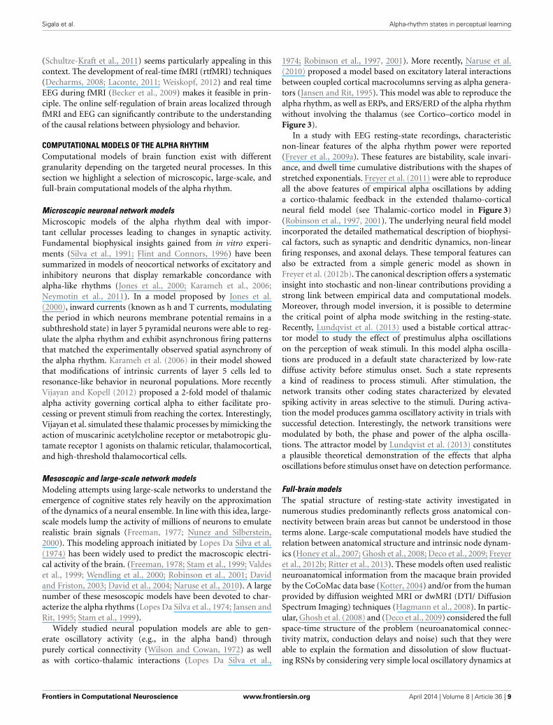
Taken together, empirical evidence demonstrates that thealpha rhythm can be generated through cortical interactions withor without the need for thalamic input. Results vary substantiallydepending on whether neural assembles are studied in vitro orin the intact brain, as well as on the particular animal model,task and hence brain region investigated. As will be shown inthe section on microscopic, mesoscopic, large-scale, and full-brain computational models, alpha activity can be generated withor without the need for thalamic activity, i.e., using exclusivelycortical interactions (see Figure 3).
IMAGING THE ALPHA RHYTHM AND OTHER FEATURES OF ONGOINGBRAIN ACTIVITYUsing combined recordings of EEG and BOLD fMRI activity(Ritter and Villringer, 2006; Becker et al., 2009; Ritter et al., 2010)it has been possible to observe thalamic and cortical BOLD activ-ity in relation to the alpha rhythm in human subjects. Severalstudies have reported higher alpha-rhythm amplitudes in occip-ital (Goldman et al., 2002; Moosmann et al., 2003; Feige et al.,2005; Goncalves et al., 2006; De Munck et al., 2007; Difrancesco,2008) and sensorimotor cortex (Ritter et al., 2009) associatedwith negative BOLD fMRI signals in sensory areas (Ritter andVillringer, 2002). There exist distinct relations between fMRIresting-state network (RSN) fluctuations and EEG global fields(i.e., average activity of all EEG channels) for different frequencybands (Mantini et al., 2007). Considering also the space structureof the EEG, i.e., identifying ICA components with distinct topo-graphic distributions, reveals that alpha oscillations of a singlefrequency band yet with independent time structure and dif-ferent space structure (topography) may be linked to differentBOLD-RSNs (Becker et al., 2009). This may explain topographicand qualitative variability of fMRI correlates of EEG rhythms.De Munck et al. (2007) have demonstrated such variability.
Frontiers in Computational Neuroscience www.frontiersin.org April 2014 | Volume 8 | Article 36 | 5

Sigala et al. Alpha-rhythm states in perceptual learning
FIGURE 3 | Schematic depiction of two mesoscopic computational
models capable of simulating oscillations in the alpha frequency range.
Left: Cortico-thalamic model adapted from Freyer et al. (2011) involving thethalamus to generate alpha activity in a network of cortical excitatory neurons
and inhibitory interneurons; Right: Cortico-cortical model adapted fromNaruse et al. (2010) generating alpha activity without the thalamic control byinterconnecting cortical macro-columns composed of excitatory pyramidalneurons accompanied by excitatory and inhibitory interneuron networks.
Other groups reported alpha correlates in fronto-parietal net-works (Laufs et al., 2003a,b, 2006) or over Rolandic (sensorimo-tor) areas (Ritter and Becker, 2009). Simultaneously recordingBOLD and EEG signals, Scheeringa et al. (2011) observed that theBOLD response elicited by a short visual stimulus was modulatedby the phase of the ongoing alpha oscillations. Additionally forevoked potentials, alpha amplitude (Becker et al., 2008; Reinacheret al., 2009) and phase dependencies have been demonstrated(but see Ritter and Becker, 2009). Using EEG-triggered sensorystimulation (Reinacher et al., 2009) together with simultaneousBOLD measurements, another study demonstrated that sponta-neous alpha-rhythm fluctuations in power could largely explainthe evoked fMRI response variance observed in extrastriate, tha-lamic, and cerebellar areas (Becker et al., 2011). As depicted inFigure 4, Becker et al. (2011) showed that BOLD responses tovisual stimuli in clusters of visual responsive voxels are modu-lated by the state of the ongoing alpha activity. Technical advancesin simultaneous EEG–fMRI acquisition nowadays allow record-ing of a wide range of oscillations including the gamma band andsubtle ultrafast population spikes (Ritter et al., 2008; Freyer et al.,2009b) and setting those different frequency bands in relations interms of their spatial and temporal features (Schultze-Kraft et al.,2011).
ALPHA RHYTHM AND PERCEPTIONIn the last decade several studies investigating the functionalrole of alpha oscillations have focused on their relation withperception (summary in Table 1). Using visual stimuli near thedetection threshold, Ergenoglu et al. (2004) observed that tri-als with detected stimuli contained significantly less power inthe alpha band than trials with undetected stimuli. Hanslmayr
et al. (2007) also showed that successful perceptual performancein a visual task is related to little alpha power during the pres-timulus interval. Investigating the effect of competing stimuli inthe somatosensory modality, Schubert et al. (2009) demonstratedthat some features of the ongoing EEG activity (e.g., ∼10 Hz)before stimulus presentation predicted whether weak stimulicould be consciously perceived after masking it with a strongerdistractor.
Instead of lower alpha activity, Babiloni et al. (2006) observeda stronger power component in frontal, parietal, and occipi-tal alpha in trials in which stimuli were perceived (interest-ingly Linkenkaer-Hansen et al., 2004 found an inverted U-shapeassociation between alpha power and consious detection inthe somatosensory modality). Using magnetoencephalograms(MEG) and analyzing signals at the source level using spa-tial filters, Van Dijk et al. (2008) showed in a visual dis-crimination task that an increase in posterior alpha powerprevious to stimulus presentation correlated with less sen-sitivity. In a paradigm in which somatosensory stimulationwas used to bias visual perception, Lange et al. (2013)found that prestimulus alpha activity is related to improvedperception of illusory stimuli. The authors suggested thatalpha activity is generally linked to enhancement of excitabil-ity of visual cortex, rather than improving perception assuch.
While all studies mentioned above concentrated on alphaamplitude, other studies have focused on studying the phase ofalpha signals (Callaway and Yeager, 1960; Dustman and Beck,1965; Varela et al., 1981; Becker et al., 2008; Busch et al., 2009;Mathewson et al., 2009; Ritter and Becker, 2009; Busch andVanrullen, 2010; Hanslmayr et al., 2011). Mathewson et al. (2009)
Frontiers in Computational Neuroscience www.frontiersin.org April 2014 | Volume 8 | Article 36 | 6

Sigala et al. Alpha-rhythm states in perceptual learning
FIGURE 4 | (A) BOLD deactivations (red) within a visual ROI (gray) projectedonto a brain template. (B) Time courses of the responses in the clustersselected showing responses in the high alpha-state (black line),
alpha-independent (gray line) conditions, and the difference of both(high-alpha stimulus response modulation, red line). Figure adapted withpermission from Becker et al. (2011).
Table 1 | Overview of studies showing correlations between features of alpha oscillations (i.e., amplitude, power, phase) and perception.
Alpha and perception
Modality/task Region Time interval Correlation with
behavioral performance
Power or amplitude Phase
Ergenoglu et al., 2004 Visual/detection Parietal/occipital Stimulation Negative
Hanslmayr et al., 2007 Visual/discrimination Parietal/occipital Pre-stimulus Negative �Schubert et al., 2009 Somatosensory/detection
(masking)Pericentral sensorimotor Pre-stimulus Negative
Babiloni et al., 2006 Visual/visuospatial Frontal, parietal and occipital Pre-stimulus Positive
Linkenkaer-Hansen et al.,2004
Somatosensory/detection Sensorimotor parietal Pre-stimulus Intermediate amplitude.Large amp.
Van Dijk et al., 2008 Visual/discrimination Parietal/occipital Pre-stimulus Increase leads to lesssensitivity
Lange et al., 2013 Visual + somatosen-sory/discrimination
Occipital Pre-stimulus Negative
Callaway and Yeager,1960
Visual/reaction-time Occipital Stimulation �
Dustman and Beck, 1965 Visual/reaction-time Occipital Stimulation �Mathewson et al., 2009 Visual/detection Posterior Stimulation Positive �Palva et al., 2005 Somatosensory/detection Somatosensory �Becker et al., 2011 Visual/detection Posterior Pre-stimulus change Positive
For each study (rows) we indicate (columns) the sensory modality investigated, task employed, brain region implicated, and time interval analyzed. Additionally we
indicate whether the power or amplitude of the alpha band positively (+) or negatively (−) correlated with behavioral performance. In the last column on the right
part we used a tick to indicate that performance correlated with alpha phase.
demonstrated that flashed stimuli were more likely to be detectedwhen presented at the positive peak than at the negative peakof the alpha waves in trials where alpha amplitude was high.Recent studies further highlighted the importance of ongoingoscillatory alpha phase in the perception of illusory (Dugue et al.,
2011) and near-threshold visual stimuli (Mathewson et al., 2011;Vanrullen et al., 2011), as well as in the conscious access tovisual stimuli (Pincham and Szucs, 2012). Using near-thresholdtactile stimuli, Palva et al. (2005) showed stimulus locking inthe alpha band (8–14 Hz) in somatosensory regions, dominant
Frontiers in Computational Neuroscience www.frontiersin.org April 2014 | Volume 8 | Article 36 | 7

Sigala et al. Alpha-rhythm states in perceptual learning
for consciously perceived stimuli but almost unobservable forunperceived stimuli.
Besides their role in target detection, alpha oscillations havebeen also found to correlate with multi-stable perception. Adecrease of alpha power has been observed to precede percep-tual reversals of bistable visual stimuli, such as the Necker cube(Isoglu-Alkac et al., 2000; Isoglu-Alkac and Struber, 2006). Usingambiguous motion, Mathes et al. (2010) reported a perceptualreversal-related desynchronization of alpha activity in posteriorlocations. This is interesting since it highlights the potential rele-vance of alpha activity for intrinsic brain state switches (see Freyeret al., 2009a,b for a characterization of alpha modes and computa-tional models that capture those). Those alpha state switches mayoccur unrelated to external events but have significant input onour perception and cognition.
ALPHA RHYTHM AND ATTENTIONUsing audiovisual stimuli, (Foxe et al., 1998) observed that thealpha rhythm is related to visual attentional gating in the pres-ence of a relevant auditory stimulus. In a spatial cueing paradigmwith purely visual stimuli, Worden et al. (2000) noticed that alphaactivity during the cue-stimulus interval increased in the occipitalcortex contralateral to the “to-be-ignored” direction (ipsilateralto the cued location). This pattern of results has been interpretedas a signature of an inhibitory process that helps to prepare activ-ity in places where stimuli are expected and visual processing isrequired (Worden et al., 2000; Kelly et al., 2006; Handel et al.,2011).
Other investigators have found a decrease in alpha activity(event related desynchronization or ERD) over posterior elec-trodes contralateral to the attended side (Kelly et al., 2006; Thutet al., 2006; Rihs et al., 2007; Wyart and Tallon-Baudry, 2008;Yamagishi et al., 2008; Mathewson et al., 2009; O’connell et al.,2009; Rihs et al., 2009; Snyder and Foxe, 2010; Mo et al., 2011).Since this desynchronization effect correlated with subsequentbehavioral performance, alpha ERD has been associated withan enhanced excitability of cortical areas in charge of process-ing stimuli in the attended visual field. Supporting this idea,Rohenkohl and Nobre (2011) have also reported alpha ERD ina task in which temporal expectations were manipulated.
Idling states of alpha are also investigated in terms of directedand non-directed attention (non-specific alertness). To explainthe role of alpha in non-directed attention Sadaghiani et al.(2010) proposed a generalized “windshield wiper” mechanism.The authors suggest that alpha oscillations rhythmically and syn-chronously clear sensory information on a rapid time-scale fromspecific channels that are require for the detection of novel andrelevant incoming sensory information (Sadaghiani et al., 2010).If the above hypothesis is indeed true, then this would sug-gest that alpha activity can bias cortical processing in favor ofstrong and recent sensory signals. In both cases, non-directed anddirected attention, alpha increases responsiveness of some areasbut decreases responsiveness of others. Low-frequency but highamplitude alpha oscillations show larger impact on target popu-lations. Yet during desynchronization of faster oscillations (suchas gamma) population gain increases, most likely, in accord withthe gradual release of inhibition and amounts to specific and focal
disruption of this global effect. The abovementioned theory forselective attention is supported by a large pool of literature show-ing that in directed attention, regions representing the attendedsite exhibit ERD while the others (non-attended) exhibit ERS(Klimesch, 2012).
CAUSAL ROLE OF THE ALPHA RHYTHMAlpha activity correlates with important processes underly-ing information processing, including perceptual learning. Yet,whether these correlations indicate a causal relation betweenbehavior and the alpha rhythm remains unknown. Interveningbrain activity through neurofeeedback and/or non-invasive brainstimulation can shed light on the causal relation between alpharhythm and perceptual learning. Transcranial magnetic stimula-tion (TMS) has proven to successfully modulate ongoing alphaoscillations, eventually modulating visual perception (Romeiet al., 2010; Thut et al., 2011; see Neuling et al., 2012 for theauditory modality; Romei et al., 2012; see Thut et al., 2012 for areview). Neurofeedback training on the other hand, is a protocolin which subjects learn to generate specific brain patterns of activ-ity interpreted through a so-called “brain-computer interface”(BCI). Neurofeedback has been used in clinical applications (e.g.,Hardt, 1978; Saxby and Peniston, 1995; Birbaumer et al., 1999;Gruzelier et al., 1999; Sterman, 2000) but also to boost the perfor-mance of healthy subjects in a wide variety of tasks (see Vernon,2005 for a review) such as those reflecting cognitive performance,for example working memory (Vernon et al., 2003) and mentalrotation tasks (Vernon et al., 2003; Hanslmayr et al., 2005; Zoefelet al., 2011). Ros et al. (2010) combined neurofeedback trainingand TMS to show that alpha oscillations contribute significantlyto cortical plasticity in motor cortex, causing brain changes thatoutlast their phase of entrainment. The authors speculate that theplasticity effects they observe could be explained by mechanismsrelated to long-term and short-term potentiation, which in turncould interact with alpha oscillations in the context of perceptuallearning.
A number of reasons have been proposed to explain the dif-ficulty when using neurofeedback to control the alpha rhythmand cognitive performance. One is the fact that the peak of thealpha frequency varies among subjects, the identification of whichis necessary to select the exact frequency band that needs to beenhanced (Klimesch et al., 1993). The alpha frequency can befurther separated into different sub-bands of differential rele-vance for different cognitive tasks. These sub-bands include loweralpha 1 (6–8 Hz), medium alpha 2 (8–10 Hz), and upper alpha(10–12 Hz). Lower alpha is related to attentional demands, whilstthe upper alpha is associated with semantic memory (Klimeschet al., 1994, 1998). By making such a distinction between alphasub-bands it has been recently shown, for example, that trainingof the upper alpha band increases cognitive control in a mentalrotation task (Zoefel et al., 2011).
The computer-model guided self-regulation of precisely local-ized brain activity with control of high-resolution temporalinformation appears to be a promising approach to controllingcognitive performance. Combining EEG and fMRI utilizing theirsynergies in terms of spatial and temporal resolutions with ana-lytical tools that account for the space-time structure of the brain
Frontiers in Computational Neuroscience www.frontiersin.org April 2014 | Volume 8 | Article 36 | 8
Sigala et al. Alpha-rhythm states in perceptual learning
(Schultze-Kraft et al., 2011) seems particularly appealing in thiscontext. The development of real-time fMRI (rtfMRI) techniques(Decharms, 2008; Laconte, 2011; Weiskopf, 2012) and real timeEEG during fMRI (Becker et al., 2009) makes it feasible in prin-ciple. The online self-regulation of brain areas localized throughfMRI and EEG can significantly contribute to the understandingof the causal relations between physiology and behavior.
COMPUTATIONAL MODELS OF THE ALPHA RHYTHMComputational models of brain function exist with differentgranularity depending on the targeted neural processes. In thissection we highlight a selection of microscopic, large-scale, andfull-brain computational models of the alpha rhythm.
Microscopic neuronal network modelsMicroscopic models of the alpha rhythm deal with impor-tant cellular processes leading to changes in synaptic activity.Fundamental biophysical insights gained from in vitro experi-ments (Silva et al., 1991; Flint and Connors, 1996) have beensummarized in models of neocortical networks of excitatory andinhibitory neurons that display remarkable concordance withalpha-like rhythms (Jones et al., 2000; Karameh et al., 2006;Neymotin et al., 2011). In a model proposed by Jones et al.(2000), inward currents (known as h and T currents, modulatingthe period in which neurons membrane potential remains in asubthreshold state) in layer 5 pyramidal neurons were able to reg-ulate the alpha rhythm and exhibit asynchronous firing patternsthat matched the experimentally observed spatial asynchrony ofthe alpha rhythm. Karameh et al. (2006) in their model showedthat modifications of intrinsic currents of layer 5 cells led toresonance-like behavior in neuronal populations. More recentlyVijayan and Kopell (2012) proposed a 2-fold model of thalamicalpha activity governing cortical alpha to either facilitate pro-cessing or prevent stimuli from reaching the cortex. Interestingly,Vijayan et al. simulated these thalamic processes by mimicking theaction of muscarinic acetylcholine receptor or metabotropic glu-tamate receptor 1 agonists on thalamic reticular, thalamocortical,and high-threshold thalamocortical cells.
Mesoscopic and large-scale network modelsModeling attempts using large-scale networks to understand theemergence of cognitive states rely heavily on the approximationof the dynamics of a neural ensemble. In line with this idea, large-scale models lump the activity of millions of neurons to emulaterealistic brain signals (Freeman, 1977; Nunez and Silberstein,2000). This modeling approach initiated by Lopes Da Silva et al.(1974) has been widely used to predict the macroscopic electri-cal activity of the brain. (Freeman, 1978; Stam et al., 1999; Valdeset al., 1999; Wendling et al., 2000; Robinson et al., 2001; Davidand Friston, 2003; David et al., 2004; Naruse et al., 2010). A largenumber of these mesoscopic models have been devoted to char-acterize the alpha rhythms (Lopes Da Silva et al., 1974; Jansen andRit, 1995; Stam et al., 1999).
Widely studied neural population models are able to gen-erate oscillatory activity (e.g., in the alpha band) throughpurely cortical connectivity (Wilson and Cowan, 1972) as wellas with cortico-thalamic interactions (Lopes Da Silva et al.,
1974; Robinson et al., 1997, 2001). More recently, Naruse et al.(2010) proposed a model based on excitatory lateral interactionsbetween coupled cortical macrocolumns serving as alpha genera-tors (Jansen and Rit, 1995). This model was able to reproduce thealpha rhythm, as well as ERPs, and ERS/ERD of the alpha rhythmwithout involving the thalamus (see Cortico–cortico model inFigure 3).
In a study with EEG resting-state recordings, characteristicnon-linear features of the alpha rhythm power were reported(Freyer et al., 2009a). These features are bistability, scale invari-ance, and dwell time cumulative distributions with the shapes ofstretched exponentials. Freyer et al. (2011) were able to reproduceall the above features of empirical alpha oscillations by addinga cortico-thalamic feedback in the extended thalamo-corticalneural field model (see Thalamic-cortico model in Figure 3)(Robinson et al., 1997, 2001). The underlying neural field modelincorporated the detailed mathematical description of biophysi-cal factors, such as synaptic and dendritic dynamics, non-linearfiring responses, and axonal delays. These temporal features canalso be extracted from a simple generic model as shown inFreyer et al. (2012b). The canonical description offers a systematicinsight into stochastic and non-linear contributions providing astrong link between empirical data and computational models.Moreover, through model inversion, it is possible to determinethe critical point of alpha mode switching in the resting-state.Recently, Lundqvist et al. (2013) used a bistable cortical attrac-tor model to study the effect of prestimulus alpha oscillationson the perception of weak stimuli. In this model alpha oscilla-tions are produced in a default state characterized by low-ratediffuse activity before stimulus onset. Such a state representsa kind of readiness to process stimuli. After stimulation, thenetwork transits other coding states characterized by elevatedspiking activity in areas selective to the stimuli. During activa-tion the model produces gamma oscillatory activity in trials withsuccessful detection. Interestingly, the network transitions weremodulated by both, the phase and power of the alpha oscilla-tions. The attractor model by Lundqvist et al. (2013) constitutesa plausible theoretical demonstration of the effects that alphaoscillations before stimulus onset have on detection performance.
Full-brain modelsThe spatial structure of resting-state activity investigated innumerous studies predominantly reflects gross anatomical con-nectivity between brain areas but cannot be understood in thoseterms alone. Large-scale computational models have studied therelation between anatomical structure and intrinsic node dynam-ics (Honey et al., 2007; Ghosh et al., 2008; Deco et al., 2009; Freyeret al., 2012b; Ritter et al., 2013). These models often used realisticneuroanatomical information from the macaque brain providedby the CoCoMac data base (Kotter, 2004) and/or from the humanprovided by diffusion weighted MRI or dwMRI (DTI/ DiffusionSpectrum Imaging) techniques (Hagmann et al., 2008). In partic-ular, Ghosh et al. (2008) and (Deco et al., 2009) considered the fullspace-time structure of the problem (neuroanatomical connec-tivity matrix, conduction delays and noise) such that they wereable to explain the formation and dissolution of slow fluctuat-ing RSNs by considering very simple local oscillatory dynamics at
Frontiers in Computational Neuroscience www.frontiersin.org April 2014 | Volume 8 | Article 36 | 9

Sigala et al. Alpha-rhythm states in perceptual learning
each node. As an extension, Deco et al. (2009) and Deco and Jirsa(2012) formulated and studied a detailed and realistic spikingattractor network structured in brain areas and connecting theselocal networks using a neuroanatomical large-scale connectivitymatrix obtained from human subjects via dwMRI tractography.
Cognition results from interactions between functionally spe-cialized but spatially distributed brain areas. As multiple brainareas are involved in such computations, full-brain models arenecessary to account for the mechanisms leading to cognitivestates. In this regard, a newly developed simulation platform, TheVirtual Brain (TVB, http://thevirtualbrain.org), provides the nec-essary tools to perform full-brain simulations (Jirsa et al., 2010;Ritter et al., 2013; Sanz Leon et al., 2013). This neuroinformat-ics platform simulates full-brain network dynamics taking intoaccount biologically realistic connectivity information. The plat-form integrates the large-scale structure of brain connectivity;it spans brain regions modeled with descriptions at microscopicand mesoscopic levels (neural networks and neural masses), usingrealistic local cortical connectivity. Thus, regional dynamics canbe evaluated in the context of long-range spatio-temporal inter-actions and at the same time preserving the perspective on globaldynamics of the brain. Models of the microcircuit can finallybe put into the functional context, and the large body of the-ory developed in computational neuroscience on the microscopicscale can be exploited for the investigation of large-scale brainfunction. Such model-based inferences would establish a stronglink across brain scales between the underlying neurophysiolog-ical mechanisms and the macroscopic large-scale brain signalsobserved in different imaging modalities.
THE ROLE OF THE ALPHA RYTHM IN PERCEPTUALLEARNINGSince ongoing brain oscillations such as the alpha rhythm emergefrom the underlying brain architecture (see section Cellular andRegional Substrates of the Alpha Rhythm), changes in the struc-tural neural connectivity induced by learning are likely to alterongoing oscillatory activity. At the same time, plastic mechanismsunderlying learning can be boosted during specific brain statesdefined by spatial patterns of ongoing oscillatory activity (Freyeret al., 2013). While recent studies support this circular relationbetween learning and ongoing brain activity (e.g., Lewis et al.,2009; Freyer et al., 2012a, 2013), the exact mechanisms behindthis interaction are still unclear. Empirical evidence (Freyer et al.,2012a, 2013) shows that perceptual learning is alpha state depen-dent and that perceptual learning alters locally the coherence ofspontaneous alpha activity. However, based on the empirical dataalone we cannot answer how local plastic changes alter large-scale brain activity, and how ongoing oscillations affect the neuralmechanisms underlying learning. In this section we address theseissues. Due to the existing empirical evidence we focus here onthe relation between the alpha rhythm and perceptual learning.Perceptual learning can be induced via time-dependent stimula-tion (Beste and Dinse, 2013), in which case it can be associatedto alpha oscillations (Freyer et al., 2012a, 2013). This leads us tobelieve that the effect of alpha oscillations on perceptual learningis related to time-dependent cellular plasticity. This hypothe-sis is further detailed in section How Oscillatory Brain States
Facilitate Learning. In section How Learning Shapes OngoingActivity we present evidence showing that learning systematicallyalters the activity of large-scale functional networks. In addition,we highlight recent computational findings demonstrating thatmodifications of large-scale functional connectivity may resultfrom plastic alterations in local cortical circuits.
HOW OSCILLATORY BRAIN STATES FACILITATE LEARNINGInformation processing in general and plasticity in particulardepend on time-precision that can be provided by periodic sig-nals such as brain rhythms. Neural oscillations are often phaseentrained; they can index naturally spike timing and, therefore,they can influence spike-timing dependent plasticity (STPD). Infact, several computational models have successfully linked STDP(see for a review Caporale and Dan, 2008) and brain oscillations(Hosaka et al., 2008; Masquelier et al., 2009; Neymotin et al.,2011). At the same time, large-scale neural oscillations providespectral fingerprints for neuronal interactions across brain areasunderlying visual perceptual learning or other cognitive processes(Engel et al., 2001; Salinas and Sejnowski, 2001; Varela et al.,2001; Siegel et al., 2012). Experiments in animals and humans andcomputational models have stressed the importance of timing-dependent neural processes for information processing and learn-ing. Extracellular recordings in awake animals have demonstratedthat neural spiking activity with respect to the phase of local fieldpotentials (LFP) recorded simultaneously carries complementaryinformation about sensory stimuli (Montemurro et al., 2008;Kayser et al., 2009). In a more recent study, Ng et al. (2013) inves-tigated the relationship between phase of ongoing oscillations andcognitive variables. The authors report that stimulus selective fir-ing patterns imprint on the phase rather than on the amplitude ofslow EEG and LFP oscillations. Stimuli that can be discriminatedbased on firing rates can also be discriminated via oscillatoryphase patterns but clearly not via oscillatory amplitude.
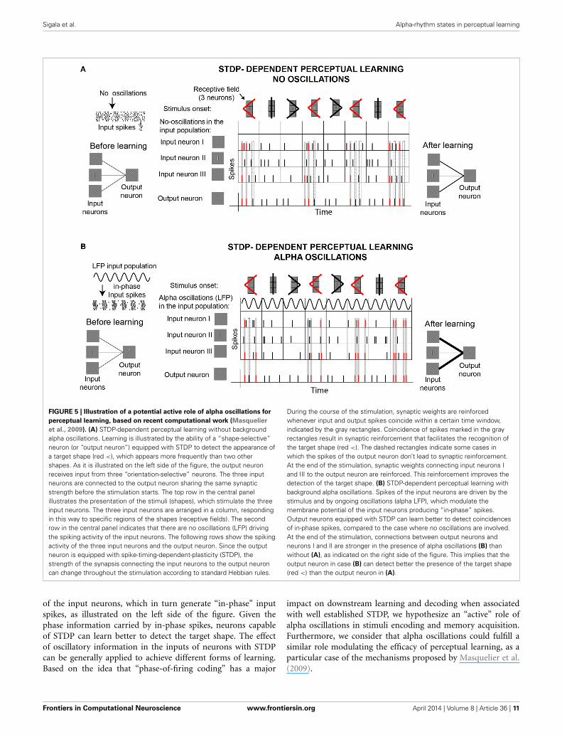
Concurrent modeling work demonstrates the importance ofrhythmic input and STDP on downstream learning (Masquelieret al., 2009). Masquelier et al. (2009) showed how a single down-stream neuron equipped with STDP can decode a repetitive inputpattern encoded in the oscillatory phases of a subset of affer-ents (∼10%). While a role of STDP in pattern detection is wellestablished (Masquelier et al., 2008), the demonstration that theencoding of patterns in oscillatory phases facilitates learning isnovel. An interesting finding of this work is that, while oscilla-tions in the alpha range proved to be good for STDP-learning,oscillations in the gamma range (>40 Hz) turned to be not opti-mal. Figure 5 illustrates these ideas by showing the behavior ofa neuron equipped with STDP that learns to detect a targetshape. This “output” neuron receives inputs from three differ-ent “orientation-selective” neurons. Depending on their receptivefields, input neurons respond to different parts of the shapes.In one situation (Figure 5A), the only external force drivingthe responses of the input neurons are the stimuli. In the sec-ond situation (Figure 5B), the spikes generated by the inputneurons depend on two external forces, the stimuli and anoscillatory signal (LFP) that modulates the membrane poten-tial of the input neurons. In contrast to the former case, in thelater case the LFP oscillations influence the spiking probability
Frontiers in Computational Neuroscience www.frontiersin.org April 2014 | Volume 8 | Article 36 | 10
Sigala et al. Alpha-rhythm states in perceptual learning
FIGURE 5 | Illustration of a potential active role of alpha oscillations for
perceptual learning, based on recent computational work (Masquelier
et al., 2009). (A) STDP-dependent perceptual learning without backgroundalpha oscillations. Learning is illustrated by the ability of a “shape-selective”neuron (or “output neuron”) equipped with STDP to detect the appearance ofa target shape (red <), which appears more frequently than two othershapes. As it is illustrated on the left side of the figure, the output neuronreceives input from three “orientation-selective” neurons. The three inputneurons are connected to the output neuron sharing the same synapticstrength before the stimulation starts. The top row in the central panelillustrates the presentation of the stimuli (shapes), which stimulate the threeinput neurons. The three input neurons are arranged in a column, respondingin this way to specific regions of the shapes (receptive fields). The secondrow in the central panel indicates that there are no oscillations (LFP) drivingthe spiking activity of the input neurons. The following rows show the spikingactivity of the three input neurons and the output neuron. Since the outputneuron is equipped with spike-timing-dependent-plasticity (STDP), thestrength of the synapsis connecting the input neurons to the output neuroncan change throughout the stimulation according to standard Hebbian rules.
During the course of the stimulation, synaptic weights are reinforcedwhenever input and output spikes coincide within a certain time window,indicated by the gray rectangles. Coincidence of spikes marked in the grayrectangles result in synaptic reinforcement that facilitates the recognition ofthe target shape (red <). The dashed rectangles indicate some cases inwhich the spikes of the output neuron don’t lead to synaptic reinforcement.At the end of the stimulation, synaptic weights connecting input neurons Iand III to the output neuron are reinforced. This reinforcement improves thedetection of the target shape. (B) STDP-dependent perceptual learning withbackground alpha oscillations. Spikes of the input neurons are driven by thestimulus and by ongoing oscillations (alpha LFP), which modulate themembrane potential of the input neurons producing “in-phase” spikes.Output neurons equipped with STDP can learn better to detect coincidencesof in-phase spikes, compared to the case where no oscillations are involved.At the end of the stimulation, connections between output neurons andneurons I and II are stronger in the presence of alpha oscillations (B) thanwithout (A), as indicated on the right side of the figure. This implies that theoutput neuron in case (B) can detect better the presence of the target shape(red <) than the output neuron in (A).
of the input neurons, which in turn generate “in-phase” inputspikes, as illustrated on the left side of the figure. Given thephase information carried by in-phase spikes, neurons capableof STDP can learn better to detect the target shape. The effectof oscillatory information in the inputs of neurons with STDPcan be generally applied to achieve different forms of learning.Based on the idea that “phase-of-firing coding” has a major
impact on downstream learning and decoding when associatedwith well established STDP, we hypothesize an “active” role ofalpha oscillations in stimuli encoding and memory acquisition.Furthermore, we consider that alpha oscillations could fulfill asimilar role modulating the efficacy of perceptual learning, as aparticular case of the mechanisms proposed by Masquelier et al.(2009).
Frontiers in Computational Neuroscience www.frontiersin.org April 2014 | Volume 8 | Article 36 | 11

Sigala et al. Alpha-rhythm states in perceptual learning
It is likely that the interaction between alpha oscillations andperceptual learning occurs by means of time-dependent informa-tion processing. However, this interaction might occur at differenttimes of the learning process, and involving brain mechanismsthat work at different time scales. Consequently, alpha oscilla-tions and perceptual learning may interact differently before,during and after training or stimulation. Analogously, this inter-action may differ depending on whether we consider 1 min, 1 h,or 1 day before or after learning. A recent study on a visualperceptual task (Hamame et al., 2011) investigated the relationbetween perceptual learning and alpha oscillations at differentepochs of the learning process. They found an intriguing rela-tionship between alpha and perceptual learning by correlatingchanges in performance with different oscillatory features duringtraining. They observed a complex modulation of the ampli-tude of both, the alpha and gamma bands, along the course oftraining. Another recent study set out to investigate the effect ofneural oscillations on perceptual learning before sensory stimula-tion was applied (Freyer et al., 2013). Freyer et al. (2013) askedto what extent different ongoing neuronal states of individualsubjects before repetitive somatosensory stimulation are able toexplain individual differences in the learning success (Freyer et al.,2013) (Figure 6). The authors found that ongoing alpha oscil-lations over sensorimotor areas contralateral to the stimulatedside before stimulation positively correlate with the learning out-come induced by the stimulation. The higher the alpha power wasbefore the stimulation, the more the subjects improved their tac-tile sensitivity after the stimulation. Performance improvementand the correlation with the alpha rhythm was present only
FIGURE 6 | Correlation between two-point discrimination (2PD)
change and alpha band resting-state EEG power before perceptual
learning (left column: stimulated side, i.e., right finger; right column:
control side). (A) Top row, scalp distributions with Pearson’s correlationscoefficients (R and P-values). Black dots: significant cluster of channels.Gray diamond: maximum correlation. (B) Scatter plot, single subject valuesat channels CP1/CP2 (green: successful learners, red: bad learners). Figureadapted with permission from Freyer et al. (2013).
in the stimulated finger and the alpha power in the contralat-eral somatosensory region. Neither the behavioral improvementnor the correlation with alpha was observed in the control con-dition (in the non-stimulated finger of the other hand). Thispattern of results suggests that the effects of alpha in learn-ing cannot be explained by global fluctuations of attention orvigilance. Instead, these finding indicates alpha band specific plas-tic mechanisms localized in the sensorimotor cortex and otherareas implicated. According to the theory “gating-by-inhibition”(Jensen and Mazaheri, 2010), high alpha activity before stimu-lation in the study by Freyer et al. (2013) could reflect an “idlestate” or inhibited state in learning-relevant areas. Such a statecan be regarded as a standby mode that allows the system to reor-ganize rapidly when stimulation starts. Beyond this, consideringthe “timing role” of alpha oscillations detailed above, prominentalpha activity before stimulation could contribute to generatethe necessary oscillatory background that combined with STDPfacilitates learning in sensorimotor areas once the stimulationstarts.
HOW LEARNING SHAPES ONGOING ACTIVITYUnder resting-state conditions –in the absence of a particulartask or stimulation– the brain yields networks of coherentlyfluctuating ongoing activity that delineate various well-knownfunctional neural networks (Fox and Raichle, 2007; Smith et al.,2009). While resting-state brain activity predicts behavioral per-formance in various tasks (Hampson et al., 2006; Fox et al., 2007;Mennes et al., 2011; Zou et al., 2013), sensory experience andlearning leave traces in the ongoing brain activity (Harmelechand Malach, 2013). For example, changes in RSNs have beendemonstrated following semantic-matching (Wang et al., 2012)and classification (Stevens et al., 2010), short- and long-termmotor learning (Albert et al., 2009; Ma et al., 2011; Taubert et al.,2011; Vahdat et al., 2011), or associative encoding (Tambini et al.,2010). Experiments on visuomotor coordination have shown thatlearning lead to plastic changes that can be observed in electri-cal oscillations recorded with EEG during the resting-state andduring sleep. Following learning in a visuomotor task, investiga-tors observed an increase in sleep slow wave activity (EEG powerdensity between 0.5 and 4.5 Hz) in the right parietal area (Huberet al., 2004; Maatta et al., 2010; Murphy et al., 2011). Duringwakefulness in contrast, an alpha decrease over the same regionand an increase over left parietal and right frontal areas has beenreported (Landsness et al., 2011). The authors report correlationsbetween the behavioral outcome of learning and the EEG sig-natures during wakefulness and sleep. This results evidence theimpact of learning on large scale functional networks in visuo-motor coordination (Landsness et al., 2011). Taken together, asHarmelech and Malach (2013) hypothesize in their recent review,spontaneous brain activity not only reflects “external statisticalstructures” but they also seems to reflect “the entire set of indi-vidual inner cortical and cognitive biases” which partially dependon past experiences (learning).
In this line, several studies show that perceptual learningaffects large-scale resting-state BOLD signals (Lewis et al., 2009;Baldassarre et al., 2012; Ventura-Campos et al., 2013). Using ashape identification task, Lewis et al. (2009) show learning-related
Frontiers in Computational Neuroscience www.frontiersin.org April 2014 | Volume 8 | Article 36 | 12

Sigala et al. Alpha-rhythm states in perceptual learning
modulations in resting-state BOLD functional connectivity. Theydemonstrate that visual perceptual learning can modify networksthat are recruited during the course of training. Lewis et al.(2009) report increased resting-state fMRI functional connec-tivity between parietal and visual cortex after visual perceptuallearning. Additionally, after perceptual learning the visual cor-tex and fronto-parietal attention areas were negatively correlated.The higher the (negative) correlation was, the more subjectsimproved their performance. Ventura-Campos et al. (2013) com-bined task-related and resting-state fMRI to investigate their rela-tion to phonetic learning. The authors showed that resting-statefunctional connectivity between the left insula/frontal operculumand the left superior parietal lobe measured before training pre-dicts individual learning outcomes. Using EEG measurements, arecent study revealed altered functional connectivity after 30 minof HFS –capable of inducing perceptual improvement (Plegeret al., 2001; Freyer et al., 2013)—as indicated by an increase oflocal resting-state alpha coherence within distributed sensorimo-tor cortical areas, contralateral to the stimulated side (Freyer et al.,2012a). Despite the sensory stimulation is applied to the of tipa single finger, in the study by Freyer et al. (2012a) clear large-scale effects on oscillatory alpha activity could be observed overdistributed sensorimotor regions.
To understand the interaction between long-range brain com-munication and local synaptic processes, full-brain models suchas the TVB are suitable simulation frameworks (see section Full-Brain Models). Currently, such models are able to reproducecoarse/general aspects of spatiotemporal brain dynamics that arepresent in the majority of experimental data. In these mod-els, brain activity can be constrained by individual large-scaleconnectivity parameters derived from dwMRI. This allows sci-entists to simulate the effects that large-scale plastic changeshave on emerging brain dynamics. Our hypothesis of how plas-tic changes in sensory areas elicited by perceptual learning affectlarge-scale information processing is based on a recent study ofour group, that shows results of full-brain simulations (Roy et al.,in preparation). In this study, Roy et al. (in preparation) proposeways to incorporate plasticity mechanisms in existing computa-tional models that are capable of generating ongoing spontaneousactivity as a function of transmission delays, noise and connec-tivity. In those simulations, plasticity in local populations canchange the dynamical stability of global functional networks dis-tributed across multiple brain areas. The main findings are: (1)Local network activity in the absence of plasticity is characterizedby irregular oscillations between a low-amplitude asynchronousand a high amplitude synchronous state. (2) Alterations in localsynapses (due to STDP), in the order of few milliseconds, inducechanges in the local connectivity of the brain areas where plas-ticity is implemented. Such changes alters distinct features of theglobal functional connectivity (FC). (3) The interaction betweenthose regions is organized systematically in correlated and anti-correlated networks depending on the choice of the modelparameters. These parameters include plasticity parameters aswell as the amplitude, frequency of the background oscillatorystate. Anti-correlated networks after time dependent plasticityshow significantly and highly correlated BOLD spatiotempo-ral activity. In particular, simulations show that the intrinsic
alpha oscillations generated by local cortical neurons efficientlyinfluence the learning outcome of brain areas connected struc-turally. While this model does not target exclusively brain areasinvolved in perceptual learning, it proposes a general mechanismable to explain the effect of perceptual learning on resting-stateactivity, as it is the case in some of the studies presented above. Inthe near future, based on individual subject/patient parameters,this kind of model simulations will help to predict the impactthat interventional actions, which evoke plasticity will have onevolving brain dynamics. Ultimately, similar brain simulationswill aid scientists in the planning of experimental learning pro-tocols as well as clinicians developing therapeutic strategies, inorder to reveal the complex relation between perceptual learningand large-scale ongoing brain activity.
CONCLUSIONS AND OUTLOOKThe current view on how alpha oscillations relate to cognitiveabilities, such as perceptual learning, is becoming far more com-plex compared to the initial view which associates alpha activityto an “idle” brain state. Although the role of alpha oscillationsin spatial attention, working memory, and perception is welldocumented, and despite initial evidence indicating that alphaoscillations influence perceptual learning, the detailed role ofalpha rhythm in perceptual learning and its contribution to theobserved variability in learning outcome needs further empiricaland theoretical assessment. Changes in perception due to per-ceptual learning develop under a variety of conditions such astraining, sensory or even central stimulation, with and withoutattention, showing complex degrees of spatial specificity and tem-poral persistence. This suggests that a variety of neural processesand brain areas are implicated in perceptual learning (Sasaki et al.,2010) that may be subject to interaction with alpha oscillations.Alpha oscillations prior to and after learning may increase thegain of neurons through a dynamic balance of excitation andinhibition (Haider et al., 2006; Raichle, 2006). Increasing thegain of neurons has an effect on their responsiveness to inputwhich is critically important for perception and attention (Salinasand Thier, 2000)—both processes that interact with perceptuallearning. Another potential interaction mechanism may be time-dependent plasticity. Phase information could serve as an internalreference for spike trains representing input signals, improvinginput processing, and memory consolidation (Masquelier et al.,2009). The combination of different measures including behav-ior, brain activity with high temporal and spatial resolution, aswell as computational modeling will be crucial to overcome thedifficulties of understanding the precise link between ongoingoscillations such as alpha activity and perceptual learning, and forutilizing those insights in the clinical and real-life setting.
ACKNOWLEDGMENTSThe authors acknowledge the support of the German Ministryof Education and Research (Bernstein Focus State Dependenciesof Learning 01GQ0971) to Hubert R. Dinse and Petra Ritter, theDeutsche Forschungsgemeinschaft—DFG (SFB 874) to HubertR. Dinse, the James S. McDonnel Foundation (Brain NetworkRecovery Group JSMF22002082), and the Max-Planck Society(Minerva Program) to Petra Ritter.
Frontiers in Computational Neuroscience www.frontiersin.org April 2014 | Volume 8 | Article 36 | 13

Sigala et al. Alpha-rhythm states in perceptual learning
REFERENCESAberg, K. C., and Herzog, M. H. (2012). About similar characteristics
of visual perceptual learning and LTP. Vision Res. 61, 100–106. doi:10.1016/j.visres.2011.12.013
Adini, Y., Sagi, D., and Tsodyks, M. (2002). Context-enabled learning in the humanvisual system. Nature 415, 790–793. doi: 10.1038/415790a
Ahissar, M., and Hochstein, S. (1993). Attentional control of early perceptual learn-ing. Proc. Natl. Acad. Sci. U.S.A. 90, 5718–5722. doi: 10.1073/pnas.90.12.5718
Ahissar, M., and Hochstein, S. (2004). The reverse hierarchy theory of visual per-ceptual learning. Trends Cogn. Sci. 8, 457–464. doi: 10.1016/j.tics.2004.08.011
Albert, N. B., Robertson, E. M., and Miall, R. C. (2009). The resting human brainand motor learning. Curr. Biol. 19, 1023–1027. doi: 10.1016/j.cub.2009.04.028
Andersen, P., and Andersson, S. A. (1968). Physiological Basis of the Alpha Rhythm.New York, NY: Appleton-Century-Crofts.
Babiloni, C., Vecchio, F., Bultrini, A., Luca Romani, G., and Rossini, P. M. (2006).Pre- and poststimulus alpha rhythms are related to conscious visual perception:a high-resolution EEG study. Cereb. Cortex 16, 1690–1700. doi: 10.1093/cer-cor/bhj104
Baldassarre, A., Lewis, C. M., Committeri, G., Snyder, A. Z., Romani, G. L., andCorbetta, M. (2012). Individual variability in functional connectivity predictsperformance of a perceptual task. Proc. Natl. Acad. Sci. U.S.A. 109, 3516–3521.doi: 10.1073/pnas.1113148109
Ball, K., and Sekuler, R. (1982). A specific and enduring improvement in visualmotion discrimination. Science 218, 697–698. doi: 10.1126/science.7134968
Ball, K., and Sekuler, R. (1987). Direction-specific improvement in motion discrim-ination. Vision Res. 27, 953–965. doi: 10.1016/0042-6989(87)90011-3
Becker, R., Reinacher, M., Freyer, F., Villringer, A., and Ritter, P. (2011). How ongo-ing neuronal oscillations account for evoked fMRI variability. J. Neurosci. 31,11016–11027. doi: 10.1523/JNEUROSCI.0210-11.2011
Becker, R., Ritter, P., and Villringer, A. (2008). Influence of ongoing alpharhythm on the visual evoked potential. Neuroimage 39, 707–716. doi:10.1016/j.neuroimage.2007.09.016
Becker, R., Ritter, P., and Villringer, A. (2009). “Visual system,” in EEG-fMRIPhysiology, Technique and Application, eds C. Mulert and L. Lemieux (Berlin,Heidelberg: Springer), 401–417.
Berger, H. (1929). Ueber das elektrenkephalogramm des menschen. Arch. Psychiatr.Nervenkr. 87, 527–570. doi: 10.1007/BF01797193
Beste, C., and Dinse, H. R. (2013). Learning without training. Curr. Biol. 23,489–499. doi: 10.1016/j.cub.2013.04.044
Beste, C., Wascher, E., Güntürkün, O., and Dinse, H. R. (2011). Improvement andimpairment of visually guided behavior through LTP- and LTD-like exposure-based visual learning. Curr. Biol. 21, 876–882. doi: 10.1016/j.cub.2011.03.065
Birbaumer, N., Ghanayim, N., Hinterberger, T., Iversen, I., Kotchoubey, B., Kubler,A., et al. (1999). A spelling device for the paralysed. Nature 398, 297–298. doi:10.1038/18581
Biswal, B., Yetkin, F. Z., Haughton, V. M., and Hyde, J. S. (1995). Functional con-nectivity in the motor cortex of resting human brain using echo-planar MRI.Magn. Reson. Med. 34, 537–541. doi: 10.1002/mrm.1910340409
Bliss, T. V., and Gardner-Medwin, A. R. (1973). Long-lasting potentiation of synap-tic transmission in the dentate area of the unanaestetized rabbit followingstimulation of the perforant path. J. Physiol. 232, 357–374.
Bliss, T. V., and Lomo, T. (1973). Long-lasting potentiation of synaptic transmis-sion in the dentate area of the anaesthetized rabbit following stimulation of theperforant path. J. Physiol. 232, 331–356.
Bollimunta, A., Chen, Y., Schroeder, C. E., and Ding, M. (2008). Neuronal mech-anisms of cortical alpha oscillations in awake-behaving macaques. J. Neurosci.28, 9976–9988. doi: 10.1523/JNEUROSCI.2699-08.2008
Bollimunta, A., Mo, J., Schroeder, C. E., and Ding, M. (2011). Neuronal mech-anisms and attentional modulation of corticothalamic alpha oscillations.J. Neurosci. 31, 4935–4943. doi: 10.1523/JNEUROSCI.5580-10.2011
Braun, J. (1998). Vision and attention: the role of training. Nature 393, 424–425.doi: 10.1038/30875
Busch, N. A., Dubois, J., and Vanrullen, R. (2009). The phase of ongoingEEG oscillations predicts visual perception. J. Neurosci. 29, 7869–7876. doi:10.1523/JNEUROSCI.0113-09.2009
Busch, N. A., and Vanrullen, R. (2010). Spontaneous EEG oscillations reveal peri-odic sampling of visual attention. Proc. Natl. Acad. Sci. U.S.A. 107, 16048–16053.doi: 10.1073/pnas.1004801107
Buzsaki, G. (2006). Rhythms of the Brain. New York, NY: Oxford University Press.doi: 10.1093/acprof:oso/9780195301069.001.0001
Callaway, E. M. (1998). Local circuits in primary visual cortex of the macaquemonkey. Annu. Rev. Neurosci. 21, 47–74. doi: 10.1146/annurev.neuro.21.1.47
Callaway, E., and Yeager, C. L. (1960). Relationship between reaction time andelectroencephalographic alpha phase. Science 132, 1765–1766. doi: 10.1126/sci-ence.132.3441.1765
Caporale, N., and Dan, Y. (2008). Spike timing-dependent plastic-ity: a Hebbian learning rule. Annu. Rev. Neurosci. 31, 25–46. doi:10.1146/annurev.neuro.31.060407.125639
Carmel, D., and Carrasco, M. (2008). Perceptual learning and dynamic changes inprimary visual cortex. Neuron 57, 799–801. doi: 10.1016/j.neuron.2008.03.009
Caroni, P., Donato, F., and Muller, D. (2012). Structural plasticity upon learning:regulation and functions. Nat. Rev. Neurosci. 13, 478–490. doi: 10.1038/nrn3258
Casco, C., Campana, G., Grieco, A., and Fuggetta, G. (2004). Perceptuallearning modulates electrophysiological and psychophysical response tovisual texture segmentation in humans. Neurosci. Lett. 371, 18–23. doi:10.1016/j.neulet.2004.08.005
Castro-Alamancos, M. A., and Rigas, P. (2002). Synchronized oscillations caused bydisinhibition in rodent neocortex are generated by recurrent synaptic activitymediated by AMPA receptors. J. Physiol. 542, 567–581. doi: 10.1113/jphys-iol.2002.019059
Citri, A., and Malenka, R. C. (2008). Synaptic plasticity: multiple forms,functions, and mechanisms. Neuropsychopharmacol 33, 18–41. doi:10.1038/sj.npp.1301559
Da Silva, F. H., Van Lierop, T. H., Schrijer, C. F., and Van Leeuwen, W. S.(1973). Organization of thalamic and cortical alpha rhythms: spectra and coher-ences. Electroencephalogr. Clin. Neurophysiol. 35, 627–639. doi: 10.1016/0013-4694(73)90216-2
David, O., Cosmelli, D., and Friston, K. J. (2004). Evaluation of different measuresof functional connectivity using a neural mass model. Neuroimage 21, 659–673.doi: 10.1016/j.neuroimage.2003.10.006
David, O., and Friston, K. J. (2003). A neural mass model for MEG/EEG:coupling and neuronal dynamics. Neuroimage 20, 1743–1755. doi:10.1016/j.neuroimage.2003.07.015
Decharms, R. C. (2008). Applications of real-time fMRI. Nat. Rev. Neurosci. 9,720–729. doi: 10.1038/nrn2414
Deco, G., and Jirsa, V. K. (2012). Ongoing cortical activity at rest: criti-cality, multistability, and ghost attractors. J. Neurosci. 32, 3366–3375. doi:10.1523/JNEUROSCI.2523-11.2012
Deco, G., Jirsa, V., McIntosh, A., Sporns, O., and Kötter, R. (2009). Key role ofcoupling, delay, and noise in resting brain fluctuations. Proc. Natl. Acad. Sci.U.S.A. 106, 10302–10307. doi: 10.1073/pnas.0901831106
De Munck, J. C., Goncalves, S. I., Huijboom, L., Kuijer, J. P., Pouwels,P. J., Heethaar, R. M., et al. (2007). The hemodynamic response ofthe alpha rhythm: an EEG/fMRI study. Neuroimage 35, 1142–1151. doi:10.1016/j.neuroimage.2007.01.022
Difrancesco, M. W. (2008). Simultaneous EEG/functional magnetic reso-nance imaging at 4 Tesla: correlates of brain activity to spontaneousalpha rhythm during relaxation. J. Clin. Neurophysiol. 25, 255–264. doi:10.1097/WNP.0b013e3181879d56
Dinse, H. R., Kattenstroth, J. C., Gatica Tossi, M. A., Tegenthoff, M., and Kalisch,K. (2011). “Sensory stimulation for augmenting perception, sensorimotorbehaviour and cognition,” in Augmenting Cognition, eds H. Markram and I.Segev (Lausanne: EPFL Press), 11–39.
Ditye, T., Kanai, R., Bahrami, B., Muggleton, N. G., Rees, G., and Walsh, V.(2013). Rapid changes in brain structure predict improvements induced byperceptual learning. Neuroimage 81, 205–212. doi: 10.1016/j.neuroimage.2013.05.058
Dorais, A., and Sagi, D. (1997). Contrast masking effects change with practice.Vision Res. 37, 1725–1733. doi: 10.1016/S0042-6989(96)00329-X
Dosher, B. A., and Lu, Z. L. (1998). Perceptual learning reflects external noise filter-ing and internal noise reduction through channel reweighting. Proc. Natl. Acad.Sci. U.S.A. 95, 13988–13993. doi: 10.1073/pnas.95.23.13988
Dosher, B. A., and Lu, Z. L. (1999). Mechanisms of perceptual learning. Vision Res.39, 3197–3221. doi: 10.1016/S0042-6989(99)00059-0
Dugue, L., Marque, P., and Vanrullen, R. (2011). The phase of ongoing oscillationsmediates the causal relation between brain excitation and visual perception.J. Neurosci. 31, 11889–11893. doi: 10.1523/JNEUROSCI.1161-11.2011
Dustman, R. E., and Beck, E. C. (1965). Phase of alpha brain waves, reactiontime and visually evoked potentials. Electroencephalogr. Clin. Neurophysiol. 18,433–440. doi: 10.1016/0013-4694(65)90123-9
Frontiers in Computational Neuroscience www.frontiersin.org April 2014 | Volume 8 | Article 36 | 14

Sigala et al. Alpha-rhythm states in perceptual learning
Eckstein, M. P., Abbey, C. K., Pham, B. T., and Shimozaki, S. S. (2004). Perceptuallearning through optimization of attentional weighting: human versus optimalBayesian learner. J. Vis. 4, 1006–1019. doi: 10.1167/4.12.3
Engel, A. K., Fries, P., and Singer, W. (2001). Dynamic predictions: oscillationsand synchrony in top-down processing. Nat. Rev. Neurosci. 2, 704–716. doi:10.1038/35094565
Ergenoglu, T., Demiralp, T., Bayraktaroglu, Z., Ergen, M., Beydagi, H., andUresin, Y. (2004). Alpha rhythm of the EEG modulates visual detectionperformance in humans. Brain Res. Cogn. Brain Res. 20, 376–383. doi:10.1016/j.cogbrainres.2004.03.009
Fahle, M., and Morgan, M. (1996). No transfer of perceptual learning betweensimilar stimuli in the same retinal position. Curr. Biol. 6, 292–297. doi:10.1016/S0960-9822(02)00479-7
Fahle, M., and Poggio, T. (2002). Perceptual Learning. Cambridge, MA: The MITPress.
Feige, B., Scheffler, K., Esposito, F., Di Salle, F., Hennig, J., and Seifritz, E. (2005).Cortical and subcortical correlates of electroencephalographic alpha rhythmmodulation. J. Neurophysiol. 93, 2864–2872. doi: 10.1152/jn.00721.2004
Flint, A. C., and Connors, B. W. (1996). Two types of network oscillations inneocortex mediated by distinct glutamate receptor subtypes and neuronalpopulations. J. Neurophysiol. 75, 951–957.
Fox, M. D., and Raichle, M. E. (2007). Spontaneous fluctuations in brain activityobserved with functional magnetic resonance imaging. Nat. Rev. Neurosci. 8,700–711. doi: 10.1038/nrn2201
Fox, M. D., Snyder, A. Z., Vincent, J. L., and Raichle, M. E. (2007). Intrinsicfluctuations within cortical systems account for intertrial variability in humanbehavior. Neuron 56, 171–184. doi: 10.1016/j.neuron.2007.08.023
Foxe, J. J., Simpson, G. V., and Ahlfors, S. P. (1998). Parieto-occipital ∼10Hz activ-ity reflects anticipatory state of visual attention mechanisms. Neuroreport 9,3929–3933. doi: 10.1097/00001756-199812010-00030
Freeman, W. (1977). Mass Action in the Nervous System: Examination of theNeurophysiological Basis of Adaptive Behavior Through the EEG. New York, NY:Academic Press.
Freeman, W. J. (1978). Models of the dynamics of neural populations.Electroencephalogr. Clin. Neurophysiol. Suppl. 34, 9–18.
Freyer, F., Aquino, K., Robinson, P. A., Ritter, P., and Breakspear, M. (2009a).Bistability and non-Gaussian fluctuations in spontaneous cortical activity.J. Neurosci. 29, 8512–8524. doi: 10.1523/JNEUROSCI.0754-09.2009
Freyer, F., Becker, R., Anami, K., Curio, G., Villringer, A., and Ritter, P.(2009b). Ultrahigh-frequency EEG during fMRI: pushing the limits of imaging-artifact correction. Neuroimage 48, 94–108. doi: 10.1016/j.neuroimage.2009.06.022
Freyer, F., Becker, R., Dinse, H. R., and Ritter, P. (2013). State-dependent perceptuallearning. J. Neurosci. 33, 2900–2907. doi: 10.1523/JNEUROSCI.4039-12.2013
Freyer, F., Reinacher, M., Nolte, G., Dinse, H. R., and Ritter, P. (2012a). Repetitivetactile stimulation changes resting-state functional connectivity-implicationsfor treatment of sensorimotor decline. Front. Hum. Neurosci. 6:144. doi:10.3389/fnhum.2012.00144
Freyer, F., Roberts, J. A., Becker, R., Robinson, P. A., Ritter, P., and Breakspear,M. (2011). Biophysical mechanisms of multistability in resting-state corticalrhythms. J. Neurosci. 31, 6353–6361. doi: 10.1523/JNEUROSCI.6693-10.2011
Freyer, F., Roberts, J. A., Ritter, P., and Breakspear, M. (2012b). A canonical modelof multistability and scale-invariance in biological systems. PLoS Comput. Biol.8:e1002634. doi: 10.1371/journal.pcbi.1002634
Furmanski, C. S., Schluppeck, D., and Engel, S. A. (2004). Learning strengthens theresponse of primary visual cortex to simple patterns. Curr. Biol. 14, 573–578.doi: 10.1016/j.cub.2004.03.032
Gastaut, H. (1952). Etude electrocorticographique de la reactivite des rhythmsrolandiques. Rev. Neurol. (Paris) 87, 176–182.
Ghosh, A., Rho, Y., McIntosh, A. R., Kotter, R., and Jirsa, V. K. (2008). Noise duringrest enables the exploration of the brain’s dynamic repertoire. PLoS Comput.Biol. 4:e1000196. doi: 10.1371/journal.pcbi.1000196
Gilbert, C. D., Li, W., and Piech, V. (2009). Perceptual learning and adult corticalplasticity. J. Physiol. 587, 2743–2751. doi: 10.1113/jphysiol.2009.171488
Gilbert, C. D., Sigman, M., and Crist, R. E. (2001). The neural basis of perceptuallearning. Neuron 31, 681–697. doi: 10.1016/S0896-6273(01)00424-X
Goldman, R. I., Stern, J. M., Engel, J. Jr., and Cohen, M. S. (2002). SimultaneousEEG and fMRI of the alpha rhythm. Neuroreport 13, 2487–2492. doi:10.1097/00001756-200212200-00022
Goncalves, S. I., De Munck, J. C., Pouwels, P. J., Schoonhoven, R., Kuijer, J. P.,Maurits, N. M., et al. (2006). Correlating the alpha rhythm to BOLD usingsimultaneous EEG/fMRI: inter-subject variability. Neuroimage 30, 203–213. doi:10.1016/j.neuroimage.2005.09.062
Gregoriou, G. G., Gotts, S. J., Zhou, H., and Desimone, R. (2009). High-frequency,long-range coupling between prefrontal and visual cortex during attention.Science 324, 1207–1210. doi: 10.1126/science.1171402
Gruzelier, J., Hardman, E., Wild, J., and Zaman, R. (1999). Learned control of slowpotential interhemispheric asymmetry in schizophrenia. Int. J. Psychophysiol.34, 341–348. doi: 10.1016/S0167-8760(99)00091-4
Gutnisky, D. A., Hansen, B. J., Iliescu, B. F., and Dragoi, V. (2009). Attention altersvisual plasticity during exposure-based learning. Curr. Biol. 19, 555–560. doi:10.1016/j.cub.2009.01.063
Hagmann, P., Cammoun, L., Gigandet, X., Meuli, R., Honey, C. J., Wedeen, V. J.,et al. (2008). Mapping the structural core of human cerebral cortex. PLoS Biol.6:e159. doi: 10.1371/journal.pbio.0060159
Haider, B., Duque, A., Hasenstaub, A. R., and McCormick, D. A. (2006).Neocortical network activity in vivo is generated through a dynamicbalance of excitation and inhibition. J. Neurosci. 26, 4535–4545. doi:10.1523/JNEUROSCI.5297-05.2006
Hamame, C. M., Cosmelli, D., Henriquez, R., and Aboitiz, F. (2011). Neuralmechanisms of human perceptual learning: electrophysiological evidence for atwo-stage process. PLoS ONE 6:e19221. doi: 10.1371/journal.pone.0019221
Hampson, M., Driesen, N. R., Skudlarski, P., Gore, J. C., and Constable, R. T.(2006). Brain connectivity related to working memory performance. J. Neurosci.26, 13338–13343. doi: 10.1523/JNEUROSCI.3408-06.2006
Handel, B. F., Haarmeier, T., and Jensen, O. (2011). Alpha oscillations correlatewith the successful inhibition of unattended stimuli. J. Cogn. Neurosci. 23,2494–2502. doi: 10.1162/jocn.2010.21557
Hanslmayr, S., Aslan, A., Staudigl, T., Klimesch, W., Herrmann, C. S., andBäuml, K.-H. (2007). Prestimulus oscillations predict visual perception per-formance between and within subjects. Neuroimage 37, 1465–1473. doi:10.1016/j.neuroimage.2007.07.011
Hanslmayr, S., Gross, J., Klimesch, W., and Shapiro, K. L. (2011). The role ofalpha oscillations in temporal attention. Brain Res. Rev. 67, 331–343. doi:10.1016/j.brainresrev.2011.04.002
Hanslmayr, S., Sauseng, P., Doppelmayr, M., Schabus, M., and Klimesch, W. (2005).Increasing individual upper alpha power by neurofeedback improves cognitiveperformance in human subjects. Appl. Psychophysiol. Biofeedback 30, 1–10. doi:10.1007/s10484-005-2169-8
Hardt, J. V. (1978). Anxiety change through electroencephalographic alpha feed-back seen only in high anxiety subjects. Science 201, 79–81. doi: 10.1126/sci-ence.663641
Harmelech, T., and Malach, R. (2013). Neurocognitive biases and the patterns ofspontaneous correlations in the human cortex. Trends Cogn. Sci. 17, 606–615.doi: 10.1016/j.tics.2013.09.014
Herzog, M. H., Aberg, K. C., Fremaux, N., Gerstner, W., and Sprekeler, H. (2012).Perceptual learning, roving and the unsupervised bias. Vision Res. 61, 95–99.doi: 10.1016/j.visres.2011.11.001
Herzog, M. H., and Fahle, M. (1997). The role of feedback in learning avernier discrimination task. Vision Res. 37, 2133–2141. doi: 10.1016/S0042-6989(97)00043-6
Herzog, M. H., and Fahle, M. (1999). Effects of biased feedback on learningand deciding in a vernier discrimination task. Vision Res. 39, 4232–4243. doi:10.1016/S0042-6989(99)00138-8
Honey, C. J., Kotter, R., Breakspear, M., and Sporns, O. (2007). Networkstructure of cerebral cortex shapes functional connectivity on multiple timescales. Proc. Natl. Acad. Sci. U.S.A. 104, 10240–10245. doi: 10.1073/pnas.0701519104
Hosaka, R., Araki, O., and Ikeguchi, T. (2008). STDP provides the substrate forigniting synfire chains by spatiotemporal input patterns. Neural Comput. 20,415–435. doi: 10.1162/neco.2007.11-05-043
Huber, R., Ghilardi, M. F., Massimini, M., and Tononi, G. (2004). Local sleep andlearning. Nature 430, 78–81. doi: 10.1038/nature02663
Hughes, S. W., and Crunelli, V. (2005). Thalamic mechanisms of EEG alpharhythms and their pathological implications. Neuroscientist 11, 357–372. doi:10.1177/1073858405277450
Hughes, S. W., Lorincz, M. L., Blethyn, K., Kekesi, K. A., Juhasz, G., Turmaine,M., et al. (2011). Thalamic gap junctions control local neuronal synchrony and
Frontiers in Computational Neuroscience www.frontiersin.org April 2014 | Volume 8 | Article 36 | 15

Sigala et al. Alpha-rhythm states in perceptual learning
influence macroscopic oscillation amplitude during EEG alpha rhythms. Front.Psychol. 2:193. doi: 10.3389/fpsyg.2011.00193
Hussain, Z., McGraw, P. V., Sekuler, A. B., and Bennett, P. J. (2012). The rapidemergence of stimulus specific perceptual learning. Front. Psychol. 3:226. doi:10.3389/fpsyg.2012.00226
Hussain, Z., Sekuler, A. B., and Bennett, P. J. (2011). Superior identification offamiliar visual patterns a year after learning. Psychol. Sci. 22, 724–730. doi:10.1177/0956797611409591
Isoglu-Alkac, U., Basar-Eroglu, C., Ademoglu, A., Demiralp, T., Miener, M., andStadler, M. (2000). Alpha activity decreases during the perception of Neckercube reversals: an application of wavelet transform. Biol. Cybern. 82, 313–320.doi: 10.1007/s004220050585
Isoglu-Alkac, U., and Struber, D. (2006). Necker cube reversals during long-termEEG recordings: sub-bands of alpha activity. Int. J. Psychophysiol. 59, 179–189.doi: 10.1016/j.ijpsycho.2005.05.002
Ito, M., Westheimer, G., and Gilbert, C. D. (1998). Attention and perceptual learn-ing modulate contextual influences on visual perception. Neuron 20, 1191–1197.doi: 10.1016/S0896-6273(00)80499-7
Jansen, B. H., and Rit, V. G. (1995). Electroencephalogram and visual evokedpotential generation in a mathematical model of coupled cortical columns. Biol.Cybern. 73, 357–366. doi: 10.1007/BF00199471
Jensen, O., and Mazaheri, A. (2010). Shaping functional architecture by oscil-latory alpha activity: gating by inhibition. Front. Hum. Neurosci. 4:186. doi:10.3389/fnhum.2010.00186
Jiang, X., Wang, G., Lee, A. J., Stornetta, R. L., and Zhu, J. J. (2013). The organiza-tion of two new cortical interneuronal circuits. Nat. Neurosci. 16, 210–218. doi:10.1038/nn.3305
Jirsa, V. K., Sporns, O., Breakspear, M., Deco, G., and McIntosh, A. R. (2010).Towards the virtual brain: network modeling of the intact and the damagedbrain. Arch. Ital. Biol. 148, 189–205.
Jones, E. G. (2002). Thalamic circuitry and thalamocortical synchrony. Philos.Trans. R. Soc. Lond. B Biol. Sci. 357, 1659–1673. doi: 10.1098/rstb.2002.1168
Jones, S. R., Pinto, D. J., Kaper, T. J., and Kopell, N. (2000). Alpha-frequencyrhythms desynchronize over long cortical distances: a modeling study.J. Comput. Neurosci. 9, 271–291. doi: 10.1023/A:1026539805445
Jones, S. R., Pritchett, D. L., Stufflebeam, S. M., Hamalainen, M., and Moore, C. I.(2007). Neural correlates of tactile detection: a combined magnetoencephalog-raphy and biophysically based computational modeling study. J. Neurosci. 27,10751–10764. doi: 10.1523/JNEUROSCI.0482-07.2007
Karameh, F. N., Dahleh, M. A., Brown, E. N., and Massaquoi, S. G. (2006).Modeling the contribution of lamina 5 neuronal and network dynamics to lowfrequency EEG phenomena. Biol. Cybern. 95, 289–310. doi: 10.1007/s00422-006-0090-8
Kayser, C., Montemurro, M. A., Logothetis, N. K., and Panzeri, S. (2009).Spike-phase coding boosts and stabilizes information carried by spatial andtemporal spike patterns. Neuron 61, 597–608. doi: 10.1016/j.neuron.2009.01.008
Kelly, S. P., Lalor, E. C., Reilly, R. B., and Foxe, J. J. (2006). Increases in alpha oscilla-tory power reflect an active retinotopic mechanism for distracter suppressionduring sustained visuospatial attention. J. Neurophysiol. 95, 3844–3851. doi:10.1152/jn.01234.2005
Klimesch, W. (2012). alpha-band oscillations, attention, and controlledaccess to stored information. Trends Cogn. Sci. 16, 606–617. doi:10.1016/j.tics.2012.10.007
Klimesch, W., Doppelmayr, M., Russegger, H., Pachinger, T., and Schwaiger, J.(1998). Induced alpha band power changes in the human EEG and attention.Neurosci. Lett. 244, 73–76. doi: 10.1016/S0304-3940(98)00122-0
Klimesch, W., Sauseng, P., and Hanslmayr, S. (2007). EEG alpha oscilla-tions: the inhibition-timing hypothesis. Brain Res. Rev. 53, 63–88. doi:10.1016/j.brainresrev.2006.06.003
Klimesch, W., Schimke, H., and Pfurtscheller, G. (1993). Alpha frequency,cognitive load and memory performance. Brain Topogr. 5, 241–251. doi:10.1007/BF01128991
Klimesch, W., Schimke, H., and Schwaiger, J. (1994). Episodic and semantic mem-ory: an analysis in the EEG theta and alpha band. Electroencephalogr. Clin.Neurophysiol. 91, 428–441. doi: 10.1016/0013-4694(94)90164-3
Kotter, R. (2004). Online retrieval, processing, and visualization of primate con-nectivity data from the CoCoMac database. Neuroinformatics 2, 127–144. doi:10.1385/NI:2:2:127
Kuhlman, W. N. (1978). Functional topography of the human mu rhythm.Electroencephalogr. Clin. Neurophysiol. 44, 83–93. doi: 10.1016/0013-4694(78)90107-4
Laconte, S. M. (2011). Decoding fMRI brain states in real-time. Neuroimage 56,440–454. doi: 10.1016/j.neuroimage.2010.06.052
Landsness, E., Ferrarelli, F., Sarasso, S., Goldstein, M., Riedner, B., Cirelli,C., et al. (2011). Electrophysiological traces of visuomotor learning andtheir renormalization after sleep. Clin. Neurophysiol. 122, 2418–2425. doi:10.1016/j.clinph.2011.05.001
Lange, J., Oostenveld, R., and Fries, P. (2013). Reduced occipital alphapower indexes enhanced excitability rather than improved visual perception.J. Neurosci. 33, 3212–3220. doi: 10.1523/JNEUROSCI.3755-12.2013
Laufs, H., Holt, J. L., Elfont, R., Krams, M., Paul, J. S., Krakow, K., et al.(2006). Where the BOLD signal goes when alpha EEG leaves. Neuroimage 31,1408–1418. doi: 10.1016/j.neuroimage.2006.02.002
Laufs, H., Kleinschmidt, A., Beyerle, A., Eger, E., Salek-Haddadi, A., Preibisch, C.,et al. (2003a). EEG-correlated fMRI of human alpha activity. Neuroimage 19,1463–1476. doi: 10.1016/S1053-8119(03)00286-6
Laufs, H., Krakow, K., Sterzer, P., Eger, E., Beyerle, A., Salek-Haddadi, A.,et al. (2003b). Electroencephalographic signatures of attentional and cogni-tive default modes in spontaneous brain activity fluctuations at rest. Proc. Natl.Acad. Sci. U.S.A. 100, 11053–11058. doi: 10.1073/pnas.1831638100
Law, C. T., and Gold, J. I. (2008). Neural correlates of perceptual learning in asensory-motor, but not a sensory, cortical area. Nat. Neurosci. 11, 505–513. doi:10.1038/nn2070
Lehtela, L., Salmelin, R., and Hari, R. (1997). Evidence for reactive magnetic 10-Hz rhythm in the human auditory cortex. Neurosci. Lett. 222, 111–114. doi:10.1016/S0304-3940(97)13361-4
Lewis, C. M., Baldassarre, A., Committeri, G., Romani, G. L., and Corbetta, M.(2009). Learning sculpts the spontaneous activity of the resting human brain.Proc. Natl. Acad. Sci. U.S.A. 106, 17558–17563. doi: 10.1073/pnas.0902455106
Linkenkaer-Hansen, K., Nikulin, V. V., Palva, S., Ilmoniemi, R. J., and Palva,J. M. (2004). Prestimulus oscillations enhance psychophysical performancein humans. J. Neurosci. 24, 10186–10190. doi: 10.1523/JNEUROSCI.2584-04.2004
Lopes Da Silva, F. H., Hoeks, A., Smits, H., and Zetterberg, L. H. (1974). Modelof brain rhythmic activity. The alpha-rhythm of the thalamus. Kybernetik 15,27–37. doi: 10.1007/BF00270757
Lopes Da Silva, F. H., and Storm Van Leeuwen, W. (1977). The cortical source of thealpha rhythm. Neurosci. Lett. 6, 237–241. doi: 10.1016/0304-3940(77)90024-6
Lopes Da Silva, F. H., Vos, J. E., Mooibroek, J., and Van Rotterdam, A.(1980). Relative contributions of intracortical and thalamo-cortical processesin the generation of alpha rhythms, revealed by partial coherence analy-sis. Electroencephalogr. Clin. Neurophysiol. 50, 449–456. doi: 10.1016/0013-4694(80)90011-5
Lorincz, M. L., Kekesi, K. A., Juhasz, G., Crunelli, V., and Hughes, S. W. (2009).Temporal framing of thalamic relay-mode firing by phasic inhibition duringthe alpha rhythm. Neuron 63, 683–696. doi: 10.1016/j.neuron.2009.08.012
Lundqvist, M., Herman, P., and Lansner, A. (2013). Effect of prestimulusalpha power, phase, and synchronization on stimulus detection rates ina biophysical attractor network model. J. Neurosci. 33, 11817–11824. doi:10.1523/JNEUROSCI.5155-12.2013
Lu, Z. L., Liu, J., and Dosher, B. A. (2010). Modeling mechanisms of perceptuallearning with augmented Hebbian re-weighting. Vision Res. 50, 375–390. doi:10.1016/j.visres.2009.08.027
Lynch, M. A. (2004). Long-term potentiation and memory. Physiol. Rev. 84, 87–136.doi: 10.1152/physrev.00014.2003
Ma, L., Narayana, S., Robin, D. A., Fox, P. T., and Xiong, J. (2011). Changes occurin resting state network of motor system during 4 weeks of motor skill learning.Neuroimage 58, 226–233. doi: 10.1016/j.neuroimage.2011.06.014
Maatta, S., Landsness, E., Sarasso, S., Ferrarelli, F., Ferreri, F., Ghilardi, M. F., et al.(2010). The effects of morning training on night sleep: a behavioral and EEGstudy. Brain Res. Bull. 82, 118–123. doi: 10.1016/j.brainresbull.2010.01.006
Malenka, R. C., and Bear, M. F. (2004). LTP and LTD: an embarrassment of riches.Neuron 44, 5–21. doi: 10.1016/j.neuron.2004.09.012
Mantini, D., Perrucci, M. G., Del Gratta, C., Romani, G. L., and Corbetta,M. (2007). Electrophysiological signatures of resting state networks inthe human brain. Proc. Natl. Acad. Sci. U.S.A. 104, 13170–13175. doi:10.1073/pnas.0700668104
Frontiers in Computational Neuroscience www.frontiersin.org April 2014 | Volume 8 | Article 36 | 16

Sigala et al. Alpha-rhythm states in perceptual learning
Masquelier, T., Guyonneau, R., and Thorpe, S. J. (2008). Spike timing dependentplasticity finds the start of repeating patterns in continuous spike trains. PLoSONE 3:e1377. doi: 10.1371/journal.pone.0001377
Masquelier, T., Hugues, E., Deco, G., and Thorpe, S. J. (2009). Oscillations, phase-of-firing coding, and spike timing-dependent plasticity: an efficient learningscheme. J. Neurosci. 29, 13484–13493. doi: 10.1523/JNEUROSCI.2207-09.2009
Mathes, B., Pomper, U., Walla, P., and Basar-Eroglu, C. (2010). Dissociation ofreversal- and motor-related delta- and alpha-band responses during visual mul-tistable perception. Neurosci. Lett. 478, 14–18. doi: 10.1016/j.neulet.2010.04.057
Mathewson, K. E., Gratton, G., Fabiani, M., Beck, D. M., and Ro, T. (2009). To seeor not to see: prestimulus alpha phase predicts visual awareness. J. Neurosci. 29,2725–2732. doi: 10.1523/JNEUROSCI.3963-08.2009
Mathewson, K. E., Lleras, A., Beck, D. M., Fabiani, M., Ro, T., and Gratton,G. (2011). Pulsed out of awareness: EEG alpha oscillations represent apulsed-inhibition of ongoing cortical processing. Front. Psychol. 2:99. doi:10.3389/fpsyg.2011.00099
Mayhew, S. D., Li, S., and Kourtzi, Z. (2012). Learning acts on distinct processesfor visual form perception in the human brain. J. Neurosci. 32, 775–786. doi:10.1523/JNEUROSCI.2033-11.2012
McMahon, D. B., and Leopold, D. A. (2012). Stimulus timing-dependent plasticityin high-level vision. Curr. Biol. 22, 332–337. doi: 10.1016/j.cub.2012.01.003
Mennes, M., Zuo, X. N., Kelly, C., Di Martino, A., Zang, Y. F., Biswal,B., et al. (2011). Linking inter-individual differences in neural activationand behavior to intrinsic brain dynamics. Neuroimage 54, 2950–2959. doi:10.1016/j.neuroimage.2010.10.046
Mo, J., Schroeder, C. E., and Ding, M. (2011). Attentional modulation of alphaoscillations in macaque inferotemporal cortex. J. Neurosci. 31, 878–882. doi:10.1523/JNEUROSCI.5295-10.2011
Montemurro, M. A., Rasch, M. J., Murayama, Y., Logothetis, N. K., and Panzeri, S.(2008). Phase-of-firing coding of natural visual stimuli in primary visual cortex.Curr. Biol. 18, 375–380. doi: 10.1016/j.cub.2008.02.023
Moosmann, M., Ritter, P., Krastel, I., Brink, A., Thees, S., Blankenburg, F., et al.(2003). Correlates of alpha rhythm in functional magnetic resonance imagingand near infrared spectroscopy. Neuroimage 20, 145–158. doi: 10.1016/S1053-8119(03)00344-6
Mukai, I., Kim, D., Fukunaga, M., Japee, S., Marrett, S., and Ungerleider, L. G.(2007). Activations in visual and attention-related areas predict and corre-late with the degree of perceptual learning. J. Neurosci. 27, 11401–11411. doi:10.1523/JNEUROSCI.3002-07.2007
Murphy, M., Huber, R., Esser, S., Riedner, B. A., Massimini, M., Ferrarelli, F.,et al. (2011). The cortical topography of local sleep. Curr. Top. Med. Chem. 11,2438–2446. doi: 10.2174/156802611797470303
Naruse, Y., Matani, A., Miyawaki, Y., and Okada, M. (2010). Influence of coherencebetween multiple cortical columns on alpha rhythm: a computational modelingstudy. Hum. Brain Mapp. 31, 703–715. doi: 10.1002/hbm.20899
Neuling, T., Rach, S., Wagner, S., Wolters, C. H., and Herrmann, C. S. (2012). Goodvibrations: oscillatory phase shapes perception. Neuroimage 63, 771–778. doi:10.1016/j.neuroimage.2012.07.024
Neymotin, S. A., Lee, H., Park, E., Fenton, A. A., and Lytton, W. W. (2011).Emergence of physiological oscillation frequencies in a computer model ofneocortex. Front. Comput. Neurosci. 5:19. doi: 10.3389/fncom.2011.00019
Ng, B. S., Logothetis, N. K., and Kayser, C. (2013). EEG phase patterns reflectthe selectivity of neural firing. Cereb. Cortex 23, 389–398. doi: 10.1093/cercor/bhs031
Nicoll, R. A., Kauer, J. A., and Malenka, R. C. (1988). The current excitement inlong-term potentiation. Neuron 1, 97–103. doi: 10.1016/0896-6273(88)90193-6
Niedermeyer, E., and Da Silva, F. (2004). Electroencephalography: Basic Principles,Clinical Applications, and Related Fields. Philadelphia, PA: Lippincott Williams& Wilkins.
Nikouline, V. V., Linkenkaer-Hansen, K., Wikstrom, H., Kesaniemi, M., Antonova,E. V., Ilmoniemi, R. J., et al. (2000). Dynamics of mu-rhythm sup-pression caused by median nerve stimulation: a magnetoencephalographicstudy in human subjects. Neurosci. Lett. 294, 163–166. doi: 10.1016/S0304-3940(00)01562-7
Nishina, S., Seitz, A. R., Kawato, M., and Watanabe, T. (2007). Effect of spatial dis-tance to the task stimulus on task-irrelevant perceptual learning of static Gabors.J. Vis. 7, 2.1–2.10. doi: 10.1167/7.13.2
Nunez, P. L., and Silberstein, R. B. (2000). On the relationship of synaptic activityto macroscopic measurements: does co-registration of EEG with fMRI makesense? Brain Topogr. 13, 79–96. doi: 10.1023/A:1026683200895
O’connell, R. G., Dockree, P. M., Robertson, I. H., Bellgrove, M. A., Foxe,J. J., and Kelly, S. P. (2009). Uncovering the neural signature of lapsingattention: electrophysiological signals predict errors up to 20 s before they occur.J. Neurosci. 29, 8604–8611. doi: 10.1523/JNEUROSCI.5967-08.2009
Palva, S., Linkenkaer-Hansen, K., Naatanen, R., and Palva, J. M. (2005). Early neuralcorrelates of conscious somatosensory perception. J. Neurosci. 25, 5248–5258.doi: 10.1523/JNEUROSCI.0141-05.2005
Pfurtscheller, G., and Berghold, A. (1989). Patterns of cortical activation duringplanning of voluntary movement. Electroencephalogr. Clin. Neurophysiol. 72,250–258. doi: 10.1016/0013-4694(89)90250-2
Pfurtscheller, G., Stancák, A., and Neuper, C. (1996). Event-related synchronization(ERS) in the alpha band–an electrophysiological correlate of cortical idling: areview. Int. J. Psychophysiol. 24, 39–46. doi: 10.1016/S0167-8760(96)00066-9
Pincham, H. L., and Szucs, D. (2012). Conscious access is linked to ongoing brainstate: electrophysiological evidence from the attentional blink. Cereb. Cortex 22,2346–2353. doi: 10.1093/cercor/bhr314
Pleger, B., Dinse, H. R., Ragert, P., Schwenkreis, P., Malin, J. P., and Tegenthoff,M. (2001). Shifts in cortical representations predict human discrimina-tion improvement. Proc. Natl. Acad. Sci. U.S.A. 98, 12255–12260. doi:10.1073/pnas.191176298
Pleger, B., Foerster, A. F., Ragert, P., Dinse, H. R., Schwenkreis, P., Malin, J. P.,et al. (2003). Functional imaging of perceptual learning in human primaryand secondary somatosensory cortex. Neuron 40, 643–653. doi: 10.1016/S0896-6273(03)00677-9
Poggio, T., Fahle, M., and Edelman, S. (1992). Fast perceptual learning in visualhyperacuity. Science 256, 1018–1021. doi: 10.1126/science.1589770
Pourtois, G., Rauss, K. S., Vuilleumier, P., and Schwartz, S. (2008). Effects of percep-tual learning on primary visual cortex activity in humans. Vision Res. 48, 55–62.doi: 10.1016/j.visres.2007.10.027
Ragert, P., Franzkowiak, S., Schwenkreis, P., Tegenthoff, M., and Dinse, H.R. (2008). Improvement of tactile perception and enhancement of corti-cal excitability through intermittent theta burst rTMS over human primarysomatosensory cortex. Exp. Brain Res. 184, 1–11. doi: 10.1007/s00221-007-1073-2
Raichle, M. E. (2006). Neuroscience. The brain’s dark energy Science 314,1249–1250. doi: 10.1126/science. 1134405
Reinacher, M., Becker, R., Villringer, A., and Ritter, P. (2009). Oscillatory brainstates interact with late cognitive components of the somatosensory evokedpotential. J. Neurosci. Methods 183, 49–56. doi: 10.1016/j.jneumeth.2009.06.036
Rihs, T. A., Michel, C. M., and Thut, G. (2007). Mechanisms of selective inhibitionin visual spatial attention are indexed by alpha-band EEG synchronization. Eur.J. Neurosci. 25, 603–610. doi: 10.1111/j.1460-9568.2007.05278.x
Rihs, T. A., Michel, C. M., and Thut, G. (2009). A bias for poste-rior alpha-band power suppression versus enhancement during shiftingversus maintenance of spatial attention. Neuroimage 44, 190–199. doi:10.1016/j.neuroimage.2008.08.022
Ritter, P., and Becker, R. (2009). Detecting alpha rhythm phase resetby phase sorting: caveats to consider. Neuroimage 47, 1–4. doi:10.1016/j.neuroimage.2009.04.031
Ritter, P., Becker, R., Freyer, F., and Villringer, A. (2010). “EEG quality: the imageacquisition artefact,” in EEG-fMRI (Berlin, Heidelberg: Springer), 153–171.
Ritter, P., Freyer, F., Curio, G., and Villringer, A. (2008). High-frequency (600 Hz)population spikes in human EEG delineate thalamic and cortical fMRIactivation sites. Neuroimage 42, 483–490. doi: 10.1016/j.neuroimage.2008.05.026
Ritter, P., Moosmann, M., and Villringer, A. (2009). Rolandic alpha and betaEEG rhythms’ strengths are inversely related to fMRI-BOLD signal in pri-mary somatosensory and motor cortex. Hum. Brain Mapp. 30, 1168–1187. doi:10.1002/hbm.20585
Ritter, P., Schirner, M., McIntosh, A. R., and Jirsa, V. K. (2013). The virtualbrain integrates computational modeling and multimodal neuroimaging. BrainConnect. 3, 121–145. doi: 10.1089/brain.2012.0120
Ritter, P., and Villringer, A. (2002). Inhibition and functional magnetic resonanceimaging. Int. Congr. Ser. 1235, 213–222. doi: 10.1016/S0531-5131(02)00189-9
Ritter, P., and Villringer, A. (2006). Simultaneous EEG-fMRI. Neurosci. Biobehav.Rev. 30, 823–838. doi: 10.1016/j.neubiorev.2006.06.008
Robinson, P. A., Rennie, C. J., and Wright, J. J. (1997). Propagation and stability ofwaves of electrical activity in the cerebral cortex. Phys. Rev. 56, 826–840.
Robinson, P. A., Rennie, C. J., Wright, J. J., Bahramali, H., Gordon, E., and Rowe, D.L. (2001). Prediction of electroencephalographic spectra from neurophysiology.
Frontiers in Computational Neuroscience www.frontiersin.org April 2014 | Volume 8 | Article 36 | 17

Sigala et al. Alpha-rhythm states in perceptual learning
Phys. Rev. E Stat. Nonlin. Soft Matter Phys. 63:021903. doi: 10.1103/PhysRevE.63.021903
Roelfsema, P. R., Van Ooyen, A., and Watanabe, T. (2010). Perceptual learningrules based on reinforcers and attention. Trends Cogn. Sci. 14, 64–71. doi:10.1016/j.tics.2009.11.005
Rohenkohl, G., and Nobre, A. C. (2011). Alpha oscillations related to anticipa-tory attention follow temporal expectations. J. Neurosci. 31, 14076–14084. doi:10.1523/JNEUROSCI.3387-11.2011
Romei, V., Gross, J., and Thut, G. (2010). On the role of prestimulus alpha rhythmsover occipito-parietal areas in visual input regulation: correlation or causation?J. Neurosci. 30, 8692–8697. doi: 10.1523/JNEUROSCI.0160-10.2010
Romei, V., Gross, J., and Thut, G. (2012). Sounds reset rhythms of visual cor-tex and corresponding human visual perception. Curr. Biol. 22, 807–813. doi:10.1016/j.cub.2012.03.025
Ronnqvist, K. C., McAllister, C. J., Woodhall, G. L., Stanford, I. M., and Hall, S. D.(2013). A multimodal perspective on the composition of cortical oscillations.Front. Hum. Neurosci. 7:132. doi: 10.3389/fnhum.2013.00132
Ros, T., Munneke, M. A., Ruge, D., Gruzelier, J. H., and Rothwell, J. C. (2010).Endogenous control of waking brain rhythms induces neuroplasticity inhumans. Eur. J. Neurosci. 31, 770–778. doi: 10.1111/j.1460-9568.2010.07100.x
Rosenthal, O., and Humphreys, G. W. (2010). Perceptual organization without per-ception. The subliminal learning of global contour. Psychol. Sci. 21, 1751–1758.doi: 10.1177/0956797610389188
Saalmann, Y. B., Pinsk, M. A., Wang, L., Li, X., and Kastner, S. (2012). The pulvinarregulates information transmission between cortical areas based on attentiondemands. Science 337, 753–756. doi: 10.1126/science.1223082
Sadaghiani, S., Scheeringa, R., Lehongre, K., Morillon, B., Giraud, A. L.,and Kleinschmidt, A. (2010). Intrinsic connectivity networks, alpha oscilla-tions, and tonic alertness: a simultaneous electroencephalography/functionalmagnetic resonance imaging study. J. Neurosci. 30, 10243–10250. doi:10.1523/JNEUROSCI.1004-10.2010
Sagi, D. (2011). Perceptual learning in vision research. Vision Res. 51, 1552–1566.doi: 10.1016/j.visres.2010.10.019
Sagi, Y., Tavor, I., Hofstetter, S., Tzur-Moryosef, S., Blumenfeld-Katzir, T., and Assaf,Y. (2012). Learning in the fast lane: new insights into neuroplasticity. Neuron 73,1195–1203. doi: 10.1016/j.neuron.2012.01.025
Sale, A., De Pasquale, R., Bonaccorsi, J., Pietra, G., Olivieri, D., Berardi, N., et al.(2011). Visual perceptual learning induces long-term potentiation in the visualcortex. Neuroscience 172, 219–225. doi: 10.1016/j.neuroscience.2010.10.078
Salinas, E., and Sejnowski, T. J. (2001). Correlated neuronal activity and the flow ofneural information. Nat. Rev. Neurosci. 2, 539–550. doi: 10.1038/35086012
Salinas, E., and Thier, P. (2000). Gain modulation: a major computational prin-ciple of the central nervous system. Neuron 27, 15–21. doi: 10.1016/S0896-6273(00)00004-0
Salmelin, R., and Hari, R. (1994). Spatiotemporal characteristics of sensorimotorneuromagnetic rhythms related to thumb movement. Neuroscience 60, 537–550.doi: 10.1016/0306-4522(94)90263-1
Sanz Leon, P., Knock, S. A., Woodman, M. M., Domide, L., Mersmann, J.,McIntosh, A. R., et al. (2013). The virtual brain: a simulator of primate brainnetwork dynamics. Front. Neuroinform. 7:10. doi: 10.3389/fninf.2013.00010
Sasaki, Y., Nanez, J. E., and Watanabe, T. (2010). Advances in visual perceptuallearning and plasticity. Nat. Rev. Neurosci. 11, 53–60. doi: 10.1038/nrn2737
Saxby, E., and Peniston, E. G. (1995). Alpha-theta brainwave neurofeedback train-ing: an effective treatment for male and female alcoholics with depressivesymptoms. J. Clin. Psychol. 51, 685–693.
Scheeringa, R., Mazaheri, A., Bojak, I., Norris, D. G., and Kleinschmidt, A.(2011). Modulation of visually evoked cortical FMRI responses by phaseof ongoing occipital alpha oscillations. J. Neurosci. 31, 3813–3820. doi:10.1523/JNEUROSCI.4697-10.2011
Schneider, W., and Shiffrin, R. M. (1977). Controlled and automatic human infor-mation processing: I. Detection, search, and attention. Psychol. Rev. 84, 1–66.doi: 10.1037/0033-295X.84.1.1
Schubert, R., Haufe, S., Blankenburg, F., Villringer, A., and Curio, G. (2009). Nowyou’ll feel it, now you won’t: EEG rhythms predict the effectiveness of perceptualmasking. J. Cogn. Neurosci. 21, 2407–2419. doi: 10.1162/jocn.2008.21174
Schultze-Kraft, M., Becker, R., Breakspear, M., and Ritter, P. (2011). Exploitingthe potential of three dimensional spatial wavelet analysis to explore nestingof temporal oscillations and spatial variance in simultaneous EEG-fMRI data.Prog. Biophys. Mol. Biol. 105, 67–79. doi: 10.1016/j.pbiomolbio.2010.11.003
Schwartz, S., Maquet, P., and Frith, C. (2002). Neural correlates of perceptual learn-ing: a functional MRI study of visual texture discrimination. Proc. Natl. Acad.Sci. U.S.A. 99, 17137–17142. doi: 10.1073/pnas.242414599
Seitz, A. R., Kim, D., and Watanabe, T. (2009). Rewards evoke learning of uncon-sciously processed visual stimuli in adult humans. Neuron 61, 700–707. doi:10.1016/j.neuron.2009.01.016
Seitz, A. R., and Watanabe, T. (2003). Psychophysics: is subliminal learning reallypassive? Nature 422, 36–36. doi: 10.1038/422036a
Serre, T., Oliva, A., and Poggio, T. (2007). A feedforward architecture accountsfor rapid categorization. Proc. Natl. Acad. Sci. U.S.A. 104, 6424–6429. doi:10.1073/pnas.0700622104
Shibata, K., Watanabe, T., Sasaki, Y., and Kawato, M. (2011). Perceptual learn-ing incepted by decoded fMRI neurofeedback without stimulus presentation.Science 334, 1413–1415. doi: 10.1126/science.1212003
Shiffrin, R. M., and Schneider, W. (1977). Controlled and automatic humaninformation processing: II. Perceptual learning, automatic attending, anda general theory. Psychol. Rev. 84, 127–190. doi: 10.1037/0033-295X.84.2.127
Shiu, L.-P., and Pashler, H. (1992). Improvement in line orientation discrimina-tion is retinally local but dependent on cognitive set. Percept. Psychophys. 52,582–588. doi: 10.3758/BF03206720
Siegel, M., Donner, T. H., and Engel, A. K. (2012). Spectral fingerprintsof large-scale neuronal interactions. Nat. Rev. Neurosci. 13, 121–134. doi:10.1038/nrn3137
Sigala, R., Serre, T., Poggio, T., and Giese, M. (2005). “Learning features of inter-mediate complexity for the recognition of biological motion,” in Proceedingsof the 15th International Conference on Artificial Neural Networks: BiologicalInspirations—Volume Part I SE—ICANN’05 (Berlin, Heidelberg: Springer),241–246.
Sigman, M., Pan, H., Yang, Y., Stern, E., Silbersweig, D., and Gilbert, C. D. (2005).Top-down reorganization of activity in the visual pathway after learning a shapeidentification task. Neuron 46, 823–835. doi: 10.1016/j.neuron.2005.05.014
Silva, L. R., Amitai, Y., and Connors, B. W. (1991). Intrinsic oscillations of neo-cortex generated by layer 5 pyramidal neurons. Science 251, 432–435. doi:10.1126/science.1824881
Skrandies, W., and Fahle, M. (1994). Neurophysiological correlates of per-ceptual learning in the human brain. Brain Topogr. 7, 163–168. doi:10.1007/BF01186774
Smith, S. M., Fox, P. T., Miller, K. L., Glahn, D. C., Fox, P. M., Mackay, C.E., et al. (2009). Correspondence of the brain’s functional architecture dur-ing activation and rest. Proc. Natl. Acad. Sci. U.S.A. 106, 13040–13045. doi:10.1073/pnas.0905267106
Snyder, A. C., and Foxe, J. J. (2010). Anticipatory attentional suppressionof visual features indexed by oscillatory alpha-band power increases: ahigh-density electrical mapping study. J. Neurosci. 30, 4024–4032. doi:10.1523/JNEUROSCI.5684-09.2010
Solgi, M., Liu, T., and Weng, J. (2013). A computational developmental model forspecificity and transfer in perceptual learning. J. Vis. 13, 7. doi: 10.1167/13.1.7
Spaak, E., Bonnefond, M., Maier, A., Leopold, D. A., and Jensen, O. (2012).Layer-specific entrainment of gamma-band neural activity by the alpha rhythmin monkey visual cortex. Curr. Biol. 22, 2313–2318. doi: 10.1016/j.cub.2012.10.020
Squire, L. R., and Zola, S. M. (1996). Structure and function of declarative andnondeclarative memory systems. Proc. Natl. Acad. Sci. U.S.A. 93, 13515–13522.doi: 10.1073/pnas.93.24.13515
Stam, C. J., Pijn, J. P., Suffczynski, P., and Lopes Da Silva, F. H. (1999). Dynamics ofthe human alpha rhythm: evidence for non-linearity? Clin. Neurophysiol. 110,1801–1813. doi: 10.1016/S1388-2457(99)00099-1
Steriade, M., Gloor, P., Llinas, R. R., Lopes De Silva, F. H., and Mesulam, M. M.(1990). Report of IFCN committee on basic mechanisms. Basic mechanisms ofcerebral rhythmic activities. Electroencephalogr. Clin. Neurophysiol. 76, 481–508.doi: 10.1016/0013-4694(90)90001-Z
Sterman, M. B. (2000). Basic concepts and clinical findings in the treatment ofseizure disorders with EEG operant conditioning. Clin. Electroencephalogr. 31,45–55.
Stevens, W. D., Buckner, R. L., and Schacter, D. L. (2010). Correlated low-frequencyBOLD fluctuations in the resting human brain are modulated by recentexperience in category-preferential visual regions. Cereb. Cortex 20, 1997–2006.doi: 10.1093/cercor/bhp270
Frontiers in Computational Neuroscience www.frontiersin.org April 2014 | Volume 8 | Article 36 | 18

Sigala et al. Alpha-rhythm states in perceptual learning
Tambini, A., Ketz, N., and Davachi, L. (2010). Enhanced brain correlations dur-ing rest are related to memory for recent experiences. Neuron 65, 280–290. doi:10.1016/j.neuron.2010.01.001
Taubert, M., Lohmann, G., Margulies, D. S., Villringer, A., and Ragert,P. (2011). Long-term effects of motor training on resting-state net-works and underlying brain structure. Neuroimage 57, 1492–1498. doi:10.1016/j.neuroimage.2011.05.078
Teich, A. F., and Qian, N. (2003). Learning and adaptation in a recurrentmodel of V1 orientation selectivity. J. Neurophysiol. 89, 2086–2100. doi:10.1152/jn.00970.2002
Thut, G., Miniussi, C., and Gross, J. (2012). The functional importance of rhythmicactivity in the brain. Curr. Biol. 22, R658–663. doi: 10.1016/j.cub.2012.06.061
Thut, G., Nietzel, A., Brandt, S. A., and Pascual-Leone, A. (2006). Alpha-band elec-troencephalographic activity over occipital cortex indexes visuospatial attentionbias and predicts visual target detection. J. Neurosci. 26, 9494–9502. doi:10.1523/JNEUROSCI.0875-06.2006
Thut, G., Veniero, D., Romei, V., Miniussi, C., Schyns, P., and Gross, J. (2011).Rhythmic TMS causes local entrainment of natural oscillatory signatures. Curr.Biol. 21, 1176–1185. doi: 10.1016/j.cub.2011.05.049
Tsodyks, M., and Gilbert, C. (2004). Neural networks and perceptual learning.Nature 431, 775–781. doi: 10.1038/nature03013
Vahdat, S., Darainy, M., Milner, T. E., and Ostry, D. J. (2011). Functionally specificchanges in resting-state sensorimotor networks after motor learning. J. Neurosci.31, 16907–16915. doi: 10.1523/JNEUROSCI.2737-11.2011
Valdes, P. A., Jimenez, J. C., Riera, J., Biscay, R., and Ozaki, T. (1999). NonlinearEEG analysis based on a neural mass model. Biol. Cybern. 81, 415–424. doi:10.1007/s004220050572
Van Dijk, H., Schoffelen, J. M., Oostenveld, R., and Jensen, O. (2008). Prestimulusoscillatory activity in the alpha band predicts visual discrimination ability.J. Neurosci. 28, 1816–1823. doi: 10.1523/JNEUROSCI.1853-07.2008
Vanrullen, R., Busch, N. A., Drewes, J., and Dubois, J. (2011). Ongoing EEGphase as a trial-by-trial predictor of perceptual and attentional variability. Front.Psychol. 2:60. doi: 10.3389/fpsyg.2011.00060
Varela, F. J., Toro, A., John, E. R., and Schwartz, E. L. (1981). Perceptual framingand cortical alpha rhythm. Neuropsychologia 19, 675–686. doi: 10.1016/0028-3932(81)90005-1
Varela, F., Lachaux, J. P., Rodriguez, E., and Martinerie, J. (2001). The brain-web: phase synchronization and large-scale integration. Nat. Rev. Neurosci. 2,229–239. doi: 10.1038/35067550
Ventura-Campos, N., Sanjuan, A., Gonzalez, J., Palomar-Garcia, M. A., Rodriguez-Pujadas, A., Sebastian-Galles, N., et al. (2013). Spontaneous brain activitypredicts learning ability of foreign sounds. J. Neurosci. 33, 9295–9305. doi:10.1523/JNEUROSCI.4655-12.2013
Vernon, D., Egner, T., Cooper, N., Compton, T., Neilands, C., Sheri, A., et al.(2003). The effect of training distinct neurofeedback protocols on aspects ofcognitive performance. Int. J. Psychophysiol. 47, 75–85. doi: 10.1016/S0167-8760(02)00091-0
Vernon, D. J. (2005). Can neurofeedback training enhance performance? An evalu-ation of the evidence with implications for future research. Appl. Psychophysiol.Biofeedback 30, 347–364. doi: 10.1007/s10484-005-8421-4
Vijayan, S., and Kopell, N. J. (2012). Thalamic model of awake alpha oscillationsand implications for stimulus processing. Proc. Natl. Acad. Sci. U.S.A. 109,18553–18558. doi: 10.1073/pnas.1215385109
Vogels, R., and Orban, G. A. (1994). Activity of inferior temporal neurons duringorientation discrimination with successively presented gratings. J. Neurophysiol.71, 1428–1451.
Von Stein, A., Chiang, C., and Konig, P. (2000). Top-down processing mediated byinterareal synchronization. Proc. Natl. Acad. Sci. U.S.A. 97, 14748–14753. doi:10.1073/pnas.97.26.14748
Von Stein, A., and Sarnthein, J. (2000). Different frequencies for different scales ofcortical integration: from local gamma to long range alpha/theta synchroniza-tion. Int. J. Psychophysiol. 38, 301–313. doi: 10.1016/S0167-8760(00)00172-0
Walker, M. P., Stickgold, R., Jolesz, F. A., and Yoo, S. S. (2005). The functionalanatomy of sleep-dependent visual skill learning. Cereb. Cortex 15, 1666–1675.doi: 10.1093/cercor/bhi043
Wang, Z., Liu, J., Zhong, N., Qin, Y., Zhou, H., and Li, K. (2012). Changes in thebrain intrinsic organization in both on-task state and post-task resting state.Neuroimage 62, 394–407. doi: 10.1016/j.neuroimage.2012.04.051
Watanabe, T., Nanez, J. E., and Sasaki, Y. (2001). Perceptual learning withoutperception. Nature 413, 844–848. doi: 10.1038/35101601
Watanabe, T., Nanez, J. E. Sr., Koyama, S., Mukai, I., Liederman, J., and Sasaki,Y. (2002). Greater plasticity in lower-level than higher-level visual motion pro-cessing in a passive perceptual learning task. Nat. Neurosci. 5, 1003–1009. doi:10.1038/nn915
Weiskopf, N. (2012). Real-time fMRI and its application to neurofeedback.Neuroimage 62, 682–692. doi: 10.1016/j.neuroimage.2011.10.009
Wendling, F., Bellanger, J. J., Bartolomei, F., and Chauvel, P. (2000). Relevanceof nonlinear lumped-parameter models in the analysis of depth-EEG epilepticsignals. Biol. Cybern. 83, 367–378. doi: 10.1007/s004220000160
Wilson, H. R., and Cowan, J. D. (1972). Excitatory and inhibitory interac-tions in localized populations of model neurons. Biophys. J. 12, 1–24. doi:10.1016/S0006-3495(72)86068-5
Worden, M. S., Foxe, J. J., Wang, N., and Simpson, G. V. (2000). Anticipatorybiasing of visuospatial attention indexed by retinotopically specific alpha-bandelectroencephalography increases over occipital cortex. J. Neurosci. 20, RC63.
Wyart, V., and Tallon-Baudry, C. (2008). Neural dissociation betweenvisual awareness and spatial attention. J. Neurosci. 28, 2667–2679. doi:10.1523/JNEUROSCI.4748-07.2008
Yamagishi, N., Callan, D. E., Anderson, S. J., and Kawato, M. (2008). Attentionalchanges in pre-stimulus oscillatory activity within early visual cortex arepredictive of human visual performance. Brain Res. 1197, 115–122. doi:10.1016/j.brainres.2007.12.063
Yang, T., and Maunsell, J. H. (2004). The effect of perceptual learning on neu-ronal responses in monkey visual area V4. J. Neurosci. 24, 1617–1626. doi:10.1523/JNEUROSCI.4442-03.2004
Yotsumoto, Y., Chang, L. H., Ni, R., Salat, D., Andersen, G., Watanabe, T.,et al. (2010). Perceptual learning and changes in white matter in the agedbrain revealed by diffusion-tensor imaging (DTI). J. Vis. 10, 912–912. doi:10.1167/10.7.912
Yotsumoto, Y., Sasaki, Y., Chan, P., Vasios, C. E., Bonmassar, G., Ito, N., et al. (2009).Location-specific cortical activation changes during sleep after training forperceptual learning. Curr. Biol. 19, 1278–1282. doi: 10.1016/j.cub.2009.06.011
Yu, C., Klein, S. A., and Levi, D. M. (2004). Perceptual learning in contrastdiscrimination and the (minimal) role of context. J. Vis. 4, 169–182. doi:10.1167/4.3.4
Zajonc, R. B. (1968). Attitudinal effects of mere exposure. J. Pers. Soc. Psychol. 9,1–1. doi: 10.1037/h0025848
Zhang, J., and Kourtzi, Z. (2010). Learning-dependent plasticity with and withouttraining in the human brain. Proc. Natl. Acad. Sci. U.S.A. 107, 13503–13508. doi:10.1073/pnas.1002506107
Zhaoping, L., Herzog, M. H., and Dayan, P. (2003). Nonlinear ideal observationand recurrent preprocessing in perceptual learning. Network 14, 233–247. doi:10.1088/0954-898X/14/2/304
Zoefel, B., Huster, R. J., and Herrmann, C. S. (2011). Neurofeedback trainingof the upper alpha frequency band in EEG improves cognitive performance.Neuroimage 54, 1427–1431. doi: 10.1016/j.neuroimage.2010.08.078
Zou, Q., Ross, T. J., Gu, H., Geng, X., Zuo, X. N., Hong, L. E., et al. (2013).Intrinsic resting-state activity predicts working memory brain activation andbehavioral performance. Hum. Brain Mapp. 34, 3204–3215. doi: 10.1002/hbm.22136
Conflict of Interest Statement: The authors declare that the research was con-ducted in the absence of any commercial or financial relationships that could beconstrued as a potential conflict of interest.
Received: 14 January 2013; accepted: 11 March 2014; published online: 04 April 2014.Citation: Sigala R, Haufe S, Roy D, Dinse HR and Ritter P (2014) The role of alpha-rhythm states in perceptual learning: insights from experiments and computationalmodels. Front. Comput. Neurosci. 8:36. doi: 10.3389/fncom.2014.00036This article was submitted to the journal Frontiers in Computational Neuroscience.Copyright © 2014 Sigala, Haufe, Roy, Dinse and Ritter. This is an open-access articledistributed under the terms of the Creative Commons Attribution License (CC BY).The use, distribution or reproduction in other forums is permitted, provided theoriginal author(s) or licensor are credited and that the original publication in thisjournal is cited, in accordance with accepted academic practice. No use, distribution orreproduction is permitted which does not comply with these terms.
Frontiers in Computational Neuroscience www.frontiersin.org April 2014 | Volume 8 | Article 36 | 19