Toxicosis of Snake, Scorpion, Honeybee, Spider, and Wasp ...

The Reproductive Cycle of the Male Sleep SnakeSibynomorphus mikanii (Schlegel, 1837) FromSoutheastern Brazil
Claudio A. Rojas,1,2* Veronica A. Barros,2,3 and Selma M. Almeida-Santos1,2,3
1Programa de Pos-graduacao em Anatomia dos Animais Domesticos e Silvestres—Faculdade deMedicina Veterinaria e Zootecnia—Universidade de Sao Paulo (USP), Sao Paulo, Brazil2Laboratorio de Ecologia e Evolucao—Instituto Butantan, Sao Paulo, Brazil3Programa de Pos-graduacao em Biologia Animal—Universidade EstadualPaulista ‘‘Julio de Mesquita Filho’’ (UNESP) Sao Jose do Rio Preto, Sao Paulo, Brazil
ABSTRACT This study describes the male reproductivecycle of Sibynomorphus mikanii from southeastern Brazilconsidering macroscopic and microscopic variables. Sper-matogenesis occurs during spring–summer (September–December) and spermiogenesis or maturation occurs insummer (December–February). The length and width ofthe kidney, the tubular diameter, and the epitheliumheight of the sexual segment of the kidney (SSK) arelarger in summer–autumn (December–May). Histochemi-cal reaction of the SSK [periodic acid-Schiff (PAS) and bro-mophenol blue (BB)] shows stronger results duringsummer–autumn, indicating an increase in the secretoryactivity of the granules. Testicular regression is observedin autumn and early winter (March–June) when a peakin the width of the ductus deferens occurs. The distal duc-tus deferens as well as the ampulla ductus deferentis ex-hibit secretory activities with positive reaction for PASand BB. These results suggest that this secretion maynourish the spermatozoa while they are being stored inthe ductus deferens. The increase in the Leydig cell nu-clear diameter in association with SSK hypertrophy andthe presence of sperm in the female indicate that the mat-ing season occurs in autumn when testes begin todecrease their activity. The peak activity of Leydig cellsand SSK exhibits an associated pattern with the matingseason. However, spermatogenesis is dissociated of thecopulation characterizing a complex reproductive cycle. Atthe individual level, S. mikanii males present a continu-ous cyclical reproductive pattern in the testes and kidneys(SSK), whereas at the populational level the reproductivepattern may be classified as seasonal semisynchronous.J. Morphol. 274:215–228, 2013. � 2012 Wiley Periodicals, Inc.
KEY WORDS: spermatogenesis; sexual segment of thekidney; ampulla ductus deferentis; mating; neotropicalColubridae
INTRODUCTION
Reproductive cycles may be more plastic in trop-ical areas due to their climatic complexity andvariable prey availability (Seigel and Ford, 1987;Santos et al., 2005; Brown and Shine, 2006; Barroset al., 2012). Reproductive patterns of snake spe-cies from the neotropical region are not completely
understood (Pizzatto and Marques, 2002; Almeida-Santos and Salomao, 2002; Pizzatto et al., 2008a,b;Marques et al., 2009) and detailed studies onsnake reproductive biology are lacking in compari-son to the temperate region, where some speciesare well studied (Krohmer et al., 1987; Schuettet al., 2002; Siegel et al., 2009). Data on the repro-duction of neotropical snakes differ among thesexes. Reliable data on the timing of female repro-ductive events are available for some lineages(Almeida-Santos and Salomao, 2002, Barros et al.,2012). On the other hand, most studies thatinclude male reproductive events do not includeany microscopic analysis (Pizzatto and Marques,2007; Pizzatto et al., 2008a,b; Scartozzoni et al.,2009; Pinto et al., 2010; Orofino et al., 2010) andmay present misleading conclusions for this reason(Mathies, 2011).
According to Mathies (2011), only 15.5% of theliterature on reproductive biology of Central andSouth American snake species used histologicalmethods to examine testicular condition and tocharacterize the spermatogenic cycle. In verte-brates, three strategies of germ cell developmentare recognizable: (a) an anamniote pattern (fishand amphibians); (b) an amniote pattern (saurop-sids and mammals); and (c) a new strategy withinamniotes exclusive to reptilian sauropsids that
Contract grant sponsor: CNPQ; Contract grant number: 134719/2007-7; Contract grant sponsor: FAPESP; Contract grant number:07/51969-0.
*Correspondence to: Claudio Augusto Rojas, Laboratorio de Ecolo-gia e Evolucao–Instituto Butantan Vital Brazil Avenue 1500,Butanta 05503-900 Sao Paulo, Brazil. E-mail: [email protected] [email protected]
Received 4 July 2012; Revised 20 August 2012;Accepted 30 September 2012
Published online 29 November 2012 inWiley Online Library (wileyonlinelibrary.com)DOI: 10.1002/jmor.20099
JOURNAL OF MORPHOLOGY 274:215–228 (2013)
� 2012 WILEY PERIODICALS, INC.

show seasonal reproduction (Lebonde and Cler-mont, 1952; Lofts, 1977; Kumar, 1995; Gribbinsand Gist, 2003; Gribbins et al., 2003; 2005).
In some squamates, gamete production by malesis not associated directly with the mating seasonand an increase of sex hormone levels (Clessonet al., 2002). Squamates present a secondary sex-ual androgen-dependent structure which is knownas the sexual segment of the kidney (SSK; Bishop,1959). As SSK hypertrophy is under the control oftestosterone, a correlation may be established withthe mating period (Volsøe, 1944; Bishop, 1959;Fox, 1977; Schuett et al., 2002; Krohmer et al.,2004; Graham, 2006; Aldridge et al., 2011).
The SSK is characterized by a single layer of co-lumnar epithelium with basal nuclei and a cyto-plasm full of secretory granules (Sever et al., 2002;Krohmer et al., 2004). The number and density ofthese granules in the cytoplasm vary according tothe hormonal status of the species and from oneindividual to another (Bishop, 1959; Fox, 1977).The SSK secretory product is always rich in pro-teins and presents low levels of mucin, but thereaction to periodic acid-Schiff (PAS) is variable:weak or absent in amphisbaenids and most liz-ards; highly positive in Anguidae, and highly vari-able (positive to absent) in snakes (Saint-Girons,1972). Many functions have been proposed for theSSK secretions, such as sperm nutrition and acti-vation (Bishop, 1959), or a role in female spermstorage processes especially in the formation of thecopulatory plug observed in Natricinae and Viperi-dae (Fox, 1977; Almeida-Santos and Salomao,1997; Almeida-Santos et al., 2004; Marinho et al.,2008b; Aldridge et al., 2011).
In male squamates, after gametes are producedin the testes, they may be used right away duringcopulation or may be stored in the ductus deferens(Fox, 1977; Almeida-Santos et al., 2004; Sever,2004; Almeida-Santos et al., 2006). However, themechanisms that allow sperm storage for an unde-termined length of time are still unknown (Mar-inho et al., 2008a,b). The ampulla ductus deferen-tis might play a role in the sperm storage process(Trauth and Sever, 2011). The ampulla differs fromthe rest of the ductus deferens because it presentsa deeply folded epithelium with secretory cells(Setchell et al., 1994). The ampulla is related tomaturation, nutrition, storage, and phagocytosis ofthe spermatozoa in mammals (Cooper and Hamilton,1977; Bergerson et al., 1994) and has been recentlydescribed in lizards (Akbarsha and Meeran, 1995;Akbarsha et al., 2005) and snakes (Sever, 2004;Siegel et al., 2009; Trauth and Sever, 2011).
The sleep snake, Sibynomorphus mikanii (Fam:Colubridae), is a nocturnal and terrestrial speciesthat occurs only in South America (Laporta-Ferre-ira et al., 1986; Franco, 1994; Pyron et al., 2011).Vitellogenesis occurs from July to December (Piz-zatto et al., 2008b) and the mating season occurs
in autumn (Rojas, 2009). Eggs in the oviduct wererecorded from late August to February (Pizzattoet al., 2008b). Pizzatto et al. (2008b) studied thereproductive ecology of Dipsadinae snakes andpresented information on the sleep snake. Theyused testes length and ductus deferens diameter toinfer timing of spermatogenesis and sperm stor-age, respectively. As these parameters did not dif-fer significantly between the seasons, they con-cluded that male S. mikanii exhibit continuoussperm production throughout the year and absenceof sperm storage in the ductus deferens (Pizzattoet al., 2008b). The validity of these hypotheses wastested. To our knowledge, this is the first study onthe male reproductive cycle of a neotropical snakespecies that emphasizes the relationship betweenthe different reproductive components, such as thespermatogenic cycle, sperm storage strategies, andthe secretory cycle of the SSK.
MATERIAL AND METHODS
Animal collection—original data of 139 adult male Sibyno-morphus mikanii (Schlegel, 1837) from the southeastern regionof Brazil were obtained after dissection of specimens depositedbetween 1930–2005 in the collections of the Museu Nacional doRio de Janeiro (MNRJ), Colecao Herpetologica ‘‘Alphonse Rich-ard Hoge’’ (CHARH), and Colecao Municıpio de Sao Paulo(MSP). These last two collections are located at the InstitutoButantan (IB). Climate in the sampling area is tropical. Rain-fall is concentrated in summer when temperatures are higher(mean temperature 5 24.98C). During the winter, temperaturesare lower (mean temperature 5 15.58C) and rainfall is reduced.Mean annual precipitation in Sao Paulo city is 1,517 mm (Men-donca and Danni-Oliveira, 2007). Austral seasons may be char-acterized as summer (late December–late March), autumn (lateMarch–late June), winter (late June–late September), andspring (late September–late December). For each snake, the fol-lowing data were collected: (1) snout-vent length (SVL), (2)length, width, and thickness of the right testes, (3) width of theright ductus deferens in the distal portion (between the kidneyand the cloaca), (4) length of the right kidney, and (5) width of theright kidney in the proximal region. Volume of testes was calcu-lated by the ellipsoid formula 4/3 P a.b.c, in which a 5 length/2, b5 width/2, c 5 thickness/2 (Pleguezuelos and Feriche, 1999).
Forty-six specimens received at IB (Sao Paulo city) between2005 and 2007 were also analyzed. Individuals were euthanizedby an intracoelomic injection of thionembutal (30 mg/Kg), fol-lowed by a 0.2 ml intracardiac injection of potassium chloride(KCl; Rojas, 2009). After dissection, the right testes were removedand weighed (precision balance—Marte AL 500C) and the Gona-dosomatic Index (GSI) was calculated by the formula: right testismass/body mass 3 100 (Clesson et al., 2002). This work is inagreement with the Ethical Principles in Animal Research,adopted by the Brazilian College of Animal Experimentation, andit was approved by the Ethical Committee for Animal Research ofButantan Institute (protocol number 370/07).
Histology—Recently euthanized animals (N 5 24) were usedfor histological analysis. As a standard procedure, only organsof the right side were used in this study. Organs were fixed in10% formalin solution and preserved in 70% alcohol for histo-logical analysis.
From each specimen, a section was obtained from the midre-gion of the testis, the distal region of the ductus deferens (por-tion between the kidney and the cloaca), and the proximalregion of the kidney. Samples were processed for light micros-copy by historesin (Glycol methacrylate—Leica) and paraffinmethods. Sections 2 and 5 lm thick were cut for historesin and
216 C.A. ROJAS ET AL.
Journal of Morphology

paraffin, respectively. Histological staining methods used werehematoxylin and eosin (H/E) in paraffin (Junqueira et al., 1979)and toluidine blue-fuchsin (T/F) for sections in historesin (Jun-queira, 1995). Sections were submitted to the following reac-tions: bromophenol blue (BB) for protein identification, PAS foridentification of neutral carbohydrates, and alcian blue (AB) pH2.5 for carboxylated glycosaminoglycans. Slides were viewedusing an Olympus BX51 microscope. Morphometric measure-ments and images of testes, kidneys, and ductus deferens wereobtained via Image—Pro Express Olympus Program.
Morphological and structural variations in the seminiferoustubules and their interstitium were used to characterize thespermatogenic cycle. We followed Goldberg and Parker (1975)in delineating spermatogenic stages in snakes: Stage I (com-plete regression), II (early recrudescence), III (late recrudes-cence), IV (early spermiogenesis), V (spermiogenesis), and VI(early regression). Structural changes in the testes were verifiedby the following measurements: diameter and epithelial heightof the seminiferous tubules and Leydig cell nuclear diameter.Measurements recorded for the SSK were: tubular diameterand epithelial height. Ten measures of each variable per indi-vidual were obtained using the software Image-Pro Express.Only seminiferous and uriniferous tubules presenting circularforms in transverse sections were measured.
Analysis of the density and staining intensity (BB and PAS) ofthe granules of the SSK was performed. Changes related to gran-ule density were characterized by visible comparison (Krohmeret al., 1987) on a scale from 0 to 4 following Krohmer et al. (2004):0 5 no observable hypertrophy, 1 5 hypertrophy with a few gran-ules, 2 5 hypertrophy with granules evident throughout the cyto-plasm of all epithelial cells, 3 5 secretory granules visible in theapical region of the cytoplasm, 4 5 maximum density of secretorygranules within the cytoplasm. Alterations in the staining inten-
sity of the granules in the SSK were classified as Stages I to IIIaccording to Aldridge and Brown (1995): I- some granules present,lightly stained, II—many granules present, moderately stained,and III—many granules, intensely stained.
Statistical analysis—Seasonal variation in right testes vol-ume, length, and width of the right kidney was comparedbetween the seasons of the year. These data were tested byanalysis of covariance using SVL as the covariable. Differenceson width of the ductus deferens among the seasons were testedby analysis of variance (ANOVA). Post-hoc tests (Tukey test forparametric data and comparisons by Dunn method for nonpara-metric data or data with unequal variances) were used to iden-tify differences between seasons.
The GSI was determined by ANOVA and significant resultswere analyzed by the Tukey post-hoc test. Differences in diame-ter and epithelium height of the seminiferous and SSK tubulesamong seasons were tested by Kruskal-Wallis. Seasonal varia-tions observed in the Leydig cell nuclear diameter were alsotested by Kruskal-Wallis. Significant differences between sea-sons were analyzed by a post-hoc test (comparisons by Dunnmethod). All statistical analyses were performed using Statis-tica version 7, assuming P < 0.05 as the criterion for signifi-cance. The analyses were carried out according to Zar (1999)and all variables were tested for normality and homoscedastic-ity before analysis.
RESULTSSpermatogenic and Ductus Deferens Cycle
Seasonal variation in some parameters of themale reproductive cycle was identifiable during thisstudy. The GSI differed between the seasons withan increase in testicular mass during the spring(F 5 9.37, n 5 46, P 5 0.00008; Fig. 1). Post-hocanalysis (Tukey test) showed that testes were heav-ier during spring in comparison to other seasons(summer P < 0.01; autumn P < 0.01 and winterP < 0.01; Fig. 1). Testes volume showed an increaseduring spring and summer (Table 1), although sta-tistical results were not significant (F 5 1.94, n 5139, P 5 0.12). The width of the distal ductus defer-ens differed between the seasons (F 5 4.04, n 5139, P 5 0.009). A post-hoc test (Tukey test) showedthat the width was larger in autumn than in winterand spring (both, P < 0.05; Table 1).
Considering the data obtained using histologicaltechniques, the diameter of the seminiferous tubulesslightly increased in the spring (H 5 7.52, n 5 240, P5 0.057), although no significant differences wereobserved (Table 2). Variation in the seminiferous epi-thelium height was seasonal (H 5 52.84, n 5 240, P< 0.0001; Table 2) and a post-hoc test (comparisons
Fig. 1. GSI of the right testes of Sibynomorphus mikaniiwith an increase in spring suggesting an intense spermatogenicactivity. Middle point represents average values and whiskersrepresent standard errors.
TABLE 1. Macroscopic measurements of snakes deposited in collections
Season N SVL (mm) Testes volumeDuctus deferens
width (mm)Kidney
lenght (mm)Kidney
width (mm)
Summer (A) 40 355.70 6 32.2 117.63 6 56.26 1.84 6 0.45 45.50 6 10.25 4.14 6 0.65C
Autumn (B) 38 362.39 6 31.5 105.38 6 45.33 1.96 6 0.44C, D 49.86 6 9.59C, D 4.38 6 0.71C, D
Winter (C) 30 340.26 6 41.1 85.16 6 38.19 1.68 6 0.46B 41.36 6 7.59B 3.63 6 0.68A, B
Spring (D) 31 347.78 6 31.7 110.11 6 62.94 1.67 6 0.51B 41.47 6 8.53B 3.82 6 0.54B
Post hoc analysis: significant differences (P < 0.05) between seasons are indicated by letters. Date are expressed as mean 6 stand-ard errors. N 5 specimens examined; SVL 5 snout-vent length. Each letter represents a season. For example, in table 1 autumn isrepresented by letter B and no differences in testes volume among seasons were found, but the width of the ductus deferens, lengthand width of the kidney are different (larger) in autumn (B) from winter (C) and spring (D).
MALE REPRODUCTIVE CYCLE OF S. mikanii 217
Journal of Morphology

by Dunn method) showed that it was larger in springand summer compared to autumn and winter (P <0.05). Leydig cell nuclear diameter differed betweenseasons (H 5 24.11, n 5 240, P < 0.0001) and a post-hoc test (comparisons by Dunn method) showed thatit was larger in autumn than in summer and inspring (P < 0.05) and also larger in winter than insummer (P < 0.05; Table 2).
Different developmental stages were character-ized by cellular changes in the testes of Sibyno-morphus mikanii (Table 3). Complete regression ofthe testes (Stage I, characterized by the presenceof spermatogonia and Sertoli cells only) was notobserved. Early recrudescence (Stage II) occurredin early winter (June). At that time, the epithe-lium was composed of five to six cell layers with afew spermatozoa in the lumen, remaining from aprevious spermatogenic cycle. Cellular divisionbegan at this stage when many primary spermato-cytes were observed (Fig. 2A). Late recrudescence(Stage III) occurred during late winter and spring(September to November) with 10 to 12 cell layersin the seminiferous epithelium and intense activityof cellular division (meiosis). The seminiferous epi-
thelium was composed of many primary spermato-cytes and some spermatids at an early develop-mental stage (Fig. 2B). By the latter part ofspring, early spermiogenesis (Stage IV) began andthe epithelium consisted of 10 to 12 cell layerswith spermatids being the most prevalent cell type(Fig. 2C). In the summer, with spermiogenesis(Stage V) at its peak, the epithelium was composedof 8 to 10 cell layers with spermatids and sperma-tozoa being the dominant cell type (Fig. 2D). Earlyregression (Stage VI) began in autumn (March)and continued until early winter. At this stage, areduced germinal epithelium with few remainingspermatids and spermatozoa in the lumen wasobserved. This last stage resembled Stage II (Fig.2A).
The distal ductus deferens of S. mikanii exhibitsa pseudostratified columnar epithelium consistingof a mixture of medium height principal cells andlow basal cells (Fig. 3A,B). The epithelium rests ona thick muscular wall and it is surrounded by anexternal serosa (Fig. 3A,B). Sperm was presentthroughout the year in the distal ductus deferensand spermatozoa density did not differ among theseasons in this portion of the organ. Histochemicalanalysis of the distal ductus deferens presentedpositive reactions to PAS and BB in the apicalcytoplasm of epithelial cells throughout the year(Fig. 3C,D). The BB reaction was less intense thanthat for the basement membrane of the distal duc-tus deferens (Fig. 3D). Secretory globular materialassociated with sperm mass was BB and PAS posi-tive (Fig. 3C,D). The distal portion of the ductusdeferens presented negative reactions to AB.
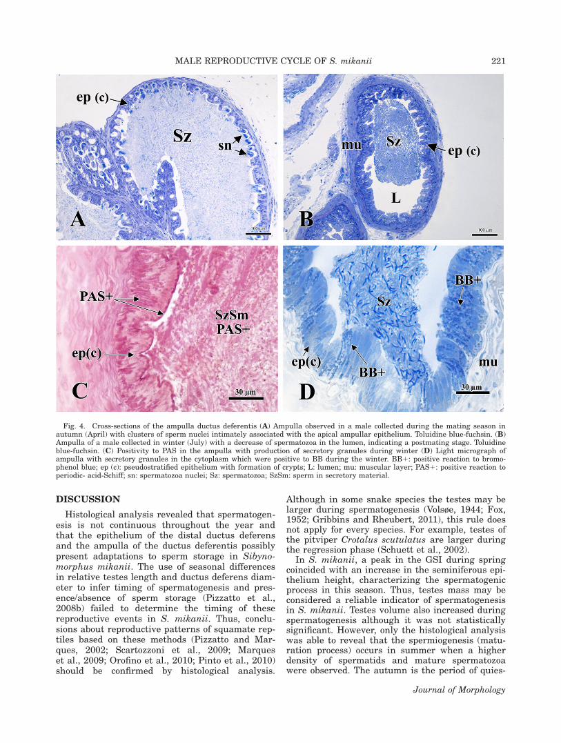
The ampulla ductus deferentis exhibited thesame layers as the rest of the distal ductus deferens,but with a taller epithelium with irregular projec-tions and formation of crypts (Fig. 4A). The nuclei ofthe spermatozoa seemed to be oriented toward thesecrypts (Fig. 4A). Spermatozoa density is lower inwinter than in other seasons in the ampulla ductusdeferentis (Fig. 4B). Secretory granules in the cyto-plasm were positive to PAS and BB only during thewinter in the ampulla (Fig. 4C,D). Positivity to PASwas also observed in the apical cytoplasm of epithe-lial cells in the ampulla throughout the year (Fig.4C). The ampulla ductus deferentis presented nega-tive reactions to AB.
TABLE 2. Specimens examined and structural variation of the testes of S. mikanii between different seasons
Season NSeminiferous tubule
diameter (lm)Seminiferous epithelial
height (lm)Leydig cell nuclear
diameter (lm)
Summer (A) 4 292.73 6 67.0 94.41 6 16.3B, C 4.99 6 1.54B, C
Autumn (B) 7 300.20 671.1 76.84 6 19.5A, D 5.96 6 1.22A, D
Winter (C) 5 315.90 6 78.6 83.22 6 28.9A, D 5.70 6 1.08A
Spring (D) 8 323.57 6 51.5 113.79 6 39.4B, C 5.22 6 0.99B
Post hoc analysis: significant differences (P < 0.05) between seasons are indicated by letters. Date are expressed as mean 6 stand-ard errors. N 5 specimens examined. Each letter represents a season. For example, in table 1 autumn is represented by letter Band no differences in testes volume among seasons were found, but the width of the ductus deferens, length and width of the kid-ney are different (larger) in autumn (B) from winter (C) and spring (D).
TABLE 3. Stages of the spermatogenic cycle in Sibynomorphusmikanii
Stages Spermatogenic condition Months of occurrence
I Complete regression: notobserved
/
II Early recrudescence:division ofspermatogonia andprimary spermatocytes
June
III Late recrudescence:primary spermatocytesand spermatids
September–November
IV Early spermiogenesis:spermatids inmetamorphosis
December
V Spermiogenesis: maturespermatoza in thelumen
December–February
VI Early regression:decrease of theseminiferousepithelium (fiveto six layers)
March–June
Adapted from Goldberg and Parker (1975), Herpetologica, AllenPress Publishing Services.
218 C.A. ROJAS ET AL.
Journal of Morphology

SSK Secretory Cycle
Macroscopic measurements of the kidneys of S.mikanii presented significant differences betweenthe seasons, with an increase in the length (F 53.56, n 5 139, P < 0.01) and width (F 5 5.14, n 5139, P < 0.002). A post-hoc test (Tukey test) showedthat kidney length was larger in autumn than inwinter and in spring (P < 0.01; Table 1). Consider-ing kidney width, a post-hoc test (Tukey test)showed that it was larger in autumn than in winter(P < 0.0001) and in spring (P < 0.001) and alsolarger in summer than in winter (P < 0.009; Table1). The tubular diameter of the SSK differs amongthe seasons (H 5 63.2, n 5 240, P < 0.0001) and apost-hoc test (comparisons by Dunn method) showedthat it was larger in summer than in autumn, win-ter, and spring (P < 0.05) and also larger in autumn
than in winter and spring (P < 0.05; Fig. 5A). Theepithelial height of the SSK exhibited a seasonalpattern (H 5 38.79, n 5 240, P < 0.0001; Fig. 5B)with a significant increase during summer andautumn in relation to winter and spring (compari-sons by Dunn method, P < 0.05).
Histological and histochemical analysis showedthat winter was the period of lowest SSK activitywith a few weakly stained granules, concentrated inthe basal region of the cells (Fig. 6A,B). The lumenwas narrow and exhibited vacuoles on the medialregion of the tubule which suggests a postsecretorypattern (Fig. 6B). Considering that the detection ofproteins (BB) reacted positively, but weakly, thisindicates that the secretory activity of granulesduring this season was low (Fig. 6C; Table 4, scale 1;Table 5, Stage I). However, SSK reacted positively to
Fig. 2. Transverse sections of the testes of Sibynomorphus mikanii during the reproductive cycle. (A) A specimen in winter(June) showing different stages of cellular division in the seminiferous epithelium; (B) Light micrograph of an individual in spring(November) with the peak of cellular division and an increase in the seminiferous epithelium; (C) summer (December), lots of sper-matids in metamorphosis; (D) late summer and early autumn (March), final phase of the spermiogenesis process. At: tunica albugi-nea; bc: blood capillaries; Lc: Leydig cells; Mc: myoid cells; Sc: Sertoli cells; L: lumen; Sd: spermatid; Sg: spermatogonia; Sp: pri-mary spermatocyte; Sp (p): primary spermatocyte at pachytene stage; Sz: spermatozoa; (*) remaining spermatozoa. Toluidine blue-fuchsin. [Color figure can be viewed in the online issue, which is available at wileyonlinelibrary.com.]
MALE REPRODUCTIVE CYCLE OF S. mikanii 219
Journal of Morphology

PAS, thus it contains neutral carbohydrates(Fig. 6D; Table 4, scales 1 and 2; Table 5, Stage II).
During spring, the SSK was characterized by thepresence of several moderately stained granulesspread throughout the cytoplasm of the cells, includingthe apical region (Fig. 7A). The lumen of the tubulebecame wider and basal nuclei were present (Fig. 7B).The SSK was lightly positive to BB and the proximalconvoluted tubule was totally negative (Fig. 7C; Table4, scale 2; Table 5, Stage I). PAS reacted positively dur-ing this season as it was observed in the winter (Fig.7D; Table 4, scales 2 and 3; Table 5, Stage II).
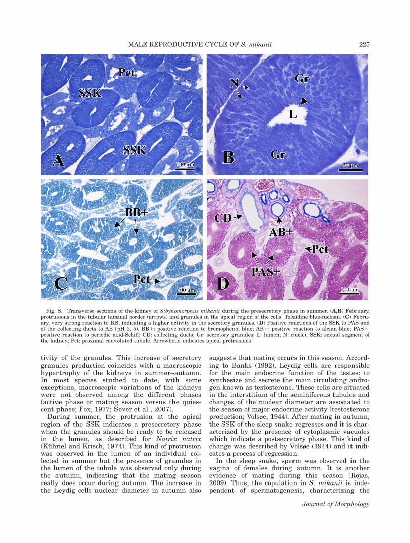
During summer, SSK exhibited a high density ofgranules all over the cytoplasm but mainly in theapical region of the cell (Fig. 8A,B). Little protru-sions in the apical region of the cells indicate asexual presecretory phase when the granules
would be ready to be released into the lumen (Fig.8B). The granules also stained and reacted moreintensively to BB and PAS when compared withthe reactions in winter and spring (Fig. 8C,D; Ta-ble 4, scales 3 and 4; Table 5, Stage III). A positivereaction to AB was observed in the collecting ductsin all seasons (Fig. 8D).
At the beginning of autumn, SSK cytoplasm wascharacterized by lots of intensively stained gran-ules and a distended lumen (Fig. 9A). Granuleswere also observed in the lumen of the tubule (Fig.9B). During the autumn, granules reacted stronglyto BB and PAS as they did in the summer, sug-gesting an increase in the production of secretorygranules (Fig. 9C,D; Table 4, scale 4; Table 5,Stage III). Secretory granules of the SSK exhibitednegative reactions to AB in all seasons.
Fig. 3. Transverse sections of the distal ductus deferens in Sibynomorphus mikanii during seasons of the year. (A) Specimen inspring with lots of spermatozoa in the lumen. Toluidine blue-fuchsin. (B) A male collected in early summer (December) presentsthe same pattern observed in spring exhibiting an epithelium composed of principal cells and basal cells. Toluidine blue-fuchsin.(C) Light micrograph with positive reaction to PAS in the apical portion of the cells and sperm mass (D) Positive reaction to BB inthe apical region of epithelial cells and secretory material. BB1: positive reaction to bromophenol blue; Bc: basal cells; bm:basement membrane; ct: connective tissue; ep (lc): pseudostratified epithelium with low cells; mu: muscular layer; PAS1: positivereaction to periodic- acid-Schiff; Pc: principal cells; Sm: secretory material; sr: serosa; Sz: spermatozoa; SzSm: sperm in secretorymaterial.
220 C.A. ROJAS ET AL.
Journal of Morphology
DISCUSSION
Histological analysis revealed that spermatogen-esis is not continuous throughout the year andthat the epithelium of the distal ductus deferensand the ampulla of the ductus deferentis possiblypresent adaptations to sperm storage in Sibyno-morphus mikanii. The use of seasonal differencesin relative testes length and ductus deferens diam-eter to infer timing of spermatogenesis and pres-ence/absence of sperm storage (Pizzatto et al.,2008b) failed to determine the timing of thesereproductive events in S. mikanii. Thus, conclu-sions about reproductive patterns of squamate rep-tiles based on these methods (Pizzatto and Mar-ques, 2002; Scartozzoni et al., 2009; Marqueset al., 2009; Orofino et al., 2010; Pinto et al., 2010)should be confirmed by histological analysis.
Although in some snake species the testes may belarger during spermatogenesis (Volsøe, 1944; Fox,1952; Gribbins and Rheubert, 2011), this rule doesnot apply for every species. For example, testes ofthe pitviper Crotalus scutulatus are larger duringthe regression phase (Schuett et al., 2002).
In S. mikanii, a peak in the GSI during springcoincided with an increase in the seminiferous epi-thelium height, characterizing the spermatogenicprocess in this season. Thus, testes mass may beconsidered a reliable indicator of spermatogenesisin S. mikanii. Testes volume also increased duringspermatogenesis although it was not statisticallysignificant. However, only the histological analysiswas able to reveal that the spermiogenesis (matu-ration process) occurs in summer when a higherdensity of spermatids and mature spermatozoawere observed. The autumn is the period of quies-
Fig. 4. Cross-sections of the ampulla ductus deferentis (A) Ampulla observed in a male collected during the mating season inautumn (April) with clusters of sperm nuclei intimately associated with the apical ampullar epithelium. Toluidine blue-fuchsin. (B)Ampulla of a male collected in winter (July) with a decrease of spermatozoa in the lumen, indicating a postmating stage. Toluidineblue-fuchsin. (C) Positivity to PAS in the ampulla with production of secretory granules during winter (D) Light micrograph ofampulla with secretory granules in the cytoplasm which were positive to BB during the winter. BB1: positive reaction to bromo-phenol blue; ep (c): pseudostratified epithelium with formation of crypts; L: lumen; mu: muscular layer; PAS1: positive reaction toperiodic- acid-Schiff; sn: spermatozoa nuclei; Sz: spermatozoa; SzSm: sperm in secretory material.
MALE REPRODUCTIVE CYCLE OF S. mikanii 221
Journal of Morphology

cence and reduction of the testicular activity, char-acterizing an annual spermatogenic cycle.
The beginning of spermatogenesis occurs duringlate winter in S. mikanii. This reproductive patternis similar to that presented by Leptodeira maculataand Leptodeira punctata which are Dipsadinaesnakes from New Mexico that present long sperma-togenic cycles with production of spermatozoa dur-ing spring, summer, and autumn (Goldberg, 2004).Thereby, spermatogenesis may begin earlier insnakes from meridional regions than it does insnakes from northerly localities with colder tem-peratures (Fox, 1954).
Recently, Mathies (2011) summarized data onreproductive cycles of tropical snakes and proposeda new classification for snake reproductive cycles.Considering the individual level, S. mikanii maleshave a continuous cyclical reproductive pattern withreduction of gonadal activity during autumn andearly winter. At the populational level, the reproduc-tive cycle is seasonal semisynchronous with a recog-nizable peak of reproductive activity in summer–autumn (Fig. 10). Total regression of the testes wasnot observed in S. mikanii. This testicular stage isusually observed in snakes from temperate areas
(Gartska et al., 1982). However, total regression inthe testes was also described for the neotropical rat-tlesnake (Crotalus durissus; Salomao and Almeida-Santos, 2002; Barros et al., 2012).
The sleep snake shows an amniote pattern ofgerm cell development with the retention of anam-niote characteristics. The germ cells proceedthrough the phases of spermatogenesis as a singlepopulation which leads to a single spermiation atthe end of this process. This pattern has alreadybeen described for some squamate species fromtemperate regions, such as Podarcis muralis (Grib-bins and Gist, 2003) and Seminatrix pygaea (Grib-bins et al., 2005).
After gametes are produced in the gonads, theypass through genital accessory ducts and arefinally deposited in the ductus deferens. The duc-tus deferens is the known site for sperm storage inreptiles and the epididymis does not play a role inthe maturation and storage of spermatozoa asobserved in mammals (Jones, 1998; Sever, 2004).Aldridge and Duvall (2002) studied viperid snakesfrom temperate regions and determined that mat-ing is independent of spermatogenesis and thatthe males actually adapt to the female reproduc-tive cycle. It is also true for S. mikanii, a neotropi-cal species: spermatozoa is found in the ductus de-ferens throughout the year, which indicates apotential for copulation at any season of the year(Almeida-Santos and Salomao, 2002; Almeida-San-tos et al., 2004).
In the sleep snake, the width of the ductus de-ferens showed a peak in the autumn which mightsuggest a spermiation process with posteriorsperm storage. We also observed that the ampullaductus deferentis is not macroscopically differentfrom other portions of the ductus deferens and ismicroscopically similar to that observed in Semina-trix pygaea (Sever, 2004).
Histochemical analysis of the distal ductus de-ferens and ampulla ductus deferentis of the sleepsnake showed some similarities with the secretorypattern of a few snake species that have alreadybeen studied (Sever, 2004; Siegel et al., 2009).Most of the cellular cytoplasm of the distal ductusdeferens of S. mikanii is apparently nonreactive tohistochemical tests as observed for Seminatrixpygaea (Sever, 2004). However, in all seasons,PAS- and BB-positive material is located in the ap-ical cytoplasm of the ductus deferens, similarly tothat found in Agkistrodon piscivorus (Siegel et al.,2009).
The ampulla ductus deferentis of the sleepsnake presents projections of the epithelium andcrypts with clusters of sperm nuclei intimatelyassociated with the apical ampullar epithelium.The presence of cytoplasmic granules rich in pro-tein and neutral carbohydrates suggests that thissecretion assists in the nutrition of spermatozoaduring storage. However, it is not possible to
Fig. 5. Relationship between the seasons of the year and theaverage of (A) tubular diameter and (B) epithelial height of theSSK in Sibynomorphus mikanii. Middle line represents averagevalues, boxes show standard errors and whiskers representminimum and maximum values.
222 C.A. ROJAS ET AL.
Journal of Morphology

understand how spermatozoa are attracted by theampulla epithelium and what are its mechanismsof secretion because the techniques used in this
study (light microscopy) are not ideal to deal withthese questions. An ultrastructural study of thedifferent regions of the ductus deferens of S. mikaniimay reveal the existence of phagocytosis and thecontribution of seminal plasma, as has beenobserved in the lizard Sitana ponticeriana (Akbarshaet al., 2005). The function and morphology of theampulla differ along the squamate lineage (Trauthand Sever, 2011). To our knowledge, this is the firstrecord of the occurrence of the ampulla ductus defer-entis in a neotropical snake species. Thus, moreresearch on this topic is necessary for a betterunderstanding of the male snake’s mechanisms ofsperm storage. On the other hand, sperm storage inthe female reproductive tract probably occurs duringwinter in S. mikanii because the mating season(autumn) is not synchronous to ovulation/fertiliza-tion (late winter).
Seasonal changes in the SSK of Sibynomorphusmikanii presented a similar pattern to that
Fig. 6. Transverse sections of the kidney of Sibynomorphus mikanii during the postsecretory phase in winter. (A and B) June,narrow lumen and presence of vacuoles, characteristic of this phase. Fuchsin-toluidine blue. (C) June, light positive reaction to BB.(D) July, PAS positive reaction with granules rich in neutral carbohydrates. BB1: Positive for bromophenol blue; bc: capillary bloodvessels; Gr: secretory granules; L: lumen; PAS1: Positivity to the periodic acid-Schiff; Pct: proximal convoluted tubule; SSK: sexualsegment of the Kidney; V: cytoplasmic vacuoles.
TABLE 4. Density of sexual granules in the SSK epithelium ofSibynomorphus mikanii
ScaleVisible differences
categorized Months
0 No hypertrophy Not observed1 Hypertrophy with a few
granulesJune–September
2 Hypertrophy with granulesevident throughout thecytoplasm
September–December
3 Secretory granules visible inthe apical region of thecytoplasm
September–March
4 Maximum density of secretorygranules within thecytoplasm
March–June
Adapted from Krohmer et al. (2004), Comp Biochem Physiol
MALE REPRODUCTIVE CYCLE OF S. mikanii 223
Journal of Morphology

observed in the testes: continuous cyclical andseasonal semisynchronous (Fig. 10). However,these processes occur in different seasons.The period of minor activity of the germinativeepithelium of the testes (autumn) occurs simulta-neously to the period of major activity of the SSKepithelium. In S. mikanii, the increase of the tu-bular diameter and epithelium height of the SSKduring summer–autumn occurred due to the max-
imum density and staining intensity of the gran-ules produced in those seasons. The secretorygranules produced by the SSK are of glycoproteinand mucoprotein nature. Histochemical analysisshowed a light reaction to BB and a moderatereaction for PAS during winter. However, duringsummer and autumn, SSK exhibited a strongreaction for proteins (BB1) and neutral carbohy-drates (PAS1), indicating an intense secretory ac-
Fig. 7. Histology of the kidney of Sibynomorphus mikanii during the beginning of the presecretory phase in spring. (A and B)kidney of a male from November showing an intense staining in the cytoplasm and the presence of basal nuclei (H/E). (C) Speci-men of September with SSK positive reaction to BB. (D) Section of October with SSK positive reaction to PAS. Bn: basal nuclei;BB1: positive reaction to bromophenol blue; Gr(a): secretory granules at the apical region; L: lumen; PAS1: positive reaction toperiodic acid-Schiff; Pct: proximal convoluted tubule, SSK: sexual segment of the kidney.
TABLE 5. Seasonal variation in SSK granule staining intensity in Sibynomorphus mikanii
Stages Staining intensity of granules
Months
Bromophenol blue (BB) Periodic acid-Schiff (PAS)
I some lightly stained granules June-September not observedII many moderately stained granules September- December June-DecemberIII many intensely stained granules December- June December-June
Adapted from Aldridge and Brown (1995), J Herpetol.
224 C.A. ROJAS ET AL.
Journal of Morphology
tivity of the granules. This increase of secretorygranules production coincides with a macroscopichypertrophy of the kidneys in summer–autumn.In most species studied to date, with someexceptions, macroscopic variations of the kidneyswere not observed among the different phases(active phase or mating season versus the quies-cent phase; Fox, 1977; Sever et al., 2007).
During summer, the protrusion at the apicalregion of the SSK indicates a presecretory phasewhen the granules should be ready to be releasedin the lumen, as described for Natrix natrix(Kuhnel and Krisch, 1974). This kind of protrusionwas observed in the lumen of an individual col-lected in summer but the presence of granules inthe lumen of the tubule was observed only duringthe autumn, indicating that the mating seasonreally does occur during autumn. The increase inthe Leydig cells nuclear diameter in autumn also
suggests that mating occurs in this season. Accord-ing to Banks (1992), Leydig cells are responsiblefor the main endocrine function of the testes: tosynthesize and secrete the main circulating andro-gen known as testosterone. These cells are situatedin the interstitium of the seminiferous tubules andchanges of the nuclear diameter are associated tothe season of major endocrine activity (testosteroneproduction; Volsøe, 1944). After mating in autumn,the SSK of the sleep snake regresses and it is char-acterized by the presence of cytoplasmic vacuoleswhich indicate a postsecretory phase. This kind ofchange was described by Volsøe (1944) and it indi-cates a process of regression.
In the sleep snake, sperm was observed in thevagina of females during autumn. It is anotherevidence of mating during this season (Rojas,2009). Thus, the copulation in S. mikanii is inde-pendent of spermatogenesis, characterizing the
Fig. 8. Transverse sections of the kidney of Sibynomorphus mikanii during the presecretory phase in summer. (A,B) February,protrusions in the tubular luminal border (arrows) and granules in the apical region of the cells. Toluidine blue-fuchsin. (C) Febru-ary, very strong reaction to BB, indicating a higher activity in the secretory granules. (D) Positive reactions of the SSK to PAS andof the collecting ducts to AB (pH 2, 5). BB1: positive reaction to bromophenol blue; AB1: positive reaction to alcian blue; PAS1:positive reaction to periodic acid-Schiff; CD: collecting ducts; Gr: secretory granules; L: lumen; N: nuclei, SSK: sexual segment ofthe kidney; Pct: proximal convoluted tubule. Arrowhead indicates apical protrusions.
MALE REPRODUCTIVE CYCLE OF S. mikanii 225
Journal of Morphology

reproductive cycle as dissociated (cf. Crews, 1984).However, two exceptions need to be highlighted:the increase of Leydig cell nuclear diameter
(indicative of sex hormone secretion), and the SSKhypertrophy is coincident with the mating seasonin autumn. These are characteristics of an associ-ated reproductive cycle which reinforces the ideathat reproductive strategies are complex in snakespecies (cf. Crews, 1984; Shine, 2003). In summary,the endocrine activity of the testes (secretion ofsex steroid hormone by Leydig cells) is associatedwith the mating season, whereas the exocrineactivity (sperm production) is dissociated in S.mikanii.
Data presented herein described, for the firsttime, the SSK secretory cycle and its relationshipwith the testicular cycle of a snake from SouthAmerica. Nevertheless, this study is not exhaus-tive: studies using other microscopic techniquessuch as scanning and transmission electron mi-croscopy may reveal important details of S. mika-nii reproductive strategies. We hope that thisstudy may open new perspectives and perhaps
Fig. 9. Light micrographs of transverse sections of the kidney of Sibynomorphus mikanii during the secretory phase in autumn.(A,B) specimen in April with secretory granules being released in the lumen. Toluidine blue-fuchsin. (C) March, strong positivereaction to BB. (D) April, strong reaction to PAS. AB1: positive reaction to alcian blue; BB1: positive reaction to bromophenolblue; Gr(*): secretory granules in the lumen; L: lumen; N: nuclei; PAS1: positive reaction to periodic acid-Schiff; SSK: sexual seg-ment of the kidney; Pct: proximal convoluted tubule.
Fig. 10. Phases of the annual reproductive cycle of male Sib-ynomorphus mikanii.
226 C.A. ROJAS ET AL.
Journal of Morphology
stimulate research on squamate reptiles in theneotropical region, which is still poorly known.
ACKNOWLEDGMENTS
The authors thank Carlos Jared and MartaAntoniazzi for kindly allowing use of material andequipments in the Laboratory of Cell Biology (IB),Simone Jared and Leonardo de Oliveira for theirtechnical assistance. Francisco L. Franco (IB) andRonaldo Fernandes (MNRJ) allowed access to col-lections. This study is based on a MSc thesisundertaken by Claudio A. Rojas.
LITERATURE CITED
Akbarsha MA, Meeran MM. 1995. Occurrence of ampulla in theductus deferens of the Indiana garden lizard Calotes versi-color Daudin. J Morphol 225:261–268.
Aldridge RD, Brown WS. 1995. Male reproductive cycle, age atmaturity, and cost of reproduction in the timber rattlesnakeCrotalus horridus. J Herpetol 29:399–407.
Akbarsha MA, Tamilarasan V, Kadalmi B, Daisy P. 2005. Ultra-structural evidence for secretion from the epithelium ofampulla ductus deferentis of the fan-throated lizard Sitanaponticeriana Cuvier. J Morphol 266:94–111.
Aldridge RD, Duvall D. 2002. Evolution of the mating season inthe pitvipers of North America. Herpetol Monogr 16:1–25.
Aldridge RD, Flanagan WP, Swarthout JT. 1995. Biology of thewater snake Nerodia rhombifer from Veracruz, Mexico. Her-petologica 51:182–192.
Aldridge RD, Jellen BC, Siegel DS, Wisniewski SS. 2011. Thesexual segment of the kidney. In: Aldridge RD, Sever DM,editors. Reproductive Biology and Philogeny of Snakes.Enfield: Science Publishers. pp 477–509.
Almeida-Santos SM, Salomao,MG. 1997. Long term sperm stor-age in the female neotropical rattlesnake Crotalus durissusterrificus (Viperidae: Crotalidae). Jpn J Herpetol 17:46–52.
Almeida-Santos SM, Salomao MG 2002. Reproduction in neo-tropical pitvipers, with emphasis on species of the genusBothrops. In: Schuett G, Hoggren M, Douglas ME, GreeneHW, editors. Biology of the Vipers. Carmel, Indiana: EagleMountain. pp 445–462.
Almeida-Santos SM, Ferreira IL, Antoniazzi MM, Jared C.2004. Sperm storage in males of the snake Crotalus durissusterrificus (Crotalinae: Viperidae) in southeastern Brazil.Comp Biochem Physiol A 139:169–174.
Almeida-Santos SM, Pizzatto L, Marques OAV. 2006. Intra-sexsynchrony and inter-sex coordination in the reproductive tim-ing of the coral snake Micrurus corallinus (Elapidae). Herpe-tol J 16:371–376.
Banks WJ. 1992. Sistema Reprodutor Masculino. In: HistologiaVeterinaria Aplicada. Sao Paulo: Manole. pp 546–563.
Barros VA, Sueiro LR, Almeida-Santos SM. 2012. Reproductivebiology of the neotropical rattlesnake Crotalus durissus fromnortheastern Brazil: A test of phylogenetic conservatism ofreproductive patterns. Herpetol J 22:97–104.
Bergerson W, Amselgruber W, Sinowatz F, Bergerson M. 1994.Morphological evidence of sperm maturation in the ampullaductus deferentis of the bull. Cell Tissue Res 275:537–541.
Bishop JE. 1959. A histological and histochemical study of thekidney tubule of the common garter snake, Thamnophis sirta-lis, with special reference to the sexual segment in the male.J Morphol 104:307–357.
Brown GP, Shine R. 2006. Effects of nest temperature and mois-ture on phenotypic traits of hatchling snakes (Tropidonophismairii, Colubridae) from tropical Australia. Biol J Linn Soc89:159–168.
Clesson D, Bautista A, Baleckaitis DD, Kromer RW. 2002.Reproductive biology of male eastern garter snakes (Thamno-phis sirtalis sirtalis) from a denning populations in CentralWisconsin. Am Midl Nat 147:376–386.
Cooper TG, Hamilton DW. 1977. Phagocytosis of spermatozoa inthe terminal region and gland of the vas deferens of the rat.Am J Anat 150:247–268.
Crews D. 1984. Gamete production, sex hormone secretion, andmating behavior uncoupled. Horm Behav 18:22–28.
Fox W. 1952. Seasonal variation in the male reproductive sys-tem of Pacific coast garter snakes. J Morphol 90:481–553.
Fox W. 1954. Genetic and Environmental variation in the tim-ing of the reproductive cycles of male garter snakes. J Mor-phol 95:415–450.
Fox W. 1977. The urogenital system of reptiles. In: Gans C, ParsonsTS. Biology of the Reptilia. New York: Academic Press: pp 1–157.
Franco FL. 1994. O genero Sibynomorphus FITZINGER, 1843,no Brasil (Colubridae: Xenodontinae, Dipsadini). [Disserta-tion]. Porto Alegre (RS): Pontifıcia Universidade Catolica doRio Grande do Sul. Biblioteca do Instituto de Biociencias daPUCRS, Rio Grande do Sul, RS; 000129246, 144p.
Garstka WR, Camazine B, Crews D. 1982. Interactions ofbehavior and physiology during the annual reproductive cycleof the red-sided garter snake (Thamnophis sirtalis parietalis).Herpetologica 38:104–123.
Goldberg SR. 2004. Reproduction in the coffee snake, Ninia macu-lata (Serpentes: Colubridae), from Costa Rica. Tex J Sci 56:81–84.
Goldberg SR, Parker WS. 1975. Seasonal testicular histology ofthe colubrid snakes, Masticophis taeniatus and Pituophis mel-anoleucus. Herpetologica 31:317–322.
Graham S. 2006. An integrative analysis of reproduction andstress in free-living male cottonmouths, Agkistrodon piscivo-rus. [Dissertation (Master of science)]. Georgia: College ofArts and Sciences of the State University. 85 p. Available at:http://digitalarchive.gsu.edu/biology_theses/6. Last accessMarch 15, 2012.
Gribbins KM, Gist DH. 2003. The cytological evaluation of sper-matogenesis within the germinal epithelium of the male Eu-ropean wall lizard, Podarcis muralis. J Morphol 258:296–306.
Gribbins KM, Rheubert JM. 2011. The ophidian testis, sperma-togenesis, and mature spermatozoa. In: Aldridge RD, SeverDM, editors. Reproductive Biology and Philogeny of Snakes.Enfield: Science Publishers. pp 183–264.
Gribbins KM, Gist DH, Congdon JD. 2003. The cytological eval-uation of spermatogenesis and organization of the germinalepithelium in the male slider turtle, Trachemys scripta. JMorphol 255:337–346.
Gribbins KM, Happ CS, Sever DM. 2005. Ultrastructure of thereproductive system of the black swamp snake (Seminatrixpygae). V. The temporal germ cell development strategy of thetestis. Acta Zool 86:223–230.
Jones RC. 1998. Evolution of the vertebrates epididymis. JReprod Fertil Suppl 53:163–181.
Junqueira LCU. 1995. Histology revisited—Technical improve-ment promoted by the use of hydrophilic resin embedding.Cien Cult 47:92–95.
Junqueira LCU, Bignolas G, Brentani R. 1979. Picrosiriusstaining plus polarization microscopy, a specific method forcollagen detection in tissue sections. Histochem J 11:447–455.
Krohmer RW, Grassman M, Crews D. 1987. Annual reproduc-tive cycle in the male red-sided garter snake, Thamnophissirtalis parietalis: Field and laboratory studies. Gen CompEndocrinol 68:64–75.
Krohmer RW, Martinez D, Mason RT. 2004. Development of therenal sexual segment in inmature snakes: Effect of sex ste-roid hormones. Comp Biochem Physiol 139:55–64.
Kuhnel W. Krisch B. 1974. On the sexual segment of the kidneyin the snake (Natrix natrix). Cell Tissue Res 148:417–429.
Kumar M. 1995. Spermatogenesis in the house sparrow, Passardomesticus: Histological observations. Pavo 33:1–4.
Laporta-Ferreira IL, Salomao MG, Sawaya P. 1986. Biologia deSibynomorphus (Colubridae—Dipsadinae)—Reproducao ehabitos alimentares. Rev Bras Biol 46:793–799.
MALE REPRODUCTIVE CYCLE OF S. mikanii 227
Journal of Morphology

Lebonde CP, Clermont Y. 1952. Spermiogenesis of rat, mouse,hamster, guinea pig as evealed by the periodic acid-fuchsinsulfurous acid technique. Am J Anat 90:167–215.
Lofts B. 1977. Patterns of spermatogenesis and steroidogenesisin male reptiles. In: Calaby JH, Tyndale-Boscoe CH, editors.Reproduction and Evolution. Canberra: Australian AcademicScience. pp 127–136.
Marinho CE, Almeida-Santos SM, Carneiro SM, Yamasaki SC,Silveira,PF. 2008a. Peptidase activities in Crotalus durrisusterrificus semen. Reproduction 136:767–776.
Marinho CE, Almeida-Santos SM, Yamasaki SC, Silveira PF.2008b. Seasonal variation of peptidase activities in the repro-ductive tract of Crotalus durrisus terrificus. Gen Comp Endo-crinol 160:84–92.
Marques OAV, Almeida-Santos SM, Rodrigues M, Camargo R.2009. Mating and reproductive cycle in the neotropicalcolubrid snake Chironius bicarinatus. South Am J Herpetol4:76–80.
Mathies T. 2011. Reproductive cycles of tropical snakes. In:Aldridge RD, Sever DM, editors. Reproductive Biology andPhilogeny of Snakes. Enfield: Science Publishers. pp 511–550.
Mendonca F, Danni-Oliveira IM. 2007. Climatologia: Nocoesbasicas e climas do Brasil. Sao Paulo: Oficina de Textos.206p.
Orofino RP, Pizatto L, Marques OAV. 2010. Reproductive biologyand food habits of Pseudoboa nigra (Serpentes: Dipsadidae)from the Brazilian Cerrado. Phyllomedusa J Herpetol 9:53–61.
Pinto RR, Marques OAV, Fernandes R. 2010. Reproductive biol-ogy of two sympatric colubrid snakes Chironius flavolineatusand Chironius quadricarinatus from the Brazilian Cerradodomain. Amphibia-Reptilia 31:463–473.
Pizzatto L, Marques OAV. 2002. Reproductive biology of thefalse coral snake Oxyrhopus guibei (Colubridae) from south-eastern Brazil. Amphibia-Reptilia 23:495–504.
Pizzatto L, Marques OAV. 2007. Reproductive ecology of Boinaesnakes with emphasis on Brazilian species and a comparisonto Pythons. South Am J Herpetol 2:107–133.
Pizzatto L, Jordao RS, Marques OAV. 2008a. Overview of repro-ductive strategies in Xenodontini (Serpentes: Colubridae:Xenodontinae) with new data for Xenodon neuwiedii andWaglerophis merremii. J Herpetol 42:153–162.
Pizzatto L, Cantor M, Oliveira J, Marques OAV, Capovilla V,Martins M. 2008b. Reproductive ecology of Dipsadine snakes,with emphasis on South American species. Herpetologica64:168–179.
Pleguezuelos JM, Feriche M. 1999. Reproductive ecology of thehorseshoe whip snake, Coluber hippocrepis, in the southeastof the Iberian Peninsula. J Herpetol 33:202–207.
Pyron RA, Burbrink FT, Colli GR, Oca ANM, Vitt LJ, Kuczyn-ski CA, Wiens JJ. 2011. The phylogeny of advanced snakes(Colubroidea), with discovery of a new subfamily and compar-ison of support methods for likelihood trees. Mol PhylogenetEvol 58:329–342.
Rojas CA. 2009. Modulacao fisiologica do ciclo reprodutivo, com-portamento sexual e atividade sazonal em Sibynomorphusmikanii e Sibynomorphus neuwiedi (dormideira) [Disserta-tion]. Sao Paulo (SP): Universidade de Sao Paulo. Biblioteca
do Instituto de Ciencias Biomedicas da Universidade de SaoPaulo, Sao Paulo, SP; T-ICB Biot QH 323-6 R741 mf, 118p.
Saint Girons H. 1972. Morphologie comparee du segment sexueldu rein des squamates (Reptilia). Archs Anat Microsc Mor-phol Exp 61:243–266.
Salomao MG, Almeida-Santos SM. 2002. The reproductive cyclein male neotropical rattlesnake (Crotalus durissus terrificus).In: Schuett GW, Hoggren M, Douglas ME, Greene HW, edi-tors. Biology of the Vipers. Carmel, Indiana: Eagle Montain.pp 507–514.
Santos X, Llorente GA, Feriche M, Pleguezuelos JM, Casals F,Sostoa A. 2005. Food availability induces geographic variationin reproductive timing of an aquatic oviparous snake (Natrixmaura). Amphibia-Reptilia 26:183–191.
Scartozzoni RR, Salomao MG, Almeida-Santos S. 2009. Naturalhistory of the vine snake Oxybelis fulgidus (Serpentes, Colu-bridae) from Brazil. South Am J Herpetol 4:81–89.
Schuett G W, Carlisle SL, Holycross AT, O’Leile JK, Hardy DL,Van Kirk EA, Murdoch WJ. 2002. Mating system of maleMojave rattlesnakes (Crotalus scutulatus): Seasonal timing ofmating, agonistic behavior, spermatogenesis, sexual segmentof the kidney, and plasma sex steroids. In: Schuett GW, Hogg-ren M, Douglas ME, Greene,HW, editors. Biology of theVipers. Carmel, Indiana: Eagle Mountain. pp 515–532.
Seigel RA, Ford NB. 1987. Reproductive ecology. In: Seigel RA,Collins JT, Novak SS, editors. Snakes: Ecology and Evolu-tionary Biology. New York: McGraw-Hill Publishing Company.pp 210–252.
Setchell BP, Maddocks S, Brooks DE. 1994. Anatomy, vascula-ture, innervation, and fluids of the male reproductive tract.In: Knobil E, Neill JD, editors. The Physiology of Reproduc-tion, 2nd ed. New York: Raven Press. pp 1063–1175.
Sever DM. 2004. Ultrastucture of the reproductive system ofthe black swamp snake (Seminatrix pygaea). IV. Occurrenceof an ampulla ductus deferentis. J Morphol 262:714–730.
Sever DM, Stevens RA, Ryan T, Hamlett WC. 2002. Ultrastruc-ture of the reproductive system of the black swamp snake(Seminatrix pygaea). III. Sexual segment of the male kidney.J Morphol 252:238–254.
Sever DM., Siegel SD, Bagwill A, Eckstut EM, Alexander L,Camus A, Morgan C. 2007. Renal sexual segment of the cot-tonmouth snake Agkistrodon piscivorous (Reptilia, Squamata,Viperidae). J Morphol 1:1–14.
Shine R. 2003. Reproductive strategies in snakes. Proc R SocLond B 270:995–1004.
Siegel DS, Sever DM, Rheubert JL, Gribbins KM. 2009. Repro-ductive biology of Agkistrodon piscivorus Lacepede (Squamata,Serpentes, Viperidae, Crotalinae). Herpetol Monogr 23:74–107.
Trauth SE, Sever DM. 2011. Male urogenital ducts and cloacalanatomy. In: Aldridge RD, Sever DM, editors. ReproductiveBiology and Philogeny of Snakes. Enfield: Science Publishers.pp 411–475.
Volsøe H. 1944. Structure and seasonal variation of the malereproductive organs of Vipera berus (L.). Spolia Zool MusHauniensis 5:1–157.
Zar JR. 1999. Biostatistical Analysis. New Jersey: Prentice-Hall. pp 875
228 C.A. ROJAS ET AL.
Journal of Morphology
Copyright © 2022 FDOKUMEN


![(DISSERTATION) THE NINETEENTH-CENTURY BRITISH PLANTATION SETTLEMENT AT LAMANAI, BELIZE (1837–1868) [pgs 101-111]](https://static.fdokumen.com/doc/165x107/6344dc63df19c083b107cac7/dissertation-the-nineteenth-century-british-plantation-settlement-at-lamanai.jpg)