The relative timing of VMO and VL in the aetiology of anterior knee pain: a systematic review and...
14
BioMed Central Page 1 of 14 (page number not for citation purposes) BMC Musculoskeletal Disorders Open Access Research article The relative timing of VMO and VL in the aetiology of anterior knee pain: a systematic review and meta-analysis Rachel Chester* 1,2 , Toby O Smith 2 , David Sweeting 3 , John Dixon 4 , Sarah Wood 2 and Fujian Song 1 Address: 1 School of Allied Health Professions, University of East Anglia, Norwich, Norfolk, NR4 7TJ, UK, 2 Physiotherapy Department, Norfolk and Norwich University Hospital, Norwich, NR4 7UY, UK, 3 Physiotherapy Department, Great Yarmouth & Waveney PCT, James Paget University Hospital NHS Trust, Great Yarmouth, Norfolk, NR31 6LA, UK and 4 Centre for Rehabilitation Sciences, School of Health and Social Care, University of Teesside, Middlesbrough, TS1 3BA, UK Email: Rachel Chester* - [email protected]; Toby O Smith - [email protected]; David Sweeting - [email protected]; John Dixon - [email protected]; Sarah Wood - [email protected]; Fujian Song - [email protected] * Corresponding author Abstract Background: Anterior knee pain (AKP) is a common musculoskeletal complaint. It has been suggested that one factor that may contribute to the presence of AKP is a delay in the recruitment of the vastus medialis oblique muscle (VMO) relative to the vastus lateralis muscle (VL). There is however little consensus within the literature regarding the existence or nature of any such delay in the recruitment of the VMO within the AKP population. The purpose of this systematic review and meta-analysis was to examine the relative timing of onset of the VMO and VL in those with AKP in comparison to the asymptomatic population. Methods: The bibliographic databases AMED, British Nursing Index, CINAHL, EMBASE, Ovid Medline, PEDro, Pubmed and the Cochrane Library were searched for studies comparing the timing of EMG onset of the VMO and VL in those with AKP versus the asymptomatic population. Studies fulfilling the inclusion criteria were independently assessed. Heterogeneity across the studies was measured. A meta-analysis of results was completed for those studies where adequate data was supplied. Where comparable methodologies had been used, results were pooled and analysed. Results: Fourteen studies met the inclusion criteria; one prospective and thirteen observational case control. Eleven compared VMO and VL EMG onset times during voluntary active tasks while four investigated reflex response times. All used convenience sampling and did not state blinding of the assessor. Study methodologies/testing and assessment procedures varied and there was considerable heterogeneity within individual samples. Whilst a trend was identified towards a delay in onset of VMO relative to the VL in the AKP population during both voluntary active tasks and reflex activity, a substantial degree of heterogeneity across the pooled studies was identified (I 2 = 69.9–93.4%, p < 0.01). Conclusion: Findings are subject to substantial and unexplained heterogeneity. A trend was demonstrated towards a delayed onset of VMO relative to VL in those with AKP in comparison to those without. However not all AKP patients demonstrate a VMO-VL dysfunction, and this is compounded by normal physiological variability in the healthy population. The clinical and therapeutic significance is therefore difficult to assess. Published: 1 May 2008 BMC Musculoskeletal Disorders 2008, 9:64 doi:10.1186/1471-2474-9-64 Received: 6 July 2007 Accepted: 1 May 2008 This article is available from: http://www.biomedcentral.com/1471-2474/9/64 © 2008 Chester et al; licensee BioMed Central Ltd. This is an Open Access article distributed under the terms of the Creative Commons Attribution License (http://creativecommons.org/licenses/by/2.0 ), which permits unrestricted use, distribution, and reproduction in any medium, provided the original work is properly cited.
-
Upload
eastanglia -
Category
Documents
-
view
5 -
download
0
Transcript of The relative timing of VMO and VL in the aetiology of anterior knee pain: a systematic review and...
BioMed CentralBMC Musculoskeletal Disorders
ss
Open AcceResearch articleThe relative timing of VMO and VL in the aetiology of anterior knee pain: a systematic review and meta-analysisRachel Chester*1,2, Toby O Smith2, David Sweeting3, John Dixon4, Sarah Wood2 and Fujian Song1Address: 1School of Allied Health Professions, University of East Anglia, Norwich, Norfolk, NR4 7TJ, UK, 2Physiotherapy Department, Norfolk and Norwich University Hospital, Norwich, NR4 7UY, UK, 3Physiotherapy Department, Great Yarmouth & Waveney PCT, James Paget University Hospital NHS Trust, Great Yarmouth, Norfolk, NR31 6LA, UK and 4Centre for Rehabilitation Sciences, School of Health and Social Care, University of Teesside, Middlesbrough, TS1 3BA, UK
Email: Rachel Chester* - [email protected]; Toby O Smith - [email protected]; David Sweeting - [email protected]; John Dixon - [email protected]; Sarah Wood - [email protected]; Fujian Song - [email protected]
* Corresponding author
AbstractBackground: Anterior knee pain (AKP) is a common musculoskeletal complaint. It has been suggestedthat one factor that may contribute to the presence of AKP is a delay in the recruitment of the vastusmedialis oblique muscle (VMO) relative to the vastus lateralis muscle (VL). There is however littleconsensus within the literature regarding the existence or nature of any such delay in the recruitment ofthe VMO within the AKP population. The purpose of this systematic review and meta-analysis was toexamine the relative timing of onset of the VMO and VL in those with AKP in comparison to theasymptomatic population.
Methods: The bibliographic databases AMED, British Nursing Index, CINAHL, EMBASE, Ovid Medline,PEDro, Pubmed and the Cochrane Library were searched for studies comparing the timing of EMG onsetof the VMO and VL in those with AKP versus the asymptomatic population. Studies fulfilling the inclusioncriteria were independently assessed. Heterogeneity across the studies was measured. A meta-analysis ofresults was completed for those studies where adequate data was supplied. Where comparablemethodologies had been used, results were pooled and analysed.
Results: Fourteen studies met the inclusion criteria; one prospective and thirteen observational casecontrol. Eleven compared VMO and VL EMG onset times during voluntary active tasks while fourinvestigated reflex response times. All used convenience sampling and did not state blinding of theassessor. Study methodologies/testing and assessment procedures varied and there was considerableheterogeneity within individual samples. Whilst a trend was identified towards a delay in onset of VMOrelative to the VL in the AKP population during both voluntary active tasks and reflex activity, a substantialdegree of heterogeneity across the pooled studies was identified (I2 = 69.9–93.4%, p < 0.01).
Conclusion: Findings are subject to substantial and unexplained heterogeneity. A trend wasdemonstrated towards a delayed onset of VMO relative to VL in those with AKP in comparison to thosewithout. However not all AKP patients demonstrate a VMO-VL dysfunction, and this is compounded bynormal physiological variability in the healthy population. The clinical and therapeutic significance istherefore difficult to assess.
Published: 1 May 2008
BMC Musculoskeletal Disorders 2008, 9:64 doi:10.1186/1471-2474-9-64
Received: 6 July 2007Accepted: 1 May 2008
This article is available from: http://www.biomedcentral.com/1471-2474/9/64
© 2008 Chester et al; licensee BioMed Central Ltd. This is an Open Access article distributed under the terms of the Creative Commons Attribution License (http://creativecommons.org/licenses/by/2.0), which permits unrestricted use, distribution, and reproduction in any medium, provided the original work is properly cited.
Page 1 of 14(page number not for citation purposes)
BMC Musculoskeletal Disorders 2008, 9:64 http://www.biomedcentral.com/1471-2474/9/64
BackgroundAnterior knee pain (AKP) is one of the most common con-ditions presenting to physiotherapists [1], with a reportedincidence in up to 25% of the population [2]. Despitethis, the exact cause of pain remains largely unknown [3].It has been suggested that a variety of factors may contrib-ute to the development and maintenance of AKP. Onesuch factor is the presence of a delay in the recruitment ofthe vastus medialis oblique muscle (VMO) relative to thevastus lateralis muscle (VL) during functional activity[2,4]. It has been claimed that the presence of such a delaymay adversely affect the tracking of the patella, thus con-tributing towards the presence of AKP [2,4-6]. One objec-tive of some treatment strategies commonly used in therehabilitation of AKP is to restore the normal timing ofthe VMO and VL muscles [2,4].
Previous descriptive reviews however have highlightedpotential disagreements within the literature regardingthe scale, existence and even the direction of any abnor-mality in the firing patterns of VMO and VL in the AKPpopulation [7,8]. This brings into question the validity ofthe basis upon which these treatment strategies are based.Improved understanding regarding the existence andnature of any dysfunction in the timing of these muscleswould contribute to the effective management of thiscommon condition. The purpose of this systematic reviewis to synthesise evidence from comparative studies thathave investigated the relative onset timing of the VMOand VL in AKP and asymptomatic control subjects.
MethodsSearch StrategyThe literature search was performed by TS using the bibli-ographic electronic databases AMED, British NursingIndex, CINAHL, EMBASE, Ovid Medline, PhysiotherapyEvidence Database (PEDro), Pubmed and the CochraneLibrary, from their inception to June 2006. The followingMedical Subject Headings and key words were combined:anterior knee pain or patellofemoral pain syndrome orchondromalacia or extensor mechanism or vastus media-lis or vastus lateralis; AND activity or timing or recruit-ment or torque or EMG or electromyography orelectromyographic. This was updated by a second elec-tronic search (DS and JD) for any publications in theperiod June 2006–June 2007.
A hand search was performed in the specialist journals:The Knee (1994- June 2006), Physical Therapy in Sport(2000- June 2006), American Journal of Sport Medicine(1986- June 2006), British Journal of Sport Medicine(1986- June 2006) and the Journal of Science and Medi-cine in Sport (1998- June 2006). The reference lists of allretrieved papers including review articles were searched
for any additional publications unidentified by the initialsearch strategy.
Study selectionWe included any primary studies that compared differ-ences in the onset timing in milliseconds (ms) of VMOand VL as a primary or secondary outcome between sub-jects with anterior knee pain, patellofemoral pain syn-drome or chondromalacia patellae and asymptomaticcontrol subjects. Studies which compared timing of peakEMG activity or percentage of the gait cycle were excludedas were those which included animals and cadavers orsubjects suffering patellar instability. Full text English lan-guage publications only were included, regardless of theyear of publication.
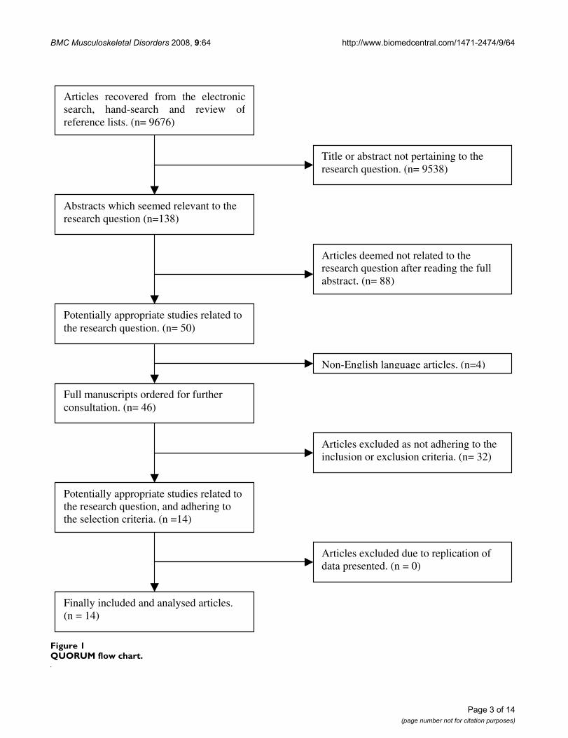
Three reviewers independently (RC, DS and SW) screenedthe titles and abstracts of all identified papers to deter-mine those potentially relevant to the review. The fullmanuscripts were then retrieved and each paper inde-pendently assessed for inclusion/exclusion criteria by twoof four reviewers (RC, TS, DS, and SW), any doubts or dis-agreements were discussed between the four reviewersuntil a consensus was reached. The QUORUM flow chart(Figure 1) illustrates the process by which manuscriptswere selected and numbers involved.
Data extractionEach study which met the inclusion criteria was independ-ently assessed by two of four reviewers, (DS, RC, SW andTS), each of whom completed a data extraction form [seeadditional file 1]. This included: study design, participantselection, sample size, population characteristics of AKPsubjects and control groups, procedural details and meth-ods of EMG assessment, results, and relevant study limita-tions. Data extraction forms were compared for accuracyand interpretation; where there was disagreement orinformation was ambiguous all four reviewers met toreach an agreement. In the absence of a recognised meth-odological scoring system for comparative observationalstudies, a qualitative critical appraisal of each study wasundertaken. This included an assessment of the factorsidentified from the data extraction form and their impacton the results, their interpretation and generalisability.
Evidence synthesis and statistical methodsIn relation to the relative timing of the VMO and VL, themost relevant and commonly used outcome measure wasthe onset timing difference (milliseconds) between theVMO and VL (i.e., Δ = VMO-VL). Where Δ > 0 indicatesthat the VMO onset was later than the VL onset, and Δ < 0indicates that the VMO onset was earlier than the VLonset. In this systematic review, we compared the differ-ence in relative timing of the VMO and VL between AKPpatients and control subjects. That is, the primary out-
Page 2 of 14(page number not for citation purposes)
BMC Musculoskeletal Disorders 2008, 9:64 http://www.biomedcentral.com/1471-2474/9/64
Page 3 of 14(page number not for citation purposes)
QUORUM flow chartFigure 1QUORUM flow chart.
Articles recovered from the electronicsearch, hand-search and review ofreference lists. (n= 9676)
Title or abstract not pertaining to the research question. (n= 9538)
Abstracts which seemed relevant to the research question (n=138)
Articles deemed not related to the research question after reading the full abstract. (n= 88)
Potentially appropriate studies related to the research question. (n= 50)
Articles excluded as not adhering to the inclusion or exclusion criteria. (n= 32)
Potentially appropriate studies related to the research question, and adhering to the selection criteria. (n =14)
Finally included and analysed articles. (n = 14)
Articles excluded due to replication of data presented. (n = 0)
Full manuscripts ordered for further consultation. (n= 46)
Non-English language articles. (n=4)
BMC Musculoskeletal Disorders 2008, 9:64 http://www.biomedcentral.com/1471-2474/9/64
come measure used in the meta-analyses was the meandifference (MD) between ΔAKP and ΔCTRL, where ΔAKP refersto the VMO-VL difference in AKP patients and ΔCTRL refersto the VMO-VL difference in control subjects. When MD =0, it indicates that there was no difference in the onset tim-ing of VMO relative to VL between AKP patients and con-trol subjects. When MD > 0, the onset of VMO wasrelatively later in AKP patients than that in control sub-jects.
Some primary studies did not provide sufficient data orstatistics to allow meta-analysis. For several studies[5,6,15,20], we acquired data based on graphical illustra-tion in published papers. Particularly, there was a lack ofdata on standard deviations of VMO-VL difference in AKPpatients and VMO-VL difference in control subjectsrequired for meta-analysis. An average estimate of stand-ard deviation was therefore calculated based on data fromother relevant studies [9] and input to studies in which thestandard deviation was not available.
Heterogeneity in results across primary studies was statis-tically tested and measured by I2 statistic [10]. Meta-anal-ysis was carried out using REVMAN software (version 4.2for Windows. Copenhagen: The Nordic Cochrane Centre,The Cochrane Collaboration, 2003). The existence of sta-tistically significant heterogeneity means that the poolingof results from primary studies in meta-analysis may becontroversial. When judged appropriate and helpful, weconducted meta-analyses using random-effects model.Significant heterogeneity was further narratively investi-
gated in the discussion, by examining whether differencesin study results could be possibly explained by differentstudy characteristics. The possibility of publication bias inmeta-analyses was statistically tested by using funnel-plotrelated methods, Begg's test [11] and Egger's test [12].
ResultsStudy CharacteristicsA total of fourteen studies adhered to the pre-determinedinclusion and exclusion criteria and were included in thereview; eleven studies compared VMO and VL onset timesduring active and functional tasks, whilst four studiesinvestigated reflex response times of VMO and VL duringthe patella tendon reflex reaction. Study design and meth-odological issues are presented in table 1 and populationcharacteristics and procedural details are presented intable 2 and 3.
Thirteen of the studies were comparative observational/case-control designs involving a total of 322 AKP subjectsand 341 controls. The number of participants within eachstudy ranged from 22 [16] to 74 [20]. One study [12] wasa prospective longitudinal study with a two year follow-upof 282 students, 24 of whom developed AKP and becamesubjects; the remaining 258 acting as controls.
With the exception of the control group in Crossley et al's[13] study, the mean age of participants was thirty yearsand under, and in four studies, below 25 years [14-17].Documented age ranges and calculations of two standarddeviations from the mean indicated that over 95% of par
Table 1: Study design and methodological issues
Study/country of origin Study design: Com.obs Pros/long
Subject Select-ion
Control selection: matched/NS
Justification. Sample size: Y/NS
Elect. position reprod: Y/N
Reliability assessed Y/N
Tester blind to group alloc: Y/NS
Sufficient results in text: Y/N
Boling et al [15] USA Com.obs Conv NS NS Y Y NS YBrindle et al [21] USA Com.obs Conv Matched for age NS N N NS YCowan et al [5] Australia
Com.obs Conv NS NS Y Y NS N
Cowan et al [16] Australia
Com.obs Conv NS From prev. study Y Y NS N
Cowan et al [20] Australia
Com.obs Conv Matched for gender
NS Y Y NS N
Crossley et al [14] Australia
Com.obs Conv NS NS N Y NS Y
Earl et al [17] USA Com.obs Conv Matched on no. of factors
NS Y Y NS N
Karst and Willett [30] USA
Com.obs Conv NS NS N Y NS Y
McClinton et al [22] USA
Com.obs Conv NS NS Y Y NS Y
Morrish & Woled ge [46] UK
Com.obs Conv NS NS N N NS N
Owings et al [19] USA Com.obs Conv NS NS N N NS YWitvrouw et al [18] Belgium
Com.obs Conv NS NS Y Y NS Y
Witvrouw et al [13] Belgium
Pros. long. Conv NA Not specifically for timing
Y Y NS Y
Voight & Wieder [6] USA
Com.obs Conv NS NS N N NS N
Abbreviations: Com.obs – Comparative observational, Pros. long – Prospective longitudinal study. Conv – convenience, NS – not stated, NA – Not applicable, Y – yes, N – No, Reprod – Reproducible
Page 4 of 14(page number not for citation purposes)
BM
C M
uscu
losk
elet
al D
isor
ders
200
8, 9
:64
http
://w
ww
.bio
med
cent
ral.c
om/1
471-
2474
/9/6
4
Page
5 o
f 14
(pag
e nu
mbe
r not
for c
itatio
n pu
rpos
es)
Table 2: Population characteristics and procedural details. Voluntary muscle activation
Study and country of origin
Subject details (age in years; height in cm) Asymptomatic control details (age in years; height in cm)
Task Electrode type; sampling rate; signal processing
EMG onset determination method AKP during test
Boling et al [15]USA
No start/completion: 14/14M/F: 5/9Age: 24 ± 6Height: 167.5 ± 10.1Profile: University population and local community.Symptom Duration: Min 2 months
No start – completion: 14/14M/F: 5/9Age: 23 ± 2Height: 170.9 ± 7.3Profile: University population.
Stance phase asc/desc stair (20 cm step height)
Surface Electrodes; SR 1000 Hz; FWR & LPF 50 Hz
Point at which signal exceeded mean + 3 SD of baseline level for minimum of 25 ms
NS
Brindle et al [21]USA
No: 16/16M/F: 4/12Age: range 18–35Height: NSProfile: Student population and local community.Symptom Duration: Min 2 months
No: 12/12M/F: 5/7Age: "Age matched"Height: NSProfile: NS
Stance phase asc/desc stairs (18 cm step height, 22 cm depth)
Surface Electrodes; SR 960 Hz; FWR & LPF 15 Hz
Point at which mean voltage of a moving 25 ms window exceeded mean + 5 SD of baseline level
Yes. 4.4 ± 3.0 during asc., 4.25 ± 1.8 during desc (on 10 cm VAS)
Cowan et al [5]Australia
No: 33/33M/F: 11/22Age: 27.0 ± 8.1Height: 171.1 ± 9.3Profile: NSSymptom Duration: 42.2 months ± 49.9 (1–144)
No: 33/33M/F: 13/20Age: 23.6.0 ± 4.9Height: 169.8 ± 11.9Profile: University of Melbourne School of Physiotherapy.
Stance phase asc/desc stairs (20 cm step height)
Surface Electrodes; SR 1000 Hz; FWR & LPF 50 Hz
Point at which signal exceeded mean + 3 SD of baseline level for minimum of 25 ms
Yes 2.6 ± 2 (on 0–10 cm VAS)
Cowan et al [16]Australia
No: 10/10M/F: 3/7Age: 22.7 ± 8.0Height: 167.1 ± 9.6Profile: N/SSymptom duration: > 1 month
No: 12/12M/F: 4/8Age: 19.5 ± 1.4Height: 170.9 ± 10.5Profile: Students – School of Physiotherapy
Stance phase asc/desc stairs (20 cm step height)
Surface Electrodes; SR 1000 Hz; FWR & LPF 50 Hz
Point at which signal exceeded mean + 3 SD of baseline level for minimum of 25 ms
Yes 3.5 ± 0.5 (on 0–10 cm VAS)
Cowan et al [20]Australia
No: 37/37M/F: 14/23Age: 28.5 ± 7.3Height: 170.7 ± 8.9Profile: N/SSymptom duration: 10.9 months ± 22.3
No: 37/37M/F: 14/23Age: 24.4 ± 5.8Height: 171.9 ± 12Profile: Students – School of Physiotherapy
Reaction time to visual instruction – 2 random tasks: rocking back on heels or rising on toes
Surface Electrodes; SR 1000 Hz; FWR & LPF 50 Hz
Point at which signal exceeded mean + 3 SD of baseline level for minimum of 25 ms.
No
Crossley et al [14]Australia
No: 48/47M/F: 17/31Age: 28.5 ± 8Height: 170 ± 9Profile: N/SSymptom duration: 8 months ± 8
No: 18/18M/F: 9/9Age: 35.5 ± 5Height: 172 ± 12Profile: Students University of Melbourne
Stance phase asc/desc stair (20 cm step height)
Surface Electrodes; SR 1000 Hz; FWR & LPF 50 Hz
Point at which signal exceeded mean + 3 SD of baseline level for minimum of 25 ms.
Yes, 2.5 ± 2.0 (0–10 VAS)
Earl et al [17]USA
No: 16/15M/F: 3/13Age: 21.5 ± 4.2Height: 165.3 ± 10.2Profile: Recreational athletes. Physical therapy and Sports Medicine Clinics Symptom duration: 9.3 weeks, (1–24 weeks)
No: 16/15M/F: 3/13Age: 21.1 ± 11.5Height: 165.6 ± 11.5Profile: Recreational athletes matched on gender, age, height, weight and exercise per week.
Lateral step down off 20.3 cm block
Surface Electrodes; SR 1200 Hz; processed with RMS 10 data point window
Point at which signal exceeded mean + 3 SD of baseline level for minimum of 25 ms.
NS
Karst and Willett [30]USA
No: 24/24M/F: 6/18Age: 28.3 ± 7.6Height: 172.0 ± 10.9Profile: NSSymptom duration: At least one year
No: 24/24M/F: 8/16Age: 28.8 ± 7.9Height: 173.5 ± 8.2Profile: NS
1) See table below2) NWB active knee extension3) WB knee extension – lateral step up onto 8 cm block
Surface Electrodes; SR 4000 Hz; unclear what smoothing process used (e.g. LPF or RMS)
Point at which signal exceeded mean + 1 SD of baseline level
NS
McClinton et al [22]USA
No: 20/20M/F: 11/9Age: 29.5 ± 10.0Height : 173 ± 10Profile: NSSymptom duration: NS
No: 20/20M/F: 10/10Age: 25.4 ± 3.1Height: 172 ± 12Profile: NS
Stance phase asc stairs (8, 14, 20, 26, 32 cm step height)
Surface Electrodes; SR 960 Hz; FWR & LPF 6 Hz
Point at which signal exceeded + 2SD of baseline level for 20 ms.
Yes 1.7 ± 0.3 (14 cm step) to 3.1 ± 0.5 (32 cm step) (0–10 VAS)
Morrish and Woledge [46]UK
No: 49/49M/F: 16/33Age: 20–37 (median 26)Height: NSSymptom duration: 6–120 months.Profile: NS
No: 20/20M/F: 7/13Age: 20–33 (median 25)Height-not stated.Profile: University staff and students who were non-or recreational athletes.
Isometric knee extension in sitting, with knee in 20 degrees flexion.
Surface Electrodes; SR not stated; FWR & integrat'n over 100 ms intervals
Actual onset not determined. Level of EMG signal over time relative to quadriceps force generation (termed lag factor).
NS.
Owings et al [19]USA
No: 20/20M/F: 8/12Age: F33.7 ± 6.9;M29.1 ± 10.7Height: F1.65 ± 0.06;M1.77 ± 0.08Profile: NSSymptom Duration: NS
No: 14/14M/F: 10/4Age: F22.3 ± 1.6;M24.5 ± 2.3Height: F1.63 ± 0.07,M1.81 ± 0.06Profile: NS
Isokinetic knee flexion/extension in sitting. Hip and trunk angle approx 100 degrees. Control 60 degree/sec, Subjects 15 deg/sec.
Surface Electrodes; SR 1000 Hz; raw signal used for onset
Point at which signal surpassed resting value
NS but did need to change method due to pain.
Abbreviations:AKP – anterior knee pain; approx – approximately; asc – ascending; cm – centimetres; deg – degrees; desc – descending; EMG – electromyography; F – Female; FWR – full wave rectification; LPF – low pass filter; M – Male; ms – milliseconds; No – Number; NS – Not stated; NWB – non weight bearing; RMS – root mean square; SD- standard deviation; sec – seconds; SR – sampling rate; VAS – visual analogue scale; WB – weight bearing
BMC Musculoskeletal Disorders 2008, 9:64 http://www.biomedcentral.com/1471-2474/9/64
ticipants were above seventeen years of age and underforty five years of age, with the exception of the partici-pants in two studies [6,18] where the upper age limitreached forty five years.
The duration of AKP pain was documented in eleven stud-ies and varied considerably, ranging from one week [16]to 12 years [5]. Five studies did not indicate the presenceor absence of AKP during testing. One study implied thatAKP was experienced during testing [19], one clearlystated that AKP was not present during testing [20] and sixgave full details of intensity [5,13-15,21,22].
All thirteen comparative observation/case control studiesappeared to use convenience sampling, at least in part, bymeans of recruiting subjects and/or controls. The poten-tial for examiner bias was not controlled in any of thepapers reviewed; none of the studies indicate whether ornot the researcher was blinded to group allocation duringdata collection or analysis. Electrode position was repro-ducible in eight of the fourteen studies. The number oftests prior to data collection, and whether results wererecorded as a single test or mean of several, varied withinthe papers reviewed. Ten studies indicated that reliabilitywas assessed. There were no studies which provided justi-fication for the selected sample size and six studies did not
provide complete results (mean and standard deviation ofVMO-VL of each group). See table 1 for further details.
Differences between the tasks meant that it would beinappropriate to make any direct comparisons betweenthe included studies. Six studies however investigatedEMG onset during step ascent, and five studies during stepdescent, with steps ranging from 17.8–20.3 cm in height.Four studies investigated the patella tendon reflex reac-tion. The results for each of these three procedures werepooled for exploratory data analysis.
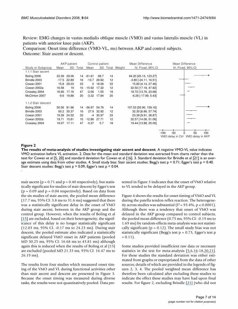
Relative onset timing of the VMO and VLFigure 2 shows the results of meta-analysis of studiesinvestigating stair ascent and descent. It may be of interestto note that the evidence presented in Figure 2 was mainlyfrom a single research team (two studies by Cowan et al[5,16], and one by Crossley et al [14]).
There is substantial heterogeneity across the six studiesinvestigating timing during stair ascent (I2 = 85.7%, p <0.00001) and across the five studies investigating stairdescent (I2 = 69.9%, p = 0.01). Boling et al [15] reportedthe greatest effect size (Figure 2). The test of small studybias (or funnel-plot asymmetry) using Begg's test andEgger's test was not statistically significant for studies of
Table 3: Population characteristics and procedural details. Reflex activation.
Study and country of origin
Subject details (age in years; height in cm)
Asymptomatic control details (age in years; height in cm)
Task Electrode type; sampling rate; signal processing
EMG onset determination method
AKP during test
Karst and Willett [30]USA
See table 1 See table 1 Reflex knee extension elicited by patella tendon tap. High sitting with leg hanging over side of plinth, hip and knee at 90 degrees flexion
Surface Electrodes: SR 4000 Hz; raw signal used
Computerized: point at which signal exceeded mean + 1 SD of baseline level
NS
Witvrouw et al [18]Belgium
No: 19/19M/F:8/11Age: 21.1, range 17–26Height: NSProfile: University Hospital clinic, Dept of Orthopaedic Surgery.Symptom Duration: > 1 and 1/2 months
No: 80/80M/F: 37/43Age: 18.4, range 17–22Height: NSProfile: NS
Surface Electrodes: SR not stated; raw signal used
Visual: point at which EMG leaves baseline.
NS
Witvrouw et al [13]Belgium
No: 480/282.24 developed AKP and became subjects.Profile (includes control): Students taking physical education classes.12–14 hrs sport per week. No history of AKP at start.M/F: 11/13Mean age 18.6, (17–21) includes controls.Height: 179.3 ± 5.38Symptom Duration: > 1 and 1/2 months
As across. 258 did not develop AKP and became controls.M/F: 151/131Height: 180.16 ± 6.25
Surface Electrodes: SR not stated; raw signal used
Visual: point at which EMG leaves baseline.
NS
Voight and Wieder [6]USA
No: 16/16M/F: 10/6Age: 26.1 (19–31)Height: NSProfile:Professional, recreational and non-athletes from sports clinic Miami University.Symptom Duration: NS
No: 41/41M/F: 17/24Age: 24.8, (18 – 45)Height: NSProfile: "@age matched", otherwise NS
Surface Electrodes: SR not stated; raw signal used
Visual: point at which EMG leaves baseline.
NS
Abbreviations:AKP – anterior knee pain; cm – centimetres; EMG – electromyography; F – female; FWR – full wave rectification; LPF – low pass filter; M – male; No – number; NS – not stated; RMS – root mean square; SD- standard deviation
Page 6 of 14(page number not for citation purposes)
BMC Musculoskeletal Disorders 2008, 9:64 http://www.biomedcentral.com/1471-2474/9/64
stair ascent (p = 0.71 and p = 0.40 respectively), but statis-tically significant for studies of stair descent by Egger's test(p = 0.09 and p = 0.04 respectively). Based on data fromthe six studies of stair ascent, the pooled mean difference(17.7 ms, 95% CI: 3.8 ms to 31.6 ms) suggested that therewas a statistically significant delay in the onset of VMOduring stair ascent, between in the AKP group and thecontrol group. However, when the results of Boling et al[15] are excluded, based on their heterogeneity, the signif-icance of this delay is no longer statistically significant(12.03 ms, 95% CI. -0.17 ms to 24.23 ms). During stairdescent, the pooled estimate also indicated a statisticallysignificant delayed VMO onset in AKP patients (pooledMD 30.25 ms, 95% CI: 16.68 ms to 43.81 ms) althoughagain this is reduced when the results of Boling et al [15]are excluded (pooled MD 21.33 ms, 95% CI. 16.47 ms to26.19 ms).
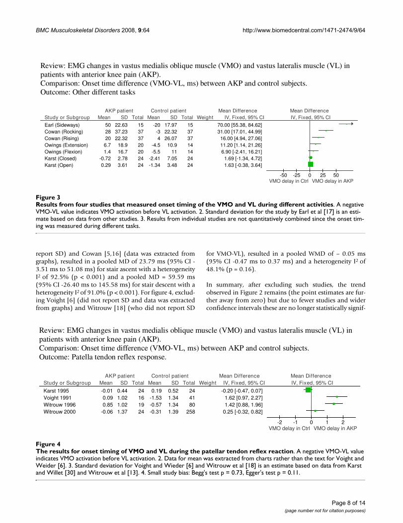
The results from four studies which measured onset tim-ing of the VMO and VL during functional activities otherthan stair ascent and descent are presented in Figure 3.Because the onset timing was measured during diversetasks, the results were not quantitatively pooled. Data pre-
sented in Figure 3 indicates that the onset of VMO relativeto VL tended to be delayed in the AKP group.
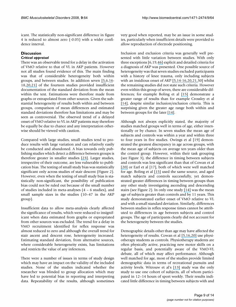
Figure 4 shows the results for onset timing of VMO and VLduring the patella tendon reflex reaction. The heterogene-ity across studies was substantial (I2 = 93.4%, p < 0.0001).Although there was a tendency that onset of VMO wasdelayed in the AKP group compared to control subjects,the pooled mean difference (0.75 ms, 95% CI: -0.19 ms to1.69 ms) by random-effects meta-analysis was not statisti-cally significant (p = 0.12). The small study bias was notstatistically significant (Begg's test p = 0.73, Egger's test p= 0.11).
Some studies provided insufficient raw data or necessarystatistics in the text for meta-analysis [5,6,16-18,20,21].For these studies the standard deviation was either esti-mated from graphs or expropriated from the data of othersources; details of which are provided in the legends of fig-ures 2, 3, 4. The pooled weighted mean difference hastherefore been calculated after excluding these studies toindicate the effect these studies may have had upon finalresults. For figure 2, excluding Brindle [21] (who did not
The results of meta-analysis of studies investigating stair ascent and descentFigure 2The results of meta-analysis of studies investigating stair ascent and descent. A negative VMO-VL value indicates VMO activation before VL activation. 2. Data for the mean and standard deviation was extracted from charts rather than the text for Cowan et al [5, 20] and standard deviation for Cowan et al [16]. 3. Standard deviation for Brindle et al [21] is an aver-age estimate using data from other studies. 4. Small study bias: Stair ascent studies: Begg's test p = 0.71; Egger's test p = 0.40. Stair descent studies: Begg's test p = 0.09, Egger's test p = 0.04.
Review: EMG changes in vastus medialis oblique muscle (VMO) and vastus lateralis muscle (VL) in patients with anterior knee pain (AKP). Comparison: Onset time difference (VMO-VL, ms) between AKP and control subjects. Outcome: Stair ascent or descent.
Study or Subgroup1.1.1 Stair ascent
Boling 2006Brindle 2003Cowan 2001Cowan 2002aCrossley 2004McClinton 2007
1.1.2 Stair descent
Boling 2006Brindle 2003Cowan 2001Cowan 2002aCrossley 2004
Mean
22.39-17.515.8
16.5816.66
-9.6
50.5660.2
19.3919.7119.07
SD
29.0622.8929.03
1917.1619.88
81.9635.3724.5215.8117.11
Total
141633104720
1416331047
Mean
-61.81-13.7
0-15.92-2.06-3.32
-56.9727.9
-4-12.86-0.37
SD
68.729.9218.0617.32
1.5517.84
54.7632.9230.9727.71
5.7
Total
141233121820
1412331218
Weight IV, Fixed, 95% CI
84.20 [45.13, 123.27]-3.80 [-24.11, 16.51]
15.80 [4.14, 27.46]32.50 [17.18, 47.82]18.72 [13.76, 23.68]-6.28 [-17.99, 5.43]
107.53 [55.90, 159.16]32.30 [6.86, 57.74]23.39 [9.91, 36.87]
32.57 [14.08, 51.06]19.44 [13.88, 25.00]
AKP patient Control patient Mean Difference Mean DifferenceIV, Fixed, 95% CI
-100 -50 0 50 100VMO delay in Ctrl VMO delay in AKP
Page 7 of 14(page number not for citation purposes)
BMC Musculoskeletal Disorders 2008, 9:64 http://www.biomedcentral.com/1471-2474/9/64
report SD) and Cowan [5,16] (data was extracted fromgraphs), resulted in a pooled MD of 23.79 ms (95% CI -3.51 ms to 51.08 ms) for stair ascent with a heterogeneityI2 of 92.5% (p < 0.001) and a pooled MD = 59.59 ms(95% CI -26.40 ms to 145.58 ms) for stair descent with aheterogeneity I2 of 91.0% (p < 0.001). For figure 4, exclud-ing Voight [6] (did not report SD and data was extractedfrom graphs) and Witrouw [18] (who did not report SD
for VMO-VL), resulted in a pooled WMD of – 0.05 ms(95% CI -0.47 ms to 0.37 ms) and a heterogeneity I2 of48.1% (p = 0.16).
In summary, after excluding such studies, the trendobserved in Figure 2 remains (the point estimates are fur-ther away from zero) but due to fewer studies and widerconfidence intervals these are no longer statistically signif-
The results for onset timing of VMO and VL during the patellar tendon reflex reactionFigure 4The results for onset timing of VMO and VL during the patellar tendon reflex reaction. A negative VMO-VL value indicates VMO activation before VL activation. 2. Data for mean was extracted from charts rather than the text for Voight and Weider [6]. 3. Standard deviation for Voight and Wieder [6] and Witrouw et al [18] is an estimate based on data from Karst and Willet [30] and Witrouw et al [13]. 4. Small study bias: Begg's test p = 0.73, Egger's test p = 0.11.
Review: EMG changes in vastus medialis oblique muscle (VMO) and vastus lateralis muscle (VL) in patients with anterior knee pain (AKP). Comparison: Onset time difference (VMO-VL, ms) between AKP and control subjects. Outcome: Patella tendon reflex response.
Study or Subgroup
Karst 1995Voight 1991Witrouw 1996Witrouw 2000
Mean
-0.010.090.85
-0.06
SD
0.441.021.021.37
Total
24161924
Mean
0.19-1.53-0.57-0.31
SD
0.521.341.341.39
Total
244180
258
Weight IV, Fixed, 95% CI
-0.20 [-0.47, 0.07]1.62 [0.97, 2.27]1.42 [0.88, 1.96]
0.25 [-0.32, 0.82]
AKP patient Control patient Mean Difference Mean DifferenceIV, Fixed, 95% CI
-2 -1 0 1 2VMO delay in Ctrl VMO delay in AKP
Results from four studies that measured onset timing of the VMO and VL during different activitiesFigure 3Results from four studies that measured onset timing of the VMO and VL during different activities. A negative VMO-VL value indicates VMO activation before VL activation. 2. Standard deviation for the study by Earl et al [17] is an esti-mate based on data from other studies. 3. Results from individual studies are not quantitatively combined since the onset tim-ing was measured during different tasks.
Review: EMG changes in vastus medialis oblique muscle (VMO) and vastus lateralis muscle (VL) in patients with anterior knee pain (AKP). Comparison: Onset time difference (VMO-VL, ms) between AKP and control subjects. Outcome: Other different tasks
Study or Subgroup
Earl (Sideways)Cowan (Rocking)Cowan (Rising)Owings (Extension)Owings (Flexion)Karst (Closed)Karst (Open)
Mean
502820
6.71.4
-0.720.29
SD
22.6337.2322.3218.916.72.783.61
Total
15373720202424
Mean
-20-34
-4.5-5.5
-2.41-1.34
SD
17.9722.3226.0710.9
117.053.48
Total
15373714142424
Weight IV, Fixed, 95% CI
70.00 [55.38, 84.62]31.00 [17.01, 44.99]16.00 [4.94, 27.06]11.20 [1.14, 21.26]6.90 [-2.41, 16.21]
1.69 [-1.34, 4.72]1.63 [-0.38, 3.64]
AKP patient Control patient Mean Difference Mean DifferenceIV, Fixed, 95% CI
-50 -25 0 25 50VMO delay in Ctrl VMO delay in AKP
Page 8 of 14(page number not for citation purposes)
BMC Musculoskeletal Disorders 2008, 9:64 http://www.biomedcentral.com/1471-2474/9/64
icant. The statistically non-significant difference in figure4 is reduced to almost zero (-0.05) with a wider confi-dence interval.
DiscussionCritical appraisalThere was an observable trend for a delay in the activationof VMO relative to that of VL in AKP patients. Howevernot all studies found evidence of this. The main findingwas that of considerable heterogeneity both withingroups, and between studies. In addition seven [5,6,16-18,20,21] of the fourteen studies provided insufficientdocumentation of the standard deviation from the meanwithin the text. Estimations were therefore made fromgraphs or extrapolated from other sources. Given the sub-stantial heterogeneity of results both within and betweengroups, comparison of mean differences and estimatedstandard deviations therefore has limitations and may beseen as controversial. The observed trend of a delayedonset of VMO relative to VL in AKP patients may thereforebe equally be due to chance and any interpretation other-wise should be viewed with caution.
Compared with large studies, small studies tend to pro-duce results with large variation and can relatively easilybe conducted and abandoned. A bias towards only pub-lishing studies which detect a difference between groups istherefore greater in smaller studies [23]. Larger studies,irrespective of their outcome, are less vulnerable to publi-cation bias. The testing of small study bias was statisticallysignificant only across studies of stair descent (Figure 2).However, even when the testing of small study bias is sta-tistically non-significant, the possibility of publicationbias could not be ruled out because of the small numberof studies included in meta-analyses (4 – 6 studies), andsmall sample sizes in the studies (10–47 in the AKPgroup).
Insufficient data to allow meta-analysis clearly affectedthe significance of results, which were reduced to insignif-icant when data estimated from graphs or expropriatedfrom other sources was excluded. The trend for a delay inVMO recruitment identified for reflex response wasalmost reduced to zero and although the overall trend forstair ascent and descent rose, heterogeneity increased.Estimating standard deviation, from alternative sources,where considerable heterogeneity exists, has limitationsand restricts the value of any inferences.
There were a number of issues in terms of study designwhich may have an impact on the validity of the includedstudies. None of the studies indicated whether theresearcher was blinded to group allocation which mayhave led to potential bias in reporting and interpretingdata. Repeatability of the results, although sometimes
very good when reported, may be an issue in some stud-ies, particularly when insufficient details were provided toallow reproduction of electrode positioning.
Inclusion and exclusion criteria was generally well pre-sented with little variation between studies. With onlythree exceptions [6,19,46] explicit and detailed criteria fora diagnosis of AKP was presented. One possible source ofheterogeneity was that seven studies excluded participantswith a history of knee trauma, only including subjectswith an insidious onset of AKP [5,14-16,20,21,30] whilstthe remaining studies did not state such criteria. Howevereven within this group of seven, there are considerable dif-ferences; for example Boling et al [15] demonstrate agreater range of results than for example Crossley et al[14], despite similar inclusion/exclusion criteria. This issurprising given the greater age range both within andbetween groups for the later [14].
Although not always explicitly stated, the majority ofstudies matched groups well in terms of age, either inten-tionally or by chance. In seven studies the mean age ofsubjects and controls was within a year and within threeto four years in five studies. Owings et al [19] demon-strated the greatest discrepancy in age across groups, withthe mean age of subjects on average ten years older thanthe control group. However, within their task grouping(see Figure 3), the difference in timing between subjectsand controls was less significant than that of Cowan et al[20] or Earl et al [17], both of which were well matchedfor age. Boling et al [15] used the same source, and agedmatch subjects and controls successfully, yet demon-strated greater differences in timing between groups thanany other study investigating ascending and descendingstairs (see Figure 2). In only one study [14] was the meanage of subjects greater than controls and by 11 years. Thisstudy demonstrated earlier onset of VMO relative to VLand with a small standard deviation. Similarly, differencesbetween studies in reflex response times cannot be attrib-uted to differences in age between subjects and controlgroups. The age of participants clearly did not account forthe heterogeneity between the studies.
Demographic details other than age may have affected theheterogeneity of results. Cowan et al [5,16,20] use physi-otherapy students as controls. Physiotherapy students areoften physically active, practicing new motor skills on aregular basis, and potentially aware of the VMO-VLdebate, all of which may affect performance. Althoughwell matched for age, most of the studies provide limiteddemographic data in terms of recreational pursuits andactivity levels. Witrouw et al's [13] study was the onlystudy to use one cohort of subjects, all of whom partici-pated in 12–14 hours of sport weekly. Their results indi-cated little difference in timing between subjects with and
Page 9 of 14(page number not for citation purposes)
BMC Musculoskeletal Disorders 2008, 9:64 http://www.biomedcentral.com/1471-2474/9/64
without AKP. Earl et al [17] stated that as well as age, thefifteen recreational athletes in each group of their studywere matched with regards to gender, height, weight andduration of exercise per week, and so this cannot accountfor the large differences between this group, compared tothe other studies in Figure 4. However, recreational differ-ences may have accounted for heterogeneity elsewhere.
Pain has been linked to changes in normal muscle recruit-ment in a number of musculoskeletal conditions [24-27].All but one of the studies [15] investigating stair ascentand decent indicated the presence and intensity of painduring data collection using a ten point visual analoguescale (VAS). Boling et al [15] used this same scale to recordpain during the week prior to data collection on a numberof activities of which stair ascent and descent was one.Their mean pain scores were the highest for this groupingat 4.9 ± 2.3 and they demonstrate the greatest magnitudeof difference between subjects and controls in VMO-VLrecruitment (see Figure 2). However the influence ofhigher levels of pain during testing on VMO-VL resultswas not supported by Brindle et al's [21] study, in whichonly slightly lower VAS scores were recorded actually dur-ing testing, and where there was little difference betweenthe groups in the opposite direction. In addition, Cowanet al [20], explicitly stated the absence of pain during rock-ing onto the heels, yet record the second highest differ-ence between subjects and controls for this group of tasks(see Figure 3). The presence of pain during testing was notstated in any other studies. The heterogeneity of results inthis review was not explained by the presence and inten-sity of pain during data collection.
Differences in the duration of AKP rather than the pres-ence of pain during data collection may have influencedthe heterogeneity of results between studies. When docu-mented, the duration of AKP pain ranged from one week[17] to twelve years [5]. It is interesting to note that withthe exception of Boling et al [15], these two studies dem-onstrated the largest difference between subject and con-trol groups within their respective task groupings (seeFigure 2 and 3). These extremes demonstrate that theduration of symptoms does not appear to be a factor con-tributing to the results in the reviewed studies.
None of the studies included in this review indicatedwhether participants were receiving physiotherapy or haddone so in the past. Some physiotherapists seek to nor-malise aberrant muscle recruitment patterns with theintention of reducing pain [7,28,29]. The effectiveness ofphysiotherapy in achieving this specific target can only beassessed once a clearer definition of normal has beenestablished.
Factors affecting EMG data collection and analysisThe EMG studies included in this review investigated threetypes of muscle activation: reflex, voluntary closed kineticchain, and voluntary open kinetic chain. From the litera-ture, reflex onset times appear the most repeatable, andmarked variability has been noted in voluntary EMGonset times [5,30], with open kinetic chain appearing themost variable [31]. This may have affected the heterogene-ity seen in the results.
Many factors can affect EMG results, and methodologicaldifferences are frequently cited as reasons for the lack ofagreement between studies. These factors can include elec-trode placement and orientation, data sampling rates, lev-els of smoothing or filtering, and onset determinationmethods. Readers are referred to articles such as Soderbergand Knutson [32] for more detail. Importantly, EMGonset times can be affected by the onset determinationmethods [32], and the level of EMG smoothing [33] (seeTable 1). Various onset determination methods were usedin the eighteen studies. Three of the four reflex studies usevisual determination of EMG onset, and this has beenshown to be highly repeatable [34]. The majority of stud-ies investigating voluntary muscle activation determinedEMG onset as the point at which the signal exceeded themean resting "baseline" value, prior to activity, by morethan a set number of standard deviations for a specifiedperiod of time. This is done to avoid type I errors, classify-ing the muscle as active when it is not. Both the numberof standard deviations and period of time stated variedbetween studies, or were not stated. These factors areimportant for direct comparisons of individual muscletiming between studies. Of course, providing that anonset threshold was standardised for each study, thereshould have been relatively little affect on between-groupdifferences in the relative activation times of VMO and VL,as any differences in threshold should have affected bothmuscles and both groups similarly. The level of smooth-ing was also important as excessive smoothing couldreduce the ability to detect small timing differences thatmay be clinically relevant [33,35].
It is interesting that Boling et al [15] used the same EMGprocessing and analysis methods as in the studies byCowan and Crossley et al [5,14,16], but obtained datathat indicated far greater differences between the patientgroup and the control group. In addition, the magnitudeof within-group standard deviation for the former is con-siderable and for the latter particularly small. This couldbe due to differences in the identification of the restingbaseline window, which could have contained more orless muscle activity, and hence affected the onset thresh-old. Although all studies relating to stair ascent anddescent reported EMG onset times relative to foot strike orstance phase, method of measurement varied. Boling et al
Page 10 of 14(page number not for citation purposes)
BMC Musculoskeletal Disorders 2008, 9:64 http://www.biomedcentral.com/1471-2474/9/64
[15] report EMG onset times to be pre-foot-strike (e.g.negative values are provided for independent VMO andVL onset timing) which contrasts with for example,McClinton et al [22] who evaluate onset times commenc-ing at heel contact and Brindle et al [21], who evaluateonset times commencing with toe contact. The exact com-mencement of readings for "stance phase" for the Cowanand Crossley group [5,14,16] is at "foot contact". Thesedifferences are not always immediately apparent orexplicit in the text.
Documentation of electrode positioning varied. Somestudies provided references such Basmajian and Blumen-stein [36], and Tully and Stillman [37] or provide anatom-ical landmarks, whilst others simply stated that electrodeswere positioned over the muscle bellies of VMO and VL.This would make the replication of these studies challeng-ing, whilst also making it difficult to directly compareresults.
Reflex latency times are highly repeatable [34,38]. The dif-ferences found between the reflex reaction studies aretherefore very interesting, and again, may be due to anumber of reasons, including methodological differencesin the stimulus used to deliver the patellar tendon tap,onset determination methods, or differences between par-ticipants such as symptom duration or height [30]. How-ever, it has been noted that the range of reflex latenciesdisplayed by Voight and Weider [6] was abnormally large[30], and included some values as low as 10 ms. Theseshort latencies may be physiologically questionable [30],the normal range being approximately 16 to 30 ms [38],varying slightly with methodology, but importantly lim-ited by the maximum human nerve conduction velocityof approximately 50 m/s. Whilst these very short latenciescould possibly be due to time delays in the methodologi-cal set-up, they could have been movement artefacts onthe EMG traces [30].
Clinical relevance of VMO-VL timing differencesWhilst the findings of this review suggest a trend towardsrelatively delayed onset of the VMO when compared tothe VL in the AKP population, the clinical significance ofthese findings is unclear. The differences in timingdescribed are all relatively small, and it appears as yetunknown at what point such a difference may becomeclinically significant; although interestingly, Neptune et al[39] suggested that timing differences as low as 5 ms canelicit a biomechanical imbalance at the patellofemoraljoint. It is notable however that this figure is lower thanthe standard error of the measurement reported by theCowan and Crossley group [5], and marked within-sub-ject variability in VMO – VL onset times in voluntary mus-cle activation have also been reported [3,30].
The between group and between subject variabilityrecorded in each of the fourteen studies is considerable,and does not appear to be attributable to anything otherthan true variability between subjects. A comparison ofmean group values, whether to reflect a trend or indicatea statistically significant finding, may be appropriate sta-tistically, but is of questionable clinical relevance. Thelarge variability between subjects in a given population,whether this be healthy or AKP patients, does not make itpossible to make generalisations.
Differences in procedural and onset determination meth-ods may account for the differences seen between studiesin the timing of VMO and VL. However, as stated previ-ously, this should have relatively little effect on the relativeactivation times of VMO and VL, the focus of this review.The possible sources of heterogeneity discussed betweenstudies and summarised in Table 4, do not convincinglyaccount for the differences in results across studies.
Clinically, the relevance of any trends towards delayedVMO onset in the AKP population may be increased if
Table 4: Possible sources of heterogeneity
Methodology Publication BiasConvenience rather than random sampling may not represent true populationLack of assessor blinding to group allocationSmall sample size
Population characteristics Intensity of AKPDuration of AKPRecreational pursuitsPrevious or concomitant physiotherapyAge rangePrevious knowledge pertaining to research question (i.e. population of physiotherapy students knowledge on VMO/VL debate and techniques to facilitate VMO
Procedural details Differences between taskDifferences between similar tasks- e.g stair height, timing of taskNo of tests prior to data collection (learning effect/fatigue)Results recorded as single test or mean of severalElectrode placementData sampling ratesEMG onset determinationLevels of smoothing or filtering
Abbreviations:AKP – anterior knee pain; EMG – electromyography; VL – vastus lateralis; VMO – vastus medialis oblique
Page 11 of 14(page number not for citation purposes)
BMC Musculoskeletal Disorders 2008, 9:64 http://www.biomedcentral.com/1471-2474/9/64
there is evidence that therapies can favourably influenceit. Some studies have demonstrated alterations in VMO-VL onset times in AKP patients following or during suc-cessful therapeutic interventions. Boling et al [15] andCowan et al [40] demonstrated a significant delay in VMOactivation prior to physiotherapy, and subsequently, fol-lowing successful treatment with pain reduction, a signif-icantly earlier VMO activation was observed. It has alsobeen reported that therapeutic patella taping can improveVMO-VL onset time differences [16,41]. Indeed from aprospective study of 30 subjects, Witrouw [42] report thatfaster VMO onset times are a predictor of successful reha-bilitation, although no primary data is provided. If thereported results of these small studies are transferable theycould indicate that although normal timing is variable,enhancing VMO onset times by reducing any delay in acti-vation may be associated with pain relief.
Clinically, it is interesting that the type of muscle contrac-tion, i.e. reflex or voluntary, and closed or open kineticchain, seems to influence variability, as stated in theResults section. This is probably due to differences inmotor unit recruitment strategies between the contractiontypes, for example caused by differences in proprioceptivefeedback and knee joint reaction forces between open andclosed chain activities [31]. This may be a factor to beborne in mind in rehabilitation programmes aimed attreating aberrant muscle activation patterns in this pathol-ogy.
The overall tendency towards a delayed onset of VMO rel-ative to VL in AKP patients was consistent for both volun-tary functional and non-functional tasks, as well as to alesser degree reflex response times. Despite this however,the heterogeneity across the studies was substantial andunexplained, and the questionable clinical significance ofany such trend is highlighted by the fact that some asymp-tomatic individuals represented within the control groupsdemonstrate similar patterns of VMO to VL dysfunction –but do not experience AKP. One possibility is that patientswith AKP are not a homogenous group, and that relativedelay of the VMO represents just one of a variety of factorsthat may lead to this syndrome.
This review presented some limitations. Firstly, althoughall retrieved full text articles and some specialist journalswere hand-searched, the majority of this review's searchstrategy was performed using computer databases.Accordingly relevant papers may have been missed byemploying this method [43]. No attempt was made toidentify unpublished work and grey literature (such asuniversity theses and conference proceedings). As a result,publication bias may have influenced the results [23,44].One foreign language paper [45] was identified by thesearch but excluded from our review. The quality of
reporting compromised the validity of the included stud-ies – in particular assessor blinding of group allocationand insufficient data to allow meta-analysis. This wastherefore in part based on data extracted from graphicalillustrations or expropriated from the data of othersources. Heterogeneity of results both within subject andcontrol groups as well as between studies indicate that anytrends identified should be interpreted with caution.
ConclusionThe findings from this review are subject to substantialand unexplained heterogeneity, and the impact of publi-cation bias and methodological flaws such as blinding tostudy allocation could not be ruled out. There were largevariations within subject and control groups, as well asbetween studies. There was a trend for delayed onset ofVMO relative to VL in subjects with AKP in comparison tothose without. This was consistent for functional taskssuch as ascending and descending stairs, stepping side-ways and rocking onto the toes/heels, as well as less func-tional tasks such as isokinetic testing and reflex responsetimes. However not all AKP patients demonstrate a VMO-VL dysfunction, and this is compounded by considerablenormal physiological variability in the healthy popula-tion. Because of unexplained heterogeneity and methodo-logical limitations, any inferences based on statisticalanalysis should be viewed with caution. The clinical andtherapeutic significance of these findings based on theexisting literature is therefore difficult to assess.
Competing interestsThe authors declare that they have no competing interests.
Authors' contributionsRC co-coordinated the review, and contributed to the lit-erature search, data extraction and drafting of the manu-script. TOS contributed to the literature search, dataextraction and drafting of the manuscript. DS contributedto the literature search, data extraction and drafting of themanuscript. JD provided expertise on EMG analysis andcontributed to drafting of the manuscript. SW contributedto the literature search and data extraction. FS providedexpert advice on systematic reviewing, undertook meta-analysis and contributed to drafting of the manuscript. Allauthors read and approved the final manuscript.
Page 12 of 14(page number not for citation purposes)
BMC Musculoskeletal Disorders 2008, 9:64 http://www.biomedcentral.com/1471-2474/9/64
Additional material
AcknowledgementsMr John Losasso at the Norfolk and Norwich University Hospital Library for his assistance with electronic literature searches.
References1. Baquie P, Brukner P: Injuries presenting to an Australian Sports
Medicine Centre: a 12 month study. Clin J Sports Med 1997,7:28-31.
2. McConnell J: The management of chondromalacia patellae: along term solution. Aust J Physiother 1986, 32:215-223.
3. Crossley KM, Bennell K, Green S, McConnell J: A systematicreview of physical interventions for patellofemoral pain syn-drome. Clin J Sports Med 2001, 11:103-110.
4. McConnell J: Management of patellofemoral problems. ManTher 1996, 1:60-66.
5. Cowan S, Bennell K, Hodges P, Crossley K, McConnell J: Delayedonset of electromyographic activity of vastus medialis obliq-uus relative to vastus lateralis in subjects with patellofemo-ral pain syndrome. Arch Phys Med Rehabil 2001, 82:183-189.
6. Voight M, Wieder D: Comparative reflex response times ofvastus medialis obliquus and vastus lateralis in normal sub-jects and subjects with extensor mechanism dysfunction. AmJ Sports Med 1991, 19:131-137.
7. Powers C: Rehabilitation of patellofemoral joint disorders: acritical review. J Orthop Sports Phys Ther 1998, 28(5):345-354.
8. Thomee R, Augustsson J, Karlsson J: Patellofemoral pain syn-drome. Sports Med 1999, 28:245-262.
9. Pigott TD: Methods for handling missing data in research syn-thesis. In The handbook of research synthesis Edited by: Cooper H,Hedges LV. New York: Russell Sage Foundation; 1994.
10. Higgins JP, Thompson SG, Deeks JJ, Altman DG: Measuring incon-sistency in meta-analyses. BMJ 2003, 327:557-560.
11. Begg CB, Mazumdar M: Operating characteristics of a rank cor-relation test for publication bias. Biometrics 1994, 50:1088-1101.
12. Egger M, Davey-Smith G, Schneider M, Minder C: Bias in meta-analysis detected by a simple, graphical test. BMJ 1997,315:629-634.
13. Witvrouw E, Lysen R, Bellemans J, Cambier D, Vanderstraeten G:Intrinsic risk factors for the development of anterior kneepain in an athletic population. A two-year prospective study.Am J Sports Med 2000, 28:480-489.
14. Crossley KM, Cowan SM, Bennell KL, McConnell J: Knee flexionduring stair ambulation is altered in individuals with patel-lofemoral pain. J Orthop Res 2004, 22:267-274.
15. Boling MC, Bolgla LA, Mattacola CG, Uhl TL, Hosey RG: Outcomesof a weight-bearing rehabilitation program for patients diag-nosed with patellofemoral pain syndrome. Arch Phys Med Reha-bil 2006, 87:1428-1435.
16. Cowan SM, Bennell KL, Hodges PW: Therapeutic patellar tapingchanges the timing of vasti muscle activation in people withpatellofemoral pain syndrome. Clin J Sports Med 2002,12:339-347.
17. Earl JE, Hertel J, Denegar CR: Patterns of dynamic malalign-ment, muscle activation, joint motion, and patellofemoral-pain syndrome. J Sports Rehabil 2005, 14:215-233.
18. Witvrouw E, Sneyers C, Lysen R, Victor J, Bellemans J: Reflexresponse times of vastis medialis oblique and vastis lateralisin normal subjects and in subjects with patellofemoral painsyndrome. J Orthop Sports Phys Ther 1996, 24:160-1665.
19. Owings TM, Grabiner MD: Motor control of the vastis medialisoblique and vastis lateralis muscles is disrupted duringeccentric contractions in subjects with patellofemoral pain.Am J Sports Med 2002, 30:483-487.
20. Cowan SM, Hodges PW, Bennell KL, Crossley KM: Altered vastiirecruitment when people with patellofemoral pain syn-drome complete a postural task. Arch Phys Med Rehabil 2002,83:989-995.
21. Brindle TJ, Mattacola C, McCrory J: Electromyographic changesin the gluteus medius during stair ascent and descent in sub-jects with anterior knee pain. Knee Surg Sports Traumatol Arthrosc2003, 11:244-251.
22. McClinton S, Donatell G, Weir J, Heiderscheit B: Influence of stepheight on quadriceps onset timing and activation during stairascent in individuals wit patellofemoral pain syndrome. JOrthop Sports Phys Ther 2007, 37:239-244.
23. Song F, Eastwood AJ, Gilbody S, Duley L, Sutton AJ: Publication andrelated biases. Health Technol Assess 2000, 4:1-115.
24. Steenbrink F, de Groot JH, Veeger HEJ, Meskers CGM, van de SandeMAJ, Rozing PM: Pathological muscle activation patterns inpatients with massive rotator cuff tears, with and withoutsubacromial anaesthetics. Man Ther 2006, 11:231-237.
25. Vogt L, Pfeifer K, Banzer W: Neuromuscular control of walkingwith chronic low-back pain. Man Ther 2003, 8:21-28.
26. Le Pera D, Graven-Nielsen T, Valeriani M, Oliviero A, Di Lazzaro V,Tonali PA, Arendt-Nielsen L: Inhibition of motor system excita-bility at cortical and spinal level by tonic muscle pain. ClinicalNeurophysiology 2001, 112:1633-1641.
27. Rutherford OM, Jones DA, Newham DJ: Clinical and experimen-tal application of the percutaneous twitch superimpositiontechnique for the study of human muscle activation. J NeurolNeurosurg Psychiatry 1986, 49:1288-1291.
28. Powers CM, Landel R, Perry J: Timing and intensity of vastismuscle activity during functional activities in subjects withand without patellofemoral pain. Phys Ther 1996, 76:946-955.
29. Crossley KM, Cowan SM, Bennell KL, McConnell J: Patella taping:is clinical success supported by scientific evidence? ManualTherapy 2000, 5:142-150.
30. Karst GM, Willett GM: Onset timing of electromyographicactivity in the vastus medialis oblique and vastus lateralismuscles in subjects with and without patellofemoral painsyndrome. Phys Ther 1995, 75:813-823.
31. Stensdotter A-K, Hodges PW, Mellor R, Sundelin G, Hager-Ross C:Quadriceps activation in closed and in open kinetic chainexercise. Med Sci Sports Exerc 2003, 35:2043-2047.
32. Soderberg GL, Knutson LM: A guide for use and interpretationof kinesiologic electromyographic data. Phys Ther 2000,80:485-98.
33. Merletti R: Standards for reporting EMG data. J ElectromyogrKinesiol 1999, 9:iii-iv.
34. Dixon J, Howe TE, Kent JR, Whittaker VJ: VMO-VL reflex latencydifference between quadriceps components in osteoarthriticand asymptomatic knees. Adv Physiother 2004, 6:166-172.
35. Karst GM: EMG onset timing. Phys Ther 1998, 78:543-546.36. Basmajian J, Blumenstein R: Electrode placement in EMG biofeedback
Baltimore: Williams and Wilkins; 1980. 37. Tully EA, Stillman BC: Computer aided video analysis of verte-
brofemoral motion during toe touching in healthy subjects.Arch Phys Med Rehabil 1997, 78:759-766.
38. Struys MA, Jonkman EJ, Strijers RL: Measurement of patellar andankle tendon reflexes in normal subjects. Electromyogr Clin Neu-rophysiol 1997, 37:13-18.
39. Neptune RR, Wright IC, van den Bogert AJ: The influence oforthotic devices and vastus medialis strength and timing onpatellofemoral loads during running. Clin Biomech 2000,15:611-618.
40. Cowan SM, Bennell KL, Hodges PW, Crossley KM, McConnell J:Simultaneous feedforward recruitment of the vasti inuntrained postural tasks can be restored by physical therapy.J Orthopaedic Research 2003, 21:553-558.
41. Gilleard W, McConnell J, Parsons D: The effect of patella tapingon the onset of vastus medialis obliquus and vastus lateralis
Additional file 1Data extraction form. Table used to record study design, participant selec-tion and population characteristics, methodology, results and relevant study limitations. Comparisons between reviewers were made for accuracy and interpretation.Click here for file[http://www.biomedcentral.com/content/supplementary/1471-2474-9-64-S1.doc]
Page 13 of 14(page number not for citation purposes)
http://www.ncbi.nlm.nih.gov/entrez/query.fcgi?cmd=Retrieve&db=PubMed&dopt=Abstract&list_uids=2039064
http://www.ncbi.nlm.nih.gov/entrez/query.fcgi?cmd=Retrieve&db=PubMed&dopt=Abstract&list_uids=2039064
http://www.ncbi.nlm.nih.gov/entrez/query.fcgi?cmd=Retrieve&db=PubMed&dopt=Abstract&list_uids=2039064
http://www.ncbi.nlm.nih.gov/entrez/query.fcgi?cmd=Retrieve&db=PubMed&dopt=Abstract&list_uids=9809282
http://www.ncbi.nlm.nih.gov/entrez/query.fcgi?cmd=Retrieve&db=PubMed&dopt=Abstract&list_uids=9809282
http://www.ncbi.nlm.nih.gov/entrez/query.fcgi?cmd=Retrieve&db=PubMed&dopt=Abstract&list_uids=7786990
http://www.ncbi.nlm.nih.gov/entrez/query.fcgi?cmd=Retrieve&db=PubMed&dopt=Abstract&list_uids=7786990
http://www.ncbi.nlm.nih.gov/entrez/query.fcgi?cmd=Retrieve&db=PubMed&dopt=Abstract&list_uids=9310563
http://www.ncbi.nlm.nih.gov/entrez/query.fcgi?cmd=Retrieve&db=PubMed&dopt=Abstract&list_uids=9310563
http://www.ncbi.nlm.nih.gov/entrez/query.fcgi?cmd=Retrieve&db=PubMed&dopt=Abstract&list_uids=8866275
http://www.ncbi.nlm.nih.gov/entrez/query.fcgi?cmd=Retrieve&db=PubMed&dopt=Abstract&list_uids=8866275
http://www.ncbi.nlm.nih.gov/entrez/query.fcgi?cmd=Retrieve&db=PubMed&dopt=Abstract&list_uids=8866275
http://www.ncbi.nlm.nih.gov/entrez/query.fcgi?cmd=Retrieve&db=PubMed&dopt=Abstract&list_uids=3794735
http://www.ncbi.nlm.nih.gov/entrez/query.fcgi?cmd=Retrieve&db=PubMed&dopt=Abstract&list_uids=3794735
http://www.ncbi.nlm.nih.gov/entrez/query.fcgi?cmd=Retrieve&db=PubMed&dopt=Abstract&list_uids=3794735
http://www.ncbi.nlm.nih.gov/entrez/query.fcgi?cmd=Retrieve&db=PubMed&dopt=Abstract&list_uids=8790273
http://www.ncbi.nlm.nih.gov/entrez/query.fcgi?cmd=Retrieve&db=PubMed&dopt=Abstract&list_uids=8790273
http://www.ncbi.nlm.nih.gov/entrez/query.fcgi?cmd=Retrieve&db=PubMed&dopt=Abstract&list_uids=8790273
http://www.ncbi.nlm.nih.gov/entrez/query.fcgi?cmd=Retrieve&db=PubMed&dopt=Abstract&list_uids=7659741
http://www.ncbi.nlm.nih.gov/entrez/query.fcgi?cmd=Retrieve&db=PubMed&dopt=Abstract&list_uids=7659741
http://www.ncbi.nlm.nih.gov/entrez/query.fcgi?cmd=Retrieve&db=PubMed&dopt=Abstract&list_uids=7659741
http://www.ncbi.nlm.nih.gov/entrez/query.fcgi?cmd=Retrieve&db=PubMed&dopt=Abstract&list_uids=9597066
http://www.ncbi.nlm.nih.gov/entrez/query.fcgi?cmd=Retrieve&db=PubMed&dopt=Abstract&list_uids=9228881
http://www.ncbi.nlm.nih.gov/entrez/query.fcgi?cmd=Retrieve&db=PubMed&dopt=Abstract&list_uids=9228881
http://www.ncbi.nlm.nih.gov/entrez/query.fcgi?cmd=Retrieve&db=PubMed&dopt=Abstract&list_uids=9063657
http://www.ncbi.nlm.nih.gov/entrez/query.fcgi?cmd=Retrieve&db=PubMed&dopt=Abstract&list_uids=9063657
http://www.ncbi.nlm.nih.gov/entrez/query.fcgi?cmd=Retrieve&db=PubMed&dopt=Abstract&list_uids=9442193
BMC Musculoskeletal Disorders 2008, 9:64 http://www.biomedcentral.com/1471-2474/9/64
Publish with BioMed Central and every scientist can read your work free of charge
"BioMed Central will be the most significant development for disseminating the results of biomedical research in our lifetime."
Sir Paul Nurse, Cancer Research UK
Your research papers will be:
available free of charge to the entire biomedical community
peer reviewed and published immediately upon acceptance
cited in PubMed and archived on PubMed Central
yours — you keep the copyright
Submit your manuscript here:http://www.biomedcentral.com/info/publishing_adv.asp
BioMedcentral
muscle activity in persons with patellofemoral pain. Phys Ther1998, 78:25-32.
42. Witvrouw E, Lysen R, Bellemans J, Cambier D, Cools A, Danneels L,Bourgois J: Which risk factors predict outcome in the treat-ment program of anterior knee pain? Scand J Med Sci Sports2002, 12:40-46.
43. Colville-Stewart S: How to do a literature search. In The essentialresearcher's handbook. For nurses and healthcare professionals 2nd edi-tion. Edited by: Tarling M, Croft L. London: Baillière Tindall;2002:35-53.
44. Petrie A, Sabin S: Medical statistics at a glance Oxford: Blackwell Sci-ence; 2000.
45. Aguggia M, Gilli M, Febbraro A, Riccio A, Viglino C, Losana A,Carando S, Rossi P: Comparative electromyographic study ofthe vastus medialis muscle and vastus lateralis muscle in dys-functions of the knee extensor mechanism. Minerva Ortopedicae Traumatologica 1997, 48:103-107.
46. Morrish GM, Woledge RC: A comparison of the activation ofmuscles moving the patella in normal subjects and inpatients with chronic patellofemoral problems. Scand J RehabilMed 1997, 29:43-48.
Pre-publication historyThe pre-publication history for this paper can be accessedhere:
http://www.biomedcentral.com/1471-2474/9/64/prepub
Page 14 of 14(page number not for citation purposes)
http://www.ncbi.nlm.nih.gov/entrez/query.fcgi?cmd=Retrieve&db=PubMed&dopt=Abstract&list_uids=9442193
http://www.ncbi.nlm.nih.gov/entrez/query.fcgi?cmd=Retrieve&db=PubMed&dopt=Abstract&list_uids=9084105
http://www.ncbi.nlm.nih.gov/entrez/query.fcgi?cmd=Retrieve&db=PubMed&dopt=Abstract&list_uids=9084105