The Quiescin Sulfhydryl Oxidase (hQSOX1b) Tunes the Expression of Resistin-Like Molecule Alpha...
12
The Quiescin Sulfhydryl Oxidase (hQSOX1b) Tunes the Expression of Resistin-Like Molecule Alpha (RELM-a or mFIZZ1) in a Wheat Germ Cell-Free Extract Wael Gad 1,2,3 , Meera G. Nair 7,10 , Karolien Van Belle 1,2,3 , Khadija Wahni 1,2,3 , Henri De Greve 2,3 , Jo A. Van Ginderachter 4,5 , Guy Vandenbussche 8 , Yaeta Endo 6 , David Artis 7,9 , Joris Messens 1,2,3 * 1 Brussels Center for Redox Biology, Brussels, Belgium, 2 Department of Structural Biology, Vlaams Instituut voor Biotechnologie, Brussels, Belgium, 3 Structural Biology Brussels, Vrije Universiteit Brussel, Brussels, Belgium, 4 Lab of Cellular and Molecular Immunology, Vrije Universiteit Brussel, Brussels, Belgium, 5 Myeloid Cell Immunology Lab, Vlaams Instituut voor Biotechnologie, Brussels, Belgium, 6 Cell Free Science and Technology Research Center, Ehime University, Matsuyama, Japan, 7 Department of Microbiology and Institute for Immunology, Perelman School of Medicine, University of Pennsylvania, Philadelphia, Pennsylvania, United States of America, 8 Centre de Biologie Structurale et de Bioinformatique, Structure et Fonction des Membranes Biologiques, Universite ´ Libre de Bruxelles, Brussels, Belgium, 9 Department of Pathobiology, School of Veterinary Medicine, University of Pennsylvania, Philadelphia, Pennsylvania, United States of America, 10 Division of Biomedical Sciences, School of Medicine, University of California Riverside, Riverside, California, United States of America Abstract Background: Although disulfide bond formation in proteins is one of the most common types of post-translational modifications, the production of recombinant disulfide-rich proteins remains a challenge. The most popular host for recombinant protein production is Escherichia coli, but disulfide-rich proteins are here often misfolded, degraded, or found in inclusion bodies. Methodology/Principal findings: We optimize an in vitro wheat germ translation system for the expression of an immunological important eukaryotic protein that has to form five disulfide bonds, resistin-like alpha (mFIZZ1). Expression in combination with human quiescin sulfhydryl oxidase (hQSOX1b), the disulfide bond–forming enzyme of the endoplasmic reticulum, results in soluble, intramolecular disulfide bonded, monomeric, and biological active protein. The mFIZZ1 protein clearly suppresses the production of the cytokines IL-5 and IL-13 in mouse splenocytes cultured under Th2 permissive conditions. Conclusion/Significance: The quiescin sulfhydryl oxidase hQSOX1b seems to function as a chaperone and oxidase during the oxidative folding. This example for mFIZZ1 should encourage the design of an appropriate thiol/disulfide oxidoreductase-tuned cell free expression system for other challenging disulfide rich proteins. Citation: Gad W, Nair MG, Van Belle K, Wahni K, De Greve H, et al. (2013) The Quiescin Sulfhydryl Oxidase (hQSOX1b) Tunes the Expression of Resistin-Like Molecule Alpha (RELM-a or mFIZZ1) in a Wheat Germ Cell-Free Extract. PLoS ONE 8(1): e55621. doi:10.1371/journal.pone.0055621 Editor: Young-Hwa Song, Research Center Borstel, Germany Received March 27, 2012; Accepted January 2, 2013; Published January 31, 2013 Copyright: ß 2013 Gad et al. This is an open-access article distributed under the terms of the Creative Commons Attribution License, which permits unrestricted use, distribution, and reproduction in any medium, provided the original author and source are credited. Funding: This work was made possible thanks to financial support from Vlaams Instituut voor Biotechnologie (VIB) and the Hercules grant from the Flanders Hercules Foundation (HERC16). JM is group leader Redox Biology of the VIB. WG supported by the European Student Exchange Program ‘‘Erasmus Mundus ECW II’’ reference (141085-EM-1-2008-BE-ERA Mundus- ECW). MGN and DA are supported by the National Institutes of Health (AI091759 to MGN; AI061570, AI074878 to DA). The funders had no role in study design, data collection and analysis, decision to publish, or preparation of the manuscript. Competing Interests: The authors have declared that no competing interests exist. * E-mail: [email protected] Introduction Resistin is part of the FIZZ (found in inflammatory zones) family of genes, and was first characterized in murine models where it has been extensively studied as a potential link between type II diabetes and obesity [1]. The murine FIZZ gene family consists of three related gene products: i) in steady state, mFIZZ1 or RELM-a is found in adipocytes and lung tissue, ii) mFIZZ2 or RELM-b found in the gastrointestinal tract, and iii) mFIZZ3 or resistin found in adipocytes (Figure 1). These are small (,12 kDa) secreted proteins with a conserved cysteine pattern [2,3]. mFIZZ1 shows 41% amino sequence identity with resistin (mFIZZ3) and 51% with RELM-b (mFIZZ2), which has an unusual multimeric structure [4]. In this study, we will use the mouse protein mFIZZ1, which has an established role in influencing innate and adaptive immune responses as a negative regulator of type 2 inflammation [3,5]. The structure of mFIZZ1 is not known and it is a 10-cysteine containing protein that has to form five disulfide bonds. Disulfide bonds are here critical for proper protein folding, stability and activity, and it is known that about 15% of all the human proteins are predicted to form disulfide bonds [6,7]. They are formed by the oxidation of sulfhydryl groups between two cysteines resulting in a covalent bond after the translocation of the native polypeptide chains to the extra cytoplasmic compartments of the cell [6]. Despite many years of research, the mechanistic features and driving forces of several oxidative protein folding systems are still not fully understood and are a matter of debate. Complex enzymatic systems control the oxidation state of cysteine residues in proteins, either by reducing or oxidizing depending on the identity of the protein target, the subcellular compartment, and PLOS ONE | www.plosone.org 1 January 2013 | Volume 8 | Issue 1 | e55621
-
Upload
independent -
Category
Documents
-
view
2 -
download
0
Transcript of The Quiescin Sulfhydryl Oxidase (hQSOX1b) Tunes the Expression of Resistin-Like Molecule Alpha...

The Quiescin Sulfhydryl Oxidase (hQSOX1b) Tunes theExpression of Resistin-Like Molecule Alpha (RELM-a ormFIZZ1) in a Wheat Germ Cell-Free ExtractWael Gad1,2,3, Meera G. Nair7,10, Karolien Van Belle1,2,3, Khadija Wahni1,2,3, Henri De Greve2,3, Jo A. Van
Ginderachter4,5, Guy Vandenbussche8, Yaeta Endo6, David Artis7,9, Joris Messens1,2,3*
1 Brussels Center for Redox Biology, Brussels, Belgium, 2 Department of Structural Biology, Vlaams Instituut voor Biotechnologie, Brussels, Belgium, 3 Structural Biology
Brussels, Vrije Universiteit Brussel, Brussels, Belgium, 4 Lab of Cellular and Molecular Immunology, Vrije Universiteit Brussel, Brussels, Belgium, 5 Myeloid Cell Immunology
Lab, Vlaams Instituut voor Biotechnologie, Brussels, Belgium, 6 Cell Free Science and Technology Research Center, Ehime University, Matsuyama, Japan, 7 Department of
Microbiology and Institute for Immunology, Perelman School of Medicine, University of Pennsylvania, Philadelphia, Pennsylvania, United States of America, 8 Centre de
Biologie Structurale et de Bioinformatique, Structure et Fonction des Membranes Biologiques, Universite Libre de Bruxelles, Brussels, Belgium, 9 Department of
Pathobiology, School of Veterinary Medicine, University of Pennsylvania, Philadelphia, Pennsylvania, United States of America, 10 Division of Biomedical Sciences, School
of Medicine, University of California Riverside, Riverside, California, United States of America
Abstract
Background: Although disulfide bond formation in proteins is one of the most common types of post-translationalmodifications, the production of recombinant disulfide-rich proteins remains a challenge. The most popular host forrecombinant protein production is Escherichia coli, but disulfide-rich proteins are here often misfolded, degraded, or foundin inclusion bodies.
Methodology/Principal findings: We optimize an in vitro wheat germ translation system for the expression of animmunological important eukaryotic protein that has to form five disulfide bonds, resistin-like alpha (mFIZZ1). Expression incombination with human quiescin sulfhydryl oxidase (hQSOX1b), the disulfide bond–forming enzyme of the endoplasmicreticulum, results in soluble, intramolecular disulfide bonded, monomeric, and biological active protein. The mFIZZ1 proteinclearly suppresses the production of the cytokines IL-5 and IL-13 in mouse splenocytes cultured under Th2 permissiveconditions.
Conclusion/Significance: The quiescin sulfhydryl oxidase hQSOX1b seems to function as a chaperone and oxidase duringthe oxidative folding. This example for mFIZZ1 should encourage the design of an appropriate thiol/disulfideoxidoreductase-tuned cell free expression system for other challenging disulfide rich proteins.
Citation: Gad W, Nair MG, Van Belle K, Wahni K, De Greve H, et al. (2013) The Quiescin Sulfhydryl Oxidase (hQSOX1b) Tunes the Expression of Resistin-LikeMolecule Alpha (RELM-a or mFIZZ1) in a Wheat Germ Cell-Free Extract. PLoS ONE 8(1): e55621. doi:10.1371/journal.pone.0055621
Editor: Young-Hwa Song, Research Center Borstel, Germany
Received March 27, 2012; Accepted January 2, 2013; Published January 31, 2013
Copyright: � 2013 Gad et al. This is an open-access article distributed under the terms of the Creative Commons Attribution License, which permits unrestricteduse, distribution, and reproduction in any medium, provided the original author and source are credited.
Funding: This work was made possible thanks to financial support from Vlaams Instituut voor Biotechnologie (VIB) and the Hercules grant from the FlandersHercules Foundation (HERC16). JM is group leader Redox Biology of the VIB. WG supported by the European Student Exchange Program ‘‘Erasmus Mundus ECWII’’ reference (141085-EM-1-2008-BE-ERA Mundus- ECW). MGN and DA are supported by the National Institutes of Health (AI091759 to MGN; AI061570, AI074878 toDA). The funders had no role in study design, data collection and analysis, decision to publish, or preparation of the manuscript.
Competing Interests: The authors have declared that no competing interests exist.
* E-mail: [email protected]
Introduction
Resistin is part of the FIZZ (found in inflammatory zones)
family of genes, and was first characterized in murine models
where it has been extensively studied as a potential link between
type II diabetes and obesity [1]. The murine FIZZ gene family
consists of three related gene products: i) in steady state, mFIZZ1
or RELM-a is found in adipocytes and lung tissue, ii) mFIZZ2 or
RELM-b found in the gastrointestinal tract, and iii) mFIZZ3 or
resistin found in adipocytes (Figure 1). These are small (,12 kDa)
secreted proteins with a conserved cysteine pattern [2,3]. mFIZZ1
shows 41% amino sequence identity with resistin (mFIZZ3) and
51% with RELM-b (mFIZZ2), which has an unusual multimeric
structure [4]. In this study, we will use the mouse protein mFIZZ1,
which has an established role in influencing innate and adaptive
immune responses as a negative regulator of type 2 inflammation
[3,5]. The structure of mFIZZ1 is not known and it is a 10-cysteine
containing protein that has to form five disulfide bonds. Disulfide
bonds are here critical for proper protein folding, stability and
activity, and it is known that about 15% of all the human proteins
are predicted to form disulfide bonds [6,7]. They are formed by
the oxidation of sulfhydryl groups between two cysteines resulting
in a covalent bond after the translocation of the native polypeptide
chains to the extra cytoplasmic compartments of the cell [6].
Despite many years of research, the mechanistic features and
driving forces of several oxidative protein folding systems are still
not fully understood and are a matter of debate. Complex
enzymatic systems control the oxidation state of cysteine residues
in proteins, either by reducing or oxidizing depending on the
identity of the protein target, the subcellular compartment, and
PLOS ONE | www.plosone.org 1 January 2013 | Volume 8 | Issue 1 | e55621

the redox properties of the environment. In the periplasm of E.
coli, the Dsb (disulfide bond)-family (DsbA, DsbB, DsbC, DsbG
and DsbD) of proteins are involved [8]. In the eukaryotic
mitochondrial innermembrane space and in the endoplasmic
reticulum (ER), similar disulfide relay mechanisms with distinct
mechanistic properties introduce disulfide bonds in polypeptide
chains [9]. In the endoplasmic reticulum, protein disulfide
isomerase (PDI) has, in addition to an isomerase and a reductase
function, an oxidase function [10]. PDI is recycled via Ero1p, a
FAD-containing oxidase that uses oxygen as final electron
acceptor [11]. In yeast, an alternative Ero1-independent disulfide
bond formation pathway uses Erv2p, a FAD-binding sulfhydryl
oxidase that introduces disulfide bonds [12]. In the endoplasmic
reticulum of non-fungal eukaryotes, proteins with homologous
Erv2p-domains are known as QSOX (Quiescin–Sulfhydryl
Oxidase) [13], for which two variants (hQSOX1 and hQSOX2)
are described in humans [14–16]. Two splice variants of the
human hQSOX1 gene have been reported, one which encodes for
the 747 amino acids hQSOX1a, and another shorter one that
lacks the transmembrane helix, which encodes for the 604 amino
acids hQSOX1b [13]. These sulfhydryl oxidase proteins consist of
a fusion of two functional domains [17]. At the N-terminus,
QSOX contains a dithiol/disulfide oxidoreductase domain [18]
related to PDI. Towards its C-terminus, QSOX has a sulfhydryl
oxidase domain [19], which forms disulfides de novo [20]. These
sulfhydryl oxidases catalyze disulfide bond formation by reduction
of molecular oxygen to hydrogen peroxide.
We have chosen for a fast and easy to tune expression system,
the eukaryotic cell-free translation system based on the wheat
germ embryo (WGE) [21]. Except for mRNA, all the components
for translation are here stored in a dried state, ready for protein
synthesis as soon as germination starts [22]. We challenged this
expression system with a 10-cysteine containing protein, mFIZZ1
that has to form five disulfide bonds, and with mFIZZ19, which is
the mFIZZ1 protein with its 2.5 kDa signal peptide that contains
an extra 2 cysteines. We investigated the role of hPDI and
hQSOX1b as a possible protein folding catalyst for the expression
these proteins, and showed for the first time expression of soluble
and active monomeric mFIZZ1 using co-expression with
hQSOX1b.
Results
mFIZZ1 expressed in E. coli is always found in inclusionbodies
We first decided to target the expression of mFIZZ1 with an N-
terminal His-tag to the cytoplasm of E. coli SHuffleTM T7 Express,
OrigamiTM DE3 and BL21 DE3. Soluble and insoluble fractions
were evaluated on non-reducing 15% SDS-PAGE (Figure 2A) and
on immunoblot using anti-His antibody (Figure 2B). On a non-
reducing SDS-PAGE the band of mFIZZ1+ His-tag migrates at
11 kDa, consistent with its calculated mass. Soluble expression of
mFIZZ1 expression in E. coli was not successful. Only in the
insoluble pellet fraction a clear band of mFIZZ1 was detected.
Decreasing the temperature of expression did not help to produce
soluble protein, and periplasmic expression in E. coli of mFIZZ1
always resulted in inclusion bodies (data not shown). The Dsb
disulfide bond formation machinery of E. coli seems to be unable to
cope with this multiple cysteine containing polypeptide chain. Also
the expression of recombinant resistin (mFIZZ3) using a pQE-31
vector resulted in the expression in inclusion bodies in E. coli
JM109 [23].
A wheat germ in vitro translation system expressesmFIZZ1 in the soluble fraction
We decided to use a fast and easy to tune expression system, the
eukaryotic cell-free translation system based on the wheat germ
embryo [21]. In the embryos, all the components for translation,
except for mRNA, are stored in a dried state, ready for protein
synthesis as soon as germination starts [22]. This system is
expected to have the ability to synthesize eukaryotic multi-domain
proteins in a folded state [22], and we challenged this expression
system with mFIZZ1 and mFIZZ19. Important to note is that the
signal peptide contains two extra cysteine residues. Gene
fragments encoding for mFIZZ1 and mFIZZ19 were cloned into
pEU-His vector. The plasmid DNA (2 mg) was transcribed for 6 h
at 37uC using SP6 RNA polymerase, the mRNA was cooled down
and checked on agarose gel. For translation, the mRNA (10 ml)
was mixed with wheat germ extract (10 ml) and added carefully to
form the bottom layer. The amino acid mixture (206 ml) was
added to form the upper layer. The total reaction mixture (226 ml)
was translated for 20 h at 15uC in a 96-well plate. The expression
of mFIZZ1 and mFIZZ19 were evaluated on non-reducing 15%
SDS-PAGE (Figure 2C and 2E) and immunoblot with anti-His
Figure 1. The cysteines in the resistin family are highly conserved. A ClustalW alignment [50] of the mFIZZ protein amino acid sequences isshown. Gene Bank accession numbers are for mFIZZ1, AF205951; mFIZZ2, Q99P86 and mFIZZ3 Q99P87. The conserved residues have been colouredin blue grade (conservation level 30%). The conserved cysteines are marked with black asterisks, and the signal peptides are underlined. The positionof the extra N-terminal cysteine in mFIZZ2 and mFIZZ3 is indicated with a red asterisk. The extra N-terminal cysteine is thought to be involved in aninter-molecular disulfide bond in mFIZZ2 [27].doi:10.1371/journal.pone.0055621.g001
hQSOX1b Tunes the Expression of mFIZZ1
PLOS ONE | www.plosone.org 2 January 2013 | Volume 8 | Issue 1 | e55621

antibody (Figure 2D and 2F). The indicated band was excised
from the gel and mass spectrometric analysis of the peptides after a
tryptic digest identified the band as mFIZZ1. For the first time, we
were able to express mFIZZ1 with and without signal peptide in
the soluble fraction. This was not completely surprising, as
previous studies have shown that an eukaryotic expression system
is more suited for the expression of recombinant eukaryotic
proteins [22]. However, still part of the mFIZZ1 proteins were not
correctly folded and found in the insoluble pellet.
The sulfhydhryl oxidase hQSOX1b folds mFIZZ1 andmFIZZ19
In our attempt to increase the amount of soluble mFIZZ1, we
evaluated the co-expression of mFIZZ1 and mFIZZ19 with the
Figure 2. mFIZZ1 soluble expressed using wheat germ extract. (A) The expression of mFIZZ1 in SHuffleTM T7, Origami DE3 and BL21 DE3analysed on non-reducing 15% SDS-PAGE stained with Coomassie Brilliant Blue. (B) Strip of the immunoblot from the SDS-PAGE in (A) developedwith anti-His antibody is shown. (C) The expression of mFIZZ19 with wheat germ extract analysed on non-reducing 15% SDS-PAGE stained withCoomassie Brilliant Blue. (D) Strip of the respective immunoblot of (C) developed with anti-His antibody is shown. (E) The expression of mFIZZ1 withwheat germ extract analysed on non-reducing 15% SDS-PAGE stained with Coomassie Brilliant Blue. (F) Strip of the respective immunoblot of (E)developed with anti-His antibody is shown. As marker (M) the PageRulerTM pre-stained Protein Ladder (Fermentas) is used. P = pellet, S = solublefraction and T = total. The corresponding bands are indicated with an asterisk.doi:10.1371/journal.pone.0055621.g002
hQSOX1b Tunes the Expression of mFIZZ1
PLOS ONE | www.plosone.org 3 January 2013 | Volume 8 | Issue 1 | e55621

helper proteins hQSOX1b and hPDI, both eukaryotic thiol/
disulfide oxidoreductases. We cloned hQSOX1b and hPDI into a
pEU-GST vector and evaluated the co-expression of hQSOX1b,
hPDI and both thiol/disulfide oxidoreductases together with
mFIZZ1 or mFIZZ19 under the same experimental cell free
expression conditions. The plasmid DNA of the four constructs
(2 mg) was transcribed for 6 h at 37uC using SP6 RNA
polymerase, the mRNA of the plasmids were cooled down and
checked on agarose gel (Figure 3A). In all experiments, the
expression levels of hQSOX1b and hPDI during the reactions
were checked by immunoblot using anti-GST antibody
(Figure 3D). The isomerase hPDI was soluble expressed; and for
hQSOX1b, more than 50% of the expressed protein was in the
soluble fraction.
Interestingly, when we compared the expression of mFIZZ1 and
mFIZZ19 by immunoblot (Figures 3B and 3C), we observed an
increase of mFIZZ1 (70%) and mFIZZ19 (65%) in the soluble
fraction when we co-expressed in the presence of the oxidase
hQSOX1b (Table 1). Co-expression with hPDI also increased the
soluble expression (51% and 59%), but not as much as compared
to co-expression with hQSOX1b. On the other hand, combining
hPDI and hQSOX1b does not increase soluble fraction of
mFIZZ1 and mFIZZ19 (Table 1). Combining the plasmids results
in low expression due to the competition during translation of the
three plasmids for the same amount of components in the reaction
mixture.
Figure 3. Co-expression with hQSOX1b increased the soluble expression of mFIZZ1. (A) Ethidium bromide stained agarose gel with themRNA of hQSOX1b, hPDI, mFIZZ1 and mFIZZ19 after transcription is shown. (B) An immunoblot developed with anti-His antibody shows theexpression of mFIZZ19 without thiol-disulfide oxidoreductase, with hQSOX1b, with hPDI and with hQSOX1b + hPDI. (C) An immunoblot developedwith anti-His antibody shows the expression of mFIZZ1 without thiol-disulfide oxidoreductase, with hQSOX1b, with hPDI and with hQSOX1b + hPDI.(D) An immunoblot developed with anti-GST antibody shows the expression of both hQSOX1b+GST (93 kDa) and hPDI+GST (81 kDa) after the co-expression reaction with mFIZZ1.doi:10.1371/journal.pone.0055621.g003
Table 1. Scanned protein bands (%) of the immunoblots ofthe figures 2B and 2C.
protein band onimmunoblot soluble (%) pellet (%)
mFIZZ1 44 56
mFIZZ1 + hQSOX1b 70 30
mFIZZ1 + hPDI 51 49
mFIZZ1 + hQSOX1b +hPDI
58 42
mFIZZ19 55 45
mFIZZ19 + hQSOX1b 65 35
mFIZZ19 + hPDI 59 41
mFIZZ19 + hQSOX1b +hPDI
50 50
doi:10.1371/journal.pone.0055621.t001
hQSOX1b Tunes the Expression of mFIZZ1
PLOS ONE | www.plosone.org 4 January 2013 | Volume 8 | Issue 1 | e55621

mFIZZ1 and mFIZZ19 are monomeric proteins with alldisulfide bonds formed
In the next step, we purified mFIZZ1 and mFIZZ19 in the
presence and absence of hQSOX1b using Ni2+ Immobilized
Metal Affinity Chromatography (IMAC). We obtained a final yield
of ,300 mg mFIZZ19 in a 6 ml wheat germ reaction. Co-
expression with hQSOX1b resulted in a final yield of ,120 mg for
a 6 ml reaction mixture, which might be due to the translation
from two plasmids with the same and limited amount of
compounds. For mFIZZ1 the yield was ,340 mg, while in the
presence of hQSOX1b it was ,160 mg.
Purified proteins were evaluated on 15% SDS-PAGE under
reducing and non-reducing conditions followed by immunoblot
using an anti-His antibody (Figure 4A). The samples are highly
pure and proteins migrate at the same position under reducing and
non-reducing conditions, indicating that no intermolecular disul-
fide bonds are formed. This is different compared to what has
been reported for mFIZZ3, which forms a disulfide-linked
homodimer [24]. We confirmed this observation by checking the
number of free thiols in a Thiostar assay. With an increasing
amount of glutathione as a standard, we showed that both
mFIZZ1 and mFIZZ19 prepared with and without hQSOX1b
showed no free thiols (Figure 5A).
As mFIZZ1 and mFIZZ19 might still form non-disulfide linked
multimers in solution, we also analyzed the proteins on native gels
(Figure 4B) under reducing and non-reducing conditions. Non-
boiled samples of mFIZZ1 (pI 4.81) and mFIZZ19 (pI 5.18)
migrate based on their intrinsic charge at pH 8.9 on slightly
different positions as a monomer and no multimeric bands were
observed. Moreover, we performed a crosslinking experiment with
mFIZZ1 and mFIZZ19 produced in the presence of hQSOX1b. If
mFIZZ1 or mFIZZ19 were multimers in solution, we expect to
observe a band shift in the presence of cross-linker on SDS-PAGE.
We incubated samples of mFIZZ1 and mFIZZ19 for 3 hours with
EDC (1-ethyl-3-[3-dimethylaminopropyl]carbodiimide hydrochlo-
ride) to crosslink carboxylates (-COOH) to primary amines (-NH2)
in the presence of N-hydroxysuccinimide (NHS) to stabilize the
amine-reactive intermediate [25,26]. Boiled samples incubated
without and with EDC/NHS were evaluated on SDS-PAGE next
to positive and negative control proteins for which the oligomeric
state is known (Figure 4C). Both mFIZZ1 and mFIZZ19 migrate as
a single band and no band shifts were observed like for DsbG from
Escherichia coli. Our results strongly indicate that mFIZZ1 and
mFIZZ19 are monomeric in solution. For the experiment in the
absence of hQSOX1b similar results were obtained. In mFIZZ2
and mFIZZ3, an extra N-terminal cysteine is present (Figure 1),
which in the structure of the related human FIZZ2 [4] is involved
in intermolecular disulfide bond formation. In mFIZZ1, this N-
terminal cysteine is not present, which might explain why
recombinant mFIZZ1 and mFIZZ19 are monomeric proteins
with no intermolecular disulfide bonds. Our result confirms the
observation of Banerjee et al. [27]. They showed disulfide-linked
dimerization for FIZZ2 and FIZZ3 via the N-terminal cysteine,
and characterized FIZZ1 as a monomer.
Folding in the presence of hQSOX1b decreases thealpha-helical content of mFIZZ19
We checked secondary structure of recombinant purified
mFIZZ19 in the presence and absence of hQSOX1b with far-
UV circular dichroism (CD). The CD-spectra of mFIZZ19
produced in the presence and absence of hQSOX1b showed a
double minimum at 208 and 222 nm characteristic for a-helical
proteins, indicating that the protein produced contains a
significant amount of secondary structure (Figure 5B). We used
the CDSSTR algorithm [28] from DiChroWeb (http://
dichroweb.cryst.bbk.ac.uk) [29] to determine the secondary
structure. Both calculated CD curves (mFIZZ19 and
mFIZZ19+hQSOX1b) gave an almost perfect fit with nrsmd
values of 0.004 and 0.001, respectively. For mFIZZ19 produced in
the presence of hQSOX1b, the best fit resulted in an a-helical
content of 60% and a b sheet content 15%, while in the absence of
hQSOX1b an a-helical content of 65% and b sheet content of
10% were obtained. Compared to resistin (mFIZZ3) [23] and
RELM-b (human FIZZ2) [4], the a-helical content of mFIZZ19 is
much higher. mFIZZ3 contains 36% a-helical content and 9% b-
sheet [23], whereas human FIZZ2 has a multimeric structure with
carboxy-terminal disulfide-rich b-sandwich ‘‘head’’ domain (38%)
and an amino-terminal a-helical ‘‘tail’’ segment (12%) (PDB code
1HR7) [4]. Although resistin proteins have a clearly conserved
cysteine pattern (Figure 1), they have clearly different structural
folds and mFIZZ19 seems to be predominantly helical. Intriguing,
the quiescin sulfhydryl oxidase hQSOX1b has an impact on the
folding of mFIZZ19 decreasing its helical content by 5%.
Only hQSOX1b co-expressed mFIZZ and mFIZZ19 arebiologically active
We next evaluated the ability of purified mFIZZ19 and mFIZZ1
to suppress Th2 cytokine expression by splenocytes [30].
Recombinant proteins expressed with hQSOX1b or without
hQSOX1b were used at a concentration of 200 ng/ml. As
negative and positive controls, we employed PBS-treated cells and
the commercially available bacterial derived recombinant
mFIZZ1 (rRa) from Peprotech at the same concentration [30].
Although the samples of mFIZZ19 prepared in the absence of the
quiescin sulfhydryl oxidase hQSOX1b showed secondary struc-
ture with no free thiols (Figures 5 A and B), no significant activity
was measured compared to PBS-control (Figure 6). This might
indicate that the disulfides in this preparation were not correctly
formed or that other post-translational modifications like over-
oxidation to sulfenic, sulfinic or sulfonic occurs in the absence of
hQSOX1b.
In contrast, recombinant mFIZZ1 and mFIZZ19 co-expressed
with hQSOX1b significantly decreased IL-5 and IL-13 secretion,
and the same values for the bacterially derived protein (rRA) were
obtained (Figure 6). The concentration of mFIZZ19 and mFIZZ1
used is reflective for the levels observed in vivo [31], highlighting
the physiological relevance of employing biologically active
mFIZZ1 and mFIZZ19 that was made when co-expressed with
hQSOX1b. Together these data demonstrate that for mFIZZ1
and mFIZZ19 activity on splenocytes, all disulfide bonds need to
be correctly connected and that the sulfhydryl oxidase hQSOX1b
plays an essential role in the oxidative folding process.
hQSOX1b has oxidase and chaperone activityFrom the previous results, it is not clear whether hQSOX1b
works as an oxidase, an isomerase, or a chaperone. We used an E.
coli RNase I activity assay [32] to figure out the specific function of
hQSOX1b by using a disulfide number molar excess of
recombinant hQSOX1b in comparison with DsbA, DsbC and
hPDI. We showed in the past successful in vitro folding of RNase I
with DsbA and DsbC under these condition [32], as both Dsb
enzymes are not regenerated after a single catalytic event [33].
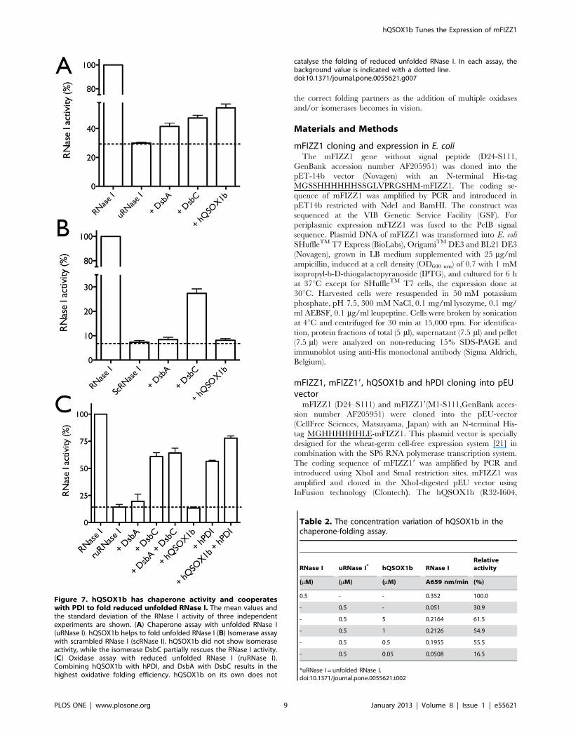
In the chaperone activity assay (Figure 7A), unfolded RNase I
was pre-incubated with hQSOX1b for 3 min at 15uC in a final
concentration of 0.5 mM unfolded RNase I before measuring
RNase activity. The quiescin sulfhydryl oxidase hQSOX1b
showed the highest chaperone activity compared to DsbA and
hQSOX1b Tunes the Expression of mFIZZ1
PLOS ONE | www.plosone.org 5 January 2013 | Volume 8 | Issue 1 | e55621

Figure 4. mFIZZ1 and mFIZZ19 are highly pure and monomeric. (A) CBB-stained 15% SDS-PAGE of the purified mFIZZ19 and mFIZZ1 co-expressed with and without hQSOX1b under reducing and non-reducing conditions followed by immunoblot developed with anti-His antibody. Thesamples are highly pure and proteins migrate at the same position under reducing and non-reducing conditions, indicating that no intermoleculardisulfide bonds are formed. (B) Basic native PAGE of the purified mFIZZ1 and mFIZZ19 co-expressed with and without hQSOX1b under reducing andnon-reducing conditions. The protein bands for mFIZZ1 (pI 4.81) and mFIZZ19 (pI 5.18) migrate on a slightly different position due to their pI. Nomajor multimeric bands are observed. (C) A CBB-stained 15% non-reducing SDS-PAGE of mFIZZ1 and mFIZZ19 co-expressed with hQSOX1b aftercross-linking with a mixture EDC/NHS for 3 h at room temperature is shown. As negative and positive controls, monomeric Corynebacteriumglutamicum mycoredoxin-1 (Cg_Mrx1) and dimeric Escherichia coli DsbG (Ec_DsbG) were used. Monomeric Cg_Mrx1 migrates after crosslinking asmonomer. For dimeric Ec_DsbG (present as a mixture between monomer and dimer on SDS-PAGE), bands shift from the monomeric and the dimericmigration position to a higher molecular weight positions. For both mFIZZ1 and mFIZZ19 no shift to a higher molecular weight is observed. As marker(M) in panels A, B and C, the PageRulerTM pre-stained Protein Ladder (Fermentas) is used, which contains SDS.doi:10.1371/journal.pone.0055621.g004
hQSOX1b Tunes the Expression of mFIZZ1
PLOS ONE | www.plosone.org 6 January 2013 | Volume 8 | Issue 1 | e55621

DsbC of E. coli. (Figure 7A). Decreasing the concentrations of
hQSOX1b from 5 mM to 50 nM gives similar RNase I activity up
to 0.5 mM (Table 2). A further decrease of the hQSOX1b
concentration below the concentration of RNase I results in
background RNase I activity. At least the same concentrations of
hQSOX1b as RNase I are needed to refold RNase I, which
indicates chaperone activity for hQSOX1b.
We next tested the isomerase activity of hQSOX1b on
scrambled RNase I of E. coli with its disulfide bonds wrongly
connected (0.5 mM scRNase I) (Figure 7B). hQSOX1b did not
show isomerase activity, while DsbC was able to partially refold
RNase I [32].
Finally, we tested the oxidase activity with reduced and
unfolded RNase I (1 mM ruRNase I) incubated for 3 min at
25uC. The combination of the folding catalysts hQSOX1b + hPDI
and DsbA + DsbC results in the highest oxidative folding
efficiency, with the largest contribution for the isomerases hPDI
and DsbC (Figure 7C). The in vitro oxidase activity of DsbC
[32,34] and hPDI have already been shown [35]. Important to
note is that for the refolding of ruRNase I only catalytic
concentrations of 50 nM hQSOX1b were used. The electrons
are directly passed via its FAD-group to O2 to form H2O2, and
hQSOX1b does not need to be recycled by a downstream
pathway like for the Dsb-proteins. As 50 nM of hQSOX1b has no
chaperone effect on unfolded RNase I (Table 2), hQSOX1b
should function as an oxidase in this experiment. On the other
hand, 50 nM or 5 mM hQSOX1b alone do not catalyze the
folding of the ruRNase I (Figure 7C). These results suggest that
hQSOX1b might cooperate with hPDI present in the wheat germ
extract [36] to correctly fold mFIZZ1. Also, Rancy and Thorpe
showed that QSOX and PDI work together to generate native
pairings in two unfolded reduced proteins [37].
Discussion
All together, well-folded, soluble full-length monomeric
mFIZZ1 is produced in a wheat germ cell-free expression system.
The protein mFIZZ1 forms five disulfides and the protein
mFIZZ19 with its signal peptide forms six disulfides. Both mFIZZ1
proteins are only biologically active in the presence of hQSOX1b.
Indeed, in the absence of hQSOX1b, all disulfide bonds are
formed, but no bioactivity was detected. We might conclude that
these non-active forms are disulfide-scrambled with a higher
helical content (CD-spectrum) and a different structural fold. The
fact that mFIZZ19 with its predicted signal peptide gave a similar
result came as a surprise. In vivo, the signal peptide of mFIZZ1 is
removed by signal peptidase enzymes [38].
The quiescin sulfhydryl oxidase hQSOX1b seems to have
chaperone and oxidase activity.
The chaperone activity of hQSOX1b might prevent incorrect
disulfide formation or might even prevent post-translational over-
oxidation. As an oxidase, hQSOX1b needs to work in concert
with the isomerase hPDI. To our knowledge, this is the first time
that chaperone activity has been described for hQSOX1b.
Perhaps not a complete surprise as this quiescin sulfhydryl oxidase
consists N-terminal Trx-domains like in PDI. For PDI, molecular
chaperone activity has been reported for the folding of procollagen
[39] and for glyceraldehyde-3-phosphate dehydrogenase [40].
More examples with several disulfide bond free substrate proteins
are needed to further confirm the chaperone activity of
hQSOX1b. Moreover, it has recently been shown that the
chaperone activity of PDI and the overall conformation of human
PDI are redox-regulated [41]. As such, in the experiment where
we combined hQSOX1b with hPDI, the produced H2O2 from the
formation of disulfide bonds by hQSOX1b may have increased
the chaperone activity of hPDI. In this case hPDI might correct
disulfide bonds as an isomerase and its chaperone activity will
increase the moment more disulfide bonds are introduced by the
oxidase hQSOX1b, which would result in a clear working
together of those two thiol/disulfide oxidoreductase. At this stage,
however, it is not clear which of the properties of hQSOX1b and
hPDI are needed to correctly fold mFIZZ1 and mFIZZ19 into a
biological active protein. In vivo, the functional role of hQSOX1b
in the folding of mFIZZ1 protein is highly unlikely due to different
cellular locations.
The final total protein yield of purified mFIZZ1 varies from 200
to 300 mg, which is relatively low due to the short translational life
span of the wheat germ extract compared to cell-based expression
Figure 5. mFIZZ19 has a high helical content and all the intramolecular disulfide bonds are formed. (A) The results of a Thiostar assayusing glutathione as a standard are shown. mFIZZ19 and mFIZZ1 prepared with and without hQSOX1b show no free thiols. (B) The CD-spectrum ofmFIZZ19 produced with and without hQSOX1b showed a double minimum at 208 and 222 nm characteristic for a-helical proteins.doi:10.1371/journal.pone.0055621.g005
hQSOX1b Tunes the Expression of mFIZZ1
PLOS ONE | www.plosone.org 7 January 2013 | Volume 8 | Issue 1 | e55621

[22]. For scaling-up the production using wheat germ extracts, an
automatized system might be required [21]. Alternatively, co-
expression with hQSOX1b [42] and/or hPDI in the E. coli might
also be explored. This method is certainly cheaper and higher
yields might be obtained, but here, the endotoxin levels are a
drawback. When we tested the endotoxin level on purified
expressed mFIZZ19 in the limulus amebocyte lysate (LAL) assay,
the same values as for PBS (negative control) were obtained (data
not shown). So, the use of a bacteria-free expression system also
overcomes the potential effects of LPS and other endotoxin
contaminants, which is certainly an advantage for follow-up
animal and/or cell culture testing.
We would like to emphasize that with the addition of quiescin
sulfhydryl oxidase hQSOX1b we have introduced a new method
for the folding of disulfide rich eukaryotic proteins. The sulfhydryl
oxidase hQSOX1b does not require any additional partners to
introduce disulfides into proteins. Although, the reaction with
substrate proteins results in the formation of H2O2, and as a
consequence oxidative stress, quiescin sulfhydryl oxidases are the
most competent catalysts known for the de novo insertion of protein
disulfide bonds in the endoplasmic reticulum [15,43]. We used
mFIZZ1 and mFIZZ19 as model proteins that potentially had to
form multiple disulfides for bioactivity. So far, no soluble protein
expression method was established for this immunological
important protein and no structural data are available. Nguygen
et al. [24] followed another methodological approach for the
expression of disulfide rich proteins in the cytoplasm of E. coli with
the help of sulfhydryl oxidase/disulfide isomerase Erv1p. They
showed how Erv1p is capable of introducing multiple disulfide
bonds in a fragment of tissue plasminogen activator (vtPA) and the
homodimeric resistin (mFIZZ3). We use the open cell free
expression system. Our wheat germ cell free expression method
is simple and effective for the in vitro production of soluble and
active recombinant eukaryotic proteins that have to form multiple
disulfides. Moreover, an open system makes it easier to screen for
Figure 6. mFIZZ19 and mFIZZ1 decreased the IL-13 and IL-5 secretion of splenocytes. Splenocytes were cultured at 200,000 cells/well andactivated under Th2 permissive conditions for 4 days. Recombinant mFIZZ19 and mFIZZ1 expressed with or without hQSOX1b were used at 200 ng/ml. rRa is the bacterial recombinant FIZZ1 (200 ng/ml) from (Peprotech) and PBS is the control. (A) Recombinant mFIZZ19 and mFIZZ1 co-expressedwith hQSOX1b significantly decreased the IL-5 secretion compared to the proteins expressed alone. (B) Recombinant mFIZZ19 and mFIZZ1 co-expressed with hQSOX1b decreased the IL-13 secretion compared to the proteins expressed alone. ***P,0.001; **P,0.01; *P,0.05; #P = 0.07.Results are representative of three independent experiments for mFIZZ19 and two independent experiments for mFIZZ1, and statistical analysis wasperformed by two-way ANOVA.doi:10.1371/journal.pone.0055621.g006
hQSOX1b Tunes the Expression of mFIZZ1
PLOS ONE | www.plosone.org 8 January 2013 | Volume 8 | Issue 1 | e55621
the correct folding partners as the addition of multiple oxidases
and/or isomerases becomes in vision.
Materials and Methods
mFIZZ1 cloning and expression in E. coliThe mFIZZ1 gene without signal peptide (D24-S111,
GenBank accession number AF205951) was cloned into the
pET-14b vector (Novagen) with an N-terminal His-tag
MGSSHHHHHHSSGLVPRGSHM-mFIZZ1. The coding se-
quence of mFIZZ1 was amplified by PCR and introduced in
pET14b restricted with NdeI and BamHI. The construct was
sequenced at the VIB Genetic Service Facility (GSF). For
periplasmic expression mFIZZ1 was fused to the PeIB signal
sequence. Plasmid DNA of mFIZZ1 was transformed into E. coli
SHuffleTM T7 Express (BioLabs), OrigamiTM DE3 and BL21 DE3
(Novagen), grown in LB medium supplemented with 25 mg/ml
ampicillin, induced at a cell density (OD600 nm) of 0.7 with 1 mM
isopropyl-b-D-thiogalactopyranoside (IPTG), and cultured for 6 h
at 37uC except for SHuffleTM T7 cells, the expression done at
30uC. Harvested cells were resuspended in 50 mM potassium
phosphate, pH 7.5, 300 mM NaCl, 0.1 mg/ml lysozyme, 0.1 mg/
ml AEBSF, 0.1 mg/ml leupeptine. Cells were broken by sonication
at 4uC and centrifuged for 30 min at 15,000 rpm. For identifica-
tion, protein fractions of total (5 ml), supernatant (7.5 ml) and pellet
(7.5 ml) were analyzed on non-reducing 15% SDS-PAGE and
immunoblot using anti-His monoclonal antibody (Sigma Aldrich,
Belgium).
mFIZZ1, mFIZZ19, hQSOX1b and hPDI cloning into pEUvector
mFIZZ1 (D24–S111) and mFIZZ19(M1-S111,GenBank acces-
sion number AF205951) were cloned into the pEU-vector
(CellFree Sciences, Matsuyama, Japan) with an N-terminal His-
tag MGHHHHHHLE-mFIZZ1. This plasmid vector is specially
designed for the wheat-germ cell-free expression system [21] in
combination with the SP6 RNA polymerase transcription system.
The coding sequence of mFIZZ19 was amplified by PCR and
introduced using XhoI and SmaI restriction sites. mFIZZ1 was
amplified and cloned in the XhoI-digested pEU vector using
InFusion technology (Clontech). The hQSOX1b (R32-I604,
Figure 7. hQSOX1b has chaperone activity and cooperateswith PDI to fold reduced unfolded RNase I. The mean values andthe standard deviation of the RNase I activity of three independentexperiments are shown. (A) Chaperone assay with unfolded RNase I(uRNase I). hQSOX1b helps to fold unfolded RNase I (B) Isomerase assaywith scrambled RNase I (scRNase I). hQSOX1b did not show isomeraseactivity, while the isomerase DsbC partially rescues the RNase I activity.(C) Oxidase assay with reduced unfolded RNase I (ruRNase I).Combining hQSOX1b with hPDI, and DsbA with DsbC results in thehighest oxidative folding efficiency. hQSOX1b on its own does not
catalyse the folding of reduced unfolded RNase I. In each assay, thebackground value is indicated with a dotted line.doi:10.1371/journal.pone.0055621.g007
Table 2. The concentration variation of hQSOX1b in thechaperone-folding assay.
RNase I uRNase I* hQSOX1b RNase IRelativeactivity
(mM) (mM) (mM) A659 nm/min (%)
0.5 - - 0.352 100.0
- 0.5 - 0.051 30.9
- 0.5 5 0.2164 61.5
- 0.5 1 0.2126 54.9
- 0.5 0.5 0.1955 55.5
- 0.5 0.05 0.0508 16.5
*uRNase I = unfolded RNase I.doi:10.1371/journal.pone.0055621.t002
hQSOX1b Tunes the Expression of mFIZZ1
PLOS ONE | www.plosone.org 9 January 2013 | Volume 8 | Issue 1 | e55621

GenBank accession number NP_001004128.1) without signal
peptide and hPDI (A18-L508, GenBank accession number
NP_000909.2) without signal peptide genes were cloned with a
GST-tag at the N-terminal position into the pEU-GST-MCS
vector. The coding sequence of hQSOX1b and hPDI were
amplified by PCR and introduced into the pEU-GST-MCS vector
digested with BamHI and SmaI, or the XhoI and SmaI,
respectively.
All constructs were sequenced at the VIB Genetic Service
Facility (GSF).
Small-scale transcription and translation reactionPlasmid DNA of mFIZZ1, mFIZZ19, hPDI and hQSOX1b
(2 mg) was transcribed using SP6 RNA polymerase, 25 mM NTP
mix, RNase inhibitor and 56 transcription buffer (Cell Free
Sciences, Matsuyama, Japan) for 6 h at 37uC. The mRNA was
cooled down to avoid degradation, and checked on 1% agarose
gel. For translation, 10 ml of mRNA was mixed with the same
amount of the wheat germ extract WEPRO 7240 (CellFree
Sciences, Matsuyama, Japan) and 0.1 mg of creatine kinase to
make the bottom layer, and incubated with 206 ml of 16 SUB-A
MIX SGC (upper layer) at 15uC for 20 h without shaking in a 6-
well plate (Greiner bio-one, Belgium) in a Thermomixer (Roche,
Germany). The reaction mixture was centrifuged (15,000 rpm) for
30 min at 4uC. For identification, protein fractions, total (5 ml),
soluble (7.5 ml) and pellet (7.5 ml) of the expressed proteins were
visualized on immunoblot using as primary antibody anti-His or
anti-GST antibody (EnoGene, Germany) and as secondary anti
mouse polyclonal antiserum (Sigma Aldrich, Belgium). The same
samples were ran on a non-reducing 15% SDS-PAGE followed by
Coomassie Brilliant Blue staining.
Co-expression of mFIZZ1 with hQSOX1b and/or hPDIpEU GST-tag hQSOX1b, GST-tag hPDI and His-tag mFIZZ1
or mFIZZ19 (2 mg each) were separately transcribed using SP6
RNA polymerase, 25 mM NTP mix, RNase inhibitor and 56transcription buffer. The mRNA of respectively mFIZZ1,
mFIZZ19, hQSOX1b and hPDI were translated for 10 min using
the wheat germ extract WEPRO 7240 at 26uC. mRNA of
mFIZZ1 or mFIZZ19 (10 ml each) was then mixed with the same
amount of mRNA from hQSOX1b, hPDI, and hQSOX1b +hPDI and incubated with 206 ml of the SUB-A mixture for each
reaction at 15uC for 20 h without shaking in a 96-well plate. After
the incubation, the reaction mixture was centrifuged at
15,000 rpm for 30 min at 4uC. The protein concentration of the
soluble and pellet fractions was determined using a Bradford assay
[44]. A same amount (30 mg) of pellet and soluble proteins were
ran on a non-reducing 15% SDS-PAGE and visualized by
immunoblot using anti-His (Sigma Aldrich, Belgium) and anti-
GST antibody (EnoGene, Germany). All bands from the
immunoblots were scanned and the percentage was determined
using Labimage programe (http://www.labimage.com). The
experiments were repeated three times for reproducibility.
Upscaling of the mFIZZ1 and mFIZZ19 production withand without hQSOX1b
For up-scaling, plasmid DNA of His-tag mFIZZ1 and mFIZZ19
(25 mg) and (25 mg) GST-tag hQSOX1b were transcribed
separately using SP6 RNA polymerase, 25 mM NTP mix, RNase
inhibitor and 56 transcription buffer for 6 h at 37uC. For
translation, 250 ml of mRNA was mixed with the same amount of
wheat germ extract (WEPRO 7240), and with 1 mg of creatine
kinase to make the bottom layer. 5.5 ml of 16 SUB-A MIX
included a mixture of amino acids were used to make the upper
layer. Translation was performed at 15uC for 20 h without shaking
in a 6-well plate using a Thermomixer. The expressed proteins
were detected by immunoblotting using anti-His antibody (Sigma
Aldrich, Belgium) and on CBB-stained 15% SDS-PAGE prior to
purification.
For co-expression, mRNA from mFIZZ1 or mFIZZ19 and
hQSOX1b were translated for 10 min at 26uC using wheat germ
extract WEPRO 7240H. After 10 min incubation, mRNA of
mFIZZ1 or mFIZZ19 were mixed with the same amount of
mRNA from hQSOX1b and translated at 15uC for 20 h without
shaking.
For purification, 2 batches (mFIZZ1 or mFIZZ19) of 6 ml
reaction (with and without hQSOX1b) were centrifuged at
15,000 rpm for 15 min at 4uC. The supernatant was separately
loaded on 1 ml His-trap Ni-NTA resin equilibrated with 50 mM
potassium phosphate buffer solution pH 7.5, 150 mM NaCl
containing 10 mM imidazole. The column was washed with 5
column volumes and, the protein was eluted with a linear
imidazole gradient from 50 to 500 mM in the same buffer
solution. The purity of the elution peak fractions was evaluated on
15% SDS-PAGE under reducing and non- reducing conditions.
Pure fractions were collected and dialyzed against PBS for 4 h at
4uC with two buffer changes. Protein concentrations were
spectrophotometrically determined with a molar extinction of
18,740 M21 cm21 at 280 nm. Protein aliquots were stored at
220uC.
Basic-native gel protocolFor the basic-native gel conditions, a method for acidic and
neutral proteins was used (http://wolfson.huji.ac.il/purification/
Protocols/PAGE_Basic.html). Briefly, the samples of mFIZZ1 (pI
4.81) and mFIZZ19 (pI 5.18) expressed with and without
hQSOX1b were mixed on ice with sample buffer solution
containing 100 mM Tris/HCl, pH 6.8, bromophenol blue, and
glycerol. For the reduced conditions, the samples were first
incubated for 30 min with 20 mM DTT. Samples were loaded on
a polyacrylamide native gel (5% stacking gel (pH 6.8) and 15%
resolving gel (pH 8.9)). The running buffer solution contained
50 mM Tris/HCl, pH 8.9, and 380 mM glycine. As marker the
PageRulerTM pre-stained Protein Ladder (Fermentas) is used,
which contains SDS. After a 5 h run at 4uC, gels were CBB
stained.
Cross-linking conditionsSamples of mFIZZ1 (10 mM), mFIZZ19 (5.3 mM), mFIZZ1 +
hQSOX1b (20 mM), and mFIZZ19 + hQSOX1b (9.2 mM) were
were incubated with the cross-linker mixture of 1 mM EDC and
1 mM NHS (Pierce, Belgium) for 3 h at room temperature in PBS,
pH 7.4 buffer solution. The reaction was stopped by addition of
SDS-PAGE sample buffer. After boiling for 10 minutes, the
mixture was evaluated on 15% SDS-PAGE under non-reducing
conditions. As negative control, monomeric Corynebacterium gluta-
micum mycoredoxin-1 (Cg_Mrx1) [45,46] was used. As positive
control, dimeric Escherichia coli DsbG (Ec_DsbG) [8,47] was used.
Circular dichroism (CD) and mass spectrophotometryidentification
CD-spectra were recorded at 25uC using a spectropolarimeter
(Jasco, model 715, Tokyo, Japan). Protein was dissolved in water at
0.2 mg/ml. CD-spectra were recorded at a scan speed of 50 nm/
min, 1 nm bandwidth and with a response time of 1 s. Spectra
were recorded between in 0.01 cm path length quartz cuvette
hQSOX1b Tunes the Expression of mFIZZ1
PLOS ONE | www.plosone.org 10 January 2013 | Volume 8 | Issue 1 | e55621

from 190 to 260 nm. The mFIZZ1 protein band was excised from
the gel and the tryptic peptides were analyzed by tandem mass
spectrometry (MS/MS) as described [48]. The percentage of a-
helical and b-strand content was determined by sending the
recorded spectra (190–260 nm) to the Dichroweb server (http://
dichroweb.cryst.bbk.ac.uk) [29].We tested the different available
algorithms. The CDSSTR algorithm [28] gave the lowest nmrsd
values and the best fit to our experimental data.
Disulfide bond formation assayThe number of free thiols in samples was determined using a
Thiostar assay (Detect Xtm, Luminos) [42,49]. A standard curve of
reduced L-glutathione (Sigma) ranging from 0 to 6 mM in a 96-
well plate was prepared in water. mFIZZ1 and mFIZZ19 samples
expressed with and without hQSOX1b (5 mM) were 10 times
diluted in water. After mixing with 15 ml of Thiostar reagent, the
plate was incubated for 30 min in the dark, prior to measure at
510 nm with excitation at 390 nm in a fluorescent plate reader
(Infinite M200, TECAN).
Expression, and purification of DsbA, DsbC, hQSOX1band hPDI
Expression and purification of DsbA and DsbC were performed
as mentioned [32], hQSOX1b [43] and hPDI [37]. Purified
DsbA, DsbC and hPDI were stored at 220uC. The purified
hQSOX1b was stored at 4uC in the dark.
Bioactivity assay of mFIZZ1Single cell suspensions from C57BL/6 mouse spleens were
cultured under Th2 permissive conditions with the addition of
PBS (control), bacterial recombinant mFIZZ1/RELMa (Pepro-
tech), or mFIZZ19 expressed with or without hQSOX1b at
concentrations indicated. Peprotech RELMa was generated in E.
coli according to standard bacterial expression systems, and in the
absence of any specific protocols to ensure disulfide bond
formation (see www.peprotech.com for more information). Protein
purity was confirmed by SDS-PAGE and HPLC analyses. Cells
were cultured at the concentration of 26105 cells/well in 96-well
round-bottom plates. Th2-permissive conditions were: aCD3/
aCD28 (1 mg/mL each, eBioscience), rIL-4 (40 ng/mL;
eBioscience), anti-IL-12 (10 mg/mL; clone C17.8) and anti-IFNc(10 mg/mL; clone XMG 1.2). Four days later, supernatants were
recovered for quantification of IL-5 and IL-13 by standard
sandwich ELISA protocols (antibodies from eBioscience). Results
are shown +/2 S.D. and are representative of two or three
independent experiments with quadruplicate wells per condition.
Statistical significance was determined by using two-way anova
analysis with treatment and experiment repeats as variables.
RNase I activity assayThe RNA hydrolysis activity was performed as described [32].
RNA solution was mixed with the methylene blue buffer to obtain
a final concentration of 450 mM RNA. After pre-incubation for
3 min at 25uC in the dark, E. coli RNase I was added. The
absorbance change was followed in function of time at 659 nm,
the wavelength with a maximum spectral difference between
methylene blue intercalated RNA with and without RNase I.
Oxidase, isomerase and chaperone assayFor the chaperone activity, unfolded RNase I was prepared by
mixing guanidine hydrochloride (GuHCl), 50 mM Tris/HCl
pH 8.0 and RNase I wild type to obtain the final concentration
of 1.2 mM RNase I, 6.7 M GuHCl, 56 mM Tris/HCl pH 8.0.
After 2 h incubation at 25uC, the mixture was dialyzed for 2 h
against 0.6 M GuHCl, 50 mM Tris pH 8.7, 50 mM NaCl.
Unfolded RNase I (500 nM) was pre-incubated with varying
concentrations of (5 mM–50 nM) hQSOX1b, 5 mM DsbA or
5 mM DsbC in 50 mM Hepes/NaOH, pH 7.4, 150 mM NaCl for
3 min at 15uC before RNase activity was measured.
For the isomerase activity, E. coli scrambled RNase I (scRNase I)
[32] was pre-incubated with 5 mM hQSOX1b, 5 mM DsbA, or
5 mM DsbC in 50 mM Hepes pH 7.4, 150 mM NaCl for 3 min at
15uC at a final concentration of 0.5 mM scRNase before
measuring RNase activity.
For the oxidase assay, reduced and unfolded RNase I (ruRNase
I) [32] was pre-incubated with the thiol/disulfide oxidoreductases
in 50 mM Hepes/NaOH, pH 7.4, 150 mM NaCl for 3 min at
25uC at a final concentration of 1 mM ruRNase before measuring
RNase activity. hPDI was reduced with 20 mM DTT for 1 h at
25uC. DTT was removed on a Superdex75 HR column (GE
Healthcare Life Sciences) equilibrated in 50 mM Na phosphate,
pH 7.5, 150 mM NaCl. Reduced and unfolded RNase I was pre-
incubated with 5 mM hQSOX1b, 5 mM hPDI, 50 nM hQSOX1b
+5 mM hPDI, 5 mM DsbA, 5 mM DsbC, 5 mM DsbA +5 mM
DsbC, in 50 mM Hepes/NaOH, pH 7.4, 150 mM NaCl for
3 min at 25uC at a final concentration of 1 mM ruRNase I, before
measuring RNase I activity.
Acknowledgments
We would like to thank Irene U. Ajonina and Gholamreza Hassanzadeh
for their many efforts in trying to refold mFIZZ1 from inclusion bodies,
and Benoıt Stijlemans for help with the LAL-assay. We would like to thank
Colin Thorpe for kindly providing us a pTRC HisA plasmid with human
QSOX1b, Wim Hol for the pProEX HTb plasmid with human PDI, and
the academic editor of PlosOne, Young-Hwa Son, for the suggestions that
improved the manuscript.
Author Contributions
Conceived and designed the experiments: JM MN HDG. Performed the
experiments: WG MN GV KVB KW. Analyzed the data: JM WG JVG
YE DA. Contributed reagents/materials/analysis tools: YE. Wrote the
paper: JM WG JVG MN.
References
1. Holcomb IN, Kabakoff RC, Chan B, Baker TW, Gurney A, et al. (2000) FIZZ1,
a novel cysteine-rich secreted protein associated with pulmonary inflammation,
defines a new gene family. EMBO J 19: 4046–4055.
2. Nair MG, Guild KJ, Artis D (2006) Novel effector molecules in type 2
inflammation: lessons drawn from helminth infection and allergy. J Immunol
177: 1393–1399.
3. Nair MG, Gallagher IJ, Taylor MD, Loke P, Coulson PS, et al. (2005) Chitinase
and Fizz family members are a generalized feature of nematode infection with
selective upregulation of Ym1 and Fizz1 by antigen-presenting cells. Infect
Immun 73: 385–394.
4. Patel SD, Rajala MW, Rossetti L, Scherer PE, Shapiro L (2004) Disulfide-
dependent multimeric assembly of resistin family hormones. Science 304: 1154–
1158.
5. Sandler NG, Mentink-Kane MM, Cheever AW, Wynn TA (2003) Global gene
expression profiles during acute pathogen-induced pulmonary inflammation
reveal divergent roles for Th1 and Th2 responses in tissue repair. J Immunol
171: 3655–3667.
6. Depuydt M, Messens J, Collet JF (2011) How proteins form disulfide bonds.
Antioxid Redox Signal 15: 49–66.
hQSOX1b Tunes the Expression of mFIZZ1
PLOS ONE | www.plosone.org 11 January 2013 | Volume 8 | Issue 1 | e55621

7. Wouters MA, Fan SW, Haworth NL (2010) Disulfides as redox switches: from
molecular mechanisms to functional significance. Antioxid Redox Signal 12: 53–91.
8. Messens J, Collet JF (2006) Pathways of disulfide bond formation in Escherichia
coli. Int J Biochem Cell Biol 38: 1050–1062.9. Riemer J, Bulleid N, Herrmann JM (2009) Disulfide formation in the ER and
mitochondria: two solutions to a common process. Science 324: 1284–1287.10. Karala AR, Lappi AK, Ruddock LW (2010) Modulation of an active-site
cysteine pKa allows PDI to act as a catalyst of both disulfide bond formation and
isomerization. J Mol Biol 396: 883–892.11. Tu BP, Ho-Schleyer SC, Travers KJ, Weissman JS (2000) Biochemical basis of
oxidative protein folding in the endoplasmic reticulum. Science 290: 1571–1574.12. Sevier CS, Cuozzo JW, Vala A, Aslund F, Kaiser CA (2001) A flavoprotein
oxidase defines a new endoplasmic reticulum pathway for biosyntheticdisulphide bond formation. Nat Cell Biol 3: 874–882.
13. Thorpe C, Hoober KL, Raje S, Glynn NM, Burnside J, et al. (2002) Sulfhydryl
oxidases: emerging catalysts of protein disulfide bond formation in eukaryotes.Arch Biochem Biophys 405: 1–12.
14. Coppock DL, Kopman C, Scandalis S, Gilleran S (1993) Preferential geneexpression in quiescent human lung fibroblasts. Cell Growth Differ 4: 483–493.
15. Kodali VK, Thorpe C (2010) Oxidative protein folding and the Quiescin-
sulfhydryl oxidase family of flavoproteins. Antioxid Redox Signal 13: 1217–1230.
16. Wittke I, Wiedemeyer R, Pillmann A, Savelyeva L, Westermann F, et al. (2003)Neuroblastoma-derived sulfhydryl oxidase, a new member of the sulfhydryl
oxidase/Quiescin6 family, regulates sensitization to interferon gamma-inducedcell death in human neuroblastoma cells. Cancer Res 63: 7742–7752.
17. Coppock DL, Cina-Poppe D, Gilleran S (1998) The quiescin Q6 gene (QSCN6)
is a fusion of two ancient gene families: thioredoxin and ERV1. Genomics 54:460–468.
18. Powis G, Montfort WR (2001) Properties and biological activities ofthioredoxins. Annu Rev Pharmacol Toxicol 41: 261–295.
19. Lisowsky T (1992) Dual function of a new nuclear gene for oxidative
phosphorylation and vegetative growth in yeast. Mol Gen Genet 232: 58–64.20. Alon A, Heckler EJ, Thorpe C, Fass D (2010) QSOX contains a pseudo-dimer
of functional and degenerate sulfhydryl oxidase domains. FEBS Lett 584: 1521–1525.
21. Takai K, Sawasaki T, Endo Y (2010) Practical cell-free protein synthesis systemusing purified wheat embryos. Nature protocols 5: 227–238.
22. Endo Y, Sawasaki T (2006) Cell-free expression systems for eukaryotic protein
production. Curr Opin Biotechnol 17: 373–380.23. Juan CC, Kan LS, Huang CC, Chen SS, Ho LT, et al. (2003) Production and
characterization of bioactive recombinant resistin in Escherichia coli. J Biotechnol103: 113–117.
24. Nguyen VD, Hatahet F, Salo KE, Enlund E, Zhang C, et al. (2011) Pre-
expression of a sulfhydryl oxidase significantly increases the yields of eukaryoticdisulfide bond containing proteins expressed in the cytoplasm of E. coli. Microb
Cell Fact 10: 1.25. Updegrove TB, Correia JJ, Chen Y, Terry C, Wartell RM (2011) The
stoichiometry of the Escherichia coli Hfq protein bound to RNA. RNA 17: 489–500.
26. Panchaud A, Hansson J, Affolter M, Bel Rhlid R, Piu S, et al. (2008) ANIBAL,
stable isotope-based quantitative proteomics by aniline and benzoic acid labelingof amino and carboxylic groups. Mol Cell Proteomics 7: 800–812.
27. Banerjee RR, Lazar MA (2001) Dimerization of resistin and resistin-likemolecules is determined by a single cysteine. J Biol Chem 276: 25970–25973.
28. Sreerama N, Woody RW (2000) Estimation of protein secondary structure from
circular dichroism spectra: comparison of CONTIN, SELCON, and CDSSTRmethods with an expanded reference set. Anal Biochem 287: 252–260.
29. Whitmore L, Wallace BA (2004) DICHROWEB, an online server for proteinsecondary structure analyses from circular dichroism spectroscopic data. Nucleic
Acids Res 32: W668–673.
30. Nair MG, Du Y, Perrigoue JG, Zaph C, Taylor JJ, et al. (2009) Alternativelyactivated macrophage-derived RELM-{alpha} is a negative regulator of type 2
inflammation in the lung. J Exp Med 206: 937–952.
31. Munitz A, Seidu L, Cole ET, Ahrens R, Hogan SP, et al. (2009) Resistin-like
molecule alpha decreases glucose tolerance during intestinal inflammation.J Immunol 182: 2357–2363.
32. Messens J, Collet JF, Van Belle K, Brosens E, Loris R, et al. (2007) The oxidaseDsbA folds a protein with a nonconsecutive disulfide. J Biol Chem 282: 31302–
31307.
33. Depuydt M, Messens J, Collet JF (2011) How proteins form disulfide bonds.
Antioxid Redox Signal 15:49–66.
34. Zapun A, Missiakas D, Raina S, Creighton TE (1995) Structural and functionalcharacterization of DsbC, a protein involved in disulfide bond formation in
Escherichia coli. Biochemistry 34: 5075–5089.
35. Hatahet F, Ruddock LW (2009) Protein disulfide isomerase: a critical evaluation
of its function in disulfide bond formation. Antioxid Redox Signal 11: 2807–2850.
36. Grynberg A, Nicolas J, Drapron R (1977) Presence of a protein disulfideisomerase (EC 5.3.4.1) in wheat germ. C R Acad Sci Hebd Seances Acad Sci D
284: 235–238.
37. Rancy PC, Thorpe C (2008) Oxidative protein folding in vitro: a study of the
cooperation between quiescin-sulfhydryl oxidase and protein disulfide isomerase.Biochemistry 47: 12047–12056.
38. Paetzel M, Karla A, Strynadka NC, Dalbey RE (2002) Signal peptidases. Chem
Rev 102: 4549–4580.
39. Wilson R, Lees JF, Bulleid NJ (1998) Protein disulfide isomerase acts as a
molecular chaperone during the assembly of procollagen. J Biol Chem 273:9637–9643.
40. Cai H, Wang CC, Tsou CL (1994) Chaperone-like activity of protein disulfideisomerase in the refolding of a protein with no disulfide bonds. J Biol Chem 269:
24550–24552.
41. Wang C, Yu J, Huo L, Wang L, Feng W, et al. (2012) Human protein-disulfide
isomerase is a redox-regulated chaperone activated by oxidation of domain a’.J Biol Chem 287: 1139–1149.
42. Abskharon RN, Ramboarina S, El Hassan H, Gad W, Apostol MI, et al (2012).A novel expression system for production of soluble prion proteins in E. coli.
Microb Cell Fact 11: 6.
43. Heckler EJ, Alon A, Fass D, Thorpe C (2008) Human quiescin-sulfhydryl
oxidase, QSOX1: probing internal redox steps by mutagenesis. Biochemistry 47:
4955–4963.
44. Bradford MM (1976) A rapid and sensitive method for the quantitation of
microgram quantities of protein utilizing the principle of protein-dye binding.Anal Biochem 72: 248–254.
45. Van Laer K, Buts L, Foloppe N, Vertommen D, Van Belle K, et al. (2012)Mycoredoxin-1 is one of the missing links in the oxidative stress defense
mechanism of Mycobacteria. Mol Microbiol 86: 787–804.
46. Ordonez E, Van Belle K, Roos G, De Galan S, Letek M, et al. (2009) Arsenate
reductase, mycothiol, and mycoredoxin concert thiol/disulfide exchange. J BiolChem 284: 15107–15116.
47. Depuydt M, Leonard SE, Vertommen D, Denoncin K, Morsomme P, et al.(2009) A periplasmic reducing system protects single cysteine residues from
oxidation. Science 326: 1109–1111.
48. Plesa M, Hernalsteens JP, Vandenbussche G, Ruysschaert JM, Cornelis P (2006)
The SlyB outer membrane lipoprotein of Burkholderia multivorans contributes
to membrane integrity. Res Microbiol 157: 582–592.
49. Park S, Lippard SJ (2011) Redox state-dependent interaction of HMGB1 and
cisplatin-modified DNA. Biochemistry 50: 2567–2574.
50. Thompson JD, Higgins DG, Gibson TJ (1994) CLUSTAL W: improving the
sensitivity of progressive multiple sequence alignment through sequenceweighting, position-specific gap penalties and weight matrix choice. Nucleic
Acids Res 22: 4673–4680.
hQSOX1b Tunes the Expression of mFIZZ1
PLOS ONE | www.plosone.org 12 January 2013 | Volume 8 | Issue 1 | e55621