The quest for faithful in vitro models of human dendritic cells ...
50
HAL Id: hal-02981716 https://hal.archives-ouvertes.fr/hal-02981716 Submitted on 30 Nov 2020 HAL is a multi-disciplinary open access archive for the deposit and dissemination of sci- entific research documents, whether they are pub- lished or not. The documents may come from teaching and research institutions in France or abroad, or from public or private research centers. L’archive ouverte pluridisciplinaire HAL, est destinée au dépôt et à la diffusion de documents scientifiques de niveau recherche, publiés ou non, émanant des établissements d’enseignement et de recherche français ou étrangers, des laboratoires publics ou privés. The quest for faithful in vitro models of human dendritic cells types Xin-Long Luo, Marc Dalod To cite this version: Xin-Long Luo, Marc Dalod. The quest for faithful in vitro models of human dendritic cells types. Molecular Immunology, Elsevier, 2020, 123, pp.40-59. 10.1016/j.molimm.2020.04.018. hal-02981716
-
Upload
khangminh22 -
Category
Documents
-
view
2 -
download
0
Transcript of The quest for faithful in vitro models of human dendritic cells ...

HAL Id: hal-02981716https://hal.archives-ouvertes.fr/hal-02981716
Submitted on 30 Nov 2020
HAL is a multi-disciplinary open accessarchive for the deposit and dissemination of sci-entific research documents, whether they are pub-lished or not. The documents may come fromteaching and research institutions in France orabroad, or from public or private research centers.
L’archive ouverte pluridisciplinaire HAL, estdestinée au dépôt et à la diffusion de documentsscientifiques de niveau recherche, publiés ou non,émanant des établissements d’enseignement et derecherche français ou étrangers, des laboratoirespublics ou privés.
The quest for faithful in vitro models of humandendritic cells typesXin-Long Luo, Marc Dalod
To cite this version:Xin-Long Luo, Marc Dalod. The quest for faithful in vitro models of human dendritic cells types.Molecular Immunology, Elsevier, 2020, 123, pp.40-59. �10.1016/j.molimm.2020.04.018�. �hal-02981716�

The quest for faithful in vitro models of human dendritic cells types.
Luo XL, Dalod M.
Mol Immunol. 2020 Jul;123:40‐59.
doi: 10.1016/j.molimm.2020.04.018. Epub 2020 May 13.
PMID: 32413788.
https://www.sciencedirect.com/science/article/abs/pii/S0161589019309174
The quest for faithful in vitro models of human dendritic cells types
Xin-Long Luo1 and Marc Dalod1*
1Aix Marseille Univ, CNRS, INSERM, CIML, Centre d'Immunologie de Marseille-Luminy, Marseille,
France
*Corresponding author at: Centre d’Immunologie de Marseille-Luminy (CIML), Parc scientifique et
technnologique de Luminy, case 906, 163 avenue de Luminy, F-13288 Marseille Cedex 09, France.
E-mail address: [email protected] (M. Dalod).
Abstract
Dendritic cells (DCs) are mononuclear phagocytes that are specialized in the induction and functional
polarization of effector lymphocytes, thus orchestrating immune defenses against infections and
cancer. The population of DC encompasses distinct cell types that vary in their efficacy for
complementary functions and are thus likely involved in defending the body against different threats.
Plasmacytoid DCs specialize in the production of high levels of the antiviral cytokines type I interferons.
Type 1 conventional DCs (cDC1s) excel in the activation of cytotoxic CD8+ T cells (CTLs) which are critical
for defense against cancer and infections by intracellular pathogens. Type 2 conventional DCs (cDC2s)
prime helper CD4+ T cells for the production of type 2 cytokines underpinning immune defenses against
worms or of IL-17 promoting control of infections by extracellular bacteria or fungi. Hence, clinically
manipulating the development and functions of DC types could have a major impact for improving
treatments against many diseases. However, the rarity and fragility of human DC types is impeding
advancement towards this goal. To overcome this roadblock, major efforts are ongoing to generate in
vitro large numbers of distinct human DC types. We review here the current state of this research field,
emphasizing recent breakthrough and proposing future priorities. We also pinpoint the necessity to
develop a consensus nomenclature and rigorous methodologies to ensure proper identification and
characterization of human DC types. Finally, we elaborate on how faithful in vitro models of human DC
types can accelerate our understanding of the biology of these cells and the engineering of next
generation vaccines or immunotherapies against viral infections or cancer.
Keywords: type 1 conventional dendritic cells; plasmacytoid dendritic cells; hematopoiesis; cancer;
viral infection

We review the current state of the art regarding in vitro generation and characterization of
human DC types.
We emphasize recent breakthroughs and highlight possible future priorities.
We provide a guideline proposal for proper identification and characterization of in vitro
derived human DC types.
We discuss how in vitro models of human DC types can accelerate our understanding of the
biology of these cells and the engineering of next generation vaccines or immunotherapies
against viral infections or cancer.

Abbreviations: AhR, aryl hydrocarbon receptor; ASDCs, AXL+ SIGLEC6+ dendritic cells; cDC1s, type 1
conventional dendritic cells; cDC2s, type 2 conventional dendritic cells; CDP, common DC progenitor;
cGMP, current good manufacturing practices; cMoP, classical monocyte progenitor; cMos, classical
monocytes; CMP, common myeloid progenitors; CTLs, cytotoxic CD8+ T cells; GMDP, granulocyte-
monocyte-DC progenitor; HSCs, hematopoietic stem cells; IFN-I, type I interferons; iPSCs, induced
pulripotent stem cells.; LCs, Langerhans cells; LMMPs, Lymphoid-primed multipotent progenitors;
MDP, macrophage and dendritic cell progenitor; MHC-I, class I major histocompatibility complex;
MLPs, Multi-lymphoid progenitors; MoDCs, monocyte-derived dendritic cells; MoMacs, monocyte-
derived macrophages; pDCs, plasmacytoid dendritic cells; pre-cDC, cDC precurosor; Pre-cDC1, cDC1
precursor; pre-cDC2, cDC2 precursor; pro-cDC, classical DC progenitor; pro-pDC, pDC progenitor; tDCs,
transitional DCs; Th, helper CD4+ T cells.

1. Introduction
Vertebrate are equipped with a complex immune system that can discriminate pathological
from normal self, enabling recognition and elimination/control of cancer or infections by intracellular
pathogens. This process largely relies on effector cytotoxic immune cell types including natural killer
cells and CD8+ T lymphocytes (CTLs), whose activation requires signals from accessory immune cells,
in particular dendritic cells (DCs). DCs are uniquely able to deliver to naïve T cells all the signals
necessary for their initial activation upon the first encounter with their cognate antigen, a process
called T cell priming (Vu Manh et al., 2015). DCs can detect a variety of danger signals and translate
their combinatorial sensing into delivery of a matched array of output signals instructing the functional
polarization of T lymphocytes towards the function that should be the best suited to fight the threat
that the organism is facing (Vu Manh et al., 2015). Hence, DC functions are highly plastic, locally shaped
by the tissue microenvironment where they reside, which contributes to establish a beneficial balance
between host defense mechanisms and avoidance of autoimmunity or immunopathology. An
additional layer ensuring the plasticity of DC functions, and the adaptability of the immune system to
different types of threats, is the existence within the DC family of distinct cell types. DC types differ in
the arrays of innate immune sensors that they express, in link with the combination of activation or
inhibitory signals that they can deliver to T cells. This functional specialization of DC types more broadly
relates to differences in their ontogeny and gene expression profiles (Vu Manh et al., 2015). Beyond
their different functional specialization in health, distinct DC types also present different
susceptibilities to infection by intracellular pathogens or to hijacking of their immunoregulatory
activities by microbes or tumor cells for their own benefits and at the expense of the host (Bakdash et
al., 2016; Fries and Dalod, 2016; Silvin et al., 2017). Thus, when harnessing DCs for vaccination or
immunotherapy purposes, it is essential to ensure targeting the right DC type for the proper function,
through a strategy preventing their repurposing in the lesion microenvironment in a manner that
would favor disease development instead of benefiting the patient. To this aim, we must gain a precise
knowledge of the identity of human DC types, their function and their molecular regulation. However,
the rarity and frailness of primary human DC types isolated ex vivo is impeding progress towards this
aim. Surrogate strategies are thus needed to overcome this roadblock. This unmet need constitutes
one of the major incent driving the quest for faithful in vitro models of human DC types. A critical
prerequisite to achieve this aim is to first develop a consensus nomenclature and rigorous
methodologies to ensure proper identification and characterization of human DC types across
biological and experimental settings and between laboratories (Vu Manh et al., 2015).
A simplified nomenclature classifies DCs in five main cell types (Guilliams et al., 2014; Guilliams et al.,
2010; Vu Manh et al., 2015). The establishment of transcriptomic homologies between mouse and

human DC types was a key contribution to initially establish this simplified nomenclature (Crozat et al.,
2010b; Guilliams et al., 2010; Robbins et al., 2008) that was further refined largely based on ontogeny
studies in mice (Guilliams et al., 2014). It is thus important to underscore the usefulness of the work
performed in mice, where studies on the phenotypic and functional characterization of DC types, as
well as on their ontogeny requirements including through the development of in vitro differentiation
models, and the underlying mechanistic studies, have paved the way for translation to human, and still
do. Some striking differences do exist between the two species (Crozat et al., 2010b; Vu Manh et al.,
2015). However, one might rather want to look at the glass as half-full rather than half-empty,
appreciating the translatability of the mouse model for understanding human immunology, provided
that a rational approach is followed to help focusing on conserved biological processes and molecular
functions (Crozat et al., 2010a; Crozat et al., 2010b; Dutertre et al., 2014; Reynolds and Haniffa, 2015;
Vu Manh et al., 2015). Plasmacytoid DCs (pDCs) are specialized in rapid and high-level production of
type I interferons (IFN-I) during many viral infections. These innate cytokines mediate both direct
antiviral effects and immunoregulatory functions (Tomasello et al., 2014). They are at the very center
of the orchestration of vertebrate antiviral immunity. Type 1 conventional DCs (cDC1s) are most
efficient for CTL priming, in particular through uptake and processing of cell-associated exogenous
antigens for their presentation in association with class I major histocompatibility complex (MHC-I)
molecules, a process called cross-presentation (Vu Manh et al., 2015). Type 2 conventional DCs (cDC2s)
are most efficient for CD4+ helper T cell (Th) priming, in particular, their polarization toward Th2 or
Th17, and for the promotion of humoral immunity. They are proposed to play a critical role in immune
defenses against extracellular pathogens (Vu Manh et al., 2015). Langerhans cells (LCs) are mostly
found in the epidermis. They are proposed to contribute to skin homeostatic and repair as well as to
promote local induction of CTL responses against intracellular pathogens or tumors (Kashem et al.,
2017). Monocyte-derived DCs (MoDCs) constitute one of the multiple differentiation fates of CD14+
classical monocytes (cMos) upon activation develop during inflammation, along with inflammatory
monocyte-derived macrophages (MoMacs) and myeloid-derived suppressor cells (Guilliams et al.,
2014; Segura et al., 2013). For a long time, the only available model of in vitro-derived DCs was the
differentiation of cMos in the presence of GM-CSF and IL-4. Studying these in vitro derived MoDCs has
therefore been instrumental in advancing our understanding of the biology of DCs and has led to many
key discoveries on the functions of those cells and their molecular regulation (Segura and Amigorena,
2015; Trombetta and Mellman, 2005).
Recently, the CD123+ BDCA2+ gate commonly used to identify human blood pDCs was shown
to encompass a newly identified population of AXL+ SIGLEC6+ DCs (ASDCs) that failed to produce IFN-I
(See et al., 2017; Villani et al., 2017). Similarly, in the mouse, a population of cells bearing mixed
features of pDCs and cDCs was recently characterized (Dress et al., 2019; Rodrigues et al., 2018), and
called pDC-like cells in one study (Rodrigues et al., 2018). These cells were then shown to align across
species and proposed to be called transitional DC (tDCs) (Leylek et al., 2019). It has been hypothesized
that tDCs account for all of the T cell activating functions previously attributed to pDCs (Dress et al.,
2019; Leylek et al., 2019; Rodrigues et al., 2018; See et al., 2017; Villani et al., 2017). Certain ontogeny
studies suggested that mouse pDC share a proximal common progenitor with B lymphocytes rather
than with cDC (Dress et al., 2019; Rodrigues et al., 2018). Based both these functional and ontogenetic
studies, it was proposed that pDCs do not belong to the DC family but rather to the innate lymphoid
cell family (Dress et al., 2019). However, there is clearly still an active controversy on the exact nature
of pDCs, including whether or not they share ontogenic and functional properties with cDCs. Indeed,
some studies showed that cDC1 commitment occurred early in the hematopoietic tree, independently
of the segregation between the lymphoid and myeloid lineages(Naik et al., 2013)(Helft et al., 2017; Lee
et al., 2017), with a sizeable fraction of cDC1 sharing a common progenitor with pDCs, lymphocytes
and eventually cDC2s, rather than with monocytes and granulocytes (Lee et al., 2017; Naik et al., 2013).
Moreover, recent studies combining the use of modern methods to purify bona fide human pDC with
proper stimulation of these cells did confirm that activated human pDC populations could efficiently
activate T cells (Alcantara-Hernandez et al., 2017; Alculumbre et al., 2018). Thus, the exact nature of
pDCs remains an open question. In any case, it is important to use proper criteria to discriminate bona
fide pDCs from tDCs, including in the validation of protocols aiming at differentiating these cells in vitro
from human hematopoietic progenitors.
Each DC type can exist in an immature state and in different mature states. Immature DCs
express low levels of co-stimulation molecules and are poorly efficient at activating T cells. In contrast,
mature DCs show improved efficacy for establishing cognate interactions with T cells and driving their
functional polarization, at least in part due to their increased expression of MHC and co-stimulation
molecules as well as to their production of various cytokines (Dalod et al., 2014). Depending on the
array of co-stimulation molecules that they express, on the cytokines that they produce, and on their
metabolism, mature DCs can also be generally classified as immunogenic or activating versus
tolerogenic or regulatory (Ardouin et al., 2016; Marin et al., 2019). CCR7 expression can be induced on
any DC type during its maturation. Under steady state conditions in vivo, it selectively marks the
tolerogenic DCs that have received maturation signals in non-lymphoid tissues and consecutively
migrated into the draining lymph nodes (CCR7+ ‘migratory’ DCs), distinguishing them from the
immature lymph-node resident CCR7- DCs. During an immune response, CCR7+ ‘migratory’ DCs can
receive signals to become immunogenic, and resident DC can upregulate CCR7 during their maturation.
Thus, depending on the pathophysiological context, CCR7+ DCs can encompass a variety of DC types
and activation states. Hence, DCs are very plastic cells that can be polarized towards different
activation states associated to distinct functional profiles, depending on the array of signals that they
received, contributing to the high heterogeneity of the DC family on top of its inclusion of different cell
types (Vu Manh et al., 2015).
Mouse DC heterogeneity has been discovered almost 30 years ago (Vremec et al., 1992) and
ever since the subject of intense research as reviewed elsewhere (Durai and Murphy, 2016; Shortman
and Heath, 2010). Whereas human blood cDC heterogeneity was reported in 2000 with the
identification of what are now referred to as pDCs, cDC1s and cDC2s (Dzionek et al., 2000), in the
following decade studies of human blood DCs mostly focused on pDCs. This could be explained by
three main reasons (Crozat et al., 2010b). First, the unique ability of pDCs to produce high levels of
IFN-I has important potential therapeutic applications (Furie et al., 2019; Pham et al., 2019; Smith et
al., 2017; Tomasello et al., 2014). Second, shortly after their discovery in humans, pDCs were shown to
be strongly conserved in mice (Asselin-Paturel et al., 2001; Bjorck, 2001; Nakano et al., 2001). Finally,
yet importantly, the ability to derive human pDCs in vitro from hematopoietic stem cells (HSCs)
enabled the molecular dissection of the mechanisms regulating their ontogeny and functions (Spits et
al., 2000) (cited 222 times). From this point of view, it is also revealing that human LCs have been
studied as extensively as pDCs, with steadily increasing numbers of reports after 1992, consecutive to
the demonstration that in vitro equivalents to these cells can be derived from cultures of HSCs in the
presence of GM-CSF and TNF (Caux et al., 1992) (cited 1401 times). In contrast, the community
struggled accepting that human cDC1s and cDC2s are truly distinct cell types. It is only after the
demonstration of their strong homologies with mouse DC types (Bachem et al., 2010; Crozat et al.,
2010a; Jongbloed et al., 2010; Poulin et al., 2010; Robbins et al., 2008; Villadangos and Shortman,
2010) that a much greater interest arose in studying their precise identity, functional specialization
and molecular regulation. The study of human cDC1s and cDC2s has also been hindered by the
difficulty to perform ex vivo functional studies on these cell types due to their rarity and frailness, and
due to the lack of any documented culture system enabling the generation of in vitro equivalents to
these cell types (Crozat et al., 2010b). However, in the last years, basic and clinical evidences have
accumulated of a higher efficacy of human cDC1s for the cross-presentation of cell-associated antigens
(Vu Manh et al., 2015) and the promotion of protective antiviral (Silvin et al., 2017) and anti-tumor
(Cancel et al., 2019) CTL responses. This has led to a further increase in the number of teams now
working on characterizing human DC types, in particular on aiming at generating and/or harnessing
cDC1s or cells harboring cDC1-like properties for treating cancer or infections by intracellular
pathogens. Recently, therapeutic vaccines using in vitro derived MoDCs pulsed with antigens derived
from autologous viral quasi-species did increase the antiviral CTL responses of HIV-1-infected patients

and improved their control of viral replication after antiretroviral therapy interruption (Brezar et al.,
2015; Garcia et al., 2013; Levy et al., 2014; Surenaud et al., 2019; Thiebaut et al., 2019). However, for
many years, the use of MoDCs for immunotherapy or vaccination against viral infections or tumors did
not yield any strong benefit for the patients. This relative inefficacy of MoDCs in adoptive cell therapy
clinical trials might have resulted from their poor recirculation to lymphoid organs and from other
additional differences with cDC1s or cDC2s. Indeed, by using gene expression profiling, we contributed
to show that human MoDCs differ strikingly from pDCs, cDC1s and cDC2s and share more similarity to
monocytes and macrophages (Robbins et al., 2008), which additional gene expression profiling and
functional studies confirmed (Alcantara-Hernandez et al., 2017; Balan et al., 2014). In addition, MoDCs
or cDC2s might be more plastic than cDC1s for functional reprogramming by their microenvironment,
presenting an increase risk of hijacking by pathogens or tumors for their own benefit to favor their
dissemination or enhance immunosuppression (Bakdash et al., 2016; Di Blasio et al., 2019; Fries and
Dalod, 2016). These different issues must be carefully considered when designing strategies to harness
DCs for treating diseases, for example to boost anti-tumor responses in cancer patients (Bakdash et
al., 2016) or to prevent graft rejection upon organ transplantation (Marin et al., 2019; Marin et al.,
2018; Thomson and Ezzelarab, 2018). Thus, it will be critical to compare DC types side-by-side for the
precise nature and stability of their functional responses to candidate drugs or vaccines. This could be
most efficiently achieved by engineering and deeply characterizing cell culture systems enabling the
simultaneous differentiation of distinct human DC types from the same progenitors in the same dish.
Moreover, once the best suited combinations of DC type and activation state has been identified to
treat a given disease, the ability to generate high yields of these cells in vitro under current good
manufacturing practices (cGMP) could enable using them clinically in adoptive cell transfer-based
treatments. Hence, we review here the current state of studies aiming at recapitulating in vitro the
differentiation of human DC types in a dish. We emphasize recent breakthroughs, pinpoint key issues
and potential pitfalls, and propose future priorities. We also discuss how faithful in vitro models of
human DC types could both advance our basic understanding of the biology of these cells and
accelerate the engineering of next generation vaccines or immunotherapies against viral infections or
cancer.

2. Strategies used to generate human DC types in vitro.
The first reports of successful in vitro differentiation of human DCs date back to almost 30 years ago,
with the pioneering demonstration that the combination of the cytokines GM-CSF with TNF or IL-4
respectively drove the differentiation of human CD34+ HSCs into LCs (Caux et al., 1992) and of human
circulating blood cMo into cells bearing morphological, phenotypic and functional key characteristics
of DCs (Sallusto and Lanzavecchia, 1994) now classically referred to as MoDCs. As illustrated by the
high number of citations collected by the original papers (over 1,400 and 4,000 citations, respectivey),
these two protocols have been extremely heavily used over the years, both for basic study of the
functions of human DCs and their molecular regulation (Segura and Amigorena, 2015; Trombetta and
Mellman, 2005), and as a source of DCs for adoptive cell therapy in clinical trials for treating cancer or
viral infections (Wimmers et al., 2014). They also paved the way for the development of alternate
protocols aiming at deriving other DC types from the same progenitors, with a different functional
specialization, including pDCs to study the molecular mechanism regulating their ontogeny and IFN-I
production, and cells sharing with cDC1s a high efficacy for the induction of anti-tumor or anti-viral
CTLs including through cross-presentation. More recently, other strategies were implemented to
achieve the same aims, including DC differentiation from iPSCs, trans-differentiation of fibroblasts into
cDC1s upon ectopic expression of key transcription factors, or immortalization of ex vivo isolated
human blood DCs. Selected key studies illustrating these different strategies are summarized in Table
1 and commented upon in the following paragraphs.
2.1 In vitro differentiation of human LCs and CD14+ DDCs from HSCs.
To the best of our knowledge, the first report of in vitro generation of human DC was published by
Christophe Caux, Jacques Banchereau and colleagues in a landmark Nature paper (Caux et al., 1992)
(Table 1). In this study, the authors designed the now classical protocol for differentiation of CD1a+ LCs
and CD14+ DDCs from CD34+ HSCs upon culture for 5 days and up to 21 days with the cytokines GM-
CSF and TNF. This recipe for in vitro differentiation of human LC from HSCs was further improved by
addition of exogenous TGF-β and of the growth factor FLT3-L (Klechevsky et al., 2008; Strobl et al.,
1997; Strobl et al., 1996). Already in their original report and in a series of other studies that followed
up over the following 15 years (Caux et al., 1992; Caux et al., 1997; Caux et al., 1996; Klechevsky et al.,
2008), Jacques Banchereau and his colleagues thoroughly characterized the in vitro generated LCs and
CD14+ DDCs. They studied them side-by-side, phenotypically and functionally, and in comparison with
their natural counterparts isolated ex vivo from human skin, demonstrating their equivalency (Table
1). Moreover, they used the in vitro derived cells to guide the functional characterization of human LCs
and CD14+ DDCs and the identification of some of the underpinning molecular mechanisms.

2.2 In vitro differentiation of human MoDCs and MoMacs from peripheral blood cMo.
In a pioneering work published By Frederica Sallusto and Antonio Lanzavecchia in 1994 (Sallusto and
Lanzavecchia, 1994), the adherent fraction of PBMCs, or the low-density PBMC fraction further
depleted of T/B cells, was shown to differentiate in vitro as rapidly as in 7 days into cells bearing
morphological, phenotypic and functional key characteristics of DCs (Table 1), including a high efficacy
for allogeneic T cell activation and for the processing and presentation of a soluble Ag to a CD4+ T cell
clone or to polyclonal T cell lines. This study is at the origin of the now classical protocol for human
MoDC generation from peripheral blood cMo. Recent adaptations of this protocol have allowed the
simultaneous in vitro generation in the same CD14+ Mo cultures of MoDCs and MoMacs (Table 1),
respectively resembling closely tumor ascites inflammatory MoDCs and Macs based on gene
expression profiling and functional characterization (Goudot et al., 2017).
2.3 High yield differentiation of human MoDCs from HSCs.
Getting enough MoDCs from in vitro differentiation of blood cMo for adoptive cell immunotherapy
requires harvesting many cells from the patient because hardly any proliferation occurs during this
differentiation process. Therefore, alternate protocols have been developed starting form HSCs (Table
1), to increase MoDC yields by taking advance by the enormous expansion potential of these
multipotent progenitors (Balan et al., 2009, 2010). This protocol thus consist in a first 7d phase of HSC
expansion with FLT3-L, SCF, IL-3 and IL-6, followed by a 12-14d differentiation phase under the
instruction of FLT3-L, SCF, GM-CSF and IL-4. This can yield near to 200 MoDCs per HSCs, as compared
to less than one MoDC per cMo.
2.4 Simultaneous in vitro differentiation of human cDC1s and MoDCs from HSCs.
Based on the knowledge gained in the mouse on the network of cytokines/growth factors and
transcription factors instructing the differentiation of mouse cDC1s and pDCs versus MoDCs (Gilliet
et al., 2002; Naik et al., 2005; Xu et al., 2007), and on observations of the simultaneous
differentiation of pDCs and CD11c+ TLR3+ cells in HSCs cultured with FLT3-L (Chen et al., 2004), it was
proposed that human HSCs might be induced to differentiate into cDC1s under the instruction of
FLT3-L combined with low dose of GM-CSF (Crozat et al., 2010b). Indeed, a landmark study was
published in 2010 by the team of Caetano Reis e Sousa (Poulin et al., 2010) reporting a two-phase
culture system enabling human cDC1 differentiation from HSCs (Table 1). The in vitro differentiated
cDC1s represented up to ~3% of live cells. Likewise to their blood counterparts, they were
characterized as CD141(BDCA3)hi CLEC9A+ CD11c+ HLA-DR+ CD11b- CLEC4C(BDCA2)- CD123(IL-3RA)-
/low, cross-presented long peptides at least as efficiently as MoDCs, and were able to produce IL-
12p70 upon proper stimulation. This protocol was further optimized to increase the cDC1 yields

(Balan et al., 2014). The other cell types generated in the same culture were also more deeply
characterized, showing that a high proportion of them corresponds to MoDCs (Balan et al., 2014)
consistent with the presence of cMo in the cultures that also encompassed putative cDC2s (Helft et
al., 2017).
2.5 In vitro differentiation of human pDCs from HSCs.
To the best of our knowledge, the first two report of in vitro generation of human pDCs from HSCs
were published almost 20 years ago by the groups of Hergen Spits (Spits et al., 2000) and Yong-Jun Liu
(Blom et al., 2000) (Table 1). The former method was based upon short-term (5d) culture on S17 feeder
cells. The rationale was that this system was previously reported to enable human B lymphocyte
differentiation from HSCs (Rawlings et al., 1997), and that pDCs wre thought to belong to the lymphoid
branch of the hematopoietic tree. Indeed, pDC share expression of many genes with B or T cells and
harbor rearrangements of the B cell receptor locus (Corcoran et al., 2003; Rissoan et al., 2002). The
latter method consisted in long-term (20d to 60d) culture with FLT3-L. In both experimental settings,
in vitro derived pDCs were shown to strongly resemble their blood counterparts based on several
readouts, including their morphology (Chen et al., 2004), their phenotype (Blom et al., 2000; Chen et
al., 2004; Spits et al., 2000), their high-level production of IFN-I upon stimulation with HSV-1 (Blom et
al., 2000; Chen et al., 2004; Spits et al., 2000), and their differentiation into mature functional DCs able
to activate allogeneic T cells (Chen et al., 2004; Spits et al., 2000). Interestingly, under conditions of
HSC differentiation into pDCs upon instruction by FLT3-L and TPO (Chen et al., 2004), the
differentiation of CD11c+ TLR3+ cells was also observed that might have corresponded to in vitro
derived cDC1s. Modified protocols were developed (Dontje et al., 2006; Nagasawa et al., 2008; Schotte
et al., 2004), combining the use of FLT3-L and IL-7 with feeder cells known to enable lymphoid
development from HSCs (Haddad et al., 2004; La Motte-Mohs et al., 2005). Finally, it was recently
reported that pDCs differentiated in vitro according to (Thordardottir et al., 2014) could be induced to
further resemble their peripheral blood counterparts upon deprivation of the growth factors Flt3-L and
TPO and stimulation with IFN-γ or IFN-β for three days. This final differentiation/maturation step
enhanced in the in vitro differentiated pDCs both the expression levels of signature genes including
HLA-DR, CD4, CD303 and CD304 and the ability to produce type I interferon after stimulation with
synthetic TLR ligands (Laustsen et al., 2018).
2.6 Simultaneous in vitro differentiation of human cDC1s, cDC2s and pDCs from HSCs.
The studies that enable in vitro differentiation of either cDC1s or pDCs from HSCs have been inspiring
for further optimization of these protocols to achieve simultaneous differentiation of human pDCs,
cDC1s and cDC2s in vitro in the same culture. This aim was first reported to be met in 2012 by the team

of Li Wu (Proietto et al., 2012). They cultured HSCs for 21d in Yssel's medium supplemented with 10%
AB serum, in the presence of FLT3-L and TPO, i.e; using a protocol very similar to that used 8 years
before by the team of Yong-Jun Liu to generate pDCs and CD11c+ TLR3+ DCs (Chen et al., 2004). Under
these conditions (Proietto et al., 2012), the authors identified distinct cell types bearing phenotypes
and expressing key genes resembling those of cDC1s (CD14- CD11cint CLEC9A+ HLA-DR+ SIRP1a-/low
CD11b- cells expressing higher levels of the IRF8 and TLR3 genes than cDC2s and cMos), cDC2s (CD14-
CD11c+ CD1c+ HLA-DR+ SIRP1ahi CD11b+ cells expressing higher levels of the IRF4 gene than cDC1s and
cMos), pDCs (CD14- CD11c- CD123+ HLA-DR+ SIRP1a+ CD11b-/low cells expressing high levels of IRF8 and
TLR7 and specifically TLR9) or cMos (CD14+ cells). In vitro differentiated pDCs were shown to produce
some IFN-α upon CpG stimulation but did not express NRP1 (alias BDCA4 or CD304) contrary to blood
pDCs and were not assessed for CLEC4C expression. In vitro derived cDC1s did not express CD141
contrary to blood cDC1s. The cells classified as cDC2s harbored a phenotype overlapping with that of
MoDCs such that further phenotypic and gene expression profiling characterization would be required
to ensure of their precise identity. The yields were relatively low (Table 1). Another study reported the
simultaneous in vitro generation of cDC1s, cDC2s and pDCs from HSCs using a similar protocol with the
exception of the addition of IL-6, SCF and StemRegenin 1 (SR1), a small molecule inhibitor of aryl
hydrocarbon receptor (AhR) (Thordardottir et al., 2014). The use of SR1 was shown to be critical for
reaching higher cell yields for all three DC types with this protocol (Table 1), especially for cDC1s and
cDC2s. cDC1s were phenotypically defined as CD14- CD123low CD141+ CD1c- HLA-DR+ and not all of them
expressed CLEC9A. cDC2s were defined as CD14- CD123low CD141- CD1c+ HLA-DR+, a phenotype
overlapping with that of MoDCs, and not all these cells expressed CD11c. These issues emphasize the
necessity to develop a consensus nomenclature and rigorous methodologies to ensure proper
identification and characterization of in vitro derived human DC types and to assess the extent of their
resemblance to their blood counterparts. Finally, other protocols were developed combining the use
of cytokines and feeder cells. Some of these protocols were optimized to enable recapitulating in vitro
the differentiation of most immune cell lineages rather than for high yields of DC types, in order to
study the ontogeny of these cells in terms of precursor-product relationships and of the underlying
transcription factor networks, (Breton et al., 2015a; Breton et al., 2015b; Lee et al., 2015a; Lee et al.,
2015b; Lee et al., 2017). Other protocols were tuned to increase the functionality and/or yields of
cDC1s and pDCs, to facilitate studying the functions of these cells and their molecular regulation (Balan
et al., 2018; Kirkling et al., 2018), and potentially with the aim to adapt them for compatibility with
clinical use for adoptive cell immunotherapy in cancer patients. In these studies, in vitro derived DCs
were characterized by gene expression profiling on sorted bulk populations in a pangenomic manner
(Lee et al., 2015a) or by focusing on over 600 immune-related genes (Kirkling et al., 2018), or even by
single cell RNA sequencing (Balan et al., 2018), demonstrating their close proximity to their blood

counterparts. The study of cDC1 differentiation in vitro from HSCs led to the identification of their
immediate, proliferative, precursor lacking XCR1 expression (Balan et al., 2018).
2.7 In vitro differentiation from cMo of cells sharing some features with blood cDC1s.
Because they are much more accessible and numerous than HSCs, several teams attempted to derive
cDC1s from blood cMos. At least three independent teams did report the in vitro differentiation of
blood cMos into cells sharing some features with blood cDC1s, including the co-expression of CD11c,
CD141 and CCR7, and eventually improved migratory, cross-presentation and co-stimulation capacities
(Findlay et al., 2019; Kim et al., 2019; Tomita et al., 2019) (Table 1). The CD141+ DCs derived from cMo
did express higher levels of CLEC9A, XCR1 and TLR3 than the other MoDCs to which they were
compared, but only slightly, and not always reaching statistical significance. Moreover, in at least one
study, these cells expressed high levels of CD11b and CD209 (Kim et al., 2019), unlike any other type
of bona fide cDC1 reported to date, but like CD141+ MoDCs (Balan et al., 2014; Goudot et al., 2017).
Thus, further investigations must be carried out before definite conclusions can be reached regarding
the precise nature of these CD141+ DCs.
2.8 In vitro differentiation from iPSCs.
Because they are amenable to gene editing more easily than HSCs to decipher the molecular
mechanisms regulating their differentiation into distinct human cell types, and because of their
potential for clinical use, iPSCs have also been used recently as progenitors for in vitro differentiation
of human DC types. At least two different groups derived iPSC lines from human dermal fibroblasts or
blood KIT+ cells and used them for in vitro differentiation in a two-step culture system (Table 1). The
first step consisted in cultivating the iPSCs for several weeks with a cytokine cocktail previously
reported to instruct their proliferation and their differentiation into hematopoietic progenitors. After,
these cells were shifted into other culture conditions for a few days, favoring their further
differentiation into DCs.
One group used GM-CSF and IL-4 for the differentiation of iPSC-derived HSCs into DCs. They were able
to differentiate cells co-expressing HLA-DR, CD11c and CD141 likewise to blood cDC1s (Sachamitr et
al., 2017; Silk et al., 2012). However, these in vitro differentiated CD141+ DCs also expressed CD11b,
CD14 and a fraction was CD209+ (Sachamitr et al., 2017; Silk et al., 2012). This phenotype more akin to
MoDCs (Balan et al., 2014; Goudot et al., 2017) raises questions on the precise nature of these CD141+
DCs. In any case, at an immature state, these CD141+ DCs exerted tolerogenic functions that could be
further reinforced by treatment with Vitamin D3, leading to an increase in the induction of regulatory
FOXP3+ or IL-10+ CD4+ T cells.

Another group differentiated the CD43+ HSCs derived from iPSCs with FLT3L, SCF, GM-CSF ± IL-4, on
irradiated OP9 feeder cells (Sontag et al., 2017). In this experimental set-up, they were able to
generated a variety of cell types bearing phenotypes and expressing key genes resembling those of
distinct immune cell types (Table 1), including cDC1s (CD141+ CLEC9A+ CD11c+ HLA-DR+), cDC2s (CD14-
CD1c+ CD11c+ HLA-DR+), pDCs (CLEC4C+ NRP1+ CD123+ HLA-DR+), cMo (CD14+ CD1c-), granulocytes
(CD66b+) and mast cells (FCER1+ KIT+). However, the pDCs generated in vitro expressed CD11c contrary
to blood pDCs and were not tested for IFN-α production upon TLR7 or TLR9 triggering. The phenotypic
definition used for cDC2s overlapped with that of MoDCs. Thus, definite conclusions cannot be drawn
yet regarding the precise nature of these putative in vitro derived cDC2s and pDCs.
2.9 Trans-differentiation from fibroblasts.
An alternative method that was recently used to differentiate in vitro cells sharing typical features of
blood cDC1s was the transdifferentiation from fibroblasts under the combined instruction of PU.1, IRF8
and BATF3 (Rosa et al., 2018). This was achieved in parallel with mouse cells, and the cDC1-like cells
obtained through this method were deeply characterized including by gene expression profiling. The
characterization of the human fibroblast-derived cDC1 was much less profound, such that further
studies will be required to determine the extent of their similarity to blood cDC1s.
2.10 Immortalization of primary DC types.
Once fully differentiated, in vitro derived DCs remain relatively resistant to lentiviral transduction
(Silvin et al., 2017) and do not survive long. Thus, availability of immortalized cell lines that would
tightly mirror the molecular make-up and immune activities of primary human DC types would
considerably ease the dissection of the molecular bases underlying their functional specialization,
including by enabling use of the CRISPR/Cas9 technology to generate cell lines knocked out for one or
even several candidate genes. However, publicly available myeloid cell lines do not align well with
either primary MoDCs or ex vivo DCs, failing to recapitulate their most critical biological and
immunological features (Lundberg et al., 2013; van Helden et al., 2008). This is even the case for widely
used tumor pDC lines, which for example produce much less IFN-I than their primary counterparts and
under a different molecular control that paradoxically required IRF5 but not IRF7 (Pelka and Latz, 2013;
Steinhagen et al., 2013; Steinhagen et al., 2016) contrary to primary pDC (Ciancanelli et al., 2015).
Recently, transduction of the TAX protein from HTLV-2 into MoDCs or blood DCs was shown to lead to
their survival for many months in a fraction of samples, or even to immortalization for blood DC, in a
manner preserving or even improving their ability to prime CTL through direct antigen presentation
(Wu et al., 2018) (Table 1). The Tax-DCs obtained were CD3- CD14- CD19- CD11c+ CD205+ TLR3+ TLR4-
cells harboring a constitutive activation of the NF-κB pathway associated to high-level expression of

positive co-stimulation molecules (CD70+ CD80+ CD83+ CD86+), spontaneous production of several
cytokines, and a strongly enhanced T cell priming ability. These DC lines express CD141 and high levels
of TLR3 and DEC205 but not CD14 and TLR4, likewise to primary cDC1s. However, they also express
high levels of TLR7 and TLR9 contrary to primary cDC1s. They were neither tested for the expression
of the cDC1-specific markers CLEC9A, XCR1 or CADM1, nor examined for cytokine responses to TLR3
triggering or characterized by global gene expression profiling. Hence, the precise identity and
functions of these cell lines, and their molecular regulation, remain to be established. Moreover, it will
be important to pursue the characterization of how their immortalization by TAX may have altered
their molecular makeup and functions, and hence to which extent, for which functions, they could
represent a good surrogate model of primary DC types. In any case, these Tax-DC lines could be
genetically modified to express tumor-associated Ags to prime antitumor CTLs, for enhanced
expression of positive co-stimulation signals and for knockout of endogenous negative immune
checkpoint engagers. This further enhanced their ability to activate anti-tumor CTLs and NK cells, which
were then are able to suppress metastasis of human lung cancer cells transplanted in NSG mice. Thus,
this study pointed out a novel and promising method for human DC immortalization with potential
therapeutic applications against viral infections or cancer.
3. Proposed guidelines to ensure of the identity of the DC types generated.
As discussed in the previous section, the precise identity of some of DC types generated in vitro was
not always clear in some studies, due to use of ambiguous phenotypic keys, lack of deep enough
transcriptional profiling, insufficient functional characterization and/or absence of side-by-side
comparison with appropriate reference populations. For example, in several studies, in vitro derived
DCs were assumed to be cDC1s mostly based on their high CD141 expression, which is inappropriate
since this cell surface marker is promiscuously expressed on other DC types in several conditions.
Indeed, in vitro derived bone fide MoDCs can express high levels of CD141 (Balan et al., 2014; Goudot
et al., 2017), which was also observed for the MoDCs isolated from tumor ascites (Segura et al., 2013)
as well as for a fraction of blood cDC2s from healthy individuals (Haniffa et al., 2012) and all of the
cDC2s isolated from the blood of patients deficient for IRF8 (Kong et al., 2018). Conversely, in one
study, in vitro derived CLEC9A+ cDC1s were reported not to express CD141 (Proietto et al., 2012).
Similarly, CD1c is not a reliable markers of cDC2s since it can also be expressed on MoDCs (Goudot et
al., 2017) and in vitro derived cDC1s (Balan et al., 2018; Balan et al., 2014; Kirkling et al., 2018; Poulin
et al., 2010), as well as on the circulating cDC1s arising upon in vivo injection of FLT3-L (Breton et al.,
2015b). Hence, for future studies, it is important to attempt defining consensus guidelines on how to
ensure of the identity of the DC types generated. To move towards this aim, we propose a concise list
of phenotypic keys, transcriptomic signatures, archetypic functions and eventually few other

characteristics that should help rigorously discriminating different DC types from one another (Table
2), as a working base to be examined, discussed and edited by other experts in the field. An important
issue remains the choice of antibody used, and the necessity to perform fluorescence minus one
controls to ensure specificity of the signal. For example, in several studies, XCR1 expression by in vitro
derived DCs was not reliably assessed due to use of an Ab of insufficient specificity and avidity (Proietto
et al., 2012; Sachamitr et al., 2017; Silk et al., 2012), or because of a very low signal-to-background
ratio with lack of side-by-side comparison with blood cDC1s (Kim et al., 2019; Sontag et al., 2017). In
fact, all of the characteristics proposed to help identifying DC types are relative, such that result
interpretation will always require side-by-side comparison with their blood or tissue counterparts as
well as with other reference populations such as classical MoDCs and MoMacs. Ultimately, each time
a novel in vitro differentiation protocol is designed to generate DC types, ideally the identities of the
corresponding cells should be established by single cell RNA sequencing in side-by-side comparison
with blood mononuclear phagocytes, classical MoDCs and classical MoMacs. As now possible with the
Cellular Indexing of Transcriptomes and Epitopes by Sequencing (CITE-seq) technology, it is even
anticipated that combining single cell RNA sequencing with a panel of cell-surface antibodies tagged
with oligonucleotide barcodes should enable in the very near future to refine invariant phenotypic
keys enabling rigorous identification of human DC types in a conserved manner between cells derived
in vitro and cells directly isolated ex vivo from various tissues.
Of note, the heterogeneity of human cDC2s and its alignment with that of mouse cDC2s is still
the object of intense research and debate. Several studies established heterogeneity in human cDC2
circulating in blood (Bakdash et al., 2016; Villani et al., 2017; Yin et al., 2017), which has since been
confirmed and refined by other studies (Alcantara-Hernandez et al., 2017; Cytlak et al., 2019; Dutertre
et al., 2019). In brief, co-expression of CLEC10A and FCER1 and lack of expression of CD88 and CD89
appeared to be one of the most reliable criteria to identify human blood cDC2s within Lineage- HLA-
DR+ CD11c+ cells (Dutertre et al., 2019; Heger et al., 2018). Within that population, BTLA expression
most robustly discriminated the two cDC2 populations described, namely BTLA+ CD5high CD14- CD163-
cells (sometimes coined DC2), versus BTLA- CD5low/- CD14+/- CD163+/- cells (sometimes coined DC3)
(Cytlak et al., 2019; Dutertre et al., 2019). Importantly, the paper by Cytlak et al. established that the
development of two types of human blood CLEC10A+ FCER1+ cDC2s differed in their requirement for
IRF8, which is only required for the BTLA+ CD5+/- blood cDC2 subset but not for the BTLA- CD5- CD14+
subset (Cytlak et al., 2019). However, the Rudensky group reported two distinct populations in human
spleen (Brown et al., 2019), which they reported to align well with mouse spleen cDC2 subsets but
apparently not with the BTLA+ and BTLA- human blood cDC2 subsets. These results thus illustrate the
complexity of the rigorous identification of cDC2s and their heterogeneity in both humans and mice,

and show that this line of research still needs to be further developed in order to try to resolve current
controversies and reach a consensual definition of cDC2s subpopulations.
4. Roles of feeder cells, cytokines, growth factors and transcription factors.
4.1 Feeder cells.
Feeder cells promote the differentiation of specific immune cell lineages, especially lymphocytes but
also pDC, that otherwise do not develop or do not reach their terminal differentiation state (Table 1).
Combinations of different types of feeder cells have been shown to be optimal to promote multi-
lineage HSC differentiation (Lee et al., 2017). In addition, feeder cells can contribute strongly increasing
cell yields. However, to the best of our knowledge, the underpinning mechanisms have not been clearly
identified yet. This line of investigation is important to pursue, both to advance our basic
understanding of the molecular mechanisms regulating human DC ontogeny and also to be able to
replace feeder cells by synthetic components. Indeed, clinical applications will require developing in
vitro differentiation protocols of human HSCs that combine robust and high yield generation of well-
identified DC types with compliance to cGMP and being as simple and cheap as possible.
4.2 Cytokines and growth factors.
4.2.1 The combination of SCF, FLT3-L and TPO promotes HSC expansion before their differentiation
into DCs.
The combination of SCF, FLT3-L and TPO promotes optimal expansion of HSCs (Murray et al., 1999), as
is achieved in some of the protocols for one week prior to shifting the cell into the culture conditions
instructing their differentiation into DCs (Table 1).
4.2.2 TGF-β is critical for the differentiation of LCs.
Exogenous addition of TGF-β allows higher yield LC differentiation from HSCs, even in the absence of
serum, as well as an increase in the frequency of these cells harboring Birbeck granules (Strobl et al.,
1996). Moreover, the endogenous TGF-β present in the serum was shown to be critical for LC
differentiation in the absence of exogenous addition of this cytokine (Caux et al., 1999). The molecular
regulation of LC differentiation, including the specific effects of TGF-β, has been thoroughly reviewed
in a recent publication (Strobl et al., 2019).
4.2.3 FLT3-L is the most critical cytokine for the differentiation of pDCs and cDCs from HSCs and
synergizes with other cytokines in particular TPO and eventually IL-7.
FLT3-L alone can support the expansion of HSCs (Piacibello et al., 1997). Culturing human HSCs with
FLT3-L alone was sufficient to enable their differentiation into pDCs (Blom et al., 2000) (Table 1).

Individual addition to FLT3-L of SCF, IL-3, IL-7, GM-CSF or G-CSF under these experimental settings did
not improve pDC differentiation (Blom et al., 2000). On the contrary, strikingly, IL-3 was shown to
inhibit pDC differentiation from HSCs in this culture system (Blom et al., 2000), even though this
cytokine promotes the survival of already differentiated human pDCs (Grouard et al., 1997). HSCs
mobilized into the peripheral blood by G-CSF administration were reported to undergo apoptosis when
cultured with FLT3-L alone, which could be counteracted by the addition of TPO and SCF (Murray et
al., 1999). Indeed, it was later shown that addition of TPO synergized with FLT3-L leading to an
improved protocol yielding higher numbers of pDCs (Chen et al., 2004) (Table1). Not only pDCs, but
also CD11c+ TLR3+ DCs (likely cDC1s) and cMo were produced upon culture of human HSCs with FLT3-
L alone and their yields were significantly increased by TPO addition (Chen et al., 2004), emphasizing
the key role of these growth factors in driving the development of both human pDCs and cDCs.
Addition of IL-7 to FLT3-L and TPO could further promote both total cell expansion and the
differentiation of pDCs and cDC1s (Balan et al., 2018). FLT3-L was shown to be indispensable for the in
vitro production of cDC1s both in the absence (Poulin et al., 2010) or presence (Balan et al., 2018) of
feeder cells. FLT3-L also cooperates with TGF-β to promote LC differentiation (Strobl et al., 1997).
Consistent with these observations, DC numbers are increased by FLT3-L injection in mice, monkeys
and humans (Breton et al., 2015b; Coates et al., 2003; Drakes et al., 1997; Maraskovsky et al., 1996;
Pulendran et al., 2000) and CD135, the receptor of FLT3-L, is expressed on the entire DC development
pathway, from BM precursors to terminally differentiated DCs (Chicha et al., 2004; Doulatov et al.,
2010; Fogg et al., 2006; Lee et al., 2015b; Naik et al., 2007; Waskow et al., 2008).
4.2.4 GM-CSF and IL-4 can promote cDC1 differentiation.
GM-CSF was shown to be critical together with FLT3-L, SCF and IL-4 for cDC1 generation from HSCs in
the absence of feeder layer cells (Poulin et al., 2010). In a protocol using OP9-DL1 cells as feeder, low
dose addition of GM-CSF to the FLT3-L, TPO and IL-7 cytokine differentiation cocktail boosted the
generation of both pDCs and cDC1s from non-mobilized adult blood HSCs, and promoted the terminal
differentiation of the cells including their expression of XCR1, which is particularly relevant for
therapeutic applications (Balan et al., 2018).
4.2.5 Different factors might tip the balance of HSC differentiation towards the monocytic lineage
at the expanse of pDCs and cDCs.
Although GM-CSF and IL-4 can promote cDC1 differentiation as discussed above, they are classically
used to drive cMo differentiation into MoDCs (Sallusto and Lanzavecchia, 1994; Sander et al., 2017).
Moreover, GM-CSF impairs human HSC and mouse bone marrow progenitor differentiation into pDCs
in FLT3-L cultures (Blom et al., 2000; Gilliet et al., 2002), consistent with signaling by GM-CSF through

STAT5 which can inhibit STAT3 signaling downstream of FLT3-L (Crozat et al., 2010b). Thus, caution
should be used regarding the time and dose of GM-CSF and IL-4 addition to HSC cultures aiming at
generating pDCs and cDCs. Adding it too early or too concentrated may shift differentiation towards
the monocyte/macrophage lineage. It is not known how pharmacological inhibition of AhR by SR1
promotes high yield differentiation of HSCs into pDCs, cDC1s and cDC2s upon culture with FLT3-L, TPO,
SCF and IL-6 (Thordardottir et al., 2014). It might in part have occurred by enhancing the expansion
and preserving the pluripotency of HSCs (Boitano et al., 2010). However, AhR was also recently shown
to promote cMo differentiation into MoDC at the expense of MoMac, through the induction of BLIMP1
and by synergizing with IL-4 or TNF to induced IRF4 expression (Goudot et al., 2017). Thus, it is possible
that AhR signaling also biases HSCs differentiation towards the monocytic/MoDC lineage at the
expense pDCs and cDCs. Indeed, IRF4 and IRF8 appear to be oppositely regulated in immune cells, with
high ratio of IRF8/IRF4 characterizing hematopoietic progenitors primed for differentiation into pDCs
or cDC1s (Lee et al., 2017; Ma et al., 2019). Moreover, IRF8 is indispensable for the differentiation of
human HSCs not only into pDCs and cDC1s but also into the BTLA+ fraction of cDC2s, whereas loss-of-
function mutations of IRF8 do not impede the differentiation of cMo and BTLA- cDC2s (Cytlak et al.,
2019).
4.3 Transcription factors.
4.3.1 The transcription factor network underpinning pDC identity.
The ability to differentiate in vitro human HSCs into pDCs under conditions amenable to genetic
manipulation was instrumental in dissecting the transcription factor network underpinning the identity
of this cell type. PU.1 was shown to be required for HSC differentiation into pDCs but also into pro-B
cells and CD14+ CD11c+ cells (Spits et al., 2000). ID2 and ID3 inhibit whereas TCF4 instructs and SPIB
favors human HSC differentiation into pDCs (Nagasawa et al., 2008; Schotte et al., 2004; Spits et al.,
2000), mirroring the molecular requirements for mouse pDC differentiation in vivo as identified by
studying mutant animals (Reizis, 2019). The critical role of TCF4 in instructing human pDC
differentiation was independently established upon the description of a significant decrease and
impaired functionality of these cells in Pitt-Hopkins syndrome patients bearing a mono-allelic loss-of-
function mutation of this gene (Cisse et al., 2008). Importantly, TCF4 overexpression increased pDC
differentiation and could even overcome its inhibition by ectopically expressed ID2, consist with a
model whereby E2-2 promotes pDC over cDC1 differentiation at least in part by sequestering ID2
(Reizis, 2019). NOTCH signaling was demonstrated to promote HSC differentiation into T cells (Dontje
et al., 2006) or cDC1s (Balan et al., 2018) over pDCs. Pharmacological inhibition of IKAROS also
promoted HSC differentiation into cDC1s over pDCs, consistent with decreased pDC numbers and
expanded cDC1s in patients with heterozygous IKZF1 mutations (Cytlak et al., 2018), and mirroring the

situation previously described in mice homozygous for a hypomorphic mutation of this transcription
factor (Allman et al., 2006). HSCs from patients deficient for IRF8 were compromised for their
differentiation into pDCs, cDC1s and BTLA+ cDC2s but not into cMo or BTLA- cDC2s (Cytlak et al., 2019;
Kirkling et al., 2018). By generating and characterizing IRF8-/- human iPSCs and ES cells, using RNA
guided CRISPR/Cas9n genome editing, other authors confirmed that IRF8 is dispensable for
hematopoietic progenitor differentiation from iPS or ES cells, whereas IRF8 deficiency biased
differentiation toward granulocytes over cMo and strongly reduced cDC1 and pDC but not cDC2
development (Sontag et al., 2017).
4.3.2 The transcription factor network underpinning cDC1 identity.
As discussed above, IRF8 was shown to be indispensable for ES cells, iPSC or HSC differentiation into
cDC1s in vitro (Cytlak et al., 2019; Kirkling et al., 2018; Sontag et al., 2017), consistent with the lack of
pDCs and cDC1s in patients deficient for IRF8 (Kong et al., 2018). BATF3 was indispensable for HSC
differentiation into cDC1s in vitro but not in vivo in humanized mice (Poulin et al., 2010), consistent
with the context-dependent requirement of this transcription factor for mouse cDC1 development in
vivo due to compensation by BATF or BATF2 under certain inflammatory conditions (Tussiwand et al.,
2012). In the cDC1 lineage, enforced ID2 expression is proposed to sequester TCF4 and prevent auto-
amplification of its expression, thus preventing pDC differentiation (Reizis, 2019). In mice, cDC1
development heavily depends on an NFIL3–ZEB2–ID2 transcription-factor regulatory circuit instructing
usage of a specific enhancer of the IRF8 gene (Bagadia et al., 2019; Durai et al., 2019).
4.3.3 The transcription factor network underpinning cDC2 identity.
The role of transcription factor networks in defining cDC2 identity and functions has been reviewed
thoroughly in a recent review (Bosteels and Scott, 2020). Most work on this topic has been performed
in the mouse, with still many questions open and the necessity to extend studies to humans. Here, we
will summarize key findings on IRF4, IRF8, NOTCH and T-BET. In both humans and mice, IRF4 is
preferentially expressed by cDC2s (Guilliams et al., 2016), and a lower IRF8/IRF4 ratio correlates with
pre-cDC commitment to cDC2s (Grajales-Reyes et al., 2015; Ma et al., 2019; Schlitzer et al., 2015). In
mice, IRF4 contributes to controlling cDC2 development, survival, maturation and ability to present
antigens to CD4+ T cells (Bosteels and Scott, 2020). However, its inactivation does not result in the
complete loss of that population. Human patients with a heterozygous dominant-negative mutation
of IRF8 harbored not only a complete loss of pDCs and cDC1s but also of BTLA+ cDC2s whereas the
BTLA- cDC2 subset was expanded (Cytlak et al., 2019). The dependency of human BTLA+ cDC2
development on IRF8 was recapitulated in vitro upon differentiation of HSCs (Cytlak et al., 2019). IRF8
inactivation abrogates cDC1 development and alters pDC phenotype and functions in mice, whereas it
appears to largely spare cDC2s at least at steady state (Sichien et al., 2016). However, Irf8−/− cDC2s
were reported to be impaired in their maturation and ability to activate CD4+ T cells (Aliberti et al.,
2003; Mattei et al., 2006). In mice, Notch2 signaling is specifically required for the development of
ESAMhigh cDC2 subset (Bosteels and Scott, 2020). Moreover, its inactivation compromises the steady
state development of intestinal IL-17-producing CD4+ T cells as well as the induction of IL-17 responses
to certain bacterial infections, likely because of loss of IL-23 production by cDC2s. Notch2 signaling in
cDC2s may also promote their ability to induce T follicular helper CD4+ T cells that promote antibody
production (Bosteels and Scott, 2020). In human culture of hematopoietic precursors, NOTCH
activation through DL1 does promote cDC2 development although muck less strikingly that for cDC1s
(Balan et al., 2018; Kirkling et al., 2018). Although typically thought of as a TF associated with T helper
cell and innate lymphoid cell (ILC) subsets, recently, T-bet expression was reported to delineate two
subsets of cDC2s in mice (Brown et al., 2019). The phenotype and gene expression pattern of T-bet+
cDC2 overlapped to some extent although not perfectly with those of ESAMhigh, Notch2-dependent
cDC2s. However, T-bet+ cDC2s were reported to be more anti-inflammatory, at least at steady
state(Brown et al., 2019), which appears contradictory with the functions assigned to ESAMhigh cDC2s
and their regulation by Notch2 signaling (Bosteels and Scott, 2020). Moreover, although two
equivalent human cDC2 subsets were identified in the human spleen, whereas the CLEC10A+ CLEC4A-
cDC2s equivalent of the mouse T-bet- cDCs were reported in all human anatomical compartments
examined, the CLEC10A- CLEC4A+ cDC2s equivalent to the mouse T-bet+ cDC2s were present in the
spleen and in tumors but absent from peripheral blood (Brown et al., 2019). Hence; further studies will
be necessary to better understand the transcription factor networks controlling cDC2 ontogeny and
functions in mice and to investigate to which extent these mechanisms are conserved in humans
(Bosteels and Scott, 2020).
4.3.4 The transcription factor network controlling MoDC versus MoMac differentiation.
cMo differentiation towards MoDC versus MoMac differentiation had been previously reported to be
instructed by a balance between the transcription factors PU.1 and MAFB (Bakri et al., 2005). Our
understanding of the molecular regulation of this bifurcation in cMo lineage commitment has recently
been further advanced by harnessing a novel differentiation protocol enabling simultaneous
generation of MoMac and MoDC in the same cMo culture, confirming that MAFB is essential for
MoMac differentiation and newly identifying IRF4 and AHR-to-BLIMP1 signaling as critical for MoDC
differentiation (Goudot et al., 2017). Finally, in an independent sudy, nuclear receptor co-repressor 2
(NCOR2) was identified as another transcription factor key for the promotion of MoDC differentiation
downstream of IL-4 signaling (Sander et al., 2017).

5. How in vitro-derived DCs can facilitate basic research on DC biology.
5.1 Ontogeny.
Our understanding of the ontogeny of mouse DC types advanced tremendously in the last decade,
benefiting from the use of a combination of methods, including the study of mutant mice deficient for
growth factors or cytokines and the development of clonal in vitro differentiation cultures (Mildner
and Jung, 2014). This led to the discovery of hematopoietic progenitors with restricted potential for
the monocytic and/or DC lineages. The macrophage and dendritic cell progenitor (MDP) gives rise
exclusively to the monocytic and dendritic cell lineages including pDCs (Auffray et al., 2009; Fogg et al.,
2006). The common DC progenitor (CDP) gives rise to cDCs and pDCs (Naik et al., 2007; Onai et al.,
2007). However, new technical advances, including the engineering and characterization of reporter
mice enabling fate mapping of specific immune lineages, and the ex vivo characterization of the gene
expression program and epigenetic landscaped of single cells, provided strong evidence that these
progenitor population are heterogeneous. They encompass distinct populations each endowed with a
more stringent commitment, namely the monocyte progenitor (cMoP) (Hettinger et al., 2013; Liu et
al., 2019), the pDC progenitor (pro-pDC) (Dress et al., 2019; Onai et al., 2013; Rodrigues et al., 2018),
the classical DC progenitor (pro-cDC)(Meredith et al., 2012; Sathe et al., 2014; Satpathy et al., 2012;
Schraml et al., 2013), and the pre-terminal stages of cDC differentiation (pre-cDC, pre-cDC1 and pre-
cDC2) (Schlitzer et al., 2015). Moreover, the possibility to genetically tag in vitro hundreds of HSCs with
unique lentivector-based barcodes allowed to follow in vivo the clonal output of single HSCs to probe
the existing models of the hematopoietic tree. Some of these studies provided strong support to a
novel view of hematopoiesis, whereby lineage commitment in vivo in mice occurs at much earlier
stages than previously thought, in particular with an early segregation of the DC lineage from pre-
committed multipotent progenitors independent of the segregation between the lymphoid and
myeloid lineages (Naik et al., 2013; Paul et al., 2015). Yet, recent papers claim that mouse pDCs
exclusively derive from lymphoid progenitors (Dress et al., 2019; Rodrigues et al., 2018). Hence, even
in the mouse, active controversies persist pertaining to the ontogeny relationship between
monocytes/macrophages, cDCs and pDCs.
The picture can only be more blurry for the ontogeny of human DC types, since many of the
experimental strategies used in mice are not achievable in the human system. Nevertheless,
remarkable progresses have been made in the last few years in a large part by using the in vitro
differentiation models described in the previous sections. Although some of these studies were
designed in a supervised manner based on the knowledge previously acquired in the mouse model,
the data obtained are quite clear and their interpretation further supported by unbiased single analysis
of progenitor cells in clonal differentiation assays and by gene expression profiling. Overall, the picture
emerging seems quite similar to what has been discovered in mice. Specifically, downstream of the

HSC to multipotent progenitors (MPP) to common myeloid progenitors (CMP) differentiation
trajectory, use of the multi-lineage differentiation model of human HSCs on MS5 feeder cells with FLT3-
L, SCF and GM-CSF led to the identification of a series of hematopoietic progenitors with progressively
more restricted commitment for DC differentiation (Figure 1), well matching the model proposed in
mice, namely the granulocyte-monocyte-DC progenitor (GMDP), the MDP, the CDP (Lee et al., 2015a;
Lee et al., 2015b) and the immediate precursors for cDCs (pre-DC) (Breton et al., 2015b; Breton et al.,
2016). The pre-cDC was further resolved into distinct subpopulations based on their expression levels
of CD172a and on their preferential commitment to differentiation into cDC1s or cDC2s, as instructed
by their expression of specific transcription factors and assessed in clonogenic differentiation assays,
namely CD172aint uncommitted pre-cDCs, BATF3hi IRF8hi CD172a- pre-cDC1s and IRF4+ CD172a+ pre-
cDC2s. These pre-cDC1s and pre-cDC2s could be detected not only in BM but also in peripheral blood,
consistent with the knowledge that mouse pre-cDCs can migrate from BM to lymphoid and non-
lymphoid tissue. A parallel, independent, study confirmed the existence of pre-cDC1s and pre-cDC2s
circulating in human blood, based on characterization of the CD123+ CD33+ CD45RA+ pre-cDC
population by a combination of high content mass cytometry and single cell RNA sequencing. This led
to the phenotypic definition of pre-cDC1s as CADM1+CD1c- and of pre-cDC2s as CADM1-CD1c+, with
functional confirmation of their differentiation commitment in clonogenic differentiation assays on
MS5 feeder cells (See et al., 2017). In another study, based on single cell RNA sequencing, a circulating
population of CD45RA+ HLA-DR+ CD11c- CD123- CD100hi CD34int cells was identified in human blood and
shown to differentiate into cDC1s and cDC2s but not pDCs in vitro on MS5 feeder cells, thus
functionally corresponding to pre-cDC (Villani et al., 2017). To which extent the populations of pre-
cDCs defined by distinct phenotypes in these different studies overlap remains to be established, but
CD100 might be a useful marker to distinguish pre-cDCs from CDP and pre-pDCs. In any case, as
proposed in the mouse, other recent studies demonstrated that the majority of mature blood cells are
produced from lineage-specified, long-term progenitor cells that proliferate and transmit their lineage
bias to their progeny (Lee et al., 2017). In particular, cDC1 commitment was shown to occur early in
the hematopoietic tree independently of the segregation between the lymphoid and myeloid lineages,
already in lymphoid-primed multipotent progenitors (LMMPs) or multi-lymphoid progenitors (MLPs),
instructed by high IRF8 expression in some of these progenitors (Helft et al., 2017; Lee et al., 2017)
(Figure 1). In any case, there is little doubt that further use of well-defined in vitro differentiation
models of human DC types will continue accelerating our understanding of the molecular mechanisms
underpinning their ontogeny, including by enabling high through screening of cytokines, transcription
factors or noncoding regulatory RNA.
5.2 Functional specialization.

In vitro differentiation models of human DC types already enabled confirming the functional
specialization of human DC types. LCs were demonstrated to excel in naïve CTL activation, including
through cross-presentation, whereas CD14+ DDCs promoted humoral immunity through Tfh induction
and direct B cell stimulation (Klechevsky et al., 2008). In vitro derived pDCs were confirmed to produce
high levels of IFN-I/III upon stimulation by virus-type stimuli (Balan et al., 2018; Chen et al., 2004;
Dontje et al., 2006; Schotte et al., 2004; Spits et al., 2000), and to diffrentiate into mature DCs able to
activate allogeneic T cells under adequate stimulation conditions (Chen et al., 2004; Spits et al., 2000).
In vitro derived cDC1s harbored a high efficacy for the cross-presentation of cell-associated antigens,
at least in comparison with MoDCs from the same culture (Balan et al., 2014). In vitro derived DC types
were demonstrated to differ in their responses to various adjuvants, with confirmation of the unique
ability of cDC1s to produce high levels of IFN-III upon TLR3 triggering and of IL-12p70 upon TLR8
triggering (Balan et al., 2018; Balan et al., 2014). cDC1s were also demonstrated to be resistant to viral
infections, depending at least in part on their high selective expression of RAB15 (Silvin et al., 2017).
MoDCs were shown to selectively produce IL-23 and induce a Th17 polarization of CD4+ T cells (Goudot
et al., 2017). Further studies using well-defined in vitro differentiation models of human DC types will
undoubtedly continue accelerating our understanding of the functional specialization of these cells
and its molecular regulation, including by enabling high through screening to test adjuvant or vaccine
candidates to improve the care of patients suffering from chronic viral infections or cancer.
6. Clinical applications of in vitro derived DCs.
6.1 Vaccination or treatment against Cancer.
Many clinical trials have been performed over the last 25 years to attempt harnessing DC functions for
treating cancer patients (Tacken et al., 2007; Wimmers et al., 2014). Up to now, the results have been
disappointingly far below expectations. These failures occurred at least in part because of the almost
exclusive use of MoDCs for adoptive cell therapy in cancer patients. Indeed, as discussed in the
previous sections, for a very long time MoDCs were the only DC type that could be produced in vitro
in high numbers and under cGMP. Moreover, it is only in the last decade that definite advances were
made regarding the precise nature and the functional specialization of other DC types. This progress
in the identification of human DC types and in our basic understanding of their heterogeneity and
functional plasticity led to many evidences converging on the hypothesis that the DCs naturally
orchestrating immune responses in our body should be better suited for boosting protective antitumor
CTL responses in cancer patients (Bol et al., 2019). Hence, novel clinical trials have starting using ex
vivo antigen loading, activation and reinfusion of autologous blood pDCs or cDC2s for treating cancer
patients, since the frequency of these cells in circulation is sufficiently high for this purpose (Wimmers
et al., 2014). Encouraging results were obtained (Schreibelt et al., 2016; Tel et al., 2013). However,

evidences are accumulating that cDC2s are highly plastic and can be reprogrammed in the tumor
microenvironment towards immunosuppressive functions deleterious for the patients (Bakdash et al.,
2016; Di Blasio et al., 2019). In parallel, preclinical studies in mice and correlative analysis in humans
are consistently pointing towards cDC1s as naturally associated with better tumor control including
during checkpoint blockade administration (Cancel et al., 2019). Moreover, a preclinical proof-of-
principle study was recently published supporting the therapeutic efficacy of cancer immunotherapy
with syngeneic dead tumor cell antigen-loaded mouse cDC1s (Wculek et al., 2019). However, their very
low frequency in peripheral blood and their frailness after ex vivo isolation constitute a major
roadblock for using human blood cDC1s in adoptive transfer settings. This issue could be circumvented
by further modifying the protocols for high yield in vitro generation of human cDC1s discussed in the
previous sections, to replace the mouse feeder layers with clinical grade synthetic components,
simplify the process as much as possible, and make it cGMP compliant.
6.2 Vaccination or treatment against HIV infection.
A recent report reviewed twelve vaccination trials against HIV-1 based on adoptive cell transfer of
autologous MoDCs exposed to different forms of antigens including mRNA, short peptides from either
a consensus sequence virus or autologous viruses, long lipopeptides encompassing both MHC-I- and
MHC-II-restricted epitopes, inactivated autologous virus, or infected apoptotic cells (Coelho et al.,
2016). The clinical results were variable, generally much below expectations. The use of MoDCs
presenting antigens derived from autologous viral quasi-species did increase the antiviral CTL
responses of HIV-1-infected patients and improved their control of viral replication after antiretroviral
therapy interruption (Brezar et al., 2015; Garcia et al., 2013; Levy et al., 2014; Surenaud et al., 2019).
A recent analysis seeking for correlates of enhanced viral control in patients who received the vaccine
consisting in autologous MoDCs loaded with HIV-1 lipopeptides showed a positive correlation with
anti-HIV-1 polyfunctional T cell responses and a negative correlation with the levels of HIV-1-specific
CD134+ CD25+ CD39+ FoxP3+ regulatory CD4+ T cells and the duration of vaccine-induced inflammatory
responses (Thiebaut et al., 2019). These observations not only identified potential mechanisms
explaining the differences in individual responses to the vaccination but also may provide a way to
predict for each patient the outcome of vaccination by profiling his early transcriptomic response
before deciding whether or not to proceed with antiretroviral therapy interruption. In any case,
vaccines need to be further improved to increase the proportion of responders whose immune system
is successfully reprogrammed for autonomous, efficient and sustained, virus control after
antiretroviral therapy interruption. Different strategies have been proposed to reach this aim,
including optimizing the format of the antigen and the types of adjuvants used to boost the
immunogenic activity of the MoDCs, or even targeting the antigens to endogenous DCs in vivo by

coupling with an anti-DEC205 antibody (Garcia, Plana et al. 2013). To the best of our knowledge, no
studies have been conducted or even proposed based on using other types of DCs with a higher efficacy
for the induction of CTL responses and a lower susceptibility to hijacking by the virus for its
dissemination or to cause immunosuppression, as compared to MoDCs. cDC1s could represent a target
of choice for this type of strategy (Silvin et al., 2017).
6.3 Induction of graft tolerance.
In solid organ transplantation, there is a critical need to identify novel treatments able to prevent
allograft rejection by inducing a sustained, donor-specific immune tolerance, enabling to reduce the
dose and duration of treatment with broadly immunosuppressive drugs that have severe side effects
including causing an enhanced susceptibility to infections and cancer. DCs represent an attractive
target for this purpose, since they are thought to play a key role in tilting the balance between immune
tolerance versus immune activation depending on the context (Marin et al., 2018; Thomson and
Ezzelarab, 2018). Hence, several teams are working on protocols to generate in vitro regulatory DCs
able to suppress anti-donor T cell responses and prolong transplant survival in preclinical animal
models, from cMos (Marin et al., 2019) of from iPSCs (Sachamitr et al., 2017; Silk et al., 2012). The goal
is to identify the best DC type and functional polarization procedure for the cGMP-compliant
manufacturing of tolerogenic DCs. This should be greatly facilitated by the ability to differentiate
different types of DCs in vitro, to genetically manipulate them, to test their functionality, including the
steadiness of their polarization for tolerance induction even under conditions of exposure to immune
activating signals, and to decipher the underlying mechanisms.
7. Concluding remarks.
The ability to generate in vitro in the same culture different types of human DCs is a major
breakthrough that has already accelerated the pace of discoveries on the ontogeny and functional
specialization of these cells. In the few coming years, these in vitro model systems will likely be
confirmed to be critical to allow us answering key questions regarding the biology of human DC types.
For example, they may enable performing high throughput screening to decipher the molecular
mechanisms regulating antigen cross-presentation by different types of human DCs. This was recently
achieved for mouse cDC1s derived in vitro in FLT3-L cultures of bone marrow progenitors isolated from
Cas9 transgenic mice and transduced with retroviruses expressing various sgRNAs (Theisen et al.,
2018). Similar strategies could be used to understand the molecular basis of the differential
susceptibility of human DC types to viral infections (Silvin et al., 2017). These models will undoubtedly
also continue helping us understand the ontogeny of human DC types. They have already allowed
confirming that, as in mice, several pairs of opposing transcription factors including E2-2/TCF4 and

IKAROS/NOTCH appear to modulate the function of IRF8 to balance HSC differentiation towards pDCs
versus cDC1s. These results advocate for the existence of a common precursor to pDCs and cDC1s in
both species, in line with results from clonal analysis of hematopoietic progenitor differentiation in
vitro and in vivo in mice (Naik et al., 2013; Naik et al., 2007; Onai et al., 2007) and in vitro in humans
(Lee et al., 2017). This contrasts with the recent claim that pDC exclusively derive from lymphoid
progenitors in mice (Dress et al., 2019; Rodrigues et al., 2018). A recent and original method enabling
to image, track, analyze and model the dynamics of single progenitor developmental trajectories in a
dish showed that mouse cDC and pDC lineages are largely separated already at the HSC stage (Lin et
al., 2018). Applying this methodology to the in vitro development of human DCs from HSCs should be
revealing. Finally, these models also represent interesting platforms to screen potential vaccine or
immunotherapeutic treatments aiming at irreversibly polarizing DCs towards specific states of
immunogenicity or tolerance to treat patients suffering from infections or cancer versus allograft
recipients or individuals with autoimmune or inflammatory diseases, respectively. However, to achieve
these goals, major efforts are required one the one hand to develop a consensus nomenclature and
rigorous methodologies to ensure proper identification and characterization of human DC types, and
on the other hand to adapt existing protocols to make them compatible with clinical use for adoptive
cell therapy.

Authorship
XL and MD wrote the manuscript.
Declaration of Competing Interest
The authors declare no conflicts of interest.
Acknowledgements
This work was supported by CNRS, INSERM, Agence Nationale de Recherches sur le SIDA et les
Hépatites Virales (ANRS, project ECTZ25472, to M.D.) and SIDACTION. X.L. is a fellow of SIDACTION.
We apologize to the authors of relevant studies that could not be cited in this review due to space
constraints.

Table 1. Selected studies reporting in vitro generation and characterization of human DC types.
Cell types generated
References Name Phenotypic characterization1 Yields2
or %Additional characterization Precursor
used7
Culture protocolM: MediumC: Cytokines
F: feederIn vitro differentiation of human LCs and CD14+ DDCs from HSCs(Caux et al., 1992; Caux et al., 1999; Caux et al., 1997; Caux et al., 1996; Klechevsky et al., 2008)
LCCD14+ DDC
HLA-DR+ CD11chi CD11b+ CD1a+ CD14+/- CD207+ E-Cad+
HLA-DR+ CD11c+ CD11bhi CD1a- CD14hi CD207- E-Cad-ND Morphology3,4,5.
Activation of allogeneic CD4+ and CD8+ T cells.
Activation of syngeneic naïve CD4+ T cells with super-Ag.
FITC dextran and HRP uptake. Naïve B cell differentiation into IgM-
secreting cells upon CD40 triggering. CD4+ T cell priming towards Th2 or Tfh. Priming of naïve CD8+ T cells (allogeneic
versus autologous Ag-specific). Cross-presentation of a soluble Ag. Ag-specific reactivation of memory CD8+ T
cells.
HSCs(Gm)
5d to 21d differentiation.M: RPMI 1640, 10% FCS; or Yssel's, 5% autologous serum.C: GM-CSF, TNF, ± FLT3-L; endogenous TGF-β is required for LC differentiation.
(Strobl et al., 1996)
LC CD45+ HLA-DR+ CD1a+ CD33+ CD4+ CD40+ 3x10⁴ Morphology. T cell proliferation.
HSCs(CB)
7 to 10d differentiation.M: serum-free X-VIVO 15C: GM-CSF, TNF, SCF, TGF-β1
(Strobl et al., 1997)
LC HLA-DR+ CD1a+ CD11clow/- CD40+ CD68+ CD32+ 3x10⁵ Langerin expression assessed by microscopy immunofluorescence.
Microscopic evaluation of clonal expansion. Quantitation of cloning efficiency of HSCs. allogeneic T cell activation.
HSC(CB)
10d differentiation.M: serum-free X-VIVO 15.C: GM-CSF,TGF-β1, TNF, SCF, FLT3-L.
In vitro differentiation of human MoDCs and MoMacs from peripheral blood cMo.(Sallusto and Lanzavecchia, 1994)
MoDC CD3- CD19- HLA-DRhi CD11c+ CD11b+ CD1c+ CD14-/low 104 Morphology3. Activation of allogeneic T cells. Processing and presentation of a soluble Ag
to a CD4+ T cell clone or polyclonal T cell lines.
cMo8 7d up to 3m differentiation.M: RPMI 1640, 10% FCS, suppl.C: GM-CSF, IL-4.
(Goudot et al., 2017)
MoDCMoMac
CD1a+ CD16- CD206+ CD163-/low CD11bhi CD172ahi
CD16+ CD1a- CD206+ CD163+ CD11bhi CD172ahi Morphology6. Gene expression profiling: pangenomic
microarrays on sorted populations as compared to inflammatory MoDC and Mac isolated from tumor ascites from cancer patients and to blood cDC2.
Cytokine production upon CD40 triggering (MoDC produce both IL-23 and IL-6, MoMac IL-6).
Activation of allogeneic naïve CD4+ T cells.
cMo (Blood)
5d differentiationM: RPMI, 10% FCS, suppl.C: M-CSF, IL-4, TNF ± SR1 or M-CSF alone.
High yield differentiation of human MoDCs from HSCs

(Balan et al., 2009, 2010)
MoDC HLA-DR+ CD11c+ CD1a- 1.8x10⁶ Morphology3,6. FITC-dextran/Lucifer yellow endocytosis
assay. Phenotypic maturation and cytokine
production (IL-12, IL-10) upon TNF + LPS + CD40L stimulation.
Activation of allogeneic T cells by mature DC.
Chemotactic assay towards CCL19 of mature DC.
HSCs(CB)
1) 21d amplification.M: IMDM, 5% auto. serum.C: FLT3-L, SCF, TPO.2) Mo enrichment, by plastic adherence or flow cytometry sorting of CD14+ cells.3) 7d differentiation.M: IMDM, 5% auto. serum.C: GM-CSF, IL-4 for 3d, then GM-CSF, TNF for 4d.
Simultaneous in vitro differentiation of human cDC1s and Mo or MoDCs from HSCs.(Helft et al., 2017; Poulin et al., 2010)
cDC1MocDC2?9
CD141hi CLEC9A+ CD11c+ HLA-DR+ CD11b-
CLEC9A- CD14+ CD1a-
CLEC9A- CD14- CD1a+ CD11chi HLA-DRhi
7x10⁵
9x10⁵
2x10⁵
/GMP10
Morphology6. Gene expression profiling: qRT-PCR for a
few selected genes on bulk sorted cDC1 derived from HSCs as compared to cDC1 or pDC sorted from humanized mice; microarray pangenomic profiling of CMP- and MLP-derived cDC1.
Cytokine production upon stimulation with Poly(I:C) or R848.
Uptake of dead cell debris. Long peptide cross-presentation to a T cell
clone. Comparison of the efficacy of sorted CMP,
GMP and MLP to differentiate into cDC1.
HSCsCMP, GMP, MLP(CB)
1) 7d amplification.M: StemSpan (serum free).C: FLT3-L, SCF, IL-3, IL-6.2) 12-14d differentiation.M: RPMI 1640, 10% FCS, suppl.C: FLT3-L, SCF, GM-CSF, IL-4.
(Balan and Dalod, 2016; Balan et al., 2014)
cDC1MoDC
CD11c+ CD141hi CLEC9A+ XCR1+ CD206-CD209-
CD11chi CD141+ CLEC9A- XCR1- CD206+ CD209+2x10⁵
7x10⁵ Morphology6. Gene expression profiling: microarrays on
sorted populations at steady state and upon activation by synthetic TLR ligands.
Responses to TLR ligands [LPS, Poly(I:C), R848].
Stimulation of allogeneic CD4+ T cells. Cross-presentation of a cell-associated Ag.
HSCs(CB or BM or Gm)
1) 7d amplification.M: StemSpan.C: FLT3-L, SCF, IL-3, TPO.2) 11d differentiation.M: RPMI 1640, 10% FCS, suppl.C: FLT3-L, SCF, GM-CSF, IL-4.
In vitro differentiation of human pDCs from HSCs.(Spits et al., 2000)
pDC CD123+ CD45RA+ CD4+ CD11c- <3x10³ IFN-α production upon stimulation with HSV-1.
Phenotypic maturation and activation of allogeneic naïve CD4+ T cells upon culture with IL-3 and simulation through CD40.
HSCs(FL)
2d differentiation.M: Yssel’s, 5% FCS.F: S17
(Blom et al., 2000)
pDC HLA-DR+ CD123hi CD45RA+ CD4+ CD11c- 5-10% IFN-α production upon stimulation with HSV-1.
HSCs(FL or CB)
20d to 60d differentiation.M: Yssel’s, 2% HS.C: FLT3-L
(Dontje et al., 2006; Schotte et al., 2004)
pDCMoPro-B cells
CD123hi CLEC4C+
CD14+ CD11c+/-
CD10+ CD19+
103
2.5x10³
7x10²
IFN-α production in bulk cultures upon stimulation with HSV-1 or CpG.
HSCs(FL or pnT)
7d differentiation.M: Yssel’s, 8% FCS; or MEMα, 20% FCS.C: FLT3-L, IL-7.F: OP9 or OP9-DLL1 or -Jag1.

(Chen et al., 2004)
pDCcDC?MoHSC
Lin- HLA-DR+ CD123hi CD11c- CD4+ CD45RA+
Lin- HLA-DR+ CD123-/low CD11c+
CD14+
CD34+
6x10⁴
2x10⁵
3x10⁶
4x10⁴
Morphology4,5. IFN-α production upon stimulation with
HSV-1. Phenotypic maturation and activation of
allogeneic T cells upon CpG or CD40L stimulation.
Gene expression profiling: RT-PCR for TLR1, TLR9 and TLR3 on bulk sorted pDC versus CD11c+ DC.
HSCs(FL or Gm)
20d differentiation.M: Yssel's.C: FLT3-L, TPO.
Simultaneous in vitro differentiation of human cDC1s, cDC2s and pDCs from HSCs.(Proietto et al., 2012)
cDC1cDC2?9
pDCMo
CD14- CD11cint CLEC9A+ HLA-DR+ SIRP1a-/low CD11b-
CD14- CD11c+ CD1c+ HLA-DR+ SIRP1ahi CD11b+
CD14- CD11c- CD123+ HLA-DR+ SIRP1a+ CD11b-/low
CD14+
2x10⁴
3x10⁴
3x10⁴
7x10⁵
Morphology6. Gene expression profiling: qRT-PCR for a
few selected genes on bulk sorted cell populations.
Cytokine production upon stimulation with CD40L, IFN-γ, GM-CSF, IL-4, and CpG for pDC leading to IFN-α production, Poly(I:C) for cDC2 or LPS for Mo.
Activation of allogeneic CD4+ T cells. Cross-presentation of a soluble Ag.
HSCs(Gm)
21d differentiation.M: Yssel's, 10% AB serumC: FLT3-L, TPO.
(Thordardottir et al., 2014; Thordardottir et al., 2017)
cDC1cDC2?9
pDC
CD14- CD123low CD141+ CD1c- HLA-DR+
CD14- CD123low CD141- CD1c+ HLA-DR+
CD14- CD123hi CLEC4C+ HLA-DR+ CD11c-
1x10⁵
5x10⁵
4x10⁵
Gene expression profiling: qRT-PCR for TLR genes on bulk sorted cell populations.
Responses to stimulation with [Poly(I:C)+R848] for CD141+ DC and CD1c+ DC, and to CpG for pDC.
Activation of allogeneic CD4+ and CD8+ T cells.
Activation of Ag-specific CD8+ T cells.
HSCs(CB)
21d differentiation.M: Yssel's, 10% AB serum, with StemRegenin 1 (SR1), a small molecule inhibitor of aryl hydrocarbon receptor (AhR).C: FLT3-L, TPO, SCF, IL-6.
(Breton et al., 2015a; Lee et al., 2015a; Lee et al., 2015b; Lee et al., 2017)
cDC1cDC2?9
pDCMoGran.B cellsNK cellsHSC
CD141+ CLEC9A+ CD11c+ HLA-DR+
CLEC9A- CD1chi CX3CR1+ SIRPahi CD11chi HLA-DRhi
HLA-DR+ CD11c- CD123hi CD45RA+ CLEC4C+ CD1c-
CD14+ CD16- CLEC9A- CD66b- CD1c-
CD66b+
CD19+
CD56+
CD34+
~6x10⁴
~3x10⁶
~2x10⁵
~106
~3x10⁵
~3x10⁴
~8x10³
~8x10³
Gene expression profiling: RNAseq on sorted populations as compared to their blood counterparts.
Cytokine production upon stimulation with to Poly(I:C) or CpG.
HSCs(CB or BM or Gm)
14d differentiation.M: α-MEM, 10% FCS.C: FLT3-L, SCF, GM-CSF.F: MS5 ± OP9 cells (mitomycin C-pretreated).
(Balan et al., 2018)
cDC1cDC2pDCASDCMoCLP?MEP?HSC
CD11c+ CD141+ CLEC9A+ CADM1+ XCR1+ CD1c+ CD1a+
Lin- HLA-DR+ [CLEC10A+ CX3CR1+ CD1A+]10
CD123+ CLEC4C+ LILRA4+ BTLA+ CLEC9A- CADM1- XCR1-
Lin- HLA-DR+ CD123+ CLEC4C+ AXL+ SIGLEC6+
CD14+
[identification based on scRNAseq analysis][identification based on scRNAseq analysis][identification based on scRNAseq analysis]
3x10⁵
~5x10⁵
106
~104
~105
NDNDND
Gene expression profiling by scRNAseq as compared to their blood counterparts.
Cytokine production upon stimulation with Poly(I:C), R848 or CpG.
HSCs(CB, BM or Gm)
1) 7d amplification.M: α-MEM, 10% FCS.C: FLT3-L, SCF, IL-7, TPO.2) 14-21d differentiation.M: α-MEM, 10% FCS, suppl.C: FLT3-L, IL-7, TPO ± GM-CSF.F: OP9 and/or OP9-DL1 cells.
(Kirkling et al., 2018)
cDC1cDC2pDCMo
Lin- HLA-DR+ CD14- CD141+ CLEC9A+
Lin- HLA-DR+ CD14- CD141+/- CLEC9A- CD1c+ CD11c+
Lin- HLA-DR+ CD14- CD11c- CD123hi (CLEC4C/NRP1)+
Lin- HLA-DR+ CD14+
~5x10⁴
~2x10⁴
~2x10³
~8x10³
Gene expression profiling: NanoString for 614 selected genes on sorted populations.
Cytokine production upon stimulation with a cocktail of Poly(I:C), LPS, CL075, CpG.
HSCs(BM)
14-21d differentiation.M: α-MEM, 10% FCS.C: FLT3-L, SCF, GM-CSF.F: OP9, OP9-DL1 or OP9-DL4 cells.

Activation of allogeneic CD4+ and CD8+ T cells.
In vitro differentiation from cMo of cells sharing some features with blood cDC1s.(Kim et al., 2019)
Att. MoDCSa. MoDCSus. MoDC
CD11b+ CD11c+ CD1a+ CD1c+ CD209+ CD141+
CD11b+ CD11c+ CD1a+ CD1c+ CD209+ CD141int
CD11b+ CD11c+ CD1a+ CD1c+ CD209+ CD141int
20%12%68%
Comparison of the DC attached, semi-attached or in suspension. No comparison with blood cDC1.
Morphology6. Dead cell uptake. Cytokine production in response to Poly(I:C)
stimulation. Activation and functional polarization of
allogeneic CD4+ T cells and CTLs.
cMo(Blood)
8d differentiation.M: RPMI 1640, 10% FBS.C: GM-CSF, IL-4.
(Findlay et al., 2019)
CD141+ DC HLA-DR+ CD11c+ CD141+ Gene expression profiling: qRT-PCR analysis of the expression of THBD, IRF8 .
Chemotaxis to CCL19, CCL21 or XCL1. IL-12p70 production and presentation of
EBV antigens for activation of autologous CTL, upon stimulation with Poly(I:C).
cMo8 7d differentiation.M: RPMI, suppl.C: GM-CSF, IL-4 ± LL-37.
(Tomita et al., 2019)
CD141+ DC CD11c+ CD1b+ CD1a+ CD141+ 20-50% Comparison of the steady state MoDC with those activated for 2 hrs with Mycobacterium‑derived mycolic acid and lipoarabinomannan.
Cross-priming of CTLs against dying tumor cells.
IL-12 and IL-10 production. Activation of allogeneic CD4+ T cells.
cMo(Blood)
6d differentiation.M: RPMI 1640, 10% FCS, suppl.C: GM-CSF, IL-4.
In vitro differentiation from iPSCs.(Sachamitr et al., 2017; Silk et al., 2012)
CD141+ ipDC HLA-DR+ CD11c+ CD141+ CD14+ CD11b+ CD209+/- Morphology3. 2d maturation with TNF, PGE2, IL-1b, IFN-γ. FITC-bead capture and Ag (DQ-OVA)
processing. Chemotaxis to CCL19 or XCL1, compared to
MoDC. IL-12 production upon combined triggering
of TLR3,4, 8 and CD40. Ag-specific priming of naïve CTLs. Soluble Ag cross-presentation to a CTL
clone. Activation and functional polarization of
allogeneic naïve T cells.
iPSCs iPS cell line differentiation from human dermal fibroblasts.24d-28d differentiation.M: XVIVO-15, suppl.C: GM-CSF (always), BMP4 (until d5), VEGF (until d14), SCF (until d19), IL-4 (from d13)2d-4d replating.M: XVIVO-15, suppl.C: GM-CSF, IL-4.
(Sontag et al., 2017)
cDC1cDC2?9
pDC?MoGran.Mats cells
CD141+ CLEC9A+ CD11c+ HLA-DR+
CD14- CD1c+ CD11c+ HLA-DR+
CLEC4C+ NRP1+ CD123+ HLA-DR+ CD11c+
CD14+ CD1c-
CD66b+
FCER1+ KIT+
10%2-10%
20%5-20%
20-60%0.5-7%
Morphology3. Activation upon 6 hr stimulation with LPS or
CpG and IL-3, and qRT-PCR analysis of the expression of selected genes (IL6, IL12, TNF, CD80, CCR7, HLA-DR).
iPSCs iPS cell line differentiation from BM KIT+ progenitor reprogramming with OCT4, SOX2, c-MYC, and KLF4 Sendai virus vectors.8d-42d differentiation of iPSCs into hematopoietic progenitors and immune lineages.M: StemPro34, suppl.C: BMP4, bFGF, VEGF, hyper-IL-6, SCF, IGF1, IL-3, TPO, FLT3L.

4-8d differentiation from d8 iPSC-derived CD43+ cells.M: RPMI, 10% FCS, suppl.C: FLT3L, SCF, GM-CSF ± IL-4.F: OP9 (irradiated).
Trans-differentiation from fibroblasts.(Rosa et al., 2018)
cDC1 CD45+ HLA-DR+ CD141+ CLEC9A+ 0.6% Morphology. Ability to engulf beads, OVA and dead cells.
Fibroblasts 7-9d differentiation of fibroblasts after transduction with lentivectors encoding PU.1, IRF8 and BATF3.
Immortalization of primary DC types.(Wu et al., 2018)
Tax-MoDCTax-bDC
CD4+
HLA-DR+ CD11c+ CD141+ CLEC4C- CD11b- CD1c- CD209- NDND
Morphology3. TLR expression profiling by intracellular
staining and western blot. Spontaneous cytokine production. Transduction with a tumor-associated Ag
for priming of anti-tumor CTLs.
MoDCBlood DC
Transduction with the HTLV-2 Tax oncogene.
1Sub-selection of the positive and negative markers studied, proposed here to be the most informative about cell type identity. 2from 104 precursors. 3Phase contrast microscopy. 4Transmission electron micrographs. 5May Grünwald Giemsa stainings of DCs alone or in co-cultures with CD4+ T cells. 6Haematoxylin/eosin- or Wright Giemsa- or May Grünwald Giemsa-stained cytospins. 7The origin of precurors is indicated in brackets. Gm, G-CSF mobilized peripheral blood; Blood, peripheral blood of untreated healthy adult donors; CB, cord blood; FL, fetal liver; pnT, postnatal thymus; BM, bone marrow; 8Adherent fraction of the PBMCs, or low-density fraction of the PBMCs further depleted of T/B cells. 9Yields for differentiation from GMPs. Yields from CMPs or MLPs can be retrieved from the original publication. 10The nature of the cells classified as cDC2s is not entirely clear since they were not deeply characterized and have a phenotype overlapping with that of MoDCs. 11The precise phenotype of cDC2s was not determined in this study but scRNAseq analyses characterized them as selectively expressing the genes CLEC10A, CD1A and CX3CR1 and lacking expression of CLEC9A and XCR1.

Table 2. Proposed guidelines to ensure of the identity of the DC types generated.
Cell type1 Proposed phenotypic identity2 Proposed transcriptomic signature3 Proposed archetypic functions Additional characteristics ReferencescDC1s HLA-DR+ CD11c+ CD141+ CLEC9A+ CADM1+ XCR1+ CD11b-/low BATF3, BTLA, CADM1, CLEC9A, CLNK,
CPNE3, ENPP1, FLT3, GCET2, HLA-DOB, IDO1, IDO2, IRF8, SEPT3, SNX22, TLR3, XCR1
IFN-III production upon TLR3 stimulation. IL-12p70 production upon TLR8
stimulation. High efficacy for cross-presentation of cell-
associated Ag.
BATF3- and IRF8-dependent differentiation.
(Balan et al., 2018; Balan et al., 2014; Carpentier et al., 2016; Robbins et al., 2008; See et al., 2017; Villani et al., 2017)
pDCs HLA-DR+ CD11c- CD123+ CLEC4C+ NRP1+ LILRA4+ CLEC4C, EPHB1, GZMB, IRF7, LILRA4, NLRP7, PACSIN1, PTPRS, RUNX2, SPIB, TCF4, TLR7, TLR9
IFN-I/III production upon TLR7 or TLR9 stimulation.
Plasmacytoid morphology.
TCF4-dependent differentiation.
(Balan et al., 2018; Robbins et al., 2008; See et al., 2017; Villani et al., 2017)
BTLA+ cDC2s HLA-DR+ CD11c+ CD1c+ FCER1A+ CLEC10A+ CD2+ CD11b+ CD206-CD209-
BTLA+ CD5+ CD14- CD163-BTLA, CXCL9, FLT3, HLA-DOB, IRF4, LAMP3, ZEB1
IL-23 production? Functional polarization of CD4+ T cells
towards Th17 or Th2?
IRF8-dependent differentiation.
(Cytlak et al., 2019; Dutertre et al., 2019; Villani et al., 2017)
BTLA- cDC2s HLA-DR+ CD11c+ CD1c+ FCER1A+ CLEC10A+ CD2+ CD11b+ CD206-CD209-
BTLA- CD5- CD14+/- CD163+/-C1QA, C1QB, CD14, CD209, CLEC4E, CLEC6A, FCGR2A/C, FCGR2B, FCGR3A/B, FCN1, IGF2R, IL1B, ITGAM, LILRA6, LILRA8, LILRB6, MARCO, MRC1, MSR1, S100A12, S100A8, S100A9, TLR4
Production of IL-1β and IL-10 in response to stimulation with a cocktail of CpG, poly(I:C), CL075 and LPS.
Pro-inflammatory; correlated with disease activity in SLE patients.
IRF8-independent differentiation.
(Cytlak et al., 2019; Dutertre et al., 2019; Villani et al., 2017)
MoDCs HLA-DR+ CD11chi CD11bhi CD172ahi CD206+ CD209+ CD1a+ CD2- CLEC9A-/low XCR1- CD163-/low CD16-
CD1A, CD209, CD226, CD68, CSF1R, EBI3, FCER1A, FCER2, FCGR2A, FCGR2B, FCGR2C, FCGR3A, IRF4, ITGAM, LAMP3, LILRB2, MAF, MAFB, MMP12, MRC1, SIRPA, TLR2, TLR4
Production of high levels of in response to TLR4 triggering.
IL-23 production upon CD40 triggering. Functional polarization of CD4+ T cells
towards Th17 (or Th2?).
IRF4-dependent differentiation.
(Balan et al., 2014; Goudot et al., 2017; Robbins et al., 2008)
MoMacs CD11bhi CD172ahi CD16+ CD163+ CD206+ CD1a- ABCA1, APOE, CD163, FCGR3A, FOLR2, GIMAP6, ITGA9, LILRB5, MERTK
High phagocytosis. Production of high levels of in response to
TLR4 triggering.
Foam cell morphology. MAFB-dependent
differentiation.
(Goudot et al., 2017)
LCs HLA-DR+ CD11chi CD11b+ CD1a+ CD207+ E-Cad+ ABCC4, AXIN2, BMPR1A, CD207, EPCAM, MICAL2, MYO6, TJP1
High efficacy for CD8+ T cell priming. Birbeck granules. (Carpentier et al., 2016)
CD14+ DDCs HLA-DR+ CD11c+ CD11bhi CD1a- CD14hi CD207- E-Cad- C1QC, CD14, CEBPB, MAFB, MSR1, TLR4
Functional polarization of CD4+ T cells towards Tfh.
Direct B cell stimulation.
(Carpentier et al., 2016; Klechevsky et al., 2008)
1For cDC2 heterogeneity, we prefer to use the nomenclature of BTLA+ versus BTLA- cDC2, because differential expression of that cell surface marker appears to be the best way to discriminate the two major subsets of human blood cDC2s. The nomenclature DC2 and DC3 may mislead the reader in thinking that DC3 are an entirely different cell type, which is not the hypothesis currently favored in the field. Moreover, the DC2 and DC3 nomenclature has been used to refer to other types of DCs. In any case, the heterogeneity of human cDC2s and its alignment with that of mouse cDC2s is still the object of intense research and debate as discussed in the main text. 2The proposed phenotypic identity should be conserved between ex vivo isolated and in vitro derived DC types, based on the references cited in the table. The markers that should be the most specific for each DC type irrespective of its origin, within CD45+ Lineage- cells, are indicated in bold. However, none of these markers alone is sufficient to ensure of the identity of the cells; it is critical to use a combination of both positive and negative markers as well as antibodies that have proven specificity

and a high signal-to-noise ratio. 3These transcriptomic signatures were selected as a subset of the genes found to be specific to both ex vivo isolated and in vitro derived DC types, based on the references cited in the table. The cDC1, pDC and LC transcriptomic signatures have been established upon comparisons with many other immune cell types (Balan et al., 2018; Balan et al., 2014; Carpentier et al., 2016; Robbins et al., 2008; See et al., 2017; Villani et al., 2017). The transcriptomic signatures of BTLA+ cDC2s and BTLA- pDCs have been established by comparison of these two populations with one another (Cytlak et al., 2019). The MoDC signature (Robbins et al., 2008) was refined from there comparison with cDC1s or MoMacs from the same cultures (Balan et al., 2014; Goudot et al., 2017). The MoMac transcriptomic signature was established in comparison with MoDCs from the same culture (Goudot et al., 2017). The CD14+ DDC signature was established by comparison with cDC1s, cDC2s, pDCs and LCs from various tissues (Carpentier et al., 2016).
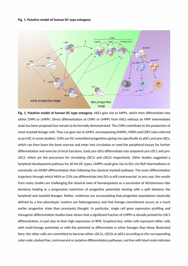
Fig. 1. Putative model of human DC type ontogeny.
Fig. 1. Putative model of human DC type ontogeny. HSCs give rise to MPPs, which then differentiate into
either CMPs or LMPPs. Direct differentiation of CMPs or LMPPs from HSCs without an MPP intermediate
state has been proposed but remain to be formally demonstrated. The CMPs contribute to the production of
most myeloid lineage cells. They can give rise to GMPs, encompassing GMDPs, MDPs and CDPs (also referred
as pro-DC in some studies). CDPs are DC committed progenitors giving rise specifically to pDCs and pre-cDCs,
which can then leave the bone marrow and enter into circulation or seed the peripheral tissues for further
differentiation and exercise of local functions. Early pre-cDCs differentiate into unipotent pre-cDC1 and pre-
cDC2, which are the precursors for circulating cDC1s and cDC2s respectively. Other studies suggested a
lymphoid development pathway for all the DC types. LMPPs could give rise to DCs via MLP intermediates or
eventually via GMDP differentiation then following the classical myeloid pathway. The exact differentiation
trajectory through which MLPs or CLPs can differentiate into DCs is still controversial. In any case, the results
from many studies are challenging the classical view of hematopoiesis as a succession of dichotomous fate
decisions leading to a progressive restriction of progenitor potentials starting with a split between the
lymphoid and myeloid lineages. Rather, evidences are accumulating that progenitor populations classically
defined by a few phenotypic markers are heterogeneous and that lineage commitment occurs at a much
earlier progenitor state than previously thought. In particular, single cell gene expression profiling and
clonogenic differentiation studies have shown that a significant fraction of LMPPs is already primed for cDC1
differentiation, in part due to their high expression of IRF8. Graphical key: white cells represent either cells
with multi-lineage potentials or with the potential to differentiate in other lineages than those illustrated
here; the other cells are committed to become either cDC1s, cDC2s or pDCs according to the corresponding
color code; dashed line, controversial or putative differentiation pathways; red line with blunt ends indicates

progenitors that are thought not to interconvert into one another. Abbreviations: CDP, common DC
progenitor; CLP, common lymphoid progenitor; CMP, common myeloid progenitor; GMDP, granulocyte-
monocyte-DC progenitor; GMP, granulocyte-monocyte progenitor; HSC, hematopoietic stem cell; LMPP,
Lymphoid-primed multipotent progenitor; MDP, monocyte-DC progenitor; MLP, multi-lymphoid progenitor;
MPP, multipotent progenitor; pre-cDC, precursor of conventional DC.
References.
Alcantara-Hernandez, M., Leylek, R., Wagar, L.E., Engleman, E.G., Keler, T., Marinkovich, M.P., Davis, M.M., Nolan, G.P., Idoyaga, J., 2017. High-Dimensional Phenotypic Mapping of Human Dendritic Cells Reveals Interindividual Variation and Tissue Specialization. Immunity 47, 1037-1050 e1036.
Alculumbre, S.G., Saint-Andre, V., Di Domizio, J., Vargas, P., Sirven, P., Bost, P., Maurin, M., Maiuri, P., Wery, M., Roman, M.S., Savey, L., Touzot, M., Terrier, B., Saadoun, D., Conrad, C., Gilliet, M., Morillon, A., Soumelis, V., 2018. Diversification of human plasmacytoid predendritic cells in response to a single stimulus. Nat Immunol 19, 63-75.
Aliberti, J., Schulz, O., Pennington, D.J., Tsujimura, H., Reis e Sousa, C., Ozato, K., Sher, A., 2003. Essential role for ICSBP in the in vivo development of murine CD8alpha + dendritic cells. Blood 101, 305-310.
Allman, D., Dalod, M., Asselin-Paturel, C., Delale, T., Robbins, S.H., Trinchieri, G., Biron, C.A., Kastner, P., Chan, S., 2006. Ikaros is required for plasmacytoid dendritic cell differentiation. Blood 108, 4025-4034.
Ardouin, L., Luche, H., Chelbi, R., Carpentier, S., Shawket, A., Montanana Sanchis, F., Santa Maria, C., Grenot, P., Alexandre, Y., Gregoire, C., Fries, A., Vu Manh, T.P., Tamoutounour, S., Crozat, K., Tomasello, E., Jorquera, A., Fossum, E., Bogen, B., Azukizawa, H., Bajenoff, M., Henri, S., Dalod, M., Malissen, B., 2016. Broad and Largely Concordant Molecular Changes Characterize Tolerogenic and Immunogenic Dendritic Cell Maturation in Thymus and Periphery. Immunity 45, 305-318.
Asselin-Paturel, C., Boonstra, A., Dalod, M., Durand, I., Yessaad, N., Dezutter-Dambuyant, C., Vicari, A., O'Garra, A., Biron, C., Briere, F., Trinchieri, G., 2001. Mouse type I IFN-producing cells are immature APCs with plasmacytoid morphology. Nat Immunol 2, 1144-1150.
Auffray, C., Fogg, D.K., Narni-Mancinelli, E., Senechal, B., Trouillet, C., Saederup, N., Leemput, J., Bigot, K., Campisi, L., Abitbol, M., Molina, T., Charo, I., Hume, D.A., Cumano, A., Lauvau, G., Geissmann, F., 2009. CX3CR1+ CD115+ CD135+ common macrophage/DC precursors and the role of CX3CR1 in their response to inflammation. J Exp Med 206, 595-606.
Bachem, A., Guttler, S., Hartung, E., Ebstein, F., Schaefer, M., Tannert, A., Salama, A., Movassaghi, K., Opitz, C., Mages, H.W., Henn, V., Kloetzel, P.M., Gurka, S., Kroczek, R.A., 2010. Superior antigen cross-presentation and XCR1 expression define human CD11c+CD141+ cells as homologues of mouse CD8+ dendritic cells. J Exp Med 207, 1273-1281.
Bagadia, P., Huang, X., Liu, T.T., Durai, V., Grajales-Reyes, G.E., Nitschke, M., Modrusan, Z., Granja, J.M., Satpathy, A.T., Briseno, C.G., Gargaro, M., Iwata, A., Kim, S., Chang, H.Y., Shaw, A.S., Murphy, T.L., Murphy, K.M., 2019. An Nfil3-Zeb2-Id2 pathway imposes Irf8 enhancer switching during cDC1 development. Nat Immunol 20, 1174-1185.
Bakdash, G., Buschow, S.I., Gorris, M.A., Halilovic, A., Hato, S.V., Skold, A.E., Schreibelt, G., Sittig, S.P., Torensma, R., Duiveman-de Boer, T., Schroder, C., Smits, E.L., Figdor, C.G., de Vries, I.J., 2016. Expansion of a BDCA1+CD14+ Myeloid Cell Population in Melanoma Patients May Attenuate the Efficacy of Dendritic Cell Vaccines. Cancer Res 76, 4332-4346.
Bakri, Y., Sarrazin, S., Mayer, U.P., Tillmanns, S., Nerlov, C., Boned, A., Sieweke, M.H., 2005. Balance of MafB and PU.1 specifies alternative macrophage or dendritic cell fate. Blood 105, 2707-2716.
Balan, S., Arnold-Schrauf, C., Abbas, A., Couespel, N., Savoret, J., Imperatore, F., Villani, A.C., Manh, T.P.V., Bhardwaj, N., Dalod, M., 2018. Large-Scale Human Dendritic Cell Differentiation Revealing Notch-Dependent Lineage Bifurcation and Heterogeneity. Cell Reports 24, 1902-+.
Balan, S., Dalod, M., 2016. In Vitro Generation of Human XCR1(+) Dendritic Cells from CD34(+) Hematopoietic Progenitors, in: Segura, E., Onai, N. (Eds.), Dendritic Cell Protocols, 3rd Edition. Humana Press Inc, Totowa, pp. 19-37.
Balan, S., Kale, V.P., Limaye, L.S., 2009. A simple two-step culture system for the large-scale generation of mature and functional dendritic cells from umbilical cord blood CD34+ cells. Transfusion 49, 2109-2121.

Balan, S., Kale, V.P., Limaye, L.S., 2010. A large number of mature and functional dendritic cells can be efficiently generated from umbilical cord blood-derived mononuclear cells by a simple two-step culture method. Transfusion 50, 2413-2423.
Balan, S., Ollion, V., Colletti, N., Chelbi, R., Montanana-Sanchis, F., Liu, H., Vu Manh, T.P., Sanchez, C., Savoret, J., Perrot, I., Doffin, A.C., Fossum, E., Bechlian, D., Chabannon, C., Bogen, B., Asselin-Paturel, C., Shaw, M., Soos, T., Caux, C., Valladeau-Guilemond, J., Dalod, M., 2014. Human XCR1+ dendritic cells derived in vitro from CD34+ progenitors closely resemble blood dendritic cells, including their adjuvant responsiveness, contrary to monocyte-derived dendritic cells. J Immunol 193, 1622-1635.
Bjorck, P., 2001. Isolation and characterization of plasmacytoid dendritic cells from Flt3 ligand and granulocyte-macrophage colony-stimulating factor-treated mice. Blood 98, 3520-3526.
Blom, B., Ho, S., Antonenko, S., Liu, Y.J., 2000. Generation of interferon alpha-producing predendritic cell (Pre-DC)2 from human CD34(+) hematopoietic stem cells. J Exp Med 192, 1785-1796.
Boitano, A.E., Wang, J., Romeo, R., Bouchez, L.C., Parker, A.E., Sutton, S.E., Walker, J.R., Flaveny, C.A., Perdew, G.H., Denison, M.S., Schultz, P.G., Cooke, M.P., 2010. Aryl hydrocarbon receptor antagonists promote the expansion of human hematopoietic stem cells. Science 329, 1345-1348.
Bol, K.F., Schreibelt, G., Rabold, K., Wculek, S.K., Schwarze, J.K., Dzionek, A., Teijeira, A., Kandalaft, L.E., Romero, P., Coukos, G., Neyns, B., Sancho, D., Melero, I., de Vries, I.J.M., 2019. The clinical application of cancer immunotherapy based on naturally circulating dendritic cells. J Immunother Cancer 7, 109.
Bosteels, C., Scott, C.L., 2020. Transcriptional regulation of DC fate specification. Mol Immunol 121, 38-46.
Breton, G., Lee, J., Liu, K., Nussenzweig, M.C., 2015a. Defining human dendritic cell progenitors by multiparametric flow cytometry. Nat Protoc 10, 1407-1422.
Breton, G., Lee, J., Zhou, Y.J., Schreiber, J.J., Keler, T., Puhr, S., Anandasabapathy, N., Schlesinger, S., Caskey, M., Liu, K., Nussenzweig, M.C., 2015b. Circulating precursors of human CD1c+ and CD141+ dendritic cells. J Exp Med 212, 401-413.
Breton, G., Zheng, S., Valieris, R., Tojal da Silva, I., Satija, R., Nussenzweig, M.C., 2016. Human dendritic cells (DCs) are derived from distinct circulating precursors that are precommitted to become CD1c+ or CD141+ DCs. J Exp Med 213, 2861-2870.
Brezar, V., Ruffin, N., Richert, L., Surenaud, M., Lacabaratz, C., Palucka, K., Thiebaut, R., Banchereau, J., Levy, Y., Seddiki, N., 2015. Decreased HIV-specific T-regulatory responses are associated with effective DC-vaccine induced immunity. PLoS Pathog 11, e1004752.
Brown, C.C., Gudjonson, H., Pritykin, Y., Deep, D., Lavallee, V.P., Mendoza, A., Fromme, R., Mazutis, L., Ariyan, C., Leslie, C., Pe'er, D., Rudensky, A.Y., 2019. Transcriptional Basis of Mouse and Human Dendritic Cell Heterogeneity. Cell 179, 846-863 e824.
Cancel, J.C., Crozat, K., Dalod, M., Mattiuz, R., 2019. Are Conventional Type 1 Dendritic Cells Critical for Protective Antitumor Immunity and How? Front Immunol 10, 9.
Carpentier, S., Vu Manh, T.P., Chelbi, R., Henri, S., Malissen, B., Haniffa, M., Ginhoux, F., Dalod, M., 2016. Comparative genomics analysis of mononuclear phagocyte subsets confirms homology between lymphoid tissue-resident and dermal XCR1(+) DCs in mouse and human and distinguishes them from Langerhans cells. J Immunol Methods 432, 35-49.
Caux, C., Dezutter-Dambuyant, C., Schmitt, D., Banchereau, J., 1992. GM-CSF and TNF-alpha cooperate in the generation of dendritic Langerhans cells. Nature 360, 258-261.
Caux, C., Massacrier, C., Dubois, B., Valladeau, J., Dezutter-Dambuyant, C., Durand, I., Schmitt, D., Saeland, S., 1999. Respective involvement of TGF-beta and IL-4 in the development of Langerhans cells and non-Langerhans dendritic cells from CD34+ progenitors. J Leukoc Biol 66, 781-791.
Caux, C., Massacrier, C., Vanbervliet, B., Dubois, B., Durand, I., Cella, M., Lanzavecchia, A., Banchereau, J., 1997. CD34+ hematopoietic progenitors from human cord blood differentiate along two independent

dendritic cell pathways in response to granulocyte-macrophage colony-stimulating factor plus tumor necrosis factor alpha: II. Functional analysis. Blood 90, 1458-1470.
Caux, C., Vanbervliet, B., Massacrier, C., Dezutter-Dambuyant, C., de Saint-Vis, B., Jacquet, C., Yoneda, K., Imamura, S., Schmitt, D., Banchereau, J., 1996. CD34+ hematopoietic progenitors from human cord blood differentiate along two independent dendritic cell pathways in response to GM-CSF+TNF alpha. J Exp Med 184, 695-706.
Chen, W., Antonenko, S., Sederstrom, J.M., Liang, X., Chan, A.S., Kanzler, H., Blom, B., Blazar, B.R., Liu, Y.J., 2004. Thrombopoietin cooperates with FLT3-ligand in the generation of plasmacytoid dendritic cell precursors from human hematopoietic progenitors. Blood 103, 2547-2553.
Chicha, L., Jarrossay, D., Manz, M.G., 2004. Clonal type I interferon-producing and dendritic cell precursors are contained in both human lymphoid and myeloid progenitor populations. J Exp Med 200, 1519-1524.
Ciancanelli, M.J., Huang, S.X., Luthra, P., Garner, H., Itan, Y., Volpi, S., Lafaille, F.G., Trouillet, C., Schmolke, M., Albrecht, R.A., Israelsson, E., Lim, H.K., Casadio, M., Hermesh, T., Lorenzo, L., Leung, L.W., Pedergnana, V., Boisson, B., Okada, S., Picard, C., Ringuier, B., Troussier, F., Chaussabel, D., Abel, L., Pellier, I., Notarangelo, L.D., Garcia-Sastre, A., Basler, C.F., Geissmann, F., Zhang, S.Y., Snoeck, H.W., Casanova, J.L., 2015. Infectious disease. Life-threatening influenza and impaired interferon amplification in human IRF7 deficiency. Science 348, 448-453.
Cisse, B., Caton, M.L., Lehner, M., Maeda, T., Scheu, S., Locksley, R., Holmberg, D., Zweier, C., den Hollander, N.S., Kant, S.G., Holter, W., Rauch, A., Zhuang, Y., Reizis, B., 2008. Transcription factor E2-2 is an essential and specific regulator of plasmacytoid dendritic cell development. Cell 135, 37-48.
Coates, P.T., Barratt-Boyes, S.M., Zhang, L., Donnenberg, V.S., O'Connell, P.J., Logar, A.J., Duncan, F.J., Murphey-Corb, M., Donnenberg, A.D., Morelli, A.E., Maliszewski, C.R., Thomson, A.W., 2003. Dendritic cell subsets in blood and lymphoid tissue of rhesus monkeys and their mobilization with Flt3 ligand. Blood 102, 2513-2521.
Coelho, A.V., de Moura, R.R., Kamada, A.J., da Silva, R.C., Guimaraes, R.L., Brandao, L.A., de Alencar, L.C., Crovella, S., 2016. Dendritic Cell-Based Immunotherapies to Fight HIV: How Far from a Success Story? A Systematic Review and Meta-Analysis. Int J Mol Sci 17.
Corcoran, L., Ferrero, I., Vremec, D., Lucas, K., Waithman, J., O'Keeffe, M., Wu, L., Wilson, A., Shortman, K., 2003. The lymphoid past of mouse plasmacytoid cells and thymic dendritic cells. J Immunol 170, 4926-4932.
Crozat, K., Guiton, R., Contreras, V., Feuillet, V., Dutertre, C.A., Ventre, E., Vu Manh, T.P., Baranek, T., Storset, A.K., Marvel, J., Boudinot, P., Hosmalin, A., Schwartz-Cornil, I., Dalod, M., 2010a. The XC chemokine receptor 1 is a conserved selective marker of mammalian cells homologous to mouse CD8alpha+ dendritic cells. J Exp Med 207, 1283-1292.
Crozat, K., Guiton, R., Guilliams, M., Henri, S., Baranek, T., Schwartz-Cornil, I., Malissen, B., Dalod, M., 2010b. Comparative genomics as a tool to reveal functional equivalences between human and mouse dendritic cell subsets. Immunol Rev 234, 177-198.
Cytlak, U., Resteu, A., Bogaert, D., Kuehn, H.S., Altmann, T., Gennery, A., Jackson, G., Kumanovics, A., Voelkerding, K.V., Prader, S., Dullaers, M., Reichenbach, J., Hill, H., Haerynck, F., Rosenzweig, S.D., Collin, M., Bigley, V., 2018. Ikaros family zinc finger 1 regulates dendritic cell development and function in humans. Nat Commun 9, 1239.
Cytlak, U., Resteu, A., Pagan, S., Green, K., Milne, P., Maisuria, S., McDonald, D., Hulme, G., Filby, A., Carpenter, B., Queen, R., Hambleton, S., Hague, R., Allen, H.L., Thaventhiran, J., Doody, G., Collin, M., Bigley, V., 2019. Differential IRF8 Requirement Defines Two Pathways of Dendritic Cell Development in Humans. SSRN.
Dalod, M., Chelbi, R., Malissen, B., Lawrence, T., 2014. Dendritic cell maturation: functional specialization through signaling specificity and transcriptional programming. EMBO J 33, 1104-1116.
Di Blasio, S., Tazzari, M., van Wigcheren, G., van Duffelen, A., Stefanini, I., Bloemendal, M., Gorris, M., Vasaturo, A., Bakdash, G., Hato, S.V., Schalkwijk, J., de Vries, I.J.M., van den Bogaard, E.H., Figdor, C.G.,

2019. The tumour microenvironment shapes dendritic cell plasticity in a human organotypic melanoma culture. BioRxiv.
Dontje, W., Schotte, R., Cupedo, T., Nagasawa, M., Scheeren, F., Gimeno, R., Spits, H., Blom, B., 2006. Delta-like1-induced Notch1 signaling regulates the human plasmacytoid dendritic cell versus T-cell lineage decision through control of GATA-3 and Spi-B. Blood 107, 2446-2452.
Doulatov, S., Notta, F., Eppert, K., Nguyen, L.T., Ohashi, P.S., Dick, J.E., 2010. Revised map of the human progenitor hierarchy shows the origin of macrophages and dendritic cells in early lymphoid development. Nat Immunol 11, 585-593.
Drakes, M.L., Lu, L., Subbotin, V.M., Thomson, A.W., 1997. In vivo administration of flt3 ligand markedly stimulates generation of dendritic cell progenitors from mouse liver. J Immunol 159, 4268-4278.
Dress, R.J., Dutertre, C.A., Giladi, A., Schlitzer, A., Low, I., Shadan, N.B., Tay, A., Lum, J., Kairi, M., Hwang, Y.Y., Becht, E., Cheng, Y., Chevrier, M., Larbi, A., Newell, E.W., Amit, I., Chen, J., Ginhoux, F., 2019. Plasmacytoid dendritic cells develop from Ly6D(+) lymphoid progenitors distinct from the myeloid lineage. Nat Immunol 20, 852-864.
Durai, V., Bagadia, P., Granja, J.M., Satpathy, A.T., Kulkarni, D.H., Davidson, J.T.t., Wu, R., Patel, S.J., Iwata, A., Liu, T.T., Huang, X., Briseno, C.G., Grajales-Reyes, G.E., Wohner, M., Tagoh, H., Kee, B.L., Newberry, R.D., Busslinger, M., Chang, H.Y., Murphy, T.L., Murphy, K.M., 2019. Cryptic activation of an Irf8 enhancer governs cDC1 fate specification. Nat Immunol 20, 1161-1173.
Durai, V., Murphy, K.M., 2016. Functions of Murine Dendritic Cells. Immunity 45, 719-736.
Dutertre, C.A., Becht, E., Irac, S.E., Khalilnezhad, A., Narang, V., Khalilnezhad, S., Ng, P.Y., van den Hoogen, L.L., Leong, J.Y., Lee, B., Chevrier, M., Zhang, X.M., Yong, P.J.A., Koh, G., Lum, J., Howland, S.W., Mok, E., Chen, J., Larbi, A., Tan, H.K.K., Lim, T.K.H., Karagianni, P., Tzioufas, A.G., Malleret, B., Brody, J., Albani, S., van Roon, J., Radstake, T., Newell, E.W., Ginhoux, F., 2019. Single-Cell Analysis of Human Mononuclear Phagocytes Reveals Subset-Defining Markers and Identifies Circulating Inflammatory Dendritic Cells. Immunity 51, 573-589 e578.
Dutertre, C.A., Wang, L.F., Ginhoux, F., 2014. Aligning bona fide dendritic cell populations across species. Cell Immunol 291, 3-10.
Dzionek, A., Fuchs, A., Schmidt, P., Cremer, S., Zysk, M., Miltenyi, S., Buck, D.W., Schmitz, J., 2000. BDCA-2, BDCA-3, and BDCA-4: three markers for distinct subsets of dendritic cells in human peripheral blood. J Immunol 165, 6037-6046.
Findlay, E.G., Currie, A.J., Zhang, A., Ovciarikova, J., Young, L., Stevens, H., McHugh, B.J., Canel, M., Gray, M., Milling, S.W.F., Campbell, J.D.M., Savill, J., Serrels, A., Davidson, D.J., 2019. Exposure to the antimicrobial peptide LL-37 produces dendritic cells optimized for immunotherapy. Oncoimmunology 8, 1608106.
Fogg, D.K., Sibon, C., Miled, C., Jung, S., Aucouturier, P., Littman, D.R., Cumano, A., Geissmann, F., 2006. A clonogenic bone marrow progenitor specific for macrophages and dendritic cells. Science 311, 83-87.
Fries, A., Dalod, M., 2016. Dendritic Cells in Viral Infection., Encyclopedia of Immunobiology, pp. 207-221.
Furie, R., Werth, V.P., Merola, J.F., Stevenson, L., Reynolds, T.L., Naik, H., Wang, W., Christmann, R., Gardet, A., Pellerin, A., Hamann, S., Auluck, P., Barbey, C., Gulati, P., Rabah, D., Franchimont, N., 2019. Monoclonal antibody targeting BDCA2 ameliorates skin lesions in systemic lupus erythematosus. J Clin Invest 129, 1359-1371.
Garcia, F., Climent, N., Guardo, A.C., Gil, C., Leon, A., Autran, B., Lifson, J.D., Martinez-Picado, J., Dalmau, J., Clotet, B., Gatell, J.M., Plana, M., Gallart, T., Group, D.M.O.S., 2013. A dendritic cell-based vaccine elicits T cell responses associated with control of HIV-1 replication. Sci Transl Med 5, 166ra162.
Gilliet, M., Boonstra, A., Paturel, C., Antonenko, S., Xu, X.L., Trinchieri, G., O'Garra, A., Liu, Y.J., 2002. The development of murine plasmacytoid dendritic cell precursors is differentially regulated by FLT3-ligand and granulocyte/macrophage colony-stimulating factor. J Exp Med 195, 953-958.

Goudot, C., Coillard, A., Villani, A.C., Gueguen, P., Cros, A., Sarkizova, S., Tang-Huau, T.L., Bohec, M., Baulande, S., Hacohen, N., Amigorena, S., Segura, E., 2017. Aryl Hydrocarbon Receptor Controls Monocyte Differentiation into Dendritic Cells versus Macrophages. Immunity 47, 582-596 e586.
Grajales-Reyes, G.E., Iwata, A., Albring, J., Wu, X., Tussiwand, R., Kc, W., Kretzer, N.M., Briseno, C.G., Durai, V., Bagadia, P., Haldar, M., Schonheit, J., Rosenbauer, F., Murphy, T.L., Murphy, K.M., 2015. Batf3 maintains autoactivation of Irf8 for commitment of a CD8alpha(+) conventional DC clonogenic progenitor. Nat Immunol 16, 708-717.
Grouard, G., Rissoan, M.C., Filgueira, L., Durand, I., Banchereau, J., Liu, Y.J., 1997. The enigmatic plasmacytoid T cells develop into dendritic cells with interleukin (IL)-3 and CD40-ligand. J Exp Med 185, 1101-1111.
Guilliams, M., Dutertre, C.A., Scott, C.L., McGovern, N., Sichien, D., Chakarov, S., Van Gassen, S., Chen, J., Poidinger, M., De Prijck, S., Tavernier, S.J., Low, I., Irac, S.E., Mattar, C.N., Sumatoh, H.R., Low, G.H.L., Chung, T.J.K., Chan, D.K.H., Tan, K.K., Hon, T.L.K., Fossum, E., Bogen, B., Choolani, M., Chan, J.K.Y., Larbi, A., Luche, H., Henri, S., Saeys, Y., Newell, E.W., Lambrecht, B.N., Malissen, B., Ginhoux, F., 2016. Unsupervised High-Dimensional Analysis Aligns Dendritic Cells across Tissues and Species. Immunity 45, 669-684.
Guilliams, M., Ginhoux, F., Jakubzick, C., Naik, S.H., Onai, N., Schraml, B.U., Segura, E., Tussiwand, R., Yona, S., 2014. Dendritic cells, monocytes and macrophages: a unified nomenclature based on ontogeny. Nat Rev Immunol 14, 571-578.
Guilliams, M., Henri, S., Tamoutounour, S., Ardouin, L., Schwartz-Cornil, I., Dalod, M., Malissen, B., 2010. From skin dendritic cells to a simplified classification of human and mouse dendritic cell subsets. Eur J Immunol 40, 2089-2094.
Haddad, R., Guardiola, P., Izac, B., Thibault, C., Radich, J., Delezoide, A.L., Baillou, C., Lemoine, F.M., Gluckman, J.C., Pflumio, F., Canque, B., 2004. Molecular characterization of early human T/NK and B-lymphoid progenitor cells in umbilical cord blood. Blood 104, 3918-3926.
Haniffa, M., Shin, A., Bigley, V., McGovern, N., Teo, P., See, P., Wasan, P.S., Wang, X.N., Malinarich, F., Malleret, B., Larbi, A., Tan, P., Zhao, H., Poidinger, M., Pagan, S., Cookson, S., Dickinson, R., Dimmick, I., Jarrett, R.F., Renia, L., Tam, J., Song, C., Connolly, J., Chan, J.K., Gehring, A., Bertoletti, A., Collin, M., Ginhoux, F., 2012. Human tissues contain CD141hi cross-presenting dendritic cells with functional homology to mouse CD103+ nonlymphoid dendritic cells. Immunity 37, 60-73.
Heger, L., Balk, S., Luhr, J.J., Heidkamp, G.F., Lehmann, C.H.K., Hatscher, L., Purbojo, A., Hartmann, A., Garcia-Martin, F., Nishimura, S.I., Cesnjevar, R., Nimmerjahn, F., Dudziak, D., 2018. CLEC10A Is a Specific Marker for Human CD1c(+) Dendritic Cells and Enhances Their Toll-Like Receptor 7/8-Induced Cytokine Secretion. Front Immunol 9, 744.
Helft, J., Anjos-Afonso, F., van der Veen, A.G., Chakravarty, P., Bonnet, D., Sousa, C.R.E., 2017. Dendritic Cell Lineage Potential in Human Early Hematopoietic Progenitors. Cell Reports 20, 529-537.
Hettinger, J., Richards, D.M., Hansson, J., Barra, M.M., Joschko, A.C., Krijgsveld, J., Feuerer, M., 2013. Origin of monocytes and macrophages in a committed progenitor. Nat Immunol 14, 821-830.
Jongbloed, S.L., Kassianos, A.J., McDonald, K.J., Clark, G.J., Ju, X., Angel, C.E., Chen, C.J., Dunbar, P.R., Wadley, R.B., Jeet, V., Vulink, A.J., Hart, D.N., Radford, K.J., 2010. Human CD141+ (BDCA-3)+ dendritic cells (DCs) represent a unique myeloid DC subset that cross-presents necrotic cell antigens. J Exp Med 207, 1247-1260.
Kashem, S.W., Haniffa, M., Kaplan, D.H., 2017. Antigen-Presenting Cells in the Skin. Annu Rev Immunol 35, 469-499.
Kim, S.J., Kim, G., Kim, N., Chu, H., Park, B.C., Yang, J.S., Han, S.H., Yun, C.H., 2019. Human CD141(+) dendritic cells generated from adult peripheral blood monocytes. Cytotherapy.
Kirkling, M.E., Cytlak, U., Lau, C.M., Lewis, K.L., Resteu, A., Khodadadi-Jamayran, A., Siebel, C.W., Salmon, H., Merad, M., Tsirigos, A., Collin, M., Bigley, V., Reizis, B., 2018. Notch Signaling Facilitates In Vitro Generation of Cross-Presenting Classical Dendritic Cells. Cell Reports 23, 3658-+.

Klechevsky, E., Morita, R., Liu, M., Cao, Y., Coquery, S., Thompson-Snipes, L., Briere, F., Chaussabel, D., Zurawski, G., Palucka, A.K., Reiter, Y., Banchereau, J., Ueno, H., 2008. Functional specializations of human epidermal Langerhans cells and CD14+ dermal dendritic cells. Immunity 29, 497-510.
Kong, X.F., Martinez-Barricarte, R., Kennedy, J., Mele, F., Lazarov, T., Deenick, E.K., Ma, C.S., Breton, G., Lucero, K.B., Langlais, D., Bousfiha, A., Aytekin, C., Markle, J., Trouillet, C., Jabot-Hanin, F., Arlehamn, C.S.L., Rao, G., Picard, C., Lasseau, T., Latorre, D., Hambleton, S., Deswarte, C., Itan, Y., Abarca, K., Moraes-Vasconcelos, D., Ailal, F., Ikinciogullari, A., Dogu, F., Benhsaien, I., Sette, A., Abel, L., Boisson-Dupuis, S., Schroder, B., Nussenzweig, M.C., Liu, K., Geissmann, F., Tangye, S.G., Gros, P., Sallusto, F., Bustamante, J., Casanova, J.L., 2018. Disruption of an antimycobacterial circuit between dendritic and helper T cells in human SPPL2a deficiency. Nat Immunol 19, 973-985.
La Motte-Mohs, R.N., Herer, E., Zuniga-Pflucker, J.C., 2005. Induction of T-cell development from human cord blood hematopoietic stem cells by Delta-like 1 in vitro. Blood 105, 1431-1439.
Laustsen, A., Bak, R.O., Krapp, C., Kjaer, L., Egedahl, J.H., Petersen, C.C., Pillai, S., Tang, H.Q., Uldbjerg, N., Porteus, M., Roan, N.R., Nyegaard, M., Denton, P.W., Jakobsen, M.R., 2018. Interferon priming is essential for human CD34+cell-derived plasmacytoid dendritic cell maturation and function. Nature Communications 9.
Lee, J., Breton, G., Aljoufi, A., Zhou, Y.J., Puhr, S., Nussenzweig, M.C., Liu, K., 2015a. Clonal analysis of human dendritic cell progenitor using a stromal cell culture. J Immunol Methods 425, 21-26.
Lee, J., Breton, G., Oliveira, T.Y., Zhou, Y.J., Aljoufi, A., Puhr, S., Cameron, M.J., Sekaly, R.P., Nussenzweig, M.C., Liu, K., 2015b. Restricted dendritic cell and monocyte progenitors in human cord blood and bone marrow. J Exp Med 212, 385-399.
Lee, J., Zhou, Y.J., Ma, W., Zhang, W., Aljoufi, A., Luh, T., Lucero, K., Liang, D., Thomsen, M., Bhagat, G., Shen, Y., Liu, K., 2017. Lineage specification of human dendritic cells is marked by IRF8 expression in hematopoietic stem cells and multipotent progenitors. Nat Immunol 18, 877-888.
Levy, Y., Thiebaut, R., Montes, M., Lacabaratz, C., Sloan, L., King, B., Perusat, S., Harrod, C., Cobb, A., Roberts, L.K., Surenaud, M., Boucherie, C., Zurawski, S., Delaugerre, C., Richert, L., Chene, G., Banchereau, J., Palucka, K., 2014. Dendritic cell-based therapeutic vaccine elicits polyfunctional HIV-specific T-cell immunity associated with control of viral load. Eur J Immunol 44, 2802-2810.
Leylek, R., Alcantara-Hernandez, M., Lanzar, Z., Ludtke, A., Perez, O.A., Reizis, B., Idoyaga, J., 2019. Integrated Cross-Species Analysis Identifies a Conserved Transitional Dendritic Cell Population. Cell Rep 29, 3736-3750 e3738.
Lin, D.S., Kan, A., Gao, J., Crampin, E.J., Hodgkin, P.D., Naik, S.H., 2018. DiSNE Movie Visualization and Assessment of Clonal Kinetics Reveal Multiple Trajectories of Dendritic Cell Development. Cell Rep 22, 2557-2566.
Liu, Z., Gu, Y., Chakarov, S., Bleriot, C., Kwok, I., Chen, X., Shin, A., Huang, W., Dress, R.J., Dutertre, C.A., Schlitzer, A., Chen, J., Ng, L.G., Wang, H., Liu, Z., Su, B., Ginhoux, F., 2019. Fate Mapping via Ms4a3-Expression History Traces Monocyte-Derived Cells. Cell 178, 1509-1525 e1519.
Lundberg, K., Albrekt, A.S., Nelissen, I., Santegoets, S., de Gruijl, T.D., Gibbs, S., Lindstedt, M., 2013. Transcriptional profiling of human dendritic cell populations and models--unique profiles of in vitro dendritic cells and implications on functionality and applicability. PLoS One 8, e52875.
Ma, W., Lee, J., Backenroth, D., Zhou, Y.J., Bush, E., Sims, P., Liu, K., Shen, Y., 2019. Single cell RNA-Seq reveals pre-cDCs fate determined by transcription factor combinatorial dose. BMC Mol Cell Biol 20, 20.
Maraskovsky, E., Brasel, K., Teepe, M., Roux, E.R., Lyman, S.D., Shortman, K., McKenna, H.J., 1996. Dramatic increase in the numbers of functionally mature dendritic cells in Flt3 ligand-treated mice: multiple dendritic cell subpopulations identified. J Exp Med 184, 1953-1962.
Marin, E., Bouchet-Delbos, L., Renoult, O., Louvet, C., Nerriere-Daguin, V., Managh, A.J., Even, A., Giraud, M., Vu Manh, T.P., Aguesse, A., Beriou, G., Chiffoleau, E., Alliot-Licht, B., Prieur, X., Croyal, M., Hutchinson, J.A.,

Obermajer, N., Geissler, E.K., Vanhove, B., Blancho, G., Dalod, M., Josien, R., Pecqueur, C., Cuturi, M.C., Moreau, A., 2019. Human Tolerogenic Dendritic Cells Regulate Immune Responses through Lactate Synthesis. Cell Metab 30, 1075-1090 e1078.
Marin, E., Cuturi, M.C., Moreau, A., 2018. Tolerogenic Dendritic Cells in Solid Organ Transplantation: Where Do We Stand? Front Immunol 9, 274.
Mattei, F., Schiavoni, G., Borghi, P., Venditti, M., Canini, I., Sestili, P., Pietraforte, I., Morse, H.C., 3rd, Ramoni, C., Belardelli, F., Gabriele, L., 2006. ICSBP/IRF-8 differentially regulates antigen uptake during dendritic-cell development and affects antigen presentation to CD4+ T cells. Blood 108, 609-617.
Meredith, M.M., Liu, K., Darrasse-Jeze, G., Kamphorst, A.O., Schreiber, H.A., Guermonprez, P., Idoyaga, J., Cheong, C., Yao, K.H., Niec, R.E., Nussenzweig, M.C., 2012. Expression of the zinc finger transcription factor zDC (Zbtb46, Btbd4) defines the classical dendritic cell lineage. J Exp Med 209, 1153-1165.
Mildner, A., Jung, S., 2014. Development and function of dendritic cell subsets. Immunity 40, 642-656.
Murray, L.J., Young, J.C., Osborne, L.J., Luens, K.M., Scollay, R., Hill, B.L., 1999. Thrombopoietin, flt3, and kit ligands together suppress apoptosis of human mobilized CD34+ cells and recruit primitive CD34+ Thy-1+ cells into rapid division. Exp Hematol 27, 1019-1028.
Nagasawa, M., Schmidlin, H., Hazekamp, M.G., Schotte, R., Blom, B., 2008. Development of human plasmacytoid dendritic cells depends on the combined action of the basic helix-loop-helix factor E2-2 and the Ets factor Spi-B. Eur J Immunol 38, 2389-2400.
Naik, S.H., Perie, L., Swart, E., Gerlach, C., van Rooij, N., de Boer, R.J., Schumacher, T.N., 2013. Diverse and heritable lineage imprinting of early haematopoietic progenitors. Nature 496, 229-232.
Naik, S.H., Proietto, A.I., Wilson, N.S., Dakic, A., Schnorrer, P., Fuchsberger, M., Lahoud, M.H., O'Keeffe, M., Shao, Q.X., Chen, W.F., Villadangos, J.A., Shortman, K., Wu, L., 2005. Cutting edge: generation of splenic CD8+ and CD8- dendritic cell equivalents in Fms-like tyrosine kinase 3 ligand bone marrow cultures. J Immunol 174, 6592-6597.
Naik, S.H., Sathe, P., Park, H.Y., Metcalf, D., Proietto, A.I., Dakic, A., Carotta, S., O'Keeffe, M., Bahlo, M., Papenfuss, A., Kwak, J.Y., Wu, L., Shortman, K., 2007. Development of plasmacytoid and conventional dendritic cell subtypes from single precursor cells derived in vitro and in vivo. Nat Immunol 8, 1217-1226.
Nakano, H., Yanagita, M., Gunn, M.D., 2001. CD11c(+)B220(+)Gr-1(+) cells in mouse lymph nodes and spleen display characteristics of plasmacytoid dendritic cells. J Exp Med 194, 1171-1178.
Onai, N., Kurabayashi, K., Hosoi-Amaike, M., Toyama-Sorimachi, N., Matsushima, K., Inaba, K., Ohteki, T., 2013. A clonogenic progenitor with prominent plasmacytoid dendritic cell developmental potential. Immunity 38, 943-957.
Onai, N., Obata-Onai, A., Schmid, M.A., Ohteki, T., Jarrossay, D., Manz, M.G., 2007. Identification of clonogenic common Flt3+M-CSFR+ plasmacytoid and conventional dendritic cell progenitors in mouse bone marrow. Nat Immunol 8, 1207-1216.
Paul, F., Arkin, Y., Giladi, A., Jaitin, D.A., Kenigsberg, E., Keren-Shaul, H., Winter, D., Lara-Astiaso, D., Gury, M., Weiner, A., David, E., Cohen, N., Lauridsen, F.K., Haas, S., Schlitzer, A., Mildner, A., Ginhoux, F., Jung, S., Trumpp, A., Porse, B.T., Tanay, A., Amit, I., 2015. Transcriptional Heterogeneity and Lineage Commitment in Myeloid Progenitors. Cell 163, 1663-1677.
Pelka, K., Latz, E., 2013. IRF5, IRF8, and IRF7 in human pDCs - the good, the bad, and the insignificant? Eur J Immunol 43, 1693-1697.
Pham, T.N.Q., Meziane, O., Miah, M.A., Volodina, O., Colas, C., Beland, K., Li, Y., Dallaire, F., Keler, T., Guimond, J.V., Lesage, S., Cheong, C., Haddad, E., Cohen, E.A., 2019. Flt3L-Mediated Expansion of Plasmacytoid Dendritic Cells Suppresses HIV Infection in Humanized Mice. Cell Rep 29, 2770-2782 e2775.
Piacibello, W., Sanavio, F., Garetto, L., Severino, A., Bergandi, D., Ferrario, J., Fagioli, F., Berger, M., Aglietta, M., 1997. Extensive amplification and self-renewal of human primitive hematopoietic stem cells from cord blood. Blood 89, 2644-2653.

Poulin, L.F., Salio, M., Griessinger, E., Anjos-Afonso, F., Craciun, L., Chen, J.L., Keller, A.M., Joffre, O., Zelenay, S., Nye, E., Le Moine, A., Faure, F., Donckier, V., Sancho, D., Cerundolo, V., Bonnet, D., Reis e Sousa, C., 2010. Characterization of human DNGR-1+ BDCA3+ leukocytes as putative equivalents of mouse CD8alpha+ dendritic cells. J Exp Med 207, 1261-1271.
Proietto, A.I., Mittag, D., Roberts, A.W., Sprigg, N., Wu, L., 2012. The equivalents of human blood and spleen dendritic cell subtypes can be generated in vitro from human CD34(+) stem cells in the presence of fms-like tyrosine kinase 3 ligand and thrombopoietin. Cell Mol Immunol 9, 446-454.
Pulendran, B., Banchereau, J., Burkeholder, S., Kraus, E., Guinet, E., Chalouni, C., Caron, D., Maliszewski, C., Davoust, J., Fay, J., Palucka, K., 2000. Flt3-ligand and granulocyte colony-stimulating factor mobilize distinct human dendritic cell subsets in vivo. J Immunol 165, 566-572.
Rawlings, D.J., Quan, S., Hao, Q.L., Thiemann, F.T., Smogorzewska, M., Witte, O.N., Crooks, G.M., 1997. Differentiation of human CD34+CD38- cord blood stem cells into B cell progenitors in vitro. Exp Hematol 25, 66-72.
Reizis, B., 2019. Plasmacytoid Dendritic Cells: Development, Regulation, and Function. Immunity 50, 37-50.
Reynolds, G., Haniffa, M., 2015. Human and Mouse Mononuclear Phagocyte Networks: A Tale of Two Species? Front Immunol 6, 330.
Rissoan, M.C., Duhen, T., Bridon, J.M., Bendriss-Vermare, N., Peronne, C., de Saint Vis, B., Briere, F., Bates, E.E., 2002. Subtractive hybridization reveals the expression of immunoglobulin-like transcript 7, Eph-B1, granzyme B, and 3 novel transcripts in human plasmacytoid dendritic cells. Blood 100, 3295-3303.
Robbins, S.H., Walzer, T., Dembele, D., Thibault, C., Defays, A., Bessou, G., Xu, H., Vivier, E., Sellars, M., Pierre, P., Sharp, F.R., Chan, S., Kastner, P., Dalod, M., 2008. Novel insights into the relationships between dendritic cell subsets in human and mouse revealed by genome-wide expression profiling. Genome Biol 9, R17.
Rodrigues, P.F., Alberti-Servera, L., Eremin, A., Grajales-Reyes, G.E., Ivanek, R., Tussiwand, R., 2018. Distinct progenitor lineages contribute to the heterogeneity of plasmacytoid dendritic cells. Nat Immunol 19, 711-722.
Rosa, F.F., Pires, C.F., Kurochkin, I., Ferreira, A.G., Gomes, A.M., Palma, L.G., Shaiv, K., Solanas, L., Azenha, C., Papatsenko, D., Schulz, O., Reis e Sousa, C., Pereira, C.F., 2018. Direct reprogramming of fibroblasts into antigen-presenting dendritic cells. Sci Immunol 3.
Sachamitr, P., Leishman, A.J., Davies, T.J., Fairchild, P.J., 2017. Directed Differentiation of Human Induced Pluripotent Stem Cells into Dendritic Cells Displaying Tolerogenic Properties and Resembling the CD141(+) Subset. Front Immunol 8, 1935.
Sallusto, F., Lanzavecchia, A., 1994. Efficient presentation of soluble antigen by cultured human dendritic cells is maintained by granulocyte/macrophage colony-stimulating factor plus interleukin 4 and downregulated by tumor necrosis factor alpha. J Exp Med 179, 1109-1118.
Sander, J., Schmidt, S.V., Cirovic, B., McGovern, N., Papantonopoulou, O., Hardt, A.L., Aschenbrenner, A.C., Kreer, C., Quast, T., Xu, A.M., Schmidleithner, L.M., Theis, H., Thi Huong, L.D., Sumatoh, H.R.B., Lauterbach, M.A.R., Schulte-Schrepping, J., Gunther, P., Xue, J., Bassler, K., Ulas, T., Klee, K., Katzmarski, N., Herresthal, S., Krebs, W., Martin, B., Latz, E., Handler, K., Kraut, M., Kolanus, W., Beyer, M., Falk, C.S., Wiegmann, B., Burgdorf, S., Melosh, N.A., Newell, E.W., Ginhoux, F., Schlitzer, A., Schultze, J.L., 2017. Cellular Differentiation of Human Monocytes Is Regulated by Time-Dependent Interleukin-4 Signaling and the Transcriptional Regulator NCOR2. Immunity 47, 1051-1066 e1012.
Sathe, P., Metcalf, D., Vremec, D., Naik, S.H., Langdon, W.Y., Huntington, N.D., Wu, L., Shortman, K., 2014. Lymphoid tissue and plasmacytoid dendritic cells and macrophages do not share a common macrophage-dendritic cell-restricted progenitor. Immunity 41, 104-115.
Satpathy, A.T., Kc, W., Albring, J.C., Edelson, B.T., Kretzer, N.M., Bhattacharya, D., Murphy, T.L., Murphy, K.M., 2012. Zbtb46 expression distinguishes classical dendritic cells and their committed progenitors from other immune lineages. J Exp Med 209, 1135-1152.

Schlitzer, A., Sivakamasundari, V., Chen, J., Sumatoh, H.R., Schreuder, J., Lum, J., Malleret, B., Zhang, S., Larbi, A., Zolezzi, F., Renia, L., Poidinger, M., Naik, S., Newell, E.W., Robson, P., Ginhoux, F., 2015. Identification of cDC1- and cDC2-committed DC progenitors reveals early lineage priming at the common DC progenitor stage in the bone marrow. Nat Immunol 16, 718-728.
Schotte, R., Nagasawa, M., Weijer, K., Spits, H., Blom, B., 2004. The ETS transcription factor Spi-B is required for human plasmacytoid dendritic cell development. J Exp Med 200, 1503-1509.
Schraml, B.U., van Blijswijk, J., Zelenay, S., Whitney, P.G., Filby, A., Acton, S.E., Rogers, N.C., Moncaut, N., Carvajal, J.J., Reis e Sousa, C., 2013. Genetic tracing via DNGR-1 expression history defines dendritic cells as a hematopoietic lineage. Cell 154, 843-858.
Schreibelt, G., Bol, K.F., Westdorp, H., Wimmers, F., Aarntzen, E.H., Duiveman-de Boer, T., van de Rakt, M.W., Scharenborg, N.M., de Boer, A.J., Pots, J.M., Olde Nordkamp, M.A., van Oorschot, T.G., Tel, J., Winkels, G., Petry, K., Blokx, W.A., van Rossum, M.M., Welzen, M.E., Mus, R.D., Croockewit, S.A., Koornstra, R.H., Jacobs, J.F., Kelderman, S., Blank, C.U., Gerritsen, W.R., Punt, C.J., Figdor, C.G., de Vries, I.J., 2016. Effective Clinical Responses in Metastatic Melanoma Patients after Vaccination with Primary Myeloid Dendritic Cells. Clin Cancer Res 22, 2155-2166.
See, P., Dutertre, C.A., Chen, J., Gunther, P., McGovern, N., Irac, S.E., Gunawan, M., Beyer, M., Handler, K., Duan, K., Sumatoh, H.R.B., Ruffin, N., Jouve, M., Gea-Mallorqui, E., Hennekam, R.C.M., Lim, T., Yip, C.C., Wen, M., Malleret, B., Low, I., Shadan, N.B., Fen, C.F.S., Tay, A., Lum, J., Zolezzi, F., Larbi, A., Poidinger, M., Chan, J.K.Y., Chen, Q., Renia, L., Haniffa, M., Benaroch, P., Schlitzer, A., Schultze, J.L., Newell, E.W., Ginhoux, F., 2017. Mapping the human DC lineage through the integration of high-dimensional techniques. Science 356.
Segura, E., Amigorena, S., 2015. Cross-Presentation in Mouse and Human Dendritic Cells. Adv Immunol 127, 1-31.
Segura, E., Touzot, M., Bohineust, A., Cappuccio, A., Chiocchia, G., Hosmalin, A., Dalod, M., Soumelis, V., Amigorena, S., 2013. Human inflammatory dendritic cells induce Th17 cell differentiation. Immunity 38, 336-348.
Shortman, K., Heath, W.R., 2010. The CD8+ dendritic cell subset. Immunol Rev 234, 18-31.
Sichien, D., Scott, C.L., Martens, L., Vanderkerken, M., Van Gassen, S., Plantinga, M., Joeris, T., De Prijck, S., Vanhoutte, L., Vanheerswynghels, M., Van Isterdael, G., Toussaint, W., Madeira, F.B., Vergote, K., Agace, W.W., Clausen, B.E., Hammad, H., Dalod, M., Saeys, Y., Lambrecht, B.N., Guilliams, M., 2016. IRF8 Transcription Factor Controls Survival and Function of Terminally Differentiated Conventional and Plasmacytoid Dendritic Cells, Respectively. Immunity 45, 626-640.
Silk, K.M., Silk, J.D., Ichiryu, N., Davies, T.J., Nolan, K.F., Leishman, A.J., Carpenter, L., Watt, S.M., Cerundolo, V., Fairchild, P.J., 2012. Cross-presentation of tumour antigens by human induced pluripotent stem cell-derived CD141(+)XCR1+ dendritic cells. Gene Ther 19, 1035-1040.
Silvin, A., Yu, C.I., Lahaye, X., Imperatore, F., Brault, J.B., Cardinaud, S., Becker, C., Kwan, W.H., Conrad, C., Maurin, M., Goudot, C., Marques-Ladeira, S., Wang, Y., Pascual, V., Anguiano, E., Albrecht, R.A., Iannacone, M., Garcia-Sastre, A., Goud, B., Dalod, M., Moris, A., Merad, M., Palucka, A.K., Manel, N., 2017. Constitutive resistance to viral infection in human CD141(+) dendritic cells. Sci Immunol 2.
Smith, N., Pietrancosta, N., Davidson, S., Dutrieux, J., Chauveau, L., Cutolo, P., Dy, M., Scott-Algara, D., Manoury, B., Zirafi, O., McCort-Tranchepain, I., Durroux, T., Bachelerie, F., Schwartz, O., Munch, J., Wack, A., Nisole, S., Herbeuval, J.P., 2017. Natural amines inhibit activation of human plasmacytoid dendritic cells through CXCR4 engagement. Nat Commun 8, 14253.
Sontag, S., Forster, M., Qin, J., Wanek, P., Mitzka, S., Schuler, H.M., Koschmieder, S., Rose-John, S., Sere, K., Zenke, M., 2017. Modelling IRF8 Deficient Human Hematopoiesis and Dendritic Cell Development with Engineered iPS Cells. Stem Cells 35, 898-908.
Spits, H., Couwenberg, F., Bakker, A.Q., Weijer, K., Uittenbogaart, C.H., 2000. Id2 and Id3 inhibit development of CD34(+) stem cells into predendritic cell (pre-DC)2 but not into pre-DC1. Evidence for a lymphoid origin of pre-DC2. J Exp Med 192, 1775-1784.
Steinhagen, F., McFarland, A.P., Rodriguez, L.G., Tewary, P., Jarret, A., Savan, R., Klinman, D.M., 2013. IRF-5 and NF-kappaB p50 co-regulate IFN-beta and IL-6 expression in TLR9-stimulated human plasmacytoid dendritic cells. Eur J Immunol 43, 1896-1906.
Steinhagen, F., Rodriguez, L.G., Tross, D., Tewary, P., Bode, C., Klinman, D.M., 2016. IRF5 and IRF8 modulate the CAL-1 human plasmacytoid dendritic cell line response following TLR9 ligation. Eur J Immunol 46, 647-655.
Strobl, H., Bello-Fernandez, C., Riedl, E., Pickl, W.F., Majdic, O., Lyman, S.D., Knapp, W., 1997. flt3 ligand in cooperation with transforming growth factor-beta1 potentiates in vitro development of Langerhans-type dendritic cells and allows single-cell dendritic cell cluster formation under serum-free conditions. Blood 90, 1425-1434.
Strobl, H., Krump, C., Borek, I., 2019. Micro-environmental signals directing human epidermal Langerhans cell differentiation. Semin Cell Dev Biol 86, 36-43.
Strobl, H., Riedl, E., Scheinecker, C., Bello-Fernandez, C., Pickl, W.F., Rappersberger, K., Majdic, O., Knapp, W., 1996. TGF-beta 1 promotes in vitro development of dendritic cells from CD34+ hemopoietic progenitors. J Immunol 157, 1499-1507.
Surenaud, M., Montes, M., Lindestam Arlehamn, C.S., Sette, A., Banchereau, J., Palucka, K., Lelievre, J.D., Lacabaratz, C., Levy, Y., 2019. Anti-HIV potency of T-cell responses elicited by dendritic cell therapeutic vaccination. PLoS Pathog 15, e1008011.
Tacken, P.J., de Vries, I.J., Torensma, R., Figdor, C.G., 2007. Dendritic-cell immunotherapy: from ex vivo loading to in vivo targeting. Nat Rev Immunol 7, 790-802.
Tel, J., Aarntzen, E.H., Baba, T., Schreibelt, G., Schulte, B.M., Benitez-Ribas, D., Boerman, O.C., Croockewit, S., Oyen, W.J., van Rossum, M., Winkels, G., Coulie, P.G., Punt, C.J., Figdor, C.G., de Vries, I.J., 2013. Natural human plasmacytoid dendritic cells induce antigen-specific T-cell responses in melanoma patients. Cancer Res 73, 1063-1075.
Theisen, D.J., Davidson, J.T.t., Briseno, C.G., Gargaro, M., Lauron, E.J., Wang, Q., Desai, P., Durai, V., Bagadia, P., Brickner, J.R., Beatty, W.L., Virgin, H.W., Gillanders, W.E., Mosammaparast, N., Diamond, M.S., Sibley, L.D., Yokoyama, W., Schreiber, R.D., Murphy, T.L., Murphy, K.M., 2018. WDFY4 is required for cross-presentation in response to viral and tumor antigens. Science 362, 694-699.
Thiebaut, R., Hejblum, B.P., Hocini, H., Bonnabau, H., Skinner, J., Montes, M., Lacabaratz, C., Richert, L., Palucka, K., Banchereau, J., Levy, Y., 2019. Gene Expression Signatures Associated With Immune and Virological Responses to Therapeutic Vaccination With Dendritic Cells in HIV-Infected Individuals. Front Immunol 10, 874.
Thomson, A.W., Ezzelarab, M.B., 2018. Regulatory dendritic cells: profiling, targeting, and therapeutic application. Curr Opin Organ Transplant 23, 538-545.
Thordardottir, S., Hangalapura, B.N., Hutten, T., Cossu, M., Spanholtz, J., Schaap, N., Radstake, T.R., van der Voort, R., Dolstra, H., 2014. The aryl hydrocarbon receptor antagonist StemRegenin 1 promotes human plasmacytoid and myeloid dendritic cell development from CD34+ hematopoietic progenitor cells. Stem Cells Dev 23, 955-967.
Thordardottir, S., Schaap, N., Louer, E., Kester, M.G., Falkenburg, J.H., Jansen, J., Radstake, T.R., Hobo, W., Dolstra, H., 2017. Hematopoietic stem cell-derived myeloid and plasmacytoid DC-based vaccines are highly potent inducers of tumor-reactive T cell and NK cell responses ex vivo. Oncoimmunology 6, e1285991.
Tomasello, E., Pollet, E., Vu Manh, T.P., Uze, G., Dalod, M., 2014. Harnessing Mechanistic Knowledge on Beneficial Versus Deleterious IFN-I Effects to Design Innovative Immunotherapies Targeting Cytokine Activity to Specific Cell Types. Front Immunol 5, 526.

Tomita, Y., Watanabe, E., Shimizu, M., Negishi, Y., Kondo, Y., Takahashi, H., 2019. Induction of tumor-specific CD8(+) cytotoxic T lymphocytes from naive human T cells by using Mycobacterium-derived mycolic acid and lipoarabinomannan-stimulated dendritic cells. Cancer Immunol Immunother.
Trombetta, E.S., Mellman, I., 2005. Cell biology of antigen processing in vitro and in vivo. Annu Rev Immunol 23, 975-1028.
Tussiwand, R., Lee, W.L., Murphy, T.L., Mashayekhi, M., Kc, W., Albring, J.C., Satpathy, A.T., Rotondo, J.A., Edelson, B.T., Kretzer, N.M., Wu, X., Weiss, L.A., Glasmacher, E., Li, P., Liao, W., Behnke, M., Lam, S.S., Aurthur, C.T., Leonard, W.J., Singh, H., Stallings, C.L., Sibley, L.D., Schreiber, R.D., Murphy, K.M., 2012. Compensatory dendritic cell development mediated by BATF-IRF interactions. Nature 490, 502-507.
van Helden, S.F., van Leeuwen, F.N., Figdor, C.G., 2008. Human and murine model cell lines for dendritic cell biology evaluated. Immunol Lett 117, 191-197.
Villadangos, J.A., Shortman, K., 2010. Found in translation: the human equivalent of mouse CD8+ dendritic cells. J Exp Med 207, 1131-1134.
Villani, A.C., Satija, R., Reynolds, G., Sarkizova, S., Shekhar, K., Fletcher, J., Griesbeck, M., Butler, A., Zheng, S., Lazo, S., Jardine, L., Dixon, D., Stephenson, E., Nilsson, E., Grundberg, I., McDonald, D., Filby, A., Li, W., De Jager, P.L., Rozenblatt-Rosen, O., Lane, A.A., Haniffa, M., Regev, A., Hacohen, N., 2017. Single-cell RNA-seq reveals new types of human blood dendritic cells, monocytes, and progenitors. Science 356.
Vremec, D., Zorbas, M., Scollay, R., Saunders, D.J., Ardavin, C.F., Wu, L., Shortman, K., 1992. The surface phenotype of dendritic cells purified from mouse thymus and spleen: investigation of the CD8 expression by a subpopulation of dendritic cells. J Exp Med 176, 47-58.
Vu Manh, T.P., Bertho, N., Hosmalin, A., Schwartz-Cornil, I., Dalod, M., 2015. Investigating Evolutionary Conservation of Dendritic Cell Subset Identity and Functions. Front Immunol 6, 260.
Waskow, C., Liu, K., Darrasse-Jeze, G., Guermonprez, P., Ginhoux, F., Merad, M., Shengelia, T., Yao, K., Nussenzweig, M., 2008. The receptor tyrosine kinase Flt3 is required for dendritic cell development in peripheral lymphoid tissues. Nat Immunol 9, 676-683.
Wculek, S.K., Cueto, F.J., Mujal, A.M., Melero, I., Krummel, M.F., Sancho, D., 2019. Dendritic cells in cancer immunology and immunotherapy. Nat Rev Immunol.
Wimmers, F., Schreibelt, G., Skold, A.E., Figdor, C.G., De Vries, I.J., 2014. Paradigm Shift in Dendritic Cell-Based Immunotherapy: From in vitro Generated Monocyte-Derived DCs to Naturally Circulating DC Subsets. Front Immunol 5, 165.
Wu, L., Zhang, H., Jiang, Y., Gallo, R.C., Cheng, H., 2018. Induction of antitumor cytotoxic lymphocytes using engineered human primary blood dendritic cells. Proc Natl Acad Sci U S A 115, E4453-E4462.
Xu, Y., Zhan, Y., Lew, A.M., Naik, S.H., Kershaw, M.H., 2007. Differential development of murine dendritic cells by GM-CSF versus Flt3 ligand has implications for inflammation and trafficking. J Immunol 179, 7577-7584.
Yin, X., Yu, H., Jin, X., Li, J., Guo, H., Shi, Q., Yin, Z., Xu, Y., Wang, X., Liu, R., Wang, S., Zhang, L., 2017. Human Blood CD1c+ Dendritic Cells Encompass CD5high and CD5low Subsets That Differ Significantly in Phenotype, Gene Expression, and Functions. J Immunol 198, 1553-1564.