The Potential Use of Monoclonal Antibodies as Therapeutic Modalities in Neonatal Infection
10
The Potential Use of Monoclonai Antibodies as Therapeutic Modalities in Neonatal infection Harry R. Hill,*.1 Luis Antonio Gonzales, 1 Douglas 14. Kelsey, 2 and Howard V. RafP 1Departments of Pathology and Pediatrics, University of Utah, Salt Lake City, UT; 2The Mead Johnson Research Center, Evansville, IN; and 3Bristol-Myers Squibb, Seattle, WA Group B streptococcal (GBS) and Escherichia coli infections are serious problems in human neonates. It is estimated that 11,000 cases of neonatal GBS disease occur per year in the US, resulting in 2600 deaths (1). In most series, GBS andE. coli account for approxi- mately equal numbers of cases of neonatal sepsis and meningitis. Thus, over 20,000 cases of neonatal disease and 4000-5000 deaths occur per year owing to these two major bacterial pathogens. Approxi- mately one-half of the survivors suffer significant sequelae. Thus, neonatal sepsis and meningitis are major health problems in this as well as in other countries throughout the world. Attempts have been made to improve methods for detecting and treating or preventing these infections, but to date these have not gained wide enough use to affect the problem significantly. The lack of opsonic antibody in human neonates is one major risk factor for the development of bac- terial infection (2,3). In 1978, we (4) showed that the administration of fresh whole-blood transfusions, containing opsonic antibody, could be effective in decreasing mortality from GBS infection in human neonates. It was impossible to predict, however, whether blood do- nors would possess antibody to the bacterial strains causing infec- *Author to whom all correspondence and reprint requests should be addressed. IVlG Therapy Today Ed.: M. Batlow o 1992 The Humana Press Inc. Totowa, NJ 29
-
Upload
independent -
Category
Documents
-
view
1 -
download
0
Transcript of The Potential Use of Monoclonal Antibodies as Therapeutic Modalities in Neonatal Infection

The Potential Use of Monoclonai Antibodies as Therapeutic Modalities
in Neonatal infection
Harry R. Hill,*.1 Luis Antonio Gonzales, 1 Douglas 14. Kelsey, 2 and Howard V. RafP
1Departments of Pathology and Pediatrics, University of Utah, Salt Lake City, UT; 2The Mead Johnson Research Center,
Evansville, IN; and 3Bristol-Myers Squibb, Seattle, WA
Group B streptococcal (GBS) and Escherichia coli infections are serious problems in human neonates. I t is est imated tha t 11,000 cases of neonatal GBS disease occur per year in the US, result ing in 2600 deaths (1). In most series, GBS andE. coli account for approxi- mately equal numbers of cases of neonatal sepsis and meningitis. Thus, over 20,000 cases of neonatal disease and 4000-5000 deaths occur per year owing to these two major bacterial pathogens. Approxi- mately one-half of the survivors suffer significant sequelae. Thus, neonatal sepsis and meningitis are major heal th problems in this as well as in other countries throughout the world. Attempts have been made to improve methods for detecting and treating or preventing these infections, but to date these have not gained wide enough use to affect the problem significantly. The lack of opsonic antibody in h u m a n neonates is one major risk factor for the development of bac- terial infection (2,3). In 1978, we (4) showed tha t the administrat ion of fresh whole-blood transfusions, containing opsonic antibody, could be effective in decreasing mortali ty from GBS infection in human neonates. I t was impossible to predict, however, whether blood do- nors would possess antibody to the bacterial strains causing infec-
*Author to whom all correspondence and reprint requests should be addressed.
IVlG Therapy Today �9 Ed.: M. Batlow o �9 1992 The Humana Press Inc. �9 Totowa, NJ
29
30 Hill et al.
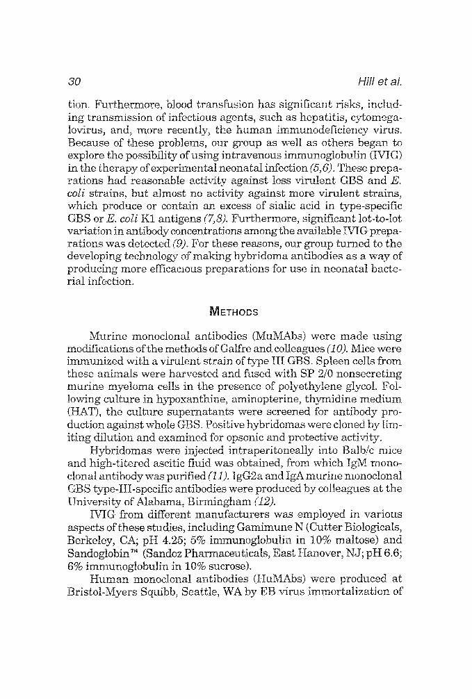
tion. Furthermore, blood transfusion has significant risks, includ- ing transmission of infectious agents, such as hepatitis, cytomega- lovirus, and, more recently, the human immunodeficiency virus. Because of these problems, our group as well as others began to explore the possibility of using intravenous immunoglobulin (IVIG) in the therapy of experimental neonatal infection (5,6). These prep a- rations had reasonable activity against less virulent GBS and E. coli strains, but almost no activity against more virulent strains, which produce or contain an excess of sialic acid in type-specific GBS or E. coli K1 antigens (7,8). ~r thermore , significant lot-to-tot variation in antibody concentrations among the available IVIG prepa- rations was detected (9). For these reasons, our group turned to the developing technology of making hybridoma antibodies as a way of producing more efficacious preparations for use in neonatal bacte- rial infection.
METHODS
Murine monoclonal antibodies (MuMAbs) were made using modifications of the methods of Galfre and colleagues (10). Mice were immunized with a virulent strain of type III GBS. Spleen cells from these animals were harvested and fused with SP 2/0 nonsecreting murine myeloma cells in the presence of polyethylene glycol. Fol- lowing culture in hypoxanthine, aminopterine, thymidine medium (HAT), the culture supernatants were screened for antibody pro- duction against whole GBS. Positive hybridomas were cloned by lim- iting dilution and examined for opsonic and protective activity.
Hybridomas were injected intraperitoneally into Balb/c mice and high-titered ascitic fluid was obtained, from which IgM mono- clonal antibody was purified (1i). IgG2a and IgA murine monoclonal GBS type-III-specific antibodies were produced by colleagues at the University of Alabama, Birmingham (12).
M G from different manufacturers was employed in various aspects of these studies, including Gamimune N (Cutter Biologicals, Berkeley, CA; pH 4.25; 5% immunoglobulin in 10% maltose) and Sandoglobin TM (Sandoz Pharmaceuticals, East Hanover, NJ; pH 6.6; 6% immunoglobulin in 10% sucrose).
Human monoclonal antibodies (HuMAbs) were produced at Bristol-Myers Squibb, Seattle, WA by EB virus immortalization of
Mabs in Neonatal Infection 31
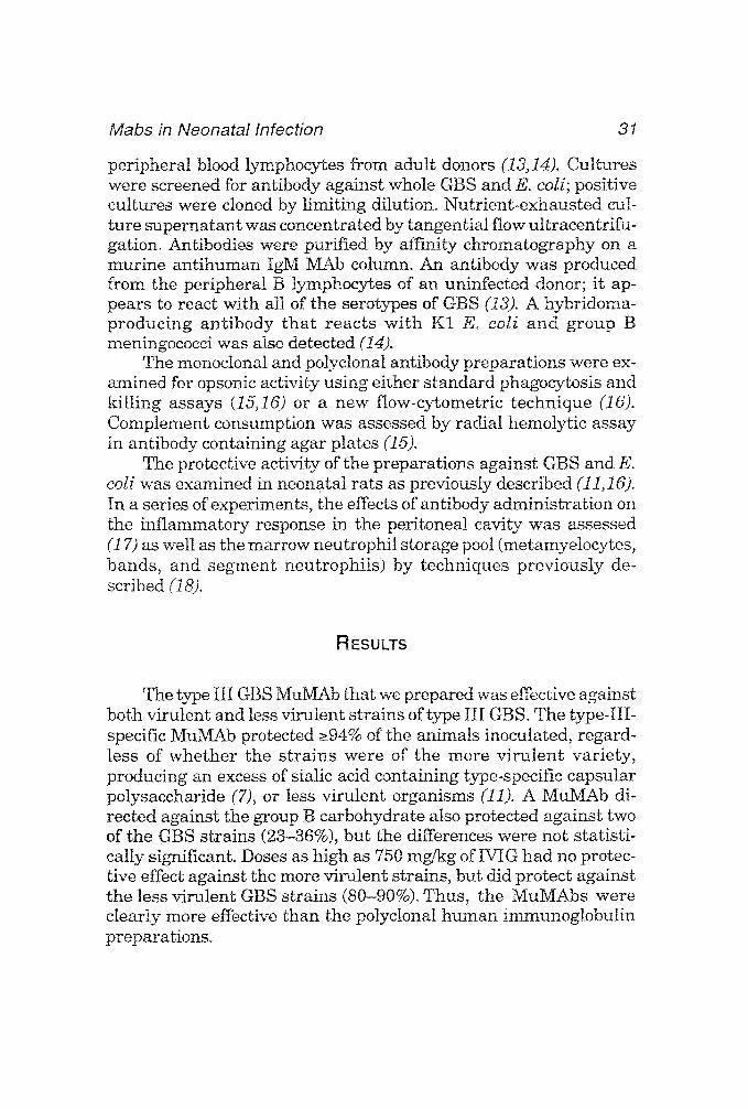
peripheral blood lymphocytes from adult donors (I3,14). Cultures were screened for antibody against whole GBS and E. coli; positive cultures were cloned by l imit ing dilution. Nutrient-exhausted cul- ture supernatant was concentrated by tangential flow ultracentrifu- gation. Antibodies were purified by affinity chromatography on a murine an t ihuman IgM MAb column. An antibody was produced from the peripheral B lymphocytes of an uninfected donor; it ap- pears to react with all of the serotypes of GBS (i3). A hybridoma- producing ant ibody tha t reacts wi th K! E. coli and group B meningococci was also detected (14).
The monoclonal and polyclonal antibody preparations were ex- amined for opsonic activity using either s tandard phagocytosis and ki l l ing assays (15,16) or a new flow-cytometric technique (16). Complement consumption was assessed by radial hemolytic assay in antibody containing agar plates (15).
The protective activity of the preparations against GBS and E. cdi was examined in neonatal rats as previously described (11,16). In a series of experiments, the effects of antibody administrat ion on the inflammatory response in the peritoneal cavity was assessed (17) as well as the marrow neutrophil storage pool (metamyelocytes, bands, and segment neutrophits) by techniques previously de- scribed (18).
RESULTS
The type III GBS MuMAb that we prepared was effective against both virulent and less virulent strains of type III GBS. The type-III- specific MuMAb protected ~94% of the animals inoculated, regard- less of whether the s t ra ins were of the more v i ru len t variety, producing an excess of sialic acid containing type-specific capsular polysaccharide (7), or less virulent organisms (11). A MuMAb di- rected against the group B carbohydrate also protected against two of the GBS strains (23-86%), but the differences were not statisti- cally significant. Doses as high as 750 mg/kg of IVIG had no protec- tive effect against the more virulent strains, but did protect against the less virulent GBS strains (80-90%). Thus, the MuMAbs were clearly more effective than the polyclonal human immunoglobulin preparations~

32 Hil l et al.
Table t Protective Effect of Type III GBS MuMAb"
Time after inoculation Number of animal~ Survival rate, %
No antibody 17 0 0 17 94 4 h 15 87 8 h 7 43 12 h 20 25 24 h 9 33
aAdministered at t ime intervals fol lowing bacterial inoculation of neonatal rats with 5 x 10 6 type I11 GBS,
Studies employing an IgG2a type-III-specific MuMab prepared by Egan and associates (12) showed tha t the MuMab IgM is approx 500-fold more effective on a molar basis than the IgG MuMab in protecting against GBS infection in the neonatal rat. Protection was also observed when the antibody preparation was administered fol- lowing bacterial inoculation (Table 1). Four hours after bacterial administration, the MuMAb continued to protect essentially as well as when it was administered at the same time as the bacteria. Pro- tection was still significant when the MuMAb was administered 24 h after inoculation.
Init ial exposure of h u m a n neonates to GBS usual ly occurs through the respiratory tract. For this reason, we determined if the IgM could protect neonatal ra ts from respiratory infection. Follow- ing an in t ranasal inoculation of 1 x 10⁷-10 s type III GBS, only three of 64 unt rea ted neonatal rats survived (11). In contrast, animals who received 1.8 mg/kg of the IgM MuMab by an intraperi toneal route had a survival rate of 94% with a 107 and 77% with a l0 s in t ranasal bacterial inoculum (11). The IgM probably prevented in- vasion of the bacteria from the respiratory tract into the systemic circulation. We could not rule out the possibility tha t the IgM was actually getting onto the inflamed respiratory surfaces, however.
We next examined an IgA monoclonal antibody against the type III GBS antigen, which was prepared by colleagues at the Univer- sity of Alabama (12). The IgA MuMAb also protected against sys- temic infection in the neonatal rat, but only when administered into the respiratory tract or systemically Mong with fibronectin (19). We have recently shown that this IgA monoclonat GBS antibody acts as an opsonin (20). This activity is significantly enhanced in the pres-

Mabs in Neonatal Infection 33
ence of fibronectin (21). Moreover, the preparation is capable of trig- gering the complement system in rat but not in human serum (20).
The IgM GBS MuMAb, in a dose of 0. 02 mg/kg, markedly in- creased the influx of leukocyies into the peritoneal cavity of GBS- infected rats (17). The IgG2a MuMAb also enhanced leukocyte infiltration, but the dose required was much higher (1.2 mg/kg). The IgA GBS MuMAb did not enhance peritoneal leukocyte accumula- tion at any t ime and, in fact, suppressed the count four hours after bacterial inoculation. The IgM GBS MuMAb also prevented the se- vere neutropenia and depletion of marrow granulocyte stores com- monly observed during GBS infection in the neonatal rat (18). The MuMAb of the IgM isotype has an absolute requirement for comple- ment in providing functional opsonic and protective activity (22). We believe that the effect of the antibody on neutropenia and mar- row neutrophil storage pool depletion is a result of complement acti- vation and the release of leukocyte-mobilizing fragments of the third component of complement including C3d,g and C3e, as well as the chemoattractant C5a (23).
Administrat ion of a murine antibody preparat ion to h u m a n neonates is unlikely to be approved, since reactions have been re- ported following the use ofmurine anti-CD3 antibody in adults. Raft and associates (13,14) have recently developed at least two very unique h u m a n monoclonal antibodies. One of these appears (!3) to react with all s trains of group B streptococci. These investigators (13) showed that this antibody is protective against type Ia and III GBS strains. We have studied the GBS HuMAb produced by Raft and colleagues in both protective and opsonic assays (16). As shown in Fig. 1, the HuMAb was effective in protecting neonatal rats from type III GBS infection in doses as low as 4 mg/kg (Fig. t). The HuMab, at a dose of 20 mg/kg, was significantly more effective in protecting neonatal rats from virulent type III GBS infection than was conven- tional IVIG (Sandoglobin ~) in a close of 500 mg/kg (Fig. 2). The GBS HuMab was also effective in similar doses against multiple s trains of type Ia, II, and III GBS (Fig. 3). We have recently found that the IgM GBS HuMab is significantly more effective in both opsonizing GBS and activating the complement system than a genetically en- gineered IgG HuMab with identical Fab regions (24,25).
Raft and associates (14) also produced a HuMAb reactive with the E. CoZi K1 antigen and the group B meningococcal polysaccha- ride. This antibody is not reactive with non-K! E. coli strains or

34 Hill et al.
1oo-
80-
60
4o
20
0
0.0
~. '& " " ' - " t~ *P < 0.05
"'m,..~......~ *P < 0.05
" ~ *P < 0.05
~ , - - HumAb 20 t r~Kg* (N=24)
' O O O O ----,-e--- HuMab 10 mg/Kg* (N=23)
HuMab 4 n'~l/Kg = (N=23)
- - - -O- - - GBS + PBS (N=20)
1.0 2.0 3.0 4.0 5.0 6~0 7.0
Days Post-inoculation
Fig. 1 Protective activity of varying doses of IgM human monoctonal GBS anti- body against virulent type III GBS infection in neonatal rats. From Hill, H. R. et al. J~ Infect. Dis. 163: 792-798, 1991, with permission from the University of Chicago Press, publisher.
100
8O
> ~ 6 0
co 40 o~
20,
0 ~
0
~, HuMab 20 mg/Kg ~ (n=24)
O GBS+PBS (n=20)
IVIG 500 mg/Kg (n=23)
1 2 3 4 5 6 7 Days Post-inoculation
Fig. 2. Comparison of the protective effect of GBS HuMAb and conventional IVIG against type 111 GBS in neonatal rats. From Hill, H. R., et al., J. infect. Dis. 163,292-298, 1991, with permission from the University of Chicago Press, Pub- lisher.
other gram-negative organisms. The antibody possesses significant opsonic activity, but only in the presence of human complement. This HuMAb protected normal and neutropenic mice and neonatal rats from challenge with K1 containing E. coli and Neisseria rnenin- gitides group B in doses ranging from 1 to 25 ~g/animal.

Mabs in Neonatal Infection 35
100
80
60
r 40
2O
0 0 1 2 3 4 5 6 7
Days Post-inoculation
3<0.05
3<0.05
3<0.05
HuMab 20 mg/Kg* (n=21)
u GBS+PBS+II R GBS (n=21)
| HuMab 20 mg/Kg*+lll R GBS [n=24)
o GBS+PBS+Ill R GBS (n=20)
HuMab 20 mg/Kg'+la GBS (n=24)
.~ GBS+PBS+Ia GBS (n=30)
Fig. 3. Protective activity of IgM human monoclonal GBS antibody against viru- lent type la, II and III GBS infection in neonatal rats.
DISCUSSION
Monoclonal antibodies with striking opsonic and protective ac- tivity have been developed against GBS and K1 F,. coli, two of the major bacterial pathogens affecting the human neonate. An IgM MuMAb that we developed was far more effective against all type III GBS strains than IVIG (11). This type-III-specific IgM mono- clonal was approx 500-fold more effective on a molar basis than an IgG2a MuMAb directed against the identical epitope on the type III polysaccharide. The IgM antibody required complement for opsonic and protective activity (22) and affected the kinetics of leukocyte release from the marrow (18). We (23) reported tha t a fragment of C3b (C3d,g) promotes the release of neutrophils from the marrow, resulting in a peripheral neutrophil ia in rats. Along with the local release of C5a, this likely results in a prompt accumulation of phago- cytes at the site of bacterial invasion (17). We have shown tha t the ability of an antibody preparation to activate the complement sys- tern can be directly related to its protective and opsonic activity (15). IgM is much more active in triggering complement than IgG. This could aid in explaining the enhanced activity of our IgM MuMAb compared to the IgG2a MuMAb.
In contrast to the type specificity of the MuMAb, the HuMAb we studied was much more broadly reactive against all GBS organ- isms. Studies suggest tha t the HuMAb is directed toward the streptococcal group B polysaccharide, rather than type-specific po-
36 Hi l l et al.
lysaccharide antigens (13). Lancefield and coworkers (26) have sug- gested that antibodies to this cell-wall antigen, present on all group B organisms, are not protective in a mouse model of infection. The protective activity of an antibody against GBS appears to be directly related to the avidity of the antibody for the antigen on the bacterial surface (27). Avidity is determined, in part, by the isotype of the antibody and the density of the epitope on the bacterial surface. Most of the data showing lack of protection with antibody directed against the group B carbohydrate have been carried out with rabbit hyper immune ant iserum containing mostly IgG (26). Perhaps IgM, but not IgG, antibody directed against the group B carbohydrate possesses sufficient avidity to result in opsonic and protective activ- ity. The activity of an antibody preparation to combine with a bacte- r ia and trigger the complement system is critical in its ability to afford protection (15). Such activation results in the generation of inf lammatory mediators, such as C3e and C3d,g, capable of releas- ing marrow granulocyte stores (23); C5a, which attracts leukocytes into the area of bacterial growth; and the deposition of C3b and iC3b on the bacterial surface. The IgM HuMAb was also approx 500 t imes more effective on a molar basis in opsonizing GBS and acti- vat ing the complement system in the presence of these organisms. We suspect that this is one of the major reasons that both the IgM MuMAb and the IgM HuMAb are so effective in protecting neonatal animals from lethal bacterial infection. Can IgM antibody be ad- ministered intravenously to humans without ill side" effects? Haque and colleaglaes (28) have safely administered a preparation of M G to septic neonates enriched for IgM compared to s tandard M G . Moreover, h u m a n monoclonal anti-J5 IgM antibody in doses of 100 mg has been administered to adult humans without significant prob- lems. Thus, it may be possible to safely administer GBS or E. coli K1 IgM HuMab to human neonates.
There is one major advantage of polyclonal preparations com- pared to monoclonal ones. intravenous immunogtobulin is produced from plasma obtained from 2000 to as many as 10,000 donors. These preparat ions contain a broad antibody representation. Before one uses a very specific monoclonal, a f irm diagnosis of the etiological agent based on antigen detection or culture would have to be made. A cocktail of HuMAb against GBS and E. coli K1 could be made for use in the human neonate. Such a preparation would not be effec- tive against other ~am-nega t ive bacteria or against gram-positive

Mabs in Neonatal Infection 37
organisms, such as Staphylococcus epiderrnidis, an increasingly com- mon isolate from neonates. Most normal adult serum contains only low concentrations of antibody against GBS and E. coli K1, thus conventional IVIG preparations possess little activity against these pathogens. In contrast, standard IVIC contains excellent activity against Klebsiella pneurnoniae, Serratia rnarcescens, Enterobacter, and staphylococci (15,29,30). For this reason, a suitable strategy might be to add HuMAb against GBS and E. coli KI to conventional IVIG for use in septic neonates. The studies described here suggest the potential for the use of monoclonal antibodies, especially human monoclonal IgM antibodies, in the therapy of fulminant bacterial infections in the human neonate.
ACKNOWLEDGMENTS
We thank Nancy Augustine, Ann Shigeoka, Seth Pincus, John Bohnsack, Kuender Yang, and Robert Christensen, who contributed greatly to these studies. We also thank Jeannette Rejali for secre- tarial aid and Don Morse for illustrations.
Supported, in part, by U S Public Health Service grant AI13150 and a grant from the Mead Johnson Research Center, Bristol-Myers Squibb Company.
REFERENCES
1. Walsh, J. A. and Hutchins, S. (1989), Pediatr. Infect. Dis. J. 8, 271-276. 2. Hemming, V. G., Hall, Ro T., Rhodes, P. G., Shigeoka, A. O., and Hill, H. R.
(1976), J. Clin. Invest. 48, 1379-1387. 3. Baker, C. J. and Kasper, D. L. (1977),J. Infect. Dis. 136,$98-$104. 4. Shigeoka, A. O., Hall, R. T., and Hill, H. R. (1978), Lancet 1, 636-638. 5. Fischer, G. W., Hunter, K. W., Wilson, S. R., and Hemming, Vo G. (1981),
Lancet I, 271. 6. Santos, J. I., Shigeoka, A. O., and Hill, H. R. (1981), J. Pediatr. 99, 873-
879. 7. Shigeoka, A. O., Rote, N. S., Santos, J. I., and Hill, H. R. (1983), J. Infect.
Dis. 147, 856-863. 8. Yeung, M. K. and Mattingly, S. J. (1984), Infect. Immun. 44, 217-221. 9. Fischer, G. W. (1988), Pediatr. Infect. Dis. J. 7, $13-$16.
10. Galfre, G., Howe, S. C., Milstein, C., Butcher, G. W., and Howard, J. C. (1977), Nature 266, 550-552.
38 Hifl et al.
11. Shigeoka, A. O., Pincus, S. H., Rote, N. S., and Hill, H. R. (1984), Jo Infect. Dis. 149, 363-372.
12. Egan, M. L., Pritchard, D. G., Dillon, H. C., Jr., and Gray, B. M. (1983), J. Exp. Med. 158, 1006-1011.
13. Raft, H. V., Sisscoe, P. J., Wolff, E. A., Maloney, G., and Shuford, W. (1988), J. Exp. ivied. 168, 905-917.
14. Raft, H. V., Devereux, D., Shuford, W., Abbott-Brown, D., and Maloney, G. (1988), J. Infect. Dis. 157, 118-126.
15. Yang, K. D., Bathras, J. M., Shigeoka, A. O., James, J., Pincus, S. H., and Hilt, H. R. (1989), J. Infect. Dis. 159, 701-707.
16. Hill, H. R., Gonzales, L. A., Knape, W. A., Fischer, G. W., Kelsey, D. K., and Raft, H. V. (1991) J. Infect. Dis. 163, 792-798.
17. Shigeoka, A. O., Weber, M. E., Pincus, S. H., Pritchard, D. G., Egan, M. L., and Hill, H. R. (1986), J. Infect. Dis. 153, 1170-1173.
18. Christensen, R. D., Rothstein, G., Hill, H. R., and Pincus, S. H. (1983), Pediatr. Res. 17, 795-799.
i9. Hill, H. R., Shigeoka, A. O., Augustine, N. H., Pritchard, D., Egan, M. L., Lundblad, J. L., and Schwartz, R. S. (1984), J. Exp. Med. 159, 1618-1628.
20. Bohnsack, J. F., Hawley, M. M., Pritchard, D. C., Egan, M. L., Shigeoka, A. O., Yang, K. D., and Hill, H. R. (1989), J. Irnrnunol. 143, 3338-3342.
21. Yang, K. D., Bohnsack, J. F., Hawley, M. M., Augustine, N. H., Knape, W. A., Egan, M. L., Pritchard, D. G., and Hill, Ho R. (1990), Jo Infect. Dis. 161, 236-241.
22. Shigeoka, A. O., Jensen, C., Pincus, S. H., and Hill, H. R. (1984), J. Infect. Dis. 150, 64-69.
23. Shigeoka, A. O., Gobel, R. J., Janatova, J., and Hill, H. R. (1988% Arno J. Pathol. 133, 623-629.
24. Raft, H. V., Bradley, C., and Brady, W. Comparison of functional activities between IgG1 and IgM class-switched hyman monoclonal antibodies reac- tive with group B streptococci or E eoli K1. (1991), J. Infect. Dis. 163, 346- 354.
25. Shyur, S. D., Raft, H. V., Bohnsack, J. F., Kelsey, D. K., and Hill, H. R. (1991), Comparison os the opsonic and complement activating activity of human monoclonal IgG and IgM antibody against group B streptococci. Pediatr. Res. 29, 185A.
26. Lancefield, R. C., McCarty, M., and Everly, W. N. (1975), J. Exp. Med. 142, 165-179.
27. Pincus, S. H., Shigeoka, A. O., Moe, A. A., Ewing, L. P., and Hill, H. R. (1988), J. Irnrnunol. 140, 2779-2785.
28. Haque, K. N., Zaidi, M. H., and Bahakim, H. (1988), Am. J. Dis. Child. 142, 1293-1296.
29. Hill, H. R. and Bathras, J. M. (1986), Rev. Infect. Dis. 8, $396-$400. 30. Yang, tC D., Augustine, N. H., Gonzalez, L. A., Bohnsack, J. F., and Hill,
H. R. (1988), J. Infect. Dis. 158, 823-830.