LaTeX4WP: A LaTeX guide specifically designed for word processor users.
Upload
lmu-munichCategory
view
1download
0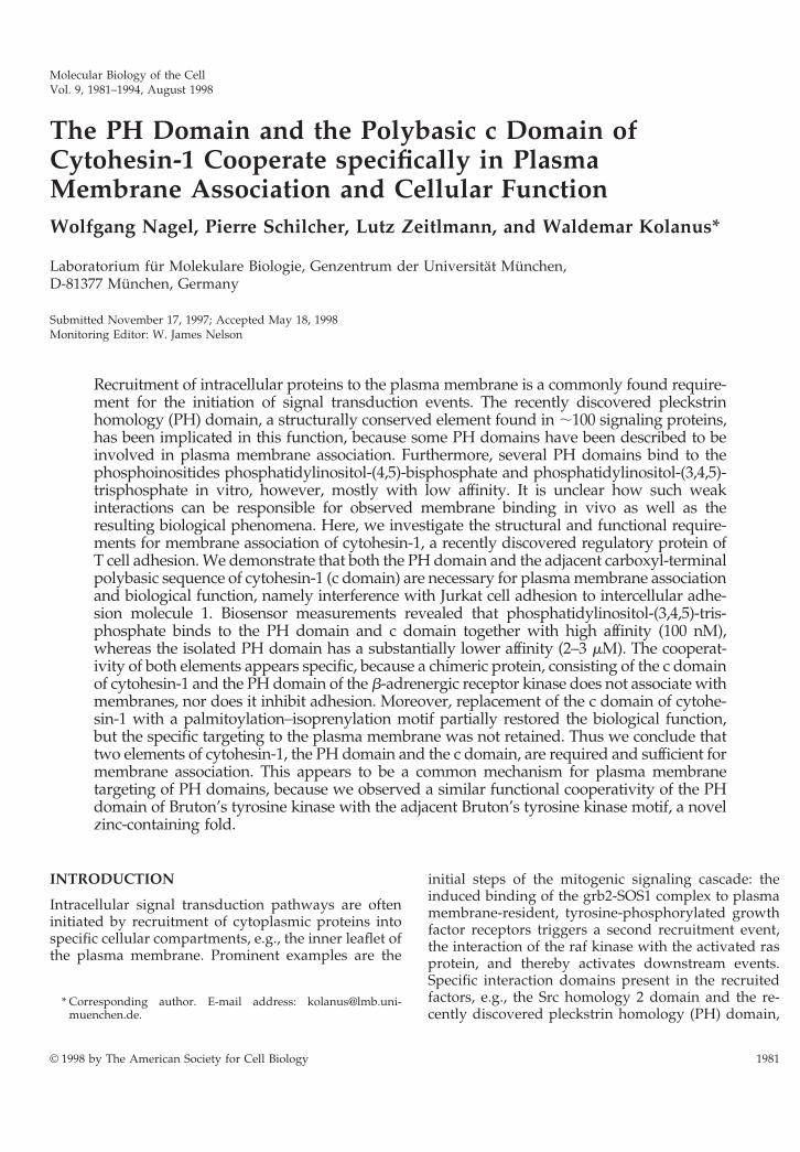
Molecular Biology of the CellVol. 9, 1981–1994, August 1998
The PH Domain and the Polybasic c Domain ofCytohesin-1 Cooperate specifically in PlasmaMembrane Association and Cellular FunctionWolfgang Nagel, Pierre Schilcher, Lutz Zeitlmann, and Waldemar Kolanus*
Laboratorium fur Molekulare Biologie, Genzentrum der Universitat Munchen,D-81377 Munchen, Germany
Submitted November 17, 1997; Accepted May 18, 1998Monitoring Editor: W. James Nelson
Recruitment of intracellular proteins to the plasma membrane is a commonly found require-ment for the initiation of signal transduction events. The recently discovered pleckstrinhomology (PH) domain, a structurally conserved element found in ;100 signaling proteins,has been implicated in this function, because some PH domains have been described to beinvolved in plasma membrane association. Furthermore, several PH domains bind to thephosphoinositides phosphatidylinositol-(4,5)-bisphosphate and phosphatidylinositol-(3,4,5)-trisphosphate in vitro, however, mostly with low affinity. It is unclear how such weakinteractions can be responsible for observed membrane binding in vivo as well as theresulting biological phenomena. Here, we investigate the structural and functional require-ments for membrane association of cytohesin-1, a recently discovered regulatory protein ofT cell adhesion. We demonstrate that both the PH domain and the adjacent carboxyl-terminalpolybasic sequence of cytohesin-1 (c domain) are necessary for plasma membrane associationand biological function, namely interference with Jurkat cell adhesion to intercellular adhe-sion molecule 1. Biosensor measurements revealed that phosphatidylinositol-(3,4,5)-tris-phosphate binds to the PH domain and c domain together with high affinity (100 nM),whereas the isolated PH domain has a substantially lower affinity (2–3 mM). The cooperat-ivity of both elements appears specific, because a chimeric protein, consisting of the c domainof cytohesin-1 and the PH domain of the b-adrenergic receptor kinase does not associate withmembranes, nor does it inhibit adhesion. Moreover, replacement of the c domain of cytohe-sin-1 with a palmitoylation–isoprenylation motif partially restored the biological function,but the specific targeting to the plasma membrane was not retained. Thus we conclude thattwo elements of cytohesin-1, the PH domain and the c domain, are required and sufficient formembrane association. This appears to be a common mechanism for plasma membranetargeting of PH domains, because we observed a similar functional cooperativity of the PHdomain of Bruton’s tyrosine kinase with the adjacent Bruton’s tyrosine kinase motif, a novelzinc-containing fold.
INTRODUCTION
Intracellular signal transduction pathways are ofteninitiated by recruitment of cytoplasmic proteins intospecific cellular compartments, e.g., the inner leaflet ofthe plasma membrane. Prominent examples are the
initial steps of the mitogenic signaling cascade: theinduced binding of the grb2-SOS1 complex to plasmamembrane-resident, tyrosine-phosphorylated growthfactor receptors triggers a second recruitment event,the interaction of the raf kinase with the activated rasprotein, and thereby activates downstream events.Specific interaction domains present in the recruitedfactors, e.g., the Src homology 2 domain and the re-cently discovered pleckstrin homology (PH) domain,
* Corresponding author. E-mail address: [email protected].
© 1998 by The American Society for Cell Biology 1981

are thought to be responsible for the tethering of cy-tosolic proteins to the membrane compartment.
PH domains are structural modules present in ;100proteins, which play known or postulated roles insignal transduction or cytoskeletal organization(Musacchio et al., 1993). It is now known that PHdomains may aid in membrane recruitment of pro-teins through their interactions with phosphorylatedligands present in cellular membranes (Pawson, 1995;Lemmon et al., 1996, 1997). Although a subgroup ofPH domains is capable of interacting with tyrosine-phosphorylated proteins (Lemmon et al., 1996), muchreminiscent of the Src homology 2 domain function,several isolated PH domains have been shown to bindto phosphoinositides such as phosphatidylinosi-tol-(4,5)-bisphosphate (PIP2) in vitro (Harlan et al.,1994, 1995; Ferguson et al., 1995; Garcia et al., 1995;Hyvonen et al., 1995; Lemmon et al., 1995; Pitcher et al.,1995; Touhara et al., 1995; Wang and Shaw, 1995; Mikiet al., 1996; Salim et al., 1996; Zheng et al., 1996; Chen etal., 1997; Frech et al., 1997; Kubiseski et al., 1997).Interestingly, certain PH domains show in vitro bind-ing preference to lipid compounds that are in vivophosphorylation products of phosphoinositol (PI)3-kinase (Salim et al., 1996; Franke et al., 1997a,b; Klar-lund et al., 1997).
Cytohesin-1 is a 47-kDa intracellular protein thatinteracts specifically in several systems with the cyto-plasmic domain of the leukocyte integrin aLb2(CD11a/18, leukocyte functional antigen-1 [LFA-1])(Kolanus et al., 1996). Cytohesin-1 bears a short amino-terminal domain, which may aid in oligomerization,an extended central homology region, which is similarto the yeast Sec7 protein, and a carboxyl-terminal PHdomain, followed by the c domain. Overexpression ofcytohesin-1 or subdomain constructs in the Jurkat Tcell line E6 was shown to have pronounced effects onthe binding of aLb2 to its ligand, the intercellularadhesion molecule 1 (ICAM-1). Overexpression offull-length cytohesin-1 or of the isolated Sec7 domainin Jurkat cells resulted in a constitutive adhesion ofaLb2, whereas the expression of a cytohesin-1 PHdomain construct, which still contained the c domain,specifically inhibited the activation of LFA-1 in a dom-inant negative manner. Because the PH domain wasnot found to be mediating the interaction with theintegrin cytoplasmic domain, it has been postulatedthat its unidentified cellular ligand may be an up-stream component of the inside-out signaling path-way of aLb2 (Kolanus et al., 1996).
We have recently shown that an intact PH domainof cytohesin-1 is required for its association with theplasma membrane and that membrane localization ofcytohesin-1 can be regulated by PI 3-kinase (Nagel etal., 1998). Furthermore, the PH domains of cytohesin-1and general receptor for phosphoinositides-1, a closehomologue of cytohesin-1, have both been demon-
strated to bind phosphatidylinositol-(3,4,5)-trisphos-phate (PIP3) (Klarlund et al., 1997). However, affinitiesof PH domains for their ligands are often in the mi-cromolar range, which appears rather low and leavesopen the question of whether these rather weak inter-actions are sufficient or compatible with observed bi-ological activities in vivo (Shpetner et al., 1996; Patki etal., 1997).
In this study we show by functional and biochemi-cal means, as well as by confocal laser microscopytechniques, that the PH domain of cytohesin-1 specif-ically mediates membrane association cooperativelywith the c domain, a 17-amino-acid stretch that islocated carboxyl-terminally adjacent to the PH do-main and that is rich in basic residues. The carboxylterminus is conserved between all members of thecytohesin family that have been described so far (Fig-ure 1). Similar polybasic regions have previously beendescribed to be involved in membrane attachment ofcellular or viral proteins (Hancock et al., 1990, 1991;Adamson et al., 1992; Newman et al., 1992; Cadwal-lader et al., 1994; Mitchell et al., 1994; Ghomashchi etal., 1995; Kwong and Lublin, 1995; Kreck et al., 1996;Soneoka et al., 1997). Both elements, PH domain and cdomain, are required to maintain association of cyto-hesin-1 with the plasma membrane. When the c do-main is replaced by the combined isoprenylation–pal-mitoylation sequence derived from H-ras (Hancock etal., 1991) (termed CAAX motif throughout the article;Figure 2), function is partially retained and membraneassociation is restored, but targeting specificity is lost,because the fusion protein localizes to a perinuclearmembrane compartment in addition to the plasmamembrane. Furthermore, an effective membrane asso-ciation element cannot be generated by grafting the cdomain of cytohesin-1 onto the b-adrenergic receptorkinase (bARK) PH domain, thus demonstrating thatthe plasma membrane localization of cytohesin-1 isachieved by two highly specific plasma membraneinteraction elements, the PH domain and the basic cdomain. In vitro studies show that the c domain sta-bilizes the interaction of the PH domain with PIP3markedly. Although the glutathione S-transferase(GST) fusion protein of the PH domain alone binds tothe PIP3 with an affinity of ;2–3 mM, use of the fusionprotein containing the PH domain and the c domainresults in high-affinity binding (100 nM).
To further investigate the cooperativity of PH do-mains with adjacent protein stretches in specificplasma membrane targeting, we introduced the PHdomain of Bruton’s tyrosine kinase (Btk) in our study.In this case we could demonstrate as well that the PHdomain cooperates with the neighboring protein ele-ment, the so-called btk motif in specific plasma mem-brane association. Interestingly, the zinc-containingbtk motif of Btk bears no similarity to the c domain ofcytohesin-1.
W. Nagel et al.
Molecular Biology of the Cell1982
MATERIALS AND METHODS
Construction of Expression PlasmidsPCR products were derived from existing expression plasmids andinserted into the Mlu1 and NotI sites of the vaccinia expressionvector pcIgTkg, subsequently encoding cytoplasmic immunoglobu-lin (Ig) fusion proteins as described (Kolanus et al., 1996). Specifi-cally, the following primer pairs were used: CGC GGG ACG CGTACC ATG GGT TTC AAT CCA GAC CGA GAA GGC TGG andCGC GGG GCG GCC GCT TTA GTG TCG CTT CGT GGA GGAGAC CTT (PH); CGC GGG ACG CGT ACC ATG GGT TTC AATCCA GAC CGA GAA GGC TGG and CGC GGG GCG GCC GCTTTA GTG TCG CTT CGT GGA GGA GAC CTT (PHccyh-1); GCGGGG ACG CGT ACC ATG GAC TAC GCC CTG GGC AAG GACand GCG GGG GCG GCC GCT TTA CTG CTG GGC CTC GCGGTA GGC GTC (bARK-PH); GCG GGG ACG CGT ACC ATG GACTAC GCC CTG GGC AAG GAC and GG GCG GGG GCG GCCGCT TTA GAG GCC GTT GGC ACT GCC (bARK-PHcbARK); GCGGGG ACG CGT ACC ATG GAC TAC GCC CTG GGC AAG GACand GCG GGG GCG GCC GCT TTA GTG TCG CTT CGT GGAGGA GAC CTT CTT TTT CCG TGC TGC GAG CAT TTC GTA CTG
CTG GGC CTC GCG GTA GGC GTC (bARK-PH-ccyh-1); CGC GGGACG CGT ACC ATG GGT TTC AAT CCA GAC CGA GAA GGCTGG and CGC GCG CGG CCG CTT TAG CTC AGC ACG CACTTG CAG CTC ATG CAG CCG GGG CCG CTG GCG CCC CCGAGC TCG AAA GGG TCC CTG CTG ATG GCT [PH-CAAX and PH(R281C)-CAAX]; CGC GGG ACG CGT GCC ACC ATG GCT GCAGTG ATA CTG GAG AGC and GCG GGG GCG GCC GCT TTAGAT TAC ATT TTT GAG CTG GTG AAT CC (Btk-PH); and CGCGGG ACG CGT GCC ACC ATG GCT GCA GTG ATA CTG GAGAGC and GCG GGG GCG GCC GCT TTA GTT CTC CAA AATTTG GCA GCC C (Btk-PHbtk motif).
All PCR products were confirmed by double-stranded sequenc-ing. Secreted receptor–globulin fusion proteins of the ICAM-1 ex-tracellular domains were used as described (Kolanus et al., 1996).
Eukaryotic Expression and Adhesion AssayVaccinia expression constructs were recombined with wild-typevaccinia virus (WR strain) in CV-1 cells; recombinant plaques werepurified; and high-titer virus stocks were generated as described(Romeo and Seed, 1991). The ICAM-1-Rg fusion protein was ex-
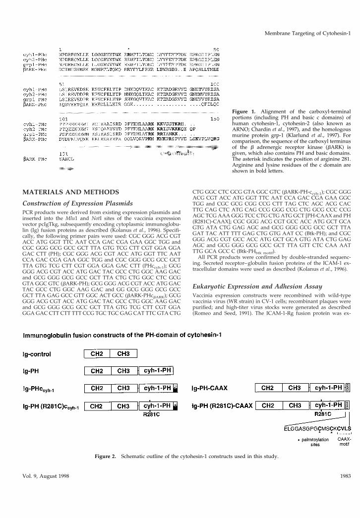
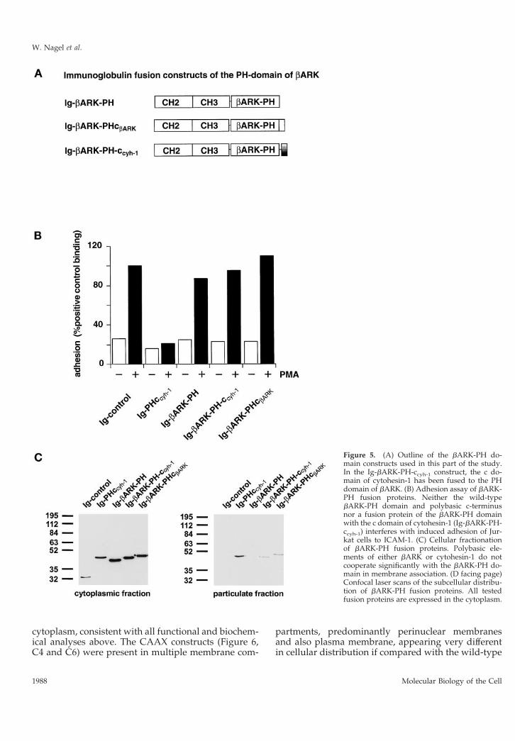
Figure 1. Alignment of the carboxyl-terminalportions (including PH and basic c domains) ofhuman cytohesin-1, cytohesin-2 (also known asARNO; Chardin et al., 1997), and the homologousmurine protein grp-1 (Klarlund et al., 1997). Forcomparison, the sequence of the carboxyl terminusof the b adrenergic receptor kinase (bARK) isgiven, which also contains PH and basic domains.The asterisk indicates the position of arginine 281.Arginine and lysine residues of the c domain areshown in bold letters.
Figure 2. Schematic outline of the cytohesin-1 constructs used in this study.
Membrane Targeting of Cytohesin-1
Vol. 9, August 1998 1983
pressed in COS cells, purified from culture supernatants by proteinA-Sepharose, eluted, resuspended in PBS and subsequently coatedonto Falcon (Lincoln Park, NJ) 1008 dishes as described (Walz et al.,1990). Jurkat cells (2 3 106) were infected with recombinant virusesand incubated for 4 h at 37°C. After centrifugation cells were resus-pended in RPMI medium and incubated for 5 min at 37°C with orwithout the addition of 40 ng/ml phorbol 12-myristate 13-acetate.Cells were subsequently allowed to adhere to ICAM-1-Rg–coateddishes at 37°C for 10 min, and the bound fraction was determinedwith the aid of an ocular reticle.
Measurement of Phosphatidylinositol Binding ofVarious GST-PH Domain Constructs by IAsysBiosensor TechnologyPH domains of cytohesin -1 were expressed as (GST) fusion proteinsas described (Nagel et al., 1998). An optical evanescence resonantmirror cuvette system (IAsys, Affinity Sensors, Cambridge, UnitedKingdom) was used to measure interaction of GST fusion proteinswith phosphatidylinositol-(3,4,5)-triphosphate (PIP3). A lipid mono-layer containing 70% (wt/wt) b-palmitoyl-g-oleoyl-l-a-phosphati-dylcholine (POPC), 30% (wt/wt) dioleoyl-l-a-phosphatidyl-dl-glycerol, or a lipid mixture of 60% (wt/wt) palmitoyl-g-oleoyl-l-a-phosphatidylcholine and 30% (wt/wt) and 10% (wt/wt) PIP3(Mantreya, Pleasant Gap, PA) was mounted on an IAsys hydropho-bic sensor surface (FCH-0601) at 0.1 mg/ml lipid. The cuvette wassubsequently washed with 0.1 M HCl, PBS, and 10 mM NaOH.After the final wash with PBS, the cuvette was equilibrated inbinding buffer (PDI: PBS, 2 mM DTT, 0.001% [vol/vol] Igepal CA-630, Sigma), and affinity-purified GST fusion proteins dissolved inbinding buffer were added. The binding of various concentrations ofthe GST-PH domain fusion proteins were monitored for 5 min.Dissociation was initiated by adding PDI to the cuvette. Determi-nation of the association equilibrium constant was done by equilib-rium titration. The interaction profiles for each protein concentra-tion were analyzed using FASTfit kinetics analysis softwaresupplied with the instrument.
Cellular FractionationCells that had been infected with recombinant vaccinia viruses werecollected by centrifugation and resuspended on ice in 0.5 ml ice-coldhypotonic solution (HS: 10 mM HEPES, pH 7.5, 10 mM KCl, 10 mMMgCl2, 0.5 mM DTT) containing 10 mg/ml leupeptin, 10 mg/mlaprotinin, and 1 mM PMSF. Fractionation of cells was performed asdescribed (Meller et al., 1996). Briefly, cells were sheared, the nucleiwere removed by centrifugation at 1000 3 g for 10 min, and thesupernatant cytosol was collected. The cytosolic fraction wasbrought to a final concentration of 1% (vol/vol) Igepal CA-630(Sigma, St. Louis, MO; identical to Nonidet P-40) and 150 mM NaCland used directly for immunoprecipitation. The pellet was resus-pended, washed with 0.5 ml HS, and centrifuged at 15,000 3 g for15 min. The resulting pellet was resuspended in HS containing 1%(vol/vol) Igepal CA-630 and 150 mM NaCl, and centrifuged again,and the supernatant representing the particulate fraction was sub-jected to immunoprecipitation. For precipitation of the Ig fusion (Ig)proteins fractions were incubated with protein A-Sepharose 6 MBbeads (Pharmacia, Piscataway, NJ) for 2 h at 4°C. Then beads werewashed with 1 ml HS containing 1% (vol/vol) Igepal CA-630 and150 mM NaCl, and immunoprecipitates were resolved by 10% SDS-PAGE and analyzed by standard Western blot techniques. Specifi-cally, proteins were blotted onto nitrocellulose and detected by aprimary mouse polyclonal antibody preparation that had beenraised against the intracellular CH2 and CH3 domains (Kolanus,unpublished data). A peroxidase-conjugated anti-mouse IgG anti-body (Jackson ImmunoResearch, West Grove, PA) and subsequentECL reaction (Amersham, Arlington Heights, IL) were used forvisualization of the bands. To assess that cytoplasmic contents werenot trapped in the particulate fraction, lactate dehydrogenase activ-ities were monitored as described (Ma et al., 1997).
Confocal Laser Scanning MicroscopySix hours after infection of Jurkat E6 cells with recombinant vacciniaviruses, cells were placed on poly-l-lysine–covered microscopeslides for 1 h in a humidified chamber at 37°C. Then nonadherent
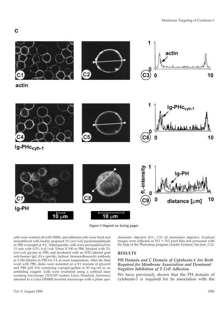
Figure 3. (A) Adhesion assay. The intact PH domain and theadjacent c domain of cytohesin-1 are both required for dominantinhibition of Jurkat cell adhesion to ICAM-1. Cytoplasmic Ig fu-sion constructs were expressed using recombinant vaccinia vi-ruses, and the adhesion assay was performed as described inMATERIALS AND METHODS. Normalization was performedagainst the positive control (Ig-control 1 PMA). (B) Cellular frac-tionation assay. The intact PH domain and the adjacent c domainof cytohesin-1 are both required for particulate association inJurkat cells. “Particulate fraction” denotes a crude lysate of cellularmembranes. The Ig-control, which is exclusively expressed in thecytoplasm, serves as an internal control for cellular fractionation.(C facing page) Confocal laser scans of the subcellular localizationof cytohesin-1 subdomain constructs Ig-PH (C7–C9) and Ig-PHc-cyh-1 (C4–C6), respectively. Ig fusion proteins were visualized withan FITC-conjugated anti-human IgG Fcg-specific antibody. Forquantitation of subcellular localization, cells were double stainedwith FITC-labeled anti-Ig (Fcg) for the respective fusion proteinand with TRITC-labeled phalloidin for the visualization of actin.Actin only is shown in C1–C3. Staining intensities measured ac-cording to pixel brightness were quantified along cell transects foreach construct in a representative positively stained cell. The large,central unstained region visible in C8 is due to the nucleus.
W. Nagel et al.
Molecular Biology of the Cell1984
cells were washed off with HBSS, and adherent cells were fixed andimmobilized with freshly prepared 3% (wt/vol) paraformaldehydein PBS overnight at 4°C. Subsequently, cells were permeabilized for15 min with 0.2% (vol/vol) Triton X-100 in PBS, blocked with 2%(wt/vol) glycine in PBS, and incubated with an FITC-labeled goatanti-human IgG (Fcg specific; Jackson ImmunoResearch) antibodyat 1:100 dilution in PBS for 2 h at room temperature. After the finalwash with PBS, slides were mounted on a 9:1 mixture of glyceroland PBS (pH 9.0) containing n-propyl-gallate at 20 mg/ml as anantifading reagent. Cells were examined using a confocal laserscanning microscope (TCS-NT system; Leica, Nussloch, Germany)attached to a Leica DMIRB inverted microscope with a plane apo-
chromatic objective 633, 1.32 oil immersion objective. Confocalimages were collected as 512 3 512 pixel files and processed withthe help of the Photoshop program (Adobe Systems, San Jose, CA).
RESULTS
PH Domain and C Domain of Cytohesin-1 Are BothRequired for Membrane Association and DominantNegative Inhibition of T Cell AdhesionWe have previously shown that the PH domain ofcytohesin-1 is required for its association with the
Figure 3 (legend on facing page).
Membrane Targeting of Cytohesin-1
Vol. 9, August 1998 1985
plasma membrane, because a point mutant of the PHdomain (R281C) abrogated both membrane associa-tion as well as in vitro binding to PIP3 (Nagel et al.,1998). Furthermore, overexpression of a recombinantPH domain construct that retained the 17-amino acidcarboxyl terminus of cytohesin-1 resulted in a domi-nant negative abrogation of LFA-1-mediated adhesionto its counter-receptor ICAM-1 (Kolanus et al., 1996).The dominant negative effect has been shown to becorrelated with competitive inhibition of the mem-brane attachment of endogenous cytohesin-1 andtherefore serves as a valid correlate of cellular function(Nagel et al., 1998).
In this study, we attempted to dissect the functionalrelationship between two structural elements of cyto-hesin-1, the PH domain and the adjacent c domain.Consequently, a construct was made in which the cdomain was deleted from the PH domain context(Ig-PH). This fusion protein was subsequently exam-ined with respect to its effect on cell adhesion andmembrane association. For comparison, the entire car-boxyl-terminal region of cytohesin-1 (Ig-PHccyh-1) or apoint mutant derivative thereof [Ig-PH (R281C)ccyh-1],containing the previously described, functionally in-activated PH domain, were used (Nagel et al., 1998).These and the constructs described below were ex-pressed in the Jurkat E6 (T cell leukemia) line with thehelp of recombinant vaccinia viruses (Figure 2). Theyall contain amino-terminal Ig domains for convenientimmunoprecipitation and detection (Kolanus et al.,1996). LFA-1-mediated adhesion was monitored byspecific binding of the cells to an immobilized ICAM-1Ig fusion protein (Kolanus et al., 1996).
Figure 3 confirms that an intact PH domain is nec-essary for membrane association (Figure 3B) and dom-inant negative inhibition of LFA-1 adhesion toICAM-1 (Figure 3A), because the R281C mutation ab-rogates both functions. Thus, the PH domain alone isnot sufficient for both membrane association and func-tion, because it requires the presence of the c domain(Figure 3, A and B). The c domain alone, used in thecontext of an inactive PH domain [Ig-PH (R281C)ccyh-1], was not sufficient for membrane association andhad no effect on T cell adhesion (Figure 3, A and B). Ittherefore appears that the PH domain and c domainare both responsible for membrane association of cy-tohesin-1.
PH Domain and C Domain of Cytohesin-1Determine Predominant Targeting Specificity to thePlasma MembraneWe and others have shown that cytohesin-1 PH do-main binds to the phosphoinositide PIP3 in vitro (Klar-lund et al., 1997; Nagel et al., 1998) and, consistent withthis notion, that its association with membranes ispossibly regulated by PI 3-kinase in vivo. Figure 3C
shows that PH and c domains are simultaneouslyrequired for predominant plasma membrane associa-tion in Jurkat cells. Confocal laser scanning micros-copy was used to show that the PH domain alone waslocalized in the cytoplasm, whereas the PHccyh con-struct colocalized very well with actin, a cytoskeletal
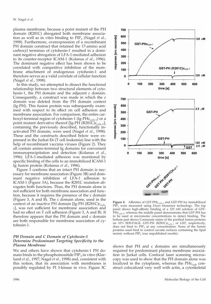
Figure 4. Affinities of GST-PHccyh-1 and GST-PH for immobilizedPIP3 were measured using IAsys biosensor technology. The toppanel shows high-affinity binding of a 125 nM solution of GST-PHccyh-1, whereas the middle panel demonstrates that GST-PH hasto be used at micromolar concentrations to detect binding. Thebottom part shows Coomassie stains of the purified fusion proteinson 10% SDS-PAGE. GST-PH (R281C)ccyh-1 or GST, respectively,does not bind to PIP3 at any concentration. None of the fusionproteins used bind to control cuvette surfaces containing the lipidmoiety without PIP3 (our unpublished results).
W. Nagel et al.
Molecular Biology of the Cell1986
protein that resides at the inner leaflet of the plasmamembrane.
In Vitro Binding of the Carboxyl-Terminal Domainsof Cytohesin-1 to Phosphatidylinosi-tol-(3,4,5)-TriphosphateHow does the c domain aid the PH domain in plasmamembrane association? Although PH domains alonecan bind to inositol-(1,3,4,5)-tetrakisphosphate or PIP3,it is possible that the positively charged c domainstabilizes this interaction. We used IAsys interactionmeasurement technology to determine the relative af-finities of the PH domain or of PHccyh-1 for PIP3, whichhad been mounted on a hydrophobic sensor surface.Figure 4 shows that GST fusion proteins containingeither the PH domain alone or the PH domain includ-ing the c domain can both bind to PIP3, confirmingthat the PH domain is sufficient for phosphoinositidebinding in vitro. Quantitative analyses showed thatthere are considerable differences in affinity. Althougha 125 nM preparation of GST-PHccyh-1 bound to PIP3with a fast on-rate and very slow off-rate, there wasbasically no binding of GST-PH at that concentration.At 2.5 mM, however, GST-PH bound to PIP3 withsimilar kinetics as GST-PHccyh-1. It therefore appearsthat the c domain enhances the on-rate of the interac-tion. Once the proteins were bound to the phospho-lipid they dissociated very slowly. We then deter-mined the affinities of either GST-PHccyh-1 or GST-PHfor PIP3. This was done by measuring binding to PIP3at various protein concentrations (our unpublishedresults). We found half-maximal binding to PIP3 using100 nM GST-PHccyh-1 or 3 mM GST-PH, respectively.Although the number for GST-PH is less exact, weconclude that there is at least one order of magnitudedifference between the affinites of the two fusion pro-teins for PIP3, which may well account for the ob-served biological effects.
The c Domain Does Not Support the MembraneAssociation of a Heterologous PH DomainWe went on to determine whether the cytohesin-1 cdomain specifically complements the cytohesin-1 PHdomain, or whether it would also cooperate with adifferent PH domain in membrane association. Therationale behind this is that the c domain is positivelycharged and may therefore nonspecifically aid PHdomains in binding negatively charged membranephospholipids. A similar, nonspecific auxiliary contri-bution to membrane association has been suggestedfor charged elements in phospholipase Cb1, whichresemble the c domain (Kim et al., 1996). The PHdomain of bARK was chosen because it has beendescribed to bind PIP2 in vitro (Pitcher et al., 1995) andhas also therefore been implicated in membrane asso-ciation. Moreover, wild-type bARK also contains a
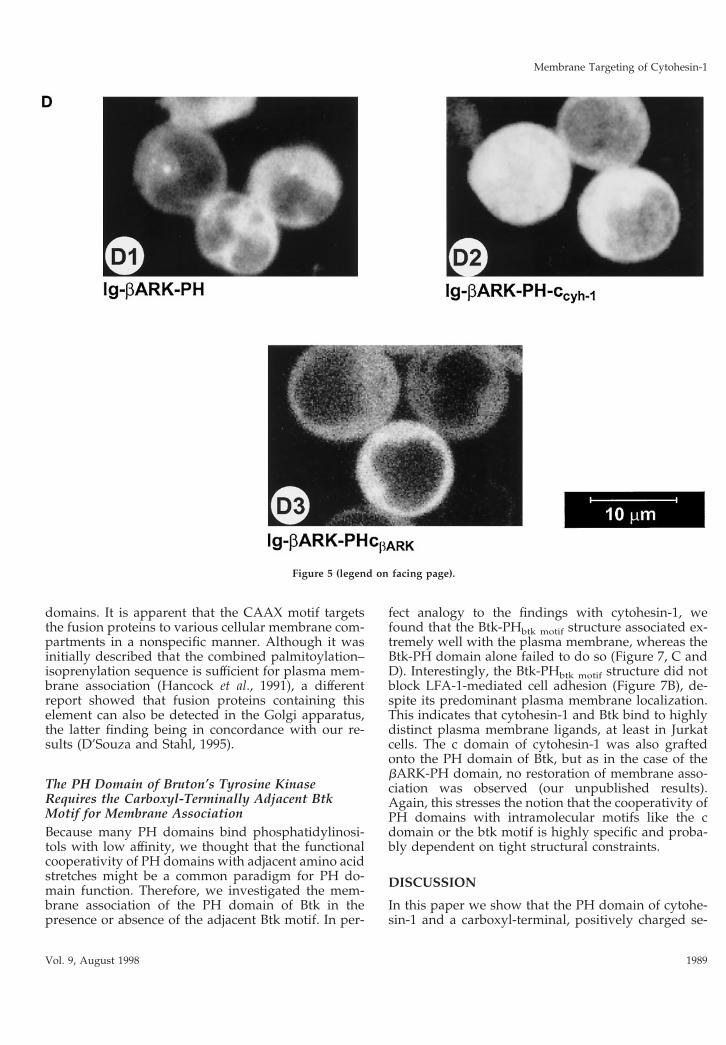
polybasic stretch, carboxyl-terminally adjacent to thePH domain (Figures 1 and 5A). Consequently, the cdomain of cytohesin-1 was grafted onto the bARK PHdomain, and the resulting fusion protein (Ig-bARK-PH-ccyh-1) was compared with the wild-type bARKconstructs (Ig-bARK-PH and Ig-bARK-PHcbARK) forits ability to interfere with LFA-1-mediated adhesionand for its capacity to associate with membranes. Fig-ure 5C shows that the wild-type PH domain of bARKdoes not associate with membranes. Addition of eitherthe wild-type c domain of bARK or of the cytohesin-1PH domain supports membrane association of therespective fusion proteins to a minor but reproducibleextent. However, confocal laser scanning microscopyconfirmed that both proteins were predominantly ex-pressed in the cytoplasm (Figure 5D). As expectedfrom these data, neither fusion protein had a signifi-cant effect on Jurkat cell adhesion compared with thecytohesin-1 Ig-PHccyh-1 construct (Figure 5B). Thus, itappears that the carboxyl-terminal elements of cyto-hesin-1 cooperate specifically in membrane associa-tion and cellular function.
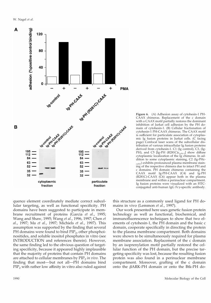
Substitution of the c Domain by a HeterologousMembrane-targeting Element (CAAX Motif) Leadsto Unspecific Association with Various IntracellularMembranes and Therefore Results in IncompleteFunctional ComplementationWe replaced the c domain of cytohesin-1 with theCAAX motif from H-ras, known to be sufficient formembrane association in cells, and tested the respec-tive constructs [Ig-PH-CAAX and Ig-PH (R281C)-CAAX] in both systems. Biochemical analyses showedthat both CAAX constructs retained the ability to as-sociate with cellular membranes, even if the PH do-main was inactivated (Figure 6B). Although the Ig-PH(R281C)-CAAX construct did readily partition into theparticulate (membrane) fraction (Figure 6B), it did notexert dominant negative inhibition of Jurkat cell ad-hesion to ICAM-1 (Figure 6A). This suggests that thecellular function of the cytohesin-1 PH domain re-quires proper in vivo ligand binding and is not onlydependent on its expression in the membrane fractionper se. The intact Ig-PH-CAAX fusion protein, on theother hand, did partially retain in vivo activity withrespect to dominant negative inhibition of Jurkat celladhesion to ICAM-1, thus demonstrating that the Ig-PH-CAAX fusion protein was functional in principle(Figure 6A).
Confocal laser scanning microscopy experimentswere conducted to elucidate the subcellular localiza-tion of the fusion proteins. Figure 6C2 shows that theIg-PHccyh-1 construct is both found in the cytoplasmand associated with the plasma membrane. On theother hand, Ig-PH (Figure 6C3) as well as Ig-PH(R281C)ccyh-1 (Figure 6C5) are only detected in the
Membrane Targeting of Cytohesin-1
Vol. 9, August 1998 1987
cytoplasm, consistent with all functional and biochem-ical analyses above. The CAAX constructs (Figure 6,C4 and C6) were present in multiple membrane com-
partments, predominantly perinuclear membranesand also plasma membrane, appearing very differentin cellular distribution if compared with the wild-type
Figure 5. (A) Outline of the bARK-PH do-main constructs used in this part of the study.In the Ig-bARK-PH-ccyh-1 construct, the c do-main of cytohesin-1 has been fused to the PHdomain of bARK. (B) Adhesion assay of bARK-PH fusion proteins. Neither the wild-typebARK-PH domain and polybasic c-terminusnor a fusion protein of the bARK-PH domainwith the c domain of cytohesin-1 (Ig-bARK-PH-ccyh-1) interferes with induced adhesion of Jur-kat cells to ICAM-1. (C) Cellular fractionationof bARK-PH fusion proteins. Polybasic ele-ments of either bARK or cytohesin-1 do notcooperate significantly with the bARK-PH do-main in membrane association. (D facing page)Confocal laser scans of the subcellular distribu-tion of bARK-PH fusion proteins. All testedfusion proteins are expressed in the cytoplasm.
W. Nagel et al.
Molecular Biology of the Cell1988
domains. It is apparent that the CAAX motif targetsthe fusion proteins to various cellular membrane com-partments in a nonspecific manner. Although it wasinitially described that the combined palmitoylation–isoprenylation sequence is sufficient for plasma mem-brane association (Hancock et al., 1991), a differentreport showed that fusion proteins containing thiselement can also be detected in the Golgi apparatus,the latter finding being in concordance with our re-sults (D’Souza and Stahl, 1995).
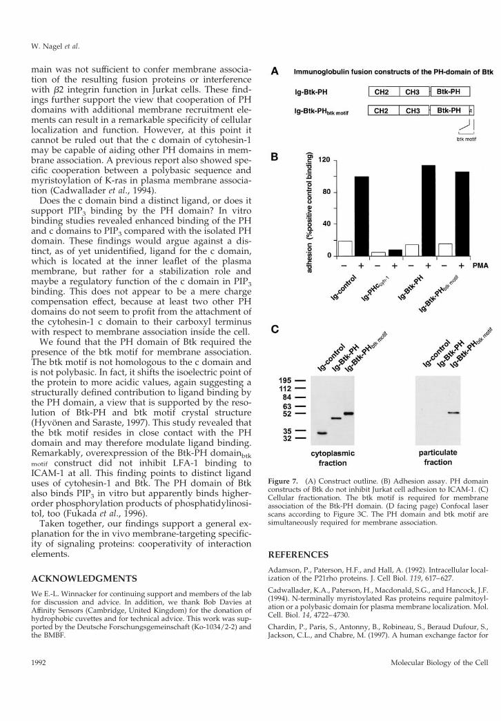
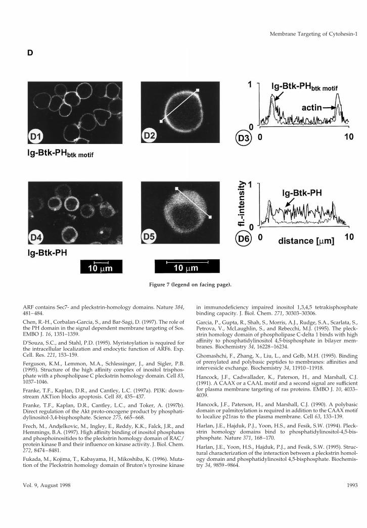
The PH Domain of Bruton’s Tyrosine KinaseRequires the Carboxyl-Terminally Adjacent BtkMotif for Membrane AssociationBecause many PH domains bind phosphatidylinosi-tols with low affinity, we thought that the functionalcooperativity of PH domains with adjacent amino acidstretches might be a common paradigm for PH do-main function. Therefore, we investigated the mem-brane association of the PH domain of Btk in thepresence or absence of the adjacent Btk motif. In per-
fect analogy to the findings with cytohesin-1, wefound that the Btk-PHbtk motif structure associated ex-tremely well with the plasma membrane, whereas theBtk-PH domain alone failed to do so (Figure 7, C andD). Interestingly, the Btk-PHbtk motif structure did notblock LFA-1-mediated cell adhesion (Figure 7B), de-spite its predominant plasma membrane localization.This indicates that cytohesin-1 and Btk bind to highlydistinct plasma membrane ligands, at least in Jurkatcells. The c domain of cytohesin-1 was also graftedonto the PH domain of Btk, but as in the case of thebARK-PH domain, no restoration of membrane asso-ciation was observed (our unpublished results).Again, this stresses the notion that the cooperativity ofPH domains with intramolecular motifs like the cdomain or the btk motif is highly specific and proba-bly dependent on tight structural constraints.
DISCUSSION
In this paper we show that the PH domain of cytohe-sin-1 and a carboxyl-terminal, positively charged se-
Figure 5 (legend on facing page).
Membrane Targeting of Cytohesin-1
Vol. 9, August 1998 1989
quence element coordinately mediate correct subcel-lular targeting, as well as functional specificity. PHdomains have been suggested to participate in mem-brane recruitment of proteins (Garcia et al., 1995;Wang and Shaw, 1995; Wang et al., 1996, 1997; Chen etal., 1997; Ma et al., 1997; Michiels et al., 1997). Thisassumption was supported by the finding that severalPH domains were found to bind PIP2, other phosphoi-nositides, and soluble inositol phosphates in vitro (seeINTRODUCTION and references therein). However,the same finding led to the obvious question of target-ing specificity, because it appeared highly implausiblethat the majority of proteins that contain PH domainsare attached to cellular membranes by PIP2 in vivo. Thefinding that most—but not all—PH domains bindPIP2 with rather low affinity in vitro also ruled against
this structure as a commonly used ligand for PH do-mains in vivo (Lemmon et al., 1997).
Our work presented here uses genetic fusion proteintechnology as well as functional, biochemical, andimmunofluorescence techniques to show that two el-ements of cytohesin-1, the PH domain and the basic cdomain, cooperate specifically in directing the proteinto the plasma membrane compartment. Both domainswere shown to be simultaneously required for plasmamembrane association. Replacement of the c domainby an isoprenylation motif partially restored the cel-lular function of the PH domain, but the precise tar-geting specificity was lost, because the resulting fusionprotein was also found in a perinuclear membranecompartment. Moreover, grafting of the c domainonto the bARK-PH domain or onto the Btk-PH do-
Figure 6. (A) Adhesion assay of cytohesin-1 PH-CAAX chimeras. Replacement of the c domainwith a CAAX motif partially restores the dominantinhibition of Jurkat cell adhesion by the PH do-main of cytohesin-1. (B) Cellular fractionation ofcytohesin-1 PH-CAAX chimeras. The CAAX motifis sufficient for particulate association of cytoplas-mic Ig fusion proteins in Jurkat cells. (C facingpage) Confocal laser scans of the subcellular dis-tribution of various intracellular Ig fusion proteinsderived from cytohesin-1. C1 (Ig control), C3, (Ig-PH), and C5 [Ig-PH (R281C)ccyh-1] show diffusecytoplasmic localization of the Ig chimeras. In ad-dition to some cytoplasmic staining, C2 (Ig-PHc-cyh1) exhibits pronounced plasma membrane stain-ing of the respective chimera due to intact PH andc domains. PH domain chimeras containing theCAAX motif Ig-PH-CAAX (C4) and Ig-PH(R281C)-CAAX (C6) appear both in the plasmamembrane and within a perinuclear compartment.Ig fusion proteins were visualized with an FITC-conjugated anti-human IgG Fcg-specific antibody.
W. Nagel et al.
Molecular Biology of the Cell1990
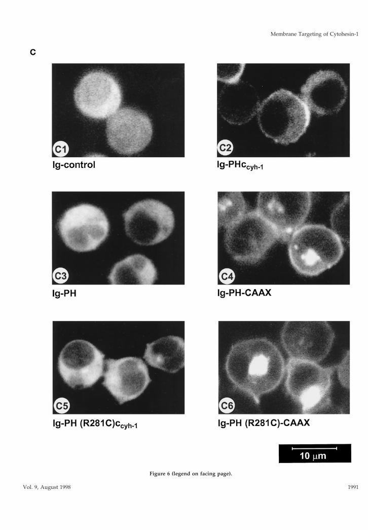
Figure 6 (legend on facing page).
Membrane Targeting of Cytohesin-1
Vol. 9, August 1998 1991
main was not sufficient to confer membrane associa-tion of the resulting fusion proteins or interferencewith b2 integrin function in Jurkat cells. These find-ings further support the view that cooperation of PHdomains with additional membrane recruitment ele-ments can result in a remarkable specificity of cellularlocalization and function. However, at this point itcannot be ruled out that the c domain of cytohesin-1may be capable of aiding other PH domains in mem-brane association. A previous report also showed spe-cific cooperation between a polybasic sequence andmyristoylation of K-ras in plasma membrane associa-tion (Cadwallader et al., 1994).
Does the c domain bind a distinct ligand, or does itsupport PIP3 binding by the PH domain? In vitrobinding studies revealed enhanced binding of the PHand c domains to PIP3 compared with the isolated PHdomain. These findings would argue against a dis-tinct, as of yet unidentified, ligand for the c domain,which is located at the inner leaflet of the plasmamembrane, but rather for a stabilization role andmaybe a regulatory function of the c domain in PIP3binding. This does not appear to be a mere chargecompensation effect, because at least two other PHdomains do not seem to profit from the attachment ofthe cytohesin-1 c domain to their carboxyl terminuswith respect to membrane association inside the cell.
We found that the PH domain of Btk required thepresence of the btk motif for membrane association.The btk motif is not homologous to the c domain andis not polybasic. In fact, it shifts the isoelectric point ofthe protein to more acidic values, again suggesting astructurally defined contribution to ligand binding bythe PH domain, a view that is supported by the reso-lution of Btk-PH and btk motif crystal structure(Hyvonen and Saraste, 1997). This study revealed thatthe btk motif resides in close contact with the PHdomain and may therefore modulate ligand binding.Remarkably, overexpression of the Btk-PH domainbtkmotif construct did not inhibit LFA-1 binding toICAM-1 at all. This finding points to distinct liganduses of cytohesin-1 and Btk. The PH domain of Btkalso binds PIP3 in vitro but apparently binds higher-order phosphorylation products of phosphatidylinosi-tol, too (Fukada et al., 1996).
Taken together, our findings support a general ex-planation for the in vivo membrane-targeting specific-ity of signaling proteins: cooperativity of interactionelements.
ACKNOWLEDGMENTS
We E.-L. Winnacker for continuing support and members of the labfor discussion and advice. In addition, we thank Bob Davies atAffinity Sensors (Cambridge, United Kingdom) for the donation ofhydrophobic cuvettes and for technical advice. This work was sup-ported by the Deutsche Forschungsgemeinschaft (Ko-1034/2-2) andthe BMBF.
REFERENCES
Adamson, P., Paterson, H.F., and Hall, A. (1992). Intracellular local-ization of the P21rho proteins. J. Cell Biol. 119, 617–627.
Cadwallader, K.A., Paterson, H., Macdonald, S.G., and Hancock, J.F.(1994). N-terminally myristoylated Ras proteins require palmitoyl-ation or a polybasic domain for plasma membrane localization. Mol.Cell. Biol. 14, 4722–4730.
Chardin, P., Paris, S., Antonny, B., Robineau, S., Beraud Dufour, S.,Jackson, C.L., and Chabre, M. (1997). A human exchange factor for
Figure 7. (A) Construct outline. (B) Adhesion assay. PH domainconstructs of Btk do not inhibit Jurkat cell adhesion to ICAM-1. (C)Cellular fractionation. The btk motif is required for membraneassociation of the Btk-PH domain. (D facing page) Confocal laserscans according to Figure 3C. The PH domain and btk motif aresimultaneously required for membrane association.
W. Nagel et al.
Molecular Biology of the Cell1992
ARF contains Sec7- and pleckstrin-homology domains. Nature 384,481–484.
Chen, R.-H., Corbalan-Garcia, S., and Bar-Sagi, D. (1997). The role ofthe PH domain in the signal dependent membrane targeting of Sos.EMBO J. 16, 1351–1359.
D’Souza, S.C., and Stahl, P.D. (1995). Myristoylation is required forthe intracellular localization and endocytic function of ARF6. Exp.Cell. Res. 221, 153–159.
Ferguson, K.M., Lemmon, M.A., Schlessinger, J., and Sigler, P.B.(1995). Structure of the high affinity complex of inositol trisphos-phate with a phospholipase C pleckstrin homology domain. Cell 83,1037–1046.
Franke, T.F., Kaplan, D.R., and Cantley, L.C. (1997a). PI3K: down-stream AKTion blocks apoptosis. Cell 88, 435–437.
Franke, T.F., Kaplan, D.R., Cantley, L.C., and Toker, A. (1997b).Direct regulation of the Akt proto-oncogene product by phosphati-dylinositol-3,4-bisphosphate. Science 275, 665–668.
Frech, M., Andjelkovic, M., Ingley, E., Reddy, K.K., Falck, J.R., andHemmings, B.A. (1997). High affinity binding of inositol phosphatesand phosphoinositides to the pleckstrin homology domain of RAC/protein kinase B and their influence on kinase activity. J. Biol. Chem.272, 8474–8481.
Fukada, M., Kojima, T., Kabayama, H., Mikoshiba, K. (1996). Muta-tion of the Pleckstrin homology domain of Bruton’s tyrosine kinase
in immunodeficiency impaired inositol 1,3,4,5 tetrakisphosphatebinding capacity. J. Biol. Chem. 271, 30303–30306.
Garcia, P., Gupta, R., Shah, S., Morris, A.J., Rudge, S.A., Scarlata, S.,Petrova, V., McLaughlin, S., and Rebecchi, M.J. (1995). The pleck-strin homology domain of phospholipase C-delta 1 binds with highaffinity to phosphatidylinositol 4,5-bisphosphate in bilayer mem-branes. Biochemistry 34, 16228–16234.
Ghomashchi, F., Zhang, X., Liu, L., and Gelb, M.H. (1995). Bindingof prenylated and polybasic peptides to membranes: affinities andintervesicle exchange. Biochemistry 34, 11910–11918.
Hancock, J.F., Cadwallader, K., Paterson, H., and Marshall, C.J.(1991). A CAAX or a CAAL motif and a second signal are sufficientfor plasma membrane targeting of ras proteins. EMBO J. 10, 4033–4039.
Hancock, J.F., Paterson, H., and Marshall, C.J. (1990). A polybasicdomain or palmitoylation is required in addition to the CAAX motifto localize p21ras to the plasma membrane. Cell 63, 133–139.
Harlan, J.E., Hajduk, P.J., Yoon, H.S., and Fesik, S.W. (1994). Pleck-strin homology domains bind to phosphatidylinositol-4,5-bis-phosphate. Nature 371, 168–170.
Harlan, J.E., Yoon, H.S., Hajduk, P.J., and Fesik, S.W. (1995). Struc-tural characterization of the interaction between a pleckstrin homol-ogy domain and phosphatidylinositol 4,5-bisphosphate. Biochemis-try 34, 9859–9864.
Figure 7 (legend on facing page).
Membrane Targeting of Cytohesin-1
Vol. 9, August 1998 1993

Hyvonen, M., Macias, M.J., Nilges, M., Oschkinat, H., Saraste, M.,and Wilmanns, M. (1995). Structure of the binding site for inositolphosphates in a PH domain. EMBO J. 14, 4676–4685.
Hyvonen, M., and Saraste, M. (1997). Structure of the PH domainand Btk motif from Bruton’s tyrosine kinase: molecular explanationsfor X-linked agammaglobulinaemia. EMBO J. 16, 3396–3404.
Kim, C.G., Park, D., and Rhee, S.G. (1996). The role of carboxyl-terminal basic amino acids in Gqalpha-dependent activation, par-ticulate association, and nuclear localization of phospholipase C-beta1. J. Biol. Chem. 271, 21187–21192.
Klarlund, J.K., Guilherme, A., Holik, J.J., Virbasius, J.V., Chawla, A.,and Czech, M.P. (1997). Signaling by phosphoinositide-3,4,5-trisphosphate through proteins containing pleckstrin and Sec7 ho-mology domains. Science 275, 1927–1930.
Kolanus, W., Nagel, W., Schiller, B., Zeitlmann, L., Godar, S., Stock-inger, H., and Seed, B. (1996). Alpha L beta 2 integrin/LFA-1 bind-ing to ICAM-1 induced by cytohesin-1, a cytoplasmic regulatorymolecule. Cell 86, 233–242.
Kreck, M.L., Freeman, J.L., Abo, A., and Lambeth, J.D. (1996). Mem-brane association of Rac is required for high activity of the respira-tory burst oxidase. Biochemistry 35, 15683–15692.
Kubiseski, T.J., Chook, Y.M., Parris, W.E., Rozakis, A.M., and Paw-son, T. (1997). High affinity binding of the pleckstrin homologydomain of mSos1 to phosphatidylinositol (4,5)-bisphosphate. J. Biol.Chem. 272, 1799–1804.
Kwong, J., and Lublin, D.M. (1995). Amino-terminal palmitate orpolybasic domain can provide required second signal to myristatefor membrane binding of p56lck. Biochem. Biophys. Res. Commun.207, 868–876.
Lemmon, M.A., Falasca, M., Ferguson, K.M., and Schlessinger, J.(1997). Regulatory recruitment of signalling molecules to the cellmembrane by pleckstrin homology domains. Trends Cell Biol. 7,237–242.
Lemmon, M.A., Ferguson, K.M., O’Brien, R., Sigler, P.B., andSchlessinger, J. (1995). Specific and high-affinity binding of inositolphosphates to an isolated pleckstrin homology domain. Proc. Natl.Acad. Sci. USA 92, 10472–10476.
Lemmon, M.A., Ferguson, K.M., and Schlessinger, J. (1996). PHdomains: diverse sequences with a common fold recruit signalingmolecules to the cell surface. Cell 85, 621–624.
Ma, A.D., Brass, L.F., and Abrams, C.S. (1997). Pleckstrin associateswith plasma membranes and induces the formation of membraneprojections: requirements for phosphorylation and the NH2-termi-nal PH domain. J. Cell Biol. 136, 1071–1079.
Meller, N., Liu, Y.C., Collins, T.L., Bonnefoyberard, N., Baier, G.,Isakov, N., and Altman, A.R.A. (1996). Direct interaction betweenprotein kinase c theta (pkc theta) and 14 3 3 tau in t cells: 14 3 3overexpression results in inhibition of pkc theta translocation andfunction. Mol. Cell. Biol. 16, 5782–5791.
Michiels, F., Stam, J.C., Hordijk, P.L., van der Kammen, R.A., Ruuls,V.S.L., Feltkamp, C.A., and Collard, J.G. (1997). Regulated mem-brane localization of Tiam1, mediated by the NH2-terminal pleck-strin homology domain, is required for Rac-dependent membraneruffling and C-Jun NH2-terminal kinase activation. J. Cell Biol. 137,387–398.
Miki, H., Miura, K., and Takenawa, T. (1996). N-WASP, a novelactin-depolymerizing protein, regulates the cortical cytoskeletal re-arrangement in a PIP2-dependent manner downstream of tyrosinekinases. EMBO J. 15, 5326–5335.
Mitchell, D.A., Farh, L., Marshall, T.K., and Deschenes, R.J. (1994). Apolybasic domain allows nonprenylated Ras proteins to function inSaccharomyces cerevisiae. J. Biol. Chem. 269, 21540–21546.
Musacchio, A., Gibson, T., Rice, P., Thompson, J., Saraste, M. (1993).The PH domain: a common piece in structural pathwork of signal-ling protein. Trends Biochem. Sci. 18, 343–348.
Nagel, W., Zeitlmann, L., Schilcher P., Geiger, C., Kolanus, W.(1998). Phosphoinositide 3-OH kinase activates the beta-2 integrinadhesion pathway and induces the recruitment of cytohesin-1.J. Biol. Chem. 273, 14853–14861.
Newman, C.M., Giannakouros, T., Hancock, J.F., Fawell, E.H., Arm-strong, J., and Magee, A.I. (1992). Post-translational processing ofSchizosaccharomyces pombe YPT proteins. J. Biol. Chem. 267, 11329–11336.
Patki, V., Virbasius, J., Lane, W.S., Toh, B.H., Shpetner, H.S., andCorvera, S. (1997). Identification of an early endosomal proteinregulated by phosphatidylinositol 3-kinase. Proc. Natl. Acad. Sci.USA 94, 7326–7330.
Pawson, T. (1995). Protein modules and signalling networks. Nature373, 573–579.
Pitcher, J.A., Touhara, K., Payne, E.S., and Lefkowitz, R.J. (1995).Pleckstrin homology domain-mediated membrane association andactivation of the beta-adrenergic receptor kinase requires coordinateinteraction with G beta gamma subunits and lipid. J. Biol. Chem.270, 11707–11710.
Romeo, C., and Seed, B. (1991). Cellular immunity to HIV activatedby CD4 fused to T cell or Fc receptor polypeptides. Cell 64, 1037–1046.
Salim, K. et al. (1996). Distinct specificity in the recognition ofphosphoinositides by the pleckstrin homology domains of dynaminand Bruton’s tyrosine kinase. EMBO J. 15, 6241–6250.
Shpetner, H., Joly, M., Hartley, D., and Corvera, S. (1996). Potentialsites of PI-3 kinase function in the endocytic pathway revealed bythe PI-3 kinase inhibitor, wortmannin. J. Cell Biol. 132, 595–605.
Soneoka, Y., Kingsman, S.M., and Kingsman, A.J. (1997). Mutagen-esis analysis of the murine leukemia virus matrix protein: identifi-cation of regions important for membrane localization and intracel-lular transport. J. Virol. 71, 5549–5559.
Touhara, K., Koch, W.J., Hawes, B.E., and Lefkowitz, R.J. (1995).Mutational analysis of the pleckstrin homology domain of the beta-adrenergic receptor kinase. Differential effects on G beta gamma andphosphatidylinositol 4,5-bisphosphate binding. J. Biol. Chem. 270,17000–17005.
Walz, G., Aruffo, A., Kolanus, W., Bevilacqua, M., and Seed, B.(1990). Recognition by ELAM-1 of the sialyl-Lex determinant onmyeloid and tumor cells. Science 250, 1132–1135.
Wang, D.S., Deng, T., and Shaw, G. (1997). Membrane binding andenzymatic activation of a Dbl homology domain require the neigh-boring pleckstrin homology domain. Biochem. Biophys. Res. Com-mun. 234, 183–189.
Wang, D.S., Miller, R., Shaw, R., and Shaw, G. (1996). The pleckstrinhomology domain of human beta I sigma II spectrin is targeted tothe plasma membrane in vivo. Biochem. Biophys. Res. Commun.225, 420–426.
Wang, D.S., and Shaw, G. (1995). The association of the C-terminalregion of beta I sigma II spectrin to brain membranes is mediated bya PH domain, does not require membrane proteins, and coincideswith a inositol-1,4,5 triphosphate binding site. Biochem. Biophys.Res. Commun. 217, 608–615.
Zheng, J., Cahill, S.M., Lemmon, M.A., Fushman, D., Schlessinger, J.,and Cowburn, D. (1996). Identification of the binding site for acidicphospholipids on the pH domain of dynamin: implications forstimulation of GTPase activity. J. Mol. Biol. 255, 14–21.
W. Nagel et al.
Molecular Biology of the Cell1994
Copyright © 2022 FDOKUMEN