The Pathways Project - University of Birmingham
226
THE PATHWAYS PROJECT: DEVELOPING GUIDELINES TO FACILITATE THE DIAGNOSIS OF CHILDHOOD BRAIN TUMOURS Dr Sophie Wilne BA (Hons) Cantab, MB BS, MRCPCH A thesis submitted in partial fulfilment of the requirements for the degree of Doctor of Medicine of the University of Birmingham Division of Reproductive and Child Health Academic Department of Obstetrics and Gynaecology The University of Birmingham 2011
-
Upload
khangminh22 -
Category
Documents
-
view
1 -
download
0
Transcript of The Pathways Project - University of Birmingham

THE PATHWAYS PROJECT: DEVELOPING
GUIDELINES TO FACILITATE THE DIAGNOSIS OF
CHILDHOOD BRAIN TUMOURS
Dr Sophie Wilne
BA (Hons) Cantab, MB BS, MRCPCH
A thesis submitted in partial fulfilment of the requirements for the degree of
Doctor of Medicine of the University of Birmingham
Division of Reproductive and Child Health
Academic Department of Obstetrics and Gynaecology
The University of Birmingham
2011

University of Birmingham Research Archive
e-theses repository This unpublished thesis/dissertation is copyright of the author and/or third parties. The intellectual property rights of the author or third parties in respect of this work are as defined by The Copyright Designs and Patents Act 1988 or as modified by any successor legislation. Any use made of information contained in this thesis/dissertation must be in accordance with that legislation and must be properly acknowledged. Further distribution or reproduction in any format is prohibited without the permission of the copyright holder.

ABSTRACT
Aims:
The Pathways project was undertaken to devise guidelines to facilitate rapid diagnosis of
paediatric brain tumours.
Methods:
A systematic review and meta-analysis of published data on paediatric brain tumour
presentation and analysis of the presentation of children newly diagnosed with a brain tumour
at four oncology centres was undertaken. The results informed a professional consensus
process.
Results:
74 papers met the inclusion criteria for the meta-analysis. 56 symptoms and signs at diagnosis
were identified. The most frequent symptoms and signs at diagnosis were: headache (33%),
nausea and vomiting (32%), abnormalities of gait and coordination (27%), and papilloedema
(13%). 139 patients were recruited to a multi-centre cohort study. Symptoms and signs at
disease onset and at diagnosis and factors associated with a long and short symptom interval
were determined. A shorter symptom interval was associated with nausea and vomiting and
motor system abnormalities. A longer symptom interval was associated with head tilt, cranial
nerve palsies, endocrine and growth abnormalities and reduced visual acuity. A multi-
disciplinary workshop and Delphi consensus voting were used to translate the evidence into a
clinical guideline comprising 76 statements advising on the identification and assessment of
children who may have a brain tumour.

AGKNOWLEDGEMENTS
Many people were involved in the Pathways project from its inception to completion. The
original project was devised by Professor Walker, Professor Grundy, Professor Kennedy,
Professor Collier and Mr Punt and it has been strongly supported throughout by the Samantha
Dickson Brain Tumour Trust. I would like to thank all my supervisors and project
collaborators for their advice, support and encouragement throughout. Special thanks must go
to Professor Grundy, my MD supervisor, and to Professor Walker both of whom provided
excellent mentorship throughout the project and greatly expanded my academic horizons. I
would like to thank Professor Collier for her excellent statistical advice and common sense
and Professor Kennedy for his insightful summaries and for providing a more general
perspective on the project and its aims. I must thank Dr Koller for her invaluable help with the
Delphi process, Dr Jenkins, Ms Mackie and Mrs Grout for their data collection work and Mrs
Franklin for providing secretarial support and advice on the inner workings of the University.
The project could not have been completed without the generosity the families who agreed for
their children‟s data to be included and the commitment and hard work of the Delphi
workshop participants and the Delphi consensus voting group. I would like to thank Dr
English and Dr Peet for agreeing to act as Birmingham University Supervisors following
Professor Grundy‟s move to Nottingham. Finally I must thank my husband, Tim, for his
excellent IT advice and support throughout and my children, Edward, Caitlin and Harry, for
tolerating my absences and for providing a welcome diversion from work.
Funding
The guideline was developed with a grant (grant number RG10044964) from the Big Lottery
Fund. The grant was applied for by the Samantha Dickson Brain Tumour Trust (registered
charity no.1060627) on behalf of the Children‟s Brain Tumour Research Centre, University of
Nottingham.

CONTENTS
CHAPTER 1: INTRODUCTION ..................................................................................... 1 1.1: Epidemiology of childhood brain tumours ........................................................................ 1 1.2: Clinical Guidelines ............................................................................................................ 4 1.3: The Delphi process ........................................................................................................... 5 1.4: Justification for the Pathways project ............................................................................... 6 1.5: Currently available guidance ............................................................................................ 9
CHAPTER 2: MATERIALS AND METHODS ............................................................. 14 2.1: Literature review methods .............................................................................................. 15
2.1.1: Identification of studies and inclusion criteria ................................................................... 16 2.1.2: Data collection .................................................................................................................... 16 2.1.3: Statistical analysis .............................................................................................................. 16
2.2: Cohort study methods ..................................................................................................... 18 2.2.1: Data collection .................................................................................................................... 18 2.2.2: Statistical analysis .............................................................................................................. 19 2.2.3: Ethics .................................................................................................................................. 19
2.3: Multidisciplinary workshop ............................................................................................ 19 2.4: Delphi process................................................................................................................. 21
CHAPTER 3: RESULTS ............................................................................................... 23 3.1: Literature review results................................................................................................. 23 3.2: Cohort study results ....................................................................................................... 32 3.2.1: Patient characteristics .................................................................................................. 32
3.2.2: Symptoms and signs - brain tumours ................................................................................. 33 3.2.3: Symptoms and signs – spinal cord tumours ....................................................................... 35 3.2.4: Symptom interval ............................................................................................................... 36 3.3.5: Referral pathways and imaging .......................................................................................... 37
3.3: Multidisciplinary workshop results ................................................................................ 40 3.3.1: Headache ............................................................................................................................ 40 3.3.2: Imaging ............................................................................................................................... 41 3.3.3: Referral pathways ............................................................................................................... 41 3.3.4: Motor assessment ............................................................................................................... 41 3.3.5: Non-specific symptoms ...................................................................................................... 42 3.3.6: Visual assessment ............................................................................................................... 42 3.3.7: Predisposing factors ........................................................................................................... 43 3.3.8: Nausea and vomiting .......................................................................................................... 43 3.3.9: Assessment of growth ........................................................................................................ 44
3.4: Delphi consensus process results ..................................................................................... 45 3.4.1: Delphi process round one ................................................................................................... 45 3.4.2: Delphi questionnaire round one .......................................................................................... 46 3.4.3: Delphi questionnaire round one results .............................................................................. 69 3.4.4: Delphi process round two ................................................................................................... 78 3.4.5: Delphi questionnaire round two ......................................................................................... 79 3.4.6: Delphi questionnaire round two results .............................................................................. 98 3.4.7: Delphi process round three .............................................................................................. 100 3.4.8: Delphi questionnaire round three ..................................................................................... 101 3.4.9: Delphi questionnaire round three results .......................................................................... 107
CHAPTER 4: CONCLUSIONS FROM THE EVIDENCE REVIEW ......................... 110

4.1: Conclusions from the systematic literature review and meta-analysis ........................... 110 4.2: Conclusions from the cohort study ................................................................................ 115
CHAPTER 5: PATHWAYS PROJECT GUIDELINE ................................................ 119 5.1: The diagnosis of brain tumours in children – an evidenced based guideline to assist
healthcare professionals in the assessment of children presenting with symptoms and signs
that may be due to a brain tumour. (quick reference guide) ................................................ 119 5.1.1 Best practice ...................................................................................................................... 119 5.1.1a: Consultation .................................................................................................................... 119 5.1.1b: Referral ........................................................................................................................... 120 5.1.1c: Imaging ........................................................................................................................... 120 5.1.1d: Feedback ......................................................................................................................... 120 5.1.2. Predisposing factors ......................................................................................................... 121 5.1.3. Presentation and assessment of a child with a potential brain tumour ............................. 121 5.1.3a: Presenting symptoms and signs ...................................................................................... 121 5.1.3b: History ............................................................................................................................ 122 5.1.3c: Assessment ...................................................................................................................... 122 5.1.4. Signs and Symptoms of a child with a potential brain tumour ........................................ 123 5.1.4a: Headache ......................................................................................................................... 123 5.1.4b: Nausea and vomiting ...................................................................................................... 123 5.1.4c: Visual symptoms and signs ............................................................................................. 124 5.1.4d: Motor symptoms and signs ............................................................................................. 125 5.1.4e: Growth and development ................................................................................................ 126 5.1.4f: Behaviour ........................................................................................................................ 127
5.2: The diagnosis of brain tumours in children – an evidenced based guideline to assist
healthcare professionals in the assessment of children presenting with symptoms and signs
that may be due to a brain tumour: ..................................................................................... 129 5.2.1: Aim of the guideline ......................................................................................................... 129 5.2.2: Scope ................................................................................................................................ 129 5.2.3: Levels of evidence and recommendation grades: ............................................................. 130 5.2.4a. Best practice - consultation ............................................................................................. 132 5.2.4b. Best practice - referral ..................................................................................................... 134 5.2.4c. Best practice – imaging ................................................................................................... 135 5.2.4d. Best practice – feedback ................................................................................................. 137 5.2.5. Predisposing factors .......................................................................................................... 137 5.2.6a. Presentation and assessment of a child with a potential brain tumour ............................ 138 5.2.6b: History ............................................................................................................................ 141 5.2.6c: Assessment ...................................................................................................................... 142 5.2.7a: Headache ......................................................................................................................... 144 5.2.7b: Nausea and vomiting ...................................................................................................... 148 5.2.5c: Visual symptoms and signs ............................................................................................. 150 5.2.7d: Motor symptoms and signs ............................................................................................. 158 5.2.7e: Growth and development ................................................................................................ 164 5.2.7f: Behaviour ........................................................................................................................ 166
CHAPTER 6: SUMMARY AND CONCLUSIONS ..................................................... 168 6.1: Guideline implementation ............................................................................................. 170 6.2: Future work .................................................................................................................. 172 6.3: Conclusion .................................................................................................................... 174
APPENDIX 1 – COMMENTS ON STATEMENTS FROM DELPHI ROUND ONE
NOT REACHING CONSENSUS ................................................................................. 175

APPENDIX 2 – COMMENTS ON STATEMENTS FROM DELPHI ROUND TWO
NOT REACHING CONSENSUS ................................................................................. 199
APPENDIX 3 – WORKSHOP PARTICIPANTS ......................................................... 207
APPENDIX 4 – DELPHI PANEL PARTICIPANTS ................................................... 208
REFERENCES ............................................................................................................ 211

FIGURES Figure 1: Guideline development ......................................................................................... 15 Figure 2: Progress through the meta-analysis ....................................................................... 25 Figure 3: Frequency of symptoms and signs in children with intracranial tumours - analysis by
age and neurofibromatosis status .......................................................................................... 29 Figure 4: Frequency of symptoms and signs in children with a central nervous system tumour
- analysis by tumour location ............................................................................................... 30 Figure 5: Central nervous system tumour presentation ......................................................... 31 Figure 6: Relationship between patient age and brain tumour presentation ........................... 39 Figure 7: Percentage in each score band for the Delphi statements in round one ................... 69 Figure 8: Percentage in each score band for the Delphi statements in round two ................... 98 Figure 9: Percentage in each score band for the Delphi statements in round three ............... 107 Figure 10: Progress through the Delphi process .................................................................. 108 Figure 11: Delphi process participants ................................................................................ 109 Figure 12: Quick reference guide ....................................................................................... 128
TABLES Table 1: World Health Organisation classification and malignancy grading of central nervous
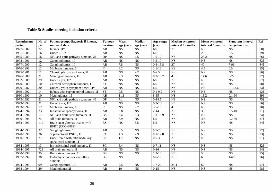
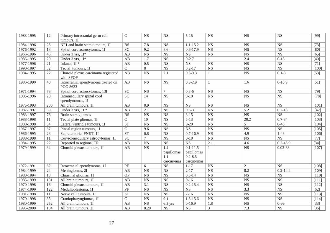
system malignancies .............................................................................................................. 3 Table 2: Attributes of high quality guidelines ......................................................................... 5 Table 3: Published symptom intervals for childhood brain tumours. ....................................... 8 Table 4: Topics covered by workshop groups ....................................................................... 21 Table 5: Studies meeting inclusion criteria ........................................................................... 26 Table 6: Tumour diagnoses of children recruited to the cohort study .................................... 32 Table 7: Symptom and sign complexes at symptom onset and at diagnosis in children with
brain tumours ....................................................................................................................... 34 Table 8: Association between symptoms and signs and symptom interval ............................ 37

1
CHAPTER 1: INTRODUCTION
1.1: Epidemiology of childhood brain tumours
One in every 550-600 children in the United Kingdom (UK) will be affected by cancer by
their fifteenth birthday. 1,500 children are diagnosed annually with cancer in the UK and a
third of these will have a central nervous system (CNS) tumour, 95-98% of which will be
brain tumours [1-5]. CNS tumours are the second most frequent malignancy in children (after
leukaemia) and are now the commonest cancer cause of death, with an annual mortality of
nine per million (80 to 100 children annually in the UK)[6]. 60% of survivors are left with
pronounced disability[7-10].
Brain tumours are not a single entity; there are several distinct histopathological subtypes
whose incidence varies with patient age and anatomical location. In order to allow national
and international collaboration in epidemiological studies and clinical trials the pathological
classification and grading of brain tumours has been standardised since 1979 [11]. The most
recent classification, the fourth edition of the WHO classification of tumours of the central
nervous system, was published in 2007 [11]. This lists ten central nervous system tumour
types that commonly occur in children (table 1).
The age standardised incidence rate for CNS tumours in UK children aged 0-14 years is 27
per million [5, 12]. Astrocytomas are the most common childhood CNS tumour, accounting
for 40-55% of specified tumours (incidence 10 per million). Their malignancy ranges from
low grade pilocytic astrocytomas through to the highly malignant glioblastome multiforme,
although tumour location is as important in determining morbidity and mortality as
histopathological grade. Astrocytomas are equally split between the supra and infratentorial
brain [13] and occur with an equal incidence throughout childhood. The male to female ratio
is 1:1.1.

2
The subgroup embryonal tumours includes medulloblastoma, atypical teratoid rhabdoid
tumours and central nervous system primitive neuroectodermal tumours (PNET). They are the
second most common group of tumours, accounting for 20-30% of specified CNS tumours
(incidence 6 per million). Approximately 70% of embryonal tumours are medulloblastoma
[13]. The highest incidence occurs age 1-4 years, it is slightly lower in infants and children
aged 5-9 years and decreases to approximately half by age 10-14. The male to female ratio is
1.6:1.7.
Ependymomas account for 10-15% of specified CNS tumours (incidence 3 per million). Two
thirds are infratentorial [13]. Ependymoma is twice as common in children aged 0-4 as it is in
older children. The male to female ration is 1.2:1.3. Other gliomas have a similar incidence to
ependymomas. The incidence of other specified tumours (excluding germ cell tumours) is less
than 3 per million. Childhood intracranial germ cell tumours have an incidence of 1 per
million.
Less information is available on the incidence of brain tumours in adolescence as their care is
divided between paediatric and adult services and their details are not recorded in paediatric
tumour registries (adult registries in many countries have a much lower ascertainment rate).
Total incidence is lower than for children overall but similar to that observed age 10-14 years.
Astrocytomas are again the most frequent histological subtype however embryonal tumours
are relatively rare in this age group.
The reported incidence of childhood brain tumours rose by 20% between the 1970s and the
1980s. Most data is available from the Surveillance, Epidemiological and End Results (SEER)
program which receives notification of cancer diagnoses from approximately 10% of the USA
population [14]. In the UK the Yorkshire Tumour Registry also shows a similar increase with
the incidence of CNS tumour rising from 25.6 to 34.9 per million per year from 1974 to 1995
[15]. The incidence of astrocytomas in 0 to 4 year olds increased from 8.3 to 11 per million

3
and the incidence of embryonal tumours from 5.2 to 9.6 per million. The average annual
increase was 1.8% for all CNS tumours and 3.0% for embryonal tumours.
Table 1: World Health Organisation classification and malignancy grading of central
nervous system malignancies
TUMOUR FAMILY TUMOUR GRADE 1 GRADE 2 GRADE 3 GRADE 4 Astrocytic tumours Pilocytic astrocytoma
Pilomyxoid astrocytoma
Diffuse astrocytma
Anaplastic astrocytoma
Glioblastoma
Oligodendroglial
tumours
Oligodendroglioma
Anaplastic oligodendroglioma
Oligoastrocytic tumours Oligoastrocytoma
Anaplastic oligoastrocytoma
Ependymal tumours Myxopapillary ependymoma
Subependymoma
Ependymoma
Anaplastic ependymoma
Choroid plexus tumours Choroid plexus papilloma
Choroid plexus carcinoma
Neuronal and mixed
neuronal-glial tumours
Ganglioglioma
DNET
Central neurocytoma
Cerebellar liponeurocytoma
Rosette-forming glioneuronal
tumour of the fourth ventricle
Pineal tumours Pineocytoma
Pineoblastoma
Pineal parenchymal tumour of
indeterminate differentiation
Embryonal tumours Medulloblastoma
AT/RT
CNS PNET
Meningeal tumours
Meningioma
Atypical meningioma
Anaplastic / malignant
meningioma
Tumours of the sellar
region
Craniopharyngioma
DNET = Dysembryoplastic neuroepithelial tumour
AT/RT = Atypical teratoid / rhabdoid tumour
PNET – primitive neuroectodermal tumour
Analysis of the SEER data shows that the pattern of increase in incidence best fit with a
“jump” from a period of low incidence to one of high incidence around 1985 [16]. This
coincided with the widespread introduction of magnetic resonance imaging (MRI), suggesting
that the increased incidence may be a result of improved diagnosis and reporting. Use of

4
stereotactic biopsy also increased during the same time which may have allowed
identification and biopsy (and hence diagnosis) of lesions that would have previously
remained unidentified. This is supported by the absence of a similar increase in mortality from
CNS tumours. However, much of the increased incidence was in low-grade astrocytomas and
gliomas, these have high survival rates and even ultimately fatal tumours often show slow
progression, so any increase in mortality would be relatively small and gradual and therefore
hard to detect.
1.2: Clinical Guidelines
Clinical guidelines are an essential component of appropriate, efficient and cost effective
health care[17]. They are systematically developed statements which support clinicians and
patients in making decisions about the appropriate management of specific conditions and
situations with the aim of improving the quality of health care[18]. Properly developed,
communicated and implemented guidelines improve patient care.
Guidelines should ideally be based on high quality contemporary evidence. Systematic
reviews and meta-analysis provide the best quality evidence[19] and these methods were used
in the Pathways guideline to summarise the current evidence on paediatric brain tumour
presentation. In the absence of high quality evidence it is necessary to use other sources of
information, these may include cohort and case-control studies and case reports. Evidence
from the Pathways‟ project cohort study supports many of the guideline recommendations. In
the absence of any evidence it is appropriate to use expert opinion and formal consensus
techniques, such as the Delphi process, are a means of collating and summarising professional
expertise[20]. Professional expertise is particularly useful for recommendations that are not
based on a clinical question or therapeutic intervention such as, in the Pathways project,
recommendations on symptom specificity, referral pathways, imaging indications and

5
acceptable waiting times. A high quality guideline should have the attributes listed in table 2
[21]:
Table 2: Attributes of high quality guidelines
Valid Correctly interpreting the evidence in order that, when followed,
guidelines lead to improvements in health
Reproducible Given the same evidence, another guideline group would produce
similar recommendations
Reliable Given the same clinical circumstances, another health
professional would apply them similarly
Representative of key
disciplines and interests
All key disciplines and interests (including patients) have
contributed to the development of the guideline
Clinically applicable The target population (those whose health the guideline aims to
improve) is defined in accordance with scientific evidence
Clinically flexible The guidelines identify where exceptions to the recommendations
lie, and indicate how patient preferences are to be incorporated in
decision making.
Clearly expressed The guidelines use precise definitions, unambiguous language
and a user-friendly format
Well documented The guidelines‟ methodology records all participants, any
assumptions and methods and clearly links recommendations to
the available evidence
Scheduled for review The guidelines state when, how and by whom they are to be
reviewed.
1.3: The Delphi process
A Delphi process is a means of developing a consensus between individuals. It provides a
structured method of consultation that minimises bias. A Delphi process involves a series of
sequential questionnaires interspersed by controlled feedback that seek to assess the extent of
agreement (consensus measurement) and resolve disagreement (consensus development)
among a group of experts [22]. The Delphi process aims to maximise the benefits from
consulting a large number of experts over a short period of time while minimising the
disadvantages associated with more traditional collective decision making processes e.g.
committee meetings or steering groups.
A Delphi process requires the selection of a Delphi panel, the presentation of the information
that the panel is to review as a series of statements and the setting of a consensus level i.e. the

6
level of agreement required for a statement to be deemed as agreed upon by the Delphi panel.
The statements are sent to the Delphi panel members and they are asked to rank their
agreement with the statements (usually by means of a 9 point Likert scale) and to comment on
the statements, particularly those with which they disagree. The rankings for each statement
are collated and any statement that has achieved the pre-determined level of consensus is
accepted. The results of the rankings are returned to the Delphi group. In a modified Delphi
process (usually undertaken in guideline development) statements which have not achieved
consensus are modified in light of the feedback received from the Delphi panel and reissued.
This process is continued until all statements have achieved consensus or until feedback
suggests that consensus is not going to be achieved.
A Delphi process therefore enables free discussion of views, allows individuals to change
their personal opinion, can involve all groups with an interest in the area under review and can
be completed within a reasonable time frame. A credible Delphi process must include a clear
decision trail that defends the appropriateness of the method to address the problem selected,
the choice of expert panel, and the consensus level selected [23]. With these included it is a
practical and validated method for guideline development [20, 24].
1.4: Justification for the Pathways project
Life-threatening clinical conditions in childhood are seen infrequently in developed countries
[6, 25]. Identification of the few serious diagnoses from the many self-limiting conditions and
fluctuations in developmental processes and behaviour is a major diagnostic challenge for
both primary and secondary health care [26, 27]. This is particularly true for childhood brain
tumours as many of the initial symptoms and signs also occur with other much more common
and less serious childhood disorders such as gastroenteritis, migraine and behavioural
problems.

7
The symptom interval of an illness is defined as the time period between symptom onset and
diagnosis. For childhood cancers the symptom interval varies greatly with disease. The mean
and median symptom interval for unselected (i.e. all brain tumour types) cohorts and case
series of children with CNS tumours published over the last 15 years ranges from 1.8 to 9.8
and 1 to 3 months respectively (see table 3) [28-42]. In comparison, the mean and median
symptom interval for children with Wilms‟ tumour has been reported as 3.3 and 3.6 months
respectively and for children with leukaemia as and 1.0 and 1.7 months[43]. In a study of 247
children with cancer (79 with a brain tumour, 45 with Wilms‟ tumour and 123 with acute
leukaemia), 84% of the children with Wilms‟ tumour and 80% of those with leukaemia were
diagnosed within a month of symptom onset in comparison to 38% of those with a brain
tumour[44].
Multiple factors contribute to the prolonged symptom interval experienced by children with
brain tumours. Childhood brain tumours are relatively rare and have a very varied
presentation. The symptoms and signs that proceed diagnosis are diverse, fluctuate in severity
and differ according to the tumour location and the developmental stage of the child[45].
Many of the initial symptoms and signs of brain tumours are non-specific and mimic other
more common and less serious disorders. Diagnosis may be hampered by a reluctance of
health professionals to consider a tumour diagnosis and undertake the necessary central
nervous system imaging. Brain imaging of young children often requires general anaesthesia
or sedation and this may also contribute to diagnostic delay.
A prolonged symptom interval in childhood CNS tumours is associated with an increased risk
of life-threatening and disabling neurological complications at presentation and a worse
cognitive outcome in survivors[46-49]. It has a detrimental effect on professional
relationships with patients and their families, and their subsequent psychological well-
being[50]. The association between symptom interval and mortality is less clear and is related
8
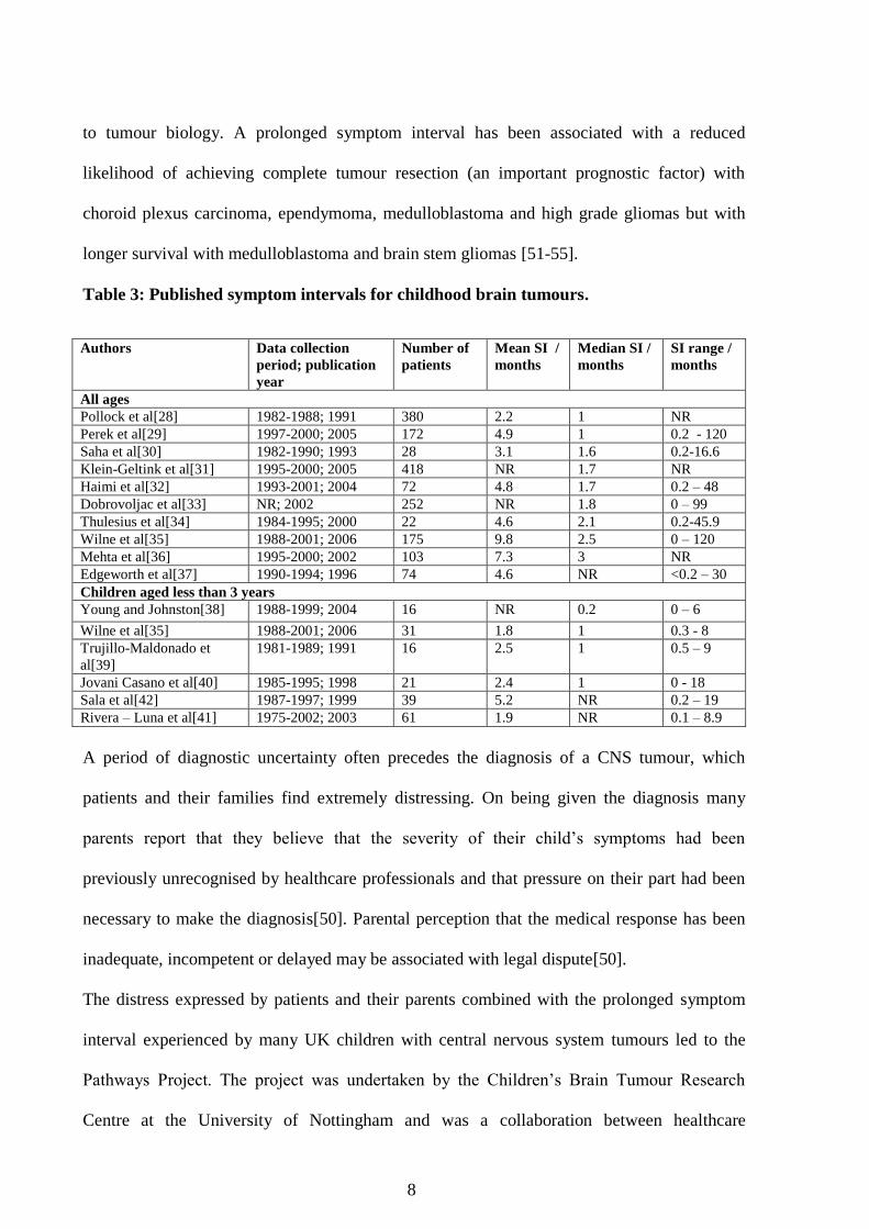
to tumour biology. A prolonged symptom interval has been associated with a reduced
likelihood of achieving complete tumour resection (an important prognostic factor) with
choroid plexus carcinoma, ependymoma, medulloblastoma and high grade gliomas but with
longer survival with medulloblastoma and brain stem gliomas [51-55].
Table 3: Published symptom intervals for childhood brain tumours.
Authors Data collection
period; publication
year
Number of
patients
Mean SI /
months
Median SI /
months
SI range /
months
All ages
Pollock et al[28] 1982-1988; 1991 380 2.2 1 NR
Perek et al[29] 1997-2000; 2005 172 4.9 1 0.2 - 120
Saha et al[30] 1982-1990; 1993 28 3.1 1.6 0.2-16.6
Klein-Geltink et al[31] 1995-2000; 2005 418 NR 1.7 NR
Haimi et al[32] 1993-2001; 2004 72 4.8 1.7 0.2 – 48
Dobrovoljac et al[33] NR; 2002 252 NR 1.8 0 – 99
Thulesius et al[34] 1984-1995; 2000 22 4.6 2.1 0.2-45.9
Wilne et al[35] 1988-2001; 2006 175 9.8 2.5 0 – 120
Mehta et al[36] 1995-2000; 2002 103 7.3 3 NR
Edgeworth et al[37] 1990-1994; 1996 74 4.6 NR <0.2 – 30
Children aged less than 3 years
Young and Johnston[38] 1988-1999; 2004 16 NR 0.2 0 – 6
Wilne et al[35] 1988-2001; 2006 31 1.8 1 0.3 - 8
Trujillo-Maldonado et
al[39]
1981-1989; 1991 16 2.5 1 0.5 – 9
Jovani Casano et al[40] 1985-1995; 1998 21 2.4 1 0 - 18
Sala et al[42] 1987-1997; 1999 39 5.2 NR 0.2 – 19
Rivera – Luna et al[41] 1975-2002; 2003 61 1.9 NR 0.1 – 8.9
A period of diagnostic uncertainty often precedes the diagnosis of a CNS tumour, which
patients and their families find extremely distressing. On being given the diagnosis many
parents report that they believe that the severity of their child‟s symptoms had been
previously unrecognised by healthcare professionals and that pressure on their part had been
necessary to make the diagnosis[50]. Parental perception that the medical response has been
inadequate, incompetent or delayed may be associated with legal dispute[50].
The distress expressed by patients and their parents combined with the prolonged symptom
interval experienced by many UK children with central nervous system tumours led to the
Pathways Project. The project was undertaken by the Children‟s Brain Tumour Research
Centre at the University of Nottingham and was a collaboration between healthcare

9
professionals and members of the public who have experienced a brain tumour diagnosis. It
aimed to reduce the symptom interval experienced by children with brain tumours by
providing improved guidance for healthcare professionals on the assessment, investigation
and referral of children who present with symptoms and signs that could result from a brain
tumour.
1.5: Currently available guidance
The UK National Collaborating Centre for Primary Care developed referral guidelines for
suspected cancer (including specific guidance for children and young people) which were
issued by the National Institute for Clinical Excellence (NICE) in June 2005[27].
The NICE guidance for childhood brain tumours is shown below:
General recommendations
Children and young people who present with symptoms and signs of cancer should be
referred to a paediatrician or a specialist children‟s cancer service, if appropriate.
Childhood cancer is rare and may present initially with symptoms and signs associated
with common conditions. Therefore, in the case of a child or young person presenting
several times (for example, three or more times) with the same problem, but with no
clear diagnosis, urgent referral should be made.
The parent is usually the best observer of the child‟s or young person‟s symptoms.
The primary healthcare professional should take note of parental insight and
knowledge when considering urgent referral.
Persistent parental anxiety should be a sufficient reason for referral of a child or young
person, even when the primary healthcare professional considers that the symptoms
are most likely to have a benign cause.
10
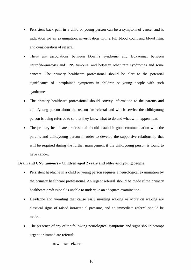
Persistent back pain in a child or young person can be a symptom of cancer and is
indication for an examination, investigation with a full blood count and blood film,
and consideration of referral.
There are associations between Down‟s syndrome and leukaemia, between
neurofibromatosis and CNS tumours, and between other rare syndromes and some
cancers. The primary healthcare professional should be alert to the potential
significance of unexplained symptoms in children or young people with such
syndromes.
The primary healthcare professional should convey information to the parents and
child/young person about the reason for referral and which service the child/young
person is being referred to so that they know what to do and what will happen next.
The primary healthcare professional should establish good communication with the
parents and child/young person in order to develop the supportive relationship that
will be required during the further management if the child/young person is found to
have cancer.
Brain and CNS tumours - Children aged 2 years and older and young people
Persistent headache in a child or young person requires a neurological examination by
the primary healthcare professional. An urgent referral should be made if the primary
healthcare professional is unable to undertake an adequate examination.
Headache and vomiting that cause early morning waking or occur on waking are
classical signs of raised intracranial pressure, and an immediate referral should be
made.
The presence of any of the following neurological symptoms and signs should prompt
urgent or immediate referral:
new-onset seizures

11
cranial nerve abnormalities
visual disturbances
gait abnormalities
motor or sensory signs
unexplained deteriorating school performance or developmental
milestones
unexplained behavioural and/or mood changes.
A child or young person with a reduced level of consciousness requires
emergency admission.
Brain and CNS tumours - Children < 2 years
In children aged younger than 2 years, any of the following symptoms may suggest a
CNS tumour, and referral (as indicated below) is required.
Immediate referral:
new-onset seizures
bulging fontanelle
extensor attacks
persistent vomiting.
Urgent referral:
abnormal increase in head size
arrest or regression of motor development
altered behaviour
abnormal eye movements
lack of visual following
poor feeding/failure to thrive.

12
Urgency contingent on other factors:
squint.
Whilst the NICE guidance provides a concise summary of the common modes of brain
tumour presentation it has three important limitations. First, it is predominantly directed at
primary care whereas children with brain tumours experience diagnostic delay throughout the
health service. Second, the “end-point” for the NICE guidelines is referral. Brain tumours are
diagnosed by imaging rather than referral and so guidance is required on indications for and
appropriate waiting times to imaging. Finally the guidance has a limited evidence base (13
references published between 1978 and 2002).
The objective of the Pathways Project and the subject of this thesis was therefore to develop
evidence-based guidance, applicable to primary and secondary care, to advise on the
following:
1. The symptoms and signs that may occur in children with brain tumours
2. Assessment of children presenting with these symptoms and signs
3. Indications and waiting times for imaging children with these symptoms and signs
Guideline development required that the following clinical questions were addressed:
1. What are the symptoms and signs that children with brain tumours develop?
2. Given that the initial symptoms and signs of a brain tumour may occur with other less
serious childhood conditions, how can healthcare professionals distinguish those
children who may have a brain tumour from the majority who do not?
3. What is the best way to clinically assess a child presenting with symptoms and / or
signs that could be due to a brain tumour?
4. What symptoms and / or signs in children increase the likelihood of a brain tumour to
the extent that their presence mandates brain imaging?
5. What is the best modality for brain imaging in children?

13
6. In a child who presents with symptoms and / or signs that could be potentially due to a
brain tumour, what is an appropriate maximum waiting time to imaging?
7. Are there specific presentations of childhood brain tumours that are repeatedly
associated with diagnostic difficulty and a prolonged symptom interval?
8. Are there other barriers to diagnosis in childhood brain tumours and if so how can
these be addressed?

14
CHAPTER 2: MATERIALS AND METHODS
Guideline development followed a two-stage process (figure 1). The initial stage comprised
appraisal of the currently available evidence on:
Childhood brain tumour presentation and diagnosis
The factors associated with a prolonged symptom interval in childhood brain tumours
A systematic review and meta-analysis of the literature on childhood brain tumour
presentation published between 1991 and 2005 was performed and cohort study of children
newly diagnosed with a brain tumour at four UK paediatric neuro-oncology centres between
2004 and 2006 was undertaken. The literature review and meta-analysis summarised the
previously published data and the cohort study provided contemporary information regarding
the presentation and diagnostic pathway of children diagnosed with a brain tumour in the UK.
The meta-analysis and the cohort study provided information on the signs and symptoms that
occur in children with brain tumours, their progression and factors associated with a
prolonged symptom interval. However, they did not address the question of the likelihood of a
child with a given symptom or sign having a brain tumour, i.e. its specificity and, except in
the case of seizures [56] and to an extent headaches [57], there are no previous studies
addressing this. The questions of symptom specificity, referral pathways, imaging indications
and acceptable waiting times cannot easily be addressed by quantitative research methods.
Qualitative methods in the form of a multi-disciplinary workshop and a Delphi consensus
process [58] were therefore employed to use professional expertise to incorporate the
evidence from the meta-analysis and cohort study into a clinical guideline.

15
Figure 1: Guideline development
2.1: Literature review methods
A systematic literature review and meta-analysis of the presenting symptoms and signs in
paediatric CNS tumours was undertaken to summarise the published literature in this field and
provide the initial evidence base to support guideline development.
The previous largest study of childhood brain tumour presentation was published in 1991 by
the Childhood Brain Tumour Consortium. This reported the symptoms and signs at diagnosis
for 3291 children diagnosed with a brain tumour in 1930–79[59]. Due to the historical nature
of the data and the rapid development of neuro-imaging techniques subsequent to the 1970‟s
which have changed the diagnostic process for children with brain tumours the Childhood
Brain Tumour Consortium was excluded from the meta-analysis. It does however provide a
historical reference and therefore all studies published subsequent to the Childhood Brain
Tumour Consortium study were included in the meta-analysis.
Systematic literature review and meta-analysis
Multi-centre cohort study EVIDENCE
Multidisciplinary workshop
Delphi consensus process
PROFESSIONAL EXPERTISE
Guideline
16
2.1.1: Identification of studies and inclusion criteria
MEDLINE, PubMed, and EMBASE were searched without language restriction, from
January, 1991 to August, 2005. Key words were: “brain tumour(s), “brain tumor(s)”, “brain
neoplasm(s)”, “spinal cord tumour(s)”, “spinal cord tumor(s)”, “spinal cord neoplasm”; and
“diagnosis”; and “sign(s)” or “symptom(s)”. Retrieved references were restricted to “all
child”. Abstracts were screened; those unrelated to CNS tumours or discussing an area
unrelated to clinical presentation were excluded. Papers with abstracts discussing tumour
presentation, tumour diagnosis, or clinical symptoms and signs were retrieved for detailed
review. All case-series or cohort studies describing symptoms and signs at diagnosis for a
minimum of ten children diagnosed with a CNS tumour and published after February, 1991
were included. Non-English language papers were translated.
2.1.2: Data collection
Numbers of children in every study with a symptom or sign at diagnosis were recorded on a
standard data extraction form. Information on symptoms and signs varied between studies.
Some studies had very detailed records on individual symptoms and signs (eg, headache,
vomiting, papilloedema), whereas others reported symptoms in clusters or complexes (eg,
symptoms of raised intracranial pressure). Symptoms and signs were recorded as described in
the individual studies. If a symptom or sign was not recorded in a study, it was assumed not to
occur in that population.
2.1.3: Statistical analysis
Analysis was done with meta-disc version β 1.1.1. Proportions (%) of children with each
symptom or sign at diagnosis were combined using one-variable relationship meta-analysis.
The effect size for each symptom and sign was calculated in the individual studies and
weighted according to its variance, and these effect sizes were then summed (for each

17
symptom and sign) and the total effect size was then divided by the sum of the weights to give
a mean effect size (pooled proportion). In meta-disc, proportions (as well as likelihood ratios
and diagnostic ratios) could be pooled with either the Mantel-Haenszel method (fixed-effects
model) or, to incorporate variation between studies, with the DerSimonian Laird method
(random-effects model). In the analysis, heterogeneity was indicated beyond what could be
expected by chance alone, by significant Q statistics and high inconsistency (I2) statistics. The
DerSimonian Laird method was selected because variability was expected across the papers,
and a random-effects model was used[60]. Symptoms and signs occurring in 5% or more of
the meta-analysis population are reported. Two papers [61, 62] reported optic atrophy and
papilloedema and one paper [63] lethargy and irritability as a combined category. Since these
papers reported detailed information for other symptoms and signs, they were included in the
meta-analysis but excluded from the analysis of the combined symptoms or signs. In one
report [61] visual acuity was not assessed in the complete cohort and, therefore, was excluded
from the meta-analysis of visual acuity.
The following subgroup analyses were undertaken: all intracranial tumours; intracranial
tumours in children aged under 4 years; children with an intracranial tumour and
neurofibromatosis; posterior fossa tumours; supratentorial (excluding central) tumours;
central tumours (third ventricle, tectum, pineal gland, pituitary gland, thalamus,
hypothalamus, optic pathway, and basal ganglia); brainstem tumours; and spinal-cord
tumours.
Analysis of all intracranial tumours was undertaken to provide a summary of paediatric
intracranial tumour presentation. Children aged under 4 years usually cannot clearly describe
symptoms such as headache, nausea, and diplopia, and therefore have a different presentation
to older children. Neurofibromatosis is the commonest genetic abnormality associated with
18
intracranial tumours and children can develop tumours before the development of cutaneous
manifestations. Children with neurofibromatosis have a high occurrence of optic-pathway
tumours, and thus their presentation differs from that of other children with intracranial
tumours. Only children with neurofibromatosis and a symptomatic intracranial tumour were
included in this subgroup analysis. Asymptomatic children with an intracranial tumour
identified by CNS imaging that was instigated after a diagnosis of neurofibromatosis were not
analysed. Analysis by tumour location was undertaken to highlight specific associations of
symptoms and signs that occur with different tumour locations.
2.2: Cohort study methods
A retrospective cohort study of children newly diagnosed with a central nervous system
tumour in four paediatric neuro-oncology centres was undertaken to provide contemporary
information on childhood brain tumour presentation and diagnosis in the UK and to
investigate factors associated with a prolonged symptom interval.
2.2.1: Data collection
Information was obtained from the hospital medical records of children diagnosed with a
brain or spinal cord tumour at Birmingham Children‟s Hospital, Queen‟s Medical Centre,
Nottingham, Southampton General Hospital and Sheffield Children‟s Hospital between
January 2004 and March 2006. Data was collected on the patient symptom interval,
symptoms and signs at disease onset and at diagnosis, deprivation score and healthcare
professionals consulted during the symptom interval. Symptoms and signs were recorded as
described in the records and then grouped into the following categories: headache, nausea and
vomiting, seizures, alteration in or loss of consciousness (excluding seizures), motor system
abnormalities (abnormal gait, abnormal co-ordination, focal motor weakness, involuntary
movements, abnormal tone, hemiplegia, paraplegia, quadriplegia, abnormal reflexes,
19
abnormal speech, abnormal handwriting and dystonia), visual system abnormalities (reduced
visual acuity, reduced visual fields, nystagmus, other abnormal eye movements, squint,
exophthalmia, diplopia, eye pain, papilloedema, optic atrophy, unequal pupils and sunsetting),
cranial nerve palsies, abdominal or back pain, spinal deformity, behavioural change
(including lethargy and school difficulties), endocrine and growth abnormalities and other
findings. Patients‟ deprivation score was determined using the Index of Multiple Deprivation
Score for wards from the Office of National Statistics [64].
2.2.2: Statistical analysis
All analyses were undertaken using SPSS 12.0. Subgroup comparison was undertaken using
the Mann-Witney and Kruskal-Wallis tests. Cox regression analysis was undertaken to
explore the relationship between symptom interval and initial symptom or sign and between
symptom interval and deprivation score. Fisher‟s exact test was used to explore the
relationship between long (greater than the median) and short (less than or equal to the
median) symptom interval and symptoms and signs with unknown date of onset.
2.2.3: Ethics
Approval was granted by Nottingham 2 REC. Written informed consent was provided by
patients aged 16 and above and by the parents or guardians of younger patients.
2.3: Multidisciplinary workshop
It was necessary to incorporate professional expertise into guideline development in order to
determine the specificity of symptoms and signs associated with childhood brain tumours and
to advise on appropriate referral pathways, imaging indications and acceptable waiting times.
Summation of the evidence from the meta-analysis and cohort study was required prior to
widespread review. This was undertaken by a multidisciplinary workshop. 20 healthcare
professionals and parents of children with brain tumours attended the workshop (see appendix
20
1 for participants). The workshop reviewed the data obtained from the meta-analysis and
cohort study and examined the following symptoms, signs, management decisions and risk
factors identified by literature review, data collection and guideline development team as
being key to the diagnosis:
Headache
Visual abnormalities
Motor abnormalities
Nausea and vomiting
Lethargy
Abnormal progression of height, weight and head circumference
Risk factors for CNS tumours
Thresholds for onward referral and imaging
Workshop Participants worked in small groups (table 4). For each of the symptoms and / or
signs the group was asked to devise statements on the following:
How would the symptoms and signs present to a healthcare professional?
How should a healthcare professional assess a child presenting with this symptom or
sign?
How should a healthcare professional determine whether the presenting symptoms and
signs could be due to a brain tumour i.e. their specificity?
What factors influence the specificity of a symptom and sign?
What are appropriate thresholds for referral and selection for imaging for a child
presenting with this symptom or signs?
What would they regard as best practice for referral and imaging of a child presenting
with this symptom and sign?

21
The group reviewing referral and imaging were asked to set standards for best practice in this
area.
Table 4: Topics covered by workshop groups
GROUP TOPIC
1 Headache
2 Motor assessment
Non-specific symptoms
3 Visual assessment
Predisposing factors
4 Nausea and vomiting
Assessment of growth
5 Imaging
Referral pathways
The conclusions from each group were discussed by the workshop. These conclusions and
discussion points from the workshop were subsequently translated into a series of statements
by the guideline development team.
2.4: Delphi process
Letters of invitation to join the Delphi panel were sent to health specialists fulfilling one or
more of the following criteria (for Delphi panel composition see appendix 2):
Involvement in the pre-diagnostic care of one or more of the 144 patients recruited to
the cohort study.
United Kingdom‟s Children‟s Cancer Study Group (UKCCSG) member from one of
the following disciplines: neurosurgeon, neuro-oncologist, neuro-radiologist,
neurologist, neuro-endocrinologist or paediatric oncologist, UKCCSG Brain Working
Group member and clinician. (From August 1st 2006 the UKCCSG merged with the
Childhood Leukaemia Part Working Party to form the UK Children‟s Cancer and
Leukaemia Group (CCLG)).
British Paediatric Neurology Association member.

22
Panel members were blind to the composition of the rest of the panel. The first, second and
third rounds of the Delphi Questionnaire was sent to panel members on 11th
April, 31st May
and 6th
July 2006 respectively. Panel members were asked to rate each statement on a 9-point
scale from strongly disagree (0) to strongly agree (9). A comments section was included for
each statement. Statements were taken as having reached consensus if 75% or more of the
Delphi Panel respondents rated the statement 7, 8 or 9. Statements were rejected if 25% or
less of the Delphi Panel respondents rated the statement 7, 8 or 9. Statements not reaching
consensus were rewritten following review of comments from the Delphi panel and then
reissued in subsequent rounds.
23
CHAPTER 3: RESULTS
3.1: Literature review results
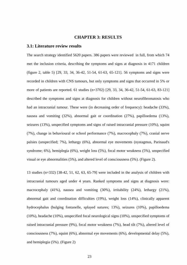
The search strategy identified 5620 papers. 386 papers were reviewed in full, from which 74
met the inclusion criteria, describing the symptoms and signs at diagnosis in 4171 children
(figure 2, table 5) [29, 33, 34, 36-42, 51-54, 61-63, 65-121]. 56 symptoms and signs were
recorded in children with CNS tumours, but only symptoms and signs that occurred in 5% or
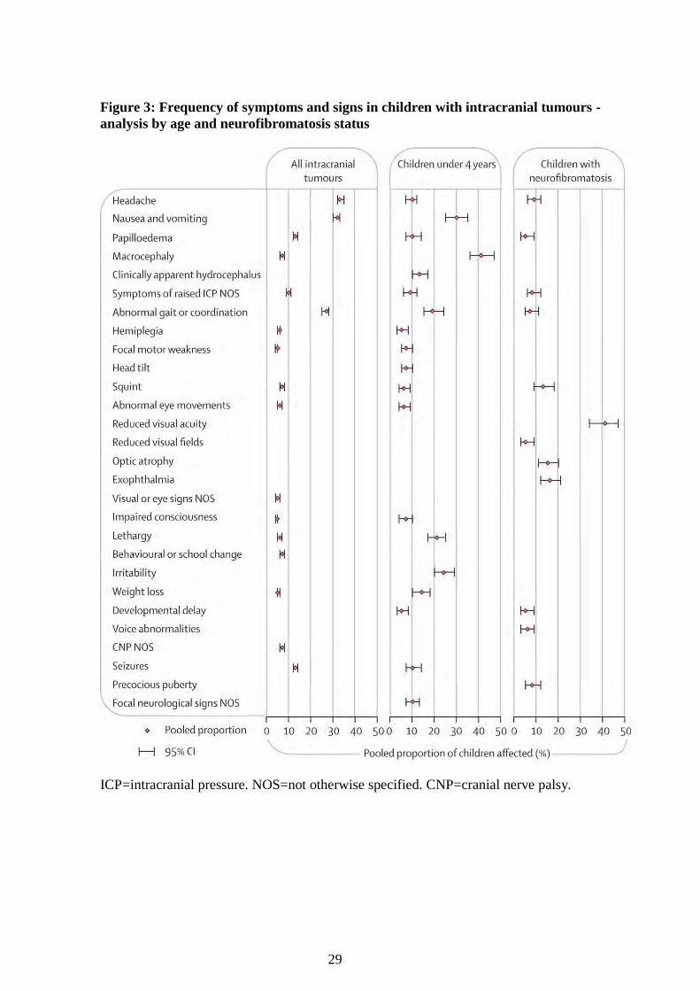
more of patients are reported. 61 studies (n=3702) [29, 33, 34, 36-42, 51-54, 61-63, 83-121]
described the symptoms and signs at diagnosis for children without neurofibromatosis who
had an intracranial tumour. These were (in decreasing order of frequency): headache (33%),
nausea and vomiting (32%), abnormal gait or coordination (27%), papilloedema (13%),
seizures (13%), unspecified symptoms and signs of raised intracranial pressure (10%), squint
(7%), change in behavioural or school performance (7%), macrocephaly (7%), cranial nerve
palsies (unspecified; 7%), lethargy (6%), abnormal eye movements (nystagmus, Parinaud's
syndrome; 6%), hemiplegia (6%), weight loss (5%), focal motor weakness (5%), unspecified
visual or eye abnormalities (5%), and altered level of consciousness (5%). (Figure 2).
13 studies (n=332) [38-42, 51, 62, 63, 65-79] were included in the analysis of children with
intracranial tumours aged under 4 years. Ranked symptoms and signs at diagnosis were:
macrocephaly (41%), nausea and vomiting (30%), irritability (24%), lethargy (21%),
abnormal gait and coordination difficulties (19%), weight loss (14%), clinically apparent
hydrocephalus (bulging fontanelle, splayed sutures; 13%), seizures (10%), papilloedema
(10%), headache (10%), unspecified focal neurological signs (10%), unspecified symptoms of
raised intracranial pressure (9%), focal motor weakness (7%), head tilt (7%), altered level of
consciousness (7%), squint (6%), abnormal eye movements (6%), developmental delay (5%),
and hemiplegia (5%). (Figure 2)

24
Eight studies (n=307) [61, 70-76] were included in the analysis of children with
neurofibromatosis and an intracranial tumour. The most common symptom and signs at
diagnosis were visual, indicating the high occurrence of optic pathway gliomas in this
population. The ranked symptoms and signs were reduced visual acuity (41%), exophthalmia
(16%), optic atrophy (15%), squint (13%), headache (9%), unspecified symptoms of raised
intracranial pressure (8%), precocious puberty (8%), abnormal gait or coordination difficulties
(7%), voice abnormalities (6%), developmental delay (5%), papilloedema (5%), and reduced
visual fields (5%). (Figure 2).
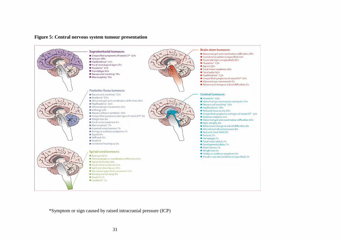
Five studies (n=476) [52,101,108.119.120] described children with posterior fossa tumours;
seven studies (n=303)[62, 88, 93, 101, 104, 106, 118] described children with supratentorial
tumours; 11 (n=276)[61, 85, 90, 99-101, 103,105, 110,114,116] children with central
tumours; five (n=276)[54, 95,96,101,102] described children with brainstem tumours; and six
studies (n=162)[77-81] described children with spinal-cord tumours (Figure 3 and Figure 4).

25
Figure 2: Progress through the meta-analysis
26
Table 5: Studies meeting inclusion criteria
Recruitment
period
No of
pts
Patient group, diagnosis if known,
source of data
Tumour
location
Mean
age (yrs)
Median
age (yrs)
Age range
(yrs)
Median symptom
interval / months
Mean symptom
interval / months
Symptom interval
range/months
Ref
1977-1987 22 Infants, 1I* AB NS NS NS NS NS NS [68]
1981-1989 16 Under 2, 1I* AB NS NS NS 1 2.5 0.5-9 [39]
1965-1989 16 NF1 and optic pathway tumours, 2I OP NS 4.5 1.5-17 NS 25.2 NS [76]
1978-1991 12 Gangliogliomas, 1I AB NS NS 3.5-17 NS NS NS [83]
1977-1990 12 Gangliogliomas, 1I AB 7.8 NS 0.8-15.8 27 40 7-96 [84]
1976-1991 12 Midbrain tumours, 1I C 8.2 NS 1.1-16 NS 4.5 0.5 [85]
1975-1981 11 Choroid plexus carcinoma, 2I AB NS 2.2 0-9.5 NS NS NS [86]
1976-1988 21 Meningeal tumours, 1I AB 9.3 NS 0.3-16.7 4 14.6 0-72 [87]
1962-1989 39 Under 2 yrs, 1I* AB NS NS NS NS NS NS [67]
1970-1989 106 Cerebral hemisphere tumours, 1I ST NS NS NS NS NS NS [88]
1970-1987 80 Under 2 yrs at symptom onset, 1I* AB NS NS NS NS NS 0-153.6 [63]
1989-1992 14 Infants with supratentorial tumors, 1I ST 0.5 NS 0.1-0.9 NS NS NS [62]
1980-1990 10 Meningiomas, 1I AB 11.1 NS 8-15 NS 13.2 0.1-60 [89]
1973-1992 21 NF1 and optic pathway tumours, 4I OP 7.1 NS 0-14.5 NS NS NS [75]
1979-1994 21 Under 2 yrs, 1I* AB NS NS 0.2-1.8 NS NS NS [66]
1983-1992 17 Midbrain tumours, 1I C NS 9.7 3.5-16 4 NS NS [90]
1974-1994 23 Intracranial ependymoma, 1I AB 8.8 NS 2-14 NS 3.8 0.5-10 [91]
1984-1994 17 NF1 and brain stem tumours, 1I BS 8.4 8.3 1.3-13.9 NS NS NS [74]
1990-1994 74 All brain tumours, 1I AB 6.9 NS NS NS 4.6 0.2-30 [37]
1988-1991 119 Brain stem gliomas treated with
HFRT (CCG-9882)
BS NS 6.5 NS NS NS NS [54]
1984-1993 32 Gangliogliomas, 1I AB 6.5 NS 0.7-20 NS NS NS [92]
1970-1995 36 Supratentorial PNET, 1I ST 4.3 2.9 0.1-12.8 NS NS NS [93]
1980-1993 27 Under three with intramedullary
spinal cord tumours,1I
SC 1.7 NS 0.5-3 NS NS NS [81]
1984-1995 13 Intrinsic spinal cord tumours, 1I SC 5.4 NS 0.7-11 NS NS NS [82]
1984-1995 723 All brain tumours, 1I AB NS NS 0-16 NS NS NS [94]
1980-1990 35 Brain stem tumours, 1I BS NS NS 1.3-13 NS 5 NS [95]
1987-1994 30 Endophytic pons or medullary
tumours, 1I
BS NS 6 0.6-16 NS 6 1-60 [96]
1974-1995 99 Gangliogliomas, 1I AB 9.5 NS 1.7-20 24.4 60 NS [97]
1968-1994 29 Meningiomas 2I AB 10 NS 0-15 NS NS NS [98]
27
1983-1995 12 Primary intracranial germ cell
tumours, 1I
C NS NS 5-15 NS NS NS [99]
1984-1996 25 NF1 and brain stem tumours, 1I BS 7.8 NS 1.1-15.2 NS NS NS [73]
1976-1992 18 Spinal cord astrocytomas, 1I SC 9.2 8.6 0.6-17.9 NS NS NS [80]
1966-1996 46 Under 3 yrs, 1I* AB NS NS NS NS NS NS [65]
1985-1995 20 Under 3 yrs, 1I* AB 1.7 NS 0-2.7 1 2.4 0-18 [40]
1977-1996 21 Infants, 1I * AB 0.5 NS NS NS NS NS [71]
1990-1997 32 Tectal tumours, 1I C 8 NS 0.2-17 NS NS NS [100]
1984-1995 22 Choroid plexus carcinoma registered
with SFOP
AB NS 2.1 0.3-9.3 1 NS 0.1-8 [53]
1986-1990 40 Intracranial ependymoma treated on
POG 8633
AB NS NS 0.3-2.9 1 1.6 0-10.9 [51]
1971-1994 73 Spinal cord astrocytomas, 13I SC NS 7 0.3-6 NS NS NS [79]
1985-1996 20 Intramedullary spinal cord
ependymomas, 1I
SC 14 NS 9-18 NS NS NS [78]
1975-1993 200 All brain tumours, 1I AB 8.9 NS NS NS NS NS [101]
1987-1997 39 Under 3 yrs, 1I * AB 2.1 NS 0.3-3 NS 5.2 0.2-18 [42]
1983-1997 76 Brain stem gliomas BS NS NS 3-15 NS NS NS [102]
1988-1998 11 Tectal plate gliomas, 1I C 10 NS 5-13 NS 28.2 0.7-84 [103]
1988-1998 54 Lateral ventricle tumours, 1I ST NS NS 0-20 NS 5 0-48 [104]
1967-1997 37 Pineal region tumours, 1I C 9.6 NS NS NS NS NS [105]
1986-1995 28 Supratentorial PNET, 1I ST 6.8 NS 0.7-16.9 NS 4.9 1-48 [106]
1988-1998 11 Cervicomedullary astrocytomas, 1I SC 7 NS 0-18 NS NS NS [77]
1984-1995 22 Reported to regional TR AB NS NS NS 2.1 4.6 0.2-45.9 [34]
1979-1999 34 Choroid plexus tumours, 1I AB NS 1.4
papillomas
1.1
carcinomas
0.1-11.5
papillomas
0.2-8.5
carcinomas
1 NS 0.03-33 [107]
1972-1991 62 Intracranial ependymoma, 1I PF 6 NS 1-17 NS 2 NS [108]
1984-1999 24 Meningiomas, 2I AB NS NS 2-17 NS 8.2 0.2-14.4 [109]
1980-1994 18 Chiasmal gliomas, 1I OP NS NS 0.5-14 NS NS NS [110]
1985-1999 181 All brain tumours, 1I AB NS NS 0-16 NS NS NS [111]
1970-1998 16 Choroid plexus tumours, 1I AB 3.1 NS 0.2-15.4 NS NS NS [112]
1974-1999 122 Medulloblastoma, 1I PF NS NS NS NS 3.3 NS [52]
1981-1998 11 Nerve cell tumours, 1I ST NS NS 2-16 NS NS NS [113]
1970-1998 35 Craniopharyngiomas, 1I C NS 9.1 1.3-15.6 NS NS NS [114]
1980-1999 252 All brain tumours, 1I AB NS 6.3 yrs 0-16.9 1.8 NS 0-99 [33]
1995-2000 104 All brain tumours, 2I AB 8.29 NS NS 3 7.3 NS [36]

28
1987-1999 22 Gangliogliomas, 2I AB NS NS 0-16 11 30 NS [115]
1980-2000 20 Thalamic and basal ganglia tumours,
1I
C 6.6 NS 0.3-18 NS 1.5 0-24 [116]
1974-1999 18 Meningiomas recorded in a hospital
TR
AB 11 NS 1.6-17 NS NS NS [117]
1975-2002 61 Infants, 2I* AB 0.5 NS 0-1 NS 1.9 0.1-8.9 [41]
1988-1999 16 Infants, 1I* AB NS 0.5 0-1 0.2 NS 0-6 [38]
1986-1990 13 Supratentorial PNET treated on POG
8633
ST NS NS 0-3 NS 0.9 0-49 [118]
1954-1997 181 Medulloblastoma registered with
Manchester Children‟s TR
PF NS NS 0-14 NS NS NS [119]
1982-2000 69 NF1 and symptomatic tumours, 7I AB NS 5.2 0.3-17 NS NS NS [72]
1996-2000 83
(51
NF1)
Optic pathway gliomas, 2I OP NS NS 0.3-17.4 NS NS NS [61]
1986-2002 51 NF1 and symptomatic optic pathway
gliomas, 2I
OP 4.8 NS 0-15.8 NS NS NS [71]
1996-2003 37 Posterior fossa tumours, 1I PF 6.7 NS 2-16 NS 3.7 NS [120]
1978-2001 18 Giant cell astrocytomas, 2I AB NS NS 4-15 9 19 2.5-96 [121]
1973-2002 57 NF1 and optic pathway tumours, 1I OP 5.2 NS NS NS NS NS [70]
1997-2000 172 All brain tumours, 1I AB NS 8.3 0.3-17.3 1 4.9 0.2-120 [29]
1I=treated at one institution. 2I=treated at two institutions. 4I=treated at four institutions. 7I=treated at seven institutions. AB=all brain. NS=not
specified. OP=optic pathway. C=central. ST=supratentorial. BS=brainstem. SC=spinal cord. PF=posterior fossa. * Study population defined by age rather than tumour type or location.
29
Figure 3: Frequency of symptoms and signs in children with intracranial tumours -
analysis by age and neurofibromatosis status
ICP=intracranial pressure. NOS=not otherwise specified. CNP=cranial nerve palsy.

30
Figure 4: Frequency of symptoms and signs in children with a central nervous system tumour - analysis by tumour location
31
Figure 5: Central nervous system tumour presentation
*Symptom or sign caused by raised intracranial pressure (ICP)

32
3.2: Cohort study results
3.2.1: Patient characteristics
189 children were diagnosed with a brain or spinal tumour at the participating centres during
the recruitment period. 144 children (139 brain tumours, 5 spinal cord tumours) were
recruited to the study (76% recruitment rate). The median age at diagnosis was 8.1 years
(range 29 days to 16.7 years) and the male to female ratio 1.5:1 (86 male, 58 female). The
tumour diagnoses are shown in table 1. Two children were diagnosed as a result of screening;
a child with tuberous sclerosis was diagnosed with a subependymal giant cell astrocytoma and
a child with probable neurofibromatosis type 2, whose identical twin had been diagnosed with
a symptomatic spinal cord tumour, with an asymptomatic spinal cord tumour. One child was
diagnosed with a cerebellar pilocytic astrocytoma following imaging to investigate precocious
puberty; the tumour was felt to be unrelated to her precocious puberty.
Table 6: Tumour diagnoses of children recruited to the cohort study
Diagnosis Number
Pilocytic astrocytoma 38
Medulloblastoma 31
Ependymoma 8
Supratentorial PNET 8
Brain stem glioma 7
Low grade glioma unspecified (excluding OPG) 7
Optic pathway gliomas (OPG) 6
Craniopharyngioma 6
Germinoma 5
High grade gliomas unspecified 5
Grade 2 astrocytoma 5
Choroid plexus tumour 4
Other 14

33
3.2.2: Symptoms and signs - brain tumours
There was a clear increase in the number of symptoms and signs from a median of one (range
1-8) at symptom onset to six (range 1-16) at diagnosis (table 7). At symptom onset the
symptoms and signs, ranked in order of frequency, were headache, nausea and / or vomiting,
motor system abnormalities, cranial nerve palsies, visual system abnormalities, seizures,
endocrine or growth abnormalities, behavioural change, abdominal or back pain, an alteration
in or loss of consciousness and spinal deformity. The most common motor abnormalities seen
were abnormalities of gait and co-ordination and the commonest visual abnormalities were
squint and reduced visual acuity. 16 of the 24 patients with a cranial nerve abnormality had
abnormalities involving the visual system. Lethargy was the only behavioural change
identified at symptom onset.
By the time of diagnosis, the most common findings were visual system abnormalities
followed by motor system abnormalities, nausea and / or vomiting, headache, cranial nerve
palsies, behavioural change, endocrine or growth abnormalities, alteration in or loss of
consciousness, seizures, abdominal or back pain and spinal abnormalities. The most common
visual system abnormalities were papilloedema which was identified in 50 children (36%),
nystagmus in 25 (18%), reduced visual acuity in 20 (14%), and squint and diplopia each in 18
children (13%). 48 of the 75 children who had a cranial nerve abnormality at diagnosis had an
abnormality involving the visual system. 62 children (45%) had a gait abnormality, 54 (39%)
abnormal co-ordination and 26 (19%) a focal motor weakness. Lethargy remained the most
common behavioural change occurring in 27 children (19%) followed by school difficulties in
23 (17%) and other behavioural changes (usually increased aggression or withdrawal) in 16
(12%). 26 children (19%) had lost weight by diagnosis.

34
Table 7: Symptom and sign complexes at symptom onset and at diagnosis in children
with brain tumours
Symptom / Sign Onset (95%
Confidence
Interval)
Diagnosis (95%
Confidence
Interval)
Increase (95%
Confidence
Interval)
Visual system
abnormalities
17% ( 15 to
23%)
70% (62-78%) 53% (45 to 61%)
Motor system
abnormalities
22% (15 to
29%)
67% (59 to 75%) 45% (37to 53%)
Cranial nerve palsy 17% (15 to
23%)
54% (46 to 62%) 37% (29 to 45%)
Behavioural change 3% (0 to 6%) 40% (32 to 48%) 37% (29 to 45%)
Nausea and / or
vomiting
28% (20 to
35%)
63% (55 to 71%) 35% (27 to 43%)
Endocrine or growth
abnormalities
7% (3 to 11%) 25% (18 to 32%) 18% (12 to 24%)
Headache 40% (32 to
48%)
58% (50 to 62%) 18% (12 to 24%)
Alteration in or loss of
consciousness
1% (-1 to 3%) 15% (9 to 21%) 14% (8 to 20%)
Abdominal or back
pain
2% (0 to 4%) 8% (3 to 13%) 6% (2 to 10%)
Seizures 10% (5 to 15%) 13% (7 to 19%) 3% (0 to 6%)
Spinal deformity 1% (-1 to 3%) 2% (0 to 4%) 1% (-1% to 3%)
Of 79 children with a single symptom or sign at symptom onset, 26 children (33%) had a
headache, 11 (14%) had a visual system abnormality, 10 (13%) nausea and / or vomiting, 10
(13%) a motor system abnormality, eight (10%) seizures, and four (5%) an endocrine or
growth abnormality. Two children (3%) had a cranial nerve abnormality not involving the
visual system (one hearing loss and one dysphagia). By diagnosis only three children still had
a single symptom or sign (one polyuria and polydipsia, one seizures and one hearing loss) and
only five children had two symptoms or signs (six motor abnormalities and one each of
headache, vomiting, visual abnormality and growth abnormality). No child had only headache
or vomiting by diagnosis. The greatest increase in number of symptoms or signs during the
symptom interval occurred with visual system abnormalities which increased by 53%. Large

35
increases also occurred in motor system abnormalities (45%), cranial nerve palsies (37%),
behavioural change (37%), and nausea and vomiting (35%).
By diagnosis 95% of children had symptoms and signs in one or more of the following
categories: headache, nausea or vomiting, visual system abnormalities and motor system
abnormalities. Only seven children did not present with symptoms and signs in these
categories. Of these, two presented with partial seizures, two with polyuria and polydipsia,
one with hearing loss, and two were diagnosed with asymptomatic tumours whilst undergoing
investigation of tuberous sclerosis and precocious puberty respectively.
Figure 6 shows the effect of patient age on brain tumour presentation. Children aged less than
four years show a different presentation to older children. In this age group motor and visual
system abnormalities, nausea and vomiting and cranial nerve palsies were the most common
symptoms and signs both at symptom onset and at diagnosis. Significant differences between
this age group and older children occur in the frequency of headache at symptom onset
(p=<0.001) and at diagnosis (p=<0.001), of motor system abnormalities at symptom onset
(p=0.04) and at diagnosis (p=0.02) and in the frequency of nausea and vomiting at diagnosis
(p=0.01). Headache is rare at symptom onset in this age group and only occurred in 19% by
diagnosis. Motor system abnormalities are more common at both symptom onset and
diagnosis whilst nausea and vomiting occurs less frequently at diagnosis than in older
children. The greatest increase in number of symptoms and signs during the symptom interval
occurred with motor system abnormalities and behavioural change.
3.2.3: Symptoms and signs – spinal cord tumours
Five children diagnosed with a spinal cord tumour were recruited. One child, with
neurofibromatosis type 2, was completely asymptomatic and was imaged when his identical
twin brother was diagnosed with a symptomatic spinal cord tumour. Of the remaining four
patients three presented with back pain, one with a spinal abnormality and one with

36
constipation. One patient had motor system abnormalities at disease onset; all symptomatic
patients had motor system abnormalities by diagnosis. There was again evidence of disease
progression during the symptom interval; the median number of symptoms and signs at
symptom onset was two, this had increased to nine by diagnosis.
3.2.4: Symptom interval
The symptom interval experienced by the patients with brain tumours ranged from 0 days to
6.9 years (median 3.3 months); for the five children with a spinal cord tumour it ranged from
0 days to 2.1 years (median 6.4 months). Due to the small numbers of spinal cord tumours,
further symptom interval analysis was restricted to the brain tumour patients. Univariate
analysis revealed no association between symptom interval and either tumour location, patient
age, sex, ethnic origin or deprivation score. High grade tumours (tumour grading was possible
for 119 patients) were significantly associated with a shorter symptom interval (p=0.004).
A shorter symptom interval was associated with initial presentation with nausea and / or
vomiting (p=0.003), abnormal gait (p=0.001), co-ordination difficulties (p=0.006), focal
motor weakness (p=0.002), unequal pupils (p=0.002), facial weakness (p=0.03), and apnoea
(p=0.036); and, when grouped into combined categories, with initial presentation with any
motor sign (p=0.001). A longer symptom interval was associated with initial presentation with
head tilt (p=0.006) and cranial nerve palsies (p=0.025). For symptoms and signs with an
unknown date of onset (i.e. those other than initial ones) endocrine and growth abnormalities
(p=0.018) and reduced visual acuity (p=0.028) were associated with a longer symptom
interval. (See table 8)

37
Table 8: Association between symptoms and signs and symptom interval
Symptom / Sign No.
affected
Significance Odds
ratio
95% CI for odds ratio Effect on symptom
interval
Upper Lower
Cox regression
Nausea and / or
vomiting
39 0.003 1.8 1.2 2.6 Decrease
Abnormal gait 17 0.001 2.3 1.4 3.9 Decrease
Co-ordination
difficulties
9 0.006 2.7 1.3 5.4 Decrease
Facial weakness 4 0.030 3.1 1.1 8.5 Decrease
Focal motor
weakness
10 0.002 2.8 1.5 5.4 Decrease
Any motor symptom
or sign
31 0.001 2.0 1.3 3.0 Decrease
Any cranial nerve
palsy
32 0.025 0.6 0.4 0.9 Increase
Head tilt 6 0.018 0.4 0.2 0.8 Increase
Fishers test
Endocrine or growth
abnormality
35 0.018 Increase
Reduced visual
acuity
20 0.028 Increase
3.3.5: Referral pathways and imaging
Referral pathway data was available for 102 children. Of these, 79% had visited their general
practitioner, 78% a hospital paediatrician, 23% an ophthalmologist, 14% an optician and 29%
had attended Accident and Emergency. Other disciplines consulted included health visitors,
orthopaedics, ENT and speech therapy. Calculation of the number of attendances to
healthcare was difficult as records frequently did not contain details of repeated attendances to
primary care. However, the reported number of attendances prior to diagnosis ranged from 0-
12 (median 3.0). A longer symptom interval was significantly associated with an increased
number of healthcare attendances (p<0.001).
51% children were imaged with CT followed by MRI, 44% with MRI alone and 5% with CT
alone. 81% of CT scans were requested by general paediatricians, 8% by accident and
emergency, 5% by ophthalmology, 4% by neuro-surgery and 1% each by general practice and

38
paediatric neurology. 48% of MRI scans were requested by neuro-surgery, 35% by general
paediatricians, 8% by paediatric neurology, 4% by ophthalmology, and 1% each by ENT,
paediatric oncology, paediatric endocrinology and orthopaedics.

39
Figure 6: Relationship between patient age and brain tumour presentation

40
3.3: Multidisciplinary workshop results
The workshop small groups noted their conclusions. These were then discussed by all
workshop participants. The discussion was recorded and the notes from group work retained.
These conclusions and discussion points were subsequently translated into a series of
statements by the guideline development team. The following is summary of the workshop
discussion and conclusions. The guideline statements developed from the discussion points
are shown. Where the guideline development team decided that a discussion point should not
be included in the guideline the reason is documented.
3.3.1: Headache
STATEMENT FROM WORKSHOP GROUP DELPHI QUESTIONNAIRE
STATEMENT
Any headache can indicate a serious condition Statement too general, therefore
not included
It is important to take seriously new headaches that have arisen
recently
H1 & H14
Children of different ages present with different types of
neurological symptoms and signs of brain tumour and other
abnormalities
H5-H9
Raised intracranial pressure causes symptoms of headaches which
can be diurnal, nausea, vomiting and altered consciousness
G10 & G16
Children with headaches should have an eye check to assess eye
movements (squint/nystagmus), fundoscopy and assessment of
visual performance (acuity/field)
G10 & G16
In patients with headaches during adolescence, pubertal
progression should be assessed
G16
Patients identified with headache without clear cause should be
followed up within 4 weeks (GP guidance)
H11
An investigatory algorithm for headaches in children should be
used
Beyond the scope of the
guideline
Red flag symptoms of headaches should be identified Included in more detail within
Headache section
In follow up, acquisition of new signs/symptoms should be a red
flag indicating referral
H14
In young children (pre-school) specific enquiries should be made
about developmental progress
G16
In young children head circumference should be monitored G16
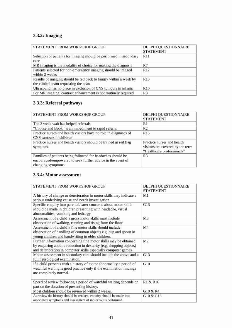
41
3.3.2: Imaging
STATEMENT FROM WORKSHOP GROUP DELPHI QUESTIONNAIRE
STATEMENT
Selection of patients for imaging should be performed in secondary
care
R11
MR imaging is the modality of choice for making the diagnosis R7
Patients selected for non-emergency imaging should be imaged
within 2 weeks
R12
Results of imaging should be fed back to family within a week by
the clinical team requesting the scan
R13
Ultrasound has no place in exclusion of CNS tumours in infants R10
For MR imaging, contrast enhancement is not routinely required R8
3.3.3: Referral pathways
STATEMENT FROM WORKSHOP GROUP DELPHI QUESTIONNAIRE
STATEMENT
The 2 week wait has helped referrals R1
“Choose and Book” is an impediment to rapid referral R2
Practice nurses and health visitors have no role in diagnoses of
CNS tumours in children
R15
Practice nurses and health visitors should be trained in red flag
symptoms
Practice nurses and health
visitors are covered by the term
“Healthcare professionals”
Families of patients being followed for headaches should be
encouraged/empowered to seek further advice in the event of
changing symptoms
R3
3.3.4: Motor assessment
STATEMENT FROM WORKSHOP GROUP DELPHI QUESTIONNAIRE
STATEMENT
A history of change or deterioration in motor skills may indicate a
serious underlying cause and needs investigation
M1
Specific enquiry into parental/carer concerns about motor skills
should be made in children presenting with headache, visual
abnormalities, vomiting and lethargy
G13
Assessment of a child‟s gross motor skills must include
observation of walking, running and rising from the floor
M3
Assessment of a child‟s fine motor skills should include
observation of handling of common objects e.g. cup and spoon in
young children and handwriting in older children.
M4
Further information concerning fine motor skills may be obtained
by enquiring about a reduction in dexterity (e.g. dropping objects)
and deterioration in computer skills especially computer games
M2
Motor assessment in secondary care should include the above and a
full neurological examination.
G13
If a child presents with a history of motor abnormality a period of
watchful waiting is good practice only if the examination findings
are completely normal.
G10
Speed of review following a period of watchful waiting depends on
part on the duration of presenting history.
R1 & R16
Most children should be reviewed within 2 weeks. G10 & R4 At review the history should be retaken, enquiry should be made into
associated symptoms and assessment of motor skills performed. G10 & G13

42
Any child representing to primary care with the same symptoms or
history requires referral to secondary care.
Statement very general and not
necessarily applicable in all
situations therefore not
included.
Brain imaging is required for any child with motor regression, gait
disturbance suggestive of a central cause, or neurological deficit.
M7
3.3.5: Non-specific symptoms
SATEMENT FROM WORKSHOP GROUP DELPHI QUESTIONNAIRE
STATEMENT
A history of lethargy may suggest a serious underlying cause O1 & O2
Environmental context is important when assessing lethargy.
Children who are lethargic in situations when they would normally
be active or playing are worrying.
O1
Lethargy in young children may manifest as reduced activity levels
or increased sleeping.
O3
Lethargy is an unusual behavioural response of children to adverse
life events. Children are more likely to become angry or upset.
Recognition of brain tumours as
a potential cause of lethargy
rather than aetiology of all
lethargy is the aim of guideline
therefore not included.
In a child presenting with lethargy enquiry should be made into
associated symptoms including headache, vomiting, visual
abnormalities, motor abnormalities, and weight loss.
G13
A period of watchful waiting is appropriate only if there are no
other associated features and no abnormalities on examination and
growth assessment.
G11
Assessment of a child with lethargy should include a complete
physical examination including assessment of growth, vision and
motor skills.
G13
3.3.6: Visual assessment
STATEMENT FROM WORKSHOP GROUP DELPHI QUESTIONNAIRE
STATEMENT
A child of any age presenting with persistent headache of
unexplained origin requires visual assessment, either in a primary
or secondary care setting [the setting being dependent on the age of
the child, and the area in which they live].
G13
A child of any age presenting with any of: odd eye movements
(nystagmus), squint, ptosis or loss of the red reflex requires visual
assessment, either in a primary or secondary care setting [the
setting being dependent on the age of the child, and the area in
which they live].
G12 & G13
A child aged <3 years presenting with abnormal gait and/or
persistent vomiting and/or macrocephaly requires visual
assessment
G13
It is unrealistic to expect optometrists to assess the vision of a child
aged < 5 years.
V4
Visual assessment of a child <5 years should be performed by a
competent paediatric ophthalmologist in a secondary care setting.
V4
Visual assessment of a co-operative child age > 5 years should be
performed by a community optometrist
V3
Visual assessment of an uncooperative child of any age should be
performed by a competent paediatric ophthalmologist in a
secondary care setting.
V4
Links between GPs and community optometrists could be V6

43
improved through the use of a user-friendly referral form, rather
than a dictated or computer-generated letter. The form would have
tick boxes for e.g. “I am worried about this patient who presented
with …”.
Community optometrists should be able to directly refer to a
secondary care centre any child aged > 5 years with abnormal eye
findings e.g. optic nerve swelling.
V8
A “watchful wait” approach should be used if assessment of the
following areas is normal: visual acuity, eye movements, pupil
responses, visual fields, colour vision, optic disc appearance.
G10, G13 &V9
If links between GPs and community optometrists are good, GPs
can request optometrists to carry out tests of visual acuity, eye
movements, pupil responses, visual fields, colour vision and optic
disc appearance.
V5
If assessment of any of the following areas is abnormal, the child
should be referred to an ophthalmologist: visual acuity, eye
movements, pupil responses, visual fields, colour vision, optic disc
appearance.
V8
If there are abnormal eye findings together with progression of
presenting non-ocular symptoms or additional symptoms, the child
should be referred for imaging.
V1, V10 – V16
To ensure effective communication between different services,
paediatric ophthalmologists should send copies of their letters to
everyone on the multidisciplinary team, including to the referring
optometrist.
V6
Unexplained decreased vision (i.e. excluding amblyopia/lazy eye
which is responding to treatment) can be associated with a CNS
lesion.
V13
Visual field defects can be associated with a CNS lesion V1 & V15
Abnormal pupil size can be associated with a CNS lesion V1
Decreased colour vision can be associated with a CNS lesion V1 & V13
Diplopia can be associated with a CNS lesion V1
Nystagmus can be associated with a CNS lesion V1 & V12
Ptosis can be associated with a CNS lesion V1
Proptosis can be associated with a CNS lesion V1 & V16
Optic disc swelling can be associated with a CNS lesion V1 & V10
Head nodding can be associated with a CNS lesion M1
3.3.7: Predisposing factors
STATEMENT FROM WORKSHOP GROUP DELPHI QUESTIONNAIRE
STATEMENT
Awareness should be raised of factors predisposing to CNS
tumours [see Table 3.5 p42 in Brain and Spinal Tumors of
Childhood ed: Walker, Perilongo, Punt & Taylor, published
2004. Arnold, London]
G13
Good history taking is crucial to the diagnosis of CNS tumours G13, R3 – R5
Listening to parents is crucial to the diagnosis of CNS tumours R3 – R5
3.3.8: Nausea and vomiting
STATEMENT FROM WORKSHOP GROUP DELPHI QUESTIONNAIRE
STATEMENT
History of awakening with nausea or vomiting in the morning
or from sleep, in the day, should prompt a visit to the GP
NV3
The association of headache is additionally concerning G16
Developmental delay or regression increases the urgency G16
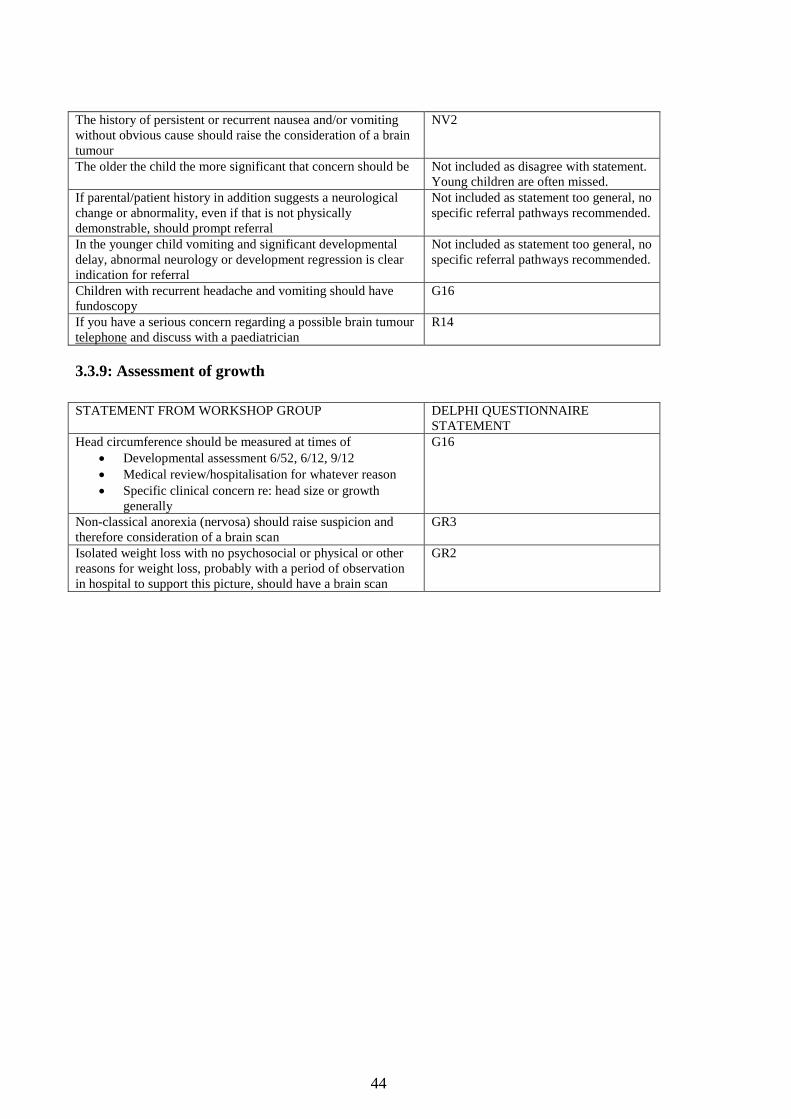
44
The history of persistent or recurrent nausea and/or vomiting
without obvious cause should raise the consideration of a brain
tumour
NV2
The older the child the more significant that concern should be Not included as disagree with statement.
Young children are often missed.
If parental/patient history in addition suggests a neurological
change or abnormality, even if that is not physically
demonstrable, should prompt referral
Not included as statement too general, no
specific referral pathways recommended.
In the younger child vomiting and significant developmental
delay, abnormal neurology or development regression is clear
indication for referral
Not included as statement too general, no
specific referral pathways recommended.
Children with recurrent headache and vomiting should have
fundoscopy
G16
If you have a serious concern regarding a possible brain tumour
telephone and discuss with a paediatrician
R14
3.3.9: Assessment of growth
STATEMENT FROM WORKSHOP GROUP DELPHI QUESTIONNAIRE
STATEMENT
Head circumference should be measured at times of
Developmental assessment 6/52, 6/12, 9/12
Medical review/hospitalisation for whatever reason
Specific clinical concern re: head size or growth
generally
G16
Non-classical anorexia (nervosa) should raise suspicion and
therefore consideration of a brain scan
GR3
Isolated weight loss with no psychosocial or physical or other
reasons for weight loss, probably with a period of observation
in hospital to support this picture, should have a brain scan
GR2

45
3.4: Delphi consensus process results
3.4.1: Delphi process round one
The statements for the first round of the Delphi consensus process were derived from the
statements developed by the multidisciplinary workshop and from the evidence base provided
by the meta-analysis and cohort study.
Round one of the Delphi consensus process comprised 77 statements describing the
presenting features of childhood brain tumours, factors that could be used to discriminate
brain tumours from other less serious conditions and possible referral pathways for children
with brain tumours. The questionnaire included a free text section in which panel members
were asked to provide their experience (if any) of the influence that ethnicity and deprivation
has on diagnostic delay in childhood brain tumours. Of 328 invited healthcare professionals
156 agreed to participate in the Delphi panel (see appendix 3).
The first round of the Delphi process, including instructions to participants is shown below.

46
3.4.2: Delphi questionnaire round one
Throughout this questionnaire:
the terms child and children refer to the age range 0-18 years unless specifically stated
otherwise
statements apply to brain and other intracranial tumours, but for ease of reading we refer to
brain tumour throughout.
HOW TO COMPLETE THIS QUESTIONNAIRE
YOUR NAME (in block letters) : ______________________________________
1. The questionnaire is divided into EIGHT topic areas. Each area has a list of
statements to be rated on a 9-point rating scale.
2. To rate each statement, check ONE box only by putting an X inside the box
under the score you have chosen.
3. Do not be put off by the length of the questionnaire. If you feel you do not have
the necessary expertise or experience to contribute to developing a particular
topic area or statement, please check the appropriate “N/C” box, and move onto
the next topic area or statement (leaving the numbered boxes blank).
4. At the end of each statement there is an opportunity to comment but please do
not feel any obligation to do so. [NOTE: we‟re particularly interested in feedback on
statements that you disagree with (e.g. is it incorrect, is it ambiguous). This will aid
development of the statements for subsequent rounds of the Delphi process].
5. Please note: The questionnaire includes an APPENDIX at the end, giving
relevant sections from the June 2005 NICE Referral Guidelines for Suspected
Cancer. You do not need to read the Appendix in order to rate the statements.
The Appendix is given for information only.
6. When you have completed the questionnaire, please return in the envelope
provided to arrive by WEDNESDAY 3rd
May 2006 to:
Dr Sophie Wilne
Children's Brain Tumour Research Centre
Academic Division of Child Health
East Block, E Floor
Queens Medical Centre
University of Nottingham
Nottingham NG7 2UH
THANKYOU
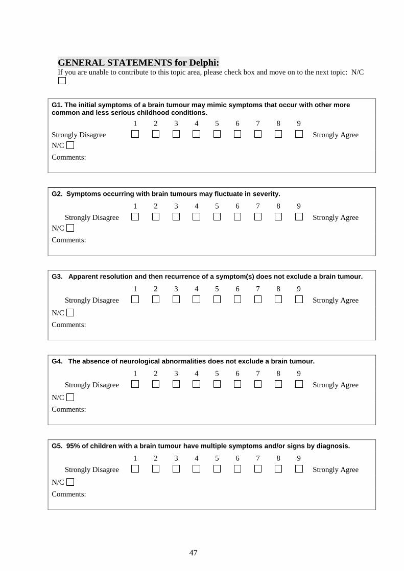
47
GENERAL STATEMENTS for Delphi: If you are unable to contribute to this topic area, please check box and move on to the next topic: N/C
G1. The initial symptoms of a brain tumour may mimic symptoms that occur with other more common and less serious childhood conditions.
Strongly Disagree
1
2
3
4
5
6
7
8
9
Strongly Agree
N/C
Comments:
G2. Symptoms occurring with brain tumours may fluctuate in severity.
Strongly Disagree
1
2
3
4
5
6
7
8
9
Strongly Agree
N/C
Comments:
G3. Apparent resolution and then recurrence of a symptom(s) does not exclude a brain tumour.
Strongly Disagree
1
2
3
4
5
6
7
8
9
Strongly Agree
N/C
Comments:
G4. The absence of neurological abnormalities does not exclude a brain tumour.
Strongly Disagree
1
2
3
4
5
6
7
8
9
Strongly Agree
N/C
Comments:
G5. 95% of children with a brain tumour have multiple symptoms and/or signs by diagnosis.
Strongly Disagree
1
2
3
4
5
6
7
8
9
Strongly Agree
N/C
Comments:
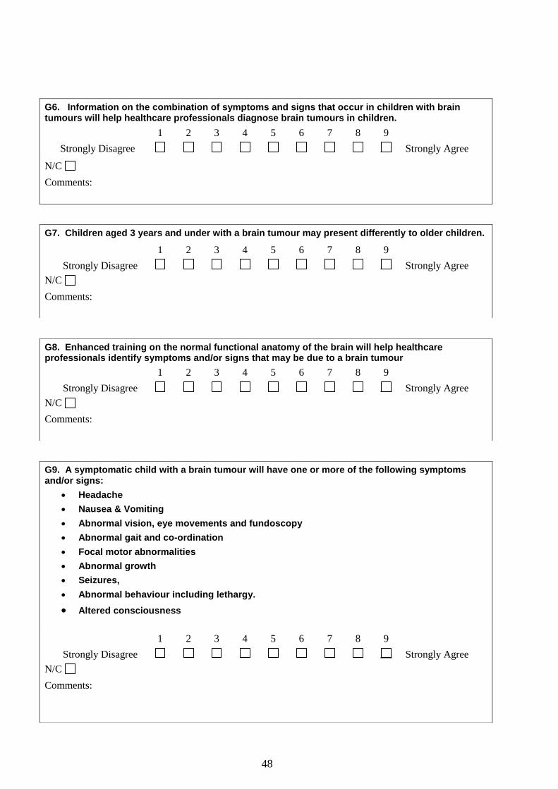
48
G6. Information on the combination of symptoms and signs that occur in children with brain tumours will help healthcare professionals diagnose brain tumours in children.
Strongly Disagree
1
2
3
4
5
6
7
8
9
Strongly Agree
N/C
Comments:
G7. Children aged 3 years and under with a brain tumour may present differently to older children.
Strongly Disagree
1
2
3
4
5
6
7
8
9
Strongly Agree
N/C
Comments:
G8. Enhanced training on the normal functional anatomy of the brain will help healthcare professionals identify symptoms and/or signs that may be due to a brain tumour
Strongly Disagree
1
2
3
4
5
6
7
8
9
Strongly Agree
N/C
Comments:
G9. A symptomatic child with a brain tumour will have one or more of the following symptoms and/or signs:
Headache
Nausea & Vomiting
Abnormal vision, eye movements and fundoscopy
Abnormal gait and co-ordination
Focal motor abnormalities
Abnormal growth
Seizures,
Abnormal behaviour including lethargy.
Altered consciousness
Strongly Disagree
1
2
3
4
5
6
7
8
9
Strongly Agree
N/C
Comments:
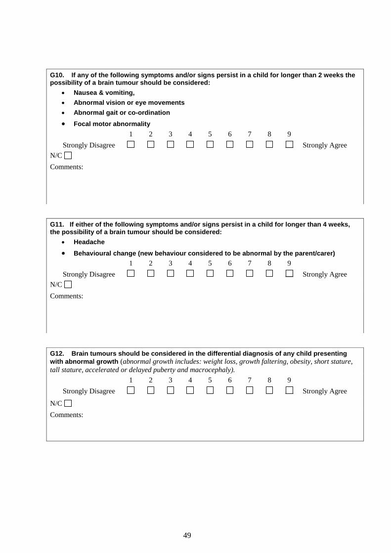
49
G10. If any of the following symptoms and/or signs persist in a child for longer than 2 weeks the possibility of a brain tumour should be considered:
Nausea & vomiting,
Abnormal vision or eye movements
Abnormal gait or co-ordination
Focal motor abnormality
Strongly Disagree
1
2
3
4
5
6
7
8
9
Strongly Agree
N/C
Comments:
G11. If either of the following symptoms and/or signs persist in a child for longer than 4 weeks, the possibility of a brain tumour should be considered:
Headache
Behavioural change (new behaviour considered to be abnormal by the parent/carer)
Strongly Disagree
1
2
3
4
5
6
7
8
9
Strongly Agree
N/C
Comments:
G12. Brain tumours should be considered in the differential diagnosis of any child presenting
with abnormal growth (abnormal growth includes: weight loss, growth faltering, obesity, short stature,
tall stature, accelerated or delayed puberty and macrocephaly).
Strongly Disagree
1
2
3
4
5
6
7
8
9
Strongly Agree
N/C
Comments:

50
G13. A child presenting with any of the symptoms and signs listed in Statements G10 – G12 requires all of the following:
a detailed history including specific enquiry for associated symptoms and predisposing factors
assessment of the visual system
assessment of the motor system
assessment of height, weight & head circumference in a child aged < 2 years
assessment of pubertal status in adolescents
assessment of developmental stage in a child < 5 years.
Strongly Disagree
1
2
3
4
5
6
7
8
9
Strongly Agree
N/C
Comments:
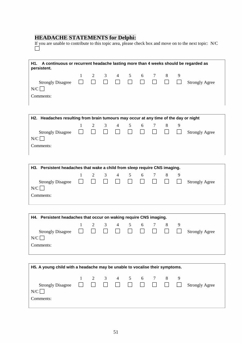
51
HEADACHE STATEMENTS for Delphi: If you are unable to contribute to this topic area, please check box and move on to the next topic: N/C
H1. A continuous or recurrent headache lasting more than 4 weeks should be regarded as persistent.
Strongly Disagree
1
2
3
4
5
6
7
8
9
Strongly Agree
N/C
Comments:
H2. Headaches resulting from brain tumours may occur at any time of the day or night
Strongly Disagree
1
2
3
4
5
6
7
8
9
Strongly Agree
N/C
Comments:
H3. Persistent headaches that wake a child from sleep require CNS imaging.
Strongly Disagree
1
2
3
4
5
6
7
8
9
Strongly Agree
N/C
Comments:
H4. Persistent headaches that occur on waking require CNS imaging.
Strongly Disagree
1
2
3
4
5
6
7
8
9
Strongly Agree
N/C
Comments:
H5. A young child with a headache may be unable to vocalise their symptoms.
Strongly Disagree
1
2
3
4
5
6
7
8
9
Strongly Agree
N/C
Comments:
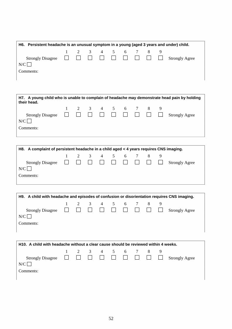
52
H6. Persistent headache is an unusual symptom in a young (aged 3 years and under) child.
Strongly Disagree
1
2
3
4
5
6
7
8
9
Strongly Agree
N/C
Comments:
H7. A young child who is unable to complain of headache may demonstrate head pain by holding their head.
Strongly Disagree
1
2
3
4
5
6
7
8
9
Strongly Agree
N/C
Comments:
H8. A complaint of persistent headache in a child aged < 4 years requires CNS imaging.
Strongly Disagree
1
2
3
4
5
6
7
8
9
Strongly Agree
N/C
Comments:
H9. A child with headache and episodes of confusion or disorientation requires CNS imaging.
Strongly Disagree
1
2
3
4
5
6
7
8
9
Strongly Agree
N/C
Comments:
H10. A child with headache without a clear cause should be reviewed within 4 weeks.
Strongly Disagree
1
2
3
4
5
6
7
8
9
Strongly Agree
N/C
Comments:

53
H11. A child with headache and vomiting who is diagnosed with migraine should usually be reviewed within 4 weeks.
Strongly Disagree
1
2
3
4
5
6
7
8
9
Strongly Agree
N/C
Comments:
H12. In a child diagnosed with a non-structural headache (e.g. migraine, tension headache) a change in the nature of the headache requires re-assessment and consideration of a structural cause.
Strongly Disagree
1
2
3
4
5
6
7
8
9
Strongly Agree
N/C
Comments:

54
NAUSEA & VOMITING STATEMENTS for Delphi: If you are unable to contribute to this topic area, please check box and move on to the next topic: N/C
NV1. Nausea and/or vomiting for longer than 2 weeks should be regarded as persistent.
Strongly Disagree
1
2
3
4
5
6
7
8
9
Strongly Agree
N/C
Comments:
NV2. Persistent nausea and/or vomiting in the absence of corroborative history, examination or investigation findings should not be attributed to a gastrointestinal cause.
Strongly Disagree
1
2
3
4
5
6
7
8
9
Strongly Agree
N/C
Comments:
NV3. Persistent nausea and/or vomiting in the absence of corroborative history, examination or investigation findings should not be attributed to an infective cause.
Strongly Disagree
1
2
3
4
5
6
7
8
9
Strongly Agree
N/C
Comments:
NV4. Persistent new vomiting on awakening (either in the morning or from a sleep in the day) requires CNS imaging.
Strongly Disagree
1
2
3
4
5
6
7
8
9
Strongly Agree
N/C
Comments:

55
VISUAL SYSTEM STATEMENTS for Delphi: If you are unable to contribute to this topic area, please check box and move on to the next topic: N/C
V1. Visual assessment of a child in whom a differential diagnosis includes a brain tumour must include assessment of:
Visual acuity
Eye movements
Pupil responses
Optic disc appearance
Visual fields (in children > 5 years)
Strongly Disagree
1
2
3
4
5
6
7
8
9
Strongly Agree
N/C
Comments:
V2. Pupil dilatation should be performed if required to obtain a clear view of the optic disc.
Strongly Disagree
1
2
3
4
5
6
7
8
9
Strongly Agree
N/C
Comments:
V3. Co-operative children aged 5 years and over can be assessed by a community optometrist.
Strongly Disagree
1
2
3
4
5
6
7
8
9
Strongly Agree
N/C
Comments:
V4. Children under 5 years and un-cooperative children should be assessed by the hospital eye service.
Strongly Disagree
1
2
3
4
5
6
7
8
9
Strongly Agree
N/C
Comments:

56
V5. If the healthcare professional assessing a child with any of the symptoms and signs listed in Statements G10-G12 is unable to perform a complete visual assessment, the child should be referred for assessment as described in Statements V3 and V4.
Strongly Disagree
1
2
3
4
5
6
7
8
9
Strongly Agree
N/C
Comments:
V6. Written communication between the lead healthcare professional and community optometry should explain the indications for assessment.
Strongly Disagree
1
2
3
4
5
6
7
8
9
Strongly Agree
N/C
Comments:
V7. Children should be assessed by ophthalmologists who have received training in paediatric ophthalmology.
Strongly Disagree
1
2
3
4
5
6
7
8
9
Strongly Agree
N/C
Comments:
V8. Community optometrists should refer any child with abnormal eye findings (excluding simple refractive errors) directly to secondary care.
Strongly Disagree
1
2
3
4
5
6
7
8
9
Strongly Agree
N/C
Comments:

57
V9. A child referred for visual assessment in whom a brain tumour is included in the differential diagnosis should be seen within two weeks of referral.
Strongly Disagree
1
2
3
4
5
6
7
8
9
Strongly Agree
N/C
Comments:
V10. CNS imaging is required for papilloedema.
Strongly Disagree
1
2
3
4
5
6
7
8
9
Strongly Agree
N/C
Comments:
V11. CNS imaging is required for optic atrophy.
Strongly Disagree
1
2
3
4
5
6
7
8
9
Strongly Agree
N/C
Comments:
V12. CNS imaging is required for new onset nystagmus.
Strongly Disagree
1
2
3
4
5
6
7
8
9
Strongly Agree
N/C
Comments:
V13. CNS imaging is required for a reduction in visual acuity not attributable to refractive error.
Strongly Disagree
1
2
3
4
5
6
7
8
9
Strongly Agree
N/C
Comments:
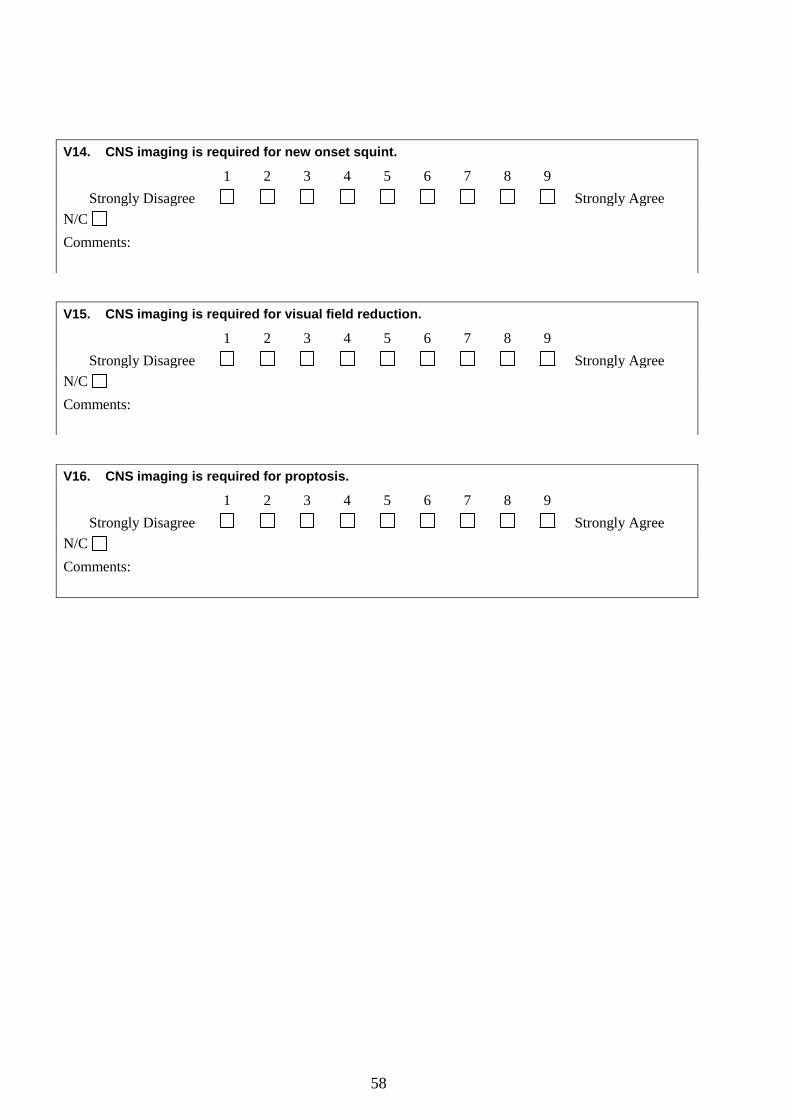
58
V14. CNS imaging is required for new onset squint.
Strongly Disagree
1
2
3
4
5
6
7
8
9
Strongly Agree
N/C
Comments:
V15. CNS imaging is required for visual field reduction.
Strongly Disagree
1
2
3
4
5
6
7
8
9
Strongly Agree
N/C
Comments:
V16. CNS imaging is required for proptosis.
Strongly Disagree
1
2
3
4
5
6
7
8
9
Strongly Agree
N/C
Comments:

59
MOTOR SYSTEM STATEMENTS for Delphi: If you are unable to contribute to this topic area, please check box and move on to the next topic: N/C
M1. A history of a change or deterioration in motor skills may indicate a brain tumour.
Strongly Disagree
1
2
3
4
5
6
7
8
9
Strongly Agree
N/C
Comments:
M2. History should enquire into subtle changes in motor skills e.g. change of hand or foot preference, loss of learned skills e.g. computer games
Strongly Disagree
1
2
3
4
5
6
7
8
9
Strongly Agree
N/C
Comments:
M3. Assessment of the gross motor skills of a child in whom a brain tumour is included in the differential diagnosis should include observation of:
sitting or crawling in infants
walking or running
gross motor coordination e.g. heel-toe walking.
Strongly Disagree
1
2
3
4
5
6
7
8
9
Strongly Agree
N/C
Comments:
M4. Assessment of a child’s fine motor and visuo-motor skills should include observation of:
handling of small objects e.g. cup, spoon, small sweet
handwriting in older children.
Strongly Disagree
1
2
3
4
5
6
7
8
9
Strongly Agree
N/C
Comments:

60
M5. Abnormal balance or gait should not be attributed to middle ear disease in the absence of corroborative history, examination or investigation findings.
Strongly Disagree
1
2
3
4
5
6
7
8
9
Strongly Agree
N/C
Comments:
M6. A child with facial nerve weakness that does not show improvement within 2 weeks should undergo CNS imaging.
Strongly Disagree
1
2
3
4
5
6
7
8
9
Strongly Agree
N/C
Comments:
M7. CNS imaging is required for any child with:
regression in motor skills
abnormal gait or co-ordination unless attributable to a non-neurological cause
focal motor weakness
Strongly Disagree
1
2
3
4
5
6
7
8
9
Strongly Agree
N/C
Comments:

61
GROWTH STATEMENTS for DELPHI: If you are unable to contribute to this topic area, please check box and move on to the next topic: N/C
GR1. Impaired growth associated with vomiting in a child should not be attributed to a gastrointestinal cause in the absence of history, examination or investigation findings suggestive of gastrointestinal disease.
Strongly Disagree
1
2
3
4
5
6
7
8
9
Strongly Agree
N/C
Comments:
GR2. A child with impaired growth with no clearly identifiable psychosocial or physical cause should have CNS imaging.
Strongly Disagree
1
2
3
4
5
6
7
8
9
Strongly Agree
N/C
Comments:
GR3. CNS imaging should be undertaken prior to attributing weight loss to anorexia nervosa if the full diagnostic criteria for anorexia nervosa are not met.
Strongly Disagree
1
2
3
4
5
6
7
8
9
Strongly Agree
N/C
Comments:
GR4. Reluctance to feed or eat leading to weight loss may result from swallowing difficulties.
Strongly Disagree
1
2
3
4
5
6
7
8
9
Strongly Agree
N/C
Comments:

62
GR5. A child with swallowing difficulties not attributable to a cause outside the CNS should have CNS imaging.
Strongly Disagree
1
2
3
4
5
6
7
8
9
Strongly Agree
N/C
Comments:
GR6. Swallowing difficulties may present with recurrent chest infections.
Strongly Disagree
1
2
3
4
5
6
7
8
9
Strongly Agree
N/C
Comments:
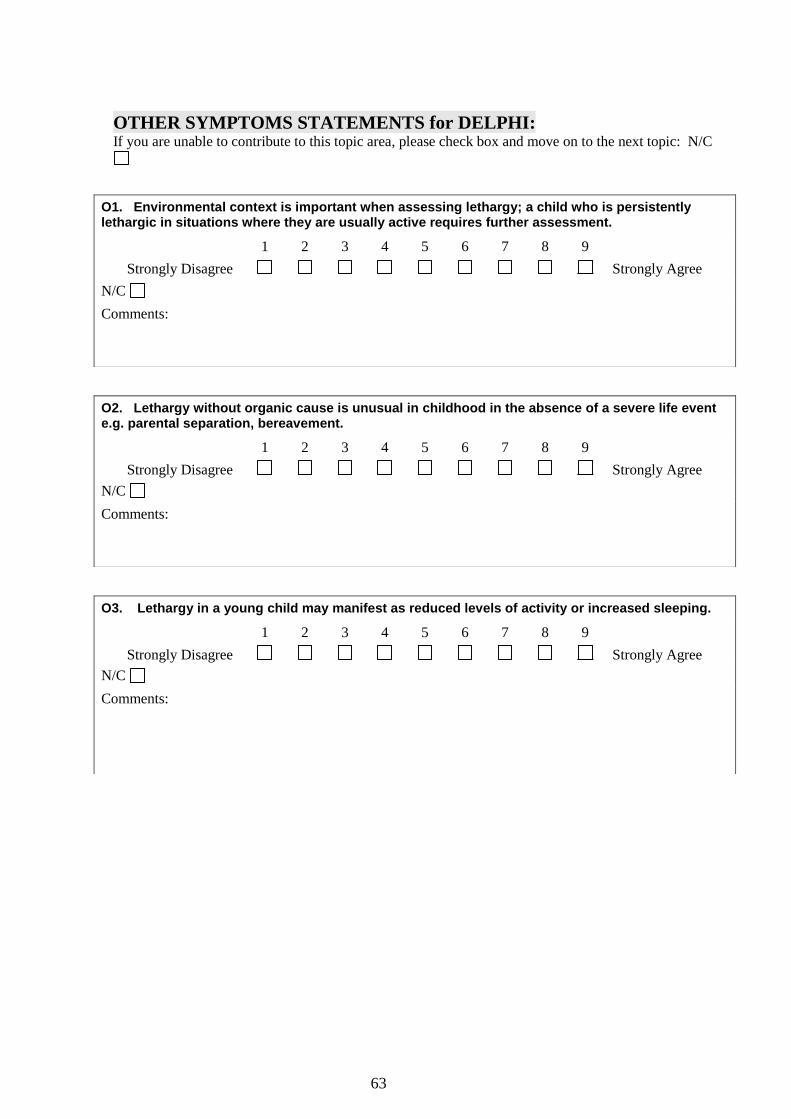
63
OTHER SYMPTOMS STATEMENTS for DELPHI: If you are unable to contribute to this topic area, please check box and move on to the next topic: N/C
O1. Environmental context is important when assessing lethargy; a child who is persistently lethargic in situations where they are usually active requires further assessment.
Strongly Disagree
1
2
3
4
5
6
7
8
9
Strongly Agree
N/C
Comments:
O2. Lethargy without organic cause is unusual in childhood in the absence of a severe life event e.g. parental separation, bereavement.
Strongly Disagree
1
2
3
4
5
6
7
8
9
Strongly Agree
N/C
Comments:
O3. Lethargy in a young child may manifest as reduced levels of activity or increased sleeping.
Strongly Disagree
1
2
3
4
5
6
7
8
9
Strongly Agree
N/C
Comments:

64
REFERRAL PATHWAYS & IMAGING STATEMENTS for DELPHI: If you are unable to contribute to this topic area, please check box and go to PAGE 22 : N/C
R1. A child referred from primary care in which the differential diagnosis includes a possible CNS space-occupying lesion should be seen within two weeks under the “two week cancer referral rule”.
Strongly Disagree
1
2
3
4
5
6
7
8
9
Strongly Agree
N/C
Comments:
R2. “Choose and Book” is an impediment to rapid referral.
Strongly Disagree
1
2
3
4
5
6
7
8
9
Strongly Agree
N/C
Comments:
R3. Parents/carers know their child best; they should be asked explicitly about their concerns in any consultation
Strongly Disagree
1
2
3
4
5
6
7
8
9
Strongly Agree
N/C
Comments:
R4. If a parent / carer expresses concerns about a brain tumour this should be reviewed carefully. If a brain tumour is unlikely the reasons why should be explained and arrangements made for review within 4 weeks.
Strongly Disagree
1
2
3
4
5
6
7
8
9
Strongly Agree
N/C
Comments:
R5. Language can be a barrier to achieving diagnosis. If the patient and healthcare professional are not fluent in a common language an interpreter must be used for the consultation
Strongly Disagree
1
2
3
4
5
6
7
8
9
Strongly Agree
N/C
Comments:

65
R6. MRI is the imaging modality of choice for a child who may have a CNS tumour.
Strongly Disagree
1
2
3
4
5
6
7
8
9
Strongly Agree
N/C
Comments:
R7. For MRI, contrast enhancement is not required to exclude a structural CNS abnormality.
Strongly Disagree
1
2
3
4
5
6
7
8
9
Strongly Agree
N/C
Comments:
R8. If MRI is not available a contrast enhanced CT scan should be performed.
Strongly Disagree
1
2
3
4
5
6
7
8
9
Strongly Agree
N/C
Comments:
R9. Cranial ultrasound has no place in exclusion of CNS tumours in infants
Strongly Disagree
1
2
3
4
5
6
7
8
9
Strongly Agree
N/C
Comments:
R10. Imaging results should be interpreted by a professional with expertise and training in CNS MR and CT imaging in children.
Strongly Disagree
1
2
3
4
5
6
7
8
9
Strongly Agree
N/C
Comments:

66
R11. A child referred for non-emergency imaging in whom a brain tumour is included in the differential diagnosis should be imaged within two weeks.
Strongly Disagree
1
2
3
4
5
6
7
8
9
Strongly Agree
N/C
Comments:
R12. The need to sedate or anaesthetise a child for imaging should not delay imaging by more than a week.
Strongly Disagree
1
2
3
4
5
6
7
8
9
Strongly Agree
N/C
Comments:
R13. Patients and their families should receive the provisional results of CNS imaging within one week of the investigation.
Strongly Disagree
1
2
3
4
5
6
7
8
9
Strongly Agree
N/C
Comments:
R14. General practitioners should be able to refer a child for CNS imaging.
Strongly Disagree
1
2
3
4
5
6
7
8
9
Strongly Agree
N/C
Comments:

67
R15. In my experience, a nursing professional (e.g. health visitor, practice nurse, school nurse) has played a critical role in the identification of a child with a brain tumour.
Strongly Disagree
1
2
3
4
5
6
7
8
9
Strongly Agree
N/C
Comments:
R16. A primary healthcare professional who has a high index of suspicion regarding a possible brain tumour in a child should discuss their concerns with a secondary healthcare professional the same day.
Strongly Disagree
1
2
3
4
5
6
7
8
9
Strongly Agree
N/C
Comments:
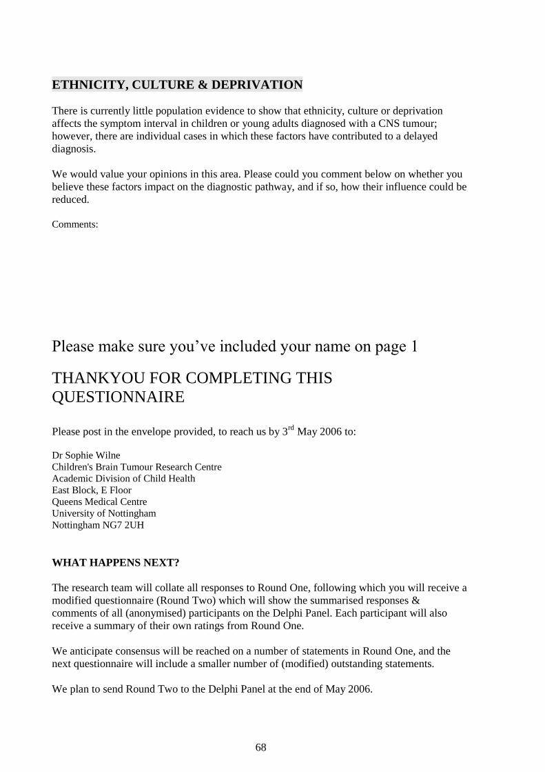
68
ETHNICITY, CULTURE & DEPRIVATION
There is currently little population evidence to show that ethnicity, culture or deprivation
affects the symptom interval in children or young adults diagnosed with a CNS tumour;
however, there are individual cases in which these factors have contributed to a delayed
diagnosis.
We would value your opinions in this area. Please could you comment below on whether you
believe these factors impact on the diagnostic pathway, and if so, how their influence could be
reduced.
Comments:
Please make sure you‟ve included your name on page 1
THANKYOU FOR COMPLETING THIS
QUESTIONNAIRE
Please post in the envelope provided, to reach us by 3
rd May 2006 to:
Dr Sophie Wilne
Children's Brain Tumour Research Centre
Academic Division of Child Health
East Block, E Floor
Queens Medical Centre
University of Nottingham
Nottingham NG7 2UH
WHAT HAPPENS NEXT?
The research team will collate all responses to Round One, following which you will receive a
modified questionnaire (Round Two) which will show the summarised responses &
comments of all (anonymised) participants on the Delphi Panel. Each participant will also
receive a summary of their own ratings from Round One.
We anticipate consensus will be reached on a number of statements in Round One, and the
next questionnaire will include a smaller number of (modified) outstanding statements.
We plan to send Round Two to the Delphi Panel at the end of May 2006.

69
3.4.3: Delphi questionnaire round one results
112 panel members returned the round one questionnaire within the required time frame.
Statements were taken as having reached consensus if 75% or more of the Delphi panel
respondents rated the statement 7, 8 or 9. Statements were rejected if 25% or less of the
Delphi panel rated the statements 7, 8 or 9. Ratings of N/C, blanks or two boxes checked in
error were excluded from the analysis of that statement. 53 of the 77 original statements
reached consensus, two were rejected and the remaining 22 statements were modified or
excluded based upon feedback. The percentage in each score band for the Delphi statements
in round one is shown in figure 3.4.2.
Figure 7: Percentage in each score band for the Delphi statements in round one
A: General statements
B: Headache

70
C: Nausea and vomiting
D: Visual system
E: Motor system

71
F: Growth statements
G: Other statements
H: Referral and imaging statements

72
The following statements from round one reached consensus:
G1. The initial symptoms of a brain tumour may mimic symptoms that occur
with other more common and less serious childhood conditions.
G2. Symptoms occurring with brain tumours may fluctuate in severity.
G3. Apparent resolution and then recurrence of a symptom(s) does not exclude
a brain tumour.
G4. The absence of neurological abnormalities does not exclude a brain
tumour.
G7. Children aged 3 years and under with a brain tumour may present
differently to older children.
G9. A symptomatic child with a brain tumour will have one or more of the
following symptoms and/or signs:
Headache
Nausea & Vomiting
Abnormal vision, eye movements and fundoscopy
Abnormal gait and co-ordination
Focal motor abnormalities
Abnormal growth
Seizures,
Abnormal behaviour including lethargy.
Altered consciousness
G10. If any of the following symptoms and/or signs persist in a child for longer
than 2 weeks the possibility of a brain tumour should be considered:
Nausea & vomiting,
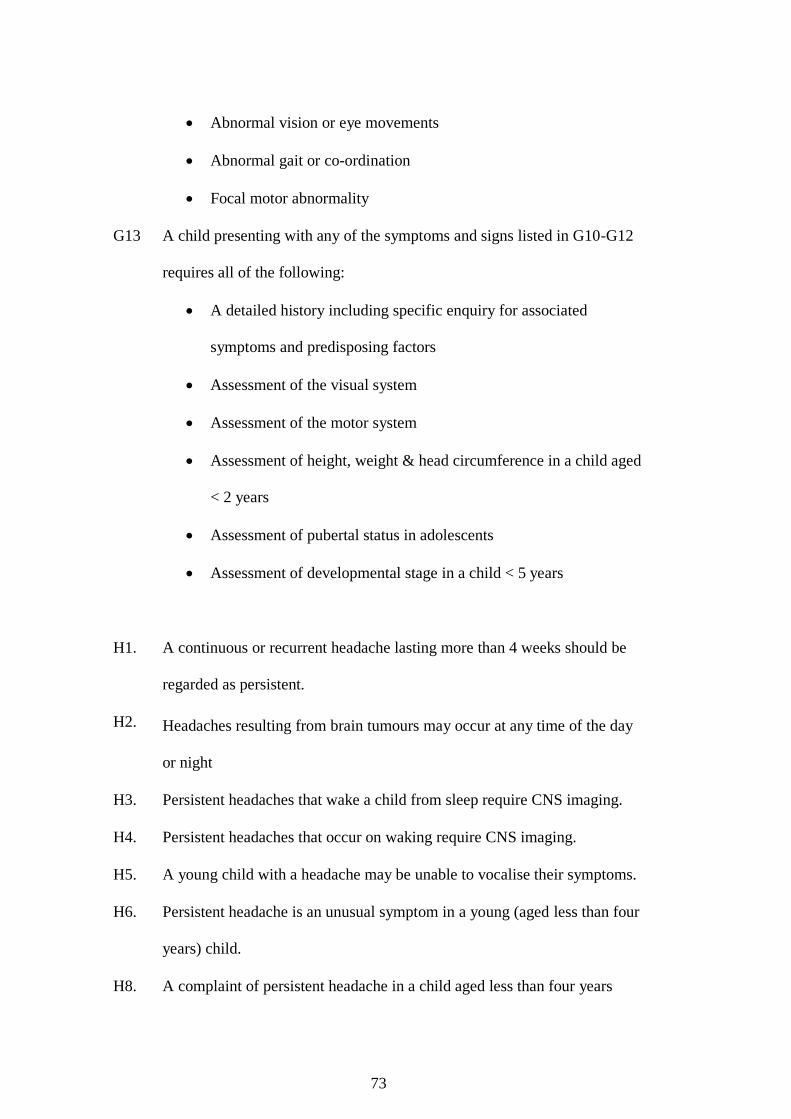
73
Abnormal vision or eye movements
Abnormal gait or co-ordination
Focal motor abnormality
G13 A child presenting with any of the symptoms and signs listed in G10-G12
requires all of the following:
A detailed history including specific enquiry for associated
symptoms and predisposing factors
Assessment of the visual system
Assessment of the motor system
Assessment of height, weight & head circumference in a child aged
< 2 years
Assessment of pubertal status in adolescents
Assessment of developmental stage in a child < 5 years
H1. A continuous or recurrent headache lasting more than 4 weeks should be
regarded as persistent.
H2. Headaches resulting from brain tumours may occur at any time of the day
or night
H3. Persistent headaches that wake a child from sleep require CNS imaging.
H4. Persistent headaches that occur on waking require CNS imaging.
H5. A young child with a headache may be unable to vocalise their symptoms.
H6. Persistent headache is an unusual symptom in a young (aged less than four
years) child.
H8. A complaint of persistent headache in a child aged less than four years

74
years requires CNS imaging.
H9. A child with headache and episodes of confusion or disorientation requires
CNS imaging.
NV1. Nausea and/or vomiting for longer than 2 weeks should be regarded as
persistent.
NV3. Persistent nausea and/or vomiting in the absence of corroborative history,
examination or investigation findings should not be attributed to an
infective cause.
NV4. Persistent new vomiting on awakening (either in the morning or from a
sleep in the day) requires CNS imaging.
V1. Visual assessment of a child in whom a differential diagnosis includes a brain
tumour must include assessment of:
Visual acuity
Eye movements
Pupil responses
Optic disc appearance
Visual fields (in children older than five years)
V4. Children under 5 years and un-cooperative children should be assessed by the
hospital eye service.
V5. If the healthcare professional assessing a child with any of the symptoms and
signs listed in Statements G10-G12 is unable to perform a complete visual
assessment, the child should be referred for assessment as described in
Statements V3 and V4.

75
V6. Written communication between the lead healthcare professional and community
optometry should explain the indications for assessment.
V8. Community optometrists should refer any child with abnormal eye findings
(excluding simple refractive errors) directly to secondary care.
V9. A child referred for visual assessment in whom a brain tumour is included in the
differential diagnosis should be seen within two weeks of referral.
V10. CNS imaging is required for papilloedema.
V11. CNS imaging is required for optic atrophy.
V12. CNS imaging is required for new onset nystagmus.
V13 CNS imaging is required for a reduction in visual acuity not attributable to
refractive error.
V15 CNS imaging is required for visual field reduction.
V16 CNS imaging is required for proptosis.
M1. A history of a change or deterioration in motor skills may indicate a brain
tumour.
M2. History should enquire into subtle changes in motor skills e.g. change of hand or
foot preference, loss of learned skills e.g. computer games
M3. Assessment of the gross motor skills of a child in whom a brain tumour is
included in the differential diagnosis should include observation of:
sitting or crawling in infants
walking or running
gross motor coordination e.g. heel-toe walking.
M4. Assessment of a child‟s fine motor and visual-motor skills should include

76
observation of:
handling of small objects e.g. cup, spoon, small sweet
handwriting in older children.
M5. Abnormal balance or gait should not be attributed to middle ear disease in the
absence of corroborative history, examination or investigation findings.
M7. CNS imaging is required for any child with:
regression in motor skills
abnormal gait or co-ordination unless attributable to a non-neurological
cause
focal motor weakness
GR1. Impaired growth associated with vomiting in a child should not be attributed to a
gastrointestinal cause in the absence of history, examination or investigation
findings suggestive of gastrointestinal disease.
GR5. A child with swallowing difficulties not attributable to a cause outside the CNS
should have CNS imaging.
GR6. Swallowing difficulties may present with recurrent chest infections.
O1. Environmental context is important when assessing lethargy; a child who is
persistently lethargic in situations where they are usually active requires further
assessment.
O3. Lethargy in a young child may manifest as reduced levels of activity or increased
sleeping.

77
R1. A child referred from primary care in which the differential diagnosis includes a
possible CNS space-occupying lesion should be seen within two weeks under the
“two week cancer referral rule”.
R2. “Choose and Book” is an impediment to rapid referral.
R3. Parents/carers know their child best; they should be asked explicitly about their
concerns in any consultation
R4. If a parent / carer expresses concerns about a brain tumour this should be
reviewed carefully. If a brain tumour is unlikely the reasons why should be
explained and arrangements made for review within 4 weeks.
R5. Language can be a barrier to achieving diagnosis. If the patient and healthcare
professional are not fluent in a common language an interpreter must be used for
the consultation
R6. MRI is the imaging modality of choice for a child who may have a CNS tumour.
R8. If MRI is not available a contrast enhanced CT scan should be performed.
R10. Imaging results should be interpreted by a professional with expertise and
training in CNS MR and CT imaging in children.
R12. The need to sedate or anaesthetise a child for imaging should not delay imaging
by more than a week.
R13 Patients and their families should receive the provisional results of CNS imaging
within one week of the investigation.
R16 A primary healthcare professional who has a high index of suspicion regarding a
possible brain tumour in a child should discuss their concerns with a secondary
healthcare professional the same day.

78
3.4.4: Delphi process round two
Round two was issued to the 112 participants returning round one. The participants were
provided with the results detailed above. Statements were modified according to feedback
from round one and then reissued. In response to feedback one new statement was also added
to round two. The round two Delphi questionnaire, shown below, asked the panel to rank their
agreement with 14 statements.
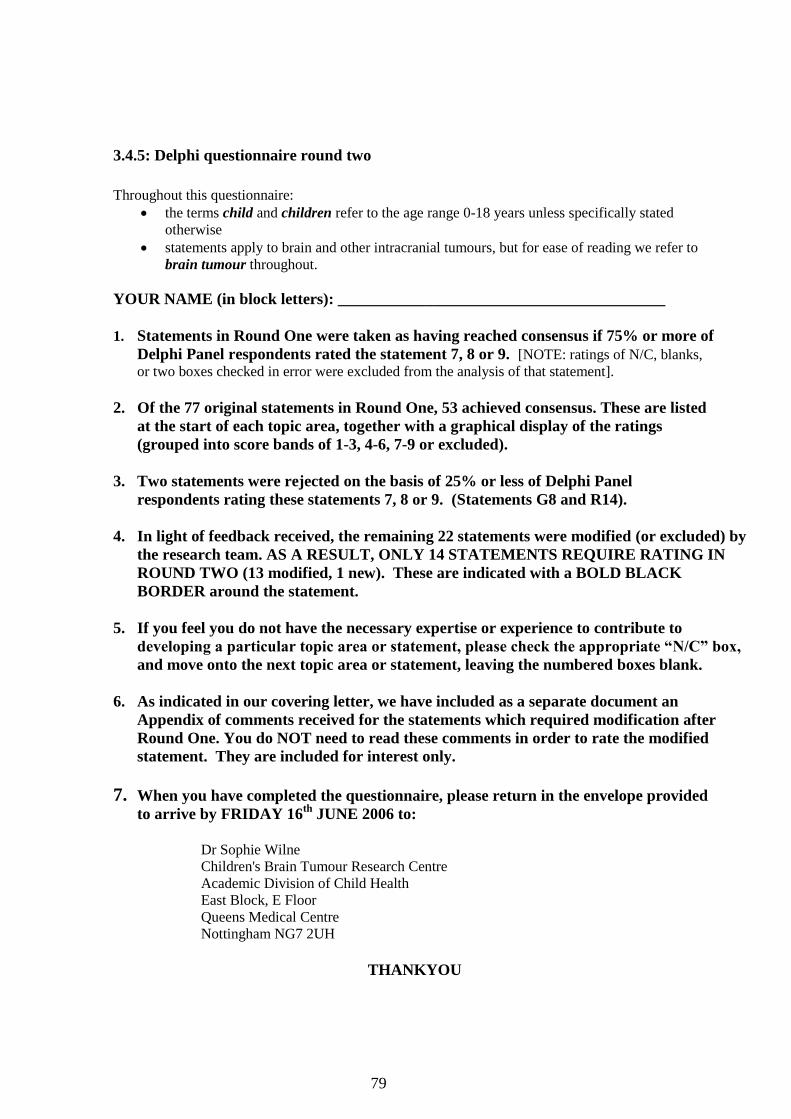
79
3.4.5: Delphi questionnaire round two
Throughout this questionnaire:
the terms child and children refer to the age range 0-18 years unless specifically stated
otherwise
statements apply to brain and other intracranial tumours, but for ease of reading we refer to
brain tumour throughout.
YOUR NAME (in block letters): _________________________________________
1. Statements in Round One were taken as having reached consensus if 75% or more of
Delphi Panel respondents rated the statement 7, 8 or 9. [NOTE: ratings of N/C, blanks,
or two boxes checked in error were excluded from the analysis of that statement].
2. Of the 77 original statements in Round One, 53 achieved consensus. These are listed
at the start of each topic area, together with a graphical display of the ratings
(grouped into score bands of 1-3, 4-6, 7-9 or excluded).
3. Two statements were rejected on the basis of 25% or less of Delphi Panel
respondents rating these statements 7, 8 or 9. (Statements G8 and R14).
4. In light of feedback received, the remaining 22 statements were modified (or excluded) by
the research team. AS A RESULT, ONLY 14 STATEMENTS REQUIRE RATING IN
ROUND TWO (13 modified, 1 new). These are indicated with a BOLD BLACK
BORDER around the statement.
5. If you feel you do not have the necessary expertise or experience to contribute to
developing a particular topic area or statement, please check the appropriate “N/C” box,
and move onto the next topic area or statement, leaving the numbered boxes blank.
6. As indicated in our covering letter, we have included as a separate document an
Appendix of comments received for the statements which required modification after
Round One. You do NOT need to read these comments in order to rate the modified
statement. They are included for interest only.
7. When you have completed the questionnaire, please return in the envelope provided
to arrive by FRIDAY 16th
JUNE 2006 to:
Dr Sophie Wilne
Children's Brain Tumour Research Centre
Academic Division of Child Health
East Block, E Floor
Queens Medical Centre
Nottingham NG7 2UH
THANKYOU

80
GENERAL STATEMENTS for Delphi:
RESULTS of ROUND ONE
The following eight GENERAL statements achieved consensus in Round One:
G1. The initial symptoms of a brain tumour may mimic symptoms that occur with other more
common and less serious childhood conditions. G2. Symptoms occurring with brain tumours may fluctuate in severity. G3. Apparent resolution and then recurrence of a symptom(s) does not exclude a brain tumour. G4. The absence of neurological abnormalities does not exclude a brain tumour. G7. Children aged 3 years and under with a brain tumour may present differently to older
children. G9. A symptomatic child with a brain tumour will have one or more of the following symptoms
and/or signs:
Headache
Nausea & Vomiting
Abnormal vision, eye movements and fundoscopy
Abnormal gait and co-ordination
Focal motor abnormalities
Abnormal growth
Seizures,
Abnormal behaviour including lethargy.
Altered consciousness G10. If any of the following symptoms and/or signs persist in a child for longer than 2 weeks the
possibility of a brain tumour should be considered:
Nausea & vomiting,
Abnormal vision or eye movements
Abnormal gait or co-ordination
Focal motor abnormality G13 A child presenting with any of the symptoms and signs listed in G10-G12 requires all of the
following:
A detailed history including specific enquiry for associated symptoms and predisposing factors
Assessment of the visual system
Assessment of the motor system
Assessment of height, weight & head circumference in a child aged < 2 years
Assessment of pubertal status in adolescents
Assessment of developmental stage in a child < 5 years
The following five GENERAL statements did NOT achieve consensus in Round One, and have been
modified for voting in Round Two, or excluded: If you are unable to contribute to this topic area,
please check box and move on to the next topic: N/C

81
G5. 95% of children with a brain tumour have multiple symptoms and/or signs by diagnosis.
Outcome: Statement excluded.
Reason: Inappropriate statement for a Delphi consensus process. The statement can be verified from
alternate sources.
G6. Information on the combination of symptoms and signs that occur in children with brain tumours will help healthcare professionals diagnose brain tumours in children.
Outcome: Statement excluded.
Reason: This statement is covered in more detail in G9, for which consensus was achieved.
G8. Enhanced training on the normal functional anatomy of the brain will help healthcare professionals identify symptoms and/or signs that may be due to a brain tumour
Outcome: Statement rejected.
Reason: Less than 25% of respondents rated this statement 7, 8 or 9 in Round One
G11. If either of the following symptoms and/or signs persist in a child for longer than 4 weeks, the possibility of a brain tumour should be considered:
Headache
Behavioural change (new behaviour considered to be abnormal by the parent/carer)
Outcome: Statement modified for Round Two in light of comments received (see appendix 1). Feedback
suggested symptoms/signs of headache and behavioural change should be considered separately, hence
modified statements G11(a) and G11(b).
MODIFIED G11(a) . If a child presents with a new headache persisting for longer than 4 weeks a brain tumour should be considered in the differential diagnosis [NOTE: ‘persisting’ defined in H1 i.e. a continuous or recurrent headache lasting more than 4 weeks]
Strongly Disagree
1
2
3
4
5
6
7
8
9
Strongly Agree
N/C
Comments:

82
MODIFIED G11(b). If a child presents with abnormal behaviour (causing concern to parents/carers) including lethargy or withdrawal and persisting for more than 4 weeks, a brain tumour should be considered in the differential diagnosis.
Strongly Disagree
1
2
3
4
5
6
7
8
9
Strongly Agree
N/C
Comments:
G12. Brain tumours should be considered in the differential diagnosis of any child presenting
with abnormal growth (abnormal growth includes: weight loss, growth faltering, obesity, short stature,
tall stature, accelerated or delayed puberty and macrocephaly).
Outcome: Statement modified for Round Two in light of comments received (appendix 1).
MODIFIED G12 : A child who presents with one or more of the following symptoms and/or signs requires early specialist referral for consideration of a brain tumour in the differential diagnosis:
Precocious puberty
Delayed puberty
Growth failure
Macrocephally
Strongly Disagree
1
2
3
4
5
6
7
8
9
Strongly Agree
N/C
Comments:

83
HEADACHE STATEMENTS for Delphi:
RESULTS of ROUND ONE:
The following eight HEADACHE statements achieved consensus in Round One:
H1. A continuous or recurrent headache lasting more than 4 weeks should be regarded as
persistent. H2. Headaches resulting from brain tumours may occur at any time of the day or night H3. Persistent headaches that wake a child from sleep require CNS imaging. H4. Persistent headaches that occur on waking require CNS imaging. H5. A young child with a headache may be unable to vocalise their symptoms. H6. Persistent headache is an unusual symptom in a young (aged 3 years and under) child. H8. A complaint of persistent headache in a child aged < 4 years requires CNS imaging. H9. A child with headache and episodes of confusion or disorientation requires CNS imaging.
The following four HEADACHE statements did NOT achieve consensus in Round One, and have been
modified for voting in Round Two, or excluded: If you are unable to contribute to this topic area, please check
box and move on to the next topic: N/C
H7. A young child who is unable to complain of headache may demonstrate head pain by holding their head.
Outcome: Statement excluded.
Reason: Statement covered by H5 in which consensus was achieved.
H11. A child with headache and vomiting who is diagnosed with migraine should usually be reviewed within 4 weeks.
Outcome: Statement excluded.
Reason: Statement covered by G9 and (modified) H10 – see below.

84
H10. A child with headache without a clear cause should be reviewed within 4 weeks.
Outcome : Statement modified for Round Two in light of comments received (appendix 1).
MODIFIED H10. A child presenting with a new and persisting headache should be reviewed within 2 weeks [‘persisting’ as defined in H1 i.e. a continuous or recurrent headache lasting more than 4 weeks].
Strongly Disagree
1
2
3
4
5
6
7
8
9
Strongly Agree
N/C
Comments:
H12. In a child diagnosed with a non-structural headache (e.g. migraine, tension headache) a change in the nature of the headache requires re-assessment and consideration of a structural cause.
Outcome: Statement modified for Round Two in light of comments received (see appendix 1).
MODIFIED H12. In a child with known migraine or tension headaches, a change in the nature of the headache requires reassessment.
Strongly Disagree
1
2
3
4
5
6
7
8
9
Strongly Agree
N/C
Comments:

85
NAUSEA & VOMITING STATEMENTS for Delphi:
RESULTS of ROUND ONE:
The following three NAUSEA & VOMITING statements achieved consensus in Round One:
NV1. Nausea and/or vomiting for longer than 2 weeks should be regarded as
persistent. NV3. Persistent nausea and/or vomiting in the absence of corroborative history,
examination or investigation findings should not be attributed to an infective cause.
NV4. Persistent new vomiting on awakening (either in the morning or from a sleep in the day) requires CNS imaging.
The following NAUSEA & VOMITING statement did NOT achieve consensus in Round One, and has been
modified for voting in Round Two:
NV2. Persistent nausea and/or vomiting in the absence of corroborative history, examination or investigation findings should not be attributed to a gastrointestinal cause.
Outcome: Statement modified for Round Two in light of comments received (see appendix 1).
MODIFIED NV2. A child presenting with persistent nausea and/or vomiting requires early specialist referral for consideration of underlying causes including CNS causes
Strongly Disagree
1
2
3
4
5
6
7
8
9
Strongly Agree
N/C
Comments:

86
VISUAL SYSTEM STATEMENTS for Delphi:
RESULTS of ROUND ONE:
The following twelve VISUAL SYSTEM statements achieved consensus in Round One:
V1. Visual assessment of a child in whom a differential diagnosis includes a brain
tumour must include assessment of:
Visual acuity
Eye movements
Pupil responses
Optic disc appearance
Visual fields (in children > 5 years)
V4. Children under 5 years and un-cooperative children should be assessed by the hospital eye service.
V5. If the healthcare professional assessing a child with any of the symptoms and signs listed in Statements G10-G12 is unable to perform a complete visual assessment, the child should be referred for assessment as described in Statements V3 and V4.
V6. Written communication between the lead healthcare professional and community optometry should explain the indications for assessment.
V8. Community optometrists should refer any child with abnormal eye findings (excluding simple refractive errors) directly to secondary care.
V9. A child referred for visual assessment in whom a brain tumour is included in the differential diagnosis should be seen within two weeks of referral.
V10. CNS imaging is required for papilloedema. V11. CNS imaging is required for optic atrophy. V12. CNS imaging is required for new onset nystagmus. V13 CNS imaging is required for a reduction in visual acuity not attributable to
refractive error. V15 CNS imaging is required for visual field reduction. V16 CNS imaging is required for proptosis.
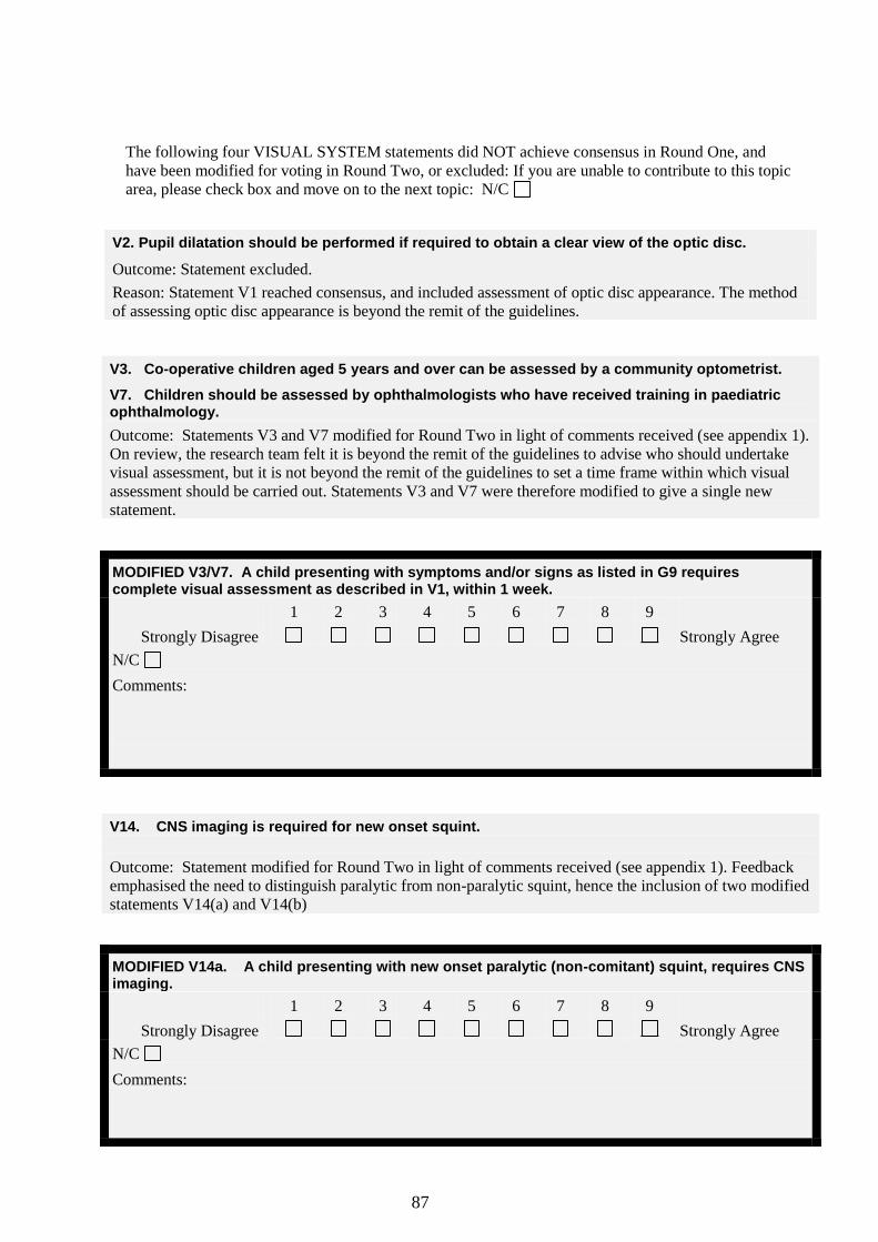
87
The following four VISUAL SYSTEM statements did NOT achieve consensus in Round One, and
have been modified for voting in Round Two, or excluded: If you are unable to contribute to this topic
area, please check box and move on to the next topic: N/C
V2. Pupil dilatation should be performed if required to obtain a clear view of the optic disc.
Outcome: Statement excluded.
Reason: Statement V1 reached consensus, and included assessment of optic disc appearance. The method
of assessing optic disc appearance is beyond the remit of the guidelines.
V3. Co-operative children aged 5 years and over can be assessed by a community optometrist.
V7. Children should be assessed by ophthalmologists who have received training in paediatric ophthalmology.
Outcome: Statements V3 and V7 modified for Round Two in light of comments received (see appendix 1).
On review, the research team felt it is beyond the remit of the guidelines to advise who should undertake
visual assessment, but it is not beyond the remit of the guidelines to set a time frame within which visual
assessment should be carried out. Statements V3 and V7 were therefore modified to give a single new
statement.
MODIFIED V3/V7. A child presenting with symptoms and/or signs as listed in G9 requires complete visual assessment as described in V1, within 1 week.
Strongly Disagree
1
2
3
4
5
6
7
8
9
Strongly Agree
N/C
Comments:
V14. CNS imaging is required for new onset squint.
Outcome: Statement modified for Round Two in light of comments received (see appendix 1). Feedback
emphasised the need to distinguish paralytic from non-paralytic squint, hence the inclusion of two modified
statements V14(a) and V14(b)
MODIFIED V14a. A child presenting with new onset paralytic (non-comitant) squint, requires CNS imaging.
Strongly Disagree
1
2
3
4
5
6
7
8
9
Strongly Agree
N/C
Comments:

88
MODIFIED V14b. A child presenting with new onset non-paralytic (comitant) squint should have early ophthalmic referral for assessment of underlying causes, including CNS causes.
Strongly Disagree
1
2
3
4
5
6
7
8
9
Strongly Agree
N/C
Comments:
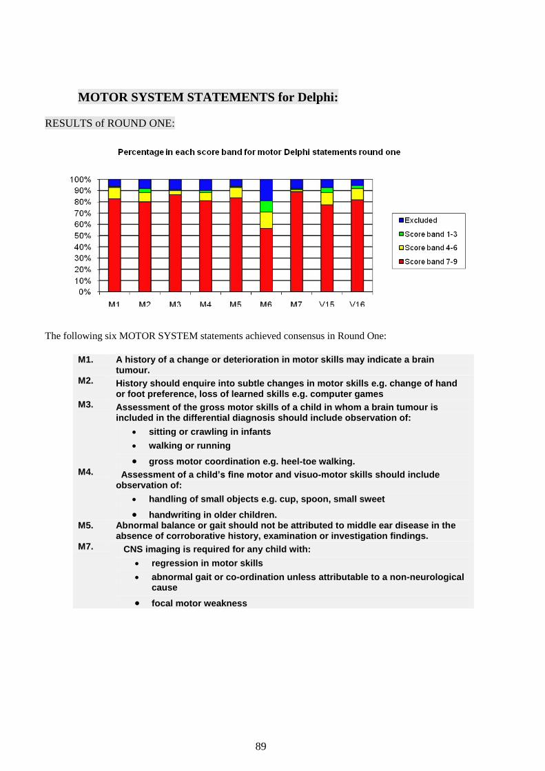
89
MOTOR SYSTEM STATEMENTS for Delphi:
RESULTS of ROUND ONE:
The following six MOTOR SYSTEM statements achieved consensus in Round One:
M1. A history of a change or deterioration in motor skills may indicate a brain
tumour. M2. History should enquire into subtle changes in motor skills e.g. change of hand
or foot preference, loss of learned skills e.g. computer games M3. Assessment of the gross motor skills of a child in whom a brain tumour is
included in the differential diagnosis should include observation of:
sitting or crawling in infants
walking or running
gross motor coordination e.g. heel-toe walking. M4. Assessment of a child’s fine motor and visuo-motor skills should include
observation of:
handling of small objects e.g. cup, spoon, small sweet
handwriting in older children. M5. Abnormal balance or gait should not be attributed to middle ear disease in the
absence of corroborative history, examination or investigation findings. M7. CNS imaging is required for any child with:
regression in motor skills
abnormal gait or co-ordination unless attributable to a non-neurological cause
focal motor weakness

90
The following MOTOR SYSTEM statement did NOT achieve consensus in Round One, and has been
modified for voting in Round Two:
M6. A child with facial nerve weakness that does not show improvement within 2 weeks should undergo CNS imaging.
Outcome: Statement modified for Round Two in light of comments received (see appendix 1).
MODIFIED M6. A child with presumed Bell’s palsy (isolated lower motor neurone facial nerve palsy) that does not show improvement within 4 weeks requires CNS imaging.
Strongly Disagree
1
2
3
4
5
6
7
8
9
Strongly Agree
N/C
Comments:
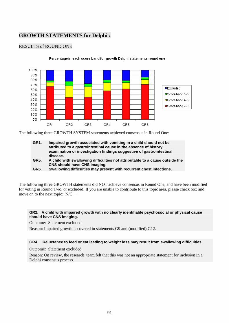
91
GROWTH STATEMENTS for Delphi :
RESULTS of ROUND ONE
The following three GROWTH SYSTEM statements achieved consensus in Round One:
GR1. Impaired growth associated with vomiting in a child should not be
attributed to a gastrointestinal cause in the absence of history, examination or investigation findings suggestive of gastrointestinal disease.
GR5. A child with swallowing difficulties not attributable to a cause outside the CNS should have CNS imaging.
GR6. Swallowing difficulties may present with recurrent chest infections.
The following three GROWTH statements did NOT achieve consensus in Round One, and have been modified
for voting in Round Two, or excluded: If you are unable to contribute to this topic area, please check box and
move on to the next topic: N/C
GR2. A child with impaired growth with no clearly identifiable psychosocial or physical cause should have CNS imaging.
Outcome: Statement excluded.
Reason: Impaired growth is covered in statements G9 and (modified) G12.
GR4. Reluctance to feed or eat leading to weight loss may result from swallowing difficulties.
Outcome: Statement excluded.
Reason: On review, the research team felt that this was not an appropriate statement for inclusion in a
Delphi consensus process.

92
GR3. CNS imaging should be undertaken prior to attributing weight loss to anorexia nervosa if the full diagnostic criteria for anorexia nervosa are not met.
Outcome: Statement modified for Round Two in light of comments received (see section 3.4.6). Feedback
suggests boys and girls should be considered separately, hence modified statements GR3(a) and GR3(b).
MODIFIED GR3(a). A boy with presumed anorexia nervosa requires early specialist referral for consideration of a brain tumour in the differential diagnosis.
Strongly Disagree
1
2
3
4
5
6
7
8
9
Strongly Agree
N/C
Comments:
MODIFIED GR3(b). A girl with presumed anorexia nervosa requires early specialist referral for consideration of a brain tumour in the differential diagnosis, if there are any atypical features.
Strongly Disagree
1
2
3
4
5
6
7
8
9
Strongly Agree
N/C
Comments:
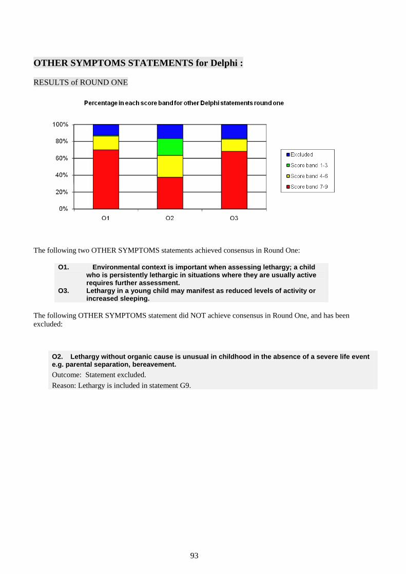
93
OTHER SYMPTOMS STATEMENTS for Delphi :
RESULTS of ROUND ONE
The following two OTHER SYMPTOMS statements achieved consensus in Round One:
O1. Environmental context is important when assessing lethargy; a child
who is persistently lethargic in situations where they are usually active requires further assessment.
O3. Lethargy in a young child may manifest as reduced levels of activity or increased sleeping.
The following OTHER SYMPTOMS statement did NOT achieve consensus in Round One, and has been
excluded:
O2. Lethargy without organic cause is unusual in childhood in the absence of a severe life event e.g. parental separation, bereavement.
Outcome: Statement excluded.
Reason: Lethargy is included in statement G9.

94
REFERRAL PATHWAYS & IMAGING STATEMENTS for Delphi :
RESULTS of ROUND ONE
The following eleven REFERRAL PATHWAYS statements achieved consensus in Round One:
R1. A child referred from primary care in which the differential diagnosis includes a
possible CNS space-occupying lesion should be seen within two weeks under the “two week cancer referral rule”.
R2. “Choose and Book” is an impediment to rapid referral. R3. Parents/carers know their child best; they should be asked explicitly about their
concerns in any consultation R4. If a parent / carer expresses concerns about a brain tumour this should be
reviewed carefully. If a brain tumour is unlikely the reasons why should be explained and arrangements made for review within 4 weeks.
R5. Language can be a barrier to achieving diagnosis. If the patient and healthcare professional are not fluent in a common language an interpreter must be used for the consultation
R6. MRI is the imaging modality of choice for a child who may have a CNS tumour. R8. If MRI is not available a contrast enhanced CT scan should be performed. R10. Imaging results should be interpreted by a professional with expertise and
training in CNS MR and CT imaging in children. R12. The need to sedate or anaesthetise a child for imaging should not delay imaging
by more than a week. R13 Patients and their families should receive the provisional results of CNS imaging
within one week of the investigation. R16 A primary healthcare professional who has a high index of suspicion regarding a
possible brain tumour in a child should discuss their concerns with a secondary healthcare professional the same day.

95
The following five REFERRAL PATHWAYS statements did NOT achieve consensus in Round One,
and have been modified for voting in Round Two, or excluded.
R7. For MRI, contrast enhancement is not required to exclude a structural CNS abnormality.
R9. Cranial ultrasound has no place in exclusion of CNS tumours in infants
Outcome: Statements excluded.
Reason: Inappropriate statements for a Delphi consensus process. The statements can be verified from
alternate sources.
R14. General practitioners should be able to refer a child for CNS imaging.
Outcome: Statement excluded.
Reason: Less than 25% of respondents rated this statement 7, 8 or 9 in Round One
R15. In my experience, a nursing professional (e.g. health visitor, practice nurse, school nurse) has played a critical role in the identification of a child with a brain tumour.
Outcome: This statement was included to determine respondents‟ experience in the diagnostic pathways of
childhood brain tumours. We have the information we require.
R11. A child referred for non-emergency imaging in whom a brain tumour is included in the differential diagnosis should be imaged within 2 weeks.
Outcome: Statement modified for Round Two in light of comments received (appendix 1).
MODIFIED R11. A child in whom CNS imaging is required to exclude a brain tumour (potential differential diagnosis, but low index of suspicion) should be imaged within 4 weeks.
Strongly Disagree
1
2
3
4
5
6
7
8
9
Strongly Agree
N/C
Comments:
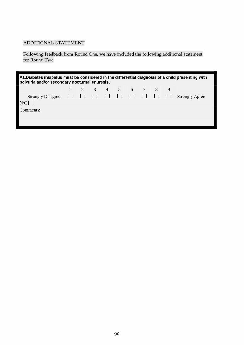
96
ADDITIONAL STATEMENT
Following feedback from Round One, we have included the following additional statement
for Round Two
A1.Diabetes insipidus must be considered in the differential diagnosis of a child presenting with polyuria and/or secondary nocturnal enuresis.
Strongly Disagree
1
2
3
4
5
6
7
8
9
Strongly Agree
N/C
Comments:

97
THANKYOU FOR COMPLETING THIS QUESTIONNAIRE.
Please make sure you‟ve included your name on page 1
Please post in the envelope provided to reach us by 16
th June 2006 to:
Dr Sophie Wilne
Children's Brain Tumour Research Centre
Academic Division of Child Health
East Block, E Floor
Queens Medical Centre
University of Nottingham
Nottingham NG7 2UH
WHAT HAPPENS NEXT?
The research team will collate responses to Round Two. Depending on whether there are any
remaining statements which have not achieved consensus, you may receive a further modified
questionnaire (Round Three).
You will be informed of the outcome of Round Two by 10th
July 2006.
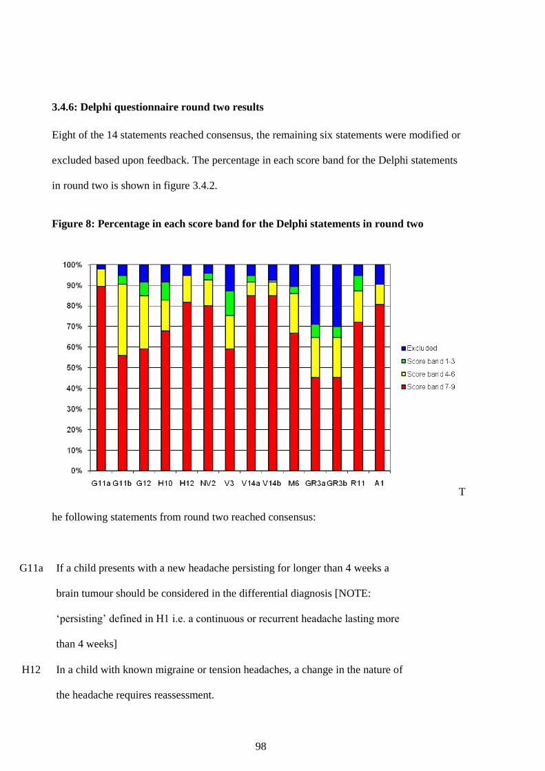
98
3.4.6: Delphi questionnaire round two results
Eight of the 14 statements reached consensus, the remaining six statements were modified or
excluded based upon feedback. The percentage in each score band for the Delphi statements
in round two is shown in figure 3.4.2.
Figure 8: Percentage in each score band for the Delphi statements in round two
T
he following statements from round two reached consensus:
G11a If a child presents with a new headache persisting for longer than 4 weeks a
brain tumour should be considered in the differential diagnosis [NOTE:
„persisting‟ defined in H1 i.e. a continuous or recurrent headache lasting more
than 4 weeks]
H12 In a child with known migraine or tension headaches, a change in the nature of
the headache requires reassessment.

99
NV2 A child presenting with persistent nausea and/or vomiting requires early
specialist referral for consideration of underlying causes including CNS causes
V14a A child presenting with new onset paralytic (non-comitant) squint, requires
CNS imaging.
V14b A child presenting with new onset non-paralytic (comitant) squint should have
early ophthalmic referral for assessment of underlying causes, including CNS
causes.
M6 A child with presumed Bell‟s palsy (isolated lower motor neurone facial nerve
palsy) that does not show improvement within 4 weeks requires CNS imaging.
R11 A child in whom CNS imaging is required to exclude a brain tumour (potential
differential diagnosis, but low index of suspicion) should be imaged within 4
weeks.
A1 Diabetes insipidus must be considered in the differential diagnosis of a child
presenting with polyuria and/or secondary nocturnal enuresis.

100
3.4.7: Delphi process round three
Round three was issued to the 93 participants returning round two. The participants were
provided with the results detailed above. Statements were modified according to feedback
from round one and then reissued. The round three Delphi questionnaire, shown below, asked
the panel to rank their agreement with 7 statements.
101
3.4.8: Delphi questionnaire round three
Throughout this questionnaire:
the terms child and children refer to the age range 0-18 years unless specifically stated otherwise
statements apply to brain and other intracranial tumours, but for ease of reading we refer to brain
tumour throughout.
YOUR NAME (in block letters): ____________________________________________
1. Statements in Round Two were taken as having reached consensus if 75% or more of
Delphi Panel respondents rated the statement 7, 8 or 9. [NOTE: ratings of N/C, blanks, or two
boxes checked in error were excluded from the analysis of that statement].
2. Of the 14 modified statements in Round Two, 8 achieved consensus. These are listed on
page 2, together with a graphical display of the ratings (grouped into score bands of 1-3, 4-
6, 7-9 or excluded).
3. Of the remaining 6 modified statements, the Project Team felt that two were covered within
other statements that have already reached consensus (Modified H10 covered within R4 and
Modified V3/V7 covered within V9) and were therefore excluded from Round Three.
4. In light of feedback received, the remaining 4 statements were further modified by the research
team to give 7 statements requiring rating in ROUND THREE (the final round of the Delphi
process). These are indicated with a BOLD BLACK BORDER around the statement.
5. If you feel you do not have the necessary expertise or experience to contribute to developing a
particular statement, please check the appropriate “N/C” box, and move onto the next statement,
leaving the numbered boxes blank.
6. As indicated in our covering letter, we have included as a separate document an Appendix
of comments received for the 4 statements which required further modification after Round
Two. You do NOT need to read these comments in order to rate the new statements. They
are included for interest only.
7. When you have completed the questionnaire, please return in the envelope provided to
arrive by FRIDAY 21st JULY 2006to:
Dr Sophie Wilne
Children's Brain Tumour Research Centre
Academic Division of Child Health
East Block, E Floor
Queens Medical Centre
Nottingham NG7 2UH
THANKYOU
MODIFIED STATEMENTS for Delphi :
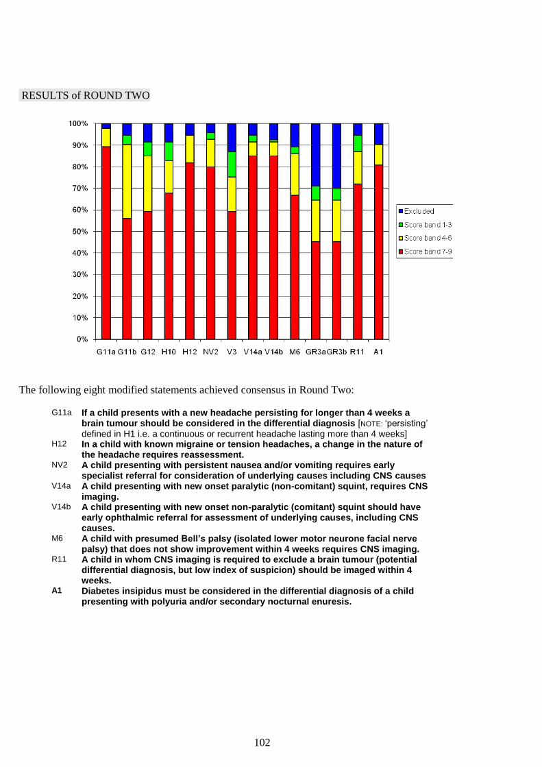
102
RESULTS of ROUND TWO
The following eight modified statements achieved consensus in Round Two: G11a If a child presents with a new headache persisting for longer than 4 weeks a
brain tumour should be considered in the differential diagnosis [NOTE: ‘persisting’ defined in H1 i.e. a continuous or recurrent headache lasting more than 4 weeks]
H12 In a child with known migraine or tension headaches, a change in the nature of the headache requires reassessment.
NV2 A child presenting with persistent nausea and/or vomiting requires early specialist referral for consideration of underlying causes including CNS causes
V14a A child presenting with new onset paralytic (non-comitant) squint, requires CNS imaging.
V14b A child presenting with new onset non-paralytic (comitant) squint should have early ophthalmic referral for assessment of underlying causes, including CNS causes.
M6 A child with presumed Bell’s palsy (isolated lower motor neurone facial nerve palsy) that does not show improvement within 4 weeks requires CNS imaging.
R11 A child in whom CNS imaging is required to exclude a brain tumour (potential differential diagnosis, but low index of suspicion) should be imaged within 4 weeks.
A1 Diabetes insipidus must be considered in the differential diagnosis of a child presenting with polyuria and/or secondary nocturnal enuresis.

103
Four modified statements did NOT achieve consensus in Round Two, and have been further
modified to give SEVEN statements for voting in Round Three:
MODIFIED G11(b). If a child presents with abnormal behaviour (causing concern to parents/carers) including lethargy or withdrawal and persisting for more than 4 weeks, a brain tumour should be considered in the differential diagnosis.
Outcome: Statement modified for Round Three in light of comments received (see appendix 2). Feedback
suggested restricting the statement to lethargy or withdrawal, rather than the broad term „abnormal
behaviour‟. Feedback from round one suggested clearer age-specification would be helpful, hence
modified statements G11(c) and G11(d).
MODIFIED G11(c). If a child presents with lethargy or withdrawal persisting for more than 4 weeks a brain tumour should be considered in the differential diagnosis.
Strongly Disagree
1
2
3
4
5
6
7
8
9
Strongly Agree
N/C
Comments:
MODIFIED G11(d). If a child aged </= 3 years presents with lethargy or withdrawal persisting for more than 4 weeks a brain tumour should be considered in the differential diagnosis.
Strongly Disagree
1
2
3
4
5
6
7
8
9
Strongly Agree
N/C
Comments:
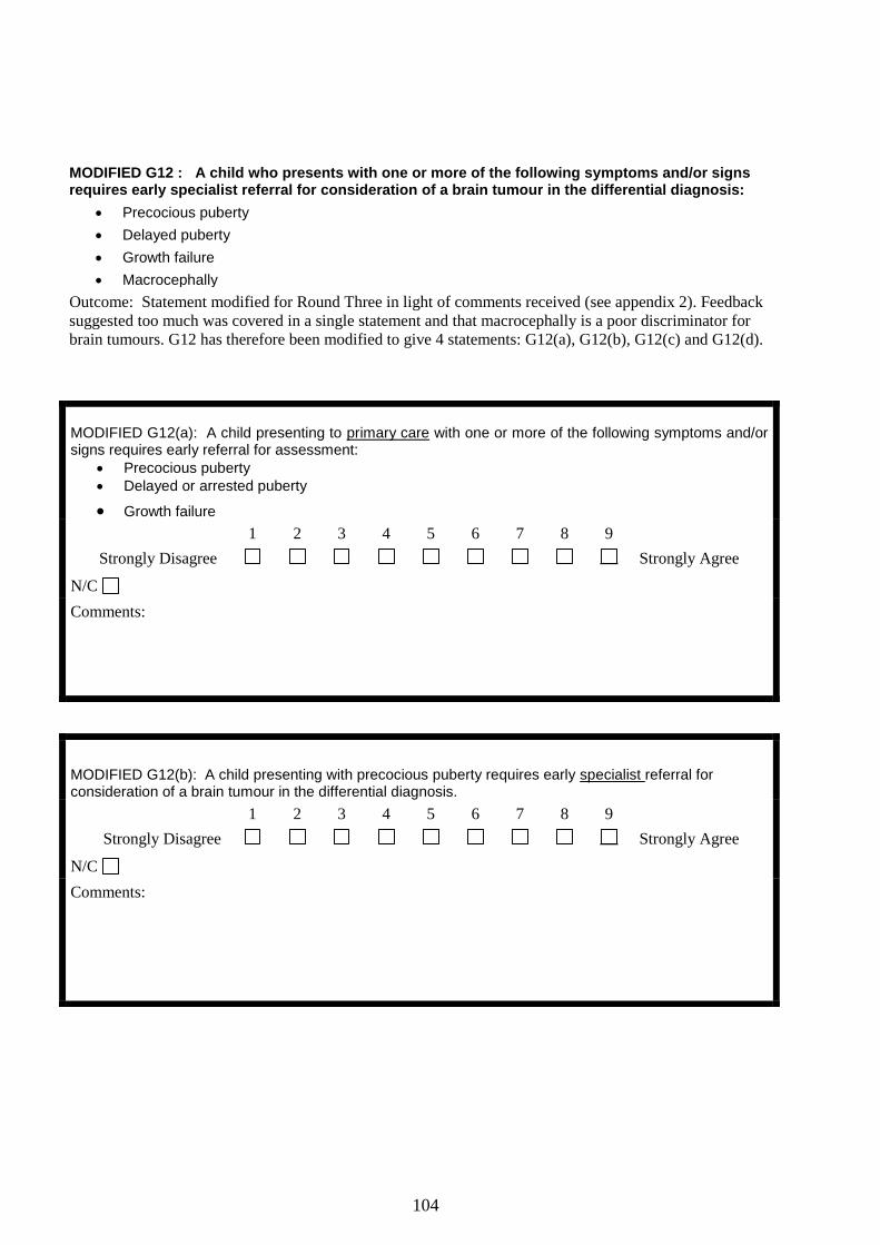
104
MODIFIED G12 : A child who presents with one or more of the following symptoms and/or signs requires early specialist referral for consideration of a brain tumour in the differential diagnosis:
Precocious puberty
Delayed puberty
Growth failure
Macrocephally
Outcome: Statement modified for Round Three in light of comments received (see appendix 2). Feedback
suggested too much was covered in a single statement and that macrocephally is a poor discriminator for
brain tumours. G12 has therefore been modified to give 4 statements: G12(a), G12(b), G12(c) and G12(d).
MODIFIED G12(a): A child presenting to primary care with one or more of the following symptoms and/or signs requires early referral for assessment:
Precocious puberty
Delayed or arrested puberty
Growth failure
Strongly Disagree
1
2
3
4
5
6
7
8
9
Strongly Agree
N/C
Comments:
MODIFIED G12(b): A child presenting with precocious puberty requires early specialist referral for consideration of a brain tumour in the differential diagnosis.
Strongly Disagree
1
2
3
4
5
6
7
8
9
Strongly Agree
N/C
Comments:

105
MODIFIED G12(c): A child presenting with any combination of the following requires consideration of a brain tumour in the differential diagnosis:
Growth failure
Delayed or arrested puberty
Polydipsia and polyuria
Strongly Disagree
1
2
3
4
5
6
7
8
9
Strongly Agree
N/C
Comments:
MODIFIED G12(d): A child aged </= 3 years presenting with weight loss despite adequate calorie intake requires consideration of a brain tumour in the differential diagnosis.
Strongly Disagree
1
2
3
4
5
6
7
8
9
Strongly Agree
N/C
Comments:

106
THANKYOU FOR COMPLETING THIS FINAL QUESTIONNAIRE
OF THE DELPHI PROCESS
Please make sure you‟ve included your name on page 1
Please post in the envelope provided to reach us by FRIDAY 21st July to:
Dr Sophie Wilne
Children's Brain Tumour Research Centre
Academic Division of Child Health
East Block, E Floor
Queens Medical Centre
University of Nottingham
Nottingham NG7 2UH
WHAT HAPPENS NEXT?
You will be informed of the outcome of the Delphi Process by 31st August 2006.
MODIFIED GR3(a). A boy with presumed anorexia nervosa requires early specialist referral for consideration of a brain tumour in the differential diagnosis.
MODIFIED GR3(b). A girl with presumed anorexia nervosa requires early specialist referral for consideration of a brain tumour in the differential diagnosis, if there are any atypical features.
Outcome: Statements modified to give a single statement for Round Three in light of comments
received (see appendix 2).
MODIFIED GR3(c). A child presenting with weight loss due to lack of appetite (anorexia) requires consideration of a brain tumour in the differential diagnosis.
Strongly Disagree
1
2
3
4
5
6
7
8
9
Strongly Agree
N/C
Comments:

107
3.4.9: Delphi questionnaire round three results
88 Delphi panel members returned round three within the required time limit. Consensus was
achieved for 3 statements. Feedback from the panel suggested that consensus was unlikely to
be achieved for the remaining 4 statements. No further rounds were undertaken. The
percentage in each score band for the Delphi statements in round two is shown in figure 9.
Figure 9: Percentage in each score band for the Delphi statements in round three
The following statements from round three reached consensus:
G12a A child presenting to primary care with one or more of the following
symptoms and/or signs requires early referral for assessment:
Precocious puberty
Delayed or arrested puberty

108
Growth failure
G12b A child presenting with precocious puberty requires early specialist referral for
consideration of a brain tumour in the differential diagnosis.
G12c A child presenting with any combination of the following requires
consideration of a brain tumour in the differential diagnosis:
Growth failure
Delayed or arrested puberty
Polydipsia and polyuria
By the end of three rounds of the Delphi process 64 statements had reached consensus. The
participants and their healthcare background (generalist or specialist) and the progress through
the Delphi process are shown in figures 10 and 11.
Figure 10: Progress through the Delphi process

109
Figure 11: Delphi process participants

110
CHAPTER 4: CONCLUSIONS FROM THE EVIDENCE REVIEW
4.1: Conclusions from the systematic literature review and meta-analysis
The meta-analysis showed the importance of patient age and neurofibromatosis status and
tumour location in determining the symptom and sign clusters present at diagnosis in children
with central nervous system tumours. Combining the most common specific symptoms or
signs of raised intracranial pressure with the proportion of children presenting with non-
specific symptoms or signs of raised intracranial pressure provided an estimate of the overall
frequency of these symptoms and signs. This indicates that symptoms linked to raised
intracranial pressure are present in about 40% of all intracranial tumours, 40% of intracranial
tumours in children aged under 4 years, 20% of intracranial tumours occurring in children
with neurofibromatosis, 80% of posterior fossa tumours, 60% of central tumours, 60% of
hemispheric tumours, 30% of brainstem tumours, and 7% of spinal-cord tumours (see figures
3 and 4). Other alerts to a possible CNS tumour identified include abnormal gait and
coordination, other motor system abnormalities, eye signs, weight loss, behavioural changes
(including lethargy and irritability) and school difficulties, developmental delay, cranial nerve
palsies, head tilt, macrocephaly, diabetes insipidus, and growth arrest. Increasing awareness
of the varied and complex symptomatology that often occurs with CNS tumours could help
tumour diagnosis and reduce the extended symptom interval experienced by many children.
Recognition that specific combinations of symptoms and signs indicate a focal CNS lesion is
crucial to the diagnosis of many CNS tumours. 45–60% of childhood brain tumours are
infratentorial, 25–40% are hemispheric, and 15–20% are midline supratentorial[123]. Meta-
analysis has emphasised the symptom and sign combinations that occur with different tumour
locations. Knowledge of these could help focus the search for corroborative findings in

111
children who present with a symptom or sign that is potentially suggestive of a CNS tumour.
In many instances, the possibility that the symptoms or signs are the result of a CNS tumour
will be (rightly) rapidly dismissed. However, consideration of this diagnosis in some cases
could lead to identification of corroborative symptoms and signs and the instigation of
imaging. Even if an underlying tumour is unlikely, patients and their families or carers should
be encouraged to return for re-assessment should symptoms or signs persist or progress, and
the diagnosis should be reviewed on re-presentation. A 5% threshold was chosen for reporting
symptoms and signs in children with CNS tumours as a practical compromise between the
need to consider an underlying CNS tumour with a clinical feature not associated with this
tumour type and those symptoms and signs that occur frequently in childhood CNS tumours.
Because of the differing presentation of CNS tumours according to patient group and tumour
location, most symptoms and signs that occurred in less than 5% of patients in one subgroup
occurred more frequently in the other subgroups. Symptoms and signs that consistently
occurred in less than 5% of patients, which could be associated with diagnostic difficulty,
were dysphagia and delayed puberty.
35 studies [29, 33, 34, 36-42, 51-53, 63, 76, 84, 85, 87, 89-91, 95-97, 103, 104, 106-109, 115,
116, 118, 120, 121]. meeting the inclusion criteria reported symptom interval duration (table
5). Symptom interval comparison is difficult for several reasons. Studies report different
measures of symptom interval (median, mean, range) and rarely report all three, and the
statistical significance of any differences in symptom interval cannot be determined from the
reported data. For asymmetric distributions such as symptom interval of paediatric brain
tumours, the median provides the best comparator. The reported median symptom interval
ranges from 1 to 27 months. The longest median symptom interval occurs with biologically
slow-growing tumours such as gangliogliomas[84, 97], although there is little association
between symptom interval range and tumour biology, indicating that extended symptom

112
intervals could occur with all types of paediatric brain tumour. Any association between
specific symptoms and signs and an extended symptom interval could not be determined by
this analysis.
A systematic search strategy and standardised inclusion criteria was used, as recommended in
the quality of reporting meta-analyses (QUOROM) statement, to identify studies for
inclusion[123]. The high number of papers identified in the past 15 years shows the sustained
interest in the mechanisms of diagnosis in this group of patients. Previously published
evidence on paediatric CNS tumour presentation has been predominantly in the form of case
studies (level 4 evidence) with infrequent tumour registry series (level 2 evidence; two studies
met the inclusion criteria for this study) [ 34, 119]. The systematic approach has generated a
cohort of patients, most of whom were diagnosed during the era of CT and MRI, six times
larger that the largest single identified study. The meta-analysis results reported here provide
level 2 evidence for this cohort, which give greater value to the rankings of symptoms and
signs by age, tumour location, and neurofibromatosis status than previous reports.
The meta-analysis has some important limitations and potential sources of bias. The search
strategy might not have identified all relevant papers and unpublished data were not sought.
Papers included in the analysis reported symptoms and signs at diagnosis in children with a
CNS tumour; therefore accuracy of these data depends on the history given by patients and
their families or carers and the signs detected by the examining health-care practitioners.
However, medical decisions will always be based on such histories and examination findings
rather than the underlying full facts to which they relate. The assumption was made that if a
symptom or sign was not described in a study, it did not occur in that population. The
variability and large number of included patients should reduce the risk that common
symptoms and signs are under-represented and uncommon ones over-represented.

113
There was variation in the data detail between studies. Some studies were very detailed,
recording individual symptoms and signs such as headache, vomiting, and papilloedema [29,
33, 34, 36, 37, 40, 42, 51, 52, 66, 85-87, 89-92, 92, 97,100-103, 105, 106, 108, 111, 113-
120]; whereas others used symptom complexes such as symptoms of raised intracranial
pressure or cranial nerve palsies[41, 54, 61, 65, 88, 93, 98, 101, 104, 107, 109]. Some
symptoms and signs could have been combined to indicate the total proportion of children
presenting with a specific symptom complex. However, since it could not be determined
exactly how the data related, some inaccuracy and misrepresentation of data could result and
thus the data was kept in their original form. Despite these problems, the analysis shows the
variability of symptoms and signs and the frequency with which they occur in childhood CNS
tumours.
Most childhood brain tumours are low-grade astrocytomas[14, 124]. Apart from optic
pathway gliomas, these astrocytomas were under-represented in the studies identified. This
result is probably due to a historical failure to include non-malignant brain tumours in tumour
registries and, until recently, absence of review of children with low-grade gliomas by
paediatric oncologists. Despite this result, the distribution of tumour location in the studies
identified here was similar to that seen in clinical practice (56% infratentorial, 23%
hemispheric, 21% central), lending support to the analysis results. Publication bias could have
led to over-representation of rare tumours or those with an unusual presentation; however,
case reports and studies with fewer than ten patients were excluded to combat this problem.
Finally, this analysis addresses the issue of sensitivity but not that of specificity of symptoms
and signs to the presence of an underlying CNS tumour. The probability of a symptom or sign
being indicative of a CNS tumour will increase with the occurrence of corroborative findings
on history and examination and the prevalence of CNS tumours in the population in question.
The previous largest study[59] of childhood brain tumour presentation, undertaken by the
114
Childhood Brain Tumor Consortium, reported on the distribution of other symptoms and
neurological signs in 3291 children with or without headache in association with a brain
tumour. For the most of this period, CT and MRI were not available. Direct comparison
between this study[59] and the present analysis is complicated by differences in anatomical
subdivision and methodology. Headache, nausea and vomiting, and seizures were reported for
the entire cohort, although other symptoms were reported for specific age groups and numbers
in each age group were not provided[59]. Similarly, although the occurrence of coma, focal
motor weakness, and papilloedema is reported for the entire group, other symptoms were not
reported unless their presence or absence was documented in the medical records. Notably,
the Childhood Brain Tumor Consortium cohort [59] reported a higher frequency of headache,
nausea and vomiting, and papilloedema in supratentorial tumours than identified in this
analysis, but reported a similar frequency of these symptoms in infratentorial tumours. This
difference is probably due to increased imaging availability to the current cohort. Because of
the vulnerability of the cerebral aqueduct to compression by tumour, posterior fossa tumours
often lead to raised intracranial pressure at an early stage. By contrast, supratentorial tumours
could present with other symptoms and signs and grow to a large size before they lead to
raised intracranial pressure. The availability of CT and MRI allows the latter children with
supratentorial tumours to be assessed before the development of raised intracranial pressure.
In the meta-analysis, the frequency of change in behaviour or school performance (7% in all
brain tumours, and 9% for central tumours) was lower than that reported by many individual
studies. Several large cohorts reported a frequency of school difficulties and behavioural
changes of 22–72%[34, 35, 37, 94]. Lethargy was analysed separately in this study (pooled
proportion: 6% for all intracranial tumours, 21% in children with intracranial tumours aged
under 4 years, 13% in posterior fossa tumours), which could account for some of the
115
difference. Adults with brain tumours are often not asked about behavioural change, and
similar reporting errors probably occur in children[125].
In summary, the meta-analysis shows both the heterogeneity of childhood CNS tumour
presentation and the importance of tumour location, age, and neurofibromatosis status in
presentation. By ranking symptoms and signs and reporting by age and tumour location, it
focuses on the associative features in a hierarchical way. Symptoms and signs of raised
intracranial pressure occur in less than 50% of all children with intracranial tumours. Motor
system abnormalities, especially abnormalities of gait and coordination, are common with all
tumour types. Eye signs are common in all intracranial tumour types. Macrocephaly is
common in children under 4 years who have intracranial tumours. Weight loss occurs with all
tumour types, growth failure with central tumours, and precocious puberty in children with
neurofibromatosis and intracranial tumours. Assessment of any child who presents with
symptoms and signs that could result from a CNS tumour should therefore include a thorough
visual and motor system examination, assessment of growth (including head circumference in
children under 4 years), and pubertal status. Specific multiple symptoms and signs (eg, in the
combinations shown in figure 5), should alert the clinician to the possibility of a CNS tumour.
4.2: Conclusions from the cohort study
The study demonstrated, in a contemporary cohort of children with a central nervous system
tumour, that a large increase occurs in the number of presenting features between symptom
onset and diagnosis. By diagnosis 95% of children had one or more of headache, nausea and
vomiting, visual or motor abnormalities; however no child had headache alone or nausea and
vomiting alone. The emergence of abnormalities of either the visual system, the motor system
or of behaviour (usually lethargy) between disease onset and diagnosis was very common. For
each of these three clinical features, the percentage of affected children increased by 40-50%
116
during the symptom interval, suggesting the need to prioritise their re-assessment in children
with non-specific symptoms that might be due to a CNS tumour.
The median symptom interval in this cohort was 3.3 months. Cranial nerve deficits, head tilt,
endocrine and visual problems were associated with a longer symptom interval. Visual acuity
is difficult to assess (and therefore may not be undertaken) in young children and
identification of endocrine and growth abnormalities requires that growth and pubertal status
be routinely assessed and recorded when children present to healthcare. Lethargy was the
most common behavioural abnormality observed among the 40% of children that had a
behavioural abnormality by diagnosis and the only one present at symptom onset. Lethargy is
frequently regarded as a non-specific marker of systemic illness, however this and previous
reports suggest that more emphasis should be placed on it as a specific marker of neurological
illness[125]. Similarly, whilst weight loss is not a specific marker for central nervous system
tumours, just under a fifth of children had lost weight by diagnosis. Other studies have
highlighted the weight loss that occurs in children with brain tumours, and the diagnostic
delay that may occur whilst possible nutritional and gastrointestinal causes are
investigated[127].
The association between symptom interval and healthcare attendances confirms that children
with central nervous system tumours present repeatedly to healthcare. Whilst children with a
prolonged symptom interval will have more time to present to heath care, the repeated
presentation suggests that diagnostic delay results from a failure to recognise symptoms and
signs as being indicative of a tumour rather than a failure to seek healthcare advice. The
majority of children were reviewed in primary care and general paediatrics prior to diagnosis;
however seven other disciplines were consulted by the cohort, highlighting the need for all

117
healthcare practitioners to have knowledge of childhood brain tumour presentation and to
have a high index of suspicion for this possibility.
The recruited cohort is likely to be representative of the current UK population of children
with central nervous system tumours. The study was multicentre, had a short recruitment
period and showed a similar tumour epidemiology to that reported in population registries[14,
124]. Data were obtained from medical records and the non-recording of a symptom or sign
was taken to mean that it was not present. Although this is clearly not true in every case, the
history recorded at diagnosis should reflect the history taken then and at the time of any
previous presentation to healthcare professionals. The decision to investigate a symptom or
sign will always be reliant on such histories rather than on the underlying full facts to which
they relate.
At symptom onset it may be difficult to distinguish between children with a central nervous
system tumour and those with a self-limiting benign condition, particularly as the most
common initial symptoms, headache, nausea and vomiting, are known to be poor
discriminators for central nervous system tumours. This study does not provide information
regarding the incidence of these symptoms in children unaffected by a central nervous system
tumour and thus does not address the issue of “specificity”. Despite this limitation, it does
identify patterns of symptoms seen in children with a central nervous system tumour i.e. the
“sensitivity” of patterns of clinical features to such a diagnosis, and highlights the importance
of undertaking a thorough assessment of children presenting with such non-specific
symptoms.
When children present with symptoms or signs identified in the cohort study, the challenge to
healthcare professionals is to distinguish the minority of children with a central nervous
system tumour from the majority who have a less serious condition. The cohort study
suggests that children presenting with symptoms and signs that may result from a central
118
nervous system tumour should undergo motor and visual assessment, pubertal staging and
comparison of height and weight with their previous growth and with age-appropriate norms.
For children in whom a central nervous system tumour is thought unlikely, the development
of additional symptoms or signs or repeated presentation should lead to a careful review of the
diagnosis.

119
CHAPTER 5: PATHWAYS PROJECT GUIDELINE
The quick reference and complete versions of the final guideline are shown below. The sort
version includes the guideline statements, the long version explains the rationale for each
statement and its evidence level, subsequent recommendation grade [19] and, where
appropriate, the degree of consensus.
5.1: The diagnosis of brain tumours in children – an evidenced based
guideline to assist healthcare professionals in the assessment of children
presenting with symptoms and signs that may be due to a brain tumour.
(quick reference guide)
Statements in a red box advise on indications for imaging.
Statements in a black box advise on presentations frequently associated with misdiagnosis.
A one-page quick reference summary is shown in figure 12.
5.1.1 Best practice
5.1.1a: Consultation
Parents and their carers should be asked explicitly about their concerns in any
consultation.
If a parent / carer expresses concerns about a brain tumour this should be reviewed
carefully. If a brain tumour is unlikely the reasons why should be explained and
arrangements made for review within 4 weeks.
If the patient, parent / carer and healthcare professional are not fluent in a common
language an interpreter must be used for the consultation (www.languageline.co.uk).

120
Low parental educational level, social deprivation and lack of familiarity with the UK
healthcare system may be associated with diagnostic delay. A lower threshold for
investigation and referral may be appropriate in these situations.
5.1.1b: Referral
A primary healthcare professional who has a high index of suspicion regarding a
possible brain tumour should discuss their concerns with a secondary health care
professional the same day.
A child referred from primary care in which the differential diagnosis includes a
possible space occupying lesion should be seen within two weeks.
5.1.1c: Imaging
A child in whom CNS imaging is required to exclude a brain tumour (potential
diagnosis but low index of suspicion) should be imaged within 4 weeks.
MRI is the imaging modality of choice for a child who may have a brain tumour.
If MRI is not available a contrast enhanced CT should be performed.
Imaging results should be interpreted by a professional with expertise and training in
central nervous system MR and CT imaging in children.
The need to sedate or anaesthetise a child for imaging should not delay imaging by
more than 1 week.
5.1.1d: Feedback
Patients and their families should receive the provisional results of CNS imaging
within 1 week of the investigation.
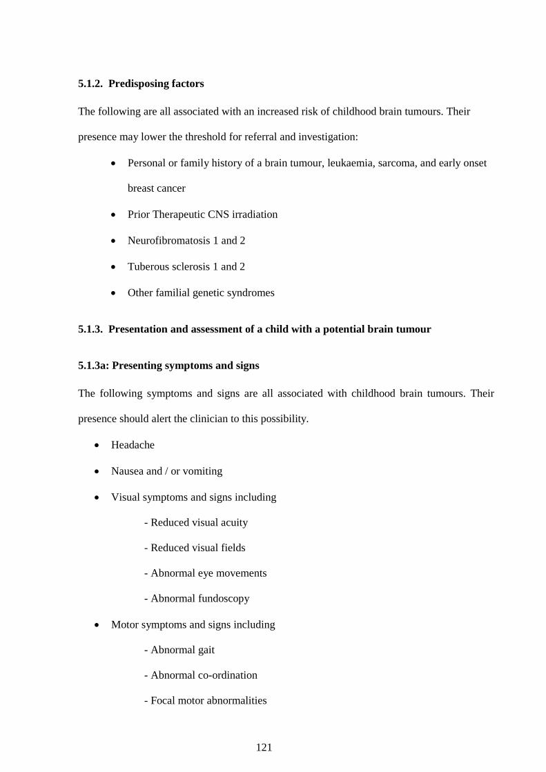
121
5.1.2. Predisposing factors
The following are all associated with an increased risk of childhood brain tumours. Their
presence may lower the threshold for referral and investigation:
Personal or family history of a brain tumour, leukaemia, sarcoma, and early onset
breast cancer
Prior Therapeutic CNS irradiation
Neurofibromatosis 1 and 2
Tuberous sclerosis 1 and 2
Other familial genetic syndromes
5.1.3. Presentation and assessment of a child with a potential brain tumour
5.1.3a: Presenting symptoms and signs
The following symptoms and signs are all associated with childhood brain tumours. Their
presence should alert the clinician to this possibility.
Headache
Nausea and / or vomiting
Visual symptoms and signs including
- Reduced visual acuity
- Reduced visual fields
- Abnormal eye movements
- Abnormal fundoscopy
Motor symptoms and signs including
- Abnormal gait
- Abnormal co-ordination
- Focal motor abnormalities

122
Growth and developmental abnormalities including
- Growth failure
- Delayed, arrested or precocious puberty
Behavioural change
Diabetes insipidus
Seizures - Not covered in this guideline (see www.nice.org.uk/CG020)
Altered consciousness - Not covered in this guideline (see
www.nottingham.ac.uk/paediatric-guideline)
Symptoms and signs in childhood brain tumours may occur singularly or in combination.
5.1.3b: History
Take detailed history and enquire specifically about:
- Predisposing factors
5.1.3c: Assessment
Assess:
- Visual system
- Motor system
- Height and weight
- Head circumference if under 2 years
- Pubertal status
The initial symptoms of a brain tumour frequently mimic those that occur with many
common childhood conditions
Symptoms frequently fluctuate in severity – resolution and then recurrence does not
exclude a brain tumour
Presentation depends upon the age of the child

123
Delayed diagnosis has been associated with failure to reassess a child with migraine
or tension headache when the headache character changes.
A normal neurological examination does not exclude a brain tumour
5.1.4. Signs and Symptoms of a child with a potential brain tumour
5.1.4a: Headache
Consider a brain tumour in any child presenting with a new persistent headache. (A
continuous or recurrent headache lasting for more than 4 weeks should be regarded as
persistent)
Brain tumour headaches can occur at any time of the day or night
Children aged younger than 4 years, or those with communication difficulties, are
frequently unable to describe headache; their behaviour e.g. withdrawal, holding head
may indicate a headache.
In a child with a known migraine or tension headache a change in the nature of the
headache requires reassessment and review of the diagnosis.
CNS IMAGING (within a maximum of 4 weeks) REQUIRED FOR:
Persistent headaches that wake a child from sleep
Persistent headaches that occur on waking
A persistent headache occurring at any time in a child younger than 4 years
Confusion or disorientation occurring with a headache
5.1.4b: Nausea and vomiting
Early specialist referral for consideration of underlying causes including CNS causes
is required for a child with persistent nausea and / or vomiting. (Nausea and / or
vomiting that lasts for more than two weeks should be regarded as persistent)

124
CNS IMAGING (within a maximum of 4 weeks) REQUIRED FOR:
Persistent vomiting on awakening (either in the morning or from a day time sleep)
NB: exclude pregnancy where appropriate.
5.1.4c: Visual symptoms and signs
Consider a brain tumour in any child presenting with a persisting visual abnormality.
(Any visual abnormality lasting longer than 2 weeks should be regarded as persistent)
Visual assessment must include assessment of:
Pupil responses
Acuity
Visual fields in school age children
Eye movements
Optic disc appearance
If the assessing healthcare professional is unable to perform a complete visual
assessment the child should be referred for assessment.
Children referred for visual assessment should be seen within two weeks of referral.
Community optometry should refer any child with abnormal eye findings (excluding
simple refractive errors) directly to secondary care.
Pre-school and uncooperative children should be assessed by the hospital eye service.
A child with a new onset non-paralytic (concomitant) squint should have early
ophthalmological assessment for consideration of underlying causes (including CNS
causes).
Delayed diagnosis has been associated with:
Attributing persistent nausea and vomiting to an infective cause in the absence of
corroborative findings e.g. contact with similar illness, pyrexia, diarrhoea.

125
5.1.4d: Motor symptoms and signs
Consider a brain tumour in any child presenting with a persisting motor abnormality.
(Any motor abnormality lasting longer than two weeks should be regarded as
persistent.)
Brain tumours may cause a deterioration or change in motor skills; this may be subtle
e.g. change in hand or foot preference, loss of learned skills (computer games).
Motor system assessment must include observation of:
Sitting and crawling in infants
Walking and running
Coordination e.g. heel to toe walking
Handling of small objects
Handwriting in school age children
Delayed diagnosis has been associated with:
Failure to fully assess vision in a young or uncooperative child
Failure of communication between community optometry and primary and secondary care
Delayed diagnosis has been associated with:
Attributing abnormal balance or gait to middle ear disease in the absence of
corroborative findings
Failure to identify swallowing difficulties as the cause of recurrent chest infections or
“chestiness”
CNS IMAGING (within a maximum of 4 weeks) REQUIRED FOR:
Papilloedema
Optic atrophy
New onset nystagmus
Reduction in visual acuity not attributable to refractive error
Visual field reduction
Proptosis
New onset paralytic (non-concomitant) squint
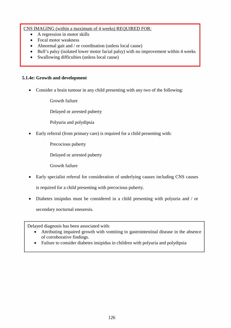
126
5.1.4e: Growth and development
Consider a brain tumour in any child presenting with any two of the following:
Growth failure
Delayed or arrested puberty
Polyuria and polydipsia
Early referral (from primary care) is required for a child presenting with:
Precocious puberty
Delayed or arrested puberty
Growth failure
Early specialist referral for consideration of underlying causes including CNS causes
is required for a child presenting with precocious puberty.
Diabetes insipidus must be considered in a child presenting with polyuria and / or
secondary nocturnal eneuresis.
CNS IMAGING (within a maximum of 4 weeks) REQUIRED FOR:
A regression in motor skills
Focal motor weakness
Abnormal gait and / or coordination (unless local cause)
Bell‟s palsy (isolated lower motor facial palsy) with no improvement within 4 weeks
Swallowing difficulties (unless local cause)
Delayed diagnosis has been associated with:
Attributing impaired growth with vomiting to gastrointestinal disease in the absence
of corroborative findings.
Failure to consider diabetes insipidus in children with polyuria and polydipsia

127
5.1.4f: Behaviour
Lethargy is the commonest behavioural abnormality that occurs with brain tumours
Environmental context is important when assessing lethargy: a child who is lethargic
in situations in which they are normally active requires further assessment.
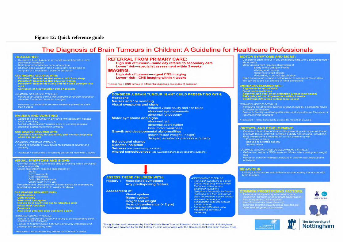
128
Figure 12: Quick reference guide

129
5.2: The diagnosis of brain tumours in children – an evidenced based
guideline to assist healthcare professionals in the assessment of children
presenting with symptoms and signs that may be due to a brain tumour:
5.2.1: Aim of the guideline
The guideline aims to advise on the following:
1. The symptoms and signs that may occur in children with brain tumour
2. Assessment of children presenting with these symptoms and signs
3. Indications and waiting times for imaging children with these symptoms and signs
5.2.2: Scope
Patient inclusion criteria
The guideline is applicable to all children aged 0-18 years who present with symptoms and /
or signs that could result from a brain tumour and are being reviewed by a healthcare
professional.
Guideline users
The guideline is intended to support the assessment and investigation by healthcare
professionals of children who may have a brain tumour.
The guideline has been developed following careful consideration of the available evidence
and has incorporated professional expertise via a Delphi consensus process. Healthcare
professionals should use it to support their decision making when assessing children who may
have an intracranial tumour. It does not however override the responsibility of a healthcare
professional to make decisions appropriate to the condition of individual children.

130
There are 76 recommendations in total with 21 grade B recommendations. Levels of evidence
and grading of recommendations are explained below and are taken from SIGN, Scottish
Intercollegiate Guideline Network (2000) [19].
5.2.3: Levels of evidence and recommendation grades:
Levels of Evidence
1++ High quality meta-analyses, systematic reviews of randomised controlled trials
(RCTs) or RCTs with a very low risk of bias
1+ Well-conducted met-analyses, systematic reviews of RCTs, or RCTs with a low risk
of bias
1- Meta-analyses, systematic reviews of RCTs, or RCTs with a high risk of bias
2++ High quality systematic reviews of case control or cohort studies
High quality case control or cohort studies with a very low risk of confounding or bias
and a high probability that the relationship is causal
2+ Well conducted case control or cohort studies with a low risk of confounding or bias
and a moderate probability that the relationship is causal
2- Case control or cohort studies with a high risk of confounding or bias and a significant
risk that the relationship is not causal
3 Non-analytic studies, e.g. case reports, case series
4 Expert opinion
Grades of Recommendation
A At least one meta-analysis, systematic review of RCTS, or RCT rated as 1++ and
directly applicable to the target population; or
A body of evidence consisting principally of studies rated as 1+, directly applicable to
the target population, and demonstrating overall consistency of results

131
B A body of evidence including studies rated as 2++, directly applicable to the target
population, and demonstrating overall consistency of results; or
Extrapolated evidence from studies rated as 1++ or 1+
C A body of evidence including studies rated as 2+, directly applicable to the target
population and demonstrating overall consistency of results; or
Extrapolated evidence from studies rated as 2++
D Evidence level 3 or 4; or
Extrapolated evidence from studies rated as 2+
Good Practice Points
Recommended best practice based on the clinical experience of the guideline development
group
132
5.2.4a. Best practice - consultation
Parents and their carers should be asked explicitly about their concerns in any
consultation.
Strength of evidence 4
Recommendation grade D
Consensus achieved 96% (round 1)
Rationale
Parents / carers of children with brain tumours are frequently concerned that their child‟s
symptoms may indicate a brain tumour for a significant period of time before the diagnosis is
made. Parents / carers may be unwilling to express these concerns for fear of seeming over
anxious or appearing to waste healthcare professionals‟ time. Explicitly asking parents / carers
of their concerns enables them to be expressed improving communication between all parties.
In some cases parental concern regarding a possible brain tumour may trigger professional
concern and lead to appropriate investigation.
If a parent / carer expresses concerns about a brain tumour this should be reviewed
carefully. If a brain tumour is unlikely the reasons why should be explained and
arrangements made for review within 4 weeks.
Strength of evidence 4
Recommendation grade D
Consensus achieved 76% (round 1)
Rationale
Parents / carers of children with brain tumours are frequently concerned that their child‟s
symptoms may indicate a brain tumour for a significant period of time before the diagnosis is
made. If on review a brain tumour seems unlikely it is important to explain why in order to

133
maintain trust and communication with the patient and their parents / carers. Symptom
progression occurs with childhood brain tumours therefore early review is recommended to
facilitate detection of any additional symptoms or signs which may make the diagnosis more
likely.
If the patient, parent / carer and healthcare professional are not fluent in a common
language an interpreter must be used for the consultation (www.languageline.co.uk).
Strength of evidence 4
Recommendation grade D
Consensus achieved 94% (round 1)
Rationale
The research team, Delphi workshop and Delphi panel could all identify individual cases
where non-English first language was associated with diagnostic delay. It is essential to take a
thorough history when assessing a child who may have a brain tumour; this is not possible if
the patient, parent / carer and healthcare professional are not fluent in a common language.
Low parental educational level, social deprivation and lack of familiarity with the UK
healthcare system may be associated with diagnostic delay. A lower threshold for
investigation and referral may be appropriate in these situations.
Strength of evidence 4
Recommendation grade D
Rationale
There is no published evidence linking low parental education, social deprivation and lack of
familiarity with the UK healthcare system with diagnostic delay in paediatric brain tumours
however the research team and many members of the Delphi panel were aware of individual
cases in which these factors may have contributed to a prolonged symptom interval. The
Delphi panel were asked in round 1 to comment on the influence ethnicity and deprivation

134
have on symptom interval in paediatric brain tumours and the above statement is a summary
of these comments.
5.2.4b. Best practice - referral
A primary healthcare professional who has a high index of suspicion regarding a
possible brain tumour should discuss their concerns with a secondary health care
professional the same day.
Strength of evidence 4
Recommendation grade D
Consensus achieved 80% (round 1)
Rationale
Children who have a brain tumour may deteriorate quickly and therefore if there is a high
possibility that they may have a brain tumour they should be assessed and arrangements made
for CNS imaging as quickly as possible.
A child referred from primary care in which the differential diagnosis includes a
possible space occupying lesion should be seen within two weeks.
Strength of evidence 4
Recommendation grade D
Consensus achieved 79% (round 1)
Rationale
A prolonged symptom interval with brain tumours occurs in part due delay between initial
referral from primary care and assessment in secondary care[34, 51, 130]. The Department of
Health has advised that a patient presenting with symptoms that are potentially indicative of a
malignancy should be assessed by a healthcare professional with expertise in that area within
2 weeks[28]. The Delphi panel agreed that this recommendation was appropriate for children
who may have a brain tumour.

135
5.2.4c. Best practice – imaging
A child in whom CNS imaging is required to exclude a brain tumour (potential diagnosis
but low index of suspicion) should be imaged within 4 weeks.
Strength of evidence 4
Recommendation grade D
Consensus achieved 76% (round 1)
Rationale
There is frequently reluctance among healthcare professionals to undertake CNS imaging of
children who may have a brain tumour until clinical signs become florid. This results in a
prolonged symptom interval and children who may extremely unwell by diagnosis. The NICE
guideline on diagnosis and management of epilepsy in primary and secondary care advises
that children who present with a focal onset of seizures should undergo CNS imaging within 4
weeks[57]. As imaging in this case is required to exclude a CNS space occupying lesion
(including brain tumours) it seemed appropriate to advise a similar waiting time to imaging
for children who present with other symptoms and signs that may be due to a brain tumour
MRI is the imaging modality of choice for a child who may have a brain tumour.
Strength of evidence 2++
Recommendation grade B
Consensus achieved 85% (round 1)
Rationale
As advised by the Royal College of Radiologists[130].
If MRI is not available a contrast enhanced CT should be performed.
Strength of evidence 2++
Recommendation grade B
Consensus achieved 92% (round 1)

136
Rationale
As advised by the Royal College of Radiologists[130].
Imaging results should be interpreted by a professional with expertise and training in
central nervous system MR and CT imaging in children.
Strength of evidence 4
Recommendation grade D
Consensus achieved 93% (round 1)
Rationale
Normal and abnormal neuro-imaging findings can vary significantly between children and
adults. In order to reduce the risk of misdiagnosis the Delphi panel agreed that central nervous
system imaging in children should be interpreted by a healthcare professional with expertise
in this area.
The need to sedate or anaesthetise a child for imaging should not delay imaging by more
than 1 week.
Strength of evidence 4
Recommendation grade D
Consensus achieved 83% (round 1)
Rationale
Young children (under 5 years) are frequently unable or unwilling to keep still enough to
allow adequate CNS imaging. In this situation they require sedation or a general anaesthetic
for imaging. The Delphi panel felt that the diagnosis of brain tumours in young children
should not be significantly delayed due to the requirement for sedation or a general
anaesthetic.

137
5.2.4d. Best practice – feedback
Patients and their families should receive the provisional results of CNS imaging within
1 week of the investigation.
Strength of evidence 4
Recommendation grade D
Consensus achieved 83% (round 1)
Rationale
Whilst the Delphi panel recognises that expert review and multi-disciplinary team discussion
prior may be necessary to adequately interpret childhood CNS imaging, it is important, to
minimise anxiety, that families are informed of provisional results as soon as possible.
5.2.5. Predisposing factors
The following are all associated with an increased risk of childhood brain tumours. Their
presence may lower the threshold for referral and investigation:
Personal or family history of a brain tumour, leukaemia, sarcoma and early onset breast
cancer.
Prior therapeutic CNS irradiation
Neurofibromatosis 1 and 2 (see www.nfauk.org)
Tuberous sclerosis 1 and 2 (see www.tuberose-sclerosis.org)
Other familial genetic syndromes
Strength of evidence 2++
Recommendation grade B
Rationale
The above are all associated with an increased risk of childhood brain tumours and therefore
their presence should alert the clinician to this possibility and may lower their threshold for
referral and investigation[131]. The majority of the association between brain tumours,

138
leukaemia, sarcoma and early onset breast cancer is due to inherited abnormalities in the P53
tumour suppressor gene (Li Fraumeni syndrome). There are associations between brain
tumours and colorectal polyposis and colorectal cancer (Turcot‟s syndrome) and with basal-
cell nevus syndrome (Gorlin‟s syndrome). Having a parent or sibling with a brain tumour is
associated with an increased risk however this is probably due to the above genetic
associations.
5.2.6a. Presentation and assessment of a child with a potential brain tumour
The following symptoms and signs are all associated with childhood brain tumours. Their
presence should alert the clinician to this possibility.
Headache
Strength of evidence 2++
Recommendation grade B
Consensus achieved 91% (round 1)
Rationale
Depending on patient age and tumour location between 10% and 67% of children reported in
the meta-analysis had a headache at diagnosis. In the cohort study 40% of children at
symptom onset and 58% by diagnosis had a headache.
Nausea and / or vomiting
Strength of evidence 2++
Recommendation grade B
Consensus achieved 91% (round 1)
Rationale
Between 10% and 67% of children reported in the meta-analysis had experienced nausea and /
or vomiting by diagnosis. In the cohort study 40% of children at symptom onset and 58% by
diagnosis experienced nausea or vomiting.

139
Visual symptoms and signs
Reduced visual acuity
Reduced visual fields
Abnormal eye movements
Abnormal fundoscopy
Strength of evidence 2++
Recommendation grade B
Consensus achieved 91% (round 1)
Rationale
Between 10% and 41% of children reported in the meta-analysis had experienced a visual
symptom or sign. Reduced visual acuity occurred in up to 41% of patients, reduced visual
fields in up to 5%, abnormal eye movements in up to 20% and abnormal fundoscopy in up to
34%. In the cohort study 17% of children at symptom onset and 70% by diagnosis had a
visual system abnormality.
Motor symptoms and signs
Abnormal gait
Abnormal co-ordination
Focal motor abnormalities
Strength of evidence 2++
Recommendation grade B
Consensus achieved 91% (round 1)
Rationale
Between 7% and 78% of children reported in the meta-analysis had experienced a motor
system abnormality. Abnormal gait and co-ordination occurred in up to 78% of patients and
140
focal motor abnormalities in up to 19%. In the cohort study 22% of children at symptom onset
and 67% by diagnosis had a motor system abnormality.
Growth and developmental abnormalities
Growth failure
Delayed, arrested or precocious puberty
Strength of evidence 2++
Recommendation grade B
Consensus achieved 91% (round 1)
Rationale
Between 5% and 14% of children reported in the meta-analysis experienced growth or
developmental abnormalities. Growth failure occurred in up to 14% and pubertal
abnormalities in up to 8%. In the cohort study endocrine and growth abnormalities occurred in
7% of children at symptom onset and 25% by diagnosis.
Behavioural change
Strength of evidence 2++
Recommendation grade B
Consensus achieved 91% (round 1)
Rationale
Between 5% and 21% of children reported in the meta-analysis experienced a behavioural
change. In the cohort study a behavioural change occurred in 3% of children at symptom
onset and 40% by diagnosis.
Diabetes insipidus
Strength of evidence 2++
Recommendation grade B
Consensus achieved 84% (round 3)

141
Rationale
Up to 12% of children in the meta-analysis experienced diabetes insipidus. One child in the
cohort study presented with diabetes insipidus.
Symptoms and signs in childhood brain tumours may occur singularly or in
combination.
Strength of evidence 2+
Recommendation grade C
Rationale
In the cohort study children had a median of one symptom or sign (range 1-8) at symptom
onset. This had increased to a median of six (range 1-16) by diagnosis.
5.2.6b: History
Take a detailed history.
Enquire specifically about associated symptoms and predisposing factors
Strength of evidence 4
Recommendation grade D
Consensus achieved 89% (round 1)
Rationale
Childhood brain tumours frequently present with symptoms that may occur with other more
common childhood illnesses. Identifying those children who may have a tumour, and thus
require imaging, from the majority that do not may be facilitated by taking a detailed history
of the presenting complaint(s) and specifically asking whether any other symptoms have
occurred and whether there are any recognised predisposing factors.

142
5.2.6c: Assessment
Assess: Visual system
Motor system
Height and weight
Pubertal status
Strength of evidence 2+
Recommendation grade C
Consensus achieved 89% (round 1)
Rationale
By diagnosis 95% of children in the cohort study presented with one or more of the following:
headache, nausea and vomiting, visual system abnormality and / or motor system
abnormality. In children presenting with a symptom that may be due to a brain tumour, the
detection of an abnormality in their growth, pubertal status or motor and visual systems
increases the likelihood that the child does have an intracranial lesion. Thus, detailed
assessment of these areas will facilitate identification of children who may have a brain
tumour from the majority who do not.
The initial symptoms of a brain tumour frequently mimic those that occur with many
common childhood conditions
Strength of evidence 2+
Recommendation grade C
Consensus achieved 94% (round 1)
Rationale
One of the reasons that it can be difficult for health care professionals to identify children
with a brain tumour early on in their symptom interval is that brain tumours may present with
symptoms that occur with many other less serious childhood conditions. In the cohort study

143
40% of children initially presented with a headache, 28% with nausea and vomiting, 17%
with a cranial nerve palsy, 10% seizures and 3% a behavioural change. Highlighting this
presentation pattern will encourage clinicians to consider a brain tumour in the differential
diagnosis of children presenting with the above symptoms.
Symptoms frequently fluctuate in severity – resolution and then recurrence does not
exclude a brain tumour
Strength of evidence 4
Recommendation grade D
Consensus achieved 77% (round 1 – fluctuation in symptoms)
83% (round 1 – resolution and then recurrence)
Rationale
Symptom fluctuation is common in children with brain tumours however clinicians may
mistakenly assume that symptom fluctuation rules out a brain tumour. There is no published
evidence to support this however there is significant professional experience of this
phenomenon, demonstrated by the consensus agreement level achieved in the Delphi process.
Presentation depends upon the age of the child
Strength of evidence 2++
Recommendation grade B
Consensus achieved 91% (round 1)
Rationale
The meta-analysis and cohort study clearly demonstrate that young children (3 years and
under) with brain tumours present very differently to older children.
A normal neurological examination does not exclude a brain tumour
Strength of evidence 2+
Recommendation grade C
144
Consensus achieved 89% (round 1)
Rationale
Not all children with a brain tumour with develop a neurological abnormality and clinicians
need to be aware that a normal neurological examination does not exclude a brain tumour. In
the cohort study 48 children at symptom onset had a normal neurological examination and at
diagnosis 2 children had no neurological signs and one child had hearing loss alone.
5.2.7a: Headache
Consider a brain tumour in any child presenting with a new persistent headache. (A
continuous or recurrent headache lasting for more than 4 weeks should be regarded as
persistent)
Strength of evidence 2++
Recommendation grade B
Consensus achieved 91% (round 2)
Rationale
Depending on patient age and tumour location between 10% and 67% of children reported in
the meta-analysis had a headache at diagnosis. In the cohort study 40% of children at
symptom onset and 58% by diagnosis had a headache.
Headache is an extremely common complaint in school age children and usually occurs in
association with benign, self limiting illness or in the context of a headache syndrome
(migraine or tension headache). It is therefore important to provide guidance as to the
characteristics of a headache that increase the likelihood that it is due to an underlying brain
tumour. As there is little published evidence in this area professional expertise via the Delphi
panel was used to identify headache factors predictive of a brain tumour. The panel concluded
that if a headache was continuous or recurrent for more than 4 weeks then the likelihood of an

145
underlying brain tumour was increased and a brain tumour should be considered in the
differential diagnosis.
Brain tumour headaches can occur at any time of the day or night
Strength of evidence 2+
Recommendation grade C
Consensus achieved 84% (round 1)
Rationale
The headache that occurs with raised intracranial pressure classically occurs first thing in the
morning after a prolonged period of sleep[132,133]. In children this pattern is less common
and whilst a headache occurring first thing in the morning is suggestive of raised intracranial
pressure, occurrence of a headache at any other time of the day does not exclude raised
intracranial pressure[35].
Children aged younger than 4 years are frequently unable to describe headache; their
behaviour e.g. withdrawal, holding head may indicate a headache.
Strength of evidence 4
Recommendation grade D
Consensus achieved 98% (round 1)
Rationale
The meta-analysis and cohort study clearly demonstrate that young children (3 years and
under) with brain tumours present very differently to older children and that headache is much
less common complaint in this age group. The incidence of raised intracranial pressure is
similar in both age groups and therefore presumably younger children do experience headache
but due to their development level and language ability are unable to vocalise this symptom;
their behaviour, however, may suggest that they are in pain. It is important that health
professionals, particularly those who infrequently assess young children, are aware that the

146
absence of headache in a young child does not exclude a brain tumour and that enquiry into
relatively subtle behavioural changes may suggest that young children are in pain.
In a child with a known migraine or tension headache a change in the nature of the
headache requires reassessment and review of the diagnosis.
Strength of evidence 3
Recommendation grade D
Consensus achieved 86% (round 2)
Rationale
Headache in childhood is rarely due to a brain tumour; other common causes include self
limiting infections and headache syndromes such as migraine or tension headache. The
presence of a headache syndrome does not prevent the development of a brain tumour and
therefore any change in the nature of headache in these situations requires reassessment and
review of the diagnosis[57].
Delayed diagnosis has been associated with failure to reassess a child with migraine or
tension headache when the headache character changes.
Strength of evidence 3
Recommendation grade D
Rationale
The guideline development team felt that it was particularly important to highlight presenting
symptoms and signs which, whilst not necessarily common presentations of childhood brain
tumours, were, in their experience, particularly associated with a prolonged symptom interval
and diagnostic difficulty. In order to make these areas easy to identify in the guideline they
have been headed with the caption “Delayed diagnosis has been associated with:”. The above
statement leads on from the proceeding statement “In a child with a known migraine or

147
tension headache a change in the nature of the headache requires reassessment and review of
the diagnosis” and was therefore not sent to the Delphi group.
CNS imaging (within a maximum of 4 weeks) required for:
Persistent headaches that wake a child from sleep
Strength of evidence 4
Recommendation grade D
Consensus achieved 88% (round 1)
Persistent headaches that occur on waking
Strength of evidence 4
Recommendation grade D
Consensus achieved 88% (round 1)
A persistent headache occurring at any time in a child younger than 4 years
Strength of evidence 4
Recommendation grade D
Consensus achieved 89% (round 1)
Confusion or disorientation occurring with a headache
Strength of evidence 4
Recommendation grade D
Consensus achieved 92% (round 1)
Rationale
For the rationale behind the maximum waiting time to imaging and the definition of a
persistent headache see statements above.
There are certain characteristics of headache that increase the likelihood that the headache is
due to a brain tumour and thus their presence should lower the threshold for imaging.
Headaches due to raised intracranial pressure are characteristically worse after a prolonged

148
period of lying down[132, 133] and thus any persistent headache that wakes a child from
sleep or occurs on waking is suggestive of an intracranial space occupying lesion. Headache is
an unusual complaint in young children and complaint of persistent headache in this age is
very unusual. Confusion or disorientation with a headache increases the likelihood of an
underlying CNS lesion. The Delphi panel agreed that these following headache characteristics
increase the likelihood of an underlying brain tumour to such an extent that CNS imaging is
required even in the absence of other symptoms and signs.
5.2.7b: Nausea and vomiting
Early specialist referral for consideration of underlying causes including CNS causes is
required for a child with persistent nausea and / or vomiting. (Nausea and / or vomiting
that lasts for more than two weeks should be regarded as persistent)
Strength of evidence 2++
Recommendation grade B
Consensus achieved 85% (round 2)
Rationale
Depending on patient age and tumour location between 8% and 75% of children reported in
the meta-analysis had nausea and / or vomiting at diagnosis. In the cohort study 28% of
children at symptom onset and 63% by diagnosis had nausea and / or vomiting.
Nausea and vomiting are extremely common complaints in children and usually occur in
association with benign, self limiting illnesses. It is therefore important to provide guidance as
to the characteristics of nausea and vomiting that increase the likelihood that they are due to
an underlying brain tumour. As there is little published evidence in this area, professional
expertise via the Delphi panel was used to identify factors predictive of a brain tumour. The
panel concluded that if nausea and / or vomiting were continuous or recurrent for more than 2
149
weeks then the likelihood of an underlying brain tumour is increased and this should be
considered in the differential diagnosis.
Delayed diagnosis has been associated with attributing persistent nausea and vomiting
to an infective cause (in the absence of corroborative findings e.g. contact with similar
illness, pyrexia, diarrhoea).
Strength of evidence 3
Recommendation grade D
Consensus achieved 79% (round 1)
The Delphi panel agreed that in the absence of corroborative findings persistent nausea and
vomiting should not be attributed to an infective course. The guideline development group felt
that this presentation needed to be highlighted, as failure to consider a central cause of
persistent nausea and vomiting, particularly in young children, has been associated with a
prolonged symptom interval and diagnostic difficulties.
CNS imaging (within a maximum of four weeks) is required for persistent vomiting on
awakening (either in the morning or from a day time sleep). N.B. exclude pregnancy
where appropriate.
Strength of evidence 4
Recommendation grade D
Consensus achieved 88% (round 1)
Rationale
For the rationale behind the maximum waiting time to imaging and the definition of persistent
vomiting see statements above.
Vomiting due to raised intracranial pressure is characteristically worse after a prolonged
period of lying down[132, 133] and thus vomiting that persistently occurs on waking is more
like to be associated with an intracranial lesion than vomiting occurring at other times. The
150
Delphi panel agreed that this increased the likelihood of a brain tumour to such an extent that
CNS imaging is required even in the absence of other symptoms and signs. Early pregnancy is
obviously a common cause of vomiting on wakening and it is important to exclude (a
concealed) pregnancy where appropriate.
5.2.5c: Visual symptoms and signs
Consider a brain tumour in any child presenting with a persisting visual abnormality.
(Any visual abnormality lasting longer than 2 weeks should be regarded as persistent)
Strength of evidence 2++
Recommendation grade B
Consensus achieved 91% (round 1)
Rationale
Depending on patient age and tumour location between 7% and 41% of children reported in
the meta-analysis had a visual system abnormality at diagnosis. In the cohort study 17% of
children at symptom onset and 70% by diagnosis had a visual system abnormality. The
Delphi panel agreed that if a visual abnormality persisted for more than two weeks then the
likelihood of an underlying brain tumour is increased and this should be considered in the
differential diagnosis.
Visual assessment must include assessment of:
Pupil responses
Strength of evidence 2+
Recommendation grade C
Consensus achieved 91% (round 1)
151
Rationale
Brain tumours may cause unequal pupil responses[134]. In the cohort study 1% of children at
symptom inset and 4% by diagnosis had unequal pupils. It is therefore important to assess
pupil responses in children who may have a brain tumour.
Acuity
Strength of evidence 2++
Recommendation grade B
Consensus achieved 91% (round 1)
Rationale
41% of children with neurofibromatosis and a brain tumour and 16% of children with a
central tumour (no neurofibromatosis) in the meta-analysis had a reduced visual acuity at
diagnosis. In the cohort study 4% of children at symptom onset and 14% at diagnosis had
reduced visual acuity. It is therefore important to assess visual acuity in children who may
have a CNS tumour.
Visual fields in school age children
Strength of evidence 2++
Recommendation grade B
Consensus achieved 91% (round 1)
Rationale
5% of children with neurofibromatosis and a brain tumour and 8% of children with a central
tumour (no neurofibromatosis) in the meta-analysis had reduced visual fields at diagnosis. In
the cohort study 1% of children at symptom onset and 8% at diagnosis had reduced visual
fields. It is therefore important to assess visual fields in children who may have a CNS tumour
however due to the co-operation required this is only technically possible in school age
children.

152
Eye movements
Strength of evidence 2++
Recommendation grade B
Consensus achieved 91% (round 1)
Rationale
Depending upon tumour location between 6% and 21% of children in the meta-analysis had
abnormal eye movements (squint, nystagmus, Parinaud‟s syndrome) at diagnosis. In the
cohort study 3% of children at symptom onset and 21% at diagnosis had abnormal eye
movements. It is therefore important to assess eye movements in children who may have a
CNS tumour.
Optic disc appearance
Strength of evidence 2++
Recommendation grade B
Consensus achieved 91% (round 1)
Rationale
Depending upon tumour location between 10% and 34% of children in the meta-analysis had
papilloedema at diagnosis. 9% of children with a central tumour and 15% of children with
neurofibromatosis had optic atrophy at diagnosis. In the cohort study 1% of children at
symptom onset and 6% at diagnosis had optic atrophy and 34% had papilloedema at
diagnosis. It is therefore important to assess optic disc appearance in children who may have a
CNS tumour.
If the assessing healthcare professional is unable to perform a complete visual
assessment the child should be referred for assessment.
Strength of evidence 4
Recommendation grade D

153
Consensus achieved 85% (round 1)
Rationale
It can be difficult to assess the visual system in children and health professionals with
expertise in other areas may not feel that they can adequately assess a child‟s visual system.
Because of the frequency of visual system abnormalities in childhood brain tumours the
Delphi panel concluded that in this situation referral for assessment is appropriate.
Children referred for visual assessment should be seen within two weeks of referral.
Strength of evidence 4
Recommendation grade D
Consensus achieved 85% (round 1)
Rationale
A prolonged symptom interval with brain tumours occurs in part due delay between initial
referral and assessment[33, 129]. The Department of Health has advised that a patient
presenting with symptoms that are potentially indicative of a malignancy should be assessed
by a healthcare professional with expertise in that area within 2 weeks [28]. The Delphi panel
agreed that this recommendation was appropriate for children who may have a brain tumour.
Community optometry should refer any child with abnormal eye findings (excluding
simple refractive errors) directly to secondary care.
Strength of evidence 4
Recommendation grade D
Consensus achieved 83% (round 1)
Rationale
Currently, if a community optometrist recommends a child for ophthalmology assessment the
referral pathway usually requires the patients GP to refer the child to ophthalmology. This
referral pathway can be time consuming and the significance of the eye findings may not be

154
fully understood by the referring healthcare professional. Community optometrists have
expertise in visual system assessment and therefore should be able to refer directly to
secondary care when this is indicated.
Pre-school and uncooperative children should be assessed by the hospital eye service.
Strength of evidence 4
Recommendation grade D
Consensus achieved 81% (round 1)
Rationale
Assessment of the visual system in young or uncooperative children requires expertise. In the
UK this expertise resides in hospital (paediatric) ophthalmology departments and thus, if such
children are to receive thorough assessment, they should be assessed by hospital eye
departments rather than community optometry.
A child with a new onset non-paralytic (comitant) squint should have early
ophthalmological assessment for consideration of underlying causes (including CNS
causes).
Strength of evidence 4
Recommendation grade D
Consensus achieved 92% (round 2)
Rationale
Non-paralytic squints may be due to a brain tumour (e.g. optic atrophy with optic pathway
gliomas), however other causes (e.g. congenital, hypermetropia, cataract, retinal disease) are
more common [135, 136]. The Delphi panel therefore concluded that whilst children with a
comitant squint required early assessment this should be in the first instance by an
ophthalmologist who could then determine the need for CNS imaging. (See also non-
comitant squint below)

155
Delayed diagnosis has been associated with:
Failure to fully assess vision in a young or uncooperative child
Failure of communication between community optometry and primary and secondary
care
Strength of evidence 4
Recommendation grade D
Rationale
Whilst uncommon, the guideline development group wanted to highlight the importance of
adequately assessing vision in young or uncooperative children and of ensuring thorough
communication between community optometry and primary and secondary care as difficulties
in both these areas have been associated with a prolonged symptom interval and difficult
diagnosis.
CNS imaging (within a maximum of four weeks) is required for:
See above for maximum waiting time to imaging.
Papilloedema
Strength of evidence 4
Recommendation grade D
Consensus achieved 97% (round 1)
Rationale
Papilloedema is due to raised intracranial pressure, causes of which include a brain tumour.
See above for frequencies of papilloedema in the meta-analysis and cohort study. The
presence of papilloedema increases the likelihood of an underlying CNS lesion, including a
brain tumour, to such an extent that the Delphi panel agreed that CNS imaging is required
even in the absence of other symptoms and signs.
156
Optic atrophy
Strength of evidence 4
Recommendation grade D
Consensus achieved 85% (round 1)
Rationale
Optic atrophy may be due to a brain tumour involving the optic pathway. See above for
frequencies of optic atrophy in the meta-analysis and cohort study. The Delphi panel agreed
that the presence of optic atrophy increased the likelihood of an underlying CNS lesion,
including a brain tumour, to such an extent that CNS imaging is required even in the absence
of other symptoms and signs.
New onset nystagmus
Strength of evidence 4
Recommendation grade D
Consensus achieved 91% (round 1)
Rationale
Whilst nystagmus has causes other than CNS lesions[137], new-onset nystagmus increases
the likelihood of an underlying CNS lesion, including a brain tumour, to such an extent that
the Delphi panel agreed that CNS imaging is required even in the absence of other symptoms
and signs. See above for frequencies of nystagmus in the meta-analysis and cohort study.
Reduction in visual acuity not attributable to refractive error
Strength of evidence 4
Recommendation grade D
Consensus achieved 81% (round 1)

157
Rationale
A refractive error is the commonest cause of a reduction in visual acuity in children however
in the absence of this it is important to exclude other causes, particularly those due to a CNS
lesion. The Delphi panel agreed that even in the absence of other symptoms and signs a
reduction in visual acuity in the absence of a refractive error increased the likelihood of an
underlying CNS tumour to such an extent that CNS imaging is required. See above for
frequencies of reduced visual acuity in the meta-analysis and cohort study.
Visual field reduction
Strength of evidence 4
Recommendation grade D
Consensus achieved 83% (round 1)
Rationale
Visual field reduction may be due to retinal disease or due to abnormalities of the optic
pathway including brain tumours. The Delphi panel agreed that, even in the absence of other
symptoms and signs, a reduction in visual acuity increased the likelihood of an underlying
CNS lesion to such an extent that CNS imaging is required. See above for the frequencies of
reduced visual acuity in the meta-analysis and cohort study.
Proptosis
Strength of evidence 4
Recommendation grade D
Consensus achieved 87% (round 1)
Rationale
In a recent series of children with proptosis over a third had malignant disease and 14% had
an optic pathway tumour [138]. In all these cases orbital and CNS imaging was an important
component of the diagnostic assessment for these children. The Delphi panel agreed that, even

158
in the absence of other symptoms and signs, proptosis increased the likelihood of an
underlying CNS lesion to such an extent that CNS imaging is required. 1% of children in the
cohort study and 16% of children with neurofibromatosis and a brain tumour in the meta-
analysis had proptosis.
New onset paralytic (non-comitant) squint
Strength of evidence 4
Recommendation grade D
Consensus achieved 90% (round 2)
Rationale
Paralytic squint occurs when one of the muscles controlling eye movement is not functioning
correctly. This may result from direct muscle damage or abnormality or be due to damage to
the innervating nerves, one cause of which is a brain tumour [139]. The Delphi panel agreed
that, even in the absence of other symptoms and signs, a new onset paralytic squint increased
the likelihood of an underlying CNS lesion to such an extent that CNS imaging is required.
See above for the frequencies of abnormal eye movements (includes squint) in the meta-
analysis and cohort study.
5.2.7d: Motor symptoms and signs
Consider a brain tumour in any child presenting with a persisting motor abnormality.
Any motor abnormality lasting longer than two weeks should be regarded as persistent.
Strength of evidence 2++
Recommendation grade B
Consensus achieved 91% (round 1)
Rationale
Depending on patient age and tumour location between 10% and 78% of children reported in
the meta-analysis had a motor system abnormality at diagnosis. In the cohort study 22% of

159
children at symptom onset and 67% by diagnosis had a motor system abnormality. The
Delphi panel agreed that if a visual abnormality persisted for more than two weeks then the
likelihood of an underlying brain tumour is increased and this should be considered in the
differential diagnosis.
Brain tumours may cause a deterioration or change in motor skills; this may be subtle
e.g. change in hand or foot preference, loss of learned skills (computer games).
Strength of evidence 3
Recommendation grade D
Consensus achieved 87% (round1)
Rationale
4% of children in the cohort study had developmental regression (includes motor skill
regression) by diagnosis. Individual case reports and professional experience has
demonstrated that the changes in motor skills that may occur with a brain tumour can be
subtle and identification may require detailed assessment. The research team, Delphi
workshop and Delphi panel felt that it was important to highlight this.
Motor system assessment must include observation of:
Sitting and crawling in infants
Strength of evidence 4
Recommendation grade D
Consensus achieved 95% (round 1)
Walking and running
Strength of evidence 4
Recommendation grade D
Consensus achieved 95% (round 1)

160
Coordination e.g. heel to toe walking
Strength of evidence 4
Recommendation grade D
Consensus achieved 95% (round 1)
Handling of small objects
Strength of evidence 4
Recommendation grade D
Consensus achieved 90% (round 1)
Handwriting in school age children
Strength of evidence 4
Recommendation grade D
Consensus achieved 90% (round 1)
Rationale
To undertake a complete motor assessment it is important to assess gross and fine motor skills
and motor coordination as a brain tumour may cause an abnormality in one of these areas
without affecting the others. The Delphi panel agreed that undertaking the above would allow
adequate assessment of a child presenting with symptoms or signs that might be due to a brain
tumour.
Delayed diagnosis has been associated with:
Attributing abnormal balance or gait to middle ear disease in the absence of
corroborative findings
Strength of evidence 3
Recommendation grade D
Consensus achieved 89% (round 1)
161
Rationale
The Delphi panel agreed that in the absence of corroborative findings abnormal balance or
gait should not be attributed to middle ear disease. The guideline team felt that this
presentation needed to be highlighted as failure to consider a central cause of abnormal
balance or gait, particularly in young children, has been associated with a prolonged symptom
interval and diagnostic difficulties.
Failure to identify swallowing difficulties as the cause of recurrent chest infections or
“chestiness”
Strength of evidence 3
Recommendation grade D
Consensus achieved 78% (round 1)
Rationale
Young children with swallowing difficulties frequently present with recurrent chest infections
or chest symptoms without evidence of overt infection (“chestiness”). Whilst swallowing
difficulties are an infrequent presentation of brain tumours (5% of cohort study at diagnosis)
the guideline development team felt that this presentation needed to be highlighted as it has
been associated with a prolonged symptom interval and diagnostic difficulties.
CNS imaging (within a maximum of 4 weeks) required for:
See above for maximum waiting time to imaging
A regression in motor skills
Strength of evidence 4
Recommendation grade D
Consensus achieved 97% (round 1)

162
Rationale
Motor skill regression may occur with brain tumours. See above for frequencies in cohort
study. The presence of a persistent regression in motor skills increases the likelihood of an
underlying CNS lesion, including a brain tumour; to such an extent that the Delphi panel
agreed that CNS imaging is required even in the absence of other symptoms and signs.
Focal motor weakness
Strength of evidence 4
Recommendation grade D
Consensus achieved 97% (round 1)
Rationale
Brain tumours may cause focal motor weakness (5% and 19% of children in the meta-
analysis). The presence of focal motor weakness increases the likelihood of an underlying
CNS lesion, including a brain tumour, to such an extent that the Delphi panel agreed that CNS
imaging is required even in the absence of other symptoms and signs.
Abnormal gait and / or coordination (unless local cause)
Strength of evidence 4
Recommendation grade D
Consensus achieved 97% (round 1)
Rationale
Between 7% and 78% of the children in the meta-analysis had abnormal gait at diagnosis and
in the cohort study 12% of children at symptom onset and 45% by diagnosis had an abnormal
gait or coordination difficulties. Unless there is an obvious local cause (e.g. local trauma, joint
infection or inflammation) the presence of abnormal gait or coordination difficulties increases
the likelihood of an underlying CNS lesion, including a brain tumour, to such an extent that

163
the Delphi panel agreed that CNS imaging is required even in the absence of other symptoms
and signs.
Bell‟s palsy (isolated lower motor facial palsy) with no improvement within 4 weeks
Strength of evidence 4
Recommendation grade D
Consensus achieved 75% (round 2)
Rationale
New onset facial nerve paralysis in children has large differential diagnosis including trauma,
infection, intracranial tumour, hypertension, toxins and myasthenia gravis [140, 141]. The
majority of cases are presumed to be due to infection and should show improvement within 4
weeks. 15% of children with a brain stem tumour in the meta-analysis had a facial palsy at
diagnosis. In the cohort study 3% of children at symptom onset and 14% at diagnosis had a
facial palsy. A facial palsy that does not show improvement within 4 weeks increases the
likelihood of an underlying CNS lesion, including a brain tumour, to such an extent that the
Delphi panel agreed that CNS imaging is required even in the absence of other symptoms and
signs.
Swallowing difficulties (unless local cause)
Strength of evidence 4
Recommendation grade D
Consensus achieved 78% (round 1)
Rationale
Swallowing difficulties may be caused by a brain tumour. See above for frequencies in the
cohort study. The presence of swallowing difficulties without an obvious local cause increases
the likelihood of an underlying CNS lesion, including a brain tumour, to such an extent that

164
the Delphi panel agreed that CNS imaging is required even in the absence of other symptoms
and signs.
5.2.7e: Growth and development
Consider a brain tumour in any child presenting with any two of the following:
Growth failure
Delayed or arrested puberty
Polyuria and polydipsia
Strength of evidence 2++
Recommendation grade B
Consensus achieved 84% (round 3)
Rationale
See above for frequencies of the above symptoms and signs in the meta-analysis and cohort
study. There are many causes for the above symptoms and signs in childhood however the
triad of growth failure, delayed or arrested puberty and diabetes insipidus is characteristic of
central brain tumours involving the hypothalamus and / or pituitary areas. In view of this the
guideline development group felt it was important to highlight this specific combination of
symptoms and signs and the Delphi panel agreed with this.
Early referral (from primary care) is required for a child presenting with:
Precocious puberty
Delayed or arrested puberty
Growth failure
Strength of evidence 4
Recommendation grade D
Consensus achieved 94% (round 3)
Rationale

165
Children presenting with the above symptoms and signs require investigation to determine the
underlying cause. Due the wide differential diagnosis the Delphi panel felt that this should be
undertaken in secondary care.
Early specialist referral for consideration of underlying causes including CNS causes is
required for a child presenting with precocious puberty.
Strength of evidence 4
Recommendation grade D
Consensus achieved 76% (round 3)
Rationale
Precocious puberty has multiple causes including brain tumours [142]. Assessment of
children with precocious puberty is complex and therefore the Delphi panel felt that such
children merited early specialist assessment (usually by a paediatric endocrinologist) for
determination of the underlying cause.
Diabetes insipidus must be considered in a child presenting with polyuria and / or
secondary nocturnal eneuresis.
Strength of evidence 4
Recommendation grade D
Consensus achieved 89% (round 2)
Whilst other causes of polyuria and secondary nocturnal eneuresis (e.g. urinary tract infection,
diabetes mellitus, behavioural difficulties) are more common in children it is important to
include diabetes insipidus in the differential diagnosis. Diabetes insipidus may be due to renal
or central (including brain tumours) causes. The Delphi panel felt that it was important to
highlight this presentation as it has been associated with a prolonged symptom interval and
diagnostic difficulties in children with central brain tumours.

166
Delayed diagnosis has been associated with:
Attributing impaired growth with vomiting to gastrointestinal disease in the absence of
corroborative findings.
Strength of evidence 3
Recommendation grade D
Consensus achieved 85% (round 1)
Rationale
The Delphi panel agreed that in the absence of corroborative findings impaired growth and
vomiting should not be attributed to gastrointestinal disease. The guideline team felt that this
presentation needed to be highlighted as failure to consider a central cause, particularly in
young children, has been associated with a prolonged symptom interval and diagnostic
difficulties.
Failure to consider diabetes insipidus in children with polyuria and polydipsia
Strength of evidence 3
Recommendation grade D
Rationale
See above. The Guideline development team felt that this point should be highlighted as it has
been associated with diagnostic difficulty and a very prolonged symptom interval in some
children.
5.2.7f: Behaviour
Lethargy is the commonest behavioural abnormality that occurs with brain tumours
Strength of evidence 2++
Recommendation grade B
Rationale

167
Up to 21% of children with a brain tumour in the meta-analysis experienced lethargy at
diagnosis. In the cohort study 3% of children at symptom onset and 19% at diagnosis
experienced lethargy. In the cohort study lethargy was the commonest behavioural
abnormality identified. The Guideline development team wanted to highlight the frequency of
lethargy in children with brain tumours as failure to recognise lethargy as a symptom has been
associated with diagnostic difficulty and a prolonged symptom interval.
Environmental context is important when assessing lethargy: a child who is lethargic in
situations in which they are normally active requires further assessment.
Strength of evidence 4
Recommendation grade D
Consensus achieved 80% (round 1)
Lethargy is a common complaint in children. The guideline development team felt it was
important to provide advice as to how to identify significant lethargy in children and the
Delphi panel agreed that context was important and that further assessment is required if a
child shows lethargy in situations in which they are normally active.

CHAPTER 6: SUMMARY AND CONCLUSIONS
The Pathways project was undertaken to support clinicians in the identification and
assessment of children who might have a brain tumour. The project objective was to develop
improved guidance for healthcare professionals on the assessment, investigation and referral
of children who present with symptoms and signs that could result from a brain tumour. The
guideline was developed according to internationally recognised standards [143]. The
guideline recommendations are based on high quality evidence where possible. Where
evidence was not available, professional opinion was determined by means of a Delphi
consensus voting process. Potential stakeholders were involved at two stages, the multi-
disciplinary workshop and the Delphi consensus process. The involvement of a broad range of
professional expertise and lay participants with personal experience of a childhood brain
tumour diagnosis in the workshop was intended to ensure that the Delphi statements were
applicable to a wide range of users. The subsequent Delphi consensus process further
extended stakeholder consultation and provided peer review.
Childhood brain tumours have a heterogeneous presentation dependent upon the tumour
location, tumour biology and age of the child [45]. Rapid diagnosis relies on clinicians
considering the diagnosis with many different, common presenting symptoms and signs,
searching for corroborative evidence and instigating imaging where appropriate. The
guideline supports this process by listing the presenting symptomatology of childhood brain
tumours, advising a structured assessment of children who present with these symptoms and
signs and listing indications, with specific time limits, for referral and imaging. By supporting
clinicians in the identification and timely imaging of children who may have a brain tumour
the guideline may reduce the symptom interval currently experienced by UK children with
brain tumours

169
A small scale local pilot of the guideline was undertaken prior to further dissemination (see
below). Six clinicians participated in the pilot: two general practitioners, three community
paediatricians and one general paediatrician. The conclusions of the pilot are as follows:
1. All participants found the new guidelines offered additional useful information.
2. All participants found the new guidelines easy to understand.
3. Participants from both primary and secondary healthcare felt this version of the
guideline may be too long to use effectively in clinical practice, and commented that
the summary sheet (Figure 12) was the easiest and most accessible part of the
guideline.
The feedback from the pilot was incorporated into the quick reference guideline (Chapter 5).
The guideline developed improves on the NICE “Referral Guidelines for Suspected Cancer in
Adults and Children” [27] in the following ways. It extends the guideline scope to secondary
as well as primary care; it provides specific advice to clinicians on the assessment and
selection of children for imaging; it specifies maximum waiting times and observation periods
and thus justifies the timing of requests for imaging and the prioritisation of children; it has a
much more extensive evidence base and therefore includes presentations not included in the
NICE document.
Clinical guidelines are systematically developed statements to assist both practitioner and
patient decisions about appropriate healthcare for specific clinical services[147]. Guidelines
should be based upon high quality current evidence, however in the absence of this clinical
expertise should be used [144]. The Delphi process was used in the development of this
guideline to answer the questions of specificity, referral pathways, imaging indications and
acceptable waiting times in childhood brain tumours as there is no published evidence these in
areas. There is no standardised definition of a Delphi panel expert or formal recommendations
of panel size. The participants of the guideline Delphi panel had experience in managing

170
children with brain tumours and represented primary, secondary and tertiary care. The number
completing all three rounds is comparable to other Delphi processes and the 21% attrition rate
of panel members between rounds one and three is better than many other studies and within
the 70% response rate reported to be necessary to minimise the risk of bias[20, 22]. There are
no formal recommendations as to the definition of consensus in a Delphi process. The choice
of 75% is similar to other studies and many statements achieved higher consensus
levels[20,22].
This guideline has several limitations. For the areas where there is little published evidence
the guideline is the opinion of the Delphi group and is therefore limited by the possibility of
collective error. The level of evidence is stated for each recommendation to enable clinicians
to see which statements have a strong evidence base. The full guideline is long; however it
has been structured to help clinicians identify the relevant area rapidly. The summary page
(figure 12) contains the most important points and is designed to be viewed as a wall chart.
The guideline development process is time consuming and therefore the guideline does not
refer to evidence published subsequent to the literature review and Delphi process. The
development group intend to review the evidence base and repeat the literature search five
years after publication. This will be used with feedback from guideline users and audit to
update the guideline.
6.1: Guideline implementation
Developing a guideline is only the initial stage in supporting or changing clinical practice;
guideline dissemination and effective implementation are also essential. The Pathways project
guideline is potentially relevant to all healthcare practitioners who care for children and thus
widespread dissemination is required. The following dissemination strategies are being
developed:
1. Publication of the guideline and its supporting evidence
171
2. Presentation of the guideline at professional conferences
3. Endorsement and dissemination by the Royal Colleges
4. Development of a guideline website
5. Medical publicity campaign
6. Public publicity campaign
The systematic literature review and meta-analysis [44] cohort study [146] and the guideline
and Delphi process [147] have been published. The guideline was presented at one of the
Clinical Guideline Sessions at the 12th
Annual meeting of The Royal College of Paediatrics
and Child Health. The Pathways project guideline has been assessed and endorsed by The
Royal College of Paediatrics and Child Health, The College of Emergency Medicine and The
Royal College of Radiologists. These colleges support the guideline content and will inform
their members of the guideline. Endorsement has also been sought from The Royal College of
Ophthalmologists, The Royal College of General Practitioners and The College of
Optometrists. These colleges have provided useful feedback on the guideline and are likely to
endorse the guideline following minor modifications. The guideline development team in
conjunction with the Samantha Dickson Brain Tumour Trust have recently been awarded a
grant from the Health Foundation Agency (148) to support development of a guideline
dissemination and education programme (including guideline website).
It is harder for a guideline development group to facilitate local implementation of a
guideline. Local implementation is dependent upon multiple factors and most of these are not
directly amendable by the guideline development team. Guidelines that have a good evidence
base and are clear, not complex and do not require much change are most likely to be
implemented [149]. Factors that have been shown to support implementation include the
presence of a clinical co-ordinator to actively manage local implementation, interactive
training on the guidance, guideline reminders in the clinical consultation and audit of
172
guideline implementation [150]. Initial presentations of the Pathways project guidelines to
professional bodies have included brief case scenarios and these could be expanded to create
an interactive teaching package to support guideline implementation. Parent and carer
pressure is another factor that can drive health service change. The planned dissemination
programme should increase public awareness of the guideline which, if discussed in
consultation with healthcare professionals, may increase professional use.
6.2: Future work
The impact of any healthcare intervention must be monitored to ensure that it is achieving the
intended aims and benefiting patient care. The Pathways project guideline has been devised
with the aim of reducing the symptom interval experienced by UK children with brain
tumours and thus measurement of symptom interval should be the primary assessment
criteria. The simplest way to monitor the guideline efficacy would be to repeat the cohort
study several years after guideline introduction. An alternative method would be to develop
an extended cancer registration process for paediatric brain tumours to collect data (supported
by a parental questionnaire) on symptoms, route of referral and timing of imaging and
diagnosis. The later method would be more expensive and may be logistically challenging
however it would both assess the guideline‟s impact and help to address current deficits in the
literature.
All guidelines require regular review and updating to ensure that they include the latest
evidence and are still clinically relevant. The pathways project guideline development team
have undertaken to review the evidence base and repeat the literature search five years after
publication. This will be used with feedback from guideline users and audit to update the
guideline.
Several areas meriting further work were identified during the guideline development
literature review. Whilst many studies report the symptoms and signs experienced by children
173
with central nervous system tumours, most are only undertaken in single centres. Only four
studies reported symptoms and signs from children enrolled in national trials, of which two
reported low numbers of patients [51, 53, 54, 118]. Many multi-institutional and multinational
trials are undertaken in paediatric neuro-oncology, although these studies, while reporting
survival, rarely report symptoms and signs. These symptoms and signs would be easy to
obtain, and would improve the level of evidence in this area [19]. Patients and their families
find the extended symptom interval associated with paediatric brain tumours very distressing
[50], and would probably be willing to provide this information if it could aid earlier
diagnosis for future children. The increasing involvement of patients and their families in the
development of oncology trials could encourage institutions to obtain these data in future
protocols.
Little published information exists on brain tumour presentation and diagnosis in adolescents
and young adults. This is a population who have less parental supervision than younger
children, in whom mood disturbance and behavioural change are common and in which
individuals might be less willing to engage with health-care providers. Adolescents and young
adults show a different tumour epidemiology to children and often have disturbances of
growth and puberty [151]. Therefore, care should be taken when generalising information
obtained from across all age groups to adolescents and young adults and, just as infants and
young children are often reported separately from older children, they should be regarded as a
separate age group.
There is very little previous published information on symptom and sign progression from
symptom onset to diagnosis. If the diagnostic pathway and symptom interval is to be
improved for children with central nervous system tumours, more information is required on
symptom and sign progression and the factors that prompt imaging. Such studies would have
to be retrospective and should obtain information directly from the patient or carers. However,
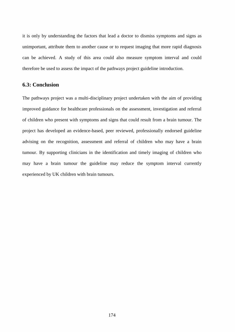
174
it is only by understanding the factors that lead a doctor to dismiss symptoms and signs as
unimportant, attribute them to another cause or to request imaging that more rapid diagnosis
can be achieved. A study of this area could also measure symptom interval and could
therefore be used to assess the impact of the pathways project guideline introduction.
6.3: Conclusion
The pathways project was a multi-disciplinary project undertaken with the aim of providing
improved guidance for healthcare professionals on the assessment, investigation and referral
of children who present with symptoms and signs that could result from a brain tumour. The
project has developed an evidence-based, peer reviewed, professionally endorsed guideline
advising on the recognition, assessment and referral of children who may have a brain
tumour. By supporting clinicians in the identification and timely imaging of children who
may have a brain tumour the guideline may reduce the symptom interval currently
experienced by UK children with brain tumours.

175
APPENDIX 1 – COMMENTS ON STATEMENTS FROM DELPHI
ROUND ONE NOT REACHING CONSENSUS
G5. 95% of children with a brain tumour have multiple symptoms and/or signs by diagnosis
Rating
1–9
Comments [52% rated 7-9] Occupation
2 If they did, our job would be easier for sure GPs
8 May be because there is a delay in diagnosis pathway
3 Depends on site of tumour Neurosurgery nurse
consultant
NC Don‟t know if the percentage figure is correct. Should use a more general
term e.g. „the majority‟
Consultant paediatric
neurosurgeons
7 Do you mean „by the time of diagnosis‟ ?
3 Not sure e.g. ataxia with indolent cerebellar tumours; epilepsy with PNET
Consultant paediatric
neurologists
4 Question unclear. What does “multiple” mean? If you mean more than one
then I would strongly agree.
3 This may depend on the health care setting and local expertise
7 Late diagnosis is still quite common
8 Is this percentage evidence-based? From my own experience I would guess
the figure is correct but one always remembers the exceptions
7 Is there not evidence to support this? Consultant
paediatricians 2 Not in my experience but there may be data on this I‟m unaware of
8 Answer reflects my feeling that review of the history (at the time of
diagnosis) may uncover prior clues, not that these are necessarily available
at initial referral or that they are neglected
2 In my experience only a few do
7 I have no evidence to support this but suspect it is the case
4 This is difficult as it varies according to tumour type e.g. optic pathway
glioma
Consultant paediatric
oncologists
7 This could reflect delays in diagnosis rather than natural history of
presentation
9 Though perhaps because of delayed referral/diagnosis
7 Depends which brain tumour – chiasmatic gliomas may have just visual loss
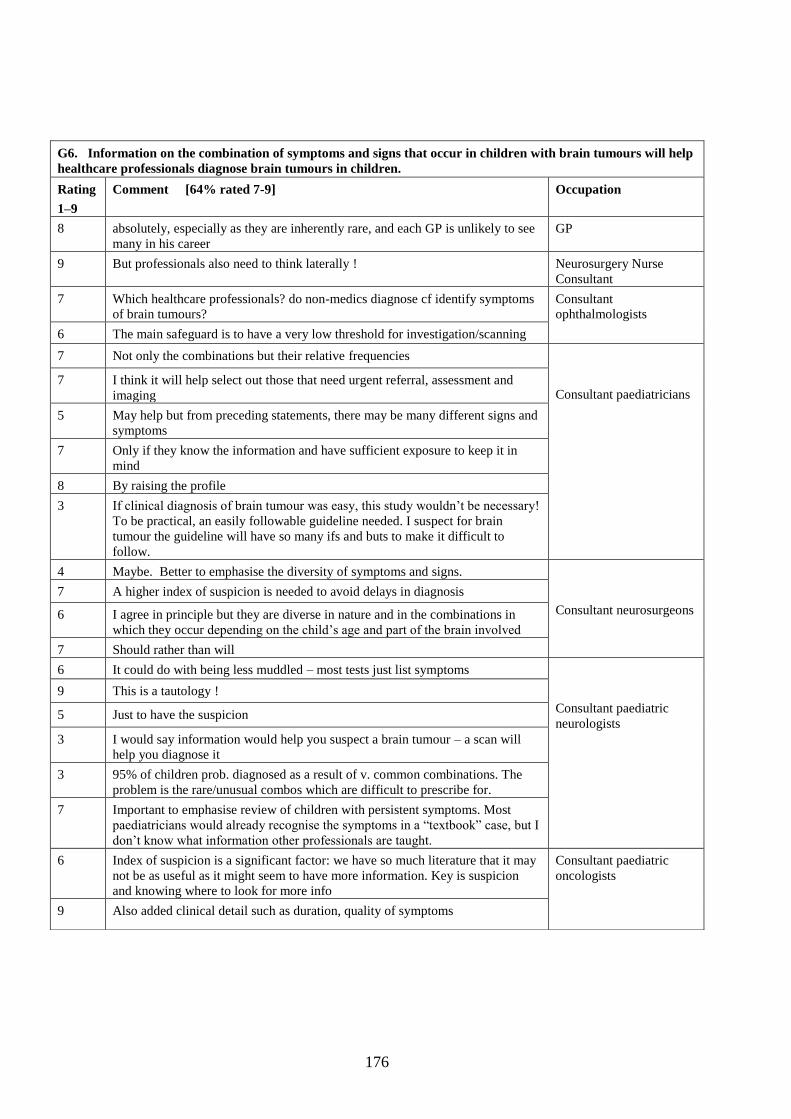
176
G6. Information on the combination of symptoms and signs that occur in children with brain tumours will help
healthcare professionals diagnose brain tumours in children.
Rating
1–9
Comment [64% rated 7-9] Occupation
8 absolutely, especially as they are inherently rare, and each GP is unlikely to see
many in his career
GP
9 But professionals also need to think laterally ! Neurosurgery Nurse
Consultant
7 Which healthcare professionals? do non-medics diagnose cf identify symptoms
of brain tumours?
Consultant
ophthalmologists
6 The main safeguard is to have a very low threshold for investigation/scanning
7 Not only the combinations but their relative frequencies
Consultant paediatricians
7 I think it will help select out those that need urgent referral, assessment and
imaging
5 May help but from preceding statements, there may be many different signs and
symptoms
7 Only if they know the information and have sufficient exposure to keep it in
mind
8 By raising the profile
3 If clinical diagnosis of brain tumour was easy, this study wouldn‟t be necessary!
To be practical, an easily followable guideline needed. I suspect for brain
tumour the guideline will have so many ifs and buts to make it difficult to
follow.
4 Maybe. Better to emphasise the diversity of symptoms and signs.
Consultant neurosurgeons
7 A higher index of suspicion is needed to avoid delays in diagnosis
6 I agree in principle but they are diverse in nature and in the combinations in
which they occur depending on the child‟s age and part of the brain involved
7 Should rather than will
6 It could do with being less muddled – most tests just list symptoms
Consultant paediatric
neurologists
9 This is a tautology !
5 Just to have the suspicion
3 I would say information would help you suspect a brain tumour – a scan will
help you diagnose it
3 95% of children prob. diagnosed as a result of v. common combinations. The
problem is the rare/unusual combos which are difficult to prescribe for.
7 Important to emphasise review of children with persistent symptoms. Most
paediatricians would already recognise the symptoms in a “textbook” case, but I
don‟t know what information other professionals are taught.
6 Index of suspicion is a significant factor: we have so much literature that it may
not be as useful as it might seem to have more information. Key is suspicion
and knowing where to look for more info
Consultant paediatric
oncologists
9 Also added clinical detail such as duration, quality of symptoms

177
G8. Enhanced training on the normal functional anatomy of the brain will help healthcare professionals identify
symptoms and/or signs that may be due to a brain tumour
Rating Comment [19% rated 7-9] Occupation
3 I can‟t see how this would help us; better to go via question G6 to help
diagnosis
GP
4 All doctors should be able to perform a full neurological assessment and be able
to recognise abnormalities, even if they can‟t exactly specify the area involved.
Not sure that enhanced training would make any difference
Consultant in Paediatric
Endocrinology &
Diabetes
2 Open access to imaging may lead to earlier diagnosis
Consultant
Neurosurgeons
3 It‟s quite complicated! – simple reminders of the significance of certain
symptoms/signs is probably more important than trying to encourage all
healthcare professionals to think deductively about functional neuroanatomy
5 The rarity of paediatric brain tumours in overall practice makes professionals to
not think of the diagnosis
Consultant Paediatric
Oncologists
5 Although I agree I expect pragmatically pattern recognition will be of more use.
1 Not sure what this means
Consultant Paediatricians
2 Most will have symptoms & signs related to raised intracranial pressure rather
than specific anatomically related problems – except in a few rarer instances.
3 Without seeing the detail about this, I‟m not sure. However, as brain tumours
are rare in spectrum of children‟s illnesses, any training given, not regularly
used (needed in day to day clinical practice) will slowly be forgotten
5 I‟m not sure a knowledge of “normal functional anatomy” would necessarily
make any difference if this is not taught in the context of what happens when
things are abnormal, ie a knowledge of pathological processes. What use is a
knowledge of the anatomy of the brain, if you don‟t know that vomiting can be
a presenting sign of a brain tumour.
Consultant Paediatric
Neurologists
3 Not just anatomy is needed – just good differentials for symptoms and signs
6 May help some understanding of mechanism eg why things are worse in
recumbency but headline features to look out for prob. better
5 I think this statement would need to be proven
3 Doubt it. What is normal functional anatomy in children?
1 Training on symptoms and signs may do. Functional anatomy training will help
localise lesions but not specifically tumours – could be other SOL or vascular
anomalies etc causing pathology
4 I‟m not sure how helpful neuroanatomy is, given how much other info health
workers have to absorb, but maybe basic understanding of how ICP develops
5 Probably but education probably better based on patterns of presentation eg
pattern of cerebellar signs in a post. fossa tumour
9 Lament the lack of neurology training in medical schools
4 Can help if they understood csf pathways and how infratentorial tumours
produce hydrocephalus
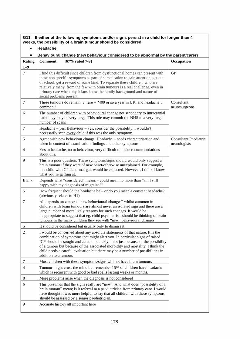
178
G11. If either of the following symptoms and/or signs persist in a child for longer than 4 weeks, the possibility of a brain tumour should be considered:
Headache
Behavioural change (new behaviour considered to be abnormal by the parent/carer)
Rating
1–9
Comment [67% rated 7-9] Occupation
7 I find this difficult since children from dysfunctional homes can present with
these non specific symptoms as part of somatisation to gain attention, get out
of school, get a reward of some kind. To separate these children, who are
relatively many, from the few with brain tumours is a real challenge, even in
primary care when physicians know the family background and nature of
social problems present.
GP
7 These tumours do remain v. rare = ?400 or so a year in UK, and headache v.
common !
Consultant
neurosurgeons
6 The number of children with behavioural change not secondary to intracranial
pathology may be very large. This rule may commit the NHS to a very large
number of scans
7 Headache – yes. Behaviour – yes, consider the possibility. I wouldn‟t
necessarily scan every child if this was the only symptom.
5 Agree with new behaviour change. Headache – needs characterisation and
taken in context of examination findings and other symptoms.
Consultant Paediatric
neurologists
4 Yes to headache, no to behaviour, very difficult to make recommendations
about this.
9 This is a poor question. These symptoms/signs should would only suggest a
brain tumour if they were of new onset/otherwise unexplained. For example,
in a child with CP abnormal gait would be expected. However, I think I know
what you‟re getting at.
Blank Depends what “considered” means – could mean no more than “am I still
happy with my diagnosis of migraine?”
5 How frequent should the headache be – or do you mean a constant headache?
(obviously relates to H1)
7 All depends on context, “new behavioural changes” whilst common in
children with brain tumours are almost never an isolated sign and there are a
large number of more likely reasons for such changes. It would be
inappropriate to suggest that eg. child psychiatrists should be thinking of brain
tumours in the many children they see with “new” behavioural changes.
5 It should be considered but usually only to dismiss it
2 I would be concerned about any absolute statements of that nature. It is the
combination of symptoms that might alert you. In particular signs of raised
ICP should be sought and acted on quickly – not just because of the possibility
of a tumour but because of the associated morbidity and mortality. I think the
child needs a careful evaluation but there may be a number of possibilities in
addition to a tumour.
7 Most children with these symptoms/signs will not have brain tumours
4 Tumour might cross the mind but remember 15% of children have headache
which is recurrent with good or bad spells lasting weeks or months.
8 More problems arise when the diagnosis is not considered
6 This presumes that the signs really are “new”. And what does “possibility of a
brain tumour” mean; is it referral to a paediatrician from primary care. I would
have thought it was more helpful to say that all children with these symptoms
should be assessed by a senior paediatrician.
9 Accurate history all important here

179
8 I‟d put nausea and vomiting here, not 2 weeks (as in G10). Consultant
paediatricians 8 Only if it is continuous or daily headache
7 Behavioural change less helpful in discriminating
7 Behaviour change is often difficult to asses and shold be considered along
with other features.
7 What you do about “considering” depends on what other diagnosis may be a
better fit at the time. Headache and behavioural issues are non-specific.
8 Considered yes but the consideration can often be quickly discounted after
further enquiry and examination
8 Brain tumour should be considered frequently, if only to think through that
unlikely at a particular point, but if symptoms persist may need a scan – i.e. a
plan for the child, and either review or instruction to parent to contact again if
symptoms don‟t clear in x time.
8 Certainly should be „considered‟ but not necessarily indication for imaging Consultant paediatric
oncologists 5 What does „a brain tumour should be considered‟ exactly mean? Headache is
a fairly common symptom and without abnormalities on neurological exam it
is unlikely that a brain tumour is the underlying problem. If „considering a
brain tumour‟ means imaging a lot of unnecessary imaging will be done.
7 Difficult as these symptoms could arise from less sinister causes. Need to
consider tumour in differential but not always arrange imaging at this time.
5 Depends what happens once diagnosis considered – we‟d be scanning loads of
upset or migrainous kids if it means a scan in all
9 In some situations would be concerned earlier.
G12. Brain tumours should be considered in the differential diagnosis of any child presenting with
abnormal growth (abnormal growth includes: weight loss, growth faltering, obesity, short stature, tall stature,
accelerated or delayed puberty and macrocephaly).
Rating
1-9
Comment [62% rated 7-9] Occupation
2 Every overweight child will be referred up with ? brain tumour! Nearly all
delayed puberty is familial and I can't think of any child in whom delayed
puberty was the sole feature of a brain tumour. Short stature again is usually
familial and what is short stature? Below 2nd
percentile? Below 0.4th
?
Abnormal growth as the only sign is highly unlikely to be due to a brain
tumour. The only area where I would strongly agree is in precocious puberty
as hamartomas are now recognised as a relatively common cause, especially
if the child is very young. MRI would be mandatory in any child with
confirmed precocious puberty. [NB: comment for G13 “despite my
comments in G12 I feel any child presenting with abnormal growth merits a
full neurological and visual assessment.”
Consultant in
paediatric
endocrinology and
diabetes
6 These would be referred for secondary opinion anyway at level of H/Visitor
in the under 5yr olds, and by GP in those over this age, even if both parents
were short in stature, for reassurance more than anything, but also that it just
might be brain tumour related. Macrocephaly will be a challenge since I
wonder how many colleagues have a head circumference chart readily at
hand and tape measure? And we are now in the grip of an obesity epidemic
so obesity itself is not going to be helpful unless accompanied by other
symptoms and signs. Weight loss would be easily identified and obvious
causes excluded.
GP
3 In some of these growth abnormalities, I would consider CNS tumour as
D/D but not all. E.g. growth faltering, obesity, short stature – if one looks at
statistics – how many young children presented with faltering growth, have
CNS tumour as a cause – Answer will be very small percentage in my
clinical practice.

180
8 Emphasis is on Considered – rare cause of growth abnormality without other
suggestive features
Consultant
paediatricians
6 Not the main diagnosis so given a low score, should be considered – yes but
not likely to be the diagnosis
3 Some of the above symptoms such as accelerated puberty prompts to think
about brain tumour than weight loss, obesity or short stature
2 Such a rare cause. If no other features present I think it‟s unlikely
9 While consideration should be given, not all will merit neuro-imaging.
2 The vast majority of children I see (as genereal paediatrician) with poor
growth do not have a brain tumour - less than 1% of this group will have
this, perhaps less than 0.1%. If this statement is put out to GPs I fear a flood
of urgent referrals of children who don't have a tumour. This will cause
operational difficulties to the rest of the service - there are other more
serious illnesses than failure to thrive/obesity that need attention before this
group.
6 Being considered is essential but the priority of the differential diagnosis
will vary
Consultant paediatric
oncologists
6 Depends on context and whether other predisposing causes are known eg
prolonged steroids and obesity/growth failure
7 In most with faltering growth other symptoms will reasonably mean other
issues are pursued first. If no explanation for faltering surfaces then mustn‟t
forget tumour especially before active child protection measures in place.
5 Not simple obesity – obesity with short stature? Consultant paediatric
neurologists 8 Endocrine referral should not be excluded
4 Again crosses the mind but many other causes. The concept of consider in
the light of a particular positive predictive value for that feature would be
helpful.
7 Often underestimated
[blank] Is this a bit early for investigation of vomiting and should g-i opinion be
considered first if ther child is otherwise "well". This also presumes that the
signs really are "new" And what does "possibility of a brain tumour" mean;
is it referral to a paediatrician from primary care. I would have thought it
was more helpful to say that all children with these symtoms should be
assessed by a senior paediatrician
H7. A young child who is unable to complain of headache may demonstrate head pain by holding their
head.
Rating
1–9
Comment [73% rated 7-9] Occupation
7 …but on the same note, may not do so. GP
7 Or playing with dolls bandaging their heads ! Consultant
paediatrician
7 Other behaviour patterns such as irritability, excessive crying may co-exist
Consultant paediatric
neurologists
7 But is the reverse true?
4 But they also hold their heads for other reasons
6 Although not holding their head does not exclude it
2 I‟m not sure that this is the case. The preverbal children I have seen have
demonstrated irritability or changed behaviour. As soon as they can
verbalise pain I think they would say their head hurt (at least in my
experience). Is there any evidence about this?

181
H10. A child with headache without a clear cause should be reviewed within 4 weeks.
Rating
1–9
Comment [72% rated 7-9] Occupation
3 I would want to see such a child sooner GP
[double
entry]
Not if a one-off headache Neurosurgical
nurse
consultant
3 Depends on how long the headache has been present for. If it is persistent or
recurrent as in H1, then I would argue imaging should be done ASAP. Lots of
children have headaches with no clear cause (used to get at least one referral a
month when doing general paediatrics) and seeing them all again within 4 weeks
is just not practical. Asking parents to contact if things don't improve or worsen is
a more pragmatic and manageable approach
Consultant in
paediatric
endocrinology
and diabetes
9 It‟s the review that‟s really important for such a common symptom ! Consultant
neurosurgeon 9 And if the headache is continuing should be strongly considered for imaging
6 Difficult to justify as a generalisation – dependant on initial assessment and
degree of concern. Sometimes ask parents to contact me if any changes or
concerns arise and then review promptly.
Consultant
paediatricians
1 Sooner
4 I found this question difficult as in most children with headache there is no clear
cause
5 Of referral? Or of first review? If the latter, a more prolonged period could be
appropriate
1 Only if it‟s persistent/recurring or has other feathers. not if it‟s all better !
5 Need to clarify the frequency/severity of the headache, and context
5 Not it it‟s a single headache. Most headaches are simple. Once again, need more
history information
1 Why 4 weeks?
8 Possibly sooner
9 If the headache persists Consultant
paediatric
neurologist
7 Ideally
4 in our centre we find almost 100% of children in our headache clinic do not have
a clear cause, we label them as chronic child headache of unknown cause, we
have a number of strategeies for them, but believe it is essential that the majority
are NOT seen soon, they need to work on the strategies , but represent
immediately should the symptoms change or any signs develop
2 Too vague do you know how many children with headaches are referred to OPD
and how many have improved by the time you see them
4 There are a lot of children with this, it isn‟t feasible to see them all so soon.
5 Depends on nature of headache and any associated other symptoms and signs
8 Depends on the length of the history eg if onset over 2 weeks in primary care
should probably be seen again in 2 weeks.
3 It is possible to make a headache diagnosis, if cannot need to refer to paed or
specialist
8 By whom I assume you mean the presenting GP Consultant
paediatric
oncologists NC Review should be driven by clinical concern and differential diagnosis, may be
much sooner than 4 weeks
9 I‟d say 2 weeks
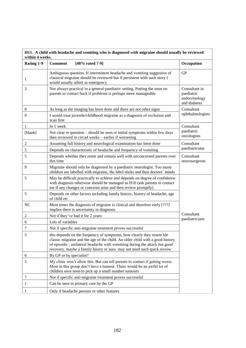
182
H11. A child with headache and vomiting who is diagnosed with migraine should usually be reviewed
within 4 weeks.
Rating 1-9 Comment [48% rated 7-9] Occupation
1
Ambiguous question. If intermittent headache and vomiting suggestive of
classical migraine should be reviewed but if persistent with such story I
would usually admit as emergency
GP
3 Not always practical in a general paediatric setting. Putting the onus on
parents to contact back if problems is perhaps more manageable
Consultant in
paediatric
endocrinology
and diabetes
8 As long as the imaging has been done and there are not other signs Consultant
ophthalmologists 9 I would treat juvenile/childhood migraine as a diagnosis of exclusion and
scan first
1 In 1 week Consultant
paediatric
oncologists [blank] Not clear re question – should be seen re initial symptoms within few days
then reviewed in circa4 weeks – earlier if worsening
2 Assuming full history and neurological examination has been done Consultant
paediatricians 5 Depends on characteristic of headache and frequency of vomiting
5 Depends whether they remit and remain well with unconcerned parents over
this time
Consultant
neurosurgeons
9 Migraine should only be diagnosed by a paediatric neurologist. Too many
children are labelled with migraine, the label sticks and then doctors‟ minds
5 May be difficult practically to achieve and depends on degree of confidence
with diagnosis otherwise should be managed as H10 (ask parents to contact
me if any changes or concerns arise and then review promptly)
Consultant
paediatricians
5 Depends on other factors including family history, history of headache, age
of child etc
NC Most times the diagnosis of migraine is clinical and therefore early [???]
implies there is uncertainty in diagnosis
2 Not if they‟ve had it for 2 years
6 Lots of variables
7 Not if specific anti-migraine treatment proves successful
5 this depends on the frequency of symptoms, how clearly they resem ble
classic migraine and the age of the child. An older child with a good history
of episodic , unilateral headache with vomitting during the attack but good
recovery, maybe a family hisory or aura may not need such quick reveiw
6 By GP or by specialist?
5 My clinic won‟t allow this. But can tell parents to contact if getting worse.
Most in this group don‟t have a tumour. There would be an awful lot of
children seen soon to pick up a small number tumours
7 Not if specific anti-migraine treatment proves successful
1 Can be seen in primary care by the GP
1 Only if headache persists or other features

183
H11. A child with headache and vomiting who is diagnosed with migraine should usually be reviewed
within 4 weeks. CONTINUED
Rating
1–9
If a confident diagnosis of migraine is made and the clinical exam is normal, there
is no need to review Occupation
1 Probably earlier
Consultant
paediatric
neurologists
8 By who?
6 Not practical
3 Reviewed where by the GP
2 Not as an absolute and it would depend on the confidence of the diagnosis – age
of child /FH nature of symptoms etc
3 This would depend on the confidence wit which a dx of migraine has been made
7 It depends on other clinical information
5 Very common in my practice. Where the diagnosis is clear I may arrange no
follow up at all; 60% chance of a life-long tendency (Ref: Bille)
1 Migraine would need to be diagnosed by a paediatrician ie not in primary care
[blank] If diagnosis secure such frequent review not needed
3 If a confident diagnosis of migraine is made and the clinical exam is normal, there
is no need to review
H12. In a child diagnosed with a non-structural headache (e.g. migraine, tension headache) a change in
the nature of the headache requires re-assessment and consideration of a structural cause.
Rating
1–9
Comment [73% rated 7-9] Occupation
9 Don‟t like the term “structural headache” what you mean is headache due to
raised ICP or meningeal irritation. non structural implies non physical change,
which is probably not true for migraine. Likewise raised ICP due to idiopathic
intracranial hypertension produced identical signs and symptoms to raised ICP
due to tumours but is “Non-structural”
Consultant paediatric
neurologist
2 Too vague – reassessment by whom and what change. Both of those conditions
are by nature variable.
5 I agree the child needs reassessment but there may be other causes as well as
structural abnormalities – commonest reason is almost certainly not a tumour
7 Seems sensible but I‟ve only seen 2 cases of this both in children with
neurofibromatosis. Migraine is common in NF1 so if they develop new
headaches they do need reassessment
1 Migraine should only be diagnosed by a paediatric neurologist. Too many
children are labelled wit migraine, the label sticks, and then doctors‟ minds
Consultant
neurosurgeon
N/C Depends on expertise in dealing with non-structural headach Consultant paediatric
oncologist
7 Important to alert parents to this at initial assessment Consultant
paediatrician

184
NV2. Persistent nausea and/or vomiting in the absence of corroborative history, examination or
investigation findings should not be attributed to a gastrointestinal cause.
Rating
1–9
Comment [73% rated 7-9] Occupation
8 If the quality of the investigations are robust to exclude a likely GI cause for the
N&V
GP
7 Should say: not only Gi problems Consultant
Paediatricians
7 In paediatric practice I tolerate absence of diagnosis (i.e. symptom based
diagnosis) rather than attribute to something for which there isn‟t good
evidence. Helps keep an open mind.
6 Difficult – many other possible causes need to be considered but still could be
GI
9 This seems to be the biggest group of missed or delayed diagnosis. Not just
scanning but a neurological history and examination would often make the
diagnosis earlier.
Consultant
paediatric
neurosurgeon
8 With comment for NV1 taken into consideration (“persistent in this context
needs clearer definition than just time. is it all the time, once a day, every other
day over 2 weeks, stopping eating, influencing activities or distractable from –
sorry being pedantic!”)
Consultant
paediatric
oncologist
5 Too vague Consultant
paediatric
neurologists 5 Not without consideration of other aetiologies
8 This assumes that a thorough assessment has been done (as for G10)
3 Commonest cause of persistent vomiting in babies is going to be
gastroesophageal reflux and they often have vague histories, normal
examination and no definite investigation that can be done to exclude/confirm it
(other than resolution with time)
Consultant in
paediatric
endocrinology and
diabetes

185
V2. Pupil dilatation should be performed if required to obtain a clear view of the optic disc.
Rating Comment [68% rated 7-9] Occupation
9 Often not practical in general practice
GPs
2 not if you‟re competent and comfortable with fundoscopy. Kids pupils
are pretty dilated anyway
9 Often not practical in general practice
2 In co-operative child in dark room may be able to see disc adequately
without dilating pupils
7 Wording here is difficult; the statement demands a STRONGLY AGREE
answer, but there is a question as to whether it is appropriate in all cases
2 I think this will be a disincentive. If there is a poor view or concern, pupil
should be dilated, or if there are other factors to suggest a visual problem
Consultant
Paediatricians
7 Usually inability to view disc is more related to co-operation of child
rather than pupil size
9 We tend to limit pupil dilatation to one specialist, i.e. so that it is only
done once
NC If DD really is brain tumour then child needs imaging – normal visual
assessment would NOT [word??]
1 If you can‟t see them, ask someone else more senior/experienced. They
may dilate the pupils.
5 Only if necessary which shouldn‟t be that often
Consultant
Neurosurgeons
7 But most GPs & paeds & indeed all non-ophthalmologists won‟t ?!
1 But should be used if the disc cannot be seen clearly
3 Not if child unstable Neurosurgery Nurse
Consultant
5 If pupil dilatation required then the examination should be performed by
an ophthalmologist
Consultant Paediatric
Oncologists
9 Yes – by optometrist/ophthalmologist
6 You often can see it fine in a cooperative child
[blank] I think most non ophthalmologists are v poor at assessing the fundi and
anyway normal fundi don‟t exclude a brain tumour. I think fundoscopy is
an over-rated pastime!
7 I suspect that if this is required it is best for the child to be seen by
ophthalmology
5 Depends on the clinical state of the child. If they are in a coma then
obviously not, if they are fully conscious and stable, then yes
Consultant Paediatric
Neurologists
CPN
2 Usually unncecessary
2 Almost never do this
8 Often this will only be in very young child. Often imaging considered,
under GA if under 8 years of age. Thus EUA of fundi would be even
better.
8 Ideally by an ophthalmologist if you are unable to obtain adequate views
9 Provided the child is neurologically stable and it will not affect neuro obs
5 This should probably be done by an ophthalmology colleague if readily
available

186
V3. Co-operative children aged 5 years and over can be assessed by a community optometrist.
Rating
1–9
Comment [63% rated 7-9] Occupation
If available quickly GP
5 Although referral may come via this route Neurosurgical nurse
consultant
1 They will not be able to assess the optic discs adequately
Consultant paediatric
ophthalmologists 2 Most optometrists are not very good as assessing children
7 Have had several children referred with papilloedema from community
optometrist, usually pretty good at picking things up.
Consultant in paediatric
endocrinology and diabetes
4 They can but this might not include all of the above observations (see V1) Consultant paediatric
neurosurgeon
[blank] A number of cases of papilloedema have been detected by community
opticians. Only a minority turned out to be brain tumours – more common
diagnoses were Drusen or BIH
Consultant paediatricians
7 Yes but ? should be, Needs to be done within 1 w
4 Depends on level of experience
3 It depends what you mean – I would have no problem with the community
optometrist testing eye movts but other aspects of examination including
fundoscoy still need to be done
Consultant paediatric
neurologists
9 However if they diagnose eg. papilloedema then this needs to be confirmed
by ophthalmologist
6 Depending on skill and expertise level
4 They are often very good, but practically the hospital specialists will work
with their own ophthalmology dept
5 Depends on expertise
2 True for acuity and fields but not other assessments
N/C Depends on how readily available
N/C I have no idea of the competence of a community optometrist, fundoscopy
should be included in the exam though
Consultant paediatric
oncologists
8 Several referrals from SpecSavers
8 I am sure they can be very effectivel Many of our referrals come from
specsavers. If you think the child has a brain tumour would you refer to the
community optometrist?

187
V7. Children should be assessed by ophthalmologists who have received training in paediatric ophthalmology.
Rating
1–9
Comment [74% rated 7-9] Occupation
[blank] The question is unanswerable as all ophthalmologists receive training in paediatric
ophthalmology. Some ophthalmologists subspecialise in paediatric ophthalmology
Consultant
ophthalmologists
7 Depends on how confident/competent the individual ophthalmologist feels re their
ability
9 ideally GPs
2 This will cause too much subspecialisation. Let all ophthalmologists be competent
to look in anyone‟s eyes
9 May not be practical Consultant
neurosurgeons 6 They should be assessed by one familiar with children but not all, especially some
very experienced senior colleagues, will have been specifically trained as
paediatric ophthalmologists
6 Not always easily/quickly available – “adult” service can look at discs
9 Becomes very critical in the youngest kids <2-3 years
8 Where possible Neurosurgery nurse
6 Ideally, but hopefully any competent ophthalmologist should be able to pick up
abnormal findings
Consultant in paediatric
endocrinology
5 Depends what you mean by training. many opthalmologists see lots of children and it is an
extensive part of their practice. identification of abnormalities should be made by a trained
opthalmolgist and delay to see a paed opthalmologist may also be an issue
Consultant paediatric
oncologists
8 This is the ideal but I believe most ophthalmologists are better than paediatricians
in this respect. So if no paediatric ophthalmologist still should be involved
6 The signs being sought should be in realm of all ophthalmologists
5 If possible – if this leads to undue delay, should be assessed by any (senior)
ophthalmologist. Know one case of glioma where waiting for super specialist
allowed vision to deteriorate.
Consultant
paediatricians
6 This is only part of the diagnostic procedure. If I was concerned I would progress
to imaging whatever the ophthalmology assessment. I think any ophthalmologist
should be able to diagnose a pale disc or papilloedema
5 May not be possible logistically in district general hospital
4 Seniority is as important, a senor general ophthalmologist is an excellent option
[blank] If they need ophthalmology assessment as questions V4 and V5 not in every child
in whom brain tumour is part of differential
7 ideally
9 Shouldn‟t all ophthalmologists have had this in their training? Consultant paediatric
neurologists 9 Ideally this is true but I would not defer assessment for 12 weeks while waiting
for an appt
5 In reality all district hospitals tend to see a lot of children and/or have a dedicated
colleague
7 Real life possibility?
4 Any ophthalmologist (adult or paediatric) should be competent in identifying
swollen disc
8 As paediatric neurologist I have ready access (same day) to a paediatric
ophthalmologist but of course I only see a selected population
9 If possible
8 Ideally

188
V14. CNS imaging is required for new onset squint.
Rating
1–9
Comment [68% rated 7-9] Occupation
7 Would refer GPs
5 Refer to ophthalmologist first
3 Depends on age of child and type of squint. Orthoptists are very
competent at distinguishing developmental abnormalities from other
CNS pathology affecting the visual system.
Consultant ophthalmologists [double
entry
1&5]
Need to differentiate between type of squint/VA/optic discs
9 Providing the patient is over 3 and has no refractive error
[double
entry
1&3]
Depends on rest of ophthalmic/orthoptic findings
6 Needs to be assessed by ophthalmologist first Consultant neurosurgeon
9 Pathological until proven otherwise. have seen venous thrombosis as
well as tumours present in this way.
Consultant in paediatric
endocrinology and diabetes
[blank] Whenever squint is noticed it will be new!! poor phrasing
Consultant paediatricians 2 Depends on context, a hypermetropic child who gets an intermittent
conv squint which corrects with specs doesn‟t need a scan
9 CNS imaging is required for children with new onset squint
8 Very likely
7 Depends on age of child and other symptoms
Consultant paediatric oncologists 6 Probably, depends what other symptoms. In absence of any other
symptoms would bet formal eye review first, then image
3 Depends on nature of squint
8 Advice from ophthalmology
5 After ophthalmological and neurological assessment first
Consultant paediatric neurologists
3 Depends on the type of squint. If it is paralytic then of course. If it is
non paralytic then probably not.
[blank] Depends on the circumstances e.g age
6 Not for a non-paralytic strabismus in a healthy child
5 Needs careful assessment
5 Unclear paralytic or non paralytic
7 Child needs to be seen by/discussed with ophthalmologist with
paediatric experience first; or seen by an experienced paediatrician

189
M6. A child with facial nerve weakness that does not show improvement within 2 weeks should undergo
CNS imaging.
Rating
1–9
Comment [69% rated 7-9] Occupation
2 Bell‟s palsy can take longer than this to resolve GP
7 Certainly upper motor neuron palsies require prompt investigation Consultant
neurosurgeon
7 Would normally wait 4-6 weeks Consultant
ophthalmologist
[blank] If they have no other symptoms at all, could wait a little longer eg 4-6 weeks Consultant in paediatric
endocrinology &
diabetes
[blank] Unless attributable to non-neurologic cause Consultant paediatric
oncologist
[blank] 2 weeks may be short for a „Bell‟s palsy‟
[blank] Not sure what percentage of Bells palsies in children improve within 2 weeks
Consultant paediatric
neurologist
8 Unless Bell‟s palsy is diagnosed with confidence
4 Bell‟s palsy often does not improve quickly
9 Unless congenital
4 Not sure about 2 weeks – maybe 4. Also recurrence is an indication
5 Not necessarily if clear evidence of lower motor nerve disease and no other
cranial nerve involvement or symptoms of raised ICP
5 Not with an isolated facial paresis and classical hx – wait 4 weeks
2 V common: a LMN VII without a VI or XIII very unlikely to be tumour.
HSV titres probably more relevant !
5 Unclear upper or lower motor neurone?
1 No evidence at all for this. Bell‟s palsy can easily take this time to improve;
the important thing is the neurological assessment (Riordan, Arch Dis Child
2001 and other refs)
2 Need history and examination follow-up
7 Have seen Bells palsy take a lot longer to resolve – but should bear possibility
of tumour in mind
Consultant
paediatrician
1 Not unless they have other symptoms, I wouldn‟t scan a Bells palsy at 2
weeks
3 Only if UMN lesion or other causes for concern
N/C I would individualise each child
1 Bells palsy takes a little longer to get better. There could be another obvious
casue for the facial weakness. If no obvious cause and not better in >3 weeks
refer to imaging
7 Show signs of improvement rather than full recovery

190
GR2. A child with impaired growth with no clearly identifiable psychosocial or physical cause should have
CNS imaging.
Rating
1–9
Comment [57% rated 7-9] Occupation
2 Investigations may come up with more common diagnosis than brain tumour eg
Coeliac‟s disease
GPs
[blank] Paediatric referral
7 Assuming endocrine causes have been excluded
2 How can psychological causes be clearly identified? Consultant in
paediatric
endocrinology &
diabetes
5 Should be considered but unsure how selective impaired growth along would be in
diagnosis of CNS tumour
Consultant
Paediatricians
3 Depends on growth velocity
N/C Impaired growth very ambiguous
1 Only if they have endocrine abn
5 Not as first line
3 Child would need detailed assessment of all the system and tailor the investigations
accordingly rather than CNS imaging as a blanket investigation
1 How often is there an “identifiable psychosocial cause” ? Most growth faltering has
no “identifiable cause”
5 psychosocial problems may not be easy to identify; CNS imaging in DGHs is a
complex problem: CT involves radiation, and repeat CTs over time may cause
damage. MRI access is difficult, especially for small children where deep
sedation/GA may be needed - such anaesthetists not always available in DGHs
9 Even with “psychosocial” causes, an organic cause should not be dismissed
Consultant
paediatric
neurologists
[blank] I would not accept psychosocial cause as a reason for withholding imaging. Children
from very poor psychosocial backgrounds develop brain tumours
[blank] What do you mean by impaired growth?
5 I think that depends on overall picture – they clearly need a proper assessment and if
concern that there may be pituitary dysfunction then imaging should be done
5 Does this assume that it is not constitutional?
8 Only part of the assessment of these children
9 What sort of image – MRI
Consultant
paediatric
oncologists
5 Agree should have it considered but in the absence of other signs and symptoms
associated with a tumour does the statement mean that all other causes of impaired
growth have already been ruled out before considering imaging?
7 This is very broad. Do we mean chronic, height & weight etc.
8 Is this height or weight
5 Difficult to accept growth failure in the absence of any history, symptoms or signs
that would already indicate the need for CNS imaging
NC Have to look at the clinical and genetic context. Want to avoid CNS imaging in
normal small children. Does impaired growth imply a change in rate of growth or
could it mean a child outside normal centile range?

191
GR3. CNS imaging should be undertaken prior to attributing weight loss to anorexia nervosa if the full
diagnostic criteria for anorexia nervosa are not met.
Rating
1–9
Comment [61% rated 7-9] Occupation
4 May still be anorexia nervosa so family and social set up and child‟s
and carers‟ past history are relevant here
GPs
[blank] Paediatric referral
5 Particularly in boys Consultant in paediatric
endocrinology and diabetes
1 Complex area – Pervasive food avoidance and other eating disorders
may better fit the clinical presentation. CNS imaging of these children
would be inappropriate
Consultant Paediatricians
5 Depends on discussion and assessment with CAMHS colleagues as to
likelihood and relevance
NC If you don‟t meet the diagnostic criteria for anorexia you don‟t have
anorexia !
3 Need for full systemic assessment
1 ?not if no other features are present
NC How many children fulfil full diagnostic criteria?
8 Only part of the assessment of these children Consultant paediatric neurologist
GR4. Reluctance to feed or eat leading to weight loss may result from swallowing difficulties.
Rating
1–9
Comment [73% rated 7-9] Occupation
[blank] Unlikely GPs
8 Well yes, those issues MAY…
2 Children will attempt to eat if hungry ! Neurosurgical nurse consultant
9 Should be other features - ?drooling etc Consultant paediatrician
3 Other features like drooling/dysarthria/choking episodes would point
more towards swallowing difficulties
Consultant paediatric neurologist

192
O2. Lethargy without organic cause is unusual in childhood in the absence of a severe life event
e.g. parental separation, bereavement.
Rating Comment [51% rated 7-9] Occupation
8 May be minor illness GP
1 ME/CFS
Consultant Paediatricians
6 What about chronic fatigue syndrome?
4 Becoming more common a symptom in terms of chronic
fatigue/ME and the like
1 100% of teenagers
1 What about depression, chronic fatigue
2 What may be interpreted as lethargy in small child often is
dis-interest. In older children post viral fatigue more common
than brain tumours etc.
4 Post viral syndrome/ME does occur in children and has no
clear life event trigger and the organic basis is not clear
Consultant Paediatric
Oncologists
[blank]
Define lethargy
5 I think may depend on the age of the child
7 Duration of and association with other signs/symptoms taken
into account
Clinical Assistant in
Paediatric Oncology
6 Yes in the under 10s, after that it seems quite common to me Con Neurosurgeon
3 Depends on the age of the child. Would agree that it‟s very
unusual in children <8, but becomes progressively less so.
Young teenagers often have no „severe‟ life event preceding,
usually a combination of many small things.
Consultant in Paediatric
Endocrinology & Diabetes
3 What about ME, depression etc. There is often no clear
“severe life event” associated
Consultant Paediatric
Neurologists
3 Depends what you mean – lethargy is a common complaint in
children and even more so in adolescence. There is often an
unrealistic expectation of how active children/adolescents
should be. Also many children/adolescents do not get enough
sleep and are lethargic in the daytime.
3 Mood disturbance may not reflect MLEs
6 Adolescent depression is probably more common than
appreciated
6 Depends on the age of the child –more concern in younger
child

193
R7. For MRI, contrast enhancement is not required to exclude a structural CNS abnormality.
Rating
1–9
Comment [35% rated 7-9] Occupation
9 But may give valuable extra information Consultant paediatric neurologists
6 Most structural abnormalities will be seen without contrast
enhancement but Gadolinium allows better differential diagnosis
3 May or may not be
7 Is this in the context of a tumour or cortical structural abnormality ??
7 Usually
8 Needs to be discussed with a neuroradiologist!!!! Caution required
before any didactic statements about imaging
3 It depends on how many other sequences are going to be done, but I
would have thought its safest to include a contrast scan
Consultant neurosurgeons
7 If PR-constrast images normal
7 My understanding is that contrast is normally utilised Consultant paediatric oncologists
3 Another double negative
5 Depends what you mean by structural – can be v helpful for tumours
7 Not essential with use of different sequences to find abnormality but
for max information as to nature of lesion will add info
NC Ask a radiologist Consultant paediatricians
3 Can‟t say for definite without a contrast

194
R9. Cranial ultrasound has no place in exclusion of CNS tumours in infants
Rating
1–9
Comment [58% rated 7-9] Occupation
Limited by age; not always useful for follow up; limited use for
neuraxial examinations
Clinical Assistant in
Paediatric Oncology
If the fontanelles are open, why not use them? GP
4 Sometimes useful. Cannot exclude tumour Consultant neurosurgeons
5 It depends on age and on whos doing it – it might help decide on
urgency of further investigation but shouldnever be the only test
9 It may show a tumour but further imaging will always be required Consultant paediatric
neurologists
9 Exception is the unstable neonate
6 It is going too far to say “no place”. However, its role is very limited
5 If other imaging modalities are not available USS will pick up
hydrocephalus although the cause may not be evident. With the
knowledge that an infant has raised ICP they can at least be urgently
transferred/referred to appropriate neurosurgical centre
3 Can be temporarily helpful in management
2 All the babies <6 mo I have seen were diagnosed on USS; however
obviously a normal USS doesn‟t exclude a tumour (though in practice
I‟ve never seen one that was missed)
6 Occasionally can be helpful as an initial screen but should not be relied
upon if negative
Consultant paediatricians
2 It may help identify a lesion or hydrocephalus – eg if MRI not
available. Then allowing urgent MRI referral
3 May have some role as a rapid and easy way of establishing whether
hydrocephalus exists while awaiting a CT/MRI will not give much
further info re tumour
Consultant paediatric
oncologist
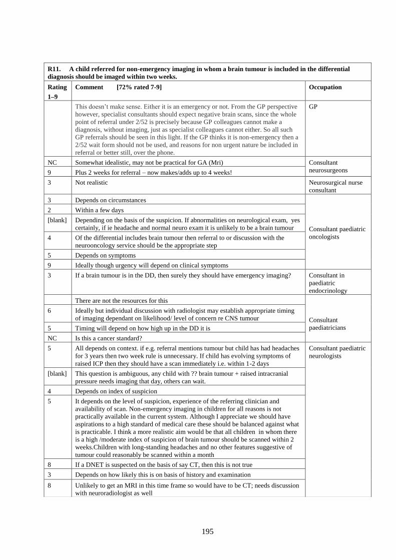
195
R11. A child referred for non-emergency imaging in whom a brain tumour is included in the differential
diagnosis should be imaged within two weeks.
Rating
1–9
Comment [72% rated 7-9] Occupation
This doesn‟t make sense. Either it is an emergency or not. From the GP perspective
however, specialist consultants should expect negative brain scans, since the whole
point of referral under 2/52 is precisely because GP colleagues cannot make a
diagnosis, without imaging, just as specialist colleagues cannot either. So all such
GP referrals should be seen in this light. If the GP thinks it is non-emergency then a
2/52 wait form should not be used, and reasons for non urgent nature be included in
referral or better still, over the phone.
GP
NC Somewhat idealistic, may not be practical for GA (Mri) Consultant
neurosurgeons 9 Plus 2 weeks for referral – now makes/adds up to 4 weeks!
3 Not realistic Neurosurgical nurse
consultant
3 Depends on circumstances
Consultant paediatric
oncologists
2 Within a few days
[blank] Depending on the basis of the suspicion. If abnormalities on neurological exam, yes
certainly, if ie headache and normal neuro exam it is unlikely to be a brain tumour
4 Of the differential includes brain tumour then referral to or discussion with the
neurooncology service should be the appropriate step
5 Depends on symptoms
9 Ideally though urgency will depend on clinical symptoms
3 If a brain tumour is in the DD, then surely they should have emergency imaging? Consultant in
paediatric
endocrinology
There are not the resources for this
Consultant
paediatricians
6 Ideally but individual discussion with radiologist may establish appropriate timing
of imaging dependant on likelihood/ level of concern re CNS tumour
5 Timing will depend on how high up in the DD it is
NC Is this a cancer standard?
5 All depends on context. if e.g. referral mentions tumour but child has had headaches
for 3 years then two week rule is unnecessary. If child has evolving symptoms of
raised ICP then they should have a scan immediately i.e. within 1-2 days
Consultant paediatric
neurologists
[blank] This question is ambiguous, any child with ?? brain tumour + raised intracranial
pressure needs imaging that day, others can wait.
4 Depends on index of suspicion
5 It depends on the level of suspicion, experience of the referring clinician and
availability of scan. Non-emergency imaging in children for all reasons is not
practically available in the current system. Although I appreciate we should have
aspirations to a high standard of medical care these should be balanced against what
is practicable. I think a more realistic aim would be that all children in whom there
is a high /moderate index of suspicion of brain tumour should be scanned within 2
weeks.Children with long-standing headaches and no other features suggestive of
tumour could reasonably be scanned within a month
8 If a DNET is suspected on the basis of say CT, then this is not true
3 Depends on how likely this is on basis of history and examination
8 Unlikely to get an MRI in this time frame so would have to be CT; needs discussion
with neuroradiologist as well
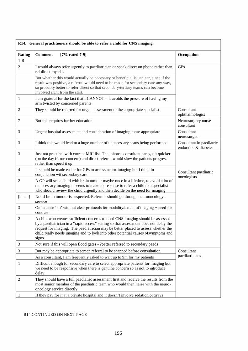
196
R14 CONTINUED ON NEXT PAGE
R14. General practitioners should be able to refer a child for CNS imaging.
Rating
1–9
Comment [7% rated 7-9] Occupation
2 I would always refer urgently to paediatrician or speak direct on phone rather than
ref direct myself.
GPs
But whether this would actually be necessary or beneficial is unclear, since if the
result was positive, a referral would need to be made for secondary care any way,
so probably better to refer direct so that secondary/tertiary teams can become
involved right from the start.
1 I am grateful for the fact that I CANNOT – it avoids the pressure of having my
arm twisted by concerned parents
2 They should be referred for urgent assessment to the appropriate specialist Consultant
ophthalmologist
7 But this requires further education Neurosurgery nurse
consultant
3 Urgent hospital assessment and consideration of imaging more appropriate Consultant
neurosurgeon
3 I think this would lead to a huge number of unnecessary scans being performed Consultant in paediatric
endocrine & diabetes
3 Just not practical with current MRI list. The inhouse consultant can get it quicker
(on the day if true concern) and direct referral would slow the patients progress
rather than speed it up
Consultant paediatric
oncologists
4 It should be made easier for GPs to access neuro-imaging but I think in
conjunction wit secondary care
2 A GP will see a child with brain tumour maybe once in a lifetime, to avoid a lot of
unnecessary imaging it seems to make more sense to refer a child to a specialist
who should review the child urgently and then decide on the need for imaging
[blank] Not if brain tumour is suspected. Referrals should go through neurooncology
service
3 On balance „no‟ without clear protocols for modality/extent of imaging + need for
contrast
2 A child who creates sufficient concerns to need CNS imaging should be assessed
by a paediatrician in a "rapid access" setting so that assessment does not delay the
request for imaging. The paediatrician may be better placed to assess whether the
child really needs imaging and to look into other potential causes ofsymptoms and
signs
3 Not sure if this will open flood gates - ?better referred to secondary paeds
3 But may be appropriate to screen referral to be scanned before consultation Consultant
paediatricians
As a consultant, I am frequently asked to wait up to 9m for my patients
1 Difficult enough for secondary care to select appropriate patients for imaging but
we need to be responsive when there is genuine concern so as not to introduce
delay
2 They should have a full paediatric assessment first and receive the results from the
most senior member of the paediatric team who would then liaise with the neuro-
oncology service directly
1 If they pay for it at a private hospital and it doesn‟t involve sedation or xrays

197
R14. General practitioners should be able to refer a child for CNS imaging. CONTINUED
Rating
1–9
Comment Occupation
3 Only in exceptional circumstances and only in older children and young adults
Consultant
paediatricians CONT
1 If there is that level of concern, the child should see a “paediatric specialist”
5 Such an approach might flood the system with more referrals than imaging
departments could cope with. Would the GP break the bad news, or would an
alien hospital team pick up the pieces?
2 Many cases of brain tumour failed to be recognised by GPs and other cases where
concern about it mis-placed
1 This is a recipe for disaster: GP refers for scan, scan done by prvate MRI facility,
scan reported incorrectly, child reassured and then presents in extremis sometime
later!!
Consultant paediatric
neurologists
5 Depending on expertise and skill
1 I think there are reasons why a GP should have access to CNS imaging, but when
a brain tumour is suspected, appropriate referral is required
3 Could overwhelm the service – only if strict guidelines or discussion with
radiologist
1 You are joking
1 Not directly for a number of reasons. Many GPs have not had specific training in
paediatric neurology and therefore may not choose optimum imaging modality.
Young children may need sedation or GA for scanning and therefore need
admitting to a hospital bed with a responsible clinician. Other investigations -
pituitary testing/tumour markers/ophthalmology assessment may be needed and
should be coordinated by an experienced paediatrician/paediatric neurologist who
can then take on further management
1 Still a relatively inaccessible resource; may lead to inappropriate radiation
exposure; headaches are very common in childhood
5 Children should be referred urgently for clinical review, not imaging (as it might
be necessary to image the spine also, for example)
2 Because will lengthen waiting lists
1 But they do need urgent access to paediatric assessment eg a rapid access clinic
3 Not as a blanket rule

198
R15. In my experience, a nursing professional (e.g. health visitor, practice nurse, school nurse) has
played a critical role in the identification of a child with a brain tumour.
Rating
1–9
Comment [30% rated 7-9] Occupation
2 It is often their concerns/observations that has started the detailing of
differential diagnoses which may include cranial tumour
GP
1 Opticians are the non medic most likely to suspect the diagnosis Consultant paediatrician
9 Health visitors have identified increased head circumference. I
remember also one optician making the diagnosis
Consultant neurosurgeon
1 The orthoptist has been the most reliable referral Consultant ophthalmologist
2 It‟s usually the parents. Have even seen the opposite, where nursing
staff have down played „classical‟ symptoms of raised ICP or tumour
Consultant in paediatric
endocrine and diabetes
5 This is a difficult question to answer, as the cases that one remembers
will usually be cases where there was a tumour, and all the other cases
will remain “background noise” as it were
Consultant paediatric
neurologists 5 Not in my direct experience but their concerns have added to those of
other professionals eg GP. However school nurses have referred
patients with loss of skills to me on 2 occasions. The eventual
diagnoses were of neurodegenerative disorders but the differential
would have included a tumour
3 I have never been referred anyone by this route
Consultant paediatric
oncologists
3 Less than I would expect
7 I‟ve had a health visitor religiously plot the OFC as it went off the page
without thinking about the possible causes of this extraordinarily rapid
growth !!

199
APPENDIX 2 – COMMENTS ON STATEMENTS FROM DELPHI
ROUND TWO NOT REACHING CONSENSUS
MODIFIED G11b. If a child presents with abnormal behaviour (causing concern to
parents/carers) including lethargy or withdrawal and persisting for more than 4 weeks, a brain
tumour should be considered in the differential diagnosis.
59% rated this statement 7-9
Rating Comment Occupation
8 Agree with the term considered alongside what will be a whole list of other
possibilities in the absence of any other signs and symptoms
Consultant
paediatric
oncologists 5 Would depend on the history. Social causes much more likely for example.
6 I would agree more strongly if the comment said „change of behaviour‟ and
then added „in absence of other obvious explanation‟
Consultant
paediatricians
3 Would be rare as sole presenting feature of a tumour
9 Yes, important to consider – exam and investigation will help decide need for
investigation follow up interval and imaging plans
5 I would still state that it depends in part whether a child has an underlying
diagnosis eg autism then this sort of change would be quite common and
would not immediately make me think of tumour. If the child was previously
„normal‟ in behavioural terms then tumour should be excluded.
[double
entry]
The whole picture needs looking at
Consultant
paediatric
neurologists
6 Not happy with the “abnormal behaviour” bit. This will fill the clinic with
kids with ADHD, the lethargy and behaviour certainly would be worrying.
7 All depends what considered means – obviously should be entertained as
possibility but no necessarily pursued beyond that
7 Should be “considered” once again but likelihood of it being related to a
tumour depends on other symptoms and examination findings as well as on
the history
4 All depends on context, in isolation I would not agree
6 This presentation has a wide differential and BT is down the list of possible
causes

200
MODIFIED G12. A child who presents with one or more of the following symptoms and/or signs requires
early specialist referral for consideration of a brain tumour in the differential diagnosis:
Precocious puberty
Delayed puberty
Growth failure
Macrocephally
65% rated this statement 7-9
Rating
1-9
Comment Occupation
1 Delayed puberty is very common and usually familial. Whilst I will always see
these young people for consideration of treatment, brain tumour is highly
unlikely to be a cause of their problems. Growth failure would be a very late
sign of a brain tumour as it will take months of insufficient growth hormone to
stop someone growing - whilst the child again needs seeing, they do not need
referral for '? brain tumour' as I feel this would create unnecessary anxiety and
also potentially swamp clinics. However, polyuria and polydipsia NOT caused
by diabetes mellitus SHOULD be included in this list as 4 out of the last 5
children seen with pituitary area tumours had this symptom for up to 2 years
before diagnosis. Precocious puberty is always investigated with an MRI but
again, I do not feel that brain tumour should be included in the initial
differential diagnosis - these children will all be seen quickly but do not need to
come in under the 2 week cancer wait target. If macrocephaly was included,
half of Stoke-on-Trent would need referring ('potters head' recognised locally as
a benign cause of macrocephaly!).
Consultant
Paediatric
Endocrinologist
8 Re: second one; need definition of “delayed puberty”; this will rarely be
diagnosed in primary care the other three can be
GP
3 This is too wide a topic for a single response and needs to be refined related to
age
Consultant
neuroradiologist
6 Growth failure is common. Growth failure due to BT with no other findings Is
very rare so growth failure is poor discriminator
Consultant
paediatric
oncologists
8 I feel this is more pertinent for precocious puberty and growth failure rather
than delayed puberty and macrocephaly
2 I would not consider a brain tumour in an otherwise well boy with delayed
puberty and no other signs or symptoms. Similarly a big head in an older child
otherwise completely well – perhaps in a young child with open fontanelles –
yes
6 I think the child should be referred to a specialist (paediatrician/endocrinologist)
but the main reason is to determine cause not only consideration of brain
tumour
7 Growth failure – is this weight or height? Failure of weight in pre-school
children usually due to inadequate food (or something other than a BT). Height
failure may be due to BT
Consultant
paediatricians
1 Delayed puberty is very common and usually familial. Whilst I will always see
these young people for consideration of treatment, brain tumour is highly
unlikely to be a cause of their problems. Growth failure would be a very late
sign of a brain tumour as it will take months of insufficient growth hormone to
stop someone growing - whilst the child again needs seeing, they do not need
referral for '? brain tumour' as I feel this would create unnecessary anxiety and
also potentially swamp clinics. However, polyuria and polydipsia NOT caused
by diabetes mellitus SHOULD be included in this list as 4 out of the last 5
children seen with pituitary area tumours had this symptom for up to 2 years
before diagnosis. Precocious puberty is always investigated with an MRI but
again, I do not feel that brain tumour should be included in the initial
Consultant
Paediatric
Endocrinologist
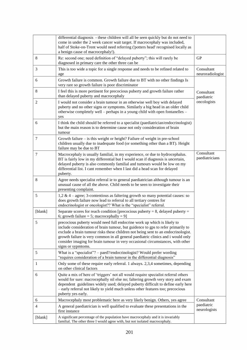
201
differential diagnosis - these children will all be seen quickly but do not need to
come in under the 2 week cancer wait target. If macrocephaly was included,
half of Stoke-on-Trent would need referring ('potters head' recognised locally as
a benign cause of macrocephaly!).
8 Re: second one; need definition of “delayed puberty”; this will rarely be
diagnosed in primary care the other three can be
GP
3 This is too wide a topic for a single response and needs to be refined related to
age
Consultant
neuroradiologist
6 Growth failure is common. Growth failure due to BT with no other findings Is
very rare so growth failure is poor discriminator
Consultant
paediatric
oncologists
8 I feel this is more pertinent for precocious puberty and growth failure rather
than delayed puberty and macrocephaly
2 I would not consider a brain tumour in an otherwise well boy with delayed
puberty and no other signs or symptoms. Similarly a big head in an older child
otherwise completely well – perhaps in a young child with open fontanelles –
yes
6 I think the child should be referred to a specialist (paediatrician/endocrinologist)
but the main reason is to determine cause not only consideration of brain
tumour
7 Growth failure – is this weight or height? Failure of weight in pre-school
children usually due to inadequate food (or something other than a BT). Height
failure may be due to BT
7 Macrocephaly is usually familial, in my experience, or due to hydrocephalus.
BT is fairly low in my differential but I would scan if diagnosis is uncertain,
delayed puberty is also commonly familial and tumours would be low on my
differential list. I cant remember when I last did a head scan for delayed
puberty.
Consultant
paediatricians
8 Agree needs specialist referral ie to general paediatrician although tumour is an
unusual cause of all the above. Child needs to be seen to investigate their
presenting complaint.
5 1,2 & 4 – agree; 3-contentious as faltering growth so many potential causes: so
does growth failure now lead to referral to all tertiary centres for
endocrinologist or oncologist?? What is the “specialist” referral.
[blank] Separate scores for reach condition [precocious puberty = 8, delayed puberty =
6; growth failure = 5; macrocephally = 9]
5 precocious puberty would need full endocrine work up which is likely to
include consideration of brain tumour, but guidence to gps to refer primarily to
exclude a brain tumour risks these children not being sent to an endocrinologist.
growth failure is very common in all general paediatric clinics and i would only
consider imaging for brain tumour in very occasional circumstances, with other
signs or sypmtoms.
5 What is a “specialist”? – paed?/endocrinologist? Would prefer wording
“requires consideration of a brain tumour in the differential diagnosis”
1 Only some of these require early referral. 1 always. 2,3,4 sometimes, depending
on other clinical factors
6 Quite a mix of here of „triggers‟ not all would require specialist referral others
would for sure: macrocephally nil else no; faltering growth very story and exam
dependent guidelines widely used; delayed puberty difficult to define early here
– early referral not likely to yield much unless other features too; precocious
puberty yes early.
6 Macrocephaly most problematic here as very likely benign. Others, yes agree Consultant
paediatric
neurologists 4 A general paediatrician is well qualified to evaluate these presentations in the
first instance
[blank] A significant percentage of the population have macrocephaly and it is invariably
familial. The other three I would agree with, but not isolated macrocephaly.

202
4 Early referral implies urgency which is not warranted. Macrocephaly –
measuring parent‟s head size takes away worry in majority. Endocrine problems
may rearely involve brain tumours but should not dictate early referral. Failure
to thrive – wide diagnosis.
3 Macrocephaly occurs in 3% normal population. Brain tumour 2/100000/year !
6 All of these have more likely causes than brain tumour

203
MODIFIED H10. A child presenting with a new and persisting headache should be reviewed within 2
weeks [„persisting‟ as defined in H1 i.e. a continuous or recurrent headache lasting more than 4 weeks].
74% rated this statement 7-9
Rating Comment Occupation
[blank] Child should not be left for 4 weeks without review Clinical
Assistant in
Paediatric
Oncology
[blank] Reviewed by who? Primary care, secondary care? Consultant
paediatric
oncologists 9 If still present now that will be approx 6 weeks of headache and imaging is
merited now
3 I don‟t see how putting a decision off helps – you‟re either worried or you‟re
not
Consultant
paediatricians
8 Suggest adding „……..new and persisting headache without explanation should
be reviewed..‟
7 I read this as an “urgent/soon outpatient referral”
3 Vast majority of children with headaches will have them >4weeks and not have
tumor current statement would capture virtually all children in to a 2 week
referral
8 Review by whom, a GP should be able to perform neurological exam and triage
patients for urgent review by Paeds
2 Depends on how strong is the suspicion of CNS tumours. If other diagnosis
seem more likely longer interval may be justified.
Why 2 weeks? It could all depend on the duration of symptoms.
5 Most new and persisting headaches are sleep related and cannot be reviewed in
2 weeks. Would prefer use of words “new, unusual and persisting”
3 Too prescriptive
6 is this by GP? prior to referal to specialist? the statement is still vague, in
some if the index of suspicion is high referal should be immediate, but in others
a diagnosis can be made, review may be as simple as 'if things are not better in
2 weeks come for review' parents should always be told that if things change or
new symptoms arise they should be seen again asap
9 In some situations earlier – as isolated feature ok this means being seen up to 6
weeks after first headache
7 Does “review” relate to primary or secondary care?
Consultant
paediatric
neurologists
[blank] Reviewed by whom and to what end?
3 Only if there are other signs
4 It is unlikely that headache will be the sole presentation if it is a tumour related
raised ICP. “review” could be by a GP/general paediatrician (if rapid access
clinics are available)
6 By whom?
3 Who by? Only if it persists? Worried could lead to lots of referrals that are
unnecessary
7 By his GP

204
MODIFIED V3/V7. A child presenting with symptoms and/or signs as listed in G9 (see below) requires
complete visual assessment as described in V1, within 1 week.
68% rated this statement 7-9
Rating
1-9
Comment Occupation
5 ?within a week?? GP
5 All these or “just” ? Consultant
ophthalmologists 9 Other G9 signs are present – i.e. abnormal fundus appearance, I think we
mean papilloedema here. If anything else then sooner assessment i.e. same
day
5 2 weeks
[blank] G9 states a symptomatic child with a brain tumour will have one or more of
the list. However if a child presents with a headache alone I do not think it
realistic or appropriate that they all get referred for visual assessment within
a week. If the child has more signs and symptoms then maybe.
Consultant
paediatric
oncologists 2 Should have visual assessment as fundoscopy + clinical assessment of
acuity and fields to confrontation as urgent measure / part of general
examination – BUT it is too much to expect that a full ophthalmological
assessment be routinely undertaken within 1 week.
[blank] Difficult to comment on this – might need a scan more than a visual
assessment and scan might determine whether visual assessment is needed
3 Agree should have assessment as in V1 as part of diagnostic process but
unclear what the 1 week timeframe achieves
5 Not sure where emphasis lies here. Eye movements/pupillary response
readily assessed in all. Visual fields in >5 year OK in most – but not
perimetry surely. The way to diagnosis is clinical suspicion & early
imaging. Should not get hung-up on completeness of this.
Consultant
paediatric
neurologists
3 A child with headache, vomiting and lethargy secondary to raised ICP
would be dead within this time frame if they have not been assessed and
referred on, we always (or should d) arrange immediate assessment if there
is anything other than a history of headache.
7 This is practically difficult to achieve unless the initial assessment is by GP
3 Some of these features require such urgent referral, others do not
1 We could not ask ophthalmologists to do this for all children with seizures
or headache, or paediatricians/paediatric neurologists either
6 This is very unlikely to be achieveable
3 This would include isolated headache or nausea/vomiting. The numbers who fulfil
just one of G9 criteria would be high and many would wind up having normal tests
and needing an unnecessary oversupply of test facility to meet this (poor value for
money)
Consultant
paediatricians 9 Fields by confrontation perimetry is sufficient for initial asst and can all be
done by GP in a child over 5
1 The signs in G9 are too non specific for brain tumours. Prefer “ if a child is
…[?word] of a brain tumour…”
5 This may not be a priority. It depends on other symptoms and degree of
suspicion. If isolated symptom then agree
[blank] Is the purpose of the VA to make a diagnosis or to define the deficit? G9 assumes
there is a brain tumour and therefore this question is in limbo. I agree it needs to be
done but it compliments neuroimaging.
3 badly worded, surely not if a child is referred with a single symptom or sign such as
poor growth or isolated seizure which our general paediatric outpatients are full of,
unless you mean that it should form part of general paediatric examination which is
obvious and is covered in statement G13
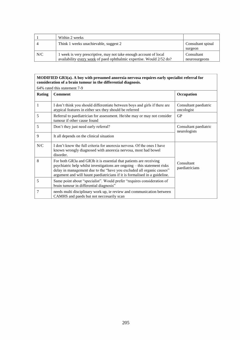
205
1 Within 2 weeks
4 Think 1 weeks unachievable, suggest 2 Consultant spinal
surgeon
N/C 1 week is very prescriptive, may not take enough account of local
availability every week of paed ophthalmic expertise. Would 2/52 do?
Consultant
neurosurgeons
MODIFIED GR3(a). A boy with presumed anorexia nervosa requires early specialist referral for
consideration of a brain tumour in the differential diagnosis.
64% rated this statement 7-9
Rating Comment Occupation
1 I don‟t think you should differentiate between boys and girls if there are
atypical features in either sex they should be referred
Consultant paediatric
oncologist
5 Referral to paediatrician for assessment. He/she may or may not consider
tumour if other cause found
GP
5 Don‟t they just need early referral? Consultant paediatric
neurologists
9 It all depends on the clinical situation
N/C I don‟t know the full criteria for anorexia nervosa. Of the ones I have
known wrongly diagnosed with anorexia nervosa, most had bowel
disorder.
Consultant
paediatricians
8 For both GR3a and GR3b it is essential that patients are receiving
psychiatric help whilst investigations are ongoing – this statement risks
delay in management due to the “have you excluded all organic causes”
argument and will haunt paediatricians if it is formalised in a guideline.
5 Same point about “specialist”. Would prefer “requires consideration of
brain tumour in differential diagnosis”
7 needs multi disciplinary work up, ie review and communication between
CAMHS and paeds but not neccesarily scan
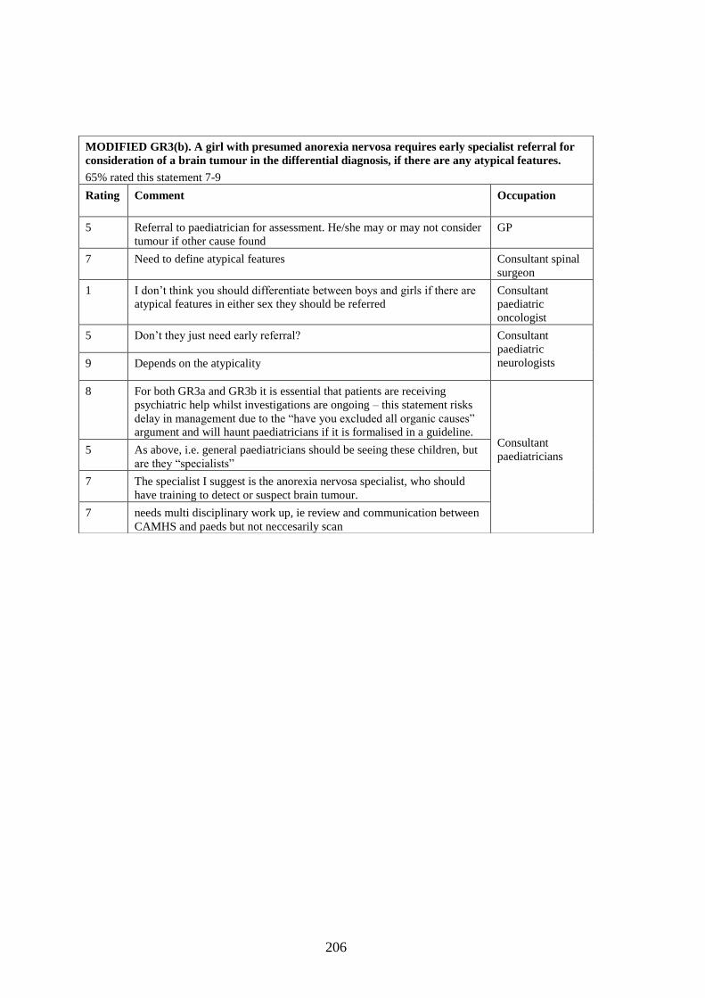
206
MODIFIED GR3(b). A girl with presumed anorexia nervosa requires early specialist referral for
consideration of a brain tumour in the differential diagnosis, if there are any atypical features.
65% rated this statement 7-9
Rating Comment Occupation
5 Referral to paediatrician for assessment. He/she may or may not consider
tumour if other cause found
GP
7 Need to define atypical features Consultant spinal
surgeon
1 I don‟t think you should differentiate between boys and girls if there are
atypical features in either sex they should be referred
Consultant
paediatric
oncologist
5 Don‟t they just need early referral? Consultant
paediatric
neurologists 9 Depends on the atypicality
8 For both GR3a and GR3b it is essential that patients are receiving
psychiatric help whilst investigations are ongoing – this statement risks
delay in management due to the “have you excluded all organic causes”
argument and will haunt paediatricians if it is formalised in a guideline.
Consultant
paediatricians 5 As above, i.e. general paediatricians should be seeing these children, but
are they “specialists”
7 The specialist I suggest is the anorexia nervosa specialist, who should
have training to detect or suspect brain tumour.
7 needs multi disciplinary work up, ie review and communication between
CAMHS and paeds but not neccesarily scan

207
APPENDIX 3 – WORKSHOP PARTICIPANTS
Mrs Jane Bond, Parent representative, Lichfield, Staffs.
Mrs Bea Brunton , PASIC Co-ordinator and parent representative, QMC, Nottingham
Ms Jessy Choi, Consultant Ophthalmologist, Chesterfield Royal Hospital
Ms Jill Gratton, Consultant Optometrist, Dolland & Aitchison, Nottingham
Mr Richard Gregson, Consultant Paediatric Ophthalmologist, QMC, Nottingham
Dr Robin Hunter, General Practitioner, Beeston, Notts
Dr Tim Jaspan, Consultant Neuroradiologist, QMC, Nottingham.
Dr Monica Lakhanpaul, Consultant Paediatrician, Leicester Royal Infirmary, Leicester
Mr John Leach and Mrs Lisa Leach, Parent representatives, Balderton, Notts.
Mrs Louise Munns, Optometrist, Dolland & Aitchison, Nottingham
Dr Chris Nelson, Consultant Paediatrician, Derby City General Hospital, Derby.
Dr Julian Nicholson, General Practitioner, Long Eaton, Nottingham.
Dr Vibert Noble, Consultant Paediatrician, Kings Mill Hospital, Notts.
Dr Stephanie Smith, Consultant Paediatrician, QMC, Nottingham
Research Team
Professor David Walker, Professor of Paediatric Oncology, QMC/University of Nottingham
Professor Richard Grundy, Professor of Neuro-oncology and Cancer Biology,
QMC/University of Nottingham
Professor Colin Kennedy, Professor of Paediatric Neurology, Southampton General Hospital
Dr Sophie Wilne, Clinical Research Fellow, University of Nottingham.
Dr Karin Koller, Research Fellow, University of Nottingham.

208
APPENDIX 4 – DELPHI PANEL PARTICIPANTS
Name Occupation
Mr D Allcutt Consultant Neurosurgeon
Dr S Bailey Consultant Paediatric Oncologist
Mr H Balaji Prasad Staff Grade Ophthalmologist
Dr D Barker Consultant Paediatrician
Dr P Baxter Consultant Paediatric Neurologist
Dr S Bennett-Britton Consultant Paediatrician
Dr S Benton Consultant Paediatric Neurologist
Dr D Bond Consultant Paediatrician
Miss J Brown Consultant Paediatric Neurosurgeon
Dr M Buckley General Practitioner
Dr A Burke Consultant Paediatric Oncologist
Dr M Butler General Practitioner
Miss L Butler Consultant Paediatric Ophthalmologist
Mr A Callear Consultant Ophthalmologist
Mr M Carter Consultant Neurosurgeon
Dr C Chadwick Consultant Paediatrician
Dr A Childs Consultant Paediatric Neurologist
Dr J Chisholm Consultant Paediatric Oncologist
Dr G Chow Consultant Paediatric Neurologist
Mr P Chumas Consultant Neurosurgeon
Dr M Clarke Consultant Paediatric Neurologist
Dr H Clements Consultant Paediatrician
Dr N Coad Consultant Paediatrician
Dr J Cobb General Practitioner
Dr A Coe Consultant Paediatrician
Dr I Collier General Practitioner
Mr E Davies Consultant Spinal Surgeon
Miss F Dean Consultant Ophthalmologist
Dr J Eames General Practitioner
Dr B Eldeeb Clinical Assistant in Paediatric Oncology
Dr M Elliott Consultant Paediatric Oncologist
Dr M English Consultant Paediatric Oncologist
Dr D Farmer General Practitioner
Mr I Fearnley Consultant Ophthalmologist
Dr C Ferrie Consultant Paediatric Neurologist
Dr R Forsyth Consultant Paediatric Neurologist
Dr A Gallagher Consultant Paediatrician
Dr F Gibbon Consultant Paediatric Neurologist
Dr H Gorringe General Practitioner
Dr J Gosalakkal Consultant Paediatric Neurologist
Dr R Groves General Practitioner
Dr A Gupta Consultant Paediatrician
Mi ss H Fernandes Consultant Paediatric Neurosurgeon
Dr J Hale Consultant Paediatric Oncologist
Dr D Hargrave Consultant Paediatric Oncologist

209
Mr W Harkness Consultant Neurosurgeon
Miss R Harrison Consultant Ophthalmologist
Dr M Hewitt Consultant Paediatric Oncologist
Dr H Hibbs General Practitioner
Dr D Hobin Consultant Paediatric Oncologist
Dr I Hughes Consultant Paediatric Neurologist
Dr Z Ibrahim Consultant Paediatrician
Dr S Jayawant Consultant Paediatric Neurologist
Dr H Jenkinson Consultant Paediatric Oncologist
Dr S Jones General Practitioner
Dr D Kalra Consultant Paediatrician
Dr V Lee Consultant Paediatric Oncologist
Dr I Leese General Practitioner
Dr D Lewis Consultant Paediatrician
Dr M Likeman Consultant Neuroradiologist
Dr A Liu Consultant Paediatric Neuroradiologist
Dr J Livingston Consultant Paediatric Neurologist
Mr D Macarthur Consultant Neurosurgeon
Dr T Martland Consultant Paediatric Neurologist
Ms L May Neurosurgery Nurse Consultant
Dr H McDowell Consultant Paediatric Oncologist
Dr J McIntyre Consultant Paediatrician
Dr K Mclachlan Consultant Paediatrician
Dr A McLellan Consultant Paediatric Neurologist
Dr C Melville Consultant Paediatrician
Dr S Meyrick Consultant Paediatrician
Dr A Michalski Consultant Paediatric Oncologist
Dr C Mitchell Consultant Paediatric Oncologist
Dr B Morland Consultant Paediatric Oncologist
Dr R Morton Consultant Paediatrician
Dr R Mulik Consultant Paediatrician
Dr V Neefjes Consultant Paediatric Oncologist
Dr R Newton Consultant Paediatric Neurologist
Dr J Nicholson Consultant Paediatric Oncologist
Dr G Nicolin Consultant Paediatric Oncologist
Dr M O'Regan Consultant Paediatric Neurologist
Dr S Parke Consultant Paediatrician
Dr A Parker Consultant Paediatric Neurologist
Dr B Pizer Consultant Paediatric Oncologist
Dr M Plunkett Consultant Paediatrician
Dr K Pohl Consultant Paediatric Neurologist
Pr of R Hayward Consultant Neurosurgeon
Dr V Ramesh Consultant Paediatric Neurologist
Dr T Randell Consultant in Paediatric Endocrinology & Diabetes
Mr P Richards Consultant Paediatric Neurosurgeon
Dr A Riordan Consultant Paediatrician
Dr N Ruggins Consultant Paediatrician
Dr V Sadavarte Staff Grade Paediatrician
Dr R Singh Consultant Paediatrician
Dr G Singh General Practitioner
210
Mr O Sparrow Consultant Neurosurgeon
Mr S Stapleton Consultant Neurosurgeon
Dr N Stoodley Consultant Neuroradiologist
Dr J Te Water Naude Consultant Paediatric Neurologist
Dr D Thomas Consultant Paediatrician
Mr J Thorne Consultant Neurosurgeon
Dr P Tomlin Consultant Paediatric Neurologist
Dr K Upton General Practitioner
Dr H Wallace Consultant Paediatric Oncologist
Dr D Webb Consultant Paediatric Neurologist
Dr K Wheeler Consultant Paediatric Oncologist
Dr T White General Practitioner
Dr C White Consultant Paediatric Neurologist
Dr T Wiggin General Practitioner
Dr D Williams Consultant Paediatric Oncologist
Mr H Willshaw Consultant Ophthalmologist
Dr W Zaw Consultant Paediatrician

211
REFERENCES
1. Stiller, C. Epidemiology of cancer in adolescents. Medical & Pediatric Oncology.
2002 Sep;39(3):149-55.
2. Stiller, C. Childhood cancer, in The health of children and young people. 2004, Office
for National Statistics.
3. Cancer Research UK. News and resources.2007.
http://info.cancerresearchuk.org.cancerstat/
4. Stiller, C.A. and Nectoux, J. International incidence of childhood brain and spinal
tumours. International Journal of Epidemiology, 1994. 23(3): p. 458-64.
5. Stiller, C.A., Allen, M.B. and Eatock, E.M. Childhood cancer in Britain: the National
Registry of Childhood Tumours and incidence rates 1978-1987. European Journal of
Cancer, 1995. 31A(12): p. 2028-34.
6. Deaths by age, sex and underlying cause, 2003 registrations. 2004, Office for
National Statistics.
7. Aarsen, F.K. et al. Functional outcome after low-grade astrocytoma treatment in
childhood. Cancer, 2006. 106(2): p. 396-402.
8. Ilveskoski, I. et al. Neuropsychologic late effects in children with malignant brain
tumors treated with surgery, radiotherapy and "8 in 1" chemotherapy.
Neuropediatrics, 1996. 27(3): p. 124-9.
9. Macedoni-Luksic, M., Jereb, B. and Todorovski, L. Long-term sequelae in children
treated for brain tumors: impairments, disability, and handicap. Pediatric Hematology
& Oncology, 2003. 20(2): p. 89-101.
10. Kennedy, C.R. and Leyland, K. Comparison of screening instruments for disability
and emotional/behavioral disorders with a generic measure of health-related quality
of life in survivors of childhood brain tumors. International Journal of Cancer -
Supplement, 1999. 12: p. 106-11.
11. Louis, D. et al. The 2007 WHO Classification of Tumours of the Central Nervous
System. Acta Neuropathologica, 2007. 114(2): p. 97-109.
11. Steliarova-Foucher, E. et al. International Classification of Childhood Cancer, third
edition. Cancer, 2005. 103(7): p. 1457-1467.
12. Parkin D.M., et al. International Incidence of Childhood Cancer,Volume II: WHO
Press.
13. Kaatsch, P. et al. Population-based epidemiologic data on brain tumors in German
children. Cancer, 2001. 92(12): p. 3155-64.
14. Linet, M.S. et al. Cancer surveillance series: recent trends in childhood cancer
incidence and mortality in the United States. Journal of the National Cancer Institute,
1999. 91(12): p. 1051-8.
15. McKinney, P.A. et al. Epidemiology of childhood brain tumours in Yorkshire, UK,
1974-95: geographical distribution and changing patterns of occurrence. British
Journal of Cancer, 1998. 78(7): p. 974-9.
16. Smith, M.A. et al. Trends in reported incidence of primary malignant brain tumors in
children in the United States. Journal of the National Cancer Institute. 1998 Sep
2;90(17):1269-77.
17. Department of Health. A first class service. Quality in the new NHS. 1998, Department
of Health: London.
18. Mann T, Clinical guidelines: using guidelines to improve patient care within the NHS.
1996, NHS Executive: Leeds.
19. Scottish Intercollegiate Network. A guideline developer's handbook SIGN. 50. 2000.
212
20. Jones, J. and Hunter, D. Consensus methods for medical and health services research.
BMJ, 1995. 311(7001): p. 376-80.
21. NHS Centre for Reviews and Dissemination. Implementing clinical practice
guidelines: Can guidelines be used to improve clinical practice? Effective Health Care
Bulletin No.8. 1994.
22. Mullen, P.M. Delphi: myths and reality. Journal of Health Organization &
Management, 2003. 17(1): p. 37-52.
23. Fink, A. et al. Consensus methods: characteristics and guidelines for use. American
Journal of Public Health, 1984. 74(9): p. 979-83.
24. Hasson, F., Keeney, S. and McKenna, H. Research guidelines for the Delphi survey
technique. Journal of Advanced Nursing, 2000. 32(4): p. 1008-15.
25. UNICEF, Monitoring the situation of children and women. 2007.
http://www.childinfo.org.
26. Young, G. et al. Recognition of common childhood malignancies. American Family
Physician, 2000. 61(7): p. 2144-54.
27. National Institute for Clinical Excellence. Referral Guidelines for Suspected Cancer in
Adults and Children. 2005, The National Collaborating Centre for Primary Care.
28. Pollock, B.H., Krischer, J.P. and Vietti, T.J. Interval between symptom onset and
diagnosis of pediatric solid tumors. Journal of Pediatrics, 1991. 119(5): p. 725-32.
29. Perek D. et al. Diagnostic problems in children with primary brain tumours treated in
Children's Memorial Health Institute. Pediatria Polska, 2005. 80(1): p. 29-36.
30. Saha, V. et al. Determinants of symptom interval in childhood cancer. Archives of
Disease in Childhood, 1993. 68(6): p. 771-4.
31. Klein-Geltink, J.E. et al. Waiting times for cancer care in Canadian children: Impact
of distance, clinical, and demographic factors. Pediatric Blood & Cancer, 2005. 44(4):
p. 318-327.
32. Haimi, M., Nahum, M.P. and Arush, M.W.P. Delay in diagnosis of children with
cancer. A retrospective study of 315 children. Pediatric Hematology and Oncology,
2004. 21(1): p. 37-48.
33. Dobrovoljac, M. et al. Delay in the diagnosis of paediatric brain tumours. European
Journal of Pediatrics, 2002. 161(12): p. 663-7.
34. Thulesius, H., Pola, J. and Hakansson, A. Diagnostic delay in pediatric malignancies--
a population-based study. Acta Oncologica, 2000. 39(7): p. 873-6.
35. Wilne, S. et al. The presenting features of brain tumours: a review of 200 cases.
Archives of Disease in Childhood, 2006. 91(6): p. 502-506.
36. Mehta, V. et al. Latency between symptom onset and diagnosis of pediatric brain
tumors: an Eastern Canadian geographic study. Neurosurgery, 2002. 51(2): p. 365-
72.
37. Edgeworth, J. et al. Why are brain tumours still being missed? Archives of Disease in
Childhood, 1996. 74(2): p. 148-51.
38. Young, H.K. and Johnston, H. Intracranial tumors in infants. J Child Neurol, 2004.
19(6): p. 424-30.
39. Trujillo-Maldonado, A. et al., [Brain tumors in nursing infants]. Boletin Medico del
Hospital Infantil de Mexico, 1991. 48(11) p. 807-13.
40. Jovani Casano, C., et al., [Central nervous system tumors in children less than three
years of age]. Anales Espanoles de Pediatria, 1998. 49(2) p. 151-6.
41. Rivera-Luna, R. et al., Brain tumors in children under 1 year of age: emphasis on the
relationship of prognostic factors. Childs Nerv Syst, 2003. 19(5-6): p. 311-4.
42. Sala, F. et al. Brain tumors in children under 3 years of age. Recent experience (1987-
1997) in 39 patients. Pediatr Neurosurg, 1999. 31(1): p. 16-26.
213
43. Principles and Practice of Pediatric Oncology. Fourth Edition, 2002. Editors Pizzo,
P.A. and Poplack D.G. Lipincott, Williams and Wilkins.
44. Flores, L.E. et al. Delay in the diagnosis of pediatric brain tumors. Am J Dis Child,
1986. 140(7): p. 684-6.
45. Wilne, S. et al. Presentation of childhood CNS tumours: a systematic review and
meta-analysis. The Lancet Oncology, 2007. 8(8): p. 685-695.
46. Yule, S.M. et al. Low grade astrocytomas in the West of Scotland 1987-96: treatment,
outcome, and cognitive functioning. Archives of Disease in Childhood, 2001. 84(1): p.
61-64.
47. Chou, S.Y. and Digre, K.B. Neuro-ophthalmic complications of raised intracranial
pressure, hydrocephalus, and shunt malfunction. Neurosurgery Clinics of North
America, 1999. 10(4)p. 587-608.
48. Reimers, T.S. et al. Cognitive deficits in long-term survivors of childhood brain
tumors: Identification of predictive factors. Med Pediatr Oncol, 2003. 40(1): p. 26-34.
49. Suharwardy, J. and Elston, J. The clinical presentation of children with tumours
affecting the anterior visual pathways. Eye, 1997. 11 ( 6)p. 838-44.
50. Dixon-Woods, M., et al. Parents' accounts of obtaining a diagnosis of childhood
cancer. Lancet, 2001. 357(9257): p. 670-4.
51. Comi, A.M. et al. Clinical and neuroradiologic findings in infants with intracranial
ependymomas. Pediatric Oncology Group. Pediatric Neurology, 1998. 18(1): p. 23-9.
52. Halperin, E.C., Watson, D.M. and George, S.L. Duration of symptoms prior to
diagnosis is related inversely to presenting disease stage in children with
medulloblastoma. Cancer, 2001. 91(8): p. 1444-50.
53. Berger, C. et al. Choroid plexus carcinomas in childhood: clinical features and
prognostic factors. Neurosurgery, 1998. 42(3): p. 470-5.
54. Kaplan, A.M. et al. Brainstem gliomas in children. A Children's Cancer Group review
of 119 cases. Pediatric Neurosurgery, 1996. 24(4): p. 185-92.
55. Cohen, M.E. et al. Prognostic factors in brainstem gliomas. Neurology, 1986. 36(5):
p. 602-5.
56. National Institute of Clinical Excellence. The epilepsies: The diagnosis and
management of the epilepsies in adults and children in primary and secondary care.
2004.
57. Abu-Arafeh, I. and Macleod, S. Serious neurological disorders in children with
chronic headache. Arch Dis Child, 2005. 90(9): p. 937-940.
58. Jones, J. and Hunter, D. Consensus methods for medical and health services research.
British Medical Journal, 1995. 311 p. 376-80. 59. The Childhood Brain Tumor Consortium. The epidemiology of headache among
children with brain tumor. Journal of Neuro-Oncology, 1991. 10: p. 31-46.
60. Lipsey, M.W. and Wilson D.B. Practical Meta-analysis. 2001: SAGE publications.
61. Czyzyk, E. et al. Optic pathway gliomas in children with and without
neurofibromatosis 1. Journal of Child Neurology, 2003. 18(7): p. 471-8.
62. Tewari, M.K. et al. Supratentorial tumours in infants. Child's Nervous System, 1994.
10(3): p. 172-175.
63. Balestrini, M.R.et al. Brain tumors with symptomatic onset in the first two years of
life. Childs Nervous System, 1994. 10(2): p. 104-10.
http://www.statistics.gov.uk/StatBase/Product.asp?vlnk=9421&Pos=1&ColRank=2&
Rank=272.
65. Furuta, T. et al. Primary brain tumors in children under age 3 years. Brain Tumor
Pathology, 1998. 15: p. 7-12.
64.

214
66. Gordon, G.S., Wallace S.J. and Neal J.W. Intracranial tumours during the first two
years of life: presenting features. Archives of Disease in Childhood, 1995. 74(4): p.
345-7.
67. Reed, U.C. et al. Brain tumors in the first two years of life: a review of forty cases.
Pediatric Neurosurgery, 1993. 19(4): p. 180-5.
68. Haddad, S.F. et al. Brain tumors occurring before 1 year of age: a retrospective
reviews of 22 cases in an 11-year period (1977-1987). Neurosurgery, 1991. 29(1): p.
8-13.
69. Chung, S. et al. Brain tumour in the first year of life. A single institute study. Journal
of Korean Medical Sciences, 1998. 13: p. 65-70.
70. Thiagalingam, S. et al. Neurofibromatosis type 1 and optic pathway gliomas: follow-
up of 54 patients. Ophthalmology, 2004. 111(3): p. 568-77.
71. King, A.L. et al. Optic Pathway Gliomas in Neurofibromatosis Type 1: The Effect of
Presenting Symptoms on Outcome. American Journal of Medical Genetics, 2003. 122:
p. 95-99.
72. Guillamo, J.S. et al. Prognostic factors of CNS tumours in Neurofibromatosis 1 (NF1):
a retrospective study of 104 patients. Brain, 2003. 126(Pt 1): p. 152-60.
73. Bilaniuk, L.T.M.et al. Neurofibromatosis type 1: brain stem tumours. Neuroradiology,
1997. 39: p. 642-653.
74. Molloy, P.T. et al. Brainstem tumors in patients with neurofibromatosis type 1: A
distinct clinical entity. Neurology, 1995. 45(10): p. 1897-1902.
75. Kuenzle, C. et al. Follow-up of optic pathway gliomas in children with
neurofibromatosis type 1. Neuropediatrics, 1994. 26(6): p. 295-300. 76. Lund, A.M. and Skovby, F.Optic gliomas in children with neurofibromatosis type 1.
European Journal of Pediatrics, 1991. 150(12): p. 835-8. 77. Young Poussaint, T. et al. Cervicomedullary astrocytomas of childhood: clinical and
imaging follow-up. Pediatric Radiology, 1999. 29(9): p. 662-8.
78. Lonjon, M.G. et al. Intramedullary spinal cord ependymomomas in children:
treatment, results and follow-up. Pediatric Neurosurgery, 1998. 29: p. 178-183.
79. Bouffet, E. et al. Prognostic factors in pediatric spinal cord astrocytoma. Cancer,
1998. 83(11): p. 2391-9.
80. Przybylski, G.J.A., Albright, A.L. and Martinez, A.J.Spinal cord astrocytomas: long-
term results comparing treatments in children. Child's Nervous System, 1997. 13: p.
375-382.
81. Constantino, S.H. et al. Intramedullary spinal cord tumors in children under the age
of 3 years. Journal of Neurosurgery, 1996. 85: p. 1036-1043.
82. Parker, A.P.J., Robinson, R.O. and Bullock, P. Difficulties in diagnosing intrinsic
spinal cord tumours. Archives of Disease in Childhood, 1996. 75(3): p. 204-207.
83. Haddad, S.F. et al. Ganglioglioma: 13 years of experience. Neurosurgery 1992. 31(2):
p. 171-8.
84. Smith, N.M. et al. Childs Nervous System, 1992. 8(5):Gangliogliomas in childhood.
Childs Nervous System, 1992. 8(5):p. 258-62.
85. Vandertop, W.P. et al. Focal midbrain tumors in children.. Neurosurgery, 1992. 31(2):
p. 186-94.
86. Packer, R.J. et al. Choroid plexus carcinoma of childhood. Cancer, 1992. 69(2): p.
580-5.
87. Hope, J.K. et al. Primary meningeal tumors in children: correlation of clinical and CT
findings with histologic type and prognosis. p. American Journal of Neuroradiology,
1992. 13(5):1353-64.

215
88. Hoppe-Hirsch, E. and Hirsch, J.E. [Malignant tumors of the cerebral hemisphere in
children. A series of 64 cases]. Archives Francaises de Pediatrie, 1993. 50(5): p. 403-
7, 1993 May.
89. Hung, P.C.et al. Intracranial meningiomas in childhood. Chung-Hua Min Kuo Hsiao
Erh Ko i Hsueh Hui Tsa Chih, 1994. 35(6): p. 495-501.
90. Robertson, P.L. et al. Pediatric midbrain tumors: a benign subgroup of brainstem
gliomas. Pediatric Neurosurgery, 1995. 22(2): p. 65-73.
91. Zhou, B., Li, J. and Luo, Z. [Clinicopathologic analysis of ependymoma in
childhood]. Chung-Hua Ping Li Hsueh Tsa Chih - Chinese Journal of Pathology,
1995. 24(6): p. 369-71, 1995 Dec.
92. Chintagumpala, M.A. et al. Mixed neuronal-glial tumors (gangliogliomas) in children.
Pediatric Neurosurgery, 1996. 24: p. 306-313.
93. Dirks, P.B. et al. Supratentorial primative neuroectodermal tumours in children.
Journal of Neuro-Oncology, 1996. 29: p. 75-84.
94. Bognar, L. Brain tumors during the first year of life. Annals of the New York
Academy of Sciences, 1997. 824: p. 148-55.
95. Chitnavis, B. et al. Intrinsic brainstem tumours in childhood: a report of 35 children
followed for a minimum of 5 years. British Journal of Neurosurgery, 1997. 11(3): p.
206-9.
96. Behnke, J. et al. Intra-axial endophytic tumors in the pons and/or medulla oblongata.
I. Symptoms, neuroradiological findings, and histopathology in 30 children. Childs
Nervous System, 1997. 13(3): p. 122-34.
97. Johnson, J.H. et al. Clinical outcome of pediatric gangliogliomas: ninety-nine cases
over 20 years. Pediatric Neurosurgery, 1997. 27(4): p. 203-7.
98. Erdincler, P.L. et al. Intracranial meningiomas in children: Review of 29 cases.
Surgical Neurology, 1997. 49: p. 136-41.
99. Lin, I.J. et al. Primary intracranial germ-cell tumor in children. Chung Hua i Hsueh
Tsa Chih - Chinese Medical Journal, 1997. 60(5): p. 259-64.
100. Young Poussaint, T.K. et al. Tectal tumours of childhood: Clinical and imaging
folllow-up. American Journal of Neuroradiology, 1998. 19: p. 977-983.
101. Keene, D.L., Hsu, E. and Ventureyra, E. Brain tumors in childhood and adolescence.
Pediatric Neurology, 1999. 20(3): p. 198-203.
102. Kansal, S., Jindal, A. and Mahapatra, A.K. Brain stem glioma--a study of 111 patients.
Indian Journal of Cancer, 1999. 36(2-4): p. 99-108.
103. Grant, G.A. et al. Management of intrinsic glioma of the tectal plate in children. A ten-
year review. Pediatric Neurosurgery, 1999. 31(4): p. 170-6.
104. Zuccaro, G. et al., Lateral ventricle tumors in children: a series of 54 cases. Childs
Nervous System, 1999. 15(11-12): p. 774-85.
105. Drummond, K.J. and Rosenfeld, J.V. Pineal region tumours in childhood. A 30-year
experience. Childs Nervous System, 1999. 15(2-3): p. 119-26; discussion 127.
106. Yang, H.J. et al. Supratentorial primitive neuroectodermal tumor in children: clinical
features, treatment outcome and prognostic factors. Childs Nervous System, 1999.
15(8): p. 377-83.
107. McEvoy, A.W. et al. Management of choroid plexus tumors in children: 20 years
experince at a single neurosurgical centre. Pediatric Neurosurgery, 2000. 32(4): p.
192-9.
108. Akyuz, C. et al. Intracranial ependymomas in childhood--a retrospective review of
sixty-two children. Acta Oncologica, 2000. 39(1): p. 97-100.
109. Amirjamshidi, A., Mehrazin, M. and Abbassioun, K. Meningiomas of the central
nervous system occurring below the age of 17: report of 24 cases not associated with
216
neurofibromatosis and review of literature. Childs Nervous System, 2000. 16(7): p.
406-16.
110. Hamelin, N. et al. [Clinical features of gliomas of the chiasma. A retrospective study
of a series of 18 patients]. Journal Francais d Opthalmologie, 2000. 23(7): p. 694-8.
111. Mucic-Pucic, B. et al. Neurologic symptoms as first signs of brain tumour in children.
Acta Clinica Croatica, 2001. 40(1): p. 27-30.
112. Due-Tonnessen, B. et al. Choroid plexus tumors in children and young adults: report
of 16 consecutive cases. Childs Nervous System, 2001. 17(4-5): p. 252-6.
113. Nishio, S. et al. Nerve cell tumours of the cerebrum: Variable clinical and
pathological manifestations. Journal of Clinical Neuroscience, 2001. 8(3): p. 225-230.
114. Rohrer, T. et al.[Clinical symptoms in 35 children and adolescents with
craniopharyngeoma at the time of diagnosis]. Klinische Padiatrie, 2002. 214(5): p.
285-90.
115. Im, S.H. et al. Intracranial ganglioglioma: preoperative characteristics and oncologic
outcome after surgery. Journal of Neuro-Oncology, 2002. 59(2): p. 173-83.
116. Martinez-Lage, J.F. et al. Thalamic tumors: clinical presentation. Childs Nervous
System, 2002. 18(8): p. 405-11.
117. Zwerdling, T. Meningiomas in Children and Adolescents. Journal of Pediatric
Hematology/Oncology, 2002. 24(3): p. 199-204.
118. Dai, A.I. et al. Supratentorial primitive neuroectodermal tumors of infancy: clinical
and radiologic findings. Pediatric Neurology, 2003. 29(5): p. 430-4.
119. Alston, R.D. et al. Childhood medulloblastoma in northwest England 1954 to 1997:
incidence and survival. Developmental Medicine & Child Neurology. 2003.
45(5):308-14.
120. Wanyoike, P.K. Posterior cranial fossa tumours in children at Kenyatta National
Hospital, Nairobi. East African Medical Journal, 2004. 81(5): p. 258-60.
121. Sharma, M.C. et al. Subependymal giant cell astrocytoma--a clinicopathological study
of 23 cases with special emphasis on histogenesis. Pathology Oncology Research,
2004. 10(4): p. 219-24.
122. Pollack, I.F. Brain tumors in children. New England Journal of Medicine, 1994.
331(22): p. 1500-7.
123. Moher, D. et al. Improving the quality of reports of meta-analyses of randomised
controlled trials: the QUOROM statement. Quality of Reporting of Meta-analyses..
Lancet, 1999. 354(9193): p. 1896-900.
124. Rickert, C.H. and Paulus, W. Epidemiology of central nervous system tumors in
childhood and adolescence based on the new WHO classification. Childs Nervous
System, 2001. 17(9): p. 503-11.
125. Davies, E. and Clarke, C., Early symptoms of brain tumours. Journal of Neurology,
Neurosurgery & Psychiatry, 2004. 75(8): p. 1205-6.
126. Shemie, S. et al. Acute obstructive hydrocephalus and sudden death in children.
Annals of Emergency Medicine, 1997. 29(4): p. 524-8.
127. Lehman, R.A.W., Krishnamutta, S. and Berlin, C.M., Weight and Height Deficits in
Children with Brain Stem Tumours. Clinical Pediatrics, 2002. 41: p. 315-321.
127. Battistella, P.A. et al. Headache in brain stem tumours. Rivista di neuroradiologia,
1996. 9(6): p. 731-734.
129. Flores, L.E. et al. Delay in the diagnosis of pediatric brain tumors. American Journal
of Diseases of Children, 1986. 140(7): p. 684-6.
130. Royal College of Radiologisys. Making the best use of a department of clinical
radiology: guidelines for doctors. 5th Edition. 2003

217
131. Stiller, C. Epidemiology in Brain and Spinal Tumours of Childhood. Editors Walker,
D., Perilongo, G., Punt, J. and Taylor, R. 2005. Arnold: London.
132. Cancer: Clinical Features and Management in Oxford Textbook of Medicine. Editors
Warell, D., Cox, T., Firth, J. and Benz E. 2004, Oxford University Press: Oxford.
133. Haslam, R. Brain Tumors in Children in Textbook of Pediatrics. Editors Nelson, W.,
Behrman, R., Kliegman, R. and Arvin, A. 1996, Harcourt Brace and Company:
Philadelphia.
134. Kardon, R.H. Chapter 203, The Pupils in Ophthalmology. Editors Yanoff, M. and
Duker, J.S. 2004, Moseby.
135. Diamond, G. Chapter 73, Esotropia in Ophthalmology. Editors Yanoff M. and Duker,
J.S. 2004, Moseby.
136. Diamond, G.R. Chapter 74, Exotropia, in Ophthalmology. Editors Yanoff M. and
Duker, J.S. 2004, Moseby.
137. Bardoff, C. (2006) Acquired nystagmus. eMedicine
www.emedicine.com.oph/topic336.htm
138. Sindhu, K. et al. Aetiology of childhood proptosis. Journal of Paediatrics and Child
Health, 1998. 34(4): p. 374-376.
139. Rubin, S.E. Chapter 77, Paralytic Stabismus in Ophthalmology. Editors Yanoff M.
and Duker, J.S. 2004, Moseby.
140. Holland, N.J. and Weiner, G.M. Recent developments in Bell's palsy. BMJ, 2004.
329(7465): p. 553-557.
141. Piercy, J. Bell's palsy. BMJ, 2005. 330(7504): p. 1374.
142. Digeorge, A. and Garibaldi L. Disorders of pubertal development in Nelson Textbook
of Pediatrics. Editors Nelson, W., Behrman, R., Kliegman, R. and Arvin. 1996,
Harcourt Brace and Company: Philadelphia.
143. Royal College of Paediatrics and Child Health. Standards for development of clinical
guidelines and implementation in paediatrics and child health. 3rd Edition 2006.
144. Field, M. J. and Lohr, K.N. Guidelines for clinical practice: From development to use.
1992: National Academy Press.
145. Sackett, D.L. et al. Evidence based medicine: what it is and what it isn't. 1996.
Clinical Orthopaedics & Related Research, 2007. 455: p. 3-5.
146. Wilne, S. et al. Progression from first symptom to diagnosis in childhood brain
tumours. European Journal of Pediatrics, 2011. Accepted for publication.
147. Wilne, S.et al. The diagnosis of brain tumours in children: a guideline to assist
healthcare professionals in the assessment of children who may have a brain tumour.
Archives of Diseases in Childhood, 2010. 95: p. 534-539.
148. http://www.health.org.uk/
149. Fixsen, D.L. et al. Implementation Research: A Synthesis of the Literature. 2005,
University of South Florida.
150. Livesey, E.A. and Noon, J.M. Implementing guidelines: what works. Archives of
Disease in Childhood Education & Practice, 2007. 92(5): p. ep129-34.
151. Levy, A. Brain tumours in children: evaluation and management. Current Problems in
Pediatric and Adolescent Health Care, 2005. 35: p. 230-245.