The neuromuscular activity of Micrurus pyrrhocryptus venom and its neutralization by commercial and...
8
RESEARCH ARTICLE ©The Author(s) | Journal of Venom Research | 2011 | Vol 2 | 24-31 | OPEN ACCESS 24 ISSN: 2044-0324 J Venom Res, 2011, Vol 2, 24-31 The neuromuscular activity of Micrurus pyrrhocryptus venom and its neutralization by commercial and specific coral snake antivenoms Thiago Magalhães Camargo α , Adolfo Rafael de Roodt β , Maria Alice da Cruz-Höfling λ , Léa Rodrigues-Simioni α, * α Department of Pharmacology, Faculty of Medical Sciences, University of Campinas (Unicamp), P.O. Box 6111, 13083-970, Campinas, SP, Brazil, β Instituto Nacional de Producción de Biológicos, Administración Nacional de Labora- torios e Institutos de Salud (A.N.L.I.S.) “Dr. Carlos G. Malbrán”, Ministerio de Salud, Av. Vélez Sarsfield 563, CP 1281, Buenos Aires, Argentina, λ Department of Histology and Embryology, Institute of Biology, University of Campinas (Uni- camp), P.O. Box 6109, 13083-970, Campinas, SP, Brazil *Correspondence to: Léa Rodrigues-Simioni, Email: [email protected], Tel: +55 19 35219533, FAX: +55 19 32892968 Received 27 May 2011; Accepted 21 June 2011; Published 24 June 2011 © Copyright The Author(s): Published by Library Publishing Media. This is an open access article, published under the terms of the Creative Commons Attribution Non-Commercial License (http://creativecommons.org/licenses/by-nc/2.5). This license permits non-commercial use, distribution and reproduction of the article, provided the original work is appro- priately acknowledged with correct citation details. ABSTRACT The neuromuscular activity of Micrurus pyrrochryptus venom was studied in chick biventer cervicis (BC) and mouse phrenic nerve-diaphragm (PND) preparations. The venom (0.5-50μg/ml) caused irreversible, time- and concentration-dependent blockade, with BC being more sensitive than PND (50% blockade with 10μg/ ml in 22±3min and 62±4min, respectively; mean±SEM, n=6; p<0.05). In BC preparations, venom (0.5μg/ ml) progressively abolished ACh-induced contractures, whereas contractures to exogenous KCl and muscle twitches in curarized preparations were unaffected. The venom neither altered creatine kinase release (venom: 25.8±1.75IU/l vs control: 24.3±2.2IU/l, n=6, after 120min), nor it caused significant muscle damage (50μg of venom/ml vs control: 3.5±0.8% vs 1.1±0.7% for PND; 4.3±1.5% vs 1.2±0.5% for BC, n=5). The venom had low PLA 2 activity. Neurotoxicity was effectively neutralized by commercial Micrurus antivenom and specific antivenom. These findings indicate that M. pyrrhocryptus venom acts postsynaptically on nicotinic receptors, with no significant myotoxicity. KEYWORDS: Coral snake venom, neuromuscular blockade, neurotoxin, nicotinic receptor, postsynaptic INTRODUCTION Coral snakes constitute a large taxonomic group of more than 120 species and subspecies divided into three genera, Leptomicrurus, Micruroides and Micrurus, with a distribu- tion ranging from the United States to Argentina (Scrocchi, 1990; Roze, 1996; da Silva and Sites, 1999; da Silva and Sites, 2001). Micrurus venoms are highly-neurotoxic, with clinical manifestations of palpebral ptosis, ophthalmoplegia and respiratory paralysis (in severe cases), indicating neuromus- cular blockade (Vital Brazil and Vieira, 1996; da Silva and Bucaretchi, 2003; Warrell, 2004; Bucaretchi et al, 2006; Manock et al, 2008). Experimental studies have shown that the neuromuscular blockade is caused by pre- and postsynaptic neurotoxins from these venoms (Vital Brazil et al, 1976; Vital Brazil et al, 1977; Vital Brazil, 1980; Gou- larte et al, 1995; Vital Brazil et al, 1995; Serafim et al, 2002; Abreu et al, 2008). However, only a few of these toxins have actually been purified and studied in vivo and in vitro (Alapé-Girón et al, 1996b; Francis et al, 1997; Dal Belo et al, 2005), primarily because of the difficulty in maintain- ing these snakes in captivity (Serapicos and Merusse, 2002; Oliveira et al, 2005) and their low venom yields (de Roodt et al, 1998). Although bites by Micrurus spp. in South America are relatively rare, severe cases of respiratory paralysis can be life-threatening if adequate therapeutic interventions are not implemented. Antivenom administration is the only specific treatment for coral snake bites, although ancillary measures, such as, mechanical ventilation and the administration of cholinesterase inhibitors (the latter
-
Upload
independent -
Category
Documents
-
view
3 -
download
0
Transcript of The neuromuscular activity of Micrurus pyrrhocryptus venom and its neutralization by commercial and...

RESEARCH ARTICLE
©The Author(s) | Journal of Venom Research | 2011 | Vol 2 | 24-31 | OPEN ACCESS 24
ISSN: 2044-0324 J Venom Res, 2011, Vol 2, 24-31
The neuromuscular activity of Micrurus pyrrhocryptus venom and its neutralization by commercial and specific coral snake antivenoms
Thiago Magalhães Camargoα, Adolfo Rafael de Roodtβ, Maria Alice da Cruz-Höflingλ,Léa Rodrigues-Simioniα,*
αDepartment of Pharmacology, Faculty of Medical Sciences, University of Campinas (Unicamp), P.O. Box 6111, 13083-970, Campinas, SP, Brazil, βInstituto Nacional de Producción de Biológicos, Administración Nacional de Labora-torios e Institutos de Salud (A.N.L.I.S.) “Dr. Carlos G. Malbrán”, Ministerio de Salud, Av. Vélez Sarsfield 563, CP 1281, Buenos Aires, Argentina, λDepartment of Histology and Embryology, Institute of Biology, University of Campinas (Uni-camp), P.O. Box 6109, 13083-970, Campinas, SP, Brazil
*Correspondence to: Léa Rodrigues-Simioni, Email: [email protected], Tel: +55 19 35219533, FAX: +55 19 32892968
Received 27 May 2011; Accepted 21 June 2011; Published 24 June 2011
© Copyright The Author(s): Published by Library Publishing Media. This is an open access article, published under the terms of the Creative Commons Attribution Non-Commercial License (http://creativecommons.org/licenses/by-nc/2.5). This license permits non-commercial use, distribution and reproduction of the article, provided the original work is appro-priately acknowledged with correct citation details.
ABSTRACT
The neuromuscular activity of Micrurus pyrrochryptus venom was studied in chick biventer cervicis (BC) and mouse phrenic nerve-diaphragm (PND) preparations. The venom (0.5-50μg/ml) caused irreversible, time- and concentration-dependent blockade, with BC being more sensitive than PND (50% blockade with 10μg/ ml in 22±3min and 62±4min, respectively; mean±SEM, n=6; p<0.05). In BC preparations, venom (0.5μg/ ml) progressively abolished ACh-induced contractures, whereas contractures to exogenous KCl and muscle twitches in curarized preparations were unaffected. The venom neither altered creatine kinase release (venom: 25.8±1.75IU/l vs control: 24.3±2.2IU/l, n=6, after 120min), nor it caused significant muscle damage (50μg of venom/ml vs control: 3.5±0.8% vs 1.1±0.7% for PND; 4.3±1.5% vs 1.2±0.5% for BC, n=5). The venom had low PLA
2 activity. Neurotoxicity was effectively neutralized by commercial Micrurus antivenom and specific
antivenom. These findings indicate that M. pyrrhocryptus venom acts postsynaptically on nicotinic receptors, with no significant myotoxicity.
KEYWORDS: Coral snake venom, neuromuscular blockade, neurotoxin, nicotinic receptor, postsynaptic
INTRODUCTION
Coral snakes constitute a large taxonomic group of more than 120 species and subspecies divided into three genera, Leptomicrurus, Micruroides and Micrurus, with a distribu-tion ranging from the United States to Argentina (Scrocchi, 1990; Roze, 1996; da Silva and Sites, 1999; da Silva and Sites, 2001).
Micrurus venoms are highly-neurotoxic, with clinical manifestations of palpebral ptosis, ophthalmoplegia and respiratory paralysis (in severe cases), indicating neuromus-cular blockade (Vital Brazil and Vieira, 1996; da Silva and Bucaretchi, 2003; Warrell, 2004; Bucaretchi et al, 2006; Manock et al, 2008). Experimental studies have shown that the neuromuscular blockade is caused by pre- and postsynaptic neurotoxins from these venoms (Vital Brazil
et al, 1976; Vital Brazil et al, 1977; Vital Brazil, 1980; Gou-larte et al, 1995; Vital Brazil et al, 1995; Serafim et al, 2002; Abreu et al, 2008). However, only a few of these toxins have actually been purified and studied in vivo and in vitro (Alapé-Girón et al, 1996b; Francis et al, 1997; Dal Belo et al, 2005), primarily because of the difficulty in maintain-ing these snakes in captivity (Serapicos and Merusse, 2002; Oliveira et al, 2005) and their low venom yields (de Roodt et al, 1998).
Although bites by Micrurus spp. in South America are relatively rare, severe cases of respiratory paralysis can be life-threatening if adequate therapeutic interventions are not implemented. Antivenom administration is the only specific treatment for coral snake bites, although ancillary measures, such as, mechanical ventilation and the administration of cholinesterase inhibitors (the latter
©The Author(s) | Journal of Venom Research | 2011 | Vol 2 | 24-31 | OPEN ACCESS 25
Chick biventer cervicis preparationBiventer cervicis muscles obtained from chicks previously anesthetized with halothane were mounted as previously described (Ginsborg and Warriner, 1960). The prepara-tions were suspended under a resting tension of 1gm in 5ml of Krebs solution of the following composition: 136mM NaCl, 5mM KCl, 2.5mM CaCl
2, 23.8mM NaHCO
3, 1.2mM
MgSO4, 1.2mM KH
2PO
4 and 11mM glucose), maintained
at 37°C or 22°C (the latter used to verify the involvement of venom PLA
2 activity in neuromuscular blockade) and aer-
ated with a mixture of 95%, v/v, O2 + 5%, v/v, CO
2. The
preparations were stimulated indirectly with supramaximal pulses (6V, 0.2ms and 0.1Hz) delivered by a Grass S4 elec-tronic stimulator (Grass Instrument Co, Quincy, MA, USA) and were allowed to stabilize for at least 15min before the addition of drugs, venom or venom:antivenom mixtures. In some experiments, the muscle contractures to exogenous carbachol (carbamylcholine - CCh, 8μM), acetylcholine (ACh, 110μM) and KCl (20mM) were obtained before and after incubation of the tissues with venom. In order to determine the kinetics of ACh contracture inhibition, prepa-rations under indirect stimulation and incubated with a sin-gle venom dose (0.5μg/ml) were assayed in different time points (5, 10, 15 and 30min) (n=5-10 preparations per time interval).
Mouse phrenic-nerve diaphragm muscle preparationWhole diaphragms along with the phrenic nerves were removed from mice anesthetized with isoflurane and sac-rificed by exsanguination. The left diaphragm was mounted essentially as described for rats (Bülbring, 1946). The prep-aration was suspended under a constant tension of 5gm in a 5ml organ bath containing aerated (95%, v/v, O
2 + 5%,
v/v, CO2) Tyrode solution (pH 7.4, 37°C) of the following
composition: 137mM NaCl, 2.7mM KCl, 1.8mM CaCl2,
0.49mM MgCl2, 0.42mM NaHPO
4, 11.9mM NaHCO
3,
and 11.1mM glucose. In some experiments, CaCl2 was
replaced by 4mM SrCl2 to assess the influence of venom
PLA2 activity on the venom-induced neuromuscular block-
ade. Supramaximal pulses (0.1Hz, 0.2ms, 3-6V) and tetanic stimuli (50Hz, 0.2ms) delivered by a Grass S4 stimulator were applied by electrodes placed on the motor nerve. Iso-metric muscle tension was recorded using a Load Cell BG 50gm force-displacement transducer (Kulite Semiconductor Products Inc., Leonia, NJ, USA) coupled to a physiograph (Gould RS 3400, Cleveland, OH, USA). The preparations were allowed to stabilize for at least 20min before the addi-tion of drugs or venom.
Reversal of neuromuscular blockade by neostigmine, 3.4-diaminopyridine and washingThe reversibility of the venom-induced blockade was assessed by incubating the preparations with neostigmine (10μg/ml) or 3.4-diaminopyridine (10μg/ml), or by exten-sive washing, after 50% neuromuscular blockade had been achieved.
PLA2 activity
Venom PLA2 activity was assayed in 10mM Tris-HCl,
pH 8.0, containing 10mM CaCl2, essentially as described
elsewhere (Abreu et al, 2008). The assays were done in triplicate, and the activity expressed as the increase in absorbance at 425nm measured in a multiwell plate reader
for postsynaptically-active venoms), can also be use-ful (Coelho et al, 1992; Vital Brazil and Vieira, 1996; Bucaretchi et al, 2006). Experimentally, the neutralizing capacity of antivenoms against neurotoxins is frequently studied in neuromuscular preparations in vitro, following pre-incubation of venom with antivenom (Barfaraz and Harvey, 1994; Alapé-Girón et al, 1996a; Alapé-Girón et al, 1997; Hodgson and Wickramaratna, 2002; Abreu et al, 2008).
M. pyrrhocryptus, which occurs in central Argentina, Bolivia, Paraguay and Brazil (in the states of Mato Grosso and Mato Grosso do Sul), was originally a subspecies of Micrurus frontalis, but was elevated to species status based on morphological features that distinguished it from M. frontalis (da Silva and Sites, 1999; Ministerio de Salud, 2007). Little is known about the composition of M. pyr-rhocryptus venom (Hoge and Lancini, 1959; de Roodt 2002; Dokmetjian et al, 2009) and its neutralization by antivenom.
In this work, we studied the effects of M. pyrrhocryptus venom on neuromuscular transmission in avian and mam-malian neuromuscular preparations, and examined the neu-tralization of neurotoxicity by commercial antivenom and specific antiserum.
MATERIAL AND METHODS
AnimalsAdult male Swiss white mice (28-35gm) were supplied by the Multidisciplinary Center for Biological investigation (Cemib) at Unicamp. HY Line chicks (4-10 days old) were obtained from Globo Aves (Jaguariúna, SP, Brazil). The animals were housed at 24°C with free access to food and water. The experiments were done in accordance with the guidelines of the Brazilian College for Animal Experimen-tation (Cobea) and were approved by the institutional Eth-ics Committee on Animal Use (CEUA/Unicamp, Protocol No. 1550-1).
Venom, antivenoms and venom neutralizationM. pyrrhocryptus venom, obtained from snakes captured in Santiago del Estero, Argentina, and specific antivenom were gifts from Dr Alejandro U Vogt (The Centro Zootoxicológ-ico de Misiones, Argentina). The specific antivenom (SAV, batch 115) was raised in horses by the Instituto Nacional de Producción de Biológicos of the Administración Nacional de Laboratorios e Institutos de Salud (A.N.L.I.S.) “Dr Carlos G. Malbrán”, Ministerio de Salud (Buenos Aires, Argentina). The venom of M. pyrrhocryptus is the main immunogen of this antivenom. Commercial Micrurus antivenom (Batch No. 03.2184), produced by immunizing horses with a pool of M. frontalis and M. corallinus venoms, was obtained from the Instituto Butantan (São Paulo, SP, Brazil). Both antivenoms consisted of F(ab’)
2 equine immu-
noglobulin fragments.
The neutralizing capacity of the antivenoms was stud-ied by pre-incubating venom (10μg/ml) with each anti-venom for 30min at 37°C, at a venom:antivenom ratio of 1.5mg of venom:1.0ml of antivenom, before adding to the organ bath.

©The Author(s) | Journal of Venom Research | 2011 | Vol 2 | 24-31 | OPEN ACCESS 26
(SpectraMax 340, Molecular Devices, Sunnyvale, CA, USA). Venom from the South American rattlesnake, Cro-talus durissus terrificus, was used as a positive control in this assay. C. d. terrificus venom was a gift from JC Cogo (Universidade do Vale do Paraíba, São José dos Campos, SP, Brazil).
Creatine kinase (CK) activitySamples of organ bath solution (100μl) were collected before and after a 120min incubation with venom (5μg/ ml in BC preparations; CK release by PND preparations was not examined); the initial 100μl aliquot was replaced by fresh solution. The samples were stored at 4°C, and CK activity was assayed within 4hrs after the experiment, using a commercial kit (Sigma Chemical Co, St Louis, MO, USA). CK activity was also assayed in control experiments without venom. Enzyme activity was expressed in interna-tional units per liter (IU/l), with one unit of activity corre-sponding to the phosphorylation of 1nmol of creatine/ min at 25°C.
Light microscopyAt the end of the experiments, when complete blockade had been achieved at venom concentrations of 5, 10 and 50μg/ml, chick BC and mouse PND preparations were immediately fixed in Bouin’s solution and proc-essed for embedding in historesin. Sections 3-5 μm thick
were stained with hematoxylin-eosin and examined by light microscopy, using an Olympus microscope (Olym-pus Optical Co Ltd, Tokyo, Japan) prior to photograph-ing. Muscle damage was quantified by counting 50 fibers (normal or damaged) in four randomly chosen, non- overlapping fields (200 fibers/section) in one section from each of five tissues (experiments) per preparation (total of 1000 fibers each for BC and PND preparations). Simi-larly, 1000 fibers were counted from five control experi-ments for each preparation. The percentage of damaged fibers was calculated as (number of damaged fibers ÷ total number of fibers) ×100.
Statistical analysisThe results were expressed as the mean ±SEM and were compared statistically using Student’s unpaired t-test or ANOVA for repeated measures. A value of p ≤ 0.05 indi-cated significance.
RESULTS
Blockade of contractile responsesM. pyrrhocryptus venom produced time- and concen-tration-dependent blockade of contractile responses in BC and PND preparations (Figure 1A and 1B), with the former preparations being more sensitive to blockade: At venom concentrations of 5μg/ml and 10μg/ml, the time
Figure 1. Concentration-dependent neuromuscular blockade caused by M. pyrrhocryptus venom in indirectly stimulated chick biv-enter cervicis (A) and mouse phrenic nerve-diaphragm (B) preparations. Each point represents the mean ±SEM of six experiments. C. Contractures of chick muscle to exogenous ACh, CCh and KCl after incubation with venom (1, 5, 10 and 50μg/ml). D. Recording of a chick biventer cervicis preparation incubated with M. pyrrhocryptus venom (5μg/ml, arrow, 0min) showing contractures to exog-enous ACh (□ 110μM), CCh (▲ 8μM) and KCl (● 20mM) before and after incubation with venom. This recording is representative of six experiments, the mean values of which are shown in panels A-C. W = wash. *p<0.05 compared with control values.

©The Author(s) | Journal of Venom Research | 2011 | Vol 2 | 24-31 | OPEN ACCESS 27
for 50% neuromuscular blockade was 52±0.9min and 22±3min (n=6 each), respectively, in BC preparations; while in PND preparations, it was 110±2min and 62±4min (n=6 each), respectively. In contrast, there was no dif-ference in sensitivity at the highest venom concentration (50μg/ml: 15.1±1.6min vs 18.8±2.3min for BC and PND preparations, respectively). Treatment with neostigmine (a cholinesterase inhibitor) and 3-4-diaminopyridine (a potassium channel blocker) produced a transient reversal after 50% of venom-induced neuromuscular blockade (n=6 each; data not shown), followed by complete irreversible blockade after 60min. This finding suggests that when only half of the endplates are affected by venom, some revers-ibility of blockade is viable.
The venom (1, 5, 10 and 50μg/ml) inhibited muscle contrac-tures to exogenous acetylcholine (ACh, 110μM) and carba-chol (CCh, 8μM) in BC preparations, but did not affect the responses to KCl (20mM) (Figure 1C and 1D).
Tetanic responses in mouse phrenic-nerve diaphragm preparationsIn PND preparations exposed to tetanic stimuli (70Hz, 0.2ms), followed by incubation with venom (10μg/ml, n=6), there was a progressive, time-dependent decrease in the amplitude of the responses (Figure 2A and 2B) more similar to that seen with α-bungarotoxin (a non-depolarizing toxin, 10μg/ml, n=6) than with succinylcholine (a depolarizing agent, 10μg/ml, n=6; data not shown).
Kinetics of inhibition of responses to exogenous acetylcholineThe incubation of BC preparations with a low venom con-centration (0.5μg/ml) for up to 30min resulted in progres-sive inhibition of the contractile response to exogenous ACh (Figure 3). This finding suggested that the venom contained components that interacted with postsynaptic nicotinic receptors.
Effect of venom on directly stimulated preparationsIncubation with venom (10μg/ml) did not significantly affect muscle twitches of curarized (d-tubocurarine, 3μM), directly stimulated PND preparations (Figure 4).
PLA2 activity
The PLA2 activity of M. pyrrhocryptus venom was
0.09±0.04U/mg, approximately one-third that of C. d. terrifi-cus (South American rattlesnake) venom (0.30±0.07U/ mg; n=6 each, p<0.05). To examine whether this PLA
2 activ-
ity could contribute to the venom-induced neuromuscular blockade, experiments were done at 22°C (BC preparations) or Ca2+ (1.8mM) was replaced by Sr2+ (4.0mM) (PND prep-arations) in order to inhibit PLA
2 activity. These interven-
tions did not significantly affect the venom potency and time for neuromuscular blockade, indicating that PLA
2 activity
was not involved or had only a minor role in the venom-induced blockade.
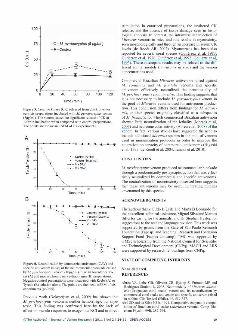
Creatine kinase (CK) releaseIncubation with M. pyrrhocryptus venom (5μg/ml) for up to 120min did not significantly alter the release of CK by BC preparations when comparing with the corresponding controls (Figure 5).
Light microscopyHistological analysis of control BC and PND preparations showed normal muscle morphology with little fiber damage (1.2±0.5% in BC and 1.1±0.7% in PND). Incubation with the venom (50μg/ml) did not significantly alter this basal damage (4.3±1.5% and 3.5±0.8% for BC and PND prepara-tions, respectively; n=5 each in all cases).
Neutralization by commercial Micrurus antivenom and specific antivenomPre-incubation (30min at 37°C) of M. pyrrhocryptus venom (10μg/ml) with commercial Micrurus antivenom in the pro-portion recommended by the manufacturer, or with specific
Figure 2. Comparison of the responses of mouse phrenic nerve-diaphragm preparations to indirect tetanic stimulation (T1, T
2 and
T3; 70Hz, 0.2ms) in the presence of (A) α-bungarotoxin and (B) M. pyrrhocryptus venom (V). Note that the tetanic response after
incubation with M. pyrrhocryptus venom was similar to that seen after exposure to α-bungarotoxin (n=6 each).

©The Author(s) | Journal of Venom Research | 2011 | Vol 2 | 24-31 | OPEN ACCESS 28
antivenom at a venom:antivenom ratio of 1.5mg of venom to 1.0ml of antivenom, totally abolished the venom-induced neuromuscular blockade in both preparations (Figure 6).
DISCUSSION
M. pyrrhocryptus venom caused irreversible, time- and concentration-dependent neuromuscular blockade of mus-cle twitches in BC and PND preparations, with the former preparations being more sensitive than the latter. The greater sensitivity of BC preparations was probably related to dif-ferences in the innervation of these two preparations, with avian muscle having both focally- and multiply-innervated fibers that can respond to electrical stimulation or exogenous nicotinic agonists (Vital Brazil, 1980; Hodgson and Wickra-maratna, 2002). These findings agree with reports for other Micrurus venoms, such as M. altirostris (Abreu et al, 2008), M. dumerilii carinicauda (Serafim et al, 2002), M. frontalis (Vital Brazil et al, 1976; Vital Brazil et al, 1977; Vital Brazil and Vieira, 1996), M. lemniscatus carvalhoi (Cecchini et al, 2005), M. nigrocinctus (Goularte et al, 1995) and M. spixii (Vital Brazil et al, 1995).
The venom inhibited contractures to exogenous ACh and CCh, indicating a predominantly postsynaptic action through the blockade of cholinergic nicotinic receptors,
as also suggested for other Micrurus venoms (Goularte et al, 1995; Serafim et al, 2002, Abreu et al, 2008). The time-dependent blockade of the responses to exogenous ACh shown in Figure 3 can only be properly understood when compared with Figure 1A, which shows venom-induced neuromuscular blockade in chick BC preparations. In Figure 3, a venom concentration of 0.5μg/ml produced complete blockade of the responses to exogenous ACh within 30min. In contrast, within a similar time frame of 30min, there was <10% blockade in indirectly stimulated preparations incubated with the double venom concentra-tion (1μg/ ml). These findings agree with the well-known existence of two populations of nicotinic receptors in BC preparations (Chang et al, 1977; Hodgson and Wickrama-ratna, 2002), i.e., one population that is extrajunctional and responds well to exogenous ACh but is rapidly blocked by venom neurotoxins, and another located in the motor end-plate that responds to nerve stimulation and is less suscep-tible to rapid blockade by neurotoxins (possibly because of difficulties related to toxin diffusion into the synaptic cleft). They also agree with studies for other neurotoxins that dis-criminate between these two populations (Chang et al, 1973; Chang and Su, 1975). The ability of toxins to distinguish between these two receptor populations could be exploited to provide a simple, sensitive assay for screening neurotox-ins and nicotinic cholinergic agonists.
The postsynaptic action of M. pyrrhocryptus venom was also indicated by the absence of fade in the tetanic response to indi-rect stimulation at 70Hz. This tetanic pattern was more simi-lar to that produced by α-bungarotoxin (a non- depolarizing toxin) than by succinylcholine (a depolarizing agent) (Gal-lacci and Oliveira, 1994; Serra and Oliveira, 2006).
Micrurus venoms are rich in PLA2 (Aird and da Silva,
1991; da Silva and Aird, 2001; Cecchini et al, 2005; Tanaka et al, 2010) that may contribute to the biological activities of these venoms (Alapé-Girón et al, 1996b; Francis et al, 1997; Oliveira et al, 2008). As shown here, M. pyrrhoc-ryptus venom had low activity when compared with that of C. d. terrificus. The finding that reducing the temperature of the experiment from 37°C to 22°C (Goularte et al, 1995; Rodrigues-Simioni et al, 2004) or the substitution of Ca2+ by Sr2+ (Rodrigues-Simioni et al, 1995; Ponce-Soto et al, 2009) to attenuate PLA
2 activity did not significantly alter
the neuromuscular blockade indicated that this enzymatic activity was not a major contributor to venom-induced blockade.
Figure 3. Kinetics of the blockade of contractures to exogenous ACh in the presence of M. pyrrhocryptus venom (0.5μg/ml) in chick biventer cervicis preparations. Note the progressive decrease in the responses over time. Each bar represents the mean ±SEM of six experiments. *p<0.05 compared with the response before venom addition (0min) (n=5 for each interval).
Figure 4. Myographic recording of mouse phrenic nerve-diaphragm contractions in response to M. pyrrhocryptus venom (V, 10μg/ml) under direct (D) and indirect (I) stimulations. The preparation was previously treated with 3μM d- tubocurarine (d-Tc) to inhibit any response of presynaptic origin (W = wash, n=6).
©The Author(s) | Journal of Venom Research | 2011 | Vol 2 | 24-31 | OPEN ACCESS 29
stimulation in curarized preparations, the unaltered CK release, and the absence of tissue damage seen in histo-logical analysis. In contrast, the intramuscular injection of Micrurus venoms in mice and rats results in myotoxicity, seen morphologically and through an increase in serum CK levels (de Roodt AR, 2002). Myonecrosis has been also reported for several coral species (Gutiérrez et al, 1983; Gutiérrez et al, 1986, Gutiérrez et al, 1992; Goularte et al, 1995). These discrepant results may be related to the dif-ferent animal models (in vitro vs in vivo) and the venom concentrations used.
Commercial Brazilian Micrurus antivenom raised against M. corallinus and M. frontalis venoms and specific antivenom effectively neutralized the neurotoxicity of M. pyrrhocryptus venom in vitro. This finding suggests that it is not necessary to include M. pyrrhocryptus venom in the pool of Micrurus venoms used for antivenom produc-tion. This conclusion differs from findings for M. altiros-tris, another species originally classified as a subspecies of M. frontalis, for which commercial Brazilian antivenom showed little neutralization of the lethality (Moraes et al, 2003) and neuromuscular activity (Abreu et al, 2008) of the venom. In fact, various studies have suggested the need to include additional Micrurus species in the pool of venoms used in immunization protocols in order to improve the neutralization capacity of commercial antivenoms (Higashi et al, 1995; de Roodt et al, 2004; Tanaka et al, 2010).
CONCLUSIONS
M. pyrrhocryptus venom produced neuromuscular blockade through a predominantly postsynaptic action that was effec-tively neutralized by commercial and specific antivenoms. The neutralization of neurotoxicity observed here suggests that these antivenoms may be useful in treating humans envenomed by this species.
ACKNOWLEDGMENTS
The authors thank Gildo B Leite and Marta B Leonardo for their excellent technical assistance, Miguel Silva and Marcos Silva for caring for the animals, and Dr Stephen Hyslop for suggestions to the text and language revision. This work was supported by grants from the State of São Paulo Research Foundation (Fapesp) and Teaching, Research and Extension Support Fund (Faepex-Unicamp). TMC was supported by n MSc scholarship from the National Council for Scientific and Technological Development (CNPq). MACH and LRS were supported by research fellowships from CNPq.
STATE OF COMPETING INTERESTS
None declared.REFERENCES
Abreu VA, Leite GB, Oliveira CB, Hyslop S, Furtado MF and Rodrigues-Simioni L. 2008. Neurotoxicity of Micrurus altiros-tris (Uruguayan coral snake) venom and its neutralization by commercial coral snake antivenom and specific antiserum raised in rabbits. Clin Toxicol (Phila), 46, 519-527.
Aird SD and da Silva NJ Jr. 1991. Comparative enzymatic compo-sition of Brazilian coral snake (Micrurus) venoms. Comp Bio-chem Physiol, 99B, 287-294.
Figure 5. Creatine kinase (CK) released from chick biventer cervicis preparations incubated with M. pyrrhocryptus venom (5μg/ml). The venom caused no significant release of CK at 120min incubation when compared with control preparations. The points are the mean ±SEM of six experiments.
Figure 6. Neutralization by commercial antivenom (CAV) and specific antivenom (SAV) of the neuromuscular blockade caused by M. pyrrhocryptus venom (10μg/ml) in avian biventer cervi-cis (A) and mouse phrenic nerve-diaphragm (B) preparations. Negative control preparations were incubated with Krebs (A) or Tyrode (B) solution alone. The points are the mean ±SEM of six experiments (p<0.05).
Previous work (Dokmetjian et al, 2009) has shown that M. pyrrhocryptus venom is neither hemorrhagic nor myo-toxic. This finding was confirmed here by the lack of effect on muscle responses to exogenous KCl and to direct

©The Author(s) | Journal of Venom Research | 2011 | Vol 2 | 24-31 | OPEN ACCESS 30
Alape-Girón A, Miranda-Arrieta K, Cortes-Bratti X, Stiles BG and Gutiérrez JM. 1997. A comparison of in vitro methods for assess-ing the potency of therapeutic antisera against the venom of the coral snake Micrurus nigrocinctus. Toxicon, 35, 573-581.
Alapé-Girón A, Stiles BG and Gutiérrez JM. 1996a. Antibody-mediated neutralization and binding-reversal studies on α-neurotoxins from Micrurus nigrocinctus nigrocinctus (coral snake) venom. Toxicon, 34, 369-380.
Alapé-Girón A, Stiles B, Schmidt J, Girón-Cortes M, Theles-tam M, Hörnvall H and Bergman T. 1996b. Characterization of multiple nicotinic acetylcholine receptor-binding proteins and phospholipases A
2 from the venom of the coral snake Micrurus
nigrocinctus nigrocinctus. FEBS Lett, 380, 29-32.Barfaraz A and Harvey AL. 1994. The use of the chick biventer
cervicis preparation to assess the protective activity of six inter-national reference antivenoms on the neuromuscular effects of snake venoms in vitro. Toxicon, 32, 267-272.
Bucaretchi F, Hyslop S, Vieira RJ, Toledo AS, Madureira PR and de Capitani EM. 2006. Bites by coral snakes (Micrurus spp.) in Campinas, State of São Paulo, southeastern Brazil. Rev Inst Med Trop São Paulo, 48, 141-145.
Bülbring E. 1946. Observations on the isolated phrenic-nerve dia-phragm preparation of the rat. Br J Pharmacol, 1, 38-56.
Cecchini AL, Marcussi S, Silveira LB et al. 2005. Biological and enzymatic activities of Micrurus sp. (coral) snake venoms. Comp Biochem Physiol Part A, 140, 125-134.
Chang CC, Chen TF and Chuang ST. 1973. N,O-di and N,N,O-tri[3H]acetyl α-bungarotoxins as specific labeling agents of cholinergic receptors. Br J Pharmacol, 47, 147-160.
Chang CC and Su MJ. 1975. Further evidence that extrinsic ace-tylcholine acts preferentially on extrajunctional receptors in the chick biventer cervicis muscles. Eur J Pharmacol, 33, 337-344.
Chang CC, Jai Su M and Hsien Tung L. 1977. Appearance of new acetylcholine receptors on the baby chick biventer cervi-cis and denervated rat diaphragm muscles after blockade with α-bungarotoxin. J Physiol, 268, 449-465.
Coelho LK, Silva E, Espositto C and Zannin M. 1992. Clinical features and treatment of Elapidae bites: report of three cases. Human Exp Toxicol, 11, 135-137.
da Silva NJ Jr and Aird SD. 2001. Prey specificity, comparative lethality and compositional differences of coral snake venoms. Comp Biochem Physiol C Toxicol Pharmacol, 128, 425-456.
da Silva NJ Jr and Sites JW. 2001. Phylogeny of South American triad coral snakes (Elapidae: Micrurus) based on molecular char-acters. Herpetologica, 57, 1-22.
da Silva NJ Jr and Bucaretchi F. 2003. Mecanismo de ação do veneno elapídico e aspectos clínicos dos acidentes. In: Cardoso JLC, França FOS, Wen FH, Málaque CMS, Haddad Jr V (Eds.), Animais Peçonhentos no Brasil. Biologia, Clínica e Terapêutica dos Acidentes, São Paulo, Sarvier/FAPESP, pp. 99-107.
Dal Belo CA, Leite GB, Toyama MH et al. 2005. Pharmacological and structural charactrerization of a novel phospholipase A
2 from
Micrurus dumerilii carinicauda venom. Toxicon, 46, 736-750.de Roodt AR, Dolab JA, Galarce PP et al. 1998. A study on the
venom yield of snake species from Argentina. Toxicon 36, 1949-1957.
de Roodt AR. Estudio Inmunobiológico del Veneno de Serpientes de Importancia Sanitaria de la Argentina. PhD Thesis. Facultad de Farmacia y Bioquímica, Universidad de Buenos Aires, Argen-tina
de Roodt AR, Paniagua-Solís JF, Dolab JA et al. 2004. Effective-ness of two common antivenoms for North, Central and South American Micrurus envenomations. J Toxicol Clin Toxicol, 42, 171-178.
Dokmetjian JC, Del Canto S, Vinzón S and de Jiménez Bonino MB. 2009. Biochemical characterization of the Micrurus pyr-rhocryptus venom. Toxicon, 53, 375-382.
Francis BR, da Silva NJ Jr, Seebart C, Casais e Silva LL, Schmidt JJ and Kaiser II. 1997. Toxins isolated from the venom of the Brazilian coral snake (Micrurus frontalis frontalis) include hem-orrhagic type phopholipases A
2 and postsynaptic neurotoxins.
Toxicon, 35, 1193-1203.
Gallacci M and Oliveira AC. 1994. Pre- and postsynaptic mecha-nisms involved in fade induced by pancuronium in the isolated rat muscle. Pharmacology, 49, 265-270.
Ginsborg BL and Warriner JN. 1960. The isolated chick biventer cervicis nerve muscle preparation. Br J Pharmacol, 15, 410-411.
Goularte FC, Cruz-Höfling MA, Cogo JC, Gutiérrez JM and Rodrigues-Simioni L. 1995. The ability of specific antivenom and low temperature to inhibit the myotoxicity and neuromus-cular block induced by Micrurus nigrocinctus venom. Toxicon, 33, 679-689.
Gutiérrez JM, Arroyo O, Chaves F, Lomonte B and Cerdas L. 1986. Pathogenesis of myonecrosis induced by coral snake (Micrurus nigrocinctus) venom in mice. Br J Exp Pathol, 67, 1-12.
Gutiérrez JM, Lomonte B, Portilla E, Cerdas L and Rojas E. 1983. Local effects induced by coral snake venoms: evidence of myonecrosis after experimental inoculations of venoms from five species. Toxicon, 21, 777-783.
Gutiérrez JM, Rojas G, da Silva NJ Jr and Nuñez J. 1992. Experi-mental myonecrosis induced by the venoms of South American Micrurus (coral snakes). Toxicon, 30, 1299-1302.
Higashi H, Guidolin R, Caricati CP et al. 1995. Antigenic cross-reactivity among components of Brazilian Elapidae snake ven-oms. Braz J Med Biol Res, 28, 767-771.
Hodgson WC and Wickramaratna JC. 2002. In vitro neuromuscu-lar activity of snake venoms. Clin Exp Pharmacol Physiol, 29, 807-814.
Hoge AR and Lancini AR. 1959. Note on Micrurus surinamensis nattereri Schmidt and Micrurus pyrrhocryptus Cope. Mem Inst Butantan, 29, 9-13.
Manock SR, Suarez G, Graham D, Avila-Aguero ML and War-rell DA. 2008. Neurotoxic envenoming by South American coral snake (Micrurus lemniscatus helleri): case report from Eastern Ecuador and review. Trans R Soc Trop Med Hyg, 102, 1127-1132.
Ministerio de Salud. 2007. Guía de Prevención, Diagnóstico, Tratamiento y Vigilancia Epidemiológica de los Envenenamien-tos Ofídicos. Ministerio de Salud, Buenos Aires, 48 pp.
Moraes FV, Sousa-e-Silva MC, Barbaro KC, Leitão MA and Fur-tado MF. 2003. Biological and immunochemical characterization of Micrurus altirostris venom and serum neutralization of its toxic activities. Toxicon, 41, 71-79.
Oliveira DA, Harasawa C, Seibert CS et al. 2008. Phospholipases A
2 isolated from Micrurus lemniscatus coral snake venom:
behavioral electroencephalographic, and neuropathological aspects. Brain Res Bull, 75, 629-639.
Oliveira L, Almeida-Santos SM, Ribeiro da Costa AC, Roveri Scar-tozzoni R, Germano VJ and Salomão MG. 2005. Manutenção de serpentes em cativeiro no Instituto Butantan: I-A longevidade do gênero Micrurus. Pap Avulsas Inst Pau Brasil, 8-9, 55-61.
Ponce-Soto LA, Barros JC, Hernandez S et al. 2009. Neuromuscu-lar activity of BaTX, a presynaptic basic PLA
2 isolated Bothrops
alternatus snake venom. Comp Biochem Physiol C Toxicol Phar-macol, 150, 291-297.
Rodrigues-Simioni L, Zamunér SR, Cogo JC et al. 2004. Phar-macological evidence for a presynaptic action of venoms from Bothrops insularis (jararaca ilhoa) and Bothrops neuwiedi (jarar-aca pintada). Toxicon, 43, 633-638.
Roze JA. 1996. Coral Snakes of the Americas. Biology, Identifica-tion and Venoms. Struik Publishing Co, Malabar FL.
Scrocchi GJ. 1990. El género Micrurus (Serpientes: Elapidae) en la República Argentina. Boll Musl Reg Sci Nat Torino, 8, 343-368.
Serafim FG, Reali M, Cruz-Höfling MA and Fontana MD. 2002. Action of Micrurus dumerilli carinicauda coral snake venom on the mammalian neuromuscular junction. Toxicon, 40, 167-174.
Serapicos EO and Merusse JLB. 2002. Variação de peso e sobre-vida de Micrurus corallinus sob diferentes condições de alimen-tação em biotério (serpentes Elapidae). Iheringia Ser Zool, 92, 105-109.
Serra CSM and Oliveira AC. 2006. Cisatracurium: myographical and electrophysiological studies in the isolated rat muscle. Fund Clin Pharmacol, 20, 291-298.

©The Author(s) | Journal of Venom Research | 2011 | Vol 2 | 24-31 | OPEN ACCESS 31
Tanaka GD, Furtado MF, Portaro FC, SantAnna OA and Tambourgi DV. 2010. Diversity of Micrurus snake spe-cies related to their venom toxic effects and the prospec-tive of antivenom neutralization. PLoS Negl Trop Dis, 4, e622.
Vital Brazil O. 1980. Venenos ofídicos neurotóxicos. Rev Ass Méd Brasil, 26, 212-218.
Vital Brazil O, Fontana MD and Pelllegrini AF. 1976/77. Physi-opathologie et thérapeutique de l’envenomation expérimentale causée para le venin de Micrurus frontalis. Mem Inst Butantan, 40/41, 221-240.
Vital Brazil O, Fontana MD, Heluany NF and Laure CJ. 1995. Mode of action of the coral snake Micrurus spixii venom at the neuromuscular junction. J Nat Toxins, 4, 19-33.
Vital Brazil O and Vieira RJ. 1996. Neostigmine in the treatment of snake accidents caused by Micrurus frontalis: report of two cases. Rev Inst Med Trop, 38, 61-67.
Warrell DA. 2004. Snakebites in Central and South America: epi-demiology, clinical features and clinical management. In: Camp-bell JA, Lamar WW (Eds.), Venomous Reptiles of the Western Hemisphere, Vol. 2. Comstock Publishing Associates/Cornell University Press, Ithaca, pp. 709-761.