The Neural Correlates of Persuasion: A Common Network across Cultures and Media
24
The Neural Basis for Spatial Relations Prin X. Amorapanth, Page Widick, and Anjan Chatterjee The University of Pennsylvania Abstract Studies in semantics traditionally focus on knowledge of objects. By contrast, less is known about how objects relate to each other. In an fMRI study, we tested the hypothesis that the neural processing of categorical spatial relations between objects is distinct from the processing of the identity of objects. Attending to the categorical spatial relations compared with attending to the identity of objects resulted in greater activity in superior and inferior parietal cortices (especially on the left) and posterior middle frontal cortices bilaterally. In an accompanying lesion study, we tested the hypothesis that comparable areas would be necessary to represent categorical spatial relations and that the hemispheres differ in their biases to process categorical or coordinate spatial relations. Voxel- based lesion symptom mapping results were consistent with the fMRI observations. Damage to a network comprising left inferior frontal, supramarginal, and angular gyri resulted in behavioral impairment on categorical spatial judgments. Homologous right brain damage also produced such deficits, albeit less severely. The reverse pattern was observed for coordinate spatial processing. Right brain damage to the middle temporal gyrus produced more severe deficits than left hemisphere damage. Additional analyses suggested that some areas process both kinds of spatial relations conjointly and others distinctly. The left angular and inferior frontal gyrus processes coordinate spatial information over and above the categorical processing. The anterior superior temporal gyrus appears to process categorical spatial information uniquely. No areas within the right hemisphere processed categorical spatial information uniquely. Taken together, these findings suggest that the functional neuroanatomy of categorical and coordinate processing is more nuanced than implied by a simple hemispheric dichotomy. Introduction Cognitive neuroscience investigations of semantics traditionally focus on knowledge of objects (Borgo & Shallice, 2001; Moore & Price, 1999; Tranel & Damasio, 1999; Caramazza & Shelton, 1998; Binder et al., 1997; Gonnerman, Andersen, Devlin, Kempler, & Seidenberg, 1997; Tranel, Logan, Frank, & Damasio, 1997; Vandenberghe, Price, Wise, Josephs, & Frackowiak, 1996; Capitani, Laiacona, Barbarotto, & Trivelli, 1994; Damasio, Damasio, Tranel, & Brandt, 1990). Although these investigations have certainly been fruitful, confining semantics to the study of objects leaves us with an impoverished understanding of the world. Until recently, relatively little attention was paid to our knowledge of how objects act in the world and how they are related to each other. Investigating the neural basis of knowledge of actions, events, and relations between objects would enrich our understanding of the human semantic system. In this report, we focused on the neural basis for a specific kind of relational knowledge, that is, spatial relations. Spatial relations between objects can be organized in different ways. These relations can be organized in the form of continuous metrics (distance, orientation, etc.) or as Reprint requests should be sent to Anjan Chatterjee, Department of Neurology, The Hospital of The University of Pennsylvania, 3 West Gates, 3400 Spruce Street, Philadelphia, PA 19104, or via [email protected]. NIH Public Access Author Manuscript J Cogn Neurosci. Author manuscript; available in PMC 2010 November 1. Published in final edited form as: J Cogn Neurosci. 2010 August ; 22(8): 1739–1753. doi:10.1162/jocn.2009.21322. NIH-PA Author Manuscript NIH-PA Author Manuscript NIH-PA Author Manuscript
-
Upload
independent -
Category
Documents
-
view
2 -
download
0
Transcript of The Neural Correlates of Persuasion: A Common Network across Cultures and Media

The Neural Basis for Spatial Relations
Prin X. Amorapanth, Page Widick, and Anjan ChatterjeeThe University of Pennsylvania
AbstractStudies in semantics traditionally focus on knowledge of objects. By contrast, less is known abouthow objects relate to each other. In an fMRI study, we tested the hypothesis that the neural processingof categorical spatial relations between objects is distinct from the processing of the identity ofobjects. Attending to the categorical spatial relations compared with attending to the identity ofobjects resulted in greater activity in superior and inferior parietal cortices (especially on the left)and posterior middle frontal cortices bilaterally. In an accompanying lesion study, we tested thehypothesis that comparable areas would be necessary to represent categorical spatial relations andthat the hemispheres differ in their biases to process categorical or coordinate spatial relations. Voxel-based lesion symptom mapping results were consistent with the fMRI observations. Damage to anetwork comprising left inferior frontal, supramarginal, and angular gyri resulted in behavioralimpairment on categorical spatial judgments. Homologous right brain damage also produced suchdeficits, albeit less severely. The reverse pattern was observed for coordinate spatial processing.Right brain damage to the middle temporal gyrus produced more severe deficits than left hemispheredamage. Additional analyses suggested that some areas process both kinds of spatial relationsconjointly and others distinctly. The left angular and inferior frontal gyrus processes coordinatespatial information over and above the categorical processing. The anterior superior temporal gyrusappears to process categorical spatial information uniquely. No areas within the right hemisphereprocessed categorical spatial information uniquely. Taken together, these findings suggest that thefunctional neuroanatomy of categorical and coordinate processing is more nuanced than implied bya simple hemispheric dichotomy.
IntroductionCognitive neuroscience investigations of semantics traditionally focus on knowledge of objects(Borgo & Shallice, 2001; Moore & Price, 1999; Tranel & Damasio, 1999; Caramazza &Shelton, 1998; Binder et al., 1997; Gonnerman, Andersen, Devlin, Kempler, & Seidenberg,1997; Tranel, Logan, Frank, & Damasio, 1997; Vandenberghe, Price, Wise, Josephs, &Frackowiak, 1996; Capitani, Laiacona, Barbarotto, & Trivelli, 1994; Damasio, Damasio,Tranel, & Brandt, 1990). Although these investigations have certainly been fruitful, confiningsemantics to the study of objects leaves us with an impoverished understanding of the world.Until recently, relatively little attention was paid to our knowledge of how objects act in theworld and how they are related to each other. Investigating the neural basis of knowledge ofactions, events, and relations between objects would enrich our understanding of the humansemantic system.
In this report, we focused on the neural basis for a specific kind of relational knowledge, thatis, spatial relations. Spatial relations between objects can be organized in different ways. Theserelations can be organized in the form of continuous metrics (distance, orientation, etc.) or as
Reprint requests should be sent to Anjan Chatterjee, Department of Neurology, The Hospital of The University of Pennsylvania, 3 WestGates, 3400 Spruce Street, Philadelphia, PA 19104, or via [email protected].
NIH Public AccessAuthor ManuscriptJ Cogn Neurosci. Author manuscript; available in PMC 2010 November 1.
Published in final edited form as:J Cogn Neurosci. 2010 August ; 22(8): 1739–1753. doi:10.1162/jocn.2009.21322.
NIH
-PA Author Manuscript
NIH
-PA Author Manuscript
NIH
-PA Author Manuscript

a discrete set of relations (such as those that can be labeled verbally by prepositions in English).For example, a pen might be 5 or 10 cm to the right of a notebook. These would representdifferent coordinate relations but not different categorical relations. The pen would have to beon top of the notebook to represent a different categorical relationship. Slobin (1996, 2000)proposed that thinking for speaking might be different than thinking for other reasons. Alongthese lines, thinking about spatial relationships for speaking might be different than thinkingabout spatial relations for other reasons. Thus, coordinate (continuous metric) representationsare critical to guide movements such as reaching or navigation but may be less relevant tolanguage. They can be described by a large and flexible set of open class terms, such as distancein centimeters, inches, or miles. By contrast, categorical representations form a more coarselyorganized set of spatial relations that are encoded in a closed class set of terms such as “on”or “in.”
The neural instantiation of spatial relations might be expected to follow naturally from the“what” versus “where” distinction (Ungerleider & Mishkin, 1982). On this central tenet ofvisual neuroscience from monkey studies, visual processing is divided into dorsal oroccipitoparietal and ventral or occipito-temporal streams. The dorsal visual stream specializesin spatial processing, and the ventral stream specializes in object processing (Haxby et al.,1991). Although the streams undoubtedly interact at multiple levels (Schiller, 1996; Ferrera,Rudolph, & Maunsell, 1994), this processing division of labor by the nervous system has beencorroborated in humans (Chatterjee, 2003; Martin, Ungerleider, & Haxby, 2000; Aguirre &D'Esposito, 1997; Farah, 1990). Occipito-temporal lesions are associated with various visualagnosias, in which individuals have trouble recognizing objects or faces, and posterior parietallesions are associated with spatial deficits such as hemispatial neglect or simultanag-nosia.Imaging studies also confirm this general distinction. Thus, the fusiform gyrus is associatedwith processing faces (Kanwisher, McDermott, & Chun, 1997), the para-hippocampal gyruswith processing places and buildings (Epstein & Kanwisher, 1998; Aguirre & D'Esposito,1997), and the lateral occipital complex with processing objects in general (Kanwisher, Woods,Iacoboni, & Mazziotta, 1997; Malach et al., 1995). Although the posterior parietal cortex isimplicated in shifts of spatial attention (Corbetta, Shulman, Miezin, & Peterson, 1995),relatively little direct functional imaging evidence supports the role of the parietal cortex inprocessing spatial relations.
Spatial relations relevant to language are likely to be categorical. Kosslyn et al. (1989) initiallysuggested the distinction between categorical and coordinate spatial relations. They proposeda left hemisphere advantage for categorical visual processing and a right hemisphere advantagefor coordinate visual processing (Kosslyn & Ochsner, 1994; Kosslyn et al., 1989). However,the empirical bases for these neural claims were limited, derived primarily from visualhemifield stimuli presentations and computer simulations. The specific results obtained byKosslyn et al. were subsequently challenged as an artifact of a bias in spatial resolution ofsensory processing (Sergent, 1991) or as an artifact of task difficulty (Slotnick, Moo, Tesoro,& Hart, 2001). Furthermore, the approach of using lateralized stimuli presentation to inferlateralized brain function might itself be suspect (Efron, 1990). In a subsequent PET study,Kosslyn, Thompson, Gitelman, and Alpert (1998) failed to find consistent left hemisphereactivation for categorical spatial judgments. However, in an fMRI study, Baciu et al. (1999)found that left angular gyrus (AG), within the inferior parietal lobe, displayed greater activitythan the right AG when subjects judged whether a dot was located above or below a bar.
Despite the limited evidence for the neural basis for categorical and coordinate spatialprocessing, this distinction seems plausible (for reviews, see Postma & Laeng, 2006; Jager &Postma, 2003). Damage to right parietal cortex appears to impair performance on tasks thatrely on precise metrics (Laeng, 1994; Hannay, 1976; Warrington & Taylor, 1973). Leftposterior parietal lesions are associated with left/right confusion, a hallmark symptom of
Amorapanth et al. Page 2
J Cogn Neurosci. Author manuscript; available in PMC 2010 November 1.
NIH
-PA Author Manuscript
NIH
-PA Author Manuscript
NIH
-PA Author Manuscript

Gerstmann syndrome (Mayer et al., 1999). To our knowledge, patients with Gerstmannsyndrome per se have not been tested more comprehensively on categorical spatial judgments.Left–right judgments may be a class of spatial judgment, as evidenced by left hemispatialneglect (Chatterjee, 2003) or mirror reading and writing (Gottfried, Sancar, & Chatterjee,2003), that implicates a special role for the horizontal axis in processing space. Whether left–right processing generalizes to categorical processing is not known.
Laeng (1994, 2006) has reported data consistent with the hemispheric specializationhypothesis. Patients with focal brain damage were shown drawings of two objects. After a shortdelay, they were asked to identify the drawing when it was paired with a drawing of the sameobjects transformed in their categorical or coordinate spatial relations. He found that patientswith right hemisphere damage (RHD) were more likely to make coordinate relation errors andpatients with left hemisphere damage (LHD) were more likely to make categorical memoryerrors. The anatomic analyses of patients in this study were rudimentary. Counterintuitively,behavior differences were found when patients had parietal damage only and not when theyalso had temporal or frontal damage in addition to the parietal damage. More relevant to ourstudy is a report by Tranel and Kemmerer (2004). They examined a large group of patients ona variety of tasks probing knowledge of locative relations. In an “odd man out” task, participantsviewed groups of three pictures, each with two objects. They were asked to point to the picturethat depicted a different categorical relationship than the other two. They found that damageunderlying the white matter of the left parietal and frontal opercula was associated with deficitsin matching categorical spatial relations.
The studies reviewed served as the backdrop to our investigations of the neural basis of spatialrelations using both functional imaging in young healthy participants and behavioral studiesin participants with focal brain lesions. Both approaches, now central methods in cognitiveneuroscience, have complementary strengths and weaknesses (Chatterjee, 2005; Fellows et al.,2005; Rorden & Karnath, 2004). Imaging studies in young normal subjects offer insights intothe processing of the unaltered brain but remain fundamentally a correlational method. Lesionstudies are inferentially stronger but are limited by uncertainties of neuronal reorganization,not to mention variables such as age, education, medications, and comor-bid conditions in anelderly population. In principle, converging evidence from both methods would provide strongsupport for brain–behavior relations.
Our first experiment used fMRI to investigate the neural systems activated by the identificationof categorical spatial relations, as compared with the identification of objects being located.By categorical relations, we mean between-object relations rather than within-object relations(such as the handle at the top of the briefcase). Our experiment relies on subjects makingdifferent judgments on the same set of stimuli. Thus, different neural activation patterns couldnot arise from differences in the perceptual properties of the stimuli themselves. The secondexperiment used participants with left or right focal brain lesions to assess their abilities tomake categorical and coordinate spatial judgments. We used a variant of voxel-based lesionsymptom mapping (VLSM) (Kimberg, Coslett, & Schwartz, 2007; Bates et al., 2003) to seekevidence consistent with our fMRI results. VLSM is a distinct advance over lesion overlapmethods because patients' behavior is considered as a continuous variable rather than beingjudged as normal or abnormal. Our general hypothesis is that the neural mediation of spatialrelations between objects is distinct from that of identification of objects. More specifically,we expected the posterior parietal cortex to be involved in computing categorical andcoordinate spatial relations (Chatterjee, 2008). Furthermore, on the grounds that categoricalspatial relations are linked more closely to language, we expected behavioral deficits incategorical spatial judgments to be associated with damage to left peri-sylvian regions anddeficits in coordinate spatial judgments to be associated with RHD.
Amorapanth et al. Page 3
J Cogn Neurosci. Author manuscript; available in PMC 2010 November 1.
NIH
-PA Author Manuscript
NIH
-PA Author Manuscript
NIH
-PA Author Manuscript

Functional NeuroimagingMethods
Participants—Sixteen right-handed participants (10 men and 6 women; mean age = 22 years;range = 18–25 years) were recruited from the University of Pennsylvania. Four subjects wereexcluded from the analysis because of poor behavioral performance, raising doubts about theirengagement in the task. All participants had normal or corrected-to-normal vision and spokeEnglish as their primary language. None had a history of neurological or psychiatric symptoms.All gave informed consent in accordance with the procedures of the institutional review boardof the University of Pennsylvania and the Declaration of Helsinki.
Stimuli—Digital pictures of two objects (a mouse, a cassette, a toy monkey, a stapler, a mug,or a spoon) in one of eight spatial relationships (that could be described by above, below, in,on, left, right, in front of, or behind) on a plain background were used. Red arrows were addedto denote the figure object to be located.
Behavioral Tasks—In an alternating blocked design, participants performed a one-backmatching task. They were instructed to attend to either the spatial relationship between theobjects or the identities of the objects. Participants pressed a button press to indicate whetherthe current image matched the previous image on the basis of either spatial relationship orobject identity. In the spatial condition, match trials were those in which the spatial relationwas the same, regardless of the objects involved (which alternated from trial to trial; Figure1A). In the object condition, match trials were those in which both objects were the same asin the preceding pair and nonmatch trials were those in which one of the objects was different(Figure 1B). Stimuli for both conditions depicted the same set of objects and spatial relations.The behavioral responses (accuracy and RT) across both conditions were not significantlydifferent in off-line pilot testing. Left–right button responses were balanced within and acrossblocks. For both conditions, the condition not being attended to alternated from trial to trial toavoid the possibility that participants could perform a simple perceptual match on each trial asa tenable strategy. For example, in the object condition, participants were presented with aseries of object pairs that might alternate between depicting the spatial relationships of “above”and “below” while the objects themselves varied in such a manner as to produce an equalnumber of match and nonmatch trials. Baseline perceptual/motor blocks were interspersedbetween alternating task blocks and consisted of presentations of central fixation points, towhich subjects pressed either button.
MRI Acquisition—Data were acquired on a 3.0-T Siemens Trio scanner using a USAInstruments four-channel head coil. BOLD-sensitive, T2*-weighted functional images wereacquired in 3-mm isotropic voxels using a gradient-echo, echo-planar pulse sequence(repetition time = 3000 msec, effective echo time = 30 msec). Forty 3-mm slices were acquiredduring each repetition, with each slice containing a 64 × 64 matrix within a 192 × 192-mmfield of view. Head motion was minimized by using foam padding, and the scanner performedboth prospective (three-dimensional prospective acquisition correction) and retrospectivemotion correction on-line. One hundred ninety-nine images were collected from each scan.The first six images of each functional scan were discarded to allow for steady-statemagnetization to be achieved. High-resolution, T1-weighted anatomical images were alsoacquired for each subject by using an MPRAGE pulse sequence (repetition time = 1620 msec,echo time = 3 msec, inversion time = 950 msec). One hundred sixty 1-mm slices were acquired,with each slice containing a 256 × 256 matrix within a 250 × 250-mm field of view.
Stimulus Presentation—A computer running E-Prime(www.pstnet.com/products/e-prime/) outside the scanner room controlled stimulus timing and
Amorapanth et al. Page 4
J Cogn Neurosci. Author manuscript; available in PMC 2010 November 1.
NIH
-PA Author Manuscript
NIH
-PA Author Manuscript
NIH
-PA Author Manuscript

response recording. Stimuli were projected onto a screen at the back of the scanner bore andviewed by subjects through a mirror mounted on the head coil. Subject responses weretransmitted by a custom-designed fiber-optic response pad. Each subject participated in twofunctional scans, which were each divided into 12 blocks, 6 for each experimental condition,and 12 blocks of the baseline condition. There were 13 stimuli per block, with stimulusdurations of 1200 msec, ISIs of 415 msec, and an interval of 3000 msec between blocks.
Preprocessing—Data processing was performed off-line using software developed at theUniversity of Pennsylvania (www.voxbo.org). After reconstructing images from the raw data,data were sinc interpolated in time to correct for staggered slice acquisition, realigned to thefirst image acquired for each subjects using a six-parameter motion-correction algorithm, andthresholded to excluded extraparenchymal voxels from subsequent analyses. Within eachsubject, a voxelwise analysis was performed using a modified general linear model thatincluded covariates modeling task conditions as well as sine and cosine regressors forfrequencies below those of the task and for frequencies in the elevated range of the noisespectrum. Task covariates were boxcar waveforms convolved with an estimate of the BOLDhemodynamic transfer function, which was empirically derived from motor cortex in a largegroup of subjects (Aguirre, Zarahn, & D'Esposito, 1998). Data were also smoothed in timewith the hemodynamic transfer function to control the false-positive rate (Aguirre, Zarahn, &D'Esposito, 1997; Zarahn, Aguirre, & D'Esposito, 1997). Group activation maps wereconstructed by a random-effects analysis of beta-values for each covariate at each voxel.
Definition of ROIs—Anatomical ROIs were derived from the automated anatomical labelingmap of the Montreal Neurological Institute (MNI) single-subject brain (Tzourio-Mazoyer etal., 2002). After translation to the appropriate file format, regions were resampled to the correctdimensions and manually adjusted to compensate for resampling errors. Anatomical ROIs were(a) superior parietal lobules, (b) inferior parietal lobules (IPLs) encompassing AG andsupramarginal gyrus), (c) lateral occipital cortex, (d) middle temporal cortex, (e) inferiortemporal cortex, (f) fusiform gyri (both occipital and temporal portions), and (g)parahippocampal gyri. These ROIs were chosen on the basis of findings in the literaturereviewed earlier, suggesting possible roles for these areas in the processing of the cognitiveaspects of both of these tasks.
Hypotheses about activity differences were tested using the following approach. Voxels wereinitially identified in which activity was significant for the main effect of spatial relations andobject matching compared with the baseline condition (p < .05, Bonferroni-corrected formultiple comparisons within an ROI). As described previously (Kable, Kan, Wilson,Thompson-Schill, & Chatterjee, 2005; Kable, Lease-Spellmeyer, & Chatterjee, 2002), thebaseline condition was not necessarily matched to the conditions of interest; rather, the goalwas to reduce the number of voxels within which the contrasts of interest would be queried.The fMRI time series was averaged for all voxels significant for the main effect within a definedROI in each subject. Next, a measure of the effect size for the orthogonal contrast betweenconditions of interest (spatial relations–object matching) was extracted from the spatiallyaveraged ROI time series in each subject. We used t values as a measure of effect size ratherthan percent signal change because the residual error term in the denominator of the t valuemost effectively corrects the effect size for scaling effects because of differences in signalintensity across scanning sessions (Postle, Zarahn, & D'Esposito, 2000). Finally, paired t testsaddressed whether this effect was consistently greater for one condition or the other in thatROI across subjects. This analysis trades spatial resolution for sensitivity by reducing the effectsize in each ROI to a single value through averaging across all active voxels within an ROI.
Amorapanth et al. Page 5
J Cogn Neurosci. Author manuscript; available in PMC 2010 November 1.
NIH
-PA Author Manuscript
NIH
-PA Author Manuscript
NIH
-PA Author Manuscript

ResultsBehavioral Results—Participants did not differ in accuracy (89% ± 3% vs. 87% ± 3%) orRTs (702 ± 32 vs. 661 ± 32 msec) for the spatial relation and object identification conditions,respectively.
Imaging Results—Within each ROI, the averaged time series across all voxels that showedactivity for the main effect of both spatial relations and object matching compared with thebaseline condition is shown in Figure 2. For each averaged time series, we calculated a measureof the difference in activity between the spatial relation and the object matching conditions andthen tested whether this difference was significantly different from zero across subjects foreach ROI (a random-effects test, see Table 1).
Across participants, there was consistently greater activation bilaterally for judgments ofspatial relations in the superior parietal lobules, left, t(11) = 3.7, p < .005, and right, t(11) =3.3, p < .01, and the IPLs, left, t(11) = 4.2, p < .005, and right, t(11) = 2.5, p < .05, and lessrobustly within the middle temporal cortices, left, t(11) = 2.4, p < .05, and right, t(11) = 2.6,p < .05. Activity was greater for judgments of object identity than spatial relation in the leftfusiform gyrus, t(11) = −2.8, p < .05 (see Table 1; Figures 3 and 4).
The only difference in hemispheric activity between ROIs was seen in the left IPL, whichdisplayed significantly greater activity than the right IPL, t(11) = 2.5, p < .05, for the spatialrelations, and in the left fusiform gyrus, which displayed significantly greater activity than theright fusiform gyrus for the object identity condition, t(11) = −2.9, p < .05.
Figure 4 displays whole-brain activation maps for the spatial relations versus object identitycontrast. Because the purpose of these figures is to display general trends in brain activity ratherthan the results of strict statistical tests, these maps are thresholded at t = 3.5. The direct-contrastmaps largely overlap with our ROIs of interest. However, bilateral posterior middle frontalgyri, not included in our original ROI analyses, also displayed greater activity in the spatialthan in the object condition.
Lesion StudyMethods
Participants—Thirty-four patients ranging from 48 to 85 years of age with chronic (at least6 months duration) unilateral hemispheric lesions were recruited from the Focal Lesion PatientDatabase (Fellows, Stark, Berg, & Chatterjee, 2008). They were evenly divided between thosewith left and those with right brain damage. By design, participants were not selected on thebasis of specific behavioral criteria, except that patients with a history of other neurologicaldisorders affecting the CNS or psychiatric disorders are excluded from the patient database.Twelve elderly control subjects ranging from 58 to 79 years of age were also tested on thesetasks.
All participants were native English speakers and right-handed. Written informed consent inaccordance with the procedures of the institutional review board of the University ofPennsylvania and the Declaration of Helsinki was obtained for each participant. A nativeEnglish speaker delivered all spoken materials. If a participant was unable to complete testingin one session, additional sessions were scheduled at least one week later.
Stimuli—Images consisted of digital color photographs (specific stimuli may be obtainedfrom the corresponding author). The objects in these images consisted of small set of relativelycommon household/office items that could function as figure and/or ground objects for thelocative relations being tested. In the images, a red arrow pointed to the figure object. Spatial
Amorapanth et al. Page 6
J Cogn Neurosci. Author manuscript; available in PMC 2010 November 1.
NIH
-PA Author Manuscript
NIH
-PA Author Manuscript
NIH
-PA Author Manuscript

relationships that could be described by in and on, which are considered topologic relations,and those that could be described by above, below, to the left of, and to the right of, which areconsidered projective relations, were used as stimuli. We recognized the polysemous natureof prepositions. With topological relationships, each exemplar for “in” depicted a containmentrelationship and for “on” a contact relationship of the figure to ground along the vertical y-axis. For the topological relationships, the verbal descriptors refer to an allocentric referenceframe (note that the stimuli themselves only used pictures; for examples, see Figure 5).
Screening Tasks—All patients were screened for unilateral neglect. Although neglectusually recovers within 3 months (Chatterjee, 1995), we wanted to ensure that severeattentional biases would not impair their ability to see or to respond to specific stimuli. Allpatients were given the gray scale test for neglect (Mattingley et al., 2004). We also wished toensure that subjects recognized the objects being pictured. All subjects identified the series ofobjects that were used in the spatial tasks. Twenty-six trials were presented in which the nameof an object was presented along with four photographic images (one image depicting the objectnamed). Participants indicated which one of the four pictures depicted the correct answer eitherby pointing or by reading the letter underneath a particular image. The location of correctresponse (top/bottom and left/right) was counterbalanced across the trials.
Categorical Spatial Matching—This task was designed to assess patients' ability to matchcategorical spatial concepts across different photographic representations. The patients werepresented with 40 trials in which a probe image containing one pair of objects in a particularspatial relationship was to be matched with one of four images containing a different pair ofobjects. Of the four choices, one depicted the correct categorical spatial relationship, and thefoils could depict within- or across-class categorical relations (Figure 5A). Twenty-four itemshad a correct topological match (12 with pictures that could be described by in and 12 thatcould be described by on). Sixteen items had a correct projective match (4 that could bedescribed by in front of, 4 behind, 4 above, 2 to the left of, and 2 to the right of). Subjectsindicated which of the four pictures depicted the correct answer either by pointing or by readingthe letter underneath the image of their choice.
Coordinate Spatial Matching—This task was designed to assess patients' abilities to matchimages on the basis of metric spatial distance. The patients were presented with 32 trials inwhich a probe image containing one pair of objects in a particular spatial relationship was tobe matched with one of four images containing the same pair of objects in the same coordinatespatial relationship (Figure 5B). Subject indicated which of the four pictures depicted thecorrect answer either by pointing or by reading the letter underneath the image of their choice.
Neuroanatomical Analysis—Lesions were segmented and coregistered using a manualprocedure with MRIcro (Rorden & Brett, 2000). A T1-weighted MNI template image was firstrotated pitchwise to correspond with the patient's scans. An experienced neurologist outlinedthe lesions on the rotated template, resulting in a map in which each voxel was labeled either0 (intact) or 1 (lesioned). Finally, the lesion maps were rotated back into a canonical MNIorientation, using nearest-neighbor interpolation to restrict the map values to 0 and 1 using anautomated feature of MRIcro. For most of the subjects, lesions were drawn on a 2 × 2 × 2-mmtemplate. For those originally drawn at higher resolution, the lesions were resampled to 2 × 2× 2 mm for the purposes of this test. All of the thresholds below correspond to an alpha criterionof .05.
Voxel-based Lesion Symptom Mapping/Analysis—We used permutation analyses toestablish VLSM (Bates et al., 2003; Nichols & Holmes, 2002). In contrast to fMRI, where thedependent value is the signal value in a given voxel and behavior the independent value, VLSM
Amorapanth et al. Page 7
J Cogn Neurosci. Author manuscript; available in PMC 2010 November 1.
NIH
-PA Author Manuscript
NIH
-PA Author Manuscript
NIH
-PA Author Manuscript

switches the roles, with lesion status comprising the independent variable and behavior thedependent variable. However, as with fMRI, the problem of multiple comparisons remains.Given the large number of comparisons across the brain, it is likely that voxels will display assignificantly associated with impairment strictly by chance. Extant methods of correcting forthis problem, such as Bonferroni correction, yield overly conservative thresholds and do notaccount for the spatial coherence of lesion data (the lesion status of one voxel is well predictedby neighboring voxels). Permutation testing provides a solution to this by correcting exactlyfor the number of independent comparisons within a volume, without making assumptionsabout the spatial structure of the data. Generally, the permutation method entails the creationof a null distribution for a given test statistic (here, a t statistic) by generating a large number(>1000) of random permutations of the independent and dependent variables (in this case,lesion status and behavioral data, respectively) and by recalculating the test statistic for eachpermutation. A maximum statistic across the brain is calculated for each permutation, andthresholds for significance are calculated from the 95th percentile of this distribution to ensurea family-wise error rate of 0.05. This yields a threshold t value that would be exceededsomewhere in the brain in only 5% of the permutations (Nichols & Holmes, 2002). When theempirically obtained pairing results in an extreme t value relative to the other permutations andexceeds this threshold, the null hypothesis is rejected. To eliminate effects produced by a singleparticipant outlier, only voxels that were damaged in at least two participants were queried.For further details of this logic, see Kimberg et al. (2007), and for a recent application, seeWu, Waller, and Chatterjee (2007).
ResultsScreening Tasks—As a group, RHD and LHD subjects displayed mild attentional biaseson the gray scales test. None of the patients had obvious neglect. All participant groupsperformed at ceiling in the object identification task, ensuring that errors on subsequent taskswere not influenced by object recognition deficits.
Categorical and Coordinate Spatial Matching: Behavioral Analyses—The elderlycontrol subjects generally performed well on both tasks, 89% accurate for categorical matchingand 93% accurate for coordinate matching. These performances showed a trend toward beingsignificantly different (Wilcoxon signed ranks test, p = .08).
With Group (LHD and RHD) as the between-subject factor and Task (categorical vs. coordinatespatial relations) as the within-subject factor, we found no significant effects of either groupor task on percent accuracy in an ANOVA. However, there was a significant interaction in theexpected direction with the LHD performing relatively better on the coordinate task than thecategorical task and the RHD performing relatively better on the categorical task than thecoordinate task, F(1, 32) = 9.59, p < .005. Behavioral data for the groups is shown in Figure6. Error analysis for the categorical relations (topological vs. projective) showed thatparticipants were most likely to make within-class errors (84.2% as compared with 45.8% ifchoosing randomly) than across-class errors (p < .001 using test of proportions).
The lack of group differences in performances of the RHD and LHD groups on the categoricalor coordinate tasks does not necessarily disconfirm the hypothesis that the left hemisphere isbiased toward categorical spatial judgments and the right to coordinate spatial judgments. Bydesign, greater behavioral variability within groups is desirable for VLSM methods to identifyspecific brain behavior correlations. This greater behavioral variability within each groupmaximizes the likelihood of finding statistically robust differences within the group andminimizes the likelihood of finding differences across groups.
Performances on the categorical and coordinate tasks correlated across groups (r = .50, p < .005). We tested the hypothesis of hemispheric differences in categorical and coordinate
Amorapanth et al. Page 8
J Cogn Neurosci. Author manuscript; available in PMC 2010 November 1.
NIH
-PA Author Manuscript
NIH
-PA Author Manuscript
NIH
-PA Author Manuscript

processing by conducting residual analyses. When performances on the categorical task wereregressed onto performances on the coordinate task, the LHD had significantly lowercategorical residuals scores than the RHD group, t(32) = 2.43, p = .02. Analogously, whenperformances on the coordinate task were regressed onto performances on the categorical task,the RHD group had significant lower coordinate residual scores than the LHD group, t(32 =2.73, p = .01.
Our fMRI results revealed greater left than right IPL activations for categorical spatial relationjudgments. To link those findings more closely to these lesion results, we sorted our patientsinto those with (n = 9 LBD and n = 9 RBD) and without (n = 8 LBD and n = 8 RBD) IPLdamage. Using residual analyses, we found the left IPL group performed worse than the rightIPL group (Mann–Whitney U test; p < .01) on categorical matching tasks when performanceon coordinate matching tasks was factored out. Similar hemispheric differences were not foundfor the patients in whom lesions spared the IPL (Mann–Whitney U test; p = .75).
Categorical and Coordinate Spatial Matching: Functional-anatomic Analyses—Representative slices from VLSM maps for both groups are shown in Figure 7. For categoricalspatial matching, empirically derived t statistic thresholds with a significance level of p < .05were 2.83 for the RHD group and 3.00 for the LHD group. In the LHD group, impairments oncategorical matching correlated with lesions to the posterior middle and inferior frontal gyri,the supra-marginal gyrus and AG, and the white matter undercutting to the anterior superiortemporal gyrus. In the RHD group, impairments on the task correlated with damage to thesuperior temporal gyrus, supramarginal gyrus, and AG. For coordinate matching, t statisticthresholds with a significance level of p < .05 were 3.51 for the RHD group and 2.48 for theLHD group. In the RHD group, impairments on coordinate matching correlated with damageto the middle temporal gyrus. In the LHD group, impairments on the coordinate matching taskcorrelated with damage to the AG and the inferior frontal gyrus.
Representative slices from VLSM maps for both groups, using residual scores, are shown inFigure 8. For categorical residual analyses, empirically derived t statistic thresholds with asignificance level of p < .05 were 2.79 for the LHD group and 5.15 for the RHD group. In theLHD group, categorical deficits were uniquely associated with damage to the white matterundercutting the anterior superior temporal gyrus. No such areas were identified in the RHDgroup. For coordinate residual analyses, empirically derived t statistic thresholds with asignificance level of p < .05 were 3.32 for the RHD group and 2.26 for the LHD group. In theRHD group, coordinate deficits were uniquely associated with damage to middle temporalgyrus. In the LHD group, coordinate deficits were uniquely associated with damage to the AGand the inferior frontal gyrus.
DiscussionWhat is the neural basis for representing spatial relations? We addressed this question usingboth functional neuroimaging and lesion methods. Despite nontrivial differences generallyinherent in these methods and particular aspects of the actual experiments conducted, we foundlargely consonant results. Taken together, our findings suggest that a distributed neural networkmediates spatial relations organized around fronto-temporo-parietal circuits, with left–righthemispheric differences. We first discuss the fMRI findings comparing categorical spatial andobject processing. We then discuss the lesion study findings for categorical processing andrelate them to the fMRI results. Then, we reviewed the neural instantiation of coordinateprocessing and highlight interactions of categorical and coordinate processing. Theseinteractions reveal that the functional-anatomic organization of spatial relations is morenuanced than generally appreciated. Finally, we placed our results in a broader context as theyrelate to spatial language.
Amorapanth et al. Page 9
J Cogn Neurosci. Author manuscript; available in PMC 2010 November 1.
NIH
-PA Author Manuscript
NIH
-PA Author Manuscript
NIH
-PA Author Manuscript

Categorical Spatial and Object Processing: Imaging EvidenceOur fMRI findings confirm the hypothesis that fronto-parietal networks are important inrepresenting categorical spatial relations in contrast to the representation of object identities.We are not aware of previous fMRI studies that have directly compared object identificationand categorical spatial relations using identical stimuli. Young healthy participants activatedthe posterior parietal lobules bilaterally as they processed categorical spatial relations morethan when they processed the identity of objects. Activity in the IPLs was greater on the leftthan on the right. The greater left hemisphere activation within the posterior inferior parietalcortex is consistent with the claim that the left hemisphere is biased to processing categoricalspatial relations (Kosslyn et al., 1989), but the bilateral activations raise questions about therelative contributions of each hemisphere. Thus, these imaging results confirm a dorsal–ventraldistinction for processing spatial relations, but not necessarily a left–right distinction forcategorical spatial processing.
Categorical and Coordinate Spatial Processing: Lesion EvidenceOur lesion study broadly confirmed and extended the results of the imaging experiment.Paralleling the imaging results, VLSM analyses revealed that damage to the left supramarginalgyrus and AG, the posterior middle and inferior frontal gyri, and the white matter undercuttingthe superior temporal gyrus was associated with deficits in categorical spatial judgments.Damage to the right superior temporal gyrus, supramarginal gyrus, and AG produced milderdeficits in categorical spatial judgments. VLSM analyses also revealed that damage to the rightmiddle temporal gyrus produced deficits in coordinate spatial processing, whereas damage tothe left AG and inferior frontal gyrus produced milder deficits in coordinate spatial processing.Thus, damage to the left AG and inferior frontal gyrus produced deficits in both coordinateand categorical processing.
In our brain-damaged participants, performances on categorical and coordinate spatial taskswere correlated. Thus, some variance in performance on each of these tasks could be accountedfor by deficits on the complementary task. To test the hypothesis that damage to some brainregions contributes uniquely to categorical or to coordinate deficits, we conducted residualanalyses. Specifically, performances on categorical tasks were regressed on coordinateperformances, and the derived residuals were subjected to analysis. Analogously, coordinateperformances were regressed on categorical performances to derive residuals then subjectedto analysis.
The LHD patients were more impaired than the RHD group on the categorical task beyond thevariance in performances accounted for by coordinate deficits. Analogously, the RHD patientswere more impaired than the LHD group on the coordinate tasks beyond variance inperformance accounted for by categorical deficits. These relative impairments cannot beexplained by difficulty in recognizing stimuli, nor can they be explained by the hypothesis thatthe left hemisphere processes relatively easy spatial relations and the right more difficult ones(Slotnick et al., 2001).
Can our lesion results be linked more directly to the fMRI data? The fMRI study revealedgreater activity in the left IPL than that in the right for categorical spatial processing than forobject identity. If these relative activations are causally related to categorical processing, thenpatients with left IPL damage should be worse than patients with right IPL damage oncategorical tasks. We tested this hypothesis by first sorting patients on the basis of whetherthey had IPL damage. Nine patients had left IPL damage and nine had right IPL damage. Usingresidual analyses, we found that patients with damaged left IPL had greater deficits thanpatients with damaged right IPL on categorical spatial tasks. The same analysis with the other
Amorapanth et al. Page 10
J Cogn Neurosci. Author manuscript; available in PMC 2010 November 1.
NIH
-PA Author Manuscript
NIH
-PA Author Manuscript
NIH
-PA Author Manuscript

patients (n = 8 each) did not reveal similar differences. Thus, we have strong convergingevidence for the importance of the left IPL on categorical spatial processing.
Our VLSM results demonstrate that the functional neuroanatomy of categorical and coordinatespatial relations is more nuanced than that suggested by the straightforward hypothesis that theleft hemisphere processes categorical spatial information and the right processes coordinatespatial information. Damage to left posterior parietal and dorsolateral prefrontal structuresproduced deficits in both categorical and coordinate processing. However, damage within theleft AG and inferior frontal gyrus produced deficits in coordinate processing (although thesewere mild deficits) not explained by categorical deficits. Similarly, damage to the white matterundercutting the left anterior superior temporal gyrus uniquely produced deficits in categoricalprocessing not accounted for by coordinate deficits. Although RHD also produced deficits inboth categorical and coordinate processing, no damaged areas were uniquely associated withcategorical deficits. By contrast, damage to the right middle temporal gyrus uniquely producedcoordinate spatial deficits.
These patterns of lesion and behavioral correlations suggest that categorical and coordinatespatial processing are quite intertwined. The right hemisphere processes coordinate spatialinformation preferentially within the posterior temporal cortex. By contrast, the left hemisphereprocesses both categorical and coordinate information conjointly within fronto-parietal circuitsas well as distinctly within the AG, inferior frontal gyrus for coordinate processing, and anteriorsuperior temporal gyrus for categorical processing.
Our categorical spatial task used stimuli that depicted both topological and projective spatialrelations. We noted that when subjects erred on this task, they were likely to choose within-class foils. That is, if the target was topological, they were more likely to choose a topologicalfoil and vice versa for projective spatial relations. Thus, despite categorical spatial deficits,some knowledge of the kind of categorical spatial relationships appears to have been retained.These observations raise the possibility that the neural underpinnings of topological andprojective spatial relations might segregate further within networks instantiating categoricalspatial relations. Future studies designed and powered to test this hypothesis will be needed.
Categorical Spatial Processing and LanguageA major motivation for anticipating that the left hemisphere is biased to processing categoricalspatial relations is its relationship to language (Chatterjee, 2008; Kemmerer, 2006).Prepositions comprise only a small set of the infinite possible spatial relations. As Landau andJackendoff (1993) and Talmy (1983) pointed out some years ago, prepositions discard muchof the geometric richness of objects in favor of relatively coarse spatial notions like“containment” or “contact.” They suggested that any aspect of space that could be expressedin language must have nonlinguistic spatial representations. Similarly, Regier (1995) usedcomputational models to argue that spatial language is grounded in spatial perception. Thus,one might infer that categorical spatial relations, which are limited in number and in whichdetails of object features are reduced to general shapes and axes, would serve as the perceptualcounterpart to locative prepositions. At an anatomic level, one would expect the processing ofthese discrete spatial representations linked to language, to be instantiated in the left hemisphereof right-handed individuals, close to or overlapping with the processing of spatial prepositions.
As an aside, we recently reported a fronto-parietal network involved in processing paths ofmovement (Wu, Morganti, & Chatterjee, 2008). This finding is relevant to the current studybecause path of motion in English is described using prepositional phrases (e.g., runningthrough the fields, jumping across the puddle). Thus, the perceptual counterparts of locativeprepositions, whether dynamic or static, may be instantiated in similar fronto-parietal networks.
Amorapanth et al. Page 11
J Cogn Neurosci. Author manuscript; available in PMC 2010 November 1.
NIH
-PA Author Manuscript
NIH
-PA Author Manuscript
NIH
-PA Author Manuscript

Few studies have examined the neural bases of locative prepositions directly. Using PETimaging, Emmorey et al. (2002) and Damasio et al. (2001) found that naming spatial locationswas associated with the left supramarginal gyrus and the inferior prefrontal cortical activations.Noordzij, Neggers, Ramsey, and Postma (2008) also found left supramarginal gyrus activityin subjects who were asked to process terms denoting left and right, but whether left–rightjudgments generalize broadly to other categorical relations is not known. Limitedelectrophysiologic data are consistent with the view that the parietal cortices instantiate the useof spatial terms (Carlson, Robert, Taylor, & Herndon, 2002). Occasional case studies of deficitswith prepositions in aphasics have been reported, but these have not been informative abouttheir neural underpinnings (Miozzo, Simon, & Postman, 2008; Chatterjee & Maher, 2000;Grodzinsky, 1988; Frederici, 1982; Schwartz, Saffran, & Marin, 1980). An important exceptionin this regard is a study by Tranel and Kemmerer (2004), which directly examined the neuralcorrelates of deficits in locative prepositions in a large group of brain-damaged individuals.They found the greatest overlap of lesions in patients with such deficits within the leftsupramarginal gyrus and subjacent white matter as well as the frontal opercula. We also foundthat damage to the posterior temporo-parietal cortex, anterior superior temporal gyrus, andinferior pFC was most closely associated with deficits in matching locative sentences topictures depicting categorical spatial relations (Wu et al., 2007). The emerging view from thesestudies is that left peri-sylvian cortices involving the supramarginal gyrus, the anterior superiortemporal gyrus, and the inferior prefrontal regions mediate locative prepositions. Areasmediating spatial categorical relations overlap or are closely aligned with these regions,consistent with the view that these regions serve as perceptual points of entry for theirlexicalizations (Chatterjee, 2008; Wu et al., 2007).
In summary, we found converging evidence from both imaging and lesion investigations ofthe neural basis for spatial relations. A bilateral network comprising the posterior parietal,posterior middle and superior temporal, and posterior middle and inferior frontal gyri mediatesspatial relations. The left hemisphere is biased toward processing categorical spatial relationsand the right toward processing coordinate spatial relations, but these distinctions are relativerather than absolute. The left hemisphere appears to mediate both categorical and coordinateprocessing, whereas we did not find evidence that the right hemisphere processes categoricalrelations independent of its coordinate processing.
AcknowledgmentsThis work was supported by NIH RO1 HD050199 and RO1 DC008779 and a subcontract under NSF SBE0541957.The authors thank Eileen Cardillo, Alexander Kranjec, and Gwenda Schmidt for helpful feedback on an earlier draftof this manuscript. They also appreciate very helpful input from David Kemmerer and a second anonymous reviewer.
ReferencesAguirre G, D'Esposito M. Environmental knowledge is subserved by separable dorsal/ventral neural
areas. Journal of Neuroscience 1997;17:2512–2518. [PubMed: 9065511]Aguirre G, Zarahn E, D'Esposito M. The variability of human BOLD hemodynamic response.
Neuroimage 1998;8:360–369. [PubMed: 9811554]Aguirre GK, Zarahn E, D'Esposito M. Empirical analysis of BOLD fMRI statistics: II. Spatially smoothed
data collected under the null-hypothesis and experimental conditions. Neuroimage 1997;5:199–212.[PubMed: 9345549]
Baciu M, Olivier K, Vernier MP, Bedoin N, Runin C, Segebarth C. Catergorical and coordinate spatialrelations: fMRI evidence for hemispheric specialization. NeuroReport 1999;10:1373–1378. [PubMed:10363956]
Bates E, Wilson S, Saygin A, Dick F, Sereno M, Knight R, et al. Voxel-based lesion symptom mapping.Nature Neuroscience 2003;6:448–450.
Amorapanth et al. Page 12
J Cogn Neurosci. Author manuscript; available in PMC 2010 November 1.
NIH
-PA Author Manuscript
NIH
-PA Author Manuscript
NIH
-PA Author Manuscript

Binder J, Frost J, Hammeke T, Cox R, Rao S, Prieto T. Human language areas identified by functionalmagnetic resonance imaging. Journal of Neuroscience 1997;17:353–362. [PubMed: 8987760]
Borgo F, Shallice T. When living things and other “sensory quality” categories behave in the same fashion:A novel category specificity effect. Neurocase 2001;7:201–220. [PubMed: 11459917]
Capitani E, Laiacona M, Barbarotto R, Trivelli C. Living and non-living categories. Is there a “normal”asymmetry? Neuropsychologia 1994;32:1453–1463. [PubMed: 7885575]
Caramazza A, Shelton J. Domain-specific knowledge systems in the brain: The animate-inanimatedistinction. Journal of Cognitive Neuroscience 1998;10:1–34. [PubMed: 9526080]
Carlson L, Robert W, Taylor H, Herndon R. Neural correlates of spatial term use. Journal of ExperimentalPsychology: Human Perception and Performance 2002;28:1391–1407. [PubMed: 12542134]
Chatterjee A. Unilateral spatial neglect: Assessment and rehabilitation strategies. NeuroRehabilitation1995;5:115–128.
Chatterjee, A. Neglect. A disorder of spatial attention. In: D'Esposito, M., editor. Neurologicalfoundations of cognitive neuroscience. Cambridge, MA: MIT Press; 2003. p. 1-26.
Chatterjee A. A madness to the methods in cognitive neuroscience? Journal of Cognitive Neuroscience2005;17:847–849. [PubMed: 15969903]
Chatterjee A. The neural organization of spatial thought and language. Seminars in Speech and Language2008;29:226–238. [PubMed: 18720319]
Chatterjee, A.; Maher, L. Grammar and agrammatism. In: Gonzalez Rothi, L.; Crosson, B.; Nadeau, S.,editors. Aphasia and language: Theory to practice. New York: Guilford Publications; 2000. p.133-156.
Corbetta M, Shulman GL, Miezin FM, Peterson SE. Superior parietal cortex activation during spatialattention shifts and visual feature conjunction. Science 1995;270:802–805. [PubMed: 7481770]
Damasio, AR.; Damasio, H.; Tranel, D.; Brandt, JP. Neural regionalization of knowledge access:Preliminary evidence. Cold Springs Harbor Symposia on Quantitative Biology, LV; 1990. p.1039-1047.
Damasio H, Grabowski TJ, Tranel D, Ponto LL, Hichwa RD, Damasio AR. Neural correlates of namingactions and of naming spatial relations. Neuroimage 2001;13:1053–1064. [PubMed: 11352611]
Efron, R. The decline and fall of hemispheric specialization. Hillsdale, NJ: Lawrence EarlbaumAssociates; 1990.
Emmorey K, Damasio H, McCullough S, Grabowski T, Ponto LL, Hichwa RD, et al. Neural systemsunderlying spatial language in American Sign Language. Neuroimage 2002;17:812–824. [PubMed:12377156]
Epstein R, Kanwisher N. A cortical representation of the local visual environment. Nature 1998;392:598–601. [PubMed: 9560155]
Farah, MJ. Visual agnosia. Cambridge, MA: MIT Press; 1990.Fellows LK, Heberlein AS, Morales DA, Shivde G, Waller S, Wu DH. Method matters: An empirical
study of impact in cognitive neuroscience. Journal of Cognitive Neuroscience 2005;17:850–858.[PubMed: 15969904]
Fellows LK, Stark M, Berg A, Chatterjee A. Patient registries in cognitive neuroscience research:Advantages, challenges, and practical advice. Journal of Cognitive Neuroscience 2008;20:1107–1113. [PubMed: 18211249]
Ferrera VP, Rudolph KK, Maunsell JHR. Responses of neurons in the parietal and temporal visualpathways during a motion task. Journal of Neuroscience 1994;14:6171–6186. [PubMed: 7931571]
Frederici A. Syntactic and semantic processing in aphasic deficits: The availability of prepositions. Brainand Language 1982;15:249–258. [PubMed: 7074344]
Gonnerman LM, Andersen ES, Devlin JT, Kempler D, Seidenberg MS. Double dissociation of semanticcategories in Alzheimer's disease. Brain & Language 1997;57:254–279. [PubMed: 9126416]
Gottfried JA, Sancar F, Chatterjee A. Acquired mirror writing and reading: Evidence for reflectedgraphemic representations. Neuropsychologia 2003;41:96–107. [PubMed: 12427568]
Grodzinsky Y. Syntactic representations in agrammatic aphasia: The case of prepositions. Language andSpeech 1988;31:115–134. [PubMed: 3256769]
Amorapanth et al. Page 13
J Cogn Neurosci. Author manuscript; available in PMC 2010 November 1.
NIH
-PA Author Manuscript
NIH
-PA Author Manuscript
NIH
-PA Author Manuscript

Hannay H. Visual localization in patients with unilateral brain disease. Journal of Neurology,Neurosurgery, and Psychiatry 1976;39:307–313.
Haxby JV, Grady CL, Horwitz B, Ungerleider LG, Mishkin M, Carson RE, et al. Dissociation of objectand spatial visual processing pathways in human extrastriate cortex. Proceedings of the NationalAcademy of Sciences, U S A 1991;88:1621–1625.
Jager G, Postma A. On the hemispheric specialization for categorical and coordinate spatial relations: Areview of the current evidence. Neuropsychologia 2003;41:504–515. [PubMed: 12559166]
Kable JK, Lease-Spellmeyer J, Chatterjee A. Neural substrates of action event knowledge. Journal ofCognitive Neuroscience 2002;14:795–804. [PubMed: 12167263]
Kable JW, Kan I, Wilson A, Thompson-Schill S, Chatterjee A. Conceptual representations of action inlateral temporal cortex. Journal of Cognitive Neuroscience 2005;17:1855–1870. [PubMed:16356324]
Kanwisher N, McDermott J, Chun M. The fusiform face area: A module in human extrastriate cortexspecialized for perception of faces. Journal of Neuroscience 1997;17:4302–4311. [PubMed:9151747]
Kanwisher N, Woods RP, Iacoboni M, Mazziotta JC. A locus in human extrastriate cortex for visualshape analysis. Journal of Cognitive Neuroscience 1997;9:133–142.
Kemmerer D. The semantics of space: Integrating linguistic typology and cognitive neuroscience.Neuropsychologia 2006;44:1607–1621. [PubMed: 16516934]
Kimberg DY, Coslett HB, Schwartz MF. Power in voxel-based lesion-symptom mapping. Journal ofCognitive Neuroscience 2007;19:1067–1080. [PubMed: 17583984]
Kosslyn S, Koenig O, Barrett C, Cave C, Tang J, Gabrielli J. Evidence for two types of spatialrepresentations: Hemispheric specialization for categorical and coordinate relations. Journal ofExperimental Psychology: Human Perception and Performance 1989;15:723–735. [PubMed:2531207]
Kosslyn S, Thompson W, Gitelman D, Alpert N. Neural systems that encode categorical versus coordinatespatial relations: PET investigations. Psychobiology 1998;26:333–347.
Kosslyn SM, Ochsner KN. In search of occipital activation during visual mental imagery. Trends inNeurosciences 1994;17:290–292. [PubMed: 7524213]
Laeng B. Lateralization of categorical and coordinate spatial function: A study of unilateral strokepatients. Journal of Cognitive Neuroscience 1994;6:189–203.
Laeng B. Constructional apraxia after left or right unilateral stroke. Neuropsychologia 2006;44:1595–1606. [PubMed: 16516249]
Landau B, Jackendoff R. “What” and “where” in spatial language and spatial cognition. Behavioral andBrain Sciences 1993;16:217–265.
Malach R, Reppas J, Benson R, Kwong K, Jiang H, Kennedy W, et al. Object related activity revealedby functional magnetic resonance imaging in human occipital cortex. Proceedings of the NationalAcademy of Sciences, U S A 1995;92:8135–8139.
Martin, A.; Ungerleider, L.; Haxby, J. The sensory/motor model of semantic representation of objects.In: Gazzaniga, M., editor. The new cognitive neurosciences. 2nd. Cambridge, MA: MIT Press; 2000.p. 1023-1036.
Mattingley J, Berberovic N, Corben L, Slavin M, Nicholls M, Bradshaw J. The greyscales task: Aperceptual measure of attentional bias following unilateral hemispheric damage. Neuropsychologia2004;42:387–394. [PubMed: 14670577]
Mayer E, Martory MD, Pegna AJ, Landis T, Delavelle J, Annoni JM. A pure case of Gerstmann syndromewith a subangular lesion. Brain 1999;122:1107–1120. [PubMed: 10356063]
Miozzo M, Simon FB, Postman J. Knowing where but not what: Impaired thematic roles and spatiallanguage. Cognitive Neuropsychology 2008;25:853–873. [PubMed: 18792829]
Moore CJ, Price CJ. A functional neuroimaging study of the variables that generate category-specificobject processing differences. Brain 1999;122:943–962. [PubMed: 10355678]
Nichols T, Holmes A. Nonparametric tests for functional neuroimaging: A primer with examples. HumanBrain Mapping 2002;15:1–25. [PubMed: 11747097]
Amorapanth et al. Page 14
J Cogn Neurosci. Author manuscript; available in PMC 2010 November 1.
NIH
-PA Author Manuscript
NIH
-PA Author Manuscript
NIH
-PA Author Manuscript

Noordzij ML, Neggers SFW, Ramsey NF, Postma A. Neural correlates of locative prepositions.Neuropsychologia 2008;46:1576–1580. [PubMed: 18249423]
Postle BR, Zarahn E, D'Esposito M. Using event-related fMRI to assess delay-period activity duringperformance of spatial and nonspatial working memory tasks. Brain Research Protocols 2000;5:57–66. [PubMed: 10719266]
Postma A, Laeng B. New insights in categorical and coordinate processing of spatial relations.Neuropsychologia 2006;44:1515–1518. [PubMed: 16519907]
Regier T. A model of human capacity for categorizing spatial relations. Cognitive Linguistics 1995;6:63–88.
Rorden C, Brett M. Stereotaxic display of brain lesions. Behavioral Neurology 2000;12:191–200.[PubMed: 11568431]
Rorden C, Karnath HO. Using human brain lesions to infer function: A relic from a past era in the fMRIage? Nature Reviews Neuroscience 2004;5:813–819.
Schiller PH. On the specificity of neurons and visual areas. Behavioural Brain Research 1996;76:21–35.[PubMed: 8734041]
Schwartz MF, Saffran EM, Marin OSM. The word order problem in agrammatism: I. Comprehension.Brain and Language 1980;10:249–262. [PubMed: 7407546]
Sergent J. Judgments of relative position and distance on representations of spatial relations. Journal ofExperimental Psychology: Human Perception and Performance 1991;17:762–789. [PubMed:1834789]
Slobin, D. From “thought and language” to “thinking for speaking”. In: Gumperz, J.; Levinsohn, S.,editors. Rethinking linguistic relativity. New York: Cambridge University Press; 1996. p. 70-96.
Slobin, D. Verbalized events. In: Niemeier, S.; Dirven, R., editors. Evidence for linguistic relativity.Amsterdam: John Benjamins Publishing Company; 2000. p. 107-138.
Slotnick SD, Moo LR, Tesoro MA, Hart J. Hemispheric asymmetry in categorical versus coordinatevisuospatial processing revealed by temporary cortical deactivation. Journal of CognitiveNeuroscience 2001;13:1088–1096. [PubMed: 11784447]
Talmy, L. How language structures space. In: Pick, H.; Acredolo, L., editors. Spatial orientation: Theory,research and application. New York: Plenum Press; 1983.
Tranel D, Damasio AR. The neurobiology of knowledge retrieval. Behavioral and Brain Sciences1999;22:303.
Tranel D, Kemmerer D. Neuroanatomical correlates of locative prepositions. Cognitive Neuropsychology2004;21:719–749.
Tranel D, Logan CG, Frank RJ, Damasio AR. Explaining category-related effects in the retrieval ofconceptual and lexical knowledge for concrete entities: Operationalization and analysis of factors.Neuropsychologia 1997;35:1329–1339. [PubMed: 9347479]
Tzourio-Mazoyer N, Landeau B, Papathanassiou D, Crivello F, Etard O, Delcroix N, et al. Automatedanatomical labeling of activations in SPM using a macroscopic anatomical parcellation of the MNIMRI single-subject brain. Neuroimage 2002;15:273–289. [PubMed: 11771995]
Ungerleider, LG.; Mishkin, M. Two cortical visual systems. In: Ingle, DJ.; Goodale, MA.; Mansfield,RJW., editors. Analysis of visual behavior. Cambridge, MA: MIT Press; 1982. p. 549-586.
Vandenberghe R, Price C, Wise R, Josephs O, Frackowiak R. Functional anatomy of a common semanticsystem for words and pictures. Nature 1996;383:254–256. [PubMed: 8805700]
Warrington EK, Taylor AM. The contribution of the right parietal lobe to object recognition. Cortex1973;9:152–164. [PubMed: 4795556]
Wu DH, Morganti A, Chatterjee A. Neural substrates of processing path and manner information of amoving event. Neuropsychologia 2008;46:704–713. [PubMed: 18023824]
Wu DH, Waller S, Chatterjee A. The functional neuroanatomy of thematic role and locative relationalknowledge. Journal of Cognitive Neuroscience 2007;19:1542–1555. [PubMed: 17714015]
Zarahn E, Aguirre G, D'Esposito M. A trial-based experimental design for fMRI. Neuroimage1997;6:122–138. [PubMed: 9299386]
Amorapanth et al. Page 15
J Cogn Neurosci. Author manuscript; available in PMC 2010 November 1.
NIH
-PA Author Manuscript
NIH
-PA Author Manuscript
NIH
-PA Author Manuscript

Figure 1.Examples of the sequence of stimuli used in the fMRI study, (A) spatial condition and (B)object condition. Images labeled for purposes of this figure; images used in the scanner wereunlabeled and in color. The arrow indicated the figure object in the spatial relations condition.
Amorapanth et al. Page 16
J Cogn Neurosci. Author manuscript; available in PMC 2010 November 1.
NIH
-PA Author Manuscript
NIH
-PA Author Manuscript
NIH
-PA Author Manuscript

Figure 2.(A–G) Averaged time series for ROIs displaying significant differences between conditions.Time series were averaged across all blocks for each subject and then across all subjects foreach experiment. Solid line depicts the average; dotted lines depict average ± SE.
Amorapanth et al. Page 17
J Cogn Neurosci. Author manuscript; available in PMC 2010 November 1.
NIH
-PA Author Manuscript
NIH
-PA Author Manuscript
NIH
-PA Author Manuscript

Figure 3.Effect sizes given as mean ± SE for the spatial relations–object matching contrast within eachROI. Dark-outlined columns denote effect sizes that are consistently different from zero acrossconditions (p < .05).
Amorapanth et al. Page 18
J Cogn Neurosci. Author manuscript; available in PMC 2010 November 1.
NIH
-PA Author Manuscript
NIH
-PA Author Manuscript
NIH
-PA Author Manuscript

Figure 4.fMRI results in the whole-brain analysis for the contrast of spatial relations–object matching,t = 3.5.
Amorapanth et al. Page 19
J Cogn Neurosci. Author manuscript; available in PMC 2010 November 1.
NIH
-PA Author Manuscript
NIH
-PA Author Manuscript
NIH
-PA Author Manuscript

Figure 5.Example stimuli for the (A) categorical matching and the (B) coordinate matching tasks. Figureobject being located indicated by the arrow. Original in color.
Amorapanth et al. Page 20
J Cogn Neurosci. Author manuscript; available in PMC 2010 November 1.
NIH
-PA Author Manuscript
NIH
-PA Author Manuscript
NIH
-PA Author Manuscript
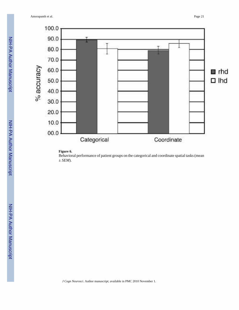
Figure 6.Behavioral performance of patient groups on the categorical and coordinate spatial tasks (mean± SEM).
Amorapanth et al. Page 21
J Cogn Neurosci. Author manuscript; available in PMC 2010 November 1.
NIH
-PA Author Manuscript
NIH
-PA Author Manuscript
NIH
-PA Author Manuscript
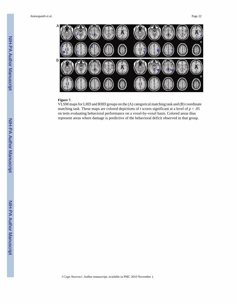
Figure 7.VLSM maps for LHD and RHD groups on the (A) categorical matching task and (B) coordinatematching task. These maps are colored depictions of t scores significant at a level of p < .05on tests evaluating behavioral performance on a voxel-by-voxel basis. Colored areas thusrepresent areas where damage is predictive of the behavioral deficit observed in that group.
Amorapanth et al. Page 22
J Cogn Neurosci. Author manuscript; available in PMC 2010 November 1.
NIH
-PA Author Manuscript
NIH
-PA Author Manuscript
NIH
-PA Author Manuscript
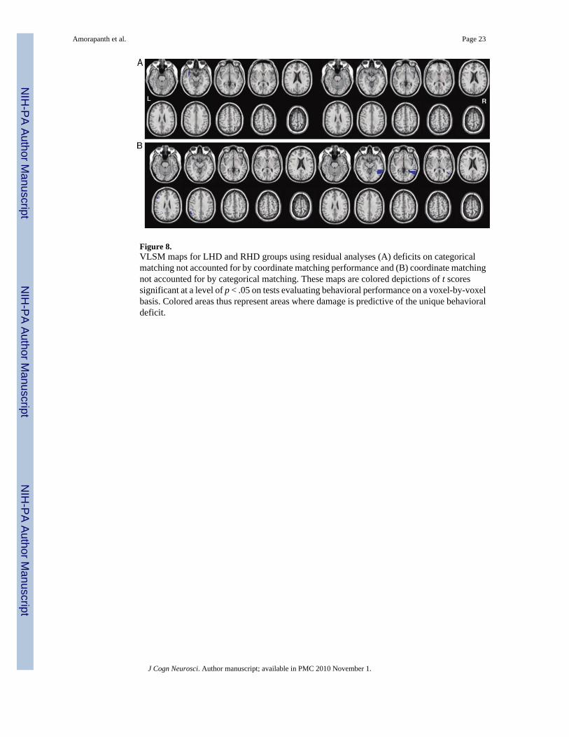
Figure 8.VLSM maps for LHD and RHD groups using residual analyses (A) deficits on categoricalmatching not accounted for by coordinate matching performance and (B) coordinate matchingnot accounted for by categorical matching. These maps are colored depictions of t scoressignificant at a level of p < .05 on tests evaluating behavioral performance on a voxel-by-voxelbasis. Colored areas thus represent areas where damage is predictive of the unique behavioraldeficit.
Amorapanth et al. Page 23
J Cogn Neurosci. Author manuscript; available in PMC 2010 November 1.
NIH
-PA Author Manuscript
NIH
-PA Author Manuscript
NIH
-PA Author Manuscript

NIH
-PA Author Manuscript
NIH
-PA Author Manuscript
NIH
-PA Author Manuscript
Amorapanth et al. Page 24
Table 1
Functional MRI Results
Spatial Relations–Object Matching Average Voxels
Left Hemisphere
Superior parietal lobe 1.84 ± 0.49 520
Inferior parietal lobe 2.07 ± 0.49 374
Lateral occipital complex −0.78 ± 0.39 520
Middle temporal gyrus 0.88 ± 0.37 149
Inferior temporal gyrus 0.25 ± 0.49 77
Fusiform gyrus −0.78 ± 0.28 325
Parahippocampal gyrus −0.47 ± 0.23 124
Right Hemisphere
Superior parietal lobe 2.04 ± 0.62 520
Inferior parietal lobe 1.39 ± 0.55 166
Lateral occipital complex −0.39 ± 0.39 414
Middle temporal gyrus 0.67 ± 0.24 189
Inferior temporal gyrus 0.55 ± 0.41 209
Fusiform gyrus −0.37 ± 0.28 380
Parahippocampal gyrus −0.08 ± 0.22 124
Effect sizes are given as mean ± SE for contrasts between conditions within each ROI. Effect sizes that are consistently different from zero acrossconditions (p < .05) are in boldface.
J Cogn Neurosci. Author manuscript; available in PMC 2010 November 1.