The multiplex bead array approach to identifying serum biomarkers associated with breast cancer
12
Open Access Available online http://breast-cancer-research.com/content/11/2/R22 Page 1 of 12 (page number not for citation purposes) Vol 11 No 2 Research article The multiplex bead array approach to identifying serum biomarkers associated with breast cancer Byoung Kwon Kim 1 , Jong Won Lee 2 , Pil Je Park 3 , Yong Sung Shin 3 , Won Young Lee 3 , Kyung Ae Lee 3 , Sena Ye 3 , Heesun Hyun 3 , Kyung Nam Kang 3 , Donghwa Yeo 4 , Youngdai Kim 4 , Sung Yup Ohn 5 , Dong Young Noh 2 and Chul Woo Kim 6 1 Department of Laboratory Medicine and Pathology, The Armed Forces Capital Hospital, 2nd street, Yul-dong, Bundnag-gu, Sungnam city, Gyeonggi- do, 434-040, Korea 2 Department of Surgery, Seoul National University College of Medicine, Daehak street, 28 Yeongeon-dong, Jongno-gu, Seoul 110-799, Korea 3 BioInfra Inc., Cancer Research Institute, Seoul National University College of Medicine, Daehak street, 28 Yeongeon-dong, Jongno-gu, Seoul 110- 799, Korea 4 Department of Statistics, Seoul National University, Gwanak street, Gwanak-gu, Seoul 151-742, Korea 5 Department of Computer Engineering, College of Engineering, Korea Aerospace University, 100 Hanggondae street, Hwajeon-dong, Deogyangu, Goyang city, Gyeonggi-do, 412-791, Korea 6 Department of Pathology, Cancer Research Institute, Tumor Immunity Medical Research Center, Seoul National University College of Medicine, Daehak street, 28 Yeongeon-dong, Jongno-gu, Seoul 110-799, Korea Corresponding author: Dong Young Noh, [email protected] Woo Kim, [email protected] Received: 16 Oct 2008 Revisions requested: 27 Nov 2008 Revisions received: 31 Mar 2009 Accepted: 28 Apr 2009 Published: 28 Apr 2009 Breast Cancer Research 2009, 11:R22 (doi:10.1186/bcr2247) This article is online at: http://breast-cancer-research.com/content/11/2/R22 © 2009 Kim et al.; licensee BioMed Central Ltd. This is an open access article distributed under the terms of the Creative Commons Attribution License (http://creativecommons.org/licenses/by/2.0 ), which permits unrestricted use, distribution, and reproduction in any medium, provided the original work is properly cited. Abstract Introduction Breast cancer is the most common type of cancer seen in women in western countries. Thus, diagnostic modalities sensitive to early-stage breast cancer are needed. Antibody- based array platforms of a data-driven type, which are expected to facilitate more rapid and sensitive detection of novel biomarkers, have emerged as a direct, rapid means for profiling cancer-specific signatures using small samples. In line with this concept, our group constructed an antibody bead array panel for 35 analytes that were selected during the discovery step. This study was aimed at testing the performance of this 35-plex array panel in profiling signatures specific for primary non- metastatic breast cancer and validating its diagnostic utility in this independent population. Methods Thirty-five analytes were selected from more than 50 markers through screening steps using a serum bank consisting of 4,500 samples from various types of cancer. An antibody- bead array of 35 markers was constructed using the Luminex™ bead array platform. A study population consisting of 98 breast cancer patients and 96 normal subjects was analysed using this panel. Multivariate classification algorithms were used to find discriminating biomarkers and validated with another independent population of 90 breast cancer and 79 healthy controls. Results Serum concentrations of epidermal growth factor, soluble CD40-ligand and proapolipoprotein A1 were increased in breast cancer patients. High-molecular-weight-kininogen, apolipoprotein A1, soluble vascular cell adhesion molecule-1, plasminogen activator inhibitor-1, vitamin-D binding protein and vitronectin were decreased in the cancer group. Multivariate classification algorithms distinguished breast cancer patients from the normal population with high accuracy (91.8% with random forest, 91.5% with support vector machine, 87.6% with linear discriminant analysis). Combinatorial markers also detected breast cancer at an early stage with greater sensitivity. Conclusions The current study demonstrated the usefulness of the antibody-bead array approach in finding signatures specific for primary non-metastatic breast cancer and illustrated the potential for early, high sensitivity detection of breast cancer. Further validation is required before array-based technology is used routinely for early detection of breast cancer. 2D-PAGE: two-dimensional polyacrylamide gel electrophoresis; A1AT: alpha-1 antitrypsin; A2M: alpha-2 macroglobulin; AFP: alpha-fetoprotein; Apo: apolipoprotein; AUC: area under the curve; BSA: bovine serum albumin; CA: cancer antigen; CAM: cell adhesion molecules; CEA: carcinoembryonic antigen; CRP: C-reactive protein; EGF: epidermal growth factor; ELISA: enzyme-linked immunosorbent assay; HDL: high-density lipoprotein; HER: human epidermal growth factor receptor; HMWK: high-molecular-weight kininogen; HSP: heat shock protein; IL: interleukin; LDA: linear discriminant analysis; MALDI: matrix-assisted laser desorption/ionisation; MPO: myeloperoxidase; MS: mass spectrometry; PAI-1: plasminogen activator inhibitor- 1; PBS: phosphate-buffered saline; PCA: principal component analysis; ProApo: proapolipoprotein; PSA: prostate specific antigen; RF: random for- ests; ROC: receiver operating curve; SAA: serum amyloid A; sCD40L: soluble CD-40 ligand; SELDI-TOF: surface-enhanced laser desorption/ioni- sation time-of-flight; sICAM-1: soluble intercellular adhesion molecule-1; sVCAM-1: soluble vascular cell adhesion molecule-1; SVM: support vector machine; VDBP: vitamin-D binding protein.
-
Upload
independent -
Category
Documents
-
view
0 -
download
0
Transcript of The multiplex bead array approach to identifying serum biomarkers associated with breast cancer

Available online http://breast-cancer-research.com/content/11/2/R22
Open AccessVol 11 No 2Research articleThe multiplex bead array approach to identifying serum biomarkers associated with breast cancerByoung Kwon Kim1, Jong Won Lee2, Pil Je Park3, Yong Sung Shin3, Won Young Lee3, Kyung Ae Lee3, Sena Ye3, Heesun Hyun3, Kyung Nam Kang3, Donghwa Yeo4, Youngdai Kim4, Sung Yup Ohn5, Dong Young Noh2 and Chul Woo Kim6
1Department of Laboratory Medicine and Pathology, The Armed Forces Capital Hospital, 2nd street, Yul-dong, Bundnag-gu, Sungnam city, Gyeonggi-do, 434-040, Korea2Department of Surgery, Seoul National University College of Medicine, Daehak street, 28 Yeongeon-dong, Jongno-gu, Seoul 110-799, Korea3BioInfra Inc., Cancer Research Institute, Seoul National University College of Medicine, Daehak street, 28 Yeongeon-dong, Jongno-gu, Seoul 110-799, Korea4Department of Statistics, Seoul National University, Gwanak street, Gwanak-gu, Seoul 151-742, Korea5Department of Computer Engineering, College of Engineering, Korea Aerospace University, 100 Hanggondae street, Hwajeon-dong, Deogyangu, Goyang city, Gyeonggi-do, 412-791, Korea6Department of Pathology, Cancer Research Institute, Tumor Immunity Medical Research Center, Seoul National University College of Medicine, Daehak street, 28 Yeongeon-dong, Jongno-gu, Seoul 110-799, Korea
Corresponding author: Dong Young Noh, [email protected] Woo Kim, [email protected]
Received: 16 Oct 2008 Revisions requested: 27 Nov 2008 Revisions received: 31 Mar 2009 Accepted: 28 Apr 2009 Published: 28 Apr 2009
Breast Cancer Research 2009, 11:R22 (doi:10.1186/bcr2247)This article is online at: http://breast-cancer-research.com/content/11/2/R22© 2009 Kim et al.; licensee BioMed Central Ltd. This is an open access article distributed under the terms of the Creative Commons Attribution License (http://creativecommons.org/licenses/by/2.0), which permits unrestricted use, distribution, and reproduction in any medium, provided the original work is properly cited.
Abstract
Introduction Breast cancer is the most common type of cancerseen in women in western countries. Thus, diagnostic modalitiessensitive to early-stage breast cancer are needed. Antibody-based array platforms of a data-driven type, which are expectedto facilitate more rapid and sensitive detection of novelbiomarkers, have emerged as a direct, rapid means for profilingcancer-specific signatures using small samples. In line with thisconcept, our group constructed an antibody bead array panelfor 35 analytes that were selected during the discovery step.This study was aimed at testing the performance of this 35-plexarray panel in profiling signatures specific for primary non-metastatic breast cancer and validating its diagnostic utility inthis independent population.
Methods Thirty-five analytes were selected from more than 50markers through screening steps using a serum bank consistingof 4,500 samples from various types of cancer. An antibody-bead array of 35 markers was constructed using the Luminex™bead array platform. A study population consisting of 98 breastcancer patients and 96 normal subjects was analysed using thispanel. Multivariate classification algorithms were used to find
discriminating biomarkers and validated with anotherindependent population of 90 breast cancer and 79 healthycontrols.Results Serum concentrations of epidermal growth factor,soluble CD40-ligand and proapolipoprotein A1 were increasedin breast cancer patients. High-molecular-weight-kininogen,apolipoprotein A1, soluble vascular cell adhesion molecule-1,plasminogen activator inhibitor-1, vitamin-D binding protein andvitronectin were decreased in the cancer group. Multivariateclassification algorithms distinguished breast cancer patientsfrom the normal population with high accuracy (91.8% withrandom forest, 91.5% with support vector machine, 87.6% withlinear discriminant analysis). Combinatorial markers alsodetected breast cancer at an early stage with greater sensitivity.Conclusions The current study demonstrated the usefulness ofthe antibody-bead array approach in finding signatures specificfor primary non-metastatic breast cancer and illustrated thepotential for early, high sensitivity detection of breast cancer.Further validation is required before array-based technology isused routinely for early detection of breast cancer.
2D-PAGE: two-dimensional polyacrylamide gel electrophoresis; A1AT: alpha-1 antitrypsin; A2M: alpha-2 macroglobulin; AFP: alpha-fetoprotein; Apo: apolipoprotein; AUC: area under the curve; BSA: bovine serum albumin; CA: cancer antigen; CAM: cell adhesion molecules; CEA: carcinoembryonic antigen; CRP: C-reactive protein; EGF: epidermal growth factor; ELISA: enzyme-linked immunosorbent assay; HDL: high-density lipoprotein; HER: human epidermal growth factor receptor; HMWK: high-molecular-weight kininogen; HSP: heat shock protein; IL: interleukin; LDA: linear discriminant analysis; MALDI: matrix-assisted laser desorption/ionisation; MPO: myeloperoxidase; MS: mass spectrometry; PAI-1: plasminogen activator inhibitor-1; PBS: phosphate-buffered saline; PCA: principal component analysis; ProApo: proapolipoprotein; PSA: prostate specific antigen; RF: random for-ests; ROC: receiver operating curve; SAA: serum amyloid A; sCD40L: soluble CD-40 ligand; SELDI-TOF: surface-enhanced laser desorption/ioni-sation time-of-flight; sICAM-1: soluble intercellular adhesion molecule-1; sVCAM-1: soluble vascular cell adhesion molecule-1; SVM: support vector machine; VDBP: vitamin-D binding protein.
Page 1 of 12(page number not for citation purposes)

Breast Cancer Research Vol 11 No 2 Kim et al.
IntroductionBreast cancer is the most common malignant disease inwomen in western countries, comprising approximately 35%of all cancers [1]. The incidence of breast cancer hasincreased over the past few decades, probably due to earlierdiagnosis, and mortality has been gradually reducing [2].Nonetheless, prevention and early detection of breast cancerare two major issues of consideration for cancer epidemiolo-gists and clinicians because radical treatment can greatlyreduce breast cancer-related mortality if breast cancer isdetected at an early stage [3]. Despite the use of mammogra-phy as a routine screening method for women 40 years of ageand older, the effectiveness of this procedure in reducing over-all population mortality is still being investigated [4]. Otherdiagnostic modalities that can improve diagnostic power incombination with conventional methods are required for stra-tegic management of the disease and improvement of theoverall mortality rate.
Biomarker research in easy-to-access biological fluids fromcancer patients is expected to open up a new era in the fieldof cancer research and cancer diagnostics. Extensivesearches have revealed several breast cancer-specific mark-ers: MUC-1 family mucin glucoproteins like CA 15.3, BR27.29(or CA27.29), and mucin-like carcinoma-associated antigen,CA 549, carcinoembryonic antigen (CEA), serum human epi-dermal growth factor receptor (HER) 2/c-erbB-2, cytokinesand cytokeratin fragments [5-10]. Although these markers arenot used for the purposes of screening and early diagnosis,they play a complementary role in staging work-up at initialpresentation as indicated in the guidelines issued by the Euro-pean Group on Tumor Markers (EGTM) [11] and the Food andDrug Administration [12].
Recent advancements in high-throughput platforms and infor-mation technology have ushered in the data-driven approach,which has emerged as a powerful and efficient way of con-ducting biomarker research and finding novel biomarkers. Inthe field of proteomics, the classical approach uses two-dimensional polyacrylamide gel electrophoresis (2D-PAGE)for comparing multiple protein profiles. However, this methodhas problems such as poor reproducibility and low throughput.Recent advances in mass spectrometry (MS), such as matrix-assisted laser desorption/ionisation (MALDI) time-of-flight MS,offer an alternative to 2D-PAGE [13]. However, some limita-tions in MALDI, such as extensive sample preparation and highsignal background problems resulting from inorganic andorganic contaminants, have hindered its wider use as a high-throughput screening tool to find useful proteins in complexbiological samples. The development of surface-enhancedlaser desorption/ionisation time-of-flight (SELDI-TOF) MS haslargely overcome these limitations [14]. In the field of breastcancer research, Li and colleagues performed a pioneeringstudy using SELDI-TOF and found potential biomarkers fordetection of breast cancer, designating the peaks as BC1 (4.3
kDa), BC2 (8.1 kDa) and BC3 (8.9 kDa) [15]. Later, some ofthese were identified as fragments of serum complement pro-tein, but these results are awaiting further validation.
Antibody-based microarray is also one of the data-drivenapproaches in proteomics that is likely to play an increasingrole in the discovery of disease-specific signatures [16]. Thespectrum of chemical biomarker information that can be eluci-dated using this method is relatively limited compared withthat obtained using MS. However, the antibody-array platformbypasses the identification step for individual markers, makingthis a faster and more direct method for profiling proteinexpression and translating this information [17,18]. Further-more, a combinatory strategy for utilising markers and statis-tics has been suggested to increase predictive power incancer diagnosis, which re-energises the search for novel can-cer-related biomarker signatures [19,20].
In line with this concept, Carlsson and colleagues adopted aplanar array platform using single-chain variable fragment(scFv) targeting for more than 60 target antigens and found aserum protein signature that distinguishes breast cancerpatients from normal subjects with high diagnostic accuracy[21]. Recently, the bead-array platform was also successfullyapplied to identify serum profiles predicting responses to neo-adjuvant chemotherapy in locally advanced breast cancer[22].
Recently, our group constructed an antibody-based beadarray panel consisting of 35 serum proteins via an extensivescreening process using 4500 serum samples from variouscancer patients. We report the characteristic serum profilesassociated with breast cancer as revealed by application ofthis panel in an independent group of patients with mostly pri-mary, non-metastatic disease and validate the diagnostic per-formance of these combinatorial markers.
Materials and methodsStudy samplesTwo sets of population were constructed. A training set (set 1)for selecting prediction biomarkers consisted of 194 people(98 breast cancer patients and 96 normal controls). The otherindependent set (set 2), consisting of 169 people (90 breastcancer patients and 79 normal controls), was used for valida-tion of selected predictors from the initial set. In each set, can-cer and control populations were age-matched. Serumsamples of breast cancer patients were obtained before anytype of surgical procedures. None of the patients had a familyhistory of breast cancer. Serum samples for the controls wereobtained from normal female subjects who voluntarily enrolledin the cancer screening program of Seoul National UniversityHospital and had no abnormalities identified on physical exam-ination, routine blood testing or mammography. A completemedical history was obtained for each patient, including med-ication, menstrual history, menopause, alcohol consumption
Page 2 of 12(page number not for citation purposes)

Available online http://breast-cancer-research.com/content/11/2/R22
and smoking. All blood samples were collected before anytype of surgical or medical intervention was performed. Periph-eral blood was collected using 5 ml syringes and stored inSST™ II tubes (Becton Dickinson, Franklin Lakes, NJ, USA) atroom temperature for one hour. Samples were centrifuged at3000 g for five minutes, and the supernatants were collectedand stored at -80°C before the assay was performed. Sampleswere drawn after obtaining informed consent from all patients.The study protocol was reviewed and approved by InstitutionalReview Board at Seoul National University Hospital (approvalNo. C-0512-502-163).
Construction of 35-plex bead array panelThe antigen panel consisted of 35 analytes (Table 1). Thirty-five markers were chosen from 51 original markers (Figure 1).A serum bank had been constructed for determination of can-cer biomarkers. The serum bank contained approximately4500 samples from five types of cancer: breast, colon, stom-ach, liver and lung. Alpha-1-antitrypin, pro-apolipoprotein(proApo) A1, apolipoprotein (Apo) A4, haptoglobin α, andtransthyretin were discovered using 2D-PAGE. ApoH, β2-microglobulin, vitamin D-binding protein, C-reactive protein(CRP), free haemoglobin and serum amyloid A were discov-ered using SELDI-TOF MS. These 11 markers were regardedas candidate markers with 40 markers selected through a lit-erature search. The list of 51 candidate markers at this stagewas as follows: adiponectin, alpha-2 macroglobulin (A2M),alpha-fetoprotein (AFP), alpha-1 antitrypsin (A1AT), ApoA1,proApoA1, ApoA2, ApoA4, ApoC2, ApoC3, ApoH, ApoJ(clusterin), beta-2 microglobulin, ferritin, CA-125, CA19-9,CA72-4, CEA, cathepsin B, chromogranin A, CRP, D-dimer,
epidermal growth factor (EGF), galectin 3, gelsolin, hap-toglobin alpha, total haptoglobin, free haemoglobin, heatshock protein (HSP) 27, HSP 70, high-molecular-weightkininogen (HMWK), human cervical cancer oncogene-1, IL-1beta, IL-1 receptor alpha, IL-6, IL-8, monocyte chemoattract-ant protein-1, macrophage inflammatory protein-1, myeloper-oxidase (MPO), neuron specific enolase, retinol bindingprotein 4, total plasminogen activator inhibitor (PAI)-1, totalprostate specific antigen (PSA), free PSA, pro-gastrin releas-ing peptide, soluble CD-40 ligand (sCD40L), soluble intercel-lular adhesion molecule (sICAM)-1, soluble vascular celladhesion molecule (sVCAM)-1, serum amyloid A (SAA), tran-sthyretin, vitamin-D binding protein (VDBP) and vitronectin.The sandwich ELISA method for individual analytes was usedas a validation method, again using samples from the serumbank. Markers with significantly different mean serum concen-trations in cancer patients and normal subjects were selectedand subjected to the 35-plex bead array panel.
Bead array kits or antibodies for the construction of the 35-plex panel were purchased from the following manufacturers:Abcam (Cambridge, UK), Bethyl (Montgomery, TX, USA),Biodesign International (Saco, ME, USA), BoditechMed(Chuncheon, Korea), Chemicon (Temecula, CA, USA), Dako(Glostrup, Denmark), EMD Chemicals Inc. (San Diego, CA,USA), Fitzgerald (Concord, MA, USA), HyTest (Turku, Fin-land), Linco Research, Inc. (St. Charles, MO, USA), Rules-based Medicine (Austin, TX, USA), R&D Systems (Minneapo-lis, MN, USA), Santa Cruz Biotechnology Inc. (Santa Cruz, CA,USA), Sigma-Aldrich (St Louis, MO, USA) and US Biological(Swampscott, MA, USA).
Figure 1
Procedure for constructing the 35-plex panelProcedure for constructing the 35-plex panel. Through using 4500 serum samples from cancer patients, five markers were discovered and identified through two-dimensional polyacrylamide gel electrophoresis (2D-PAGE), six markers through surface-enhanced laser desorption/ionisation time-of-flight (SELDI-TOF) and 24 markers through conventional sandwich ELISA method. After optimisation in capturing and detecting the antibody pair for a target analyte, the capturing antibody was conjugated with beads, and the simplex kit was validated for its dynamic range, recovery rate, parallelism with standard curve, interference, matrix effect, and lower and upper limits of detection. Twenty-three simplex kits could be grouped together accord-ing to dilution factor and absence of cross reactivity. The remaining 12 were left and used as a simplex kit. BC = breast cancer; SC = stomach can-cer; LC = lung cancer; CC = colon cancer; HCC = hepatocellular carcinoma.
Page 3 of 12(page number not for citation purposes)
Breast Cancer Research Vol 11 No 2 Kim et al.
Multiplex assay procedureMultiplex assay was performed using the following procedure:a 96-well filter-plate (Millipore, Billerica, MA, USA) wasblocked with PBS (pH 7.4) with 2% BSA. Twenty microlitersof standard curve sample, prediluted control samples andpatient samples were dispensed into the wells in duplicate.Twenty microliters of primary antibody-bead mixture wereadded into each well and incubated at room temperature forone hour. Twenty microliters of detection antibodies with bioti-nylation and 20 μL of streptavidin-phycoerythrin were addedand incubated at room temperature for one hour. Each stepwas followed by a double washing step using 0.05% Tween-20 in PBS (PBST) with vacuum manifold (Millipore Corp., Bill-erica, MA, USA).
After the final washing step, samples were resuspended with100 μL of PBST and read using a Luminex-200™ (LuminexInc., Austin, TX, USA). The standard curve was calculatedusing five-parametric-curve fitting, and results were analysedusing Beadview software (Upstate Biotechnology Inc., LakePlacid, NY, USA). Markers were grouped together accordingto dilution factor after cross-reactivity was checked across allanalytes. Control samples at two levels in the dynamic rangeof the standard curve were run together in duplicate for qualitycontrol throughout the study. Intra-assay precision rangedfrom 2 to 16%, and inter-assay precision ranged from 6 to19% during the experiment. The acceptance criteria for eachindividual run followed Westgard's rule [23].
Table 1
List of biomarkers in 35-plex panel
Marker list
Oncofetal protein Acute phase protein
Alpha-fetoprotein (AFP) Alpha-1-antitrypin (A1AT)a
Carcinoembryonic antigen (CEA) Alpha-2 macroglobulin (A2M)c
Cancer antigen 125 (CA125)c C-reactive protein (CRP)b
Cancer antigen 19-9 (CA19-9)c D-dimer (DD)c
Prostate-specific antigen (PSA)c Haptoglobin α (Hp)a
Cytokine Serum amyloid A (SAA)b
Interleukin-1β (IL-1β)c Transthyretin (TTR)a
Interleukin-1 receptor α (IL-Rα)c Coagulation/thrombosis
Interleukin-6 (IL-6)c Haemoglobin (Hg)b
Interleukin-8 (IL-8)c High-molecular-weight kininogen (HMWK)c
Monocyte chemotactic protein-1(MCP-1)c
Plasminogen activator inhibitor-1(PAI-1)c
Monocyte inflammatory protein-1α (MIP-1α)c Metabolism
Immune/inflammation Apolipoprotein A1 (ApoA1)c
Soluble CD40 ligand (sCD40L)c Pro-apolipoprotein A1 (proApoA1)a
β2-microglobulin (β 2M)b Apolipoprotein A4 (ApoA4)a
Myeloperoxidase (MPO)c Apolipoprotein H (ApoH)b
Factor/hormone Carrier
Epidermal growth factor (EGF)c Vitamin D-binding protein (VDBP)b
Adiponectinc Enzyme
Adhesion Cathepsin B (CB)c
Vitronectin (VN)c
Soluble vascular cell adhesion molecule-1 (sVCAM-1)c
Soluble intercellular cell adhesion molecule-1 (sICAM-1)c
aMarkers were discovered through two-dimensional polyacrylamide gel electrophoresis (2D-PAGE) using the serum of breast cancer patients. bMarkers were discovered through surface-enhanced laser desorption/ionisation time-of-flight (SELDI-TOF) mass spectrometry. cMarkers were selected using literature search.
Page 4 of 12(page number not for citation purposes)
Available online http://breast-cancer-research.com/content/11/2/R22
Bioinformatics and statisticsThe values of markers were transformed into log values beforeanalysis using a multivariate classification algorithm. As an ini-tial step, principal component analysis (PCA) was performedusing information related to the concentration of all 35 mark-ers, in order to study clustering of breast cancer and normalsubjects. Random forest (RF), support vector machine (SVM)and linear discriminant analysis (LDA) were the multivariatealgorithms used. Among the 196 cases, two-thirds of thecases from the breast cancer and normal groups were ran-domly assigned to training sets, and the remaining one-thirdwere assigned to test sets. We compared the prediction per-formances obtained from 50 randomly partitioned data sets.Classification models with selected predictors obtained fromthe experiment with set 1 were validated again with set 2. Areceiver operating characteristic (ROC) curve was con-structed, and the area under the curve (AUC) was calculatedusing each algorithm. We extracted a classifier consisting of asubset of protein markers yielding the best classification per-formance in the test sets. A student's t-test (two-sided) wasperformed to compare the mean serum marker levels amonggroups stratified by clinical and pathological variables, andPearson's correlation was performed to compare maximumtumour length and number of lymph node metastases withserum biomarker levels. All calculations were performed usingthe R program package (Wirtschafts universität, Wien, Aus-tria) [24].
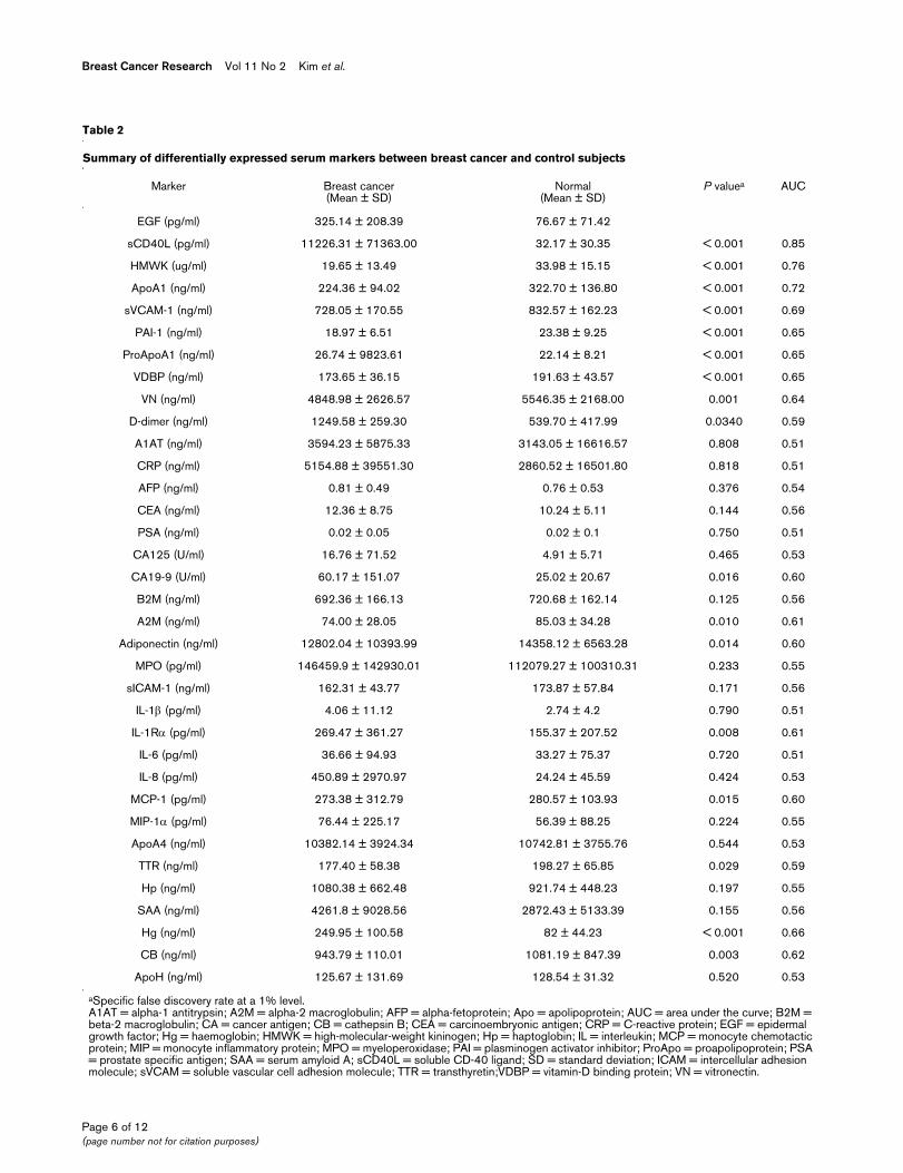
ResultsAnalysis of differentially expressed serum makers in patients with breast cancer and in normal subjectsThe mean serum concentrations for individual analytes werecompared between patients with breast cancer and thosewithout breast cancer. Among the 35 analytes, EGF, sCD40Land proApoA1 showed higher serum concentrations in breastcancer patients than in normal subjects (Table 2). HMWK,ApoA1, PAI-1, VDBP and vitronectin levels were significantlydecreased in cancer patients. EGF showed the highest AUCvalue (0.89) and exhibited a diagnostic accuracy of 82.3%,sensitivity of 94.0% and specificity of 70.6% as a singlemarker.
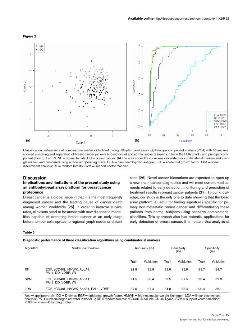
Multivariate classification using combinatorial biomarkers specific for breast cancerIn order to geometrically interpret and determine if breast can-cer patients could be segregated from normal subjects, PCAanalysis was performed using all the data related to serum lev-els of the 35 markers. Principal components deduced fromvariance-covariance structures of these markers separatedthese two groups using the top two principal components(Figure 2a). In order to find classifiers that distinguish breastcancer patients from healthy people, multivariate classificationanalysis was performed using RF, SVM and LDA. In the inter-est of constructing models and selecting predictors, two-thirds of the original set was assigned to training sets. After
training, each model consisting of different sets of classifierswas validated through the test set. The accuracy and classifi-cation error for each model were calculated in each trainingand test set. The calculated averages are summarised in Table3. RF, SVM and LDA classified breast cancer and normal sub-jects with a mean accuracy of 91.8%, 91.5% and 87.6%,respectively. For the validation of this model, an independentvalidation set consisting of 169 persons was analysed usingthe same model and predictors. The calculated averages weresimilar to those obtained from the original set (Table 3). Thecombination of markers showing the highest diagnostic accu-racy was very similar in all three models. EGF, sCD40L,HMWK, ApoA1, PAI-1 and VDBP were consistently selectedby all three algorithms. D-dimer and vitronectin were chosenby RF and SVM. Pre-treatment serum levels of CA15-3 wereavailable in 96 patients, and serum levels of tissue polypeptideantigen were available in 77 patients. When the sensitivity ofcombinatorial markers was compared with that of single mark-ers, multi-classifiers showed improvement not only in overallsensitivity for total patients but also in sensitivity for early-stagedisease (Table 4).
Comparison of biomarkers with clinico-pathological parameters of breast cancerAll clinical and pathological factors were analysed and com-pared with serum concentrations of the 35 analytes (Table 5).Advanced T-stage breast cancer (T3 and T4) showedincreased serum levels of EGF and ApoH, but decreased lev-els of A1AT. Patients with lymph node metastasis showedincreased serum concentrations of sVCAM-1 and transthyre-tin. The number of lymph node metastases was positively cor-related with sVCAM-1 and D-dimer (Pearson's correlationcoefficient = 0.23, P = 0.025; and Pearson's correlation coef-ficient = 0.25, P = 0.028, respectively). Patients with distantmetastasis showed lower levels of proApoA1 in the serum.The expression status of oestrogen receptor, progesteronereceptor and c-erbB2 in tumour tissue was also reflected inserum levels of analytes. In oestrogen receptor-positivepatients, serum levels of A2M and HMWK were increased,and concentrations of vitronectin, transthyretin and PAI-1 weredecreased. Higher serum levels of A2M and CA19-9 wereobserved in progesterone receptor-positive patients. Inpatients showing c-erbB2 expression in tumour tissue, serumlevels of sVCAM-1 were decreased, whereas SAA levels wereincreased. No correlation was noted between the serum EGFlevel and c-erbB2 expression (c-erbB2 positive = 324 ±203.58 pg/ml, vs. c-erbB2 negative = 319.39 ± 209.47 pg/ml, P = 0.908). The serum concentration of 35 analytes wasnot influenced by factors such as nuclear grade, histologicalgrade, location or multiplicity of tumour mass.
Page 5 of 12(page number not for citation purposes)
Breast Cancer Research Vol 11 No 2 Kim et al.
Page 6 of 12(page number not for citation purposes)
Table 2
Summary of differentially expressed serum markers between breast cancer and control subjects
Marker Breast cancer(Mean ± SD)
Normal(Mean ± SD)
P valuea AUC
EGF (pg/ml) 325.14 ± 208.39 76.67 ± 71.42
sCD40L (pg/ml) 11226.31 ± 71363.00 32.17 ± 30.35 < 0.001 0.85
HMWK (ug/ml) 19.65 ± 13.49 33.98 ± 15.15 < 0.001 0.76
ApoA1 (ng/ml) 224.36 ± 94.02 322.70 ± 136.80 < 0.001 0.72
sVCAM-1 (ng/ml) 728.05 ± 170.55 832.57 ± 162.23 < 0.001 0.69
PAI-1 (ng/ml) 18.97 ± 6.51 23.38 ± 9.25 < 0.001 0.65
ProApoA1 (ng/ml) 26.74 ± 9823.61 22.14 ± 8.21 < 0.001 0.65
VDBP (ng/ml) 173.65 ± 36.15 191.63 ± 43.57 < 0.001 0.65
VN (ng/ml) 4848.98 ± 2626.57 5546.35 ± 2168.00 0.001 0.64
D-dimer (ng/ml) 1249.58 ± 259.30 539.70 ± 417.99 0.0340 0.59
A1AT (ng/ml) 3594.23 ± 5875.33 3143.05 ± 16616.57 0.808 0.51
CRP (ng/ml) 5154.88 ± 39551.30 2860.52 ± 16501.80 0.818 0.51
AFP (ng/ml) 0.81 ± 0.49 0.76 ± 0.53 0.376 0.54
CEA (ng/ml) 12.36 ± 8.75 10.24 ± 5.11 0.144 0.56
PSA (ng/ml) 0.02 ± 0.05 0.02 ± 0.1 0.750 0.51
CA125 (U/ml) 16.76 ± 71.52 4.91 ± 5.71 0.465 0.53
CA19-9 (U/ml) 60.17 ± 151.07 25.02 ± 20.67 0.016 0.60
B2M (ng/ml) 692.36 ± 166.13 720.68 ± 162.14 0.125 0.56
A2M (ng/ml) 74.00 ± 28.05 85.03 ± 34.28 0.010 0.61
Adiponectin (ng/ml) 12802.04 ± 10393.99 14358.12 ± 6563.28 0.014 0.60
MPO (pg/ml) 146459.9 ± 142930.01 112079.27 ± 100310.31 0.233 0.55
sICAM-1 (ng/ml) 162.31 ± 43.77 173.87 ± 57.84 0.171 0.56
IL-1β (pg/ml) 4.06 ± 11.12 2.74 ± 4.2 0.790 0.51
IL-1Rα (pg/ml) 269.47 ± 361.27 155.37 ± 207.52 0.008 0.61
IL-6 (pg/ml) 36.66 ± 94.93 33.27 ± 75.37 0.720 0.51
IL-8 (pg/ml) 450.89 ± 2970.97 24.24 ± 45.59 0.424 0.53
MCP-1 (pg/ml) 273.38 ± 312.79 280.57 ± 103.93 0.015 0.60
MIP-1α (pg/ml) 76.44 ± 225.17 56.39 ± 88.25 0.224 0.55
ApoA4 (ng/ml) 10382.14 ± 3924.34 10742.81 ± 3755.76 0.544 0.53
TTR (ng/ml) 177.40 ± 58.38 198.27 ± 65.85 0.029 0.59
Hp (ng/ml) 1080.38 ± 662.48 921.74 ± 448.23 0.197 0.55
SAA (ng/ml) 4261.8 ± 9028.56 2872.43 ± 5133.39 0.155 0.56
Hg (ng/ml) 249.95 ± 100.58 82 ± 44.23 < 0.001 0.66
CB (ng/ml) 943.79 ± 110.01 1081.19 ± 847.39 0.003 0.62
ApoH (ng/ml) 125.67 ± 131.69 128.54 ± 31.32 0.520 0.53
aSpecific false discovery rate at a 1% level.A1AT = alpha-1 antitrypsin; A2M = alpha-2 macroglobulin; AFP = alpha-fetoprotein; Apo = apolipoprotein; AUC = area under the curve; B2M = beta-2 macroglobulin; CA = cancer antigen; CB = cathepsin B; CEA = carcinoembryonic antigen; CRP = C-reactive protein; EGF = epidermal growth factor; Hg = haemoglobin; HMWK = high-molecular-weight kininogen; Hp = haptoglobin; IL = interleukin; MCP = monocyte chemotactic protein; MIP = monocyte inflammatory protein; MPO = myeloperoxidase; PAI = plasminogen activator inhibitor; ProApo = proapolipoprotein; PSA = prostate specific antigen; SAA = serum amyloid A; sCD40L = soluble CD-40 ligand; SD = standard deviation; ICAM = intercellular adhesion molecule; sVCAM = soluble vascular cell adhesion molecule; TTR = transthyretin;VDBP = vitamin-D binding protein; VN = vitronectin.
Available online http://breast-cancer-research.com/content/11/2/R22
DiscussionImplications and limitations of the present study using an antibody-bead array platform for breast cancer proteomicsBreast cancer is a global issue in that it is the most frequentlydiagnosed cancer and the leading cause of cancer deathamong women worldwide [25]. In order to improve survivalrates, clinicians need to be armed with new diagnostic modal-ities capable of detecting breast cancer at an early stagebefore tumour cells spread to regional lymph nodes or distant
sites [26]. Novel cancer biomarkers are expected to open upa new era in cancer diagnostics and will meet current medicalneeds related to early detection, monitoring and prediction oftreatment results in breast cancer patients [27]. To our knowl-edge, our study is the only one to date showing that the beadarray platform is useful for finding signatures specific for pri-mary non-metastatic breast cancer and differentiating thesepatients from normal subjects using sensitive combinatorialclassifiers. This approach also has potential applications forearly detection of breast cancer. It is notable that analysis of
Figure 2
Classification performance of combinatorial markers identified through 35-plex panel assayClassification performance of combinatorial markers identified through 35-plex panel assay. (a) Principal component analysis (PCA) with 35 markers showed clustering and separation of breast cancer patients (closed circle) and normal subjects (open circle) in the PCA chart using principal com-ponent (Comp) 1 and 2. NF = normal female; BC = breast cancer. (b) The area under the curve was calculated for combinatorial markers and a sin-gle marker, and compared using a receiver operating curve. CEA = carcinoembryonic antigen; EGF = epidermal growth factor; LDA = linear discriminant analysis; RF = random forests; SVM = support vector machine.
Table 3
Diagnostic performance of three classification algorithms using combinatorial markers
Algorithm Marker combination Accuracy (%) Sensitivity(%)
Specificity(%)
Train Validation Train Validation Train Validation
RF EGF, sCD40L, HMWK, ApoA1,PAI-1, DD, VDBP, VN
91.8 93.8 89.8 92.8 93.7 94.7
SVM EGF, sCD40L, HMWK, ApoA1,PAI-1, DD, VDBP, VN
91.5 88.4 89.5 87.5 93.4 89.3
LDA EGF, sCD40L, HMWK, ApoA1, PAI-1, VDBP 87.6 87.4 84.8 88.4 90.4 86.1
Apo = apolipoprotein; DD = D-dimer; EGF = epidermal growth factor; HMWK = high-molecular-weight kininogen; LDA = linear discriminant analysis; PAI-1 = plasminogen activator inhibitor-1; RF = random forests; sCD40L = soluble CD-40 ligand; SVM = support vector machine; VDBP = vitamin-D binding protein.
Page 7 of 12(page number not for citation purposes)

Breast Cancer Research Vol 11 No 2 Kim et al.
diverse proteins in serum revealed biomarkers correlating withclinical and pathological variables, including receptor expres-sion status on the cancer cells. Recently, Carlsson and col-leagues introduced an scFv-antibody array platform that couldsuccessfully distinguish metastatic breast cancer patientsfrom normal people [21]. Nolen and colleagues used a multi-plex bead array platform to profile serum biomarkers predictingresponse to neoadjuvant chemotherapy in locally advancedbreast cancer [22]. Thus, in the long term, biomarkers andarray-based technology can practically be used for earlydetection of breast cancer and for stratifying patients, deter-mining their likelihood of experiencing recurrence or having adrug response, or predicting their survival expectancy [16].Our study has some limitations. Some of the markers identifiedin this study may not be specific for breast cancer and maypossibly reflect a systemic response to tissue damage orinflammation. Furthermore, the analytes included in this studyare not comprehensive. A variety of other analytes mightbehave differentially in the blood of cancer patients. Signatureprofiling of other benign breast conditions or systemic dis-eases and further array panel study using a wider range ofmarkers will resolve such issues.
Alteration of cytokines and growth factors in breast cancerAmong the cytokines and growth factors included in thisstudy, EGF was the only marker increased in the serum ofbreast cancer patients and correlated with advanced T stage.Up-regulation of other cytokines was not pronounced. Highlevels of circulating EGF were reported in serum samples fromHER2-negative breast cancer patients, although increasedlevels of IL-8 were consistently noted in serum samples frommetastatic breast cancer patients [21,28,29]. This discordantresult might be caused by differences in the study populations.In previous studies by Vazquez-martin and colleagues [28] andCarlsson and colleagues [21], serum samples were taken prin-cipally from patients with metastasis. However, only 8 of 98breast cancer patients (8.1%) in this study had metastasis. Inthe study by Benoy and colleagues relatively large numbers of
breast cancer patients without metastasis were recruited andcompared with normal control subjects [29]. This difference instudy populations might explain the discordant results acrossthe studies with regard to IL-8 and EGF levels.
Alteration of coagulation and thrombosis in breast cancerHypercoagulability is frequently seen in the setting of cancer,with Trousseau's sign first reported over 100 years ago [30].Multiple mechanisms are considered contributory to this phe-nomenon, such as secretion of tissue factor, cancer pro-coag-ulant, PAI-1, mucin molecules with altered glycan and otherthrombogenic cytokines from cancer cells [31]. The multiplexarray used in this study contained coagulation- and thrombo-sis-related markers such as sCD40L, HMWK, D-dimer, PAI-1and free haemoglobin. An assay using this panel revealedincreased concentrations of sCD40L and decreased levels ofHMWK and PAI-1 in breast cancer patients. Roselli and col-leagues first noted the association between elevated plasmasCD40L levels in lung cancer; specifically advanced squa-mous cell carcinoma. They also noted in vivo platelet activa-tion with this type of tumour [32]. Membrane-bound CD40L, aprecursor of sCD40L, is a transmembrane glycoprotein mainlyexpressed by activated T cells and activated platelets [33].Recently, it has been suggested that activation of the CD40/CD40L pathway may enhance the pro-coagulant activity oftumour cells through up-regulation of tissue factor expression[34]. Thrombin generation and peritumoural fibrin depositioninduced by tissue factor then promote angiogenesis and plate-let activation [35]. In our study, quantitative changes in serumHMWK levels were also observed in breast cancer patients.This is in agreement with a study previous reporting down-reg-ulation of HMWK in tissue samples from breast cancerpatients [36]. Given the fact that HMWK also has pro-throm-botic and pro-angiogenic properties through releasing brady-kinin [37], the behaviour of these two proteins in the serum ofbreast cancer patients is consistent with the perceived con-cept of cancer biology.
Table 4
Comparison of sensitivity of combinatorial markers vs. single marker
Stage I to II Stage III to IV Total cases
Classification method Sensitivity (%) Sensitivity (%) Sensitivity (%)
Algorithm RF 86.3(44/51) 93.6(44/47) 89.8
SVM 90.2(46/51) 93.6(44/47) 91.8
LDA 82.4(42/51) 85.1(40/47) 83.7
Single marker CA15-3* 0 (0/49) 6.4 (3/47) 3.1 (3/96)
TPA* 23.1 (9/39) 29.0 (11/38) 26.0 (20/77)
*Cut-off levels of CA15-3 and TPA were 27 U/ml and 95 U/L, respectively.CA = cancer antigen; LDA = linear discriminant analysis; RF = random forests; SVM = support vector machine; TPA = tissue polypeptide antigen.
Page 8 of 12(page number not for citation purposes)

Available online http://breast-cancer-research.com/content/11/2/R22
PAI-1 is frequently up-regulated in cancer cells [31], and ele-vated PAI-1 has been found to be a poor prognostic marker inthe setting of breast cancer [38]. PAI-1 contributes to cancerdissemination by preventing excess degradation of the extra-cellular matrix, modulating cell adhesion [39], promotingtumour angiogenesis [40] and stimulating proliferation [41].However, in our study, serum PAI-1 levels were unexpectedlydecreased in breast cancer patients compared with normalsubjects. This may have been due to pre-analytic or analyticerror, in addition to other possibilities. PAI-1 exists in plasmaor serum as a free form, a complex form mostly with vitronectinand tissue-type plasminogen activator (tPA) or urokinase-typeplasminogen activator (uPA), a latent form and a cleaved form
[42]. One report described different PAI-1 glycosylation pat-terns, depending on cellular origin [43]. There are currently nodata available concerning changes in amount or concentrationof cleaved or variant glycoforms of PAI-1 in cancer patients.The specificity of the antibodies used in our study should betested, as should qualitative alterations of PAI-1 in cancer thatmay affect antigenicity of epitopes.
D-dimer is a marker of ongoing fibrinolysis that is frequentlyincreased in various cancers [44-46]. Although the differencein the serum D-dimer concentration between breast cancerand normal patients did not reach statistical significance in ourstudy, the mean D-dimer level was higher in breast cancer
Table 5
Clinicopathological comparison of serum biomarkers in breast cancer
Clinicopathological Concentration of biomarkers factors (mean ± SD) P value
T stage T1 to 2 (n = 85) T3 to 4 (n = 13)
A1AT (ng/ml) 4059.69 ± 6175.49 550.85 ± 712.09 0.001
EGF (ng/ml) 265.71 ± 200.59 452.23 ± 221.82 0.017
ApoH (μg/ml) 122.83 ± 30.97 144.18 ± 31.13 0.023
N stage N0 (n = 32) N1 to 3 (n = 66)
sVCAM-1 (ng/ml) 672.13 ± 109.22 755.17 ± 188.23 0.007
Transthyretin (ng/ml) 160.62 ± 55.99 185.54 ± 58.18 0.047
M stage M0 (n = 90) M1 (n = 8)
proApoA1 (μg/ml) 27.38 ± 9.28 19.63 ± 13.41 0.032
ER expression Negative (n = 52) Positive (n = 44)
A2M (μg/ml) 66.73 ± 23.27 82.51 ± 31.51 0.006
Vitronectin (ng/ml) 5458.08 ± 3224.21 4126.95 ± 1508.43 0.012
Transthyretin (ng/ml) 187.61 ± 57.12 162.24 ± 51.31 0.025
HMWK (ng/ml) 16767.50 ± 12263.40 22483.41 ± 14115.87 0.035
PAI-1 (ng/ml) 20.22 ± 7.06 17.51 ± 5.68 0.043
PR expression Negative (n = 34) Positive (n = 62)
A2M (μg/ml) 65.59 ± 19.03 78.56 ± 31.52 0.031
CA19-9 (U/ml) 27.16 ± 19.19 78.89 ± 187.35 0.035
c-erbB2 expression Negative (n = 42) Positive (n = 53)
sVCAM-1 (ng/ml) 770.21 ± 197.73 696.91 ± 143.80 0.047
SAA (ng/ml) 2427.74 ± 250.48 5813.77 ± 11884.32 0.048
A1AT = alpha-1 antitrypsin; A2M = alpha-2 macroglobulin; Apo = apolipoprotein; CA = cancer antigen; EGF = epidermal growth factor; ER = oestrogen receptor; HMWK = high-molecular-weight kininogen; PAI = plasminogen activator inhibitor; PR = progesterone receptor; ProApo = proapolipoprotein; SAA = serum amyloid A; SD = standard deviation; sVCAM = soluble vascular cell adhesion molecule.
Page 9 of 12(page number not for citation purposes)
Breast Cancer Research Vol 11 No 2 Kim et al.
patients, and two algorithms (RF and SVM) selected D-dimeras a classifier specific for breast cancer. It is also noteworthythat the serum concentration of D-dimer was correlated withthe number of lymph nodes with tumour metastases in oursample.
Alterations of adhesion molecules in breast cancerSoluble variants of cell adhesion molecules (CAMs) are ele-vated in the blood of patients with inflammation, arthritis, dia-betes and various cancers [47]. It has been suggested thatsoluble forms of these CAMs may play an important role incancer cell growth and metastasis by promoting angiogenesis[48]. As expected based on in vitro results, an in vivo study onalterations in soluble CAMs in breast cancer showedincreased concentrations of sICAM-1 and sVCAM-1 in theblood of advanced breast cancer patients, which was corre-lated with the number of metastases and the number of circu-lating tumour cells [49]. However, our study showedsomewhat different behaviour on the part of sICAM-1 andsVCAM-1. The concentration of sVCAM-1 was not increased(it was even lower in breast cancer patients), and its level didnot covariate with stage or presence of metastasis. There wasno meaningful relationship between serum sICAM-1 levels andclinicopathological parameters. However, our study was con-sistent with previous studies in that the serum level of sVCAM-1 was higher in patients with lymph node metastasis. Beforethe interpretation of sCAM data, it was thought that sICAM-1and sVCAM-1 fluctuated widely throughout the menstrualcycle (not the menstruation period) to the degree that themean difference between the peak and baseline serum levelswas up to 20% [50]. It is possible that this type of factor con-founded the results in this study. Control of this confounder isnecessary in all studies on soluble CAMs so data can be cor-rectly interpreted and the exact behaviour of soluble CAMscan be determined.
Vitronectin is a component of the ECM that is involved in can-cer cell adhesion and migration through interaction of itsreceptor integrin alphavbeta5 or alphavbeta3 [51,52], uroki-nase-type plasminogen activator receptor (uPAR) complex[53] and PAI-1 [54]. It has recently been shown that matrixmetalloproteinase (MMP)-2 secreted by tumour cellsdegrades vitronectin and produces fragmented vitronectin,which is more potent than its naïve form in promoting adhesionand migration of cancer cells [55]. Fragmented vitronectin isincreased in the serum of hepatocellular carcinoma patients,but mRNA expression of vitronectin is paradoxically decreasedin carcinoma tissue [56]. In our study, only the naïve form wasmeasured by a pair of capture and detection antibodies usedin the 35-plex panel. Given the perceived role of vitronectin incancer, decreased vitronectin levels in serum might be areflection of increased turnover rate of vitronectin by tumourcells.
Alteration of metabolic markers in breast cancerProteins related to lipid metabolism are included in the currentarray panel. Among these markers, decreased expression ofApoA1 was notable in cancer serum. ProApoA1 levels wereincreased among cancer patients; lower proApoA1 levelswere correlated with the presence of metastasis. Down-regu-lation of ApoA1 is a consistent finding in serum or tissue in thesetting of several types of cancer [57,58], and our study vali-dated this phenomenon in the serum of breast cancer patients.ProApoA1 expression was found to be aberrantly increased intissues from breast cancer patients [59]. Our study also con-firmed up-regulation of this protein in serum. ApoA1 is a majorlipoprotein component of high-density lipoprotein (HDL) and isalso involved in its biogenesis [60]. Recent research on therelationship between blood lipid profiles and breast cancerhave shown that HDL-cholesterol level is lower in cancerpatients [61], and this decrease is related to up-regulation ofmitogens like oestrogen and higher breast cancer risk, espe-cially in overweight and obese women [62]. Thus, ApoA1 likeHDL-cholesterol might be a marker reflecting an unfavourablemetabolic environment predisposing to breast cancer. Thebiological and clinical implications of these metabolic markersshould be further investigated.
Alterations of carrier proteins in breast cancerVDBP, macrophage-activating factor and group component-globulin have diverse biological functions, such as transporta-tion of vitamin D, actin scavenging, induction of chemotaxiswith C5a and activation of macrophages [63]. The previousstudy investigating this protein found that alpha N-acetylgalactosaminidase, which is increased in the blood of cancerpatients, is secreted by cancer cells and this enzyme strips theglycosyl moiety of VDBP [64]. The deglycosylated variantloses its macrophage-activating activity, and this occurrence isthought to play an important role in the immune suppressioncommonly observed in cancer patients. Currently, there arenot enough data related to alterations in blood VDBP levels incancer patients to draw any decisive conclusions, and moreinformation is needed concerning the behaviour of this proteinin the setting of cancer.
ConclusionsThis study demonstrated the usefulness of the antibody-beadarray approach in finding signatures that may be specific forprimary non-metastatic breast cancer and illustrated thepotential for early detection of breast cancer. This approachalso revealed serum markers related to clinical and pathologi-cal features, including receptor expression status in tissue andprovided more general systemic information concerningresponses in breast cancer patients. Further validation isrequired before the multiplex bead array approach is routinelyused for screening, monitoring, prediction and prognosis pur-poses.
Page 10 of 12(page number not for citation purposes)

Available online http://breast-cancer-research.com/content/11/2/R22
Competing interestsPP, YS, SO, DN and CK are stock holders of BioInfra Inc.,Seoul, Korea. PP, YS, WL, KL, SY, HH and KK are employeesof BioInfra Inc. and have received salary from it. BioInfra Inc. iscurrently applying for a patent relating to the biomarkers foundand described in this manuscript.
Authors' contributionsBK and JWL contributed equally to conception, design andinterpretation of the study and drafting of the manuscript. PPand YS contributed to conception and design. WL, KL, SYand HH contributed to construction of the multiplex panel andacquisition of data. KK, YK and SO contributed to statisticalanalysis.
AcknowledgementsThis work was partly supported by the Korean Science & Engineering Foundation (KOSEF) through the TIMRC at Seoul National University College of Medicine and by grant No. FPR08A2-080 from the 21C Frontier Functional Proteomics Project of the Korean Ministry of Educa-tion, Science and Technology through the department of surgery at Seoul National University College of Medicine.
References1. Chu KC, Tarone RE, Kessler LG, Ries LA, Hankey BF, Miller BA,
Edwards BK: Recent trends in US breast cancer incidence, sur-vival, and mortality rates. J Natl Cancer Inst 1996,88:1571-1579.
2. Early Breast Cancer Trialists' Collaborative Group: Effects ofchemotherapy and hormonal therapy for early breast canceron recurrence and 15-year survival: an overview of the ran-domised trials. Lancet 2005, 365:1687-1717.
3. Schairer C, Mink PJ, Carroll L, Devesa SS: Probabilities of DeathFrom Breast Cancer and Other Causes Among Female BreastCancer Patients. J Natl Cancer Inst 2004, 96:1311-1321.
4. Antman K, Shea S: Screening mammography under age 50.JAMA 1999, 281:1470-1472.
5. Bieglmayer C, Szepesi T, Kopp B, Hoffmann G, Petrik W, Guettuo-che K, Grundler S, Gregorits M, Strasser M: CA 15.3, MCA, CAM26, CAM 29 are members of a polymorphic family of mucin-likeglycoproteins. Tumour Biol 1991, 12:138-148.
6. Dnistrian AM, Schwartz MK, Greenberg EJ, Smith CA, SchwartzDC: Evaluation of CAM 26, CAM 29, CA 15.3 and CEA as circu-lating tumor markers in breast cancer patients. Tumour Biol1991, 12:82-90.
7. Gion M, Mione R, Leon AE, Luftner D, Molina R, Possinger K, Rob-ertson JF: CA 27.29: a valuable marker for breast cancer man-agement. A confirmatory multicentric study on 603 cases. EurJ Cancer 2001, 37:355-363.
8. Molina R, Jo J, Filella X, Zanon G, Pahisa J, Munoz M, Farrus B,Latre ML, Escriche C, Estape J, Ballesta AM: c-erbB-2 oncopro-tein, CEA and CA 15.3 in patients with breast cancer: prognos-tic value. Breast Cancer Res Treat 1998, 51:109-119.
9. Van Dalen A: Pre-operative tumor marker levels in patientswith breast cancer and their prognosis. Tumour Biol 1990,11:189-195.
10. Van Dalen A: Significance of cytokeratin markers TPA, TPA(cyk), TPS and CYFRA 21.1 in metastatic disease. AnticancerRes 1996, 16:2345-2349.
11. Molina R, Barak V, van Dalen A, Duffy MJ, Einarsson R, Gion M,Goike H, Lamerz R, Nap M, Sölétormos G, Stieber P: TumorMarkers in Breast Cancer – European Group on Tumor Mark-ers Recommendations. Tumour Biol 2005, 26:281-293.
12. Chan DW, Beveridge RA, Muss H, Fritsche HA, Hortobagyi G,Theriault R, Kiang D, Kennedy BJ, Evelegh M: Use of Truquant BRradioimmunoassay for early detection of breast cancer recur-rence in patients with stage II and stage III disease. J ClinOncol 1997, 15:2322-2328.
13. Karas M, Hillenkamp F: Laser desorption ionization of proteinswith molecular masses exceeding 10,000 daltons. Anal Chem1988, 60:2299-2301.
14. Hutchens TW, Yip TT: New desorption strategies for the massspectrometric analysis of micromolecules. Rapid CommunMass Spectrom 1993, 7:576-580.
15. Li J, Zhang Z, Rosenzweig J, Wang YY, Chan DW: Proteomicsand bioinformatics approaches for identification of serumbiomarkers to detect breast cancer. Clin Chem 2002,48:1296-1304.
16. Borrebaeck CAK: Antibody microarray-based oncoproteomics.Expert Opin Biol Ther 2006, 6:833-838.
17. Knezevic V, Leethanakul C, Bichsel VE, Worth JM, Prabhu VV, Gut-kind JS, Liotta LA, Munson PJ, Petricoin EF 3rd, Krizman DB: Pro-teomic profiling of the cancer microenvironment by antibodyarrays. Proteomics 2001, 1:1271-1278.
18. Miller JC, Zhou H, Kwekel J, Cavallo R, Burke J, Butler EB, Teh BS,Haab BB: Antibody microarray profiling of human prostatecancer sera: antibody screening and identification of potentialbiomarkers. Proteomics 2003, 3:56-63.
19. Jain KK: Personalised medicine for cancer: from drug develop-ment into clinical practice. Expert Opin Pharmacother 2005,6:1463-1476.
20. Louhimo J, Finne P, Alfthan H, Stenman UH, Haglund C: Combi-nation of HCGbeta, CA 19-9 and CEA with logistic regressionimproves accuracy in gastrointestinal malignancies. Antican-cer Res 2002, 22:1759-1764.
21. Carlsson A, Wingren C, Ingvarsson J, Ellmark P, Baldertorp B,Fernö M, Olsson H, Borrebaeck CA: Serum proteome profilingof metastatic breast cancer using recombinant antibodymicroarrays. Eur J Cancer 2008, 44:472-480.
22. Nolen BM, Marks JR, Ta'san S, Rand A, Luong TM, Wang Y, Black-well K, Lokshin A: Serum biomarker profiles and response toneoadjuvant chemotherapy for locally advanced breast can-cer. Breast Cancer Res 2008, 10:R45.
23. Westgard JO, Barry PL, Hunt MR, Groth T: A multi-rule Shewhartchart for quality control in clinical chemistry. Clin Chem 1981,27:493-501.
24. The R project of statistical computing [http://www.r-project.org]
25. Garcia M, Jemal A, Ward EM, Center MM, Hao Y, Siegel RL, ThunMJ: Global Cancer Facts & Figures 2007. Atlanta, GA: AmericanCancer Society; 2007.
26. Jemal A, Siegel R, Ward E, Murray T, Xu J, Smigal C, Thun MJ:Cancer statistics, 2006. CA Cancer J Clin 2006, 56:106-130.
27. Levenson VV: Biomarkers for early detection of breast cancer:what, when, and where? Biochim Biophys Acta 2007,1770:847-856.
28. Vazquez-martin A, Colomer R, Menendeza JA: Protein array tech-nology to detect HER2 (erbB-2)-induced 'cytokine signature'in breast cancer. Eur J Cancer 2007, 43:1117-1124.
29. Benoy IH, Salgado R, Van Dam P, Geboers K, Van Marck E, Schar-pé S, Vermeulen PB, Dirix LY: Increased serum interleukin-8 inpatients with early and metastatic breast cancer correlateswith early dissemination and survival. Clin Cancer Res 2004,10:7157-7162.
30. Trousseau A: Phlegmasia alba dolens. In Clinique Medicale del'Hotel-dieu de Paris Volume 3. London: New Sydenham Society;1865:654-712.
31. Varki A: Trousseau's syndrome: multiple definitions and multi-ple mechanisms. Blood 2007, 110:1723-1729.
32. Roselli M, Mineo TC, Basili S, Martini F, Mariotti S, Aloe S, DelMonte G, Ambrogi V, Spila A, Palmirotta R, D'Alessandro R, DavìG, Guadagni F, Ferroni P: Soluble CD40 ligand plasma levels inlung cancer. Clin Cancer Res 2004, 10:610-614.
33. Schonbeck U, Libby P: The CD40/CD154 receptor/ligand dyad.Cell Mol Life Sci 2001, 58:4-43.
34. Amirkhosravi A, Amaya M, Desai H, Francis JL: Platelet-CD40 lig-and interaction with melanoma cell and monocyte CD40enhances cellular procoagulant activity. Blood Coagul Fibrinol-ysis 2002, 13:505-512.
35. Lerner WA, Pearlstein E, Ambrogio C, Karpatkin S: A new mech-anism for tumor-induced platelet aggregation, comparisonwith mechanisms shared by other tumors with possible phar-macologic strategy toward prevention of metastases. Int JCancer 1983, 31:463-469.
Page 11 of 12(page number not for citation purposes)
http://www.ncbi.nlm.nih.gov/entrez/query.fcgi?cmd=Retrieve&db=PubMed&dopt=Abstract&list_uids=8901855
http://www.ncbi.nlm.nih.gov/entrez/query.fcgi?cmd=Retrieve&db=PubMed&dopt=Abstract&list_uids=8901855
http://www.ncbi.nlm.nih.gov/entrez/query.fcgi?cmd=Retrieve&db=PubMed&dopt=Abstract&list_uids=2068512
http://www.ncbi.nlm.nih.gov/entrez/query.fcgi?cmd=Retrieve&db=PubMed&dopt=Abstract&list_uids=2068512
http://www.ncbi.nlm.nih.gov/entrez/query.fcgi?cmd=Retrieve&db=PubMed&dopt=Abstract&list_uids=2068512
http://www.ncbi.nlm.nih.gov/entrez/query.fcgi?cmd=Retrieve&db=PubMed&dopt=Abstract&list_uids=2028182
http://www.ncbi.nlm.nih.gov/entrez/query.fcgi?cmd=Retrieve&db=PubMed&dopt=Abstract&list_uids=2028182
http://www.ncbi.nlm.nih.gov/entrez/query.fcgi?cmd=Retrieve&db=PubMed&dopt=Abstract&list_uids=9879773
http://www.ncbi.nlm.nih.gov/entrez/query.fcgi?cmd=Retrieve&db=PubMed&dopt=Abstract&list_uids=9879773
http://www.ncbi.nlm.nih.gov/entrez/query.fcgi?cmd=Retrieve&db=PubMed&dopt=Abstract&list_uids=9879773
http://www.ncbi.nlm.nih.gov/entrez/query.fcgi?cmd=Retrieve&db=PubMed&dopt=Abstract&list_uids=2371496
http://www.ncbi.nlm.nih.gov/entrez/query.fcgi?cmd=Retrieve&db=PubMed&dopt=Abstract&list_uids=2371496
http://www.ncbi.nlm.nih.gov/entrez/query.fcgi?cmd=Retrieve&db=PubMed&dopt=Abstract&list_uids=8694567
http://www.ncbi.nlm.nih.gov/entrez/query.fcgi?cmd=Retrieve&db=PubMed&dopt=Abstract&list_uids=8694567
http://www.ncbi.nlm.nih.gov/entrez/query.fcgi?cmd=Retrieve&db=PubMed&dopt=Abstract&list_uids=9196146
http://www.ncbi.nlm.nih.gov/entrez/query.fcgi?cmd=Retrieve&db=PubMed&dopt=Abstract&list_uids=9196146
http://www.ncbi.nlm.nih.gov/entrez/query.fcgi?cmd=Retrieve&db=PubMed&dopt=Abstract&list_uids=9196146
http://www.ncbi.nlm.nih.gov/entrez/query.fcgi?cmd=Retrieve&db=PubMed&dopt=Abstract&list_uids=3239801
http://www.ncbi.nlm.nih.gov/entrez/query.fcgi?cmd=Retrieve&db=PubMed&dopt=Abstract&list_uids=3239801
http://www.ncbi.nlm.nih.gov/entrez/query.fcgi?cmd=Retrieve&db=PubMed&dopt=Abstract&list_uids=7471403
http://www.ncbi.nlm.nih.gov/entrez/query.fcgi?cmd=Retrieve&db=PubMed&dopt=Abstract&list_uids=7471403
http://www.ncbi.nlm.nih.gov/entrez/query.fcgi?cmd=Retrieve&db=PubMed&dopt=Abstract&list_uids=6299977

Breast Cancer Research Vol 11 No 2 Kim et al.
36. Gabrijelcic D, Svetic B, Spaiæ D, Skrk J, Budihna J, Turk V: Deter-mination of cathepsins B, H, L and kininogen in breast cancerpatients. Agents Actions Suppl 1992, 38:350-357.
37. Song JS, Sainz IM, Cosenza SC, Isordia-Salas I, Bior A, BradfordHN, Guo YL, Pixley RA, Reddy EP, Colman RW: Inhibition oftumor angiogenesis in vivo by a monoclonal antibody targetedto domain 5 of high molecular weight kininogen. Blood 2004,104:2065-2072.
38. Foekens JA, Schmitt M, van Putten WL, Peters HA, Kramer MD,Jänicke F, Klijn JG: Plasminogen activator inhibitor-1 and prog-nosis in primary breast cancer. J Clin Oncol 1994,12:1648-1658.
39. Loskutoff DJ, Curriden SA, Hu G, Deng G: Regulation of celladhesion by PAI. APMIS 1999, 107:54-61.
40. Bajou K, Noël A, Gerard RD, Masson V, Brunner N, Holst-HansenC, Skobe M, Fusenig NE, Carmeliet P, Collen D, Foidart JM:Absence of host plasminogen activator inhibitor 1 preventscancer invasion and vascularization. Nat Med 1998,4:923-928.
41. Webb DJ, Thomas KS, Gonias SL: Plasminogen activator inhib-itor 1 functions as a urokinase response modifier at the levelof cell signalling and thereby promotes MCF-7 cell growth. JCell Biol 2001, 152:741-752.
42. Declerck PJ, De Mol M, Vaughan DE, Collen D: Identification of aconformationally distinct form of plasminogen activator inhib-itor-1, acting as a noninhibitory substrate for tissue-type plas-minogen activator. J Biol Chem 1992, 267:11693-11696.
43. Brogren H, Sihlbom C, Wallmark K, Lönn M, Deinum J, Karlsson L,Jern S: Heterogeneous glycosylation patterns of human PAI-1may reveal its cellular origin. Thromb Res 2008, 122:271-278.
44. Di Micco P, Romano M, Niglio A, Nozzolillo P, Federico A, Petro-nella P, Nunziata L, Di Micco B, Torella R: Alteration of haemos-tasis in non-metastatic gastric cancer. Dig Liver Dis 2001,33:546-550.
45. Blackwell K, Haroon Z, Broadwater G, Berry D, Harris L, IglehartJD, Dewhirst M, Greenberg C: Plasma D-dimer levels in opera-ble breast cancer patients correlate with clinical stage andaxillary lymph node status. J Clin Oncol 2000, 18:600-608.
46. Ferrigno D, Buccheri G, Ricca I: Prognostic significance of bloodcoagulation tests in lung cancer. Eur Respir J 2001,17:667-673.
47. Gearing AJ, Newman W: Circulating adhesion molecules in dis-ease. Immunol Today 1993, 14:506-512.
48. Koch AE, Halloran MM, Haskell CJ, Shah MR, Polverini PF: Angio-genesis mediated by soluble forms of E-selectin and vascularcell adhesion molecule-1. Nature 1995, 376:517-519.
49. Silva HC, Garcao F, Coutinho EC, De Oliveira CF, Regateiro FJ:Soluble VCAM-1 and E-selectin in breast cancer: relationshipwith staging and with the detection of circulating cancer cells.Neoplasma 2006, 53:538-543.
50. Bonello N, Norman RJ: Soluble adhesion molecules in serumthroughout the menstrual cycle. Hum Reprod 2002,17:2272-2278.
51. Hapke S, Kessler H, Luber B, Benge A, Hutzler P, Höfler H, Sch-mitt M, Reuning U: Ovarian cancer cell proliferation and motilityis induced by engagement of integrin alpha(v)beta3/Vitronec-tin interaction. Biol Chem 2003, 384:1073-1083.
52. Carriero MV, Del Vecchio S, Capozzoli M, Franco P, Fontana L,Zannetti A, Botti G, D'Aiuto G, Salvatore M, Stoppelli MP: Uroki-nase receptor interacts with alpha(v)beta5 vitronectin recep-tor, promoting urokinase-dependent cell migration in breastcancer. Cancer Res 1999, 59:5307-5314.
53. Madsen CD, Ferraris GM, Andolfo A, Cunningham O, Sidenius N:uPAR-induced cell adhesion and migration: vitronectin pro-vides the key. J Cell Biol 2007, 177:927-939.
54. Zhou A, Huntington JA, Pannu NS, Carrell RW, Read RJ: How vit-ronectin binds PAI-1 to modulate fibrinolysis and cell migra-tion. Nat Struct Biol 2003, 10:541-544.
55. Kenny HA, Kaur S, Coussens LM, Lengyel E: The initial steps ofovarian cancer cell metastasis are mediated by MMP-2 cleav-age of vitronectin and fibronectin. J Clin Invest 2008,118:1367-1379.
56. Paradis V, Degos F, Dargère D, Pham N, Belghiti J, Degott C,Janeau JL, Bezeaud A, Delforge D, Cubizolles M, Laurendeau I,Bedossa P: Identification of a new marker of hepatocellularcarcinoma by serum protein profiling of patients with chronicliver diseases. Hepatology 2005, 41:40-47.
57. Hellman K, Alaiya AA, Schedvins K, Steinberg W, Hellström AC,Auer G: Protein expression patterns in primary carcinoma ofthe vagina. Br J Cancer 2004, 91:319-326.
58. Moore LE, Fung ET, McGuire M, Rabkin CC, Molinaro A, Wang Z,Zhang F, Wang J, Yip C, Meng XY, Pfeiffer RM: Evaluation ofapolipoprotein A1 and posttranslationally modified forms oftransthyretin as biomarkers for ovarian cancer detection in anindependent study population. Cancer Epidemiol BiomarkersPrev 2006, 15:1641-1646.
59. Chahed K, Kabbage M, Ehret-Sabatier L, Lemaitre-Guillier C,Remadi S, Hoebeke J, Chouchane L: Expression of fibrinogen E-fragment and fibrin E-fragment is inhibited in the human infil-trating ductal carcinoma of the breast: the two-dimensionalelectrophoresis and MALDI-TOF-mass spectrometry analyses.Int J Oncol 2005, 27:1425-1431.
60. Zannis VI, Chroni A, Krieger M: Role of apoA-I, ABCA1, LCAT,and SR-BI in the biogenesis of HDL. J Mol Med 2006,84:276-294.
61. Shah FD, Shukla SN, Shah PM, Patel HR, Patel P: Significance ofalterations in plasma lipid profile levels in breast cancer. IntegrCancer Ther 2008, 7:33-41.
62. Furberg AS, Jasienska G, Bjurstam N, Torjesen PA, Emaus A, Lip-son SF, Ellison PT, Thune I: Metabolic and hormonal profiles:HDL cholesterol as a plausible biomarker of breast cancerrisk. The Norwegian EBBA Study. Cancer Epidemiol Biomark-ers Prev 2005, 14:33-40.
63. Haddad JG: Plasma vitamin D-binding protein (Gc-globulin):multiple tasks. J Steroid Biochem Mol Biol 1995, 53:579-582.
64. Yamamoto N, Naraparaju VR, Asbell SO: Deglycosylation ofserum vitamin D3-binding protein leads to immunosuppres-sion in cancer patients. Cancer Res 1996, 56:2827-2831.
Page 12 of 12(page number not for citation purposes)
http://www.ncbi.nlm.nih.gov/entrez/query.fcgi?cmd=Retrieve&db=PubMed&dopt=Abstract&list_uids=1334335
http://www.ncbi.nlm.nih.gov/entrez/query.fcgi?cmd=Retrieve&db=PubMed&dopt=Abstract&list_uids=1334335
http://www.ncbi.nlm.nih.gov/entrez/query.fcgi?cmd=Retrieve&db=PubMed&dopt=Abstract&list_uids=1334335
http://www.ncbi.nlm.nih.gov/entrez/query.fcgi?cmd=Retrieve&db=PubMed&dopt=Abstract&list_uids=8040677
http://www.ncbi.nlm.nih.gov/entrez/query.fcgi?cmd=Retrieve&db=PubMed&dopt=Abstract&list_uids=8040677
http://www.ncbi.nlm.nih.gov/entrez/query.fcgi?cmd=Retrieve&db=PubMed&dopt=Abstract&list_uids=9701244
http://www.ncbi.nlm.nih.gov/entrez/query.fcgi?cmd=Retrieve&db=PubMed&dopt=Abstract&list_uids=9701244
http://www.ncbi.nlm.nih.gov/entrez/query.fcgi?cmd=Retrieve&db=PubMed&dopt=Abstract&list_uids=9701244
http://www.ncbi.nlm.nih.gov/entrez/query.fcgi?cmd=Retrieve&db=PubMed&dopt=Abstract&list_uids=1601844
http://www.ncbi.nlm.nih.gov/entrez/query.fcgi?cmd=Retrieve&db=PubMed&dopt=Abstract&list_uids=1601844
http://www.ncbi.nlm.nih.gov/entrez/query.fcgi?cmd=Retrieve&db=PubMed&dopt=Abstract&list_uids=1601844
http://www.ncbi.nlm.nih.gov/entrez/query.fcgi?cmd=Retrieve&db=PubMed&dopt=Abstract&list_uids=7506035
http://www.ncbi.nlm.nih.gov/entrez/query.fcgi?cmd=Retrieve&db=PubMed&dopt=Abstract&list_uids=7506035
http://www.ncbi.nlm.nih.gov/entrez/query.fcgi?cmd=Retrieve&db=PubMed&dopt=Abstract&list_uids=7543654
http://www.ncbi.nlm.nih.gov/entrez/query.fcgi?cmd=Retrieve&db=PubMed&dopt=Abstract&list_uids=7543654
http://www.ncbi.nlm.nih.gov/entrez/query.fcgi?cmd=Retrieve&db=PubMed&dopt=Abstract&list_uids=7543654
http://www.ncbi.nlm.nih.gov/entrez/query.fcgi?cmd=Retrieve&db=PubMed&dopt=Abstract&list_uids=7626513
http://www.ncbi.nlm.nih.gov/entrez/query.fcgi?cmd=Retrieve&db=PubMed&dopt=Abstract&list_uids=7626513
http://www.ncbi.nlm.nih.gov/entrez/query.fcgi?cmd=Retrieve&db=PubMed&dopt=Abstract&list_uids=8665521