The Matrix: A new tool for probing the whisker-to-barrel system with natural stimuli
10
Journal of Neuroscience Methods 189 (2010) 65–74 Contents lists available at ScienceDirect Journal of Neuroscience Methods journal homepage: www.elsevier.com/locate/jneumeth The Matrix: A new tool for probing the whisker-to-barrel system with natural stimuli Vincent Jacob a,1,2 , Luc Estebanez a,c,1 , Julie Le Cam a,1 , Jean-Yves Tiercelin b , Patrick Parra b , Gérard Parésys c , Daniel E. Shulz a,∗ a CNRS, Unité de Neurosciences, Information et Complexité (UNIC), 1 avenue de la Terrasse, 91190 Gif sur Yvette, France b CNRS, Institut de Neurobiologie Alfred Fessard, 91190 Gif sur Yvette, France c Laboratoire de Neurobiologie, Ecole Normale Supérieure, 75005 Paris, France article info Article history: Received 15 January 2010 Received in revised form 10 March 2010 Accepted 16 March 2010 Keywords: Multi-whisker stimulation Natural stimuli Sensory processing Barrel cortex Stimulator Tactile Somatosensory abstract The whisker to barrel system in rodents has become one of the major models for the study of sensory processing. Several tens of whiskers (or vibrissae) are distributed in a regular manner on both sides of the snout. Many tactile discrimination tasks using this system need multiple contacts with more than one whisker to be solved. With the aim of mimicking those multi-whisker stimuli during electrophysiological recordings, we developed a novel mechanical stimulator composed of 24 independent multi-directional piezoelectric benders adapted to the five rows and the five caudal arcs of the rat whisker pad. The most widely used technology for producing mechanical deflections of the whiskers is based on piezoelectric benders that display a non-linear behavior when driven with high frequency input commands and, if not compensated, show high unwanted ringing at particular resonance frequencies. If not corrected, this non- linear behavior precludes the application of high frequency deflections and the study of cortical responses to behaviorally relevant stimuli. To cope with the ringing problem, a mechanical and a software based solutions have been developed. With these corrections, the upper bound of the linear range of the bender is increased to 1 kHz. This new device allows the controlled delivery of large scale natural patterns of whisker deflections characterized by rapid high frequency vibrations of multiple whiskers. © 2010 Elsevier B.V. All rights reserved. 1. Introduction Since the pioneering work of Woolsey and Van der Loos (Woolsey and Van der Loos, 1970), the whisker to barrel system in rodents has become one of the major models for the study of sen- sory information processing. Its success is due to an outstandingly high degree of anatomical organization of the neuronal pathway linking the whiskers on the snout of the animal to the postero- medial barrel sub-field (also known as the barrel cortex), the area of the primary somatosensory cortex that receives information from the whiskers. Rodents acquire tactile information on textures and objects through repetitive contacts with multiple whiskers (Carvell and Simons, 1990; Harvey et al., 2001; Sachdev et al., 2001). In cer- tain tactile tasks the performance level was shown to depend on the number of macrovibrissae available on the snout (Krupa et al., 2001; Celikel and Sakmann, 2007). Contact of multiple whiskers ∗ Corresponding author. Tel.: +33 1 69823400; fax: +33 1 69823427. E-mail address: [email protected] (D.E. Shulz). 1 Contributed equally to the work. 2 Present address: School of Biosciences, Cardiff University, Cardiff CF10 3US, UK. with an object as it sweeps across the whisker pad appears in those cases as an important factor for accurate discrimination. Thus, to accurately mimic this observation in the laboratory a whisker stim- ulator that deflects independently most of the large macrovibrissae is required. Several previous attempts to produce such a device have been successful for small number of whiskers: five whiskers in a row (Brumberg et al., 1996; Rodgers et al., 2006), nine whiskers in a 3 by 3 grid (Drew and Feldman, 2007) and even 16 whiskers in a 4 by 4 array of 16 miniature-solenoid driven actuators (Krupa et al., 2001). Here we present a new device for large scale whisker stimulation that allows isotropic and symmetrical stimulation of 24 whiskers centered on whisker C2. Although the classical description of a whisk cycle is a protrac- tion on the caudo-rostral plane, followed by a hold period and then a retraction back to the rest position, the contacts of the whiskers on a surface generate unique kinetic signatures of whisker vibrations (Arabzadeh et al., 2005). Moreover, during texture dis- crimination tasks, particular events occur where the whisker sticks on the surface and then is suddenly released (stick-and-slip events) resulting in a period of high frequency vibrations (Hipp et al., 2006; Ritt et al., 2008; Wolfe et al., 2008). Thus, the trapezoid or ramp-and-hold stimulation does not cover the full range of rele- 0165-0270/$ – see front matter © 2010 Elsevier B.V. All rights reserved. doi:10.1016/j.jneumeth.2010.03.020
Transcript of The Matrix: A new tool for probing the whisker-to-barrel system with natural stimuli
Tn
VGa
b
c
a
ARRA
KMNSBSTS
1
(rshlmttoatt2
0d
Journal of Neuroscience Methods 189 (2010) 65–74
Contents lists available at ScienceDirect
Journal of Neuroscience Methods
journa l homepage: www.e lsev ier .com/ locate / jneumeth
he Matrix: A new tool for probing the whisker-to-barrel system withatural stimuli
incent Jacoba,1,2, Luc Estebaneza,c,1, Julie Le Cama,1, Jean-Yves Tiercelinb, Patrick Parrab,érard Parésysc, Daniel E. Shulza,∗
CNRS, Unité de Neurosciences, Information et Complexité (UNIC), 1 avenue de la Terrasse, 91190 Gif sur Yvette, FranceCNRS, Institut de Neurobiologie Alfred Fessard, 91190 Gif sur Yvette, FranceLaboratoire de Neurobiologie, Ecole Normale Supérieure, 75005 Paris, France
r t i c l e i n f o
rticle history:eceived 15 January 2010eceived in revised form 10 March 2010ccepted 16 March 2010
eywords:ulti-whisker stimulationatural stimuli
a b s t r a c t
The whisker to barrel system in rodents has become one of the major models for the study of sensoryprocessing. Several tens of whiskers (or vibrissae) are distributed in a regular manner on both sides of thesnout. Many tactile discrimination tasks using this system need multiple contacts with more than onewhisker to be solved. With the aim of mimicking those multi-whisker stimuli during electrophysiologicalrecordings, we developed a novel mechanical stimulator composed of 24 independent multi-directionalpiezoelectric benders adapted to the five rows and the five caudal arcs of the rat whisker pad. The mostwidely used technology for producing mechanical deflections of the whiskers is based on piezoelectric
ensory processingarrel cortextimulatoractileomatosensory
benders that display a non-linear behavior when driven with high frequency input commands and, if notcompensated, show high unwanted ringing at particular resonance frequencies. If not corrected, this non-linear behavior precludes the application of high frequency deflections and the study of cortical responsesto behaviorally relevant stimuli. To cope with the ringing problem, a mechanical and a software basedsolutions have been developed. With these corrections, the upper bound of the linear range of the benderis increased to 1 kHz. This new device allows the controlled delivery of large scale natural patterns ofwhisker deflections characterized by rapid high frequency vibrations of multiple whiskers.
. Introduction
Since the pioneering work of Woolsey and Van der LoosWoolsey and Van der Loos, 1970), the whisker to barrel system inodents has become one of the major models for the study of sen-ory information processing. Its success is due to an outstandinglyigh degree of anatomical organization of the neuronal pathway
inking the whiskers on the snout of the animal to the postero-edial barrel sub-field (also known as the barrel cortex), the area of
he primary somatosensory cortex that receives information fromhe whiskers. Rodents acquire tactile information on textures andbjects through repetitive contacts with multiple whiskers (Carvell
nd Simons, 1990; Harvey et al., 2001; Sachdev et al., 2001). In cer-ain tactile tasks the performance level was shown to depend onhe number of macrovibrissae available on the snout (Krupa et al.,001; Celikel and Sakmann, 2007). Contact of multiple whiskers∗ Corresponding author. Tel.: +33 1 69823400; fax: +33 1 69823427.E-mail address: [email protected] (D.E. Shulz).
1 Contributed equally to the work.2 Present address: School of Biosciences, Cardiff University, Cardiff CF10 3US, UK.
165-0270/$ – see front matter © 2010 Elsevier B.V. All rights reserved.oi:10.1016/j.jneumeth.2010.03.020
© 2010 Elsevier B.V. All rights reserved.
with an object as it sweeps across the whisker pad appears in thosecases as an important factor for accurate discrimination. Thus, toaccurately mimic this observation in the laboratory a whisker stim-ulator that deflects independently most of the large macrovibrissaeis required. Several previous attempts to produce such a devicehave been successful for small number of whiskers: five whiskersin a row (Brumberg et al., 1996; Rodgers et al., 2006), nine whiskersin a 3 by 3 grid (Drew and Feldman, 2007) and even 16 whiskersin a 4 by 4 array of 16 miniature-solenoid driven actuators (Krupaet al., 2001). Here we present a new device for large scale whiskerstimulation that allows isotropic and symmetrical stimulation of24 whiskers centered on whisker C2.
Although the classical description of a whisk cycle is a protrac-tion on the caudo-rostral plane, followed by a hold period andthen a retraction back to the rest position, the contacts of thewhiskers on a surface generate unique kinetic signatures of whiskervibrations (Arabzadeh et al., 2005). Moreover, during texture dis-
crimination tasks, particular events occur where the whisker stickson the surface and then is suddenly released (stick-and-slip events)resulting in a period of high frequency vibrations (Hipp et al.,2006; Ritt et al., 2008; Wolfe et al., 2008). Thus, the trapezoid orramp-and-hold stimulation does not cover the full range of rele-
6 oscience Methods 189 (2010) 65–74
vgitsbubTrw
lcaoaabamttlsatipios
2
2
oct7a(1aattvbathv
b2Tsnr
sE
Fig. 1. 3D reconstruction of the 24 right macrovibrissae of a 300 g Wistar rat. Onlythe first 7 mm of each whisker are shown. Whisker positions, vertical and horizontalangles and widths at skin level and at 7 mm from the skin were measured in ananesthetized animal and depicted here (see also Supplementary Table 1). The surfacedelimited by the whiskers is an open convex polyhedron with the follicle positionsas vertex. Whisker rows A, B, C, D and E are figured with colors red, purple, blue,green and yellow, respectively. St. Straddlers. (A) 3D projection. (B) Front view. (C)Top view. (D) Lateral view. (For interpretation of the references to color in this figure
6 V. Jacob et al. / Journal of Neur
ant parameters for whisker deflections in a natural context. Theeneration of natural high frequency whisker deflections is lim-ted by the mechanical properties of the piezoelectric benders, aechnology used in most of the laboratories working on the barrelystem. Indeed, when driven with high frequency commands, theenders display a non-linear behavior with a high gain for partic-lar resonance frequencies, usually around 100 Hz. This non-linearehavior precludes the application of high frequency deflections.o overcome these problems, there is a need for a device that caneproducibly and faithfully replicate behaviorally relevant stimulihile recording the system functional responses.
Here we present a new device for large scale whisker stimu-ation with a linear behavior up to 1 kHz. The stimulation deviceonsists of 24 piezoelectric benders each of them being hold byn independent arm lever and allowing the controlled deliveryf spatio-temporal patterns of whisker deflections. We performeddetailed analysis of the mechanical properties of the device
nd validated it using in vivo electrophysiological recordings. Theiological relevance of the stimulations is guaranteed by two char-cteristics of the device: first, the high degrees of freedom of itsechanical structure makes it possible to align each stimulator at
he resting position of the whiskers, and second, the use of a methodo cope with the ringing problem increased the upper bound of theinear range of the bender from 100 to 1000 Hz. A first generationtimulator that permitted deflections only in the rostro-caudal axist lower frequencies has been used recently to apply a coherent pat-ern of multi-whisker stimulations while recording unitary activityn the barrel cortex (Jacob et al., 2008). In a more recent phase of thisroject, we have developed a second generation stimulator includ-
ng multi-directional piezoelectric benders allowing the deflectionf anyone of the 24 whiskers in any direction up to 1 kHz. Bothtimulators are described here.
. Materials and methods
.1. General overview of the whisker deflection device
The whiskers on the snout of the animal are organized in a gridf rows (A to E) and arcs (1–9 and a caudal row of four whiskersalled straddlers, see Fig. 1). For rats of 250–350 g, the surface ofhe snout where the facial whiskers are inserted is on average3.5 mm2 (±3.6 mm2, SD, n = 13). Each whisker, having at its basemean diameter of 126 �m (±24 �m, SD), is on average 2.1 mm
±0.3 mm, SD, n = 24) away from its neighbors along a row and.8 mm (±0.4 mm, SD) across arcs (see Supplementary Table 1 forcomplete quantification of whisker positions, length and angles
nd Supplementary Figure 1 for a 3D diagram of the follicle posi-ions from two animals). These measures impose spatial constraintso the stimulator since the 24 piezoelectric benders have to con-erge into a very small area without touching each other. Moreover,ecause of the spherical shape of the pad, the angle of the whiskersnd of their follicles with respect to a vertical plane depends onhe identity of the whisker. For example, while whiskers in row Cave an horizontal rest position, the whiskers of row A are almostertical (see Fig. 1 and Supplementary Table 1).
Our stimulator (Fig. 2) was designed to position 24 piezoelectricenders in contact with 24 macrovibrissae (the four straddlers and0 caudal whiskers) of the five rows and the most caudal five arcs.he lever arms holding the benders (described below, see Fig. 3) hadeveral degrees of freedom to adjust the angle of the benders to the
atural position of the whiskers as measured on the anesthetizedat (see Supplementary Table 1).The lever arms were attached to five rods running parallel to thenout and corresponding each to one row of whiskers (from A to, see Fig. 2A and D). The attachment of the five rods to the main
legend, the reader is referred to the web version of the article.)
frame gave them three degrees of freedom allowing to adjust theirhorizontal, vertical and angular position (Fig. 2C). In particular, eachrod could be rotated so as to adjust the angle of the five lever armsattached to it to the global resting angles of each row of whiskers.The vertical position of each rod could be precisely adjusted bysliding their point of attachment on a rack rail (Fig. 2). This allowedpositioning the five rods in turn in front of the corresponding rowsof whiskers. To ensure a good stability of the entire apparatus, therack rail was mounted on a heavy base plate screwed to the experi-ment table (Fig. 2A and D). We have also recently adapted this baseplate to position the stimulator in a two-photon microscope set-up.The final setting of the angle was made for each bender individu-ally by rotating and translating the lever arm using the connectingbolt to the rod and using a ball joint on which the bender was glued(Fig. 3).
The lever arms were mounted on a precision translation stage(a one-axis micromanipulator, UL-AC-P, Narishige, Japan) with a15 mm movement range (Fig. 3). This mechanism allows the move-ment of the lever arm and of the bender towards the whisker pad.Whiskers were trimmed to 10 mm length and inserted 3 mm intoshort polypropylene or metal tubes (see Section 3) of adjusteddiameter glued on the tip of the bender. The whiskers were insertedinto the tubes by first, adjusting the angle of the lever arm tothe resting angle of each whisker and second, by approaching thewhisker towards the tubes with the one-axis micromanipulator.In this way, the whiskers were not bended to insert them intothe tubes ensuring a stable unconstrained position of the whiskeronce it was inserted. This was visually verified at the end of theexperiment when the Matrix was disconnected from the whiskers.
Whiskers were inserted row after row and we never experiencedloss of positioning of the inserted whiskers while manipulating theother whiskers.
V. Jacob et al. / Journal of Neuroscience Methods 189 (2010) 65–74 67
F al angt ed. Scal
2
bomamlouftfapi
Flpb(rt
ig. 2. (A) Front view of the stimulator. (B) Zoom in of the stimulator from a diagonhe piezoelectric benders and the attached plastic tubes where whiskers are insertever arm. All degrees of freedom are indicated with red arrows.
.2. Two solutions for holding piezoelectric benders
The whiskers were deflected by attaching them to piezoelectricenders that allow precise and fast displacement of low inertialbjects. However, the precision of this displacement is compro-ised at higher frequencies by the occurrence of dephasing and
mplitude variation in the translation of electrical commands intootion. We applied two different strategies to cope with this prob-
em: In a first version of the stimulator (see Jacob et al., 2008),ne-directional piezoelectric benders (Polytec-PI, Germany) weresed and a certain number of tests, described below, were per-ormed to ensure that indeed the benders were functioning withinheir linear functioning range at frequencies below the resonance
requency. In a second, more recent version of the stimulator, wepplied mechanical and software solutions to multi-directionaliezoelectric benders in order to linearize their behavior above thenitial resonance frequency. Both solutions are described below.
ig. 3. (A) Photograph of a piezoelectric bender attached to a lever arm and a trans-ation stage. (B) Schematic drawing of the lever arm and of the translation stage. Theiezoelectric bender (1) with a polypropylene tube (2) glued on it extremity is holdy a plastic piece (3). This piece is attached to the lever arm (4) through a ball joint5) that allows 2D angular motion (6). The ball joint can be clamped and unclampedemotely through a screw (7). The lever arm is hold on a 1D translation stage (8)hat provides position adjustments parallel to its main axis (black arrow).
le. (C) Side view of the tip of the lever arms (from top to bottom, rows A to E) withle bars = 2 cm. (D) Schematic drawing of the stimulator with only one rod and one
2.3. First generation stimulator: spatial linearity andnormalization of the movement of the bender
In the first version of the stimulator we used long voltage pulses(500 ms) at different amplitudes to study the spatial linear behav-ior of the benders. The deflections of the benders were recordedwith a PSD/laser displacement sensor (LD1607-0.5, Micro-Epsilon,France, Fig. 4A) at a resolution of one micrometer. The movementwas measured at the tip of the polypropylene tube where the vib-rissa was inserted. We computed a linear regression between theinput voltage and the resulting amplitude of the deflection of thebender measured after the ringing period induced by the frontedge of the stimulus (Fig. 4B). For all benders the amplitude of thedeflection depended linearly (r2 larger than 0.997) on the ampli-tude of the voltage input (Fig. 4C). Nevertheless different bendersshowed different amplitudes of deflections in response to the samevoltage input. To uniform the amplitude of deflection of the 24benders, we used the input/output regression function to adjustthe voltage input of each bender so as to obtain equal amplitudesof deflection. To control for temporal drifts in the behavior of thebenders, the measure of the input/output function was repeatedseveral times at intervals of three to six months. The maximaldeviation observed was less than 5% of the initial linear relation-ship.
Impulse-like stimulations are widely applied to study the linearreceptive field of sensory neurons. Square pulses or ramp-and-holddeflections with sufficient initial velocity to optimally drive corti-cal neurons are usually applied to whiskers. However high-speeddeflections of the bender induces unwanted ringing at the reso-nance frequency (see Fig. 4B). To find the trade-off between speedof deflection and ringing amplitude we studied how different rampdurations from 1 to 50 ms affect the behavior of the bender. The
benders were driven with RC-filtered (time constant = 2 ms) voltageinputs that introduced a 1 ms delay that should be subtracted fromthe response delay. As expected, longer ramp durations resultedin less ringing (Fig. 5A, only ramps of 2, 4, 10 and 30 ms dura-tions are shown). We choose then 10 ms long ramps that in our
68 V. Jacob et al. / Journal of Neuroscience Methods 189 (2010) 65–74
Fig. 4. Study of the linear behavior of a piezoelectric bender. (A) Measurement ofthe displacement of the bender (arrow) with a PSD/laser sensor. (B) Bender displace-ment to a gradually increasing input voltage of 100 ms duration. (C) Displacementa(ap
hd(
2s
puNstrcimistsod
(
Fig. 5. Quantification of the ringing behavior of the benders as function of the dura-tion of the input ramp. (A) Bender displacement for input voltages of 6 V and differentramp durations (2, 4, 10 and 30 ms). (B) Ringing amplitude as a function of the rampduration for a 6 V input. Measures for all 24 benders are superimposed. The ringingis calculated as a percentage of the full displacement of the bender (see Inset where
mplitude as a function of the input voltage for rostro-caudal (R-C) and caudo-rostralC-R) directions. A linear regression line was computed between the input volt-ge and the resulting amplitude of the deflection measured after the initial ringingeriod.
ands induced ringing with amplitude of less than 5% of totaleflection amplitude (Fig. 5B) independently of input amplitudeFig. 5C).
.4. Second generation stimulator: hardware and softwareolutions for a linear behavior.
In a more recent development of the stimulator (Fig. 6A), theroblem of dephasing and ringing evoked above was correctedsing a mechanical and a software approach (Patent application◦ FR 09 03752). The transfer function of the bender can be mea-
ured over the range of frequencies of interest and the inverseransfer function can be applied to all inputs. However, this cor-ection can only be applied to a piezoelectric bender under twoonditions: First, the bender should not display any ringing peakn the frequency range of interest and second, the bender should
aintain its transfer function constant during the adjustment ofts position within the experimental set-up. Here we describe theolutions we developed to ensure that both aforementioned condi-ions are verified in the mechanical design of the second generation
timulator. We then present a software solution that builds on topf the mechanical device to provide a fully adjustable 2D whiskerisplacer with a flat transfer function up to 1 kHz.The stimulator uses multi-directional piezoelectric bendersCMB-2D, Noliac, Denmark). The displacement achieved by these
a is the full deflection amplitude and b is the ringing amplitude). (C) Ringing ampli-tude as a function of the ramp duration for different input intensities (from 1.2 to12 V) measured on one bender. Note that ringing depends on the ramp duration butis independent of the input voltage.
benders, when they work against no external load is in the rangeof 250 �m, and show a resonance frequency above 2 kHz. How-ever, the benders are classically fixed rigidly by one of their ends.This causes a strong coupling between the bender and the holdingdevice, hence causing a lowering of the ringing frequency downto near 300 Hz, a frequency lying within the physiological range ofthe rat whiskers motion. In order to displace the ringing frequencywell above that range, we devised a method to uncouple the benderfrom the structure that holds it (Fig. 6C): We separated the benderfrom the holder by a layer of elastomer (Sorbothane, 5 mm thick),a material that efficiently transforms high frequency mechanicalvibrations into heat. The static stiffness of this material allows thebender to be held in place, and the mechanical energy absorptionallows efficient uncoupling of both parts of the device at highermotion frequencies. In addition, an inertia body (a 20 g piece oflead) was also placed between the bender and the elastomer layerin order to ensure rigid holding of the bender at higher frequen-
cies. The uncoupling of the bender from the holder pushed backthe ringing frequency on the edge of the kHz range (see Section 3).The compound bender/elastomer/lead piece was connected tothe lever arm through a ball joint and the lever arm was mountedon a one-axis translation stage (Figs. 3 and 6B). These two elements

V. Jacob et al. / Journal of Neuroscien
Fig. 6. Second generation stimulator. (A) Global lateral view of the device. Thetilted rack is an adaptation to fit the stimulator under a two-photon microscope. (B)Schematic drawing of a lever arm. A small steel tube (1) is glued on a 2D piezoelectricbender (2) which is held on a heavy weight (3). A sorbothane layer (4) separates thisstructure from a ball joint (5). The joint can be unclamped by remotely removing(6) the pressure exerted by a spring on the ball (7). Other mechanical structures aresimilar to the first generation stimulator.
Fig. 7. Characterization of the second generation stimulator. (A1) Schematic drawing ofthe two axes. Input command applied to the horizontal (top) and to the vertical axis (midplane in response to the vertical axis input command only and to both inputs together. (Aeach axis, the transfer function of the bender is computed and its inverse is applied to thhorizontal axis of the bender under three conditions. (B1) The sorbothane uncoupling is byball joint. No software correction is applied. (B2) The sorbothane uncoupling is in place bare applied.
ce Methods 189 (2010) 65–74 69
help respectively to obtain the correct angle of the bender and toapproach the bender to the whisker. To apply a correction to thevoltage input based on the inverse of the transfer function of thestimulator (see e.g. Maravall et al., 2007; the software is describedbelow) the transfer function of each part should be stable, and thisin spite of the adjustments that are necessary to connect the stim-ulator to each of the 24 whiskers. We designed a particular balljoint and we fine tuned the translation stage so that both displaya stable transfer function over their whole range of adjustment.The bead of the ball joint that allows the rotation of the bender islocked by the pressure exerted on it by a spring independently ofits angle (Fig. 6B). In addition, changes in the axial position of theone-axis translation stage induced changes in the transfer functionof the bender. We corrected this by carefully equalizing the pres-sure exerted on the stage over the whole translation range. Themechanical structure of the bender we presented so far showeda stable and ringing free transfer function on the kHz range. Anadditional correction was made though to linearize its behaviorby using a software that performs a filtering of the input com-mands of the bender by the inverse of its transfer function (seeMaravall et al., 2007 for an example of this procedure in the rangeof 0–200 Hz) (Fig. 7A). The software was implemented using Elphy(in-house development, Gerard Sadoc) and the Python program-ming language. The program code and implementation procedureare given as supplementary material (code.py and Supplementary
Figure 2). To compute the transfer function of the piezoelectric ben-der, a pure sinus-based approach was used. A sweep made of aseries of sinus of increasing frequency was applied to the piezo-electric bender and the resulting motion was measured using athe two deflection axes of the piezoelectric bender. (A2) Test of independence ofdle). Bottom trace: difference between the motion of the bender in the horizontal
3) Scheme of the principle of the software correction performed on the bender. Fore input commands. (B) Gain (left) and dephasing (right) transfer functions for thepassed by a set of metallic bridges that connect together the heavy weight with theut no software correction is applied. (B3) Both software and hardware corrections

7 oscien
Pacbvdcdcomorstptrwh
2
dancmdFet±mntt
amitaastmctctttbBmb
2
i(wi
0 V. Jacob et al. / Journal of Neur
SD/laser displacement sensor over the range 0–1000 Hz. The gainnd the dephasing between the command and the motion wereomputed over this range. To obtain the corrected version of a givenender input command from the computed gain and dephasingalues, the input command, previously converted into the Fourieromain, was pre-compensated at each frequency by subtracting theomputed dephasing from the phase of the input command, and byividing its amplitude by the computed gain (see Fig. 7A3). This pro-edure was computed twice, for the x and the y axes of movementf the multi-directional bender. Fig. 7A2 shows that a movementade along one axis does not interfere with the movement on the
ther axis. Both, the mechanical uncoupling and the software cor-ection were necessary to linearize the behavior of the bender ashown by the following tests. The gain (left) and dephasing (right)ransfer functions were calculated when the sorbothane uncou-ling was bypassed by a set of metallic bridges that connectedogether the heavy weight with the ball joint and no software cor-ection was applied (Fig. 7B1), with the sorbothane uncoupling butithout software correction (Fig. 7B2), and with both software andardware corrections applied (Fig. 7B3).
.5. Driving electronics for the multiple-whisker stimulator
Piezoelectric benders require electrical power/current forynamic applications. Each bender was driven by a fixed voltagend a variable voltage for each of the two axes. The command sig-als aimed at each of the 24 × 2 axes were delivered by two 32hannels DAC cards (PD2-AO-32/16, UEI, MA) driven by customade software (Elphy, G. Sadoc, UNIC-CNRS) running on a PC. A
etailed diagram of the electronics can be found in Supplementaryigure 3. An amplification stage was designed to condition prop-rly the voltage commands issued by the DAC. This stage translatedhe DAC output (±10 V) into a high amperage (maximum 100 mA),30 V driving signal that was fed into the piezoelectric bender. Weade sure during the design of this amplification stage that it did
ot significantly low pass filter the command up to 1 kHz. To verifyhe adequate deflection of the benders during operation we addedwo signal assessment systems at the amplification stage.
First, each of the 24 amplification line outputs was branched intosecondary amplification stage driving a LED proportionally to theeasured voltage. This allowed the detection of hardware failures
n the primary amplification stage. It also helped to visually verifyhe appropriateness between the expected pattern of stimulationnd the actual command being fed into the benders. Second, wedded to each amplification line a current measurement module,o we could measure the current that was actually flowing throughhe capacitive benders when applying a voltage command. By this
ean, we could ensure that the current flow caused by a voltageommand followed closely the ‘perfect capacitor’ electric charac-eristic expected from well working piezoelectric benders (higherurrent or no current would mean a broken bender). To implementhis current measurement module, we connected serially a resis-or downstream the primary amplification stage. We measuredhe resistor voltage with the ADC circuit of a low cost Arduinooard (Arduino Diecimila) after appropriate signal conditioning.y multiplexing the ADC input, we could perform all current floweasurements with a single command fed into a single Arduino
oard.
.6. Animal preparation
Male Wistar albino rats were used. Experiments were performedn conformity with French (JO 87-848) and European legislation86/609/CEE) on animal experimentation. Rats were anesthetizedith urethane (1.5 g/kg, i.p.). Atropine methyl nitrate (0.3 mg/kg,
.m.) was injected to reduce respiratory secretions. The heart
ce Methods 189 (2010) 65–74
rate and the electrocorticogram (ECoG) were monitored through-out the experiment. Anesthesia was maintained at stage III-3through online analysis of the frequency content of the ECoG, ofthe heart and breathing rates, and the control of reflexive move-ments (Friedberg et al., 1999). Supplementary doses of urethane(0.15 g/kg, i.p.) were administrated when necessary. Body tempera-ture was maintained at 37 ◦C. The animal was placed in a stereotaxicframe, and the snout was held by a modified head holder (Haidarliu,1996) allowing free access to the right vibrissae. The whisker stim-ulator was then connected to the 24 more caudal whiskers. Theleft postero-medial barrel sub-field (P0-4, L4-8 from Bregma) wasexposed. Once the electrode had been positioned on the cortex, thecraniotomy was covered with a silicon elastomer (Kwik-Cast, WPI).
2.7. Electrophysiological recordings
Neural activity was recorded extracellularly with tungsten elec-trodes (FHC, 2–10 M� at 1 kHz) vertically lowered in the barrelcortex. Signals were amplified (gain 5000) and filtered (0.3–3 kHz)for spike activity. For each recording site, up to three single unitswere isolated using a template-matching spike sorter (MSD, Alpha-Omega, Israel). For the control of electrical artifacts that could beelicited by the piezoelectric benders (see Fig. 9), the multi-unitactivity was discriminated with a threshold at three standard devi-ations above noise.
3. Results
3.1. Input/output functions of whisker loaded piezoelectricbenders
Trapezoidal deflections of the vibrissae with 10 ms rising andfalling ramps were used to quantify the response characteristics ofthe deflection system once it was connected to the whiskers. Theinput voltage waveforms and the resulting movement waveformmeasured with the CDD/laser displacement sensor were compared.The movements of the whisker were measured as close as possi-ble to the plastic tip in which the whisker is inserted. Fig. 8 showsthe fidelity with which the voltage command injected into the ben-der is reproduced by the stimulator and by the connected whisker.We found that a small time lag occurred between the onset of thecommand and the beginning of the motion of the stimulator. Thistime lag was 1 ms long for the first generation, and 0.4 ms for thesecond generation stimulator. We hypothesize that these two dif-ferent lags are due to the different capacitor characteristics of thetwo piezoelectric benders used in the two bender generations, com-bined with the capacitor that was added to the first generationelectronic driver.
Since the plastic tubes where the whiskers were inserted areslightly larger than the diameter of the whiskers, one could expecta delay to occur between the motion of the piezoelectric benderand the motion of the whisker because of the separation betweenthe tube and the whisker inside it. However, because of the naturalcurvature of the whiskers, they were always in contact with theinternal wall of the tips and thus they were entrained immediatelyby the bender. To confirm this, we have recorded (10 kHz samplingrate) the motion of the bender and of the whisker (C2 whisker wasused for this measurement) in identical conditions, and we havefound that the highest correlation between these two recordingswas obtained for a zero delay between the bender and the whisker
recordings. If desired, the whisker could have been sealed withinthe plastic tip with wax. However, following this experiment, wechoose not to do so since the input signal and the measured result-ing movement presented no visible time delay. In the second,more recent version of the stimulator, the plastic tip was replaced
V. Jacob et al. / Journal of Neuroscience Methods 189 (2010) 65–74 71
Fig. 8. Delay between the onset of the input command, the motion of the benderand the deflection of the whisker measured with a CDD/laser displacement sensor.(A) Ramp-hold-back profile of the input command (top), deflection of the first gen-eration piezoelectric bender (middle) and deflection of a C2 whisker inserted in it(bottom). A 1 ms delay was measured between the onset of the command and thebeam
bm
3r
lwat2rtostrrntbsrpwto
Fig. 9. (A) Peri-stimulus time histograms (PSTHs) of multi-unit responses to a sparse
version of the stimulator. To record natural time series of whisker
eginning of the bender deflection. (B) Same as A for the second generation piezo-lectric bender. A 0.4 ms delay was measured between the onset of the commandnd the beginning of the bender motion. Notice the 0 ms delay between the benderotion and the whisker deflection.
y a metallic tube and the same quantitative observations wereade.
.2. Absence of electrical interference with electrophysiologicalecordings
Single barrel cortex neurons were recorded using extracellu-ar electrodes in urethane-anesthetized adult rats while whiskers
ere stimulated. For each animal less than 30 min were required todjust the device to the whisker pad and to connect all 24 whiskerso the piezoelectric benders. Once connected to the whiskers, the4 benders were relatively close to the site of electrophysiologicalecordings and their activation could result in an inductive elec-romagnetic interference. Experiments to control for the absencef electrical artifacts were conducted and the results are pre-ented in Fig. 9. The recording electrodes were not shielded andhe stimulator was grounded to the general ground of the prepa-ation. Electrophysiological data were collected from a multi-unitecording in infragranular layers of the barrel cortex while sparseoise stimulation was applied (Fig. 9A). The peri-stimulus time his-ograms (PSTHs) in Fig. 9 were constructed from events detectedy a threshold discriminator at 3 SD above the noise. Identicaltimuli were repeated a second time after the stimulator had beenemoved and the benders, disconnected from the whiskers, were
laced in the air just behind them (Fig. 9B). Neuronal responsesere recorded exclusively when the stimulator was connected tohe whiskers and no induced voltage peaks (i.e. electrical artifacts)ccurred during deflections of the benders in the air.
noise stimulation of 24 whiskers loaded to the stimulator. Vertical dashed linesindicate the onset of deflection. (B) The same protocol was applied with the whiskersdisconnected from the stimulator. Notice the absence of electrical interference withthe electrophysiological recordings.
3.3. Wide range of stimulation protocols can be produced by thenew stimulator
The purpose of this section is to demonstrate the flexibil-ity and power of the new whisker stimulation device. The newstimulator allows the application of complex spatio-temporalsequences of whisker deflections with great reproducibility andwith a very wide range of parameters. In particular, precisely con-trolled deflections of the whiskers that mimic natural stimuli canbe applied.
3.3.1. Linear receptive field obtained with sparse noisestimulation
Benders were driven with voltage pulses of 30 ms duration (with10 ms plateau) to produce oscillation-free rostro-caudal deflectionsof 114 �m at 7 mm from the follicle, with an initial velocity of93◦/s. Sparse noise stimulation consisted in stimulating consecu-tively every whisker in a random order with a 50 ms delay both inthe rostro-caudal and caudo-rostral direction. At least 120 randomsequences were applied for a total duration of 5–10 min. We builtthe spatio-temporal receptive fields (STRFs) online using forwardcorrelation methods (Bringuier et al., 1999). The STRF makes a goodapproximation of the linear receptive field of the neuron. The mag-nitude of the STRF was calculated by integrating the response towhisker stimulations between 10 and 60 ms after the stimulationonset and plotted it in a color map (Fig. 10).
3.3.2. Reproducing natural time series of whisker deflectionsOne of the major advantages of the second generation new stim-
ulator is that, in addition to the performances described previously,it allows the faithful reproduction of natural whisker deflectionswith low variability between trials and with spectral content overa wide range including high frequencies.
Fig. 11 shows the reproduction of a natural stimulus having aspectral content that could have not been reproduced by the first
deflections, we used a high-speed camera (2500 Hz, Fastcam APXRS, Photron CA, USA) with a spatial resolution of 13 �m per pixel.The camera was placed above the animal’s head and a backlightsystem made of white light LEDs (Phlox, France) illuminated the

72 V. Jacob et al. / Journal of Neuroscience Methods 189 (2010) 65–74
Fig. 10. Forward correlation analysis with sparse noise. (A) The 24 whiskers wereindependently stimulated (10 ms ramp, 10 ms plateau, 10 ms backward ramp) at20 Hz in a random sequence of rostro-caudal (RC) and caudo-rostral (CR) deflec-tions repeated 120 times. Action potentials were simultaneously recorded. Actionpotentials generated by a stimulus within a time window of 0 to +60 ms are usedto increment the counting of the corresponding pixel of a rostro-caudal or a caudo-rostral spatial receptive field. (B: top) Peri-stimulus time histograms of response totbti
waSa6Wc(cwAsbb
Fig. 11. Replaying natural whisker motions. (See main text for details on the whiskermotion recordings.) (A) Stick-slip events are well reproduced by the second gener-ation stimulator. The input command (in red) is well correlated (r = 0.98) to theresulting C2 whisker motion (in purple). (B) High frequency input command (redline) and the resulting deflection of a C2 whisker with the fully corrected secondgeneration stimulator (purple); with mechanical but not software correction (green)and with no software nor mechanical correction (blue). Arrows are indicating ring-
movement, probably due to stronger constraint on the whiskers
he deflection (dashed lines) of the 24 whiskers in the rostro-caudal direction. (B:ottom) Response intensity map built by integrating the number of spikes within aime window of 10–60 ms after stimulation and subtracting the spontaneous activ-ty.
hiskers from below. The camera was pointed to whisker C2 of annaesthetized animal while a motorized rail (Linear motor MLL302,ystro GmbH, Germany) moved a texture (a bar code pattern withperiod of 1 mm) across the whisker tip at a constant speed of
0 mm/s and fixed radial distance (90% of total whisker length).e tracked whisker C2 at 2 mm from the follicle by computing the
enter of mass of squared intensities (pixel value) frame by framefinal spatial resolution of 5 �m). As shown in Fig. 11, under theseonditions the whisker at its base showed resonance behavior asell as stick-and-slip events (see e.g. Ritt et al., 2008; Lottem and
zouz, 2009). We then reproduced these movements with the newtimulator by translating them into a voltage waveform input to theender. When the bender was not corrected, a condition producedy short circuiting the Sorbothane with three metal bridges, theing artifacts. Correlation values between each recording and the input command areindicated above each trace. (C) Normalized power spectral density of the four tracesshown in B. Notice the similarity of the input command and of the fully correctedPSD up to 1 kHz (same colors as in B).
output showed clear periods of resonance vibrations (see arrowsin Fig. 11B). We computed the power spectral density (PSD) of thecommand and of the motion recordings in all three conditions, andwe found that the fully corrected stimulator displayed a very similarPSD as the original signal up to 1 kHz, while the uncorrected versiondeparted from the original PSD mainly at the high frequencies. The‘bridged’ version of the stimulator displayed a major ringing peakstarting at 600 Hz. Finally, we observed that the closer the benderto the pad skin, the better the replaying and reproducibility of the
(data not shown).In conclusion, the combined software and mechanical correc-
tions (see Section 2 for details) provides optimal replaying ofnatural stimuli, that is waveforms that are particularly demand-
oscien
ie
4
aotphptatwtmmthrumba(tcaaa
moSEaMmSaushmlotw
ttbtvft2bbpn(a
V. Jacob et al. / Journal of Neur
ng for the stimulator. With these corrections a wide avenue ofmpirical possibilities is opened up by the Matrix.
. Discussion
The whisker to barrel cortex system became in the last decadesn important model for the study of sensory processing. However,ne of the inherent difficulties in stimulating a tactile system ishat contrary to audition or vision, the sensory organ has to behysically contacted by the stimulating device. Several methodsave been used to deflect whiskers. Manual deflections are easy toroduce even on awake animals, but are not subject to any con-rol and then cannot be used in a reproducible manner (Brechtnd Sakmann, 2002). Using a computer-controlled device allowso stimulate the whiskers with a time-accurate onset. Individualhiskers have been traditionally stimulated using a galvanome-
er (Stephen, 1969; Nicolelis and Fanselow, 2002) and eventuallyoving magnet motors (Rajan et al., 2006), although with thoseethods whiskers are stimulated once at a time. More recent sys-
ems have been developed and applied to awake animals. Air puffsave been used for stimulating whiskers in head-restrained prepa-ations (Hutson and Masterton, 1986; Nunez et al., 1994) but theynavoidably deflect several whiskers at a time. More recently aethod for whisker deflection on freely moving rats was developed
y attaching a small iron particle to one whisker and by deliveringbrief magnetic pulse (1–5 ms) through an electromagnetic coil
Melzer et al., 1985; Ferezou et al., 2006). In both cases howeverhe trajectory of whisker deflections is not controlled and not pre-isely known. In addition, the magnetic field affects simultaneouslyll the whiskers having a metallic attachment and thus is not suit-ble for studies where spatio-temporal multi-whisker stimulationsre needed.
It is generally accepted that the cortical responses are stronglyodulated when two or more whiskers are deflected simultane-
usly or slightly out of phase (Brumberg et al., 1996; Carvell andimons, 1988; Drew and Feldman, 2007; Ego-Stengel et al., 2005;rchova et al., 2003; Ghazanfar and Nicolelis, 1997; Ghazanfar etl., 2000; Higley and Contreras, 2003; Kleinfeld and Delaney, 1996;irabella et al., 2001; Shimegi et al., 1999; Simons, 1985). Similarly,any vibrissae are used for solving certain tactile tasks (Carvell and
imons, 1990; Sachdev et al., 2001; Krupa et al., 2001). In the past,16 whisker stimulator based on miniature solenoids has been
sed by Krupa et al. (2001). This system has the advantage that thepatial dimension of each solenoid is small and the whiskers can beeld at their resting position. Although the kinetic properties of theovement are reasonably good, complex high frequency kinetics
ike natural whisker movements have not been tested yet and sincenly one single direction of stimulation can be applied, it is unlikelyhat this device can be upgraded to a version providing deflectionsith multiple directions.
One of the major advantages of our device is that it allowshe deflection of 24 whiskers in any spatio-temporal configura-ion within a large parametric space. Piezoelectric benders haveeen used in number of studies. Their main advantage is thathe amplitude and direction of the movement can continuouslyary in a controlled manner. A few other laboratories used themor stimulating up to nine whiskers although the resting posi-ion of the whiskers was not maintained (Drew and Feldman,007). We showed here that the spatial dimensions of piezoelectricimorphs can be compatible with the stimulation of a large num-
er of whiskers while holding their natural position at rest. We alsoresent a solution to cope with the ringing problem which was untilow precluding the application of the high temporal frequencies200–1000 Hz) encountered in natural scenes (but see Andermannnd Moore, 2008 for modified piezoelectric benders able to applyce Methods 189 (2010) 65–74 73
small movements up to 800 Hz). We applied complex patterns ofstimulation defined both at single and at multiple whisker levels.The stimulation varied spatially by changing the whisker identityand/or the amplitude and the direction of whisker deflection. It var-ied also temporally by controlling the interval of time between thewhisker stimulation and/or by defining the kinetics of the deflec-tion of the stimulated whiskers.
It is commonly accepted that the temporal profile of the angularvelocity of deflection constitutes a relevant property of the stimula-tion to elicit cortical responses (Arabzadeh et al., 2005). Protractionvelocities during free whisking are around 500–900◦/s whereasretraction velocities reach 1500◦/s (Gao et al., 2001; Grant et al.,2009). During texture discrimination, whisker deflection can reacha few thousands of degrees per second (Carvell and Simons, 1990;Ritt et al., 2008). While non-compensated piezoelectric bendersshow a limitation to reach those high velocities because of the ring-ing behavior, our corrected device can be used to deflect whiskersat those velocities relevant for natural tactile exploration by rats.For example, when the stimulator is used at 5 mm from the follicleit can produce a rapid deflection of 180 �m at 800 Hz correspondingto a velocity of 1650◦/s.
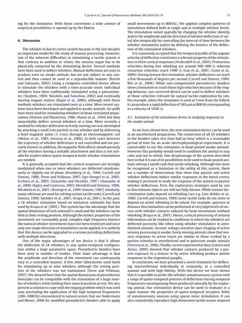
4.1. Limitation of the stimulation device in studying responses inthe awake animal
As we have shown here, the new stimulation device can be usedin an anesthetized preparation. The connection of all 24 whiskersto the benders takes less than half an hour which is a reasonableperiod of time for an acute electrophysiological experiment. It isconceivable to use this stimulator in head-posted awake animals.However this probably would need to condition the animal to staycalm and not to whisk. One disadvantage of the system describedhere is that it is out of its possibilities to be used in head-posted ani-mals solving a tactile task that needs whisking. Although this mustbe recognized as a limitation in the utility of this system, thereare a number of observations that show that passive and activewhisker deflections induce similar responses in the barrel cortex,making it pertinent to study the response of the system to passivewhisker deflections. First, the exploratory strategies used by ratsto discriminate objects are still not fully known. While texture dis-crimination tasks depend on whisking (see e.g. Guic-Robles et al.,1989; Carvell and Simons, 1990) some tactile tasks do not seem todepend on active whisking to be solved. For example, aperture ordistance discrimination can be performed at very high levels of suc-cess with whisker contacts induced by head movements withoutwhisking (Krupa et al., 2001). Hence, cortical processing of sensoryinformation can be studied in conditions in which the whiskers aredeflected passively like when using whisker stimulators in anes-thetized animals. Second, voltage-sensitive dyes imaging of activesensory processing in awake, freely moving animals show that sen-sory responses to active touch are similar to those evoked by apassive stimulus in anesthetized and in quiescent awake animals(Ferezou et al., 2006). Finally, recent experimental data (Lottem andAzouz, 2009) showed that whisker contacts produced by a pas-sive exposure to a texture or by active whisking produce similarresponses in the trigeminal ganglia.
In conclusion, we have presented a novel stimulator for deflect-ing macrovibrissae individually or conjointly, in a controlledmanner and with high fidelity. With this device we have shownthat it is possible to probe the whisker somatosensory system witha range of spatio-temporal patterns of deflections having temporal
frequencies encompassing those produced naturally by the explor-ing animal. Our stimulation device can be used to evaluate in arapid manner the properties of spatio-temporal receptive fieldsof somatosensory neurons using sparse noise stimulation. It canalso consistently reproduce high dimension tactile scenes inspired
7 oscien
fps
A
dA
A
t
R
A
A
B
B
B
C
C
C
D
E
E
F
F
G
G
G
4 V. Jacob et al. / Journal of Neur
rom natural multi-whisker stimulations and therefore enlarge theossibilities for studying the complex interactions acting betweenensory neurons receiving inputs from distinct whiskers.
cknowledgements
We thank Yves Boubenec for providing the high-speed cameraata. This work was supported by ANR (07-NEURO-025-01, NAT-CS) and FACETS (EC FP6-015879).
ppendix A. Supplementary data
Supplementary data associated with this article can be found, inhe online version, at doi:10.1016/j.jneumeth.2010.03.020.
eferences
ndermann ML, Moore CI. Mechanical resonance enhances the sensitivity of thevibrissa sensory system to near-threshold stimuli. Brain Res 2008;1235:74–81.
rabzadeh E, Zorzin E, Diamond ME. Neuronal encoding of texture in the whiskersensory pathway. PLoS Biol 2005;3:e17.
recht M, Sakmann B. Dynamic representation of whisker deflection by synapticpotentials in spiny stellate and pyramidal cells in the barrels and septa of layer4 rat somatosensory cortex. J Physiol 2002;543:49–70.
ringuier V, Chavane F, Glaeser L, Frégnac Y. Horizontal propagation of visual activityin the synaptic integration field of area 17 neurons. Science 1999;283:695–9.
rumberg JC, Pinto DJ, Simons DJ. Spatial gradients and inhibitory summation in therat whisker barrel system. J Neurophysiol 1996;76:130–40.
arvell GE, Simons DJ. Membrane potential changes in rat SmI cortical neu-rons evoked by controlled stimulation of mystacial vibrissae. Brain Res1988;448:186–91.
arvell GE, Simons DJ. Biometric analyses of vibrissal tactile discrimination in therat. J Neurosci 1990;10:2638–48.
elikel T, Sakmann B. Sensory integration across space and in time for decisionmaking in the somatosensory system of rodents. Proc Natl Acad Sci U S A2007;104:1395–400.
rew PJ, Feldman DE. Representation of moving wavefronts of whisker deflectionin rat somatosensory cortex. J Neurophysiol 2007;98:1566–80.
go-Stengel V, Mello e Souza T, Jacob V, Shulz DE. Spatiotemporal characteristicsof neuronal sensory integration in the barrel cortex of the rat. J Neurophysiol2005;93:1450–67.
rchova IA, Petersen RS, Diamond ME. Effect of developmental sensory and motordeprivation on the functional organization of adult rat somatosensory cortex.Brain Res Bull 2003;60:373–86.
erezou I, Bolea S, Petersen CC. Visualizing the cortical representation ofwhisker touch: voltage-sensitive dye imaging in freely moving mice. Neuron2006;50:617–29.
riedberg MH, Lee SM, Ebner FF. Modulation of receptive field properties of tha-lamic somatosensory neurons by the depth of anesthesia. J Neurophysiol1999;81:2243–52.
ao P, Bermejo R, Zeigler HP. Whisker deafferentation and rodent whisking
patterns: behavioral evidence for a central pattern generator. J Neurosci2001;21:5374–80.hazanfar AA, Nicolelis MA. Nonlinear processing of tactile information in the tha-lamocortical loop. J Neurophysiol 1997;78:506–10.
hazanfar AA, Stambaugh CR, Nicolelis MA. Encoding of tactile stimulus location bysomatosensory thalamocortical ensembles. J Neurosci 2000;20:3761–75.
ce Methods 189 (2010) 65–74
Grant RA, Mitchinson B, Fox CW, Prescott TJ. Active touch sensing in the rat: anticipa-tory and regulatory control of whisker movements during surface exploration.J Neurophysiol 2009;101:862–74.
Guic-Robles E, Valdivieso C, Guajardo G. Rats can learn a roughness discriminationusing only their vibrissal system. Behav Brain Res 1989;31:285–9.
Haidarliu S. An anatomically adapted, injury-free head-holder for guinea pigs. Phys-iol Behav 1996;60:111–4.
Harvey MA, Sachdev RN, Zeigler HP. Cortical barrel field ablation and unconditionedwhisking kinematics. Somatosens Mot Res 2001;18:223–7.
Higley MJ, Contreras D. Nonlinear integration of sensory responses in the rat barrelcortex: an intracellular study in vivo. J Neurosci 2003;23:10190–200.
Hipp J, Arabzadeh E, Zorzin E, Conradt J, Kayser C, Diamond ME, et al. Texture signalsin whisker vibrations. J Neurophysiol 2006;95:1792–9.
Hutson KA, Masterton RB. The sensory contribution of a single vibrissa’s corticalbarrel. J Neurophysiol 1986;56:1196–223.
Jacob V, Le Cam J, Ego-Stengel V, Shulz DE. Emergent properties of tactile scenesselectively activate barrel cortex neurons. Neuron 2008;60:1112–25.
Kleinfeld D, Delaney KR. Distributed representation of vibrissa movement in theupper layers of somatosensory cortex revealed with voltage-sensitive dyes. JComp Neurol 1996;375:89–108.
Krupa DJ, Matell MS, Brisben AJ, Oliveira LM, Nicolelis MA. Behavioral propertiesof the trigeminal somatosensory system in rats performing whisker-dependenttactile discriminations. J Neurosci 2001;21:5752–63.
Lottem E, Azouz R. Mechanisms of tactile information transmission through whiskervibrations. J Neurosci 2009;29:11686–97.
Maravall M, Petersen RS, Fairhall AL, Arabzadeh E, Diamond ME. Shifts in codingproperties and maintenance of information transmission during adaptation inbarrel cortex. PLoS Biol 2007;5:e19.
Melzer P, van der Loos H, Dörfl J, Welker E, Robert P, Emery D, et al. A magnetic deviceto stimulate selected whiskers of freely moving or restrained small rodents: itsapplication in a deoxyglucose study. Brain Res 1985;348:229–40.
Mirabella G, Battiston S, Diamond ME. Integration of multiple-whisker inputs in ratsomatosensory cortex. Cereb Cortex 2001;11:164–70.
Nicolelis MA, Fanselow EE. Thalamocortical [correction of Thalamcortical] opti-mization of tactile processing according to behavioral state. Nat Neurosci2002;5:517–23.
Nunez A, Barrenechea C, Avendano C. Spontaneous activity and responses to sensorystimulation in ventrobasal thalamic neurons in the rat: an in vivo intracellularrecording and staining study. Somatosens Mot Res 1994;11:89–98.
Rajan R, Bourke J, Cassell J. A novel stimulus system for applying tactile stimulito the macrovibrissae in electrophysiological experiments. J Neurosci Methods2006;157:103–17.
Ritt JT, Andermann ML, Moore CI. Embodied information processing: vibrissamechanics and texture features shape micromotions in actively sensing rats.Neuron 2008;57:599–613.
Rodgers KM, Benison AM, Barth DS. Two-dimensional coincidence detection in thevibrissa/barrel field. J Neurophysiol 2006;96:1981–90.
Sachdev RN, Sellien H, Ebner F. Temporal organization of multi-whisker contact inrats. Somatosens Mot Res 2001;18:91–100.
Shimegi S, Ichikawa T, Akasaki T, Sato H. Temporal characteristics of response inte-gration evoked by multiple whisker stimulations in the barrel cortex of rats. JNeurosci 1999;19:10164–75.
Simons DJ. Temporal and spatial integration in the rat SI vibrissa cortex. J Neuro-physiol 1985;54:615–35.
Stephen RO. Electro-mechanical harmonic stimulator. J Physiol 1969;202:71P–2P.Wolfe J, Hill DN, Pahlavan S, Drew PJ, Kleinfeld D, Feldman DE. Texture coding
in the rat whisker system: slip-stick versus differential resonance. PLoS Biol2008;6:e215.
Woolsey TA, Van der Loos H. The structural organization of layer IV in thesomatosensory region (S1) of mouse cerebral cortex. The description of a cor-tical field composed of discrete cytoarchitectural units. Brain Res 1970;17:205–42.