The human IL-15 superagonist N-803 promotes ... - PLOS
23
RESEARCH ARTICLE The human IL-15 superagonist N-803 promotes migration of virus-specific CD8+ T and NK cells to B cell follicles but does not reverse latency in ART-suppressed, SHIV- infected macaques Gabriela M. Webb ID 1,2 , Jhomary Molden 3 , Kathleen Busman-Sahay ID 1,2 , Shaheed Abdulhaqq ID 1,2 , Helen L. Wu ID 1,2 , Whitney C. Weber ID 1,2 , Katherine B. Bateman 1,2 , Jason S. Reed 1,2 , Mina Northrup ID 1,2 , Nicholas Maier ID 1,2 , Shiho Tanaka 4 , Lina Gao ID 2 , Brianna Davey ID 3 , Benjamin L. Carpenter 3 , Michael K. Axthelm ID 1 , Jeffrey J. Stanton 2 , Jeremy Smedley ID 1,2 , Justin M. Greene ID 1,2 , Jeffrey T. Safrit 5 , Jacob D. Estes ID 1,2 , Pamela J. Skinner ID 3 , Jonah B. Sacha ID 1,2 * 1 Vaccine & Gene Therapy Institute, Oregon Health & Science University, Beaverton, Oregon, United States of America, 2 Oregon National Primate Research Center, Oregon Health & Science University, Beaverton, Oregon, United States of America, 3 Department of Veterinary and Biomedical Sciences, University of Minnesota, St. Paul, Minnesota, United States of America, 4 ImmunityBio, Los Angeles, California, United States of America, 5 NantKWest, Los Angeles, California, United States of America * [email protected] Abstract Despite the success of antiretroviral therapy (ART) to halt viral replication and slow disease progression, this treatment is not curative and there remains an urgent need to develop approaches to clear the latent HIV reservoir. The human IL-15 superagonist N-803 (formerly ALT-803) is a promising anti-cancer biologic with potent immunostimulatory properties that has been extended into the field of HIV as a potential “shock and kill” therapeutic for HIV cure. However, the ability of N-803 to reactivate latent virus and modulate anti-viral immunity in vivo under the cover of ART remains undefined. Here, we show that in ART-suppressed, simian-human immunodeficiency virus (SHIV) SF162P3 -infected rhesus macaques, subcuta- neous administration of N-803 activates and mobilizes both NK cells and SHIV-specific CD8+ T cells from the peripheral blood to lymph node B cell follicles, a sanctuary site for latent virus that normally excludes such effector cells. We observed minimal activation of memory CD4+ T cells and no increase in viral RNA content in lymph node resident CD4+ T cells post N-803 administration. Accordingly, we found no difference in the number or magni- tude of plasma viremia timepoints between treated and untreated animals during the N-803 administration period, and no difference in the size of the viral DNA cell-associated reservoir post N-803 treatment. These results substantiate N-803 as a potent immunotherapeutic can- didate capable of activating and directing effector CD8+ T and NK cells to the B cell follicle during full ART suppression, and suggest N-803 must be paired with a bona fide latency reversing agent in vivo to facilitate immune-mediated modulation of the latent viral reservoir. PLOS PATHOGENS PLOS Pathogens | https://doi.org/10.1371/journal.ppat.1008339 March 12, 2020 1 / 23 a1111111111 a1111111111 a1111111111 a1111111111 a1111111111 OPEN ACCESS Citation: Webb GM, Molden J, Busman-Sahay K, Abdulhaqq S, Wu HL, Weber WC, et al. (2020) The human IL-15 superagonist N-803 promotes migration of virus-specific CD8+ T and NK cells to B cell follicles but does not reverse latency in ART- suppressed, SHIV-infected macaques. PLoS Pathog 16(3): e1008339. https://doi.org/10.1371/ journal.ppat.1008339 Editor: Guido Silvestri, Emory University, UNITED STATES Received: October 4, 2019 Accepted: January 20, 2020 Published: March 12, 2020 Copyright: © 2020 Webb et al. This is an open access article distributed under the terms of the Creative Commons Attribution License, which permits unrestricted use, distribution, and reproduction in any medium, provided the original author and source are credited. Data Availability Statement: All relevant data are within the manuscript and its Supporting Information files. Funding: This work was supported by National Institutes of Health (https://www.nih.gov/) grants R21 AI128970, R01 AI129703, and R01 AI140888 from the National Institute of Allergy and Infectious Diseases (NIAID) awarded to J.B.S.. Additionally, K01 OD026561 was awarded to J.M.G. and P51
-
Upload
khangminh22 -
Category
Documents
-
view
0 -
download
0
Transcript of The human IL-15 superagonist N-803 promotes ... - PLOS

RESEARCH ARTICLE
The human IL-15 superagonist N-803
promotes migration of virus-specific CD8+ T
and NK cells to B cell follicles but does not
reverse latency in ART-suppressed, SHIV-
infected macaques
Gabriela M. WebbID1,2, Jhomary Molden3, Kathleen Busman-SahayID
1,2,
Shaheed AbdulhaqqID1,2, Helen L. WuID
1,2, Whitney C. WeberID1,2, Katherine
B. Bateman1,2, Jason S. Reed1,2, Mina NorthrupID1,2, Nicholas MaierID
1,2, Shiho Tanaka4,
Lina GaoID2, Brianna DaveyID
3, Benjamin L. Carpenter3, Michael K. AxthelmID1, Jeffrey
J. Stanton2, Jeremy SmedleyID1,2, Justin M. GreeneID
1,2, Jeffrey T. Safrit5, Jacob
D. EstesID1,2, Pamela J. SkinnerID
3, Jonah B. SachaID1,2*
1 Vaccine & Gene Therapy Institute, Oregon Health & Science University, Beaverton, Oregon, United States
of America, 2 Oregon National Primate Research Center, Oregon Health & Science University, Beaverton,
Oregon, United States of America, 3 Department of Veterinary and Biomedical Sciences, University of
Minnesota, St. Paul, Minnesota, United States of America, 4 ImmunityBio, Los Angeles, California, United
States of America, 5 NantKWest, Los Angeles, California, United States of America
Abstract
Despite the success of antiretroviral therapy (ART) to halt viral replication and slow disease
progression, this treatment is not curative and there remains an urgent need to develop
approaches to clear the latent HIV reservoir. The human IL-15 superagonist N-803 (formerly
ALT-803) is a promising anti-cancer biologic with potent immunostimulatory properties that
has been extended into the field of HIV as a potential “shock and kill” therapeutic for HIV
cure. However, the ability of N-803 to reactivate latent virus and modulate anti-viral immunity
in vivo under the cover of ART remains undefined. Here, we show that in ART-suppressed,
simian-human immunodeficiency virus (SHIV)SF162P3-infected rhesus macaques, subcuta-
neous administration of N-803 activates and mobilizes both NK cells and SHIV-specific
CD8+ T cells from the peripheral blood to lymph node B cell follicles, a sanctuary site for
latent virus that normally excludes such effector cells. We observed minimal activation of
memory CD4+ T cells and no increase in viral RNA content in lymph node resident CD4+ T
cells post N-803 administration. Accordingly, we found no difference in the number or magni-
tude of plasma viremia timepoints between treated and untreated animals during the N-803
administration period, and no difference in the size of the viral DNA cell-associated reservoir
post N-803 treatment. These results substantiate N-803 as a potent immunotherapeutic can-
didate capable of activating and directing effector CD8+ T and NK cells to the B cell follicle
during full ART suppression, and suggest N-803 must be paired with a bona fide latency
reversing agent in vivo to facilitate immune-mediated modulation of the latent viral reservoir.
PLOS PATHOGENS
PLOS Pathogens | https://doi.org/10.1371/journal.ppat.1008339 March 12, 2020 1 / 23
a1111111111
a1111111111
a1111111111
a1111111111
a1111111111
OPEN ACCESS
Citation: Webb GM, Molden J, Busman-Sahay K,
Abdulhaqq S, Wu HL, Weber WC, et al. (2020) The
human IL-15 superagonist N-803 promotes
migration of virus-specific CD8+ T and NK cells to
B cell follicles but does not reverse latency in ART-
suppressed, SHIV-infected macaques. PLoS
Pathog 16(3): e1008339. https://doi.org/10.1371/
journal.ppat.1008339
Editor: Guido Silvestri, Emory University, UNITED
STATES
Received: October 4, 2019
Accepted: January 20, 2020
Published: March 12, 2020
Copyright: © 2020 Webb et al. This is an open
access article distributed under the terms of the
Creative Commons Attribution License, which
permits unrestricted use, distribution, and
reproduction in any medium, provided the original
author and source are credited.
Data Availability Statement: All relevant data are
within the manuscript and its Supporting
Information files.
Funding: This work was supported by National
Institutes of Health (https://www.nih.gov/) grants
R21 AI128970, R01 AI129703, and R01 AI140888
from the National Institute of Allergy and Infectious
Diseases (NIAID) awarded to J.B.S.. Additionally,
K01 OD026561 was awarded to J.M.G. and P51

Author summary
IL-15 regulates NK and memory T cell homeostasis and is therefore being explored for
clinical immunotherapy of chronic diseases like cancer and HIV. To explore the applica-
bility of the clinical grade IL-15 superagonist N-803 to HIV cure strategies we tested the
impact of N-803 on host immunity and latent virus in SHIV-infected rhesus macaques.
Our results suggest that N-803 beneficially modulates effector NK and CD8+ T cells by
expanding the numbers of these cells and redistributing them to lymph node B cell folli-
cles, a site known to harbor persistent latent virus during ART. However, our results fur-
ther suggest that N-803 does not perturb the viral reservoir present in memory CD4+ T
cells and that in order to fully unlock the immunotherapeutic potential of N-803 it must
be paired with latency reversal agents.
Introduction
The inability of antiretroviral therapy (ART) to clear the latent HIV reservoir, paired with the
difficulties of life-long ART adherence, have shifted the focus of HIV research towards the
development of therapies able to achieve a functional cure, namely ART-free remission from
HIV. Proof-of-concept for HIV cure was provided by Timothy Brown, the “Berlin patient,” an
HIV-positive individual who received a hematopoietic stem cell transplant from a CCR5Δ32/
Δ32 donor as part of his treatment for leukemia [1]. Now over a decade after his transplant and
discontinuation of ART, Timothy Brown shows no signs of HIV and continues to demonstrate
that durable ART-free remission is an attainable goal [2]. Indeed, this remarkable result was
recently confirmed by the report of the “London patient” [3]. HIV research has thus expanded
its scope to include a wide variety of curative strategies including the most studied approach,
“shock and kill” therapies [4]. These therapies function on the hypothesis that latency-revers-
ing agents (LRAs) activate and flush out latent HIV for subsequent recognition and clearance
by effector NK and CD8+ T cells. Several immune-based therapies are candidates to combat
latent HIV and among these are the common γ-chain cytokines due to their capacity for robust
T cell activation.
Common γ-chain cytokines have been previously explored as candidates to decrease HIV
reservoir size in patients on ART. Interleukin-2 (IL-2) failed to decrease the latent HIV bur-
den, while IL-7 modestly expanded the reservoir commensurate with its ability to expand CD4
+ T cell numbers [5,6]. Furthermore, these cytokines did not disrupt viral latency. IL-15, how-
ever, a critical mediator of NK cell and T cell activation and proliferation, induces NK cell
expansion and preferential proliferation of both CD4+ and CD8+ effector memory T cells in
non-human primate models of HIV [7–9]. In vitro, IL-15 effectively reactivates HIV produc-
tion in latently infected cells [10]. Therapeutic use of free IL-15, however, is precluded by its
rapid plasma clearance and the high dose needed to achieve biological responses in vivo.
Unlike IL-2 and IL-7, IL-15 is a part of a heterodimeric complex formed by a tight association
between single-chain IL-15 and IL-15Rα [11]. Novel IL-15-based therapies such as N-803 (for-
merly ALT-803) and hetIL-15 [12] are now designed to mimic this association, thereby over-
coming limitations of free IL-15. These compounds have already shown promise as potential
immunotherapeutics in the context of HIV/SIV [12–14].
N-803 consists of a novel IL-15 mutant (N72D), containing an asparagine to aspartic acid
mutation at position 72, which forms a stable heterodimeric complex with the alpha subunit of
the IL-15 receptor (IL-15Rα). The N72D mutation in IL-15 is crucial for N-803 as it confers a
PLOS PATHOGENS N-803 and latent SHIV
PLOS Pathogens | https://doi.org/10.1371/journal.ppat.1008339 March 12, 2020 2 / 23
OD011092 from the NIH Office of the Director
awarded to the Oregon National Primate Research
Center. The content is solely the responsibility of
the authors and does not necessarily represent the
official views of the National Institutes of Health.
The funders had no role in study design, data
collection and analysis, decision to publish, or
preparation of the manuscript.
Competing interests: I have read the journal’s
policy and the authors of this manuscript have the
following competing interests: Jeffrey Safrit is an
employee of NantBioScience Inc./NantKWest LLC,
the maker of N-803. Shiho Tanaka is an employee
of ImmunityBio, a branch of NantKWest
responsible for performing the ADA assay. No
other authors have declared that competing
interests exist.

5-fold increase in its biological activity as compared to free IL-15 [15]. This complex is
expressed as a structurally modified human IL-15N72D:IL15Rα:IgG1 Fc fusion protein that
exhibits 25-fold higher biological activity and 35-fold longer serum half-life than soluble IL-15,
which ultimately promotes stimulation of NK and memory T cells [16]. Accordingly, N-803
engenders potent anti-tumor NK and T cell immunity in small animal cancer models and is
currently being tested as an immunotherapeutic for both solid and hematologic cancers in sev-
eral different Phase I/II clinical trials (ClinicalTrials.gov; NCT01946789, NCT02099539).
Moreover, N-803 is well-tolerated in both mice and cynomolgus macaques at doses as high as
0.1 mg/kg and importantly, does not induce global cytokine storm release of pro-inflammatory
cytokines [17]. Given the promising results in the field of cancer immunology, N-803 has also
been explored as a means to directly enhance anti-viral immune responses in chronic infec-
tions such as HIV [13,14]. For example, in a humanized mouse model of HIV, early adminis-
tration of N-803 induced NK cell cytotoxicity and inhibited acute HIV replication [18]. More
recently, N-803 demonstrated the remarkable ability to both reverse HIV latency and enhance
CD8+ T cell recognition of HIV-infected cells in a primary in vitro cell culture model [10].
Furthermore, we have shown that a single dose of N-803 directs SIV-specific CD8+ T cells into
B-cell follicles of chronically SIV-infected elite controller rhesus macaques [13]. These data
support the hypothesis that N-803 could mediate both the “shock” and the “kill” in cure
approaches for HIV, but as yet N-803 has not been tested in ART-suppressed macaques.
To explore this hypothesis, we evaluated the effect of the clinical grade IL-15 superagonist
N-803 on the latent viral reservoir in SHIV-infected, ART-suppressed rhesus macaques. We
demonstrate that biweekly, subcutaneous dosing of N-803 results in repeated proliferation of
NK and memory CD8+ T cells. Within five days of N-803 administration, proliferating cells
redistribute from blood to lymph nodes. Both NK cells and antigen-specific CD8+ T cells then
gain access to B cell follicles within lymph nodes, a critical anatomical location that harbors
latent virus. In contrast, we observed minimal activation and migration of CD4+ T cells.
Although N-803 had no effect on reversing latency in vivo or on diminishing the viral cell-
associated DNA reservoir, animals that received N-803 displayed a trend towards delayed viral
rebound kinetics after ART interruption. Collectively, our data provide evidence that in the
context of ART-suppression, N-803 can drive critical effector cells to sanctuary sites for viral
replication and is a powerful tool for combinatorial HIV eradication strategies.
Results
Recent studies in mice have shown greater tissue biodistribution of N-803 to lymphoid organs
when administered by subcutaneous administration as compared to intravenous administra-
tion [19]. In humans, subcutaneous versus intravenous administration resulted in a signifi-
cantly longer serum half-life, decreased serum levels of pro-inflammatory cytokines, as well as
sustained and significantly increased activation and proliferation of both NK cells and CD8+ T
cells [20,21]. We have thus adjusted our dosing route for the current study from intravenous
to subcutaneous. We infected 10 rhesus macaques (Macaca mulatta) intravenously with
SHIVSF162P3 and initiated daily subcutaneous ART injections 14 days post-infection in order
to establish the viral reservoir while allowing for the priming of SHIV-specific CD8+ T cell
responses (Fig 1A) [22]. Because the MHC-I alleles Mamu-B�008:01 (B�08) and Mamu-B�017:01 (B�17) restrict particularly potent antiviral CD8+ T cell responses we selected and
balanced animals positive for these alleles between the treatment and control groups to ensure
the presence of high frequency anti-SHIV CD8+ T cells and to facilitate downstream in situMHC-I tetramer staining (Fig 1B). Although these MHC-I alleles are associated with sponta-
neous elite control of chronic phase viral replication, this effect does not manifest until six
PLOS PATHOGENS N-803 and latent SHIV
PLOS Pathogens | https://doi.org/10.1371/journal.ppat.1008339 March 12, 2020 3 / 23

weeks post-infection at the earliest [23]. Therefore, no impact of these MHC-I molecules on
viral replication was expected given that we began ART at two weeks post-infection. Following
ART initiation, plasma viremia declined to undetectable levels in all animals by week eight.
We continued daily ART for 20 weeks, wherein one group (n = 6) received four subcutaneous
infusions of 100 μg/kg N-803 every other week over the course of eight weeks and the control
group (n = 4) was left untreated. We continued to monitor viral loads throughout the course
of N-803 treatments to look for episodes of virus reactivation (Fig 1A).
We, and others, have previously established that N-803 induces proliferation of immune
effector cells, namely CD8+ T cells and NK cells, in SIV-infected RMs [13,14]. However, the
effect is transient and resolves within 7–14 days post-administration. Repeated, weekly dosing
Fig 1. Plasma viral loads after infection with SHIVsf162p3 and before discontinuation of ART. We intravenously infected rhesus macaques
(n = 10) with SHIVsf162p3 and initiated ART at week 2. The N-803 group (n = 6, red) received 4 subcutaneous infusions of N-803 (100 μg/kg) every
other week, indicated by vertical dashed lines between weeks 22–28. The control group (n = 4, blue) was left untreated. Horizontal dotted line
indicates limit of detection (LOD) at 50 RNA copies/mL. (A) Plasma viral loads shown prior to ART discontinuation and during N-803 treatment.
(B) Table of research animal characteristics including animal identification number, MHC type, sex, age, and the group to which the animal was
assigned.
https://doi.org/10.1371/journal.ppat.1008339.g001
PLOS PATHOGENS N-803 and latent SHIV
PLOS Pathogens | https://doi.org/10.1371/journal.ppat.1008339 March 12, 2020 4 / 23

of N-803 results in modulation of IL-15 receptor surface expression, including both IL-15/IL-
2β (CD122) and the common γC (CD132) [14]. The decline in receptor surface expression
likely reduces the responsiveness of T cells and NK cells to IL-15 over time. Hence, we admin-
istered N-803 every other week for eight weeks in order to allow for recovery of IL-15 respon-
siveness. Similar to previous reports on the effects of N-803 administered intravenously
[13,14], the subcutaneous, bi-weekly dosing regimen used here resulted in a decrease in the
absolute numbers of white blood cells, specifically CD8+ T cells and CD16+ NK cells, in blood
one day post N-803 administration, likely due to trafficking into tissue [13,24], followed rap-
idly by an increase in the numbers of these cells in the blood (S1A and S1B Fig). In contrast,
we observed no increase in the number of CD4+ T cells in blood following subcutaneous N-
803 administration. These repeated episodes of substantial expansion by NK and CD8+ T cells
in the blood, and minimal CD4+ T cell activation, following subcutaneous N-803 were
reflected in increases in proliferative activity as measured by expression of the proliferation
marker Ki-67 in blood (Fig 2). We observed increased levels of Ki-67 expression in all memory
subpopulations of CD8+ T cells, however, we only observed notable increases in the absolute
numbers of effector and central memory CD8+ T cells in the blood after repeated N-803 treat-
ments (Figs 3A and S2A). N-803 had minimal effects on memory subpopulations of CD4+ T
cells, triggering only modest increases in Ki-67 expression in the effector memory population
(Figs 3B and S2B). In line with minimal increased Ki-67 expression in memory CD4+ T cells,
there was no associated increase in plasma viral loads of these animals during administration
of N-803 (Fig 1A), suggesting that N-803 does not function as an LRA in vivo.
Fig 2. Whole blood analysis during in vivo administration of N-803. N-803 was subcutaneously administered every other week as indicated by the vertical dashed lines.
Blood was collected at time 0 before the N-803 injection and at days 1, 3, 5, 7 after each injection of N-803. Induction of proliferation marker Ki67 on CD16+ NK cells,
CD4+ and CD8+ T cells as percent of lymphocyte subset on the left, absolute cell counts on the right, both shown as a percent change from baseline. Absolute counts were
calculated based on the percentage of the particular cell subset and the WBC count. Data shown are means (± SEM) of combined data from all animals within the
designated group.
https://doi.org/10.1371/journal.ppat.1008339.g002
PLOS PATHOGENS N-803 and latent SHIV
PLOS Pathogens | https://doi.org/10.1371/journal.ppat.1008339 March 12, 2020 5 / 23

Fig 3. T cell memory analysis in blood during in vivo administration of N-803. (A) Induction of proliferation marker Ki67 on memory CD8+ T cells as percent of
subset on the left, absolute cell counts on the right, both shown as a percent change from baseline. Absolute counts were calculated based on the percentage of the
particular cell subset and the WBC count. (B) Induction of proliferation marker Ki67 on memory CD4+ T cells as percent of subset on the left, absolute cell counts on
the right, both shown as a percent change from baseline. Absolute counts were calculated based on the percentage of the particular cell subset and the WBC count.
Data shown are means (± SEM) of combined data from all animals within the designated group.
https://doi.org/10.1371/journal.ppat.1008339.g003
PLOS PATHOGENS N-803 and latent SHIV
PLOS Pathogens | https://doi.org/10.1371/journal.ppat.1008339 March 12, 2020 6 / 23
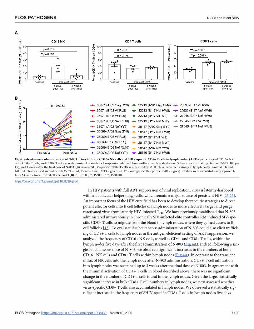
In HIV patients with full ART suppression of viral replication, virus is latently-harbored
within T follicular helper (TFH) cells, which remain a major source of persistent HIV [25,26].
An important focus of the HIV cure field has been to develop therapeutic strategies to direct
potent effector cells into B cell follicles of lymph nodes to more effectively target and purge
reactivated virus from latently HIV-infected TFH. We have previously established that N-803
administered intravenously in chronically SIV-infected elite controller RM induced SIV-spe-
cific CD8+ T cells to migrate from the blood to lymph nodes, where they gained access to B
cell follicles [13]. To evaluate if subcutaneous administration of N-803 could also elicit traffick-
ing of CD8+ T cells to lymph nodes in the antigen-deficient setting of ART suppression, we
analyzed the frequency of CD16+ NK cells, as well as CD4+ and CD8+ T cells, within the
lymph nodes five days after the first administration of N-803 (Fig 4A). Indeed, following a sin-
gle subcutaneous dose of N-803, we observed significant increases in the numbers of both
CD16+ NK cells and CD8+ T cells within lymph nodes (Fig 4A). In contrast to the transient
influx of NK cells into the lymph node after N-803 administration, CD8+ T cell infiltration
into lymph nodes was sustained up to 3 weeks after the final dose of N-803. In agreement with
the minimal activation of CD4+ T cells in blood described above, there was no significant
change in the number of CD4+ T cells found in the lymph nodes. Given the large, statistically
significant increase in bulk CD8+ T cell numbers in lymph nodes, we next assessed whether
virus-specific CD8+ T cells also accumulated in lymph nodes. We observed a statistically sig-
nificant increase in the frequency of SHIV-specific CD8+ T cells in lymph nodes five days
Fig 4. Subcutaneous administration of N-803 drives influx of CD16+ NK cells and SHIV-specific CD8+ T cells to lymph nodes. (A) The percentage of CD16+ NK
cells, CD4+ T cells, and CD8+ T cells were determined in single-cell suspensions derived from axillary lymph nodes before, 5 days after the first injection of N-803 (100 μg/
kg), and 3 weeks after the final dose of N-803. (B) Percent SHIV-specific CD8+ T cells as measured by MHC class I tetramer staining in lymph nodes. Animal IDs and
MHC-I tetramer used are indicated (35071 = red, 35069 = blue, 32213 = green, 28147 = orange, 25536 = purple, 27045 = grey). P values were calculated using a paired t-
test (A), and a linear mixed effects model (B). �, P<0.05; ��, P<0.01; ���, P<0.001.
https://doi.org/10.1371/journal.ppat.1008339.g004
PLOS PATHOGENS N-803 and latent SHIV
PLOS Pathogens | https://doi.org/10.1371/journal.ppat.1008339 March 12, 2020 7 / 23
following N-803 treatment, as assessed by MHC-I tetramer staining of single cell suspensions
of disaggregated lymph node (Fig 4B). This increase of SHIV-specific CD8+ T cells in lymph
nodes was maintained for at least as long as three weeks after the final N-803 dose (S3A Fig).
We next examined whether these virus-specific CD8+ T cells were able to gain access to B cell
follicles of lymph nodes, an important anatomical site that harbors latent HIV and SIV. In
order to assess this, we performed in situ SHIV-specific MHC-I tetramer staining of lymph
node sections from five SHIV-infected RMs before and five days after subcutaneous adminis-
tration of N-803. One macaque was excluded from all fixed tissue analysis due to high adipose
content resulting in poor lymph node architecture. Prior to N-803 treatment, SHIV-specific
CD8+ T cells primarily localized to the extrafollicular area within lymph nodes, and were
largely excluded from the B cell follicles (Fig 5A). Five days after treatment with N-803, the fre-
quency of SHIV-specific CD8+ T cells increased within the lymph nodes, achieving a statisti-
cally significant increase in the B cell follicle (Fig 5B and 5C). Given that we observed an
increase in NK cell numbers in lymph nodes following N-803 treatment, we assessed the ana-
tomical localization of effector NK cells within lymph nodes. Consistent with the results
obtained for SHIV-specific CD8+ T cells, we observed a statistically significant increase of
CD3-CD159a+ NK cells within B cell follicles five days after subcutaneous treatment with N-
803 (Fig 6). Interestingly, we found a notable population of NK cells localized within blood
vessels prior to N-803 treatment. Following N-803 administration, NK cells were primarily
localized to the B cell follicle, and in some cases, NK cells were found within the extrafollicular
area near, but not within blood vessels. This suggests that the increase in NK cells was due to
extravasation instead of local proliferation. Finally, because of the increased NK and virus-spe-
cific CD8+ T cell trafficking into B cell follicles post subcutaneous N-803 administration, we
evaluated the expression of CXCR5 on the surface of these cells using single cell suspensions of
disaggregated lymph nodes taken before and five days post N-803 administration (Fig 7), as
well as 3 weeks after the last dose of N-803 (S3B and S3C Fig). Despite finding higher numbers
of both SHIV-specific CD8+ T cells and NK cells within B cell follicles following N-803 admin-
istration, we observed a statistically significant decrease in surface CXCR5 expression on
lymph node-resident CD8+ T cells and a trend towards lower CXCR5 expression on NK cells
(Fig 7). This paradoxical finding could be the result of temporal regulation of CXCR5 expres-
sion following N-803 activation or an indication that penetration of NK and CD8+ T cells into
lymph node B cell follicles occurs via another undefined mechanism.
Recent data has shown that N-803 is able to reactivate latent virus from PBMC of ART-
treated HIV-infected patients ex vivo [10]. However, there remains a lack of concordance
between in vitro and in vivo HIV latency reversal activity. Hence, we sought to determine if N-
803 could act as an LRA in vivo and induce episodes of virus reactivation when administered
to fully ART-suppressed, SHIV-infected RMs. We measured plasma viral loads at 0, 1, 2, 3,
and 7 days post administration during the course of N-803 treatment where we detected tran-
sient episodes of SHIV plasma RNA in RMs from both the untreated control group and the
group that received subcutaneous N-803 (Fig 8A). We found no significant difference between
the treatment and control groups in either the magnitude of the viral blips as measured by area
under the curve (AUC) (Fig 8B), or in the number of detectable plasma SHIV RNA timepoints
(Fig 8C). Additionally, we assessed for LRA activity by measuring the number of viral RNA
+ cells present within the lymph node before and five days after N-803 treatment via quantita-
tive RNAscope. In line with the lack of effect on plasma viral load, we observed no statistically
significant change in the number of SHIV RNA+ cells in lymph node following N-803 treat-
ment (Fig 8D). All animals received lymph node biopsies prior to and during the course of N-
803 treatment, including the control group. The ‘blips’ in SHIV plasma RNA may be attributed
to the additional stress of frequent tissue biopsies considering that the animals were fully ART-
PLOS PATHOGENS N-803 and latent SHIV
PLOS Pathogens | https://doi.org/10.1371/journal.ppat.1008339 March 12, 2020 8 / 23

suppressed with undetectable plasma viremia for 11 weeks before N-803, and that only directly
after the first biopsies were taken at week 19 did they have detectable virus in their plasma.
Regardless, none of the results presented here support N-803 as an LRA in vivo. This is in
agreement with a report published during the review of this manuscript describing that N-803
alone does not reserve SIV latency in vivo [27].
To assess if N-803 treatment impacted the size of the latent viral reservoir, we quantified
cell-associated viral DNA in CD4+ T cells isolated from various tissues, including mesenteric
and peripheral lymph nodes, spleen, and gastrointestinal mucosa, before and after all N-803
Fig 5. SHIV-specific CD8+ T cells increase in follicular and extrafollicular regions of lymph nodes after N-803 treatment in ART-suppressed macaques.
Representative images showing Mamu-B�08 RL9 tetramer+ cells (red) and CD20+ cells (green) in lymph node sections from animal 35069, (A) before and (B) 5 days after
N-803 treatment. CD20 staining is used to define B cell follicles (F) and extrafollicular (EF) regions. Tetramer-binding cell are indicated with white arrowheads. Scale bars
indicate 200 μm (left panels) and 100 μm (right panels). (C) The numbers of tetramer+ CD8+ T cells per millimeter squared in B cell follicles and extrafollicular area
before and 5 days after N-803 treatment. P values were calculated using a linear mixed effects model. �, P<0.05; ��, P<0.01; ���, P<0.001.
https://doi.org/10.1371/journal.ppat.1008339.g005
PLOS PATHOGENS N-803 and latent SHIV
PLOS Pathogens | https://doi.org/10.1371/journal.ppat.1008339 March 12, 2020 9 / 23
treatments. We observed similar levels of cell-associated viral DNA in the control and N-803
groups both before and following treatment, with the exception of the spleen which showed
divergent levels of viral DNA at baseline, indicating that subcutaneous administration of N-
803 by itself was not sufficient to decrease the viral reservoir (Fig 9). Importantly, unlike other
common-γ chain cytokines such as IL-2 and IL-7, N-803 did not expand the viral reservoir
[5,6].
Although we did observe blips in plasma viral loads prior to ART discontinuation, these
events were not specific to N-803 administration as they occurred in both control and treat-
ment groups. Additionally, CD4+ cell-associated viral DNA levels remained unchanged
between the groups after N-803 treatment. We then wanted to determine if repeated N-803
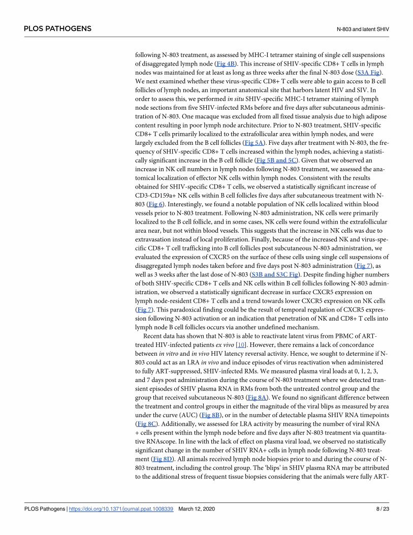
Fig 6. N-803 triggers NK cell migration from blood vessels and extrafollicular spaces into B cell zones of lymph nodes. Representative images from peripheral lymph
nodes before (A) and 5 days after (B) N-803 treatment show a significant increase of NK cells (arrowheads) within B cell follicles (F) with only a modest increase in NK
cells found in extrafollicular (EF) regions. Notably, many NK cells prior to N-803 treatment are found within blood vessels (yellow arrowheads), while most NK cells
following N-803 treatment were found within the lymph node parenchyma (white arrowheads), specifically adjacent to or within B cell follicles. Scale bar is 100 μm. (C)
Quantification of CD159a+CD3- cells within lymph nodes 5 days post-N-803 treatment demonstrated a significant increase in the number of NK cells found within B cell
follicles. P values were calculated using a paired t-test. �, P<0.05; ��, P<0.01; ���, P<0.001.
https://doi.org/10.1371/journal.ppat.1008339.g006
PLOS PATHOGENS N-803 and latent SHIV
PLOS Pathogens | https://doi.org/10.1371/journal.ppat.1008339 March 12, 2020 10 / 23

treatment would affect the time to viral rebound after ART-interruption, defined as two
detectable episodes above 62 copies/mL. At 37 weeks post-infection, nine weeks after the last
dose of N-803 was administered, ART was discontinued and we monitored for any changes in
Fig 7. Decreased expression of CXCR5 on SHIV-specific CD8+ T cells and NK cells after N-803 in lymph nodes. (A) CXCR5 staining on SHIV-specific CD8+ T cells
and (B) NK cells in peripheral lymph nodes before and after N-803 treatment. P values were calculated using a linear mixed effects model (A) and a paired t-test (B). �,
P<0.05; ��, P<0.01; ���, P<0.001.
https://doi.org/10.1371/journal.ppat.1008339.g007
Fig 8. N-803 does not reverse latency in vivo. (A) Plasma viral loads shown during N-803 administration. N-803 was administered subcutaneously at a dose of 100 μg/kg
every other week, indicated by the vertical dashed lines. Horizontal dotted line indicates limit of detection (LOD) at 50 RNA copies/mL. (B) Area under the curve analysis
of viral loads between groups between weeks 22–35. (C) Number of detectable SHIV RNA timepoints between groups. (D) Quantification of RNAscope in situhybridization in axillary lymph nodes before, 5 days after N-803 and 7 days after N-803. Shown as SHIV RNA+ cells per 100,000 cells. P values were calculated using
Mann-Whitney test (B, C) and a paired t-test (D). �, P<0.05; ��, P<0.01; ���, P<0.001.
https://doi.org/10.1371/journal.ppat.1008339.g008
PLOS PATHOGENS N-803 and latent SHIV
PLOS Pathogens | https://doi.org/10.1371/journal.ppat.1008339 March 12, 2020 11 / 23

the SHIV rebound kinetics (Fig 10A). We measured viral RNA in the plasma from all animals
following ART cessation and all animals from both groups reached detectable levels of plasma
viral RNA. Interestingly, the animals treated with N-803 displayed a trend towards delayed
viral rebound kinetics as compared to the control animals (Fig 10B–10D). This trend could be
due to the low animal numbers in the control group or to other unknown parameters (Fig
10B–10D). However, we took precautions to balance the animals in the groups such that the
peak plasma viral loads and area AUC of plasma viremia prior to ART initiation were not sta-
tistically different between groups (S4A and S4B Fig). We also found no correlation between
Fig 9. N-803 treatment does not perturb the viral reservoir. Cell-associated SHIV DNA from CD4-sorted, single-cell suspensions of tissues (mesenteric lymph nodes,
peripheral lymph nodes, spleen, and colon) before N-803 and after all 4 injections of N-803. Data shown are means (± SEM). P values were calculated using a Mann-
Whitney test. �, P<0.05; ��, P<0.01; ���, P<0.001.
https://doi.org/10.1371/journal.ppat.1008339.g009
Fig 10. Animals that receive N-803 exhibit a slight delay in virus rebound after ART discontinuation. (A) Dynamics of SHIV viral load in plasma after ART release.
Grey box indicates the end of ART regimen. Control group is shown on the left in blue and the N-803 group is shown on the right in red. Bold black line in each graph
shows the geometric mean of each group. (B) Geometric means of both groups. (C) Area under the curve during the time of ART discontinuation to necropsy (week 37–
46.5) (D) Kaplan-Meier curve showing the percent of animals with no viral rebound. Horizontal dotted line indicates limit of detection (LOD) at 62 RNA copies/mL. P
values were calculated using a Mann-Whitney test (C), and log-rank test (D). �, P<0.05; ��, P<0.01; ���, P<0.001.
https://doi.org/10.1371/journal.ppat.1008339.g010
PLOS PATHOGENS N-803 and latent SHIV
PLOS Pathogens | https://doi.org/10.1371/journal.ppat.1008339 March 12, 2020 12 / 23

acute peak viral load and the time to rebound, although a correlation between peak acute
phase viral load and peak viral load post ART cessation did exist (S4C Fig). Given that these
animals had no significant differences in their virus reservoir size, this delay in rebound could
be the result of an increase in effector cells, either or both NK cells and CD8+ T cells, elicited
by N-803, thus providing the animals with more immune control, albeit transient, of rebound-
ing virus following ART cessation.
Discussion
Because of its ability to potently induce NK and T cell proliferation in cancer models
(3,5,11,12), N-803, a novel IL-15 superagonist, is now a promising candidate for targeting
and eradicating the HIV reservoir. In a humanized mouse model, treatment with N-803 led to
suppression of acute HIV infection by increasing the antiviral activity of NK cells [18]. Addi-
tionally, in vitro evidence suggests N-803 may serve as a latency-reversal agent [10]. These
data, together with our recent data showing that N-803 can drive SIV-specific CD8+ T cells to
lymph nodes in SIV-infected RM [13], suggest that N-803 has the potential to flush virus out
of latency and enhance killing of infected cells. Here we sought to determine if N-803 immu-
notherapy could indeed serve as both arms of the “shock and kill” strategy of HIV cure. In
this study, we infected rhesus macaques with SHIVSF162P3 and initiated ART 14 days post-
infection to sufficiently seed the viral reservoir [22,28]. While on ART, animals in the N-803
group received subcutaneous doses of N-803 every other week and we did not observe any
notable differences in episodes of viral reactivation between the N-803 group and the control
group. While our dosing regimen of N-803 elicited repeated and potent proliferation of NK
cells and effector memory CD8+ T cells, as well as migration of these effectors to B cell follicles
of lymph nodes, N-803 failed to impact the size of the viral reservoir. Interestingly, upon dis-
continuation of ART, animals that received N-803 experienced a slight delay in virus recrudes-
cence. Our results reveal that although N-803 enhances trafficking of effector cells within B
cell follicles of lymph nodes, it is not sufficient by itself to clear the latent virus reservoir, which
further substantiates the need for combinatorial HIV cure approaches moving forward.
We previously defined that intravenous injection of N-803 triggers the influx of CD8+ T
cells into lymph nodes of SIV-infected rhesus macaques [13]. Recent reports have compared
the safety, tolerability, pharmacokinetics, immunologic events, and biodistribution between
subcutaneous and intravenous routes of N-803 administration [21,29]. Although subcutane-
ous N-803 administration leads to 100-fold less serum concentrations compared to intrave-
nous administration, it exhibits greater biodistribution to lymphoid organs. N-803 given via
either route, however, induced comparable proliferation and activation of CD8+ T cells and
NK cells. We have, therefore, adjusted our dosing route of N-803 from intravenous to subcuta-
neous, in order to more appropriately mirror the dosing route used in clinical trials currently
testing N-803 in humans (NCT02523469, NCT03381586, NCT03054909, NCT03022825).
Indeed, we found N-803 was still able to promote proliferation and activation of NK cells and
T cells when administered subcutaneously. We also found that even with the development of
anti-drug antibodies in four out of six of our treated animals, N-803 remained efficacious (S5
and S6 Figs). Interestingly, in contrast to our studies using intravenously administered N-803,
we observed an influx of both CD8+ T cells and NK cells into lymph nodes with our subcuta-
neous administration of N-803. This could be due to the increased tissue biodistribution of N-
803 observed via subcutaneous administration [29]. In addition to modifying our route of N-
803 administration, we altered the timing of our dosing in order to achieve repeated prolifera-
tion of NK cells and T cells. We and others have shown that once a week dosing induces a
burst in effector memory T cell and NK cell proliferation that then diminishes upon further
PLOS PATHOGENS N-803 and latent SHIV
PLOS Pathogens | https://doi.org/10.1371/journal.ppat.1008339 March 12, 2020 13 / 23

injections, likely due to rapid recycling of the IL-15 receptors (CD122 and CD132) [8,14]. To
mitigate this effect and allow for the recovery of IL-15 responsiveness, we adjusted our dosing
schedule to biweekly N-803 and we were able to elicit potent proliferative responses in T cells
and NK cells after each dose of N-803. Thus, with biweekly subcutaneous N-803 administra-
tion, we have achieved an effective dosing regimen for repeated proliferation of effector cells as
well as trafficking of both NK cells and SHIV-specific CD8+ T cells from blood to B cell
follicles.
The “shock-and-kill” strategy for HIV cure aims to combine a latency-reversing agent with
effective immune-mediated killing of infected cells [30]. N-803 has recently been shown to
induce HIV transcription from both primary cell models of latency and from ex vivo patient
samples [10]. Therefore, we hypothesized that N-803 could act as both a latency-reversal agent
and a potent immune-stimulant. To test this, we administered N-803 to rhesus macaques on
suppressive ART and monitored the impact on plasma viremia. We found there to be no
appreciable difference in the magnitude or number of transient plasma viremia events between
animals that received N-803 versus animals that were left untreated. This was further con-
firmed via RNAscope of lymph node samples taken at five days post N-803 administration.
Although a dose of 100 μg/kg N-803 given intravenously can reach maximum serum concen-
trations (Cmax) of 30 nM in cynomolgus macaques [17], recent findings demonstrate a
100-fold lower Cmax of N-803 when administered subcutaneously compared to intravenously
in mice [29]. Therefore, the lack of latency reversal activity could be due to the lower maxi-
mum serum concentration Cmax of N-803 achieved with subcutaneous dosing. Given that ani-
mals in both groups experienced episodes of reactivation, it can also be argued that 20 weeks
of ART suppression was not sufficient time to achieve complete viral suppression. Alterna-
tively, a more sensitive viral load detection assay may be required to detect full suppression of
less than 1 copy/mL, as is done in humans after long-term ART [31]. An important caveat to
note is that we are using N-803, a human molecule, in a macaque model of SIV infection,
which may exhibit different effects in macaques versus humans. We also failed to detect any
changes in the viral reservoir within animals that received N-803. A limitation to our measure-
ment, however, is that total SHIV gag DNA in CD4+ T cells includes replication defective pro-
viruses, which vastly overestimates the size of the viral reservoir. While N-803 has been shown
to potently activate and increase cytolytic potential of NK cells and CD8+ T cells in vitro[10,18,32], our previous in vivo studies indicate N-803 has no consistent impact on SIV-spe-
cific T cell responses in blood [13]. Intriguingly, upon ART discontinuation animals that
received N-803 displayed slightly delayed rebound kinetics, despite evidence indicating N-803
had no effect on the viral reservoir. This may be due to the small number of animals studied,
or the effect may be attributed to the increased frequency and tissue trafficking of immune
effectors triggered by N-803, thus providing more immediate, although ultimately insufficient,
immune control of recrudescent virus after ART interruption.
In conclusion, we have demonstrated that, in the context of ART suppression, N-803 is a
valuable tool for combinatorial studies for HIV cure strategies. We have shown that subcutane-
ous dosing of N-803 enhances lymphocyte proliferation and activation and that biweekly dos-
ing allows for the regeneration of IL-15-responsive cells. We have further established that
subcutaneous N-803 delivery drives both NK cells and CD8+ T cells, including SHIV-specific
CD8+ T cells to lymph nodes, granting them access to B cell follicles, a primary site of the
HIV/SIV reservoir. However, we also demonstrated that subcutaneously administered N-803
does not appear to reverse latency in vivo, further supported by a recent report describing that
N-803 alone does not reverse SIV latency in vivo [27]. Nevertheless, the semi-immune privi-
leged cell follicle remains an obstacle to HIV cure [25,33] and N-803 can facilitate immune
effector penetration of this barrier. Given this recent data, N-803 is likely not a viable option to
PLOS PATHOGENS N-803 and latent SHIV
PLOS Pathogens | https://doi.org/10.1371/journal.ppat.1008339 March 12, 2020 14 / 23

mediate both the “shock” and the “kill” in eradication strategies, but instead can mediate “kill”
and thus would be powerful in combination with a potent LRA. N-803 could also be combined
with broadly neutralizing antibodies to promote antibody-dependent cell-mediated cytotoxic-
ity by NK cells or with a therapeutic vaccine regimen to augment virus-specific CD8+ T cells.
In sum, although N-803 itself does not appear to have any appreciable direct ability to reacti-
vate latent virus, its immunotherapeutic benefits make N-803 an attractive candidate for com-
binatorial HIV eradication approaches.
Methods
Animals, reagents, and procedures
RM were infected intravenously with SHIVSF162P3, harvest 2 dated 9/12/2016 (100 TCID50),
which was provided by Nancy Miller, National Institute of Allergy and Infectious Disease
(NIAID), NIH. The TCID50 in PHA-activated rhesus PBMC is 1,758/ml and the TCID50 in
TZMbl cells is 2.67 x 10⁵/ml. The p27 content of the stock is 182.79 ng/ml. ART treatment
commenced two weeks post-infection and consisted of once daily subcutaneous injectable of
tenofovir disoproxil fumarate (TDF; 5.1 mg/kg), emtricitabine (FTC; 40 mg/kg), and dolute-
gravir (DTG; 2.5 mg/kg) purchased from APIChem and formulated as described [28]. The IL-
15 superagonist N-803 was generated by Altor Bioscience as previously described [34]. All N-
803 injections were given as subcutaneous doses of 100 μg/kg. Axillary and inguinal lymph
node biopsies, colon biopsies and laparoscopic collection of mesenteric lymph node and spleen
were collected in accordance with the procedures outlined [35]. The Oregon Health & Science
University Institutional Animal Care and Use Committee reviewed and approved all study
protocols, which were in accordance with the U.S. Department of Health and Human Services’
Guide for the Care and Use of Laboratory Animals.
Virus detection in plasma and tissue homogenates
Nucleic acid from plasma was purified using a Maxwell 16 instrument (Promega, Madison,
WI) according to the manufacturer’s protocol, using the LEV Viral Nucleic Acid Kit and the
LEV Whole-Blood Nucleic Acid Kit, respectively. SHIV viral loads in plasma were determined
by quantitative RT-PCR using the methods developed by Piatak et al. [36], except for a slightly
modified master mix to increase sample input per reaction. SHIV viral loads in PBMC DNA
were determined by quantitative PCR using Fast Advanced Mastermix on an Applied Biosys-
tems QuantStudio 6 Flex instrument (Life Technologies, Carlsbad, CA). Reactions were per-
formed with 2 μg nucleic acid input for 45 cycles using the FAST cycling protocol (95˚C for 1
s, 60˚C for 20 s) in a 30-μl reaction volume. Virus copy numbers were estimated by compari-
son to a linearized pBSII-SIVgag standard curve and calculated per cell equivalent using the
input nucleic acid mass and by assuming a DNA content of 6.5 μg per million cells. Primers
and probe used for plasma and PBMC assays were those described by Piatak et al. [36]:
SGAG21 forward (GTCTGCGTCATPTGGTGCATTC), SGAG22 reverse (CACTAGKTG
TCTCTGCACTATPTGTTTTG), and pSGAG23 (50-(FAM)-CTTCPTCAGTKTGTTTCAC
TTTCTCTTCTGCG-(BHQ1)-30).
For viral DNA reservoir detection in tissues, a recently developed ultrasensitive nested
quantitative PCR [37] targeting a highly conserved region in SIV and SHIV gag was used.
Primers used for DNA pre-amplification were SIVnestF01 (GATTTGGATTAGCAGAAA
GCCTGTTG) and SIVnestR01 (GTTGGTCTACTTGTTTTTGGCATAGTTTC). Primers
used for quantitative PCR were SGAG21 forward, SGAG22 reverse, and pSGAG23 as
described above. Briefly, samples were heated at 95˚C for 5 min and then put on ice. Each sam-
ple was assayed in 12 replicates (5 μg each), with two of the reactions including a spike of 10 or
PLOS PATHOGENS N-803 and latent SHIV
PLOS Pathogens | https://doi.org/10.1371/journal.ppat.1008339 March 12, 2020 15 / 23

20 copies of DNA or RNA, respectively, containing the SIV gag target sequence in order to
assess PCR reaction efficiency. None of the tested DNA samples showed significant amplifica-
tion inhibition, which was defined as a 5-cycle amplification delay as compared to the amplifi-
cation kinetics of reactions containing solely 10 copies of standard. First-round amplification
involved 12 cycles (95˚C for 30 s and 60˚C for 1 min) in 50-μl reactions. Then, 5 μl of each
pre-amplified replicate was assayed by quantitative PCR using Fast Advanced Mastermix in a
30-μl reaction volume in the QuantStudio 6 Flex instrument. Reactions were performed for 45
cycles using the FAST cycling protocol. Virus copy numbers were derived from the frequency
of positive replicates using the Poisson distribution and calculated as copies per μg of DNA.
Staff members performing the DNA assays were blinded to the plasma and tissue samples that
were being tested for virus. The limit of detection was 50 copies/mL, until the time of ART
release (week 37 on) where the limit of detection was 62 copies/mL.
Blood and tissue processing
Whole blood was collected into EDTA-treated tubes (BD Biosciences, San Jose, Ca, USA).
Blood was assessed for complete blood counts using an ABX Pentra 60 C+ (Horiba, Irvine,
CA, USA). Colon was finely diced and placed in a 50 ml conical containing 25 ml RPMI 1640,
supplemented with 3% FCS (R3; Hyclone Laboratories, Logan, UT, USA). DTT was added at a
final concentration of 200 μM, and tissues were shaken at 225 rpm for 15 min at room temper-
ature. Tissues were allowed to settle, and the R3 with DTT was aspirated and replaced with R3
containing 5 mM EDTA. Tissues were shaken at 225 rpm for 30 min at 37˚C, and the cell-con-
taining supernatant was collected and passed through a cell strainer. R3 containing EDTA was
added again, tissues shaken, and cells collected. Tissues were washed three times in 1X HBSS
to remove excess EDTA and then were suspended in R3 containing 0.2 mg/ml collagenase
(Sigma-Aldrich, St. Louis, MO, USA) and 0.2 mg/ml DNase I (Roche, Indianapolis, IN, USA).
Tissues were shaken at 225 rpm for 1 hour at 37˚C, and the cell-containing supernatant was
collected and passed through a metal strainer. Cell fractions collected from the EDTA and col-
lagenase digestion steps were combined (total tissue) and resuspended in 70% isotonic Percoll
(GE Healthcare, Buckinghamshire, UK). The cells were then underlayed in 37% Percoll gradi-
ent and spun at 500 g with the brake off. Mononuclear cells from the lower interface were col-
lected and washed in RPMI 1640 containing 10% FCS (R10). Lymph node and spleen were
diced with scalpels and then forced through a 70-μm cell strainer. The strainer was rinsed
repeatedly with R10 to obtain a single-cell suspension. Immune cell phenotyping was con-
ducted on whole blood samples that were washed twice in 1X PBS and then surface-stained for
30 minutes at room temperature. CD4+ and CD8+ T cell memory populations were deter-
mined via CD28 and CD95 staining. Samples were then incubated in 1 ml FACS lyse for 8
minutes, spun at 830 g for 4 minutes, and washed three times in 1X PBS, supplemented with
10% FCS (FACS buffer). For intracellular Ki67, granzyme B and perforin assessment, fixed
cells were washed twice with FACS buffer and incubated for 10 minutes with 1X FACS Perm
(1X FACS lyse with 0.05% Tween20). Cells were then washed three times with FACS buffer
and stained with intracellular Ki67, granzyme B, and perforin at room temperature for 45
mins. Cells were then washed once with FACS buffer and then run on an LSR II (Becton Dick-
inson, Franklin Lakes, NJ, USA). Flow cytometric data were analyzed using FlowJo, version 10
(TreeStar Ashland, OR, USA).
In situ tetramer staining combined with immunohistochemistry
In situ tetramer staining combined with immunohistochemistry was performed on fresh
lymph tissue specimens shipped overnight, sectioned with a compresstome and stained
PLOS PATHOGENS N-803 and latent SHIV
PLOS Pathogens | https://doi.org/10.1371/journal.ppat.1008339 March 12, 2020 16 / 23
essentially as previously described [33]. Biotinylated peptide-loaded MHC-class I monomers
for Mamu-A1�001:01 Gag181-189CM9, Mamu-A1�002:01 Gag71-79GY9, Mamu-B�08:01 Vif172-
179RL8, Mamu-B�08:01 Vif123-131RL9, Mamu-B�08:01 Nef137-146RL10, Mamu-B�17:01 Nef165-
173IW9, Mamu-B�17:01 Vif66-73HW8 (National Institute of Health Tetramer Core Facility,
Emory University, Atlanta GA) were converted to FITC-labeled MHC-class I tetramers. The
MHC-tetramers used to stain antigen specific T cells in tissues were limited by the amount of
tissue sections obtained from lymph node biopsies, and by the FITC-labeled MHC-tetramers
we had available for in situ staining. We thus focused our studies on MHC tetramers that rec-
ognize predicted immunodominant T cell responses. Fresh lymph node sections were incu-
bated with MHC-class I tetramers (0.5 μg/ml) and rat-anti-human CD8 antibody (2 μg/mL,
clone YTC182.20, Acris). For secondary incubations, sections were incubated with 1) rabbit-
anti-FITC Abs (0.5 μg/mL, BioDesign, Saco, ME) and mouse-anti-human CD20 Abs (0.19 μg/
mL, clone L26, Novocastra), or 2) mouse-anti-human CD20 Abs (0.19 μg/mL, clone L26,
Novocastra) and rat-anti-human CD3 Abs (2 μg/mL, clone CD3-12, BioRad). For the tertiary
incubations, all sections were incubated with Cy3-conjugated goat-anti-rabbit Abs (0.3 μg/mL,
Jackson ImmunoResearch Laboratories), Alexa 488-conjugated goat-anti-mouse Abs (0.75 μg/
mL, Molecular probes), and Cy5-conjugated goat anti-rat Abs (0.3 μg/mL, Jackson ImmunoR-
esearch Laboratories). Sections were imaged using a Leica DM6000 confocal microscope.
Montage images of multiple 512 × 512 pixels were created and used for analysis. Confocal z-
series were collected in a step size of 3 μm.
Quantification of SHIV-specific CD8 T cells in situImages were opened and analyzed in LAS X (Leica confocal) software directly. Follicular areas
were identified morphologically as clusters of brightly stained, closely aggregated CD20+ cells.
Follicular and extrafollicular areas were delineated and measured using LAS X software. Areas
that showed loosely aggregated B cells that were ambiguous as to whether the area was a follicle
were not included. To prevent bias, the red tetramer channel was turned off when follicular
and extrafollicular areas were delineated. Cell counts were done on single z-scans. An average
of 16 tetramer+ cells (range, 1–101) in follicular regions and 23 (range,0–97) in extrafollicular
regions in each animal were analyzed. An average of 1.766 mm2 (0.45–5.03 mm2) was evalu-
ated for each lymph node.
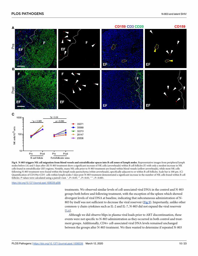
Fluorescence microscopy for NK cell analysis
Fluorescence microscopy was performed on formaldehyde fixed, paraffin-embedded (FFPE)
tissue sections (5 μm) according to our previously published protocol [38] with the following
minor modifications: antigen retrieval was performed with citrate pH 6 without protease treat-
ment and antigen stripping was performed by incubating slides in heated citraconic anhydride
antigen retrieval buffer (95˚-99˚C) for 10 min. The antibodies used were anti-CD159a (Sigma-
Aldrich; HPA004471), anti-CD3 (ThermoFisher; clone SP7), and CD20 (Biocare; clone L26).
Detection was performed sequentially with polymer horseradish peroxidase (HRP)-conjugated
systems (GBI Labs) coupled with tyramide-conjugated Alexa Fluors (ThermoFisher). Slides
were counterstained with DAPI, cover slipped using ProLong Gold Antifade Mountant (Ther-
moFisher; P36930), and scanned on an Axio Scan.z1 at 20x (Zeiss). Blood vessels were defined
as morphologically-distinct channels containing RBCs, which are dual autofluorescent in the
488 and 594 channels. In the images provided, the RBCs within the yellow dotted regions
appear to be a bright blue, due to the pseudocoloring of the image and the intensity of the
other colors in the image which make them appear single positive.
PLOS PATHOGENS N-803 and latent SHIV
PLOS Pathogens | https://doi.org/10.1371/journal.ppat.1008339 March 12, 2020 17 / 23
SHIV RNA in situ hybridization
RNAscope was performed on FFPE tissue sections (5μm) according to our previously pub-
lished protocol [39] with the following minor modifications: heat-induced epitope retrieval
was performed by boiling slides in 1x target retrieval (322000; ACD) for 30 min, followed by
incubation at 40˚C with a 1:10 dilution of protease III (322337; ACD) in 1x PBS for 20 min.
Slides were incubated with the target probe SIVmac239 (312811; ACD) for 2 hours at 40˚C.
Amplification was performed with RNAscope 2.5 HD Detection kits (322360; ACD) according
to manufacturer’s instructions, with 0.5X wash buffer (310091; ACD) used through amplifica-
tion step 4 and TBS-T used from amplification step 5 until the end of the assay. The resulting
signal was detected with Warp Red chromogen (WR806M; Biocare Medical). Slides were
counterstained with CAT hematoxylin (CATHE-GL; Biocare Medical), mounted with Clear-
mount (17885–15; EMS) until dry, coverslipped using Permount (SP15-100; Fisher Scientific),
and scanned at 40x magnification on an Aperio AT2 (Leica Biosystems).
Quantitative image analysis
Images were quantitatively analyzed using Halo software (v2.3.2089.27; Indica Labs). For NK
cell quantification, the random forest classifier was used to define anatomical regions based on
CD3 and CD20 staining and embedded within the Cytonuclear FL v1.4 analysis module. NK
cells were defined as CD159+CD3- cells and quantified relative to the total cells within each
specific anatomical region. NK cells present within blood vessels were excluded from quantifi-
cation. For RNAscope analysis, the ISH v2.2 module settings were set based on concomitantly
assayed, early chronically infected SIV+ control slides to determine the vRNA minimum signal
size necessary to exclude detection of the smaller single vRNA and/or vDNA molecules. Single
copy size was set at 0.49 μm2 and the threshold for analysis was set at 0.85 μm2. Total cell
counts were determined with the Cytonuclear v1.6 module to increase accuracy. In all quanti-
fications, manual curation was performed on each sample to correct for false positives/false
negatives.
N-803 Antidrug Antibody (ADA) Assay
MSD 96-well SECTOR plate (Meso Scale Diagnostic, LLC) was coated by adding 50 μL per
well of 2.5 μg/mL of N-803 in PBS and incubated overnight at 4˚C. The plate was washed 3x
with PBS-T and blocked with 150 μL per well of blocking solution (5% BSA in PBS) for 1 hour
with shaking at 700 rpm at room temperature. To make the standard curve, a 4-fold dilution
series of mouse anti-IL15 (R&D systems) was prepared using 10% normal cynomolgus mon-
key serum (Abcam) in assay buffer (1% BSA in PBS); the concentrations of the standards were
400, 100, 25, 6.25, 1.562, 0.391, 0.098, and 0 ng/mL. All serum samples were diluted 1:10 in
assay buffer. After a 1 hour incubation, the blocking solution was removed from the plate.
50 μL of 0.0625 μg/mL SULFO-tag conjugated N-803 in assay buffer and 25 μL of standards
and diluted serum samples were added to each well and incubated for 2 hours with shaking at
700 rpm at room temperature. The plate was washed 3x with PBS-T and 150 μL of 2X Read
Buffer T (Meso Scale Diagnostic, LLC) was added to each well before reading on the MSD
reader SECTOR S 600 (Meso Scale Diagnostic, LLC).
Ethics statement
All rhesus macaques (RMs) (Macaca mulatta) in this study were managed according to the
ONPRC animal husbandry program, which aims at providing consistent and excellent care to
nonhuman primates. This program is based on the laws, regulations, and guidelines set forth
PLOS PATHOGENS N-803 and latent SHIV
PLOS Pathogens | https://doi.org/10.1371/journal.ppat.1008339 March 12, 2020 18 / 23

by the United States Department of Agriculture (e.g., the Animal Welfare Act and its regula-
tions, and the Animal Care Policy Manual), Institute for Laboratory Animal Research (e.g.,
Guide for the Care and Use of Laboratory Animals, 8th edition), Public Health Service,
National Research Council, Centers for Disease Control, the Weatherall Report titled “The use
of nonhuman primates in research”, and the Association for Assessment and Accreditation of
Laboratory Animal Care (AAALAC) International. The nutritional plan utilized by the
ONPRC is based on National Research Council recommendations and supplemented with a
variety of fruits, vegetables, and other edible objects as part of the environmental enrichment
program established by the Behavioral Management Unit. Paired/grouped animals exhibiting
incompatible behaviors were reported to the Behavioral Management staff and managed
accordingly. All efforts were made to minimize suffering through the use of minimally invasive
procedures, anesthetics, and analgesics when appropriate. Animals were painlessly euthanized
with sodium pentobarbital and euthanasia was assured by exsanguination and bilateral pneu-
mothorax, consistent with the recommendations of the American Veterinary Medical Guide-
lines on Euthanasia (2013).
Statistics
Data from whole blood analysis is displayed as a percent change from baseline calculated as
such: ((value-baseline)/baseline) x 100 = percent change. Log-rank test was used for analysis of
Kaplan-Meier curve comparisons. Analyses between groups was performed using Mann-
Whitney U test. To account for within animal correlation in tetramer analysis, a linear mixed
effects model was used. For each outcome, the final sensible model was selected using AIC.
Fixed effects considered include time and tetramer specificity. Random effects components
were selected between random intercept only and random intercept and random slope for
time. Visual model diagnostics were performed to detect severe violations to the assumptions
of linear mixed effect model. Analysis was performed using SAS9.4. Log-rank test was used for
Kaplan-Meier curve analysis. For correlations, linear regression with Pearson’s correlation was
used. Paired Student’s t test was used for all other analyses. Statistical analyses were conducted
using GraphPad Prism version 6.0 (GraphPad Software, La Jolla, California, USA). Statistical
significance of the findings was set at a p-value of less than 0.05.
Supporting information
S1 Fig. Whole blood analysis during in vivo administration of N-803. N-803 was subcutane-
ously administered every other week as indicated by the vertical dashed lines. Blood was col-
lected at time 0 before the N-803 injection and at days 1, 3, 5, 7 after each injection of N-803.
(A) White blood count (WBC), lymphocytes, monocytes, and neutrophils were analyzed from
blood. (B) CD16+ NK cells, CD4+ T cells, CD8+ T cells were analyzed from blood and shown
as a percent of CD45+ cells, absolute cell counts on the right, both shown as a percent change
from baseline. Absolute counts were calculated based on the percentage of the particular cell
subset and the WBC count. Data shown are means (± SEM) of combined data from all animals
within the designated group.
(TIF)
S2 Fig. Whole blood analysis during in vivo administration of N-803. N-803 was subcutane-
ously administered every other week as indicated by the vertical dashed lines. Blood was col-
lected at time 0 before the N-803 injection and at days 1, 3, 5, 7 after each injection of N-803.
Memory subpopulations (naïve, effector memory, central memory) of (A) CD8+ T cells and
(B) CD4+ T cells. On the left is the percent of CD8+ or CD4+ T cells and absolute cell counts
PLOS PATHOGENS N-803 and latent SHIV
PLOS Pathogens | https://doi.org/10.1371/journal.ppat.1008339 March 12, 2020 19 / 23

are on the right, both shown as a percent change from baseline. Absolute counts were calcu-
lated based on the percentage of the particular cell subset and the WBC count. Data shown are
means (± SEM) of combined data from all animals within the designated group.
(TIF)
S3 Fig. Dynamics of SHIV-specific CD8+ T cells and CXCR5 in the lymph nodes during
and after N-803 administration. (A) Percent SHIV-specific CD8+ T cells as measured by
MHC class I tetramer staining in lymph nodes prior to N-803, 5 days after N-803, and 3 weeks
after the final N-803 administration. (B) CXCR5 staining on SHIV-specific CD8+ T cells in
lymph nodes prior to N-803, 5 days after N-803, and 3 weeks after the final N-803 administra-
tion. (C) CXCR5 staining on NK cells in lymph nodes prior to N-803, 5 days after N-803, and
3 weeks after the final N-803 administration. P values were calculated using a paired t-test. �,
P<0.05; ��, P<0.01; ���, P<0.001.
(TIF)
S4 Fig. Viral load analysis and correlations of viral rebound. (A) Peak plasma viral loads
and (B) area under the curve of viral loads prior to ART discontinuation. (C) Correlation of
peak viral load post-ART release with pre-ART peak viral load. (D) Correlation of the time to
the first detectable viral RNA in plasma after ART release with pre-ART peak viral load. Data
shown are means (± SEM). P values were calculated using a Mann-Whitney test (A, B), and
linear regression with Pearson’s correlation (C, D). �, P<0.05; ��, P<0.01; ���, P<0.001.
(TIF)
S5 Fig. Anti-drug antibody and CD16+ NK cell count during the course of N-803 adminis-
tration. Anti-drug antibody development in each animal that received N-803 and the absolute
cell count of CD16+ NK cells. Vertical dashed lines indicate times of N-803 administration.
(TIF)
S6 Fig. Anti-drug antibody and CD8+ T cell count during the course of N-803 administra-
tion. Anti-drug antibody development in each animal that received N-803 and the absolute
cell count of CD8+ T cells. Vertical dashed lines indicate times of N-803 administration.
(TIF)
Acknowledgments
We thank Jillian Hattersley, Joseph Hesselgesser, Bei Li, and Romas Geleziunas at Gilead for
providing their expertise with ART formulation. We thank Nancy Miller for providing
SHIVSF162P3. We would like to acknowledge the NIAID DAIDS Nonhuman Primate Core
Virology Laboratory for AIDS Vaccine Research and Development Contract
#HHSN272201800003C. We acknowledge the animals that contributed to this study.
Author Contributions
Conceptualization: Gabriela M. Webb, Jonah B. Sacha.
Data curation: Gabriela M. Webb, Jhomary Molden, Kathleen Busman-Sahay.
Formal analysis: Gabriela M. Webb, Jhomary Molden, Kathleen Busman-Sahay, Lina Gao.
Funding acquisition: Jonah B. Sacha.
Investigation: Gabriela M. Webb, Jhomary Molden, Kathleen Busman-Sahay, Shaheed Abdul-
haqq, Helen L. Wu, Whitney C. Weber, Katherine B. Bateman, Jason S. Reed, Mina
PLOS PATHOGENS N-803 and latent SHIV
PLOS Pathogens | https://doi.org/10.1371/journal.ppat.1008339 March 12, 2020 20 / 23

Northrup, Nicholas Maier, Shiho Tanaka, Brianna Davey, Benjamin L. Carpenter, Jeffrey J.
Stanton, Justin M. Greene.
Methodology: Jacob D. Estes, Pamela J. Skinner, Jonah B. Sacha.
Project administration: Gabriela M. Webb, Jeremy Smedley, Jonah B. Sacha.
Resources: Michael K. Axthelm, Jeffrey J. Stanton, Jeffrey T. Safrit, Jacob D. Estes, Pamela J.
Skinner, Jonah B. Sacha.
Supervision: Jeremy Smedley, Jacob D. Estes, Pamela J. Skinner, Jonah B. Sacha.
Visualization: Gabriela M. Webb, Jhomary Molden, Kathleen Busman-Sahay.
Writing – original draft: Gabriela M. Webb, Jonah B. Sacha.
Writing – review & editing: Gabriela M. Webb, Jhomary Molden, Kathleen Busman-Sahay,
Shaheed Abdulhaqq, Helen L. Wu, Whitney C. Weber, Katherine B. Bateman, Jason S.
Reed, Mina Northrup, Nicholas Maier, Shiho Tanaka, Lina Gao, Brianna Davey, Benjamin
L. Carpenter, Michael K. Axthelm, Jeffrey J. Stanton, Justin M. Greene, Jeffrey T. Safrit,
Jacob D. Estes, Pamela J. Skinner, Jonah B. Sacha.
References1. Hutter G, Nowak D, Mossner M, Ganepola S, Mussig A, Allers K, et al. Long-term control of HIV by
CCR5 Delta32/Delta32 stem-cell transplantation. N Engl J Med. 2009; 360: 692–698. https://doi.org/10.
1056/NEJMoa0802905 PMID: 19213682
2. Yukl SA, Boritz E, Busch M, Bentsen C, Chun T-W, Douek D, et al. Challenges in Detecting HIV Persis-
tence during Potentially Curative Interventions: A Study of the Berlin Patient. Cullen BR, editor. PLoS
Pathog. Public Library of Science; 2013; 9: e1003347. https://doi.org/10.1371/journal.ppat.1003347
PMID: 23671416
3. Gupta RK, Abdul-Jawad S, McCoy LE, Mok HP, Peppa D, Salgado M, et al. HIV-1 remission following
CCR5Δ32/Δ32 haematopoietic stem-cell transplantation. Nature. Nature Publishing Group; 2019; 568:
244–248. https://doi.org/10.1038/s41586-019-1027-4 PMID: 30836379
4. Deeks SG, Lewin SR, Ross AL, Ananworanich J, Benkirane M, Cannon P, et al. International AIDS
Society global scientific strategy: towards an HIV cure 2016. Nat Med. 2016. https://doi.org/10.1038/
nm.4108 PMID: 27400264
5. Chun TW, Davey RT, Engel D, Lane HC, Fauci AS. Re-emergence of HIV after stopping therapy.
Nature. 1999; 401: 874–875. https://doi.org/10.1038/44755 PMID: 10553903
6. Vandergeeten C, Fromentin R, DaFonseca S, Lawani MB, Sereti I, Lederman MM, et al. Interleukin-7
promotes HIV persistence during antiretroviral therapy. Blood. 2013; 121: 4321–4329. https://doi.org/
10.1182/blood-2012-11-465625 PMID: 23589672
7. Mueller YM, Petrovas C, Bojczuk PM, Dimitriou ID, Beer B, Silvera P, et al. Interleukin-15 increases
effector memory CD8+ t cells and NK Cells in simian immunodeficiency virus-infected macaques. Jour-
nal of Virology. 2005; 79: 4877–4885. https://doi.org/10.1128/JVI.79.8.4877-4885.2005 PMID:
15795273
8. Picker LJ, Reed-Inderbitzin EF, Hagen SI, Edgar JB, Hansen SG, Legasse A, et al. IL-15 induces CD4
effector memory T cell production and tissue emigration in nonhuman primates. J Clin Invest. 2006;
116: 1514–1524. https://doi.org/10.1172/JCI27564 PMID: 16691294
9. Lugli E, Goldman CK, Perera LP, Smedley J, Pung R, Yovandich JL, et al. Transient and persistent
effects of IL-15 on lymphocyte homeostasis in nonhuman primates. Blood. 2010; 116: 3238–3248.
https://doi.org/10.1182/blood-2010-03-275438 PMID: 20631381
10. Jones RB, Mueller S, O’Connor R, Rimpel K, Sloan DD, Karel D, et al. A Subset of Latency-Reversing
Agents Expose HIV-Infected Resting CD4+ T-Cells to Recognition by Cytotoxic T-Lymphocytes. PLoS
Pathog. Public Library of Science; 2016; 12: e1005545. https://doi.org/10.1371/journal.ppat.1005545
PMID: 27082643
11. Chertova E, Bergamaschi C, Chertov O, Sowder R, Bear J, Roser JD, et al. Characterization and favor-
able in vivo properties of heterodimeric soluble IL-15�IL-15Rα cytokine compared to IL-15 monomer.
Journal of Biological Chemistry. American Society for Biochemistry and Molecular Biology; 2013; 288:
18093–18103. https://doi.org/10.1074/jbc.M113.461756 PMID: 23649624
PLOS PATHOGENS N-803 and latent SHIV
PLOS Pathogens | https://doi.org/10.1371/journal.ppat.1008339 March 12, 2020 21 / 23

12. Watson DC, Moysi E, Valentin A, Bergamaschi C, Devasundaram S, Fortis SP, et al. Treatment with
native heterodimeric IL-15 increases cytotoxic lymphocytes and reduces SHIV RNA in lymph nodes.
PLoS Pathog. Public Library of Science; 2018; 14: e1006902. https://doi.org/10.1371/journal.ppat.
1006902 PMID: 29474450
13. Webb GM, Li S, Mwakalundwa G, Folkvord JM, Greene JM, Reed JS, et al. The human IL-15 superago-
nist ALT-803 directs SIV-specific CD8+ T cells into B-cell follicles. Blood Adv. American Society of
Hematology; 2018; 2: 76–84. https://doi.org/10.1182/bloodadvances.2017012971 PMID: 29365313
14. Ellis-Connell AL, Balgeman AJ, Zarbock KR, Barry G, Weiler A, Egan JO, et al. ALT-803 transiently
reduces SIV replication in the absence of antiretroviral treatment. Journal of Virology. American Society
for Microbiology; 2017;: JVI.01748–17. https://doi.org/10.1128/JVI.01748-17 PMID: 29118125
15. Zhu X, Marcus WD, Xu W, Lee H-I, Han K, Egan JO, et al. Novel human interleukin-15 agonists. J
Immunol. American Association of Immunologists; 2009; 183: 3598–3607. https://doi.org/10.4049/
jimmunol.0901244 PMID: 19710453
16. Xu W, Jones M, Liu B, Zhu X, Johnson CB, Edwards AC, et al. Efficacy and mechanism-of-action of a
novel superagonist interleukin-15: interleukin-15 receptor αSu/Fc fusion complex in syngeneic murine
models of multiple myeloma. Cancer Res. American Association for Cancer Research; 2013; 73: 3075–
3086. https://doi.org/10.1158/0008-5472.CAN-12-2357 PMID: 23644531
17. Rhode PR, Egan JO, Xu W, Hong H, Webb GM, Chen X, et al. Comparison of the Superagonist Com-
plex, ALT-803, to IL15 as Cancer Immunotherapeutics in Animal Models. Cancer Immunol Res. Ameri-
can Association for Cancer Research; 2016; 4: 49–60. https://doi.org/10.1158/2326-6066.CIR-15-
0093-T PMID: 26511282
18. Seay K, Church C, Zheng JH, Deneroff K, Ochsenbauer C, Kappes JC, et al. In Vivo Activation of
Human NK Cells by Treatment with an Interleukin-15 Superagonist Potently Inhibits Acute In Vivo HIV-
1 Infection in Humanized Mice. Journal of Virology. American Society for Microbiology; 2015; 89: 6264–
6274. https://doi.org/10.1128/JVI.00563-15 PMID: 25833053
19. Liu B, Kong L, Han K, Hong H, Marcus WD, Chen X, et al. A Novel Fusion of ALT-803 (Interleukin (IL)-
15 Superagonist) with an Antibody Demonstrates Antigen-specific Antitumor Responses. Journal of
Biological Chemistry. American Society for Biochemistry and Molecular Biology; 2016; 291: 23869–
23881. https://doi.org/10.1074/jbc.M116.733600 PMID: 27650494
20. Romee R, Cooley S, Berrien-Elliott MM, Westervelt P, Verneris MR, Wagner JE, et al. First-in-human
Phase 1 Clinical Study of the IL-15 Superagonist Complex ALT-803 to Treat Relapse after Transplanta-
tion. Blood. American Society of Hematology; 2018;: blood–2017–12–823757. https://doi.org/10.1182/
blood-2017-12-823757 PMID: 29463563
21. Margolin K, Morishima C, Velcheti V, Miller JS, Lee SM, Silk AW, et al. Phase I Trial of ALT-803, A
Novel Recombinant IL15 Complex, in Patients with Advanced Solid Tumors. Clin Cancer Res. Ameri-
can Association for Cancer Research; 2018; 24: 5552–5561. https://doi.org/10.1158/1078-0432.CCR-
18-0945 PMID: 30045932
22. Okoye AA, Hansen SG, Vaidya M, Fukazawa Y, Park H, Duell DM, et al. Early antiretroviral therapy lim-
its SIV reservoir establishment to delay or prevent post-treatment viral rebound. Nat Med. Nature Pub-
lishing Group; 2018; 24: 1430–1440. https://doi.org/10.1038/s41591-018-0130-7 PMID: 30082858
23. Mudd PA, Ericsen AJ, Burwitz BJ, Wilson NA, O’Connor DH, Hughes AL, et al. Escape from CD8+ T
Cell Responses in Mamu-B*00801+ Macaques Differentiates Progressors from Elite Controllers. The
Journal of Immunology. American Association of Immunologists; 2012; 188: 3364–3370. https://doi.org/
10.4049/jimmunol.1102470 PMID: 22387557
24. Waldmann TA, Lugli E, Roederer M, Perera LP, Smedley JV, Macallister RP, et al. Safety (toxicity),
pharmacokinetics, immunogenicity, and impact on elements of the normal immune system of recombi-
nant human IL-15 in rhesus macaques. Blood. American Society of Hematology; 2011; 117: 4787–
4795. https://doi.org/10.1182/blood-2010-10-311456 PMID: 21385847
25. Fukazawa Y, Lum R, Okoye AA, Park H, Matsuda K, Bae JY, et al. B cell follicle sanctuary permits per-
sistent productive simian immunodeficiency virus infection in elite controllers. Nat Med. 2015; 21: 132–
139. https://doi.org/10.1038/nm.3781 PMID: 25599132
26. Banga R, Procopio FA, Noto A, Pollakis G, Cavassini M, Ohmiti K, et al. PD-1(+) and follicular helper T
cells are responsible for persistent HIV-1 transcription in treated aviremic individuals. Nat Med. Nature
Publishing Group; 2016; 22: 754–761. https://doi.org/10.1038/nm.4113 PMID: 27239760
27. McBrien JB, Mavigner M, Franchitti L, Smith SA, White E, Tharp GK, et al. Robust and persistent reacti-
vation of SIV and HIV by N-803 and depletion of CD8+ cells. Nature. Nature Publishing Group; 2020;
94: 13193–6. https://doi.org/10.1038/s41586-020-1946-0 PMID: 31969705
28. Whitney JB, Lim S-Y, Osuna CE, Kublin JL, Chen E, Yoon G, et al. Prevention of SIVmac251 reservoir
seeding in rhesus monkeys by early antiretroviral therapy. Nat Commun. Nature Publishing Group;
2018; 9: 5429. https://doi.org/10.1038/s41467-018-07881-9 PMID: 30575753
PLOS PATHOGENS N-803 and latent SHIV
PLOS Pathogens | https://doi.org/10.1371/journal.ppat.1008339 March 12, 2020 22 / 23

29. Liu B, Jones M, Kong L, Noel T, Jeng EK, Shi S, et al. Evaluation of the biological activities of the IL-15
superagonist complex, ALT-803, following intravenous versus subcutaneous administration in murine
models. Cytokine. 2018; 107: 105–112. https://doi.org/10.1016/j.cyto.2017.12.003 PMID: 29452720
30. Deeks SG. HIV: Shock and kill. Nature. Nature Publishing Group; 2012; 487: 439–440. https://doi.org/
10.1038/487439a PMID: 22836995
31. Davis Z, Thorkelson A, Anderson J, Wong HC, Karn J, Dobrowlski C, et al. A Phase 1 Study of ALT-803
(IL-15 superagonist) to Clear Latent HIV Reservoirs [Internet]. Conference on Retroviruses and Oppor-
tunistic Infections. Available: https://www.croiconference.org/sessions/phase-1-study-alt-803-il-15-
superagonist-clear-latent-hiv-reservoirs
32. Rosario M, Liu B, Kong L, Collins LI, Schneider SE, Chen X, et al. The IL-15-Based ALT-803 Complex
Enhances FcγRIIIa-Triggered NK Cell Responses and In Vivo Clearance of B Cell Lymphomas. Clin
Cancer Res. American Association for Cancer Research; 2016; 22: 596–608. https://doi.org/10.1158/
1078-0432.CCR-15-1419 PMID: 26423796
33. Connick E, Mattila T, Folkvord JM, Schlichtemeier R, Meditz AL, Ray MG, et al. CTL fail to accumulate
at sites of HIV-1 replication in lymphoid tissue. J Immunol. 2007; 178: 6975–6983. https://doi.org/10.
4049/jimmunol.178.11.6975 PMID: 17513747
34. Han K-P, Zhu X, Liu B, Jeng E, Kong L, Yovandich JL, et al. IL-15:IL-15 receptor alpha superagonist
complex: high-level co-expression in recombinant mammalian cells, purification and characterization.
Cytokine. 2011; 56: 804–810. https://doi.org/10.1016/j.cyto.2011.09.028 PMID: 22019703
35. Zevin AS, Moats C, May D, Wangari S, Miller C, Ahrens J, et al. Laparoscopic Technique for Serial Col-
lection of Liver and Mesenteric Lymph Nodes in Macaques. J Vis Exp. 2017;: e55617. https://doi.org/
10.3791/55617 PMID: 28518089
36. Cline AN, Bess JW, Piatak M, Lifson JD. Highly sensitive SIV plasma viral load assay: practical consid-
erations, realistic performance expectations, and application to reverse engineering of vaccines for
AIDS. J Med Primatol. John Wiley & Sons, Ltd (10.1111); 2005; 34: 303–312. https://doi.org/10.1111/j.
1600-0684.2005.00128.x PMID: 16128925
37. Hansen SG, Ford JC, Lewis MS, Ventura AB, Hughes CM, Coyne-Johnson L, et al. Profound early con-
trol of highly pathogenic SIV by an effector memory T-cell vaccine. Nature. 2011; 473: 523–527. https://
doi.org/10.1038/nature10003 PMID: 21562493
38. Mudd JC, Busman-Sahay K, DiNapoli SR, Lai S, Sheik V, Lisco A, et al. Hallmarks of primate lentiviral
immunodeficiency infection recapitulate loss of innate lymphoid cells. Nat Commun. Nature Publishing
Group; 2018; 9: 3967–12. https://doi.org/10.1038/s41467-018-05528-3 PMID: 30262807
39. Deleage C, Wietgrefe SW, Del Prete G, Morcock DR, Hao XP, Piatak M, et al. Defining HIV and SIV
Reservoirs in Lymphoid Tissues. Pathog Immun. Faculty at Case Western Reserve University School
of Medicine; 2016; 1: 68–106. https://doi.org/10.20411/pai.v1i1.100 PMID: 27430032
PLOS PATHOGENS N-803 and latent SHIV
PLOS Pathogens | https://doi.org/10.1371/journal.ppat.1008339 March 12, 2020 23 / 23