The Growing Relevance of Immunoregulation in Pediatric ...
20
cancers Review The Growing Relevance of Immunoregulation in Pediatric Brain Tumors Viktoria Melcher * and Kornelius Kerl * Citation: Melcher, V.; Kerl, K. The Growing Relevance of Immunoregulation in Pediatric Brain Tumors. Cancers 2021, 13, 5601. https://doi.org/10.3390/cancers 13225601 Academic Editor: Mary Poupot Received: 28 October 2021 Accepted: 5 November 2021 Published: 9 November 2021 Publisher’s Note: MDPI stays neutral with regard to jurisdictional claims in published maps and institutional affil- iations. Copyright: © 2021 by the authors. Licensee MDPI, Basel, Switzerland. This article is an open access article distributed under the terms and conditions of the Creative Commons Attribution (CC BY) license (https:// creativecommons.org/licenses/by/ 4.0/). Department of Pediatric Hematology and Oncology, University Children’s Hospital Münster, 48149 Münster, Germany * Correspondence: [email protected] (V.M.);[email protected] (K.K.) Simple Summary: Pediatric brain tumors are the leading cause of childhood cancer-related deaths worldwide. Considering the dismal prognosis and the adverse effects of chemo- and radio-therapy, strategies targeting the tumor microenvironment represent a promising approach for improving the efficacy of standard and targeted molecular therapeutics. This review presents the current understanding of the juvenile innate immune system in the central nervous system and gives insights into the brain as a unique tumor site. Moreover, we outline an explorative overview of studies about the tumor microenvironment of pediatric brain tumors and its role in tumor progression and therapy resistance. We further put attention to the potential immunomodulatory effects of current therapeutic regimens. Finally, we provide a perspective regarding the present immunotherapeutic treatment options and future clinical implications of targeting the immune cells. Abstract: Pediatric brain tumors are genetically heterogeneous solid neoplasms. With a prevailing poor prognosis and widespread resistance to conventional multimodal therapy, these aggressive tumors are the leading cause of childhood cancer-related deaths worldwide. Advancement in molecular research revealed their unique genetic and epigenetic characteristics and paved the way for more defined prognostication and targeted therapeutic approaches. Furthermore, uncovering the intratumoral metrics on a single-cell level placed non-malignant cell populations such as innate immune cells into the context of tumor manifestation and progression. Targeting immune cells in pediatric brain tumors entails unique challenges but promising opportunities to improve outcome. Herein, we outline the current understanding of the role of the immune regulation in pediatric brain tumors. Keywords: pediatric brain tumors; embryonal brain tumors; atypical teratoid/rhabdoid tumor; ependymoma; high-grade glioma; low-grade glioma; medulloblastoma; therapy resistance; tumor microenvironment; immune cells; immunomodulation; immunotherapy 1. Introduction Tumors of the central nervous system (CNS) are the most common solid tumors in children and the leading cause of worldwide childhood cancer-related deaths [1]. The most prevalent pediatric brain tumors are categorized according to World Health Organization (WHO) into low-grade gliomas (LGG), high-grade gliomas (HGG), ependymomas (EPN), and embryonal brain tumors. The latter are high-grade neoplasms, originating from undif- ferentiated embryonic cells, and are classified into atypical teratoid/rhabdoid tumor (ATRT), medulloblastoma (MB), embryonal tumor with multilayered rosettes (ETMR), and other CNS embryonal tumors (previously named CNS primitive neuroectodermal tumor) [2]. Pediatric brain tumors have been profoundly characterized on the genomic and transcrip- tomic levels. Besides recurrent mutations in distinct signaling pathways (e.g., sonic hedge- hog (SHH), Wnt/wingless (WNT) and mitogen-activated protein kinase (MAPK)), epige- netic processes play a crucial role in their pathogenesis [3–5]. Comprehensive molecular Cancers 2021, 13, 5601. https://doi.org/10.3390/cancers13225601 https://www.mdpi.com/journal/cancers
-
Upload
khangminh22 -
Category
Documents
-
view
0 -
download
0
Transcript of The Growing Relevance of Immunoregulation in Pediatric ...

cancers
Review
The Growing Relevance of Immunoregulation in PediatricBrain Tumors
Viktoria Melcher * and Kornelius Kerl *
�����������������
Citation: Melcher, V.; Kerl, K.
The Growing Relevance of
Immunoregulation in Pediatric Brain
Tumors. Cancers 2021, 13, 5601.
https://doi.org/10.3390/cancers
13225601
Academic Editor: Mary Poupot
Received: 28 October 2021
Accepted: 5 November 2021
Published: 9 November 2021
Publisher’s Note: MDPI stays neutral
with regard to jurisdictional claims in
published maps and institutional affil-
iations.
Copyright: © 2021 by the authors.
Licensee MDPI, Basel, Switzerland.
This article is an open access article
distributed under the terms and
conditions of the Creative Commons
Attribution (CC BY) license (https://
creativecommons.org/licenses/by/
4.0/).
Department of Pediatric Hematology and Oncology, University Children’s Hospital Münster,48149 Münster, Germany* Correspondence: [email protected] (V.M.); [email protected] (K.K.)
Simple Summary: Pediatric brain tumors are the leading cause of childhood cancer-related deathsworldwide. Considering the dismal prognosis and the adverse effects of chemo- and radio-therapy,strategies targeting the tumor microenvironment represent a promising approach for improvingthe efficacy of standard and targeted molecular therapeutics. This review presents the currentunderstanding of the juvenile innate immune system in the central nervous system and gives insightsinto the brain as a unique tumor site. Moreover, we outline an explorative overview of studies aboutthe tumor microenvironment of pediatric brain tumors and its role in tumor progression and therapyresistance. We further put attention to the potential immunomodulatory effects of current therapeuticregimens. Finally, we provide a perspective regarding the present immunotherapeutic treatmentoptions and future clinical implications of targeting the immune cells.
Abstract: Pediatric brain tumors are genetically heterogeneous solid neoplasms. With a prevailingpoor prognosis and widespread resistance to conventional multimodal therapy, these aggressivetumors are the leading cause of childhood cancer-related deaths worldwide. Advancement inmolecular research revealed their unique genetic and epigenetic characteristics and paved the wayfor more defined prognostication and targeted therapeutic approaches. Furthermore, uncoveringthe intratumoral metrics on a single-cell level placed non-malignant cell populations such as innateimmune cells into the context of tumor manifestation and progression. Targeting immune cells inpediatric brain tumors entails unique challenges but promising opportunities to improve outcome.Herein, we outline the current understanding of the role of the immune regulation in pediatricbrain tumors.
Keywords: pediatric brain tumors; embryonal brain tumors; atypical teratoid/rhabdoid tumor;ependymoma; high-grade glioma; low-grade glioma; medulloblastoma; therapy resistance; tumormicroenvironment; immune cells; immunomodulation; immunotherapy
1. Introduction
Tumors of the central nervous system (CNS) are the most common solid tumors inchildren and the leading cause of worldwide childhood cancer-related deaths [1]. The mostprevalent pediatric brain tumors are categorized according to World Health Organization(WHO) into low-grade gliomas (LGG), high-grade gliomas (HGG), ependymomas (EPN),and embryonal brain tumors. The latter are high-grade neoplasms, originating from undif-ferentiated embryonic cells, and are classified into atypical teratoid/rhabdoid tumor (ATRT),medulloblastoma (MB), embryonal tumor with multilayered rosettes (ETMR), and otherCNS embryonal tumors (previously named CNS primitive neuroectodermal tumor) [2].Pediatric brain tumors have been profoundly characterized on the genomic and transcrip-tomic levels. Besides recurrent mutations in distinct signaling pathways (e.g., sonic hedge-hog (SHH), Wnt/wingless (WNT) and mitogen-activated protein kinase (MAPK)), epige-netic processes play a crucial role in their pathogenesis [3–5]. Comprehensive molecular
Cancers 2021, 13, 5601. https://doi.org/10.3390/cancers13225601 https://www.mdpi.com/journal/cancers

Cancers 2021, 13, 5601 2 of 20
analyses paved the way for entity sub-classification, allowing for a more defined prognos-tication and targeted therapeutic approaches [6–8]. However, patients undergo intensivemultimodal therapy with surgical resection, chemo- and/or radiotherapy. These treatmentregimens have multiple adverse effects and often cause neurocognitive deficits [9,10].
Besides the increasing knowledge about molecular subgroups and their implicationson clinical outcome in pediatric brain tumors, much less is known about the role ofinfiltrating immune cells in these malignancies. In contrast, in adult cancer, the discipline ofcancer immunotherapy has grown largely in recent years [11]. As brain tumors in childrenand adults are distinct on different levels, e.g., frequency, anatomic location, as well as thepathologic, genetic and epigenetic spectrum, it might not be possible to extrapolate resultsfrom adult studies to pediatric tumor entities [12]. Pediatric cancers are generally assumedto be less immunogenic, which might be based on their low mutational burden [13,14].
However, recent efforts have shown that antitumor immune control exists in pediatricbrain tumors, e.g., rhabdoid tumors and LGG showed strong T cell infiltration [15–17].Moreover, several studies present prognostications of specific immune cell infiltrates onoverall survival [18–22] (Table 1). Thus, the indications that host immunity may alreadyimpact patients’ survival in pediatric brain tumors suggests that immune profiling can addvaluable information in diagnostics and assessment of immunotherapeutic therapies forindividual patients.
This review aims at summarizing the current knowledge of tumor-associated immunecells in pediatric brain tumors and further considers important aspects for the implementa-tion of immunotherapeutic targets to conventional therapy.
2. The (Postnatal) Brain as a Unique Tumor Site and CNS Immune Surveillance
Malignancies developing in highly organized organs such as the central nervoussystem (CNS) can severely impact the whole organism. In the (adult) tumor-developingbrain, the stroma is initially composed of neuronal cells, astrocytes, oligodendrocytes,and microglia surrounded by a distinctive extracellular matrix (ECM) [23]. However, dueto the complexity of brain development and its (context-dependent) cellular interactionsalready under physiological conditions, crosstalk between these cell populations and tu-morigenic cells in the developing brain is barely characterized. During brain development,the generation of neurons and glial cells is precisely regulated by diverse mechanisms,including the immune system. The first immune cells that encounter the developing brainare microglia [24,25]. This section discusses how the immune system interacts with tumorcells in general and how distinct immune cells specifically contribute to tumor cell growthin the developing brain.
Generally, in the setting of tumor development, the immune system operates atmultiple levels when preventing and eliminating cancerous cells while sparing healthy cells:viral infections that can induce tumors are eliminated or at least suppressed; pathogensare killed and the consequential inflammation is controlled to prevent an inflammatoryenvironment that is susceptible for carcinogenesis; and lastly, innate immune cells recognizestressed cells or tumor-specific antigens and molecules and eradicate (pre-)tumorigeniccells. However, the fact that tumors still develop under functional immune system isnowadays explained by the term “tumor immunoediting” [26]. Immunoediting describesa concept of three phases during the suppression of tumor cells by innate and adaptiveimmune cells: (1) elimination, (2) equilibrium, and (3) escape. First, cells that escapedmechanisms of intrinsic tumor suppression are detected and eliminated by innate immunecells. If eradication is incomplete, tumor cells can enter the second phase, tumor dormancy,and are in a temporary equilibrium with the adaptive immune system that suppressestumor expansion. In this dynamic phase, tumor cells can evolve (further mutations; changesin gene expression that modulate tumor-specific antigens or boost immunosuppressivemechanisms) and adjust to the immune pressure, which finally leads to a selection ofresistant tumor cell clones. Thus, the tumor escapes from the antitumor immune response

Cancers 2021, 13, 5601 3 of 20
and progresses [27]. More recent studies revealed that innate immune cells can undergocancer immunoediting in the absence of adaptive immunity [28].
Studies on brain tumor biology and immunology mechanisms in adults outweigh in-fant and juvenile tumor immunology knowledge. Immune mechanisms are predominantlystudied in infectious diseases, which is plausible, considering the low incidence of pediatric(brain) cancer compared to the age-related increase in adults. Though many processesare similar in pediatric and adult immunity, there are substantial age-specific develop-mental changes through the transition from the fetal stage to neonatal and infant. Mostof all, the immune system does not fully mature until approximately the age of 12 years.Due to that, generalizing results from adult research to children should be interpreted withcaution [29]. Most notably, the fetal, neonatal and infant stages of development require im-mune suppression. Otherwise, conditions like the semi-allogeneic state of the mother/fetusduring pregnancy or the transition from a protected environment in utero to postnatalrapidly changing environmental influences would result in systemic inflammation. Thisis in part realized via a tolerogenic immune response involving Treg cells as well as IL-10production by neonatal antigen-presenting cells (APCs) and high concentrations of theimmunosuppressive purine metabolite adenosine [30–33]. Thus, children are born withrelatively naive T and B cells, hypofunctional innate immunity and fetal red blood cells [24].
Another aspect of the brain as a unique tumor site is the physical protection by theblood–brain barrier (BBB), a tight unity of endothelial cells, astrocytes, pericytes and mi-croglia. This cellular cohesion protects the CNS from pathogens and prevents neurotoxicityby inflammation from peripherally infiltrating immune cells. Further, the CNS lacks clas-sical lymphatic drainage, meaning the transport route of antigens and APCs from theinflammation site to the nearby lymph nodes [34]. Hence, it has long been believed thatthe brain is immune privileged. Still, the brain also has direct access to the blood systemvia circumventricular organs, which serve as a direct passage of cytokines [35]. Moreover,also immune cells, such as B cells, T cells, and macrophages, are observed in the meningeal,ventricular and perivascular space and choroid plexus. These cell types have crucial rolesin brain function [24,34]. In CNS tumors, the permeability of the BBB is frequently alteredand functionality varies between brain tumor entities [36]. In the WNT-MB subtype, tu-mor cells block the endothelial WNT signaling and disrupt the BBB formation, while theSHH-MB subtype showed a normal vascular phenotype. These tumor-intrinsic differencescould explain why WNT-MB are vulnerable to systemic chemotherapy, which has to crossthe BBB, but SHH-MB is not responding [37]. In ATRT, BBB deficits could also explainthe effectiveness of chemotherapeutic treatment, resulting in improved survival of ATRTpatients [38,39]. Meel et al. [40] found BBB phenotypic abnormalities in ATRT-SHH andATRT-MYC and observed in vivo efficacy of two non-BBB penetrable drugs [40].
In the following section, we describe the divergent role of the resident and infiltratingimmune cells of the CNS in tumor suppression and promotion.
2.1. Microglia as Brain-Resident Innate Immune Cell Populations
Microglia exhibit regenerative properties, namely myelin-debris removal, secretion ofgrowth and neurotrophic factors, which are essential for proper remyelination of newlydifferentiated oligodendrocytes. Regenerative properties of microglia are associated withalternative M2-phenotypes, which secrete anti-inflammatory cytokines and growth factors.Besides that, they are the first line of innate immunity defense and, as highly motile cells,they continuously survey the brain parenchyma. This classically activated M1-phenotype isassociated with antigen presentation, secretion of pro-inflammatory cytokines and reactiveoxygen and nitrogen species [41]. Therefore, they are probably the first innate immunecells to interact with malignant cells. It is reported that microglia highly adapt to theirenvironment, resulting in diverse temporal and spatial heterogeneous subtypes [42,43].Concretely, microglia are most heterogenous during postnatal development in a region-dependent manner [44,45].

Cancers 2021, 13, 5601 4 of 20
Pediatric brain tumors exhibit clear correlations with stalled developmental programsand develop in various brain locations [46]. It is speculative if different tumor entitiesprobably adapt to local niches and, thus, progress. For example, microglia subtypesdiffer in a temporal and region-dependent manner throughout the brain parenchyma.Notably, CD11c+ microglia are highly enriched in the cerebellum during early postnataldevelopment, where they promote the expansion and survival of granule neural precursorcells through insulin-like growth factor 1 (IGF1) secretion [47]. IGF1 was reported to becrucial for tumor growth and migration of (murine) SHH-MB, whose cell of origin ispostulated to be cerebellar granule neural precursor cells [48,49].
The impact of tumor-associated microglia/macrophages on tumor progression wasreported extensively in various tumor entities [23,41,50].
2.2. Brain-Infiltrating Immune Cell Populations
Peripheral blood immune cells, such as natural killer (NK) cells, macrophages (MACs),neutrophils, dendritic cells (DCs), innate lymphoid cells, myeloid-derived suppressor cells(MDSCs) as well as T and B cells can pass into the brain during instances of particularlysevere inflammation or trauma [23,24]. In physiological brain development, B cells andT cells reside in the meningeal space and choroid plexus. B cells are involved in oligo-dendrogenesis and T cells promote the formation of inhibitory synapses by producingIFN-γ [24].
Natural killer (NK) cells, natural killer T cells (NKT), neutrophils and γδ T cells searchfor pathogens or abnormal cells and then destroy the cell surface using cell toxins. Togetherwith phagocytes, they eliminate pathogens and induce inflammation, which attracts naïve Tcells. If this is insufficient to eliminate the pathogen, DCs migrate to the lymph nodes whereantigens can be presented to pathogen-specific T-lymphocytes by displaying the antigenson a major histocompatibility complex (MHC). Upon binding to an antigen, CD4+ T cells,CD8+ T cells, and B cells undergo clonal expansion that secrete monoclonal antibodies [51].
In cancer, however, inflammation is not entirely resolved, e.g., due to downregulationof MHC or the lack of neoantigens (as described in the section about immunoediting), andthe chronic inflammation leads to recruitment of immunosuppressive immune cells, suchas regulatory T cells (Tregs) or myeloid-derived suppressor cells (MDSCs). Further, thisimmunosuppressed tumor microenvironment impedes T cell infiltration into the tumorcore, described as an immunologically “cold” environment [51]. Moreover, peripheralimmune cells probably undergo transcriptomic and phenotypic changes upon entering thebrain tumor microenvironment [23].
Furthermore, bone marrow-derived monocytes are recruited to the CNS via thechemokine axis CCR2 and CXCR3, where they differentiate into macrophages [42].It was reported that distinct tumor-associated macrophages (TAM) populations are al-tered during the course of radiotherapy, response and recurrence [52]. Whether microgliaand monocyte-derived macrophages have distinct functions in the brain TME is currentlynot known, but ontogenically differences were described, e.g., in MB and ATRT [18,53].In ATRT, TAMs highly interact with tumor cells and support tumor progression by exert-ing immune-suppressive functions [18]. In contrast, macrophage depletion (using CSF-1receptor inhibitors) in Gr.3-MB and SHH-MB did not prevent recurrence and metastaticspread [54].
In summary, there is evidence that heterogenous microglia and infiltrating monocyte-derived macrophages are potent regulators of brain tumor development and progressionin some brain tumor entities [18,42,52,53]. Despite the growing evidence of macrophages’pro-tumoral role in tumor biology, the heterogeneity and plasticity of tumor-associatedmyeloid cells will need to be considered in translational strategies for targeting theseinnate immune cells. Moreover, since immune cells of the adaptive immune system areencountered in the TME as well, adoptive cellular therapy, like chimeric antigen receptor(CAR)-T cell therapy or vaccine therapy, can be implemented.

Cancers 2021, 13, 5601 5 of 20
3. Targeting Immune Cells in Pediatric Brain Tumors
As described in the previous section, tumor-infiltrating innate immune cells playdiverse roles in tumorigenesis. Being part of the complex tumor microenvironment (TME),they interact with heterogeneous tumor cell populations, endothelial and stromal cells, andthe adaptive immune cells by producing soluble growth factors, cytokines, and chemokines,which support or suppress tumor growth and metastasis [23,50]. While the influence of theTME has been studied very well in adult solid cancers, a comprehensive characterizationof the TME in pediatric brain tumors is still lacking. In the following section, we give anexplorative overview of immunophenotyping studies in pediatric brain tumors.
3.1. Immunophenotyping Studies of the Tumor Microenvironment in Pediatric Brain Tumors
In recent years, the number of immunophenotyping studies in pediatric brain cancersconsistently increased. A literature search in the bibliographic database of PubMed wasperformed for the period from 2000 to 2021 using the following keywords: (“pediatricbrain tumors” or “embryonal brain tumors”) and (“immune” or “microenvironment”).The titles and abstracts of the papers were evaluated concerning microenvironment-relatedor immunophenotyping studies in pediatric brain tumors.
Most studies relied on immunohistochemical (IHC) analysis of conserved patient tissueor multicolor fluorescence-activated cell sorting (FACS) analysis of fresh material. Since IHCand FACS analyses are based on limited cell phenotype markers, and tissue is disaggregatedfor flow cytometry, methods for deconvolution of immune cell signatures from bulk RNAexpression data have become more prominent over the years. Algorithm like ESTIMATE,CIBERSORT or Microenvironment Cell Populations (MCP)-counter quantify immune andstromal cell populations based on defined marker gene expression signatures [55–57].Further progress on deconvolution approaches was made on genome-wide DNA methyla-tion data, introducing MeTIL, MethylCIBERSORT and DIMEimmune [58–60].
Grabovska and colleagues [21] used MethylCIBERSORT to recapitulate the tumormicroenvironment of over 6000 primarily pediatric CNS tumors by interrogating 12 broadcell types, namely B cells, CD4+ T cells, CD8+ T cells, CD4+/FOXP3+ Treg, NK cells,monocytes, neutrophils, eosinophils, endothelial cells, glial cells, neurons and cancercells [21]. The total amount of infiltrating cells was significantly higher in low-gradegliomas (LGGs) than in high-grade tumors such as embryonal tumors (MB, ATRT, ETMR).In addition, key molecular features of a tumor type, such as MYC amplification in Gr.3-MB or H3.3G34 mutations in HGG, were significantly associated with high numbers ofCD8+ T cells and B cells, and CD8+ T cells, respectively. Thus, these mutations potentiallyhave an impact on the immunological antitumor response. Furthermore, they describedthree distinct TME classes, which strongly correlate to tumor subgroups and WHO grades.In detail, 86% of grade IV tumors were classified to panCNSIC2 (CD8+ T cells high; CD4+T and NK cells low) and 87% of grade I tumors belonged to panCNSIC3 (monocyteshigh; CD8+ T cells low). PanCNSIC1 (Tregs high; CD8+ T cells low) displayed a balancedgrade I–IV tumor distribution. Lastly, significant prognostic relationships were found ininfant SHH-MB (B cell amount significantly greater), low-risk Gr.3-MB (Treg significantlyassociated with poor survival) and pediatric HGG (monocytes significantly enrichedin tumors with MAPK mutations) [21]. Safaei et al. [60] developed DIMEimmune fordifferential methylation analysis for immune cell estimation [60]. This study calculatedestimations of CD4+, CD8+ and tumor-infiltrating lymphocytes (TILs) for glioma, MB,ATRT and EPN methylation data. Concordant with the previous study, pHGG showedhigher scores of TILs than pLGG and IDH-mutated gliomas. ATRT were highly infiltratedby lymphocytes, most prominently in the MYC subgroup, while there were overall onlya few TILs in MB subgroups. Among EPN, PFA-EPN had the largest estimated numberof TILs [60].
Recently, a large immune profiling study from transcriptomic data of 495 pediatricgliomas (PGs) was performed [61]. Basically, PGs were classified into three immunesubtypes, namely immune hot (IS-I), immune altered (IS-II), and immune cold (IS-III).

Cancers 2021, 13, 5601 6 of 20
IS-I tumors (more pLGGs, no DIPGs), characterized by substantial immune infiltration andhigh expression of immune checkpoint molecule (ICM), had a favorable prognosis, whereasIS-III tumors (large fractions of pHGGs and DIPGs) showed weak immune infiltration andlow ICM expression, had a dismal prognosis and poor immunotherapy responsiveness.The IS-II classification represented a transitional stage. Immune classification was alsocorrelated with somatic mutations, copy number alterations, and molecular pathwaysrelated to tumorigenesis, metabolism, and immune responses [61]. The finding that LGGsare characterized by greater immune cell infiltration, especially T cells, compared to HGGsare in line with other publications [17]. However, even among LGG, T cell infiltration washighly variable and subgroup-dependent, with greater T cell density in pleomorphic xan-thoastrocytoma (PXA) and ganglioglioma (GG) [17]. A gene-expression study comparingadult and pediatric HGG found that age did not shape the tumor microenvironment butmutational and transcriptional phenotypes [62].
Another comprehensive study intercorporate proteomics inclusive of the genomics,and transcriptomics of a cohort of 218 tumor samples representing LGG, EPN, HGG, ATRT,MB, GG and craniopharyngioma (CP). This study reveals downstream effects of geneticalterations not evident in transcriptomics. ATRT, MB and EPN showed lower immuneinfiltration, while LGG had higher immune infiltration [63].
These data demonstrate, that in silico deconvolution methods are feasible to be imple-mented into a diagnostic assessment of patients for personalized therapies since methyla-tion data are increasingly used for molecular diagnostics. Nonetheless using these methods,one potentially misses tumor-specific characteristics of infiltrating cells. More depth intoTME analyses can be achieved by single-cell transcriptomics (scRNA-seq). Marker genesfor distinct immune cells can be easily applied to identify these different cell types, whilehaving additional information on tumor-specific signatures of these cells. Until now, onlyfew single-cell studies were obtained from pediatric brain tumors [15,18,49,53,64].
ScRNA-seq analysis of murine ATRTs revealed elevated immune cell infiltration inATRT-MYC and ATRT-SHH, predominantly composed of myeloid cells and T cells [15,18].Further, these studies unraveled a heterogeneity of myeloid cells, having a critical role ontumor progression and chemoresistance [15,18]. These myeloid cells impact T cells, whichrecapitulated many of the T cell subpopulations identified in several immunogenic adultcancer types, like lung adenocarcinoma and melanoma [15]. In addition, CD8+ T cells wereclonally expanded, revealing immunogenicity of rhabdoid tumors [15].
Furthermore, several studies examined the influence of infiltrating immune cells onpatients’ prognosis [18–20,22,65] (Table 1). For example, among MB subgroups TAMsare enriched in SHH-MB patients, having an antitumoral role [66]. In contrast, CD68+expression was significantly associated with a worse prognosis in ATRT patients, whereATRT-SHH and ATRT-MYC were highly infiltrated by CD68+ cells [18]. Finally, a selectionof immune profiling studies in pediatric brain tumors is listed in Table 2.
Table 1. Associations between immune microenvironment and molecular features/prognosis in pediatric brain tumors.
EntityImmunologicalProfile/Immune
Population
AssociatedMolecularFeatures
Prognosis Sample Cohort Study
pLGG, pHGG
Hot (IS-I): morepLGGs, no DIPGs.
BRAF mutation(69.6%) MS 1: 29.8y/>18y 384 from
CBTTC/111 fromICGC
[61]Altered (IS-II):transitional stage.
SVIL mutation(55.5%) MS 1: 19.2y/13.3y
Cold (IS-III): largefractions of
pHGGs, DIPGs.
CACNA1Amutation (74.2%) MS 1: 14.5y/1.99y
Monocytic lineageexpression ↑ Improved OS 2 113 [62]

Cancers 2021, 13, 5601 7 of 20
Table 1. Cont.
EntityImmunologicalProfile/Immune
Population
AssociatedMolecularFeatures
Prognosis Sample Cohort Study
pHGG
Monocytes ↑ MAPK mutation143 [21]NK cells ↑ G34/WT-C Poor OS 2: 3.0
B cells ↓ WT-A Poor OS 2: 4.3
CD8 T cells ↑
hypermutatortumors (MMRD 4;
POLE/POLD1mutations);
BRAFV600E or NF1
113 [67]
Gr.4-MB Monocytes ↑408 [21]SHH-MB 3 Tregs ↑ Poor OS 2: HR 1.7
Gr.3-MB CD8 T cells ↑; Bcells ↑; Tregs ↓ MYC amplification Poor OS 2: HR 3.3
SHH-MB AIF1 expression(MAC/MG 5) ↑ Improved OS 2 172 [66]
B cells ↑ Improved OS 2 35 [21]
ATRT CD68+ MAC/MG5 ↑ Poor OS 2: HR 11.9 34 [18]
CD4/8 T cells ↑ PBRM1 ↑ Improved OS 233 [68]
CD163+macrophages ↑ PBRM1 ↑ Poor OS 2
1 Median survival; 2 overall survival; 3 infant; 4 mismatch repair deficiency; 5 macrophages/microglia. ↑: High cell number/expression, ↓:Low cell number/expression
Table 2. Immune profiling studies in pediatric brain tumors.
Entity Findings Sample Origin Techniques
ATRT
Immune cell infiltration with CD68+microglia/macrophages and
CD4+/CD8+ T cells [69]H IHC
Immune cell infiltration with myeloidand T cells; ATRT-MYC highly infiltrated;
clonally expanded T cells [15]H, M IHC, FACS, RNAseq,
sc-RNAseq
Subgroup-specific immune cellinfiltration; CD68+ cells as negative
prognostic factor [18]H, M IHC, scRNA-seq
CD8+ T cell infiltration higher inATRT-MYC than ATRT-TYR/-SHH;
PD-L1 and PD-1 expression [16]H RNAseq, IHC
Analysis of naturally & crypticpresented HLA-class-I and class-II
ligands [70]H MS
MB
Immunosuppressed myeloid cells; Tcells are less frequent than in PA and
EPN [71]H FACS, RNAseq
CD163 expression is most enriched inSHH-MB compared to other MB
subtypes; CD1d expression in a subset ofinfantile MB [72]
H RNAseq, IHC
Increased expression ofinflammation-related genes; greatest
number of CD163+ TAMs in SHH-MBacross MB subtypes [73]
H RNAseq, IHC

Cancers 2021, 13, 5601 8 of 20
Table 2. Cont.
Entity Findings Sample Origin Techniques
CD4+, CD8+ T cells, MDSCs, DCs andTAMs in SHH-MB > Gr.3-MB; CD8+PD-1+ T cells in Gr.3-MB > SHH-MB;Gr.3-MB respond to PD-1 blockade,
MB-SHH not [74]
M In vivo, FACS, IHC
No PD-L1 expression in four MBs [75] H IHCNo PD-L1 expression in 26 MBs; no
correlation of TILs with overall survival;expression of granzyme inhibitor
SERPINB1 was associated with bettersurvival [22]
H IHC
Subgroup-specific immunemicroenvironment [76] H RNAseq
Antitumoral role of TAMs in SHH-MB[66] H, M RNAseq, in/ex vivo, FACS
Increased expression ofimmune-related genes in SHH-MB
compared to other MB subtypes; CD8+ Tcells and neutrophils enriched in G4-MB;
no PD-L1 expression in 19 MBs [77]
H RNAseq, IHC
TAMs in SHH-MB are of microglialorigin and monocyte-derived; radiation
therapy, but not targeted therapy,recruited immunosuppressive
monocyte-derived macrophages thatreduced T cells and neutrophils [53]
H, MRNAseq, scRNAseq, in vivo,
IHC, RNA in situhybridization, FACS
EPN
Higher numbers of T cells and myeloidcells compared to MB and GBM [71] H FACS, RNAseq
High expression of PD-L1 inST-EPN-RELA with PD-1+ CD4+ and
CD8+ T cells [78]H RNAseq, WB, IHC, FACS
LGG
T cell infiltrates higher in LGGcompared to HGG; Within LGG greaterT-cell density in PXA and GG; CD3+ T
cell infiltration correlates inversely withSOX2 expression [17]
H Multiplex IHC, Single-cellmass cytometry
PD-L1 expression independent ofBRAFV600E mutational status [79] H IHC, in vitro
Highest CD8+ T-cell density in PXAand hypermutator LGGs. Histon mutant
tumors immune “cold” [67]H RNAseq, IHC
HGG
T cells and myeloid cells are lessfrequent than in PA and EPN [71] H FACS, RNAseq
Immunologic profiling of pediatric andadult HGGs [62] H RNAseq
Immunologic profiling of pediatricLGG and HGG [61] H RNAseq
Outlining the results of all mentioned immunophenotyping studies for a consensusoutput is quite challenging because of the different methods, sample cohorts, and thechoice of read-out. In addition, each method has its limitations, and some study results areconflicting. Larger cohorts of well-defined molecular subtypes are needed to assess theprognostic role of immune cells in pediatric brain tumors.
Notwithstanding, it became apparent that the tumor microenvironment is a promisingperspective to overcome therapy resistance. In the next chapter, we further consideressential aspects for implementing immunotherapeutic targets to conventional therapy.

Cancers 2021, 13, 5601 9 of 20
First, we discuss the impact of tumor-specific alterations in epigenetic modifiers andsignaling pathways on the tumor immune microenvironment. Second, we summarizehow standard therapeutic approaches (e.g., chemotherapy and radiotherapy) influencetumor-associated immune cells, and third, we give insights into therapeutic strategiestargeting tumor-associated immune cells to implement innovative treatment strategies forchildren with brain cancers.
3.2. Impact of Epigenetic Dysregulation and Aberrant Signaling Pathways in Tumorson Immune Cells
Some pediatric brain tumors are characterized by alterations in epigenetic modulators,e.g., mutations in chromatin remodeling complexes like SWItch/Sucrose Non-Fermentable(SWI/SNF) [80]. Deregulation of epigenetic mechanisms results in aberrant gene expres-sion, contributing to tumor formation and presumably altered immune response [81].A genome-scale CRISPR-Cas9 screening of melanoma cells identified PBRM1, ARID2, andBRD7 of the chromatin-remodeler PBAF (polybromo-associated BAF) to contribute to thetumor cell resistance by blocking the effects of cytotoxic T cells [82]. Alterations in thechromatin-remodeling complexes of the SWI/SNF family (BAF, PBAF), particularly in thesubunits SMARCB1 (INI1) and SMARCA4, are the primary driver event for rhabdoid tumordevelopment [83,84]. Recently, a study elucidating the SWI/SNF complex heterogeneity inATRT and extracranial rhabdoid tumors (eRT) found a correlation between the PBAF sub-unit gene expression and immune cell infiltration in ATRT and eRT. PBRM1, which encodesa component of the PBAF complex, regulates the expression of immune-related genes, likeinterleukins (e.g., IL-13 and IL-16) and tumor necrosis factor alpha, in rhabdoid tumors.The level of PBRM1 expression was inversely correlated with the degree of CD8+ cytotoxicT cell infiltration. PBRM1-low patients (10% ATRT-SHH, 40% ARTR-MYC, 50% ATRT-TYR)had higher T cell infiltration and a better outcome than PRBM1-high patients (around60% ATRT-SHH, 20% ARTR-MYC, 20% ATRT-TYR). Conversely, CD163+ macrophageswere significantly increased in PBRM1-high patients. The authors suggest that immunecheckpoint inhibition can be a potential therapeutic strategy in PBRM1-low rhabdoidtumors [68]. Another hint for a potential connection between epigenetic dysregulation andTME is found in pediatric glioma. Histone mutant (H3 K27M, H3 G34RV) tumors wereconsiderably immune “cold” as defined by a lack of TILs, especially CD8+ T cells, and hada poor outcome in comparison to non-histone mutated HGG [67,85].
Besides epigenetic dysregulation, various signaling pathways are activated by geneticevents in pediatric brain tumors, e.g., MAPK in LGG, SHH and WNT signaling in MB andETMR, SHH and TYR in ATRT [3,7,86]. Subgroup-specific immune cell infiltration in ATRT,MB and glial tumors implies that specific molecular aberrations probably impact on theimmune microenvironment [17,18,76].
Generally, there is an ongoing debate if oncogenic molecular aberrations can besufficient to drive immune exclusion in tumors. For example, tumors with active WNT/β-catenin signaling are characterized by lower levels of T cell infiltration. Thus, targetingthis pathway would antagonize not only the aberrant signaling pathway but also restoreT cell infiltration [87]. Though, in MB subgroups, lymphocyte infiltration was low in allfour MB subgroups, not exclusively in WNT-MB [71,77]. In a proteomics-based study ofpediatric brain tumors, a non-inflammatory microenvironment exhibited upregulation ofWNT/β-catenin signaling, regulation of apoptosis and proteasome. This “cold” phenotypewas attributed to MB samples without subgroup specification [63]. Still, there are somemoderate differences within MB subgroups according to their TME profile [76]. A signifi-cantly higher proportion of TILs, cytotoxic T cells, and B cells and a lower infiltration ofTregs were associated with MYC amplification in Gr.3-MB [21].
Further, the immunomodulatory role of SHH/GLI signaling was described in variousinflammatory and malignant diseases. Elevated SHH signaling induces immunosuppres-sive mechanisms such as recruitment of MDSCs and MACs [88,89]. Moreover, WNT andSHH pathways are associated with a stemness-enriched tumor profile, and stemness, inturn, was linked to resistance to immune-mediated destruction [89]. ATRT-SHH tumors

Cancers 2021, 13, 5601 10 of 20
showed a higher stemness score than other ATRT subtypes and were highly infiltratedby myeloid cells [18,19]. In glial tumors, CD3+ T cell infiltration correlates inversely withthe expression of SOX2, an embryonal stem cell marker commonly expressed by glialtumors [17].
Another association between molecular aberration and infiltrating cell estimation wasfound in gliomas. LGG BRAFWT-rich, LGG BRAFFusion-rich, and CP/LGG BRAFV600E
had higher immune infiltration [63]. Further, M1 macrophages and M2 microglia wereupregulated in BRAFFusion compared with the wildtype. BRAFFusion promoted more M2microglia, whereas BRAFV600E promoted more M2 macrophages [79]. Besides PXA-liketumors, which harbored either BRAF- or NF1-driven MAP-kinase alterations, hypermu-tated cases had the highest amount of CD8+ T cells [17,67]. Further, PD-L1 expression wasindependent of BRAFV600E mutational status [79].
3.3. Immunomodulation by Chemotherapy, Radiotherapy and Targeted Anticancer Agents
As outlined before, significant advances have been made in the field of molecu-lar biology in pediatric neuro-oncology. These studies generated valuable molecularsub-classification of pediatric brain tumor entities, established new guidelines for pa-tient risk stratification and identified some candidate target genes (reviewed in [5,8]).Nevertheless, we have little knowledge about the function of infiltrating immune cells inpediatric brain tumors and their potential modulation by radiation, chemotherapy, andtargeted molecular therapies.
Ionizing radiation induces DNA damage in quickly dividing tumor cells, sparingnon-transformed stromal cells. Observations of lesions outside the radiation area revealedthat systemic antitumor effects take place at the lesion borders. These effects include theinduction of immunogenic cell death (ICD), characterized by exposed danger-associatedmolecular patterns (DAMP) that recruit immune cells, and increased MHC I expression.This phenomenon could be a strategic approach for converting immunologically “cold”tumor environments into “hot” environments [90]. Additionally, chemotherapeutic agentscan promote the initiation of ICD of tumors by enhancing the antigenicity of tumor cells.This chemotherapy-induced ICD is highly dependent on the agent and the specific compo-sition of the TME [91]. For example, chemotherapy treatment of glioblastoma with widelyused temozolomide (TMZ) has limited efficacy as a monotherapy. Still, it has severalimmunomodulatory effects, like enhancing antigen-specific T cell proliferation [92].
Besides that, there are hints that radiation and chemotherapy provoke the infiltrationof suppressive monocyte-derived macrophages (MDM), which have a role in acquiredresistance after therapeutic intervention [18,52,53]. In glioma, MDM and microglia werealtered substantially in number and phenotype during radiation therapy, response afterirradiation, and recurrence. By combining radiotherapy with colony-stimulating factor–1receptor (CSF1R) inhibition for TAM depletion, overall survival was enhanced in preclinicalglioma models [52]. In a relapse xenograft model of ATRT-MYC, Melcher et al. [18] showedthat MDM contribute to chemotherapy resistance and tumor relapse. In murine SHH-MBradiation therapy, but not targeted therapy (SHH-pathway inhibitor GDC-0449), recruitedimmunosuppressive MDM that reduced T cells and neutrophils numbers [53].
Furthermore, it should also be considered that some anticancer agents used in earlyclinical studies for the treatment of pediatric brain tumor patients, e.g., for epigeneticreprogramming, have immunomodulating properties that can hamper the therapeuticefficacy of targeted therapies. The stimulatory or suppressive effects on immunity canoriginate from the effect of the drug on tumor cells, as well as from the drug’s ability tomodulate immune cells directly [93].
For example, EZH2 methyltransferase activity inhibitor GSK126 supported the ex-pansion of immunosuppressive MDSCs in two carcinoma mouse models. This epigeneticintervention restrained tumor growth in immunocompromised mice but not in immuno-competent mice [94]. Indeed, regression of INI-deficient rhabdoid tumors after EZH2inhibition was observed based on preclinical evaluation with human cancer cell lines

Cancers 2021, 13, 5601 11 of 20
and xenograft models in immunodeficient mice [95,96]. A Phase 1 study of the EZH2 in-hibitor tazemetostat in pediatric patients with relapsed or refractory INI1-negative tumors(NCT02601937) is ongoing, though interim results presented an overall response rate of17% [97]. In a phase 2 study of adult patients with tazemetostat (NCT02601950), a stabledisease as the best overall response was observed in 7 of 31 patients in cohort 1 (rhabdoidtumors) [98]. In the same study, 15% of 62 epithelioid sarcoma patients (cohort 5) had anobjective response [99]. It remains speculative if EZH2 inhibition restrains antitumoralimmune response, and if a multimodal treatment is decisive to maximize the efficacy oftargeted anticancer agents. For example, 5-fluorouracil- or gemcitabine-based chemother-apy that deplete MDSCs and/or limit their immunosuppressive functions stand out ascombinatorial partners for EZH2 inhibitors [91]. In DIPG (H3K27M mutation), the use ofEZH2 resulted in decreased cancer cell invasion, increased microglial phagocytosis, andtumor cell death, but not tumor cells per se contribute to the observed tumor repressionafter EZH2 inhibition, but rather the induced transition of microglia to an antitumoralphenotype [100].
Epigenetic reprogramming with histone deacetylases (HDAC) inhibitors upregulatePD-L1 and PD-L2 in preclinical melanoma models. Treated mice with a combinatorialPD-1 blockade and HDAC inhibitor showed decreased tumor progression and bettersurvival than control mice and single-agent treated mice [101]. In addition, this treatmentcombination leads to general changes in the TME, such as enhanced immune cell infiltration,elevated T cell memory, and a significant reduction of M2 macrophages [102].
The spectrum of immunomodulation by cyclin-dependent kinases (CDKs) inhibitorslike CDK4/6 is broad. Various inhibitors mediate immunostimulatory effects (secretionof pro-inflammatory cytokines; enhanced antigenicity of malignant cells; upregulation ofPD-L1) and act directly on immune cells, e.g., depleting Tregs [93].
Considering the adverse effects of chemo- and radio-therapy and the immunomodula-tory effects of targeted therapies, a synergistic treatment regimen involving immunotherapycould be promising to improve the efficacy of standard and genome-basedmolecular therapeutics.
3.4. Immunotherapeutic Strategies
Immunotherapy can be broadly categorized in monoclonal antibody (mAb) therapyand adoptive cellular therapy, which include immune checkpoint inhibition (ICI), chimericantigen receptor (CAR)-T cell therapy, vaccine therapy, and oncolytic virus therapy. Presenttherapeutics targeting TME have focused predominantly on T cells, but the majority ofpatients do not respond to ICI or T cell therapy as a result of elevated immunosuppressedmicroenvironment or inadequate antigenic load within the tumor [103]. Due to extensiveresearch on the TME, it is now evident that the innate immune cells indirectly influence theTME by controlling T cell fate. These innate immune cell types, including macrophages,DCs, neutrophils, MDSCs, NK, and ILCs, critically sculpt the TME [23].
3.4.1. Immune Checkpoint Inhibition (ICI)
In solid adult malignancies, tumor mutational burden (TMB; number of DNA mu-tations per megabase in a tumor genome) and the presence of TILs are considered asbiomarkers for the efficacy of ICI [11]. Immune checkpoint molecules like programmedcell death protein 1 (PD-1), indoleamine 2,3-dioxygenase (IDO), or cytotoxic T lymphocyte-associated antigen 4 (CTLA-4) are regulators of the adaptive immune response, whichprevent exaggerated T cell activity when bound to their receptors on the T cell surface [104].Thus, tumor cells that express, e.g., the ligand of PD-1, PD-L1, can inhibit T cell response.Compared to adult cancer or the adult counterpart, pediatric tumors have a low TMB [14].A recent comprehensive study examining TMB and driver mutations in 723 pediatric braintumors (covered inter alia LGG, HGG, MB and embryonal tumors, EPN) confirmed thegenerally low TMB in pediatric brain cancers (91.8% of the tumors). Tumors with a highTMB (2.1%) were high-grade gliomas and had alterations in TP53 and concurrent mismatch

Cancers 2021, 13, 5601 12 of 20
repair gene alteration [13]. Hence, ICI may only have significant application for a smallproportion of pediatric brain tumor patients. Moreover, PD-L1 expression is low frequentedand heterogenous in pediatric brain tumors (Table 2) (Figure 1) [22,75,79,105].
3.4.2. Targets for CAR-T Cell Therapy and Innate Immune Cells
A strong antitumor T cell response requires an immunogenic antigen not recognizedas a “self” molecule. Tumor neoantigens derived from genetic alterations are potential Tcell targets, but the generally low mutational load in pediatric tumors impedes the numberof targets for immunotherapy with T cells expressing tumor-specific chimeric antigenreceptors (CARs) [13]. Still, CAR-T cell therapy seems to be more promising than ICI sinceMB, ATRT, EPN and ETMR showed low expression of MHC-class I molecules, which areindispensable for HLA-based immune response by conventional αβTCR T cells [106]. Abroad characterization of the five potential CAR-T cell targets IL13Rα2, HER2, EPHA2,B7-H3 and GD2, investigated in 49 patient-derived orthotopic xenografts of pediatricbrain tumors, was performed by Haydar et al. [106] (Table 3). The most promising targetwas B7-H3, having the highest expression in MB and HGG, followed by GD2. Anotherscreen of B7-H3 expression in solid pediatric tumors and brain neoplasms showed similarresults [107]. However, Haydar et al. did not detect B7-H3 in ATRT by immunohistochemicalexamination, which stands in contradiction to the study of Theruvath et al. [108]. Aside fromthat, locoregional administration of B7-H3-CAR-T cells mediated potent antitumor responseagainst cerebral ATRT and MB xenografts, and local or systemic administration inducedtumor regression in patient-derived xenografts and immunocompetent murine gliomamodels resulting in a significant survival advantage [106–108]. In primary, metastatic, andrecurrent Gr.3-MB and PFA-EPN xenografts, intrathecal delivery of EPHA2-, HER2- andIL13Rα2-CAR-T cells were validated as an effective treatment. Furthermore, administrationin the cerebrospinal fluid, alone or combined with azacytidine, was highly effective formultiple metastatic mouse models of Gr.3-MB and PFA-EPN [109].
Another therapeutic target, which is probably more relevant for pediatric brain tumorswith elevated myeloid cell infiltration, is cluster of differentiation 47 (CD47) on tumorcells that binds to signal-regulatory protein alpha (SIRPα) on myeloid cells and inhibitsmacrophage-induced phagocytosis [110]. Blocking this pathway with anti-CD47 antibodyinhibited tumor growth in patient-derived xenografts of Gr.3-MB, ATRT, PNET, GBM anddiffuse midline glioma (DMG) [111] (Table 3).
Table 3. Targets for immunotherapy in pediatric brain tumors.
Entity Targets Therapy Approach Study
GBM, MB, EPN EGFRvIII CAR T cells [112]MB, EPN IL13Rα2 CAR T cells [109,113]MB, EPN HER2 CAR T cells [109,114,115]MB, EPN EPHA2 CAR T cells [109]
SHH-MB CD1d Vα24-invariant (type-I)NKT cells [116]
MB PRAMECAR T cells (SLL TCR T
cells with induciblecaspase-9 gene)
[117]
MB No target
allogeneic cordblood-derived
NK cells expressing adominant negative TGF-β
receptor
[118]
SHH-MB CSF1R CSF1R inhibitor (PLX5622)for TAM depletion [119]
ATRT, MB, ETMR, EPN,HGG B7-H3 CAR T cells [106–108]
ATRT, MB, ETMR, EPN,HGG GD2 CAR T cells [106,120]
Gr.3-MB, ATRT, PNET,GBM, DMG CD47 α-CD47 against tumor cells [111]
ATRT PD-1 α-PD-1 against PD-1+immune cells [15]

Cancers 2021, 13, 5601 13 of 20
As mentioned before, dendritic cells (DCs) link innate and adaptive immunity by pre-senting antigens to T cells and trigger T cell response [51]. Thus, DCs present a promisingtarget for immunotherapy. In ATRT, a small cohort of seven patients received autologousDCs loaded with tumor-lysate. Three patients survived long-term, and tumor-specificCD8+ T cell responses were reported [121]. Another study used tumor RNA-loaded DCsto treat seven pediatric brain tumor patients, of which three clinically responded, but nolymphocyte-mediated antitumor response was detected [122]. Further, in a study of 45children with different brain tumors, patients were vaccinated with tumor-lysate-loadedDCs. The DC vaccine therapy was especially beneficial for ATRT and HGG [123].
Moreover, DCs can be loaded with peptides and proteins, as well. Recently, a com-prehensive mass spectrometry study of MHC-class I and -class II peptides on 23 ATRTsreported possible targets for T cell recognition [70]. Analysis of the ATRT cell surface, whichincludes tumor-associated antigens derived from canonical non-mutated or overexpressednatural proteins and non-canonical or cryptic sequences (from noncoding regions), revealed55 HLA-class I, 139 HLA-class II tumor-specific peptide sequences, and 61 HLA-class Ipeptides derived from non-canonically translated peptides. Comparative analyses to othertumor entities showed overlap with the cryptic ligandome of glioblastomas, whereas nosimilarity was found with extracranial tumors. Testing the immunogenicity of the peptides,over 80% of ATRT-specific peptides were able to prime CD8+ T cells. Moreover, over 50% ofthese peptides were also recognized by glioblastoma-derived T cells but not healthy T cells.The authors conclude that the results of ATRT’s immunopeptidome could be paradigmaticfor other low mutated pediatric cancers and suggested including cryptic peptides intotherapeutic vaccines [70].
Lastly, oncolytic virus therapy is another immunotherapeutic approach by whichviruses target and kill cancer cells selectively while leaving normal tissues unaffected.For example, the oncolytic adenovirus Delta-24-RGD induced dose-dependent tumorcytotoxicity and increased CD8+ T cell infiltration in ATRT and other embryonal braintumor models, resulting in significantly increased survival [124].
Hence, although promising targets for mAb and CAR-T cell therapy have been foundin preclinical studies (Table 3) and vaccine therapies showed promising outcomes, furtherinvestigations in clinical trials for pediatric brain tumors are still scarce. Safety risks andadverse effects, such as cytokine releasing syndrome as the most common adverse eventfollowing CAR-T therapy, require close monitoring and management strategies to beset up [125].

Cancers 2021, 13, 5601 14 of 20
Cancers 2021, 13, x FOR PEER REVIEW 14 of 21
possible targets for T cell recognition [70]. Analysis of the ATRT cell surface, which in-
cludes tumor-associated antigens derived from canonical non-mutated or overexpressed
natural proteins and non-canonical or cryptic sequences (from noncoding regions), re-
vealed 55 HLA-class I, 139 HLA-class II tumor-specific peptide sequences, and 61 HLA-
class I peptides derived from non-canonically translated peptides. Comparative analyses
to other tumor entities showed overlap with the cryptic ligandome of glioblastomas,
whereas no similarity was found with extracranial tumors. Testing the immunogenicity of
the peptides, over 80% of ATRT-specific peptides were able to prime CD8+ T cells. More-
over, over 50% of these peptides were also recognized by glioblastoma-derived T cells but
not healthy T cells. The authors conclude that the results of ATRT’s immunopeptidome
could be paradigmatic for other low mutated pediatric cancers and suggested including
cryptic peptides into therapeutic vaccines [70].
Lastly, oncolytic virus therapy is another immunotherapeutic approach by which vi-
ruses target and kill cancer cells selectively while leaving normal tissues unaffected. For
example, the oncolytic adenovirus Delta-24-RGD induced dose-dependent tumor cyto-
toxicity and increased CD8+ T cell infiltration in ATRT and other embryonal brain tumor
models, resulting in significantly increased survival [124].
Hence, although promising targets for mAb and CAR-T cell therapy have been found
in preclinical studies (Table 3) and vaccine therapies showed promising outcomes, further
investigations in clinical trials for pediatric brain tumors are still scarce. Safety risks and
adverse effects, such as cytokine releasing syndrome as the most common adverse event
following CAR-T therapy, require close monitoring and management strategies to be set
up [125].
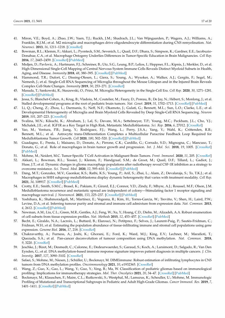
Figure 1. Graphical summary of the key topics in this review. (A) Hallmarks of immunogenicity of pediatric brain cancer.
(B) Ranking of pediatric brain tumor entities according to the amount of immune cell infiltration. Subgroup ranking is
Figure 1. Graphical summary of the key topics in this review. (A) Hallmarks of immunogenicity of pediatric brain cancer.(B) Ranking of pediatric brain tumor entities according to the amount of immune cell infiltration. Subgroup ranking is relatedto the respective entity. Abbreviations: ETMR (embryonal tumors with multilayered rosettes), MB (medulloblastoma),ATRT (atypical teratoid rhabdoid tumor), EPN (ependymoma), HGG (high-grade glioma), LGG (low-grade glioma), GBM(glioblastoma), DIPG (diffuse intrinsic pontine glioma), AA (anaplastic astrocytoma), PFA (posterior fossa, pediatric-type),ST-RELA (supratentorial, RELA fusion), DNET (dysembryoplastic neuroepithelial tumor), PA (pilocytic astrocytoma), GG(ganglioglioma), PXA (pleomorphic xanthoastrocytoma). (C) Ranking pediatric brain tumor entities along a scale of twobroadly defined immune cell infiltrate categories, namely myeloid-enriched (violet) or T cell-enriched (red) infiltration.(D) Illustration of a selection of immunotherapeutic targets evaluated in several pediatric brain tumor studies. The color coderepresents a qualitative evaluation. Abbreviations: PF-EPN (posterior fossa ependymoma), SP-EPN (spinal ependymoma),n. a. (not analyzed in depicted studies), * (contradicting results due to different methods used in the depicted studies).Findings in (B–D) were retrieved from [15–18,21,61,63,78,106,108,109,111] descriptively. Created with BioRender.com.
3.5. Challenges in Clinical Translation
Many advancements have been made in developing immunotherapeutic approachesfor brain tumors, especially in adult cancers. As presented in this review, pediatric braintumors are not as “cold” as assumed. Still, it is just the beginning of a deeper evaluationand consideration of the pediatric brain tumor microenvironment for implementation intostratifications and therapies. More research needs to be done to understand the TME,tumor epitopes, the heterogeneity among entities and subgroups, as well as different BBBphenotypes. Further, there is a lack of appropriate model systems.
Several challenges need to be addressed: (1) low mutational load of pediatric brain tu-mors and potentially limited immunogenicity, (2) differences in adult and pediatric immuneinfiltration, and differences even throughout childhood development [29],(3) combinatorial treatment regimens and interactions, and iv. management of immunother-apy toxicity since the CNS is particularly vulnerable to structural and functional damagein response to systemic inflammation.

Cancers 2021, 13, 5601 15 of 20
4. Conclusions
There is no doubt that therapies reducing the side effects of highly intensive chemo- andradio-therapy are needed in the treatment regimen of pediatric brain tumor patients. The poten-tial of immunotherapy in solid pediatric cancers is yet not fully uncovered.However, recent research efforts shed light on the immune-biological processes and laidthe ground for in-depth analysis of the tumor microenvironment of all pediatric brain tumors.Besides analyzing the specific molecular and genetic characteristics of each patient’s tumor,immune correlate studies should be considered prior to exposure to chemotherapy to maximizetreatment efficacy of combined treatment regimen with immunotherapeutic approaches.
Author Contributions: V.M. and K.K. wrote this review; V.M. and K.K. agreed to the published ver-sion of the manuscript. All authors have read and agreed to the published version of the manuscript.
Funding: This research was funded by “Wilhelm Sander Stiftung, 2019.158.1” and “Gesellschaftfür Kinderkrebsstiftung”.
Conflicts of Interest: The authors declare no conflict of interest.
References1. Johnson, K.J.; Cullen, J.; Barnholtz-Sloan, J.S.; Ostrom, Q.T.; Langer, C.E.; Turner, M.C.; McKean-Cowdin, R.; Fisher, J.L.; Lupo, P.J.;
Partap, S.; et al. Childhood Brain Tumor Epidemiology: A Brain Tumor Epidemiology Consortium Review. Cancer Epidemiol.Biomark. Prev. 2014, 23, 2716–2736. [CrossRef] [PubMed]
2. Louis, D.N.; Perry, A.; Reifenberger, G.; von Deimling, A.; Figarella-Branger, D.; Cavenee, W.K.; Ohgaki, H.; Wiestler, O.D.;Kleihues, P.; Ellison, D.W. The 2016 World Health Organization Classification of Tumors of the Central Nervous System: Asummary. Acta Neuropathol. 2016, 131, 803–820. [CrossRef]
3. Northcott, P.A.; Korshunov, A.; Witt, H.; Hielscher, T.; Eberhart, C.G.; Mack, S.; Bouffet, E.; Clifford, S.C.; Hawkins, C.E.;French, P.; et al. Medulloblastoma Comprises Four Distinct Molecular Variants. J. Clin. Oncol. 2010, 29, 1408–1414. [CrossRef][PubMed]
4. Zhang, J.; Wu, G.; Miller, C.P.; Tatevossian, R.G.; Dalton, J.D.; Tang, B.; Orisme, W.; Punchihewa, C.; Parker, M.;Qaddoumi, I.; et al. Whole-genome sequencing identifies genetic alterations in pediatric low-grade gliomas. Nat. Genet. 2013, 45,602–612. [CrossRef] [PubMed]
5. Cacciotti, C.; Fleming, A.; Ramaswamy, V. Advances in the molecular classification of pediatric brain tumors: A guide to thegalaxy. J. Pathol. 2020, 251, 249–261. [CrossRef] [PubMed]
6. Capper, D.; Jones, D.T.W.; Sill, M.; Hovestadt, V.; Schrimpf, D.; Sturm, D.; Koelsche, C.; Sahm, F.; Chavez, L.; Reuss, D.E.; et al.DNA methylation-based classification of central nervous system tumours. Nature 2018, 555, 469–474. [CrossRef] [PubMed]
7. Gröbner, S.N.; Worst, B.C.; Weischenfeldt, J.; Buchhalter, I.; Kleinheinz, K.; Rudneva, V.A.; Johann, P.D.; Balasubramanian, G.P.;Segura-Wang, M.; Brabetz, S.; et al. The landscape of genomic alterations across childhood cancers. Nature 2018, 555, 321–327.[CrossRef]
8. Fangusaro, J.; Bandopadhayay, P. Advances in the classification and treatment of pediatric brain tumors. Curr. Opin. Pediatr. 2021,33, 26–32. [CrossRef]
9. Rutkowski, S.; Bode, U.; Deinlein, F.; Ottensmeier, H.; Warmuth-Metz, M.; Soerensen, N.; Graf, N.; Emser, A.; Pietsch, T.;Wolff, J.E.A.; et al. Treatment of Early Childhood Medulloblastoma by Postoperative Chemotherapy Alone. N. Engl. J. Med. 2005,352, 978–986. [CrossRef]
10. Vinchon, M.; Baroncini, M.; Leblond, P.; Delestret, I. Morbidity and tumor-related mortality among adult survivors of pediatricbrain tumors: A review. Child’s Nerv. Syst. 2011, 27, 697–704. [CrossRef]
11. Lieberman, N.A.P.; Vitanza, N.A.; Crane, C.A. Immunotherapy for brain tumors: Understanding early successes and limitations.Expert Rev. Neurother. 2018, 18, 251–259. [CrossRef]
12. Paugh, B.S.; Qu, C.; Jones, C.; Liu, Z.; Adamowicz-Brice, M.; Zhang, J.; Bax, D.A.; Coyle, B.; Barrow, J.; Hargrave, D.; et al.Integrated molecular genetic profiling of pediatric high-grade gliomas reveals key differences with the adult disease. J. Clin.Oncol. 2010, 28, 3061–3068. [CrossRef] [PubMed]
13. Patel, R.R.; Ramkissoon, S.H.; Ross, J.; Weintraub, L. Tumor mutational burden and driver mutations: Characterizing the genomiclandscape of pediatric brain tumors. Pediatr. Blood Cancer 2020, 67, e28338. [CrossRef]
14. Vogelstein, B.; Papadopoulos, N.; Velculescu, V.E.; Zhou, S.; Diaz, L.A., Jr.; Kinzler, K.W. Cancer Genome Landscapes. Science2013, 339, 1546–1558. [CrossRef]
15. Leruste, A.; Tosello, J.; Ramos, R.N.; Tauziède-Espariat, A.; Brohard, S.; Han, Z.Y.; Beccaria, K.; Andrianteranagna, M.;Caudana, P.; Nikolic, J.; et al. Clonally Expanded T Cells Reveal Immunogenicity of Rhabdoid Tumors. Cancer Cell 2019,36, 597–612. [CrossRef] [PubMed]

Cancers 2021, 13, 5601 16 of 20
16. Chun, H.J.E.; Johann, P.D.; Milne, K.; Zapatka, M.; Buellesbach, A.; Ishaque, N.; Iskar, M.; Erkek, S.; Wei, L.;Tessier-Cloutier, B.; et al. Identification and Analyses of Extra-Cranial and Cranial Rhabdoid Tumor Molecular Subgroups RevealTumors with Cytotoxic T Cell Infiltration. Cell Rep. 2019, 29, 2338.e7–2354.e7. [CrossRef] [PubMed]
17. Robinson, M.H.; Vasquez, J.; Kaushal, A.; MacDonald, T.J.; Velázquez Vega, J.E.; Schniederjan, M.; Dhodapkar, K. Subtype andgrade-dependent spatial heterogeneity of T-cell infiltration in pediatric glioma. J. Immunother. Cancer 2020, 8, 6–11. [CrossRef]
18. Melcher, V.; Graf, M.; Interlandi, M.; Moreno, N.; de Faria, F.W.; Kim, S.N.; Kastrati, D.; Korbanka, S.; Alfert, A.; Gerß, J.; et al.Macrophage-tumor cell interaction promotes ATRT progression and chemoresistance. Acta Neuropathol. 2020, 139, 913–936.[CrossRef] [PubMed]
19. Stahl, D.; Knoll, R.; Gentles, A.J.; Vokuhl, C.; Buness, A.; Gütgemann, I. Prognostic Gene Expression, Stemness and ImmuneMicroenvironment in Pediatric Tumors. Cancers 2021, 13, 854. [CrossRef] [PubMed]
20. Wang, Y.; Zhou, C.; Luo, H.; Cao, J.; Ma, C.; Cheng, L.; Yang, Y. Prognostic implications of immune-related eight-gene signature inpediatric brain tumors. Braz. J. Med. Biol. Res. 2021, 54, 1–12. [CrossRef]
21. Grabovska, Y.; Mackay, A.; O’Hare, P.; Crosier, S.; Finetti, M.; Schwalbe, E.C.; Pickles, J.C.; Fairchild, A.R.; Avery, A.;Cockle, J.; et al. Pediatric pan-central nervous system tumor analysis of immune-cell infiltration identifies correlates of antitumorimmunity. Nat. Commun. 2020, 11, 4324. [CrossRef]
22. Vermeulen, J.F.; Van Hecke, W.; Adriaansen, E.J.M.; Jansen, M.K.; Bouma, R.G.; Villacorta Hidalgo, J.; Fisch, P.; Broekhuizen, R.;Spliet, W.G.M.; Kool, M.; et al. Prognostic relevance of tumor-infiltrating lymphocytes and immune checkpoints in pediatricmedulloblastoma. Oncoimmunology 2018, 7, e1398877. [CrossRef]
23. Quail, D.F.; Joyce, J.A. The Microenvironmental Landscape of Brain Tumors. Cancer Cell 2017, 31, 326–341. [CrossRef] [PubMed]24. Tanabe, S.; Yamashita, T. The role of immune cells in brain development and neurodevelopmental diseases. Int. Immunol. 2018,
30, 437–444. [CrossRef]25. Morimoto, K.; Nakajima, K. Role of the Immune System in the Development of the Central Nervous System. Front. Neurosci.
2019, 13, 916. [CrossRef]26. Dunn, G.P.; Bruce, A.T.; Ikeda, H.; Old, L.J.; Schreiber, R.D. Cancer immunoediting: From immunosurveillance to tumor escape.
Nat. Immunol. 2002, 3, 991–998. [CrossRef]27. Schreiber, R.D.; Old, L.J.; Smyth, M.J. Cancer immunoediting: Integrating immunity’s roles in cancer suppression and promotion.
Science 2011, 331, 1565–1570. [CrossRef] [PubMed]28. O’Sullivan, T.; Saddawi-Konefka, R.; Koebel, W.V.; Arthur, C.; White, J.M.; Uppaluri, R.; Andrews, D.M.; Ngiow, S.F.;
Teng, M.W.L.; Smyth, M.J.; et al. Cancer immunoediting by the innate immune system in the absence of adaptive immu-nity. J. Exp. Med. 2012, 209, 1869–1882. [CrossRef] [PubMed]
29. Simon, A.K.; Hollander, G.A.; McMichael, A. Evolution of the immune system in humans from infancy to old age. Proc. R. Soc.2015, 282, 20143085. [CrossRef]
30. Rieber, N.; Gille, C.; Köstlin, N.; Schäfer, I.; Spring, B.; Ost, M.; Spieles, H.; Kugel, H.A.; Pfeiffer, M.; Heininger, V.; et al.Neutrophilic myeloid-derived suppressor cells in cord blood modulate innate and adaptive immune responses. Clin. Exp.Immunol. 2013, 174, 45–52. [CrossRef]
31. McGovern, N.; Shin, A.; Low, G.; Low, D.; Duan, K.; Yao, L.J.; Msallam, R.; Low, I.; Shadan, N.B.; Sumatoh, H.R.; et al. Humanfetal dendritic cells promote prenatal T-cell immune suppression through arginase-2. Nature 2017, 546, 662–666. [CrossRef]
32. Olin, A.; Henckel, E.; Chen, Y.; Lakshmikanth, T.; Pou, C.; Mikes, J.; Gustafsson, A.; Bernhardsson, A.K.; Zhang, C.;Bohlin, K.; et al. Stereotypic Immune System Development in Newborn Children. Cell 2018, 174, 1277–1292. [CrossRef]
33. Kollmann, T.R.; Kampmann, B.; Mazmanian, S.K.; Marchant, A.; Levy, O. Protecting the Newborn and Young Infant fromInfectious Diseases: Lessons from Immune Ontogeny. Immunity 2017, 46, 350–363. [CrossRef]
34. Engelhardt, B.; Vajkoczy, P.; Weller, R.O. The movers and shapers in immune privilege of the CNS. Nat. Immunol. 2017, 18,123–131. [CrossRef]
35. Vitkovic, L.; Konsman, J.P.; Bockaert, J.; Dantzer, R.; Homburger, V.; Jacque, C. Cytokine signals propagate through the brain. Mol.Psychiatry 2000, 5, 604–615. [CrossRef]
36. Warren, K.E. Beyond the blood: Brain barrier: The importance of central nervous system (CNS) pharmacokinetics for thetreatment of CNS tumors, including diffuse intrinsic pontine glioma. Front. Oncol. 2018, 8, 239. [CrossRef] [PubMed]
37. Phoenix, T.N.; Patmore, D.M.; Boop, S.; Boulos, N.; Jacus, M.O.; Patel, Y.T.; Roussel, M.F.; Finkelstein, D.; Goumnerova, L.;Perreault, S.; et al. Medulloblastoma Genotype Dictates Blood Brain Barrier Phenotype. Cancer Cell 2016, 29, 508–522. [CrossRef]
38. Ginn, K.F.; Gajjar, A. Atypical teratoid rhabdoid tumor: Current therapy and future directions. Front. Oncol. 2012, 2, 114.[CrossRef]
39. Reddy, A.T.; Strother, D.R.; Judkins, A.R.; Burger, P.C.; Pollack, I.F.; Krailo, M.D.; Buxton, A.B.; Williams-Hughes, C.; Fouladi, M.;Mahajan, A.; et al. Efficacy of High-Dose Chemotherapy and 3-D Conformal Radiation for Atypical Teratoid/Rhabdoid Tumor:A Report from the Children’s Oncology Group Trial ACNS0333. J. Clin. Oncol. 2020, 38, 117. [CrossRef] [PubMed]
40. Meel, M.H.; Guillén Navarro, M.; De Gooijer, M.C.; Metselaar, D.S.; Waranecki, P.; Breur, M.; Lagerweij, T.; Wedekind, L.E.;Koster, J.; Van De Wetering, M.D.; et al. Mek/melk inhibition and blood-brain barrier deficiencies in atypical teratoid/rhabdoidtumors. Neuro-Oncology 2020, 22, 58–69. [CrossRef]
Cancers 2021, 13, 5601 17 of 20
41. Miron, V.E.; Boyd, A.; Zhao, J.W.; Yuen, T.J.; Ruckh, J.M.; Shadrach, J.L.; Van Wijngaarden, P.; Wagers, A.J.; Williams, A.;Franklin, R.J.M.; et al. M2 microglia and macrophages drive oligodendrocyte differentiation during CNS remyelination. Nat.Neurosci. 2013, 16, 1211–1218. [CrossRef]
42. Bowman, R.L.; Klemm, F.; Akkari, L.; Pyonteck, S.M.; Sevenich, L.; Quail, D.F.; Dhara, S.; Simpson, K.; Gardner, E.E.; Iacobuzio-Donahue, C.A.; et al. Macrophage Ontogeny Underlies Differences in Tumor-Specific Education in Brain Malignancies. Cell Rep.2016, 17, 2445–2459. [CrossRef] [PubMed]
43. Mrdjen, D.; Pavlovic, A.; Hartmann, F.J.; Schreiner, B.; Utz, S.G.; Leung, B.P.; Lelios, I.; Heppner, F.L.; Kipnis, J.; Merkler, D.; et al.High-Dimensional Single-Cell Mapping of Central Nervous System Immune Cells Reveals Distinct Myeloid Subsets in Health,Aging, and Disease. Immunity 2018, 48, 380–395. [CrossRef] [PubMed]
44. Hammond, T.R.; Dufort, C.; Dissing-Olesen, L.; Giera, S.; Young, A.; Wysoker, A.; Walker, A.J.; Gergits, F.; Segel, M.;Nemesh, J.; et al. Single-Cell RNA Sequencing of Microglia throughout the Mouse Lifespan and in the Injured Brain RevealsComplex Cell-State Changes. Immunity 2019, 50, 253–271. [CrossRef]
45. Masuda, T.; Sankowski, R.; Staszewski, O.; Prinz, M. Microglia Heterogeneity in the Single-Cell Era. Cell Rep. 2020, 30, 1271–1281.[CrossRef] [PubMed]
46. Jessa, S.; Blanchet-Cohen, A.; Krug, B.; Vladoiu, M.; Coutelier, M.; Faury, D.; Poreau, B.; De Jay, N.; Hébert, S.; Monlong, J.; et al.Stalled developmental programs at the root of pediatric brain tumors. Nat. Genet. 2019, 51, 1702–1713. [CrossRef] [PubMed]
47. Li, Q.; Cheng, Z.; Zhou, L.; Darmanis, S.; Neff, N.F.; Okamoto, J.; Gulati, G.; Bennett, M.L.; Sun, L.O.; Clarke, L.E.; et al.Developmental Heterogeneity of Microglia and Brain Myeloid Cells Revealed by Deep Single-Cell RNA Sequencing. Neuron2019, 101, 207–223. [CrossRef]
48. Svalina, M.N.; Kikuchi, K.; Abraham, J.; Lal, S.; Davare, M.A.; Settelmeyer, T.P.; Young, M.C.; Peckham, J.L.; Cho, Y.J.;Michalek, J.E.; et al. IGF1R as a Key Target in High Risk, Metastatic Medulloblastoma. Sci. Rep. 2016, 6, 27012. [CrossRef]
49. Yao, M.; Ventura, P.B.; Jiang, Y.; Rodriguez, F.J.; Wang, L.; Perry, J.S.A.; Yang, Y.; Wahl, K.; Crittenden, R.B.;Bennett, M.L.; et al. Astrocytic trans-Differentiation Completes a Multicellular Paracrine Feedback Loop Required forMedulloblastoma Tumor Growth. Cell 2020, 180, 502–520. [CrossRef] [PubMed]
50. Guadagno, E.; Presta, I.; Maisano, D.; Donato, A.; Pirrone, C.K.; Cardillo, G.; Corrado, S.D.; Mignogna, C.; Mancuso, T.;Donato, G.; et al. Role of macrophages in brain tumor growth and progression. Int. J. Mol. Sci. 2018, 19, 1005. [CrossRef][PubMed]
51. Mohme, M.; Neidert, M.C. Tumor-Specific T Cell Activation in Malignant Brain Tumors. Front. Immunol. 2020, 11, 205. [CrossRef]52. Akkari, L.; Bowman, R.L.; Tessier, J.; Klemm, F.; Handgraaf, S.M.; de Groot, M.; Quail, D.F.; Tillard, L.; Gadiot, J.;
Huse, J.T.; et al. Dynamic changes in glioma macrophage populations after radiotherapy reveal CSF-1R inhibition as a strategy toovercome resistance. Sci. Transl. Med. 2020, 12, 595–610. [CrossRef] [PubMed]
53. Dang, M.T.; Gonzalez, M.V.; Gaonkar, K.S.; Rathi, K.S.; Young, P.; Arif, S.; Zhai, L.; Alam, Z.; Devalaraja, S.; To, T.K.J.; et al.Macrophages in SHH subgroup medulloblastoma display dynamic heterogeneity that varies with treatment modality. Cell Rep.2021, 34, 108917. [CrossRef] [PubMed]
54. Crotty, E.E.; Smith, S.M.C.; Brasel, K.; Pakiam, F.; Girard, E.J.; Connor, Y.D.; Zindy, F.; Mhyre, A.J.; Roussel, M.F.; Olson, J.M.Medulloblastoma recurrence and metastatic spread are independent of colony—Stimulating factor 1 receptor signaling andmacrophage survival. J. Neurooncol. 2021, 153, 225–237. [CrossRef] [PubMed]
55. Yoshihara, K.; Shahmoradgoli, M.; Martínez, E.; Vegesna, R.; Kim, H.; Torres-Garcia, W.; Treviño, V.; Shen, H.; Laird, P.W.;Levine, D.A.; et al. Inferring tumour purity and stromal and immune cell admixture from expression data. Nat. Commun. 2013,4, 2612. [CrossRef] [PubMed]
56. Newman, A.M.; Liu, C.L.; Green, M.R.; Gentles, A.J.; Feng, W.; Xu, Y.; Hoang, C.D.; Diehn, M.; Alizadeh, A.A. Robust enumerationof cell subsets from tissue expression profiles. Nat. Methods 2015, 12, 453–457. [CrossRef] [PubMed]
57. Becht, E.; Giraldo, N.A.; Lacroix, L.; Buttard, B.; Elarouci, N.; Petitprez, F.; Selves, J.; Laurent-Puig, P.; Sautès-Fridman, C.;Fridman, W.H.; et al. Estimating the population abundance of tissue-infiltrating immune and stromal cell populations using geneexpression. Genome Biol. 2016, 17, 218. [CrossRef]
58. Chakravarthy, A.; Furness, A.; Joshi, K.; Ghorani, E.; Ford, K.; Ward, M.J.; King, E.V.; Lechner, M.; Marafioti, T.;Quezada, S.A.; et al. Pan-cancer deconvolution of tumour composition using DNA methylation. Nat. Commun. 2018,9, 3220. [CrossRef]
59. Jeschke, J.; Bizet, M.; Desmedt, C.; Calonne, E.; Dedeurwaerder, S.; Garaud, S.; Koch, A.; Larsimont, D.; Salgado, R.; Van DenEynden, G.; et al. DNA methylation-based immune response signature improves patient diagnosis in multiple cancers. J. Clin.Investig. 2017, 127, 3090–3102. [CrossRef]
60. Safaei, S.; Mohme, M.; Niesen, J.; Schüller, U.; Bockmayr, M. DIMEimmune: Robust estimation of infiltrating lymphocytes in CNStumors from DNA methylation profiles. Oncoimmunology 2021, 10, e1932365. [CrossRef]
61. Wang, Z.; Guo, X.; Gao, L.; Wang, Y.; Guo, Y.; Xing, B.; Ma, W. Classification of pediatric gliomas based on immunologicalprofiling: Implications for immunotherapy strategies. Mol. Ther. Oncolytics 2021, 20, 34–47. [CrossRef] [PubMed]
62. Bockmayr, M.; Klauschen, F.; Maire, C.L.; Rutkowski, S.; Westphal, M.; Lamszus, K.; Scheuller, U.; Mohme, M. ImmunologicProfiling of Mutational and Transcriptional Subgroups in Pediatric and Adult High-Grade Gliomas. Cancer Immunol. Res. 2019, 7,1401–1411. [CrossRef] [PubMed]

Cancers 2021, 13, 5601 18 of 20
63. Petralia, F.; Tignor, N.; Reva, B.; Koptyra, M.; Chowdhury, S.; Rykunov, D.; Krek, A.; Ma, W.; Zhu, Y.; Ji, J.; et al. IntegratedProteogenomic Characterization across Major Histological Types of Pediatric Brain Cancer. Cell 2020, 183, 1962.e31–1985.e31.[CrossRef]
64. Albert, T.K.; Interlandi, M.; Sill, M.; Graf, M.; Moreno, N.; Menck, K.; Rohlmann, A.; Melcher, V.; Korbanka, S.; Meyer zuHörste, G.; et al. An extracellular vesicle-related gene expression signature identifies high-risk patients in medulloblastoma.Neuro-Oncology 2021, 23, 586–598. [CrossRef] [PubMed]
65. Plant, A.S.; Koyama, S.; Sinai, C.; Solomon, I.H.; Griffin, G.K.; Ligon, K.L.; Bandopadhayay, P.; Betensky, R.; Emerson, R.;Dranoff, G.; et al. Immunophenotyping of pediatric brain tumors: Correlating immune infiltrate with histology, mutational load,and survival and assessing clonal T cell response. J. Neurooncol. 2018, 137, 269–278. [CrossRef] [PubMed]
66. Maximov, V.; Chen, Z.; Wei, Y.; Robinson, M.H.; Herting, C.J.; Shanmugam, N.S.; Rudneva, V.A.; Goldsmith, K.C.;MacDonald, T.J.; Northcott, P.A.; et al. Tumour-associated macrophages exhibit anti-tumoural properties in Sonic Hedgehogmedulloblastoma. Nat. Commun. 2019, 10, 2410. [CrossRef] [PubMed]
67. Lu, J.-Q.; Wilson, B.A.; Yong, V.W.; Pugh, J.; Mehta, V. Immune cell infiltrates in atypical teratoid/rhabdoid tumors. Can. J. Neurol.Sci. J. Can. Sci. Neurol. 2012, 39, 605–612. [CrossRef] [PubMed]
68. Marcu, A.; Schlosser, A.; Keupp, A.; Trautwein, N.; Johann, P.; Wölfl, M.; Lager, J.; Monoranu, C.M.; Walz, J.S.; Henkel, L.M.; et al.Natural and cryptic peptides dominate the immunopeptidome of atypical teratoid rhabdoid tumors. J. Immunother. Cancer 2021,9, e003404. [CrossRef] [PubMed]
69. Griesinger, A.M.; Birks, D.K.; Donson, A.M.; Amani, V.; Hoffman, L.M.; Waziri, A.; Wang, M.; Handler, M.H.; Foreman, N.K.Characterization of Distinct Immunophenotypes across Pediatric Brain Tumor Types. J. Immunol. 2013, 191, 4880–4888. [CrossRef][PubMed]
70. Teo, W.Y.; Elghetany, M.T.; Shen, J.; Man, T.K.; Li, X.; Chintagumpala, M.; Su, J.M.F.; Dauser, R.; Whitehead, W.;Adesina, A.M.; et al. Therapeutic implications of CD1d expression and tumor-infiltrating macrophages in pediatricmedulloblastomas. J. Neurooncol. 2014, 120, 293–301. [CrossRef]
71. Margol, A.S.; Robison, N.J.; Gnanachandran, J.; Hung, L.T.; Kennedy, R.J.; Vali, M.; Dhall, G.; Finlay, J.L.; Erdreich-Epstein, A.;Krieger, M.D.; et al. Tumor-associated macrophages in SHH subgroup of medulloblastomas. Clin. Cancer Res. 2015, 21, 1457–1465.[CrossRef] [PubMed]
72. Pham, C.D.; Flores, C.; Yang, C.; Pinheiro, E.M.; Yearley, J.H.; Sayour, E.J.; Pei, Y.; Moore, C.; McLendon, R.E.; Huang, J.; et al.Differential Immune Microenvironments and Response to Immune Checkpoint Blockade among Molecular Subtypes of MurineMedulloblastoma. Clin. Cancer Res. 2016, 22, 582–595. [CrossRef]
73. Aoki, T.; Hino, M.; Koh, K.; Kyushiki, M.; Kishimoto, H.; Arakawa, Y.; Hanada, R.; Kawashima, H.; Kurihara, J.;Shimojo, N.; et al. Low Frequency of Programmed Death Ligand 1 Expression in Pediatric Cancers. Pediatr. Blood Can-cer 2016, 63, 1461–1464. [CrossRef] [PubMed]
74. Bockmayr, M.; Mohme, M.; Klauschen, F.; Winkler, B.; Budczies, J.; Rutkowski, S.; Schüller, U. Subgroup-specific immune andstromal microenvironment in medulloblastoma. Oncoimmunology 2018, 7, e1462430. [CrossRef]
75. Diao, S.; Gu, C.; Zhang, H.; Yu, C. Immune cell infiltration and cytokine secretion analysis reveal a non-inflammatory microenvi-ronment of medulloblastoma. Oncol. Lett. 2020, 20, 397. [CrossRef] [PubMed]
76. Witt, D.A.; Donson, A.M.; Amani, V.; Moreira, D.C.; Sanford, B.; Hoffman, L.M.; Handler, M.H.; Levy, J.M.M.; Jones, K.L.;Nellan, A.; et al. Specific expression of PD-L1 in RELA-fusion supratentorial ependymoma: Implications for PD-1-targetedtherapy. Pediatr. Blood Cancer 2018, 65, e26960. [CrossRef]
77. Martin, A.M.; Bell, W.R.; Yuan, M.; Harris, L.; Poore, B.; Arnold, A.; Engle, E.L.; Asnaghi, L.; Lim, M.; Raabe, E.H.; et al. PD-L1expression in pediatric low-grade gliomas is independent of BRAF V600E mutational status. J. Neuropathol. Exp. Neurol. 2020, 79,74–85. [CrossRef]
78. Mackay, A.; Burford, A.; Molinari, V.; Jones, D.T.W.; Izquierdo, E.; Brouwer-Visser, J.; Giangaspero, F.; Haberler, C.; Pietsch, T.;Jacques, T.S.; et al. Molecular, Pathological, Radiological, and Immune Profiling of Non-brainstem Pediatric High-Grade Gliomafrom the HERBY Phase II Randomized Trial. Cancer Cell 2018, 33, 829–842. [CrossRef] [PubMed]
79. Panwalkar, P.; Pratt, D.; Chung, C.; Dang, D.; Le, P.; Martinez, D.; Bayliss, J.M.; Smith, K.S.; Adam, M.; Potter, S.; et al. SWI/SNFcomplex heterogeneity is related to polyphenotypic differentiation, prognosis, and immune response in rhabdoid tumors.Neuro-Oncology 2020, 22, 785–796. [CrossRef] [PubMed]
80. Alfert, A.; Moreno, N.; Kerl, K. The BAF complex in development and disease. Epigenet. Chromatin 2019, 12, 19. [CrossRef][PubMed]
81. Northcott, P.A.; Pfister, S.M.; Jones, D.T.W. Next-generation (epi)genetic drivers of childhood brain tumours and the outlook fortargeted therapies. Lancet Oncol. 2015, 16, e293–e302. [CrossRef]
82. Pan, D.; Kobayashi, A.; Jiang, P.; De Andrade, L.F.; Tay, R.E.; Luoma, A.M.; Tsoucas, D.; Qiu, X.; Lim, K.; Rao, P.; et al. Amajor chromatin regulator determines resistance of tumor cells to T cell–mediated killing. Science 2018, 359, 770–775. [CrossRef][PubMed]
83. Biegel, J.A.; Zhou, J.Y.; Rorke, L.B.; Stenstrom, C.; Wainwright, L.M.; Fogelgren, B. Germ-line and acquired mutations of INI1 inatypical teratoid and rhabdoid tumors. Cancer Res. 1999, 59, 74–79. [CrossRef] [PubMed]

Cancers 2021, 13, 5601 19 of 20
84. Hasselblatt, M.; Nagel, I.; Oyen, F.; Bartelheim, K.; Russell, R.B.; Schüller, U.; Junckerstorff, R.; Rosenblum, M.; Alassiri, A.H.;Rossi, S.; et al. SMARCA4-mutated atypical teratoid/rhabdoid tumors are associated with inherited germline alterations andpoor prognosis. Acta Neuropathol. 2014, 128, 453–456. [CrossRef]
85. Price, G.; Bouras, A.; Hambardzumyan, D.; Hadjipanayis, C.G. Current knowledge on the immune microenvironment andemerging immunotherapies in diffuse midline glioma. EBioMedicine 2021, 69, 103453. [CrossRef]
86. Johann, P.D.; Erkek, S.; Zapatka, M.; Hasselblatt, M.; Pfister, S.M.; Kool, M. Atypical Teratoid/Rhabdoid Tumors Are Comprisedof Three Epigenetic Subgroups with Distinct Enhancer Landscapes. Cancer Cell 2016, 29, 379–393. [CrossRef]
87. Luke, J.J.; Bao, R.; Sweis, R.F.; Spranger, S.; Gajewski, T.F. WNT/b-catenin pathway activation correlates with immune exclusionacross human cancers. Clin. Cancer Res. 2019, 25, 3074–3083. [CrossRef]
88. Grund-Gröschke, S.; Stockmaier, G.; Aberger, F. Hedgehog/GLI signaling in tumor immunity—New therapeutic opportunitiesand clinical implications. Cell Commun. Signal. 2019, 17, 172. [CrossRef]
89. Mehlman, C.; Kamga, P.T.; Costantini, A.; Julié, C.; Dumenil, C.; Dumoulin, J.; Ouaknine, J.; Giraud, V.; Chinet, T.; Emile, J.F.; et al.Baseline hedgehog pathway activation and increase of plasma Wnt1 protein are associated with resistance to immune checkpointinhibitors in advanced non-small-cell lung cancer. Cancers 2021, 13, 1107. [CrossRef]
90. Sevenich, L. Turning “Cold” into “Hot” tumors—Opportunities and challenges for radio-immunotherapy against primary andmetastatic brain cancers. Front. Oncol. 2019, 9, 163. [CrossRef] [PubMed]
91. Galluzzi, L.; Humeau, J.; Buqué, A.; Zitvogel, L.; Kroemer, G. Immunostimulation with chemotherapy in the era of immunecheckpoint inhibitors. Nat. Rev. Clin. Oncol. 2020, 17, 725–741. [CrossRef] [PubMed]
92. Karachi, A.; Dastmalchi, F.; Mitchell, D.A.; Rahman, M. Temozolomide for immunomodulation in the treatment of glioblastoma.Neuro-Oncology 2018, 20, 1566–1572. [CrossRef] [PubMed]
93. Petroni, G.; Buqué, A.; Zitvogel, L.; Kroemer, G.; Galluzzi, L. Immunomodulation by targeted anticancer agents. Cancer Cell 2021,39, 310–345. [CrossRef] [PubMed]
94. Huang, S.; Wang, Z.; Zhou, J.; Huang, J.; Zhou, L.; Luo, J.; Wan, Y.Y.; Long, H.; Zhu, B. EZH2 inhibitor GSK126 suppressesantitumor immunity by driving production of myeloid-derived suppressor cells. Cancer Res. 2019, 79, 2009–2020. [CrossRef][PubMed]
95. Alimova, I.; Birks, D.K.; Harris, P.S.; Knipstein, J.A.; Venkataraman, S.; Marquez, V.E.; Foreman, N.K.; Vibhakar, R. Inhibition ofEZH2 suppresses self-renewal and induces radiation sensitivity in atypical rhabdoid teratoid tumor cells. Neuro-Oncology 2013,15, 149–160. [CrossRef]
96. Knutson, S.K.; Warholic, N.M.; Wigle, T.J.; Klaus, C.R.; Allain, C.J.; Raimondi, A.; Scott, M.P.; Chesworth, R.; Moyer, M.P.;Copeland, R.A.; et al. Durable tumor regression in genetically altered malignant rhabdoid tumors by inhibition of methyltrans-ferase EZH2. Proc. Natl. Acad. Sci. USA 2013, 110, 7922–7927. [CrossRef] [PubMed]
97. Chi, S.N.; Bourdeaut, F.; Laetsch, T.W.; Fouladi, M.; Macy, M.E.; Makin, G.W.J.; Shukla, N.N.; Wetmore, C.; Margol, A.S.;Casanova, M.; et al. Phase I study of tazemetostat, an enhancer of zeste homolog-2 inhibitor, in pediatric pts with re-lapsed/refractory integrase interactor 1-negative tumors. J. Clin. Oncol. 2020, 38, 10525. [CrossRef]
98. Jones, R.L.; Blay, J.; Agulnik, M.; Chugh, R.; Mir, O.; Italiano, A.; Thomas, D.; Gupta, A.; Jahan, T.; Cote, G.; et al. A phase 2,multicenter study of the EZH2 inhibitor tazemetostat in adults (rhabdoid tumor cohort) (NCT02601950). Ann. Oncol. 2018, 29,viii580–viii581. [CrossRef]
99. Gounder, M.; Schöffski, P.; Jones, R.L.; Agulnik, M.; Cote, G.M.; Villalobos, V.M.; Attia, S.; Chugh, R.; Chen, T.W.W.; Jahan, T.; et al.Tazemetostat in advanced epithelioid sarcoma with loss of INI1/SMARCB1: An international, open-label, phase 2 basket study.Lancet Oncol. 2020, 21, 1423–1432. [CrossRef]
100. Keane, L.; Cheray, M.; Saidi, D.; Kirby, C.; Friess, L.; Gonzalez-rodriguez, P.; Gerdes, M.E.; Grabert, K.; McColl, B.W. Inhibition ofmicroglial EZH2 leads to anti-tumoral effects in pediatric diffuse midline gliomas. Neuro-Oncology 2021, 3, vdab096. [CrossRef]
101. Woods, D.M.; Sodré, A.L.; Villagra, A.; Sarnaik, A.; Sotomayor, E.M.; Weber, J. HDAC inhibition upregulates PD-1 ligands inmelanoma and augments immunotherapy with PD-1 blockade. Cancer Immunol. Res. 2015, 3, 1375–1385. [CrossRef]
102. Knox, T.; Sahakian, E.; Banik, D.; Hadley, M.; Palmer, E.; Noonepalle, S.; Kim, J.; Powers, J.; Gracia-Hernandez, M.;Oliveira, V.; et al. Selective HDAC6 inhibitors improve anti-PD-1 immune checkpoint blockade therapy by decreasing theanti-inflammatory phenotype of macrophages and down-regulation of immunosuppressive proteins in tumor cells. Sci. Rep.2019, 9, 6136. [CrossRef] [PubMed]
103. Sharma, P.; Hu-Lieskovan, S.; Wargo, J.A.; Ribas, A. Primary, Adaptive, and Acquired Resistance to Cancer Immunotherapy. Cell2017, 168, 707–723. [CrossRef] [PubMed]
104. Ring, E.K.; Markert, J.M.; Gillespie, G.Y.; Friedman, G.K. Checkpoint proteins in pediatric brain and extracranial solid tumors:Opportunities for immunotherapy. Clin. Cancer Res. 2017, 23, 342–350. [CrossRef]
105. Majzner, R.G.; Simon, J.S.; Grosso, J.F.; Martinez, D.; Pawel, B.R.; Santi, M.; Merchant, M.S.; Geoerger, B.; Hezam, I.; Marty, V.; et al.Assessment of programmed death-ligand 1 expression and tumor-associated immune cells in pediatric cancer tissues. Cancer2017, 123, 3807–3815. [CrossRef]
106. Haydar, D.; Houke, H.; Chiang, J.; Yi, Z.; Odé, Z.; Caldwell, K.; Zhu, X.; Mercer, K.S.; Stripay, J.L.; Shaw, T.I.; et al. Cell-surfaceantigen profiling of pediatric brain tumors: B7-H3 is consistently expressed and can be targeted via local or systemic CAR T-celldelivery. Neuro-Oncology 2021, 23, 999–1011. [CrossRef]

Cancers 2021, 13, 5601 20 of 20
107. Majzner, R.G.; Theruvath, J.L.; Nellan, A.; Heitzeneder, S.; Cui, Y.; Mount, C.W.; Rietberg, S.P.; Linde, M.H.; Xu, P.; Rota, C.; et al.CAR T cells targeting B7-H3, a pan-cancer antigen, demonstrate potent preclinical activity against pediatric solid tumors andbrain tumors. Clin. Cancer Res. 2019, 25, 2560–2574. [CrossRef]
108. Theruvath, J.; Sotillo, E.; Mount, C.W.; Graef, C.M.; Delaidelli, A.; Heitzeneder, S.; Labanieh, L.; Dhingra, S.; Leruste, A.;Majzner, R.G.; et al. Locoregionally administered B7-H3-targeted CAR T cells for treatment of atypical teratoid/rhabdoid tumors.Nat. Med. 2020, 26, 712–719. [CrossRef]
109. Donovan, L.K.; Delaidelli, A.; Joseph, S.K.; Bielamowicz, K.; Fousek, K.; Holgado, B.L.; Manno, A.; Srikanthan, D.; Gad, A.Z.; VanOmmeren, R.; et al. Locoregional delivery of CAR T cells to the cerebrospinal fluid for treatment of metastatic medulloblastomaand ependymoma. Nat. Med. 2020, 26, 720–731. [CrossRef] [PubMed]
110. Gupta, A.; Taslim, C.; Tullius, B.P.; Cripe, T.P. Therapeutic modulation of the CD47-SIRPα axis in the pediatric tumor microenvi-ronment: Working up an appetite. Cancer Drug Resist. 2020, 3, 550–562. [CrossRef]
111. Gholamin, S.; Mitra, S.S.; Feroze, A.H.; Liu, J.; Kahn, S.A.; Zhang, M.; Esparza, R.; Richard, C.; Ramaswamy, V.; Remke, M.; et al.Disrupting the CD47-SIRPα anti-phagocytic axis by a humanized anti-CD47 antibody is an efficacious treatment for malignantpediatric brain tumors. Sci. Transl. Med. 2017, 9, e001341. [CrossRef] [PubMed]
112. Ravanpay, A.C.; Gust, J.; Johnson, A.J.; Rolczynski, L.S.; Chang, C.A.; Hoglund, V.J.; Mukherjee, R.; Nicholas, A.; Orentas, R.J.;Jensen, M.C. EGFR806-CAR T cells selectively target a tumor-restricted EGFR epitope in glioblastoma. Oncotraget 2019, 10,7080–7095. [CrossRef] [PubMed]
113. Stastny, M.J.; Brown, C.E.; Ruel, C.; Jensen, M.C. Medulloblastomas Expressing IL13Ra2 are Targets for IL13-zetakine+ CytolyticT Cells. J. Pediatr. Hematol. Oncol. 2007, 29, 669–677. [CrossRef] [PubMed]
114. Ahmed, N.; Ratnayake, M.; Savoldo, B.; Perlaky, L.; Dotti, G.; Wels, W.S.; Bhattacharjee, M.B.; Gilbertson, R.J.; Shine, H.D.;Weiss, H.L.; et al. Regression of Experimental Medulloblastoma following Transfer of HER2-Specific T Cells. Cancer Res. 2007, 67,5957–5964. [CrossRef] [PubMed]
115. Nellan, A.; Rota, C.; Majzner, R.; Lester-McCully, C.M.; Griesinger, A.M.; Mulcahy Levy, J.M.; Foreman, N.K.; Warren, K.E.;Lee, D.W. Durable regression of Medulloblastoma after regional and intravenous delivery of anti-HER2 chimeric antigen receptorT cells. J. Immunother. Cancer 2018, 6, 30. [CrossRef] [PubMed]
116. Liu, D.; Song, L.; Brawley, V.S.; Robison, N.; Wei, J.; Gao, X.; Tian, G.; Margol, A.; Ahmed, N.; Asgharzadeh, S.; et al. Medul-loblastoma expresses CD1d and can be targeted for immunotherapy with NKT cells. Clin. Immunol. 2013, 149, 55–64. [CrossRef][PubMed]
117. Orlando, D.; Miele, E.; De Angelis, B.; Guercio, M.; Boffa, I.; Sinibaldi, M.; Po, A.; Caruana, I.; Abballe, L.; Carai, A.; et al. Adoptiveimmunotherapy using PRAME-specific T cells in medulloblastoma. Cancer Res. 2018, 78, 3337–3349. [CrossRef] [PubMed]
118. Powell, A.B.; Yadavilli, S.; Saunders, D.; Van Pelt, S.; Chorvinsky, E.; Burga, R.A.; Albihani, S.; Hanley, P.J.; Xu, Z.; Pei, Y.; et al.Medulloblastoma rendered susceptible to NK-cell attack by TGFβ neutralization. J. Transl. Med. 2019, 17, 321. [CrossRef]
119. Tan, I.; Arifa, R.D.N.; Rallapalli, H.; Kana, V.; Lao, Z.; Sanghrajka, R.M.; Bayin, N.S.; Tanne, A.; Wojcinski, A.; Korshunov, A.; et al.CSF1R Inhibition Depletes Tumor-Associated Macrophages and Attenuates Tumor Progression in a Mouse Sonic Hedgehog-Medulloblastoma Model. Oncogene 2021, 40, 396–407. [CrossRef] [PubMed]
120. Mount, C.W.; Majzner, R.G.; Sundaresh, S.; Arnold, E.P.; Kadapakkam, M.; Haile, S.; Labanieh, L.; Hulleman, E.; Woo, P.J.;Rietberg, S.P.; et al. Potent antitumor efficacy of anti-GD2 CAR T cells in H3-K27M(+) diffuse midline gliomas. Nat. Med. 2018, 24,572–579. [CrossRef]
121. van Gool, S.W.; Holm, S.; Rachor, J.; Adamson, L.; Technau, A.; Maass, E.; Frühwald, M.C.; Schlegel, P.G.; Eyrich, M. Immunother-apy in atypical teratoid-rhabdoid tumors: Data from a survey of the HGG-Immuno Group. Cytotherapy 2016, 18, 1178–1186.[CrossRef] [PubMed]
122. Caruso, D.A.; Orme, L.M.; Neale, A.M.; Radcliff, F.J.; Armor, G.M.; Maixner, W.; Downie, P.; Hassall, T.E.; Tang, M.L.K.;Ashley, D.M. Results of a phase 1 study utilizing monocyte-derived dendritic cells pulsed with tumor RNA in children and youngadults with brain cancer. Neuro-Oncology 2004, 6, 236–246. [CrossRef] [PubMed]
123. Ardon, H.; De Vleeschouwer, S.; Claes, L.; Van Calenbergh, F.; Kramm, C.M.; Rutkowski, S.; Wolff, J.E.A.; Van Gool, S.W. AdjuvantDendritic Cell-Based Tumour Vaccination for Children With Malignant Brain Tumours. Pediatr. Blood Cancer 2010, 54, 519–525.[CrossRef]
124. Garcia-Moure, M.; Gonzalez-Huarriz, M.; Labiano, S.; Guruceaga, E.; Bandres, E.; Zalacain, M.; Marrodan, L.; de Andrea, C.;Villalba, M.; Martinez-Velez, N.; et al. Delta-24-RGD, an oncolytic adenovirus, increases survival and promotes proinflammatoryimmune landscape remodeling in models of AT/RT and CNS-PNET. Clin. Cancer Res. 2021, 27, 1807–1820. [CrossRef] [PubMed]
125. Bonifant, C.L.; Jackson, H.J.; Brentjens, R.J.; Curran, K.J. Toxicity and management in CAR T-cell therapy. Mol. Ther. Oncolytics2016, 3, 16011. [CrossRef]