The genome of Tetranychus urticae reveals herbivorous pest adaptations
6
ARTICLE doi:10.1038/nature10640 The genome of Tetranychus urticae reveals herbivorous pest adaptations Miodrag Grbic ´ 1,2 *, Thomas Van Leeuwen 3 *, Richard M. Clark 4 *, Stephane Rombauts 5,6 , Pierre Rouze ´ 5,6 , Vojislava Grbic ´ 1,2 , Edward J. Osborne 4 , Wannes Dermauw 3 , Phuong Cao Thi Ngoc 5,6 , Fe ´lix Ortego 7 , Pedro Herna ´ndez-Crespo 7 , Isabel Diaz 8 , Manuel Martinez 8 , Maria Navajas 9 ,E ´ lio Sucena 10,11 , Sara Magalha ˜es 12 , Lisa Nagy 13 , Ryan M. Pace 13 , Sergej Djuranovic ´ 14 , Guy Smagghe 3 , Masatoshi Iga 3 , Olivier Christiaens 3 , Jan A. Veenstra 15 , John Ewer 16 , Rodrigo Mancilla Villalobos 16 , Jeffrey L. Hutter 17 , Stephen D. Hudson 17 , Marisela Velez 18 , Soojin V. Yi 19 , Jia Zeng 19 , Andre Pires-daSilva 20 , Fernando Roch 21 , Marc Cazaux 1 , Marie Navarro 1 , Vladimir Zhurov 1 , Gustavo Acevedo 1 , Anica Bjelica 1 , Jeffrey A. Fawcett 5,6 {, Eric Bonnet 5,6 {, Cindy Martens 5,6 , Guy Baele 5,6 , Lothar Wissler 22 , Aminael Sanchez-Rodriguez 23 , Luc Tirry 3 , Catherine Blais 24 , Kristof Demeestere 25 , Stefan R. Henz 26 , T. Ryan Gregory 27 , Johannes Mathieu 28 , Lou Verdon 29 , Laurent Farinelli 30 , Jeremy Schmutz 31,32 , Erika Lindquist 32 , Rene ´ Feyereisen 33 & Yves Van de Peer 5,6 The spider mite Tetranychus urticae is a cosmopolitan agricultural pest with an extensive host plant range and an extreme record of pesticide resistance. Here we present the completely sequenced and annotated spider mite genome, representing the first complete chelicerate genome. At 90 megabases T. urticae has the smallest sequenced arthropod genome. Compared with other arthropods, the spider mite genome shows unique changes in the hormonal environment and organization of the Hox complex, and also reveals evolutionary innovation of silk production. We find strong signatures of polyphagy and detoxification in gene families associated with feeding on different hosts and in new gene families acquired by lateral gene transfer. Deep transcriptome analysis of mites feeding on different plants shows how this pest responds to a changing host environment. The T. urticae genome thus offers new insights into arthropod evolution and plant–herbivore interactions, and provides unique opportunities for developing novel plant protection strategies. Mites belong to the Chelicerata, the second largest group of terrestrial animals. Chelicerates represent a basal branch of arthropods. Subsequent to their origin in the Cambrian period, arthropods radiated into two lineages: the Chelicerata and the Mandibulata (com- prising the Myriapoda and the Pancrustacea (which includes both crustaceans and insects)) 1,2 . Extant lineages of chelicerates include Pycnogonida, Xiphosura (horseshoe crabs) and Arachnida (a large group comprising scorpions, spiders and the Acari (ticks and mites)) 3,4 (Supplementary Fig. 1.1). Within the Acari, T. urticae belongs to the Acariformes with the earliest fossils dating from the Lower Devonian period (410 million years ago). The Acari represent the most diverse chelicerate clade, with over 40,000 described species that exhibit tremendous variations in lifestyle, ranging from parasitic to predatory to plant-feeding. Some mites are of major concern to human health and include allergy-causing dust mites, scabies mites and mite vectors of scrub typhus 5 . The two-spotted spider mite, Tetranychus urticae, is a cosmopolitan agricultural pest 6 belonging to an assemblage of web-spinning mites. The name ‘spider’ highlights their ability to produce silk-like webbing used to establish a colonial micro-habitat, protect against abiotic agents, shelter from predators, communicate via pheromones and provide a vehicle for dispersion 7 . Tetranychus urticae represents one of the most polyphagous arthropod herbivores, feeding on more than 1,100 plant species belonging to more than 140 different plant families including species known to produce toxic compounds. It is a major pest in greenhouse production and field crops, destroying annual and perennial crops such as tomatoes, peppers, cucumbers, strawberries, maize, soy, *These authors contributed equally to this work. 1 Department of Biology, The University of Western Ontario, London N6A 5B7, Canada. 2 Instituto de Ciencias de la Vid y el Vino (CSIC, UR, Gobiernode La Rioja), 26006 Logron ˜ o, Spain. 3 Department of Crop Protection, Faculty of Bioscience Engineering, Ghent University, B-9000 Ghent, Belgium. 4 Department of Biology, University of Utah, Salt Lake City, Utah 84112, USA. 5 Department of Plant Systems Biology, VIB, Technologiepark 927, B-9052 Ghent, Belgium. 6 Department of Plant Biotechnology and Bioinformatics, Ghent University, Technologiepark 927, B-9052 Ghent, Belgium. 7 Department of Environmental Biology, Centro de Investigaciones Biolo ´ gicas, CSIC, 28040 Madrid, Spain. 8 Centro de Biotecnologı ´a y Geno ´ mica de Plantas, UPM-INIA, 28223 Madrid, Spain. 9 INRA, UMR CBGP (INRA/IRD/ Cirad/Montpellier SupAgro), Campus international de Baillarguet, 34988 Montferrier-sur-Lez, France. 10 Instituto Gulbenkian de Cie ˆ ncia, 2781-901 Oeiras, Portugal. 11 Universidade de Lisboa, Faculdade de Cie ˆ ncias, Departamento de Biologia Animal, 1749-016 Lisbon, Portugal. 12 Universidade de Lisboa, Faculdade de Cie ˆ ncias, Centro de Biologia Ambiental, 1749-016 Lisbon, Portugal. 13 Department of Molecular and Cellular Biology, University of Arizona, Tucson, Arizona 85721, USA. 14 Johns Hopkins University School of Medicine, Department of Molecular Biology & Genetics, Baltimore, Maryland 21205, USA. 15 Institut de Neurosciences Cognitives et Inte ´ gratives d’Aquitaine Universite ´ de Bordeaux 1, 33405 Talence, France. 16 Centro Interdisciplinario de Neurociencia de Valparaı ´so, Facultad de Ciencias, Universidad de Valparaı ´so, Valparaı ´so 2360102, Chile. 17 Department of Physics and Astronomy, The University of Western Ontario, N6A 5B7 London, Canada. 18 Instituto de Cata ´ lisis y Petroleoquı ´mica CSIC, Madrid, Spain; IMDEA Nanociencias, Facultad de Ciencias, Universidad Autonoma de Madrid, 28050 Madrid, Spain. 19 School of Biology, Georgia Institute of Technology, Atlanta, Georgia 30332, USA. 20 Department of Biology, University of Texas at Arlington, Arlington, Texas 76019, USA. 21 Universite de Toulouse, UPS, Centre de Biologie du Developpement, Universite Paul Sabatier, 31062 Toulouse, France; Centre National de la Recherche Scientifique, UMR 5547, Centre de Biologie du Developpement, 31062 Toulouse, France. 22 Westfa ¨ lische Wilhelms University, Institute for Evolution and Biodiversity, Evolutionary Bioinformatics Group, Hu ¨ fferstrasse 1, D-48149 Mu ¨ nster, Germany. 23 CMPG, Department of Microbial and Molecular Systems, K.U. Leuven, B-3001 Leuven, Belgium. 24 UPMC Univ Paris 06, UMR CNRS 7622, Equipe Biogene ` se des signaux hormonaux, Case 29, 75005 Paris, France. 25 Research Group EnVOC, Department of Sustainable Organic Chemistry and Technology, Faculty of Bioscience Engineering, Ghent University, B-9000 Ghent, Belgium. 26 Max Planck Institute for Developmental Biology, D-72076 Tu ¨ bingen, Germany. 27 Department of Integrative Biology, University of Guelph, N1G 2W1 Guelph, Canada. 28 Boyce Thompson Institute for Plant Research, Ithaca, New York 14853, USA. 29 Southern Crop Protection and Food Research Centre, Agriculture and Agri-Food Canada, N5V 4T3 London, Canada. 30 Fasteris SA, CH-1228 Plan-les-Ouates, Switzerland. 31 HudsonAlpha Institute for Biotechnology Huntsville, Alabama 35806, USA. 32 DOE Joint Genome Institute, Walnut Creek, California 94598, USA. 33 UMR 1301, INRA, CNRS and Universite ´ de Nice Sophia Antipolis, 06903 Sophia Antipolis, France. {Present addresses: Institut Curie, 26 rue d’Ulm, Paris 75248, France; INSERM, U900, Paris 75248, France; Mines ParisTech, Fontainebleau 77300, France (E.B.); Graduate University for Advanced Studies, Hayama, Kanagawa 240-0193, Japan (J.A.F.). 24 NOVEMBER 2011 | VOL 479 | NATURE | 487 Macmillan Publishers Limited. All rights reserved ©2011
-
Upload
independent -
Category
Documents
-
view
0 -
download
0
Transcript of The genome of Tetranychus urticae reveals herbivorous pest adaptations
ARTICLEdoi:10.1038/nature10640
The genome of Tetranychus urticaereveals herbivorous pest adaptationsMiodrag Grbic1,2*, Thomas Van Leeuwen3*, Richard M. Clark4*, Stephane Rombauts5,6, Pierre Rouze5,6, Vojislava Grbic1,2,Edward J. Osborne4, Wannes Dermauw3, Phuong Cao Thi Ngoc5,6, Felix Ortego7, Pedro Hernandez-Crespo7, Isabel Diaz8,Manuel Martinez8, Maria Navajas9, Elio Sucena10,11, Sara Magalhaes12, Lisa Nagy13, Ryan M. Pace13, Sergej Djuranovic14,Guy Smagghe3, Masatoshi Iga3, Olivier Christiaens3, Jan A. Veenstra15, John Ewer16, Rodrigo Mancilla Villalobos16,Jeffrey L. Hutter17, Stephen D. Hudson17, Marisela Velez18, Soojin V. Yi19, Jia Zeng19, Andre Pires-daSilva20, Fernando Roch21,Marc Cazaux1, Marie Navarro1, Vladimir Zhurov1, Gustavo Acevedo1, Anica Bjelica1, Jeffrey A. Fawcett5,6{, Eric Bonnet5,6{,Cindy Martens5,6, Guy Baele5,6, Lothar Wissler22, Aminael Sanchez-Rodriguez23, Luc Tirry3, Catherine Blais24,Kristof Demeestere25, Stefan R. Henz26, T. Ryan Gregory27, Johannes Mathieu28, Lou Verdon29, Laurent Farinelli30,Jeremy Schmutz31,32, Erika Lindquist32, Rene Feyereisen33 & Yves Van de Peer5,6
The spider mite Tetranychus urticae is a cosmopolitan agricultural pest with an extensive host plant range and anextreme record of pesticide resistance. Here we present the completely sequenced and annotated spider mitegenome, representing the first complete chelicerate genome. At 90 megabases T. urticae has the smallest sequencedarthropod genome. Compared with other arthropods, the spider mite genome shows unique changes in the hormonalenvironment and organization of the Hox complex, and also reveals evolutionary innovation of silk production. We findstrong signatures of polyphagy and detoxification in gene families associated with feeding on different hosts and in newgene families acquired by lateral gene transfer. Deep transcriptome analysis of mites feeding on different plants showshow this pest responds to a changing host environment. The T. urticae genome thus offers new insights into arthropodevolution and plant–herbivore interactions, and provides unique opportunities for developing novel plant protectionstrategies.
Mites belong to the Chelicerata, the second largest group of terrestrialanimals. Chelicerates represent a basal branch of arthropods.Subsequent to their origin in the Cambrian period, arthropodsradiated into two lineages: the Chelicerata and the Mandibulata (com-prising the Myriapoda and the Pancrustacea (which includes bothcrustaceans and insects))1,2. Extant lineages of chelicerates includePycnogonida, Xiphosura (horseshoe crabs) and Arachnida (a largegroup comprising scorpions, spiders and the Acari (ticks andmites))3,4 (Supplementary Fig. 1.1). Within the Acari, T. urticaebelongs to the Acariformes with the earliest fossils dating from theLower Devonian period (410 million years ago). The Acari representthe most diverse chelicerate clade, with over 40,000 described speciesthat exhibit tremendous variations in lifestyle, ranging from parasiticto predatory to plant-feeding. Some mites are of major concern to
human health and include allergy-causing dust mites, scabies mitesand mite vectors of scrub typhus5.
The two-spotted spider mite, Tetranychus urticae, is a cosmopolitanagricultural pest6 belonging to an assemblage of web-spinning mites.The name ‘spider’ highlights their ability to produce silk-like webbingused to establish a colonial micro-habitat, protect against abioticagents, shelter from predators, communicate via pheromones andprovide a vehicle for dispersion7.
Tetranychus urticae represents one of the most polyphagousarthropod herbivores, feeding on more than 1,100 plant speciesbelonging to more than 140 different plant families including speciesknown to produce toxic compounds. It is a major pest in greenhouseproduction and field crops, destroying annual and perennial cropssuch as tomatoes, peppers, cucumbers, strawberries, maize, soy,
*These authors contributed equally to this work.
1Department of Biology, The University of Western Ontario, London N6A 5B7, Canada. 2Instituto de Ciencias de la Vid y el Vino (CSIC, UR, Gobiernode La Rioja), 26006 Logrono, Spain. 3Department of CropProtection, Faculty of Bioscience Engineering, Ghent University, B-9000 Ghent, Belgium. 4Department of Biology, University of Utah, Salt Lake City, Utah 84112, USA. 5Department of Plant Systems Biology,VIB, Technologiepark 927, B-9052 Ghent, Belgium. 6Department of Plant Biotechnology and Bioinformatics, Ghent University, Technologiepark 927, B-9052 Ghent, Belgium. 7Department ofEnvironmental Biology, Centro de Investigaciones Biologicas, CSIC, 28040 Madrid, Spain. 8Centro de Biotecnologıa y Genomica de Plantas, UPM-INIA, 28223 Madrid, Spain. 9INRA, UMR CBGP (INRA/IRD/Cirad/Montpellier SupAgro), Campus international de Baillarguet, 34988 Montferrier-sur-Lez, France. 10Instituto Gulbenkian de Ciencia, 2781-901 Oeiras, Portugal. 11Universidade de Lisboa, Faculdadede Ciencias, Departamento de Biologia Animal, 1749-016 Lisbon, Portugal. 12Universidade de Lisboa, Faculdade de Ciencias, Centro de Biologia Ambiental, 1749-016 Lisbon, Portugal. 13Department ofMolecular and Cellular Biology, University of Arizona, Tucson, Arizona 85721, USA. 14Johns Hopkins University School of Medicine, Department of Molecular Biology & Genetics, Baltimore, Maryland21205, USA. 15Institut de Neurosciences Cognitives et Integratives d’Aquitaine Universite de Bordeaux 1, 33405 Talence, France. 16Centro Interdisciplinario de Neurociencia de Valparaıso, Facultad deCiencias, Universidad de Valparaıso, Valparaıso 2360102, Chile. 17Department of Physics and Astronomy, The University of Western Ontario, N6A 5B7 London, Canada. 18Instituto de Catalisis yPetroleoquımica CSIC, Madrid, Spain; IMDEA Nanociencias, Facultad de Ciencias, Universidad Autonoma de Madrid, 28050 Madrid, Spain. 19School of Biology, Georgia Institute of Technology, Atlanta,Georgia 30332, USA. 20Department of Biology, University of Texas at Arlington, Arlington, Texas 76019, USA. 21Universite de Toulouse, UPS, Centre de Biologie du Developpement, Universite Paul Sabatier,31062 Toulouse, France; Centre National de la Recherche Scientifique, UMR 5547, Centre de Biologie du Developpement, 31062 Toulouse, France. 22Westfalische Wilhelms University, Institute forEvolution and Biodiversity, Evolutionary Bioinformatics Group, Hufferstrasse 1, D-48149 Munster, Germany. 23CMPG, Department of Microbial and Molecular Systems, K.U. Leuven, B-3001 Leuven,Belgium. 24UPMC Univ Paris 06, UMR CNRS 7622, Equipe Biogenese des signaux hormonaux, Case 29, 75005 Paris, France. 25Research Group EnVOC, Department of Sustainable Organic Chemistry andTechnology, Faculty of Bioscience Engineering, Ghent University, B-9000 Ghent, Belgium. 26Max Planck Institute for Developmental Biology, D-72076 Tubingen, Germany. 27Department of IntegrativeBiology, University of Guelph, N1G 2W1 Guelph, Canada. 28Boyce Thompson Institute for Plant Research, Ithaca, New York 14853, USA. 29Southern Crop Protection and Food Research Centre, Agricultureand Agri-Food Canada, N5V 4T3 London, Canada. 30Fasteris SA, CH-1228 Plan-les-Ouates, Switzerland. 31HudsonAlpha Institute for Biotechnology Huntsville, Alabama 35806, USA. 32DOE Joint GenomeInstitute, Walnut Creek, California 94598, USA. 33UMR 1301, INRA, CNRS and Universite de Nice Sophia Antipolis, 06903 Sophia Antipolis, France. {Present addresses: Institut Curie, 26 rue d’Ulm, Paris75248, France; INSERM, U900, Paris 75248, France; Mines ParisTech, Fontainebleau 77300, France (E.B.); Graduate University for Advanced Studies, Hayama, Kanagawa 240-0193, Japan (J.A.F.).
2 4 N O V E M B E R 2 0 1 1 | V O L 4 7 9 | N A T U R E | 4 8 7
Macmillan Publishers Limited. All rights reserved©2011
apples, grapes and citrus. The recent introduction of the relatedspecies Tetranychus evansi to Europe and Africa from SouthAmerica demonstrates the invasive nature of these pests in globalagriculture8. Computer modelling suggests that with intensifyingglobal warming, the detrimental effects of spider mites in agriculturewill markedly increase9 due to accelerated development at hightemperatures.
Tetranychus urticae is known for its ability to develop rapid resist-ance to pesticides. Among arthropods it has the highest incidence ofpesticide resistance10. Chemical control often causes a broad cross-resistance within and between pesticide classes, resulting in resistanceto novel pesticides within 2–4 years. Many aspects of the biology ofthe spider mite, including rapid development, high fecundity andhaplo-diploid sex determination, seem to facilitate rapid evolutionof pesticide resistance. Control of multi-resistant mites has becomeincreasingly difficult and the genetic basis of such resistance remainspoorly understood11.
As the first completed chelicerate genome, the comparison of theT. urticae genome with the genomes of insects and the crustaceanDaphnia pulex expands the arthropod genetic toolkit. At the sametime, the very compact T. urticae genome has unique attributesamong arthropod genomes with remarkable instances of gene gainsand losses. The completion of the T. urticae genome sequence opensnew avenues for understanding the fundamentals of plant–herbivoreinteractions, developing novel pest-management strategies and pro-ducing new biomaterials on the nanometre scale.
The small genome of T. urticaeThe T. urticae genome (strain London) was sequenced (Sanger) to8.053 coverage and assembled into 640 scaffolds covering 89.6megabases (Mb) (Supplementary Notes 1, 2.1 and 2.2). 70,778Sanger expressed sequence tag (EST) sequences from embryos, larvae,nymphs and adults were generated, and further complemented withRNA-seq data on matching samples (Supplementary Note 2.3). Weidentified 18,414 protein-coding gene models, of which 84% (15,397)are supported by EST (8,243), protein homology (11,433) and/orRNA-seq data (14,545) (Supplementary Note 2.4 and Supplemen-tary Fig. 2.4.1). From alignments of ,43-million paired-endIllumina reads from a second T. urticae strain (Montpellier) to theLondon sequence, 542,600 single nucleotide polymorphisms andsmall indels were predicted (Supplementary Note 2.5). The completegenome annotation of T. urticae is available at the BOGAS website12.With an estimated genome size of about 90 Mb, the T. urticae genomeis the smallest arthropod genome sequenced so far. The genomes ofother chelicerates are much larger (565–7,100 Mb), with the un-finished genome of the tick Ixodes scapularis estimated at2,100 Mb13. Multiple characteristics of the T. urticae genome correlatewith its compact size: small transposable element content and micro-satellite density, increased gene density and holocentric chromo-somes (see Supplementary Note 3.1 for chromosomal features).
Transposable elements totalled 9.09 Mb (Supplementary Note 3.2),putting T. urticae together with D. pulex and Apis mellifera as arthropodswith 10% or less of their genomes comprised of transposable elements.Long terminal repeat (LTR) retrotransposons, and in particular Gypsy-like elements, were the most abundant type of transposable elements.L1-like Long interspersed elements (LINEs), Tc1/Mariner-like DNAtransposons, and Maverick (Polinton) elements were also detected(see Supplementary Table 3.2.1). Deep sequencing of small RNAs(,19–30 nucleotides) across developmental stages (SupplementaryNote 4.1) identified 226,829 unique RNAs that mapped to 676,266different loci in the genome. The number of unique small RNA countsper size category shows a peak at 21 and 26 nucleotides. These two peaksinclude short interfering RNAs and Piwi-interacting RNAs, respectively,similar to what is observed in Drosophila melanogaster14. Their align-ments to the genome indicate that both probably silence diverse trans-posable elements. Included among ,21-nucleotide small RNAs are 52
predicted microRNAs (miRNAs). On the basis of the identity of theirseed regions (nucleotides 2–7 of the miRNA sequence), the T. urticaemiRNAs can be grouped into 43 families (Supplementary Note 4). Halfof the predicted miRNAs were not conserved when compared toannotated miRNAs and available genomes of other arthropods15, sug-gesting that they might be T. urticae- or lineage-specific (Supplemen-tary Tables 4.3.1–4.3.4).
The microsatellite density in the T. urticae genome is among thelowest observed for arthropods (Supplementary Note 3.3 andSupplementary Fig. 3.3.1), consistent with the expectation that repeatcontent of genomes typically scales with genome size. The T. urticaemicrosatellite classes have a distinct profile: mono-nucleotide repeatsare virtually non-existent, and di-nucleotide repeats, normally themost abundant type of microsatellites, are found significantly lessoften than tri-nucleotides, as in Tribolium castaneum16. The genedensity is twice as high compared to D. melanogaster, with 205 versus92 genes per Mb, respectively. The mean number of exons per genewas low and similar to that found in D. melanogaster (,3.8 exons pergene). The size distribution of introns was typically skewed with amean intron size of 400 bp and a median of 96 bp (see SupplementaryNote 3.4, Supplementary Fig. 2.4.3 and Supplementary Table 2.4.1).The holocentric nature of T. urticae chromosomes17 (the absence ofcentromeres and the diffuse nature of the kinetochores) is correlatedwith a lack of large tracts of gene-poor heterochromatin. The uniformlydistributed gene density (Supplementary Note 3.1.1 and SupplementaryFig. 3.2.1) contrasts with the human body louse (Pediculus humanus,Phthiraptera, a hemimetabolous insect with a small genome), where95% of the genes are concentrated in only 55 Mb of the 110-Mbgenome18.
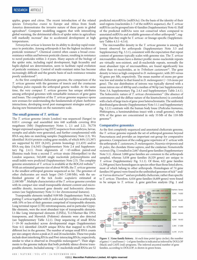
Comparative genomicsAs the first completely sequenced and annotated chelicerate genome,the T. urticae genome expands the set of arthropod genomes beyondPancrustacea and provides an important out-group for comparativegenomics. Comparison of the coding gene repertoire of T. urticae withthe arthropods T. castaneum, D. melanogaster, Nasonia vitripennis andD. pulex, the chordate Homo sapiens, and the cnidarian Nematostellavectensis (Fig. 1) resulted in 2,667 shared gene families (SupplementaryNote 5.1). Almost 3,000 gene families are common to the arthropodssampled, whereas 5,038 gene families (8,329 genes) are unique toT. urticae (Supplementary Fig. 5.1.1). Of those, 622 gene families(1,398 genes) have homologues in species other than those listed above,most of which belong to other arthropods. Homologues of 74 genefamilies (93 genes) were found in the unfinished genomes of tick13 and/or Varroa destructor19 and are probably chelicerate, rather than specificto T. urticae. Therefore, 4,416 gene families (6,609 genes) were foundto be unique to T. urticae. A gene gain/loss analysis (Fig. 1 and
+307/–566 +307/–573
+227/–360 +227/–367
+516/–1,011 +516/–1,018
+182/–123+190/–131
+245/–400+279/–364
+298/–84+293/–95
+661/–1,593 +661/–1,546
+632/–426 +632/–395
+262/–610+257/–621
+5,518+5,518
+1,597/–640 +1,597/–577
2
1
4
3
5
7
1013
9 6
8
Drosophila melanogaster 2,943 14,832
Tetranychus urticae
Tribolium castaneum 3,410 16,422
Homo sapiens
Nasonia vitripennis 10,737 19,800
Orphans Genes
Daphnia pulex 4,859
2,364
4,293
30,940
21,828
+1,327 +1,327
+479+448
12
11
Nematostella vectensis 2,478 27,273
18,423
5,767
5,708
5,863
5,649
5,997
5,518
Figure 1 | Gene family history. At each time point (grey circles), the numberof gains (1) and losses (2) of gene families is indicated as inferred by DOLLOP(black) and CAFE (red) programs. The inferred ancestral number of genefamilies, according to DOLLOP, is shown in green boxes.
RESEARCH ARTICLE
4 8 8 | N A T U R E | V O L 4 7 9 | 2 4 N O V E M B E R 2 0 1 1
Macmillan Publishers Limited. All rights reserved©2011

Supplementary Note 5.2) of these genomes showed a gain of about 700new gene families in the lineage leading to T. urticae, plus almost 4,300genes that are single copy (orphans). More than 1,000 gene families,still present in other arthropods, were lost in T. urticae. The 58 genefamilies that are significantly (z-score .2) expanded inT. urticae compared to the other arthropods are shown in Supplemen-tary Note 5.2 and Supplementary Fig. 5.2.1.
Feeding and detoxificationTetranychus urticae is one of the most striking examples of polyphagyamong herbivores and it has an unmatched ability to develop resist-ance to pesticides6,10. We discovered that known gene families impli-cated in digestion, detoxification and transport of xenobiotics had aunique spider mite composition, and were often expanded whencompared to insects (Supplementary Note 6.1). This included a three-fold proliferation of cysteine peptidase genes, particularly C1A papainand the C13 legumain genes (Supplementary Table 6.1.11), consistentwith proteolytic digestion based mostly on cysteine peptidaseactivity20. Eighty-six cytochrome P450 (CYP) genes were detectedin the T. urticae genome, a total number similar to insects but withan expansion of T. urticae-specific intronless genes of the CYP2 clan(Supplementary Table 6.1.2). The carboxyl/cholinesterases (CCEs)gene family contained 71 genes, with a single acetylcholinesterasegene (Ace1) but two new clades at the root of the neurodevelop-mental class of CCEs, representing 34 and 22 CCEs, respectively
(Supplementary Table 6.1.6). A notable case of expansion was foundwithin the family of 32 glutathione S-transferases (GSTs) that includea group of 12 Mu-class GSTs that were, until now, believed to bevertebrate-specific (Supplementary Table 6.1.3). Finally, we discovered39 multidrug resistance proteins belonging to the ATP-bindingcassette (ABC) transporters (class C). The repertoire from thisclass of ABC transporters far exceeds the number (9–14) found incrustaceans, insects, vertebrates and nematodes (SupplementaryTable 6.1.8). Few of the genes involved in detoxification had closeinsect homologues, and only four of the CYP genes could clearly beassigned as orthologues of insect and crustacean CYP genes.
The involvement of these gene families and their spider-mite-specific expansion in host plant adaptation is markedly illustratedby RNA-seq transcriptome profiling of spider mite feeding on itspreferred host, bean (Phaseolus vulgaris), and on hosts to which theLondon strain is not adapted: Arabidopsis thaliana and tomato(Solanum lycopersicum) (Fig. 2) (Supplementary Notes 6.2). Wefound 24% of all genes to be differentially expressed upon host transfer(Fig. 2a–c); relative to bean, more genes were differentially expressedon tomato than on A. thaliana (Supplementary Note 6.2.4 andSupplementary Fig. 6.2.1), but responses were nonetheless correlated(Fig. 2b, c). Genes in the detoxification and peptidase families exhibitedthe most profound changes (Fig. 2a–c), with expression of nearly halfof P450 genes affected by the host plant, including 19 of 39 genes in theintronless CYP392 family and the CYP389 family. These subfamilies
a b
c
CYP385C4CYP385C3
CYP385C2CYP385C1CYP385B1
CYP385A1CYP384A1
CYP383A1CYP382A1
CYP386A1CYP389A1
CYP389B1CYP389C1CYP389C3CYP389C4CYP389C2CYP389C8CYP389C5CYP389C12
CYP389C11CYP389C10CYP389C9
CYP389C7CYP389C6
CYP388A1CYP387A1
CYP387A2CYP407A1
CYP390A1CYP391A1
CYP4CL1CYP4CF2
CYP406A1CYP381A1CYP381A2
CYP314A1CYP302A1
CYP315A1CYP307A1CYP392C1
CYP392A16CYP392A12
CYP392A11CYP392A15CYP392A13v2CYP392A13
CYP392A14CYP392A6
CYP392A7CYP392A10CYP392A9
CYP392A5CYP392A4
CYP392A1CYP392A3
CYP392B2CYP392B1
CYP392B3CYP392E1
CYP392E2CYP392E7CYP392E8CYP392E6CYP392E3CYP392E9CYP392E10CYP392D1
CYP392D6CYP392D2CYP392D7
CYP392D3CYP392DnCYP392D8
CYP392D4
CYP392A8
0.8
Upregulated on
A. thaliana
Upregulated on tomato
−8 −7 −6 −5 −4 −3 −2 −1 876543210
Upregulated on bean
Upregulated on bean
–8
–7
–6
–5
–4
–3
–2
–1
0
1
2
3
4
5
6
7
8
Upregulated on A. thaliana
Upregulated on bean
Upregulated on bean
Upregulated on tomato
–8 –7 –6 –5 –4 –3 –2 –1 876543210
Both (n = 2,019)
Bean−A. thaliana
only (n = 483)
Bean−tomato only
(n = 1,932)
Differentially expressed
Peptidases ABCs
CCEsGSTs
CYPsLipoproteins
Family/class
Differentially expressed
Bean−tomato
Bean−A. thaliana
Both
–2 –1.5 1.5 2Fold change
Mito
cla
n
Bea
n–
tom
ato
Bea
n–
A. t
halia
na
Lo
g2 f
old
chang
e
–8
–7
–6
–5
–4
–3
–2
–1
0
1
2
3
4
5
6
7
8
Lo
g2 f
old
chang
e
Log2 fold change
Log2 fold change
CY
P3 c
lan
CY
P2 c
lan
CY
P4 c
lan
Figure 2 | Gene expression changes when mites are shifted from P. vulgaris(bean) to A. thaliana or to S. lycopersicum (tomato). a, A phylogeny of thecytochrome P450 (CYP) genes and heat map of the response of CYP genes tohost transfer. Two-thirds of the genes that are tandemly duplicated or that formclusters (indicated by black vertical lines) are co-regulated. b, Global changes in
gene expression after host shift. c, Fold changes of important gene familymembers in digestion and detoxification are colour coded. The analysis ofdifferential expression (b and c) is with a 5% false discovery rate as assessed withRNA-seq data collected in biological triplicate (fold changes between meanvalues are plotted).
ARTICLE RESEARCH
2 4 N O V E M B E R 2 0 1 1 | V O L 4 7 9 | N A T U R E | 4 8 9
Macmillan Publishers Limited. All rights reserved©2011
are spider-mite-specific P450 expansions that define lineage-specificexpansions21. This finding is unprecedented. In humans, only up toone-third of P450 genes are metabolizing xenobiotics22, and inD. melanogaster only one-third of the CYP genes are inducible byxenobiotics23. The proportion of P450 genes responding to the chemicalenvironment is much greater in the spider mite. Similar patterns werealso found within other families (Fig. 2c). For GSTs and CCEs, theexpression of Mu and Delta GSTs and the two spider-mite-specific CCE clades were most affected and about one-third of cysteinepeptidases, the C1A papains and C13 legumains, were overexpressedafter transfer to tomato. More than two-thirds of the CYP and GSTgenes affected by the host plant are present in clusters of (multiple)tandem duplicated genes. Co-regulation of the majority of tandemduplicates strongly indicates that the ancestral gene was alreadyplant-responsive before duplication, and that a role in plant adaptationmay have favoured duplicate retention.
Although these data indicate that spider-mite-specific expansion ofknown gene families contributes to the ability of spider mites toovercome host defences, many genes differentially regulated uponhost transfer lack homology to genes of known function. Notably,among those with the most extreme expression fold-changes are genesthat encode putative secreted proteins or lipid-binding proteins.Understanding extracellular binding and transport of small ligandsis therefore likely to be important in further dissecting spider mite–plant interactions.
Lateral gene transferOur search for genes related to detoxification and digestion alsorevealed the existence and surprising expansion of intradiol ringcleavage dioxygenases, genes previously unreported from metazoangenomes but characteristic for bacteria and fungi24. We annotated 16functional genes in this family in T. urticae, whereas bacterialgenomes usually carry only 1 to 7. They have an average sequencesimilarity of 43% with the homologue of Streptomyces avermitilis andshare the conserved 2 His 2 Tyr non-haem iron(III) binding site. Thesedioxygenases might have evolved to metabolize aromatic compoundsfound in plant allelochemicals. Other clear instances of lateral genetransfers include (1) the presence of a cobalamin-independentmethionine synthase (MetE) gene with four predicted introns andup to 58% sequence identity to the MetE gene of soil Bacilli (thissequence has not previously been reported in any animal species);(2) two very similar levanase-encoding genes of probable bacterialorigin that encode secreted exo-fructosidases upregulated upon feed-ing on tomato; and (3) a cyanate lyase-encoding gene that might beinvolved in feeding on cyanogenic plants (Supplementary Table6.3.1).
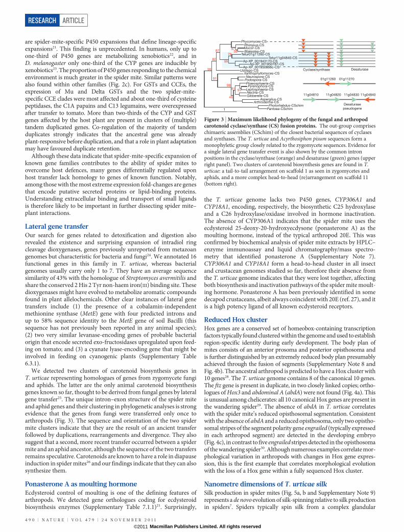
We detected two clusters of carotenoid biosynthesis genes inT. urticae representing homologues of genes from zygomycete fungiand aphids. The latter are the only animal carotenoid biosynthesisgenes known so far, thought to be derived from fungal genes by lateralgene transfer25. The unique intron–exon structure of the spider miteand aphid genes and their clustering in phylogenetic analyses is strongevidence that the genes from fungi were transferred only once toarthropods (Fig. 3). The sequence and orientation of the two spidermite clusters indicate that they are the result of an ancient transferfollowed by duplications, rearrangements and divergence. They alsosuggest that a second, more recent transfer occurred between a spidermite and an aphid ancestor, although the sequence of the two transfersremains speculative. Carotenoids are known to have a role in diapauseinduction in spider mites26 and our findings indicate that they can alsosynthesize them.
Ponasterone A as moulting hormoneEcdysteroid control of moulting is one of the defining features ofarthropods. We detected gene orthologues coding for ecdysteroidbiosynthesis enzymes (Supplementary Table 7.1.1)21. Surprisingly,
the T. urticae genome lacks two P450 genes, CYP306A1 andCYP18A1, encoding, respectively, the biosynthetic C25 hydroxylaseand a C26 hydroxylase/oxidase involved in hormone inactivation.The absence of CYP306A1 indicates that the spider mite uses theecdysteroid 25-deoxy-20-hydroxyecdysone (ponasterone A) as themoulting hormone, instead of the typical arthropod 20E. This wasconfirmed by biochemical analysis of spider mite extracts by HPLC–enzyme immunoassay and liquid chromatography/mass spectro-metry that identified ponasterone A (Supplementary Note 7).CYP306A1 and CYP18A1 form a head-to-head cluster in all insectand crustacean genomes studied so far, therefore their absence fromthe T. urticae genome indicates that they were lost together, affectingboth biosynthesis and inactivation pathways of the spider mite moult-ing hormone. Ponasterone A has been previously identified in somedecapod crustaceans, albeit always coincident with 20E (ref. 27), and itis a high potency ligand of all known ecdysteroid receptors.
Reduced Hox clusterHox genes are a conserved set of homeobox-containing transcriptionfactors typically found clustered within the genome and used to establishregion-specific identity during early development. The body plan ofmites consists of an anterior prosoma and posterior opisthosoma andis further distinguished by an extremely reduced body plan presumablyachieved through the fusion of segments (Supplementary Note 8 andFig. 4b). The ancestral arthropod is predicted to have a Hox cluster with10 genes28. The T. urticae genome contains 8 of the canonical 10 genes.The ftz gene is present in duplicate, in two closely linked copies; ortho-logues of Hox3 and abdominal A (abdA) were not found (Fig. 4a). Thisis unusual among chelicerates: all 10 canonical Hox genes are present inthe wandering spider29. The absence of abdA in T. urticae correlateswith the spider mite’s reduced opisthosomal segmentation. Consistentwith the absence of abdA and a reduced opisthosoma, only two opistho-somal stripes of the segment polarity gene engrailed (typically expressedin each arthropod segment) are detected in the developing embryo(Fig. 4c), in contrast to five engrailed stripes detected in the opisthosomaof the wandering spider30. Although numerous examples correlate mor-phological variation in arthropods with changes in Hox gene expres-sion, this is the first example that correlates morphological evolutionwith the loss of a Hox gene within a fully sequenced Hox cluster.
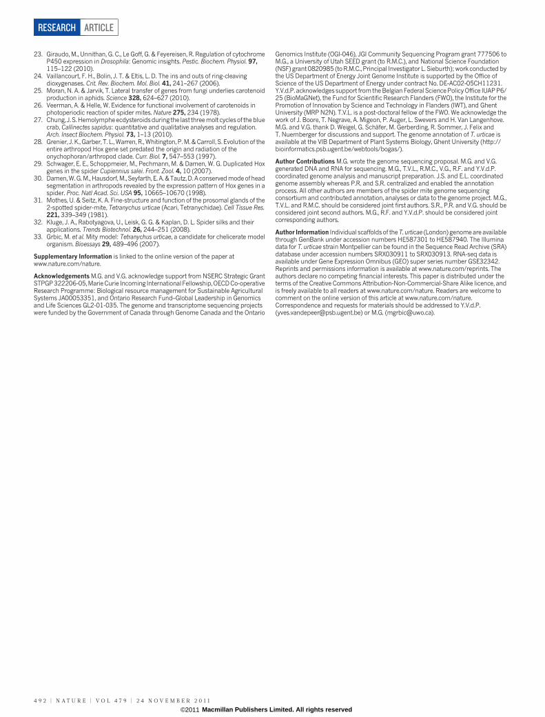
Nanometre dimensions of T. urticae silkSilk production in spider mites (Fig. 5a, b and Supplementary Note 9)represents a de novo evolution of silk-spinning relative to silk productionin spiders7. Spiders typically spin silk from a complex glandular
Phycomyces-CSRhizopus-CSMucor-CSBlakeslea-CS
Tetur01g11260-CSTetur11g04840-CS
Ap-XP_001943170-CSAp-XP_001950787-CS
Ap-XP_001950868c-CS
9758
7887
82
91
48
81 93
Ustilago-CSXanthophyllomyces-CS
Neurospora-CSPodospora-CSPhaeosphaeria-CSPyrenophora-CS
Leptosphaeria-CSNectria-CSGibberella-CS
Aspergilus-CSArthroderma-CS
Photorhabdus-CSchimPantoea-CSchim
Cyclase/synthase Desaturase
01g1127001g11260
11g04810 11g04820 11g04830
Desaturase
pseudogene
11g04840
1
99
99
99
Figure 3 | Maximum likelihood phylogeny of the fungal and arthropodcarotenoid cyclase/synthase (CS) fusion proteins. The out-group compriseschimaeric assemblies (CSchim) of the closest bacterial sequences of cyclasesand synthases. The T. urticae and Acyrthosiphon pisum sequences form amonophyletic group closely related to the zygomycete sequences. Evidence fora single lateral gene transfer event is also shown by the common intronpositions in the cyclase/synthase (orange) and desaturase (green) genes (upperright panel). Two clusters of carotenoid biosynthesis genes are found in T.urticae: a tail-to-tail arrangement on scaffold 1 as seen in zygomycetes andaphids, and a more complex head-to-head (re)arrangement on scaffold 11(bottom right).
RESEARCH ARTICLE
4 9 0 | N A T U R E | V O L 4 7 9 | 2 4 N O V E M B E R 2 0 1 1
Macmillan Publishers Limited. All rights reserved©2011
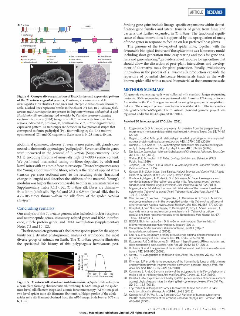
abdominal spinneret, whereas T. urticae uses paired silk glands con-nected to the mouth appendages (pedipalps)31. Seventeen fibroin geneswere uncovered in the genome of T. urticae (Supplementary Table9.1.1) encoding fibroins of unusually high (27–39%) serine content.We performed mechanical testing on fibres deposited by adult andlarval mites with an atomic force microscope. This technique measuresthe Young’s modulus of the fibres, which is the ratio of applied stress(tension per cross-sectional area) to the resulting strain (fractionalchange in length) and describes the stiffness of the material. Young’smodulus was higher than or comparable to other natural materials (seeSupplementary Table 9.1.2), but T. urticae silk fibres are thinner—54 6 3 nm (adult silk, Fig. 5c) and 23.3 6 0.9 nm (larval silk), that is,435–185 times thinner—than the silk fibres of the spider Nephilaclavipes32.
Concluding remarksOur analysis of the T. urticae genome also included nuclear receptorsand neuropeptide genes, immunity-related genes and RNA interfer-ence, cuticle protein genes, and DNA methylation (SupplementaryNotes 7.3 and 10–12).
The first complete genome of a chelicerate species provides the oppor-tunity for a detailed phylogenomic analysis of arthropods, the mostdiverse group of animals on Earth. The T. urticae genome illustratesthe specialized life history of this polyphagous herbivorous pest.
Striking gene gains include lineage-specific expansions within detoxi-fication gene families and lateral transfer of genes from fungi andbacteria that further expanded in T. urticae. The functional signifi-cance of these innovations is supported by the upregulation of manyof these genes in response to feeding on less preferred host plants.
The genome of the two-spotted spider mite, together with thefavourable biological features of the spider mite as a laboratory modelincluding short generation time, easy rearing and tools for gene ana-lysis and gene silencing33, provide a novel resource for agriculture thatshould allow the dissection of pest–plant interactions and develop-ment of alternative tools for plant protection. Finally, evolutionaryinnovation in the process of T. urticae silk production expands therepertoire of potential chelicerate biomaterials (such as the well-known spider silk) with a natural biomaterial at the nanometre scale.
METHODS SUMMARYAll genomic sequencing reads were collected with standard Sanger sequencingprotocols. RNA sequencing was performed with Illumina RNA-seq protocols.Annotation of the T. urticae genome was done using the gene prediction platformEuGene. The complete genome annotation is available at http://bioinformatics.psb.ugent.be/webtools/bogas/. The T. urticae (London) genome project wasregistered under the INSDC project ID 71041.
Received 30 June; accepted 17 October 2011.
1. Edgecombe, G. D. Arthropod phylogeny: An overview from the perspectives ofmorphology, molecular dataand the fossil record.ArthropodStruct. Dev. 39, 74–87(2010).
2. Regier, J. C. et al. Arthropod relationships revealed by phylogenomic analysis ofnuclear protein-coding sequences. Nature 463, 1079–1083 (2010).
3. Dunlop, J. A. & Selden, P. A. Calibrating the chelicerate clock: a paleontologicalreply to Jeyaprakash and Hoy. Exp. Appl. Acarol. 48, 183–197 (2009).
4. Dunlop, J. A.Geological historyandphylogeny ofChelicerata. ArthropodStruct. Dev.39, 124–142 (2010).
5. Walter, D. E. & Proctor, H. C. Mites: Ecology, Evolution and Behaviour (CABIPublishing, 1999).
6. Jeppson, L. R., Keifer, H. H. & Baker, E. W. Mites Injurious to Economic Plants (Univ.California Press, 1975).
7. Gerson, U. in Spider Mites: their Biology, Natural Enemies and Control Vol. 1A (edsHelle, W. & Sabelis, M. W.) 223–232 (Elsevier, 1985).
8. Boubou, A., Migeon, A., Roderick, G. K. & Navajas, M. Recent emergence andworldwide spread of the red tomato spider mite, Tetranychus evansi: geneticvariation and multiple cryptic invasions. Biol. Invasions 13, 81–92 (2011).
9. Migeon, A. et al. Modelling the potential distribution of the invasive tomato redspider mite, Tetranychus evansi (Acari: Tetranychidae). Exp. Appl. Acarol. 48,199–212 (2009).
10. Van Leeuwen, T., Vontas, J., Tsagkarakou, A., Dermauw, W. & Tirry, L. Acaricideresistance mechanisms in the two-spotted spider mite Tetranychus urticae andother important Acari: a review. Insect Biochem. Mol. Biol. 40, 563–572 (2010).
11. Khajehali, J., Van Nieuwenhuyse, P., Demaeght, P., Tirry, L. & Van Leeuwen, T.Acaricide resistance and resistance mechanisms in Tetranychus urticaepopulations from rose greenhouses in the Netherlands. Pest Manag. Sci. 67,1424–1433 (2011).
12. BOGAS. Bioinformatics Gent Online Genome Annotation Service Æhttp://bioinformatics.psb.ugent.be/webtools/bogas/æ (2011).
13. VectorBase. Ixodes scapularis Wikel annotation, IscaW1 Æhttp://iscapularis.vectorbase.orgæ (2008).
14. Lau, N. C. et al. Abundant primary piRNAs, endo-siRNAs, and microRNAs in aDrosophila ovary cell line. Genome Res. 19, 1776–1785 (2009).
15. Kozomara, A. & Griffiths-Jones, S. miRBase: integrating microRNA annotation anddeep-sequencing data. Nucleic Acids Res. 39, D152–D157 (2011).
16. Richards, S. et al. The genome of the model beetle and pest Tribolium castaneum.Nature 452, 949–955 (2008).
17. Oliver, J. H. Cytogenetics of mites and ticks. Annu. Rev. Entomol. 22, 407–429(1977).
18. Kirkness, E. F. et al. Genome sequences of the human body louse and its primaryendosymbiont provide insights into the permanent parasitic lifestyle. Proc. NatlAcad. Sci. USA 107, 12168–12173 (2010).
19. Cornman, S. R. et al. Genomic survey of the ectoparasitic mite Varroa destructor, amajor pest of the honey bee Apis mellifera. BMC Genom. 11, 602 (2010).
20. Carrillo, L. et al. Expression of a barley cystatin gene in maize enhances resistanceagainst phytophagous mites by altering their cysteine-proteases. Plant Cell Rep.30, 101–112 (2011).
21. Feyereisen, R. Arthropod CYPomes illustrate the tempo and mode in P450evolution. Biochim. Biophys. Acta 1814, 19–28 (2011).
22. Guengerich, F. P., Wu, Z. L. & Bartleson, C. J. Function of human cytochromeP450s: characterization of the orphans. Biochem. Biophys. Res. Commun. 338,465–469 (2005).
D. melanogaster
T. castaneum
T. urticae100 kb
a
labial
proboscipedia
zerknullt-related
zerknullt
bicoid
Deformed
Sex combs reduced
fushi tarazu
Antennapedia
Ultrabithorax
abdominal-A
Abdominal-B
*
*
*
*
b c
PpL1
L4
O1
O2P O
Figure 4 | Comparative organization of Hox clusters and expression patternof the T. urticae engrailed gene. a, T. urticae, T. castaneum and D.melanogaster Hox clusters. Gene sizes and intergenic distances are shown toscale. Dashed lines represent breaks in the cluster .1 Mb. In T. urticae, fushitarazu and Antennapedia are present in duplicate whereas abdominal-A andHox3/zerknullt are missing (red asterisk). b, Variable pressure scanningelectron microscopy (SEM) image of adult T. urticae with two main bodyregions indicated: P, prosoma; O, opisthosoma. c, T. urticae engrailed (en)expression pattern. en transcripts are detected in five prosomal stripes thatcorrespond to future pedipalpal (Pp), four walking leg (L1–L4) and twoopisthosomal (O1 and O2) segments. Scale bars: b, 0.125 mm; c, 40 mm.
60
40
20
0
–400 –200 0 200 400
Horizontal distance (nm)
Heig
ht
ab
ove
sub
str
ate
(nm
)a b c
Figure 5 | T. urticae silk structure and dimensions. a, Spider mite colony ona bean plant forming characteristic silk webbing. b, SEM image of the spidermite larval silk filament (top), and atomic force microscopy (AFM) image oftwo larval spider mite silk filaments (bottom). c, Height profile of the adultspider mite silk filament obtained from the AFM image. Scale bars: a, 0.75 cm;b, 1mm.
ARTICLE RESEARCH
2 4 N O V E M B E R 2 0 1 1 | V O L 4 7 9 | N A T U R E | 4 9 1
Macmillan Publishers Limited. All rights reserved©2011
23. Giraudo, M., Unnithan, G. C., Le Goff, G. & Feyereisen, R. Regulation of cytochromeP450 expression in Drosophila: Genomic insights. Pestic. Biochem. Physiol. 97,115–122 (2010).
24. Vaillancourt, F. H., Bolin, J. T. & Eltis, L. D. The ins and outs of ring-cleavingdioxygenases. Crit. Rev. Biochem. Mol. Biol. 41, 241–267 (2006).
25. Moran, N. A. & Jarvik, T. Lateral transfer of genes from fungi underlies carotenoidproduction in aphids. Science 328, 624–627 (2010).
26. Veerman, A. & Helle, W. Evidence for functional involvement of carotenoids inphotoperiodic reaction of spider mites. Nature 275, 234 (1978).
27. Chung, J.S.Hemolympheecdysteroidsduring the last threemolt cyclesof thebluecrab, Callinectes sapidus: quantitative and qualitative analyses and regulation.Arch. Insect Biochem. Physiol. 73, 1–13 (2010).
28. Grenier, J. K., Garber, T. L., Warren, R., Whitington, P. M. & Carroll, S. Evolution of theentire arthropod Hox gene set predated the origin and radiation of theonychophoran/arthropod clade. Curr. Biol. 7, 547–553 (1997).
29. Schwager, E. E., Schoppmeier, M., Pechmann, M. & Damen, W. G. Duplicated Hoxgenes in the spider Cupiennius salei. Front. Zool. 4, 10 (2007).
30. Damen,W.G.M., Hausdorf, M., Seyfarth, E. A. & Tautz,D. A conserved modeofheadsegmentation in arthropods revealed by the expression pattern of Hox genes in aspider. Proc. Natl Acad. Sci. USA 95, 10665–10670 (1998).
31. Mothes, U. & Seitz, K. A. Fine-structure and function of the prosomal glands of the2-spotted spider-mite, Tetranychus urticae (Acari, Tetranychidae). Cell Tissue Res.221, 339–349 (1981).
32. Kluge, J. A., Rabotyagova, U., Leisk, G. G. & Kaplan, D. L. Spider silks and theirapplications. Trends Biotechnol. 26, 244–251 (2008).
33. Grbic, M. et al. Mity model: Tetranychus urticae, a candidate for chelicerate modelorganism. Bioessays 29, 489–496 (2007).
Supplementary Information is linked to the online version of the paper atwww.nature.com/nature.
Acknowledgements M.G. and V.G. acknowledge support from NSERC Strategic GrantSTPGP322206-05,MarieCurie Incoming InternationalFellowship,OECDCo-operativeResearch Programme: Biological resource management for Sustainable AgriculturalSystems JA00053351, and Ontario Research Fund–Global Leadership in Genomicsand Life Sciences GL2-01-035. The genome and transcriptome sequencing projectswere funded by the Government of Canada through Genome Canada and the Ontario
Genomics Institute (OGI-046), JGI Community Sequencing Program grant 777506 toM.G., a University of Utah SEED grant (to R.M.C.), and National Science Foundation(NSF) grant 0820985 (to R.M.C., Principal Investigator L. Sieburth); work conducted bythe US Department of Energy Joint Genome Institute is supported by the Office ofScience of the US Department of Energy under contract No. DE-AC02-05CH11231.Y.V.d.P. acknowledges support from the Belgian Federal Science Policy Office IUAP P6/25 (BioMaGNet), the Fund for Scientific Research Flanders (FWO), the Institute for thePromotion of Innovation by Science and Technology in Flanders (IWT), and GhentUniversity (MRP N2N). T.V.L. is a post-doctoral fellow of the FWO. We acknowledge thework of J. Boore, T. Negrave, A. Migeon, P. Auger, L. Swevers and H. Van Langenhove.M.G. and V.G. thank D. Weigel, G. Schafer, M. Gerberding, R. Sommer, J. Felix andT. Nuernberger for discussions and support. The genome annotation of T. urticae isavailable at the VIB Department of Plant Systems Biology, Ghent University (http://bioinformatics.psb.ugent.be/webtools/bogas/).
Author Contributions M.G. wrote the genome sequencing proposal. M.G. and V.G.generated DNA and RNA for sequencing. M.G., T.V.L., R.M.C., V.G., R.F. and Y.V.d.P.coordinated genome analysis and manuscript preparation. J.S. and E.L. coordinatedgenome assembly whereas P.R. and S.R. centralized and enabled the annotationprocess. All other authors are members of the spider mite genome sequencingconsortium and contributed annotation, analyses or data to the genome project. M.G.,T.V.L. and R.M.C. should be considered joint first authors. S.R., P.R. and V.G. should beconsidered joint second authors. M.G., R.F. and Y.V.d.P. should be considered jointcorresponding authors.
Author Information Individual scaffoldsof the T. urticae (London) genome are availablethrough GenBank under accession numbers HE587301 to HE587940. The Illuminadata for T. urticae strain Montpellier can be found in the Sequence Read Archive (SRA)database under accession numbers SRX030911 to SRX030913. RNA-seq data isavailable under Gene Expression Omnibus (GEO) super series number GSE32342.Reprints and permissions information is available at www.nature.com/reprints. Theauthors declare no competing financial interests. This paper is distributed under theterms of the Creative Commons Attribution-Non-Commercial-Share Alike licence, andis freely available to all readers at www.nature.com/nature. Readers are welcome tocomment on the online version of this article at www.nature.com/nature.Correspondence and requests for materials should be addressed to Y.V.d.P.([email protected]) or M.G. ([email protected]).
RESEARCH ARTICLE
4 9 2 | N A T U R E | V O L 4 7 9 | 2 4 N O V E M B E R 2 0 1 1
Macmillan Publishers Limited. All rights reserved©2011


![IJCBS RESEARCH PAPER VOL. 1 [ISSUE 3] JUNE, 2014 ISSN:-2349–2724 Light Emitting Diodes (LEDs) Reduce Vertimec, Resistance in Tetranychus urticae (Koch](https://static.fdokumen.com/doc/165x107/631ba468a906b217b9069031/ijcbs-research-paper-vol-1-issue-3-june-2014-issn-23492724-light-emitting.jpg)