A Developmentally Regulated Switch Directs Regenerative Growth of Schwann Cells through Cyclin D1
Upload
independentCategory
view
0download
0
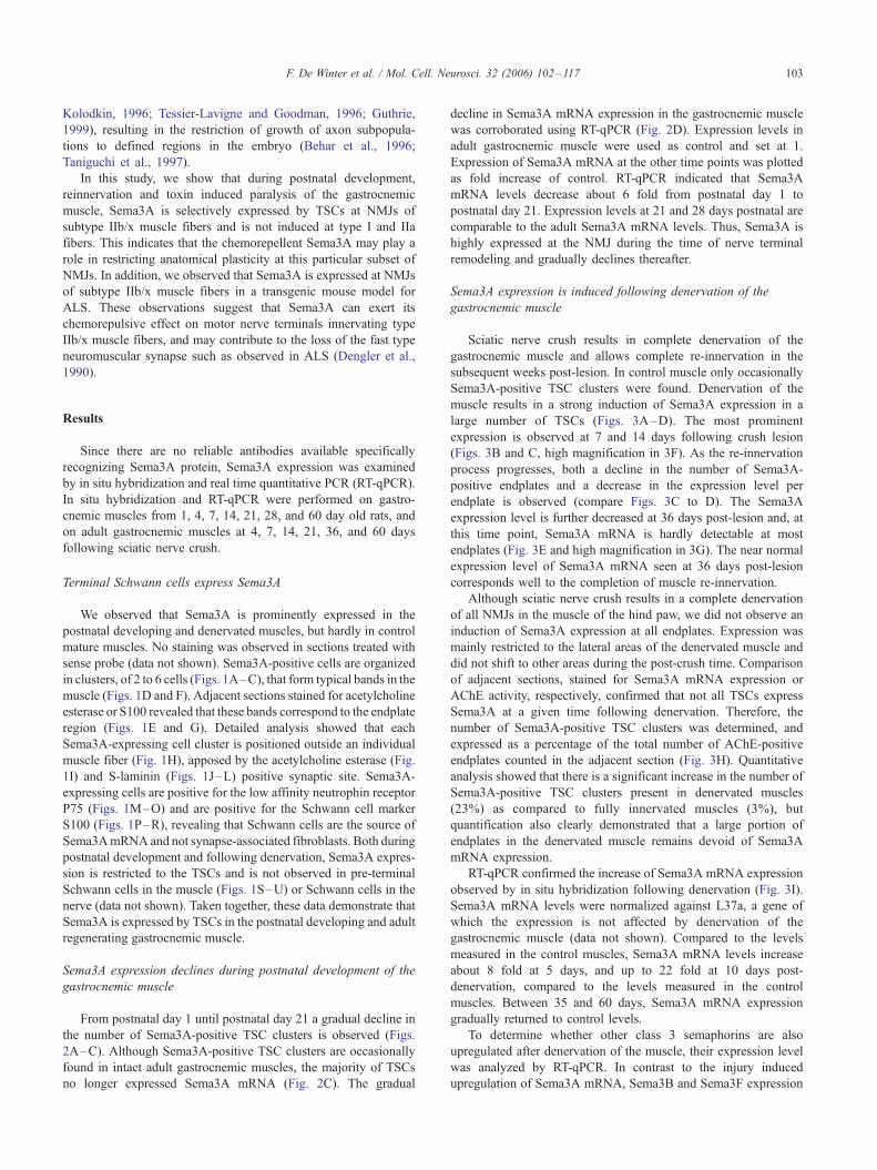
www.elsevier.com/locate/ymcne
Mol. Cell. Neurosci. 32 (2006) 102 – 117
The expression of the chemorepellent Semaphorin 3A is selectively
induced in terminal Schwann cells of a subset of neuromuscular
synapses that display limited anatomical plasticity and enhanced
vulnerability in motor neuron disease
Fred De Winter,a,1 Tam Vo,a Floor J. Stam,a,b Liselijn A.B. Wisman,c Peter R. Bar,c
Simone P. Niclou,a Freek L. van Muiswinkel,c and Joost Verhaagena,*
aGraduate School for Neurosciences Amsterdam, Netherlands Institute for Brain Research, Meibergdreef 33, 1105 AZ Amsterdam, The NetherlandsbGraduate School for Neurosciences Amsterdam, Department of Cellular and Molecular Neurobiology, Vrije Universiteit Amsterdam,
De Boelelaan 1085, 1081 HV Amsterdam, The NetherlandscDepartment of Neurology, Rudolf Magnus Institute of Neuroscience, University Medical Center Utrecht, P.O. Box 85500, 3508 GA, Utrecht, The Netherlands
Received 21 December 2005; revised 9 March 2006; accepted 13 March 2006
Available online 3 May 2006
Neuromuscular synapses differ markedly in their plasticity. Motor
nerve terminals innervating slow muscle fibers sprout vigorously
following synaptic blockage, while those innervating fast-fatigable
muscle fibers fail to exhibit any sprouting. Here, we show that the
axon repellent Semaphorin 3A is differentially expressed in terminal
Schwann cells (TSCs) on different populations of muscle fibers:
postnatal, regenerative and paralysis induced remodeling of neuro-
muscular connections is accompanied by increased expression of
Sema3A selectively in TSCs on fast-fatigable muscle fibers. To our
knowledge, this is the first demonstration of a molecular difference
between TSCs on neuromuscular junctions of different subtypes of
muscle fibers. Interestingly, also in a mouse model for amyotrophic
lateral sclerosis (ALS), Sema3A is expressed at NMJs of fast-
fatigable muscle fibers. We propose that expression of Sema3A by
TSCs not only suppresses nerve terminal plasticity at specific
neuromuscular synapses, but may also contribute to their early
and selective loss in the motor neuron disease ALS.
D 2006 Elsevier Inc. All rights reserved.
Recent studies have provided evidence for the presence of
subtypes of neuromuscular synapses in muscles with a mixed fiber-
type population (Frey et al., 2000; Pun et al., 2002, 2006). In
mammals, three main types of motor units exist, slow, fast fatigue-
resistant and fast-fatigable. Each type has its own muscle fiber
subtype(s) that can be identified by the expression of subtype
1044-7431/$ - see front matter D 2006 Elsevier Inc. All rights reserved.
doi:10.1016/j.mcn.2006.03.002
* Corresponding author. Fax: +31 20 696 1006.
E-mail address: [email protected] (J. Verhaagen).1 Currently working in the Salk Institute for Biological Studies, 10010
North Torrey Pines Road, La Jolla, CA 92186, USA.
Available online on ScienceDirect (www.sciencedirect.com).
specific myosin heavy chain protein isoforms, type I, type IIa or
type IIb and IIx, respectively. These motor units do not only differ
in their physiological properties, but also in anatomical plasticity
and susceptibility to loss of neuromuscular connectivity. Paralysis
induced motor nerve terminal sprouting correlates strongly with the
distribution of muscle fiber subtypes (Duchen, 1970; Frey et al.,
2000). Slow-type synapses display extensive motor nerve sprout-
ing, whereas fast-fatigable-type synapses show hardly any nerve
sprouting. In addition, fast-fatigable neuromuscular synapses are
susceptible to early loss in motor neuron diseases (Dengler et al.,
1990; Pun et al., 2006).
Plasticity, maintenance and regeneration of neuromuscular
junctions (NMJs) importantly rely on the terminal Schwann cells
(TSCs) that cover the motor nerve terminals (reviewed in Balice-
Gordon, 1996; Koirala et al., 2003). Blockade of neurotransmitter
release or denervation of muscle fibers induces expression of
proteins associated with process formation, like growth-associat-
ed-protein-43 and glial fibrillary acidic protein, in TSCs
(Reynolds and Woolf, 1992; Woolf et al., 1992; Ulenkate et al.,
1993; Georgiou et al., 1994). Processes formed by TSCs upon
loss of muscle innervation serve as guidance substrate for
regenerating motor nerve sprouts, and can evoke the formation
of new nerve sprouts at neighboring NMJs following partial
denervation (Son and Thompson, 1995a,b). Since local cues
provided by active TSCs facilitate nerve terminal sprouting
during regeneration and following synapse blockade, we asked
whether TSCs can also selectively restrict anatomical plasticity at
specific fast-fatigable-type synapses during these processes. In the
developing nervous system, several families of guidance mole-
cules have been identified that govern neurite outgrowth.
Members of the netrin, ephrin, slit, and semaphorin family
display inhibitory effects on growing neurites (Luo et al., 1993;
YMCNE-01826; No. of pages: 16; 4C: 3, 6, 9, 10
F. De Winter et al. / Mol. Cell. Neurosci. 32 (2006) 102–117 103
Kolodkin, 1996; Tessier-Lavigne and Goodman, 1996; Guthrie,
1999), resulting in the restriction of growth of axon subpopula-
tions to defined regions in the embryo (Behar et al., 1996;
Taniguchi et al., 1997).
In this study, we show that during postnatal development,
reinnervation and toxin induced paralysis of the gastrocnemic
muscle, Sema3A is selectively expressed by TSCs at NMJs of
subtype IIb/x muscle fibers and is not induced at type I and IIa
fibers. This indicates that the chemorepellent Sema3A may play a
role in restricting anatomical plasticity at this particular subset of
NMJs. In addition, we observed that Sema3A is expressed at NMJs
of subtype IIb/x muscle fibers in a transgenic mouse model for
ALS. These observations suggest that Sema3A can exert its
chemorepulsive effect on motor nerve terminals innervating type
IIb/x muscle fibers, and may contribute to the loss of the fast type
neuromuscular synapse such as observed in ALS (Dengler et al.,
1990).
Results
Since there are no reliable antibodies available specifically
recognizing Sema3A protein, Sema3A expression was examined
by in situ hybridization and real time quantitative PCR (RT-qPCR).
In situ hybridization and RT-qPCR were performed on gastro-
cnemic muscles from 1, 4, 7, 14, 21, 28, and 60 day old rats, and
on adult gastrocnemic muscles at 4, 7, 14, 21, 36, and 60 days
following sciatic nerve crush.
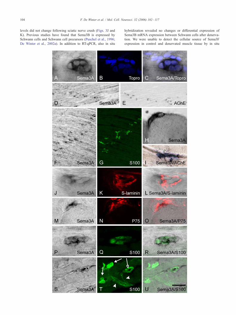
Terminal Schwann cells express Sema3A
We observed that Sema3A is prominently expressed in the
postnatal developing and denervated muscles, but hardly in control
mature muscles. No staining was observed in sections treated with
sense probe (data not shown). Sema3A-positive cells are organized
in clusters, of 2 to 6 cells (Figs. 1A–C), that form typical bands in the
muscle (Figs. 1D and F). Adjacent sections stained for acetylcholine
esterase or S100 revealed that these bands correspond to the endplate
region (Figs. 1E and G). Detailed analysis showed that each
Sema3A-expressing cell cluster is positioned outside an individual
muscle fiber (Fig. 1H), apposed by the acetylcholine esterase (Fig.
1I) and S-laminin (Figs. 1J–L) positive synaptic site. Sema3A-
expressing cells are positive for the low affinity neutrophin receptor
P75 (Figs. 1M–O) and are positive for the Schwann cell marker
S100 (Figs. 1P–R), revealing that Schwann cells are the source of
Sema3AmRNA and not synapse-associated fibroblasts. Both during
postnatal development and following denervation, Sema3A expres-
sion is restricted to the TSCs and is not observed in pre-terminal
Schwann cells in the muscle (Figs. 1S–U) or Schwann cells in the
nerve (data not shown). Taken together, these data demonstrate that
Sema3A is expressed by TSCs in the postnatal developing and adult
regenerating gastrocnemic muscle.
Sema3A expression declines during postnatal development of the
gastrocnemic muscle
From postnatal day 1 until postnatal day 21 a gradual decline in
the number of Sema3A-positive TSC clusters is observed (Figs.
2A–C). Although Sema3A-positive TSC clusters are occasionally
found in intact adult gastrocnemic muscles, the majority of TSCs
no longer expressed Sema3A mRNA (Fig. 2C). The gradual
decline in Sema3A mRNA expression in the gastrocnemic muscle
was corroborated using RT-qPCR (Fig. 2D). Expression levels in
adult gastrocnemic muscle were used as control and set at 1.
Expression of Sema3A mRNA at the other time points was plotted
as fold increase of control. RT-qPCR indicated that Sema3A
mRNA levels decrease about 6 fold from postnatal day 1 to
postnatal day 21. Expression levels at 21 and 28 days postnatal are
comparable to the adult Sema3A mRNA levels. Thus, Sema3A is
highly expressed at the NMJ during the time of nerve terminal
remodeling and gradually declines thereafter.
Sema3A expression is induced following denervation of the
gastrocnemic muscle
Sciatic nerve crush results in complete denervation of the
gastrocnemic muscle and allows complete re-innervation in the
subsequent weeks post-lesion. In control muscle only occasionally
Sema3A-positive TSC clusters were found. Denervation of the
muscle results in a strong induction of Sema3A expression in a
large number of TSCs (Figs. 3A–D). The most prominent
expression is observed at 7 and 14 days following crush lesion
(Figs. 3B and C, high magnification in 3F). As the re-innervation
process progresses, both a decline in the number of Sema3A-
positive endplates and a decrease in the expression level per
endplate is observed (compare Figs. 3C to D). The Sema3A
expression level is further decreased at 36 days post-lesion and, at
this time point, Sema3A mRNA is hardly detectable at most
endplates (Fig. 3E and high magnification in 3G). The near normal
expression level of Sema3A mRNA seen at 36 days post-lesion
corresponds well to the completion of muscle re-innervation.
Although sciatic nerve crush results in a complete denervation
of all NMJs in the muscle of the hind paw, we did not observe an
induction of Sema3A expression at all endplates. Expression was
mainly restricted to the lateral areas of the denervated muscle and
did not shift to other areas during the post-crush time. Comparison
of adjacent sections, stained for Sema3A mRNA expression or
AChE activity, respectively, confirmed that not all TSCs express
Sema3A at a given time following denervation. Therefore, the
number of Sema3A-positive TSC clusters was determined, and
expressed as a percentage of the total number of AChE-positive
endplates counted in the adjacent section (Fig. 3H). Quantitative
analysis showed that there is a significant increase in the number of
Sema3A-positive TSC clusters present in denervated muscles
(23%) as compared to fully innervated muscles (3%), but
quantification also clearly demonstrated that a large portion of
endplates in the denervated muscle remains devoid of Sema3A
mRNA expression.
RT-qPCR confirmed the increase of Sema3A mRNA expression
observed by in situ hybridization following denervation (Fig. 3I).
Sema3A mRNA levels were normalized against L37a, a gene of
which the expression is not affected by denervation of the
gastrocnemic muscle (data not shown). Compared to the levels
measured in the control muscles, Sema3A mRNA levels increase
about 8 fold at 5 days, and up to 22 fold at 10 days post-
denervation, compared to the levels measured in the control
muscles. Between 35 and 60 days, Sema3A mRNA expression
gradually returned to control levels.
To determine whether other class 3 semaphorins are also
upregulated after denervation of the muscle, their expression level
was analyzed by RT-qPCR. In contrast to the injury induced
upregulation of Sema3A mRNA, Sema3B and Sema3F expression
F. De Winter et al. / Mol. Cell. Neurosci. 32 (2006) 102–117104
levels did not change following sciatic nerve crush (Figs. 3J and
K). Previous studies have found that Sema3B is expressed by
Schwann cells and Schwann cell precursors (Puschel et al., 1996;
De Winter et al., 2002a). In addition to RT-qPCR, also in situ
hybridization revealed no changes or differential expression of
Sema3B mRNA expression between Schwann cells after denerva-
tion. We were unable to detect the cellular source of Sema3F
expression in control and denervated muscle tissue by in situ

Fig. 2. Analysis and quantification of Sema3A mRNA expression in the developing postnatal rat gastrocnemic muscle. In situ hybridization for Sema3A
mRNA on tissue sections derived from different postnatal stages of the rat developing gastrocnemic muscle (A–C). Sema3A mRNA expression is localized in
band-like structures in the postnatal developing gastrocnemic muscle (A and B). The number of Sema3A-positive clusters declines in the first postnatal weeks
(A and B), and are absent in adult gastrocnemic muscle (C). Quantitative analysis of Sema3A mRNA expression levels in developing rat gastrocnemic muscles.
Quantitative real-time qPCR was used to measure Sema3A mRNA expression levels at 1 (P1), 4 (P4), 7 (P7), 14 (P14) and 21 (P21) days following birth and
compared to adult expression levels. Expression was normalized against mRNA expression of internal reference gene L37a and expressed as fold increase of
mean adult Sema3A mRNA expression levels. Compared to adult, high levels of Sema3A mRNA is measured in P1 muscles. Sema3A expression levels
decrease significantly in the first postnatal week when compared to adult expression levels (*P < 0.008, Kruskal–Wallis non-parametric ANOVA test).
Expression had reached adult levels at postnatal day 21 (P21), and had decreased 6 fold as compared to P1. Scale bar is 350 Am.
F. De Winter et al. / Mol. Cell. Neurosci. 32 (2006) 102–117 105
hybridization. Sema3C and Sema3E mRNA expression was not
detectable in muscles at any time point after denervation.
In summary, the combined in situ hybridization and RT-qPCR
data show a specific spatio-temporal increase of Sema3A
expression in a subpopulation of TSCs in the denervated muscle
followed by a subsequent decline in expression upon re-innerva-
tion of the muscle. Interestingly, Sema3A expression is not
observed in TSCs at all endplates but is restricted to a
subpopulation of neuromuscular junctions.
Induction of Sema3A expression by TSCs is muscle fiber type
specific
We next examined whether Sema3A is expressed in TSCs
present at NMJs on specific muscle fiber types. The medial
gastrocnemic muscle is a mixed-type hindlimb muscle and
consists of three main muscle fiber types, notably slow (type I),
fast fatigue-resistant (type IIa), and fast-fatigable (type IIb and IIx)
fibers.
Muscle fiber subtype-specific myosin heavy chain (MHC)
monoclonal antibodies were used to determine whether Sema3A-
expressing TSCs are restricted to a particular muscle fiber type
(Webster et al., 1988). Because there are no good antibodies
available that exclusively recognize rat muscle fiber type IIb and/or
IIx following in situ hybridization, we used combinations of
antibodies to identify the fast-fatigable fiber population. By using a
Fig. 1. Sema3A mRNA in the developing and denervated rat gastrocnemic musc
purple in color pictures) and immunohistochemistry was used to determine the ce
developing rat gastrocnemic muscle. At 7 days after denervation, Sema3A mRN
identified with the nuclear marker Topro (blue in panel B) do not express Sema3A
Sema3A-positive cell clusters are clearly aligned in band like structures (D and F),
endplate regions (E and G, respectively). In high magnification photographs Sema3
muscle fibers (H), and co-localize with AChE (blue in panel I) and with the NMJ b
7 days after denervation. Furthermore, Sema3A-positive cells (M and P) express t
and green in panel Q, respectively, overlay in panels O and R). Sema3A (S and U)
arrows in panel T) S100-positive TSCs and was not observed in S100-positive p
mix of antibodies against type IIa and type I the non-stained
population could be identified as fast-fatigable (type IIb/x) fibers
(Figs. 4A–H). In addition, sections were stained for fiber type IIa
and all fast fiber types II (a+b/x), resulting in double stained type
IIa, single stained type IIb/x and non-stained type I fibers (Figs.
4I–P).
At 4, 7, and 14 days post-sciatic nerve crush, the majority of the
muscle fibers still predominantly express one of the MHC
subtypes. Therefore, we could readily determine the distribution
of Sema3A-positive TSCs on muscle fiber types. Surprisingly, all
Sema3A-expressing TSCs are located on type IIb/x muscle fibers,
and are never seen on type I or IIa (Figs. 4A–D). In adjacent
sections, AChE-positive endplates can be visualized on all three
muscle fiber types (Figs. 4E–H). All Sema3A-expressing TSCs are
located on fast type muscle fibers, but not on the fast type IIa
muscle fibers (Figs. 4I–L), while AChE-positive endplates are
present on fast type IIa muscle fibers (Figs. 4M–P). Prolonged
denervation results in a temporary de-differentiation of the muscle
fiber, causing a mixed expression of MHC subtypes. In addition,
re-innervation can cause shifts in MHC subtype expression by
muscle fibers (Gillespie et al., 1987; Dengler et al., 1990; Burke,
1994; Ijkema-Paassen et al., 2001). At 21 days post-sciatic nerve
crush, we occasionally found Sema3A-expressing TSCs on fibers
that expressed multiple MHC isoforms in re-innervated muscles
(data not shown). Taken together, we show that, for as long as the
muscle fibers express predominantly one MHC subtype, denerva-
le is expressed in TSCs. Combined in situ hybridization (black in b/w and
llular origin of Sema3A mRNA expression in the denervated and postnata
A was restricted to small cell clusters (A), while other neighboring cells
mRNA (overlay in panel C). In the 7-day postnatal gastrocnemic muscle
which in adjacent sections can be identified as the AChE and S100-positive
A mRNA-positive cells can been seen to be positioned on top of individua
asal lamina marker S-laminin (red in panel K, overlay in panel L) in muscles
he low affinity receptor P75 and Schwann cell marker S100 (red in panel N
expression co-localizes with some (open arrow in panel T) but not all (solid
re-terminal Schwann cells (arrow heads in panel T).
l
,
l
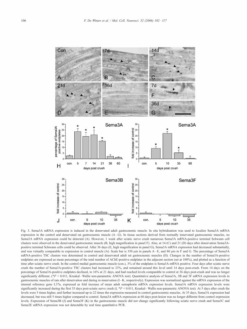
Fig. 3. Sema3A mRNA expression is induced in the denervated adult gastrocnemic muscle. In situ hybridization was used to localize Sema3A mRNA
expression in the control and denervated rat gastrocnemic muscle (A–G). In tissue sections derived from normally innervated gastrocnemic muscles, no
Sema3A mRNA expression could be detected (A). However, 1 week after sciatic nerve crush numerous Sema3A mRNA-positive terminal Schwann cell
clusters were observed in the denervated gastrocnemic muscle (B, high magnification in panel F). Also, at 14 (C) and 21 (D) days after denervation Sema3A-
positive terminal Schwann cells could be observed. After 36 days (E, high magnification in panel G), Sema3A mRNA expression had decreased substantially,
and was virtually comparable to expression in control muscle (A). Scale bar is 350 Am in panels A–E, and 80 Am in F and G. The percentage of Sema3A
mRNA-positive TSC clusters was determined in control and denervated adult rat gastrocnemic muscles (H). Changes in the number of Sema3A-positive
endplates are expressed as mean percentage of the total number of AChE-positive endplates in the adjacent section (set at 100%), and plotted as a function of
time after sciatic nerve crush. In the control medial gastrocnemic muscle (con.), 3% of the endplates is Sema3A mRNA positive. Four days after sciatic nerve
crush the number of Sema3A-positive TSC clusters had increased to 23%, and remained around this level until 14 days post-crush. From 14 days on the
percentage of Sema3A-positive endplates declined, to 16% at 21 days, and had reached levels comparable to control at 36 days post-crush and was no longer
significantly different. (*P < 0.011, Kruskal–Wallis non-parametric ANOVA test). Quantitative analysis of Sema3A, 3B and 3F mRNA expression levels in
gastrocnemic muscles of rats after denervation and during re-innervation (I –K, respectively). Expression was normalized against the mRNA expression of the
internal reference gene L37a, expressed as fold increase of mean adult semaphorin mRNA expression levels. Sema3A mRNA expression levels were
significantly increased during the first 35 days post-sciatic nerve crush (I, *P < 0.011, Kruskal–Wallis non-parametric ANOVA test). At 5 days after crush the
levels were 5 times higher, and further increased up to 22 times the expression measured in control gastrocnemic muscles. At 35 days, Sema3A expression had
decreased, but was still 5 times higher compared to control. Sema3A mRNA expression at 60 days post-lesion was no longer different from control expression
levels. Expression of Sema3B (J) and Sema3F (K) in the gastrocnemic muscle did not change significantly following sciatic nerve crush and Sema3C and
Sema3E mRNA expression was not detectable by real time quantitative PCR.
F. De Winter et al. / Mol. Cell. Neurosci. 32 (2006) 102–117106
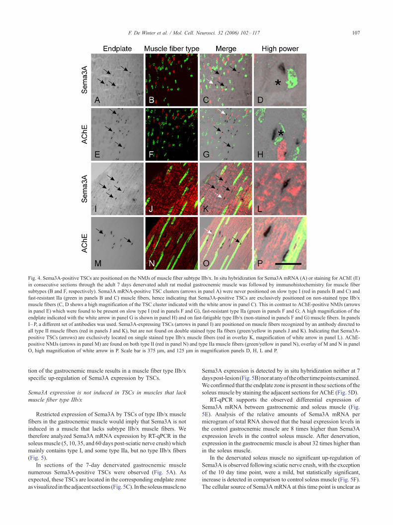
Fig. 4. Sema3A-positive TSCs are positioned on the NMJs of muscle fiber subtype IIb/x. In situ hybridization for Sema3A mRNA (A) or staining for AChE (E)
in consecutive sections through the adult 7 days denervated adult rat medial gastrocnemic muscle was followed by immunohistochemistry for muscle fiber
subtypes (B and F, respectively). Sema3A mRNA-positive TSC clusters (arrows in panel A) were never positioned on slow type I (red in panels B and C) and
fast-resistant IIa (green in panels B and C) muscle fibers, hence indicating that Sema3A-positive TSCs are exclusively positioned on non-stained type IIb/x
muscle fibers (C, D shows a high magnification of the TSC cluster indicated with the white arrow in panel C). This in contrast to AChE-positive NMJs (arrows
in panel E) which were found to be present on slow type I (red in panels F and G), fast-resistant type IIa (green in panels F and G; A high magnification of the
endplate indicated with the white arrow in panel G is shown in panel H) and on fast-fatigable type IIb/x (non-stained in panels F and G) muscle fibers. In panels
I–P, a different set of antibodies was used. Sema3A-expressing TSCs (arrows in panel I) are positioned on muscle fibers recognized by an antibody directed to
all type II muscle fibers (red in panels J and K), but are not found on double stained type IIa fibers (green/yellow in panels J and K). Indicating that Sema3A-
positive TSCs (arrows) are exclusively located on single stained type IIb/x muscle fibers (red in overlay K, magnification of white arrow in panel L). AChE-
positive NMJs (arrows in panel M) are found on both type II (red in panel N) and type IIa muscle fibers (green/yellow in panel N), overlay of M and N in panel
O, high magnification of white arrow in P. Scale bar is 375 Am, and 125 Am in magnification panels D, H, L and P.
F. De Winter et al. / Mol. Cell. Neurosci. 32 (2006) 102–117 107
tion of the gastrocnemic muscle results in a muscle fiber type IIb/x
specific up-regulation of Sema3A expression by TSCs.
Sema3A expression is not induced in TSCs in muscles that lack
muscle fiber type IIb/x
Restricted expression of Sema3A by TSCs of type IIb/x muscle
fibers in the gastrocnemic muscle would imply that Sema3A is not
induced in a muscle that lacks subtype IIb/x muscle fibers. We
therefore analyzed Sema3A mRNA expression by RT-qPCR in the
soleus muscle (5, 10, 35, and 60 days post-sciatic nerve crush) which
mainly contains type I, and some type IIa, but no type IIb/x fibers
(Fig. 5).
In sections of the 7-day denervated gastrocnemic muscle
numerous Sema3A-positive TSCs were observed (Fig. 5A). As
expected, these TSCs are located in the corresponding endplate zone
asvisualized in theadjacent sections (Fig.5C). In thesoleusmuscleno
Sema3A expression is detected by in situ hybridization neither at 7
dayspost-lesion(Fig.5B)noratanyof theother timepointsexamined.
We confirmed that the endplate zone is present in these sections of the
soleus muscle by staining the adjacent sections for AChE (Fig. 5D).
RT-qPCR supports the observed differential expression of
Sema3A mRNA between gastrocnemic and soleus muscle (Fig.
5E). Analysis of the relative amounts of Sema3A mRNA per
microgram of total RNA showed that the basal expression levels in
the control gastrocnemic muscle are 8 times higher than Sema3A
expression levels in the control soleus muscle. After denervation,
expression in the gastrocnemic muscle is about 32 times higher than
in the soleus muscle.
In the denervated soleus muscle no significant up-regulation of
Sema3A is observed following sciatic nerve crush, with the exception
of the 10 day time point, were a mild, but statistically significant,
increase is detected in comparison to control soleus muscle (Fig. 5F).
The cellular source of Sema3A mRNA at this time point is unclear as
Fig. 5. Sema3A mRNA expression in the denervated rat soleus muscle, a muscle devoid of type IIb/x muscle fibers. In situ hybridization for Sema3A mRNA
(A and B) and AChE staining (C and D) on adjacent sections through the 7 days denervated rat medial gastrocnemic (A and C) and soleus (B and D) muscle.
Several Sema3A-positive groups of TSCs are present in the endplate region (arrows in panel A), as determined by AChE staining in the adjacent section
(arrows in panel C), of the denervated gastrocnemic muscle. Whereas in the endplate region of the soleus muscle (arrows in panel D), no Sema3A-positive
TSCs can be found (B). Scale bar is 300 Am. Real time quantitative PCR for Sema3A mRNA on control and denervated soleus and medial gastrocnemius
muscles (E and F). Expression was normalized against mRNA expression of internal reference gene L37a, and expressed as fold increase of mean adult
Sema3A mRNA expression levels. Sema3A mRNA expression levels in the denervated soleus muscle show, a significant (*P < 0.043, Kruskal–Wallis non-
parametric ANOVA test), increase at 10 days following sciatic nerve crush (E). Other time points did not differ significantly form the control expression levels
in the soleus muscle. Analysis of relative amounts of Sema3A mRNA per microgram total RNA showed that both control and 10 days denervation induced
Sema3A mRNA expression levels in the soleus muscle are lower than both control and denervated gastrocnemic muscle (F).
F. De Winter et al. / Mol. Cell. Neurosci. 32 (2006) 102–117108
in situ hybridization on an extensive series of sections through the
soleus muscle failed to detect any Sema3A mRNA expression.
Taken together, these results show that there is a basal level of
Sema3A expression in both the gastrocnemic and soleus muscle of
which the cellular origin is unknown since Sema3A is undetectable
by in situ hybridization. Importantly, an upregulation of Sema3A
expression after nerve crush is only observed in the TSCs of the
muscle containing subtype IIb/x fibers (gastrocnemic), and is not
observed in TSC in the soleus muscle which is devoid of subtype
IIb/x muscle fibers.
Sema3A is specifically induced at ‘‘non-plastic’’
neuromuscular junctions
Neuromuscular synapse subtypes differ in their capacity to
exhibit paralysis induced nerve sprouting (Brown et al., 1981; Frey
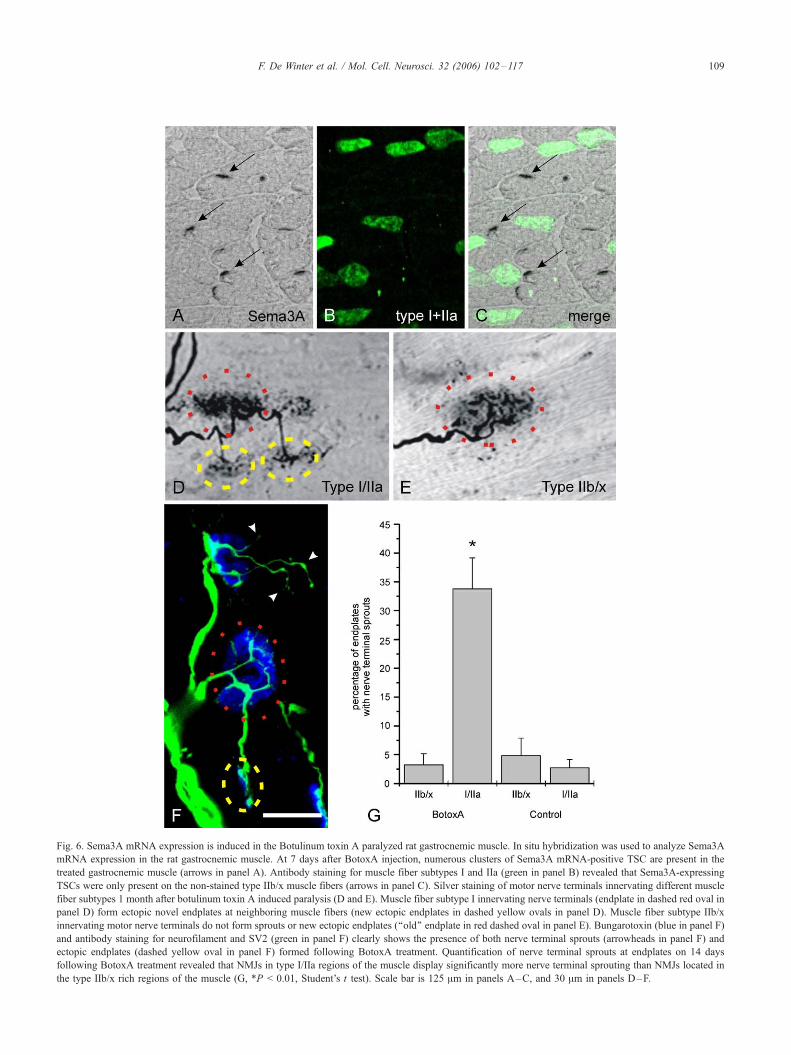
Fig. 6. Sema3A mRNA expression is induced in the Botulinum toxin A paralyzed rat gastrocnemic muscle. In situ hybridization was used to analyze Sema3A
mRNA expression in the rat gastrocnemic muscle. At 7 days after BotoxA injection, numerous clusters of Sema3A mRNA-positive TSC are present in the
treated gastrocnemic muscle (arrows in panel A). Antibody staining for muscle fiber subtypes I and IIa (green in panel B) revealed that Sema3A-expressing
TSCs were only present on the non-stained type IIb/x muscle fibers (arrows in panel C). Silver staining of motor nerve terminals innervating different muscle
fiber subtypes 1 month after botulinum toxin A induced paralysis (D and E). Muscle fiber subtype I innervating nerve terminals (endplate in dashed red oval in
panel D) form ectopic novel endplates at neighboring muscle fibers (new ectopic endplates in dashed yellow ovals in panel D). Muscle fiber subtype IIb/x
innervating motor nerve terminals do not form sprouts or new ectopic endplates (‘‘old’’ endplate in red dashed oval in panel E). Bungarotoxin (blue in panel F)
and antibody staining for neurofilament and SV2 (green in panel F) clearly shows the presence of both nerve terminal sprouts (arrowheads in panel F) and
ectopic endplates (dashed yellow oval in panel F) formed following BotoxA treatment. Quantification of nerve terminal sprouts at endplates on 14 days
following BotoxA treatment revealed that NMJs in type I/IIa regions of the muscle display significantly more nerve terminal sprouting than NMJs located in
the type IIb/x rich regions of the muscle (G, *P < 0.01, Student’s t test). Scale bar is 125 Am in panels A–C, and 30 Am in panels D–F.
F. De Winter et al. / Mol. Cell. Neurosci. 32 (2006) 102–117 109
F. De Winter et al. / Mol. Cell. Neurosci. 32 (2006) 102–117110
et al., 2000). Botulinum toxin A (BotoxA) induced nerve terminal
sprouting occurs mainly on muscle fiber type I, whereas muscle
fiber type IIb/x synapses are characterized by the absence of nerve
sprouting. To examine whether Sema3A expression by TSCs could
contribute to this profound difference in synaptic plasticity, we
analyzed Sema3A expression in BotoxA-treated gastrocnemic
muscle of adult rat.
BotoxA treatment results in complete loss of function of the
hindlimb within several days, and clear muscle atrophy within 1
week. Using in situ hybridization, prominent Sema3A mRNA
expression was detected in TSCs in the BotoxA-treated gastro-
cnemic muscle (Fig. 6A), whereas in the saline-treated muscle
hardly any expression was observed (data not shown). Sema3A
expression is again restricted to the TSCs located on type IIb/x
muscle fibers (Figs. 6B and C). Silver staining of sections
revealed that, as previously described also for mice (Frey et al.,
2000), botoxA treatment induced nerve terminal sprouting and
the formation of ectopic endplates occurs mainly on type I and
IIa muscle fibers, and that type IIb/x fibers display hardly any
change upon paralysis (Figs. 6D and E, respectively). Quantifi-
cation of muscle sections stained for neurofilament/SV2/bungar-
otoxin revealed that 33% of the NMJs in the subtype I/IIa-
enriched regions of the gastrocnemic muscle, as determined by
muscle fiber subtype staining of the adjacent sections, display
nerve terminal sprouting and/or the formation of ectopic
endplates at 14 days following BotoxA treatment (Figs. 6F and
G). This is in marked contrast to the NMJs in the subtype IIb/x-
enriched regions of the muscle of which only 3% displays nerve
terminal sprouting.
Sema3A is expressed in TSCs of the gastrocnemic muscle in
G93A-hSOD1 mice
Selective and progressive loss of muscle innervation is an
indicator of advancing neuromuscular pathology in the G93A-
hSOD1 transgenic mouse model for ALS (Gurney et al., 1994; Dal
Canto and Gurney, 1995; Wong et al., 1995; Tu et al., 1996;
Barneoud et al., 1997). Interestingly, in several motor neuron
diseases, the start of neuromuscular synapse loss is dependent on
the motor unit subtype. Thus, the fast-fatigable subtype of
neuromuscular synapse is most vulnerable and is the first subtype
to be lost in several neuromuscular diseases, whereas the synapses
on slow subtype muscle fibers survive until the terminal phase of
the disease (Dengler et al., 1990; Pinter et al., 1995; Pinter et al.,
1997; Frey et al., 2000).
To examine whether Sema3A is present at the NMJ during
development and progression of ALS-like pathology we analyzed
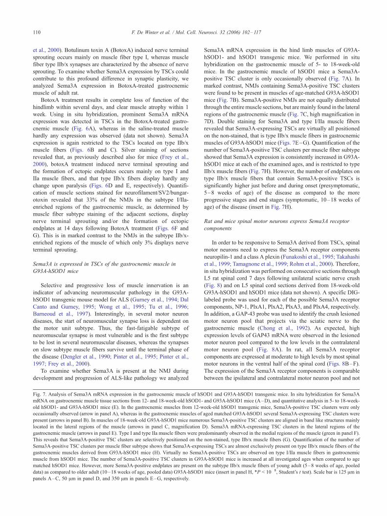
Fig. 7. Analysis of Sema3A mRNA expression in the gastrocnemic muscle of hS
mRNA on gastrocnemic muscle tissue sections from 12- and 18-week-old hSOD1-
old hSOD1- and G93A-hSOD1 mice (E). In the gastrocnemic muscles from 12-w
occasionally observed (arrow in panel A), whereas in the gastrocnemic muscles of
present (arrows in panel B). In muscles of 18-week-old G93A-hSOD1 mice numero
located in the lateral regions of the muscle (arrows in panel C, magnification D
gastrocnemic muscle (arrows in panel E). Type I and type IIa muscle fibers were pr
This reveals that Sema3A-positive TSC clusters are selectively positioned on the
Sema3A-positive TSC clusters per muscle fiber subtype shows that Sema3A-expre
gastrocnemic muscles derived from G93A-hSOD1 mice (H). Virtually no Sema3
muscle from hSOD1 mice. The number of Sema3A-positive TSC clusters in G93
matched hSOD1 mice. However, more Sema3A-positive endplates are present on
data) as compared to older adult (10–18 weeks of age, pooled data) G93A-hSOD1
panels A–C, 50 Am in panel D, and 350 Am in panels E–G, respectively.
Sema3A mRNA expression in the hind limb muscles of G93A-
hSOD1- and hSOD1 transgenic mice. We performed in situ
hybridization on the gastrocnemic muscle of 5- to 18-week-old
mice. In the gastrocnemic muscle of hSOD1 mice a Sema3A-
positive TSC cluster is only occasionally observed (Fig. 7A). In
marked contrast, NMJs containing Sema3A-positive TSC clusters
were found to be present in muscles of age-matched G93A-hSOD1
mice (Fig. 7B). Sema3A-positive NMJs are not equally distributed
through the entire muscle sections, but are mainly found in the lateral
regions of the gastrocnemic muscle (Fig. 7C, high magnification in
7D). Double staining for Sema3A and type I/IIa muscle fibers
revealed that Sema3A-expressing TSCs are virtually all positioned
on the non-stained, that is type IIb/x muscle fibers in gastrocnemic
muscles of G93A-hSOD1 mice (Figs. 7E–G). Quantification of the
number of Sema3A-positive TSC clusters per muscle fiber subtype
showed that Sema3A expression is consistently increased in G93A-
hSOD1 mice at each of the examined ages, and is restricted to type
IIb/x muscle fibers (Fig. 7H). However, the number of endplates on
type IIb/x muscle fibers that contain Sema3A-positive TSCs is
significantly higher just before and during onset (presymptomatic,
5–8 weeks of age) of the disease as compared to the more
progressive stages and end stages (symptomatic, 10–18 weeks of
age) of the disease (insert in Fig. 7H).
Rat and mice spinal motor neurons express Sema3A receptor
components
In order to be responsive to Sema3A derived from TSCs, spinal
motor neurons need to express the Sema3A receptor components
neuropilin-1 and a class A plexin (Funakoshi et al., 1995; Takahashi
et al., 1999; Tamagnone et al., 1999; Rohm et al., 2000). Therefore,
in situ hybridization was performed on consecutive sections through
L5 rat spinal cord 7 days following unilateral sciatic nerve crush
(Fig. 8) and on L5 spinal cord sections derived from 18-week-old
G93A-hSOD1 and hSOD1 mice (data not shown). A specific DIG-
labeled probe was used for each of the possible Sema3A receptor
components, NP-1, PlxA1, PlxA2, PlxA3, and PlxA4, respectively.
In addition, a GAP-43 probe was used to identify the crush lesioned
motor neuron pool that projects via the sciatic nerve to the
gastrocnemic muscle (Chong et al., 1992). As expected, high
expression levels of GAP43 mRNA were observed in the lesioned
motor neuron pool compared to the low levels in the contralateral
motor neuron pool (Fig. 8A). In rat, all Sema3A receptor
components are expressed at moderate to high levels by most spinal
motor neurons in the ventral half of the spinal cord (Figs. 8B–F).
The expression of the Sema3A receptor components is comparable
between the ipsilateral and contralateral motor neuron pool and not
OD1 and G93A-hSOD1 transgenic mice. In situ hybridization for Sema3A
and G93A-hSOD1 mice (A–D), and quantitative analysis in 5- to 18-week
eek-old hSOD1 transgenic mice, Sema3A-positive TSC clusters were only
aged matched G93A-hSOD1 several Sema3A-expressing TSC clusters were
us Sema3A-positive TSC clusters are aligned in band like structures mainly
). Sema3A mRNA-expressing TSC clusters in the lateral regions of the
edominantly observed in the medial regions of the muscle (green in panel F)
non-stained, type IIb/x muscle fibers (G). Quantification of the number o
ssing TSCs are almost exclusively present on type IIb/x muscle fibers of the
A-positive TSCs are observed on type I/IIa muscle fibers in gastrocnemic
A-hSOD1 mice is increased at all investigated ages when compared to age
the subtype IIb/x muscle fibers of young adult (5–8 weeks of age, pooled
mice (insert in panel H, *P < 10�8, Student’s t test). Scale bar is 125 Am in
-
.
f
F. De Winter et al. / Mol. Cell. Neurosci. 32 (2006) 102–117 111
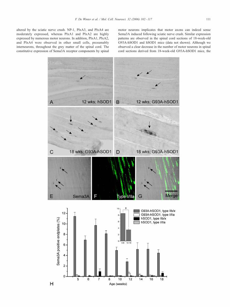
altered by the sciatic nerve crush. NP-1, PlxA3, and PlxA4 are
moderately expressed, whereas PlxA1 and PlxA2 are highly
expressed by numerous motor neurons. In addition, PlxA1, PlxA2,
and PlxA4 were observed in other small cells, presumably
interneurons, throughout the grey matter of the spinal cord. The
constitutive expression of Sema3A receptor components by spinal
motor neurons implicates that motor axons can indeed sense
Sema3A induced following sciatic nerve crush. Similar expression
patterns are observed in the spinal cord sections of 18-week-old
G93A-hSOD1 and hSOD1 mice (data not shown). Although we
observed a clear decrease in the number of motor neurons in spinal
cord sections derived from 18-week-old G93A-hSOD1 mice, the
Fig. 8. Sema3A receptor expression in the rat lumbar spinal cord following sciatic nerve crush. In situ hybridization was used to analyze GAP-43, NP-1, PlxA1,
PlxA2, PlxA3, and PlxA4 mRNA expression in adjacent transversal sections through the rat L5 spinal cord, 7 days following unilateral sciatic nerve crush.
Expression of growth-associated-protein-43 (GAP-43) mRNA was used to identify the injured sciatic motor neuron pool (closed arrow in panel A). A clear
induction of GAP-43 mRNA expression is seen in the ipsilateral motor neuron (closed arrow in panel A), whereas low expression levels were observed in the
contralateral side (open arrow in panel A) in the ventral motor neuron pools. All possible Sema3A receptor components, NP-1, PlxA1, PlxA2, PlxA3, and
PlxA4 (arrows in panels B–F, respectively), were present in the motor neuron pool corresponding to the crushed sciatic nerve. The mRNA levels for these
receptor components did not differ between the contralateral (open arrow) and the ipsilateral side (solid arrow). Scale bar is 650 Am.
F. De Winter et al. / Mol. Cell. Neurosci. 32 (2006) 102–117112
surviving motor neurons continue to express Sema3A receptor
components.
Discussion
Here, we show that TSCs located on NMJs of type IIb/x muscle
fibers, but not on type I and type IIa muscle fibers, upregulate the
expression of the chemorepellent Sema3A after mechanical
denervation and following pharmacological blockage of neuro-
transmission. In addition, we demonstrate that TSCs positioned at
the NMJs of type IIb/x muscle fibers in the G93A-hSOD1 mouse
model for ALS selectively up-regulate the chemorepellent
Sema3A. In contrast to motor nerve terminals on type I and type
IIa muscle fibers, motor axons on type IIb/x fibers fail to display
anatomical plasticity and are the most vulnerable synapses in the
motor neuron disease ALS (Dengler et al., 1990; Frey et al., 2000,
Pun et al., 2006). The present findings suggest that TSC of
synapses on type IIb/x muscle fibers may not only restrict
anatomical plasticity, but could also negatively influence the
integrity of this specific subset of neuromuscular synapses by
selectively inducing the expression of the chemorepellent Sema3A.
The prevailing view is that TSCs provide an important trophic
influence on the motor nerve terminal. Following denervation,
TSCs extend long processes over the muscle. These processes
appear to guide ingrowing motor neuron axons to vacant synaptic
sites on the muscle (Reynolds and Woolf, 1992; Astrow et al.,
1994; Son and Thompson, 1995a,b; O’Malley et al., 1999; Tam et
al., 2001). Also, blockage of transmission at the NMJ results in the
formation of nerve sprouts that grow along the processes of TSCs
(Holland and Brown, 1980; Brown et al., 1981; Son and
Thompson, 1995a,b). Although neuromuscular synapses on
specific subsets of muscle fibers differ profoundly in their capacity
to display anatomical plasticity, the molecular basis for this
difference is not known, but may be attributed to molecular
properties of one of the three components of the NMJ, notably the
motor neurons, the muscle fiber or the TSCs (Sanes and Lichtman,
1999).
No intrinsic molecular differences between motor neurons
innervating different subtypes of muscle fibers that may account
for the difference in plasticity of NMJ have been described so far.
Intrinsic molecular properties of muscle fiber subtypes do differ
significantly but these are unlikely to regulate nerve terminal
plasticity directly. Moreover, although the muscle cell surface and
extracellular matrix are highly specialized at the NMJ and contain
growth supporting molecules such as laminin, N-CAM, fibronec-
tin and heparin sulfate proteoglycans (Patton, 2003), the
expression of these molecules appears not to differ between
muscle fiber subtypes. Membrane associated ephrins are differ-
entially expressed between individual muscles and in distinct
regions within a muscle (Donoghue et al., 1996), and thereby
regulate positionally selective synaptogenesis of motor neurons
(Feng et al., 2000). Also, Drosophila semaphorin II functions as
a muscle derived signal for selective terminal arborization
(Matthes et al., 1995). To our knowledge, however, no muscle
fiber type I, type IIa or type IIb/x specific molecules have been
identified that could regulate differential nerve terminal plasticity
in mammals.
It is often suggested that nerve terminal sprouting arises
from short-range diffusible stimuli produced by inactive muscle
fibers (Brown et al., 1978; Slack and Pockett, 1981; Pockett
and Slack, 1982; Slack and Pockett, 1982). Some activity-
controlled trophic factors have been identified (Caroni and
Grandes, 1990; Caroni, 1993; Caroni and Schneider, 1994;
Funakoshi et al., 1995). Of these NT-4 is selectively expressed
by type I muscle fibers (Funakoshi et al., 1995). However, NT-
4 expression is down-regulated in bungarotoxin treated muscles,
suggesting that NT-4 does not contribute to the selective nerve
sprouting at type I neuromuscular synapses following activity
blockage.
The third cellular component of the NMJ, the TSCs, have not
been reported to differ between muscle fiber subtypes and seem to
share persistent and regulated expression of intracellular proteins
including GAP-43, GFAP and S100, and cell adhesion molecules
like L1 and N-CAM. The ability of TSCs to change their
F. De Winter et al. / Mol. Cell. Neurosci. 32 (2006) 102–117 113
morphological and molecular properties in response to altered
synaptic activity supports their possible role in nerve terminal
plasticity (Jahromi et al., 1992; Reist and Smith, 1992; Robitaille,
1995; Rochon et al., 2001). With the use of MHC isoform specific
antibodies, we showed that only TSCs positioned on subtype IIb/x
muscle fibers express Sema3A in the regenerating and paralyzed
gastrocnemic muscle. The absence of Sema3A expression by
TSCs in the denervated soleus muscle, which in contrast to the
gastrocnemic muscle does not contain subtype IIb/x muscle fibers,
supports the muscle fiber subtype specificity of Sema3A mRNA
expression. The discovery of a molecular difference between
TSCs on types I/IIa and type IIb/x muscle fibers is a first
indication for a differential role of TSCs in the plasticity of NMJs
on different muscle fiber types. The selective expression of
Sema3A in TSCs on type IIb/x muscle fibers suggests that the
trophic role of TSCs in development, maintenance and plasticity
cannot be generalized.
Sema3A is a secreted glycoprotein, initially identified as a
growth cone collapse-inducing factor for dorsal root ganglion
neurons (Luo et al., 1993). When Sema3A is secreted by non-
neuronal cell aggregates, it acts as a chemorepellent for sensory
and motor axons extending from co-cultured embryonic neuronal
explants (Shepherd et al., 1996; Varela-Echavarria et al., 1997;
Kuhn et al., 1999). Disruption of the Sema3A gene during
embryonic development results in abnormal growth of peripheral
axons, including motor axons, outside their typical nerve tracts
(Behar et al., 1996; Taniguchi et al., 1997). Since certain
semaphorins, including Sema3A are thought to associate with
components of the extracellular matrix (Kantor et al., 2004; De Wit
et al., 2005), Sema3A secreted by TSCs may adhere to basal
lamina-associated molecules, like heparin sulfate proteoglycans at
the NMJ, thereby restricting the growth permissive properties of
the synapse microenvironment.
The chemorepulsive properties of Sema3A could account for
the absence of nerve terminal sprouting at subtype IIb/x muscle
fibers. Indeed, motor neurons innervating type I muscle fibers do
form sprouts and lack Sema3A inhibition at the NMJ. The
persistent expression of essential Sema3A receptor components,
neuropilin-1 and class A plexins in adult spinal motor neurons
strongly suggests their ongoing sensitivity for Sema3A (Giger
et al., 1998; Pasterkamp et al., 1998; this study).
Recently, it has been shown that the responsiveness of
growing neurites towards local guidance cues, like Sema3A, is
strongly influenced by the activity state of the neuron (Song et
al., 1998; Ming et al., 2001). Synaptic remodeling, but also
synapse elimination are activity dependent and may be influenced
by Sema3A present at the postnatal developing and regenerating
NMJ. Since elimination of (excessive) neuronal connections is an
important process in the developing nervous system, it is
interesting that class 3 semaphorin family members were recently
reported as retraction inducers for stereotyped pruning of axons in
the developing hippocampus (Bagri et al., 2003). Anatomical
plasticity in the target region may also play an important role in
adaptation and regeneration of neuromuscular synapses during
neuromuscular diseases (Gordon et al., 2004). Selective synaptic
weakening and denervation, characteristic for neuromuscular
diseases such as ALS, start long before the actual motor neuron
loss, and prior to the appearance of clinical symptoms (Fischer et
al., 2004). Interestingly, although we observed increased numbers
of Sema3A-positive TSCs positioned at the neuromuscular
synapses of type IIb/x muscle fibers in the G93A-hSOD1 mouse
model for ALS at all stages of the disease, relatively more
Sema3A-expressing TSCs were observed on the type IIb/x fibers
in the muscles of young adult presymptomatic (5–8 weeks of
age) G93A-hSOD1 mice compared to older symptomatic (10–18
weeks of age) G93A-hSOD1 mice. TSC-derived Sema3A could
therefore directly act on NP-1/PlexA-expressing motor nerve
terminals during these presymptomatic stages and induce the
retraction of these terminals from the neuromuscular synapse
before the death of motoneurons occurs. In line with this view is
the observation that subtype IIb/x innervating motoneurons
disconnect their peripheral synapses mainly during the presymp-
tomatic stage of the disease (that is before 8 weeks of age (Pun et
al., 2006)). Denervated subtype IIb/x muscle fibers that are
reinnervated by collateral sprouts of motoneurons of another
muscle fiber subtype are known to convert to the subtype of the
reinnervating motoneuron (Gillespie et al., 1987; Dengler et al.,
1990; Burke, 1994; Ijkema-Paassen et al., 2001), as a result the
total number of IIb/x muscle fibers decreases with disease
progression (Pun et al., 2006). Loss of compensatory nerve
sprouting and the death of motor neurons are primarily seen in
the symptomatic stages of the disease (Dal Canto and Gurney,
1995; Wong et al., 1995; Tu et al., 1996; Barneoud et al., 1997;
Frey et al., 2000; Schaeffer et al., 2005; Pun et al., 2006).
Sema3A could therefore be a component of the retrograde
signaling cascade that has been suggested to be involved in the
loss of spinal motor neurons in animal models for several motor
neuron diseases (Cavanagh, 1964; Schmalbruch et al., 1991;
Sendtner et al., 1992; Pinter et al., 1995; Raff et al., 2002; Ferri
et al., 2003). Interestingly, VEGF-165, a competing ligand for
NP-1, is a modifier of ALS in mice and human (Soker et al.,
1998; Oosthuyse et al., 2001). Humans with low circulating
levels of VEGF have an increased risk to acquire ALS
(Lambrechts et al., 2003). Intracerebroventricular administration
of VEGF-165 does not only protect motor neurons in a transgenic
rat model for ALS, but also, probably after being anterogradely
transported, preserves NMJs (Storkebaum et al., 2005). Whether
shifting the Sema3A-VEGF balance at the level of the NMJ, e.g.,
by reducing Sema3A expression in TSCs or preventing of
Sema3A signaling will contribute to the rescue of muscle
innervation and to motor neuron survival in ALS, will be
determined in future studies using various genetically engineered
ALS mouse models.
Experimental methods
Animals and surgical procedures
All surgical and animal care procedures were carried out according to
the local guidelines of the experimental animal care committee. Wistar rats
(Harlan CPB-Zeist, Netherlands) were housed in group cages, maintained
on a 12 h light/dark cycle with ad libitum access to food and water.
Sciatic nerve crush
Adult male Wistar rats (n = 36) were anaesthetized for aseptic surgery
with Hypnorm (Janssen Pharmaceuticals Ltd., England; 0.04 ml/100 g,
intramuscular) and Dormicum (Roche Nederland B.V., Netherlands; 0.08
ml/100 g, subcutaneous). The right sciatic nerve was exposed at the mid-
tight level and crushed for 30 s, by closing a hemostatic forceps with
grooved jaws. Postoperatively the animals received buprenorphine
hydrochloride (Temgesic; Schering-Plough, Netherlands; 0.03 ml/100 g,
subcutaneous).
F. De Winter et al. / Mol. Cell. Neurosci. 32 (2006) 102–117114
Botulinum toxin treatment
Adult male Wistar rats (n = 16) received 1 injection of Botulinum toxin
A {Allergan, Switzerland, 1 ng/0.1 ml phosphate buffered saline (PBS; pH
7.4)} per 7 days in the medial gastrocnemic muscle of the right hind limb.
The left limb was used as control and injected with 0.1 ml PBS.
ALS mouse model
Two transgenic mice strains were used. G93A-hSOD1 transgenic mice
(females, n = 60) which carry a high copy number of the human superoxide
dismutase 1 (SOD1) gene containing a mutation of amino acid 93
(GlyYVal) {B6SJL-TgN(SOD1-G93A)1Gur, The Jackson Laboratory,
Bar Harbor, ME, USA}, and hSOD1 transgenic control mice (females, n
= 60) which over-express the wild type human SOD1 gene (Deng et al.,
1993; Rosen et al., 1993; Gurney et al., 1994; Chiu et al., 1995).
Tissue preparation
At the appropriate postnatal days (1 to day 60), postoperative survival
times (4, 7, 10, 14, 21, 35, and 60 days) and following the start of
botulinum toxin treatment (7, 14 and, 28 days), rats were sedated with O2/
CO2 and decapitated. Three animals were used per time point. Gastro-
cnemic muscle and the spinal cord of adult animals were dissected out and
immediately frozen in dry-ice-cooled 2-methyl-butane. Deeply anesthetized
G93A-hSOD1 transgenic and hSOD control mice were transcardially
perfused with 0.1 M phosphate buffer (PB, pH 7.4) and subsequently fixed
by perfusion with 4% paraformaldehyde (PFA) in 0.1 M PB (pH 7.4).
Spinal cord and gastrocnemic muscle were dissected out and postfixed
overnight in 4% PFA at 4-C. Overnight treatment with EDTA (4-C, 250mM in PB) to enhance tissue penetration was followed by 24 h of
cryoprotection by immersion in 25% sucrose in PB at 4-C. Tissue was
rapidly frozen in dry ice-cooled 2-methylbutane and stored at �80-C until
use.
Consecutive cryo-sections (20 Am)were subjected to in situ hybridization
and/or immunohistochemistry. For the quantitative polymerase chain
reaction every third cryostat section (60 Am) of the complete (i.c. postnatal
time course study) or the medial (i.c. sciatic nerve crush study) gastro-
cnemic muscle was used for total RNA isolation.
In situ hybridization
Generation of digoxigenin labeled cRNA probes by in vitro transcription
and in situ hybridization were performed as described previously (DeWinter
et al., 2002b). Linearized full-length cDNA templates used for probe
generation: rat Semaphorin 3A (Giger et al., 1996), rat Neuropilin-1 {a gift
from Dr. A.L. Kolodkin (Kolodkin et al., 1997)}, mouse plexin A1, A2, A3,
and A4 {a gift from Dr. H. Fujisawa (Murakami et al., 2001; Suto et al.,
2003)}.
Immunohistochemistry
Immunohistochemistry was performed following in situ hybridization.
Sections were rinsed in 0.1 M Tris buffered saline (TBS; pH 7.4) and
blocked in TBS containing 5% fetal calf serum, followed by an overnight
incubation at 4-C with primary antibodies in TBS. The next day, sections
were washed in TBS and incubated with fluorescent secondary antibodies
in TBS for 1 h at RT. Sections were mounted in aquamount.
Antibodies
To characterize the neuromuscular junction and identify the different
muscle fiber subtypes primary antibodies were used that recognize: S-
laminin (D7), slow type I muscle fibers (A4.840), fast type IIa muscle fibers
(A4.74), slow type I and fast type IIa muscle fibers (N2.261), Neurofila-
ment (2H3), synaptic vesicle protein 2 (SV2), all from Developmental
Studies Hybridoma Bank (Iowa City, IA, USA), P75, S-100 (Dako,
Denmark). Secondary antibodies: peroxidase-conjugated rabbit anti-mouse
immunoglobulins (Dako, Denmark), goat anti-mouse IgG1-R-PE (Southern
Biotech, Birmingham, USA, Goat anti-mouse IgM (Jackson ImmunoR-
esearch Laboratories, Inc., Baltimore Pike, USA).
Acetylcholine esterase staining
Cryosections (20 Am) thaw-mounted on superfrost microscope slides
(Menzel Glaser, Germany) were incubated at 37-C in PBS (100 ml PBS
(pH 7.2) with 210 mg Potassium ferrocyanide (II) (Merck, The Nether-
lands), 170 mg Potassium ferricyanide (III) (Merk, The Netherlands) and 13
mg 5-bromoindoxyl acetate (Sigma, The Netherlands). After rinsing in
PBS, the sections were dehydrated and mounted in Entelan (Merck, The
Netherlands).
Silver staining
Silver staining was performed as described by Pestronk and Drachman
(Pestronk and Drachman, 1978).
Real time quantitative PCR
Total RNAwas isolated from complete (postnatal time course study) or
the medial (sciatic nerve crush study in adult) gastrocnemic muscles using
TRIzol reagent (Invitrogen) following the manufacturer’s guidelines. First
strand DNA was synthesized with 2 Ag total RNA using Superscript II
(Invitrogen) and oligo dT primers. The reverse transcriptase reaction was
carried out at 42-C for 60 min, followed by 15 min at 70-C to inactivate the
enzyme. SYBR Green PCR reagent kit (Applied Biosystems) was used for
the quantitative PCR. Amplification was performed in MicroAmp Optical
96-well Reaction Plates (Applied Biosystems) on an ABI PRISM 7700
Sequence Detection System (Applied Biosystems). Reaction mixture was
composed of 10 Al SYBR Green mix, 3 Al primers (2 pmol of each primer),
1.5 Al cDNA and 5.5 Al H2O. Reaction conditions were 10 min 95-C and
40 cycles of 15 s at 95-C and 1 min at 59-C. The following primer sets
were used to detect individual members of the class 3 semaphorins and
reference gene L37a:
Sema3A; 5 VATGACGATGGAGATGGCTCT; 3 VCGTGGGT-
CCTCCTGTTTCTA,
Sema3B; 5VGCCTACAACCACACCCATT; 3VGACCAAGGC-
GACCAAGGCTTCGAAAGATG,
Sema3C; 5VACTGACACAATGCCGAGGCT; 3VAATGCGCT-
AATGCGCTCGTTCAGTTTACC,
Sema3E; 5 VACACAGGAATTGTGCTGCTG; 3 VCATGTC-
CATGTCGCAGTGATGGAATC,
Sema3F; 5VCAGAGCGCAATGACGATAAA; 3VTGGGTCTC-
TGGGTCTCGATACCATCCTC,
L37a ; 5 VCTCCTTCTGTGGCAAGACCAA; 3 VCACCGG-
CCACTGTTTTCAT.
Statistical analysis
For the quantitative polymerase chain reaction every third cryostat
section (60 Am) of the complete (i.c. postnatal time course study) or the
medial (i.c. sciatic nerve crush study) gastrocnemic muscle was used for
total RNA isolation. Three animals were used per time point and RT-qPCR
reaction was carried out in duplet. The mRNA levels of the housekeeping
gene L37a (this gene was chosen because it was stably expressed in the
gastrocnemic muscle, while other housekeeping genes were not) were used
to normalize the expression level of the genes of interest. The relative gene
expression was calculated using the comparative treshold cycle (Ct)
method. In brief, while the DCt value for each sample is calculated by
subtracting the Ct values for the target gene and L37a, respectively, and the
DDCt value by subtracting the DCt value by the mean DCt value of the
control adult tissue, the amount of target, i.e., normalized to an endogenous
F. De Winter et al. / Mol. Cell. Neurosci. 32 (2006) 102–117 115
reference and relative to control, is given by 2�DDC. Ct values were used to
calculate the fold increase compared to control adult. Data are presented as
mean plus or minus SEM, and was analyzed for statistical significance
using the Kruskal–Wallis non-parametric Analysis of Variance (ANOVA)
test.
For the evaluation of the number of Sema3A-positive TSC clusters and
AChE-positive endplates every 10th cryostat section (20 Am) through the
entire medial gastrocnemic muscle (i.c. sciatic nerve crush study) or all
sections through the complete gastrocnemic muscle (i.c. of the G93A-
hSOD1 and hSOD1 transgenic mice) were used. Data from 3 animals per
time point are presented as mean plus or minus SEM and statistical analysis
was performed with the Student’s t test. P < 0.05 was considered
statistically significant. To determine the differential expression of Sema3A
between presymptomatic and symptomatic animals, we pooled the data
form all young adult G93A-hSOD1 mice (5–8 weeks of age) and older
adult (10–18 weeks of age) G93A-hSOD1 mice. These data are presented
as mean plus or minus SEM. Statistical analysis was performed with the
Student’s t test. P < 0.05 was considered statistically significant.
Nerve terminal sprouting following BotoxA or saline treatment was
quantified in two separate muscle regions, a region predominantly
composed of subtype I/IIa and a region predominantly composed a
subtype IIb/x muscle fibers, as determined by muscle fiber subtype
staining of the adjacent section. In each region 30 to 60 NMJ were scored
for the presence or absence of nerve terminal sprouts. Data from 3 animals
are presented as mean plus or minus SEM. Statistical analysis was
performed with the Student’s t test. P < 0.05 was considered statistically
significant.
References
Astrow, S.H., Son, Y.J., Thompson, W.J., 1994. Differential neural
regulation of a neuromuscular junction-associated antigen in muscle
fibers and Schwann cells. J. Neurobiol. 25, 937–952.
Bagri, A., Cheng, H.J., Yaron, A., Pleasure, S.J., Tessier-Lavigne, M., 2003.
Stereotyped pruning of long hippocampal axon branches triggered by
retraction inducers of the semaphorin family. Cell 113, 285–299.
Balice-Gordon, R.J., 1996. Dynamic roles at the neuromuscular junction.
Schwann cells. Curr. Biol. 6, 1054–1056.
Barneoud, P., Lolivier, J., Sanger, D.J., Scatton, B., Moser, P., 1997.
Quantitative motor assessment in FALS mice: a longitudinal study.
Neuroreport 8, 2861–2865.
Behar, O., Golden, J.A., Mashimo, H., Schoen, F.J., Fishman, M.C., 1996.
Semaphorin III is needed for normal patterning and growth of nerves,
bones and heart. Nature 383, 525–528.
Brown, M.C., Holland, R.L., Ironton, R., 1978. Is the stimulus for
motoneurons terminal sprouting localized? [proceedings]. J. Physiol.
282, 7–8P.
Brown, M.C., Holland, R.L., Hopkins, W.G., 1981. Motor nerve sprouting.
Annu. Rev. Neurosci. 4, 17–42.
Burke, R.E., 1994. Physiology of motor unit. In: Engel, A.G. (Ed.),
Myology, Franzini-Amstrong C, pp. 464–484.
Caroni, P., 1993. Activity-sensitive signaling by muscle-derived insulin-like
growth factors in the developing and regenerating neuromuscular
system. Ann. N. Y. Acad. Sci. 692, 209–222.
Caroni, P., Grandes, P., 1990. Nerve sprouting in innervated adult skeletal
muscle induced by exposure to elevated levels of insulin-like growth
factors. J. Cell Biol. 110, 1307–1317.
Caroni, P., Schneider, C., 1994. Signaling by insulin-like growth factors in
paralyzed skeletal muscle: rapid induction of IGF1 expression in muscle
fibers and prevention of interstitial cell proliferation by IGF-BP5 and
IGF-BP4. J. Neurosci. 14, 3378–3388.
Cavanagh, J.B., 1964. The significance of the ‘‘dying back’’ process in
experimental and human neurological disease. Int. Rev. Exp. Pathol. 3,
219–267.
Chiu, A.Y., Zhai, P., Dal Canto, M.C., Peters, T.M., Kwon, Y.W., Prattis,
S.M., Gurney, M.E., 1995. Age-dependent penetrance of disease in a
transgenic mouse model of familial amyotrophic lateral sclerosis. Mol.
Cell. Neurosci. 6, 349–362.
Chong,M.S., Fitzgerald,M.,Winter, J., Hu-Tsai,M., Emson, P.C.,Wiese, U.,
Woolf, C.J., 1992. GAP-43 mRNA in rat spinal cord and dorsal root
ganglia neurons: developmental changes and re-expression following
peripheral nerve injury. Eur. J. Neurosci. 4, 883–895.
Dal Canto, M.C., Gurney, M.E., 1995. Neuropathological changes in two
lines of mice carrying a transgene for mutant human Cu,Zn SOD, and in
mice overexpressing wild type human SOD: a model of familial
amyotrophic lateral sclerosis (FALS). Brain Res. 676, 25–40.
De Winter, F., Oudega, M., Lankhorst, A.J., Hamers, F.P., Blits, B.,
Ruitenberg, M.J., Pasterkamp, R.J., Gipsen, W.H., Verhaagen, J., 2002a.
Injury-induced class 3 semaphorin expression in the rat spinal cord.
Exp. Neurol. 175, 61–75.
De Winter, F., Holtmaat, A.J., Verhaagen, J., 2002b. Neuropilin and class 3
semaphorins in nervous system regeneration. Adv. Exp. Med. Biol. 515,
115–139.
De Wit, J., De Winter, F., Klooster, J., Verhaagen, J., 2005. Semaphorin 3A
displays a punctate distribution on the surface of neuronal cells and
interacts with the extracellular matrix. Mol. Cell. Neurosci. 29, 40–55.
Deng, H.X., Hentati, A., Tainer, J.A., Iqbal, Z., Cayabyab, A., Hung, W.Y.,
Getzoff, E.D., Hu, P., Herzfeldt, B., Roos, R.P., et al., 1993.
Amyotrophic lateral sclerosis and structural defects in Cu,Zn superoxide
dismutase. Science 261, 1047–1051.
Dengler, R., Konstanzer, A., Kuther, G., Hesse, S., Wolf, W., Struppler, A.,
1990. Amyotrophic lateral sclerosis: macro-EMG and twitch forces of
single motor units. Muscle Nerve 13, 545–550.
Donoghue, M.J., Lewis, R.M., Merlie, J.P., Sanes, J.R., 1996. The Eph
kinase ligand AL-1 is expressed by rostral muscles and inhibits
outgrowth from caudal neurons. Mol. Cell. Neurosci. 8, 185–198.
Duchen, L.W., 1970. Changes in motor innervation and cholinesterase
localization induced by botulinum toxin in skeletal muscle of the
mouse: differences between fast and slow muscles. J. Neurol. Neuro-
surg. Psychiatry 33, 40–54.
Feng, G., Laskowski, M.B., Feldheim, D.A., Wang, H., Lewis, R., Frisen,
J., Flanagan, J.G., Sanes, J.R., 2000. Roles for ephrins in positionally
selective synaptogenesis between motor neurons and muscle fibers.
Neuron 25, 295–306.
Ferri, A., Sanes, J.R., Coleman, M.P., Cunningham, J.M., Kato, A.C., 2003.
Inhibiting axon degeneration and synapse loss attenuates apoptosis and
disease progression in a mouse model of motoneuron disease. Curr.
Biol. 13, 669–673.
Fischer, L.R., Culver, D.G., Tennant, P., Davis, A.A., Wang, M.,
Castellano-Sanchez, A., Khan, J., Polak, M.A., Glass, J.D., 2004.
Amyotrophic lateral sclerosis is a distal axonopathy: evidence in mice
and man. Exp. Neurol. 185, 232–240.
Frey, D., Schneider, C., Xu, L., Borg, J., Spooren, W., Caroni, P., 2000.
Early and selective loss of neuromuscular synapse subtypes with
low sprouting competence in motoneuron diseases. J. Neurosci. 20,
2534–2542.
Funakoshi, H., Belluardo, N., Arenas, E., Yamamoto, Y., Casabona, A.,
Persson, H., Ibanez, C.F., 1995. Muscle-derived neurotrophin-4 as an
activity-dependent trophic signal for adult motor neurons. Science 268,
1495–1499.
Georgiou, J., Robitaille, R., Trimble, W.S., Charlton, M.P., 1994. Synaptic
regulation of glial protein expression in vivo. Neuron 12, 443–455.
Giger, R.J., Wolfer, D.P., De Wit, G.M., Verhaagen, J., 1996. Anatomy of
rat semaphorin III/collapsin-1 mRNA expression and relationship to
developing nerve tracts during neuroembryogenesis. J. Comp. Neurol.
375, 378–392.
Giger, R.J., Pasterkamp, R.J., Holtmaat, A.J., Verhaagen, J., 1998.
Semaphorin III: role in neuronal development and structural plasticity.
Prog. Brain Res. 117, 133–149.
Gillespie, M.J., Gordon, T., Murphy, P.R., 1987. Motor units and
histochemistry in rat lateral gastrocnemius and soleus muscles: evidence
for dissociation of physiological and histochemical properties after
reinnervation. J. Neurophysiol. 57, 921–937.
F. De Winter et al. / Mol. Cell. Neurosci. 32 (2006) 102–117116
Gordon, T., Hegedus, J., Tam, S.L., 2004. Adaptive and maladaptive
motor axonal sprouting in aging and motoneuron disease. Neurol. Res.
26, 174.
Gurney, M.E., Pu, H., Chiu, A.Y., Dal Canto, M.C., Polchow, C.Y.,
Alexander, D.D., Caliendo, J., Hentati, A., Kwon, Y.W., Deng, H.X.,
et al., 1994. Motor neuron degeneration in mice that express a human
Cu,Zn superoxide dismutase mutation. Science 264, 1772–1775.
Guthrie, S., 1999. Axon guidance: starting and stopping with slit. Curr.
Biol. 9, R432–R435.
Holland, R.L., Brown, M.C., 1980. Postsynaptic transmission block can
cause terminal sprouting of a motor nerve. Science 207, 649–651.
Ijkema-Paassen, J., Meek, M.F., Gramsbergen, A., 2001. Muscle differen-
tiation after sciatic nerve transection and reinnervation in adult rats.
Ann. Anat. 183, 369–377.
Jahromi, B.S., Robitaille, R., Charlton, M.P., 1992. Transmitter release
increases intracellular calcium in perisynaptic Schwann cells in situ.
Neuron 8, 1069–1077.
Kantor, D.B., Chivatakarn, O., Peer, K.L., Oster, S.F., Inatani, M., Hansen,
M.J., Flanagan, J.G., Yamaguchi, Y., Sretavan, D.W., Giger, R.J.,
Kolodkin, A.L., 2004. Semaphorin 5A is a bifunctional axon guidance
cue regulated by heparan and chondroitin sulfate proteoglycans. Neuron
44, 961–975.
Koirala, S., Reddy, L.V., Ko, C.P., 2003. Roles of glial cells in the
formation, function, and maintenance of the neuromuscular junction.
J. Neurocytol. 32, 987–1002.
Kolodkin, A.L., 1996. Growth cones and the cues that repel them. Trends
Neurosci. 19, 507–513.
Kolodkin, A.L., Levengood, D.V., Rowe, E.G., Tai, Y.T., Giger, R.J., Ginty,
D.D., 1997. Neuropilin is a semaphorin III receptor. Cell 90, 753–762.
Kuhn, T.B., Brown, M.D., Wilcox, C.L., Raper, J.A., Bamburg, J.R., 1999.
Myelin and collapsin-1 induce motor neuron growth cone collapse
through different pathways: inhibition of collapse by opposing mutants
of rac1. J. Neurosci. 19, 1965–1975.
Lambrechts, D., Storkebaum, E., Morimoto, M., Del-Favero, J., Desmet, F.,
Marklund, S.L., Wyns, S., Thijs, V., Andersson, J., van Marion, I., Al-
Chalabi, A., Bornes, S., Musson, R., Hansen, V., Beckman, L.,
Adolfsson, R., Pall, H.S., Prats, H., Vermeire, S., Rutgeerts, P.,
Katayama, S., Awata, T., Leigh, N., Lang-Lazdunski, L., Dewerchin,
M., Shaw, C., Moons, L., Vlietinck, R., Morrison, K.E., Robberecht,
W., Van Broeckhoven, C., Collen, D., Andersen, P.M., Carmeliet, P.,
2003. VEGF is a modifier of amyotrophic lateral sclerosis in mice and
humans and protects motoneurons against ischemic death. Nat. Genet.
34, 383–394.
Luo, Y., Raible, D., Raper, J.A., 1993. Collapsin: a protein in brain that
induces the collapse and paralysis of neuronal growth cones. Cell 75,
217–227.
Matthes, D.J., Sink, H., Kolodkin, A.L., Goodman, C.S., 1995. Semaphorin
II can function as a selective inhibitor of specific synaptic arborizations.
Cell 81, 631–639.
Ming, G., Henley, J., Tessier-Lavigne, M., Song, H., Poo, M., 2001.
Electrical activity modulates growth cone guidance by diffusible
factors. Neuron 29, 441–452.
Murakami, Y., Suto, F., Shimizu, M., Shinoda, T., Kameyama, T.,
Fujisawa, H., 2001. Differential expression of plexin-A subfamily
members in the mouse nervous system. Dev. Dyn. 220, 246–258.
O’Malley, J.P., Waran, M.T., Balice-Gordon, R.J., 1999. In vivo observa-
tions of terminal Schwann cells at normal, denervated, and reinnervated
mouse neuromuscular junctions. J. Neurobiol. 38, 270–286.
Oosthuyse, B., Moons, L., Storkebaum, E., Beck, H., Nuyens, D.,
Brusselmans, K., Van Dorpe, J., Hellings, P., Gorselink, M., Heymans,
S., Theilmeier, G., Dewerchin, M., Laudenbach, V., Vermylen, P., Raat,
H., Acker, T., Vleminckx, V., Van Den Bosch, L., Cashman, N.,
Fujisawa, H., Drost, M.R., Sciot, R., Bruyninckx, F., Hicklin, D.J., Ince,
C., Gressens, P., Lupu, F., Plate, K.H., Robberecht, W., Herbert, J.M.,
Collen, D., Carmeliet, P., 2001. Deletion of the hypoxia-response
element in the vascular endothelial growth factor promoter causes motor
neuron degeneration. Nat. Genet. 28, 131–138.
Pasterkamp, R.J., Giger, R.J., Verhaagen, J., 1998. Regulation of
semaphorin III/collapsin-1 gene expression during peripheral nerve
regeneration. Exp. Neurol. 153, 313–327.
Patton, B.L., 2003. Basal lamina and the organization of neuromuscular
synapses. J. Neurocytol. 32, 883–903.
Pestronk, A., Drachman, D.B., 1978. A new stain for quantitative
measurement of sprouting at neuromuscular junctions. Muscle Nerve
1, 70–74.
Pinter, M.J., Waldeck, R.F., Wallace, N., Cork, L.C., 1995. Motor unit
behavior in canine motor neuron disease. J. Neurosci. 15, 3447–3457.
Pinter, M.J., Waldeck, R.F., Cope, T.C., Cork, L.C., 1997. Effects of 4-
aminopyridine on muscle and motor unit force in canine motor neuron
disease. J. Neurosci. 17, 4500–4507.
Pockett, S., Slack, J.R., 1982. Source of the stimulus for nerve terminal
sprouting in partially denervated muscle. Neuroscience 7, 3173–3176.
Pun, S., Sigrist, M., Santos, A.F., Ruegg, M.A., Sanes, J.R., Jessell, T.M.,
Arber, S., Caroni, P., 2002. An intrinsic distinction in neuromuscular
junction assembly and maintenance in different skeletal muscles.
Neuron 34, 357–370.
Pun, S., Santos, A.F., Saxena, S., Xu, L., Caroni, P., 2006. Selective
vulnerability and pruning of phasic motoneuron axons in motoneuron
disease alleviated by CNTF. Nat. Neurosci. 9, 408–419.
Puschel, A.W., Adams, R.H., Betz, H., 1996. The sensory innervation of the
mouse spinal cord may be patterned by differential expression of and
differential responsiveness to semaphorins. Mol. Cell. Neurosci. 7,
419–431.
Raff, M.C., Whitmore, A.V., Finn, J.T., 2002. Axonal self-destruction and
neurodegeneration. Science 296, 868–871.
Reist, N.E., Smith, S.J., 1992. Neurally evoked calcium transients in
terminal Schwann cells at the neuromuscular junction. Proc. Natl. Acad.
Sci. U. S. A. 89, 7625–7629.
Reynolds, M.L., Woolf, C.J., 1992. Terminal Schwann cells elaborate
extensive processes following denervation of the motor endplate.
J. Neurocytol. 21, 50–66.
Robitaille, R., 1995. Purinergic receptors and their activation by endoge-
nous purines at perisynaptic glial cells of the frog neuromuscular
junction. J. Neurosci. 15, 7121–7131.
Rochon, D., Rousse, I., Robitaille, R., 2001. Synapse–glia interac-
tions at the mammalian neuromuscular junction. J. Neurosci. 21,
3819–3829.
Rohm, B., Ottemeyer, A., Lohrum, M., Puschel, A.W., 2000. Plexin/neur-
opilin complexes mediate repulsion by the axonal guidance signal
semaphorin 3A. Mech. Dev. 93, 95–104.
Rosen, D.R., Siddique, T., Patterson, D., Figlewicz, D.A., Sapp, P.,
Hentati, A., Donaldson, D., Goto, J., O’Regan, J.P., Deng, H.X., et
al., 1993. Mutations in Cu/Zn superoxide dismutase gene are
associated with familial amyotrophic lateral sclerosis. Nature 362,
59–62.
Sanes, J.R., Lichtman, J.W., 1999. Development of the vertebrate
neuromuscular junction. Annu. Rev. Neurosci. 22, 389–442.
Schaeffer, A.M., Sanes, J.R., Lichtman, W.J., 2005. A compensatory
subpopulation of motor neurons in a mouse model of amyotrophic
lateral sclerosis. J. Comp. Neurol. 490, 209–219.
Schmalbruch, H., Jensen, H.J., Bjaerg, M., Kamieniecka, Z., Kurland, L.,
1991. A new mouse mutant with progressive motor neuronopathy.
J. Neuropathol. Exp. Neurol. 50, 192–204.
Sendtner, M., Schmalbruch, H., Stockli, K.A., Carroll, P., Kreutzberg,
G.W., Thoenen, H., 1992. Ciliary neurotrophic factor prevents
degeneration of motor neurons in mouse mutant progressive motor
neuronopathy. Nature 358, 502–504.
Shepherd, I., Luo, Y., Raper, J.A., Chang, S., 1996. The distribution of
collapsin-1 mRNA in the developing chick nervous system. Dev. Biol.
173, 185–199.
Slack, J.R., Pockett, S., 1981. Terminal sprouting of motoneurones is a local
response to a local stimulus. Brain Res. 217, 368–374.
Slack, J.R., Pockett, S., 1982. Motor neurotrophic factor in denervated adult
skeletal muscle. Brain Res. 247, 138–140.
F. De Winter et al. / Mol. Cell. Neurosci. 32 (2006) 102–117 117
Soker, S., Takashima, S., Miao, H.Q., Neufeld, G., Klagsbrun, M., 1998.
Neuropilin-1 is expressed by endothelial and tumor cells as an
isoform-specific receptor for vascular endothelial growth factor. Cell
92, 735–745.
Son, Y.J., Thompson, W.J., 1995a. Nerve sprouting in muscle is induced and
guided by processes extended by Schwann cells. Neuron 14, 133–141.
Son, Y.J., Thompson, W.J., 1995b. Schwann cell processes guide
regeneration of peripheral axons. Neuron 14, 125–132.
Song, H., Ming, G., He, Z., Lehmann, M., McKerracher, L., Tessier-
Lavigne, M., Poo, M., 1998. Conversion of neuronal growth cone
responses from repulsion to attraction by cyclic nucleotides. Science
281, 1515–1518.
Storkebaum, E., Lambrechts, D., Dewerchin, M., Moreno-Murciano, M.P.,
Appelmans, S., Oh, H., Van Damme, P., Rutten, B., Man, W.Y., De Mol,
M., Wyns, S., Manka, D., Vermeulen, K., Van Den Bosch, L., Mertens,
N., Schmitz, C., Robberecht, W., Conway, E.M., Collen, D., Moons, L.,
Carmeliet, P., 2005. Treatment of motoneuron degeneration by intra-
cerebroventricular delivery of VEGF in a rat model of ALS. Nat.
Neurosci. 8, 85–92.
Suto, F., Murakami, Y., Nakamura, F., Goshima, Y., Fujisawa, H., 2003.
Identification and characterization of a novel mouse plexin, plexin-A4.
Mech. Dev. 120, 385–396.
Takahashi, T., Fournier, A., Nakamura, F., Wang, L.H., Murakami, Y.,
Kalb, R.G., Fujisawa, H., Strittmatter, S.M., 1999. Plexin–neuro-
pilin-1 complexes form functional semaphorin-3A receptors. Cell 99,
59–69.
Tam, S.L., Archibald, V., Jassar, B., Tyreman, N., Gordon, T., 2001.
Increased neuromuscular activity reduces sprouting in partially dener-
vated muscles. J. Neurosci. 21, 654–667.
Tamagnone, L., Artigiani, S., Chen, H., He, Z., Ming, G.I., Song, H.,
Chedotal, A., Winberg, M.L., Goodman, C.S., Poo, M., Tessier-
Lavigne, M., Comoglio, P.M., 1999. Plexins are a large family of
receptors for transmembrane, secreted, and GPI-anchored semaphorins
in vertebrates. Cell 99, 71–80.
Taniguchi, M., Yuasa, S., Fujisawa, H., Naruse, I., Saga, S., Mishina, M.,
Yagi, T., 1997. Disruption of semaphorin III/D gene causes severe
abnormality in peripheral nerve projection. Neuron 19, 519–530.
Tessier-Lavigne, M., Goodman, C.S., 1996. The molecular biology of axon
guidance. Science 274, 1123–1133.
Tu, P.H., Raju, P., Robinson, K.A., Gurney, M.E., Trojanowski, J.Q., Lee,
V.M., 1996. Transgenic mice carrying a human mutant superoxide
dismutase transgene develop neuronal cytoskeletal pathology resem-
bling human amyotrophic lateral sclerosis lesions. Proc. Natl. Acad. Sci.
U. S. A. 93, 3155–3160.
Ulenkate, H.J., Verhaagen, J., Plantinga, L.C., Mercken, M., Veldman, H.,
Jennekens, F.G., Gispen, W.H., Oesteicher, A.B., 1993. Upregulation of
B-50/GAP-43 in Schwann cells at denervated motor endplates and
motorneurons after rat facial nerve crush. Restor. Neurol. Neurosci. 6,
35–47.
Varela-Echavarria, A., Tucker, A., Puschel, A.W., Guthrie, S., 1997. Motor
axon subpopulations respond differentially to the chemorepellents
netrin-1 and semaphorin D. Neuron 18, 193–207.
Webster, C., Silberstein, L., Hays, A.P., Blau, H.M., 1988. Fast muscle
fibers are preferentially affected in Duchenne muscular dystrophy. Cell
52, 503–513.
Wong, P.C., Pardo, C.A., Borchelt, D.R., Lee, M.K., Copeland, N.G.,
Jenkins, N.A., Sisodia, S.S., Cleveland, D.W., Price, D.L., 1995. An
adverse property of a familial ALS-linked SOD1 mutation causes motor
neuron disease characterized by vacuolar degeneration of mitochondria.
Neuron 14, 1105–1116.
Woolf, C.J., Reynolds, M.L., Chong, M.S., Emson, P., Irwin, N., Benowitz,
L.I., 1992. Denervation of the motor endplate results in the rapid
expression by terminal Schwann cells of the growth-associated protein
GAP-43. J. Neurosci. 12, 3999–4010.
Copyright © 2022 FDOKUMEN