The early external cephalic version (ECV) Trial: A RCT of ECV beginning at 34 weeks' vs delayed ECV...
14
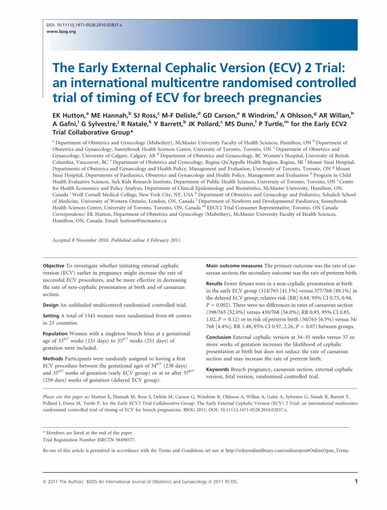
The Early External Cephalic Version (ECV) 2 Trial: an international multicentre randomised controlled trial of timing of ECV for breech pregnancies EK Hutton, a ME Hannah, b SJ Ross, c M-F Delisle, d GD Carson, e R Windrim, f A Ohlsson, g AR Willan, h A Gafni, i G Sylvestre, j R Natale, k Y Barrett, b JK Pollard, c MS Dunn, l P Turtle, m for the Early ECV2 Trial Collaborative Group* a Department of Obstetrics and Gynecology (Midwifery), McMaster University Faculty of Health Sciences, Hamilton, ON b Department of Obstetrics and Gynaecology, Sunnybrook Health Sciences Centre, University of Toronto, Toronto, ON c Department of Obstetrics and Gynaecology, University of Calgary, Calgary, AB d Department of Obstetrics and Gynaecology, BC Women’s Hospital, University of British Columbia, Vancouver, BC e Department of Obstetrics and Gynecology, Regina Qu’Appelle Health Region, Regina, SK f Mount Sinai Hospital, Departments of Obstetrics and Gynaecology and Health Policy, Management and Evaluation, University of Toronto, Toronto, ON g Mount Sinai Hospital, Departments of Paediatrics, Obstetrics and Gynaecology and Health Policy, Management and Evaluation h Program in Child Health Evaluative Sciences, Sick Kids Research Institute, Department of Public Health Sciences, University of Toronto, Toronto, ON i Centre for Health Economics and Policy Analysis, Department of Clinical Epidemiology and Biostatistics, McMaster University, Hamilton, ON, Canada j Weill Cornell Medical College, New York City, NY, USA k Department of Obstetrics and Gynecology and Pediatrics, Schulich School of Medicine, University of Western Ontario, London, ON, Canada l Department of Newborn and Developmental Paediatrics, Sunnybrook Health Sciences Centre, University of Toronto, Toronto, ON, Canada m EECV2 Trial Consumer Representative, Toronto, ON Canada Correspondence: EK Hutton, Department of Obstetrics and Gynecology (Midwifery), McMaster University Faculty of Health Sciences, Hamilton, ON, Canada. Email: [email protected] Accepted 8 November 2010. Published online 4 February 2011. Objective To investigate whether initiating external cephalic version (ECV) earlier in pregnancy might increase the rate of successful ECV procedures, and be more effective in decreasing the rate of non-cephalic presentation at birth and of caesarean section. Design An unblinded multicentred randomised controlled trial. Setting A total of 1543 women were randomised from 68 centres in 21 countries. Population Women with a singleton breech fetus at a gestational age of 33 0/7 weeks (231 days) to 35 6/7 weeks (251 days) of gestation were included. Methods Participants were randomly assigned to having a first ECV procedure between the gestational ages of 34 0/7 (238 days) and 35 6/7 weeks of gestation (early ECV group) or at or after 37 0/7 (259 days) weeks of gestation (delayed ECV group). Main outcome measures The primary outcome was the rate of cae- sarean section; the secondary outcome was the rate of preterm birth. Results Fewer fetuses were in a non-cephalic presentation at birth in the early ECV group (314/765 [41.1%] versus 377/768 [49.1%] in the delayed ECV group; relative risk [RR] 0.84, 95% CI 0.75, 0.94, P = 0.002). There were no differences in rates of caesarean section (398/765 [52.0%] versus 430/768 [56.0%]; RR 0.93, 95% CI 0.85, 1.02, P = 0.12) or in risk of preterm birth (50/765 [6.5%] versus 34/ 768 [4.4%]; RR 1.48, 95% CI 0.97, 2.26, P = 0.07) between groups. Conclusion External cephalic version at 34–35 weeks versus 37 or more weeks of gestation increases the likelihood of cephalic presentation at birth but does not reduce the rate of caesarean section and may increase the rate of preterm birth. Keywords Breech pregnancy, caesarean section, external cephalic version, fetal version, randomised controlled trial. Please cite this paper as: Hutton E, Hannah M, Ross S, Delisle M, Carson G, Windrim R, Ohlsson A, Willan A, Gafni A, Sylvestre G, Natale R, Barrett Y, Pollard J, Dunn M, Turtle P, for the Early ECV2 Trial Collaborative Group. The Early External Cephalic Version (ECV) 2 Trial: an international multicentre randomised controlled trial of timing of ECV for breech pregnancies. BJOG 2011; DOI: 10.1111/j.1471-0528.2010.02837.x. * Members are listed at the end of the paper. Trial Registration Number ISRCTN 56498577. Re-use of this article is permitted in accordance with the Terms and Conditions set out at http://wileyonlinelibrary.com/onlineopen#OnlineOpen_Terms ª 2011 The Authors BJOG An International Journal of Obstetrics and Gynaecology ª 2011 RCOG 1 DOI: 10.1111/j.1471-0528.2010.02837.x www.bjog.org
Transcript of The early external cephalic version (ECV) Trial: A RCT of ECV beginning at 34 weeks' vs delayed ECV...
The Early External Cephalic Version (ECV) 2 Trial:an international multicentre randomised controlledtrial of timing of ECV for breech pregnanciesEK Hutton,a ME Hannah,b SJ Ross,c M-F Delisle,d GD Carson,e R Windrim,f A Ohlsson,g AR Willan,h
A Gafni,i G Sylvestre,j R Natale,k Y Barrett,b JK Pollard,c MS Dunn,l P Turtle,m for the Early ECV2
Trial Collaborative Group*a Department of Obstetrics and Gynecology (Midwifery), McMaster University Faculty of Health Sciences, Hamilton, ON b Department of
Obstetrics and Gynaecology, Sunnybrook Health Sciences Centre, University of Toronto, Toronto, ON c Department of Obstetrics and
Gynaecology, University of Calgary, Calgary, AB d Department of Obstetrics and Gynaecology, BC Women’s Hospital, University of British
Columbia, Vancouver, BC e Department of Obstetrics and Gynecology, Regina Qu’Appelle Health Region, Regina, SK f Mount Sinai Hospital,
Departments of Obstetrics and Gynaecology and Health Policy, Management and Evaluation, University of Toronto, Toronto, ON g Mount
Sinai Hospital, Departments of Paediatrics, Obstetrics and Gynaecology and Health Policy, Management and Evaluation h Program in Child
Health Evaluative Sciences, Sick Kids Research Institute, Department of Public Health Sciences, University of Toronto, Toronto, ON i Centre
for Health Economics and Policy Analysis, Department of Clinical Epidemiology and Biostatistics, McMaster University, Hamilton, ON,
Canada j Weill Cornell Medical College, New York City, NY, USA k Department of Obstetrics and Gynecology and Pediatrics, Schulich School
of Medicine, University of Western Ontario, London, ON, Canada l Department of Newborn and Developmental Paediatrics, Sunnybrook
Health Sciences Centre, University of Toronto, Toronto, ON, Canada m EECV2 Trial Consumer Representative, Toronto, ON Canada
Correspondence: EK Hutton, Department of Obstetrics and Gynecology (Midwifery), McMaster University Faculty of Health Sciences,
Hamilton, ON, Canada. Email: [email protected]
Accepted 8 November 2010. Published online 4 February 2011.
Objective To investigate whether initiating external cephalic
version (ECV) earlier in pregnancy might increase the rate of
successful ECV procedures, and be more effective in decreasing
the rate of non-cephalic presentation at birth and of caesarean
section.
Design An unblinded multicentred randomised controlled trial.
Setting A total of 1543 women were randomised from 68 centres
in 21 countries.
Population Women with a singleton breech fetus at a gestational
age of 330/7 weeks (231 days) to 356/7 weeks (251 days) of
gestation were included.
Methods Participants were randomly assigned to having a first
ECV procedure between the gestational ages of 340/7 (238 days)
and 356/7 weeks of gestation (early ECV group) or at or after 370/7
(259 days) weeks of gestation (delayed ECV group).
Main outcome measures The primary outcome was the rate of cae-
sarean section; the secondary outcome was the rate of preterm birth.
Results Fewer fetuses were in a non-cephalic presentation at birth
in the early ECV group (314/765 [41.1%] versus 377/768 [49.1%] in
the delayed ECV group; relative risk [RR] 0.84, 95% CI 0.75, 0.94,
P = 0.002). There were no differences in rates of caesarean section
(398/765 [52.0%] versus 430/768 [56.0%]; RR 0.93, 95% CI 0.85,
1.02, P = 0.12) or in risk of preterm birth (50/765 [6.5%] versus 34/
768 [4.4%]; RR 1.48, 95% CI 0.97, 2.26, P = 0.07) between groups.
Conclusion External cephalic version at 34–35 weeks versus 37 or
more weeks of gestation increases the likelihood of cephalic
presentation at birth but does not reduce the rate of caesarean
section and may increase the rate of preterm birth.
Keywords Breech pregnancy, caesarean section, external cephalic
version, fetal version, randomised controlled trial.
Please cite this paper as: Hutton E, Hannah M, Ross S, Delisle M, Carson G, Windrim R, Ohlsson A, Willan A, Gafni A, Sylvestre G, Natale R, Barrett Y,
Pollard J, Dunn M, Turtle P, for the Early ECV2 Trial Collaborative Group. The Early External Cephalic Version (ECV) 2 Trial: an international multicentre
randomised controlled trial of timing of ECV for breech pregnancies. BJOG 2011; DOI: 10.1111/j.1471-0528.2010.02837.x.
* Members are listed at the end of the paper.
Trial Registration Number ISRCTN 56498577.
Re-use of this article is permitted in accordance with the Terms and Conditions set out at http://wileyonlinelibrary.com/onlineopen#OnlineOpen_Terms
ª 2011 The Authors BJOG An International Journal of Obstetrics and Gynaecology ª 2011 RCOG 1
DOI: 10.1111/j.1471-0528.2010.02837.x
www.bjog.org

Introduction
The fetus presents as a breech in 3–4% of all full-term sin-
gleton pregnancies and many of these pregnancies are
delivered by caesarean section.1,2 External cephalic version
(ECV) is an obstetrical procedure used during pregnancy
to try to turn a breech fetus to cephalic by externally
manoeuvring the fetus through the maternal abdomen.
A Cochrane review reported that ECV at full-term gestation
(‡37 weeks) decreases both the likelihood that the fetus
will be in a non-cephalic presentation at birth and the need
for caesarean section, and concluded that ECV should be
recommended for all women with a breech fetus at term
when there is no contraindication.3–5 However, ECV is
unsuccessful in about 40% of attempts.6,7
We hypothesised that initiating ECV earlier in the preg-
nancy (before the fetal breech descends into the pelvis and
while the maximum amount of amniotic fluid is present)
might increase the rate of successful ECV procedures, and
decrease both the rates of non-cephalic presentation at
birth and of caesarean section. We undertook a pilot trial
to determine if beginning ECV somewhat earlier than term
(34–35 weeks) might be more effective than beginning it at
term (37–38 weeks) in terms of decreasing the rate of non-
cephalic presentation at birth.8 The pilot study reported a
rate of non-cephalic presentation at birth in the early ECV
group of 66/116 (56.9%) compared with 77/116 (66.4%) in
the delayed ECV group (relative risk [RR] 0.86, 95% CI
0.70, 1.05, P = 0.09). Although the difference was not sta-
tistically significant, we felt that the results were sufficiently
promising to justify a larger trial. A Cochrane Systematic
Review confirmed this finding and recommended that fur-
ther trials be conducted.9 The Early ECV 2 Trial was
undertaken to answer the primary research question, ‘For
women with a fetus in breech presentation, does early ECV
(at 340/7 weeks to 356/7 weeks of gestation) versus delayed
ECV (not before 370/7 weeks of gestation) decrease or
increase the likelihood of caesarean section?’ and the sec-
ondary research question, ‘Is the risk of preterm birth
(<370/7 weeks of gestation) higher or lower with early ver-
sus delayed ECV?’.
Methods
The study was funded by the Canadian Institutes of Health
Research (CIHR) and was co-ordinated jointly at Sunny-
brook and Women’s College Health Sciences Centre in
Toronto, the University of British Columbia in Vancouver
and McMaster University in Hamilton, Canada.
A pragmatic, multicentred, parallel group randomised
controlled trial design, with prognostic stratification for
parity (0 and ‡1) and centre, was used to test for superior-
ity. Individual women were randomised using computer-
generated random block sizes and 1:1 allocation. Randomi-
sation was centrally controlled with a computerised
randomisation program accessible by a toll-free 24-hour,
7-day-a-week telephone service. Baseline data were collected
before randomisation. Data were collected on carbonless
duplicate paper forms and the original copy was mailed to
the Co-ordinating Centre where it was scanned into a
TELEform� data management system (Autonomy Cardiff
Software, Vista, CA, USA). Logic and range checks were
used to verify the accuracy of the data.
The study received ethical approval at the co-ordinating
sites and all participating centres; participating women gave
consent before randomisation. Pregnant women with a sin-
gleton fetus in a breech presentation who had a recent
screening ultrasound and were between 330/7 weeks
(231 days) and 356/7 weeks (251 days) of gestation were eli-
gible for the study. Women were ineligible when they pre-
sented with contraindications to ECV (such as fetal heart
rate abnormalities, placental abruption, major life-threaten-
ing fetal anomalies, uterine anomalies, hyper-extended fetal
head, rupture of fetal membranes, severe oligohydramnios
or hydramnios); contraindications to early ECV (such as
increased risk of preterm labour or placental abruption); or
contraindications to labour or vaginal birth (such as pla-
centa praevia, previous classical caesarean section); or if
they had been prior participants in the trial; were at
increased risk of unstable lie (such as grand multiparity);
or if they planned to give birth by caesarean section even if
the fetus turned to a cephalic position, or if they planned
a vaginal birth if the fetus remained breech.
SettingsCentres were invited to participate in the trial if they had
clinicians who were experienced in ECV and birth facilities
that were deemed to meet Canadian standards as detailed
in Table 1. The ECV procedures were undertaken or super-
vised by experienced clinicians. Experienced clinicians were
those who judged themselves to be skilled and experienced
in the ECV procedure and who’s Heads of Departments
agreed with that judgement. This definition of experience
has been demonstrated to be robust.10
The interventionA screening ultrasound was undertaken at 320/7–356/7 weeks
of gestation (224–251 days) and within 1 week of randomi-
sation to confirm breech presentation and rule out contra-
indications to ECV (see Figure 1). To allow time for
booking of procedures, women were randomised at a gesta-
tional age as early as 330/7 weeks and up to 356/7 weeks of
gestation, but no ECV was to be undertaken before 340/7
weeks of gestation. The nature of the intervention did not
lend itself to blinding of either participants or clinicians.
The first ECV procedure was to be performed in the early
Hutton et al.
2 ª 2011 The Authors BJOG An International Journal of Obstetrics and Gynaecology ª 2011 RCOG
ECV group between 340/7 and 356/7 weeks of gestation and
within 7 days following randomisation and in the delayed
ECV group at or after 370/7 weeks of gestation. All attempts
undertaken to manoeuvre the fetus at one visit were
considered part of one procedure. In either group, if a
procedure was unsuccessful, or if a fetus later reverted to
non-cephalic, a repeat ECV procedure could be performed
at a later date at the discretion of the care provider in
consultation with the woman.
Immediately before the ECV procedure, women were
reassessed to ensure eligibility for ECV including confirma-
tion of fetal presentation by ultrasound. Fetal wellbeing was
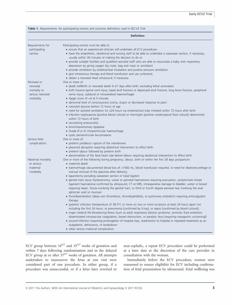
Table 1. Requirements for participating centres and outcome definitions used in EECV2 Trial
Definition
Requirements for
participating
centres
Participating centres must be able to:
• ensure that an experienced clinician will undertake all ECV procedures
• have the anaesthetic, obstetrical and nursing staff to be able to undertake a caesarean section, if necessary,
usually within 30 minutes of making the decision to do so
• provide suitable facilities and qualified neonatal staff who are able to resuscitate a baby with respiratory
depression by giving oxygen (by mask, bag and mask or ventilator)
• provide ventilation by endotracheal intubation and positive pressure ventilation
• give intravenous therapy and blood transfusion and use surfactant;
• obtain a neonatal head ultrasound, if necessary.
Perinatal or
neonatal
mortality or
serious neonatal
morbidity
One or more of:
• death (stillbirth or neonatal death 0–27 days after birth, excluding lethal anomalies)
• birth trauma (spinal cord injury, basal skull fracture or depressed skull fracture, long bone fracture, peripheral
nerve injury, subdural or intracerebral haemorrhage)
• Apgar score of <4 at 5 minutes
• abnormal level of consciousness (coma, stupor or decreased response to pain)
• neonatal seizures before 72 hours of age
• need for assisted ventilation for ‡24 hours via endotracheal tube initiated within 72 hours after birth
• infection (septicaemia [positive blood culture] or meningitis [positive cerebrospinal fluid culture]) determined
within 72 hours of birth
• necrotising enterocolitis
• bronchopulmonary dysplasia
• Grade III or IV intraventricular haemorrhage
• cystic periventricular leucomalacia.
Serious fetal
complications
One or more of:
• preterm prelabour rupture of the membranes
• placental abruption requiring obstetrical intervention to effect birth
• preterm labour followed by preterm birth
• abnormalities of the fetal heart rate before labour requiring obstetrical intervention to effect birth.
Maternal mortality
or serious
maternal
morbidity
One or more of the following during pregnancy, labour, birth or within the first 28 days postpartum:
• maternal death
• haemorrhage (documented blood loss of >1500 mL, blood transfusion required, or need for dilation/curettage or
manual removal of the placenta after delivery),
• laparotomy excluding caesarean section or tubal ligation
• genital tract injury (hysterectomy, vulvar or perineal haematoma requiring evacuation, symptomatic broad
ligament haematoma confirmed by ultrasound, CT or MRI, intraoperative damage to bladder, ureter or bowel
requiring repair, fistula involving the genital tract, or third or fourth degree perineal tear involving the anal
sphincter and/ or mucosa)
• thromboembolism (deep vein thrombosis, thrombophlebitis, or pulmonary embolism) requiring anticoagulant
therapy
• systemic infection (temperature of 38.5�C or more on two or more occasions at least 24 hours apart not
including the first 24 hours, or pneumonia [confirmed by X-ray], or sepsis [confirmed by blood culture]),
• major medical life-threatening illness (such as adult respiratory distress syndrome, amniotic fluid embolism,
disseminated intravascular coagulation, bowel obstruction, or paralytic ileus [requiring nasogastric suctioning])
• wound infection (requiring prolongation of hospital stay, readmission to hospital or repeated treatment as an
outpatient), dehiscence, or breakdown
• other serious maternal complication.
Early ECV2 Trial
ª 2011 The Authors BJOG An International Journal of Obstetrics and Gynaecology ª 2011 RCOG 3

assessed by continuous fetal heart rate monitoring for
20 minutes to ensure a normal baseline rate, good variabil-
ity and no evidence of decelerations, and was then moni-
tored intermittently during the ECV procedure using
auscultation, Doppler, or ultrasound viewing of fetal heart
rate. The protocol recommended the use of tocolytic agents
to relax the uterus during the ECV but their use was left to
the discretion of the healthcare provider, as was the
decision to use regional analgesia to facilitate ECV. Clini-
cians were directed to use the same approach to tocolytics
and to regional analgesia for women in both arms of the
trial.
The ECV procedure was discontinued if fetal heart tones
were non-reassuring, if it was not easily accomplished, or if
the woman reported undue discomfort. Fetal presentation
was confirmed by ultrasound immediately following all
procedures. Trial participants were monitored for at least
30 minutes following procedures to confirm fetal move-
ment on ultrasound and a reactive fetal heart rate on
continuous fetal heart rate monitoring. Anti-D immuno-
globulin was recommended for all rhesus-negative women
following the procedure.
Caesarean section was recommended to all women with
a fetus that remained non-cephalic at the time of birth
including in situations where labour began spontane-
ously.11,12 Vaginal birth was planned for all fetuses who
were cephalic at onset of labour when there was no contra-
indication. All other aspects of care during pregnancy,
labour and birth were determined by the woman and her
caregiver.
Compliance with the timing of the ECV procedures in
the two groups was assessed quarterly, for the trial as a
whole and by centre, and reasons for non-compliance were
reviewed with centres as needed. Site visits were made dur-
ing study promotional visits and included random chart
audits of selected data fields as well as a review of the study
facility using an evaluation check list based on the Guide-
lines for Good Clinical Research Practice.13
To avoid bias, the gestational age determined before ran-
domisation was used in determining gestational age at birth
and the rate of preterm birth. Members of the independent
Data Safety and Monitoring Board reviewed all stillbirths
and neonatal deaths, blinded to allocation group, for the
existence of any anomaly considered incompatible with life
and to make a determination regarding exclusion of any
women from the analysis of perinatal/neonatal outcomes.
Women and infants were followed until 28 days after birth
or until hospital discharge, whichever was later.
1543 pregnant women randomised
767 assigned early ECV baseline data reported
774 assigned delayed ECV baseline data reported
769 women 461 (60%) had at least 1 ECV
• 430 first ECV 37 weeks 308 (40%) had No ECV
765 women had outcomes analysed
764 infants had perinatal and neonatal outcomes analysed
768 women had outcomes analysed
768 infants had perinatal and 765 had neonatal outcomes analysed
1 lethal anomaly (trisomy 18) removed from analysis following adjudication by Data Safety Monitoring Board
5 lost to follow up 2 lost to follow up
1 withdrawal
1 loss to follow up
1 withdrawal
765 women 615 (80%) had at least 1 ECV
• 584 first ECV 34 < 36 weeks 150 (20%) had No ECV
Figure 1. Trial profile.
Hutton et al.
4 ª 2011 The Authors BJOG An International Journal of Obstetrics and Gynaecology ª 2011 RCOG
Outcome measuresThe primary outcome was the rate of caesarean section.
The secondary outcome was the rate of preterm birth
before 37 weeks of gestation. Other outcomes included:
non-cephalic presentation at birth; admission to the neona-
tal intensive care unit for ‡24 hours; perinatal or neonatal
mortality or serious neonatal morbidity; serious fetal com-
plications; maternal death or serious maternal morbidity;
pain experienced during the procedure; and maternal satis-
faction. Women provided other information regarding their
likes and dislikes about ECV and about trial participation
and these results will be reported elsewhere. A health eco-
nomic analysis is planned. Definitions are provided in
Table 1.
Pain experienced during the procedure (for women hav-
ing an ECV procedure) was measured immediately follow-
ing the ECV using a single visual analogue scale with 0
representing ‘no pain at all’ and 100 representing ‘most
pain imaginable’. Maternal satisfaction was determined
using a structured questionnaire asking women if they
would use the same approach to the timing of ECV in
another pregnancy with a breech baby or to recommend it
to a friend.
Statistical analysis
Sample sizeWe estimated the rate of caesarean section in the delayed
ECV group to be 65% based on rates from the pilot trial
and adjusted for the change in the inclusion criteria.
A sample size of 610/group was calculated to have 80%
power to find an 8-percentage-point reduction in the rate
of CS, from 65% in the delayed group to 57% in the early
group (Type I error rate of 0.05; two-tailed), if such a
reduction existed. This sample size would provide >70%
power to detect an increase from 6% to 10% in the rate of
preterm birth (Type I error rate of 0.05; two-tailed) and
>85% power to detect a three-fold increase in the rate of
perinatal or neonatal mortality or serious neonatal morbid-
ity from 1.6% to 4.8%.
We then increased the sample size by 20%, initially to
1460 and finally to 1526, as we anticipated that approxi-
mately 20% of women in the early group would not have
an ECV procedure because they would experience a sponta-
neous cephalic version after randomisation and before the
scheduled version.
Interim analysesTwo planned safety interim analyses were conducted after
complete data were received on the first 500 and 900
women randomised. Results were reviewed by an indepen-
dent Data Safety and Monitoring Board blinded to group
assignment.
Final analysisAn intention-to-treat analysis was conducted. Baseline data
were compared descriptively between treatment groups.
Perinatal and neonatal deaths were excluded from the anal-
yses of measures of neonatal morbidity. Fisher’s exact test
was used to compare binary outcomes and Student’s t-test
to compare continuous variables that were normally dis-
tributed. Because only women with an ECV procedure were
assessed for pain during the procedure, we used linear
regression to calculate adjusted between-treatment compar-
isons of pain by controlling for baseline variables where
there was either imbalance between treatment groups or
where the variable correlated with pain scores on the first
ECV. A P-value of <0.05 (two-sided) indicated statistical
significance for the primary and secondary outcome and
<0.01 (two-sided) indicated statistical significance for other
outcomes. Relative risks and 95% CI were used to report
the effects of the intervention on each outcome and risk
difference (RD) was calculated for the primary and second-
ary outcomes. Subgroup analyses were undertaken using
logistic regression analyses to test for interactions between
baseline characteristics (parity [0 versus ‡1], type of breech
[frank versus non-frank], gestational age at randomisation
[33–34 weeks versus 35 weeks of gestation], and the
national perinatal mortality rate of the country [£20/1000
versus >20/1000]) and treatment group for the primary
and secondary outcomes. The statistical software sas ver-
sion 9.1 (SAS Institute, Cary, NC, USA) was used for the
analyses.
In keeping with recommendations for presentation of
findings in the context of prior research and existing
knowledge,14 we undertook meta-analyses of the primary
and secondary outcome data from this trial and the Early
ECV Pilot Trial, the only previous study to compare early
and delayed timing of ECV. We pooled the summary data
for the primary and secondary outcomes of caesarean sec-
tion and preterm birth before 37 weeks of gestation using a
Mantel–Haenszel fixed effects model to calculate the RR,
weighted RD and 95% CIs of harm or untoward outcome,
using Review Manager (RevMan Version 5.0 Copenha-
gen: The Nordic Cochrane Centre, The Cochrane Collabo-
ration, 2008) Using data from both trials, we calculated the
number needed to treat to prevent one caesarean section
and the number needed to treat to harm for preterm birth.
Results
A total of 1543 women were randomised from 68 centres
in 21 countries between 30 December 2004 and 25 June
2008 (see Appendix S1). Two women, one in each group,
asked to be removed from the study leaving 1541 women
for the analyses of baseline characteristics; 767 in the early
ECV group, and 774 in the delayed ECV group (Figure 1).
Early ECV2 Trial
ª 2011 The Authors BJOG An International Journal of Obstetrics and Gynaecology ª 2011 RCOG 5

Eight women were lost to follow up (two assigned to early
ECV and six assigned to delayed ECV); seven before any
intervention, and one following a first ECV procedure. This
left 1533 women (99.4%) for the analysis of maternal out-
comes; 765 in the early ECV group, 768 in the delayed
ECV group.
Baseline characteristics were similar in the two groups
(Table 2). The timing and number of ECV procedures
including success of the procedure and rates of spontane-
ous version are presented in Table 3. Of the 765 women
randomised to the early ECV group, 615 (80.4%) under-
went at least one ECV procedure versus 461 (60.0%) of
769 women in the delayed ECV group. The majority
of women in both groups underwent only one procedure.
Of 615 women having an ECV procedure in the early ECV
group, 584 (95.0%) had their first procedure at the
expected time between 340/7 and 356/7 weeks of gestation;
of 461 women having an ECV procedure in the delayed
ECV group, 430 (93.3%) had their first ECV procedure at
the expected time at or after 370/7 weeks of gestation.
All ECV procedures were performed in hospital with
almost all on or near the labour and birth delivery area
(563/615 [91.5%] in the early ECV group and 414/460
[90.0%] in the delayed ECV group) and by an experienced
practitioner as determined a priori (603/615 [98.1%] in the
early ECV group and 446/461 [96.8%] in the delayed ECV
group). Clinician experience in both groups was similar
and the majority of ECV procedures were undertaken by
clinicians with >10 years experience with ECV: early ECV
group 343/615 (55.8%) and delayed ECV group 255/461
(55.3%). Obstetricians performed most ECV procedures
with midwives performing the remaining procedures; 2.1%
(13/615) and 2.6% (12/461) in the early and delayed
groups, respectively.
The characteristics of the fetuses at the time of the first
ECV procedure for those women having an ECV procedure
Table 2. Baseline characteristics
Characteristic at randomisation Early ECV (n = 767)
n (%)
Delayed ECV (n = 774)
n (%)
Maternal age (years) (median 5th, 95th centile) 30.0 (18.9, 39.2) 30.0 (19.8, 39.2)
Parity
0 409 (53.3) 411 (53.1)
1–4 346 (45.1) 354 (45.7)
>4 12 (1.6) 9 (1.2)
Gestational age (weeks) Median (5th, 95th centile) 34.7 (33.1, 35.7) 34.9 (33.3, 35.7)
330–336 131 (17.1) 109 (14.1)
340–346 318 (41.5) 323 (41.7)
350–356 318 (41.5) 342 (44.2)
Method of determining gestational age*
Clinical history only (no ultrasound) 61 (8.0) 49 (6.4)
First ultrasound £20 weeks (± clinical history) 627 (81.9) 656 (85.2)
First ultrasound >20 weeks (± clinical history) 78 (10.2) 65 (8.4)
Maternal height (cm) (median 5th, 95th centile)� 163.0 (151.0, 176.0) 163.0 (152.0, 176.0)
Maternal weight at last prenatal visit (kg)§ Median (5, 95th centile) 74.0 (56.0, 104.5) 72.0 (54.4, 100.0)
One previous caesarean section– 36 (4.7) 29 (3.8)
Time from last ultrasound to randomisation £7 (days)** 757 (99.0) 768 (99.6)
Placental location anterior� 314 (41.0) 319 (41.4)
Type of breech at last ultrasound§§
Frank 475 (62.1) 467 (61.0)
Complete 247 (32.3) 253 (33.0)
Footling 43 (5.6) 46 (6.0)
National perinatal mortality rate––
£10/1000 656 (85.5) 664 (85.8)
>10–20/1000 61 (8.0) 62 (8.0)
>20/1000 50 (6.5) 48 (6.2)
Missing values in early, delayed group as follows: *1,4; �1,4; §13,9; –1,1; **1,3; �1,3; §§2, 8.
––Countries with a national perinatal mortality rate of £10/1000 were Australia, Canada, Chile, Denmark, Estonia, Germany, Hungary, Ireland,
Israel, The Netherlands, Poland, Portugal, Spain, UK, USA; countries with a national perinatal mortality rate of >10–20/1000 were Argentina,
Brazil, Oman; countries with a national perinatal mortality rate of >20/1000 were Egypt, Jordan, South Africa; Reference for National Perinatal
Mortality Rates: Neonatal and perinatal mortality: country, regional and global estimates. World Health Organization. Geneva 2006.
Hutton et al.
6 ª 2011 The Authors BJOG An International Journal of Obstetrics and Gynaecology ª 2011 RCOG

Table 3. Description of ECV procedures
Description of procedures Early ECV
n (%)
Delayed ECV
n (%)
n = 765 n = 769
Time from randomisation to first ECV procedure£7 days 571 (74.6) 10 (1.3)
>7 days 44 (5.8) 451 (58.7)
No ECV procedure
undertaken
150 (19.6) 308 (40.1)
Reasons for no ECV procedure*
Spontaneous version 106 (13.9) 194 (25.2)
Mother declined ECV 29 (3.8) 47 (6.1)
Developed contraindication
to ECV
14 (1.8) 33 (4.3)
Mother delivered before
ECV
1 (0.1) 34 (4.4)
Clinician declined 3 (0.4) 6 (0.8)
Logistics 2 (0.3) 1 (0.1)
Other� 0 2 (0.3)
ECV procedure 615 (80.4) 461 (60.0)
n = 615 n = 461
Gestational age at first ECV procedure
Median (weeks) (5–95th
centile)
35.1 (34.0, 35.9) 37.3 (36.9, 37.9)
<340/7 14 (2.3) 0 (0.0)
340/7–356/7 584 (95.0) 5 (1.1)
360/7–366/7 12 (2.0) 26 (5.6)
‡370/7 5 (0.8) 430 (93.3)
Fetal presentation before first ECV procedure
Frank breech 372 (60.5) 275 (59.7)
Complete breech 189 (30.7) 144 (31.2)
Footling breech 31 (5.0) 24 (5.2)
Unknown breech 6 (1.0) 1 (0.2)
Transverse/oblique lie 17 (2.8) 17 (3.7)
Position of breech at first ECV procedure
Sacrum anterior 120 (20.1) 91 (20.5)
Sacrum posterior 71 (11.9) 47 (10.6)
Sacrum transverse 353 (59.0) 266 (60.0)
Unknown 54 (9.0) 40 (9.0)
Station of presenting part before first ECV procedure
Floating 236 (38.4) 121 (26.3)
Dipping 244 (39.7) 209 (45.3)
Well into the pelvis or
engaged
122 (19.8) 124 (26.9)
Unknown 13 (2.1) 7 (1.5)
Fetus easily palpated before first ECV procedure
Yes 518 (84.2) 414 (89.8)
No 94 (15.3) 45 (9.8)
Unknown 3 (0.5) 2 (0.4)
Use of tocolytics for ECV
During all ECV procedures 419 (68.1) 316 (68.6)
During some ECV
procedures
10 (1.6) 8 (1.7)
During no ECV procedures 186 (30.2) 136 (29.5)
Tocolytics used*
Betamimetic 335 (54.5) 252 (54.7)
Nitric oxide donor 36 (5.9) 23 (5.0)
Oxytocin antagonist 29 (4.7) 30 (6.5)
Calcium channel blocker 27 (4.4) 15 (3.3)
Table 3. (Continued )
Description of procedures Early ECV
n (%)
Delayed ECV
n (%)
Prostaglandin synthetase
inhibitor
19 (3.1) 20 (4.3)
Magnesium sulphate 0 1 (0.2)
Use of epidural for one or
more procedures
0 (0.0) 0 (0.0)
Use of spinal for one or
more procedures
1 (0.2) 0 (0.0)
Number having same
spinal for delivery
0 (0.0) 0 (0.0)
Success of ECV proceduresAny ECV procedure successful 329 (53.5) 201 (43.6)
Only one procedure
undertaken
536 (87.2) 426 (92.4)
First procedure successful 316 (51.4) 194 (42.1)
Two procedures
undertaken
73 (11.3) 34 (7.4)
Second procedure
successful
22 7
More than two procedures
undertaken
6 (1.0) 0 (0.0)
Third or subsequent
procedure successful
3
Ease with which first successful ECV performed§
Very easy 104 (32.9) 43 (22.2)
Somewhat easy 121 (38.3) 81 (41.8)
Neither easy nor difficult 55 (17.4) 40 (20.6)
Somewhat difficult 29 (9.2) 27 (13.9)
Very difficult 6 (1.9) 3 (1.6)
Reasons for discontinuing first ECV*
Unable to turn fetus 211 (34.3) 187 (40.6)
Unable to lift breech from
pelvis
97 (15.8) 93 (20.2)
Maternal discomfort 74 (12.0) 61 (13.2)
Non-reassuring fetal heart
rate
11 (1.8) 11 (2.4)
Obese/unable to palpate 11 (1.8) 5 (1.1)
Other 4 (0.7) 6 (1.3)
Maternal and fetal complications during ECV*
Non reassuring fetal heart
rate
21 (3.4) 16 (3.5)
Contractions 1 (0.2) 2 (0.4)
Hypotension 1 (0.2) 2 (0.4)
Vaginal bleeding/suspected
abruption placenta
0 1 (0.2)
Fetal presentation after first ECV
Cephalic 316 (51.4) 194 (42.1)
Breech 283 (46.0) 250 (54.2)
Transverse lie 16 (2.6) 17 (3.7)
Presentation of fetus after final ECV
Cephalic 328 (53.3) 200 (43.4)
Breech 275 (44.7) 246 (53.4)
Transverse lie 12 (2.0) 14 (3.0)
Pain during first ECV–
Adjusted Mean (SE)** 40.6 (1.06) 45.2 (1.2)
*May be more than one reason given.
�Other reasons in delayed group included an unstable lie and an unfa-
vourable position for ECV with spines posterior.
§One missing value for early group.
–Fifteen missing values in the early group; six missing values in the
delayed group.
**Adjusted for parity, maternal age and weight, and anterior placenta;
P = 0.0001.
Early ECV2 Trial
ª 2011 The Authors BJOG An International Journal of Obstetrics and Gynaecology ª 2011 RCOG 7

are reported in Table 3, as are the outcomes of the ECV
procedures. The rate of any maternal and fetal complica-
tions encountered during any ECV procedure was low:
(21/615 [3.4%] in the early ECV group and 20/461 [4.3%]
in the delayed ECV group), with non-reassuring fetal heart
rate during the procedure being the complication that
occurred most frequently. Women in the early ECV group
experienced less pain during the first ECV procedure than
those in the delayed ECV group, after adjusting for parity,
maternal age and weight, and anterior placenta (adjusted
mean score 40.6 [standard error (SE) 1.06] compared with
45.2 [SE 1.2], P = 0.0001).
Most infants in both groups were born in hospital and
women had a median postpartum hospital stay of 68 hours
(Table 4).
Fewer infants were in a non-cephalic presentation at
birth in the early ECV group than the delayed ECV group
(314/765 [41.1%] versus 377/768 [49.1%]; RR 0.84, 95%
CI 0.75, 0.94; P = 0.002). The rate of caesarean section was
not different between groups (398/765 [52.0%] in the early
ECV group versus 430/768 [56.0%] in the delayed ECV
group; RR 0.93, 95% CI 0.85, 1.02; P = 0.12; RD )0.04,
95% CI )0.09, 0.01). The rate of preterm birth at
<37 weeks of gestation was not different between groups
(6.5% [50/765] in the early ECV group, 4.4% [34/768] in
the delayed ECV group, RR 1.48, 95% CI 0.97, 2.26;
P = 0.07; RD )0.02, 95% CI )0.00, 0.04). There was no
difference between groups in the rate of one or more seri-
ous fetal complications following randomisation (41/765
[5.4%] in the early ECV group versus 31/768 [4.0%] in the
delayed ECV group; RR 1.33, 95% CI 0.84, 2.09; P = 0.23).
There were no maternal deaths in either group and no dif-
ference between groups in serious maternal morbidity (39/
765 [5.1%] in the early ECV group versus 29/768 [3.8%]
in the delayed ECV group; RR 1.35, 95% CI 0.84, 2.16;
P = 0.22) (Table 4 and Appendix S2).
One infant in the early ECV group was excluded from
the analysis of perinatal/neonatal outcomes because of a
lethal anomaly (trisomy 18), leaving 764 infants in the early
ECV group and 768 infants in the delayed ECV group for
the analyses of perinatal and neonatal outcomes (Table 5).
Three infants died, all in the delayed ECV group, none of
whom had had an ECV procedure. The causes of death
were unexplained intrauterine fetal death at a gestational
age of 37 weeks 5 days; sepsis following cardiac surgery;
and sepsis after preterm birth. One infant in each group
had serious neonatal morbidity. Perinatal or neonatal mor-
tality or serious neonatal morbidity was not different
between the groups (1/764 [0.1%] in the early ECV group
versus 4/768 [0.5%] in the delayed ECV group, RR 0.25,
95% CI 0.03, 2.25; P = 0.37). There were no reported cases
of intubation and ventilation via endotracheal tube for
24 hours or longer, bronchopulmonary dysplasia, seizures
within 72 hours of age, necrotising enterocolitis, birth
trauma, grade III or IV intraventricular haemorrhage, cystic
periventricular leucomalacia, or cases with evidence of
stroke. The risk of being admitted to neonatal intensive
care for ‡24 hours was not different between groups (5/764
[0.7%] in the early ECV group versus 6/765 [0.8%] in the
delayed ECV group, RR 0.83, 95% CI 0.26, 2.72; P = 1.00).
Women in the early ECV group were more likely than
those in the delayed ECV group to indicate that they would
use the same approach to the timing of ECV in another
pregnancy with a fetus in breech presentation or to recom-
mend it to a friend (630/723 [87.1%] versus 531/710
[74.8%], RR 1.17; 95% CI 1.11, 1.23, P < 0.0001).
For outcomes of caesarean section and preterm birth,
there were no significant interactions between treatment
group and the baseline variables parity, type of breech, ges-
tational age at randomisation and national perinatal mor-
tality rate. As a result, there were no subgroup effects
observed for these outcomes.
When the data from this trial were combined with those
of the pilot trial we found no evidence of heterogeneity
(I2 = 0%). The caesarean section rate in the early ECV
group was 53.7% (473/881) compared with 58.1% (513/
884) in the delayed ECV group (RR 0.93, 95% CI 0.85,
1.00, P = 0.06; RD )0.04, 95% CI )0.09, 0.00, P = 0.06)
(Figure 2). The rate of preterm birth in the early ECV
group was 6.8% (60/881) compared with 4.6% (41/884) in
the delayed group (RR 1.47, 95% CI 1.00, 2.16, P = 0.05;
RD 0.02, 95% CI )0.00, 0.04) (Figure 3). The differences
between groups in the rates of both caesarean section and
preterm birth are at borderline conventional levels of statis-
tical significance. We calculated that 23 women (95% CI 11
to undefined) would need to plan early ECV procedures to
prevent one caesarean section, and for every 46 (95% CI 23
to undefined) planned early ECV procedures, one addi-
tional late preterm birth would result. In both these calcu-
lations, because the risk difference is not statistically
significant at the 5% level (two-sided), the upper limit is
undefined and can be considered arbitrarily large.
Discussion
When the fetus presents as a breech, ECV undertaken at
term reduces the need for caesarean section, avoids the risk
of preterm birth and is considered safe for the fetus.3,6,7,15,16
However, because of the risk of a failed procedure
when ECV is not undertaken until 37 weeks of gestation,
we hypothesised that initiating ECV somewhat earlier
than term might increase the likelihood of successful
ECV, thereby further reducing the need for caesarean
section.17–19
The Early ECV2 Trial found that initiating ECV at 340/7–
356/7 weeks of gestation, rather than waiting until term,
Hutton et al.
8 ª 2011 The Authors BJOG An International Journal of Obstetrics and Gynaecology ª 2011 RCOG

Table 4. Characteristics and outcomes of pregnancy, labour and birth
Characteristic or outcome Early ECV (n = 765)
n (%)
Delayed ECV (n = 768)
n (%)
Presentation at delivery*
Cephalic 451 (59.0) 391 (50.9)
Non-cephalic 314 (41.1) 377 (49.1)
Caesarean section� 398 (52.0) 430 (56.0)
Parity = 0 286/409 (69.9) 294/406 (72.4)
Parity = 0 and cephalic at birth 52/174 (29.9) 40/148 (27.0)
Parity ‡1 112/356 (31.4) 136/361 (37.7)
Parity ‡1 and cephalic at birth 36/277 (13.0) 25/242 (10.3)
Caesarean section� 398 (52.0) 430 (56.0)
Before labour or in early labour 285 (37.3) 338 (44.0)
During active first§ or second stage labour 113 (14.8) 92 (12.0)
Vaginal birth 367 (48.0) 338 (44.0)
Spontaneous cephalic 318 (41.6) 287 (37.4)
Assisted cephalic (vacuum or forceps) 45 (5.9) 39 (5.1)
Vaginal breech– 4 (0.5) 12 (1.6)
Place of birth
Hospital 751 (98.2) 734 (95.6)
Home, birthing centre, or other** 14 (1.8) 34 (4.4)
Gestational age at delivery (median, 5th, 95th centile) 39.1 (36.6, 41.6) 39.1 (37.1, 41.4)
Preterm birth <37 weeks� 50 (6.5) 34 (4.4)
Gestational age <34 weeks 0 1 (0.1)
Gestational age 340–346 weeks 5 (0.7) 1 (0.1)
Gestational age 350–356 weeks 17 (2.2) 13 (1.7)
Gestational age 360–366 weeks 28 (3.7) 19 (2.5)
Birth within 48 hours of ECV procedure 7 (1.1) 1 (0.2)
Serious fetal complications following randomisation
Prelabour rupture of membranes <37 weeks 12 (1.6) 9 (1.2)
Abruptio placenta requiring intervention to effect delivery 3 (0.4) 2 (0.3)
Preterm labour resulting in preterm birth 26 (3.4) 17 (2.2)
FHR abnormalities before labour requiring delivery 10 (1.3) 9 (1.2)
At least one serious fetal complication following randomisation§§ 41 (5.4) 31 (4.0)
At least one serious fetal complications within 48 hours of ECV 9/615 (1.5) 3/461 (0.7)
Other fetal outcomes
Prelabour rupture of membranes 201 (26.3) 180 (23.4)
Placental abruption 10 (1.3) 5 (0.7)
Preterm labour 35 (4.6) 26 (3.4)
FHR abnormalities before labour (n) 28 (3.7) 23 (3.0)
Maternal mortality or any serious maternal morbidity––
during pregnancy, labour, birth or up to 28 days following birth***
39 (5.1) 29 (3.8)
Duration of postpartum stay in hours (median, 5th, 95th centile) 67.9 (9.2, 123.5) 67.8 (5.8, 124.4)
ECV = External Cephalic Version;
*RR = 0.84; 95% CI: 0.75, 0.94; p-value = 0.002;
�RR = 0.93; 95% CI: 0.85, 1.02; p-value = 0.12;
§Active labour is defined as cervical dilation ‡3 cm or 80% effaced and contractions £5 minutes apart; no significant interaction between treat-
ment group and the baseline variables parity, type of breech, gestational age at randomisation and national perinatal mortality rate;
–All vaginal breeches reported as assisted, no forceps for aftercoming head and no breech extraction;
**Other two locations in delayed group include ambulance and unplanned out-of-hopsital birth;
�RR = 1.48; 95% CI: 0.97, 2.26; p-value = 0.07; no significant interaction between treatment group and the baseline variables parity, type of
breech, gestational age at randomisation and national perinatal mortality rate;
§§RR = 1.33; 95% CI: 0.84, 2.09; p-value = 0.23;
––There were no maternal deaths or any cases of fistula involving the genital tract, sympotomatic broad ligament haematoma, pulmonary embo-
lism, adult respiratory distress syndrome, or amniotic fluid embolism.
***RR = 1.35; 95% CI 0.84, 2.16; p-value = 0.22.
Early ECV2 Trial
ª 2011 The Authors BJOG An International Journal of Obstetrics and Gynaecology ª 2011 RCOG 9

was successful at decreasing the likelihood of a non-cepha-
lic presentation at birth. However, the decrease in non-
cephalic presentation did not translate into a reduction in
use of caesarean section. The results of the meta-analysis,
although at borderline conventional levels of statistical sig-
nificance, suggest that the small (4%) reduction in absolute
risk of caesarean section that we found in this trial with
early versus delayed ECV may be real. For women with a
fetus in breech presentation who wish to reduce their risk
of caesarean section, even if it is only by this small percent-
age, ECV at 34–35 weeks of gestation is an option that
may be considered.
Although our trial did not find higher risks of adverse
outcome for the fetus with early versus delayed ECV and
generally the infants did well, the results of the meta-analy-
sis suggest that early ECV may be associated with a higher
risk of preterm birth. This may be because the manipula-
tion of the uterus in attempting to turn the fetus as part of
ECV contributes to initiating labour or results in a fetal
complication which, in the early group, may lead to pre-
term birth. Irrespective of the cause, preterm birth, even at
34–36 weeks of gestation, is a concern because it is associ-
ated with acute morbidities such as respiratory distress syn-
drome as well as poorer long-term outcomes.20,21 Hence
any benefits to undertaking the ECV before term in avoid-
ing caesarean section will need to be balanced against the
potentially higher risk of preterm birth.
Pain scores for the ECV procedure in both groups
were moderate. Although the early group perceived less
pain, it is unlikely that a 5mm difference on the 100mm
visual analogue scale is clinically important. For example,
in an emergency room setting a difference of <13 mm in
pain scores on the visual analogue scale was not found to
be clinically important.22 Women were more satisfied
with early ECV than delayed ECV in this trial. This may
be because of the perceived benefits of a lower risk of
caesarean section or because the early procedure was not
generally available outside the trial and so some of the
women joining the trial may have been doing this to
have a chance of receiving the treatment that they pre-
ferred. Further research would be useful to explore
women’s views about the timing of ECV, given the find-
ings from this trial.
It is not clear why the decrease in non-cephalic presenta-
tion did not result in decreased rates of caesarean section.
It may be because more breech fetuses in the delayed group
were born vaginally; the study was undertaken in centres
where the rates of caesarean section for cephalic presenta-
tion are generally high; and because the slightly higher rate
of preterm fetal complications in the Early ECV Group
may have contributed to a greater need for caesarean sec-
tion in that group despite the fetus being in cephalic pre-
sentation.
Table 5. Neonatal outcomes
Outcome Early ECV
n (%)
Delayed ECV
n (%)
(n = 764) (n = 768)
Perinatal or neonatal
mortality or serious
neonatal morbidity*,�
1 (0.1) 4 (0.5)
Perinatal or neonatal death
0–27 days after birth§
0 (0.0) 3 (0.4)
Stillbirth 0 (0.0) 1 (0.1)
Neonatal death 0 (0.0) 2 (0.3)
(n = 764) (n = 765)
Serious neonatal
morbidity–
1 (0.1) 1 (0.1)
Apgar score <4 at
5 minutes**
0 (0.0) 1 (0.1)
Sepsis within 72 hours of birth
– confirmed by blood culture
1 (0.1) 0 (0.0)
Other neonatal morbidity–
Apgar score <7 at
5 minutes**
6 (0.8) 4 (0.5)
Intubation and ventilation 5 (0.7) 2 (0.3)
Admission to neonatal
intensive care for
‡24 hours�
5 (0.7) 6 (0.8)
Other neonatal outcomes
Birthweight
Median (5th, 95th centile)
(grams)
3340
(2475, 4120)
3330
(2590, 4110)
<2500 g 39 (5.1) 24 (3.1)
>4000 g 67 (8.8) 61 (8.0)
Gender
Male 362 (47.4) 366 (47.8)
Female 402 (52.6) 399 (52.2)
*RR = 0.25; 95% CI 0.03, 2.25; P = 0.37
�There were no reported cases of: birth trauma; abnormal level of
consciousness that included coma, stupor or decreased response to
pain; neonatal seizures before 72 hours of age; need for assisted
ventilation ‡24 hours via endotracheal tube initiated within 72 hours
after birth; meningitis within 72 hours after birth; necrotising
enterocolitis; bronchopulmonary dysplasia; Grade III or IV intraven-
tricular haemorrhage; or cystic periventricular leucomalacia
§One term infant died at 17 days from sepsis following cardiac sur-
gery; one 2570-g infant born at 35 weeks 1 day died on day 11 of
Klebsiella pneumoniae after being discharged home and readmitted
to NICU; one was an unexplained stillbirth born at 37 weeks 5 days;
none of the three women had an ECV procedure.
–Excludes deaths.
**Missing data for two women in the early group, one woman in
the delayed group; for these women, there was no noted resuscita-
tion at time of delivery and no other complications noted.
�RR = 0.83, 95% CI 0.26, 2.72; P = 1.00.
Hutton et al.
10 ª 2011 The Authors BJOG An International Journal of Obstetrics and Gynaecology ª 2011 RCOG

Tocolytics are often used as part of the ECV procedure
and are known to increase the success of the procedure.23
As such tocolytics are the most important co-intervention
to consider in this trial. We believe that our study reflects
the use of tocolytics in practice; that is some practitioners
use tocolytics routinely, some use them selectively and
some do not use tocolytics at all for ECV. It is, for exam-
ple, the case in Canada that betamimetics are not available
for ECV. In our trial, 70% of the ECV procedures in both
groups were carried out using tocolytics; so the use of
tocolytics was balanced between groups averting any
co-intervention bias.
Lack of power limits the ability to conclude that a real
difference in caesarean section rates between groups exists.
The original total sample size of 1460 was based on having
a power of 80% to detect an 8-percentage-point difference,
assuming the caesarean section rate in the delayed group
was 65%. For a caesarean section rate of 56%, as observed
in the trial, a total sample size of 1460 provides a power of
78%. However, as 1524 women were enrolled, a power of
80% was maintained. Although we calculated our sample
size using data from our pilot study, The Early ECV2 Trial
had a smaller than anticipated between-group difference in
rate of caesarean section (8% in sample size calculations
versus 4% in trial) which undermined the power to find a
difference in rate of caesarean section. The rate of caesar-
ean section may have been lower in the current study
because the proportion of multiparous women enrolled
was higher (47%) than in the pilot study (35%), and mul-
tiparity is associated with higher rates of spontaneous ver-
sion of breech to cephalic, of successful ECV and of
vaginal birth. We also had a 1% vaginal breech rate that
was not accounted for in the sample size calculation.
In addition, vaginal breech births were not evenly distrib-
uted between the groups with the early ECV group having
4/765 (0.5%) compared with 12/768 (1.6%) in the delayed
ECV group. These factors all have the potential to decrease
statistical power. Compliance with the study protocol was
generally good, however 6.7% (31/461) of the ECV proce-
dures in the delayed group were undertaken before
37 weeks of gestation, usually as part of a clinical manage-
ment plan for a condition that arose following randomisa-
tion.
Factors that affect external validity and generalisability of
findings in randomised controlled trials have been well
enumerated.24,25 The number of women enrolled in the
Early ECV2 Trial was low relative to potentially eligible
women. This may be partly explained by the fact that ECV
is not offered or recommended to women as frequently as
it should be according to current recommendations,3–5 and
when it is offered, women are often fearful of the pain and
safety of the procedure.26 Potentially eligible women may
not have been identified in time to enrol in our study, as
not all clinicians focus on fetal presentation as early as 34–
36 weeks of gestation. In addition, recruitment is often dif-
ficult, generally, to randomised controlled trials. Despite
the proportionately low numbers of women recruited, we
believe that our sample was probably representative, as the
rate of successful ECV in our study groups (54% and 44%
for early and delayed groups, respectively) was similar to
the mean rate found in two large systematic reviews report-
ing on ECV outcomes (60% and 58%).6,7 Our multicentred
pragmatic trial design will enhance the generalisability of
the findings. The entry criteria were broad and inclusive
and practitioners undertook the ECV according to their
local protocol.
Study or subgroupHutton 2003Hutton 2011
Total (95% CI)Total eventsHeterogeneity: χ² = 0.08, df = 1 (P = 0.78); I ² = 0%Test for overall effect: Z = 1.85 (P = 0.06)
Events75
398
473
Total116765
881
Events83
430
513
Total116768
884
Weight16.2%83.8%
100.0%
M-H, fixed, 95% CI0.90 [0.76, 1.08]0.93 [0.85, 1.02]
0.93 [0.85, 1.00]
Early ECV group Delayed ECV group Risk ratio Risk ratioM-H, fixed, 95% CI
0.5 0.7 1 1.5 2Favours early ECV Favours delayed ECV
Figure 2. Forest plot comparison: meta-analysis of caesarean section.
Study or subgroupHutton 2003Hutton 2011
Total (95% CI)Total eventsHeterogeneity: χ² = 0.00, df = 1 (P = 0.95); I ² = 0%Test for overall effect: Z = 1.95 (P = 0.05)
Events1050
60
Total116765
881
Events7
34
41
Total116768
884
Weight17.1%82.9%
100.0%
M-H, fixed, 95% CI1.43 [0.56, 3.62]1.48 [0.97, 2.26]
1.47 [1.00, 2.16]
Early ECV group Delayed ECV group Risk ratio Risk ratioM-H, fixed, 95% CI
0.05 0.2 1 5 20
Favours early ECV Favours delayed ECV
Figure 3. Forest plot comparison: meta-analysis of preterm birth <37 weeks of gestation.
Early ECV2 Trial
ª 2011 The Authors BJOG An International Journal of Obstetrics and Gynaecology ª 2011 RCOG 11
Conclusion
External cephalic version initiated at 34–35 weeks of gesta-
tion compared with 37 or more weeks of gestation
increases the probability of cephalic presentation at birth,
but does not reduce the rate of caesarean section and may
increase the rate of preterm birth. The study findings
should be discussed with women so that they can make an
informed choice as to what is best for them and their
infants.
EECV2 Collaborative Group
Steering committee. EK Hutton, J Barrett, GD Carson, MF
Delisle, M Dunn, S Edwards, A Fernandez, A Gafni, ME
Hannah, S Hewson, R Natale, A Ohlsson, SJ Ross, AR
Willan, R Windrim, JK Pollard, I Schweitzer, G Sylvestre,
P Turtle.
Data Safety Monitoring Board. Bracken M, Crowley P,
Donner A, Duley L, Ehrenkranz R.
Collaborators. Argentina—M Curioni, R Abalos Gorostiaga
(Hospital Ramon Carrillo, Santiago del Estero); C Becker,
PA Elizabeth, L Errandonea, M Palermo, CA Ramos, M
Trabucco, D Montes Varela (Hospital Posadas, Buenos
Aires); MS Bertin, JL Castaldi (Hospital Penna, Bahia
Blanca); M Mohedano de Duhalde (Hospital Avellaneda,
Tucuman); C Becker (Hospital Durand, Buenos Aires);
A Messina (Hospital Alvarez, Buenos Aires); Australia—
J Baumgartner, G Kovacs, B Malcolm, JR Neil (Box Hill
Hospital, Box Hill); K Mahomed (Ipswich Hospital,
Ipswich); A Child, B DeVries, H Phipps, A Welsh (Royal
Prince Alfred Hospital, Sydney); GK Davis, L Roberts, NP
Watts (St George Hospital, Sydney); M Cybulski, D Gib-
son, S Tucker (Toowoomba Base Hospital, Toowoomba);
I McCahon, P Sheehan, M Umstad (University of Mel-
bourne Dept. of Obsetrics & Gynecology, The Royal
Women’s Hospital, Carlton); J Milligan, J Morris, K Ric-
kard (Royal North Shore Hospital, St. Leonards); G Gar-
dener, S Jenkins-Manning (Mater Mothers’ Hospital,
Brisbane); C Boniface, M Edmondson, D Watson (Towns-
ville Hospital, Townsville); A Green (Ipswich Hospital, Ips-
wich); Brazil—A Ayub (ISCMPA-Maternidade Mario
Totta, Porto Alegre); Canada—MF Delisle, S Soanes, (Chil-
dren’s & Women’s Health Centre of BC, Vancouver); A Jor-
dan, R Windrim (Mount Sinai Hospital, Toronto);
C Fanning, B Parish (IWK Health Centre, Halifax); R Na-
tale, MA Watson (St Joseph’s Health Centre, London);
D Reid, P Scheufler (Trillium Health Centre, Mississauga);
AM Malott, A Reitsma (Hamilton Health Sciences Corpo-
ration McMaster Site, Hamilton); KA Haslauer, M Lipp
(Lion’s Gate Hospital, Vancouver); D Farquharson, K Gray
(Royal Columbian Hospital, New Westminster); N Demia-
nczuk, E Penttinen (Royal Alexandra Hospital, Edmonton);
E Herer, K McLean (Sunnybrook Health Sciences Centre,
Toronto); F Aghajafari, S Williams (Regina General Hospi-
tal, Regina); C Moravac, M Yudin (St Michael’s Hospital,
Toronto); J Pollard, L Miller (Calgary Health
Region—Foothills Hospital, Calgary); RB Anderson (St
Paul’s Hospital, Vancouver); M Good, MC Walker (The
Ottawa Hospital—General Campus, Ottawa); R Kulkarni,
R Scarfone (North Bay General Hospital, North Bay);
C Cameron, T Peel (Markham Stouffville Hospital, Mark-
ham); Chile—J Carrillo, A Cruces, Y Gonzalez (Hospital
Padre Hurtado, Santiago); J Figueroa Poblete, L Lama
Hormazabal, J Saez, (Hospital Clinico San Borja Arriaran,
Santiago); E Oyarzun, A Rioseco (Pontificia Universidad
Catolica de Chile, Santiago); S Illanes, C Kottmann (Hospi-
tal Parroquial de San Bernardo, San Bernardo); M Parra,
S Quezada, L Quiroz (Hospital Clinico Universidad de
Chile JJ Aguirre, Santiago); Denmark—L Hvidman, IM
Mogensen, A Mouritzen (Aarhus University Hospital, Aar-
hus); B Ostberg (Gentofte University Hospital, Hellerup);
Egypt—SNM Abdel-Samad, T Al-Hussaini, I El-Nashar (As-
siut University Hospital, Assiut); Estonia—F Kirss, K Rull,
E Ustav, P Vaas (Tartu University Hospital-Women’s
Clinic, Tartu); Germany—V Brink-Spalink, K Weizsaecker
(CUB-Virchow Klinikum, Berlin); Hungary—T Major,
R Poka (University of Debrecen, Debrecen); Ireland—
S Daly, (Coombe Women’s Hospital, Dublin); Israel—
H Kaneti, D Rosen, B Schachter (Meir Medical Center,
Kfar-Saba); B Chayen, L Harel (Ma’ayney HaYeshua Medi-
cal Center, Bnei Brak); Z Hiaeb, G Malinger (Edith Wolf-
son Medical Center, Holon); D Dukler, E Lunenfeld
(Soroka Medical Center, Beer Sheva); Jordan—L AlFaris, M
El-Zibdeh (Islamic Hospital, Amman); Poland—I Dom-
zalska-Popadiuk, P Kobiela, Z Pankrac, J Preis, K Preis, M
Swiatkowska-Freund (Medical University of Gdansk,
Gdansk); Portugal—J Cravo (Hospital Distrital de Faro,
Faro); South Africa—AM Theron, GB Theron (Stellenbosch
University, Tygerberg); HS Cronje, JM du Plessis (Univer-
sity of Free State, Bloemfontein); Spain—M Munoz (Hos-
pital Clinic-University of Barcelona, Barcelona); Sultanate
of Oman—G Khan, S Khan (Khoula Hospital, Muscat); the
Netherlands—S Goossens, M Pieters, FJME Roumen
(Atrium Medical Center, Heerlen); F ten Cate, M Pieters,
F Smits (Academisch Ziekenhuis Maastricht, Maastricht);
M Heres, E Krabbendam (Sint Lucas Andreas Ziekenhuis,
Amsterdam); United Kingdom—R Airey, D Farrar, DJ Tuff-
nell (Bradford Royal Infirmary, Bradford); V Heyes, C Mel-
vin, C Schram (Royal Blackburn Hospital, Blackburn);
A Galimberti, P Stewart (Royal Hallamshire Hospital, Shef-
field); J Cresswell (Chesterfield Royal Hospital, Chester-
field); C McCormick (Nottingham City Hospital,
Nottingham); United States of America—J Andrews, D Fle-
ener (University of Iowa Hospitals and Clinics, Iowa City);
D Coonrod, BF Jimenez (Maricopa Medical Center,
Hutton et al.
12 ª 2011 The Authors BJOG An International Journal of Obstetrics and Gynaecology ª 2011 RCOG

Phoenix); S Brown, A Gregg (University of South Carolina,
Columbia); C Pitchford, D Seubert (New York University
School of Medicine, New York).
Disclosure of interestNo author had any financial or personal relationships with
other people or organisations that could inappropriately
influence (bias) their work on this project. The funding
sponsor The Canadian Institutes of Health Research (CIHR)
had no role in the collection, analysis or interpretation of
data; in the writing of the report; or decision to submit
findings for publication. A staff member of CIHR sat as
ex officio member of the Steering Committee for the trial.
Contribution to authorshipAll authors participated in the design, methodological
issues implementation, conduct, monitoring, analysis, inter-
pretation of the study, and participated in reviewing and
editing the manuscript. The corresponding author had full
access to all the data in the study and had final responsibil-
ity for the decision to submit for publication.
Details of ethics approvalEthics approval was obtained from each of three co-ordi-
nating centres (University of British Columbia; Sunnybrook
and Women’s Health Sciences Centre; McMaster Univer-
sity), and at each of the recruiting centres where the study
took place. In addition, informed consent was sought from
each woman enrolled in the trial.
FundingEECV2 was funded by The Canadian Institutes of Health
Research (MTC-65630) and registered with Clinical Tri-
als.gov (ISRCTN 56498577). The data co-ordinating centre
was supported by grants from Sunnybrook Health Sciences
Centre, Women’s College Hospital and the Department of
Obstetrics and Gynaecology at the University of Toronto.
Eileen Hutton was supported by salary awards from The
Canadian Institutes of Health Research and from Michael
Smith Foundation for Health Research.
AcknowledgementsWe thank all of the women who participated in this trial.
The staff at the Maternal, Infant and Reproductive Health
Research Unit (now the Centre for Maternal Infant Child
Research) in Toronto is recognised for excellence and dili-
gence in their work; the steering committee for timely
review and guidance.
Supporting information
The following supplementary materials are available for this
article:
Appendix S1. EECV2 Trial: final trial recruitment,
annual delivery rate and number of certified ECV practitio-
ners by participating site.
Appendix S2. Maternal mortality and morbidity.
Additional Supporting Information may be found in the
online version of this article.
Please note: Wiley-Blackwell are not responsible for the
content or functionality of any supporting information
supplied by the authors. Any queries (other than missing
material) should be directed to the corresponding
author. j
References
1 Hutton EK, Hannah ME, Barrett J. Use of external cephalic version
for breech pregnancy and mode of delivery for breech and twin
pregnancy: a survey of Canadian practitioners. J Obstet Gynaecol
Can 2002;24:804–10.
2 Goffinet F, Carayol M, Foidart JM, Alexander S, Uzan S, Subtil D,
et al., for the PREMODA Study Group. Is planned vaginal delivery
for breech presentation at term still an option? Results of an obser-
vational prospective survey in France and Belgium. Am J Obstet
Gynecol 2006;194:1002–11.
3 Hofmeyr GJ, Kulier R. External cephalic version for breech presenta-
tion at term. Cochrane Database Syst Rev 2005; Issue 1. Art. No.:
CD000083. DOI: 10.1002/14651858.CD000083
4 Impey LWM, Hofmeyr GJ. External Cephalic Version and Reducing
the Incidence of Breech Presentation. Green Top Guidelines No.20a.
London: RCOG Press, 2006.
5 American College of Obstetrics and Gynecology (ACOG). Clinical
management guidelines for obstetrician–gynecologists: External
Cephalic Version. ACOG Practice Bull 2000;13:380–5. (reaffirmed
2009).
6 Collaris RJ, Oei SG. External cephalic version: a safe procedure?
A systematic review of version-related risks. Acta Obstet Gynecol
Scand 2004;83:511–8.
7 Grootscholten K, Kok M, Oei SG, Mol BWJ, van der Post JA. External
Cephalic Version – related risks: a meta-analysis. Obstet Gynecol
2008;112:1143–51.
8 Hutton EK, Kaufman K, Hodnett E, Amankwah K, Hewson SA,
McKay D, et al., for the Early External Cephalic Version Trial Group.
External cephalic version beginning at 34 weeks’ gestation versus 37
weeks’ gestation: a randomized multicenter trial. Am J Obstet Gyne-
col 2003;189:245–54. [Canadian Institutes of Health Research grant
MT-15223].
9 Hutton EK, Hofmeyr GJ. External cephalic version for breech presen-
tation before term. Cochrane Database Syst Rev 2006; Issue 1. Art.
No.: CD000084. DOI: 10.1002/14651858.CD000084.pub2.
10 Su M, McLeod L, Ross S, Willan A, Hannah WJ, Hutton E, et al., for
the Term Breech Trial Collaborative Group. Factors associated with
adverse perinatal outcome in the Term Breech Trial. Am J Obstet
Gynecol 2003;189:740–5.
11 Hannah ME, Hannah WJ, Hewson SA, Hodnett ED, Saigal S, Willan
AR, et al., for the Term Breech Trial Collaborative Group. Planned
caesarean section versus planned vaginal birth for breech presen-
tation at term: a randomised multicentre trial. Lancet 2000;356:
1375–83.
12 Hofmeyr GJ, Hannah M. Planned caesarean section for term breech
delivery. Cochrane Database Syst Rev 2003; Issue 2. Art. No.:
CD000166. DOI: 10.1002/14651858.CD000166.
Early ECV2 Trial
ª 2011 The Authors BJOG An International Journal of Obstetrics and Gynaecology ª 2011 RCOG 13

13 Good Clinical Practice. Consolidated Guideline. Ottawa: Minister of
Health, 1997.
14 Young C, Horton R. Putting clinical trials into context. Lancet
2005;366:107.
15 Nassar N, Roberts CL, Barratt A, Bell JC, Olive EC, Peat B. Systematic
review of adverse outcomes of external cephalic version and persist-
ing breech presentation at term. Paediatr Perinat Epidemiol
2006;20:163–71.
16 Collins S, Ellaway P, Harrington D, Pandit M, Impey L. The complica-
tions of external cephalic version: results from 805 consecutive
attempts. BJOG 2007;114:636–8.
17 Hofmeyr GJ, Sadan O, Myer IG, Galal KC, Simko G. External cepha-
lic version and spontaneous version rates: ethnic and other determi-
nants. Br J Obstet Gynaecol 1986;93:13–6.
18 Lau TK, Lo KWK, Wan D, Rogers MS. Predictors of successful exter-
nal cephalic version at term: a prospective study. Br J Obstet Gynae-
col 1997;104:798–802.
19 Aisenbrey GA, Catanzarite VA, Nelson C. External cephalic version:
predictors of success. Obstet Gynecol 1999;94:783–6.
20 Shapiro-Mendoza CK, Tomashek KM, Kotelchuck M, Barfield W,
Nannini A, Weiss J, et al. Effect of later-preterm birth and maternal
medical conditions on newborn morbidity risk. Pediatrics
2008;121:e223–32.
21 Morse SB, Zheng H, Tang Y, Roth J. Early school-age outcomes of
late preterm infants. Pediatrics 2009;123:e622–9.
22 Todd KH, Funk KG, Bonacci R. Clinical significance of reported
changes in pain severity. Ann Emerg Med 1996;27:485–9.
23 Hofmery GJ, Gyte GML. Interventions to help external cephalic version
for breech presentation at term. Cochrane Database Syst Rev 2004;
Issue 1. Art. No.:CD000184. DOI:10. 1002/14651858. CD000184.pub2.
24 Rothwell PM. Factors that can affect the external validity of rando-
mised controlled trials. PLoS Clin Trials 2006;1:e9DOI: 10.1371/jour-
nal.pctr.0010009.
25 Steiner DL. The 2 ‘‘E’s’’ of research: efficacy and effectiveness trials.
Can J Psychiatry 2002;47:552–6.
26 Caukwell S, Joels LA, Kyle PM, Mills MS. Women’s attitudes towards
management of breech presentation at term. J Obstet Gynaecol
2002;22:486–8.
Hutton et al.
14 ª 2011 The Authors BJOG An International Journal of Obstetrics and Gynaecology ª 2011 RCOG