The Chlamydomonas Genome Reveals the Evolution of Key Animal and Plant Functions
8
DOI: 10.1126/science.1143609 , 245 (2007); 318 Science et al. Sabeeha S. Merchant, Evolution of Key Animal and Plant Functions Genome Reveals the Chlamydomonas The www.sciencemag.org (this information is current as of October 15, 2007 ): The following resources related to this article are available online at http://www.sciencemag.org/cgi/content/full/318/5848/245 version of this article at: including high-resolution figures, can be found in the online Updated information and services, http://www.sciencemag.org/cgi/content/full/318/5848/245/DC1 can be found at: Supporting Online Material http://www.sciencemag.org/cgi/content/full/318/5848/245#otherarticles , 26 of which can be accessed for free: cites 53 articles This article http://www.sciencemag.org/cgi/collection/genetics Genetics : subject collections This article appears in the following http://www.sciencemag.org/about/permissions.dtl in whole or in part can be found at: this article permission to reproduce of this article or about obtaining reprints Information about obtaining registered trademark of AAAS. is a Science 2007 by the American Association for the Advancement of Science; all rights reserved. The title Copyright American Association for the Advancement of Science, 1200 New York Avenue NW, Washington, DC 20005. (print ISSN 0036-8075; online ISSN 1095-9203) is published weekly, except the last week in December, by the Science on October 15, 2007 www.sciencemag.org Downloaded from
-
Upload
independent -
Category
Documents
-
view
1 -
download
0
Transcript of The Chlamydomonas Genome Reveals the Evolution of Key Animal and Plant Functions
DOI: 10.1126/science.1143609 , 245 (2007); 318Science
et al.Sabeeha S. Merchant,Evolution of Key Animal and Plant Functions
Genome Reveals theChlamydomonasThe
www.sciencemag.org (this information is current as of October 15, 2007 ):The following resources related to this article are available online at
http://www.sciencemag.org/cgi/content/full/318/5848/245version of this article at:
including high-resolution figures, can be found in the onlineUpdated information and services,
http://www.sciencemag.org/cgi/content/full/318/5848/245/DC1 can be found at: Supporting Online Material
http://www.sciencemag.org/cgi/content/full/318/5848/245#otherarticles, 26 of which can be accessed for free: cites 53 articlesThis article
http://www.sciencemag.org/cgi/collection/geneticsGenetics
: subject collectionsThis article appears in the following
http://www.sciencemag.org/about/permissions.dtl in whole or in part can be found at: this article
permission to reproduce of this article or about obtaining reprintsInformation about obtaining
registered trademark of AAAS. is aScience2007 by the American Association for the Advancement of Science; all rights reserved. The title
CopyrightAmerican Association for the Advancement of Science, 1200 New York Avenue NW, Washington, DC 20005. (print ISSN 0036-8075; online ISSN 1095-9203) is published weekly, except the last week in December, by theScience
on
Oct
ober
15,
200
7 w
ww
.sci
ence
mag
.org
Dow
nloa
ded
from
The Chlamydomonas GenomeReveals the Evolution of Key Animaland Plant FunctionsSabeeha S. Merchant,1* Simon E. Prochnik,2* Olivier Vallon,3 Elizabeth H. Harris,4Steven J. Karpowicz,1 George B. Witman,5 Astrid Terry,2 Asaf Salamov,2 Lillian K. Fritz-Laylin,6Laurence Maréchal-Drouard,7 Wallace F. Marshall,8 Liang-Hu Qu,9 David R. Nelson,10Anton A. Sanderfoot,11 Martin H. Spalding,12 Vladimir V. Kapitonov,13 Qinghu Ren,14Patrick Ferris,15 Erika Lindquist,2 Harris Shapiro,2 Susan M. Lucas,2 Jane Grimwood,16Jeremy Schmutz,16 Chlamydomonas Annotation Team,† JGI Annotation Team,†Igor V. Grigoriev,2 Daniel S. Rokhsar,2,6‡ Arthur R. Grossman17‡
Chlamydomonas reinhardtii is a unicellular green alga whose lineage diverged from land plants over1 billion years ago. It is a model system for studying chloroplast-based photosynthesis, as well as thestructure, assembly, and function of eukaryotic flagella (cilia), which were inherited from the commonancestor of plants and animals, but lost in land plants. We sequenced the ~120-megabase nucleargenome of Chlamydomonas and performed comparative phylogenomic analyses, identifying genesencoding uncharacterized proteins that are likely associated with the function and biogenesis ofchloroplasts or eukaryotic flagella. Analyses of the Chlamydomonas genome advance our understandingof the ancestral eukaryotic cell, reveal previously unknown genes associated with photosynthetic andflagellar functions, and establish links between ciliopathy and the composition and function of flagella.
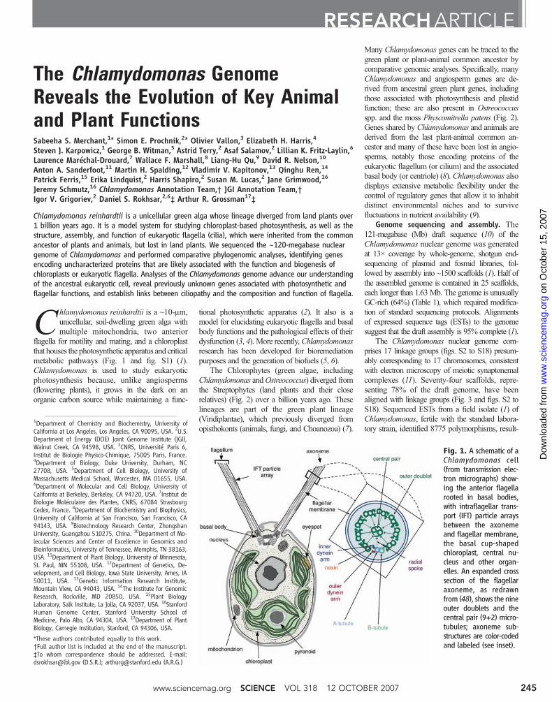
Chlamydomonas reinhardtii is a ~10-mm,unicellular, soil-dwelling green alga withmultiple mitochondria, two anterior
flagella for motility and mating, and a chloroplastthat houses the photosynthetic apparatus and criticalmetabolic pathways (Fig. 1 and fig. S1) (1).Chlamydomonas is used to study eukaryoticphotosynthesis because, unlike angiosperms(flowering plants), it grows in the dark on anorganic carbon source while maintaining a func-
tional photosynthetic apparatus (2). It also is amodel for elucidating eukaryotic flagella and basalbody functions and the pathological effects of theirdysfunction (3, 4). More recently,Chlamydomonasresearch has been developed for bioremediationpurposes and the generation of biofuels (5, 6).
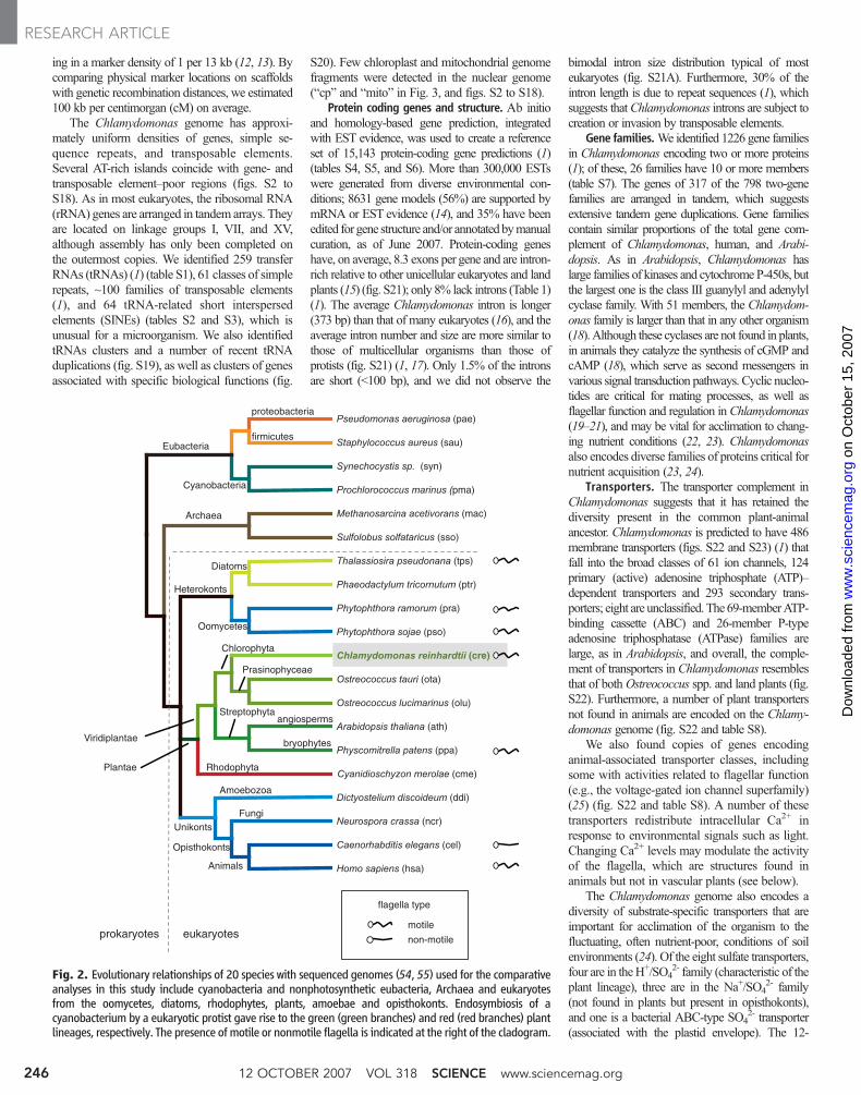
The Chlorophytes (green algae, includingChlamydomonas andOstreococcus) diverged fromthe Streptophytes (land plants and their closerelatives) (Fig. 2) over a billion years ago. Theselineages are part of the green plant lineage(Viridiplantae), which previously diverged fromopisthokonts (animals, fungi, and Choanozoa) (7).
Many Chlamydomonas genes can be traced to thegreen plant or plant-animal common ancestor bycomparative genomic analyses. Specifically, manyChlamydomonas and angiosperm genes are de-rived from ancestral green plant genes, includingthose associated with photosynthesis and plastidfunction; these are also present in Ostreococcusspp. and the moss Physcomitrella patens (Fig. 2).Genes shared by Chlamydomonas and animals arederived from the last plant-animal common an-cestor and many of these have been lost in angio-sperms, notably those encoding proteins of theeukaryotic flagellum (or cilium) and the associatedbasal body (or centriole) (8). Chlamydomonas alsodisplays extensive metabolic flexibility under thecontrol of regulatory genes that allow it to inhabitdistinct environmental niches and to survivefluctuations in nutrient availability (9).
Genome sequencing and assembly. The121-megabase (Mb) draft sequence (10) of theChlamydomonas nuclear genome was generatedat 13× coverage by whole-genome, shotgun end-sequencing of plasmid and fosmid libraries, fol-lowed by assembly into ~1500 scaffolds (1). Half ofthe assembled genome is contained in 25 scaffolds,each longer than 1.63Mb. The genome is unusuallyGC-rich (64%) (Table 1), which required modifica-tion of standard sequencing protocols. Alignmentsof expressed sequence tags (ESTs) to the genomesuggest that the draft assembly is 95% complete (1).
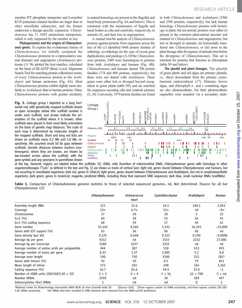
The Chlamydomonas nuclear genome com-prises 17 linkage groups (figs. S2 to S18) presum-ably corresponding to 17 chromosomes, consistentwith electron microscopy of meiotic synaptonemalcomplexes (11). Seventy-four scaffolds, repre-senting 78% of the draft genome, have beenaligned with linkage groups (Fig. 3 and figs. S2 toS18). Sequenced ESTs from a field isolate (1) ofChlamydomonas, fertile with the standard labora-tory strain, identified 8775 polymorphisms, result-
RESEARCHARTICLE
1Department of Chemistry and Biochemistry, University ofCalifornia at Los Angeles, Los Angeles, CA 90095, USA. 2U.S.Department of Energy (DOE) Joint Genome Institute (JGI),Walnut Creek, CA 94598, USA. 3CNRS, Université Paris 6,Institut de Biologie Physico-Chimique, 75005 Paris, France.4Department of Biology, Duke University, Durham, NC27708, USA. 5Department of Cell Biology, University ofMassachusetts Medical School, Worcester, MA 01655, USA.6Department of Molecular and Cell Biology, University ofCalifornia at Berkeley, Berkeley, CA 94720, USA. 7Institut deBiologie Moléculaire des Plantes, CNRS, 67084 StrasbourgCedex, France. 8Department of Biochemistry and Biophysics,University of California at San Francisco, San Francisco, CA94143, USA. 9Biotechnology Research Center, ZhongshanUniversity, Guangzhou 510275, China. 10Department of Mo-lecular Sciences and Center of Excellence in Genomics andBioinformatics, University of Tennessee, Memphis, TN 38163,USA. 11Department of Plant Biology, University of Minnesota,St. Paul, MN 55108, USA. 12Department of Genetics, De-velopment, and Cell Biology, Iowa State University, Ames, IA50011, USA. 13Genetic Information Research Institute,Mountain View, CA 94043, USA. 14The Institute for GenomicResearch, Rockville, MD 20850, USA. 15Plant BiologyLaboratory, Salk Institute, La Jolla, CA 92037, USA. 16StanfordHuman Genome Center, Stanford University School ofMedicine, Palo Alto, CA 94304, USA. 17Department of PlantBiology, Carnegie Institution, Stanford, CA 94306, USA.
*These authors contributed equally to this work.†Full author list is included at the end of the manuscript.‡To whom correspondence should be addressed. E-mail:[email protected] (D.S.R.); [email protected] (A.R.G.)
Fig. 1. A schematic of aChlamydomonas cell(from transmission elec-tron micrographs) show-ing the anterior flagellarooted in basal bodies,with intraflagellar trans-port (IFT) particle arraysbetween the axonemeand flagellar membrane,the basal cup-shapedchloroplast, central nu-cleus and other organ-elles. An expanded crosssection of the flagellaraxoneme, as redrawnfrom (48), shows the nineouter doublets and thecentral pair (9+2) micro-tubules; axoneme sub-structures are color-codedand labeled (see inset).
www.sciencemag.org SCIENCE VOL 318 12 OCTOBER 2007 245
on
Oct
ober
15,
200
7 w
ww
.sci
ence
mag
.org
Dow
nloa
ded
from
ing in a marker density of 1 per 13 kb (12, 13). Bycomparing physical marker locations on scaffoldswith genetic recombination distances, we estimated100 kb per centimorgan (cM) on average.
The Chlamydomonas genome has approxi-mately uniform densities of genes, simple se-quence repeats, and transposable elements.Several AT-rich islands coincide with gene- andtransposable element–poor regions (figs. S2 toS18). As in most eukaryotes, the ribosomal RNA(rRNA) genes are arranged in tandem arrays. Theyare located on linkage groups I, VII, and XV,although assembly has only been completed onthe outermost copies. We identified 259 transferRNAs (tRNAs) (1) (table S1), 61 classes of simplerepeats, ~100 families of transposable elements(1), and 64 tRNA-related short interspersedelements (SINEs) (tables S2 and S3), which isunusual for a microorganism. We also identifiedtRNAs clusters and a number of recent tRNAduplications (fig. S19), as well as clusters of genesassociated with specific biological functions (fig.
S20). Few chloroplast and mitochondrial genomefragments were detected in the nuclear genome(“cp” and “mito” in Fig. 3, and figs. S2 to S18).
Protein coding genes and structure. Ab initioand homology-based gene prediction, integratedwith EST evidence, was used to create a referenceset of 15,143 protein-coding gene predictions (1)(tables S4, S5, and S6). More than 300,000 ESTswere generated from diverse environmental con-ditions; 8631 gene models (56%) are supported bymRNA or EST evidence (14), and 35% have beenedited for gene structure and/or annotatedbymanualcuration, as of June 2007. Protein-coding geneshave, on average, 8.3 exons per gene and are intron-rich relative to other unicellular eukaryotes and landplants (15) (fig. S21); only 8% lack introns (Table 1)(1). The average Chlamydomonas intron is longer(373 bp) than that of many eukaryotes (16), and theaverage intron number and size are more similar tothose of multicellular organisms than those ofprotists (fig. S21) (1, 17). Only 1.5% of the intronsare short (<100 bp), and we did not observe the
bimodal intron size distribution typical of mosteukaryotes (fig. S21A). Furthermore, 30% of theintron length is due to repeat sequences (1), whichsuggests thatChlamydomonas introns are subject tocreation or invasion by transposable elements.
Gene families.We identified 1226 gene familiesin Chlamydomonas encoding two or more proteins(1); of these, 26 families have 10 or more members(table S7). The genes of 317 of the 798 two-genefamilies are arranged in tandem, which suggestsextensive tandem gene duplications. Gene familiescontain similar proportions of the total gene com-plement of Chlamydomonas, human, and Arabi-dopsis. As in Arabidopsis, Chlamydomonas haslarge families of kinases and cytochrome P-450s, butthe largest one is the class III guanylyl and adenylylcyclase family. With 51 members, the Chlamydom-onas family is larger than that in any other organism(18). Although these cyclases are not found in plants,in animals they catalyze the synthesis of cGMP andcAMP (18), which serve as second messengers invarious signal transduction pathways. Cyclic nucleo-tides are critical for mating processes, as well asflagellar function and regulation inChlamydomonas(19–21), and may be vital for acclimation to chang-ing nutrient conditions (22, 23). Chlamydomonasalso encodes diverse families of proteins critical fornutrient acquisition (23, 24).
Transporters. The transporter complement inChlamydomonas suggests that it has retained thediversity present in the common plant-animalancestor. Chlamydomonas is predicted to have 486membrane transporters (figs. S22 and S23) (1) thatfall into the broad classes of 61 ion channels, 124primary (active) adenosine triphosphate (ATP)–dependent transporters and 293 secondary trans-porters; eight are unclassified.The 69-memberATP-binding cassette (ABC) and 26-member P-typeadenosine triphosphatase (ATPase) families arelarge, as in Arabidopsis, and overall, the comple-ment of transporters in Chlamydomonas resemblesthat of bothOstreococcus spp. and land plants (fig.S22). Furthermore, a number of plant transportersnot found in animals are encoded on the Chlamy-domonas genome (fig. S22 and table S8).
We also found copies of genes encodinganimal-associated transporter classes, includingsome with activities related to flagellar function(e.g., the voltage-gated ion channel superfamily)(25) (fig. S22 and table S8). A number of thesetransporters redistribute intracellular Ca2+ inresponse to environmental signals such as light.Changing Ca2+ levels may modulate the activityof the flagella, which are structures found inanimals but not in vascular plants (see below).
The Chlamydomonas genome also encodes adiversity of substrate-specific transporters that areimportant for acclimation of the organism to thefluctuating, often nutrient-poor, conditions of soilenvironments (24). Of the eight sulfate transporters,four are in theH+/SO4
2- family (characteristic of theplant lineage), three are in the Na+/SO4
2- family(not found in plants but present in opisthokonts),and one is a bacterial ABC-type SO4
2- transporter(associated with the plastid envelope). The 12-
Fig. 2. Evolutionary relationships of 20 species with sequenced genomes (54, 55) used for the comparativeanalyses in this study include cyanobacteria and nonphotosynthetic eubacteria, Archaea and eukaryotesfrom the oomycetes, diatoms, rhodophytes, plants, amoebae and opisthokonts. Endosymbiosis of acyanobacterium by a eukaryotic protist gave rise to the green (green branches) and red (red branches) plantlineages, respectively. The presence ofmotile or nonmotile flagella is indicated at the right of the cladogram.
12 OCTOBER 2007 VOL 318 SCIENCE www.sciencemag.org246
RESEARCH ARTICLE
on
Oct
ober
15,
200
7 w
ww
.sci
ence
mag
.org
Dow
nloa
ded
from
member PiT phosphate transporter and 6-memberKUP potassium channel families are larger than inother unicellular eukaryotes, and the formerunderwent a lineage-specific expansion. Chlamy-domonas has 11 AMT ammonium transporters,which is only surpassed by the number in rice.
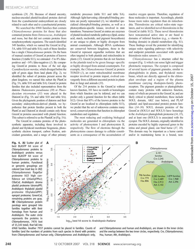
Phylogenomics and the origins of Chlamydom-onas genes. To explore the evolutionary history ofChlamydomonas, we initially compared theChlamydomonas proteome to a representative ani-mal (human) and angiosperm (Arabidopsis) pro-teome (1). We plotted the best matches, calculatedon the basis of BLASTP (Basic Local AlignmentSearch Tool for searching protein collections) scores,of every Chlamydomonas protein to the Arabi-dopsis and human proteomes (Fig. 4A). MostChlamydomonas proteins exhibit slightlymore sim-ilarity to Arabidopsis than to human proteins.ManyChlamydomonas proteins with greater similarity
to animal homologs are present in the flagellar andbasal body proteomes (Fig. 4A and below). This isconsistent with the maintenance of flagella andbasal bodies as cilia and centrioles, respectively, inanimals (8), and their loss in angiosperms.
A mutual best-hit analysis of Chlamydomonasproteins against proteins from organisms across thetree of life (1) identified 6968 protein families oforthologs, co-orthologs (in the case of recent geneduplications), and paralogs (1). Of theChlamydom-onas proteins, 2489 were homologous to proteinsfrom both Arabidopsis and humans (Fig. 4B).Chlamydomonas and humans shared 706 proteinfamilies (774 and 806 proteins, respectively), butthese were not shared with Arabidopsis. Thesegenes were either lost or diverged beyond recog-nition in green plants (table S9), and are enrichedfor sequences encoding cilia and centriole proteins(8, 26). Conversely, 1879 protein families are found
in both Chlamydomonas and Arabidopsis (1968and 2396 proteins, respectively), but lack humanhomologs. Chlamydomonas proteins with homol-ogy to plant, but not animal, proteins were either (i)present in the common plant-animal ancestor andretained in Chlamydomonas and angiosperms, butlost or diverged in animals; (ii) horizontally trans-ferred into Chlamydomonas; or (iii) arose in theplant lineage after divergence of animals (but beforethe divergence of Chlamydomonas). This set isenriched for proteins that function in chloroplasts(table S9 and below).
The plastid and plant lineages. The plastidsof green plants and red algae are primary plastids,i.e., direct descendants from the primary cyano-bacterial endosymbiont (27). Diatoms, brownalgae, and chlorophyll a– and c–containing algaeare also photosynthetic, but their photosyntheticorganelles were acquired via a secondary endo-
Table 1. Comparison of Chlamydomonas genome statistics to those of selected sequenced genomes. nd, Not determined. [Source for all butChlamydomonas (1)]
Chlamydomonas Ostreococcustauri
Cyanidioschyzon Arabidopsis Human
Assembly length (Mb) 121 12.6 16.5 140.1 2,851Coverage 13× 6.7× 11× nd ~8×Chromosomes 17 20 20 5 23G+C (%) 64 58 55 36 41G+C (%) coding sequence 68 59 57 44 52Gene number 15,143 8,166 5,331 26,341 ~23,000Genes with EST support (%) 63 36 86 60 ndGene density (per kb) 0.125 0.648 0.323 0.190 ~0.0008Average bp per gene 4312 nd 1553 2232 27,000Average bp per transcript 1580 1257 1552 nd ndAverage number of amino acids per polypeptide 444 387 518 413 491Average number of exons per gene 8.33 1.57 1.005 5.2 8.8Average exon length 190 750 1540 251 282*Genes with introns (%) 92 39 0.5 79 85†Mean length of intron 373 103 248 164 3,365Coding sequence (%) 16.7 81.6 44.9 33.0 ~1Number of rDNA units (28S/18S/5.8S + 5S) 3 + 3 4 + 4 3 + 3‡ 12 + 700 5 + ndNumber tRNAs 259§ nd 30 589 497Selenocysteine (Sec) tRNAs 1 nd nd 0 1*National Center for Biotechnology Information (NIH) NCBI 36 from Ensembl build 38. †[Source (56)]. ‡Three regions contain 5S rDNA exclusively, and three regions contain 28S-18S-5.8S rDNAs exclusively. §65 tRNAs that were included in SINE elements were removed from the tRNA-scanSE predictions.
Fig. 3. Linkage group I depicted as a long hori-zontal rod, with genetically mapped scaffolds shownas open rectangles below (the scaffold number isunder each scaffold, and arrows indicate the ori-entation of the scaffold where it is known; otherscaffolds were placed in their most likely orientationon the basis of genetic map distances. The scale ofeach map is determined by molecular lengths ofthe mapped scaffolds. Short and long red ticks aredrawn on scaffolds every 0.2 Mb and 1.0 Mb, re-spectively. We assumed small 50 kb gaps betweenscaffolds. Genetic distances between markers (cen-timorgans), where they are known, are shown bytwo-headed arrows above the scaffold, with thegene symbol and any synonyms in parentheses shownat the top. Genomic regions are labeled below the scaffolds: 5S, rDNA, mito (insertion of mitochondrial DNA). Chlamydomonas genes with homologs in otherorganisms/lineages (“Cuts” as defined in the text and Fig. 5) are shown as tracks of vertical bars: light red, genes shared between Chlamydomonas and humans, butnot occurring in nonciliated organisms; dark red, genes in CiliaCut; light green, genes shared between Chlamydomonas and Arabidopsis, but not in nonphotosyntheticorganisms; dark green, genes in GreenCut; magenta, predicted tRNAs, including those that represent SINE sequences; dark blue, small nucleolar RNAs (snoRNAs).
www.sciencemag.org SCIENCE VOL 318 12 OCTOBER 2007 247
RESEARCH ARTICLE
on
Oct
ober
15,
200
7 w
ww
.sci
ence
mag
.org
Dow
nloa
ded
from
symbiosis (28, 29). Because of shared ancestry,nucleus-encoded plastid-localized proteins derivedfrom the cyanobacterial endosymbiont are closelyrelated to each other and to cyanobacterial proteins.
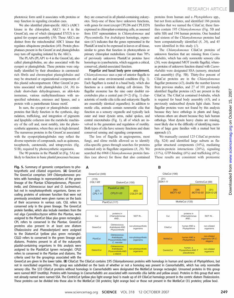
We searched the 6968 families that containChlamydomonas proteins for those that alsocontained proteins fromOstreococcus, Arabidopsisand moss, but that did not contain proteins fromnonphotosynthetic organisms. The search identified349 families, which we named the GreenCut (Fig.5A, table S10 and table SA); each of these familieshas a single Chlamydomonas protein. On the basisof manual curation of GreenCut proteins of knownfunction (1) (table S11), we estimated ~5 to 8% false-positives and ~14% false-negatives (1). By compar-ing GreenCut proteins to those of the red algaCyanidioschyzonmerolae, which diverged before thesplit of green algae from land plants (Fig. 2), weidentified the subset of proteins present across theplant kingdom; we named this subset the PlantCut(Fig. 5A, table S10 and table SA). GreenCut proteinfamilies that also included representatives from thediatoms Thalassiosira pseudonana (30) or Phaeo-dactylum tricornutum (31) were placed in theDiatomCut (Fig. 5A and table S10 and table SA).Given the phylogenetic position of diatoms and theirsecondary endosymbiosis-derived plastids, we hy-pothesize that protein families present in both thePlantCut and DiatomCut should contain only thoseGreenCut proteins associated with plastid function.This subset is referred to as the PlastidCut (Fig. 5A).
The GreenCut contains proteins of the photo-synthetic apparatus, including those involved inplastid and thylakoid membrane biogenesis, photo-synthetic electron transport, carbon fixation, anti-oxidant generation, and a range of other primary
metabolic processes (table S11 and table SA).Although light-harvesting chlorophyll-binding pro-teins are poorly represented (1), we identified spe-cialized chlorophyll-binding proteins, as well as aphotosynthesis-specific kinase, involved in statetransitions. NumerousGreenCut entries are enzymesof plastid-localizedmetabolic pathways (lipid, aminoacid, starch, nucleotide, and pigment biosynthesis)or are unique to plants or highly divergent fromanimal counterparts. Although tRNA synthetasesare conserved between kingdoms, those in theGreenCut represent organellar isoforms that areoften targeted to both plastids and mitochondria inplants (32). GreenCut proteins that do not functionin the plastids tend to be green lineage–specificor highly diverged from animal counterparts. Forexample, the Chlamydomonas GreenCut proteinTOM20 (1), an outer mitochondrial membranereceptor involved in protein import, evolved con-vergently from a different ancestral protein in plantsthan in fungi and animals (33).
Of the 214 proteins in the GreenCut withoutknown function, 101 have no motifs or homologiesfrom which function can be inferred, and we canpredict only a general function for the others (tableS12). Given that 85% of the known proteins in theGreenCut are localized to chloroplasts (table S13),we predict that the set of unknowns contains manynovel, conserved proteins that function in chloroplastmetabolism and regulation.
The most reducing and oxidizing biologicalmolecules are generated in chloroplasts via theactivity of photosystem I and photosystem II,respectively. The flow of electrons through thephotosystems causes damage to cellular constit-uents as a consequence of the accumulation of
reactive oxygen species. Therefore, regulation ofthese molecules is important. Accordingly, plastidshouse more redox regulators than do mitochon-dria. Thioredoxins are critical redox-state regu-lators, and we identified novel thioredoxins in theGreenCut (table S12). These novel thioredoxinshave noncanonical active sites or are fused todomains of inferred function (e.g., a vitamin K–binding domain) in plastid metabolism (fig. S1).These findings reveal the potential for identifyingunique redox signaling pathways with selectivityand midpoint potentials associated with specificthioredoxin redox sensors (1).
Chlamydomonas has a structure called theeyespot (Fig. 1) which can sense light and triggerphototactic responses. The eyespot is composedof several layers of pigment granules, similar toplastoglobules in plants, and thylakoid mem-brane, which are directly apposed to the chloro-plast envelope and a region of the plasmamembrane carrying rhodopsin-family photo-receptors. The pigment granules or plastoglobulescontain many proteins with unknown function,many of which are present in theGreenCut, and arelikely critical to plastid metabolism; these includeSOUL domain, AKC (see below), and PLAP(plastid- and lipid-associated protein) protein fam-ilies (34–36). SOUL domain proteins of theGreenCut (SOUL4 and SOUL5) have homologsin theArabidopsis plastoglobule proteome (34, 35),and at least one (SOUL3) is associated with theeyespot. The SOULdomain, originally identified inproteins encoded by highly expressed genes in theretina and pineal gland, can bind heme (37, 38).This domain may be important as a heme carrierand/or in maintaining heme in a bound, non-
Fig. 4. (A) Scatter plot ofbest BLASTP hit score ofChlamydomonas proteins toArabidopsis proteins versusbest BLASTP hit score ofChlamydomonas proteins tohuman proteins. Functionalor genomic groupings arecolored [see inset key in (A)]:Chlamydomonas flagellarproteome (42) high con-fidence set (chlamyFPhc);CiliaCut; Arabidopsis stromaplastid proteome (stromaPP);Arabidopsis thylakoid plastidproteome (thylakoidPP);eyespot proteome; GreenCut;remaining proteins are gray.(B) Chlamydomonas proteinparalogs were grouped intofamilies together with theirhomologs from human andArabidopsis. The outer circlerepresents the proteins inChlamydomonas, 7476 (outof 15,143 total), that fall into6968 families. Another 7937 proteins cannot be placed in families. Counts offamilies (and the numbers of proteins from each species in them) with proteinsfrom Chlamydomonas and human only, Chlamydomonas and Arabidopsis only,
and Chlamydomonas and human and Arabidopsis, are shown in the inner circlesand the overlap between the two inner circles, respectively. Cre, Chlamydomonas;Hsa, human; Ath, Arabidopsis.
12 OCTOBER 2007 VOL 318 SCIENCE www.sciencemag.org248
RESEARCH ARTICLE
on
Oct
ober
15,
200
7 w
ww
.sci
ence
mag
.org
Dow
nloa
ded
from
phototoxic form until it associates with proteins ormay function in signaling circadian cues.
We also identified plant-specific AKCs (ABC1kinase in the chloroplast, AKC1 to 4 in theGreenCut), one of which (designated EYE3) is re-quired for eyespot assembly (39). These AKCs aredistinct from the mitochondrial ABC1 kinase thatregulates ubiquinone production (40). Protein phos-phatases present in the GreenCut and plastoglobulesmay turn off signaling initiated by the AKCs.
The PLAPs (PLAP1 to 4 in the GreenCut), alsocalled plastoglobulins, are also associated with theeyespot or plastoglobule. These proteins were orig-inally identified by their abundance in carotenoid-rich fibrils and chromoplast plastoglobules andmay be structural or organizational components ofthis plastid subcompartment. Other GreenCut pro-teins associated with plastoglobules (34, 36) in-clude short-chain dehydrogenases, an aldo-ketoisomerase, various methyltransferases with un-specified substrates, esterases and lipases, and aprotein with a pantothenate kinase motif.
In sum, the eyespot or plastoglobules containproteins that likely function in the synthesis, deg-radation, trafficking, and integration of pigmentsand lipophilic cofactors into the metabolic machin-ery of the cell and, most notably, into the photo-synthetic apparatus,where they are in high demand.The numerous proteins in the GreenCut associatedwith the eyespot/plastoglobules may reflect thediverse repertoire of compounds, such as quinones,tocopherols, carotenoids, and tetrapyrroles (fig.S1B), required by photosynthetic organisms.
The 90 proteins in the PlastidCut (Fig. 5A) arelikely to function in basic plastid processes because
they are conserved in all plastid-containing eukary-otes. Sixty-one of these have unknown functions,with genes for most (except CPLD6 and CPLD29)expressed in chloroplast-containing cells, as assessedfrom EST representation in Chlamydomonas andPhyscomitrella. For Arabidopsis homologs, expres-sion (41) indicates that the genes represented in thePlastidCut tend to be expressed in leaves or all tissue,similar to genes that function in photosynthesis orprimary chloroplast metabolism. Greater than 70%of previously unknown PlastidCut proteins havehomologs in cyanobacteria,which suggests a critical,conserved, plastid-associated function.
Flagellar and basal body gene complement.Chlamydomonas uses a pair of anterior flagella toswim and sense environmental conditions (Fig. 1).Each flagellum is rooted in a basal body, which alsofunctions as a centriole during cell division. Theflagellar axoneme has the nine outer doublet mi-crotubules plus a central pair (9+2) (Fig. 1) char-acteristic of motile cilia (cilia and eukaryotic flagellaare essentially identical organelles). In addition tomotile cilia, animals contain nonmotile cilia thatfunction as a sensory organelle and typically lackouter and inner dynein arms, radial spokes, andcentral microtubules (Fig. 1), all of which are in-volved in the generation and regulation of motility.Both types of cilia have sensory functions and shareconserved sensing and signaling components.
The loss of flagella in angiosperms, mostfungi, and slime molds allowed us to identifycilia-specific genes through searches for proteinsretained only in flagellate organisms (8, 26). Wesearched the 6968 Chlamydomonas protein fam-ilies (see above) for those that also contained
proteins from human and a Phytophthora spp.,but not from aciliates, and identified 186 proteinfamilies that we named the CiliaCut; these fam-ilies contain 195 Chlamydomonas (Fig. 5B andtable SB) and 194 human proteins. One hundredand sixteen of the Chlamydomonas proteins hadbeen computationally identified (8, 26), and 45were identified in this study (1).
The Chlamydomonas CiliaCut proteins ofunknown function that are missing from Caeno-rhabditis, which has only nonmotile sensory cilia(26), were designated MOT (motile flagella), where-as proteins of unknown function shared withCaeno-rhabditis were designated SSA (sensory, structuraland assembly) (Fig. 5B). Thirty-five percent ofCiliaCut proteins are in the Chlamydomonasflagellar proteome (42), double the number knownfrom previous studies, and 27 of 101 previouslyidentified flagellar proteins (42) are present in theCiliaCut. The CiliaCut contained d-tubulin, whichis required for basal body assembly (43), and apreviously undescribed dynein light chain. Someflagellar proteins were not found by this analysisbecause they have orthologs in plants and fungi,whereas others are absent because they lack humanorthologs. Most dynein heavy chains are missing,most likely due to the difficulty of identifying mem-bers of large gene families with a mutual best hitapproach (1).
We manually curated 125 CiliaCut proteins(fig. S24) and identified large subsets as fla-gellar structural components (16%), mediatingprotein-protein interactions (26%), signaling(11%), GTP-binding (6%) and trafficking (6%).These results are consistent with proteomic
Fig. 5. Summary of genomic comparisons to pho-tosynthetic and ciliated organisms. (A) GreenCut:The GreenCut comprises 349 Chlamydomonas pro-teins with homologs in representatives of the greenlineage of the Plantae (Chlamydomonas, Physcomi-trella, and Ostreococcus tauri and O. lucimarinus),but not in nonphotosynthetic organisms. Genes en-coding proteins of unknown function that were notpreviously annotated were given names on the basisof their occurrence in various cuts. CGL refers toconserved only in the green lineage. The GreenCutprotein families, which also include members from thered alga Cyanidioschyzon within the Plantae, wereassigned to the PlantCut (blue plus green rectangles).CPL refers to conserved in the Plantae. GreenCutproteins also present in at least one diatom(Thalassiosira and Phaeodactylum) were assignedto the DiatomCut (yellow plus green rectangle).CGLD refers to conserved in the green lineage anddiatoms. Proteins present in all of the eukaryoticplastid-containing organisms in this analysis wereassigned to the PlastidCut (green rectangle). CPLDrefers to conserved in the Plantae and diatoms. Thecriteria used for the groupings associated with theGreenCut are given in the lower table. (B) CiliaCut: The CiliaCut contains 195 Chlamydomonas proteins with homologs in human and species of Phytophthora, butnot in nonciliated organisms. This group was subdivided on the basis of whether or not a homolog was present in Caenorhabditis, which has only nonmotilesensory cilia. The 133 CiliaCut proteins without homologs in Caenorhabditis were designated the MotileCut (orange rectangle). Unnamed proteins in this groupwere named MOT (motility). Proteins with homologs in Caenorhabditis are associated with nonmotile cilia (white and yellow areas). Proteins in this group that werenot already named were named SSA. The CentricCut (yellow plus light orange box) is made up of 69 CiliaCut homologs present in the centric diatom Thalassiosira.These proteins can be divided into those also in the MotileCut (38 proteins; light orange box) or those not present in the MotileCut (31 proteins; yellow box).
A B
CiliaCut (195)
CentricCut (69)
MotileCut (133)
(38)MOT43-55
(31)SSA12-20
(95)MOT1-42
(31)SSA1-11
Cutproteins in
ChlamydomonasAND...
NOT IN...
GreenCutOstreococcus,
Arabidopsis ANDPhyscomitrella
non-photosyntheticorganisms
PlantCut
Ostreococcus,Arabidopsis,
Physcomitrella ANDC. merolae
non-photosyntheticorganisms
DiatomCut
Ostreococcus,Arabidopsis,
Physcomitrella ANDa diatom
non-photosyntheticorganisms
Cutproteins in
ChlamydomonasAND...
NOT IN...
CiliaCuthuman AND a Phytophthora
non-ciliatedorganisms
MotileCuthuman AND a Phytophthora
non-ciliatedorganisms OR C.
elegans
CentricCuthuman, a
Phytophthora AND T.pseudonana
non-ciliatedorganisms
GreenCut (349)
DiatomCut (150)
PlantCut (117)
CPLD1-53(60)
CGLD1-30(27)
CPL1-11
(172)CGL1-83
PlastidCut (90)
www.sciencemag.org SCIENCE VOL 318 12 OCTOBER 2007 249
RESEARCH ARTICLE
on
Oct
ober
15,
200
7 w
ww
.sci
ence
mag
.org
Dow
nloa
ded
from
analysis of the flagellum (42) and highlight theimportance of signaling even in motile flagella.
The 62 CiliaCut proteins that Chlamydomonasshares with Caenorhabditis are predicted to havestructural, sensory, or assembly roles in the cilium.As expected, the 133CiliaCut proteinsmissing fromCaenorhabditis (Fig. 5B) (1), designated theMotileCut, include a number of proteins associatedwithmotility (42) (table S14). This data set also con-tains 31 proteins of unknown function found in theflagellar and basal body proteomes, 36 known butuncharacterized proteins, and 55 novel proteins(designated MOT1 to MOT55); these flagellarproteins are all predicted to be involved specifi-cally in motility.
A comparison of CiliaCut proteins with proteinsencoded by the Physcomitrella genome indi-cates that Physcomitrella has lost five of the outerdynein arm proteins (Fig. 1, table S14). However,Physcomitrella contains inner dynein arm subunitsIDA4 and DHC2, as well as subunits of the centralmicrotubules, the radial spokes, and the dynein reg-ulatory complex (table S14). From this we concludethat Physcomitrella sperm flagella have a “9+2”axoneme containing inner dynein arms, centralmicrotubules, and radial spokes, but lack theouter dynein arms. Although the structure of thePhyscomitrella sperm flagellum is not known,sperm flagella of the bryalean moss Aulacomniumpalustre have just such an axoneme (44).
In contrast, the motile flagella of centricdiatoms lack the central pair of microtubules(45, 46). Orthologs of 69 of the 195 CiliaCut pro-teins (namedCentricCut, Fig. 5B)were predicted tobe present in the centric diatom Thalassiosira. Asexpected, Thalassiosira lacks all central pairproteins. However, it also lacks all radial spokeand inner dynein arm proteins, but hasmost of theouter dynein arm proteins. The contrastingpatterns of loss of axonemal structures predictedforPhyscomitrella and Thalassiosira suggest thatthe central pair and radial spokes function as aunit with the inner arms, but are dispensable forthe generation of motility by the outer arms.
Intraflagellar transport (IFT), which is conservedin ciliated organisms except malaria parasites (47), isessential for flagellar growth (48). The IFT machin-ery consists of at least 16 proteins in two complexes(A and B) that are moved in anterograde and retro-grade directions by the molecular motors kinesin-2and cytoplasmic dynein 1b, respectively (Fig. 1).Our analysis of Thalassiosira reveals that it hascomponents of the anterograde motor and complexB, but has lost the retrograde motor and complex A(table S14). This is intriguing, as retrograde IFT isessential for flagellar maintenance in Chlamydomo-nas (49) and is important for recycling IFT com-ponents (50). In addition, both Physcomitrella andThalassiosira have lost the Bardet-Biedl syndrome(BBS) genes.BBSgene products are associatedwiththe basal body inChlamydomonas and mammals(8, 51) and sensory cilia in Caenorhabditis (52),where they may be involved in IFT (53).
We searched the CiliaCut proteins for proteinssharedwithOstreococcus spp., a green alga lacking a
flagellate stage. The Ostreococcus spp. retain 46(24%) of the 195 CiliaCut proteins but, consistentwith loss of the flagellum, are missing genesencoding the IFT-particle proteins and motors, theinner and outer dynein arm proteins, the radial spokeand central pair proteins, and 32 out of 39 flagella-associated proteins (FAPs) (table S14). They havealso lost many genes encoding basal body proteins,including all BBS proteins (table S14), which sug-gests thatOstreococcus also lack basal bodies. How-ever, Ostreococcus spp. have retained many otherCiliaCut proteins (table S14), which suggests eitherthat they recently lost their flagella, or that theyretained flagellar proteins for other cellular functions.
Conclusions. This analysis of the Chlamy-domonas genome sheds light on the nature of thelast common ancestor of plants and animals andidentifies many cilia- and plastid-related genes. Thegene complement also provides insights into life inthe soil environment where extreme competition fornutrients likely drove expansion of transporter genefamilies, as well as sensory flagellar and eyespotfunctions (e.g., facilitating nutrient acquisition andoptimization of the light environment). As more ofthe ecology and physiology ofChlamydomonas andother unicellular algae are explored, additional directlinks between gene content and functions associatedwith the soil life-style will be unmasked with in-creased potential for biotechnological exploita-tion of these functions.
References and Notes1. Materials and methods and supplemental online (SOM) text
are available as supporting material on Science Online.2. E. H. Harris, Annu. Rev. Plant Physiol. Plant Mol. Biol.
52, 363 (2001).3. L. C. Keller, E. P. Romijn, I. Zamora, J. R. Yates 3rd,
W. F. Marshall, Curr. Biol. 15, 1090 (2005).4. G. J. Pazour, N. Agrin, B. L. Walker, G. B. Witman, J. Med.
Genet. 43, 62 (2006).5. C. Vilchez, I. Garbayo, E. Markvicheva, F. Galvan, R. Leon,
Bioresour. Technol. 78, 55 (2001).6. M. L. Ghirardi et al., Annu. Rev. Plant Biol. 58, 71 (2007).7. H. S. Yoon, J. D. Hackett, C. Ciniglia, G. Pinto,
D. Bhattacharya, Mol. Biol. Evol. 21, 809 (2004).8. J. B. Li et al., Cell 117, 541 (2004).9. A. R. Grossman et al., Curr. Opin. Plant Biol. 10, 190 (2007).10. Chlamydomonas reinhardtii v 3.0, DOE Joint Genome
Institute, www.jgi.doe.gov/chlamy.11. R. Storms, P. J. Hastings, Exp. Cell Res. 104, 39 (1977).12. P. Kathir et al., Eukaryot. Cell 2, 362 (2003).13. L. A. Rymarquis, J. M. Handley, M. Thomas, D. B. Stern,
Plant Physiol. 137, 557 (2005).14. M. Jain et al., Nucleic Acids Res. 35, 2074 (2007).15. Q. Yuan et al., Plant Physiol. 138, 18 (2005).16. M. Yandell et al., PLoS Comput Biol 2, e15 (2006).17. B. Palenik et al., Proc. Natl. Acad. Sci. U.S.A. 104, 7705
(2007).18. P. Schaap, Front. Biosci. 10, 1485 (2005).19. E. Hasegawa, H. Hayashi, S. Asakura, R. Kamiya, Cell
Motil. Cytoskeleton 8, 302 (1987).20. S. M. Pasquale, U. W. Goodenough, J. Cell Biol. 105,
2279 (1987).21. A. R. Gaillard, L. A. Fox, J. M. Rhea, B. Craige, W. S. Sale,
Mol. Biol. Cell 17, 2626 (2006).22. D. Gonzalez-Ballester, A. de Montaigu, J. J. Higuera,
A. Galvan, E. Fernandez, Plant Physiol. 137, 522 (2005).23. S. V. Pollock, W. Pootakham, N. Shibagaki, J. L. Moseley,
A. R. Grossman, Photosynth. Res. 86, 475 (2005).24. A. Grossman, H. Takahashi, Annu. Rev. Plant Physiol.
Plant Mol. Biol. 52, 163 (2001).25. S. Somlo, B. Ehrlich, Curr. Biol. 11(9), R356 (2001).26. T. Avidor-Reiss et al., Cell 117, 527 (2004).
27. M. W. Gray, Curr. Opin. Genet. Dev. 9, 678 (1999).28. D. Bhattacharya, H. S. Yoon, J. D. Hackett, Bioessays 26,
50 (2004).29. P. Keeling, Protist 155, 3 (2004).30. E. V. Armbrust et al., Science 306, 79 (2004).31. Phaeodactylum tricornutum, v2.0, DOE Joint Genome
Institute, www.jgi.doe.gov/phaeodactylum.32. A. M. Duchêne et al., Proc. Natl. Acad. Sci. U.S.A. 102,
16484 (2005).33. A. J. Perry, J. M. Hulett, V. A. Likic, T. Lithgow,
P. R. Gooley, Curr. Biol. 16, 221 (2006).34. A. J. Ytterberg, J. B. Peltier, K. J. van Wijk, Plant Physiol.
140, 984 (2006).35. M. Schmidt et al., Plant Cell 18, 1908 (2006).36. P. A. Vidi et al., J. Biol. Chem. 281, 11225 (2006).37. M. J. Zylka, S. M. Reppert, Brain Res. Mol. Brain Res. 74,
175 (1999).38. E. Sato et al., Biochemistry 43, 14189 (2004).39. M. R. Lamb, S. K. Dutcher, C. K. Worley, C. L. Dieckmann,
Genetics 153, 721 (1999).40. T. Q. Do, A. Y. Hsu, T. Jonassen, P. T. Lee, C. F. Clarke,
J. Biol. Chem. 276, 18161 (2001).41. P. Zimmermann, M. Hirsch-Hoffmann, L. Hennig,
W. Gruissem, Plant Physiol. 136, 2621 (2004).42. G. J. Pazour, N. Agrin, J. Leszyk, G. B. Witman, J. Cell Biol.
170, 103 (2005).43. E. T. O'Toole, T. H. Giddings, J. R. McIntosh,
S. K. Dutcher, Mol. Biol. Cell 14, 2999 (2003).44. D. L. Bernhard, K. S. Renzaglia, Bryologist 98, 52 (1995).45. I. Manton, K. Kowallik, H. A. von Stosch, J. Cell Sci. 6,
131 (1970).46. I. B. Heath, W. M. Darley, J. Phycol. 18, 51 (1972).47. L. J. Briggs, J. A. Davidge, B. Wickstead, M. L. Ginger, K.
Gull, Curr. Biol. 14, R611 (2004).48. J. L. Rosenbaum, G. B. Witman, Nat. Rev. Mol. Cell Biol.
3, 813 (2002).49. G. J. Pazour, B. L. Dickert, G. B. Witman, J. Cell Biol. 144,
473 (1999).50. H. Qin, D. R. Diener, S. Geimer, D. G. Cole, J. L. Rosenbaum,
J. Cell Biol. 164, 255 (2004).51. S. J. Ansley et al., Nature 425, 628 (2003).52. O. E. Blacque et al., Genes Dev. 18, 1630 (2004).53. G. Ou et al., Mol. Biol. Cell 18, 1554 (2007).54. F. D. Ciccarelli et al., Science 311, 1283 (2006).55. P. J. Keeling et al., Trends Ecol. Evol. 20, 670 (2005).56. L. Eichinger et al., Nature 435, 43 (2005).57. We thank R. Howson for help with drawing figures,
E. Begovic and S. Nicholls for comments on themanuscript. SM is supported by the grants NIHGM42143, DOE DE-FG02-04ER15529 USDA 2004-35318-1495. SP and DSR are funded by USDA and DOE,Joint Genome Institute. ARG is supported by USDA 2003-35100-13235, DOE DE-AC36-99GO10337 and the NSF-funded Chlamydomonas Genome Project, MCB 0235878.SJK was supported in part by a Ruth L. KirschsteinNational Research Service Award GM07185. The authorsdeclare they have no conflicts of interest. Genomeassembly together with predicted gene models and andannotations were deposited at DDBJ/EMBL/GenBankunder the project accession ABCN00000000. Sincemanual curation continues, some models or anotationsare changing and the latest set of gene models andannotations is available from www.jgi.doe.gov/chlamy.The most recent set, which includes a number of changescompared with the frozen set used for this analysis, wassubmitted as the first version, ABCN01000000.
Full author list and affiliationsSabeeha S. Merchant,1 Simon E. Prochnik,2 Olivier Vallon,3
Elizabeth H. Harris,4 Steven J. Karpowicz,1 George B.Witman,5 Astrid Terry,2 Asaf Salamov,2 Lillian K. Fritz-Laylin,6
Laurence Maréchal-Drouard,7 Wallace F. Marshall,8 Liang-HuQu,9 David R. Nelson,10 Anton A. Sanderfoot,11 Martin H.Spalding,12 Vladimir V. Kapitonov,13 Qinghu Ren,14 PatrickFerris,15 Erika Lindquist,2 Harris Shapiro,2 Susan M. Lucas,2
Jane Grimwood,16 Jeremy Schmutz,16 Igor V. Grigoriev,2
Daniel S. Rokhsar,2,6 Arthur R. Grossman17
Chlamydomonas Annotation Team. Pierre Cardol,3,18
Heriberto Cerutti,19 Guillaume Chanfreau,1 Chun-Long Chen,9
Valérie Cognat,7 Martin T. Croft,20 Rachel Dent,21 Susan
12 OCTOBER 2007 VOL 318 SCIENCE www.sciencemag.org250
RESEARCH ARTICLE
on
Oct
ober
15,
200
7 w
ww
.sci
ence
mag
.org
Dow
nloa
ded
from
Dutcher,22 Emilio Fernández,23 Patrick Ferris,15 HideyaFukuzawa,24 David González-Ballester,17 Diego González-Halphen,25 Armin Hallmann,26 Marc Hanikenne,18 MichaelHippler,27 William Inwood,21 Kamel Jabbari,28 MingKalanon,29 Richard Kuras,3 Paul A. Lefebvre,11 Stéphane D.Lemaire,30 Alexey V. Lobanov,31 Martin Lohr,32 AndreaManuell,33 Iris Meier,34 Laurens Mets,35 Maria Mittag,36 TelsaMittelmeier,37 James V. Moroney,38 Jeffrey Moseley,17 CarolynNapoli,39 Aurora M. Nedelcu,40 Krishna Niyogi,21 Sergey V.Novoselov,31 Ian T. Paulsen,14 Greg Pazour,41 Saul Purton,42
Jean-Philippe Ral,43 Diego Mauricio Riaño-Pachón,44 WayneRiekhof,45 Linda Rymarquis,46 Michael Schroda,47 DavidStern,48 James Umen,15 Robert Willows,49 Nedra Wilson,50
Sara Lana Zimmer,48 Jens Allmer,51 Janneke Balk,20 KaterinaBisova,52 Chong-Jian Chen,9 Marek Elias,53 Karla Gendler,39
Charles Hauser,54 Mary Rose Lamb,55 Heidi Ledford,21 JoanneC. Long,1 Jun Minagawa,56 M. Dudley Page,1 Junmin Pan,57
Wirulda Pootakham,17 Sanja Roje,58 Annkatrin Rose,59 EricStahlberg,34 Aimee M. Terauchi,1 Pinfen Yang,60 StevenBall,61 Chris Bowler,28,62 Carol L. Dieckmann,37 Vadim N.Gladyshev,31 Pamela Green,46 Richard Jorgensen,39 StephenMayfield,33 Bernd Mueller-Roeber,44 Sathish Rajamani,63
Richard T. Sayre34
JGI Annotation Team. Peter Brokstein,2 Inna Dubchak,2
David Goodstein,2 Leila Hornick,2 Y. Wayne Huang,2 JinalJhaveri,2 Yigong Luo,2 Diego Martínez,2 Wing Chi AbbyNgau,2 Bobby Otillar,2 Alexander Poliakov,2 Aaron Porter,2
Lukasz Szajkowski,2 Gregory Werner,2 Kemin Zhou2
1Department of Chemistry and Biochemistry, University ofCalifornia Los Angeles, Los Angeles, CA 90095, USA. 2U.S.Department of Energy, Joint Genome Institute, Walnut Creek,CA 94598, USA. 3CNRS, UMR 7141, CNRS/Université Paris 6,Institut de Biologie Physico-Chimique, 75005 Paris, France.4Department of Biology, Duke University, Durham, NorthCarolina 27708, USA. 5Department of Cell Biology, Universityof Massachusetts Medical School, Worcester, MA 01655, USA.6Department of Molecular and Cell Biology, University ofCalifornia at Berkeley, Berkeley, CA94720, USA. 7Institut deBiologie Moléculaire des Plantes, CNRS, 67084 StrasbourgCedex, France. 8Department of Biochemistry and Biophysics,University of California at San Francisco, San Francisco, CA94143, USA. 9Biotechnology Research Center, ZhongshanUniversity, Guangzhou 510275, China. 10Department ofMolecular Sciences and Center of Excellence in Genomics andBioinformatics, University of Tennessee, Memphis, TN 38163,USA. 11Department of Plant Biology, University of Minnesota,
St. Paul MN 55108, USA. 12Department of Genetics,Development, and Cell Biology, Iowa State University, Ames,IA 50011, USA. 13Genetic Information Research Institute,Mountain View, CA 94043, USA. 14The Institute for GenomicResearch, Rockville, MD 20850, USA. 15Plant BiologyLaboratory, Salk Institute, La Jolla, CA 92037, USA. 16StanfordHuman Genome Center, Stanford University School ofMedicine, Palo Alto, CA 94304, USA. 17Department of PlantBiology, Carnegie Institution, Stanford, CA 94306, USA.18Plant Biology Institute, Department of Life Sciences,University of Liège, B-4000 Liège, Belgium. 19University ofNebraska-Lincoln, School of Biological Sciences–Plant ScienceInitiative, Lincoln, NE 68588, USA. 20Department of PlantSciences, University of Cambridge, Cambridge CB2 3EA, UK.21Department of Plant and Microbial Biology, University ofCalifornia at Berkeley, Berkeley, CA 94720, USA.22Department of Genetics, Washington University School ofMedicine, St. Louis, MO 63110, USA. 23Departamento deBioquímica y Biología Molecular, Facultad de Ciencias,Universidad de Córdoba, Campus de Rabanales, 14071Córdoba, Spain. 24Graduate School of Biostudies, KyotoUniversity, Kyoto 606-8502, Japan. 25Departamento deGenética Molecular, Instituto de Fisiología Celular,Universidad Nacional Autónoma de México, México 04510 DF,Mexico. 26Department of Cellular and Developmental Biologyof Plants, University of Bielefeld, D-33615 Bielefeld,Germany. 27Department of Biology, Institute of PlantBiochemistry and Biotechnology, University of Münster,48143 Münster, Germany. 28CNRS UMR 8186, Départementde Biologie, Ecole Normale Supérieure, 75230 Paris, France.29Plant Cell Biology Research Centre, The School of Botany,The University of Melbourne, Parkville, Melbourne, VIC 3010,Australia. 30Institut de Biotechnologie des Plantes, UMR 8618,CNRS/Université Paris-Sud, Orsay, France. 31Department ofBiochemistry, N151 Beadle Center, University of Nebraska,Lincoln, NE 68588–0664, USA. 32Institut für AllgemeineBotanik, Johannes Gutenberg-Universität, 55099 Mainz,Germany. 33Department of Cell Biology and Skaggs Institutefor Chemical Biology, Scripps Research Institute, La Jolla, CA92037, USA. 34PCMB and Plant Biotechnology Center, OhioState University, Columbus, OH 43210, USA. 35MolecularGenetics and Cell Biology, University of Chicago, Chicago, IL60637, USA. 36Institut für Allgemeine Botanik undPflanzenphysiologie, Friedrich-Schiller-Universität Jena,07743 Jena, Germany. 37Department of Molecular andCellular Biology, University of Arizona, Tucson, AZ 85721,USA. 38Department of Biological Science, Louisiana StateUniversity, Baton Rouge, LA 70803, USA. 39Department of
Plant Sciences, University of Arizona, Tucson, AZ 85721, USA.40Department of Biology, University of New Brunswick,Fredericton, NB, Canada E3B 6E1. 41Department ofPhysiology, University of Massachusetts Medical School,Worcester, MA 01605, USA. 42Department of Biology,University College London, London WC1E 6BT, UK. 43Unité deGlycobiologie Structurale et Fonctionnelle, UMR8576 CNRS/USTL, IFR 118, Université des Sciences et Technologies deLille, Cedex, France. 44Universität Potsdam, Institut fürBiochemie und Biologie, D-14476 Golm, Germany.45Department of Medicine, National Jewish Medical andResearch Center, Denver, CO 80206, USA. 46DelawareBiotechnology Institute, University of Delaware, Newark, DE19711, USA. 47Institute of Biology II/Plant Biochemistry,79104 Freiburg, Germany. 48Boyce Thompson Institute forPlant Research at Cornell University, Ithaca, NY 14853, USA.49Department of Chemistry and Biomolecular Sciences,Macquarie University, Sydney 2109, Australia. 50Departmentof Anatomy and Cell Biology, Oklahoma State University,Center for Health Sciences, Tulsa, OK 74107, USA. 51IzmirEkonomi Universitesi, 35330 Balcova-Izmir Turkey. 52Instituteof Microbiology, Czech Academy of Sciences, Czech Republic.53Department of Plant Physiology, Faculty of Sciences, CharlesUniversity, 128 44 Prague 2, Czech Republic. 54BioinformaticsProgram, St. Edward's University, Austin, TX 78704, USA.55Department of Biology, University of Puget Sound, Tacoma,WA 98407, USA. 56Institute of Low-Temperature Science,Hokkaido University, 060-0819, Japan. 57Department ofBiology, Tsinghua University, Beijing, China 100084.58Institute of Biological Chemistry, Washington State University,Pullman, WA 99164, USA. 59Appalachian State University,Boone, NC 28608, USA. 60Department of Biology, MarquetteUniversity, Milwaukee, WI 53233, USA. 61UMR8576 CNRS,Laboratory of Biological Chemistry, 59655 Villeneuve d'Ascq,France. 62Cell Signaling Laboratory, Stazione Zoologica, I 80121Naples, Italy. 63Graduate Program in Biophysics, Ohio StateUniversity, Columbus, OH 43210, USA.
Supporting Online Materialwww.sciencemag.org/cgi/content/full/318/5848/245/DC1Materials and MethodsSOM TextFigs. S1 to S25Tables S1 to S14References and Notes
9 April 2007; accepted 5 September 200710.1126/science.1143609
REPORTS
Dislocation Avalanches, StrainBursts, and the Problem of PlasticForming at the Micrometer ScaleFerenc F. Csikor,1,2 Christian Motz,3 Daniel Weygand,3 Michael Zaiser,2 Stefano Zapperi4,5*
Under stress, many crystalline materials exhibit irreversible plastic deformation caused by the motionof lattice dislocations. In plastically deformed microcrystals, internal dislocation avalanches lead to jumpsin the stress-strain curves (strain bursts), whereas in macroscopic samples plasticity appears as a smoothprocess. By combining three-dimensional simulations of the dynamics of interacting dislocations withstatistical analysis of the corresponding deformation behavior, we determined the distribution of strainchanges during dislocation avalanches and established its dependence on microcrystal size. Our resultssuggest that for sample dimensions on the micrometer and submicrometer scale, large strain fluctuationsmay make it difficult to control the resulting shape in a plastic-forming process.
In recent years, experimental evidence hasaccumulated that indicates that plastic flowis—at least on the micrometer scale—
characterized by intermittent strain bursts withscale-free (i.e, power-law) size distributions (1–8).The phenomenology of these strain bursts close-
ly resembles that of macroscopic plastic insta-bilities: Stress-strain curves are characterized byserrated yielding under displacement control andassume a staircase shape under conditions ofstress control. Temporal intermittency is associ-ated with spatial localization because each strainburst corresponds to the formation of a narrowslip line or slip band (9). On the macroscopicscale, spatiotemporal localization of plastic defor-mation associated with plastic instabilities iswell known to have a detrimental effect on form-ability. A classic example is the strain burstsdiscovered by Portevin and le Chatelier (PLCeffect), which arise from the interaction betweendislocations and diffusing solutes (10). The PLCeffect limits the applicability of many alumi-num alloys in sheet metal–forming processes,but only arises under specific deformation con-ditions. Thus, the instability can be circum-vented by appropriately choosing the processpath, avoiding those temperature and strain rate
www.sciencemag.org SCIENCE VOL 318 12 OCTOBER 2007 251
on
Oct
ober
15,
200
7 w
ww
.sci
ence
mag
.org
Dow
nloa
ded
from