The Cambridge natural history - World Register of Marine ...
108
v^\0 THE CAMBRIDGE NATURAL HISTORY EDITED BY S. F. HARMEE., M.A., Fellow of King's College, Cambridge ; Super- intendent of the University Museum of Zoology AND A. E. SHIPLEY, M.A., Fellow of Christ's College, Cambridge; University Lecturer on the Morphology of Invertebrates VOLUME II
-
Upload
khangminh22 -
Category
Documents
-
view
1 -
download
0
Transcript of The Cambridge natural history - World Register of Marine ...

v^\0
THE
CAMBRIDGE NATURAL HISTORYEDITED BY
S. F. HARMEE., M.A., Fellow of King's College, Cambridge ; Super-
intendent of the University Museum of Zoology
AND
A. E. SHIPLEY, M.A., Fellow of Christ's College, Cambridge;
University Lecturer on the Morphology of Invertebrates
VOLUME II
^
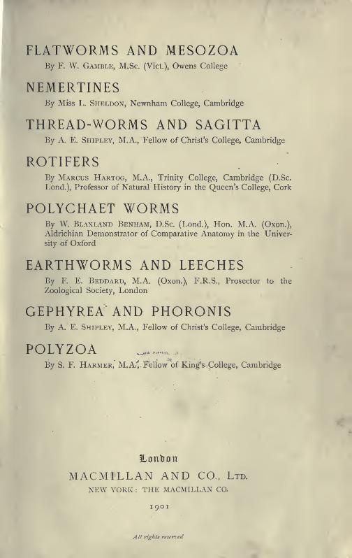
FLATWORMS AND MESOZOABy F. W. Gamble, M.Sc. (Vict.), Owens College
NEMERTINESBy Miss L. Sheldon, Newnham College, Cambridge
THREAD-WORMS AND SAGITTABy A. E. Shipley, M.A., Fellow of Christ's College, Cambridge
ROTIFERSBy Marcus Hartog, M.A., Trinity College, Cambridge (D.Sc.
Lond.), Professor of Natural History in the Queen's College, Cork
POLYCHAET WORMSBy W. Blaxland Benham, D.Sc. (Lond.), Hon. M.A. (Oxon.),
Aldrichian Demonstrator of Comparative Anatomy in the Univer-
sity of Oxford
EARTHWORMS AND LEECHESBy F. E. Beddard, M.A. (Oxon.), F.R.S., Prosector to the
Zoological Society, London
GEPHYREA AND PHORONISIp By A. E. Shipley, M.A., Fellow of Christ's College, Cambridge
POLYZOA _.......
By S. F. Harmer, MjAi^i^ellow of King^s College, Cambridge
Hontion
MACMILLAN AND CO., Ltd.
NEW YORK : THE MACMILLAN CO.
I 901
All rights reserz'ed
Nous allons faire des vers ensemble'
Andr^ be Chenier
First Edition 1896. Reprinted 1901
CONTENTS
Scheme of the Classification adopted in this BookPAfiF
ix
PLATYHELMINTHES AND MESOZOA
CHAPTER I
turbellaria
Introduction—Description of the Polyclad Leptoplana tremellaris—Appearance—Habits—Structure : Polycladida— Classification
Habits — Anatomy — Development : Tricladida — Occurrence—Structure—Classification : Rhabdocoelida—Occurrence—Habits—Keproduction—Classification 3
CHAPTER II
Trematoda
Characters of Trematodes — Habits and Structure of TrematodaECTOPARASITICA (MoNOGENEA)— LiFE- HISTORIES OF POLYSTOMUM IN-
tegerrimim, diplozoon paradoxum, and gvrodactylus elegans—Trematoda Endoparasitica (Digenea)—Occurrence and Habits of
Digenea — Life -History of Distomum macrostohvm— Distomvmhepaticum and its effects
—
bilharzia haematobia— bisexual
Trematodes—Table of Hosts—Classification 51
CHAPTER III
cestoda
Introduction -^Nature of Cestodes—Occurrence of Cestodes—TheTape-Worms of Man and Domestic Animals—Table of Life-His-
tories OF Principal Cestodes of Man and Domestic Animals—Structure and Development of Cestodes—Table for the Dis-
crimination of the More Usual Ckstodes of jSIan and Domestic
Animals—Classification 74
VI CONTENTS
CHAPTER IV
PAGEDiCYEMiDAE
—
Structure—Reproduction—Occurrence : Orthonectidae—Occurrence—Structure : Trichoplax : Salinella ... 92
NEMEETINEA
CHAPTER V
nemertinea
Introductory— External Characters— Anatomy— Classification-
Development— Habits— Regene ration— Breeding—GeographicalDistribution — Land, Fresh - Water, and Parasitic Forms—Affinities 99
NEMATHELMINTHES AND CHAETOGNATHA
CHAPTER VI
nemathelminthes
Introduction— Nematoda — Anatomy — Embryology — Classification—Ascaridae — Strongylidae — Trichotrachelidae — Filariidae—Mermithidae— Anguillulidae— Enoplidae—Parasitism : Nemato-morpha — Anatomy — Classification — Life - History : Acantho-CEPHALA
—
Anatomy—Embryology—Classification . . . .123
CHAPTER VII
chaetognatha
Structure—Reproduction— Habits— Food— Classification—Table of
Identification [see also p. 534] . . . 186
EOTIFEEA, GASTEOTEICHA, AND KINOEHYNCHA
CHAPTER VIII
rotifera, gastrotricha, and kinorhyncha
RoTiFERA
—
History—External Features—Movements—Anatomy—Re-
production—Embryology— Classification— Distribution— Affini-
ties : Gastrotricha : Kinorhyncha . 197
I
CONTENTS vil
AECHIANNELIDA, POLYCHAETA, ANDMYZOSTOMAEIA
CHAPTER IXPAGE
The Chaetopopous Worms—The Arch iannelida—Anatomy of Nereis,
AS Typical of the Polychaeta 241
CHAPTER X
Classification of the Polychaeta — Shape — Head — Parapodia-
Chaetae— Gills — Internal Organs — Jaws — Sense Organs —Reproduction—Larval Forms— Budding— Fission— Branching—Regeneration 257
CHAPTER XI
Natural History of Polychaetes—General Habits—Character of
Tube and its Formation— Colouring—Protective and Mimetic
Devices— Phosphorescence— Food — Uses — Associated Worms—Worms as Hosts— Distribution-Fossil Remains . . . .284
CHAPTER XII
Chaiiacters of the Sub-Ordeus of Polychaetes—Characters of the
Families—Description of British Genera and Species : The Myzo-
stomaria 303
OLIGOCHAETA (EARTHWOKMS, ETC.), AND
HIRUDINEA (LEECHES)
CHAPTER XIII
oligochaeta (earthworms and their allies)
Introduction— Anatomy— Reproduction— Bionomics—Distribution-
Classification—MiCRODRILI AND MeGADRILI 34'
CHAPTER XIV
HIRUDINEA (leeches)
Introduction— Anatomy— Reproduction— Classification — Rhyncho-
bdellae and Gnathobdellae •. 392
Vlll CONTENTS
GEPHYREA AND PHORONIS
CHAPTER XV
GEPHYREAPAGE
iNTRODrCTION — AnATOMY — DEVELOPMENT — SiPUNCULOIDEA --- PriA-
puloidea—echiuroidea—epithetosomatoidea—affinities of the
Group 411
CHAPTER XVI
phoronis
History—Habits—Structure—Reproduction— Larva— Metamorphosis
—List of Species and Localities—Systematic Position . . . 450
POLYZOA
CHAPTER XVII
POLYZOA
Introduction—General Characters and Terminology—Brown Bodies—History—Outlines of Classification—Marine Polyzoa—Occur-
rence—Forms of Colony and of Zooecia—Ovicells—Avicularia—Vibracula—Entoprocta 465
CHAPTER XVIII
POLYZOA
—
continued
Fresh-water Polyzoa—Phylactolaemata—Occurrence—Structure of
CristATELLA—Division of Colony—Movements of Colony—Retrac-
tion AND Protrusion of Polypides in Polyzoa—Statoblasts—Table
FOR Determination of Genera of Fresh-water Polyzoa—Repro-
ductive Processes of Polyzoa—Development—Affinities—Meta-
morphosis—Budding . . . 492
CHAPTER XIX
polyzoa—continued
Classification—Geographical Distribution—Palaeontology—Methodsfor the Examination of Specific Characters—Terminology—KeyFOR the Determination of the Genera of British Marine Polyzoa 515
Addendum to Chaetognatha
Index
534
535
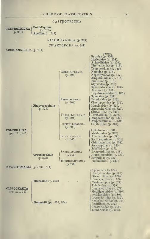
SCHEME OF THE CLASSIFICATION ADOPTED
IN THIS BOOK
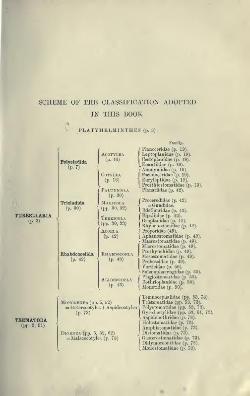
- PLATYHELMINTHES (p. 3)
TURBELLARIA(p. 3)
Polycladida
(p. 7)
Tricladida
(p. 30)
Rhabdocoelida(p. 42)
ACOTYLEA(p. 16)
COTYLEA(p. 16)
'' Paludicola(p. 30)
Maricola(pp. 30, 32)
Terricola
I (pp. 30,33)
ACOELA(p. 42)
Rhabdocoela(p. 43)
Alloeocoela(p. 43)
TREMATODA(pp. 3, 51)
Monogenea (pp. 5, 52)
Family.
f Planoceridae (p. 19).
ILeptoplanidae (p. 19).
I
Cestoplanidae (p. 19).
tEnantiidae (p. 19).
{Anonymidae (p. 19).
Pseudoceridae (p. 19).
Euryleptidae (p. 19).
Prosthiostomatidae (p. 19).
Planariidae (p. 42).
( Procerodidae (p. 42).
-] = Gundidae.[Bdellouridae (p. 42).
{Bipaliidae (p. 42).
Geoplanidae (p. 42).
Rhynchodemidae (p. 42).
( Proporidae (49).
\ Aphanostomatidae (p. 49).
r Macrostomatidae (p. 49).
Microstoniatidae (p. 49).
Prorhynchidae (p. 49).
Mesostomatidae (p. 49).
Proboscidae (p. 49).
Vorticidae (p. 50).
Solenopharyngidae (p. 50).
f Plagiostomatidae (p. 50).
4 Bothrioplanidae (p. 50).
{ Mouotidae (p. 50).
r Temnocephalidae (pp. 53, 73).
Tristomatidae (pp. 53, 73).
(p. 73)
DiOENEA (pp. 5, 52, 62)= Malacocotylea (p. 73)
i~\ rolyst
GyrodV Asnidi
= Heterocotylea + Aspidocotylea^ Polystomatidae (pp. 53, 73)^" '^^^ ' '^"rodactylidae (pp. 53, 61,
_,pidobotliridae (p. 73).
Holostomatidae (p. 73).
Amphistomatidae (p. 73).
, Distomatidae (p. 73).
IGasterostomatidae (p. 73).
Didymozoontidae (p. 73).
Monostomatidae (p. 73).
SCHEME OF CLASSIFICATION
CESTODA (pp. 3, 74)
MESOZOA (pp. 3, 92)
HOPLONEMERTEA (p. 110) =
SCHIZONEMERTEA (p. 111) =
PALAEONEMERTEA (p. Ill)
MESOZOA
Family.
Cestodariidae = Monozoa (p. 91-)
Bothriocephalidae (p. 91).
Tetrarhynchidae (p. 91).
Tetraphyllidae (p. 91).
^Taeniidae (p. 91).
f Dicyeniidae (p. 93).
V Orthonectida (p. 94).
NEMERTINEA (p. 99)
: Metanemertini (p. 112).
= Heteronemertuii {ex parte) (p. 113).
r Protonemertini (p. 112).
= A + Mesonemertini (p. 112).
[ + Heteronemertini {ex 2)arte) ^. 113).
NEMATHELMINTHES (p. 123)
Ascaridae (p. 138)
NEMATODA (pp. 123, 124)
NEMATOMORPHA (pp. 123, 164)
ACANTHOCEPHALA (pp. 123, 174)
Strongylidae (jx 142).
Tricliotrachelidae (p. 144).
Filariidae (p. 147).
Mermithidae (p. 150).
Anguillulidae (p. 154).
Enoplidae (p. 157).
Chaetosomatidae (p. 158).
^ Desmoscolecidae (p. 159).
Gordiidae (p. 164).
I"
Echinorhyncliidae (p. 182)
I Gigantorhynchidae (p. 183).'1 Neorhynciiidae (p. 184).
I Arhynchidae (p. 185).
CHAETOGNATHA (p. 186)
ROTIFERA (p. 197)
FLOSCULARIACEAE (p. 220)
MELICERTACEAE (p. 221)
BDELLOIDA (p. 222) .
ASPLANCHNACEAE (p. 222)
SCIRTOPODA (p. 223
PLOIMA (p. 223)
lUoricata (p. 223)
Loricata (p. 224)
SEISONACEAE (p. 225)
( Flosculariidae (p. 221).
\ Apsilidae (p. 221).
f Melicertidae (p. 221).
\ Trocliosphaeridae (p. 221).
Philodinidae (p. 222).
Asplanchnidae (p. 223).
Pedalionidae (p. 223).
Microcodonidae (p. 224).
Khinopidae (p. 224).
Hydatinidae (p. 224).
Synchaetidae (p. 224).
Notommatidae (p. 224).
Drilophagidae (p. 224).
Triarthridae (p. 224).
Rattulidae (p. 225).
Dinocharididae (p. 225).
Salpinidae (p. 225).
Euchlaiiididae (p. 225).
Cathypnidae (p. 225).
Coluridae (p. 225).
Pterodinidae (p. 225).
Bracliionidap (p. 225).
Atiuraeidae (p. 225).
Seisonidae (p. 226).
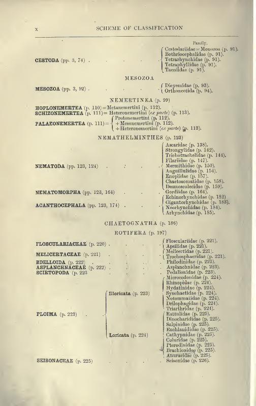
SCHEME OF CLASSIFICATION
GASTROTRICHA
(x^ 231) i ^1 •-'^^^
(p. Z61) (Apodina (p. 235)
KINORHYNCHA (p. 236)
CHAETOPODA (p. 241)
ARCHIANNELIDA (p. 241)
( Nereidiformia(p. 303)
(Phanerocephala -
(p. 303)
POLYCHAETA(pp. 241, 245) >
CryptocephalaI (p. 303)
MYZOSTOMARIA (pp. 241, 341)
Spioniformia(p. 304)
Tkrebelliformia(p. 304)
Capitelliformia(p. 305)
ScolecifokmiaL (p. 305)
(Sabelliformia(p. 305)
Hermelliformia(p. 306)
OLIGOCHAETAa.p. 241, 347) 1
Microdrili (p. 373)
Megadrili (pp. 373, 374).
Family.
( Syllidae (p. 306).
Hesionidae (p. 308).
Aphroditidae (p. 309).
Phyllodocidae (p. 313).
Tomopteridae (p. 315).
Nereidae (p. 315).
Nephthydidae (p. 317).
Amphinomidae (p. 318).
Eunicidae (p. 318).
Glyceridae (p. 320).
Sphaerodorioae (j). 320).
Ariciidae (p. 321).
. Typhloscolecidae (p. 321).
Spionidae (p. 321).
Polydoridae (p. 323).
Chaetopteridae (p. 323).
Magelonidae (p. 325.
Ammocharidae (p. 325).
Cirratulidae (p. 325).
Terebellidae (p. 327).
Am{)haretidae (p. 330).
Ampliictenidae (p. 330).
Capitellidae (p. 331).
Opheliidae (p. 331).
Maldanidae (p. 332). •
Arenicolidae (p. 333).
Scalibregmidae (p. 334).
Chlorhaeniidae (p. 334).
Sternaspidae (p. 335).
/ Sabellidae (p. 336).' Eriogi-aphidae (p. 338).
]Aiuphicorinidae (p. 339).
( Serpulidae (p. 339).
Hermellidae (p. 341)!
/"Aphaneiira (p. 374).
Eiichytraeidae (p. 375).
Discodrilidae (p. 376).
Phreoiyctidae (p. 376).
Naidoinorpha (p. 377).
Tubificidae (p. 378).
Lumbriculidae (p. 379).
Moniligastridae (p. 380).
Perichaetidae (p. 380).
Cry]>todrilidae (p. 382).
Acanthodrilidae (p. 384).
|Eiidnlidae(p. 385).
Geoscolicidae (p. 386).
1, Lumbricidae (p. 388).
Xll SCHEME OF CLASSIFICATION
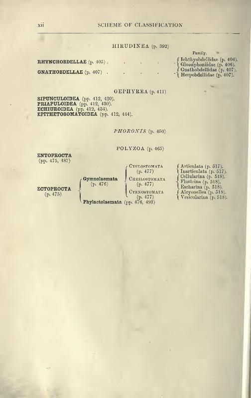
HIRUDINEA (p. 392)
RHYNCHOBDELLAE (p. 405)
GNATHOBDELLAE (p. 407)
Family.
r Iclithyobdellidae (p. 406).
\ Glossiplioniidae (p. 406).
/ Gnathobdellidae (p. 407).
\ Herpobdellidae (p. 407).
GEPHYREA (p. 411)
SIPUNCULOIDEA (pp. 412, 420).
PRIAPULOIDEA (pp. 412, 430).
ECHIUROIDEA (pp. 412, 434).
EPITHETOSOMATOIDEA (pp. 412, 444).
FHORONIS (p. 450)
POLYZOA (p. 465)
ENTOPROCTA(pp. 475, 487)
ECTOPROCTA(p. 475)
r Gymnolaemata
I
(P- 476)
f Cyclostomata(p. 477)
Cheilostomata(p. 477)
Ctenostomata(p. 477)
Phylactolaemata (pp. 476, 493)
/ Articulata (p. 517).
\ Inarticulata (p. 517).
{Cellularina (p. 518).
Flustrina (p. 518).
Escharina (p. 518).
/ Alcyonellea (p. 518).
\ Vesicularina (p. 518).

B({!i OEPT
UNIV - .10,
PLATYHELMINTHES AND MESOZOA
F. W. GAMBLE, M.Sc. (Vict.)
Demonstrator aud Assistant-Lecturer in Zoology in the Owens College, Mancliester.
VOL. II !£

CHAPTEK I
.TUKBELLARIA
INTRODUCTION : DESCRIPTION OF THE POLYCLAD LEPTOPLANATREMELLARIS APPEARANCE HABITS STRUCTURE : POLY-
CLADIDA CLASSIFICATION HABITS ANATOMY DEVELOP-
MENT : TRICLADIDA OCCURRENCE STRUCTURE CLASSIFICA-
TION : RHABDOCOELIDA OCCURRENCE HABITS REPRODUC-
TION CLASSIFICATION.
The Platyhelminthes, or Flat Worms, form a natural assemblage
of animals, the members of which, however widely they maydiffer in appearance, habits, or life -history, exhibit a funda-
mental similarity of organisation which justifies their separation
from other classes of worms, and their union into a distinct
phylum. Excluding the leeches (Hirudinea), and the long sea-
worms (Nemertinea)—which, though formerly included, are nowtreated independently—the Platyhelminthes may be divided into
three branches: (1) Turbellaria (including the Planarians), (2)
Trematoda (including the liver-flukes), and (3) Cestoda (tape-
worms). The Mesozoa will be treated as an appendix to the
Platyhelminthes.
The Turbellaria were so called by Ehrenberg ^ (1831) on
account of the cilia or vibratile processes with which these
aquatic animals are covered, causing by their incessant action,
tiny currents (" turbellae," disturbances) in the surrounding
water. The ciliary covering distinguishes this free-living group
from the parasitic Trematodes and Cestodes, some of which
possess such an investment, but only during their early free
1 Hemprich and Ehrenberg, Symbolae physicae, Berlin, fol. 1831.
PLATYHELMINTHES
larval stage, for the short period when they have left the
parental host and are seeking another (Figs. 26, 27, 42).
Some Turbellaria (Ehabdocoelida) resemble Infusoria in their
minute size, shape, and movements. Nevertheless they possess
an organisation of considerable complexity. The fresh -water
Planarians (Fig. 14), abounding in ponds and streams, vary from
a quarter to half an inch in length, and are elongated and
flattened. Their body is soft, and progresses by a characteristic,
even, gliding motion like a snail. The marine Planarians or
Polyclads (Fig. 8) are usually broad and leaf- like, sometimes
attaining a length of six inches, and swim or creep in a most
graceful way. Land Planarians occur in this country (Fig. 15),
but far more abundantly in tropical and sub-tropical districts, in
moist places, venturing abroad at night in pursuit of prey. They
are elongated and cylindrical, in some cases measuring, whenfully extended, a foot or more in length, and are often ornamented
with brilliantly coloured, longitudinal bands.
Turbellaria are carnivorous, overpowering their prey by peculiar
cutaneous offensive weapons, and then sucking out the contents of
the victim by the "pharynx." Land Planarians feed on earth-
worms, molluscs, and wood-lice ; fresh-water Planarians on Oligo-
chaet worms, water-snails, and water-beetles ; marine forms devour
Polychaet worms and molluscs. Some Turbellaria seem to prefer
freshly-killed or weakly examples of animals too large to be over-
powered when fully active. Certain Ehabdocoelida are messmates
of Molluscs and Echinoderms, and a few others are truly parasitic
—a mode of life adopted by all Trematodes save Temnocepliala.
The Trematodes^ may be divided into those living on the
outer surface of various aquatic animals, usually fish (Ectopara-
sites) ; and those which penetrate more or less deeply into the
alimentary canal or the associated organs of the host (Endo-
parasites). They are oval, flattened Platyhelminthes ranging from
a microscopic size to a length of three feet {Nematohotlirmm, Fig.
22), and are provided with organs of adhesion by which they cling
to the outer surface, or to the interior, of the animals they inhabit.
Trematodes occur parasitically in all groups of Vertebrates, but,
with the exception of the liver-flukes of the sheep {Distomum hepa-
ticum and D. magnum), and of Bilharzia haematobia found in man(in the blood-vessels of the urinary bladder) over the greater part
^ Tpijfia, a hole ; referring to the orifices of the suckers.
It INTRODUCTION
of Africa, their attacks are not usually of a serious nature. Ecto-
parasitic Trematodes are Monogenetic ; that is, their larvae growup directly into mature forms. The Endoparasitic species, how-ever, are usually Digenetic. Their larvae enter an Invertebrate
and produce a new generation of different larvae, and these
another. The last are immature flukes. They enter a second
host, which is swallowed by the final Vertebrate host in whichthey become mature.
The Cestodes or Tape-worms have undergone more profound
modifications both in structure and in mode of development.
They are all endoparasitic, and, with one exception (Archigetes),
attain maturity solely within the alimentary canal of Vertebrates.
In length they range from a few millimetres to several metres, but
this great size is attained from the need for the rapid production
and accumulation of enormous numbers of eggs. The " head
"
or " scolex " is attached to the mucous membrane of the host bysuckers or hooks, but there is no mouth nor any certain trace of a
digestive tract at any stage of the life-history of Cestodes. Fornourishment they absorb, through the skin, the previously-digested
food (of the host) that bathes them. In a few Cestodes the bodyis simple and not divided into " proglottides " or generative seg-
ments, but in most cases it is jointed in such a way that the last
segment is the oldest, and each contains a set of reproductive
organs. The life-histories of Cestodes are most remarkable. Theproglottides containing the eggs pass out of the final host along
with the faeces and enter the intermediate host with the food.
The larvae hatch, and boring their way into the blood-vessels, are
carried by the circulation to various internal organs. Here they
usually become " bladder-worms," and develop the " head " of the
future sexual form. Then, if, as is usually the case, the inter-
mediate host is preyed upon by the final host, the larval Cestodes
•enter the alimentary canal of the latter. The head of the larva
alone survives digestion, and from it the mature worm is formed.
Of these three branches of the phylum Platyhelminthes, the
Turbellaria possess features of special interest and importance. Notonly do they furnish the explanation of the structure of the two
parasitic groups (which have probably arisen from Turbellarian-
like ancestors), but they occupy the lowest position in the whole
group of worms. There are reasons for thinking that this is the
simplest group of bilateral animals which adopt the habit of creep-
PLATYHELMINTHES
ing. The Turbellaria are most closely allied tp that great extinct
group from which they, the Nemertinea, Eotifera, and even the
Annelids, offer increasingly convincing evidence of having been
derived. Many questions relating to the affinities of, or the
origin of organs in, the Annelids, resolve themselves into similar
questions about the Turbellaria. For these reasons, this group
is here dealt with at greater length than the others, the interest
of which is of a more special nature.
The history of our knowledge of the Cestodes dates back to
ancient times, as the presence and effects of tape-worms early
attracted the attention of physicians. Trematodes are first
distinctly referred to in the sixteenth century, while Turbellaria
first figure in Trembley's memoir on Hydra (1744).-^ The whole
subject of the increase in our knowledge of parasitic Platyhel-
minthes is dealt with in the standard work. The Parasites of
Man, by Leuckart,^ and a complete list of references in zoological
literature to Cestodes and Trematodes is to be found in Bronn's
Thierreicli? 0. F. Mliller^ and Ehrenberg founded our know-
ledge of the Turbellaria, but for a long time the group remained
in a most neglected condition. In this country Montagu, G.
Johnston, and in Ireland, AVilliam Thompson, discovered several
marine species, one of which, Planocera folium (from Berwick),
has not again been • met with on British shores. Dalyell ^ con-,
ducted classical researches on the habits of Planarians, and Fara-
day ^ made interesting experiments on their power of regenerat-
ing lost parts. The credit of assigning the correct interpreta-
tion to most of the various organs of fresh-water Planarians
belongs to von Baer"^ and Duges,^ while Mertens^ effected a
similar service for the marine forms, or Polyclads. The minute
Ehabdocoels were first successfully investigated and classified byOscar Schmidt.^*^ The great work on this group is, however, the
^ M4moires pour servir ci lliistoire d. Polypes cVeau douce, Leyden, 1744.
2 Die Parasiten des Menschen, 1879 . Engl. Transl. by W. E. Hoyle, i. 1886.
^ Band 4, by M. Braun. (Mesozoa and Trematoda completed ; Cestoda in progress.
)
* Verm. terr. etfluv. . . . succinda histo7na, 1773 ; Zool. Danica, 1777.
^ Observations on Planariae, Edinburgh, 1S13.^ M. Faraday, "On the Planariae," Medical Gazette, Feb. 1832; and in Edin-
burgh New Philosojjh. Journal, vol. xiv. 1833, i>p. 183-189.'' Nov. Act. Acad. Caes. Leop. -Carol, torn. xiii. 1827.s Ann. Sci. Nat. {Zoo\.) I. torn. xv. 1828. ; ibid. torn. xxi. 1830.
^ Mem. Acad. St. Pitersbourg, 5th ser. torn. ii. 1832.^** Die rJuibdocoelen Turbellarien des Susswassers. Jena 1848.
TURBELLARIA
monograph by von Graff.^ A similarly comprehensive and indis-
pensable treatise by Lang, on the Polycladida,^ contains references
to all previous publications on the group, among which the
papers by Quatrefages, Johannes Milller, Keferstein, Minot, and
Hallez stand out conspicuously. Moseley's work^ on the Land
Planarians of Ceylon is imdoubtedly the most revolutionary
paper referring to this group, and the
best contribution towards elucidating ,,:f:' ] ;\. y
the structure of the Tricladida at a
time when the subject was very obscure.
A monograph on Land Planarians is ^
being prepared by von Graff.
The Turbellaria are divided into :
(1) Polycladida, marine forms with
multiple intestinal branches; (2) Tri-
cladida, marine, fresh-water, and ter-
restrial Planarians with three main
intestinal branches; (3) the Rhahdocoe-
lida, as varied in habit as the Triclads,
but possessing a straight and simple or
slightly lobed, intestine. A detailed
description of an example of the Poly-
clads, and then a comparative account
of each division, will now be given.
Turbellaria. I. Polycladida.
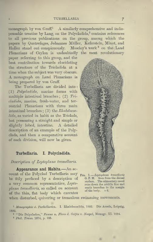
Description of Leptoplana tremellaris.
Appearance and Habits.—An ac-
count of the Polyclad Turbellaria may^^^^ i.—Leptopiano tremellaris
be fitly prefaced by a description of O.F.M. Seeu from the dorsal
a very common representative, Lepto-
plana tremellaris, so called on account
of the thin, flat body which executes
when disturbed, quivering or tremulous swimming movements,
surface. The alimentary canal
runs down the middle line and
sends branches to the margin
of the body. x 6.
^ 3fonogra2)hie d. Turhellarien. I. Rhabdocoelida, 1882. Die Acoela, Leipzig,
1892.
2 "Die Polycladen," Fauna u. Flora d. GoJfes v. Neapel, Monogr. XI. 1884.
3 Phil. Trans. 1874, p. 105,
8 PLATYIIELMINTHES TURBELLARIA
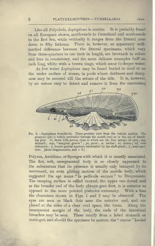
Like all Polyclads, Leptoplana is marine. It is probably found
on all Eui'opean shores, northwards to Greenland and southwards
to the Eed Sea, while vertically it ranges from the littoral zone
down to fifty fathoms. There is, however, an apparently well-
marked difference between the littoral specimens, which vary
from three-quarters to one inch in length, are brownish in colom*
and firm in consistency, and the more delicate examples half an
inch long, white with a brown tinge, which occur in deeper water.
At low water Leptoplana may be found buried in mud or on
the under surface of stones, in pools where darkness and damp-
ness may be ensured till the return of the tide. It is, however,
by no means easy to detect and remove it from the encrusting
u t / ,'^
vd
Fig. 2.
—
Leptoplana tremdlaris. Three-quarters \\q\\ from the ventral surface. The. pharynx {ph) is widely protruded through the mouth {mo) as in the act of attack-
ing prey, hr, Brain with nerves, close to which are the four groups of eyes ; mgystomach; mgc, "marginal groove"; pe, penis; sc, sucker; v.t, uterus; xd, vasa
deferentia ; 9, female genital aperture surrounded by the shell-gland ; i , male aper-
ture. (Semi-diagrammatic, and x 6.)
Polyzoa, Ascidians, or Sponges with which it is usually associated.
The flat, soft, unsegmented body is so closely appressed to
the substratum that its presence is usually only betrayed by its
movement, an even gliding motion of the mobile body, which
suggested the apt name " la pellicule animee " to Dicquemare.
The creeping surface is called ventral, the upper one dorsal, and
as the broader end of the body always goes first, it is anterior as
opposed to the more pointed posterior extremity. With a lens
the characters shown in Tigs. 1 and 2 may l^e observed. Theeyes are seen as black dots near the anterior end, and are
placed at the sides of a clear oval space, the brain. Along the
transparent margin of the body, the ends of the intestinal
branches may be seen. These ramify from a lobed stomach or
main-gut, and should the specimen be mature, the " uterus '' loaded
I
POLYCLADIDA 9
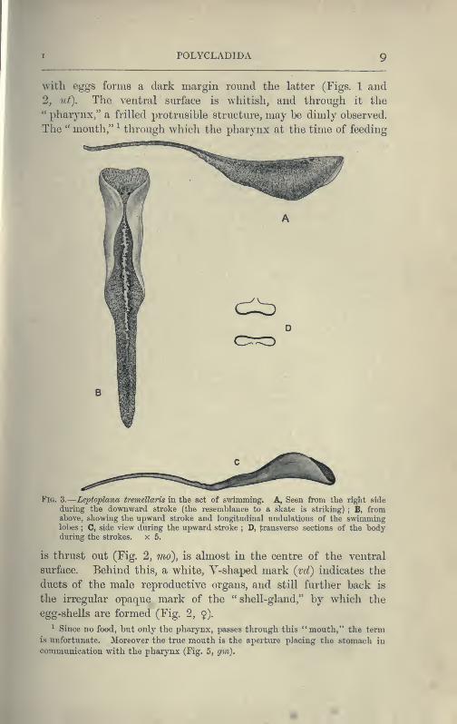
with eggs forms a dark margin round the latter (Figs. 1 and
2, ut). The ventral surface is whitish, and through it the" pharynx," a frilled protrusible structure, may be dimly observed.
The " mouth," ^ through which the pharynx at the time of feeding
d^
[G. 3.
—
Leptoplana tremellaris in the act of swimming. A, Seen from the right side
during the downward stroke (the resemblance to a skate is striking) ; B, fromabove, showing the upward stroke and longitudinal undulations of the swimminglobes ; C, side view during the upward stroke ; D, transverse sections of the bodyduring the strokes. x 5.
is thrust out (Fig. 2, mo), is almost in the centre of the ventral
surface. Behind this, a white, Y-shaped mark {vd) indicates the
ducts of the male reproductive organs, and still further back is
the irregular opaque mark of the "shell-gland," by which the
egg-shells are formed (Fig. 2, $).
^ Since no food, but only the pharynx, passes through this ** mouth," the termis unfortunate. Moreover the true mouth is the aperture placing the stomach in
communication with the pharynx (Fig. 5, gm).
10 PLATYHELMINTHES TURBELLARIA chap.
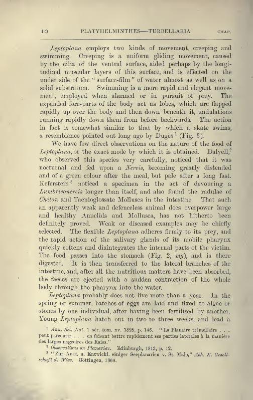
Lefto'plana employs two kinds of movement, creeping and
swimming. Creeping is a uniform gliding movement, caused
by the cilia of the ventral surface, aided perhaps by the longi-
tudinal muscular layers of this surface, and is effected on the
under side of the " surface-film " of water almost as well as on a
solid substratum. Swimming is a more rapid and elegant move-
ment, employed when alarmed or in pursuit of prey. The
expanded fore-parts of the body act as lobes, which are flapped
rapidly up over the body and then down beneath it, undulations
running rapidly down them from before backwards. The action
in fact is somewhat similar to that by which a skate swims,
a resemblance pointed out long ago by Duges-^ (Fig. 3).
We have few direct observations on the nature of the food of
Lepto'plana, or the exact mode by which it is obtained. Dalyell,"
who observed this species very carefully, noticed that it was
nocturnal and fed upon a Nereis, becoming greatly distended
and of a green colour after the meal, but pale after a long fast.
Keferstein^ noticed a specimen in the act of devouring a
Luvibriconereis longer than itself, and also found the radulae of
Chiton and Taenioglossate Molluscs in the intestine. That such
an apparently weak and defenceless animal does overpower large
and healthy Annelids and Mollusca, has not hitherto been
definitely proved. Weak or diseased examples may be chiefly
selected. The flexible Leptoplana adheres firmly to its prey, and
the rapid action of the salivary glands of its mobile pharynx
quickly softens and disintegrates the internal parts of the victim.
The food passes into the stomach (Fig. 2, mg), and is there
digested. It is then transferred to the lateral branches of the
intestine, and, after all the nutritious matters have been absorbed,
the faeces are ejected with a sudden contraction of the whole
body through the pharynx into the v/ater.
Leptoplana probably does not live more than a year. In the
spring or summer, batches of eggs are laid and fixed to algae or
stones by one individual, after having been fertilised by another.
Young Leptoplana hatch out in two to three weeks, and lead a
^ Ann. Sci. Nat. 1 ser. torn. xv. 1828, p. 146. "La Planaire tremellaire . . .
pent parcoiirir ... en faisant battre rapidement ses parties laterales a la manieredes larges nageoires des Raies."
2 Observations on Planariae. Edinburgh, 1813, p. 12.
^ "Zur Anat. u. Entwickl. einiger Seej)lanarien v. St. Malo," ^JA. K. Gescll-
sehaft d. TFiss. Gottingen, 1868.
ANATOMY OF POLYCLADIDA I I
pelagic existence till they are three or four millimetres in length.
In late summer, numbers of such immature examples may be foundamong sea-weeds and Corallina in tide pools. In the succeeding
spring they develop first the male and then the female reproduc-tive organs.
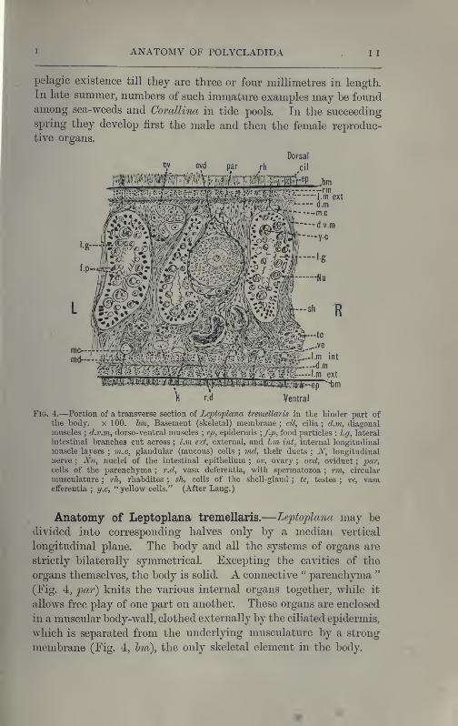
"te
I.m int
Ventral
Fig. 4.—Portion of a transverse section of Leptoplana tremellaris in the hinder part ofthe body. x 100. hm, Basement (skeletal) membrane ; cil, cilia ; d.in, diagonalmuscles ; d.v.m, dorso-ventral muscles ; ep, epidermis
; f.p, food particles : l.g, lateral
intestinal branches cut across ; l.m ext, external, and l.m wt, internal longitudinal
muscle layers ; m.c, glandular (mucous) cells ; vid, their ducts ; iV, longitudinal
nerve ; Nu, nuclei of the intestinal epithelium ; ov, ovary ; ovd, oviduct;
^jar,
cells of the parenchyma ; r.d, vasa deferentia, with spermatozoa ; rm, circular
musculature ; rh, rhabdites ; sh, cells of the shell-gland ; te, testes ; ve, vasaefferentia
;y.c, "yellow cells." (After Lang.)
Anatomy of Leptoplana tremellaris.—Leptoplana may be
divided into corresponding halves only by a median vertical
longitudinal plane. The body and all the systems of organs are
strictly bilaterally symmetrical. Excepting the cavities of the
organs themselves, the body is solid. A connective " parenchyma "
(Fig. 4, par) knits the various internal organs together, while it
allows free play of one part on another. These organs are enclosed
in a muscular body-wall, clothed externally by the ciliated epidermis,
which is separated from the underlying musculature by a strong
membrane (Fig. 4, Im), the only skeletal element in the body.
12 PLATYHELMINTHES TURBELLARIA chap.
Body-Wall.—The epidermis (Fig. 4, ep) is composed of a
single layer of ciliated cells, containing small, highly refractive,
pointed rods or " rhabdites " {rh), and gives rise to deeply-
placed mucous cells (m.c), which are glandular and pour out
on the surface of the body a fluid in which the cilia vibrate.
The tenacious hold on a stone which Le'ptoplana exerts if sud-
denly disturbed, or when grasping its prey, is probably due to the
increased glutinous secretion of these glands, aided perhaps by
rhabdites, which on such occasions are shot out in great numbers.
The basement membrane is an elastic skeletal membrane com-
posed of stellate cells embedded in a firm matrix. It serves
chiefly for the origin and insertion of the dorso-ventral muscles
(d.v.m). Under the basement membrane lies a very thin layer
of transverse muscular fibres (Fig. 4, rm), which are, however,
apparently absent on the ventral surface. Then follows a stout
layer of longitudinal fibres (l.m ext), and beneath this a diagonal
layer (d.m), the fibres of which intersect along the median line
in such a way that the inner fibres of one side become the outer
diagonal fibres of the other. Lastly, within this again, on the
ventral surface, is a second stout longitudinal layer {l.m int).
The sucker (sc. Figs. 2 and 5) is a modification of the body-wall
at that point. In addition to the dorso-ventral muscles, there
exists a complex visceral musculature regulating the movements
of the pharynx, intestine, and copulatory organs.
Parenchyma.—The spaces between the main organs of the
body are filled by a tissue containing various kinds of cells,
salivary glands, shell-glands, and prostate glands. Besides these,
however, we find a vacuolated, nucleated, thick-walled network,
and to this the word parenchyma is properly applied. Besides
its connective function, the parenchyma confers that elasticity on
the body which Leptoplana possesses in such a high degree. Pig-
ment cells are found in the parenchyma in many Polyclads.
Digestive System.—The general arrangement of this system
may be seen in Figs. 2, 5, and 7 ; and may be compared, especially
when the pharynx is protruded, as in Fig. 2, with the gastral
system of a Medusa. The " mouth " (there is no anus) is placed
almost in the centre of the ventral surface. It leads (Fig. 7, B,^As)
into a chamber (the peripharyngeal space) divided into an upper
and a lower division by the insertion of a muscular collar-fold
(the pharynx, ph), which may be protruded, its free lips

I ANATOMY OF POLYCLADIDA 1
3
advancing, through the mouth (Fig. 2), and is then capable of
enclosing by its mobile frilled margin, prey as large as Leptoplana
itself The upper division of the chamber communicates by a
hole in its roof ^ (the true mouth, Figs. 5 and 7, g.rri) with the
cavity of the main-gut or stomach {m.g), which runs almost the
length of the body in the middle line, forwards over the brain
(Fig. 5, u;p). Seven pairs of lateral gut-branches convey the
digested food to the various organs, not directly however, but only
after the food mixed with sea-water has been repeatedly driven
by peristalsis first towards the blind end of the gut-branches and
then back towards the stomach. Eespiration is probably largely
effected by this means. The epithelium of the intestine (Fig. 4,
l.g) of a starving specimen is composed of separate flagellated
cells frequently containing " yellow cells." ^ After a meal, how-
ever, the cell outlines are invisible. Gregarines, encysted Cercariae,
and Orthonectida ^ occur parasitically in the gut-branches.
An excretory system of " flame-cells " and fine vessels has
hitherto been seen only by Schultze ^ in this species, which will
not, however, resist intact the compression necessary to enable
the details to be determined. They are probably similar to
those of Thysanozoon described on p. 25.
Nervous System.—The brain, which is enclosed in a tough
capsule (Fig. 5, Ir), is placed in front of the pharynx, but some
distance behind the anterior margin of the body. It is of an
oval shape, subdivided superficially into right and left halves by
a shallow depression, and is provided in front with a pair of
granular-looking appendages,composed of ganglion-cells from which
numerous sensory nerves arise, supplying the eyes and anterior
region. Posteriorly the brain gives rise to a chiefly motor,
nervous sheath (Fig. 5, nn), which invests the body just within
the musculature. This sheath is thickened along two ventral
lines (Fig. 5, In) and two lateral lines {n.s), but is very slightly
developed on the dorsal surface. Ganglion-cells occur on the
course of the nerves, and are particularly large at the point of
origin of the great motor nerves.
Sense Organs.—Le]ptoplana possesses eyes, stiff tactile, mar-
^ The roof of the peripharyngeal chamber is hence known as the "diaphragm."2 See Brandt, Fauna u. Flora d. Golfcs v. Neapel, ]\Ionogr. XIII. 1885, p. 65.
3 See p. 94.
^ Verhandlungen d. Tried. Gesellschaft zu Wurzburg, iv. 1854, p. 223.

Ir
-.'Od
-Udp
Fig. 5.—Diagrammaticview of the struc-
ture of Leptoplanatremellaris as a
type of the Poly-cladida. The bodyis cut across themiddle to show the
relative jjositiou of
organs in trans-
verse section. Inthe posterior half
the alimentarycanal has been bi-
sected and removedfrom the left
side, to exhibit the
deeply placed nerv-
ous sheath {nn)
and the male re-
productive organs.
hr. Brain ; diJ,
" diaphragm "; e,
cerebral group of
eyes ; et, tentac-
ular eye - group;
gr, marginalgroove
;gm, true
mouth ; Ig, lateral
gut - branch ; Irk,
longitudinal nervestem ; vi, external
mouth ; mg, mg',
main -gut, whole,and bisected ; n,
sensory nerve sup-
plying the eyes;
nn, nervous net-
work lying on the
ventral muscula-ture ; n.s, lateral
nerve ; od, oviduct
;
ov, ovary; j»>6, penis
(in section);ph,
pharynx;pr, jjros-
tate or " granulegland"; 5C, sucker;sg, shell-gland
;
te, testes ; vpi, an-
terior unpairedgut - branch ; vt,
uterus; va, vagina
(in section) ; vd,
vas deferens ; ve,
vasa efferentia; cJ,
male genital j^ore ;
?, female pore.

ICHAP. I ANATOMY OF POLYCLADIDA I 5
ginal cilia, and possibly a sense organ in the "marginal groove."
The eyes, which are easily seen as collections of black dots lying
at the sides of the brain, may be divided into two paired groups
:
(1) cerebral eyes (Fig. 5, e), and (2) ten-
tacle eyes {et), which indicate the position
of a pair of tentacles in allied forms (Fig.
8, A, ^ and B). Each ocellus consists of
a capsule placed at right angles to the
surface of the body in the parenchyma,
below the dorsal muscles, and with its con-
vex face outwards. It is a single cell
in which pigment granules have accumu-
lated. The light, however, can only reach
the refractive rods, which lie within it, p^^ e.-Diagram of an eye
obliquely at their outer ends. These rods of Leptopiana from the
• ,i ,1 J.- 1 n J tentacle group, x 600.are m connexion with the retinal cells, and ^^^^gj. Lang.)
thus communicate by the optic nerve with
the brain. The cerebral eyes are really paired, and are directed
some upwards, some sideways, some downwards.
The " marginal groove " is a shallow depression of the
epidermis (Fig. 5, gr) lined by cilia, and containing the ducts
of very numerous gland-cells. It runs almost parallel to the
anterior margin of the body, a short distance from it, but we
have no observations on its functions.
Reproductive Organs.—Le'pto^lana is hermaphrodite, and,
as in most hermaphrodites, the reproductive organs are com-
plicated. The male organs are the first to ripen, but this does
not appear to prevent an overlapping of the periods of maturity
of the male and female products, so that when the eggs are
being laid, the male organs are, apparently, still in a functional
state. The principal parts are seen in Fig. 5. The very
numerous testes (te) are placed ventrally, and are connected with
fine vasa efferentia (ve), which form a delicate network opening at
various points into the two %msa deferentia (vd). These tubes,
especially when distended with spermatozoa, may easily be seen
(Fig. 2, vd) converging at the base of the penis, and connected
posteriorly by a loop that runs behind the female genital pore
(Fig. 5). The penis (pe) is pyriform and muscular, and is
divided into two chambers, a large upper one for the sperma-
tozoa, and a smaller lower one for the secretion of a special
i6 PLATYHELMINTHES TURBELLARIA CHAP.
" prostate " gland. The apex of the penis is eversible and not
merely protrusible, being turned inside out when evaginated. Theovaries (Fig. 5, ov) are numerous and somewhat spherical. Theyare dorsally placed, but when fully developed extend deeply
wherever they can find room to do so, and they not only furnish
the ova, but elaborate food-yolk in the ova, as there are no
special yolk-glands. The slender oviducts (od) open at several
points into the " uterus " (ut) (a misnomer, as no development
takes place within it), which encircles the pharynx, and opens by
a single duct into the vagina (va). Here the ova are probably
fertilised, and one by one invested by the shell-gland (sg) with a
secretion which hardens and forms a resistant shell. They are
then laid in plate-like masses which are attached to stones or
shells. The development is a direct one, and the young Lepto-
plana, which hatches in about three weeks,, has the outline of a
spherical triangle, and possesses most of the organs of the adult.
After leading a floating life for a few weeks it probably attains
maturity in about nine months.
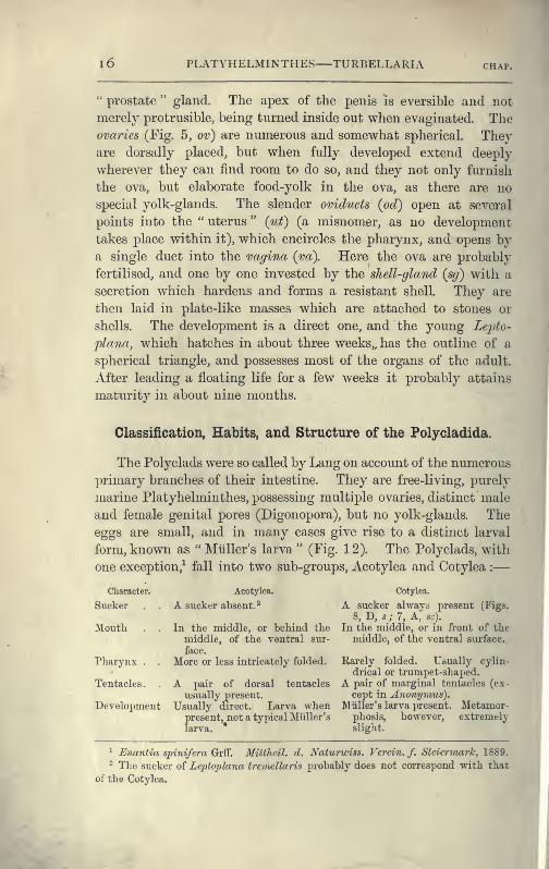
Classification, Habits, and Structure of the Polycladida.
The Polyclads were so called by Lang on account of the numerous
primary branches of their intestine. They are free-living, purely
marine Platyhelminthes, possessing multiple ovaries, distinct male
and female genital pores (Digonopora), but no yolk-glands. The
eggs are small, and in many cases give rise to a distinct larval
form, known as "Mliller's larva" (Fig. 12). The Polyclads, with
one exception,^ fall into two sub-groups, Acotylea and Cotylea :
—
Character.
Sucker
Mouth .
Pharynx .
Tentacles
.
Acotylea.
A sucker absent.^
In the middle, or behind the
middle, of the ventral sur-
face.
More or less intricately folded.
A pair of dorsal tentacles
usually present.
Development Usually direct. Larva whenpresent, not a typical Miiller's
Cotylea.
A sucker always present (Figs.
8, D, s; 7, A, sc).
In the middle, or in front of themiddle, of the ventral surface.
Rarely folded. Usually cylin-
drical or trumpet-shaped.A pair of marginal tentacles (ex-
cept in Anonymus).Miiller's larva present. Metamor-
phosis, howevex", extremelyslight.
^ Enantia spinifera Grff. Mitthcil. d. Naturwiss. Verein. f. SteiermarTc, 1889.
- The sucker of Leptoplana tremellaris probably does not correspond with that
of the Cotylea.
CLASSIFICATION OF POLYCLADIDA 17
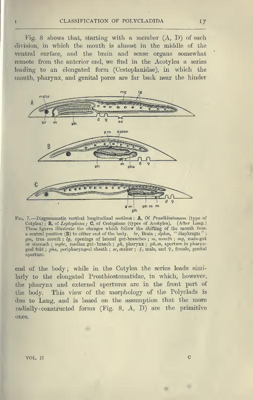
Fig. 8 shows that, starting with a member (A, D) of each
division, in which the mouth is ahnost in the middle of the
ventral surface, and the brain and sense organs somewhat
remote from the anterior end, we find in the Acotylea a series
leading to an elongated form (Oestoplanidae), in wliich the
mouth, pharynx, and genital pores are far back near the hinder
g m :ph m m
ph
Fig. 7.—Diagrammatic vertical longitudinal sections : A, Of Prosthiostomum (type of
Cotylea) ; B, of Leptoplana ; C, of Cestoplana (types of Acotylea). (After Lang.
)
These tigures illustrate the changes which follow the shifting of the mouth from
a central position (B) to either end of the body, hr, Brain ; dphm, " diaphragm ";
gm, true month ; Ig, openings of lateral gut-branches ; m, mouth ; mg, main-gut
or stomach ; raghr, median gut -branch;ph, pharynx
;ph.m, aperture in pharyn-
geal fold;phs, peripharyngeal sheath ; sc, sucker ; <J, male, and ?, female, genital
aperture.
end of the body ; while in the Cotylea the series leads simi-
larly to the elongated Prosthiostomatidae, in which, however,
the pharynx and external apertures are in the front part of
the body. This view of the morphology of the Polyclads is
due to Lang, and is based on the assumption that the more
radially -constructed forms (Fig. 8, A, D) are the primitive
ones.
VOL IT
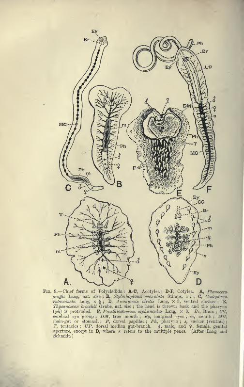
Ph--k=^A.-^
Fig. 8.—Chief forms of Polycladicla : A-C, Acotylea ; D-F, Cotylea. A, Planncera
graffii Lang, iiat. size ; B, Stylocho2)lana maculata Stimps, x 7 ; C, Cestoplanaruhrocincta Lang, x f ; D, Anonymus virilis Lang, x 3, ventral surface ; E,
Thysanozoon hrocchii Grube, nat. size ; the head is thrown back and the pharynx{ph) is protruded. F, Prosthiostovium siphunculus Lang, x 3. Br, Brain ; CG,cerebral eye group ; DM, true mouth ; Ey, marginal eyes ; m, mouth ; MG,main-gut or stomach ; P, dorsal papillae ; Ph, pharynx ; s, sucker (ventral)
;
T, tentacles ; UP, dorsal median gut-branch. S, male, and ?, female, genital
aperture, except in D, where S refers to the multiple Irenes. (After Lang andSchmidt.
)

CHAP. I CLASSIFICATION OF POLYCLADIDA 19
Classification of Polycladida.
ACOTYLEA.
Family.
Planoceridae.With dorsal tentacles.
Mouth sub-central.
Leptoplanidae.Without dorsal tenta-
cles. Penis directed
backwards.
Cestoplanidae.No tentacles. Bodyelongated. Penisdirected forwards.
Genus.
Planocera (Fig. 8, A),
Imoginc.Conoceros.
Stylochus.
Stylochoplana (Fig. 8, B).
Diplonckus.Planctoplana.
r Discocelis.
Crijptocelis.
- Leptoplana. .
Trigonoporus.
1 1 Polyp)ostia (see p. 27).
(Gestoplana (Fig. 8, C).
In Mediterranean andon French side of theChannel.
British Representatives.
Planocera foliuvi Grube.Berwick-on-Tweed.
Stylochoplana maculataQuatref. Among brownweeds in Laniinarian zone.
( Leptoplana tremellaris 0. F.
I
Mlill.
I
Z./a/^aaj Quatref. Plymouth.L. droebachensis Oe. Ply-
mouth Sound.L. atomata 0. F. Miill.
Doubtful species.
Enantiidae.No sucker No tentacles,
j^,,^,,^,-^^
Main-gut very short.-^ ^^^.^^.^ g^^^
External apertures as
in Eurylcptidac.
COTYLEA.
Anonymidae. i
Mouth central. No] Anonymus (Fig. 8, D).
tentacles. With twoj
Naples (two specimens),
rows of penes. '^
Pseudoceridae.Marginal tentaclesfolded. Mouth in
| yanterior halt. I
"^
jThysaiiozoon (Fig. 8, E).
Pseudoceros.
EURYLEPTIDAE.Tentacles usually pre-
sent and pointed, or
represented by tAvo
groups of eyes. Mouthclose to anterior end.
Pharynx cylindrical.
Prosthiostomatidae.Tentacles absent. Bodyelongated. Pharynxlong, cylindrical.Penis with accessory
muscular vesicles.
Prostheceraeus.
Cyelop)orus'.
Burylepta.
I Oligocladus.
Stylostomuvt,
y AcerOS.
Prosthiostomurn (Fig. 8,
F).
Prostheceraeus vittatus Mont.On west coast.
P. ar^ws Quatref. Guernsey.Cycloporus papillosus Lang.On Ascidians in 2-30 fms.
Eurylepta cornuta 0. F. Miill.
On sponges and shells,
2-10 fms.
Oligocladus sanguinolentus
Quatref.
0, auHtus Clap. Doubtful.
Stylostomum variabile Lang.
20 PLATYHELMINTHES—TURBELLARIA chap.
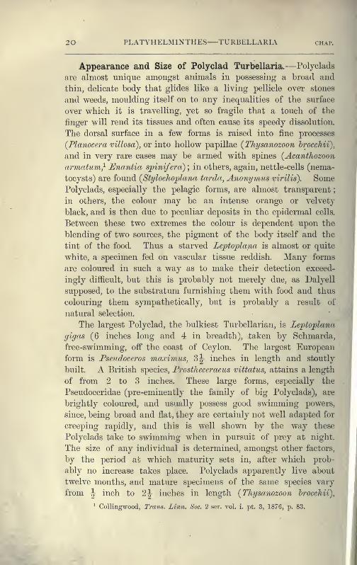
Appearance and Size of Polyclad Turbellaria.—Polyclads
are almost unique amongst animals in possessing a broad and
thin, delicate body that glides like a living pellicle over stones
and weeds, moulding itself on to any inequalities of the surface
over which it is travelling, yet so fragile that a touch of the
finger will rend its tissues and often cause its speedy dissolution.
The dorsal surface in a few forms is raised into fine processes
(Planocera villosa), or into hollow papillae (^Thysanozoon hrocchii),
and in very rare cases may be armed with spines (AcantJiozoon
armatum} Enantia spinifeirc) ; in others, again, nettle-cells (nema-
tocysts) are found {Stylochoplana tarda, Anonymus virilis). SomePolyclads, especially the pelagic forms, are almost transparent
;
in others, the colour may be an intense orange or velvety
black, and is then due to peculiar deposits in the epidermal cells.
Between these two extremes the colour is dependent upon the
blending of two sources, the pigment of the body itself and the
tint of the food. Thus a starved Leptoplana is almost or quite
white, a specimen fed on vascular tissue reddish. Many forms
are coloured in such a way as to make their detection exceed-
ingly difficult, but this is probably not merely due, as Dalyell
supposed, to the substratum furnishing them with food and thus
colouring them sympathetically, but is probably a result of
natural selection.
The largest Polyclad, the bulkiest Turbellarian, is Leptojplana
gigas (6 inches long and 4 in breadth), taken by Schmarda,
free-swimming, off the coast of Ceylon. The largest European
form is Pseudoceros maximus, 3^ inches in length and stoutly
built. A British species, Frostheceraeus vittatus, attains a length
of from 2 to 3 inches. These large forms, especially the
Pseudoceridae (pre-eminently the family of big Polyclads), are
brightly coloured, and usually possess good swimming powers,
since, being broad and flat, they are certainly not well adapted for
creeping rapidly, and this is well shown by the way these
Polyclads take to swimmiTig when in pursuit of prey at night.
The size of any individual is determined, amongst other factors,
by the period ab which maturity sets in, after which prob-
ably no increase takes place. Polyclads apparently live about
twelve months, and mature specimens of the same species vary
from ^ inch to 2^ inches in length {Thysanozoon hrocchii),
^ Collingwood, Trans. Linn. Soc. 2 fser. vol. i. pt. 3, 1876, p. 83.
HABITS OF P0LYCLADII!)A 2 I
showing that growth is, under favourable conditions, very
rapid.
Habits of Polyclad Turbellaria.—Polyclads are exchisively
marine, and for the most part littoral, animals. Moreover, there
is no evidence of their occurrence in those inland seas where
^^ certain marine animals (including one or two species of other-
^Hwise characteristically marine Ehabdocoelida, p. 46) have per-
sisted under changed conditions. From half-tide mark down to
50 fathoms, some Polyclads probably occur on all coasts, but as
to their relative abundance in different seas we have very little
accurate information. The southern seas of Europe possess moreindividuals and species than the northern, and probably the
maximum development of the group takes place on the coasts
and coral islands of the tropics.^ No Polyclads have been taken
below 6 fathoms ; but their delicacy and inconspicuousness render
this negative evidence of little value. Six truly pelagic forms,
however, are known,^ and these are interesting on account of their
wide distribution (three occurring in the Atlantic, Pacific, andIndian oceans), and also from the distinct modifications they
have undergone in relation to their pelagic existence.
Whatever may be the interpretations of the fact, Polyclads
are notoriously difficult to detect, and this fact doubtless explains
the scanty references to them by the older naturalists who col-
lected even in tropical seas. Lang, who worked seven years at
Naples, added to the Mediterranean fauna as many Polyclads as
were previously known for all Europe, in spite of the assiduous
labours of his predecessors, Delle Chiaje and Quatrefages.
Again Hallez, collecting at Wimereux at low -water, obtained
some twenty specimens of Lepto'plana tremellaris in an hour,
while some other collectors working by his side could only find
two or three. Yet, even making allowance for the difficulty of
finding Polyclads, few of them appear to be abundant.
Leptoplana tremellaris is frequently associated with colonies
of Botryllus, and if separated soon perishes, whereas the free-
living individuals are distinctly hardy (Hallez). A closely allied
but possibly distinct form lives upon the surface of the Polyzoon
^ Von Stummer-Traunfels, Zcitschr. /. iviss. ZooL Bd. Ix. 1895, p. 689.
^ Planoccra inllucida Mertens, P. simrothix. Grff., P. gnibei Grft"., Stylocho-
jjlana sargassicola Mertens, S. californica Woodworth, Plancto2)lana challcngeri
Grff., all belonging to the Planoceridae. See v. Graff, " Pelagische Polyeladen,"
Zcitschriftf. iviss. Zoologie, Bd. Iv. 1892, p. 190.
2 2 PLATYHELMINTHES TURBELLARIA chap.
Schizoporella, on the French side of the Channel, and cannot
long endure separation from its natural habitat, to which it
is adaptively coloured. A striking case of protective mimicry^
is exhibited by Cydoporus papillosus, on the British coasts.
This species, eminently variable in colour and in the presence or
absence of dorsal papillae, is usually a quarter of an inch in
length and of a firm consistency. Fixed by its sucker to Poly-
clinid and other Ascidians, Cycloporus appears part and parcel of
the substratum, an interesting parallel to Lamellaria persioicua}
though we are not justified in calling the Polyclad parasitic.
Indeed, though a few cases of association between Polyclads and
large Gasteropods, Holothurians, and Echinids are known,^ there
is only one case, that of Planocera inquilina^ in the branchial
chamber of the Gasteropod Sycotypus canaliculatus, which would
seem to bear the interpretation of parasitism. The jet-black Pseudo-
ceros velutinus and the orange Yu7igia aurantiaca of the Medi-
terranean, are large conspicuous forms with no attempt at con-
cealment, but their taste, which is not known, may protect them.
Other habits, curiously analogous with devices employed by
Nudibranch Mollusca (compare Thysanozoon hrocchii with Aeolis
papulosa), emphasise the conclusion that the struggle for exist-
ence in the littoral zone has adapted almost each Polyclad to its
particular habitat.
As regards the vertical distribution of this group on the
British coasts, Leptoplana tremellaris has an extensive range, and
appears to come from deeper to shallower water to breed.'^ In
the upper part of the Laminarian zone, Cycloporus papillosus,
and, among brown weeds, Styloclioplana maculata are found. Atand below lowest water-mark Prostlieceraeus vittatus, P. argus,
and Eurylepta cornitta occur. Stylostomum variahile and Oligo-
cladus sanguinolentus, though occasionally found between tide-
marks, especially in the Channel Islands, are characteristic, along
with Leptoplana droebacliensis and L. fallax, of dredge material
from 10 to 20 fathoms.
Locomotion.—Locomotion is generally performed by Poly-
clads at night when in search of food, and two methods, creeping
^ Camhridge Natural History, vol. iii, jx 74.
2 Lang, "• Pohjdadm," p. 629.
^ Wheeler, Journal of Moriiliolocjy, vol. ix. part 2, 1894, p. 195.•^ Many Nudibranchiate Mollusca undergo this change of habitat. See Garstan^
Journal of the Marine Biological Assoc, n.s. i. No. 4, 1890, p. 447.
HABITS OF POLYCLADIDA 23
and illy employed-swimnimg, are usually empioyea—creeping by the cilia,
aided possibly, as in the case of some Gasteropod Mollusca, by
the longitudinal muscles of the ventral surface ; and swimming,
by undulations of the expanded margins of the body. In the
former case the cilia work in a glandular secretion which bathes
the body, and enables them to effect their purpose equally well
on different substrata. The anterior region is generally lifted
up, exploring the surroundings by the aid of the tentacles, which
are here usually present. The rest of the body is closely appressed
to the ground.
Swimming is particularly well performed by the Pseudoceridae,
certain species of Prostheceraeus, the large Planoceridae, some
Stylochoplana, Discocelis, and Leptoplana, and in the same manner
as in Lepto'plana tremellaris (p. 9). In Cryptocelis, Lej)to'plana
alcinoi, and L. pallida, how-
ever, the whole body executes
serpentine movements like an
active leech (e.g. JVephelis) ; a
cross section of the body would
thus present the same appear-
ance during the whole move-
ment. Many Polyclads, notably
Anonymus (Lang), if irritated,
spread out in all directions,
becoming exceeding thin and
transparent.
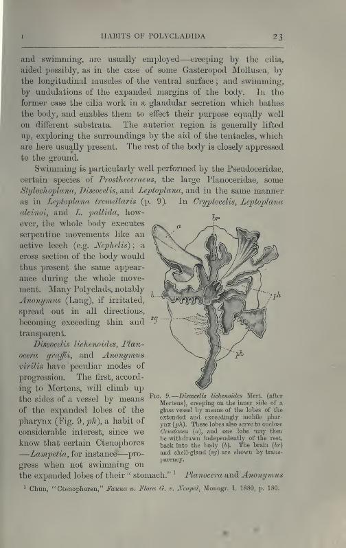
Discocelis lichenoides, Plan-
ocera graffii, and Anonymusvirilis have peculiar modes of
progression. The first, accord-
ing to Mertens, will climb up
the sides of a vessel by means
of the expanded lobes of the
pharynx (Fig. 9,;ph), a habit of
considerable interest, since weknow that certain Ctenophores
—Lampetia, for instance—pro-
gress when not swimming on
the expanded lobes of their " stomach." ^ Planocera and Anonymus
^ Chun, "Ctenophoren," Fauna u. Flora G. v. Xeapel, Monogr. I. 1880, p. 180.
Fig. 9.
—
Discocelis lichenoides Mert. (after
Mertens), creeping on the inner side of a
glass vessel by means of the lobes of the
extended and exceedingly mobile phar-
ynx {ph). These lobes also serve to enclose
Crustacea (a), and one lobe may then
be withdrawn independently of the rest,
back into the body (6). The brain {hr)
and shell-gland {sg) are shown by trans-
jiarency.
24 PLATYHELMINTHES TURBELLARIA chai-.
creep by extending parts of the ^anterior margin and dragging
the rest of the body behind. In consequence, the brain and
dorsal tentacles may come to lie actually behind the middle of
the body, and thus no definite anterior end or " head " advances
first. Along with this curious habit it may be noticed (Lang)
that the radial symmetry of the body is well marked ; but even
without accepting this author's suggestion of the concurrent
development of a '' head " with locomotion in a definite direction,
the facts, whether these two forms are primitive or not, are
highly interesting.
Food.—Though we are probably right in calling Polyclads a
carnivorous group, the food of very few forms has been ascer-
tained. Those which possess a large frilled pharynx (most
Acotylea) probably enclose and digest large, and, it may be,
powerful prey, as appears to be the case in Leptoplana tremel-
laris. Cryptocelis alba has been seen by Lang with the pharynx
so distended, owing to a large Drepanophorus (Nemertine) which
it contained, as to resemble a yolk-sac projecting from the under
surface of an embryo. The Cotylea such as Thysanozoon, with a
bell- or trumpet-shaped pharynx, are fond of fixing this to the side
of the aquarium, but whether they thus obtain minute organisms
is not clear. Prostliiostomum shoots out its long pharynx with
great vehemence (Fig. 8, F) and snaps up small Annelids by its
aid (Lang). Those Polyclads which, as Cycloporus and others,
are definitely associated with other organisms are not certainly
known to feed upon the latter,
---TN ^.^ though " Planaria velellae " has
/m---.A been seen by Lesson ^ devouring
^^^'^^\'''''^J^^^Ss^''"^^^iQ(-' ^^^ fleshy parts of its host. The
/^~^^2^ 'W- '-'^Xs^^iv^^y glands which open on
^_».J^'"" \^);;^^/\^_^/^ the lips and the inner sur-
' ^Sirl/ ^^^® ^^ ^^^^ pharynx powerfully
-^ disintegrate the flesh of the prey.
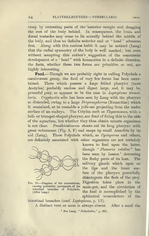
Fig. 10.—Diagram of the musculature, Bigestion takes place in thecausing peristaltic movements of the main-aut, and the circulation ofintestinal branches of Polyclads.
r. i • t i i i
(After Lang.) the lood IS accomplished by the
sphinctral musculature of the
intestinal branches (conf. Leptoplana, p. 13).
A distinct vent or anus is always absent. After a meal the
^ See Lang, " Polycladen," p. 607.

I ANATOMY OF POLYCLADIDA 25
faecal matter collects in the main-gut, and is discharged violently
hj the pharynx into the water. In a few species, however,
the intestinal branches open to the exterior (Jl,ang). Yungiaaurantiaca, a large and abundant Il^eapolitan form, possesses
such openings over the greater part of the dorsal surface
;
Cycloporus papillosus has marginal pores ; Oligocladus sangvAno-
lentus apparently possesses an opening at the posterior end of
the main-gut ; and Thysanozoon hrocchii frequently rends at this
point, in consequence of the accumulation of food.
Respiration.—^The oxygen of the atmosphere dissolved in
the sea-water is, in default of a special circulatory fluid, brought
to the tissues of Polyclads in two ways. The ciliated epidermis
provides a constant change of the surrounding water, by whicli
the superficial organs may obtain their supply ; and the peri-
staltic movements of the digestive system, aided by the cilia of
the endoderm cells, ensure a rough circulation of the sea-water,
which enters along with the food, to the internal organs. Thepapillae of Tliysomozoon hrocchii, containing outgrowths of the
intestinal branches, are possibly so much additional respiratory
surface, although still larger forms ("other Pseudoceridae) are
devoid of such outgrowths.
Excretion.—The excretory system of only one Polyclad
(Thysanozoon hrocchii) is accurately known. Lang, by compressing
light-coloured specimens, found the three parts of the system
known to occur in many Platyhelminthes : (1) the larger longi-
tudinal canals, and (2) the capillary vessels, which commence with
(3) the flame-cells in the parenchyma of the body. The mode of
distribution of these parts is not, however, ascertained. The canals
are delicate, sinuous, apparently intracellular tubes, coursing close
to the margin of the body and sending offsets which suspend the
canals to the dorsal surface, where possibly openings may occur.
In dilatations of these vessels bunches of cilia, and occasionally
flame-cells, are found. Usually, however, flame-cells occur at the
commencement or during the course of the capillaries, which are
straight, rarely branching, tubes of exceeding tenuity, and
appear (Lang) to be outgrowths of the flame-cells, just as the
duct is an outgrowth of a gland-cell. In fact there is little doubt
that the stellate flame-cells are modified parenchymatous gland-
cells, containing a lumen filled with a fluid into which a number
of cilia project and vibrate synchronously. The cells excrete
26 PLATYHELMINTHES TURBELLARIA chap.
iiitrogeneous waste substances, which are then discharged into the
capillaries, whence the cilia of the main vessels drive them pre-
sumably to the exterior, though external openings of the excretory
system are not known. Traces of this system have been observed
in young Leptoplana (first by Schultze in 1854) and also in
Cestoplana.
Sensation.—A nervous sheath, with scattered ganglion cells,
everywhere underlies the musculature. It is exceedingly faintly
marked on the dorsal surface, but laterally and ventrally forms
a dense network with polygonal meshes. Thickenings of this
sheath give rise to lateral nerves, and also to a pair of stout longi-
tudinal nerves from which the internal organs are probably in-
nervated. The brain, hardly distinct in pelagic Polyclads, in most
forms does not differ greatly from that of Leptoplana (p. 13).
The sense organs of Polyclads have the form of tentacles,
eyes, otocysts (in Leptoplana otophora), and stiff tactile cilia. The
solid dorsal tentacles of Planoceridae contrast strongly with the
folded or pointed hollow processes of the Cotylea. The former
(Fig. 8, A, T) are muscular and very contractile, and are placed
near the brain some distance from the anterior end. The latter
are outgrowths of the front margin of the body, and are some-
times {Yungict) provided superficially with olfactory pits and
internally with eyes and intestinal coeca.
The eyes which occur in Polyclads may be divided into {a) a
pair of cerebral groups overlying the brain ; (J)) those embedded
in the tentacles (tentacular group) ; and (c) the marginal eyes,
which in Anonymus occur all round the margin. A complex
form is sometimes assumed by the cerebral eyes of Pseudoceridae,
resulting probably from incomplete fission (Fig. 11). Lepto-
plana otopJiora was obtained by Schmarda on the south coast of
Ceylon. On each side of the brain is a capsule containing two
otoliths. This is the only known case of the occurrence of
these organs in Polyclads.
Reproduction.—Although Polyclads are able to repair the
result of injuries to a very considerable extent, they are not knownto multiply asexually. The two processes are intimately associated,
but, though probably all Turbellaria can regenerate certain lost
parts, asexual reproduction only occurs sporadically.
All known. Polyclads are hermaphrodite. The male organs,
scattered, like the testes of Leptopla7ia, over the ventral siurface,

ANATOMY OF POLYCLADIDA 27
develop earlier than the ovaries, though the periods of maturation
overlap ; hence the possibility of self-fertilisation, though remote,
is still worth consideration. The genital apertures, through
which, in the male, spermatozoa, and in the female, ova, are
emitted, are usually situated as in Lejitoplaiia (Figs. 2 and 5,
S and ? ). In Trigoiioporus, a genus once found at Xaples, a
secondary female aperture has been discovered leading into the
female genital canal ^ ; and in Anonymus, Polypostia, and Thysa-
nozoon (Fig. 7, E, <^) two or more male pores and penes have
been found. Anonymus has several penes (Fig. 7, 1), ^)arranged radially round the body. Folypostia, a remarkable form
described by Bergendal,^ belonging to the Acotylea, possesses
Fig. 11.—Double eye from the cerebral group of Pseudoceros maximus. (After Lang.)
about twenty such structures ranged round the female genital
aperture. Lang, whose attention was attracted by these singular
facts, made the interesting discovery that Thysaiiozoon uses its
penes as weapons of offence rather than as copulating organs,
burying them in the skin of another Polyclad (Yicngia) that
happened to cross its path, spermatozoa being of course left in
the wound. Lang further fovmd that Prostheceraeus cdhocinctus
and Cryptocelis alha in this way implanted a spermatophore in
the skin of another individual of the same species, and he
suggested that from this point the spermatozoa wandered through
the tissues till they met with and fertilised the eggs. It
is now known that a similar process of " hypodermic impreg-
nation " occurs sporadically in several groups of animals.^
^ Lang, '' Pohjcladen,'' PL 30, Fig. S.
- Kongl. Fysiograf. Sdllskajicts Haiidlingar, Bd. iv. Lund, 1892-93.
^ Whitman, Journal of Morphology, vol. iv. 1890, p. 361.
2 8 PLATYHELMINTHES—TURBELLARIA chap.
Nevertheless, in some Polyclads it is probable, and in Stylo-
cli'iis neapolitanus it is certain, that normal copulation takes
place. The sperm-masses are transferred to a coecal diverti-
culum of the female genital canal, and then by a delicate
mechanism, of which we know only the effects, one sperma-
tozoon obtains entrance into one matured ovum, which differs
from the ova of most Turbellaria in that it contains in its ownprotoplasm the yolk necessary for the nutrition of the embryo.
In other words, there are no special yolk-glands. After fertilisa-
tion, the ovum in all Polyclads is coated with a shell formed by
the shell-gland, which also secretes a substance uniting the eggs
together. They are deposited on stones and shells, either in
plate-like masses or in spirals (like those of Nudibranchs).
Cryptocelis alba lays masses of an annular shape, with two ova
in each shell, and buries them in sand.
Development.^—The first stages in the embryology of
Polyclads appear to be very uniform. They result, in all
Cotylea and in certain Planoceridae, in the formation of a
Miiller's larva (Fig. 12) about a couple of weeks after the eggs
are laid. This larva (1-1 '8 mm. long), which is modified in the
Planoceridae, is distinguished by the presence of a ciliated band,
running somewhat transversely round the body, and usually
produced into a dorsal, a ventral, and three pairs of lateral
processes. When swimming the body is placed as in Fig. 12,
and twists round rapidly about its longitudinal axis by means
of the strong locomotor cilia placed in transverse rows upon
the processes. The cilia of each row vibrate synchronously, and
recall the action of the swimming plates of a Ctenophore. It
is noteworthy that whereas Styloclius ^9^7^6?m??i passes through
a modified or, according to some authors, a primitive larval
stage, its near ally, S. neapolitanus, develops directly. Most
^ A full account of Polyclad development is contained in Lang's " Polydaden,"
with references to the literature of the subject. Since the date of that work (1884)
the embryology of Ctenoi^hora has become better known, but, though the segment-
ation of the egg and early stages of development are very similar in both cases,
the elaborate investigations of E. B. Wilson {Journ. Morphology, vol. vi. p. 361)
show that the segmentation of Polychaet worms is again similar. The question ofthe
affinities of the Polycladida is also discussed by Lang {^^ Polydaden," p. 642 et seq.).
The work of the last decade has neither proved nor disproved his suggestion that
the Ctenophores and Polyclads have been derived from common ancestors. On this
subject the remarks made by Hatschek {Lehrhuch d. Zoologie, p. 319) are some of
the weightiest that have appeared.

DEVELOPMENT OF POLYCLADIDA 29
and their free-swiiiiiiiiiig youngAcotylea indeed develop directly
differ from Midler's larva merely in the absence of the ciliated
band and in the mode of swimmino;.
Fig. 12.— Section throughMiiller's larva of Thy-sanozoon brocchii(modified from Lang).
The right half is seen
from inside. x 150.
Semi-diagrammatic.hr, Brain ; dl, dorsal
ciliated lobe ; dr, saliv-
ary gland-cells of phar-
ynx ; e, eye ; ej), cili-
ated epidermis contain-
ing rhabdites ; 7ng,
stomach or main-giit;
mg^, unpaired gutbranch over the brain
;
mo, " mouth " of larva
;
n, ?ij, section of nerves;
oe, ectodermic pitforming oesophagus of
larva; ixvr, paren-
chyma filling the space
between the alimentary
tract and the bodywall
;ph, pharynx
g in tlie cavity of the peripharyngeal sheath, the nuclei of which are visible ; sly,
sl-i, lateral ciliated lobes of the riglit side ; rl ventral ciliated lobe.
pcur
lyiu
SI2,
Fig. 13.—Diagrammatic transverse sections
of a larval Polyclad at difi"erent stages,
to illustrate the development of the
pharynx. (After Lang.) A, Larva of the
eighth day still within the shell. Themain-gut (m(/) is still solid, the epidermis
is slightly invaginated, and a pair of mus-cular mesodermic thickenings (7ns) are
present. B, Young pelagic larva. Theepidermic invagination has deepened
and developed laterally. C, The lateral
pouches have formed the wall of the
peripharyngeal sheath, enclosing the
mesodermic, muscular, thickening or
pharyngeal fold {ph). (Compare Fig. 1 2.
)
Towards the end of larval life, whenthe ciliated processes {si, Fig. 12) haveaborted, the stage D is reached. Bythe opening outwards of the pharyngeal
sheath (ph.sh) the two apertures gjn, or true mouth, and w, or external mouth, are
formed, which together correspond with the oesophageal opening of the younger larva.
(Compare the transverse section in Fig. 5.)
Polyclads possess an undoubted mesoderm, which gives rise
to the muscles, the pharyngeal fold, and the parenchyma. The
ectoderm forms the epidermis, in the cells of which the rhab-

30 PLATYHELMINTHES—TURBELLARIA chap.
dites (Fig. 12) arise, apparently as so many condensed secretions.
From the ectoderm the brain arises as two pairs of ingrowths,
which fuse together, and from these the peripheral nervous system
grows out. Three pigmented ectoderm cells give rise, bydivision, to the eyes—an unpaired cell (Fig. 12, e) to the cere-
bral group of eyes, and the other two to the marginal and
tentacular groups. The copulatory organs apparently arise to a
large extent as ingrowths from the ectoderm, from which the
accessory glands (prostates, shell-glands) are also formed. Theendoderm forms the lining of the main-gut and its branches.
The pharynx is developed as in Fig. 13, which shows that the" mouth " of the young larva (C) does not correspond exactly with
that of the adult (D). The salivary glands arise from ectoderm
cells, which sink deeply into the parenchyma. The reproductive
organs (ovaries and testes) possibly arise by proliferation from
the gut-cells (Lang, v. Graff). The change from the larva to the
adult is gradual, the ciliary band being absorbed and the creep-
ing mode of life adopted.
Turbellaria. II. Tricladida.
The Triclads are most conveniently divided into three groups ^
;
(i.) Paludicola, the Planarians of ponds and streams;
(ii.) the
Maricola, the Triclads of the sea; and (iii.) Terricola or Land
Planarians. From the Polyclads they differ in their mode of
occurrence ; in the elongated form of their body and almost
constant, mid -ventral position of the mouth; in possessing a
single external genital pore (Monogopora) ; and in the production
of a few, large, hard-shelled eggs provided with food-yolk.
Occurrence of the Paludicola.—The Planarians of our ponds
and streams are the most familiar and accessible Turbellaria.
Their elongated, flattened bodies, and gliding movements, render
them conspicuous objects on the under surface of stones and on
the leaves of aquatic plants, where they live gregariously. The
variable Polycelis nigra (Fig. 14, H) is very abundant in stagnant
water and slowly-moving streams, whereas its ally, P. cormda
(Fig. 14, G), distinguished by a pair of tentacles, is more local.
Planaria {Dendrocoelum) lactea (A), P. polychroa (I), P. torva,
and P. 'punctata are not infrequently found together, but the last
^ Hallez, Revue Biologique du Nord de la France, torn. ii. 1889-90.
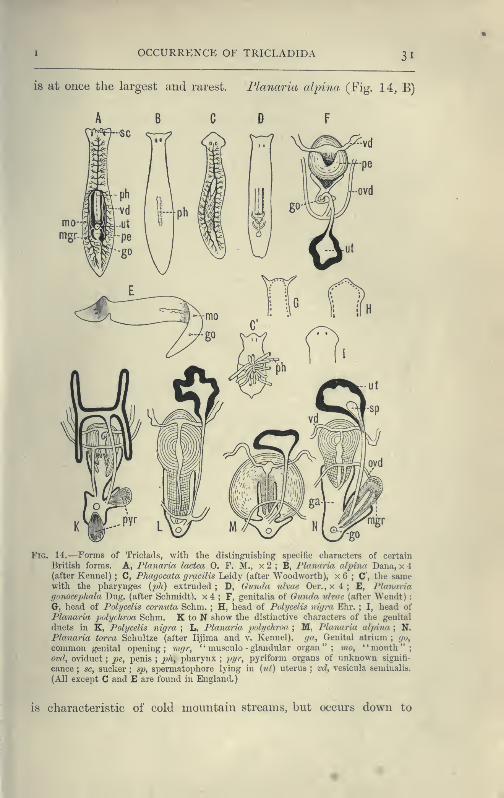
OCCURRENCE OF TRICLADIDA 31
is at oiice the largest and rarest. Flanaria cdpina (Fig. 14, B)
pe
Fig. 14,—Forms of Triclads, with the distinguishing specific characters of certain
British forms. A, Flanaria lactea 0. F. M., x 2 ; B, Planaria alpino Dana, x 4
(after Kennel) ; C, Pluigocata gracilis Leidy (after Woodworth), x 6 ; C, the samewith the pharynges {ph) extruded ; D, (wuncla ulvae Oer., x 4 ; E, Planariagonocepluda Dug. (after Schmidt), x 4 ; F, genitalia of Oiinda ulvae (after Wendt) :
G, head of Polycelis cornuta Schm. ; H, head of Polycelis nigra Ehr. ; I, head of
Planaria polychroa Schm. K to N show the distinctive characters of the genital
ducts in K, Polycelis nigra ; L, Planaria polychroa ; M, Planaria alpina ; N,
Planaria torva Schultze (after lijima and. v. Kennel), ga, Genital atrium;
go,
common genital opening; 7ngr, " musculo - glandular organ"; mo, "mouth";ovd, oviduct
;pe, penis
;ph, pharynx
;pyr, pyriform organs of unknown signifi-
cance ; sc, sucker ; sp, spermatophore lying in {tU) uterus ; vd, vesicula seminalis.
(All except C and E are found in England.)
is characteristic of cold mountain streams, but occurs down to

S2 PLATYHELMINTHES—TURBELLARIA chap.
sea-level in England, the Isle of Man, and Ireland, and from its
abundance in sjjring water, probably enjoys a wide distribution
underground. In the Swiss Alps it has been found at altitudes
of over 6000 feet, at lower levels in the Ehone, and also in the
I^ke of Geneva, This wide distribution may i>erhaj>s be accounted
for, partly, by its faculty for asexual reproduction in summer, and
also, by the production, later in the year, of hard-shelled eggs which
are laid logsely, not attached to stones or jjlants.^ But we have
no really direct evidence of the means of disjjersal of this or of
any of the foregoing species, although they all have a wide
distribution in Europe. Of extra-European forms the accounts
that exist are very fragmentary. The only indubitable diagnostic
character of a Triclad is the structure of its genital ducts, and
this is accurately known in only a few cases. Several species
such as Dicoti/lus pulvinccr (Fig. 16, B), at present known only
from Lake Baikal,^ and others {Planaria mrazekii, P. albissima)
from Bohemia,^ will doubtless be found elsewhere when they are
carefully looked for. Fhagocata gracilis is a remarkable North
American form, yjossessing several pharynges (Fig. 14, and C'),
recalling the independent movement of the pharyngeal lobes of
Discocelis lichenoides ^Fig. 9;.**
Occurrence of the Maricola.—Little as we accurately knowof the distriljution of the fresh-water Planariae, our knowledge
of the occurrence of the marine forms is still more limited,
Guncla {Procerodes) ulva^ (Fig. 14, D) is the commonest European
form, occurring abundantly in the upper part of the littoral zone,
on the shores of the Baltic. G. segmentata from Messina has
been carefully described by Lang,^ but these are almost the only
species of Maricola which can be accurately determined. They
differ from the Paludicola in the jjosition of tlie " uterus " behind
the genital pore and in the absence of a " musculo-glandular
organ " (Fig, 1 4, F). A sj^ecial interest attaches to the Bdellouridae,
a family containing three species, all parasitic on JAmulus from the
east coast of America. These remarkable Triclads usually have a
sucker at the hinder end of the body, by which they attach
themselves firmly to the cephalo-thoracic appendages and to the
^ Voigt, Zool. Am. xv, p. 238.
2 Grube, Archivf. Naturyeschichte, 38 Jahrg. Bd, i, 1872, p. 273.
3 Vejdovsky, Zeitschr. f. wiss. Zooloyie, Bd. Ix, 1895, p, 200.
"* Woorlworth, Bulletin Mus. Comp. Zoology, Harvard, vol. xxi. No, 1, 1891.
5 Mitth. Zool. Slat. Neapel, 1882, p. 187.

OCCURRENCE OF TRICLADIDA 33
gill-plates, upon which the eggs may be found in considerable
numbers. One species, Syncoelidium pellucidum, possesses a pair
of problematical organs in the hinder part of the body, opening
to the exterior ventro-laterally by a couple of chitinous mouth-
pieces, but having no connexion with the genital ducts.^
Occurrence and Distribution of Land Planarians.—The
terricolous Triclads or Land Planarians are the most interesting
division of the group. Some forms, such as Bipalium kewense,
attain large dimensions, being usually 6 to 9 inches in length,
and specimens fully extended have measured 18 inches. Their
bodies are frequently banded or striped with brilliant colours.
Geoplana coerulea Mos. has a blue ventral surface and is olive
green or dark Prussian blue above. G. splendens Dendy, is
marked dorsally by three stripes of emerald green alternating
with four dark brown longitudinal bands. The mode of colora-
tion, though somewhat variable, is an important specific char-
acter. Its significance, however, is not clearly imderstood. The
colours may be a warning signal, as some Geoplana at least are
disagreeable to the taste of man and some birds ^ ; but since
Land Planarians are largely nocturnal animals, living by day
under logs, banana leaves, and in other moist and dark situa-
tions, this explanation is clearly insufficient. Two Geoplaruc
have been noticed by Mr. Dendy which seem to be protectively
coloured. G. triangulata var. australis occurs abundantly in the
beech forest in the South Island of New Zealand, and its brown
back and yellow or orange ventral surface match the leaves around
its haunts. G. gelatinosa again looks like a mere slimy patch on
the rotten bark where it is found. In arid districts, during the
dry season. Land Planarians burrow in the soil and form a cyst,
in which they lie coiled up, after the manner of earthworms.^
The glutinous investment of their delicate bodies forms a moist
medium in which the cilia covering the body (and especially the
ventral surface) may constantly and evenly vibrate, and by
which they adhere firmly to their prey. In some tropical Plan-
arians, in addition to possessing offensive properties, the mucus
is so copious in amount and hardens with such rapidity, that
^ Wheeler, Journal of Morphology, vol. ix. 1894, p. 167.
2 Dendy, Trans. Roy. Soc. Victoria 1890, p. 65 ; Id. Austral. Assoc. Brisbane,
1895, "Presid. Add. to Sect. D," p. 15.
^ Darwin, Aim. and Mag. Nat. Hist. vol. xiv. 1844, p. 241.
VOL. II D
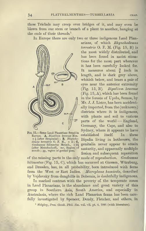
34 PLATYHELMINTHES TURBELLARIA CHAP.
these Triclads may creep over bridges of it, and may even be
blown from one stem or branch of a plant to another, hanging at
the ends of their threads.^
In Europe there are only two or three indigenous Land Plan-
arians, of which Rhyncliodemus
terrestris 0. Y. M. (Fig. 15, B) is
the most widely distributed, and
lias been found in moist situa-
tions for the most part wherever
it has been carefully looked for.
It measures about £ inch in
length, and is dark grey above,
whitish below, and bears a pair of
eyes near the anterior extremity
(Fig. 15, B). Bipalium keivense
(Fig. 15, A), which has been found
in the forests of Upolu, Samoa, by
Mr. J. J. Lister, has been accident-
ally imported, from the (unknown)
districts where it is indigenous,
with plants and soil to various
parts of the world— England,
Germany, the Cape, and also to
Sydney, where it appears to have
established itself. In these
X A (after Bergendai); B, Rhyncho- Bipalia living in hothouses, the
demus terrestris 0. F. M., x 2 ; C, . ,.
2^ genitalia never appear to attain
Fig. 15.—Some Land Planarians found in
Europe. A, Bi^Jalimn kewense Mos.
Geodesmus hilinecdus Metsch.
(after MetschnikoflF). mr Region of maturity, and apparently multiplemou til
; ^jj, region of genital pore.. 7
fission and subsequent reparation
of the missing parts is the only mode of reproduction. Geodesmus
hilineatus (Fig. 15, C), which has occurred at Giessen, Wtirzburg,
and Dresden, has, in all probability, been introduced with ferns
from the West or East Indies. Microplana humicola, described
by Vejdovsky from dunghills in Bohemia, is doubtfully indigenous.
In marked contrast with the poverty of the temperate zones
in Land Planarians, is the abundance and crreat variety of this
group in Southern Asia, South America, and especially in
Australasia, where the rich Land Planarian fauna has been care-
fully investigated by Spencer, Dendy, Fletcher, and others, in
^ Sliipley, Proc. Camb. Phil. Soc. vol. vii. pt. 4, 1891 (with literature).
TRICLADIDA 3 5
certain parts of Victoria, New South Wales, and New Zealand.^
About forty species of Planarians have been discovered on the
^ft Australian continent, thirty-five of which belong to the pre-^^ dominant genus Geoplana, distinguished by the presence of
numerous eyes along the border of the simple anterior extremity.
Of the remaining five, four belong to the genus Rhynchodemus,
with, lastly, the introduced Bipalium kewense. The distribution
of any one species, however, is so limited that only three forms
are common to the two former colonies ; and although some of
the twenty known New Zealand Planarians (chiefly species of
Geo^plana), are identical with Australian species, yet only one, or
possibly two, varieties of these species are Australian also. In
addition to their prevalence in Australasia, the Geoplaniclae also
occur in South America, South Africa, Japan, and the East Indies.
The Bipaliidae are characteristic of the Oriental region, being
found in China, Borneo, Bengal, and Ceylon. The Ehynchode-
midae are a cosmopolitan family, occurring in Europe, North and
^1 South America, the Cape of Good Hope, Ceylon, the East Indies,
^* Australia (particularly Lord Howe Island), and Samoa.^
Habits and Structure of Triclads.—The common Planaria
{Dendrocoelum) lactea, which usually progresses by ciliary action,
aided, it is said, by muscular contractions of the ventral surface,
performs, if alarmed, a series of rapid " looping " movements, byaffixing a sucker (Fig. 14, A, sc), placed on the under side of the
head, to the substratum, and pulling the posterior end close to
this. The sucker, discovered by Leydig, is even better developed
in P. punctata (Fig. 16, A), P. mrazekii, and P. cavatica, and is
an efficient adhering-organ which has probably been developed
from a similar but simpler structure found in a considerable
number of both fresh-water and marine Triclads (P. alpina. Fig.
16, E). Probably the sucker of the Land Planarian Cotylo2')lana
(D) is the same structure, but the two suckers of Dicotylus (B)
are at present unique. Planaina dioica, found by Claparede on
the coast of Normandy,^ is covered with minute adhesive papillae,
^ Trans. Boy. Soc. Victoria from 1889 onwards. Trails. New Zealand Institute,
1894-95.
2 Moseley, Phil. Trans. 1874, p. 105 ; Id. Quart. Journ. Micr. Sci. vol. xlvii.
1877, p. 273 ; Loman, Bijdrag tot d. DierJcunde, Aflev. 14, 1887, p. 71 ; Id. Zool.
Ergeh. ein. Reise i7i Nieder-Ost-Indien, Hft. 1, p. 131 ; Beddard, Zoogeography^
1895, p. 53.
^ Beohachtungen il. Anat. u. Entwickcl. an der Kuste von Normandie, 1863, p. 18.
36 PLATYHELMINTHES TURBELLARIA
similar to those of certain Ehabdocoelida (e.g. Monotus, Fig. 19/D), enabling it to cling tightly to the Zostera, and so to resist
the loosening action of the waves.
The movements of Land Planarians are somewhat peculiar.
The ventral surface of Bipalium has a median groove, into
which the ducts of numerous mucus -glands open. This is
bordered by two ridges clothed with long and powerful cilia,
which perform the chief part in propelling the animal, aided,
according to Lehnert,^ by muscular waves which pass from the
Fig. le.^Suckers of Tri-
clads. A, Planariapunctata Pall. ; «, dorsal
surface of head ; h, ven-
tral surface (freely mov-ing) showing the sucker
;
c, sucker contracted
(after Hallez) : B, ven-
tral surface of head of
Dicotyhis pulvinar Gr.,
from Lake Baikal (after
Grube): C, dorsal sur-
face of Procotylea Jluvi-
atilis Gir. (after Girard)
:
D, sucker of Cotyloplanawhiteleggei Sp. (after
Spencer) : E, ventral
view of head of Plan-aria alpina Dana (pre-
served specimen) ; hg,
adhering groove ; m,thickened musculature
forming the margin of
the sucker ; sc, sucker ;
t, tentacles.
head, backwards, i.e. opposite in direction to those by which • a
snail slides along. This observation, however, needs confirma-
tion. The whole body executes sinuous movements, during which
the crescentic head, lifted slightly above the ground (Fig. 15, A),
is constantly altering and regaining its normal shape, somewhat
as a Planaria lactea uses the lobes of its head. Further ex-
amination shows that the margin of the head of Bipalium is not
only provided with eyes, but in addition, with ciliated, (probably)
olfactory pits. Such depressions, innervated directly from the
cerebral ganglia, have been found in sixteen species of Geoplana,
'^ArcUvf. NaturgeschicUe, 57 Jahrg. Bd. i. Hft. 3, 1891, p. 308.

I HABITS OF TRICLADIDA 3)r
and in one or two species of Rhynchodemus} Some LandPlanarians (a species of Bhynchodemus from Ceylon, and a
DolicJioplana from the Philippines) wriggle out of a box or the
hand with great speed (Moseley).
The skin of Triclads is full of minute rods or rhabdites,
which are shot out in great numbers when the animal is irritated,
and doubtless serve an offensive purpose. The Terricola possess
two kinds of these: (1) needle-like rods; and (2) in Bipalium
keivense, flagellated structures, bent into a V-form and with a
slender thread attached to one end (Shipley). In Geo^plana
coerulea these bent rods furnish the blue colour of the ventral
surface. The rhabdites arise in all Triclads in cells below the
basement-membrane, which they are said to traverse in order to
reach the epidermis, thus differing in origin, and also in struc-
ture, from the rods of Polyclads.
Food.—Triclads are largely if not wholly carnivorous animals,
feeding upon Annelids, Crustacea, Insects, Insect-larvae, and Mol-
luscs. The mouth is usually mid-ventral or behind the middle
of the body, but in the anomalous Lemiaco'psis terricola Schm.
from the Andes ^ and in DolicJioplana it is near the anterior end.
The pharynx (Figs. 17, 18, ;ph) is cylindrical or bell-shaped,
exceedingly dilatable and abundantly supplied with glands and
nervous tissue. It opens into the three main intestinal branches,
one of which runs in the median plane forwards, the others back-
wards right and left, enclosing a space in which the genital ducts
lie (Figs. 14, A, 17). The fresh-water Planarians prey upon
Oligochaeta, Hydrophilidae (aquatic beetles), and the commoner
pond-snails. Bipalium hewense pursues earthworms, seizes the
upper surface of the anterior end by the glutinous secretion of its
ventral surface, and then proceeds to envelop part or the whole
of the worm within its pharynx, which is stretched as a thin skin
over the body of its struggling prey (Lehnert). The tissues of
the latter pass into the intestine of the Planarian, and distend it
greatly. After such a meal, which lasts from one to five hours,
a Bipalium may remain for three months without seeking food.
Geohia suhterranea, a white eyeless form from Brazil, pursues
earthworms (Lumhricus corethruriis) in their burrows, and has
been seen by Fritz Muller sucking the blood out of a young
^ Dendy, Froc. Hoy. Soc. Victoria^ vol. iv. n.s. i. 1892.
2 Schniarda, Ncue ivirbellose Thiere, Leipzig, 1859, I. i. p. 30.
38 PLATYHELMINTHES TURBELLARIA chap, i
worm.^ Geoplana typhlo'ps, a Tasmaniaii species, is also blind,
and pursues worms, as does G. triangulata (Dendy). In Trinidad,
von Kennel ^ observed that land-snails (Subulinae) were the food
of certain Land Planarians, the name of which, however, he does
not state. The pharynx was employed to suck out the soft parts
of the snail even from the upper whorls of the shell.
Reproduction.—In Planaria lactea the numerous testes (Fig.
17, te) are placed both above and below the alimentary canal
throughout the greater part of its course. The membraneof each gonad is continued into a minute vas efferens, which
unites with those of neighbouring testes. Two vasa deferentia
(v.d) arise thus on each side, one from the posterior, the other
from the anterior testes of the body, and open into the vesiculae
seminales (v.s), which may be seen in the living animal as tortu-
ous whitish tubes at the sides of the pharynx (Fig. 14, A). These
open into the penis (Figs. 14, A; l'l,pe), a large pyriform organ,
the apex of which, when retracted, points forwards, projecting
into the penial cavity. When this apical portion is evaginated
and turned inside out, it is of considerable length, and is able to
pass into the long slender duct of the uterus {ut) of another
individual. Tlie penial sheath {'ps) is part of the genital atrium
{gs), which is developed as a pit from the skin, and invests the
end of the genital ducts, the mouth of the pit forming the commongenital pore {go), through which both male and female genital
products are emitted.
There are two ovaries (ov) placed far forwards, between the
third and fourth pairs of intestinal coeca. The oviducts (ovcl) lie
just over the lateral nerves, and have a slightly tortuous course,
at each outward bend receiving the duct (ifo) of a yolk-gland
{yg), so that ova and yolk are already associated when the oviducts
open by a short unpaired tube into the genital atrium. The
yolk-glands develop rapidly,^ and when fully formed are massive
glands occupying the spaces between the intestinal branches and
the testes which are then aborting. The so-called uterus (ut),
apparently at first a diverticulum of the genital atrium, expands
' AWiaiidl. d. Naturf. Gesell. zu Halle, Bd. iv. 1857, p. 33.
- Arh. Zool.-Zoot. Instil. JViirzburg, Bd. v. 1882, p. 120.
^ Woodworth {loc. cit. p. 38) states that in Phagocata the yolk-glands arise
by proliferation from two parovaria, placed just in front of the ordinary ovaries,
lijima, however {Zeitschr. f. wiss. Zool. Bd. xl. 1883, p. 454), regarded them as
derivatives of the parenchyma.
'Ovd
2/0"-^
Tnsrr— -,:^
Fig. 17.—Diagrammatic view of thestructure of Planaria {Dendro-coditm) lactea. x 7. The body-lias been cut across and a por-tion removed. In the posterior
half the alimentary tract of theleft side is removed and theuterus, penis, and muscular organsliced open horizontally. Thenervous system is represented byblack, and the yolk - glands bydotted lines, br, Brain ; ey, eyewith lens and optic nerve
;go,
external genital aperture for bothmale and female products
;gs,
genital atrium; Ig, paired lateral
intestinal branch ; In, longitudinal
nerve ; mg, unpaired anterior
intestine, the branches of whichare cut off close to the mainstem ; mgr, eversible ' * musculo-glandular organ "
; nc, nerve-cells
in the pharynx ; nn, lateral
nerve -twigs ; ns, nerve -sheath;
ov, ovary ; ovd, oviduct; pe,
the eversible penis, the corru-
gated inner white portion of
which is the apex;ph, pharynx
;
phs, pharyngeal sheath;
pr," prostate " or granule-gland (re-
presented by dotted lines openinginto the penis)
;^w, penial sheath
;
te, testes ; to, tactile lobe of thehead ; ut, " uterus " opening into
the genital atriiim just abovemgr ; vd, vasa deferentia ; vs,
vesicula seminalis; yg, yolk-
glands;yo, openings of the yolk-
ducts into the oviducts.
4p PLATYHELMINTHES—TURBELLARIA CHi
behind the pharynx into a receptacle lined by long glandular
columnar cells, which, however, are not all of the same kind.
The uterine duct opens into the atrium just above the aper-
ture of a problematical, eversible, "musculo -glandular organ"
(mgr).
Fertilisation appears to occur in the uterus, where ova, yolk,
and spermatozoa, or (in P. torvd) spermatophores (Fig. 14, N, sp),
are found. The formation of the cocoon in Planaria lactea is
probably begun in the " uterus," but is undoubtedly completed
in the genital atrium. In P. polychroa, however, the stalked
cocoon is formed wholly in the "uterus." Thus we find two
types of cocoons in different species of the genus Planaria
associated with two types of reproductive organs (Hallez) :
—
I. Planariae in which the two oviducts open separately into
the posterior part of the duct of the uterus. A musculo-glandular
organ is absent. The cocoons are spherical . and stalked. Ex-
amples— Planaria polychroa (Fig. 14, L), P. alhissima, P.
gonocephala.
II. Planarjae in which the two oviducts open by means of an
unpaired duct into the genital atrium. A musculo-glandular
organ present (Planaria torva (Fig. 14, N), P. mrazekii, P. lactea,
P. cavatica), or absent {P. cdpina, Fig. 14, M). The cocoons
are sessile.
The genitalia of the Maricola (Fig. 14, F) and Terricola do
not differ very much from those of Planaria. The uterus (greatly
reduced in the Land Planarians) lies behind the genital pore,
and several ova, together with much milky yolk, are enclosed in
a capsule which is formed in the genital atrium.
Asexual Reproduction.^—It has long been known that fresh-
water Planarians have not only great powers of repairing injuries,
but that they use this faculty in order to multiply by transverse
fission. Planaria alpina and Polycelis cornuta^ in summer,
separate off the posterior part of the body, and this ultimately
becomes an entire individual. P. alhissima, and especially P.
suhtentaculata, anticipate matters so far, that before fission is
complete, the new individual has a head nearly fully formed.
The new organs are largely regenerated in both parent and young,
^ The extensive literature on this subject is fairly completely summarised by
Voigt in Biol. Centralhlatt, vol. xiv. Nos. 20, 21, 1894. Faraday's observations
(cf. p. 6, note 6) have been generally overlooked.
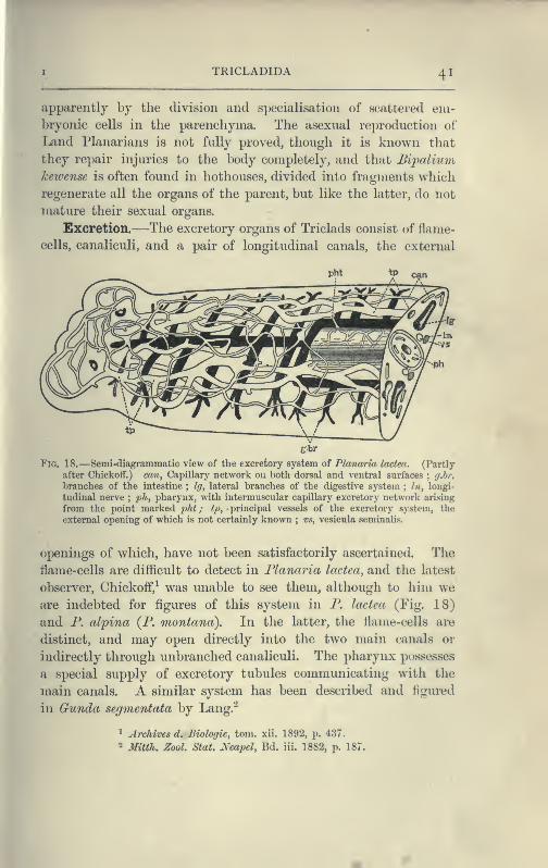
TRICLADIDA 41
apparently by the division and specialisation of scattered em-bryonic cells in the parenchyma. The asexual reproduction of
Land Planarians is not fully proved, though it is known that
they repair injuries to the body completely, and that Bipalium
kewense is often found in hothouses, divided into fragments which
regenerate all the organs of the parent, but like the latter, do not
mature their sexual organs.
Excretion.—The excretory organs of Triclads consist of flame-
cells, canaliculi, and a pair of longitudinal canals, the external
£rbr
Fig. 18.—Semi-diagrammatic view of the excretory system of Planarkt laden. (Partly
after ChickofF.) can, Capillary network on both dorsal and ventral surfaces ; ij.hr,
branches of the intestine ; Ig, lateral branches of the digestive system ; /», longi-
tudinal nerve;ph, pharynx, with intermuscular capillary excretory network arising
from the point marked 'pht ; tp, principal vessels of the excretory system, the
external opening of which is not certainly known ; vs, vesicula seminalis.
openings of which, have not been satisfactorily ascertained. The
flame-cells are difficult to detect in Flanaria Icictea, and the latest
observer, Chickoff,^ was unable to see them, although to him weare indebted for figures of this system in P. lactea (Fig. 18)
and P. alpina (P. montana). In the latter, the flame-cells are
distinct, and may open directly into the two main canals or
indirectly through unbranched canaliculi. The pharynx possesses
a special supply of excretory tubules communicating with the
main canals. A similar system has been described and figui-ed
in Gunda segmentata by Lang.^
^ Archives d. Biologie, torn, xii. 1892, p. 437.
- Mitth. Zool. Stat. Neapel, Bd. ill. 1882, p. 187.
42 PLATYHELMINTHES TURBELLARIA
Classification of Tricladida.
Family.
Planariidae
Procerodidae( = Gundidae).
Bdellouridae
BiPALIIDAEGeoplanidae .
Rhynchodemidak
^Belonging to undetermined Families
PALUDICOLA.Genus and British Species.
Planaria ladea 0. F. M., P. punctata Pall., P. polycliroaSchm., P. torva M. Sch., P. alpma Dana.
Polycells nigra Ehr., P. cornuta Schm.Anocelis.
Ol%goceUs.'\V>Q>\\SAiv\
Procotyla. /genera.Sorocelis.
Dicotylus.
MARICOLA.
Gunda) ulvae Oersted, P. Uttoralis vanProcerodes (
=
Beneden.Cercyra.
Uteriporus.
Bdelloura.Syncoelidiuvi.
TERRICOLA.
Bipaliiim ketvense Moseley (introduced).Geoplana.Geodesvius.
Rhynchodemus terrestris 0. F. M.
r Dolichoplana.
•JPolycladus.
I
Microplana.
I Leimacopsis.
Turbellaria. III. Rhabdocoelida.
The Ehabdocoelida include a very heterogeneous assemblage of
usually minute Turbellaria, distinguished collectively from the
Polyclads and Triclads by the form of the digestive tract. This
is a simple or slightly lobed sac, except in the Bothrioplanidae,
which in this and many other points closely resemble the
Triclads. It is to the straight, rod-like nature of the alimentary
canal that the name of the group refers. The size and form of
the body, and the structure of the pharynx and genitalia, vary
within wide limits.
The Ehabdocoelida are subdivided into three tribes :
—
(1) Acoela, in which a sub-central mouth and pharynx are
present, but lead into the parenchyma of the body, not into an
intestine with proper walls. An excretory system has not
hitherto been seen. Yolk-glands are absent. An otolith under-
lies the brain. The Acoela are marine.
RHABDOCOELIDA 43
(2) Bhdbdocoela, which possess a complete alimentary tract
separated from the body-wall (except for a few suspensory
strands) by a space or body-cavity, filled with fluid. This space
is sometimes ( Vortex viridis) lined by an endothelium of flattened
parenchymatous cells. There are two compact testes, which are
enclosed (as are the ovaries and yolk -glands) in a distinct
membrane. An otolith is present in some genera and species.
Terrestrial, fresh-water, marine.
(3) Alloeocoela, in which the body-cavity is greatly reduced.
Except in the Bothrioplanidae, the gonads have no distinct mem-brane. Testes numerous
;yolk-glands present. Marine with a
few exceptions.
Occurrence and Habits of the Rhabdocoelida.—The
Acoela are usually minute, active Turbellaria abounding amongst
weeds throughout the lower half of the Littoral, and the whole
of the Laminarian zone, but are most plentiful in the pools
exposed during spring-tides on our coasts, especially on the shores
of Devonshire. The species of Haplodiscus, however, and Convoluta
henseni are modified pelagic forms found in the Atlantic Ocean.^
Convoluta paradoxa (Fig. 19, B) is the commonest British species.
It is from 1 to 9 mm. in length, and of a brown colour, marked
above by one or more transverse white bars. The brown colour
is due to a symbiotic alga, the nature of which has not been
thoroughly investigated. In an allied species, however {C.
roscoffensis), from the coast of Brittany, the alga, which is here
green, has been carefully examined by Professor Haberlandt,^ and
it appears from his researches that the algae form a special assimi-
lating tissue, enabling the Convoluta to live after the fashion of
a green plant. At Eoscoff, these elongated green Convoluta live
gregariously in the sandy tide-pools, fully exposed to the sun's
rays, and have the appearance of a mass of weed floating at
the surface of the water. Access to the atmosphere and to sun-
light are necessary in order to enable the assimilating tissue to
form the carbohydrates, upon which this form lives exclusively.
Not only has the alga itself undergone such profound changes
(loss of membrane, inability to live independently after the death
of the host) as to disguise its true nature (a tissue-cell derived
from algal ancestors), but the Convoluta has also undergone con-
^ Bblimig, Ergehnisse d. Plankton Expedition, Bd. ii. H. g. 1895,
2 von Graff, Die Acoela, Leipzig, 1892. Appendix.

44 PLATYHELMINTHES TURBELLARIA chap.
comitant changes, in form, in the loss of a carnivorous habit,
and in the development of marked heliotropic movements, thus
adapting itself to an holophytic or plant-like mode of nutrition.
Nevertheless the Acoela, as a group, are carnivorous, feeding
upon Diatoms, Copepoda, and small Ehabdocoela, the absence of
a digestive tract indeed being probably more apparent than
real.^
The Bliahdocoela live under varied conditions. One form, Pro-
rhynchus sphyrocephalus, has been found among plants far from
water in the neighbourhood of Leyden, by De Man.^ With this
exception the group is purely aquatic, and though a few genera
and even individuals of the same species occur both in salt and
fresh water, whole sub-families and genera are either marine or
paludicolous. Among the latter, Mesostoma, Castracla, Vortex, and
Derostoma are common in brooks and ponds, especially at certain
times, often only for one month (May or June) in the year.
Species of Macrostoma, Stenostgma, and Microstoma are also
abundant in similar places. The two latter occur in chains
formed by fission ; but the sexual individuals (which are of
distinct sexes, contrary to the usual hermaphrodite condition of
Flat Worms) only appear at stated times and are not well
known. A large number of genera are purely marine, and one
family, the Proboscidae (distinguished by having the anterior
end invaginated by special muscles and converted into a sensory
.
organ), is entirely so. The most cursory examination of littoral
weeds reveals species of IfacrorJiynchus, Acrorhynclius, Promeso-
stoma, Byrsophlebs, and Proxenetes, the character of which maybe gathered from von Graffs great monograph, or from Gamble's
paper on the " British Marine Turbellaria." ^ Much, however,
still remains to be done before we possess an adequate idea of
the occurrence of this group on our coasts.
Some Ehabdocoels are parasitic. Fecampia erythrocepliala,
^ The development of the Acoela has been worked out recently by Mdlle. Pereyas-
lawzewa {Zapiski Novoross. Obshch. Odessa, 17 Bd. 1892) and Gardiner {Jour7ial
of Morphology, xi. No. 1, 1895, p. 155) with conflicting results. The former finds
four endoderm cells, which give rise to a larval intestine. The Acoela are for her,
Pseudacocla. Gardiner, on the other hand, finds no trace of an endoderm at any
stage of the development of Polychocrus caudatus.
' Tijdschr. Nederland. Dierk. Ver. Deel ii. 1875.
^ Von Graff, Monographie d. Turhellarien: I. Rhahdocoeliden, 1882. Gamble,
Quart. Journ. Microscop. Science, vol. xxxiv. 1893, p. 433.
I

HABITS OF RHABDOCOELIDA 45
which occurs in the lacunar spaces and alimentary canal of youngshore crahs (Carcinus maenas), is a white cylindrical animal1- inch long, with a red snout. After attaining maturity it
works its way out of the crab and encysts under stones,
forming a pyriform mass in shape like a " Prince Kupert's drop."
Within this case the eggs develop, and the young probabl}'
emerge through the open nar-
row end of the hard white
tube, but how they reach the
crab is not known. GraffiUa
muricicola is found in the
kidney of Murex hrandaris and
M. trunculus, at Naples and
Trieste; G. tethydicola in the
foot of Tethys. AnoplocUuiti
parasiticum occurs among the
muscles which attach the
cloaca of Holotliuria tuhulosa
to the body - wall ; and A.
schneideri occurs in the sea-
cucumber, SticJiopus variegatus.
These are truly parasitic forms,
constituting a special sub-
family. They have no rhab-
dites in the skin ; the nervous
system and sense-organs are
only slightly developed ; and^^^^ ^^ -Forms of Rhabdocoelida. A, Meso-
stoma tetniffonum 0. F. M. (Rhabtlocoela),
X 10 ; B, Convolutajjaradoxa Oe. (Acoela),
X 10 ; C, Vorticeros auriculatum 0. F. M.,
X 6 ; D, Mimotv.s fuscus Oe. (Alloeocoela).
X 4. ap, Adhesive jiapillae ; d, intestine;
m, pharynx ; ot, otolith ; rh, rliabdites ;
te, testes ; nt, uterus with eggs; yg, yolk-
glands ; (J, male, 9 , female genital i)ores.
(A after Braun.)
the pharynx has undergone a
notable reduction in relation
to the simpler mode of obtain-
ing nourishment. Other cases
of association between certain
Ehabdocoels (closely allied to,
if not identical with, certain
free-living species) and Lamellibranchs or Sea-urchins, are, how-
ever, of another kind. Thus on the gills or in the mantle
cavity of species of Mytilus, Cyprina, Tellina, and upon the
test of Clypeaster, such forms as Unterostojna mytili, Acmostoma
cyprinae, and Frovortex tellinae have been found. But it is
probable that these Tm-bellaria here obtain merely a temporary
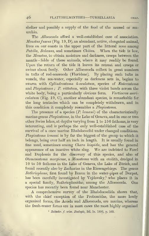
46 PLATYHELMINTHES TURBELLARIA chai
shelter and possibly a supply of the food of the mussel or sea-
urchin.
The Alloeocoela afford a well-established case of association.
Monotusfuscus (Fig. 19, D), an abundant, active, elongated animal,
lives on our coasts in the upper part of the littoral zone amongPatella, Balanus, and sometimes Chiton. When the tide is low,
the Monotus, to obtain moisture and darkness, creeps between the
mantle - folds of these animals, where it may readily be found.
Upon the return of the tide it leaves its retreat and creeps or
swims about freely. Other Alloeocoela collect in great numbers
in tufts of red-seaweeds (Florideae). By placing such tufts in
vessels, the sea-water, especially as darkness sets in, begins to
swarm with Cylindrostoma A-oculatum, species of Enterostoma
and Plagiostoma ; P. vittatum, with three violet bands across the
white body, being a particularly obvious form. Vorticeros auri-
culatum (Fig. 19, C), another abundant species, is remarkable for
the long tentacles which can be completely withdrawn, and in
this condition it completely resembles a Plagiostoma.
The presence of a species (P. lemani) of the characteristically
marine- genus Plagiostoma, in the Lake of Geneva, and in one or two
other Swiss lakes, at depths varying from 1 to 150 fathoms, is very
interesting, and is perhaps the only well-established case of the
survival of a once marine Ehabdocoelid under changed conditions.
Plagiostoma lemani is by far the biggest of the group to which it
belongs, being over half an inch in length. It is usually found in
fine mud, sometimes among Chara hispida, and has the general
appearance of an inactive white slug. We are indebted to Forel
and Duplessis for the discovery of this species, and also of
Otomesostoma morgiense, a Mesostoma with an otolith, dredged in
10 to 50 fathoms in the Lake of Geneva, the Lake of Zurich, and
found recently also by Zacharias in the Eiesengebirge. The genus
Botlirioplana, first found by Braun in the water-pipes of Dorpat,
has been carefully investigated by Vejdovsky,^ who places it in
a special family, Bothrioplanidae, among the Alloeocoela. Onespecies has recently been found near Manchester.
A comprehensive survey of the Rhabdocoelida shows that,
with the chief exception of the Proboscidae, the more lowly
organised forms, the Acoela and Alloeocoela, are marine, whereas
the fresh-water forms are in most cases the most highly organised
' Zeitschr. f. wiss. Zoologie, Bd. Ix. 1895, p. 163. ^_
*

I
ANATOMY OF RHABDOCOELIDA 47,
genera {Mesostoma, Vortex). But Macrorhynchus helgolandicns,
though minute (l'5-2 mm. long), has a more complex structure^
than any other species of the specialised marine genus to which
it belongs, and is a remarkable instance of great complexity
being associated with small size.
Reproduction.— The Ehabdocoelida present the greatest
diversity in the development of the reproductive system. The
Acoela and AUoeocoela have the simplest arrangement. Scattered
testes, often without a distinct membrane, form the spermatozoa,
which in most cases wander into parenchymatous spaces, but
in Monoporus riobropunctatus and Bothrioplana, into distinct vasa
deferentia. In both groups a protrusible penis opens inde-
pendently to the exterior, and may be simply muscular or provided
with a chitinous armature. Two ovaries are present, and the
oviducts, if distinct, are continuations of the ovarian membrane.
In most forms a " bursa seminalis," which receives the sperma-
tozoa of another individual, is appended to the female genital
canal. In many of the AUoeocoela, however, a portion of the
ovary is sterile, and its cells, forming a yolk-gland, feed the
fertile portion, the whole structure being then spoken of as a
germ-yolk-gland. In many others (Monotidae) this sterile part
has become an independent yolk-gland, which communicates by
yolk-ducts with the oviducts. The Acoela form no egg-case, the
body of the parent becoming a bag for the ova, which elaborate
their own food -yolk. The AUoeocoela lay hard -shelled eggs,
which are produced in Bothrioplana and Automolos by the activity
and interaction of reproductive organs, resembling closely those
of certain Triclads.^
The Ehabdocoela exhibit every stage in the development of a
complex reproductive system, from the simple ovaries and testes
of a Microstoma or Macrostoma, to the intricate system of ducts
and glands of a Maci'orhynchus (Proboscidae), in which there is
still much to be made out. The complications of the copulatory
organs chiefly arise from the way in which the spermatozoa are
brought into contact with a nutritive prostatic fluid, or are
formed into spermatophores ; and also from the penial armature,
^ See von GrafTs Monographu, pi. ix. ; and Jensen, Turhcllaria ad Litora
Xorvegiae, Bergen, 1878, pi. iv.
- For the reprodnctive organs of Rhabdocoelida consult von Gratf, Mono-
graphie, "Die Acoela" ; and Bolimig, Zcitschr. f. iciss. Zool. Bd. li. 1891, p. 167.
48 PLATYHELMINTHES TURBELLARIA chap.
which is often very complex, and may consist of a curved chitinoid
hook or a coiled loop (Fromesostoma), of hooks (Proboscidae), or of
an intricate arrangement of plates (Proxenetes) ; or the penis maytake on a complex corkscrew-like form {Pseudorhynchus), The(frequently armed) female genital canal usually possesses a bursa
seminalis for the fertilisation of the eggs, but a receptaculum
seminis or spermatheca may serve for the reception, the bursa,
for the lodgment of the spermatozoa of another individual. The
fertilised ovum is provided with a supply of food-yolk and with
a shell, which may be formed in a special diverticulum, the'' uterus." The development of these organs strains the resources
of the animal to the utmost, and in some Proboscidae the
alimentary canal is squeezed out and disintegrates, in order to
make room for them.
A few Mesostoma (if. ehrenbergii, M. productum, M. lingua)
produce two kinds of eggs—thin- and thick-shelled. The latter
are laid throughout the summer, and lie dormant through
winter. The young which hatch in spring out of these
" winter " eggs develop rapidly, and when only 7 to 8 mm. long
{i.e. one-third the size of the parent) already possess functional
genital organs ; the penis, however, is rudimentary, and incapable
of being used for copulation. Hence it is probable that this
stunted progeny self-fertilise their thin-shelled or " summer " eggs.
After the formation of these eggs the same parent is said
(Schneider ^) to produce thick-shelled or winter eggs, but how-
ever that may be, the first young which hatch from the thin-
shelled ova are produced in great numbers at a time (April to
May) when food is abundant. These grow rapidly to the full
size, and then having attained maturity, cross -fertilise one
another's ova, which become encased in a thick brown shell ; and
it is these numerous " winter " eggs that lie dormant through-
out the autumn and winter. Many Mesostoma, and practically
all other Ehabdocoela, however, produce only thick-shelled eggs,
and in all cases it is probable that to these many species owe
their wide distribution, the exact range of which is, however,
unknown, as is also the means of dispersal.
^ Untersuchungen ii. Platyhelminthen, Giessen, 1873, p. 101.
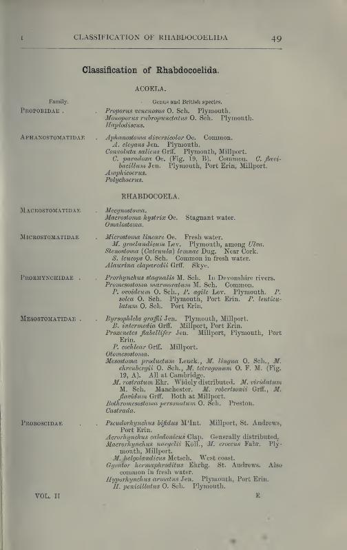
CLASSIFICATION OF RHABDOCOELIDA 49
Classification of Rhabdocoelida.
Family.
Proporidae .
Aphanostomatidae
ACOELA.
Genus aad British species.
Proporus venenosus 0. Sch. Plymouth,Monoporus ruhroinmctatus O. Sch. Plymouth.Haplodisctis.
Aphanostoma diversicolor Oe. Common.A. elcgans Jen. Plymouth.
Convoluta saliens Grti". Plymouth, Millport.
C paradoxa Oe. (Fig. 19, B). Common. C. flavi-
hacillum Jen. Plymouth, Port Erin, Millport.
Amphicoerus.Polychoerus.
RHABDOCOELA.
Macrostomatidae
MiCROSTOMATIDAE
Prorhynchidae
Mesostomatidae
Proboscidae
Mecynostoma.Macrostoma hystrix Oe.
Omalostoma.Stagnant water.
Microstoma linearc Oe. Fresh water.
M. groelandicum Lev. Plymouth, among Ulva.
Stenostoma {Catenula) lemnae Dug. Near Cork.S. leucops 0. Sch, Common in fresh water.
Alaurina claparedii Grff". Skye.
Prorhynchus stagnalis M. Sch. In Devonshire rivers.
Promesostoma marmoratuni M. Sch. Common.P. ovoideum 0. Sch., P. agile Lev, Plymouth. P.
solea 0, Sch, Plymoutli, Port Erin, P. lenticu-
latum 0. Sch. Port Erin.
Byrsophlehs graffii Jen. Plymouth, Millport.
B. intermedia Grff. Millport, Port Erin.
Proxenetes flahellifer Jen. Millport, Plymouth, PortErin,
P. cochlear Grff. Millport.
Otomesostoma.Mesostoma productum, Leuck., M. lingua 0. Sch., M.
ehrenbergii 0. Sch., M. tetragonum 0. F. M. (Fig.
19, A). All at Cambridge.31. rostratum Ehr. Widely distributed. M. viridatumM. Sch. Manchester. M. rohertsonii Grff., M,flavidum Grff. Both at Millport.
Bothromesostoma personatum 0. Sch. Preston.
Castrada.
Millport, St. Andrews,
VOL. II
Psevdorhynchus hifidus M'Int.Port Erin.
Acrorhynchus caledonicus Clap,
Mac7-orhynchus naegelii KolL,mouth, Millport.
M. helgolandicus Metsch. West coast.
Gyrator hermaphroditus Ehrbg. St. Andrews.common in fresh water.
Hyporhynchus armatus Jen. Plymouth, Port ErinH. penicillatus O. Sch. Plymouth.
E
Generally distributed.
J/, croceus Fabr. Ply-
Also
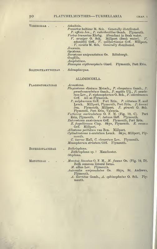
50 PLATYHELMINTHES TURBELLARIA CHAP. I
VORTICIDAE HcliuUzia.
Provortex halticus M. Sch. Generally distributed.
P. affinis Jen., P. ruhrohacillus Ga,mh. Plymouth.Vortex truncatus Ehrbg. Abundant in fresh water.
V. armiger 0. Sch. Millport (fresh water). V.schmidtii Grff., V. millportianus Grff. Millport.
V. viridis M. Sch. Generally distributed.
Jensenia.Opistoma.Derostoma unipundatum Oe. Edinburgh.Grajilla.
Anoplodium.Fecampia crythrocephala Giard. Plymouth, Port Erin.
SoLENOPHARYNGiDAE . SoUnopharynx.
ALLOEOCOELA.
Plagiostomatidae
BOTHRIOPLANIDAE
Monotidae .
Acmostoma.Plagiostoma dioicum Metscli., P. elongatum Gamb., P.
pseudomaeulatu7n Gamb., P. sagitta Ulj,, P. cauda-tum Lev., P. siphonophorum 0. Sch., P. ochroleueumGrtr. All at Plymouth.
P. sulphureitm Grtf. Port Erin. P. vittatum F. andLeuck. Millport, Plymouth, Port Erin. P. koreni
Jen. Plymouth, Millport. P. girardi 0. Sch.
Plymouth, Port Erin, Valencia.
Vorticcros auriculatum 0. F. M. (Fig. 19, C). PortErin, Plymouth. V. luteum Grtf. Plymouth.
Enterostovia austriacum Grff. Plymouth, Port Erin.
£. fingalianum Clap. Skye, Plymouth. E. coccumGrff. Millport.
Allostoma pallidum van Ben. Millport.
Cylindrostoma ^-oculatum Leuck. Skye, Millport, Ply-
mouth.C. incrmc Hall, C. elongatum Lev. Plymouth.
Monoopliorum striatum Grff. Plymouth.
Bothrioplana.Bothrioplana sj). ? Manchester.
Otoplana.
Monotus lineatus 0. F. M., M. fuscus Oe. (Fig. 19, D).
Both common littoral forms.
M. alhus Lev. Plymouth.Automolos unipunctatus Oe. Skye, St. Andrews,
Plymouth.A. horridus Gamb., A. ophiocephalus 0. Sch. Ply-
mouth.

CHAPTER II
TKEMATODA
CHARACTERS OF TREMATODES HABITS AND STRUCTURE OF TREMA-TODA ECTOPARASITICA (MONOGENEA) LIFE-HISTORIES OF
POLYSTOMUM INTEGERRIMUM, DIPLOZOON PARADOXUM, ANDGYRODAGTYLUS ELEGANS TREMATODA ENDOPARASITICA
(DIGENEA) OCCURRENCE AND HABITS OF DIGENEA LIFE-
HISTORY OF DISTOMUM MACROSTOMUM DISTOMUM HEPATI-CTW AND ITS EFFECTS BILHARZIA HAEMATOBIA BISEXUAL
TREMATODES TABLE OF HOSTS CLASSIFICATION.
From the Turbellaria we now pass on to a consideration of the
second great subdivision of the Platyhelminthes, the Trematodes
or " flukes," of which the " liver-fluke " is the best known, since
it is one of the most dangerous parasites that infest domestic
animals.
It has been pointed out that the Polyclads, Triclads, and Ehab-
docoels are carnivorous, and that in each of these groups sporadic
cases of parasitism occur. In other words, when the prey is
much larger than the Turbellarian, the latter tends to become
a parasite, and we can trace the development of the parasitic
habit from the gradual association of Turbellaria with Ascidians,
Crustacea, Molluscs, and Polyzoa merely for protective purposes,
through the adoption, not only of the body of the host for shelter,
l)ut of its flesh for food ; though it is only in some Ehabdocoels
(Graffilla, etc.) that there exists a degeneration corresponding to
the easier mode of nutrition and simpler life. The Trematodes,^
however, are wholly parasitic, either on the outer surface, the
gills, or internal organs of their host, which is almost always a
^ Compare the remarks on Trematodes, pp. 4-5.
PLATYHELMINTHES—TREMATODA
Vertebrate. Some Trematod.es lodge in the mouth ; others wander
down the oesophagus into the stomach or intestine, where they
fix themselves to the mucous membrane. Again, others work
their way into the digestive glands by the ducts, and. thus
become farther and further removed from the external world,
and more adapted to live in the particular organs of that host
in which they best flourish. The most important result of the
adoption of this internal habitat by endoparasitic Trematodes
is, however, seen in their life-history. If a liver-fluke were to
deposit its million or so of eggs in the bile-ducts of the sheep,
and these were to develop i7b situ, the host could not withstand
the increased drain upon its vital resources, and host and para-
sites would perish together. Hence it is clear that the infection
of a second host by Trematodes is highly necessary, whether
they be ectoparasitic, in which case the infection is easily effected,
when two hosts are in contact, by the adult worms, as well as whenthey are apart, by free-swimming larvae. In endoparasitic Trema-
todes it is brought about by the migration of the young to the outer
world, their entrance into a, usually. Invertebrate host and their
asexual multiplication within it, and the capture and deglutition
of this " intermediate host " by the final Vertebrate one. Within
the latter the immature parasites find out the organ in which their
parents flourished, and here they too grow and attain maturity.
The chances of any one Qgg of an endoparasitic Trematode pro-
ducing eventually an adult are, therefore, far less favourable
than in the case of an ectoparasitic form. In other words, while
the former -must lay a great number of small eggs, the latter
need only deposit a (comparatively) few large ones, and this fact
has a corresponding influence on the structure of the genitalia
in the two cases. The Digenea, which employ two hosts in a
lifetime, have accordingly a different generative mechanism from
that of the Monogenea. The great need of the latter is a power-
ful apparatus for adhering to the surface of the body of its host
;
while the adaptations which the endoparasite requires are, in
addition, (1) protection against the solvent action of the glands
of its host, (2) the power of firm adhesion to a smooth internal
surface, and (3) the ability not only to produce a large quantity
of spermatozoa and ova, but in the absence of a fellow-parasite,
to fertilise its own ova ; and we find these conditions abundantly
satisfied.
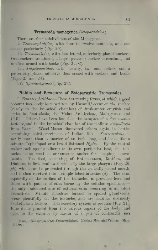
TREMATODA MONOGENEA 5 3
Trematoda monogenea {ectoparasitica).
There are four subdivisions of the Monogenea :
—
I. Temnoceplialidae, with four to twelve, tentacles, and one
sucker posteriorly (Fig. 20).
TI. Tristomatidae, with two lateral, anterior!j-placed suckers.
Oral suckers are absent, a large posterior sucker is constant, and
is often armed with hooks (Fig. 22, C).
III. Folystomatidae, with, usually, two oral suckers and a
posteriorly-placed adhesive disc armed with suckers and hooks
(Figs. 23 and 24).
IV. Gyrodactylidae (Fig. 29).
Habits and Structure of Ectoparasitic Trematodes.
I. T'emnocephfdidac.—These interesting forms, of w^iich a good
account has lately been written by Haswell,^ occur on the surface
(rarely in the branchial chamber) of fresh-water crayfish and
crabs in Australasia, the Malay Archipelago, Madagascar, and
Chili. Others have been found on the carapace of a fresh-water
tortoise, and in the branchial chamber of the mollusc Ampidlaria
from Brazil. Wood-Mason discovered others, again, in bottles
containing spirit -specimens of Indian fish. Teimiocephcda is
rarely more tlian a quarter of an inch long, and looks like a
minute Cephalopod or a broad flattened Hydra. By the ventral
sucker each species adheres to its own particular host, the ten-
tacles being used as an» anterior sucker for " looping " move-
ments. The food, consisting of Entomostraca, Eotifera, and
Diatoms, is first swallowed whole by the large pharynx (Fig. 20,
ph), which can be protruded through the ventrally-placed mouth,
and is then received into a simple lobed intestine (d). The skin,
especially on the surface of the tentacles, is provided here and
there with patches of cilia borne by the cellular epidermis,
—
the only undoubted case of external cilia occurring in an adult
Trematode. Minute rhabdites formed in special gland -cells,
occur plentifully on the tentacles, and are another distinctly
Turbellarian feature. The excretory system is peculiar (Fig. 21).
Fine ducts proceed from the various organs of the body, and
open to the exterior by means of a pair of contractile
^ VL^ls^'qW, Monograph of the Temnocephaleac. Macleay Memorial Volume. Mem.iii. 1893.

54 PLATYHELMINTHES TREMATODA
placed on the dorsal surface. Each sac is a single cell, and
within it not one merely, but several " flames," or bunches of
rhythmically contractile cilia, are present. These are placed on
the course of excessively fine canals, which perforate the proto-
plasm of this cell. The terminal branches of the excretory canals
end in branched cells, apparently devoid of " flames."
The reproductive system is very similar to that of certain
Fig. 20.— Tevinocephala novae-zealandiae Fig. 21.—The same from the dorsal surface,
Has. X 10. Ventral view to show the to show the excretory system (double
digestive and reproductive systems. line), and the nervous system (black
(After Haswell.) and shaded). (After Haswell.)
d, Intestine ; din, dorso-lateral nerve ; dn, dorsal nerve ; ex.o, excretory aperture on
dorsal surface ; ex.s, terminal excretory sac ; m, mouth ; ov, ovary ; ovd, oviduct
;
2>h, pliarynx ; rh, rhabdites ; rhx, cells in which the rhabdites are formed ; rv, yolk
receptacle ; sc, sucker ; sh, shell-gland ; te, testes ; ut, uterus ; vg, vagina ; vn,
ventral nerve ; vs, vesicula seminalis;yd, yolk-duct
; yg, yolk-gland. ?, c5, commongenital pore.
Khabdocoels. An armed penis and the female genital duct
open into a genital atrium, and this by a single aperture
($,(?, I'ig. 20) to the exterior. The fertilised ovum and yolk
are enclosed in a stalked shell formed in the uterus.
The interest and importance of the Temnocephalidae lies in
the fact that they are almost as much Turbellaria as Trematodes.
I
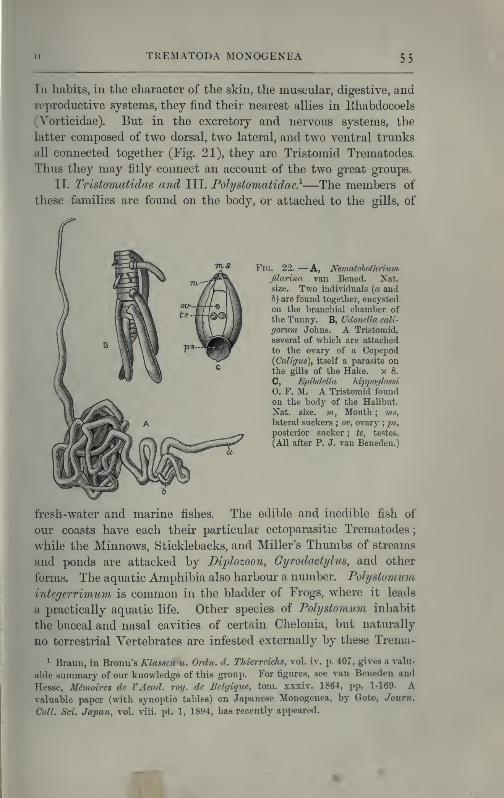
TREMATODA MONOGENEA 55
IIn habits, in the character of the skin, the muscular, digestive, and
reproductive systems, they find their nearest allies in Ehabdocoels
(Vorticidae). But in the excretory and nervous systems, the
latter composed of two dorsal, two lateral, and two ventral trunks
all connected together (Fig. 21), they are Tristomid Trematodes.
Thus they may fitly connect an account of the two great groups.
II. Tristomatidae and III. Polystomatidae}—The members of
these families are found on the body, or attached to the gills, of
Yw. 22. — A, NetnatohothHum,
JUarina van Bened. Nat.size. Two individuals (« antl
h) are found together, encysted
on the branchial chamber of
the Tunny. B, Udonella cali-
gorum Johns. A Tristomid,
several of which are attached
to the ovary of a Copepod(Caligus), itself a parasite onthe gills of the Hake, x 8.
C, Eiiibdella hippoglossi
0. F. M. A Tristomid foundon the body of the Halibut.
Nat. size. ?«, Mouth ; ms,
lateral suckers ; ov, ovary;ps,
posterior sucker ; te, testes.
(All after P. J. van Beneden.)
fresh-water and marine fishes. The edible and inedible fish of
our coasts have each their particular ectoparasitic Trematodes
;
while the Minnows, Sticklebacks, and Miller's Thumbs of streams
and ponds are attacked by Diplozoon, Gyrodactylus, and other
forms. The aquatic Amphibia also harbour a number. Polystomum
integerrimum is common in the bladder of Frogs, where it leads
a practically aquatic life. Other species of Polystomum inhabit
the buccal and nasal cavities of certain Chelonia, but naturally
no terrestrial Vertebrates are infested externally by these Trema-
^ Braun, in Bronu's Klasscn u. Ordn. d. Thicrrcichs, vol. iv. p. 407, gives a valu-
able summary of our knowledge of this group. For figures, see van Beneden and
Hesse, Memoircs de VAcad. roy. dc Bclgiquc, torn, xxxiv. 1864, pp. 1-169. Avaluable paper (with synoptic tables) on Japanese Monogenea, by Goto, Joum.
Coll. Sci. Japan, vol. viii. pt. 1, 1894, has recently appeared.
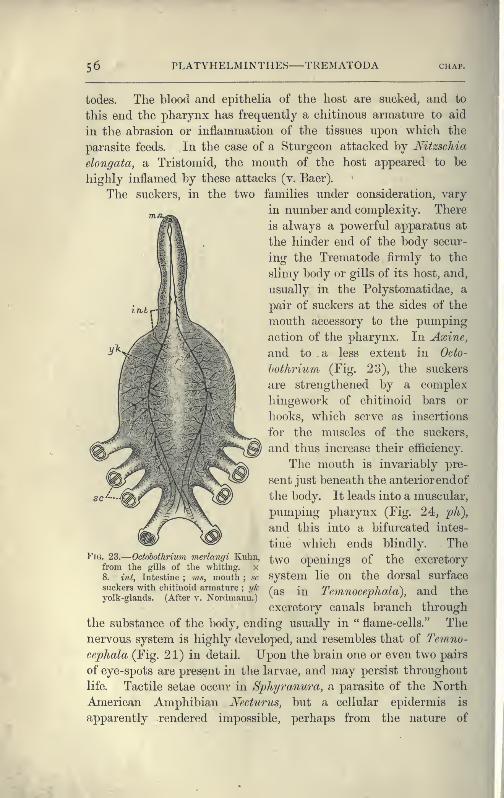
56 PLATYHELMINTHES TREMATODA CHAP.
todes. The blood and epithelia of the host are sucked, and to
this end the pharynx has frequently a chitinous armature to aid
in the abrasion or inflanamation of the tissues upon which the
parasite feeds. In the case of a Sturgeon attacked by Mtzschia
elongata, a Tristomid, the mouth of the host appeared to be
highly inflamed by these attacks (v. Baer).
The suckers, in the two families under consideration, vary
„ „ in number and complexity. There
is always a powerful apparatus at
the hinder end of the body secur-
ing the Trematode firmly to the
slimy body or gills of its host, and,
usually in the Polystomatidae, a
pair of suckers at the sides of the
mouth accessory to the pumping
action of the pharynx. In Axine,
and to . a less extent in Octo-
hothrium (Fig. 23), the suckers
are strengthened by a complex
of chitinoid bars or
which serve as insertions
for the muscles of the suckers,
and thus increase their efficiency.
The mouth is invariably pre-
sent just beneath the anteriorend of
the body. It leads into a muscular,
pumping pharynx (Fig. 24, 2^A),
and this into a bifurcated intes-
tine which ends blindly. The
two openings of the excretory
system lie on the dorsal surface
(as in Temnocephala), and the
excretory canals branch through
the substance of the body, ending usually in " flame-cells." The
nervous system is highly developed, and resembles that of 'Temno-
cephala (Fig. 21) in detail. Upon the brain one or even two pairs
of eye-spots are present in the larvae, and may persist throughout
life. Tactile setae occur in Sphyranura, a parasite of the I^orth
American Amphibian Necturus, but a cellular epidermis is
apparently rendered impossible, perhaps from the nature of
hingework
hooks,
Fig. 23.
—
Odohothrium merlangi Kulm,from the gills of the whiting. x
8. int, Intestine ; ms, mouth ; sc
suckers with chitinoid armature;yk
yolk-glands. (After v. Xordmann.)
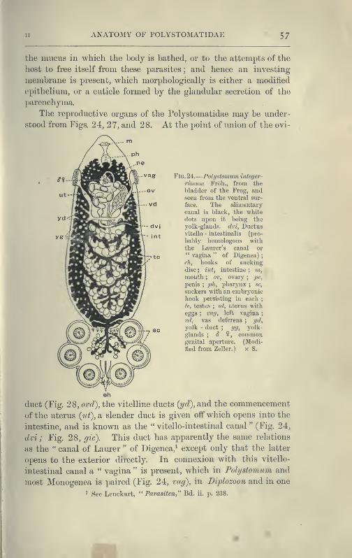
ANATOMY OF POLYSTOMATIDAE 57
the mucus in which the body is bathed, or to the attempts of the
host to free itself from these parasites ; and hence an investing
membrane is present, which morphologically is either a modified
epithelium, or a cuticle formed by the glandular secretion of the
parenchyma.
The reproductive organs of the Polystomatidae may be under-
stood from Figs. 24, 27, and 28. At the point of union of the ovi-
FiG. 24.
—
Polystomum integer-
rimuni Froh., from the
bladder of the Frog, andseen from the ventral sur-
face. The alimentary
canal is black, the white
dots upon it being the
yolk-glands, dvi, Ductusvitello - intestinalis (pro-
bably homologous with
the Laurer's canal or" vagina " of Digenea)
;
eh, hooks of sucking
disc;, int, intestine; m,mouth ; ov, ovary
;pe,
penis;ph, pharynx ; sc,
suckers with an embryonichook persisting in each
;
te, testes ; nt, uterus with
eggs ; -vag, left vagina ;
rd, vas deferens;
yd,
yolk - duct; yg, yolk-
glands ; <? ?, commongenital aperture. (Modi-
tied from Zeller. ) x 8.
luct (Fig. 28, ovd), the vitelline ducts {yd), and the commencement
of the uterus {ut), a slender duct is given off which opens into the
intestine, and is known as the " vitello-intestinal canal " (Fig. 24,
dvi ; Fig. 28, gic). This duct has apparently the siime relations
as the " canal of Laurer " of Digenea,^ except only that the latter
opens to the exterior directly. In connexion with tliis vitello-
intestinal canal a " vagina " is present, which in Polystomum and
most Monogenea is paired (Fig. 24, vag), in Diplozoon and in one
^ See Leuckart, " Pai'asiten," Bd. ii. p. 238.

58 PLATYHELMINTHES TREMATODA CHAJ
or two other forms, however, unpaired. The vagina receives the
penis of another individual during copulation (Fig. 26),and does not
appear to have an homologue in the liver-fluke or other Digenea.
Life-Histories of the Polystomatidae.^—Polystomum integer-
rimum. After the mutual fertilisation of two individuals, the
eggs are laid in the water by the protrusion of the body of the
parent through the urinary aperture of the Frog. About 1000
eggs are laid in the spring at the rate of 100 a day for ten
days. After about six weeks, the larva ('3 mm. long) hatches
Fia. 25.—Eggs of Monogenea. A, Eggs of Encotyldbe pagelli v. Ben. -Hesse ; B, eggs
of Udonella pollachii v. Ben.-Hesse (with young forms just hatching out) ; C, egg
of Microcotyle lahracis v. Ben.-Hesse. (After van Beneden and Hesse.) x 50.
out, and swims about freely by means of bands of large ciliated
cells (Fig. 26, A) ; but if it does not meet with a tadpole within
twenty-four hours, it dies. Should it, however, encounter one,
the larva creeps along it in a looping fashion until it approaches
the opercular spout, or opening of the branchial chamber, on the
left side ; into this it darts suddenly, fixes itself, and throws off
its cilia. Here it remains eight or ten weeks, feeding, in-
creasing in size, and forming the suckers from behind forwards.
^ Zeller, Zeitschr. f. wiss. Zool. xxii. 1872, pp. 1, 168 ; also Bd. xxvii. 1876, p.
238 ; xlvi. 1888, p. 233.
1
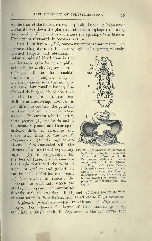
LIFE-HISTORIES OF POLYSTOMATIDAE 59
^H At the time of the tadpole's metamorphosis, the young Folystomum^B works its way down the pharynx into the oesophagus and along
^H the intestine, till it reaches and enters the opening of the bladder.
^H Three years afterwards it becomes mature.
^H Sometimes, however, Polystomum experiences another fate. The
^V larvae settling down on the external gills of a young, recently-
hatched tadpole, and obtaining a
richer supply of blood than in the
previous case, grow far more rapidly,
so that in five weeks they are mature,
although still in the branchial
chamber of the tadpole. They do
not then wander into the aliment-
ary canal, but usually, having dis-
charged their eggs, die at the time
of the tadpole's metamorphosis.
Still more interesting, however, is
the difference between the genitalia
in these and in the normal Poly-
stomum. In contrast with the latter,
these possess (1) one testis and a
rudimentary penis ; and their sper-
matozoa differ in structure and
shape from those of the normal
Polystomum. (2) The vaginae are
absent, a fact connected with the
absence of a functional COpulatory Fig, 2Q.—Polystomum integernmum.
nrcTflTi r^^ Tn rninnPTisation for A, Free-swimming larva, seen fromorgan. ^6) in compensation lor
^^^ ^^^^^^^^ surface, x 80. B,
the loss of these, a duct connects Two mature individuals in mutual
the single testis and the point of
union of oviduct and yolk-ducts,
and by this self-fertilisation occurs.
(4) The uterus is absent ; the
"ootype" or duct into which the
sliell- gland opens, communicating
directly with the exterior. In (1) and (4) these abeiTant Poly-
stomum resemble P. ocellatum, from the Tortoise Umi/s europaea.
Diplozoon jparadoxum.—The life -history of Diplozoon is
unique. For whereas the larvae of most animals grow up,
each into a single adult, in Diplozoon, of the few larvae that
coition attached to the bladder
of a Frog, x 5. (After Zeller.)
d, Intestine ; ex.o, excretory pore,
dorsal in position, seen here bytransparency ; ey, eye-spots
;gl,
frontal glands ; m, mouth;ph,
pharynx ; sd, adhering disc ; vag^
vagina.
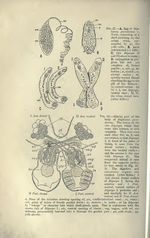
Fig. 27.—a, Egg of Dip-lozoon paradoxum v.
Nord., consisting of ashell enclosing o%\ theactual ovum, sur-
rounded by yc, the
yolk - cells. ; B, larva
just hatched ( x 125) ;
C, two Diporpa (I
and II) about to unite;
D, conjugation in pro-
gress but not yetcomplete, dt, Dorsalpapilla ; e, eye
; g, in-
testine ; VI, mouth ; sc,
ad-oral sucker ; th,
spirally-wound threadattaching the egg to the
gill of the Minnow;
vs, ventral sucker;
(in
D) I, I, one JJiporpa,
ventral view; II, II,
the other, dorsal view.
(After Zeller.)
Fig. 28.—Hinder part of thebody of Diplozoon para-doxum. The fusion of the
two Diporpa, where theycome into contact, is nowcomplete. They now cross
each other like an X, andare twisted, so that DiporpaI, in front of the point offusion, is seen from the
dorsal surface ; behind,
from the ventral surface;
and the reverse is the case
with Diporpa II. Thecompoi;nd animal is seen
from the opposite surface
to that shown in Fig. 27,
D. The digestive andexcretory organs areomitted. (After Zeller.) I
Ant. dorsal, dorsal surface
of Diporpa I, facing the
anterior end ; I Post,
ventral, ventral surface of
Diporpa I, posterior end;
II Post, dorsal I Post, ventral and similarly for II A^it.
ventral and II Post, dorsal,
d, Piece of the intestine showing opening of, gic, vitello-intestinal canal ; ov, ovary;
ovd, point of union of female genital ducts ; sc, suckers ; te, testis ; ut (in DiporpaI), " ootype " or chamber into which shell-glands open. This is continuous with theuterus (ut) of Diporpa I ; iito, ventral opening of uterus ; vag, vagina, with vd, vasdeferens, permanently inserted into it through the genital pore
;yd, yolk-ducts
; yg,yolk -glands.
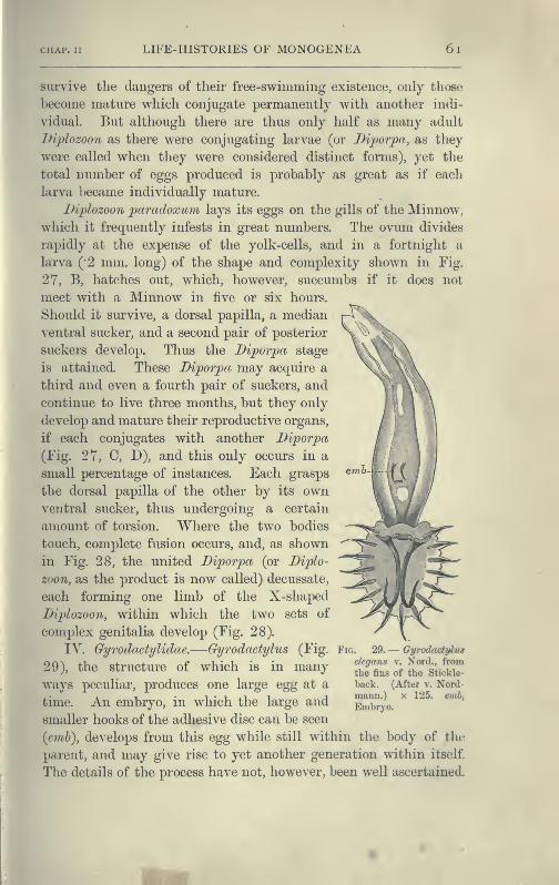
LIFE-HISTORIES OF MONOGENEA 6l
survive the dangers of their free-swimming existence, only those
become mature which conjugate permanently with another indi-
vidual. But although there are thus only half as many adult
Diplozoon as there were conjugating larvae (or Dipoiya, as they
were called when they were considered distinct forms), yet the
total number of eggs produced is probably as great as if each
larva became individually mature.
Diplozoon paradoxum lays its eggs on the gills of the Minnow,
which it frequently infests in great numbers. The ovum divides
rapidly at the expense of the yolk-cells, and in a fortnight a
larva ("2 mm. long) of the shape and complexity shown in Fig.
27, B, hatches out, which, however, succumbs if it does not
meet with a Minnow in five or six hours.
Should it survive, a dorsal papilla, a median
ventral sucker, and a second pair of posterior
suckers develop. Thus the Diporpa stage
is attained. These Diporpa may acquire a
third and even a fourth pair of suckers, and
continue to live three months, but they only
develop and mature their reproductive organs,
if each conjugates with another Diporpa
(Fig. 27, C, D), and this only occurs in a
small percentage of instances. Each grasps
the dorsal papilla of the other by its ownventral sucker, thus undergoing a certain
amount of torsion. Where the two bodies
touch, complete fusion occurs, and, as shown
in Fig. 28, the united Diporpa (or Diplo-
zoon, as the product is now called) decussate,
each forming one limb of the X-shaped
Diplozoon, within which the two sets of
complex genitalia develop (Fig. 28).
IV. Gyrodactylidae.—Gyrodactylus (Fig.
29), the structure of which is in manyways peculiar, produces one large egg at a
time. An embryo, in which the large and
smaller hooks of the adhesive disc can be seen
{eml), develops from this egg while still within the body of the
parent, and may give rise to yet another generation within itself.
The details of the process have not, however, been well ascertained.
Fig. 29.— Gyrodactylus
elegans v, Nord., fromthe fius of the Stickle-
back. (After V. Nord-maiin.) x 125. emb,
Embryo.
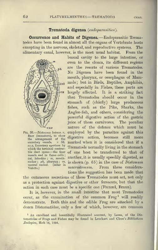
62 PLATYHELMINTHES TREMATODA
inb
Trematoda digenea {endoparasitica).
Occurrence and Habits of Digenea.—Endoparasitic Trema-
todes have been found in almost all the organs of Vertebrate hosts
excepting in the nervous, skeletal, and reproductive systems. Thealimentary canal, however, is the most usual habitat. From the
buccal cavity to the large intestine, or
even to the cloaca, its different regions
are the resorts of various Trematodes.
No Digenea have been found in the
mouth, pharynx, or oesophagus of Mam-mals ; but in Birds, Eeptiles, Amphibia,
and especially in Fishes, these parts are
largely affected. It is a striking fact
that Trematodes should occur in the
stomach of (chiefly) large predaceous
fishes, such as the Pike, Sharks, the
Angler-fish, and others, considering the
powerful digestive action of the gastric
juice of these carnivores. The peculiar
•ex.o nature of the defence which must be
Fig. SO.—Distomum luteum v. employed by the parasites against this
ri^J'I.'^reS '"of*Z digestive action, becomes still moreexcretory vessels, x 50. marked whcu it is Considered that if a
AvIikh^^TSinai^^^^^^^^ Trematode normally living in the stomachtile duct opens— the finer of One host be transferred to that ofvessels end in flame - cells
; ^, ... ,, ^•^ -y- , -t
int, intestine ; vi, mouth- another, it IS usually speedily digested, as
sucker; ph, pharynx; vs, jg ghown (p. 65) in the case of I)istomu7nventral sucker. (After la ,
Vaiette.) macrostomum. ±rom these considera-
tions the suggestion has been made that
the cutaneous secretions of these Trematodes must act, not only
as a protection against digestive or other ferments, but that the
action in each case must be a specific one (Frenzel, Braun).
It is, however, in the small intestine that most Trematodes
occur, as the examination of the common Frog^ will readily
demonstrate. Both this and the edible Frog are attacked by a
dozen Distomatidae, only a few of which, however, are common
1 An excellent and beautifully illustrated account, by Looss, of the Dis-
tomatidae of Frogs and Fishes may be found in Leuckart and Chun's Bihliotheca
^oologica, Heft 16, 1894.

II TREMATODA DIGENEA 63
to both hosts, and a number of Holostomatidae also pass a sta^
of their development within these Amphibia. Some idea of the
extent to which animals, whose habits lead to infection, may be
attacked by Trematodes (to say nothing of Cestodes and Nema-todes, which often occur also) may be gathered from the fact that
in dissecting a black stork, Nathusius found several hundred
Holostomum excavatum and about a hundred Distomum ferox in
the small intestine, twenty-two D. hians in the oesophagus, five
others in the stomach, and one D, echinatum in the intestine.
Snipe, Woodcock, Sandpipers, Dunlin, Gulls, Bittern, Geese, and
Wild Ducks are, to mention a few cases, greatly infested bymembers of this group.
The following Trematodes have occurred in man 1 :
—
Distomum hepaticum Abild. Distomum pulmonale Balz { = D.
„ lanceolatum Mehlis. ringeri Cobb., D. wester-
„ conjunctum Cobbold. manni Kerb.).
„ spathulatum Leuckart (
=
„ oculi humani Ammon { — D.
D. sinense Cobb., D. ophthalmobium Dies.).
japonicitm R. Blanch.).. Monostomum lentis v. Nord.
„ rathouisi Poir. (probably = Amphistomum hominis Lewis and
D. crassum Busk, D. M'Connell.
huskii Lank.). Bilharzia haematobia Cobb.
„ heterophijes v. Sieb.
Life-histories of the Digenea.—The classification of Trema-
todes according to their life-histories, expressed in the divisions
Monogenea and Digenea, though a very useful one, breaks downentirely in the case of certain forms. Thus the life-history of
Gyrodactylus is probably digenetic rather than monogenetic. Aspi-
dogaster conchicola,^ which lives in the pericardial cavity of the
fresh-water mussel (possibly the only case of a Trematode becom-
ing normally mature in an Invertebrate host, since other species
of Aspidogaster live in Chelonia), produces larvae which enter
another Anodonta and develop directly into the sexual form. In
other words, Aspidogaster, though structurally a digenetic form,
possesses a life-history which is direct and simple, i.e. monogenetic.
The Holostomatidae, which live in birds of prey and
aquatic birds, give rise to eggs from which a minute larva
escapes. The fate of this aquatic larva is not directly known,
,
^ Leuckart, Parasiten d. Menschen, " Trematoden, " 1892-94; R. Blanchard,
Traitid. Zool. medicale, i. 1889 ; H. B. Ward, Report for 1894 of Nebraska State
Board of Agrie. Lincoln, U.S.A. 1895, p. 225.
2 Huxley, Anat. of Invert. Animals, 1877, p. 194.
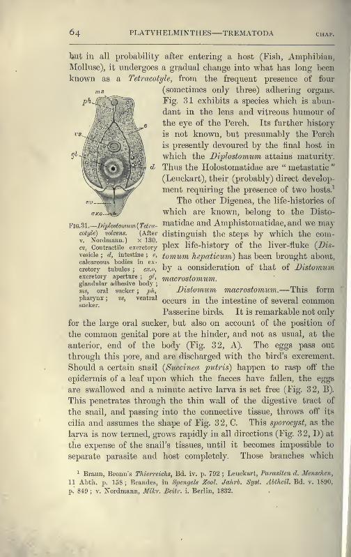
64 PLATYHELMINTHES TREMATODA chap.
b,ut in all probability after entering a host (Fish, Amphibian,
Mollusc), it undergoes a gradual change into what has long been
known as a Tetracotyle, from the frequent presence of four
ms (sometimes only three) adhering organs,
rig. 31 exhibits a species which is abun-
dant in the lens and vitreous humour of
the eye of the Perch. Its further history
is not known, but presumably the Perch
is presently devoured by the final host in
which the Diplostomum attains maturity.
Thus the Holostomatidae are " metastatic"
(Leuckart), their (probably) direct develop-
ment requiring the presence of two hosts.^
The other Digenea, the life-histories of
exo-^=i^a^ which are known, belong to the Disto-
FlG.Sl.-mplostom^^m{Telra- matidae and Amphistomatidae, and we maycoti/ie) voivens. (After distinguish the stcps by which the com-
ci, Coutmctiie excretory pl^x life-history of the liver-fluke {Dis-
vesicie;
d, intestine;
e, tomum lievaticum) has been brought about,calcareous bodies in ex- . c l^ , n n- ^
cretory tubules ; ex.o, by a Consideration 01 that oi JJistomumexcretory aperture
;gl, ^acTOStomum.
glandular adhesive body;
ms, oral sucker;
ph, Distomum macTOstomum.—This formpimrynx
;vs, ventral Q^curs in the intestine of several common
sucker.
Passerine birds. It is remarkable not only
for the large oral sucker, but also on account of the position of
the common genital pore at the hinder, and not as usual, at the
anterior, end of the body (Fig. 32, A). The eggs pass out
through this pore, and are discharged with the bird's excrement.
Should a certain snail (Succinea putris) happen to rasp off the
epidermis of a leaf upon which the faeces have fallen, the eggs
are swallowed and a minute active larva is set free (Fig. 32, B).
This penetrates through the thin wall of the digestive tract of:
the snail, and passing into the connective tissue, throws off its
cilia and assumes the shape of Fig. 32, C. This sporocyst, as the
larva is now termed, grows rapidly in all directions (Fig. 32, D) at
the expense of the snail's tissues, until it becomes impossible to
separate parasite and host completely. Those branches which
1 Braun, Bronn's Thierreichs, Bd. iv. p. 792 ; Leuckart, Parasiten d. Menschen,
11 Abth. p. 158 ; Brandes, in Spengels ZooL Jahrh. Syst. Abtheil. Bd. v. 1890,
p." 849 ; V. Nordmann, Mikr. Beitr. i. Berlin, 1832.
I
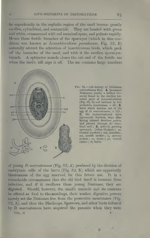
LIFE-HISTORIES OF DISTOMATIDAE 65
«
lie superficially in the cephalic region of the snail become greatly
swollen, cylindrical, and contractile. They are banded with green
and white, ornamented with red terminal spots, and pulsate rapidly.
Hence these fertile branches of the sporocyst (which in this con-
dition was known as Leucochloridium paradoxum, Fig. 33, B)
naturally attract the attention of insectivorous birds, which peck
off the tentacles of the snail, and with it the swollen sporocyst-
branch. A sphincter muscle closes the cut end of the fertile sac
when the bird's bill nips it off. The sac contains large numbers
Fig. 32.—Life-history of Distomummacrostomum Rud. A, ImmatureDistomum (really a tailless Cer-
caria) found in the swollen ter-
minal parts of Leucochloridium(Fig. 33, B) and enclosed in twoprotective membranes, x 40 ; B,larva which hatches out of theegg of D. viacrostomtim, x 125
;
C, the metamorphosed larva
(sporocyst) fourteen days after
having entered Succinea jmtris,
and pierced through its intes-
tinal wall ; D, actively growingsporocyst. (After Heckert.) go,
Genital aperture ; int, intestine;
ms, mouth sucker ; n, nervous
system ; ov, ovary;ps, ventral
sucker ; te, testis.
of young D. macrostomum (Fig. 32, A), produced by the division of
embryonic cells of the larva (Fig. 32, B), which are apparently
blastomeres of the egg reserved for this future use. It is a
remarkable circumstance that the old bird itself is immune from
infection, and if it swallows these young Distomes, they are
digested. Should, however, the snail's tentacle and its contents
be offered as food to the nestlings, their weaker digestive powers
merely set the Distomes free from the protective membranes (Fig.
32, A), and thus the Blackcaps, Sparrows, and other birds infested
by D. macrostomum have acquired the parasite when they were
VOL. II F
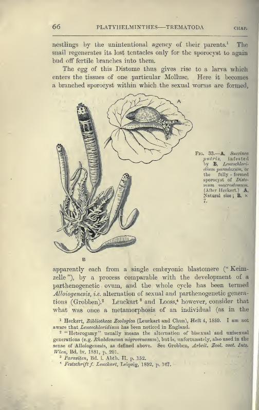
66 PLATYHELMINTHES TREMATODA CHAP.
nestlings by the unintentional agency of their parents.^ Thesnail regenerates its lost tentacles only for the sporocyst to again.
bud off fertile branches into them.
The ^gg of this Distome thus gives rise to a larva which
enters the tissues of one particular Mollusc. Here it becomes
a branched sporocyst within which the sexual worms are formed,
Fig. 33.—A, Succinea\
putris, infestedby B, Leucochlcri-
dixim paradoxuni, or\
the fully - formed !
sporocyst of Disto-
rnum macrostmmim.(After Heckert.) A,Natural size \ B, x
apparently each from a single embryonic blastomere (" Keim
zelle"), by a process comparable with the development of a
parthenogenetic ovum, and the whole cycle has been termed
Alloiogenesis, i.e. alternation of sexual and parthenogenetic genera
tions (Grobben).- Leuckart ^ and Looss,^ however, consider that
what was once a metamorphosis of an individual (as in the
^ Heckert, BiUiotheea Zoologica (Leuckart and Chun), Heft 4, 1889. I am not
aware that Leucochloridium has been noticed in England.2 "Heterogamy" usually means the alternation of bisexual and unisexual
generations (e.g. Rhahdonema nigrovenosum), but is, unfortimately, also used in the
sense of AUoiogenesis, as defined above. See Grobben, Arbeit. Zool. zoot. Ints.
Wien, Bd. iv. 1881, p. 201.
3 Para^ten, Bd. i. Abth. II. p. 152.
" Festschrift f. Leuckart, Leipzig, 1892, p. 167.
fc DISTOMUM HEPATICUM 67
«
ft)
^^" on
Holostomatidae) has now become, by maturation of the Cercaria
in the comparatively modern warm-blooded bird, a metamorphosis
extending over two or more generations.
Distomum (Fasciola) hepaticum—The liver-fluke of the Sheep,
which produces the disastrous disease, liver-rot, has a distribution
as wide as that of a small water-snail, Limnaea truncatula, the
connexion between the two being, as Thomas ^ and Leuckart dis-
covered, that this snail is the intermediate host in which the
earlier larval, sporocyst, and redia stages are passed through, and
a vast number of immature flukes (Cercariae) are developed-
These leave the snail and encyst upon grass, where they are eaten
by the sheep. Over the whole of Europe, Northern Asia,
Abyssinia, and North Africa, the Canaries, and the Faroes, the fluke
and the snail are known to occur, and recently the former has
been found in Australia and the Sandwich Islands, where a snail,
apparently a variety of Limimea truncatula, is also found." Over
these vast areas, however, the disease usually only occurs in certain
marshy districts and at certain times of the year. Meadows of
a clayey soil, liable to be flooded (as in certain parts of Oxford-
shire), are the places where this Limnaea occurs most abundantly,
and these are consequently the most dangerous feeding-grounds
for sheep. The wet years 1816, 1817, 1830, 1853, and 1854—memorable for the occurrence of acute liver-rot in England,
Crermany, and France—showed that the weather also plays a
nsiderable part in extending the suitable ground for Limnaeaover wide areas, which in dry years may l^e safe pastures. In
1830 England lost from this cause,^ one and a haK million
heep, representing some four millions of money, while in
1879-80 three millions died. In 1862 Ireland lost 60 per
nt of the flocks, and in 1882 vast numbers of sheep perished
Buenos Ayres from this cause. In the United Kingdom the
annual loss was formerly estimated at a million animals, but is
now probably considerably less. After infection during a wet
autumn, it is usually in the succeeding winter that the disease
reaches its height.
^ Quart. Joum. Micros. Set. vol. xxiii. 1883, p. 90.
- The intermediate liost in the Sandwich Islands is said to be Limnaea peregra.
See Lutz, Centralhl. f. Baktcr. xi. 1892, p. 783.
^ The mortality in wet years, however, is said to be largely due to pulmonary
inflammation. This and other causes of death are not always discriminated in the
returns.
68 PLATYHELMINTHES—TREMATODA chap.
The symptoms of " rot " appear about a month after infection,
more acutely in lambs than in sheep, and again, less in oxen than
in sheep. At first, death may result from cerebral apoplexy, but
if the first few weeks are passed through, a pernicious anaemia
sets in, the sheep are less lively and fall at a slight touch, the
appetite diminishes, and rumination becomes irregular. The
conjunctiva is of a whitish-yellow colour, the dry, brittle wool
falls ofF, and there is sometimes fever and quickened respiration.
In January, about three months after infection, the wasting, or
fat^l, period sets in. Oedemas or swellings, usually visible before,
become larger at the dependent parts of the body, a large one in
the submaxillary region being especially well marked, and this
is considered one of the most characteristic symptoms (" watery
poke "). Through this period few of the infected sheep survive,
but should they do so, the flukes begin to migrate, though some
remain much longer within the liver. Migration is effected
through the bile-duct into the duodenum and outwith the faeces,
in which the altered remains of the J}ist(ynunn are sometimes
scarcely recognisable. Under these circumstances (or owing to
death of the fluke in situ) the sheep recover more or less fully.
The preventive measures seem to be : (1) Destruction of the
eggs and of the manure of rotten sheep; (2) slaughter of badly
fluked sheep; (3) adequate drainage of pastures
; (4) an allowance
of salt and a little dry food to the sheep ; and (5) dressings of
lime or salt on the ground to destroy the embryos,^
I}istomitm hepatiminiy contrary to most Trematodes, enjoys a
wide ranore of hosts. Man himself occasionallv falls a ^^ctim: thus
in Dalmatia, in the Xarenta Valley, the disease is endemic but
slight in its effects. The horse, deer, camel, antelopes, goat,
pig, rabbit, kangaroo, bea^'er, and squirrel have all been known
to harbour this fluke occasionally. In the Italian deer-parks at
Mandria a large species, D. magnum, decimated the herds some
years ago ; and this species, probably imported from Italy, is now
ahnost as dangerous a parasite on the western plains of the
United States as B. Jupaticum.
BUharzia haematohiar—This fonnidable parasite was dis-
covered by Bilharz in 1853 in the veins of the bladder of patients
1 Sec Thomas, QuaH. Jount. Micros. Sdenegy xriii. 1883. Neumaim, Parasites
of Domesticated Animals, translated by Fleming, 1892.
* Leackart, loc cit. ; Looss, Archivf. miJ:roskop. Anatomic, Bd. xlvi. 1895, p. 1.
I
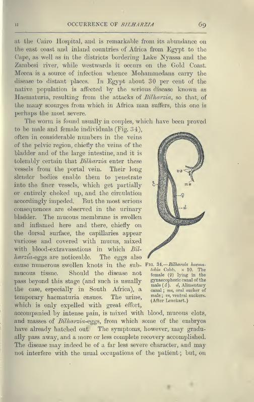
OCCURRENCE OF BILHARZIA 69
at the Cairo Hospital, and is remarkable from its abundance on
the east coast and inland countries of Africa from Egypt to the
Cape, as well as in the districts bordering Lake Nyassa and the
Zambesi river, while westwards it occurs on the Gold Coast.
Mecca is a source of infection whence Mohammedans carry the
disease to distant places. In Egypt about 30 per cent of the
native population is affected by the serious disease known as
Haematuria, resulting from the attacks of Bilharzia, so that, of
the many scourges from which in Africa man suffers, this one is
perhaps the most severe.
The worm is found usually in couples, which have been proved
to be male and female individuals (Fig. 34),
often in considerable nimibers in the veins
of the pelvic region, chiefly the veins of the
bladder and of the large intestine, and it is
tolerably certain that Bilharzia enter these
• vessels from the portal vein. Their long
slender bodies enable them to penetrate
into the finer vessels, which get partially
or entirely choked up, and the circulation
accordingly impeded. But the most serious
consequences are observed in the urinary
bladder. The mucous membrane is swollen
and inflamed here and there, chiefly on
the dorsal surface, the capillaries appear
varicose and covered with mucus, mixed
with blood-extravasations in which Bil-
• harzia-egcrs are noticeable. The egops also
cause numerous swollen knots in the sub- Fio. 34.
—
Bilharzia haema-tobia Cobb, x 10, Thefemale (9) lying in the
gynaecophoric canal of the
male ( cJ ). d, Alimentarycanal ; ms, oral sucker of
male ; r.?, ventral suckers,
(After Leuckart.
)
mucous tissue. Should the disease not
pass beyond this stage (and such is usually
the case, especially in South Africa), a
temporary haematuria ensues. The urine,
which is only expelled with great effort,
accompanied by intense pain, is mixed with blood, mucous clots,
and masses of BUhm^zia-eggs, from which some of the embryos
have already hatched out. The symptoms, however, may gradu-
ally pass away, and a more or less complete recovery accomplished.
The disease may indeed be of a far less severe character, and maynot interfere with the usual occupations of the patient ; but, on
70 PLATYHELMINTHES TREMATODA chap.
the other hand, a far more extensive thickening of the wall of
the bladder sometimes occurs ; hard masses of eggs, uric acid
crystals, and other deposits, may lead to the formation of stones,
degeneration of the substance of the ureter, and eventually to
that of the kidney itself. The stone, indeed, has long been
known to be a prevalent disease in Egypt, and it is now knownto arise from concretions formed round masses of Bilharzia
eggs. From the portal vein, again, other Bilharzia may gain
access to the rectum, or the liver, and it has also been found in
the lungs, and may give rise to most serious complications, if
indeed the patient lives.
How infection occurs is a question to which at present no
satisfactory answer can be made. The attempt to introduce
embryos of Bilharzia into the common fresh-water animals of
Alexandria has hitherto proved fruitless (Looss ^), although there
seems little doubt that the comparative immunity of Europeans
from the disease is in some way owing to their drinking purer
water than the natives. Possibly, as Leuckart suggests, the
embryo becomes a sporocyst in man himself, somewhat as Taenia
murina is known to develop in the rat without an intermediate
host.^ The immense numbers of the parasite in one host would
then readily receive an explanation.
A Bilharzia, possibly B. haematohia, was found by Cobbold in
the portal vein of Cercopithecus fidiginosus ; and B. crassa infests
the cattle of Egypt, Sicily, and certain parts of India, but does
not produce haematuria.
Of the other Trematodes of man and domestic animals there
is not room to speak fully. Distomum puhnonale, which occurs
in the lungs of the cat, tiger, and dog, as well as in man, is
especially common in Japan, China, Corea, and Formosa. I).
sinense and I), rathouisi have been also found in inhabitants of
these countries.
Bisexual Trematodes.^Zoologically, Bilharzia is interesting
from its bisexual condition. It is not, however, the only bisexual
Trematode. In cysts in the branchial chamber of Eay's bream,
Brama raii, two worms are found, which are probably the slender
male and the swollen female of the same species {Distomum
ohenii). The only doubt that can arise proceeds from the
tendency in all Trematodes for the male organs to ripen before
^ In Leuckart, Die Parasiten d. Menschen, pp. 521-528, 1894. ^ Qf^ p_
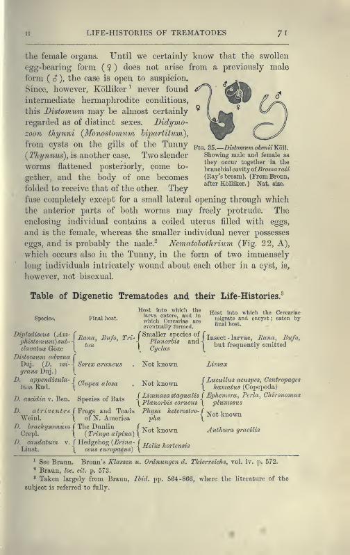
LIFE-HISTORIES OF TREMATODES 71
the female organs. Until we certainly know that the swollen
egg-bearing form ( $ ) does not arise from a previously male
form (S), the case is open to suspicion.
Since, however, Kolliker^ never found
intermediate hermaphrodite conditions,
this Bistomicm may be almost certainly
regarded as of distinct sexes. Didymo-
zoon tliynni {Monostomum hipartitum),
from cysts on the gills of the Tunny(Thynnus), is another case. Two slender
worms flattened posteriorly, come to-
gether, and the body of one becomes
folded to receive that of the other. They
fuse completely except for a small lateral opening through which
the anterior parts of both worms may freely protrude. The
enclosing individual contains a coiled uterus filled with eggs,
and is the female, whereas the smaller individual never possesses
eggs, and is probably the male.^ Nematohothrium (Fig. 22, A),
which occurs also in the Tunny, in the form of two immensely
long individuals intricately wovmd about each other in a cyst, is,
however, not bisexual.
Fig. 35.
—
Distomum okenii Koll.
Showing male and female as
they occur together in the
branch ial cavityofBramaraii
(Ray's bream). (FromBronn,after Kolliker. ) Nat. size.
Table of Digenetic Trematodes and their Life-Histories.
Species. Final host.
iplodiscus {Am- f n„,,^
davatus Goze |^
Distomum advena C
Duj. (D. mi- < Sorex araneusgrans Duj.) [
D. a2jpendicula-
ti^m Rud.
Host into which thelarva enters, and in
wliich Cercariae areeventually formed.
Host into which the Cercariaemigrate and encyst ; eaten byfinal host.
, Bufo, ^r^^ P^/leyp^^ Rana, Bufo,
1 Cidas 1 b^^t ^"requently omitted
Not known Limax
D. ascidia v. Ben.
-{ Clupea
Species of Bats
/ Frogs and Toads
Not known
D. atriventre f Frogs and roaasWeinl. \ of N. America
D. bi-achysomum J The Dunlin JCrepl. \ ( Tringa alpina) \
D. caudatum v. / Hedgehog {Erina- JLinst. \ ceus eurojtaeus) \
J Lucullus acuspes, Centropagcs
\ hamatus (Copepoda)
( Limnaeastagnalis { Ephevicra, Pcrla, Ghironomus
\ Planorhis corneus \ plumosus
Ae^.ro5^ro-rj^^t knownpha y
Not known
Helix hortensis
Anthura gracilis
^ See Braun. Bronn's Klassen u. Ordnungen d. Thierreichs, vol. iv. p. 572.
^ Braun, loc. cit. p. 573.
2 Taken largely from Braun, Ibid. pp. 864-866, where the literature of the
subject is referred to fully.
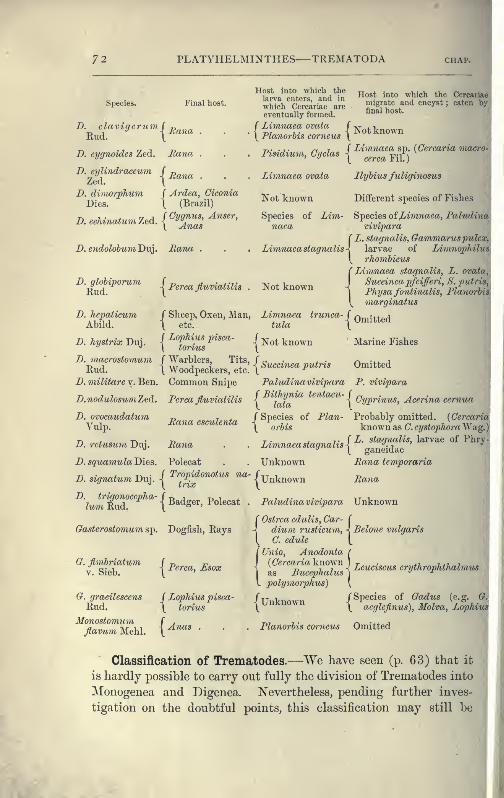
72 PLATYHELMINTHES TREMATODA CHAP.
Species. Final host.
Rud. i^^'^^^ •
D. cygnoides Zed. Mana .
D. cijliTidraceum f p„,,^Zed.
-^Aa^ia .
D. dimorphum j Ardea, Ciconia
Dies. \ (Brazil)
B. echinatum Zed. \ ^^J^t''
D.endolohmn'Dn]. Rana .
Host into which the Cercarii
migrate and encyst ; eaten bj
final host.
Not known
Host into which thelarva enters, and ia
which Cercariae areeventually formed.
f Limnaea ovata J
\ Planorbis corneus |
Pi\idiinn rnrla-i -f-^^"^^««^ ^p. {Cercaria ^niaero
risidium, byclas^ ^^^^^ j,.^ .j
Ilyhius fuliginosus
Different species of Fishes
Species ofXiimnaea, Paludim
Limnaea ovata
Not known
Species of Lim-naea vivipara
Limnaea stagih
( L. stagnalis,
alis-l larvae (
(^ 7-hombicus
Gammaruspulof Li7nnojphilu
D. glohiporumRud.
D. hepaticumAbild.
D. hystrix Duj.
D. macrostomumRud.
D. militare v. Ben.
D. nodulosum Zed.
D. ovocaudatumVulp.
D. retusum Duj.
D. squamula Dies,
D. signatum Duj.
D. trigonocepha-
lum Rud.
Gasterostomum sp.
G. fimhriatum
Perca fluviatilis . Not known
Limnaea stagnalis, L. ovata
Succinea pfeifferi, S. putrii
Physa fontinalis, Planorbimarginatus
J Sheep, Oxen, Man,\ etc.
f Lophius pisca-
\ torius
/ Warblers, Tits,
\ Woodpeckers, etc.
Common Snipe
Perca fluviatilis
Eana esculenta
Rana
Polecat
J Tropidonotus na-1 trix
Limnaea trunca-
tula
Not known
i Omitted
Marine Fishes
< Succinea putris
Paludina vivipara
( Bithynia tentacu-
\ lata
J Species of Plan-
\ oi'bis
Omitted
P. vivipara
-I Cyprinus, Acerina cermia
Probably omitted. {Cercaria
known as C. cystophora Wag.)
r • . J. f L. stagnalis, larvae of PhryLimnaeastagnahsi^
ganeidae
Unknown Rana temporaria
\ Unknown Rana
er, Polecat . Paludina vivipara Unknown
Belone vulgaris( Ostrea edulis, Car- (
Dogfish, Rays -! dium^ rusticum, \
\ C. edule(^
V. Sieb.[Perca,Esox
G. gracilescens J Lophius pisca-
Rud. \ torius
Monostomumflavum Mehl.
edulis, Carrusticum
edule
Unio, Anodonta, f
{Cercaria kno^vn)
\as Bucephalus "j
vohfmorphus) {
Leuciscus erythrophthalmus
polymorphus)
Unknown /Species of Gadus (e.g. G.
y aeglefinus), Molva, Lophius
Arias . Planorbis corneus Omitted
Classification of Trematodes.—We have seen (p. 63) that it
is hardly possible to carry out fully the division of Trematodes into
Monogenea and Digenea. Nevertheless, pending further inves-
tigation on the doubtful points, this classification may still be
I
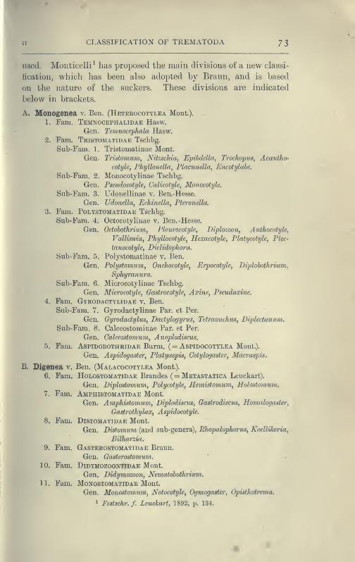
CLASSIFICATION OF TREMATODA 73
used. Monticelli^ has proposed the main divisions of a new classi-
fication, which has been also adopted by Braun, and is based
on the nature of the suckers. These divisions are indicated
below in brackets.
A. Monogenea v. Ben. (Heterocotylea Mont.).
1. Fam. Temnocephalidae Hasw.
Gen. Temnocephala Hasw.
2. Fam. Tristomatidae Tschbg.
Sub-Fam. 1. Tristomatinae Mont.
Gen. Tristomum, Nihschia, Epihdella, Trochopus, Acaniho-
cotyle, Phyllonella, Placunella, Encotylahe.
Sub-Fam. 2. Monocotylinae Tschbg.
Gen. Pseudocotyle, Calicotyle, Monocotyle.
Sub-Fam. 3. Udonellinae v. Ben.-Hesse.
Gen. Udonella, Echinella, Pteronella.
3. Fam. Polystomatidae Tschbg.
Sub-Fam. 4. OctocotyHnae v. Ben.-Hesse.
Gen. Odobothrium, Pleurocotyle^ Diplozoon, ATithocotyle,
Vallisniaj Phyllocotyle, Hexacotyk, Platycotyle, Plec-
tanocotyle, Biclidophora.
Sub-Fam. 5. Polystomatinae v. Ben.
Gen. Polystomum, Onchocotyle, Erpocotyle, Diplohothrium.
Sphyramira.
Sub-Fam. 6. Microcotylinae Tschbg.
Gen. Microcotyle, Gastrocotykj Axine^ Pseudaxine.
4. Fam. Gyrodactylidae v. Ben.
Sub-Fam. 7. Gyrodactylinae Par. et Per.
Gen. GyrodadyluSy Dadylogyrus, Tetraonchus, Diplectanum.
Sub-Fam. 8. Calceostominae Par. et Per.
Gen. Calceostornum, Anoplodiscus.
5. Fam. Aspidobothridae Burm. ( = Aspidocottlea Mont.).
Gen. Aspidogastei', Platyaspis, Cotylogaster, Macraspis.
B. Digenea v. Ben. (Malacocotylea Mont.).
6. Fam. Holostomatidae Brandes ( = Metastatica Leuckart).
Gen. Diplostomum, Polycotyle, Hemistomum, Holostomum.
7. Fam. Amphistomatidae Mont.
Gen. Amphistomum^ Diplodiscus, Gastrodiscus, Homalogaster^
Gastrothylax, Aspidocotyle.
8. Fam. Distomatidae Mont.
Gen. Distomum (and sub-genera), Rhopalophorus, Koellikeriaj
Bilharzia.
9. Fam. Gasterostomatidae Braun.
Gen. Gasterostomum.
10. Fam. DiDYMOZooNTiDAE Mont.
Gen. Didymozoon, Neviatobothriuvi.
11. Fam. MONOSTGMATIDAE Mont.
Gen. Moiwstomum, Notocotyle^ Ogmogaster, Opisthotrema.
^ Festschr.f. Leuckart, 1892, p. 134.

7^ PLATYHELMINTHES CESTODA
-'SC
and with those of the head. Each joint contains at first male'
genitalia comparable with those of a Trematod^ ; then the female
organs develop, and finally self-fertilisation follows. The Cestodes
feed through their skin, probably by the aid of fine protoplasmic
processes, which penetrate the tough investing membrane and
absorb the already digested food which bathes them. When a
proglottis of Ccdliohothrium is approaching maturity it separates
from the parent, the broken ends of muscles, nerves, and excre-
tory vessels speedily heal, and it is now cap-
able of continued growth and of fairly active
movement if it remains in the intestine of
the host. According to van Beneden, it mayeven attain a size equal to, or exceeding, that
of the whole parent or " strobila." -^ These
considerations led Leuckart, von Siebold, P. J.
van Beneden, and others, to Steenstrup's con-
clusion that a jointed tape-worm is really a
colony composed of two generations— the
head and neck derived from the larva, and
the proglottides produced by the segmenta-
tion of the neck.^ This view of the colonial
nature of jointed Cestodes was generallyFig. 37. — Archigetes
sieboldii [appendi-
culatiis), from the
coelom of Tuhifexrivtdorum. x 40.
app, Persistent larval
appendage ; c/o, geni-
tal pore ; hk, per-
sistent larval hooks;
ov, ovary ; sc,
sucker ; te, testes;
yg, yolk -glands.(After Leuckart.)
adopted from 1851 to 1880. During the
last fifteen years, however, the varied inter-
pretations of the facts of the ontogeny of this
group have led some authors to adopt the
monozootic view (that a Cestode is one indi-
vidual), others are still of the older opinion,
and Hatschek (Lehrhuch, p. 349) and Lang
take up intermediate positions. Lang considers
that the formation of the joints of a tape-
worm from a small fixed " scolex," is not only largely comparable
with the strobilation of a scypliistoma and the consequent forma-
tion of a pile of medusae, as in the life-history of Aurelia, but
' The mature j^roglottis of Ccdliohothrium eschrichti is 8-9 mm. long, whereas
the strobila only measures 4-5 mm. in length. Species of Phylliobothritim, Antho-
bothrvmn, and Tetrarhynchus show a similar but not an equal contrast between
the size of the parent and proglottis (P. J. van Beneden, "Les Vers Cestoides,"
Nouv. 3fem. de VAcad. Roy. d, Belgique, tom. xxv. 1850).
^ The difficult question of the nature of the Cestode body and Cestode larvae
is adequately discussed by Braun, loc. cit. p. 1167.
I
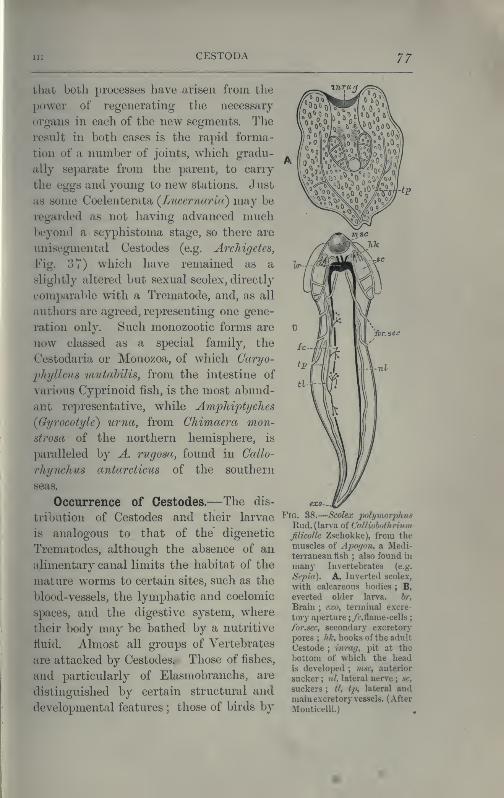
Ill CESTODA 77
that both processes have arisen from the
power of regenerating the necessary
oro^ans in each of the new sef^ments. The
result in both cases is the rapid forma-
tion of a number of joints, which gradu-
ally separate from the parent, to carry
the eggs and young to new stations. Just
as some Coelenterata {Liicernarut) may be
regarded as not having advanced muchbeyond a scyphistoma stage, so there are
unisegmental Cestodes (e.g. Archigetes,
Fig. 37) which have remained as a
slightly altered but sexual scolex, directly
comparable with a Trematode, and, as all
authors are agreed, representing one gene-
ration only. Such monozootic forms are
now classed as a special family, the
Cestodaria or Monozoa, of which Caryo-
phi/lleus mutabilis, from the intestine of
various Cyprinoid fish, is the most abund-
ant representative, while Amphiptyches
(Gyrocotyle) urna, from Chimaera mon-
strosa of the northern hemisphere, is
paralleled by A. rugosa, found in Ccdlo-
rhy7iclius antarcticus of the southern
seas.
Occurrence of Cestodes.—The dis-
tribution of Cestodes and their larvae
is analogous to that of the digenetic
Trematodes, although the absence of an
alimentary canal limits the habitat of the
mature worms to certain sites, such as tlie
blood-vessels, the lymphatic and coelomic
spaces, and the digestive system, where
their body may be bathed by a nutritive
fluid. Almost all groups of Vertebrates
are attacked by Cestodes. Those of fishes,
and particularly of Elasmobranchs, are
distinguished by certain structural and
developmental features ; those of birds by
Fig. 38.
—
Scolex polyviorphus
Riid. (larva of Calliobothrivm
filicolle Zschokke), from tlie
muscles of Apoffon, a Medi-terranean fish ; also found in
many Invertebrates (e.g.
Si'pia). A, Inverted scolex,
with calcareous bodies ; B,
everted older larva. br.
Brain ; e,a>, terminal excre-
tory aperture ;/f,flame-cells
;
far.sec, secondary excretory
pores ; hk, hooks of tlie adult
Cestode ; inrwj, pit at the
bottom of which the headis developed ; msc, anterior
sucker ; nl, lateral nerve ; sc,
suckers ; tl, tp, lateral andmain excretory vessels. ( After
Mouticelli.)
78 PLATYHELMINTHES CESTODA chap.
others; those of mammals, by a third set of characters. Theyoung stages of the Cestodes of Sharks and Eays occur encysted
in the body-cavity, or in the pyloric appendages, of Teleosteans,
which probably swallow them along with those invertebrate
animals upon which they prey. The larvae of the Cestodes of
carnivorous mammals or piscivorous birds, live respectively in
herbivores and fishes, but how the latter are infected we knowin very few instances. Cestode larvae are known to occur in manyInvertebrates, and occasionally are taken free swimming in the
sea, presumably crossing from one host to the next. Cteno-
phores, Siphonophores, Copepods, Ostracods, Decapods, various
Molluscs especially Cephalopods, Earthworms, and other Anne-lids, are the intermediate hosts of these larvae (see Fig. 38),
the fate of which, however, has been determined in but few cases.
Occurrence of Cestodes in Man.^—Tape-worms, either in
the adult or larval stages (bladder-worms), have, from ancient
times, been known to occur in man, and in the animals that serve
him as food. Until comparatively recent times, however, the
true nature of these parasites, and particularly of " hydatids
"
(cystic larvae), was unrecognised. Up to the seventeenth
century the larvae were regarded as abscesses or diseased growths
of the affected organs, and it was only at the close of that century
that their animal nature was even suggested. Even at the
beginning of the nineteenth century, three modes of origin of
Cestodes—by " generatio aequivoca " from the tissues of the body,
or by the union of previously distinct proglottides, or again by
metamorphosis of free-living worms drunk with water by cattle
or birds (as Linnaeus suggested)—were still variously held, at a
time when Malpighi, Pallas, and Goeze had recognised the true
connexion between the cystic and segmented states of Taenia
crassicollis (the cat tape-worm), and when Goeze had seen the
eggs of Taeniae, and Abildgaard ^ had even conducted the first
helminthological experiments (conversion of the larval Schisto-
ceplialus, Fig. 40, into the adult form).
Generally speaking, " a tape-worm " in Western Europe will
prove to be Taenia saginata Goeze (the beef tape-worm. Fig. 39, A),
^ Leuckart, Die Parasiten d. Menschen [English trans, by W. E. Hoyle];
Blanchard, TraiU cle Zoologic medicate, 1893." For a full account of the history of this subject see Leuckart, Parasiten d.
Menschen, p. 28 ; Braun, loc. cit. Bd. iv. p, j929 et seq. ; Huxley, Collected Essays,
vol. viii. p. 229.
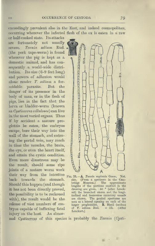
OCCURRENCE OF CESTODA 79
•exceedingly prevalent also in the East, and indeed cosmopolitan,
occurring wherever the infected flesh of the ox is eaten in a raw
or half-cooked state. Its attacks
are fortunately not usually
severe. Taenia solium Eud.
(the pork tape-worm) is found
wherever the pig is kept as a
domestic animal, and has con-
sequently a world-wide distri-
bution. Its size (6-9 feet long)
and powers of adhesion would
alone render T. solium a for-
midable parasite. But the
danger of its presence in the
body of man, or in the flesh of
pigs, lies in the fact that the
larva or bladder-worm (known
as Cysticercus celhdosae) can live
in the most varied organs. Thus
if by accident a mature pro-
glottis be eaten, the embryos
escape, bore their way into the
^m wall of the stomach, and enter-
^^ ing the portal vein, may reach
in time the muscles, the brain,
^k the eye, or even the heart itself,
^^ and attain the cystic condition.
Even more disastrous may be
^m the result, should some ripe
^" joints of a mature worm work
their way from the intestine
jB back towards the stomach.^^ Should this happen (and though
it has not been directly proved,
the possibility is to be reckoned
with), the result would be the
release of vast numbers of em-
bryos capable of inflicting fatal
Fig. 39.—A, Taenia saginata Goeze. Nat.
size. (From a specimen in the Caiu-
bridge Museum.) The approximatelengths of the portions omitted in the
drawing are given. At * (after Leuck-
art) the branched uterus and the longi-
tudinal and transverse excretorj- vessels
are shown. The genital apertures are
seen as a lateral opening on each of the
larger proglottides. B, Head (scolex)
of T. solium Rud. x 12. (After
. . , , , , Leuckart.)injury on the host. An abnor-
mal Cysticercus of this species is probably the Taenia (Cysti-

8o PLATYHELMINTHES CESTODA chap.
cercus) acanthotrias Weinl. (see, however, Leuckart, loc. cit.
711).
Taenia {Hymenolepis) nana v. Sieb.^ is found in man in
Egypt, Italy, England, Servia, Argentine Eepublic, and the
United States. Though small (J-1 inch long), its numbers
usually excite digestive and nervous disorders of considerable
severity, more serious, indeed, than those caused by the commonertape-worms. H: diminuta Eud. (flavopunctata Weinl.), normally
found in Eodents, has been rarely recorded in man. Taeni
(Dipi/lidmm) caninum L. { = T. cucumerina Bloch = T. elliptic^
Batsch), the commonest parasite of pet cats and dogs, and 2(Davainea) madagascariensis Davaine, have occasionally bee:
recorded from infants and young children. But the attacks o
these species are insignificant in comparison with those of th
cystic stage {Echinococcus polyvwrplnis) of a tape-worm (71 ecliino
coccus V. Sieb.) which lives when mature in the dog.
Uchinococcus is most frequent in Iceland, where it affects 2
to 3 per cent of the population, and a still larger proportion o:
sheep ; while in Copenhagen, Northern Germany, some districts
of Switzerland, and Victoria it is not uncommon, but is frequently
found during post-7norte77i examinations when no definite symptoms
of its presence had been previously noticed. Echinococcus ^ varies
greatly in size, form, and mode of growth, but is distinguished
in the formation not of one scolex only, as in the Cysticercus, but
in the production of a number of vesicles, usually from the inner
wall. "Within these, large numbers of scolices may be developed;
The whole organism continues to swell by the formation of a watery
liquid within it, and if its growth be rapid the fluid tension maycause the rupture of the enclosing connective -tissue capsule
formed around the parasite, at the expense of the host, and the,
protrusion of the daughter vesicles. It is the consequent injury
to the surrounding organs of the host, at this critical stage, often
only reached after the lapse of several years, that occasions
serious or even fatal results. Zoologically, Taenia echinococcus.
and T. coenurus are interesting, since they exhibit an indubit-
^ By Grassi this form is considered identical with T. murina. The latter
species is known, from this author's researches, to develop in rats without migra-
tion into an intermediate host. Should Grassi"s synonymy prove correct, the:
presence of large numbers of this tape-worm in man would readily receive its.
explanation,
^ Leuckart, loc. cit. p. 752 et seq.
1

Ill OCCURRENCE OF CESTODES 8 I
able alternation of asexual generations in the larval state, with
a sexual adult stage."
Bothriocephalus latus Brems., the broad tape-worm, which
attains a length of 20-30 feet, or even more, occurs in manendemically in the eastern ]>altic provinces, certain parts of
Switzerland, generally throughout Eussia (especially near Kasan),
in North America, and connnonly in Japan,—that is, in districts
where the population partake largely of pike or other fish in a
raw or partially-cooked state. Elsewhere it occurs sporadically,
and in Munich, where it was unknown l)efore 1880, its presence
has been traced to emigrants from infected districts, who settled
on the shores of the Starenberger Lake, from which Munich was
supplied with fish. How the pike, the usual but not invariable
intermediate liost, becomes infested (and its musculature is
frequently riddled with tlie larvae) we do not accurately know,
but some Invertebrate, the prey of the pike, is probably the first
host into which the free-swimming ciliated larva (Fig. 42) finds
its way. In Greenland, B. cordatus is very common in the dog,
and probably also in man, though few cases have been recorded.
B. mansoni Cobb. ( = B. liguloides Leuck.) was, till recently,
known only in the larval state from China and Japan. lijima,
however, has found older specimens in the latter country. B.
cristatus Dav. is a species founded somewhat doubtfully on two
^fragments found, one in a child, the other in a man, in France.
Occurrence of Oestodes in Domestic Animals.^—Amongdomestic animals, the dog is, undoubtedly, the most frequently
attacked by Taeniae. Six species of Taenia (T. serrata, mar-
ginata, coenurus, echinoccccus, krahhei, and possibly T. serialis),
Bipi/lidiuin caiiinum (the commonest form), 3fesocestoides lineatus,
and three or four species of Bothriocephahis' have been found in
the dog. The table of life-histories (p. 83) shows that sheep,
rabbits and other Eodents serve as the intermediate hosts, in
which the cystic stages of the species of Taenia are found.
Hence the prevalence of T. serrata in a given locality is con-
nected with the abundance there of the rabbit and hare, in which
the larva (Cysticercus pisiformis) occurs. Bothriocephalus cordatus
develops from the young stage present in the fish which the
Icelanders give to their dogs. In Iceland and certain parts of
^ The distinctive features cf these and the foregoing tape-worms are given on
pp. 89-90.
VOL. II G
8o PLATYHELMINTHES CESTODA chap. •
cercus) acanthotrias Weinl. (see, however, Leuckart, loc. cit. p.
711).
Taenia (^Hymenolepis) nana v. Sieb.^ is found in man in
Egypt, Italy, England, Servia, Argentine Eepublic, and the
United States. Though small (^-1 inch long), its numbers
usually excite digestive and nervous disorders of considerable
severity, more serious, indeed, than those caused by the commonertape-worms. H: diyninuta Eud. {flavopunctata WeinL), normally
found in Eodents, has been rarely recorded in man. Taenia
{Dipylidium) caninum L. ( = T cucumerina Bloch = T, elliptica
Batsch), the commonest parasite of pet cats and dogs, and 2\
{Davainea) madagascariensis Davaine, have occasionally been
recorded from infants and young children. But the attacks of
these species are insignificant in comparison with those of the
cystic stage (Uchinococcus poIymorpJuis) of a tape-worm (T. echino-
coccus V. Sieb.) which lives when mature in the dog.
Echinococcus is most frequent in Iceland, where it affects 2
to 3 per cent of the population, and a still larger proportion of
sheep ; while in Copenhagen, Northern Germany, some districts
of Switzerland, and Victoria it is not uncommon, but is frequently
found diuxing post-mortem examinations when no definite symptomsof its presence had been previously noticed. Echinococcus ^ varies
greatly in size, form, and mode of growth, but is distinguished
in the formation not of one scolex only, as in the Cysticercus, but
in the production of a number of vesicles, usually from the inner
wall. Within these, large numbers of scolices may be developed.
The whole organism continues to swell by the formation of a watery
liquid within it, and if its growth be rapid the fluid tension maycause the rupture of the enclosing connective -tissue capsule
formed around the parasite, at the expense of the host, and the
protrusion of the daughter vesicles. It is the conseqaent injury
to the surrounding organs of the host, at this critical stage, often
only reached after the lapse of several years, that occasions
serious or even fatal results. Zoologically, Taenia echinococcus
and T. coenurus are interesting, since they exhibit an indubit-
^ By Grassi this form is considered identical with T. muriiia. The latter
species is known, from this author's researches, to develop in rats without migra-
tion into an intermediate host. Should Grassi"s synonymy prove correct, the
presence of large numbers of this tape-worm in man would readily receive its
explanation.
- Leuckart, loc. cit. p. 752 et seq.

Ill OCCURRENCE OF CESTODES 8 I
able alternation of asexual generations in the larval state, with
a sexual adult stage."
Bothriocejjhahis latus Brems., the broad tape-worm, which
attains a length of 20-30 feet, or even more, occurs in manendemically in the eastern Baltic provinces, certain parts of
Switzerland, generally throughout Eussia (especially near Kasan),
in North America, and commonly in Japan,—that is, in districts
where the population partake largely of pike or other fish in a
raw or partially-cooked state. Elsewhere it occurs sporadically,
and in Munich, where it was unknown before 1880, its presence
has been traced to emigrants from infected districts, who settled
on the shores of the Starenberger Lake, from which Munich was
supplied with fish. How the pike, the usual but not invariable
intermediate host, becomes infested (and its musculature is
frequently riddled with the larvae) we do not accurately know,
but some Invertebrate, the prey of the pike, is probably the first
host into which the free-swimming ciliated larva (Fig. 42) finds
its way. In Greenland, B. cordatus is very common in the dog,
and probably also in man, though few cases have been recorded.
B. mansoni Cobb. { = B. ligidoides Leuck.) was, till recently,
known only in the larval state from China and Japan. lijima,
however, has found older specimens in the latter country. B.
cristatus Dav. is a species founded somewhat doubtfully on two
fragments found, one in a child, the other in a man, in Franca
Occurrence of Cestodes in Domestic Animals.^—Amongdomestic animals, the dog is, undoubtedly, the most frequently
attacked by Taeniae. Six species of Taenia (T. serrata, mar-
ginata, coenurus, echinoccccus, krahhei, and possibly T. serialis),
Dipijlidium caniniim (the commonest form), 3fesocestoides lineatus,
and three or four species of Botlwioceplialus' have been found in
the dog. The table of life-histories (p. 83) shows that sheep,
rabbits and other Eodents serve as the intermediate hosts, in
which the cystic stages of the species of Taenia are found.
Hence the prevalence of T. serrata in a given locality is con-
nected with the abundance there of the rabbit and hare, in which
the larva (Cysticercus jyisiformis) occurs. Bothriocejyhalus cordatus
develops from the young stage present in the fish which the
Icelanders give to their dogs. In Iceland and certain parts of
^ The distinctive features cf these and the foregoing tape-womis are given on
pp. 89-90.
VOL. IT G
82 PLATYHELMINTHES—CESTODA
Australia T. echinococcus infests one-third to one-half the number
of dogs examined ; a fact connected with the frequency of Uchino-
cocous in man in these countries.
In sheep the most noteworthy and dangerous parasite is
Coenurus cerehralis (or the cystic stage of the dog-taenia, T.
coenurus), which gives rise to the disease known as " gid " or
" staggers." It is found in various parts of the brain or spinal
cord, and the symptoms differ according to the position of the
parasite. If this presses upon one hemisphere the sheep describes
circles and finally falls : if- on the optic lobes, the eyes are
affected : if the pressure affects the cerebellum the movements
of the sheep are uncertain and incoordinated. Four or six
weeks after the appearance of the symptoms, death results from
cerebral paralysis, or from general debility, and the loss of sheep
incurred by this disease (happily less frequent in England than
formerly) has been calculated by Youatt at a million for France
annually ; at 3 5 per cent of the flocks for England in bad seasons;
and about 2 per cent for Germany. Besides sheep, which are
most subject to " gid " during their first year, various ruminants
—Goat, Ox, Moufflon, Chamois, Koe, Antelope, Eeindeer, Drome-
dary—are attacked in the same way. A similar form, Coenurus
serialis BailL, is common in the wild rabbit in this country, and
in Australia in the hare and squirrel. It forms large swellings
in the connective tissue of various parts of the body, but usually
does not affect the health of the host. It is not known in whatcarnivore Taenia serialis BailL normally occurs. Experiments
have, however, shown that it develops rapidly in dogs.
The preventive measures which are steadily diminishing the
prevalence of the Cestode parasites in man in some parts of
Western Europe cannot be dealt with here, but it may be
noticed that the Jewish observance with regard to swine is the
surest preventive measure against taeniasis and trichinosis.
Careful inspection of meat and general cleanliness, are the lead-
ing measures that in these hygienic matters secure the greatest
immunity from disease.
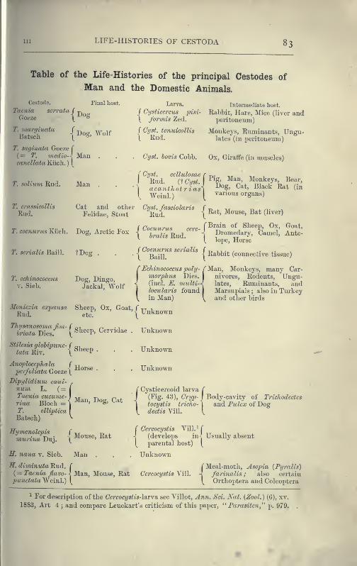
LIFE-HISTORIES OF CESTODA 83
Table of the Life-Histories of the principal Cestodes of
Man and the Domestic Animals.
ICestode.unia scrrata
Goeze
13
{Dog
Final host. Larva. Intermediate liost.
f Cysticercus pisi- Rabbit, Hare, Mice (liver and\ formis Zed. peritoneum)
margmataBatscli I
Dog, Wolf ( Cyst. tcnuicolUs
\ Rud.IVIonkeys, Ruminants, Ungu-
lates (in peritoneum)
T. saginata Goeze T
(= 1\ medio-', Mancancllata Klicli.
) [
Cyst, hovis Cobb. Ox, Giraffe (in muscles)
Isoluini Rud. ]\ra
( Cyst.
I Rud.cellulosae r -r,. nr t.«^ , -m.
(1 Cii'it ^^S' ^^*^' Monkeys, Bear,
acanthotriasxWeinl.) [
various organs)
crassieollis
Rud.Cat and other Cyst, fasciolaris f r» . ^.r ^ ^ n-
Felidae, Stoat Rud. \ ^**^' ^^°"«^' ^'^^ (^i^'^^')
I", cocnurtcs Kiicli. Dog, Arctic Fox
scrialis Baill. ? Dog
,_ r Brain of Sheep, Ox, Goat,"4 Dromedary, Camel, Ante-
(^ lope, Horse
\ Rabbit (connective tissue)
T. echinococcus
V. Sieb.
Dog, Dingo,Jackal, Wolf
\ bralis Rud.
f Coenurus serialis
\ Baill.
(Echinococcus poly- f Man, Monkeys, many Car-morphus Dies. I nivores. Rodents, Ungu-(incl. E. onulti-J. lates, Ruminants, andlocularis found
jMarsupials ; also in Turkey
in Man) \ and other birds
Moniczia expansa Sheej), Ox, Goat, f tt vRud. etc.
Thysanosoma fivi- f r^-. ^ .
,
oriata Dies. [ ^^^^^P' Cervidae
Stilcsia glohipunc- f
\
tata Riv.Sheep .
Anoplocephcda f ^jpcrfoliata Goeze\^^^^^ •
Dipylidium cani-num L. ( =Taenia cucume- i ,r t-v n 1.
Tina Bloch = i ^^^^^' ^°g' ^^^
T. elliptica
Batsch)
Hymenolepismurina Duj.
Mouse, Rat
E. nana v. Sieb. Man
H. diminuta Rud.( = Taenia Jiavo-
punctata Weinl.)
Unknown
Unknown
Unknown
f Cysticercoid larva f
I(Fig. 43), Cryp-j Body-cavity of Trichodectcs
\tocystis tricho-^ and Pw^ca; of Dogdectis Vill. V
Cercocystis Vill.^ f
(develops in- Usually absentparental host) [
Unknown
-j Man, Mouse, Rat Cercocystis Yill.
r Meal-moth, Asopia (Pyralis)
-] farinalis ; also certain
(^ Orthoptera and Coleoptera
^ For description of the CcrcocT/s^ts-larva see A'illot, Ann. Sci. Kat. {Zool.) (6), xv.
1883, Art 4 ; and compare Leuckart's criticism of this paper, " Farasitcn," p. 979.

84 TLATYHELMINTHES CESTODA CHAPJ
Cestode.
Drepanidotaeniagracilis Zed.
Final host.
Duck, Goose, Wild f
Duck
Larva.
Cercocystis Yill.
D.anatinaYiva\)hQ Duck ,
D. setigera Froli. Goose .
formts Goeze (_
Dicranotacnia( Duo'k
coronula Duj. \
Davainea proglot- f y ^
tina Dav. \
D. madagascari-cnsis Wax.
Children
D. fri^dhergerijpi^.^.^^t
V. Linst. (
Mesocestoides line-
atus Goeze
Bothriocephahcs f
latus Brems. \
Intermediate host.
TThe Ostracods Candona-! trata and Cypris comprcssa,
( and also Cyclops viridis
J Cypris incongruens, and also
t Perch
Cyclops hrcvicaudatus
House-fly
Cyp>ris ovum
? Limax ciiiereus, L. agrestis
Unknown
Doer
Man, Dog, ? Cat
Unknown ? Ants
Unknown
Plerocercoid, t.c r-n i ri x? . , t
solid, elongate / P^b^bly first enters an Inver-
larva, with nobladder ^
tebrate host, which is eatenby Pike, Perch, Trout, etc.
Structure and Development of Cestoda.^—Of the unseg-
mented Cestodes, Caryophyllaeus mutahilis, from the intestine of
carp and other Cyprinoid fishes, is the most easily accessible form.
Fig. 40.—a, Stickle-
back {Gasterosteusacideatns) infested
by an advancedlarva of Schisto-
cephalus solidus
Crepl. B. Thelarva. All x 1|.
(From specimens
in the CambridgeUniversity Mu-seum.
)
TriaenoplioTus nodulosus, which is very useful for the study of the
excretory system, occurs mature in the pike. In the body-cavity of
the Stickleback (Fig. 40) a large, broad, yellow worm may sometimes
^ Moniez, "Sur les Cysticerques," Paris, 1880; Id. "Sur les Cestodes," 1881 ;,
Zschokke, " Recherches sur la structure anatomique et histologique d. Cestodes,"
Geneve, 1888.
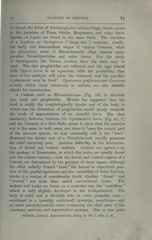
Ill ANATOMY OF CESTODA 85
be found, the larva of Schistocephal/as solidus Crepl., which occurs
in the intestine of Terns, Storks, Mergansers, and other birds.
Species of Zlgula are found in the same birds. The intestine
of a Lophius or Cyclopterus (" lump-fisli ") contains, usually,
the early and intermediate stages of various Cestodes, while
the alimentary canal of Elasmobranchs often contain manypeculiar Tetrarhynchidae and other forms. For the study
of development, the Taenia anatina from the duck may be
used. The ripe proglottides are collected, and the eggs placed
with Cypris ovum in an aquarium, with the probability that
some of the embryos will enter the Ostracod, and the peculiar
Cysticercoid may be bred.^ Cysticercus p)isiformis and Coenurus
serialis, which occur commonly in rabbits, are also suitable
objects for examination.
A Cestode such as Eehinohothrium (Fig. 36) is divisible
into head and proglottides. Moniez has suggested that the
head is really the morphologically hinder end of the body, in
which case the formation of proglottides would closely resemble
the mode of segmentation of an Annelid larva. The close
similarity, however, between the Cysticercoid larva (Fig. 43, F)
and the Cercaria of a liver-fluke, seems to show that the anterior
end is the same in both cases, and since it bears the central part
of the nervous system, we may reasonably call it the " head."
Moreover the hinder end of a Platyhelminth usually possesses
the chief excretory pore. Another difficulty is the determina-
tion of dorsal and ventral surfaces. Authors are agreed,—on
the analogy of Trematodes, in which the testes are usually dorsal
and the ovaries ventral,—that the dorsal and ventral aspects of a
Cestode are determined by the position of these organs, although
the often radially formed " head," the lateral or superficial posi-
tion of the genital apertures, and the variability of these features^
render it a matter of considerable doubt whether " dorsal " and" ventral " are more than useful conventional terms. The
suckers and hooks are borne on a muscular cap, the " rostellum,"
which is only slightly developed in the Ichthyotaeniae. The
body is solid, and is divisible into an outer muscular coat
—
enveloped in a (possibly epidermal) investing membrane—and
an inner parenchymatous tissue containing the chief part of the
excretory, nervous, and reproductive systems. One or two pairs
^ Schmidt, ArcJiiv f. JVaiurgcschichte, Jahrg. Ix. Bd. 1, 1894, p. 65.
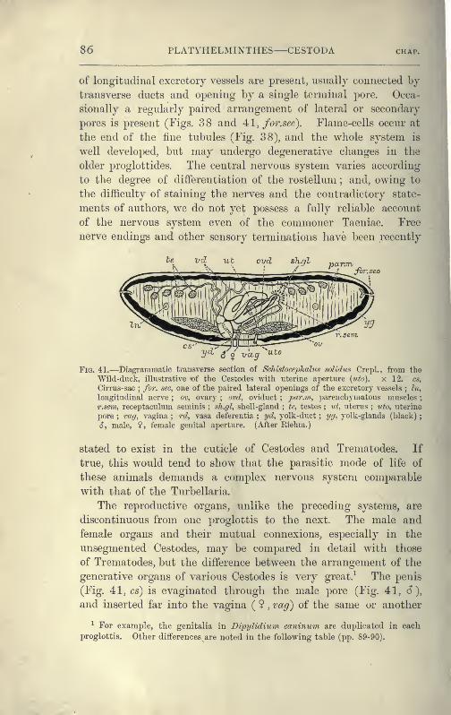
S6 PLATYHELMINTHES CESTODA
of longitudinal excretory vessels are present, usually connected by
transverse ducts and opening by a single terminal pore. Occa-
sionally a regularly paired arrangement of lateral or secondary
pores is present (Pigs. 38 and 4tl, fo7\sec). Flame-cells occur at
the end of the fine tubules (Fig. 38), and the whole system is
well developed, but may undergo degenerative changes in the
older proglottides. The central nervous system varies according
to the degree of differentiation of the rostellum ; and, owing to
the difficulty of staining the nerves and the contradictory state-
ments of authors, we do not yet possess a fully reliable account
of the nervous system even of the commoner Taeniae. Free
nerve endings and other sensory terminations liave been recently
Jvr.se
2/^' ^ $utov-ccg
Fia. 41.—Diagrammatic ti'ansverse section of Schistocephalus soHdtis Crepl., from the
Wild-duck, illustrative t)f the Cestodes with uterine aperture {uto). x 12. cs.
Cirrus-sac;for. sec, one of the paired lateral openings of the excretory vessels ; In,
longitudinal nerve ; ov, ovary ; ovd, oviduct;
^;«r.?H, parenchymatous muscles ;
r.sem, receptaculum seminis ; sh.gl, shell-gland ; te, testes ; vt, uterus ; iffo, i;terine
pore ; vag, vagina ; rd, vasa deferentia ; yd, yolk-duct ; ijg, yolk-glands (black);
6, male, ?, female genital aperture. (After Riehui.)
stated to exist in the cuticle of Cestodes and Trematodes. If
true, this would tend to show that the parasitic mode of life of
these animals demands a complex nervous system comparable
with that of the Turbellaria.
The reproductive organs, unlike the preceding systems, are
discontinuous from one proglottis to the next. The male and
female organs and their mutual connexions, especially in the
unsegmented Cestodes, may be compared in detail with those
of Trematodes, but the difference between the arrangement of the
generative organs of various Cestodes is very great.-^ The penis
(Fig. 41, cs) is evaginated through the male pore (Fig. 41, 6),
and inserted far into the vagina ( ? , vag) of the same or another
^ For example, the genitalia in Dipylidium caninum are duplicated in each
proglottis. Other differences are noted in the following table (pp. 89-90).
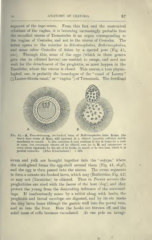
Ill ANATOMY OF CESTODA 8/
segment of the tape-worm. From this fact and the anatomical
relations of the vagina, it is becoming increasingly probable that
the so-called uterus of Trematodes is an organ corresponding to
the vagina of Cestodes, and not to the uterus of Cestodes. The
latter opens to the exterior in Schistoceijhalus, Bothriocephalus,
and some other Cestodes of fishes by a special pore (Fig. 41,
tito). Through this, some of the eggs (which in these genera
give rise to ciliated larvae) are enabled to escape, and need not
wait for the detachment of the proglottis, as must happen in the
Taeniidae, where the uterus is closed. This uterus, a true physio-
logical one, is probably the homologue of the " canal of Laurer"
(". Laurer-Stieda canal," or " vagina ") of Trematoda. The fertilised
Fig. 42.—A, Free-swimming, six-hooked larva of Bothriocephalus latus Brems. (the
broad tape -worm of Man), still enclosed in a ciliated (possibly cellular) double
membrane or mantle. In this condition it may continue to live in water for a weekor more, but eventually throws off its ciliated coat (as in B) and commences to
creep about vigorously by the aid of its hooks, in search of its tirst host, which is at
present unknown. (After Schauiusland.) x 600.
ovum and yolk are brought together into the " ootype," where
the shell-gland forms the egg-shell around them (Fig. 41, sh.gl),
and the egg is then passed into the uterus. The ovum segments
to form a minute six-hooked larva, which may (Bothriidae, Fig. 42)
or may not (Taeniidae) be ciliated. Thus in Taenia serrata the
proglottides are shed with the faeces of the host (dog), and they
protect the young from the desiccating influence of the surround-
ings. If inadvertently eaten by a rabbit along with herbs, the
proglottis and larval envelope are digested, and by its six hooks
the tiny larva bores through the gastric wall into the portal vein,
and so into the liver. Here the hooks are thrown off, and the
solid mass of cells becomes vacuolated. At one pole an invagi-
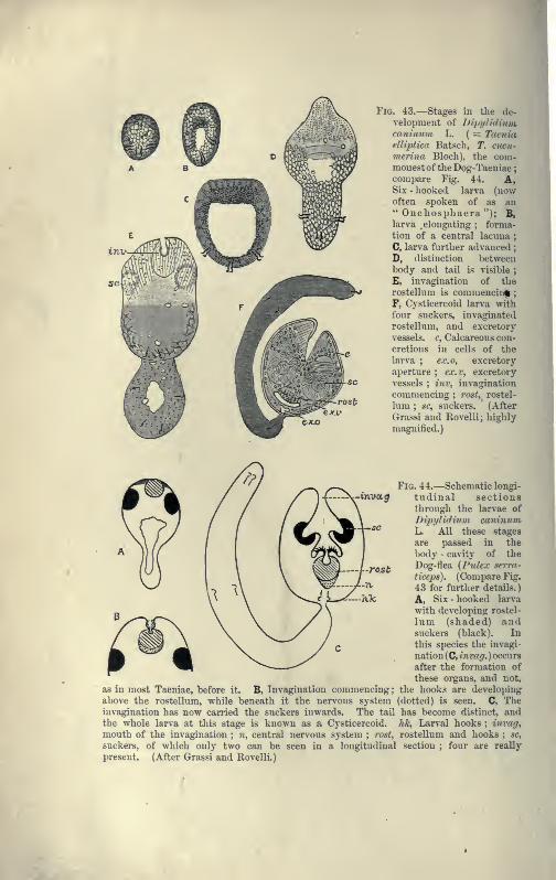
Fig. 43.—Stages in the de-
velopment of DipyUdiuvicaninum L. ( = Taeniaelliptica Batsch, T. cticu-
vierina Bloch), the com-monest of the Dog-Taeniae
;
compare Fig. 44. A,Six - liooked larva (nowoften spoken of as an" Onchosphaera "); B,larva .elongating ; forma-
tion of a central lacuna;
C, larva fnrther advanced;
D, distinction betweenbody and tail is visible
;
E, invagination of the
rostellnm is commencin||
;
F, Cysticercoid larva with
fonr snckers, invaginated
rostellnm, and excretory
vessels, c, Calcareous con-
cretions in cells of the
larva ; ex.o, excretory
aperture ; ex.r, excretory
vessels ; inv, invagination
commencing ; rost, rostel-
him ; sc, suckers. (After
Grassi and Rovelli; highly
magnified.)
xroctg
rost
as in most Taeniae, before it. B, Invagination commencing
;
above the rostelluni, while beneath it the nervous systeminvagination has now carried the suckers inwards. The tail
the whole larva at this stage is known as a Cysticercoid.
mouth of the invagination ; «, central nervous system ; rost,
suckers, of which only two can be seen in a longitudinal
present. (After Grassi and Rovelli.)
Fig. 44.—Schematic longi-
tudinal sectionsthrough the larvae of
Dipylidium caninumL. All these stages
are passed in the
body - cavity of the
Dog-flea (Pulex serra-
t iceps). (Compare Fig.
43 for further details.)
A, Six - hooked larva
with developing rostel-
lnm (shaded) andsuckers (black). In
this species the invagi-
nation (C, in vag. ) occurs
after the formation of
these organs, and not,
the hooks are developing
(dotted) is seen. C, Tlie
has become distinct, andhk, Larval hooks ; invag,
rostellnm and hooks ; sc,
section ; four are really
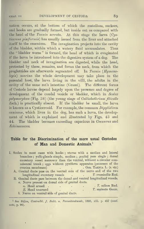
CHAP. Ill DEVELOPMENT OF CESTODA 89
nation occurs, at the bottom of which the rostelhini, suckers,
and hooks are gradually formed, but inside out as compared with
the head of the Taenia serrata. At this stage the larva {Cys-
ticercus pisiformis) has usually issued from the liver and attached
itself to the omentum. The invagination projects into the cavity
of the bladder, within which a watery fluid accumulates. Thus
the " bladder worm " is formed, the head of which is evaginated
if the larva be introduced into the digestive system of a dog. Thebladder and neck of invagination are digested, while the head,
protected by these, remains, and forms the neck, from which the
proglottides are afterwards segmented off. In Taenia {Hymeno-
lepis) murina the wliole development may take place in the
parental host, the larva living in the villi, the adults in the
cavity of the same rat's intestine (Grassi). The different forms
of Cestode larvae depend largely upon the presence and degree of
development of the caudal vesicle or bladder, which in Scolex
polymorphus (Fig. 38) (the young stage oi Calliohothrium filicolle
Zsch.) is practically absent. If the bladder be small, the larva
is known as a Cysticercoid. For example, the common Dipylidium
caninum, which lives in the dog, has such a larva, the develop-
ment of which is explained and illustrated by Figs. 43 and
44. The bladder becomes exceeding capacious in Coenurus and
Echinococcus.
Table for the Discrimination of the more usual Cestodes
of Man and Domestic Animals.^
I. Scolex ill most cases with hooks ; uterus with a median and lateral
branches;yolk-glands simple, median
;genital pore single ; dorsal
excretory vessel narrower than the ventral, without a circular com-
missural trunk ; eggs without pyriform apparatus (processes of the
ovarian membrane) . . . Gen. Taenia L. (s, sir.)
A. Genital ducts pass on the ventral side of the nerve and of the two
longitudinal excretory vessels . . T. crassicollis Rud.
B. Genital ducts pass between the dorsal and ventral longitudinal vessels.
a. Nerve present on dorsal side of genital ducts.
a. Head armed . . . . - T. solium Rud.
f3. Head unarmed . , . T. saginata Goeze.
h. Nerve on ventral side of genital ducts.
^ See Stiles, Centralhl. /. BaTct. u. Parasitenkunde, 1893, xiii. p. 457 (conf.
note, p. 90).
90 PLATYIIELMINTHES—CESTODA
Dog-Taeniae ^
'Single ^
Many proglottides;
strobila several
centimetres long;s'
small hooks withguard.
Bifid hooks,which are
Entire largehooks, whichare
230At-260Ai long2;genital
pore very distinct
T. serrata Goeze.
136/;t—157/x, long;
genital
pore not very salient
T. serialis Ball.
180/^-220^ long ; length of
mature segments double
their widthT. margincda Batscli.
150/i-l70ycc long ; length of
mature segments treble
their widtli
T. coenurus Kiich.
T. echinococcus v. Sieb.
Dijjylidmm canimiin L.
Mesocestoides lineahis Goeze.
Head armed;
genital pore
marginal
13-4 segments ; a few mm. long .
^Double and bilateral
Head unarmed ; two genital pores on ventral surface
11. Scolex without hooks ; one or two transverse uteri present ; one or twogenital pores and yolk-glands, the latter never median
;genital ducts
pass on the dorsal side of the nerve ; eggs with pyriform apparatus.
A. One transverse uterus present.
a. Uterus with bullate egg-sacs;pyriform apparatus without horns ;
genital ducts between dorsal and ventral vessels
Thysanosoma Dies.
a. Head large (1'5 mm.) ; square lobed testes in median field;
posterior margin of segments fimbriated;genital pore double
T. Jimbriata Dies.
/3. Head small ; no fimbriae;pore rarely double . T. giardii Riv.
Uterus without saccular dilatations ; segments short, thick, and
slightly imbricate . . Anoplocephala E. Blanch.
i , j'No posterior lobes A. 'plicata Zed.
-" ' ^ &1^ Four posterior lobes A. i^erfoliata Goeze
( /?. Head small, Avithout*posterior lobes A. mamillana MehLB. Two uteri and two genital pores present ; horns of pyriform apparatus
well developed;genital ducts pass on the dorsal side of the longi-
tudinal vessels .... Moniezia R. B1.
a. Interproglottidal glands ^ arranged in linear series {planissima
group) . . M. planissima S. and H.
M. heyiedeni Mz. M. neumani Mz.
h. Interproglottidal glands saccular (expansa group) M. expansa Rud.
M. ohlongiceps S. and H. M. trigonoijhora S. and H.c. Interproglottidal glands absent
,,,.,, s { M. denticulata Rud.
C. Uterus single or double, without spore-like egg-sacs ; eggs with a
single shell;genital pores irregularly alternate ; strobila narrow
;
testes absent from median part of the field . Stilesia RailL
^ Taken from Neumann, Parasites of Domesticated Animals, 1892, p. 448.
^A'= TTr(5-Tr niillimetre.
^ For a description of these glands, and for further diagnostic details and litera-
ture, see Stiles and Hassall, U. S. Department of Agriculture, Btireau of AnimalIndustry, Bulletin 4, 1893.
h.
HORSE-Taeniae
in CLASSIFICATION OP^ CESTODA 91
A transverse uterus in middle part of median field ; head 2 mi
aS'. centripundata Riv.
less than 1 mm. in
S. globijmnctata Riv.
genital pores on left
aiameter ....h. Two lateral uteri in each segment ; head
diameter ....III. Scolex almost invariably provided with hooks
;
border of segment ; eggs with three shells but no cornua. Segments
broader than long;posterior angles salient. Hymenolepis Weinl.
a. Scolex with a single series of 24-30 hooks, each 14-18/x long
H. nana v. Sieb. H. murina Duj.
h. Scolex very small, unarmed . . H. diminuta Rud.
IV. Scolex provided with two elongated muscular pits. Body segmented;
three genital apertures in middle of ventral surface
BOTHRIOCEPHALUS Rud.
Body 2-20 metres in length . B. latus Brems. B. cristatns Dav.
(doubtful species). B. cordatus Leuck. B. mansoni Cobb.
{ = B, liguloides Leuck.)
Classification of Cestodes.—The following classification,
which, so far as the Taeniidae are concerned, follows that
employed by Eailliet, Blanchard, and most recent writers, in-
cludes only a few representative genera :
—
1. Fam. Cestodariidae Mont. (Monozoa Lang).
Gen. Caryophyllaeus, Archigetes, Gyrocotyle, Amphilina.
2. Fam. Bothriocephalidae.
Sub-Fam. 1. Bothriocephalinae. Gen. Bothriocephalus, Schistocephalus,
Triaenophorus ( = Tricuspidaria).
Sub-Fam. 2. Ligulinae. Gen. Ligula.
Sub-Fam. 3. Solenophorinae. Gen. Solenophorus, Duthiersia.
Sub-Fam. 4. Diphyllinae. Gen. Echinohothrium.
3. Fam. Tetrarhtnchidae.Gen. Tetrarhynchus.
4. Fam. Tetraphyllidae.
Sub-Fam. 1. Phyllobothrinae.
etc.
Sub-Fam. 2. Phyllacanthinae.
etc.
5. Fam. Taeniidae.
Sub-Fam. 1. Cystotaeninae. Gen. Taenia s. str.
Sub-Fam. 2. Anoplocephalinae. Gen. Moniezia, Thysanosoma, Stilesia,
Anoplocep>hala.
Sub-Fam. 3. Cystoidotaeninae. Gen. Dipylidium, Hymenolepis, Drep)-
anidotaenia, Dicranotaenia, EcJmiocotyle, Davainea.
Sub-Fam. 4. Mesocestoidinae. Gen. Mesocestoides, Dithyridium.
Sub-Fam. 5. Ichthyotaeninae. Gen. Ichthyotaenia, Corallobothrium.
Gen. Fhyllobothrium, Echeneibothrium,
Gen. Calliobothrium, Anthobothrium,
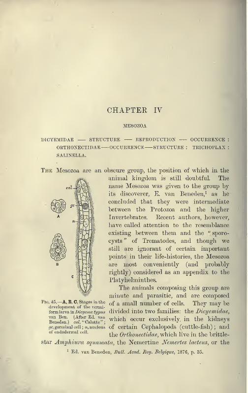
CHAPTER IV
MESOZOA
DICYEMIDAE STRUCTURE REPRODUCTION OCCURRENCE :
ORTHONECTIDAE OCCURRENCE STRUCTURE : TRICHOPLAX :
SALINELLA.
The Mesozoa are an obscure group, the position of which in the
animal kingdom is still doubtful. The
name Mesozoa was given to the group by
its discoverer, E. van Beneden,^ as he
concluded that they were intermediate
between the Protozoa and the higher
Invertebrates. Recent authors, however,
have called attention to the resemblance
existing between them and the " sporo-
cysts " of Trematodes, and though westill are ignorant of certain important
points in their life-histories, the Mesozoa
are most conveniently (and probably
rightly) considered as an appendix to the
Platyhelminthes.
The animals composing this group are
minute and parasitic, and are composed-A, B,C, stages in the ^f ^ Small number of cells. They may be
divided into two families: the Bicyemidae,
which occur exclusively, in the kidneys
Fig. 45.
development of the vermi-
form larva in Dicyema typua
van Ben. (After Ed. vanBeneden.) m^, " Calotte "
,
fz-c, germinal cell ; ^i, nucleus of certain Cephalopods (cuttlc-fish) ; andof endodermai cell.
^-^^ Orthonectidae, which live in the brittle-
star Amjphiura squamata, the Nemertine Nemertes lacteiLS, or the
^ Ed. van Beneden, Bull. Acad. Ro^j. Belgique, 1876, p. 35.
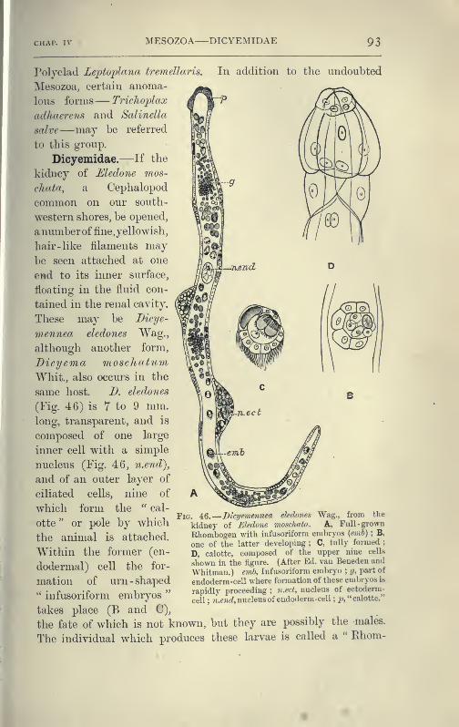
CHAP. IV MESOZOA DICYEMIDAE 93
Polyclad Leptoplana tremellaris. In addition to the undoubted
Mesozoa, certain anoma-
lous forms— Triclioflax
adliaerens and Salinella
salve—may be referred
to this group.
Dicyemidae.—If the
kidney of Eledone mos-
cliata, a Cephalopod
common on our south-
western shores, be opened,
anumberof fine, yellowish,
hair -like filaments maybe seen attached at one
end to its inner surface,
floating in the fluid con-
tained in the renal cavity.
These may be Bicye-
meyinea eledones Wag.,
although another form,
Dicyema moschatiim
Whit., also occurs in the
same host. D. eledones
(Fig. 46) is 7 to 9 mm.long, transparent, and is
composed of one large
inner cell with a simple
nucleus (Fig. 46, n.end),
and of an outer layer of
ciliated cells, nine of
which form the " cal-
otte " or pole by which
the animal is attached.
Within the former (en-
dodermal) cell the for-
mation of urn - shaped'" infusoriform embryos
"
takes place (B and C),
the fate of which is not known, but they are possibly the males.
The individual which produces these larvae is called a " Ehom-
FiG. 46.
—
Dicyemennea eledones Wag., from tlie
kidney of Eledone mosdmta. A, Full-grown
Rhombogeu with infusoriform embryos {emh) ; B,
one of the latter developing ; C, fully formed;
D, calotte, composed of the upper nine cells
shown in the figure. (After Ed. van Beneden and
Whitman.) emb, Infusoriform embryo; g, part of
eudoderm-cell where formation of these embryos is
rapidly proceeding ; n.ect, nucleus of ectoderm
-
cell ; n.ejK^, nucleus of endoderm-cell ; 2>, "calotte."
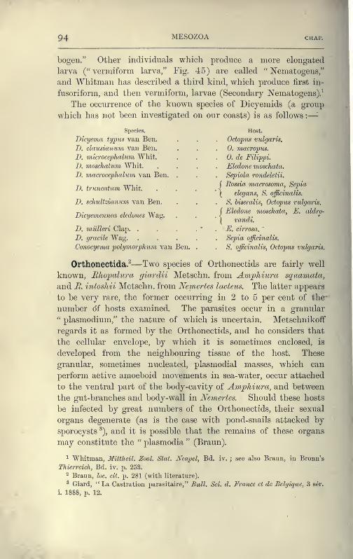
94 MESOZOA CHAP.
bogen." Other individuals which produce a more elongated
larva ("vermiform larva," Fig. 45) are called " Nematogens/'
and Whitman has described a third kind, which produce first in-
fusoriform, and then vermiform, larvae (Secondary Nematogens).-^
The occurrence of the known species of Dicyemids (a group
which has not been investigated on our coasts) is as follows :
—
-
Species.
Dicyema typus van Ben.
D. clausiaiium van Ben.
D. microcephalum Whit.
D. moschatum Whit.
D. macrocephalum van Ben.
D. truncatum Whit.
D. schultzianum van Ben.
Dicyemennea elcdones Wag.
i>. millleri Clap. .
D. gracile Wag.Conocyema polymorphum van Ben.
Host.
Octopus vulgaris.
0. macropus.
0. de Filippi.
Eledone moschata.
Sepiola rondelelii.
j Eossia macrosoma, Sepia
\ elegans, S. officinalis.
S. hiseralis, Octopus vulgaris.
{Eledone moschata, E. aldro-
vandi.
E. cirrosa.
bepia ol
S. officinalis, Octopus vulgaris.
Orthonectida.^—Two species of Orthonectids are fairly well
known, Bhopahira giardii Metschn. from AmpTiiura sq^aamata,
and JR. intoshii Metschn. from Nemertes lacteus. The latter appears
to be very rare, the former occurring in 2 to 5 per cent of the-
number of hosts examined. The parasites occur in a granular'' Plasmodium," the nature of which is uncertain. Metschnikoff
regards it as formed by the Orthonectids, and he considers that
the cellular envelope, by which it is sometimes enclosed, is
developed from the neighbouring tissue of the host. These
granular, sometimes nucleated, plasmodial masses, which can
perform active amoeboid movements in sea-water, occur attached
to the ventral part of the body-cavity of Amphiura, and between
the gut-branches and body-wall in Nemertes. Should these hosts
be infected by great numbers of the Orthonectids, their sexual
organs degenerate (as is the case with pond-snails attacked by
sporocysts ^), and it is possible that the remains of these organs
may constitute the " plasmodia " (Braun).
^ Whitman, Miltheil. Zool. Stat. Ncapel, Bd. iv. ; see also Braun, in Bronn's
Thierrcich, Bd. iv, p. 253.
2 Braun, lac. cit. p. 281 (with literature).
^ Giard, "La Castration j)arasitaire," Bull. Sci. d. France et de Belgiquc, 3 ser.
i. 1888, p. 12.
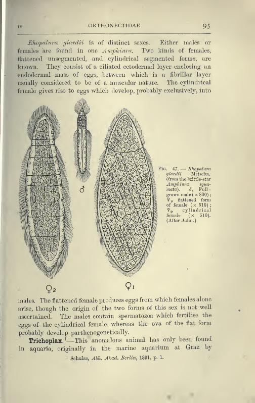
ORTHONECTIDAE 95
Rliopalura giardii is of distinct sexes. Either males or
females are found in one Amphiura. Two kinds of females,
flattened unsegmented, and cylindrical segmented forms, are
known. They consist of a ciliated ectodermal layer enclosing an
endoderm'dl mass of eggs, between which is a fibrillar layer
usually considered to be of a muscular nature. The cylindrical
female gives rise to eggs which develop, probably exclusively, into
Fig. 47. — Rhopaluragiardii Metschii.
(from the brittle-star
A mphiura sqna-
inata). 6, Full -
grown male ( x 800)
;
? 1, flattened formof female ( x 510) ;
?2, cylindricalfemale ( x 510).
(After Juliu.
)
males. The flattened female produces eggs from which females alone
arise, though the origin of the two forms of this sex is not well
ascertained. The males contain spermatozoa which fertilise the
eggs of the cylindrical female, whereas the ova of the flat form
probably develop parthenogenetically.
Trichoplax/—This anomalous animal has only been found
in aquaria, originally in the marine aquarium at Graz by
1 Schulze, Abh. Akad. Berlin^ 1891, p. 1.
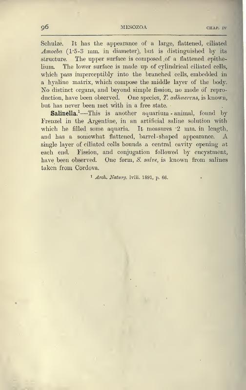
96 MESOZOA CHAP. IV
Schulze. It has the appearance of a large, flattened, ciliated
Amoeba (1*5-3 mm. in diameter), but is distinguished by its
structure. The upper surface is composed .of a flattened epithe-
lium. The lower surface is made up of cylindrical ciliated cells,
which pass imperceptibly into the branched cells, embedded in
a hyaline matrix, which compose the middle layer of the body.
No distinct organs, and beyond simple fission, no mode of repro-
duction, have been observed. One species, 2\ adhaerens, is known,
but has never been met with in a free state.
Salinella/—This is another aquarium - animal, found by
Frenzel in the Argentine, in an artificial saline solution with
which he filled some aquaria. It measures '2 mm. in length,
and has a somewhat flattened, barrel -shaped appearance. Asingle layer of ciliated cells bounds a central cavity opening at
each end. Fission, and conjugation followed by encystment,
have been observed. One form, S. salve, is known from salines
taken from Cordova.
^ Arch. Naturg. Iviii. 1891, p. ^^.