The BOLD signal and neurovascular coupling in autism
8
Developmental Cognitive Neuroscience 6 (2013) 72–79 Contents lists available at ScienceDirect Developmental Cognitive Neuroscience j our na l ho me pa g e: h ttp://www.elsevier.com/locate/dcn The BOLD signal and neurovascular coupling in autism Clare Reynell ∗ , Julia J. Harris Department of Neuroscience, Physiology and Pharmacology, University College London, Gower St, London, WC1E 6BT, UK a r t i c l e i n f o Article history: Received 5 February 2013 Received in revised form 3 June 2013 Accepted 3 July 2013 Keywords: BOLD fMRI Autism Blood flow Neurovascular coupling Energy Glutamate a b s t r a c t BOLD (blood oxygen level dependent) fMRI (functional magnetic resonance imaging) is commonly used to study differences in neuronal activity between human populations. As the BOLD response is an indirect measure of neuronal activity, meaningful interpretation of differences in BOLD responses between groups relies upon a stable relationship exist- ing between neuronal activity and the BOLD response across these groups. However, this relationship can be altered by changes in neurovascular coupling or energy consumption, which would lead to problems in identifying differences in neuronal activity. In this review, we focus on fMRI studies of people with autism, and comparisons that are made of their BOLD responses with those of control groups. We examine neurophysiological differences in autism that may alter neurovascular coupling or energy use, discuss recent studies that have used fMRI to identify differences between participants with autism and control par- ticipants, and explore experimental approaches that could help attribute between-group differences in BOLD signals to either neuronal or neurovascular factors. © 2013 Elsevier Ltd. All rights reserved. Contents 1. Introduction . . . . . . . . . . . . . . . . . . . . . . . . . . . . . . . . . . . . . . . . . . . . . . . . . . . . . . . . . . . . . . . . . . . . . . . . . . . . . . . . . . . . . . . . . . . . . . . . . . . . . . . . . . . . . . . . . . . . . . . . 72 2. Neurovascular coupling and the BOLD signal . . . . . . . . . . . . . . . . . . . . . . . . . . . . . . . . . . . . . . . . . . . . . . . . . . . . . . . . . . . . . . . . . . . . . . . . . . . . . . . . . . . . . . 73 3. Does neurovascular coupling differ in autism? . . . . . . . . . . . . . . . . . . . . . . . . . . . . . . . . . . . . . . . . . . . . . . . . . . . . . . . . . . . . . . . . . . . . . . . . . . . . . . . . . . . . 73 4. Abnormalities in neuronal activity . . . . . . . . . . . . . . . . . . . . . . . . . . . . . . . . . . . . . . . . . . . . . . . . . . . . . . . . . . . . . . . . . . . . . . . . . . . . . . . . . . . . . . . . . . . . . . . . . 74 5. Abnormalities in vasoactive mediators . . . . . . . . . . . . . . . . . . . . . . . . . . . . . . . . . . . . . . . . . . . . . . . . . . . . . . . . . . . . . . . . . . . . . . . . . . . . . . . . . . . . . . . . . . . . 75 6. Abnormalities in energy use . . . . . . . . . . . . . . . . . . . . . . . . . . . . . . . . . . . . . . . . . . . . . . . . . . . . . . . . . . . . . . . . . . . . . . . . . . . . . . . . . . . . . . . . . . . . . . . . . . . . . . . . 75 7. Medication-related effects on neurovascular coupling . . . . . . . . . . . . . . . . . . . . . . . . . . . . . . . . . . . . . . . . . . . . . . . . . . . . . . . . . . . . . . . . . . . . . . . . . . . . 76 8. Potential impact of neurovascular coupling changes in autism on fMRI studies . . . . . . . . . . . . . . . . . . . . . . . . . . . . . . . . . . . . . . . . . . . . . . . . . . 76 9. Experimentally excluding neurovascular coupling differences . . . . . . . . . . . . . . . . . . . . . . . . . . . . . . . . . . . . . . . . . . . . . . . . . . . . . . . . . . . . . . . . . . . . 76 10. Conclusion . . . . . . . . . . . . . . . . . . . . . . . . . . . . . . . . . . . . . . . . . . . . . . . . . . . . . . . . . . . . . . . . . . . . . . . . . . . . . . . . . . . . . . . . . . . . . . . . . . . . . . . . . . . . . . . . . . . . . . . . . . 77 Conflict of interest . . . . . . . . . . . . . . . . . . . . . . . . . . . . . . . . . . . . . . . . . . . . . . . . . . . . . . . . . . . . . . . . . . . . . . . . . . . . . . . . . . . . . . . . . . . . . . . . . . . . . . . . . . . . . . . . . . 78 Acknowledgements . . . . . . . . . . . . . . . . . . . . . . . . . . . . . . . . . . . . . . . . . . . . . . . . . . . . . . . . . . . . . . . . . . . . . . . . . . . . . . . . . . . . . . . . . . . . . . . . . . . . . . . . . . . . . . . . 78 References . . . . . . . . . . . . . . . . . . . . . . . . . . . . . . . . . . . . . . . . . . . . . . . . . . . . . . . . . . . . . . . . . . . . . . . . . . . . . . . . . . . . . . . . . . . . . . . . . . . . . . . . . . . . . . . . . . . . . . . . . . 78 ∗ Corresponding author. Tel.: +44 020 679 0186. E-mail address: [email protected] (C. Reynell). 1. Introduction Many important questions in neuroscience and neu- rology concern differences in brain activity between two or more groups of people. Because fMRI is a powerful and non-invasive tool to examine brain function in humans, it is often employed to read out neuronal activity in 1878-9293/$ – see front matter © 2013 Elsevier Ltd. All rights reserved. http://dx.doi.org/10.1016/j.dcn.2013.07.003
Transcript of The BOLD signal and neurovascular coupling in autism

Developmental Cognitive Neuroscience 6 (2013) 72– 79
Contents lists available at ScienceDirect
Developmental Cognitive Neuroscience
j our na l ho me pa g e: h t tp : / /www.e lsev ier .com/ locate /dcn
The BOLD signal and neurovascular coupling in autism
Clare Reynell ∗, Julia J. HarrisDepartment of Neuroscience, Physiology and Pharmacology, University College London, Gower St, London, WC1E 6BT, UK
a r t i c l e i n f o
Article history:Received 5 February 2013Received in revised form 3 June 2013Accepted 3 July 2013
Keywords:BOLD fMRIAutismBlood flow
a b s t r a c t
BOLD (blood oxygen level dependent) fMRI (functional magnetic resonance imaging) iscommonly used to study differences in neuronal activity between human populations. Asthe BOLD response is an indirect measure of neuronal activity, meaningful interpretationof differences in BOLD responses between groups relies upon a stable relationship exist-ing between neuronal activity and the BOLD response across these groups. However, thisrelationship can be altered by changes in neurovascular coupling or energy consumption,which would lead to problems in identifying differences in neuronal activity. In this review,we focus on fMRI studies of people with autism, and comparisons that are made of their
Neurovascular couplingEnergyGlutamate
BOLD responses with those of control groups. We examine neurophysiological differencesin autism that may alter neurovascular coupling or energy use, discuss recent studies thathave used fMRI to identify differences between participants with autism and control par-ticipants, and explore experimental approaches that could help attribute between-groupdifferences in BOLD signals to either neuronal or neurovascular factors.
© 2013 Elsevier Ltd. All rights reserved.
Contents
1. Introduction . . . . . . . . . . . . . . . . . . . . . . . . . . . . . . . . . . . . . . . . . . . . . . . . . . . . . . . . . . . . . . . . . . . . . . . . . . . . . . . . . . . . . . . . . . . . . . . . . . . . . . . . . . . . . . . . . . . . . . . . 722. Neurovascular coupling and the BOLD signal . . . . . . . . . . . . . . . . . . . . . . . . . . . . . . . . . . . . . . . . . . . . . . . . . . . . . . . . . . . . . . . . . . . . . . . . . . . . . . . . . . . . . . 733. Does neurovascular coupling differ in autism? . . . . . . . . . . . . . . . . . . . . . . . . . . . . . . . . . . . . . . . . . . . . . . . . . . . . . . . . . . . . . . . . . . . . . . . . . . . . . . . . . . . . 734. Abnormalities in neuronal activity . . . . . . . . . . . . . . . . . . . . . . . . . . . . . . . . . . . . . . . . . . . . . . . . . . . . . . . . . . . . . . . . . . . . . . . . . . . . . . . . . . . . . . . . . . . . . . . . . 745. Abnormalities in vasoactive mediators . . . . . . . . . . . . . . . . . . . . . . . . . . . . . . . . . . . . . . . . . . . . . . . . . . . . . . . . . . . . . . . . . . . . . . . . . . . . . . . . . . . . . . . . . . . . 756. Abnormalities in energy use . . . . . . . . . . . . . . . . . . . . . . . . . . . . . . . . . . . . . . . . . . . . . . . . . . . . . . . . . . . . . . . . . . . . . . . . . . . . . . . . . . . . . . . . . . . . . . . . . . . . . . . . 757. Medication-related effects on neurovascular coupling . . . . . . . . . . . . . . . . . . . . . . . . . . . . . . . . . . . . . . . . . . . . . . . . . . . . . . . . . . . . . . . . . . . . . . . . . . . . 768. Potential impact of neurovascular coupling changes in autism on fMRI studies . . . . . . . . . . . . . . . . . . . . . . . . . . . . . . . . . . . . . . . . . . . . . . . . . . 769. Experimentally excluding neurovascular coupling differences . . . . . . . . . . . . . . . . . . . . . . . . . . . . . . . . . . . . . . . . . . . . . . . . . . . . . . . . . . . . . . . . . . . . 7610. Conclusion . . . . . . . . . . . . . . . . . . . . . . . . . . . . . . . . . . . . . . . . . . . . . . . . . . . . . . . . . . . . . . . . . . . . . . . . . . . . . . . . . . . . . . . . . . . . . . . . . . . . . . . . . . . . . . . . . . . . . . . . . . 77
Conflict of interest . . . . . . . . . . . . . . . . . . . . . . . . . . . . . . . . . . . . . . . . . . . . . . . . . . . . . . . . . . . . . . . . . . . . . . . . . . . . . . . . . . . . . . . . . . . . . . . . . . . . . . . . . . . . . . . . . . 78Acknowledgements . . . . . . . . . . . . . . . . . . . . . . . . . . . . . . . . . . . . . . . . . . . . . . . . . . . . . . . . . . . . . . . . . . . . . . . . . . . . . . . . . . . . . . . . . . . . . . . . . . . . . . . . . . . . . . . . 78References . . . . . . . . . . . . . . . . . . . . . . . . . . . . . . . . . . . . . . . . . . . . . . . . . . . . . . . . . . . . . . . . . . . . . . . . . . . . . . . . . . . . . . . . . . . . . . . . . . . . . . . . . . . . . . . . . . . . . . . . . . 78
∗ Corresponding author. Tel.: +44 020 679 0186.E-mail address: [email protected] (C. Reynell).
1878-9293/$ – see front matter © 2013 Elsevier Ltd. All rights reserved.http://dx.doi.org/10.1016/j.dcn.2013.07.003
1. Introduction
Many important questions in neuroscience and neu-rology concern differences in brain activity between two
or more groups of people. Because fMRI is a powerful andnon-invasive tool to examine brain function in humans,it is often employed to read out neuronal activity in
ntal Cog
samagwca
fiaiitanpserlrda2
icdtnrnsspwinBb
2
hiii–rdirc((e
dp
C. Reynell, J.J. Harris / Developme
uch studies. However, for fMRI-based measures of brainctivity to be reliably compared between populations, itust be assumed that the relationship between neuronal
ctivity and the BOLD signal is the same in the differentroups. This relationship depends on the mechanism byhich active neurons alter local blood flow (neurovascular
oupling) and on the oxygen use evoked by neuronalctivity (Attwell and Iadecola, 2002; Harris et al., 2011).
Several neurophysiological changes have been identi-ed in autism (as discussed below). These changes maylter the signalling from neurons to the vasculature and,n consequence, the relationship between neuronal activ-ty and the BOLD signal. Typically, studies using fMRIo compare control participants and participants withutism attribute observed differences in the BOLD sig-al to altered neuronal activity, without considering theossibility that neurovascular coupling or oxygen con-umption is also altered (Goldberg et al., 2011; Pfeifert al., 2012; Watanabe et al., 2012). More recently, however,esearchers have begun to acknowledge that neurovascu-ar coupling or oxygen use changes must be experimentallyuled out before BOLD differences can be used as evi-ence for differences of neuronal function between controlnd autistic groups (Dinstein et al., 2012; Feczko et al.,012).
In this article, we start by briefly reviewing the phys-ological basis of the BOLD signal and the neurovascularoupling mechanisms that mediate it. We then look inetail at the neurophysiological changes that are knowno occur in autism and how these changes might influenceeurovascular coupling and oxygen use, and thus the BOLDesponse. We then examine the implications that alteredeurovascular coupling or oxygen use would have for fMRItudies. By focusing on several representative studies cho-en from the literature, we highlight the interpretationalroblems that can occur when this issue is ignored. Finally,e explore ways in which the scientific community study-
ng autism can address the challenge of separating outeurovascular coupling and neuronal activity effects on theOLD signal experimentally, as some groups are alreadyeginning to do.
. Neurovascular coupling and the BOLD signal
fMRI is regularly used to study neuronal activity inumans because of its non-invasive nature, which allows
t to be used to study a broad range of participants, includ-ng patients suffering from pathologies such as autism. Its important to note, however, that the signal produced
the BOLD signal – is not a direct measure of neu-onal activity, but instead reflects the operation of severalifferent brain processes associated with neuronal activ-
ty, including synaptic transmitter release, the resultingelease of signalling molecules that mediate neurovas-ular coupling and thus increase local blood flow, andmaking the BOLD signal smaller) oxygen consumptionLogothetis et al., 2001; Magistretti et al., 1999; Attwell
t al., 2010).The BOLD signal reports changes in the amount ofeoxyhaemoglobin in the blood. Deoxyhaemoglobin isaramagnetic, and it decreases MRI signals by making
nitive Neuroscience 6 (2013) 72– 79 73
the local magnetic field less homogenous. During neu-ronal activity, the level of deoxyhaemoglobin initiallyincreases as oxygen use increases, and then decreases asa blood flow increase occurs which over-compensates forthe amount of oxygen used. This decrease in the level ofdeoxyhaemoglobin results in a more homogeneous mag-netic field and thus increases the magnetic resonancesignal. The size of the BOLD signal is larger when thereis a large blood flow increase, but is decreased by theuse of oxygen by neurons (for a detailed description, seeLogothetis and Wandell, 2004). In what follows we focussimply on the amplitude of the BOLD signal, becausealthough BOLD signals are often analysed in terms oftheir stimulus-evoked amplitude relative to the ongoingnoise, a procedure that requires detailed statistical analy-sis (Friston, 1995; Friston et al., 2007), even consideringjust the amplitude of the signal reveals significant con-cerns about how to compare control and autistic groupsof subjects.
When using fMRI to identify differences in neuronalactivity between groups, we rely upon the assump-tion that the BOLD signal represents the same set ofprocesses occurring in these different groups, despitethe neurophysiological differences found in patholo-gies such as autism. Neurophysiological differences mayinclude neuronal activity differences, which is what mostresearchers are interested in characterising. However,they may also include differences in other processeslinked to neuronal activity, such as oxygen consump-tion or neurovascular coupling. Neurovascular couplinginvolves several pathways, both neuronal and glial, thatlead to the release of vasoactive mediators, such asnitric oxide, prostaglandins and 20-HETE. These altervessel diameter by promoting either smooth muscle relax-ation, which increases vessel diameter, or smooth musclecontraction, which decreases vessel diameter (reviewedby Attwell et al., 2010). Changes in how the releaseof any of these mediators are controlled by neuronalactivity may alter the relationship between neuronalactivity and the BOLD signal, potentially obscuring anydifferences in neuronal activity between the groups stud-ied.
In order to be sure that a BOLD signal differencebetween groups reflects a neuronal activity difference, it isimportant that we understand as much as possible aboutthe factors contributing to the BOLD signal. This meansconsidering not only the role of neuronal activity, but alsothe involvement of other factors, including neurovascularcoupling and oxygen consumption, which may differ incertain pathologies.
3. Does neurovascular coupling differ in autism?
As research into the pathology of autism progresses,we are gaining insight into the neurophysiological abnor-malities of the autistic brain (reviewed in Belger et al.,2011), and developing therapeutic strategies to target them
(reviewed in Hampson et al., 2012). It is probable that therelationship between neuronal activity and blood flow isaffected by some of these abnormalities, or the medicationsused to treat them, as follows (Fig. 1).
74 C. Reynell, J.J. Harris / Developmental Cognitive Neuroscience 6 (2013) 72– 79
Fig. 1. Pathways from interneurons, neurons and astrocytes that regulate blood flow, leading to either dilation (black arrows to upper half of blood vessel)or constriction (black arrows to lower half of blood vessel) of the nearby vasculature. Red arrows indicate how points in these pathways are altered inautism. These changes may affect the relationship between neuronal activity and blood flow response. On the left, excitatory synapses release glutamateonto both interneurons and excitatory neurons. In interneurons, activation of glutamate receptors leads to the production of nitric oxide, a diffusiblevasodilatory messenger. It also triggers the release of GABA, which inhibits postsynaptic excitatory neurons, reducing downstream glutamate release. Theaction of glutamate on astrocytic mGluRs leads to the production of both vasoactive dilators and constrictors (note, recent evidence suggests that mGluRsare only present on astrocytes during infancy; Sun et al., 2013; see text). Serotonin release provides a basal constriction of blood vessels. (For interpretation
he web
of the references to colour in this figure legend, the reader is referred to t4. Abnormalities in neuronal activity
Decreased inhibition is observed in the brains of autis-tic patients: the expression of the enzymes that synthesisethe inhibitory neurotransmitter, GABA, and the expressionof the receptors on which GABA acts are both reducedin the autistic brain (Fatemi et al., 2002, 2009, 2010).This decreased inhibition is a possible therapeutic targetfor treatment of autism, and several drugs that enhancethe action of GABA are currently in phase II clinical tri-als (divalproex and arbaclofen; clinicaltrials.gov identifierNCT00211757 and NCT00846547, respectively). The effectof decreased inhibition on neuronal activity can be detectedby fMRI: because GABA decreases excitability, an autisticbrain with impaired GABA function would experience moreneuronal excitability and therefore more neuronal firingand more glutamate release, which triggers the release of
vasodilators and increases blood flow (Attwell et al., 2010).An increase in BOLD activity would thus be expected, andin this case it would be related to changes in neuronal func-tion.version of the article.)
However, one major issue with this is the potentialfor nonlinearities in the relationships between inhibition,excitation and blood flow response. For instance, if GABAactivity decreases by half, neuronal excitability will notnecessarily double – simplistically, it could increase bymuch more than a factor of two if the depolarising effect ofreduced GABA takes most neurons from below to above thethreshold for action potential firing, or it could increase byless than a factor of two if the depolarising effect is not suf-ficient to cross this threshold for most neurons. Similarly,if neuronal excitability doubles, the blood flow responsewill not necessarily double – it could increase by very littleif blood vessels are already near-maximally dilated, whichmay be the case much of the time in brains where reducedGABA function increases the level of resting excitability.A further complication comes from the recent suggestionthat GABA released from interneurons may act on glial
cells or directly on microvessels to increase blood flow(Kocharyn et al., 2008). A reduced action of this pathwayin autistic patients would lead to a decreased BOLD signal,unrelated to neuronal excitability.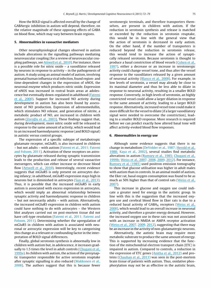
ntal Cog
Gto
5
inpibaptnocedmwmadrti
g–aclmflsinTaws–tcbhFmrtp
cu1ta2
C. Reynell, J.J. Harris / Developme
How the BOLD signal is affected overall by the change ofABAergic inhibition in autism will depend, therefore, on
he relative magnitude of these opposing effects of GABAn blood flow, which may vary between brain regions.
. Abnormalities in vasoactive mediators
Other neurophysiological changes observed in autismnclude alterations in the signalling pathways mediatingeurovascular coupling (for a review of neurovascular cou-ling pathways, see Attwell et al., 2010). For instance, there
s a possible role for nitric oxide – a vasodilator releasedy neurons in response to activity – in the pathogenesis ofutism. A study using an animal model of autism, involvingrenatal human influenza viral infection, found region- andime-dependent changes in the expression of nNOS, theeuronal enzyme which produces nitric oxide. Expressionf nNOS was increased in rostral brain areas at adoles-ence but eventually down-regulated in adulthood (Fatemit al., 2000). Support for increased NO activity duringevelopment in autism has also been found by assess-ent of NO production. Expression of adrenomedullin,hich stimulates NO release, and generation of nitrite, aetabolic product of NO, are increased in children with
utism (Zoroglu et al., 2003). These findings suggest that,uring development, more nitric oxide will be released inesponse to the same amount of activity, which would leado an increased haemodynamic response (and BOLD signal)n autistic versus control groups.
The expression of a specific subtype of metabotropiclutamate receptor, mGluR5, is also increased in children
but not adults – with autism (Fatemi et al., 2011; Fatemind Folsom, 2011). Activation of these receptors on astro-ytes by glutamate released during synaptic transmissioneads to the production and release of several vasoactive
essengers, which can either increase or decrease bloodow (Attwell et al., 2010). Interestingly, recent evidenceuggests that mGluR5 is only present on astrocytes dur-ng infancy; in adulthood, mGluR5 expression stays high ineurons but is diminished in astrocytes (Sun et al., 2013).hus, it is possible that the increased mGluR5 in earlyutism is associated with excess expression in astrocytes,hich would imply an abnormal relationship between
ynaptic activity and haemodynamic response in children but not necessarily adults – with autism. Alternatively,he increased mGluR5 expression in children with autismould have nothing to do with astrocytes – the Westernlot analyses carried out on post-mortem tissue did notave cell-type resolution (Fatemi et al., 2011; Fatemi andolsom, 2011). Determining whether the upregulation ofGluR5 in children with autism is associated with neu-
onal or astrocytic expression will be key to categorisinghis change as a relevant or confounding factor in the inter-retation of BOLD signal differences.
Finally, global serotonin synthesis is abnormally low inhildren with autism but, in adolescence, it increases grad-ally to 1.5 times the level in adult controls (Chugani et al.,
999a). In children with autism, the action of the presynap-ic transporter responsible for active serotonin reuptakefter synaptic signalling is also reduced (Makkonen et al.,008). The authors suggest that this is because fewernitive Neuroscience 6 (2013) 72– 79 75
serotonergic terminals, and therefore transporters them-selves, are present in children with autism. If thereduction in serotonin synthesis and release is matchedor exceeded by the reduction in serotonin reuptake,this would be in line with the general view thatthe action of serotonin is decreased in early autism.On the other hand, if the number of transporters isreduced beyond the reduction in serotonin release,this would tend to increase the action of synapti-cally released serotonin. Because serotonin is thought toproduce a basal constriction of blood vessels (Cohen et al.,1997), either a decrease or an increase in serotonergicactivity could change vessel tone, and thus alter the vesselresponse to the vasodilators released by a given amountof neuronal activity (Blanco et al., 2008). For example, inlow levels of serotonin, a vessel may already be close toits maximal diameter and thus be less able to dilate inresponse to neuronal activity, resulting in a smaller BOLDresponse. Conversely, in high levels of serotonin, an highlyconstricted vessel could allow a larger dilation in responseto the same amount of activity, leading to a larger BOLDresponse. Alternatively, increased vessel tone could make itmore difficult for the vessel to dilate (if a larger vasodilatorysignal were needed to overcome the constriction), lead-ing to a smaller BOLD response. More research is requiredbefore we can predict exactly how altered basal tone willaffect activity-evoked blood flow responses.
6. Abnormalities in energy use
Although some evidence suggests that there is nochange in metabolism (DeVolder et al., 1987; Herold et al.,1988; Kaya et al., 2002), several studies find metabolicalterations in autism (Rumsey et al., 1985; Chugani et al.,1999b; Weiss et al., 2007, 2008, 2009, 2012). For instance,Rumsey et al. (1985) used positron emission tomographyto show that glucose utilisation was higher in adult maleswith autism than in controls. In an animal model of autism,the Eker rat, basal oxygen consumption was found to be asmuch as 50% higher than that of control rats (Weiss et al.,2007).
This increase in glucose and oxygen use could indi-cate a greater need for energy in the autistic group. Inline with this is the suggestion that the increased oxy-gen use and cerebral blood flow in Eker rats is due to areduced basal activity of GABAA receptors (Weiss et al.,2008), which would lead to an overall increase in neuronalactivity, and therefore a greater energy demand. However,the increased oxygen use in these rats was not associatedwith an increase in NMDA or AMPA receptor activation(Weiss et al., 2007, 2009, 2012), suggesting that there maybe an increase in the activity of non-glutamatergic neurons.
Alternatively, the autistic brain may require moremetabolic substrate to produce the same amount of energy.This is supported by increasing evidence that the func-tion of the mitochondrial electron transport chain (ETC) isimpaired in autism. Compared to controls, a reduction in
the expression of ETC genes (Anitha et al., 2012) and pro-teins (Chauhan et al., 2011) was seen in the post-mortembrain tissue of patients with autism. Thus, oxidative phos-phorylation may not be as effective in the autistic brain,
ntal Cog
76 C. Reynell, J.J. Harris / Developmeperhaps requiring more oxygen and/or glucose to pro-duce the same amount of ATP. Since the amount of energyneeded to restore ionic concentrations after neuronalsignalling is fixed by the amount of sodium ion entry occur-ring (Attwell and Laughlin, 2001), this would imply a largeroxygen requirement to support the same level of neuronalactivity. If this excess oxygen were obtained by increasedoxygen extraction from the blood, then this would lead toa smaller BOLD response. However, in the mouse modelof autism, oxygen extraction is unchanged while cerebralblood flow is increased (Weiss et al., 2007, 2008, 2012),suggesting that the excess oxygen is obtained by increasedvascular supply. This would lead to a larger BOLD responseto the same amount of neuronal activity in people withautism.
7. Medication-related effects on neurovascularcoupling
The medication used to treat autism may also affectthe relationship between neuronal activity and the BOLDresponse. For example, serotonin reuptake inhibitors(SSRIs), a medication commonly used by autistic patients,increase extracellular serotonin levels in the brain. Theymay, therefore, increase the tone of vessels – perhapsbeyond normal levels – and thus alter the blood flowresponse evoked by neuronal activity (see above). A recentstudy by Feczko et al. (2012) addressed this question, find-ing no significant difference in the BOLD response to avisuomotor task presented to non-medicated participantsand those taking SSRIs, in the brain areas they examined.However, this does not rule out SSRI-induced neurovascu-lar coupling changes in the brain circuits that underpin thesocial behaviours more closely associated with the autisticphenotype.
8. Potential impact of neurovascular couplingchanges in autism on fMRI studies
Having highlighted some of the neurophysiological dif-ferences found in autism that may alter the relationshipbetween neuronal activity and the BOLD response in thiscondition, we will now consider some fMRI studies inwhich the results may be affected by these differences.
Goldberg et al. (2011) asked children with or withouthigh functioning autism spectrum disorder (ASD) to carryout an error monitoring task. After a trial in which an errorwas made, the BOLD signal in the anterior medial pre-frontal cortex and the superior temporal gyrus was foundto increase in ASD participants compared to control partic-ipants. This increase in the BOLD signal was interpretedas increased neuronal activity in these regions. Becausethe behavioural performance on the task was the samefor both groups, the assumed increase in neuronal activ-ity was suggested to indicate a “greater attention towardsthe internally-driven emotional state associated with mak-ing an error” in children with ASD. But the authors did
not take any steps to rule out the possibility that thedifference between these groups may be neurovascularin nature, and nothing to do with neuronal activity. Forinstance, the increased nitric oxide level found in childrennitive Neuroscience 6 (2013) 72– 79
with autism (Zoroglu et al., 2003) could lead to a largervascular response to activation of the same number of neu-rons (see above). This would cause the BOLD response toappear larger in children with autism, despite no differencein neuronal activity.
Watanabe et al. (2012) found that the BOLD response toa social judgement task was decreased in participants withASD compared to control participants. The degree of BOLDsignal reduction in the two regions most active in controlparticipants during this task (the anterior cingulate cor-tex/ventromedial prefrontal cortex and the dorsal medialprefrontal cortex) predicted the severity of communica-tion deficit in the ASD group. This result might suggest thatsome physiological abnormality in these regions is associ-ated with the behavioural abnormality in social judgement.However, the idea that the physiological abnormality isreduced neuronal activity can only be an assumption untilother possibilities are ruled out. For example, the decreasedserotonin levels seen in autistic patients (Chugani et al.,1999a) may restrict the amplitude of the vascular and thusthe BOLD response (see above). The authors point out thatthe reduction in the BOLD response in autistic partici-pants is not global, as task-evoked activity in one area, theamygdala, was found to increase. However, neurovascu-lar coupling mechanisms can be pathway-specific (Enageret al., 2009) and, therefore, task-specific. It is thus possiblethat changes in neurovascular coupling are responsible foreither the decreased or increased BOLD responses found byWatanabe et al. (2012).
Pfeifer et al. (2012) compared the BOLD signal whenparticipants were asked to make an appraisal of them-selves or of others. In typically developing children andadolescents, two main brain regions – the medial prefrontalcortex (mPFC) and the middle cingulate cortex (MCC) –showed an increased BOLD response during self-appraisalcompared to appraisal of others. Age-matched participantswith ASD lacked any task-related pattern in the mPFC, andshowed the opposite pattern in the MCC (increased BOLDsignal when appraising others compared to self). However,with increasing age in the ASD group, the BOLD signal in themPFC associated with the differentiation of the self fromothers was found to increase. This trend suggests that thisbrain region is increasingly relied upon as the social impor-tance of the self-other distinction is learned. However, theinterpretation of age-related BOLD changes comes with itsown set of issues, as the neurovasculature matures (Harriset al., 2011), perhaps differently in autistic and typicallydeveloping populations.
9. Experimentally excluding neurovascularcoupling differences
Since most researchers are interested in the neuronalchanges associated with autism, most changes in fMRIresponse are simply assumed to be due to altered neuronalprocessing rather than altered neurovascular coupling oroxygen use. None of the studies above considered neu-
rovascular abnormalities in autism, which, if present coulddirectly affect the BOLD signal even in the absence ofchanges in neuronal activity. The mislabelling of neu-rovascular changes as neuronal activity changes could
ntal Cog
becct
Apree(ravtSecig
tcittceisicbg
rrmtepvetee
mbctrbrniabbn
C. Reynell, J.J. Harris / Developme
e misleading. The best way to avoid this is with anxperimental design that can distinguish a neurovascularoupling (or oxygen use) change from a neuronal activityhange. A few labs have begun to design experiments withhis goal in mind.
Feczko et al. (2012) asked children with and withoutSD to press a button at the onset and offset of a visuallyresented flickering chequerboard, and compared BOLDesponses in the two groups during this task in 19 differ-nt brain regions. The same paradigm has previously beenmployed to compare BOLD responses across age groupsKang et al., 2003). This approach assumes that the neu-onal activity mediating a simple, low-level task is notffected by autism (or age). This assumption may not bealid, since altered visual processing has been reported inhe retina and striate cortex in autism (Ritvo et al., 1988;immons et al., 2009; Neumann et al., 2011; but see Koht al., 2010). But if it is a correct assumption, then identi-al BOLD responses in the two groups would presumablymply that neurovascular coupling is the same in the tworoups.
Since Feczko et al. found no significant difference inhe visual task-evoked BOLD response between ASD andontrol children, they argued that neurovascular couplingn the two groups must be the same, ideally throughouthe brain. This is the most parsimonious argument, buthe authors do acknowledge the small possibility that aonfounding difference in neurovascular coupling that hasqual and opposite effects to a difference in neuronal activ-ty between the groups could lead to the same overall BOLDignal. For example, a smaller amount of neuronal activityn autism may be compensated for by a stronger neurovas-ular signalling pathway, or vice versa. In this case, a similarlood flow response would be produced in the differentroups by differing levels of neuronal activity.
Even if one accepts the argument that a similar BOLDesponse for a low level visual task implies similar neu-ovascular coupling, as noted by the authors, it can only beade for this particular visual task and need not generalise
o other tasks. As mentioned above, this is because differ-nt tasks activate different brain regions, different neuronalathways and different neurotransmitter systems, whichary in their neurovascular coupling mechanisms (Sloant al., 2010; Devonshire et al., 2012). Indeed, even withinhe same brain region, neuronal activity evoked by differ-nt inputs can generate different BOLD responses (Enagert al., 2009).
Such input- or task-specific neurovascular couplingakes it difficult for results based on one task alone to
e extrapolated usefully to general claims about neurovas-ular coupling between groups – it is always a possibilityhat the chosen task happens to be one in which neu-onal responses and neurovascular coupling are not alteredetween groups, while for tasks employing different brainegions and pathways there may indeed be differences ofeurovascular coupling between the groups. For example,
n Feczko et al.’s case, it is possible that low level visual
reas do not have their neurovascular coupling alteredy the neuronal changes associated with autism, whilerain areas dealing with social interactions do have theireurovascular coupling altered. Nevertheless, this studynitive Neuroscience 6 (2013) 72– 79 77
represents the first attempt to experimentally rule out neu-rovascular coupling confounds in the interpretation of fMRIdata, and paves the way for paradigms which address thisissue directly.
Dinstein et al. (2012) used a different approachto exclude the possibility that an increase in thevariability of neurovascular coupling in autism mightunderpin the greater fMRI response variability that theyobserved. Specifically, they found that, although meanevoked responses to several stimuli (visual, auditory, andsomatosensory) did not differ between participants withand without autism, there was a significant increase intrial-by-trial variability in participants with autism. Theycompared these task-evoked responses in visual, auditoryor somatosensory regions (“local evoked activity”) with thefMRI fluctuations that were common to the entire cortex inthe absence of stimulus-evoked responses (“global ongoingactivity”), which did not show increased variability in par-ticipants with autism. Because the authors started with theassumption that a change in neurovascular coupling wouldaffect “evoked responses and ongoing activity in a similarmanner”, they argued that neurovascular coupling changescould therefore not be the source of the increased variabil-ity in the local task-evoked BOLD signals. The conclusionfollowed that task-related neuronal activity is abnormallyunreliable in autism.
This line of reasoning on its own is weak, because thereis no reason why a neurovascular coupling change couldnot selectively alter task-evoked signalling to the vascul-ature without affecting resting state blood flow. This isobvious when one considers that the majority of neu-rovascular coupling changes are likely to occur at pointsalong the pathway from synaptic transmission to vesseldilation/constriction, and may therefore only be revealedduring task-evoked synaptic activity.
Nevertheless, Dinstein et al.’s conclusions are supportedby EEG data showing that visual task-evoked responsevariability is significantly greater in participants with ASD(Milne, 2011). The combination of fMRI with a direct mea-sure of neuronal activity, such as EEG, is one of the mosthopeful approaches for experimentally dissecting out therelative contributions of altered neuronal activity and neu-rovascular coupling to differences in the BOLD signal.For instance, an increased BOLD response echoed by anincreased EEG response to the same task is a very good indi-cation that the BOLD increase reflects increased neuronalactivity. The fMRI data can then be used to gain a clearerspatial picture of the change. If, however, BOLD and EEGdata contradict one another, this would indicate that otherfactors – perhaps a change in neurovascular coupling orenergy use – are contributing to the BOLD signal difference.In this way, performing EEG alongside fMRI can guide theidentification of the neuronal differences between groups.
10. Conclusion
Autism is a brain disorder, with its diverse behavioural
phenotypes rooted in abnormalities of neuronal circuitryand function, and we do not wish to suggest that fMRIcannot reveal these neuronal differences. The aim of thisreview is to dispel the idea that neurovascular coupling
ntal Cog
78 C. Reynell, J.J. Harris / Developmecan be lumped in with easily corrected for non-neuronalconfounds such as head motion or breathing artefacts. Onthe contrary, neurovascular coupling changes can have pro-found, complex and region- and task-specific effects oncerebral blood flow and therefore the BOLD signal.
Autism is associated with many neurophysiologicalchanges, and there are just as many reasons why an abnor-mal relationship between neuronal activity and the BOLDsignal might therefore be expected. Being aware of thesepotential neurovascular coupling and energy use differ-ences will be critical to the field’s ability to dissect outthe neuronal changes that underpin behavioural differ-ences. Eventually, detailed knowledge of the physiologyof the BOLD signal (Attwell and Iadecola, 2002) will allowmore confident interpretation of BOLD response differ-ences between participants. The most promising way toachieve this is by using a combination of imaging and physi-ological research in animals (Devonshire et al., 2012; Schulzet al., 2012). Meanwhile, an experimental approach – basedaround carefully chosen control tasks or a combination offMRI and EEG (or, indeed, any method that reports theelectrical activity of neurons, such as MEG) – is the beststrategy to distinguish the contributions of neurovascularcoupling and of neuronal activity to differences in BOLDsignals between groups.
Conflict of interest
The authors declare no conflict of interest.
Acknowledgements
Supported by the MRC, Wellcome Trust, FondationLeducq and ERC. We thank David Attwell, Renaud Jolivetand Anusha Mishra for comments on the manuscript.
References
Anitha, A., Nakamura, K., Thanseem, I., Matsuzaki, H., Miyachi, T., Tsujii,M., Iwata, Y., Suzuki, K., Sugiyama, T., Nori, N., 2012. Downregulationof the expression of mitochondrial electron transport complex genesin autism brains. Brain Pathol. [Epub ahead of print].
Attwell, D., Laughlin, S.B., 2001. An energy budget for signaling in the greymatter of the brain. J. Cereb. Blood Flow Metab. 21, 1113–1145.
Attwell, D., Iadecola, C., 2002. The neuronal basis of functional brain imag-ing signals. Trends Neurosci. 25, 621–625.
Attwell, D., Buchan, A.M., Charpak, S., Lauritzen, M., MacVicar, B.A., New-man, E.A., 2010. Glial and neuronal control of brain blood flow. Nature468, 232–243.
Belger, A., Carpenter, K.L., Yucel, G.H., Cleary, K.M., Donkers, F.C., 2011. Theneuronal circuitry of autism. Neurotox. Res. 20, 201–214.
Blanco, V.M., Stern, J.E., Filosa, J.A., 2008. Tone-dependent vascularresponses to astrocyte-derived signals. Am. J. Physiol. Heart Circ.Physiol. 294, H2855–H2863.
Chauhan, A., Gu, F., Essa, M.M., Wegiel, J., Kaur, K., Brown, W.T., Chauhan,V., 2011. Brain region-specific deficit in mitochondrial electron trans-port chain complexes in children with autism. J. Neurochem. 117,209–220.
Chugani, D.C., Muzik, O., Behen, M., Rothermel, R., Janisse, J.J., Lee, J.,Chugani, H.T., 1999a. Developmental changes in brain serotonin syn-thesis capacity in autistic and nonautistic children. Neurology 45,287–295.
Chugani, D.C., Bhavani, S.S., Behen, M., Lee, M., Moore, G.J., 1999b. Evidenceof altered energy metabolism in autistic children. Prog. Neuropsy-chopharmacol. Biol. Psychiatry 23, 635–641.
Cohen, Z., Bonvento, G., Lacombe, P., Hamel, E., 1997. Serotonin in theregulation of brain microcirculation. Prog. Neurobiol. 50, 335–362.
nitive Neuroscience 6 (2013) 72– 79
DeVolder, A.G., Bol, A., Micel, C., Cogneau, M., Goffinet, A.M., 1987. Brainglucose metabolism in children with the autistic syndrome: positrontomography analysis. Brain Dev. 9 (6), 581–587.
Devonshire, I.M., Papadakis, N.G., Port, M., Berwick, J., Kennerley, A.J.,Mayhew, J.E.W., Overton, P.G., 2012. Neurovascular coupling is brainregion-dependent. Neuroimage 59, 1997–2006.
Dinstein, I., Heeger, D.J., Lorenzi, L., Minshew, N.J., Malach, R., Behrmann,M., 2012. Unreliable evoked responses in autism. Neuron 75,981–991.
Enager, P., Piilgaard, H., Offenhauser, N., Kocharyan, A., Fernandes,P., Hamel, E., Lauritzen, M., 2009. Pathway-specific variationsin neurovascular and neurometabolic coupling in rat primarysomatosensory cortex. J. Cereb. Blood Flow Metab. 29, 976–986.
Fatemi, S.H., Cuadra, A.E., El-Fakahany, E.E., Sidwell, R.W., Thuras, P.D.,2000. Prenatal viral infection causes alterations in nNOS expressionin developing mouse brains. Neuroreport 11 (7), 1493–1496.
Fatemi, S.H., Halt, A.R., Stary, J.M., Kanodia, R., Schulz, S.C., Realmuto,G.R., 2002. Glutamic acid decarboxylase 65 and 67 kDa proteins arereduced in autistic paretial and cerebellar cortices. Biol. Psychiatry 52,805–810.
Fatemi, S.H., Reutiman, T.J., Folsom, T.D., Thuras, P.D., 2009. GABAA recep-tor downregulation in brains of subjects with autism. J. Autism Dev.Disord. 39, 223–230.
Fatemi, S.H., Reutiman, T.J., Folsom, T.D., Rooney, R.J., Patel, D.H., Thuras,P.D., 2010. mRNA and protein levels for GABA A�4, �5, �1 andGABABR1 receptors are altered in brains from subjects with autism. J.Autism Dev. Disord. 40, 743–750.
Fatemi, S.H., Folsom, T.D., 2011. Dysregulation of fragile X mental retarda-tion protein and metabotropic glutamate receptor 5 in superior frontalcortex of individuals with autism: a postmortem brain study. Mol.Autism 2, 6.
Fatemi, S.H., Folsom, T.D., Kneeland, R.E., Liesch, S.B., 2011. Metabotropicglutamate receptor 5 upregulation in children with autism is asso-ciated with underexpression of both Fragile X mental retardationprotein and GABAA receptor beta 3 in adults with autism. Anat. Rec.294, 1635–1645.
Feczko, E., Miezin, F.M., Constantino, J.N., Schlagger, B.L., Peterson, S.E.,Pruett, J.R., 2012. The hemodynamic response in children with simplexautism. Dev. Cogn. Neurosci. 2, 396–408.
Friston, K., 1995. Commentary and opinion: II, statistica parametric map-ping: ontology and current issues. J. Cereb. Blood Flow Metab. 15,361–370.
Friston, K., Ashburner, J.T., Kiebel, S.J., Nichols, T.E., Penny, W.D., 2007. Sta-tistical Parametric Mapping: The Analysis of Functional Brain Images.Academic Press, London.
Goldberg, C.M., Spinelli, S., Joel, S., Pekar, J.J., Denckla, M.B., Mostofsky,S.H., 2011. Children with high functioning autism show increasedprefrontal and temporal cortex activity during error monitoring. Dev.Cogn. Neurosci. 1, 47–56.
Hampson, D.R., Gholizadeh, S., Pacey, L.K., 2012. Pathways to drug devel-opment for autism spectrum disorders. Clin. Pharmacol. Ther. 91,189–200.
Harris, J.J., Reynell, C., Attwell, D., 2011. The physiology of developmentalchanges in BOLD functional imaging signals. Dev. Cogn. Neurosci. 1,199–216.
Herold, S., Frackowiak, R.S.J., Le Couteur, A., Rutter, M., Howlin, P., 1988.Cerebral blood flow and metabolism of oxygen and glucose in youngautistic adults. Psychol. Med. 18, 823–831.
Kang, H.C., Burgund, E.D., Lugar, H.M., Peterson, S.E., Schlagger, B.L., 2003.Comparison of functional activation foci in children and adults usinga common stereotactic space. Neuroimage 19, 16–28.
Kaya, M., Karasalihoglu, S., Ustün, F., Gültekin, A., Cermik, T.F., Fazlioglu, Y.,Türe, M., Yigitbas i, O.N., Berkarda, S., 2002. The relationship between99mTc-HMPAO brain SPECT and the scores of real life rating scale inautistic children. Brain Dev. 24, 77–81.
Kocharyn, A., Fernandes, P., Tong, X.K., Vaucher, E., Hamel, E., 2008. Spe-cific cubtypes of cortical interneurons contribute to the neurovascularcoupling response to basal forebrain stimulation. J. Cereb. Blood FlowMetab. 28, 221–231.
Koh, H.C., Milne, E., Dobkins, K., 2010. Spatial contrast sensitivity in ado-lescents with autism spectrum disorders. J. Autism Dev. Disord. 40,978–987.
Logothetis, N.K., Pauls, J., Augath, M., Trinath, T., Oeltermann, A., 2001.Neurophysiological investigation of the basis of the fMRI signal.
Nature 412, 150–157.Logothetis, N.K., Wandell, B.A., 2004. Interpreting the BOLD signal. Annu.Rev. Physiol. 66, 735–769.
Magistretti, P.J., Pellerin, L., Rothman, D.L., Shulman, R.G., 1999. Energy ondemand. Science 283, 496–497.

ntal Cog
M
M
N
P
R
R
S
S
S
C. Reynell, J.J. Harris / Developme
akkonen, I., Riikonen, R., Kokki, H., Airaksinen, M.M., Kuikka, J.T., 2008.Serotonin and dopamine transporter binding in children with autismdetermined by SPECT. Dev. Med. Child Neurol. 50, 593–597.
ilne, E., 2011. Increased intra-participant variability in children withautistic spectrum disorders: evidence from single-trial analysis ofevoked EEG. Front. Psychol. 2, 1–12.
eumann, N., Dubischar-Krivec, A.M., Poustka, F., Birbaumer, N., Bölte, S.,Braun, C., 2011. Electromagnetic evidence of altered visual processingin autism. Neuropsychologia 49, 3011–3017.
feifer, J.H., Merchant, J.S., Colich, N.L., Hernandez, L.M., Rudie, J.D.,Dapretto, M., 2012. Neural and behavioural responses during self-evaluative processes differ in youth with and without autism. J. AutismDev. Disord. [Epub ahead of print].
itvo, E.R., Creel, D., Realmuto, G., Crandall, A.S., Freeman, B.J., Bateman,J.B., Barr, R., Pingree, C., Coleman, M., Purple, R., 1988. Electroretino-grams in autism: a pilot study of b-wave amplitudes. Am. J. Psychiatry.145, 229–232.
umsey, J.M., Duara, R., Grady, C., Rapoport, J.L., Margolin, R.A., Rapoport,S.I., Cutler, N.R., 1985. Brain metabolism in autism, resting cerebralglucose utilization rates as measured with positron emission tomo-graphy. Arch. Gen. Psychiatry. 42, 448–455.
chulz, K., Sydekum, E., Krueppel, R., Engelbrecht, C.J., Schlegel, F.,Schroter, A., Rudin, M., Helmchen, F., 2012. Simultaneous BOLD fMRIand fiber-optic calcium recording in rat neocortex. Nat. Methods 9,
597–602.immons, D., Robertson, A.E., McKay, L.S., Toal, E., McAleer, P., Pollick, F.,2009. Vision in autism spectrum disorders. Vision Res. 49, 2705–2739.
loan, H.L., Austin, V.C., Blamire, A.M., Schnupp, J.W., Lowe, A.S., Allers,K.A., Matthews, P.M., Sibson, N.R., 2010. Regional differences in
nitive Neuroscience 6 (2013) 72– 79 79
neurovascular coupling in rat brain as determined by fMRI and elec-trophysiology. Neuroimage 53, 399–411.
Sun, W., McConnell, E., Pare, J.F., Xu, Q., Chen, M., Peng, W., Lovatt, D., Han,X., Smith, Y., Nedergaard, M., 2013. Glutamate-dependent neuroglialcalcium signaling differs between young and adult brain. Science 339,197–200.
Watanabe, T., Yahata, N., Abe, O., Kuwabara, H., Inoue, H., Takano, Y.,Iwashiro, N., Natsubori, T., Aoki, Y., Takao, H., Sasaki, H., Gonoi, W.,Murakami, M., Katsura, M., Kunimatsu, A., Kawakubo, Y., Matsuzaki,H., Tsuchiya, K.J., Kato, N., Kano, Y., Miyashita, Y., Kasai, K., Yamasue, H.,2012. Diminished prefrontal activity behind autistic social judgmentsof incongruent information. PLoS One 7, e39561.
Weiss, H.R., Liu, X., Zhang, Q., Chi, O.Z., 2007. Increased cerebral oxygenconsumption in Eker rats and effects of N-methyl-D-aspartate block-ade: implications for autism. J. Neurosci. Res. 85, 2512–2517.
Weiss, H.R., Liu, X., Chi, O.Z., 2008. Cerebral O(2) consumption in youngEker rats, effects of GABA blockade: implications for autism. Int. J. Dev.Neurosci. 26, 517–521.
Weiss, H.R., Liu, X., Hunter, C., Chi, O.Z., 2009. Effects of alpha-amino-3-hydroxy-5-methyl-4-isoxazole propionic acid (AMPA) receptorblockade on increased cerebral O(2) consumption in Eker rats. BrainRes. 1294, 138–143.
Weiss, H.R., Liu, X., Grewal, P., Chi, O.Z., 2012. Reduced effect of stimula-tion of AMPA receptors on cerebral O2 consumption in a rat model of
autism. Neuropharmacology 63, 837–841.Zoroglu, S.S., Yurekli, M., Meram, I., Sogut, S., Tutkan, H., Yetkin, O., Sivasli,E., Savas , H.A., Yanik, M., Herken, H., Akyol, O., 2003. Pathophysiolo-gical role of nitric oxide and adrenomedullin in autism. Cell Biochem.Funct. 23, 55–60.