The basic leucine zipper transcription factor E4BP4 is essential for natural killer cell development
9
© 2009 Nature America, Inc. All rights reserved. 1118 VOLUME 10 NUMBER 10 OCTOBER 2009 NATURE IMMUNOLOGY ARTICLES NK cells represent a distinct lymphocyte subset with a central role in innate immunity, and NK cells increasingly seem to serve impor- tant functions in influencing the nature of the adaptive immune response 1,2 . Their cytotoxic function is crucial to many processes such as tumor immunosurveillance 3 and elimination of microbial infection 4 . A great deal of progress has been made in delineating the cytotoxic mechanisms of NK cell action, specifically events that con- trol target cell recognition and receptor signaling, as well as the pro- duction of proinflammatory cytokines 5 such as interferon-γ (IFN-γ). However, the molecular basis of NK cell development is much less well understood and has been characterized as one of the most important problems to be addressed in NK cell biology 6 . Greater knowledge of how NK cells develop into functional effector cells is essential for understanding their contribution to disease processes as well as for exploiting their therapeutic potential. The bone marrow is the main site of NK cell development in the adult mouse. In the bone marrow, CD122 + NK1.1 – NK cell precursors (NKPs) derived from hematopoietic stem cells give rise to immature NK (iNK) cells and mature NK (mNK) cells 7 . The mNK cells can emigrate from the bone marrow and represent the main NK cell popu- lation in the peripheral lymphoid organs such as spleen and lymph nodes. NK cell development depends on the cytokine interleukin 15 (IL-15), and both Il15 –/– mice 8 and IL-15 receptor α-chain (IL-15R)- deficient (Il15ra –/– ) mice 9 lack peripheral NK cells and detectable NK cell–mediated cytotoxicity. However, the action of IL-15 is not confined to NK cells, as both Il15 –/– and Il15ra –/– mice also have con- siderably fewer NKT cells and CD8 + memory T cells. Specific transcription factors ‘program’ the developmental pathway from hematopoietic stem cells toward lineage-restricted differentia- tion 10 . These transcription factors are well characterized in B lym- phocyte, T lymphocyte, erythroid and myeloid lineages, but so far no gene has been identified that specifically determines the NK lineage. Several transcription factors, such as Ets-1 (ref. 11), Id2 (ref. 12), GATA- 3 (ref. 13), PU.1 (ref. 14), Mef 15 , T-bet 16 and Irf-2 (ref. 17), have been reported to regulate NK cell maturation, but deletion of each of their respective genes produces additional defects in other hematopoietic cell lineages. In addition, many of these factors act after commitment to the NK cell lineage. For example, spleens of Id2 –/– mice have only 10% as many mNK cells as are present in wild-type spleen but have no fewer NKPs or iNK cells in the bone marrow 12 . This block in development is later than that in Il15 –/– mice, which show much less iNK cell produc- tion. Loss of Gata3 has also been shown to hinder the development of mNK cells and lead to defective IFN-γ production 13 . NFIL3 (also called E4BP4) is a mammalian basic leucine zipper transcription factor with most similarity to the PAR (proline- and acidic residue–rich) subclass 18 . It was identified by its DNA-binding activity at the promoters of the genes encoding adenovirus E4 protein 19 and human IL-3 (ref. 20) and is proposed to mediate IL-3-dependent survival of pro–B cells 21 . Subsequent work has shown that E4BP4 is involved in transcriptional control of the mammalian circadian clock, notably in the chick pineal gland 22–24 . The involve- ment of E4BP4 in other cellular processes such as motorneuron growth and survival 25 , regulation of von Willebrand factor expres- sion 26 and osteoblast function 27 has also been proposed. 1 Molecular Haematology and Cancer Biology Unit, University College London Institute of Child Health and Great Ormond Street Hospital for Children, London, UK. 2 Division of Molecular Immunology and 3 Division of Immune Cell Biology, Medical Research Council National Institute for Medical Research, Mill Hill, London, UK. 4 Centre for Immunology and Infection, Department of Biology and Hull York Medical School, University of York, UK. 5 Immunology and Infection Section, Division of Cell and Molecular Biology, Imperial College, South Kensington, London, UK. 6 Present address: Immunobiology Unit, Instituto de Medicina Molecular, Faculdade de Medicina de Lisboa, Lisboa, Portugal. 7 These authors contributed equally to this work. Correspondence should be addressed to H.J.M.B. ([email protected]). Received 17 June; accepted 29 July; published online 13 September 2009; corrected after print 5 October 2009; doi:10.1038/ni.1787 The basic leucine zipper transcription factor E4BP4 is essential for natural killer cell development Duncan M Gascoyne 1,7 , Elaine Long 1,7 , Henrique Veiga-Fernandes 2,6 , Jasper de Boer 1 , Owen Williams 1 , Benedict Seddon 3 , Mark Coles 4 , Dimitris Kioussis 2 & Hugh J M Brady 1,5 Natural killer (NK) cells are a subset of lymphocytes crucial for innate immunity and modification of adaptive immune responses. In contrast to commitment to the T cell or B cell lineage, little is known about NK cell lineage commitment. Here we show that the basic leucine zipper (bZIP) transcription factor E4BP4 (also called NFIL3) is essential for generation of the NK cell lineage. E4BP4-deficient mice (Nfil3 –/– ; called ‘E4bp4 –/– ’ here) had B cells, T cells and NKT cells but specifically lack NK cells and showed severely impaired NK cell–mediated cytotoxicity. Overexpression of E4bp4 was sufficient to increase NK cell production from hematopoietic progenitor cells. E4BP4 acted in a cell-intrinsic manner ‘downstream’ of the interleukin 15 receptor (IL-15R) and through the transcription factor Id2. E4bp4 –/– mice may provide a model for definitive analysis of the contribution of NK cells to immune responses and pathologies.
Transcript of The basic leucine zipper transcription factor E4BP4 is essential for natural killer cell development

©20
09 N
atu
re A
mer
ica,
Inc.
All
rig
hts
res
erve
d.
1118 VOLUME 10 NUMBER 10 OctOBER 2009 nature immunology
A rt i c l e s
NK cells represent a distinct lymphocyte subset with a central role in innate immunity, and NK cells increasingly seem to serve impor-tant functions in influencing the nature of the adaptive immune response1,2. Their cytotoxic function is crucial to many processes such as tumor immunosurveillance3 and elimination of microbial infection4. A great deal of progress has been made in delineating the cytotoxic mechanisms of NK cell action, specifically events that con-trol target cell recognition and receptor signaling, as well as the pro-duction of proinflammatory cytokines5 such as interferon-γ (IFN-γ). However, the molecular basis of NK cell development is much less well understood and has been characterized as one of the most important problems to be addressed in NK cell biology6. Greater knowledge of how NK cells develop into functional effector cells is essential for understanding their contribution to disease processes as well as for exploiting their therapeutic potential.
The bone marrow is the main site of NK cell development in the adult mouse. In the bone marrow, CD122+NK1.1– NK cell precursors (NKPs) derived from hematopoietic stem cells give rise to immature NK (iNK) cells and mature NK (mNK) cells7. The mNK cells can emigrate from the bone marrow and represent the main NK cell popu-lation in the peripheral lymphoid organs such as spleen and lymph nodes. NK cell development depends on the cytokine interleukin 15 (IL-15), and both Il15–/– mice8 and IL-15 receptor α-chain (IL-15R)-deficient (Il15ra–/–) mice9 lack peripheral NK cells and detectable NK cell–mediated cytotoxicity. However, the action of IL-15 is not confined to NK cells, as both Il15–/– and Il15ra–/– mice also have con-siderably fewer NKT cells and CD8+ memory T cells.
Specific transcription factors ‘program’ the developmental pathway from hematopoietic stem cells toward lineage-restricted differentia-tion10. These transcription factors are well characterized in B lym-phocyte, T lymphocyte, erythroid and myeloid lineages, but so far no gene has been identified that specifically determines the NK lineage. Several transcription factors, such as Ets-1 (ref. 11), Id2 (ref. 12), GATA-3 (ref. 13), PU.1 (ref. 14), Mef15, T-bet16 and Irf-2 (ref. 17), have been reported to regulate NK cell maturation, but deletion of each of their respective genes produces additional defects in other hematopoietic cell lineages. In addition, many of these factors act after commitment to the NK cell lineage. For example, spleens of Id2–/– mice have only 10% as many mNK cells as are present in wild-type spleen but have no fewer NKPs or iNK cells in the bone marrow12. This block in development is later than that in Il15–/– mice, which show much less iNK cell produc-tion. Loss of Gata3 has also been shown to hinder the development of mNK cells and lead to defective IFN-γ production13.
NFIL3 (also called E4BP4) is a mammalian basic leucine zipper transcription factor with most similarity to the PAR (proline- and acidic residue–rich) subclass18. It was identified by its DNA-binding activity at the promoters of the genes encoding adenovirus E4 protein19 and human IL-3 (ref. 20) and is proposed to mediate IL-3-dependent survival of pro–B cells21. Subsequent work has shown that E4BP4 is involved in transcriptional control of the mammalian circadian clock, notably in the chick pineal gland22–24. The involve-ment of E4BP4 in other cellular processes such as motorneuron growth and survival25, regulation of von Willebrand factor expres-sion26 and osteoblast function27 has also been proposed.
1Molecular Haematology and Cancer Biology Unit, University College London Institute of Child Health and Great Ormond Street Hospital for Children, London, UK. 2Division of Molecular Immunology and 3Division of Immune Cell Biology, Medical Research Council National Institute for Medical Research, Mill Hill, London, UK. 4Centre for Immunology and Infection, Department of Biology and Hull York Medical School, University of York, UK. 5Immunology and Infection Section, Division of Cell and Molecular Biology, Imperial College, South Kensington, London, UK. 6Present address: Immunobiology Unit, Instituto de Medicina Molecular, Faculdade de Medicina de Lisboa, Lisboa, Portugal. 7These authors contributed equally to this work. Correspondence should be addressed to H.J.M.B. ([email protected]).
Received 17 June; accepted 29 July; published online 13 September 2009; corrected after print 5 October 2009; doi:10.1038/ni.1787
The basic leucine zipper transcription factor E4BP4 is essential for natural killer cell developmentDuncan M Gascoyne1,7, Elaine Long1,7, Henrique Veiga-Fernandes2,6, Jasper de Boer1, Owen Williams1, Benedict Seddon3, Mark Coles4, Dimitris Kioussis2 & Hugh J M Brady1,5
Natural killer (NK) cells are a subset of lymphocytes crucial for innate immunity and modification of adaptive immune responses. In contrast to commitment to the T cell or B cell lineage, little is known about NK cell lineage commitment. Here we show that the basic leucine zipper (bZIP) transcription factor E4BP4 (also called NFIL3) is essential for generation of the NK cell lineage. E4BP4-deficient mice (Nfil3–/–; called ‘E4bp4–/–’ here) had B cells, T cells and NKT cells but specifically lack NK cells and showed severely impaired NK cell–mediated cytotoxicity. Overexpression of E4bp4 was sufficient to increase NK cell production from hematopoietic progenitor cells. E4BP4 acted in a cell-intrinsic manner ‘downstream’ of the interleukin 15 receptor (IL-15R) and through the transcription factor Id2. E4bp4–/– mice may provide a model for definitive analysis of the contribution of NK cells to immune responses and pathologies.
©20
09 N
atu
re A
mer
ica,
Inc.
All
rig
hts
res
erve
d.
nature immunology VOLUME 10 NUMBER 10 OctOBER 2009 1119
Here we identify the gene encoding E4BP4 (Nfil3; called ‘E4bp4’ here) as being specifically required for NK cell development. We meas-ured E4bp4 expression in lymphocyte populations and found that it was high in IL-15-dependent NK, NKT and CD8+ memory T cell populations. By generating E4bp4–/– mice, we found that this gene was critical, in a dose-dependent manner, for NK cell development but not for the production of NKT cells or CD8+ memory T cells. E4bp4–/– mice had no detectable mNK cell–mediated cytotoxicity. NK cell development in E4bp4–/– was blocked at the IL-15-dependent transitions from the NKP to the iNK cell stage and from the iNK to the mNK cell stage. E4bp4 increased NK cell production when expressed in normal hematopoietic progenitor cells (HPCs). In addition, we show that E4BP4 functioned ‘downstream’ of IL-15R signaling and that its action was mediated via Id2.
RESULTSExpression of E4bp4 in NK and NKT cellsE4BP4 (ref. 18) is expressed in lymphoid cells and has been proposed to regulate apoptosis in both B and T cells21,28. To investigate a poten-tial function for E4BP4 in lymphocyte development, we measured the expression of E4bp4 in mouse B, T, NKT and NK cell popula-tions by real-time PCR analysis. We confirmed efficient sorting of B cell and T cell populations by high expression of the genes Pax5 (encoding the transcription factor Pax5) and Notch1 (encoding), respectively. In contrast, we found higher E4bp4 expression not in B cell or T cell populations but in NK and NKT lymphocyte subsets, in which expression was eightfold greater than that in bone marrow enriched for HPCs (lineage-negative (Lin–) bone marrow; Fig. 1).
Loss of NK cells in E4bp4–/– miceTo investigate a potential requirement for E4BP4 in lymphocyte devel-opment, we generated mice with deletion of E4bp4 by targeting the single coding exon of E4bp4 in embryonic stem cells (Supplementary Fig. 1a). Offspring from heterozygous crosses were generated in the expected mendelian ratios, and there were no gross phenotypic
abnormalities in E4bp4–/– mice. We confirmed the absence of E4BP4 expression in E4bp4–/– mice by immunoblot and RT-PCR analysis (Supplementary Fig. 1b).
The expression profile of E4bp4 suggested that its deletion might ‘preferentially’ affect NK and NKT cell lineages. To test this hypothesis, we analyzed the main lymphocyte populations in spleens of E4bp4+/+, E4bp4+/– and E4bp4–/– mice (Fig. 2a). The E4bp4 genotype did not significantly alter the number of T cells (CD4+ or CD8+) or B cells (CD19+), but there were significantly fewer NK cells (NK1.1+CD3–) in E4bp4+/– and E4bp4–/– mice than in E4bp4+/+ mice. The NK cell population, further defined as CD122+NK1.1+CD3– cells (Fig. 2b) or NKp46+NK1.1+CD3– cells (Supplementary Fig. 2), was almost entirely absent from the periphery of E4bp4–/– mice. E4bp4 showed haploinsufficiency, as NK cell numbers in E4bp4+/– mice were inter-mediate between those found in E4bp4+/+ mice and E4bp4–/– mice (Fig. 2a). In contrast, NKT cell numbers were not lower in either E4bp4+/– or E4bp4–/– mice (Supplementary Fig. 3).
To confirm the diminished NK cell cytotoxicity in E4bp4–/– mice, we measured the ‘preferential’ killing of major histocompatibility complex (MHC) class I–negative target cells by NK cells in vivo29. We labeled MHC class I–deficient RMA/s mouse lymphoma cells and MHC class I–expressing RMA cells with different concentrations of the cytosolic dye CFSE, then injected equal numbers of cells into mice. We collected spleens from the recipient mice after 16 h and determined the relative number of cells remaining in each population by flow cytometry. ‘Preferential’ elimination of RMA/s cells due to NK cell activity was 100% efficient in both E4bp4+/+ and E4bp4+/– mice (Fig. 2c). In contrast, minimal NK activity was detectable in E4bp4–/– mice. We measured IFN-γ production by bone marrow–derived NK cells from E4bp4–/– mice and found it also to be minimal (Fig. 2d). Therefore, E4bp4–/– mice have effectively no peripheral NK cells and no inherent NK cell cytotoxic activity.
E4bp4 in NK cell developmentFunctional NK cells in the periphery are mostly mNK cells that developed mainly in the bone marrow from hematopoietic stem cells via NKP and iNK cell populations7. The much lower bone mar-row NK cell numbers suggested that E4bp4–/– mice have a defect in NK development. To examine this possibility in greater detail, we compared the number of NKP, iNK and mNK cells in the bone marrow30 across E4bp4 genotypes (Fig. 3a–c). No substantial differ-ence between genotypes in NKP (CD122+NK1.1–CD3−) numbers was apparent, but E4bp4–/– bone marrow had considerably fewer iNK (CD122+NK1.1+CD3–) cells and an even greater reduction in mNK (CD122+NK1.1+CD11b+) cell numbers, such that almost no mNK cells were detectable. Comprehensive analysis of differentia-tion markers showed that the expression of NKp46, NKG2D, DX5 and CD43 was lower on NK cells from E4bp4–/– bone marrow, as measured by mean fluorescence intensity and/or percent positiv-ity, whereas expression of the mostly inhibitory receptors Ly49C,
2,100 BMLNThymusSpleen
1,800
1,500
Pax
5 m
RN
A(r
elat
ive
expr
essi
on)
Not
ch1
mR
NA
(rel
ativ
e ex
pres
sion
)E
4bp4
mR
NA
(rel
ativ
e ex
pres
sion
)
1,200
900
14
12
10
8
6
4
2
0
10
89
765
0
Lin–
Early
B
Late
B
Recirc
B
Mat
ue B
DN TDP T
CD4+ T T
CD8+ T
NKT NK NK
1234
600
300
0
Figure 1 Expression of E4bp4 in mouse lymphoid populations. Real-time PCR analysis of the expression of Pax5, Notch1 and E4bp4 in Lin– bone marrow cells and CD19+c-Kit+IgD– B cells (Early B), CD19+c-Kit–IgD– B cells (Late B) and CD19+IgD+ recirculating B cells (Recirc B) from bone marrow (BM); CD19+ B cells from lymph node (LN; Mature B); double-negative (CD4–CD8–) thymocytes (DN T) and double-positive (CD4+CD8+) thymocytes (DP T); mature CD3+ T cells from lymph node (CD4+ T and CD8+ T), splenic CD3+ T cells (T) and NK1.1+CD3+ NKT cells (NKT); and NK1.1+CD3– NK cells from bone marrow and spleen (NK). Expression is normalized to that of Hprt1 (encoding hypoxanthine guanine phosphoribosyl transferase) and is presented relative to that of Lin– bone marrow. Data are representative of four experiments (mean ± s.d.).
A rt i c l e s
©20
09 N
atu
re A
mer
ica,
Inc.
All
rig
hts
res
erve
d.
1120 VOLUME 10 NUMBER 10 OctOBER 2009 nature immunology
A rt i c l e s
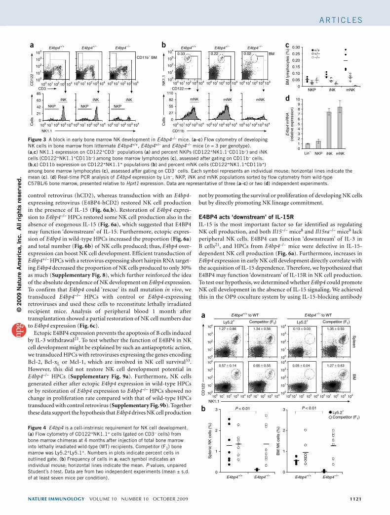
Ly49I, Ly49F, Ly49H and KLRG1 was higher (Supplementary Fig. 4). We used real-time PCR to quantify E4bp4 expression at each stage of NK cell development in bone marrow. A fivefold increase at the NKP-to-iNK cell transition was apparent, and the resulting higher E4bp4 expression was maintained in mNK cells (Fig. 3d). Such an expression profile accurately correlates with the NKP-iNK and iNK-mNK developmental-stage defects in E4bp4–/– bone mar-row. Therefore, progression through the NKP-iNK and iNK-mNK transitions requires E4bp4 expression.
To investigate whether the requirement for E4bp4 expression in NK cell development is cell intrinsic, we did adoptive transfer experiments with bone marrow as the source of hematopoietic stem cells. Bone marrow from wild-type (Ly5.1+) mice transferred into sublethally irradiated E4bp4–/– (Ly5.2+) mice repopulated the NK lineage both in the short term (4 weeks) and long term (3 months; Supplementary Fig. 5), which demonstrated that the E4bp4–/– niche is competent to support NK cell development. This suggested a cell-intrinsic require-ment for E4BP4 in NK cell development. To confirm this hypothesis, we transferred E4bp4+/+ or E4bp4–/– bone marrow (Ly5.2+) at a ratio of 1:1 with competitor bone marrow (Ly5.1+Ly5.2+) from F1 mice into lethally irradiated Ly5.1+ recipients. The degree of in vivo recon-stitution in hematopoietic cells overall (Supplementary Fig. 6a) and specifically for NKT (NK1.1+CD3+) cells (Supplementary Fig. 6b) was similar for both E4bp4–/– and E4bp4+/+ donor bone marrow. Although E4bp4+/+ and competitor bone marrow contributed similarly to long-term NK cell reconstitution, the contribution of E4bp4–/– bone
marrow was only 10% as much (Fig. 4). Therefore, the critical require-ment for E4bp4 in NK cell production is cell intrinsic.
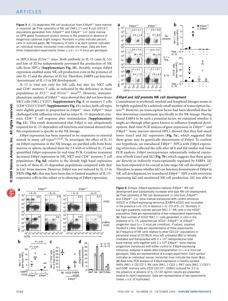
Ectopic E4bp4 increases NK cell productionNK cells can be produced in vitro from HPCs cultured on stromal cells such as OP9 in the presence of IL-15 (ref. 31), whereas con-tinuous culture of HPCs in the cytokines IL-7 and the ligand for the receptor tyrosine kinase Flt3 (Flt3L) produces B cells32. To confirm the cell-intrinsic nature of the E4bp4–/– NK cell defect, we isolated bone marrow enriched for HPCs from E4bp4–/– and E4bp4+/+ mice, cultured the bone marrow on OP9 cells with subsequent addition of cytokines and quantified lymphocyte production by flow cytom-etry (Supplementary Fig. 7a). E4bp4–/– HPCs produced effectively no NK (NK1.1+) cells compared with the substantial number of NK cells produced by E4bp4+/+ HPCs (Fig. 5). In contrast, loss of E4bp4 resulted in twofold greater production of B (CD19+) cells from HPCs (Fig. 5). To confirm that the IL-7 and Flt3L culture phase did not select against IL-15-dependent progenitors giving rise to NK cells, we incubated E4bp4–/– HPCs immediately after isolation with IL-15 in OP9 coculture, but this had no ‘rescue’ effect on NK cell production (Supplementary Fig. 7b). Thus, both in vivo and in vitro data indicate that NK development requires cell-intrinsic E4bp4 expression.
To verify the direct relationship between E4bp4 and NK development, we extended the in vitro system by restoring E4bp4 expression in E4bp4–/– HPCs by retroviral transduction. As anticipated, E4bp4–/– HPCs failed to produce NK cells after transduction with
50
a
c
d
b
100 6
5
4
3
2
1
0
80
60
40
20
0
P = 0.66
P = 0.37
P = 0.17 P < 0.01
P < 0.01
NK1.1
OutputInput
CFSE
IFN-γ
RMA RMA/s320 120
90
60
30
0
35
26
17
8
0
120
90
60
30
0
120
90
60
30
0
240
160
80
0
+/++/––/–
P = 0.46
40
30
CD
8+ c
ells
(×1
06 )C
D19
+ c
ells
(×1
06 )
NK
1.1+
CD
3– cel
ls (
×106 )
CD
122
Cel
ls
Cel
ls
20
10
0
50
104
103
102
101
100
100 101 102 103 104
100 101 102 103 104
40
30
20
10
0100 101 102 103 104
40
30
20
10
0100 101 102 103 104
100 101 102 103 104 100 101 102 103 104 100 101 102 103 104
104
103
102
101
100
104
103
102
101
100
100 101 102 103 104 100 101 102 103 104
P = 0.84
P = 0.312.98 ± 1.15 0.03 ± 0.01
0.42 ± 0.14
41.9 ± 3.2 30.1 ± 2.7 10.3 ± 1.5
0.02 ± 0.01
0.59 ± 0.18 0.03 ± 0.01
Spleen
Blood
BM
E4bp4+/+
E4bp4+/+ E4bp4+/– E4bp4–/–
E4bp4+/+ E4bp4+/– E4bp4–/–
E4bp4–/–
40
30
CD
4+ c
ells
(×1
06 )
20
10
0
Figure 2 Loss of peripheral NK cells and NK activity in E4bp4–/– mice. (a) Total viable cell numbers in peripheral splenic B cell (CD19+), T cell (CD8+CD3+ or CD4+CD3+) and NK cell (NK1.1+CD3–) populations from E4bp4+/+ and E4bp4+/– and E4bp4–/– mice (n > 10 per genotype, all litter-matched), determined by Trypan blue–exclusion counting and immunophenotypic analysis. Each symbol represents an individual mouse; horizontal line indicate the mean. P values, unpaired Student’s t-test. (b) Flow cytometry of CD122+NK1.1+ cells in E4bp4+/+ and E4bp4–/– mice. Numbers in plots indicate percent cells in outlined gate (mean ± s.d. of at least three mice per genotype). (c) Flow cytometry of CFSE-stained RMA/s cells (MHC class I–negative; high CFSE concentration) and RMA cells (MHC class I–positive; low CFSE concentration) injected at a ratio of 1:1 (Input) into E4bp4+/+, E4bp4+/– and E4bp4–/– mice (n = 3 per genotype), followed by analysis of CFSE in cells from recipient spleens 16 h later (Output) to assess in vivo NK cell killing activity. Injection of RMA cells labeled with both high and low CFSE concentrations showed no nonspecific loss of the population labeled with a high CFSE concentration (negative control; data not shown). (d) Intracellular IFN-γ in NK1.1+CD3– populations from total bone marrow incubated for 4 h with (black lines) or without (gray filled histograms) IL-12 and IL-18. Numbers above bracketed lines indicate percent cells in gate (mean ± s.d.; n = 3 mice per genotype). Data are representative of six (a), three (b), two (c,d) experiments.
©20
09 N
atu
re A
mer
ica,
Inc.
All
rig
hts
res
erve
d.
nature immunology VOLUME 10 NUMBER 10 OctOBER 2009 1121
A rt i c l e s
control retrovirus (hCD2), whereas transduction with an E4bp4-expressing retrovirus (E4BP4-hCD2) restored NK cell production in the presence of IL-15 (Fig. 6a,b). Restoration of E4bp4 expres-sion to E4bp4–/– HPCs restored some NK cell production also in the absence of exogenous IL-15 (Fig. 6a), which suggested that E4BP4 may function ‘downstream’ of IL-15. Furthermore, ectopic expres-sion of E4bp4 in wild-type HPCs increased the proportion (Fig. 6a) and total number (Fig. 6b) of NK cells produced; thus, E4bp4 over-expression can boost NK cell development. Efficient transduction of E4bp4+/– HPCs with a retrovirus expressing short hairpin RNA target-ing E4bp4 decreased the proportion of NK cells produced to only 30% as much (Supplementary Fig. 8), which further reinforced the idea of the absolute dependence of NK development on E4bp4 expression. To confirm that E4bp4 could ‘rescue’ its null mutation in vivo, we transduced E4bp4–/– HPCs with control or E4bp4-expressing retroviruses and used these cells to reconstitute lethally irradiated recipient mice. Analysis of peripheral blood 1 month after transplantation showed a partial restoration of NK cell numbers due to E4bp4 expression (Fig. 6c).
Ectopic E4BP4 expression prevents the apoptosis of B cells induced by IL-3 withdrawal21. To test whether the function of E4BP4 in NK cell development might be explained by such an antiapoptotic action, we transduced HPCs with retroviruses expressing the genes encoding Bcl-2, Bcl-xL or Mcl-1, which are involved in NK cell survival33. However, this did not restore NK cell development potential in E4bp4–/– HPCs (Supplementary Fig. 9a). Furthermore, NK cells generated either after ectopic E4bp4 expression in wild-type HPCs or by restoration of E4bp4 expression to E4bp4–/– HPCs showed no change in proliferation rate compared with that of wild-type HPCs transduced with control retrovirus (Supplementary Fig. 9b). Together these data support the hypothesis that E4bp4 drives NK cell production
not by promoting the survival or proliferation of developing NK cells but by directly promoting NK lineage commitment.
E4BP4 acts ‘downstream’ of IL-15RIL-15 is the most important factor so far identified as regulating NK cell production, and both Il15–/– mice8 and Il15ra–/– mice9 lack peripheral NK cells. E4BP4 can function ‘downstream’ of IL-3 in B cells21, and HPCs from E4bp4–/– mice were defective in IL-15-dependent NK cell production (Fig. 6a). Furthermore, increases in E4bp4 expression in early NK cell development directly correlate with the acquisition of IL-15 dependence. Therefore, we hypothesized that E4BP4 may function ‘downstream’ of IL-15R in NK cell production. To test our hypothesis, we determined whether E4bp4 could promote NK cell development in the absence of IL-15 signaling. We achieved this in the OP9 coculture system by using IL-15-blocking antibody
104
a b c
d
103
102
101
100
85
63
42
21
0
100 101 102 103 104
100 101 102 103 104 100 101 102 103 104
110
82
55
27
0
100 101 102 103 104 100 101 102 103 104 100 101 102 103 104 100 101 102 103 104
100 101 102 103 104
0.33
NKP
iNK
CD3
CD
122
Cel
ls
Cel
ls
CD122
CD11b
NK
1.1
NK1.1
CD11b– BMBM
NKP
iNK
NKP
iNK mNK mNK mNK
0.30
0.25
0.20
0.15
0.10
BM
lym
phoc
ytes
(%
)
0.05
109876543210
NKP
Lin– NKP iNK mNK
iNK
+/++/––/–
mNK0
0.22 0.02
100 101 102 103 104
104
103
102
101
100
100 101 102 103 104 100 101 102 103 104 100 101 102 103 104
E4bp4+/+ E4bp4+/– E4bp4–/– E4bp4+/+ E4bp4+/–
E4b
p4 m
RN
A(r
elat
ive
expr
essi
on)
E4bp4–/–
among bone marrow lymphocytes (c), assessed after gating on CD3– cells. Each symbol represents an individual mouse; horizontal lines indicate the mean (c). (d) Real-time PCR analysis of E4bp4 expression by Lin–, NKP, iNK and mNK populations sorted by flow cytometry from wild-type C57BL/6 bone marrow, presented relative to Hprt1 expression. Data are representative of three (a–c) or two (d) independent experiments.
E4bp4+/+ to WT
E4bp4+/+ E4bp4–/–
E4bp4–/– to WT
Ly5.2+
1.27 ± 0.86
Competitor (F1)104
103
102
101
100
1.34 ± 0.56
Ly5.2+
Spleen
0.13 ± 0.03
Competitor (F1)
1.35 ± 0.50
0.57 ± 0.14
CD
122
NK1.1
a
b
104
103
102
101
100
104
103
102
101
100
104
103
102
101
100
100 101 102 103 104
0.95 ± 0.55
100 101 102 103 104
BM
0.05 ± 0.04
100101 102 103 104
1.27 ± 0.63
P < 0.01P < 0.013
2
Spl
enic
NK
cel
ls (
%)
Ly5.2+
Competitor (F1)
0
1
E4bp4+/+ E4bp4–/–
3
2
BM
NK
cel
ls (
%)
1
0
100 101 102 103 104
Figure 4 E4bp4 is a cell-instrinsic requirement for NK cell development. (a) Flow cytometry of CD122+NK1.1+ cells (gated on CD3– cells) from bone marrow chimeras at 4 months after injection of total bone marrow into lethally irradiated wild-type (WT) recipients. Competitor (F1) bone marrow was Ly5.2+Ly5.1+. Numbers in plots indicate percent cells in outlined gate. (b) Frequency of cells in a; each symbol indicates an individual mouse; horizontal lines indicate the mean. P values, unpaired Student’s t-test. Data are from two independent experiments (mean ± s.d. of at least seven mice per condition).
Figure 3 A block in early bone marrow NK development in E4bp4–/– mice. (a–c) Flow cytometry of developing NK cells in bone marrow from littermate E4bp4+/+, E4bp4+/– and E4bp4–/– mice (n = 3 per genotype). (a,c) NK1.1 expression on CD122+CD3– populations (a) and percent NKPs (CD122+NK1.1–CD11b–) and iNK cells (CD122+NK1.1+CD11b–) among bone marrow lymphocytes (c), assessed after gating on CD11b– cells. (b,c) CD11b expression on CD122+NK1.1+ populations (b) and percent mNK cells (CD122+NK1.1+CD11b+)
©20
09 N
atu
re A
mer
ica,
Inc.
All
rig
hts
res
erve
d.
1122 VOLUME 10 NUMBER 10 OctOBER 2009 nature immunology
A rt i c l e s
or HPCs from Il15ra–/– mice. Both antibody to IL-15 (anti-IL-15) and loss of Il15ra independently prevented the production of NK cells from HPCs (Supplementary Fig. 10). Notably, ectopic E4bp4 expression enabled some NK cell production even in the presence of anti-IL-15 and the absence of Il15ra. Therefore, E4BP4 can function ‘downstream’ of IL-15 in NK development.
IL-15 is vital not only for NK cells but also for NKT cells and CD8+ memory T cells, as indicated by the deficiency in these populations in Il15–/– and Il15ra–/– mice8,9. However, immuno-phenotypic analysis of E4bp4–/– mice showed they did not have fewer NKT cells (NK1.1+CD3+; Supplementary Fig. 3) or memory T cells (CD8+CD25+CD44hi; Supplementary Fig. 11); in fact, both cell types were slightly greater in proportion in E4bp4–/– mice. E4bp4–/– mice challenged with influenza virus had an intact IL-15-dependent cyto-toxic CD8+ T cell response after restimulation (Supplementary Fig. 12). This result demonstrated that E4bp4 is not ubiquitously required for IL-15-dependent cell functions and instead showed that this requirement is specific to the NK lineage.
E4bp4 expression has been reported to be responsive to external stimuli in many cell types21,23,28. To investigate the effect of IL-15 on E4bp4 expression in the NK lineage, we purified cells from bone marrow or spleen, incubated them for 3 h with or without IL-15 and quantified E4bp4 expression by real-time PCR. Cytokine treatment increased E4bp4 expression in NK, NKT and CD8+ memory T cell populations (Fig. 6d) relative to the already high basal expression in each of these IL-15-dependent populations compared with that in Lin– bone marrow. However, E4bp4 was not induced by IL-15 in NKPs (Fig. 6d); this may have been due to limited numbers of IL-15-responsive cells in this subset or to silencing of E4bp4 expression.
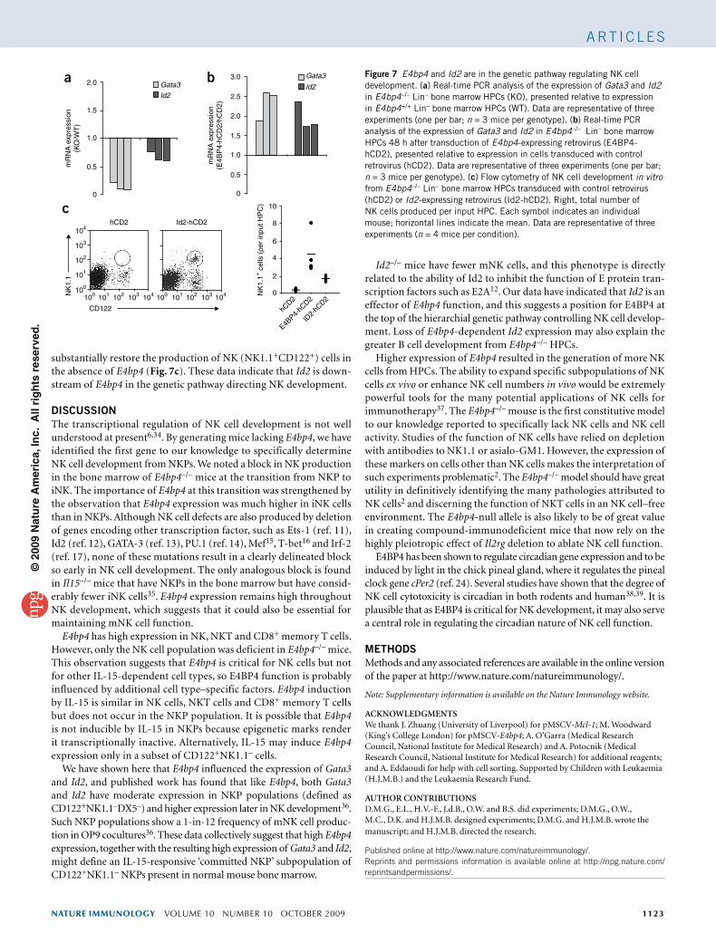
E4bp4 and Id2 promote NK cell developmentCommitment to erythroid, myeloid and lymphoid lineages seems to be tightly regulated by a relatively small number of transcription fac-tors10. However, no transcription factor had been identified thus far that determines commitment specifically to the NK lineage. Having found E4BP4 to be such a potential factor, we examined whether it might act through other genes known to influence lymphoid devel-opment. Real-time PCR analysis of gene expression in E4bp4+/+ and E4bp4–/– bone marrow–derived HPCs showed that they had much lower Gata3 and Id2 expression (Fig. 7a), which suggested that these genes may be genetically downstream of E4bp4. To confirm our hypothesis, we transduced E4bp4–/– HPCs with E4bp4-express-ing retrovirus, collected the cells after 48 h and did similar real-time PCR analysis. E4bp4 overexpression substantially induced expres-sion of both Gata3 and Id2 (Fig. 7b), which suggests that these genes are directly or indirectly transcriptionally regulated by E4BP4. Id2 has been reported to be crucial in late-stage NK cell development12. Therefore, to assess whether Id2 can function downstream of E4bp4 in NK cell development, we transduced E4bp4–/– HPCs with retrovirus expressing Id2 and monitored NK cell production. Id2 was able to
Figure 5 IL-15-dependent NK cell production from E4bp4–/– bone marrow is impaired. (a) Flow cytometry of NK cell (NK1.1+) and B cell (CD19+) populations generated from E4bp4+/+ and E4bp4–/– Lin– bone marrow on OP9–green fluorescent protein stroma in the presence or absence of exogenous cytokines (right margin). Numbers in plots indicate percent cells in outlined gates. (b) Frequency of cells in a; each symbol indicates an individual mouse; horizontal lines indicate the mean. Data are from three independent experiments (mean ± s.d.; n = 4 mice per genotype).
Figure 6 Ectopic E4bp4 expression restores E4bp4–/– NK cell development and substantially increases wild-type NK cell development. (a) Flow cytometry of NK cell development in vitro from E4bp4+/+ and E4bp4–/– Lin– bone marrow transduced with control retrovirus (hCD2) or E4bp4-expressing retrovirus (E4BP4-hCD2) and incubated in the presence (+IL-15) or absence (−IL-15) of IL-15. Numbers in top right quadrants indicate percent NK1.1+ NK cells in the hCD2+ population. Data are representative of two independent experiments. (b) Total number of hCD2+NK1.1+ cells generated in vitro in the presence of IL-15, presented per hCD2+ E4bp4+/+ or E4bp4–/– progenitor input (n = 3 mice per condition). P values, unpaired Student’s t-test. Data are representative of three experiments. (c) Frequency of NK cells relative to other CD122+ populations in peripheral blood of C57BL/6 mice left untreated (B6) or lethally irradiated and transplanted with 2 × 10⁵ radioprotective total bone marrow cells together with 2 × 10⁵ E4bp4–/– bone marrow progenitors transduced with either control or E4bp4-expressing retrovirus, analyzed 4 weeks after transplantation (n = 4–6 mice per genotype). Data are representative of a single experiment. Each symbol indicates an individual mouse; horizontal lines indicate the mean (b,c). (d) Real-time PCR analysis of E4bp4 expression in freshly isolated NKPs (NK1.1–CD122+), NK cells (NK1.1+CD3–), NKT cells (NK1.1+CD3+) and CD8+ memory cells (CD3+CD122+; CD8m) cultured for 3 h in the presence or absence of IL-15 (30 ng/ml); results are presented relative to Hprt1 expression. Data are representative of two experiments (mean ± s.d. of triplicates).
104
103
102
101
100
0.4 ± 0.1
0.6 ± 0.4
11 ± 5.1
3.4 ± 1.3
0.1 ± 0.1
7.4 ± 4.3
0.0 ± 0.0
36 ± 10 59 ± 13
0.0 ± 0.0
10 ± 6.0 Nocytokine
IL-7 +Flt3L
IL-15
CD19
Flt3L + IL-7IL-15
Flt3L + IL-7IL-15
– –– –
– ––+ +
+ +
–– –
–++
–
– –– +
+–
4
40
30
20
10
0
3
2
1
0
NK
1.1
NK
1.1+
cel
ls(p
er in
put H
PC
)
CD
19+ c
ells
(per
inpu
t HP
C)
5.9 ± 1.4
104
103
102
101
100
104
104
103
103
102
102
101
101
100
100
104
103
102
101
100
E4bp4+/+ E4bp4–/– E4bp4+/+
E4bp4–/–a b
104+ IL-15
E4bp4+/+ E4bp4–/–
– IL-15 + IL-15 – IL-15
hCD2
E4BP4-hCD2
1033.9
26 3.8 5.6 1.6
0.4 0.0 0.0
102
101
100
104
104
103
103
102
102
101
NK
1.1
101100
104
103
102
101
100
104
103
102
101
100
hCD2
2070
35 –IL-15+IL-1530
25
20
15
10
5
0B6 NKP NK NKTCD8mhCD2 E4BP4
-hCD2
6050403020100
15
E4BP4-hCD2
10
NK
1.1+
cel
ls(p
er in
put H
PC
)
NK
1.1+
CD
3– ce
lls(%
of t
otal
CD
122+
)
E4b
p4 m
RN
A(r
elat
ive
expr
essi
on)
5
– + – +0
P = 0.81
P = 0.04
100 104103102101100 104103102101100 104103102101100
E4bp4+/+ E4bp4–/–
E4bp4–/–
a
b c d
©20
09 N
atu
re A
mer
ica,
Inc.
All
rig
hts
res
erve
d.
nature immunology VOLUME 10 NUMBER 10 OctOBER 2009 1123
A rt i c l e s
substantially restore the production of NK (NK1.1+CD122+) cells in the absence of E4bp4 (Fig. 7c). These data indicate that Id2 is down-stream of E4bp4 in the genetic pathway directing NK development.
DISCUSSIONThe transcriptional regulation of NK cell development is not well understood at present6,34. By generating mice lacking E4bp4, we have identified the first gene to our knowledge to specifically determine NK cell development from NKPs. We noted a block in NK production in the bone marrow of E4bp4–/– mice at the transition from NKP to iNK. The importance of E4bp4 at this transition was strengthened by the observation that E4bp4 expression was much higher in iNK cells than in NKPs. Although NK cell defects are also produced by deletion of genes encoding other transcription factor, such as Ets-1 (ref. 11), Id2 (ref. 12), GATA-3 (ref. 13), PU.1 (ref. 14), Mef15, T-bet16 and Irf-2 (ref. 17), none of these mutations result in a clearly delineated block so early in NK cell development. The only analogous block is found in Il15–/– mice that have NKPs in the bone marrow but have consid-erably fewer iNK cells35. E4bp4 expression remains high throughout NK development, which suggests that it could also be essential for maintaining mNK cell function.
E4bp4 has high expression in NK, NKT and CD8+ memory T cells. However, only the NK cell population was deficient in E4bp4–/– mice. This observation suggests that E4bp4 is critical for NK cells but not for other IL-15-dependent cell types, so E4BP4 function is probably influenced by additional cell type–specific factors. E4bp4 induction by IL-15 is similar in NK cells, NKT cells and CD8+ memory T cells but does not occur in the NKP population. It is possible that E4bp4 is not inducible by IL-15 in NKPs because epigenetic marks render it transcriptionally inactive. Alternatively, IL-15 may induce E4bp4 expression only in a subset of CD122+NK1.1– cells.
We have shown here that E4bp4 influenced the expression of Gata3 and Id2, and published work has found that like E4bp4, both Gata3 and Id2 have moderate expression in NKP populations (defined as CD122+NK1.1–DX5–) and higher expression later in NK development36. Such NKP populations show a 1-in-12 frequency of mNK cell produc-tion in OP9 cocultures36. These data collectively suggest that high E4bp4 expression, together with the resulting high expression of Gata3 and Id2, might define an IL-15-responsive ‘committed NKP’ subpopulation of CD122+NK1.1– NKPs present in normal mouse bone marrow.
Id2–/– mice have fewer mNK cells, and this phenotype is directly related to the ability of Id2 to inhibit the function of E protein tran-scription factors such as E2A12. Our data have indicated that Id2 is an effector of E4bp4 function, and this suggests a position for E4BP4 at the top of the hierarchial genetic pathway controlling NK cell develop-ment. Loss of E4bp4-dependent Id2 expression may also explain the greater B cell development from E4bp4–/– HPCs.
Higher expression of E4bp4 resulted in the generation of more NK cells from HPCs. The ability to expand specific subpopulations of NK cells ex vivo or enhance NK cell numbers in vivo would be extremely powerful tools for the many potential applications of NK cells for immunotherapy37. The E4bp4–/– mouse is the first constitutive model to our knowledge reported to specifically lack NK cells and NK cell activity. Studies of the function of NK cells have relied on depletion with antibodies to NK1.1 or asialo-GM1. However, the expression of these markers on cells other than NK cells makes the interpretation of such experiments problematic2. The E4bp4–/– model should have great utility in definitively identifying the many pathologies attributed to NK cells2 and discerning the function of NKT cells in an NK cell–free environment. The E4bp4-null allele is also likely to be of great value in creating compound-immunodeficient mice that now rely on the highly pleiotropic effect of Il2rg deletion to ablate NK cell function.
E4BP4 has been shown to regulate circadian gene expression and to be induced by light in the chick pineal gland, where it regulates the pineal clock gene cPer2 (ref. 24). Several studies have shown that the degree of NK cell cytotoxicity is circadian in both rodents and human38,39. It is plausible that as E4BP4 is critical for NK development, it may also serve a central role in regulating the circadian nature of NK cell function.
METHODSMethods and any associated references are available in the online version of the paper at http://www.nature.com/natureimmunology/.
Note: Supplementary information is available on the Nature Immunology website.
ACKnOWLEDGMEntSWe thank J. Zhuang (University of Liverpool) for pMSCV-Mcl-1; M. Woodward (King’s College London) for pMSCV-E4bp4; A. O’Garra (Medical Research Council, National Institute for Medical Research) and A. Potocnik (Medical Research Council, National Institute for Medical Research) for additional reagents; and A. Eddaoudi for help with cell sorting. Supported by Children with Leukaemia (H.J.M.B.) and the Leukaemia Research Fund.
AUtHOR COntRIBUtIOnSD.M.G., E.L., H.V.-F., J.d.B., O.W. and B.S. did experiments; D.M.G., O.W., M.C., D.K. and H.J.M.B. designed experiments; D.M.G. and H.J.M.B. wrote the manuscript; and H.J.M.B. directed the research.
Published online at http://www.nature.com/natureimmunology/. reprints and permissions information is available online at http://npg.nature.com/reprintsandpermissions/.
2.0a b
c
1.5
mR
NA
exp
ress
ion
(KO
/WT
)
mR
NA
exp
ress
ion
(E4B
P4-
hCD
2/hC
D2)
0.5
0
1.0
2.0
1.0
1.5
2.5
104
103
hCD2 Id2-hCD2
102
101
100
10
8
6
4
2
0104103102101100
CD122
NK
1.1
NK
1.1+
cel
ls (
per
inpu
t HP
C)
104
hCD2
E4BP4-
hCD2
ID2-
hCD2103102101100
Gata3
ld2
Gata3
ld23.0
0.5
0
Figure 7 E4bp4 and Id2 are in the genetic pathway regulating NK cell development. (a) Real-time PCR analysis of the expression of Gata3 and Id2 in E4bp4–/– Lin– bone marrow HPCs (KO), presented relative to expression in E4bp4+/+ Lin– bone marrow HPCs (WT). Data are representative of three experiments (one per bar; n = 3 mice per genotype). (b) Real-time PCR analysis of the expression of Gata3 and Id2 in E4bp4–/– Lin– bone marrow HPCs 48 h after transduction of E4bp4-expressing retrovirus (E4BP4-hCD2), presented relative to expression in cells transduced with control retrovirus (hCD2). Data are representative of three experiments (one per bar; n = 3 mice per genotype). (c) Flow cytometry of NK cell development in vitro from E4bp4–/– Lin– bone marrow HPCs transduced with control retrovirus (hCD2) or Id2-expressing retrovirus (Id2-hCD2). Right, total number of NK cells produced per input HPC. Each symbol indicates an individual mouse; horizontal lines indicate the mean. Data are representative of three experiments (n = 4 mice per condition).
©20
09 N
atu
re A
mer
ica,
Inc.
All
rig
hts
res
erve
d.
1124 VOLUME 10 NUMBER 10 OctOBER 2009 nature immunology
1. Di Santo, J.P. Natural killer cell developmental pathways: a question of balance. Annu. Rev. Immunol. 24, 257–286 (2006).
2. Vivier, E., Tomasello, E., Baratin, M., Walzer, T. & Ugolini, S. Functions of natural killer cells. Nat. Immunol. 9, 503–510 (2008).
3. Wu, J. & Lanier, L.L. Natural killer cells and cancer. Adv. Cancer Res. 90, 127–156 (2003).
4. Lodoen, M.B. & Lanier, L.L. Natural killer cells as an initial defense against pathogens. Curr. Opin. Immunol. 18, 391–398 (2006).
5. Yokoyama, W.M., Kim, S. & French, A.R. The dynamic life of natural killer cells. Annu. Rev. Immunol. 22, 405–429 (2004).
6. Di Santo, J.P. Natural killer cells: diversity in search of a niche. Nat. Immunol. 9, 473–475 (2008).
7. Huntington, N.D., Vosshenrich, C.A. & Di Santo, J.P. Developmental pathways that generate natural-killer-cell diversity in mice and humans. Nat. Rev. Immunol. 7, 703–714 (2007).
8. Kennedy, M.K. et al. Reversible defects in natural killer and memory CD8 T cell lineages in interleukin 15-deficient mice. J. Exp. Med. 191, 771–780 (2000).
9. Suzuki, H., Duncan, G.S., Takimoto, H. & Mak, T.W. Abnormal development of intestinal intraepithelial lymphocytes and peripheral natural killer cells in mice lacking the IL-2 receptor beta chain. J. Exp. Med. 185, 499–505 (1997).
10. Orkin, S.H. & Zon, L.I. Hematopoiesis: an evolving paradigm for stem cell biology. Cell 132, 631–644 (2008).
11. Barton, K. et al. The Ets-1 transcription factor is required for the development of natural killer cells in mice. Immunity 9, 555–563 (1998).
12. Boos, M.D., Yokota, Y., Eberl, G. & Kee, B.L. Mature natural killer cell and lymphoid tissue-inducing cell development requires Id2-mediated suppression of E protein activity. J. Exp. Med. 204, 1119–1130 (2007).
13. Samson, S.I. et al. GATA-3 promotes maturation, IFN-γ production, and liver-specific homing of NK cells. Immunity 19, 701–711 (2003).
14. Colucci, F. et al. Differential requirement for the transcription factor PU.1 in the generation of natural killer cells versus B and T cells. Blood 97, 2625–2632 (2001).
15. Lacorazza, H.D. et al. The ETS protein MEF plays a critical role in perforin gene expression and the development of natural killer and NK-T cells. Immunity 17, 437–449 (2002).
16. Townsend, M.J. et al. T-bet regulates the terminal maturation and homeostasis of NK and Vα14i NKT cells. Immunity 20, 477–494 (2004).
17. Taki, S., Nakajima, S., Ichikawa, E., Saito, T. & Hida, S. IFN regulatory factor-2 deficiency revealed a novel checkpoint critical for the generation of peripheral NK cells. J. Immunol. 174, 6005–6012 (2005).
18. Cowell, I.G. E4BP4/NFIL3, a PAR-related bZIP factor with many roles. Bioessays 24, 1023–1029 (2002).
19. Cowell, I.G., Skinner, A. & Hurst, H.C. Transcriptional repression by a novel member of the bZIP family of transcription factors. Mol. Cell. Biol. 12, 3070–3077 (1992).
20. Zhang, W. et al. Molecular cloning and characterization of NF-IL3A, a transcriptional activator of the human interleukin-3 promoter. Mol. Cell. Biol. 15, 6055–6063 (1995).
21. Ikushima, S. et al. Pivotal role for the NFIL3/E4BP4 transcription factor in interleukin 3-mediated survival of pro-B lymphocytes. Proc. Natl. Acad. Sci. USA 94, 2609–2614 (1997).
22. Mitsui, S., Yamaguchi, S., Matsuo, T., Ishida, Y. & Okamura, H. Antagonistic role of E4BP4 and PAR proteins in the circadian oscillatory mechanism. Genes Dev. 15, 995–1006 (2001).
23. Doi, M., Okano, T., Yujnovsky, I., Sassone-Corsi, P. & Fukada, Y. Negative control of circadian clock regulator E4BP4 by casein kinase Iepsilon-mediated phosphorylation. Curr. Biol. 14, 975–980 (2004).
24. Doi, M., Nakajima, Y., Okano, T. & Fukada, Y. Light-induced phase-delay of the chicken pineal circadian clock is associated with the induction of cE4bp4, a potential transcriptional repressor of cPer2 gene. Proc. Natl. Acad. Sci. USA 98, 8089–8094 (2001).
25. Junghans, D. et al. The CES-2-related transcription factor E4BP4 is an intrinsic regulator of motoneuron growth and survival. Development 131, 4425–4434 (2004).
26. Hough, C. et al. Cell type-specific regulation of von Willebrand factor expression by the E4BP4 transcriptional repressor. Blood 105, 1531–1539 (2005).
27. Silvestris, F. et al. Negative regulation of the osteoblast function in multiple myeloma through the repressor gene E4BP4 activated by malignant plasma cells. Clin. Cancer Res. 14, 6081–6091 (2008).
28. Priceman, S.J. et al. Calcium-dependent upregulation of E4BP4 expression correlates with glucocorticoid-evoked apoptosis of human leukemic CEM cells. Biochem. Biophys. Res. Commun. 344, 491–499 (2006).
29. Kärre, K., Ljunggren, H.G., Piontek, G. & Kiessling, R. Selective rejection of H-2-deficient lymphoma variants suggests alternative immune defence strategy. Nature 319, 675–678 (1986).
30. Kim, S. et al. In vivo developmental stages in murine natural killer cell maturation. Nat. Immunol. 3, 523–528 (2002).
31. Williams, N.S. et al. Differentiation of NK1.1+, Ly49+ NK cells from flt3+ multipotent marrow progenitor cells. J. Immunol. 163, 2648–2656 (1999).
32. Vieira, P. & Cumano, A. Differentiation of B lymphocytes from hematopoietic stem cells. Methods Mol. Biol. 271, 67–76 (2004).
33. Huntington, N.D. et al. Interleukin 15-mediated survival of natural killer cells is determined by interactions among Bim, Noxa and Mcl-1. Nat. Immunol. 8, 856–863 (2007).
34. Boos, M.D., Ramirez, K. & Kee, B.L. Extrinsic and intrinsic regulation of early natural killer cell development. Immunol. Res. 40, 193–207 (2008).
35. Vosshenrich, C.A. et al. Roles for common cytokine receptor γ-chain-dependent cytokines in the generation, differentiation, and maturation of NK cell precursors and peripheral NK cells in vivo. J. Immunol. 174, 1213–1221 (2005).
36. Rosmaraki, E.E. et al. Identification of committed NK cell progenitors in adult murine bone marrow. Eur. J. Immunol. 31, 1900–1909 (2001).
37. Terme, M., Ullrich, E., Delahaye, N.F., Chaput, N. & Zitvogel, L. Natural killer cell-directed therapies: moving from unexpected results to successful strategies. Nat. Immunol. 9, 486–494 (2008).
38. Fernandes, G., Carandente, F., Halberg, E., Halberg, F. & Good, R.A. Circadian rhythm in activity of lympholytic natural killer cells from spleens of Fischer rats. J. Immunol. 123, 622–625 (1979).
39. Arjona, A. & Sarkar, D.K. Circadian oscillations of clock genes, cytolytic factors, and cytokines in rat NK cells. J. Immunol. 174, 7618–7624 (2005).
A rt i c l e s
©20
09 N
atu
re A
mer
ica,
Inc.
All
rig
hts
res
erve
d.
nature immunologydoi:10.1038/ni.1787
ONLINE METHODSGeneration of E4bp4 –/– mice. E4bp4 genomic DNA spanning the coding exon (6.4 kilobases) was isolated from a 129/Sv bacteriophage P1–derived artificial chromosome. The targeting vector was generated by cloning of a neomycin-resistance gene flanked by two loxP sites into a ClaI site 3′ of the E4bp4 DNA. An additional 1.9-kilobase fragment 3′ to the artificial chromosome–derived DNA was amplified by PCR from 129/Sv DNA and was directionally cloned 3′ of the neomycin-resistance cassette. The final vector contained an additional loxP site inserted at a SpeI site 2 kilobases upstream of the targeted exon. After electroporation and selection, correctly targeted clones were expanded, karyotyped and injected into wild-type C57BL/6 blastocysts to create chime-ras. Presence of the E4bp4-null allele was assessed in offspring by Southern blot and PCR analysis and offspring were subsequently crossed to C57BL/6 background more than ten times. All experiments used 4- to 12-week-old mice from at least the sixth backcross.
Mice. All animal husbandry and experimental procedures were carried out according to UK Home Office regulations and institute guidelines. Male chimeras showing a high degree of chimerism were bred to C57BL/6 females. In male chimeras, recombination of the loxP sites was induced during spermatogenesis by protamine promoter–driven Cre recombinase present in PC3 embryonic stem cells (Supplementary Fig. 1a). The resulting litters yielded mice heterozygous for the targeted allele minus the neomycin-resistance gene. The mice were backcrossed onto the C57BL/6 background until the PC3 Cre transgene was lost. PCR genotyping was done routinely with primer 5′-CTCTGAGCTTGGCTGATGTG-3′ plus primer 5′-GCTT CAAGTCTCCACCAAGC-3′ (for amplification of the wild-type allele) or primer 5′-CCATGCTCCTGTCTTGATGA-3′ (for amplification of the null allele). Loss of expression was confirmed by immunoblot analysis of whole-cell lysates from heart tissue (prepared with radioimmunoprecipitation assay buffer) with anti-E4BP4 (C-18; Santa Cruz) and anti-β-actin (C-4; Santa Cruz) and by RT-PCR analysis of RNA (purified from total thymus with TRIzol (Invitrogen)) with the primers 5′-CGGAAGTTGCATCTCAGTCA-3′ and 5′-GCAAAGCTCTCCAACTCCAC-3′.
Cell isolation and gene expression analysis. Lin− HPCs were purified from bone marrow by magnetic depletion of cells stained with anti-Ter119 (TER119), anti-B220 (RA3-6B2), anti–mouse CD2 (RM2-5), anti-NK1.1 (PK136), anti-Gr-1 (RB6-8C5) and anti-CD11b (M1/70, all from eBioscience) on MACS columns (Miltenyi). For gene-expression analysis, cells were purified from 5- to 6-week-old adult female C57BL/6 mice by specific antibody staining followed by sorting on a MoFlo XDP (Beckman Coulter). Sorted populations (>97% pure) were immediately frozen in TRIzol (Invitrogen) for RNA isolation or were cultured for 3 h in RPMI medium plus 10% (vol/vol) FCS with or without IL-15 treatment before RNA isolation. For cDNA synthesis, 100 ng to 1 mg RNA was reverse-transcribed with SuperScript II (Invitrogen) and mRNA abundance was quantified by real-time PCR analysis on a 7900HT (Applied Biosystems) with probe sets recognizing Pax5 (Mm00435501_m1), Notch1 (Mm00435245_m1), E4bp4 (Nfil3) (Mm00600292_s1), Gata3 (Mm00484683_m1), Id2 (Mm00711781_m1) and Hprt1 (Mm00446968_m1; Applied Biosystems identifiers in parentheses).
Flow cytometry. Cells were preincubated with anti-Fc block (monoclonal antibody 2.4G2 to the Fcγ III-II receptor; BD Biosciences) before being stained with conjugate antibodies to c-Kit, (2B8; BD Biosciences), KLRG1 (2F1, Abcam), NKG2D (R&D systems), CD19 (1D3), IgD (11-26), CD4 (RM4-5), CD8 (53-6.7), CD3 (145-2C11), NK1.1 (PK136), CD122 (5H4), NKp46 (29A1.4), NKG2AB6 (16a11) Ly49C/I/F/H (14B11), DX5 (DX5), CD11b (M1/70), Ly5.1 (A20), Ly5.2 (1O4), CD25 (PC61), CD44 (IM7) and human CD2 (RPA-2.10; all from eBioscience). IFN-γ production in bone marrow
NK cells cultured ex vivo for 4 h with or without IL-12 (30 ng/ml) and IL-18 (20 ng/ml) was measured after permeabilization and staining with anti-IFN-γ (XMG1.2; eBioscience).
In vivo NK cell killing assay. In experiments based on published protocols40, 2 × 10⁸ RMA and 2 × 10⁸ RMA/s thymoma cells (cultured in Iscove’s modified Dulbecco’s medium supplemented with 10% FCS) were labeled with 16 ml CFSE (carboxyfluorescein diacetate, succinimidyl ester; Molecular Probes) at a concentration of 1 µM or 2.5 µM, respectively, and 4 × 10⁷ cells total (2 × 10⁷ of each type) were injected intravenously into E4bp4+/+, E4bp4+/– and E4bp4–/– recipients. After 16 h, recipients were killed, spleens were removed and CFSE positivity was determined in gated non-autofluorescent cell popula-tions similar in size to those of input RMA and RMA/s cells.
Adoptive cell transfer. E4bp4+/+ and E4bp4–/– recipients were irradiated with 600 rads and were transplanted intravenously with 1 × 10⁶ bone marrow cells from C57BL/6 (Ly5.1+) donors. C57BL/6 (Ly5.1+) recipients were irradiated with two doses of 500 rads and were transplanted with 1 × 10⁶ total bone marrow cells from E4bp4+/+ or E4bp4–/– donors (Ly5.2+) plus 1 × 10⁶ total bone marrow competitor cells from C57BL/6 F1 donors (Ly5.1+ Ly 5.2+). For in vivo ‘rescue’, wild-type recipients were irradiated with two doses of first 400 rads and then 500 rads and were transplanted with 2 × 10⁵ total wild-type bone marrow cells plus 2 × 10⁵ retrovirally transduced HPCs. Reconstitution of recipients was assessed by flow cytometry of blood at 4 weeks after transplantation.
In vitro lymphocyte development. During retroviral transduction, HPCs were cultured for 4–5 d in DMEM supplemented with 10% (vol/vol) FCS (Stem Cell Technologies), Flt3L (10 ng/ml; R&D Systems), IL-7 (10 ng/ml; PeproTech) and stem cell factor (100 ng/ml; PeproTech)31. Cells were washed and then cul-tured (3 × 10⁴ cells per well) for 6 d on OP9 stromal cells in MEM-α medium containing 20% (vol/vol) FCS (Sigma) supplemented with either no cytokine, Flt3L plus IL-7, or IL-15 alone (30 ng/ml; PeproTech)31. Antibodies for block-ing experiments were anti-IL-15 (500-P173; PeproTech) and normal rabbit IgG (500-P00; PeproTech), each used at a concentration of 10 µg/ml.
Retroviral transduction of progenitor cells was done as described41. The plasmid pMSCV-hCD2tailess-miR30 (pM2miR) was generated from pMSCV-neo (Clontech) and contains hCD2tailess fused to the miR30 micro-RNA42. Of three micro-RNA vectors designed, only pM2miR-1305 (5′-CCCGCACAAGCTTCGGATTAAA-3′) substantially diminished E4bp4 expression. Total viable lymphocyte numbers were determined by Trypan blue exclusion microscopy when cells were plated onto stroma and at the end of the experiment. Proliferation of NK cells generated in vitro was determined by pulsing of cells with BrdU (5-bromodeoxyuridine) for 1 h before staining with anti-NK1.1 and anti-human CD2, followed by fixation, permeabilization and anti-BrdU staining according to the manufacturer’s instructions (BD Biosciences).
Statistical analyses. An unpaired Student’s t-test, with unequal variation assumed, was used for all statistical comparisons of means; P values greater than 0.05 were considered not significant.
40. Westerhuis, G., Maas, W.G., Willemze, R., Toes, R.E. & Fibbe, W.E. Long-term mixed chimerism after immunologic conditioning and MHC-mismatched stem-cell transplantation is dependent on NK-cell tolerance. Blood 106, 2215–2220 (2005).
41. Morrow, M., Horton, S., Kioussis, D., Brady, H.J.M. & Williams, O. TEL-AML1 promotes development of specific hematopoietic lineages consistent with preleukemic activity. Blood 103, 3890–3896 (2004).
42. Stegmeier, F., Hu, G., Rickles, R.J., Hannon, G.J. & Elledge, S.J. Lentiviral microRNA-based system for single-copy polymerase II-regulated RNA interference in mammalian cells. Proc. Natl. Acad. Sci. USA 102, 13212–13217 (2005).
Erratum: The basic leucine zipper transcription factor E4BP4 is essential for natural killer cell developmentDuncan M Gascoyne, Elaine Long, Henrique Veiga-Fernandes, Jasper de Boer, Owen Williams, Benedict Seddon, Mark Coles, Dimitris Kioussis & Hugh JM BradyNat. Immunol. 10, 1118–1124 (2009); published online 13 September 2009; corrected after print 5 October 2009
In the version of this article initially published, the equal contribution of Duncan M. Gascoyne and Elaine Long is not noted. The error has been corrected in the HTML and PDF versions of the article.
errata©
2009
Nat
ure
Am
eric
a, In
c. A
ll ri
gh
ts r
eser
ved
.