Clinicopathological Study of Regressed Testicular Tumors (Apparent Extragonadal Germ Cell Neoplasms)
1
Testicular Microlithiasis and Neoplastic Lesions in Wild Eland
(Tragelaphus oryx): Possible Effects of Exposure to Environmental
Pollutants?
MS Bornman1, IEJ Barnhoorn
1, C de Jager
1, DNR Veeramachaneni
2
1Andrology, Department of Urology, University of Pretoria, Private Bag X169, Pretoria,
South Africa; 2Animal Reproduction and Biotechnology Laboratory, Colorado State
University, Fort Collins, Colorado, USA
Keywords
: Eland, testis, rete testis, adenoma, microlithiasis, alkylphenols, DDT, endocrine
disrupter chemicals
Correspondence: Prof MS (Riana) Bornman Department of Urology, University of Pretoria,
PO Box 169, Pretoria 0001, South Africa
Tel: +27 –12-3541513
Fax: +27-12-3542500
E-mail: [email protected]

2
Abstract
The purpose of the study was to compare wildlife in the proximity and away from the sources
of known industrial pollution. Macroscopic, focal, gritty areas that appeared white were
observed in the testes of all 24 South African eland (Tragelaphus oryx) culled in the Rietvlei
Nature Reserve (RNR; n=17) between 2001 and 2003 and Suikerbosrand Nature Reserve
(SNR; n=7) in 2004. Histopathological evaluation of testes showed multiple intratubular
dystrophic calcifications, focal areas of sperm stasis, and interstitial chronic cell infiltrates
with fibrosis. Spermatogenesis was generally impaired; a few atypical germ cells were also
encountered. Sertoli cell vacuolization and sloughing of the seminiferous epithelium were
evident. Adenomatous changes of the rete testis, reflective of possible chronic estrogenic
exposure, were found. In testes collected from three reference eland in 2007 from the Molopo
Nature Reserve (MNR) in the Kalahari/Kgalagadi Desert, except for one focal area of sperm
stasis and another with microcalcification, the seminiferous epithelium as well as
collecting/rete tubules were normal. Analyses of fat tissue for environmental pollutants
showed that 11 out of 17 RNR eland contained a detectable estrogenic chemical p-
nonylphenol (mean ± SD: 184.8 ± 24.6 µg/kg fat); no organochlorine chemicals or
polychlorinated biphenyls were detected. Of the 7 SNR eland, 5 had detectable octylphenol
residues (50.2 ± 30.9 µg/kg fat), 3 had detectable p-nonylphenol (137.8 ± 77.9 µg/kg fat), 3
had o-p’-DDT (114.9 ± 31.1 µg/kg fat), 3 had p-p’-DDT (127.3 ± 49.9 µg/kg(79.5 ± 30.4
µg/kg fat) and 5 contained o-p’-DDE (27.7 ± 9.9 µg/kg fat). One eland from the MNR
contained one 70.6 µg o-p’-DDT/kg fat and another p-p’-DDE 61.3 µg/kg fat. Therefore, in
eland with testicular abnormalities, significant amounts of various estrogenic chemicals were
bioaccumulated in fat samples. It therefore seems likely that the lesions found in RNR and
SNR were associated with the relatively high body-burden of environmental pollutants
(phenols), although the possibility of systemic infections cannot be ruled out. No testicular

3
abnormalities were found in reference eland. These findings are the first indication of
mammalian wildlife being affected by environmental pollution of endocrine disrupting
chemicals in South Africa.
INTRODUCTION
Exposures to endocrine-disrupting chemicals (EDCs) have been implicated in a variety of
urogenital disorders in men including testicular abnormalities (Giwercman et al., 1993;
Toppari et al., 1996). It was suggested that these urogenital disorders are all symptoms of one
underlying disease entity called testicular dysgenesis syndrome (Skakkebaek et al., 2001;
Skakkebaek et al., 2003). Testicular microlithiasis (TM) is one of the elements of the
testicular dysgenesis syndrome (Skakkebaek et al., 2004). TM is a rare pathologic condition
in which numerous calcifications form inside the seminiferous tubules (Renshaw, 1998;
Ganem et al., 1999). These calcifications originate from degenerating intratubular cellular
debris with subsequent mineralization of the epithelium (Holm et al., 2001). Men with male
infertility have a risk to develop testicular cancer, but the risk seems higher in the presence of
TM (Negri et al., 2008) and TM is therefore regarded as a premalignant condition (Derogee
et al., 2001).
The notable increase in the occurrence of urogenital abnormalities in men over a relatively
short period led to the speculation that the causative factors are adverse environmental effects
rather than specific gene mutations (Skakkebaek et al., 2001; Skakkebaek et al., 2003).
However, the possibility of transgenerational epigenetic effects also has been suggested
(Anway et al., 2005). All these urogenital disorders could be experimentally induced in
laboratory animals by developmental exposure to estrogenic and anti-androgenic substances
EDCs (Viguier-Martinez et al., 1983; Newbold et al., 1985; 2000; Yasuda et al., 1985; Luthra

4
and Hutson, 1989; Walker et al., 1990; de Jager et al., 1999b; Gray et al., 2001; Higuchi et
al., 2003; Kilian et al., 2007) supporting a possible link between exposure to environmental
hormone disrupters and developmental defects of the male reproductive tract (Main et al.,
2009).
Several field and laboratory studies demonstrated that exposure to certain EDCs has
contributed to adverse effects in wildlife species and populations (Guillette LJ. Jr et al., 1994;
Facemire et al., 1995; Bowerman et al., 1998; Morcillo and Porte, 1999; Vos et al., 2000;
Larsson and Förlin, 2002). Aquatic species at the top of the food chain are most affected, but
effects have also been observed in terrestrial species such as birds, reptiles and amphibians.
The first case of intersex in a fish species from a water source in South Africa was found in
the Rietvlei Nature Reserve (RNR) where the water and sediment contained significant
amounts of p-nonylphenol (p-NP) (Barnhoorn et al., 2004). It seemed likely that the water
pollution with estrogenic contaminants might have an effect on other wildlife species
dependent on these sources. Incidentally, in the course of our environmental studies in Nature
Reserves in South Africa, we noticed TM in Eland (Tragelaphus oryx) in RNR. Although
EDCs including organochlorines were detected in wild mammals (Naso et al., 2004;
Verreault et al., 2005), their adverse effects on reproduction have only been confirmed in a
few instances (International Programme on Chemical Safety (Damstra et al., 2002). Most of
the data come from Europe and North America, and very little is known about EDCs in
Africa.
Therefore, the study areas were two nature reserves (Rietvlei- and Suikerbosrand Nature
Reserves; RNR and SNR) in Gauteng Province and the reference (control) area was Molopo
Nature Reserve (MNR) close to the Kalahari/Kgalagadi Desert in Northwest Province. The
RNR (25 53S 28 17E) is one of the world's largest urban nature reserves (3800 hectares)

5
situated within the city limits of Pretoria (also known as Tshwane). The RNR is at an altitude
of 1700 m and is one of the very few reserves situated in the grassland biome on the central
South African highveld. The stream flowing into the RNR receives effluent from sewage
treatment plants, industries, and informal settlements in the catchment areas (Bornman et al.,
2007). The SNR (26 30S 28 13E) is approximately 70 km due south of RNR. The SNR is at
an altitude of more than 1800 m and is a protected area with little human activity on its
periphery. The SNR covers an area of 13,337 hectares and is an area of unspoiled natural
environment and a mountain range with grassland the major habitat
(http://en.wikipedia.org/wiki/Suikerbosrand_Nature_Reserve). There are two main types of
grassland within the reserve, montane (above 1800m) and a non-montane savanna type. The
fast disappearing Bankenveld grassland, a rare and endangered type of high altitude grassland
(http://www.conservancies.co.za/) also occurs here, making this one of the Highveld's most
valuable reserves. The area has a relatively low runoff, high evaporation and periodic drying
out of the catchment with long periods of no rainfall. The stream at SNR is a seasonal stream
and only flows after storm events.
The MNR (25 48S 22 53E), which is located 542 km due east of RNR, close to the
Kalahari/Kgalagadi Desert, in the North West Province, South Africa. The Kalahari Desert is
sometimes referred to as the last remaining paradise on earth (http://abbott-
infotech.co.za/index-kalahari.html). The MNR is in a desolate area where big herds of
antelope species including eland are found on the gently sloping red Kalahari dunes covered
in part with bushman grass (http://www.sa-venues.com/game-reserves/nwp_molopo.html).
The species of animals found in these reserves include, amongst others, the world’s largest
antelope, the eland.
6
The purpose of the study was to compare wildlife in the proximity and away from the sources
of known industrial pollution. We report the findings on body fat residues of EDCs and
testicular lesions in eland from two nature reserves in contrast to reference eland in a remote
reserve in South Africa. We discuss the similarities of these testicular lesions in eland with
the testicular dysgenesis in humans.
MATERIAL AND METHODS
Tissue collection
Body fat and testicular samples were collected from eland during the hunting seasons; 17
eland in RNR (2001-3) of which 13 eland were for trophy hunting and four eland were
younger than seven years culled for research purposes. In SNR seven eland were hunted
(2004) and three from MNR, the reference area (2007). Hunters selected the trophy animals
based on their size; the ages of the eland were estimated to be between 8 and 11 years
(lifespan: 15-20 yrs; age at puberty: ~two yrs). At necropsy, the testes, epididymides and
accessory sex glands were evaluated for any gross abnormalities. Testes were bisected
sagittally and the cut surfaces examined for macroscopic lesions.
Analyses
Histology: Two testicular tissue samples were taken from each animal and fixed in Bouin’s
solution for at least 3 days. Samples were trimmed, dehydrated in a graded series of ethanol
and were embedded in paraffin wax. Five-µm-thick sections were cut and stained with
hematoxylin and eosin, as well as Von Kossa stain for identifying calcification. Testicular
sections were examined and photographed using a Nikon Optiphot (De Jager et al., 1999a) or
Microphot FXA light microscope equipped with planapochromatic objectives interfaced with
a computerized imaging system (ImagePro, version 5.0). Histological evaluations included
7
assessment of any degenerative changes in seminiferous epithelium, normalcy of interstitium
and rete testis using criteria established for bovine testis (Veeramachaneni et al., 1986).
Target chemical analyses: Approximately 100 g perirenal fat was collected from each animal,
wrapped in aluminum foil and frozen at –18°C for analysis of target chemicals:
organochlorine pesticides, polychlorinated biphenyls, and alkylphenols. The panel of
chemicals analyzed were: aldrin, alpha– , beta–, gamma– (lindane), delta– isomers of
hexachlorocyclohexane (BHC), 2,4-(ortho-para; o-p’-) and 4,4-(para-para; p-p’-) isomers of
DDT (dichlorodiphenyl trichloroethane) and their metabolites DDE (dichlorodiphenyl
dichloroethylene) and DDD (dichlorodiphenyl dichloroethane, dieldrin, endosulfan I,
endosulfan II, endosulfan sulphate, endrin, endrin aldehyde, endrin ketone, heptachlor,
heptachlor epoxide, and methoxychlor; polychlorinated biphenyls (PCB) PCB8, PCB28,
PCB20, PCB52, PCB101, PCB118, PCB153, PCB138, and PCB 180; and alkylphenols
octylphenol and p-nonylphenol (p-NP) by an ISO 17025 Accredited Laboratory.
The fat samples were extracted and clean up performed on a C18 cartridge followed by florisil
solid phase extraction (SPE) (Bordet et al., 2002). For the organochlorine pesticides the
analytes were eluted with petroleum ether–diethyl ether. Aldrin was used as an internal
standard and quantification was accomplished via fortified calibration curve. Analyses were
performed by gas chromatography, mass spectrometry with electron capture detection, and
high performance liquid chromatography using a Shimadzu gas chromatography-mass
spectrometry (GC-MS) -QP2010 (Agricultural Research Council Residue Laboratory,
Onderstepoort, Pretoria; Barnhoorn et al., 2004). Quantification was accomplished via a
fortified calibration curve in matrix; the correlation coefficient was 0.99 and the level of
detection 0.010 mg/kg.
8
For octylphenol and nonylphenol acetonitrile was used for extraction and sample clean up
performed on a florisil and a C18 cartridge (Tsuda et al., 1999). Analytes are eluted with
methanol from C18 and quantification accomplished via a fortified calibration curve.
Alkylphenols were then detected using fluorescence detection at a quantification limit of 0.05
mg/kg.
RESULTS
All 27 eland had testes in the scrotal position and no overt epididymal lesions or cystic
dilatations were noted. In the eland from reference area, the testes were of homogenous
consistency and had no signs of previous trauma or infection. No gross lesions were observed
on dissection. Except for a focal area of sperm stasis in one eland and interstitial
lymphocytic infiltration in two, no significant histological lesions were found. Both the
collecting and rete tubules were normal with no adenomatous changes and the seminiferous
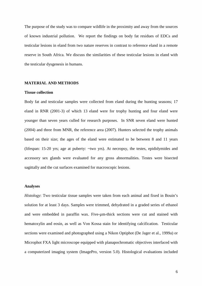
epithelium was normal (Fig. 1).
Fig. 1. (A) Eland testis from the control area showing normal seminiferous epithelium. The boxed area is shown
at a higher magnification in panel B. (B) Mitotic figures (secondary spermatocytes) are apparent in circled area.
Hematoxylin and eosin staining. Scale bars: A 200 μm, B 50 μm.

9
Fig. 2. Eland testis showing sperm stasis and calcifications. (A) Testis cut midsagitally through mediastinum
(arrow heads) showing white, gritty, calcified loci (arrows). (B) Histological section showing cross sections of
seminiferous tubules with sperm stasis (upper half) and calcification (lower half). Arrow heads point to
transition between terminal segment of seminiferous tubules and rete testis. Note: mineralized luminal contents
in these structures. Hematoxylin and eosin staining. Scale bar: 250 μm
Fig. 3. Photomicrographs of eland testis showing calcification of static sperm in the lumen of seminiferous
tubule. (A) Sperm stasis. (B) Sperm undergoing mineralization. (C) Completely calcified luminal contents.
Note: degeneration of seminiferous epithelium (asterisk) adjacent to a calcified tubule. Hematoxylin and eosin
staining. Scale bars: A and B=50 μm, C=100 μm.

10
In contrast, the gross appearance of testes collected from the RNR and SNR was different
from testes collected from MNR. Conspicuously, hard, nodular areas were palpable beneath
the tunica albuginea of some testes. Macroscopically, focal white gritty areas were observed
in testes of all 24 eland from RNR and SNR which, on sectioning, appeared as grey-white,
calcified areas varying in size, dispersed throughout testes without a specific distribution
pattern (Fig. 2A). Microscopic examination of testicular sections containing these white gritty
areas revealed loci of seminiferous tubules with sperm stasis and dystrophic calcifications
(Fig. 2B) but the degree and extent of mineralization between samples varied. On several
sections in which mediastinum testis was present, lesions of the rete testis characterized by
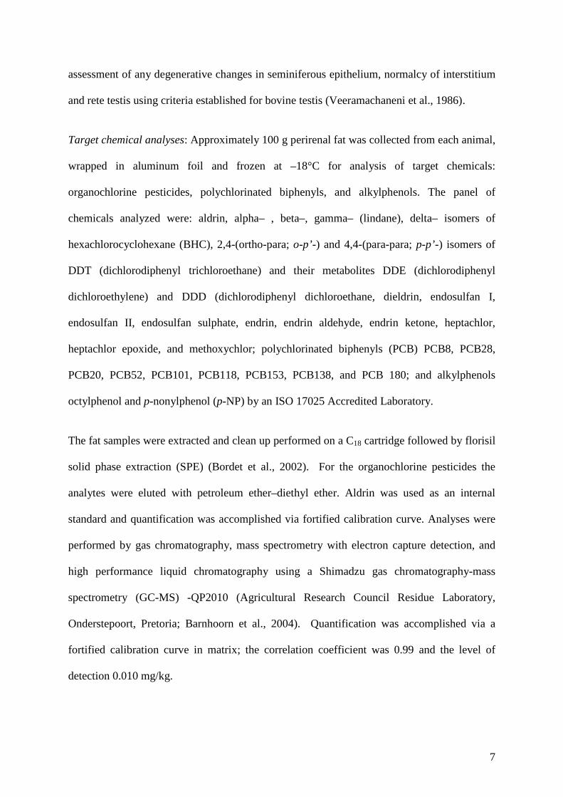
epithelial hypertrophy and adenomatous proliferation was observed (Fig. 3). The proliferative
lesions in the rete testis appeared to have obliterated sperm transit to excurrent ducts resulting
in sperm stasis in seminiferous tubules (Fig. 4A) leading to disintegration of static sperm and
mineralization of luminal contents (Fig. 4B) ultimately replacing the entire seminiferous
epithelium by calcified masses (microliths) (Fig. 4C). The proliferative lesions of the rete
testis were also associated with seminiferous epithelial degeneration in segments of the
tubules proximal to the affected rete (Fig. 4). The degenerative lesions included vacuolization
of Sertoli cells, and death and desquamation of differentiating germ cells (Fig. 5A). Although
full complement of spermatogenesis was observed in focal areas, spermatogenesis was
generally impaired consequent to progression of degenerative changes in seminiferous
epithelium. The progression of degenerative process was manifested by a spectrum of lesions
including complete sloughing of seminiferous epithelium, calcification of exfoliated detritus
(microlithiasis), granulomatous reaction (Fig. 5B) and, ultimately, fibrosis (Fig. 5C) of
lobules of seminiferous tubules.
Eleven of 17 RNR eland fat samples contained p-NP ranging from 35.0 to 290 µg/kg fat
11
(mean ± SD: 180 ± 61.8 µg/kg) and no organochlorine pesticides or PCBs. In six eland p-NP
was present, but could not be quantified. Of the seven SNR eland, five contained octylphenol
(50 ± 31 µg/kg fat) and three p-NP (140 ±78 µg/kg fat); three had o-p’-DDT (115 ± 30 µg/kg
fat), five o-p’-DDE (28 ± 10 µg/kg fat), three had p-p’-DDT (130 ± 50 µg/kg fat), and all
seven had o-p’-DDD (80 ± 30 µg/kg fat). No p-p’-DDE was detected at SNR. Of the three
reference MNR eland fat samples, one contained 60 µg p-p’-DDE/kg, and another one o-p’-
DDT 71 µg/kg. None of them had any other organochlorines or alkylphenols.
Fig. 4. Photomicrographs of proliferative neoplastic lesions in the rete testis. (A) Hypertrophy of epithelium and
adenomatous lesions are seen in rete tubules around asterisks. Note apparently normal seminiferous tubules in
the lower left and occluded seminiferous tubules with degenerate detritus in the lower right (arrow). The tubule
marked by rectangle is magnified in panel B. (B) Adenomatous proliferation of rete epithelium. (C)
Adenomatous changes in rete epithelium and adenotic changes in rete stroma. Hematoxylin and eosin staining.
Scale bars: A=250 μm, B and C=50 μm.

12
Fig. 5. Photomicrographs of progression of degenerative changes in seminiferous tubules consequent to rete
adenoma and occlusion. A. Adenomatous lesions in rete tubules (arrow heads) and vacuolisation of Sertoli cells.
(B) Note granulomatous reaction around several tubules (asterisks) while the remaining seminiferous tubules are
apparently normal. The clusters of degenerate seminiferous tubules are likely the ones connected to the rete
tubules afflicted by adenoma, adenosis and occlusion. (C) As the degenerative process advances, the entire
lobules of tubules are replaced by fibrous connective tissue. Note: sperm stasis (asterisk) and proliferative
lesions (arrow head) in rete testis. Hematoxylin and eosin staining. Scale bars: A and C= 100 μm, B=250 μm.
DISCUSSION
The presence of adenomatous lesions of the rete testis in the eland from RNR and SNR was
similar to diethylstilbestrol-induced rete testis adenocarcinoma in laboratory animals
(Newbold et al., 1985; Newbold, 2000) and was, therefore, suggestive of possible chronic
estrogenic exposure. The alkylphenols as well as o-p’- and p-p’-DDT have estrogenic activity
(Sonnenschein and Soto, 1998; Sonneveld et al., 2005). The possibility of simultaneous
exposure to estrogenic agents such as phytoestrogens and estrogenic mycotoxins cannot be
ruled out, and therefore, considering the spectrum of pollutants present in the body fat, the

13
testicular lesions observed in eland could have been caused by chronic exposure to these
pollutant chemicals. Experimentally, it has been demonstrated that a variety of xenobiotics
released from fat during fasting produce estrogenic effects (Bigsby et al., 1997). The
magnitude of concentration of estrogenic pollutants in body fat indicates that EDCs
bioaccumulate in terrestrial mammals as in aquatic life (Barnhoorn et al., 2004). The levels of
p-NP in eland were even higher than those found bioconcentrated in fat of catfish inhabiting
the two dams in RNR. Intersex conditions where testicular oocytes occurred were found in
some of these catfish (Barnhoorn et al., 2004), while water and sediment samples had
estrogenic activity on the yeast screen test (Aneck-Hahn et al., 2008). The eland are
dependent on these water sources in RNR. In this specific instance, we suspect that the source
of p-NP to be the industrial effluent draining into the stream leading to the reservoir/dam. p-
Nonylphenol is lipophilic and, therefore, bio-accumulates in fat of humans and animals and
as a result has a more persistent effect than natural estrogens (Tapiero et al., 2002).
The findings of Sertoli cell vacuolization and sloughing of the epithelium observed in eland
were similar to those observed in rats following experimental exposure to p-NP (De Jager et
al., 1999b). Despite an extensive literature search, only a few papers on testicular
calcifications in ruminants could be found. For example, testicular calcifications were found
in association with testicular dysgenesis in Sitka Black-tailed deer in Alaska
(Veeramachaneni et al., 2006), testicular degeneration in beef bulls having small testes
(Veeramachaneni et al., 1986), degenerative changes in the seminiferous tubules of goats
occurring spontaneously (Ahmad et al., 1993) or with experimentally induced Trypanosoma
evansi orchitis (Ngeranwa et al., 1991). In Merino sheep the prevalence of testicular lesions,
including calcifications, increased with age (Watt, 1978). Testicular calcifications were found
after surgical biopsy and also in the contralateral testis of adult rams (Vrzgulova, 1995). In
14
Ethiopian Menz rams, calcifications were coincident with orchitis (Hibret et al., 2001).
Although subclinical infections such as trypanosomiasis or brucellosis cannot be excluded in
eland in the current study, there was no record of incidence of these conditions according to
the RNR Reserve Manager or veterinarian. These animals appeared to be in good health and
no swollen testes or signs of chronic wasting.
The fact that all eland examined from two disparate nature reserves and possibly from
genetically diverse wild populations exhibited similar lesions concomitant with considerable
body burdens of estrogenic EDCs, suggests a causal link. Edwards et al. (2006) reviewed the
evidence of reproductive dysgenesis in wildlife and pointed out that microlithiasis forms part
of the TDS in comparable vertebrate groups. The review supported the hypothesis that TDS
is the result of feminization or demascilinization of the male reproductive system. The
induction of a TDS-like syndrome in the male offspring (Mylchreest et al., 2000; Parks et al.,
2000; Fisher et al., 2000) of rats exposed in utero to the ubiquitous environmental chemical
di(n-butyl) phthalate (DBP) unequivocally provided support for the link between chemical
exposure and the TDS hypothesis in human males. Advances in the general scientific
understanding from research have led to refinement of the TDS hypothesis, highlighting the
central role that deficient androgen production/action during fetal testis development, may
play in the origin of downstream disorders (Sharpe and Skakkebaek, 2008).
Interestingly, the histological appearance of the intraluminal testicular calcifications in eland
was similar to testicular calcifications reported in humans. It is noteworthy that generalized
hypospermatogenesis with patchy normal areas is commonly found in subfertile men
(Skakkebaek et al., 1973; Gottschalk-Sabag et al., 1995) and intratubular microcalcifications
are also not uncommon in these patients. These features, described as manifestations of

15
testicular dysgenesis, have been ascribed to possible exposures to environmental endocrine
disruptors (Skakkebaek et al., 2001).
While use of DDT has been banned or restricted in most Western countries for 20 or more
years, in regions where malaria is still endemic such as South Africa, DDT is sprayed onto
the interior surfaces of homes for mosquito vector control. Because of its long half-life
(Agency for Toxic Substances and Disease Registry (ATSDR), 2002), lipophilicity and
ability to bioconcentrate and bio-accumulate in food chains, there is growing concern that
DDT and the breakdown product DDE is associated with adverse human health outcomes
(Eskenazi et al, 2009). Exposure in utero to p-p’-DDT or its metabolite p-p’-DDE or
octylphenol, induced atypical germ cells resembling carcinoma in situ (CIS) in rabbits
(Veeramachaneni, 2000; Veeramachaneni, 2006). It is likely that DDT and its metabolites are
exported through agricultural produce and the atmosphere (Simonich and Hites, 1995) to
regions where its use is now restricted. Therefore, it is not surprising to find residues of DDT
and its metabolites in eland on SNR, although this Reserve is secluded from human activity.
Such was the case in Sitka black-tailed deer in Alaska (unpublished data). Thus, it is not
unrealistic that humans and animals are exposed inadvertently to pesticides even though they
have been banned or restricted. In fact, p-p’-DDE is the major DDT-derived residue in food
(Spindler, 1983) and human body fat (Barquet, 1981).
World production of alkylphenol polyethoxylates (APE) is estimated to be 300 000 tons per
year (Houde et al., 2002) for use as a surfactant in plasticizers, detergents, paints, herbicides,
cosmetics, and as anti-oxidants and lubricants in a variety of industrial, agricultural, and
household applications (U.K. Department of the Environment; Kent, 1993). No data on the
production and use of APEs in South Africa are publicly available. APEs eventually end up

16
in surface waters and aquatic sediments, undergo microbial breakdown resulting in
alkylphenols including octylphenol and p-NP (Nimrod et al., 1996), which have even higher
lipophilic and persistent properties than the parent APEs (Talmage, 1994). Khan et al. (2003)
demonstrated that alkylphenols may affect hormone signaling in a variety of tissues,
including testes, via an estrogen receptor-independent mechanism by altering calcium
homeostasis thereby impairing cellular function. Sertoli cells and the differentiating germ
cells that they sustain could be targets for these detrimental actions.
p-Nonylphenol is known to have adverse effects on the testis and epididymis of rodents (Lee
et al., 1999; De Jager et al., 1999a,b). The negative impact is further enhanced when animals
are exposed to an environmentally relevant mixture of chemicals containing deltamethrin,
DDT, phytoestrogens and p-NP (Kilian et al., 2005).
Human testicular cancer arises from CIS cells, which are suspected to originate from
primordial germ cells that escaped normal differentiation in utero (Skakkebæk et al., 1987;
Rajpert-De Meyts et al., 1998). The first cases in animals of atypical germ cells resembling
CIS cells of human testis were reported in a subfertile, unilaterally cryptorchid stallion
(Veeramachaneni and Sawyer, 1998) and an infertile rabbit (Veeramachaneni and
VandeWoude, 1999). Although a few atypical germ cells were encountered in eland testes,
detailed morphological evaluation ascertaining CIS was not possible because of limitations of
tissue fixation and processing. Interestingly, a variety of testicular tumours including rete
adenocarcinoma and seminoma along with microlithiasis and CIS were found in Sitka black-
tailed deer suspected to have been developmentally exposed to an environmental estrogenic
agent(s) (Veeramachaneni et al., 2006).

17
These novel findings in eland may therefore be the first evidence from South Africa that
mammalian wildlife are impacted by environmental pollution of EDCs in South Africa and
may serve as ecological harbingers.
ACKNOWLEDGEMENTS
This study was partially supported by grants from the Water Research Commission, National
Research Foundation and Post-doctoral Fellowship Programme, University of Pretoria. We
thank W. Louw, C. Moeller, R. Marais, J. Palmer, JC van Dyk, H Bouwman and Staff, and
the Wildlife Breeding Resource Centre for technical assistance. We gratefully acknowledge
Prof Niels Skakkebaek and Dr Ewa Rajpert-De Meyts from Rigshospitalet, Copenhagen,
Denmark for examining histological sections. The authors declare they have no competing
financial interests.
REFERENCES
Ahmad, N., Noakes, D.E., Middleton D.J., 1993. Use of ultrasound to diagnose testicular
degeneration in a goat. Vet. Rec. 132 (17), 436-439.
Aneck-Hahn, N.H., Bornman, M.S., de Jager., C., 2008. Preliminary assessment of
oestrogenic activity in water sources in Rietvlei Nature Reserve, Gauteng, South Africa.
Afric. J. Aquat. Sci. 33(3), 249–254.
Anway, D.A.., Cupp A.S., Uzumcu, M., Skinner, M.K., 2005. Epigenetic transgenerational
actions of endocrine disruptors and male fertility. Science 308, 1466-1469.
Agency for Toxic Substances and Disease Registry (ATSDR). Toxicological profile for
DDT, DDE, DDD., 2002. Atlanta, GA: U.S. Department of Health and Human Services,
Public Health Service. http://www.atsdr.cdc.gov/toxprofiles/tp35.html. Accessed 9 Oct
2008.
18
Barnhoorn, I.E.J., Bornman, M.S., Pieterse, G.M., Van Vuren J.H.J., 2004. Histological
evidence of intersex in feral sharptooth catfish (Clarias gariepinus) from an estrogen
polluted water sources in Gauteng, South Africa. Environ. Toxicol. 19, 603-608.
Barquet, A., Morgade, C., Pfannenberger, C.D., 1981. Determination of organochlorine
pesticides and metabolites in drinking water, human blood serum, and adipose tissue. J.
Toxicol. Environ. Health 7, 469-479.
Bigsby, R.M., Caperell-Grant, A., Madkhukar B.V., 1977. Xenobiotics released from fat
during fasting produce estrogenic effects in ovariectomized mice. Cancer Res. 57, 865-
869.
Bornman, M.S., van Vuuren, J.H., Bouwman, H., de Jager, C., Genthe, B., Barnhoorn, E.J.
2007. Endocrine disruptive activity and the potential health risk in an urban nature reserve.
Water Research Commission Report No 1505/1/07 290pp.
www.wrc.org.za/publications_reports2.html.
Bowerman, W.W., Best, D.A., Grubb, T.G., Zimmerman, G.M., Giesy, J.P., 1998. Trends of
contaminants and effects in bald eagles of the Great Lakes basin. Environ. Monit. Assess.
53, 197-212.
Damstra, T., Barlow, S., Bergman, A., Kavlock, R., van der Kraak, G. (Eds.), 2002. Exposure
of selected potential EDCs in humans and wildlife EDCs in humans and wildlife. In:
Global assessment of the state-of-the-science of endocrine disruptors -prepared by an
expert group on behalf of the World Health Organization, the International Labour
Organisation, and the United Nations Environment Programme. World Health
Organization, pp.34-50.
De Jager, C., Bornman, M.S., Van der Horst G., 1999a. The effect of p-nonylphenol, an
environmental toxicant with estrogenic properties, on fertility potential in adult male rats.
Andrologia, 31, 99-106.
19
De Jager, C., Bornman, M.S., Ooosthuizen J.M.C., 1999b. The effect of p-nonylphenol on
fertility potential of male rats after gestational, lactational and direct exposure. Andrologia
31(2), 107-113.
Derogee, M., Bevers, R.F.M., Prins, H.J., Jonges, T.G.N., Elbers, F.H., Boon, T.A., 2001.
Testicular microlithiasis, a premalignant condition: prevalence, histopathology findings,
and relation to testicular tumor. Urol. 57, 1133–1137.
Edwards, T.M., Moore, B.C., Guillette, L.J., 2006. Reproductive dysgenesis in wildlife: a
comparative view. Int. J. Androl. 29(1), 109-21.
Eskenazi, B., Chevrier, J., Goldman Rosas, L., Anderson, H.A., Bornman, M.S., Bouwman,
H., Chen, A., Cohn, B.A., de Jager, C., Henshel, D.S., Leipzig, F., Leipzig, J.S., Lorenz,
E.C., Snedeker, S.M., Stapleton, D., 2009. The Pine River Statement: Human Health
Consequences of DDT Use. doi: 10.1289/ehp.11748 (available at http://dx.doi.org/).
Online 4 May 2009
Facemire, C.F., Gross, T.S., Guillette, L.J.(jr), 1995. Reproductive impairment in the Florida
Panther: Nature or nurture? Environ. Health Perpect. 103(4), 79-86.
Fisher, J.S., Macpherson, S., Marchetti, N., Sharpe, R.M., 2003. Human ‘‘testicular
dysgenesis syndrome’’: a possible model using in-utero exposure of the rat to dibutyl
phthalate. Hum. Reprod. 18, 1383–94.
Ganem, J.P., Workman, K.R., Shaban, S.F., 1999. Testicular microlithiasis is associated with
testicular pathology. Urol. 53, 209-213.
Giwercman, A., Carlsen, E., Keiding, N., Skakkebaek, N.E., 1993. Evidence for increasing
evidence of abnormalities of the human testis: a review. Environ. Health Perspect.
101(Suppl 2), 65-71.
Gottschalk-Sabag, S., Weiss, D.B., Folb-Zacharow, N., Zuckermann, Z., 1995. Is one
testicular specimen sufficient for quantitative evaluation of spermatogenesis? Fertil. Steril.

20
64, 399-401.
Gray, L.E.(Jr)., Ostby, J., Furr, J., Wolf, C.J., Lambright, C., Parks, L., Veeramachaneni,
D.N., Wilson, V., Price, M., Hotchkiss, A., Orlando, E., Guilette, L., 2001. Effects of
environmental antiandrogens on reproductive development in experimental animals.
Human Reprod. Update. 7, 248-264.
Guillette, L.J.(Jr)., Gross, T.S., Masson, G.R., Matter, J.M., Franklin Percival, H.,
Woodward, A.R., 1994. Developmental abnormalities of the gonad and abnormal sex
hormone concentrations in juvenile alligators from contaminated and control lakes in
Florida. Environ. Health Perspect. 102, 680-688.
Hibret, A., Toe, F., Mukasa-Mugerwa, E., Kassa, T., Markos, T., 2001. Genital disorders,
linear and testicular characteristics in Menz rams. Trop. Anim. Health Prod. 33, 219-227.
Higuchi, T.T., Palmer, J.S., Gray, L.E.(Jr)., Veeramachaneni, D.N.R.., 2003. Effects of
dibutyl phthalate in male rabbits following in utero, adolescent, or postpubertal exposure.
Toxicol. Sci. 72, 301-313.
Holm, M., Lenz, S., De Meyts, E.R., Skakkebaek, N.E., 2001. Microcalcifications and
carcinoma in situ of the testis. Brit. J. Urol. Int. 87, 144-149.
Houde, F., DeBlois, C., Berryman, D., 2002. Liquid chromatographic–tandem mass
spectrometric determination of nonylphenol polyethoxylates and nonylphenol carboxylic
acids in surface water. J. Chrom. A. 961, 245-256.
Khan, S.Z., Kirk, C.J., Michelangeli, F., 2003. Alkylphenol endocrine disrupters inhibit IP3
sensitive Ca2+
channels. Biochem. Biophys. Res. Com. 310, 261–266.
Kilian, E., Delport, R., Bornman, M.S., de Jager, C., 2005. Simultaneous exposure to low
concentrations of dichlorodiphenyltrichloroethane, deltamethrin, nonylphenol and
phytoestrogens has negative effects on the reproductive parameters in male Spraque-
Dawley rats. Andrologia. 39, 128–135.
21
Luthra, M., Hutson JM., 1989. Late-gestation exogenous oestrogen inhibits testicular descent
in fetal mice despite Müllerian duct regression. Pediat. Surg. Int. 4, 260-264.
McLeese, D.W., Zitko, V., Sergent, D.B., Burridge, L., Metcalfe, D., 1981. Lethality and
accumulation of alkylphenols in aquatic fauna. Chemosphere 10, 723-730.
Main, K.M., Skakkebaek, N.E., Toppari, J., 2009. Cryptorchidism as part of the testicular
dysgenesis syndrome: the environmental connection. Endocr. Dev. 4, 167-73. Epub 2009
Feb 27.
Mylchreest, E., Wallace, D.G., Cattley, R.C., Foster, P.M., 2000. Dose-dependent alterations
in androgen-regulated male reproductive development in rats exposed to Di(n-butyl)
phthalate during late gestation. Toxicol Sci. 55, 143–51.
Naso, B., Zaccaroni, A., Perrone, D., Ferrante, M.C., Severino, L., Stracciari, G.L., Luciasino
C., 2004. Organochlorine pesticides and polychlorinated biphenyls in European roe deer
Capreolus capreolus resident in a protected area in Northern Italy. Sci. Tot. Environ. 328,
83–93.
Negri, L., Benaglia, R., Fiamengo, B., Pizzocaro, A., Albani, E., Levi Setti PE., 2008. Cancer
risk in male factor-infertility. Placenta. 29, S178-S187.
Newbold, R.R., Bullock, B.C., McLachlan, J.A., 1985. Lesions of the rete testis in mice
exposed prenatally to diethylstilbestrol. Cancer Res. 45, 5145-5150.
Newbold, R.R., Hanson, R.B., Jefferson, W.N., Bullock, B.C., Haseman, J., McLachlan, J.A.,
2000. Proliferative lesions and reproductive tract tumors in male descendants of mice
exposed developmentally to diethylstilbestrol. Carcinog. 21, 1355.
Parks, L.G., Ostby, J.S., Lambright, C.R., Abbott, B.D., Klinefelter, G.R., Barlow, N.J.,
Gray, L.E., 2000. The plasticizer diethylhexyl phthalate induces malformations by
decreasing fetal testosterone synthesis during sexual differentiation in the male rat.
Toxicol. Sci. 58, 339–349.

22
Larsson, D.G.J., Förlin, L., 2002. Male-biased sex ratios of fish embryos near a pulp mill:
Temporary recovery after a short-term shutdown. Environ. Health Perspect. 110(8), 739-
742.
Morcillo, Y., Porte, C., 1999. Evidence of endocrine disruption in the imposex-affected
gastropod Bolinus brandaris. Environ. Res. 81(A), 349-354.
Ngeranwa, J.J.N., Mutiga, E.R., Agumbah, G.J.O., Gathumbi, P.K., Munuya W.K., 1991.
The effects of experimental Trypanosoma (Tryponozoon) (Brucei) Evansi infection on the
fertility of male goats. Vet. Res. Com. 15, 301-308.
Nimrod, A.C., Benson, W.H., 1996. Environmental estrogenic effects of alkylphenol
ethoxylates. Crit. Rev. Toxicol. 26, 335-64.
Renshaw, A.A. 1998. Testicular calcifications: Incidence, histology and proposed
pathological criteria for testicular microlithiasis. J. Urol. 160, 1625-1628.
Rajpert-De-Meyts, E., Jorgenson, N., BRondum-Nielsen, K., Muller, J., Skakkebaek, N.E.,
1998. Developmental arrest of germ cells in the pathogenesis of germ cell neoplasia. Acta
Pathol. Microbiol. Immunol. Scand. 106: 198.
Sharpe, R.M., Skakkebaek, N.E., 2008. Testicular dysgenesis syndrome: mechanistic
insights and potential new downstream effects. Fertil. Steril. 89, e33–38.
Simonich, S.L., Hites, R.A. 1995. Global distribution of persistent organochlorine
compounds. Science 269, 1851-1854.
Skakkebaek, N.E., Hammen, R., Philip, H., Rebbe, H., 1973. Quantification of human
seminferous epithelium. III. Histological studies in 44 infertile men and controls with
normal chromosome complements. Acta Pathol. Microbiol. Immunol. Scand. 81:97-111.
Skakkebaek, N.E., 2004. Testicular dysgenesis syndrome: new epidemiological evidence.
Int. J. Androl. 27, 189–191.
Skakkebaek, N.E., Rajpert-De-Meyts, E., Main, K.M., 2001. Testicular dysgenesis syndrome:

23
an increasingly common developmental disorder with environmental aspects. Opin. Hum.
Reprod. 16, 972-978.
Skakkebaek, N.E., Holm, E., Hoei-Hansen, C., Jorgenson, N., Rajpert-De-Meyts, E., 2003.
Asscociation between testicular dysgenesis syndrome (TDS) and testicular neoplasia:
Evidence form 20 adult patients with signs of maldevelopment of the testis. Acta Pathol.
Microbiol. Immunol. Scand. 111, 1-11.
Sonnenschein, C., Soto, A.M., 1998. An updated review of environmental estrogen and
androgen mimics and antagonists. J. Ster. Biochem. Molec. Biol. 65, 143-150.
Sonneveld, E., Jansen, H.J., Riteco, J.A., Brouwer, A., Van Den Burg, B. 2005. Development
of androgen-and estrogen-responsive bioassays, members of a panel of human cell line-
based highly selective steroid-responsive bioassays. Toxicol. Sci. 83, 136-148.
Spindler, M., 1983. DDT: Health aspects in relation to man and risk / benefit assessment
based thereupon. Residue Rev. 90, 1-34.
Talmage, S.S., 1994. Environmental and human safety of major surfactants. Alcohol
ethoxylates and alkylphenol ethoxylates. Lewis Publishers, London, 364pp.
Toppari, J., Larsen, J.C., Christiansen, P., Giwercman, A., Grandjean, P., Guillette Jr, L.J.,
Jegou, B., Jensen, T.K., Jouannet, P., Keiding, N., Leffers, H., McLachlan, J.A., Meyer,
O., Muller, J., Rajpert-De Meyts, E., Scheike, T., Sharpe, R., Sumpter, J., Skakkebaek,
N.E., 1996. Male reproductive health and environmental xenoestrogens. Environ. Health
Perspect. 104 (Suppl. 4), 741–803.
Tsuda, T., Takino, A., Kojima, M., Harada, H., Muraki, K., 1999. Gas chromatographic–
mass spectrometric determination of 4-nonylphenols and 4-tert-octylphenol in biological
samples. J Chromatogr B. 723 (1-2), 273-279.
U.K. Department of the Environment., 1993. Uses, fate and entry to the environment of nonyl
ethoxylates. Final Report, U.K. Kent: Department of the Environment,78pp.

24
Veeramachaneni, D.N.R., Ott, R.S., Heath, E.H., McEntee, K., Bolt, D.J., Hixon J.E., 1986.
Pathophysiology of small testes in beef bulls: relationship between scrotal circumference,
histopathologic features of testes and epididymides, seminal characteristics, and endocrine
profiles. Am. J. Vet. Res. 47, 1988-1999.
Veeramachaneni, D.N.R., Sawyer, H.R.., 1998. Carcinoma in situ and seminoma in equine
testis. Acta Pathol. Microbiol. Immunol. Scand. 106, 183.
Veeramachaneni, D.N.R., Vandewoude, S., 1999. Interstitial cell tumour and germ cell
tumour with carcinoma in situ in rabbit testes. Int. J. Androl. 22, 97-101.
Veeramachaneni, D.N.R, 2000. Deteriorating trends in male reproduction: idiopathic or
environmental? Anim. Reprod. Sci. 60-61 (2), 121-130.
Veeramachaneni, D.N.R, 2006. Germ cell atypia in undescended testes hinges on the
aetiology of cryptorchidism but not the abdominal location per se. Int. J. Androl. 29, 235-
240.
Veeramachaneni, D.N.R., Amann, R.P., Jacobson, J.P., 2006. Testis and antler dysgenesis in
Sitka Black-tailed deer on Kodiak Island, Alaska: Sequela of environmental endocrine
disruption? Environ. Health Perspect. 114(Suppl1), 51-59.
Verreault, J., Muir, D.C.G., Norstrom, R.J., Stirling, I, Fisk, A.T., Gabrielsen, G.W., et al.,
2005. Chlorinated hydrocarbon contaminants and metabolites in polar bears (Ursus
maritimus) from Alaska, Canada, East Greenland, and Svalbard: 1996−2002. Sci. Tot.
Environ. 351-352, 369-390.
Viguer-Martinez, M.C., Hochereau De Reviers, M.T., Barenton, B., Perreau, C., 1983. Effect
of a non-steroidal antiandrogen, flutamide on the hypothalamopituitary axis, genital tract
and testis in growing rats: endocrinological and histological data. Acta Endocrinol. 102,
299-306.
Vos, J.G., Dybing, E., Greim, H.A., Ladefoged, O., Lambré, C., Tarazona, J.V., Brandt, I.,
25
Vethaak, A.D, 2000. Health effects of endocrine-disrupting chemicals on wildlife, with
special reference to the European situation. Crit. Rev. Toxicol. 30(1), 71-133.
Vrzgulova, M., Vrgulova, L., Bires, J., 1995. Morphologic characteristics of the testes in
breeding rams after experimental loading with industrial emissions containing copper. Vet.
Med. Czech. 40, 105-110.
Walker, A.H., Bernstein, L., Warren, D.W., Warner, N.E., Zheng, X., Henderson, B.E., 1990.
The effect of in utero ethinyl oestradiol exposure on the risk of cryptochid testis and
testicular teratomas in mice. Brit. J. Cancer. 62, 599-602.
Watt, D.A. 1978. Testicular pathology of merino rams. Aust. Vet. J. 54, 473-478.
Yasuda, Y., Kihara, T., Tanimura, T. 1985. Effect of ethinyl estradiol on the differentiation of
mouse fetal testis. Teratology. 32,113-118.
Copyright © 2022 FDOKUMEN