Tabanids: Neglected subjects of research, but important vectors of disease agents!
20
Tabanids: Neglected subjects of research, but important vectors of disease agents! Frédéric Baldacchino a,b,⇑ , Marc Desquesnes c,d , Steve Mihok e , Lane D. Foil f , Gérard Duvallet a , Sathaporn Jittapalapong d a UMR5175 CEFE, University Paul-Valéry, route de Mende, 34199 Montpellier Cedex 5, France b Department of Biodiversity and Molecular Ecology, Research and Innovation Centre, Fondazione Edmund Mach (FEM), 38010 San Michele all’Adige, Italy c Cirad-Bios, UMR-InterTryp, Montpellier F-34000, France d Department of Parasitology, Faculty of Veterinary Medicine, Kasetsart University, Chatuchak, Bangkok 10900, Thailand e 388 Church Street, Russell, Ontario K4R 1A8, Canada f Department of Entomology, Louisiana State University Agricultural Center, Bâton Rouge, LA 70803, USA article info Article history: Received 17 February 2014 Received in revised form 21 March 2014 Accepted 28 March 2014 Available online 13 April 2014 Keywords: Tabanid Pathogens Livestock Mechanical transmission Trapping Vector control abstract Tabanids are nuisance pests for people and livestock because of their painful and irritating bite, persistent biting behavior, and blood ingestion. About 4400 tabanid species have been described; they are seasonally present in all kinds of landscapes, latitudes, and altitudes. High populations have a significant economic impact on outdoor activities, tourism, and livestock production. Tabanids are also vectors of animal disease agents, including viruses, bacteria and parasites. However, tabanids have received little attention in comparison with other hematophagous Diptera. Here, we highlight the many direct and indirect impacts of tabanids and provide a brief summary of tabanid morphology, biology, and life cycle. Impacts include pathogen transmission, parasite transportation (Dermatobia hominis), biological transmission (Loa loa), and mechanical transmission of viruses, such as equine infectious anemia virus, protozoa, such as Trypanosoma evansi and Besnotia besnoiti, and bacteria, such as Bacillus anthracis and Anaplasma marginale. We discuss parameters of mechanical transmission and its mathematical modeling. Control methods for tabanid populations are also summarized; these include trapping, the use of insecticides, repellents, and livestock protection. Lastly recommendations are provided for the direction of future research. Ó 2014 Elsevier B.V. All rights reserved. 1. Introduction Tabanidae are a cosmopolitan Dipteran family comprising about 4400 species belonging to 144 genera (Roskov et al., 2013). The family includes four subfamilies divided into tribes: Chrysopsinae (Bouvieromyiini, Chrysopsini and Rhinomyzini), Pangoniinae (Mycteromyiini, Pangoniini, Philolichini and Scionini), Scepsidinae and Tabaninae (Diachlorini, Haematopini and Tabanini) (Coscarón and Philip, 1979; Lessard et al., 2013; Mackerras, 1954, 1955). Most of the economically important tabanids are in the Chrysopsinae, particularly the genus Chrysops, and the Tabaninae (Mullens, 2002). They are commonly referred to as horse flies, deer flies, or clegs, but also gad flies, breeze flies or March flies (used for Australian species). The term horse fly is generally applied to the Tabaninae species (except the tribe Haematopini). The term deer fly is used for Chrysops spp. and the term cleg for Haematopota spp. Horse flies are relatively large (10–30 mm) whereas deer flies and clegs are smaller (6–13 mm) (Chvála et al., 1972). Tabanids are nuisance pests for people and livestock because of their painful bite and persistent biting behavior (Foil and Hogsette, 1994). Dense tabanid populations can have an economic impact on outdoor activities, tourism and agriculture. Pastured cattle may suffer severely from heavy attacks of tabanids causing losses in weight gain or milk production. However, present data on the true economic importance of these flies are scarce (Goodwin and Drees, 1996). Tabanids can also transmit human and animal disease agents, biologically or mechanically (Foil, 1989; Krinsky, 1976). Biological transmission occurs when the disease agent replicates or develops http://dx.doi.org/10.1016/j.meegid.2014.03.029 1567-1348/Ó 2014 Elsevier B.V. All rights reserved. ⇑ Corresponding author at: Department of Biodiversity and Molecular Ecology, Fondazione Edmund Mach, Centro Ricerca e Innovazione, Via E. Mach, 1, 38010 San Michele all’Adige, TN, Italy. Tel.: +39 0461615167. E-mail addresses: [email protected] (F. Baldacchino), marc.desquesnes@ cirad.fr (M. Desquesnes), [email protected] (S. Mihok), [email protected] (L.D. Foil), [email protected] (G. Duvallet), [email protected] (S. Jittapalapong). Infection, Genetics and Evolution 28 (2014) 596–615 Contents lists available at ScienceDirect Infection, Genetics and Evolution journal homepage: www.elsevier.com/locate/meegid
Transcript of Tabanids: Neglected subjects of research, but important vectors of disease agents!

Infection, Genetics and Evolution 28 (2014) 596–615
Contents lists available at ScienceDirect
Infection, Genetics and Evolution
journal homepage: www.elsevier .com/locate /meegid
Tabanids: Neglected subjects of research, but important vectorsof disease agents!
http://dx.doi.org/10.1016/j.meegid.2014.03.0291567-1348/� 2014 Elsevier B.V. All rights reserved.
⇑ Corresponding author at: Department of Biodiversity and Molecular Ecology,Fondazione Edmund Mach, Centro Ricerca e Innovazione, Via E. Mach, 1, 38010 SanMichele all’Adige, TN, Italy. Tel.: +39 0461615167.
E-mail addresses: [email protected] (F. Baldacchino), [email protected] (M. Desquesnes), [email protected] (S. Mihok), [email protected](L.D. Foil), [email protected] (G. Duvallet), [email protected](S. Jittapalapong).
Frédéric Baldacchino a,b,⇑, Marc Desquesnes c,d, Steve Mihok e, Lane D. Foil f, Gérard Duvallet a,Sathaporn Jittapalapong d
a UMR5175 CEFE, University Paul-Valéry, route de Mende, 34199 Montpellier Cedex 5, Franceb Department of Biodiversity and Molecular Ecology, Research and Innovation Centre, Fondazione Edmund Mach (FEM), 38010 San Michele all’Adige, Italyc Cirad-Bios, UMR-InterTryp, Montpellier F-34000, Franced Department of Parasitology, Faculty of Veterinary Medicine, Kasetsart University, Chatuchak, Bangkok 10900, Thailande 388 Church Street, Russell, Ontario K4R 1A8, Canadaf Department of Entomology, Louisiana State University Agricultural Center, Bâton Rouge, LA 70803, USA
a r t i c l e i n f o a b s t r a c t
Article history:Received 17 February 2014Received in revised form 21 March 2014Accepted 28 March 2014Available online 13 April 2014
Keywords:TabanidPathogensLivestockMechanical transmissionTrappingVector control
Tabanids are nuisance pests for people and livestock because of their painful and irritating bite, persistentbiting behavior, and blood ingestion. About 4400 tabanid species have been described; they areseasonally present in all kinds of landscapes, latitudes, and altitudes. High populations have a significanteconomic impact on outdoor activities, tourism, and livestock production. Tabanids are also vectors ofanimal disease agents, including viruses, bacteria and parasites. However, tabanids have received littleattention in comparison with other hematophagous Diptera. Here, we highlight the many direct andindirect impacts of tabanids and provide a brief summary of tabanid morphology, biology, and life cycle.Impacts include pathogen transmission, parasite transportation (Dermatobia hominis), biologicaltransmission (Loa loa), and mechanical transmission of viruses, such as equine infectious anemia virus,protozoa, such as Trypanosoma evansi and Besnotia besnoiti, and bacteria, such as Bacillus anthracis andAnaplasma marginale. We discuss parameters of mechanical transmission and its mathematical modeling.Control methods for tabanid populations are also summarized; these include trapping, the use ofinsecticides, repellents, and livestock protection. Lastly recommendations are provided for the directionof future research.
� 2014 Elsevier B.V. All rights reserved.
1. Introduction
Tabanidae are a cosmopolitan Dipteran family comprisingabout 4400 species belonging to 144 genera (Roskov et al., 2013).The family includes four subfamilies divided into tribes: Chrysopsinae(Bouvieromyiini, Chrysopsini and Rhinomyzini), Pangoniinae(Mycteromyiini, Pangoniini, Philolichini and Scionini), Scepsidinaeand Tabaninae (Diachlorini, Haematopini and Tabanini) (Coscarónand Philip, 1979; Lessard et al., 2013; Mackerras, 1954, 1955). Mostof the economically important tabanids are in the Chrysopsinae,particularly the genus Chrysops, and the Tabaninae (Mullens,
2002). They are commonly referred to as horse flies, deer flies, orclegs, but also gad flies, breeze flies or March flies (used forAustralian species). The term horse fly is generally applied to theTabaninae species (except the tribe Haematopini). The term deerfly is used for Chrysops spp. and the term cleg for Haematopotaspp. Horse flies are relatively large (10–30 mm) whereas deer fliesand clegs are smaller (6–13 mm) (Chvála et al., 1972).
Tabanids are nuisance pests for people and livestock because oftheir painful bite and persistent biting behavior (Foil and Hogsette,1994). Dense tabanid populations can have an economic impact onoutdoor activities, tourism and agriculture. Pastured cattle maysuffer severely from heavy attacks of tabanids causing losses inweight gain or milk production. However, present data on the trueeconomic importance of these flies are scarce (Goodwin and Drees,1996).
Tabanids can also transmit human and animal disease agents,biologically or mechanically (Foil, 1989; Krinsky, 1976). Biologicaltransmission occurs when the disease agent replicates or develops
F. Baldacchino et al. / Infection, Genetics and Evolution 28 (2014) 596–615 597
within the fly prior to transmission. Mechanical transmissionoccurs when the disease agent is transmitted without amplifica-tion and development within the fly via contaminated blood onmouthparts (Mullens, 2002).
Non-biting insects such as house flies and cockroaches can carryand passively transport pathogens on mouthparts, body and legsetae, or on tarsi (Graczyk et al., 2005; Kappel et al., 2013). Theyare considered to be mechanical carriers (indirect mechanicaltransmission). The term mechanical vector (direct mechanicaltransmission) is mostly used for biting insects (Foil and Gorham,2000). The mechanical transmission of pathogens implies that bit-ing insects are interrupted during blood feeding upon an infectedhost and immediately transfer to a susceptible host to completetheir blood-meal (Desquesnes et al., 2009). Tabanids and stomoxesare the most important hematophagous Diptera responsible formechanical transmission (Baldacchino et al., 2013d; Foil, 1989).However, other groups such as mosquitoes or tsetse, which trans-mit pathogens biologically, may also act occasionally as mechani-cal vectors (Chihota et al., 2001; Moloo et al., 2000; Turell andKnudson, 1987). Tabanids, overall, have all the characteristics ofgood mechanical vectors. They frequently engage in interrupted-feeding, they are highly mobile and are endowed with largemouthparts. They represent some of the largest biting flies(Fig. 1) (Foil, 1989).
Mechanical transmission has sometimes been viewed as beingof negligible epidemiological importance as disease agents can betransmitted by biological vectors and can also sometimes be trans-mitted without the involvement of insects (Carn, 1996). Africananimal trypanosomes can be transmitted biologically and/ormechanically depending on circumstances and local conditions(Desquesnes et al., 2009). However, mechanical transmissionmay be as efficient as biological transmission under specific cir-cumstances and its potential impact remains difficult to evaluateor to predict.
During the last ten years, (re)emergence of some animal dis-eases has focused on the role of tabanids as mechanical vectors:(i) outbreaks of equine infectious anemia have been reported inEurope and Asia (Maresca et al., 2012), (ii) bovine besnoitiosis isspreading in Europe with an increasing number of cases(Alvarez-García et al., 2013; Jacquiet et al., 2010), and (iii) surra,endemic in North Africa, the Middle-East, Asia and South-America,
Fig. 1. Size comparison of Tabanus (left), Chrysops (upper right), Stomoxys (center dowcomposite figure. (Photo: Desquesnes M.).
was recently observed in the Canary Islands, and the Spanish andFrench mainlands (Desquesnes et al., 2013b; Gutierrez et al., 2010).
How can we assess more accurately the epidemiological role oftabanids in the mechanical transmission of diseases agents? Whatvector control strategy should be adopted for high populations oftabanids? What research prospects should be investigated givenlittle financial supports for research on mechanical vectors? Theseare some of the questions that we address here. In this review (i)knowledge on the morphology, the biology and the life cycle oftabanids are discussed, (ii) direct nuisance factors to people andlivestock, and their potential role as disease agents vectors becauseof mechanical transmission are described, (iii) various methodsused for tabanid control are examined, and (iv) future directionsfor research are suggested.
2. Morphology and biology of adults
Tabanid adults are stout-bodied flies with a striking appearanceand large colored brilliant eyes. Eye colors and associated boldstripes are related to the presence of corneal interference filters(Bernard, 1971; Lunau and Knüttel, 1995), and provide useful tax-onomic characteristics. Colors disappear when the specimens drybut can be restored. The eyes consist of ommatidial facets whichare larger dorsally in males (Mullens, 2002). The males have holop-tic eyes, touching each other medially, and the females have dich-optic eyes, separated by a frontal stripe. Tabanids are mainlydiurnal, and optical cues are relevant for mating, host locationand oviposition. The ventral eye region has special morphologicalfeatures that indicate high polarization sensitivity (Wundererand Smola, 1986). Horizontally polarized light is attractive to bothmale and female tabanids (Egri et al., 2012b; Horváth et al., 2008).Female tabanids possess two different kinds of polarotaxis. Waterdetection is governed by horizontally polarized light reflected nat-urally by bodies of water. Host detection is partly governed by thedegree of polarization of light reflected from hosts, independent ofthe direction of polarization (Egri et al., 2012b).
The frons, more or less broad, extends from the subcallus in thelower inner angle of the eye to the vertex at the top of thehead (Chvála et al., 1972; Teskey, 1990). It is often covered by veryfine pubescence and has distinct patterns formed by bare raisedareas called calli, the presence and shape of which are used for
n) and Haematobia species (lower right). The sizes are at the same scale in this
598 F. Baldacchino et al. / Infection, Genetics and Evolution 28 (2014) 596–615
identification. Three distinct ocelli are present at the vertex inPangoniinae and Chrysopsinae. In the Tabaninae, ocelli arevestigial or absent. In species of the genus Hybomitra, an ocellartubercle is usually present.
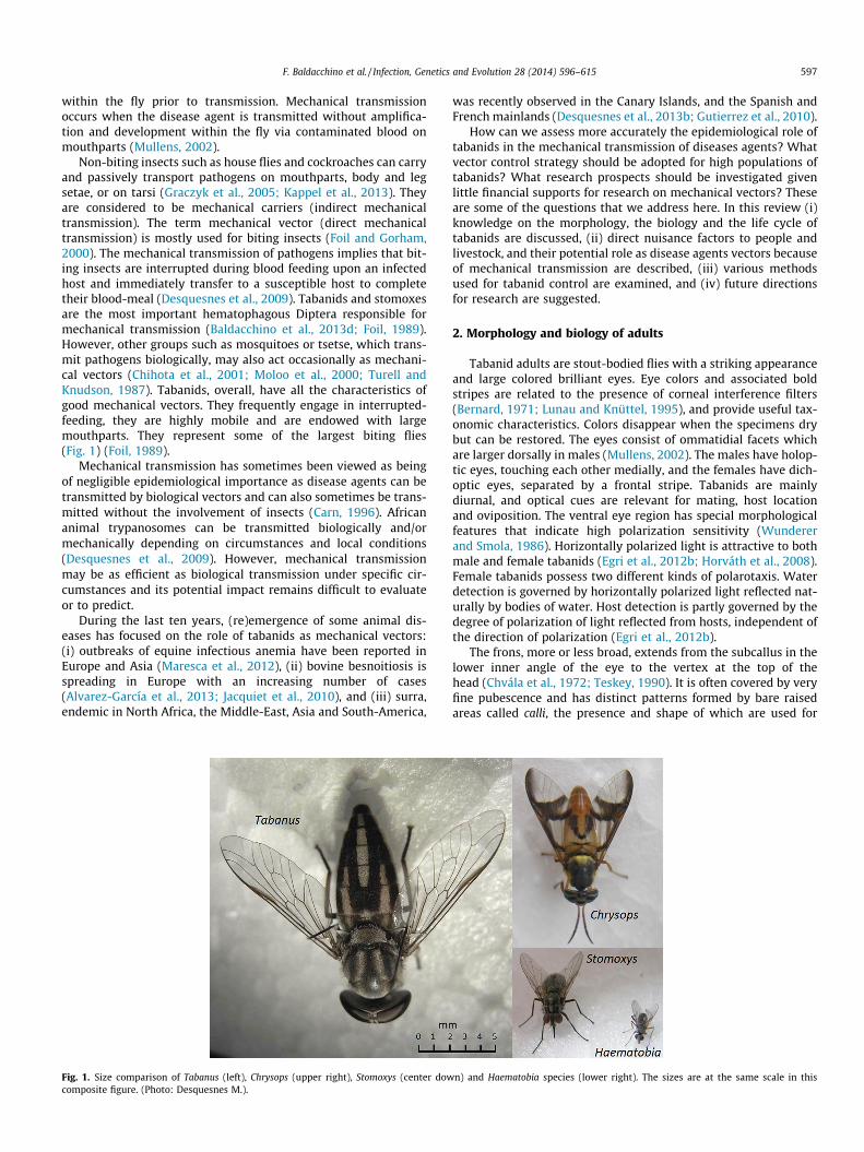
The antennae of tabanids are divided into three parts: the scape,the pedicel and the flagellum, with four to eight flagellomeres.Antennae are of major importance for classification at all levels.In Chrysopsinae and Haematopini, the basal flagellomere is slenderand elongate whereas in the Tabanini, it is enlarged and flat (plate),with a more or less developed dorsal tooth; the pedicel is short inboth Tabanini and Haematopotini. The pedicel is slender inChrysopsinae; conversely the scape is short in Tabanini whereasit is long in both Chrysopsinae and Haematopotini (Fig. 2).Antennae are the main olfactory organs in many insects and chem-ical signals are involved in host location by female tabanids. Themajority of olfactory sensilla appear to be present in the firstsegment of the flagellum (Ivanov, 2007; Parashar et al., 1994).Some olfactory sensilla are also present on the maxillary palps.
The mouthparts of the female consist of the unpaired dorsal lab-rum, the unpaired hypopharynx, the paired mandibles, the pairedmaxillae composed of the palps and the laciniae, and the unpairedventral labium with its distal labellum (McKeever and French,1999; Stoffolano and Yin, 1983). The palps and the labium are notbiting structures. During blood-feeding, mandibles and maxillarylaciniae are used to cut the skin in a scissor-like fashion (Lehane,2005) and, together with the labrum and hypopharynx, they piercethe skin and subcutaneous capillaries to create a micro-hematoma.Saliva is introduced into the wound via the salivary duct opening atthe tip of the hypopharynx and blood is drawn up into the foodcanal. This is formed by a deep gutter in the labrum. This feedingmethod is known as pool feeding or telmophagy, in contrast tothe vessel feeding or solenophagy used by mosquitoes (Mullens,2002). The genus Philoliche, a highly aggressive blood feeder onlivestock in Africa, has elongated mouthparts that allow it to probefor nectar in deep-throated flowers (Morita, 2008).
The thorax is stout with prominent notopleural lobes and strongwing muscles. Tabanids are fast flyers with high dispersal ability,e.g., 1–2 km daily (Cooksey and Wright, 1987; Konstantinov,1993). Wings have a remarkably constant venation within theTabanidae. Most species have clear wings or with a grayish orbrownish tinge, however some wings are partially or totally infus-cated providing useful features for identification. The wings ofChrysops and Ancala have distinct darkened bands, whereas thewings of Haematopota have patterns of spots or rosettes.
Fig. 2. General morphology and detail of antennae of three important genera: Haematopcomposite figure (Photo: Desquesnes M.).
The legs vary little in the family. Pangoniinae and Chrysopsinaepossess apical spurs on the hind tibiae, forelegs of Ancala havebulbous tibias, and the color of the legs is sometimes used for iden-tification. The abdomen of tabanids is as broad as the thorax,slightly compressed dorso-ventrally and usually with a distinctivecolor or pattern.
Altogether, it is clear that one of the main characteristics oftabanids is the variety and diversity of bold colors and patternson different body parts such as the eyes, wings and abdomen(McKeever and French, 1997). These distinctive colors and patternsare useful for identification. However, their function remainsunclear. Do these features play a role in color vision, in intra-and/or inter-specific visual communication, in camouflage?
3. Life cycle and reproduction
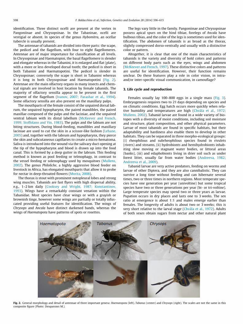
Females usually lay 100–800 eggs in a single mass (Fig. 3).Embryogenesis requires two to 21 days depending on species andon climatic conditions. Egg hatch occurs more quickly when rela-tive humidity and temperatures are high (Chvála et al., 1972;Mullens, 2002). Tabanid larvae are found in a wide variety of bio-topes with a diversity of moist conditions, including soil moistureand structure, plant composition, and other physical parameters.Larvae of most tabanids are found in specific habitats, but theiradaptability and hardiness also enable them to develop in otherhabitats. They can be separated in three morpho-ecological groups:(i) rheophilous and subrheophilous species found in rivulets(rivers) and streams, (ii) hydrobionts and hemihydrobionts inhab-iting slow moving or stagnant water bodies, or littoral areas(banks), (iii) and edaphobionts living in drier soil such as underforest litter, usually far from water bodies (Andreeva, 1982;Andreeva et al., 2009).
Tabanid larvae are very active predators, feeding on worms andlarvae of other Diptera, and they are also cannibalistic. They cansurvive a long time without feeding and can hibernate severaltimes, two or three times in northern regions. Most temperate spe-cies have one generation per year (univoltine) but some tropicalspecies have two or three generations per year (bi- or tri-voltine).Large temperate species may spend two or three years as larvae.Pupation occurs in dry places and lasts one to 3 weeks. The sexratio at emergence is about 1:1 and males emerge earlier thanfemales. The longevity of adults is about two or 3 weeks; this isvery short relative to the larval stage (Chvála et al., 1972). Adultsof both sexes obtain sugars from nectar and other natural plant
ota (left), Tabanus (center) and Chrysops (right). The scales are not the same in this
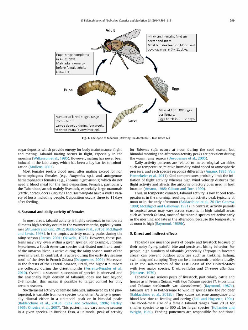
Fig. 3. Life cycle of tabanids (Drawing: Baldacchino F., Ink: Bosco G.).
F. Baldacchino et al. / Infection, Genetics and Evolution 28 (2014) 596–615 599
sugar deposits which provide energy for body maintenance, flight,and mating. Tabanid mating occurs in flight, especially in themorning (Wilkerson et al., 1985). However, mating has never beeninduced in the laboratory, which has been a key barrier to coloni-zation (Mullens, 2002).
Most females seek a blood meal after mating except for nonhematophagous females (e.g., Pangonius sp.), and autogenoushematophagous females (e.g., Tabanus nigrovittatus) which do notneed a blood meal for the first oviposition. Females, particularlythe Tabaninae, attack mainly livestock, especially large mammals(cattle, horses, deer). Chrysops and Haematopota have a wider vari-ety of hosts including people. Oviposition occurs three to 11 daysafter feeding.
4. Seasonal and daily activity of females
In most areas, tabanid activity is highly seasonal; in temperateclimates high activity occurs in the warmer months, typically sum-mer (Altunsoy and Kiliç, 2012; Baldacchino et al., 2013e; McElligottand Lewis, 1998). In the tropics, activity usually peaks during therainy season (Barros, 2001; Okiwelu, 1975). However, these pat-terns may vary, even within a given species. For example, Tabanusimportunus, a South American species distributed north and southof the Amazon River, is active during the rainy season south of theriver in Brazil. In contrast, it is active during the early dry seasonsnorth of the river in French Guiana (Desquesnes, 2004). Moreover,in the forests of the Central Amazon, Brazil, the highest numbersare collected during the driest months (Ferreira-Keppler et al.,2010). Overall, a seasonal succession of species is observed andthe seasonally high density of tabanids does not last beyond2–3 months; this makes it possible to target control for onlycertain seasons.
Nycthemeral activity of female tabanids, influenced by the pho-toperiod, is variable from one species to another. Activity is gener-ally diurnal either in a unimodal peak or in bimodal peaks(Baldacchino et al., 2013e; Cilek and Schreiber, 1996; Harley,1965; Oliveira et al., 2007). This pattern may vary among seasonsin a given species. In Burkina Faso, a unimodal peak of activity
for Tabanus sufis occurs at noon during the cool season, butbimodal morning and afternoon activity peaks are prevalent duringthe warm rainy season (Desquesnes et al., 2005).
Daily activity patterns are related to meteorological variablessuch as temperature, relative humidity, wind speed or atmosphericpressure, and each species responds differently (Amano, 1985; VanHennekeler et al., 2011). Cool temperatures probably limit the ini-tiation of flight activity whereas high wind velocity disturbs theflight activity and affects the airborne olfactory cues used in hostlocation (Amano, 1985; Gibson and Torr, 1999).
Thus, in temperate climates, tabanid activity is low at cool tem-peratures in the morning, resulting in an activity peak typically atnoon or in the early afternoon (Baldacchino et al., 2013e; Ganeva,1999; McElligott and Galloway, 1991). In contrast, activity periodsin tropical areas may vary across seasons. In high rainfall areassuch as French Guiana, most of the tabanid species are active earlyin the morning and late in the afternoon, because the temperatureat noon is high (Raymond, 1989b).
5. Direct and indirect effects
Tabanids are nuisance pests of people and livestock because oftheir noisy flying, painful bite and persistent biting behavior. Forpeople, high numbers of tabanids (especially Chrysops in forestedareas) can prevent outdoor activities such as trekking, fishing,swimming and camping. They can be an economic problem locally,as in the salt-marshes of the East Coast of the United-Stateswith two major species, T. nigrovittatus and Chrysops atlanticus(Hansens, 1979).
Tabanids are serious pests of livestock, particularly cattle andhorses as in French Guiana, with two Tabanus species (T. importunusand Tabanus occidentalis var. dorsovittatus) (Raymond, 1987a),tabanids are also bothersome to wildlife species like the red deer(Baldacchino et al., 2013b). They cause extreme annoyance andblood loss due to feeding and oozing (Foil and Hogsette, 1994).The blood-meal size of a female tabanid ranges from 20 lL forsmaller species to up to 600 lL for larger species (Hollander andWright, 1980). Feeding punctures are responsible for additional

600 F. Baldacchino et al. / Infection, Genetics and Evolution 28 (2014) 596–615
blood loss from natural oozing. Thus, oozing may also serve assecondary feeding sites for flies unable to puncture the skin. Thismay extend the duration of bleeding by the action of their saliva(Fig. 4). Tabanid bites may cause local reactions like dermalnodules or attract myiasis-causing flies for oviposition(Desquesnes, 2004; Foil and Hogsette, 1994). The annoyance levelof tabanids is closely related to fly abundance; large numbers ofbites can induce stress and reduce the quantity and quality of foodintake, thus weight gain, milk yield and manure production in cat-tle (Perich et al., 1986). An average of 66–90 horse flies per animalper day resulted in a weight loss of 0.08–0.10 kg per animal per day(Foil and Hogsette, 1994). In French Guiana, a decrease of the meandaily weight gain from 1.2 to 0.2 kg was observed in growing bullsduring the tabanid season. In horses, a total seasonal loss of 40 kgon average was observed. Riding activity was impossible duringthe tabanid season due to the violent reaction of horses to tabanidbites (Desquesnes, 2004). Such strong effects, by themselves, canjustify setting up control methods, although few have proven tobe efficient.
Beside these direct effects, tabanids are indirectly responsiblefor important losses through the transmission of pathogens fromone host to another. In some cases, tabanids may act as biologicalvectors of pathogens or they may transport some pathogens. How-ever, they are mostly known as mechanical vectors of an unlimitedrange of pathogens present in the blood of their hosts (Table 1).
6. Pathogens transported by tabanids
6.1. Dermatobia hominis
D. hominis (Diptera: Oestridae), the human bot fly or torsalo, is aneotropical species occurring from Mexico to Argentina (Marchiet al., 2012). D. hominis larvae cause furuncular myiasis in peopleand animals. Reports of D. hominis incidence in cattle have showneconomic losses such as reduced production of milk and meat, lossin body weight and cost depreciation in the leather (Pinto et al.,2002).
Female adults of D. hominis capture various arthropods and gluetheir eggs onto the arthropods in clusters (Catts and Mullen, 2002).Eggs are transported by arthropods-carriers to vertebrate hosts andhatch while the arthropods feed. Several Diptera have beenobserved as carriers, such as Anthomyiidae, Calliphoridae,
Fig. 4. (a) Female Philipomyia aprica feeding on the leg of a cow. Non-biting flies suckamericanus, a very large species, feeding on the leg of a horse. Lesions and clotted blood
Culicidae, Fanniidae, Muscidae, Sarcophagidae, Simuliidae, andTabanidae (Guimaraes et al., 1982; Ribeiro et al., 1985). Amongtabanids, Chrysops sp. and Tabanus sp. have been observed withD. hominis eggs.
7. Pathogens biologically transmitted by tabanids
7.1. Loa loa
Chrysops spp. are the biological vectors of the filaria L. loa, theagent of human loiasis known as the tropical eye worm and asso-ciated with Calabar swelling. The parasite is limited to the equato-rial rain forests of Africa (Kelly-Hope et al., 2012). Chrysops areintermediate hosts in which the microfilariae develop into thethird-stage infective larvae that are found in the insect mouthpartsafter a ten-day cycle (Connal and Connal, 1922). Chrysops silaceaand Chrysops dimidiata are the main vectors of loiasis in people;other Chrysops spp. are more involved in the transmission of thesimian form of Loa as they are mainly zoophilic (Orihel andLowrie, 1975). In the Chailu Mountains, Congo Republic, C. dimidi-ata is the major vector in the forest, whereas C. silacea is the majorvector in cleared forest zones near villages. The populations ofthese two species are abundant and stable in the rainy season asthe infection rate of parous females is related to the microfilarialrate in the human population (Noireau et al., 1990).
7.2. Other parasites and pathogens
Tabanids are considered biological vectors of other filarial nem-atodes such as Elaeophora schneideri and Dirofilaria roemeri; theyalso are cyclical vectors of protozoa such as Haemoproteus metch-nikovi and Trypanosoma theileri.
E. schneideri is a filarial nematode of domestic and wild rumi-nants in North America; it occurs commonly in mule deer andelk, but also in moose, white-tailed deer or sika deer (Couvillionet al., 1985; LeVan et al., 2013; Robinson et al., 1978). Hybomitraspp. and Tabanus spp. are intermediate hosts of Elaeophoraschneideri as third-stage larvae have been detected in adulttabanids and successfully inoculated into ruminants (Couvillionet al., 1986; Hibler et al., 1971). However, natural transmissionhas not yet been demonstrated. Other Elaeophorosis agents weredetected in wild ruminants in Europe (Elaeophora elaphi) and in
the blood oozing from the puncture (Photo: Baldacchino F.), (b) Female Tabanusdue to tabanid bites are visible on the skin (Photo: Foil L.D.).

Table 1Disease agents associated with tabanids.
Disease agents Vectors Geographicoccurrence
Transmission Associationa References
VirusesEquine infectious anemia
virusTabanus spp., Hybomitraspp., Chrysops spp.
Worldwide Mechanical Experimental andnatural transmission;isolation
Foil et al. (1983), Hawkins et al. (1973), Kemenet al. (1978), Scott (1922) and Stein et al.(1942)
Bovine leukosis virus Tabanus spp. Worldwide Mechanical Experimental andnatural transmission
Buxton et al. (1985), Foil et al. (1988b, 1989a),Hasselschwert et al. (1993) and Perino et al.(1990)
Bovine viral diarrhea virus Haematopota pluvialis Worldwide Mechanical Natural transmission Tarry et al. (1991)Hog cholera virus Tabanus spp. Worldwide Mechanical Experimental and
natural transmissionTidwell et al. (1972)
Vesicular stomatitis virus Tabanus spp., Chrysops spp. America Mechanical Experimentaltransmission
Ferris et al. (1955)
Rinderpest virus Tabanus orientis Eradicatedworldwidein 2011
Mechanical Natural transmission Bhatia (1935)
California encephalitisviruses
Hybomitra lasiophtalma,Chrysops spp.
NorthAmerica
Unknown Isolation DeFoliart et al. (1969) and Miller et al. (1983)
Western equineencephalitis virus
Tabanus nigrovittatus NorthAmerica
Unknown Isolation Goldfield et al. (1968)
Tick-borne encephalitisvirus
Tabanus flavicornis Asia andEurope
Unknown Isolation Yagodinsky and Skvortsov (1962)
Influenza virus Atylotus agrestis Worldwide Unknown Isolation Zhanseitova et al. (1982)
Bacteria/rickettsiaAnaplasma marginale Tabanus spp., Chrysops spp. Worldwide
(tropics,subtropics)
Mechanical Experimental andnatural transmission;isolation
Abramov and Grobov (1968), Hawkins et al.(1982), Hornok et al. (2008), Lotze and Yiengst(1941), Roberts and Love (1977), Sanborn et al.(1932), Scoles et al. (2008) and Wilson andMeyer (1966)
Francisella tularensis Chrysops spp., Tabanus spp.,Haematopota spp.
Northernhemisphere
Mechanical Experimental andnatural transmission;isolation
Cox (1965), Francis and Mayne (1921), Klocket al. (1973), Olsufiev and Golov (1936),Petersen et al. (2009) and Philip (1968)
Bacillus anthracis Tabanus spp., Haematopotapluvialis, Chysops spp.
Worldwide Mechanical Experimental andnatural transmission.isolation
Kolonin (1969), Mitzmain (1914) and Morris(1918)
Borrelia burgdorferi Chrysops spp., Hybomitraspp., Tabanus spp.
Northernhemisphere
Unknown Isolation Magnarelli et al. (1986)
Coxiella burnetii Tabanus staegeri Worldwide Unknown Isolation Amanzhulov et al. (1965)Clostridium chauvoei Tabanus rubidus Worldwide Mechanical Experimental
transmissionNieschulz and Huber (1928)
Pasteurella multocida Tabanus spp., Chrysopsdispar
Worldwide Mechanical Experimentaltransmission
Nieschulz and Kraneveld (1929)
Brucella spp. Haematopota spp.,Hybomitra solstitialis,Tabanus spp., Heptatomapellucens
Worldwide Mechanical Experimentaltransmission
Wellmann (1951)
Listeria monocytogenes Haematopota pallens,Hybomitra spp., Tabanusspp.
Worldwide Mechanical Experimentaltransmission
Alekseev et al. (1969) and Dem’yachenko andBaranenkov (1970)
Erysipelothrix rhusiopathiae Haematopota pluvialis,Tabanus bromius, Heptatomapellucens
Worldwide Mechanical Experimentaltransmission
Wellmann (1950)
Ehrlichia risticii Chrysops vittatus, Tabanusspp.
NorthAmerica
Mechanical Experimentaltransmission
Levine et al. (1992)
ProtozoaBesnoitia besnoiti Tabanus taeniola, Atylotus
nigromaculatus, Tabanocelladenticornis, Haematopotaspp.
SouthAmerica,Europe,Africa,Middle-East,Asia
Mechanical Experimental andnatural transmission
Bigalke (1968)
Trypanosoma evansi Tabanus spp., Haematopotaspp., Chrysops spp.,Ancalaspp., Atylotus spp.
SouthAmerica,North Africa,Asia, Europe
Mechanical Experimental andnatural transmission
Collier (1928), Gruvel and Balis (1965),Mitzmain (1913), Mohler and Thompson(1911), Nieschulz (1925, 1926, 1927a,b),Nieschulz and Ponto (1927), Sergent andSergent (1905), Singh (1926) and Yutuc (1949)
Trypanosoma vivax Tabanus spp., Atylotus spp.,Cryptotylus unicolor
SouthAmerica,Africa
Mechanical Natural transmission Desquesnes and Dia (2003b, 2004), Ferencet al. (1990), Otte and Abuabara (1991), Parra-Henao et al. (2008) and Raymond (1990)
Trypanosoma theileri Haematopota spp., Tabanusspp., Hybomitra spp.
Worldwide Mechanicalandbiological
Experimental andnatural transmission;isolation anddevelopment
Böse et al. (1987), Davies and Clark (1974),Dirie et al. (1990), Kraneveld (1931), Nöller(1916, 1925) and Packchanian (1957)
(continued on next page)
F. Baldacchino et al. / Infection, Genetics and Evolution 28 (2014) 596–615 601

Table 1 (continued)
Disease agents Vectors Geographicoccurrence
Transmission Associationa References
Trypanosoma equiperdum Atylotus tomentosus Worldwide Mechanical Experimentaltransmission
Sergent and Sergent (1906)
Trypanosoma brucei Atylotus nemoralis, Tabanusspp., Haematopota spp.
Africa Mechanical Experimentaltransmission; isolation
Duke et al. (1934) and Sergent and Sergent(1906)
Trypanosoma congolense Atylotus agrestis Africa Mechanical Natural transmission Desquesnes and Dia (2003a)Haemoproteus metchnikovi Chrysops callidus Worldwide Mechanical
andbiological
Experimentaltransmission; isolationand development
DeGiusti et al. (1973)
Filarial nematodesLoa Loa Chrysops spp. Central
AfricaBiological Experimental and
natural transmission;isolation anddevelopment
Connal and Connal (1922), Duke (1972),Gordon and Crewe (1953), Kleine (1915),Lavoipierre (1958), Leiper (1913), Noireauet al. (1990), Orihel and Lowrie (1975) andWilliams (1960)
Elaeophora schneideri Hybomitra spp., Tabanusspp.
NorthAmerica,Europe
Biological Experimentaltransmission; isolationand development
Clark and Hibler (1973), Couvillion et al.(1986), Hibler et al. (1971) and Weinmannet al. (1973)
Dirofilaria repens Haematopota variegata Africa, Asia,Europe
Biological Development Coluzzi (1964)
Dirofilaria roemeri Tabanus spp., Dasybasis spp. Australia Biological Experimentaltransmission; isolationand development
Spratt (1974)
a Association between disease agents and tabanids is described as follows: isolation (agent isolated from tabanids), development (as if tabanids were natural intermediatehost), experimental transmission (transmission of agent by unnatural mode of infection or to unnatural host), and natural transmission (transmission of agent from onenatural host to another by exposure to tabanids) according to Krinsky (1976).
602 F. Baldacchino et al. / Infection, Genetics and Evolution 28 (2014) 596–615
Africa (Elaeophora poeli and Elaeophora sagitta) (Huchzermeyeret al., 2001; Santin-Duran et al., 2000). To our knowledge, the roleof tabanids in their epidemiology has not been studied.
D. roemeri is a filarial nematode of macropods in Australia andthird-stage larvae have been detected in the mouthparts ofDasybasis oculata and Tabanus parvicallosus (Spratt, 1974).
T. theileri is a nonpathogenic hemoprotozoa widely distributedin cattle (Villa et al., 2008). As a stercorarian trypanosome,T. theileri is ingested by the insects with the blood of its host andthus implements a cyclopropagative development in the posteriorpart of the insect’s digestive tract (Hoare, 1972). Infective metacy-clic stages were identified in the gut and in the feces of tabanidssuch as Haematopota pluvialis, Haematopota italica, Hybomitramicans and Tabanus bromius (Böse et al., 1987). These stages canpenetrate the intact oral mucosa (Kraneveld, 1931) of theirmammalian hosts. Böse et al. (1987) suggested that oral contami-nation of mucosal membranes could take place while cattle defendthemselves against tabanids is a hemoprotozoa of turtles. A sporo-zoite form of the parasite has been found in the salivary glands ofChrysops callidus and gave rise to gametocyte forms after inocula-tion to turtles (DeGiusti et al., 1973).
8. Main mechanically transmitted pathogens
Pathogens mechanically transmitted by tabanids were compre-hensively reviewed by Krinsky (1976); they include viruses, bacte-ria and parasites, the relative role of mechanical transmissionranged from suspected or marginal to fundamental.
8.1. Viruses
8.1.1. Equine infectious anemia virus (EIAV)EIAV is a lentivirus of the retrovirus family, infecting equids
exclusively (Cook et al., 2013). EIA, referred to as swamp fever,has a worldwide distribution. Currently, EIA is endemic in SouthAmerica and Africa, and outbreaks of EIA have been reported inEurope and Asia during the last ten years (Herholz et al., 2008;
Maresca et al., 2012). There is a growing concern about outbreaksin equine populations due to the expansion of global trade inhorses. EIA is of considerable importance to the equine industry.It is one of only eleven notifiable equine-specific diseases listedby OIE, the world organization for animal health (Cook et al., 2013).
The mechanical transmission of EIAV is generally accepted as amajor factor in transmission of the virus. Tabanids such as Chrysopsspp., Hybomitra spp. and Tabanus spp. are considered the primarymechanical vectors (Foil et al., 1983, 1988a). Transmission of EIAVfrom a febrile infected pony to a susceptible pony by a singleTabanus fuscicostatus has been reported by Hawkins et al. (1976).Other experiments on the natural transmission of EIAV implicatedafebrile or inapparent donors (Foil et al., 1983; Issel and Foil, 1984;Kemen et al., 1978). Foil et al. (1988a) commented that the proba-bility of vector transmission increases when the circulating titer infebrile donors approaches 106/mL; at this titer, transmission wasshown to occur with transfers of a single horse fly and as few assix deer flies (Foil and Issel, 1991; Foil et al., 1983). Mechanicaltransmission trials have demonstrated transmission of EIAV fromafebrile donors to susceptible equids using groups of 25 horse flies,but the titer of the virus in the donor animals was not established(Foil and Issel, 1991). Transmission of EIAV from acutely infecteddonors to recipients also has been shown to occur using groupsof stable flies ranging from 50 to 100 (Foil et al., 1983). This indi-cates that horse flies have a 50–100 times higher mechanical trans-mission potential than stable flies. These results are consistentwith the estimates of blood meal residues on the mouthparts ofhorse flies (10 nL) and stable flies (0.4 nL) reported in differentstudies (Foil et al., 1987; Scoles et al., 2005, 2008).
8.1.2. Other virusesTabanids can transmit other viruses even if mechanical trans-
mission is not the major route of transmission. Among theseviruses, bovine leukosis virus and bovine viral diarrhea virus havebeen transmitted naturally between cattle by T. fuscicostatus and H.pluvialis, respectively (Foil et al., 1988b, 1989a; Hasselschwertet al., 1993; Tarry et al., 1991). Hog cholera virus, agent of classicalswine fever, has also been naturally transmitted between pigs by

F. Baldacchino et al. / Infection, Genetics and Evolution 28 (2014) 596–615 603
Tabanus spp. (Tidwell et al., 1972). Other viruses, such as West Nilefever virus and lumpy skin disease virus are suspected to be trans-mitted mechanically by tabanids as they have been experimentallytransmitted by other biting insects (Chihota et al., 2001; Doyleet al., 2011). However, the highly seasonal, and thus temporary,impact of these insects makes it difficult to demonstrate causeand effect. Consequently, their relative role in disease transmissionis not well documented. This is not the case with acute and fataldiseases like EIA. This is because of the association of tabanidpopulation peaks with EIA epizootics, and the linked geographicdistribution of the disease with the presence of high tabanidpopulations (Foil and Issel, 1991).
8.2. Bacteria
8.2.1. Bacillus anthracisAnthrax is a non-contagious disease caused by spore-forming
bacterium, B. anthracis, dispersed into the environment. Althoughthe incidence of anthrax is generally decreasing worldwide, it per-sists in many countries, following ineffective control programs(Fasanella et al., 2010a). The potential role of insects in spreadinganthrax infection has been confirmed in many studies. Non-hema-tophagous Diptera such as house flies or flesh flies can act as sporecarriers (Blackburn et al., 2010; Fasanella et al., 2010b) whereashematophagous Diptera such as tabanids, stomoxes and mosqui-toes can act as mechanical vectors (Kraneveld and Djaenoedin,1940; Turell and Knudson, 1987). Kraneveld and Djaenoedin(1940) conducted successful experiments on the transmission ofB. anthracis to horses and water buffalos by immediate transferfeeding or 48 h-delayed feeding with Tabanus rubidus while a pos-sible infection via tabanid legs or feces was prevented (in Krinsky,1976). Epidemiologic evidence has also been reported. During anoutbreak of sheep anthrax in southern Italy, a case of humancutaneous anthrax was diagnosed in a breeder who had no contactwith infected or dead animals, but who had been bitten bytabanids (Fasanella et al., 2013).
8.2.2. Anaplasma marginaleA. marginale, belonging to the order of Rickettsiales, is the
etiological agent of bovine anaplasmosis, an infectious but non-contagious disease (Aubry and Geale, 2011). Bovine anaplasmosisoccurs in tropical and subtropical regions worldwide includingSouth and Central America, the United States, southern Europe,Africa, Asia and Australia. Natural transmission between cattlehas been demonstrated with Tabanus spp. (Howell et al., 1941;Lotze, 1944; Wilson and Meyer, 1966). Krinsky (1976) conductedan in depth review of transmission of A. marginale by differenthorse flies. Many studies used naturally infected cattle and adultcows as recipients, and mechanical transmission was consistentlydemonstrated. Hawkins et al. (1982) showed that transmission ofA. marginale from acutely infected splenectomized calves to sus-ceptible splenectomized calves can be accomplished with as fewas ten horse fly bites. Horse flies remained mechanically infectivefor at least two hours.
A. marginale can be transmitted mechanically by biting flies andbiologically by ticks (Kocan et al., 2010). Biological transmission byticks is considered to be more efficient than mechanical transmis-sion and can result in enzootic stability. Scoles et al. (2008) showedthis when they failed to transmit A. marginale to susceptible calves(donor and recipient calves all had intact spleens) with horse flies.Transmission did not occur with T. fuscicostatus at a rickettsemia240-fold higher than that found in the tick Dermacentor andersoni,which transmitted at 100% efficiency. This is due to the propaga-tive cycle of the rickettsia in the tick, which makes it a carrier oflarge quantities of parasites that are re-injected into hosts(Chartier et al., 2000). However, epidemiological impacts also
depend on tick biology, particularly the capacity to move fromone host to another. This is regular for 3 hosts-ticks such asD. andersoni, but it may be limited or very restricted as in one-hostticks such as Rhipicephalus (Boophilus) microplus (Desquesnes,1997; Mason and Norval, 1981). In such cases, the role of the tickis to multiply and re-inject parasites on the same animal. Thisamplifies the parasite burden leading to immune failure andappearance of clinical signs. In contrast, biting flies can transmita small quantity of blood, acting as mechanical vectors ofAnaplasma, responsible for epizootics especially in areas withoutefficient biological vectors (3 hosts-ticks). Chronic transmissionsolely by efficient mechanical vectors such as Tabanus taeniolaappears to occur in some situations (Wiesenhutter, 1975a), but isdifficult to verify when ticks may still be present at very lownumbers. Factors such as needle transmission can also confounda clear interpretation of results (Wiesenhutter, 1975b).
These studies on mechanical transmission clearly show thathorse flies are efficient mechanical vectors of A. marginale, but use-ful comparisons of vectorial efficiency are limited by differences indonor and recipient animals among experiments (Krinsky, 1976).Importantly, mechanical transmission by flies or iatrogenic meansdoes appear to be the major mode of transmission in areas wheretick vector populations are limited or where strains of A. marginaleare not transmissible by ticks. In Costa Rica, Oliveira et al. (2010)found an association between the seroprevalence of bovine ana-plasmosis and the presence of biting flies like tabanids. In FrenchGuiana, epidemiological studies have indicated that tabanids arelikely responsible for the transmission of the parasite. In contrast,cattle ticks are responsible for the amplification of the infection,thus the clinical occurrence of the cases. Clinical anaplasmosisand high tick burdens occurring in tandem, make it difficult tounravel the underlying epidemiological processes (Desquesnes,1997).
8.2.3. Francisella tularensisTularemia, known as rabbit fever or deer fly fever, is a rare
zoonotic infectious disease, caused by F. tularensis. The diseaseoccurs around the northern hemisphere including North America,Europe and Asia (Nigrovic and Wingerter, 2008). Outbreaks of tula-remia may be associated with exposure to ticks or biting flies,infected small animals, food or water, fomites, or occasionallyaerosol-borne bacteria (Foley and Nieto, 2010). Klock et al.(1973) provided epidemiologic evidences of the mechanical trans-mission of F. tularensis in humans by Chrysops discalis. F tularensishas also been isolated in naturally infected tabanids includingChrysops spp., H. pluvialis and Tabanus spp. (Petersen et al., 2009).
8.2.4. Other bacteriaTabanids have also been associated with the transmission of
other bacteria such as Brucella spp., Pasteurella multocida,Erysipelothryx rhusiopathiae or Ehrlichia risticii through experimen-tal transmission (Krinsky, 1976; Levine et al., 1992).
8.3. Protozoa
8.3.1. Besnoitia besnoitiB. besnoiti is a cyst-forming apicomplexan parasite of cattle
causing bovine besnoitiosis (Jacquiet et al., 2010). Bovinebesnoitiosis is widely distributed and has been recognized as are-emerging disease in Europe due to an increased number of casesand a geographic expansion (Alvarez-García et al., 2013). Themechanical transmission of B. besnoiti between cattle has beendemonstrated with African tabanids such as Atylotus nigromaculatusand Tabanocella denticornis. The parasite can survive for about 24-hon tabanid mouthparts (Bigalke, 1968).
604 F. Baldacchino et al. / Infection, Genetics and Evolution 28 (2014) 596–615
Stomoxys calcitrans also appears to be capable of transmitting B.besnoiti mechanically (Lienard et al., 2013). However, the volumeof blood retained in the mouthparts of stable flies, close to 0.4 nL,is very low in comparison with that retained by horse flies (about10 nL) or deer flies (6 nL) (Scoles et al., 2005, 2008). Bigalke (1968)estimated that more than 50,000 stable fly bites were required tocause infection in cattle whereas three horse fly bites were suffi-cient. Unfortunately, the horse flies were fed individually and thestable flies were fed using cages with multiple flies and multipletransfers, preventing a more quantitative comparison of vectorpotentials.
In the field, the transmission of B. besnoiti is likely facilitated bychronically infected cattle in which tissue cysts are frequentlyfound in the skin (Frey et al., 2013).
8.3.2. Trypanosoma spp. (with the exception of T. theileri)Trypanosoma brucei brucei, Trypanosoma vivax and Trypanosoma
congolense are agents of Animal African Trypanosomosis or naganain livestock which has a great impact on cattle production in Africa.These Trypanosoma spp. are normally transmitted by tsetse flies,their biological vectors (Holmes, 2013). T. vivax is a special caseas it can be transmitted cyclically and mechanically by biting fliesincluding tsetse flies (Desquesnes and Dia, 2004; Moloo et al.,2000). The mechanical transmission of T. vivax has been proven,under experimental conditions, with neotropical tabanids such asTabanus nebulosus, T. importunus and Cryptotylus unicolor (Ferencet al., 1990; Otte and Abuabara, 1991; Raymond, 1990), and afro-tropical tabanids such as Atylotus agrestis and Atylotus fuscipes(Desquesnes and Dia, 2003b, 2004). In Latin America in the absenceof tsetse, T. vivax is solely transmitted by mechanical vectors. Itstransmission may be facilitated by differences in circulating strains(Hamilton, 2012; Jones and Davila, 2001).
T. congolense DNA has been detected in tabanids in the field inone exploratory study (Solano and Amsler-Delafosse, 1995), pre-sumably as a result of the presence of ingested blood, but, furtheron, Desquesnes and Dia (2003a) demonstrated the mechanicaltransmission of T. congolense between cattle by A. agrestis. The inci-dence of transmission, however, was lower than in parallel exper-imental studies with T. vivax. Records of parasitemia suggestedthat mechanical transmission of T. congolense was less likely thanT. vivax due to lower parasitemia (Desquesnes et al., 2009). Hence,mechanical transmission of T. congolense may be limited in thefield.
The demonstration of mechanical transmission of Trypanosomaspp. by tabanids, supported by epidemiological studies, has nowshown that cattle trypanosomosis in Africa also involves tabanidsas well as tsetse. This has been documented in studies of T. vivaxprevalence in areas outside of the tsetse belt, for example in Sudan(Rahman, 2005), Ethiopia (Cherenet et al., 2004), and Chad(Delafosse et al., 2006). Similar evidence exists for T. vivax in east-ern and southern Africa, but is more difficult to interpret due to theproximity of tsetse to many study areas, and livestock exchangebetween tsetse-free and tsetse-infested areas (Van den Bossche,2001; Wiesenhutter, 1976). Therefore, the eradication of tsetseflies in Africa will not necessary lead to the eradication of trypan-osomosis from domestic livestock (Desquesnes and Dia, 2003b;Sinshaw et al., 2006).
Trypanosoma evansi is the agent of surra; it can affect a verylarge range of domestic and wild animals; the parasite is especiallypathogenic in camelids and equids (Desquesnes et al., 2013b). Thedisease spreads from North Africa towards the Middle-East, Asiaand South America. Surra was also introduced into Europe (Spainand France) following the importation of infected camels fromthe Canary Islands (Gutierrez et al., 2010). T. evansi is an unappar-ent spreading parasite due to its ability to persist at low numbersin many hosts, to diffuse silently via healthy carriers and to be
transmitted by non-specific mechanical vectors (Desquesneset al., 2013b). Many tabanids can transmit T. evansi mechanicallyamong various mammals (Krinsky, 1976). High populations oftabanids have been associated with high prevalence of surra inIndia (Nair et al., 2011) and in Somalia where Philoliche spp. wereincriminated as the major vectors (Dirie et al., 1989). Mechanicaltransmission by tabanids is the major route of transmission ofsurra even if some other means of transmission may have signifi-cant impacts under certain circumstances, such as the oral routein carnivores (Desquesnes et al., 2013a).
9. Determinant factors for mechanical transmission
9.1. Vector behavior and characteristics
The probability of mechanical transmission is dependent onmultiple factors linked to the biting behavior of tabanids and someanatomical characteristics: interrupted feeding, quantity of bloodmeal residues on mouthparts, sensitivity to host defensive reac-tions and the tendency to switch hosts, biting intensity andabundance.
Interrupted feeding is probably the most important factordetermining the role of tabanids as efficient mechanical vectors(Magnarelli and Anderson, 1980). The number of flies that natu-rally leave a host before completing a blood meal is critical toassessing the probability of mechanical transmission. The tenacityby which horse flies pursue a single host until engorged is called‘‘feeding persistence’’ (Foil, 1989). Foil (1983) compared the rela-tive feeding persistence of representative Louisiana tabanids withhorses tethered in a 9 m square design and with flies marked,but not disturbed. The percentages of flies which transferred toanother host was higher for larger tabanids such as Tabanussulcifrons (7.1%) and Tabanus petiolatus (12.3%) than for smallerones such as Tabanus lineola (2%) and Chrysops spp. (2.8%).
Some tabanids are more easily dislodged by hosts and may thustransfer between animals more often. In the USA, 83% of Chrysopsfemales completed blood meals by continuous feeding, unlike40% of Tabanus and Hybomitra females; Hybomitra and Tabanuswere more frequently interrupted and attempted to feed on thesame animal or on another one (Magnarelli and Anderson, 1980).In Australia, Tabanus pallipennis was observed to be very sensitiveto host defensive reactions. It required as many as nine partialmeals before reaching engorgement (Muzari et al., 2010b).
The quantity of blood meal residue that remains on mouthpartsfollowing an interrupted feed also influences the amount of infec-tious material transported between hosts. For instance, the amountof blood remaining on dissected mouthparts of T. fuscicostatusimmediately after feeding was estimated at 10 ± 5 nL (Foil et al.,1987). T. fuscicostatus is a medium-sized tabanid; smaller tabanidsprobably carry less residual blood (Foil, 1989). Smaller tabanids arealso less effective mechanical vectors than larger tabanids becausethey often feed completely and continuously. Differences in popu-lation density may also modify the intrinsic importance of differ-ent-sized vectors (Foil, 1989). Since many tabanids have beenshown to be capable of mechanically transmitting pathogens likeEIAV, total tabanid populations must be considered in field trans-mission studies (Foil et al., 1984). The total tabanid populationappears to be the most consistent phenomenon associated withpeaks of infection.
9.2. Host behavior and characteristics
Tabanids are not equally attracted to all hosts. Typically, horsesare more attractive than cattle, which are more attractive thansheep and goats. Young animals are usually bitten less than adults,
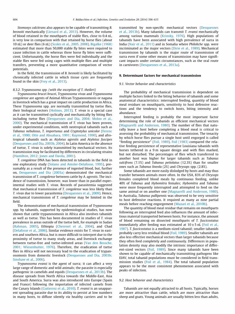
Fig. 5. Mark-recapture results of Tabanus importunus after artificially interrupting feeding on a horse (according to Barros and Foil, 2007).
F. Baldacchino et al. / Infection, Genetics and Evolution 28 (2014) 596–615 605
e.g., calves versus cows (Raymond and Rousseau, 1987). Foil et al.(1985) reported that foals had as low as 1.4% of the tabanid burdenof mares, and hence, a lower incidence of EIA. This appears to resultfrom the lower inherent attractiveness of young versus old ani-mals, and differences in defensive behaviors. Overall, adults andlarge animals are visually and olfactorily more attractive, due totheir size and the release of larger amounts of carbon dioxideand other attractants.
Intensity (darkness/brightness), color and the polarizationproperties of light reflected from hosts are also important param-eters of attraction. The ventral eye region of tabanids senses hori-zontally polarized light, which is attractive to both sexes in thecontext of seeking water (Egri et al., 2012a; Horváth et al., 2008).Dark-haired or large-spotted animals are more attractive to femalehost-seeking tabanids than white-haired or small-spotted ones.This is partly due to the higher degrees of polarization of coat-reflected light (Blahó et al., 2012a; Horváth et al., 2010; Kriskaet al., 2009). Striped patterns as in zebra hair are the least attrac-tive (Egri et al., 2012b). Tabanid reactions to zebra stripes are notcountered by the presence of attractive odours (Blahó et al., 2013).
Ungulates use varied individual or group behaviors to escapefrom biting flies and to repel them (Baldacchino et al., 2014;Hughes et al., 1981; Mooring et al., 2007; Rutberg, 1987). Thesebehaviors should be considered when assessing the risk ofmechanical transmission. Individual defensive reactions such asskin shaking, leg stamping, head wiping, head throwing or tailflicking are effective in removing flies and reducing feeding success(Baylis, 1996; Torr and Mangwiro, 2000). Their intensity dependson host species, age (young animals are more reactive to the bites),excitability, or habituation to pain (Mohamed-Ahmed and Mihok,2009; Mullens et al., 2006; Raymond and Rousseau, 1987;Schofield and Torr, 2002). Among hosts, pigs seem to be the leastsensitive to tabanid bites. Muzari et al. (2010b) suggested that pigsare a good reservoir of surra because tabanids biting them feed fora long time, but do not engorge fully, and hence probably completefeeding on another host. Conversely, host defensive movementsmay be so efficient that they can prevent infection, e.g., as in goatsin the vicinity of horses and cattle infected by T. evansi (Kongkaewet al., 2012).
If host excitability underlies variation in host defensive reac-tions in some species or individuals, habituation to pain may alsoresult in a decrease in host defensive reactions through time(Mullens et al., 2006). The physiological status of an infected hostmay also affect its behavior. For example, an acutely infected ani-mal may be highly attractive because of fever, and may also not becapable of expending the energy required to dislodge a feedingtabanid (Foil, 1989). Other behaviors that need to be taken intoaccount involve escape or avoidance, e.g., moving to habitats withlower rates of tabanid attack, immersing in water, or moving intoshelters. One phenomenon that is easy to observe is that horseswill often stand in pairs ‘‘head to tail’’ so that the tail of one pro-tects the head of the other. In the arctic with its extremely short
and synchronized biting fly season, animals have evolved manystrategies to cope with intense fly activity (Anderson et al.,2001). Avoidance of biting flies likely results in considerablyreduced food intake under these circumstances.
In bovine herds under high tabanid pressure, animals gathertightly and reduce the space for insects to reach their skin, limitingthe number of bites per individual. When flies are abundant, horsesin larger groups suffer from fewer flies than horses in smallergroups (Duncan and Vigne, 1979; Rutberg, 1987). Increasing thesize of a herd will attract more insects but not linearly. In largeherds, animals suffer from less biting attempts than in small herds(Torr et al., 2007). However, the close proximity of hosts in a herdis a detrimental factor that favors mechanical transmission frominfected to susceptible individuals. Indeed, when hosts are distant,the prevalence of mixed blood meals is lower. After interruption offeeding on horses, about 40% of the tabanids returned to the origi-nal host. In contrast, nearly 10% transferred to another host 5–10 maway, and nearly 5% transferred to another host 25 m away (Barrosand Foil, 2007) (Fig. 5). The percentage of tabanids that movedbetween hosts is strongly influenced by the distance that separatesthem. No tabanids were observed to transfer between horses sep-arated by 50 m. These results support the findings of Foil (1983) aswell as the recommendation of a 200 m quarantine distancebetween infected and susceptible animals to prevent the mechanicaltransmission of pathogens by tabanids (Barros and Foil, 2007).
9.3. Pathogens and infections
The mechanical transmission of pathogens by insects is depen-dent on the nature of the disease agent and its titer in the infectedhost. Some pathogens such as B. anthracis and F. tularensis are envi-ronmentally resistant and allow time for heteroxenous feeding bytabanids even when hosts are not spatially close (Foil, 1989). Whenstudying the epidemiology of such diseases, inter-herd transmis-sion must be considered, due to the potential for delayed mechan-ical transmission. A high titer of the pathogenic agent in theinfected host also increases the risk of mechanical transmission.Hawkins et al. (1976) found that a single tabanid (T. fuscicostatus)transmitted EIAV when the donor’s infectious titer reached106 infectious doses/mL. Foil et al. (1988b) found that 10–20 and100–150 T. fuscicostatus were able to transmit BLV when donorshad lymphocyte counts of 31.5 and 14.6 lymphocytes/nL, respec-tively. Transmission did not occur with up to 185 horse flies whenthe donors had roughly 103 infectious particles/mL.
Clearly, a high titer of the pathogen facilitates mechanicaltransmission. Conversely, a low level of pathogenemia may notbe sufficient (Desquesnes et al., 2009). For example, T. evansi isactively transmitted by tabanids among camels which naturallyexhibit very high parasitemia. In contrast, although receptive tothe parasite, sheep are most often an epidemiological dead endfor T. evansi due to their low parasitemia. Similarly, surra will bespread by tabanids very quickly in horses, whereas in buffaloes
606 F. Baldacchino et al. / Infection, Genetics and Evolution 28 (2014) 596–615
or cattle, the incidence will most often remain low (Kongkaewet al., 2012). Prevalence of infection and immunity in a hostpopulation must also be considered when estimating the probabil-ity of mechanical transmission in nature (Foil, 1989).
In summary, by their annoyance, tabanids cause hosts to gathertogether, which results in close contact between infected and non-infected animals. Tabanids also stress and immunosuppress hostswhich induces high parasitemia in infected animals, and high sus-ceptibility in non-infected animals. Increasing parasitemia in aninfected animal induces fever which makes hosts more attractiveto biting insects, and allows for the ‘‘delivery’’ of pathogens tothe mouthparts of vectors. Tabanids also have painful bites thatinduce host defensive reactions interrupting blood feeding. Dueto close contact, interrupted blood meals taken on an infected ani-mal may re-start on other animals nearby. These features alto-gether facilitate mechanical transmission of blood pathogens.
10. Modeling mechanical transmission
Modeling the mechanical transmission of pathogens by bitingflies was done by Desquesnes et al. (2009) in a series of experi-ments in Burkina Faso, in heifers experimentally infected withT. vivax or T. congolense. The cattle were placed in semi-free condi-tions in a fly-proof system. They were challenged by a controlledtabanid population (A. agrestis or A. fuscipes) captured and intro-duced daily into the system (Desquesnes et al., 2009; Desquesnesand Dia, 2003a,b, 2004). This work demonstrated a very highpotential for mechanical transmission. The incidence of newinfections reached 60–75% after 21 days of exposure to the bitingflies. The data collected were used to develop a mathematical rela-tionship linking the initial prevalence of the infection, the level ofparasites in the blood of infected animals, the daily tabanid burdenand the incidence of the infection. From this set of parameters, amathematical model was developed to simulate the evolution ofprevalence under various predictive circumstances, includingcontrol measures. This model could be used to assess the risk ofmechanical transmission under field conditions (Desquesneset al., 2009).
The model demonstrated that the success of mechanical trans-mission, measured by the incidence of new infections, is mathemat-ically linked with the level of the pathogenemia and the density ofbiting flies. It demonstrated that mechanical transmission by taba-nids becomes efficient when the pathogenemia reaches 106/mL.Additionally, it emphasized that an initial rate of infection of10–15% allows not only statistically a maximal incidence, but is alsorepresentative of a highly susceptible population. This helps toexplain why outbreaks of mechanically transmitted pathogens aremostly periodic, e.g., occurring every 3–5 years. Indeed, this periodof time is required for a population of hosts to clear gradually from apost-epizootic situation in which most of the animals are infected(prevalence >80%), down to a pre-epizootic situation in which mostof the animals are not infected (10% < prevalence < 15%), thushighly susceptible (Desquesnes, 2004).
11. Limits of mechanical transmission
The minimal theoretical conditions required for mechanicaltransmission are (i) high parasitemia in donors, (ii) high densityof mechanical vectors, (iii) high receptivity and susceptibility of amajor portion of potential recipients, and (iv) close contactbetween recipients and donors. Furthermore, for parasites to besustained in a population, high parasitemia in donors and a highdensity of insects need to be synchronized, in addition to thecapacity of a host/reservoir to periodically exhibit high levels ofparasitemia. For these reasons, mechanical transmission remains
an occasional phenomenon with highly variable frequency andimpact (Desquesnes et al., 2009).
Based on several studies (including capture-mark-releaseexperiments), a safe distance between donors and recipients isestimated to be about 200 m in horses (Barros and Foil, 2007). Atgreater distances, tabanids may not leave one host for another.These observed probabilities relate to the specific circumstancesand hence provide only general guidance. These findings are par-ticularly subject to the relative ‘‘weight’’ of the two groups. Indeed,if a single infected horse is 300 m from a group of 60 non-infectedhorses, it is reasonable to assume that the chance that a tabanidwill move from the single infected animal towards the group isnot zero. The olfactory signature of a large herd is likely detectableat hundreds of meters, making a large group inherently ‘‘detect-able’’ and hence attractive. With the exception of some elegantstudies done with a very large electric net to monitor tabanidbehavior upwind and downwind of a single oxen in Zimbabwe,very little is known about the critical distance at which tabanidscan detect hosts (Phelps and Vale, 1976). In these studies, the effec-tive range of odor attraction downwind for a single ox was about15 m for Tabanus, and up to about 80 m for Philoliche.
12. Trapping technology
Traps are designed to attract targeted insects using sensory cuessuch as sound, odor, color, intensity (darkness/brightness), move-ment, and light polarization (Egri et al., 2012a; Horváth et al.,2008; Kriska et al., 2009; Takken and Knols, 2010). They are effec-tive tools for vector surveillance but have only very occasionallybeen tested for control purposes. A particularly large number oftraps have been developed for tsetse flies; but only one was specif-ically optimized for the simultaneous collection of other biting fliessuch as tabanids and stomoxes (Mihok, 2002). In Africa, there aremany compact practical traps made from blue and black fabricsthat have been refined for different tsetse. Examples are: theBiconical trap (Challier and Laveissière, 1973), the Vavoua trap(Laveissière and Grébaut, 1990), the F3 trap (Flint, 1985), the Epsi-lon trap (Hargrove and Langley, 1990), the Pyramidal trap(Gouteux and Lancien, 1986), the NG2G trap (Brightwell et al.,1991), the Nzi trap (Mihok, 2002), and the Tetra trap (Dia et al.,2008). In North America, very different trap designs have beenused for sampling tabanids, often with the incorporation of a shiny,black spherical target(s). Some of these traps are particularly large;examples are: the Malaise trap (Schreck et al., 1993), the Canopytrap (Hribar et al., 1991) derived from the Manitoba trap(Thorsteinson et al., 1965), the Box trap and its refined model forT. nigrovittatus and T. lineola, the Greenhead trap (Wall andDoane, 1980), and the Epps trap (Watson et al., 2007). Deer fliesare particularly difficult to catch in any kind of trap, a two-tierbox trap has also been designed for Chrysops (French and Hagan,1995). Some genus-specific trends are as follows: the Vavoua trapis useful for Chrysops spp. (as well as Stomoxys spp.), the Malaisetrap is useful for Haematopota spp., and the Nzi trap for diversetabanids and many Stomoxys spp. (Desquesnes et al., 2005).
The value of using a shiny, large black object reflecting polar-ized light in tabanid traps of various designs was demonstratedmany years ago (Thorsteinson et al., 1966), and is a common prac-tice. However, it is only very recently that the underlying basis forthis observation has been elucidated in exquisite detail. Attractionof female tabanids to black, shiny objects of a certain shape, height,orientation, etc. requires a high degree of polarization of reflectedlight (Egri et al., 2012a). This phenomenon appears to be a newkind of polarotaxis that differs from attraction of tabanids to water,which is dependent on the reflection of horizontally-polarizedlight.
F. Baldacchino et al. / Infection, Genetics and Evolution 28 (2014) 596–615 607
Among all these visually attractive traps, the Nzi trap appears tobe particularly effective for different tabanids worldwide; it hasbeen tested in Africa, Australia, North America and Europe(Baldacchino et al., 2013a; Mihok, 2002; Mihok et al., 2006; VanHennekeler et al., 2008). The absolute efficiency of an unbaitedNzi trap, defined as the proportion of flies caught from the totalattracted to the trap, was evaluated at 45% for tabanids in Sudan(Mohamed-Ahmed et al., 2007). This efficiency increased to 91%when the Nzi trap was baited with octenol. Very few researchershave ever measured trap efficiency, with even fewer studies onhow well trap collections reflect the species attracted to hosts(Muzari et al., 2010a).
The development of traps for tabanids has largely been empiricalwith only minimal guidance from basic research on visual ecology,mostly in conjunction with studies on tsetse (Phelps and Vale, 1975;Vale, 1982; Vale and Phelps, 1974). For example, despite theintriguing diversity of eye stripes and colors in tabanids, the spec-tral sensitivity of the eye is known for only one species with a uni-form emerald green eye with a single median stripe, T. nigrovittatus(Allan et al., 1991). The eye of this species has high sensitivity in theblue-green region as opposed to the blue region as found in tsetse.This finding may be related to improved catches of some species intraps made from turquoise rather than blue cloth (Mihok et al.,2006). Similarly, although the attraction of tabanids to shiny blacksurfaces has been known since the 1960’s, this important phenom-enon has rarely been exploited for practical applications (Hall et al.,1998). This situation has now changed with a large suite of elegantstudies on the vision of tabanids unequivocally demonstrating theimportance of linearly polarized light in tabanid visual ecology(Egri et al., 2012a; Horváth et al., 2008; Kriska et al., 2009). This find-ing has led to the design of new visually attractive ‘‘sticky flypaper’’traps for tabanids consisting of large shiny black horizontal andvertical surfaces near ground level (Blahó et al., 2012b; Egri et al.,2013a). Incorporation of the reflection of horizontally polarizedlight in these devices facilitates the collection of both male andfemale tabanids seeking water (including gravid females). Thestrongly polarizing vertical part of this sticky black trap attractspolarotactic host-seeking female tabanids (Egri et al., 2013b). Incontrast, conventional traps without horizontally polarizing targets(even those incorporating shiny black spherical targets), mainlycollect female tabanids seeking hosts.
The polarization sensitivity of the eyes of tabanids governingtheir positive polarotaxis plays an important role in their host find-ing (linearly polarized light) and water detection (horizontallypolarized light) (Egri et al., 2012b). Polarization sensitivity hashardly been studied at the structural level in the eyes of tabanids,and may very well differ among species. For instance, edaphobionttabanid females lay their eggs on dry soil, which does not reflecthorizontally polarized light, and which has a low degree of polari-zation of reflected light. Further anatomical and electrophysiolog-ical studies on polarization sensitivity in tabanids would clearlybe useful in developing further practical applications of thesenew findings.
13. Control of tabanid populations and livestock protection
In most cases, control of tabanids has to be based on protectivemeasures against adults rather than larvae; effective reduction ofpopulations remains a challenge due to multiple factors associatedwith the life cycle (Foil and Hogsette, 1994). These factors are asfollows: (i) female adults spend only few minutes feeding on a hostto obtain enough blood to produce eggs, (ii) they can feed ondomestic hosts, but also on wild hosts which are sufficient bloodsources to maintain populations, and (iii) they lay their eggs in awide variety of sites and the larvae are ubiquitous throughoutthe environment. An overall constraint is also the biological and
ecological diversity of tabanids; this severely constrains any singleapproach. We examine, therefore, different control methods tosuggest future directions for integrated control strategies.
13.1. Biological control
As in other insects, tabanids are affected by some uniquepathogens (fungi, protozoa, mermithid nematodes), parasitoids(hymenopterous) or predators (bembicine wasps, Asilidae andSphecidae) (Andreeva, 1976; Cuisance et al., 1994; Desquesneset al., 2005; Johnson and Hays, 1973; Poiner, 1985). However, bio-logical methods for tabanid control have been rarely explored. Onlypredation by bembicine wasps has shown some efficacy in thereduction of tabanid populations around cattle (Raymond, 1989a;Roberts and Wilson, 1967). Unfortunately, the larval habitats ofthese wasps are usually specific, and their seasonal populationpeaks are much shorter than those of tabanids (Foil and Hogsette,1994). Creating sand piles or sandy areas in pastures as suitablelarval habitats could be an option to accommodate these insects.
13.2. Animal grazing management
Ecological studies have shown that tabanids are most active atthe pasture-forest ecotone near breeding, host-seeking and restingsites (Baldacchino et al., 2013e). Sheppard and Wilson (1977)observed that female Tabanus spp. have the greatest host-seekingactivity at the edge of woods. Their activity decreased over 200 mfrom the edge or more than 20 m inside the woods. Hansens(1979) also reported that Chrysops spp. were much more active atthe edge of woods than at 50 m into cultivated fields. In mountainpastures, altitude was also a determining factor (Dolin andAndreeva, 1983; Hackenberger et al., 2009). In the French Pyrenees,the abundance of the two major species, T. bromius and Philipomyiaaprica, decreased at higher elevations (Baldacchino et al., 2013e).
Management of grazing relative to tabanid seasonal activity canclearly limit the annoyance level on livestock (Foil and Hogsette,1994). Selective grazing in large open areas, well away fromwooded habitats and/or at high elevation levels, may reduce bitingactivity on animals during the peak tabanid season. When hostsare semi-free, they naturally select areas of low tabanid activitysuch as dense thickets, windy hilltops, deep forest or water ponds(Duncan and Cowtan, 1980; Hughes et al., 1981; Raymond andRousseau, 1987).
13.3. Livestock enclosures and smoke repellent
Female tabanids generally do not enter inside animal housingfor feeding, hence basic shelters can reduce tabanid biting(Raymond and Rousseau, 1987). Some studies have demonstratedthat females fly around rather than over barriers such as solidphysical barriers or dense vegetation (Morgan and Lee, 1977).Unfed females were observed flying around a 2.4 m net barrier inattempting to reach a steer, but they did not fly over the net barrier(Roberts, 1972). Studies on other biting flies have suggested thatphysical barriers may reduce the numbers of flies able to reach ahost (Vale, 1998). Domestic animals surrounded by insecticide-treated nets have been successfully protected against tsetse fliesor stable flies in Africa (Bauer et al., 2011; Maia et al., 2010).
Smoke generated by burning wood or plants also has been usedin traditional pest control (Torr et al., 2011). Torr et al. (2011)found that smoke reduced the numbers of tsetse and other fliesattracted to an ox by 50–70%. The largest reductions occurredwhen the fires were close to the animal. Smoke associated withor without enclosures may be effective against tabanids in tradi-tional agro-pastoral systems. However, when the animals are freeto move, they will rest for a long time under the protection of
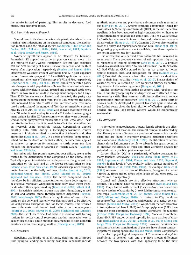
608 F. Baldacchino et al. / Infection, Genetics and Evolution 28 (2014) 596–615
the smoke instead of pasturing. This results in decreased foodintake, thus economic losses.
13.4. Insecticide-treated livestock
Several insecticides have been tested against tabanids with con-trasting results depending on the chemical compound, the applica-tion methods and the tabanid species (Anderson, 1985; Bruce andDecker, 1951; Foil et al., 1989b, 1990; Leak et al., 1995; Leprinceet al., 1991; Presley and Russel, 1986).
Synthetic pyrethroids appear to be effective for tabanids.Permethrin 1% applied on cattle as pour-on caused more than55% mortality over 2 weeks. Permethrin 10% ear tags produced79% and 55% mortality of Tabanus abactor the second and the thirdweeks after application, respectively (Presley and Russel, 1986).Effectiveness was most evident within the first 12-h post-exposureperiod. Fenvalerate sprays at 0.01% and 0.02% applied on cattle alsocaused mortality rates of Tabanus spp. of 67% and 79%, respectively(Leprince et al., 1991). Foil et al. (1989b) similarly detected parityrate changes in wild populations of horse flies when animals weretreated with fenvalerate sprays. Treated and untreated cattle wereplaced in two areas of wildlife management complex for 4 days.Then 1235 females were collected and dissected; the parity ratedecreased in the treated area from 44% to 31%, whereas the parityrate increased from 30% to 44% in the untreated area. This indi-cated a reduction of the number of flies that returned for a secondmeal by up to 20%. Foil et al. (1990) also showed that there was upto a 39% reduction of feeding time and a 31% reduction in engorge-ment weight for flies (T. fuscicostatus) when they were allowed tofeed on steers sprayed with fenvalerate at a sub-lethal dose. Thesetwo studies indicate that it is possible to affect the tabanid burdenon livestock using pyrethroid sprays. Cypermethrin appliedmonthly onto cattle during a tsetse/trypanosomosis controlprogram in Ethiopia resulted in a reduction of tabanids and otherbiting flies (Leak et al., 1995). In a series of reports (some unpub-lished due to the death of the first author), deltamethrin, appliedin pour-on or spray-on formulations to cattle every ten daysreduced the annoyance of tabanids in French Guiana (Raymondand Favre, 1991a,b,c).
In studies with less promising results, poor efficacy seems to berelated to the distribution of the compound on the animal body.Topically applied insecticides on cattle persist at the greatest con-centration on the back and at the lowest concentration on legs(Stendel et al., 1992; Vale et al., 1999). Tabanus spp. often stronglyprefer landing on the belly and legs (Baldacchino et al., 2014;Mohamed-Ahmed and Mihok, 2009; Muzari et al., 2010b;Raymond and Rousseau, 1987). The active compound should,therefore, be in sufficient concentration on these body regions tobe effective. Moreover, when licking their body, cows ingest insec-ticide which then appears in dung (Bourn et al., 2005; Laffont et al.,2001). Insecticide residues in dung may affect dung fauna, as wellas soil and aquatic fauna through further dispersion (Beynon,2012; Wardhaugh, 2005). Restricted application of insecticide tocattle on the belly and legs only was demonstrated to be effectivefor Amblyomma variegatum and for tsetse control. This reducedinsecticide costs and limited toxic effects on the ecosystem(Bourn et al., 2005; Bouyer et al., 2007; Stachurski and Lancelot,2006). The use of insecticidal foot baths in association with feedingstations for vector control represents another innovative way todeliver insecticides. These methods are amenable to both domesticlivestock and to free-ranging wildlife (Ndeledje et al., 2013).
13.5. Repellents
Repellents act locally or at distance, deterring an arthropodfrom flying to, landing on or biting host skin. Repellents include
synthetic substances and plant-based substances such as essentialoils (Nerio et al., 2010). Among synthetic compounds tested formosquitoes, N,N-diethyl-m-toluamide (DEET) is the most effectiverepellent. It has been sprayed at high concentration on horses toprotect them from tabanids and stable flies. DEET 75% was effectivefor 3–4 h, but adverse effects were observed: exfoliation, dermati-tis, salivation (Blume et al., 1971). Oxamate 20% was also tested oncows as a spray and repelled tabanids for 12 h (Minár et al., 1987).Long-lasting preparations are not available, thus these repellentsare not in common use for tabanids.
Use of essential oils derived from botanicals has increased inrecent years. These products can control arthropod pests by actingas repellents or feeding deterrents (Zhu et al., 2012). A productbased on essential oils called ‘‘Bremsen-Frei-Plus’’ (Co. Dr. SchaetteAG, Bad Waldsee, GER) sprayed on horses has protected horsesagainst tabanids, flies, and mosquitoes for 50 h (Sünder et al.,2011). Essential oils, however, lose effectiveness after a short timedue to their high volatility (Nerio et al., 2010). Encapsulation ofvolatile essential oils could be used to extend efficacy by control-ling release of the active ingredient (Dong et al., 2011).
Studies employing long-lasting dispensers with repellents arefew. In one study targeting tsetse, dispensers were attached to col-lars worn by cattle. They were not sufficiently effective to reducetrypanosome infection incidence (Bett et al., 2010). These sorts ofdevices could be developed to protect livestock against tabanids,but further research on the identification of effective repellents isneeded, and only long-lasting systems would be economicallysustainable.
13.6. Attractants
As for other hematophagous Diptera, female tabanids use olfac-tory stimuli in host location. The chemical compounds detected bythe olfactory organs of insects are products of mammalian metab-olism and are found in excretory products, glandular secretionsand exudations (Madubunyi et al., 1996). Identification of semio-chemicals, or kairomones specific to tabanids has great potentialto improve the efficacy of traps and other attractive devices forpotential use in practical pest management.
Carbon dioxide (CO2) is a particularly effective attractant formany tabanids worldwide (Cilek and Olson, 2008; Hayes et al.,1993; Leprince et al., 1994; Phelps and Vale, 1976; Raymond,1987b), higher levels of CO2 typically collect greater numbers oftabanids (Kline et al., 1991; Vale, 1980). For example, Kline et al.(1991) observed that catches of Diachlorus ferrugatus increased4 times, 27 times and 90 times when levels of CO2 were 0.02, 0.2and 2 L min�1, respectively.
Octenol and phenols are also effective attractants whereasketones, like acetone, have no effect on catches (Gibson and Torr,1999). Traps baited with octenol (1-octen-3-ol) can sometimesincrease catches of tabanids by 2- to 9-fold in comparison to unba-ited traps (Baldacchino et al., 2013c; Krcmar, 2005; Mihok et al.,2007; Mihok and Mulye, 2010). In contrast to CO2, no dose–response effect has been detected with octenol at practical concen-trations (Mihok and Mulye, 2010). Two phenols that are attractiveto tsetse, 4-methylphenol (4MP) and 3-n-propylphenol (3PP), havebeen the most commonly tested phenol attractants for tabanids(Krcmar, 2007; Phelps and Holloway, 1992). Alone or in combina-tions, 4MP, 3PP and/or octenol typically increase catches of taba-nids (Baldacchino et al., 2013c; Jaenson et al., 1991; Mihok andLange, 2012; Phelps and Holloway, 1992). However, detailed com-parisons of various combinations of phenols have shown contrast-ing patterns among species (Mihok and Mulye, 2010). Comparisonsof the electrophysiological responses of T. bromius and Atylotusquadrifarius to octenol, 4MP and 3PP have shown differencesbetween the two species, with 4MP appearing to be the most

F. Baldacchino et al. / Infection, Genetics and Evolution 28 (2014) 596–615 609
effective stimulus (Baldacchino et al., 2013c). These physiologicaldifferences likely explain diverse behavioral responses amongspecies among field studies.
Aged mammalian urine from certain species is now also knownto be effective in attracting tabanids (Djiteye et al., 1998; Krcmar,2005; Krcmar and Lajos, 2011; Krcmar et al., 2006; Mihok andLange, 2012). Livestock urine has been studied the most and con-sists of a complex mixture of ketones, fatty alcohols and phenols(Baldacchino et al., 2013c). The phenols 4MP and 3PP were origi-nally identified as the most important components for the attrac-tiveness of buffalo urine to tsetse (Owaga et al., 1988). In agedhorse urine volatiles, 4MP is the most abundant constituent at80% of total volatiles, whereas the proportion of 3PP is less than2% (Baldacchino et al., 2013c). The attractiveness of aged horseurine for tabanids appeared to result not only from the amountof 4MP in urine, but also from the presence of other active com-pounds that may act synergistically. Hribar et al. (1992) showedthat very high levels of ammonia attract Hybomitra lasiophthalma;striking synergism between ammonia and phenols has recentlybeen demonstrated for several Hybomitra spp. in Canada. Thismay explain the high attractiveness of aged urine to some, butnot all horse fly species (Mihok and Lange, 2012).
A better understanding of the involvement of kairomones inhost location should help in the design of more efficient tabanidtraps (or more practically, insecticide-impregnated blue–blacktargets as shown by Mihok and Lange, 2012). A better understandingof visual ecology is also needed to limit the catches of usefulinsects, especially pollinators. Even though many traps have beendesigned for sampling tabanids, and several effective attractantshave been identified, there are still very few studies of the use ofattractive devices for tabanid control. The difficulty of controllingtabanids on a large scale was shown many years ago in a trial ofodour-baited black targets to control tsetse flies in Zimbabwe.Although effective control of Glossina pallidipes was demonstrated,tabanid population densities did not change greatly in this majorexperiment over an area of 600 km2 (Vale et al., 1988). Since then,many similar trials have been conducted in Africa, but researchershave not typically enumerated tabanids, hence no new insightshave been gained.
On a local scale, sticky traps baited with a universal but imprac-tical attractant (dry ice) set around a pasture appeared to tempo-rarily reduce horse fly populations (Wilson, 1968). Numbersremained low on cattle while traps were in operation, butincreased within a few days after trapping was discontinued. Thecontinuing deployment of simple blue box traps to suppressgreenheads at Cape Cod, USA salt marshes (G. Sakolsky, personalcommunication) is probably the only ongoing example of the effec-tive application of trapping for tabanid control (Wall and Doane,1980). The nuisance level of horse flies has perceptively decreasedas a result of this program. However, long-term suppression hasnot been achieved, most likely because females T. nigrovittatusare autogenous and hence are not caught until after the firstoviposition.
Although great advances have been made in the development ofbaits and attractive devices for tsetse, and more recently for otherbiting flies, these techniques have yet to be tested for the protec-tion of livestock against tabanids. Investigations on the practicaluse of attractive devices for reducing tabanid bites on livestockin manageable situations (pastures, paddocks, and similar isolatedsituations) should be conducted.
14. Conclusion
This paper has highlighted the many direct and indirect effectsof tabanids as vectors of disease agents and has provided a brief
summary of tabanid morphology, biology and life cycle. Comparedto other biting flies such as stomoxes, tabanids are large insects.They are very prolific, laying 200–800 eggs in 5–6 ovipositionsfor a total production of up to 1000–4800 eggs. Tabanids live fora long time due to an extended larval stage which occurs in a widevariety of habitats. The larval stage can extend from 2 months to3 years; pupation is short. Adult emergence is highly seasonal withunpredictable peaks of abundance; this leads to short but extre-mely high periods tabanid activity. There are about 4400 tabanidspecies adapted to all kinds of landscapes. As a result of their largesize, highly prolific reproduction and seasonal abundance, tabanidscan have drastic impacts on both animals and people.
The direct impact of tabanids results from (i) the noise andvisual stress/annoyance produced when flying around hosts andthe contact stress of landing and biting, (ii) the pain of the biteitself (linked with the large size of mouthparts and damagingtelmophagous feeding mechanism), (iii) the toxic and allergeniceffects of their saliva, and (iv) the large bloodmeal size(20–600 lL/blood meal) with associated blood losses. We haveemphasized that these effects are far from negligible, and inhighly-infested areas, they may alone justify control.
As a result of very high attack rates at seasonally high densities,tabanids induce hosts, such as cattle, buffalos and/or horses togroup together. Hence infected and non-infected animals are inclose contact. This increases the probability of interrupted feedingon different hosts. As a result of the toxicity of saliva, biting sitesecondary infections, loss of blood, and resulting annoyance andstress, tabanid bites have immunosuppressive effects. This facili-tates increasing pathogenemia in infected hosts and susceptibilityin non-infected ones. As a result of the large size of their mouth-parts and the damaging telmophagic feeding mechanism, tabanidshave a painful bite. This induces host defensive movements, lead-ing to interrupted feeding. Tabanids can retain a large amount ofblood (7–15 nL) in their mouthparts, especially when comparedto Stomoxys (0.4 nL) or mosquitoes (0.001–0.0001 nL) (Foil andGorham, 2000). This makes them 20–20,000 times more efficientmechanical vectors than other biting insects.
The mechanisms and mathematical modeling of mechanicaltransmission of pathogens by tabanids indicate that transmissionis largely a non-specific process. If pathogenemia reaches orexceeds one million infecting doses per milliliter of blood, anypathogen can likely be mechanically transmitted. Experimentsunder realistic conditions of tabanid challenge indicate thatmechanical transmission can be a significant process, e.g., a60–80% incidence of new infections within 3 weeks of bitingexposure. Given the presence of tabanids in any kind of landscape,altitude, and climate, mechanical transmission must be consideredas a potential transmission mode of blood pathogens. However,variable peaks of tabanid abundance make quantitative predictionsof the impacts of mechanical transmission difficult. Predictions arefurther complicated by the restricted conditions necessary for effi-cient transmission (high abundance of tabanids, presence ofinfected hosts in close contact with non-infected ones, optimal rateof infection of the population, a mostly receptive population, exis-tence of very high pathogenemia in some of the ‘‘donor hosts’’,etc.). A last confounding variable is the presence of both biologicaland mechanical vectors in many situations, e.g., as for Trypanosomaspp. in Africa. Thus, assessing the epidemiological role of tabanidsin the mechanical transmission of disease agents requires detailedknowledge and sampling of tabanids and hosts, along with parallelinvestigations of the prevalence of infections.
In reviewing attempts to control tabanids, we suggest that newdevelopments in biting fly biology and control justify revisitingthis topic with a fresh approach. Due to the diversity of tabanids,the highly prolific reproduction, and the extensive habitat of larvalstages, controlling tabanid populations in vast, open areas (typical

610 F. Baldacchino et al. / Infection, Genetics and Evolution 28 (2014) 596–615
of farms) may limit what can be accomplished. Nevertheless,improvements in the use of insecticide-impregnated, odour-baitedtraps/targets to kill adult flies show great promise for providingsome level of relief to livestock. Reducing peak tabanid densitycould clearly reduce the direct impact of tabanids on a local scale.Practical tests of this nature have yet to be done, hence it is not yetclear what level of control would be needed to have an effect ondisease agent transmission and/or animal health. However, math-ematical modelling suggests that below a density of 10 flies perhead mechanical transmission would most often be of low effi-ciency; a threshold that could be tested in a practical setting.
In the long-term, repellents may provide the most sustainableapproach to prevent mechanical transmission; unfortunately, nolong-lasting products are readily-available to test now. Thus, inthe short-term, keeping animals at distance or under fly proof pro-tection remains the only practical strategy to prevent mechanicaltransmission. As an eminently simple strategy, it is also highly rec-ommended to keep bovines and horses separate.
Research studies on tabanids have received very little financialsupport, whether on aspects of blood feeding, mechanical trans-mission, or their control. This is most probably because (i) tabanidsare responsible for transitory/periodic problems, so demonstratingsuccess is challenging and remains a risky research topic, (ii) bio-logical vectors are more obvious since they produce events thatare predictable versus the unpredictable events produced bymechanical vectors, (iii) donors are keen to sponsor securedresearch projects with demonstrable, short-term outcomes, (iv)researchers do not like to pursue problems that require a long-term, integrated effort.
This review calls for more interest in tabanids and othermechanical vectors, given their demonstrated role in reducing live-stock production, and their important role in the mechanical trans-mission of diverse disease agents.
Acknowledgements
This project was supported by the University Paul-Valéry,Montpellier, France. We would like to thank Claire Garros, CIRAD,for her invitation to submit in this special issue. We also thankthe anonymous reviewers for critical and helpful comments onthe manuscript.
References
Abramov, I.V., Grobov, O.F., 1968. Transmission of the agent of bovine anaplasmosis(Anaplasma marginale Theiler 1910) by Tabanidae. Preliminary communication.In: Levine, N.D. (Ed.), Natural Nidality of Diseases and Questions of Parasitology.University of Illinois Press, Urbana, p. 158.
Alekseev, A.N., Chirov, P.A., Kadysheva, M.A., Sviridenko, V.F., 1969. Survival,multiplication and discharge into the environment of the pathogens oflisteriosis and unicteric leptospirosis by horse flies in experimentalconditions. Med. Parazitol. (Mosk) 38, 583–588.
Allan, S.A., Stoffolano Jr., J.G., Bennett, R.R., 1991. Spectral sensitivity of the horse flyTabanus nigrovittatus (Diptera: Tabanidae). Can. J. Zool. 69, 369–374.
Altunsoy, F., Kiliç, A.Y., 2012. Seasonal abundance of horse fly (Diptera: Tabanidae)in Western Anatolia. J. Entomol. Res. Soc. 14, 95–105.
Alvarez-García, G., Frey, C.F., Mora, L.M., Schares, G., 2013. A century of bovinebesnoitiosis: an unknown disease re-emerging in Europe. Trends Parasitol.http://dx.doi.org/10.1016/j.pt.2013.06.002.
Amano, K., 1985. Statistical analyses of the influence of meteorological factors onflight activity of female tabanids. Kontyû (Tokyo) 53, 161–172.
Amanzhulov, S.A., Amosenkova, N.I., Postricheva, O.V., 1965. Finding of Rickettsiaburneti in horseflies Tabanus staegeri. Med. Parazitol. 34, 612–614.
Anderson, J.F., 1985. The control of horse flies and deer flies (Diptera: Tabanidae).Myia 3, 547–598.
Anderson, J.R., Nilssen, A.C., Hemmingsen, W., 2001. Use of host-mimicking trapcatches to determine which parasitic flies attack reindeer, Rangifer tarandus,under different climatic conditions. Can. Field Nat. 115, 274–286.
Andreeva, V.R., 1976. Study of Coelomomyces milkoi Dudka et Koval, pathogenic forthe horsefly larvae. Med. Parazitol. 45, 212–218.
Andreeva, V.R., 1982. On ecologo-morphological typing of tabanid larvae (Diptera,Tabanidae). Entomol. Rev. 64, 49–54.
Andreeva, V.R., Kilic, A.Y., Altunsoy, F., 2009. New contribution to information abouttabanidae (Diptera) adult and larvae from West Anatolia. J. Entomol. Res. Soc.11, 19–30.
Aubry, P., Geale, D.W., 2011. A review of bovine anaplasmosis. TransboundaryEmerg. Dis. 58, 1–30.
Baldacchino, F., Cadier, J., Porciani, A., Buatois, B., Dormont, L., Jay-Robert, P., 2013a.Behavioural and electrophysiological responses of females of two species oftabanid to volatiles in urine of different mammals. Med. Vet. Entomol. 27, 77–85.
Baldacchino, F., Gardès, L., De Stordeur, E., Jay-Robert, P., Garros, C., 2013b.Blood-feeding patterns of horse flies in the French Pyrenees. Vet. Parasitol. 199,283–288.
Baldacchino, F., Manon, S., Puech, L., Buatois, B., Dormont, L., Jay-Robert, P., 2013c.Olfactory and behavioural responses of tabanid horseflies to octenol, phenolsand aged horse urine. Med. Vet. Entomol. http://dx.doi.org/10.1111/mve.12038.
Baldacchino, F., Muenworn, V., Desquesnes, M., Desoli, F., Charoenviriyaphap, T.,Duvallet, G., 2013d. Transmission of pathogens by Stomoxys flies (Diptera,Muscidae): a review. Parasite 20. http://dx.doi.org/10.1051/parasite/2113026.
Baldacchino, F., Porciani, A., Bernard, C., Jay-Robert, P., 2013e. Spatial and temporaldistribution of Tabanidae in the Pyrenees Mountains: influence of altitude andlandscape structure. Bull. Entomol. Res. 104, 1–11.
Baldacchino, F., Puech, L., Manon, S., Hertzog, L.R., Jay-Robert, P., 2014. Bitingbehaviour of Tabanidae on cattle in mountainous summer pastures, Pyrenees,France, and effects on weather variables. Bull. Entomol. Res. http://dx.doi.org/10.1017/S0007485314000170.
Barros, A.M., 2001. Seasonality and relative abundance of Tabanidae (Diptera)captured on horses in the Pantanal, Brazil. Mem. Inst. Oswaldo Cruz. 96, 917–923.
Barros, A.T.M., Foil, L.D., 2007. The influence of distance on movement of tabanids(Diptera: Tabanidae) between horses. Vet. Parasitol. 144, 380–384.
Bauer, B., Holzgrefe, B., Mahama, C.I., Baumann, M.P.O., Mehlitz, D., Clausen, P.-H.,2011. Managing tsetse transmitted trypanosomosis by insecticide treated nets– an affordable and sustainable method for resource poor pig farmers in Ghana.PLoS. Negl. Trop. Dis. http://dx.doi.org/10.1371/journal.pntd.0001343.
Baylis, M., 1996. Effect of defensive behaviour by cattle on the feeding success andnutritional state of the tsetse fly, Glossina pallidipes (Diptera: Glossinidae). Bull.Entomol. Res. 86, 329–336.
Bernard, G.D., 1971. Evidence for visual function of corneal interference filters. J.Insect Physiol. 17, 2287–2300.
Bett, B., Randolph, T.F., Irungu, P., Nyamwaro, S.O., Kitala, P., Gathuma, J., Grace, D.,Vale, G., Hargrove, J., McDermott, J., 2010. Field trial of a synthetic tsetse-repellent technology developed for the control of bovine trypanosomosis inKenya. Prev. Vet. Med. 97, 220–227.
Beynon, S.A., 2012. Potential environmental consequences of administration ofectoparasiticides to sheep. Vet. Parasitol. 189, 125–135.
Bhatia, H.L., 1935. The role of Tabanus orientis and Stomoxys calcitrans in themechanical transmission of rinderpest. Indian J. Vet. Sci. Anim. Husbandry 5, 2–22.
Bigalke, R.D., 1968. New concepts on the epidemiological features of bovinebesnoitiosis as determined by laboratory and field investigations.Onderstepoort J. Vet. 35, 3–137.
Blackburn, J.K., Curtis, A., Hadfield, T.L., O’Shea, B., Mitchell, M.A., Hugh-Jones, M.E.,2010. Confirmation of Bacillus anthracis from flesh-eating flies collected during awest Texas anthrax season. J. Wildl. Dis. 46, 918–922.
Blahó, M., Egri, Á., Bahidszki, L., Kriska, G., Hegedüs, R., Akesson, S., Horváth, G.,2012a. Spottier targets are less attractive to tabanid flies: on the tabanid-repellency of spotty fur patterns. PLoS One. http://dx.doi.org/10.1371/journal.pone.0041138.
Blahó, M., Egri, Á., Barta, A., Antoni, G., Kriska, G., Horváth, G., 2012b. How canhorseflies be captured by solar panels? A new concept of tabanid traps usinglight polarization and electricity produced by photovoltaics. Vet. Parasitol. 189,353–365.
Blahó, M., Egri, Á., Száz, D., Kriska, G., Akesson, S., Horváth, G., 2013. Stripes disruptodour attractivness to biting horseflies. Battle between ammonia, CO2, andcolour pattern for dominance in the sensory systems of host-seeking tabanids.Physiol. Behav. 119, 168–174.
Blume, R.R., Roberts, R.H., Eschle, J.L., Matter, J.J., 1971. Tests of aerosols of Deet forprotection of livestock from biting flies. J. Econ. Entomol. 64, 1193–1196.
Böse, R., Friedhoff, K.T., Olbrich, S., Buscher, G., Domeyer, I., 1987. Transmission ofTrypanosoma theileri to cattle by Tabanidae. Parasitol. Res. 73, 421–424.
Bourn, D., Grant, I., Shaw, A., Torr, S., 2005. Cheap and safe tsetse control forlivestock production and mixed farming in Africa. Aspects Appl. Biol. 75, 1–12.
Bouyer, J., Stachurski, F., Kabore, I., Bauer, B., Lancelot, R., 2007. Tsetse control incattle from pyrethroid footbaths. Prev. Vet. Med. 78, 223–238.
Brightwell, R., Dransfield, R.D., Kyorku, C., 1991. Development of a low-cost tsetsetrap and odor baits for Glossina pallidipes and G. longipennis in Kenya. Med. Vet.Entomol. 5, 153–164.
Bruce, W.N., Decker, G.C., 1951. Tabanid control on dairy and beef cattle withsynergized pyrethrins. J. Econ. Entomol. 44, 154–159.
Buxton, B.A., Hinkle, N.C., Schultz, R.D., 1985. Role of insects in the transmission ofbovine leukosis virus: potential for transmission by stable flies, horn flies, andtabanids. Am. J. Vet. Res. 46, 123–126.
Carn, V.M., 1996. The role of dipterous insects in the mechanical transmission ofanimal viruses. Br. Vet. J. 152, 377–393.
Catts, E.P., Mullen, G.R., 2002. Myiasis (Muscoidea, Ostroidea). In: Mullen, G.,Durden, L. (Eds.), Medical and Veterinary Entomology. Academic Press, SanDiego, pp. 318–347.
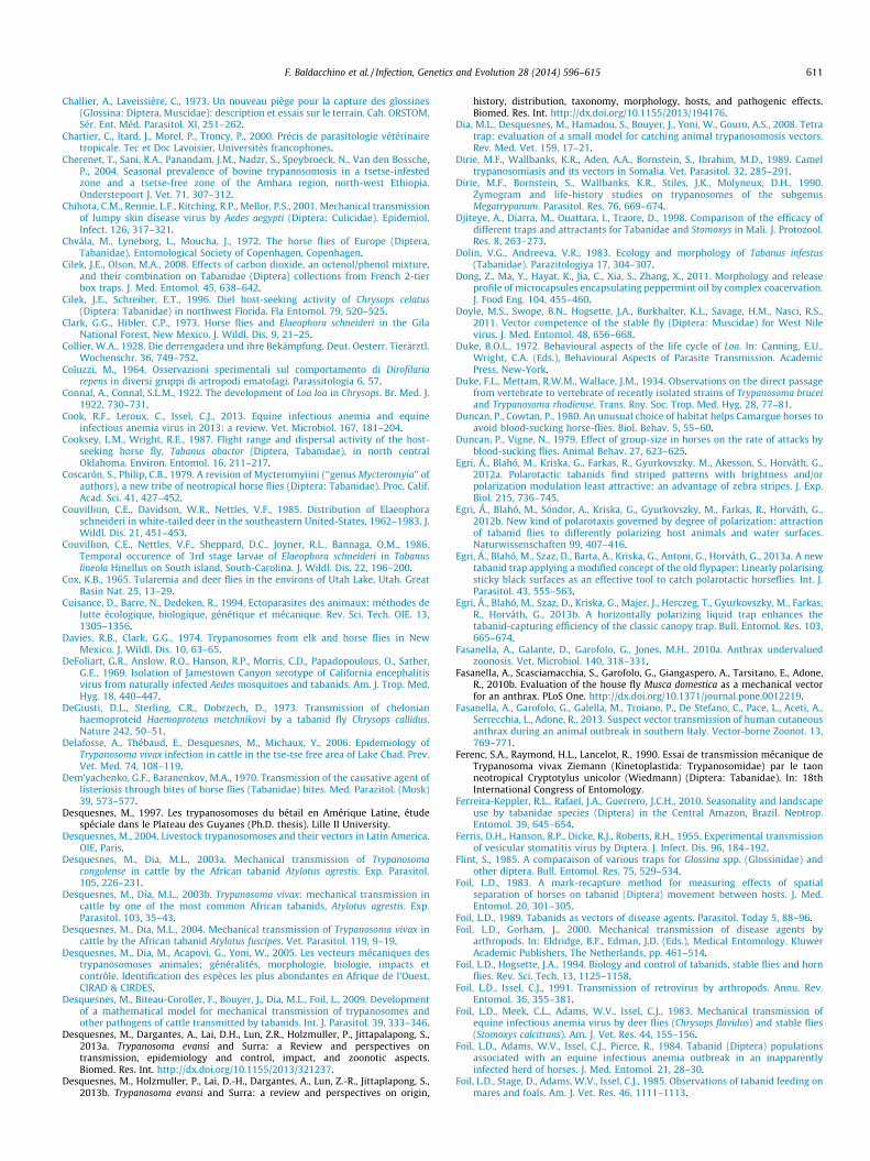
F. Baldacchino et al. / Infection, Genetics and Evolution 28 (2014) 596–615 611
Challier, A., Laveissière, C., 1973. Un nouveau piège pour la capture des glossines(Glossina: Diptera, Muscidae): description et essais sur le terrain. Cah. ORSTOM,Sér. Ent. Méd. Parasitol. XI, 251–262.
Chartier, C., Itard, J., Morel, P., Troncy, P., 2000. Précis de parasitologie vétérinairetropicale. Tec et Doc Lavoisier, Universités francophones.
Cherenet, T., Sani, R.A., Panandam, J.M., Nadzr, S., Speybroeck, N., Van den Bossche,P., 2004. Seasonal prevalence of bovine trypanosomosis in a tsetse-infestedzone and a tsetse-free zone of the Amhara region, north-west Ethiopia.Onderstepoort J. Vet. 71, 307–312.
Chihota, C.M., Rennie, L.F., Kitching, R.P., Mellor, P.S., 2001. Mechanical transmissionof lumpy skin disease virus by Aedes aegypti (Diptera: Culicidae). Epidemiol.Infect. 126, 317–321.
Chvála, M., Lyneborg, L., Moucha, J., 1972. The horse flies of Europe (Diptera,Tabanidae). Entomological Society of Copenhagen, Copenhagen.
Cilek, J.E., Olson, M.A., 2008. Effects of carbon dioxide, an octenol/phenol mixture,and their combination on Tabanidae (Diptera) collections from French 2-tierbox traps. J. Med. Entomol. 45, 638–642.
Cilek, J.E., Schreiber, E.T., 1996. Diel host-seeking activity of Chrysops celatus(Diptera: Tabanidae) in northwest Florida. Fla Entomol. 79, 520–525.
Clark, G.G., Hibler, C.P., 1973. Horse flies and Elaeophora schneideri in the GilaNational Forest, New Mexico. J. Wildl. Dis. 9, 21–25.
Collier, W.A., 1928. Die derrengadera und ihre Bekämpfung. Deut. Oesterr. Tierärztl.Wochenschr. 36, 749–752.
Coluzzi, M., 1964. Osservazioni sperimentali sul comportamento di Dirofilariarepens in diversi gruppi di artropodi ematofagi. Parassitologia 6, 57.
Connal, A., Connal, S.L.M., 1922. The development of Loa loa in Chrysops. Br. Med. J.1922, 730–731.
Cook, R.F., Leroux, C., Issel, C.J., 2013. Equine infectious anemia and equineinfectious anemia virus in 2013: a review. Vet. Microbiol. 167, 181–204.
Cooksey, L.M., Wright, R.E., 1987. Flight range and dispersal activity of the host-seeking horse fly, Tabanus abactor (Diptera, Tabanidae), in north centralOklahoma. Environ. Entomol. 16, 211–217.
Coscarón, S., Philip, C.B., 1979. A revision of Mycteromyiini (‘‘genus Mycteromyia’’ ofauthors), a new tribe of neotropical horse flies (Diptera: Tabanidae). Proc. Calif.Acad. Sci. 41, 427–452.
Couvillion, C.E., Davidson, W.R., Nettles, V.F., 1985. Distribution of Elaeophoraschneideri in white-tailed deer in the southeastern United-States, 1962–1983. J.Wildl. Dis. 21, 451–453.
Couvillion, C.E., Nettles, V.F., Sheppard, D.C., Joyner, R.L., Bannaga, O.M., 1986.Temporal occurence of 3rd stage larvae of Elaeophora schneideri in Tabanuslineola Hinellus on South island, South-Carolina. J. Wildl. Dis. 22, 196–200.
Cox, K.B., 1965. Tularemia and deer flies in the environs of Utah Lake, Utah. GreatBasin Nat. 25, 13–29.
Cuisance, D., Barre, N., Dedeken, R., 1994. Ectoparasites des animaux: méthodes delutte écologique, biologique, génétique et mécanique. Rev. Sci. Tech. OIE. 13,1305–1356.
Davies, R.B., Clark, G.G., 1974. Trypanosomes from elk and horse flies in NewMexico. J. Wildl. Dis. 10, 63–65.
DeFoliart, G.R., Anslow, R.O., Hanson, R.P., Morris, C.D., Papadopoulous, O., Sather,G.E., 1969. Isolation of Jamestown Canyon serotype of California encephalitisvirus from naturally infected Aedes mosquitoes and tabanids. Am. J. Trop. Med.Hyg. 18, 440–447.
DeGiusti, D.L., Sterling, C.R., Dobrzech, D., 1973. Transmission of chelonianhaemoproteid Haemoproteus metchnikovi by a tabanid fly Chrysops callidus.Nature 242, 50–51.
Delafosse, A., Thébaud, E., Desquesnes, M., Michaux, Y., 2006. Epidemiology ofTrypanosoma vivax infection in cattle in the tse-tse free area of Lake Chad. Prev.Vet. Med. 74, 108–119.
Dem’yachenko, G.F., Baranenkov, M.A., 1970. Transmission of the causative agent oflisteriosis through bites of horse flies (Tabanidae) bites. Med. Parazitol. (Mosk)39, 573–577.
Desquesnes, M., 1997. Les trypanosomoses du bétail en Amérique Latine, étudespéciale dans le Plateau des Guyanes (Ph.D. thesis). Lille II University.
Desquesnes, M., 2004. Livestock trypanosomoses and their vectors in Latin America.OIE, Paris.
Desquesnes, M., Dia, M.L., 2003a. Mechanical transmission of Trypanosomacongolense in cattle by the African tabanid Atylotus agrestis. Exp. Parasitol.105, 226–231.
Desquesnes, M., Dia, M.L., 2003b. Trypanosoma vivax: mechanical transmission incattle by one of the most common African tabanids, Atylotus agrestis. Exp.Parasitol. 103, 35–43.
Desquesnes, M., Dia, M.L., 2004. Mechanical transmission of Trypanosoma vivax incattle by the African tabanid Atylotus fuscipes. Vet. Parasitol. 119, 9–19.
Desquesnes, M., Dia, M., Acapovi, G., Yoni, W., 2005. Les vecteurs mécaniques destrypanosomoses animales; généralités, morphologie, biologie, impacts etcontrôle. Identification des espèces les plus abondantes en Afrique de l’Ouest.CIRAD & CIRDES.
Desquesnes, M., Biteau-Coroller, F., Bouyer, J., Dia, M.L., Foil, L., 2009. Developmentof a mathematical model for mechanical transmission of trypanosomes andother pathogens of cattle transmitted by tabanids. Int. J. Parasitol. 39, 333–346.
Desquesnes, M., Dargantes, A., Lai, D.H., Lun, Z.R., Holzmuller, P., Jittapalapong, S.,2013a. Trypanosoma evansi and Surra: a Review and perspectives ontransmission, epidemiology and control, impact, and zoonotic aspects.Biomed. Res. Int. http://dx.doi.org/10.1155/2013/321237.
Desquesnes, M., Holzmuller, P., Lai, D.-H., Dargantes, A., Lun, Z.-R., Jittaplapong, S.,2013b. Trypanosoma evansi and Surra: a review and perspectives on origin,
history, distribution, taxonomy, morphology, hosts, and pathogenic effects.Biomed. Res. Int. http://dx.doi.org/10.1155/2013/194176.
Dia, M.L., Desquesnes, M., Hamadou, S., Bouyer, J., Yoni, W., Gouro, A.S., 2008. Tetratrap: evaluation of a small model for catching animal trypanosomosis vectors.Rev. Med. Vet. 159, 17–21.
Dirie, M.F., Wallbanks, K.R., Aden, A.A., Bornstein, S., Ibrahim, M.D., 1989. Cameltrypanosomiasis and its vectors in Somalia. Vet. Parasitol. 32, 285–291.
Dirie, M.F., Bornstein, S., Wallbanks, K.R., Stiles, J.K., Molyneux, D.H., 1990.Zymogram and life-history studies on trypanosomes of the subgenusMegatrypanum. Parasitol. Res. 76, 669–674.
Djiteye, A., Diarra, M., Ouattara, I., Traore, D., 1998. Comparison of the efficacy ofdifferent traps and attractants for Tabanidae and Stomoxys in Mali. J. Protozool.Res. 8, 263–273.
Dolin, V.G., Andreeva, V.R., 1983. Ecology and morphology of Tabanus infestus(Tabanidae). Parazitologiya 17, 304–307.
Dong, Z., Ma, Y., Hayat, K., Jia, C., Xia, S., Zhang, X., 2011. Morphology and releaseprofile of microcapsules encapsulating peppermint oil by complex coacervation.J. Food Eng. 104, 455–460.
Doyle, M.S., Swope, B.N., Hogsette, J.A., Burkhalter, K.L., Savage, H.M., Nasci, R.S.,2011. Vector competence of the stable fly (Diptera: Muscidae) for West Nilevirus. J. Med. Entomol. 48, 656–668.
Duke, B.O.L., 1972. Behavioural aspects of the life cycle of Loa. In: Canning, E.U.,Wright, C.A. (Eds.), Behavioural Aspects of Parasite Transmission. AcademicPress, New-York.
Duke, F.L., Mettam, R.W.M., Wallace, J.M., 1934. Observations on the direct passagefrom vertebrate to vertebrate of recently isolated strains of Trypanosoma bruceiand Trypanosoma rhodiense. Trans. Roy. Soc. Trop. Med. Hyg. 28, 77–81.
Duncan, P., Cowtan, P., 1980. An unusual choice of habitat helps Camargue horses toavoid blood-sucking horse-flies. Biol. Behav. 5, 55–60.
Duncan, P., Vigne, N., 1979. Effect of group-size in horses on the rate of attacks byblood-sucking flies. Animal Behav. 27, 623–625.
Egri, Á., Blahó, M., Kriska, G., Farkas, R., Gyurkovszky, M., Akesson, S., Horváth, G.,2012a. Polarotactic tabanids find striped patterns with brightness and/orpolarization modulation least attractive: an advantage of zebra stripes. J. Exp.Biol. 215, 736–745.
Egri, Á., Blahó, M., Sóndor, A., Kriska, G., Gyurkovszky, M., Farkas, R., Horváth, G.,2012b. New kind of polarotaxis governed by degree of polarization: attractionof tabanid flies to differently polarizing host animals and water surfaces.Naturwissenschaften 99, 407–416.
Egri, Á., Blahó, M., Szaz, D., Barta, A., Kriska, G., Antoni, G., Horváth, G., 2013a. A newtabanid trap applying a modified concept of the old flypaper: Linearly polarisingsticky black surfaces as an effective tool to catch polarotactic horseflies. Int. J.Parasitol. 43, 555–563.
Egri, Á., Blahó, M., Szaz, D., Kriska, G., Majer, J., Herczeg, T., Gyurkovszky, M., Farkas,R., Horváth, G., 2013b. A horizontally polarizing liquid trap enhances thetabanid-capturing efficiency of the classic canopy trap. Bull. Entomol. Res. 103,665–674.
Fasanella, A., Galante, D., Garofolo, G., Jones, M.H., 2010a. Anthrax undervaluedzoonosis. Vet. Microbiol. 140, 318–331.
Fasanella, A., Scasciamacchia, S., Garofolo, G., Giangaspero, A., Tarsitano, E., Adone,R., 2010b. Evaluation of the house fly Musca domestica as a mechanical vectorfor an anthrax. PLoS One. http://dx.doi.org/10.1371/journal.pone.0012219.
Fasanella, A., Garofolo, G., Galella, M., Troiano, P., De Stefano, C., Pace, L., Aceti, A.,Serrecchia, L., Adone, R., 2013. Suspect vector transmission of human cutaneousanthrax during an animal outbreak in southern Italy. Vector-borne Zoonot. 13,769–771.
Ferenc, S.A., Raymond, H.L., Lancelot, R., 1990. Essai de transmission mécanique deTrypanosoma vivax Ziemann (Kinetoplastida: Trypanosomidae) par le taonneotropical Cryptotylus unicolor (Wiedmann) (Diptera: Tabanidae). In: 18thInternational Congress of Entomology.
Ferreira-Keppler, R.L., Rafael, J.A., Guerrero, J.C.H., 2010. Seasonality and landscapeuse by tabanidae species (Diptera) in the Central Amazon, Brazil. Neotrop.Entomol. 39, 645–654.
Ferris, D.H., Hanson, R.P., Dicke, R.J., Roberts, R.H., 1955. Experimental transmissionof vesicular stomatitis virus by Diptera. J. Infect. Dis. 96, 184–192.
Flint, S., 1985. A comparaison of various traps for Glossina spp. (Glossinidae) andother diptera. Bull. Entomol. Res. 75, 529–534.
Foil, L.D., 1983. A mark-recapture method for measuring effects of spatialseparation of horses on tabanid (Diptera) movement between hosts. J. Med.Entomol. 20, 301–305.
Foil, L.D., 1989. Tabanids as vectors of disease agents. Parasitol. Today 5, 88–96.Foil, L.D., Gorham, J., 2000. Mechanical transmission of disease agents by
arthropods. In: Eldridge, B.F., Edman, J.D. (Eds.), Medical Entomology. KluwerAcademic Publishers, The Netherlands, pp. 461–514.
Foil, L.D., Hogsette, J.A., 1994. Biology and control of tabanids, stable flies and hornflies. Rev. Sci. Tech. 13, 1125–1158.
Foil, L.D., Issel, C.J., 1991. Transmission of retrovirus by arthropods. Annu. Rev.Entomol. 36, 355–381.
Foil, L.D., Meek, C.L., Adams, W.V., Issel, C.J., 1983. Mechanical transmission ofequine infectious anemia virus by deer flies (Chrysops flavidus) and stable flies(Stomoxys calcitrans). Am. J. Vet. Res. 44, 155–156.
Foil, L.D., Adams, W.V., Issel, C.J., Pierce, R., 1984. Tabanid (Diptera) populationsassociated with an equine infectious anemia outbreak in an inapparentlyinfected herd of horses. J. Med. Entomol. 21, 28–30.
Foil, L.D., Stage, D., Adams, W.V., Issel, C.J., 1985. Observations of tabanid feeding onmares and foals. Am. J. Vet. Res. 46, 1111–1113.
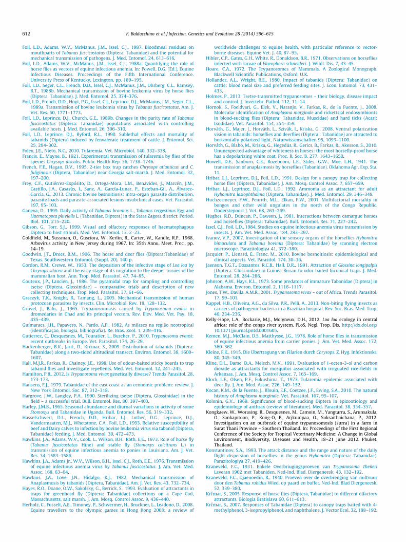
612 F. Baldacchino et al. / Infection, Genetics and Evolution 28 (2014) 596–615
Foil, L.D., Adams, W.V., McManus, J.M., Issel, C.J., 1987. Bloodmeal residues onmouthparts of Tabanus fuscicostatus (Diptera, Tabanidae) and the potential formechanical transmission of pathogens. J. Med. Entomol. 24, 613–616.
Foil, L.D., Adams, W.V., McManus, J.M., Issel, C.J., 1988a. Quantifying the role ofhorse flies as vectors of equine infectious anemia. In: Powell, D.G. (Ed.), EquineInfectious Diseases. Proceedings of the Fifth International Conference.University Press of Kentucky, Lexington, pp. 189–195.
Foil, L.D., Seger, C.L., French, D.D., Issel, C.J., McManus, J.M., Ohrberg, C.L., Ramsey,R.T., 1988b. Mechanical transmission of bovine leukemia virus by horse flies(Diptera, Tabanidae). J. Med. Entomol. 25, 374–376.
Foil, L.D., French, D.D., Hoyt, P.G., Issel, C.J., Leprince, D.J., McManus, J.M., Seger, C.L.,1989a. Transmission of bovine leukemia virus by Tabanus fuscicostatus. Am. J.Vet. Res. 50, 1771–1773.
Foil, L.D., Leprince, D.J., Church, G.E., 1989b. Changes in the parity rate of Tabanusfuscicostatus (Diptera: Tabanidae) populations associated with controllingavailable hosts. J. Med. Entomol. 26, 306–310.
Foil, L.D., Leprince, D.J., Byford, R.L., 1990. Sublethal effects and mortality oftabanids (Diptera) induced by fenvalerate treatment of cattle. J. Entomol. Sci.25, 294–302.
Foley, J.E., Nieto, N.C., 2010. Tularemia. Vet. Microbiol. 140, 332–338.Francis, E., Mayne, B., 1921. Experimental transmission of tularemia by flies of the
species Chrysops discalis. Public Health Rep. 36, 1738–1746.French, F.E., Hagan, D.V., 1995. 2-tier box trap catches Chrysops atlanticus and C.
fuliginosus (Diptera, Tabanidae) near Georgia salt-marsh. J. Med. Entomol. 32,197–200.
Frey, C.F., Gutiérrez-Expósito, D., Ortega-Mora, L.M., Benavides, J., Marcén, J.M.,Castillo, J.A., Casasùs, I., Sanz, A., García-Lunar, P., Esteban-Gil, A., Álvarez-García, G., 2013. Chronic bovine besnoitiosis: intra-organ parasite distribution,parasite loads and parasite-associated lesions insubclinical cases. Vet. Parasitol.197, 95–103.
Ganeva, D., 1999. Daily activity of Tabanus bromius L., Tabanus tergestinus Egg andHaematopota pluvialis L. (Tabanidae, Diptera) in the Stara Zagora district. Period.Biol. 101, 215–220.
Gibson, G., Torr, S.J., 1999. Visual and olfactory responses of haematophagousDiptera to host stimuli. Med. Vet. Entomol. 13, 2–23.
Goldfield, M., Sussman, O., Gusciora, W., Kerlin, R., Carter, W., Kandle, R.P., 1968.Arbovirus activity in New Jersey during 1967. In: 35th Annu. Meet. Proc., pp.14–19.
Goodwin, J.T., Drees, B.M., 1996. The horse and deer flies (Diptera:Tabanidae) ofTexas. Southwestern Entomol. (Suppl. 20), 140 p.
Gordon, R.M., Crewe, W., 1953. The deposition of the infective stage of Loa loa byChrysops silacea and the early stage of its migration to the deeper tissues of themammalian host. Ann. Trop. Med. Parasitol. 47, 74–85.
Gouteux, J.P., Lancien, J., 1986. The pyramidal trap for sampling and controllingtsetse (Diptera, Glossinidae) – comparative trials and description of newcollecting techniques. Trop. Med. Parasitol. 37, 61–66.
Graczyk, T.K., Knight, R., Tamang, L., 2005. Mechanical transmission of humanprotozoan parasites by insects. Clin. Microbiol. Rev. 18, 128–132.
Gruvel, J., Balis, J., 1965. Trypanosomiasis caused by Trypanosoma evansi indromedaries in Chad and its principal vectors. Rev. Elev. Med. Vet. Pay. 18,435–439.
Guimaraes, J.H., Papavero, N., Pardo, A.P., 1982. As miíases na região neotropical(identificação, biologia, bibliografia). Re. Bras. Zool. 1, 239–416.
Gutierrez, C., Desquesnes, M., Touratier, L., Buscher, P., 2010. Trypanosoma evansi:recent outbreaks in Europe. Vet. Parasitol. 174, 26–29.
Hackenberger, B.K., Jaric, D., Krcmar, S., 2009. Distribution of tabanids (Diptera:Tabanidae) along a two-sided altitudinal transect. Environ. Entomol. 38, 1600–1607.
Hall, M.J.R., Farkas, R., Chainey, J.E., 1998. Use of odour-baited sticky boards to traptabanid flies and investigate repellents. Med. Vet. Entomol. 12, 241–245.
Hamilton, P.B., 2012. Is Trypanosoma vivax genetically diverse? Trends Parasitol. 28,173-173.
Hansens, E.J., 1979. Tabanidae of the east coast as an economic problem: review. J.New York Entomol. Soc. 87, 312–318.
Hargrove, J.W., Langley, P.A., 1990. Sterilizing tsetse (Diptera, Glossinidae) in thefield – a successful trial. Bull. Entomol. Res. 80, 397–403.
Harley, J.M.B., 1965. Seasonal abundance and diurnal variations in activity of someStomoxys and Tabanidae in Uganda. Bull. Entomol. Res. 56, 319–332.
Hasselschwert, D.L., French, D.D., Hribar, L.J., Luther, D.G., Leprince, D.J.,Vandermaaten, M.J., Whetstone, C.A., Foil, L.D., 1993. Relative susceptibility ofbeef and Dairy calves to infection by bovine leukemia virus via tabanid (Diptera,Tabanidae) feeding. J. Med. Entomol. 30, 472–473.
Hawkins, J.A., Adams, W.V., Cook, L., Wilson, B.H., Roth, E.E., 1973. Role of horse fly(Tabanus fuscicostatus Hine) and stable fly (Stomoxys calcitrans L.) intransmission of equine infectious anemia to ponies in Louisiana. Am. J. Vet.Res. 34, 1583–1586.
Hawkins, J.A., Adams Jr., W.V., Wilson, B.H., Issel, C.J., Roth, E.E., 1976. Transmissionof equine infectious anemia virus by Tabanus fuscicostatus. J. Am. Vet. Med.Assoc. 168, 63–64.
Hawkins, J.A., Love, J.N., Hidalgo, R.J., 1982. Mechanical transmission ofAnaplasmosis by tabanids (Diptera, Tabanidae). Am. J. Vet. Res. 43, 732–734.
Hayes, R.O., Doane, O.W., Sakolsky, G., Berrick, S., 1993. Evaluation of attractants intraps for greenhead fly (Diptera: Tabanidae) collections on a Cape Cod,Massachusetts, salt marsh. J. Am. Mosq. Control Assoc. 9, 436–440.
Herholz, C., Fusselt, A.E., Timoney, P., Schwermer, H., Bruckner, L., Leadono, D., 2008.Equine travellers to the olympic games in Hong Kong 2008: a review of
worldwide challenges to equine health, with particular reference to vector-borne diseases. Equine Vet. J. 40, 87–95.
Hibler, C.P., Gates, G.H., White, R., Donaldson, B.R., 1971. Observations on horsefliesinfected with larvae of Elaeophora schneideri. J. Wildl. Dis. 7, 43–45.
Hoare, C.A., 1972. The Trypanosomes of Mammals. A Zoological Monograph.Blackwell Scientific Publications, Oxford, U.K.
Hollander, A.L., Wright, R.E., 1980. Impact of tabanids (Diptera: Tabanidae) oncattle: blood meal size and preferred feeding sites. J. Econ. Entomol. 73, 431–433.
Holmes, P., 2013. Tsetse-transmitted trypanosomes – their biology, disease impactand control. J. Invertebr. Pathol. 112, 11–14.
Hornok, S., Foeldvari, G., Elek, V., Naranjo, V., Farkas, R., de la Fuente, J., 2008.Molecular identification of Anaplasma marginale and rickettsial endosymbiontsin blood-sucking flies (Diptera: Tabanidae, Muscidae) and hard ticks (Acari:Ixodidae). Vet. Parasitol. 154, 354–359.
Horváth, G., Majer, J., Horváth, L., Szivák, I., Kriska, G., 2008. Ventral polarizationvision in tabanids: horseflies and deerflies (Diptera : Tabanidae) are attracted tohorizontally polarized light. Naturwissenschaften 95, 1093–1100.
Horváth, G., Blahó, M., Kriska, G., Hegedüs, R., Gerics, B., Farkas, R., Akesson, S., 2010.Ununexpected advantage of whiteness in horses: the most horsefly-proof horsehas a depolarizing white coat. Proc. R. Soc. B. 277, 1643–1650.
Howell, D.E., Sanborn, C.E., Rozeboom, L.E., Stiles, G.W., Moe, L.H., 1941. Thetransmission of anaplasmosis by horseflies (Tabanidae). Oklahoma Agr. Exp. Sta.11.
Hribar, L.J., Leprince, D.J., Foil, L.D., 1991. Design for a canopy trap for collectinghorse flies (Diptera, Tabanidae). J. Am. Mosq. Control Assoc. 7, 657–659.
Hribar, L.J., Leprince, D.J., Foil, L.D., 1992. Ammonia as an attractant for adultHybomitra lasiophthalma (Diptera, Tabanidae). J. Med. Entomol. 29, 346–348.
Huchzermeyer, F.W., Penrith, M.L., Elkan, P.W., 2001. Multifactorial mortality inbongos and other wild ungulates in the north of the Congo Republic.Onderstepoort J. Vet. 68, 263–269.
Hughes, R.D., Duncan, P., Dawson, J., 1981. Interactions between camargue horsesand horseflies (Diptera: Tabanidae). Bull. Entomol. Res. 71, 227–242.
Issel, C.J., Foil, L.D., 1984. Studies on equine infectious anemia virus transmission byinsects. J. Am. Vet. Med. Assoc. 184, 293–297.
Ivanov, V.P., 2007. Investigation of the sensory organs of the horseflies Hybomitrabimaculata and Tabanus bovinus (Diptera: Tabanidae) by scanning electronmicroscope. Parazitologiya 41, 372–380.
Jacquiet, P., Lienard, E., Franc, M., 2010. Bovine besnoitiosis: epidemiological andclinical aspects. Vet. Parasitol. 174, 30–36.
Jaenson, T.G.T., Dossantos, R.C.B., Hall, D.R., 1991. Attraction of Glossina longipalpis(Diptera: Glossinidae) in Guinea-Bissau to odor-baited biconical traps. J. Med.Entomol. 28, 284–286.
Johnson, A.W., Hays, K.L., 1973. Some predators of immature Tabanidae (Diptera) inAlabama. Environ. Entomol. 2, 1116–1117.
Jones, T.W., Davila, A.M.R., 2001. Trypanosoma vivax – out of Africa. Trends Parasitol.17, 99–101.
Kappel, H.B., Oliveira, A.G., da Silva, P.R., Pelli, A., 2013. Non-biting flying insects ascarriers of pathogenic bacteria in a Brazilian hospital. Rev. Soc. Bras. Med. Trop.46, 234–236.
Kelly-Hope, L.A., Bockarie, M.J., Molyneux, D.H., 2012. Loa loa ecology in centralafrica: role of the congo river system. PLoS. Negl. Trop. Dis. http://dx.doi.org/10.1371/journal.pntd.0001605.
Kemen, M.J., McClain, D.S., Matthysse, J.G., 1978. Role of horse flies in transmissionof equine infectious anemia from carrier ponies. J. Am. Vet. Med. Assoc. 172,360–362.
Kleine, F.K., 1915. Die Übertragung von Filarien durch Chrysops. Z. Hyg. Infektionskr.80, 345–349.
Kline, D.L., Dame, D.A., Meisch, M.V., 1991. Evaluation of 1-octen-3-ol and carbondioxide as attractants for mosquitos associated with irriguated rice-fields inArkansas. J. Am. Mosq. Control Assoc. 7, 165–169.
Klock, L.E., Olsen, P.F., Fukushima, T., 1973. Tularemia epidemic associated withdeer fly. J. Am. Med. Assoc. 226, 149–152.
Kocan, K.M., de la Fuente, J., Blouin, E.F., Coetzee, J.F., Ewing, S.A., 2010. The naturalhistory of Anaplasma marginale. Vet. Parasitol. 167, 95–107.
Kolonin, G.V., 1969. Significance of blood-sucking Diptera in epizootiology andepidemiology of anthrax (review of literature). Med. Parazitol. 38, 354–357.
Kongkaew, W., Worasing, R., Desquesnes, M., Camoin, M., Yangtarra, S., Arunsakula,O., Sankapitoon, P., Kong-O, P., Arjkumpaa, O., Suksaithaichana, P., 2012.Investigation on an outbreak of equine trypanosomosis (surra) in a farm inSurat Thani Province – Southern Thailand. In: Proceedings of the First RegionalConference of the Society for Tropical Veterinary Medicine: A Change in GlobalEnvironment, Biodiversity, Diseases and Health, 18–21 June 2012, Phuket,Thailand.
Konstantinov, S.A., 1993. The attack distance and the range and nature of the dailyflight dispersion of horseflies in the genus Hybomitra (Diptera: Tabanidae).Parazitologiya 27, 419–426.
Kraneveld, F.C., 1931. Enkele Overbriugingsproeven van Trypanosoma TheileriLaveran 1902 met Tabaniden. Ned-Ind. Blad. Diergeneesk. 43, 132–192.
Kraneveld, F.C., Djaenoedin, R., 1940. Proeven over de overbrenging van miltvuurdoor den Tabanus rubidus Wied. op paard en buffet. Ned-Ind. Blad Diergeneesk.52, 339–380.
Krcmar, S., 2005. Response of horse flies (Diptera, Tabanidae) to different olfactoryattractants. Biologia Bratislava 60, 611–613.
Krcmar, S., 2007. Responses of Tabanidae (Diptera) to canopy traps baited with 4-methylphenol, 3-isopropylphenol, and naphthalene. J. Vector Ecol. 32, 188–192.

F. Baldacchino et al. / Infection, Genetics and Evolution 28 (2014) 596–615 613
Krcmar, S., Lajos, P., 2011. Response of horse flies to aged equine urine (Diptera:Tabanidae). Entomol. Gen. 33, 245–250.
Krcmar, S., Mikuska, A., Merdic, E., 2006. Response of Tabanidae (Diptera) todifferent natural attractants. J. Vector Ecol. 31, 262–265.
Krinsky, W.L., 1976. Animal-disease agents transmitted by horse flies and deer flies(Diptera, Tabanidae). J. Med. Entomol. 13, 225–275.
Kriska, G., Bernáth, B., Farkas, R., Horváth, G., 2009. Degrees of polarization ofreflected light eliciting polarotaxis in dragonflies (Odonata), mayflies(Ephemeroptera) and tabanid flies (Tabanidae). J. Insect Physiol. 55, 1167–1173.
Laffont, C.M., Alvinerie, M., Bousquet-Melou, A., Toutain, P.L., 2001. Lickingbehaviour and environmental contamination arising from pour-on ivermectinfor cattle. Int. J. Parasitol. 31, 1687–1692.
Laveissière, C., Grébaut, P., 1990. Research on tsetse-fly traps (Diptera, Glossinidae)– development of an economic model, the Vavoua trap. Trop. Med. Parasitol. 41,185–192.
Lavoipierre, M.M.J., 1958. Studies on the host-parasite relationships of filarialnematodes and their arthropod hosts. I. The sites of development and themigration of Loa loa in Chrysops silacea, the escape of the infective forms fromthe head of the fly, and the effect of the worm on its insect host. Ann. Trop. Med.Parasitol. 52, 103–121.
Leak, S.G.A., Mulatu, W., Rowlands, G.J., Dieteren, G.D.M., 1995. A trial of acypermethrin pour-on insecticide to control Glossina pallidipes, G. fuscipesfuscipes and G. morsitans submorsitans (Diptera, Glossinidae) in south-westEthiopia. Bull. Entomol. Res. 85, 241–251.
Lehane, M.J., 2005. The biology of blood-sucking in insects. Cambridge UniversityPress, Cambridge.
Leiper, R.T., 1913. Calabar swellings. Lancet 1, 31.Leprince, D.J., Foil, L.D., Byford, R.L., 1991. Evaluation of pyrethroid ear tag and spray
treatment of cattle against horse flies (Diptera, Tabanidae). J. Entomol. Sci. 26,271–280.
Leprince, D.J., Hribar, L.J., Foil, L.D., 1994. Responses of horse flies (Diptera,Tabanidae) to Jersey Bullocks and canopy traps baited with ammonia, octenoland carbon dioxide. J. Med. Entomol. 31, 729–731.
Lessard, B.D., Cameron, S.L., Bayless, K.M., Wiegmann, B.M., Yeates, D.K., 2013. Theevolution and biogeography of the austral horse fly tribe Scionini (Diptera:Tabanidae: Pangoniinae) inferred from multiple mitochondrial and nucleargenes. Mol. Phylogenet. Evol. 68, 516–540.
LeVan, I.K., Fox, K.A., Miller, M.W., 2013. High elaeophorosis prevalence amongharvested colorado moose. J. Wildl. Dis. 49, 666–669.
Levine, J.F., Levy, M.G., Nicholson, W.L., Irby, W.S., Gager, R., Apperson, C.S., 1992.Attempted mechanical transfer of Ehrlichia risticii by tabanids (Diptera,Tabanidae). J. Med. Entomol. 29, 806–812.
Lienard, E., Salem, A., Jacquiet, P., Grisez, C., Prevot, F., Blanchard, B., Bouhsira, E.,Franc, M., 2013. Development of a protocol testing the ability of Stomoxyscalcitrans (Linnaeus, 1758) (Diptera: Muscidae) to transmit Besnoitia besnoiti(Henry, 1913) (Apicomplexa: Sarcocystidae). Parasitol. Res. 112, 479–486.
Lotze, J.C., 1944. Carrier cattle as a source of infective material for horseflytransmission of anaplasmosis. Am. J. Vet. Res. 5, 164–165.
Lotze, J.C., Yiengst, M.J., 1941. Mechanical transmission of bovine anaplasmosis bythe horsefly Tabanus sulcifrons. Am. J. Vet. Res. 2, 323–326.
Lunau, K., Knüttel, H., 1995. Vision through colored eyes. Naturwissenschaften 82,432–434.
Mackerras, I.M., 1954. The classification and distribution of Tabanidae (Diptera) I.General review. Aust. J. Zool. 2, 431–454.
Mackerras, I.M., 1955. The classification and distribution of Tabanidae (Diptera) II.History; morphology; classification; subfamily pangoniinae. Aust. J. Zool. 3,439–511.
Madubunyi, L.C., Hassanali, A., Ouma, W., Nyarango, D., Kabii, J., 1996.Chemoecological role of mammalian urine in host location by tsetse, Glossinaspp. (Diptera: Glossinidae). J. Chem. Ecol. 22, 1187–1199.
Magnarelli, L.A., Anderson, J.F., 1980. Feeding-behavior of Tabanidae (Diptera) oncattle and serological analysis of partial blood meals. Environ. Entomol. 9, 664–667.
Magnarelli, L.A., Anderson, J.F., Barbour, A.G., 1986. The etiologic agent of Lymedisease in deer flies, horse flies and mosquitoes. J. Infect. Dis. 154, 355–358.
Maia, M., Clausen, P.-H., Mehlitz, D., Garms, R., Bauer, B., 2010. Protection ofconfined cattle against biting and nuisance flies (Muscidae: Diptera) withinsecticide-treated nets in the Ghanaian forest zone at Kumasi. Parasitol. Res.106, 1307–1313.
Marchi, M.J., Pereira, P.A., Tironi De Menezes, R.M., Tubaki, R.M., 2012. New recordsof mosquitoes carrying Dermatobia hominis eggs in the state of Sao Paulo,Southeastern Brazil. J. Am. Mosq. Control Assoc. 28, 116–118.
Maresca, C., Scoccia, E., Faccenda, L., Zema, J., Costarelli, S., 2012. Equine infectiousanemia: active surveillance in Central Italy 2007–2009. J. Equine Vet. Sci. 32,596–598.
Mason, C., Norval, R., 1981. The transfert of Boophilus microplus (Acarina, Ixodidae)from infested to uninfested cattle under field conditions. Vet. Parasitol. 8, 185–188.
McElligott, P.E.K., Galloway, T.D., 1991. Daily activity patterns of horse flies(Diptera, Tabanidae, Hybomitra spp.) in northern and southern Manitoba. Can.Entomol. 123, 371–378.
McElligott, P.E.K., Lewis, D.J., 1998. Seasonal changes in abundance and gonotrophicage of host-seeking Tabanidae (Diptera) from a subarctic Labrador peatland. J.Med. Entomol. 35, 763–770.
McKeever, S., French, F.E., 1997. Fascinating, beautiful blood feeders: deer flies andhorse flies, the Tabanidae. Am. Entomol. 43, 217–226.
McKeever, S., French, F.E., 1999. Comparative study of adult mouthparts of fifty-twospecies representing thirty genera of Tabanidae (Diptera). Mem. Entomol. Int.14, 327–353.
Mihok, S., 2002. The development of a multipurpose trap (the Nzi) for tsetse andother biting flies. Bull. Entomol. Res. 92, 385–403.
Mihok, S., Lange, K., 2012. Synergism between ammonia and phenols for Hybomitratabanids in northern and temperate Canada. Med. Vet. Entomol. 26, 282–290.
Mihok, S., Mulye, H., 2010. Responses of tabanids to Nzi traps baited with octenol,cow urine and phenols in Canada. Med. Vet. Entomol. 24, 266–272.
Mihok, S., Carlson, D.A., Krafsur, E.S., Foil, L.D., 2006. Performance of the Nzi andother traps for biting flies in North America. Bull. Entomol. Res. 96, 387–397.
Mihok, S., Carlson, D.A., Ndegwa, P.N., 2007. Tsetse and other biting fly responses toNzi traps baited with octenol, phenols and acetone. Med. Vet. Entomol. 21, 70–84.
Miller, B.R., Kloter, K.O., Beaty, B.J., Magnarelli, L.A., 1983. Nonreplication ofJamestown Canyon and Keystone (California group) viruses in tabanids(Diptera, Tabanidae). J. Med. Entomol. 20, 214–215.
Minár, J., Kostenko, L.A., Riha, J., 1987. Losses in the milk yields of cows caused bythe bloodsucking dipterous insects and the protection of cattles with oxamaterepellent in the leningrad region. Vet. Med. Czech 32, 355–363.
Mitzmain, M.B., 1913. The mechanical transmission of surra by Tabanus striatusFabr. Philipp. J. Sci. 8, 223–229.
Mitzmain, M.B., 1914. Experimental insect transmission of anthrax. Public HealthRep. 29, 75–77.
Mohamed-Ahmed, M.M., Mihok, S., 2009. Alighting of Tabanidae and muscids onnatural and simulated hosts in the Sudan. Bull. Entomol. Res. 99, 561–571.
Mohamed-Ahmed, M.M., Abdulla, M.A., Mohamed, Y.O., El Rayah, I.E., El Amin, Y.E.,2007. Trapability of periurban populations of horseflies (Diptera: Tabanidae) inKarthoum State, Sudan. J. Sci. Technol. 8, 46–63.
Mohler, J.R., Thompson, W., 1911. A study of surra found in an importation of cattle,followed by prompt eradication. USDA Bureau of Animal Industry, Washington.
Moloo, S.K., Kabata, J.M., Gitire, N.M., 2000. Study on the mechanical transmissionby tsetse fly Glossina morsitans centralis of Trypanosoma vivax, T. congolense or T.brucei brucei to goats. Acta Trop. 74, 105–108.
Mooring, M.S., Blumstein, D.T., Reisig, D.D., Osborne, E.R., Niemeyer, J.M., 2007.Insect-repelling behaviour in bovids: role of mass, tail length, and group size.Biol. J. Linn. Soc. 91, 383–392.
Morgan, N.O., Lee, R.P., 1977. Vegetative barriers influence flight direction ofsaltmarsh greenheads. Mosq. News 37, 263–267.
Morita, S.I., 2008. A phylogeny of long-tongued horse flies (Diptera: Tabanidae:Philoliche) with the first cladistic review of higher relationships within thefamily. Invert. Syst. 22, 311–327.
Morris, H., 1918. Blood-sucking insects as transmitters of anthrax or charbon, 6:Baton Rouge, Louisiana Agr. Exp. Sta. Bull. 163.
Mullens, 2002. Horse flies and deer flies (Tabanidae). In: Mullen, G., Durden, L.(Eds.), Medical and Veterinary Entomology. Academic Press, San Diego, pp. 263–277.
Mullens, B.A., Lii, K.S., Mao, Y., Meyer, J.A., Peterson, N.G., Szijj, C.E., 2006.Behavioural responses of dairy cattle to the stable fly, Stomoxys calcitrans, inan open field environment. Med. Vet. Entomol. 20, 122–137.
Muzari, M.O., Jones, R.E., Skerratt, L.F., Duran, T.L., 2010a. Feeding success andtrappability of horse flies evaluated with electrocuting nets and odour-baitedtraps. Vet. Parasitol. 171, 321–326.
Muzari, M.O., Skerratt, L.F., Jones, R.E., Duran, T.L., 2010b. Alighting and feedingbehaviour of tabanid flies on horses, kangaroos and pigs. Vet. Parasitol. 170,104–111.
Nair, A.S., Ravindran, R., Lakshmanan, B., Kumar, S.S., Tresamol, P.V., Saseendranath,M.R., Senthilvel, K., Rao, J.R., Tewari, A.K., Ghosh, S., 2011. Haemoprotozoa ofcattle in Northern Kerala, India. Trop. Biomed. 28, 68–75.
Ndeledje, N., Bouyer, J., Stachurski, F., Grimaud, P., Belem, A.M.G., MoléléMbaïndingatoloum, F., Bengaly, Z., Oumar Alfaroukh, I., Cecchi, G., Lancelot, R.,2013. Treating cattle to protect people? Impact of footbath insecticidetreatment on tsetse density in Chad. PLoS One. http://dx.doi.org/10.1371/journal.pone.0067580.
Nerio, L.S., Olivero-Verbel, J., Stashenko, E., 2010. Repellent activity of essential oils:a review. Bioresour. Technol. 101, 372–378.
Nieschulz, O., 1925. Zoologishe bijdragen tot het Surraprobleem. I. Experimenteeleoverbrenging van Tryp. evansi door Tabanus stantoni Ricardo en T. ceylonicusSchiner. Veeartsenijk. Med. 53.
Nieschulz, O., 1926. Overbrengingsproeven mit Tabanus rubidus, Wied, T. striatus,Fabr., en Stomoxys calcitrans L. Veeartsenijk. Med. 55.
Nieschulz, O., 1927a. Overbrengingsproeven met Haematopota cingulata Wied. Ned.Ind. Blad Diergeneesk. 38.
Nieschulz, O., 1927b. Zoologische bijdragen tot het Surraproblem. XI. Enkeleproeven met Haematopota truncata Schuum. Stekh., H. irrorata Macq. en Tabanusbrumipes Sch. Steckh. Veeartsenijk. Med. 61.
Nieschulz, O., Huber, F.L., 1928. Over enkele boutvuuren parabout-vuuroverbrengingsproeven met Tabaniden. Veeartsenijk. Med. 67.
Nieschulz, O., Kraneveld, F.C., 1929. Experimentelle Untersuchungen über dieUebertragung der Büffelseuche durch Insekten. Zentralbl. Bakteriol. Origin 113,403–417.
Nieschulz, O., Ponto, S.A.S., 1927. Zoologishe bijdragen tot het Surraprobleem. XV.Enkele overbrengingsproeven met Chrysops flaviventris Macq. en C. dispar Fabr.Veeartsenijk. Med. 62.
Nigrovic, L.E., Wingerter, S.L., 2008. Tularemia. Infect. Dis. Clin. North Am. 22, 489–504.
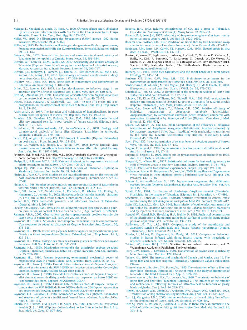
614 F. Baldacchino et al. / Infection, Genetics and Evolution 28 (2014) 596–615
Noireau, F., Nzoulani, A., Sinda, D., Itoua, A., 1990. Chrysops silacea and C. dimidiatafly densities and infection rates with Loa loa in the Chaillu mountains, CongoRepublic. Trans. R. Soc. Trop. Med. Hyg. 84, 153–155.
Nöller, W., 1916. Die Übertragung des Trypanosoma theileri laveran 1902. BerlinTieraerztl. Wochenschr 39, 457–460.
Nöller, W., 1925. Der Nachweis des Übertragers des gemeinen Rindertrypanosomas,Trypanosoma theileri, mit Hilfe des Kulturverfabrens. Zentralbl. Bakteriol. Origin79, 133–142.
Okiwelu, S.N., 1975. Seasonal distribution and variations in diurnal activity ofTabanidae in the republic of Zambia. Mosq. News. 35, 551–554.
Oliveira, A.F., Ferreira, R.L.M., Rafael, J.A., 2007. Seasonality and diurnal activity ofTabanidae (Diptera : Insecta) of canopy in the adolpho ducke forested reserve,Manaus, Amazonas State, Brazil. Neotrop. Entomol. 36, 790–797.
Oliveira, J.B., Montoya, J., Romero, J.J., Urbina, A., Soto-Barrientos, N., Melo, E.S.,Ramos, C.A., Araújo, F.R., 2010. Epidemiology of bovine anaplasmosis in dairyherds from Costa Rica. Vet. Parasitol. 177, 359–365.
Olsufiev, N.G., Golov, D.A., 1936. Horse flies as transmitters and convervators oftularemia. Animaux Pathog. 2, 187–226.
Orihel, T.C., Lowrie, R.C., 1975. Loa loa: development to infective stage in anamerican deerfly, Chrysops atlanticus. Am. J. Trop. Med. Hyg. 24, 610–615.
Otte, M.J., Abuabara, J.Y., 1991. Transmission of South-American Trypanosoma vivaxby the neotropical horsefly Tabanus nebulosus. Acta Trop. 49, 73–76.
Owaga, M.L.A., Hassanali, A., McDowell, P.G., 1988. The role of 4-cresol and 3-n-propylphenol in the attraction of tsetse flies to buffalo urine. Int. J. Trop. InsectSci. 9, 95–100.
Packchanian, A., 1957. The isolation and cultivation of hemoflagellates in pureculture from six species of insects. Tex. Rep. Biol. Med. 15, 399–410.
Parashar, B.D., Chauhan, R.S., Prakash, S., Rao, K.M., 1994. Mechanotactile andolfactory antennal sensilla in four species of female tabanids (Diptera). Boll.Zool. 61, 121–128.
Parra-Henao, G., Alarcon-Pineda, E.P., Lopez-Valencia, G., 2008. Ecology andparasitological analysis of horse flies (Diptera: Tabanidae) in Antioquia,Colombia. Caldasia 30, 179–188.
Perich, M.J., Wright, R.E., Lusby, K.S., 1986. Impact of horse flies (Diptera: Tabanidae)on beef-cattle. J. Econ. Entomol. 79, 128–131.
Perino, L.J., Wright, R.E., Hoppe, K.L., Fulton, R.W., 1990. Bovine leukosis virustransmission with mouthparts from Tabanus abactor after interrupted feeding.Am. J. Vet. Res. 51, 1167–1169.
Petersen, J.M., Mead, P.S., Schriefer, M.E., 2009. Francisella tularensis: an arthropod-borne pathogen. Vet. Res. http://dx.doi.org/10.1051/vetres:2008045.
Phelps, R.J., Holloway, M.T.P., 1992. Catches of tabanidae in response to visual andodour attractants in Zimbabwe. J. Afr. Zool. 106, 371–380.
Phelps, R.J., Vale, G.A., 1975. Seasonal flight periods of Tabanidae (Diptera) inRhodesia. J. Entomol. Soc. South. Afr. 38, 341–348.
Phelps, R.J., Vale, G.A., 1976. Studies on the local distribution and on the methods ofhost location of some Rhodesian Tabanidae (Diptera). J. Entomol. Soc. S. Afr. 39,67–81.
Philip, C.B., 1968. Overlap between neartic and neo-tropical faunae of Tabanidae inwestern North America (Diptera). Pan-Pac. Entomol. 44, 332–335.
Pinto, S.B., Soccol, V.T., Vendruscolo, E., Rochadelli, R., Ribeiro, P.B., Freitag, A.,Henemann, C., Uemura, M., 2002. Bioecologia de Dermatobia hominis (LinnaeusJr., 1781) em Palotina, Paraná, Brazil. Ciênca Rural 32, 821–827.
Poiner, G.O., 1985. Nematode parasites and infectious diseases of Tabanidae(Diptera). Myia 3, 599–616.
Presley, S.M., Russel, E.W., 1986. Field test of pyrethroid ear tags, sprays, and a pour-on formulation for control of horse flies on cattle. J. Agr. Entomol. 3, 369–373.
Rahman, A.H.A., 2005. Observations on the trypanosomosis problem outside thetsetse belts of Sudan. Rev. Sci. Tech. OIE 24, 965–972.
Raymond, H.L., 1987a. Action des taons (Diptera: Tabanidae) sur le comportementd’un troupeau de zébus au pâturage en Guyane Française. Ann. Zootech. 36,375–386.
Raymond, H.L., 1987b. Intérêt des pièges de Malaise appâtés au gaz carbonique pourl’étude des taons crépusculaires (Diptera: Tabanidae). Int. J. Trop. Insect Sci. 8,337–341.
Raymond, H.L., 1989a. Biologie des mouches-lézards, guêpes Bembicines de GuyaneFrancaise. Bull. Soc. Entomol. Fr. 93, 301–308.
Raymond, H.L., 1989b. Distribution temporelle des principales espèces de taons(Diptera. Tabanidae) nuisibles au bétail en Guyane Française. Ann. Soc. Entomol.Fr. 25, 289–294.
Raymond, H.L., 1990. Tabanus importunus, experimental mechanical vector ofTrypanosoma vivax in French Guiana. Ann. Parasitol. Hum. Comp. 65, 44–46.
Raymond, H.L., Favre, J., 1991a. Essai de lutte contre les taons de Guyane Française;efficacité et rémanence du RUV 165ND sur l’espèce crépusculaire Cryptolylusunicolor. Rapport INRA/Roussel-UCLAF (non publié).
Raymond, H.L., Favre, J., 1991b. Essai de lutte contre les taons de Guyane Française;effet d’un traitement de détiquage des bovins par aspersion au Butox 50ND surl’abondance des taons. Rapport INRA/Roussel-UCLAF.
Raymond, H.L., Favre, J., 1991c. Essai de lutte contre les taons de Guyane Française;comparaison du RUV 165ND, du Butox 50ND et du Butox 7,5ND pour la protectiondes bovins et des chevaux. Rapport INRA/Roussel-UCLAF (non publié).
Raymond, H.L., Rousseau, F., 1987. Abundance of horse flies (Diptera, Tabanidae)and reactions of cattle in a traditionnal farm of French-Guiana. Acta Oecol-OecAppl. 8, 125–134.
Ribeiro, P.B., Oliveira, C.M., Costa, P.R., Souza, F.S., 1985. Forèticos da Dermatobiahominis (L. Jr., 1781) (Diptera: Cuterebridae) no Rio Grande do Sul, Brasil. Arq.Bras. Med. Vet. Zoot. 37, 507–509.
Roberts, R.H., 1972. Relative attractiveness of CO2 and a steer to Tabanidae,Culicidae and Stomoxys calcitrans (L). Mosq. News. 32, 208–211.
Roberts, R.H., Love, J.N., 1977. Infectivity of Anaplasma marginale after ingestion bypotential insect vectors. Am. J. Vet. Res. 38, 1629–1630.
Roberts, L.W., Wilson, B.H., 1967. Predation on horse flies by two bembicine waspspecies in certain areas of southern Louisiana. J. Econ. Entomol. 60, 412–415.
Robinson, R.M., Jones, L.P., Galvin, T.J., Harwell, G.M., 1978. Elaeophorosis in sikadeer in Texas. J. Wildl. Dis. 14, 137–141.
Roskov, Y., Kunze, T., Paglinawan, L., Abucay, L., Orrell, T., Nicolson, D., Culham, A.,Bailly, N., Kirk, P., Bourgoin, T., Baillargeon, G., Decock, W., De Wever, A.,Didziulis, V., 2013. Species 2000 & ITIS Catalogue of Life, 10th December 2013.Species 2000, Naturalis, Leiden, The Netherlands. Digital resource at<www.catalogueoflife.org/col>.
Rutberg, A.T., 1987. Horse flies harassment and the social-behavior of feral ponies.Ethology 75, 145–154.
Sanborn, C.E., Stiles, G.W., Moe, L.H., 1932. Preliminary experiments in thetransmission of anaplasmosis by Horseflies. Okla. Agr. Exp. Sta. Bull. 204.
Santin-Duran, M., Alunda, J.M., San Miguel, J.M., Hoberg, E.P., de la Fuente, C., 2000.Elaeophorosis in red deer from Spain. J. Wildl. Dis. 36, 779–782.
Schofield, S., Torr, S.J., 2002. A comparison of the feeding behaviour of tsetse andstable flies. Med. Vet. Entomol. 16, 177–185.
Schreck, C.E., Kline, D.L., Williams, D.C., Tidwell, M.A., 1993. Field evaluations inmalaise and canopy traps of selected targets as attractants for tabanid species(Diptera, Tabanidae). J. Am. Mosq. Control Assoc. 9, 182–188.
Scoles, G.A., Broce, A.B., Lysyk, T.J., Palmer, G.H., 2005. Relative efficiency ofbiological transmission of Anaplasma marginale (Rickettsiales:Anaplasmataceae) by Dermacentor andersoni (Acari: Ixodidae) compared withmechanical transmission by Stomoxys calcitrans (Diptera: Muscidae). J. Med.Entomol. 42, 668–675.
Scoles, G.A., Miller, J.A., Foil, L.D., 2008. Comparison of the efficiency of biologicaltransmission of Anaplasma marginale (Rickettsiales: Anaplasmataceae) byDermacentor andersoni Stiles (Acari: Ixodidae) with mechanical transmissionby the horse fly, Tabanus fuscicostatus Hine (Diptera: Muscidae). J. Med.Entomol. 45, 109–114.
Scott, J.W., 1922. Insect transmission of swamp fever or infectious anemia of horses.Wyo. Agr. Exp. Sta. Bull. 133, 57–137.
Sergent, E., Sergent, E., 1905. Trypanosomiase des dromadaires de l’Afrique du nord.Ann. Instit. Pasteur. 19, 17–18.
Sergent, E., Sergent, E., 1906. Etudes sur les trypanosomiases de Berbérie en 1905.Ann. Instit. Pasteur. 20, 665–681.
Sheppard, C., Wilson, B.H., 1977. Relationship of horse fly host seeking activity toedge of wooded areas in southern Louisiana. Environ. Entomol. 6, 781–782.
Singh, K., 1926. Surra transmission experiments. Punjab. Dep. Agr. Vet. Bull.Sinshaw, A., Abebe, G., Desquesnes, M., Yoni, W., 2006. Biting flies and Trypanosoma
vivax infection in three highland districts bordering lake Tana, Ethiopia. Vet.Parasitol. 142, 35–46.
Solano, P., Amsler-Delafosse, S., 1995. Trypanosoma congolense chez différentesespèces de taons (Diptera: Tabanidae) au Burkina Faso. Rev. Elev. Med. Vet. Pay.48, 145–146.
Spratt, D.M., 1974. Distribution of third-stage Dirofilaria roemeri (Nematoda:Filarioidea) in the tissues of Tabanidae (Diptera). Int. J. Parasitol. 4, 477–480.
Stachurski, F., Lancelot, R., 2006. Footbath acaricide treatment to control cattleinfestation by the tick Amblyomma variegatum. Med. Vet. Entomol. 20, 402–412.
Stein, C.D., Lotze, J.C., Mott, L.O., 1942. Transmission of equine infectious anemia bythe stable fly, Stomoxys calcitrans, the horse fly, Tabanus sulcifrons (Macquart)and by injection of minute amounts of virus. Am. J. Vet. Res. 3, 183–193.
Stendel, W., Hamel, H.D., Sieveking, H.U., Bruhne, D., 1992. Analytical determinationof the distribution of flumethrin on the body-surface of cattle following topicalpour-on application. Vet. Parasitol. 42, 137–143.
Stoffolano, J.G., Yin, L.R.S., 1983. Comparative study of the mouthparts andassociated sensilla of adult male and female Tabanus nigrovittatus (Diptera,Tabanidae). J. Med. Entomol. 20, 11–32.
Sünder, U., Moors, E., Hagemann, K., Gauly, M., 2011. Comparative behaviourstudies in horses infested with flying insects treated with insecticide orrepellent substances. Berl. Munch. Tierarztl. 124, 28–35.
Takken, W., Knols, B.G.J., 2010. Olfaction in vector-host interactions, vol. 2.Wageningen Acadaemic Publishers, The Netherlands.
Tarry, D.W., Bernal, L., Edwards, S., 1991. Transmission of bovine virus diarrhea byblood feeding flies. Vet. Rec. 128, 82–84.
Teskey, H.J., 1990. The insects and arachnids of Canada and Alaska, part 16. Thehorse flies and deer flies (Diptera: Tabanidae). Agriculture Canada Publication,Ottawa.
Thorsteinson, A.J., Bracken, G.K., Hanec, W., 1965. The orientation of horse flies anddeer flies (Tabanidae, Diptera). III. The use of traps in the study of orientation oftabanids in the field. Entomol. Exp. Appl. 8, 189–192.
Thorsteinson, A.J., Bracken, G.K., Tostowaryk, W., 1966. The orientation behavior ofhorse flies and deer flies (Tabanidae: Diptera) V. The influence of the numberand inclination of reflecting surfaces on attractiveness to tabanids of glossyblack polyhedra. Can. J. Zool. 44, 275–279.
Tidwell, M.A., Dean, W.D., Combs, G.P., Anderson, D.W., Cowart, W.O., Axtell, R.C., 1972.Transmission of hog cholera virus by horseflies. Am. J. Vet. Res. 33, 615–622.
Torr, S.J., Mangwiro, T.N.C., 2000. Interactions between cattle and biting flies: effectson the feeding rate of tsetse. Med. Vet. Entomol. 14, 400–409.
Torr, S.J., Prior, A., Wilson, P.J., Schofield, S., 2007. Is there safety in numbers? Theeffect of cattle herding on biting risk from tsetse flies. Med. Vet. Entomol. 21,301–311.
F. Baldacchino et al. / Infection, Genetics and Evolution 28 (2014) 596–615 615
Torr, S.J., Mangwiro, T.N.C., Hall, D.R., 2011. Shoo fly, don’t bother me! Efficacy oftraditional methods of protecting cattle from tsetse. Med. Vet. Entomol. 25,192–201.
Turell, M.J., Knudson, G.B., 1987. Mechanical transmission of Bacillus anthracis bystable flies (Stomoxys calcitrans) and mosquitoes (Aedes aegypti and Aedestaeniorhynchus). Infect. Immun. 55, 1859–1861.
Vale, G.A., 1980. Field studies of the responses of tsetse flies (Glossinidae) and otherdiptera to carbon dioxide, acetone and other chemicals. Bull. Entomol. Res. 70,563–570.
Vale, G.A., 1982. The trap-oriented behaviour of tsetse flies (Glossinidae) and otherDiptera. Bull. Entomol. Res. 72, 71–93.
Vale, G.A., 1998. Responses of tsetse flies (Diptera: Glossinidae) to vegetation inZimbabwe: implications for population distribution and bait siting. Bull.Entomol. Res. 88, 10–54.
Vale, G.A., Phelps, R.J., 1974. Notes on the host-finding behaviour of Tabanidae(Diptera). Arnoldia, Rhodesia 6, 1–6.
Vale, G.A., Lovemore, D.F., Flint, S., Cockbill, G.G., 1988. Odour-baited targets tocontrol tsetse flies, Glossina spp. (Diptera: Glossinidae), in Zimbabwe. Bull.Entomol. Res. 78, 31–49.
Vale, G.A., Mutika, G., Lovemore, D.F., 1999. Insecticide-treated cattle for controllingtsetse flies (Diptera: Glossinidae): some questions answered, many posed. Bull.Entomol. Res. 89, 569–578.
Van den Bossche, P., 2001. Some general aspects of the distribution andepidemiology of bovine trypanosomosis in southern Africa. Int. J. Parasitol.31, 592–598.
Van Hennekeler, K., Jones, R.E., Skerratt, L.F., Fitzpatrick, L.A., Reid, S.A., Bellis, G.A.,2008. A comparison of trapping methods for Tabanidae (Diptera) in NorthQueensland, Australia. Med. Vet. Entomol. 22, 26–31.
Van Hennekeler, K., Jones, R.E., Skerratt, L.F., Muzari, M.O., Fitzpatrick, L.A., 2011.Meteorological effects on the daily activity patterns of tabanid biting flies innorthern Queensland, Australia. Med. Vet. Entomol. 25, 17–24.
Villa, A., Gutierrez, C., Gracia, E., Moreno, B., Chacon, G., Sanz, P.V., Buescher, P.,Touratier, L., 2008. Presence of Trypanosoma theileri in Spanish Cattle. Ann. N. Y.Acad. Sci. 1149, 352–354.
Wall, W.J., Doane Jr, O.W., 1980. Large scale use of box traps to study and controlsaltmarsh greenhead flies (Diptera: Tabanidae) on Cape Cod, Massachusetts.Environ. Entomol. 9, 371–375.
Wardhaugh, K.G., 2005. Insecticidal activity of synthetic pyrethroids,organophosphates, insect growth regulators, and other livestock parasiticides:an Australian perspective. Environ. Toxicol. Chem. 24, 789–796.
Watson, D.W., Denning, S.S., Calibeo-Hayes, D.I., Stringham, S.M., Mowrey, R.A.,2007. Comparison of two fly traps for the capture of horse flies (Diptera:Tabanidae). J. Entomol. Sci. 42, 123–132.
Weinmann, C.J., Anderson, J.R., Longhurst, W.M., Connolly, G., 1973. Filarial wormsof Columbian black-tailed deer in California. 1. Observations in the vertebratehost. J. Wildl. Dis. 9, 213–220.
Wellmann, G., 1950. Blood-sucking arthropods as the vectors of zootic disease. Berl.Munch. Tierarztl. 8, 155–160.
Wellmann, G., 1951. Blood sucking insects as mechanical vectors of Brucella.Zentralbl. Bakteriol. Origin 156, 414–426.
Wiesenhutter, E., 1975a. Research into the relative importance of Tabanidae(Diptera) in mechanical disease transmission. II. Investigation of the behaviourand feeding habits of Tabanidae in relation to cattle. J. Nat. His. 9, 385–392.
Wiesenhutter, E., 1975b. Research into the relative importance of Tabanidae(Diptera) in mechanical disease transmission. III. The epidemiology ofanaplasmosis in a Dar-es-Salaam dairy farm. Trop. Anim. Health Prod. 7, 15–22.
Wiesenhutter, E., 1976. Research into the relative importance of Tabanidae(Diptera) in mechanical disease transmission. IV. A contribution to theepidemiology of bovine trypanosomiasis in Africa. Trop. Anim. Health Prod. 8,195–201.
Wilkerson, R.C., Butler, J.F., Pechuman, L.L., 1985. Swarming, hovering, and matingbehavior of male horse flies and deer flies (Diptera: Tabanidae). Myia 3, 515–546.
Williams, P., 1960. Studies on Ethiopian Chrysops as possible vectors of loiasis. II.Chrysops silacea Austen and human loiasis. Ann. Trop. Med. Parasitol. 54, 439–459.
Wilson, B.H., 1968. Reduction of tabanid populations on cattle with sticky trapsbaited with dry ice. J. Econ. Entomol. 61, 827–829.
Wilson, B.H., Meyer, R.B., 1966. Transmission studies of bovine anaplasmosis withhorse flies Tabanus fuscicostatus and Tabanus nigrovittatus. Am. J. Vet. Res. 27,367–369.
Wunderer, H., Smola, U., 1986. Functional morphology of the retina of Chrysopscaecutiens L. and Haematopota pluvialis L. (Diptera: Tabanidae): region aroundeye equator. Int. J. Insect Morphol. 15, 311–319.
Yagodinsky, V.N., Skvortsov, B.I., 1962. [On the question of epidemiology andincidence of tick-borne encephalitis by unknown means of infection]. Avtoref.Kratk. Soobshch. Ob’edinen. 7. Sess. Inst. Polio. Virus. Entsef. AMN SSR Beloruss.Inst. Epidem. Mikrobiol. Gig. (Minsk), pp. 169–170.
Yutuc, L.M., 1949. Observations on the prevalence of tabanid flies and surra-transmission experiments. Philipp. J. Sci. 78, 379–387.
Zhanseitova, M.T., Shuratov, I., Drobishchenko, N.I., Chizhov, A.V., 1982. Isolation ofinfluenza viruses from bloodsucking arthropods in Kazakhastan. Izv. Akad.Nauk Kaz. SSR. Ser. Biol. 3, 57–59.
Zhu, J.J., Berkebile, D.R., Dunlap, C.A., Zhang, A., Boxler, D., Tangtrakulwanich, K.,Behle, R.W., Baxendale, F., Brewer, G., 2012. Nepetalactones from essential oil ofNepeta cataria represent a stable fly feeding and oviposition repellent. Med. Vet.Entomol. 26, 131–138.