
T Cell Recognition of Major Histocompatibility Complex Class II Complexes with Invariant Chain...
11
T Cell Recognition of Major Histocompatibility Complex Class II Complexes with Invariant Chain Processing Intermediates By Stanislaw Morkowski,* Ananda W. Goldrath,* Susan Eastman,~ Lakshmi tkamachandra,:[: Daniel C. Freed,w Phyllis Whiteley,w Hand AlexanderYu. Rudensky*:~ From the *Department of Immunology and ");Howard Hughes Medical Institute, University of Washington, Seattle, Washington 98195; and w Research Laboratories,Rahaway, New Jersey 07065; and ][Syntex Discovery Research, PaloAlto, California 94304 Summary Peptides from the lumenal portion of invariant chain (Ii) spanning residues 80--106 (class II- associated Ii peptide [CLIP]) are found in association with several mouse and human major his- tocompatibility complex (MHC) class II allelic variants in wild-type and presentation-deficient mutant cells. The ready detection of these complexes suggests that such an intermediate is es- sential to the MHC class II processing pathway. In this study, we demonstrate that T cells rec- ognize CLIP/MHC class II complexes on the surface of normal and mutant cells in a manner indistinguishable from that of nominal antigenic peptides. Surprisingly, T cell hybrids specific for human CLIP bound to routine MHC class II molecule I-Ab and a new monoclonal anti- body 30-2 with the same specificity, recognize two independent epitopes expressed on this peptide/class II complex. T cell recognition is dependent on a Gln residue (position 100) in CLIP, whereas the 30-2 antibody recognizes a Lys residue at position 90. These two residues flank the 91-99 sequence that is conserved among human, mouse, and rat Ii, potentially repre- senting an MHC class II-binding site. Our results suggest that the COOH-terminal portion of CLIP that includes TCR contact residue Gln 100 binds in the groove ofI-A b molecule. More- over, both T cells and the antibody recognize I-Ab complexed with larger Ii processing inter- mediates such as the "-'12-kD small leupeptin-induced protein (SLIP) fragments. Thus, SLIP fragments contain a CLIP region bound to MHC class II molecule in a conformation identical to that of a free CLIP peptide. Finally, our data suggest that SLIP/MHC class II complexes are precursors of CLIP/MHC class II complexes. 1 Tnvariant chain (Ii) is a type II transmembrane glycopro- Jktein transiently associated with MHC class II molecules- during their intracellular assembly and transport (1). The association of class II with Ii interferes with peptide binding to the peptide-binding groove of MHC class II molecules (2-4). Large multimeric complexes of three Ii polypeptides and three MHC class II ot~3 dimers form rapidly in the en- doplasmic reticulum (ELL) (5). These complexes are trans- ported across Golgi stacks to the trans-Golgi network and are sorted to the endosomal compartment (1). Several stud- ies in mice with a targeted disruption of the Ii gene dem- onstrated that this sorting event is to a large extent medi- ated by Ii (6-8). Earlier in vitro studies revealed an endosomal 1Abbreviations used in this paper: CLIP, class lI-associated invariant chain peptide; ER, endoplasmic reticulum; HEL, hen egg lysozyme;HPLC, high performance liquid chromatography; Ii, invariantchain; SLIP,small leupeptin-inducedprotein;TfR, transferrinreceptor. targeting signal in the first 16 amino acid residues in the cy- toplasmic tail of Ii (9). After arrival in endosomes, Ii is cleaved by endosomal proteases. Studies using protease in- hibitors indicated that an aspartyl proteinase(s) and a cys- teine proteinase(s) are sequentially involved in endosomal degradation of Ii in B cells (10, 11). In cells treated with a cysteine proteinase inhibitor, leupeptin, two major inter- mediates of Ii processing were demonstrated (10, 11). These intermediates are NH2-terminal 21-24-kD frag- ments of Ii (leupeptin-induced protein [LIP]) and 11-14-kD fragments (small leupeptin-induced protein [SLIP]). Sequence analysis of peptides associated with several al- lelic variants of human and mouse MHC class II in normal cells demonstrated an abundant set ofpeptides with ragged NH2- and COOH-termini derived from Ii residues 80-106 (12-15). Importantly, these peptides are associated with the majority of MHC class II molecules expressed in human presentation deficient mutant cell lines. The peptides were 1403 J. Exp. Med. 9 The Rockefeller University Press 9 0022-1007/95 / 11/ 1403 / 11 $2.00 Volume 182 November 1995 1403-1413 on August 16, 2015 jem.rupress.org Downloaded from Published November 1, 1995
-
Upload
universitasnegerimalang -
Category
Documents
-
view
1 -
download
0
Transcript of T Cell Recognition of Major Histocompatibility Complex Class II Complexes with Invariant Chain...

T Cell Recognition of Major Histocompatibility Complex Class II Complexes with Invariant Chain Processing Intermediates By Stanislaw M o r k o w s k i , * Ananda W. Go ld ra th ,* Susan Eastman,~
Lakshmi tkamachandra,:[: Dan ie l C. Freed ,w Phyllis Whi te ley ,w H and
Alexande rYu . Rudensky*:~
From the *Department of Immunology and ");Howard Hughes Medical Institute, University of Washington, Seattle, Washington 98195; and w Research Laboratories, Rahaway, New Jersey 07065; and ][Syntex Discovery Research, Palo Alto, California 94304
Summary Peptides from the lumenal portion of invariant chain (Ii) spanning residues 80--106 (class II- associated Ii peptide [CLIP]) are found in association with several mouse and human major his- tocompatibility complex (MHC) class II allelic variants in wild-type and presentation-deficient mutant cells. The ready detection of these complexes suggests that such an intermediate is es- sential to the MHC class II processing pathway. In this study, we demonstrate that T cells rec- ognize CLIP/MHC class II complexes on the surface of normal and mutant cells in a manner indistinguishable from that of nominal antigenic peptides. Surprisingly, T cell hybrids specific for human CLIP bound to routine MHC class II molecule I-A b and a new monoclonal anti- body 30-2 with the same specificity, recognize two independent epitopes expressed on this peptide/class II complex. T cell recognition is dependent on a Gln residue (position 100) in CLIP, whereas the 30-2 antibody recognizes a Lys residue at position 90. These two residues flank the 91-99 sequence that is conserved among human, mouse, and rat Ii, potentially repre- senting an MHC class II-binding site. Our results suggest that the COOH-terminal portion of CLIP that includes T C R contact residue Gln 100 binds in the groove ofI-A b molecule. More- over, both T cells and the antibody recognize I-A b complexed with larger Ii processing inter- mediates such as the "-'12-kD small leupeptin-induced protein (SLIP) fragments. Thus, SLIP fragments contain a CLIP region bound to MHC class II molecule in a conformation identical to that of a free CLIP peptide. Finally, our data suggest that SLIP/MHC class II complexes are precursors of CLIP/MHC class II complexes.
1 Tnvariant chain (Ii) is a type II transmembrane glycopro- Jktein transiently associated with MHC class II molecules- during their intracellular assembly and transport (1). The association of class II with Ii interferes with peptide binding to the peptide-binding groove of MHC class II molecules (2-4). Large multimeric complexes of three Ii polypeptides and three MHC class II ot~3 dimers form rapidly in the en- doplasmic reticulum (ELL) (5). These complexes are trans- ported across Golgi stacks to the trans-Golgi network and are sorted to the endosomal compartment (1). Several stud- ies in mice with a targeted disruption of the Ii gene dem- onstrated that this sorting event is to a large extent medi- ated by Ii (6-8). Earlier in vitro studies revealed an endosomal
1Abbreviations used in this paper: CLIP, class lI-associated invariant chain peptide; ER, endoplasmic reticulum; HEL, hen egg lysozyme; HPLC, high performance liquid chromatography; Ii, invariant chain; SLIP, small leupeptin-induced protein; TfR, transferrin receptor.
targeting signal in the first 16 amino acid residues in the cy- toplasmic tail of Ii (9). After arrival in endosomes, Ii is cleaved by endosomal proteases. Studies using protease in- hibitors indicated that an aspartyl proteinase(s) and a cys- teine proteinase(s) are sequentially involved in endosomal degradation of Ii in B cells (10, 11). In cells treated with a cysteine proteinase inhibitor, leupeptin, two major inter- mediates of Ii processing were demonstrated (10, 11). These intermediates are NH2-terminal 21-24-kD frag- ments of Ii (leupeptin-induced protein [LIP]) and 11-14-kD fragments (small leupeptin-induced protein [SLIP]).
Sequence analysis of peptides associated with several al- lelic variants of human and mouse MHC class II in normal cells demonstrated an abundant set ofpeptides with ragged NH2- and COOH-termini derived from Ii residues 80-106 (12-15). Importantly, these peptides are associated with the majority of MHC class II molecules expressed in human presentation deficient mutant cell lines. The peptides were
1403 J. Exp. Med. �9 The Rockefeller University Press �9 0022-1007/95 / 11 / 1403 / 11 $2.00 Volume 182 November 1995 1403-1413
on August 16, 2015
jem.rupress.org
Dow
nloaded from
Published November 1, 1995

named CLIP (class II-associated Ii peptide; 16). Recent stud- ies using a series o f deletions in the lumenal port ion o f Ii indicate that the same 80-110 region o f Ii is essential for its binding to M H C class II (17-19). Altogether, these results suggest that C L I P / M H C class II complexes are essential in- termediates in the M H C class II processing pathway. The relationship be tween CLIP and larger Ii fragments such as LIP and SLIP, however, remains unclear. Thus, it is not known whether CLIP represents a remnant o f l i still associ- ated with M H C class II after Ii truncation (12) or, alterna- tively, if CLIP is first released during Ii proteolysis and sub- sequently rebinds in the peptide-binding groove (12, 16, 20).
To address these questions, we have generated and char- acterized a panel o f T cell hybrids recognizing human CLIP peptides bound to mouse M H C class II molecule I -A b. Using these T cells and a new mAb, 30-2, which rec- ognizes the same complex (Eastman, S., M. Deftos, P. De - Roos, D. H. Hsu, L. Teyton, N. S. Braunstein, C. J. Hackett , and A. Yu. Rudensky, manuscript submitted for publication), we show here that the CLIP region o f Ii binds to M H C class II molecule in a conformation similar to that o f the free peptide. Mapping o f antibody and T cell epitopes within the CLIP peptide allowed us to identify the con- served 91-99 region o f Ii as a peptide capable o f compet i - tively inhibit ing M H C class II binding to antigenic pep- tides. Further, we demonstrate that the ~ 1 2 - k D SLIP fragment of li is a precursor of CLIP. O u r data suggest a model o f CLIP binding to I -A b with the C O O H terminus o f this peptide bound in the M H C class I I -b ind ing groove.
Materials and Methods Mice. C57BL/6, BALB/c, and B6.C-H-2 bin12 mice were
purchased from The Jackson Laboratory (Bar Harbor, ME). Antibodies. The mAb 30-2 recognizing human CLIP/I-A b
complex was generated in our laboratory (Eastman, S., et al., manuscript submitted for publication), and the the YAe antibody recognizing complex of I-A b molecule with Ec~52-68 peptide was characterized earlier (21). The mAbs PIN.1 and 1G12, which recognize the cytoplasmic tail of the human Ii chain (5) and hu- man transferrin receptor (TfR), respectively, were kindly pro- vided by Dr. Janice Blum (University of Indiana, Indianapolis, IN). Antibody BU45 against lumenal portion of human Ii chain was purchased from The Binding Site (San Diego, CA). Mouse hybridoma HP6035 producing mAb against human Ig K chain was purchased from American Type Culture Collection (Rock- ville, MD). Antibody Y237, which recognizes I-A b, was kindly provided by Dr. Donal Murphy (New York State Department of Health, Albany, NY). Hybridomas secreting l-Ab-specific mAbs Y3P, AF6-120.1.2, and M5/114 are maintained in our labora- tory. All antibodies were purified from culture supernatants or as- citic fluids on protein G-Sepharose columns.
Cell Lines. The mouse B cell lymphoma LB27.4 (I-A bxa) was cotransfected with a genomic construct of the human Ii (gift from Dr. Paul Roche, National Institutes of Health, Bethesda, MD) and a vector carrying the neomycin resistance pMClpA. Trans- fectants were selected for Ii expression by Western blotting of cell lysates using the anti-human Ii antibody PIN. 1. The T2 cell line and T2 transfected with I-A k (22) were provided by Dr. Peter Cresswell (Yale University, New Haven, CT). T2 cells trans-
fected with I-A b were characterized earlier (Eastman, S., et al., manuscript submitted for publication). The human lymphoblas- toid B cell line Sweig was transfected with I-A b o~ and 13 cDNAs cloned into expression vectors pNA and pHA carrying neomycin and hygromycin resistance genes, respectively, and was screened for surface expression of I-A b using Y3P and AF6-120.1.2 anti- bodies. T cell hybridomas CLIP 51.5, CLIP 51.10, CLIP 51.11, CLIP 51.24, CLIP 51.26, CLIP 51.31, CLIP 51.38, and CLIP 51.40 were generated by fusion ofBW5147 o~-[3- thymoma with activated lymph node T cells from H-2 b mice immunized with human CLIP (Ii81-104) peptide in CFA. Alloreactive T cell hy- bridoma BPB211 specific for I-A b was generated by fusion of BW5147 cells with activated lymph node cells derived from a B10.Br (H-2 k) mouse immunized with cells expressing I-A >.
All cell lines have been maintained in RPMI1640 supple- mented with 5% FCS, L-glutamine, penicillin/streptomycin (all from GIBCO BRL, Gaithersburg, MD) in a 5% CO2 atmosphere at 37~
Peptides. Peptides were synthesized using an automated pep- tide synthesizer (Synergy 432: Applied Biosystems, Inc., Foster City, CA) using F-moc chemistry. Peptides were analyzed by re- verse-phase HPLC. Purity of the peptides used in this study was greater than 90%.
T Cell Assays. 5 X 10⁴ T cell hybridomas were cocultured with 5 X 10⁴ APCs per well in 200 I*1 of supplemented RPMI in 96-well flat-bottomed tissue culture microplates at 37~ in the presence of titrated amounts of peptides. After 18 h of culture, 50-1xl aliquots of culture supernatants were tested for IL-2 using the IL-2-sensitive indicator cell line HT2 (5 X 10³ cells per weU). After 20 h of culture, 10 I~1 of Alamar Blue solution (Alamar Bio- sciences, Sacramento, CA) was added to the wells, color reaction developed for 18 h at 37~ and OD (As70/60{}) was measured us- ing an ELISA plate reader (EL311; Bio-Tek Instruments, Wi- nooski, VT). To assay T cell response to MHC class II molecules captured by anti-MHC antibodies, ELISA microplates (Im,nulon 4; Dynatech, Chantilly, VA) were coated overnight at 4~ with 30-2, anti-I-A b, or control antibodies (5 Ixg/ml in PBS, pH 8.0, 50 Ixl per well). Since PIN.1 mAb loses its activity during purifi- cation, in experiments with the lysates of leupeptin treated cells, plates were coated with goat anti-mouse IgG-Fc antibodies (Ac- curate Chemical and Scientific Corp., Westbury, NY) followed by PIN.1 antibody, anti I-A 6, or control antibodies containing culture supernatants. The plates were then blocked with PBS con- taining 1% BSA and 0.4% NP-40 (Sigma), washed three times with PBS, 0.1% NP-40. Cell lysates (50 txl per well) were added and incubated overnight at 4~ The wells were subsequently washed with PBS, 0.1% NP-40 with PBS, 1% BSA, and finally with RPMI, 5% FCS, three times each. T cells were added to the plate, and overnight culture supernatants were tested using HT2 cells.
Cell Lysates, Western Blotting, and MHC Class II ELISA. Cells were harvested in the logarithmic phase of growth, washed twice with PBS, and lysed at 107 cells per ml in PBS containing 1% NP-40, 5 mM EDTA, 0.1 mM PMSF, 0.2 mM TLCK, 10 U/nil aprotinin for 20 rain on ice. Lysates were cleared by centrifuga- tion at 16,000g at 4~ To enrich for SLIP fragnnents in some ex- periments, 2.5 X 10⁷ T2-I-A 6 cells were cultured in the presence of 100 btg/ml leupeptin for 24 h, lysed as described above, and treated with 200 Ixl of mAb BU45-coated protein A-Sepharose (Phannacia, Uppsala, Sweden) (200 Ixl of ascites per 1.2 ml of 50% gel slurry) for 1 h in 4~ The Sepharose beads were re- moved by centrifugation, supernatants were collected, and the procedure was repeated five times followed by a 2-h incubation with protein A-Sepharose. SDS-PAGE and Western blot analysis
1404 T Cell Recognition of Invariant Chain Processing Intermediates
on August 16, 2015
jem.rupress.org
Dow
nloaded from
Published November 1, 1995
was performed as described before (21). Briefly, 50, 10, and 2 p~l of the lysates were run over 10-20% polyacrylamide gradient gels and transferred onto nitrocellulose filters. Ii and its fragments con- taining intact cytoplasmic tail were detected with PIN.1 anti- body. The MHC class II sandwich ELISA was previously de- scribed (Eastman, S., et al., manuscript submitted for publication).
Peptide-binding Assay. Peptide binding to I-A b on the live B cells was performed as described before (Eastman, S., et al., manuscript submitted for publication). Briefly, splenocytes from C57BL/6 mice were depleted of T cells by treatment with anti- Thy-1 antibody Y19 and complement. Live B cells were isolated by centrifugation over a Ficoll density gradient. 3-4 • l0 s B cells were preincubated with or without inhibitor peptides for 1 h at 37~ 5% CO2, in 50 I*1 of supplemented R.PMI in the wells of round-bottomed 96-well plates. After this first incubation, refer- ence peptide E(x52-68 was added to each well in 20 I*1 of supple- mented R.PMI at final concentrations of 6, 20, and 60 ~M, and was incubated for 4 h as described above. Cells were stained with YAe antibody and analyzed on a FACScan | flow cytometer (Bec- ton Dickinson & Co., Mountain View, CA).
Biosynthetic Labeling and Immunoprecipitation. Pulse-chase label- ing of cells with [35S]methionine/cysteine (Tran3SS-Label; ICN, Costa Mesa, CA) in the presence of 100 Ixg/rnl leupeptin and immunoprecipitation were performed as previously described (Eastman, S., et al., manuscript submitted for publication). Im- munoprecipitates were analyzed by Tris-tricine SDS-PAGE as described previously (23).
R e s u l t s
CLIP/I-Ab-specific T Cells and mAb Recognize Different Epitopes, To define interactions o f class II with Ii peptide,
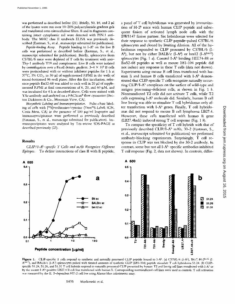
a panel of T cell hybridomas was generated by immuniza- tion o f H-2 b mice with human CLIP peptide and subse- quent fusion o f activated lymph node cells with the BW5147 fusion partner. Six hybridomas were selected for their response to synthetic CLIP peptide--pulsed C57BL/6 splenocytes and cloned by l imiting dilution. All o f the hy- bridomas responded to CLIP presented by C57BL/6 (I- Ab), but not by either BALB/c (I-A d) or bin12 (I-A bm12) splenocytes (Fig. 1 a). Cont ro l I -A b binding HEL74-88 or Eo~52-68 peptides as well as mouse Ii81-104 peptide did not induce any response in these T cells (data not shown). Experiments using mouse B cell lines transfected with hu- man Ii and human B cells transfected with I -A b demon- strated that CLIP-specific T cells recognize naturally occur- ring C L I P / I - A b complexes on the surface o f wi ld- type and antigen processing-deficient cells, as shown in Fig. 1 b. Nontransfected T2 cells did not activate T cells, while T2 cells expressing I -A b molecule did. Similarly, human B cell line Sweig was able to stimulate T cell hybridomas only af- ter transfection with I -A b genes. Finally, T cell hybr ido- mas did not respond to mouse B cell lymphoma LB27.4. However , these cells transfected with human Ii gene (LB27.4huli) induced strong T cell response (Fig, 1 b).
To compare the specificity o f T cell hybrids with that o f previously described C L I P / I - A b mAb, 30-2 (Eastman, S., et al., manuscript submitted for publication) we performed ant ibody-blocking experiments. Surprisingly, T cell re- sponse to CLIP was not blocked by the 30-2 antibody. In contrast, some but not all I-Ab-specific antibodies inhibited T cell response (Fig. 2, data not shown). In controls, differ-
A 6 0 0
0.4
a 0.3
B6 sc 0.2 BALB/r sc
bm12 sc
0.1
0.0 . . . . . , 0.1 1 10 100
P e p t i d e c o n c e n t r a t i o n [~tg/rnl]
A 6 0 0
0.5
0.4 b
0.3
0.2
[ ] 51.24 �9 51.26
[ ] 51.31
0.1
0.0 c~l ,Q ~ O .Q 'q" " -
m
Figure 1. CLIP-specific T cells respond to synthetic and naturally processed CLIP peptide bound to I-A b. (a) C57BL/6 (I-Ab), B6.C-H-2 bin12 (I- Abm12), and BALB/c (I-A a) splenocytes pulsed with titrated amounts of synthetic CLIP (Ii81-104) peptide stimulate T cell hybridoma 51.24. (b) CLIP- specific 51.24, 51.26, and 51.31 T cell hybrids respond to naturally processed CLIP presented by human T2 and Sweig cell lines transfected with I-A b or by the mouse I-Ab-positive LB27.4 B cell line transfected with human Ii. Corresponding nontransfected cell lines were used as controls. T cell activation was measured by the IL-2-dependent HT-2 cell line using Alamar blue colorimetric assay.
1405 Morkowski et al.
on August 16, 2015
jem.rupress.org
Dow
nloaded from
Published November 1, 1995

A 600 6 0 0 -
5 0 0 -
4 0 0 �9
3 0 0 '
2 0 0 '
1 0 0
.~ n- 7 7- ,,, 'g,
._= ~ ~ '~ g
Figure 2. Inhibition of CLiP-specific T cell response by some, but not all, anti-I-A b mAbs. LB27.4 cells were incubated with Ii81-104 peptide (1 Ixg/ml) and antibodies (10 p,g/ml) and CLIP-specific hybridoma 51.31 (hatched bars). The alloreactive I-A b specific-hybridoma BPB211 was used as a control (filled bars). Background response of BPB211 hybridoma to syngeneic (H-2 k) spleen cells was <OD 0.10 (represented by 100 on the scale). T cell response was measured as described above.
Table 1. Sequences of Synthetic Ii Peptides
81 85 90 95 i00 i04
Ii81-i04 LPKPPKPVSKMRMATPLLMQALPM ** * * * * *
Ii81-99 LPKPPKPVSKMRMATPLLM
Ii81-93 LPKPPKPVSKMRM
Ii81-91 LPKPPKPVSKM
Ii85-i04 PKPVSKMRMATPLLMQALPM
Ii91-i04 MRMATPLLMQALPM
Ii93-i04 MATPLLMQALPM
Ii89-i01 SKMRMATPLLMQA
Ii85-99 KPVSKMRMATPLLM
Ii91 99 MRMATPLLM
Ii91-104(100R) MRMATPLLMRALPM
Ii91-104(101P) MRMATPLLMQPLPM
Ii91 I04(I02M) MRMATPLLMQAMPM
Ii91-I04(I03S) MRMATPLLMQALSM
Ii81-I04 LPKSAKPVSQMRMATPLLMRPMSM
(mouse
sequence)
* Residues that are different in mouse and human CLIP sequences
ential inhibition o f response o f I-Ab-specific alloreactive hybrid by this panel o f antibodies was demonstrated.
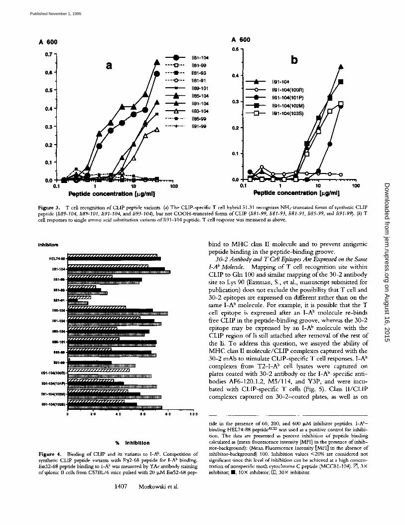
Fine Specificity of C L I P / L A b Recognizing T Cells. To map the T cell recognition site within the CLIP sequence, we synthesized a series o f truncation variants o f the CLIP pep- tide (Table 1) and analyzed their ability to induce T cell re- sponse and to bind to the I-A b molecule. COOH-te rmina l truncation variants lacking residues 100-104 failed to stim- ulate T cell hybridomas (Fig. 3 a, peptides Ii81-99, Ii81-93, and Ii81-91). In contrast, Ii85-104 peptide with the N H 2- terminal truncation stimulated T cells as strongly as the full- length CLIP peptide (Ii81-104). This suggests, that the T cell recognition site(s) is contained within C O O H - t e r m i - nal portion o f the CLIP peptide. Since sequences o f mouse and human Ii are identical between residues 91 and 99, we suggest that T cell specificity is confined to four residues, 100-103, at which mouse and human Ii sequences differ. We generated four peptide variants with single amino acid substitutions changing human Ii residues for corresponding mouse residues. Two of these peptides with 102 Leu Met and 103 Pro---)Ser mutations induced response o f T cells similar to that o f the wild-type Ii91-104 peptide (Fig. 3 b, peptides Ii91-104, Ii91-104102M] and Ii91- 1041103S]). Proline substitution for alanine at position 101 (101 Ala ---) Pro) decreased T cell response significantly. The substitution ofglutamine at position 100 with arginine (100 Gln ~ Arg), however, abolished the response com- pletely (Fig. 3 b, peptides Ii91-104, Ii91-1041101P] and Ii91-1041100R]), indicating that Gln 100 is critical for T cell recognition. Furthermore, truncated peptide Ii89-101 was able to activate T cells similarly to peptide Ii91-104
(Fig. 3 a). Interestingly, this peptide is recognized by the 30-2 antibody as well, since it contains Lys 90, a critical residue for 30-2 binding (Eastman, S., et al., manuscript submitted for publication, and data not shown). All CLIP- specific T cell hybrids demonstrated the same pattern o f re- activity to CLIP peptide variants, but different sensitivity (data not shown).
Binding of CLIP Peptide Variants to I-A ~ Molecule. Since truncated and mutated variants o f CLIP may differ in their ability to bind I-A b, we compared their ability to compete for I-A b with titrated amounts of a known I-Ab-binding peptide, E~52-68. Ecx52-68 binding to GA b was measured using E~52-68 / I -A b complex-specific mAb YAe (21). The long Ii81-104 peptide (CLIP) proved to be an efficient I-A b binder (Fig. 4). NH2- and COOH-terminal truncation variants Ii85-104, li91-104, Ii85-99, Ii89-101, and Ii81-99 bind to I-A b with similar efficiency comparable to that o f CLIP. Similarly, single amino acid substitutions Gin100 -+ Arg, Alal01 -+ Pro, Leul02 -+ Met, and Pro103 -+ Ser in Ii91-104 peptide did not affect its I-Ab-binding capacity (Fig. 4). Since Ii91-104 and Ii81-99 peptides bind to I-A b, we suggest that the 91-99 region they share is responsible for their binding to M H C class II. Indeed, further trunca- tions reaching into the 91-99 region from both ends sharply reduced their ability to compete for I-A b. Peptides Ii81-93 and Ii81-91 were very inefficient competitors in this assay (Fig. 4). To directly demonstrate interaction o f this region of Ii with the I-A b molecule, we synthesized the Ii91-99 peptide and tested it in an E0~52-68 peptide com- petition assay. As shown in Fig. 4, this peptide competes for L A b. This result indicates that Ii91-99 peptide is able to
1406 T Cell Recognition of Invariant Chain Processing Intermediates
on August 16, 2015
jem.rupress.org
Dow
nloaded from
Published November 1, 1995
A 600 0.S
0 .6 li81-93 0.4
. . . . . li81-gl
0 .5 li89-101
li85-104
0 . 4 l ig3-1 O4
. . . . - I - - - . , 85 -99
0 .3 . . . . "+" ' " li91-99 0.2
0 .2
0.1
0.1
~ " ' " ~176 " ' ' I 0 .0 " " " ' " _ . . . . . . " - - n . . . 0.0
0.1
A 600
b
§ iiiiii :::::
1 10 100 0.1 1 10 100 Poptide concentration [l~glml] Poptlde concentration [l~g/ml]
Figure 3. T cell recognition of CLIP peptide variants. (a) The CLIP-specific T cell hybrid 51.31 recognizes NH2-truncated forms of synthetic CLIP peptide (Ii89-104, Ii89-101, Ii91-104, and Ii93-104), but not COOH-truncated forms of CLIP (Ii81-99, Ii81-93, Ii81-91, Ii85-99, and Ii91-99). (b) T cell responses to single amino acid substitution variants of Ii91-104 peptide. T cell response was measured as above.
I n h i b l t o m
~ / / / / / / / I / / / / / I / / / / / / / / / / / I / / / / ~ HEL?441I
1111-104 I l l l l l l l l l l l l l l l n l l l l | l l u l l l l l l l l l l l n l l l l l l l l l l l j l l l ~ l l l l l l l l l l l l l l l
m141e I I I I I I I I I I I I I I I I I l l l l l l lu l l l I l l l l l l l l l l l l I I I I I I In
1 1 8 1 - 0 1 " ~
l / / / J / J J J / / / J / / / / / / J / I IIIIBICHI
| l l l l l l l l l l l l I i | | t l l t l l l l l l l l l l l l l l | l l l l l l l l l l l l l lU | l l l l l l I l I | l l | | l l l l
11~1~ I ] l l l l l l l l l l l l l l l l l n l l l l l l l l l l l l l l l l l l l l l l l l l l l l l l l n l l l l l l l l l l l l ~ J l l l l ~ l ~ l l l l l l l l J
IRIS104 I I I I I I I I I I I I I I I I I I I I I I lU l l ] l l l i l l ln l l l l l l l | l l l l l l~ l | I I I I I I I I I I I I I I ~ I / A
Iim-101 lllllllliiilllnlllllillllllliiiiiillllllllllilnilqlllllllll
iiiiiiiiiiiiiiiiiiiiiiIiiiiiiIiiIIiiiiiiiiiiiiiiiiiiiiiiiiiiiii
Ugl~IO ~llllllllllllllllllllllllIlllllllllllllllllllll I / / / . / / / / / / . / / I / / / / / H ,
I~1"104~I~R1 ~IiilllliiiiiiiIilliiiiiniiiil iiiiiil i i ii i iiiiiii~iilll i ii
l~1-104(101P) | w
Iml-104(lO2M) I I l | l l l l l l l | l l l l l l l i l l I i I | l l l l l l l l I l l l l l l l l l l l l l l l l l l l l l | l l l | l | l l I / l l ~ l l l ~ l l l l l l l l l l l J
I191-1~I~1o~ISI InllIIIllIIllllllIIlUl111nlllilllllnIIllnllll111111111111111I
2'0 4'0 6'0 8'0 1 ; 0
% I n h i b i t i o n
Figure 4. Binding of CLIP and its variants to I-A b. Competition of synthetic CLIP peptide variants with E~/2-68 peptide for I-A b binding. Ea52-68 peptide binding to I-A b was measured by YAe antibody staining of splenic B cells from C57BL/6 mice pulsed with 20 ~M E(x52-68 pep-
1407 Morkowski et al.
bind to M H C class II molecule and to prevent antigenic peptide binding in the peptide-binding groove.
30-2 Antibody and T Cell Epitopes Are Expressed on the Same I-A b Molecule. Mapping of T cell recognition site within CLIP to Gin 100 and similar mapping of the 30-2 antibody site to Lys 90 (Eastman, S., et al., manuscript submitted for publication) does not exclude the possibility that T cell and 30-2 epitopes are expressed on different rather than on the same I-A b molecule. For example, it is possible that the T cell epitope is expressed after an I-A b molecule re-binds free CLIP in the peptide-binding groove, whereas the 30-2 epitope may be expressed by an I-A b molecule with the CLIP region of Ii still attached after removal of the rest of the Ii. To address this question, we assayed the ability of M H C class II molecule/CLIP complexes captured with the 30-2 mAb to stimulate CLIP-specific T cell responses. I-A b complexes from T2-I-A b cell lysates were captured on plates coated with 30-2 antibody or the I-Ab~specific anti- bodies AF6-120.1.2, M5/114, and Y3P, and were incu- bated with CLIP-specific T cells (Fig. 5). Class I I /CLIP complexes captured on 30-2-coated plates, as well as on
tide in the presence of 60, 200, and 600 p,M inhibitor peptides. I-A t'- binding HEL74-88 peptide ̂ u2z was used as a positive control for inhibi- tion. The data are presented as percent inhibition of peptide binding calculated as (mean fluorescence intensity [MFI] in the presence of inhib- itor-background): (Mean Fluorescence Intensity [MFI] in the absence of inhibitor-background) 100. Inhibition values <20% are considered not significant since this level of inhibition can be achieved at a high concen- tration ofnonspecific moth cytochrome C peptide (MCC8t-104). E~, 3• inhibitor; I , 10• inhibitor; F13, 30• inhibitor.
on August 16, 2015
jem.rupress.org
Dow
nloaded from
Published November 1, 1995

A 600 6 0 0 "
n
500 -
4 0 0 �9
300 �9
2 0 0 "
100
0 ~ ~1 ~1 6 3 1 . 5 0-=..8 0
cell equivalents per well (x 1000)
Figure 5. T ceils recognize plate-captured CLIP/I-A b complexes. 51.24 T hybrid response to total I-A b and CL1P/I-A ~ complexes from T2-I-A b celt lysates bound to plates coated with 30-2 (filled circles), anti-I- A b mAbs M5/114 (filled squares), Y3P (open squares), and AF6 (open trian- gles). As a control, anti-human Ig K chain (small squares) and anti-human TflL (small triangles) antibodies were used. Additionally, AF6, M5/114, Y3P, and 30-2-coated plates were preincubated with T2-I-A k lysates. The response to all dilutions of T2-I-A ~ lysate was <OD 0.10 (repre- sented by 100 on the scale). T cell response was measured as above.
A F 6 - 1 2 0 . 1 . 2 - or M 5 / 1 1 4 - c o a t e d plates elicited a strong T cell response. This suggests that 30-2- reac t ive I -A ~ m o le - cules are recognized by CLIP-specific T cell hybrid. Q u a n - 6tative differences in T cell response to C L I P / I - A b c o m - plexes captured by these n o n i n h i b i t o ry antibodies most likely reflect differences in their affinities. T2 cells and T 2 - I - A b transfectants express T f R and surface Ig, and they can be stained wi th mAbs against these molecules (data no t shown). A n t i - I g K and an t i -Tf lk mAbs were therefore used to control for the nonspecific b i n d i n g o f I -A b molecules to an t ibody-coated plates. As shown in Fig. 5, there was no T cell s t imulat ion detected on plates coated wi th either c o n - trol ant ibody. Similarly, I -A k molecules f rom T 2 - I - A k ly- sates captured o n plates coated wi th AF6-120.1 .2 (recog- n iz ing I -A k as well as I -A b) did n o t induce any measurable IL-2 product ion . Identical results were ob ta ined wi th sev- eral o ther CLIP-specif ic hybr id T cell lines (data no t shown). The low level o f the response to complexes cap- tured on the Y3P ant ibody at the highest concent ra t ion o f cell lysates is probably caused by some minu te aggregation o f I -A b in the lysates.
Potential aggregation o f M H C class II molecules giving rise to mixed 30-2 + and 3 0 - 2 - I -A b aggregates could ac- coun t for the observed T cell responses to 30 -2 -cap tu red I -A b molecules. T o rule this out, we performed a sandwich ELISA using the same I-Ab-specific an t ibody as a first ant i - body immobi l i zed on the solid phase and as a second ant i - body in the fluid phase to detect homoaggregates. This im - munochemica l approach did no t reveal any aggregation o f I -A b molecules, since homologous combina t ion o f plate- immobi l i zed and biot inylated 30-2, AF6-120.1 .2 , and an-
A 4 9 0
0 . 6 0 ~
0 . 6 0 -
0 . 4 0
0 . 2 0
0 . 0 0
A 4 9 0
0 . 8 0 -
0 . 6 0 -
0 . 4 0 "
0 . 2 0 "
0 . 0 0
A 4 9 0
0 . 8 0 -
0 . 6 0 -
0 . 4 0 -
0 .20 -
0 .00
a
b
L
~o e~ m N P- ~ m
C
i , i ~ i �9 ~ , i , i , i - i , I , E
o~ ua
A 4 9 0
0 . 8 0
" ~ ~ : ~ : : 0 . . ~ 4 1 ~
0 . 0 0 I i ' , " , ' , - , " , - , " , " , ' ,
e 4 i-~ ~ m
A 4 9 0
0 .60
0 .40
0 . 2 0
0 . 0 0 i , i , i , i . = . J - i . [ . [ - i
A 4 9 0
0 . 8 0
0 . 6 0
0 . 4 0
0 . 2 0
0 . 0 0
f
i , [ . i . i , i . i . i , i , , . i
A 4 9 0
0 . 4 0 "
0 . 3 0 "
0 . 2 0 "
0 . 1 0 "
0 . 0 0 J ' l ' J ' J ' J ' J ' l ' , ' l ' l
Figure 6. Analysis ofhomoaggregation of CLIP/GA b complexes and I-A b molecules captured on antibody-coated plates by a sandwich ELISA. Determination ofI-A b from T2-I-A b cell lysates on Y3P-coated plates us- ing biotinyiated Y3P (a) and AF6 (d) antibodies. Detenrfination of CLlP/ I-A b complexes from T2-I-A b cell lysates on 30-2-coated plates using bi- otinylated 30-2 (b) and AF6 (e) antibodies. Determination of [-A b and CLIP/GA b complexes from T2-I-A b cell lysates on AF6-coated plates us- ing biotinylated AF6 (c), Y3P ~, and 30-2 (g) antibodies. Ordinate axis represents the dilution of cell lysates. Filled triangles, T2-I-A b cell lysates; squares, T2 cell lysates (control).
o ther I-Ab-specific ant ibody, Y3P, did no t detect any sig- nal in T 2 - I - A b lysates above control T2 lysates incubated wi th the corresponding plates (Fig. 6 a-c). In contrast, het- erologous combina t ions o f antibodies readily detected I -A b and C L I P / I - A b complexes (Fig. 6 d-g). In addit ion, we did no t observe CLIP-specific T cell responses on YAe-coated plates incuba ted wi th I-Ab-transfected Sweig cell lysates expressing both EoffD1Ko~)52-68/I-A b and C L I P / I - A b c o m - plexes (data no t shown). Therefore, we conclude that n o n -
1408 T Cell Recognition of Invariant Chain Processing Intermediates
on August 16, 2015
jem.rupress.org
Dow
nloaded from
Published November 1, 1995
over lapping 30-2 and T cell epi topes are expressed on the same popula t ion o f I - A b molecules associated wi th CLIP.
I-A b Complexes with ~ 1 2 - k D Ii Fragments Express 30-2 and T Cell Epitopes. Since the T and B cell epi topes o f C L I P / I - A b are independent , 30-2 ant ibody and T cell hy - brids w e r e used for analysis o f con fo rma t ion o f C L I P / I - A b and for ident i f icat ion o f C L I P precursor(s) a m o n g larger fragments, i.e., LIP and SLIP, w h i c h are p roduced dur ing endosomal degradat ion o f Ii. Previously, w e have shown that 30-2 ant ibody does no t recognize I - A b molecules asso- ciated wi th intact Ii (Eastman, S., et al., manuscr ipt submi t -
ted for publicat ion). T o reveal expression o f the 30-2 epi tope by complexes o f LIP and SLIP w i t h I -A b, w e per - fo rmed i m m u n o p r e c i p i t a t i o n exper iments in 35S-methio-
n ine /cys te ine pulse- labeled T 2 - I - A b cells treated wi th l eu-
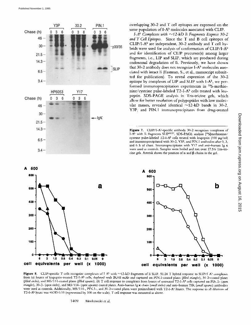
peptin. S D S - P A G E analysis in Tr is - t r ic ine gels, w h i c h al low for bet ter resolut ion o fpo lypep t ides wi th l o w m o l e c - ular masses, revealed identical ~ 1 2 - k D bands in 30-2, Y3P, and P IN.1 immunoprec ip i t a tes f rom drug- t rea ted
Figure 7. CLlP/I-Ab-specific antibody 30-2 recognizes complexes of I-A b with Ii fragments SLIP Av27. SDS-PAGE analysis [35S]methionine/ cysteine pulse-labeled T2-I-A b cells treated with leupeptin (100 p~g/ml) and immunoprecipitated with 30-2, Y3P, and PIN. 1 antibodies after 0, 3, and 6 h of chase. Immunoprecipitates with Y17 and anti-human Ig K were used as controls. Samples were boiled and run over 17.5% Tris-tri- cine gels. Asterisk shows the position of~x and [3 chains in the gel.
A 6 0 0 A 6 0 0
600 600 ]
5OO
4OO
30O
2O0
IO0
o . . . . . . . - - . - - - - . . 6 3 1.5 0.8 0.4 0.2 0.1 0.05 0 6 3 1.5 0.8 0.4 0.2 0.1 0.05 0
c e l l e q u i v a l e n t s p e r w e l l (x 1 0 0 0 ) c e l l e q u i v a l e n t s p e r w e l l ( x 1 0 0 0 )
Figure 8. CLIP-specific T cells recognize complexes of l -A b with ~12-kD fragments of l i SLIP. 51.24 T hybrid response to SLIP/I-A b complexes from (a) lysates of leupeptin-treated T2-I-A b cells, depleted with BU45 mAb and captured on PIN.l-coated plates (filled triangles), 30-2-coated plates (filled circles), and M5/114-coated plates (filled squares). (b) T cell response to complexes from lysates of untreated T2-I-A b cells captured on PIN. 1- (open triangles), 30-2- (open circles), and M5/114- (open squares) coated plates. Anti-human Ig K chain (small circles) and anti-human TfR (small squares) antibodies were used as controls. Additionally, M5/114-, PIN.l-, and 30-2-coated plates were preincubated with T2-I-A k lysates. The response to all dilutions of T2-I-A k tysate was ~<OD 0.10 (represented by 100 on the scale). T cell response was measured as above.
1409 Morkowski et al.
on August 16, 2015
jem.rupress.org
Dow
nloaded from
Published November 1, 1995

A 4 9 0
1.50 -
1.25 "
1 .00"
0.75 ]
0.50 ~
0.25 ~
0.00
a
171 ' J " 1 " 1 " ' l " V ' I " | �9 I ' " " l " 1
6
A 4 9 0
1 b 1.oo "t
0.50
0.25
0 .00
,~ c~ r o ~ .
A 4 9 0
1.50-. C
1.25 -
1.00 -
0.75 -
0.50 -
0.25 -.
0.00 , . , . , . l . , . 1 . r . , ' , . ~ , .
A 4 9 0
1.5o - d 1.25 -
1.00 -
0.75
0.50
0.25 ~ ; ~ < ~ ~ . ~ . ~
0.00 r l "I rl ;I~I ; 1 " I "I "I " I" I " !
0
A 4 9 0
1.50 -
1.25 -
1.00 -
0.75 -
0.50
0.25
0.00 ' I . I " 1 " l ' l , | " 1 1 1 ; - I . I " 1 " 1
A 4 9 0
f 1.25 -
1.00 -
0.75 -
0.50 -
0.00 - ~ - ~ , ~ ' O ~ , l , t ' ~ , C ' | , ~ ,
CI
Figure 9. Analysis of homoaggregation ofSLIP/I-A b complexes and I-A b molecules in lysates ofleupeptin-treated T2-1-A b cells by sandwich EL1SA. Determination ofSLIP/[-A b complexes in leupeptin-treated T2-I-A B cell tysates depleted with BU45 mAb on 30-2-coated plates using biotinylated 30-2 (a), AF6 (b), and Y3P (c) antibodies. Determination of GA b in the same ceil [ysates on Y3P-coated plates using biotinylated Y3P (at), AF6 (e) and 30-2
antibodies. Filled triangles, T2-I-A b cell lysates; open squares, T2-I-A b cell lysates; open squares, T2-I-A k cell lysates (control).
cells after 0, 3, and 6 h of chase (Fig. 7). Since PIN.1 anti- body precipitates intact Ii as well as LIP and SLIP fragments containing an intact cytoplasmic tail, the observed 12-kD fragments in 30-2 and Y3P as well as in PIN.1 immuno- precipitates can be identified as SLIP. Ant i -human lg K chain antibody and irrelevant antibody Y17 used as con- trols did not precipitate ~ 1 2 - k D polypeptides. Similarly, these bands were not observed in anti-TflK precipitates (data not shown). Therefore, SLIP/I -A b complexes express the 30-2 epitope.
To demonstrate expression of the T cell epitope on SLIP/ I -A b complexes, we tested T cell responses to these complexes captured on PIN. 1-coated plates. As a source of SLIP/ I -A b complexes we used lysates of T2-1A b cells en- riched for these complexes by overnight culture in the presence of 100 Ixg/ml of leupeptin. T o avoid potential competi t ion of intact Ii with SLIP fragments for the bind- ing to P IN . l - coa ted plates, lysates were extensively de- pleted of intact Ii by treatment with BU45 antibody, which recognizes the lumenal port ion o f li and does not react with SLIP fragments and protein A-Sepharose. Western blot analysis o f lysates using PIN.1 antibody showed (a)
high level o f expression o f Ii fragments in the presence of leupeptin and (b) very efficient "~ depletion of in- tact Ii by BU45 adsorption (data not shown). SLIP/ I -A b complexes from BU45-t rea ted lysates captured on P I N . l - coated plates induced a strong response o f CLiP-specific T cells comparable to that on M5/114 or 30-2-coated plates (Fig. 8). To rule out potential oligomerization of SLIP/GA b or mixed S L I P / C L I P / I - A b complexes in the lysate o f l e u - peptin-treated cells, we performed a sandwich ELISA anal- ogous to that shown above on Fig. 6. Similarly, we did not get a significant signal in the ELISA using homologous (30- 2/30-2-biot in, YBP/Y3P-biotin, Fig. 9, a and d) or heter- ologous pairs o f plate-immobilized and biotinylated anti- bodies with overlapping epitopes (30-2/Y3P-biot in and Y3P/30-2-biot in , Fig. 9, c andJ~. In contrast, a strong sig- nal was detected with heterologous pairs o f antibodies with nonoverlapping epitopes (30-2/AF6-biotin and Y3P/AF6- biotin, Fig. 9, b and e). These results, as well as low re- sponse to I-Ab/Ii complexes from nontreated cells captured on PIN. I (Fig. 8 b) antibody, argue against aggregation of SLIP/ I -A b complexes. It is not unexpected, since Lip/ M H C class II nonamers dissociate in nonionic detergents
1410 T CeII Recognition oflnvariant Chain Processing Intermediates
on August 16, 2015
jem.rupress.org
Dow
nloaded from
Published November 1, 1995

(23a). Thus, we conclude that both T cell and 30-2 epi- topes are expressed by SLIP/I-A b complexes, which strong- ly suggests that these complexes are precursors of CLIP/ I-A b complexes.
Discuss ion
In this study, we demonstrated for the first time that T ceils recognize complexes of M H C class II molecules and Ii-derived CLIP peptides in a manner indistinguishable from that of conventional antigenic peptides. T cell hybrids recognizing human CLIP bound to I-A b generate a strong response to normal B cell lines of human or mouse origin that coexpress human Ii and I-A b (Fig. 1). In addition, the human presentation-deficient mutant cell line T2 and its parental cell line, T1, transfected with I-A b present this complex very efficiendy to CLiP-specific T cells. These findings are in agreement with our studies of the expression of CLIP/I -A b complexes in mutant and wild-type cells us- ing the mAb 30-2. This antibody specific for human CLIP/ I-A b complexes cross-reacts with corresponding synthetic and endogenous mouse peptides bound to I-A b (Eastman, S., et al., manuscript submitted for publication). Immuno- histochemical and biochemical studies in C57BL/6 mice and bone marrow chimeras using the 30-2 antibody re- vealed expression of these complexes on the surface of nor- mal antigen-presenting cells in spleen, lymph nodes, and thymus (Farr, A., and A. Yu. Rudensky, manuscript in preparation). Polyclonal T cells from C57BL/6 mice im- munized with the human CLIP develop a relatively low but significant in vitro proliferative response to this peptide (stimulation indexes 2--6). In contrast, T cell responses to the mouse CLIP (Ii81-104 peptide), as well as to its short variant Ii85-99 (12), were not detected (data not shown). Since mouse Ii peptides bind to I-A b with a comparable or even higher affinity than corresponding human Ii peptides (Eastman, S., et al., manuscript submitted for publication), this indicates that T cells are tolerant to mouse Ii peptides.
The lack of T cell response to mouse Ii peptides allowed us to study the fine specificity of CLIP-specific T cell hy- brids by replacing nonconserved amino acid residues in hu- man CLIP sequence with corresponding mouse residues. This analysis demonstrated that all T cell hybrids recognize the same residue of the CLIP peptide that is distinct from that recognized by the 30-2 antibody. Previously, we have identified the a lysine residue at position 90 of the human Ii as a critical residue for 30-2 antibody recognition (Eastman, S., et al., manuscript submitted for publication). In con- trast, T cell specificity is confined to the COOH-terminal portion of CLIP, particularly to glutamine at position 100. Interestingly, antibody and T cell epitopes are not overlap- ping, since the 30-2 antibody does not block T cell re- sponse to CLIP presented by I-Ab-positive APCs. In addi- tion, CLIP/I -A b complexes bound to 30-2-coated plates are efficiently recognized by T cells, which further proves that both T cell and antibody epitopes are expressed on the same M H C class II molecule. Importantly, T cell and anti- body recognition are both critically dependent, not only on
1411 Morkowski et al.
the peptide, but also on the M H C class II allele. Com- plexes of CLIP with I-A a and I-A bm12 are not recognized by the 30-2 antibody or the T cells (Eastman, S., et al., manuscript submitted for publication, and Fig. 1). The lat- ter differs from I-A b at three amino acid residues at posi- tions 67, 70, and 71 (24). How can we explain the lack of a steric inhibitory effect of the 30-2 antibody on CLIP-spe- cific T cell response, as well as the effect o f b m l 2 mutation on both TCIL and antibody recognition? First, the 30-2 antibody may bind from the side of the M H C class I I - binding groove with both CLIP residues Lys 90 and some I-A b residues contributing to the binding site, whereas the T C k may interact with the I-Ab/CLIP complex from the top of the groove. It is possible that distinct I-A b residues affected by bm12 mutation contribute differently to T cell and antibody epitopes. Another possible interpretation for these findings, which does not exclude the first one, is an indirect effect of certain amino acid residues in the CLIP (e.g., Lys 90) or in the I-A b (bm12 substitutions) on the conformation of the entire complex. Structural analysis is required to give a final answer to this question.
Our analysis of the CLIP binding to I-A b using the orig- inal peptide and its truncation variants demonstrated that the nonamer corresponding to the 91-99 region of Ii can bind to I-A b and compete for the presentation with the an- tigenic Eot52-68 peptide. This part of the CLIP sequence is identical in the mouse, human, and rat Ii (25-28), and it suggests that the conserved 91-99 region of the Ii is the M H C class II-binding site. These results are in agreement with several studies of I i /MHC class II association using a series of COOH-terminal deletion variants of Ii coex- pressed with human and mouse M H C class II (17-19). Freisenwinkel and coauthors demonstrated that the 80-110 region of the Ii is necessary for the Ii association with M H C class II (17). These authors postulated binding of CLIP at a position distinct from the peptide-binding groove. In contrast, R, omagnoli and Gerrnain (18) sug- gested a model of CLIP binding inside the groove while CLIP is still a part of the Ii1-107 fragment. The latter is supported by our findings. A recent study by Bijlmakers and colleagues further narrowed the site of I i /MH C class II interaction to residues 96-104 (19). In agreement with these data, we did not find significant binding of the Ii81- 93 peptide, in contrast to 93-104 peptide, which was able to bind to I-A b and to elicit a T cell response. These data suggest that the 91-99 peptide of the Ii may represent an M H C class II-binding site. Interestingly, the Ii89-101 pep- tide, which contains the M H C class II-binding 91-99 site flanked with Lys 90, Gln 100, and Ala 101, which are crit- ical for 30-2 and T cell recognition, respectively, binds to I-A b and is recognized by the antibody and T cell (Figs. 3 a and 4). Recent study by Malcherek et al. (29) provided ev- idence that CLIP may act as a universal groove-binding peptide, taking advantage of sequence supermotifs and al- lowing CLIP to bind to various M H C alleles. All previous studies, as well as this one, do not exclude contribution of other regions of the Ii, such as the transmembrane region or the more COOH-terminal parts of the lumenal portion
on August 16, 2015
jem.rupress.org
Dow
nloaded from
Published November 1, 1995
of Ii, to its binding to M H C class II molecule. These addi- tional sites may be critical for Ii binding to M H C class II al- leles with a relatively low affinity for CLIP, e.g., HLA- D R 4 or DR11 (15, 20).
Our results suggest that the C O O H - t e r m i n a l portion of the CLIP region, which includes T cell contact residue Glu 100, binds in the groove of the I-A b molecule. It seems likely that the orientation of NH2 and C O O H termini o f CLIP associated with M H C class II follows that o f anti- genic MHC-b ind ing peptides. In this case, the very NH2- terminal portion of CLIP more distant from the M H C - binding site is likely to be exposed and recognized in an MHC- independen t manner, whereas Lys 90 is still in the groove or in its immediate proximity such that it is recog- nized by the 30-2 antibody in an MHC-rest r ic ted fashion. This model is supported by the observation that the Cer- CLIP.1 antibody, recognizing free as well as M H C - b o u n d CLIP independently of an M H C allele (30), does not react with any of NH2-terminal truncation variants o f CLIP used in this study (Cresswell, P., personal communication). In addition, the binding of CLIP to I -A b molecules protects CLIP containing both T cell and antibody recognition sites from cleavage by trypsin, similarly to a single amino acid substitution variant o f antigenic EoL52-68 peptide (57 Glu--)Lys) containing a single trypsin cleavage site in the middle of the sequence (Goldrath, A. W., and A. Yu. Rudensky, unpublished observation). Similarly, CLIP pep- tide bound to the H L A - D R 3 molecule was protected from proteolytic degradation by a mixture o f cathepsin B and D (20).
The T cell recognition o f C L I P / M H C class II com- plexes raises a question concerning the mechanism respon- sible for generating of these complexes. In other words, is
CLIP a remnant of the Ii containing the M H C class I I - binding site that remains associated with M H C class II after the rest o f the Ii is cleaved off, or is this fragment first re- leased by proteases as a free peptide which then binds in the groove of M H C class II molecule? Generation of C L I P / l - Ab-specific T cells and mAb 30-2 provided us with a tool to compare the conformation o f I-A b molecule associated with free CLIP peptide and I-A b complexes with larger fragments o f Ii containing the CLIP region. These frag- ments, 21-kD LIP and 11-14-kD SLIP, were first de- scribed in B cell lines treated with the cysteine proteinase inhibitor leupeptin (10, 11). LIP and SLIP fragments stay associated with M H C class II molecules in the endosomal compartment, thus preventing class II export to the cell surface (31). Both 30-2 antibody and CLIP-specific T cells recognize complexes of I -A b with N12 kD SLIP fragments (Figs. 7 and 8). Thus, SLIP fragments contain CLIP region bound to M H C class II molecule in a conformation indis- tinguishable from that o f free CLIP peptide. This indicates that (a) CLIP is a remnant o f Ii and (b) SLIP/I -A b com- plexes are precursors of C L I P / I - A b complexes. SLIP frag- ments are generated in late endosomes and in a dense en- dosomal compartment, M I I C (reference 11 and J. Blum and Rudensky, A., manuscript in preparation). Recent studies suggested that the latter compartment is a critical in- tracellular site for M H C class II complex assembly with peptides derived from both endogenous and exogenous an- tigens (32-35). Therefore, it is likely that the bulk o f C L I P / M H C class II complexes is generated in the same compartment. Further experiments with CLIP-specific T cell hybrids and the 30-2 antibody using subcellular frac- tionation will directly test this prediction.
The authors thank Dr. Janice Blum and members of our laboratory for valuable discussions and reading the manuscript.
Address correspondence to Alexander Yu. Rudensky, Howard Hughes Medical Institute, University of Washington, Seattle, Washington 98195.
Received for publication 25January 1995 and in revised form 9June 1995.
References 1. Cresswell, P. 1994. Assembly, transport and function of MHC
class II molecules. Annu. Rev. Immunol. 12:259-293. 2. Roche, P.A., and P. Cresswell. 1991. Proteolysis of the class
II associated invariant chain generates a peptide binding site in intracellular HLA-DR molecules. Proc. Natl. Acad. Sci. USA. 88:3150-3154.
3. Teyton, L., D. O'Sullivan, V. Lotteau, A. Sette, P. Fink, and P.A. Peterson. 1990. Invariant chain distinguishes between the exogenous and endogenous antigen presentation path- ways. Nature (Lond,). 348:39-44.
4. Newcomb, J.R., and P. Cresswell. 1993. Characterization of
endogenous peptides bound to purified HLA-DR molecules and their absence from invariant chain-associated dimers. J. lmmunol. 150:499-507.
5. Roche, P.A., M.S. Marks, and P. Cresswell. 1991. Formation of a nine-subunit complex by HLA class II glycoprotein and the invariant chain. Nature (Lond.). 354:392-394.
6. Viville, S., J. Neetjes, V. Lotteau, A. Dierich, M. Lemeur, H. Ploegh, C. Benoist, and D. Mathis. 1993. Mice lacking the MHC class II-associated invariant chain. Cell. 72:635-648.
7. Bikoff, E.K., L.-Y. Huang, V. Episkopou, J. Meerwijk, R.N. Germain, and E.J. Robertson. 1993. Defective major histo-
1412 T Cell Recognition of Invariant Chain Processing Intermediates
on August 16, 2015
jem.rupress.org
Dow
nloaded from
Published November 1, 1995

compatibility complex class II assembly, transport, peptide ac- quisition, and CD4 + T cell selection in mice lacking invari- ant chain expression.J. Exp. Med. 177:1699-1712.
8. Elliott, E.A., J.R. Drake, S. Amigorena, J. Elsemore, P. Webster, I. Mellman, and R.A. Flavell. 1994. The invariant chain is required for intracellular transport and function of major histocompatibility complex class II molecules. J. Exp. Med. 179:681-694.
9. Bakke, O., and B. Dobberstein. 1990. MHC class lI-associ- ated invariant chain contains a sorting signal for endosomal compartments. Cell. 63:707-716.
10. Blum, J.S., and P. Cresswell. 1988. Role ofintracellular pro- teases in the processing and transport of class II HLA antigens. Proc. Natl. Acad. Sci. USA. 85:3975-3980.
11. Maric, M.A., M.D. Taylor, andJ.S. Blum. 1994. Endosomal aspartic proteinases are required for invariant-chain process- ing. Proc. Natl. Acad. Sci. USA. 91:2171-2175.
12. Rudensky, A.Y., P. Preston-Hurlburt, S.-C. Hong, A. Bar- low, and C.A. Janeway, Jr. 1991. Sequence analysis of pep- tides bound to MHC class II molecules. Nature (Lond.). 353: 622-628.
13. Hunt, D.F., H. Michel, T.A. Dickinson, J. Shabanowitz, A.L. Cox, K. Sakaguchi, E. Apella, H.M. Grey, and A. Sette. 1992. Peptides presented to the immune system by the mu- rine class II major histocompatibility molecule I-A d. Science (Wash. DC). 256:1817-1820.
14. Chicz, R.M., R.G. Urban, J.C. Gorga, L.J. Stern, D.A.A. Vignali, and J.L. Strominger. 1992. Predominant naturally processed peptides bound to HLA-DR1 are derived from MHC-related molecules and are heterogeneous in size. Na- ture (Lond.). 358:764-766.
15. Chicz, R.M., R.G. Urban, J.C. Gorga, D.A.A. Vignali, W.S. Lane, and J.L. Strominger. 1993. Specificity and promiscuity among naturally processed peptides bound to HLA-DR alle- les.J. Exp. Med. 178:27-32.
16. Riberdy, J.M., J.R. Newcomb, M.J. Surman, J.A. Barbosa, and P. Cresswell. 1992. HLA-DR molecules from an anti- gen-processing mutant cell line are associated with invariant chain peptides. Nature (Lond.). 360:474-476.
17. Freisenwinkel, I.M., K. Schenck, and N. Koch. 1993. The segment of invariant chain that is critical for association with major histocompatibility complex class II molecules contains the sequence of a peptide eluted from class II polypeptides. Proc. Natl. Acad. Sci. USA. 90:9703-9706.
18. Romagnoli, P., and R.N. Germain. 1994. The CLIP region ofinvariant chain plays a critical role in regulating major his- tocompatibility complex class II folding, transport, and pep- tide occupancy.J. Exp. Med. 180:1107-1113.
19. Bijlmakers, M.E., P. Benaroch, and H.L. Ploegh. 1994. Map- ping functional regions in the lumenal domain of the class I I - associated invariant chain.J. Exp. Med. 180:623-629.
20. Avva., R.R., and P. Cresswell. 1994. In vivo and in vitro formation and dissociation of HLA-DR complexes with in- variant chain-derived peptides. Immunity. 1:763-774.
21. Rudensky, A.Y., S. Rath, P. Preston-Hurlburt, D.B. Mur- phy, and C.A. Janeway, Jr. 1991. On the complexity of self. Nature (Lond.). 353:660--662.
22. Riberdy, J.M., and P. Cresswell. 1992. The antigen-process-
ing mutant T2 suggests a role for MHC-linked genes in class II antigen processing.J. Immunol. 148:2586-2590.
23. Schagger, H., and G. vonJagow. 1987. Tricine-sodium dode- cyl sulfate-polyacrylamide gel electrophoresis for the separa- tion of proteins in the range from 1 to 100 kDa. Anal. Bio- chem. 166:368-379.
23a.Newcomb, J.K., and P. Cresswell. 1995. Structural analysis of proteolytic products of MHC class II-invariant chain com- plexes generated in vivo.J. Immunol. 151:4153-4163.
24. McKenzie, I.F.C., G.M. Morgan, M.S. Sandrin, M. Michae- lidis, R.W. Melvold, and H.I. Kohn. 1979. A new H-2 mu- tation in the I region in the mouse.J. Exp. Med. 150:1323- 1338.
25. Strubin, M., B. Mach, and E.O. Long. 1984. The complete sequence of the mRNA for the HLA-DR-associated invari- ant chain reveals a polypeptide with an unusual transmem- brane polarity. EMBO (Eur. Mol. Biol. Organ.) J. 3:869-872.
26. O'Sullivan, D.M., D. Noonan, and V. Quaranta. 1987. Four Ia invariant chain forms derive from a single gene by alterna- tive initiation of transcription/translation. J. Exp. Med. 166: 444-460.
27. Koch, N., W. Lauer, J. Habicht, and B. Dobberstein. 1987. Primary structure of the gene for the murine Ia antigen-asso- ciated invariant chains (Ii). An alternatively spliced exon en- codes a cysteine-rich domain highly homologous to a repeti- tive sequence ofthyroglobulin. EMBO (Eur. Mol. Biol. Organ.) J. 5:3483-3487.
28. McKnight, J., D.W. Mason, and A.N. Barclay. 1989. Se- quence of a rat MHC class II-associated invariant chain cDNA clone containing a 64 amino acid thyroglobulin-like domain. Nucleic Acids Res. 17:3983-3985.
29. Malcherek, G., V. Gnau, G. Jung, H.-G. Rammensee, and A. Melms. 1995. Supermotifs enable natural invariant chain- derived peptides to interact with many major histocompati- bility complex class II molecules.J. Exp. Med. 181:527-536.
30. Denzin, L.K., N.F. Robbins, C. Carboy-Newcomb, and P. Cresswell. 1994. Assembly and intracellular transport of HLA- DM and correction of the class II antigen-processing defect in T2 cells. Immunity. 1:595-606.
31. Neet]es, J.J., and H.L. Ploegh. 1992. Inhibition ofendosomal proteolytic activity by leupeptin blocks surface expression of MHC class II molecules and their conversion to SDS resistant ell3 heterodimers in endosomes. EMBO (Eur. Mol. Biol. Or- gan.)J. 11:411--416.
32. Peters, P.J., J.J. Nee0es, V. Oorschot, H.L. Ploegh, and H.J. Geuze. 1991. Segregation of MHC class II molecules from MHC class I molecules in the Golgi complex for transport to lysosomal compartments. Nature (Lond.). 349:669-676.
33. Tulp, A., D. Verwoerd, B. Dobberstein, H.L. Ploegh, and J. Pieters. 1994. Isolation and characterization of the intracellu- lar MHC class II compartment. Nature (Lond.). 369:120--126.
34. West, M.A., J.M~ Lucocq, and C. Watts. 1994. Antigen pro- cessing and class II MHC peptide-loading compartments in human B-lymphoblastoid cells. Nature (Lond.). 369:147-151.
35. Rudensky, A.Y., M. Maric, S. Eastman, L. Shoemaker, P.C. DeRoos, and J.S. Blum. 1994. Intracellular assembly and transport of endogenous peptide:MHC class II complexes. Immunity. 1:585-594.
1413 Morkowski et al.
on August 16, 2015
jem.rupress.org
Dow
nloaded from
Published November 1, 1995