Ezra-Coat-Pattern-DIY-Kit-Instructions-The-School-of-Making ...

ORIGINAL ARTICLE
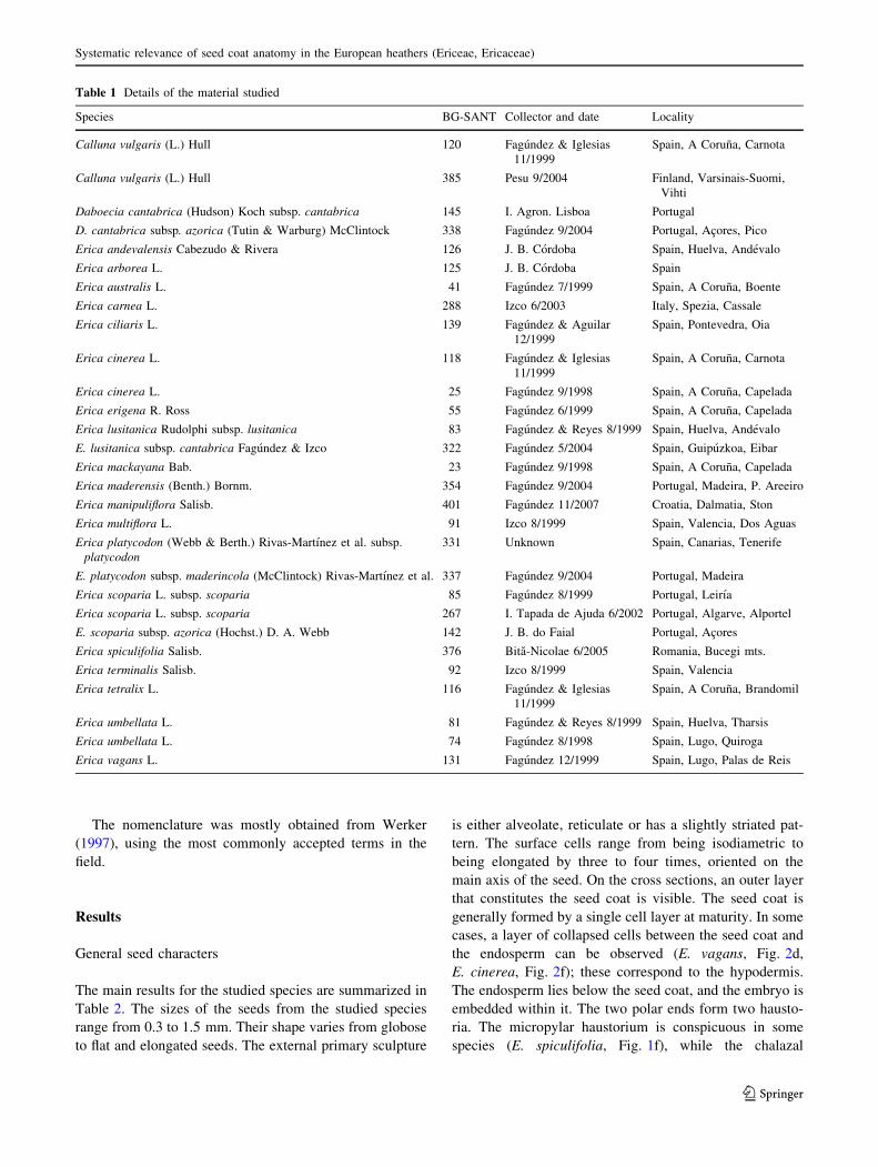
Systematic relevance of seed coat anatomy in the Europeanheathers (Ericeae, Ericaceae)
Jaime Fagundez • Rocıo Juan • Inmaculada Fernandez •
Julio Pastor • Jesus Izco
Received: 27 February 2009 / Accepted: 25 October 2009
� Springer-Verlag 2009
Abstract The anatomy of the seed coat of the European
species of tribe Ericeae (Calluna, Daboecia and Erica) of
the Ericaceae family was studied, and the taxonomic
importance of their characters was analyzed. The seed coat
is mostly formed by a one-cell layer with thick, pitted inner
walls and thin outer walls that collapse at maturity over the
inner walls. The cell junctions are either raised with anti-
clinal walls up to four times the height of the periclinal
walls or are not raised with similar values for the height of
both the anticlinal and periclinal walls. Three main cell
junction types were found and described. The thickness of
the inner walls is variable, but there is a large overlap
among the results for different species. Calluna vulgaris is
the only species with no pits, and E. multiflora has a pitted
pattern on its inner walls, which is distinctive from the rest
of the species. Our main results agree with the external
seed morphology, and valuable new data were obtained for
certain groups such as the E. cinerea-E. terminalis or the
E. scoparia complex. The similarities that are found in seed
coat characters are not in accordance with the classical
taxonomic delimitation of infrageneric groups within
Erica.
Keywords Erica � Calluna � Daboecia � Seed coat �Seed anatomy � Taxonomy
Introduction
Since the thorough study of the Ericaceae performed by
Kron et al. (2002), the systematics of the family have been
clarified at different infrafamiliar levels. According to their
analysis Calluna Salisb., Daboecia D. Don and Erica L. are
the only genera that are integrated into the tribe Ericeae, and
consequently included in the subfamily Ericoideae. Before
this work, many different studies of the whole family
resulted in much discussion as to the systematics of these
genera (Stevens 1971; Anderberg 1993; Judd and Kron
1993). Daboecia has a septicidal capsule, a deciduous
corolla and differs from the other two genera in its leaf and
anther morphology. Calluna has a septifragal capsule, an
expanded petalloid calyx, sagittate leaves and a distinct pith
type. Both genera are monotypic, contrasting with the vast
Erica genus of around 860 species, which are characterized
by a loculicidal capsule, a persistent corolla and strongly
revolute leaves. Important differences are also found in the
seed morphology for the three genera, but many species of
Erica have not yet been studied.
Erica is a very large genus with a high level of mor-
phological variation and a wide ecological and geograph-
ical range (Oliver 2000; Stevens et al. 2004). The early
treatments of this genus, for example by Don (1834) or
Klotzsch (1838), considered several genera, whereas now it
is considered to be a unique genus. Bentham0s (1839)
arrangement included 19 genera, and Erica was divided
into four subgenera and several sections. The classic genera
Bruckenthalia Reichenb., Pentaptera Klotzsch, Philippia
Klotzsch, Ericinella Klotzsch, Blaeria L. and the ‘‘minor’’
J. Fagundez (&)
School of Biology, IE University, Campus de Santa Cruz la Real,
Cardenal Zuniga 12, 40003 Segovia, Spain
e-mail: [email protected]
R. Juan � I. Fernandez � J. Pastor
Departamento de Biologıa Vegetal y Ecologıa,
Universidad de Sevilla, 41012 Sevilla, Spain
J. Izco
Departamento de Botanica, Facultade de Farmacia,
Universidade de Santiago de Compostela,
15782 Santiago de Compostela, A Coruna, Spain
123
Plant Syst Evol
DOI 10.1007/s00606-009-0240-2

genera of species with indehiscent or partially dehiscent
fruits are now part of the large genus Erica according to
Oliver (1989, 2000).
Approximately 20 species of Erica are considered in the
Northern area of the genus, which includes Europe, the
Mediterranean coasts of Northern Africa and the Eastern-
most part of Asia (Webb and Rix 1972; Greuter et al.
1986), including Erica spiculifolia, the only species
included in genus Bruckenthalia that was first considered
as part of Erica by Salisbury (1802) and was recovered by
Oliver (2000). The infrageneric classification of Bentham
(1839) has been modified for the Cape species by Guthrie
and Bolus (1905) and other authors. Hansen (1950) pro-
posed some changes to the infrageneric classification for
the Northern species. Recently, some attempts have been
made to use molecular analysis in order to determine the
phylogeny of the genus (McGuire and Kron 2005). The
researchers performed a preliminary phylogenetic analysis
for Erica, including Calluna and Daboecia as outgroups,
with puzzling results inconsistent with Bentham0s classifi-
cation. Moreover, different interpretations have been given
to closely related taxa, and some points regarding the
delimitation of the species are still unclear. For example,
the systematic position of Erica andevalensis Cabezudo
and Rivera has been discussed and subsequently placed as
a subspecies of Erica mackayana Bab. (McClintock
1989a).
Early studies in seed anatomy were developed by
Artopoeus (1903) and Peltrisot (1904). Both studies
focused on the whole family Ericaceae, and the seeds of
several genera were analyzed. Both Artopoeus and Peltrisot
included Calluna, Daboecia and several Erica species,
including Bruckenthalia, in their studies. Additional data
were obtained by Stevens (1971), again from general
studies on the family. Oliver (2000) used several seed coat
features for his systematic analysis on the Ericeae. The
testa thickness and occurrence of pits were coded, along
with surface cell characters. Szkudlarz (2006) performed
an analysis of seed characters in a set of 33 South African
species. More importance has been given to the seed sur-
face characters, such as the elongation of surface cells
(Stevens 1971; Anderberg 1993; Oliver 2000; Kron et al.
2002). In our studies on seed morphology of the Ericeae,
important diagnostic features were obtained for the
delimitation of the three genera in order to solve the
ongoing taxonomic problems regarding species delimita-
tion (Fagundez and Izco 2003a, b, 2004a, b, c, d, 2008,
2009a, b, c). However, difficulties were found when these
characters were used in a generic approach to the system-
atics of the Ericeae because of continuing variation, and
problems in coding characters arise.
In this paper, we studied the seed coat anatomy to reveal
the nature of the external features of seed surface that we
have previously studied. We also looked for new diagnostic
characters that could add information to enable the further
delimitation of the species. In a similar manner to what was
found with regards to seed morphology, the anatomy of the
seeds is only related to the systematic position in some
cases. Some closely related species have a different seed
coat type, while a similar pattern was found in others
considered as unrelated. This reveals once again that an
overall study of this genus is needed.
Materials and methods
Seeds of 29 samples were studied (Table 1), including 3
genera, 21 species and 4 subspecies. Nomenclature follows
the most accepted taxonomic treatment for the species
(Webb and Rix 1972; Stevens 1978; Greuter et al. 1986;
Bayer 1993 and others) except for the following taxa:
E. platycodon (Webb and Berth.) Rivas-Martınez et al.
subsp. platycodon, E. platycodon subsp. maderincola
(McClintock) Rivas-Martınez et al. and E. scoparia subsp.
azorica (Hochst.) D. A. Webb. Other treatments and sub-
specific taxa were considered and discussed in the text. Seeds
of Erica sicula Guss. that occur in Sicily were not studied.
Seeds were stored under low levels of humidity and
temperature at the Seedbank of the University of Santiago
de Compostela. A collection of Ericaceae seeds has been
collected and stored for 10 years, resulting in more than
400 samples available for conservation and research. Seeds
were collected in the field or obtained from different
seedbanks (Table 1). Samples were cleaned and dried
using silica gel and put into glass tubes with a certain
amount of silica gel with a color component to detect
possible humidification. These tubes were then stored in a
4�C chamber. Voucher specimens of field samples are
stored at the SANT herbarium.
Seeds sectioned for the light microscopy studies were
first fixed in FAA for at least 48 h, dehydrated in an eth-
anol–tertiary butanol (Johansen 1940 modif.; Juan et al.
1999) and embedded in paraffin (Paraplast plus). Sections
were cut at a thickness of 5 lm with a Micron HM-355
microtome. Three or four slides were mounted for each
sample. The slides were studied, photographed and drawn
with a digital camera adjusted to an Olympus BX-50
microscope. Image analysis was performed with Motic
Image Plus 2.0 ML software.
Handmade sections were prepared for seeds undergoing
scanning electron microscopy (SEM) observations. These
were mounted onto the stubs using double-sided adhesive
tape and coated with gold. The samples were studied
and photographs were obtained using a LEO-435VP
microscope (Leica Microsystems, Cambridge, UK) at the
University of Santiago de Compostela.
J. Fagundez et al.
123
The nomenclature was mostly obtained from Werker
(1997), using the most commonly accepted terms in the
field.
Results
General seed characters
The main results for the studied species are summarized in
Table 2. The sizes of the seeds from the studied species
range from 0.3 to 1.5 mm. Their shape varies from globose
to flat and elongated seeds. The external primary sculpture
is either alveolate, reticulate or has a slightly striated pat-
tern. The surface cells range from being isodiametric to
being elongated by three to four times, oriented on the
main axis of the seed. On the cross sections, an outer layer
that constitutes the seed coat is visible. The seed coat is
generally formed by a single cell layer at maturity. In some
cases, a layer of collapsed cells between the seed coat and
the endosperm can be observed (E. vagans, Fig. 2d,
E. cinerea, Fig. 2f); these correspond to the hypodermis.
The endosperm lies below the seed coat, and the embryo is
embedded within it. The two polar ends form two hausto-
ria. The micropylar haustorium is conspicuous in some
species (E. spiculifolia, Fig. 1f), while the chalazal
Table 1 Details of the material studied
Species BG-SANT Collector and date Locality
Calluna vulgaris (L.) Hull 120 Fagundez & Iglesias
11/1999
Spain, A Coruna, Carnota
Calluna vulgaris (L.) Hull 385 Pesu 9/2004 Finland, Varsinais-Suomi,
Vihti
Daboecia cantabrica (Hudson) Koch subsp. cantabrica 145 I. Agron. Lisboa Portugal
D. cantabrica subsp. azorica (Tutin & Warburg) McClintock 338 Fagundez 9/2004 Portugal, Acores, Pico
Erica andevalensis Cabezudo & Rivera 126 J. B. Cordoba Spain, Huelva, Andevalo
Erica arborea L. 125 J. B. Cordoba Spain
Erica australis L. 41 Fagundez 7/1999 Spain, A Coruna, Boente
Erica carnea L. 288 Izco 6/2003 Italy, Spezia, Cassale
Erica ciliaris L. 139 Fagundez & Aguilar
12/1999
Spain, Pontevedra, Oia
Erica cinerea L. 118 Fagundez & Iglesias
11/1999
Spain, A Coruna, Carnota
Erica cinerea L. 25 Fagundez 9/1998 Spain, A Coruna, Capelada
Erica erigena R. Ross 55 Fagundez 6/1999 Spain, A Coruna, Capelada
Erica lusitanica Rudolphi subsp. lusitanica 83 Fagundez & Reyes 8/1999 Spain, Huelva, Andevalo
E. lusitanica subsp. cantabrica Fagundez & Izco 322 Fagundez 5/2004 Spain, Guipuzkoa, Eibar
Erica mackayana Bab. 23 Fagundez 9/1998 Spain, A Coruna, Capelada
Erica maderensis (Benth.) Bornm. 354 Fagundez 9/2004 Portugal, Madeira, P. Areeiro
Erica manipuliflora Salisb. 401 Fagundez 11/2007 Croatia, Dalmatia, Ston
Erica multiflora L. 91 Izco 8/1999 Spain, Valencia, Dos Aguas
Erica platycodon (Webb & Berth.) Rivas-Martınez et al. subsp.
platycodon331 Unknown Spain, Canarias, Tenerife
E. platycodon subsp. maderincola (McClintock) Rivas-Martınez et al. 337 Fagundez 9/2004 Portugal, Madeira
Erica scoparia L. subsp. scoparia 85 Fagundez 8/1999 Portugal, Leirıa
Erica scoparia L. subsp. scoparia 267 I. Tapada de Ajuda 6/2002 Portugal, Algarve, Alportel
E. scoparia subsp. azorica (Hochst.) D. A. Webb 142 J. B. do Faial Portugal, Acores
Erica spiculifolia Salisb. 376 Bita-Nicolae 6/2005 Romania, Bucegi mts.
Erica terminalis Salisb. 92 Izco 8/1999 Spain, Valencia
Erica tetralix L. 116 Fagundez & Iglesias
11/1999
Spain, A Coruna, Brandomil
Erica umbellata L. 81 Fagundez & Reyes 8/1999 Spain, Huelva, Tharsis
Erica umbellata L. 74 Fagundez 8/1998 Spain, Lugo, Quiroga
Erica vagans L. 131 Fagundez 12/1999 Spain, Lugo, Palas de Reis
Systematic relevance of seed coat anatomy in the European heathers (Ericeae, Ericaceae)
123

Ta
ble
2M
ain
char
acte
rso
fth
ese
edco
ato
fth
est
ud
ied
tax
a
Cal
Dab
and
arb
aus
azo
car
cil
cin
eri
lus
mac
See
dsi
ze0.5
5–0.6
50.5
5–0.7
50.4
0.4
–0.5
0.9
–1.1
0.5
–0.5
51–1.2
0.4
0.7
–0.9
0.9
-10.3
–0.4
0.4
See
dsh
ape
Ell
ipG
lob
Ell
ip–O
blo
ng
Ell
ipO
blo
ng
Ell
ipE
llip
–O
blo
ng
Ell
ip–O
blo
ng
Ell
ip–C
urv
eE
llip
–O
blo
ng
Glo
b–E
llip
Ell
ip–O
blo
ng
Surf
pat
tern
Ret
*1
Ret
Str
Str
Ret
Ret
Sli
ret
Str
gre
tR
etR
etR
et
Cel
lra
tio
1:1
–2:1
1:1
1:1
2:1
–3:1
3:1
–5:1
1:1
–2:1
1:1
–2:1
2:1
1:1
3:1
–4:1
1:1
1:1
Inner
per
icl
wal
ls*3
19.6
3±
0.8
14
7.6
6±
0.9
38
5.3
2±
0.3
90
10.6
7±
1.2
06
3.6
9±
0.3
76
10.1
8±
0.5
97
6.5
6±
0.6
59
14.6
7±
0.6
81
10.0
1±
1.5
12
6.8
8±
0.3
20
6.6
7±
1.6
88
Inner
anti
clw
alls
*3
Eq
9.9
3±
0.6
24
Eq
Eq
9.0
2±
1.3
53
28.9
±2.6
85
Eq
42.3
7±
9.1
13
48.5
3±
6.5
29
9.7
7±
1.5
04
11.3
8±
1.8
41
Rat
io*3
–1.3
––
2.4
52.8
4–
2.8
94.8
51.4
21.7
1
Pit
sA
bs
Pre
Pre
Pre
Pre
Pre
Pre
Pre
Pre
Pre
Pre
Pre
Oute
ran
ticl
wal
ls–
Inc
Inc
Inc
Inc
Sli
Sli
Inc
Cons
Cons
Inc
Inc
Cel
lboun
type
–A
AB
BA
CA
BC
CA
A
mad
mde
man
mul
pla
sco
spi
ter
tet
um
bvag
See
dsi
ze0.6
–0.7
0.5
–0.5
50.6
5–0.7
51–1.5
0.5
–0.5
50.5
–0.5
50.5
0.6
–0.7
50.4
0.5
–0.6
0.5
–0.6
See
dsh
ape
Ell
ip–C
urv
eE
llip
Ell
ipO
blo
ng
Ell
ipE
llip
Ell
ipE
llip
–C
urv
eE
llip
Ell
ipG
lob
Surf
pat
tern
Ret
Ret
Ret
Str
Ret
Ret
Ret
Ret
Ret
*2
Ret
Cel
lra
tio
1:1
1:1
–2:1
1:1
–2:1
4:1
–8:1
1:1
–2:1
1:1
–2:1
5:1
1:1
1:1
2:1
–3:1
1:1
Inner
per
icl
wal
ls2.3
6±
0.6
80
7.9
6±
0.8
58
7.8
7±
1.4
78
9.5
0±
1.3
81
9.1
1±
1.7
98
4.1
4±
0.5
90
2.6
1±
0.4
02
8.5
0±
0.4
18
6.9
7±
0.5
77
5.2
0±
1.1
95
15.0
2±
1.6
68
Inner
anti
clw
alls
6.1
2±
1.0
83
10.0
8±
1.1
61
12.6
1±
2.7
71
13.3
7±
0.8
08
12.1
±1.9
45
8.9
3±
0.4
99
11.4
±1.0
215.4
±0.9
56
9.4
0±
1.1
14
Eq
25.1
0±
1.4
55
Rat
io2.6
1.2
71.6
1.4
11.3
32.1
64.3
61.8
11.3
5–
1.6
7
Pit
sP
reP
reP
re*4
Pre
Pre
Pre
Pre
Pre
Pre
Pre
Oute
ran
ticl
wal
lsIn
cIn
cIn
cIn
cIn
cIn
cIn
cIn
cIn
cIn
cIn
c
Cel
lboun
type
AA
A*4
BB
AB
BA
C
The
firs
tfo
ur
row
sar
efr
om
pre
vio
us
pap
ers
(Fag
undez
and
Izco
2003a,
b,
2004a,
b,
c,d
,2008
,2009a,
b,
c)
Cal,
Call
una
vulg
ari
s;D
ab,
Daboec
iaca
nta
bri
ca;
and
,E
rica
andev
ale
nsi
s;arb
,E
.arb
ore
a;
aus,
E.
aust
rali
s;azo
,E
.sc
opari
asu
bsp
.azo
rica
;ca
r,E
.ca
rnea
;ci
l,E
.ci
liari
s;ci
n,
E.
ciner
ea;
lus,
E.
lusi
tanic
a;
mac,
E.
mack
aya
na
;m
ad
,E
.m
ader
ensi
s;m
de,
E.
pla
tyco
don
subsp
.m
ader
inco
la;
man
,E
.m
anip
uli
flora
;m
ul,
E.
mult
iflora
;pla
,E
.pla
tyco
don
subsp
.pla
tyco
don
;sc
o,
E.
scopari
asu
bsp
.sc
opari
a;
spi,
E.
spic
uli
foli
a;
ter,
E.
term
inali
s;te
t ,E
.te
trali
x;um
b,
E.
um
bel
lata
;va
g,
E.
vagans;
Ell
ip,
elli
pti
cal;
Glo
b,
glo
bose
;R
et,
reti
cula
te;
Str
,st
riat
e;Sli
,sl
ightl
y;
Str
g,
stro
ngly
;E
q,
appro
xim
atel
yeq
ual
toin
ner
per
icli
nal
wal
ls;
Abs,
abse
nt;
Pre
,pre
sent;
Cons,
consp
icuous;
Inc,
inco
nsp
icuous
Cel
lju
nct
ion
types
are
des
crib
edin
the
text
and
Fig
.4
.S
eed
size
inm
m,
oth
erval
ues
inl
m
Note
s:*1
Daboec
iahas
are
ticu
late
pat
tern
wit
ha
pap
illa
ein
each
cell
(Fag
undez
and
Izco
2004d
,se
ete
xt)
;*2
E.
um
bel
lata
has
adis
tinct
pri
mar
yorn
amen
tati
on,
wit
hra
ised
mar
gin
sof
per
icli
nal
wal
lsbut
sunken
anti
clin
alw
alls
(Fag
undez
and
Izco
2008
);*3
Call
una
vulg
ari
shas
ath
inse
edco
atth
atco
uld
not
be
mea
ssure
d(s
eeR
esult
s);
*4
E.
mult
iflora
could
not
be
ascr
ibed
toth
ese
types
(see
Res
ult
s)
J. Fagundez et al.
123

haustorium is reduced and is hardly visible. The endosperm
is fleshy, formed by large cells with very thin walls filled
with starch, which in turn are surrounded by a cellulosic
membrane (Fig. 6d). The embryo is elongated on the main
axis of the seed to between 100 and 150 lm. It occupies a
central position on the surrounding endosperm. The two
cotyledons are short and thick, measuring between 30 and
50 lm (Fig. 6b).
Seed coat characters
The seed coat provides the most important diagnostic
characters regarding seed anatomy for these species. In
mature seeds, the testa cells are dead, and the thin outer
walls collapse over the thick inner walls. The outer walls
are very thin (E. multiflora, Fig. 2b) or moderately thin
(E. australis, Fig. 6d). Sometimes, the anticlinal (radial)
outer walls remain visible (E. erigena, Fig. 1e). In
Daboecia, the outer walls are not collapsed over the inner
walls. This gives rise to its distinctive seed coat surface,
which is papillate (Fagundez and Izco 2004d). The papil-
late pattern is the result of the sculpting of the outer peri-
clinal cell walls. Each papilla is formed by a single cell in
this outer layer. The raised rim that forms the outline of the
cell corresponds to the edge of this cell and its junction
with the neighboring cells (Figs. 6c, 5a).
The inner walls are thick but values are variable for the
species, from 2 to 20 lm. According to the thickness of
the periclinal (tangential) inner walls, four groups could
be assigned (Fig. 3). The thinnest walls are those of
E. maderensis and E. spiculifolia with values close to
2 lm. The second group of 3–10 lm is the case for most of
the species despite the differences due to overlapping
ranges (Table 2; Fig. 3). The third group of approximately
15 lm is formed by E. cinerea and E. vagans, and
Daboecia stands alone in the fourth group of ca. 20 lm.
Fig. 1 Light microscopy
photographs of: a Ericascoparia subsp. azorica;
b E. scoparia subsp. scoparia;
c E. platycodon subsp.
maderincola; d E. ciliaris;
e E. erigena; f E. spiculifolia.
a–e Seed coat and endosperm.
f Micropylar haustorium.
op Outer periclinal walls,
h micropylar haustorium.
Scale bar is 50 lm
Systematic relevance of seed coat anatomy in the European heathers (Ericeae, Ericaceae)
123

The case of Calluna is an exception: the inner walls are
thin, contrasting with the rest of the studied species. The
thickness of these walls could not be measured, and
interpretation was therefore difficult. The SEM pictures
revealed that the epidermal layer is thin and strongly
attached to the endosperm (Fig. 5b).
The inner anticlinal walls either are raised to a height of
up to 50 lm or are almost equal to the periclinal walls
(Table 2). The highest ratio values for anticlinal walls
compared to periclinal walls were found in E. erigena
(4.85) and E. spiculifolia (4.36). Three testa cell junction
types are present (Fig. 4): (a) Cell boundaries are continued
by the neighboring cells and no prominent anticlinal walls
are present. (b) Cell boundaries are raised and the inner
anticlinal walls are gradually narrowed, giving the
boundaries a triangular aspect. (c) Anticlinal walls are
conspicuous on both neighboring cells (double anticlinal
walls) and are rounded at the confluence. Type A junctions
are clearly found in E. australis or E. arborea (Fig. 6d, h),
and type C is present in E. cinerea and both species of
section Callicodon: E. carnea and E. erigena (Figs. 1e, 2f).
Type B is the most common, although the height of the
anticlinal walls is variable. It is found in several species
such as E. vagans or E. lusitanica (Figs. 2c, 5c). Type B
juctions are also found in Daboecia. Close to the cell
limits, the outer layer is collapsed over the thickened inner
walls, which are responsible for the hardness of the testa
(Fig. 6c, 5a).
The outer walls collapse over the inner walls, but at the
junction of neighboring cells the outer walls may raise over
the inner walls. This was observed mostly in species with
Type C junctions, as in E. erigena (Fig. 1e).
The inner periclinal walls have no pits in Calluna, but
are always pitted in the rest of the species. E. multiflora has
a lower density of pits, and most of these are incomplete as
they do not reach both ends of the walls (Fig. 2b). The
Fig. 2 Light microscopy
photographs of: a Ericamanipuliflora; b E. multiflora;
c E. vagans; d E. terminalis;
e E. maderensis; f E. cinerea.
a–e Seed coat and endosperm.
Scale bar is 50 lm
J. Fagundez et al.
123

density of the pits is high in the rest of the species of Erica
(for example, Fig. 6g) and Daboecia (Fig. 6c).
Discussion
Seed anatomy in Ericaceae
The seeds of the Ericaceae are derived from anatropous,
unitegmic, tenuinucellate ovules (Peltrisot 1904; Neto-
litzky 1926; Stevens 1971; Corner 1976; Stevens et al.
2004). Seed sizes range from 0.2 to 3.5 mm (Szkudlarz
2001; Stevens et al. 2004), their shape is variable from
globose to strongly flattened seeds, and they are winged in
genera such as Rhododendron or Ledum, in which wings
develop at both of the polar ends and continue the seed
coat. A caruncle is only rarely present, such as in Erica
australis (Oliver 2000; Fagundez and Izco 2004a). The
endosperm is cellular, according to Stevens et al. (2004),
and generally develops two haustoria at the micropylar and
chalazal ends (Peltrisot 1904; Stevens 1971). In our
material, these haustoria are reduced. The micropylar
haustorium is visible in some species like E. spiculifolia
(Fig. 1f) or E. manipuliflora, but the chalazal haustorium is
very narrow as found by Palser and Murty (1967) in 54
Erica species. Ganapathy and Palser (1964) consider
Daboecia to be an exception within the Phyllodoceae
because both haustoria are similar in size. In our study, the
embryo is axial and straight with two short cotyledons, and
this is the most common type in the family (Peltrisot 1904;
Szkudlarz 2001). Rarely, the embryo is formed by a few
undifferentiated cells as in Monotropae (Olson 1980;
Stevens et al. 2004). The seed coat is commonly a single-
cell layer (Szkudlarz 1999, 2001, 2002, 2006, 2008;
Stevens et al. 2004), although two to three cell layers have
been recorded in different genera such as Andromeda or
Oxycoccus (Szkudlarz 2001). Ganapathy and Palser (1964)
considered that Daboecia has a hypodermal layer of cells
in its seed coat, but results from Stevens (1971), Oliver
(2000) and our own work show only a single layer. Sub-
epidermal layers of tanniferous cells, which are strongly
compressed or disappear at maturity, have been reported
(Peltrisot 1904; Stevens 1971; Brisson and Peterson 1976).
In mature seeds, the outer walls collapse over the inner
walls, which are commonly thick (Brisson and Peterson
1976; Szkudlarz 2001). The inner periclinal walls and
sometimes the anticlinal walls of the testa cells are thick
and present plasmodesmata (pits) in many different genera
(Peltrisot 1904; Stevens 1971; Takahashi 1993; Szkudlarz
2001; Stevens et al. 2004). In our results, pits are present in
every species except Calluna, but differences are found
between species as regards the density of these pits. The
outer walls that collapse over the inner walls are sometimes
indented, as a result of the thin outer wall covering the
pitted inner wall. The seed coat is loosely attached to the
Inner periclinal wall thickness
DaboeciaE. vagans
E. cinereaE. australis
E. carneaE. erigena
E. multifloraE. platycodon
E. terminalisE. maderincola
E. manipulifloraE. andevalensis
E. tetralixE. lusitanica
E. mackayanaE. ciliaris
E. arboreaE. umbellata
E. scopariaE. azorica
E. spiculifoliaE. maderensis
0 2 4 6 8 10 12 14 16 18 20 22
Fig. 3 Mean ± SD of the
thickness of the inner periclinal
seed coat walls. Values in lm.
Dashed lines show limits for
non-overlapping groups (see
text)
Fig. 4 Seed coat cell junction types (see text for description). Arrowspoints to cellular limits
Systematic relevance of seed coat anatomy in the European heathers (Ericeae, Ericaceae)
123

endosperm membrane, and the fixation process was diffi-
cult in many samples.
Systematics of Ericeae: the treatment of Daboecia
and Calluna
The three genera today considered as tribe Ericeae (Kron
et al. 2002), Daboecia, Calluna and Erica, have histori-
cally undergone different systematic treatments. Daboecia
is a single species, a puzzling genus that has been related to
the Phyllodoceae (Ganapathy and Palser 1964) but has
mostly been included in the Rhododendroideae (Stevens
1971; Anderberg 1993; Judd and Kron 1993). Daboecia
has a septicidal capsule and a deciduous corolla. These
characters are not found in the other members of the tribe,
but other molecular and morphological characters support
its inclusion in the Ericeae (Kron et al. 2002). The anatomy
of the seeds of Daboecia has been studied by Peltrisot
(1904), Ganapathy and Palser (1964), Stevens (1971) and
Oliver (2000). The fact that its distinctive papillate surface
is only found in this genus and in some South African
Erica species (Oliver 2000; Stevens et al. 2004) within the
Ericaceae supports the relationship of these two genera.
However, anatomical studies of those Erica species should
be performed to confirm that these seed types are homol-
ogous. Calluna is another ambiguous genus, with its own
tribe according to Stevens (1971) or Oliver (1989). It not
only has significantly individual characteristics with
regards to its leaf and calyx morphology, phylotaxis and
type of inflorescence, but also has individual anatomical
characteristics as recorded by Watson (1964) with regards
to its stomata and pith structure, which would relate this
species to Cassiope, now placed in the subfamily Cassio-
poideae, sister to the Ericoideae (Kron et al. 2002). In
Calluna seed development, the micropylar haustorium
intensively develops and breaks through the tegument,
leaving a pore at the polar hilum end (Peltrisot 1904;
Szkudlarz 2001; Fagundez and Izco 2004c). This feature is
unique within the family, and again its systematic signifi-
cance is not clear. The absence of pits on the inner walls
has not been found in the other species studied here, but
Oliver (2000) reports this feature in several African Erica
species. Pits are present on Erica and Daboecia, but also in
many different genera from other subfamilies within Er-
icaceae (Peltrisot 1904; Stevens 1971; Szkudlarz 2001).
This character was not included in the family delimitation
study by Kron et al. (2002). The interpretation of the seed
coat elements in this species is uncertain, and therefore no
relationships could be asserted. However, the differences
on seed anatomy compared with the other members of the
Ericeae studied here are clear.
Systematics of Erica
Since Bentham0s classification (Bentham 1839) and the
changes that were proposed by Hansen (1950), no
taxonomical study has included the Northern (European
and rest of the Mediterranean) species of the Erica
Fig. 5 Scanning electorn
microscope (SEM) photographs
of seed sections: a Daboeciacantabrica subsp. azorica;
b Calluna vulgaris; c Ericalusitanica subsp. cantabrica;
d E. cinerea
J. Fagundez et al.
123

genus. However, the results of the anatomy of the seeds
may help to solve several taxonomic problems regarding
the Northern heathers. Bentham established four sub-
genera, with most of the European species included in
subgenus Euerica and one in subgenus Ectasis (E. car-
nea). Today, two species are considered to be in this last
group: E. carnea and E. erigena. Their specific status
has been at least partially solved by seed morphology
(Fagundez and Izco 2003a; Szkudlarz 2008). Seed anat-
omy also reveals differences with the rest of the Euro-
pean species, including the feature of the highly raised
anticlinal walls with Type C junctions and outer raised
anticlinal walls. Our results agree with those of Szkudlarz
(2008).
Fig. 6 Light microscopy
photographs of: a Erica vagans;
b Erica platycodon subsp.
maderincola; c Daboeciacantabrica; d Erica australis;
e E. mackayana; f E.andevalensis; g E. lusitanica;
h E. arborea. a Whole seed
section; b embryo; c–h seed
coat and endosperm. eEndosperm, s seed coat, ccotyledons, o outer walls, i inner
walls, m endosperm membrane,
a anticlinal walls, p periclinal
walls. Scale bars are: a 100 lm,
b 30 lm, c–h 50 lm
Systematic relevance of seed coat anatomy in the European heathers (Ericeae, Ericaceae)
123

In another group of related species, the systematic
position of the three species of section Gypsocallis has
been discussed on several occasions (Webb and Rix 1972;
McClintock 1989a; Fagundez and Izco 2009c). Seed mor-
phology added several important features for the correct
interpretation of these taxa. In a previous paper (Fagundez
and Izco 2009c), we studied the seed morphology of this
set of species. E. multiflora has seeds of approximately
1 mm, flattened, yellowish and with a striate major orna-
mentation. We also reported the thin outer periclinal walls
that were detected in the SEM surface view, and it can now
be observed in the anatomical study of the seed coat
(Fig. 2b). The distinctive feature of the low density of pits
in the inner walls, most of them incomplete, confirms the
exclusive seed type of this species within the Northern
nucleus. E. vagans and E. manipuliflora, the two other
species of this section, are closely related, although several
features such as anther morphology, sepal fusion or the
presence or absence of lignotuber confirm the specific level
of these two taxa (Fagundez and Izco 2009c). The seeds of
E. vagans are 0.5 mm long, globose and have a strongly
reticulate primary ornamentation. E. manipuliflora has
larger, ellipsoid seeds, but the seeds of the populations
from the Dalmatian coasts, recognized by McClintock
(1989a, 1991) as subsp. anthura, are closer to the E. vagans
type (Fagundez and Izco 2009c). The anatomy of the seed
coat discriminates between both species, with thicker inner
walls and Type C cell junctions for E. vagans (Table 2),
while E. manipuliflora has Type B cell junctions and
thinner inner walls. This feature can be used as a new
diagnostic character that discriminates E. vagans seeds
from E. manipuliflora seeds of the anthura group.
In the E. scoparia group (section Chlorocodon), differ-
ent treatments have been proposed for the Macaronesian
taxa (from Acores, Madeira and Canarias Atlantic Islands)
either as a species or subspecies within E. scoparia. In our
studies in seed morphology, the continental species was
mostly discriminated by their fused surface cell boundaries,
while in the Macaronesian these were observed to be
channeled (Fagundez and Izco 2003b). Later, we found that
some of the Azorean populations had partially fused walls.
The testa thickness of the continental and Azorean seeds
are of ca. 4 lm, and the Canary and Madeiran populations
have values of 8–9 lm (Fig. 1a, b, c, Table 2; Fig. 3).
This supports a new alternative classification in which
E. platycodon would be raised to a specific level with two
subspecies: platycodon for plants from the Canaries and
maderincola for those from Madeira and Porto Santo.
E. scoparia would remain as two subspecies: scoparia for
the continental, Western Mediterranean species and azo-
rica for the plants from Acores. This interpretation is, in
our opinion, also supported by flower size, with larger
flowers in E. platycodon as stated by McClintock (1989b),
and leaves that are larger and with less revolute margins in
E. platycodon. Moreover, E. scoparia and E. azorica have
very weak diagnostic characters as pointed out by Webb
and Rix (1972).
E. maderensis is another case of a Macaronesian
species that has undergone different treatments and is
now recognized as a separate species instead of the long
held view that it was a variety or subspecies of
E. cinerea (McClintock 1989a; Fagundez and Izco
2009a). In our morphological seed studies, we found that
the seeds of E. cinerea had an exclusive set of features
within the group, mostly with respect to its ‘‘inflated’’
anticlinal walls, while E. maderensis had seeds that are
almost identical to those of E. terminalis. Anatomical
studies confirm that E. cinerea has a distinct testa type,
which is very thick, with Type C cell junctions and very
sunken cells, the thick anticlinal walls giving the inflated
aspect (Figs. 2f, 5d). E. terminalis has moderate values
for testa thickness, but surprisingly E. maderensis has the
lowest values of all the species for testa thickness with
approximately 2 lm (Fig. 2d, e; Table 2; Fig. 3). This
feature confirms the differences between it and E. cine-
rea, but also with E. terminalis, with which we found
strong similarities in their seed morphology (Fagundez
and Izco 2009a). The systematic position of this species
remains unresolved.
The systematics of Erica, and particularly the Northern
species, is far from being concluded. An infrageneric
classification was proposed by Bentham (1839) with some
modifications made for the Northern species by Hansen
(1950), but a phylogenetic, natural arrangement of this
genus is still lacking (Oliver 2000). Reasons for this are
predominantly due to the complexity of the variation
between many features of this family, such as pollen types
and anther appendages. Palser and Murty (1967, p. 304)
concluded that classifications of Erica ‘‘…must be artifi-
cial; that is, they are based on the obvious gross charac-
teristics and serve more for the construction of keys to be
used in identification of species than for the recognition of
closely related species.’’ They claim that the use of ana-
tomical features might clarify the natural relationships
between the species. In our studies, we found that some
classical groups have different seed coat types. As an
example, E. arborea and E. lusitanica are grouped in the
Arsace section, but seeds of E. arborea have many features
discriminating them from E. lusitanica (Fagundez and Izco
2009b), such as size, shape or primary ornamentation.
These differences can also be found in the seed coat
anatomy (Fig. 6g, h). E. lusitanica has similar seeds to the
E. tetralix type, a group of species that seems to be unre-
lated to E. lusitanica. This shows that our results do not fit
with the classical proposal of Bentham, which uses sub-
genera and sections. Neither do those obtained by
J. Fagundez et al.
123

Szkudlarz (2006) from seed characters of a set of South
African species.
A molecular study was also performed in this genus
(McGuire and Kron 2005), but the resulting cladogram
grouped species into even more artificial groups. Oliver
(2000) found a high homoplasy in his phylogenetic
approach from a wide set of characters, and he recognized
that it is difficult to extract any hypothesis on how to
construct new taxonomic groups within the genus. Palser
and Murty (1967, p. 306) suggest that ‘‘…one possibility is
that the majority of species can interbreed so that at least
certain characteristics become scattered through the entire
species population.’’ Some sort of low hybridization could
also explain the resulting phylogenetic trees of McGuire
and Kron (2005). The position of several species such as
E. umbellata, E. australis or E. spiculifolia (previously
considered as Bruckenthalia spiculifolia) is still uncertain,
and extensive studies into different fields of morphology,
anatomy and molecular analysis are needed. In a limited
manner due to the restricted species number and charac-
teristics, this study presents new features that may make
useful contributions to a natural rearrangement of the
genus.
Acknowledgments The head of the Animal Physiology Department
at the University of Santiago de Compostela, Joaquın Espinosa,
supported this experimental study. Help was also obtained from the
following researchers in different aspects of the histological tech-
niques: Rodrigo Carbajal, Raquel Iglesias and Pablo Farinha from
Santiago de Compostela University, and Carmen Acedo from Leon
University. This study was partially supported by the Spanish
government, project CGL2006-06890, Ministerio de Ciencia y
Tecnologıa, Spain.
References
Anderberg AA (1993) Cladistic interrelationships and major clades of
the Ericales. Pl Syst Evol 184:207–231
Artopoeus A (1903) Uber den Bau und die Offnungsweise der
Antheren und die Entwickelung der Samen der Erikaceen. Flora
92:309–345
Bayer EH (1993) Erica L. In: Castroviejo S, Aedo C, Gomez Campo
C, Laınz M, Montserrat P, Morales R, Munoz Garmendia F,
Nieto Feliner G, Rico E, Talavera S, Villar L (eds) Flora Iberica,
vol 4. CSIC, Madrid, pp 485–506
Bentham G (1839) Ericaceae In: De Candolle AP (ed) Prodromus
sistematis naturalis regni vegetabilis, vol 7, pp 580–733
Brisson JD, Peterson RL (1976) A critical review of the use of
scanning electron microscopy in the study of the seed coat.
Scanning Electron Microsc 2:477–495
Corner EJH (1976) The seeds of dicotyledons. University Press,
Cambridge
Don G (1834) A general history of the dichlamydeous plants, vol 3
Fagundez J, Izco J (2003a) Seed morphology of Erica L. sect.
Callicodon Bentham. Taxonomic implications. Pl Biosyst
137:111–116
Fagundez J, Izco J (2003b) Seed morphology of Erica L. sect.
Chlorocodon Bentham. Acta Bot Gallica 150:401–410
Fagundez J, Izco J (2004a) Seed morphology of Erica L. sect.
Tylospora Salisb. ex Bentham. Israel J Pl Sci 52:341–346
Fagundez J, Izco J (2004b) Taxonomic value of seed characters in the
Erica tetralix L. group (Ericaceae). Pl Biosyst 138:207–213
Fagundez J, Izco J (2004c) Seed morphology of Calluna Salisb.
(Ericaceae). Acta Bot Malacitana 29:215–220
Fagundez J, Izco J (2004d) Seed morphology of Daboecia D.Don
(Ericaceae). Belgian J Bot 137:188–192
Fagundez J, Izco J (2008) Seed morphology of two distinct European
species of Erica L. (Ericaceae). Acta Bot Malacitana 33:1–9
Fagundez J, Izco J (2009a) Seed morphology of Erica L. sect.
Loxomeria Salisb. ex Benth., sect. Eremocallis Salisb. ex Benth,
sect. Brachycallis I. Hansen (Ericaceae) and its systematic
implications. Pl Biosyst 143:328–336
Fagundez J, Izco J (2009b) Seed morphology of the European species
of Erica L. sect. Arsace Salisb. ex Benth. (Ericaceae). Acta Bot
Gallica 156 (in press)
Fagundez J, Izco J (2009c) Seed morphology and systematics of the
European species of Erica L. sect. Gypsocallis Salisb. (Erica-
ceae) (in preparation)
Ganapathy PS, Palser BF (1964) Studies of Floral Morphology in the
Ericales VII. Embryology in the Phyllodoceae. Bot Gazette
125:280–297
Greuter W, Burdet HM, Long G (1986) Med-Checklist 3. Optima,
Geneve
Guthrie F, Bolus H (1905) Erica L. In: Thistleton-Dyer WT (ed) Flora
capensis vol 4, pp 4–315
Hansen I (1950) Die europaischen Arten der Gattung Erica L. Bot
Jahrb 75:1–81
Johansen DA (1940) Plant microtechnique. McGraw-Hill, London
Juan R, Pastor J, Fernandez I (1999) Morphological and anatomical
studies of Linaria species from South-west Spain: seeds. Ann
Bot 84:11–19
Judd WS, Kron KA (1993) Circumscription of Ericaceae (Ericales) as
determined by preliminary cladistics analyses based on morpho-
logical, anatomical, and embryological features. Brittonia
45:99–114
Klotzsch JF (1838) Ericearum genera et species. Linnaea 12:211–
247
Kron KA, Judd WS, Stevens PF, Crayn DM, Anderberg AA, Gadek
PA, Quinn CJ, Luteyn JL (2002) Phylogenetic classification of
Ericaceae: molecular and morphological evidence. Bot Rev
68:335–423
McClintock D (1989a) The heathers of Europe and adjacent areas.
Bot J Linn Soc 101:279–289
McClintock D (1989b) The Erica scoparia in Madeira. The heather
society yearbook, pp 32–36
McClintock D (1991) Some of the story of the Erica vagans group.
The heather society yearbook, pp 27–29
McGuire AF, Kron KA (2005) Phylogenetic relationships of Euro-
pean and African ericas. Int J Pl Sci 166:311–318
Netolitzky F (1926) Anatomie der Angiospermen-Samen. Handbuch
der Pflanzenanatomie 10. Borntraeger, Berlin
Oliver EGH (1989) The Ericoideae and the southern African heathers.
Bot J Linn Soc 101:319–327
Oliver EGH (2000) Systematics of Ericeae (Ericaceae–Ericoideae):
species with indehiscent and partially deshiscent fruits. Contrib
Bolus Herb 19:1–483
Olson AR (1980) Seed morphology of Monotropa uniflora L.
(Ericaceae). Amer J Bot 67:968–974
Palser BF, Murty YS (1967) Studies of floral morphology in the
Ericales VIII. Organography and vascular anatomy in Erica. Bull
Torrey Bot Club 94:243–320
Peltrisot MCN (1904) Developpement et structure de la graine chez
les Ericacees. J Bot 10–11:234–242, 309–367, 386–402
Salisbury RA (1802) Species of Erica. Trans Linn Soc 6:316–388
Systematic relevance of seed coat anatomy in the European heathers (Ericeae, Ericaceae)
123

Stevens PF (1971) A classification of the Ericaceae: subfamilies and
tribes. Bot J Linn Soc 64:1–53
Stevens PF (1978) Erica L. In: Davis PH (ed) Flora of Turkey, vol 6.
Edinburgh University Press, Edinburgh, pp 95–97
Stevens PF, Luteyn J, Oliver EGH, Bell TL, Brown EA, Crowden
RK, George AS, Jordan GJ, Ladd P, Lemson K, McLean CB,
Menadue Y, Pate JS, Stace HM, Weiller CM (2004) Ericaceae
In: Kubitzki K (ed) The families and genera of flowering plants,
vol 6. Springer, Berlin, Heidelberg, pp 145–194
Szkudlarz P (1999) The morphological and anatomical structure of
dry fruits in the family Ericaceae. Biol Bull Poznan 36:27–41
Szkudlarz P (2001) Morphological and anatomical structure of seeds
in the family Ericaceae. Biol Bull Poznan 38:113–132
Szkudlarz P (2002) Morphological and anatomical structure of seeds
and fruits in Phyllodoce coerulea and Loiseleuria procumbens(Ericaceae). Biol Lett 39:3–6
Szkudlarz P (2006) Taxonomy of South African ericas (Erica L.) and
differentiation of their seeds. Biodivers Res Conserv 1–2:25–30
Szkudlarz P (2008) Some notes on the morphology and anatomy of
seeds of two similar heathers, Erica carnea L. and Erica erigenaR. Ross. Dendrobiol 59:51–55
Takahashi H (1993) Seed morphology and its systematic implications
in Pyroloideae (Ericaceae). Int J Pl Sci 154:175–186
Watson L (1964) The taxonomic significance of certain anatomical
observations on Ericaceae. New Phytol 63:274–280
Webb DA, Rix EM (1972) Erica L. In: Tutin TG, Heywood VH,
Burges NA, Moore DM, Valentine DH, Walters SM, Webb DA
(eds) Flora Europaea, vol 3. Cambridge University Press,
Cambridge, pp 5–8
Werker E (1997) Seed anatomy. Encyclopedia of plant anatomy,
vol 10, part 3. Gebruder Borntraeger, Berlin, Stuttgart
J. Fagundez et al.
123
Copyright © 2022 FDOKUMEN