Synthesis of Cr-doped CaTiSiO5 ceramic pigments by spray drying
7
Synthesis of Cr-doped CaTiSiO 5 ceramic pigments by spray drying T. Stoyanova Lyubenova a, *, F. Matteucci b , A.L. Costa b , M. Dondi b , M. Ocan ˜a c , J. Carda a a University Jaume I, Campus del Riu Sec, 12071 Castello ´n de la Plana, Spain b Institute of Science and Technology for Ceramics, ISTEC-CNR, via Granarolo 64, 48018 Faenza, Italy c Materials Science Institute of Seville, c/Americo Vespucio, 49, 41092 Sevilla, Spain 1. Introduction Titanite or sphene (CaTiSiO 5 ) is an orthosilicate accessory mineral in igneous and metamorphic rocks [1] and its high temperature dimorph (space group A12/a1) has a structure topologically identical to that of malayaite (CaSnSiO 5 ) [2] a well-known ceramic pigment [3]. Later examinations of single- crystal photographs discovered that synthetic titanite’s space group symmetry is actually P2 1 /a instead of A2/a as previously reported for natural mineral [4,5]. Titanite has a good thermal stability and is recommended as excellent candidate for a host lattice of ceramic materials [6]. Since it is possible to incorporate a variety of elements into its crystal lattice [7], it has been used for immobilization of radioactive waste from nuclear power reactors [8] or luminescent materials [9,10]. The titanite structure is colorless, but becomes colored when doped with transition metal cations, which act as chromophore agents, e.g. Cr(III). However, Cr-doped titanite exhibits a reddish brown shade [11,12], clearly distinct from the pink color of the isostructural Cr-doped malayaite. The local environment of the chromophore cation in the titanite lattice, along with its oxidation state [13] and solubility [14] are crucial to bestow the desired optical properties on the pigment. These aspects have been still not thoroughfully investigated in titanite, as chromium is known to be the most ecleptic chromophore used in ceramic pigments, due to its variety of oxidation states (Cr III, IV, V) [11,13] which produce a wide color palette, depending on several factors, e.g. the crystal field strength, covalency of chemical bonding, degree of distortion of the crystallographic site, which are all affected by the synthesis procedure. Cr(III) has a strong preference for the octahedral environment, being never found in tetrahedral coordination in oxides structures [11]. The titanite crystal structure consists of corner-sharing TiO 6 octahedra, connected via isolated SiO 4 tetrahedra to form a TiOSiO 4 framework, where calcium ions occupy distorted polyhedra with a sevenfold coordination, CaO 7 [5]. The TiO 6 octahedra are strongly distorted and the consequent low point symmetry is desirable to develop an intense coloration: for instance, chromium in malayaite (CaSnSiO 5 ) gives rise to a characteristic pink shade that turns to be purple in cassiterite (SnO 2 ) and brown in titanite. Recent studies proved that different chromium oxidation states – i.e. Cr(III), Cr(IV) and perhaps Cr(V) – are involved and could be responsible for the various colors. In malayaite pigments, the main chromophore ion is Cr(IV), which may reside in both octahedral and tetrahedral positions. On the other hand, Cr-doped SnO 2 contains both Cr(III) and Cr(IV) ions, developing a typical violet shade [15]. Materials Research Bulletin 44 (2009) 918–924 ARTICLE INFO Article history: Received 24 April 2008 Received in revised form 16 July 2008 Accepted 6 August 2008 Available online 15 August 2008 Keywords: Ceramics Chemical synthesis X-ray diffraction Optical properties ABSTRACT Cr-doped CaTiSiO 5 was synthesized by spray drying and conventional ceramic method in order to assess its potential as ceramic pigment. The evolution of the phase composition with thermal treatment was investigated by X-ray powder diffraction (XRPD) and thermal analyses (DTA-TGA-EGA). Powder morphology and particle size distribution were analyzed by scanning electron microscopy (SEM) and laser diffraction, respectively. The color efficiency of pigments was evaluated by optical spectroscopy (UV–vis–NIR) and colorimetric analysis (CIE Lab). Results proved that spray drying is an efficient procedure to prepare highly reactive pigment precursors. The spray-dried powders consist of hollow spherical particles with aggregate size in the 1–10 mm range, developing a brown coloration. Optical spectra reveal the occurrence of Cr(III) and Cr(IV), both responsible for the brown color of this pigment. The former occupies the octahedral site of titanite, in substitution of Ti(IV), while the latter is located at the tetrahedral site, where replaces Si(IV). ß 2008 Elsevier Ltd. All rights reserved. * Corresponding author. Tel.: +34 964728245; fax: +34 964728214. E-mail address: [email protected] (T.S. Lyubenova). Contents lists available at ScienceDirect Materials Research Bulletin journal homepage: www.elsevier.com/locate/matresbu 0025-5408/$ – see front matter ß 2008 Elsevier Ltd. All rights reserved. doi:10.1016/j.materresbull.2008.08.009
Transcript of Synthesis of Cr-doped CaTiSiO5 ceramic pigments by spray drying
Materials Research Bulletin 44 (2009) 918–924
Synthesis of Cr-doped CaTiSiO5 ceramic pigments by spray drying
T. Stoyanova Lyubenova a,*, F. Matteucci b, A.L. Costa b, M. Dondi b, M. Ocana c, J. Carda a
a University Jaume I, Campus del Riu Sec, 12071 Castellon de la Plana, Spainb Institute of Science and Technology for Ceramics, ISTEC-CNR, via Granarolo 64, 48018 Faenza, Italyc Materials Science Institute of Seville, c/Americo Vespucio, 49, 41092 Sevilla, Spain
A R T I C L E I N F O
Article history:
Received 24 April 2008
Received in revised form 16 July 2008
Accepted 6 August 2008
Available online 15 August 2008
Keywords:
Ceramics
Chemical synthesis
X-ray diffraction
Optical properties
A B S T R A C T
Cr-doped CaTiSiO5 was synthesized by spray drying and conventional ceramic method in order to assess
its potential as ceramic pigment. The evolution of the phase composition with thermal treatment was
investigated by X-ray powder diffraction (XRPD) and thermal analyses (DTA-TGA-EGA). Powder
morphology and particle size distribution were analyzed by scanning electron microscopy (SEM) and
laser diffraction, respectively. The color efficiency of pigments was evaluated by optical spectroscopy
(UV–vis–NIR) and colorimetric analysis (CIE Lab). Results proved that spray drying is an efficient
procedure to prepare highly reactive pigment precursors. The spray-dried powders consist of hollow
spherical particles with aggregate size in the 1–10 mm range, developing a brown coloration. Optical
spectra reveal the occurrence of Cr(III) and Cr(IV), both responsible for the brown color of this pigment.
The former occupies the octahedral site of titanite, in substitution of Ti(IV), while the latter is located at
the tetrahedral site, where replaces Si(IV).
� 2008 Elsevier Ltd. All rights reserved.
Contents lists available at ScienceDirect
Materials Research Bulletin
journa l homepage: www.e lsev ier .com/ locate /matresbu
1. Introduction
Titanite or sphene (CaTiSiO5) is an orthosilicate accessorymineral in igneous and metamorphic rocks [1] and its hightemperature dimorph (space group A12/a1) has a structuretopologically identical to that of malayaite (CaSnSiO5) [2] awell-known ceramic pigment [3]. Later examinations of single-crystal photographs discovered that synthetic titanite’s spacegroup symmetry is actually P21/a instead of A2/a as previouslyreported for natural mineral [4,5]. Titanite has a good thermalstability and is recommended as excellent candidate for a hostlattice of ceramic materials [6]. Since it is possible to incorporate avariety of elements into its crystal lattice [7], it has been used forimmobilization of radioactive waste from nuclear power reactors[8] or luminescent materials [9,10].
The titanite structure is colorless, but becomes colored whendoped with transition metal cations, which act as chromophoreagents, e.g. Cr(III). However, Cr-doped titanite exhibits a reddishbrown shade [11,12], clearly distinct from the pink color of theisostructural Cr-doped malayaite. The local environment of thechromophore cation in the titanite lattice, along with its oxidation
* Corresponding author. Tel.: +34 964728245; fax: +34 964728214.
E-mail address: [email protected] (T.S. Lyubenova).
0025-5408/$ – see front matter � 2008 Elsevier Ltd. All rights reserved.
doi:10.1016/j.materresbull.2008.08.009
state [13] and solubility [14] are crucial to bestow the desiredoptical properties on the pigment. These aspects have been still notthoroughfully investigated in titanite, as chromium is known to bethe most ecleptic chromophore used in ceramic pigments, due toits variety of oxidation states (Cr III, IV, V) [11,13] which produce awide color palette, depending on several factors, e.g. the crystalfield strength, covalency of chemical bonding, degree of distortionof the crystallographic site, which are all affected by the synthesisprocedure.
Cr(III) has a strong preference for the octahedral environment,being never found in tetrahedral coordination in oxides structures[11]. The titanite crystal structure consists of corner-sharing TiO6
octahedra, connected via isolated SiO4 tetrahedra to form a TiOSiO4
framework, where calcium ions occupy distorted polyhedra with asevenfold coordination, CaO7 [5]. The TiO6 octahedra are stronglydistorted and the consequent low point symmetry is desirable todevelop an intense coloration: for instance, chromium in malayaite(CaSnSiO5) gives rise to a characteristic pink shade that turns to bepurple in cassiterite (SnO2) and brown in titanite. Recent studiesproved that different chromium oxidation states – i.e. Cr(III), Cr(IV)and perhaps Cr(V) – are involved and could be responsible for thevarious colors. In malayaite pigments, the main chromophore ionis Cr(IV), which may reside in both octahedral and tetrahedralpositions. On the other hand, Cr-doped SnO2 contains both Cr(III)and Cr(IV) ions, developing a typical violet shade [15].
T.S. Lyubenova et al. / Materials Research Bulletin 44 (2009) 918–924 919
Nowadays, the industry of ceramic pigments is focusing itsattention to develop processes able to give always ever pure andreproducible colors. In fact, the conventional synthesis of ceramicpigments, based on solid-state reactions between oxide pre-cursors, requires high firing temperatures with prolongedretention times and/or the massive addition of fluxing agents[16], which need to be removed by costly washing with relatednegative environmental effects [17]. Furthermore, a final grindingprocess is necessary to get the desired coloring efficiency [18].Morphological characteristics and color of pigments are not easyto be accurately reproduced, due to the frequent occurrence ofaccessory phases [16]. Therefore, several kinds of preparationmethods that have been developed to overcome the disadvan-tages of the solid-state reaction. Many authors have tried to obtainpure and reproducible colors recurring to different synthesisroutes, such as sol–gel [19], coprecipitation [20], combustion [21],spray pyrolysis [12,22,23], and hydrothermal [24]. These methodsrequire complicated preparation processes to reach the correctstoichiometric compound and appropriate morphology. However,due to the high cost of both raw materials and equipmentsinvolved in the syntheses, no industrial application of thesetechniques has been achieved yet.
Spray drying is well-known method for the preparation of finehomogeneous powders [25]. It offers several advantages overconventional material processing techniques. The main benefit isthe high purity of the product powder due to the reaction at anatomic scale. The lack of initial or additional milling processes,usually required to obtain a fine powders, avoid the introductionof further impurities. Also, particles produced by spray dryingroute are more uniform in size and composition than thoseproduced by many other techniques. This is especially importantfor multicomponent systems where each droplet must containprecursors in the same stoichiometry as desired in the finalproduct. The spherical morphology with narrow grain sizedistribution and controllable micrometer order are easy toproduce. Another advantage of the spray drying procedure is itrelative simplicity [26].
The aim of this work is to study the potential of the spray dryingprocedure as a non-conventional synthesis of Cr-doped titanitepigments, by assessing phase evolution during the thermaltreatment, powder morphology and size distribution, coloringperformance and optical properties.
2. Experimental
2.1. Powder preparation
Cr-doped titanite pigments were prepared by spray drying andtraditional ceramic method (solid-state reaction of oxides). Thenominal compositions and the corresponding references of thesamples prepared by both synthesis routes are detailed in Table 1.
The Cr-doped titanite samples were prepared by drying liquidaerosols, consisting of aqueous solutions prepared by solubilizingmetal salts in silica sol. The commercial silica sol (Ludox PT-40,Grace GmbH&Co) was first purified in our laboratory by passingthrough a cation exchanging resin (Amberlite, Rohm & Haas) in
Table 1References and studied samples
Composition Synthesis method and sample references
Ceramic Spray drying
CaTi0.98Cr0.02SiO5 C1 SD1
CaTi0.95Cr0.05SiO5 C2 SD2
order to remove any alkali-metal ions. The metal saltsCa(NO3)2�4H2O (J.T. Baker, >99%) and Cr(NO3)3�9H2O (Aldrich,99%) were added to the sol without previous dissolution. Thetitanium solution (0.32 M) was firstly prepared by adding Titaniumisopropoxide (C12H28O4Ti, Aldrich, 97%) to a citric acid solution(2.5 mol/mol of titanium) in the presence of H2O2 (2 mol/mol of Ti).The resulting solution was then added to the precursor sol that wasbasified at pH 6 with NH4OH in order to avoid its decomposition inwater.
The precursor sols were atomized in a laboratory spray drier(Mod. SD-05, Lab-Plant Ltd.). The aerosol was generated bynebulization using air as a carrier gas with constant pressure. Theoverall flow rate of air used as a carrier gas was 7 L/min. The feedrate of precursor solution was 200 mL/h. An inner jet nozzle wasapplied to introduce the starting solution into the expansionchamber, where the liquid droplets were heated at 220 8C andconverted into solid oxide particles through evaporation of thesolvent, precipitation of the solute and finally drying. The resultingpowders were collected in a glass bottle and further heated instatic air in an electric furnace at maximum temperatures of 800,1000 and 1200 8C with 4 h soaking time and a heating rate of200 8C/h.
For comparison, pigments were prepared by the traditionalceramic method, using CaO (J.T. Backer, 99%), SiO2 (StremChemical, 98%), TiO2 (Strem Chemical, 98%) and Cr2O3 (J.T. Backer,98%) as precursors. Stoichiometric amounts of precursors werethoroughfully mixed and homogenized by stirring in distilledwater, then dried (100 � 5 8C) and further calcined with the samefiring conditions of the spray-dried samples.
2.2. Characterization techniques
Simultaneous thermogravimetric (TGA) and differential ther-mal analyses (DTA) were carried out in air atmosphere withplatinum crucible, 25–1250 8C range with a heating rate of10 8C min�1 (model TG/DTA 851e, Mettler Toledo) along withevolved gas analysis (EGA) performed by mass spectrometry(Quadstartm 422, Balzers).
The quantitative phase analysis (QPA) was performed by X-raypowder diffraction (XPRD, Geigerflex, Rigaku, 15–608 2u range,step 0.028, 3 s counting time per step). Full profile datainterpretation was carried out by the Rietveld method using the
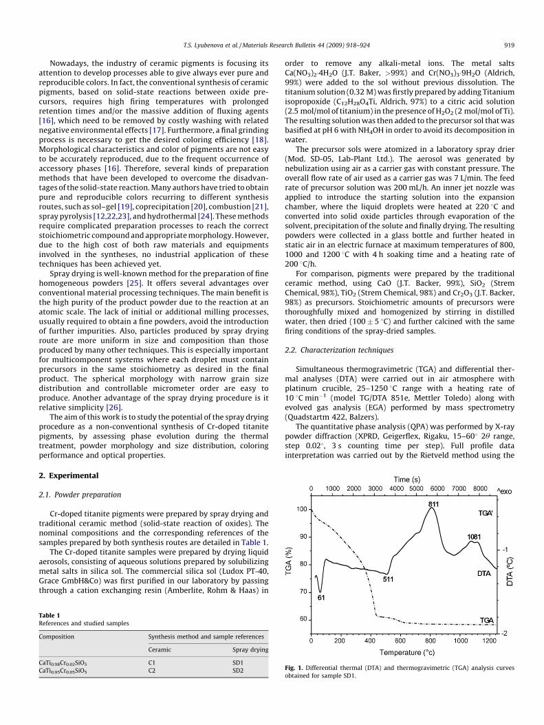
Fig. 1. Differential thermal (DTA) and thermogravimetric (TGA) analysis curves
obtained for sample SD1.
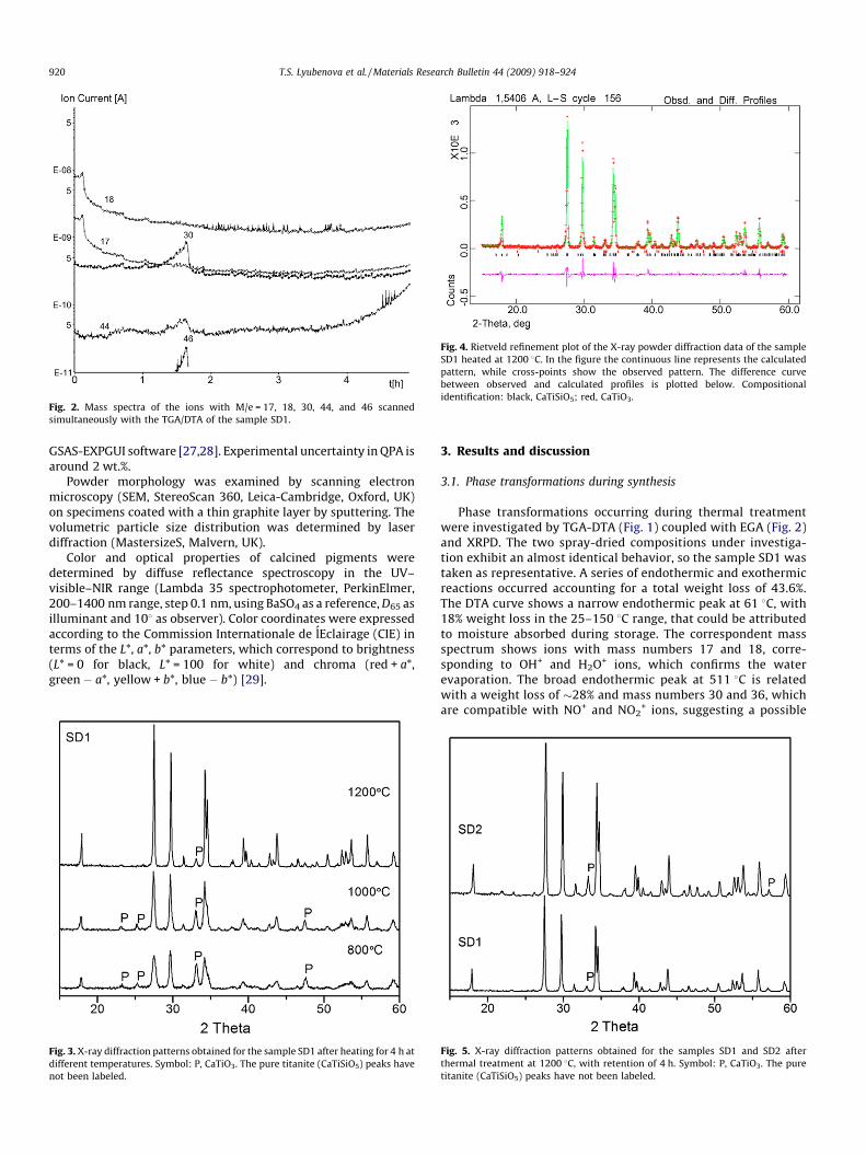
Fig. 2. Mass spectra of the ions with M/e = 17, 18, 30, 44, and 46 scanned
simultaneously with the TGA/DTA of the sample SD1.
Fig. 4. Rietveld refinement plot of the X-ray powder diffraction data of the sample
SD1 heated at 1200 8C. In the figure the continuous line represents the calculated
pattern, while cross-points show the observed pattern. The difference curve
between observed and calculated profiles is plotted below. Compositional
identification: black, CaTiSiO5; red, CaTiO3.
T.S. Lyubenova et al. / Materials Research Bulletin 44 (2009) 918–924920
GSAS-EXPGUI software [27,28]. Experimental uncertainty in QPA isaround 2 wt.%.
Powder morphology was examined by scanning electronmicroscopy (SEM, StereoScan 360, Leica-Cambridge, Oxford, UK)on specimens coated with a thin graphite layer by sputtering. Thevolumetric particle size distribution was determined by laserdiffraction (MastersizeS, Malvern, UK).
Color and optical properties of calcined pigments weredetermined by diffuse reflectance spectroscopy in the UV–visible–NIR range (Lambda 35 spectrophotometer, PerkinElmer,200–1400 nm range, step 0.1 nm, using BaSO4 as a reference, D65 asilluminant and 108 as observer). Color coordinates were expressedaccording to the Commission Internationale de lEclairage (CIE) interms of the L*, a*, b* parameters, which correspond to brightness(L* = 0 for black, L* = 100 for white) and chroma (red + a*,green � a*, yellow + b*, blue � b*) [29].
Fig. 3. X-ray diffraction patterns obtained for the sample SD1 after heating for 4 h at
different temperatures. Symbol: P, CaTiO3. The pure titanite (CaTiSiO5) peaks have
not been labeled.
3. Results and discussion
3.1. Phase transformations during synthesis
Phase transformations occurring during thermal treatmentwere investigated by TGA-DTA (Fig. 1) coupled with EGA (Fig. 2)and XRPD. The two spray-dried compositions under investiga-tion exhibit an almost identical behavior, so the sample SD1 wastaken as representative. A series of endothermic and exothermicreactions occurred accounting for a total weight loss of 43.6%.The DTA curve shows a narrow endothermic peak at 61 8C, with18% weight loss in the 25–150 8C range, that could be attributedto moisture absorbed during storage. The correspondent massspectrum shows ions with mass numbers 17 and 18, corre-sponding to OH+ and H2O+ ions, which confirms the waterevaporation. The broad endothermic peak at 511 8C is relatedwith a weight loss of �28% and mass numbers 30 and 36, whichare compatible with NO+ and NO2
+ ions, suggesting a possible
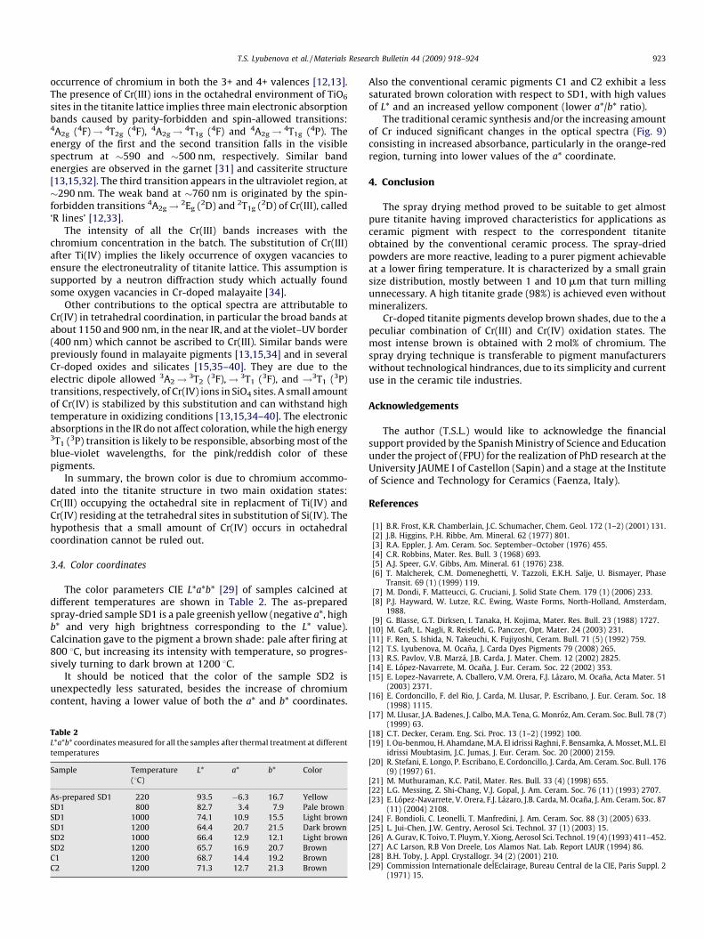
Fig. 5. X-ray diffraction patterns obtained for the samples SD1 and SD2 after
thermal treatment at 1200 8C, with retention of 4 h. Symbol: P, CaTiO3. The pure
titanite (CaTiSiO5) peaks have not been labeled.
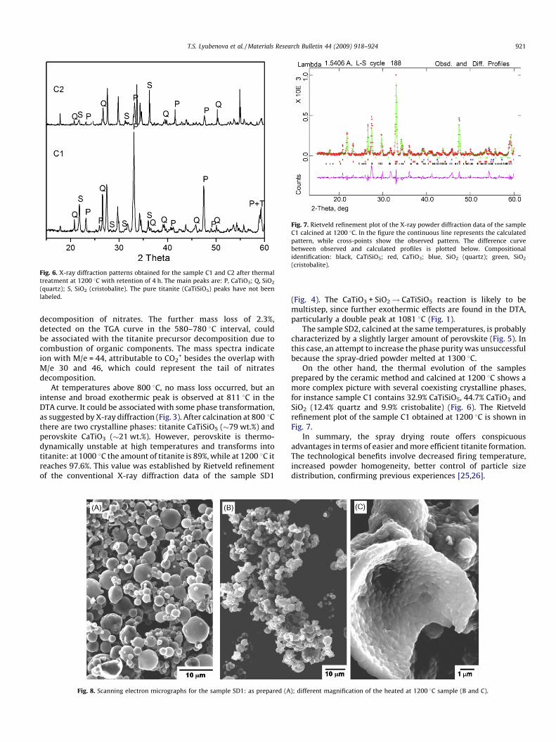
Fig. 6. X-ray diffraction patterns obtained for the sample C1 and C2 after thermal
treatment at 1200 8C with retention of 4 h. The main peaks are: P, CaTiO3; Q, SiO2
(quartz); S, SiO2 (cristobalite). The pure titanite (CaTiSiO5) peaks have not been
labeled.
Fig. 7. Rietveld refinement plot of the X-ray powder diffraction data of the sample
C1 calcined at 1200 8C. In the figure the continuous line represents the calculated
pattern, while cross-points show the observed pattern. The difference curve
between observed and calculated profiles is plotted below. Compositional
identification: black, CaTiSiO5; red, CaTiO3; blue, SiO2 (quartz); green, SiO2
(cristobalite).
T.S. Lyubenova et al. / Materials Research Bulletin 44 (2009) 918–924 921
decomposition of nitrates. The further mass loss of 2.3%,detected on the TGA curve in the 580–780 8C interval, couldbe associated with the titanite precursor decomposition due tocombustion of organic components. The mass spectra indicateion with M/e = 44, attributable to CO2
+ besides the overlap withM/e 30 and 46, which could represent the tail of nitratesdecomposition.
At temperatures above 800 8C, no mass loss occurred, but anintense and broad exothermic peak is observed at 811 8C in theDTA curve. It could be associated with some phase transformation,as suggested by X-ray diffraction (Fig. 3). After calcination at 800 8Cthere are two crystalline phases: titanite CaTiSiO5 (�79 wt.%) andperovskite CaTiO3 (�21 wt.%). However, perovskite is thermo-dynamically unstable at high temperatures and transforms intotitanite: at 1000 8C the amount of titanite is 89%, while at 1200 8C itreaches 97.6%. This value was established by Rietveld refinementof the conventional X-ray diffraction data of the sample SD1
Fig. 8. Scanning electron micrographs for the sample SD1: as prepared (A
(Fig. 4). The CaTiO3 + SiO2! CaTiSiO5 reaction is likely to bemultistep, since further exothermic effects are found in the DTA,particularly a double peak at 1081 8C (Fig. 1).
The sample SD2, calcined at the same temperatures, is probablycharacterized by a slightly larger amount of perovskite (Fig. 5). Inthis case, an attempt to increase the phase purity was unsuccessfulbecause the spray-dried powder melted at 1300 8C.
On the other hand, the thermal evolution of the samplesprepared by the ceramic method and calcined at 1200 8C shows amore complex picture with several coexisting crystalline phases,for instance sample C1 contains 32.9% CaTiSiO5, 44.7% CaTiO3 andSiO2 (12.4% quartz and 9.9% cristobalite) (Fig. 6). The Rietveldrefinement plot of the sample C1 obtained at 1200 8C is shown inFig. 7.
In summary, the spray drying route offers conspicuousadvantages in terms of easier and more efficient titanite formation.The technological benefits involve decreased firing temperature,increased powder homogeneity, better control of particle sizedistribution, confirming previous experiences [25,26].
); different magnification of the heated at 1200 8C sample (B and C).
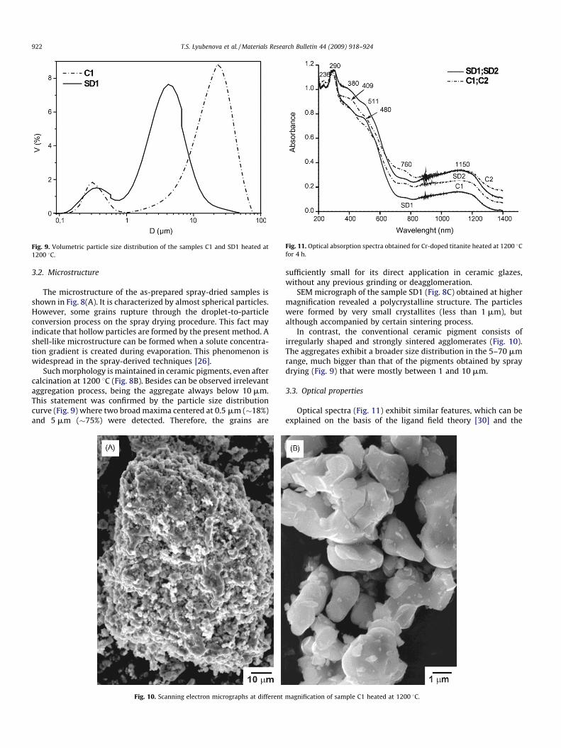
Fig. 11. Optical absorption spectra obtained for Cr-doped titanite heated at 1200 8Cfor 4 h.
Fig. 9. Volumetric particle size distribution of the samples C1 and SD1 heated at
1200 8C.
T.S. Lyubenova et al. / Materials Research Bulletin 44 (2009) 918–924922
3.2. Microstructure
The microstructure of the as-prepared spray-dried samples isshown in Fig. 8(A). It is characterized by almost spherical particles.However, some grains rupture through the droplet-to-particleconversion process on the spray drying procedure. This fact mayindicate that hollow particles are formed by the present method. Ashell-like microstructure can be formed when a solute concentra-tion gradient is created during evaporation. This phenomenon iswidespread in the spray-derived techniques [26].
Such morphology is maintained in ceramic pigments, even aftercalcination at 1200 8C (Fig. 8B). Besides can be observed irrelevantaggregation process, being the aggregate always below 10 mm.This statement was confirmed by the particle size distributioncurve (Fig. 9) where two broad maxima centered at 0.5 mm (�18%)and 5 mm (�75%) were detected. Therefore, the grains are
Fig. 10. Scanning electron micrographs at different
sufficiently small for its direct application in ceramic glazes,without any previous grinding or deagglomeration.
SEM micrograph of the sample SD1 (Fig. 8C) obtained at highermagnification revealed a polycrystalline structure. The particleswere formed by very small crystallites (less than 1 mm), butalthough accompanied by certain sintering process.
In contrast, the conventional ceramic pigment consists ofirregularly shaped and strongly sintered agglomerates (Fig. 10).The aggregates exhibit a broader size distribution in the 5–70 mmrange, much bigger than that of the pigments obtained by spraydrying (Fig. 9) that were mostly between 1 and 10 mm.
3.3. Optical properties
Optical spectra (Fig. 11) exhibit similar features, which can beexplained on the basis of the ligand field theory [30] and the
magnification of sample C1 heated at 1200 8C.
T.S. Lyubenova et al. / Materials Research Bulletin 44 (2009) 918–924 923
occurrence of chromium in both the 3+ and 4+ valences [12,13].The presence of Cr(III) ions in the octahedral environment of TiO6
sites in the titanite lattice implies three main electronic absorptionbands caused by parity-forbidden and spin-allowed transitions:4A2g (4F)! 4T2g (4F), 4A2g! 4T1g (4F) and 4A2g! 4T1g (4P). Theenergy of the first and the second transition falls in the visiblespectrum at �590 and �500 nm, respectively. Similar bandenergies are observed in the garnet [31] and cassiterite structure[13,15,32]. The third transition appears in the ultraviolet region, at�290 nm. The weak band at �760 nm is originated by the spin-forbidden transitions 4A2g! 2Eg (2D) and 2T1g (2D) of Cr(III), called‘R lines’ [12,33].
The intensity of all the Cr(III) bands increases with thechromium concentration in the batch. The substitution of Cr(III)after Ti(IV) implies the likely occurrence of oxygen vacancies toensure the electroneutrality of titanite lattice. This assumption issupported by a neutron diffraction study which actually foundsome oxygen vacancies in Cr-doped malayaite [34].
Other contributions to the optical spectra are attributable toCr(IV) in tetrahedral coordination, in particular the broad bands atabout 1150 and 900 nm, in the near IR, and at the violet–UV border(400 nm) which cannot be ascribed to Cr(III). Similar bands werepreviously found in malayaite pigments [13,15,34] and in severalCr-doped oxides and silicates [15,35–40]. They are due to theelectric dipole allowed 3A2! 3T2 (3F),! 3T1 (3F), and !3T1 (3P)transitions, respectively, of Cr(IV) ions in SiO4 sites. A small amountof Cr(IV) is stabilized by this substitution and can withstand hightemperature in oxidizing conditions [13,15,34–40]. The electronicabsorptions in the IR do not affect coloration, while the high energy3T1 (3P) transition is likely to be responsible, absorbing most of theblue-violet wavelengths, for the pink/reddish color of thesepigments.
In summary, the brown color is due to chromium accommo-dated into the titanite structure in two main oxidation states:Cr(III) occupying the octahedral site in replacment of Ti(IV) andCr(IV) residing at the tetrahedral sites in substitution of Si(IV). Thehypothesis that a small amount of Cr(IV) occurs in octahedralcoordination cannot be ruled out.
3.4. Color coordinates
The color parameters CIE L*a*b* [29] of samples calcined atdifferent temperatures are shown in Table 2. The as-preparedspray-dried sample SD1 is a pale greenish yellow (negative a*, highb* and very high brightness corresponding to the L* value).Calcination gave to the pigment a brown shade: pale after firing at800 8C, but increasing its intensity with temperature, so progres-sively turning to dark brown at 1200 8C.
It should be noticed that the color of the sample SD2 isunexpectedly less saturated, besides the increase of chromiumcontent, having a lower value of both the a* and b* coordinates.
Table 2L*a*b* coordinates measured for all the samples after thermal treatment at different
temperatures
Sample Temperature
(8C)
L* a* b* Color
As-prepared SD1 220 93.5 �6.3 16.7 Yellow
SD1 800 82.7 3.4 7.9 Pale brown
SD1 1000 74.1 10.9 15.5 Light brown
SD1 1200 64.4 20.7 21.5 Dark brown
SD2 1000 66.4 12.9 12.1 Light brown
SD2 1200 65.7 16.9 20.7 Brown
C1 1200 68.7 14.4 19.2 Brown
C2 1200 71.3 12.7 21.3 Brown
Also the conventional ceramic pigments C1 and C2 exhibit a lesssaturated brown coloration with respect to SD1, with high valuesof L* and an increased yellow component (lower a*/b* ratio).
The traditional ceramic synthesis and/or the increasing amountof Cr induced significant changes in the optical spectra (Fig. 9)consisting in increased absorbance, particularly in the orange-redregion, turning into lower values of the a* coordinate.
4. Conclusion
The spray drying method proved to be suitable to get almostpure titanite having improved characteristics for applications asceramic pigment with respect to the correspondent titaniteobtained by the conventional ceramic process. The spray-driedpowders are more reactive, leading to a purer pigment achievableat a lower firing temperature. It is characterized by a small grainsize distribution, mostly between 1 and 10 mm that turn millingunnecessary. A high titanite grade (98%) is achieved even withoutmineralizers.
Cr-doped titanite pigments develop brown shades, due to the apeculiar combination of Cr(III) and Cr(IV) oxidation states. Themost intense brown is obtained with 2 mol% of chromium. Thespray drying technique is transferable to pigment manufacturerswithout technological hindrances, due to its simplicity and currentuse in the ceramic tile industries.
Acknowledgements
The author (T.S.L.) would like to acknowledge the financialsupport provided by the Spanish Ministry of Science and Educationunder the project of (FPU) for the realization of PhD research at theUniversity JAUME I of Castellon (Sapin) and a stage at the Instituteof Science and Technology for Ceramics (Faenza, Italy).
References
[1] B.R. Frost, K.R. Chamberlain, J.C. Schumacher, Chem. Geol. 172 (1–2) (2001) 131.[2] J.B. Higgins, P.H. Ribbe, Am. Mineral. 62 (1977) 801.[3] R.A. Eppler, J. Am. Ceram. Soc. September–October (1976) 455.[4] C.R. Robbins, Mater. Res. Bull. 3 (1968) 693.[5] A.J. Speer, G.V. Gibbs, Am. Mineral. 61 (1976) 238.[6] T. Malcherek, C.M. Domeneghetti, V. Tazzoli, E.K.H. Salje, U. Bismayer, Phase
Transit. 69 (1) (1999) 119.[7] M. Dondi, F. Matteucci, G. Cruciani, J. Solid State Chem. 179 (1) (2006) 233.[8] P.J. Hayward, W. Lutze, R.C. Ewing, Waste Forms, North-Holland, Amsterdam,
1988.[9] G. Blasse, G.T. Dirksen, I. Tanaka, H. Kojima, Mater. Res. Bull. 23 (1988) 1727.
[10] M. Gaft, L. Nagli, R. Reisfeld, G. Panczer, Opt. Mater. 24 (2003) 231.[11] F. Ren, S. Ishida, N. Takeuchi, K. Fujiyoshi, Ceram. Bull. 71 (5) (1992) 759.[12] T.S. Lyubenova, M. Ocana, J. Carda Dyes Pigments 79 (2008) 265.[13] R.S. Pavlov, V.B. Marza, J.B. Carda, J. Mater. Chem. 12 (2002) 2825.[14] E. Lopez-Navarrete, M. Ocana, J. Eur. Ceram. Soc. 22 (2002) 353.[15] E. Lopez-Navarrete, A. Cballero, V.M. Orera, F.J. Lazaro, M. Ocana, Acta Mater. 51
(2003) 2371.[16] E. Cordoncillo, F. del Rio, J. Carda, M. Llusar, P. Escribano, J. Eur. Ceram. Soc. 18
(1998) 1115.[17] M. Llusar, J.A. Badenes, J. Calbo, M.A. Tena, G. Monroz, Am. Ceram. Soc. Bull. 78 (7)
(1999) 63.[18] C.T. Decker, Ceram. Eng. Sci. Proc. 13 (1–2) (1992) 100.[19] I. Ou-benmou, H. Ahamdane, M.A. El idrissi Raghni, F. Bensamka, A. Mosset, M.L. El
idrissi Moubtasim, J.C. Jumas, J. Eur. Ceram. Soc. 20 (2000) 2159.[20] R. Stefani, E. Longo, P. Escribano, E. Cordoncillo, J. Carda, Am. Ceram. Soc. Bull. 176
(9) (1997) 61.[21] M. Muthuraman, K.C. Patil, Mater. Res. Bull. 33 (4) (1998) 655.[22] L.G. Messing, Z. Shi-Chang, V.J. Gopal, J. Am. Ceram. Soc. 76 (11) (1993) 2707.[23] E. Lopez-Navarrete, V. Orera, F.J. Lazaro, J.B. Carda, M. Ocana, J. Am. Ceram. Soc. 87
(11) (2004) 2108.[24] F. Bondioli, C. Leonelli, T. Manfredini, J. Am. Ceram. Soc. 88 (3) (2005) 633.[25] L. Jui-Chen, J.W. Gentry, Aerosol Sci. Technol. 37 (1) (2003) 15.[26] A. Gurav, K. Toivo, T. Pluym, Y. Xiong, Aerosol Sci. Technol. 19 (4) (1993) 411–452.[27] A.C Larson, R.B Von Dreele, Los Alamos Nat. Lab. Report LAUR (1994) 86.[28] B.H. Toby, J. Appl. Crystallogr. 34 (2) (2001) 210.[29] Commission Internationale delEclairage, Bureau Central de la CIE, Paris Suppl. 2
(1971) 15.

T.S. Lyubenova et al. / Materials Research Bulletin 44 (2009) 918–924924
[30] J.R. Gispert, Coordination Chemistry, Omega Publ., Barcelona, 2000.[31] R. Galindo, M. Llusar, M.A. Tena, G. Monrros, J.A. Badenes, J. Eur. Ceram. Soc. 27
(2007) 199.[32] E. Lopez-Navarrete, A.R. Gonzalez-Elipe, M. Ocana, Ceram. Int. 29 (2003) 385.[33] Y. Marinova, J.M. Hohemberger, E. Cordoncillo, P. Escribano, J.B. Carda, J. Eur.
Ceram. Soc. 23 (2003) 213.[34] G. Cruciani, M. Dondi, M. Ardit, T. Stoyanova Lyubenova, J.B. Carda, F. Matteucci,
A.L. Costa, Dyes Pigments. (2009), in press.
[35] F. Matteucci, G. Cruciani, M. Dondi, G. Baldi, A. Barzanti, Acta Mater. 55 (2007)2229.
[36] R. Feldman, Y. Shimony, Z. Burshtein, Optical Mater. 24 (2003) 333.[37] D. Reinen, U. Kesper, M. Atanasov, J. Roost, Inorg. Chem. 34 (1995) 184.[38] B. Henderson, H.G. Gallagher, T.P.J. Han, M.A. Scott, J. Phys.: Condens. Matter 12
(2000) 1927.[39] A. Domenech, F.J. Torres, E.R. de Sola, J. Alarcon, Eur. J. Inorg. Chem. (2006) 638–648.[40] A.M. Heyns, P.M. Harden, J. Phys. Chem. Solids 60 (1999) 277.