Syntactic comprehension deficits are associated with MRI white matter alterations in dementia
10
Syntactic comprehension deficits are associated with MRI white matter alterations in dementia TANIA GIOVANNETTI, 1 MARY W. HOPKINS, 2 JACLYN CRAWFORD, 3 BRIANNE MAGOUIRK BETTCHER, 1 KARA S. SCHMIDT, 4 and DAVID J. LIBON 4 1 Department of Psychology, Temple University, Philadelphia, Pennsylvania 2 Department of Psychology, Mercy College, Dobbs Ferry, New York 3 Department of Medicine, University of Medicine and Dentistry of New Jersey–School of Osteopathic Medicine, Stratford, New Jersey. 4 Department of Neurology, Drexel University College of Medicine, Philadelphia, Pennsylvania (Received February 13, 2007; Final Revision February 27, 2008; Accepted March 3, 2008) Abstract Comprehension difficulties associated with periventricular and deep white matter alterations (WMA) in mild dementia were investigated using portions of the Boston Diagnostic Aphasia Examination (BDAE) Complex Ideation subtest and Syntax subtests. Mild dementia participants were grouped according to the extent of their WMA as observed on magnetic resonance imaging (mild WMA n 5 45 vs. moderate to severe WMA n 5 52). Correlation and regression analyses also were performed to examine the link between WMA and comprehension abilities, as well as the link between comprehension abilities and neuropsychological measures of executive functioning, language, episodic memory, and overall dementia severity. Results showed that the WMA groups differed on the BDAE-Syntax subtests, with the severe WMA group demonstrating more impairment. Correlation and regression analyses including the entire sample also demonstrated that the extent of WMA was significantly linked to Syntax test scores but not Complex Ideation scores. Regression analyses including neuropsychological measures showed that the BDAE-Complex Ideation score was marginally predicted by only overall dementia severity, whereas the BDAE-Syntax scores were significantly predicted by independent measures of working memory0executive functioning. In conclusion, greater subcortical WMA and executive deficits are associated with greater difficulties in syntactic comprehension in individuals with mild dementia. ( JINS, 2008, 14, 542–551.) Keywords: Alzheimer’s disease, Vascular dementia, Subcortical dementia, Aphasia, Comprehension INTRODUCTION There is now considerable interest in the effect of subcor- tical white matter alterations (WMA) on the clinical pre- sentation of dementia. A growing body of research has demonstrated that subcortical vascular disease results in differential impairment on a wide range of neuropsycho- logical tests that measure concept formation, executive con- trol and working memory, and graphomotor functioning (see Libon et al., 2004 for a review). However, language comprehension deficits have not been extensively studied in vascular dementia (VaD) and the relation between com- prehension difficulties and WMA in dementia remains unknown. This paper aims to fill this gap in the current literature. Language comprehension deficits have been widely reported in people with Alzheimer’s disease (AD). Specif- ically, lexical comprehension difficulties have been reported most frequently and attributed to semantic knowledge deg- radation (Chan et al., 1995; Chertkow & Bub, 1990; Hodges et al., 1992; Huff et al., 1988). While several authors have reported syntactic comprehension difficulties in AD (Grober & Bang, 1995; Small et al., 1997) others have shown that syntactic comprehension, per se, is relatively preserved (Grossman et al., 1996, 1998, Grossman & White-Devine, 1998; Rochon et al., 1994; Schwartz et al., 1979). For exam- ple, using a sentence-picture matching task, Rochon et al. (1994) have shown that comprehension abilities in AD were unaffected by syntactic complexity (see also Rochon & Saf- fran, 1995; Waters et al., 1995, 1998). Correspondence and reprint requests to: Tania Giovannetti, Ph.D., Tem- ple University, Department of Psychology, 1701 N. 13th Street, Philadel- phia, PA 19122. E-mail: [email protected] Journal of the International Neuropsychological Society (2008), 14, 542–551. Copyright © 2008 INS. Published by Cambridge University Press. Printed in the USA. doi:10.10170S1355617708080715 542
-
Upload
independent -
Category
Documents
-
view
2 -
download
0
Transcript of Syntactic comprehension deficits are associated with MRI white matter alterations in dementia

Syntactic comprehension deficits are associated with MRIwhite matter alterations in dementia
TANIA GIOVANNETTI,1 MARY W. HOPKINS,2 JACLYN CRAWFORD,3
BRIANNE MAGOUIRK BETTCHER,1 KARA S. SCHMIDT,4 and DAVID J. LIBON4
1Department of Psychology, Temple University, Philadelphia, Pennsylvania2Department of Psychology, Mercy College, Dobbs Ferry, New York3Department of Medicine, University of Medicine and Dentistry of New Jersey–School of Osteopathic Medicine,Stratford, New Jersey.
4Department of Neurology, Drexel University College of Medicine, Philadelphia, Pennsylvania
(Received February 13, 2007; Final Revision February 27, 2008; Accepted March 3, 2008)
Abstract
Comprehension difficulties associated with periventricular and deep white matter alterations (WMA) in milddementia were investigated using portions of the Boston Diagnostic Aphasia Examination (BDAE) ComplexIdeation subtest and Syntax subtests. Mild dementia participants were grouped according to the extent of theirWMA as observed on magnetic resonance imaging (mild WMA n5 45 vs. moderate to severe WMA n5 52).Correlation and regression analyses also were performed to examine the link between WMA and comprehensionabilities, as well as the link between comprehension abilities and neuropsychological measures of executivefunctioning, language, episodic memory, and overall dementia severity. Results showed that the WMA groupsdiffered on the BDAE-Syntax subtests, with the severe WMA group demonstrating more impairment. Correlationand regression analyses including the entire sample also demonstrated that the extent of WMA was significantlylinked to Syntax test scores but not Complex Ideation scores. Regression analyses including neuropsychologicalmeasures showed that the BDAE-Complex Ideation score was marginally predicted by only overall dementiaseverity, whereas the BDAE-Syntax scores were significantly predicted by independent measures of workingmemory0executive functioning. In conclusion, greater subcortical WMA and executive deficits are associated withgreater difficulties in syntactic comprehension in individuals with mild dementia. (JINS, 2008, 14, 542–551.)
Keywords: Alzheimer’s disease, Vascular dementia, Subcortical dementia, Aphasia, Comprehension
INTRODUCTION
There is now considerable interest in the effect of subcor-tical white matter alterations (WMA) on the clinical pre-sentation of dementia. A growing body of research hasdemonstrated that subcortical vascular disease results indifferential impairment on a wide range of neuropsycho-logical tests that measure concept formation, executive con-trol and working memory, and graphomotor functioning(see Libon et al., 2004 for a review). However, languagecomprehension deficits have not been extensively studiedin vascular dementia (VaD) and the relation between com-prehension difficulties and WMA in dementia remains
unknown. This paper aims to fill this gap in the currentliterature.
Language comprehension deficits have been widelyreported in people with Alzheimer’s disease (AD). Specif-ically, lexical comprehension difficulties have been reportedmost frequently and attributed to semantic knowledge deg-radation (Chan et al., 1995; Chertkow & Bub, 1990; Hodgeset al., 1992; Huff et al., 1988). While several authors havereported syntactic comprehension difficulties in AD (Grober& Bang, 1995; Small et al., 1997) others have shown thatsyntactic comprehension, per se, is relatively preserved(Grossman et al., 1996, 1998, Grossman & White-Devine,1998; Rochon et al., 1994; Schwartz et al., 1979). For exam-ple, using a sentence-picture matching task, Rochon et al.(1994) have shown that comprehension abilities in AD wereunaffected by syntactic complexity (see also Rochon & Saf-fran, 1995; Waters et al., 1995, 1998).
Correspondence and reprint requests to: Tania Giovannetti, Ph.D., Tem-ple University, Department of Psychology, 1701 N. 13th Street, Philadel-phia, PA 19122. E-mail: [email protected]
Journal of the International Neuropsychological Society (2008), 14, 542–551.Copyright © 2008 INS. Published by Cambridge University Press. Printed in the USA.doi:10.10170S1355617708080715
542

As stated, relatively few studies have examined languagecomprehension abilities in VaD. The handful of studies inthe literature has focused on comparing comprehension abil-ities in participants with VaD to those with AD and0or otherdementia syndromes. Theoretical papers have hypothesizedgreater lexical comprehension deficits in AD and relativelygreater syntactic deficits in VaD (Desmond, 2004) based onneuropathology differences between AD and VaD, as wellas strongly held notions in the aphasia0language literature.Namely, individuals with VaD show differential impair-ment in executive functioning, and syntactical processinghas been associated with working memory and prefrontal0executive functioning, even among dementia patients (seeKempler et al., 1998). Moreover, lexical and semantic pro-cessing have been associated with declarative memory sys-tems and the temporal cortex, and these processes are knownto be differentially impaired in people with AD (see Friedericiet al., 2003; Ullman, 2001; Ullman et al., 1997). To date,only one report supports this dissociation in AD and VaDparticipants (Kertesz et al., 1990); other empirical studieshave demonstrated little to no differences in comprehen-sion abilities between AD and VaD participants (Powellet al., 1988; Villardita, 1993; Vuorinen et al., 2000). Anothergroup of studies has reported results that contradict thishypothesis (Grossman et al., 1996; Kontiola et al., 1990).For example, Grossman and colleagues (1996) showed thatparticipants with VaD performed better than AD partici-pants on tests of both lexical and syntactic comprehension.
One major limitation of the aforementioned AD versusVaD studies is that patients were grouped according to theirclinical diagnosis. Only Kertesz and colleagues (1990)grouped participants according to WMA as observed onneuroimaging. There is accumulating evidence to suggestthat the neuropathology underlying the clinical syndromesof AD and VaD can overlap (Jellinger, 2002; Pantoni &Garcia, 1997; see also Libon et al., 2004, 2008; Price et al.,2005). Therefore, the grouping methods used in prior stud-ies may have obscured meaningful differences in neuropa-thology across participants. To address this possibility, recentstudies have demonstrated statistically significant neuropsy-chological differences across dementia groups that differedaccording to WMA observed on magnetic resonance imag-ing (MRI) of the brain (Libon et al., 2008; Price et al.,2005, 2007). Thus, in the current study, patients diagnosedclinically with AD or VaD were re-grouped on the basis ofthe severity of their WMA as measured on MRI. Further-more, participants’ WMA scores were evaluated as a con-tinuous variable in correlation and regression analyses, alongwith measures of comprehension abilities.
The goal of this study was to evaluate language compre-hension abilities among mild dementia participants thatdiffered according to the extent of their MRI-WMA. Com-prehension abilities were assessed using subtests of theBoston Diagnostic Aphasia Battery (BDAE): Complex Ide-ation (paragraph portion) and Syntax subtests (EmbeddedSentences & Touch A with B). According to the BDAEmanual, the Syntax subtests specifically assess partici-
pants’ ability to process complex syntactical relations; theComplex Ideation subtest is described as a more elemen-tary measure of language comprehension and is associatedwith other tests of lexical and semantic processing (Good-glass et al., 2001). We hypothesized that participants withgreater WMA, who are known to be especially disadvan-taged on neuropsychological measures of executive func-tioning and working memory (Lamar et al., 2007; Libonet al., 2004; Looi & Sachdev, 1999), would experiencelanguage comprehension deficits characterized by syntac-tic comprehension difficulties. In contrast, dementia par-ticipants of comparable overall dementia severity but withless severe WMA are known to demonstrate greater defi-cits in declarative memory systems than those with mod-erate to severe WMA (Libon et al., 2004); therefore, wehypothesized that these participants would experience com-prehension deficits secondary to lexical or semantic pro-cessing impairments. Based on these hypotheses, wepredicted that dementia patients with significant WMAwould demonstrate greater deficits on the BDAE-Syntacticsubtests than participants with little or no WMA, whereasparticipants with mild WMA would demonstrate greaterdeficits on the BDAE-Complex Ideation subtest. Alongthese lines, we also predicted that performance on theBDAE-Syntax subtests would be associated with indepen-dent measures of working memory0executive functioning;performance on the BDAE-Complex Ideation subtest wouldbe associated with independent measures of lexical andsemantic processing (i.e., naming and category fluency).
METHODS
ParticipantsNinety-seven outpatients with AD or VaD were recruitedfrom the Memory Assessment Program (MAP) of the NewJersey Institute for Successful Aging, University of Medi-cine and Dentistry of New Jersey–School of OsteopathicMedicine. All participants were evaluated by a social worker,neurologist, and neuropsychologist. Laboratory and brainMRI studies also were obtained on all patients. Patients’diagnoses were determined during an interdisciplinary teammeeting according to established criteria (Chui et al., 1992;McKhann et al., 1984). Comprehension test scores were notused to determine participants’ clinical diagnoses; how-ever, neuroimaging data were considered when making diag-nostic decisions.
Forty-seven patients were diagnosed with probable AD(McKhann et al., 1984), and 50 patients were diagnosedwith possible (n5 22) or probable VaD (n5 28; Chui et al.,1992). No patient diagnosed with VaD presented with acortical stroke or a sudden onset0stepwise decline in cog-nitive functioning.
MRI protocol and participant groups
A 1.5 Tesla Siemens MRI scanner was used to obtainT1-weighted (TR-500ms,TE-9ms)andFLAIR(TR-8500ms,
Comprehension and dementia 543
TE-99 ms) images with a 5 mm slice thickness and 1 mmgap between slices. The severity of MRI-WMA was quan-tified using the 40-point Leukoaraiosis (LA) Scale of Junque(Junque et al., 1990). The Junque scale is similar to severalother scales described in the literature (Scheltens et al., 1993;Wahlund et al., 2001; Ylikoski et al., 1993); however, oneadvantage of using the Junque Scale is that it offers a widerrange of measurement (i.e., 0– 40) that permits robust sta-tistical analyses. More specifically, the Junque Scale entailsscores for white matter alterations in five specific areas foreach hemisphere: frontal centrum semiovale, parietal cen-trum semiovale, white matter around the frontal horns, whitematter around the body of the lateral ventricles and whitematter around the atrium and the occipital horns. Scores foreach area range from 0 (no visible white matter alterations)to 4 (severe white matter alterations) and are summed toequal a total score with a possible range of 0 to 40. Paststudies have demonstrated high inter-rater reliability betweenneuroradiologists using this scale (r5 .93, p, .001; Libonet al., 1998).
For the present study, FLAIR-weighted MRI scans for allparticipants were evaluated using the Junque Scale by aboard-certified neuroradiologist who never met or inter-acted with the patient. The neuroradiologist also was blindto all clinical information, including dementia diagnosis,and neuropsychological test scores, and he did not partici-pate in the interdisciplinary diagnosis consensus meeting.
Currently, there is no gold standard offering concreteguidelines for the operational definition of the clinical sig-nificance of MRI-WMA. Prior work from our laboratoryhas suggested a Junque cut score of 10 yields distinct demen-tia groups that differ on neuropsychological tests of execu-tive functioning and episodic memory (Libon et al., 2004,2008; Price et al., 2005, 2007). Based on these past reports,participants with Junque scores ,10 were assigned to theminimal to mild white matter group (Mild WMA Group).Participants with Junque scores of 10 or greater wereassigned to the Moderate to Severe WMA Group.
Procedures
The UMDNJ-SOM Institutional Review Board approvedthe project, and informed consent was obtained consistentwith the Declaration of Helsinki. Tests of language compre-hension and other neuropsychological functions were admin-istered as part of a comprehensive clinical protocol.
Assessment of comprehension
Comprehension was evaluated using the Complex Ideationand Syntax Comprehension subtests (Embedded Sentences& Touch A with B) from the BDAE, a comprehensive lan-guage assessment protocol (Goodglass et al., 2001). TheComplex Ideation and Syntax Comprehension subtests wereselected for this study because they purportedly assess dif-ferent aspects of language comprehension. Statistical analy-ses with a standardization sample of participants with aphasia
have been reported in the recent edition of the BDAE man-ual (Goodglass et al., 2001). These analyses have shownthat the Complex Ideation subtest clusters with other mea-sures of basic auditory comprehension, including single-word0lexical comprehension (Basic Word Discrimination,Tools Foods, etc.) and comprehension of basic commandsand semantic information (Semantic Probes, Commands);Complex Ideation did not cluster with measures of syntac-tical comprehension, such as Embedded Sentences and TouchA with B subtests. These latter two subtests clustered togetherand with another measure of syntactic comprehension (i.e.,Reversible Possessives).
Complex ideation subtest
For this study, only the paragraph portion of the ComplexIdeation subtest was administered. This portion of the sub-test requires participants to listen to a brief paragraph readaloud by the examiner and then answer 4 yes0no questionsregarding the paragraph. Four different paragraphs, com-prised of grammatically regular and relatively simple sen-tences, are presented. The yes0no questions are designed totap the ability to extract meaning from the short passages.Our scoring system for the paragraph portion of the BDAE-Complex Ideation subtest differed from that described inthe BDAE manual in that we assigned 1 point for eachcorrect answer (range5 0–16). This scoring procedure wasused to increase the variance for subsequent statisticalanalysis.
BDAE-Syntax comprehension subtests
The syntax comprehension module of the BDAE is com-prised of three subsections: Touching A with B, ReversiblePossessives, and Embedded Sentences. The Reversible Pos-sessives subtest was not included in this study, because inour clinic population the large majority of patients per-form at ceiling on this measure. The Touch A with B sub-test requires participants to view a series of picturesdepicting a person holding or touching a fork, comb, scis-sors, pencil, knife, and0or spoon. Participants are requiredto indicate which picture accurately depicts a statementread aloud by the examiner (e.g., “Which picture showstouching the spoon and the scissors”). All of the state-ments in this section contain the same number of words invaried orders.
The Embedded Sentences subtest contains syntacticallyreversible phrases and requires patients to determine whichcartoon representation (out of 4 options) best depicts theevents described by the stimulus statement (e.g., “Show methe picture of the boy hitting the girl who is sitting down?”).This task assesses participants’ ability to appropriately linka descriptive subordinate clause to either the subject or theobject. The length of each statement is limited to 7 to 10words. Our scoring procedures for the syntax subtests didnot differ from those described in the BDAE manual; onepoint was awarded for each correctly identified picture.
544 T. Giovannetti et al.

Neuropsychological Assessment
In addition to the Mini Mental-State Examination (Folsteinet al., 1975) and Geriatric Depression Scale (Yesavage,1986), all patients were administered tests that assessedworking memory0executive functioning, expressive lan-guage, and episodic memory. All measures are described indetail in Table 1.
RESULTS
Demographic and NeuropsychologicalCharacteristics of the Groups
As shown in Table 2, the groups did not differ significantlyin age, education, dementia severity0MMSE, level ofdepression0GDS, or the distribution of men versus women.As expected, the Mild WMA Group demonstrated a signif-icantly lower Junque Score than the Moderate-Severe WMAGroup. Additionally, as expected, the distribution of patientsclinically diagnosed with AD versus VaD differed acrossthe groups, with a significantly higher proportion of ADpatients in the Mild WMA Group and a higher proportionof VaD patients in the Moderate-Severe WMA Group (seeTable 2).
Consistent with prior studies, the WMA groups also dif-fered neuropsychologically, with the Moderate WMA Groupdemonstrating significantly worse performance on tests ofexecutive functioning (i.e., WMS-Mental Control & FAS).By contrast, the Mild WMA Group demonstrated worseperformance on the measure of episodic memory. The groupsdid not differ on tests of lexical processing0language (BNT& Animal Naming).
Between Group Differences on the BDAEComprehension Subtests
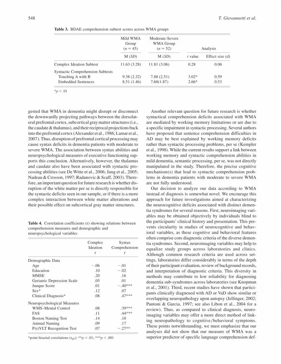
As predicted, the Moderate-Severe WMA Group obtainedsignificantly lower scores on the two syntax subtests (i.e.,Touch A with B & Embedded Sentences) than the MildWMA Group (see Table 3). Contrary to our prediction, how-ever, the groups did not differ on the BDAE-Complex Ide-ation subtest.
It is worth noting that when the sample was dividedaccording to clinical diagnosis, a significant difference wasobserved for both Syntax Comprehension Subtests (Touch-ing A with B t 5 4.16, p , .01; Embedded Sentences t 54.4, p , .01); participants with AD obtained better scoreson both tests. The clinical groups did not differ on the Com-plex Ideation subtest (t5 .70, p. .05).
Relations among Comprehension Measures
Before examining the neuropsychological correlates of com-prehension performance, correlations among the compre-hension subtests were performed to determine whether thesemeasures were assessing similar or different constructs.
Results showed that the Complex Ideation subtest was notsignificantly related to either Syntax Comprehension test(Touch A with B–r52.02; Embedded Sentences–r5 .05).However, the two BDAE Syntax Comprehension subtestswere significantly correlated (r5 .47, p, .001). Thus, forsubsequent analyses, the Syntax Comprehension Test scoreswere summed and analyzed as a single measure.
Relations between Comprehension Tests andDemographic and NeuropsychologicalVariables
As shown in Table 4, Complex Ideation and Syntax Com-prehension scores showed different patterns of correlationswith demographic and neuropsychological variables. TheSyntax Comprehension score was significantly and nega-tively related to Junque Score, with lower or worse scoreson syntactic comprehension tasks in participants with greaterwhite matter alterations. The same pattern of correlationswas noted for clinical diagnosis. Correlations including allother demographic variables were weak and nonsignificant.Note that point-biserial correlations were performed to eval-uate the link between dichotomous variables, such as sexand clinical diagnosis, and comprehension test scores; the102 value of the rpb value should not be interpreted forthese analyses.
There also were significant and positive relations betweenworking memory0executive measures and Syntax Compre-hension scores. As expected, as performance on measuresof working memory0executive functioning improved, scoreson Syntax Comprehension tests also increased or improved.Unexpectedly, a significant negative correlation wasobserved between the episodic memory measures and syn-tax comprehension. This suggests that participants whoexhibited better episodic memory performance demon-strated poorer syntax comprehension. Contrary to predic-tion, all correlation analyses including the Complex Ideationscore were nonsignificant; only the MMSE showed a mod-est, but nonsignificant relation to Complex Ideation.
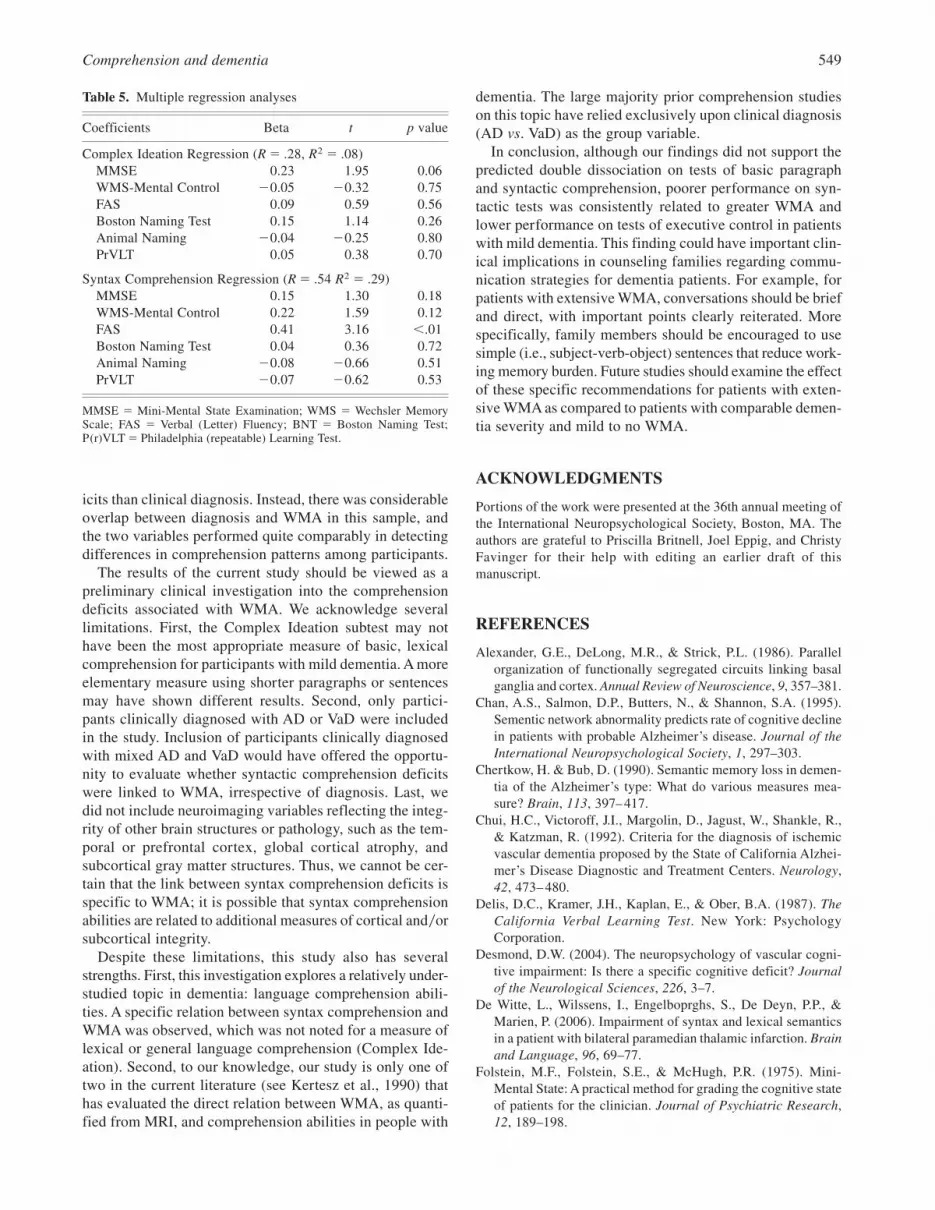
To assess the influence of multiple neuropsychologicalpredictors on comprehension abilities, two multiple regres-sion analyses were performed with Complex Ideation orSyntax Comprehension scores as the dependent variable.For both regressions, neuropsychological variables wereincluded as predictor0independent variables; the MMSE wasentered into the regression model first to account for gen-eral dementia severity, followed by the remaining test scores.The results are shown in Table 5. For the BDAE-ComplexIdeation subtest, the MMSE was only marginally signifi-cant (F 5 3.75, p 5 .057); all other neuropsychologicalmeasures were not significant predictors (F 5 1.08, p 5.38). For the Syntax Comprehension Tests, the MMSE wasnot significantly related to performance (F 5 1.82, p 5.18); however, the remaining neuropsychological variableswere significant (F5 4.98, p, .01), with FAS, a measureof working memory0executive functioning, as the only sig-nificant predictor variable.
Comprehension and dementia 545

Table 1. Neuropsychological Protocol
Test Description References
Working Memory0Executive Functioning
Boston Revision of the WechslerMemory Scale Mental Control Subtest(WMS-Mental Control)
The accuracy of performance on three nonautomatized tasks (months backward, alphabet rhyming,alphabet visualization) was calculated with the following algorithm: [12 (false positives1misses)0(#possible correct)]3 100; possible range5 0–100. Similar tests have been shown to activatedorsolateral prefrontal regions in healthy younger adults.
Lamar et al., 2002;Wildgruber et al., 1999
Phonemic Word List Generation (FAS) Participants were asked to generate as many words as possible in 60 seconds beginning with F, A, and S,excluding proper nouns.
Spreen & Strauss, 1998
Lexical Processing0Language
Boston Naming Test The number of pictures correctly named spontaneously without a semantic cue; possible range5 0– 60. Kaplan et al., 1983
Animal Naming The number of unique animal names generated within 60 seconds. Goodglass et al., 2001
Episodic Memory
Philadelphia (Repeatable) VerbalLearning Test–Discriminability Index(PrVLT-Discriminability)
Participants were asked to remember a 9 word list, as on the California Verbal Learning Test (Delis et al.,1987; see also Libon et al., 1996). The dependent variable was the accuracy on the delayed recognitionmemory task (Recognition Discriminability); possible range5 0–100. Free-recall test trials were not usedas dependent variables because they require executive abilities, such as organizational skills and retrieval.The Recognition Discriminibility index was selected because it is a relatively purer measure of episodicmemory encoding.
Garrett et al., 2004;Libon et al., 2005;Price et al., 2004
546T.G
iovannettiet
al.

DISCUSSION
This study assessed the influence of WMA in people withmild dementia on lexical and syntactic comprehension. Ashypothesized, moderate to severe WMA were associatedwith worse performance on measures of syntactic compre-hension. This relation was observed in both between-group analyses comparing participants with mild WMA tothose with moderate-severe WMA, as well as correlationanalyses between syntax comprehension scores and WMA(Junque) scores. Contrary to our second hypothesis, how-ever, we did not observe a relation between WMA scoresand performance on more elementary lexical comprehen-sion abilities, as measured with the BDAE Complex Ide-ation subtest. Specifically, the two WMA groups did notdiffer on this test and the Complex Ideation score was notsignificantly correlated with WMA (Junque) scores.
We also predicted significant relations between specificcomprehension abilities and specific measures of neuropsy-chological functioning. That is, we predicted a link betweensyntax comprehension and independent neuropsychologi-cal tests of working memory0executive functioning, andthis prediction was supported by both correlation and regres-sion analyses. However, our prediction of a link betweenlexical comprehension abilities (Complex Ideation) and inde-pendent measures of lexical processing was not supported.
Complex Ideation was marginally related to and predictedby only general dementia severity, as measured by theMMSE. This suggests that lexical comprehension abilitiesmay be negatively influenced by multiple cognitive diffi-culties in mild dementia patients. Alternatively, it is possi-ble that Complex Ideation was not an adequate or specificmeasure of lexical comprehension abilities for this popula-tion. That is, the Complex Ideation task may have incurreddemands on language abilities as well as working memoryand episodic memory on participants, thereby limiting ourability to obtain a pure measure of lexical comprehension.
While our conclusions regarding lexical comprehensionin dementia are ambiguous, our findings regarding syntaxcomprehension have implications for neurocognitive mod-els of syntactic comprehension deficits in mild dementia. Forexample, Grossman and colleagues (1996, 1998) reported alink between executive functioning and impaired syntax indementia after observing that patients with FrontotemporalDementia (FTD) showed greater syntax comprehension def-icits than patients withAD. Moreover, Grossman et al. (1998)reported that syntax comprehension deficits in FTD were asso-ciated with reduced cerebral perfusion in the left frontal andanterior temporal lobes. Our findings suggest that the linkbetween syntax comprehension and executive functioning isalso observed in cases where executive deficits may be sec-ondary to subcortical vascular pathology. It has been sug-
Table 2. Demographic and neuropsychological data across WMA groups
Mild WMA Group(n5 45)
Moderate-SevereWMA Group
(n5 52) Analysis
M (SD) M (SD) t value
Demographic DataAge 77.76 (6.13) 80.00 (5.21) 1.95Education 12.56 (2.19) 11.98 (2.63) 1.16GDS 3.60 (3.48) 4.31 (4.05) 0.92Junque Score 4.13 (2.48) 16.67 (5.58) 13.92*
n (%) n (%)chi
square value
Sex (n, % women) 32 (71%) 42 (82%) 2.41Clinical Diagnosis (n, %
Alzheimer’s disease)40 (89%) 7 (14%) 54.95*
M (SD) M (SD) t value
Neuropsychological Test ScoresMMSE 22.64 (2.64) 21.98 (3.33) 1.06WMS–Mental Control 74.27 (15.39) 56.02 (24.11) 4.23*FAS 25.02 (10.76) 18.00 (9.55) 3.34**Boston Naming Test 40.49 (12.32) 37.88 (11.59) 1.07Animal Naming 9.07 (4.14) 8.88 (3.72) 1.01PrVLT Recognition Test 67.87 (11.64) 78.82 (11.37) 4.68*
*p, .001; **p, .05MMSE5Mini-Mental State Examination; GDS5 Geriatric Depression Scale; BDAE5 Boston Diagnostic AphasiaExamination; WMS5Wechsler Adult Intelligence Scale; P(r)VLT5 Philadelphia (repeatable) Verbal Learning Test.
Comprehension and dementia 547
gested that WMA in dementia might disrupt or disconnectthe downwardly projecting pathways between the dorsolat-eral prefrontal cortex, subcortical gray matter structures (i.e.,the caudate & thalamus), and their reciprocal projections backinto the prefrontal cortex (Alexander et al., 1986; Lamar et al.,2007). Thus, disruption of prefrontal cortical processing maycause syntax deficits in dementia patients with moderate tosevere WMA. The association between syntax abilities andneuropsychological measures of executive functioning sup-ports this conclusion. Alternatively, however, the thalamusand caudate also have been associated with syntactic pro-cessing abilities (see De Witte et al., 2006; Jung et al., 2005;Nadeau & Crosson, 1997; Radanovic & Scaff, 2003). There-fore, an important question for future research is whether dis-ruption of the white matter per se is directly responsible forthe syntactic deficits seen in our sample, or if there is a morecomplex interaction between white matter alterations andtheir possible effect on subcortical gray matter structures.
Another relevant question for future research is whethersyntactical comprehension deficits associated with WMAare mediated by working memory limitations or are due toa specific impairment in syntactic processing. Several authorshave proposed that sentence comprehension difficulties inAD may be best explained by working memory deficitsrather than syntactic processing problems, per se (Kempleret al., 1998). While the current results support a link betweenworking memory and syntactic comprehension abilities inmild dementia, semantic processing, per se, was not directlymanipulated in the study. Therefore, the precise cognitivemechanism(s) that lead to syntactic comprehension prob-lems in dementia patients with moderate to severe WMAare not fully understood.
Our decision to analyze our data according to WMAinstead of diagnosis is somewhat novel. We encourage thisapproach for future investigations aimed at characterizingthe neurocognitive deficits associated with distinct demen-tia syndromes for several reasons. First, neuroimaging vari-ables may be obtained objectively by individuals blind tothe participants’ clinical history and presentation. This pre-vents circularity in studies of neurocognitive and behav-ioral variables, as these cognitive and behavioral featuresoften comprise core diagnostic criteria of the diverse demen-tia syndromes. Second, neuroimaging variables may help toequalize study groups across laboratories and clinics.Although common research criteria are used across set-tings, laboratories differ considerably in terms of the depthof their participant evaluation, review of background records,and interpretation of diagnostic criteria. This diversity inmethods may contribute to low reliability for diagnosingdementia sub-syndromes across laboratories (see Knopmanet al., 2001). Third, recent studies have shown that partici-pants clinically diagnosed with AD or VaD show similar oroverlapping neuropathology upon autopsy (Jellinger, 2002;Pantoni & Garcia, 1997; see also Libon et al., 2004 for areview). Thus, as compared to clinical diagnosis, neuro-imaging variables may offer a more direct method of link-ing neuropathology to cognitive0behavioral symptoms.These points notwithstanding, we must emphasize that ouranalyses did not show that our measure of WMA was asuperior predictor of specific language comprehension def-
Table 3. BDAE comprehension subtest scores across WMA groups
Mild WMAGroup
(n5 45)
Moderate-SevereWMA Group
(n5 52) Analysis
M (SD) M (SD) t value Effect size (d)
Complex Ideation Subtest 11.63 (3.28) 11.81 (3.06) 0.28 0.06
Syntactic Comprehension SubtestsTouching A with B 9.38 (2.32) 7.88 (2.51) 3.02* 0.59Embedded Sentences 8.51 (1.46) 7.60(1.87) 2.66* 0.53
*p, .01
Table 4. Correlation coefficients (r) showing relations betweencomprehension measures and demographic andneuropsychological variables
ComplexIdeation
r
SyntaxComprehension
r
Demographic DataAge 2.06 2.01Education .10 2.02MMSE .20 .16Geriatric Depression Scale .05 .01Junque Score .01 2.40***Sex* .12 .07Clinical Diagnosis* .08 .47***
Neuropsychological MeasuresWMS–Mental Control .08 .39***FAS .11 .44***Boston Naming Test .14 .10Animal Naming .09 .17P(r)VLT Recognition Test .07 2.27**
*point biserial correlations (rpb); **p, .01; ***p, .001
548 T. Giovannetti et al.
icits than clinical diagnosis. Instead, there was considerableoverlap between diagnosis and WMA in this sample, andthe two variables performed quite comparably in detectingdifferences in comprehension patterns among participants.
The results of the current study should be viewed as apreliminary clinical investigation into the comprehensiondeficits associated with WMA. We acknowledge severallimitations. First, the Complex Ideation subtest may nothave been the most appropriate measure of basic, lexicalcomprehension for participants with mild dementia. A moreelementary measure using shorter paragraphs or sentencesmay have shown different results. Second, only partici-pants clinically diagnosed with AD or VaD were includedin the study. Inclusion of participants clinically diagnosedwith mixed AD and VaD would have offered the opportu-nity to evaluate whether syntactic comprehension deficitswere linked to WMA, irrespective of diagnosis. Last, wedid not include neuroimaging variables reflecting the integ-rity of other brain structures or pathology, such as the tem-poral or prefrontal cortex, global cortical atrophy, andsubcortical gray matter structures. Thus, we cannot be cer-tain that the link between syntax comprehension deficits isspecific to WMA; it is possible that syntax comprehensionabilities are related to additional measures of cortical and0orsubcortical integrity.
Despite these limitations, this study also has severalstrengths. First, this investigation explores a relatively under-studied topic in dementia: language comprehension abili-ties. A specific relation between syntax comprehension andWMA was observed, which was not noted for a measure oflexical or general language comprehension (Complex Ide-ation). Second, to our knowledge, our study is only one oftwo in the current literature (see Kertesz et al., 1990) thathas evaluated the direct relation between WMA, as quanti-fied from MRI, and comprehension abilities in people with
dementia. The large majority prior comprehension studieson this topic have relied exclusively upon clinical diagnosis(AD vs. VaD) as the group variable.
In conclusion, although our findings did not support thepredicted double dissociation on tests of basic paragraphand syntactic comprehension, poorer performance on syn-tactic tests was consistently related to greater WMA andlower performance on tests of executive control in patientswith mild dementia. This finding could have important clin-ical implications in counseling families regarding commu-nication strategies for dementia patients. For example, forpatients with extensive WMA, conversations should be briefand direct, with important points clearly reiterated. Morespecifically, family members should be encouraged to usesimple (i.e., subject-verb-object) sentences that reduce work-ing memory burden. Future studies should examine the effectof these specific recommendations for patients with exten-sive WMA as compared to patients with comparable demen-tia severity and mild to no WMA.
ACKNOWLEDGMENTS
Portions of the work were presented at the 36th annual meeting ofthe International Neuropsychological Society, Boston, MA. Theauthors are grateful to Priscilla Britnell, Joel Eppig, and ChristyFavinger for their help with editing an earlier draft of thismanuscript.
REFERENCES
Alexander, G.E., DeLong, M.R., & Strick, P.L. (1986). Parallelorganization of functionally segregated circuits linking basalganglia and cortex. Annual Review of Neuroscience, 9, 357–381.
Chan, A.S., Salmon, D.P., Butters, N., & Shannon, S.A. (1995).Sementic network abnormality predicts rate of cognitive declinein patients with probable Alzheimer’s disease. Journal of theInternational Neuropsychological Society, 1, 297–303.
Chertkow, H. & Bub, D. (1990). Semantic memory loss in demen-tia of the Alzheimer’s type: What do various measures mea-sure? Brain, 113, 397– 417.
Chui, H.C., Victoroff, J.I., Margolin, D., Jagust, W., Shankle, R.,& Katzman, R. (1992). Criteria for the diagnosis of ischemicvascular dementia proposed by the State of California Alzhei-mer’s Disease Diagnostic and Treatment Centers. Neurology,42, 473– 480.
Delis, D.C., Kramer, J.H., Kaplan, E., & Ober, B.A. (1987). TheCalifornia Verbal Learning Test. New York: PsychologyCorporation.
Desmond, D.W. (2004). The neuropsychology of vascular cogni-tive impairment: Is there a specific cognitive deficit? Journalof the Neurological Sciences, 226, 3–7.
De Witte, L., Wilssens, I., Engelboprghs, S., De Deyn, P.P., &Marien, P. (2006). Impairment of syntax and lexical semanticsin a patient with bilateral paramedian thalamic infarction. Brainand Language, 96, 69–77.
Folstein, M.F., Folstein, S.E., & McHugh, P.R. (1975). Mini-Mental State: A practical method for grading the cognitive stateof patients for the clinician. Journal of Psychiatric Research,12, 189–198.
Table 5. Multiple regression analyses
Coefficients Beta t p value
Complex Ideation Regression (R5 .28, R2 5 .08)MMSE 0.23 1.95 0.06WMS-Mental Control 20.05 20.32 0.75FAS 0.09 0.59 0.56Boston Naming Test 0.15 1.14 0.26Animal Naming 20.04 20.25 0.80PrVLT 0.05 0.38 0.70
Syntax Comprehension Regression (R5 .54 R2 5 .29)MMSE 0.15 1.30 0.18WMS-Mental Control 0.22 1.59 0.12FAS 0.41 3.16 ,.01Boston Naming Test 0.04 0.36 0.72Animal Naming 20.08 20.66 0.51PrVLT 20.07 20.62 0.53
MMSE 5 Mini-Mental State Examination; WMS 5 Wechsler MemoryScale; FAS 5 Verbal (Letter) Fluency; BNT 5 Boston Naming Test;P(r)VLT5 Philadelphia (repeatable) Learning Test.
Comprehension and dementia 549

Friederici, A.D., Ruschemeyer, S.-A., Hahne, A., & Fiebach, C.J.(2003). The role of left inferior frontal and superior temporalcortex in sentence comprehension: Localizing syntactic andsemantic processes. Cerebral Cortex, 13, 170–177.
Garrett, K.L., Price, C., Libon, D.J., Swenson, R., Penney, D.L.,Cosentino, S., & Jefferson, A.L. (2004). Verbal serial list learn-ing among dementia patients with and without white matterchanges: Neuropsychological Correlates. Journal of the Inter-national Neuropsychological Society, 10 (Suppl. 1), 7–8
Goodglass, H., Kaplan, E., & Barresi, B. (2001). The Boston Diag-nostic Aphasia Examination (3rd edition). Baltimore, MD: Lip-pincott, Williams, and Wilkins.
Grober, E. & Bang, S. (1995). Sentence comprehension in Alzhei-mer’s disease. Developmental Neuropsychology, 11, 95–107.
Grossman, M., D’Esposito, M., Hughes, E., Onishi, K., Biassou,N., White-Devine, T., & Robinson, K.M. (1996). Languagecomprehension profiles in Alzheimer’s disease, multi-infarctdementia, and frontotemporal degeneration. Neurology, 47,183–189.
Grossman, M., Payer, F., Onishi, K., D’Esposito, M., Morrison,D., Sadek, A., & Alavi, A. (1998). Language comprehensionand regional cerebral defects in frontotemporal degenerationand Alzheimer’s disease. Neurology, 50, 157–163.
Grossman, M. & White-Devine, T. (1998). Sentence comprehen-sion in Alzheimer’s disease. Brain and Language, 62, 186–201.
Hodges, J.R., Salmon, D.P., & Butters, N. (1992). Semantic mem-ory impairment in Alzheimer’s disease: Failure of access ordegraded knowledge. Neuropsychologia, 30, 301–314.
Huff, F.J., Mack, L., Mahlmann, J., & Greenberg, S. (1988). Acomparison of lexico-semantic impairment in left hemispherestroke patients and Alzheimer’s disease. Brain and Language,34, 262–278.
Jellinger, K.A. (2002). Alzheimer disease and cerebrovascularpathology: An update. Journal of Neural Transmission, 109,813–836.
Jung, S., Hwan, S.-H., Kwon, S.-B., Yu, K.-H., & Lee, B.-C. (2005).The clinioco-radiologic properties of deep small basal gangliainfarction: Lacune or small striatocapsular infarction? Journalof the Neurological Sciences, 238, 47–52.
Junque, C., Pujol, J., Vendrell, P., Bruna, O., Jodar, M., Ribas, J.C.,Vinas, J., Capevila, A., & Marti-Vilalta, J.L. (1990). Leukoarai-osis on magnetic resonance imaging and speed of mental pro-cessing. Archives of Neurology, 47, 151–156.
Kaplan, E., Goodglass, H., & Weintraub, S. (1983). The BostonNaming Test. Philadelphia, PA: Lea and Febiger.
Kempler, D., Almor, A., Tyler, L.K., Andersen, E.S., & Mac-Donald, M.C. (1998). Sentence comprehension deficits in Alz-heimer’s disease: A comparison of off-line vs. on-line sentenceprocessing. Brain and Language, 64, 297–316.
Kertesz, A., Polk, M., & Carr, T. (1990). Cognition and whitematter changes on magnetic resonance imaging in dementia.Archives of Neurology, 47, 387–391.
Knopman, D.S., DeKosky, S.T., Cummings, J.L., Chui, H., Corey–Bloom, J., Relkin, N., Small, G.W., Miller, B., & Stevens, J.C.(2001). Practice parameter: Diagnosis of dementia (an evidence-based review). Report of the Quality Standards Subcommitteeof the American Academy of Neurology. Neurology, 56,1143–1153.
Kontiola, P., Laaksonen, R., Sulkava, R., & Erkinjuntti, T. (1990).Pattern of language impairment is different in Alzheimer’s dis-ease and multi-infarct dementia. Brain and Language, 38,364–383.
Lamar, M., Price, C., Davis, K., Kaplan, E., & Libon, D. (2002).Capacity to maintain mental set in dementia. Neuropsycholo-gia, 40, 435– 445.
Lamar, M., Price, C., Libon, D.J., Schmidt, K.S., Penny, D.L.,Kaplan, E. & Heilman, K.M. (2007). Alterations in workingmemory as a function of leukoaraiosis in dementia. Neuropsy-chologia, 45, 245–254.
Libon, D.J., Bogdanoff, B., Cloud, B.S., Skalina, S., Giovannetti,T., Gitlin, H.L., & Bonavita, J. (1998). Declarative and proce-dural learning, quantitative measures of the hippocampus, andsubcortical white matter alterations in Alzheimer’s disease andischemic vascular dementia. Journal of Clinical and Experi-mental Neuropsychology, 20, 30– 41.
Libon, D.J., Mattson, R.E., Glosser, G., Sands, L.P., Kaplan, E.,Malamut, B.L., Swenson, R., & Cloud, B.S. (1996). A nine-word, dementia version of the California Verbal Learning Test.The Clinical Neuropsychologist, 10, 237–244.
Libon, D.J., Price, C.C., Garrett, K.D. & Giovannetti, T. (2004).From Binswanger’s disease to Leukoaraiosis: What we havelearned about subcortical vascular dementia. The Clinical Neuro-psychologist, 18, 83–100.
Libon, D.J., Price, C.C., Giovannetti, T., Swenson, R., Bettcher,B.M., Heilman, K.M., & Pennisi, A. (2008). Linking MRI hyper-intensities with patterns of neuropsychological impairment: Evi-dence for a threshold effect. Stroke, 39, 806–813.
Libon, D.J., Schmidt, K., Gallo, J., Penney, D.L., Swenson, R.,Giovannetti, T., & Miller, J.A. (2005). The prototypicality ofextra-list intrusion errors produced on a serial list learning taskby patients with mild dementia. Journal of the InternationalNeuropsychological Society, 11(Suppl. 1), 92–93.
Looi, J.C.L. & Sachdev, P.S. (1999). Differentiation of vasculardementia from AD on neuropsychological tests. Neurology,53, 670.
McKhann, G., Drachman, D., Folstein, M.F., Katzman, R., Price,D.S., & Stadlan, M. (1984). Clinical diagnosis of Alzheimer’sdisease: report of the NINCDS-ADRDA work group under theauspices of the Department of Health and Human ServicesTask Force on Alzheimer’s Disease. Neurology, 34, 939–944
Nadeau, S. E. & Crosson, B. (1997). Subcortical aphasia. Brainand Language, 58, 355– 402.
Pantoni, L. & Garcia, J.H. (1997). Pathogenesis of leukoaraiosis:A review. Stroke, 28, 652– 659.
Powell, A.L., Cummings, J.L., Hill, M.A., & Benson, D.F. (1988).Speech and language alterations in multi-infarct dementia. Neu-rology, 38, 717–719.
Price, C.C., Garrett, K.D., Jefferson, A.L., Cosentino, S., Bettcher,B.M., Giovannetti, T., Penney, D.L., Swenson, R., & Libon,D.J. (2007). When does leukoaraiosis (LA) indicate a subcor-tical dementia? Comparison of LA groups to Huntington’s dis-ease on a list-learning paradigm. Presented at the 3rd biannualmeeting of the International Society for Vascular Behaviouraland Cognitive Disorders.
Price, C., Garrett, K.D., Libon, D.J., Swenson, R., Penney, D.L.,Jefferson, A.L., & Cosentino, S. (2004). Verbal serial list learn-ing among dementia patients with and without white matterchanges: Factor Solutions. Journal of the International Neuro-psychological Society, 10(Suppl. 1), 8.
Price, C., Jefferson, A.L., Merino, J., Heilman, K., & Libon, D.J.(2005). Towards an operational definition of the ResearchCriteria for Subcortical Vascular Dementia: Integrating neuro-radiological and neuropsychological data. Neurology, 65,376–382.
550 T. Giovannetti et al.

Radanovic, M. & Scaff, M. (2003). Speech and language distur-bances due to subcortical lesions. Brain and Language, 84,337–352.
Rochon, E. & Saffran, E. (1995). A semantic contribution to sen-tence comprehension impairments in Alzheimer’s disease. Brainand Language, 65, 107–110.
Rochon, E., Waters, G.S., & Caplan, D. (1994). Sentence compre-hension in patients with Alzheimer’s disease. Brain and Lan-guage, 46, 329–349.
Scheltens, P., Barkhof, F., Leys, D., Pruvo, J.P., Nauta, J.J., Verm-ersch, P., Steinling, M., & Valk, J. (1993). A semiquantativerating scale for the assessment of signal hyperintensities onmagnetic resonance imaging. Journal of the Neurological Sci-ence, 114, 7–12.
Schwartz, M.F., Marin, O., & Saffran, E. (1979). Dissociations oflanguage function in dementia: A case study. Brain and Lan-guage, 7, 277–306.
Small, J.A., Kemper, S., & Lyons, K. (1997). Sentence compre-hension in Alzheimer’s disease: Effects of grammatical com-plexity, speech rate, and repetition. Psychology and Aging, 12,3–11.
Spreen, O. & Strauss, E. (1998). A Compendium of Neuropsycho-logical Tests: Administration, Norms, and Commentary. NewYork: Oxford University Press.
Ullman, M.T. (2001). A neurocognitive perspective on language:The declarative0procedural model. Nature Review Neurosci-ence, 2, 717–726.
Ullman, M.T., Corkin, S., Coppola, M., Hickok, G., Growdon,J.H., Koroshetz, W.J., & Pinker, S. (1997). A neural dissocia-tion within language: Evidence that the mental dictionary ispart of declarative memory, and that grammatical rules are
processed by the procedural system. Journal of Cognitive Neuro-science, 9, 266–276.
Villardita, C. (1993). Alzheimer’s disease compared with cerebro-vascular dementia: Neuropsychological similarities and differ-ences. Acta Neurologica Scandinavica, 87, 299–308.
Vuorinen, E., Laine, M., & Rinne, J. (2000). Common pattern oflanguage impairment in vascular dementia and in Alzheimerdisease. Alzheimer Disease and Associated Disorders, 14, 81–86.
Wahlund, L.O., Barkhof, F., Fazekas, F., Bronge, L., Augustin,M., Sjögren, M., Wallin, A., Ader, H., Leys, D., Pantoni, F.,Pasquier, F., Erkinjuntti, T., & Scheltens, P. (2001). A newrating scale for age-related white matter changes applicable toMRI and CT. Stroke, 32, 1318–1322.
Waters, G., Caplan, D., & Rochon, E. (1995). Processing capacityand sentence comprehension in patients with Alzheimer’s dis-ease. Cognitive Neuropsychology, 12, 1–30.
Waters, G., Rochon, E., & Caplan, D. (1998). Task demands andsentence comprehension in patients with dementia of the Alz-heimer’s type. Brain and Language, 62, 361–397.
Wildgruber, D., Kischka, U., Ackermann, H., Klose, U., & Grodd,W. (1999). Dynamic pattern of brain activation during sequenc-ing of word strings evaluated by f MRI. Cognitive BrainResearch, 7, 285–94.
Yesavage, J. (1986). The use of self-rating depression scales in theelderly. In L.W. Poon (Ed.), Handbook of Clinical MemoryAssessment of Older Adults (pp. 213–217). Washington, DC:American Psychological Association.
Ylikoski, R., Ylikoski, A., Erkinjuntti, T., Sulkava, R., Raininko,R., & Tilvis, R. (1993). White matter changes in healthy elderlypersons correlate with attention and speed of mental process-ing. Archives of Neurology, 50, 818–824.
Comprehension and dementia 551