Synchrotron infrared and Raman spectroscopy of microdiamonds from Erzgebirge, Germany
10
Synchrotron infrared and Raman spectroscopy of microdiamonds from Erzgebirge, Germany Larissa F. Dobrzhinetskaya a, ⁎ , Zhenxian Liu b , Pierre Cartigny c , Junfeng Zhang a , Dalila Tchkhetia d , Russell J. Hemley b , Harry W. Green II e a Department of Earth Sciences, University of California, Riverside, CA 92521, USA b Geophysical Laboratory, Carnegie Institution of Washington, Washington, DC, USA c Laboratoire de Géochimie des Isotopes Stables, Université Paris, Paris, France d Vernadsky Institute of Geochemistry and Analytical Chemistry, Moscow, Russia e Institute of Geophysics and Planetary Physics, University California, Riverside, USA Received 21 January 2006; received in revised form 30 May 2006; accepted 30 May 2006 Available online 13 July 2006 Editor: M.L. Delaney Abstract Metamorphic diamonds from the Erzgebirge, Germany have been investigated using synchrotron infrared absorption, Raman scattering, and fluorescence spectroscopy. Infrared absorption features associated with C–C, C–H bonds, molecular H 2 O, OH − and CO 3 2− radicals, and N-impurities were observed. The results suggest that a carbon–oxygen–hydrogen (COH) supercritical fluid is the most probable concept to explain the origin of diamonds from ultrahigh-pressure metamorphic terranes (UHPM). Investigation of the nitrogen impurities suggests that the Erzgebirge diamonds belong to the Type 1b-1aA, which is similar to metamorphic diamonds from the Kokchetav massif of Kazakhstan and the Western Gneiss Region of Norway, and differentiates them from other nitrogen-bearing diamonds from kimberlitic sources (Type 1aAB). The occurrence of nitrogen impurities as single atoms in the crystal lattice implies that the Erzgebirge diamonds had a short residence at high-pressure and high-temperature, which therefore suggests a possibility for very fast exhumation. Both infrared and previous studies of nanoinclusions using a transmission electron microscope support a concept of diamond crystallization from a COH rich supercritical fluid. © 2006 Elsevier B.V. All rights reserved. Keywords: synchrotron; infrared; Raman; microdiamond 1. Introduction Over the last decade, microdiamonds of 1- to 300-μm size have been found in ultrahigh-pressure metamorphic (UHPM) rocks with crustal affinities within the Paleozoic–Mesozoic collisional orogenic belts of Kazakhstan (Kokchetav massif), China (Dabie and Qiadam territories), Norway (Western Gneiss Region), Germany (Erzgebirge massif), Greece (Rhodope), and Russia (Ural Mountains) [1–12]. The microdiamonds in all of these areas, except for a pseudomorphic cubic graphite replacing diamond and microcrystalline dia- monds in the Ural Mountains [12], have been repeatedly confirmed and unconditionally accepted. The largest diamond deposits are located in the Kokchetav massif, with the average diamond contai- ning ∼ 20–30 carats/ton in felsic gneisses and up to Earth and Planetary Science Letters 248 (2006) 340 – 349 www.elsevier.com/locate/epsl ⁎ Corresponding author. Tel.: +1 951 827 2028. E-mail address: [email protected] (L.F. Dobrzhinetskaya). 0012-821X/$ - see front matter © 2006 Elsevier B.V. All rights reserved. doi:10.1016/j.epsl.2006.05.037
-
Upload
ucriverside -
Category
Documents
-
view
0 -
download
0
Transcript of Synchrotron infrared and Raman spectroscopy of microdiamonds from Erzgebirge, Germany

tters 248 (2006) 340–349www.elsevier.com/locate/epsl
Earth and Planetary Science Le
Synchrotron infrared and Raman spectroscopy of microdiamondsfrom Erzgebirge, Germany
Larissa F. Dobrzhinetskaya a,⁎, Zhenxian Liu b, Pierre Cartigny c, Junfeng Zhang a,Dalila Tchkhetia d, Russell J. Hemley b, Harry W. Green II e
a Department of Earth Sciences, University of California, Riverside, CA 92521, USAb Geophysical Laboratory, Carnegie Institution of Washington, Washington, DC, USA
c Laboratoire de Géochimie des Isotopes Stables, Université Paris, Paris, Franced Vernadsky Institute of Geochemistry and Analytical Chemistry, Moscow, Russia
e Institute of Geophysics and Planetary Physics, University California, Riverside, USA
Received 21 January 2006; received in revised form 30 May 2006; accepted 30 May 2006Available online 13 July 2006
Editor: M.L. Delaney
Abstract
Metamorphic diamonds from the Erzgebirge, Germany have been investigated using synchrotron infrared absorption, Ramanscattering, and fluorescence spectroscopy. Infrared absorption features associated with C–C, C–H bonds, molecular H2O, OH
−
and CO32− radicals, and N-impurities were observed. The results suggest that a carbon–oxygen–hydrogen (COH) supercritical fluid
is the most probable concept to explain the origin of diamonds from ultrahigh-pressure metamorphic terranes (UHPM). Investigationof the nitrogen impurities suggests that the Erzgebirge diamonds belong to the Type 1b-1aA, which is similar to metamorphicdiamonds from the Kokchetav massif of Kazakhstan and the Western Gneiss Region of Norway, and differentiates them from othernitrogen-bearing diamonds from kimberlitic sources (Type 1aAB). The occurrence of nitrogen impurities as single atoms in thecrystal lattice implies that the Erzgebirge diamonds had a short residence at high-pressure and high-temperature, which thereforesuggests a possibility for very fast exhumation. Both infrared and previous studies of nanoinclusions using a transmission electronmicroscope support a concept of diamond crystallization from a COH rich supercritical fluid.© 2006 Elsevier B.V. All rights reserved.
Keywords: synchrotron; infrared; Raman; microdiamond
1. Introduction
Over the last decade, microdiamonds of 1- to 300-μmsize have been found in ultrahigh-pressure metamorphic(UHPM) rocks with crustal affinities within thePaleozoic–Mesozoic collisional orogenic belts ofKazakhstan (Kokchetav massif), China (Dabie and
⁎ Corresponding author. Tel.: +1 951 827 2028.E-mail address: [email protected] (L.F. Dobrzhinetskaya).
0012-821X/$ - see front matter © 2006 Elsevier B.V. All rights reserved.doi:10.1016/j.epsl.2006.05.037
Qiadam territories), Norway (Western Gneiss Region),Germany (Erzgebirge massif), Greece (Rhodope), andRussia (Ural Mountains) [1–12]. The microdiamonds inall of these areas, except for a pseudomorphic cubicgraphite replacing diamond and microcrystalline dia-monds in the Ural Mountains [12], have been repeatedlyconfirmed and unconditionally accepted.
The largest diamond deposits are located in theKokchetav massif, with the average diamond contai-ning ∼20–30 carats/ton in felsic gneisses and up to

341L.F. Dobrzhinetskaya et al. / Earth and Planetary Science Letters 248 (2006) 340–349
∼2700 carats/ton in marbles [13]. The Erzgebirgemassif in Saxonia, Germany is another site with a highdiamond concentration similar to that of Kazakhstan. Inboth localities, the diamonds are well preserved,although some of them are graphitized or completelytransformed to graphite. In contrast, only a few diamondcrystals have been found in other UHPM terranes.Geologists believe that the rarity of diamonds in someultrahigh-pressure rocks is due to a complete diamond-to-graphite transformation accompanied by intensiverecrystallization of rock-forming minerals at lowtemperatures and low pressures or a very low concen-tration of CO2 (XCO2
<0.01) that would not lead to asignificant diamond crystallization, or both [14,15].
The size of microdiamonds is an important parameterbecause it may cast light on the history of the diamonds'formation. In the Kokchetav massif of Kazakhstan, themicrodiamonds range in size from 1 to 300 μm; smallerdiamonds of a size ∼1–50 μm are abundant within Si-rich rocks (quartzo-feldspathic gneisses), whereasdiamonds of larger size (∼50–300 μm) occur mostlywithin garnet pyroxenites and marbles. The largerKokchetav diamonds (100–300 μm) were intensivelystudied with conventional infrared (IR) spectroscopy[16–18], which revealed that the diamonds containmolecular H2O, CO3
2− and OH− radicals, and nitrogen;the latter exists in a low aggregation state (Type 1b-1aA). The IR data are consistent with direct observa-tions of nanometric fluid and carbonate and OH-bearingcrystalline inclusions in these microdiamonds [19–22].Two concepts of diamond origin are under discussion:(1) crystallization from a supercritical carbon–oxygen–hydrogen (COH) or COH-multicomponent fluid[13,17–23], or (2) from a silicate or carbonate melt[24,25]; it has also been proposed that diamonds fromthe Kumdikol area of the Kokchetav massif werecrystallized during two episodes [13]. The uncertaintiesin interpretation of diamond origins and the evidence oftwo stages of diamond crystallization suggest that IRstudies with a better resolution, including those utilizingadvanced microspectroscopy techniques, are needed toinvestigate smaller diamonds and their zoning, becausethe smaller and larger crystals may be formed bydifferent mechanisms. It is also possible that diamondsof different sizes may originate from different carbonsources [13].
In contrast to the Kokchetav massif, the Erzgebirgediamonds are hosted by quartzo-feldspathic gneisses,with no evidence of any carbonate rocks that wouldassociate with the diamond-bearing lithologies. More-over, the Erzgebirge diamonds are represented only bysmall crystals of ∼1–50 μm in size. There is a need for
characterizations of diamonds from UHPM terranesusing state-of-the-art microspectroscopy techniques.This paper presents first IR absorption, Raman scatter-ing and fluorescence spectroscopy studies of loosediamonds extracted from garnet–phengite–quartzo-feldspathic gneisses collected from the Erzgebirgemassif in Germany.
2. Geological setting and sampling
The Late Paleozoic crystalline massif of the Erzge-birge is located in Saxony, Germany and continues tothe northern part of the Czech Republic. The central partof the massif is composed by ortho- and paragneissesthat contain numerous lenses of eclogite and a fewlenses of garnet peridotite [25–27]. Diamonds occur inquartzo-feldspathic gneisses as inclusions in garnet,kyanite, and zircon [7,8]. Relics of TiO2 with α-PbO2
structure [28] and conventional geothermometry datasuggest that felsic rocks were metamorphosed atT=1200 °C and P∼8 GPa in a course of deepsubduction [27]. During exhumation process, thediamond-bearing rocks were partly recrystallized atlower pressures and temperatures, although manydiamond crystals still remain well-preserved in theserocks [14,25].
We collected diamond-bearing garnet–phengite–quatzo-feldspathic gneisses in small outcrops at theeastern shore of the Saidenbach Water Reservoir, about1.5 km northwest of the village of Forchheim, SaxonianErzgebirge, Germany. We found abundant microdia-monds of 3–20 μm size as inclusions in garnets,kyanites, and zircons during studies of the polished thinsections with the aid of an optic microscope. Severaldiamond grains were extracted from the rock specimensfor synchrotron micro-IR studies, which require a singlecrystal sample.
3. Diamond extraction from the rock
The separation of diamonds from their host rocks byNaOH fusion is a well-established technique in thekimberlitic diamond exploration industry, but theprocedure is not applicable to the extraction ofmicrodiamonds from UHPM rocks due to their verysmall size and fragility. A special method of thermo-chemical digestion of diamond-bearing, ultrahigh-pres-sure rocks was developed in Russian institutions in the1980s during the period of Kokchetav diamondprospecting and exploration [29]. This method com-bines elements of the traditional chemical digestion ofrock-forming minerals such as quartz, feldspars, garnet

342 L.F. Dobrzhinetskaya et al. / Earth and Planetary Science Letters 248 (2006) 340–349
and micas, followed by dissolving in strong acids (HFand/or HNO3, HCl), step by step, to eliminate all solidremains of refractory minerals such as zircon, kyanite,and titanate. The diamond, due to its chemical inertness,remains as a solid residual at the bottom of the chemicaldish. We have modified this method by carefullychoosing suitable concentrations and proportions ofchemical reagents, as well as the temperature and timeof reactions, so as to remove undesirable components ofthe rock while keeping the diamonds protected fromchemical digestion and “burning”. Such a modificationwas obtained through a series of chemical experimentson powdered “blank” rocks containing no diamonds, ondiamond-bearing gneisses with a higher and a lowercontent of diamonds, and on garnet and zircon separatescontaining diamond inclusions. The most successfulprocedure is the following:
Step 1 (oxidation) After standard electromagneticseparation, 5 mg of garnet concen-trate powdered to 70–50 μm wasmixed with 0.5 ml of 0.1% KNO3
and kept at room temperature untiloxidation reactions were com-pleted and the solid residuum wascompletely dried.
Step 2 (fusion) The resulting residuum was mixedwith 1.2–1.4 g of granular NaOHand kept at 470 °C at atmosphericpressure in the electrical furnace(Cress MFG Co) during 20 min.We have established during severalexperiments that the upper limit oftemperature is 450–500 °C for whichdiamonds retain their delicate, intri-cate, shapes, and, at the same time,all of the surrounding silicate miner-als have been completely dissolved.
Step 3 (rinsing) This step involved rinsing of thealkali-fused residuum, first withwater and then with concentratedHCl acid until full neutralization.Both processes were performedseveral times, using careful filtra-tion, until only a small amount ofother solids remained. The resi-duum obtained after rinsing andneutralization was dried at roomconditions and examined with aidof an optical microscope. Suchresidua usually consist of fused orstrongly leached pieces of garnet,
and undamaged crystals of dia-mond. The amount of diamondfound in the residuum depends onthe original diamond concentrationin the sample. The procedure ofoxidation, fusion and rinsing mayrequire repetition several times togradually minimize the amount ofthe residuum until the diamondcrystals can be found.
Although we provide here the exact procedure, wewarn those who would like to reconstruct the methodthat successful microdiamond extraction highlydepends on the chemical composition of the hostgarnets, the volume of mechanical defects introducedduring powdering, and alteration products within thegarnets and at the diamond–garnet boundaries. There-fore, the main stages of the procedure must becreatively “tailored” to the chosen host-rock/mineralcompositions by changing proportion and concentra-tion of KNO3 needed for oxidation, and increasing orshortening the time of fusion of the residuum withNaOH.
We have extracted nine diamond crystals from 4 mgof Erzgebirge garnet concentrates; dozens of others werelost during processing as we developed the rightprocedure. Seven of the recovered diamonds ranged3–15 μm in size, and two of them were ∼30 and∼50 μm in size. All diamonds were transparent andexhibited light honey-brownish or light-brown colorthat distinguished them from light-yellow, yellow-greenish, and colorless diamonds from the Kokchetavmassif, Kazakhstan [29] and the light yellow-greenishdiamonds from the island of Fjortoft, Norway [5]. Thediamonds of ∼30 and 50 μm size were chosen forstudies.
4. Scanning electron microscope studies
Electron images of loose diamonds were obtainedusing a scanning electron microscope (SEM) (PhilipsXL30 FEG) at the Central Facility for AdvancedMicroscopy and Microanalysis of the University ofCalifornia at Riverside. The EDAX microanalyticalsystem of this microscope consists of an energydispersive X-ray spectrometer (EDS) equipped with anSi detector with a super-ultrathin window and aresolution of 137 eV MnKa. The spectral data wereacquired at 1500–2000 counts per second with deadtime below 25%, a beam current of approximately 1 nA,and an effective spot size of about 1.5 mm.

343L.F. Dobrzhinetskaya et al. / Earth and Planetary Science Letters 248 (2006) 340–349
The diamonds were picked up under the binocularmicroscope by a thin needle and carefully placed on astandard SEM holder covered with a “carbon” tape, andno additional carbon coating was applied to thediamonds. Images of the diamonds were acquired insecondary electron mode at 15 and 20 kV with aworking distance of 10 units. The first diamond(∼35 μm; Fig. 1a) is an imperfect crystal; its shape ischaracterized by a combination of cube and octahedralfaces complicated with sharp corners, hillocks, and tinychannel-like cavities. The second diamond (∼50 µm;Fig. 1b) is a round-like crystal complicated withnumerous tiny cube-like and octahedral faces. Alongwith the images of the loose diamonds, we provide theimage of a diamond in situ (Fig. 2) specifically to
Fig. 1. SEM, secondary electron images (15 kV, spot size 3) ofdiamonds extracted from garnet–phengite–quartzo-feldspathic gneissof the Erzgebirge massif, Germany: (a) diamond #1, (b) diamond #2.
Fig. 2. SEM, secondary electron image (at 5 kV, spot size 3) ofErzgebirge diamond included in zircon [18].
emphasize the similarity of their morphologies. Fig. 2shows the secondary electron SEM image of a typicalErzebirge diamond in situ, which stands out above theflat surface of its host mineral zircon [22]. Half of thisdiamond crystal is still contained within the zircon hostgrain. With a final polishing using the SiO2 colloidalsystem of 0.06 μm, we were able to polish out an ∼2.5-μm-thick layer of host zircon to let the denser diamondstand out above the zircon surface. A comparison ofdiamonds extracted from their host rock with those “insitu” showed that the morphology of the Erzgebirgediamonds is uniform.
5. Synchrotron IR and Raman measurements
5.1. Technical background on the synchrotronexperiments
Synchrotron radiation, a bright source of infraredphotons, was first exploited in microspectroscopy toprobe organic and non-organic materials with sizes closeto the diffraction limit [30–33]. This provides IR spectrawith high signal-to-noise ratios at spatial resolutions asfine as 3–10 μm. Mid-IR photons are too low in energy(0.05–0.5 eV) to either break the chemical bonds orcause ionization; they also produce a minimal heating ofsamples. Synchrotron microspectroscopy is a well-established method that over a decade was successfullyapplied to studies of biological tissues and cells,semiconductors, geological materials, interplanetarydust particles, forensics, the corrosion of metals,structural composites, and polymer laminates.
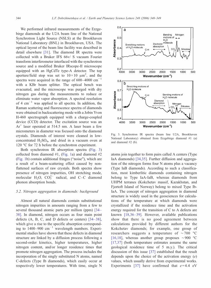
Fig. 3. Synchrotron IR spectra (beam line U2A, BrookhavenNational Laboratory) obtained from Erzgebirge diamond #1 (a)and diamond #2 (b).
344 L.F. Dobrzhinetskaya et al. / Earth and Planetary Science Letters 248 (2006) 340–349
We performed infrared measurements of the Erzge-birge diamonds at the U2A beam line of the NationalSynchrotron Light Source (NSLS) at the BrookhavenNational Laboratory (BNL) in Brookhaven, USA. Theoptical layout of the beam line facility was described indetail elsewhere [31]. The diamond IR spectra werecollected with a Bruker IFS 66v/ S vacuum Fouriertransform interferometer interfaced with the synchrotronsource and a modified Bruker IRscope-II microscopeequipped with an HgCdTe type-A detector. The topaperture/field stop was set to 10×10 μm2, and thespectra were acquired in the range of 600–4000 cm−1
with a KBr beam splitter. The optical bench wasevacuated, and the microscope was purged with drynitrogen gas during the measurements to reduce oreliminate water vapor absorption. A spectral resolutionof 4 cm−1 was applied to all spectra. In addition, theRaman scattering and fluorescence spectra of diamondswere obtained in backscattering mode with a Jobin YvonH-460 spectrograph equipped with a charge-coupleddevice (CCD) detector. The excitation source was anAr+ laser operated at 514.5 nm. A laser beam a fewmicrometers in diameter was focused onto the diamondcrystals. Diamonds of interest were cleaned in low-concentrated H2SO4, and dried in a vacuum oven at120 °C for 72 h before the synchrotron experiment.
Both synchrotron IR absorption spectra (Fig. 3)collected from diamond #1, (Fig. 1a) and diamond #2(Fig. 1b) contain additional fringes (“noise”), which area result of a beam-scattering effect caused by non-flattened surfaces of raw crystals. Both spectra showpresence of nitrogen impurities, OH stretching mode,molecular H2O, CO3
2− radical, and C–C diamondphonon absorption bonds.
5.2. Nitrogen aggregation in diamonds: background
Almost all natural diamonds contain substitutionalnitrogen impurities in amounts ranging from a few toseveral thousand atomic parts per million (ppm) [34–38]. In diamond, nitrogen occurs as four main pointdefects (A, B, C, and D defects or centers) [34–38],which give a rise to the specific absorption correspond-ing to 1400–900 cm−1 wavelength numbers. Experi-mental studies have shown that these defects in diamondstructure are linked by a diffusion process following asecond-order kinetics, higher temperatures, highernitrogen content, and/or longer residence times thatpromote nitrogen aggregation. The process includes theincorporation of the singly substituted N atoms, namedC-defects (Type Ib diamonds), which easily occur atrespectively lower temperatures. With time, single N
atoms join together to form pairs called A centers (TypeIaA diamonds) [34,35]. Further diffusion and aggrega-tion of the nitrogen forms four N atoms plus a vacancy(Type IaB diamonds). According to such a classifica-tion, most kimberlitic diamonds containing nitrogenbelong to Type IaA-IaB, whereas diamonds fromUHPM terranes (Kokchetav massif, Kazakhstan, andFjortoft Island of Norway) belong to mixed Type Ib-IaA. The concept of nitrogen aggregation in diamondstructure is widely used in the geosciences for calcula-tions of the temperature at which diamonds werecrystallized if the residence time and the activationenergy required for the transition of C to A defects areknown [18,36–39]. However, available publicationsshow that there is no good agreement betweencalculations provided by different authors for theKokchetav diamonds; for example, one group ofresearchers suggests a temperature of ∼700 °C[16,18], whereas another group proposes ∼900 °C[17,37] (both temperature estimates assume the samegeological residence time of 5 m.y.). The criticaldiscussion of this issue [37] established that the resultdepends upon the choice of the activation energy (e)values, which usually derive from experimental works.Experiments [37] have confirmed that e=4.4 eV

Fig. 4. Raman (a) and cathodoluminescence (b) spectra (beam lineU2A, Brookhaven National Laboratory) of Erzgebirge diamonds #1and #2.
345L.F. Dobrzhinetskaya et al. / Earth and Planetary Science Letters 248 (2006) 340–349
provides the best estimate of temperature for octahedralgrowth sectors of diamonds, while e=6.0 eV fits betterfor cube growth sectors of diamond. The temperature of900 °C calculated by Taylor et al. [17,37], using thee=6.0 eV parameter and therefore assuming that almostall Kokchetav diamonds are represented by cuboidalcrystals, fits better with the range of temperaturesderived from a conventional geothermobarometry(920–1250 °C) applied for natural diamond-bearingrocks [40–42].
5.3. Erzgebirge diamonds: IR microspectroscopy
Both studied diamonds exhibit the characteristics C–C diamond phonon absorption bonds in the region of2600–2000 cm−1. Diamond #1 is characterized by clearnitrogen-related absorptions, with a strong peak at1282 cm−1 testifying for the presence of nitrogenaggregated as N-pairs (A defects). There are also weakbut significant nitrogen absorption bands at 1130 cm−1
(as detected by software) and around 1344 cm−1 (Fig.3a), which are characteristics of C defects. Given thatnitrogen forms C defects which then aggregate to Adefects, and further from A to B defects, the absence ofabsorptions typical of B defects (e.g. at 1175 cm−1) isconsidered as a confirmation that this diamond belongsto Type Ib-IaA.
Deconvolution of the collected IR spectra wasperformed by both the hand method and using ahomemade computational program. In both cases, thecalculated amount of nitrogen in diamond #1 was∼1630 ppm (±20%) with 70%±10% of 1aA. The lattererror is quite large, and it mostly depends on theuncertainty associated with baseline correction in theregion of wavelength numbers below 1000 cm−1.
In addition to nitrogen, the IR spectrum ofdiamond #1 also exhibits a clear absorption at3107 cm−1 corresponding to C–H bonds in the dia-mond matrix. The sample also contains a small (butdetectable) amount of water presented as absorptionbands at 1630 cm−1, which reflects bending motionsof the H2O molecule; absorption bands at 3420 cm−1
are identified as O–H stretches of the H2O molecule.There is also a well-pronounced absorption band at1430 cm−1, corresponding to the carbonate radical CO3
2−;the latter might be incorporated in diamonds as carbonatemicroinclusions.
Diamond #2 is characterized by a minor amount ofnitrogen because none of the major peaks typical of N-bearing defects can be identified (Fig. 3b). We infer thatthe N content in this sample is not higher than 160 ppmbecause above this value N content would lead to
resolvable IR absorption. The IR spectrum clearlyshows the presence of bending motions of the H2Omolecule (absorption at 1630 cm−1) and the OH regionat 3420 cm−1; again, the latter reflects the stretchingmotions of the H2O molecule. Peaks at 2870, 2925, and3042 cm−1 correspond to the C–H stretching bands. Inour case, as it was stated by a beam-scientist, they recorda contamination of the beam with engine oil of thevacuum pumps that appeared in IR window at U2Abeam line.
The Erzgebirge diamond #2 IR spectral character-istics are strikingly similar in both N content and C–Hstretching bands to three microdiamonds from Akluilakminette dyke, Nunavut, Canada (see Fig. 1b in [39])showing a possible correlation between C–H stretchingbands and low N content. The recently discovereddiamonds belong to a metamorphic diamond population(Type Ib-IaA), which, it is believed, were crystallizedwithin deeply subducted sediments and later were

Fig. 5. Diagram of nitrogen aggregation versus nitrogen content inworldwide collected kimberlitic diamonds (gray patterns) anddiamonds from ultrahigh pressure metamorphic terranes of theKokchetav massive, Kazakhstan, and from the Western GneissRegion, Norway (open circles) [18]. Black circles with crossinglines are diamonds from the Erzgebirge massif, Germany (this study).
346 L.F. Dobrzhinetskaya et al. / Earth and Planetary Science Letters 248 (2006) 340–349
“sampled” by the minette dyke and brought to theEarth's surface [39].
5.4. The Erzgebirge diamonds: Raman scattering andfluorescence spectroscopy
Raman spectroscopy (Fig. 4a) revealed that diamond#1 is characterized by a sharp, high-intensity band at1327.9 cm− 1, which deviates from the standarddiamond peak at 1332 cm−1, which reflects the first-order diamond lattice vibration of LO=TO phononalong the two-dimensional planes. The shift of theRaman band in diamonds to the lower- or higher-frequency regions may be interpreted as (1) deformationof the diamond lattice due to internal stress, (2)instrumental uncertainties, or (3) the presence of pointdefects or growth zoning defects in the diamond crystal[43–45]. Although we excluded any instrumental error,at this moment we are not able to provide anyexplanation of the Raman shift in diamond #1 to thelower-frequency region; for that, additional micro-X-raystudies would be necessary. Diamond #2 has a lower-intensity peak than diamond #1, but its Raman peak at1331.98 cm−1 is similar to the diamond standard Ramanband at 1332 cm−1. The fact that the wide band occurredaround 4900 cm−1 in the Raman spectra of bothdiamonds might be due to the presence of nitrogenimpurities (Fig. 4b).
6. Discussion
Our synchrotron IR studies showed that the Erzge-birge diamonds contain nitrogen impurities, molecularH2O, and OH- and CO3 radicals. The presence of bothnitrogen C- and A defects classifies the studieddiamonds as Type Ib-IaA, which is similar to othermetamorphic diamonds from marbles and garnet–pyroxenites of the Kokchetav massif of Kazakhstan[16–18] and from the felsic gneisses on the island ofFjortoft in the Western Gneiss Region of Norway [5].
As we see from the diagram of nitrogen contentsversus nitrogen aggregation values built up on thenitrogen measurements in the well-known kimberliticdiamonds and diamonds from UHPM terranes (Fig. 5),the Erzgebirge diamond plots are within the ranges ofthose from Kazakhstan and Norway, and they areclearly distinguished from diamonds from kimberliticsources. The lowest concentration of nitrogen in theErzgebirge diamonds (∼160 ppm) agrees with that ofdiamonds from Akluilak minette dyke, Nunavut,Canada [39], and it is close to the diamonds fromFjortoft, Norway [5].
Significant variability of nitrogen contents have beenidentified among microdiamonds from the same UHPMterrane (e.g., the Kokchetav massif: 150–10,000 ppm),between two diamonds recovered from the same sample(e.g., Erzgebirge: 160 and 1630 ppm), or even within asingle sample, as has been shown for Kokchetavdiamonds [16]. The origin of such variation remainsunclear; it may reflect either the contribution of distinctor inhomogeneous fluids, fluid fractionation (e.g. byprecipitating N-bearing minerals such as phengite), orkinetic fractionation of nitrogen related to diamondgrowth rate, or even different growth episodes fromdistinct fluids [13,46]. It also may be referred to thediamond growth from different fluids, or to the differentrate of nitrogen incorporation into diamond structureduring its crystallization. The nitrogen impuritiescharacteristics suggest that the Erzgebirge diamondsbelong to the Type 1b-1aA, which is similar tometamorphic diamonds from the Kokchetav massif ofKazakhstan and the Western Gneiss Region of Norway,and differentiates them from other nitrogen-bearingdiamonds from kimberlitic sources (Type 1aAB).According to the rather low activation energy(∼4.4 eV) for single N-atoms to aggregate into N-pairs, the occurrence of nitrogen impurities as singleatoms in the crystal lattice implies that the Erzgebirgediamonds had a short residence time in a high-pressureand high-temperature environment (see related issue

347L.F. Dobrzhinetskaya et al. / Earth and Planetary Science Letters 248 (2006) 340–349
developed for Kokchetav diamonds [16]). Indeed, anydiamond with N-content higher than 100 ppm would beType 1aA at a temperature of 700 °C within 1 m.y.Therefore, the detection of a 1b-component suggests thepossibility for very fast exhumation (<1 m.y.). Thoughat this moment our limited data on nitrogen aggregationdo not allow us to calculate a definite duration ofexhumation of Erzgebirge diamond-bearing rocks, werefer to numerical modeling according to whichexhumation from UHPM conditions occurred during1–5 m.y. [47].
The IR data are consistent with our previous studiesof inclusions in the Erzgebirge diamonds performedwith the aid of focused ion beam (FIB) and transmissionelectron microscopes (TEM) [22]. The studies demon-strate that Erzgebirge diamonds contain abundantnanometric solid crystalline inclusions of SiO2 with anadmixture of minor components such as K, P, Ti, Fe, orSiO2-free crystalline inclusions of K, P, and O, or PbxOy
and/or Pb2CO3, and Al2SiO5. We were unable toidentify the structure of these nanometric phases byselected-area electron diffraction because they were notappropriately oriented for identification and tiltingresulted in multiple patterns superimposed. Neverthe-less, they yielded Bragg diffraction spots indicating thatthey were crystalline [22]. Hwang et al. [48] havereported that instead of crystalline inclusions they haveobserved phosphorous–potassium–silica (P–K–SiO2)glass or Al–SiO2 glass, which they interpreted to be aformer melt hosted by Erzgebirge diamonds (theirsamples 25966 and M1). However, it is not clear to uswhether Hwang et al. [48] have actually identified glass.They say (p. 98): “… careful EDX analyses in thepresent study revealed that these pockets are in fact P/K-rich silica glass….” We do not understand how one canidentify glass by electron dispersive X-ray (EDX)analysis; to our understanding, diffraction is necessary.Hwang et al. [48] reported electron diffraction patternsonly for host diamond, but not for nano-scale inclusions.
Our focused ion beam-assisted TEM studies showedthat Erzgebirge diamonds also contain a considerableamount of nanometric-size euhedral cavities that arebelieved to represent decrepit fluid inclusions. Many ofthese cavities associate with dislocations of diamondgrowth. TEM researches have shown that the cavitiesare surrounded by amorphous film with average lowatomic weight materials [22]. As we learned from IR,such cavities might present traces of former fluidinclusions containing CO2, H2O and potentially IRinactive species such as N2, which were “burned” outdue to sample treatment by ion (FIB) and electron(TEM) beams. The Raman spectroscopy also provides
some evidence that internal residual pressure probablystill exists in diamond #1, which may therefore partlyprevent a fluid from crystallizing. The synchrotron IRdata, combined with observations of solid inclusionsusing TEM, suggest that the Erzgeberge diamondscrystallized in a media rich in volatile components suchas CO2, OH, and H2O. Because no CH4 (we know thatthe C–H absorption at 3107 cm−1 is due to engine oil),or other reduced natural chemical species have beendetected in the Erzgebirge diamonds, we assume thattheir crystallization occurred at relatively high oxygenfugacity (fO2∼5–6 log units), which was close to theupper limit of fO2 for allowing diamond stabilization[49,50]. We emphasize that a concept of UHPMdiamond crystallization from the COH fluid enrichedin local components coherent with their host minerals orrock chemistry is unconditionally supported by directobservation of nanoinclusions using the TEM and IRdata and observations on diamond-bearing pocketsincluded in garnets [14,22,23]. Furthermore, extremelynegative values of δ13C in diamonds from Erzgebirge(δ13C=−17‰ to −27‰ [46]) and from Kazakhstan(δ13C=−10‰ to −17‰ [13,29]) does not fit with thediamond crystallization from a carbonate/carbonatitemelt (average δ13C=−0.22‰ to −4.31‰ [51]) becausesuch a strong isotopic fractionation between diamondand carbonate melt would not happen at high tempera-tures (i.e. the fractionation between calcite and diamondis below 5‰ at temperature above 800 °C [52]).
Finally, we would like to emphasize that sometimesdiscussions related to metamorphic diamond origin asbeing “fluid versus melt” reflect, simply, a semanticproblem. Experiments have demonstrated that there iscomplete miscibility between silicate melt and water inmost of the upper mantle, except at very shallow depths.Bureau and Keppler [53] showed that the hydrous meltand silicate-bearing vapor coexisting at low temperaturemerge compositionally to form a single-phase, super-critical fluid, stable at higher pressures (1.5–2.5 GPa).Therefore, above the supercritical point there is nodifference in physical parameters between melt andfluid. Applying this concept to UHPM diamonds,whenever water is available in the subduction zone thesolid material, including organic carbon matter, may bedissolved in the supercritical fluid with increasingpressure and temperature, and such a fluid becomes amedia for diamond crystallization. Our interpretation ofdiamond origin is consistent with the recently obtainedexperimental evidence for diamond crystallization athigh P and T directly from COH fluid or from graphitein the presence of H2O [49,50]. This interpretation isalso similar to that independently reached from

348 L.F. Dobrzhinetskaya et al. / Earth and Planetary Science Letters 248 (2006) 340–349
observations on diamond-bearing multiphase pocketsincluded in garnet from Erzgebirge felsic gneisses [23].
Acknowledgements
Diamond-bearing rocks were collected in 2000during a field trip to Erzgebirge with H.-I. Massonneand B. Stöckhert who are greatly acknowledged. Wethank our colleague D. Parker, Department of Environ-mental Sciences at UCR, for providing us with access tohis laboratory facilities to develop and perform chemicaldigesting of the diamond-bearing rocks and to developdiamond separation techniques. We gratefully acknowl-edge V. Shatsky and O. Kozmenko (Institute ofMineralogy and Petrography, Novosibirsk, Russia) fortheir helpful advice and discussions related to thediamond extraction process. We thank Taras Gerya andan anonymous reviewer for their thoughtful and detailedreviews and comments. This research was supported bythe International Division of the U.S. National ScienceFoundation, Grant EAR 0229666, and Pacific RimProgram of the University of California to LD andHWG. IR measurements were performed at the U2Abeam line at NSLS of Brookhaven National Laboratory,the U.S. Department of Energy (DOE), Contract DE-AC02-98CH10886. The U2A beam line is supported byCOMPRES, the Consortium for Materials PropertiesResearch in Earth Sciences under the U.S. NSFCooperative Agreement Grant (EAR 01-35554) andDOE (CDAC, Contract No. DE-FC03-03N00144).
References
[1] O.M. Rozen, Yu. M. Zorin, A.A. Zayachkovsky, A find of thediamonds related to eclogites of the Precambrian Kokchetavmassif, Dokl. Akad. Nauk SSSR 203 (1972) 674–676.
[2] N.V. Sobolev, V.S. Shatsky, Diamond inclusions in garnets frommetamorphic rocks: a new environment of diamond formation,Nature 343 (1990) 742–746.
[3] S.T Xu, A.I. Okay, S.Y. Ji, A.M.C. Sengor, S. Wen, Y.C. Liu,L.L. Jiang, Diamond from the Dabie-Shan metamorphic rocksand its implication for tectonic setting, Science 256 (1992)80–82.
[4] J. Yang, Z. Xu, L.F. Dobrzhinetskaya, H.W. Green, X. Pei, R.Shi, C. Wu, J.L. Wooden, J. Zhang, Y. Wan, H. Li, Discovery ofmetamorphic diamonds in Central China: an indication of a>4000 km-long-zone of deep subduction resulting from multiplecontinental collisions, Terra Nova 15 (2003) 370–379.
[5] L. Dobrzhinetskaya, E. Eide, A. Korneliussen, R. Larsen, J.Millege, T.V. Posukhova, D.S. Smith, B.A. Sturt, W.R. Taylor, R.Tronnes, Diamond in metamorphic rocks of the Western GneissRegion in Norway, Geology 23 (1995) 597–600.
[6] H.L.M van Roermund, D.A. Carswell, M.R. Drury, T.C.Heijboer, Microdiamonds in megacrystic garnet websterite podfrom Bardane on the island of Fjortoft, western Norway:
evidence for diamond formation in mantle rocks during deepcontinental subduction, Geology 30 (2002) 959–962.
[7] H.-J. Massonne, A new occurrence of microdiamonds inquartzofeldspathic rocks of the Saxonian Erzgebirge, Germany,and the metamorphic evolution, Proceedings of 7th InternationalKimberlitic Conference. Capetown, vol. 2, 1999, pp. 533–539.
[8] L. Nasdala, H.-J. Massonne, Microdiamonds from the SaxonianErzgebirge, Germany: in situ micro-Raman characterization, Eur.J. Mineral. 12 (2000).
[9] E.D. Mposkos, D.K. Kostopoulos, Diamond, former coesite andsupersilicic garnet in metasedimentary rocks from the GreekRhodope: a new ultrahigh-pressure metamorphic provinceestablished, Earth Planet. Sci. Lett. 192 (2001) 497–506.
[10] O. Beyssac, C. Chopin, Comment on ‘Diamond, former coesiteand supersilisic garnet in metasedimentary rocks from the GreekRhodope: a new ultrahigh-pressure metamorphic provinceestablished’ by E.D. Mposkos and D.K. Kostopoulos [Earthand Planetary Science Letters 192 (2001) 497–506], EarthPlanet. Sci. Lett. 214 (2003) 669–674.
[11] E.D. Mposkos, D.K. Kostopoulos, Diamond, former coesite andsupersilicic garnet in metasedimentary rocks from the GreekRhodope: a new ultrahigh-pressure metamorphic provinceestablished—reply, Earth Planet. Sci. Lett. 214 (2003) 675–678.
[12] B.C. Bostick, R.E. Jones, C. Chen, W.G. Ernst, M.L. Leech, R.J.Beane, Low-temperature microdiamond aggregates in theMaksutov Metamorphic Complex, South Ural Mountains,Russia, Am. Mineral. 88 (2003) 1709–1717.
[13] Y. Ogasawara, Microdiamonds in ultrahigh-pressure meta-morphic rocks, Elements 1 (2005) 91–96.
[14] L.F. Dobrzhinetskaya, H.W. Green, H.-J. Massonne, B. Stock-hert, Origin of microdiamonds from ultra-high-pressure terranes(abstract), Abstract volume of Eleventh Annual V.M. Gold-schmidt Conference in Virginia, USA, CD-Rom article #3478,2001.
[15] Y. Ogasawara, M. Ohta, K. Fukasawa, I. Katayama, S.Maruyama, Diamond-bearing and diamond-free metacarbonaterocks from Kymdi-Kol in the Kokchetav massif, northernKazakhstan, Isl. Arc 9 (2000) 400–416.
[16] K. De Corte, P. Cartigny, V.S. Shatsky, N.V. Sobolev, M. Javoy,First evidence of fluid inclusions in metamorphic microdiamondsfrom the Kokchetav massif, Northern Kazakhstan, Geochim.Cosmochim. Acta 62 (1998) 3765–3777.
[17] K. De Corte, W.R. Taylor, P. De Paepe, Inclusion contents ofmicrodiamonds from UHP metamorphic rocks of the Kokchetavmassif, in: C.D. Parkinson, I. Katayma, J.G. Liou, S. Maryama(Eds.), The Diamond-Bearing Kokchetav Massif, UniversalAcademy Press, Inc., Kazakhstan, 2002, pp. 115–135.
[18] P. Cartigny, K. De Corte, V.S. Shatsky, M. Ader, P. de Paepe, N.V. Sobolev, M. Javoy, The origin and formation of metamorphicmicrodiamonds from the Kokchetav massif, Kazakhstan: anitrogen and carbon isotopic study, Chem. Geol. 176 (2001)265–281.
[19] L.F. Dobrzhinetskaya, R. Wirth, H.W. Green, Direct observationand analysis of a trapped COH fluid growth medium inmetamorphic diamond, Terra Nova 17 (2005) 422–477.
[20] L.F. Dobrzhinetskaya, H.W. Green, T.E. Mitchell, R.M. Dick-erson, Metamorphic diamonds: mechanism of growth andinclusion of oxides, Geology 29 (2001) 263–266.
[21] L.F. Dobrzhinetskaya, H.W. Green, T.E. Mitchell, R.M. Dick-erson, K.N. Bozhilov, Crystallization environment of Kazakhstanmicrodiamonds: evidence from nanometric inclusions andmineral associations, J. Metamorph. Geol. 21 (2003) 425–437.

349L.F. Dobrzhinetskaya et al. / Earth and Planetary Science Letters 248 (2006) 340–349
[22] L.F. Dobrzhinetskaya, H.W. Green, M.Weschler, M. Darus, Y.-C.Wang, H.-J.Massonne, B. Stöckhert, Focused ion beam techniqueand transmission electron microscope studies of microdiamondsfrom the Saxonian Erzgebirge, Germany, Earth Planet. Sci. Lett.210 (2003) 399–410.
[23] B. Stöckhert, J. Duyster, C. Trepmann, H.-J. Massonne,Microdiamond daughter crystals precipitated from supercriticalCOH+silicate fluids included in garnet, Erzgebirge, Germany,Geology 29 (2001) 391–394.
[24] S.L. Hwang, P. Shen, H.T. Chu, T.F. Yui, C.C. Lin, Genesis ofmicrodiamonds from melt and associated multiphase inclusionsin garnet of ultrahigh-pressure gneiss from Erzgebirge, Germany,Earth Planet. Sci. Lett. 188 (2001) 915–919.
[25] H.-J. Massonne, A comparison of the evolution of diamond-iferous quartz-rich rocks from the Saxonian Erzgebirge and theKokchetav massif: are so-called diamondiferous gneisses mag-matic rocks? Earth Planet. Sci. Lett. 216 (2003) 347–364.
[26] H.-J. Massonne, First find of coesite in ultra-high pressuremetamorphic region of the Central Erzgebirge, Germany, Eur. J.Mineral. 13 (2001) 565–570.
[27] H.-J. Massonne, P.J. O'Brien, The Bohemian Massif and the NWHimalaya, EMU Notes Mineral. 5 (2003) 145–187.
[28] S.L. Hwang, P. Shen, H.T. Chu, T.F. Yui, Nanometer-size a-PbO2-type TiO2 in garnet: a thermobarometer for ultrahigh-pressure metamorphism, Science 288 (2000) 321–324.
[29] T.E. Ekimova, L.A. Lavrova, E.D. Nadezhdina, M.A. Petrova, in:V.I. Vaganov (Ed.), In situ and Alluvial Diamond Deposits ofNorthern Kazakhstan, TSNIGRI, Moscow, Russia, 1992, p. 168,(in Russian).
[30] H.-Y.N. Holman, A. Bjornstad, P. McNamara, C. Martin, R.McKinney, A. Blakely, Synchrotron infrared spectromicroscopyas a novel bioanalytical microprobe for individual living cells:cytotoxicity considerations, J. Biomed. Opt. 7 (2002) 417–424.
[31] Z. Liu, J. Hu, H. Yang, H.K. Mao, R.J. Hemley, High-pressuresynchrotron X-ray diffraction and infrared microspectroscopy:applications to dense hydrous phases, J. Phys., Condens. Matter14 (2002) 10641–10646.
[32] G.L. Carr, M. Hanfland, G.P. Williams, The mid-infraredbeamline at the NSLS port U2B, Rev. Sci. Instrum. 66 (1995)1643–1648.
[33] J.A. Reffner, P.A. Martoglio, G.P. Williams, Fourier transforminfrared microscopical analysis with synchrotron radiation—themicroscope optics and system performance, Rev. Sci. Instrum. 66(1995) 1298–1302.
[34] R.M. Chrenko, R.E. Tuft, H.M. Strong, Transformation of thestate of nitrogen in diamond, Nature 270 (1977) 141–144.
[35] T. Evans, Z. Qi, The kinetics of the aggregation of nitrogen atomsin diamond, Proc. R. Soc. Lond., A 381 (1982) 159–178.
[36] W.R. Taylor, A.L. Jaques, M. Ridd, Nitrogen-defect aggregationcharacteristics of some Australasian diamonds: time–temperatureconstraints on the source regions of pipe and alluvial diamonds,Am. Mineral. 75 (1990) 1290–1310.
[37] W.R. Taylor, D. Canil, H.J. Milledge, Kinetics of 1b to 1aAnitrogen aggregation in diamond, Geochim. Cosmochim. Acta60 (1996) 4725–4733.
[38] R. Jones, P.R. Briddon, S. Oberg, First-principles theory of nitrogenaggregates in diamond, Philos. Mag. Lett. 66 (1992) 67–74.
[39] P. Cartigny, I. Chinn, K.S. Viljoen, D. Robinson, EarlyProterozoic ultrahigh-pressure metamorphism: evidence frommicrodiamonds, Science 304 (2004) 835–855.
[40] R.Y. Zhang, J.G. Liou, R.G. Coleman, R.G. Ernst, N.V. Sobolev,V.S. Shatsky, Metamorphic evolution of diamond-bearing andassociated rocks from the Kokchetav massif, northern Kazakh-stan, J. Metamorph. Geol. 15 (1997) 479–496.
[41] Y. Ogasawara, K. Fukasawa, S. Maruyama, Coesite exsolutionfrom supersilicic titanite in UHP marble from Kokchetav massif,northern Kazakhstan, Am. Mineral. 87 (2002) 454–461.
[42] K. Okamoto, J.G. Liou, Y. Ogasawara, Petrology of the diamond-grade eclogite in the Kokchetav Massif, northern Kazakhstan, Isl.Arc 9 (2000) 379–399.
[43] J.A. Xu, H.K. Mao, Moissanite: a window for high-pressureexperiments, Science 290 (2000) 783–785.
[44] G. Morell, O. Quinones, Y. Diaz, I.M. Vargas, B.R. Weiner, R.S.Katiyar, Measurements and analysis of diamond Ramanbandwidths, Diamond Relat. Mater. 7 (1998) 1029–1032.
[45] L. Nasdala, W. Hofmeister, J.W. Harris, J. Glinnemann, Growthzoning and strain patterns inside diamond crystals as revealed byRaman maps, Am. Mineral. 90 (2005) 745–748.
[46] N. Takahata, Y. Sano, K. Shirai, L. Dobrzhinetskaya, H.W. GreenII, Deep-subduction of organic matter: evidence from nanoSIMSstudies of carbon isotopes in microdiamonds from Erzgebirge,Germany, EOS, Fall AGU meeting in San Francisco, abstractvolume (2005) F-1234.
[47] B. Stöckhert, T. Gerya, Pre-collisional high pressure metamorph-ism and nappe tectonics at active continental margins: anumerical simulation, Terra Nova 17 (2005) 102–110.
[48] S.L. Hwang, H.T. Chu, T.F. Yui, P. Shen, H.P. Shertl, J.G.Liou, N.V. Sobolev, Nanometer-size P/K-rich silica glass(former melt) inclusions in microdiamond from the gneissesof Kokchetav and Erzgebirge massifs: diversified character-istics of formation media of metamorphic microdiamonds inUHP rocks due to host-rock buffering, Earth Planet. Sci. Lett.243 (2006) 94–106.
[49] M. Akaishi, S. Yamaoka, Crystallization of diamond from C–O–H fluids under high-pressure and high-temperature condition,J. Cryst. Growth 209 (2000) 999–1003.
[50] L. Dobrzhinetskaya, A. Renfro, H.W. Green II, Synthesis ofskeletal diamonds: implication to orogenic belts microdiamondformation, Geology 32 (2004) 869–872.
[51] A. Simonetti, K. Bell, S.G. Viladkar, Isotopic data from AmbaDongar Carbonatite Complex, west-central India: evidence for anenriched mantle source, Chem. Geol. 122 (1995) 185–198.
[52] Y. Bottinga, Carbon isotope fractionation between graphite,diamond and carbon dioxide, Earth Planet. Sci. Lett. 5 (1969)301–307.
[53] H. Bureau, H. Keppler, Complete miscibility between silicatemelts and hydrous fluids in the upper mantle: experimentalevidence and geochemical implications, Earth Planet. Sci. Lett.165 (1999) 187–196.