Synchronous with Your Feelings: Sensorimotor Band and Empathy for Pain
9
Behavioral/Systems/Cognitive Synchronous with Your Feelings: Sensorimotor Band and Empathy for Pain Viviana Betti, 1,2 Filippo Zappasodi, 3 Paolo Maria Rossini, 4,5 Salvatore Maria Aglioti, 2,6 and Franca Tecchio 3,5 1 Associazione Fatebenefratelli per la Ricerca, Ospedale Fatebenefratelli, 00186 Rome, Italy, 2 Fondazione Santa Lucia, Istituto di Ricovero e Cura a Carattere Scientifico, 00142 Rome, Italy, 3 Istituto Scienze e Tecnologie della Cognizione, Consiglio Nazionale delle Ricerche, 00185 Rome, Italy, 4 Clinica Neurologica, Universita ` Campus Biomedico, 00128 Rome, Italy, 5 Casa di Cura San Raffaele, 03043 Cassino, Italy, and San Raffaele Pisana, Istituto di Ricovero e Cura a Carattere Scientifico, 00163 Rome, Italy, and 6 Dipartimento di Psicologia, Sapienza Universita ` di Roma, 00185 Rome, Italy Neuroscience studies on the social sharing of observed or imagined pain focused on whether empathic pain resonance is linked to affective or sensory nodes of the pain matrix. However, empathy, like other complex cognitive processes, is inherently linked to the activation of functional networks rather than of separate brain areas. Here, we used magnetoencephalography (MEG) to explore the relationship between empathy and functional coupling of neuronal activity in primary somatosensory (SI) and motor (MI) cortices. MEG recording was performed while healthy participants observed movie-clips depicting the static hand of a stranger model, the same hand deeply penetrated by a needle, or gently touched by a Q-tip. Subjects were asked to rate the movie-derived sensations attributed to self or to the model. For each type of clip observation, we analyzed spectral power and coherence values in , , and frequency bands. While spectral power indexes separate neural activity in SI and MI, coherence values index functional cross-talk between these two areas. No power changes of SI or MI sources were induced by observation conditions in any of the frequency bands. Crucially, -band coherence values were significantly higher during needle-in-hand than touch and static hand observation and correlated with self-and other- referred pain ratings derived from needle-in-hand movies observation. Thus, observation of others’ pain increases neuronal synchroni- zation and cross-talk between the onlookers’ sensory and motor cortices, indicating that empathic resonance relies upon the activity of functional networks more than of single areas. Introduction A large variety of painful experiences, ranging from physical pain perception (e.g., a wound) (Tracey and Mantyh, 2007) to social rejection (Eisenberger et al., 2003) activates a widely distributed brain network, referred to as the “pain matrix” (Melzack, 1999). The ability to understand others by sharing their intentions, emo- tions, and feelings, a distinctive feature of empathy (Preston and de Waal, 2002), is mapped on largely overlapping neural struc- tures (Hein and Singer, 2008). Previous studies focused on whether vicarious pain is mapped on affective or sensory nodes of the pain matrix (Hein and Singer, 2008). However, empathy for pain, like higher-order cognitive functions, may be based on the functional interaction of neuronal assemblies distributed within and across different specialized brain regions (Varela et al., 2001; Fries, 2005, 2009). Indeed, mounting evidence indicates that rhythmic neural oscillations and their synchronization provide indices of dynamic and flexi- ble communication between and within the cerebral networks underlying a specific behavior (Fries, 2005). Synchronized oscil- latory activity in the , , and frequency bands is associated to a number of functions ranging from working memory and mental imagery to selective attention, sensorimotor integration and perceptual awareness. Moreover, disrupted oscillatory activity seems associated to neuropsychiatric disorders (e.g., schizophrenia) characterized by dysfunctional cognition and behavior (Uhlhaas et al., 2008). In particular, studies support the role of -frequency band (30 Hz) synchronization as a mechanism underlying the dy- namic construction of the large-scale neuronal networks involved in object representation (Tallon-Baudry and Bertrand, 1999), binding of visual features (Singer and Gray, 1995; Engel and Singer, 2001), selective attention and memory (Womelsdorf and Fries, 2006; Jensen et al., 2007; Doesburg et al., 2008), sensorimotor integration (Szurhaj et al., 2006), and even very complex processes such as the “aha!” component of cognitive insight (Jung-Beeman et al., 2004; Sheth et al., 2009). Relevant to the present study is a recent MEG report showing that pain-induced oscillations in primary somato- sensory cortices are specifically related to the subjective pain experi- ence instead than to the objective attributes of painful stimuli such as their actual intensity (Gross et al., 2007). Somatosensory evoked potential (SEP) and transcranial magnetic stimulation (TMS) stud- ies show that primary somatosensory (SI) (Bufalari et al., 2007) and motor (MI) (Avenanti et al., 2005, 2006, 2009; Fecteau et al., 2008; Minio-Paluello et al., 2006, 2009) cortices are involved in the direct observation of pain stimuli delivered to stranger individuals. How- ever, no studies have so far investigated whether empathic pain map- ping is linked to the construction of a functional SI–MI network. Received June 12, 2009; revised July 25, 2009; accepted Aug. 12, 2009. The financial contribution of Ministero Istruzione Universita ` e Ricerca (PRIN) and Ministero Sanita `, Italy is grate- fully acknowledged. Thanks are due to Matilde Ercolani for her excellent technical assistance. Correspondence should be addressed to either Viviana Betti or Salvatore M. Aglioti, Fondazione Santa Lucia, Via Ardeatina 306, 00142 Rome, Italy, E-mail: [email protected] or [email protected]. V. Betti’s and F. Zappasodi’s present address: Universita ` “G. d’Annunzio,” 66013 Chieti, Italy. DOI:10.1523/JNEUROSCI.2759-09.2009 Copyright © 2009 Society for Neuroscience 0270-6474/09/2912384-09$15.00/0 12384 • The Journal of Neuroscience, October 7, 2009 • 29(40):12384 –12392
-
Upload
independent -
Category
Documents
-
view
2 -
download
0
Transcript of Synchronous with Your Feelings: Sensorimotor Band and Empathy for Pain

Behavioral/Systems/Cognitive
Synchronous with Your Feelings: Sensorimotor � Band andEmpathy for Pain
Viviana Betti,1,2 Filippo Zappasodi,3 Paolo Maria Rossini,4,5 Salvatore Maria Aglioti,2,6 and Franca Tecchio3,5
1Associazione Fatebenefratelli per la Ricerca, Ospedale Fatebenefratelli, 00186 Rome, Italy, 2Fondazione Santa Lucia, Istituto di Ricovero e Cura a CarattereScientifico, 00142 Rome, Italy, 3Istituto Scienze e Tecnologie della Cognizione, Consiglio Nazionale delle Ricerche, 00185 Rome, Italy, 4Clinica Neurologica,Universita Campus Biomedico, 00128 Rome, Italy, 5Casa di Cura San Raffaele, 03043 Cassino, Italy, and San Raffaele Pisana, Istituto di Ricovero e Cura aCarattere Scientifico, 00163 Rome, Italy, and 6Dipartimento di Psicologia, Sapienza Universita di Roma, 00185 Rome, Italy
Neuroscience studies on the social sharing of observed or imagined pain focused on whether empathic pain resonance is linked toaffective or sensory nodes of the pain matrix. However, empathy, like other complex cognitive processes, is inherently linked to theactivation of functional networks rather than of separate brain areas. Here, we used magnetoencephalography (MEG) to explore therelationship between empathy and functional coupling of neuronal activity in primary somatosensory (SI) and motor (MI) cortices. MEGrecording was performed while healthy participants observed movie-clips depicting the static hand of a stranger model, the same handdeeply penetrated by a needle, or gently touched by a Q-tip. Subjects were asked to rate the movie-derived sensations attributed to self orto the model. For each type of clip observation, we analyzed spectral power and coherence values in �, �, and � frequency bands. Whilespectral power indexes separate neural activity in SI and MI, coherence values index functional cross-talk between these two areas. Nopower changes of SI or MI sources were induced by observation conditions in any of the frequency bands. Crucially, �-band coherencevalues were significantly higher during needle-in-hand than touch and static hand observation and correlated with self-and other-referred pain ratings derived from needle-in-hand movies observation. Thus, observation of others’ pain increases neuronal synchroni-zation and cross-talk between the onlookers’ sensory and motor cortices, indicating that empathic resonance relies upon the activity offunctional networks more than of single areas.
IntroductionA large variety of painful experiences, ranging from physical painperception (e.g., a wound) (Tracey and Mantyh, 2007) to socialrejection (Eisenberger et al., 2003) activates a widely distributedbrain network, referred to as the “pain matrix” (Melzack, 1999).The ability to understand others by sharing their intentions, emo-tions, and feelings, a distinctive feature of empathy (Preston andde Waal, 2002), is mapped on largely overlapping neural struc-tures (Hein and Singer, 2008).
Previous studies focused on whether vicarious pain is mappedon affective or sensory nodes of the pain matrix (Hein and Singer,2008). However, empathy for pain, like higher-order cognitivefunctions, may be based on the functional interaction of neuronalassemblies distributed within and across different specializedbrain regions (Varela et al., 2001; Fries, 2005, 2009). Indeed,mounting evidence indicates that rhythmic neural oscillationsand their synchronization provide indices of dynamic and flexi-ble communication between and within the cerebral networksunderlying a specific behavior (Fries, 2005). Synchronized oscil-
latory activity in the �, �, and � frequency bands is associated toa number of functions ranging from working memory and mentalimagery to selective attention, sensorimotor integration andperceptual awareness. Moreover, disrupted oscillatory activityseems associated to neuropsychiatric disorders (e.g., schizophrenia)characterized by dysfunctional cognition and behavior (Uhlhaas et al.,2008). In particular, studies support the role of �-frequency band(�30 Hz) synchronization as a mechanism underlying the dy-namic construction of the large-scale neuronal networks involved inobject representation (Tallon-Baudry and Bertrand, 1999), bindingof visual features (Singer and Gray, 1995; Engel and Singer, 2001),selective attention and memory (Womelsdorf and Fries, 2006;Jensen et al., 2007; Doesburg et al., 2008), sensorimotor integration(Szurhaj et al., 2006), and even very complex processes such as the“aha!” component of cognitive insight (Jung-Beeman et al., 2004;Sheth et al., 2009). Relevant to the present study is a recent MEGreport showing that pain-induced � oscillations in primary somato-sensory cortices are specifically related to the subjective pain experi-ence instead than to the objective attributes of painful stimuli such astheir actual intensity (Gross et al., 2007). Somatosensory evokedpotential (SEP) and transcranial magnetic stimulation (TMS) stud-ies show that primary somatosensory (SI) (Bufalari et al., 2007) andmotor (MI) (Avenanti et al., 2005, 2006, 2009; Fecteau et al., 2008;Minio-Paluello et al., 2006, 2009) cortices are involved in the directobservation of pain stimuli delivered to stranger individuals. How-ever, no studies have so far investigated whether empathic pain map-ping is linked to the construction of a functional SI–MI network.
Received June 12, 2009; revised July 25, 2009; accepted Aug. 12, 2009.The financial contribution of Ministero Istruzione Universita e Ricerca (PRIN) and Ministero Sanita, Italy is grate-
fully acknowledged. Thanks are due to Matilde Ercolani for her excellent technical assistance.Correspondence should be addressed to either Viviana Betti or Salvatore M. Aglioti, Fondazione Santa Lucia, Via
Ardeatina 306, 00142 Rome, Italy, E-mail: [email protected] or [email protected]. Betti’s and F. Zappasodi’s present address: Universita “G. d’Annunzio,” 66013 Chieti, Italy.DOI:10.1523/JNEUROSCI.2759-09.2009
Copyright © 2009 Society for Neuroscience 0270-6474/09/2912384-09$15.00/0
12384 • The Journal of Neuroscience, October 7, 2009 • 29(40):12384 –12392

Here, we explored for the first time the possible link betweenneuronal synchronization and empathic sharing of others’ painby obtaining magnetoencephalographic (MEG) indices of sepa-rate neural activity in primary somatosensory (SI) and motor(MI) cortices (using spectral power) and of functional cross-talkbetween these two areas (using sensorimotor coherence) in �, �,and � frequency bands while subjects observed painful or innoc-uous stimuli delivered to the hand of stranger model. Moreover,to explore whether attentional, sensory, and motor states of theonlooker influence the emergence of a functional sensorimotornetwork, subjects were asked to stay relaxed, to perform a motortask or to perceive a non-noxious electrical stimulation duringthe observation of video-clips.
Materials and MethodsSubjectsTwelve healthy subjects (9 women) aged 20 –25 (mean 23.64 years) wererecruited for the study. All participants were right-handed according tothe Edinburgh Handedness Questionnaire (mean 81 � 15) (Oldfield,1971). They had normal or corrected-to-normal visual acuity in botheyes and were naive as to the purposes of the experiment. Participantsgave their written informed consent. The procedures were approved bythe Fatebenefratelli “S. Giovanni Calibita” ethics committee and were inaccordance with the standards of the 1964 Declaration of Helsinki.
MEG recordingNeural activity was recorded using a 28-channel system operating in amagnetically shielded room (Vacuumschmelze). The active channels (16internal axial gradiometers and the other 12 peripheral magnetometers)were regularly distributed on a spherical surface (13.5 cm of curvatureradius) covering a total area of �180 cm 2. The MEG system was centeredon the C3 position of the 10 –20 International EEG system (Jasper, 1958),in correspondence of the primary sensorimotor hand area. The electro-myographic (EMG) signal from the right thumb’s opponent muscle wasacquired by a pair of electrodes. The active electrode was placed on theskin over the belly of the muscle and the reference electrode was placed incorrespondence of the thumb metacarpophalangeal joint at a distance of2.5 cm from the active one. The MEG and EMG signals were bandpassfiltered (0.48 –256 Hz, sampling rate 1000 Hz), digitized and stored on acomputer for off-line analysis.
Visual stimuliNeuronal recording was performed during observation of video-clips de-picting the following: (1) the ventral view of a still right hand (“Static”); (2) aneedle penetrating the opponent muscle of the thumb of a right hand(“Needle”); (3) a Q-tip gently touching the skin area over the same mus-cle (“Q-tip”) (see supplemental movie, available at www.jneurosci.org assupplemental material). The order of different types of video-clips wascounterbalanced between subjects. The clips were presented via an LCDprojector onto a screen positioned inside the magnetically shielded roomin front of the subject. All the videos were represented from a first personperspective and lasted 7 s. The order of video clips was counterbalancedbetween subjects. Only right body parts were presented in the videos so asto achieve complete congruency between the onlookers’ hand and thebody part penetrated or touched in the model. To avoid habituation,different versions of similar video-clips were used for each observationcondition. To avoid activation of the motor mirror system due to theaction observation (Rizzolatti et al., 2001), which may also modulatethe activity of somatosensory cortices (Avikainen et al., 2002); in none ofthe videos was the holder of the syringe or the Q-tip visible.
Experimental procedureDuring MEG recording, subjects were laying down on their right side ina comfortable bed and they were asked to stay awake and relax theirmuscles. The subjects were instructed to watch carefully and pay atten-tion to the events depicted in the video-clips and to imagine that theobserved body parts belonged to them. The observation condition blocks(Static, Q-tip, and Needle) were separated by a 5 min interval and their
order was counterbalanced across subjects. During each observationcondition block, MEG signals were recorded from the left Rolandic re-gion corresponding to the primary sensorimotor hand area in three dif-ferent experimental tasks where the subjects: (1) stayed relaxed withoutperforming any task (rest); (2) performed an isometric contraction of thethumb’s opponent muscle of the right hand (motor). For this task, sub-jects were instructed to perform the 10 –20% of the maximum isometriccontraction by using a device in which the pressure of a pump allows toforce water into a small plastic tube until the liquid reach the level previ-ously indicated by the experimenter. To ensure the correct execution ofthe motor task, subjects were instructed to check the amount of theirisometric contraction which was displayed on a screen. The EMG wasalso checked by an experimenter. Contraction epochs of �20 s wereseparated by 5–10 s pauses to avoid muscle fatigue. (3) Perceived a non-painful electric stimulation of the right thumb (sensory) (appliedthrough ring electrodes on the proximal and distal phalange, square wavepulses, and stimulus intensity slightly above the sensory threshold, with afrequency of 0.3 Hz, duration 0.2 ms, mean intensity of 18 � 5 mA). Noovert motor reaction was induced by the electric stimulation (Fig. 1).
In each experimental task (Rest, Motor, Sensory), subjects were askedto observe three types of clips (Static, Q-tip, Needle observationalblocks). Nine different versions of each type of clip (Static, Q-tip, Needle)were used. In particular, we used: (1) three different-size and color-content syringes (big-red, medium-yellow, small-orange) and three dif-ferent colored Q-tips (light blue, yellow, white); (2) three differentpenetration or touching hand points. This procedure allowed us to min-imize habituation effects. Each clip lasted 7 s. In the interval betweendifferent versions of each video-clip, a 1 s blank screen was used. MEGrecording took place during the whole observation blocks. However,MEG signals acquired in the blank periods were discarded.
The stimulation site and the isometric contraction of the opponentmuscle of the thumb were chosen so as to achieve complete congruencybetween the onlookers’ stimulated hand and the body part penetrated ortouched in the model. MEG recording lasted 2 min in rest and motortasks. In the sensory task, an 8-min-long recording window was neces-sary to have the same number of segments free from artifacts due to theelectrical stimulation. The interval between each task was 3 min. Theorder of experimental tasks was counterbalanced across subjects.
To sum up, the MEG recording session included three observationblocks (Static, Needle, Q-tip) and three experimental tasks (rest, sensory,motor) and it lasted �60 min.
State- and trait-interpersonal reactivity measuresState-empathy measures. State-empathy measures were obtained at theend of each observation condition block by asking subjects to evaluatethe following: (1) the intensity of the sensation evoked during the obser-vation of stimuli delivered to others (sensory, self-referred); (2) the un-pleasantness of the sensation evoked during the observation of stimulidelivered to others (emotional, self-referred); (3) the intensity of thesensation supposedly felt by the model (sensory, other-referred); and (4)the unpleasantness of the sensation felt by the model (emotional, other-referred). These ratings were obtained using 101-point visual analogscales (VAS), in which 0 indicates no sensation (intensity or unpleasant-ness) and 100 the maximum sensation that can be imagined.
Measures of trait empathy. Subjects were asked to complete the Italianversion (Bonino et al., 1998; Albiero et al., 2006) of the InterpersonalReactivity Index (IRI) (Davis, 1996), a 28-item self-report survey thatconsists of four subscales: (1) empathic concern (EC, which assesses thetendency to experience feelings of sympathy and compassion for othersin need), (2) personal distress (PD, which assesses the extent to which anindividual feels distress as a result of witnessing another’s emotionaldistress), (3) perspective taking (PT, which assesses the dispositionaltendency of an individual to adopt the perspective of another), and (4)fantasy scale (FS, which assesses an individual’s propensity to becomeimaginatively involved with fictional characters and situations). Currentsocial psychological interpretations indicate that while PD taps self-oriented aspects of interpersonal reactivity, the other three subscales,namely EC, PT, and FS tap other-oriented interpersonal reactivity.
Betti et al. • �-Band Synchronization and Empathy for Pain J. Neurosci., October 7, 2009 • 29(40):12384 –12392 • 12385

Data handlingThe first step of the analysis we have employedis a semiautomatic independent componentanalysis (ICA)-based procedure to identify andremove cardiac and/or ocular artifacts withoutrejecting the contaminated epochs. The ICAprocedure, as many other blind source separa-tion (BSS) techniques, decomposes the EEG/MEG data into sources with independent timecourse on the basis of the statistical propertiesof the generated signal. No information abouthead geometry or electrode locations (Makeiget al., 2004) but only information contained inthe waveform of original signals is taken intoaccount. The main ICA assumption is that a setof statistically mutually independent sources shave been mixed linearly in the recorded data xby means of a mixing matrix A. The aim is torecover both s and A starting from the obser-vation of the linear mixture x � As. Severalstudies have proved its effectiveness in remov-ing artifacts and extracting relevant activationsfrom MEG and EEG signals (Barbati et al.,2004). However, some of the distinctive fea-tures of the electrophysiological signals are wellknown. Thus, a new approach, recently devel-oped by Tecchio et al. (2007), named func-tional source separation (FSS), allows toenhance the separation of relevant signals byexploiting some a priori knowledge whichtakes into account also the information con-tained in original signal waveforms. With re-spect to the standard ICA, a modified contrastfunction is defined as: F � J � �R, where J is thestatistical index normally used in ICA, while Raccounts for the prior information of thesources. According to the weighting parameter �, it is possible to adjustthe relative weight of these two aspects. Moreover, since prior informa-tion on the sources may also be described by a nondifferentiable func-tion, the new contrast function F is optimized by means of simulatedannealing. This procedure does not require the use of derivatives andallows to achieve global optimization, while gradient-based algorithmsusually employed in ICA only guarantee local optimization (Barbati etal., 2006). To identify the specific contribution of different neuronalsources (which are assumed to be nonindependent/noncorrelated), theFSS source-specific functional constraints are applied to the originaldata. Our study aimed to investigate the separate activity in SI and MIand their cross-talk by means of spectral power and sensorimotor coher-ence. Thus, we first identified the neuronal sources. The localization ofthe primary somatosensory cortex was obtained by analyzing the corticalreactivity to the sensory stimulation of the thumb, in a given time window.The identified neuronal source was named FSSI. As the cortical reactivity(thought to originate from area 3b) is maximal at �24 ms from the stimu-lation of the finger, the functional constraint taking into account the “reac-tivity” to the stimuli was defined as follows:
RS1 � �t24�2
t24�23
�EA�t� � � �10
15
�EA�t� � ,
with the evoked activity EA computed by averaging source signal epochstriggered on the thumb stimulus (t � 0); t24 is the time point with themaximum magnetic-field power on the maximal original MEG channel�24 ms (searched in the [20 –28] ms window) after the stimulus delivery;the baseline (no response) was computed in the time interval from 10 to15 ms. As only one component is extracted at each time, it is possible toavoid the amplitude indeterminacy inherent to the general ICA method.Once the source which optimizes the contrast function F has been ob-tained, the estimated solution is multiplied by the Euclidean norm of itsweight vector aS1 (aS1 such as aS1 � aS1âS1, with �âS1� � 1). This makespossible amplitude comparisons among sources in a fixed position.
Based on a large body of literature demonstrating that the cortical-muscular coupling in the � band is an index of the primary motor cortexinvolvement in the voluntary movement (i.e., Schoffelen et al., 2008), weused this parameter to identify the functional primary motor source-FSMI-devoted to the control of the thumb movements. The correspond-ing functional constraint was as follows:
RM1 � ��max�1�max
�max�2�max
Coh��� ,
where Coh is a function of frequency �, obtained for each � as theamplitude of the cross-spectrum between the FSMI source signal and therectified OP EMG during the isometric thumb opposition to the otherfingers, normalized by the root mean square of the power spectral den-sities of these two signals; 1�max(2�max) � frequency point corre-sponding to a coherence amplitude of 50% of the maximal value between13.5 and 33 Hz— called �max— before (after) �max. As for FSSI, FSMI wasobtained by multiplying it by the Euclidean norm of its weight vector aM1
(aM1 such as aM1 � aM1âM1, with �âM1� � 1).Although the FSS constraints allow identifying sources on the basis of
their function behavior, the field distribution they generate can be ob-tained by retro-projecting the source activity in the sensor space and canbe used as the input for inverse-problem solution algorithms. We usedthe model of a single or multi-dipoles within an isotropic spherical head,and the sources were in all cases positioned in agreement with well estab-lished anatomical knowledge, i.e., FSSI in the postcentral and FSMI in theprecentral walls of the �-shaped region of the Rolandic sulcus (Tecchio etal., 2007). Mean (�SD) positions of the two sources were (x � �31 � 6,y � 16 � 14, z � 91 � 18, mm) and (x�-22 � 6, y � 20 � 19, z � 103 �14, mm), respectively for FSSI and FSMI, in the coordinate system definedon the basis of the subjective anatomical landmark. Thus, the positivey-axis passes through the nasion and the midpoint between the twopreauricular points, the positive z-axis passes through the vertex perpen-dicular to y-axis, thus the positive x-axis is rightward.
Figure 1. Schematic representation of the experimental apparatus. Subjects were laying down on their right side while ob-serving video-clips presented via an LCD projector onto a screen positioned inside the magnetically shielded room (left side). Theelectric, nonpainful stimulation was delivered by applying ring electrodes on the skin overlapping the thumb’s muscle (zoomedinsert). Contraction of the thumb opponent was obtained by asking subjects to press a small pump.
12386 • J. Neurosci., October 7, 2009 • 29(40):12384 –12392 Betti et al. • �-Band Synchronization and Empathy for Pain
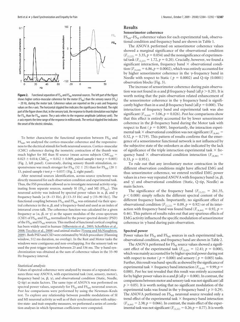
To better characterize the functional separation between FSSI andFSMI, we analyzed the cortico-muscular coherence and the responsive-ness to the electrical stimuli for both neuronal sources. Cortico-muscular(CMC) coherence during the isometric contraction of the thumb wasmuch higher for MI than SI source (mean across subjects CMCMI �0.023 � 0.014; CMCSI � 0.012 � 0.009, paired sample t test p � 0.005)(Fig. 2, left panel). Conversely, during sensory thumb stimulation, re-sponsiveness was much stronger for FSSI (52 � 25) than for FSMI (20 �15, paired sample t test p � 0.037) (Fig. 2, right panel).
After neuronal sources identification, across-source synchrony wasdirectly measured for each observation condition and experimental task.Thus, the FSS procedure allowed us to investigate neuronal activity orig-inating from separate sources, namely SI (FSSI) and MI (FSMI). Thisneuronal activity was indexed by spectral power values in �, �, and �frequency bands [� (8 –13 Hz), � (13–33 Hz) and � (33–90 Hz)]. Thefunctional coupling between FSSI and FSMI was estimated via their spec-tral coherence in the �, �, and � frequency-band and used as an index ofinterareal cross-talk. The coherence function was calculated for a givenfrequency � (�, �, or �) as the square modulus of the cross-spectrum(CSD) of FSSI and FSMI normalized by the power spectral density (PSD)of the FSSI and FSMI sources. This method of functional cortical couplinghas been widely used in human (Silberstein et al., 2005; Schoffelen et al.,2008; Tecchio et al., 2008) and animal studies (Young and McNaughton,2009). Both PSD and CSD were estimated by Welch procedure (Hanningwindow, 512 ms duration, no overlap). In the Rest and Motor tasks thewindows were contiguous and non-overlapping. For the sensory task weused the post-trigger intervals between 25 and 536 ms. The �-band syn-chronization was obtained as the sum of coherence values in the 33–90Hz frequency interval.
Statistical analysisValues of spectral coherence were analyzed by means of a repeated mea-sures three-way ANOVA, with experimental task (rest, sensory, motor),frequency band (�, �, �), and observational condition (Static, Needle,Q-tip) as main factors. The same type of ANOVA was performed onspectral power values, separately for FSSI and FSMI neuronal sources.Post hoc comparisons were performed by using the Newman–Keulstest. To explore any relationship between possible modulations of SIand MI neuronal activity as well as of their synchronization with subjec-tive state- and trait-empathy measures, we performed a series of correla-tion analyses in which Spearman coefficients were computed.
ResultsSensorimotor coherenceFSMI–FSSI coherence values for each experimental task, observa-tional condition and frequency band are shown in Table 1.
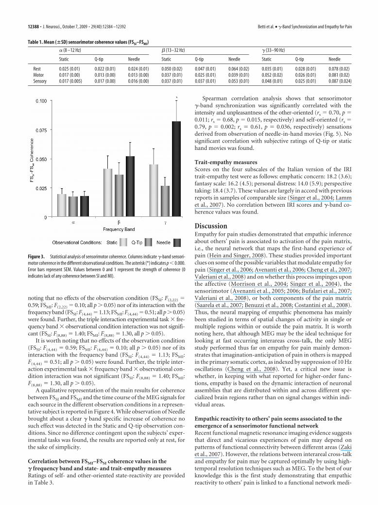
The ANOVA performed on sensorimotor coherence valuesshowed a marginal significance of the observational condition(F(2,22) � 3.35, p � 0.054) and the nonsignificance of experimen-tal task (F(2,22) � 1.72, p � 0.20). Crucially, however, we found asignificant interaction, frequency band observational condi-tion (F(4,44) � 6.86, p � 0.0002), which was entirely accounted forby higher sensorimotor coherence in the �-frequency band inNeedle with respect to Static ( p � 0.0002) and Q-tip (0.0001)observation blocks (Fig. 3).
The increase of sensorimotor coherence during pain observa-tion was not found in � and � frequency-band (all p � 0.20). It isworth noting that the pain observation related enhancement ofthe sensorimotor coherence in the � frequency band is signifi-cantly higher than in � and � frequency band (all p � 0.000). Theinteraction of frequency band and experimental task was alsosignificant (F(4,44) � 3.06, p � 0.026). Post hoc comparisons showthat this effect is entirely accounted for by lower sensorimotorcoherence in the �-frequency band during the Motor task withrespect to Rest ( p � 0.009). Importantly, the interaction experi-mental task observational condition was not significant (F(4,44) �0.52, p � 0.719). This pattern of results confirms that the emer-gence of a sensorimotor functional network is not influenced bythe subjective state of the onlookers as also indicated by the lackof significance of the triple interaction experimental task fre-quency band observational condition interaction (F(8,88) �0.33, p � 0.951).
To rule out that any involuntary motor contraction in thedifferent observation conditions might have affected EMG andthus sensorimotor coherence, we entered rectified EMG powervalues in a two-way repeated ANOVA with frequency band (�, �,and �) and observational condition (Static, Q-tip, Needle) asmain factors.
The significance of the frequency band (F(2,22) � 261.35,p � 0.000) simply reflects the different spectral content of thedifferent frequency bands. Importantly, no significant effect ofobservational condition (F(2,22) � 0.09, p � 0.92) or of its inter-action with frequency band was found band (F(4,44) � 0.92, p �0.46). This pattern of results rules out that any spurious effects ofEMG activity influenced the specific modulation of sensorimotorcoherence in � band during pain observation.
Spectral powerPower values for FSSI and FSMI sources in each experimental task,observational condition, and frequency band are shown in Table 2.
The ANOVA performed for FSSI source values showed a signifi-cant effect of the experimental task (F(2,22) � 12.41; p � 0.000),which was mainly accounted for by higher spectral power during restwith respect to motor ( p � 0.000) and sensory ( p � 0.005) tasks.Further, this result was band-specific as showed by the significance ofexperimental task frequency band interaction (F(4,44) � 9.99; p �0.000). Post hoc test revealed that this result was entirely accountedfor by higher power values in � and � (all p � 0.000). In contrast, thecomparisons between motor and sensory task was not significant (allp � 0.05). It is worth noting that no significant modulation of theexperimental tasks was found in the �-frequency band ( p � 0.29).The ANOVA performed on FSMI neuronal source revealed only atrend effect of the experimental task frequency band interaction(F(4,44) � 2.38; p � 0.066). In contrast, the main effect of the exper-imental task was not significant (F(2,22) � 0.26; p � 0.77). It is worth
Figure 2. Functional separation of FSSI and FSMI neuronal sources. The left part of the figureshows higher cortico-muscular coherence for the motor (FSMI) than the sensory source (FSSI)�20 Hz, during the motor task. Coherence values are reported on the y-axis and frequencyvalues on the x-axis. The horizontal stippled line indicates the significance threshold. The rightpart of the figure shows that, in the sensory task, the response to thumb stimulation was higherfor FSSI than for FSMI source. The y-axis refers to the response amplitude (arbitrary unit). Thex-axis reports the time range of the response in milliseconds. The vertical stippled line indicatesthe onset of the electric stimulus.
Betti et al. • �-Band Synchronization and Empathy for Pain J. Neurosci., October 7, 2009 • 29(40):12384 –12392 • 12387
noting that no effects of the observation condition (FSSI: F(2,22) �0.59; FSMI: F(2,22) � 0.10; all p � 0.05) nor of its interaction with thefrequency band (FSSI: F(4,44) � 1.13; FSMI: F(4,44) � 0.51; all p � 0.05)were found. Further, the triple interaction experimental task fre-quency band observational condition interaction was not signifi-cant (FSSI: F(8,88) � 1.40; FSMI: F(8,88) � 1.30, all p � 0.05).
It is worth noting that no effects of the observation condition(FSSI: F(4,44) � 0.59; FSMI: F(4,44) � 0.10; all p � 0.05) nor of itsinteraction with the frequency band (FSSI: F(4,44) � 1.13; FSMI:F(4,44) � 0.51; all p � 0.05) were found. Further, the triple inter-action experimental task frequency band observational con-dition interaction was not significant (FSSI: F(8,88) � 1.40; FSMI:F(8,88) � 1.30, all p � 0.05).
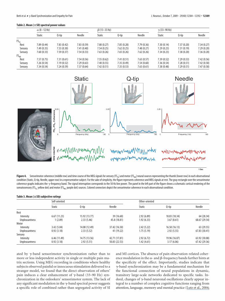
A qualitative representation of the main results for coherencebetween FSSI and FSMI and the time course of the MEG signals foreach source in the different observation conditions in a represen-tative subject is reported in Figure 4. While observation of Needlebrought about a clear � band specific increase of coherence nosuch effect was detected in the Static and Q-tip observation con-ditions. Since no difference contingent upon the subjects’ exper-imental tasks was found, the results are reported only at rest, forthe sake of simplicity.
Correlation between FSMI–FSSI coherence values in the� frequency band and state- and trait-empathy measuresRatings of self- and other-oriented state-reactivity are providedin Table 3.
Spearman correlation analysis shows that sensorimotor�-band synchronization was significantly correlated with theintensity and unpleasantness of the other-oriented (rs � 0.70, p �0.011; rs � 0.68, p � 0.015, respectively) and self-oriented (rs �0.79, p � 0.002; rs � 0.61, p � 0.036, respectively) sensationsderived from observation of needle-in-hand movies (Fig. 5). Nosignificant correlation with subjective ratings of Q-tip or statichand movies was found.
Trait-empathy measuresScores on the four subscales of the Italian version of the IRItrait-empathy test were as follows: emphatic concern: 18.2 (3.6);fantasy scale: 16.2 (4.5); personal distress: 14.0 (5.9); perspectivetaking: 18.4 (3.7). These values are largely in accord with previousreports in samples of comparable size (Singer et al., 2004; Lammet al., 2007). No correlation between IRI scores and �-band co-herence values was found.
DiscussionEmpathy for pain studies demonstrated that empathic inferenceabout others’ pain is associated to activation of the pain matrix,i.e., the neural network that maps the first-hand experience ofpain (Hein and Singer, 2008). These studies provided importantclues on some of the possible variables that modulate empathy forpain (Singer et al., 2006; Avenanti et al., 2006; Cheng et al., 2007;Valeriani et al., 2008) and on whether this process impinges uponthe affective (Morrison et al., 2004; Singer et al., 2004), thesensorimotor (Avenanti et al., 2005; 2006; Bufalari et al., 2007;Valeriani et al., 2008), or both components of the pain matrix(Saarela et al., 2007; Benuzzi et al., 2008; Costantini et al., 2008).Thus, the neural mapping of empathic phenomena has mainlybeen studied in terms of spatial changes of activity in single ormultiple regions within or outside the pain matrix. It is worthnoting here, that although MEG may be the ideal technique forlooking at fast occurring interareas cross-talk, the only MEGstudy performed thus far on empathy for pain mainly demon-strates that imagination-anticipation of pain in others is mappedin the primary somatic cortex, as indexed by suppression of 10 Hzoscillations (Cheng et al., 2008). Yet, a critical new issue iswhether, in keeping with what reported for higher-order func-tions, empathy is based on the dynamic interaction of neuronalassemblies that are distributed within and across different spe-cialized brain regions rather than on signal changes within indi-vidual areas.
Empathic reactivity to others’ pain seems associated to theemergence of a sensorimotor functional networkRecent functional magnetic resonance imaging evidence suggeststhat direct and vicarious experiences of pain may depend onpatterns of functional connectivity between different areas (Zakiet al., 2007). However, the relations between interareal cross-talkand empathy for pain may be captured optimally by using high-temporal resolution techniques such as MEG. To the best of ourknowledge this is the first study demonstrating that empathicreactivity to others’ pain is linked to a functional network medi-
Table 1. Mean (�SD) sensorimotor coherence values (FSSI–FSMI)
� (8 –12 Hz) � (13–32 Hz) � (33–90 Hz)
Static Q-tip Needle Static Q-tip Needle Static Q-tip Needle
Rest 0.025 (0.01) 0.022 (0.01) 0.024 (0.01) 0.050 (0.02) 0.047 (0.01) 0.064 (0.02) 0.035 (0.01) 0.028 (0.01) 0.078 (0.02)Motor 0.017 (0.00) 0.013 (0.00) 0.013 (0.00) 0.037 (0.01) 0.025 (0.01) 0.039 (0.01) 0.052 (0.02) 0.026 (0.01) 0.081 (0.02)Sensory 0.017 (0.005) 0.017 (0.00) 0.016 (0.00) 0.037 (0.01) 0.037 (0.01) 0.053 (0.01) 0.048 (0.01) 0.025 (0.01) 0.087 (0.024)
Figure 3. Statistical analysis of sensorimotor coherence. Columns indicate �-band sensori-motor coherence in the different observational conditions. The asterisk (*) indicates p � 0.000.Error bars represent SEM. Values between 0 and 1 represent the strength of coherence (0indicates lack of any coherence between SI and MI).
12388 • J. Neurosci., October 7, 2009 • 29(40):12384 –12392 Betti et al. • �-Band Synchronization and Empathy for Pain
ated by �-band sensorimotor synchronization rather than tomore or less independent activity in single or multiple pain ma-trix sections. Using MEG recording in conditions where healthysubjects observed painful or innocuous stimulation delivered to astranger model, we found that the direct observation of others’pain induces a clear enhancement of �-band (33–90 Hz) syn-chronization in the onlookers’ sensorimotor system. The lack ofany significant modulation in the �-band spectral power suggestsa specific role of combined rather than segregated activity of SI
and MI cortices. The absence of pain observation-related coher-ence modulation in the �- and �-frequency bands further hints atthe specificity of the effect. Importantly, studies indicate that�-band synchronization may be a fundamental mechanism forthe functional connection of neural populations in dynamic,transitory large-scale networks dedicated to specific tasks. In-deed, changes of �-band neuronal oscillations clearly appear in-tegral to a number of complex cognitive functions ranging fromattention, language, memory and mental practice (Lutz et al., 2004;
Figure 4. Sensorimotor coherence (middle row) and time course of the MEG signals for sensory (FSSI) and motor (FSMI) neural sources representing the thumb (lower row) in each observationalcondition (Static, Q-tip, Needle, upper row) in a representative subject. For the sake of simplicity, the figure represents coherence and MEG signals at rest. The gray rectangle over the sensorimotorcoherence graphs indicates the �-frequency band. The signal interruption corresponds to the 50 Hz line power. The panel in the left part of the figure shows a schematic cortical rendering of thesomatosensory (FSSI, yellow dot) and motor (FSMI, purple dot) sources. Colored connectors depict the sensorimotor coherence in each observational condition.
Table 2. Mean (�SD) spectral power values
� (8 –12 Hz) � (13–33 Hz) � (33–90 Hz)
Static Q-tip Needle Static Q-tip Needle Static Q-tip Needle
FSSI
Rest 7.89 (0.44) 7.83 (0.42) 7.83 (0.59) 7.80 (0.27) 7.85 (0.28) 7.79 (0.36) 7.30 (0.14) 7.37 (0.20) 7.34 (0.27)Sensory 7.49 (0.35) 7.53 (0.30) 7.41 (0.40) 7.54 (0.25) 7.62 (0.25) 7.48 (0.27) 7.29 (0.23) 7.31 (0.19) 7.29 (0.20)Sensory 7.60 (0.33) 7.59 (0.37) 7.54 (0.33) 7.63 (0.26) 7.65 (0.26) 7.62 (0.26) 7.34 (0.23) 7.38 (0.20) 7.36 (0.20)
FSMI
Rest 7.57 (0.75) 7.31 (0.61) 7.54 (0.56) 7.55 (0.62) 7.41 (0.51) 7.63 (0.57) 7.39 (0.52) 7.29 (0.53) 7.42 (0.56)Sensory 7.26 (0.54) 7.19 (0.52) 7.29 (0.63) 7.40 (0.55) 7.35 (0.49) 7.54 (0.60) 7.36 (0.54) 7.28 (0.51) 7.42 (0.58)Sensory 7.34 (0.54) 7.24 (0.59) 7.37 (0.64) 7.42 (0.51) 7.35 (0.53) 7.63 (0.61) 7.38 (0.48) 7.29 (0.51) 7.47 (0.58)
Table 3. Mean (�SD) subjective ratings
Self-oriented Other-oriented
Static Q-tip Needle Static Q-tip Needle
RestIntensity 6.67 (11.23) 15.92 (13.77) 39 (16.60) 2.92 (6.89) 18.83 (18.34) 64 (28.34)Unpleasantness 1 (2.89) 2.33 (5.46) 45.8 (18.81) 1.92 (6.33) 3.67 (8.61) 68.67 (29.54)
MotorIntensity 3.42 (5.04) 14.08 (12.49) 37.42 (16.50) 2.42 (5.22) 16.58 (16.13) 63 (29.55)Unpleasantness 0.92 (3.18) 2.33 (5.52) 41 (19.22) 1.75 (5.19) 2.92 (5.55) 67.83 (30.41)
SensoryIntensity 6.46 (11.49) 14.75 (13.56) 42.71 (17.81) 2.92 (6.72) 19.96 (16.07) 63.92 (30.08)Unpleasantness 0.92 (3.18) 2.92 (5.51) 50.83 (22.53) 1.42 (4.61) 3.17 (6.06) 67.42 (29.36)
Betti et al. • �-Band Synchronization and Empathy for Pain J. Neurosci., October 7, 2009 • 29(40):12384 –12392 • 12389

Bastiaansen and Hagoort, 2006) to cross-modal (Kanayama et al., 2009), and senso-rimotor integration (Alonso et al., 1996;Wallace et al., 1996; Bauer et al., 2006;Szurhaj and Derambure, 2006). Relevantto the present study is that �-band activityplays a specific role in first-hand pain pro-cessing (Chen and Herrmann, 2001;Ohara et al., 2006; Gross et al., 2007;Hauck et al., 2007). In particular, a recentMEG study showed that pain-induced�-band oscillations in the primary so-matosensory cortex were related to thesubjective perception of pain stimulus in-tensity rather than to the objective intensityof the stimulus itself (Gross et al., 2007).Therefore, �-band activity seems to reflectthe neural processes underlying the repre-sentation of subjective aspects of percep-tual (Gross et al., 2007) and cognitive(Jung-Beeman et al., 2004; Guggisberg etal., 2007; Sheth et al., 2009) experience. Ourresults extend the notion that �-band ac-tivity is linked to higher order functionsby indicating its relationship with the so-cial sharing of pain. The increase of pain observation related�-band synchronization of primary somatosensory motor corti-cal neurons, which are heavily connected at both anatomical andfunctional level (Rizzolatti and Luppino, 2001), hints at a specificrole of these regions during observation of “flesh and bone” pain-ful stimuli delivered to a model. While there is ample consensusthat both the affective and the sensorimotor node of the painmatrix can be recruited during the vicarious experience of pain(Hein and Singer, 2008), we focused on the possible role of pri-mary sensory and motor cortex in empathic pain mapping. Im-portantly, these regions seem to be specifically involved in theobservation of flesh and bone pain stimuli delivered to body partsof stranger individuals (Avenanti et al., 2005, 2006, 2009; Bufalariet al., 2007; Fecteau et al., 2008; Minio-Paluello et al., 2006, 2009).The involvement of sensorimotor regions in the empathic map-ping of others’ pain is in keeping with TMS (Avenanti et al., 2005,2006; Minio-Paluello et al., 2006), somatosensory (Bufalari et al.,2007) and laser-evoked potentials (Valeriani et al., 2008) studies.However, the main point of novelty of our study is that the es-sence of empathic mapping of observed pain resides in the dy-namic cross-talk between somato-motor regions much morethan on their separate neural activity. Indeed, mere observationof needle penetrating the hand of a stranger model induced aspecific, fast and short-lasting increase in the functional connec-tivity between the primary somatosensory and motor cortex inthe absence of detectable neural activity modulation in each area.The specific correlation between �-band coherence with self-and other-oriented ratings of pain intensity and unpleasantnesswhen observing needle-in hand clips but not when observingQ-tip on hand or static hand movie clips, also speaks in favor of aspecific relationship between pain observation and functionalsensorimotor coupling. The �-band sensorimotor cross-talk un-derlying empathy for pain was not different when the observingsubject performed an isometric contraction of the opponentmuscle of the thumb, perceived non-painful electric stimuli onthe skin overlaying the thumb opponent muscle, or was at rest,i.e., did not perform any task in addition to the pain observationone. This pattern of results indicates that the emergence of a
dynamic sensorimotor network from mapping others’ pain maybe a powerful, automatic process that minimizes the interferenceof additional tasks.
Sensory and motor cortical signatures of onlookers’ restingstate in � and � frequency bandsIn the present study, interareal cross-talk as indexed by sensori-motor coherence was modulated by pain observation only in the�-frequency band. Although no pain-observation related modu-lation of interareal cross-talk was found in any other bands,changes of activity in primary SI and MI cortices, as indexed byspectral power values, was found in � and � frequency bands.However, such modulation did not depend on pain observationbut on the onlookers’ state. Indeed, we found that the spectralpower, in both �- and �-frequency band, both for the SI and MIsources, was higher in the rest with respect to sensory and motortasks. Spontaneous oscillatory activity is related to the functionalstate of a given system. Studies in the primary visual, somatosen-sory, and motor areas show that higher amplitude of oscillatoryactivity reflects resting states, whereas lower amplitude is associ-ated with higher activation and excitability (Pfurtscheller andLopes da Silva, 1999). This effect has been linked to the notion of“idling systems,” i.e., systems which do not process any informa-tion at a given instant (Adrian and Matthews, 1934). In particu-lar, while the occipital �-rhythm reflects the idling condition ofvisual areas (Pfurtscheller and Lopes da Silva, 1999), the rolandic rhythm constitutes a signature of idling sensorimotor systems(Kuhlman, 1978). Mu rhythm is largely independent from thepresence or absence of visual input and, occurring only whenmuscles are relaxed, is considered a motor-relaxation-associatedrhythm (Buzsaki, 2006). Moreover, rhythm is thought to orig-inate from the primary somatosensory cortex representing thehand, and in particular from the thumb (Hari and Salmelin,1997). In view of this, our result of a rest specific increase of � and� bands spectral power may reflect the idling status of the pri-mary sensory and motor areas at rest. This status is likely inter-rupted by the stimulation of the thumb, occurring in the Sensoryand motor tasks. Moreover, the significant increase of the senso-
Figure 5. Correlational analysis. Scatter plots of sensorimotor coherence values in the Needle block and VAS scores concerningintensity and unpleasantness of the sensation derived from observation of others’ pain, attributed to self (self-oriented) (top row)or to the model (other oriented) (bottom row).
12390 • J. Neurosci., October 7, 2009 • 29(40):12384 –12392 Betti et al. • �-Band Synchronization and Empathy for Pain
rimotor coherence found in the � band, as well as the trend effectshown in the � band, is likely attributable to the high synchro-nized activity of the somatosensory system, characterized by a rhythm at rest.
All in all, the increase of cortical sensorimotor coherence con-tingent upon observation of noxious stimuli delivered to othersmay represent a neural signature of the interareal cross-talk thatunderpins the basic form of empathy we have called sensorimo-tor contagion (Avenanti et al., 2005). It is worth noting that thistype of resonance seems to be absent in persons with autisticspectrum disorders (Minio-Paluello et al., 2009). Moreover, pa-tients with autism exhibit patterns of aberrant �-band EEG activ-ity (�40 Hz) contingent upon a visual binding task (Grice et al.,2001), a result which is in keeping with neuroimaging studiesindicating that disrupted long-range connectivity and interre-gional interactions may underlie a range of behavioral deficits inautism (Muller, 2008; Uhlhaas and Singer, 2008). Thus, we positthat a lack of �-band sensorimotor coherence increase duringpain observation may represent a sensitive marker of empathicreactivity in a number of clinical conditions where this process islikely defective.
ReferencesAdrian ED, Matthews BH (1934) The Berger rhythm: potential changes
from the occipital lobes in man. Brain 57:355–385.Albiero P, Ingoglia S, Lo Coco A (2006) Contributo all’adattamento italiano
dell’Interpersonal Reactivity Index. TPM 13:107–125.Alonso JM, Usrey WM, Reid RC (1996) Precisely correlated firing in cells of
the lateral geniculate nucleus. Nature 383:815– 819.Avenanti A, Bueti D, Galati G, Aglioti SM (2005) Transcranial magnetic
stimulation highlights the sensorimotor side of empathy for pain. NatNeurosci 8:955–960.
Avenanti A, Minio Paluello I, Bufalari I, Aglioti SM (2006) Stimulus-drivenmodulation of motor-evoked potentials during observation of others’pain. Neuroimage 32:316 –324.
Avenanti A, Minio-Paluello I, Bufalari I, Aglioti SM (2009) The pain of amodel in the personality of an onlooker: influence of state-reactivity andpersonality traits on embodied empathy for pain. Neuroimage44:275–283.
Avikainen S, Forss N, Hari R (2002) Modulated activation of the human SIand SII cortices during observation of hand action. Neuroimage15:640 – 646.
Barbati G, Porcaro C, Zappasodi F, Rossini PM, Tecchio F (2004) Optimi-zation of an independent component analysis approach for artifact identifi-cation and removal in magnetoencephalographic signals. Clin Neurophysiol115:1220–1232.
Barbati G, Sigismondi R, Zappasodi F, Porcaro C, Graziadio S, Valente G,Balsi M, Rossini PM, Tecchio F (2006) Functional source separationfrom Magnetoencephalographic signals. Hum Brain Mapp 27:925–934.
Bastiaansen M, Hagoort P (2006) Oscillatory neuronal dynamics duringlanguage comprehension. Prog Brain Res 159:179 –196.
Bauer M, Oostenveld R, Peeters M, Fries P (2006) Tactile spatial attentionenhances �-band activity in somatosensory cortex and reduces low-frequency activity in parieto-occipital areas. J Neurosci 26:490 –501.
Benuzzi F, Lui F, Duzzi D, Nichelli PF, Porro CA (2008) Does it look painfulor disgusting? Ask your parietal and cingulate cortex. J Neurosci28:923–931.
Bonino S, Lo Coco A, Tani F (1998) Empatia. I processi di condivisione delleemozioni. Firenze, Italy: Giunti.
Bufalari I, Aprile T, Avenanti A, Di Russo F, Aglioti SM (2007) Empathy forpain and touch in the human somatosensory cortex. Cereb Cortex17:2553–2561.
Buzsaki G (2006) Rhythms of the brain. Oxford: Oxford UP.Chen AC, Herrmann CS (2001) Perception of pain coincides with the spa-
tial expansion of electroencephalographic dynamics in human subjects.Neurosci Lett 297:183–186.
Cheng Y, Lin CP, Liu HL, Hsu YY, Lim KE, Hung D, Decety J (2007) Exper-tise modulates the perception of pain in others. Curr Biol 17:1708 –1713.
Cheng Y, Yang CY, Lin CP, Lee PL, Decety J (2008) The perception of pain
in others suppresses somatosensory oscillations: a magnetoencephalogra-phy study. Neuroimage 40:1833–1840.
Costantini M, Galati G, Romani GL, Aglioti SM (2008) Empathic neuralreactivity to noxious stimuli delivered to body parts and non-corporealobjects. Eur J Neurosci 28:1222–1230.
Davis MH (1996) Empathy: a social approch. Madison, WI: Westview.Doesburg SM, Roggeveen AB, Kitajo K, Ward LM (2008) Large-scale
gamma-band phase synchronization and selective attention. Cereb Cor-tex 18:386 –396.
Eisenberger NI, Lieberman MD, Williams KD (2003) Does rejection hurt?An FMRI study of social exclusion. Science 302:290 –292.
Engel AK, Singer W (2001) Temporal binding and the neural correlates ofsensory awareness. Trends Cogn Sci 5:16 –25.
Fecteau S, Pascual-Leone A, Theoret H (2008) Psychopathy and the mirrorneuron system: preliminary findings from a non-psychiatric sample. Psy-chiatry Res 160:137–144.
Fries P (2005) A mechanism for cognitive dynamics: neuronal communica-tion through neuronal coherence. Trends Cogn Sci 9:474 – 480.
Fries P (2009) Neuronal gamma-band synchronization as a fundamentalprocess in cortical computation. Annu Rev Neurosci 32:209 –224.
Grice SJ, Spratling MW, Karmiloff-Smith A, Halit H, Csibra G, de Haan M,Johnson MH (2001) Disordered visual processing and oscillatory brainactivity in autism and Williams syndrome. Neuroreport 12:2697–2700.
Gross J, Schnitzler A, Timmermann L, Ploner M (2007) Gamma oscillationsin human primary somatosensory cortex reflect pain perception. PLoSBiol 5:e133.
Guggisberg AG, Dalal SS, Findlay AM, Nagarajan SS (2007) High-frequencyoscillations in distributed neural networks reveal the dynamics of humandecision making. Front Hum Neurosci 1:14.
Hari R, Salmelin R (1997) Human cortical oscillations: a neuromagneticview through the skull. Trends Neurosci 20:44 – 49.
Hauck M, Lorenz J, Engel AK (2007) Attention to painful stimulation en-hances �-band activity and synchronization in human sensorimotor cor-tex. J Neurosci 27:9270 –9277.
Hein G, Singer T (2008) I feel how you feel but not always: the empathicbrain and its modulation. Curr Opin Neurobiol 18:153–158.
Jasper H (1958) Report of the committee on methods of clinical examina-tion in electroencephalography. Electroencephalogr Clin Neurophysiol10:370 –371.
Jensen O, Kaiser J, Lachaux JP (2007) Human gamma-frequency oscilla-tions associated with attention and memory. Trends Neurosci30:317–324.
Jung-Beeman M, Bowden EM, Haberman J, Frymiare JL, Arambel-Liu S,Greenblatt R, Reber PJ, Kounios J (2004) Neural activity when peoplesolve verbal problems with insight. PLoS Biol 2:E97.
Kanayama N, Sato A, Ohira H (2009) The role of gamma band oscillationsand synchrony on rubber hand illusion and crossmodal integration. BrainCogn 69:19 –29.
Kuhlman WN (1978) Functional topography of the human mu rhythm.Electroencephalogr Clin Neurophysiol 44:83–93.
Lamm C, Batson CD, Decety J (2007) The neural substrate of human em-pathy: effects of perspective-taking and cognitive appraisal. J Cogn Neu-rosci 19:42–58.
Lutz A, Greischar LL, Rawlings NB, Ricard M, Davidson RJ (2004) Long-term meditators self-induce high-amplitude gamma synchrony duringmental practice. Proc Natl Acad Sci U S A 101:16369 –16373.
Makeig S, Debener S, Onton J, Delorme A (2004) Mining event-relatedbrain dynamics. Trends Cogn Sci 8:204 –210.
Melzack R (1999) From the gate to the neuromatrix. Pain [Suppl6]:S121–S126.
Minio-Paluello I, Avenanti A, Aglioti SM (2006) Left hemisphere domi-nance in reading the sensory qualities of others’ pain? Soc Neurosci1:320 –333.
Minio-Paluello I, Baron-Cohen S, Avenanti A, Walsh V, Aglioti SM (2009)Absence of embodied empathy during pain observation in Asperger syn-drome. Biol Psychiatry 65:55– 62.
Morrison I, Lloyd D, di Pellegrino G, Roberts N (2004) Vicarious responsesto pain in anterior cingulate cortex: is empathy a multisensory issue?Cogn Affect Behav Neurosci 4:270 –278.
Muller RA (2008) From loci to networks and back again: anomalies in thestudy of autism. Ann N Y Acad Sci 1145:300 –315.
Ohara S, Crone NE, Weiss N, Lenz FA (2006) Analysis of synchrony dem-
Betti et al. • �-Band Synchronization and Empathy for Pain J. Neurosci., October 7, 2009 • 29(40):12384 –12392 • 12391

onstrates ‘pain networks’ defined by rapidly switching, task-specific,functional connectivity between pain-related cortical structures. Pain123:244 –253.
Oldfield RC (1971) The assessment and analysis of handedness: the Edin-burgh inventory. Neuropsychologia 9:97–113.
Pfurtscheller G, Lopes da Silva FH (1999) Event-related EEG/MEG syn-chronization and desynchronization: basic principles. Clin Neurophysiol110:1842–1857.
Preston SD, de Waal FB (2002) Empathy: Its ultimate and proximate bases.Behav Brain Sci 25:1–20.
Rizzolatti G, Luppino G (2001) The cortical motor system. Neuron31:889 –901.
Rizzolatti G, Fogassi L, Gallese V (2001) Neurophysiological mechanismunderlying the understanding and imitation of action. Nat Rev Neurosci2:661– 670.
Saarela MV, Hlushchuk Y, Williams AC, Schurmann M, Kalso E, Hari R(2007) The compassionate brain: humans detect intensity of pain fromanother’s face. Cereb Cortex 17:230 –237.
Schoffelen JM, Oostenveld R, Fries P (2008) Imaging the human motorsystem’s beta-band synchronization during isometric contraction. Neu-roimage 41:437– 447.
Sheth BR, Sandkuhler S, Bhattacharya J (2009) Posterior beta and anteriorgamma oscillations predict cognitive insight. J Cogn Neurosci 21:1269 –1279.
Silberstein P, Pogosyan A, Kuhn AA, Hotton G, Tisch S, Kupsch A, Dowsey-Limousin P, Hariz MI, Brown P (2005) Cortico-cortical coupling inParkinson’s disease and its modulation by therapy. Brain 128:1277–1291.
Singer W, Gray CM (1995) Visual feature integration and the temporal cor-relation hypothesis. Annu Rev Neurosci 18:555–586.
Singer T, Seymour B, O’Doherty J, Kaube H, Dolan RJ, Frith CD (2004)Empathy for pain involves the affective but not sensory components ofpain. Science 303:1157–1162.
Singer T, Seymour B, O’Doherty JP, Stephan KE, Dolan RJ, Frith CD (2006)Empathic neural responses are modulated by the perceived fairness ofothers. Nature 439:466 – 469.
Szurhaj W, Derambure P (2006) Intracerebral study of gamma oscillationsin the human sensorimotor cortex. Prog Brain Res 159:297–310.
Tallon-Baudry C, Bertrand O (1999) Oscillatory gamma activity in humansand its role in object representation. Trends Cogn Sci 3:151–162.
Tecchio F, Porcaro C, Barbati G, Zappasodi F (2007) Functional source sep-aration and hand cortical representation for a brain-computer interfacefeature extraction. J Physiol 580:703–721.
Tecchio F, Zappasodi F, Porcaro C, Barbati G, Assenza G, Salustri C, RossiniPM (2008) High-gamma band activity of primary hand cortical areas: asensorimotor feedback efficiency index. Neuroimage 40:256 –264.
Tracey I, Mantyh PW (2007) The cerebral signature for pain perception andits modulation. Neuron 55:377–391.
Uhlhaas PJ, Haenschel C, Nikolic D, Singer W (2008) The role of oscilla-tions and synchrony in cortical networks and their putative relevance forthe pathophysiology of schizophrenia. Schizophr Bull 34:927–943.
Valeriani M, Betti V, Le Pera D, De Armas L, Miliucci R, Restuccia D, Ave-nanti A, Aglioti SM (2008) Seeing the pain of others while being in pain:a laser-evoked potentials study. Neuroimage 40:1419 –1428.
Varela F, Lachaux JP, Rodriguez E, Martinerie J (2001) The brainweb: phasesynchronization and large-scale integration. Nat Rev Neurosci 2:229 –239.
Wallace MT, Wilkinson LK, Stein BE (1996) Representation and integra-tion of multiple sensory inputs in primate superior colliculus. J Neuro-physiol 76:1246 –1266.
Womelsdorf T, Fries P (2006) Neuronal coherence during selective atten-tional processing and sensory-motor integration. J Physiol Paris 100:182–193.
Young CK, McNaughton N (2009) Coupling of theta oscillations betweenanterior and posterior midline cortex and with the hippocampus in freelybehaving rats. Cereb Cortex 19:24 – 40.
Zaki J, Ochsner KN, Hanelin J, Wager TD, Mackey SC (2007) Different circuitsfor different pain: patterns of functional connectivity reveal distinct networksfor processing pain in self and others. Soc Neurosci 2:276–291.
12392 • J. Neurosci., October 7, 2009 • 29(40):12384 –12392 Betti et al. • �-Band Synchronization and Empathy for Pain